Top 3 Diff erentials in Neuroradiology lrhieme
626
Top 3 Diff erentials in Neuroradiology A Case Review William T. O'Brien Sr. lrhieme
-
Upload
khangminh22 -
Category
Documents
-
view
2 -
download
0
Transcript of Top 3 Diff erentials in Neuroradiology lrhieme
Top 3 Diff erentials in Neuroradiology A Case Review
William T. O'Brien Sr.
lrhieme


lrhieme

Top 3 Differentials in Neuroradiology
A Case Review
William T. O'Brien Sr., DO
Program Director, Diagnostic Radiology Residency
David Grant USAF Medical Center
Travis Air Force Base, California
Former Chairman, Department of Radiology
Wilford Hall USAF Ambulatory Surgical Center
Joint Base San Antonio-Lackland, Texas
Associate Clinical Professor
Department of Radiology
University of california, Davis School of Medicine
Sacramento, California
Thieme
New York •Stuttgart • Delhi• Rio de Janeiro
Executive Editor: William Lamsback
Managing Editor: J. Owen Zurhellen IV
Assistant Managing Editor: Heather Allen
International Production Director: Andreas Schabert
Senior Vice President, Editorial and E-Product
Development: Cornelia Schulze
International Marketing Director: Fiona Henderson
International Sales Director: Louisa Turrell
Director of Sales, North America: Mike Roseman
Senior Vice President and Chief Operating
Officer: Sarah Vanderbilt
President: Brian D. Scanlan
Printer: Replika
Library of Congress Cataloging-in-Publication Data
O'Brien, William T .. author.
Top 3 differentials in neuroradiology : a case review /
William T. O'Brien.
p.; cm.
Top three differentials in neuroradiology
Includes bibliographical references.
ISBN 978-1-60406-723-1 (pbk. : alk. paper) -
ISBN 978-1-60406-724-8 ( e-book)
1. Title. II. Title: Top three differentials in neuroradiology.
[DNLM: 1. Diagnosis, Differential-Case Reports. 2. Neuro-
radiography-Case Reports. 3. Central Nervous System-radiogra
phy-Case Reports. 4. Central Nervous System Diseases
radiography-Case Reports. WL 141.5.N47]
RC71.5
616.07'5-dc23
© 2015 Thieme Medical Publishers, Inc.
Thieme Publishers New York
2014026028
333 Seventh Avenue, New York, NY 10001 USA, 1-800-782-3488
Thieme Publishers Stuttgart
Rüdigerstrasse 14, 70469 Stuttgart, Germany,
+49 (0]711 8931 421
Thieme Publishers Delhi
A-12, Second Floor, Sector-2, NOIDA-201301, Uttar Pradesh, India,
+91 120 45 566 OO
Thieme Publishers Rio, Thieme Publicaçêies Ltda.
Argentina Building 16th floor, Ala A, 228 Praia do Botafogo Rio de
Janeiro 22250-040 Brazil, +55 21 3736-3631
Printed in India
5 4 3 2 1
ISBN 978-1-60406-723-1
Also available as an e-book:
eISBN 978-1-60406-724-8
Important note: Medicine is an ever-changing science under
going continuai development. Research and clinical experience
are continually expanding our knowledge, in particular our
knowledge of proper treatment and drug therapy. Insofar as
this book mentions any dosage or application, readers may rest
assured that the authors, editors, and publishers have made every
effort to ensure that such references are in accordance with the
state of knowledge at the time of production of the book.
Nevertheless, this does not involve, imply, or express any
guarantee or responsibility on the part of the publishers in respect
to any dosage instructions and forms of applications stated in the
book. Every user is requested to examine carefully the manu
facturers' leaflets accompanying each drug and to check, if nec
essary in consultation with a physician or specialist, whether the
dosage schedules mentioned therein or the contraindications
stated by the manufacturers differ from the statements made
in the present book. Such examination is particularly important
with drugs that are either rarely used or have been newly released
on the market. Every dosage schedule or every form of application
used is entirely at the user's own risk and responsibility. The
authors and publishers request every user to report to the pub
Iishers any discrepancies or inaccuracies noticed. If errors in this
work are found after publication, errata will be posted at www.
thieme.com on the product description page.
Sorne of the product names, patents, and registered
designs referred to in this book are in fact registered trade
marks or proprietary names even though specific reference
to this fact is not always made in the text. Therefore, the
appearance of a name without designation as proprietary is
not to be construed as a representation by the publisher that
it is in the public domain.
jJ, FSC www.fsc.org
MIX
Paperfrom responsible sources
FSC"' C021256
This book, including ail parts thereof, is legally protected by
copyright. Any use, exploitation, or commercialization outside
the narrow limits set by copyright legislation, without the pub
lisher's consent, is illegal and Iiable to prosecution. This applies in
particular to photostat reproduction, copying, mimeographing,
preparation of microfilms, and electronic data processing and
storage.
The views expressed in this material are those of the author,
and do not reflect the official policy or position of the United
States Govemment, the Department of Defense, or the Depart
ment of the Air Force.
Dedicated in memory of
Robert L Meals, DO
12 March 1928-9 June 2005
© Susan Schary 2005
For decades, Dr. Meals inspired thousands of students while serving as Academic Chairman of the
Department of Radiology, Philadelphia College of Osteopathic Medicine, Philadelphia, Pennsylvania.
Dr. Meals was more than an instructor; he was a mentor and a true friend.
To those who chose to pursue a career in radiology, he will always be a legend.
He is sorely missed but will never be forgotten.
vi
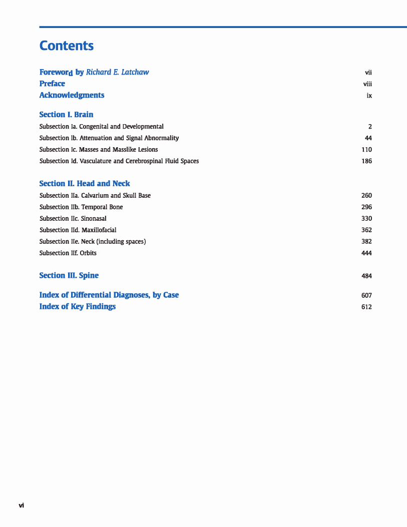
Contents
Foreworcl by Richard E. Latchaw
Preface
Aclmowledgments
Section 1. Brain
Subsection la. Congenital and Developmental
Subsection lb. Attenuation and Signal Abnormality
Subsection le. Masses and Masslike Lesions
Subsection Id. Vasculature and Cerebrospinal Fluid Spaces
Section IL Head and Neck
Subsection Ha. calvarium and Skull Base
Subsection IIb. Temporal Bone
Subsection IIe. Sinonasal
Subsection IId. Maxillofacial
Subsection IIe. Neck (induding spaces)
Subsection Hf. Orbits
Section ID. Spine
Index of Differential Diagnoses, by Case
Index of Key Findi�
vii
viii
ix
2
44
110
186
260
296
330
362
382
444
484
607
612
Foreword
Unique/
lbat is the best word to describe Top 3 DijJerentials in
Neuroradiology by William T. O'Brien-unique in its approach
to the clinical practiœ of neuro-imaging, and unique in its
approach to education in this rapidly expanding subspecialty.
The traditional clinical practiœ of a neurologist, neuro
surgeon, orthopedic surgeon-any physician ordering a
neuro-imaging examination-is to evaluate the patient's his
tory in conjunction with signs and symptoms, corne ta a
probable conclusion, and then request an imaging study to
confirm or deny that clinical conclusion.
The clinical practice of a radiologist initially requires the
recognition of a combination of findings on an imaging study
within the stated clinical context This is followed by the
iterative comparison of these findings to examples from
diagnostic categories, including masses, demyelinating dis
eases, ischemia, infection, degenerative disease, etc. This
iterative proœss may be mental or actually require compari
son with published examples. The result is a differential
diagnosis that may vary in specificity and depth. One might
list the top three possible diagnoses, or one could list the most
likelywith that which is the most dangerous and thus must be
excluded, along with one that would be easy to exclude with
more studies.
How dowe traditionally educate a reader of neuro-imaging
studies7 We usually ensure that the novice reader has seen
examples from the various diagnostic categories with which
we deal, and has leamed how diseases within each category
differ from those in other categories. The organization of our
books and our teaching sessions is typically based upon such
categories: Neoplasms, Congenital Disease, Infections, etc.
However, what happens when the imager is confronted
with an "unknown," a finding that does not fit easily into
one of the categories to which he or she has become so
accustomed? Unfortunately, even though the imager has
leamed the appearances of the majority of entities within
a given category of disease, the finding does not tell the
imager to which category it belongs! So, the imager must
now search the categorically based textbooks for a "look
alike," which is very time-consuming and may not even be successful.
Dr. O'Brien's approach ta both the clinical practice and the
education of neuro-imaging is quite unique amongst the
textbooks 1 have seen over many years as a neuroradiologist
He has divided this book into three sections: Brain, Head and
Neck, and Spine. Within each section, he conœntrates on the
most apparent imaging finding(s) within the presenting clin
ical context, and gives the "Top 3" potential diagnoses for that
appearance ( that "gamut"), including entities that may well
derive from multiple diagnostic categories. For some appear
ances, he even indudes some uncommon but potentially
important considerations ("Additional Diagnostic Considera
tions"), thus providing more than just three possibilities for
cases with more nonspecific findings. He finishes each case
with dinical and imaging "Pearls," which provide quick dif
ferentiating features. He also provides some selected refer -
ences for more in-depth reading on the topic.
Sorne imaging appearances within each section are unique,
without differential diagnoses and not having a Top 3; they
are called "Aunt Minnies." Dr. O'Brien considers a number of
these to be fundamental to the knowledge base of the student,
sa they are presented at the end of each section. Each has an
extensive discussion regarding pathophysiology and charac
teristic imaging appearances, along with selected referenœs,
similar to that found with the cases having Top 3 differential
possibilities.
How did Dr. O'Brien validate his Top 3 choices with so
many varied appearances in diverse clinical contexts? By
doing extensive research as to the most common diagnoses
for a given finding; by consulting with many radiologists who
subspecialize in neuroradiology, head and neck radiology,
and spinal radiology; and by incorporating entities
that tend to be favorites in general and subspecialty board
examinations.
How will this book change how we practice and teach
neuro-imaging? lt is vital that neuro-imagers have ingrained
in their brain the basic categories of neuropathology, so that
they can be sure that they caver ail potential disease catego
ries when confronted with an unknown case. However,
O'Brien's approach can easily be superimposed on that basic
knowledge of disease organization. lt is fast, accurate, and
removes the potential that the reader will be slowed down,
trying to ensure that ail categories are covered. This approach
provides a way to be "complete" in developing differential
diagnoses rapidly and accurately.
1 found reading this book to be a joy. One can approach it by
playing the student, viewing each image as an unknown,
determining what the most prominent finding is, and then
giving one's own Top 3. Frankly, this is a book not just for the
resident or fellow, but one that will give any academic faculty
member a positive learning experienœ, just like the one that
1 hadl
Richard E. Latchaw, MD
Professor of Radiology
Neuroradiology Section
University of California, Davis Medical Center
Sacramento, California
vii
viii
Preface
It is a distinct pleasure to present Top 3 Differentials in
Neuroradiology: A Case Review. Developing a neuroradiology
version of the original "Top 3" book, Top 3 Differentials in
Radiology, had been an aspiration of mine since its publica
tion in 201 O. This subspedalty version is primarily designed
for senior radiology residents, neuroradiology fellows, and
staff radiologists preparing for the neuroradiology portion
of initial and recertification board examinations; however, it
may also prove useful for dinicians and surgeons who
routinely utilize neuroimaging.
This book is organized into three main sections: brain,
head and neck, and spine imaging; and further divided into
subsections based upon anatomie region or pattern of imag
ing abnormality. Each section begins with a series of
unknown differential-based cases and ends with "Roentgen
dassics," which are cases with imaging findings character
istic of a single diagnosis.
On the first page of each case, readers are presented with
images from an unknown case, along with a clinicat history
and an image legend. The images are meant to illustrate a
key imaging finding, which is the basis for the subsequent
case discussion. The second page Iists the key imaging
finding, from which a list of differentials is broken down
into the Top 3, along with "additional diagnostic considera
tions." The discussion section of each case provides a brief
review of important imaging and clinical manifestations for
all entities on the list of differentials, making this a high
yield reference for board preparation. lmaging pearls are
provided at the end of each case to allow for a quick review
of key points. The final diagnosis is provided for each case;
however, it is by no means the focus of this review book. In
fact, many illustrative cases have a final diagnosis that
would not be considered in the Top 3 for the particular
gamut. Instead, the primary aim of the book is to generate
and have an understanding of a reasonable list of gamut
based differentials rather than to obtain the "correct"
answer.
As with the earlier Top 3 Differentials in Radiology, it is
important to realize that the differentials and discussions are
based on the key finding or gamut and not necessarily the
illustrative cases that are shown This is by design, because 1
felt it would be more high-yield to base the differentials and
discussions on the overall gamut/key finding rather than the
illustrative case presented. Having an understanding of
gamut-based differentials will allow one to subsequently
tailor the Iist of differentials for any case that is shown within
the gamut, whereas basing the differentials on the selected
images would be more limited in terms of future utility.
Given the vast, evolving field of neuroimaging, this book is
not meant to be a comprehensive reference book; rather, it is
meant to serve as a high-yield review for board preparation,
as well as a quick reference for clinical practice. With these
intentions in mind, the selection and ordering of differentials
for each gamut were based upon a combination of the most
likely diagnoses to be enrountered in a board setting, as well
as clinical practice. Sorne "additional diagnostic considera
tions" were selected over others (which may actually be more
rommon) in order to provide the opportunity to discuss as
many diagnostic entities as possible throughout the book.
1 sinœrely hope that you find this Top 3 case-based
approach enjoyable and useful, and I wish you all the best
in your future endeavors.
Acknowledgments
This book would not have been possible without the contri
butions of numerous colleagues and mentors. First and
foremost, 1 am forever indebted ta the faculty of David Grant
USAFMedical Center, the UniversityofCalifornia-Davis, and
Oakland Children's Hospital, where 1 completed my radiol
ogy residency training, as well as the University of Cincin
nati and Cincinnati Children's Hospital Medical Center,
where 1 completed my neuroradiology fellowships. The
dedicated staff at these institutions afforded me their
time and expertise during my years of training and have
had a profound impact on my career. Their influence is what
inspires me to remain in academics in the hopes of having a
similar impact on the next generation of radiologists.
Severa! colleagues contributed to the content of this book
through images and case material, some of which was
induded in the original "Top 3" book, Top 3 Di.fferentials
in Radiology. Their contributions have greatly enhanced the
final manu script. The contributors are listed at the end of the
image legend for each case in which they were involved. 1 cannot possibly thank them enough for their significant
contributions ta this book. Although there are far tao
many ta name individually, 1 would like to espedally thank
Paul M. Sherman, MD, who not only authored portions of
the neuroimaging sections in the original "Top 3" book, but
also served as my neuroradiology mentor during residency
and bas been one of my neuroradiology partners in San
Antonio for the past 4 years.
Lastly, 1 would like to thank my family for their continu
ous love and support, as well as the sacrifices they
made during completion of this project. 1 have been
blessed with a wonderful wife, Annie; two sons, Patrick
and Liam; and a daughter, Shannon. Annie and 1 have been
together for nearly two decades, and we could not be more
proud of our three incredible children. I am grateful
beyond words for the joy that they bring into my life
each and every day.
ix


2
Brain
Case 1
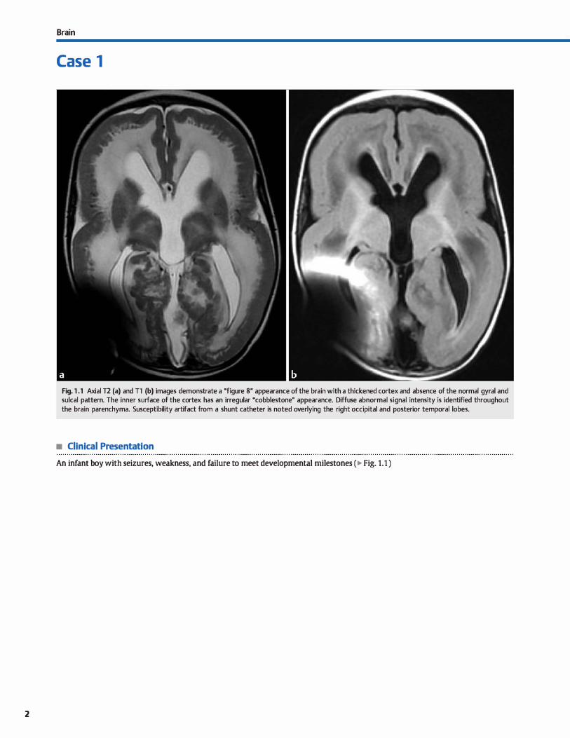
Fig. 1 .1 Axial T2 (a) and Tl (b) images demonstrate a "figure 8" appearance of the brain with a thickened cortex and absence of the normal gyral and sulcal pattern. The inner surface of the cortex has an irregular "cobblestone" appearance. Diffuse abnormal signal intensity is identified throughout the brain parenchyma. Susceptibility artifact from a shunt catheter is noted overlying the right occipital and posterior temporal lobes.
• Clinical Presentation
An infant boy with seizures, weakness, and failure to meet developmental milestones ( � Fig. 1.1 )
• Key lmaging Finding
Agyria
• Top 3 Differential Diagnoses
• JYpe 1 lissencephaly. Type 1 or dassic lissencephaly is a con
genital neuronal migration disorder that results in a smooth
appearance of the brain secondary to absence of the normal
gyral and sulcal pattern. There may be diffuse involvement
(agyria) or focal involvement (pachygyria) of the cerebral cor
tex. Diffuse involvement results in a "figure 8" appearance of
the brain with vertically oriented Sylvian fissures and absence
of the normal gyral and sulcal pattern. On pathologie evaluation, there is a thickened, smooth four-layer cortex with a thin ribbon of subcortical band heterotopia, rather than a normal
six-layer cortex. Type 1 lissencephaly may be associated with
cytomegalovirus (CMV) infection, Miller-Dieker syndrome,
and cerebellar hypoplasia. With CMV infection, periventricu
lar and intraparenchymal calcifications are noted. Patients
with Miller-Dieker syndrome demonstrate midline septal
calcifications, microcephaly, and characteristic dysmorphie
facial features. • JYpe 2 lissencephaly. Type 2 or cobblestone lissencephaly is
characterized by overmigration of neurons, severe dis
organization of the gray matter, underdevelopment of gyri
and sulci, and diffuse white matter hypomyelination. The
disorganized gray matter results in an irregular, "cobble
stone" appearance of the cortex. There is an association with
congenital muscular dystrophies, induding Walker-Warburg
• Additional Differential Diagnoses
• Prematurity. Prier ta -26 weeks gestation, the fetal brain nor
mally appears Iissencephalic due ta Jack of gyral and sulcal development. After 26 weeks gestation, the gyral and sulcal
pattern gradually progresses until its relatively normal
• Diagnosis
1}'pe 2 cobblestone lissencephaly in a patient with Walker
Warburg syndrome
y' Pearls
• The premature infant brain normally appears lissencephalic
prier ta 26 weeks gestation. • Llssencephaly is a neuronal migration disorder with absence
of normal gyri/sulci and a thickened cortex.
Suggested Readings
Barlmvich AJ, Chuang SH, Norman D. MR of neuronal migration anomalies. Am J
Roentgenol 1988; 150: 179-187
Case 1
syndrome, Fukuyama congenital muscular dystrophy, and to
a lesser degree, musde-eye-brain disease. Patients present
early in Iife with severe muscular weakness, eye abnormali
ties, developmental delay or mental retardation, and compli
cations of assodated brain malformations. Patients with
Walker-Warburg often have characteristic findings, includ
ing occipital cephaloceles, cerebellar and brain stem hypo
plasia, and kinking of the brain stem with a dassic "striking
cobra" appearance on sagittal sequences. Hydrocephalus is present in the vast majority of cases.
• Band heterotopia. Gray matter heterotopia refers ta collec
tions of disorganized neurons in abnormal locations. It results
from premature arrest of normal neuronal migration. Neurons migrate from the ependymal surface of the lateral ventri
des to the peripheral cortex, and then undergo organization
into a normal six-layer cortex. If arrest occurs at any point during migration, heterotopias occur. Heterotopia may be
dassified as nodular (most common), which most often occurs along the margins of the lateral ventricles, or band,
which is located within the subcortical or deep white matter.
When diffuse and subcortical in location, band heterotopia
may mimic lissencephaly. Patients typically present with
seizures, developmental delay, and spasticity.
appearance at term. Therefore, lissencephaly should not be
diagnosed until after 26 weeks gestation. When uncertain, a
follow-up examination may be helpful to evaluate for interval
gyral and sulcal formation.
• Type 1 lissencephaly is smooth and may be associated with CMV, Miller-Dieker, and cerebellar hypoplasia.
• Type 2 lissencephaly has a "cobblestone" appearance and is
associated with congenital muscular dystrophies.
Ghai S, Fong KW, Thi A, Chitayat D, Pantazi S, Blaser S. Prenatal US and MR imaging
findings of lissenœphaly: review offetal œrebral sulcal devclopment Radiographies 2006; 26; 389-405
3
4
Brain
Case 2
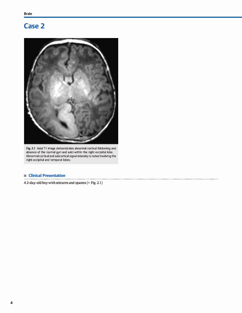
Fig. 2.1 Axial Tl image demonstrates abnormal cortical thickening and absence of the normal gyri and sulci within the right occipital lobe. Abnormal cortical and subcortical signal intensity is noted involving the right occipital and temporal lobes.
• Clinical Presentation
A 2-day-old boy with seizures and spasms (� Fig. 2.1 )
• Key lmaging Finding
Cortical malformation
• Top 3 Differential Diagnoses
• Pachygyria. Pachygyria is an incomplete or focal form of
lissenœphaly. As with lissencephaly, there is both abnormal neuronal migration and failure to form the normal six-layer cortex. Instead, a four-layer cortex is most commonly seen pathologicaliy. Imaging findings are characterized by short,
broad gyri with a lack of sulcation in the involved segments. Symptoms depend upon the extent and location of parenchy
mal involvement. Patients may present with seizures, devel
opmental delay, mental retardation, and/or spasticity. • Polymicrogyria. Polymicrogyria is a neuronal migration
abnormality characterized by abnormal distribution of neurons along the cortical surface. Multiple, small gyri replace the normal organized gyral and sulcal pattern. It is thought to
result from laminar necrosis of neurons after they reach the cortical surface. It is commonly seen in association with cyto
megalovirus (CMV) infection. Para-Sylvian locations are commonly involved. The polymicrogyria pattern is best depicted on magnetic resonanœ imaging (MRI). Abnormal signal is commonly seen in the subjacent white matter. dinically, patients present with seizures, developmental delay, mental
• Additional Differential Diagnoses
• Subcortical band heterotopia. Gray matter heterotopia
refers to collections of disorganized neurons in abnormal locations due to premature arrest of normal migration. Neurons migrate from the ependymal surface of the lateral ventricles to the peripheral cortex, and then undergo orga
nization into a normal six-layer cortex. If arrest occurs at any point during migration, heterotopias occur. Heteroto
pia may be classified as nodular, which most often occurs along the margins of the lateral ventricles, or band-type, which occurs within the subcortical or deep white matter. Patients typically present with seizures, developmental delay, and spasticity.
• Schizencephaly. Schizencephaly is a congenital malformation characterized by gray matter-lined clefts extending from the
• Diagnosis
Pachygyria
� Pearls
• Pachygyria is a form of focal lissencephaly with a thickened, four-layer cortex (instead of the normal six layers).
• With polymicrogyria, small gyri replace the normal, organized gyral pattern; it is associated with CMV.
Suggested Readings
Barlwvich AJ, Clmang SH, Norman D. MR of neuIOllill migration anomalies. Am J
Roentgenol 1988; 150: 179-187 Broumandi DD. Haywanl UM, Benzian]M, Gonzalez 1, Nelson MD. Best cases from
the AFIP: hemimegalenœphaly. Radiographies 2004; 24: 843-848
Case 2
retardation, and, occasionally, hemiparesis. Polymicrogyria may be associated with various syndromes, including Aicardi (callosal anomalies, infantile spasms, and retinal lesions) and Zellweger (cerebrohepatorenal) syndromes.
• Hemimegalencephaly. Hemimegalencephaly is a hamartom
atous overgrowth of ail or a portion of one cerebral hemisphere with associated neuronal migration abnormalities of varying severity. It is thought to occur as a result of an insult during neuronal migration. The ipsilateral hemisphere and ventricle are enlarged. Affected gyri are thickened and may show a primitive lissenœpahlic appearanœ with shallow or absent sulci. There is often abnormal attenuation (computed tomography) and signal intensity (MRI) within the subjacent
white matter. Calcifications are not uncommon. dinically, the patient may present with seizures, developmental delay, mental retardation, and/or hemiplegia. Syndromes associated with hemimegalencephaly include neurofibromatosis type 1, Klippel-Trenaunay-Weber syndrome, tuberous sderosis, and Proteus syndrome.
pial surface to the ventricle. The defts are typically paraSylvian in location and lined by polymicrogyric gray matter. In Type I (dosed-lip) schizenœphaiy, the gray matter Iinings are apposed with a small ventricular dimple of cerebrospinal fluid (CSF) extending into the deft. Type II (open-lip) schizen
œphaly consists of a large CSF-filled space between the gray matter linings. Schizenœphaly may be bilateral and assodated with septooptic dysplasia. dinical manifestations depend upon the severity of the lesion. Patients with type I are often almost normal in terms of development, but may have seizures and hemiparesis. Type II patients usually dem
onstrate mental retardation, seizures, hypotonia, spasticity, inability to walk or speak, and blindness.
• Heterotopia (nodular or band) refers to collections of disorganized neurons in abnormal locations.
Hayashi N, Tsutswrù Y, Barlcvvich AJ. Morphological features and associated anoma
lies of schizencephaly in the dinkal population: detailed analysis of MR images. Neuroradiology 2002; 44: 418-427
5
6
Brain
Case 3
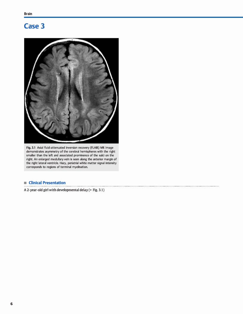
Fig. 3.1 Axial fluid-attenuated inversion recovery (FLAIR) MR image demonstrates asymmetry of the cerebral hemispheres with the right smaller than the left and associated prominence of the sulci on the right. An enlarged medullary vein is seen along the anterior margin of the right lateral ventride. Hazy, periatrial white matter signal intensity corresponds to regions of terminal myelination.
• Clinical Presentation
A 2-year-old girl with developmental delay (� Fig. 3.1 )
• Key lmaging Finding
Asymmetry of cerebral hemispheres
• Top 3 Differential Diagnoses
• Nonnal variant. Slight variation in size of an entire cerebral hemisphere, one or more lobes, or individual sulci is not uncommon, occurring in -10% of normal cases. Parenchymal
morphology, attenuation, and signal intensity should otherwise be normal and are useful discriminators from pathologie causes of parenchymal volume Joss. Patients are oft:en neuro
logically and developmentally intact for age.
• Encephalomalacia. Enœphalomalacia refers to parenchymal volume Joss as a result of some form of insult Hypoxic-ischemic injury is the most common cause of enœphalomalacia, followed by trauma and infectious or inflammatory processes. Ischemic
injury typically follows a vascular distribution. During the acute
phase of injury, there is often focal edema and swelling. In the chronic stage, there is volume Joss with surrounding gliosis. In
the setting of an asymmetric small œrebral hemisphere, a large
• Additional Differential Diagnoses
• Dyke-Davidolf-Mason syndrome (DDMS). DDMS refers to compensatory enlargement of the ipsilateral calvarium, paranasal sinuses, and mastoid air cells secondary to underdevelopment or atrophy of the underlying œrebral hemisphere.
The most common causes of ipsilateral cerebral atrophy
include a large-territory ischemic insult at a young age or SWS. Symptoms are related to the causative process.
• Hemimegalencephaly. Hemimegalencephaly is a hamartomatous overgrowth of all or a portion of one œrebral hemisphere with associated neuronal migration abnormalities. lt is
thought to result from an insult during neuronal migration. The ipsilateral hemisphere and ventride are enlarged. Affected gyri are thickened and may show a lissenœpahlic appearance with shallow or absent suld. There is oft:en abnor
mal attenuation {computed tomography) and signal intensity (MRI) within the white matter of the ipsilateral hemisphere.
• Diagnosis
Sturge-Weber syndrome
� Pearls
• Enœphalomalacia refers to parenchymal volume Joss from some form of insult; ischemia is most common.
• SWS is characterized by seizures, cutaneous port-wine stain, and pial angiomatosis of the ipsilateral hemisphere.
Suggested Readings
Case 3
territory infarct (middle œrebral artery) is the most likely cause of enœphalomalacia.
• Sturge-Weber syndrome (SWS; encephalotrigeminal angiomatosis). SWS is a sporadic phakomatosis thought to result from abnormal development of venous drainage. lt is characterized by a cutaneous port-wine stain (usually in the Vl distribution of the trigeminal nerve) and pial angiomatosis overlying the ipsi
Iateral œrebral hemisphere. Venous drainage is diverted through enlarged medullary and subependymal veins. Hemiatrophy
results, likely from venous hypertension. Magnetic resonanœ imaging (MRI) shows œrebral atrophy, abnormal leptomeningeal enhanœment, and increased enhanœment within a hypertrophied ipsilateral choroid plexus. The involved hemisphere may demonstrate abnormal signal, cortical enhanœment, and
cortical calcifications in a "tram trad<" configuration.
calcifications are not uncommon. Œnically, patients may present with seizures, developmental delay, mental retardation, and hemiplegia. Associated syndromes indude neurofibromatosis type 1, Klippel-Trenaunay-Weber syndrome, tuberous sderosis, and Proteus syndrome.
• Rasmussen encephalitis. Rasmussen enœphalitis is a rare,
progressive, inflammatory neurological disorder of unknown
origin. A viral or postviral autoimmune etiology bas been postulated. Patients present in childhood with persistent,
relentless, focal motor seizures (epilepsia partialis continua), hemiplegia, and cognitive deficits. Early on, MRI demonstrates abnormal edema and increased T2 signal within the involved hemisphere. Chronically, findings are more charac
teristic with abnormal signal, asymmetric atrophy, and decreased perfusion and metabolism on the affected side. Treatment consists of functional hemispherectomy.
• Hemimegalencephaly is a hamartomatous overgrowth of all or part of one cerebral hemisphere.
Shapiro R, Galloway SJ, Shapiro MD. Minimal asyrnrnetryof the brain: a normal vari- Sener RN, Jinkins JR. MR of cranioœrebral herniatrophy. Œn Irnaging 1992; 16:
ant ArnJ RDentgenol 1986; 147: 753-756 93-97
7

Brain
Case 4
• Clinical Presentation
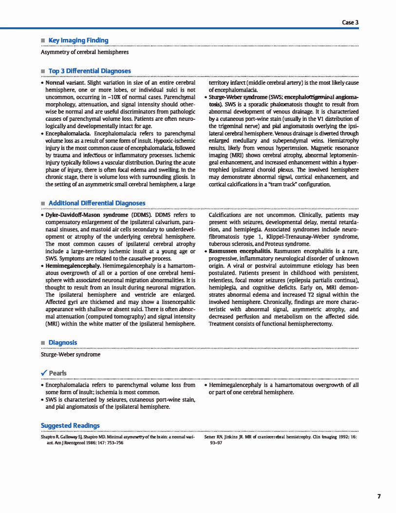
An adolescent with seizures (� Fig. 4.1 )
8
Fig. 4.1 Axial T2 (a) and fluid-attenuated inversion recovery (FLAIR) (b) images of the brain demonstrate a hypointense subependymal nodular lesion within the frontal horn of the right lateral ventricle. The lesion is isointense to white matter on Tl sequences (c) and demonstrates homogeneous enhancement (d). Wedge-shaped regions
of cortical and subcortical signal abnormality are also noted on the
T2/FLAIR sequences (left hemisphere). (Courtesy of Paul M. Sherman, MD.)
• Key lmaging Finding
Subependymal nodules
• Top 3 Differential Diagnoses
• Tuberous sclerosis (TS). TS is a neurocutaneous syndrome that results from gene mutations affecting chromosomes
9q34.3 {hamartin) and 16p13.3 {tuberin). Two-thirds of cases occur sporadically, whereas the remaining occur in an autosomal dominant fashion with variable penetrance. The classic triad consists of fadai angiofibromas, mental retardation, and seizures, but it is only seen in approximately one-third of
cases. Central nervous system (CNS) manifestations include cortical/subcortical tubers, white matter lesions that occur in a radial pattern along paths of neuronal migration, subependymal nodules, and subependymal giant œll astrocytomas (SEGAs). The cortical/subcortical tubers are composed of disorganized glial tissue and heterotopic neuronal elements. They present as triangular regions of cortical and subcortical
signal abnormality that may calcify and occasionally demonstrate enhancement Subependymal nodules have variable Tl and T2 signal intensity and commonly enhance. They demonstrate gradient echo susœptibility (hypointensity) when caldfied; the majority are caldfied by 20 years of age. SEGAs are low-grade tumors that occur in -1 O to 15% of cases. They are located at the foramen of Monro, enlarge over time, and
enhance. Interval growth is the best sign to distinguish SEGAs from dominant subependymal nodules. Treattnent is typically geared toward œrebrospinal fluid diversion. Com
mon abnormalities associated with TS include retinal hamar-
• Additional Differential Diagnoses
• Metastatic disease. Subependymal metastatic disease may result from primary CNS neoplasms or hematogenous spread from extracranial malignandes. Primary CNS neoplasms prone to subependymal spread include glioblastoma multi
forme, medulloblastoma, ependymoma, primary CNS lym-
• Diagnosis
Tuberous sclerosis
� Pearts
• TS results in subependymal nodules, which caldfy and demonstrate enhancement.
• Heterotopic gray matter is due to an insult in utero and follows gray matter signal on all MR sequenœs.
Suggested Readings
Barlmvich AJ, Chuang SH, Norman D. MR of neuronal migration anomalies. AmJ
Roentgenol 1988; 150; 179-187
Braffman BH, Bilaniuk IJ', Naidich TP et al. MR imaging oftuberous sderosis: patho
genesis of tlùs phalclllnatosis, use of gadopentetate dimeglumine, and literatuœ
review. Radiology 1992; 183; 227-238
Case 4
tomas, cardiac rhabdomyomas, renal cysts and angiomyolipo
mas, pulmonary lymphangioleiomyomatosis, subungual fibromas, and skin lesions, such as "ash-leaf spots" and shagreen patches.
• Heterotopic gray matter. Heterotopic gray matter results from arrest or disruption of normal neuronal migration
from the subependymal region to the overlying cortex. lt is thought to occur secondary to some form of fetal insult during
development. Heterotopia may be nodular or bandlike. Subependymal heterotopic gray matter is isointense to gray matter on ail magnetic resonanœ (MR) sequences, does not enhance, and does not caldfy. Patients often present with
seizures and developmental delay. Mild cases, however, may be asymptomatic.
• TORCH infection. The TORCH infections consist of toxoplasmosis, rubella, cytomegalovirus (CMV), and herpes simplex virus. CMV is the most common TORCH infection to result in subependymal and periventricular calcifications, mimicking tuberous sclerosis on computed tomography {CT). Toxoplasmosis also causes intracranial calcifications; however, the distribution is more random with Jess propensity for the peri
ventricular region. Common associated findings indude microcephaly and neuronal migration abnormalities, including polymicrogyria and pachygyria. Patients commonly suffer from mental retardation, seizures, and hearing Joss.
phoma, germ cell neoplasms, pineal cell neoplasms, and choroid plexus tumors. Extracranial metastases from multiple primary sites may involve the subependymal surfaces and choroid plexus, particularly breast carcinoma.
• CMV is the most common TORCH infection to cause subependymal/periventricular calcifications.
• Metastases from primary CNS or extracranial malignandes may present as subependymal nodules.
Pink KR, Thapa MM, Ishak GE, Prutlù S. Neuroimaging of pediatric central nervou5
systrm cytmnegalovirus infection. Radiographics2010; 30: 1779-1796
9
Brain
Case 5
• Clinical Presentation
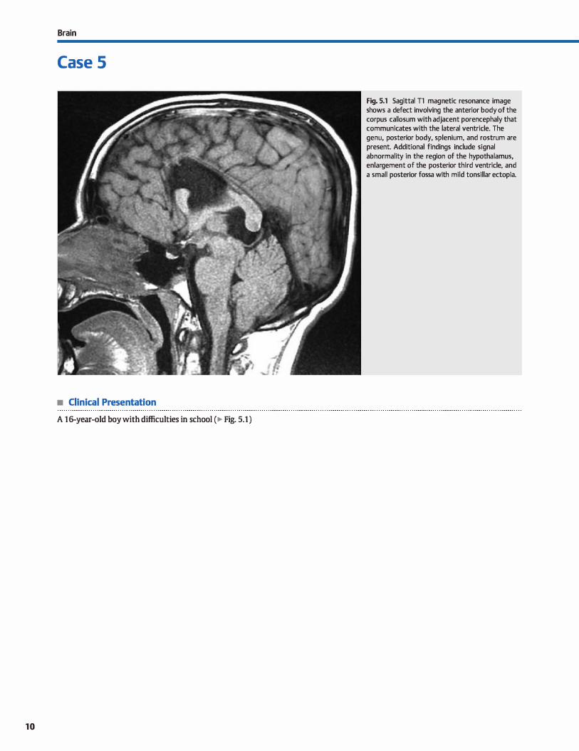
A 16-year-old boy with difficulties in school (� Fig. 5.1 )
10
Fig. 5.1 Sagittal Tl magnetic resonance image shows a defect involving the anterior body of the corpus callosum with adjacent porencephaly that communicates with the lateral ventride. The genu, posterior body, splenium, and rostrum are present. Additional findings indude signal abnormality in the region of the hypothalamus, enlargement of the posterior third ventride, and a small posterior fossa with mild tonsillar ectopia.
• Key lmaging Finding
Callosal abnormality
• Top 3 Differential Diagnoses
• Agenesis/hypogenesis of the corpus callosum (ACC). Normal
development of the corpus callosum occurs from anterior to posterior with formation of the genu first, followed by the body and splenium. The rostrurn is located along the inferior margin of the genu and is the last portion to form. lmaging findings with complete agenesis include absence of the corpus callosum and Jack of visualization of the cingulate gyrus
due to failure of rotation. As a result, the third ventride is ele
vated between the lateral ventrides, which are parallel in configuration on axial images. There is colpocephaly with dilatation of the atria and occipital homs of the lateral ventricles. The white matter tracts that would cross through the corpus callosum instead align along the media! margin of the lateral ventricles and run in an anterior-posterior direction. These
tracts are referred to as Probst bundles. On coronal sequences, the frontal horns of the lateral ventricles demonstrate a "long
horn" configuration secondary ta indentation medially by the Probst bundles and absence of the genu. The gyri of the
media) cerebral hemispheres extend to the margin of the third ventride with a radial configuration. ACC is nearly always associated with additional anomalies. With hypogene
sis of the corpus callosum, portions of the body, splenium,
and rostrum are absent. Absence of the rostrum is a key feature in distinguishing hypogenesis (rostrum absent) from an
enœphaloclastic process in which the rostrum is typically present Pericallosal lipomas are often seen in the setting of abnormal callosal development.
• Additional Differential Diagnoses
• Volume loss. The volume of the corpus callosum is related to
the volume of white matter within the supratentorial brain. Prior to myelination, the corpus callosum normally appears thin. As myelination progresses, it obtains its more typical
volume and appearanœ. With severe supratentorial parenchymal injury, al! or portions of the corpus callosum demon-
• Diagnosis
Callosal injury/encephaloclastic process (postsurgical)
� Pearls
• With ACC or hypogenesis, al! or a portion of the corpus callosum is absent, including the rostrum, which is last to form.
• Callosal injury is most often postsurgical, followed by trauma and hemorrhage.
Suggested Readings
Batul B, Kocaoglu M, Akgun V, Bulakbasi N, Tayfun C. Corpus callosum; normal
imaging appearanœ, variants and pathologie conditions. j Med lmaging Radiat
Oncol 2010; 54: 541-549
Cases
• Callosal injury/encephaloclastic process. The majority of encephalodastic injuries are from surgical interventions, usu
ally associated with resection of a mass within the third ventricle or suprasellar region. Trauma and hemorrhage are Jess common causes of callosal destruction. Imaging findings include absence of the callosum in the region of injury, whereas the remaining portions of the callosum are intact
Presenœ of the rostrum exdudes hypogenesis. • Holoprosenœphaly. Holoprosencephaly is a spectrum of
anomalies characterized by failure of the forebrain to separate into two distinct hemispheres. There are three variants: alobar, semilobar, and lobar, all of which have complete or partial
absence of the faix and septum pellucidum. In the alobar form (most severe), there is a large dorsal interhemispheric cyst (monoventride), and the remaining cerebral parenchyma is
fused and flattened anteriorly. Thalami are also fused. The corpus callosum, anterior faix, interhemispheric fissure, and Sylvian fissures are absent Associated craniofacial abnormalities include hypotelorism and cleft patate. In the semilobar variant, the posterior portions of the callosum are usually
present, whereas anterior portions, induding the rostrum, are absent. In the least severe lobar variant, the corpus callosum may appear normal or demonstrate partial absence of
the genu. Holoprosencephaly is the one congenital anomaly in which the genu may be absent whereas the body and splenium are present.
strate atrophy, because the callosal volume is dependent
upon the white matter fibers forming the tracts. Severe hydrocephalus may produce similar findings secondary to pressure-related changes or encephalomalacia of the corpus
callosum.
• Holoprosencephaly is the one congenital anomaly where the genu may be absent and the splenium present.
Sztriha I. Spectrum of corpus callosum agenesis. Pediatr Neurol 2005; 32; 94-101
1 1

Brain
Case 6
• Clinical Presentation
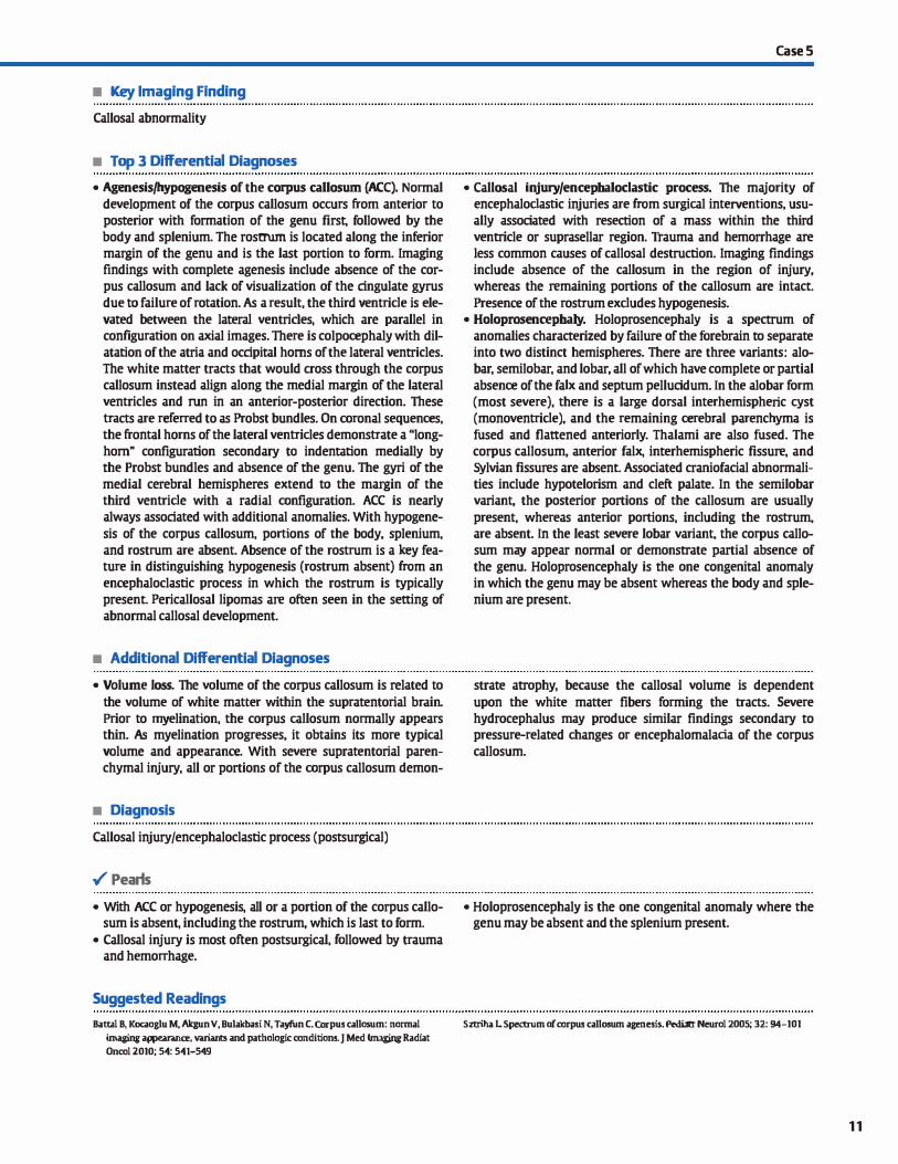
A 20-year-old man with chronic ataxia and progressive neurological decline (� Fig. 6.1)
12
Fig. 6.1 Sagittal T2 image demonstrates significantly decreased volume of the cerebellar vermis with prominence of the sulci. The brain stem appears normal in size and morphology.
• Key lmaging Finding
Cerebellar atrophy/volume Joss
• Top 3 Acquired Differential Diagnoses
• Alcohol abuse. Alcohol abuse results in progressive cerebellar degeneration. Alcohol is neurotoxic, causing cerebellar
and cortical {frontal lobe predominant) degeneration, as well as peripheral polyneuropathies. There is disproportionate
involvement of the superior vermis and cerebellum compared with the cerebral hemispheres. Associated findings may include Wemicke enœphalopathy, which presents as
abnormal T2 hyperintensity within the periaqueductal gray matter, mammillary bodies, medial thalamus, and hypothalamus: and Jess commonly Marchiafava-Bignami disease,
which results in abnormal signal intensity within the corpus callosum.
• Anticonvulsant therapy. Both seizures and long-term anti
convulsant therapy may produce irreversible cerebellar
• Top 3 Sporadic or lnherited Differential Diagnoses
• Sporadic olivopontocerebellar atrophy (sOPCA). sOPCA, also referred to as multisystem atrophy, is a neurodegenerative disorder of unknown etiology that typically presents in adulthood. Cross-sectional imaging demonstrates atrophy of the ventral pons and midbrain with
enlargement of the fourth ventride and widening of the superior and middle œrebellar peduncles. There is hemi
spheric greater than vermian cerebellar atrophy, as well as less pronounced cerebral atrophy, which most preferentially
involves the frontal and parietal lobes. Crudform-Iike T2 hyperintensity in the base of the pons gives the characteristic "hot cross bun" sign. Abnormal signal intensity is also seen in the middle cerebellar peduncles and dorsolateral
putamen. Patients present with parkinsonian features, ataxia, dysarthria, and autonomie dysfunction.
• Ataxia telangiectasia (AT). AT is an autosomal reœssive complex that results in spinoœrebellar degeneration. ocular and cutaneous telangiectases, radiation sensitivity, immunodefi-
• Diagnosis
Ataxia telangiectasia
� Pearts
• Alcohol, anticonvulsant therapy, and paraneoplastic syn
dromes are secondary causes of œrebellar atrophy. • sOPCA results in cerebellar and brain stem atrophy; pontine
hyperintensity is referred to as "hot cross bun" sign.
Suggested Readings
Fisdtbein NJ, Dillon WP, Barkovidl. AJ. Teaching Atlas of Brain Imaging. New York,
NY: Thieme. 1999
Case 6
degeneration with disproportionate œrebellar atrophy. Patients present with ataxia, nystagmus, and peripheral neuropathies. Phenytoin is the most common drug therapy, and its use may also result in diffuse calvarial thickening.
• Paraneoplastic syndrome. Cerebellar degeneration may occur as a result of a paraneoplastic syndrome. Breast and
Jung cancer are by far the most common primary neoplasms. Less common associated malignancies indude gastro
intestinal and genitourinary neoplasms, Hodgkin lymphoma, and neuroblastoma. The œrebellar degeneration is thought to result from autoantibodies to Purkinje fibers or a cytotoxic process associated with T cells. The paraneoplastic
cerebellar degeneration often precedes the diagnosis of a primary tumor.
ciendes, and increased risk of neoplasms. Patients often
present as toddlers with signs of ataxia. The neurological decline is progressive. Cross-sectional imaging demonstrates œrebellar atrophy with enlargement of the cerebellar suld and compensatory enlargement of the fourth ventricle. There
is also atrophy of the dentate nuclei. lntracranial telangiectases may result in scattered foci of gradient echo susceptibility secondary to microhemorrhages. Occasionally, associated supratentorial white matter demyelination or dysmyelination may be seen.
• Friedreich ataxia. Also known as spinocerebellar ataxia, Frie
dreich ataxia typically presents in the second decade of life and has bath autosomal dominant and recessive forms. Crosssectional imaging demonstrates mild atrophy of the vermis
and paravermian structures, a small medulla, and significant atrophy of the spinal cord. The dorsal cord has a flattened appearance. Oinically, patients often present with Iawer
extremity ataxia, upper extremity tremors, and kyphoscoliosis.
• AT presents with spinocerebellar degeneration, telangiectases, immunodeficiendes, and risk ofneoplasms.
Huang YP, Tuason MY, Wu T, Plaitaki5 A MRI and CT feature5 of cerebeUar degenera
tion j Formes Med Assoc 1993; 92: 494-508
13
Brain
Case 7
• Clinical Presentation
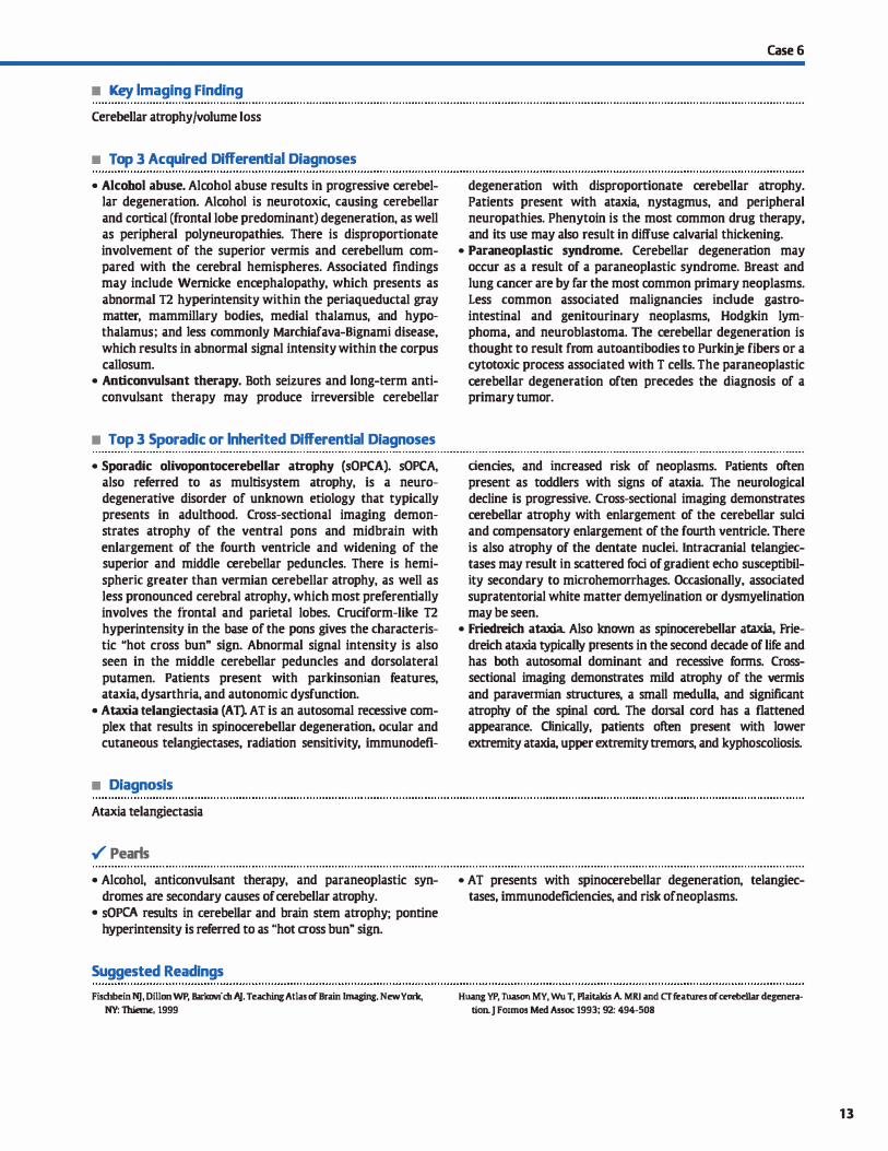
Adolescent boy with headaches (� Fig. 7.1 )
14
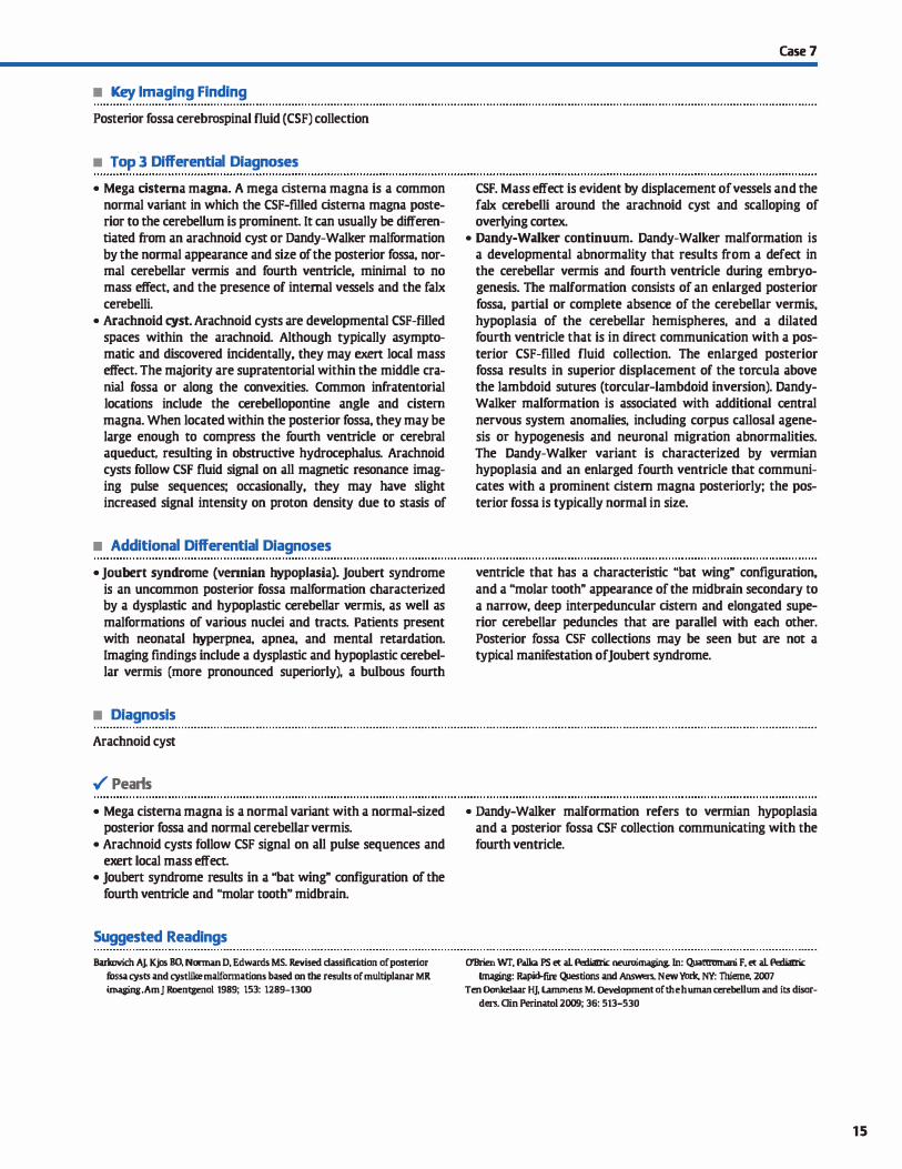
Fig. 7 .1 Sagittal T2 magnetic resonance image (a) demonstrates a large CSF signal intensity mass within the posterior fossa with anterior and superior displacement of the cerebellum. There is also compression of the fourth ventricle and associated enlargement of the third ventricle. Axial T2 (b), fluid-attenuated inversion recovery (c), and Tl (d) weighted images reveal that the mass follows CSF signal intensity on al l sequences. There is no direct communication with the fourth ventricle. Enlargement of the bilateral temporal horns of the lateral ventricles is also seen.
• Key lmaging Finding
Posterior fossa cerebrospinal fluid (CSF) collection
• Top 3 Differential Diagnoses
• Mega cistema magna. A mega cistema magna is a common
normal variant in which the CSF-filled cistema magna poste
rior to the cerebellum is prominent. It can usually be differen
tiated from an arachnoid cyst or Dandy-Walker malformation
by the normal appearance and size of the posterior fossa, nor
mal cerebellar vermis and fourth ventricle, minimal to no
mass effect, and the presence of internai vessels and the faix
cerebelli.
• Arachnoid cyst. Arachnoid cysts are developmental CSF-filled
spaces within the arachnoid. Although typically asympto
matic and discovered incidentally, they may exert local mass
effect. The majority are supratentorial within the middle cra
mai fossa or along the convexities. Common infratentorial
locations include the cerebellopontine angle and cistem
magna. When located within the posterior fossa, they may be
large enough to compress the fourth ventricle or cerebral
aqueduct, resulting in obstructive hydrocephalus. Arachnoid
cysts follow CSF fluid signal on all magnetic resonance imag
ing pulse sequences; occasionally, they may have slight
increased signal intensity on proton density due to stasis of
• Additional Differential Diagnoses
• Joubert syndrome (vennian hypoplasia). Joubert syndrome
is an uncommon posterior fossa malformation characterized by a dysplastic and hypoplastic cerebellar vermis, as well as
malformations of various nuclei and tracts. Patients present
with neonatal hyperpnea, apnea, and mental retardation.
Imaging findings include a dysplastic and hypoplastic œrebel
lar vermis (more pronounced superiorly), a bulbous fourth
• Diagnosis
Arachnoid cyst
� Pearls
• Mega cistema magna is a normal variant with a normal-sized
posterior fossa and normal cerebellar vermis.
• Arachnoid cysts follow CSF signal on ail pulse sequences and
exert local mass effect.
• Joubert syndrome results in a ubat wing" configuration of the
fourth ventricle and "molar tooth" midbrain.
Suggested Readings
Barlmvich AJ, Kjos BO, Norman D, Edwan:ls MS. Revised classification of postmor
fossa cysts and cystlike malfonnations based on the results of multiplanar MR
imaging.Amj Roentgenol 1989; 153; 1289-1300
Case 7
CSF. Mass effect is evident by displacement of vessels and the
faix œrebelli around the arachnoid cyst and scalloping of
overlying cortex.
• Dandy-Walker continuum. Dandy-Walker malformation is
a developmental abnormality that results from a defect in
the cerebellar vermis and fourth ventricle during embryo
genesis. The malformation consists of an enlarged posterior
fossa, partial or complete absence of the cerebellar vermis,
hypoplasia of the cerebellar hemispheres, and a dilated fourth ventricle that is in direct communication with a pos
terior CSF-filled fluid collection. The enlarged posterior
fossa results in superior displacement of the torcula above
the lambdoid sutures (torcular-lambdoid inversion). Dandy
Walker malformation is associated with additional central
nervous system anomalies, including corpus callosal agene
sis or hypogenesis and neuronal migration abnormalities.
The Dandy-Walker variant is characterized by vermian
hypoplasia and an enlarged fourth ventricle that communi
cates with a prominent cistern magna posteriorly; the pos
terior fessa is typically normal in size.
ventricle that has a characteristic "bat wing" configuration,
and a "molar tooth" appearance of the midbrain secondary to
a narrow, deep interpeduncular cistem and elongated supe
rior cerebellar pedundes that are parallel with each other.
Posterior fessa CSF collections may be seen but are not a
typical manifestation of Joubert syndrome.
• Dandy-Walker malformation refers to vermian hypoplasia
and a posterior fossa CSF collection communicating with the
fourth ventride.
O'Brien Wf, Palka PS et al Peliatric: neuro:ima8ing. ln: Quattromani F, et al Peliatric: Imaging: Rapid-fire QJestions and AnsWl!rs. New York, NY: Tiùeme, 2007
Ten Donlœlaar HJ, Lammens M. Development of the human cerebellum and its disor
ders. Clin Perinatol 2009; 36: 513-530
15
16
Brain
Case 8
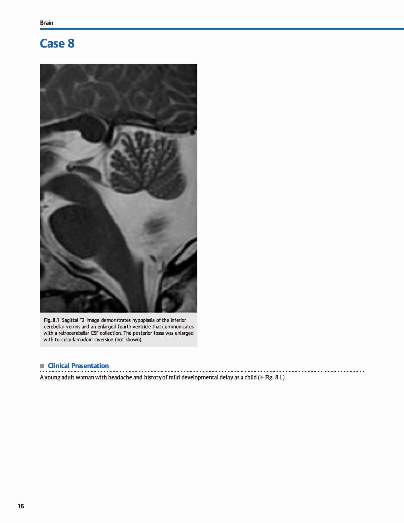
Fig. 8.1 Sagittal T2 image demonstrates hypoplasia of the inferior cerebellar vermis and an enlarged fourth ventricle that communicates with a retrocerebellar CSF collection. The posterior fossa was enlarged with torcular-lambdoid inversion (not shown).
• Clinical Presentation
A young adult woman with headache and history of mild developmental delay as a child ( � Fig. 8.1 )
• Key lmaging Finding
Vermian hypoplasia
• Top 3 Differential Diagnoses
• Dandy-Walker malformation (DWM) or variant (DWV). DWM is a developmental abnormality that results from a
defect in the cerebellar vermis and feurth ventride during
embryogenesis. lmaging findings indude an enlarged poste
rior fossa, partial or complete absence of the cerebellar ver
mis, hypoplasia of the cerebellar hemispheres, and a dilated
feurth ventricle that is in direct communication with a poste
rior cerebrospinal fluid {CSF)-filled fluid collection. The
enlarged posterior fossa results in superior displacement of the torcula above the lambdoid sutures (lambdoid-torcular
inversion). The DWV is a Jess severe anomaly characterized
by a relatively normal-sized posterior fessa with inferior vermian hypoplasia. The fourth ventricle is enlarged and communicates with the cistern magna posteriorly, which is promi
nent. In general, the DWM has more severe clinicat manifesta
tions because it is often associated with additional central
nervous system anomalies, induding corpus callosal agenesis
or hypogenesis and neuronal migration abnormalities. The
clinical manifestations of DWV are more variable and less severe, typically ranging from normal to relatively mild devel
opmental delay and neurological deficits. The presenœ of
additional abnormalities often determines the clinicat course.
• Joubert syndrome. Joubert syndrome is a rare form of con
genital vermian hypoplasia that presents early in life and is
characterized clinically by ataxia, apnea or hyperpnea, hypo
tonia, and developmental delay or mental retardation. The majority of cases occur sporadically, although autosomal pat
terns have also been observed. Cross-sectional imaging dem-
• Diagnosis
Dandy-Walker malformation
� Pearls
• DWM refers to vermian hypoplasia and a posterior fessa CSF collection communicating with the feurth ventride.
• Joubert syndrome is characterized by vennian hypoplasia
with a "molar tooth" configuration of the midbrain.
Suggested Readlngs
Case 8
onstrates a dysplastic and hypoplastic cerebellar vermis with
a midline deft (best seen on coronal sequences or reformats).
The smalt vermis results in an enlarged fourth ventricle in a
ubat wing" configuration. The superior cerebellar pedundes
are elongated, enlarged, and parallel with one another. This
configuration, combined with a hypoplastic midbrain, results in the characteristic umolar tooth" appearance on axial
images. Although once considered to be pathognomonic of Joubert syndrome, the molar tooth sign may be seen with
additional syndromes. Unlike DWM, the posterior fossa is
normal in size, and posterior fossa CSF collections are not a
typical manifestation of Joubert syndrome.
• Rhombencephalosynapsis. Rhombencephalosynapsis is an
uncommon developmental anomaly characterized by fusion
or failure of segmentation of the cerebellar hemispheres.
The cerebellar vermis is either absent or significantly hypo
plastic. The abnormal cerebellar configuration results in a
transverse orientation of the cerebellar folia and posterior
painting of the fourth ventride, which assumes a ukeyhole"
configuration. There is typically fusion of the superior cere
bellar peduncles and dentate nuclei as well. Associated
supratentorial anomalies are variable and include fused
thalami, fornices, and colliculi; absence of the septum pellu
cidum; aqueductal stenosis with hydrocephalus; callosal
and anterior commissure dysgenesis; and neuronal migra
tional abnormalities. Facial defects have also been reported.
Prognosis is related to the presence and severity of supra
tentorial abnormalities.
• Rhombencephalosynapsis is congenital fusion of the cerebellar hemispheres with vermian aplasia/hypoplasia.
Kendall B, IGngsley D, Lambert SR, Taylor D, Finn P. Joubert syndrome: a clinia:i-rad.i- Patel S. Barlwvich J\l. Analysis and dassification of œrebellar malformations. AmJ
ological study. Neuroradiology 1990; 31; 502-506 Neuroradiol 2002; 23; 1074-1087
17
18
Brain
Case 9
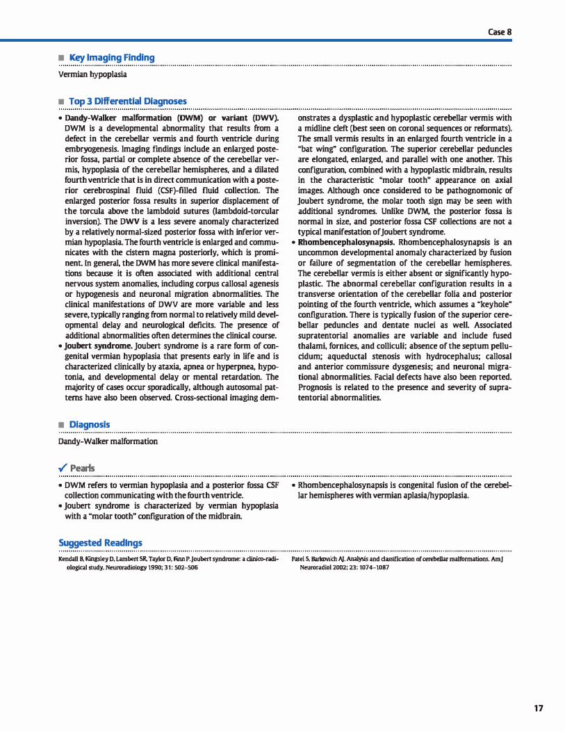
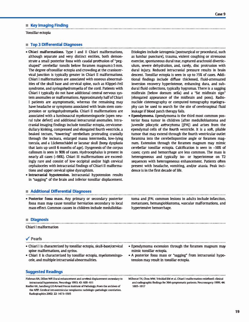
Fig. 9.1 Sagittal Tl (a) and T2 (b) images of the cervical spi ne demonstrate significant cerebellar tonsillar herniation below the fora men magnum with "peg-like" tonsils. The posterior fossa is small and there is mass effect upon the brain stem. No syrinx is identified.
• Clinical Presentation
A 34-year-old man with headaches and vertigo (� Fig. 9.1 )
• Key lmaging Finding
Tonsillar ectopia
• Top 3 Differential Diagnoses
• Chiari malformations. Type I and II Chiari malformations, although separate and very distinct entities, both demonstrate a small posterior fossa with caudal protrusion of "pegshaped" cerebellar tonsils below foramen magnum � 5 mm. The degree oftonsillar ectopia and crowding at the craniocervical junction is typically greater in Chiari II malformations. Chiari 1 malformations are associated with osseous abnormalities of the skull base and cervical spine, such as Klippel-Feil
syndrome, and syringohydromyelia of the cord. Patients with Chiari I typically do not have additional central nervous system anomalies or malformations. Approximately half of Chiari I patients are asymptomatic, whereas the remaining may have headache or symptoms associated with brain stem compression or syringohydromyelia. Chiari II malformations are
associated with a lumbosacral myelomeningocele (open neural tube defect) and additional intracranial anomalies. lntracranial imaging findings include tonsillar ectopia, cervicomedullary kinking, compressed and elongated fourth ventricle, a beaked tectum, "towering" œrebellum protruding cranially through the incisura, enlarged massa intermedia, low-lying torcula, and a Lückenschiidel or lacunar skull (bony dysplasia
that lasts up until 6 months of age). Dysgenesis of the corpus callosum is seen in 90% of cases. Hydrocephalus is present in nearly ail cases (-98%). Chiari III malformations are exceedingly rare and consist of low ocàpital and/or high cervical cephaloceles with intracranial findings of Chiari II malformations and upper cervical spine dysraphism.
• Intracranial hypotension. lntracranial hypotension results
in "sagging" of the brain and inferior tonsillar displacement.
• Additional Differential Diagnoses
• Posterior fossa mass. Any primary or secondary posterior fossa mass may cause tonsillar hemiation secondary to local mass etfect. Common causes in children indude medulloblas-
• Diagnosis
Chiari 1 malformation
� Pearts
• Chiari 1 is characterized by tonsillar ectopia, skull-base/œrvical spine malformations, and syrinx.
• Chiari II is characterized by tonsillar ectopia, myelomeningocele, and multiple intracranial abnormalities.
Suggested Readings
Fishman RA, Dillon WP. Durai enhancement and œrebral displaœment secondary ta
intracranial hypotension Neurology 1993; 43: 609-611 Koeller KK, Sandberg GD Armed Forces lnstituœ of Pathology. From the archives of
the AFIP. Cerdiral intraventricular neoplasms: radiologie-pathologie eorrelation.
Radiographies 2002; :z:z: 1473-1505
Case 9
Etiologies include iatrogenia {postsurgical or procedural, such as lumbar puncture), trauma, violent coughing or strenuous exercise, spontaneous durai tear, ruptured arachnoid diverticulum, severe dehydration, and, rarely, dise protrusion with durai injury. Reduœd intracranial pressure results in brain
descent. Tonsillar ectopia is seen in up to 75% of cases. Additional findings include diffuse thickened, fluid-attenuated inversion recovery hyperintense, enhancing dura, and subdural fluid collections, typically hygromas. There is a sagging midbrain {below dorsum sella) and a "fat midbrain sign" (elongated appearance of the midbrain and pans). Radionuclide cistemography or computed tomography myelography can be used to search for the site of cerebrospinal fluid leakage if blood patch therapy fails.
• Ependymoma. Ependymoma is the third most common posterior fessa tumor in children {after medulloblastoma and juvenile pilocytic astrocytoma [JPA]) and arises from the ependymal cells of the fourth ventride. It is a soft, pliable tumor that may extend through the fourth ventricular outlet foramina into the cerebellopontine angle or foramen magnum. Extension through the foramen magnum may mimic
cerebellar tonsillar ectopia. Calàfication is seen in -50% of cases; cysts and hemorrhage are Jess common. The mass is heterogeneous and typically iso- or hyperintense on T2 sequences with heterogeneous enhancement. Patients often present with headache, vomiting, and/or ataxia. Peak incidence is in the first decade of life.
toma and JPA; common lesions in adults include infarction, metastases, hemangioblastoma, vascular malformations, and hypertensive hemorrhage.
• Ependymoma extension through the foramen magnum may mimic tonsillar ectopia.
• A posterior fossa mass or "sagging" from intracranial hypo
tension may result in tonsillar ectopia.
Milhorat TH, Cltou MW, Trinidad EM et al. Chiari I malformation redefined: clinical
and radiographie findings for 364 symptomatic patients. Neurosurgery 1999; 44:
1005-1017
1 9
Brain
Case 1 0
• Clinical Presentation
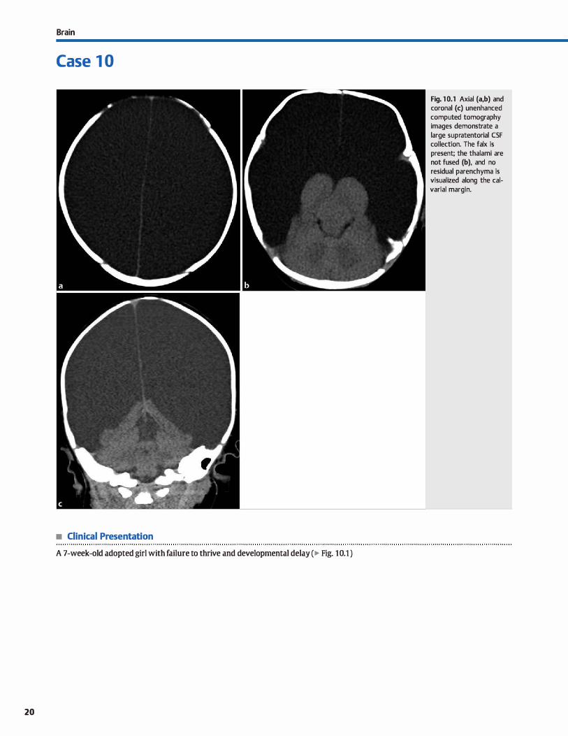
A 7-week-old adopted girl with failure to thrive and developmental delay (� Fig. 10.1 )
20
Fig. 1 0.1 Axial (a,b) and coronal (c) unenhanced computed tomography images demonstrate a large supratentorial CSF collection. The faix is present; the thalami are not fused (b), and no residual parenchyma is visualized along the calvariai margin.
• Key lmaging Finding
Supratentorial cerebrospinal fluid {CSF) collection
• Top 3 Differential Diagnoses
• Massive hydrocephalus. Hydroœphalus refers to ventriculo
megaly with increased volume of CSF due to obstruction, overproduction, or decreased resorption. In the newborn,
this results in macrocephaly because the sutures are open. Massive hydroœphalus displaces and compresses the brain parenchyma along the peripheral calvarial margin, mimicking hydranenœphaly or holoprosencephaly. Key distinguishing
features indude a thin mantle of cortex along the inner
calvarial margin and the presence of the faix, respectively. Aqueductal stenosis is a common cause of massive hydro
cephalus. Additional causes include obstructing masses, such as posterior fossa, pineal gland, tectal plate, and intraventricular neoplasms. Transependymal flow of CSF is seen in cases of acute uncompensated hydrocephalus. causes of nonob
structive communicating hydrocephalus indude a history of
prior meningitis or ventriculitis, as well as prior subarachnoid
hemorrhage. • Hydranencephaly. Hydranencephaly is characterized by Iiqu
efactive necrosis of the supratentorial brain parenchyma in the anterior {internai carotid artery) circulation secondary to some form of in utero insult. There is sparing of the
parenchyma in the posterior (posterior cerebral artery and cerebellar branch vessels) circulation. Most cases are thought
• Additional Differential Diagnoses
• Agenesis of the corpus callosum {ACC) with midline inter
hemispheric cyst. ACC may be associated with midline interhemispheric cysts in addition to elevation of the third ventricle. The cysts may represent a diverticulum of the lat
eral ventricle (type I) or multiple interhemispheric cysts {type
II). Ventriculomegaly is commonly seen. The interhemispheric
cysts result in lateral displaœment of the brain parenchyma. One-half to three-fourths of cases of ACC have additional
central nervous system malformations.
• Diagnosis
Hydranenœphaly
� Pearts
• Massive hydroœphalus in a neonate is commonly due to
obstruction; a thin peripheral cortical mantle is seen. • Hydranencephaly is Iiquefactive necrosis in the anterior circu
lation; faix is present with no cortical mantle.
Suggested Readings
Dublin AB, French BN, Diagnostic image evaluation ofhydranencephaly and pictori
ally similar entities, with emphasis on romputed tmnography. Radiology 1980;
137:81-91
Case 10
to result from an ischemic, traumatic, or taxie insult between
-20 and 27 weeks gestation. Key distinguishing features include the presenœ of the faix œrebri; intact thalami, brain stem, cerebellum, and typically portions of the posterior
occipital and parietal lobes; and absence of a cortical mantle
around a large supratentorial CSF-filled cavity. Neonates commonly present with macrocrania and neurological function
limited to the brain stem; death typically occurs in infancy or early childhood.
• Alobar holoprosencephaly. Holoprosencephaly is a spectrum
of congenital forebrain malformations characterized as alobar, semilobar, and lobar variants. The alobar form is most severe
and is characterized by a large, dorsal interhemispheric cyst
and fusion of the thalami and remaining brain parenchyma,
which is flattened anteriorly. The corpus callosum, anterior faix, interhemispheric fissure, and Sylvian fissures are absent Associated craniofadal abnonnalities include hypotelorism, fused metopic suture, and cleft patate. Semilobar and lobar variants are Jess severe forms with varying degrees of defective separation of the anterior and central brain structures, as
well as complete or partial absence of the faix. An azygous
anterior œrebral artery is commonly seen.
• Dilaterai open-lip schizencephaly. 'JYpe II (open-lip) schizen
cephaly consists of a large CSF-filled cleft that is lined by poly
microgyric gray matter. The abnormality may be bilateral in up to half of cases and may be assodated with septo-optic
dysplasia. Differentiating features indude gray matter-lined
clefts and an intact faix. Heterotopia or cortical dysplasia may be associated findings. Patients often present with seizures and varying degrees of developmental delay and/or motor
deficits.
• Alobar holoprosencephaly results in a large dorsal monoventricle with fused parenchyma anteriorly.
Oh KY, Kennedy AM, Frias AE,jr, Byme JL Fetal schizencephaly; pre-and postnatal
imaging with a review of the clinical manifestations. Radiographia 2005; 25:
647-657
21
22
Brain
Case 1 1
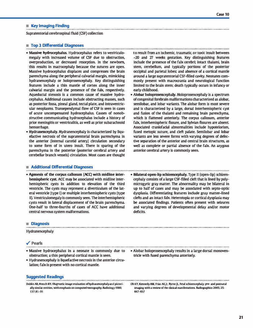
Fig. 1 1 .1 Contrast-enhanced axial computed tomography image through the lateral ventricles demonstrates a gray matter-lined CSF cleft that communicates with the frontal horn of the right lateral ventricle. There is also absence of the septum pellucidum.
• Clinical Presentation
An adolescent with seizures (� Fig. 1 1.1)
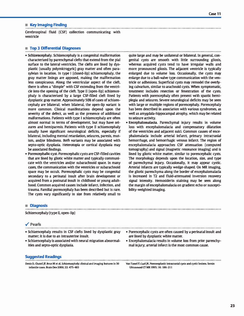
• Key lmaging Finding
Cerebrospinal fluid {CSF) collection communicating with ventride
• Top 3 Differential Diagnoses
• Schizencephaly. Schizencephaly is a congenital malformation characterized by parenchymal defts that extend from the pial surface ta the lateral ventrides. The defts are lined by dysplastic (usually polymicrogyric) gray matter and often para
sylvian in location. In type I {dosed-lip) schizencephaly, the gray matter linings are apposed, making the malformation Jess conspicuous. Along the ventricular aspect of the cleft, there is often a udimple" with CSF extending from the ventride into the opening of the cleft. "IYPe Il (open-lip) schizenœphaly is characterized by a large CSF-filled deft lined by dysplastic gray matter. Approximately 50% of cases of schizen
cephaly are bilateral; when bilateral, the open-lip variant is more common. Clinical manifestations depend upon the severity of the defect, as well as the presenœ of additional malformations. Patients with type I schizencephaly are often almost normal in terms of development, but may have seizures and hemiparesis. Patients with type Il schizenœphaly usually have significant neurological deficits, especially if bilateral, induding mental retardation, seizures, paresis, mut
ism, and/or blindness. Both variants may be associated with septo-optic dysplasia. Heterotopia or cortical dysplasia may be associated findings.
• Porencephalic cyst. Porencephalic cysts are CSF-filled cavities that are lined by gliotic white matter and typically communicate with the ventricles and/or subarachnoid space. In many cases, the communication with the ventricles or subarachnoid spaœ may be occult Porencephalic cysts may be congenital secondary to a perinatal insult after brain development or acquired from a postnatal insult in childhood or young adulthood. Common acquired causes include infarct. infection, and trauma. Familial porenœphaly has been described but is rare. The cysts vary significantly in size from relatively small ta
• Diagnosis
Schizencephaly (type II, open-lip)
� Pearls
• Schizenœphaly results in CSF clefts lined by dysplastic gray matter; it is due ta an intrauterine insult.
• Schizenœphaly is associated with neural migration abnormalities and septo-optic dysplasia.
Suggested Readings
Denis D, Otaœil JF, Brun M et al. Schizenœphaly; dinical and imaging features in 30
infantile cases. Brain Dev 2000; 22: 475--483
Case 1 1
quite large and may be unilateral or bilateral. In general, congenital cysts are smooth with little surrounding gliosis, whereas acquired cysts tend ta have irregular walls and more pronounced gliosis. The adjacent ventride is typically enlarged due to volume Joss. Occasionally, the cysts may enlarge due to a ball-valve type communication with the ventride or adhesions. Superficial cysts may remodel the overlying calvarium, similar to arachnoid cysts. When symptomatic, treatment indudes resection or fenestration of the cysts. Patients with porencephaly often present with spastic hemiplegia and seizures. Severe neurological deficits may be seen with large or multiple regions of porencephaly. Porencephaly has been described in association with various syndromes, as well as amygdala-hippocampal atrophy, which may be related to seizure activity.
• Encephalomalacia. Parenchymal injury results in volume Joss with encephalomalacia and compensatory dilatation of the ventrides and adjacent sulci. Common causes of encephalomalacia indude arterial infarct, primary intracranial hemorrhage, and hemorrhagic venous infarct. The region of
encephalomalacia approaches CSF attenuation { computed tomography) and signal (magnetic resonanœ imaging) and is lined by gliotic white matter, similar to porencephalic cysts. The morphology depends upon the location, size, and type of parenchymal injury. Occasionally, it may appear cystic. Arterial infarcts are typically wedge-shaped. On MR imaging, the gliotic parenchyma along the border of encephalomalacia is increased in T2 and fluid-attenuated inversion recovery
signal intensity. Hemosiderin staining may be seen along the margin of encephalomalacia on gradient echo or susceptibility-weighted imaging.
• Porencephalic cysts are often caused by a perinatal insult and are lined by dysplastic white matter.
• Encephalomalacia results in volume loss from prier parenchymal injury; arterial infarct is the most common cause.
Van Tassel P, CUré jK. Nonneoplastic intracranial cysts and cystic lesions. Sernin
Ultrasound cr MR 1995; 16: 186-211
23
24
Brain
Case 1 2
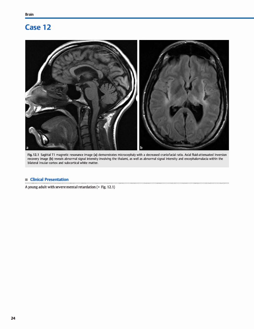
Fig. 1 2.1 Sagittal Tl magnetic resonance image (a) demonstrates microcephaly with a decreased craniofacial ratio. Axial fluid-attenuated inversion recovery image (b) reveals abnormal signal intensity involving the thalami, as well as abnormal signal intensity and encephalomalacia within the bilateral insular cortex and subcortical white matter.
• Clinical Presentation
A young adult with severe mental retardation (� Fig. 12.1)
• Key lmaging Finding
Microcephaly
• Top 3 Differential Diagnoses
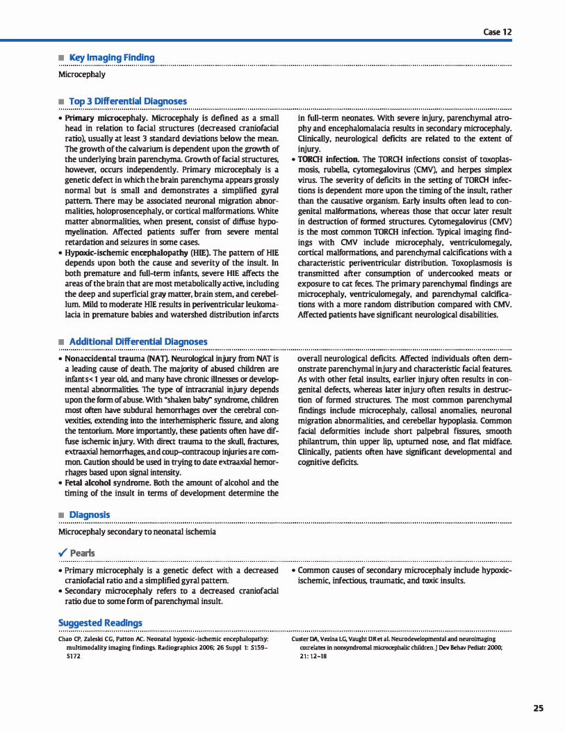
• Primary microcephaly. Microcephaly is defined as a small head in relation to facial structures ( decreased craniofacial ratio), usually at least 3 standard deviations below the mean.
The growth of the calvariurn is dependent upon the growth of the underlying brain parenchyma. Growth of facial structures, however, occurs independently. Primary microcephaly is a genetic defect in which the brain parenchyma appears grossly
normal but is small and demonstrates a simplified gyral
pattern. There may be associated neuronal migration abnormalities, holoprosenœphaly, or cortical malformations. White
matter abnormalities, when present, consist of diffuse hypomyelination. Affected patients suffer from severe mental
retardation and seizures in some cases.
• Hypoxic-ischemic encephalopathy (HIE). The pattern of HIE depends upon both the cause and severity of the insult. In
both premature and full-term infants, severe HIE affects the areas of the brain that are most metabolically active, induding
the deep and superficial gray matter, brain stem, and cerebellum. Mild to moderate HIE results in periventricular leukomalacia in premature babies and watershed distribution infarcts
• Additional Differential Diagnoses
• Nonaccidental trauma (NAT). Neurological injury from NAT is a Ieading cause of death. The majority of abused children are infants< 1 year old. and many have chronic illnesses or developmental abnormalities. The type of intracranial injury depends upon the form of abuse. With ushaken baby" syndrome, children most often have subdural hemorrhages over the œrebral con
vexities, extending into the interhemispheric fissure, and along the tentorium. More importantly, these patients often have dif
fuse ischemic injury. With direct trauma to the skull, fractures, extraaxial hemorrhages, and coup-contracoup injuries are common. Caution should be used in trying to date extraaxial hemorrhages based upon signal intensity.
• Fetal alcohol syndrome. Both the amount of alcohol and the timing of the insult in terms of development determine the
• Diagnosis
Microcephaly secondary to neonatal ischemia
� Pearls
• Primary microcephaly is a genetic defect with a decreased craniofadal ratio and a simplified gyral pattern.
• Secondary microcephaly refers to a decreased craniofadal
ratio due to some form of parenchymal insult.
Suggested Readings
Chao CP, Zaleski CG, Patton AC.. Neonatal hypoxic-ischemic encephalopathy;
multimodality imaging findings. Radiographies 2006; 26 Suppl 1: S159-
St 72
Case 1 2
in full-term neonates. With severe injury, parenchymal atrophy and encephalomalacia results in secondary microcephaly. Oinically, neurological defidts are related ta the extent of in jury.
• TORŒI infection. The TORœ infections consist of toxoplasmosis, rubella, cytomegalovirus (CMV), and herpes simplex
virus. The severity of deficits in the setting of TORΠinfections is dependent more upon the timing of the insult, rather
than the causative organism. Early insults often lead to congenital malformations, whereas those that occur later result in destruction of formed structures. Cytomegalovirus (CMV) is the most common TORCH infection. Typical imaging find
ings with CMV indude microcephaly, ventriculomegaly, cortical malformations, and parenchymal calcifications with a
characteristic periventricular distribution. Toxoplasmosis is
transmitted after consurnption of undercooked meats or exposure to cat feces. The primary parenchymal findings are microcephaly, ventriculomegaly, and parenchymal calcifications with a more random distribution compared with CMV. Affected patients have significant neurological disabilities.
overall neurological deficits. Affected individuals often demonstrate parenchymal injury and characteristic facial features.
As with other fetal insults, earlier injury often results in congenital defects, whereas later injury often results in destruction of formed structures. The most common parenchymal
findings include microcephaly, callosal anomalies, neuronal migration abnormalities, and cerebellar hypoplasia. Common
facial deformities include short palpebral fissures, smooth philantrum, thin upper lip, upturned nase, and fiat midface. Oinically, patients often have significant developmental and cognitive defidts.
• Common causes of secondary microœphaly include hypoxicischemic, infectious, traurnatic, and taxie insults.
CUster DA, Ve:zina LG, Vaught DR et al, Neurodevelopmental and neuroimaging
correlaœs in nonsyndromal microœphalic children.j Dev Behav Pediatr 2000;
21: 12-18
25
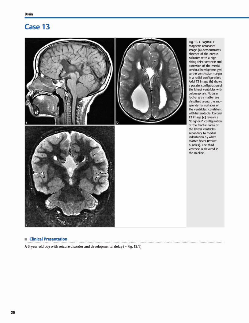
Brain
Case 1 3
• Clinical Presentation
A 6-year-old boy with seizure disorder and developmental delay (� Fig. 13.1 )
26
Fig. 13.1 Sagittal Tl magnetic resonance image (a) demonstrates absence of the corpus callosum with a highriding third ventricle and extension of the medial cerebral hemisphere gyri to the ventricular margin in a radial configuration. Axial T2 image (b) shows a parallel configuration of the lateral ventricles with colpocephaly. Nodular foci of gray matter are visualized along the subependymal surfaces of the ventricles, consistent with heterotopia. Coronal T2 image (c) reveals a "longhorn" configuration of the frontal horns of the lateral ventricles secondary to medial indentation by white matter fibers (Probst bundles). The third ventricle is elevated in the midline.
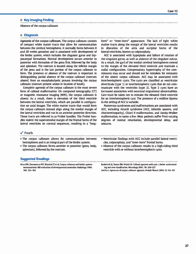
• Key lmaging Finding
Absence of the corpus callosum
• Diagnosis
Agenesis of the corpus callosum.. The mrpus callosum consists of mmpact white matter tracts that allow for mmmunication between the cerebral hemispheres. It normally forms between 8 and 20 weeks gestation and is associated with development of the limbic system, which indudes the dngulate gyrus and hippocampal formations. Normal development occurs anterior to posterior with formation of the genu first, followed by the body and splenium. The rostrum is located along the inferior margin of the genu and is the last portion of the corpus callosum to form. The presence or absence of the rostrum is important in distinguishing partial absence of the corpus callosum (rostrum absent) from an enœphalodastic proœss involving the corpus callosum (rostrum present unless in location ofinsult).
Complete agenesis of the corpus callosum is the most severe form of callosal malformation. On computed tomography (CT) or magnetic resonanœ imaging (MRI), the corpus callosum is absent As a result, there is elevation of the third ventride between the lateral ventrides, which are parallel in configuration on axial images. The white matter tracts that would form the corpus callosum instead align along the medial margin of the lateral ventrides and run in an anterior-posterior direction. These tracts are referred to as Probst bundles. The Probst bundles indent the superomedial margin of the frontal homs of the lateral ventrides on coronal sequences, resulting in a "long-
� Pearts
• The corpus callosum allows for communication between hemispheres and is an integral part of the limbic system.
• The corpus callosum forms anterior ta posterior (genu, body, splenium), followed by the rostrum.
Suggested Readings
Atlas SW, Zimmennan RA, Bilaniuk LT et al. Corpm callosum and limbic system:
neuroanatomic MR evaluation of developmental anomalies. R.ôldiology 1986; 160; 355-362
Case 13
hom" or "steer-horn" appearance. The lad< of tight white matter tracts along the margin of the lateral ventrides results in dilatation of the atria and occipital homs of the lateral ventrides, known as colpocephaly.
ACC is associated with hypoplasia and failure of rotation of the cingulate gyrus, as well as absence of the cingulate sulcus. As a result, the gyri of the medial cerebral hemispheres extend to the margin of the elevated third ventricle and maintain a radial configuration. Compensatory hypertrophy of the commissures may occur and should not be mistaken for remnants of the absent corpus callosum. ACC may be associated with interhemispheric cysts. The cysts are dassified as ventricular
diverticula (type 1) or interhemispheric cysts that do not communicate with the ventrides (type 2). JYpe 2 cysts have an increased association with neuronal migrational abnormalities. Care must be taken not ta mistake the elevated third ventride for an interhemispheric cyst The presence of a midline lipoma in the setting of ACC is variable.
Numerous syndromes and malformations are associated with ACC, induding Aicardi syndrome (ACC, infantile spasms, and chorioretinopathy), Chiari Il malformation, and Dandy-Walker malformation, to name a few. Most patients suffer from varying degrees of mental retardation, developmental delay, and seizures.
• Ventricular ftndings with ACC include parallel lateral ventrides, colpocephaly, and "steer-hom" frontal homs.
• Absence of the corpus callosum results in a high-riding third ventride with or without interhemispheric cysts.
BarkovichAJ, Simon EM, Walsh CA. Callosal agenesis with cyst: a better understand
ing and new classification. Neurology 2001: 56: 220-227
Sztriha I. Spectrurn of corpus callosurn agenesis. Pediatr Neurol 2005; 32: 94-101
27
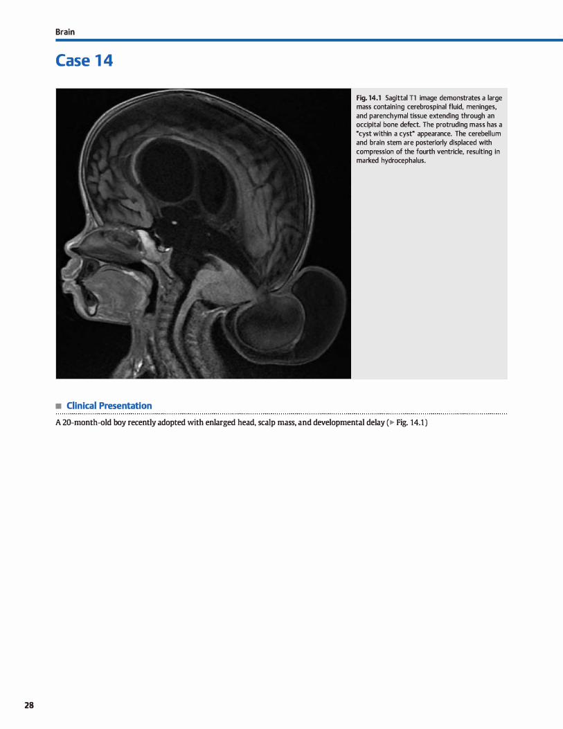
28
Brain
Case 1 4
• Clinical Presentation
Fig. 14.1 Sagittal Tl image demonstrates a large mass containing cerebrospinal fluid, meninges, and parenchymal tissue extending through an occipital bone defect. The protruding mass has a "cyst within a cyst" appearance. The cerebellum and brain stem are posteriorly displaced with compression of the fourth ventride, resulting in marked hydrocephalus.
A 20-month-old boy recently adopted with enlarged head, scalp mass, and developmental delay (� Fig. 14.1 )
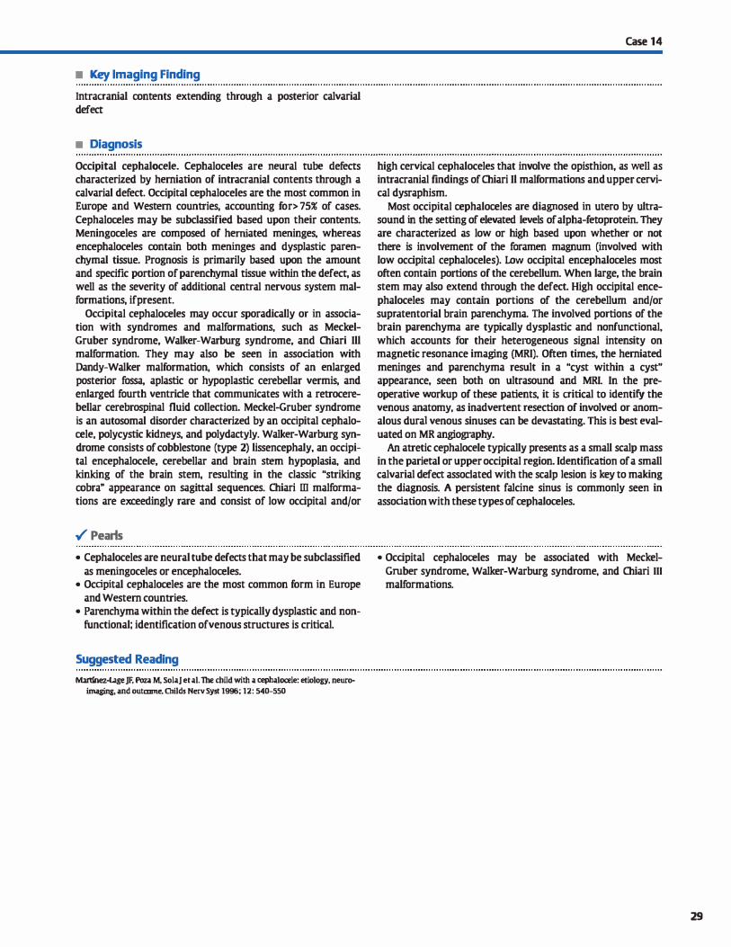
• Key lmaging Finding
lntracranial contents extending through a posterior calvarial defect
• Diagnosis
Occipital cephalocele. Cephaloceles are neural tube defects characterized by herniation of intracranial contents through a calvarial defect. Occipital cephaloceles are the most common in Europe and Western countries, accounting for> 75% of cases.
Cephaloceles may be subclassified based upon their contents. Meningoceles are composed of herniated meninges, whereas encephaloceles contain both meninges and dysplastic parenchymal tissue. Prognosis is primarily based upon the amount and specific portion of parenchymal tissue within the defect, as well as the severity of additional central nervous system malformations, ifpresent.
Occipital cephaloœles may occur sporadically or in association with syndromes and malformations, such as MeckelGruber syndrome, Walker-Warburg syndrome, and Chiari Ill malformation. They may also be seen in association with Dandy-Walker malformation, which consists of an enlarged posterior fossa, aplastic or hypoplastic cerebellar vermis, and enlarged fourth ventride that communicates with a retrocerebellar cerebrospinal fluid collection. Meckel-Gruber syndrome
is an autosomal disorder characterized by an occipital cephalocele, polycystic kidneys, and polydactyly. Walker-Warburg syndrome consists of cobblestone (type 2) lissencephaly, an occipital encephalocele, cerebellar and brain stem hypoplasia, and kinking of the brain stem, resulting in the classic "striking cobra" appearance on sagittal sequences. Chiari III malformations are exceedingly rare and consist of low occipital and/ or
� Pearls
• Cephaloceles are neural tube defects that may be subclassified
as meningoceles or enœphaloœles. • Occipital cephaloceles are the most common form in Europe
and Western countries. • Parenchyma within the defect is typically dysplastic and non
functional; identification ofvenous structures is critical.
Suggested Reading
Martfnez-1.age JF, Poza M, SolaJ et al. The child with a œphaloœle: etiology, neuro
imaging, and outmme. Childs Nerv Syst 1996; 12: 540-550
Case 14
high cervical cephaloceles that involve the opisthion, as well as intracranial findings of Chiari Il malformations and upper cervical dysraphism.
Most occipital cephaloceles are diagnosed in utero by ultrasound in the setting of elevated levels of alpha-fetoprotein. They are characterized as low or high based upon whether or not there is involvement of the foramen magnum (involved with low occipital cephaloceles). I.ow occipital encephaloceles most often contain portions of the cerebellum. When large, the brain stem may also extend through the defect. High occipital ence
phaloceles may con tain portions of the cerebellum and/ or supratentorial brain parenchyma. The involved portions of the brain parenchyma are typically dysplastic and nonfunctional,
which accounts for their heterogeneous signal intensity on magnetic resonance imaging (MRI). Often times, the herniated meninges and parenchyma result in a "cyst within a cyst" appearance, seen both on ultrasound and MRI. In the pre
operative workup of these patients, it is critical to identify the venous anatomy, as inadvertent resection of involved or anomalous durai venous sinuses can be devastating. This is best evaluated on MR angiography.
An atretic cephalocele typically presents as a small scalp mass in the parietal or upper occipital region. Identification of a small calvarial defect assodated with the scalp lesion is key to making the diagnosis. A persistent falcine sinus is commonly seen in
association with these types of œphaloœles.
• Occipital cephaloœles may be associated with Meckel
Gruber syndrome, Walker-Warburg syndrome, and Chiari III malformations.
29
Brain
Case 1 5
Fig. 1 5.1 (a,b) Axial T2 images demonstrate absence of the cerebellar vermis with fusion or failure of segmentation of the cerebellar hemispheres.
• Clinical Presentation
A 1 5-year-old girl with long-standing gait instability and cognitive deficits (� Fig. 1 5.1 )
30
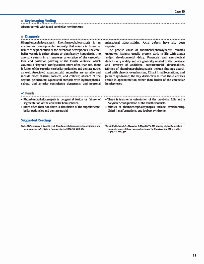
• Key lmaging Finding
Absent vermis with fused œrebellar hemispheres
• Diagnosis
Rhombencephalosynapsis. Rhombencephalosynapsis is an uncommon developmental anomaly that results in fusion or
failure of segmentation of the œrebellar hemispheres. The cerebellar vermis is either absent or significantly hypoplastic. The anomaly results in a transverse orientation of the cerebellar folia and posterior pointing of the fourth ventride, which assumes a "keyhole" configuration. More often than not, there
is fusion of the superior cerebellar peduncles and dentate nuclei
as well. Associated supratentorial anomalies are variable and indude fused thalami, fornices, and colliculi; absence of the
septUm pellucidum; aqueductal stenosis with hydrocephalus; callosal and anterior commissure dysgenesis; and neuronal
� Pearts
• Rhombencephalosynapsis is congenital fusion or failure of segmentation of the cerebellar hemispheres.
• More often than not, there is also fusion of the superior œrebellar pedundes and dentate nuclei.
Suggested Readings
Case 15
migrational abnormalities. Facial defects have also been reported.
The precise cause of rhombenœphalosynapsis remains unknown. Patients usually present early in life with ataxia and/or developmental delay. Prognosis and neurological
deficits vary widely and are generally related to the presence and severity of additional supratentorial abnormalities.
Mimics of rhombencephalosynapsis indude findings associated with chronic overshunting, Chiari Il malformations, and
Joubert syndrome; the key distinction is that these entities result in approximation rather than fusion of the cerebellar hemispheres.
• There is transverse orientation of the œrebellar folia and a "keyhole" configuration of the fourth ventride.
• Mimics of rhombenœphalosynapsis include overshunting, Chiari Il malformations, and Joubert syndrome.
lllelle SP, Yalcinlcaya C, KoœrN et al, Rhombenœphalosynapsis; dinical findings and lhlwit a., Barlccvich AJ, Shanahan R, MaroldolV. MR imaging ofrhmnbenœphalo-
neuroimaging in 9 children. Neuropediabics 2002; 33: 209-214 synapsis: report of three cases and reviewof the literature. Am j Neuroradiol
1991; 12; 957-965
31
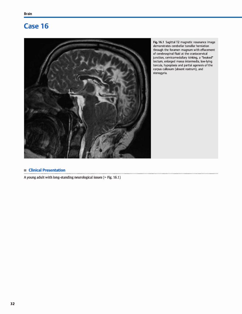
Brain
Case 1 6
• Clinical Presentation
A young adult with long-standing neurological issues ( � Fig. 16.1 )
32
Fig. 16.1 Sagittal T2 magnetic resonance image demonstrates cerebellar tonsillar herniation through the foramen magnum with effacement of cerebrospinal fluid at the craniocervical junction, cervicomedullary kinking, a "beaked" tectum, enlarged massa intermedia, low-lying torcula, hypoplasia and partial agenesis of the corpus callosum (absent rostrum), and stenogyria.
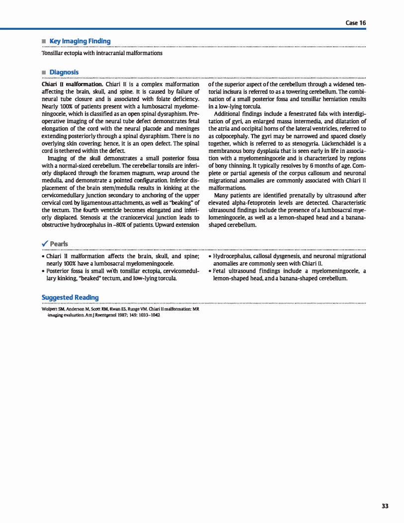
• Key lmaging Finding
Tonsillar ectopia with intracranial malformations
• Diagnosis
Chiari n malformation. Chiari II is a complex malformation affecting the brain, skull, and spine. lt is caused by failure of neural tube closure and is associated with folate deficiency.
Nearly 100% of patients present with a lurnbosacral myelomeningocele, which is dassified as an open spinal dysraphism. Preoperative imaging of the neural tube defect demonstrates fetal elongation of the cord with the neural placode and meninges
extending posteriorly through a spinal dysraphism. There is no overlying skin covering; hence, it is an open defect. The spinal cord is tethered within the defect.
Imaging of the skull demonstrates a small posterior fossa with a normal-sized cerebellum. The cerebellar tonsils are inferiorly displaced through the foramen magnum, wrap around the medulla, and demonstrate a pointed configuration. Inferior dis
plaœment of the brain stem/medulla results in kinking at the œrvicomedullary junction secondary to anchoring of the upper
cervical cord by ligamentous attachments, as well as Mbeaking" of the tectum. The fourth ventride becomes elongated and inferiorly displaced. Stenosis at the cranioœrvical junction leads to obstructive hydroœphalus in -80% of patients. Upward extension
� Pearls
• Chiari Il malformation affects the brain, skull, and spine; nearly 100% have a lumbosacral myelomeningocele.
• Posterior fossa is small with tonsillar ectopia, œrvicomedullary kinking, Mbeaked" tectum, and Iow-lying torcula.
Suggested Reading
Wolpert SM, Anderson M, Scott RM, Kw.m ES, Runge VM. Clliari li malformation: MR
imilgingevaluation.AmJRœntgenol 1987; 149: 1033-1042
Case 16
of the superior aspect of the œrebellum through a widened tentorial incisura is referred to as a towering œrebellum. The combination of a small posterior fossa and tonsillar hemiation results in a low-lying torcula.
Additional findings indude a fenestrated faix with interdigi
tation of gyri, an enlarged massa intermedia, and dilatation of the atria and occipital homs of the lateral ventricles, referred to
as colpocephaly. The gyri may be narrowed and spaced dosely together, which is referred to as stenogyria. Lückenchadel is a membranous bony dysplasia that is seen early in Iife in association with a myelomeningocele and is characterized by regions
of bony thinning. lt typically resolves by 6 months of age. Com
plete or partial agenesis of the corpus callosum and neuronal migrational anomalies are commonly associated with Chiari Il malformations.
Many patients are identified prenatally by ultrasound after elevated alpha-fetoprotein levels are detected. Characteristic ultrasound findings include the presence of a lurnbosacral myelomeningocele, as well as a lemon-shaped head and a bananashaped œrebellum.
• Hydroœphalus, callosal dysgenesis, and neuronal migrational
anomalies are commonly seen with Chiari II. • Fetal ultrasound findings indude a myelomeningocele, a
Iemon-shaped head, and a banana-shaped cerebellum.
33
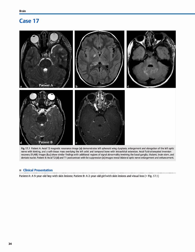
34
Brain
Case 1 7
Fig. 1 7.1 Patient A: Axial T2 magnetic resonance image (a) demonstrates left sphenoid wing dysplasia, enlargement and elongation of the left optic nerve with kinking, and a soft-tissue mass overlying the left orbit and temporal bone with intraorbital extension. Axial fluid-attenuated inversion recovery (FLAIR) images (b,c) show similar findings with additional regions of signal abnormality involving the basal ganglia, thalami, brain stem, and dentate nuclei. Patient B: Axial T2 (d) and Tl postcontrast with fat suppression (e) images reveal bilateral optic nerve enlargement and enhancement.
• Clinical Presentation
Patient A: A 9-year-old boy with skin lesions; Patient B: A 2-year-old girl with skin lesions and visual Joss (� Fig. 17.1 )
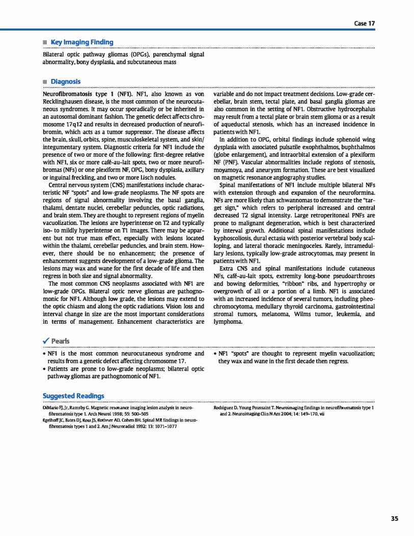
• Key lmaging Finding
Bilateral optic pathway gliomas (OPGs), parenchymal signal abnormality, bony dysplasia, and subcutaneous mass
• Diagnosis
Neurofibromatosis type t (NFt). NFt, also known as von Recklinghausen disease, is the most common of the neurocutaneous syndromes. It may occur sporadically or be inherited in an autosomal dominant fashion. The genetic defect affects chromosome 17q12 and results in decreased production of neurofibromin, which acts as a tumor suppressor. The disease affects the brain, skull, orbits, spine, musculoskeletal system, and skin/ integumentary system. Diagnostic criteria for NFt indude the presence of two or more of the following: first-degree relative with NF1, six or more café-au-lait spots, two or more neurofibromas (NFs) or one plexiform NF, OPG, bony dysplasia, axillary
or inguinal freckling, and two or more lisch nodules. Central nervous system (CNS) manifestations include charac
teristic NF Mspots" and low-grade neoplasms. The NF spots are regions of signal abnormality involving the basal ganglia, thalami, dentate nuclei, cerebellar peduncles, optic radiations, and brain stem. They are thought to represent regions of myelin vacuolization. The lesions are hyperintense on T2 and typically iso- to mildly hyperintense on Tl images. There may be appar
ent but not true mass effect, especially with lesions located within the thalami, cerebellar peduncles, and brain stem. However, there should be no enhancement; the presence of enhancement suggests development of a low-grade glioma. The lesions may wax and wane for the first decade of life and then regress in both size and signal abnormality.
The most common CNS neoplasms associated with NF1 are low-grade OPGs. Dilaterai optic nerve gliomas are pathognomonic for NF1. Although low grade, the lesions may extend to the optic chiasm and along the optic radiations. Vision loss and interval change in size are the most important considerations in terms of management. Enhancement characteristics are
� Pearls
• NF1 is the most common neurocutaneous syndrome and results from a genetic defect affecting chromosome 17.
• Patients are prone to low-grade neoplasms; bilateral optic pathway gliomas are pathognomonic of NF1.
Suggested Readings
DiMario FJ,jr, Rarnsby G. Magnetic resonanœ irnaging lesion analysis in neuro
fibromatosis type 1. Ardt New-ol 1998; 55: 500-505 Egelhoff JC, Ba tes DJ, Ross JS, Rothner AD, Cohen BH. Spinal MR findings in neuro
fibromatosis types 1 and 2. Am] Neuroradiol 1992; 13: 1071-1077
Case 17
variable and do not impact treatment decisions. Low-grade cerebellar, brain stem, tectal plate, and basal ganglia gliomas are also common in the setting of NF1. Obstructive hydrocephalus may result from a tectal plate or brain stem glioma or as a result of aqueductal stenosis, which has an increased incidence in patients with NFl.
In addition to OPG, orbital findings indude sphenoid wing dysplasia with associated pulsatile exophthalmos, buphthalmos (globe enlargement), and intraorbital extension of a plexiform NF (PNF). Vascular abnormalities include regions of stenosis,
moyamoya, and aneurysm formation. These are best visualized on magnetic resonance angiography studies.
Spinal manifestations of NF1 include multiple bilateral NFs with extension through and expansion of the neuroformina. NFs are more likely than schwannomas to demonstrate the Mtarget sign," which refers to peripheral increased and central decreased T2 signal intensity. Large retroperitoneal PNFs are
prone to malignant degeneration, which is best characterized by interval growth. Additional spinal manifestations indude kyphoscoliosis, durai ectasia with posterior vertebral body scalloping, and lateral thoracic meningoceles. Rarely, intramedullary lesions, typically low-grade astrocytomas, may present in patients with NFl.
Extra CNS and spinal manifestations indude cutaneous NFs, café-au-lait spots, extremity long-bone pseudoarthroses and bowing deformities, "ribbon" ribs, and hypertrophy or overgrowth of all or a portion of a limb. NFt is associated with an increased incidence of several tumors, induding pheochromocytoma, medullary thyroid carcinoma, gastrointestinal stromal tumors, melanoma, Wilms tumor, leukemia, and lymphoma.
• NF1 Mspots" are thought to represent myelin vacuolization; they wax and wane in the first decade then regress.
Rodriguez Il, Young Pous saint T. Neuroimaging findings in neurofibrornatosis type 1
and 2. Neuroirnaging Clin N Am 2004; 14: 149-170, vil
35
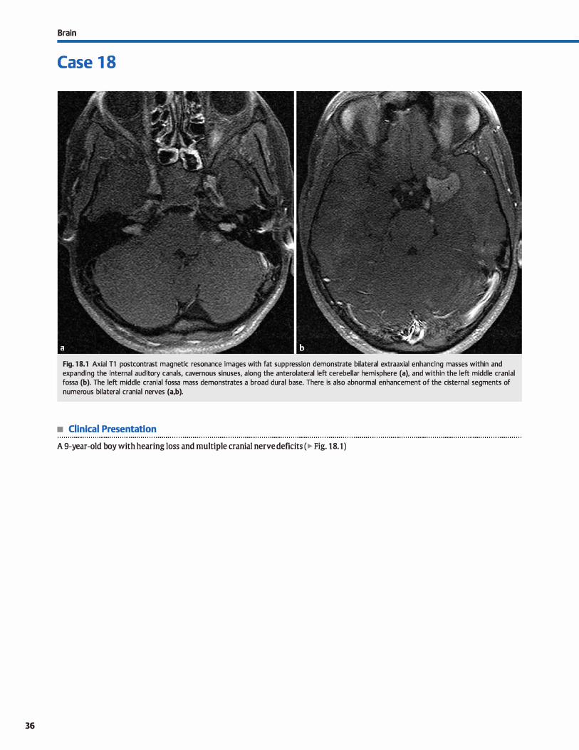
36
Brain
Case 1 8
Fig. 1 8.1 Axial Tl postcontrast magnetic resonance images with fat suppression demonstrate bilateral extraaxial enhancing masses within and expanding the internai auditory canais, cavernous sinuses, along the anterolateral left cerebellar hemisphere (a), and within the left middle cranial fossa (b). The left middle cranial fossa mass demonstrates a broad durai base. There is also abnormal enhancement of the cisternal segments of numerous bilateral cranial nerves (a,b).
• Clinical Presentation
A 9-year-old boy with hearing Joss and multiple cranial nerve deficits ( � Fig. 18.1)
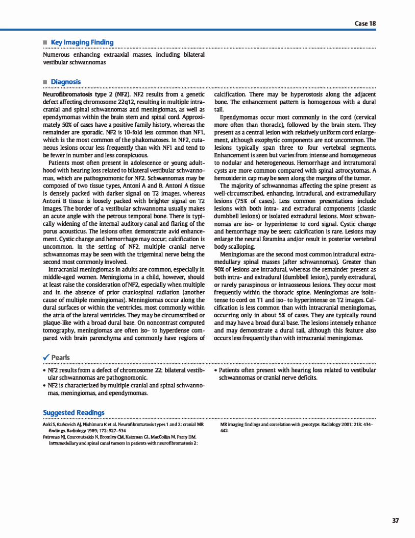
• Key lmaging Finding
Numerous enhancing extraaxial masses, induding bilateral vestibular schwannomas
• Diagnosis
Neurofibromatosis type 2 {NF2). NF2 results from a genetic defect affecting chromosome 22qt2, resulting in multiple intracranial and spinal schwannomas and meningiomas, as well as ependymomas within the brain stem and spinal cord. Approximately 50% of cases have a positive family history, whereas the remainder are sporadic. NF2 is 10-fold Jess common than NFl, which is the most common of the phakomatoses. In NF2, cutaneous lesions occur Jess frequently than with NFl and tend to be fewer in number and Jess conspicuous.
Patients most often present in adolescence or young adulthood with hearing Joss related to bilateral vestibular schwanno
mas, which are pathognomonic for NF2. Schwannomas may be composed of two tissue types, Antoni A and B. Antoni A tissue is densely packed with darker signal on T2 images, whereas Antoni B tissue is loosely packed with brighter signal on T2 images. The border of a vestibular schwannoma usually makes an acute angle with the petrous temporal bone. There is typically widening of the internai auditory canal and flaring of the parus acousticus. The lesions often demonstrate avid enhancement. Cystic change and hemorrhage may occur; calcification is uncommon. In the setting of NF2, multiple cranial nerve schwannomas may be seen with the trigeminal nerve being the second most commonly involved.
lntracranial meningiomas in adults are common, especially in middle-aged women. Meningioma in a child, however, should at least raise the consideration ofNF2, especially when multiple and in the absence of prior craniospinal radiation (another cause of multiple meningiomas). Meningiomas occur along the durai surfaces or within the ventricles, most commonly within the atria of the lateral ventrides. They may be circumscribed or plaque-like with a broad durai base. On noncontrast computed tomography, meningiomas are often iso- ta hyperdense compared with brain parenchyma and commonly have regions of
� Pearls
• NF2 results from a defect of chromosome 22; bilateral vestibular schwannomas are pathognomonic.
• NF2 is characterized by multiple cranial and spinal schwannomas, meningiomas, and ependymomas.
Suggested Readings
Aoki S, Bariccvich Aj, Nishimura K et al. Neurofibromatmis types 1 and 2: cranial MR findings. Radiology 1989; 172: 527-534
Patrollas JIU, Courœutsakis N, Bromley CM, Katzman GL, MacCollin M, Pany DM.
Intr.unedullary and spinal canal twnors in patients with neurofibromatosis 2:
Case 18
calcification. There may be hyperostosis along the adjacent bone. The enhanœment pattern is homogenous with a durai tail.
Ependymomas occur most commonly in the cord (cervical more often than thoradc), followed by the brain stem. They present as a central lesion with relatively uniform cord enlargement, although exophytic components are not uncommon. The lesions typically span three to four vertebral segments. Enhancement is seen but varies from intense and homogeneous to nodular and heterogeneous. Hemorrhage and intratumoral cysts are more common compared with spinal astrocytomas. A hemosiderin cap may be seen along the margins of the tumor.
The majority of schwannomas affecting the spine present as well-circumscribed, enhancing, intradural, and extramedullary lesions (75% of cases). Less common presentations indude lesions with both intra- and extradural components ( dassic dumbbell lesions) or isolated extradural lesions. Most schwan
nomas are iso- or hyperintense ta cord signal. Cystic change and hemorrhage may be seen; calcification is rare. Lesions may enlarge the neural foramina and/or result in posterior vertebral body scalloping.
Meningiomas are the second most common intradural extramedullary spinal masses {after schwannomas). Greater than 90% of lesions are intradural, whereas the remainder present as bath intra- and extradural (dumbbell lesion), purely extradural,
or rarely paraspinous or intraosseous lesions. They occur most frequently within the thoracic spine. Meningiomas are isointense to cord on Tl and iso- to hyperintense on T2 images. calcification is less common than with intracranial meningiomas, occurring only in about 5% of cases. They are typically round and may have a broad durai base. The lesions intensely enhanœ and may demonstrate a durai tail, although this feature also occurs Jess frequently than with intracranial meningiomas.
• Patients often present with hearing Joss related to vestibular schwannomas or cranial nerve defidts.
MR imaging findings and correlation with genotype. Radiology 2001; 218: 434-
442
37
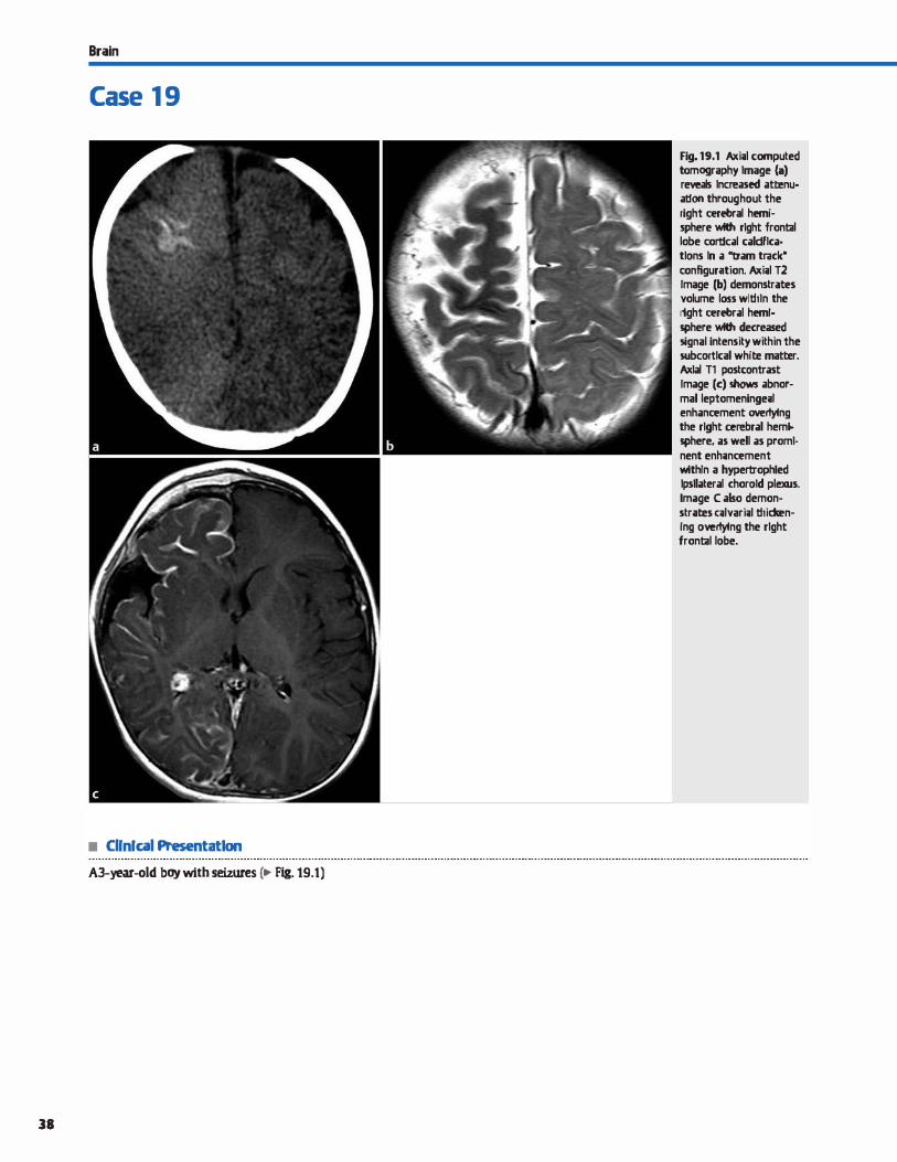
Brain
Case 1 9
• Cllnfcal Presentat1on
A3-year-old boy with seizures c.- Fig. 19.t)
38
Fig. 19.1 Axi;tl computed tomography Image (a) reveals lncreased atœnu· alfon throughout the right œrebral hemi· sphere wlth rlght frontal lobe cortical caldflca· tlons ln a "tram track• configur<rtion. Axi;il T2 Image (b) demonstr;rtes volume loss wlthln the rtght œrebral hem!· sphere wlth decreased 5ignal intensity within the subcortlcal white matter. Axial T1 postcontrast Image (c) shows abnormal leptomeninge;il enhanœment overlying the rlght cerebral hem._ sphere, as well as proml· nent enh;inœment wlthln a hypertrophled lpsllateral chorold plexus. Image C also demonstrates ulvari;il thidœn· lng overlying the rlght frontal lobe.
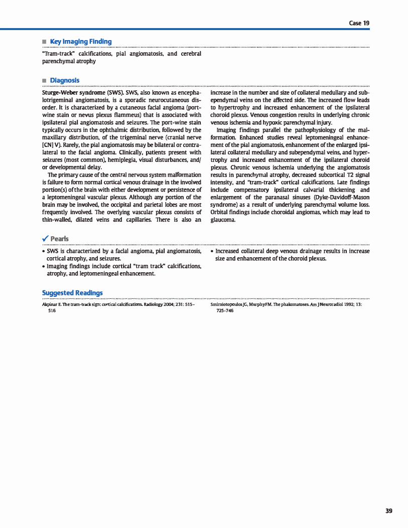
• Key lmaging Finding
"Tram-trad<" calcifications, pial angiomatosis, and cerebral parenchymal atrophy
• Diagnosis
Sturge-Weber syndrome (SWS). SWS, also known as encephalotrigeminal angiomatosis, is a sporadic neurocutaneous disorder. lt is characterized by a cutaneous facial angioma (portwine stain or news plexus flammeus) that is associated with ipsilateral pial angiomatosis and seizures. The port-wine stain typically occurs in the ophthalmic distribution, followed by the maxillary distribution, of the trigeminal nerve { cranial nerve [ CN] V). Rarely, the pial angiomatosis may be bilateral or contralateral to the facial angioma. Oinically, patients present with seizures (most common), hemiplegia, visual disturbances, and/ or developmental delay.
The primary cause of the central nervous system malformation is failure to form normal cortical venous drainage in the involved portion(s) ofthe brain with either development or persistence of a leptomeningeal vascular plexus. Although any portion of the brain may be involved, the occipital and parietal lobes are most frequently involved. The overlying vascular plexus consists of thin-walled, dilated veins and capillaries. There is also an
� Pearts
• SWS is characterized by a fadai angioma, pial angiomatosis, cortical atrophy, and seizures.
• lmaging findings include cortical "tram trad<" calcifications, atrophy, and leptomeningeal enhancement.
Suggested Readings
Akpimr E. 1be tram-track sign: cortical calcifications. R.miolagy 2004; 231 : 515-
516
Case 19
increase in the number and size of collateral medullary and subependymal veins on the affected side. The increased flow leads to hypertrophy and increased enhancement of the ipsilateral choroid plexus. Venous congestion results in underlying chronic venous ischemia and hypoxie parenchymal injury.
lmaging findings parallel the pathophysiology of the malformation Enhanced studies reveal leptomeningeal enhanœment of the pial angiomatosis, enhancement of the enlarged ipsilateral collateral medullary and subependymal veins, and hypertrophy and increased enhanœment of the ipsilateral choroid plexus. Chronic venous ischemia underlying the angiomatosis results in parenchymal atrophy, decreased subcortical 12 signal intensity, and "tram-trad<" cortical calcifications. Late findings indude compensatory ipsilateral calvarial thickening and enlargement of the paranasal sinuses (Dyke-Davidoff-Mason syndrome) as a result of underlying parenchymal volume Joss. Orbital findings indude choroidal angiomas, which may lead to glaucoma.
• Increased collateral deep venous drainage results in increase size and enhanœment of the choroid plexus.
SrnimiotopoulasjG, MurphyFM, Jbe phalrornatDses, Arnj Neuroradial 1992; 13;
725-746
39
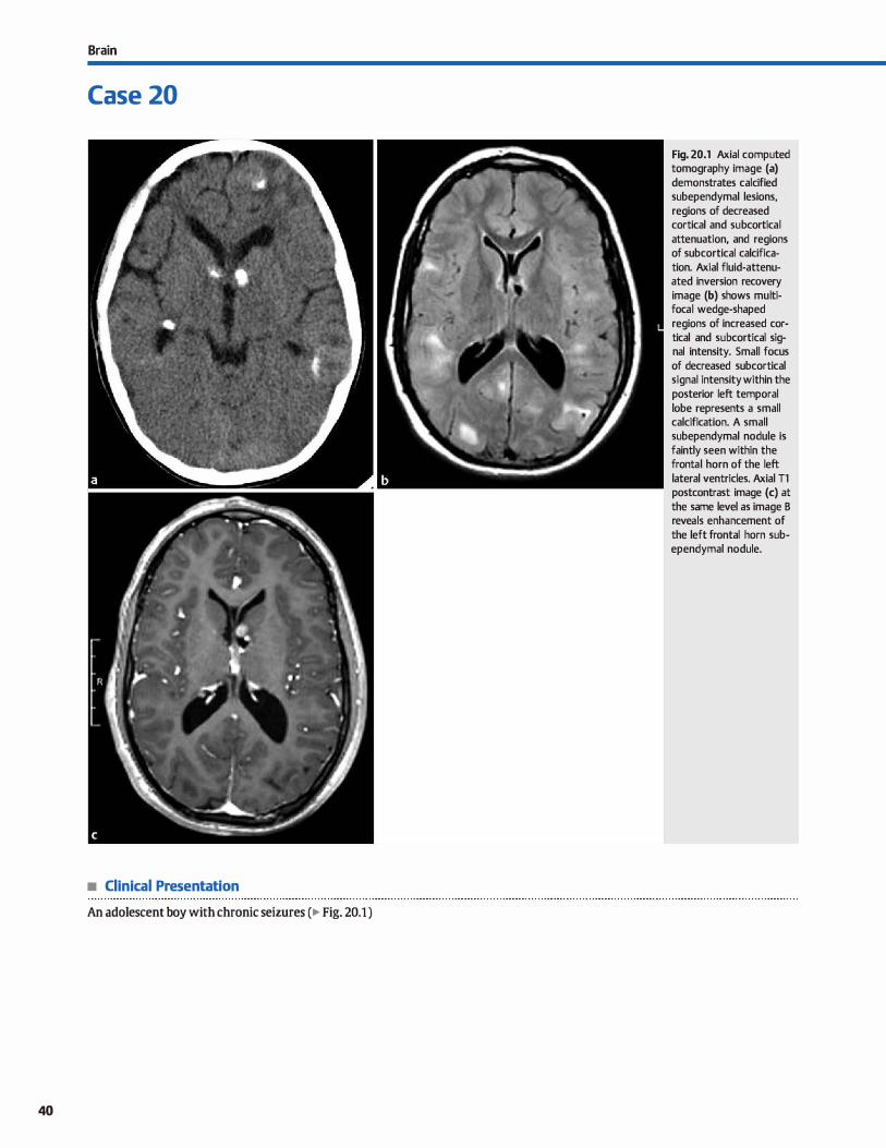
Brain
Case 20
• Clinical Presentation
An adolescent boy with chronic seizures (� Fig. 20.1 )
40
Fig. 20.1 Axial computed tomography image (a) demonstrates calcified subependymal lesions, regions of decreased cortical and subcortical attenuation, and regions of subcortical calcification. Axial fluid-attenuated inversion recovery image (b) shows multifocal wedge-shaped regions of increased cortical and subcortical signal intensity. Small focus of decreased subcortical signal intensity within the posterior left temporal lobe represents a small calcification. A small subependymal nodule is faintly seen within the frontal horn of the left lateral ventricles. Axial Tl postcontrast image (c) at the same level as image B reveals enhancement of the left frontal horn subependymal nodule.
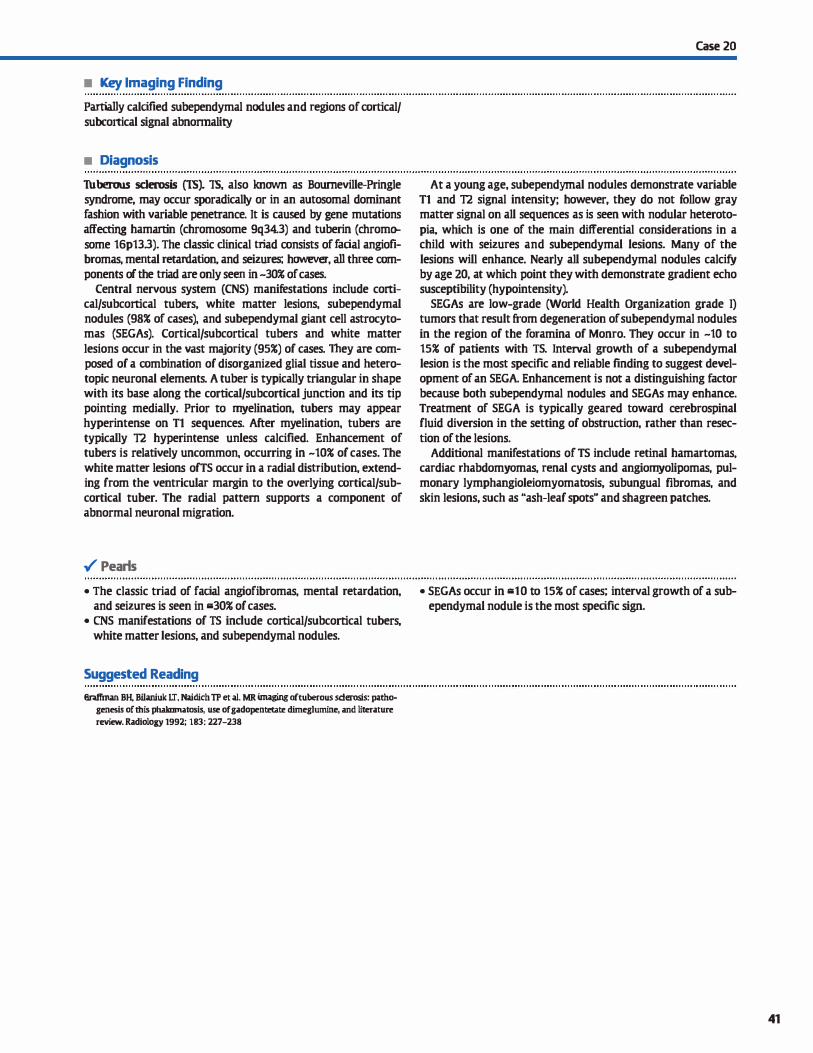
• Key lmaging Finding
Partially calcified subependymal nodules and regions of cortical/ subcortical signal abnonnality
• Diagnosis
Tuberous sclerosis (TS). TS, also knawn as Bourneville-Pringle syndrome, may occur sporadically or in an autosomal dominant fashion with variable penetranœ. It is caused by gene mutations affecting hamartin (chromosome 9q343) and tuberin (chromo
some 16p133). The dassic dinical triad consists of facial angiofibromas, mental retardation, and seizures; however, ail three components of the triad are only seen in -30% of cases.
Central nervous system (CNS) manifestations include cortical/subcortical tubers, white matter lesions, subependymal nodules (98% of cases), and subependymal giant cell astrocytomas (SEGAs). Cortical/subcortical tubers and white matter
lesions occur in the vast majority {95%) of cases. They are composed of a combination of disorganized glial tissue and heterotopic neuronal elements. A tuber is typically triangular in shape with its base along the cortical/subcortical junction and its tip painting medially. Prior to myelination, tubers may appear hyperintense on Tt sequences. After myelination, tubers are typically T2 hyperintense unless calcified. Enhancement of tubers is relatively uncommon, occurring in -10% of cases. The
white matter lesions ofTS occur in a radial distribution, extending from the ventricular margin to the overlying cortical/subcortical tuber. The radial pattern supports a component of abnormal neuronal migration.
� Pearts
• The classic triad of fadai angiofibromas, mental retardation, and seizures is seen in 11130% of cases.
• CNS manifestations of TS indude cortical/subcortical tubers, white matter lesions, and subependymal nodules.
Suggested Reading
Braffman BH, Bilaniuk LT, Naidkh TP et al. MR imaging oftuberous sderosis: patho
genesis of this phakmnatosis, use of gadopentetate dimeglurnine, and literature
review. Radiology 1992; 183: 227-238
Case 20
At a young age, subependymal nodules demonstrate variable Tl and T2 signal intensity; however, they do not follow gray matter signal on ail sequences as is seen with nodular heteroto
pia, which is one of the main differential considerations in a child with seizures and subependymal lesions. Many of the lesions will enhance. Nearly ail subependymal nodules calcify by age 20, at which point they with demonstrate gradient echo susceptibility (hypointensity).
SEGAs are low-grade {World Health Organization grade I) tumors that result from degeneration of subependymal nodules
in the region of the foramina of Monro. They occur in -10 to 15% of patients with TS. Interval growth of a subependymal lesion is the most specific and reliable finding to suggest development of an SEGA. Enhancement is not a distinguishing factor because both subependymal nodules and SEGAs may enhanœ. Treatment of SEGA is typically geared toward œrebrospinal fluid diversion in the setting of obstruction, rather than resec
tion of the lesions. Additional manifestations of TS indude retinal hamartomas,
cardiac rhabdomyomas, renal cysts and angiomyolipomas, pulmonary lymphangioleiomyomatosis, subungual fibromas, and skin lesions, such as "ash-leaf spots" and shagreen patches.
• SEGAs occur in 11110 to 15% of cases; interval growth of a subependymal nodule is the most specific sign.
41
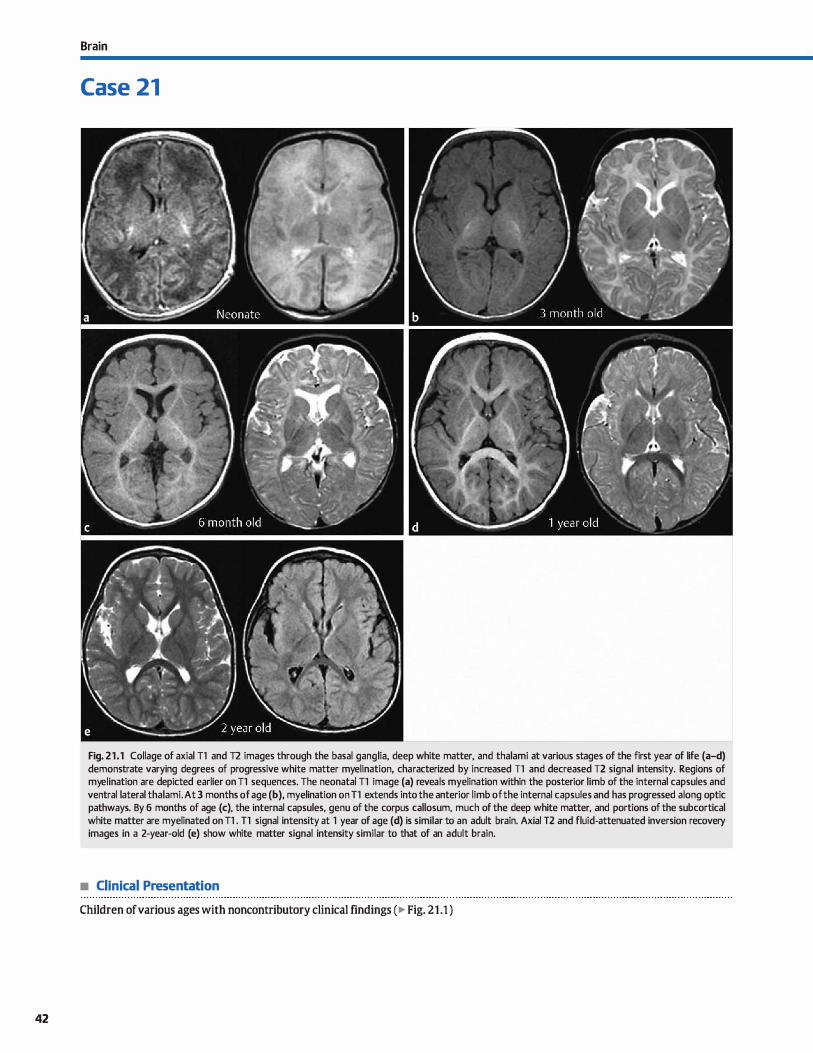
42
Brain
Case 21
Fig. 21.1 Collage of axial Tl and T2 images through the basal gang lia, deep white matter, and thalami at various stages of the first year of life (a-d) demonstrate varying degrees of progressive white matter myelination, characterized by increased Tl and decreased T2 signal intensity. Regions of myelination are depicted earlier on Tl sequences. The neonatal Tl image (a) reveals myelination within the posterior limb of the internai capsules and ventral lateral thalami. At 3 months of age (b), myelination on T1 extends into the anterior limb of the internai capsules and has progressed a long optic pathways. By 6 months of age (c), the internai capsules, genu of the corpus callosum, much of the deep white matter, and portions of the subcortical white matter are myelinated on Tl . T1 signal intensity at 1 year of age (d) is similar to an adult brain. Axial T2 and fluid-attenuated inversion recovery images in a 2-year-old (e) show white matter signal intensity similar to that of an adult brain.
• Clinical Presentation
Children of various ages with noncontributory clinical findings ( � Fig. 21.1 )
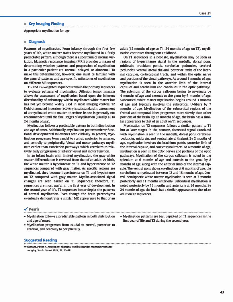
• Key lmaging Finding
Appropriate myelination for age
• Diagnosis
Patterns of myelination. From infancy through the first few years of life, white matter tracts become myelinated in a fairly predictable pattern, although there is a spectrum of normal variation. Magnetic resonance imaging (MRI) provides a means of determining whether patterns and progression of myelination in a particular patient are normal, delayed, or abnormal. To make this detennination, however, one must be familiar with the general patterns and age-specific milestones of myelination
on ditferent MR sequenœs. 11- and 12-weighted sequenœs remain the primary sequenœs
to evaluate patterns of myelination. Diffusion tensor imaging allows for assessment of myelination based upon the inherent directionality of anisotropy within myelinated white matter but bas not yet become widely used in most imaging œnters. 12
fluid-attenuated inversion recovery is substandard in assessment of unmyelinated white matter; therefore, its use is generally not recommended until the final stages ofmyelination (usually 18 to 24 months ofagei
Myelination follows a predictable pattern in bath distribution and age of onset. Additionally, myelination patterns mirror functional developmental milestones seen dinically. In general, myelination progresses from caudal to rostral, posterior to anterior, and œntrally to peripherally. Visual and motor pathways myelinate earlier than association pathways, which correlates to relatively early progression of infants' visual and motor function.
ln an infant brain with limited myelination, the gray-white matter differentiation is reversed from that of an adult At birth, the white matter is hypointense on Tl and hyperintense on 12
sequences compared with gray matter. As specific regions are myelinated, they become hyperintense on Tl and hypointense on 12 compared with gray matter. Myelin-associated signal changes are seen earlier on Tl sequences; therefore, Tl sequenœs are most useful in the first year of development ln the second year of life, 12 sequenœs better depict the patterns of normal myelination. Even though the brain parenchyma
eventually demonstrates a similar MR appearance to that of an
� Pearls
• Myelination follows a predictable pattern in bath distribution and age of onset.
• Myelination progresses from caudal to rostral, posterior to anterior, and centrally to peripherally.
Suggested Reading
Welker KM, Patton A. Assessment of normal m;yelination with magnetic resonance
imaging. Sernin Neurol 2012; 32; 15-28
Case 21
adult (12 months of age on Tl; 24 months of age on 12), myelination continues throughout childhood.
On Tl sequences in a neonate, myelination may be seen as regions of hyperintense signal in the medulla, dorsal pans, midbrain, brachium pontis, cerebellar pedundes, cerebral
pedundes, ventral lateral thalami, posterior limbs of the internai capsules, corticospinal tracts, and within the optic nerve and portions of the visual pathways. At around 3 months of age, myelination is seen in the anterior limb of the internai capsules and cerebellum and continues in the optic pathways. The splenium of the corpus callosum begins to myelinate by 4 months of age and extends ta the genu by 6 months of age. Subcortical white matter myelination begins around 3 months of age and typically involves the subcortical U-fibers by 7 months of age. Myelination of the subcortical regions of the frontal and temporal lobes progresses more slowly than other portions of the brain. By 12 months of age, the brain has a similar appearance to that of an adult on Tl sequences.
Myelination on 12 sequenœs follows a similar pattern to Tl but at later stages. In the neonate, decreased signal associated
with myelination is seen in the medulla, dorsal pans, cerebellar pedundes, midbrain. and ventral lateral thalami. By 2 months of age, myelination involves the brachium pontis, posterior limb of the internai capsule, and corticospinal tracts. At 4 months of age, myelination is seen in the optic nerves and portions of the optic pathways. Myelination of the corpus callosum is noted in the splenium at 6 months of age and extends to the genu by 8
months of age, along with the anterior limb of the internai capsule. The ventral pons shows myelination at 6 months of age; the cerebellum is myelinated between 12 and 18 months of age. Central hemispheric white matter myelination is seen at 7 months posteriorly and 11 months anteriorly. Subcortical myelination is noted posteriorly by 15 months and anteriorly at 24 months. By 24 months of age, the brain has a similar appearanœ to that of an adult on 12 sequences.
• Myelination patterns are best depicted on Tl sequences in the first year of life and 12 during the second year.
43
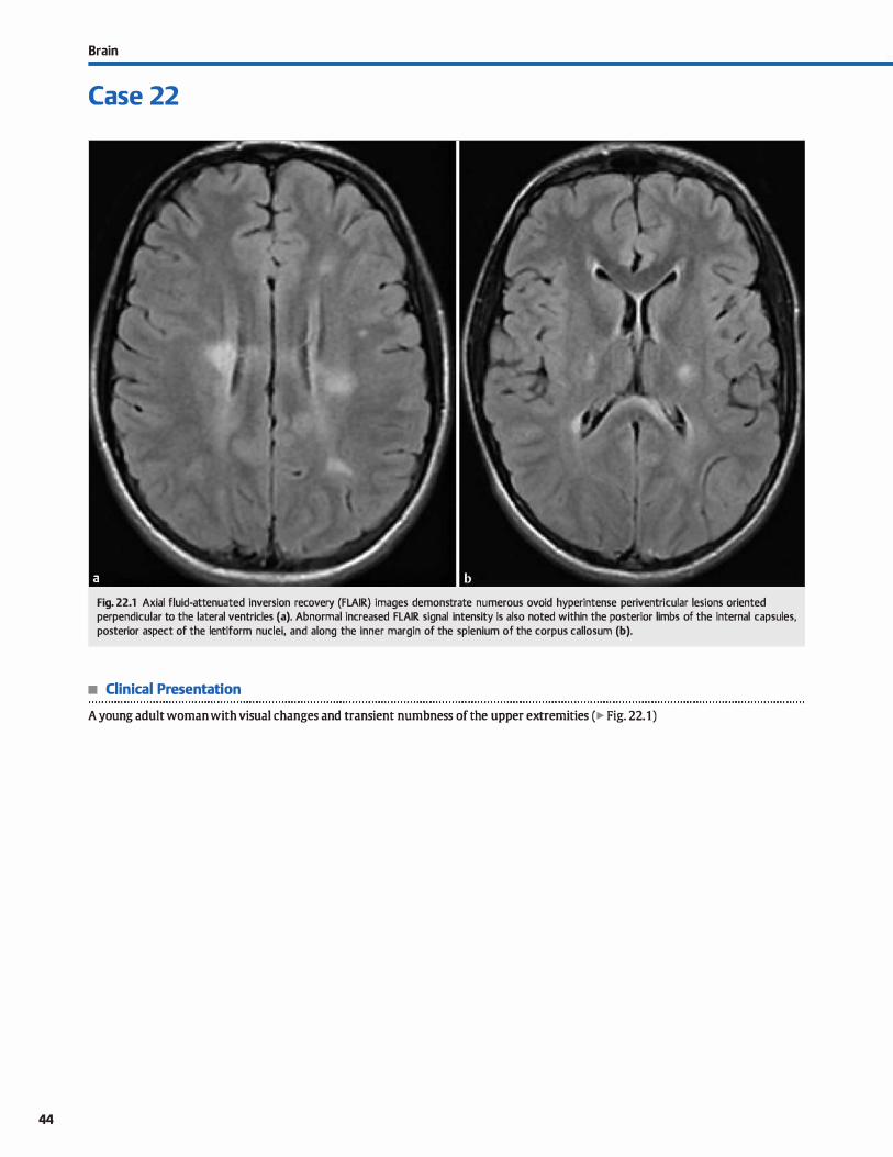
44
Brain
Case 22
Fig. 22.1 Axial fluid-attenuated inversion recovery (FLAIR) images demonstrate numerous ovoid hyperintense periventricular lesions oriented perpendicular to the lateral ventricles (a). Abnormal increased FLAIR signal intensity is also noted within the posterior limbs of the internai capsules, posterior aspect of the lentiform nuclei, and along the inner margin of the splenium of the corpus callosum (b).
• Clinical Presentation
A young adult woman with visual changes and transient numbness of the upper extremities (� Fig. 22.1)
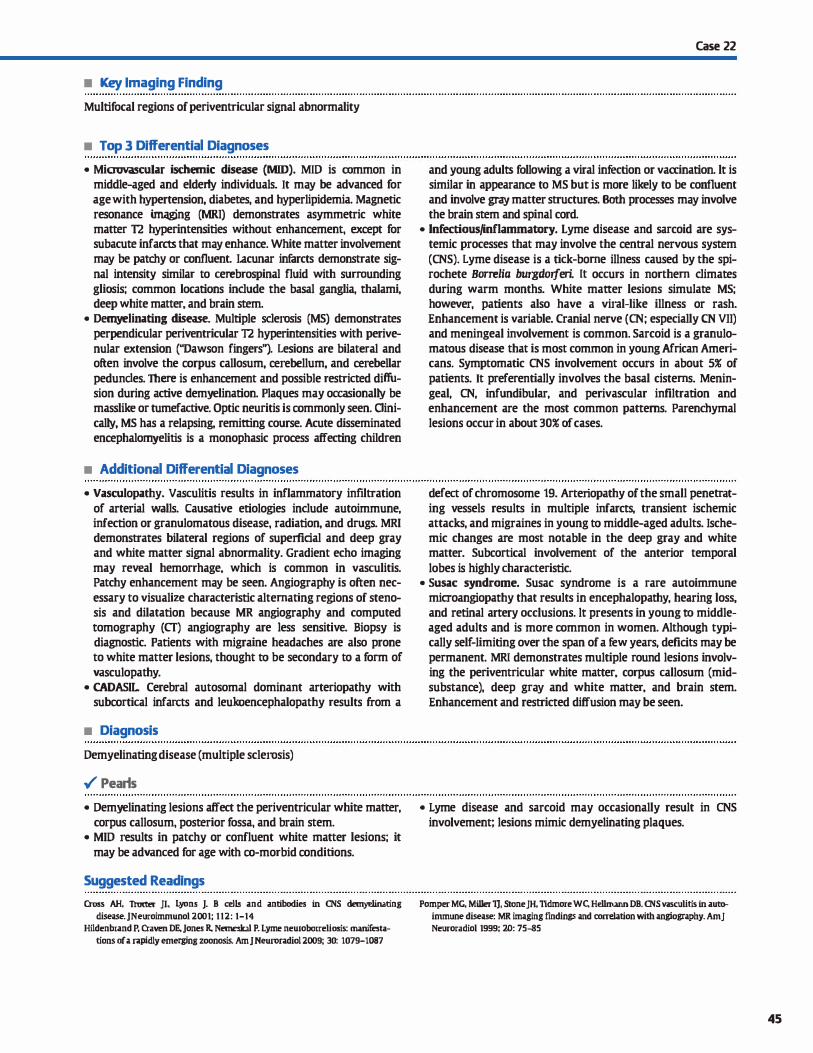
• Key lmaging Finding
Multifocal regions of periventricular signal abnormality
• Top 3 Differential Diagnoses
• Microvascular ischemic disease (MID). MID is rommon in middle-aged and elderly individuals. lt may be advanced for age with hypertension, diabetes, and hyperlipidemia. Magnetic resonance irnaging {MRI) demonstrates asymmetric white matter T2 hyperintensities without enhanœment, exœpt for subacute infarcts that may enhance. White matter involvement may be patchy or confluent. I.acunar infarcts demonstrate signal intensity similar to cerebrospinal fluid with surrounding gliosis; common locations indude the basal ganglia, thalami, deep white matter, and brain stem.
• Demyelinating disease. Multiple sderosis {MS) demonstrates perpendicular periventricular T2 hyperintensities with perivenular extension {�Dawson fingers"). Lesions are bilateral and often involve the corpus callosum, œrebellum, and œrebellar pedundes. There is enhanœment and possible restricted diffusion during active demyellnation. Plaques may occasionally be masslike or tumefactive. Optic neuritis is commonly seen. Oinically, MS has a relapsing, remitting course. Acute disseminated enœphalomyelitis is a monophasic proœss affecting children
• Additional Differential Diagnoses
• Vasculopathy. Vasculitis results in inflammatory infiltration of arterial walls. causative etiologies indude autoimmune, infection or granulomatous disease, radiation, and drugs. MRI demonstrates bilateral regions of superiiàal and deep gray and white matter signal abnormality. Gradient echo imaging may reveal hemorrhage, which is common in vasculitis. Patchy enhancement may be seen. Angiography is often necessary to visualize characteristic altemating regions of stenosis and dilatation because MR angiography and computed tomography (CT) angiography are Jess sensitive. Biopsy is diagnostic. Patients with migraine headaches are also proue to white matter lesions, thought to be secondary to a form of vasculopathy.
• CADASIL Cerebral autosomal dominant arteriopathy with subcortical infarcts and leukoencephalopathy results from a
• Diagnosis
Demyelinating disease (multiple sclerosis)
� Pearls
• Demyelinating lesions affect the periventricular white matter, corpus callosum, posterior fossa, and brain stem.
• MID results in patchy or confluent white matter lesions; it may be advanced for age with co-morbid conditions.
Suggested Readings
Cross AH, Trotter JI., cyons ]. B cells and antibodies in CNS demyelinating
disease.JNeuroimmunol 2001; 112: 1-14
Hildenbrand P, Craven DE, Jones R, Nemeskal P. Lyme neuroborreliosis: manifesta
tions of a rapidly emerging zoonosis. Am J Neuroradiol 2009; 30: 1079-1087
Case 22
and young adults following a viral infection or vaccination. lt is similar in appearance ta MS but is more likely ta be confluent and involve gray matter structures. Both proœsses may involve the brain stem and spinal cord.
• lnfectious/inflammatory. Lyme disease and sarcoid are systemic processes that may involve the central nervous system (CNS). Lyme disease is a tick-bome illness caused by the spirochete Borrelia burgdolferi. lt occurs in northem dimates during warm months. White matter lesions simulate MS; however, patients also have a viral-like illness or rash. Enhancement is variable. Cranial nerve (CN; especially CN VII) and meningeal involvement is common. Sarcoid is a granulomatous disease that is most common in young African Americans. Symptomatic CNS involvement occurs in about 5% of patients. lt preferentially involves the basal cistems. Meningeal, CN, infundibular, and perivascular infiltration and enhancement are the most common patterns. Parenchymal lesions occur in about 30% of cases.
defect of chromosome 19. Arteriopathy of the small penetrating vessels results in multiple infarcts, transient ischemic attacks, and migraines in young to middle-aged adults. Ischemic changes are most notable in the deep gray and white matter. Subcortical involvement of the anterior temporal lobes is highly characteristic.
• Susac syndrome. Susac syndrome is a rare autoimmune microangiopathy that results in encephalopathy, hearing loss, and retinal artery occlusions. lt presents in young ta middleaged adults and is more common in women. Although typically self-limiting over the span of a few years, defidts may be permanent. MRI demonstrates multiple round lesions involving the periventricular white matter, corpus callosum (midsubstanœ ), deep gray and white matter, and brain stem. Enhancement and restricted diffusion may be seen.
• Lyme disease and sarcoid may occasionally result in CNS involvement; lesions mimic demyelinating plaques.
Pomper MG, Miller 1], Stone JH, Tidmore WC. Hellmann DB. CNS vasculitis in autu
immune disease: MR imaging findings and IDITe!ation with angiography. AmJ
Neuroradiol 1999; 20: 75-85
45
46
Brain
Case 23
Fig. 23.1 Axial (a) and coronal (b) FLAIR images demonstrate a focal region of il l-defined increased signal intensity involving the right frontal cortex and subcortical white matter with blurring of the gray-white matter junction.
• Clinical Presentation
A 6-year-old girl with seizures (� Fig. 23.1 )
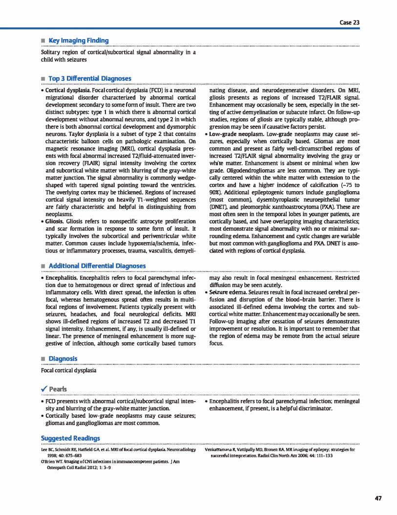
• Key lmaging Finding
Solitary region of cortical/subcortical signal abnonnality in a
child with seizures
• Top 3 Differential Diagnoses
• Cortical dysplasia. Focal cortical dysplasia {FCO) is a neuronal
migrational disorder characterized by abnormal cortical development secondary ta some fonn of insult. There are two distinct subtypes: type 1 in which there is abnonnal cortical
development without abnonnal neurons, and type 2 in which
there is bath abnonnal cortical development and dysmorphie
neurons. Taylor dysplasia is a subset of type 2 that contains
characteristic balloon cells on pathologie examination. On magnetic resonance imaging {MRI), cortical dysplasia presents with focal abnormal increased T2/fluid-attenuated inversion recovery {FIAIR) signal intensity involving the cortex
and subcortical white matter with blurring of the gray-white matter junction. The signal abnonnality is commonly wedge
shaped with tapered signal painting toward the ventricles. The overlying cortex may be thickened. Regions of increased cortical signal intensity on heavily TI -weighted sequences are fairly characteristic and helpful in distinguishing from neoplasms.
• Gliosis. Gliosis refers to nonspedfic astrocyte proliferation
and scar formation in response to some fonn of insult. It
typically involves the subcortical and periventricular white
matter. Common causes include hypoxemia/ischemia, infec
tious or inflammatory processes, trauma, vasculitis, demyeli-
• Additional Differential Diagnoses
• Encephalitis. Encephalitis refers to focal parenchymal infec
tion due to hematogenous or direct spread of infectious and inflammatory cells. With direct spread, the infection is often focal, whereas hematogenous spread often results in multifocal regions of involvement. Patients typically present with seizures, headaches, and focal neurological deficits. MRI shows ill-defined regions of increased T2 and decreased Tt signal intensity. Enhancement, if any, is usually ill-defined or
linear. The presence of meningeal enhancement is more sug
gestive of infection, although some cortically based tumors
• Diagnosis
Focal cortical dysplasia
� Pearls
• FCO presents with abnormal cortical/subcortical signal intensity and blurring of the gray-white matter junction.
• Cortically based low-grade neoplasms may cause seizures;
gliomas and gangliogliomas are most common.
Suggested Readings
Case 23
nating disease, and neurodegenerative disorders. On MRI, gliosis presents as regions of increased T2/FIAIR signal. Enhancement may occasionally be seen, especially in the set
ting of active demyelination or subacute infarct. On follow-up studies, regions of gliosis are typically stable, although pro
gression may be seen if causative factors persist.
• Low-grade neoplasm. Low-grade neoplasms may cause seizures, especially when cortically based. Gliomas are most
common and present as fairly well-drcumscribed regions of increased T2/FLAIR signal abnonnality involving the gray or white matter. Enhanœment is absent or minimal when low
grade. Oligodendrogliomas are less common. They are typically œntered within the white matter with extension to the
cortex and have a higher incidence of calcification (-75 to
90%). Additional epileptogenic tumors indude ganglioglioma {most common1 dysembyroplastic neuroepithelial tumor {ONET), and pleomorphic xanthoastrocytoma (PXA). These are most often seen in the temporal lobes in younger patients, are cortically based, and have overlapping imaging characteristics;
most demonstrate signal abnonnality with no or minimal sur
rounding edema. Enhancement and cystic changes are variable but most common with ganglioglioma and PXA. ONET is asso
dated with regions of cortical dysplasia.
may also result in focal meningeal enhancement. Restricted
diffusion may be seen acutely. • Seizure edema. Seizures result in focal increased œrebral per
fusion and disruption of the blood-brain barrier. There is
assodated ill-defined edema involving the cortex and subcortical white matter. Enhancement may occasionally be seen.
Follow-up imaging after cessation of seizures demonstrates improvement or resolution. It is important to remember that the region of edema may be remote from the actual seizure
focus.
• Encephalitis refers to focal parenchymal infection; meningeal enhanœment, if present, is a helpful discriminator.
Lee BC, Schmidt RE, Hatfield GA, et al, MRI of focal cortical dyspwia. Neuroradiology Venkatramana R, Vattipally MD, Bronen RA. MR imaging of epilepsy: strategies for
1998; 40: 675-683 sucœs5ful interpretation. Radio! Oin North Am 2006; 44: 111-133
O'Brien Wf. lmaging of CNS infections in imm1111DCDmpetent patients. J Am
Osteopath Coll Radio! 2012; 1: 3-9
47

Brain
Case 24
• Clinical Presentation
A 4-year-old boy with new onset developmental delay (� Fig. 24.1)
48
Fig. 24.1 Axial fluidattenuated inversion recovery (FLAIR) (a) and coronal T2 (b) weighted images demonstrate bilateral, symmetric, confluent regions of increased signal intensity within the subcortical and periventricular white matter; subcortical U-fibers are involved. There is a strikingly posterior predominance. Postgadolinium axial Tl -weighted image (c) reveals nodular enhancement along the leading edges.
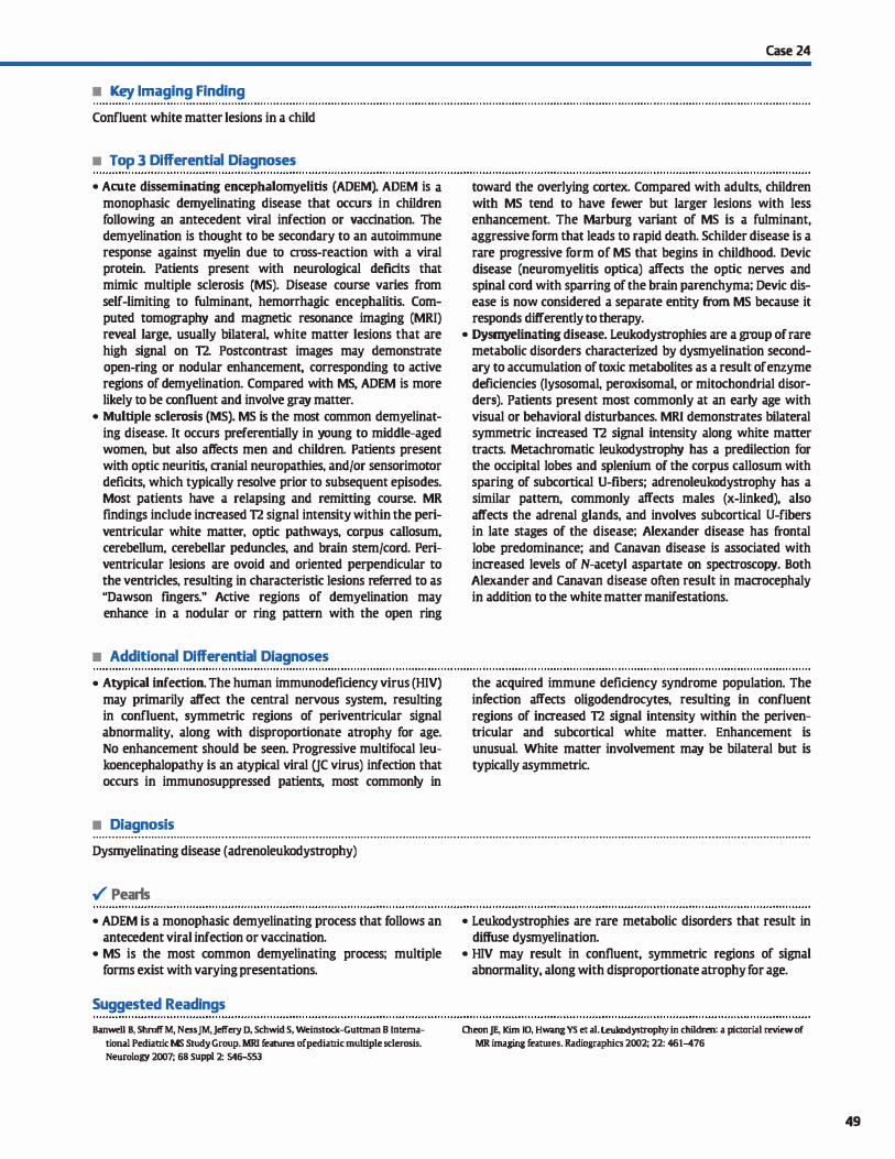
• Key lmaging Finding
Confluent white matter lesions in a child
• Top 3 Differential Diagnoses
• Awte disseminating encephalomyelitis {ADEM). ADEM is a monophasic demyelinating disease that occurs in children following an antecedent viral infection or vaccination. The demyelination is thought to be secondary to an autoimmune response against myelin due to cross-reaction with a viral protein. Patients present with neurological deficits that mimic multiple sclerosis {MS). Disease course varies from
self-limiting to fulminant, hemorrhagic encephalitis. Computed tomography and magnetic resonance imaging (MRI) reveal large, usually bilateral, white matter lesions that are
high signal on T2. Postcontrast images may demonstrate open-ring or nodular enhancement, corresponding to active regions of demyelination. Compared with MS, ADEM is more likely to be confluent and involve gray matter.
• Multiple sclerosis {MS). MS is the most common demyelinat
ing disease. It occurs preferentially in young to middle-aged women, but also affects men and children. Patients present with optic neuritis, cranial neuropathies, and/or sensorimotor detidts, which typically resolve prior to subsequent episodes. Most patients have a relapsing and remitting course. MR findings include increased T2 signal intensity within the peri
ventricular white matter, optic pathways, corpus callosum, cerebellum, cerebellar pedundes, and brain stem/cord. Periventricular lesions are ovoid and oriented perpendicular to the ventrides, resulting in characteristic lesions referred to as uoawson tingers." Active regions of demyelination may enhance in a nodular or ring pattern with the open ring
• Additional Differential Diagnoses
• Atypical infection. The human immunodefidency virus (HIV)
may primarily affect the central nervous system, resulting in confluent, symmetric regions of periventricular signal abnormality, along with disproportionate atrophy for age. No enhancement should be seen. Progressive multifocal leukoencephalopathy is an atypical viral UC virus) infection that occurs in immunosuppressed patients, most commonly in
• Diagnosis
Case 24
toward the overlying cortex. Compared with adults, children with MS tend to have fewer but larger lesions with Jess enhanœment. The Marburg variant of MS is a fulminant, aggressive form that leads to rapid death. Schilder disease is a
rare progressive form of MS that begins in childhood. Devic disease (neuromyelitis optica) affects the optic nerves and spinal cord with sparring of the brain parenchyma; Devic disease is now considered a separate entity from MS because it responds differently to therapy.
• Dysmyelinating disease. Leukodystrophies are a group of rare metabolic disorders characterized by dysmyelination secondary to accumulation of toxic metabolites as a result of enzyme
deficiencies (lysosomal, peroxisomal, or mitochondrial disorders). Patients present most commonly at an early age with
visual or behavioral disturbances. MRI demonstrates bilateral symmetric increased T2 signal intensity along white matter tracts. Metachromatic leukodystrophy bas a predilection for the occipital lobes and spleniurn of the corpus callosurn with sparing of subcortical U-fibers; adrenoleukodystrophy has a similar pattern, commonly affects males (x-linked), also
affects the adrenal glands, and involves subcortical U-fibers in late stages of the disease; Alexander disease has frontal
lobe predominance; and Canavan disease is associated with increased levels of N-acetyl aspartate on spectroscopy. Both Alexander and canavan disease often result in macrocephaly in addition to the white matter manifestations.
the acquired immune defidency syndrome population. The
infection affects oligodendrocytes, resulting in confluent
regions of increased T2 signal intensity within the periventricular and subcortical white matter. Enhancement is
unusual. White matter involvement may be bilateral but is typically asymmetric.
. . . . . . . . . . . . . . . . . . . . . . . . . . . . . . . . . . . . . . . . . . . . . . . . . . . . . . . . . . . . . . . . . . . . . . . . . . . . . . . . . . . . . . . . . . . . . . . . . . . . . . . . . . . . . . . . . . . . . . . . . . . . . . . . . . . . . . . . . . . . . . . . . . . . . . . . . . . . . . . . . . . ..... . . . . . . . . . . . . . . .. . . . . . . . . . . . . . . . . . . . . . . . . . . . . . . . ........
Dysmyelinating disease ( adrenoleukodystrophy)
� Pearls
• ADEM is a monophasic demyelinating process that follows an antecedent viral infection or vaccination.
• MS is the most common demyelinating process; multiple forms exist with varying presentations.
Suggested Readings
Banwell B, Shroff'M, NessjM,Jeffery D, Schwid S, Weinstock-Guttman B Interna
tional Pediatric MS Study Group. MRI features ofpediatric multiple sclerosis.
Neurology 2007; 68 Suppl 2: 546-553
• Leukodystrophies are rare metabolic disorders that result in diffuse dysmyelination.
• HIV may result in confluent, symmetric regions of signal abnormality, along with disproportionate atrophy for age.
Cheon JE, Kim 10, Hwang YS et al, Leukndystrophy in children; a pictorial review of MR imaging features. Radiographies 2002; 22: 461-476
49
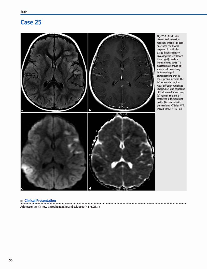
Brain
Case 25
• Clinical Presentation
Adolescent with new onset headache and seizures (� Fig. 25.1 )
50
Fig. 25.1 Axial fluidattenuated inversion recovery image (a) demonstrates multifocal regions of cortically based hyperintensity involving the left (more than right) cerebral hemispheres. Axial Tl postcontrast image (b) shows mild overlying leptomeningeal enhancement that is most pronounced in the left opercular region. Axial diffusion-weighted imaging (c) and apparent diffusion coefficient map (d) reveals regions of restricted diffusion bilaterally. (Reprinted with permissions: O'Brien WT. JAOCR 201 2;1 (1 ):3-9.)

• Key lmaging Finding
Multifocal regions of cortical signal abnormality
• Top 3 Differential Diagnoses
• Cerebritis. Cerebritis refers to focal infection of the brain
parenchyma and may result from hematogenous spread, direct spread, or as a complication of meningitis. Direct spread tends to result in focal infection, whereas hematogenous spread and meningitis typically present with multifocal involvement. Patients present with fever, headache, seizures, or focal neurological defidts. On imaging, cerebritis presents
as ill-defined cortical and subcortical hypoattenuation ( computed tomography, CT) and increased T2/fluid-attenuated inversion recovery signal intensity (magnetic resonance imaging, MRI). Restricted diffusion may be seen, which may mimic an infarct acutely. Enhancement, if present, is ill-defined or thin and linear. Meningeal enhancement may also be seen.
• Ischemia/infarcts. Infan:ts result from thromboembolic disease
in older patients and vasculitis and arterial dissection in younger patients. They present as wedge-shaped regions of cortical and subcortical hypoattenuation on cr and T2 hyperintensity on MRI. Cytotoxic edema results in Joss of gray-white matter differentiation and sulcal effacement Acute infarcts demon-
• Additional Differential Diagnoses
• Vasculitis. Vasculitides result in inflammation of arterial walls. Common etiologies indude infectious, granulomatous (induding primary angiitis of the central nervous system), collagen vascular diseases, radiation-induced, and drug
induced. Imaging findings range from normal to marked vascular and parenchymal abnormalities. Involved arteries demonstrate altemating regions of stenosis and dilatation. Parenchymal findings indude regions of ischemia or infarct involving the superficial and deep gray and white matter. Restricted diffusion may be seen acutely. Lesions may enhance, especially in the subacute phase. Regions of micro
hemorrhage are seen as foci of hypointensity on gradientecho and susceptibility-weighted imaging.
• Cortical tubers. Tuberous sderosis is a neurocutaneous syndrome with a dassic triad of fadai angiofibromas, mental retardation, and seizures. Subependymal nodules and cortical tubers are present in the vast majority of cases. Tubers are
• Diagnosis
Cerebritis
y' Pearls
• Cerebritis results from hematogenous or direct spread of infection or as a complication of meningitis.
• Thromboembolic disease, vasculitides, and arterial injury may result in cortical/subcortical ischemia or infarction.
Suggested Readings
O'Brien Wf. Imaging of CNS infections in immunocompetent patients. J Am
Osteopath Coll Radio!. 2012; 1 : 3-9
Case 25
strate restricted diffusion. Gyral or patchy enhancement may be seen in the subacute phase. Perfusion imaging is useful in identifying the penumbra, which represents viable paren
chyma at risk of infarct cr angiography or MR angiography may show the region ofvascular occlusion. Hemorrhagic transformation occurs in 15 to 20% of cases. Venous infants do not
follow a vascular territory and are prone to hemorrhage. • Contusions. Cerebral contusions result from traumatic injury
as the brain parenchyma contacts the skull base or durai reflections. They occur most frequently along the inferior frontal lobes, anterior temporal lobes, and parasagittal locations. Contusions directly beneath the site of impact are referred to as coup injuries, whereas those on the opposite side of injury are referred to as contrecoup injuries. Acutely,
there is cortical and subcortical edema and swelling. Foci of parenchymal or extraaxial hemorrhage are useful discriminators. Within the first 2 to 3 days, the contusions may expand with increase in amount of hemorrhage. In the chronic stage, regions of encephalomalada are commonly seen.
composed of disorganized glial tissue and heterotopic neuronal elements. On imaging. they appear as wedge-shaped regions of cortical and subcortical signal abnormality with the
tapered portion pointing medially. Prior to myelination, tubers may be hyperintense of Tl; after myelination, they are typically Tl hypo- and T2 hyperintense. 1\Jbers may occasionally enhance or calcify, which will alter the signal characteristics.
• MELAS. MEIAS refers to mitochondrial encephalopathy with lactic addosis and stroke-like episodes. It is an inherited mitochondrial disorder which is more common in boys and men. Patients present in adolescence or early adulthood with seizures and stroke-like episodes. lmaging reveals regions of
attenuation and signal abnormality involving the cortex/subcortical white matter, as well as the basal ganglia and deep
white matter. MR spectroscopy over the basal ganglia may reveal an elevated lactate peak, which is fairly characteristic
of mitochondrial disorders.
• MELAS presents in adolescence or early adulthood with a seizures and stroke-like episodes.
Pomper MG, Miller 1], Stone JH, Tidmore WC. Hellmann DB. CNS vasculitis in auto
immune disease: MR imaging findings and IDITe!ation with angiography. Amj
Neuroradiol 1999; 20; 75�5
51
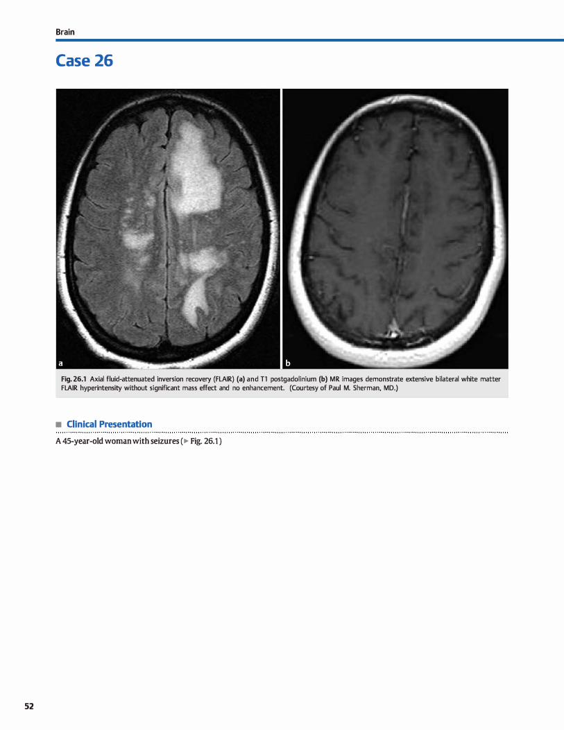
52
Brain
Case 26
Fig. 26.1 Axial fluid-attenuated inversion recovery (FLAIR) (a) and Tl postgadolinium (b) MR images demonstrate extensive bilateral white matter FLAIR hyperintensity without significant mass effect and no enhancement. (Courtesy of Paul M. Sherman, MD.)
• Clinical Presentation
A 45-year-old woman with seizures (� Fig. 26.1 )
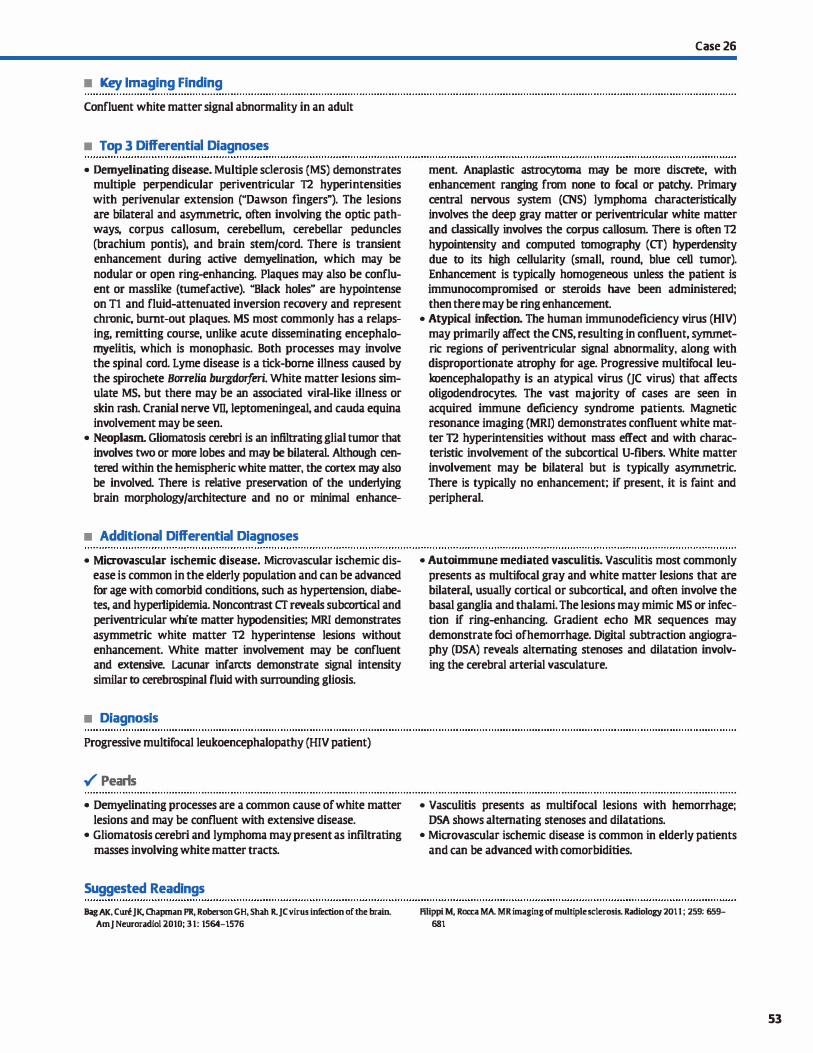
• Key lmaging Finding
Confluent white matter signal abnormality in an adult
• Top 3 Differential Diagnoses
• Demyelinating disease. Multiple sderosis {MS) demonstrates multiple perpendicular periventricular T2 hyperintensities with perivenular extension (UDawson fingers"). The lesions are bilateral and asymrnetric, often involving the optic pathways, corpus callosum, cerebellum, cerebellar peduncles (brachium pontis), and brain stem/cord. There is transient enhancement during active demyelination, which may be
nodular or open ring-enhandng. Plaques may also be confluent or masslike (tumefactive). UBlack holes" are hypointense on T1 and fluid-attenuated inversion recovery and represent chronic, burnt-out plaques. MS most commonly has a relapsing, remitting course, unlike acute disseminating encephalomyelitis, which is monophasic. Both processes may involve the spinal cord. Lyme disease is a tick-borne illness caused by
the spirochete Borrelia burgdorferi. White matter Iesions simulate MS, but there may be an associated viral-like illness or
skin rash. Cranial nerve VII, leptomeningeal, and cauda equina involvement may be seen.
• Neoplasm Gliomatosis œrebri is an infiltrating glial tumor that involves two or more lobes and may be bilateral. Although œn
tered within the hemispheric white matter, the cortex may also be involved. There is relative preservation of the underlying brain morphology/architecture and no or minimal enhance-
• Additional Differential Diagnoses
• Microvascular ischemic disease. Microvascular ischemic disease is common in the elderly population and can be advanœd for age with comorbid conditions, such as hypertension, diabetes, and hyperlipidemia. Noncontrast cr reveals subcortical and periventricular white matter hypodensities; MRI demonstrates
asymmetric white matter T2 hyperintense lesions without enhancement. White matter involvement may be confluent and extensive. Lacunar infarcts demonstrate signal intensity similar to œrebrospinal fluid with surrounding gliosis.
• Diagnosis
Progressive multifocal leukoencephalopathy {HIV patient)
� Pearts
• Demyelinating processes are a common cause of white matter lesions and may be confluent with extensive disease.
• Gliomatosis œrebri and lymphoma may present as infiltrating
masses involving white matter tracts.
Suggested Readings
Bag AK, CUré JK. Chapman PR, Robenan GH, Shah R. JC virus infection of the brain.
AmJNeuroradial 2010; 31: 1564-1576
Case 26
ment Anaplastie astrocytoma may be more discrete, with enhanœment ranging from none to focal or patchy. Primary central nervous system (CNS) lymphoma characteristically involves the deep gray matter or periventricular white matter
and dassically involves the corpus callosum. There is often 12
hypointensity and computed tomography (CT) hyperdensity due to its high œllularity (small, round, blue cell tumor). Enhancement is typically homogeneous unless the patient is immunocompromised or steroids have been administered; then there may be ring enhanœment
• Atypical infection. The human immunodefidency virus (HIV) may primarily affect the CNS, resulting in confluent, symrnetric regions of periventricular signal abnormality, along with disproportionate atrophy for age. Progressive multifocal leu
koencephalopathy is an atypical virus UC virus) that affects oligodendrocytes. The vast majority of cases are seen in acquired immune deficiency syndrome patients. Magnetic resonance imaging {MRI) demonstrates confluent white matter T2 hyperintensities without mass effect and with characteristic involvement of the subcortical U-fibers. White matter involvement may be bilateral but is typically asymrnetric.
There is typically no enhancement; if present, it is faint and peripheral.
• Autoimmune mediated vasculitis. Vasculitis most commonly
presents as multifocal gray and white matter lesions that are bilateral, usually cortical or subcortical, and often involve the basal ganglia and thalami. The lesions may mimic MS or infection if ring-enhandng. Gradient echo MR sequences may
demonstrate foci ofhemorrhage. Digital subtraction angiography (DSA) reveals altemating stenoses and dilatation involv
ing the cerebral arterial vasculature.
• Vasculitis presents as multifocal lesions with hemorrhage; DSA shows altemating stenoses and dilatations.
• Microvascular ischemic disease is common in elderly patients and can be advanced with comorbidities.
Filippi M, Rocca MA. MR imaging of multiple sclerosis, Radiolagy 2011 ; 259; 659-
681
53
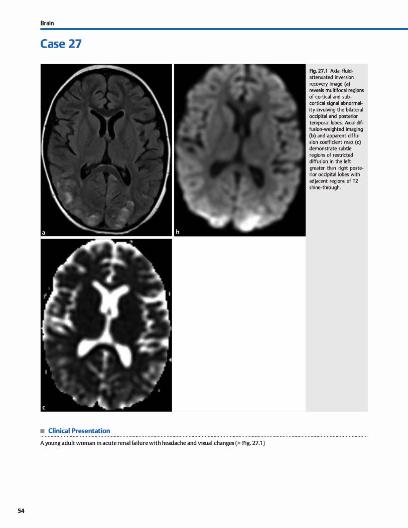
Brain
Case 27
• Clinical Presentation
A young adult woman in acute renal failure with headache and visual changes (� Fig. 27.1 )
54
Fig. 27.1 Axial fluidattenuated inversion recovery image (a) reveals multifocal regions of cortical and subcortical signal abnormality involving the bilateral occipital and posterior temporal lobes. Axial diffusion-weighted imaging (b) and apparent diffusion coefficient map (c) demonstrate subtle regions of restricted diffusion in the left greater than right posterior occipital lobes with adjacent regions of T2 shine-through.
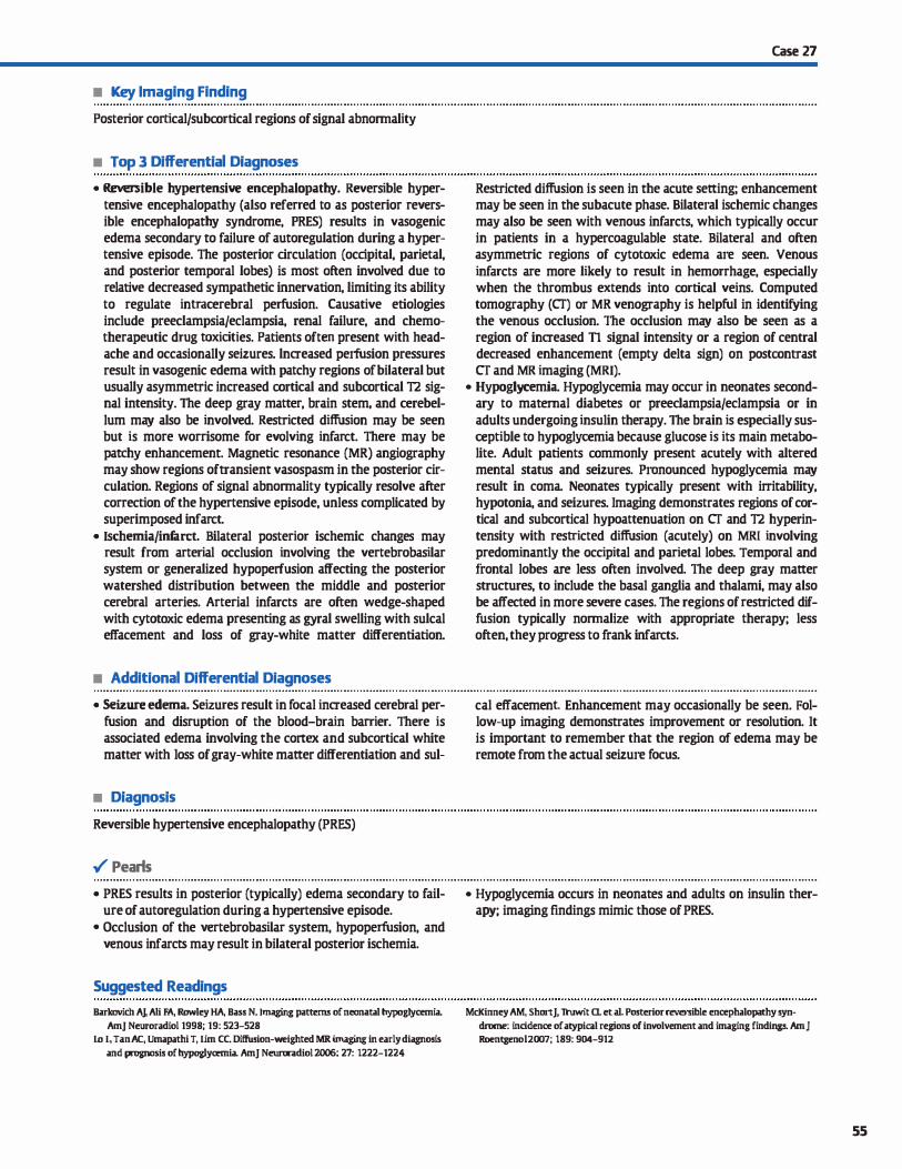
• Key lmaging Finding
Posterior cortical/subcortical regions of signal abnorrnality
• Top 3 Differential Diagnoses
• Reversible hypertensive enœphalopathy. Reversible hypertensive encephalopathy (also referred to as posterior reversible encephalopathy syndrome, PRES) results in vasogenic edema secondary to failure of autoregulation during a hypertensive episode. The posterior circulation (occipital, parietal, and posterior temporal lobes) is most often involved due ta relative decreased sympathetic innervation, limiting its ability
to regulate intraœrebral perfusion. Causative etiologies include preeclampsia/eclampsia, renal failure, and chemotherapeutic drug toxicities. Patients often present with headache and occasionally seizures. lncreased pelfusion pressures result in vasogenic edema with patchy regions of bilateral but usually asymmetric increased cortical and subcortical T2 signal intensity. The deep gray matter, brain stem, and œrebel
lum may also be involved. Restricted diffusion may be seen but is more worrisome for evolving infarct. There may be patchy enhancement. Magnetic resonance (MR) angiography may show regions oftransient vasospasm in the posterior circulation. Regions of signal abnorrnality typically resolve after correction of the hypertensive episode, unless complicated by superimposed infarct.
• Ischemia/infan:t. Bilateral posterior ischemic changes may result from arterial occlusion involving the vertebrobasilar system or generalized hypoperfusion affecting the posterior watershed distribution between the middle and posterior cerebral arteries. Arterial infarcts are often wedge-shaped with cytotoxic edema presenting as gyral swelling with sulcal effacement and loss of gray-white matter differentiation.
• Additional Differential Diagnoses
• Seizure edema. Seizures result in focal increased cerebral per
fusion and disruption of the blood-brain barrier. There is associated edema involving the cortex and subcortical white matter with Joss of gray-white matter differentiation and sui-
• Diagnosis
Reversible hypertensive enœphalopathy (PRES)
� Pearts
• PRES results in posterior (typically) edema secondary to failure of autoregulation during a hypertensive episode.
• Occlusion of the vertebrobasilar system, hypopelfusion, and venous infarcts may result in bilateral posterior ischemia.
Suggested Readings
Barlrovich AJ, Ali FA, RDwley HA, Bass N. lmaging patterns of neonatal hypoglyœmia.
AmJ Neuroradiol 1998; 19: 523-528 Lo L, Tan AC, Umapathi T, Lim CC. Dilfusion-weighted MR imaging in early diagnosis
and prognosis of hypoglycemia. Amj Neuroradiol 2006; 27: 1222-1224
Case 27
Restricted diffusion is seen in the acute setting; enhanœment may be seen in the subacute phase. Bilateral ischemic changes may also be seen with venous infarcts, which typically occur in patients in a hypercoagulable state. Bilateral and often asymmetric regions of cytotoxic edema are seen. Venous
infarcts are more likely to result in hemorrhage, especially when the thrombus extends into cortical veins. Computed tomography (CT) or MR venography is helpful in identifying the venous occlusion. The occlusion may also be seen as a region of increased 11 signal intensity or a region of central decreased enhancement (empty delta sign) on postcontrast CT and MR imaging (MRI).
• Hypoglyc:emia. Hypoglycemia may occur in neonates secondary to maternai diabetes or preedampsia/eclampsia or in
adults undergoing insulin therapy. The brain is especially susceptible to hypoglycemia because glucose is its main metabolite. Adult patients commonly present acutely with altered mental status and seizures. Pronounced hypoglycemia may result in coma. Neonates typically present with irritability, hypotonia, and seizures. Imaging demonstrates regions of cortical and subcortical hypoattenuation on CT and T2 hyperin
tensity with restricted diffusion (acutely) on MRI involving predominantly the occipital and parietal lobes. Temporal and
frontal lobes are Jess often involved. The deep gray matter structures, to include the basal ganglia and thalami, may also be affected in more severe cases. The regions of restricted diffusion typically norrnalize with appropriate therapy; less often, they progress to frank infarcts.
cal effacement Enhanœment may occasionally be seen. Follow-up imaging demonstrates improvement or resolution. It is important to remember that the region of edema may be remote from the actual seizure focus.
• Hypoglycemia occurs in neonates and adults on insulin therapy; imaging findings mimic those of PRES.
McKinney AM, Shortj, lhlwit a. et al, Posterior revenible enœphalopathy syn
drome: inddenœ of atypical regions of involvement and imaging findings. Am j
Roentgenol2007; 189: 904-912
55
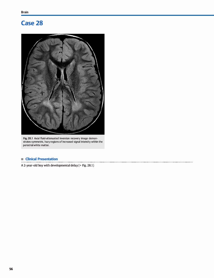
56
Brain
Case 28
Fig. 28.1 Axial fluid-attenuated inversion recovery image demonstrates symmetric, hazy reg ions of increased signal intensity within the periatrial white matter.
• Clinical Presentation
A 2-year-old boy with developmental delay (� Fig. 28.1 )
• Key lmaging Finding
Periatrial signal abnormality in a young child
• Top 3 Differential Diagnoses
• Delayed myelination. Myelination occurs in a relatively predictable pattern with maturation progressing from central to peripheral, inferior ta superior, and posterior ta anterior. The pattern of myelination mirrors neurological development with early myelination of corticospinal and optic tracts, as well as the posterior brain stem. On magnetic resonanœ imaging (MRI), white matter approximates that of an adult brain on T1 sequenœs by 1 year of age and on T2 sequenœs by 2 years of age. The terminal zones of myelination indude the periatrial white matter and subcortical white matter of the anterior frontal and temporal lobes. Patients with delayed myelination on imaging may be neurologically normal or demonstrate developmental delay.
• Demyelinating disease. Acute disseminated enœphalomyelitis (ADEM) is the most common demyelinating disease in children. It is monophasic and occurs following an antecedent viral infection or vaccination. The disease course varies from self-limiting ta fulminant, hemorrhagic enœphalitis. MRI reveals regions of increased T2 signal abnormality within the white matter that may be confluent. Lesions are often bilateral but asymmetric. Involvement of the corpus callosum, optic pathways, œrebellar hemispheres, and brain stem is characteristic. Periventricular white matter lesions orient perpendicular to the ventrides, resulting in characteristic Iesions referred to as �Dawson fin-
• Additional Differential Diagnoses
• Neonatal hypoxie ischemic enœphalopathy (HIE). Parenchymal injury related ta HIE is based upon the gestational age at the time of the insult, as well as the severity of the insult. Severe HIE affects the most metabolically active portions of the brain, induding the deep gray and white matter. ln preterm infants, mild to moderate HIE affects the periventricular white matter, resulting in signal abnormality and volume loss and often leading to periventricular leukomalacia. In full-term infants, mild ta moderate HIE results in watershed ischemic changes, similar ta the adult brain.
• Diagnosis
Hypo/delayed myelination
� Pearls
• Terminal zones of myelination indude the periatrial and subcortical frontal and temporal lobe white matter.
• MS/ADEM result in asymmetric white matter lesions with characteristic morphology and distribution.
Suggested Readings
Case 28
gers." Active lesions may demonstrate enhanœment along the leading edge of demyelination in an üopen ring" pattern. Multiple sderosis (MS) occurs more frequently in adults and aider children. As opposed to those with ADEM, patients with MS often have a relapsing and remitting rather than monophasic course. The imaging appearanœ may be identical ta ADEM.
• Metabolic disease. Dysmyelinating diseases are rare metabolic disorders that occur secondary ta accumulation of taxie metabolites as a result of enzyme deficiencies (lysosomal, peroxisomal, or mitochondrial disorders). Patients present at an early age with visual or behavioral disturbanœs. MRI demonstrates bilateral, symmetric regions of T2 hyperinten
sity along white matter tracts. Metachromatic Ieukodystrophy has a predilection for the occipital lobes and splenium of the corpus callosum with sparing of subcortical U-fibers; adrenoleukodystrophy bas a similar pattern, commonly affects boys (x-Iinked), also affects the adrenal glands, and involves subcortical U-fibers; Alexander disease bas frontal lobe predominance; and Canavan disease is associated with increased levels of N-acetyl aspartate on spectroscopy. Bath Alexander and Canavan disease often result in macrocephaly. Canavan disease results in increased T2 signal within the thalami and globus pallidi. Krabbe disease characteristically results in hyperdensity of deep gray nudei on computed tomography.
• Atypical perivascular spaces. Occasionally, regions of incidental, persistent, hazy periatrial signal abnormality may be associated with perivascular spaœs. Perivascular spaces occur along a radial course from the periventricular region to the overlying cortex. Focal prominence is common in the basal ganglia, periatrial, and subcortical regions. Although they typically suppress on fluid-attenuated inversion recovery sequenœs, there may be atypical, hazy surrounding hyperintensity.
• Metabolic disorders result in bilateral, symmetric regions of confluent white matter signal abnormality.
Konishi Y, Kuriyama M, Hayakawa K et al Periventricular hyperintensity detecœd by Parazzini C. Baldoli c, Scotti G, Triulzi F. Tenninal zones of myelination: MR eval-
magnetic resanance imaging in infancy. Ped.iatr Neurol 1990; 6; 229-232 uatian of children aged 20-40 manths. AmJ Neuraradial 2002; 23; 1669-
Nowell MA. Grossman RI, Haclmey DB, Zimmennan RA, Galdberg Hl, Bilaniuk LT. 1673
MRimagingofwhitematœrdisease indûldren.AmJRoentgenol 1988; 151:
359-365
57
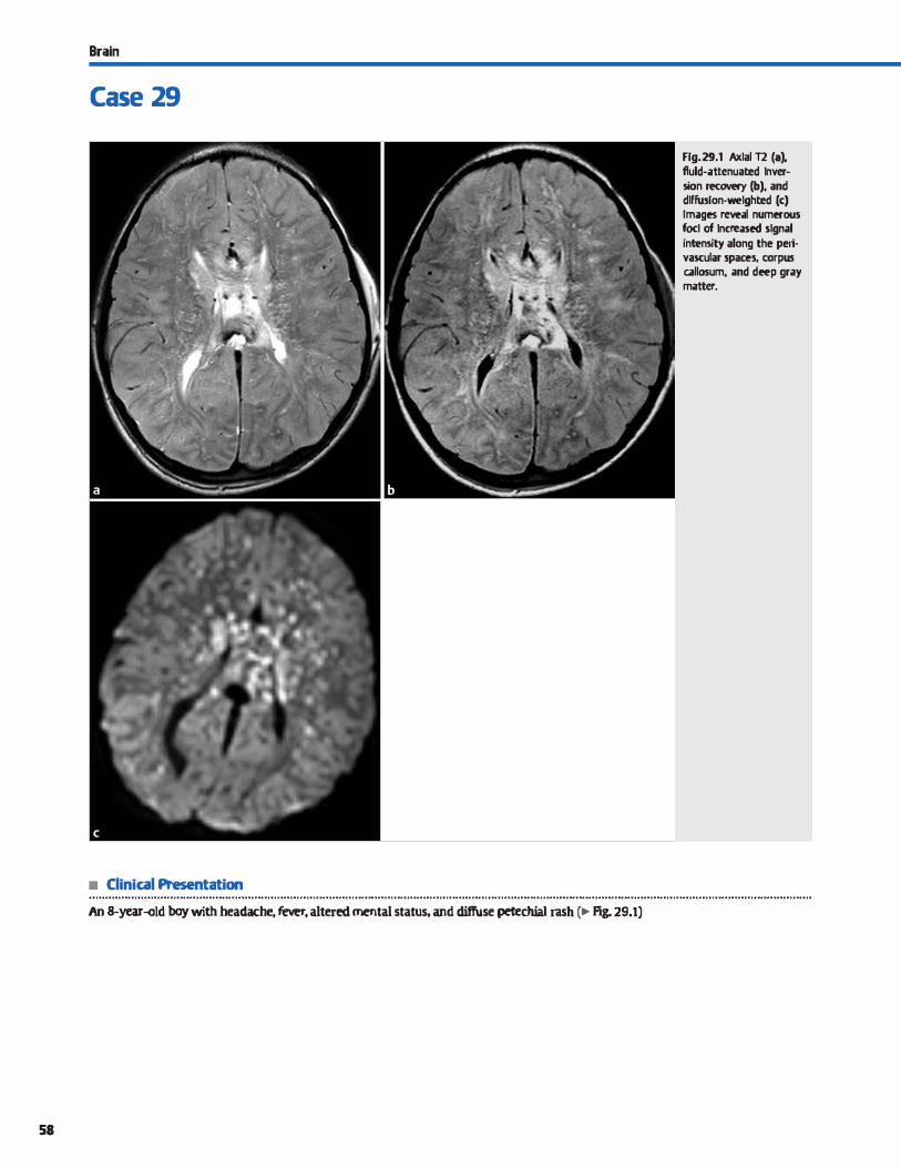
Brain
Case 29
• Clinical Presentation
An 8-year-old boy with headache. fever. altered mental status. and diffuse petechial rash c� Fig. 29.1)
58
Fig. 29.1 Axlal T2 {a), fluld-attenuated lnver-5ion recavery (b), and dlffusion·welghted (c) Images reveal numll!fous focl of lncreased signal intensity along the pefi. vascular spaces, corpus c.allosum, and deep gray matter.
• Key lmaging Finding
Numerous foci of perivascular and deep gray/white matter signal abnormality
• Top 3 Differential Diagnoses
• Cryptococcus. Cryptococcus is a fungal infection that occurs in the setting of acquired immune deficiency syndrome (AIDS). It is the second most common opportunistic central nervous system (CNS) infection in AIDS after toxoplasmosis. It
most often affects the CNS through hematogenous spread from a pulmonary infection. The most common imaging find
ing is multiple T2 hyperintense lesions involving the deep perivascular spaœs, basal ganglia, and cerebral white matter. Four primary patterns of infection are recognized: granulomatous meningitis with nodular enhancement, dilated nonenhancing Virchow-Robin spaœs representing cysts, "gelati
nous pseudocysts" {which may or may not enhance) in the basal ganglia, and cryptococcomas with solid or ring-enhancing masses that often involve the choroid plexus. Hydrocephalus is commonly seen.
• cyme disease. Lyme disease is a tick-bome illness caused by the spirochete Borrelia burgdolferi. It is endemic to northem dimates and occurs during warm months. The infection is systemic with CNS involvement in more severe cases. The most common neurological imaging finding is abnonnal cranial
nerve (CN) enhancement, particularly of CN VII, which results in a Bell palsy. The infection may also spread ta the meninges with associated dinical and imaging findings of meningitis, ta
indude diffuse or focal leptomeningeal enhanœment. Parenchymal involvement is relatively uncommon and results from small-vessel vasculitis. Magnetic resonanœ irnaging (MRI) reveals multiple perivascular white matter lesions, which may
• Diagnosis
Rocky Mountain spotted fever
� Pearls
• Cryptococcus is a common fungal infection in the setting of AIDS; it preferentially involves perivascular spaœs.
• Lyme disease is a tick-bome illness that is endemic to northern dimates during warm months.
Suggested Readlngs
Baganz MD. Dross PE. ReinhanltJA. Rocky Mountain spotted fever enœphalitis: MR
findings. Amj Neuroradiol 1995; 16 Suppl; 919-922
Hildenbrand P, Craven DE, Jones R. Nemeskal P. L.Yme neuroborreliosis: manifestations of a r.apidly emerging zoonosis, Am J Neuroradiol 2009; 30; 1079-1087
Case 29
sirnulate multiple sderosis. Deep gray matter structures may also be involved. Parenchymal enhanœment is variable. A key dinical distinction is the presenœ of a viral-like illness with a characteristic "bull's-eye" rash {erythema migrans) secondary
ta the tick bite. • Rocky Mountain spotted fever (RMSF). RMSF is a tick-bome
illness caused by the bacterium Rickettsia rickettsii. It typically occurs in wooded areas during late spring and early summer. The bacterium enters the bloodstream and results in a systemic small-vessel vasculitis. Patients most often present with fever, headache, and a rash that begins at the distal extremities and progresses proximally. The rash is maculo
papular in its early stage and then becomes petechial with regions of necrosis. CNS manifestations vary from mild headache to a fulminant fatal meningoencephalitis. The smallvessel vasculitis preferentially involves the perivascular spaœs, as well as the deep gray and white matter. MRI is more sensitive than computed tomography, better demonstrating involvement of perivascular spaces with surrounding gliosis.
The perivascular involvement is somewhat similar to that of cryptococcus. Deep gray and white matter lesions are similar to that of other vasculitides, including Lyme disease. More extensive involvement of deep gray and white matter structures, to indude the corpus callosum, is noted in severe cases. There may be restricted diffusion in the acute phase. If recognized early and treated appropriately, imaging findings may
regress or resolve.
• CNS involvement with Lyme disease includes CN or meningeai enhanœment and perivascular white matter lesions.
• RMSF causes small-vessel vasculitis that preferentially involves perivascular spaœs and deep gray/white matter.
Smith AB, SmirniotopoulosjG, Rushing i;r. From the archives of the AFIP: central
nervous system infections associated with human immunodefidency virus
infection: radiologie-pathologie correlation. Radiographies 2008; 28: 2033-2058
59
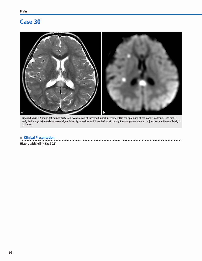
60
Brain
Case 30
Fig. 30.1 Axial T2 image (a) demonstrates an ovoid reg ion of increased signal intensity within the splenium of the corpus callosum. Diffusionweighted image (b) reveals increased signal intensity, as well as additional lesions at the right insular gray-white matter junction and the medial right thalamus.
• Clinical Presentation
History withheld ( � Fig. 30.1 )
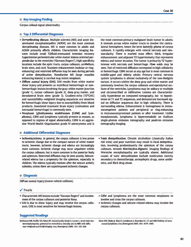
• Key lmaging Finding
Corpus callosal signal abnormality
• Top 3 Differential Diagnoses
• Demyelinating disease. Multiple sderosis (MS) and acute disseminated enœphalomyelitis {ADEM) are the most common demyelinating diseases. MS is more common in adults and ADEM primarily affects children. Characteristic imaging features indude ovoid 12/fluid-attenuated inversion recovery hyperintense periventricular white matter lesions oriented perpendicular to the ventrides (uDawson tingers"). High-spedfidty locations indude the optic tracts, corpus callosum, œrebellum, brain stem, and cord. Transient enhancement {nodular or ringenhancing) and restricted diffusion may be seen during periods of active demyelination. Turnefactive MS (large masslike enhancing lesions) is rare but may mimic neoplasm.
• Diffuse axonal injury (DAI). DAI results from white matter shear injury and presents as multifocal hemorrhagic or nonhemorrhagic lesions involving the gray-white matter junction (grade 1 ), corpus callosum {grade 2), deep gray matter, and dorsolateral brain stem (grade 3). Gradient-echo (T2.GRE) and susceptibility-weighted imaging sequences are sensitive for hemorrhagic shear injury due to susceptibility from blood products. Assodated traumatic brain injury (contusions and extraaxial hemorrhage) is commonly seen.
• Neoplasm (glioblastoma multiformes [GBM] and lymphoma). GBM and lymphoma typically present as masses, as opposed to regions of signal abnormality. GBM is an aggressive World Health Organization grade IV astrocytoma and is
• Additional Differential Diagnoses
• lschemia/edema. ln general, the corpus callosum is Jess prone to ischemic change due to the compact nature of white matter tracts; however, ischemic change and edema are increasingly more common. lschemic change may occur anywhere within the corpus callosum, but is more common in the posterior body and splenium. Restricted diffusion may be seen acutely. Seizurerelated edema has a propensity for the splenium, especially in children. The edema typically resolves after the seizure activity subsides, unless there are superimposed ischemic changes.
• Diagnosis
Diffuse axonal injury (motor vehide collision)
� Pearls
• Characteristic MS lesions indude "Dawson fingers" and involvement of the corpus callosum and posterior fossa.
• DAI is due to shear injury and may involve the corpus callosum; GRE is most sensitive for hemorrhagic lesions.
Suggested Readings
Case 30
the most common primary malignant brain tumor in adults. 1t extends across white matter tracts to involve the contralateral hemisphere, hence the term butterfly glioma of corpus callosum. It rapidly enlarges with central necrosis and neovascularity. There is marked mass effect; thick, irregular enhanœment; and adjacent T2 hyperintensity that represents edema and tumor invasion. The tumor is primarily T2 hyperintense with necrosis and hemorrhage. Flow voids may be seen. Foci of restricted diffusion correspond to high œllularity or hemorrhage. GBM occurs at any age but is most common in middle-aged and elderly adults. Primary central nervous system lymphoma is almost exdusively of the non-Hodgkin variant. 1t occurs within the deep gray and white matter and commonly involves the corpus callosum and ependymal surfaces of the ventrides. Lymphoma may be solitary or multiple and drcumscribed or infiltrative. Lesions are characteristically hyperdense on computed tomography, iso- to hypointense on Tl and T2 sequenœs, and demonstrate increased signal on diffusion sequenœs due to high cellularity. There is surrounding edema. Enhanœment is homogenous in immunocompetent patients and peripheral or ring-enhandng in immunocompromised patients or after treatment. Unlike toxoplasmosis, lymphoma is hypermetabolic on thallium single-photon emission tomography and positron emission tomography imaging.
• Toxic demyelination. Chronic alcoholism ( classically Italian red wine) and poor nutrition may result in focal demyelination, involving predominantly the splenium of the corpus callosum, tenned Marchiafava-Bignami. Imaging findings of Wernicke encephalopathy are typically absent. Additional causes of toxic demyelination indude medication toxicity secondary to chemotherapy, antiepileptic drugs, some antibiotics, and illicit drug abuse.
• GBM and lymphoma are the most common neoplasms to involve and cross the corpus callosum.
• Ischemic changes and seizure-related edema may involve the corpus callosum.
McKinney AM. Kieffer SA. Paylor RT, Santacruz KS, Kendi A. Lucato L Acute toxic leu- Slone HW, Blake .D. Shah R. Guttikonda S. Bow-ekas EC. cr and MRI findings of intra-
kcenœphalopathy: poœntial for reversibility dinic:ally and on MRI with diffu- cranial lymphoma. Am j Roentgenol 2005; 184: 1679-1685
sion-weighted and FIAIR imaging. Amj Roentgenol 2009; 193: 192-206
61

62
Brain
Case 31
Fig. 31 .1 Axial fluid-attenuated inversion recovery images reveal symmetric reg ions of signal abnormality involving the medial cerebellar hemispheres/nuclei, cerebellar peduncles, and brain stem [medulla (a) and pans (b)] without significant mass effect.
• Clinical Presentation
A 5-year-old boy with developmental delay and altered mental status (� Fig. 31.1 )
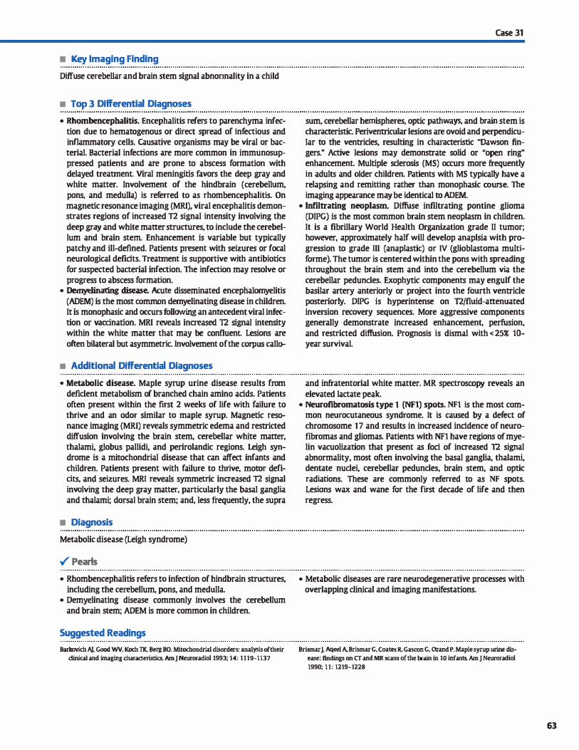
• Key lmaging Finding
Diffuse cerebellar and brain stem signal abnonnality in a child
• Top 3 Differential Diagnoses
• Rhombencephalitis. Encephalitis refers to parenchyma infection due ta hematogenous or direct spread of infectious and inflammatory cells. causative orgarusms may be viral or bacterial. Bacterial infections are more common in immunosuppressed patients and are prone to abscess formation with delayed treatment. Viral meningitis favors the deep gray and white matter. Involvement of the hindbrain ( œrebellum,
pons, and medulla) is referred to as rhombenœphalitis. On
magnetic resonanœ imaging (MRI), viral encephalitis demonstrates regions of increased T2 signal intensity involving the
deep gray and white matter structures, to include the cerebellum and brain stem Enhancement is variable but typically patchy and ill-defined. Patients present with seizures or focal neurological deficits. Treatment is supportive with antibiotics
for suspected bacterial infection. The infection may resolve or
progress to absœss formation. • Demyelinating disease. Acute disseminated encephalomyelitis
(ADEM) is the most common demyelinating disease in children. It is monophasic and occurs following an antecedent viral infection or vaccination. MRI reveals increased T2 signal intensity within the white matter that may be confluent Lesions are
often bilateral but asymmetric. Involvement of the corpus calle>-
• Additional Differential Diagnoses
• Metabolic disease. Maple syrup urine disease results from detident metabolism of branched chain amine acids. Patients
often present within the first 2 weeks of life with failure to thrive and an odor similar ta maple syrup. Magnetic resonance imaging (MRI) reveals symmetric edema and restricted diffusion involving the brain stem, cerebellar white matter, thalami, globus pallidi, and perirolandic regions. Leigh syndrome is a mitochondrial disease that can affect infants and
children. Patients present with failure to thrive, motor deficits, and seizures. MRI reveals symmetric increased T2 signal involving the deep gray matter, particularly the basal ganglia and thalami; dorsal brain stem; and, Jess frequently, the supra
• Diagnosis
Metabolic disease (Leigh syndrome)
� Pearls
• Rhombencephalitis refers ta infection of hindbrain structures, including the cerebellum, pans, and medulla.
• Demyelinating disease commonly involves the œrebellum
and brain stem; ADEM is more common in children.
Suggested Readings
Case 31
sum, cerebellar hemispheres, optic pathways, and brain stem is characteristic. Periventricular lesions are ovoid and perpendicular to the ventrides, resulting in characteristic uoawson fingers." Active lesions may demonstrate solid or uopen ring" enhanœment. Multiple sderosis (MS) occurs more frequently
in adults and aider children. Patients with MS typically have a relapsing and remitting rather than monophasic rourse. The imaging appearance may be identical to ADEM.
• lnfiltrating neoplasm. Diffuse infiltrating pontine glioma (DIPG) is the most common brain stem neoplasm in children. It is a fibrillary World Health Organization grade II tumor; however, approximately half will develop anaplsia with pro
gression to grade III (anaplastie) or IV (glioblastoma multiforme). The tumor is œntered within the pons with spreading throughout the brain stem and into the œrebellum via the
cerebellar pedundes. Exophytic components may engulf the basilar artery anteriorly or project into the fourth ventride posteriorly. DIPG is hyperintense on T2/fluid-attenuated inversion recovery sequences. More aggressive components generally demonstrate increased enhancement, perfusion,
and restricted diffusion. Prognosis is dismal with < 25% 10-year survival.
and infratentorial white matter. MR spectroscopy reveals an
elevated lactate peak. • Neurofibromatosis type 1 {NFt) spots. NF1 is the most com
mon neurocutaneous syndrome. It is caused by a defect of
chromosome 17 and results in increased incidence of neurofibromas and gliomas. Patients with NFl have regions of myelin vacuolization that present as foci of increased T2 signal abnormality, most often involving the basal ganglia, thalami, dentate nudei, cerebellar pedundes, brain stem, and optic
radiations. These are commonly referred to as NF spots. Lesions wax and wane for the first decade of life and then regress.
• Metabolic diseases are rare neurodegenerative processes with overlapping dinical and imaging manifestations.
Barlo:lVidt AJ, Good WV, Kodt TI<, Berg BO. Mitodtondrial disorders: analysis oftheir Brismar J, Aqeel A. Brismar c, Coates R. Gascon c, Ozand P. Maple syrup urine dis-dinical and imaging characterutics. Am J Neuroradiol 1993; 14: 111 S-1137 ease: fin ding;! on cr and MR scans of the brain in 1 D infants. Am J Neuroradiol
1990; 11: 1219-1228
63
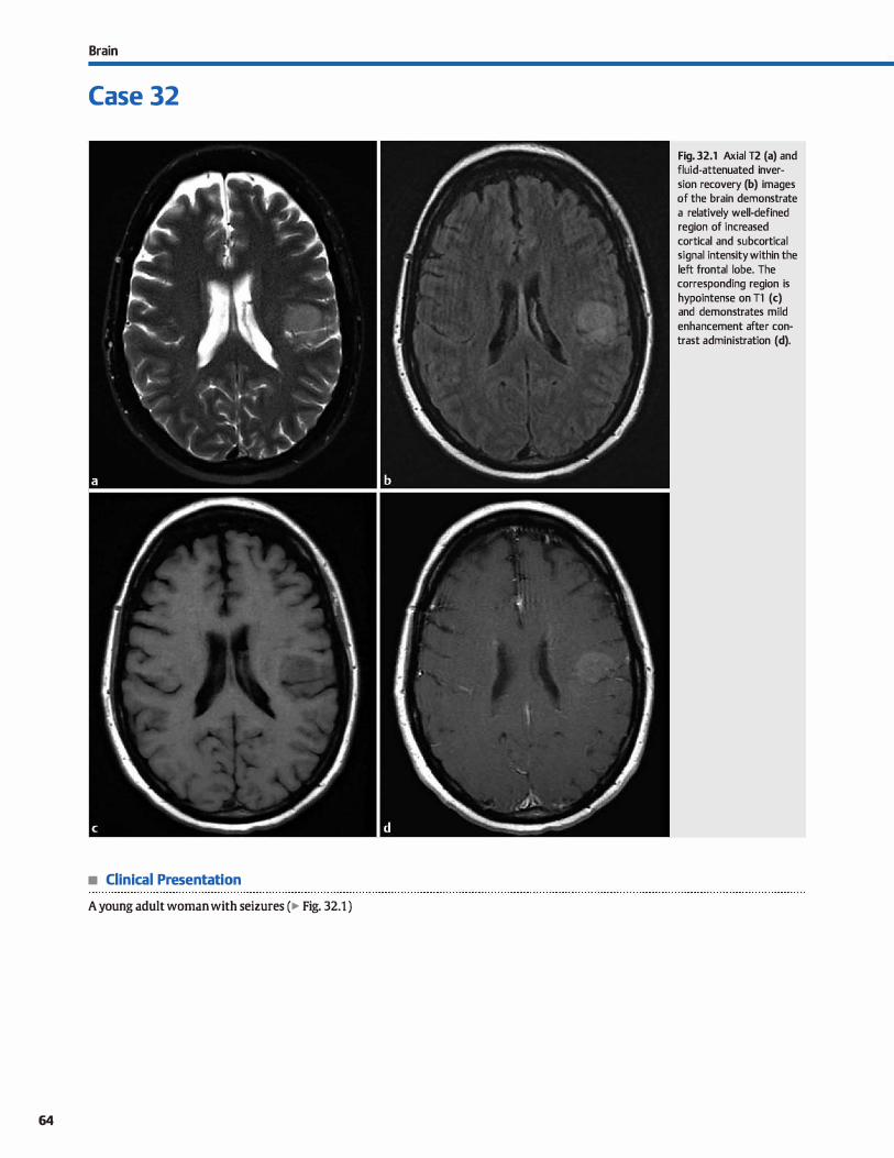
Brain
Case 32
• Clinical Presentation
A young adult woman with seizures (� Fig. 32.1 )
64
Fig. 32.1 Axial T2 (a) and fluid-attenuated inversion recovery (b) images of the brain demonstrate a relatively well-defined region of increased cortical and subcortical signal intensity within the left frontal lobe. The corresponding region is hypointense on Tl (c) and demonstrates mild
enhancement after contrast administration (d).
• Key lmaging Finding
Solitary region of cortical and subcortical signal abnonnality in an adult
• Top 3 Differential Diagnoses
• lschemia/infarct. Cerebrovascular disease is common, especially in older patients, and most often results from thrombo
embolic arterial occlusion. ln younger patients, vasculitis and dissection are prevalent Patients present with focal neurological deficits, altered mental status.. and/or aphasia. Arterial infarcts result in wedge-shaped regions of cortical and subcortical hypoattenuation (computed tomography, CT) and T2 hyperintensity (magnetic resonanœ imaging, MRI). Cytotoxic edema results in cortical swelling with effacement of adjacent
sulci Acute infarcts demonstrate restricted diffusion on MRI. Gyral or patchy enhanœment may be seen in the subacute phase, beginning as early as 2 days and peaking around 1 O to 14 days. CT angiography and MR angiography may show vascular occlusion. Perfusion imaging is useful in identifying the penumbra, which represents viable parenchyma that is at risk of infarct Hemorrhagic transformation occurs in 15 to 20% of
cases. Venous infarcts do not follow a vascular territory and are more prone to hemorrhage. CT venography or MR venography reveal the region of thrombosis.
• Neoplasm. Astrocytomas are the most common primary brain tumors in adults. Variants that present with cortical signal abnonnality indude fibrillary (World Health Organization [WHO] grade Il) and anaplastie (WHO grade III). Fibrillary
astrocytoma typically presents with indistinct or Jess corn-
• Additional Differential Diagnoses
• Contusion. Cerebral contusions result from trauma as the brain parenchyma contacts the adjacent skull base or durai reflections. Common locations indude the inferior frontal lobes, anterior temporal lobes, and parasagittal locations.
Contusions directly beneath the site of impact are referred to as coup injuries, whereas those on the opposite side of injury are referred to as contrecoup injuries. Acutely, there is cortical and subcortical edema and swelling. Foci of parenchymal or extraaxial hemorrhage are hyperdense on CT and variable in signal on MRI depending upon the timing of imaging. MRI is more sensitive than CT and typieally demonstrates additional
foct of parenchymal injury, to indude diffuse axonal injury, if
• Diagnosis
Low-grade (fibrillary) astrocytoma
� Pearts
• lnfarcts typically result from thromboembolic disease; vasculitis and dissection are common in younger patients.
• Astrocytomas and oligodendrogliomas are the most common primary tumors with focal cortical signal abnormality.
Suggested Readings
Case 32
monly well-defined signal abnonnality centered within the white matter with or without overlying cortical involvement. Enhancement, when present, is mild. Anaplastie astrocytoma is more aggressive with regions of increased pelfusion, edema, and enhancement. MR spectroscopy reveals increased choline and decreased N-acetyl aspartate. Unlike infarcts, restricted diffusion is not typieally seen, unless there is focal
progression to a high-grade tumor. Oligodendrogliomas occur in middle-aged adults who present with seizures. They are centered within the subcortical white matter and commonly extend to the overlying cortex. The frontal lobe is the most common location. Lesions are well defined with little edema and mild enhanœment. Regions of calcification are typically seen.
• Cerebritis. Cerebritis refers to focal infection of the brain parenchyma and may result from hematogenous spread,
direct spread, or as a complication of meningitis. Patients present with fever, headache, seizures, or focal neurological deficits. On imaging, œrebritis presents as ill-defined cortical and subcortical hypoattenuation {CT) and increased T2/fluidattenuated inversion recovery signal intensity (MRI). Restricted diffusion may be seen, which may mimic an infarct
acutely. Enhancement, if present, is ill-defined or thin and linear. Meningeal enhancement is a useful discriminator.
present. Within the first 2 to 3 days, contusions may enlarge. Encephalomalacia is often seen chronically.
• Seizure edema. Seizure-related edema results in cortical and subcortical edema that is transient. It most often occurs in the setting of status epilepticus with continuous seizure activity that lasts for at least 30 minutes. Acutely, there is gyral swelling and sulcal effacement. Restricted diffusion and patchy enhancement may be seen. On follow-up imaging, the edema should decrease or resolve if the patient remains seizure-free. The regions of edema may or may not be in the same location
as the underlying seizure focus.
• Contusions occur in characteristic locations with focal edema, swelling, and hemorrhage.
Koeller KK, Rushing m. From the ardtives of the AHP: oligodendroglioma and its var- O'Brien Wf. lmaging ofCNS infections in immunocompetent patients.JAOCRJ Am
iants: radiologie-pathologie correlation. Radiographies 2005; 25; 1669-1688 Osteopath Coll Radial. 2012; 1: 3-9
65
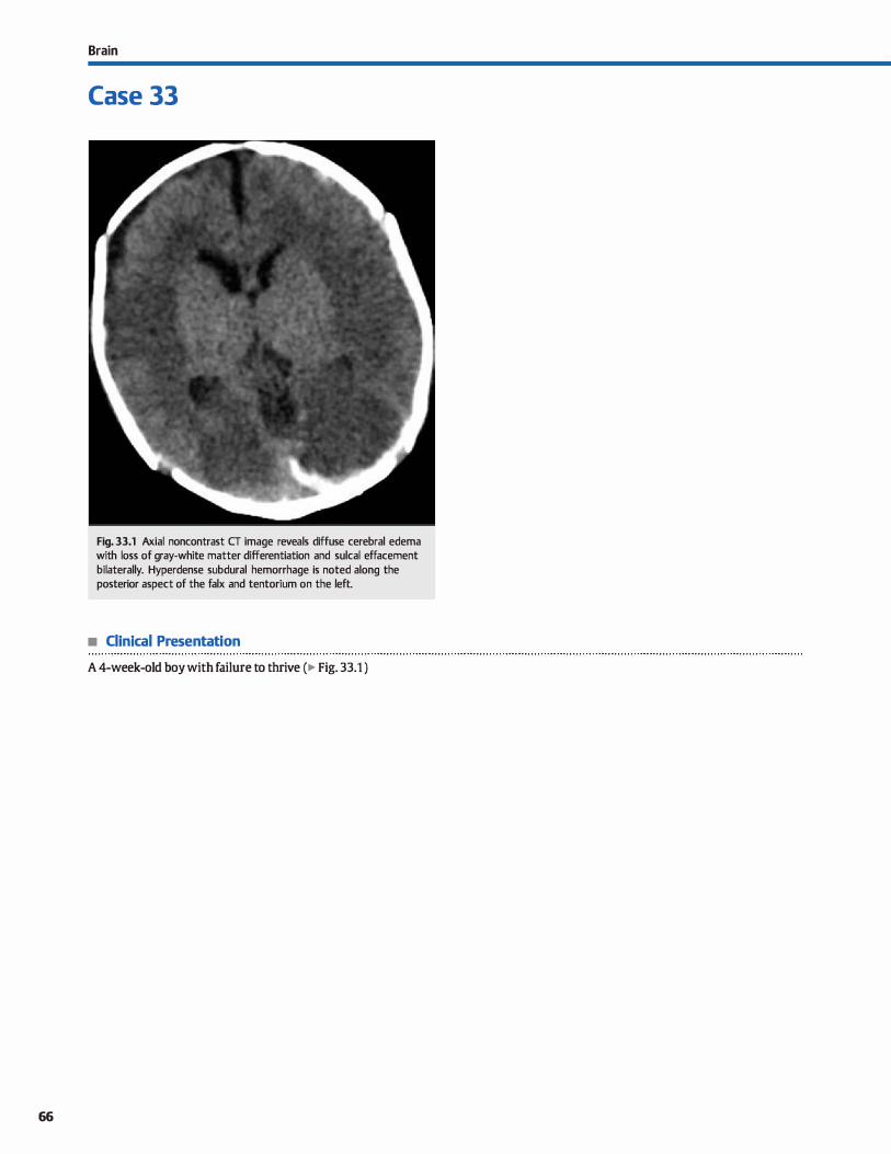
66
Brain
Case 33
Fig. 33.1 Axial noncontrast CT image reveals diffuse cerebral edema with loss of gray-white matter differentiation and sulcal effacement bilaterally. Hyperdense subdural hemorrhage is noted along the posterior aspect of the faix and tentorium on the left.
• Clinical Presentation
A 4-week-old boy with failure to thrive (� Fig. 33.1 )
• Key lmaging Finding
Diffuse cerebral edema in an infant
• Top 3 Differential Diagnoses
• Hypoxic-ischemic encephalopathy {HIE). The neurological
sequela of HIE depends upon a number of factors, induding the severity of the insult, timing of the insult with respect to neuronal maturation, and duration of the insult. ln the preterm infant, mild to moderate HIE results in injury to the periventricular white matter, resulting in periventricular leukomalada. Early ultrasound imaging reveals hyperechoic periventricular
white matter that may progress to cystic change with volume loss. Follow-up computed tomography (CT) or magnetic reso
nanœ imaging {MRI) will show abnormal signal and volume loss within the periatrial white matter with focal ventricular
enlargement. The ventrides will have irregular margins. More severe HIE affects the deep gray matter structures ( espedally the thalami), brain stem, œrebellum, and œrebral cortex, par
ticularly the perirolandic region. ln full-term infants, mild to
moderate HIE results in watershed ischemia, similar to adults. Abnormal hypoattenuation (CT) or signal (MRI) is seen in the cortex, subcortical white matter, and centrum semiovale of the
watershed regions between the vascular distributions of the middle cerebral artery and the anterior and posterior œrebral arteries. More severe HIE is similar in the premature infant
with less involvement of the œrebellum and increased involvement of the basal ganglia and œrebral cortex.
• li'auma. Trauma may be accidentai or nonaccidental (NAT)
with the main distinction being a history that is or is not compatible with the severity and pattern of injury. ln direct blunt trauma (accidentai or NAT), similar findings are seen as with adults, induding skull fractures, extraaxial hemorrhage, and
coup and contrecoup parenchymal injuries. It is important to describe the type, location, and pattern of injury, as well as ta
evaluate complications, such as herniation syndromes or
ischemic changes. A subset of NAT includes "shaken baby" syndrome. With this type of injury, more specific findings are noted, including subdural hemorrhages with extension into the interhemispheric fissure and hypoxic-ischemic changes.
• Diagnosis
Nonacddental trauma
� Pearls
• Neurological sequela of HIE depends upon severity, duration, and timing of the insult with respect to brain maturation.
• HIE with cerebral edema may occur as a direct result or com
plication of trauma.
Suggested Readings
Chao Cl', Zaleski CG, Patton Ar.. Neonatal hypoxic-ischemic enœphalopathy: multi
modality imaging findings. Radiographies 2006; 26 Suppl 1: Sl 59-Sl 72
Choi CG, Yoo HW. Localized proton MR spectroscopy in infants with urea cycle
defect. AmJ Neuroradiol 2001; 22: 834-1137
Case 33
Rarely, retinal hemorrhages may be seen on imaging,
although they are readily evident on physical examination. CT angiography or MR angiography may show regions of vascular injury or spasm. ln the setting of NAT, it is important to remember that the density or signal of subdural hemorrhage
is dependent upon numerous factors; therefore, pinpointing the precise age of an extraaxial collection is fraught with
error. Additionally, the predictable pattern of hemorrhage evolution on MRI is derived from intraparenchymal hemorrhages and not extraaxial collections. If NAT is suspected, a
skeletal survey should be performed and appropriate notifications must be made and documented.
• Metabolic disease. Numerous rare metabolic diseases may result in diffuse cerebral edema. Although the pattern of
parenchymal involvement may suggest a particular process, a dinical workup is required ta establish the diagnosis. Causes of diffuse cerebral edema in a neonate or infant include mito
chondrial disorders, urea cycle disorders, and nonketotic hyperglydnemia. There are numerous variants of mitochon
drial disorders that ultimately affect energy production due ta enzyme deficiencies. Patients often present with encephal
opathy or seizures. lmaging reveals abnormal attenuation (CT) and signal (MRI) involving the basal gang]ia, cerebral
white matter, cerebellum, and/or brain stem. Basal ganglia involvement is characteristic. In addition to parenchymal findings, Menkes disease {enlarged tortuous vasculature) and glutaric adduria type 1 (widened Sylvian fissures with bilateral middle cranial fossa ucysts") may also present with sub
dural collections. Urea cycle disorders present acutely with
diffuse cerebral swelling and abnormal hypoattenuation and signal involving the cortex, subcortical white matter, and
deep gray nuclei. There is decreased swelling and parenchymal atrophy in the chronic phase. Nonketonic hypergldnemia presents with abnormal attenuation and signal within the basal ganglia, corpus callosum, and cerebral cortex.
• Rare metabolic diseases may result in cerebral edema; an
extensive workup is needed to establish the diagnosis.
Loneigan GJ, Baker AM, MoreyMK. Boos SC. From the archives of the AFIP. Child
abuse; radiologie-pathologie corrdation. Radiographies 2003; 23; 811-845
67
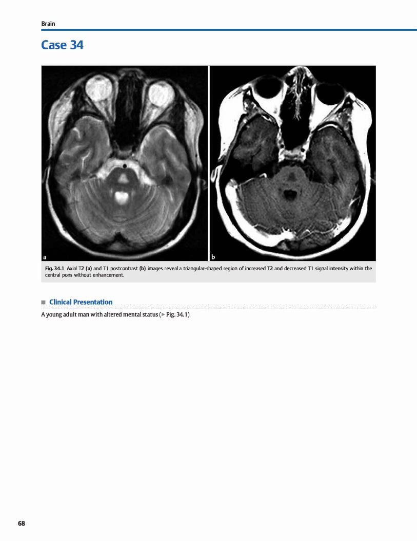
68
Brain
Case 34
Fig. 34.1 Axial T2 (a) and Tl postcontrast (b) images reveal a triangular-shaped reg ion of increased T2 and decreased Tl signal intensity within the central pons without enhancement.
• Clinical Presentation
A young adult man with altered mental status (� Fig. 34.1)
• Key lmaging Finding
Focal pontine signal abnormality
• Top 3 Differential Diagnoses
• lschemia. Microvascular ischemic disease (MVID) of the pons is relatively common in middle-aged and elderly patients, especially in the setting of hypertension, hyperlipidemia, and diabetes. As with other foci of MVID, lesions are hypodense on computed tomography and hyperintense on T2 sequences. Pontine infarcts typically result Crom occlusion of small perforators from the basilar artery. lnfarcts from basilar artery occlusion are less common and may be catastrophic. Acutely, lesions demonstrate restricted diffusion; enhancement may be seen in the subacute phase. Lacunar infarcts follow cerebrospinal fluid signal intensity with a rim of gliosis.
• Demyelinating disease. Multiple sclerosis (MS) and acute disseminated encephalomyelitis (ADEM) are the most common demyelinating diseases. MS most commonly affects adults and has a relapsing and remitting course; ADEM is more common in children and is monophasic. Magnetic resonance imaging (MRI) reveals regions of increased T2 signal within
• Additional Differential Diagnoses
• Vascular malformation (VM). cavemous malformations (CMs) and capillary telangiectases are the most common VMs to involve the brain stem. CMs are dilated vascular channels without intervening brain parenchyma. Lesions have a peripheral T2 hypointense hemosiderin rim and central regions ofîl and
T2 hyper- and hypointensity, giving the characteristic "popcorn" appearance. Signal characteristics result from hemorrhages of different ages. Enhancement occurs in -15% of cases. Surrounding edema may be seen with acute hemorrhage. capillary telangiectases represent dilated capillaries with normal intervening brain parenchyma. They are asymptomatic, inci
dental lesions. Half will demonstrate subtle T2 hyperintensity; the remaining are occult on T2 sequenœs. Hypointensity is seen on gradient echo and susceptibility-weighted imaging. Lesions demonstrate faint brushlike enhancement.
• Brain sœm glioma. Brain stem glioma represents approximately 1 O to 20% of pediatric brain tumors and commonly pres-
• Diagnosis
Osmotic demyelination (central pontine myelinolysis)
� Pearls
• MVID commonly involves the pons in older patients secondary to occlusion of perforating vessels.
• Osmotic demyelination results from rapid shift in osmolality; central pontine signal spares the corticospinal tracts.
Suggested Readlngs
Smith AB. Vascular malformations of the br.Un: radiologie and pathologie correla
tion.J Am Osteopath Coll Radio!. 2012; 1; 10-22
Case 34
the white matter that may be confluent. Lesions are often bilateral but asymmetric. Involvement of the corpus callosum, cerebellar hemispheres, optic pathways, and brain stem is characteristic. Periventricular lesions orient perpendicular to the ventricles, resulting in ovoid lesions referred to as "Dawson fingers." Active lesions may demonstrate solid or "open ring" enhancement, as well as restricted diffusion.
• Osmotic demyelination. Also known as central pontine myelinolysis, osmotic demyelination results from a rapid shift in osmolality, dassically described with overzealous correction of hyponatremia. Approximately half of cases involve the central pons where lesions present as triangular or confluent T2 hyperintensities with sparing of corticospinal tracts. The remaining cases involve the deep gray and white matter, also referred to as extrapontine myelinolysis, with or without pontine involvement. Restricted diffusion may be seen acutely. Signal abnormality may regress over time.
ents in the first 2 decades of life. lt typically presents as a diffuse, infiltrating pontine mass (World Health Organization grade Il to Ill) that may have exophytic components. lt is hyper
intense on 12/fluid-attenuated inversion recovery sequenœs. More aggressive components demonstrate increased enhanœment, perfusion, and restricted diffusion. Prognosis is dismal The low-grade pilocytic variant is Jess common and presents as a cystic mass with enhancing nodule. lt has an overall better prognosis.
• Brain stem encephalitis. Encephalitis refers to focal infection of the brain parenchyma. Viral etiologies more commonly affect the brain stem. Bacterial infections are more common in immunosuppressed patients. On MRI, encephalitis demonstrates regions of increased T2 signal intensity. Restricted diffusion may be seen. Enhancement is variable but typically patchy and ill-defined. The infection may resolve or progress to abscess formation.
• CMs have a characteristic "popcorn" appearance on MRI; capillary telangiectases have faint brushlike enhancement
Venkatanarasimha N, Mulmnowesluw W.Jonesj. AJR teaching file: symmetrie
demyelination. AmJ Roentgenol 2008; 191 Suppl; S34-S36
69
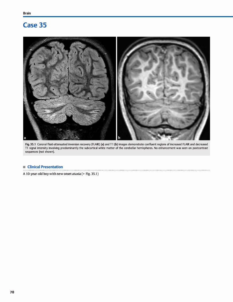
70
Brain
Case 35
Fig. 35.1 Coronal fluid-attenuated inversion recovery (FLAIR) (a) and Tl (b) images demonstrate confluent regions of increased FLAIR and decreased Tl signal intensity involving predominantly the subcortical white matter of the cerebellar hemispheres. No enhancement was seen on postcontrast sequences (not shown).
• Clinical Presentation
A 10-year-old boy with new onset ataxia (� Fig. 35.1 )
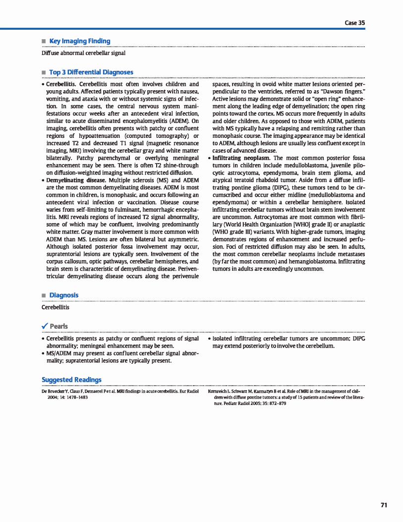
• Key lmaging Finding
Diffuse abnormal cerebellar signal
• Top 3 Differential Diagnoses
• Cerebellitis. Cerebellitis most often involves children and young adults. Affected patients typically present with nausea.
vomiting, and ataxia with or without systemic signs of infec
tion. In some cases, the central nervous system mani
festations occur weeks after an antecedent viral infection,
similar to acute disseminated enœphalomyelitis (ADEM). On
imaging, œrebellitis often presents with patchy or confluent
regions of hypoattenuation (computed tomography) or
increased T2 and decreased Tl signal (magnetic resonanœ
imaging, MRI) involving the œrebellar gray and white matter
bilaterally. Patchy parenchymal or overlying meningeal
enhancement may be seen. There is often T2 shine-through
on diffusion-weighted imaging without restricted diffusion.
• Demyelinating disease. Multiple sderosis (MS) and ADEM are the most common demyelinating diseases. ADEM is most
common in children, is monophasic, and occurs following an
anteœdent viral infection or vaccination. Disease course varies from self-limiting to fulminant, hemorrhagic encepha
litis. MRI reveals regions of increased T2 signal abnormality,
some of which may be confluent, involving predominantly
white matter. Gray matter involvement is more common with
ADEM than MS. Lesions are often bilateral but asymmetric. Although isolated posterior fossa involvement may occur,
supratentorial lesions are typically seen. Involvement of the
corpus callosum, optic pathways, cerebellar hemispheres, and
brain stem is characteristic of demyelinating disease. Periven
tricular demyelinating disease occurs along the perivenule
• Diagnosis
Cerebellitis
� Pearts
• Cerebellitis presents as patchy or confluent regions of signal
abnormality; meningeal enhancement may be seen.
• MS/ADEM may present as confluent œrebellar signal abnormality; supratentorial lesions are typically present.
Suggested Readings
Case 35
spaœs, resulting in ovoid white matter Iesions oriented per
pendicular to the ventrides, referred to as "Dawson fingers."
Active lesions may demonstrate solid or "open ring" enhance
ment along the leading edge of demyelination; the open ring
points toward the cortex. MS occurs more frequently in adults
and older children. As opposed to those with ADEM, patients with MS typically have a relapsing and remitting rather than
monophasic course. The imaging appearance may be identical
to ADEM, although lesions are usually less confluent except in
cases of advanced disease.
• lnfiltrating neoplasm. The most common posterior fossa
tumors in children include medulloblastoma, juvenile pilo
cytic astrocytoma, ependymoma, brain stem glioma, and atypical teratoid rhabdoid tumor. Aside from a diffuse infil
trating poutine glioma (DIPG), these tumors tend to be cir
cumscribed and occur either midline (medulloblastoma and ependymoma) or within a œrebellar hemisphere. Isolated
infiltrating cerebellar tumors without brain stem involvement
are uncommon. Astrocytomas are most common with fibril
lary (World Health Organization [WHO] grade 11) or anaplastie
(WHO grade III) variants. With higher-grade tumors, imaging
demonstrates regions of enhancement and increased perfu
sion. Foci of restricted diffusion may also be seen. In adults,
the most common cerebellar neoplasms include metastases
(by far the most common) and hemangioblastoma. Infiltrating tumors in adults are exceedingly uncommon.
• Isolated infiltrating œrebellar tumors are uncommon; DIPG
may extend posteriorly to involve the œrebellum.
De BruedœrY, Claus F, Demaerel Pet al. MRI findings in acute œrebellitis. Eur Radiol Komreich !., Schwarz M, Karmazyn B et al. Role ofMRI in the management of dtil-
2004; 14; 1478-1483 dren with diffuse pontine tumors; a study of 15 patients and review of the litera
ture. Pediatr Radiol 2005; 35: 872-879
71
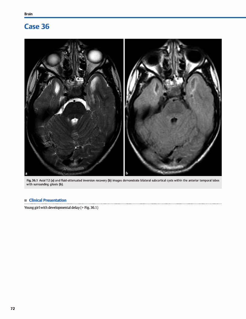
72
Brain
Case 36
Fig. 36.1 Axial T2 (a) and fluid-attenuated inversion recovery (b) images demonstrate bilateral subcortical cysts within the anterior temporal lobes with surrounding gliosis (b).
• Clinical Presentation
Young girl with developmental delay ( � Fig. 36.1 )
• Key lmaging Finding
Subcortical cysts in a child
• Top 3 Differential Diagnoses
• Cytomegalovirus {CMV). CMV is the most common in utero TORCH infection. lt may result from a primary infection during gestation or reactivation of a latent virus. Affected infants suffer from developmental delay and sensorineural hearing loss. As with other TORCH infections, the degree of insult is mostly dependent on the timing of infection in terms of development with more severe manifestations occurring
earlier in gestation. The disease is often systemic with growth retardation, hepatosplenomegaly, jaundice, and a petechial rash. Central nervous system {CNS) manifestations indude microcephaly, cortical malformations, ventriculomegaly, parenchymal calcifications with a periventricular distribution, and hypomyelination with increased T2 signal intensity predominantly within the deep white matter. Germinolytic cysts
commonly form with a predilection for the subependymal region, periventricular white matter, and subcortical white matter of the anterior temporal lobes.
• Megalencephalic leukeoencephalopathy with subcortical cysts (MLC). Also known as van der Knaap disease, MLC is a leukodystrophy that results in diffuse white matter signal abnormality and regions of vacuolization. The white matter
becomes edematous early in the disease process, resulting in
• Diagnosis
Megalencephalic leukeoencephalopathy with subcortical cysts
y' Pearts
• CMV infection results in numerous CNS abnormalities and malformations, induding subcortical germinolytic cysts.
• MLC is a leukodystrophy that results in diffuse white matter signal abnormality and regions ofvacuolization/cysts.
Suggested Readings
de Vries L'i, Gwrardi H, Barth PG, Bok IJ\, Verboon-Maciolek MA, Groenendaal F. The
speârum of cranial ultrasound and magnetic resonanœ imaging abnonnalities
in congenital cytomegalovirus inrection. Neuropediatrics 2004; 35: 113-119
Tu YF, Chen C'l, Huang CC, Lee CS. Vacuolating megalencephalic leukoenœphalop
athywith mild dinical course validated by diffusion terumrimaging and MR spec
troscopy. Amj Neuroradiol 2004; 25: 1041-1045
Case 36
macrocephaly. In this phase, the primary differentials indude other leukodystrophies with macrocephaly, such as Canavan and Alexander disease. Enhancement is not typically seen. With time, the white matter edema dissipates, with persistence of diffuse signal abnormality, and vacuolating cysts
appear. Subcortical cysts affecting the anterior temporal lobes are common and characteristic of MLC. Subcortical cysts may also be seen in the frontal and parietal lobes.
• CADASIL Cerebral autosomal dominant arteriopathy with subcortical infarcts and leukoencephalopathy {CADASIL) is an inherited disorder that affects young to middle-aged adults. The arteriopathy primarily affects smalt vessels of the CNS.
Patients present with a history of migraine-type headaches and transient ischemic attacks. Early in the disease process, imaging demonstrates multifocal regions of nonspecific hypoattenuation {computed tomography) or T2 hyperintensity {magnetic resonanœ imaging) within the subcortical and deep white matter. As the disease progresses, frank infarcts occur with associated restricted diffusion in the acute setting. Subcortical infarcts affecting the frontal lobes and anterior
temporal lobes are fairly characteristic.
• CADASIL affects young to middle-aged adults with small-vessel ischemia and characteristic subcortical infarcts.
van den Boom R, Lesnik Oberstein �. Ferr.ui MD, Haan J, van Buchem MA. Cerebral autosomal dominant arteriopathy with subcortical infarcts and leukoenœphal
opathy: MR imaging findings at different ages-Jrd-6th decades. Radiology 2003;
229: 683-690
73
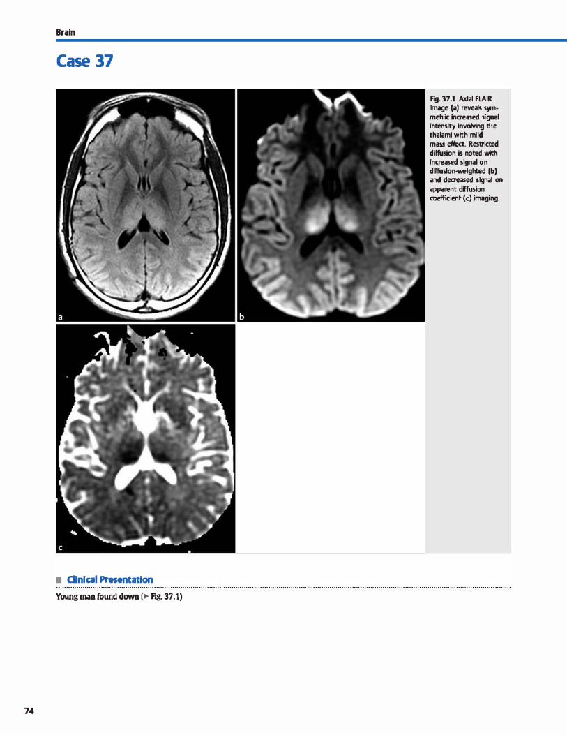
Brain
Case 37
• Cllnlcal Presentatlon
Young man found down (� F'ig. 37.1)
74
Fig. 37.1 Axlal FLAIR Image (a) reveals sytn· metric increased !ignal lntenslty tnvoMng the thalaml wtth mtld mass effect. Restrlcted diffusion is not:ed with lncreased signal on dlffuslon·welghted (b) and deaeased signal on ;ippuent diffusion coefficient (c) imaging.
• Key lmaging Finding
Bilateral thalamic signal abnormality
• Top 3 Differential Diagnoses
• Ischemia/infarct. Thalamic arterial supply arises from perforating branches of the posterior œrebral artery (PCA) and the posterior communicating artery (PComA). The PComA {when present) supplies the anterior thalami and the PCA supplies
the medial and lateral thalami. An artery of Percheron (AOP) is an anatomie variant where a single perforator supplies the bilateral medial thalami and anterior midbrain. Occlusion of
the AOP results in bilateral medial thalamic infarcts with or
without midbrain involvement Occlusion of the distal basilar artery is an additional cause of bilateral thalamic infarcts but
is characterized by more extensive ischemic changes. Acutely, there is bilateral hypoattenuation ( computed tomography, CT) and increased T2/fluid-attenuated inversion recovery (Fl.AIR)/ diffusion-weighted imaging signal with edema. Deep venous
infarcts are more common with hypercoagulable states and typically result from internai œrebral vein occlusion. Venous
infarcts do not follow a vascular distribution. Thrombosed venous structures are often hyperdense on CT. Venous infarcts
are more prone to hemorrhage. • lnfilttating neoplasm. Thalamic gliomas may result as an
extension of a brain stem glioma or as primary thalamic neo-
• Additional Differential Diagnoses
• Osmotic demyelination. Osmotic demyelination results from
rapid shifts in osmolality. It is dassically due ta rapid correction of hyponatremia, but may also be seen in malnourished and diabetic children. Fifty percent of cases involve the central pons, whereas the remaining are extrapontine with involve
ment of the basal ganglia, thalami, and cerebral white matter. Findings are typically bilateral and symmetric. Lesions are hypodense on CT and on T2/FLAIR hyperintense.
• Wemiclœ encephalopathy. Wemcike encephalopathy is
caused by thiamine deficiency. It is most common in alcoholic patients, but may also be seen with malabsorption or nutri
tional deficiencies. The dassic dinical triad consists of altered mental status, ataxia, and ocular dysfunction. MRI features include symmetric increased T2 signal within the medial
• Diagnosis
Infarct (venous)
� Pearls
• Arterial and venous ischemia are important causes of abnor
mal bithalamic signal; restricted diffusion is seen acutely.
• Thalamic gliomas occur in children and young adults; they may be primary or an extension of a brain stem glioma.
Suggested Readings
Case 37
plasms. They occur more frequently in children and young adults. Most are fibrillary (World Health Organization [WHO] grade II) but commonly degenerate to high-grade tumors, such as anaplastie (WHO grade Ill). Imaging reveals thalamic
expansion with hypoattenuation (CT) and increased T2/FLAIR signal {magnetic resonance imaging, MRI). Higher grade com
ponents demonstrate restricted diffusion, increased perfu
sion, and enhancement. Prognosis is poor. Central nervous system lymphoma can also involve the thalami but is charac
teristically hypointense on T2 sequences due ta high cellular
ity and avidly enhances. • Viral encephalitis. Mosquito-borne viral enœphalitides such
as West Nile, Eastern equine, and Japanese encephalitis are relatively common in endemic regions. Patients present with flu-like symptoms initially, followed by rapid progression
to meningitis or encephalitis with seizures and focal neurological deficits. Imaging reveals hypoattenuation (CT) and increased T2/FLAIR signal {MRI) with edema involving the
thalami, lentiform nudei, caudate, mesial temporal lobes, and brain stem. Patchy or meningeal enhancement, hemorrhage, and restricted diffusion may be seen.
thalami, periaqueductal gray matter, tectal plate, and mamil
lary bodies. Enhanœment may be seen. • Acute disseminated encephalomyelitis (ADEM). ADEM is a
monophasic demyelinating process that follows an anteœdent viral infection or immunization. It occurs most often in children and young adults. White matter lesions may mimic those of multiple sclerosis (MS) with ovoid periventricular lesions and involvement of the corpus callosurn, cerebellar
hemispheres, and brain stem. Compared with MS, ADEM is
more likely ta be confluent and involve gray matter, induding the thalami. Lesions are hyperintense on T2/FLAIR and may
demonstrate enhancement and restricted diffusion with active demyelination. Presence of characteristic white matter lesions is a useful discriminator.
• Infectious and parainfectious etiologies of thalamic signal abnormality indude viral encephalitis and ADEM.
Hegde AN, Mohan s, Lath N, I.im cc. Differential diagnosis for bilateral abnormalities Lazzaro NA, Wright B, ùstillo Met al. Artery ofperdleron infarttion: imaging pat-
ofthe basal ganglia and thalamus. Radiographies 2011; 31: 5-30 tems and clinical spectrum.Amj Neuroradiol 2010; 31: 1283-1289
75
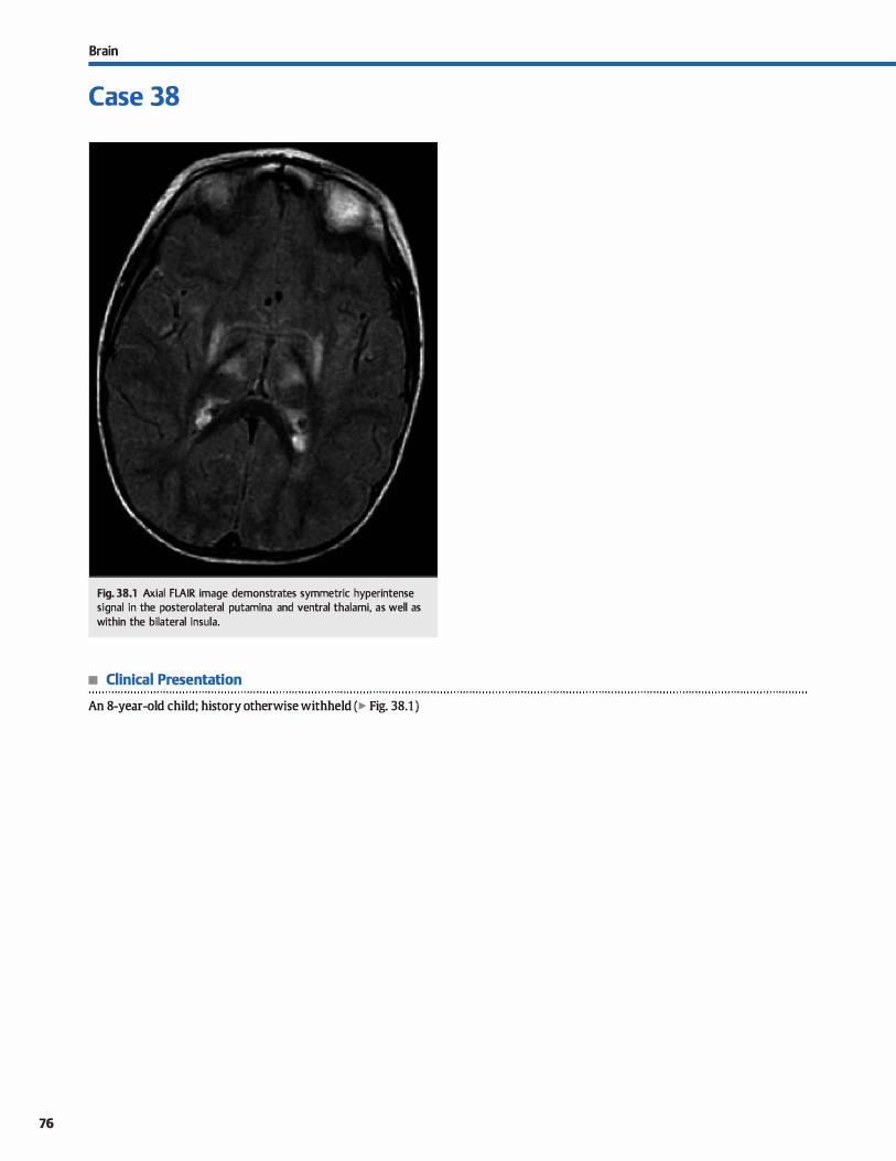
76
Brain
Case 38
Fig. 38.1 Axial FLAIR image demonstrates symmetric hyperintense signal in the posterolateral putamina and ventral thalami, as well as within the bilateral insula.
• Clinical Presentation
An 8-year-old child; history otherwise withheld (� Fig. 38.1 )
• Key lmaging Finding
Increased T2 signal in basal ganglia/thalami in a child
• Top 3 Differential Diagnoses
• Hypoxic-ischemic or anoxie injury. Severe hypoxic-ischemic injury predominantly affects the deep gray matter, which is
particularly wlnerable to hypoxia. The posterior putamina and ventrolateral thalami are most commonly involved. There
is also involvement of the gray-white matter junction, especially in watershed regions. Affected areas demonstrate T2/ fluid-attenuated inversion recovery (FLAIR) hyperintensity;
restricted diffusion is seen in the acute setting. Common eti
ologies in children indude trauma, hypoperfusion, and anoxie events. Deep venous infarcts are an important cause of bilat
eral increased T2 signal within the thalami and are prone to
hemorrhage. lbrombosed venous structures are often hyperdense on computed tomography (CT). Anoxie injury due to
inhalation of carbon monoxide gas results in symmetric hypodensity (CT) and T2 hyperintensity (magnetic resonanœ imaging, MRI) of the globus pallidi. A hypointense hemosid
erin rim may be seen. The caudate and putamina may also be affected. Confluent white matter hyperintensity reflects diffuse demyelination. Methanol poisoning preferentially
involves the putamina with hemorrhagic putaminal necrosis. Optic nerves may also be involved.
• Wilson disease. Wilson disease (hepatolenticular degeneration) is an autosomal reœssive disorder characterized by excessive copper accumulation in the liver and brain (particularly the basal ganglia� resulting in chronic ischemia. It results from defi-
• Additional Differential Diagnoses
• Kernicterus. Kernicterus refers to encephalopathy from deposition of unconjugated bilirubin. Acutely (within the first 2 ta 5 days of life), there is T1 and subtle T2 hyperintensity in the
bilateral globus pallidi, hippocampi, and substantia nigra.
Chronically, there is T2 hyperintensity in the posteromedial
globus pallidi and dentate nuclei with normal T1 signal. • Osmotic demyelination. Osmotic demyelination results from
rapid shifts in osmolality. It is classically due to rapid correc
tion of hyponatremia, but may also be seen in malnourished and diabetic children. Fifty percent of cases involve the central
pans, whereas the remaining are extrapontine with involve-
• Diagnosis
Hypoxic-ischemic insult (near drowning)
y' Pearts
• Deep gray matter is wlnerable to hypoxic-ischemic and anoxie encephalopathy, particularly in at-risk children.
• Deposition disorders (Wilson disease and kemicterus) involve the deep gray matter, particularly the basal ganglia.
Suggested Reading
Hegde AN. Mohan S, Lath N, I.im CC. Dilferential diagnosis for bilateral abnormalities
of the basal ganglia and thalamus. Radiographies 2011; 31: 5-30
Case 38
dency of œruloplasmin, which transports copper. There is
increased T2/FLAIR signal in the lentiform nudei, midbrain,
thalami, and white matter tracts. The characteristic "faœ ofthe
panda" sign on axial T2 images through the midbrain is due to
hyperintense tegmentum and hypointense superior colliculi. Associated abnormalities indude neuropsychiatrie illnesses, Kayser-Aeischer comeal rings, and cirrhosis.
• Metabolic disorder. Mitochondrial disorders result from
damage to energy-produdng mitochondria. MELAS (mito
chondrial myopathy, enœphalopathy, lactic acidosis, and
stroke-like episodes) is an uncommon but important cause of
stroke in children. There are ischemic findings in nonvascular distributions. Chronic atrophy of the basal ganglia and cortex occurs with T2 hyperintensity in the deep gray and white matter. I.eigh syndrome affects infants and children who present with failure to thrive, motor defidts, and seizures. MRI reveals symmetric increased T2 signal involving the deep gray
matter, particularly the basal ganglia and thalarni; dorsal brain stem; and Jess frequently, the supra- and infratentorial
white matter. MR spectroscopy reveals an elevated lactate peak. canavan disease is a leukodystrophy that results in diffuse abnormal signal within the white matter, thalami, and globus pallidus; macrocephaly; and increased N-acetyl aspar
tate on spectroscopy. juvenile Huntington disease is associated with caudate atrophy.
ment of the basal ganglia, thalami, and cerebral white matter.
Findings are typically bilateral and symmetric. Lesions are hypodense on CT and on T2/FLAIR hyperintense.
• Neurofibromatosis type 1 {NFt) spots. NFl is the most com
mon neurocutaneous syndrome. It is caused by a defect of
chromosome 17 that results in increased inàdence of neurofibromas and gliomas. Patients with NFl have regions of mye
lin vacuolization that are hyperintense on T2 sequences and involve the basal ganglia, thalami, dentate nuclei, cerebellar pedundes, brain stem, and optic radiations. I.esions wax and
wane for the first decade and then regress.
• Metabolic disorders affect deep gray matter structures; mitochondrial diseases demonstrate a lactate peak on MR spectroscopy.
77
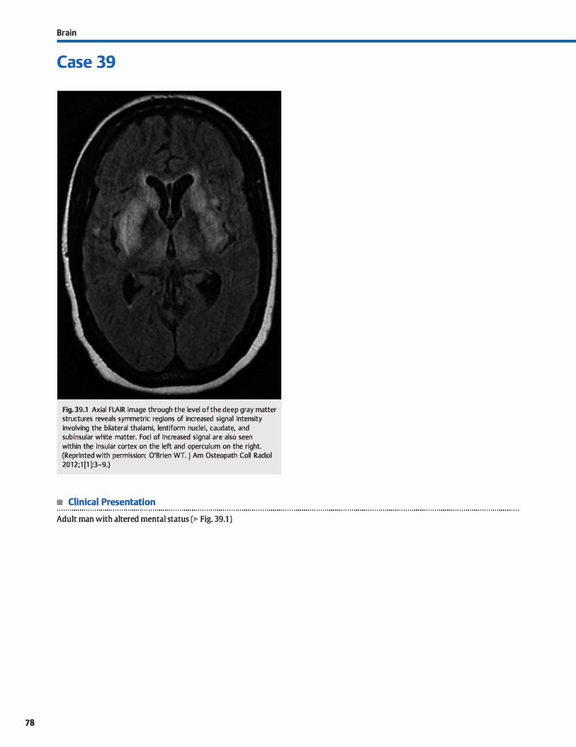
78
Brain
Case 39
Fig. 39.1 Axial FLAIR image through the level of the deep gray matter structures reveals symmetric regions of increased signal intensity involving the bilateral thalami, lentiform nudei, caudate, and subinsular white matter. Foci of increased signal are also seen within the insular cortex on the left and operculum on the right. (Reprinted with permission: O'Brien WT. J Am Osteopath Coll Radiol 2012;1 [1 ]:3-9.)
• Clinical Presentation
Adult man with altered mental status (� Fig. 39.1 )
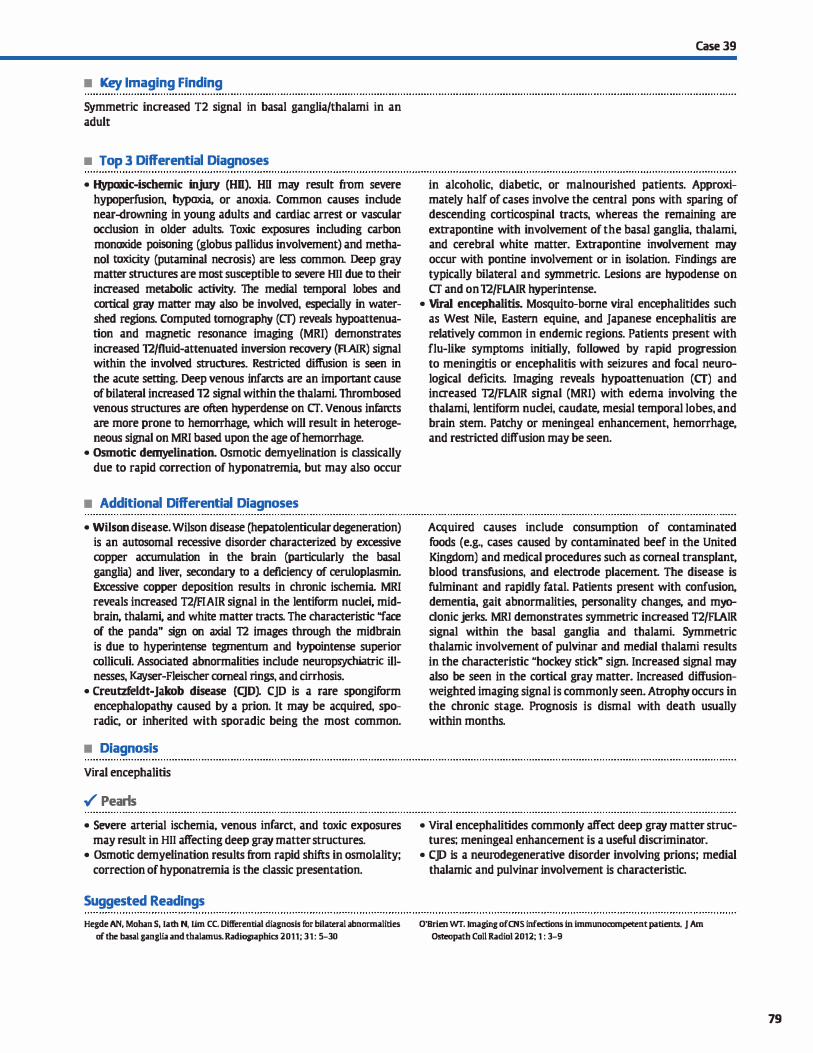
• Key lmaging Finding
Symmetric increased T2 signal in basal ganglia/thalami in an adult
• Top 3 Differential Diagnoses
• Hypaxic-ischemic injwy (HII). HII may result from severe hypoperfusion, hypoxia, or anoxia. Common causes indude near-drowning in young adults and cardiac arrest or vascular occlusion in older adults. Toxic exposures induding carbon monoxide poisoning (globus pallidus involvement) and methanol toxicity (putaminal necrosis) are Jess common Deep gray matter structures are most susceptible to severe HII due to their increased metabolic activity. The medial temporal lobes and cortical gray matter may also be involved, especially in watershed regions. Computed tomography (CT) reveals hypoattenuation and magnetic resonanœ imaging (MRI) demonstrates increased T2/fluid-attenuated inversion recovery (FLAIR) signal within the involved structures. Restricted diffusion is seen in the acute setting. Deep venous infarcts are an important cause of bilateral increased T2 signal within the thalami. Thrombosed venous structures are often hyperdense on CT. Venous infarcts are more prone to hemorrhage, which will result in heterogeneous signal on MRI based upon the age ofhemorrhage.
• Osmotic demyelination. Osmotic demyelination is dassically due ta rapid correction of hyponatremia, but may also occur
• Additional Differential Diagnoses
• Wilson disease. Wilson disease {hepatolenticular degeneration) is an autosomal reœssive disorder characterized by excessive copper accumulation in the brain (particularly the basal ganglia) and liver, secondary to a deficiency of œruloplasmin. Excessive copper deposition results in chronic ischemia. MRI reveals increased T2/FLAIR signal in the lentiform nudei, midbrain, thalami, and white matter tracts. The characteristic "face of the panda" sign on axial T2 images through the midbrain is due to hyperintense tegmentum and hypointense superior colliculi. Associated abnormalities indude neuropsychiatrie illnesses, Kayser-Fleischer comeal rings, and cirrhosis.
• Creutzfeldt-Jakob disease (CJD). CJD is a rare spongiform enœphalopathy caused by a prion. It may be acquired, sporadic, or inherited with sporadic being the most common.
• Diagnosis
Viral encephalitis
� Pearts
• Severe arterial ischemia, venous infarct, and toxic exposures may result in HII affecting deep gray matter structures.
• Osmotic demyelination results from rapid shifts in osmolality; correction of hyponatremia is the dassic presentation.
Suggested Readings
Case 39
in alcoholic, diabetic, or malnourished patients. Approximately half of cases involve the central pons with sparing of descending corticospinal tracts, whereas the remaining are extrapontine with involvement of the basal ganglia, thalami, and cerebral white matter. Extrapontine involvement may occur with pontine involvement or in isolation. Findings are typically bilateral and symmetric. Lesions are hypodense on CT and on T2/FLAIR hyperintense.
• Viral enœphalitis. Mosquito-borne viral enœphalitides such as West Nile, Eastern equine, and japanese encephalitis are relatively common in endemic regions. Patients present with flu-like symptoms initially, followed by rapid progression to meningitis or encephalitis with seizures and focal neurological deficits. Imaging reveals hypoattenuation (CT) and increased T2/FLAIR signal (MRI) with edema involving the thalami, lentiform nudei, caudate, mesial temporal lobes, and brain stem. Patchy or meningeal enhancement, hemorrhage, and restricted diffusion may be seen.
Acquired causes include consumption of contaminated foods ( e.g., cases caused by contaminated beef in the United Kingdom) and medical procedures such as corneal transplant, blood transfusions, and electrode placement The disease is fulminant and rapidly fatal. Patients present with confusion, dementia, gait abnormalities, personality changes, and myodonic jerks. MRI demonstrates symmetric increased T2/FLAIR signal within the basal ganglia and thalami. Symmetric thalamic involvement of pulvinar and media] thalami results in the characteristic ühockey stick" sign. Increased signal may also be seen in the cortical gray matter. Increased diffusionweighted imaging signal is commonly seen. Atrophy occurs in the chronic stage. Prognosis is dismal with death usually within months.
• Viral encephalitides commonly affect deep gray matter structures; meningeal enhancement is a useful discriminator.
• CJD is a neurodegenerative disorder involving prions; media! thalamic and pulvinar involvement is characteristic.
Hegde AN, Mahan s, Lath N, Iim cc. Dilferential diagnasis far bilateral abnarmalities O'Brien WT, lmaging afCNS infections in immunorompetent patients, J Am
of the basal ganglia and thalamus. Radiographies 2011; 31: 5-30 Osteapath Cali Radial 2012; 1 : 3-9
79
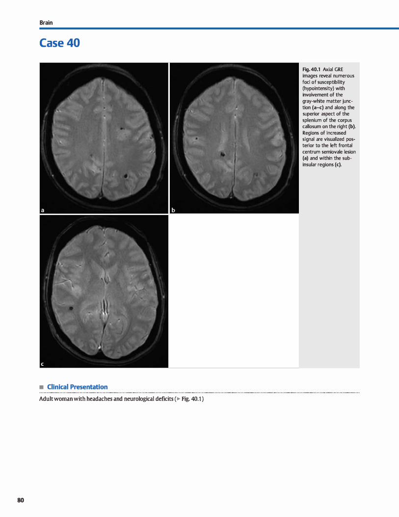
Brain
Case 40
• Clinical Presentation
Adult woman with headaches and neurological deficits (� Fig. 40.1 )
80
Fig. 40.1 Axial GRE images reveal numerous foci of susceptibility (hypointensity) with involvement of the gray-white matter junction (a-c) and along the superior aspect of the splenium of the corpus callosum on the right (b). Regions of increased signal are visualized posterior to the left frontal centrum semiovale lesion (a) and within the subinsular regions (c).
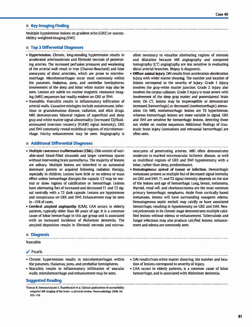
• Key lmaging Finding
Multiple hypointense lesions on gradient echo (GRE) or susceptibility-weighted imaging (SWI)
• Top 3 Differential Diagnoses
• Hypertension. Chronic, long-standing hypertension results in acœlerated arteriosderosis and fibrinoid necrosis of penetrat
ing arteries. The increased perfusion pressures and weakening of the arterial wall result in true {Charcot-Bouchard) and false aneurysms of distal arterioles, which are prone to microhemorrhage. Microhemorrhages occur most commonly within the putamen, thalamus.. pons, and œrebellar hemispheres. Involvement of the deep and lobar white matter may also be seen. I.esions are subtle on routine magnetic resonanœ imag
ing (MRI) sequences but readily evident on GRE or SWL • Vasculitis. Vasculitis results in inflammatory infiltration of
arterial walls. causative etiologies include autoimmune, infectious or granulomatous disease, radiation, and elidt drugs. MRI demonstrates bilateral regions of superficial and deep gray and white matter signal abnonnality {increased T2/fluidattenuated inversion recovery [FLAIR] signal intensity). GRE and SWI commonly reveal multifocal regions of microhemorrhage. Patchy enhanœment may be seen. Angiography is
• Additional Differential Diagnoses
• Multiple cavemous malfonnations (CMs). CMs consist ofvari
able-sized blood-filled sinusoids and larger cavemous spaces without intervening brain parenchyma. The majority of lesions are solitary. Multiple Iesions are inherited in an autosomal dominant pattern or acquired following radiation therapy, espedally in children. I.esions have little or no edema or mass etfect unless hemorrhage disrupts the capsule. cr may be normal or show regions of calcification or hemorrhage. I.esions
have altemating foci of increased and decreased 11 and T2 signal œntrally with a T2 dark capsule. I.esions are hypointense and conspicuous on GRE and SWI. Enhanœment may be seen in -15% of cases.
• Cerebral amyloid angiopathy {CAA). CM occurs in elderly patients, typically aider than 60 years of age. It is a common cause of lobar hemorrhage in this age group and is associated with an increased incidence of Alzheimer dementia. The
amyloid deposition results in fibrinoid necrosis and microa-
• Diagnosis
Vasculitis
y' Pearts
• Chronic hypertension results in microhemorrhages within the putamen, thalamus, pans, and œrebellar hemispheres.
• Vasculitis results in inflammatory infiltration of vascular walls; microhemorrhage and enhancement may be seen.
Suggested Reading
Thomas B, Somasundaram s, lbamburaj K et al. Clinical applications of susœptibility
weighted MR imaging of the brain-a pictorial review, Neuroradiology 2008; 50;
105-116
Case 40
often necessary to visualize altemating regions of stenosis and dilatation because MR angiography and computed tomography {CT) angiography are Jess sensitive in evaluating distal arterial branches. Biopsy is diagnostic.
• Diffuse axonal injwy. DAI results from acceleration-deceleration injury with white matter shearing. The number and location of lesions correspond to the severity of injury. Grade 1 injury
involves the gray-white matter junction. Grade 2 injury also involves the corpus callosum. Grade 3 injury is most severe with involvement of the deep gray matter and posterolateral brain stem On cr, lesions may be imperceptible or demonstrate increased (hemorrhagic) or decreased {nonhemonhagic) attenuation. On MRI, nonhemorrhagic lesions are T2 hyperintense, whereas hemorrhagic lesions are more variable in signal GRE and SWI are sensitive for hemorrhagic lesions, detecting those
not visible on routine sequenœs. Additional findings of traumatic brain injury (contusions and extraaxial hemonhage) are often seen.
neurysms of penetrating arteries. MRI often demonstrates moderate to marked microvascular ischemic disease, as well as multifocal regions of GRE and SWI hypointensity with a lobar, rather than deep, predominance.
• Hemat:ogenous spread of tumor or infection. Hemonhagic metastases present as multiple fod of decreased signal intensity on GRE and SWI. Tl and T2 signal intensity depends on the size of the lesions and age of hemorrhage. Lung. breast, melanoma,
thyroid, renal œll, and choriocarcinoma are the most common primary hemonhagic neoplasms. Aside from cortically based metastases, Iesions will have surrounding vasogenic edema. Hematogenous septic emboli may calcify or have associated hemorrhage, resulting in hypointensity on GRE and SWI. Neurocystiœrcosis in its chronic stage demonstrates multiple caldfied lesions without edema or enhanœment Tuberculosis and
fungal infections may also produœ caldfied lesions; enhanœment and edema are commonly seen.
• DAI results from white matter shearing; the number and location of Iesions correspond to severity of injury.
• CM occurs in elderly patients, is a common cause of lobar hemorrhage, and is associated with Alzheimer dementia.
81
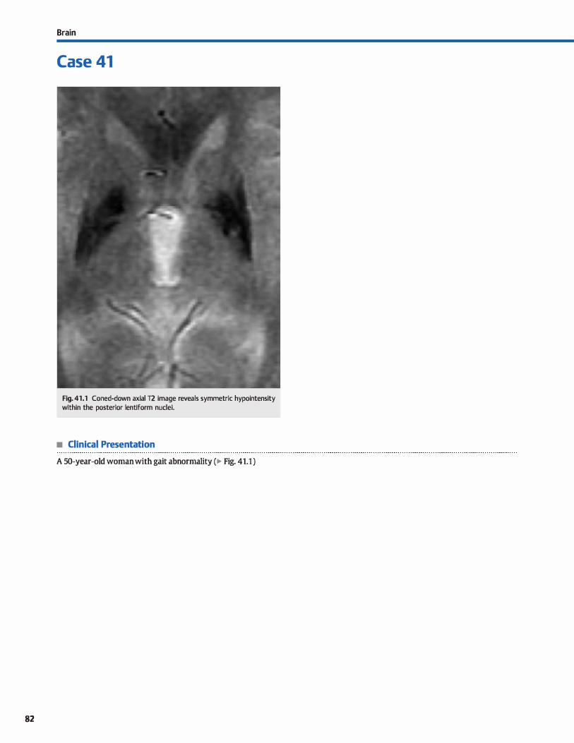
82
Brain
Case 41
Fig. 41.1 Coned-down axial T2 image reveals symmetric hypointensity within the posterior lentiform nudei.
• Clinical Presentation
A 50-year-old woman with gait abnormality (� Fig. 41.1 )
• Key lmaging Finding
Decreased T2 signal in basal ganglia
• Top 3 Differential Diagnoses
• Nonnal aging. In middle-aged and older patients, decreased T2 signal may be seen within the basal ganglia, predominantly the globus pallidus, secondary ta a combination of increased iron deposition and age-related senesœnt calcifications. Early onset of senescent calcification may occur in patients with Down syndrome. Associated findings of parenchymal atrophy and microvascular ischemic disease are commonly seen.
• Basal ganglia calcification. There are numerous causes of basal ganglia calcifications with senescent age-related calcifications being the most common. Additional causes of basal ganglia calcifications indude metabolic disorders (mitochondrial disorders), endocrinopathies (abnormalities of calcium and phosphate or hypothyroidism), severe hypoxic-ischemic enœphalopathy, postinfection (TORCH, congenital human immunodeficiency virus, tuberculosis, and neurocystiœrcosis ), prior radiation and chemotherapy treatment, and Fahr disease. ln general, nonsenesœnt calcifications have more extensive involvement of additional deep gray matter structures and occasionally the subcortical white matter. calcifications are typically hypointense on T2 and variable in signal intensity on Tl sequences.
• Additional Differential Diagnoses
• Demyelinating disease. Demyelinating disease consists of multiple sclerosis (MS) and acute disseminated encephalomyelitis (ADEM). MS is most often relapsing and remitting, whereas ADEM is monophasic following an anteœdent viral infection or immunization. Compared with MS, ADEM more often involves children, tends ta be more confluent, and more commonly involves gray matter. Focal lesions within the deep gray matter are typically T2 hyperintense. With chronic disease, increased iron deposition results in decreased T2 signal intensity within the deep gray matter structures. Associated white matter lesions with characteristic morphology ( ovoid,
• Diagnosis
Neurodegenerative disorder (Parkinson disease)
� Pearls
• With aging, increased iron deposition and senescent calcifications result in decreased T2 signal in the globus pallidus.
• Iron deposition is common in neurodegenerative diseases, including PKAN, Alzheimer, Parkinson, and Huntington.
Suggested Readings
Case 41
• Neurodegenerative disorder. Panthothenate kinase-associated neurodegeneration {PKAN1 formerly known as HalloverdenSpatz disease, is an uncommon neurodegenerative disorder characterized by iron deposition in the globus pallidus and substantia nigra Patients most often present in childhood with ataxia, dystonia, and visual abnormalities. lmaging reveals bilateral T2 hypointensity within the globus pallidus with central hyperintensity, referred to as the "eye of the tiger" sign. Hypointensity of additional deep gray matter structures and nudei may be seen. Less common disorders of neurodegeneration with brain iron accumulation (NBIA) or neuroferrinopathies demonstrate decreased T2 signal in a similar distribution but without the eye of the tiger appearanœ. Parkinson, Alzheimer, and Huntington disease have been associated with increased iron deposition. Parkinson disease is a movement disorder that involves the pars compacta of the substantia nigra. lt typically affects aider adult patients who present with bradykinesia, resting tremor, Mcogwheel" rigidity, and a shuftling gait Huntington disease is an autosomal dominant disorder {chromosome 4) that results in progressive dementia and choreatic movement disorder. Imaging reveals caudate atrophy.
perivenular) and distribution (periventricular, corpus callosum, cerebellum, and brain stem) are also seen.
• Hemochrom.atosis. Hemochromatosis is a disorder of iron overload and may be primary (inherited) or secondary (usually acquired from multiple transfusions� In the abdomen, primary hemochromatosis involves the liver and pancreas, whereas secondary hemochromatosis involves the Iiver and spleen. Central nervous system involvement is relatively uncommon and occurs late in the disease proœss. Imaging fmdings, when present indude decreased T2 signal within the basal ganglia; patients may demonstrate extrapyrimidal findings.
• Chronic MS plaques may have iron deposition; characteristic T2 hyperintense demyelinating lesions are often seen.
Hegde AN, Mohan S, Lath N, lim CC. Differential diagnosis for bilateral abnonnalities Stankiewiczj, Panter SS. Neema M, AroraA. Batt CE, Babhi R. Iron in chronic brain
of the basal ganglia and thalamus. Radiographies 2011; 31: 5-30 disorders; imaging and neurotherapeutic implications, Neurotherapeutics 2007;
4: 371-386
83
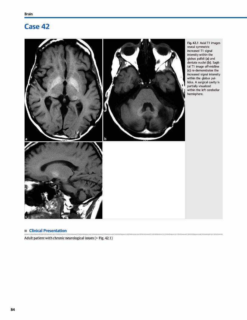
Brain
Case 42
• Clinical Presentation
Adult patient with chronic neurological issues (� Fig. 42.1 )
84
Fig. 42.1 Axial Tl images reveal symmetric increased Tl signal intensity within the globus pallidi (a) and dentate nudei (b). Sagittal Tl image off-midline (c) re-demonstrates the
increased signal intensity within the globus pallidus. A surgical cavity is partially visualized within the left cerebellar hemisphere.
• Key lmaging Finding
Symmetric increased Tl signal in basal ganglia
• Top 3 Differential Diagnoses
• Basal ganglia (BG) calcification or minerali7.ation. There are numerous causes of BG calcification with senescent age-related calcifications being the most common. Senescent calcifications occur in adults and preferentially involve the globus pallidi. Early onset of senesœnt calcification may occur in patients with Dawn syndrome. Additional causes of BG calcification or mineralization include metabolic disorders (mitochondrial
disorders1 endocrinopathies (abnormalities of calcium and phosphate or hypothyroidismi severe hypoxic-ischemic encephalopathy, postinfection, prior radiation and chemotherapy, and Fahr disease. Nonsenescent calcification or mineralization has more extensive involvement of deep gray matter structures and occasionally involves white matter.
• Manganese deposition. Exœss manganese deposition results in increased Tl signal intensity within the basal ganglia sec
ondary to paramagnetic effects. Common etiologies include
• Additional Differential Diagnoses
• Neurodegenerative disorders. Neurodegenerative disorders commonly involve the basal ganglia. Those that may result in hyperintense Tl signal indude Wilson disease and pantothenate kinase-assodated neurodegeneration (PKAN or Hallovorden-Spatz disease). Wilson disease results in excessive
copper deposition within deep gray matter structures, particularly the BG, secondary to a deficiency in ceruloplasmin. The liver is also affected. In children, increased Tl signal
intensity is seen in the BG. In adults, there is increased T2/ fluid-attenuated inversion recovery signal in the lentiform nuclei, midbrain, thalami, and white matter tracts. The characteristic "face of the panda" sign on axial T2 images through the midbrain is due to hyperintense tegmentum and hypoin
tense superior colliculi. PKAN results in excessive iron deposition within the globus pallidi. Imaging reveals central T2 hyperintensity with peripheral hypointensity, referred to as the Meye of the tiger" sign. Tl signal intensity is variable with regions that are hypo- and hyperintense.
• Diagnosis
Basal ganglia mineralization (postradiation therapy)
� Pearls
• Calcification or mineralization are common causes of
increased Tl signal intensity in the BG. • Manganese deposition most often results from hepatic failure
(typically chronic) or long-term parenteral nutrition.
Suggested Readings
Lai PH, Chen c, Liang HL, Pan HB. Hyperintense basal ganglia on TI-weighted MR
imaging.Amj Roentgenol 1999; 172: 1109-1115
Case 42
hepatic encephalopathy secondary to chronic or occasionally acute hepatic failure, as well as patients receiving long-term parenteral feeding for chronic illnesses. The manganese deposition predominantly affects the globus pallidi and substantia nigra.
• Hypoxic-ischemic or anoxie encephalopathy. Severe hypoxic-ischemic and anoxie encephalopathy affects the most
metabolically active portions of the brain, particularly the deep gray matter structures. Causes include hypoperfusion,
near drowning, and toxic exposures, such as carbon monoxide poisoning. Computed tomography reveals hypoattenuation of the deep gray matter structures, as well as the perirolandic gray and white matter. On magnetic resonanœ imaging, regions of increased Tl and T2 signal may be seen. Restricted
diffusion is noted with acute infarcts. Chronically, there is volume Joss with calcifications.
• Neurofibromatosis type 1 (NFt). Central nervous system manifestations ofNFl indude characteristic NF "spots," which are thought to represent regions of myelin vacuolization. They involve the BG, thalami, dentate nuclei, cerebellar pedundes, optic radiations, and brain stem. The lesions are hyperintense
on T2 and typically mildly hyperintense on Tl images. There may be apparent but not true mass effect, especially with
lesions located within the thalami, cerebellar pedundes, and brain stem. However, there should be no enhancement; the presence of enhancement suggests development of a lowgrade glioma. Lesions wax and wane for the first decade of life
and then regress.
• Kemicterus. Kemicterus refers to encephalopathy from deposition of unconjugated bilirubin. Acutely (within the first 2 to
5 days of life), there is Tl and subtle T2 hyperintensity in the globus pallidi, hippocampi, and substantia nigra. Chronically,
there is T2 hyperintensity in the posteromedial globus pallidi and dentate nuclei with normal Tl signal.
• Neurodegenerative disorders (Wilson disease and PKAN) may
occasionally result in increased BG Tl signal. • NF1 spots are mildly hyperintense on Tl sequences; they wax
and wane for the first decade of life and then regress.
Rl:lvira A. Alonso J, C6rdobaJ. MR imaging findings in hepatic enœphalopathy. AmJ
Neuroradiol 2008; 29: 1612-1621
85
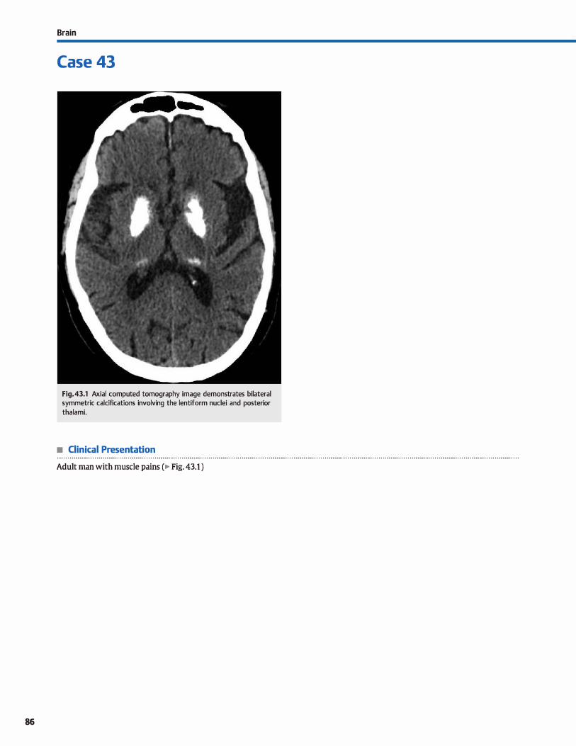
86
Brain
Case 43
Fig. 43.1 Axial computed tomography image demonstrates bilateral symmetric calcifications involving the lentiform nuclei and posterior thalami.
• Clinical Presentation
Adult man with muscle pains (� Fig. 43.1 )
• Key lmaging Finding
Basal ganglia calcifications
• Top 3 Differential Diagnoses
• Senescent calcifications. Senescent calcifications involve the lentiform nuclei in adult patients. They are incidental, considered a normal part of the aging process, and may increase over time. The globus pallidi are involved to a greater degree than the putamina. Involvement of the caudate or thalami should suggest an underlying condition other than senesœnt calcifications. Patients with Down syndrome experienœ accelerated effects of aging within the central nervous system. These include early onset of senesœnt basal ganglia calcifications, parenchymal atrophy, and dementia.
• Metabolic or Endocrine disease. Abnormalities of calcium or phosphorus are common causes of parenchymal calcifications. The Ientiform nuclei are most often involved. The caudate, thalami, dentate nuclei, and subcortical white matter may also be affected. Tertiary hyperparathyroidism may result in durai and scierai calcifications along with or in absence of parenchymal calcifications. Hypothyroidism is another cause of parenchymal calcification with a similar distribution. There are numerous variants of mitochondrial disorders that ultimately affect energy production due to enzyme deficiencies. Basal ganglia involvement is characteris
tic. Mitochondrial encephalopathy with Iactic acidosis and stroke-like episodes {MELAS) and Leigh syndrome are the two most common to result in basal ganglia calcifications in the chronic setting. The putamina are involved to a greater extent than the caudate or globus pallidi. Associated basal
• Additional Differential Diagnoses
• li'eatment-relaœd. Radiation and chemotherapy treatment result in a mineralizing microangiopathy with associated parenchymal injury and calcifications. The mineralizing angiopathy generally occurs a few years after treatment and most often involves the deep gray matter structures and subcortical white matter. Atrophy occurs in chronic stages. Patients treated at a younger age are more prone to treatment-related parenchymal injury.
• Diagnosis
Endocrine disease (hyperparathyroidism)
� Pearls
• Senescent calcifications are considered a normal part of aging and preferentially involve the globus pallidi.
• Endocrinopathies result in extensive deep gray matter calcifications and occasional white matter involvement.
Suggested Readings
Case 43
ganglia and white matter signal abnormalities are commonly seen.
• lschemic or infectious injury. Severe hypoxic-ischemic injury affects the most metabolically active portions of the brain, induding the deep gray matter structures. There is edema acutely with hypoattenuation on computed tomography and increased T2 and diffusion-weighted imaging signal on magnetic resonance imaging. In the chronic stages, atrophy and calcification may be seen. Carbon monoxide poisoning may produce similar findings with a predilection for the globus pallidi. An appropriate history is the key to making the diagnosis. Numerous infectious agents may result in basal ganglia calcifications, although additional findings are commonly present ta aid in identifying the organism. In utero eytomegalovirus (CMV) or toxoplasmosis infections result in parenchymal calcifications: CMV has a periventricular distribution, while toxoplasmosis is more random. Tuberculosis most often results in a basilar meningitis. Intraparenchymal tuberculomas occur in a minority of patients and present with intraparenchymal lesions that enhance and may calcify. Neurocystiœrcosis is the most common cause of acquired seizures worldwide and occurs in endemic locations. Imaging appearanœ depends on the stage of infection; the chronic, nodular stage results in calcified parenchymal lesions. Basal ganglia calcifications may also be seen in patients with human immunodeficiency virus, particularly children.
• Fahr disease. Fahr disease is a rare inherited neurodegenerative disorder that leads to symmetric intracranial calcifications involving predominantly the deep gray matter structures. Characteristic locations indude the globus pallidi (most common). putamina, caudate, thalami, and dentate nudei. Calcifications within the white matter are often seen. Fahr disease most often affects young and middle-aged adults with neurocognitive or neuromotor deficits.
• Radiation and chemotherapy may cause a mineralizing microangiopathy with resultant parenchymal calcifications.
• Fahr disease is an inherited disorder with extensive parenchymal calcifications and cognitive and motor deficits.
Hegde AN, Mohan S, Lath N, lim CC. Dilferential diagnosis for bilateral abnormalities Makariou E, Patsalides AD. Intracranial caldfications. Appl Radiol 2009; 38; 48-60
of the basal ganglia and thalilillus. Radiographies 2011; 31; 5-30
87
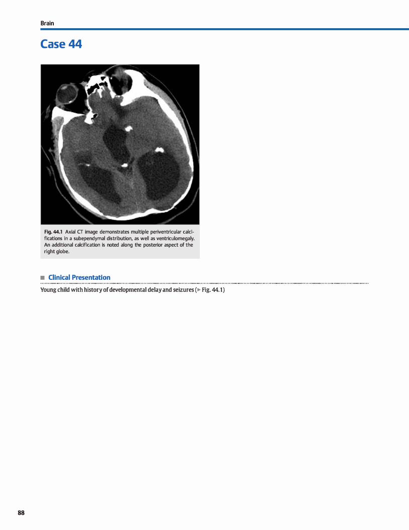
88
Brain
Case 44
Fig. 44.1 Axial CT image demonstrates multiple periventricular calcifications in a subependymal distribution, as well as ventriculomegaly. An additional calcification is noted along the posterior aspect of the right globe.
• Clinical Presentation
Young child with history of developmental delay and seizures (� Fig. 44.1)
• Key lmaging Finding
Periventricular calcifications in an infant/child
• Top 3 Differential Diagnoses
• TORCH infection. The TORCH infections consist of toxoplasmosis, syphilis, rubella. cytomegalovirus (CMV), herpes simplex virus, and human immunodeficiency virus (HIV). CMV and toxoplasmosis are both the most comrnon in incidence, as well as the most common to result in parenchymal calcifications. The calcifications in CMV are characteristically peri
ventricular in distribution, whereas those associated with
toxoplasmosis are more random. Associated central nervous
system (CNS) findings with TORCH infections indude microcephaly, neuronal migration abnormalities, and cortical malformations, particularly with CMV. Œnically, patients often suffer from seizures, mental retardation, and varying degrees
ofhearing and visual Joss.
• Tuberous sderosis (TS). TS may occur sporadically or in an autosomal dominant fashion with variable penetrance. The
classic clinicat triad consists of facial angiofibromas, mental retardation, and seizures but is only seen in approximately
30% of cases. CNS manifestations indude subependymal nodules, cortical tubers, white matter lesions, and retinal hamartomas. The majority of subependymal nodules calcify by end
of the second decade and many demonstrate enhancement
Nodules have variable T1 and T2 signal, but demonstrate T2
gradient echo susceptibility when calcified. Subependymal giant cell astrocytomas (World Health Organization grade I)
are seen in approximately 15% of cases. They are located at the foramen of Monro, enhance, and enlarge over time. Cortical tubers and white matter lesions occur in the vast majority
of cases. Tubers are wedge-shaped and T1 hyperintense prior
• Diagnosis
Tuberous sclerosis (subependymal nodules and retinal hamartoma)
y' Pearts
• CMV and toxoplasmosis are the most common TORCH infections to result in parenchymal calcifications.
• CMV calcifications are characteristically periventricular in dis
tribution; toxoplasmosis calcifications are random.
Suggested Readings
Blankenberg FG, Loh NN, Bracd Pet al. Sonography, CT, and MR imaging: a prospec
tive comparison of neonates with suspected intraaanial ischernia and hemor
rhage. AmJ Neuroradiol 2000; 21: 213-218
Case 44
to myelination with variable T1 and hyperintense T2 signal
intensity after myelination. White matter lesions are linear T2/fluid-attenuated inversion recovery hyperintensities along the radial migration lines from the ventride to the overlying cortex.
• Prior germinal matrix hemorrltage {GMH). GMH occurs in premature infants with a low birth weight, typically within
the first few days ta the first week of Iife. The underdevel
oped, highly vascular GM is prone to hemorrhage associated with the stresses of prematurity. The degree and distribution
of hemorrhage is divided into four grades with grades 1 and
2 having a favorable prognosis and grades 3 and 4 having a worse prognosis. Grade 1 hemorrhage is limited to the caudothalamic groove. In grade 2, there is intraventricular exten
sion of the hemorrhage without ventriculomegaly. With
grade 3, there is intraventricular hemorrhage, which causes
ventriculomegaly. Grade 4 presents with GMH, intraventricular hemorrhage, and parenchymal hemorrhage within the periventricular white matter, Iikely due to venous infarction. Head ultrasound {US) reveals regions of increased echogenic
ity within the regions of hemorrhage. The hemorrhage is
hyperdense on computed tomography (CT). Magnetic reso
nance imaging (MRI) appearance is dependent upon the location and age of hemorrhage. Blooming hypointensity is noted
on gradient echo and susceptibility-weighted sequences.
With chronic hemorrhage and/or ischemia, dystrophie subependymal and periventricular calcifications may be seen. Overall, CT and MRI are more sensitive than US.
• Oassic triad ofTS indudes facial angiofibromas, mental retardation, and seizures; subependymal nodules calcify.
• Chronic GMH and associated ischemia may result in dys
trophie subependymal and periventricular calcifications.
Makariou E, Patsalides AD. Intracranial calcificatioll5. Appl Radio! 2009; 38: 48-60
89
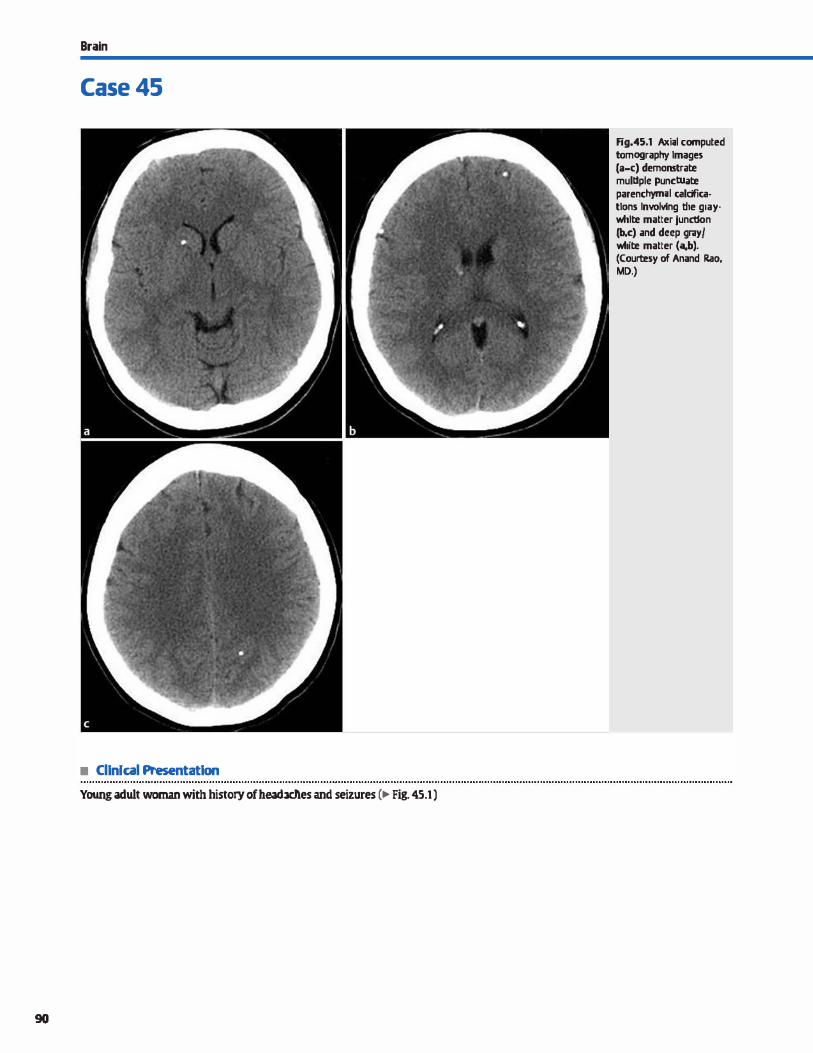
Brain
Case 45
• Cllnlcal Presentatlon
Young adult woman with history of headaches and seizures (� Fig. 45.1)
90
Fig.45.1 Axi;tl computed tomography lmagn (a-c) demonstrate multlple punctuate parenchyrmtl calcffica· tians lnwlving the gray. white matter junctlon (b,c) and deep gray/ white matter ("4b). (Courtesy of Anand Rao, MD.)
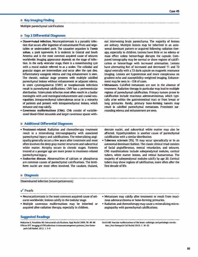
• Key lmaging Finding
Multiple parenchymal calcifications
• Top 3 Differential Diagnoses
• Disseminated infection. Neurocysticercosis is a parasitic infection that occurs after ingestion of contaminated fruits and vegetables or undercooked porl<. The causative organism is Taenia solium, a pork tapewonn. lt is endemic in Central and South America and is the most common acquired cause of seizures worldwide. lmaging appearanœ depends on the stage of infection. ln the early vesicular stage, there is a nonenhandng cyst
with a mural nodule referred to as a scolex. The colloïdal and granular stages are intermediate and occur after the cyst dies. Inflammatory vasogenic edema and ring enhancement is seen. The chronic, nodular stage presents with multiple caldfied parenchymal lesions without enhanœment or adjacent edema. ln utero cytomegalovirus {CMV) or toxoplasmosis infections result in parenchymal calcifications; CMV has a perivennicular
distribution. Tubertulosis infection most often results in a basilar meningitis with avid meningeal enhancement and cranial neuropathies. lntraparendtymal tuberculomas occur in a minority of patients and present with intraparenchymal lesions, which enhance and may caldfy.
• cavemous malformations (CMs). CMs consist of variablesized blood-filled sinusoids and larger cavernous spaœs with-
• Additional Differential Diagnoses
• lreatment-related. Radiation and chemotherapy treatment result in a mineralizing microangiopathy with associated parenchymal injury and calcifications. The mineralizing angiopathy generally occurs a few years after treatment and most often involves the deep gray matter structures and subcortical white matter. Atrophy occurs in chronic stages. Patients treated at a younger age are more prone ta treatment-related
parenchymal injury. • Endocrine disease. Abnormalities of calcium or phosphorus
are common causes of parenchymal calcifications. The Ienti
form nuclei are most often involved. The caudate, thalami,
• Diagnosis
Disseminated infection {neuorcystiœrcosis)
� Pearts
• Neurocysticercosis is the most common acquired cause of seizures worldwide; lesions calcify in the nodular stage.
• Multiple cavernous malformations may be inherited or acquired after radiation therapy, especially in children.
Suggested Readings
Makariou E, Patsalides AD. Intracranial calcificatiom. Appl Radial 2009; 38; 48--60
O'Brien Wf. Imaging af CNS infectiom in immunocompetent patients.} Am Osteo
path Coll Radiol 2012; 1: 3-9
Case 45
out intervening brain parenchyma. The majority of lesions are solitary. Multiple lesions may be inherited in an autosomal dominant pattern or acquired following radiation therapy, especially in children. Lesions have little or no edema or
mass effect unless hemorrhage disrupts the capsule. Computed tomography may be normal or show regions of calcifi
cation or hemorrhage with increased attenuation. Lesions have altemating foci of increased and decreased Tl and T2 signal centrally with a T2 dark capsule on magnetic resonanœ imaging. Lesions are hypointense and more conspicuous on gradient echo and susceptibility-weighted imaging. Enhancement may be seen in -15% of cases.
• Metastases. Calcified metastases are rare in the absence of treatment Radiation therapy in particular may lead to multiple
regions of parenchymal calcification. Primary tumors prone to calcification include mucinous adenocarcinomas, which typically arise within the gastrointestinal tract or from breast or
lung primaries. Rarely, primary bone-fbrming tumors may result in calcified parenchymal metastases. Prominent surrounding edema and enhancement are seen.
dentate nudei, and subcortical white matter may also be
affected. Hypothyroidism is another cause of parenchymal calcification with a similar distribution.
• Tuberons sderosis {TS). TS may occur sporadically or in an autosomal dominant fashion. The classic dinical triad consists of fadai angiofibromas, mental retardation, and seizures. CNS manifestations indude subependymal nodules, cortical
tubers, white matter lesions, and retinal hamartomas. The
majority of subependymal nodules calcify by age 20. Cortical tubers may show regions of calcification, more often after the
first decade of life.
• Metastases may caldfy after treatment or result from mucinous adenocardnoma or bone-forming primaries.
• Radiation and chemotherapy may cause a mineralizing microangiopathy with parenchymal calcifications.
Smith AB. Vascular malformations of the brain; radiologie and pathologie correla
tion J Am Osteopath Coll Radial. 2012; 1 : 10-22
91
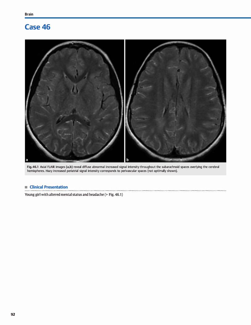
92
Brain
Case 46
Fig. 46.1 Axial FLAIR images (a,b) reveal diffuse abnormal increased signal intensity throughout the subarachnoid spaces overlying the cerebral hemispheres. Hazy increased periatrial signal intensity corresponds to perivascular spaces (not optimally shown).
• Clinical Presentation
Young girl with altered mental status and headache (� Fig. 46.1)
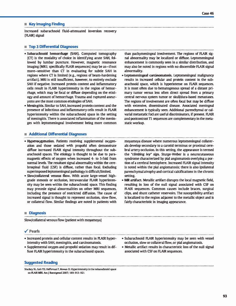
• Key lmaging Finding
Increased subarachnoid fluid-attenuated inversion recovery (FLAIR) signal
• Top 3 Differential Diagnoses
• Subarachnoid hemorrhage (SAH). Computed tomography (CT) is the modality of choiœ in identifying acute SAH, followed by lumbar puncture. However, magnetic resonanœ imaging (MRI; specifically FLAIR sequences) may be as-ifnot
more-sensitive than CT in evaluating for subtle SAH in regions where CT is limited (e.g., regions of beam-hardening artifact). MRI is still insufficient, however, to entirely exclude SAH if negative. Increased protein content and inflammatory œlls result in FLAIR hyperintensity in the region of hemorrhage, which may be focal or diffuse depending on the etiology and amount ofhemorrhage. Trauma and ruptured aneurysms are the most common etiologies of SAH.
• Merûngitis. Similar to SAH, increased protein content and the presence of infectious and inflammatory cells result in FLAIR
hyperintensity within the subarachnoid spaœ in the setting of meningitis. There is associated inflammation of the meninges with leptomeningeal involvement being more common
• Additional Differential Diagnoses
• Hype:roxygenation. Patients receiving supplemental oxygen
ation and those sedated with propofol often demonstrate diffuse increased FIJ\IR signal intensity throughout the subarachnoid spaces. The etiology is thought ta be due ta paramagnetic etfects of oxygen when increased 4- to 5-fold from normal levels. The resultant signal abnormality within the cerebrospinal fluid (CSF) is diffuse, rather than focal Exduding superimposed leptomeningeal pathology is difficultflimited
• Slow/collateral venous flow. With acute large-vessel highgrade stenosis or occlusion, intravascular FLAIR hyperinten
sity may be seen within the subarachnoid space. This finding may precede signal abnormalities on other MRI sequenœs,
including the presence of restricted diffusion. The cause of increased signal is thought ta represent occlusion, slow flow, or collateral flow. Similar findings are noted in patients with
• Diagnosis
Slow/collateral venous flow (patient with moyamoya)
� Pearls
• Increased protein and cellular content results in FLAIR hyperintensity with SAH, meningitis, and carcinomatosis.
• Supplemental oxygen and propofol sedation may result in dif
fuse FLAIR hyperintensity in the subarachnoid spaœs.
Suggested Reading
Stuclœy SI., Goh ID, Heffernan T, Rowan D. Hyperintensity in the subaradmoid space
on FLAIR MRI. AmJ Roentgmol 2007; 189: 913-921
Case 46
than pachymeningeal involvement. The regions of FLAIR signal abnormality may be localized or diffuse. Leptomeningeal enhanœment is commonly seen in a similar distribution, and may also be noted in regions with no disœmible FLAIR signal abnormality.
• Leptomeningeal carcinomatosis. Leptomeningeal malignancy results in increased cellular and protein content in the sub
arachnoid space, which is hyperintense on FLAIR sequences. It is most often due to hematogenous spread of a distant primary tumor versus Jess often direct spread from a primary central nervous system tumor or skull/dura-based metastases.
The regions of involvement are often focal but may be diffuse with extensive, disseminated disease. Associated meningeal enhanœment is typically seen. Additional parenchymal or calvariai metastatic fod are useful discriminators, if present. FLAIR and posttontrast 11 sequences are complementary in the metastatic workup.
moyamoya disease where numerous leptomeningeal collater
als develop secondary to a carotid terminus or proximal cerebral artery occlusion. In this setting, the appearance is termed
the �climbing ivy" sign. Sturge-Weber is a neurocutaneous syndrome characterized by pial angiomatosis overlying a portion of a cerebral hemisphere. Increased FLAIR signal intensity is noted within the pial angiomatosis; there is also ipsilateral
parenchymal atrophy and cortical calcifications in the chronic setting.
• MR artifact. Metallic artifact disrupts the local magnetic field,
resulting in Joss of the null signal associated with CSF on FLAIR sequences. Common causes include braces, surgical clips, and shunt catheter reservoirs. The susœptibility artifact
is localized ta the region adjacent ta the metallic object and is fairly characteristic in imaging appearance.
• Subarachnoid FLAIR hyperintensity may be seen with vessel occlusion, slow or collateral flow, or pial angiomatosis.
• Metallic artifact results in characteristic loss of the null signal associated with CSF on FLAIR sequenœs.
93
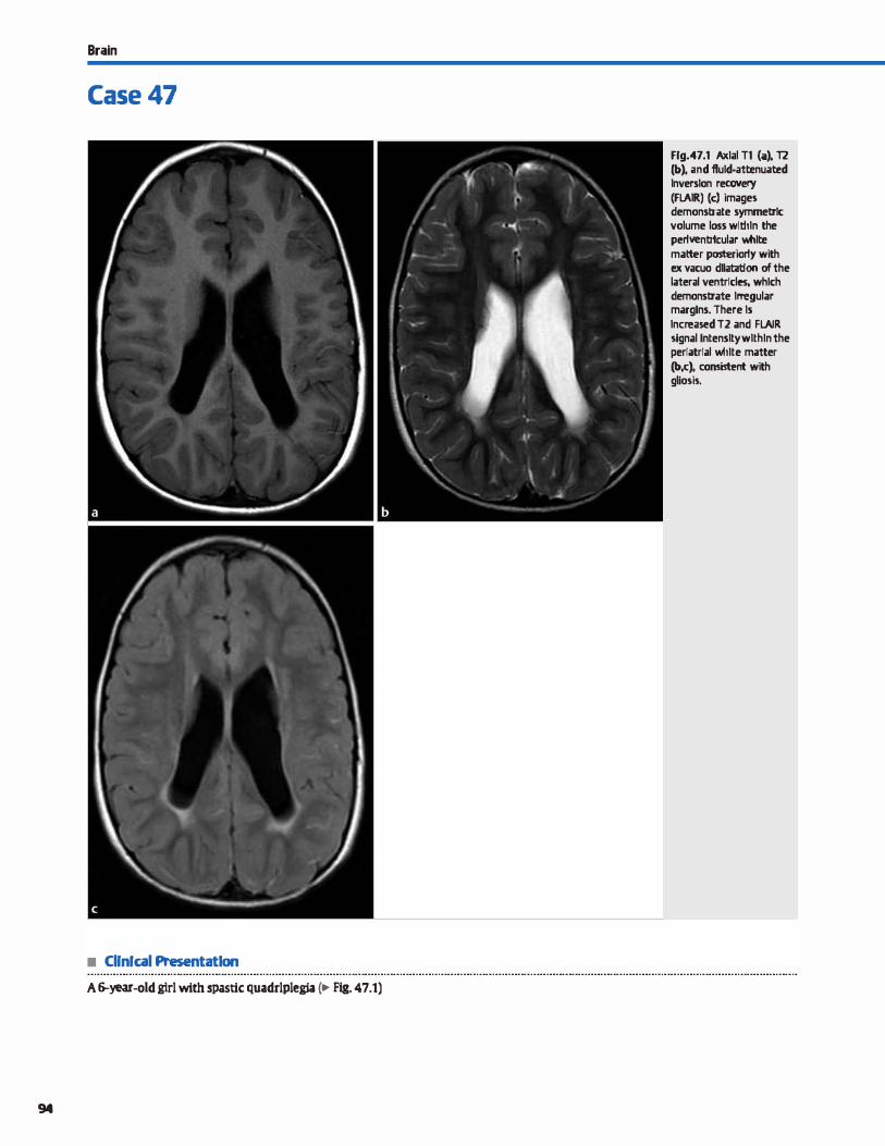
Brain
Case 47
• Cllnfcal Presentat1on
A 6-year-old girl wlth spastlc quadrlplegia c.- Fig. 47.1)
94
Frg.47.1 Axlal Tl (iit). 12 (b). and fluld·attenuat:ed Inversion recovery (FLAIR) (c) images demonstrate symmetrlc volume loss wlltiln the per1venb1cular white matter posteriorly with ex vacuo dllatat:lon of the lateral ventrfcles, whlch demonstrate lrTegular marglns. There ls lncreased T2 and FLAIR signal lntenslty wlthln the perfatrfal white matter (b,c), consistent with gliosis.
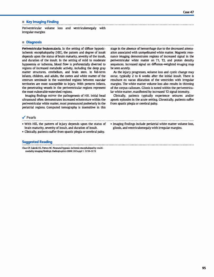
• Key lmaging Finding
Periventricular volume loss and ventriculomegaly with irregular margins
• Diagnosis
Perivmtricular leukomalada. In the setting of diffuse hypoxicischemic enœphalopathy (HIE), the pattern and degree of insult depends upon the status of brain maturity, severity of the insult, and duration of the insult. In the setting of mild to moderate hypoxemia or ischemia, blood flaw is preferentially diverted to regions of increased metabolic activity, induding the deep gray matter structures, œrebellum, and brain stem In full-term infants, children, and adults, the cortex and white matter of the œntrum semiovale in the watershed regions between vascular territories are most susceptible to injury. With preterm infants, the penetrating vessels in the periventricular regions represent the most vulnerable watershed regions.
lmaging fmdings mirror the pathogenesis of HIE. Initial head ultrasound often demonstrates increased echotexture within the periventricular white matter, most pronounœd posteriorly in the periatrial regions. Computed tomography is insensitive in this
� Pearts
• With HIE, the pattern of injury depends upon the status of brain maturity, severity of insult, and duration of insult.
• Oinically, patients suffer from spastic plegia or cerebral palsy.
Suggested Reading
Chao Cl', Zilleski CC, Patton At:. Neonatal hypoxic-ischemic enœphalopathy: multi
modality imaging findings. Radiographies 2006; 26 Suppl 1: Sl 59-Sl 72
Case 47
stage in the absence of hemorrhage due to the decreased attenuation associated with unmyelinated white matter. Magnetic resonanœ imaging demonstrates regions of increased signal in the periventricular white matter on Tt, T2, and proton density sequences. Increased signal on ditfusion-weighted intaging may be seen acutely.
As the injury progresses, volume Joss and cystic change may occur, typically 2 to 6 weeks after the initial insult. There is resultant ex vacuo dilatation of the ventrides with irregular margins. The white matter volume Joss also results in thinning of the corpus callosum. Gliosis is noted within the periventricular white matter, manifested by increased T2 signal intensity.
dinically, patients typically experience seizures and/or apneic episodes in the acute setting. Chronically, patients suffer from spastic plegia or cerebral palsy.
• Imaging findings include periatrial white matter volume loss, gliosis, and ventriculomegaly with irregular margins.
95
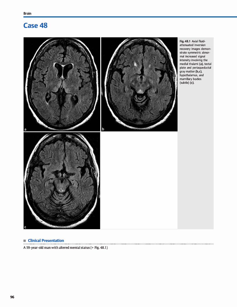
Brain
Case 48
• Clinical Presentation
A 59-year-old man with altered mental status (� Fig. 48.1 )
96
Fig. 48.1 Axial fluidattenuated inversion recovery images demonstrate symmetric abnormal increased signal intensity involving the medial thalami (a), tectal plate and periaqueductal gray matter (b,c), hypothalamus, and mamillary bodies (subtle) (c).
• Key lmaging Finding
Symmetric signal abnonnality within the media) thalami, periaqueductal gray matter, and mamillary bodies
• Diagnosis
Wernicke encephalopathy. Wemicke encephalopathy is a neurological disorder caused by thiamine deficiency. It most often occurs in alcoholic patients, but it may also be seen in nonalcoholic patients with malabsorption or nutritional
defidencies, including gastrointestinal neoplasms, prior gastric surgery, prolonged starvation, hyperemesis, and anorexia nervosa. Although the degree of clinicat symptoms varies, the dassic triad consists of altered consdousness,
ataxia, and ocular dysfunction. Wernicke encephalopathy is a neurological emergency that requires prompt replenishment of thiamine.
� Pearts
• Wernicke encephalopathy is a neurological disorder caused
by thiamine defidency.
• Wemicke encephalopathy occurs in alcoholic patients or nonalcoholic patients with malabsorption or nutritional
defidendes.
Suggested Reading
Zuca>li G, Gallucri M, Capellades J et al. Wemicke enœphalopathy: MR findings at
dinical presentation in twenty-six almholic and nonalmholic patients. Am J Neu
roradiol 2007; 28: 1328-1331
Case 48
Magnetic resonance imaging (MRI) features indude symmetric increased T2/fluid-attenuated inversion recovery signal intensity within the medial thalami (most common), periaqueductal gray matter, tectal plate, and mamillary bodies. Mamil
lary body involvement is fairly specific and occurs in just over half of patients. Mass effect may be seen acutely. In alcoholic
patients, there may be enhanœment of the involved regions, particularly within the mamillary bodies; enhancement is less common in nonalcoholic patients for reasons that remain unknown. With appropriate treatment, clinicat symptoms and
imaging fmdings may regress or resolve.
• MRI reveals symmetric increased signal in the medial thalami, periaqueductal gray matter, and mamillary bodies.
97
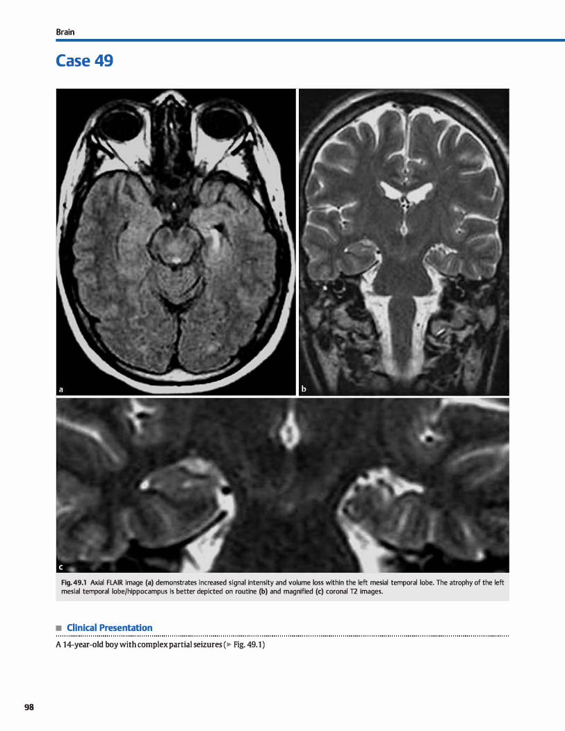
98
Brain
Case 49
Fig. 49.1 Axial FLAIR image (a) demonstrates increased signal intensity and volume loss within the left mesial temporal lobe. The atrophy of the left mesial temporal lobe/hippocampus is better depicted on routine (b) and magnified (c) coronal T2 images.
• Clinical Presentation
A 14-year-old boy with complex partial seizures (� Fig. 49.1)

• Key lmaging Finding
Atrophy and increased 12/fluid-attenuated inversion recovery (FLAIR) signal intensity within the mesial temporal lobe
• Diagnosis
Mesial temporal sderosis (MTS). Temporal lobe epilepsy is relatively common in adolescents and young adults and typically manifests as complex partial seizures. Within this patient population, MIS is thought to represent the most common identifiable cause of temporal lobe seizures. Debate exists as to whether MIS is acquired or a developmental abnormality. Sorne studies show an increased incidence in patients with a history of infant febrile seizures.
Magnetic resonanœ imaging is the mainstay in the workup of seizure disorders because computed tomography is oft:en normal. Seizure protocols for children and young adults typically indude high-resolution coronal sequences through the mesial temporal lobes/hippocampal formations. Imaging
y' Pearts
• Temporal lobe epilepsy is common in adolescents and young adults; it typically manifests as complex partial seizures.
• MTS is characterized by atrophy and increased 12/FLAIR signal intensity within the involved hippocampus.
Suggested Readings
Bocti C, Robitaille Y, Diadori P et al. The pathological basis of temporal lobe epilepsy
in childhaod, Neumlagy 2003; 60; 191-195
Case 49
findings of MIS indude hippocampal atrophy with assodated dilatation of the ipsilateral temporal horn of the lateral ventricle. Increased 12/FLAIR signal intensity is also commonly seen and increases the speàfidty in terms of suggesting the diagno
sis. MR spectroscopy shows decreased N-acetyl aspartate in the affected temporal lobe and may be helpful in localizing certain cases. Interictal FDG-PET (18F-fluorodeoxyglucose positron emission tomography) imaging demonstrates hypometabolism with decreased uptake within the affected mesial temporal lobe.
Medical treatment is successful in approximately 25% of cases of MTS. Anterior temporal lobe resection is often necessary for persistent seizure activity.
• Interictal FDG-PET shows hypometabolism with decreased uptake within the affected mesial temporal lobe.
Castillo M, SmithJK, Kwock L Proton MR spectroscupy in patients with acute tempo
ral lobe seizures. Arn] Neuroradial 2001; 22: 152-157
99
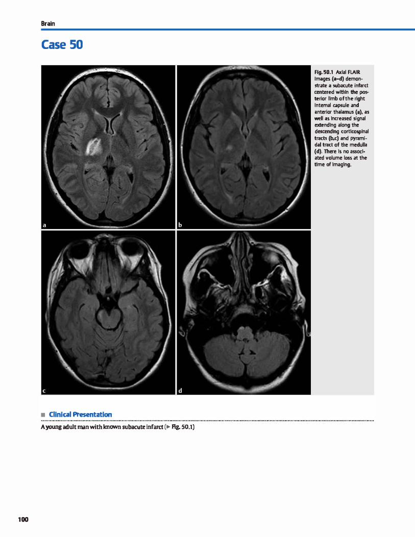
Brain
Case 50
• Clinical Presentation
A young adult man with known subacute infarct (� Fig. 50.1)
100
Fig. 50.1 Axlal FLAIR Images (a-d) demonstrate a subacute infarct œntered wlthln the posterfor llmb of the rlght Internai capsule and ;mterior thalamus (i}, as well as lncreased signal extendlng along the desœndlng cortlcosplnal tracts (b,c) and pyramidal tract of the medulla (d}. There ls no assoclated volume loss at the tlme of lmaglng.
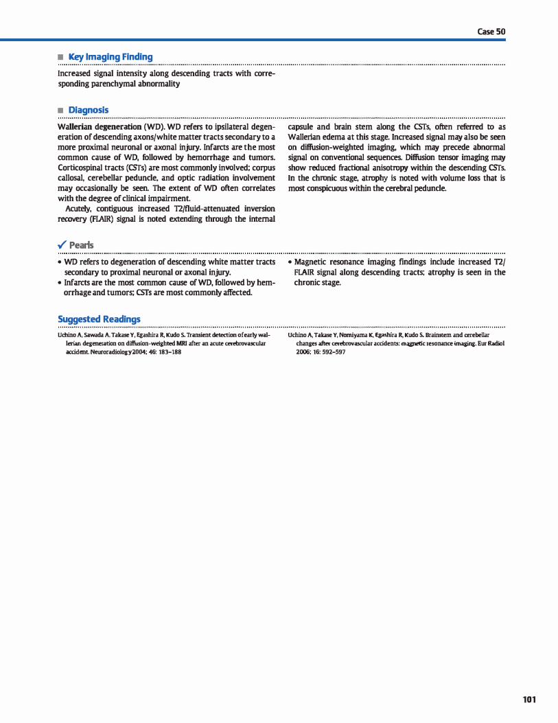
• Key lmaging Finding
Increased signal intensity along desœnding tracts with corre
sponding parenchymal abnormality
• Diagnosis
Wallerian degeneration (WD). WD refers to ipsilateral degeneration of descending axons/white matter tracts secondary to a more proximal neuronal or axonal injury. Infarcts are the most common cause of WD, followed by hemorrhage and tumors.
Corticospinal tracts (CSfs) are most commonly involved; corpus callosal, cerebellar pedunde, and optic radiation involvement
may occasionally be seen. The extent of WD often correlates
with the degree of clinical impairment. Acutely, contiguous increased T2/fluid-attenuated inversion
recovery (FIAIR) signal is noted extending through the internai
� Pearls
• WD refers to degeneration of descending white matter tracts
secondary to proximal neuronal or axonal injury. • Infarcts are the most common cause of WD, followed by hem
orrhage and tumors; CSTs are most commonly affected.
Suggested Readings
Uchino A, Sawada A, Takase Y, Egashira R, Kudo S. Transient detection of early wal
lerian degeneration on diffusion-weighted MRI after an acute cerdirovascular
accident. Neuroradiology:Z004; 46: 183-188
Case 50
capsule and brain stem along the CSTs, often referred to as Wallerian edema at this stage. Increased signal may also be seen on diffusion-weighted imaging, which may precede abnormal signal on conventional sequenœs. Diffusion tensor imaging may
show reduced fractional anisotropy within the descending csrs. In the chronic stage, atrophy is noted with volume Joss that is most conspicuous within the œrebral pedunde.
• Magnetic resonance imaging findings indude increased TJ./
FLAIR signal along desœnding tracts; atrophy is seen in the chronic stage.
Uchino A, Takase Y, Nomiyama K, Egashira R, Kudo S. Brainstem and œrebellar
changes after cerdirovilSCUlar accidents: magnetic resonance imaging. Eur Radial
2006; 16: 592-597
101
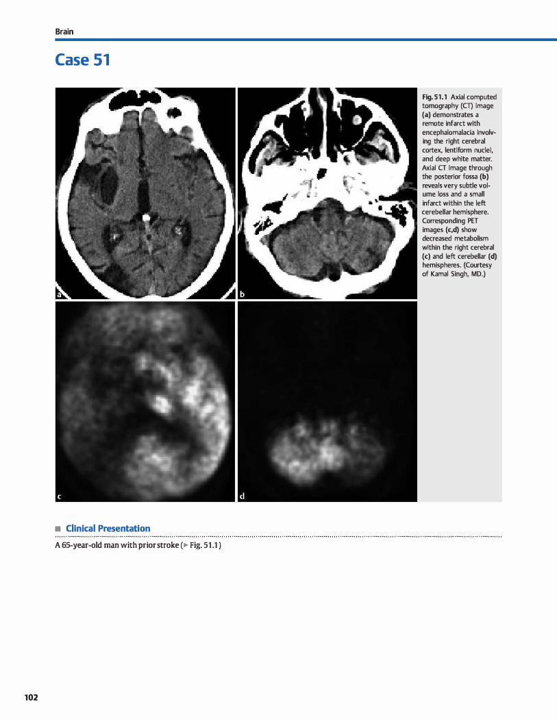
Brain
Case 51
• Clinical Presentation
A 65-year-old man with prior stroke (� Fig. 51 .1 )
102
Fig. 51 . 1 Axial computed tomography (CT) image (a) demonstrates a remote infarct with encephalomalacia involving the right cerebral cortex, lentiform nuclei, and deep white matter. Axial CT image through the posterior fossa (b) reveals very subtle volume loss and a small
infarct within the left cerebellar hemisphere. Corresponding PET images (c,d) show decreased metabolism within the right cerebral (c) and left cerebellar (d) hemispheres. (Courtesy of Kamal Singh, MD.)
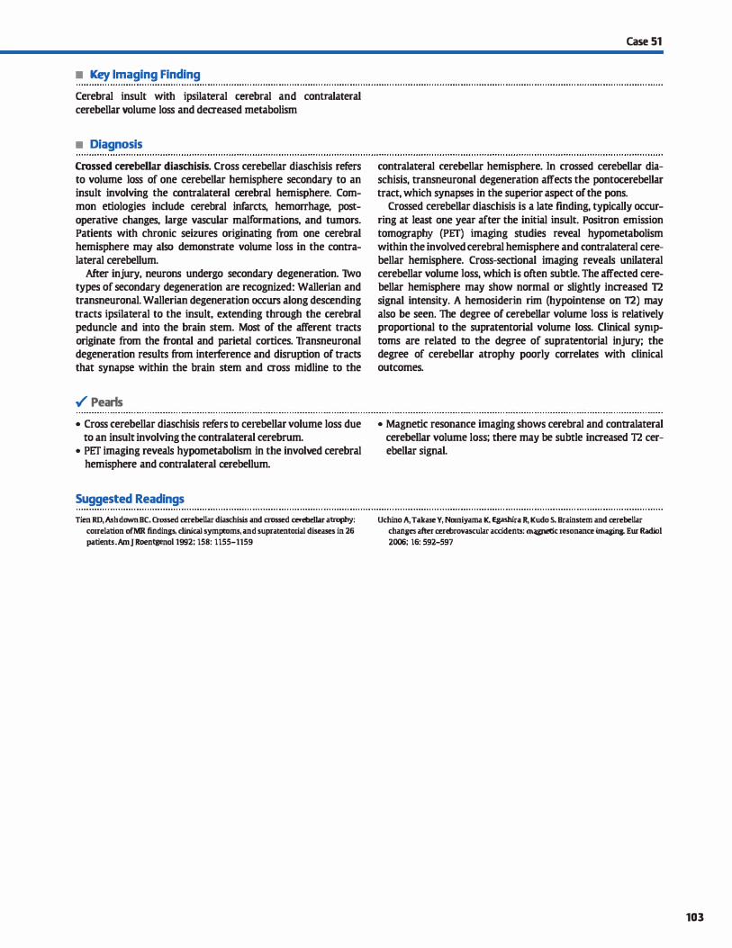
• Key lmaging Finding
Cerebral insult with ipsilateral œrebral and contralateral cerebellar volume loss and decreased metabolism
• Diagnosis
Crossed cerebellar diaschisis. Cross cerebellar diaschisis refers to volume loss of one cerebellar hemisphere secondary to an insult involving the contralateral cerebral hemisphere. Common etiologies indude cerebral infarcts, hemorrhage, post
operative changes, large vascular malformations, and tumors. Patients with chronic seizures originating from one cerebral hemisphere may aise demonstrate volume Joss in the contralateral cerebellum.
After injury, neurons undergo secondary degeneration. Two types of secondary degeneration are recognized: Wallerian and transneuronal. Wallerian degeneration occurs along descending
tracts ipsilateral to the insult, extending through the œrebral peduncle and into the brain stem. Most of the afferent tracts originate from the frontal and parietal cortices. lfansneuronal degeneration results from interference and disruption of tracts that synapse within the brain stem and cross midline to the
� Pearts
• Cross cerebellar diaschisis refers to cerebellar volume loss due to an insult involving the contralateral cerebrum.
• PET imaging reveals hypometabolism in the involved cerebral hemisphere and contralateral cerebellum.
Suggested Readings
Tien RD, Ashdown BC. Ü'DSsed œrebellar diaschim and crossed cerebellar atrophy;
correlation ofMR findings, clinical symptoms, and supratentorial diseases in 26
patients.AmJRoentgenol 1992; 158: 1155-1159
Case 51
contralateral cerebellar hemisphere. In crossed cerebellar diaschisis, transneuronal degeneration affects the pontoœrebellar tract, which synapses in the superior aspect of the pons.
Crossed cerebellar diaschisis is a late finding, typically occur
ring at Ieast one year after the initial insult. Positron emission tomography (PET) imaging studies reveal hypometabolism within the involved cerebral hemisphere and contralateral cerebellar hemisphere. Cross-sectional imaging reveals unilateral cerebellar volume Joss, which is often subtle. The affected cerebellar hemisphere may show normal or slightly increased T2 signal intensity. A hemosiderin rim (hypointense on T2) may also be seen. The degree of œrebellar volume Joss is relatively proportional to the supratentorial volume Joss. dinical syntptoms are related ta the degree of supratentorial injury; the degree of cerebellar atrophy poorly correlates with clinicat outcomes.
• Magnetic resonance imaging shows cerebral and contralateral cerebellar volume loss; there may be subtle increased T2 cerebellar signal.
Uchino A, Takase Y, Nmniyama K, Egashira R, Kudo s. Brainstem and œrebellar
changes after œrebrovascular accidents: magnetic resonance imaging. Eur Radio!
2006; 16: 592-597
103
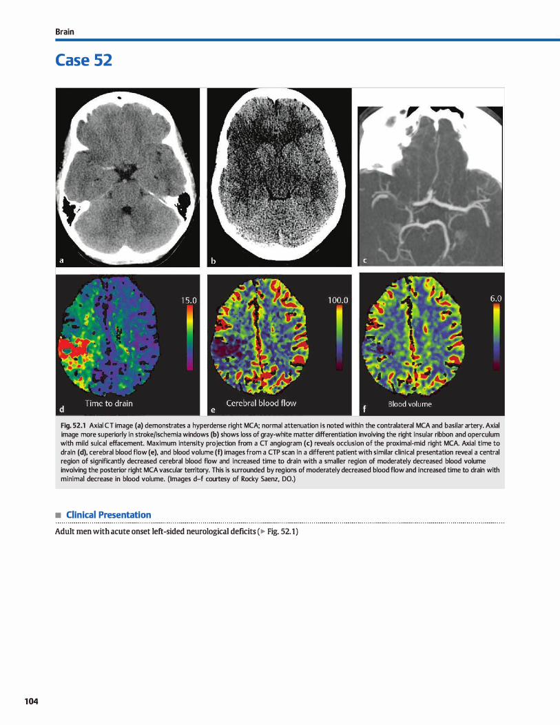
104
Brain
Case 52
Fig. 52.1 Axial CT image (a) demonstrates a hyperdense right MCA; normal attenuation is noted within the contralateral MCA and basilar artery. Axial image more superiorly in stroke/ischemia windows (b) shows loss of gray-white matter differentiation involving the right insular ribbon and operculum with mild sulcal effacement. Maximum intensity projection from a CT angiogram (c) reveals occlusion of the proximal-mid right MCA. Axial time to drain (d), cerebral blood flow (e), and blood volume (f) images from a CTP scan in a different patient with similar clinical presentation reveal a central region of significantly decreased cerebral blood flow and increased time to drain with a smaller region of moderately decreased blood volume involving the posterior right MCA vascular territory. This is surrounded by reg ions of moderately decreased blood flow and increased time to drain with minimal decrease in blood volume. (Images d-f courtesy of Rocky Saenz, DO.)
• Clinical Presentation
Adult men with acute onset left-sided neurological deficits (� Fig. 52.1)
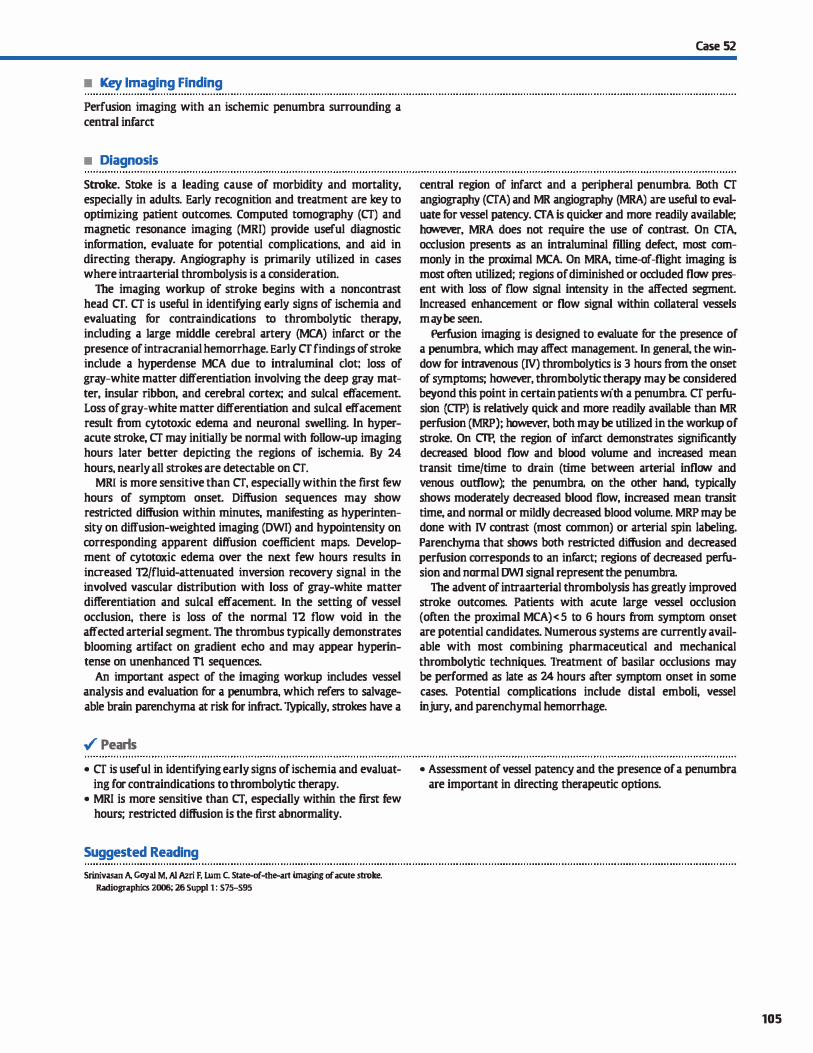
• Key lmaging Finding
Perfusion imaging with an ischemic penumbra surrounding a
central infarct
• Diagnosis
Stroke. Stoke is a leading cause of morbidity and mortality, especially in adults. Early recognition and treatment are key to optimizing patient outcomes. Computed tomography (CT) and magnetic resonance imaging (MRI) provide useful diagnostic
information, evaluate for potential complications, and aid in directing therapy. Angiography is primarily utilized in cases where intraarterial thrombolysis is a consideration.
The imaging workup of stroke begins with a noncontrast head CT. CT is useful in identifying early signs of ischemia and evaluating for contraindications to thrombolytic therapy, induding a large middle cerebral artery (MCA) infarct or the
presence of intracranial hemorrhage. Early CT findings of stroke indude a hyperdense MCA due to intraluminal dot: loss of gray-white matter differentiation involving the deep gray matter, insular ribbon, and cerebral cortex; and sulcal effacement Loss of gray-white matter differentiation and sulcal effacement result from cytotoxic edema and neuronal swelling. ln hyperacute stroke, CT may initially be normal with follow-up imaging hours later better depicting the regions of ischemia. By 24
hours, nearly all strokes are detectable on CT. MRI is more sensitive than CT, especially within the first few
hours of symptom onset Diffusion sequences may show restricted diffusion within minutes, manifesting as hyperintensity on diffusion-weighted imaging (DWI) and hypointensity on corresponding apparent diffusion coefficient maps. Development of cytotoxic edema over the next few heurs results in
increased T2/fluid-attenuated inversion recovery signal in the involved vascular distribution with loss of gray-white matter differentiation and sulcal effacement ln the setting of vesse! occlusion, there is Joss of the normal T2 flow void in the affected arterial segment The thrombus typically demonstrates blooming artifact on gradient echo and may appear hyperintense on unenhanced T1 sequenœs.
An important aspect of the imaging workup indudes vesse] analysis and evaluation for a penumbra, which refers to salvageable brain parenchyma at risk for infract lYPically, strokes have a
y' Pearts
• cr is useful in identifying early signs of ischemia and evaluating for contraindications to thrombolytic therapy.
• MRI is more sensitive than CT, espedally within the first few hours; restricted diffusion is the first abnormality.
Suggested Reading
Srinivasan A, Gayal M, Al Azri F, IJJm c. State-of-the-art imaging of acute stroke.
Radiographies 2006; 26 Suppl t: S75-S95
Case 52
central region of infarct and a peripheral penumbra Bath cr angiography ( crA) and MR angiography (MRA) are useful to eval
uate for vesse! patency. crA is quiclœr and more readily available; however, MRA does not require the use of contrast. On crA, occlusion presents as an intraluminal filling defect, most commonly in the proximal MCA. On MRA, time-of-flight imaging is most often utilized; regions of diminished or ocduded flow present with Joss of flow signal intensity in the affected segment lncreased enhanœment or flow signal within collateral vessels maybe seen.
Perfusion imaging is designed to evaluate for the presence of a penumbra, which may affect management ln general, the window for intravenous (N) thrombolytics is 3 hours from the onset of symptoms; however, thrombolytic therapy may be considered beyond this point in certain patients with a penumbra cr perfusion (CTP) is relatively quick and more readily available than MR perfusion (MRP); however, bath may be utilized in the workup of
stroke. On CTP, the region of infarct demonstrates significantly decreased blood flow and blood volume and increased mean transit time/time to drain (time between arterial inflow and venous outflow); the penumbra, on the other band, typically shows moderately decreased blood flow, increased mean transit time, and normal or mildly decreased blood volume. MRP may be done with N contrast (most common) or arterial spin labeling.
Parenchyma that shows bath restricted diffusion and decreased perfusion corresponds to an infarct; regions of decreased perfusion and normal DWI signal represent the penumbra.
The advent of intraarterial thrombolysis bas greatly improved stroke outcomes. Patients with acute large vessel occlusion ( often the proximal MCA) < 5 to 6 hours from symptom onset are potential candidates. Numerous systems are currently available with most combining pharmaceutical and mechanical
thrombolytic techniques. Treatment of basilar ocdusions may be performed as late as 24 hours after symptom onset in some cases. Potential complications include distal emboli, vessel injury, and parenchymal hemorrhage.
• Assessment of vessel patency and the presenœ of a penumbra are important in directing therapeutic options.
105
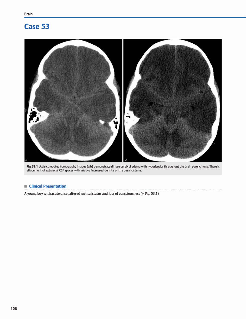
106
Brain
Case 53
Fig. 53.1 Axial computed tomography images (a,b) demonstrate diffuse cerebral edema with hypodensity throughout the brain parenchyma. There is effacement of extraaxial CSF spaces with relative increased density of the basal cisterns.
• Clinical Presentation
A young boy with acute onset altered mental status and Joss of consciousness (� Fig. 53.1)
• Key lmaging Finding
Diffuse cerebral edema with hyperdense basal cistems
• Diagnosis
Pseudosubarachnoid hemorrliage. Pseudosubarachnoid hemorrhage {pseudo-SAH) refers to the hyperdense appearanœ of the basal cistems in the setting of diffuse œrebral edema; less commonly, it bas been described in association with intracranial hypotension. Pseudo-SAH is believed to result from a variety of factors affecting both the extraaxial cerebrospinal fluid (C:SF) spaœs and the underlying brain parenchyma. In the setting of dif
fuse œrebral edema, there is increased intracranial pressure, which results in engorgement of the blood-filled pial veins, as
y' Pearts
• Pseudo-SAH refers to the hyperdense appearance of the basal àsterns in the setting of diffuse cerebral edema.
• With œrebral edema, increased density in the basal dstems
and decreased density of the parenchyma simulates SAH.
Suggested Reading
Given CA, ll, Burdette JH, Elster AD, Williams TIW, lll. Pseudo-subarachnoid hemor
rhage: a potential irnaging pitfall assodated with diffilse œrebral edema. AmJ
Neuroradiol 2003; 24: 254-256
Case 53
we1l as effacement of the hypodense extraaxial CSF. This com
bined with underlying cortical hypodensity secondary ta cytotoxic edema, results in relative increased attenuation within the basal cisterns, simulating SAH. The Hounsfield unit (HU) measurement of pseudo-SAH (approximately 30 HU) is considerably
Jess than that expected with SAH in a patient with a normal hematocrit {-55 ta 70 HU), which helps distinguish between the two entities.
• The HU of pseudo-SAH is considerably less than that expected with SAH in a patient with a normal hematocrit
107
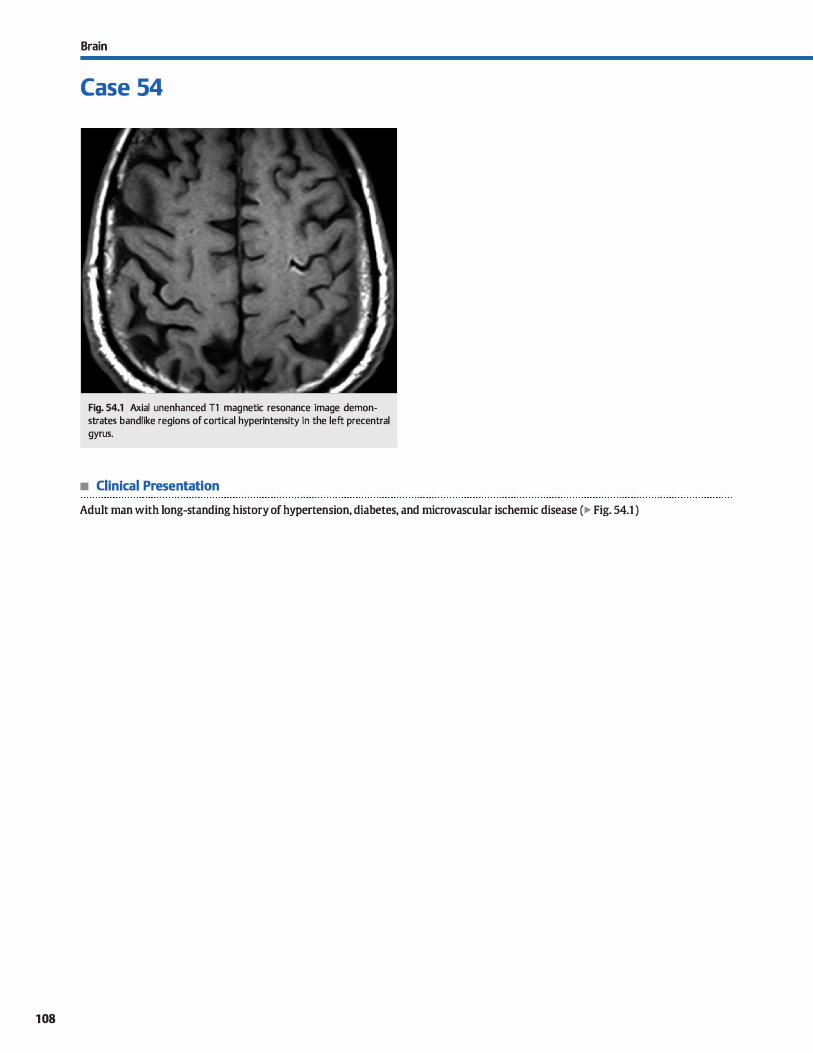
108
Brain
Case 54
Fig. 54.1 Axial unenhanced Tl magnetic resonance image demonstrates bandlike regions of cortical hyperintensity in the left precentral gyrus.
• Clinical Presentation
Adult man with long-standing history of hypertension, diabetes, and microvascular ischemic disease (� Fig. 54.1 )
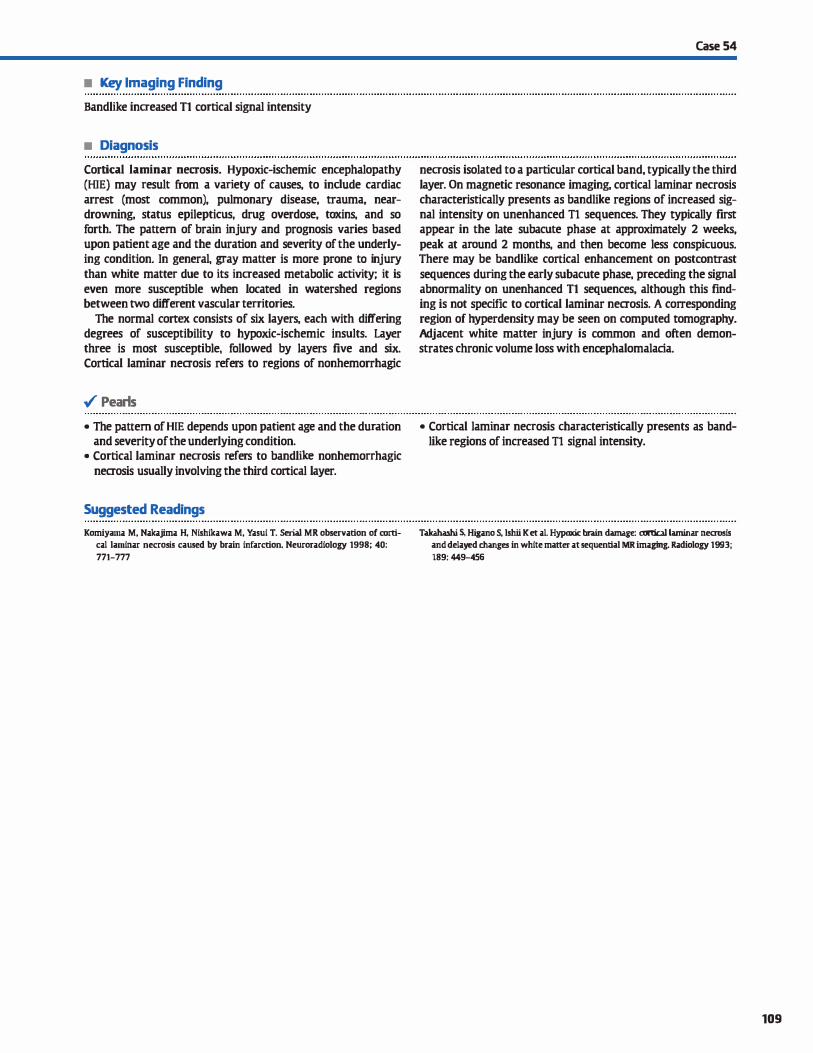
• Key lmaging Finding
Bandlilœ increased Tl cortical signal intensity
• Diagnosis
Cortical laminar necrosis. Hypoxic-ischemic encephalopathy (HIE) may result from a variety of causes, to indude cardiac arrest (most common), pulmonary disease, trauma, neardrowning, status epilepticus, drug overdose, toxins, and so forth. The pattern of brain injury and prognosis varies based upon patient age and the duration and severity of the underlying condition. In general, gray matter is more prone ta injury than white matter due to its increased metabolic activity; it is even more susceptible when located in watershed regions between two different vascular territories.
The normal cortex consists of six layers, each with differing degrees of susœptibility to hypoxic-ischemic insults. Layer three is most susceptible, followed by layers five and six. Cortical laminar necrosis refers to regions of nonhemorrhagic
y' Pearts
• The pattern of HIE depends upon patient age and the duration and severity of the underlying condition.
• Cortical laminar necrosis refers to bandlike nonhemorrhagic necrosis usually involving the third cortical layer.
Suggested Readings
Komiyililla M, Nakajima H, Nishikawa M, Yasui T. Serial MR observation of corti
cal lilillinar necrosis caused by brain infarction. Neuroradiology 1998; 40;
771-777
Case 54
necrosis isolated to a particular cortical band, typically the third layer. On magnetic resonanœ imaging, cortical laminar necrosis characteristically presents as bandlike regions of increased signal intensity on unenhanced Tl sequenœs. They typically first appear in the late subacute phase at approximately 2 weeks, peak at around 2 months, and then become less conspicuous. There may be bandlilœ cortical enhancement on postcontrast sequences during the early subacute phase, preceding the signal abnormality on unenhanced Tl sequences, although this finding is not specific to cortical laminar necrosis. A corresponding region of hyperdensity may be seen on computed tomography. Adjacent white matter injury is common and often demonstrates chronic volume Joss with encephalomalada.
• Cortical laminar necrosis characteristically presents as bandlike regions of increased Tl signal intensity.
Takàhashi S. Higano S, lshü K et al. Hypoxie brain damage: cortical laminar necrosis
and delayt!d changes in white malter at sequential MR imaging. Radiology 1993;
189: 449-456
109
Brain
Case 55
• Clinical Presentation
An 8-year-old boy with headaches, vomiting, and ataxia (� Fig. 55.1)
110
Fig. 55.1 Sagittal contrast-enhanced Tl image (a) demonstrates a large posterior fossa mass with cystic and enhancing solid components. There is obstruction of the fourth ventricle with hydrocephalus and inferior displacement of the cerebellar tonsils. On axial T2 (b) and Tl postcontrast (c) images, the mass is slightly eccentric to the left. There is solid enhancement, as well as enhancement along the walls of the cystic components (c). The fourth ventricle is effaced and there is surrounding edema in the cerebellar hemispheres (b).
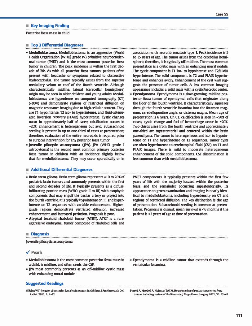
• Key lmaging Finding
Posterior fossa mass in child
• Top 3 Differential Diagnoses
• Medulloblastoma. Medulloblastoma is an aggressive (World Health Organization [WHO] grade IV) primitive neuroectodermal tumor (PNET) and is the most common posterior fossa tumor in children. The peak incidence is within the first decade of life. As with all posterior fossa tumors, patients often present with headache or symptoms related to obstructive hydroœphalus. The tumor typically arises from the superior
medullary velum or roof of the fourth ventride. Although characteristically midline, lateral (œrebellar hemisphere) origin may be seen in older children and young adults. Medulloblastomas are hyperdense on computed tomography {CT) (-90%) and demonstrate regions of restricted diffusion on magnetic resonanœ imaging due to high cellular content. They are Tl hypointense, T2 iso- to hyperintense, and fluid-attenu
ated inversion recovery {FLAIR) hyperintense. Cystic changes occur in approximately half of cases; calcification occurs in -20%. Enhanœment is heterogeneous but avid. Subarachnoid seeding is present in up to one-third of cases at presentation; therefore, evaluation of the entire neuroaxis is required prior to surgical intervention for any posterior fossa tumor.
• juvenile pilocytic astrocytoma {JPA). ]PA (WHO grade I astrocytoma) is the second most common primary posterior fossa tumor in children with an incidence slightly below that for medulloblastoma. They may occur sporadically or in
• Additional Differential Diagnoses
• Brain stem glioma. Brain stem glioma represents .. 10 to 20% of pediatric brain tumors and commonly presents within the first and second decades of life. It typically presents as a diffuse, infiltrating pontine mass (WHO grade II to Ill) with exophytic components that may engulf the basilar artery or project into
the fourth ventride. It is typically hypointense on T1 and hyperintense on T2 sequenœs with variable enhancement. Highergrade regions demonstrate restricted diffusion, increased enhancement, and increased perfusion. Prognosis is poor.
• Atypical teratoid rhabdoid tumor (AlKf). ATRT is a rare, aggressive embryonal tumor composed of rhabdoid cells and
• Diagnosis
Juvenile pilocytic astrocytoma
� Pearts
• Medulloblastoma is the most common posterior fossa mass in a child, is midline, and often seeds the CSF.
• ]PA most commonly presents as an off-midline cystic mass
with enhancing mural nodule.
Suggested Readings
Case 55
association with neurofibromatosis type 1. Peak incidence is 5
to 15 years of age. The tumor arises from the cerebellar hemisphere; therefore, it is typically off-midline. The most common presentation is a cystic mass with an enhancing mural nodule. The cystic component is T1 iso- to hypointense and T2/FIAIR
hyperintense. The solid component is T2 and FLAIR hyperintense and enhances avidly. Enhanœment of the cyst wall suggests the presence of tumor cells. A Jess common imaging appearanœ indudes a solid mass with a cystic/necrotic œnter.
• Ependymoma. Ependymoma is a slow-growing, midline posterior fossa tumor of ependymal cells that originates along the floor of the fourth ventride. It characteristically squeezes
through the fourth ventride foramina into the foramen magnum, cerebellopontine angle, or cistema magna. Mean age of presentation is 6 years. On CT, calcification is seen in .. 5()% of cases; cystic change and foci of hemorrhage occur in .. 20%. 1\vo-thirds arise from the fourth ventricle and approximately one-third are supratentorial and centered within the brain parenchyma. The tumor is heterogeneous and iso- to hypointense on T1 and hyperintense on T2 sequenœs. Tumor cysts
are often hyperintense to œrebrospinal fluid (CSF) on T1 and FLAIR images. There is mild to moderate heterogeneous enhancement of the solid components. CSF dissemination is Jess common than with medulloblastoma.
PNET components. It typically presents within the first few years of life with the majority located within the posterior fossa and the remainder occurring supratentorially. Its appearanœ on gross examination and imaging is nearly identical to medulloblastoma, including hyperdensity on cr and
regions of restricted diffusion. The key distinction is the age of presentation. Subarachnoid seeding is common at presentation. Prognosis is dismal; mean survival is < 6 months if the patient is < 3 years of age at time of presentation.
• Ependymoma is a midline tumor that extends through the ventricular foramina.
O'Brien wr. Imaging ofposterior fossa brain twnors in dùldren.J Am Osteopath Coll Poretti A, Meoded A, Huisman TACM. Neuroimaging ofpediatric posterior fossa
Radial. 2013; 2; 2-12 tuman induding review of the liter.atllre.J Magn Resan Imaging 2012; 35; 32-47
1 1 1
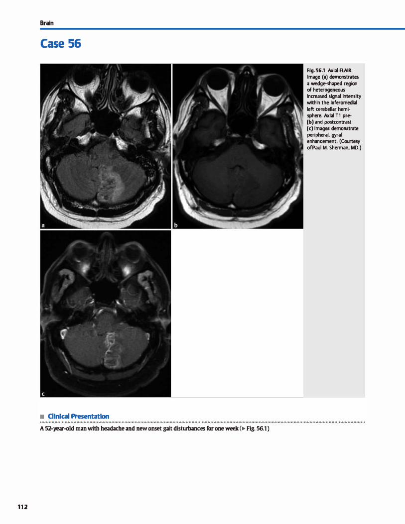
Brain
Case 56
• Cllnlcal Presentatlon
A 52-year-old man with headache and new onset gait disturbances for one week (,.. Fig. 56.1)
112
Fig. 56.1 Axlal FLAIR Image (a) demonstrates ;i wedg�haped region of heterogeneous lncreased signal lntenslty wtthln the lnferomedlal left cerebell;ir hemi· sphere. Axlal Tl pre-(b) and postcontrast (c) Images demonstrate peripher;il, gyr;il enh;incement. (Courtesy orP.aul M. Shennan, MD.)
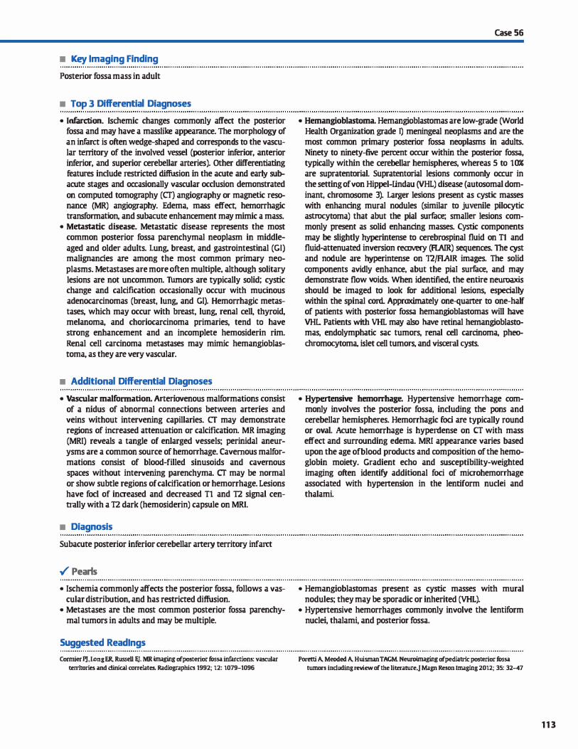
• Key lmaging Finding
Posterior fossa mass in adult
• Top 3 Differential Diagnoses
• Infarction. Ischemic changes a:immonly affect the posterior fossa and may have a masslilœ appearanœ. The morphology of an infarct is often wedge-shaped and corresponds to the vascular territory of the involved vessel (posterior inferior, anterior inferior, and superior œrebellar arteries ). Other differentiating features indude restricted diffusion in the acute and early subacute stages and occasionally vascular occlusion demonstrated
on computed tomography (CT) angiography or magnetic resonanœ (MR) angiograpbY. Edema, mass effect, hemorrhagic transformation, and subacute enhanœment may mimic a mass.
• Metastatic disease. Metastatic disease represents the most common posterior fossa parenchymal neoplasm in middle
aged and older adults. Lung, breast, and gastrointestinal (CI) malignancies are among the most common primary neo
plasms. Metastases are more often multiple, although solitary lesions are not uncommon. Turnors are typically solid; cystic change and calcification occasionally occur with mucinous adenocarcinomas (breast, lung, and Cl). Hemorrhagic metastases, which may occur with breast, lung, renal œll, thyroid, melanoma, and choriocarcinoma primaries, tend ta have strong enhancement and an incomplete hemosiderin rim.
Renal œll carcinoma metastases may mimic hemangioblastoma, as they are very vascular.
• Additional Differential Diagnoses
• Vascular malfonnation. Arteriovenous malformations consist of a nidus of abnormal connections between arteries and veins without intervening capillaries. cr may demonstrate regions of increased attenuation or calcification. MR imaging (MRI) reveals a tangle of enlarged vessels; perinidal aneur
ysms are a common source ofhemorrhage. Cavemous malformations consist of blood-filled sinusoids and cavernous spaœs without intervening parenchyma. CT may be normal or show subtle regions of calcification or hemorrhage. Lesions have fod of increased and decreased Tl and T2 signal centrally with a T2 dark (hemosiderin) capsule on MRI.
• Diagnosis
Subacute posterior inferior cerebellar artery territory infarct
� Pearts
• Ischemia commonly affects the posterior fossa, follows a vascular distribution, and bas restricted diffusion.
• Metastases are the most common posterior fossa parenchymal tumors in adults and may be multiple.
Suggested Readlngs
Cormier PJ, Long ER, Russell lij. MR imaging ofposterior fossa infarctions: vascular
territories and dinical correlates. Radiographies 1992; 12: 1079-1096
Case 56
• Hemangioblastoma. Hemangioblastomas are low-grade (World Health Organization grade I) meningeal neoplasms and are the most common primary posterior fossa neoplasms in adults. Ninety to ninety-five percent ocrur within the posterior fossa, typically within the œrebellar hemispheres, whereas 5 to 10% are supratentorial. Supratentorial lesions commonly occur in the setting of von Hippel-lindau (VHL) disease ( autosomal dom
inant, chromosome 3i Larger lesions present as cystic masses with enhandng mural nodules (similar to juvenile pilocytic astrocytoma) that abut the pial surface; smaller lesions commonly present as solid enhancing masses. Cystic components
may be slightly hyperintense to œrebrospinal fluid on Tl and fluid-attenuated inversion recovery (FlAIR) sequenœs. The cyst and nodule are hyperintense on T2/FLAIR images. The solid
a:imponents avidly enhanœ, abut the pia1 surface, and may demonstrate flow voids. When identified, the entire neuroaxis should be imaged to look for additional lesions, espedal!y within the spinal cord. Appraxirnately one-quarter to one-hait of patients with posterior fossa hemangioblastomas will have VHL Patients with VHL may also have retinal hemangioblastomas, endolymphatic sac tumors, renal œll carcinoma, pheo
chromocytoma, islet œll tumors, and visceral cysts.
• Hypertensive hemorrhage. Hypertensive hemorrhage commonly involves the posterior fossa, induding the pons and œrebellar hemispheres. Hemorrhagic fod are typically round or oval. Acute hemorrhage is hyperdense on CT with mass effect and surrounding edema. MRI appearance varies based
upon the age ofblood products and composition of the hemoglobin moiety. Gradient echo and susceptibility-weighted imaging often identify additional foci of microhemorrhage assoàated with hypertension in the lentiform nudei and thalami.
• Hemangioblastomas present as cystic masses with mural nodules; they may be sporadic or inherited (VHL).
• Hypertensive hemorrhages commonly involve the lentiform nuclei, thalami, and posterior fessa.
Poretti A, Meoded A, Huisman TAGM. NeW"Dimaging ofpediatric posterior fossa tumors induding review of the Iiterature.j Magn Reson lmaging 2012; 35: 32--47
1 1 3
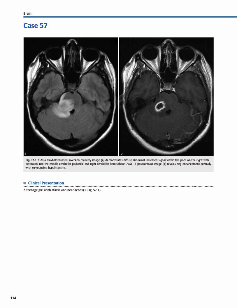
114
Brain
Case 57
Fig. 57 .1 1 Axial fluid-attenuated inversion recovery image (a) demonstrates diffuse abnormal increased signal within the pans on the right with extension into the middle cerebellar peduncle and right cerebellar hemisphere. Axial Tl postcontrast image (b) reveals ring enhancement centrally with surrounding hypointensity.
• Clinical Presentation
A teenage girl with ataxia and headaches (� Fig. 57.1 )
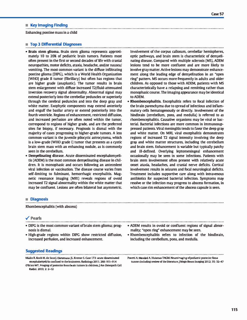
• Key lmaging Finding
Enhancing pontine mass in a child
• Top 3 Differential Diagnoses
• Brain stem glioma. Brain stem glioma represents approximately 10 ta 20% of pediatric brain tumors. Patients most often present in the first or second decades of life with cranial neuropathies, motor deficits, ataxia, headache, and/or nausea/ vomiting. The most common variant is the diffuse infiltrating pontine glioma (DIPG), which is a World Health Organization [WHO] grade Il tumor (fibrillary) but often bas regions that
are higher grade (anaplastie). The tumor results in brain
stem enlargement with diffuse increased T2/fluid-attenuated inversion recovery signal abnormality. Abnormal signal may
extend posteriorly into the cerebellar pedundes or superiorly through the cerebral peduncles and into the deep gray and white matter. Exophytic components may extend anteriorly and engulf the basilar artery or extend posteriorly into the
fourth ventricle. Regions of enhancement, restricted diffusion, and increased perfusion are often noted within the tumor, correspond ta regions of higher grade, and are the preferred sites for biopsy, if necessary. Prognosis is dismal with the majority of cases progressing ta higher-grade tumors. A less common variant is the juvenile pilocytic astrocytoma, which is a low-grade (WHO grade I) tumor that presents as a cystic
brain stem mass with an enhancing nodule, as is commonly seen in the cerebellum.
• Demyelinating disease. Acute disseminated encephalomyeli
tis (ADEM) is the most common demyelinating disease in children. lt is monophasic and occurs following an antecedent viral infection or vaccination. The disease course varies from self-limiting to fulminant, hemorrhagic encephalitis. Mag
netic resonance imaging (MRI) reveals regions of ovoid increased T2 signal abnormality within the white matter that
may be confluent. Lesions are often bilateral but asymmetric.
• Diagnosis
Rhombencephalitis (with absœss)
� Pearls
• DIPG is the most common variant ofbrain stem glioma; prognosis is dismal.
• High-grade regions within DIPG show restricted diffusion, increased perfusion, and increased enhancement.
Suggested Readings
Mialin R, Kcob M, de SezeJ, Dietemann JI. Kremer S. Case 173: acute disserninated
enœphalomyelitis confined to the brainstem Radiology 2011 : 260: 911-914
O'Brien wr. Imaging of posterior fossa br.lin tumors in children. J Am Osteopath Coll
Radio!. 2013; 2: 2-12
Case 57
Involvement of the corpus callosum, œrebellar hemispheres,
optic pathways, and brain stem is characteristic of demyelinating disease. Compared with multiple sderosis (MS), ADEM lesions tend to be more confluent and are more likely to involve gray matter. Active lesions may demonstrate enhance
ment along the leading edge of demyelination in an �open ring" pattern. MS occurs more frequently in adults and aider children. As opposed to those with ADEM, patients with MS
characteristically have a relapsing and remitting rather than monophasic course. The imaging appearance may be identical to ADEM.
• Rhombencephalitis. Encephalitis refers to focal infection of
the brain parenchyma due to spread of infectious and inflammatory cells hematogenously or directly. Involvement of the hindbrain (cerebellum, pons, and medulla) is referred to as rhombencephalitis. causative organisms may be viral or bacterial. Bacterial infections are more common in immunosuppressed patients. Viral meningitis tends ta favor the deep gray and white matter. On MRI, viral encephalitis demonstrates regions of increased T2 signal intensity involving the deep gray and white matter structures, including the cerebellum
and brain stem. Enhancement is variable but typically patchy
and ill-defined. OVerlying leptomeningeal enhancement occasionally may be seen in some infections. Patients with brain stem involvement often present with relatively acute onset ataxia, headaches, and cranial nerve defidts. Cortical involvement results in seizures and focal neurological deficits.
Treatment includes supportive care along with intravenous antibiotics for suspected bacterial infection. Symptoms may resolve or the infection may progress ta absœss formation, in which case rim enhancement of the abscess capsule is seen.
• ADEM results in ovoid or confluent regions of signal abnormality; �open ring" enhancement may be seen.
• Rhombencephalitis refers to infection of the hindbrain, including the œrebellum, pans, and medulla.
Poretti A, Meoded A, Huisman TAGM. Neuroim"8ing ofpediatric posterior rossa
tumors induding review of the literature.J Magn Reson Imaging 2012; 35: 32-47
1 1 5
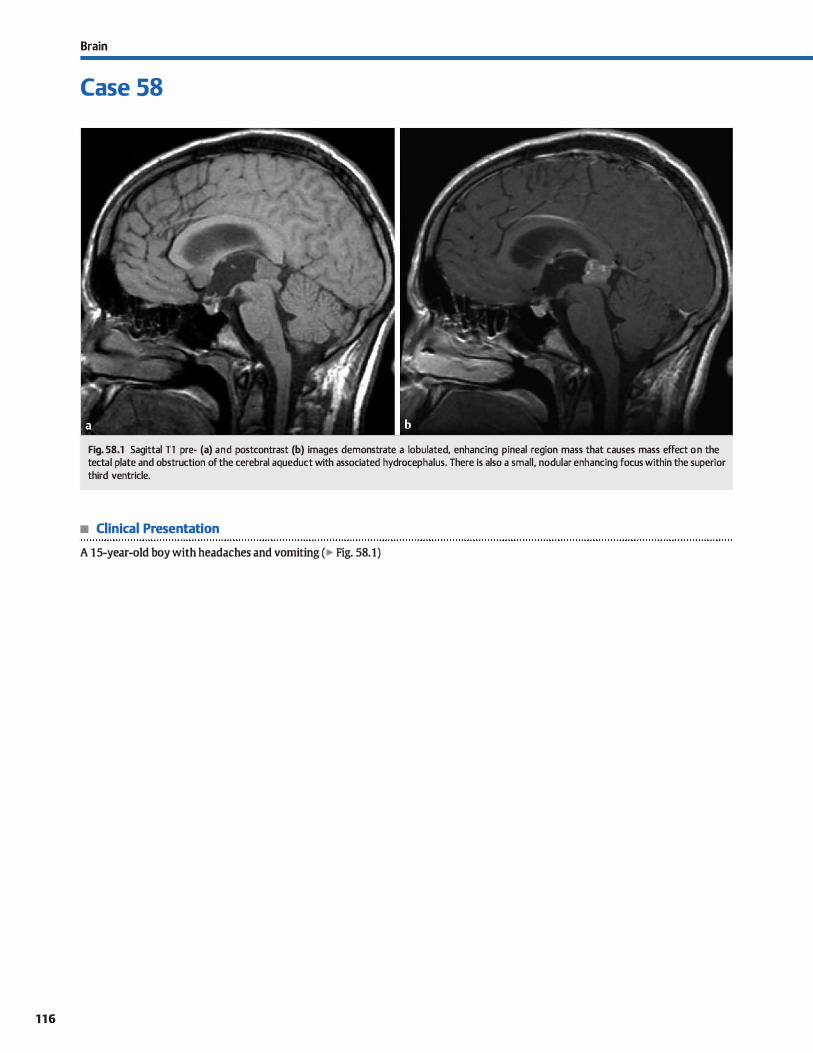
116
Brain
Case 58
Fig. 58.1 Sagittal Tl pre- (a) and postcontrast (b) images demonstrate a lobulated, enhancing pineal region mass that causes mass effect on the tectal plate and obstruction of the cerebral aqueduct with associated hydrocephalus. There is also a small, nodular enhancing focus within the superior third ventricle.
• Clinical Presentation
A 1 5-year-old boy with headaches and vomiting (� Fig. 58.1)
• Key lmaging Finding
Pinea) region mass
• Top 3 Differential Diagnoses
• Pineal cyst. Pineal cysts are common and usually inddental. Rarely, large cysts (> 15mm) may produce symptoms secondary to local mass effect resulting in headache or visual disturbances. Simple cysts follow fluid signal intensity on magnetic resonance imaging (MRI) and lack central, solid enhancement; surrounding enhancement involving the cyst wall and within the pineal substance is commonly seen. The cysts may demonstrate increased signal intensity on Tl sequences due ta proteinaceous content or occasionally hemorrhage with trauma. Cysts > 10 mm are often followed with serial imaging ta ensure stability, if atypical features are identified.
• Genn cell twnor (GCT). GCTs are the most common malignant
neoplasms of the pineal gland with germinomas representing more than 60% of cases. Most cases occur in adolescent and young adult males. Presenting symptoms are usually secondary to mass effect resulting in obstructive hydroœphalus, Parinaud syndrome (paralysis of upward gaze), or endocrine dysfunction. Germinomas are hyperdense on computed tomography {CT) due to the high nudear-to-cytoplasmic ratio and may contain calcifications centrally. On MRI, germinomas are typically intermediate in signal intensity on Tl and T2 sequences with avid
enhanœment. On occasion, germinomas may appear cystic. As œrebrospinal fluid ( CSF) dissemination is common, the entire spine must be imaged to evaluate for drop metastases. Other GCTs indude teratomas, yolk sac tumors, and choriocarci-
• Additional Differential Diagnoses
• Tectal plate glioma. Tectal gliomas are slow growing, lowgrade astrocytomas that occur primarily in children. Patients present with symptoms related to increased intracranial pressure. Imaging reveals bulbous enlargement of the tectal plate
with narrowing or obstruction of the cerebral aqueduct. There is increased T2 signal intensity and typically no or minimal enhancement. Treatment is geared toward CSF diversion.
• Diagnosis
Pinea) germinoma {with CSF dissemination)
� Pearts
• Pineal cysts are often asymptomatic and incidentally found; no internai enhancement should be seen.
• Gcrs are the most common pineal neoplasms and commonly result in CSF dissemination.
Suggested Reading
Smith AB, Rushing EJ, Smirniatopoulos JG, From the archives of the AFIP; lesions of
the pineal region: radiologie-pathologie correlation. Radiographies 201 O; 30:
2001-2020
Case 58
nomas. Teratomas will typically have macroscopic fat and calcification. Yolk sac tumors may be cystic and assodated with elevated levels of alpha-fetoprotein. Choriocarcinomas have a propensity to bleed and are assodated with elevated levels of human chorionic gonadotropin.
• Pineal cell tumor. Pineal cell origin tumors consist of pineoblastomas, pineocytomas, and pineal parenchymal tumors of intermediate differentiation (PPTID). Pineoblastomas are more malignant with a peak incidence in the first decade of Iife. Seeding of the CSF is common. Pineocytomas {World Health Organization [WHO] grade I) have a peak incidence in the third and fourth decades of life and are less aggressive. PPTID are intermediate tumors (WHO grade II or III) that occur most commonly in young adults. Presenting symptoms
are usually due to mass effect resulting in obstructive hydrocephalus, Parinaud syndrome, or endocrine dysfunction. On imaging, the tumors may dosely resemble one another. On
average, pineoblastomas are larger at the time of presentation. Ali may be hyperdense on cr due to the high cellularity. When calcifications occur with pineal cell tumors, they are typically along the periphery in an "exploded" pattern. On MRI, pineal cell tumors are of intermediate signal intensity on Tl and intermediate to hyperintense on T2 sequences with avid enhancement As with Gcrs, the spine must be imaged to evaluate for drop metastases.
• Meningioma. Meningiomas are the most common extraaxial intracranial tumors. They may occur along the margin of the tentorium, mimicking a pineal gland mass. The pineal gland and internai cerebral veins are often displaced superiorly. On noncontrast cr, meningiomas may be hyperdense; calcification is common. The enhancement pattern is typically avid and homogenous with a durai tait.
• Pineal origin tumors are the second most common pineal neoplasms and may also result in CSF dissemination.
• Tectal plate gliomas are low-grade T2 hyperintense astrocyto
mas that obstruct the cerebral aqueduct
1 1 7
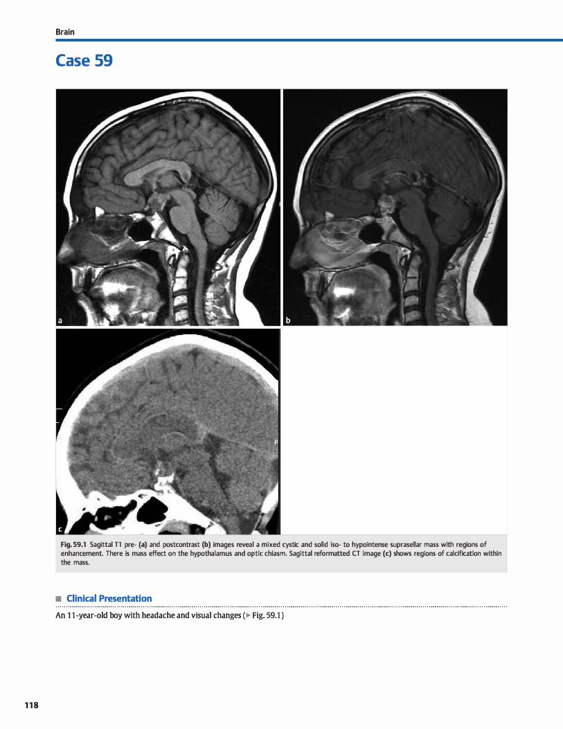
118
Brain
Case 59
Fig. 59.1 Sagittal Tl pre- (a) and postcontrast (b) images reveal a mixed cystic and solid iso- to hypointense suprasellar mass with regions of enhancement. There is mass effect on the hypothalamus and optic chiasm. Sagittal reformatted CT image (c) shows regions of calcification within the mass.
• Clinical Presentation
An 1 1-year-old boy with headache and visual changes (� Fig. 59.1 )
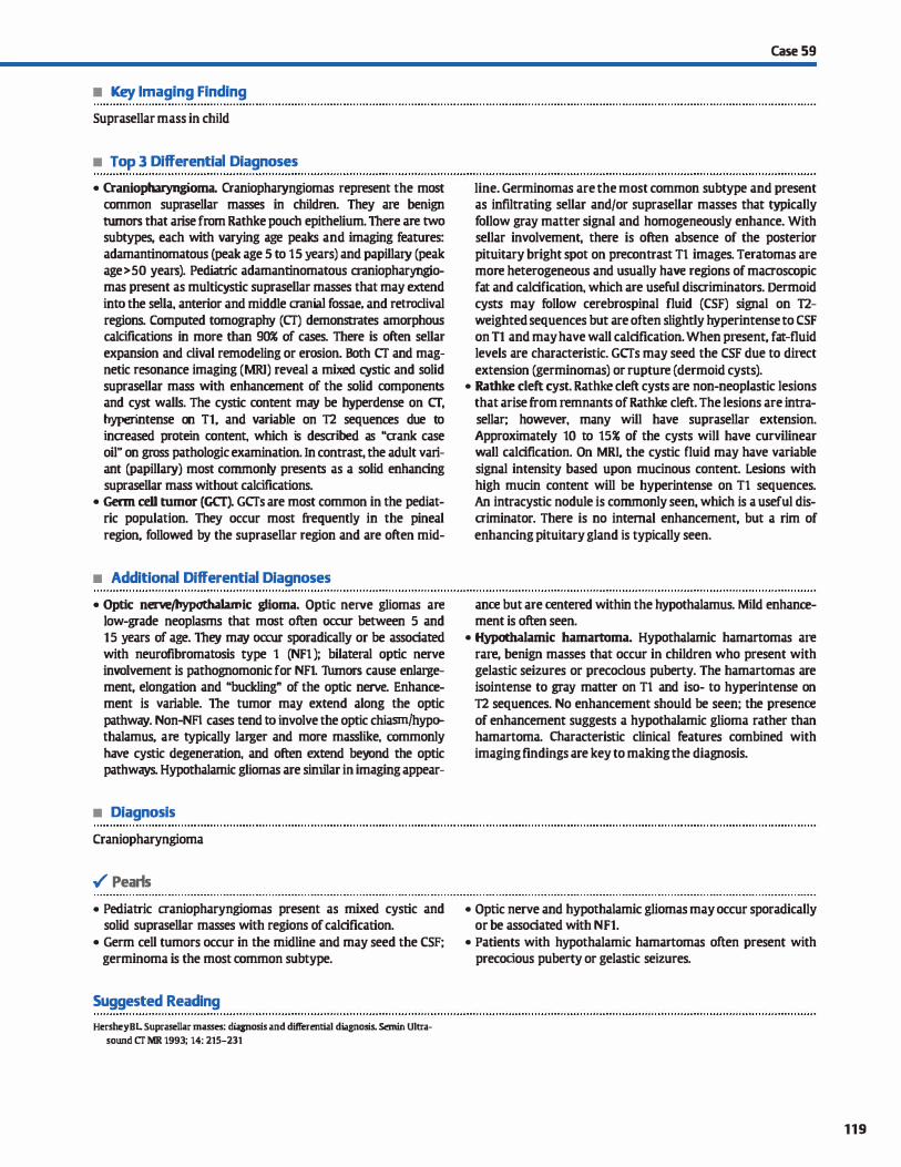
• Key lmaging Finding
Suprasellar mass in child
• Top 3 Differential Diagnoses
• Craniopharyngioma. Craniopharyngiomas represent the most common suprasellar masses in children. They are benign tumors that arise from Rathke pouch epithelium. There are two subtypes, each with varying age peaks and imaging features: adamantinomatous (peak age 5 to 15 years) and papillary (peak age>SO years). Pediatric adamantinomatous craniopharyngiomas present as multicystic suprasellar masses that may extend into the sella, anterior and middle cranial fossae. and retrodival regions. Computed tomography (CT) demonstrates amorphous calcifications in more than 90% of cases. There is often sellar expansion and dival remodeling or erosion. Both CT and magnetic resonance imaging (MRI) reveal a mixed cystic and solid suprasellar mass with enhancement of the solid components and cyst walls. The cystic content may be hyperdense on CT,
hyperintense on T1, and variable on T2 sequences due to increased protein content, which is described as "crank case oil" on gross pathologie examination. In contrast, the adult variant (papillary) most comrnonly presents as a solid enhancing suprasellar mass without calcifications.
• Germ ceU tumor {GCT). GCTs are most common in the pediatric population. They occur most frequently in the pineal region, followed by the suprasellar region and are often mid-
• Additional Differential Diagnoses
• Optic nervejhypothalamic glioma. Optic nerve gliomas are low-grade neoplasms that most often occur between 5 and 15 years of age. They may occur sporadically or be associated with neurofibromatosis type 1 (NFl ); bilateral optic nerve involvement is pathognomonic for NFt. Tumors cause enlargement, elongation and "buckling" of the optic nerve. Enhancement is variable. The tumor may extend along the optic pathway. Non-NFl cases tend to involve the optic chiasmfhypothalamus, are typically larger and more masslike, commonly have cystic degeneration, and often extend beyond the optic pathways. Hypothalamic gliomas are sinùlar in imaging appear-
• Diagnosis
Craniopharyngioma
� Pearls
• Pediatric craniopharyngiomas present as mixed cystic and solid suprasellar masses with regions of calcification.
• Germ cell tumors occur in the midline and may seed the CSF; germinoma is the most common subtype.
Suggested Reading
HersheyBL Suprasellar masses; diagnosis and differential diagnosis. Sernin Ultra
so1111d cr MR 1993; 14: 215-2.31
Case 59
line. Germinomas are the most common subtype and present as infiltrating sellar and/or suprasellar masses that typically follow gray matter signal and homogeneously enhance. With sellar involvement, there is often absence of the posterior pituitary bright spot on precontrast Tl images. Teratomas are more heterogeneous and usually have regions of macroscopic fat and calcification, which are useful discriminators. Dermoid cysts may follow cerebrospinal fluid {CSF) signal on T2-weighted sequences but are often slightly hyperintense to CSF on Tl and may have wall calcification. When present, fat-fluid levels are characteristic. GCTs may seed the CSF due to direct extension (germinomas) or rupture {dermoid cysts).
• Rathke cleft cyst. Rathke cleft cysts are non-neoplastic lesions that arise from remuants of Rathlœ cleft. The lesions are intrasellar; however, many will have suprasellar extension. Approximately 10 to 15% of the cysts will have curvilinear wall calcification. On MRI, the cystic fluid may have variable signal intensity based upon mucinous content. Lesions with high mucin content will be hyperintense on Tl sequences. An intracystic nodule is comrnonly seen, which is a useful discriminator. There is no internai enhancement, but a rim of enhancing pituitary gland is typically seen.
anœ but are œntered within the hypothalamus. Mild enhancement is often seen.
• Hypothalamic hamartoma. Hypothalamic hamartomas are rare, benign masses that occur in children who present with gelastic seizures or precodous puberty. The hamartomas are isointense to gray matter on Tl and iso- to hyperintense on T2 sequences. No enhancement should be seen; the presenœ of enhancement suggests a hypothalamic glioma rather than hamartoma. Characteristic clinical features combined with imaging findings are key to making the diagnosis.
• Optic nerve and hypothalamic gliomas may occur sporadically or be associated with NF1.
• Patients with hypothalamic hamartomas often present with precocious puberty or gelastic seizures.
1 1 9
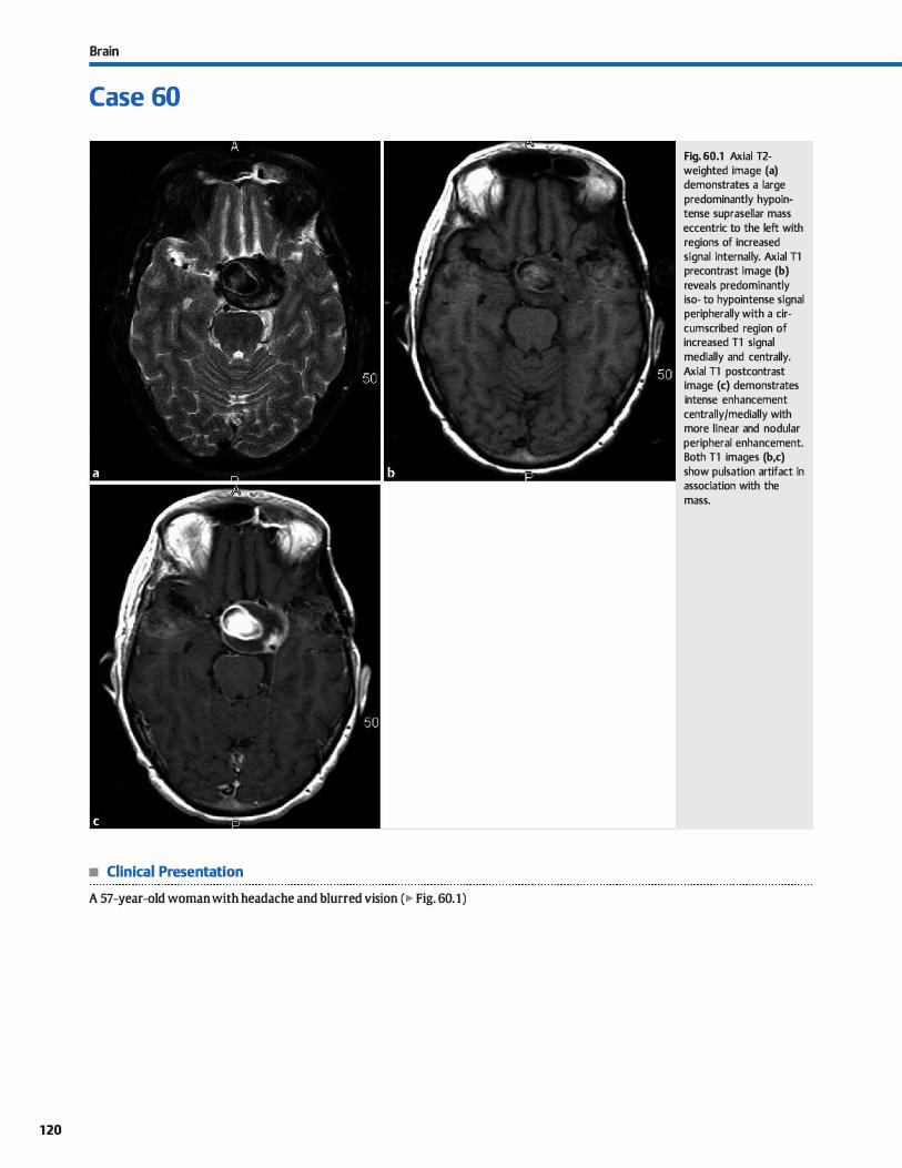
Brain
Case 60
• Clinical Presentation
A 57-year-old woman with headache and blurred vision (� Fig. 60.1)
120
Fig. 60.1 Axial T2-weighted image (a) demonstrates a large predominantly hypointense suprasellar mass eccentric to the left with regions of increased signal internally. Axial Tl precontrast image (b) reveals predominantly iso- to hypointense signal peripherally with a cir
cumscribed region of increased Tl signal medially and centrally. Axial Tl postcontrast image (c) demonstrates intense enhancement centrally/medially with more linear and nodular peripheral enhancement. Both Tl images (b,c) show pulsation artifact in association with the mass.
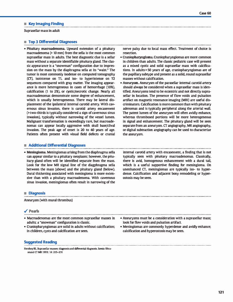
• Key lmaging Finding
Suprasellar mass in adult
• Top 3 Differential Diagnoses
• Pituitary macroadenoma. Upward extension of a pituitary macroadenoma (> 10 mm) from the sella is the most common
suprasellar mass in adults. The best diagnostic due is a sellar mass without a separate identifiable pituitary gland. The dassic appearance is a "snowman" configuration due to impression on the mass by the diaphragma sella at its ''waist." The tumor is most commonly isodense on computed tomography
(cr), isointense on Tl, and iso- to hyperintense on T2 sequences compared with gray matter. The imaging appearance is more heterogeneous in cases of hemorrhage (10%), calcification (1 to 2%), or cystic/necrotic change. Nearly ail macroadenomas demonstrate some degree of enhancement, which is usually heterogeneous. There may be lateral displaœment of the ipsilateral internai carotid artery. With cav
emous sinus invasion, there is carotid artery encasernent (> two-thirds is typically considered a sign of cavemous sinus
invasion), typically without narrowing of the vessel lumen. Malignant transformation is exœedingly rare, but macroadenomas can appear locally aggressive with skull base/clival
invasion. The peak age of onset is 20 to 40 years of age. Patients often present with visual field defects or cranial
• Additional Differential Diagnoses
• Meningioma. Meningiomas arising from the diaphragma sella can appear similar to a pituitary neoplasm; however, the pituitary gland often will be identified separate from the mass. Look for the low MR signal line of the diagphragma sella between the mass (above) and the pituitary gland (below). Durai thickening assoàated with meningioma is more extensive than with a pituitary macroadenoma. With cavernous
sinus invasion, meningiomas often result in narrowing of the
• Diagnosis
Aneurysm {with mural thrombus)
y' Pearls
• Macroadenomas are the most common suprasellar masses in
adults; a usnowman" configuration is dassic. • Craniopharyngiomas are solid in adults without calcification;
in children, cysts and calcification are seen.
Suggested Reading
Hershey BL Suprasellar masses: diagnosis and differential diagnosis. Sernin lfltra
sound cr MR 1993; 14: 215-231
Case 60
nerve palsy due to local mass effect. Treatment of choice is
resection. • Craniophacyngioma. Craniopharyngiomas are more common
in children than adults. The dassic pediatric case will present as a mixed cystic and solid suprasellar mass with calcifications. In adults > 50 years of age, craniopharyngiomas are of the papillary subtype and present as a solid, round suprasellar
masses without calcification. • Aneurysm. Aneurysm of the parasellar internai carotid artery
should always be considered when a suprasellar mass is identified. Aneurysms tend to be ecœntric and not directly suprasellar in location. The presence of flow voids and pulsation
artifact on magnetic resonance imaging (MRI) are useful discriminators. Calcification is more common than with pituitary adenomas and is typically peripheral along the arterial wall.
The patent lumen of the aneurysm will often avidly enhance, whereas thrombosed portions will be more heterogeneous in signal and enhancement. The pituitary gland will be seen separate from an aneurysm. cr angiography, MR angiography, or digital subtraction angiography can be used to characterize
the aneurysm
internai carotid artery with encasernent, a finding that is not typically seen with pituitary macroadenomas. dassically,
there is avid, homogenous enhancement with a durai tail, which is a useful supportive finding for meningioma. On unenhanced cr, meningiomas are typically iso- to hyperdense. Calcification and adjacent bony remodeling or hyperostosis may be seen.
• Aneurysms must be a consideration with a suprasellar mass;
look for flow voids and pulsation artifact. • Meningiomas are commonly hyperdense and avidly enhance;
calcification and hyperostosis may be seen.
121
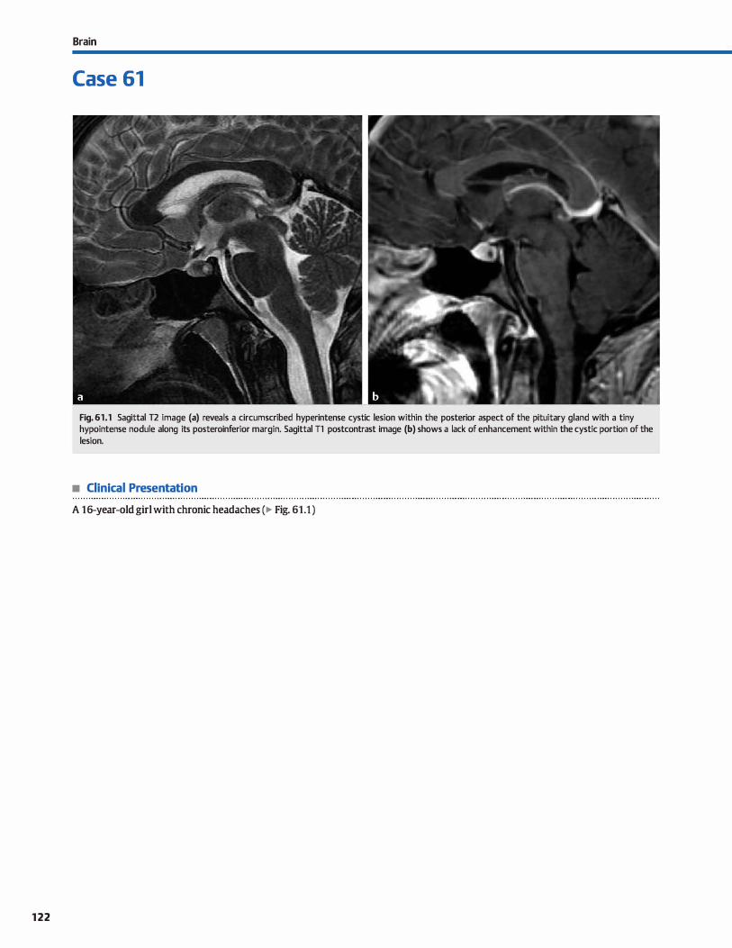
122
Brain
Case 61
Fig. 61.1 Sagittal T2 image (a) reveals a circumscribed hyperintense cystic lesion within the posterior aspect of the pituitary gland with a tiny hypointense nodule along its posteroinferior margin. Sagittal Tl postcontrast image (b) shows a lack of enhancement within the cystic portion of the lesion.
• Clinical Presentation
A 16-year-old girl with chronic headaches (� Fig. 61.1 )
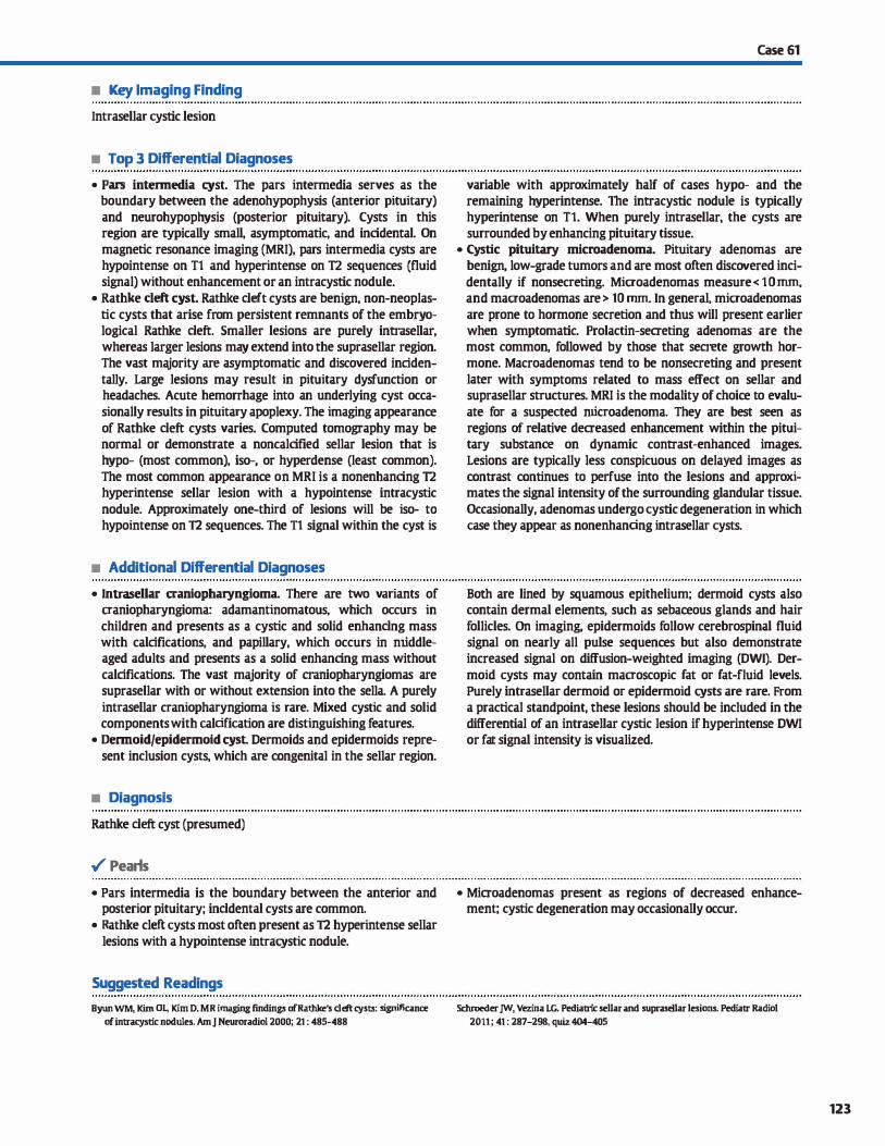
• Key lmaging Finding
lntrasellar cystic lesion
• Top 3 Differential Diagnoses
• Pars intennedia cyst. The pars intermedia serves as the boundary between the adenohypophysis (anterior pituitary) and neurohypophysis (posterior pituitary). Cysts in this region are typically small, asymptomatic, and incidental. On magnetic resonance imaging (MRI), pars intermedia cysts are hypointense on T1 and hyperintense on T2 sequenœs {fluid signal) without enhancement or an intracystic nodule.
• Rathke cleft cyst. Rathke deft cysts are benign, non-neoplastic cysts that arise from persistent remnants of the embryological Rathke deft. Smaller lesions are purely intrasellar, whereas larger lesions may extend into the suprasellar region. The vast majority are asymptomatic and discovered inciden
tally. Large lesions may result in pituitary dysfunction or headaches. Acute hemorrhage into an underlying cyst occa
sionally results in pituitary apoplexy. The imaging appearanœ
of Rathke deft cysts varies. Computed tomography may be normal or demonstrate a noncalcified sellar lesion that is hypo- (most common), iso-, or hyperdense (least common). The most common appearanœ on MRI is a nonenhancing T2 hyperintense sellar lesion with a hypointense intracystic
nodule. Approximately one-third of lesions will be iso- ta hypointense on T2 sequences. The T1 signal within the cyst is
• Additional Differential Diagnoses
• lntrasellar craniopharyngioma. There are two variants of craniopharyngioma: adamantinomatous, which occurs in children and presents as a cystic and solid enhandng mass with calcifications, and papillary, which occurs in nùddleaged adults and presents as a solid enhancing mass without calcifications. The vast majority of craniopharyngiomas are suprasellar with or without extension into the sella. A purely
intrasellar craniopharyngioma is rare. Mixed cystic and solid components with calcification are distinguishing features.
• Dennoid/epidermoid cyst. Dermoids and epidermoids represent inclusion cysts, which are congenital in the sellar region.
• Diagnosis
Rathke deft cyst (presumed)
� Pearls
• Pars intermedia is the boundary between the anterior and posterior pituitary; inddental cysts are common.
• Rathke cleft cysts most often present as T2 hyperintense sellar lesions with a hypointense intracystic nodule.
Suggested Readings
Byun WM, Kim 01., Kim D. MR imaging finding:s ofRathke's dd't cysts; significanœ
of intracysticnodules. Am j Neuroradiol 2000; 21 : 485-488
Case 61
variable with approximately half of cases hypo- and the remaining hyperintense. The intracystic nodule is typically hyperintense on T1. When purely intrasellar, the cysts are surrounded by enhancing pituitary tissue.
• Cystic pituitary microadenoma. Pituitary adenomas are benign, low-grade tumors and are most often discovered inci
dentally if nonsecreting. Microadenomas measure < 10 mm., and macroadenomas are > 10 mm. In general, microadenomas are prone ta hormone secretion and thus will present earlier when symptomatic. Prolactin-secreting adenomas are the most common, followed by those that secrete growth hormone. Macroadenomas tend to be nonsecreting and present later with symptoms related to mass effect on sellar and suprasellar structures. MRI is the modality of choiœ to evalu
ate for a suspected nùcroadenoma. They are best seen as regions of relative decreased enhancement within the pituitary substance on dynamic contrast-enhanced images. Lesions are typically Jess conspicuous on delayed images as contrast continues ta perfuse into the lesions and approximates the signal intensity of the surrounding glandular tissue.
Occasionally, adenomas undergo cystic degeneration in which case they appear as nonenhancing intrasellar cysts.
Both are lined by squamous epithelium; dermoid cysts also contain dermal elements, such as sebaœous glands and hair follicles. On imaging, epidermoids follow cerebrospinal fluid signal on nearly ail pulse sequenœs but also demonstrate increased signal on diffusion-weighted imaging (DWI). Der
moid cysts may contain macroscopic fat or fat-fluid levels. Purely intrasellar dermoid or epidermoid cysts are rare. From a practical standpoint, these lesions should be included in the differential of an intrasellar cystic lesion if hyperintense DWI or fat signal intensity is visualized.
• Microadenomas present as regions of decreased enhancement; cystic degeneration may occasionally occur.
Schroeder JW, Vezina LG. Pediatric sellar and suprasellar lesions. Pediatr Radio!
2011 ; 41 ; 287-298, quiz 404-405
123
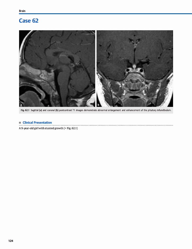
Brain
Case 62
Fig. 62.1 Sagittal (a) and coronal (b) postcontrast Tl images demonstrate abnormal en largement and enhancement of the pituitary infundibulum.
• Clinical Presentation
A 9-year-old girl with stunted growth (� Fig. 62.1 )
124
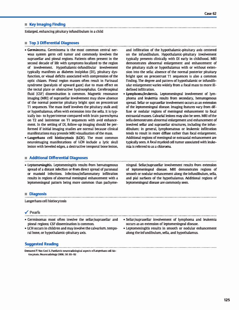
• Key lmaging Finding
Enlarged, enhancing pituitary infundibulum in a child
• Top 3 Differential Diagnoses
• Germinoma. Germinoma is the most common central ner
vous system germ cell tumor and commonly involves the
suprasellar and pineal regions. Patients often present in the
second decade of life with symptoms localized to the region
of involvement. Hypothalamic-infundibular involvement
typically manifests as diabetes insipidus (Dl), pituitary dysfunction, or visual deficits associated with compression of the
optic chiasm. Pineal region masses often result in Parinaud
syndrome (paralysis of upward gaze) due to mass etfect on
the tectal plate or obstructive hydrocephalus. Cerebrospinal
fluid (CSF) dissemination is common. Magnetic resonanœ
imaging (MRI) of suprasellar involvement may show absence of the normal posterior pituitary bright spot on precontrast
Tl sequences. The mass itself involves the pituitary stalk and/
or hypothalamus, often with extension into the sella. It is typically iso- to hyperintense compared with brain parenchyma
on T2 and isointense on Tl sequences with avid enhance
ment. In the setting of DI, follow-up imaging should be per
formed if initial imaging studies are normal because clinicat
manifestations may precede MRI visualization of the mass.
• Langerhans cell histiocytosis (LCH). The most common
neuroimaging manifestations of LCH indude a lytic skull
lesion with beveled edges, a destructive temporal bene lesion,
• Additional Differential Diagnoses
• Leptomeningitis. Leptomeningitis results Crom hematogenous
spread of a distant infection or Crom direct spread of paranasal
or mastoid infections. Infectious/inflammatory infiltration
results in regions of abnormal meningeal enhancement with a
leptomeningeal pattern being more common than pachyme-
• Diagnosis
Langerhans œll histiocytosis
� Pearls
• Germinomas most often involve the sellar/suprasellar and
pineal regions; CSF dissemination is common. • LCH occurs in children and may involve the calvariurn, tempo
ral bone, or hypothalamic-pituitary axis.
Suggested Reading
Demaerel P, Van Goal S. Paediatric neuroradiological aspects ofLange!hans cell his
tiocytosis. Neuroradiology 2008; 50; 85-92
Case 62
and infiltration of the hypothalamic-pituitary axis centered
on the infundibulum. Hypothalamic-pituitary involvement
typically presents dinically with DI early in childhood. MRI
demonstrates abnormal enlargement and enhancement of
the pituitary stalk or hypothalamus with or without exten
sion into the sella; absence of the normal posterior pituitary
bright spot on precontrast Tl sequences is aise a common
finding. The degree and pattern of hypothalamic or infundib
ular enlargement varies widely from a focal mass ta more ill
defined infiltration.
• cymphomaJieukemia. Leptomeningeal involvement of lym
phoma and leukemia results from secondary, hematogenous
spread. Sellar or suprasellar involvement occurs as an extension of the leptomeningeal disease. lmaging features vary from dif
fuse or nodular regions of meningeal enhancement to focal
extraaxial masses. Calvarial lesions may aise be seen. MRI ofthe sella demonstrates abnormal enlargement and enhancement of
involved sellar and suprasellar structures, induding the infun
dibulum; in general, lymphomatous or leukemic infiltration tends ta result in more diffuse rather than focal enlargement.
Additional regions of meningeal or extraaxial enhancement are
typically seen. A focal myeloid œll tumor associated with leuke
mia is referred to as a chloroma
ningeal Sellar/suprasellar involvement results Crom extension
of leptomeningeal disease. MRI demonstrates regions of
smooth or nodular enhancement along the infundibulum, sella,
and pial swfaces of the hypothalamus. Additional regions of
leptomeningeal disease are commonly seen.
• Sellar/suprasellar involvement of lymphoma and leukemia
occurs as an extension of leptomeningeal disease.
• Leptomeningitis results in smooth or nodular enhancement
along the infundibulum, sella, and hypothalamus.
125
126
Brain
Case 63
Fig. 63.1 Axial (a) and coronal (b) Tl postcontrast images demonstrate an avidly enhancing left cavernous sinus mass with encasernent and marked narrowing of the cavernous segment of the left ICA. There is extension into the prepontine cistern on the left. Hyperintense signal intensity overlying the pans on the right (a) was artifactual.
• Clinical Presentation
A 31-year-old post-partum woman complaining of facial numbness (� Fig. 63.1 )
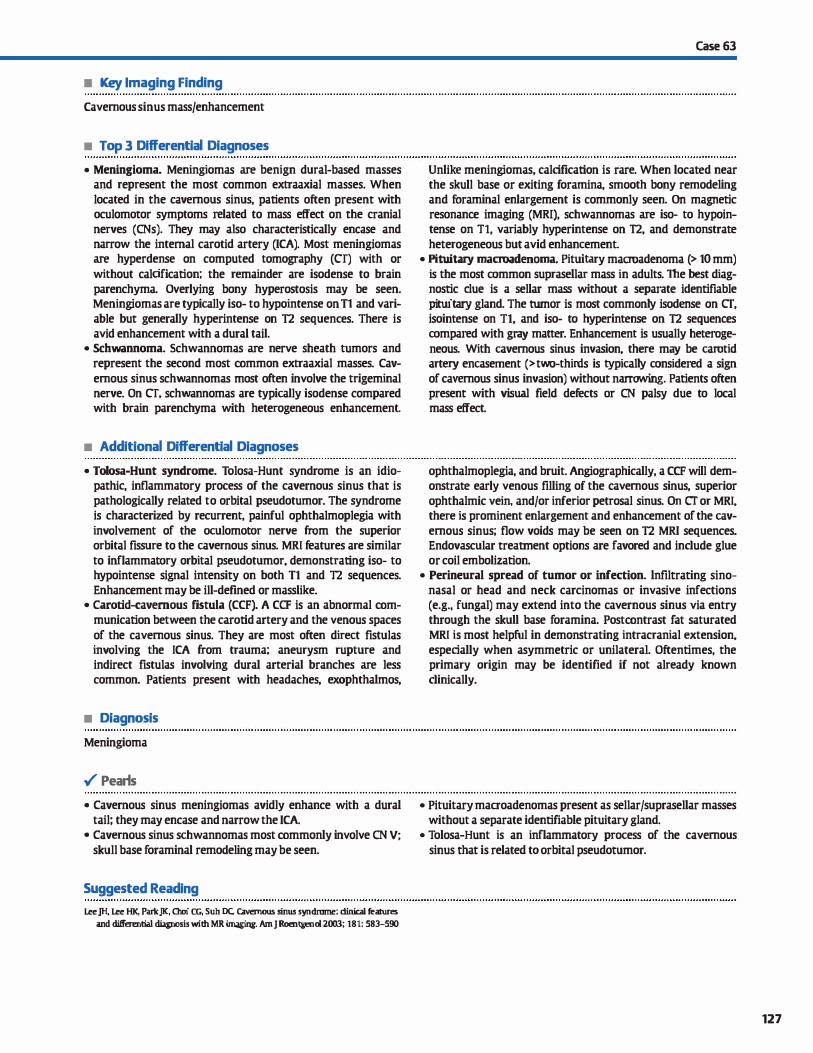
• Key lmaging Finding
Cavemous sinus mass/enhancement
• Top 3 Differential Diagnoses
• Meningioma. Meningiomas are benign dural-based masses and represent the most common extraaxial masses. When
located in the cavemous sinus, patients often present with oculomotor symptoms related to mass effect on the cranial nerves (CNs). They may also characteristically encase and narrow the internai carotid artery (ICA). Most meningiomas are hyperdense on computed tomography (cr) with or
without calcification; the remainder are isodense to brain parenchyma. OVerlying bony hyperostosis may be seen. Meningiomas are typically iso- to hypointense on T1 and variable but generally hyperintense on T2 sequenœs. There is avid enhancement with a durai tail.
• Schwannoma. Schwannomas are nerve sheath tumors and represent the second most common extraaxial masses. cav
emous sinus schwannomas most often involve the trigeminal nerve. On cr. schwannomas are typically isodense compared with brain parenchyma with heterogeneous enhancement
• Additional Differential Diagnoses
• Tolosa-Hunt syndrome. Tolosa-Hunt syndrome is an idiopathie, inflammatory process of the cavernous sinus that is pathologically related to orbital pseudotumor. The syndrome is characterized by recurrent, painful ophthalmoplegia with
involvement of the oculomotor nerve from the superior orbital fissure to the cavernous sinus. MRI features are similar
to inflammatory orbital pseudotumor, demonstrating iso- to hypointense signal intensity on both Tt and T2 sequences. Enhancement may be ill-defined or masslike.
• carotid-cavemous ftstula (CCF). A CŒ is an abnormal communication between the carotid artery and the venous spaces
of the cavemous sinus. They are most often direct fistulas involving the ICA from trauma; aneurysm rupture and indirect fistulas involving durai arterial branches are less common. Patients present with headaches, exophthalmos,
• Diagnosis
Meningioma
� Pearls
• cavernous sinus meningiomas avidly enhance with a durai tail; they may encase and narrow the ICA.
• cavernous sinus schwannomas most commonly involve CN V;
skull base foraminal remodeling may be seen.
Suggested Reading
Lee JH, Lee HIC, Parie JK, Cl10i Œ, Suh OC cavemous 5inm syndrome: dinic.al reatures
and differential diagnosis with MR imaging. Am j Rœn�ol 2003; 181: 583-590
Case 63
Unlike meningiomas, calcification is rare. When located near the skull base or exiting foramina, smooth bony remodeling and foraminal enlargement is commonly seen. On magnetic resonance imaging (MRI), schwannomas are iso- to hypointense on Tl, variably hyperintense on T2, and demonstrate
heterogeneous but avid enhancement • Pituitary macroadenoma. Pituitary macroadenoma (> 10 mm)
is the most common suprasellar mass in adults. The best diagnostic due is a sellar mass without a separate identifiable pituitary gland. The tumor is most commonly isodense on cr. isointense on Tt, and iso- to hyperintense on T2 sequences compared with gray matter. Enhanœment is usually heteroge
neous. With cavemous sinus invasion, there may be carotid artery encasernent (>two-thirds is typically considered a sign of cavemous sinus invasion) without narrowing. Patients often present with visual field defects or CN palsy due to local mass effect.
ophthalmoplegia, and bruit. Angiographically, a CCF will demonstrate early venous filling of the cavemous sinus, superior
ophthalmic vein, and/or inferior petrosal sinus. On CT or MRI, there is prominent enlargement and enhanœment of the cav
emous sinus; flow voids may be seen on T2 MRI sequences. Endovascular treatment options are favored and indude glue or coil embolization.
• Perineural spread of tumor or infection. lnfiltrating sinonasal or head and neck carcinomas or invasive infections (e.g., fungal) may extend into the cavernous sinus via entry through the skull base foramina. Postcontrast fat saturated
MRI is most helpful in demonstrating intracranial extension, espeàally when asymmetric or unilateral. Oftentimes, the primary origin may be identified if not already known dinically.
• Pituitary macroadenomas present as sellar/suprasellar masses without a separate identifiable pituitary gland.
• Tolosa-Hunt is an inflammatory process of the cavemous
sinus that is related ta orbital pseudotumor.
127
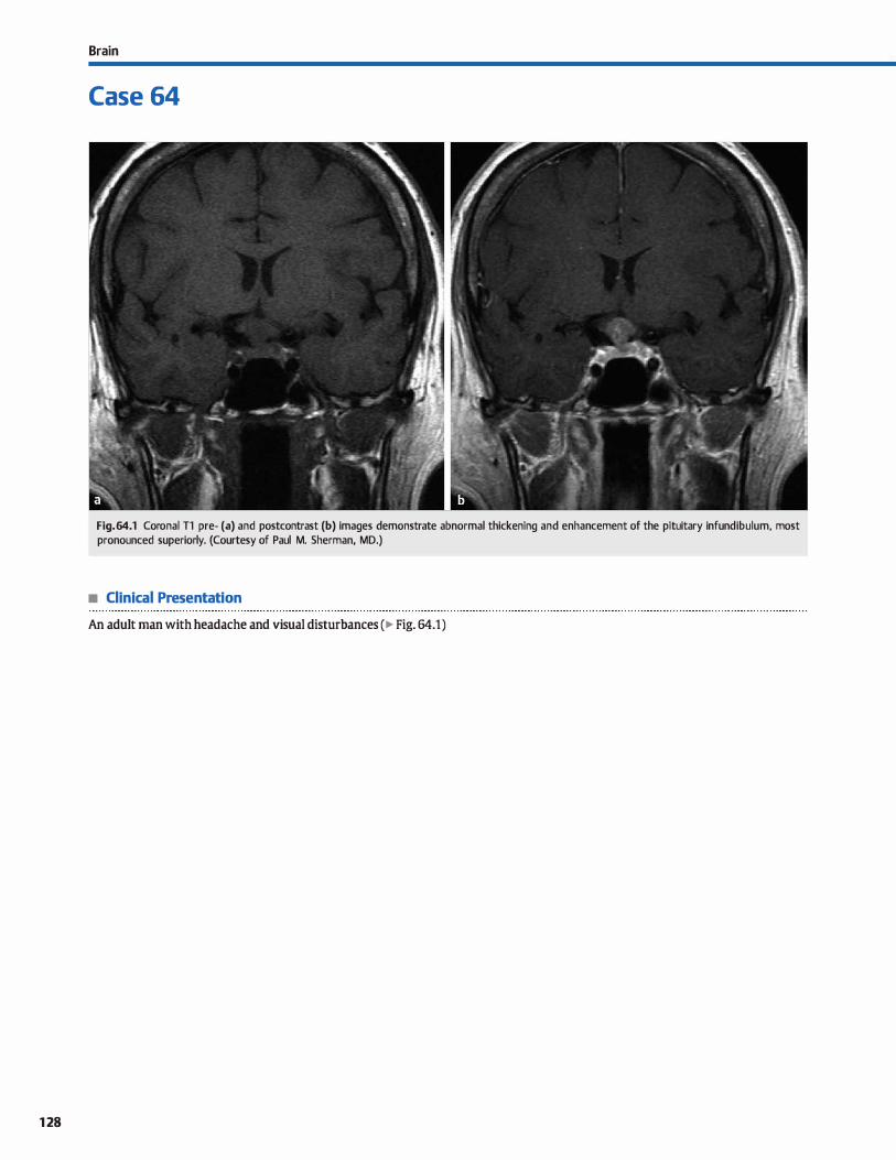
128
Brain
Case 64
Fig. 64.1 Coronal Tl pre- (a) and postcontrast (b) images demonstrate abnormal thickening and enhancement of the pituitary infundibulum, most pronounced superiorly. (Courtesy of Paul M. Sherman, MD.)
• Clinical Presentation
An adult man with headache and visual disturbances ( � Fig. 64.1)
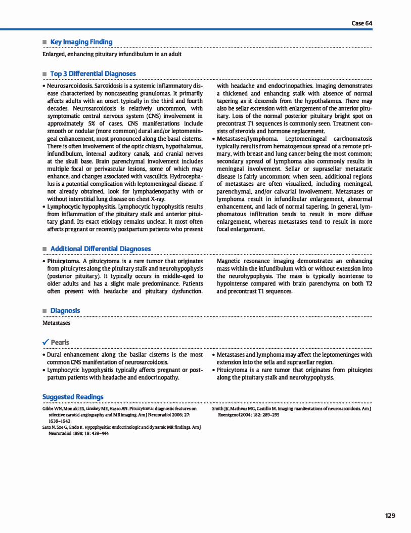
• Key lmaging Finding
Enlarged, enhancing pituitary infundibulum in an adult
• Top 3 Differential Diagnoses
• Neurosan:oidosis. Sarcoidosis is a systemic inflammatory dis
ease characterized by noncaseating granulomas. lt primarily
affects adults with an onset typically in the third and fourth decades. Neurosarcoidosis is relatively uncommon, with symptomatic central nervous system (CNS) involvement in approximately 5% of cases. CNS manifestations include smooth or nodular (more common) durai and/or leptomenin
geal enhanœment, most pronounced along the basal cistems. There is often involvement of the optic chiasm, hypothalamus, infundibulum, internai auditory canais, and cranial nerves at the skull base. Brain parenchymal involvement includes multiple focal or perivascular lesions, some of which may enhance, and changes associated with vasculitis. Hydroœphalus is a potential complication with leptomeningeal disease. If
not already obtained, look for lymphadenopathy with or
without interstitial Jung disease on chest X-ray.
• Lymphocytic hypophysitis. Lymphocytic hypophysitis results from inflammation of the pituitary stalk and anterior pituitary gland. Its exact etiology remains unclear. lt most often
affects pregnant or recently postpartum patients who present
• Additional Differential Diagnoses
• Pituicytoma. A pituicytoma is a rare tumor that originates from pituicytes along the pituitary stalk and neurohypophysis
(posterior pituitary). lt typically occurs in middle-aged to
older adults and has a slight male predominance. Patients
often present with headache and pituitary dysfunction.
• Diagnosis
Metastases
� Pearts
• Durai enhanœment along the basilar cistems is the most
common CNS manifestation of neurosarcoidosis.
• Lymphocytic hypophysitis typically affects pregnant or postpartum patients with headache and endocrinopathy.
Suggested Readings
Gibbs WN, Monuki ES, lin5lœy ME, Hasso AN. Pituicytmna; diagnostic features on selective carotid angiography and MR imaging. AmJ Neuroradiol 2006; 27: 1639-1642
Sato N, Sze G, Endo K. Hypophysitis: endocrinologie and dynamic MR findings. AmJ Neuroradiol 1998; 19: 439-444
Case 64
with headache and endocrinopathies. Imaging demonstrates
a thickened and enhanàng stalk with absence of normal tapering as it descends from the hypothalamus. There may also be sellar extension with enlargement of the anterior pituitary. I.oss of the normal posterior pituitary bright spot on precontrast T1 sequences is commonly seen. Treatment con
sists of steroids and hormone replacement.
• Metastases/lymphoma. Leptomeningeal cardnomatosis
typically results from hematogenous spread of a remote primary, with breast and Jung cancer being the most common; secondary spread of lymphoma also commonly results in
meningeal involvement. Sellar or suprasellar metastatic
disease is fairly uncommon; when seen, additional regions of metastases are often visualized, including meningeal,
parenchymal, and/or calvarial involvement. Metastases or lymphoma result in infundibular enlargement, abnormal
enhancement, and lack of normal tapering. In general, lymphomatous infiltration tends ta result in more diffuse
enlargement, whereas metastases tend ta result in more focal enlargement.
Magnetic resonance imaging demonstrates an enhandng
mass within the infundibulum with or without extension into the neurohypophysis. The mass is typically isointense to hypointense compared with brain parenchyma on bath 12 and precontrast Tl sequences.
• Metastases and lymphoma may affect the leptomeninges with extension into the sella and suprasellar region.
• Pituicytoma is a rare tumor that originates from pituicytes along the pituitary stalk and neurohypophysis.
Smith JIC, Mathem MG, castillo M. lmaging manifestations of neurosarmidosis. Am J Roentgenol2004; 182: 289-29S
129
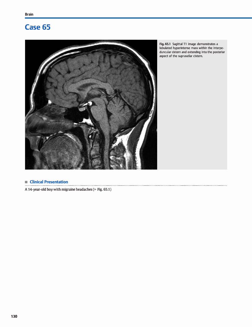
Brain
Case 65
• Clinical Presentation
A 14-year-old boy with migraine headaches (� Fig. 65.1 )
130
Fig. 65.1 Sagittal Tl image demonstrates a lobulated hyperintense mass within the interpeduncular cistern and extending into the posterior aspect of the suprasellar cistern.
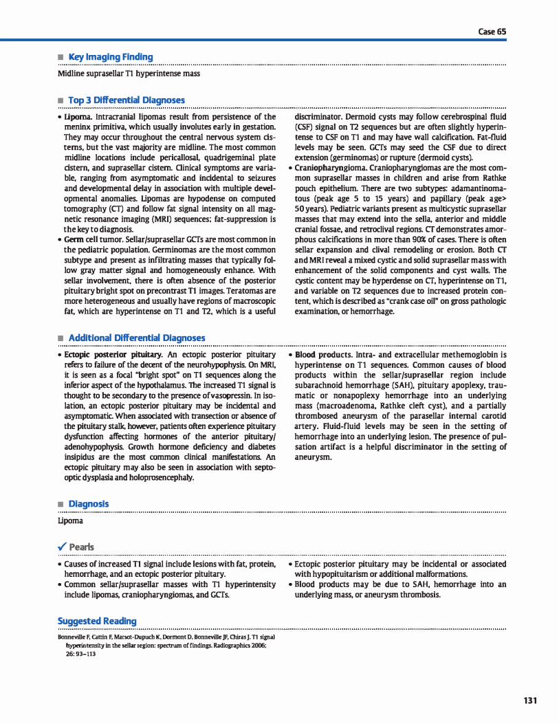
• Key lmaging Finding
Midline suprasellar Tl hyperintense mass
• Top 3 Differential Diagnoses
• lipoma. Intracranial lipomas result from persistence of the meninx primitiva, which usually involutes early in gestation. They may occur throughout the central nervous system dstems, but the vast majority are midline. The most common midline locations include pericallosal, quadrigeminal plate dstern, and suprasellar cistem. Œnical symptoms are variable, ranging from asymptomatic and inddental to seizures and developmental delay in association with multiple developmental anomalies. Llpomas are hypodense on computed tomography (CT) and follow fat signal intensity on ail magnetic resonance imaging (MRI) sequences; fat-suppression is the key to diagnosis.
• Germ cell tumor. Sellar/suprasellar GCTs are most common in the pediatric population. Germinomas are the most common subtype and present as infiltrating masses that typically follow gray matter signal and homogeneously enhance. With sellar involvement, there is often absence of the posterior pituitary bright spot on precontrast Tl images. Teratomas are more heterogeneous and usually have regions of macroscopic fat, which are hyperintense on Tl and T2, which is a useful
• Additional Differential Diagnoses
• Ectopie posterior pituitary. An ectopie posterior pituitary refers to failure of the decent of the neurohypophysis. On MRI, it is seen as a focal "bright spot" on Tl sequences along the inferior aspect of the hypothalamus. The increased Tl signal is thought to be secondary to the presence ofvasopressin. In isolation, an ectopie posterior pituitary may be incidental and asymptomatic. When associated with transection or absence of the pituitary stalk, however, patients often experienœ pituitary dysfunction affecting hormones of the anterior pituitary/ adenohypophysis. Growth hormone deficiency and diabetes insipidus are the most common dinical manifestations. An
ectopie pituitary may also be seen in association with septooptic dysplasia and holoprosenœphaly.
• Diagnosis
Llpoma
� Pearls
• causes of increased Tl signal include lesions with fat. protein, hemorrhage, and an ectopie posterior pituitary.
• Common sellar/suprasellar masses with Tl hyperintensity include lipomas, craniopharyngiomas, and GCTs.
Suggested Reading
Bonneville F, Cittin F, Marsot-Dupuch K, Dormont D, Bonneville JF, Otlras j. TI signal
hyperintensity in the sellar region: spectrum of findings. Radiographies 2006; 26: 93-113
Case 65
discriminator. Dermoid cysts may follow cerebrospinal fluid (CSF) signal on T2 sequences but are often slightly hyperintense ta CSF on Tl and may have wall calcification. Fat-fluid levels may be seen. Gers may seed the CSF due to direct extension (germinomas) or rupture {dermoid cysts).
• Craniopharyngioma. Craniopharyngiomas are the most common suprasellar masses in children and arise from Rathke
pouch epithelium. There are two subtypes: adamantinomatous (peak age 5 ta 15 years) and papillary {peak age> 50 years). Pediatric variants present as multicystic suprasellar masses that may extend into the sella, anterior and middle cranial fossae, and retroclival regions. CT demonstrates amorphous calcifications in more than 90% of cases. There is often sellar expansion and clivai remodeling or erosion. Both CT and MRI reveal a mixed cystic and solid suprasellar mass with enhancement of the solid components and cyst walls. The cystic content may be hyperdense on CT, hyperintense on Tl, and variable on T2 sequences due to increased protein content, which is described as "crank case oil" on gross pathologie examination, or hemorrhage.
• Blood products. Intra- and extracellular methemoglobin is hyperintense on Tl sequences. Common causes of blood products within the sellar/suprasellar region include subarachnoid hemorrhage {SAH), pituitary apoplexy, traumatic or nonapoplexy hemorrhage into an underlying mass (macroadenoma, Rathke cleft cyst), and a partially thrombosed aneurysm of the parasellar internai carotid artery. Fluid-fluid levels may be seen in the setting of hemorrhage into an underlying lesion. The presence of pulsation artifact is a helpful discriminator in the setting of aneurysm.
• Ectopie posterior pituitary may be incidental or associated with hypopituitarism or additional malformations.
• Blood products may be due to SAH, hemorrhage into an underlying mass, or aneurysm thrombosis.
131
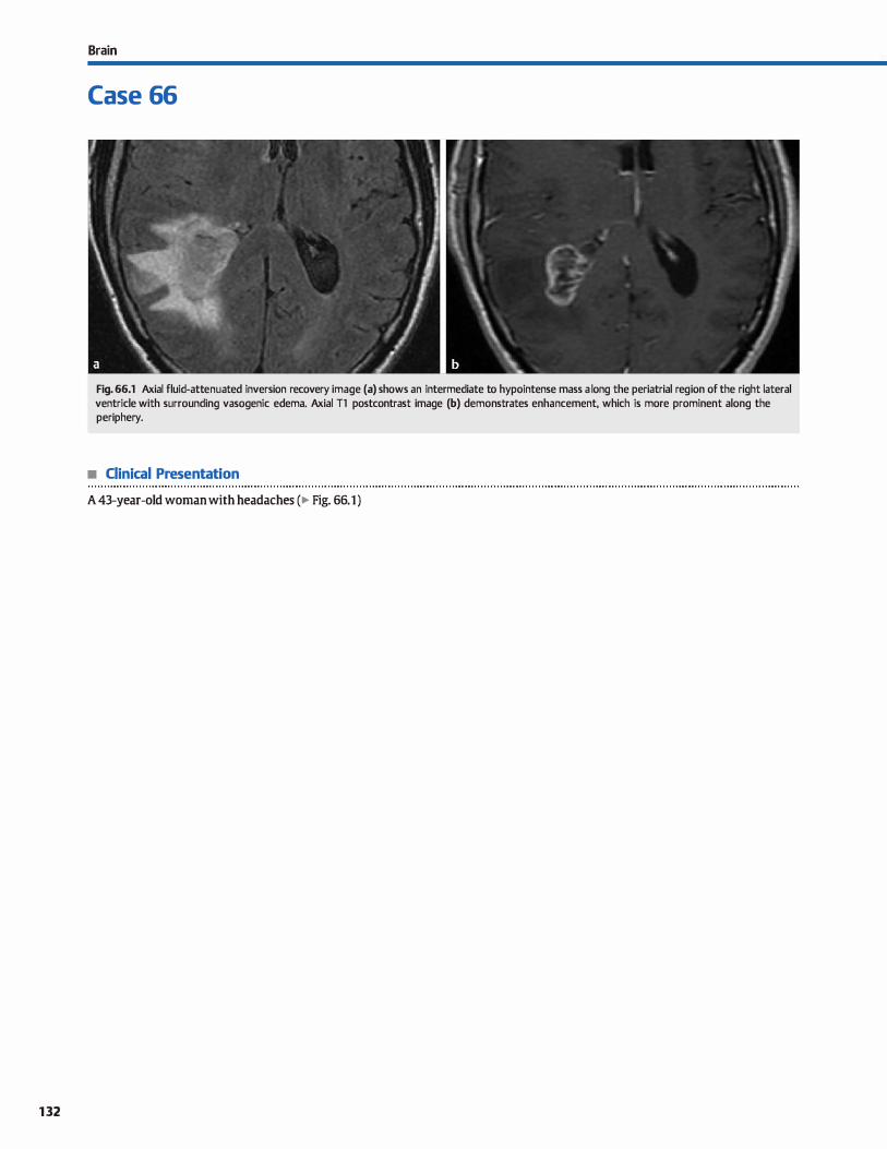
132
Brain
Case 66
Fig. 66.1 Axial fluid-attenuated inversion recovery image (a) shows an intermediate to hypointense mass a long the periatrial reg ion of the right lateral ventricle with surrounding vasogenic edema. Axial Tl postcontrast image (b) demonstrates enhancement, which is more prominent along the periphery.
• Clinical Presentation
A 43-year-old woman with headaches (� Fig. 66.1)
• Key lmaging Finding
Periventricular mass/enhancement with edema
• Top 3 Differential Diagnoses
• Primary central nervous system {CNS) lymphoma. Primary
CNS lymphoma is almost exdusively of the non-Hodgkin
variant. It typically occurs within the periventricular white
matter and basal ganglia and commonly involves the corpus
callosum and ependymal surfaces of the ventricles. Leptome
ningeal and durai involvement occurs more often with sec
ondary lymphoma. Lymphoma may present as a solitary mass
or multiple Iesions and can be drcumscribed or infiltrative. There is typically mild surrounding edema. The lesions are
characteristically hyperdense on computed tomography (CT)
and iso- to hypointense on both Tl and T2 sequences due
to high cellularity. Increased signal on diffusion sequenœs
may also be seen. Hemorrhage and necrosis are uncommon.
Enhanœment is intense and homogenous in immuno
competent patients and may be peripheral or ring-enhandng
in immunocompromised patients. Unlike toxoplasmosis, lym
phoma is hypermetabolic on Thallium single-photon emission
CT (SPECT) and positron emission tomograpby imaging and demonstrates increased perfusion.
• Glial neoplasm. Aggressive glial tumors commonly involve
the deep gray and white matter and may extend into the ven
tricles along the ependymal surface. Glioblastoma multiforme
(World Health Organization grade IV astrocytoma) is the
most common subtype and results in thick, irregular
enhancement and central necrosis. The tumor is heteroge-
• Diagnosis
Primary CNS lymphoma
� Pearts
• CNS lymphoma and aggressive glial tumors often involve the
periventricular region and corpus callosum.
• Regions of tumor with high cellularity are often hyperdense
on CT and hypointense on T2 MRI sequences.
Suggested Reading
Smimiotopoulos JG, Murphy FM, Rushing EJ, ReesjH, Schroeder JW. Patœrrus of con
trast enhanœrnent in the brain and rneninges. Radiographies 2007; 27: 525-551
Case 66
neous but primarily T2 hyperintense and Tl iso- to hypoin
tense with associated cysts, hemorrhage, and fluid debris.
There is prominent surrounding edema; viable tumor cells
extend beyond the margin of signal abnormality on patholog
ical evaluation. Regions of diffusion restriction and increased perfusion are noted within solid components. Prominent flow
voids may be seen and are associated with neovascularity.
As with primary CNS lymphoma, the corpus callosum is com
monly involved and expanded. Prognosis is poor.
• Ventriculitis/Ependymitis. Ventricular or ependymal spread
of infection may occur from hematogenous spread to the cho
roid plexus or direct extension from a parenchymal infection,
such as intraventricular rupture of a periventricular abscess. Immunocompromised patients and those with intraventricu
lar drainage catheters are more prone to intraventricular
infections. Cytomegalovirus, toxoplasmosis, and tuberculosis
are common causative agents in immunosuppressed patients.
Magnetic resonance imaging {MRI) demonstrates increased
T2/fluid-attenuated inversion recovery signal along the ven
tricular margin and avid, thin ependymal enhancement
There is often layering of dependent debris, which may
demonstrate restricted diffusion in the setting of pyogenic
infection. Short- and long-term complications include hydro
cephalus, which is most often communicating. Periventricular
extension may result in taxie or ischemic parenchymal injury.
• GBM is an aggressive tumor with thick, irregular enhance
ment; central necrosis and flow voids are common.
• Ventriculitis is more common in immunosuppressed patients; there is enhanœment and layering of debris.
133
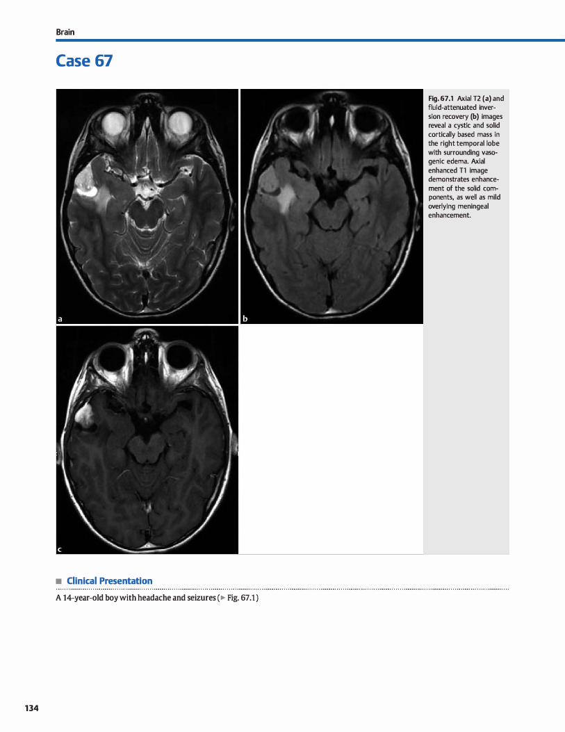
Brain
Case 67
• Clinical Presentation
A 14-year-old boy with headache and seizures (� Fig. 67.1 )
134
Fig. 67.1 Axial T2 (a) and fluid-attenuated inversion recovery (b) images reveal a cystic and solid cortically based mass in the right temporal lobe with surrounding vasogenic edema. Axial
enhanced Tl image demonstrates enhancement of the solid components, as well as mild overlying meningeal enhancement.
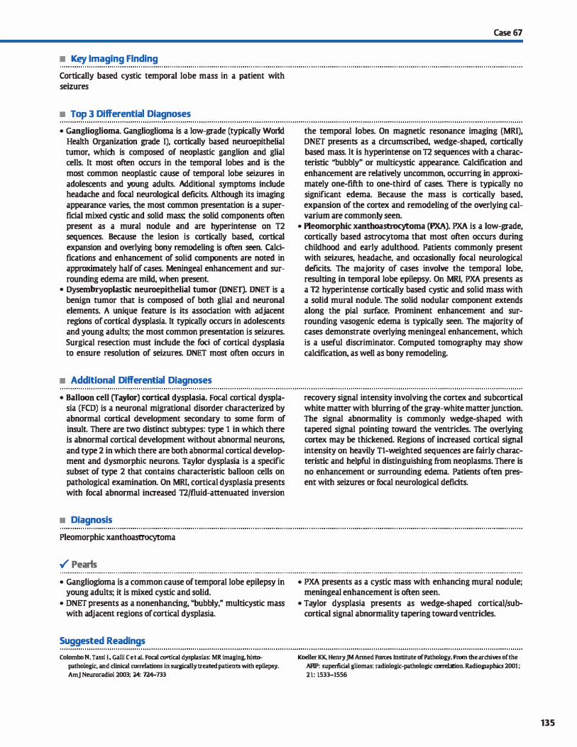
• Key lmaging Finding
Cortically based cystic temporal lobe mass in a patient with seizures
• Top 3 Differential Diagnoses
• Ganglioglioma. Ganglioglioma is a law-grade (typically World Health Organization grade I). cortically based neuroepithelial tumor, which is composed of neoplastic ganglion and glial cells. It most often occurs in the temporal lobes and is the most common neoplastic cause of temporal lobe seizures in adolescents and young adults. Additional symptoms indude headache and focal neurological deficits. Although its imaging appearanœ varies, the most common presentation is a superficial mixed cystic and solid mass; the solid components often present as a mural nodule and are hyperintense on T2 sequenœs. Because the lesion is cortically based, cortical expansion and overlying bony remodeling is often seen. Calcifications and enhancement of solid components are noted in approximately half of cases. Meningeal enhanœment and surrounding edema are mild, when present.
• Dysembryoplastic neuroepithelial tum.or {DNET). DNET is a benign tumor that is composed of both glial and neuronal elements. A unique feature is its association with adjacent regions of cortical dysplasia. It typically occurs in adolescents and young adults; the most common presentation is seizures. Surgical resection must indude the foci of cortical dysplasia to ensure resolution of seizures. DNET most often occurs in
• Additional Differential Diagnoses
• Balloon cell (Taylor) cortical dysplasia. Focal cortical dysplasia {FCD) is a neuronal migrational disorder characterized by abnormal cortical development secondary to some form of insult. There are two distinct subtypes: type 1 in which there is abnormal cortical development without abnormal neurons,
and type 2 in which there are both abnormal cortical development and dysmorphie neurons. Taylor dysplasia is a specific subset of type 2 that contains characteristic balloon cells on pathological examination. On MRI, cortical dysplasia presents with focal abnormal increased T2/fluid-attenuated inversion
• Diagnosis
Pleomorphic xanthoastrocytoma
� Pearls
• Gangliogioma is a common cause of temporal lobe epilepsy in young adults; it is mixed cystic and solid.
• DNEf presents as a nonenhancing, "bubbly," multicystic mass with adjacent regions of cortical dysplasia.
Suggested Readings
Colombo N, Tassi L, Galli Cet al. Focal rortical dysplasias; MR imaging, histDpathologic, ilI!d clinicat cmrelations in sUIJÏcally treated patients with epilepsy.
AmJNeuroradiol 2003; 2.4: 724-733
Case 67
the temporal lobes. On magnetic resonance imaging (MRI), DNEf presents as a circumscribed, wedge-shaped, cortically based mass. lt is hyperintense on T2 sequenœs with a characteristic "bubbly" or multicystic appearance. Calcification and enhanœment are relatively uncommon, occurring in approximately one-fifth to one-third of cases. There is typically no significant edema. Because the mass is cortically based, expansion of the cortex and remodeling of the overlying calvarium are commonly seen.
• Pleomorphic xanthoastrocytoma (PXA). PXA is a law-grade, cortically based astrocytoma that most often occurs during childhood and early adulthood. Patients commonly present with seizures, headache, and occasionally focal neurological deficits. The majority of cases involve the temporal lobe, resulting in temporal lobe epilepsy. On MRI, PXA presents as a T2 hyperintense cortically based cystic and solid mass with a solid mural nodule. The solid nodular component extends along the pial surface. Prominent enhanœment and surrounding vasogenic edema is typically seen. The majority of cases demonstrate overlying meningeal enhancement, which is a useful discriminator. Computed tomography may show calcification, as well as bony remodeling.
recovery signal intensity involving the cortex and subcortical white matter with blurring of the gray-white matter junction. The signal abnormality is commonly wedge-shaped with tapered signal painting taward the ventrides. The overlying cortex may be thickened. Regions of increased cortical signal intensity on heavily Tl-weighted sequenœs are fairly characteristic and helpful in distinguishing from neoplasms. There is no enhancement or surrounding edema. Patients often present with seizures or focal neurological deficits.
• PXA presents as a cystic mass with enhancing mural nodule; meningeal enhanœment is often seen.
• Taylor dysplasia presents as wedge-shaped cortical/subcortical signal abnormality tapering toward ventrides.
Koeller KK, Henry JM Anned Fon:es lnstitute of Pathology. From the ardti� of the
AFIP: supedicial gliomas: radiologie-pathologie correlation. Radiographia 2001; 21: 1533-1556
135
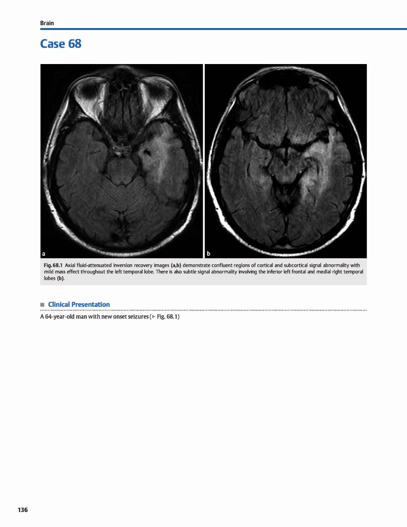
136
Brain
Case 68
Fig. 68.1 Axial fluid-attenuated inversion recovery images (a,b) demonstrate confluent regions of cortical and subcortical signal abnormality with mild mass effect throughout the left temporal lobe. There is also subtle signal abnormality involving the inferior left frontal and medial right temporal lobes (b).
• Clinical Presentation
A 64-year-old man with new onset seizures (� Fig. 68.1)
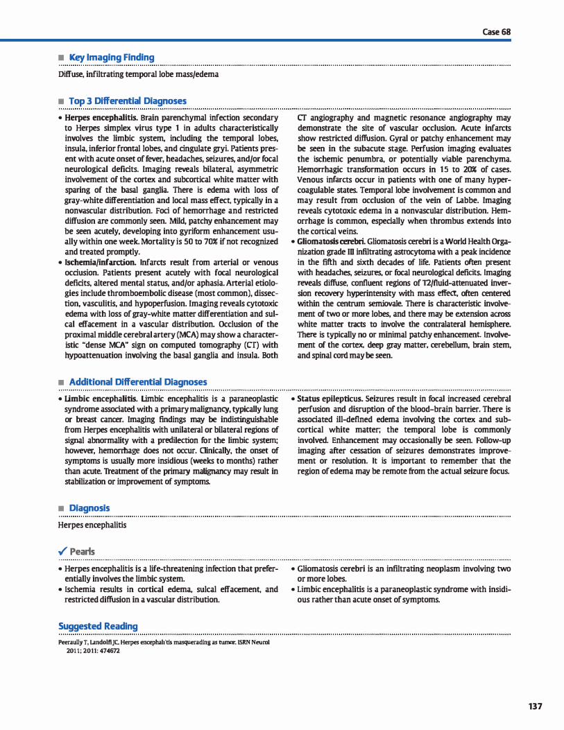
• Key lmaging Finding
Diffuse, infiltrating temporal lobe mass/edema
• Top 3 Differential Diagnoses
• Herpes encephalitis. Brain parenchymal infection secondary to Herpes simplex virus type 1 in adults characteristically involves the limbic system, including the temporal lobes, insula, inferior frontal lobes, and dngulate gryi. Patients present with acute onset of fever, headaches, seizures, and/or focal neurological defidts. Imaging reveals bilateral, asymmetric involvement of the cortex and subcortical white matter with sparing of the basal ganglia. There is edema with Joss of gray-white differentiation and local mass effect, typically in a nonvascular distribution. Foci of hemorrhage and restricted diffusion are commonly seen. Mild, patchy enhancement may be seen acutely, developing into gyriform enhancement usually within one week. Mortality is 50 to 70% if not recognized and treated promptly.
• Ischemia/infarction. Infarcts result from arterial or venous ocdusion. Patients present acutely with focal neurological defidts, altered mental status, and/or aphasia. Arterial etiologies include thromboembolic disease (most common), dissection, vasculitis, and hypoperfusion. Imaging reveals cytotoxic edema with Joss of gray-white matter differentiation and sulcal effacement in a vascular distribution. Occlusion of the proximal middle cerebral artery (MCA) may show a characteristic "dense MCA" sign on computed tomography (CT) with hypoattenuation involving the basal ganglia and insula. Both
• Additional Differential Diagnoses
• limbic encephalitis. Llrnbic encephalitis is a paraneoplastic syndrome associated with a primary malignancy, typically Jung or breast cancer. Imaging findings may be indistinguishable from Herpes enœphalitis with unilateral or bilateral regions of
signal abnormality with a predilection for the limbic system: hawever, hemorrhage does not occur. Oinically, the onset of symptoms is usually more insidious (weeks to months) rather than acute. li'eatment of the primary malignancy may result in stabilization or improvement of symptoms.
• Diagnosis
Herpes enœphalitis
� Pearls
• Herpes encephalitis is a life-threatening infection that preferentially involves the limbic system.
• Ischemia results in cortical edema. sulcal effacement, and restricted diffusion in a vascular distribution.
Suggested Reading
Peeraully T, undolfijC. Herpes enœphalitis masquerading as tumor. ISRN Neurol
2011; 2011: 474672
Case 68
cr angiography and magnetic resonanœ angiography may demonstrate the site of vascular occlusion. Acute infarcts show restricted diffusion. Gyral or patchy enhancement may be seen in the subacute stage. Perfusion imaging evaluates the ischemic penumbra, or potentially viable parenchyma. Hemorrhagic transformation occurs in 15 to 20% of cases. Venous infarcts occur in patients with one of many hypercoagulable states. Temporal lobe involvement is common and may result from occlusion of the vein of Labbe. Imaging reveals cytotoxic edema in a nonvascular distribution. Hemorrhage is common, espedally when thrombus extends into the cortical veins.
• Gliomatosis œrebri. Gliomatosis œrebri is a World Health Organization grade Ill infiltrating astrocytoma with a peak incidence in the fifth and sixth decades of life. Patients often present with headaches, seizures. or focal neurological defidts. lmaging reveals diffuse, confluent regions of 12/fluid-attenuated inversion recovery hyperintensity with mass effect, often œntered within the œntrum serniovale. There is characteristic involvement of two or more lobes, and there may be extension across white matter tracts to involve the contralateral hemisphere. There is typically no or minimal patchy enhanœment Involvement of the cortex. deep gray matter, œrebellum, brain stem, and spinal cord may be seen.
• Status epilepticus. Seizures result in focal increased cerebral perfusion and disruption of the blood-brain barrier. There is assodated ill-deflned edema involving the cortex and subcortical white matter; the temporal lobe is commonly
involved. Enhancement may occasionally be seen. Follow-up imaging after cessation of seizures demonstrates improvement or resolution. It is important to remember that the region of edema may be remote from the actual seizure focus.
• Gliomatosis œrebri is an infiltrating neoplasm involving two or more lobes.
• Limbic encephalitis is a paraneoplastic syndrome with insidious rather than acute onset of symptoms.
137
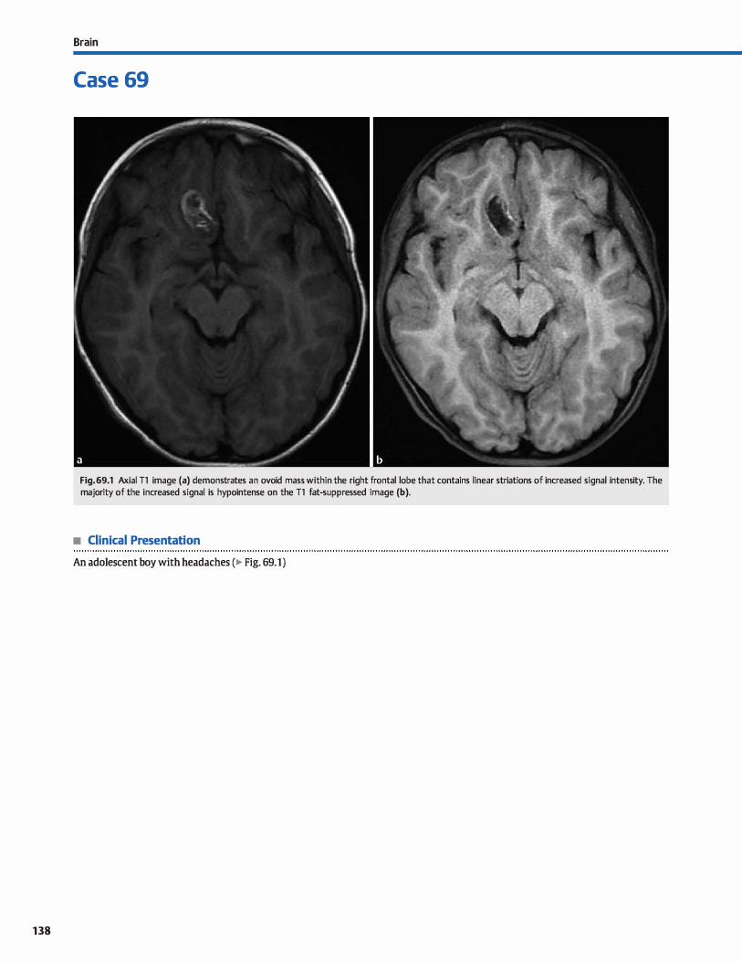
138
Brain
Case 69
Fig. 69.1 Axial Tl image (a) demonstrates an ovoid mass within the right frontal lobe that contains linear striations of increased signal intensity. The majority of the increased signal is hypointense on the Tl fat-suppressed image (b).
• Clinical Presentation
An adolescent boy with headaches (� Fig. 69.1)
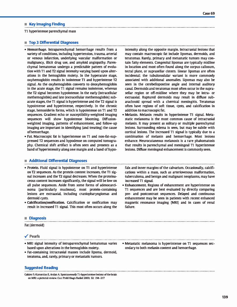
• Key lmaging Finding
Tl hyperintense parenchymal mass
• Top 3 Differential Diagnoses
• Hemorrhage. Intraparenchymal hemorrhage results from a variety of conditions, including hypertension, trauma. arterial or venous infarction, underlying vascular malformation or malignancy, illicit drug use, and amyloid angiopathy. Paren
chymal hematomas undergo a predictable pattern of evolution with Tl and T2 signal intensity varying based upon alterations in the hemoglobin moiety. In the hyperacute stage,
oxyhemoglobin results in isointense Tl and hyperintense T2 signal. As the oxyhemoglobin converts to deoxyhemoglobin in the acute stage, the Tl signal remains isointense, whereas
the l2 signal becomes hypointense. In the early (intraœllular methemoglobin) and late (extracellular methemoglobin) subacute stages, the Tl signal is hyperintense and the T2 signal is hypointense and hyperintense, respectively. In the chronic
stage, hemosiderin forms, which is hypointense on T1 and T2 sequences. Gradient echo or susceptibility-weighted imaging
sequences will show hypointense blooming. Diffusionweighted imaging, patterns of enhancement, and follow-up imaging are important in identifying (and treating) the cause ofhemorrhage.
• Fat. Macroscopic fat is hyperintense on Tl and non-fat-suppressed T2 sequences and hypodense on computed tomography. Chemical shift artifact is often seen and presents as a band of hyperintensity along one margin and a band of hypo-
• Additional Differential Diagnoses
• Protein. Fluid signal is hypointense on Tl and hyperintense
on T2 sequenœs. As the protein content increases, the Tl signal increases and the l2 signal decreases. When the proteinaceous content increases significantly, the signal will be low on
ail pulse sequenœs. Aside from some forms of adenocard
noma (particularly mudnous), most protein-containing lesions are extraaxial, including craniopharyngiomas and
dermoid cysts. • calcificationjossification. calcification or ossification may
result in increased Tl signal. This most often occurs along the
• Diagnosis
Fat ( dermoid)
� Pearls
• MRI signal intensity of intraparenchymal hematomas varies
based upon alterations in the hemoglobin moiety. • Fat-containing intracranial masses include lipoma, dermoid,
teratoma, and, rarely, primary or metastatic tumors.
Suggested Reading
cakirer S. Karaarslan E, Arslan A. Spontaneously Tl-hyperintense les ions of the brain on MRI: a pictorial review. Curr Probl Diagn Radiol 2003; 32: 194-217
Case 69
intensity along the opposite margin. Intracranial lesions that may contain macroscopic fat indude lipomas, dermoids, and
teratomas. Rarely, primary and metastatic tumors may contain fatty elements. Congenital lipomas are typically midline in location and most often found along the corpus callosum,
tectal plate, or suprasellar cistern. linear lipomas are often
incidental; the tubulonodular variant is more commonly associated with additional anomalies. lipomas may also be
seen in the cerebellopontine angle and internai auditory
canal. Dermoids and teratomas most often occur in the suprasellar region or off-midline where they may be intra- or extraaxial. Ruptured dermoids may result in diffuse sub
arachnoid spread with a chemical meningitis. Teratomas often have regions of soft tissue, cysts, and calcification in
addition to macroscopic fat. • Melanin. Melanin results in hyperintense Tl signal. Meta
static melanoma is the most common cause of intracranial
melanin. lt may present as solitary or multiple parenchymal lesions. Surrounding edema is seen, but may be subtle with cortical lesions. The increased Tl signal is typically due ta a
combination of melanin and hemorrhage. Most lesions enhance. Neurocutaneous melanosis is a rare phakomatosis
that results in parenchymal and meningeal Tl hyperintense lesions. Diffuse meningeal enhancement is commonly seen.
faix and inner margins of the calvarium. Occasionally, calcifi
cations within a mass, such as arteriovenous malformation, tuberculoma, and benign and malignant neoplasms, may have increased Tl signal.
• Enhanœment. Regions of enhancement are hyperintense on Tl sequences and are best evaluated by directly comparing
pre- and postcontrast sequences. Delayed and continuous
enhancement may be seen in patients with recent enhanced magnetic resonanœ imaging {MRI) and in cases of renal failure.
• Metastatic melanoma is hyperintense on Tl sequenœs secondary to bath melanin content and hemorrhage.
139
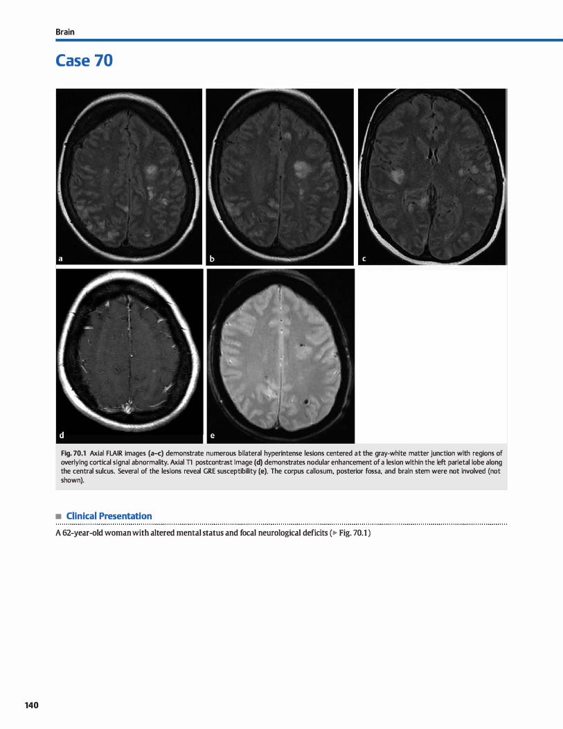
140
Brain
Case 70
Fig. 70.1 Axial FLAIR images (a-c) demonstrate numerous bilateral hyperintense lesions centered at the gray-white matter junction with regions of overlying cortical signal abnormality. Axial Tl postcontrast image (d) demonstrates nodular enhancement of a lesion within the left parietal lobe a long the central sulcus. Several of the lesions reveal GRE susceptibility (e). The corpus callosum, posterior fossa, and brain stem were not involved (not shown).
• Clinical Presentation
A 62-year-old woman with altered mental status and focal neurological deficits (� Fig. 70.1)
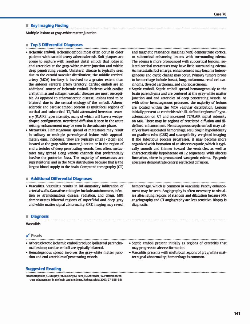
• Key lmaging Finding
Multiple lesions at gray-white matter junction
• Top 3 Differential Diagnoses
• Ischemic emboli. Ischemic emboli most often occur in older patients with carotid artery atherosderosis. Soft plaques are prone to ruptUre with resultant distal emboli that lodge in end arterioles at the gray-white matter junction and within deep penetrating vessels. Unilateral disease is typically seen due to the carotid vascular distribution; the middle cerebral artery (MCA) territory is involved to a greater extent than the anterior cerebral artery territory. Cardiac emboli are an additional source of ischemic emboli. Patients with cardiac arrhythmias and collagen vascular diseases are most susceptible. As opposed to atherosderotic disease, lesions tend to be bilateral due to the central etiology of the emboli. Atherosclerotic and cardiac emboli present as multifocal regions of cortical and subcortical T2/fluid-attenuated inversion recov
ery (FLAIR) hyperintensity, many ofwhich will have a wedgeshaped configuration. Restricted diffusion is seen in the acute setting; enhancement may be seen in the subacute phase.
• Metastases. Hematogenous spread of metastases may result in solitary or multiple parenchymal lesions with approximately equal incidence. They are typically small ( < 2 cm) and located at the gray-white matter junction or in the region of
end arterioles of deep penetrating vessels. Less often, metastases may spread along venous channels that preferentially involve the posterior fossa. The majority of metastases are supratentorial and in the MCA distribution because that is the largest blood supply to the brain. Computed tomography (CT)
• Additional Differential Diagnoses
• Vasculitis. Vasculitis results in inflammatory infiltration of arterial walls. causative etiologies include autoimmune, infection or granulomatous disease, radiation, and drugs. MRI demonstrates bilateral regions of superfidal and deep gray and white matter signal abnormality. GRE imaging may reveal
• Diagnosis
Vasculitis
� Pearls
• Atherosclerotic ischemic emboli produce ipsilateral parenchymal lesions; cardiac emboli are typically bilateral.
• Hematogenous spread involves the gray-white matter junction and end arterioles ofpenetrating vessels.
Suggested Reading
Smimiotopoulos JG, Murphy FM, Rushing EJ, Rees JH, Schroeder JW. Pa�ms of con
trast enhanœment in the brain and meninges. R.adiographics 2007; 27: 525-551
Case 70
and magnetic resonance imaging {MRI) demonstrate cortical or subcortical enhancing lesions with surrounding edema. The edema is more pronounced with subcortical lesions; isolated cortical metastases may have little surrounding edema. As metastatic fod enlarge, enhancement may become hetero
geneous and cystic change may occur. Primary tumors prone to hemorrhage include breast, Jung, melanoma, renal cell cardnoma, thyroid cardnoma, and choriocardnoma.
• Septic emboli Septic emboli spread hematogenously to the brain parenchyma and are œntered at the gray-white matter junction and end arterioles of deep penetrating vessels. As with other hematogenous processes, the majority of lesions
are located within the MCA vascular distribution. Lesions initially present as œrebritis with ill-defined regions of hypoattenuation on cr and increased T2/FLAIR signal intensity on MRI. There may be regions of restricted diffusion and illdefined enhancement Hematogenous septic emboli may caldfy or have associated hemorrhage, resulting in hypointensity on gradient echo (GRE) and susceptibility-weighted imaging. If the infectious process progresses, it may become more
organized with formation of an absœss capsule, which is typically smooth and thinner toward the ventricles, as well as characteristically hypointense on T2 sequences. With absœss formation, there is pronounced vasogenic edema. Pyogenic abscesses demonstrate central restricted diffusion.
hemorrhage, which is common in vasculitis. Patchy enhance
ment may be seen. Angiography is often necessary to visualize altemating regions of stenosis and dilatation because MR angeiography and cr angiography are Jess sensitive. Biopsy is diagnostic.
• Septic emboli present initially as regions of cerebritis that may progress to abscess formation.
• Vasculitis presents with multifocal regions of gray/white matter signal abnormality; hemorrhage is common.
141
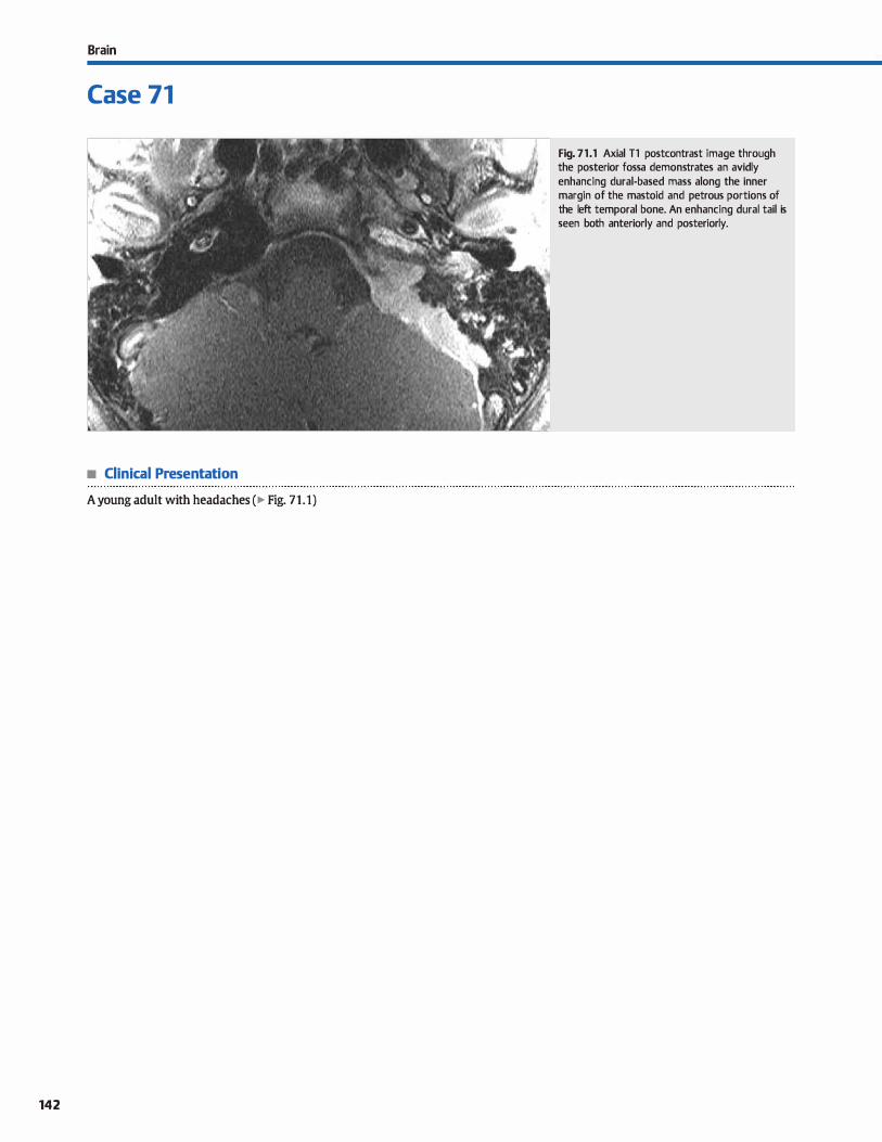
Brain
Case 71
• Clinical Presentation
A young adult with headaches (� Fig. 71.1)
142
Fig. 71 .1 Axial Tl postcontrast image through the posterior fossa demonstrates an avidly enhancing dural-based mass along the inner margin of the mastoid and petrous portions of the left temporal bone. An enhancing durai tail is seen both anteriorly and posteriorly.
• Key lmaging Finding
Dural-based mass
• Top 3 Differential Diagnoses
• Meningioma. Meningiomas are benign dural-based masses and represent the most common extraaxial masses. Common loca
tions indude parasagittal, convexities. middle cranial fessa, and planum sphenoidale. Less common locations indude the sellar/ suprasellar region and œrebellopontine angle. They occur most frequently in adult women and are typically solitary. Multiple meningiomas occur sporadically, in patients with neurofibromatosis type 2 (NF2), or as a complication of radiation ther
apy. Lesions have a broad, durai base and may be circumscribed or plaque-lilœ. The majority are hyperdense on computed
tomography (cr): the remainder are isodense to brain parenchyma. Calcification is noted in about one-quarter of cases. OVerlying bony remodeling or hyperostosis is common. There is often mass effect on the underlying parenchyma; edema is not uncommon with larger lesions. On cr and magnetic resonanœ imaging (MRI), there is avid enhanœment with a durai tail. MRI shows the extra-axial location with a cerebrospinal fluid deft separating the Iesion from the underlying parenchyma. Meningiomas are typically iso- to hypointense on Tl, variable in signal on T2, and hyperintense on fluid-attenuatEd inversion recovery (FLAIR). Calcification is typically hypointense on ail sequences. As with cr, vasogenic edema may be seen.
• Metast.ases/Lymphoma. Metastatic disease may involve the dura primarily or represent extension from calvarial
• Additional Differential Diagnoses
• Granulomatous disease. Tuberculosis and sarcoidosis are granulomatous processes that involve the dura. Most often, these present as regions of durai thickening and enhancement with a propensity for the basal cistems, resulting in cranial neuropathies. A Jess common presentation is a focal or plaque-like dural-based mass that may mimic a meningioma. Regions of nodular leptomeningeal enhancement are a useful discriminator.
• Extramedullary hematopoiesis. Extramedullary hematopoiesis occurs in patients with severe anemia secondary to chronic disease or marrow infiltrative proœsses. Imaging findings indude calvarial thickening with well-defmed, smooth extra-durai
• Diagnosis
Meningioma (plaque-like in a patient with NF2)
y' Pearls
• Meningiomas are the most common extraaxial masses; they are benign and occur most often in adult women.
• Metastases and lymphoma may involve the meninges primarily or result from extension of calvarial metastases.
Suggested Reading
Johnson MD, Powell SZ, Boyer PJ, Weil�. Moots PL Durai lesions mimicking meningiomas. Hwn Pathol 2002; 33: 1211-1226
Case 71
metastases. The most common primaries with dural-based metastases indude breast, lung, non-Hodgkin lymphoma, leukemia, prostate, and neuroblastoma. Skull-based metastases with durai extension are most often seen with lung, breast, prostate, and renal cell carcinoma. Lesions present as enhancing dural-based masses that may mimic meningiomas, espedally lymphoma, which may also be hyperdense on cr. Lymphoma is also typically hypointense on T2 sequences due to its high cellularity. Calvarial destruction is a useful discriminator, as is the presence of additional lesions, espedally parenchymal masses.
• Hemangiopericytoma. Hemangiopericytoma is a dural-based mesenchymal tumor of pericytes. It occurs in middle-aged adults and is histologically more aggressive (World Health Organization grade II or III) than meningioma. It presents as a heterogeneous dural-based mass that is iso- to hyperdense to parenchyma on cr without calcification. Cystic change is common, as is underlying mass effect and vasogenic edema. Calvarial erosion is common without hyperostosis. On MRI, hemangiopericytomas demonstrate heterogeneous signal and enhancement; prominent flow voids are commonly seen. There may be a durai tail; however, it is a less characteristic feature.
masses. Multiple lesions are more common than a solitary lesioIL On cr, fod of hematopoiesis are iso- to hyperdense, mimicking hematomas, meningiomas, or lymphoma. Lesions are typically TI isointense, T2 hypointense, and FLAIR hyperintense with avid, homogeneous enhanœment
• Rosai-Dorfman. Rosai-Dorfman disease is a non-Langerhans cell histiocytosis that most often occurs in children and young adults. The most common imaging feature is cervical lymphadenopathy, which is often massive. Less common presentations indude dural-based and orbital masses. Dural-based lesions are typically multiple, isointense on Tl, hyperintense on T2, and diffusely enhance.
• Hemangiopericytomas are more aggressive than meningiomas and lack calcification or bony hyperostosis.
143
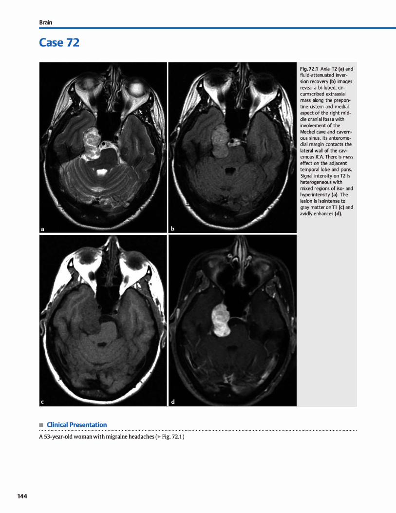
Brain
Case 72
• Clinical Presentation
A 53-year-old woman with migraine headaches (� Fig. 72.1 )
144
Fig. 72.1 Axial T2 (a) and fluid-attenuated inversion recovery (b) images reveal a bi-lobed, circumscribed extraaxial mass along the prepontine cistern and medial aspect of the right middle cranial fossa with involvement of the Meckel cave and cavernous sinus. lts anteromedial margin contacts the lateral wall of the cavernous ICA. There is mass effect on the adjacent temporal lobe and pons. Signal intensity on T2 is heterogeneous with mixed regions of iso- and hyperintensity (a). The lesion is isointense to gray matter on Tl (c) and avidly enhances (d).
• Key lmaging Finding
Circumscribed extraaxial mass at the skull base
• Top 3 Differential Diagnoses
• Meningioma. Meningiomas are benign dural-based masses and represent the most common extraaxial masses. Common locations indude parasagittal convexities, middle cranial fossa. and planum sphenoidale. Less common locations indude the sellar/ suprasellar region and cerebellopontine angle. They occur most frequently in adult women and are typically solitary. Multiple meningiomas occur sporadically, in patients with neuro
fibromatosis type 2 {NF2), or as a complication of radiation
therapy. Lesions have a broad, durai base and may be circumscribed or plaque-like. The majority are hyperdense on com
puted tomography {CT); the remainder are isodense to brain parenchyma. calcification is noted in about one-quarter of cases. Overlying bony remodeling or hyperostosis is common. There is often mass effect on the underlying parenchyma;
edema is not uncommon with larger lesions. On CT and magnetic resonanœ imaging {MRI), there is avid enhanœment with
a durai tait. MRI shows the extra-axial location with a cerebrospinal fluid deft separating the lesion from the undertying parenchyma. Meningiomas are typically iso- ta hypointense on Tl, variable in signal on T2, and hyperintense on fluid-attenuated inversion recovery. Calcification is typically hypointense
on all sequenœs. As with CT, vasogenic edema may be seen. • Schwannoma. Schwannomas are nerve sheath tumors that
arise from perineural Schwann cells. They are the second most common extraaxial masses after meningiomas. Common locations include the cerebellopontine angle {CPA); the
skull base, basal cistems, and exiting foramina along the course of the cranial nerves; and within the cavemous sinus.
• Additional Differential Diagnoses
• Lymphoma. Dural-based or extraaxial lymphoma typically results from hematogenous spread of non-Hodgkin lym
phoma. Lesions commonly present as dural-based masses
• Diagnosis
Schwannoma {trigeminal)
� Pearls
• Meningiomas are the most common extraaxial masses; calcification and a durai tail are characteristic features.
• Schwannomas are the second most common extraaxial masses; they occur along the course of cranial nerves.
Suggested Reading
Yuh wrc, Wright OC, Barloon 1J, Schultz DH, Sato Y, Cenrantes CA. MR irnaging of
primacy turnors of trigeminal nerve and Meckel's cave. AmJ Roentgenol t 988; 151: 577-582
Case 72
After vestibular schwannoma, trigeminal schwannomas are
the next most common. Schwannomas are composed of two tissue types: Antoni A and B. Antoni A tissue is densely packed, whereas Antoni B tissue is loosely packed. On CT, schwannomas are typically isodense compared with brain
parenchyma with heterogeneous enhancement There is often mass effect on adjacent parenchyma without edema. Unlike
meningiomas, calcification is rare. When located near the skull base or exiting foramina, smooth bony remodeling and enlargement of foramina is commonly seen. On MRI, schwannomas are iso- to hypointense on Tl sequences with heterogeneous but avid enhancement The T2 signal intensity varies
based upon tissue composition. Antoni A components are isoto hyperintense, whereas Antoni B components are prominently hyperintense. The vast majority of lesions are solitary; multiple lesions may be seen in the setting of NF2.
• Hemangiopericytoma. Hemangiopericytoma is a dural-based mesenchymal tumor of pericytes. It occurs in middle-aged adults and is histologically more aggressive (World Health Organization grade II or III) than meningioma. It presents as a heterogeneous dural-based mass that is iso- to hyperdense to
parenchyma on CT without calcification. Cystic change is common, as is underlying mass effect and vasogenic edema. Calvarial erosion is common without hyperostosis. On MRI, hemangiopericytomas demonstrate heterogeneous signal and enhancement; prominent flow wids are commonly seen. There may be a durai tail; however, it is a less characteristic feature.
{well-defined) or (plaque-like) that may mimic meningiomas. They are often hyperdense on CT and iso- to hypointense on
T2 MRI sequences due to high cellularity.
• Hemangiopericytoma is a relatively rare vascular tumor that mimics an aggressive meningioma on imaging.
• Dural-based or extraaxial lymphoma typically results from hematogenous spread of non-Hodgkin lymphoma.
145
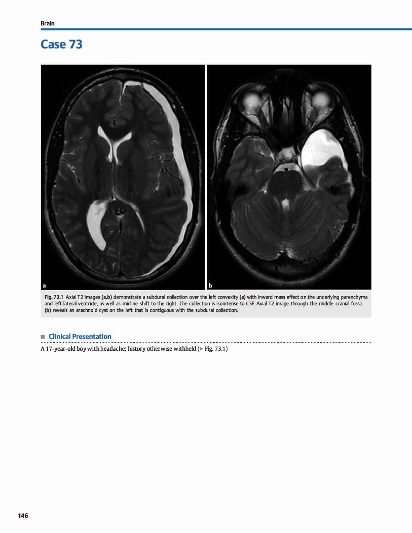
146
Brain
Case 73
Fig. 73.1 Axial T2 images (a,b) demonstrate a subdural collection over the left convexity (a) with inward mass effect on the underlying parenchyma and left lateral ventricle, as well as midline shift to the right. The collection is isointense to CSF. Axial T2 image through the middle cranial fossa (b) reveals an arachnoid cyst on the left that is contiguous with the subdural collection.
• Clinical Presentation
A 17-year-old boy with headache; history otherwise withheld (� Fig. 73.1 )
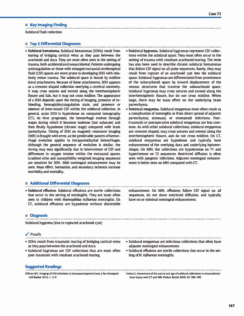
• Key lmaging Finding
Subdural fluid collection
• Top 3 Differential Diagnoses
• Subdural hema1oma. Subdural hematomas (SDHs) result from tearing of bridging cortical veins as they pass between the arachnoid and dura. They are most often seen in the setting of trauma, both accidentai and nonaccidental. Patients undergoing anticoagulation or those with enlarged extraaxial œrebrospinal fluid {C:SF) spaœs are more prone to developing SDH with relatively minor trauma. The subdural spaœ is bound by midline
durai attachments. Because of these attachments, SDH appears as a crescent-shaped collection overlying a œrebral convexity. It may cross sutures and ex.tend along the interhemispheric
fissure and faix, but it may not cross midline. The appearanœ of a SDH depends upon the timing of imaging, presenœ of rebleeding, hemoglobin/coagulation state, and presence or absence of inter-mixed CSF within the subdural collection. ln
general, acute SDH is hyperdense on computed tamography
(CT). As tirne progresses, the hemorrhage evolves through stages during which it becomes isodense (late subacute) and then fmally hypodense (chronic stage) compared with brain parenchyma. Timing of SDH on magnetic resonanœ imaging (MRI) is fraught with error, as the predictable pattern ofhemorrhage evolution applies ta intraparenchymal hemorrhages.
Although the general sequenœ of evolution is similar, the timing may vary significantly due ta intermixture of CSF and differenœs in oxygen tension within the extraaxial spaœs. Gradient echo and susœptibility weighted imaging sequenœs are sensitive for SDH. Mild meningeal enhanœment may be seen. Mass effect, herniation, and secondary ischemia increase morbidity and mortality.
• Additional Differential Diagnoses
• Subdural effusion. Subdural effusions are sterile collections that occur in the setting of meningitis. They are most often
seen in children with Haemophilus injluenza meningitis. On CT, subdural effusions are hypodense without discernible
• Diagnosis
Subdural hygroma {due to ruptured arachnoid cyst)
� Pearts
• SDHs result from traumatic tearing of bridging cortical veins as they pass between the arachnoid and dura.
• Subdural hygromas are CSF collections that are most often post-traumatic with resultant arachnoid tearing.
Suggested Readings
O'Brien wr. Imaging of CNS infections in immunocompetent hosts. J Am 05teopath
Qill Radial 2012; 1: 3-9
Case 73
• Subdural hygroma. Subdural hygromas represent CSF collections within the subdural spaœ. They most often occur in the
setting of trauma with resultant arachnoid tearing. The term has also been used to describe chronic subdural hematomas that follow CSF signal on all pulse sequenœs. Rarely, they may result from rupture of an arachnoid cyst into the subdural spaœ. Subdural hygromas are differentiated from prominence
of the subarachnoid space by inward displaœment of the
venous structures that traverse the subarachnoid spaœ. Subdural hygromas may cross sutures and extend along the interhemispheric fissure, but do not cross midline. When large, there may be mass effect on the underlying brain parenchyma.
• Subdural empyema. Subdural empyemas most often result as a complication of meningitis or from direct spread of adjacent
parenchymal, sinonasal, or otomastoid infections. Posttraumatic or postoperative subdural empyemas are Jess common. As with other subdural collections, subdural empyemas are cresœnt-shaped, may cross sutures and extend along the interhemispheric fissure, and do not cross midline. On cr, subdural empyemas are hypodense and typically have
enhancement of the overlying dura and underlying Ieptameninges. On MRI, the collections are hypointense on Tl and hyperintense on T2 sequences. Restricted diffusion is often seen with pyogenic infections. Adjacent meningeal enhance
ment is better seen on MRI compared with CT.
enhanœment. On MRI, effusions follow CSF signal on ail sequenœs, do not show restricted diffusion, and typically have no or minimal meningeal enhancement.
• Subdural empyemas are infectious collections that often have adjacent meningeal enhancement
• Subdural effusions are sterile collections that occur in the setting of H. injluenza meningitis.
Ve:zina G. Assessment of the nature and age of subdural collections in nonacddental
head injury with cr and MRI. Pediatr Radial 2009; 39: 586-590
147
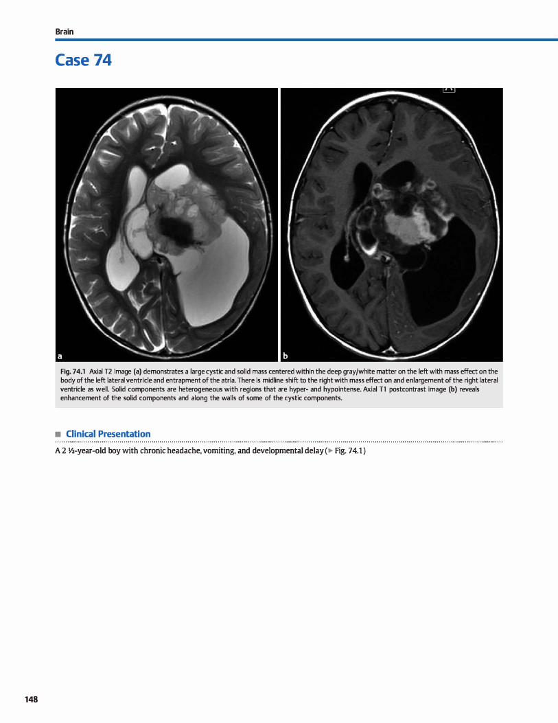
148
Brain
Case 74
Fig. 74.1 Axial T2 image (a) demonstrates a large cystic and solid mass centered within the deep gray/white matter on the left with mass effect on the body of the left lateral ventricle and entrapment of the atria. There is midline shift to the right with mass effect on and enlargement of the right lateral ventricle as well. Solid components are heterogeneous with regions that are hyper- and hypointense. Axial Tl postcontrast image (b) reveals enhancement of the solid components and along the walls of some of the cystic components.
• Clinical Presentation
A 2 �-year-old boy with chronic headache, vomiting, and developmental delay (� Fig. 74.1 )
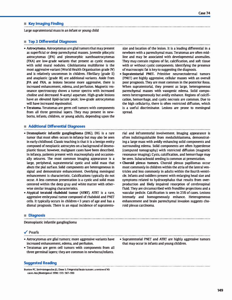
• Key lmaging Finding
Large supratentorial mass in an infant or young child
• Top 3 Differential Diagnoses
• Astrocytoma. Astrocytomas are glial tumors that may present as superficial or deep parenchymal masses. juvenile pilocytic astrocytomas (JPA) and pleomorphic xanthoastrocytomas (PXA) are low-grade variants that present as cystic masses with solid mural nodules. Glioblastoma multiforme is the most aggressive variant (World Health Organization grade IV) and is relatively uncommon in children. Fibrillary (grade II) and anaplastie (grade 111) are additional variants. Aside from JPA and PXA, as lesions become more aggressive, there is increased enhancement, edema, and perfusion. Magnetic resonance spectroscopy shows a tumor spectra with increased choline and decreased N-aœtyl aspartate. High-grade lesions have an elevated lipid-lactate peak; low-grade astrocytomas will have increased myoinositol.
• Teratoma. Teratomas are germ cell tumors with components from ail three germinal layers. They may present in newboms, infants, children, or young adults, depending upon the
• Additional Differential Diagnoses
• Desmoplastic infantile ganglioglioma (DIG). DIG is a rare tumor that most often occurs in infancy but may also be seen in early childhood. Classic teaching is that it is a benign entity composed of neoplastic astrocytes on a background of desmoplastic tissue; however, malignant cases have been described. In infancy, patients present with macrocephaly and occasionally seizures. The most common imaging appearanœ is a large, peripheral, supratentorial cystic and solid mass that abuts the pial surface. Solid components are heterogeneous in signal and demonstrate enhancement. OVerlying meningeal enhancement is characteristic. calcifications typically do not occur. A Jess common presentation is a cystic and solid mass œntered within the deep gray and white matter with otherwise similar imaging characteristics.
• Atypical teratoid rhabdoid tumor (Anrr). ATRT is a rare, aggressive embryonal tumor composed of rhabdoid and PNET cells. It typically occurs in children < 3 years of age and has a dismal prognosis. There is an equal incidence of supratento-
• Diagnosis
Desmoplastic infantile ganglioglioma
� Pearls
• Astrocytomas are glial tumors; more aggressive variants have increased enhanœment, edema, and perfusion.
• Teratomas are germ cell tumors with components from ail
three germinal layers; they are common in newboms/infants.
Suggested Reading
BuelDW PC, Smimiotnpaulos JG, Done s. Congenit.al brain tllmon: a review of 45
cases.AmJRoentgenol 1990; 155: 587-593
Case 74
size and location of the lesion. It is a leading differential in a newborn with a parenchymal mass. Teratomas are often midline and may be associated with developmental anomalies. They may contain regions of fat, calcification, and soft tissue with or without cystic components. Identifying the presenœ of macroscopic fat is key to suggesting the diagnosis.
• Supratentorial PNIIT. Primitive neuroectodermal tumors (PNET) are highly aggressive, cellular masses with an overall poor prognosis. They are most common in the posterior fossa. When supratentorial, they present as large, heterogeneous parenchymal masses with vasogenic edema. Solid components heterogeneously but avidly enhance. Regions of calcifi
cation, hemorrhage, and cystic necrosis are common. Due to the high cellularity, there is often restricted diffusion, which is a useful discriminator. Lesions are prone to meningeal spread.
rial and infratentorial involvement. Imaging appearance is often indistinguishable from medulloblastoma, demonstrating a large mass with avidly enhancing solid components and surrounding edema. Solid components are often hyperdense (computed tomography) with restricted diffusion (magnetic resonanœ imaging). Cysts, calcification, and hemorrhage may be seen. Subarachnoid seeding is common at presentation.
• Choroid plexus tumors. Choroid plexus papillomas occur most commonly in children within the atria of the lateral ventrides and less commonly in adults within the fourth ventride. Infants and toddlers present with enlarging head size and symptoms related to hydroœphalus that results from overproduction and likely impaired resorption of cerebrospinal fluid. They are circumscribed with frondlike projections and a vascular pedide. Calcification is seen in 25% of cases. Lesions intensely and homogeneously enhance. Heterogeneous enhancement and brain parenchymal invasion suggests choroid plexus carcinoma.
• Supratentorial PNET and ATRT are highly aggressive tumors that may occur in infants and young children.
149
150
Brain
Case 75
Fig. 75.1 Axial T2 image through the brain demonstrates a large intermediate to hyperintense mass involving and expanding the splenium of the corpus callosum. (Image courtesy of Paul M. Sherman, MD)
• Clinical Presentation
A 53-year-old man with altered mental status (� Fig. 75.1 )
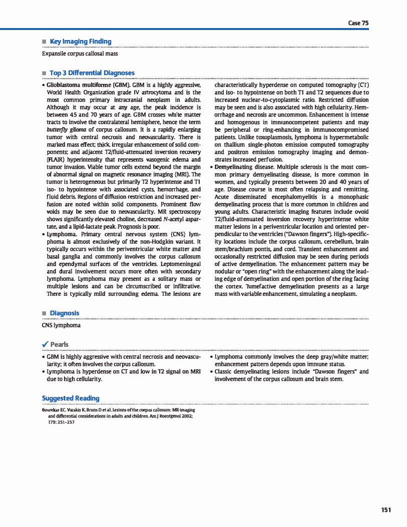
• Key lmaging Finding
Expansile corpus callosal mass
• Top 3 Differential Diagnoses
• Glioblastoma multiforme (GBM). GBM is a highly aggressive, World Health Organization grade IV astrocytoma and is the most comnton primary intracranial neoplasm in adults. Although it may occur at any age, the peak incidence is between 45 and 70 years of age. GBM crosses white matter tracts to involve the contralateral hemisphere, henœ the term butteifly glioma of corpus callosum. It is a rapidly enlarging
tumor with central necrosis and neovascularity. There is
marked mass effect; thick, irregular enhancement of solid components; and adjacent T2/fluid-attenuated inversion recovery
(FLAIR) hyperintensity that represents vasogenic edema and tumor invasion. Viable tumor œlls extend beyond the margin of abnormal signal on magnetic resonance imaging (MRI). The tumor is heterogeneous but primarily T2 hyperintense and Tl iso- to hypointense with assoàated cysts, hemorrhage, and fluid debris. Regions of diffusion restriction and increased per
fusion are noted within solid components. Prominent flow voids may be seen due to neovascularity. MR spectroscopy shows significantly elevated choline, decreased N-aœtyl aspartate, and a lipid-lactate peak. Prognosis is poor.
• Lymphoma. Primary central nervous system (CNS) lymphoma is almost exclusively of the non-Hodgkin variant. It typically occurs within the periventricular white matter and
basal ganglia and commonly involves the corpus callosum and ependymal surfaces of the ventrides. Leptomeningeal and durai involvement occurs more often with secondary lymphoma. Lymphoma may present as a solitary mass or multiple lesions and can be circumscribed or infiltrative.
There is typically mild surrounding edema. The lesions are
• Diagnosis
CNS lymphoma
y' Pearts
• GBM is highly aggressive with central necrosis and neovascularity; it often involves the corpus callosum.
• Lymphoma is hyperdense on CT and low in T2 signal on MRI due ta high cellularity.
Suggested Reading
Bourekas EC, Varakis K, Bruns D et al. Lesions of the corpus callosum: MR imaging
and düferential considerations in adults and children. AmJ Roentgenol 2002;
179: 251-257
Case 75
characteristically hyperdense on computed tomography (CT) and iso- ta hypointense on bath Tl and T2 sequences due ta increased nuclear-to-cytoplasmic ratio. Restricted diffusion may be seen and is aise associated with high cellularity. Hemorrhage and necrosis are uncommon. Enhancement is intense
and homogenous in imntunocompetent patients and may be peripheral or ring-enhancing in immunocompromised patients. Unlike toxoplasmosis, lymphoma is hypermetabolic on thallium single-photon emission computed tomography and positron emission tomography imaging and demonstrates increased perfusion.
• Demyelinating disease. Multiple sderosis is the most com
mon primary demyelinating disease, is more common in women, and typically presents between 20 and 40 years of age. Disease course is most often relapsing and remitting.
Acute disseminated encephalomyelitis is a monophasic demyelinating process that is more c0mn1on in children and young adults. Characteristic imaging features indude ovoid T2/fluid-attenuated inversion recovery hyperintense white matter lesions in a periventricular location and oriented perpendicular to the ventricles ("Dawson fingers"). High-specific
ity locations include the corpus callosum, cerebellum, brain
stem/brachium pontis, and cord. Transient enhancement and occasionally restricted diffusion may be seen during periods of active demyelination. The enhancement pattern may be nodular or "open ring" with the enhanœment along the leading edge of demyelination and open portion of the ring facing
the cortex. Tumefactive demyelination presents as a large mass with variable enhancement, simulating a neoplasm.
• Lymphoma commonly involves the deep gray/white matter; enhancement pattern depends upon imntune status.
• dassic demyelinating lesions include MDawson fingers" and involvement of the corpus callosum and brain stem.
151
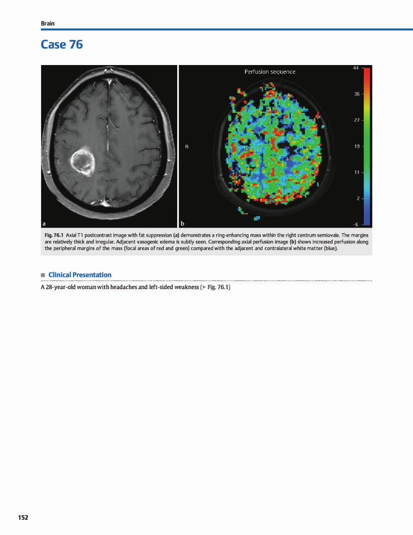
152
Brain
Case 76
Fig. 76.1 Axial Tl postcontrast image with fat suppression (a) demonstrates a ring-enhancing mass within the right centrum semiovale. The margins are relatively thick and irregular. Adjacent vasogenic edema is subtly seen. Corresponding axial perfusion image (b) shows increased perfusion along the peripheral margins of the mass (focal areas of red and green) compared with the adjacent and contralateral white matter (blue).
• Clinical Presentation
A 28-year-old woman with headaches and left-sided weakness (� Fig. 76.1)
• Key lmaging Finding
Ring-enhancing mass
• Top 3 Differential Diagnoses
• Neoplasm. High-grade gliomas, lymphoma, and metastases represent the majority of ring-enhancing tumors. Glioblastoma multiforme is the most common primary brain tumor in adults and is highly aggressive (World Health Organization grade IV). There is rapid growth with neovascularity. Lesions are typically solitary with marked mass effect, surrounding vasogenic edema, thick irregular peripheral enhancement,
and central necrosis. Although heterogeneous, the tumor is predominantly T2 hyperintense. Regions of hemorrhage and restricted diffusion (due to high cellularity) are common.
Involvement of the corpus callosum is often seen. Primary central nervous system lymphoma characteristically involves the deep gray and white matter. Involvement of the corpus callosum and ependymal surface of the ventrides is common.
Lesions may show characteristic hypointensity on T2 images due to high cellularity. There is solid enhancement in immu
nocompetent patients; ring enhancement may be seen following treatment or in immunosuppressed patients. Positron emission tomography or thallium imaging demonstrates increased metabolic activity, which distinguishes lymphoma from toxoplasmosis. As with other tumors, there is increased perfusion. Metastatic lesions may be solitary (-45%) or multi
ple. They often involve the gray-white matter junction, deep gray/white matter, and posterior fossa. There is often pronounced edema, except for cortically based metastases that
• Additional Differential Diagnoses
• Demyelinating disease. Multiple sclerosis is the most common primary demyelinating disease; acute disseminating encephalomyelitis is monophasic and more common in children. Characteristic features include ovoid T2/fluidattenuated inversion recovery hyperintense lesions oriented perpendicular to the ventricles {"Dawson fingers"). Highspecificity locations include the corpus callosum, optic
pathways, and posterior fossa. Enhancement suggests active demyelination. The enhancement pattern may be nodular or uopen ring" with the open portion facing the cortex. Tumefactive lesions mimic neoplasms.
• Diagnosis
Neoplasm (high-grade glioma)
� Pearls
• Glioblastoma multiforme, lymphoma, and metastases are the most common tumors to present as ring-enhancing lesions.
• Absœsses characteristically have a T2 hypointense capsule
with smooth enhancement and restricted diffusion.
Suggested Reading
Smimiotopoulos JG, Murphy FM, Rushing EJ, Rees JH, Schroeder JW. Pa�ms of con
trast enhanœment in the brain and meninges. R.adiographics 2007; 27: 525-551
Case 76
show little edema. Ring enhancement occurs with cystic metastases or central necrosis.
• Absœss. Abscesses result from hematogenous spread; direct spread from sinonasal or otomastoid infections, trauma, or surgery; or as a complication of meningitis. They may be solitary or multiple. Well-formed absœsses have smooth rim
enhancement with surrounding vasogenic edema. On magnetic resonance imaging {MRI), they have a characteristic T2 hypointense capsule that is thinner toward the ventrides and thus prone to intraventricular rupture. Pyogenic abscesses demonstrate central restricted diffusion. Toxoplasmosis, a common ring-enhancing parasitic infection in immunosuppressed patients, does not typically show restricted diffusion. As opposed to tumors, absœsses have decreased perfusion. MR spectroscopy shows an elevated lipid-lactate doublet.
• Subacute infarct. Cortically based infarcts are typically located in a vascular distribution; deep infarcts may be more difficult to characterize. The presence of edema, mass effect, hemorrhagic transformation, and subacute enhancement may mimic a mass lesion, especially if rim-enhancing. Infarcts show restricted diffusion acutely that may persist into the subacute phase. This is helpful but nonspecific, as abscesses
and cellular neoplasms may also demonstrate restricted diffusion. Enhancement begins as early as 2 days and peaks at 2 weeks.
• Resolving contusion. A history of trauma is important in differentiating a contusion from other ring-enhancing lesions.
Blood products are present in varying stages, depending upon the age of the hematoma. Rim enhancement occurs within a few days in a vascularized capsule. Acute blood products are isointense on T1 and hypointense on T2. Subacute blood products (intra- or extraœllular methemoglobin (MetHb]) are T1
hyperintense. Intracellular MetHb is T2 hypointense; extracelluar MetHb is T2 hyperintense.
• Enhancement pattern of demyelinating plaques may be uopen ring" with the open portion facing the cortex.
153
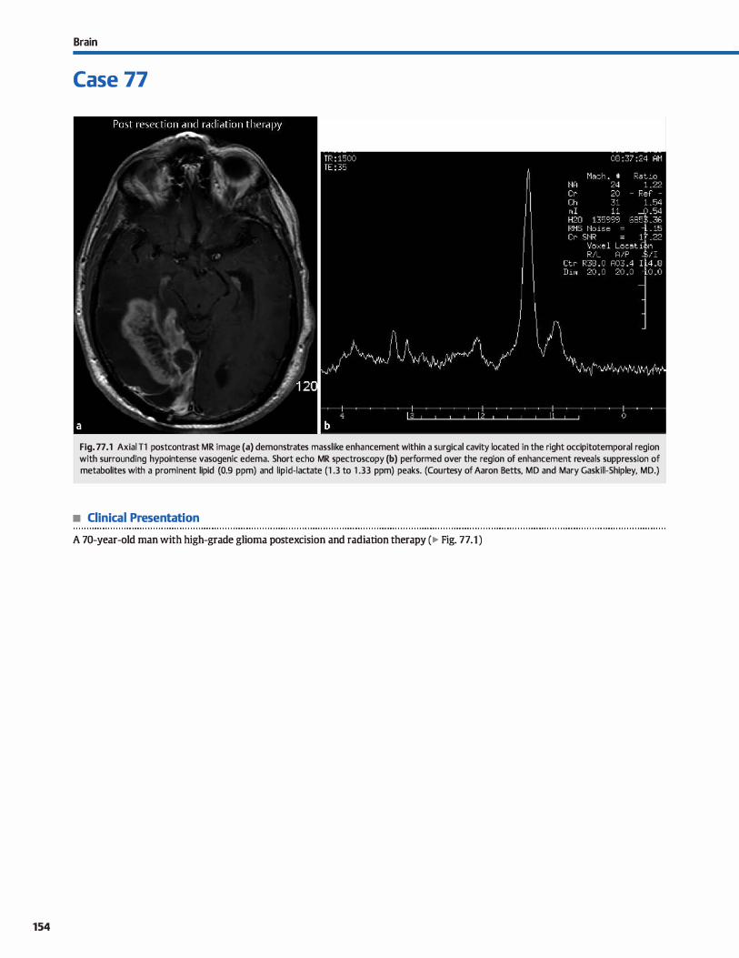
154
Brain
Case 77
b -:: ,,
Fig. 77 .1 Axial Tl postcontrast MR image (a) demonstrates masslike enhancement within a surgical cavity located in the right occipitotemporal region with surrounding hypointense vasogenic edema. Short echo MR spectroscopy (b) performed over the region of enhancement reveals suppression of metabolites with a prominent lipid (0.9 ppm) and lipid-lactate (1 .3 to 1 .33 ppm) peaks. (Courtesy of Aaron Betts, MD and Mary Gaskill-Shipley, MD.)
• Clinical Presentation
A 70-year-old man with high-grade glioma postexcision and radiation therapy (� Fig. 77.1)
• Key lmaging Finding
Enhanœment in surgical bed
• Top 3 Differential Diagnoses
• Recurrent or residual tumor. Glioblastoma multiforme is the most common primary brain tumor in adults; it may also occasionally be seen in children. It is highly aggressive (World Health Organization grade IV) and presents as an infiltrating mass with vasogenic edema, thick peripheral enhancement,
and central necrosis. lnvolvement of the corpus callosum is common. Treatment consists ofresection with radiation ther
apy to the surgical/tumor bed. Tumor œlls often extend
beyond the regions of T2/fluid-attenuated inversion recovery signal abnormality; therefore, complete resection is difficult. Adjuvant therapies indude steroids, chemotherapy, and antiangiogenic agents. Recurrent tumor most often occurs within the surgical bed. lt presents as increased mass effect, edema, and enhancement. Patients often suffer from new or progres
sive neurological deficits. Imaging appearanœ of recurrent tumor, treatment changes, and radiation necrosis overlaps on
conventional magnetic resonance imaging (MRI) sequences. Perfusion imaging in the setting of recurrence reveals increased cerebral blood flow (CBF) and volume {CBV) compared with adjacent white matter. On MR spectroscopy, highgrade tumors demonstrate elevated choline, decreased N
aœtyl aspartate (NAA), and elevated lipid-lactate peak. • Post-treatment changes. Treatment of high-grade tumors
often involves surgical resection with radiation therapy within the surgical/tumor bed. Adjuvant chemotherapy and antiangiogenic therapies are becoming commonplace in dinical practice. Steroid therapy results in decreased edema, decreased enhancement, and improvement in neurological symptoms. Expected postoperative changes include regions of hemorrhage and enhancement within and overlying the
surgical bed. Postoperative enhanœment is often linear;
nodular foci may be seen due to the presence of granulation tissue. These regions can be followed to ensure stabilicy or resolution. Newer therapies have resulted in findings that
present a challenge in determining response ta therapy.
Pseudoprogression is seen most often in patients treated with radiation and chemotherapy. lt presents as increased
• Diagnosis
Radiation necrosis
� Pearts
• Recurrent tumor typically occurs in the surgical bed; there is
increased perfusion and a tumor spectra on MR spectroscopy. • Treatment choices affect imaging findings; knowledge of
these is critical in determining treatment response.
Suggested Reading
Fatœrpekar GM, Galheigo D, Narayana A. Johnson G, Knopp E. Treatment-relaœd
change versus tumor recurrenœ in high-grade gliomas: a diagnostic conundrum
Case 77
enhanœment in the surgical bed 2 to 6 months after therapy. It stabilizes or decreases on follow-up imaging. lnterestingly, pseudoprogression is associated with better treatment outcomes. Also, these patients are typically asymptomatic. Perfusion imaging demonstrates similar perfusion characteristics
compared with white matter, whereas recurrent tumor shows increased perfusion. Pseudoresponse is another post
treatment change that typically results from a combination of chemotherapy and antiangiogenic agents. These therapies result in significant decrease in enhancement within the
surgical/tumor bed without decrease in tumor volume. Although patients who demonstrate pseudoresponse have
short-term survival benefits, long-term outcomes are variable. • Radiation necrosis. High-grade tumors are typically treated
with fractionated radiotherapy over several weeks. Radiation
necrosis is a severe complication that results in tissue necrosis due to radiation toxicity. It most often occurs 3 to 12 months
after therapy, but delayed occurrences may be seen years and even decades later. The incidence of radiation necrosis is between 5 and 25% and is proportional to the radiation dose. On MRI, radiation necrosis presents as regions of prominent
enhancement with fod of central necrosis, often referred to as a "soap bubble" or "Swiss cheese" appearanœ. There is mass effect and surrounding vasogenic edema. It occurs in
the region of highest radiation dose, which is typically within the postoperative bed. On conventional sequences, it is difficult if not impossible to distinguish from recurrent tumor.
New regions of enhancement within the radiation bed but in regions remote from prior tumor are more likely ta represent
radiation necrosis. On perfusion imaging, radiation necrosis bas similar CBF and CBV compared with normal white matter, whereas high-grade tumors have increased CBF and CBV that is similar to gray matter. On MR spectroscopy, radiation
necrosis demonstrates decreased choline, NAA, and creatine
with an elevated lipid-lactate doublet. This pattern reflects the absence of tumor and presence of necrotic parenchyma.
• Radiation necrosis mimics recurrent tumor on conventional
sequences; perfusion imaging and MR spectroscopy are helpful.
-use of dynamic susœptibility contrast-enhanœd (DSC) perfusion MRI. Am j
Roentgenol 2012; 198: 19-26
155
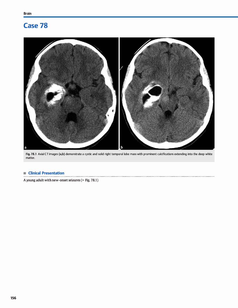
156
Brain
Case 78
Fig. 78.1 Axial CT images (a,b) demonstrate a cystic and solid right temporal lobe mass with prominent calcifications extending into the deep white matter.
• Clinical Presentation
A young adult with new-onset seizures (� Fig. 78.1 )
• Key lmaging Finding
Calcified supratentorial parenchymal mass
• Top 3 Differential Diagnoses
• Astrocytoma. There are numerous variants of astrocytoma that range from low to high grade. They may present as superficial or deep parenchymal masses. The diffuse fibrillary variant (World Health Organization grade II) is the most common to calcify, although calcifications are only seen in -20% of cases. The calcification pattern varies from punctate to diffuse and is nonspecific. Although astrocytomas have a lower
frequency of calcification compared with oligodendrogliomas, they are more common in incidence.
• Oligodendroglioma. Oligodendroglioma is a glial tumor that most often occurs in adults. It typically involves the subcortical white matter and overlying cortex. Oinically, patients
often present with seizures, headaches, or focal neurological deficits. Although they may occur anywhere, frontal lobe involvement is most common. On imaging, oligodendroglioma presents as a well-circumscribed (more common) or ill-defined mass with a high incidence of calcification (up to 90%). Calcifications may be punctate, ribbon-like, or diffuse.
• Additional Differential Diagnoses
• Ependymoma. The majority of ependymomas present as
fourth ventricular masses in children. However, they may also occur in the supratentorial compartment as a parenchymal mass centered in the white matter with surrounding edema. As with posterior fossa ependymomas, cystic components and calcification ( .. 50%) are relatively common. Solid components heterogeneously enhance. Ependymomas occur predominantly in children and young adults.
• Infection. lnfectious proœsses prone to parenchymal calcifications indude neurocystiœrcosis, tuberculosis (TB), crypto
coccus, and human immunodeficiency virus in children. Of these, tuberculoma is the most common to present as a calcified mass. Cysticercosis most often presents as multiple parenchymal calcifications with or without additional cystic
• Diagnosis
Oligodendroglioma
� Pearts
• Tumors with calcification include astrocytoma, oligodendroglioma, ependymoma, and, rarely, metastases.
• AVMs and CMs may have regions of calcification; AVMs have a vascular nidus and are prone to hemorrhage.
Suggested Readings
Case 78
There is typically no or mild surrounding vasogenic edema. Enhanœment, if present, is subtle and ill-defined.
• Vascular malformation. Arteriovenous malformations (AVMs) consist of a nidus of abnormal connections between arteries and veins without an intervening capillary network. Patients often present in young adulthood with headaches, hemorrhage, seizures, or focal neurological defidts. Computed tomography
( CT) may be normal or demonstrate regions of increased attenuation or calcification. Magnetic resonanœ imaging (MRI) reveals a tangle of enlarged vessels, corresponding to the nidus. Perinidal aneurysms are a common source of hemorrhage. cavemous malformations (CMs) consist of variable-sized blood-filled sinusoids and larger cavemous spaœs without intervening brain parenchyma. The majority of lesions are soli
tary. cr may be normal or show subtle regions of calcification or hemorrhage. Lesions have fod of increased and decreased Tl and 12 signal centrally with a 12 dark (hemosiderin) capsule on MRI.
or rim-enhancing lesions. Tuberculomas result from hematog
enous spread; concurrent meningeal involvement may be seen, preferentially involving the basal cistems. Imaging appearance varies widely from focal cerebritis to frank abscesses with rim enhancement. calcifications occur in the minority of cases.
• Metastases. Metastatic mucinous adenocarcinoma may occasionally present as a calcified parenchymal mass. Common primaries indude breast, Jung, colon, and ovarian carcinoma. Bone-forming tumors such as osteosarcoma may also present
with calcified or ossified lesions. Metastases may be solitary or multiple and typically have pronounced vasogenic edema, with the exception of cortically based metastases that may have little edema.
• TB, fungal, and neurocysticercosis are the most common infections to result in calcified parenchymal lesions.
Koeller KK, Rmhing EJ. From the archives of the AHP: oligodendrogliama and its var- Smith AB. V;iscular malfannatians of the brain; r.adialagic and pathologie
iants: r.adiolagic-pathalagic rorrelatimL Radiographies 2005; 25: 1669-1688 rorrelatimL j Am Osteapath Cali Radial. 2012; 1: 10-22
Makariau E, Patsalides AD. lntracranial caldficatiam. Appt Radial 2009; 38: 48-60
157
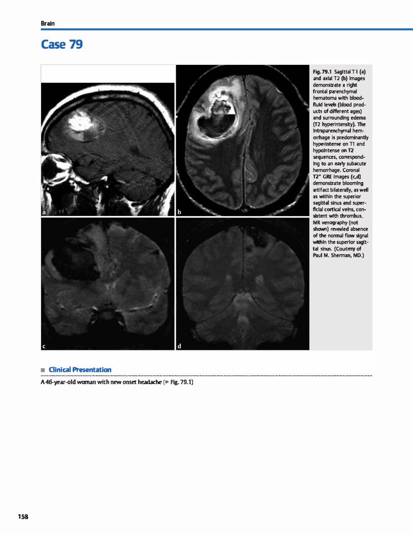
Brain
Case 79
• Clinical Presentation
A 46-year-old woman with new onset headache (., Fig. 79.1)
158
Fig. 79.1 Sagittal Tl (a) and axlal T2 (b) Images demonstrate ;i right frontal parenchymal hematoma wlth blood· flukl levds (blood products of different ages) and surroundlng edema (T2 hyperfntenslty). The lntraparenchymal hemorThage is predominantly hyperintense on T1 and hypolntense on 12 sequences, corTeSpond· lng to an early subacute hemorrhage. Coronal 12• GRE Images (c,d) demonstrate bloomlng utifact bilaterally, as well ;is within the !uperior sagittal sinus and superflclal cortlc.al velns, con· slstent wlth thrombus. MR wnogr;iphy (not shawn) revt!i!led absence of the normal flow signal wtthln the superlor sagittal !inll!I. (Courtesy of Paul M. Sherman, MD.)
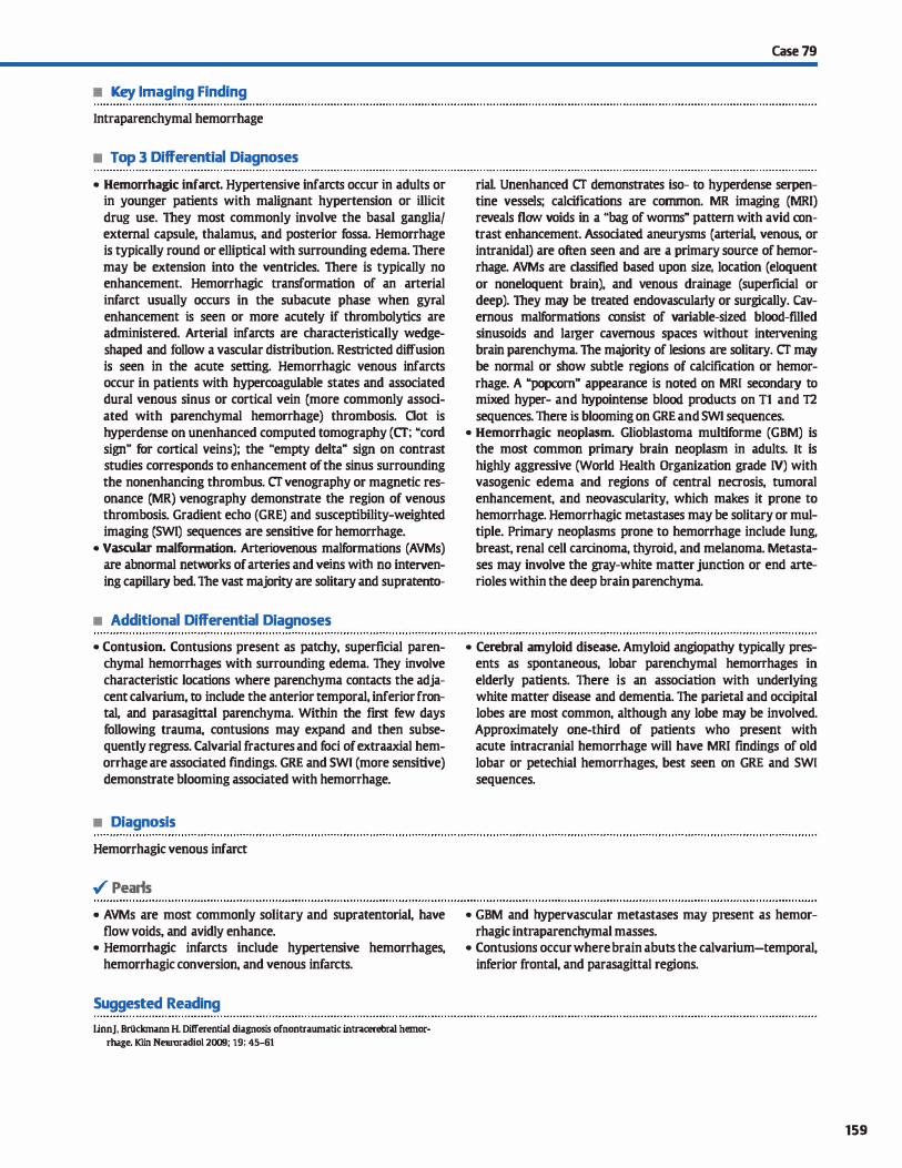
• Key lmaging Finding
lntraparenchymal hemorrhage
• Top 3 Differential Diagnoses
• Hemorrhagic infarct. Hypertensive infarcts occur in adults or in younger patients with malignant hypertension or illicit drug use. They most commonly involve the basal ganglia/ external capsule, thalamus, and posterior fossa. Hemorrhage is typically round or elliptical with surrounding edema. There may be extension into the ventrides. There is typically no enhancement. Hemorrhagic transformation of an arterial infarct usually occurs in the subacute phase when gyral enhancement is seen or more acutely if thrombolytics are administered. Arterial infarcts are characteristically wedgeshaped and follow a vascular distribution. Restricted diffusion is seen in the acute setting. Hemorrhagic venous infarcts occur in patients with hypercoagulable states and associated durai venous sinus or cortical vein {more commonly associated with parenchymal hemorrhage) thrombosis. Oot is hyperdense on unenhanced computed tomography (cr; "cord sign" for cortical veins); the "empty delta" sign on contrast studies corresponds to enhancement of the sinus surrounding the nonenhanàng thrombus. cr venography or magnetic resonance {MR) venography demonstrate the region of venous thrombosis. Gradient echo (GRE) and susceptibility-weighted imaging {SWI) sequences are sensitive for hemorrhage.
• Vascular malformation. Arteriovenous malformations (AVMs) are abnormal networks of arteries and veins with no intervening capillary bed. The vast majority are solitary and supratento-
• Additional Differential Diagnoses
• Contusion. Contusions present as patchy, superficial parenchymal hemorrhages with surrounding edema. They involve characteristic locations where parenchyma contacts the adjacent calvarium, to include the anterior temporal, inferior frontal, and parasagittal parenchyma. Within the first few days following trauma, contusions may expand and then subsequently regress. Calvarial fractures and foci of extraaxial hemorrhage are assoàated findings. GRE and SWI (more sensitive) demonstrate blooming assoàated with hemorrhage.
• Diagnosis
Hemorrhagic venous infarct
y' Pearls
• AVMs are most commonly solitary and supratentorial, have flow voids, and avidly enhance.
• Hemorrhagic infarcts include hypertensive hemorrhages, hemorrhagic conversion, and venous infarcts.
Suggested Reading
linnJ, Brückmann H. Differential diagnosis ofnontraumatic intraœrebral hemor
rhage. Klin Neuroradiol 2009; 19; 45-61
Case 79
rial Unenhanced cr demonstrates iso- to hyperdense serpentine vessels; calcifications are conunon. MR imaging (MRI) reveals flow voids in a "bag of wonns" pattern with avid contrast enhancement. Associated aneurysms { arteriaL venous, or intranidal) are often seen and are a primary source of hemorrhage. AVMs are dassified based upon size, location (eloquent
or noneloquent brain), and venous drainage (superfiàal or deep ). They may be treated endovascularly or surgically. cavernous malformations consist of variable-sized blood-filled sinusoids and larger cavernous spaces without intervening brain parenchyma. The majority of lesions are solitary. cr may be normal or show subtle regions of calcification or hemor
rhage. A "popcorn" appearanœ is noted on MRI secondary ta mixed hyper- and hypointense blood products on Tt and 12
sequences. There is blooming on GRE and SWI sequences. • Hemorrhagic neoplasm. Glioblastoma multiforme (GBM) is
the most common primary brain neoplasm in adults. lt is highly aggressive (World Health Organization grade IV) with vasogenic edema and regions of central necrosis, tumoral enhancement, and neovascularity, which makes it prone ta hemorrhage. Hemorrhagic metastases may be solitary or multiple. Primary neoplasms prone ta hemorrhage include lung.
breast, renal cell carcinoma, thyroid, and melanoma. Metastases may involve the gray-white matter junction or end arterioles within the deep brain parenchyma.
• Cerebral amyloid disease. Amyloid angiopathy typically presents as spontaneous, tabar parenchymal hemorrhages in elderly patients. There is an assoàation with underlying white matter disease and dementia. The parietal and ocàpital lobes are most common, although any lobe may be involved. Approximately one-third of patients who present with acute intracranial hemorrhage will have MRI findings of old lobar or petechial hemorrhages, best seen on GRE and SWI sequences.
• GBM and hypervascular metastases may present as hemorrhagic intraparenchymal masses.
• Contusions occur where brain abuts the calvarium-temporal, inferior frontal, and parasagittal regions.
159
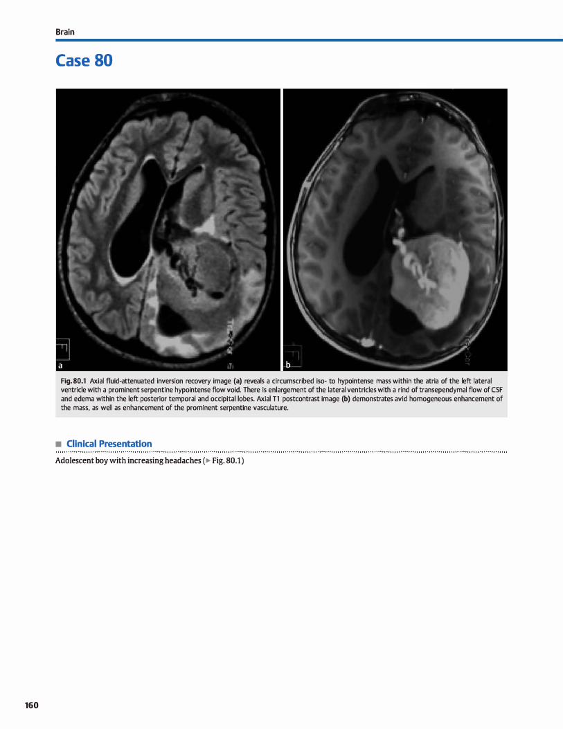
160
Brain
Case 80
Fig. 80.1 Axial fluid-attenuated inversion recovery image (a) reveals a circumscribed iso- to hypointense mass within the atria of the left lateral ventricle with a prominent serpentine hypointense flow voici. There is enlargement of the lateral ventricles with a rind of transependymal flow of CSF and edema within the left posterior temporal and occipital lobes. Axial Tl postcontrast image (b) demonstrates avid homogeneous enhancement of the mass, as well as enhancement of the prominent serpentine vasculature.
• Clinical Presentation
Adolescent boy with increasing headaches (� Fig. 80.1)
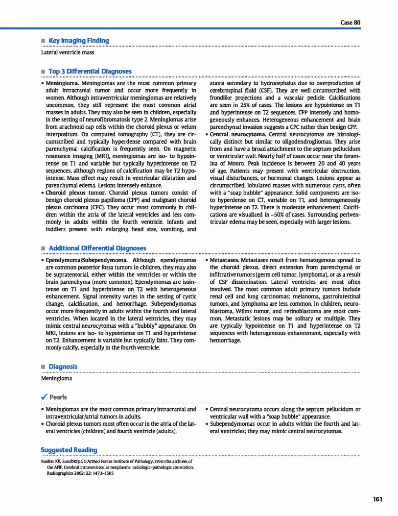
• Key lmaging Finding
Lateral ventricle mass
• Top 3 Differential Diagnoses
• Meningioma. Meningiomas are the most common primary adult intracranial tumor and occur more frequently in
women. Although intraventricular meningiomas are relatively uncommon, they still represent the most common atrial masses in adults. They may also be seen in children, especially in the setting of neurofibromatosis type 2. Meningiomas arise from arachnoid cap cells within the choroid plexus or velum
interpositum. On computed tomography (CT), they are circumscribed and typically hyperdense compared with brain parenchyma; calcification is frequently seen. On magnetic resonance imaging (MRI), meningiomas are iso- to hypointense on Tl and variable but typically hyperintense on T2 sequences, although regions of calcification may be T2 hypointense. Mass effect may result in ventricular dilatation and
parenchymal edema. Lesions intensely enhance. • Choroid plexus tumor. Choroid plexus tumors consist of
benign choroid plexus papilloma (CPP) and malignant choroid plexus carcinoma (CPC). They occur most commonly in children within the atria of the lateral ventrides and Jess commonly in adults within the fourth ventride. Infants and
toddlers present with enlarging head size, vomiting, and
• Additional Differential Diagnoses
• Ependymoma/Subependymoma. Although ependymomas are common posterior fossa tumors in children, they may also be supratentorial, either within the ventrides or within the brain parenchyma (more common). Ependymomas are isointense on Tl and hyperintense on T2 with heterogeneous enhancement. Signal intensity varies in the setting of cystic change, calcification, and hemorrhage. Subependymomas
occur more frequently in adults within the fourth and lateral
ventricles. When located in the lateral ventricles, they may mimic central neurocytomas with a "bubbly" appearance. On MRI, lesions are iso- to hypointense on Tl and hyperintense on T2. Enhancement is variable but typically faint. They commonly calcify, especially in the fourth ventricle.
• Diagnosis
Meningioma
� Pearls
• Meningiomas are the most common primary intracranial and intraventricular/atrial tumors in adults.
• Choroid plexus tumors most often occur in the atria of the lateral ventricles (children) and fourth ventride (adults).
Suggested Reading
Koeller KK, Sandberg CD Armed Fon:es lnstitute of Pathology. From the archives of
the AFIP. Cerebral intraventricular neoplasms: radiologie-pathologie eorrelation.
Radiographies 2002; 22: 1473-1505
Case 80
ataxia secondary ta hydrocephalus due to overproduction of œrebrospinal fluid (CSF). They are well-circumscribed with frondlike projections and a vascular pedide. Calcifications are seen in 25% of cases. The lesions are hypointense on Tl and hyperintense on T2 sequences. CPP intensely and homogeneously enhanœs. Heterogeneous enhancement and brain parenchymal invasion suggests a CPC rather than benjgn CPP.
• Central neurocytoma. Central neurocytomas are histologically distinct but similar to oligodendrogliomas. They arise from and have a broad attachment to the septum pellucidum or ventricular wall. Nearly half of cases occur near the foramina of Monro. Peak incidence is between 20 and 40 years
of age. Patients may present with ventricular obstruction, visual disturbances, or hormonal changes. I.esions appear as
circumscribed, lobulated masses with numerous cysts, often with a "soap bubble" appearance. Solid components are isoto hyperdense on cr, variable on Tl, and heterogeneously hyperintense on T2. There is moderate enhancement. Calcifications are visualized in -50% of cases. Surrounding periventricular edema may be seen, especially with larger lesions.
• Metastases. Metastases result from hematogenous spread to the choroid plexus, direct extension from parenchymal or
infiltrative tumors (germ cell tumor, lymphoma), or as a result of CSF dissemination. Lateral ventricles are most often involved. The most common adult primary tumors indude renal cell and Jung carcinomas; melanoma, gastrointestinal tumors, and lymphoma are Jess common. ln children, neuro
blastoma. Wilms tumor, and retinoblastoma are most common. Metastatic lesions may be solitary or multiple. They are typically hypointense on Tl and hyperintense on T2 sequences with heterogeneous enhancement, especially with hemorrhage.
• Central neurocytoma occurs along the septum pelluddum or ventricular wall with a "soap bubble" appearance.
• Subependymomas occur in adults within the fourth and lateral ventrides; they may mimic central neurocytomas.
161
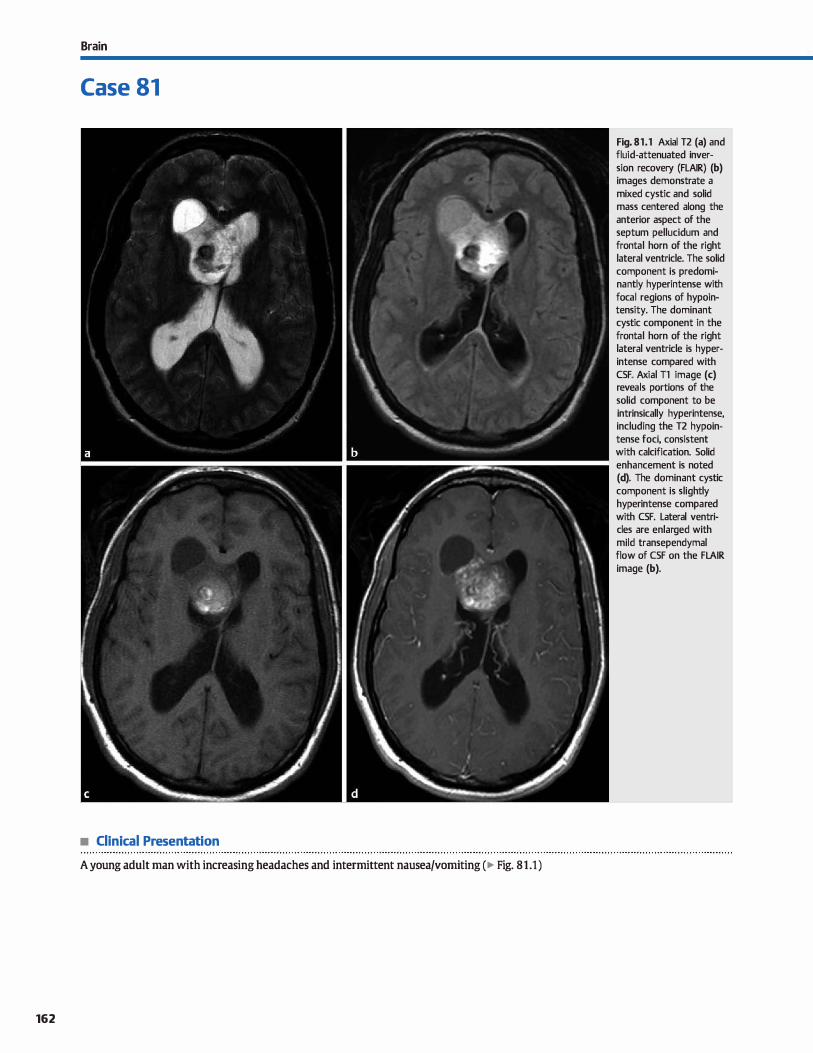
Brain
Case 81
• Clinical Presentation
A young adult man with increasing headaches and intermittent nausea/vomiting ( � Fig. 81.1)
162
Fig. 81.1 Axial T2 (a) and fluid-attenuated inversion recovery (FLAIR) (b) images demonstrate a mixed cystic and solid mass centered along the anterior aspect of the septum pellucidum and frontal horn of the right lateral ventricle. The solid component is predominantly hyperintense with focal regions of hypointensity. The dominant cystic component in the frontal horn of the right lateral ventricle is hyperintense compared with CSF. Axial Tl image (c) reveals portions of the solid component to be intrinsically hyperintense, including the T2 hypointense foci, consistent with calcification. Solid enhancement is noted (d). The dominant cystic component is slightly hyperintense compared with CSF. Lateral ventricles are enlarged with mild transependymal flow of CSF on the FLAIR image (b).
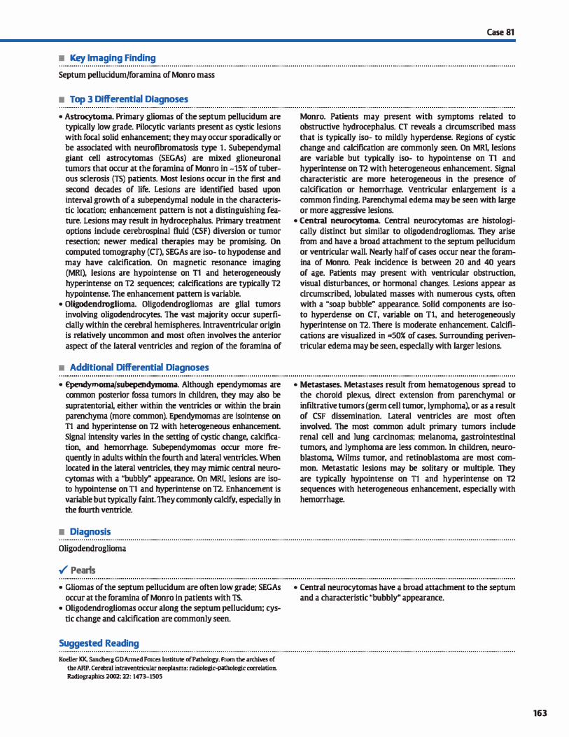
• Key lmaging Finding
Septum pellucidum/foramina of Monro mass
• Top 3 Differential Diagnoses
• Astrocytoma. Primary gliomas of the septum pellucidum are typically low grade. Pilocytic variants present as cystic lesions with focal solid enhancement; they may occur sporadically or be associated with neurofibromatosis type 1. Subependymal giant cell astrocytomas (SEGAs) are mixed glioneuronal tumors that occur at the foramina of Monro in -15% of tuberous sclerosis (TS) patients. Most lesions occur in the first and
second decades of life. Lesions are identified based upon interval growth of a subependymal nodule in the characteristic location; enhancement pattern is not a distinguishing feature. Lesions may result in hydrocephalus. Primary treannent options indude cerebrospinal fluid (CSF) diversion or tumor resection; newer medical therapies may be promising. On computed tomography (CT), SEGAs are iso- to hypodense and may have calcification. On magnetic resonanœ imaging (MRI), lesions are hypointense on Tl and heterogeneously hyperintense on T2 sequenœs; calcifications are typically T2 hypointense. The enhanœment pattern is variable.
• Oligodendroglioma. Oligodendrogliomas are glial tumors involving oligodendrocytes. The vast majority occur superficially within the œrebral hemispheres. lntraventricular origin is relatively uncommon and most often involves the anterior
aspect of the lateral ventricles and region of the foramina of
• Additional Differential Diagnoses
• Ependymomajsubependymoma. Although ependymomas are common posterior fossa tumors in children, they may also be
supratentorial, either within the ventrides or within the brain parenchyma (more common� Ependymomas are isointense on Tl and hyperintense on T2 with heterogeneous enhancement. Signal intensity varies in the setting of cystic change, calcification, and hemorrhage. Subependymomas occur more frequently in adults within the fourth and lateral ventrides. When located in the lateral ventrides, they may mimic central neurocytomas with a "bubbly" appearance. On MRI, lesions are isoto hypointense on Tl and hyperintense on T2. Enhancement is variable but typically faint They commonly caldfy, especially in the fourth ventride.
• Diagnosis
Oligodendroglioma
� Pearls
• Gliomas of the septum pellucidum are often low grade; SEGAs occur at the foramina of Monro in patients with TS.
• Oligodendrogliomas occur along the septum pellucidum; cys
tic change and calcification are commonly seen.
Suggested Reading
Koeller KK, Sandberg GD Armed Forces lnstituœ of Pathology. From the an:hives of
the AFIP. Cerebral intraventricular neoplasms: radiologie-pathologie correlation.
Radiographies 2.002; 2.2.: 1473-1505
Case 81
Monro. Patients may present with symptoms related to obstructive hydrocephalus. cr reveals a circumscribed mass that is typically iso- to mildly hyperdense. Regions of cystic change and calcification are commonly seen. On MRI, lesions are variable but typically iso- to hypointense on Tt and hyperintense on T2 with heterogeneous enhancement. Signal characteristic are more heterogeneous in the presence of calcification or hemorrhage. Ventricular enlargement is a common finding. Parenchymal edema may be seen with large or more aggressive lesions.
• Central neurocytoma. Central neurocytomas are histologically distinct but similar to oligodendrogliomas. They arise from and have a broad attachment to the septum pellucidum or ventricular wall. Nearly half of cases occur near the foramina of Monro. Peak incidence is between 20 and 40 years of age. Patients may present with ventricular obstruction, visual disturbances, or hormonal changes. Lesions appear as drcumscribed, lobulated masses with numerous cysts, often with a "soap bubble" appearance. Solid components are isoto hyperdense on CT, variable on Tt, and heterogeneously hyperintense on T2. There is moderate enhancement. Calcifications are visualized in •50% of cases. Surrounding periventricular edema may be seen, espedally with larger Iesions.
• Metastases. Metastases result from hematogenous spread to the choroid plexus, direct extension from parenchymal or
infiltrative tumors (germ cell tumor, lymphoma), or as a result of CSF dissemination. Lateral ventricles are most often involved. The most common adult primary tumors indude renal cell and Jung carcinomas; melanoma, gastrointestinal tumors, and lymphoma are Jess common. ln children, neuroblastoma, Wilms tumor, and retinoblastoma are most common. Metastatic lesions may be solitary or multiple. They
are typically hypointense on Tl and hyperintense on T2 sequences with heterogeneous enhancement, especially with hemorrhage.
• Central neurocytomas have a broad attachment ta the septum and a characteristic "bubbly" appearance.
163
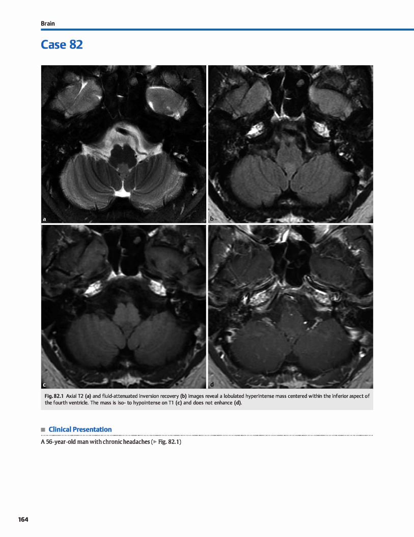
164
Brain
Case 82
Fig. 82.1 Axial T2 (a) and fluid-attenuated inversion recovery (b) images reveal a lobulated hyperintense mass centered within the inferior aspect of the fourth ventricle. The mass is iso- to hypointense on Tl (c) and does not enhance (d).
• Clinical Presentation
A 56-year-old man with chronic headaches (� Fig. 82.1)
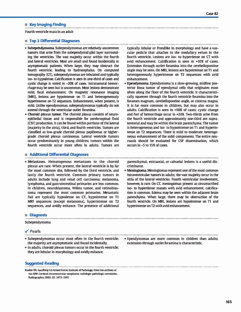
• Key lmaging Finding
Fourth ventride mass in an adult
• Top 3 Differential Diagnoses
• Subependymoma. Subependymomas are relatively uncommon tumors that arise from the subependymal glial layer surround
ing the ventrides. The vast majority occur within the fourth and Iateral ventrides. Most are small and found incidentally in
asymptomatic patients. When large, they may obstruct the fourth ventride, leading to hydrocephalus. On computed tomography (CT), subependymomas are lobulated and typically iso- to hypodense. calcification is seen in one-third of cases and
cystic change is noted in -20% of cases. Intratumoral hemorrhage may be seen but is uncommon. Most lesions demonstrate
mild, focal enhancement On magnetic resonance imaging
(MRI1 lesions are hypointense on Tl and heterogeneously hyperintense on T2 sequences. Enhancement, when present, is mild Unlike ependymomas, subependymomas typically do not extend through the ventricular outlet foramina.
• Choroid plexus tumor. The choroid plexus consists of neuroepithelial tissue and is responsible for œrebrospinal fluid
(CSF) production. It can be found within portions of the lateral
(majority in the atria), third, and fourth ventrides. Tumors are classified as low-grade choroid plexus papillomas or highergrade choroid plexus carcinomas. Lateral ventride tumors occur predominantly in young children; tumors within the fourth ventride occur more often in adults. Tumors are
• Additional Differential Diagnoses
• Metastases. Hematogenous metastases to the choroid plexus are rare. When present, the lateral ventricle is by far the most common site, followed by the third ventricle, and lastly the fourth ventricle. Common primary tumors in
adults include Jung and renal cell carcinoma; melanoma, lymphoma, and gastrointestinal primaries are Jess common. In children, neuroblastoma, Wilms tumor, and retinoblas
toma represent the most common primaries. Metastatic foci are typically hypodense on CT, hypointense on Tl MRI sequences (except melanoma), hyperintense on T2 sequences, and avidly enhance. The presence of additional
• Diagnosis
Subependymoma
� Pearls
• Subependymomas occur most often in the fourth ventride; the majority are asymptomatic and found incidentally.
• In adults, choroid plexus tumors occur in the fourth ventride; they are lobular in morphology and avidly enhance.
Suggested Reading
Koeller KK, Sandberg CD Armed Fon:es lnstitute of Pathology. From the archives of
the AFIP. Cerebral intraventrirular neoplasms: radiologie-pathologie earrelation.
Radiographies 2002; 22: 1473-1505
Case 82
typically lobular or frondlike in morphology and have a vascular pedicle that attaches to the medullary velum in the fourth ventricle. Lesions are iso- to hyperdense on CT with
avid enhancement. calcification is seen in "'10% of cases. Extension through outlet foramina into the cerebellopontine angle may be seen. On MRI, lesions are hypointense on Tl and heterogeneously hyperintense on T2 sequenœs with avid
enhanœment. • Ependymoma. Ependymoma is a slow-growing, midline pos
terior fossa tumor of ependymal cells that originates most
often along the floor of the fourth ventride. It characteristically squeezes through the fourth ventricle foramina into the
foramen magnum. œrebellopontine angle, or cistema magna. It is far more common in children, but may also occur in adults. calcification is seen in .,.5m; of cases; cystic change and fod of hemorrhage occur in •20%. Two-thirds arise from the fourth ventride and approximately one-third are supra
tentorial and may be within the brain parenchyma. The tumor is heterogeneous and iso- ta hypointense on Tl and hyperin
tense on T2 sequences. There is mild to moderate heterogeneous enhancement of the solid components. The entire neuroaxis should be evaluated for CSF dissemination, which occurs in -5 ta 15% of cases.
parenchymal, extraaxial, or calvarial lesions is a useful discriminator.
• Meningioma. Meningiomas represent one of the most common intraventricular tumors in adults; the vast majority occur in the atria of the lateral ventricles. Fourth ventricular involvement, however, is rare. On CT, meningiomas present as circumscribed
iso- to hyperdense masses with avid enhancement; calcifica
tion is common. Edema may be seen within the adjacent brain parenchyma. When large, there may be obstruction of the fourth ventride. On MRI, lesions are hypointense on Tl and hyperintense on T2 with avid enhanœment
• Ependymomas are more common in children than adults; extension through outlet foramina is characteristic.
165
Brain
Case 83
• Clinical Presentation
A47-year-old woman with headaches (�Fig. 83.1)
166
Fig. 83.1 Axlal T2 {a) and Tl (b) Images demonstrate a subtle cystic leslon ln the atrla of the rtght lat:eral ventrlde. The leslon ls lsolntense to CSF on T2 and iso- to 5'ightly hyperlntense on Tl. Axlal FLAIR (c) and DWI (d) Images shaw lncreased 5ignal. A !imilar lesion (not shawn) was noted on ltte contralateral skie.
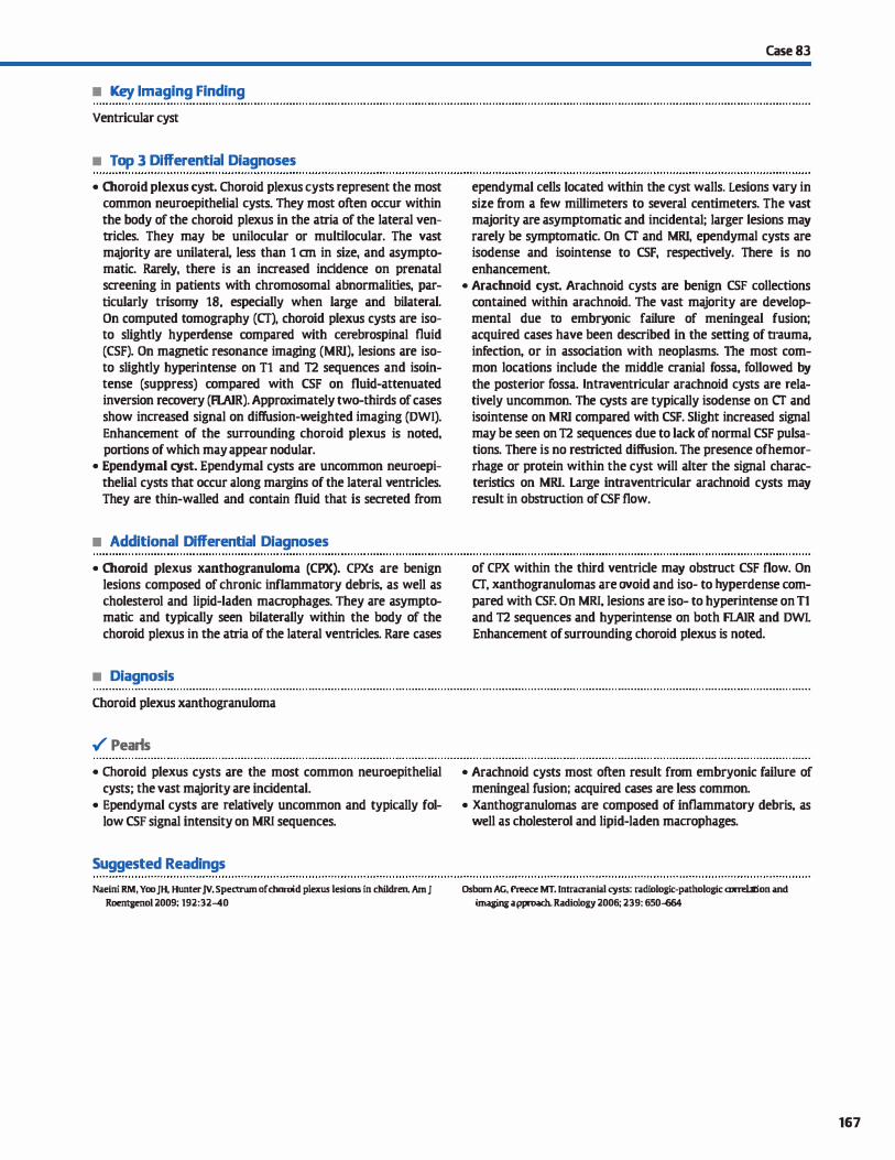
• Key lmaging Finding
Ventricular cyst
• Top 3 Differential Diagnoses
• Choroid plexus cyst. Choroid plexus cysts represent the most common neuroepithelial cysts. They most often occur within
the body of the choroid plexus in the atria of the lateral ventrides. They may be unilocular or multilocular. The vast majority are unilateral, less than 1 cm in size, and asymptomatic. Rarely, there is an increased incidence on prenatal screening in patients with chromosomal abnormalities, par
ticularly trisomy 18, especially when large and bilateral.
On computed tomography (CT), choroid plexus cysts are isoto slightly hyperdense compared with cerebrospinal fluid (CSF). On magnetic resonance imaging (MRI), lesions are isoto slightly hyperintense on Tl and T2 sequences and isointense (suppress) compared with CSF on fluid-attenuated inversion recovery (FIJ\IR). Approximately two-thirds of cases show increased signal on diffusion-weighted imaging {DWI).
Enhancement of the surrounding choroid plexus is noted, portions of which may appear nodular.
• Ependymal cyst. Ependymal cysts are uncommon neuroepithelial cysts that occur along margins of the lateral ventricles. They are thin-walled and contain fluid that is secreted from
• Additional Differential Diagnoses
• Choroid plexus xanthogranuloma (CPX). CPXs are benign lesions composed of chronic inflammatory debris, as well as cholesterol and lipid-laden macrophages. They are asymptomatic and typically seen bilaterally within the body of the
choroid plexus in the atria of the lateral ventrides. Rare cases
• Diagnosis
Choroid plexus xanthogranuloma
� Pearls
• Choroid plexus cysts are the most common neuroepithelial cysts; the vast majority are incidental.
• Ependymal cysts are relatively uncommon and typically follow CSF signal intensity on MRI sequences.
Suggested Readings
Naeini RM, Yoo JH, Hunter JV. Spectrum of choroid plexus les ions in children. Am j
Roentgenol2009; 192:32-40
Case 83
ependymal cells Iocated within the cyst walls. Lesions vary in size from a few millimeters to several centimeters. The vast majority are asymptomatic and incidental; larger lesions may rarely be symptomatic. On cr and MRI, ependymal cysts are isodense and isointense to CSF, respectively. There is no
enhancement. • Arachnoid cyst. Arachnoid cysts are benign CSF collections
contained within arachnoid. The vast majority are developmental due ta embryonic failure of meningeal fusion; acquired cases have been described in the setting of trauma, infection, or in association with neoplasms. The most common locations include the middle cranial fessa, fellowed by
the posterior fessa. lntraventricular arachnoid cysts are relatively uncommon. The cysts are typically isodense on cr and isointense on MRI compared with CSF. Slight increased signal may be seen on T2 sequences due to Jack of normal CSF pulsations. There is no restricted diffusion. The presence ofhemorrhage or protein within the cyst will alter the signal characteristics on MRI. Large intraventricular arachnoid cysts may result in obstruction of CSF flow.
of CPX within the third ventride may obstruct CSF flow. On CT, xanthogranulomas are ovoid and iso- to hyperdense com
pared with CSF. On MRI, lesions are iso- to hyperintense on Tl and T2 sequences and hyperintense on bath FIJ\IR and DWI. Enhancement of surrounding choroid plexus is noted.
• Arachnoid cysts most often result from embryonic failure of meningeal fusion; acquired cases are Jess common.
• Xanthogranulomas are composed of inflammatory debris, as well as cholesterol and lipid-laden macrophages.
Osborn AG, Preeœ MT. Intracranial cysts; radiologie-pathologie correlation and
imaging approach. Radiology 2006; 239: 650-664
167
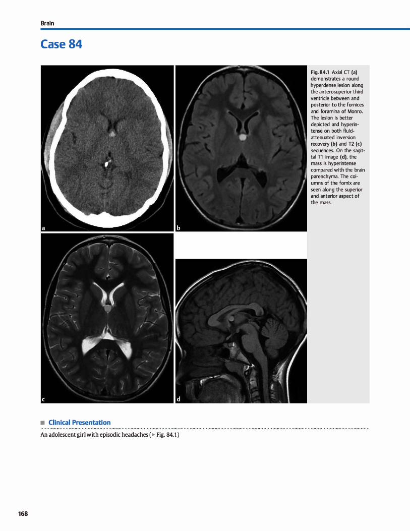
Brain
Case 84
• Clinical Presentation
An adolescent girl with episodic headaches ( � Fig. 84.1 )
168
Fig. 84.1 Axial CT (a) demonstrates a round hyperdense lesion along the anterosuperior third ventride between and posterior to the fornices and foramina of Monro. The lesion is better depicted and hyperintense on both fluidattenuated inversion recovery (b) and T2 (c) sequences. On the sagittal Tl image (d), the mass is hyperintense compared with the brain parenchyma. The columns of the fornix are seen along the superior and anterior aspect of the mass.
• Key lmaging Finding
Hyperdense/hyperintense, circumscribed mass along the ante
rosuperior third ventride
• Diagnosis
Colloid cyst. Colloid cysts are benign lesions that originate
along the anterosuperior aspect of the third ventride. Most are
inddental; however, they may cause headaches and intermit
tent acute hydrocephalus as a result of obstruction of the
foramina of Monro. Therefore, they are typically treated with
surgical resection, aspiration, or œrebrospinal fluid diversion.
The location and imaging appearanœ are fairly characteristic.
Colloid cysts occur between and posterior to the fomiceal col
umns as they pass along the fonnina of Monro. The internai
contents have varying amounts of proteinaceous fluid and vis-
� Pearls
• Colloid cysts are benign and originate along the anterosupe
rior aspect of the third ventricle.
• Colloid cysts may cause intermittent acute hydrocephalus as a result of obstruction of the foramina of Monro.
Suggested Reading
Armao Il, Castille M, Oten H, Kwock L Colloid cyst of the third ventride; imaging
pathologic correlation Amj Neuroradiol 2000; 21: 1470-1477
Case 84
cosity, which account for its imaging appearance. On computed
tomography {CT), the lesions are most often hyperdense due to
a high protein content and increased viscosity; however, lesions
with relatively low protein content will be isodense and may be
overlooked. On magnetic resonanœ imaging, colloid cysts are typically iso- to hyperintense on Tl sequenœs. The T2 signal is
more variable. In general, as the protein content and viscosity
increase, the T2 signal decreases. Subtle peripheral enhance
ment may be seen; however, the lesions themselves should not enhance.
• Imaging appearanœ of colloid cysts depends upon the protein
content and viscosity of the fluid.
• Colloid cysts are usually hyperdense on CT, iso- to hyperintense on Tl, and variable in signal on T2 sequences.
169
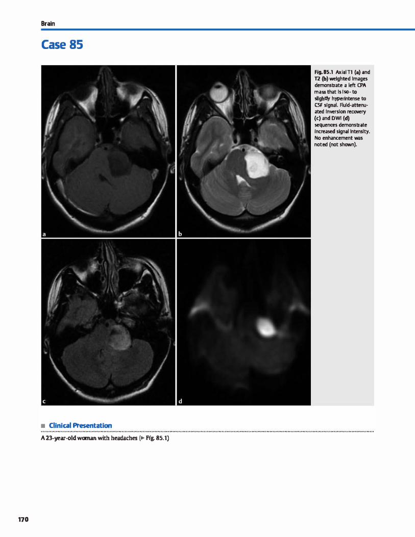
Brain
Case 85
• Clinical Presentation
A 23-year-old woman with headaches c.- Fig. 85.1)
170
Fig. 85.1 Axi;tl Tl (;a) and 12 (b) welghted tmagn demonstrate a left a>A mass that ls Isa-to slightiy hyperintense to CSF signal. Flu!d-attenuated Inversion recovery (c) and DWI (cf) sequences demonstrate !ncreased signal lntens!ty. No enhancement was noted (not shown).
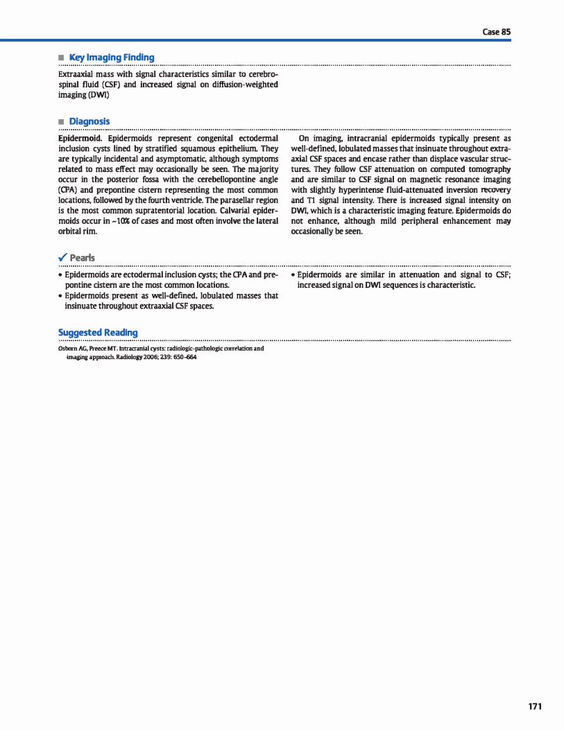
• Key lmaging Finding
Extraaxial mass with signal characteristics similar to cerebro
spinal fluid (CSF) and increased signal on diffusion-weighted
imaging (DWI)
• Diagnosis
Epidermoid. Epidermoids represent congenital ectodermal indusion cysts lined by stratified squamous epitheliurn. They are typically incidental and asymptomatic, although symptoms related to mass effect may occasionally be seen. The majority occur in the posterior fessa with the œrebellopontine angle
(CPA) and prepontine cistern representing the most common locations, followed by the fourth ventride. The parasellar region is the most common supratentorial location. Calvarial epider
moids occur in -10% of cases and most often involve the lateral orbital rim.
� Pearts
• Epidermoids are ectodermal inclusion cysts; the CPA and pre
pontine dstem are the most common locations.
• Epidermoids present as well-defined, lobulated masses that insinuate throughout extraaxial CSF spaces.
Suggested Reading
Osborn AG, Preece MT. lntracranial cysts; radiologie-pathologie mrrelation and
imaging approach. Radiology 2006; 239: 650-664
Case 85
On imaging, intracranial epidermoids typically present as well-defined, lobulated masses that insinuate throughout extraaxial CSF spaces and encase rather than displace vascular structures. They follow CSF attenuation on computed tomography and are similar ta CSF signal on magnetic resonanœ imaging
with slightly hyperintense fluid-attenuated inversion recovery
and Tl signal intensity. There is increased signal intensity on
DWI, which is a characteristic imaging feature. Epidermoids do not enhance, although mild peripheral enhancement may occasionally be seen.
• Epidermoids are similar in attenuation and signal ta CSF;
increased signal on DWI sequenœs is characteristic.
171
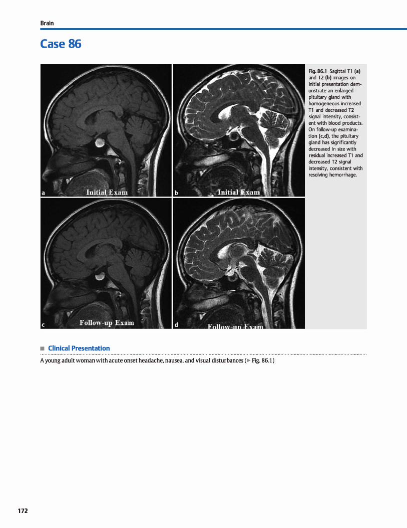
Brain
Case 86
• Clinical Presentation
A young adult woman with acute onset headache, nausea, and visual disturbances (� Fig. 86.1 )
172
Fig. 86.1 Sagittal Tl (a) and T2 (b) images on initial presentation demonstrate an enlarged pituitary gland with homogeneous increased Tl and decreased T2 signal intensity, consistent with blood products. On follow-up examination (c,d), the pituitary gland has significantly decreased in size with residual increased Tl and decreased T2 signal intensity, consistent with resolving hemorrhage.
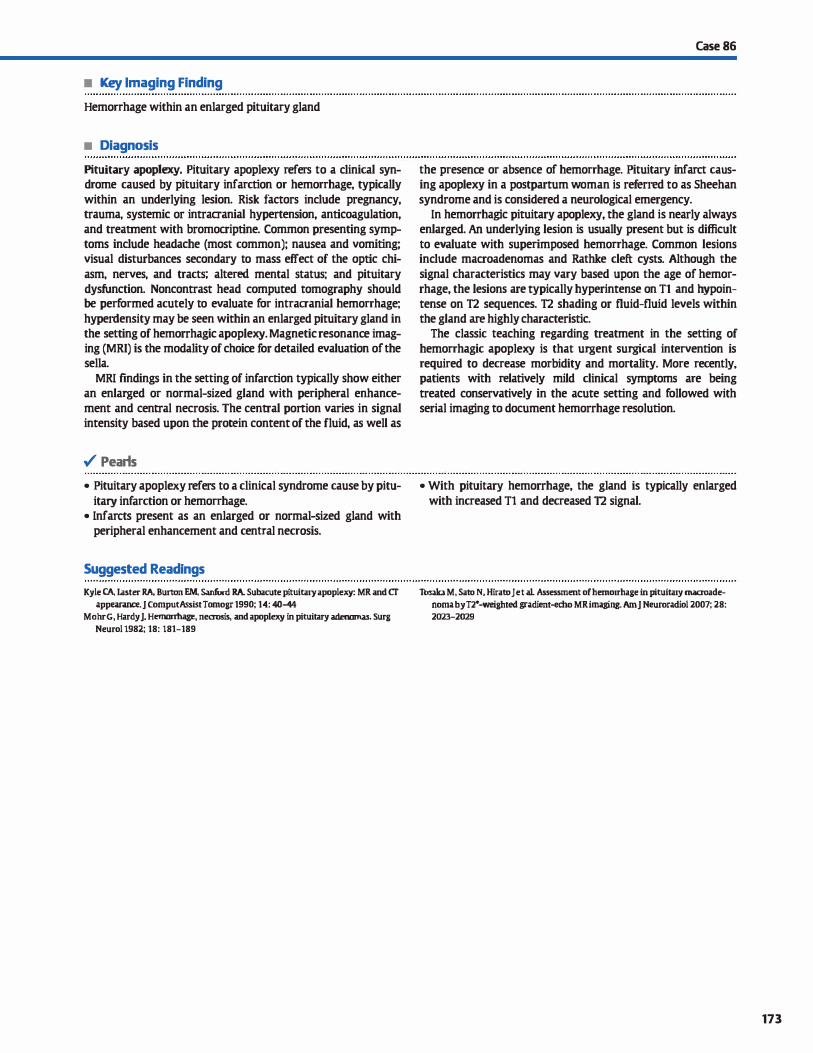
• Key lmaging Finding
Hemorrhage within an enlarged pituitary gland
• Diagnosis
Pituitary apoplexy. Pituitary apoplexy refers to a dinical syndrome caused by pituitary infarction or hemorrhage, typically
within an underlying lesion. Risk factors indude pregnancy, trauma, systemic or intracranial hypertension, anticoagulation, and treatment with bromocriptine. Common presenting symptoms indude headache (most common); nausea and vomiting; visual disturbances secondary to mass effect of the optic chi
asm, nerves, and tracts; altered mental status; and pituitary dysfunction. Noncontrast head computed tomography should be performed acutely to evaluate for intracranial hemorrhage;
hyperdensity may be seen within an enlarged pituitary gland in the setting of hemorrhagic apoplexy. Magnetic resonanœ imaging (MRI) is the modality of choice for detailed evaluation of the sella.
MRI findings in the setting of infarction typically show either an enlarged or normal-sized gland with peripheral enhancement and central necrosis. The central portion varies in signal intensity based upon the protein content of the fluid, as well as
v" Pearls
• Pituitary apoplexy refers to a clinical syndrome cause by pitu
itary infarction or hemorrhage. • Infarcts present as an enlarged or normal-sized gland with
peripheral enhancement and central necrosis.
Suggested Readings
Case 86
the presenœ or absence of hemorrhage. Pituitary infarct causing apoplexy in a postpartum woman is referred to as Sheehan syndrome and is considered a neurological emergency.
In hemorrhagic pituitary apoplexy, the gland is nearly always enlarged. An underlying lesion is usually present but is difficult
to evaluate with superimposed hemorrhage. Common lesions indude macroadenomas and Rathke cleft cysts. Although the
signal characteristics may vary based upon the age of hemorrhage, the lesions are typically hyperintense on Tt and hypointense on T2 sequences. T2 shading or fluid-fluid levels within the gland are highly characteristic.
The classic teaching regarding treatment in the setting of
hemorrhagic apoplexy is that urgent surgical intervention is required to decrease morbidity and mortality. More recently, patients with relatively mild clinical symptoms are being treated conservatively in the acute setting and followed with serial imaging to document hemorrhage resolution.
• With pituitary hemorrhage, the gland is typically enlarged
with increased Tt and decreased T2 signal.
Kyle CA, Laster RA, Burton EM, Sanford RA. Subarute pituitary apoplexy: MR and cr Tosaka M, Sato N, Hirato jet al Assessrnent ofhemorrhage in pituitary rnacroade-
appearanœ. J ComputAssist Tomogr 1990; 14: 40-44 noma by T2°-weighted gradient-echo MR imaging. Am J Neuroradiol 2007; 28:
Mahr G, Hardy J. Hernarrhage, necrosis, and apoplexy in pituitary adenanw, Surg 2023-2029
Neurol 1982; 18: 181-189
173
Brain
Case 87
• Clinical Presentation
An 8-year-old girl with chronic headaches (� Fig. 87 .1 )
174
Fig. 87 .1 Sagittal Tl midline image reveals a curvilinear hyperintensity along the outer margin of the splenium and posterior body of the corpus callosum. The corpus callosum appears normally formed.
• Key lmaging Finding
Midline fat signal intensity along the margin of the corpus callosum
• Diagnosis
Pericallosal lipoma. Intracranial lipomas result from persistence of the meninx primitiva that usually involutes early in gestation. They may occur throughout the central nervous system dsterns, but the vast majority are midline with the pericallosal
region being most common. Oinical symptoms are variable, ranging from asymptomatic and inddental to seizures and developmental delay in association with multiple developmen
tal anomalies. The morphology of the lipoma correlates well with patient presentation and associated findings.
Pericallosal Iipomas occur in two distinct forms: tubulonodular and curvilinear. The tubulonodular variant is larger (typi
cally> 2 cm) and bas a rounded or lobular masslike configuration. lt occurs more commonly along the anterior portion of the corpus callosum and bas a bigber incidence of associated callosal, parenchymal, and craniofacial anomalies. There may be extension into the lateral ventrides and choroid plexus, wbich is fairly characteristic. The curvilinear form is thin ( < 1 an), typically occurs along the posterior body and splenium of the
� Pearts
• Intracranial Iipomas result from persistence of the meninx primitiva; the vast majority are midline.
• Pericallosal lipomas may be tubulonodular {more often associated with additional anomalies) or curvilinear.
Suggested Readings
Idccwitz V, Eurin D, Rypens F et al Prenatal diagnosis and postnatal fbllaw-up of
pericallosal lipama: report of seven new cases. AmJ Neuroradiol 2001; 22: 767-
n2
Case 87
corpus callosum, and is often incidental. The underlying corpus callosum may be mildly bypoplastic.
On computed tomography, lipomas are characteristically bypodense. catdfications may be seen, especially with the
tubulonodular variant Lipomas may engulf vessels, so careful evaluation of the adjacent vasculature is important. On magnetic resonanœ imaging, lipomas follow fat signal with hyperintensity seen on both T1 and T2 sequences. There may be chemical sbift artifact along its margins secondary to differences in the resonance frequencies of fat and water in the
frequency encoding direction. This results in a band of hyperintensity on one margin and a band of bypointensity along the opposite margin. There should be no enbancement calcifications are best seen on gradient echo or susceptibility-weighted sequences. Vascular flow voids may be seen within the Iipomas. Fat-suppressed sequenœs confirm the diagnosis. Interestingly, Iipomas may occasionally enlarge secondary to lipomatous
bypertropby, especially during periods of somatic growth.
• Lipomas follow fat signal or attenuation; calcifications may be seen, especially with the tubulonodular variant
Tart RP, Quisling RG. Curvilinear and tubulonodular varieties of liporna of the corpus
callosum: anMRand CTstudy.J ComputAssistTomogr 1991; 15: 805-810
175
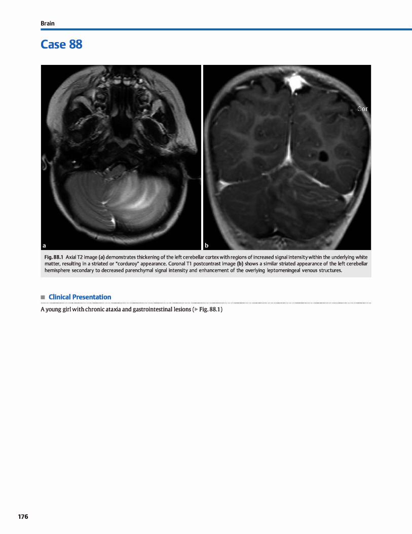
176
Brain
Case 88
Fig. 88.1 Axial T2 image (a) demonstrates thickening of the left cerebellar cortex with reg ions of increased signal intensity within the underlying white matter, resulting in a striated or "corduroy" appearance. Coronal Tl postcontrast image (b) shows a similar striated appearance of the left cerebellar hemisphere secondary to decreased parenchymal signal intensity and enhancement of the overlying leptomeningeal venous structures.
• Clinical Presentation
A young girl with chronic ataxia and gastrointestinal lesions (� Fig. 88.1 )
• Key lmaging Finding
Cerebellar "mass" with a striated or "conl.uroy" appearanœ
• Diagnosis
Lherm.itte-Dudos disease. Lhermitte-Dudos disease (IDD) refers to a dysplastic œrebellar gangliocytoma of uncertain etiology. Controversy exists over classification of this lesion
because it may have pathological characteristics of both a hamartoma and a benign neoplasm. It is associated with Cowden syndrome, which is an autosomal dominant syndrome characterized by mucocutaneous lesions and increased inci
dence of neoplasms involving the gastrointestinal tract, genitourinary tract, breast, and thyroid.
IDD presents most often in young to middle-aged adults. It is aise infrequently seen in children. Patients may be asymptomatic or present with ataxia, cranial nerve deficits, or symptoms related to mass effect, induding obstructive hydrocephalus. Symptoms are usually insidious in onset.
y' Pearts
• LDD is a dysplastic cerebellar gangliocytoma; it often has characteristics of both a hamartoma and a neoplasm.
• LDD results in thickening of the cerebellar folia/cortex and underlying increased T2 and decreased Tt signal.
Suggested Readings
Klisch J, juengling F, Spreer j et al. Uiennitte-Duclos disease: assessrnent with MR irnaging, positron ernission tornography, single-photDn ernission CT, and MR spectroscopy. Arnj Neuroradiol 2001; 22: 824-830
Case 88
On computed tomography, LOD presents as a relatively illdefined region of enlargement and hypoattenuation within the cerebellar hemisphere(s) with mass effect. calcifications may be seen. Findings on magnetic resonance imaging are more characteristic, demonstrating thickening of the cerebellar folia/
cortex and increased T2 signal intensity within the underlying white matter, resulting in a striated or "conl.uroy" appearance. A similar striated appearanœ is seen on T1 postcontrast
sequences with parenchymal hypoattenuation and enhancement of the overlying leptomeningeal veins. The lesion itself should not enhance. If symptomatic, surgery is the treatment of choice; recurrenœs have been described.
• Signal characteristics result in a "corduroy" appearance on T2 and postcontrast Tt sequences.
• LOD is associated with Cowden syndrome (mucocutaneous lesions and increased incidence ofneoplasms).
Shinagare AB, Patil NK, Sorte SI.. Case 144: dysplastic œrebellar gangliocytorna (Uterrnitte-Dudos disease). Radiology 2009; 251: 298-303
177
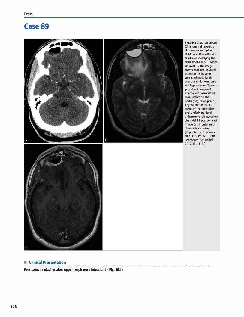
Brain
Case 89
• Clinical Presentation
Persistent headaches after upper respiratory infection ( � Fig. 89.1 )
178
Fig. 89.1 Axial enhanced CT image (a) reveals a rim-enhancing epidural fluid collection with airfluid level overlying the right frontal lobe. Followup axial T2 (b) image shows that the epidural collection is hyperintense, whereas its rim and the underlying dura are hypointense. There is prominent vasogenic edema with associated mass effect on the underlying brain parenchyma. Rim enhancement of the collection and underlying durai enhancement is noted on the axial Tl postcontrast image (c). Frontal sinus disease is visualized. (Reprinted with permission, O'Brien WT. J Am Osteopath Coll Radiol 201 2;1 (1 ):3-9.)
• Key lmaging Finding
Rim-enhancing epidural fluid collection with durai enhanœment
• Diagnosis
Epidural absœss. Epidural abscesses most often result from direct spread of paranasal sinus or otomastoid infections or as a result of post-traurnatic or postoperative complications. They may also extend from adjacent calvarial processes, such as osteomyelitis. As with other epidural collections, epidural abscesses are biconvex or lenticular in shape, confined by sutures, and may cross midline.
On computed tomography (CT), epidural abscesses are hypodense and may have air-fluid levels. The absœss cavity demonstrates rim enhancement, often thick; enhancement of the underlying dura may also be seen. Similar findings are noted on magnetic resonanœ imaging (MRI) where the epidural
� Pearts
• Epidural abscesses often result from paranasal sinus/mastoid infections or from postoperative complications.
• On CT, epidural abscesses are hypodense, rim-enhancing, and may have air-fluid levels.
Suggested Reading
O'Brien wr. Imaging of CNS infections in immunorompetent hosts. J Arn 05teopath
Qill Radial 2012; 1: 3-9
Case 89
collections are hypointense on T1 and hyperintense on T2 sequenœs with rim and durai enhancement. The rim of the abscess cavity and of the underlying dura are hypointense on T2 sequences. Foci of air are hypointense on ail pulse sequenœs. The presence of restricted diffusion is variable but typically seen. When adjacent to durai venous sinuses, it is important to look for evidence of sinus thrombosis.
Treatment of epidural abscesses indudes a combination of antibiotic therapy and surgical drainage. If left untreated, epidural abscesses may extend through the dura to involve the subdural spaœ, leptomeninges, or brain parenchyma.
• On MRI, epidural abscesses are T2 hyperintense with rim and durai enhanœment; restricted diffusion is common.
179
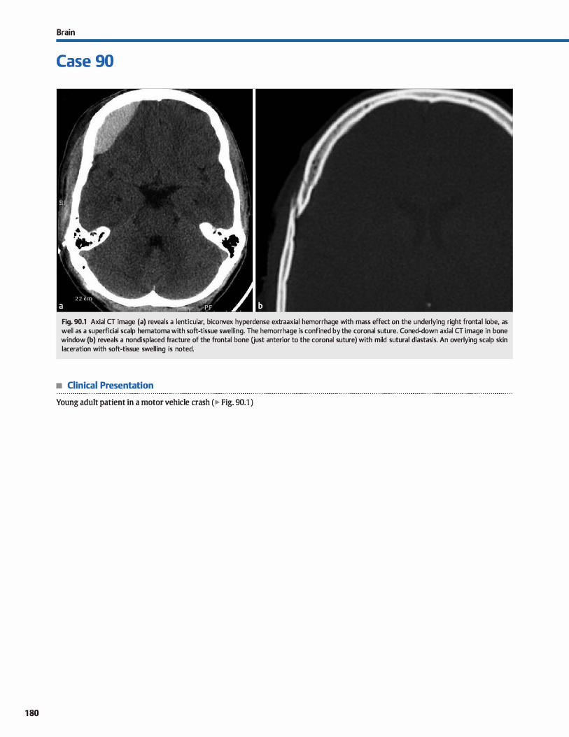
180
Brain
Case 90
Fig. 90.1 Axial CT image (a) reveals a lenticular, biconvex hyperdense extraaxial hemorrhage with mass effect on the underlying right frontal lobe, as well as a superficial scalp hematoma with soft-tissue swelling. The hemorrhage is confined by the coronal suture. Coned-down axial CT image in bone window (b) reveals a nondisplaced fracture of the frontal bone Oust anterior to the coronal suture) with mild sutural diastasis. An overlying scalp skin laceration with soft-tissue swelling is noted.
• Clinical Presentation
Young adult patient in a motor vehicle crash (� Fig. 90.1 )
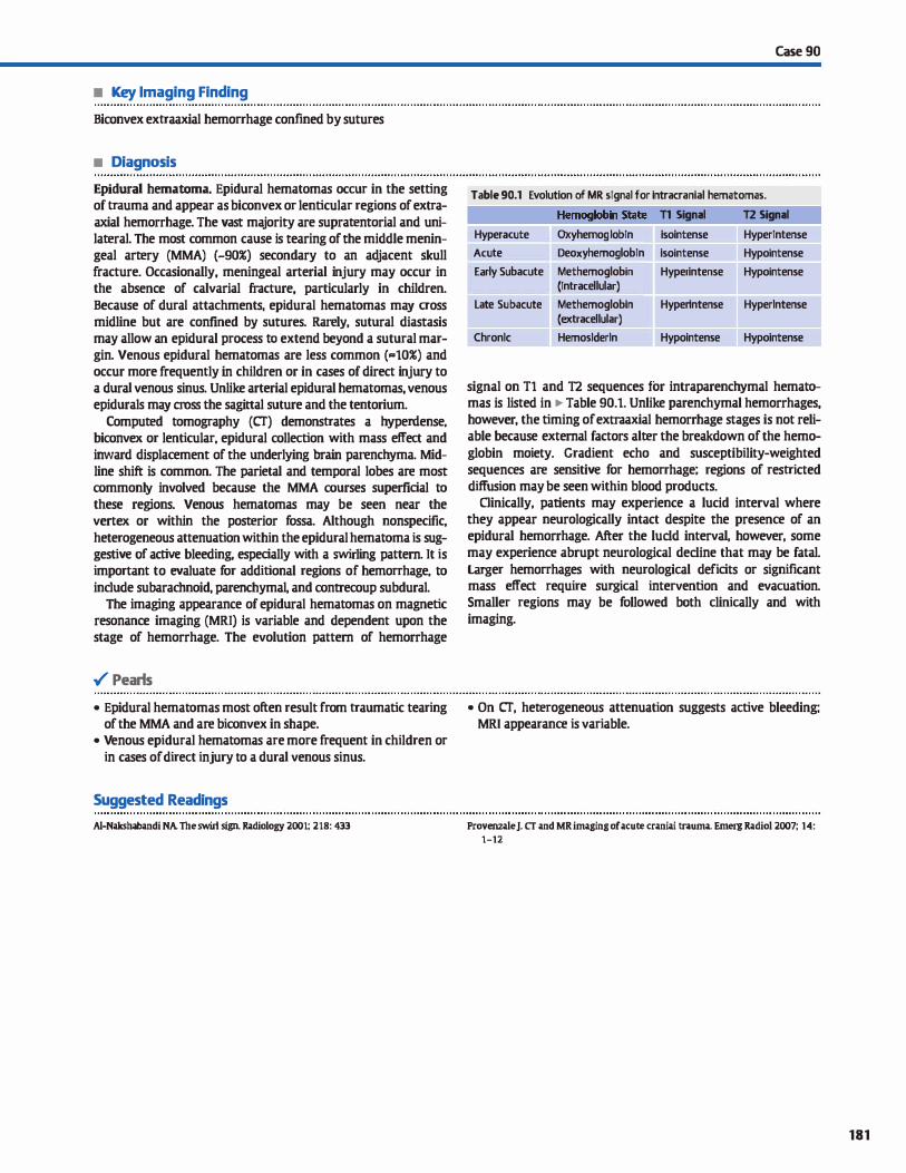
• Key lmaging Finding
Biconvex extraaxial hemorrhage confined by sutures
• Diagnosis
Epidural hematoma. Epidural hematomas occur in the setting of trauma and appear as biconvex or lenticular regions of extra
axial hemorrhage. The vast majority are supratentorial and unilateral. The most common cause is tearing of the middle meningeal artery {MMA) {-90%) secondary to an adjacent skull fracture. Occasionally, meningeal arterial injury may occur in the absence of calvarial fracture, particularly in children.
Because of durai attachments, epidural hematomas may cross midline but are confined by sutures. Rarely, sutural diastasis may allow an epidural process ta extend beyond a sutural margin. Venous epidural hematomas are less common {"'10%) and occur more frequently in children or in cases of direct injury to a durai venous sinus. Unlike arterial epidural hematomas, venous epidurals may cross the sagittal suture and the tentorium.
Computed tomography { CT) demonstrates a hyperdense. biconvex or lenticular, epidural collection with mass effect and inward displaœment of the underlying brain parenchyma. Midline shift is common. The parietal and temporal lobes are most commonly involved because the MMA courses superficial to these regions. Venous hematomas may be seen near the vertex or within the posterior fossa. Although nonspecitic,
heterogeneous attenuation within the epidural hematoma is suggestive of active bleeding, especially with a swirling pattern. It is important to evaluate for additional regions of hemorrhage, to
indude subarachnoid, parenchymal, and contrecoup subdural. The imaging appearance of epidural hematomas on magnetic
resonance imaging {MRI) is variable and dependent upon the stage of hemorrhage. The evolution pattern of hemorrhage
� Pearts
• Epidural hematomas most often result from traumatic tearing of the MMA and are biconvex in shape.
• Venous epidural hematomas are more frequent in children or
in cases of direct injury to a durai venous sinus.
Suggested Readings
Al-Nakshabandi NA. The swirl sign. R.adiology 2001: 218: 433
Case 90
Table 90.1 Evolution of MR signal for lntracranlal hematomas.
Hemoglobln 5tate Tl Signal 12 Signal
Hyperacute Oxyhemog lobin lsointense Hyperintense
Acute Deoxyhemoglobin lsointense Hypointense
Early Subacute Methemoglobin Hyperintense Hypointense (intracellular)
Late Su bacute Methemoglobln Hyperintense Hyperlntense (extracellular)
Chronlc Hemoslderln Hypointense Hypointense
signal on Tl and T2 sequences for intraparenchymal hematomas is listed in ... Table 90.1. Unlike parenchymal hemorrhages, however, the timing of extraaxial hemorrhage stages is not reliable because externat factors alter the breakdown of the hemo
globin moiety. Gradient echo and susceptibility-weighted sequenœs are sensitive for hemorrhage; regions of restricted diffusion may be seen within blood products.
Oinically, patients may experience a lucid interval where they appear neurologically intact despite the presenœ of an epidural hemorrhage. After the ludd interval, however, some may experienœ abrupt neurological dedine that may be fatal.
Larger hemorrhages with neurological deficits or significant mass effect require surgical intervention and evacuation. Smaller regions may be followed bath clinically and with
imaging.
• On CT, heterogeneous attenuation suggests active bleeding; MRI appearanœ is variable.
Provenzale J. cr and MR imaging of acuœ crantai trauma. Emerg R.adiol 2007; 14:
1-12
181
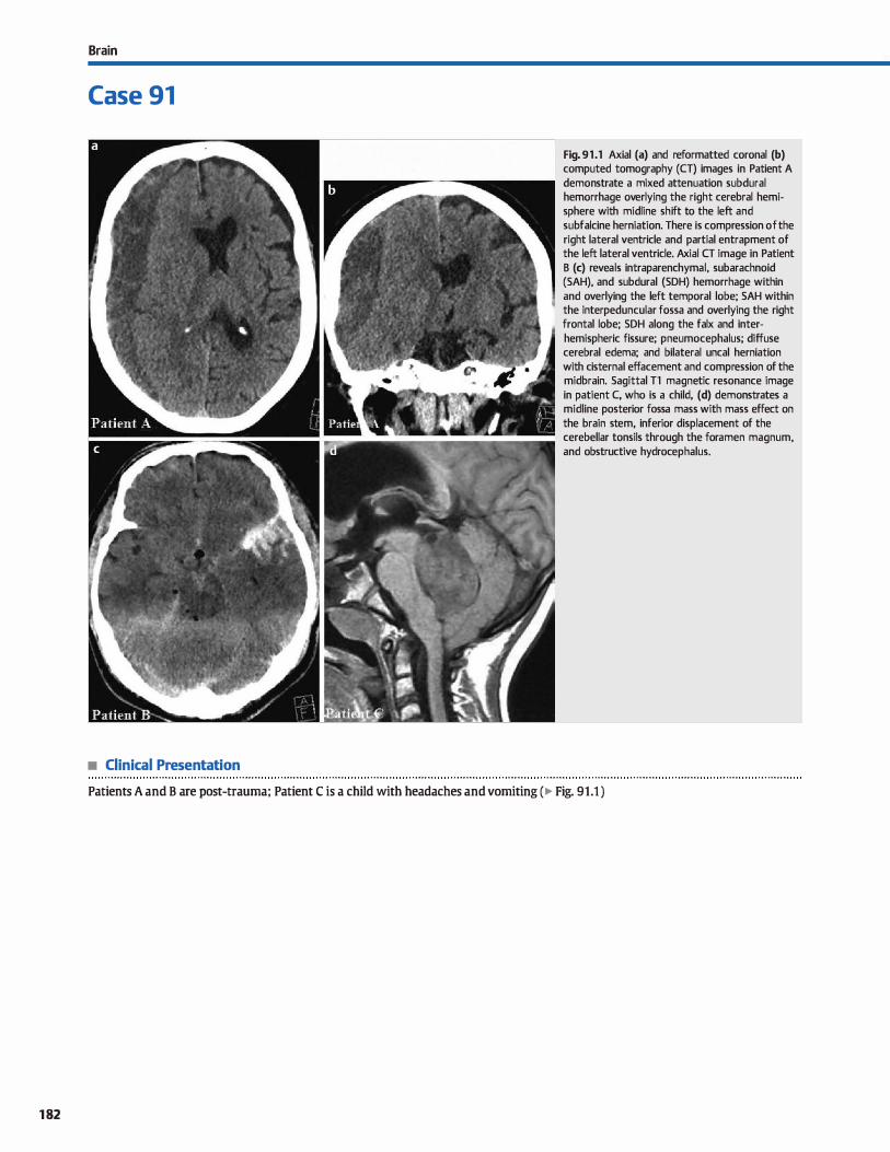
182
Brain
Case 91
• Clinical Presentation
Fig. 91 .1 Axial (a) and reformatted coronal (b) computed tomography (CT) images in Patient A demonstrate a mixed attenuation subdural hemorrhage overlying the right cerebral hemisphere with midline shift to the left and subfalcine herniation. There is compression of the right lateral ventricle and partial entrapment of the left lateral ventricle. Axial CT image in Patient B (c) reveals intraparenchymal, subarachnoid (SAH), and subdural (SDH) hemorrhage within and overlying the left temporal lobe; SAH within the interpeduncular fossa and overlying the right frontal lobe; SDH along the faix and interhemispheric fissure; pneumocephalus; diffuse cerebral edema; and bilateral uncal herniation with cisternal effacement and compression of the midbrain. Sagittal Tl magnetic resonance image in patient C, who is a child, (d) demonstrates a midline posterior fossa mass with mass effect on the brain stem, inferior displacement of the cerebellar tonsils through the foramen magnum, and obstructive hydrocephalus.
Patients A and B are post-trauma; Patient C is a child with headaches and vomiting (� Fig. 91.1 )
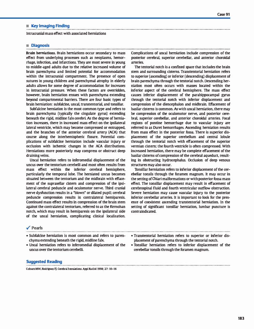
• Key lmaging Finding
lntracranial mass effect with associated hemiations
• Diagnosis
Brain herniations. Brain hemiations occur secondary to mass effect from underlying processes such as neoplasms, hemorrhage, infection, and infarctions. They are most severe in young to middle-aged adults due to the relative increased volume of brain parenchyma and limited potential for accommodation within the intracranial compartment. The presenœ of open sutures in young children and parenchymal atrophy in elderly
adults allows for some degree of accommodation for increases in intracranial pressure. When these factors are overridden, however, brain hemiation ensues with parenchyma extending
beyond compartmental barriers. There are four basic types of brain hemiation: subfalcine, uncal, transtentorial, and tonsillar.
Subfalcine herniation is the most common type and refers to brain parenchyma (typically the cingulate gyrus) extending
beneath the rigid, midline faix œrebri. As the degree of hemiation increases, there in increased mass effect on the ipsilateral
lateral ventride, which may become compressed or entrapped, and the branches of the anterior cerebral artery (ACA) that course along the interhemispheric fissure. Potential complications of subfaldne hemiation include vascular injury or occlusion with ischemic changes in the ACA distributions.
Herniations more posteriorly may compress or obstruct deep draining veins.
Uncal hemiation refers ta inferomedial displaœment of the
uncus aver the tentorium cerebelli and most often results from mass effect within the inferior cerebral hemisphere, particularly the temporal lobe. The herniated uncus becomes situated between the tentorium and the midbrain with efface
ment of the suprasellar dstem and compression of the ipsilateral cerebral peduncle and oculomotor nerve. Third cranial nerve dysfunction results in a Mblown" or dilated pupil; cerebral pedunde compression results in contralateral hemiparesis. Continued mass effect results in compression of the brain stem against the contralateral tentorium, referred to as the Kemohan notch, which may result in hemiparesis on the ipsilateral side
of the uncal herniation, complicating clinicat localization.
� Pearls
• Subfaldne hemiation is most common and refers to parenchyma extending beneath the rigid, midline faix.
• Uncal hemiation refers to inferomedial displaœment of the uncus over the tentorium cerebelli.
Suggested Reading
Cobum MW, Rodriguez FJ. Cerebral hemiations. Appl Radiol 1998; 27: 10-16
Case 91
Complications of uncal hemiation include compression of the posterior œrebral, superior cerebellar, and anterior choroidal arteries.
The tentorial notch is a confined spaœ that includes the brain stem and surrounding cistems. Transtentorial hemiation refers
to superior ( ascending) or inferior ( descending) dis placement of brain parenchyma through the tentorial notch. Desœnding her
niation most often occurs with masses located within the inferior aspect of the œrebral hemisphere. The mass effect causes inferior displacement of the parahippocampal gyrus through the tentorial notch with inferior displacement and compression of the diencephalon and midbrain. Effacement of
basilar dstems is common. As with uncal hemiation, there may be compression of the oculomotor nerve, and posterior cerebral, superior cerebellar, and anterior choroidal arteries. Focal regions of pontine hemorrhage due to vascular injury are referred ta as Duret hemorrhages. Asœnding hemiation results from mass effect in the posterior fossa. There is superior displacement of the superior cerebellum and central lobule through the tentorial notch with effacement of the superior
vermian cistem; the fourth ventride is often compressed. With increased hemiation, there may be complete effacement of the
basilar dstems of compression of the œrebral aqueduct, resulting in obstructing hydrocephalus. Occlusion of deep venous structures may also occur.
Tonsillar hemiation refers to inferior displacement of the œrebellar tonsils through the foramen magnum. It may occur in
the setting of Chiari malformations or with posterior fessa mass effect. The tonsillar displaœment may result in effacement of cerebrospinal fluid and fourth ventricular outflow obstruction. Severe hemiation may cause vascular injury to the posterior inferior cerebellar arteries. It is important to look for the presence of coexistent ascending transtentorial hemiation. In the setting of signiflcant tonsillar hemiation, lumbar puncture is
contraindicated.
• Transtentorial hemiation refers to superior or inferior displacement of parenchyma through the tentorial notch.
• Tonsillar hemiation refers to inferior displacement of the œrebellar tonsils through the foramen magnum.
183
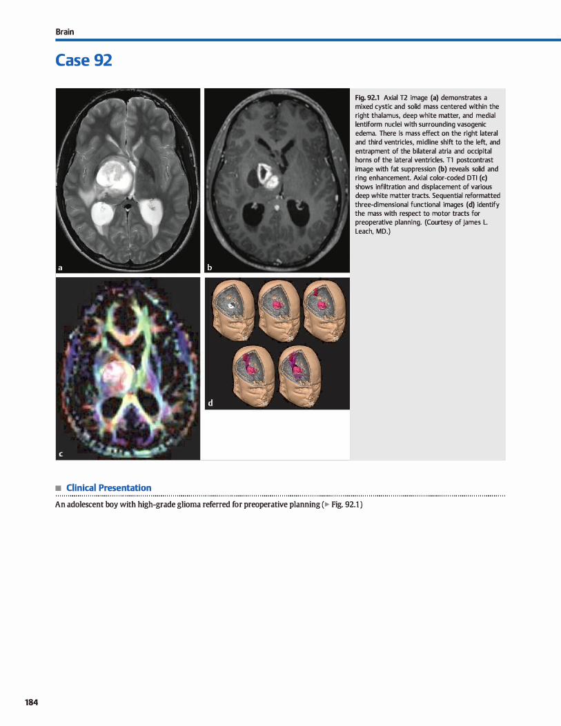
184
Brain
Case 92
• Clinical Presentation
Fig. 92.1 Axial T2 image (a) demonstrates a mixed cystic and solid mass centered within the right thalamus, deep white matter, and medial lentiform nuclei with surrounding vasogenic edema. There is mass effect on the right lateral and third ventricles, midline shift to the left, and entrapment of the bilateral atria and occipital horns of the lateral ventricles. Tl postcontrast image with fat suppression (b) reveals solid and ring enhancement. Axial color-coded DTI (c) shows infiltration and displacement of various deep white matter tracts. Sequential reformatted three-dimensional functional images (d) identify the mass with respect to motor tracts for preoperative planning. (Courtesy of James L. Leach, MD.)
An adolescent boy with high-grade glioma referred for preoperative planning (� Fig. 92.1 )
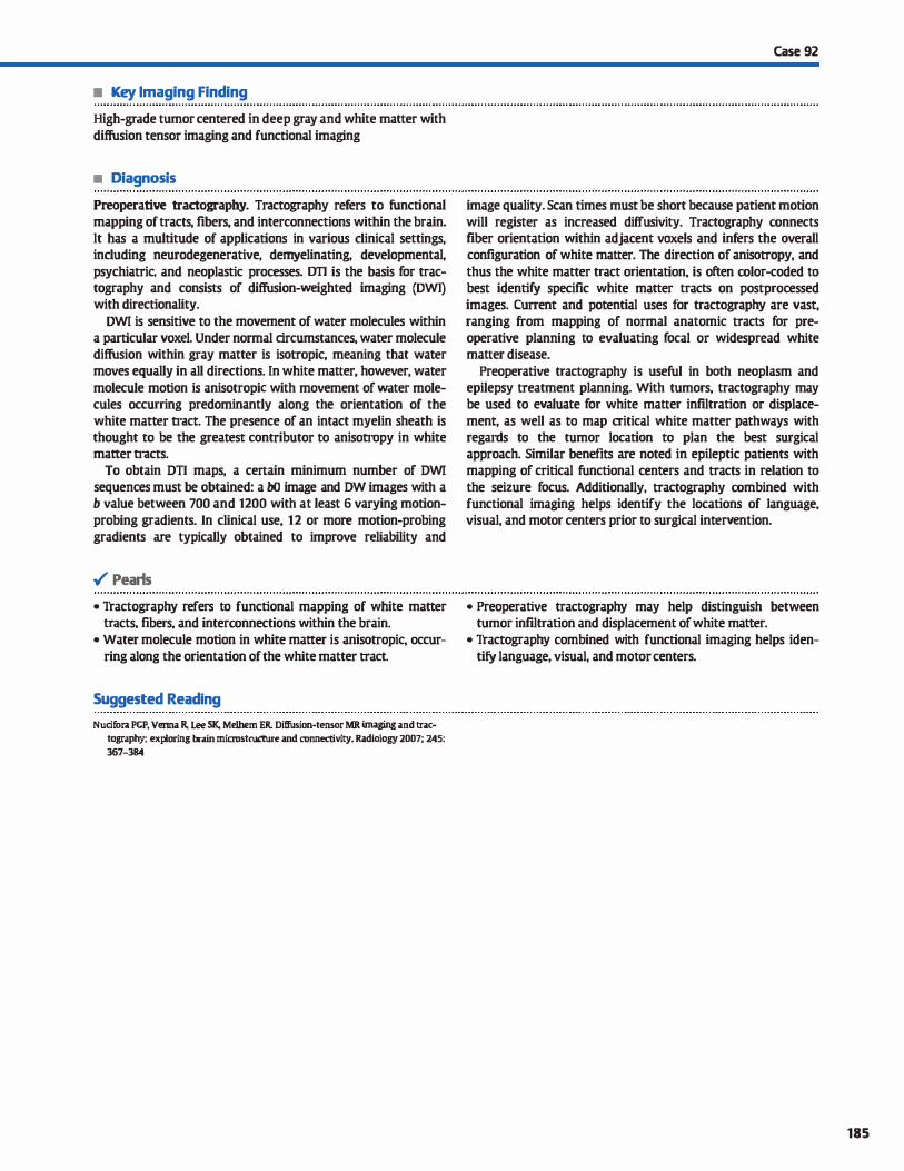
• Key lmaging Finding
High-grade tumor centered in deep gray and white matter with
diffusion tensor imaging and functional imaging
• Diagnosis
Preoperative tractography. Tractography refers to functional mapping of tracts, fibers, and interconnections within the brain. lt has a multitude of applications in various dinical settings, induding neurodegenerative, demyelinating, developmental,
psychiatrie, and neoplastic processes. DTI is the basis for tractography and consists of diffusion-weighted imaging (DWI) with directionality.
DWI is sensitive to the movement of water molecules within a particular voxel. Under normal circumstances, water molecule diffusion within gray matter is isotropie, meaning that water moves equally in ail directions. In white matter, however, water
molecule motion is anisotropie with movement of water molecules occurring predominantly along the orientation of the white matter tract. The presence of an intact myelin sheath is thought to be the greatest contributor to anisotropy in white matter tracts.
To obtain DTI maps, a certain minimum number of DWI
sequenœs must be obtained: a bO image and DW images with a b value between 700 and 1200 with at least 6 varying motion
probing gradients. In clinicat use, 12 or more motion-probing gradients are typically obtained to improve reliability and
� Pearls
• Tractography refers to functional mapping of white matter
tracts. fibers, and interconnections within the brain. • Water molecule motion in white matter is anisotropie, occur
ring along the orientation of the white matter tract.
Suggested Reading
Nucifor.i. PGP, Venna R. Lee SK, Melhem ER. Diffusion-tensor MR imaging and trac
tography: explming brain miaustructure and ronnectivity. Radiology 2007; 245; 367-384
Case 92
image quality. Sean times must be short because patient motion will register as increased diffusivity. Tractography connects fiber orientation within adjacent voxels and infers the overall configuration of white matter. The direction of anisotropy, and
thus the white matter tract orientation, is often color-coded to best identify specific white matter tracts on postproœssed images. Current and potential uses for tractography are vast, ranging from mapping of normal anatomie tracts for preoperative planning to evaluating focal or widespread white matter disease.
Preoperative tractography is useful in bath neoplasm and epilepsy treatment planning. With tumors, tractography may be used to evaluate for white matter infiltration or displace
ment, as well as to map critical white matter pathways with regards to the tumor location to plan the best surgical approach. Similar benefits are noted in epileptic patients with mapping of critical functional centers and tracts in relation to
the seizure focus. Additionally, tractography combined with functional imaging helps identify the locations of Ianguage, visual, and motor centers prior to surgical intervention.
• Preoperative tractography may help distinguish between tumor infiltration and displacement of white matter.
• Tractography combined with functional imaging helps identify language, visual, and motor centers.
185
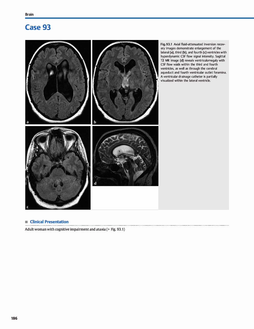
Brain
Case 93
• Clinical Presentation
Adult woman with cognitive impairment and ataxia (� Fig. 93.1)
186
Fig. 93.1 Axial fluid-attenuated inversion recovery images demonstrate enlargement of the lateral (a), third (b), and fourth (c) ventricles with hyperdynamic CSF flow signal intensity. Sagittal T2 MR image (d) reveals ventriculomegaly with CSF flow voids within the third and fourth ventricles, as well as through the cerebral aqueduct and fourth ventricular outlet foramina. A ventricular drainage catheter is partially visualized within the lateral ventricle.
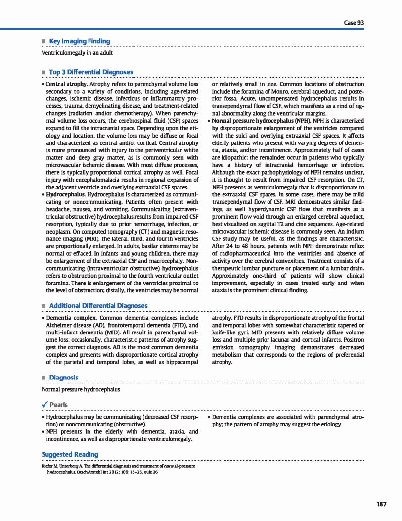
• Key lmaging Finding
Ventriculomegaly in an adult
• Top 3 Differential Diagnoses
• Central atrophy. Atrophy refers to parenchymal volume Joss secondary to a variety of conditions, including age-related changes, ischemic disease, infectious or inflammatory processes, trauma, demyelinating disease, and treatment-related changes (radiation and/or chemotherapy). When parenchymal volume loss occurs, the œrebrospinal fluid (CSF) spaœs expand to fill the intracranial spaœ. Depending upon the eti
ology and location, the volume Joss may be diffuse or focal and characterized as central and/or cortical. Central atrophy is more pronounced with injury to the periventricular white matter and deep gray matter, as is commonly seen with microvascular ischemic disease. With most diffuse processes, there is typically proportional cortical atrophy as well. Focal injury with encephalomalada results in regional expansion of
the adjacent ventricle and overlying extraaxial CSF spaces. • Hydrocephalus. Hydroœphalus is characterized as communi
cating or noncommunicating. Patients often present with headache, nausea, and vomiting. Communicating { extraventricular obstructive) hydroœphalus results from impaired CSF resorption, typically due to prior hemorrhage, infection, or
neoplasm. On computed tomography (CT) and magnetic resonance imaging (MRI), the Iateral, third, and fourth ventricles are proportionally enlarged. In adults, basilar cistems may be normal or effaced. In infants and young children, there may be enlargement of the extraaxial CSF and macrocephaly. Noncommunicating {intraventricular obstructive) hydrocephalus refers to obstruction proximal to the fourth ventricular outlet
foramina. There is enlargement of the ventricles proximal ta the Ievel of obstruction; distally, the ventrides may be normal
• Additional Differential Diagnoses
• Dementia complex. Common dementia complexes indude Alzheimer disease (AD), frontotemporal dementia (FID), and multi-infarct dementia {MID). Ali result in parenchymal volume Joss: occasionally, characteristic patterns of atrophy sug
gest the correct diagnosis. AD is the most common dementia
compiex and presents with disproportionate cortical atrophy of the parietal and temporal lobes, as well as hippocampal
• Diagnosis
Normal pressure hydroœphalus
� Pearls
• Hydrocephalus may be communicating { decreased CSF resorption) or noncommunicating (obstructivei
• NPH presents in the elderly with dementia, ataxia, and incontinence, as well as disproportionate ventriculomegaly.
Suggested Reading
Kim M, Unterberg A. The differential diagnosis and treatment ofnonnal-pœssuœ hydroœphalus. Dtsdl.Arztebl lnt 2012; 109; 15-25, quiz 26
Case 93
or relatively small in size. Common locations of obstruction include the foramina of Monro, cerebral aqueduct, and posterior fossa. Acute, uncompensated hydrocephalus results in
transependymal flow of CSF, which manifests as a rind of signal abnormality along the ventricular margins.
• Nonnal pressure hydrocephalus (NPH). NPH is characterized by disproportionate enlargement of the ventrides compared
with the sulci and overlying extraaxial CSF spaœs. It affects elderly patients who present with varying degrees of dementia, ataxia, and/or incontinence. Approximately half of cases are idiopathie; the remainder occur in patients who typically have a history of intracranial hemorrhage or infection. Although the exact pathophysiology of NPH remains undear, it is thought ta result from impaired CSF resorption. On cr, NPH presents as ventriculomegaly that is disproportionate ta the extraaxial CSF spaœs. In some cases, there may be mild transependymal flow of CSF. MRI demonstrates similar findings, as well hyperdynamic CSF flow that manifests as a prominent flow void through an enlarged œrebral aqueduct, best visualized on sagittal T2 and dne sequences. Age-related microvascular ischemic disease is commonly seen. An indium
CSF study may be useful, as the findings are characteristic. After 24 to 48 hours, patients with NPH demonstrate reflux of radiopharmaceutical into the ventricles and absence of activity over the cerebral convexities. Treatment consists of a therapeutic lumbar puncture or placement of a lumbar drain. Approximately one-third of patients will show clinicat improvement, especially in cases treated early and when ataxia is the prominent dinical finding.
atrophy. FID results in disproportionate atrophy of the frontal and temporal lobes with somewhat characteristic tapered or knife-like gyri. MID presents with relatively diffuse volume loss and multiple prier lacunar and cortical infarcts. Positron
emission tomography imaging demonstrates decreased metabolism that corresponds to the regions of preferential atrophy.
• Dementia complexes are assodated with parenchymal atrophy; the pattern of atrophy may suggest the etiology.
187
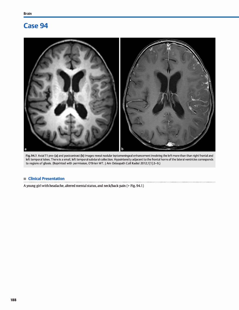
188
Brain
Case 94
Fig. 94.1 Axial Tl pre- (a) and postcontrast (b) images reveal nodular leptomeningeal enhancement involving the left more than than right frontal and left temporal lobes. There is a small, left temporal subdural collection. Hypointensity adjacent to the frontal horns of the lateral ventricles corresponds to regions of gliosis. (Reprinted with permission, O'Brien WT. j Am Osteopath Coll Radial 2012;1 [1 ]:3-9.)
• Clinical Presentation
A young girl with headache, altered mental status, and neck/back pain (� Fig. 94.1 )
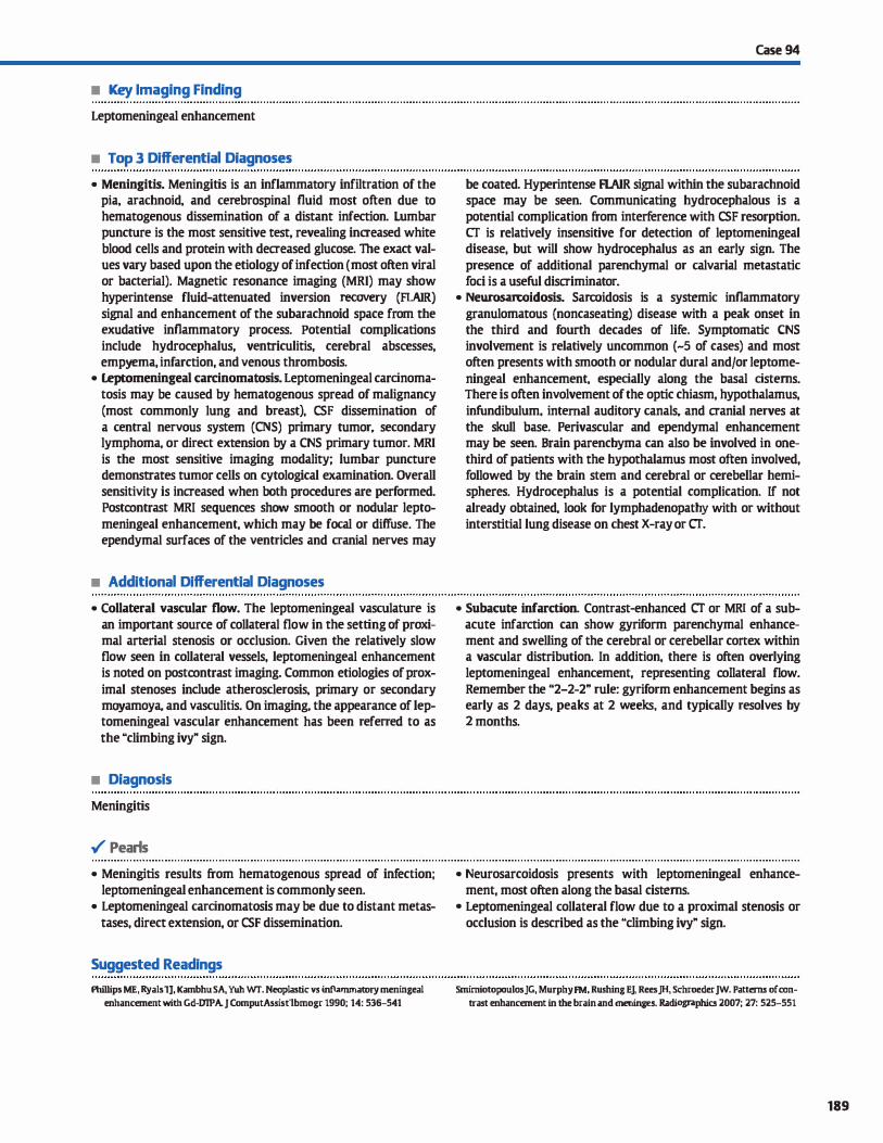
• Key lmaging Finding
Leptomeningeal enhancement
• Top 3 Differential Diagnoses
• Meningitis. Meningitis is an inflammatory infiltration of the pia, arachnoid, and cerebrospinal fluid most often due to hematogenous dissemination of a distant infection. Lumbar puncture is the most sensitive test, revealing increased white blood cens and protein with decreased glucose. The exact values vary based upon the etiology of infection ( most often viral or bacterial). Magnetic resonanœ imaging (MRI) may show hyperintense fluid-attenuated inversion recovery (FLAIR)
signal and enhancement of the subarachnoid space from the exudative inflammatory process. Potential complications include hydroœphalus, ventriculitis, cerebral absœsses, empyema, infarction, and venous thrombosis.
• Leptomeningeal carcinomatosis. Leptomeningeal cardnomatosis may be caused by hematogenous spread of malignancy (most commonly Jung and breast), CSF dissemination of a central nervous system {CNS) primary tumor, secondary lymphoma, or direct extension by a CNS primary tumor. MRI is the most sensitive imaging modality; lumbar puncture demonstrates tumor cells on cytological examination. OVerall sensitivity is increased when bath proœdures are performed. Postcontrast MRI sequenœs show smooth or nodular leptomeningeal enhancement, which may be focal or diffuse. The ependymal surfaces of the ventrides and cranial nerves may
• Additional Differential Diagnoses
• Collateral vascular flow. The leptomeningeal vasculature is an important source of collateral flow in the setting of proximal arterial stenosis or occlusion. Given the relatively slow flow seen in collateral vessels, leptomeningeal enhancement is noted on postcontrast imaging. Common etiologies of prox
imal stenoses indude atherosclerosis, primary or secondary moyamoya, and vasculitis. On imaging, the appearance of leptomeningeal vascular enhancement bas been referred to as the üclimbing ivy" sign.
• Diagnosis
Meningitis
� Pearts
• Meningitis results from hematogenous spread of infection; leptomeningeal enhancement is commonly seen.
• Leptomeningeal cardnomatosis may be due to distant metastases, direct extension, or CSF dissemination.
Suggested Readings
Phillips ME, Ryals lJ, Kambhu SA, Yuh WT, Neoplastic vs inflammatDl'y meningeal
enhanœment with Gd-IJIPAJ ComputAssist'lbrnogr 1990; 14: 536-541
Case 94
be coated. Hyperintense FLAIR signal within the subarachnoid spaœ may be seen. Communicating hydrocephalous is a potential complication from interference with CSF resorption. CT is relatively insensitive for detection of leptomeningeal disease, but will show hydrocephalus as an early sign. The presence of additional parenchymal or calvarial metastatic foci is a useful discriminator.
• Neurosarcoidosis. Sarcoidosis is a systemic inflammatory granulomatous {noncaseating) disease with a peak onset in the third and fourth decades of life. Symptomatic CNS involvement is relatively uncommon {-5 of cases) and most often presents with smooth or nodular durai and/or leptome
ningeal enhancement, espedally along the basal dstems. There is often involvement of the optic chiasm, hypothalamus, infundibulum. internai auditory canais, and cranial nerves at the skull base. Perivascular and ependymal enhancement may be seen. Brain parencbyma can also be involved in onethird of patients with the hypothalamus most often involved, followed by the brain stem and cerebral or œrebellar hemispheres. Hydrocephalus is a potential complication. If not already obtained, look for lymphadenopathy with or without interstitial lung disease on chest X-ray or cr.
• Subacute infarction. Contrast-enhanced cr or MRJ of a subacute infarction can show gyriform parenchymal enhancement and swelling of the cerebral or cerebellar cortex within a vascular distribution. In addition, there is often overlying leptomeningeal enhancement, representing co1lateral flow. Remember the "2-2-2" rule: gyriform enhancement begins as early as 2 days, peaks at 2 weeks, and typically resolves by 2 months.
• Neurosarcoidosis presents with leptomeningeal enhancement, most often along the basal cistems.
• Leptomeningeal collateral flow due ta a proximal stenosis or occlusion is described as the üdimbing ivy" sign.
Smimiotopoulos JG, Murphy FM, Rushing EJ, Rees jH, Schroeder JW, Patterns of con
trast enhanœment in the brain and meninges. Radiographia 2007; 27: 525-551
189
190
Brain
Case 95
Fig. 95.1 Coronal Tl postcontrast image reveals abnormal, diffuse durai thickening and enhancement. Enlargement of the fourth ventride is partially seen.
• Clinical Presentation
A young patient with headaches (� Fig. 95.1 )
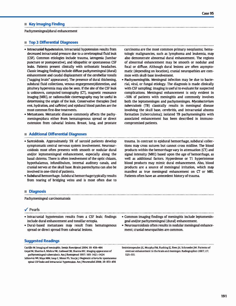
• Key lmaging Finding
Pachymeningeal/dural enhancement
• Top 3 Differential Diagnoses
• lntracranial hypotension. lntracranial hypotension results from decreased intracranial pressure due to a œrebrospinal fluid leak (CSF). Common etiologies indude trauma, iatrogenia (lumbar puncture or postoperative), and idiopathie or spontaneous CSF leaks. Patients present dinically with orthostatic headaches. Oassic imaging findings indude diffuse pachymeningeal (durai) enhanœment and caudal displaœment of the œrebellar tonsils
(Msagging brain" appearance). The presenœ of durai thickening,
subdural fluid collections, venous engorgement/distention, and pituitary hyperemia may also be seen. If the site of the CSF leak is unknown, computed tomography (CT), magnetic resonance imaging (MRI), or radionudide cisternography may be useful in detennining the origin of the leak. Conservative therapies (bed rest, hydration, and caffeine) and epidural blood patches are the
most common first-line treatments.. • Metastases. Metastatic disease commonly affects the pachy
meninges/dura either from hematogenous spread or direct extension from calvarial lesions. Breast, lung, and prostate
• Additional Differential Diagnoses
• Sarcoidosis. Approximately 5% of sarcoid patients develop
symptomatic central nervous system involvement. Neurosarcoidosis most often presents with smooth or nodular durai and/or leptomeningeal enhancement, especially along the
basal dstems. There is often involvement of the optic chiasm, hypothalamus, infundibulum, internai auditory canais, and cranial nerves at the skull base. Brain parenchyma can also be involved in one-third of patients.
• Subdural hemorrhage. Subdural hemorrhage typically results from tearing of bridging veins and is most often due ta
• Diagnosis
Pachymeningeal carcinomatosis
� Pearls
• Intracranial hypotension results from a CSF leak: findings include durai enhancement and tonsillar ectopia.
• Dural-based metastases may result from hematogenous spread or direct spread from calvarial lesions.
Suggested Readings
Case 95
carcinoma are the most common primary neoplasms; hematologic malignancies, such as lymphoma and leukemia, may also demonstrate abnormal durai enhancement. The regions of abnormal enhancement may be smooth or nodular and
focal or diffuse. Although durai lesions are often asymptomatic ( depending on location), cranial neuropathies are common with skull-base involvement.
• Pachymeningitis. Meningeal infection may be due to bacterial, viral, or fungal etiology. The diagnosis is made dinically with CSF sampling; imaging is useful to evaluate for suspected complications. Meningeal enhancement is only evident in -50% of patients with meningitis and commonly involves
both the leptomeninges and pachymeninges. Mycobacterium tuberculosis (TB) classically results in meningeal disease
involving the skull base, cerebritis, and intracranial abscess formation (tuberculoma); isolated TB pachymeningitis with associated enhancement bas been described in immuno
compromised patients.
trauma. In contrast ta epidural hemorrhage, subdural collec
tions may cross sutures but cannot cross midline. The blood products within the hemorrhage vary in attenuation (CT) and signal intensity (MRI) based upon the age of hemorrhage, as well as additional factors. Hyperdense or Tl hyperintense blood products may mimic durai enhancement. Also, blood products are a source of meningeal irritation, which may
manifest as true meningeal enhancement on cr or MRL Patients often have an antecedent history of trauma.
• Common imaging findings of meningitis include leptomeningeal and/or pachymeningeal (durai) enhancement.
• Neurosarcoidosis often results in nodular meningeal enhancement; cranial neuropathies are common.
Castille M. lmaging of meningitis. Sernin Roentgenol 2004; 39: 458-464 Smirniotopoulos JG, Murphy FM, Rushing FJ, Rees JH, Schroeder JW. Patterns of
Goya! M, Sharma A, Mishra NK, Gaikwad SB, Shanna MC. lmaging appearanœ of contrast enhancement in the brain and meninges. Radiographies 2007: 27;
pachymeningeal tuberculosis. Am J Roentgenol 1997; 169: 1421-1424 525-551
Sdlievink WI, Maya MM, lJlUy C, Moser FG, lburjej. Diagœstic criœria fer spontaneou5
spinal C:SF leaks and intr.u:ranial hypotemion. Am J Neunradiol 2008; 29; 853�56
191
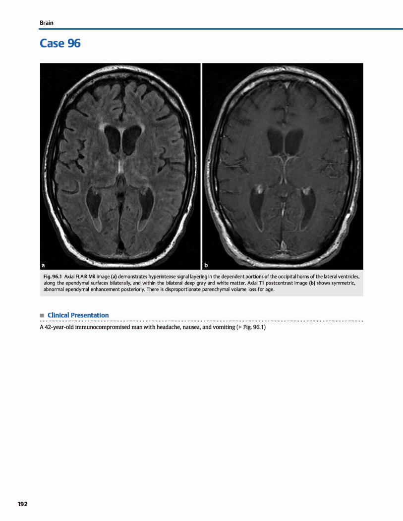
192
Brain
Case 96
Fig. 96.1 Axial FLAIR MR image (a) demonstrates hyperintense signal layering in the dependent portions of the occipital horns of the lateral ventricles, along the ependymal surfaces bilaterally, and within the bilateral deep gray and white matter. Axial Tl postcontrast image (b) shows symmetric, abnormal ependymal enhancement posteriorly. There is disproportionate parenchymal volume loss for age.
• Clinical Presentation
A 42-year-old immunocompromised man with headache, nausea, and vomiting (� Fig. 96.1)
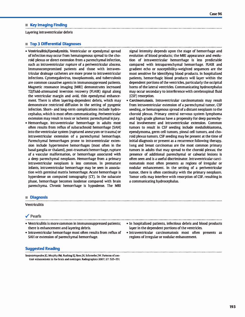
• Key lmaging Finding
Layering intraventricular debris
• Top 3 Differential Diagnoses
• Ventriculitis/Ependymitis. Ventricular or ependymal spread
of infection may occur from hematogenous spread to the cho
roid plexus or direct extension from a parenchymal infection,
such as intraventricular rupture of a periventricular abscess.
Immunocompromised patients and those with intraven
tricular drainage catheters are more prone to intraventricular infections. Cytomegalovirus, toxoplasmosis, and tuberculosis
are common causative agents in immunosuppressed patients.
Magnetic resonance imaging (MRI) demonstrates increased T2/fluid-attenuated inversion recovery (FLAIR) signal along
the ventricular margin and avid, thin ependymal enhance
ment. There is often layering-dependent debris, which may
demonstrate restricted diffusion in the setting of pyogenic
infection. Short- and long-term complications include hydro
cephalus, which is most often communicating. Periventricular extension may result in toxic or ischemic parenchymal injury.
• Hemorrhage. Intraventricular hemorrhage in adults most often results from reflux of subarachnoid hemorrhage (SAH)
into the ventricular system (ruptured aneurysm or trauma) or intraventricular extension of a parenchymal hemorrhage.
Parenchymal hemorrhages prone to intraventricular exten
sion indude hypertensive hemorrhages (most often in the
basal ganglia or thalami), post-traumatic hemorrhage, rupture
of a vascular malformation, or hemorrhage associated with
a deep parenchymal neoplasm. Hemorrhage from a primary
intraventricular neoplasm is Jess common. ln premature
infants, intraventricular hemorrhage may be seen in associa
tion with germinal matrix hemorrhage. Acute hemorrhage is
hyperdense on computed tomography (CT). In the subacute
phase, hemorrhage becomes isodense compared with brain parenchyma. Chronic hemorrhage is hypodense. The MRI
• Diagnosis
Ventriculitis
� Pearls
• Ventriculitis is more common in immunosuppressed patients;
there is enhancement and layering debris.
• Intraventricular hemorrhage most often results from reflux of SAH or extension of parenchymal hemorrhage.
Suggested Reading
Smimiotopoulos JG, Murphy FM, Rushing EJ, Rees JH, Schroeder JW. Pa�ms of con
trast enhanœment in the brain and meninges. R.adiographics 2007; 27: 525-551
Case 96
signal intensity depends upon the stage of hemorrhage and
evolution of blood products; the MRI appearance and evolu
tion of intraventricular hemorrhage is less predictable
compared with intraparenchymal hemorrhage. FLAIR and
gradient echo or susceptibility-weighted sequences are the
most sensitive for identifying blood products. In hospitalized
patients, hemorrhagic blood products will layer within the
dependent portions of the ventrides, particularly the occipital
homs of the lateral ventrides. Communicating hydrocephalus may occur secondary to interference with cerebrospinal fluid
(CSF) resorption.
• carcinomatosis. Intraventricular carcinomatosis may result
from intraventricular extension of a parenchymal tumor, CSF seeding, or hematogenous spread of a distant neoplasm to the
choroid plexus. Primary central nervous system lymphoma
and high-grade gliomas have a propensity for deep parenchy
mal involvement and intraventricular extension. Common
tumors to result in CSF seeding indude medulloblastoma,
ependymoma, germ cell tumors, pineal cell tumors, and cho
roid plexus tumors. CSF seeding may be present at the time of
initial diagnosis or present as a recurrence following therapy.
Lung and breast carcinomas are the most common primary
tumors in adults that may spread to the choroid plexus; the
presence of additional parenchymal or calvarial lesions is
often seen and is a useful discriminator. lntraventricular carci
nomatosis most often presents as regions of irregular or
nodular enhancement. In the setting of a periventricular
tumor, there is often continuity with the primary neoplasm
Tumor cells may interfere with resorption of CSF, resulting in
a communicating hydroœphalus.
• In hospitalized patients, infectious debris and blood products
layer in the dependent portions of the ventricles.
• Intraventricular carcinomatosis most often presents as regions of irregular or nodular enhancement.
193
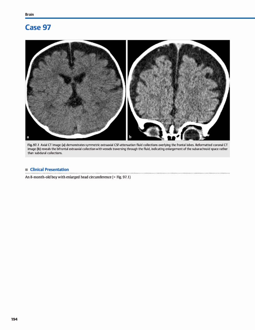
194
Brain
Case 97
Fig. 97 .1 Axial CT image (a) demonstrates symmetric extraaxial CSF-attenuation fluid collections overlying the frontal lobes. Reformatted coronal CT image (b) reveals the bifrontal extraaxial collection with vessels traversing through the fluid, indicating enlargement of the subarachnoid space rather than subdural collections.
• Clinical Presentation
An 8-month-old boy with enlarged head circumference (� Fig. 97.1)
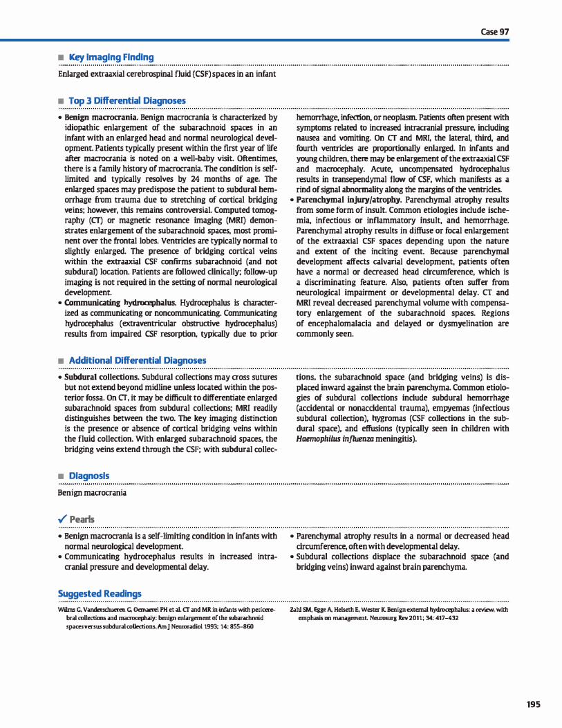
• Key lmaging Finding
Enlarged extraaxial cerebrospinal fluid {CSF) spaœs in an infant
• Top 3 Differential Diagnoses
• Benign macrocrania. Benign macrocrania is characterized by idiopathie enlargement of the subarachnoid spaœs in an
infant with an eruarged head and normal neurological development. Patients typically present within the first year of life
after macrocrania is noted on a well-baby visit. Oftentimes, there is a family history of macrocrania. The condition is selflimited and typically resolves by 24 months of age. The
eruarged spaœs may predispose the patient to subdural hemorrhage from trauma due to stretching of cortical bridging
veins; however, this remains controversial. Computed tomography (CT) or magnetic resonance imaging (MRI) demon
strates eruargement of the subarachnoid spaœs, most prominent over the frontal lobes. Ventrides are typically normal ta slightly eruarged. The presenœ of bridging cortical veins
within the extraaxial CSF confirms subarachnoid (and not subdural) location. Patients are followed clinically; follow-up
imaging is not required in the setting of normal neurological development.
• Communicaûng hydroœphalus. Hydroœphalus is character
ized as communicating or noncommunicating. Communicating
hydroœphalus ( extraventricular obstructive hydroœphalus) results from impaired CSF resorption, typically due to prior
• Additional Differential Diagnoses
• Subdural collections. Subdural collections may cross sutures but not extend beyond midline unless located within the pos
terior fessa. On cr, it may be difficult to differentiate enlarged subarachnoid spaces from subdural collections; MRI readily distinguishes between the two. The key imaging distinction is the presence or absence of cortical bridging veins within the fluid collection. With enlarged subarachnoid spaces, the
bridging veins extend through the CSF; with subdural collec-
• Diagnosis
Benign macrocrania
� Pearts
• Benign macrocrania is a self-limiting condition in infants with
normal neurological development. • Communicating hydrocephalus results in increased intra
cranial pressure and developmental delay.
Suggested Readings
Case 97
hemorrhage, infection, or neoplasm. Patients often present with symptoms related to increased intracranial presrure, induding nausea and vomiting. On CT and MRI, the lateral, third, and fourth ventrides are proportionally enlarged. In infants and
young children, there may be eruargement of the extraaxial CSF
and macroœphaly. Acute, uncompensated hydroœphalus results in transependymal flow of CSF, which manifests as a rind of signal abnormality along the margins of the ventrides.
• Parenchymal injury/atrophy. Parenchymal atrophy results
from some form of insult. Common etiologies indude ischemia, infectious or inflammatory insult, and hemorrhage. Parenchymal atrophy results in diffuse or focal enlargement
of the extraaxial CSF spaces depending upon the nature
and extent of the inciting event. Because parenchymal development affects calvarial development, patients often have a normal or decreased head circumference, which is
a discriminating feature. Also, patients often suffer from neurological impairment or developmental delay. CT and MRI reveal decreased parenchymal volume with compensatory enlargement of the subarachnoid spaœs. Regions
of encephalomalacia and delayed or dysmyelination are commonly seen.
tians, the subarachnoid spaœ (and bridging veins) is displaced inward against the brain parenchyma. Common etiologies of subdural collections indude subdural hemorrhage (accidentai or nonacddental trauma), empyemas {infectious subdural collection), hygromas (CSF collections in the subdural spaœ), and effusions (typically seen in children with Haemophilus influenza meningitis).
• Parenchymal atrophy results in a normal or decreased head
drcumference, often with developmental delay. • Subdural collections displace the subarachnoid space {and
bridging veins) inward against brain parenchyma.
Wilms G, Vanderschueren G, Demaerel PH et al. cr and MR in infants with periœre- Zahl SM. Egge A. Helseth E, Wesœr K. Benign extemal hydroœphalus: a review, with brai collectiDllS and macrocephaly: benign enlargement of the subarachrnJid emphasis on management. Neurosurg Rev 2011; 34; 417-432
spaces vers\15 subdural collections. Arn j Neuroradiol 1993; 14: 855-860
195
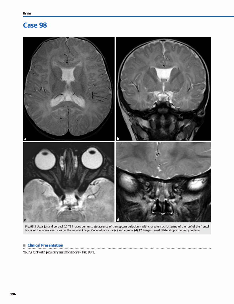
196
Brain
Case 98
Fig. 98.1 Axial (a) and coronal (b) T2 images demonstrate absence of the septum pellucidum with characteristic flattening of the roof of the frontal horns of the lateral ventricles on the coronal image. Coned-down axial (c) and coronal (d) T2 images reveal bilateral optic nerve hypoplasia.
• Clinical Presentation
Young girl with pituitary insufficiency (� Fig. 98.1 )
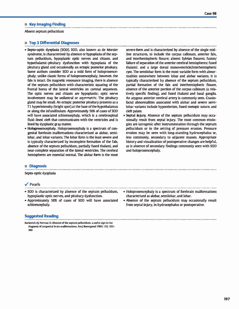
• Key lmaging Finding
Absent septum pellucidum
• Top 3 Differential Diagnoses
• Septo-optic dysplasia {SOD). SOD, also knawn as de Morsier
syndrome, is characterized by absence or hypoplasia of the sep
tum pelluddum, hypoplastic optic nerves and chiasm, and hypothalamic-pituitary dysfunction with hypoplasia of the pituitary gland and occasionally an ectopie posterior pituitary. Sorne authors consider SOD as a mild fonn of holoprosencephaly; unlike dassic forms of holoprosenœphaly, however, the
faix is intact. On magnetic resonance imaging, there is absence
of the septum pellucidum with characteristic squaring of the frontal homs of the lateral ventricles on coronal sequences. The optic nerves and chiasm are hypoplastic; optic nerve involvement may be unilateral or asymmetric. The pituitary gland may be small. An ectopie posterior pituitary presents as a Tl hyperintensity (bright spot) at the base of the hypothalamus
or along the infundibulum. Approximately 50% of cases of SOD will have associated schizenœphaly, which is a cerebrospinal
fluid-lined deft that communicates with the ventrides and is lined by dysplastic gray matter.
• Holoprosenœphaly. Holoprosenœphaly is a spectrum of con
genital forebrain malfonnations characterized as alobar, semi
lobar, and lobar variants. The lobar fonn is the least severe and is typically characterized by incomplete fonnation of the faix,
absence of the septum pelluddum, partially fused thalami, and near-complete separation of the lateral ventrides. The cerebral hemispheres are essential normal The alobar fonn is the most
• Diagnosis
Septo-optic dysplasia
� Pearts
• SOD is characterized by absence of the septum pelluddum,
hypoplastic optic nerves, and pituitary dysfunction.
• Approximately 50% of cases of SOD will have associated schizenœphaly.
Suggested Reading
Barlrovich AJ, Nonnan D. Absenœ of the septum pellucidum: a useful sign in the
diagnosis of congenital brain malfonnations. AmJ Roentgenol 1989; 152: 353-
360
Case 98
severe fonn and is characterized by absence of the single mid
line structures, to indude the corpus callosum, anterior faix, and interhemispheric fissure; absent Sylvian fissures; fusion/ failure of separation of the anterior cerebral hemispheres; fused
thalami; and a large dorsal monoventride/interhemispheric
cyst The semilobar fonn is the most variable form with abnormalities somewhere between lobar and alobar variants. It is typically characterized by absence of the septum pellucidum, partial formation of the faix and interhemispheric fissure, absence of the anterior portion of the corpus callosum (a rela
tively specific finding), and fused thalami and basal ganglia.
An azygous anterior cerebral artery is commonly seen. Craniofacial abnonnalities associated with alobar and severe semilobar variants include hypotelorism, fused metopic suture, and
deft palate. • Septal injury. Absence of the septum pellucidum may occa
sionally result Crom septal injury. The most common etiologies are iatrogenic after instrumentation through the septum pellucidum or in the setting of pressure erosion. Pressure erosion may be seen with long-standing hydrocephalus or,
less commonly, secondary to adjacent masses. Appropriate
history and visualization of postoperative changes are helpful, as is absence of secondary findings commonly seen with SOD
and holoprosencephaly.
• Holoprosencephaly is a spectrum of forebrain malfonnations characterized as alobar, semilobar, and lobar.
• Absence of the septum pellucidum may occasionally result
from septal injury, in hydroœphalus or postoperative.
197
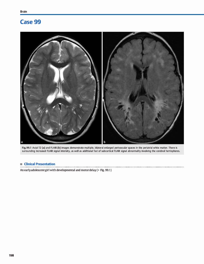
198
Brain
Case 99
Fig. 99.1 Axial T2 (a) and FLAIR (b) images demonstrate multiple, bilateral enlarged perivascular spaces in the periatrial white matter. There is surrounding increased FLAIR signal intensity, as well as additional foci of subcortical FLAIR signal abnormality involving the cerebral hemispheres.
• Clinical Presentation
An early adolescent girl with developmental and motor delay (� Fig. 99.1 )
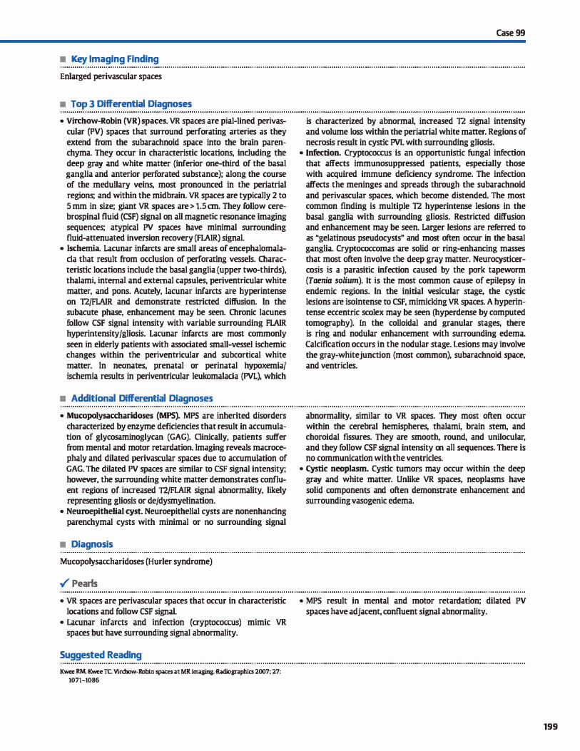
• Key lmaging Finding
Enlarged perivascular spaœs
• Top 3 Differential Diagnoses
• Virchow-Robin (VR) spaces. VR spaces are pial-lined perivascular (PV) spaœs that surround perforating arteries as they extend from the subarachnoid space into the brain parenchyma. They occur in characteristic locations, induding the deep gray and white matter (inferior one-third of the basal ganglia and anterior perforated substance); along the course of the medullary veins, most pronounced in the periatrial
regions; and within the midbrain. VR spaces are typically 2 to
5 mm in size; giant VR spaces are > 1.5 cm. They follow cerebrospinal fluid (CSF) signal on ail magnetic resonance imaging
sequences; atypical PV spaœs have minimal surrounding fluid-attenuated inversion recovery (FLAIR) signal.
• lschemia. Lacunar infarcts are smalt areas of encephalomala
cia that result from occlusion of perforating vessels. Charac
teristic locations include the basal ganglia (upper two-thirds), thalami, internai and extemal capsules, periventricular white matter, and pans. Acutely, lacunar infarcts are hyperintense on T2/FLAIR and demonstrate restricted diffusion. ln the subacute phase, enhancement may be seen. Chronic lacunes follow CSF signal intensity with variable surrounding FLAIR hyperintensity/gliosis. Lacunar infarcts are most commonly
seen in elderly patients with associated small-vessel ischemic changes within the periventricular and subcortical white matter. ln neonates, prenatal or perinatal hypoxemia/ ischemia results in periventricular leukomalacia (PVL), which
• Additional Differential Diagnoses
• Mucopolysaccharidoses (MPS). MPS are inherited disorders
characterized by enzyme deficiencies that result in accumulation of glycosaminoglycan (GAG). Oinically, patients suffer from mental and motor retardation. lmaging reveals macrocephaly and dilated perivascular spaœs due to accumulation of
GAG. The dilated PV spaœs are similar to CSF signal intensity; however, the surrounding white matter demonstrates confluent regions of increased T2/FLAIR signal abnormality, likely representing gliosis or de/dysmyelination.
• Neuroepithelial cyst. Neuroepithelial cysts are nonenhancing parenchymal cysts with minimal or no surrounding signal
• Diagnosis
Mucopolysaa:haridoses {Hurler syndrome)
� Pearls
• VR spaœs are perivascular spaœs that occur in characteristic
locations and follow CSF signal
• Lacunar infarcts and infection (cryptocoa:us) mimic VR spaces but have surrounding signal abnormality.
Suggested Reading
Kwee RM, Kwee TC. Virdww-Rcbin spaces at MR imaging. Radiographies 2007; 27: 1071-1086
Case 99
is characterized by abnormal, increased T2 signal intensity and volume Joss within the periatrial white matter. Regions of necrosis result in cystic PVL with surrounding gliosis.
• Infection. Cryptocoa:us is an opportunistic fungal infection that affects immunosuppressed patients, especially those with acquired immune deficiency syndrome. The infection affects the meninges and spreads through the subarachnoid
and perivascular spaœs, which become distended. The most common finding is multiple T2 hyperintense lesions in the basal ganglia with surrounding gliosis. Restricted diffusion and enhancement may be seen. Larger lesions are referred to as "gelatinous pseudocysts" and most often occur in the basal ganglia. Cryptococcomas are solid or ring-enhancing masses that most often involve the deep gray matter. Neurocysticer
cosis is a parasitic infection caused by the pork tapeworm (Taenia sollum). lt is the most common cause of epilepsy in endemic regions. ln the initial vesicular stage, the cystic lesions are isointense ta CSF, mimicking VR spaœs. A hyperintense eccentric scolex may be seen (hyperdense by computed tomography). In the colloidal and granular stages, there is ring and nodular enhanœment with surrounding edema.
Calcification occurs in the nodular stage. Lesions may involve
the gray-whitejunction (most common), subarachnoid spaœ, and ventricles.
abnormality, similar to VR spaœs. They most often occur within the cerebral hemispheres, thalami, brain stem, and choroidal fissures. They are smooth, round, and unilocular, and they follow CSF signal intensity on ail sequences. There is no communication with the ventricles.
• Cystic neoplasm. Cystic tumors may occur within the deep gray and white matter. Unlike VR spaœs, neoplasms have
solid components and often demonstrate enhancement and surrounding vasogenic edema.
• MPS result in mental and motor retardation; dilated PV
spaœs have adjacent, confluent signal abnormality.
199
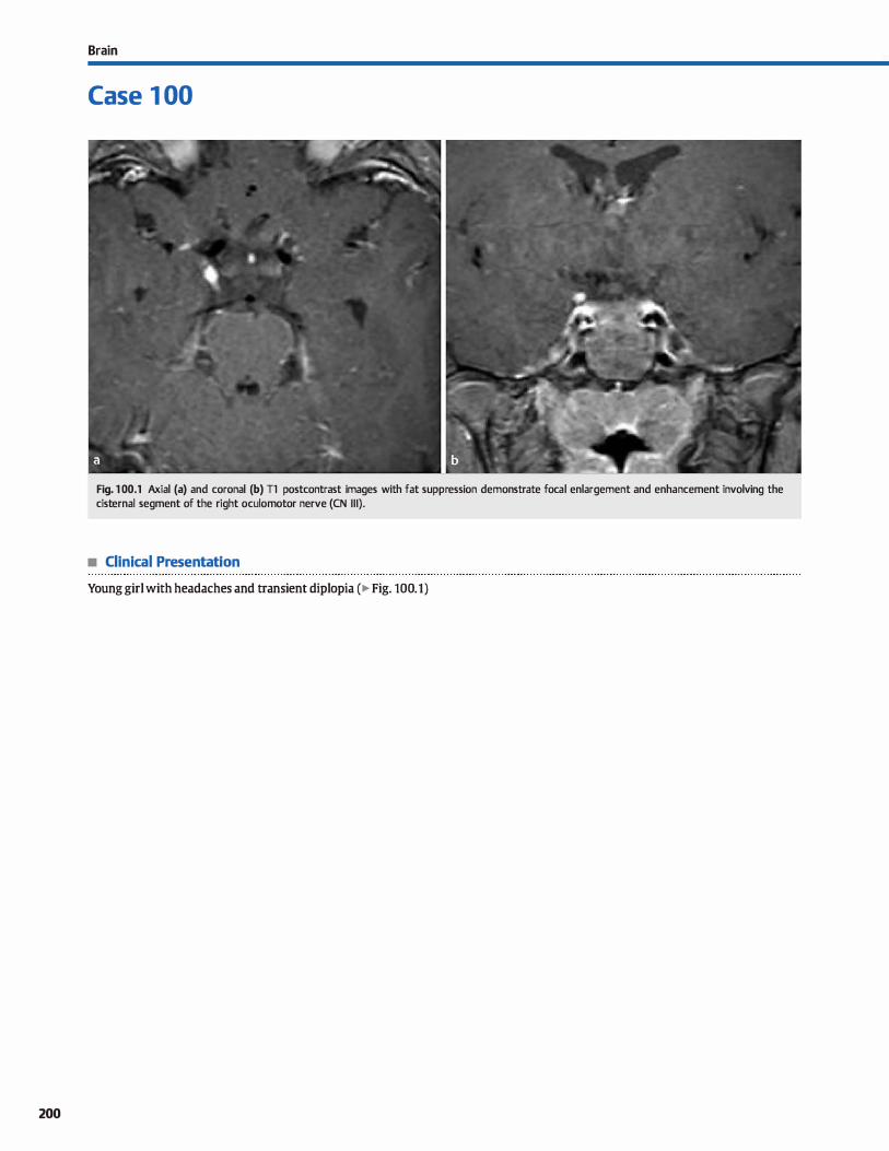
200
Brain
Case 1 00
Fig. 1 00.1 Axial (a) and coronal (b) Tl postcontrast images with fat suppression demonstrate focal enlargement and enhancement involving the cisternal segment of the right oculomotor nerve (CN 111).
• Clinical Presentation
Young girl with headaches and transient diplopia ( � Fig. 100.1)
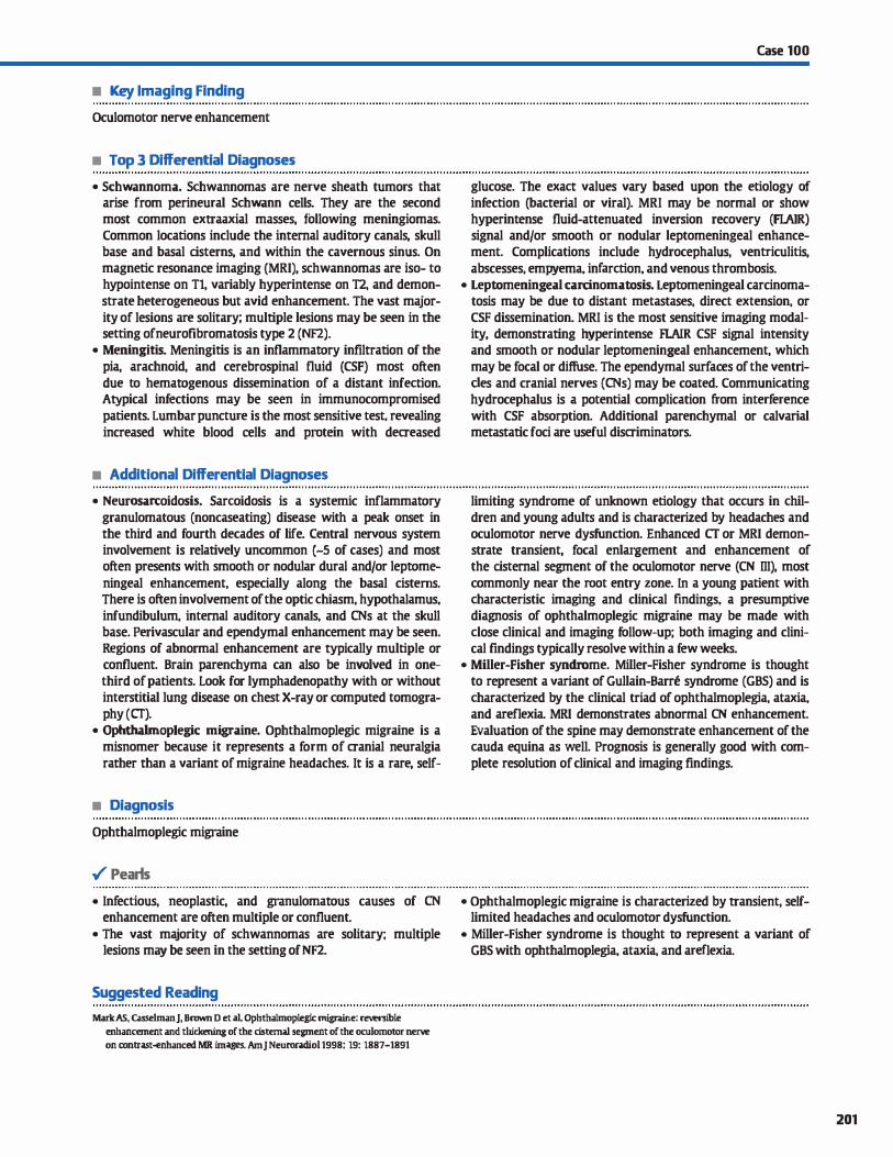
• Key lmaging Finding
Oculomotor nerve enhancement
• Top 3 Differential Diagnoses
• Schwannoma. Schwannomas are nerve sheath tumors that arise from perineural Schwann œlls. They are the second most common extraaxial masses, following meningiomas. Common locations include the internai auditory canais, skull base and basal dsterns, and within the cavernous sinus. On magnetic resonanœ imaging (MRI), schwannomas are iso- to hypointense on Tl, variably hyperintense on T2, and demonstrate heterogeneous but avid enhancement The vast majority of lesions are solitary; multiple lesions may be seen in the setting ofneurofibromatosis type 2 (NF2).
• Meningitis. Meningitis is an inflammatory infiltration of the pia, arachnoid, and cerebrospinal fluid (CSF) most often due to hematogenous dissemination of a distant infection. Atypical infections may be seen in immunocompromised patients. Lumbar puncture is the most sensitive test. revealing increased white blood œlls and protein with decreased
• Additional Differential Diagnoses
• Neurosan:oidosis. Sarcoidosis is a systemic inflammatory granulomatous (noncaseating) disease with a peak onset in the third and fourth decades of life. Central nervous system involvement is relatively uncommon (-5 of cases) and most often presents with smooth or nodular durai and/or leptome
ningeal enhancement, especially along the basal cistems. There is often involvement of the optic chiasm, hypothalamus, infundibulum, internai auditory canais, and CNs at the skull base. Perivascular and ependymal enhancement may be seen. Regions of abnormal enhancement are typically multiple or confluent. Brain parenchyma can also be involved in onethird of patients. Look for lymphadenopathy with or without interstitial Jung disease on chest X-ray or computed tomography ( CT).
• Ophthalmoplegic migraine. Ophthalmoplegic migraine is a misnomer because it represents a form of cranial neuralgia rather than a variant of migraine headaches. It is a rare, self-
• Diagnosis
Ophthalmoplegic migraine
� Pearls
• Infectious, neoplastic, and granulomatous causes of CN enhancement are often multiple or confluent.
• The vast majority of schwannomas are solitary; multiple lesions may be seen in the setting of NF2.
Suggested Reading
Mark AS, Casselman J, Brown Il et al. Ophthalmoplegic migraine; revenible
enhanœment and tlùckening of the cisternal segment of the oculornotor nerve
on contrast-enhanced MR images. AmJ Neuroradiol 1998; 19: 1887-1891
Case 100
glucose. The exact values vary based upon the etiology of
infection (bacterial or viral). MRI may be normal or show hyperintense fluid-attenuated inversion recovery (FLAIR) signal and/or smooth or nodular leptomeningeal enhancement. Complications include hydrocephalus, ventriculitis,
abscesses, empyema, infarction, and venous thrombosis. • Leptomeningeal can:inomatosis. Leptomeningeal carcinoma
tosis may be due to distant metastases, direct extension, or CSF dissemination. MRI is the most sensitive imaging modality, demonstrating hyperintense FLAIR CSF signal intensity and smooth or nodular leptomeningeal enhancement, which may be focal or diffuse. The ependymal surfaces of the ventrides and cranial nerves {CNs) may be coated. Communicating hydrocephalus is a potential complication from interference with CSF absorption. Additional parenchymal or calvarial metastatic foci are useful discriminators.
limiting syndrome of unknown etiology that occurs in children and young adults and is characterized by headaches and oculomotor nerve dysfunction. Enhanœd CT or MRI demonstrate transient, focal enlargement and enhanœment of the dsternal segment of the oculomotor nerve (CN III), most commonly near the root entry zone. In a young patient with characteristic imaging and clinical findings, a presumptive diagnosis of ophthalmoplegic migraine may be made with close dinical and imaging follow-up; both imaging and clinicat findings typically resolve within a few weeks.
• Miller-Fisher syndrome. Miller-Fisher syndrome is thought to represent a variant of Gullain-Barré syndrome (GBS) and is characterized by the dinical triad of ophthalmoplegia, ataxia, and areflexia. MRI demonstrates abnormal CN enhancement Evaluation of the spine may demonstrate enhancement of the cauda equina as well. Prognosis is generally good with complete resolution of clinical and imaging findings.
• Ophthalmoplegic migraine is characterized by transient, selflimited headaches and oculomotor dysfunction.
• Miller-Fisher syndrome is thought to represent a variant of GBS with ophthalmoplegia, ataxia, and areflexia.
201
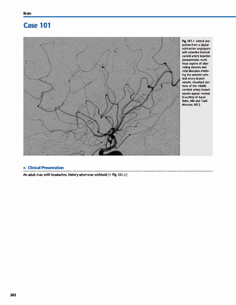
Brain
Case 1 01
• Clinical Presentation
An adult man with headaches; history otherwise withheld c� Fig. 101.1)
202
Fig. 101.1 Lateral projection from a digital subtractlon anglogram wlth selective Internai carotld artery Injection demon5trates mult:ifocal reglons of alternat:lng stenosls and mtld dllataUon lnYOIYlng the anterior cerebral utery branch vessels; vlsuallzed porlfons of the mlddle cerebral artery branch vessels ;ippeu nonn;il. (Courtesy of Aaron Betts, MD and Todd Abl\lzzo, MD.)
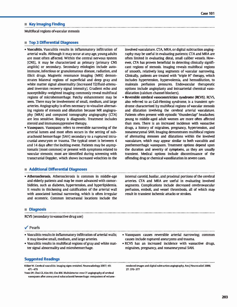
• Key lmaging Finding
Multifocal regions ofvascular stenosis
• Top 3 Differential Diagnoses
• Vasculitis. Vasculitis results in inflammatory infiltration of arterial watts. Although it may occur at any age, young adults are most often affected. Within the central nervous SYStem (CNS), it may be characterized as primary (primary CNS angiitis) or secondary. Secondary etiologies include autoimmune, infectious or granulomatous disease, radiation, and illicit drugs. Magnetic resonanœ imaging {MRI) demon
strates bilateral regions of superficial and deep gray and white matter signal abnormality {increased T2/fluid-attenuated inversion recovery signal intensity). Gradient echo and susceptibility-weighted imaging commonly reveal multifocal regions of microhemorrhage. Patchy enhanœment may be seen. There may be involvement of small, medium, and large arteries. Angiography is often necessary to visualize alternating regions of stenosis and dilatation because MR angiography {MRA) and computed tomography angiography { CTA) are Jess sensitive. Biopsy is diagnostic. Treatment includes steroid and immunosuppressive therapy.
• Vasospasm. Vasospasm refers to reversible narrowing of the arterial lumen and most often occurs in the setting of sub
arachnoid hemorrhage {SAH) secondary to a ruptured intracranial aneurysm or trauma. The typical onset is between 4
and 14 days after the inciting event. Patients may be asymptomatic {most common) or present with symptoms related to vascular stenosis; most are identified during screening with transcranial Doppler, which shows increased velocities in the
• Additional Differential Diagnoses
• Atherosderosis. Atherosclerosis is common in middle-age and elderly patients and may be more advanœd with comorbidities, such as diabetes, hypertension, and hyperlipidemia. lt results in thidœning and calcification of the arterial wall with associated luminal narrowing, which is often irregular and ecœntric. Common intracranial locations include the
• Diagnosis
RCVS {secondary to vasoactive drug use)
� Pearls
• Vasculitis results in inflammatory infiltration of arterial walls; it may involve small, medium, and large arteries.
• Vasculitis results in multifocal regions of gray and white matter signal abnormality and microhemorrhage.
Suggested Readings
Külœr W. cerebral vasculitis; imaging signs revisited, Neuroradiology 2007; 49;
471-479
Yoon DY, Chai CS, Kim KH, Cho BM. Multidet:ector-row CT angiography of cerebral vasospasm after aneurysmal subarachnoid hemorrhage: comparison of volume-
Case 101
involved vasculature. CTA, MRA, or digital subtraction angiography may be useful in evaluating patients; CTA and MRA are often limited in evaluating distal, small caliber vessels. However, CTA has proven beneficial in detecting clinically signifi
cant regions of stenosis. Imaging reveals multifocal regions of smooth, relatively long segments of vascular narrowing. Oinically, patients are treated with utriple H" therapy, which includes hypertension, hypervolemia, and hemodilution, ta maintain perfusion pressures. Endovascular therapeutic options include angioplasty and intraarterial chemical vasodilatation {calcium channel blockers).
• Reversible œrebral vasoconstriction syndrome {RCVS). RCVS,
also referred to as Call-Fleming SYDdrome, is a transient SYD
drome characterized by multifocal regions of vascular stenosis and dilatation involving the œrebral arterial vasculature. Patients often present with episodic uthunderclap" headaches; young to middle-aged adult women are more often affected than men. There is an increased incidence with vasoactive drugs, a history of migraines, pregnancy, hypertension, and nonaneurysmal SAH. lmaging demonstrates multifocal regions of altemating stenoses and dilatations within the involved
vasculature, which may appear similar to bath vasculitis and posthemorrhagic vasospasm. Treatment options depend upon the duration and severity of SYffiptoffiS, as they are usually transient. Medical options indude discontinuanœ of the offending drug or chemical vasodilatation in severe cases.
internai carotid, basilar, and proximal portions of the cerebral
arteries. CTA and MRA are useful in evaluating involved segments. Complications include decreased œrebrovascular perfusion, emboli, and vessel thrombosis, ail of which may result in transient ischemic attacks or strokes.
• Vasospasm causes reversible arterial narrowing; common causes include ruptured aneurysms and trauma.
• RCVS bas an increased incidence with vasoactive drugs,
migraines, pregnancy, and nonaneurysmal SAH.
rendered images and digital subtraction angiography. AmJ Neuroradiol 2006;
27: 37C>-377
203
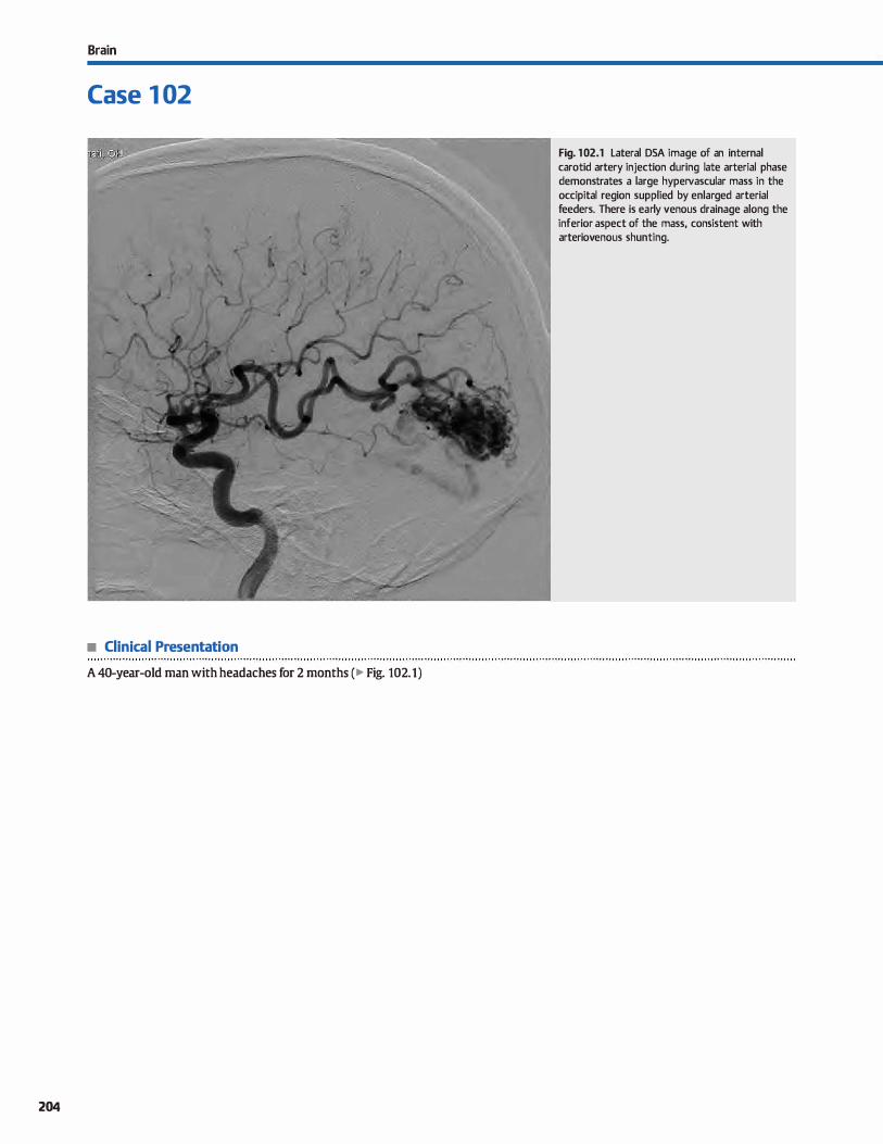
Brain
Case 1 02
• Clinical Presentation
A 40-year-old man with headaches for 2 months (� Fig. 102.1)
204
Fig. 102.1 Lateral DSA image of an internai carotid artery injection during late arterial phase demonstrates a large hypervascular mass in the occipital region supplied by enlarged arterial feeders. There is early venous drainage along the inferior aspect of the mass, consistent with arteriovenous shunting.
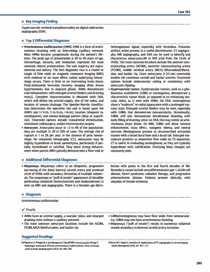
• Key lmaging Finding
Hypervascular cerebral mass/abnonnality on digital subtraction angiography (DSA)
• Top 3 Differential Diagnoses
• Arteriovenous malfonnation {AVM). AVM is a form of arteri
ovenous shunting with no intervening capillary network.
Most AVMs become symptomatic during the patient's life
time. The peak age of presentation is 20 to 40 years of age.
Hemorrhage, seizures, and headaches represent the most
common dinical presentations. The vast majority are supratentorial and solitary. The best diagnostic due is a cluster or
tangle of flow voids on magnetic resonance imaging (MRI)
with minimal or no mass effect, unless underlying hemor
rhage occurs. There is little or no intervening brain tissue.
Fluid-attenuated inversion recovery imaging often shows
hyperintensity due to adjacent gliosis. AVMs demonstrate
avid enhancement with enlarged arterial feeders and draining
vein(s). Complete characterization is obtained with DSA, which will define the arterial supply, size of the nidus, and
location of venous drainage. The Spetzler-Martin classifica
tion determines the operative risk and is based upon the
AVM's size (< 3 cm, 3 to 6 cm, >Sem), location {eloquent vs.
noneloquent), and venous drainage pattern (deep or superfi
dal). Treatment options include transarterial embolization,
stereotaxic radiosurgery, and/or microvascular surgery.
• Aneurysm Aneurysms occur in -2 to 4% of the population; they are multiple in 20 to 25% of cases. The average risk of
rupture is 1 to 2% per year, in the absence of prier hemor
rhage. On computed tomography {CT), aneurysms may be
slightly hyperdense to brain parenchyma, particularly if par
tially thrombosed or calcified. They show strong enhance
ment when patent. MRI typically demonstrates a flow void or
• Additional Differential Diagnoses
• Moyamoya. Moyamoya refers to an idiopathie, progressive
narrowing of the distal internai carotid artery and proximal circle of Willis with secondary formation of multiple collater
als. The moyamoya or "puff of smoke" appearance of doudlike perforating collaterals (lenticulostriate and thalamostriate) is
seen on MRI and angiography. There is a bimodal age distri-
• Diagnosis
Arteriovenous malformation
y' Pearts
• AVMs have an arterial supply, a vascular nidus, and enlarged
draining veins without a capillary network. • The most common aneurysm locations include the ACOM,
PCOM, MCA bi/trifurcation, and basilar tip.
Suggested Readings
Geibpramt S, Pongpedt S. Jiarakcngmun P, Shroff MM, Armstrong DC, Kring! T. Radiologie assessment of brain arteriovenous mallbnnations; what clinicians
need ta know. Radiographies 2010; 30: 483-501
Case 102
heterogeneous signal, especially with thrombus. Pulsation
artifact, when present, is a useful discriminator. CT angiogra
phy, MR angiography, and DSA can be used to identify and
characterize aneurysms.;90 to 95% arise from the Cirde of
Willis. The most common locations include the anterior com
municating artery (ACOM), posterior communicating artery
(PCOM), middle cerebral artery (MCA) bifurcation/trifurca
tion, and basilar tip. Giant aneurysms (> 2.5 cm) commonly
involve the cavernous carotid and basilar arteries. Treatment
options include endovascular coiling or craniotomy with
aneurysm clipping.
• Hypervascular tumor. Hypervascular tumors, such as a glio
blastoma multiforme {GBM) or meningioma, demonstrate a characteristic tumor blush, as opposed to an enhancing vas
cular nidus, as is seen with AVMs. On DSA, meningiomas
show a "sunburst" or radial appearance with a prolonged vas
cular stain. Enlarged arterial feeders may be seen, especially
with GBMs that demonstrate neovascularity. Occasionally,
GBMs will also demonstrate intralesional shunting with
early filling of draining veins; on DSA, this may mimic an arteriovenous shunt lesion. On MRI, GBMs will show tumoral
enhancement, mass effect, vasogenic edema, and central
necrosis. Meningiomas present as circumscribed extraaxial
masses with a broad durai base and a durai tait. Enlarged vas
culature presents as serpentine flow voids on T2 sequences.
CT is useful in evaluating meningiomas, as they are typically
hyperdense with calcification. Overlying bony changes are commonly seen.
bution with peaks in the first and fourth decades of life.
Secondary causes include neurofibromatosis type 1, sickle cell
disease, Down syndrome, radiation therapy, and progressive
atherosclerotic disease. Patients present clinically with
sequelae of chronic ischemia.
• GBMs/meningiomas may have flow voids from neovascular
ity; GBMs may also have arteriovenous shunting.
• Moyamoya ("puff of smoke") results in numerous collateral
vessels secondary to internai carotid artery ocdusion.
O'Brien Wf, Vagal A. Cornelius R. Applications of CT angiography in neuroimaging. Sernin Roentgenol 2010; 45; 107-115
205
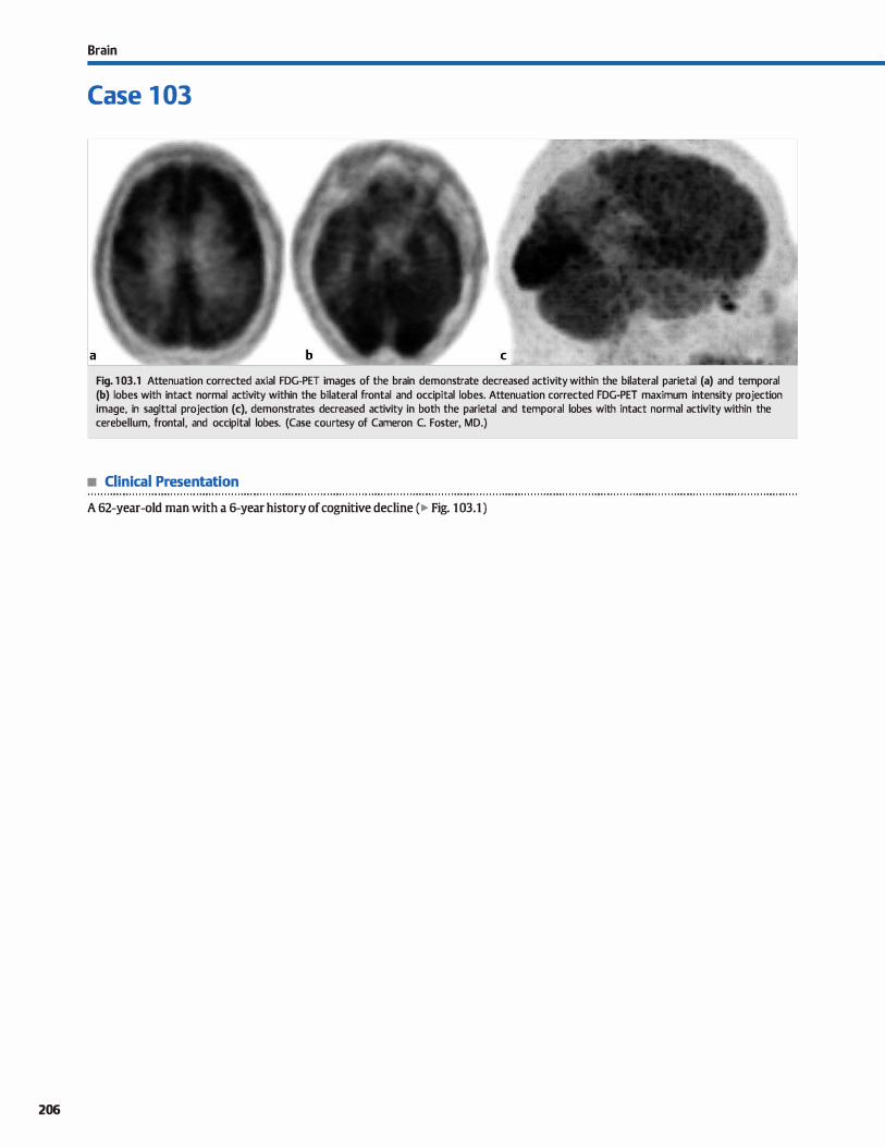
206
Brain
Case 1 03
Fig. 1 03.1 Attenuation corrected axial FDG-PET images of the brain demonstrate decreased activity within the bilateral parietal (a) and temporal (b) lobes with intact normal activity within the bilateral frontal and occipital lobes. Attenuation corrected FDG-PET maximum intensity projection image, in sagittal projection (c), demonstrates decreased activity in both the parietal and temporal lobes with intact normal activity within the cerebellum, frontal, and occipital lobes. (Case courtesy of Cameron C. Foster, MD.)
• Clinical Presentation
A 62-year-old man with a 6-year history of cognitive decline (� Fig. 103.1 )
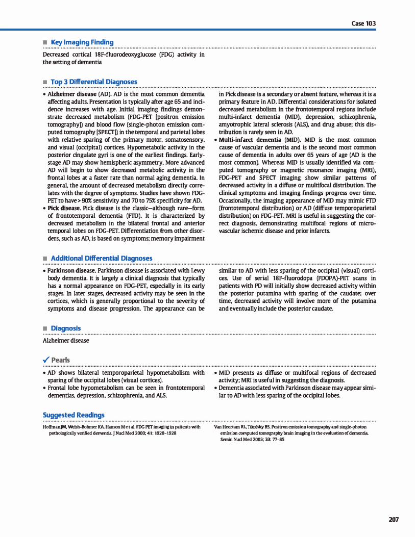
• Key lmaging Finding
Decreased cortical 18F-fluorodeoxyglucose (FDG) activity in
the setting of dementia
• Top 3 Differential Diagnoses
• Alzheimer disease (AD). AD is the most common dementia
affecting adults. Presentation is typically after age 65 and inci
dence increases with age. Initial imaging findings demon
strate decreased metabolism (FDG-PET [positron emission
tomography]) and blood flow (single-photon emission com
puted tomography [SPECT]) in the temporal and parietal lobes
with relative sparing of the primary motor, somatosensory,
and visual (occipital) cortices. Hypometabolic activity in the
posterior dngulate gyri is one of the earliest findings. Early
stage AD may show hemispheric asymmetry. More advanced
AD will begin to show decreased metabolic activity in the
frontal lobes at a faster rate than normal aging dementia. In
general, the amount of decreased metabolism directly corre
lates with the degree of symptoms. Studies have shown FDG
PET to have > 90% sensitivity and 70 to 75% specifidty for AD. • Pick disease. Pidc disease is the dassic-although rare-form
of frontotemporal dementia (FID). It is characterized by
decreased metabolism in the bilateral frontal and anterior temporal lobes on FDG-PET. Differentiation from other disor
ders, such as AD, is based on symptoms; memory impairment
• Additional Differential Diagnoses
• Parkinson disease. Parkinson disease is associated with Lewy
body dementia. lt is largely a dinical diagnosis that typically
has a normal appearance on FDG-PET, espedally in its early
stages. In later stages, decreased activity may be seen in the
cortices, which is generally proportional to the severity of
symptoms and disease progression. The appearance can be
• Diagnosis
Alzheimer disease
� Pearls
• AD shows bilateral temporoparietal hypometabolism with sparing of the occipital lobes (visual cortiœs).
• Frontal lobe hypometabolism can be seen in frontotemporal
dementias, depression, schizophrenia, and ALS.
Suggested Readings
HoflinanJM, Welsh-Bohmer KA, Hanson Met al. FDG PET imaging in patients with
patholagically verified dement:ia, J Nucl Med 2000; 41: 1920-1928
Case 103
in Pick disease is a secondary or absent feature, whereas it is a
primary feature in AD. Differential considerations for isolated
decreased metabolism in the frontotemporal regions indude
multi-infarct dementia (MID), depression, schizophrenia,
amyotrophie lateral sclerosis (ALS), and drug abuse; this dis
tribution is rarely seen in AD.
• Multi-infarct dementia (MID). MID is the most common
cause of vascular dementia and is the second most common
cause of dementia in adults over 65 years of age (AD is the most common). Whereas MID is usually identified via com
puted tomography or magnetic resonance imaging (MRI),
FDG-PET and SPECT imaging show similar patterns of decreased activity in a diffuse or multifocal distribution. The
dinical symptoms and imaging findings progress over time.
Occasionally, the imaging appearance of MID may mimic FfD (frontotemporal distribution) or AD (diffuse temporoparietal
distribution) on FDG-PET. MRI is useful in suggesting the cor
rect diagnosis, demonstrating multifocal regions of micro
vascular ischemic disease and prior infarcts.
similar to AD with Jess sparing of the occipital (visual) corti
œs. Use of serial 18F-fluorodopa (FDOPA)-PET scans in
patients with PD will initially show decreased activity within
the posterior putamina with sparing of the caudate; over time, decreased activity will involve more of the putamina
and eventually include the posterior caudate.
• MID presents as diffuse or multifocal regions of decreased activity; MRI is useful in suggesting the diagnosis.
• Dementia associated with Parkinson disease may appear simi
lar to AD with less sparing of the occipital lobes.
Van Heertwn RL. Tilo:lfsky RS. Positron emission tomography and single-photon emission computed tomography brain imaging in the evaluation of dement:ia.
Sernin NuclMed 2003; 33: 77-85
207
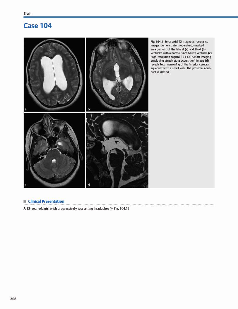
Brain
Case 1 04
• Clinical Presentation
A 13-year-old girl with progressively worsening headaches (� Fig. 104.1 )
208
Fig. 1 04.1 Serial axial T2 magnetic resonance images demonstrate moderate-to-marked enlargement of the lateral (a) and third (b) ventricles with a normal-sized fourth ventricle (c). High-resolution sagittal T2 FIESTA (fast imaging employing steady state acquisition) image (d) reveals focal narrowing of the inferior cerebral aqueduct with a small web. The proximal aqueduct is dilated.
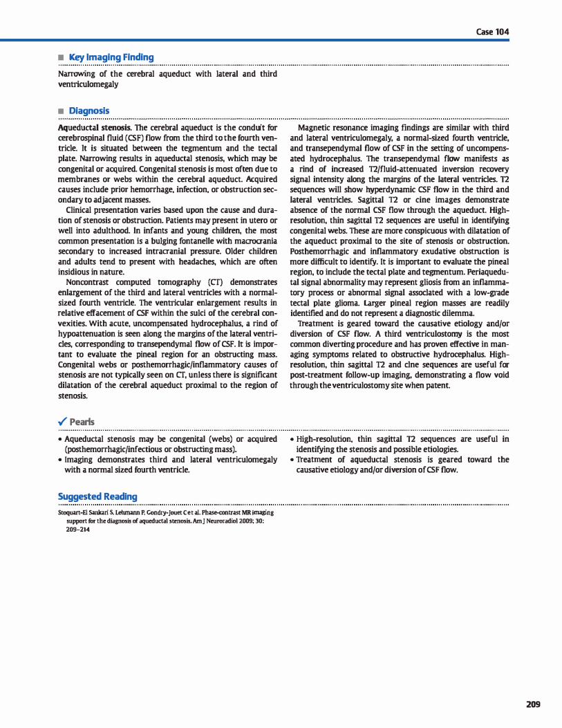
• Key lmaging Finding
Narrowing of the œrebral aqueduct with lateral and third venniculomegaly
• Diagnosis
Aqueductal stenosis. The cerebral aqueduct is the conduit for œrebrospinal fluid (CSF) flow from the third ta the fourth ventricle. It is situated between the tegmentum and the tectal plate. Narrowing results in aqueductal stenosis, which may be
congenital or acquired. Congenital stenosis is most often due ta membranes or webs within the cerebral aqueduct. Acquired causes include prier hemorrhage, infection, or obstruction secondary to adjacent masses.
Clinicat presentation varies based upon the cause and duration of stenosis or obstruction. Patients may present in utero or well into adulthood. ln infants and young children, the most
common presentation is a bulging fontanelle with macrocrania secondary ta increased intracranial pressure. Older children
and adults tend ta present with headaches, which are often insidious in nature.
Noncontrast computed tomography {CT) demonstrates
enlargement of the third and lateral ventricles with a normalsized fourth vennide. The vennicular enlargement results in relative effacement of CSF within the sulci of the cerebral con
vexities. With acute, uncompensated hydrocephalus, a rind of hypoattenuation is seen along the margins of the Iateral vennides, corresponding to transependymal flow of CSF. lt is important to evaluate the pineal region for an obstructing mass. Congenital webs or posthemorrhagic/inflammatory causes of stenosis are not typically seen on CT, unless there is significant dilatation of the cerebral aqueduct proximal to the region of
stenosis.
� Pearts
• Aqueductal stenosis may be congenital (webs) or acquired (posthemorrhagic/infectious or obstructing mass).
• lmaging demonstrates third and lateral venniculomegaly with a normal sized fourth ventricle.
Suggested Reading
Stoquart-El Sankari S. Lehmann P, Condry-Jouet cet al. Phase-contrast MR imaging
support fur the diagnosis of aqueductal sœnosis. Am J Neuroradiol 2009; 30;
209-214
Case 104
Magnetic resonance imaging findings are similar with third and lateral ventriculomegaly, a normal-sized fourth vennide, and transependymal flow of CSF in the setting of uncompens
ated hydrocephalus. The transependymal flow manifests as
a rind of increased T2/fluid-attenuated inversion recovery signal intensity along the margins of the lateral ventrides. T2 sequenœs will show hyperdynamic CSF flow in the third and lateral vennides. Sagittal T2 or cine images demonstrate absence of the normal CSF flow through the aqueduct. Highresolution, thin sagittal T2 sequenœs are useful in identifying
congenital webs. These are more conspicuous with dilatation of the aqueduct proximal to the site of stenosis or obstruction. Posthemorrhagic and inflammatory exudative obstruction is more difficult ta identify. lt is important ta evaluate the pineal region, to indude the tectal plate and tegmentum. Periaquedutal signal abnormality may represent gliosis from an inflammatory process or abnormal signal assodated with a low-grade
tectal plate glioma. Larger pineal region masses are readily identified and do not represent a diagnostic dilemma.
Treatment is geared toward the causative etiology and/or diversion of CSF flow. A third ventriculostomy is the most common diverting procedure and has proven effective in managing symptoms related to obstructive hydroœphalus. Highresolution, thin sagittal T2 and dne sequences are useful for post-treatment follow-up imaging, demonstrating a flow void
through the ventriculostomy site when patent.
• High-resolution, thin sagittal T2 sequences are useful in identifying the stenosis and possible etiologies.
• Treatment of aqueductal stenosis is geared toward the causative etiology and/or diversion of CSF flow.
209
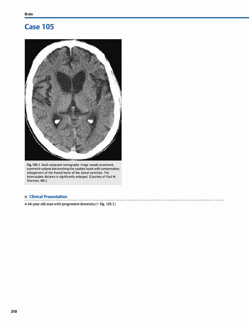
210
Brain
Case 1 05
Fig. 1 05.1 Axial computed tomography image reveals prominent, symmetric volume loss involving the caudate heads with compensatory enlargement of the frontal horns of the lateral ventricles. The intercaudate distance is significantly enlarged. (Courtesy of Paul M. Sherman, MD.)
• Clinical Presentation
A 44-year-old man with progressive dementia (� Fig. 105.1)
• Key lmaging Finding
Preferential caudate volume loss in an adult
• Diagnosis
Huntington disease. Huntington disease {HD) is an autosomal dominant neurodegenerative disorder that most often presents in early to mid-adulthood (fourth and fifth decades) with dementia, psychosis, and choreoathetosis; chorea is the dominant dinical feature. A juvenile variant is less common, presents prior to 20 years of age, and dinically manifests with rigidity as a dominant feature rather than chorea.
The most common imaging feature in the adult variant is
preferential atrophy of the caudate nuclei with associated dilatation of the frontal homs of the Iateral ventrides. Atrophie
changes result in an increased intercaudate distance, which refers to the distance between the medial aspects of the caudate heads. In normal individuals, the distance is typically 10 to 14mm, whereas in Huntington chorea it is>20 mm. There is
y' Pearts
• HD is an autosomal dominant neurodegenerative disorder that most often affects adults; chorea is the main dinical feature.
Suggested Readings
Ho VB, Clmang HS. Rovira MJ, Koo B.Juvenile Huntington disease: CT and MR fea
tures, AmJ Neuroradial 1995; 16: 1405-1412
Case 105
often diffuse parenchymal volume Joss; however, the caudate nuclei are disproportionately involved to a greater degree. Signal abnonnality of the deep gray matter structures is not a characteristic feature of the adult variant.
In the juvenile fonn of HD, symmetric hyperintense T2/fluid
attenuated inversion recovery signal is visualized in the basal ganglia, particularly the caudate nudei and putamina The
signal abnonnality is nonspecific and may be seen in additional neurodegenerative processes, metabolic diseases, and with ischemic, taxie, or infectious injury. Preferential caudate atrophy is a useful secondary finding to suggest HD.
HD is incurable and progressive. Treatment strategies are geared toward mitigating the dinical manifestations ( chorea,
rigidity, psychosis, etc.).
• The most common imaging feature in the adult variant is preferential atrophy of the caudate nuclei.
• Juvenile HD presents prior to 20 years of age with rigidity and hyperintense T2 signal in the basal ganglia.
StaberT, Wwisow W, Schimrigk K. Bicaudate diameter-the most spedfic and simple CT parameter in the diagnasis afHuntingtnn's disease. NeW'llr.ldialagy 1984; 26;
25-28
211
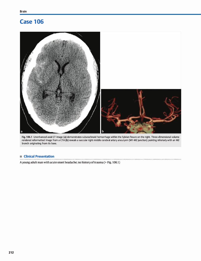
212
Brain
Case 1 06
Fig. 1 06.1 Unenhanced axial CT image (a) demonstrates subarachnoid hemorrhage within the Sylvian fissure on the right. Three-dimensional volume rendered reformatted image from a CTA (b) reveals a saccular right middle cerebral artery aneurysm (Ml -M2 junction) painting inferiorly with an M2 branch originating from its base.
• Clinical Presentation
A young adult man with acute onset headache; no history of trauma (� Fig. 106.1 )
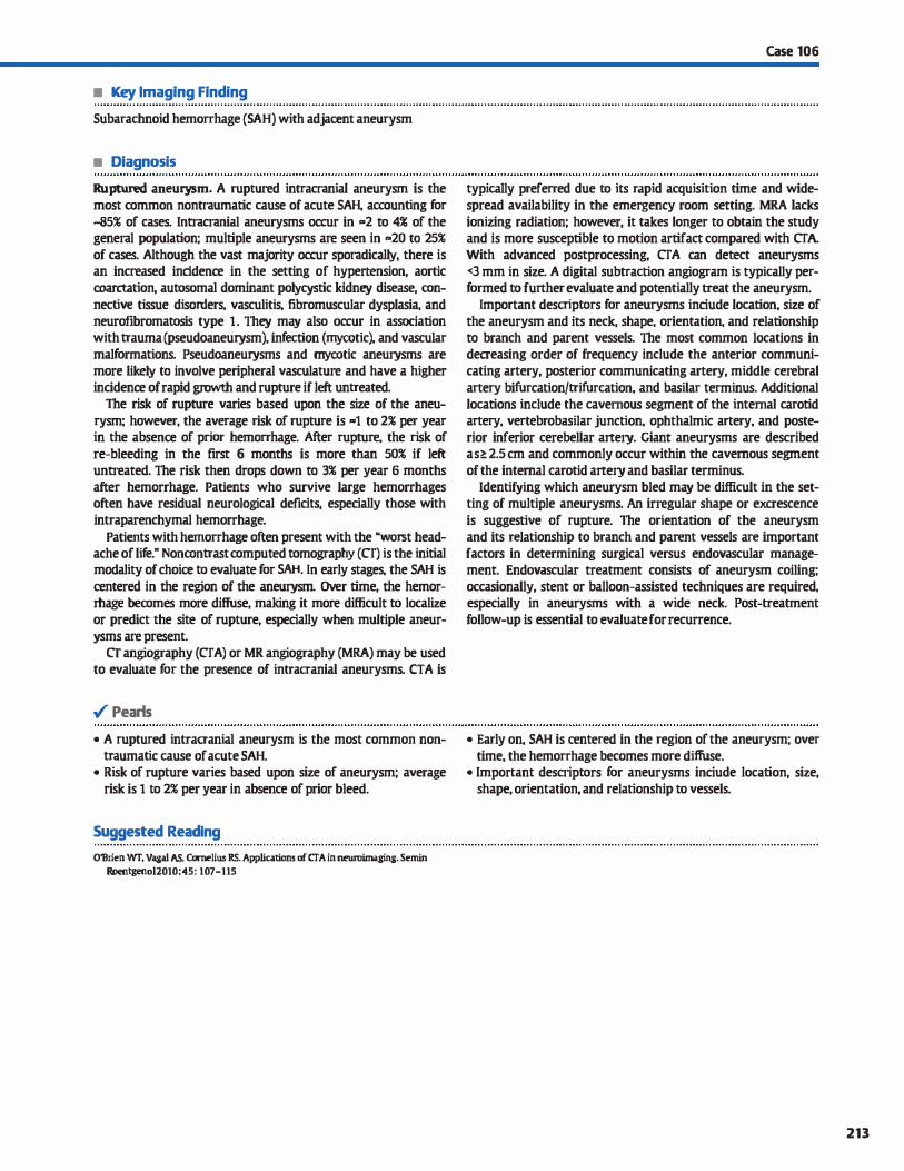
• Key lmaging Finding
Subarachnoid hemorrhage {SAH) with adjacent aneurysm
• Diagnosis
Ruptured aneurysm. A ruptured intracranial aneurysm is the most common nontraumatic cause of acute SAH, accounting for -85% of cases. Intracranial aneurysms occur in .. 2 to 4% of the general population; multiple aneurysms are seen in .. 20 ta 25% of cases. Although the vast majority occur sporadically, there is an increased incidence in the setting of hypertension, aortic coarctation, autasomal dominant polycystic kidney disease, con
nective tissue disonlers, vasculitis, fibromuscular dysplasia, and neurofibromatasis type 1. They may also occur in association with trauma (pseudoaneurysm), infection (mycotic� and vascular malformations. Pseudoaneurysms and mycotic aneurysms are more likely ta involve peripheral vasculature and have a higher incidence of rapid growth and rupture if left untreated.
The risk of rupture varies based upon the size of the aneu
rysm; however, the average risk of rupture is •1 to 2% per year in the absence of prier hemorrhage. After rupture, the risk of re-bleeding in the first 6 months is more than 50% if left untreated. The risk then drops down ta 3% per year 6 months after hemorrhage. Patients who survive large hemorrhages often have residual neurological defidts, espedally those with intraparenchymal hemorrhage.
Patients with hemorrhage often present with the "worst headache of life." Noncontrast computed tamography (CT) is the initial modality of choice to evaluate for SAH. In early stages, the SAH is
œntered in the region of the aneurysm OVer time, the hemormage becomes more diffuse, making it more difficult ta localize or predict the site of rupture, espedally when multiple aneurysms are present.
CT angiography (CTA) or MR angiography (MRA) may be used to evaluate for the presence of intracranial aneurysms. CTA is
y' Pearts
• A ruptured intracranial aneurysm is the most common nontraumatic cause of acute SAH.
• Risk of rupture varies based upon size of aneurysm; average risk is 1 to 2% per year in absence of prior bleed.
Suggested Reading
O'Brien Wf, Vagal AS, Cornelius RS. Applications of CTA in neuroimaging. Sernin
Roentgeno!Z010;45: 107-115
Case 106
typically preferred due to its rapid acquisition time and widespread availability in the emergency room setting. MRA Jacks ionizing radiation; however, it takes longer to obtain the study and is more susceptible to motion artifact compared with CTA With advanced postprocessing, CTA can detect aneurysms
<3 mm in size. A digital subtraction angiogram is typically performed ta further evaluate and potentially treat the aneurysm.
Important descriptors for aneurysms indude location, size of the aneurysm and its neck, shape, orientation, and relationship ta branch and parent vessels. The most common locations in decreasing order of frequency include the anterior communicating artery, posterior communicating artery, middle cerebral
artery bifurcation/trifurcation, and basilar terminus. Additional locations include the cavernous segment of the internai carotid artery, vertebrobasilar junction, ophthalmic artery, and posterior inferior cerebellar artery. Giant aneurysms are described as� 2.5 cm and commonly occur within the cavernous segment of the internai carotid artery and basilar terminus.
Identifying which aneurysm bled may be difficult in the setting of multiple aneurysms. An irregular shape or excrescence
is suggestive of rupture. The orientation of the aneurysm and its relationship to branch and parent vessels are important factors in determining surgical versus endovascular management. Endovascular treatment consists of aneurysm coiling; occasionally, stent or balloon-assisted techniques are required, especially in aneurysms with a wide neck. Post-treatment follow-up is essential to evaluate for recurrence.
• Early on, SAH is œntered in the region of the aneurysm; over time, the hemorrhage becomes more diffuse.
• Important descriptars for aneurysms indude location, size, shape, orientation, and relationship to vessels.
213
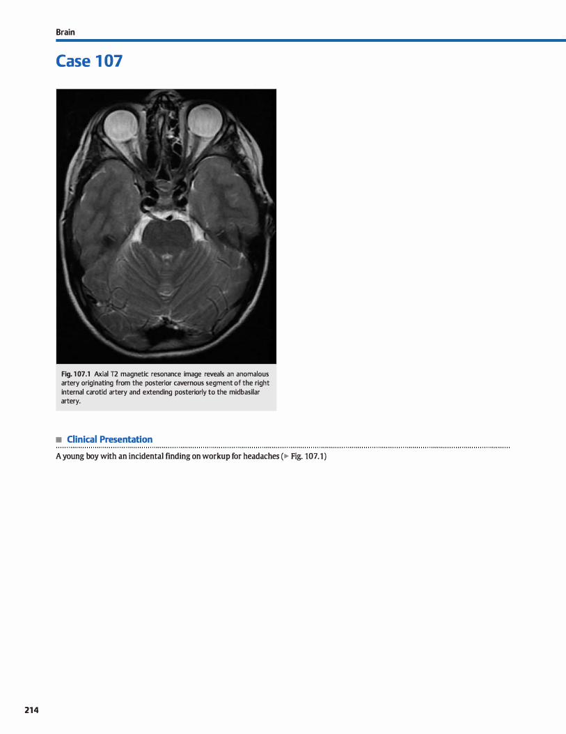
214
Brain
Case 1 07
Fig. 1 07.1 Axial T2 magnetic resonance image reveals an anomalous artery originating from the posterior cavernous segment of the right internai carotid artery and extending posteriorly to the midbasilar artery.
• Clinical Presentation
A young boy with an incidental finding on workup for headaches (� Fig. 107.1)
Case 107
• Key lmaging Finding
Anomalous carotid-vertebrobasilar anastomosis
• Diagnosis • • • • • • • • • • • • • • • • • • • • • • • • • • • • • • • • • • • • • • • • • • • • • • • • • • • • • • • • • • • • • • • • • • • • • • • • • • • • • • • • • • • • • • • • • • • • • • • • • • • • • • • • • • • • • • • • •• • • • • • • • • • • • • • • • • • • • • • • • • • • • • • • • • • • • • • • • • • • • • • • • • • • • • • • • • • • • • • • • • • • • • • • • • • • • • • • • • • • • • • • • • u . . . . . . . . . . . . . ..... .
Persistent fetal carotid-vertebrobasilar anastomoses (persistent A persistent hypoglossal artery is the second most common trigeminal artery). During early fetal development, anastomoses of the fetal carotid-vertebrobasilar anastomoses. It originates exist between the developing carotid arteries and the vertebroba- from the distal cervical internai carotid artery, passes through silar system. Failure ofthese connections to regress results in per- an enlarged hypoglossal canal, and connects with the basilar sistent vascular communications. The arteries are named with artery. The vertebral arteries and inferior aspect of the basilar respect to adjacent structures. The most common persistent artery proximal ta the anastomosis are typically hypoplastic. vessels from superior to inferior indude the trigeminal, otic, Given its location within the hypoglossal canal, patients may
hypoglossal, and proatlantal arteries. present with hypoglossal nerve dysfunction.
A persistent trigeminal artery is the most common of the The proatlantal artery originates from the distal common fetal carotid-vertebrobasilar anastomoses. It originates anteri- carotid or proximal intemal/external carotid arteries and orly just after the carotid artery exits the carotid canal and anastomoses with the suboccipital vertebral artery. The verteextends posteriorly ta the midbasilar artery. The inferior aspect brai artery proximal to the anastomosis may be hypoplastic or
of the basilar artery proximal ta the anastomosis is absent. Additional intracranial vascular variants and intrarelatively hypoplastic, whereas the portion distal ta the anasto- cranial aneurysms have been reported. mosis is prominent in caliber. The persistent trigeminal artery A persistent otic artery is the rarest of the fetal carotidmay course laterally along the trigeminal nerve or medially vertebrobasilar anastomoses. It arises from the petrous segthrough the sellar or suprasellar regions. The most important ment of the internai carotid artery, extends posteriorly and latreason for identifying this normal variant is ta avoid inadver- erally through the internai auditory canal, and connects with tent injury in patients requiring transsphenoidal or other skull- the proximal basilar artery. The inferior aspect of the basilar base surgeries. As with other anomalies that alter hemo- artery proximal ta the anastomosis is typically hypoplastic. dynamics, there is an increased incidence of intracranial
aneurysms.
� Pearls
• Persistent fetal carotid-vertebrobasilar anastomoses are uncommon but important anomalies.
• Persistent arteries are narned based upon adjacent structures: trigeminal, otic, hypoglossal, and proatlantal.
Suggested Reading
Dimmick SJ, Faulder KC. Normal variants of the œrebral ciIOJlation at multidetector cr angiography. Radiographies 2009; 29; 1027-1043
• The persistent trigeminal artery is the most common and is
important ta identify prior ta skull-base surgery. • A persistent hypoglossal artery extends through the hypo
glossal canal and may result in cranial neuropathy.
215
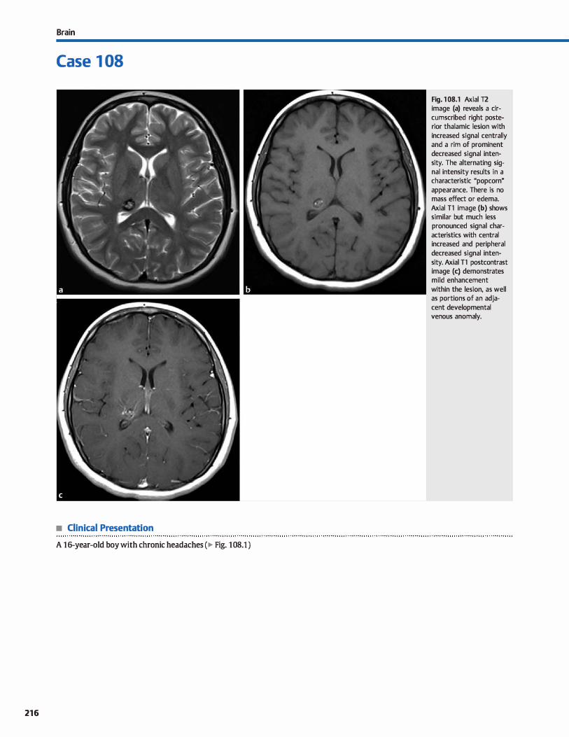
Brain
Case 1 08
• Clinical Presentation
A 16-year-old boy with chronic headaches (� Fig. 108.1 )
216
Fig. 1 08.1 Axial T2 image (a) reveals a circumscribed right posterior thalamic lesion with increased signal centrally and a rim of prominent decreased signal intensity. The alternating signal intensity results in a characteristic "popcorn" appearance. There is no mass effect or edema. Axial Tl image (b) shows similar but much less pronounced signal characteristics with central increased and peripheral decreased signal intensity. Axial Tl postcontrast image (c) demonstrates mild enhancement within the lesion, as well as portions of an adjacent developmental venous anomaly.
• Key lmaging Finding
Circumscribed lesion with central increased and peripheral
decreased signal with a "popcorn" appearance
• Diagnosis
cavernous malformation {CM). CMs are slow-flow lesions that
are composed of dilated sinusoids, intercapillary spaœs, and
cavemous spaœs without intervening brain parenchyma. They
are filled with blood of various stages and surrounded by a
hemosiderin capsule. CMs represent the second most common
vascular malformation (VM) after developmental venous
anomalies (DVAs) and commonly occur in association with
DVAs.
The vast majority of CMs are sporadic and solitary. They may
be multiple in -1 O to 20% of cases, espedally in familial cases
(autosomal dominant) or in the setting of prior radiation ther
apy. Most lesions are asymptomatic. When present, common
symptoms include acute intracranial hemorrhage, seizures, or
focal neurological deficits. The location of the lesion(s) often corresponds with symptoms; lesions in the posterior fossa have
a higher incidence of hemorrhage, whereas superficial supra
tentorial lesions more often result in seizures or focal neurolog
ical defidts. The average annual risk of hemorrhage is < 1%. Mer
an episode of acute hemorrhage, the risk of re-bleeding
increases to -4.5% per year for the next 2 ta 3 years and then
returns to baseline.
On unenhanced computed tomography (CT), large lesions
present as relatively well-defined regions of subtle increased
� Pearts
• CMs are slow-flow lesions without intervening brain tissue;
they are the second most common VM after DVAs. • The vast majority of CMs are sporadic and solitary; they may
be multiple in familial cases or prior radiation therapy.
Suggested Reading
Smith AB. Vascular rnalfonnations of the brain: radiologie and pathologie rorrela
tion.J Am Osteopath Coll Radio!. 2012: 1 : 10-22
Case 108
attenuation. Calcification may be seen in "'60% of cases. Smalt
lesions are often isodense to surrounding brain parenchyma
and difficult to identify. In the absence of acute hemorrhage,
there is typically no mass effect or surrounding edema. Acute hemorrhage extending through the capsule will typically result
in surrounding edema and mass effect, as well as acute onset of symptoms.
The magnetic resonance imaging {MRI) appearanœ of CMs is
characteristic. On T2 sequenœs, the lesions have a peripheral
hypointense hemosiderin rim Centrally, there are altemating
regions of increased and decreased signal intensity on both Tt and T2 sequences due to blood products of various stages; this is tenned the "popcorn" appearanœ. The lesions "bloom" on
gradient echo and susceptibility-weighted imaging. Enhancement is variable and seen in "'15% of cases. As with CT, there is
typically no mass effect or edema unless complicated by acute hemorrhage.
If asymptomatic, CMs are usually followed to ensure stability.
In the setting of acute hemorrhage, conservative therapy is ideal,
unless the dinical picture requires intervention. Treatment
options indude surgical resection or radiosurgery, depending
upon the lesion size, lesion location, and dinical setting.
• On MRI, CMs have a peripheral hypointense hemosiderin rim
with a central ''popcorn" appearance. • CMs are typically followed for stability unless lesion size, loca
tion, or dinical setting requires intervention.
217

Brain
Case 1 09
• Clinical Presentation
A 44-year-old woman with headaches and seizures (� Fig. 109.1)
218
Fig. 1 09.1 Axial CT image (a) demonstrates a hyperdense mass with calcifications and mild surrounding edema within the posterior left frontal lobe. A ventricular drainage catheter is partially visualized within the right frontal lobe. Axial T2 MR image (b) reveals a nidus of enlarged serpentine flow voids with a large venous aneurysm laterally. There is prominent enhancement and flow-related artifact on the axial Tl postcontrast image (c). Lateral oblique digital subtraction angiogram of the left internai carotid artery (d) demonstrates early venous filling
during the arterial phase, including the large venous aneurysm, as well as enlarged vessels feeding, draining, and within the nidus. Serpentine subtraction artifact corresponds to partial embolization.
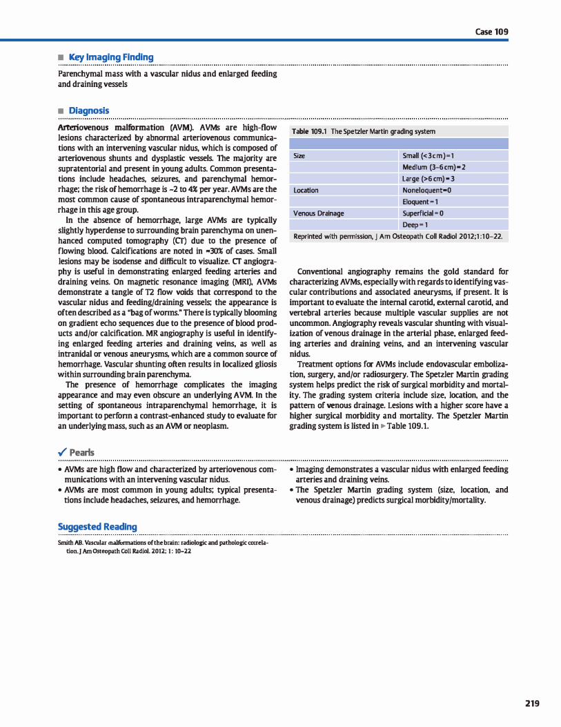
• Key lmaging Finding
Parenchymal mass with a vascular nidus and enlarged feeding
and draining vessels
• Diagnosis
Arteriovenous malformation (AVM). AVMs are high-flow lesions characterized by abnonnal arteriovenous communications with an intervening vascular nidus, which is composed of arteriovenous shunts and dysplastic vessels. The majority are
supratentorial and present in young adults. Common presentations include headaches, seizures, and parenchymal hemorrhage; the risk of hemorrhage is -2 to 4% per year. AVMs are the most common cause of spontaneous intraparenchymal hemorrhage in this age group.
ln the absence of hemorrhage, large AVMs are typically slightly hyperdense to surrounding brain parenchyma on unen
hanced computed tomography (CT) due ta the presence of flowing blood. Calcifications are noted in •30% of cases. Smalt lesions may be isodense and difficult ta visualize. cr angiography is useful in demonstrating enlarged feeding arteries and draining veins. On magnetic resonanœ imaging (MRI), AVMs demonstrate a tangle of T2 flow voids that correspond to the vascular nidus and feeding/draining vessels; the appearanœ is often described as a �bag of wonns." There is typically blooming
on gradient echo sequences due to the presence of blood products and/or calcification. MR angiography is useful in identify
ing enlarged feeding arteries and draining veins, as well as intranidal or venous aneurysms. which are a common source of hemorrhage. Vascular shunting often results in localized gliosis within surrounding brain parenchyma.
The presence of hemorrhage complicates the imaging
appearance and may even obscure an underlying AVM. ln the setting of spontaneous intraparenchymal hemorrhage, it is important to perfonn a contrast-enhanced study ta evaluate for an underlying mass, such as an AVM or neoplasm.
� Pearts
• AVMs are high flow and characterized by arteriovenous communications with an intervening vascular nidus.
• AVMs are most common in young adults; typical presentations include headaches, seizures, and hemorrhage.
Suggested Reading
Smith AB. Vascular rnalfunnations of the brain: radiologie and pathologie rorrela
tion. J Am Osteopath Coll Radio!. 2012: 1 : 10-22
Table 109.1 The Spetzler Martin gradlng system
Slze
Location
Venous Drainage
Small (<3cm)-1
Medium (3-6 cm)• 2
Large (>6 cm) • 3
Noneloquent•O
Eloquent= l
Superflcial - O
Deep= l
Case 109
Reprinted with permission, J Am Osteopath Coll Radiol 2012;1 :10-22.
Conventional angiography remains the gold standard for characterizing AVMs, especially with regards to identifying vascular contributions and assodated aneurysms, if present. lt is important to evaluate the internai carotid, externat carotid, and
vertebral arteries because multiple vascular supplies are not uncommon. Angiography reveals vascular shunting with visualization of venous drainage in the arterial phase, enlarged feed
ing arteries and draining veins, and an intervening vascular nid us.
Treatment options for AVMs include endovascular embolization, surgery, and/or radiosurgery. The Spetzler Martin grading system helps predict the risk of surgical morbidity and mortal
ity. The grading system criteria include size, location, and the pattern of venous drainage. Lesions with a higher score have a
higher surgical morbidity and mortality. The Spetzler Martin grading system is listed in � Table 109.1.
• lmaging demonstrates a vascular nidus with enlarged feeding arteries and draining veins.
• The Spetzler Martin grading system (size, location, and venous drainage) predicts surgical morbidity/mortality.
219
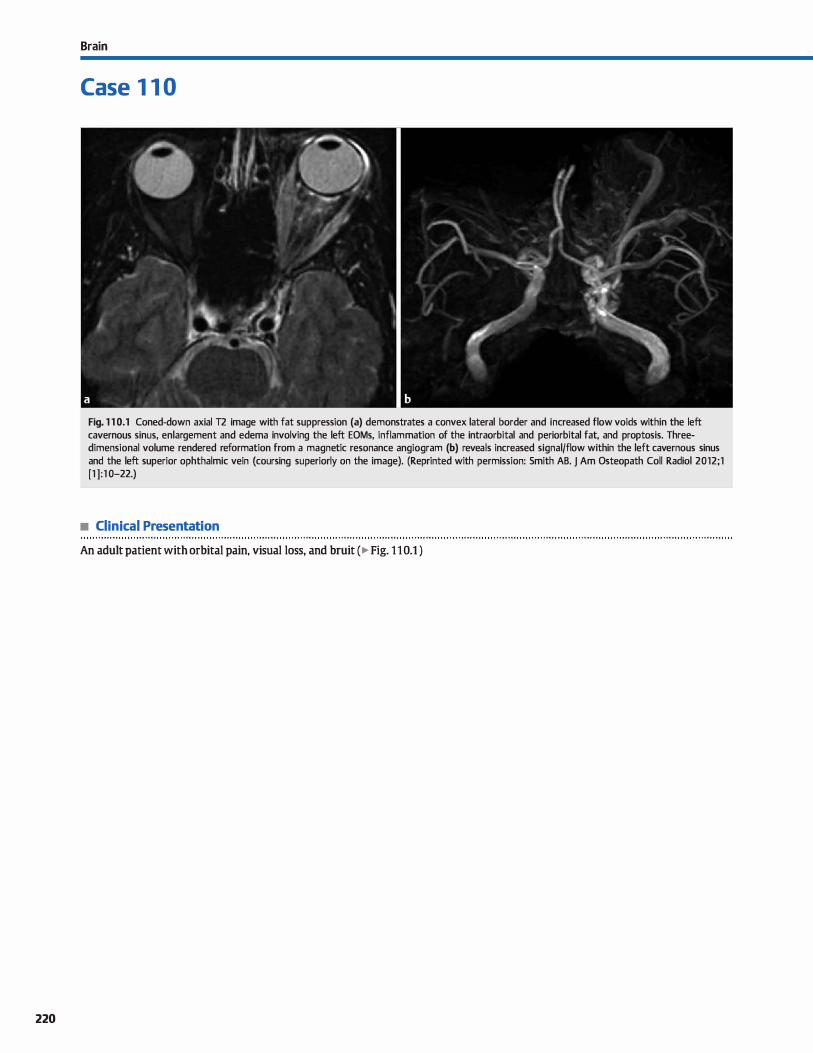
220
Brain
Case 1 1 0
Fig. 1 1 0.1 Coned-down axial T2 image with fat suppression (a) demonstrates a convex lateral border and increased flow voids within the left cavernous sinus, enlargement and edema involving the left EOMs, inflammation of the intraorbital and periorbital fat, and proptosis. Threedimensional volume rendered reformation from a magnetic resonance angiogram (b) reveals increased signal/flow within the left cavernous sinus and the left superior ophthalmic vein (coursing superiorly on the image). (Reprinted with permission: Smith AB. j Am Osteopath Coll Radiol 2012;1 [1 ] : 1 0-22.)
• Clinical Presentation
An adult patient with orbital pain, visual Joss, and bruit (� Fig. 110.1 )
• Key lmaging Finding
Enlarged cavernous sinus with early venous filling on arterial phase imaging
• Diagnosis
carotid-cavemous fistula (CCF). CCFs refer to abnormal communications between the carotid artery and cavemous sinus. They are dassified as direct or indirect. Direct CCFs result in high-flow arteriovenous shunting between the cavernous internai carotid artery {ICA) and the cavernous sinus with a relatively rapid onset of symptoms, which include pulsatile exophthalmos, orbital bruit, chemosis, and occasionally intracranial hemorrhage. The vast majority of direct CCFs result from trauma; rupture of a cavemous carotid aneurysm is a much less common cause. Indirect CCFs are slow-flow shunts between durai branches of the ICA or the externat carotid artery {ECA) and the cavemous sinus. They most often occur spontaneously and have a more insidious onset of symptoms.
CCFs result in increased venous and orbital pressures. There is enlargement of the cavernous sinus and diversion of venous
y' Pearts
• CCFs (direct or indirect) are abnormal communications between the carotid artery and cavemous sinus.
• Direct CCFs result from trauma and have a rapid onset of pulsatile exophthalmos, orbital bruit, and chemosis.
Suggested Reading
Smith AB. Vascular malfonnations of the brilin: radiologie and pathologie
correlation.J Am Osteopath Coll Radio!. 2012.; 1 : 10-ll
Case 1 1 0
drainage through the ipsilateral superior ophthalmic vein {SOV). Magnetic resonance imaging demonstrates a convex lateral border of the cavemous sinus on the involved side (it is typically flat or concave), increased T2 flow voids within the cavemous sinus, enlarged ipsilateral SOV and extraocular muscles {EOMs), and proptosis. Cavemous sinus enlargement may be bilateral because there are communications with the contralateral side within the cavemous sinus.
Angiography demonstrates early venous filling { cavemous sinus and superior ophthalmic vein) in the arterial phase. Endovascular treatment options are favored and include glue or coil embolization.
• lmaging findings indude enlargement of the cavemous sinus (with flow voids), SOV, EOMs, and proptosis.
• Endovascular treatment options for CCFs are favored and include glue or coil embolization.
221
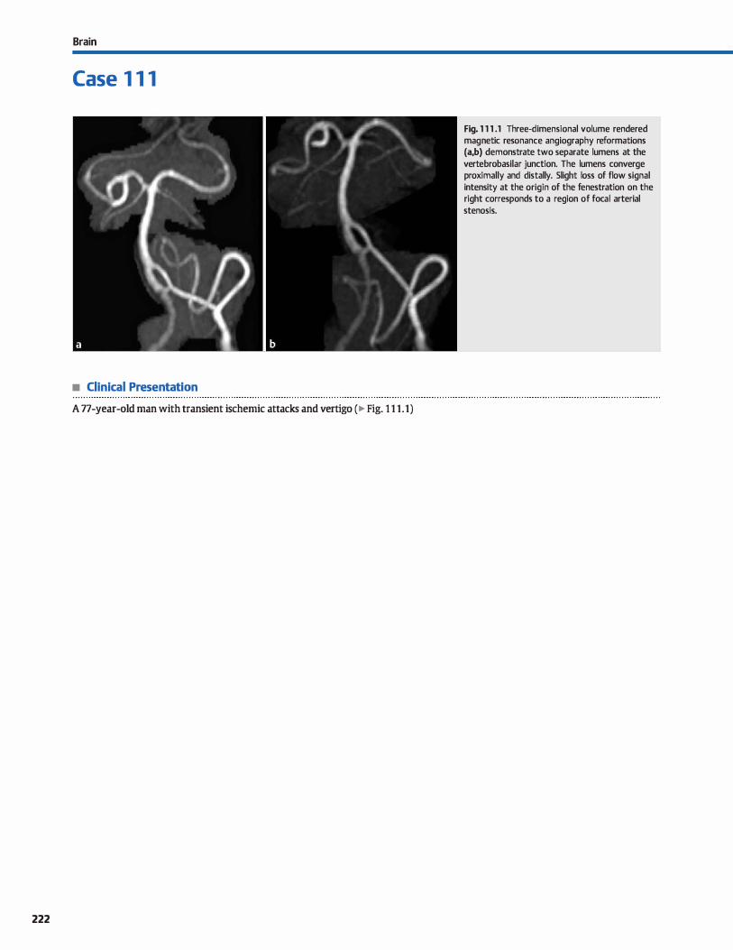
Brain
Case 1 1 1
• Clinical Presentation
A 77-year-old man with transient ischemic attacks and vertigo (� Fig. 1 1 1 .1)
222
Fig. 1 1 1 .1 Three-dimensional volume rendered magnetic resonance angiography reformations (a,b) demonstrate two separate lumens at the vertebrobasilar junction. The lumens converge proximally and distally. Slight loss of flow signal intensity at the origin of the fenestration on the right corresponds to a region of focal arterial stenosis.
• Key lmaging Finding
Separate arterial lumens that converge proximal and distally
• Diagnosis
Arterial fenestration. A fenestration refers to two separate vascular channels composed of independent endothelium and muscular walls but shared adventitia. They typically occur in short segments with a normal caliber parent artery or arteries proximally and distally. Because fenestrations alter hemodynamic flow, there is an increased association with aneurysm formation compared with the general population.
� Pearts
Case 1 1 1
Within the drcle of Willis, the most common locations
indude the anterior communicating and basilar arteries. In the case of basilar artery fenestrations, they are thought to result from incomplete fusion ofpaired fetal longitudinal neural arteries. Most basilar artery fenestrations occur near the vertebroba
silar junction. Involvement of the cerebral and vertebral arteries is less common but does occur.
• Fenestrations refer to distinct arterial channels with their • The most common locations of fenestrations indude the ante-own endothelium/muscularis but a shared adventitia. rior communicating and basilar arteries.
• Because fenestrations alter hemodynamic flow, there is an
increased association with aneurysm formation.
Suggested Reading
Dinunick SJ, Faulder KC. Normal variants of the œrebral circulation at multidetector cr angiography. Radiographies 2009; 29: 1027-1043
223
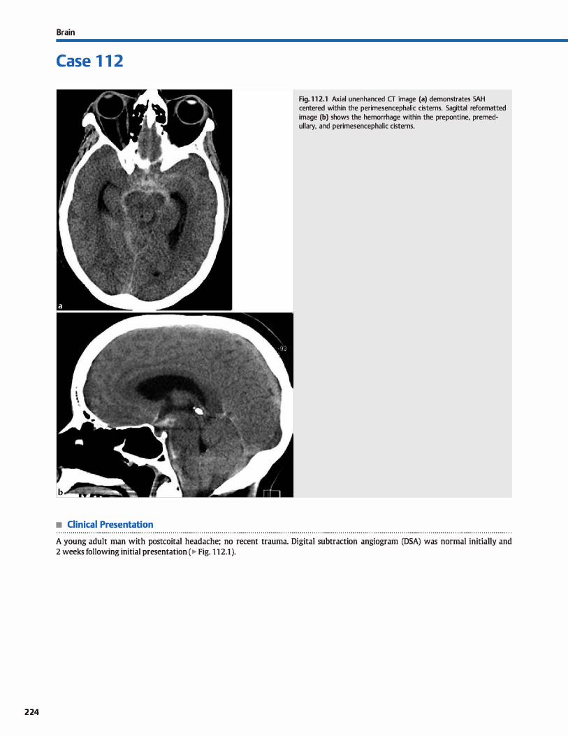
224
Brain
Case 1 1 2
• Clinical Presentation
Fig. 1 1 2.1 Axial unenhanced CT image (a) demonstrates SAH centered within the perimesencephalic cisterns. Sagittal reformatted image (b) shows the hemorrhage within the prepontine, premedullary, and perimesencephalic cisterns.
A young adult man with postcoital headache; no recent trauma. Digital subtraction angiogram (DSA) was normal initially and
2 weeks following initial presentation ( � Fig. 1 1 2.1 ).
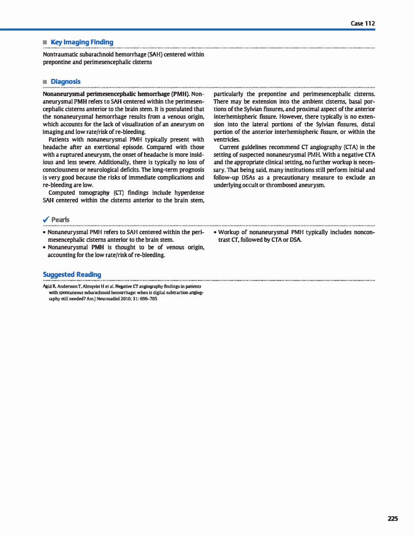
• Key lmaging Finding
Nontraumatic subarachnoid hemorrhage (SAH) centered within
prepontine and perimesencephalic dstems
• Diagnosis
Nonaneurysmal perimesenœphalic hemorrhage (PMH). Non
aneurysmal PMH refers ta SAH centered within the perimesen
cephalic cisterns anterior ta the brain stem. It is postulated that the nonaneurysmal hemorrhage results from a venous origin,
which accounts for the lack of visualization of an aneurysm on
imaging and low rate/risk of re-bleeding.
Patients with nonaneurysmal PMH typically present with
headache after an exertional episode. Compared with those
with a ruptured aneurysm, the onset of headache is more insidious and less severe. Additionally, there is typically no loss of
consciousness or neurological detidts. The long-term prognosis
is very good because the risks of immediate complications and
re-bleeding are low.
Computed tomography (CT) findings indude hyperdense SAH centered within the dstems anterior to the brain stem,
y' Pearts
• Nonaneurysmal PMH refers to SAH centered within the peri
mesencephalic cistems anterior to the brain stem. • Nonaneurysmal PMH is thought to be of venous origin,
accounting for the low rate/risk of re-bleeding.
Suggested Reading
Aiid R, Andersson T, Almqvist H et al. Negative CT angiography findings in patients with spontaneous subaradmoid hem01Thage: when is digital subtraction angiography 5till needed? Am] Neuroradiol 2010; 31: 696-705
Case 1 1 2
particularly the prepontine and perimesencephalic cisterns. There may be extension into the ambient cistems, basal por
tions of the Sylvian fissures, and proximal aspect of the anterior
interhemispheric tissure. However, there typically is no exten
sion into the lateral portions of the Sylvian fissures, distal portion of the anterior interhemispheric fissure, or within the
ventrides.
Current guidelines recommend CT angiography (CTA) in the
setting of suspected nonaneurysmal PMH. With a negative CTA and the appropriate clinicat setting, no further workup is neces
sary. That being said, many institutions still perform initial and
follow-up DSAs as a precautionary measure to exclude an
underlying ocrult or thrombosed aneurysm.
• Workup of nonaneurysmal PMH typically includes noncon
trast CT, followed by CT A or DSA
225
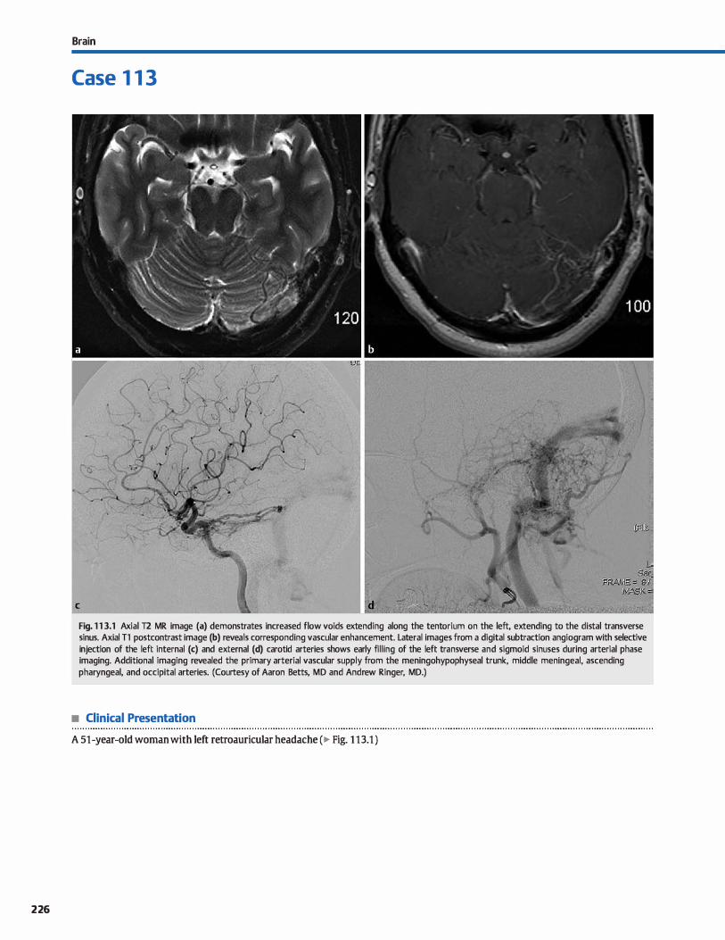
226
Brain
Case 1 1 3
Fig. 1 1 3.1 Axial T2 MR image (a) demonstrates increased flow voids extending along the tentorium on the left, extending to the distal transverse sinus. Axial Tl postcontrast image (b) reveals corresponding vascular enhancement. Lateral images from a digital subtraction angiogram with selective injection of the left internai (c) and external (d) carotid arteries shows early filling of the left transverse and sigmoid sinuses during arterial phase imaging. Additional imaging revealed the primary arterial vascular supply from the meningohypophyseal trunk, middle meningeal, ascending pharyngeal, and occipital arteries. (Courtesy of Aaron Betts, MD and Andrew Ringer, MD.)
• Clinical Presentation
A 51-year-old woman with left retroauricular headache (� Fig. 1 13.1 )
• Key lmaging Finding
Dural-based arteriovenous shunt
• Diagnosis
Durai arteriovenous fistula {AVF). AVFs differ from arteriovenous malformations (AVMs) in that AVFs have a direct arterial to venous communication without an intervening vascular nidus, as is seen with AVMs. Otheiwise, bath are highflow shunt lesions.
Durai AVFs (dAVFs) are dassified as infantile or adult lesions. The infantile form is rare and is characterized by multiple
dAVFs in response to venous sinus thrombosis. Magnetic resonanœ imaging (MRI) is useful in identifying the venous sinus thrombosis, as well as identifying the vascular lesion, which is typically centered in the torcula.
The adult fonn is thought to be acquired also as a result of dural venous sinus thrombosis. It typically presents in middleaged adults. The dAVFs consist of small shunts in the wall of a thrombosed venous sinus; the most common locations indude the junction of the transverse and sigmoid sinuses and the cavernous sinus. Symptoms are variable and based upon location. Those located at the transverse-sigmoid junction often present with tinnitus and vertigo. Focal neurological deficits and hemorrhage are potential complications and are thought to result from venous congestion.
dAVFs are characterized as benign or aggressive based upon the pattern of venous drainage. The presence of retrograde Ieptomeningeal venous drainage is an indicator of a more aggressive lesion and should prompt early intervention.
� Pearls
• AVFs have a direct arterial to venous communication without an intervening vascular nidus.
• dAVFs are thought to be acquired due to durai venous sinus thrombosis; there are adult and infantile fonns.
Suggested Reading
Smith AB. Vascular malfonnations of the brain; radiologie and pathologie correla
tion.J Am Osteopath Coll Radio! 2012; 1: 10-22
Case 113
Noncontrast computed tomography (CT) is often normal in the setting of an uncomplicated dAVF. CT angiography may reveal enlarged arteries or draining veins along a durai venous sinus. CT venography may reveal flow-limiting stenosis or occlusion of the adjacent durai venous sinus. On MRI. there are often multiple flow voids on T2 sequenœs corresponding to the dAVF; these may be subtle. A thrombosed sinus will have
absence of the normal T2 flow void and is better depicted on MR venography. Venous congestion results in gliosis within the adjacent brain parenchyma.
Digital subtraction angiography best depicts dAVFs with multiple arterial feeders and venous filling during arterial phase
imaging. The majority of lesions have arterial supply from durai and transosseous branches of the externat carotid artery; however, durai and tentorial branches of the internai carotid and/or vertebral arteries may also be involved. Therefore, it is important to interrogate all arterial vessels during a œrebral angiogram. More aggressive lesions will demonstrate reversai of flow within the durai venous sinus and leptomeningeal/cortical veins with a characteristic "pseudophlebitic" pattern.
Treatment options include endovascular embolization, surgical resection. or radiosurgery.
• Treatment options for dAVFs indude endovascular embolization, surgical resection. or radiosurgery.
227
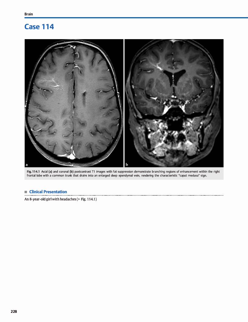
228
Brain
Case 1 1 4
Fig. 1 1 4.1 Axial (a) and coronal (b) postcontrast Tl images with fat suppression demonstrate branching regions of enhancement within the right frontal lobe with a common trunk that drains into an enlarged deep ependymal vein, rendering the characteristic "caput medusa" sign.
• Clinical Presentation
An 8-year-old girl with headaches ( � Fig. 1 14.1 )
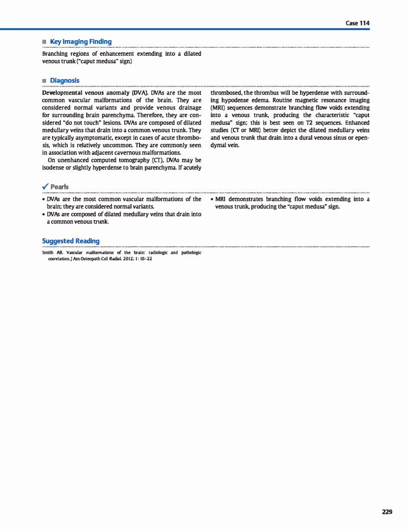
• Key lmaging Finding
Branching regions of enhancement extending into a dilated
venous trunk ("caput medusa" sign)
• Diagnosis
Developmental venous anomaly (DVA). DVAs are the most common vascular malformations of the brain. They are
considered normal variants and provide venous drainage
for surrounding brain parenchyma. Therefore, they are con
sidered "do not touch" lesions. DVAs are composed of dilated
medullary veins that drain into a common venous trunk. They
are typically asymptomatic, except in cases of acute thrombo
sis, which is relatively uncommon. They are commonly seen
in association with adjacent cavernous malformations.
On unenhanced computed tomography {CT), DVAs may be
isodense or slightly hyperdense ta brain parenchyma. If acutely
� Pearts
• DVAs are the most common vascular malformations of the
brain; they are considered normal variants.
• DVAs are composed of dilated medullary veins that drain into
a common venous trunk.
Suggested Reading
Smith AB. Vascular rnalfonnations of the brain; radiologie and pathologie
correlation.j Am Osteopath Coll Radio!. 2012; 1 : 10-22
Case 1 14
thrombosed, the thrombus will be hyperdense with surrounding hypodense edema. Routine magnetic resonance imaging
(MRI) sequences demonstrate branching flow voids extending into a venous trunk, produdng the characteristic "caput
medusa" sign; this is best seen on T2 sequences. Enhanced
studies (CT or MRI) better depict the dilated medullary veins
and venous trunk that drain into a durai venous sinus or epen
dymal vein.
• MRI demonstrates branching flow voids extending into a
venous trunk, producing the "caput medusa" sign.
229
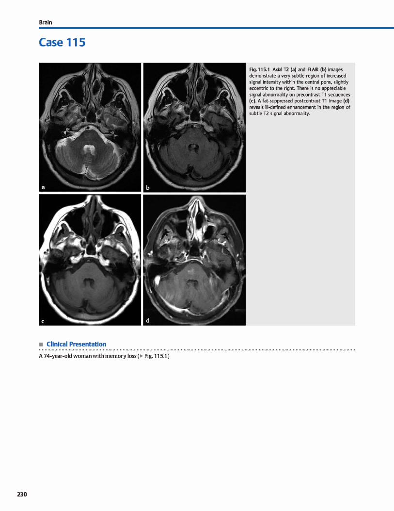
Brain
Case 1 1 5
• Clinical Presentation
A 74-year-old woman with memory Joss (� Fig. 1 1 5.1 )
230
Fig. 1 1 5.1 Axial T2 (a) and FLAIR (b) images demonstrate a very subtle region of increased signal intensity within the central pons, slightly eccentric to the right. There is no appreciable signal abnormality on precontrast Tl sequences (c). A fat-suppressed postcontrast Tl image (d) reveals ill-defined enhancement in the region of subtle T2 signal abnormality.
• Key lmaging Finding
Faint region of brain stem signal abnormality with ill-defined
enhanœment
• Diagnosis
(apillary telangiectasia. capillary telangiectases represent
dilated vessels that are interspersed between normal brain
parenchyma. Although their etiology is uncertain, they are
thought to be acquired lesions associated with venous drainage
or venous anomalies. The majority are located within the pans; additional locations include the temporal lobes, basal ganglia, and other portions of the brain stem. They are typically small
( < 1 cm), incidental, and asymptomatic.
Telangiectases are not visible on computed tomography.
Magnetic resonance imaging (MRI) often shows a region of
� Pearls
• Telangiectases represent dilated capillary-like vessels inter
spersed between normal brain parenchyma.
• capillary telangiectases are thought to be acquired lesions; the majority are located within the pons.
Suggested Reading
Smith AB. Vascular malfonnations of the brain; radiologie and pathologie
correlation.j Am OsteopathColl Radio!. 2.012.; 1 : 10-2.2.
Case 115
normal or subtly increased 1'2/fluid-attenuated inversion recov
ery (FLAIR) signal intensity with ill-defined "brush-like"
enhancement. On gradient echo {GRE) or susœptibility
weighted (SWI) sequenœs, there is hypointense signal due to
deoxyhemoglobin within the lesions. There is no mass effect, gliosis, or calcification associated with the lesions. In many
cases, there may be a prominent draining vein or associated
developmental venous anomaly. If characteristic in appearance, no further imaging workup or follow-up is neœssary.
• MRI shows subtle increased TI/FI.AIR signal, decreased signal
on GRE/SWI, and "brush-like" enhanœment
231
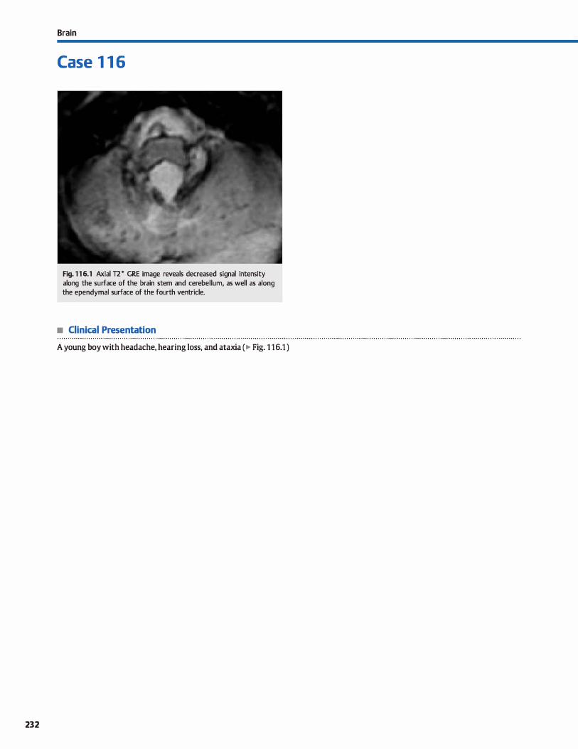
232
Brain
Case 1 1 6
Fig. 1 1 6.1 Axial T2 • GRE image reveals decreased signal intensity along the surface of the brain stem and cerebellum, as well as along the ependymal surface of the fourth ventride.
• Clinical Presentation
A young boy with headache, hearing Joss, and ataxia ( � Fig. 1 16.1 )
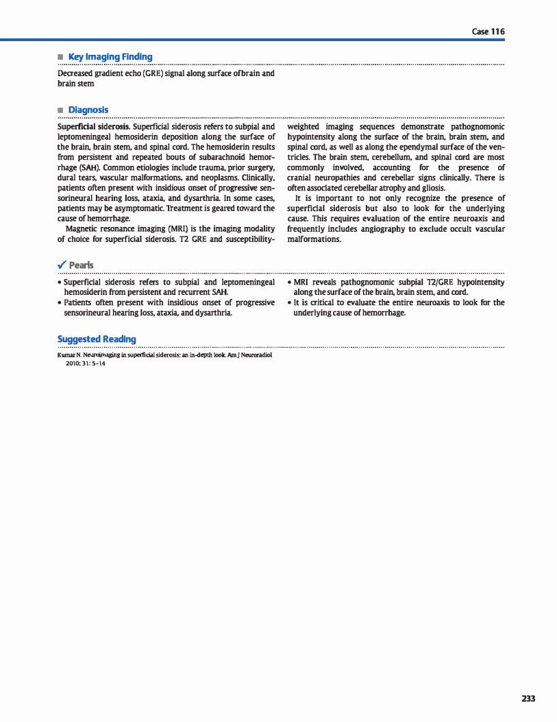
• Key lmaging Finding
Decreased gradient echo (GRE) signal along surface ofbrain and brain stem
• Diagnosis
Supedidal siderosis. Superticial siderosis refers to subpial and leptomeningeal hemosiderin deposition along the surface of the brain, brain stem, and spinal cord. The hemosiderin results from persistent and repeated bouts of subarachnoid hemor
rhage (SAH). Common etiologies include trauma, prior surgery, durai tears, vascular malformations, and neoplasms. Clinically,
patients often present with insidious onset of progressive sen
sorineural hearing Joss, ataxia, and dysarthria. In some cases, patients may be asymptomatic. Treatment is geared toward the cause of hemorrhage.
Magnetic resonanœ imaging {MRI) is the imaging modality
of choiœ for superficial siderosis. T2 GRE and susœptibility-
� Pearls
• Superticial siderosis refers to subpial and leptomeningeal hemosiderin from persistent and recurrent SAH.
• Patients often present with insidious onset of progressive
sensorineural hearing loss, ataxia, and dysarthria.
Suggested Reading
Kumar N. Nelll1IÎII1agÎII in superficial siderosis: an in-depth look. AmJ Neuroradiol 2010; 31; 5-14
Case 1 1 6
weighted imaging sequences demonstrate pathognomonic hypointensity along the surface of the brain, brain stem, and spinal cord, as well as along the ependymal surface of the ventricles. The brain stem, cerebellum, and spinal cord are most
commonly involved, accounting for the presenœ of cranial neuropathies and cerebellar signs dinically. There is
often associated cerebellar atrophy and gliosis. It is important to not only recognize the presence of
superficial siderosis but also to look for the underlying cause. This requires evaluation of the entire neuroaxis and
frequently indudes angiography to exclude occult vascular
malformations.
• MRI reveals pathognomonic subpial T2/GRE hypointensity along the surface of the brain, brain stem, and cord.
• It is critical to evaluate the entire neuroaxis to look for the
underlying cause of hemorrhage.
233
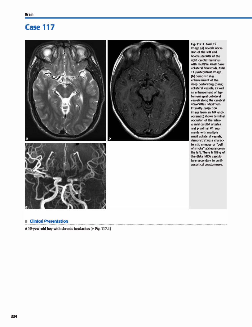
Brain
Case 1 1 7
• Cllnlcal Presentatlon
A 16-year-old boywith chronicheadaches (� rig. 117.1)
234
Fig. 117.1 Axial T2 Image (a) reveals occlu· slon of the left and severe stenosls of the right c.11rotid terminus wlth multiple small basal collateral flow volds. Axlal Tl postcontrast Image (b) demonstrates enhancement of the deep perforalfng (basal) collateral vessets. as well ;is enh;inœment of leptomeninge;il colliteral vessels along the œrebral ccnvexltles. Maximum lntenslty projection image from ;in MR angi· ogram (c) shows terminal occlusion of ltte lntra· aanlal carolfd arterles ;ind proxim;il Ml seg· menb wtth multiple small collateral vessels, demonstratlng a characteristic smudgy or •putt of smoke• appearanœ on the left. There ls fllllng of the dlstal MCA vasailature secondary to corti· cocortlcal anastomoses.
• Key lmaging Finding
Carotid terminus occlusion with numerous basal collateral vessels
• Diagnosis
Moyamoya. Moyarnoya is a relatively rare, progressive, occlusive cerebrovascular disease that primarily involves the intracranial internai carotid and proximal œrebral arteries. The term "moyamoya" means Mpuff of smoke" in japanese and corre
sponds to the appearance of abnormal collateral vessels on angiography when first described. The idiopathie variant is commonly seen in Asia. Moyamoya syndrome may be assoctated with neurofibromatosis type 1 (NFl ), sickle œll disease (SCD), Down syndrome, collagen vascular disease {CVD), and prior radiation therapy.
There is a bimodal age distribution involving children within
the first decade of life and young adults within the third and fourth decades of life. Children often present with headaches, multiple transient ischemic attacks or infarcts, and seizures. Hemorrhage is more common in adults. Treatment is geared toward the underlying etiology and prevention and management of complications. Depending upon the severity of the disease and symptoms, anticoagulation and vascular bypass procedures, such as superfidal temporal artery {STA)-middle
œrebral artery {MCA) bypass, may be beneficial. Unenhanced computed tomography {CT) is useful ta evaluate
for parenchymal hemorrhage and may also show signs of chronic ischemia, often in a watershed distribution. Magnetic resonanœ imaging {MRI) is the imaging modality of choiœ to identify the disease and its complications. T2 sequences reveal stenosis or occlusion of the carotid terminus and portions
of the proximal cerebral arteries. Dilaterai involvement is
� Pearts
• Moyamoya results in progressive occlusion of the intracranial internai carotid and proximal œrebral arteries.
• Moyamoya may be idiopathie or associated with NFl, SCD, Dawn syndrome, CVD, or prier radiation therapy.
Suggested Readings
Case 117
common. Multiple enlarged T2 flow voids are seen involving the perforating parenchymal {basilar) vessels near the carotid terminus. Enhanced sequenœs demonstrate enhanœment
of the basilar collateral vessels, as well as enhancement of enlarged collateral leptomeningeal veins. The appearanœ of leptomeningeal collaterals along the cerebral convexities has been described as the "climbing ivy" sign. Durai collaterals are not as well seen on MRI but are readily identified on conventional angiography.
MR angiography demonstrates loss of flow signal at the
carotid terminus and proximal cerebral arteries. The enlarged basilar collaterals demonstrate a smudgy or Mpuff of smoke" appearance. With an appropriate field of view, enlarged leptomeningeal collaterals are well depicted along the œrebral convexities. Findings are similar ta those noted on conventional angiography. Conventional angiography offers real-time evaluation and better depiction of durai collaterals. However, given
that angiography is an invasive proœdure, diagnostic findings
on MRI/MR angiography are often sufficient CT and MR perfusion may be utilized to monitor disease pro
gression and identify potential candidates for bypass procedures. CT angiography is especially useful because it allows for better visualization of externat carotid artery branches (STA) used for bypass. After bypass treatment, perfusion imaging is used to detennine if there is improved/adequate reserve in the
affected vascular distribution, as well as ta directly visualize patency of the ST A-MCA anastomosis.
• MR angiography demonstrates occlusion of the carotid terminus and proximal cerebral arteries with multiple collaterals.
• Enlarged basilar collaterals demonstrate a Mpuff of smoke" appearanœ on MR and conventional angiography.
Burlœ GM, Burke AM, ShermaAK, Hurley MC, Batjer HH, Bendok BR. Moy.unoya dis- Geibprasert s, Pongpech S,Jiarakongmun P, Shroff MM, Amtstrong oc, Krings T.
ease; a summary. Neurosurg FoCll.! 2009; 26; El 1 Radiologie assessment ofbrain arteriDV51ous malformatiom: what dinicians
need to know. Radiographies 2010; 30: 483-501
235
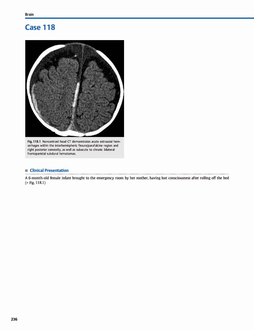
236
Brain
Case 1 1 8
Fig. 1 1 8.1 Noncontrast head CT demonstrates acute extraaxial hemorrhages within the interhemispheric fissure/parafalcine region and right posterior convexity, as well as subacute to chronic bilateral frontoparietal subdural hematomas.
• Clinical Presentation
A 6-month-old female infant brought to the emergency room by her mother, having lost consciousness after rolling off the bed
(� Fig. 1 1 8.1 )
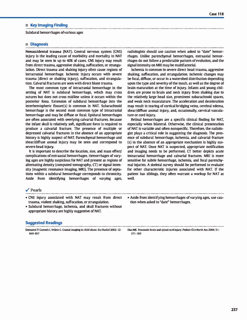
• Key lmaging Finding
Subdural hemorrhages ofvarious ages
• Diagnosis
Nonaccidental trauma {NAT). Central nervous system {CNS) injury is the leading cause of morbidity and mortality in NAT
and may be seen in up to 40% of cases. CNS injury may result from direct trauma, aggressive shaking, suffocation, or strangu
lation. Direct trauma and shaking injury often cause regions of intracranial hemorrhage. Ischemic injury occurs with severe trauma (direct or shaking injury), suffocation, and strangula
tion. Calvarial fractures are seen with direct blunt trauma.
The most common type of intracranial hemorrhage in the setting of NAT is subdural hemorrhage, whic:h may cross
sutures but does not cross midline unless it occurs within the posterior fessa. Extension of subdural hemorrhage into the interhemispheric fissure(s) is common in NAT. Subarac:hnoid hemorrhage is the second most common type of intracranial
hemorrhage and may be diffuse or focal. Epidural hemorrhages
are often associated with overlying calvarial fractures. Because
the infant skull is relatively soft, significant force is required to produce a calvarial fracture. The presenœ of multiple or depressed calvarial fractures in the absence of an appropriate history is highly suspect of NAT. Parenc:hymal hemorrhage and shear/diffuse axonal injury may be seen and correspond to
severe head injury.
lt is important to describe the location, size, and mass effect/ complications of extraaxial hemorrhages. Hemorrhages of vary
ing ages are highly suspidous for NAT and present as regions of altemating density ( computed tomography, CT) or signal inten
sity (magnetic resonance imaging, MRI). The presence of septations within a subdural hemorrhage corresponds to chronicity.
Aside from identifying hemorrhages of varying ages,
� Pearls
• CNS injury assodated with NAT may result from direct trauma, violent shaking, suffocation, or strangulation.
• Subdural hemorrhage, isc:hemia, and skull fractures without
appropriate history are highly suggestive of NAT.
Suggested Readings
Case 118
radiologists should use caution when asked to "date" hemor
rhages. Unlike parenc:hymal hemorrhages, extraaxial hemorrhages do not follow a predictable pattern of evolution, and the signal intensity on MRI may be multifactorial.
Ischemia is common in severe direct head trauma, aggressive
shaking, suffocation, and strangulation. Ischemic changes may be focal, diffuse, or occur in a watershed distribution depending
upon the type and severity of the insult. as well as the degree of brain maturation at the time of injury. Infants and young chil
dren are prone to brain and neck injury from shaking due to the relatively large head size, prominent subarachnoid spaces, and weak neck musculature. The acœleration and deœleration
may result in tearing of cortical bridging veins, cerebral edema,
shear/diffuse axonal injury, and, occasionally, cervical vasculature or cord injury.
Retinal hemorrhages are a specific dinical finding for NAT, especially when bilateral. Otherwise, the dinical presentation of NAT is variable and often nonspeàfic. Therefore, the radiolo
gist plays a critical rote in suggesting the diagnosis. The presence of subdural hemorrhage, ischemia, and calvarial fracture
{s) in the absence of an appropriate mechanism is highly suspect of NAT. Once NAT is suspected, appropriate notification and imaging needs to be performed. CT better depicts acute intracranial hemorrhage and calvarial fractures. MRI is more sensitive for subtle hemorrhage, ischemia, and focal parenchymal injuries. A skeletal survey should be performed to evaluate for other characteristic injuries associated with NAT. If the patient bas siblings, they often warrant a workup for NAT as
well.
• Aside from identifying hemorrhages of varying ages, use caution when asked to "date" hemorrhages.
Demaerel P, Casteels !, Wilms G. Cr.mial imaging in child abuse. Eur Radiol 2002; 12: Dias MS. lhlumatic brain and spinal cord iajury. Pediatr Oin North Am 2004; 51:
849-857 271-303
237
238
Brain
Case 1 1 9
Fig. 1 1 9.1 Axial unenhanced CT images (a-c) demonstrate hyperattenuation involving the superior sagittal sinus (a,b), cortical veins along the cranial vertex (a), and the transverse sinuses (c). Coronal 2-D TOF MR image (d) reveals iso- to hypointense filling defects within the superior sagittal and bilateral transverse sinuses. Minimal hyperintense flow can be seen along the periphery of the filling defects. Corresponding coronal Tl postcontrast image (e) shows the "empty delta sign" with enhancement around nonenhancing filling defects within the superior sagittal and transverse sinuses.
• Clinical Presentation
A 19-year-old woman with persistent, worsening headaches (� Fig. 1 1 9.1 )
• Key lmaging Finding
Hyperdense durai venous sinuses ( computed tomography, CT) with loss offlow signal (magnetic resonance venography, MRV) and filling defects (postcontrast)
• Diagnosis
Venous sinus thrombosis. Venous sinus thrombosis most often occurs in patients with an underlying hypercoagulable state related to trauma, regional or systemic infection, malignancy, pregnancy, medications (including oral contraceptives), dehydration, or inherited coagulopathies. In -25% of cases, however,
a causative factor is not identified. The clinicat presentation of venous sinus thrombosis is variable; therefore, imaging plays a critical roie in reaching the correct diagnosis. The most common clinical presentation is headaches. Focal neurological defidts and seizures typically result when there are parenchymal abnormalities assodated with the venous sinus thrombosis. Treatment consists of anticoagulation and correction of the
underlying hypercoagulable state. Venous sinus thrombosis most often involves the sagittal,
transverse, and sigmoid sinuses. At times, congenital variability in sinus development may complicate evaluation for stenosis or occlusion. In some patients, the anterior portion of the superior sagittal sinus is hypoplastic or atretic. Also, most patients have a dominant transverse and sigmoid sinus on one side with vari
able degrees of hypoplasia on the contralateral side. An additional pitfall is the presence of arachnoid granulations that project into the durai sinuses. most often the distal transverse sinus. A useful discriminator is that arachnoid granulations typically follow œrebrospinal fluid density and signal intensity, whereas thrombus does not.
On unenhanced CT, a thrombosed sinus is typically hyper
dense compared with the remaining vascular structures. One caveat is that in the setting of dehydration or increased hematocrit, ail vascular structures may be hyperdense. To distinguish between the two, it is helpful to compare arterial and venous density. In the setting of venous thrombosis, the thrombosed venous structure will have increased density compared with the arteries of the drcle of Willis. On contrast-enhanced CT,
� Pearts
• Venous thrombosis is hyperdense on unenhanœd CT; the uempty delta sign" is seen on enhanced CIV.
• MR signal of thrombus varies based upon chronicity; 2-D TOF MRV is most often used for venous thrombosis.
Suggested Reading
Leach JI., Fortuna RB,jones BV, Gaskill-Shipley MF. Irnaging of œrebral venous
thrombosis: c:urrent tedmiques, spectrum of findings, and diagnostic pitfall5.
Radiographies 2006; 26 Suppl 1 : S19-541, discussion 542-543
Case 119
there is contrast outlining a central hypodense clot, which is referred to as the uempty delta" sign.
On unenhanced MR imaging, routine sequences demonstrate variable signal intensity depending upon the age of the clot sec
ondary to the paramagnetic effects of hemoglobin breakdown products. Acutely (within the first 5 days), thrombus is typically isointense on Tl and hypointense on T2 images, which may mimic a patent flow void on T2. In the subacute stage (6 to 15 days). thrombus is more conspicuous and hyperintense on both Tl and T2. Chronically, the appearance is heterogeneous and
variable based upon the degree of recanalization and hemosiderin. Gradient echo or susceptibility-weighted images often demonstrate blooming artifact in the region of thrombus.
MRV is useful for evaluating venous thrombosis. fypically two-dimensional (2-D) time-of-fligltt (TOF) or contrastenhanced MRV are utilized. It is important to perform TOF imaging in two planes because in-plane flow may result in Joss of signal, which may be mistaken for absent flow. Contrast
enhanced MRV is helpful, especially in the acute phase where decreased T2 signal in the thrombus may mimic patent flow. Findings are similar to CIV with contrast outlining an intraluminal filling defect. In the chronic stage, enhanœd MRV is Jess useful because enhancement may be seen within an organized thrombus or partially recanalized clot, which can be mistaken for a patent sinus.
Parenchymal abnormalities associated with venous thrombo
sis most often occur when there is venous hypertension or when thrombus extends into superficiaJ veins. Parenchymal complications include cytotoxic or vasogenic edema, infarct, and hemorrhage. One of the more commonly involved superficial veins is the vein of Labbe; occlusion of the vesse! often results in characteristic temporal lobe hemorrhage.
• Parenchymal findings (edema, infarct, or hemorrhage) typically occur with clot extension into superficial veins.
239
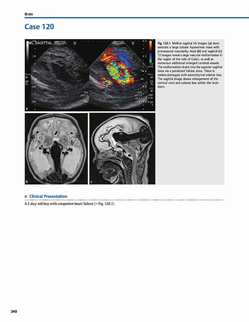
Brain
Case 1 20
• Clinical Presentation
A 2-day-old boy with congestive heart failure ( � Fig. 120.1)
240
Fig. 1 20.1 Midline sagittal US images (a) demonstrate a large tubular hypoechoic mass with pronounced vascularity. Axial (b) and sagittal (c) T2 images reveal a large vascular malformation in the region of the vein of Galen, as well as numerous additional enlarged cerebral vessels. The malformation drains into the superior sagittal sinus via a persistent falcine sinus. There is ventriculomegaly with parenchymal volume loss. The sagittal image shows enlargement of the cervical cord and volume loss within the brain stem.
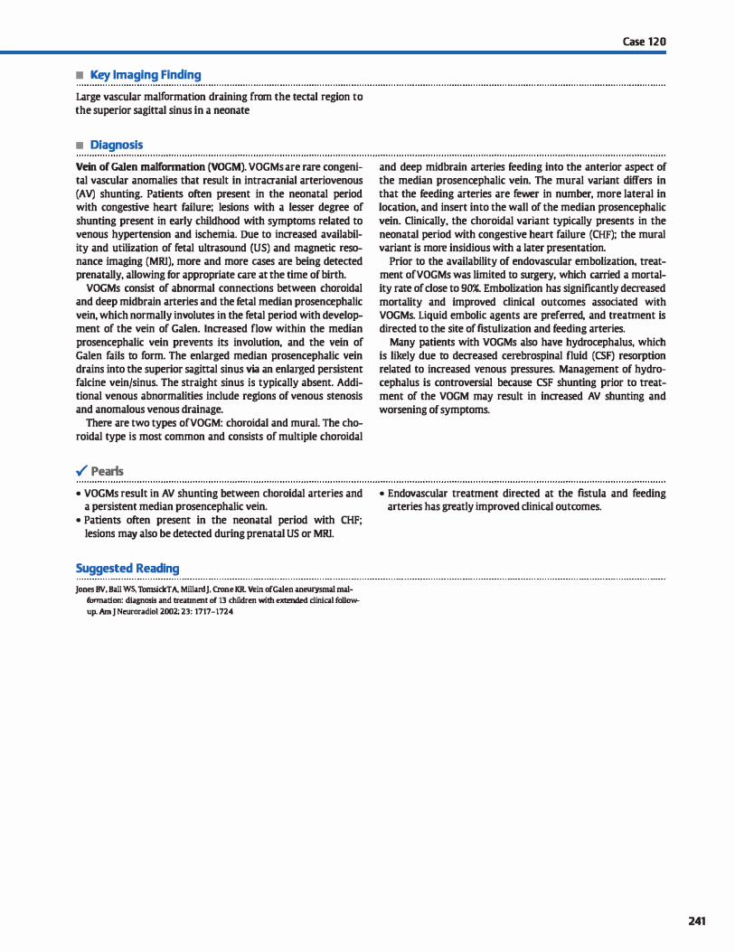
• Key lmaging Finding
Large vascular malformation draining from the tectal region ta the superior sagittal sinus in a neonate
• Diagnosis
Vein of Galen malfonnation (VOGM). VOGMs are rare congenital vascular anomalies that result in intracranial arteriovenous (AV) shunting. Patients often present in the neonatal period with congestive heart failure; lesions with a lesser degree of
shunting present in early c:hildhood with symptoms related ta venous hypertension and ischemia. Due to increased availability and utilization of fetal ultrasound (US) and magnetic resonance imaging {MRI), more and more cases are being detected prenatally, allowing for appropriate care at the time of birth.
VOGMs consist of abnormal connections between choroidal and deep midbrain arteries and the fetal median prosenœphalic vein, which normally involutes in the fetal period with development of the vein of Galen. lncreased flow within the median prosencephalic vein prevents its involution, and the vein of Galen fails to form. The enlarged median prosencephalic vein drains into the superior sagittal sinus via an enlarged persistent falcine vein/sinus. The straight sinus is typically absent. Additional venous abnormalities include regions of venous stenosis and anomalous venous drainage.
There are two types ofVOGM: choroidal and mural. The choroidal type is most common and consists of multiple choroidal
� Pearls
• VOGMs result in AV shunting between c:horoidal arteries and a persistent median prosencephalic vein.
• Patients often present in the neonatal period with CHF; lesions may also be detected during prenatal US or MRI.
Suggested Reading
Jones BV, Ball WS. TomsickTA, MillanlJ, Crone KR. Vein ofGalen aneurysmal malformation: diagnosis and treattnent of 13 children with extrnded dinical followup. Am j Neuroradiol 2002; 23: 1717-1724
Case 120
and deep midbrain arteries feeding into the anterior aspect of the median prosenœphalic vein. The mural variant differs in that the feeding arteries are fewer in number, more lateral in location, and insert into the wall of the median prosencephalic vein. Clinically, the c:horoidal variant typically presents in the neonatal period with congestive heart failure (CHF); the mural variant is more insidious with a later presentation.
Prior ta the availability of endovascular embolization, treatment ofVOGMs was limited ta surgery, which carried a mortality rate of dose to 90%. Embolization has significantly decreased
mortality and improved clinicat outcomes associated with VOGMs. Liquid embolie agents are preferred, and treatment is directed ta the site of fistulization and feeding arteries.
Many patients with VOGMs also have hydrocephalus, whic:h is likely due to decreased cerebrospinal fluid {CSF) resorption related to increased venous pressures. Management of hydrocephalus is controversial because CSF shunting prior to treatment of the VOGM may result in increased AV shunting and worsening of symptoms.
• Endovascular treatment directed at the fistula and feeding arteries bas greatly improved dinical outcomes.
241
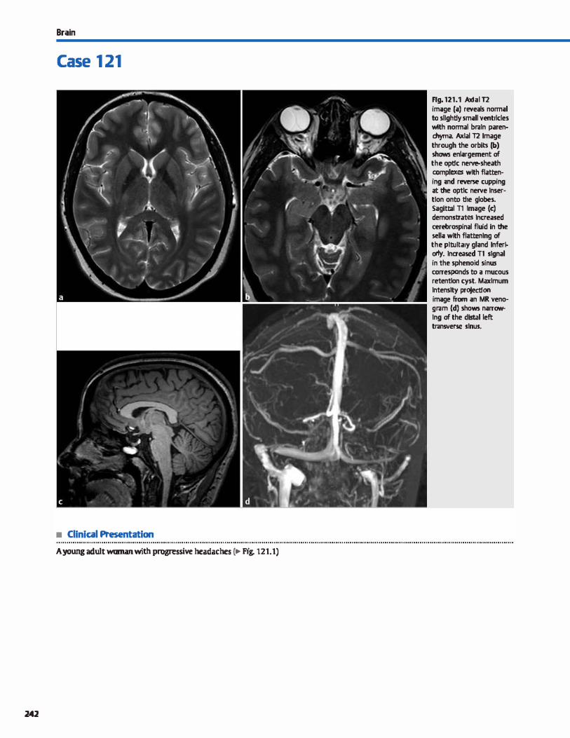
Brain
Case 1 21
• Clinical Presentation
A young adult woman with progressive headaches c.- Fig. 121.1)
242
Ffg.121.1 Axtal n image (a) reveals normal to sllghliy small ventrlcles wtth normal braln paren· chyma. Axlal n Image through the orbits (b) shaws enlargement of the oplfc nerve-sheath ccmplexes wlth flattening ;md reverse cupping at the optlc nerve lnser· tlon onto the globes. Sagittal T1 Image (c) demonstrates lncreased œrebro:spinal fluid in the sella wlth flattenlng of the pltultary gland lnfert· oriy. lnaeased Tl signal in the sphenoid sinus corresponds to a mucous retentlon cyst. Maximum lntenslty projeclfon image from an MR veno· gram (d) shows narrowlng of the dlrull left transverse sinus.
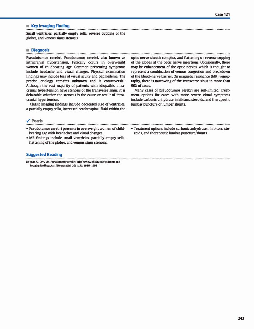
• Key lmaging Finding
Small ventricles, partially empty sella, reverse cupping of the
globes, and venous sinus stenosis
• Diagnosis
Pseudotumor cerebri. Pseudotumor œrebri, also known as intracranial hypertension, typically occurs in overweight women of childbearing age. Common presenting symptoms indude headache and visual changes. Physical examination
findings may include loss of visual acuity and papilledema. The precise etiology remains unknown and is controversial. Although the vast majority of patients with idiopathie intra
cranial hypertension have stenosis of the transverse sinus, it is debatable whether the stenosis is the cause or result of intracranial hypertension.
Classic imaging findings indude decreased size of ventrides,
a partially empty sella, increased cerebrospinal fluid within the
� Pearls
• Pseudotumor œrebri presents in overweight women of childbearing age with headaches and visual changes.
• MR findings indude small ventricles, partially empty sella,
flattening of the globes, and venous sinus stenosis.
Suggested Reading
Degnan JIJ, Levy IM. Pseudotumor œrebri: brief review of clinical syndrome and
imagingfindings.AmJNeuroradiol 2011; 32; 1986-1993
Case 121
optic nerve-sheath complex, and flattening or reverse cupping
of the globes at the optic nerve insertions. Occasionally, there may be enhanœment of the optic nerves, which is thought to represent a combination of venous congestion and breakdown
of the blood-nerve barrier. On magnetic resonance (MR) venography, there is narrowing of the transverse sinus in more than
90% of cases. Many cases of pseudotumor cerebri are self-limited. Treat
ment options for cases with more severe visual symptoms indude carbonic anhydrase inhibitors, steroids, and therapeutic
lumbar puncture or lumbar shunts.
• Treatment options indude carbonic anhydrase inhibitors, steroids, and therapeutic lumbar puncture/shunts.
243
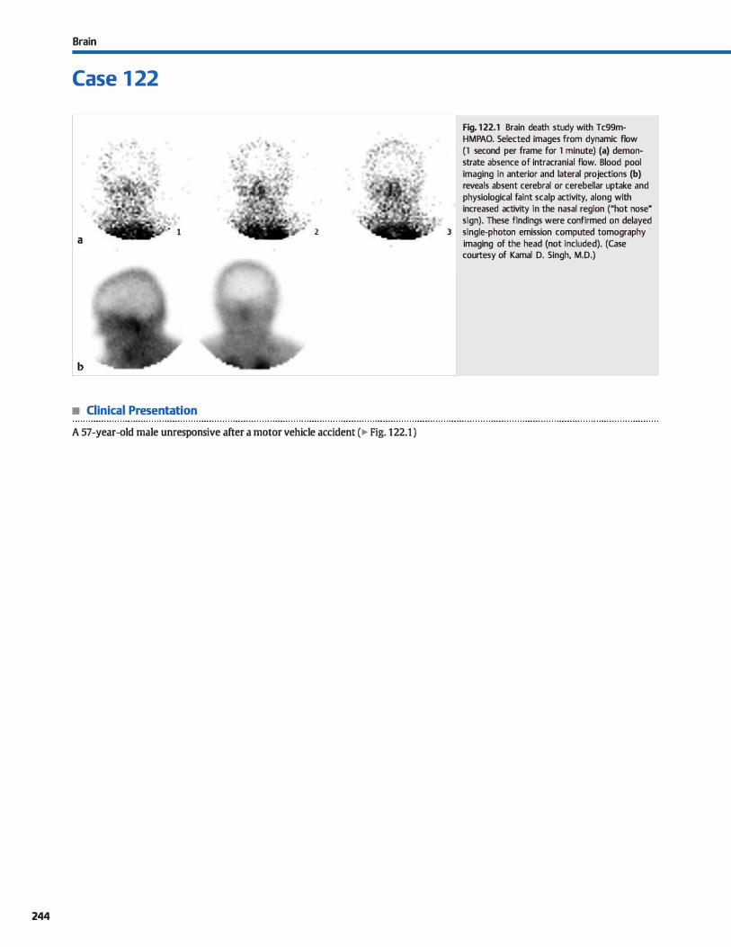
Brain
Case 1 22
2 a
b
• Clinical Presentation
A 57-year-old male unresponsive after a motor vehicle accident (� Fig. 122.1 )
244
Fig. 1 22.1 Brain death study with Tc99mHMPAO. Selected images from dynamic flow (1 second per frame for 1 minute) (a) demonstrate absence of intracranial flow. Blood pool imaging in anterior and lateral projections (b) reveals absent cerebral or cerebellar uptake and physiological faint scalp activity, along with increased activity in the nasal region ("hot nose" sign). These findings were confirmed on delayed
3 single-photon emission computed tomography imaging of the head (not included). (Case courtesy of Kamal D. Singh, M.D.)
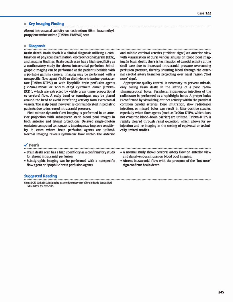
• Key lmaging Finding
Absent intracranial activity on technetium 99 m hexamethylpropyleneanùne oxime (Tc99m-HMPAO) scan
• Diagnosis
Brain death. Brain death is a clinicat diagnosis utilizing a combination of physical examination, electroencephalogram (EEG), and imaging findings. Brain death scan has a high spedfidty as a confirmatory study for absent intracranial perfusion. Scinti
graphie imaging can be performed at the patient's bedside with a portable gamma camera. lmaging may be performed with a nonspecific flow agent (Tc99 m diethylene-triamine-pentaacetate [f c99m-DTPA]) or with lipophilic brain perfusion agents (Tc99m-HMPAO or Tc99 m ethyl cysteinate dimer [fc99mECD]), which are extracted by viable brain tissue proportional to cerebral flow. A scalp band or tourniquet may be placed
around the head ta avoid interfering activity from extracranial vessels. The scalp band, however, is contraindicated in pediatric patients due ta increased intracranial pressure.
First minute dynanùc flow imaging is performed in an anterior projection with subsequent static blood pool images in bath anterior and lateral projections. Delayed single-photon emission computed tomography imaging may improve sensitivity in cases where brain perfusion agents are utilized. Normal imaging reveals symmetric flow within the anterior
� Pearts
• Brain death scan bas a high specificity as a confirmatory study for absent intracranial perfusion.
• Scintigraphie imaging can be performed with a nonspecific flow agent or lipophilic brain perfusion agents.
Suggested Reading
Conrad GR. Sinha P. Sdntigraphy as a confinnatory test of brain death. Sernin Nud
Med 2003; 33; 312-323
Case 122
and middle cerebral arteries (utrident sign") on anterior view with visualization of durai venous sinuses on blood pool imaging. In brain death, there is termination of carotid activity at the skull base due ta increased intracranial pressure overcoming
perfusion pressure, thereby shunting blood through the external carotid artery branches projecting over nasal region ("hot nase" sign).
Appropriate quality contrai is necessary to prevent mistakenly calling brain death in the setting of a poor radiopharmaceutical bolus. Peripheral intravenous injection of the
radiotraœr is performed as a rapid/tight bolus. A proper bolus is confirmed by visualizing distinct activity within the proximal common carotid arteries. Dose infiltration, slow radiotracer injection, or missed bolus can result in false-positive studies, especially when flow agents (such as Tc99m-DTPA, which does not cross the blood-brain barrier) are utilized. Tc99m-DTPA is rapidly cleared through renal excretion, which allows for re
injection and re-imaging in the setting of equivocal or technically limited studies.
• A normal study shows œrebral artery flow on anterior view and durai venous sinuses on blood pool imaging.
• Absent intracranial flow with the presenœ of the uhot nase" sign confirms brain death.
245
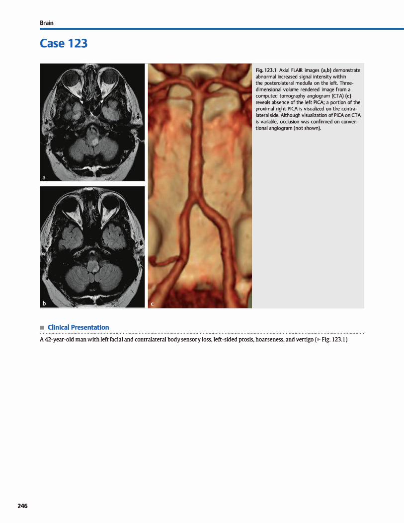
246
Brain
Case 1 23
• Clinical Presentation
Fig. 1 23.1 Axial FLAIR images (a,b) demonstrate abnormal increased signal intensity within the posterolateral medulla on the left. Threedimensional volume rendered image from a computed tomography angiogram (CTA) (c) reveals absence of the left PICA; a portion of the proximal right PICA is visualized on the contralateral side. Although visualization of PICA on CTA is variable, occlusion was confirmed on conventional angiogram (not shown).
A 42-year-old man with left facial and contralateral body sensory Joss, left-sided ptosis, hoarseness, and vertigo ( � Fig. 123.1 )
• Key lmaging Finding
Abnormal increased fluid-attenuated inversion recovery
(FLAIR) signal along the posterolateral medulla
• Diagnosis
Lateral medullary syndrome (LMS). LMS or Wallenberg syn
drome dassically results from occlusion of the posterior inferior
cerebellar artery (PICA) with associated infarct of the postero
lateral medulla. Similar findings may be seen with involvement
of the vertebral artery as well. The majority of cases occur in
older patients secondary to thromboembolic disease; in youn
ger patients, vertebral artery dissection associated with trauma
or vasculopathies are the most common etiologies.
Clinically, patients present acutely with Joss of pain and tem
perature sensation involving the ipsilateral face and contra
lateral body, ipsilateral Homer syndrome, ipsilateral limb
ataxia, and vertigo. Hoarseness and difficulty swallowing com
monly occur secondary to cranial nerve involvement. The
dinical symptoms and prognosis correlate well with the overall
� Pearts
• LMS classically results from occlusion of PICA with associated
infarct of the posterolateral medulla.
• Symptoms include Joss of pain/temperature sensation, Homer
syndrome, limb ataxia, hoarseness, and vertigo.
Suggested Reading
Kim JS, Lee JH, Suh OC. Lee MC. Spectrum of lateral medullary syndrome. Qirœlation
between dinical findings and magnetic resonance imaging in 33 subjects. Strolœ
1994; 25: 1405-1410
Case 123
size of the infarct; patients with larger infarcts have a worse prognosis. The majority of patients, however, have a relatively
good outcome with improvement of symptoms over time.
Computed tomography is often normal due to the inherent
difficulty in evaluating posterior fossa structures. Magnetic resonanœ imaging (MRI) is more sensitive, demonstrating regions
of increased T2/FLAIR signal along the posterolateral medulla.
lsolated medullary lesions may be subtle and only visualized on
the inferior-most axial images. Larger infarcts may also involve
the inferior cerebellum and are more conspicuous. Restricted
diffusion and mass effect/edema is seen acutely. Subtle
enhancement may be seen in the subacute phase. Chronically,
there is volume Joss of the involved segments.
• MRI reveals increased T2/FLAIR signal and restricted diffusion
(acutely) along the posterolateral medulla.
• Isolated medullary lesions may be subtle and only visualized
on the inferior-most axial MR images.
247
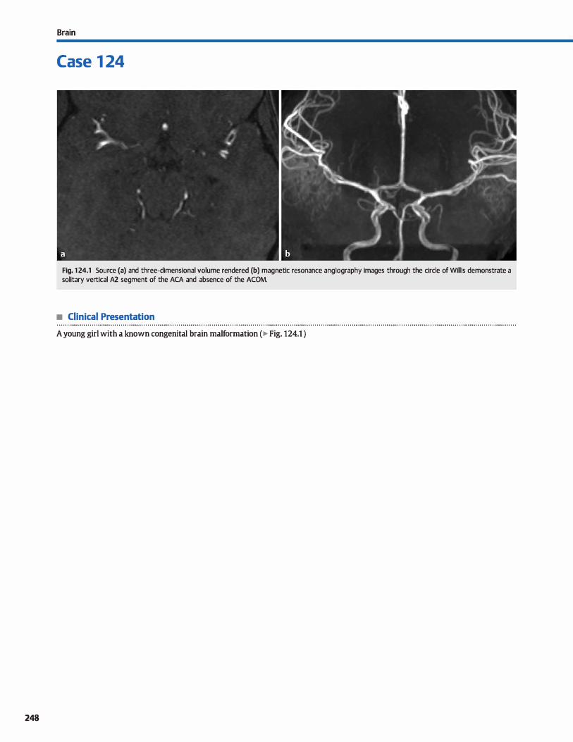
248
Brain
Case 1 24
Fig. 1 24.1 Source (a) and three-dimensional volume rendered (b) magnetic resonance angiography images through the circle of Willis demonstrate a solitary vertical A2 segment of the ACA and absence of the ACOM.
• Clinical Presentation
A young girl with a known congenital brain malformation ( � Fig. 124.1 )
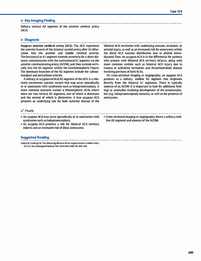
• Key lmaging Finding
Solitary vertical A2 segment of the anterior cerebral artery (ACA)
• Diagnosis
Azygous anterior œrebral artery (ACA). The ACA represents the anterior branch of the internai carotid artery after its bifurcation into the anterior and middle cerebral arteries. The horizontal or A 1 segment extends anteriorly for a short distance, communicates with the contralateral A 1 segment via the anterior communicating artery (ACOM), and then extends vertically into the A2 segment within the interhemispheric fissure. The dominant branches of the A2 segment indude the callosomarginal and pericallosal arteries.
A solitary or azygous vertical A2 segment of the ACA is a relatively uncommon vascular variant that may occur sporadically or in association with syndromes such as holoprosenœphaly. A more common anatomie variant is bihemispheric ACAs where there are two vertical A2 segments, one of which is dominant and the second of which is diminutive. A true azygous ACA presents an underlying risk for both ischemic disease of the
� Pearts
• An azygous ACA may occur sporadically or in association with syndromes such as holoprosencephaly.
• An azygous ACA presents a risk for bilateral ACA territory infarcts and an increased risk of distal aneurysms.
Suggested Reading
Le May M, Gao ding CA. The clinical significance of the azygos anterior cerebral artery
{A.CA). Am j Roentgenol Radium Ther Nud Med 1966; 98: 602-610
Case 124
bilateral ACA territories with underlying stenosis, ocdusion, or arterial injury, as well as an increased risk for aneurysms within the distal ACA vascular distribution due to altered hemodynamic flow. An azygous ACA is in the differential for patients who present with bilateral ACA territory infarcts, along with more common entities such as bilateral ACA injury due to trauma or subfalcine hemiation and thromboembolic disease involving portions of both ACAs.
On cross-sectional imaging or angiography, an azygous ACA presents as a solitary, midline A2 segment that originates directly from the bilateral A 1 segments. There is typically absence of an ACOM. lt is important to look for additional findings or anomalies involving development of the prosencephalon ( e.g., holoprosencephaly variants), as well as the presenœ of aneurysms.
• Cross-sectional imaging or angiography shows a solitary midline A2 segment and absence of the ACOM.
249
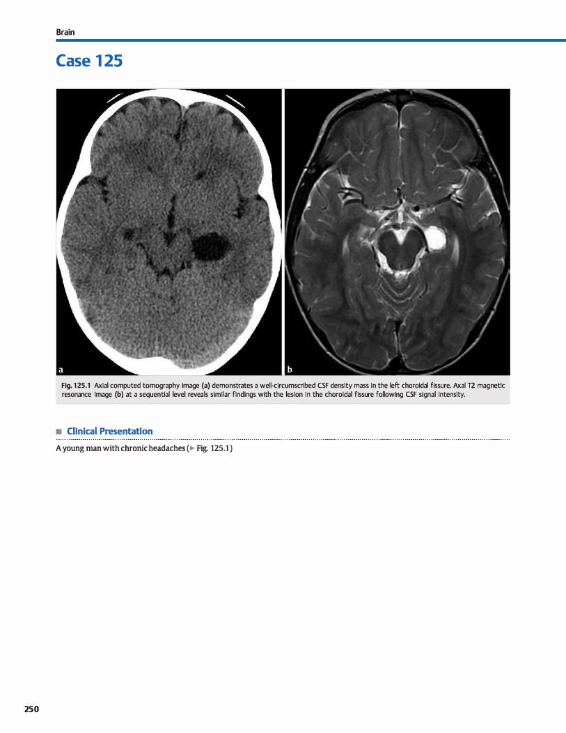
250
Brain
Case 1 25
Fig. 1 25.1 Axial computed tomography image (a) demonstrates a well-circumscribed CSF density mass in the left choroidal fissure. Axai T2 magnetic resonance image (b) at a sequential level reveals similar findings with the lesion in the choroidal fissure following CSF signal intensity.
• Clinical Presentation
A young man with chronic headaches (� Fig. 125.1 )
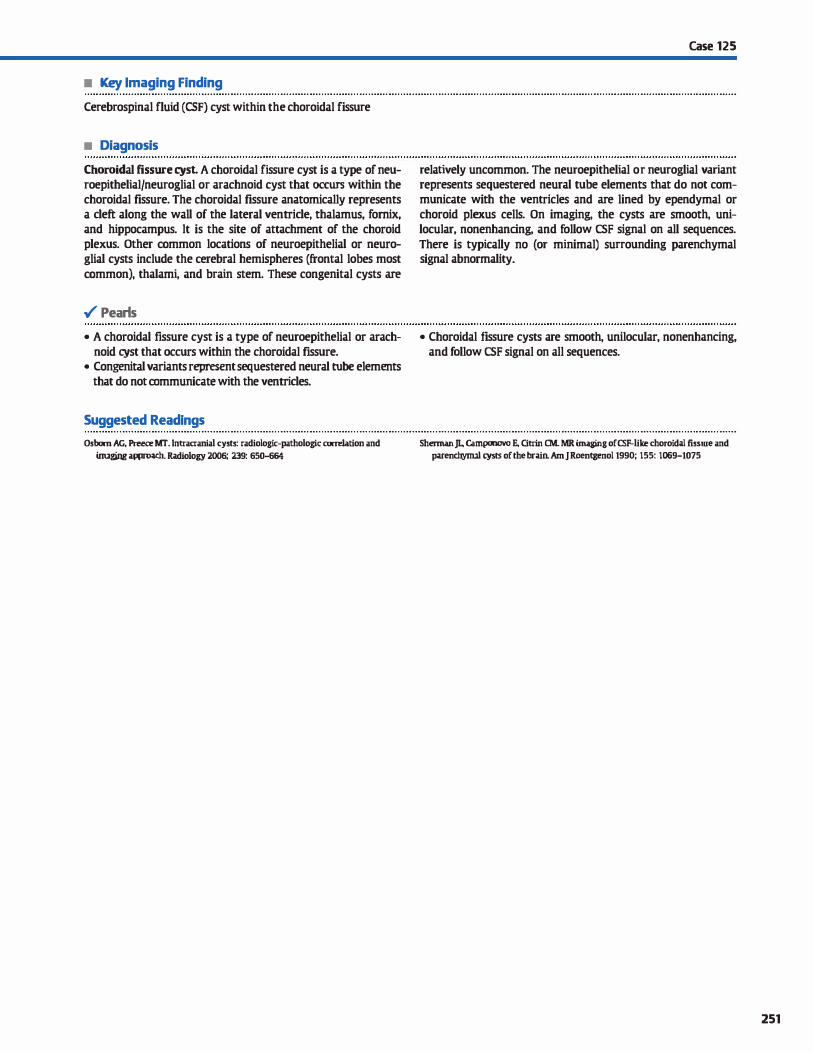
• Key lmaging Finding
Cerebrospinal fluid (CSF) cyst within the choroidal fissure
• Diagnosis
Choroidal fissure cyst. A choroidal fissure cyst is a type of neuroepithelial/neuroglial or arachnoid cyst that occurs within the choroidal fissure. The choroidal fissure anatomically represents a deft along the wall of the lateral ventride, thalamus, fomix, and hippocampus. lt is the site of attachment of the choroid plexus. Other common locations of neuroepithelial or neuroglial cysts include the œrebral hemispheres (frontal lobes most
common), thalami, and brain stem. These congenital cysts are
� Pearts
• A choroidal fissure cyst is a type of neuroepithelial or arachnoid cyst that occurs within the choroidal fissure.
• Congenital variants represent sequestered neural tube elements that do not communicate with the ventrides.
Suggested Readings
Osborn AG, Preece MT. lntracranial cysts: radiologie-pathologie correlation and
imaging appro;ich. Radiology 2006; 239: 650-664
Case 125
relatively uncommon. The neuroepithelial or neuroglial variant represents sequestered neural tube elements that do not communicate with the ventricles and are lined by ependymal or choroid plexus cells. On imaging, the cysts are smooth, unilocular, nonenhandng, and follow CSF signal on ail sequences.
There is typically no (or minimal) surrounding parenchymal signal abnormality.
• Choroidal fissure cysts are smooth, unilocular, nonenhancing, and follow CSF signal on ail sequences.
Sherman JL, Camponovo E, Gtrin CM. MR imaging of CSF-lilœ choroidal fissure and
parench;yrnal cysts of the brain AmJ Rcentgenol 1990; 155: 1069-1075
251
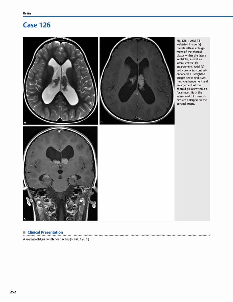
Brain
Case 1 26
• Clinical Presentation
A 4-year-old girl with headaches (� Fig. 126.1 )
252
Fig. 1 26.1 Axial T2-weighted image (a) reveals diffuse enlargement of the choroid plexus within the lateral ventricles, as well as lateral ventricular enlargement. Axial (b) and coronal (c) contrastenhanced Tl -weighted images show avid, symmetric enhancement and enlargement of the choroid plexus without a focal mass. Both the lateral and third ventricles are enlarged on the coronal image.
• Key lmaging Finding
Diffuse, symmetric choroid plexus enlargement without focal
mass
• Diagnosis
Villous hyperplasia of the choroid plexus. Villous hyperplasia
of the choroid plexus is a rare congenital abnonnality charac
terized by diffuse, symmetric enlargement of the choroid
plexus. Patients present with communicating hydrocephalus
due to overproduction of cerebrospinal fluid {CSF). On patho
logical examination, the choroid plexus cells have a normal
appearanœ but are increased in number. Magnetic resonanœ
� Pearts
• Villous hyperplasia is characterized by diffuse, symmetric
choroid plexus enlargement • Patients present with communicating hydrocephalus due to
overproduction of CSF.
Suggested Reading
Naeini RM, Yoo JH, Hunter JV. Spectrum of choroid plexus les ions in children Am j Roentgenol2009; 192: 32-40
Case 126
imaging (MRI) and computed tomography (CT) demonstrate
diffuse, symmetric choroid plexus enlargement and enhance
ment without focal mass, which distinguishes this entity from
focal lesions such as choroid plexus papilloma or carcinoma.
Treatment options indude CSF shunting versus choroid plexus
resection or coagulation.
• MRI and CT demonstrate diffuse choroid plexus enlargement
and enhancement without focal mass.
• Treatment options for hydroœphalus include CSF shunting
versus choroid plexus resection/coagulation.
253
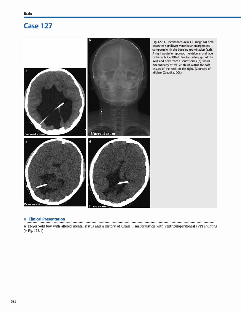
254
Brain
Case 1 27
• Clinical Presentation
Fig. 1 27.1 Unenhanced axial CT image (a) demonstrates significant ventricular enlargement compared with the baseline examination (c,d). A right posterior approach ventricular drainage catheter is identified. Frontal radiograph of the skull and neck from a shunt series (b) shows discontinuity of the VP shunt within the soft tissues of the neck on the right. (Courtesy of Michael Zapadka, DO.)
A 12-year-old boy with altered mental status and a history of Chiari II malformation with ventriculoperitoneal (VP) shunting
(� Fig. 127.1 )
• Key lmaging Finding
lnterval dilatation of ventricles and clinical decline in a patient
with a VP shunt
• Diagnosis
Shunt catheter malfunction. Hydroœphalus may result from obstruction, overproduction, or reduœd resorption of cerebrospinal fluid (CSF). Communicating hydrocephalus occurs secondary to decreased resorption of CSF at the arachnoid villi
and may be due to hemorrhage, infection, carcinomatosis, or arachnoid villi immaturity. Noncommunicating hydrocephalus is caused by a congenital or acquired obstruction of CSF at the
ventricular level. OVerproduction of CSF is seen in the setting of tumors or villous hypertrophy of the choroid plexus.
VP shunting remains the most common treatment for chronic hydroœphalus. VP shunts consist of the intraventricular drain
age catheter, an extracranial valve and reservoir, and a catheter that extends throughout the superficial soft tissues and into the peritoneum. Shunt malfunctions are unfortunately common in chronically shunted patients. Malfunction may be mechanical
(most common) or due to an underlying infection. Common presenting symptoms indude headache, irritability, lethargy, nausea, and vomiting. Additional symptoms include fever and
localizing pain. Young children with open sutures often present
with increasing head size. dinically, slowed filling of the shunt
reservoir is suggestive of malfunction. Mechanical failure may result from obstruction of the shunt
catheter, disconnections or breaks in the catheter tubing sys
tem, and migration. Obstruction is most common at the intraventricular end of the shunt catheter and may be caused by proteinaceous material (hemorrhage, infection, tumor), brain
parenchyma, or choroid plexus obstructing the inflow of CSF. Distal obstruction at the peritoneal end is Jess common and typically results from peritoneal adhesions near the catheter
tip. Occasionally, an intraventricular cyst or abdominal pseudocyst may obstruct CSF flow. Nudear medicine radioisotope or
contrast-enhanœd computed tomography (CT) or fluoroscopic shunt studies can be used to evaluate for obstruction within the
VP shunt system.
� Pearts
• Shunt malfunction is common in chronically shunted patients
and may be mechanical or related to infection.
• It is important to know the type of catheter used and which portions are normally radio-opaque and radiolucent
Suggested Reading
Goeser CD, Mcl.eary MS, Young LW, Diagnostic imaging of ventriculoperitoneal shunt malfunctions and complications. Radiographies 1998; 18: 635-651
Case 127
Disconnection of shunt components most often occurs where catheter tubing connects to the reservoir. Breaks occur in regions of increased mobility, such as the neck. Migration typi
cally affects the proximal or distal end of the VP shunt catheter.
When interpreting a plain film shunt study, it is important to know the type of catheter used, particularly with respect to the portions that are normally radio-opaque and radioluœnt, to avoid mistaking normal radioluœnt portions of the tubing as regions of discontinuity. If needed, cross-sectional imaging can be used for problem solving. A focal fluid collection may be
identified at the site of malfunction.
cr or magnetic resonance imaging (MRI) (preferred) of the brain most often demonstrates ventricular enlargement with
shunt malfunction; however, this finding is not entirely sensitive or spedfic. False-negatives occur with scarring along the ventrides or decreased parenchymal complianœ. Conversely, ventricular enlargement may be seen with normally function
ing shunts. Secondary findings of shunt malfunction indude sulcal and cistemal effacement and transependymal flow of CSF, which suggest uncompensated, acute hydrocephalus. Com
plications of VP shunting also include slit ventricle syndrome and subdural fluid collections. Etiologies of slit ventricle syndrome indude overshunting, fibrosis, decreased parenchymal compliance, and intracranial hypotension. OVerdrainage may
result in subdural hematomas or hygromas. lYJ>ically, subdural collections are self-limited.
Shunt infections most often occur within the first few
months of catheter placement Patients present with signs of shunt malfunction, as well as fever. lmaging may be normal or demonstrate findings associated with ventriculitis or meningitis, to indude abnormal T2/fluid-attenuated inversion recovery signal intensity and enhanœment along the margins of the ven
trides and meninges. Replacement of the infected shunt is often necessary.
• cr or MRI (preferred) most often demonstrate ventricular enlargement in the setting of shunt failure.
• Overshunting may result in slit ventride syndrome and subdural hematomas or hygromas.
255
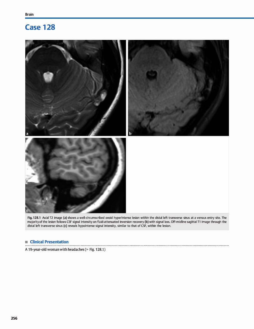
256
Brain
Case 1 28
Fig. 1 28.1 Axial T2 image (a) shows a well-circumscribed ovoid hyperintense lesion within the distal left transverse sinus at a venous entry site. The majority of the lesion follows CSF signal intensity on fluid-attenuated inversion recovery (b) with signal loss. Off-midline sagittal T1 image through the distal left transverse sinus (c) reveals hypointense signal intensity, similar to that of CSF, within the lesion.
• Clinical Presentation
A 19-year-old woman with headaches (� Fig. 128.1 )
• Key lmaging Finding
Well-circumscribed Tl hypointense, T2 hyperintense lesion
within a dural sinus
• Diagnosis
Arachnoid granulation. Arachnoid granulations represent pro
tuberances of the leptomeninges that extend into the lumen of
dural venous sinuses, particularly the sagittal, transverse, and
sigmoid sinuses. They are nonnal, incidental structures but
may be mistaken for durai sinus pathology, such as thrombus,
on cross-sectional imaging studies.
Arachnoid granulations are similar to arachnoid villi but are
exponentially larger (villi are microscopie) and more complex.
Their predse function remains unclear; however, they are
thought to play a rote in cerebrospinal fluid (CSF) resorption,
similar to arachnoid villi. They are commonly seen at venous
entry sites and have increased incidence and conspicuity with
age. Although anatomically they are most frequently seen in the
sagittal sinus, on imaging studies, they are most commonly
identified in the distal transverse sinus.
On contrast-enhanced computed tomography (CT), arachnoid
granulations present as focal, well-circumscribed filling defects
within the durai venous sinus, surrounded by enhandng portions of the dural sinus. They are typically hypodense, similar in
attenuation ta CSF, but they may occasionally be isodense
� Pearts
• Arachnoid granulations represent protuberances of leptomeninges that extend into a dural venous sinus lumen.
• Functionally, arachnoid granulations are similar ta arachnoid
villi but are much larger and more complex.
Suggested Reading
Leadt]l.Jones BV, Tomsidi: TA, Sœw.ut CA, Ballco MC. Normal appearanœ of arach
noid granulatillll5 on contrast-enhanœd CT and MR of the brain; differentiation
front dw-al sinus disease. Amj Neuroradiol 1996; 17: 1523-1532
Case 128
relative to brain parenchyma. On magnetic resonanœ imaging
(MRI), arachnoid granulations are hypointense on Tl and
hyperintense on T2 sequences with variable signal intensity on
proton density and fluid-attenuated inversion recovery
sequenœs. Lesions do not demonstrate contrast enhancement The characteristic locations and imaging appearance of
arachnoid granulations are helpful in distinguishing them from venous thrombosis, which typically involves an entire segment
of a dural venous sinus or multiple segments with extension
into cortical veins. Thrombus is oft:en hyperdense on CT and
hyperintense on unenhanced Tl-weighted MR sequences, especially with acute or subacute thrombus. Secondary signs of
venous hypertension, such as collateralization, meningeal
enhancement, parenchymal edema, and hemorrhage, may be seen with venous sinus thrombosis, espedally with extension
of thrombus into cortical veins. These are never seen in the set
ting of arachnoid granulations.
As nonnal structures, arachnoid granulations are incidental,
asymptomatic lesions that require no treatment or follow-up.
• Arachnoid granulations are most common in the sagittal,
transverse, and sigmoid sinuses at venous entry points.
• Arachnoid granulations are nonnal structures that are well
drcumscribed, Tl hypo-, and T2 hyperintense.
257


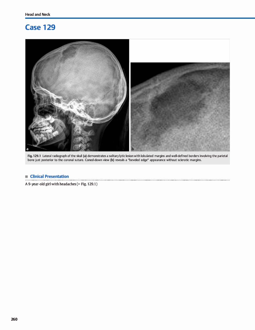
260
Head and Neck
Case 1 29
Fig. 1 29.1 Lateral radiograph of the skull (a) demonstrates a solitary lytic lesion with lobulated margins and well-defined borders involving the parietal bone just posterior to the coronal suture. Coned-down view (b) reveals a "beveled edge" appearance without sderotic margins.
• Clinical Presentation
A 9-year-old girl with headaches ( � Fig. 129.1 )
• Key lmaging Finding
Lytic skull lesion in a child
• Top 3 Differential Diagnoses
• Langerhans cell histiocytosis {LCH). The skull is the most common location for osseous involvement in LCH. The classic radiological description is a well-defined lytic lesion without sderotic borders. A ubeveled" edge appearance often results from greater involvement of the inner than outer table of the skull. LŒ may also present as a round, radiolucent skull defect with a central dense nidus or sequestrum of intact
bone, referred to as a "button sequestrum." The most common dinical symptoms indude pain, palpable mass, and/or systemic symptoms. Magnetic resonance imaging (MRI) reveals a Tt hypointense and T2 intermediate to hyperintense soft-tissue mass with moderate enhancement associated with an osseous defect.
• Epidermoid cyst. Epidermoid cysts result from abnormal deposition of epithelial rests within the diploic space during development. They are a relatively common cause of solitary lytic skull lesions in the pediatric population and tend to occur along sutural lines. When an epidermoid cyst involves the diploic spaœ, the radiographical appearance may overlap
• Additional Differential Diagnoses
• Infection. Osteomyelitis has a variety of imaging manifestations, which range from a normal appearanœ to an aggressive
appearance that mimics a neoplastic process. Bony destruction from osteomyelitis commonly results in lytic calvarial lesions that often have poorly defined, infiltrating margins. There may be overlying soft-tissue swelling. Subacute and chronic infections may present radiographically as a round, radiolucent skull defect with a central dense nidus or sequestrum of intact bone, referred ta as a "button sequestrum." On MRI, osteomyelitis presents as regions of marrow infiltration or bony destruction with variable enhanœment.
• Diagnosis
Langerhans cell histiocytosis
� Pearls
• LŒ dassically presents as a lytic skull lesion with nonsderotic, "beveled" margins.
• LŒ and osteomyelitis can have "button sequestra" and systemic symptoms.
Suggested Readings
Gibson SE, Prayson RA. Primary slrull lesions in the pediatric population: a 25-year experienœ.ArchPatholl.ab Med 2007; 131; 761-766
Glass RBJ, Fembach SK, Norton KI, Owi PS, Naidich TP. The infant skull: a vault of
infonnation. Radiographies 2004; 24: 507-522
Case 129
that of LŒ. Epidermoid cysts are generally well-defined expansile lesions without a central matrix. They may or may not have a sderotic rim. On MRI, epidermoid cysts are isoto hypointense ta cerebrospinal fluid (CSF) on Tt and iso- ta hyperintense on T2 with characteristic increased signal on diffusion-weighted imaging.
• Malignancy. The most common malignancies ta involve the calvarium in children indude lymphoma, leulœmia, Ewing sarcoma, and metastatic neuroblastoma. Rhabdomyosarcoma may also invade the calvarium via direct spread. Malignancies often result in bony expansion and destruction, which manifests as poorly defined lucencies. Aggressive periosteal reaction
may be noted, especially with Ewing sarcoma. Lesions may be solitary or multiple; multiple lesions are suggestive of hematogenous spread On MRI, malignancies demonstrate marrow infiltration or bony expansion and destruction with a softtissue mass. The soft-tissue component is typically hypointense on Tt and hyperintense on T2 with avid but heterogeneous enhancement.
• Leptomeningeal cyst. The tenn leptomeningeal cyst (growing skull fracture) refers ta a well-defined bony defect that arises
when traumatic laceration of the dura exposes the bone to the pulsations of CSF within the subarachnoid spaœ. Pulsatile pressure erosion gradually widens the fracture line. Leptomeningeal cysts are an uncommon complication of skull fractures and are most common in children < 3 years of age. Cross-sectional imaging demonstrates CSF attenuation or signal without an underlying soft-tissue component.
• Common malignant skull lesions in children include lymphoma, leulœmia, Ewing sarcoma, and neuroblastoma.
• A leptomeningeal cyst {growing skull fracture) is caused by CSF pulsations from disruption of the dura.
Krasnokutsky MV. The button sequestrum sign. Radiology 2005; 236; 1026-1027 Yalçin 0, Yildirim T, Kizilkiliç 0 et al. CT and MRI findings in calvarial non-infectious
lesions.DiagnlntervRadiol 2007; 13: 68-74
261
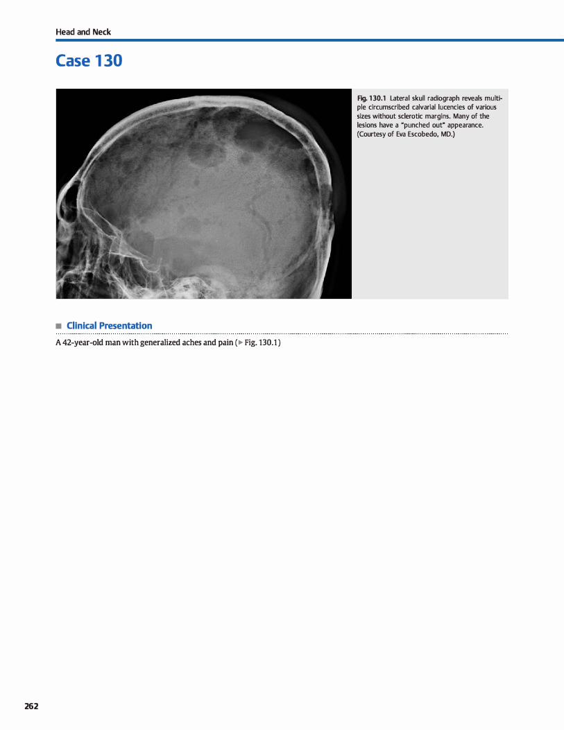
Head and Neck
Case 1 30
• Clinical Presentation
A 42-year-old man with generalized aches and pain (� Fig. 130.1 )
262
Fig. 130.1 Lateral skull radiograph reveals multiple circumscribed calvarial lucencies of various sizes without sclerotic margins. Many of the lesions have a "punched out" appearance. (Courtesy of Eva Escobedo, MD.)
• Key lmaging Finding
Lytic skull lesion(s) in an adult
• Top 3 Differential Diagnoses
• Malignancy. Metastases, multiple myeloma, and lymphoma are common causes of solitary or multiple (more common)
lytic calvarial lesions in adults. Primary sites of metastatic disease indude Jung, breast, prostate, renal, and thyroid cardnomas. Lung metastases are lytic; prostate lesions are sderotic; and breast metastases may be lytic or sderotic. Renal and thyroid lesions are often solitary, hypervascular, and may
also be lytic or sderotic. Metastatic disease may appear as enhancing regions of marrow infiltration or soft-tissue masses with bony destruction. Multiple myeloma is the most common primary bone tumor in adults; plasmacytoma is the solitary form. Myeloma lesions appear as well-defined "punched out" lesions with involvement of bath the inner and outer tables of the calvarium. The soft-tissue component
is hypointense on T1 and hyperintense on T2 images with enhancement. A skeletal survey should be performed to identify additional areas of involvement. Lymphoma is similar
in appearance ta metastatic disease with an enhancing lytic calvarial lesion.
• Fibrous dysplasia (ID). FD occurs most often in young adults and is more common in women. The calvarium and facial
• Additional Differential Diagnoses
• Surgical defect. Surgical defects may be secondary to burr hales or craniotomy: craniotomy changes are typically obvious. Bur hales are well-drcumscribed and most often located over the frontal lobes (right > left). There is no associated softtissue component, unless portions of the meninges and/or parenchyma extend into the defect. Occasionally, a hypodense (CT) or T2 hyperintense (MRI) tract may be seen in cases of
prior shunt catheter or intracranial monitor placement
• Epidermoid cyst Epidermoid cysts result from abnormal deposition of epithelial rests within the diploic spaœ during
development. They are generally well-defined expansile lesions without a central matrix. They may or may not have a sderotic rim. On MRI, epidermoid cysts are iso- to hypoin-
• Diagnosis
Malignancy (multiple myeloma)
� Pearls
• Metastases, multiple myeloma, and lymphoma often involve
the skull; myeloma has "punched out" lesions. • Paget disease of the skull may result in osteoporosis circum
scripta, sderosis, or a "cotton wool" appearanœ.
Suggested Reading
Yalçin o, Yildirim T, Kizilkiliç o et al. cr and MRI findings in calvarial non-infectious lesiorus. Diagn lnterv Radiol 2007; 13: 68-74
Case 130
bones are most often affected in the head and neck. The dassic appearanœ on computed tomography (CT) is focal bony expansion with "ground glass" attenuation of the diploic spaœ. However, sderotic and cystic variants due occur; the
cystic variant may mimic a lytic lesion. On magnetic resonanœ imaging (MRI), FD is typically hypointense on bath T1 and T2 sequences and demonstrates avid enhancement
Regions of cystic involvement are Jess characteristic because they are often hyperintense on T2 sequences. The MRI appearance can mimic aggressive lesions; therefore, CT is preferred in suspected cases of FD.
• Paget disease. Paget disease occurs in elderly patients and
consists of three stages: lytic, sderotic, and mixed. Calvarial involvement during the lytic phase results in a characteristic large lytic lesion with erosion of the outer table, referred ta as osteoporosis drcumscripta. During the sderotic phase, there is expansion and sderosis of the cortex and diploic spaces, which may be diffuse or focal. ln the mixed phase, a combination of lytic and sclerotic lesions results in the characteristic "cotton wool" appearanœ of the skull.
tense to CSF on T1 and iso- to hyperintense on T2 with characteristic increased signal on ditfusion-weighted imaging.
• Metabolic disease. Hyperparathyroidism (HPT) may be primary due to hyperlunctioning parathyroid glands, secondary due to vitamin D deficiency or chronic renal failure, or tertiary due to hyperplasia from long-standing secondary HPT. Increased parathormone results in systemic calciumfbony
resorption. In the skull, this manifests as alternating regions of lucency and sderosis, referred ta as the "sait and pepper" appearance. In advanced cases, focal Brown tumors may
occur, presenting as well-circumscribed lytic lesions that are often multiple.
• HPT manifests as altemating regions of lucency and sderosis,
referred to as a "sait and pepper" appearance.
263
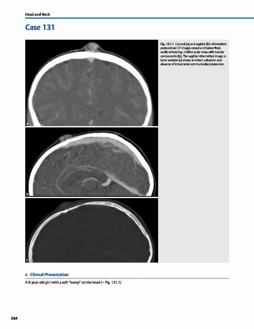
Head and Neck
Case 1 31
• Clinical Presentation
A �year-old girl with a soft "bump" on the head (.,Fig. 131.1)
264
Flg.131.1 Coronal(a) and sagittal (b) refonmrtted postcontrast a Images rewal a clramsafbed, avldly enhanctng, mtdllne scalp mass Wth tubular components (b). 1he s;igitt.11 ref'ormitted im;ige in bone w!ndaw (<) shows an lntict calvarlum and absence of lntraaanlal communtcadon/extenslon.
• Key lmaging Finding
Enhancing scalp lesion in a child
• Top 3 Differential Diagnoses
• Hemangioma. Hemangiomas are benign lesions composed of endothelial œlls and commonly occur in the head and neck (-60%). They are often solitary and sporadic; however, they may be multiple in the setting of PHACES syndrome (posterior fossa malformations, hemangiomas, arterial anomalies,
cardiac defects, eye abnormalities, and sternal cleft and supraumbilical raphe syndrome). Lesions are categorized as infan
tile (most common), which typically occur within the first
few weeks of life, proliferate in infancy and early childhood, and involute within the first decade of life with fatty replace
ment; or congenital, which are present at birth and either involute shortly thereafter (rapidly involuting congenital hemangioma [RICH]) or do not involute (noninvoluting congenital hemangioma [NICH]). On computed tomography (CT)
and magnetic resonanœ imaging (MRI), lesions are welldefined, lobulated, and avidly enhance. They are characteristi
cally hyperintense on T2 sequences. They are typically located within superticial soft tissues but may extend into multiple compartments. Treatment is reserved for symptomatic lesions
with mass effect on vital structures.
• Venolymphatic malfonnation {VM). VMs are congenital anomalies that are part of the spectrum of low-flow venolym
phatic malformations. They commonly occur in the head and neck region (-40%), and the majority are solitary and spo
radic; however, multiple lesions may be seen in the setting of
• Additional Differential Diagnoses
• Langerhans œll histiocytosis {LCH). The skull is the most common location for osseous involvement in LCH. The classic
radiological description is a well-defined lytic lesion without sclerotic borders. A ubeveled" edge appearance often results
from greater involvement of the inner than outer table of the
skull. LCH may also present as a round, radiolucent skull defect with a central dense nidus or sequestrum of intact
bone, referred to as a ubutton sequestrum." The most common clinicat symptoms indude pain, palpable mass, and/or
systemic symptoms. MRI reveals a T1 hypointense and T2
• Diagnosis
Scalp venous malformation
� Pearls
• Infantile hemangiomas occur after birth, proliferate in
infancy/childhood, and involute within the first decade. • VMs are congenital anomalies, are present at birth, grow pro
portional to somatic growth, and do not involute.
Suggested Reading
Moron FE, Morriss MC, Jones.Il, Hunter JV. Lumps and bumps on the head in dùldren: use of CT and MR imaging in solving the clinical diagnostic dilemma. Radiographies 2004; 24: 1655-1674
Case 131
Klippel-lTenaunay-Weber (bony overgrowth/hypertrophy),
Maffucci (multiple enchondromatosis), and blue rubber bleb nevus (superficial and visceral hemangiomas) syndromes. Unlike infantile hemangiomas, they are present at birth and do not undergo involution. Growth is seen in childhood
proportional to somatic growth. On imaging, lesions are typically circumscribed and may be multicystic with avid enhanœment. Thrombosis may complicate the imaging find
ings. Phleboliths are characteristic and are hyperdense on CT
and hypointense on MRI. • Sinus pericranü. Sinus pericranii refers to anomalous trans
osseous communication between extracranial and intra
cranial venous structures, typically a durai venous sinus. It most often presents in childhood as a blue or purplish soft
scalp mass that increases in size when lying fiat or with
Valsalva maneuvers. Ultrasound reveals a superticial vascular scalp mass that extends intracranially via a dilated transdi
ploic vein. The extracranial component may be circumscribed or consist of a single or multiple dilated veins. CT is ideal for identifying the underlying osseous defect, which may be subtle; the venous communication is best depicted on CT or MR
venography. MR venography is often preferred due to the lack of ionizing radiation. On routine MR sequences, the scalp mass is Tl iso- to hypointense and T2 hyperintense with avid enhancement. Treatment is surgical.
intermediate ta hyperintense soft-tissue mass with moderate enhancement associated with an osseous defect.
• Malignancy. Superficial scalp neoplasms are uncommon in children and most often result from spread of an underlying
bony malignancy, such as lymphoma, leukemia, Ewing sar
coma. or metastases (neuroblastoma being most common); a primary sarcoma is less common. Malignancies are typically
aggressive with soft-tissue infiltration and bony destruction, T2 intermediate to hyperintense, and avid but heterogeneous enhancement.
• Sinus pericranii refers to anomalous transosseous communication between extracranial and intracranial veins.
265
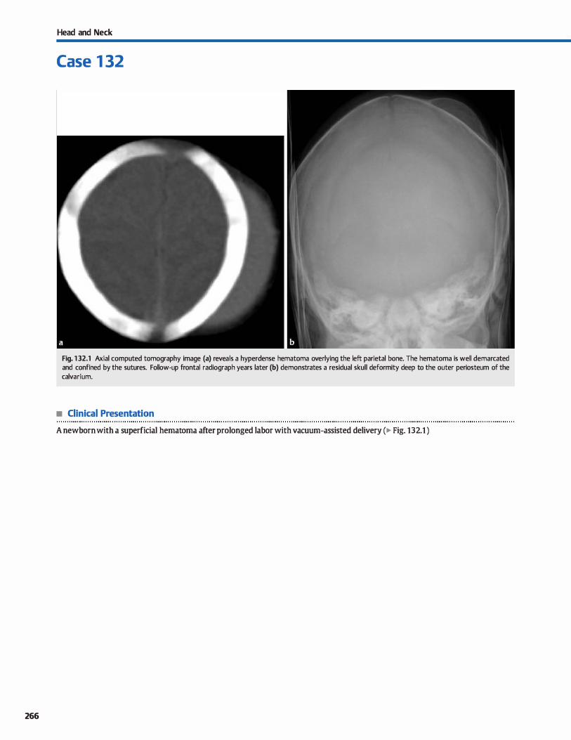
266
Head and Neck
Case 1 32
Fig. 132.1 Axial computed tomography image (a) reveals a hyperdense hematoma overlying the left parietal bone. The hematoma is well demarcated and confined by the sutures. Follow-up frontal radiograph years later (b) demonstrates a residual skull deformity deep to the outer periosteum of the calvarium.
• Clinical Presentation
A newborn with a superficial hematoma after prolonged labor with vacuum-assisted delivery (� Fig. 132.1 )
• Key lmaging Finding
Superficial hematoma in a neonate
• Top 3 Differential Diagnoses
• Subgaleal hematoma. A subgaleal hemorrhage occurs when blood accumulates superficial to the periosteum of the calvarium and beneath the galeal aponeurosis. It most often occurs in the setting of instrumentation during labor, typically with suction deviœs. The instrumentation results in rupture of smalt, bridging emissary veins. It is important to look for underlying calvarial fracture, which occurs relatively fre
quently. Given its location and source of bleeding, a subgaleal hematoma may spread diffusely through the cranium. Although the majority of cases are self-limited. subgaleal hemorrhage may occasionally be extensive and symptomatic.
• Cephalohem.atoma. A cephalohematoma refers to hemor
rhage between the calvarium and periosteum. It most often results from birth trauma with rupture of venous structures traversing the periosteum. There is an increased incidence
• Diagnosis
Cephalohematoma
y' Pearts
• With subgaleal hemorrhage, blood accumulates superfidal to the periosteum and beneath the aponeurosis.
• Subgaleal hemorrhage may spread diffusely and occasionally
result in extensive, symptomatic hemorrhage.
Suggested Reading
Glass RBF, Fembach SK, Norton KI, Owi PS, Naidich 'Il'. The infant skull: a vault of infonnation. Radiographies 2004; 2.4: 507-522
Case 132
with prolonged labor and instrumentation (forceps, suction devices, etc.) during delivery. Given the subperiosteal location, the hemarrhage daes nat cross sutures. It is important ta evaluate for the presence of underlying fracture or intracranial hemarrhage. The hematoma may persist for several months; in a minority of cases, the hemorrhage may calcify, resulting in a focal region of calvarial thickening and sclerosis.
• Ca.put sucœdaneum. Ca.put sucœdaneum refers to scalp hemorrhage superfidal ta bath the periasteum of the skull and the epicranial aponeurosis. Given its location, it is not baund by sutures; therefore, it may spread diffusely thraughout the scalp, even across midline. Most cases present with a scalp contour abnonnality, are self-limited, and resolve fairly quickly.
• Cephalohematomas occur between the calvarium and periosteum and are bound by sutures.
• Ca.put succedaneum refers to scalp hemorrhage superficial to bath the skull periosteum and galeal aponeurosis.
267
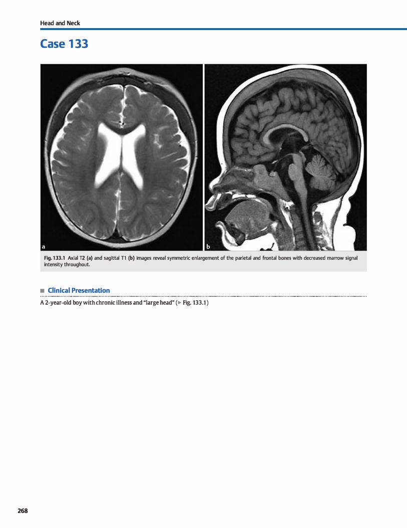
268
Head and Neck
Case 1 33
Fig. 133.1 Axial T2 (a) and sagittal Tl (b) images reveal symmetric enlargement of the parietal and frontal bones with decreased marrow signal intensity throughout.
• Clinical Presentation
A 2-year-old boy with chronic illness and "large head" (� Fig. 133.1 )
• Key lmaging Finding
Diffuse calvarial thickening in a child
• Top 3 Differential Diagnoses
• Nonnal variant. Most cases of mild or equivocal calvarial thickening are within the range of normal variation and are found incidentally. In cases of normal variation, there is symmetric expansion of the diploic space, which is otherwise normal in density (computed tomography) and signal intensity (magnetic resonanœ imaging, MRI), and the cortices are normal in appearance. Focal osseous lesions, abnormalities in density or signal intensity, or a history of metabolic or hematoiogic disorder should warrant further workup.
• Chronic anemias/hemoglobinopathies. Chronic anemia or hemoglobinopathies result in increased hematopoietic marrow, which manifests as enlargement of the diploic spaœ with conversion of yellow marrow ta red marrow. The calvarial enlargement is typically symmetric with the parietal and frontal bones most commoruy involved; the occipital bones are often relatively spared. IHhalassemia bas a characteristic
• Additional Differential Diagnoses
• Phenytoin therapy. Phenytoin is a common antiepileptic medication used in treatment of most types of seizures. Chronic us� may lead to numerous adverse effects, the most common ofwhich is gingival hyperplasia. Central nervous system abnormalities indude œrebellar hypoplasia with associated neurological deficits and diffuse calvarial thickening. Calvarial thickening occurs in approximately one-third of epilepsy patients on long-term phenytoin treatment, is thought ta result from osteoblast stimulation, and is primarily cosmetic.
• Hyperparathyroidism {HPT). HPT may be primary due to hyperfunctioning parathYroid glands, secondary due to vitamin D deficiency or chronic renal failure, or tertiary due to hyperplasia from long-standing secondary HPT. lncreased parathormone results in systemic calcium/bony resorption. In the skull, this manifests as altemating regions of lucency and sclerosis within the diploic spaœ, referred to as a granular or
• Diagnosis
Chronic anemia
� Pearts
• Chronic anemia/hemoglobinopathies result in enlargement of the diploic space and decreased marrow signal.
• �-thalassemia has a characteristic "haïr on end" appearance on plain films due to ctiploic spaœ expansion.
Suggested Readings
Clmw KM, SzetD a:. Cerebral atrophy and 5kull thidœning due ID dtronic phenytDin therapy. Canadian Med As50Cj 2007; 176: 321-323
Case 133
appearance on radiographs where the expansion of the diploic spaœ resembles a "hair on end" appearance. On MRI, red marrow conversion results in decreased signal intensity on bath T1 and T2 sequences.
• Bony dysplasia. Fibrous dysplasia is the most common bony dysplasia to involve the calvarium, skull base, and facial bones; however, it is typically a focal rather than diffuse process. The characteristic finding is focal enlargement of the diploic spaœ with a "ground glass" matrix. Osteopetrosis is an inherited ctisorder that is caused by ineffective osteodastic activity and results in increased bony deposition and sderosis. Although sclerotic, the bones are relatively fragile. Calvarial involvement presents with diffuse skull thickening and sderosis. Gigantism is the pectiatric form of acromegaly and is caused by increased growth hormone.
"sait and pepper" appearance. There is also thinning of the inner and outer tables of the calvarium. In advanced cases, focal Brown tumors may occur, presenting as well-circumscribed lytic lesions that are often multiple. Characteristic Joss of the lamina dura (thin region of sderosis) surrounding dentition may be identified.
• Chronic shunted hydroœphalus. The growth of the brain parenchyma and its coverings stimulates growth of the overlying calvarium during skeletal development and maturity. Patients with sucœssful, chronic shunted hydroœphalus often have relative intracranial hypotension and decreased intracranial volume, which decreases the growth stimulus on the calvarium. Without the stimulus for outward growth, there is diffuse endosteal calvarial thickening to fil) the vacated spaœ. Ventride size is variable but often small in caliber.
• Calvarial thickening with chronic shunted hydrocephalus is due ta decreased stimulus for skull growth.
• Chronic phenytoin antiepileptic therapy may result in diffuse calvarial thickening and cerebellar hypoplasia.
Georgy BA, Snow RD, Brogdon HG et al. Value of bone window images in routine brain CT: examinations beyand trauma. Appt Radial 1997; 2: 2&-38
269
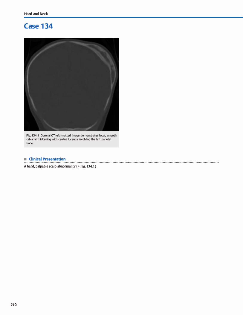
270
Head and Neck
Case 1 34
Fig. 134.1 Coronal CT reformatted image demonstrates focal, smooth calvarial thickening with central lucency involving the left parietal bone.
• Clinical Presentation
A bard, palpable scalp abnormality (� Fig. 134.1 )
• Key lmaging Finding
Focal calvarial thickening
• Top 3 Differential Diagnoses
• Fibrous dysplasia. FD occurs most often in young adults and is more common in women. The calvarium, skull base, and facial boues are most often affected in the head and neck. The dassic appearanœ on computed tomography (CT) is focal bony expansion with Mground glass" attenuation of the diploic spaœ; sderotic and cystic or lytic variants are Jess common. On magnetic resonanœ imaging (MRI), FD is typically hypointense on both Tt and 12 sequences and demonstrates avid enhancement; regions of cystic involvement, however, are 12 hyperintense. The MRI appearance can mimic aggressive lesions; therefore, CT is preferred in suspected cases ofFD.
• Malignancy. Metastatic disease commonly involves the cal
varium. "JYpically, metastatic Iesions present as soft-tissue infiltration or soft-tissue masses with bony destruction: focal sderosis is Jess common. Malignancies prone ta sderotic metastases include prostate and breast carcinoma, as well as lymphoma. Primary bone tumors are Jess common and
• Additional Differential Diagnoses
• Meningioma. Meningiomas are benign dural-based masses and represent the most common extraaxial masses. Common locations indude parasagitta� convexities. middle cranial fessa, and planum sphenoidale. Less common locations indude the sellar/suprasellar region and œrebellopontine angle. Extradural meningiomas are rare, aa:ounting for fewer than 2% of cases. Intraosseous meningiomas are a subset of extradural meningiomas that typically occur in adults; most often involve the calvarium, skull base, or fadai bones; and are œntered within the diploic spaœ. They present with expansion of the diploic space, cortical thiclœning, and mixed sderosis and lucency, often mimicking fibrous dysplasia or a malignant Iesion, espedally onMRL
• Diagnosis
Calcified cephalohematoma
� Pearts
• FD occurs in young adults; the classic CT appearance is focal bony expansion with Mground glass" attenuation.
• Malignant calvarial lesions indude metastases, lymphoma, osteosarcoma, and Ewing sarcoma (in a child).
Suggested Reading
Gemgy BA, Snow RD, Brogdon DG et al Value ofbone window images in routine brain CT; examinations beyond trauma. Appl Radiol 1997; 2; 26-38
Case 134
indude Ewing sarcoma in children and osteosarcoma in young adults or the elderly with a history of Paget disease or prior radiation therapy. Both Ewing and osteosarcoma present as focal, aggressive bone-forming tumors on CT; lytic lesions are Jess common. MRI most often reveals intermediate Tt and 12 signal intensity with avid but heterogeneous enhancement; calcified portions are hypointense.
• Page(: disease. Paget disease occurs in elderly patients and consists of three stages: lytic, sderotic, and mixed. Calvarial involvement during the lytic phase results in a characteristic large lytic lesion with erosion of the outer table, referred to as osteoporosis drcumscripta. During the sderotic phase, there is expansion and sderosis of the cortex and diploic spaces, which may be diffuse or focal. In the mixed phase, a combination of lytic and sderotic Iesions results in the characteristic Mcotton wool" appearanœ of the skull.
• caJdfied cephalohematoma. A œphalohematoma refers to hemorrhage between the calvarium and periosteum. It most often results from birth trauma with rupture of venous structures traversing the periosteum. There is an increased incidence with prolonged tabor and instrumentation {forceps, suction devices, etc.) during delivery. Given the subperiosteal location, the hemorrhage does not cross sutures. The hematoma may persist for several months; in a minority of cases, the hemorrhage may calcify, resulting in a focal region of calvarial thickening and sclerosis that persists into adulthood.
• Paget disease occurs in elderly patients; the sderotic phase results in cortical/diploic thiclœning and sderosis.
• Meningiomas may result in calvarial thiclœning due to primary bony involvement or reactive hyperostosis.
271
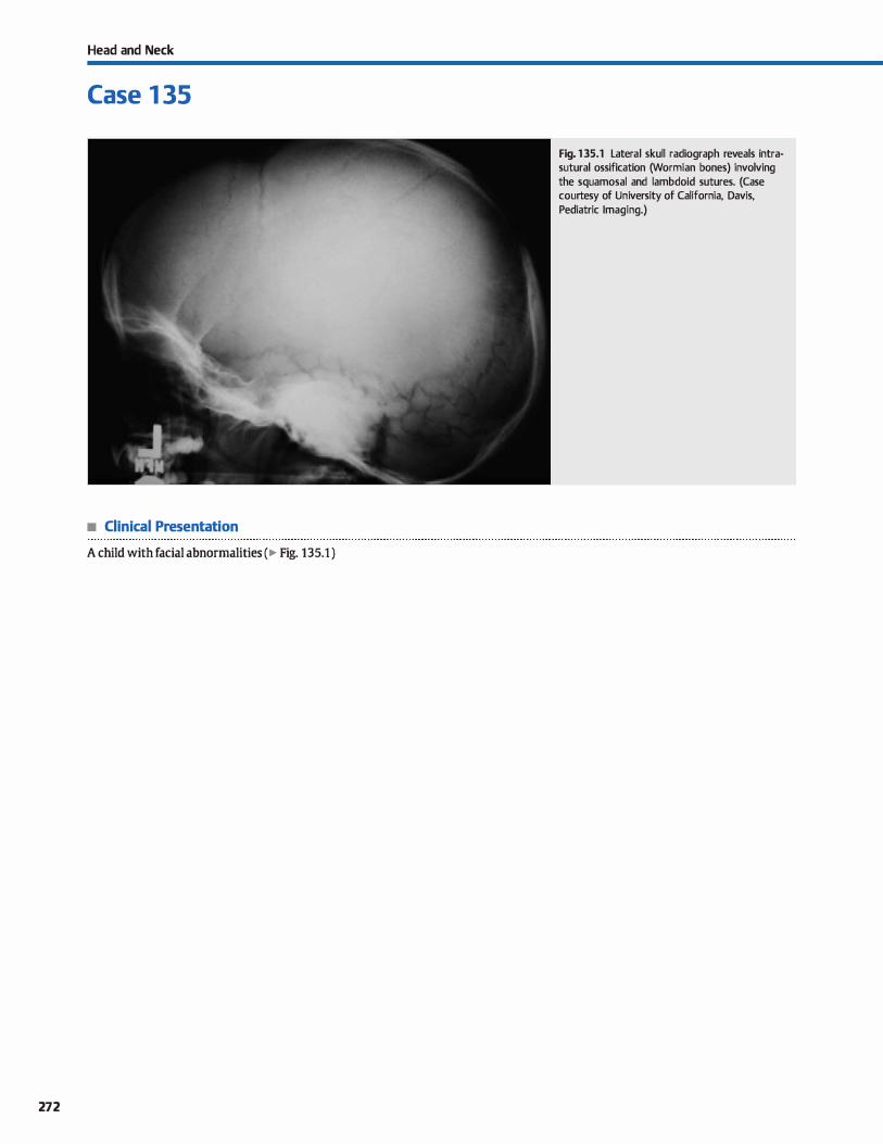
Head and Neck
Case 1 35
• Clinical Presentation
A child with facial abnormalities (� Fig. 135.1 )
272
Fig. 135.1 Lateral skull radiograph reveals intrasutural ossification (Wormian bones) involving the squamosal and lambdoid sutures. (Case courtesy of University of California, Davis, Pediatric lmaging.)
• Key lmaging Finding
Wonnian bones
• Top 3 Differential Diagnoses
• Idiopathie. Wonnian boues are irregular ossicies located within the sutures of the calvarium (intrasutural boues). Idio
pathie or inddental Wonnian boues are reported ta be smaller and Jess numerous { usually < 10) than those assod
ated with skeletal dysplasias; however, there are no definitive criteria to distinguish between benign and pathological causes. Most commonly located within the lambdoid suture
(50%), Wormian bones may also occur in the coronal suture (25%) and have been identified in all cranial sutures and fon
tanelles. A large intrasutural bone located at the junction of the lambdoid and sagittal sutures is termed an "Inca bone." Arriving at a definitive diagnosis radiographically often requires additional studies to assess for the wide spectrum of pathological entities that may be assodated with Wonnian
bones. • Osteogenesis imperfecta. Osteogenesis imperfecta (OI) is a
genetie disorder (typically autosomal dominant), which results in abnormal type t collagen formation. Type t collagen is found in osseous structures and the sciera of the eyes;
• Additional Differential Diagnoses
• Down syndrome. Down syndrome {trisomy 21) is the most
common genetic cause of mental impairment in children and
has been assodated with an increased incidence with
advanced maternai age (>35 years of age). A hast of radiographie findings are assodated with Dawn syndrome, inciud
ing Wonnian bones. Additional musculoskeletal findings include a hypoplastic arch of Cl with craniocervical stenosis, atlantoaxial subluxation, absent 12th ribs, short tubular bones of the hands {including clinodactyly of the middle phalanx of the little finger), wide iliac wings, hip dysplasia, and
patellar dislocation. There is an increased incidence of early
onset Alzheimer disease in patients with Dawn syndrome.
• Diagnosis
Oeidocranial dysostosis
� Pearls
• Wormian bones may be idiopathie or inddental in etiology;
the lambdoid suture is most commonly involved.
• Wormian bones in osteogenesis imperfecta may be numerous and demonstrate a mosaie pattern.
Suggested Readings
Jeanty P, Silva SR, Turner C. Prenatal diagnosis of wormian bones. j Ultraso1111d Med 2000; 19: 863-869
Case 135
hence, patients present with fragile bones, multiple fractures,
and blue sciera. There are four major types of or with types 1 and 4 being the most common. Type 1 presents in childhood
with multiple fractures and Wormian bones. Wonnian bones
in QI are often numerous and demonstrate a mosaic ("crazy
paving") pattern. Musculoskeletal imaging findings include diffuse demineralization, bowing deformities, multiple frac
tures, vertebra plana, kyphoscoliosis, enlarged epiphyses, characteristie "popcorn" metaphyseal calcifications, and cortical thinning.
• Oeidocranial dysostosis. Oeidocranial dysostosis is a genetic disorder characterized by a defect in the formation of mem
branous boues. Oinical findings indude macroœphaly, hypertelorism, facial hypoplasia, and absence or hypoplasia of
the ciavicles. Central nervous system imaging reveals these
findings, along with Wormian bones and delayed closure of fontanelles and sutures. Systemic musculoskeletal abnormali
ties inciude a widened symphysis pubis and dysplasia of the femoral neck with a coxa vara defonnity.
• Metabolic disease (riclœts, hypothyroidism). Wormian bones
may be found in patients with multiple metabolic deficien
cies, inciuding rickets, hypothyroidism, and hypophosphatasia
(genetic cause of rickets). ln rickets, there is demineralization and softening ofbones due to Vitamin D (most common), cal
cium, or phosphorous deficiencies. Early systemic musculoskeletal findings inciude frayed physes and flaring (cupping) of the metaphyses. Wormian bones are associated with the healing phase of the disorder. Hypothyroidism is characterized by markedly delayed skeletal maturity, "bullet" vertebrae
at the thoracolumbar junction, epiphyseal fragmentation, and
Wormian bones.
• Oeidocranial dysostosis presents with Wonnian bones and
absence or hypoplasia of the clavicles.
• Wormian bones may be found in patients with metabolie deficiendes, including rickets and hypothyroidism.
Paterson CR. Radiological features of the brittle bone diseases j Diagn Radiogr bnaging. 2003; 5: 39-45
273
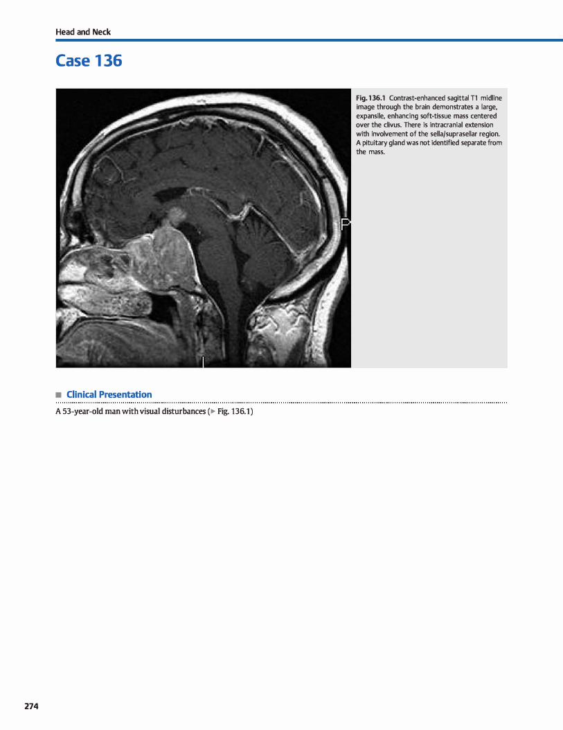
Head and Neck
Case 1 36
• Clinical Presentation
A 53-year-old man with visual disturbances (� Fig. 136.1)
274
Fig. 136.1 Contrast-enhanced sagittal Tl midline image through the brain demonstrates a large, expansile, enhancing soft-tissue mass centered over the clivus. There is intracranial extension with involvement of the sella/suprasellar region. A pituitary gland was not identified separate from the mass.
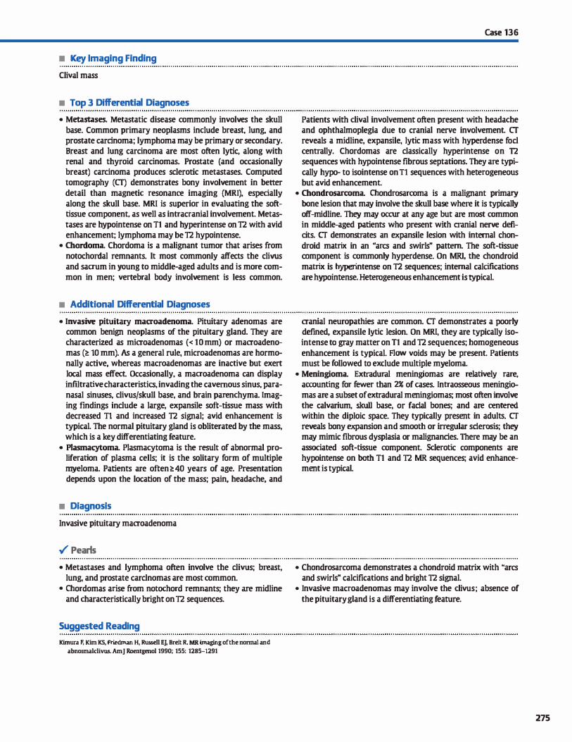
• Key lmaging Finding
Clivai mass
• Top 3 Differential Diagnoses
• Metastases. Metastatic disease commonly involves the skull
base. Common primary neoplasms include breast, Jung, and prostate carcinoma; lymphoma may be primary or secondary. Breast and Jung carcinoma are most often Iytic, along with renal and thyroid carcinomas. Prostate (and occasionally breast) carcinoma produces sderotic metastases. Computed tomography (CT) demonstrates bony involvement in better detail than magnetic resonanœ imaging (MRI), especially
along the skull base. MRI is superior in evaluating the softtissue component. as well as intracranial involvement Metas
tases are hypointense on Tl and hyperintense on T2 with avid enhancement; lymphoma may be T2 hypointense.
• Chordoma. Chordoma is a malignant tumor that arises from notochordal remuants. It most commonly affects the cliws
and sacrum in young to middle-aged adults and is more common in men; vertebral body involvement is Jess common.
• Additional Differential Diagnoses
• Invasive pituitary macroadenoma. Pituitary adenomas are common benign neoplasms of the pituitary gland. They are characterized as microadenomas ( < 1 O mm) or macroadenomas (� 10 mm). As a general rule, microadenomas are hormonally active, whereas macroadenomas are inactive but exert local mass effect. Occasionally, a macroadenoma can display
infiltrative characteristics, invading the cavemous sinus, paranasal sinuses, divus/skull base, and brain parenchyma. lmaging findings indude a large, expansile soft-tissue mass with decreased Tl and increased T2 signal; avid enhancement is typical. The normal pituitary gland is obliterated by the mass, which is a key differentiating feature.
• Plasmacytoma. Plasmacytoma is the result of abnormal proliferation of plasma cells; it is the solitary form of multiple
myeloma. Patients are often � 40 years of age. Presentation depends upon the location of the mass; pain, headache, and
• Diagnosis
Invasive pituitary macroadenoma
� Pearts
• Metastases and lymphoma often involve the divus; breast, lung, and prostate cardnomas are most common.
• Chordomas arise from notochord remuants; they are midline and characteristically bright on T2 sequences.
Suggested Reading
Kimura F, Kim !CS, Friedman H, Russell EJ, Breit R. MR imaging of the nonnal and
abnormalclivm.Amj Rcentgmol 1990; 155: 1285-1291
Case 136
Patients with clivai involvement often present with headache and ophthalmoplegia due to cranial nerve involvement. CT reveals a midline, expansile, lytic mass with hyperdense fod centrally. Chordomas are dassically hyperintense on T2
sequences with hypointense fibrous septations. They are typi
cally hypo- ta isointense on Tl sequences with heterogeneous but avid enhancement
• Chondrosarcoma. Chondrosarcoma is a malignant primary bone lesion that may involve the skull base where it is typically off-midline. They may occur at any age but are most common in middle-aged patients who present with cranial nerve defidts. CT demonstrates an expansile lesion with internai chon
droid matrix in an �an:s and swirls" pattern. The sort-tissue component is commonly hyperdense. On MRI, the chondroid
matrix is hyperintense on T2 sequenœs; internai calcifications are hypointense. Heterogeneous enhancement is typical.
cranial neuropathies are common. CT demonstrates a poorly defined, expansile lytic lesion. On MRI, they are typically isointense ta gray matter on Tl and T2 sequences; homogeneous
enhancement is typical. Flow voids may be present. Patients must be followed to exclude multiple myeloma.
• Meningioma. Extradural meningiomas are relatively rare, accounting for fewer than 2% of cases. lntraosseous meningiomas are a subset of extradural meningiomas; most often involve the calvarium, skull base, or fadai bones; and are œntered within the diploic space. They typically present in adults. CT reveals bony expansion and smooth or irregular sderosis; they may mimic fibrous dysplasia or malignancies. There may be an
associated sort-tissue component Sclerotic components are hypointense on bath Tl and T2 MR sequences; avid enhanœment is typical.
• Chondrosarcoma demonstrates a chondroid matrix with "arcs and swirls" calcifications and bright T2 signal.
• Invasive macroadenomas may involve the clivus; absence of
the pituitary gland is a differentiating feature.
275
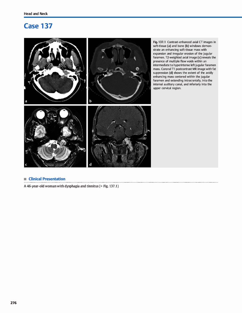
Head and Neck
Case 1 37
• Clinical Presentation
A 46-year-old woman with dysphagia and tinnitus (� Fig. 137.1 )
276
Fig. 137.1 Contrast-enhanced axial CT images in soft-tissue (a) and bone (b) windows demonstrate an enhancing soft-tissue mass with expansion and irregular erosion of the jugular foramen. T2-weighted axial image (c) reveals the presence of multiple flow voids within an intermediate to hyperintense left jugular fora men mass. Coronal Tl postcontrast MR image with fat suppression (d) shows the extent of the avidly enhancing mass centered within the jugular foramen and extending intracranially, into the internai auditory canal, and inferiorly into the upper cervical region.
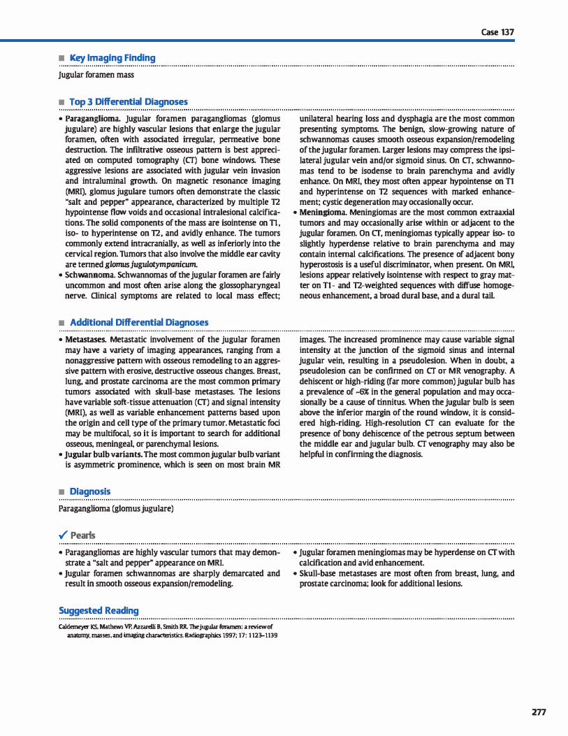
• Key lmaging Finding
Jugular foramen mass
• Top 3 Differential Diagnoses
• Paraganglioma. jugular foramen paragangliomas (glomus jugulare) are highly vascular lesions that enlarge the jugular foramen, often with assodated irregular, penneative bone destruction. The infiltrative osseous pattern is best appreciated on computed tomography (CT) bene windows. These aggressive lesions are associated with jugular vein invasion and intraluminal growth. On magnetic resonanœ imaging
(MRI), glomus jugulare tumors often demonstrate the classic usait and pepper" appearance, characterized by multiple T2 hypointense flow voids and occasional intralesional calcifications. The solid components of the mass are isointense on TI, iso- to hyperintense on T2, and avidly enhance. The tumors commonly extend intracranially, as well as inferiorly into the cervical region. Tumors that also involve the middle ear cavity
are tenned glomus jugulotympanicum. • Schwannoma. Schwannomas of the jugular foramen are fairly
uncommon and most often arise along the glossopharyngeal nerve. Œnical symptoms are related ta local mass effect;
• Additional Differential Diagnoses
• Metastases. Metastatic involvement of the jugular foramen
may have a variety of imaging appearances, ranging from a nonaggressive pattern with osseous remodeling to an aggressive pattern with erosive, destructive osseous changes. Breast, lung, and prostate carcinoma are the most common primary tumors assodated with skull-base metastases. The lesions have variable soft-tissue attenuation (CT) and signal intensity (MRI), as well as variable enhancement patterns based upon the origin and cell type of the primary tumor. Metastatic foci may be multifocal, so it is important to search for additional osseous, meningeal, or parenchymal Iesions.
• jugular bulb variants. The most common jugular bulb variant is asymmetric prominenœ, which is seen on most brain MR
• Diagnosis
Paragang]ioma (glomus jugulare)
� Pearls
• Paragangliomas are highly vascular tumors that may demonstrate a "sait and pepper" appearance on MRI.
• Jugular foramen schwannomas are sharply demarcated and result in smooth osseous expansion/remodeling.
Suggested Reading
Calde1Iley8" KS, Mathews VP, Azzardli 8, Snùth RR. Thejugular forunen: a review of
anatomy, masses, and imaging characteristics. Radiographies 1997; 17: 1123-1139
Case 137
unilateral hearing Joss and dysphagia are the most common presenting symptoms. The benign, slow-growing nature of schwannomas causes smooth osseous expansion/remodeling of the jugular foramen. Larger lesions may compress the ipsilateral jugular vein and/or sigmoid sinus. On CT, schwannomas tend to be isodense to brain parenchyma and avidly enhanœ. On MRI, they most often appear hypointense on Tl and hyperintense on T2 sequences with marked enhancement; cystic degeneration may occasionally occur.
• Meningioma. Meningiomas are the most common extraaxial tumors and may occasionally arise within or adjacent to the jugular foramen. On CT, meningiomas typically appear iso- to slightly hyperdense relative to brain parenchyma and may contain internai calcifications. The presence of adjacent bony hyperostosis is a useful discriminator, when present. On MRI, lesions appear relatively isointense with respect to gray matter on Tl - and T2-weighted sequences with diffuse homogeneous enhancement, a broad durai base, and a durai tail.
images. The increased prominence may cause variable signal intensity at the junction of the sigmoid sinus and internai jugular vein, resulting in a pseudolesion. When in doubt, a pseudolesion can be confinned on CT or MR venography. A
dehiscent or high-riding (far more common) jugular bulb has a prevalence of -6% in the general population and may occasionally be a cause of tinnitus. When the jugular bulb is seen above the inferior margin of the round window, it is considered high-riding. High-resolution CT can evaluate for the
presenœ of bony dehiscenœ of the petrous septum between the middle ear and jugular bulb. CT venography may also be helpful in confinning the diagnosis.
• Jugular foramen meningiomas may be hyperdense on CT with calcification and avid enhancement.
• Skull-base metastases are most often from breast, Jung, and prostate carcinoma; look for additional lesions.
277
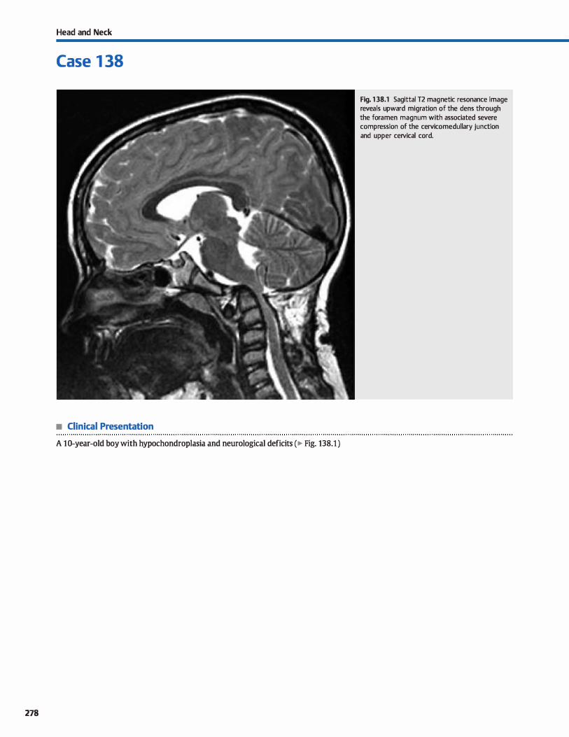
Head and Neck
Case 1 38
• Clinical Presentation
A 10-year-old boy with hypochondroplasia and neurological deficits (� Fig. 138.1 )
278
Fig. 138.1 Sagittal T2 magnetic resonance image reveals upward migration of the dens through the foramen magnum with associated severe compression of the cervicomedullary junction and upper cervical cord.
• Key lmaging Finding
Extension of dens through foramen magnum with compression
of the craniocervical junction
• Top 3 Differential Diagnoses
• Basilar invagination. Basilar invagination refers to abnormal
protrusion of the vertebral column through the foramen mag
num due ta a congenital or developmental defect. There are
numerous causes, including hypoplasia of the osseous compo
nents of the skull base, atlantoocdpital assimilation, bony and
cartilaginous dysplasias, Klippel-Feil syndrome, Dawn syn
drome, and Chiari cranioœrvical junctions. Oinically, patients
may be asymptomatic or present with insidious onset of cere
bellar, brain stem, or cervical cord neurological deficits. On
imaging, the tip of the odontoid projects through the foramen
magnum with indentation or compression of the cervicome
dullary junction. There is typically normal orientation of Ct with respect to C2. The Chamberlain line is used ta evaluate
for abnormal positioning of the vertebral column with respect
to the skull base. lt is drawn from the posterior margin of the
hard palate to the opisthion, which is the posterior margin of the foramen magnum. ln the majority of normal cases, the tip
of the odontoid process is at or below the Chamberlain line.
In some cases, it may extend a few millimeters above the
line. Extension 5 mm or more above the Chamberlain line is
abnormal and consistent with basilar invagination or basilar
• Diagnosis
Basilar invagination
y' Pearts
• Basilar invaginationfbasilar impression results in upward
migration of the dens through the foramen magnum.
• Basilar invagination is primary or congenital, whereas basilar
impression is acquired.
Suggested Reading
Smoker WRK. Craniovertdiraljunction: nonnal analDmy, craniometry, and congeni
tal anomalies. Radiograplùcs 1994; 14: 255-277
Case 138
impression (see below). Both may be associated with platyba
sia or flattening of the skull base.
• Basilar impression. The clinical and imaging manifestations of basilar invagination and basilar impression are identical.
The key distinction between the two is that basilar invagina
tion is a congenital or developmental abnormality, whereas
basilar impression is acquired. Because basilar impression
is acquired, the underlying bony development is normal.
Common acquired causes of basilar impression include Paget
disease, osteogenesis imperfecta, rickets, metabolic abnormalities involving calcium and phosphate, and trauma.
• Cranial settling. Cranial setting aise results in upward exten
sion of the dens through the foramen magnum; however, it is
due to disruption of the ocdpitoatlantal and atlantoaxial
articulations and subsequent inferior migration of the atlas
and skull base with respect ta C2. The key imaging distinction
is that the anterior arch of Ct articulates with the base of C2
rather than with the dens. Rheumatoid arthritis {RA) is the most common cause of cranial settling and most often
involves middle-aged women.
• The Chamberlain line is used ta diagnose basilar invagination;
it extends from the posterior hard palate ta the opisthion.
• Cranial setting refers ta inferior migration of Ct and the skull
base with respect to C2, most often due to RA.
279
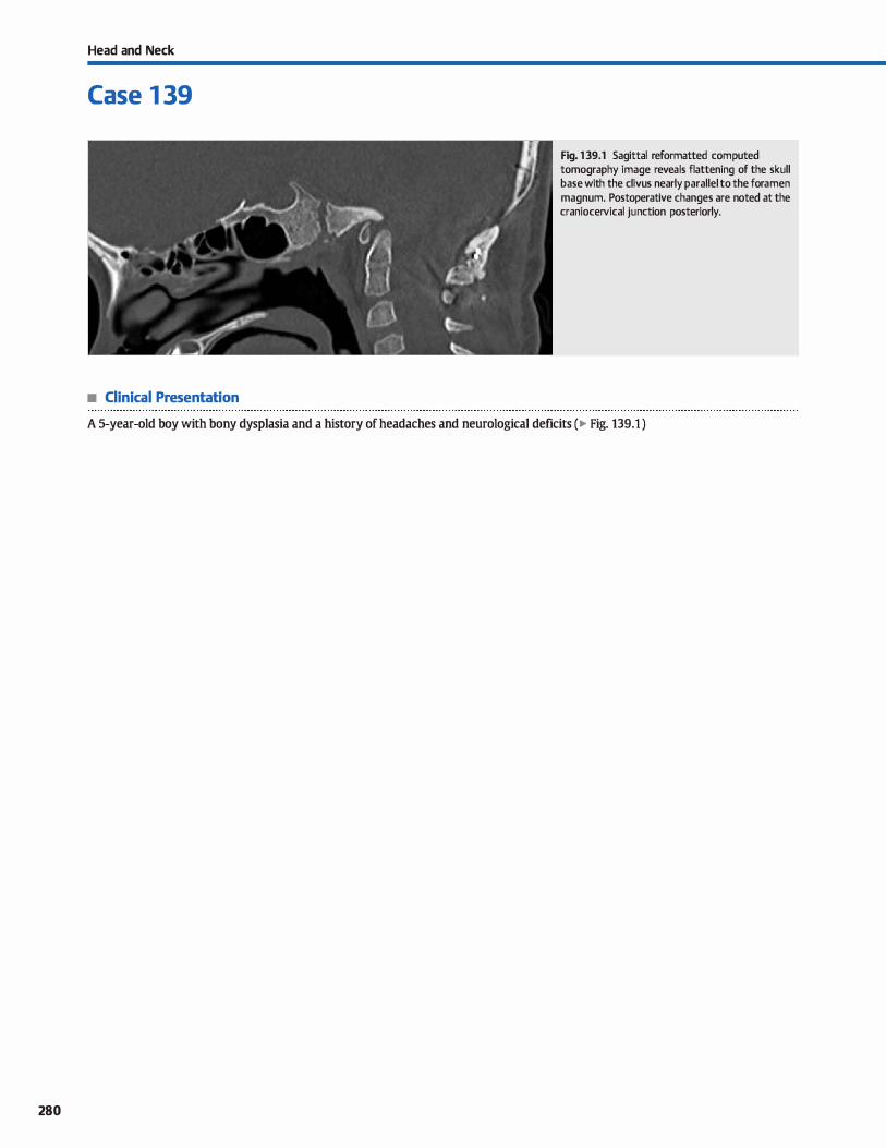
280
Head and Neck
Case 1 39
• Clinical Presentation
Fig. 139.1 Sagittal reformatted computed tomography image reveals flattening of the skull base with the divus nearly parallel to the fora men magnum. Postoperative changes are noted at the craniocervical junction posteriorly.
A 5-year-old boy with bon y dysplasia and a history of headaches and neurological deficits ( � Fig. 139.1 )
• Key lmaging Finding
Abnormal flattening of skull base
• Diagnosis
Platybasia. Platybasia refers to flattening of the skull base. The causes of platybasia are similar to congenital causes of basilar invagination {hypoplasia of the osseous components of the skull base, atlantooccipital assimilation, bony and cartilaginous dysplasias, Chiari craniocervical junctions, etc.) and acquired causes ofbasilar impression {Paget disease, osteogenesis imperfecta, rickets, metabolic abnonnalities involving calcium and
phosphate, and trauma). In most cases. there is associated basilar invagination/impression; however, isolated cases of platybasia do occur.
� Pearts
• Platybasia refers to flattening of the skull base; the causes are similar to basilar invagination/impression.
• The Welcher basal angle is used to evaluate for platybasia; an angle> 140 degrees is consistent with platybasia.
Suggested Reading
Smolœr WRK. Craniovertebraljunction: normal amtmny, craniometty, and congeni
tal anomalies. Radiographies 1994; 14: 255-277
Case 139
The Welcher basal angle is used to evaluate for platybasia. The first line is drawn from the nasion to the tuberculum sellae; the second line is drawn from the tuberculum to the basion (anterior margin of the foramen magnum). The average angle in normal individuals is -132 degrees. An angle> 140 degrees is
consistent with platybasia. Treatment is generally reserved for cases of brain stem and/
or cervical cord compression and consists of posterior decompression and craniocervical fusion.
• Treatment is reserved for neurological compression and consists of posterior decompression with fusion.
281
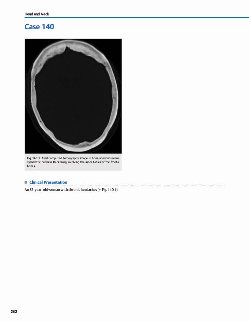
282
Head and Neck
Case 1 40
Fig. 140.1 Axial computed tomography image in bone window reveals symmetric calvarial thickening involving the inner tables of the frontal bon es.
• Clinical Presentation
An 82-year-old woman with chronic headaches (� Fig. 140.1 )
• Key lmaging Finding
Symmetric calvarial thickening involving the inner tables of the frontal bones
• Diagnosis
Hyperostosis frontalis interna (HFI). HFI refers ta calvarial thickening due ta bony overgrowth along the inner table. It occurs in adults and is far more common in women. HFI characteristically involves the frontal bones and is typically bilateral and symmetric. In some cases, it may extend to the orbital roofs
� Pearls
• Hyperostosis frontalis interna refers to calvarial thickening due to bony overgrowth along the inner table.
• HFI is more common in women, characteristically involves the frontal bones, and is bilateral and symmetric.
Suggested Reading
She R. Szakacs J. Hyperostosis frontalis interna: case report and review of Iiteratw'e.
Ann Clin I.ab Sd 2004; 34; 206-208
Case 140
or, occasionally, beyond the coronal sutures to involve the parietal bones. The exact etiology remains unknown; however, there have been reported associations with endocrinopathies. HFI is an asymptomatic inddental finding that requires no further workup or imaging.
• HFI is an asymptomatic incidental finding that requires no further workup or imaging.
283
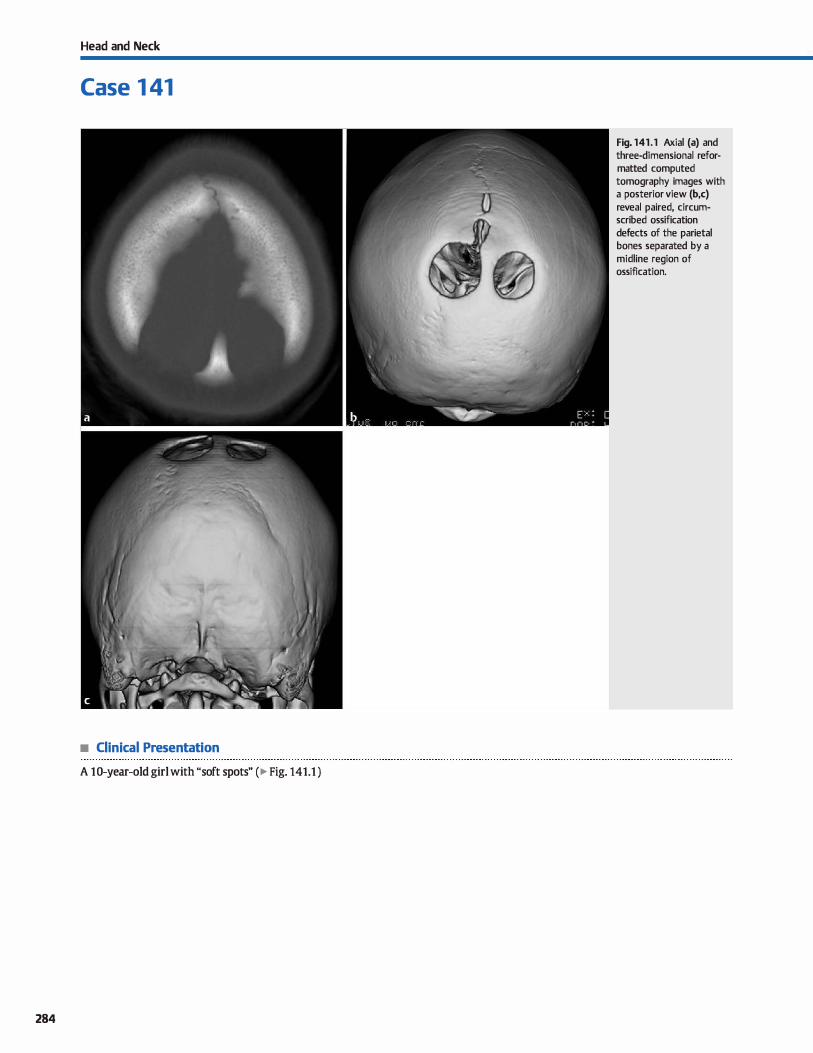
Head and Neck
Case 1 41
• Clinical Presentation
A 10-year-old girl with "soft spots" (� Fig. 141.1 )
284
Fig. 141.1 Axial (a) and three-dimensional reformatted computed tomography images with a posterior view (b,c) reveal paired, circumscribed ossification defects of the parietal bones separated by a midline region of ossification.
• Key lmaging Finding
Circumscribed, symmetric parietal calvarial defects
• Diagnosis
Parietal foram.ina. Parietal foramina refer to hereditary ossification defects involving the parietal bone. They are most often isolated abnormalities but may be associated with syndromes. On dinical exam, parietal foramina present with üsoft spots" in addition to the normal open sutures. lmaging reveals
� Pearts
• Parietal foramina are hereditary ossification defects that occur
in isolation or in association with syndromes. • Oinically, parietal foramina present with "soft spots" in addi
tion to the normal open sutures.
Suggested Reading
Glass RBF, Fembach SK, Nm1Dn KI, Clmi PS, Naidich lP. 1be infant skull; a vault of
information. Radiographies 2004; 24: 507-522
Case 141
either paired, circumscribed parietal defects separated by a midline region of ossification or a large, single ossification defect that extends across the midline. The defects may persist into adulthood.
• Imaging shows paired parietal ossification defects or a soli
tary defect that may persist into adulthood.
285
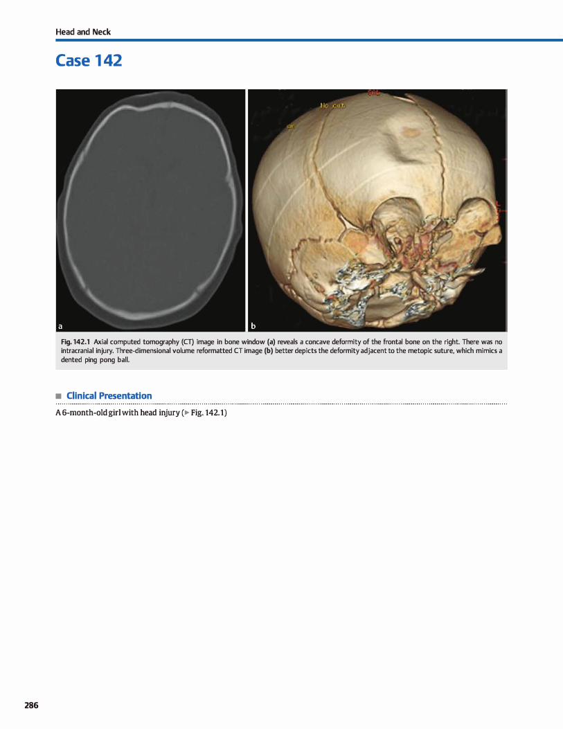
286
Head and Neck
Case 1 42
Fig. 142.1 Axial computed tomography (CT) image in bone window (a) reveals a concave deformity of the frontal bone on the right. There was no intracranial injury. Three-dimensional volume reformatted CT image (b) better depicts the deformity adjacent to the metopic suture, which mimics a dented ping pong ball.
• Clinical Presentation
A 6-month-old girl with head injury (� Fig. 142.1)

• Key lmaging Finding
Concave skull defonnity in an infant
• Diagnosis
Ping Pong fracture. "Ping pong" fractures refer to specific depressed skull fractures in infants that result in a concave deformity, similar to a dented ping pong ball. Oftentimes, there is no associated Iuœncy because the infant skull is malleable. Ping pong fractures are relatively rare and may be congenital or secondary to external injury; instrumentation during delivery
� Pearls
• MPing pong" fractures occur in infants and result in a concave defonnity, similar to a dented ping pong ball.
• Ping pong fractures are rare; instrumentation during delivery is one of the more common causes.
Suggested Readlngs
Basaldella !., Marton E, Belœlis K, Longatti P. Spontaneous resolution of atraumatic intrauterine ping-pong fractures in newbmm delivered bycesarean section.] Cllild NelU'Ol 2011; 26: 1449-1451
Case 142
is one of the more common causes. Treatment options depend upon the severity of the deformity and presenœ or absence of associated injuries, induding intracranial hemorrhage. Mild fonns may resolve spontaneously or be treated with suction devices. More severe deformities may require surgical
correction.
• Treatment options depend upon the severity of the defonnity and presenœ or absence of associated injuries.
Brittain C. Muthukumar P, Job S, et al. "Ping pong" fractlll"e in a term infant BMI Case Reports. 2012
287
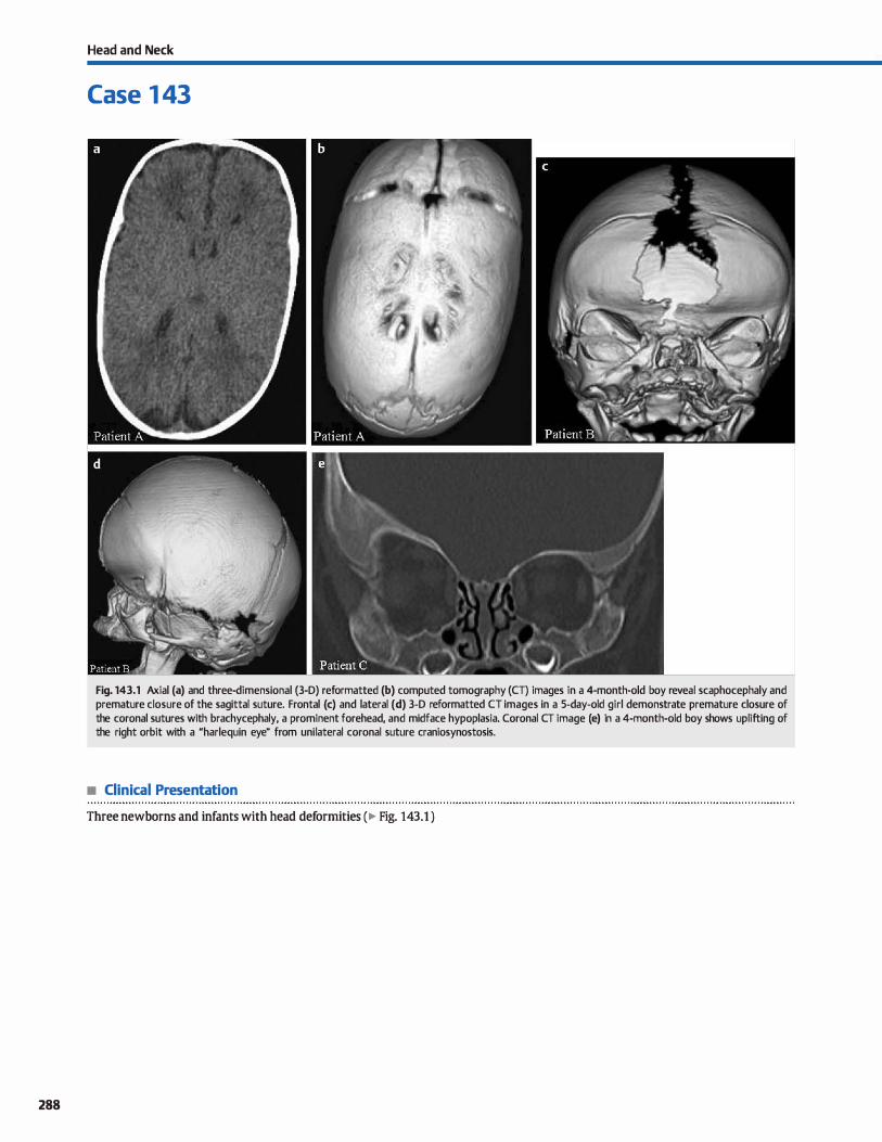
288
Head and Neck
Case 1 43
Fig. 143.1 Axial (a) and three-dimensional (3-D) reformatted (b) computed tomography (CT) images in a 4-month-old boy reveal scaphocephaly and premature closure of the sagittal suture. Frontal (c) and lateral (d) 3-D reformatted CT images in a 5-day-old girl demonstrate premature closure of the coronal sutures with brachycephaly, a prominent forehead, and midface hypoplasia. Coronal CT image (e) in a 4-month-old boy shows uplifting of the right orbit with a "harlequin eye" from unilateral coronal suture craniosynostosis.
• Clinical Presentation
Three newborns and infants with head deformities (� Fig. 143.1 )
• Key lmaging Finding
Premature closure of sutures with head defonnities
• Diagnosis
Craniosynostoses. Craniosyntostosis refers ta premature dosure of sutures, resulting in head deformities with or without associated facial abnormalities. Patients present at birth, infancy, or early childhood with asymmetric cranial development. Craniosynostosis is more common in boys and may be sporadic (-85%) or associated with numerous syndromes ( .. 15%), induding Apert (hand/foot anomalies, midfaœ hypoplasia, hearing Joss) and Crouzon (craniofacial dysostosis, external auditory canal atresia) syndromes, as well as thanatophoric dwarfism {pansynostosis). Syndromic causes are more severe, more likely to involve multiple sutures, and present earlier in life. Treatment depends on the degree of deformity and age at diagnosis. Moderate to severe defonnities are treated with surgical distraction and/or reconstruction.
Premature dosure of a suture causes increased growth parallel ta the suture and decreased growth perpendicular to the suture. Therefore, characteristic head deformities occur based upon which sutures are involved: o Sagittal suture-increased anteroposterior {AP) and decreased
transverse diameter, referred ta as scaphoœphaly or dolichoœphaly.
o Metopic suture-triangular-shaped forehead. referred ta as trigonocephaly.
o Unilateral coronal or lambdoid suture-asymmetric unilateral growth, referred ta as plagioœphaly; unilateral coronal suture
� Pearls
• Craniosyntostasis refers ta premature dosure of sutures, resulting in head and possibly facial deformities.
• Craniosynostosis is most often sporadic but may aise occur in association with numerous syndromes.
Suggested Reading
Glass RBF, Fembach SK, Nortun KI, Choi PS, Naidich 'IP. The infant skull; a vault of
information. Radiographies 2004; 24: 507-522
Case 143
synostosis results in uplifting of the ipsilateral orbit causing a Mharlequin eye" defonnity.
o Bilateral coronal or lambdoid sutures-increased transverse and decreased AP diameter, referred ta as brachycephaly.
o Bilateral coronal and lambdoid sutures-increased transverse and decreased AP diameter with a utowering" appearance, referred ta as turricephaly.
o Sagittal, bilateral coronal, and bilateral lambdoid suturesdoverleaf configuration of the skull, referred ta as kleeblattschadel.
The sagittal suture is by far the most commonly involved suture in craniosynostosis. followed by the coronal, metopic, and lambdoid sutures. Sutures normally remain open during skeletal growth with the exception of the metopic suture, which typically doses in the first year of life (usually 6 ta 9 months of age). The sagittal suture is the Iast to close under normal conditions.
Low dose computed tomography with three-dimensional reformats/volume rendering is best for initial and follow-up evaluation for craniosynostosis. The region of synostosis may be fibrous or osseous and may involve all or only a portion of the suture ta result in head deformity. It is important ta evaluate for associated malformations of the temporal and facial bones.
• Characteristic head deformities occur based upon which suture(s) prematurely dose.
• Sagittal suture is by far most commonly involved, followed by the coronal, metopic, and lambdoid sutures.
289
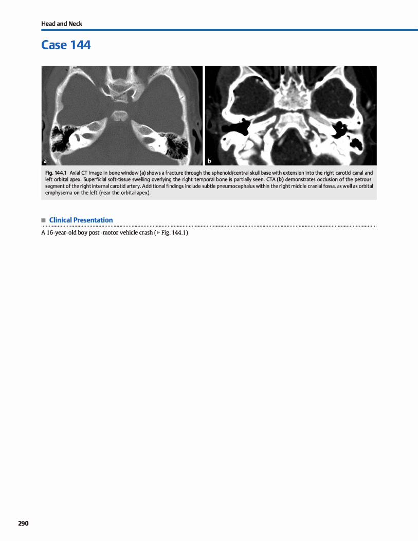
290
Head and Neck
Case 1 44
Fig. 144.1 Axial CT image in bone window (a) shows a fracture through the sphenoid/central skull base with extension into the right carotid canal and left orbital apex. Superficial soft-tissue swelling overlying the right temporal bone is partially seen. CTA (b) demonstrates occlusion of the petrous segment of the right internai carotid artery. Additional findings include subtle pneumocephalus within the right middle cranial fossa, as well as orbital emphysema on the left (near the orbital apex).
• Clinical Presentation
A 16-year-old boy post-motor vehicle crash (� Fig. 144.1 )
• Key lmaging Finding
Central skull-base fracture with vascular injury
• Diagnosis
Skull-base fracture with carotid occlusion. Fractures involving
the central skull base are typically the result of blunt trauma
with extension of associated calvarial or facial fractures. With the configuration of the cranial vault, the vectors and fractures caused by blunt trauma extend inferiorly and œntrally to
involve the skull base.
ln the setting of significant trauma, such as a motor vehide
crash, the vast majority of trauma œnters perfonn panscan
computed tomography (CT), which indudes unenhanced scans
of the head and cervical spine, followed by a CT angiogram
(CTA) of the head and neck and contrast-enhanced studies of the chest, abdomen, and pelvis. Dedicated studies or refonnatted images of the orbits, facial bones, and temporal bones may be perfonned based upon the clinicat setting and associated
findings. Central skull-base/sphenoid fractures may be subtle
� Pearts
• Central skull-base fractures typically result from blunt trauma
with extension of calvarial or facial fractures. • Sphenoid fractures may involve the carotid canal with inter
nai carotid artery injury or the orbital apex, or they may cause a CSF leak.
Suggested Reading
Slilœr CIN. ffi1111t cerebrovasrular injuries: imilging with multidetector CT angiography. Radiographies 2008; 28: 1689-1708, discussion 1709-1710
Case 144
because they are typically nondisplaœd and may occur in the plane of imaging; therefore, multiplanar refonnats are helpful.
CTA is reconunended and necessary to evaluate the integrity of vascular structures in the setting of a skull-base fracture.
lt is important to evaluate and describe the extent of frac
tures and structures involved. Sphenoid fractures will commonly involve the carotid canais and orbital apiœs. carotid
canal extension may result in vascular injury, induding dissec
tion, emboli, occlusion, and carotid-cavemous fistula (CCF). Extension into the orbital apex may result in optic nerve injury or compression. An uncommon but important complication of skull-base fractures is a cerebrospinal fluid {CSF) leak. Most leaks resolve spontaneously. If persistent, there is a significant
risk of infection. A nudear medicine or CT myelogram may be perfonned to evaluate a persistent CSF leak.
• Vascular injuries associated with central skull-base fractures
include dissection, emboli, occlusion, and CCF.
291
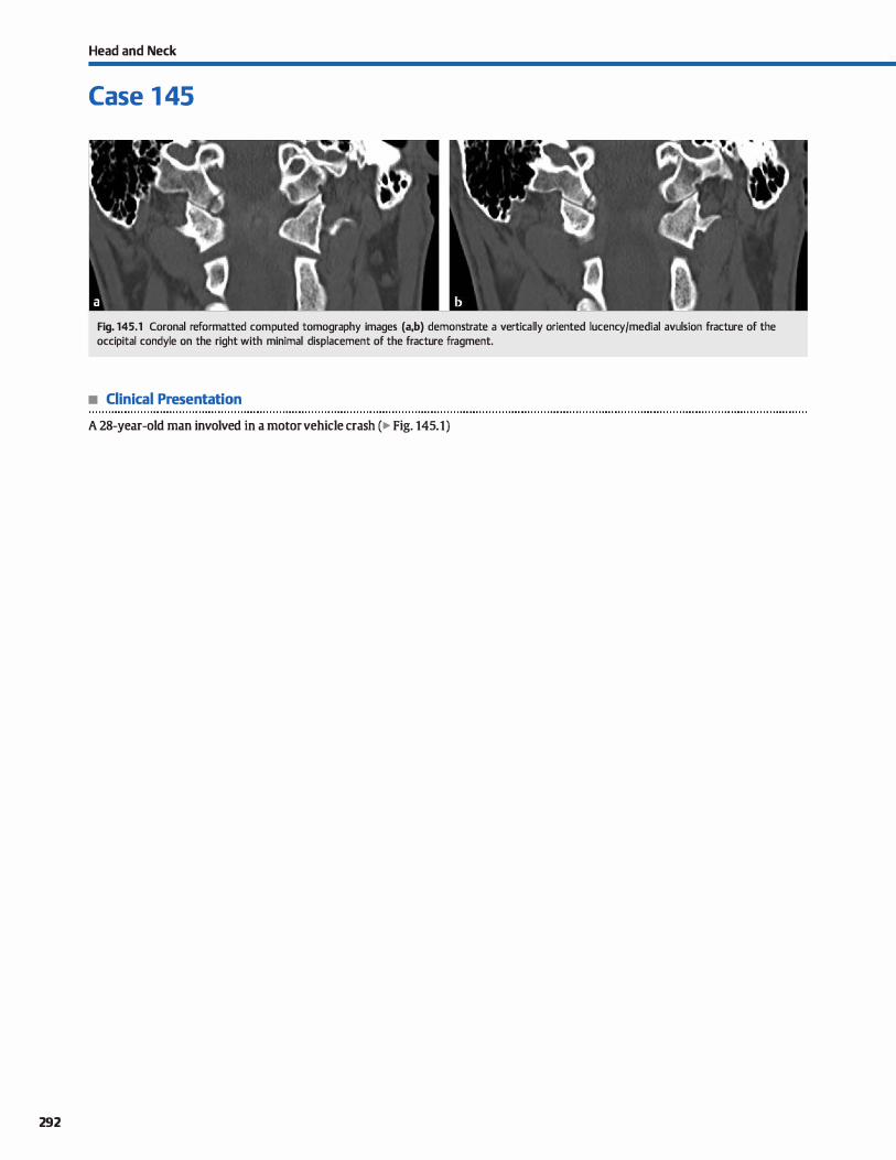
292
Head and Neck
Case 1 45
Fig. 145.1 Coronal reformatted computed tomography images (a,b) demonstrate a vertically oriented lucency/medial avulsion fracture of the occipital condyle on the right with minimal displacement of the fracture fragment.
• Clinical Presentation
A 28-year-old man involved in a motor vehicle crash (� Fig. 145.1)
• Key lmaging Finding
Vertically oriented lucency through the occipital condyle
• Diagnosis
Occipital condyle fracture (OCF). The occipital condyles are paired extensions of the occipital bone that form portions of
the foramen magnum and articulate with Cl. They are integral components of the cranioœrvical junction (CC]), along with the
atlas (Cl) and axis {C2). Llgamentous structures provide stabil
ity of the CCJ. Structures intimately related to the occipital condyles indude the medulla, vertebral arteries, spinal arteries,
hypoglossal canais, andjugular foramina.
OCFs have become more evident as imaging technology and resolution have improved. The fractures most often result from high-speed deceleration injuries, such as those seen with motor vehicle collisions, faits, or direct trauma.
OCFs are categorized into three main types based upon their overall morphology and mechanism of injury. A type I
fracture refers to a comminuted impaction fracture of the occipital condyle with no or minimal displaœment of fracture fragments. It is thought to occur from axial loading and is con
sidered a stable fracture if unilateral because the major stabilizing ligaments of the CC] {tectorial membrane and alar ligaments) typically remain intact. If bilateral, however, there may be some degree of instability. A type II fracture consists of a
� Pearts
• fype I OCF refers to a comminuted impaction fracture with no
or minimal displaœment of fracture fragments. • fype II OCF consists of a basilar skull-base fracture that
extends into one or both occipital condyles.
Suggested Reading
Leone A. Cerase A, Colosimo C. Lauro !., Puca A. Marano P. Oa:ipitll condylar fractures: a review, Radiology 2000; 216; 635-644
Case 145
more complex basilar skull-base fracture that extends into one or both occipital condyles. lt most often occurs secondary to a direct blow to the skull and is considered stable. A type III fracture represents an avulsion fracture along the medial aspect of the occipital condyle at the insertion of the alar ligament. The
fracture fragment is typically displaœd medially into the foramen magnum. The mechanism of injury consists of forœd rota
tion and side bending. The fracture is potentially unstable due to strain or disruption of the stabilizing ligaments.
Occipital condyle fractures are often occult on conventional radiographs, espeàally if nondisplaœd. Computed tomography
with sagittal and coronal reformats is the imaging modality of
choice to identify bony injuries, which may be subtle. Magnetic resonanœ imaging allows for evaluation of underlying ligamen
tous structures, which ultimately determines stability of the injury. Additional findings associated with high-veloàty trauma
indude multiple bony fractures, soft-tissue injury, vascular injury, cord injury, as well as injury to intracranial structures.
Treatment options depend upon the stability of the injury. Stable fractures are managed conservatively with immobilization. Unstable fractures may require surgical fixation.
• 'JYpe III OCF represents a media] avulsion fracture of the occipital condyle at the insertion of the alar ligament.
• 'JYpe II and unilateral type I OCFs are usually stable; type III and bilateral type I OCFs are potentially unstable.
293
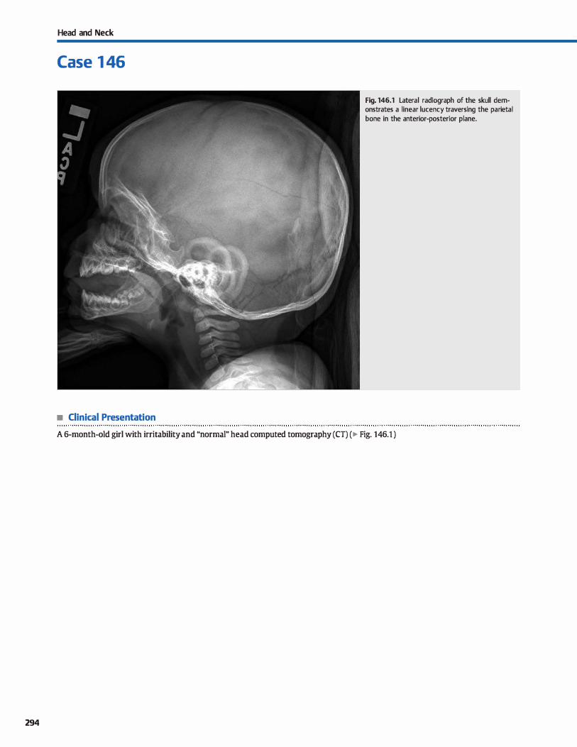
294
Head and Neck
Case 1 46
• Clinical Presentation
Fig. 146.1 Lateral radiograph of the skull demonstrates a linear lucency traversing the parietal bone in the anterior-posterior plane.
A 6-month-old girl with irritability and "normal" head computed tomography (CT) (� Fig. 146.1 )
• Key lmaging Finding
Llnear skull luœncy
• Diagnosis
Linear skull fracture. Linear fractures are the most common type of skull fracture and result from blunt injury. In most
cases, the fractures are dinically insignificant, unless there is depression of fracture fragments (depressed skull fracture), involvement of vascular structures {e.g., middle meningeal artery), or extension into and diastasis of a cranial suture. Prognosis is more dependent upon the presence or absence of asso
ciated intracranial injuries { e.g., intracranial hemorrhage, parenchymal shear injury, cerebral edema, etc.).
cr is the imaging modality of choiœ for head injury because
it is readily available, allows for rapid acquisition, and best depicts acute intracranial abnormalities, especially intracranial hemorrhage. In most cases, skull fractures are readily visualized on bone windows. However, when the fracture is parallel to
the plane of imaging, nondisplaœd skull fractures may be
� Pearts
• Llnear fractures are the most common type of skull fracture
and result Crom blunt injury. • In the absence of depressed fracture fragments, vascular
injury, or sutural diastasis, they are often self-limited.
Suggested Reading
Glass RBF, Fembach SK, Norton KI, Choi PS, Naidich lP. The infant skull: a vault of
information. Radiographies 2004; 24; 507-522
Case 146
overlooked. In these cases, linear fractures are best seen on cor
onal refonnatted images, three-dimensional volume rendered images, or on the scout view or plain radiographs.
Llnear skull fractures may be differentiated from sutures by their linear rather than serrated or interdigitating contour.
Additionally, fractures are more luœnt compared with sutures. Fractures may aise be distinguished from vascular grooves
because fractures involve both the inner and outer tables of the
skull, whereas vascular grooves only involve the inner table. When presented with a skull fracture in a child, it is impor
tant to consult with the ordering provider to ensure that the history is appropriate and corresponds with the imaging findings. In cases where the history or proposed mechanism of
injury is not consistent with the imaging findings, nonaccidental trauma should be considered.
• Llnear fractures may be differentiated from sutures by their
linear contour and increased lucency. • cr is the modality of choice for head injury; it is readily avail
able and best depicts acute intracranial injuries.
295
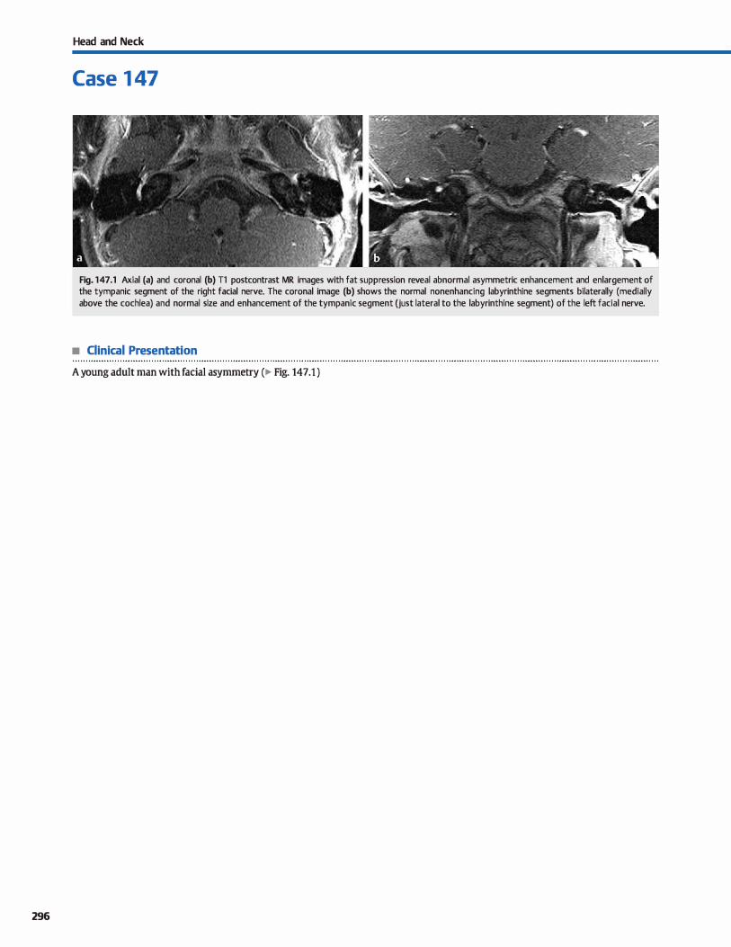
296
Head and Neck
Case 1 47
Fig. 147.1 Axial (a) and coronal (b) Tl postcontrast MR images with fat suppression reveal abnormal asymmetric enhancement and enlargement of the tympanic segment of the right facial nerve. The coronal image (b) shows the normal nonenhancing labyrinthine segments bilaterally (medially above the cochlea) and normal size and enhancement of the tympanic segment (just lateral to the labyrinthine segment) of the left facial nerve.
• Clinical Presentation
A young adult man with facial asymmetry (� Fig. 147.1 )
• Key lmaging Finding
Facial nerve enhancement
• Top 3 Differential Diagnoses
• Normal facial nerve enhancement. The proximal portions of the facial nerve that are surrounded by cerebrospinal fluid (the cisternal, canalicular, and labyrinthine segments) should not enhanœ; enhancement of these segments is considered abnormal. Beginning at the geniculate ganglion and extending distally into the horizontal (tympanic) and vertical {mastoid) segments, the facial nerve may enhance
normally. Normal enhancement is typically symmetric or
mildly asymmetric. • Bell palsy. Bell palsy refers to acute onset infectious or postin
fectious ipsilateral facial nerve palsy. Most cases are self-limiting and thought to result from the herpes simplex virus type
1, the same virus that causes cold sores. Imaging is reserved
for atypical or nonresolving cases. On magnetic resonanœ imaging (MRI), the geniculate ganglion, tympanic segment,
and mastoid segment of the facial nerve may enhance in normal patients. With Bell palsy, there is often smooth asymmet-
• Additional Differential Diagnoses
• Facial nerve hemangioma. Facial nerve hemangioma is a vas
cular tumor that occurs along the course of the facial nerve. It most often occurs in the region of the geniculate ganglion, followed by the distal internai auditory canal and junction of the horizontal (tympanic) and vertical {mastoid) segments
of the facial nerve. cr reveals a characteristic "honeycomb" bony matrix. On MRI, lesions are T2 hyperintense with avid
enhancement, corresponding to the vascular nature of the tumor.
• Perineural spread of tumor. Perineural spread of tumor along
the facial nerve most often results from parotid malignancies, specifically adenoid cystic carcinoma, which has a propensity for perineural spread. Facial nerve involvement from primary
head and neck or skin neoplasms is much Jess common. On cr. perineural spread most often manifests as abnormal enlargement of the bony facial nerve canal within the
involved segments, typically extending proximally from the
• Diagnosis
Facial nerve schwannoma
� Pearls
• Bell palsy most often involves the distal intracanalicular and
labyrinthine segments; it is usually self-limiting.
• Tumors {schwannoma and perineural spread) result in abnormal signal, enhancement, and nerve enlargement.
Suggested Reading
Saremi F, Helmy M, Farzin S, Zee CS. Go JI. MRI of cranial nenre enhanœrnent. Am j RDentgenol2005; 185: 1487-1497
Case 147
rie enhanœment of the involved tympanic and/or mastoid segments, as well as abnormal enhanœment of the portions of the facial nerve that are usually protected by the bloodnerve barrier, to indude the intracanalicular and labyrinthine
segments. The distal intracanalicular and labyrinthine segments are most commonly involved.
• Nerve sheath tumor. Schwannomas arise from perineural Schwann cells. They are composed of two tissue types: Antoni
A ( densely packed) and Antoni B (loosely packed, T2 hyperintense � The majority of patients with facial nerve schwannomas present with hearing Joss or facial neuropathy. Temporal bone computed tomography ( CT) reveals smooth enlargement of
the involved segments of the facial nerve canal. The region of the geniculate ganglion is the most common site with exten
sion into adjacent segments of the facial nerve. MRI demonstrates abnormal signal, tubular or fusiform enlargement, and abnormal enhanœment of the involved segments.
stylomastoid foramen. MRI demonstrates abnormal enlargement, signal intensity, and enhanœment of the involved portions of the facial nerve. When perineural spread of tumor is suspected, imaging should ex.tend through the neck to evaluate for a primary origin.
• Ramsey Hunt syndrome. Ramsey Hunt syndrome is caused by reactivation of the varicella-zoster virus, which lies dormant in the geniculate ganglion. Patients are often elderly or immunocompromised and present with ipsilateral otalgia,
a vesicular rash involving the external auditory canal and
auride, and facial neuropathy. MRI reveals abnormal enhancement within the involved segments of the facial
nerve; abnormal enhancement of the vestibulocochlear nerve and labyrinthine structures is also commonly seen. Patients are typically treated with steroids and antiviral medications
( e.g., acydovir). There is a good prognosis/recovery in -50 to 70% of cases.
• Hemangiomas are vascular tumors that result in a "honeycomb" matrix on CT and avid enhancement on MRI.
297
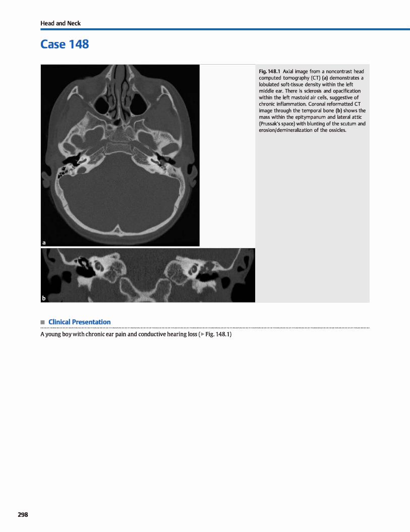
Head and Neck
Case 1 48
• Clinical Presentation
A young boy with chronic ear pain and conductive hearing Joss ( � Fig. 148.1)
298
Fig. 148.1 Axial image from a noncontrast head computed tomography (CT) (a) demonstrates a lobulated soft-tissue density within the left middle ear. There is sclerosis and opacification within the left mastoid air cells, suggestive of chronic inflammation. Coronal reformatted CT image through the temporal bone (b) shows the mass within the epitympanum and lateral attic (Prussak's space) with blunting of the scutum and erosion/demineralization of the ossicles.
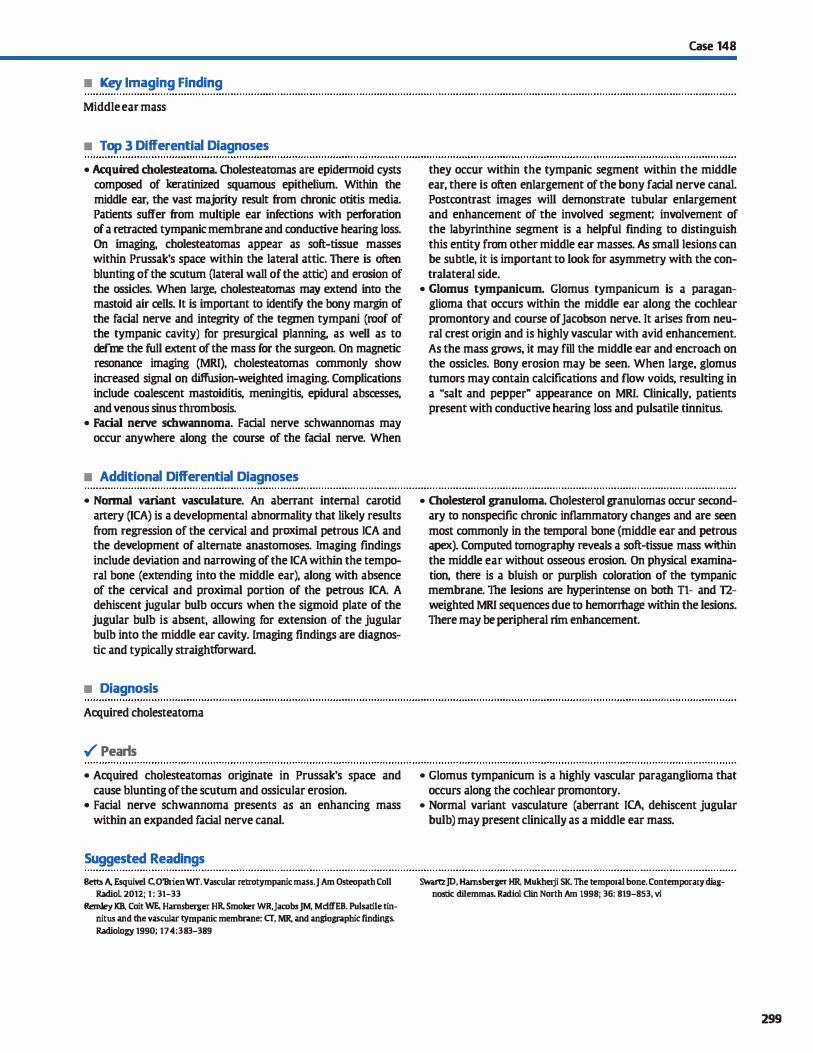
• Key lmaging Finding
Middle ear mass
• Top 3 Differential Diagnoses
• Acquired cholesteatoma. Cholesteatomas are epidermoid cysts composed of lœratinized squamous epithelium. Within the middle ear, the vast majority result from chronic otitis media. Patients sutfer from multiple ear infections with perforation of a retracted tympanic membrane and conductive hearing loss. On imaging, cholesteatomas appear as soft-tissue masses within Prussak's spaœ within the lateral attic. There is often blunting of the scutum {lateral wall of the attic) and erosion of the ossides. When large, cholesteatomas may extend into the mastoid air œlls. It is important to identify the bony margin of the facial nerve and integrity of the tegmen tympani (roof of the tympanic cavity) for presurgical planning, as well as to defme the full extent of the mass for the surgeon On magnetic resonanœ imaging (MRI), cholesteatomas commonly show increased signal on diffusion-weighted imaging. Complications include coalescent mastoiditis, meningitis, epidural absœsses, and venous sinus thrombosis.
• Facial nerve schwannoma. Faàal nerve schwannomas may occur anywhere along the course of the faàal nerve. When
• Additional Differential Diagnoses
• Nonnal variant vasculature. An aberrant internai carotid artery (ICA) is a developmental abnormality that likely results from regression of the cervical and proximal petrous ICA and the development of altemate anastomoses. Imaging findings include deviation and narrowing of the ICA within the temporal bone (extending into the middle ear), along with absence of the cervical and proximal portion of the petrous ICA. A dehiscent jugular bulb occurs when the sigmoid plate of the jugular bulb is absent, allowing for extension of the jugular bulb into the middle ear cavity. lmaging findings are diagnostic and typically straightforward.
• Diagnosis
Acquired cholesteatoma
� Pearts
• Acquired cholesteatomas originate in Prussak's spaœ and cause blunting of the scutum and ossicular erosion.
• Facial nerve schwannoma presents as an enhancing mass within an expanded facial nerve canal.
Suggested Readings
Betts A. EsquiVI!! C. O'Brien wr. Vascular œtrotympanic mass. j Am Osteopath Coll Radiol. 2012; 1 ; 31-33
Rrmley KB, Coit WE, Hamsberger HR, Smolœr WR.jacobs JM, Mdff'EB. Pulsatile tinnitus and the vascular tympanic membrane: cr, MR. and angiographie findings. Radiology 1990; 174;383-389
Case 148
they occur within the tympanic segment within the middle ear, there is often enlargement of the bony faàal nerve canal. Postcontrast images will demonstrate tubular enlargement and enhancement of the involved segment; involvement of the labyrinthine segment is a helpful finding to distinguish this entity from other middle ear masses. As small lesions can be subtle, it is important to look for asymmetry with the contralateral side.
• Glomus tympanicum. Glomus tympanicum is a paraganglioma that occurs within the middle ear along the cochlear promontory and course of Jacobson nerve. lt arises Crom neural crest origin and is highly vascular with avid enhancement As the mass grows, it may fill the middle ear and encroach on the ossicles. Bony erosion may be seen. When large, glomus tumors may contain calcifications and flow voids, resulting in a Msalt and pepper" appearance on MRI. Clinically, patients present with conductive hearing Joss and pulsatile tinnitus.
• Cholesterol granuloma. Cholesterol granulomas occur secondary to nonspeàfic chronic inflammatory changes and are seen most commonly in the temporal bone (middle ear and petrous apex). Computed tomography reveals a soft-tissue mass within the middle ear without osseous erosion On physical examination, there is a bluish or purplish coloration of the tympanic membrane. The lesions are hyperintense on both Tl- and T2-weighted MRI sequenœs due to hemorrhage within the lesions. There may be peripheral rim enhanœment.
• Glomus tympanicum is a highly vascular paraganglioma that occurs along the cochlear promontory.
• Normal variant vasculature (aberrant ICA, dehiscent jugular bulb) may present clinically as a middle ear mass.
SwartzjD, Harnsberger HR, Mukherji SK. The temporal bene. Contemporary diagnostic dilemmas. Radial Oin North Am 1998; 36; 819-853, vi
299
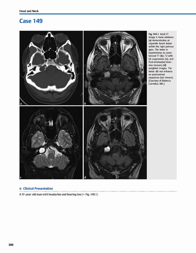
Head and Neck
Case 1 49
• Clinical Presentation
A 37-year-old man with headaches and hearing Joss (� Fig. 149.1 )
300
Fig. 149.1 Axial CT image in bone windows (a) demonstrates an expansile lucent lesion within the right petrous apex. The lesion is hyperintense on unenhanced Tl (b), T2 with fat suppression (c), and fluid-attenuated inversion recovery (d) weighted images. The lesion did not enhance on postcontrast sequences (not shown). (Courtesy of Rebecca Cornelius, MD.)
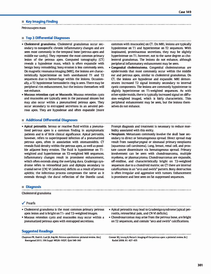
• Key lmaging Finding
Petrous apex mass
• Top 3 Differential Diagnoses
• Cholesterol granuloma. Cholesterol granulomas occur secondary to nonspecific chronic inflammatory changes and are seen most commonly in the temporal bone (petrous apex and
middle ear cavity). They represent the most common primary lesion of the petrous apex. Computed tomography (CT) reveals a hypodense mass, which is often expansile with benign bony remodeling; bony erosion is Jess commonly seen.
On magnetic resonance imaging (MRI), the lesions are charac
teristically hyperintense on both unenhanœd Tt and T2 sequences due to hemorrhage within the lesions. Occasion
ally, a T2 hypointense hemosiderin ring is seen. There may be peripheral rim enhanœment, but the lesions themselves will not enhance.
• Mucous retention cyst or Mucocele. Mucous retention cysts and mucoœles are typically seen in the paranasal sinuses but
may also occur within a pneumatized petrous apex. They occur secondary to entrapped secretions in an aerated petrous apex. They are hypodense and often expansile (more
• Additional Differential Diagnoses
• Apical petrositis. Sercus or reactive fluid within a pneuma
tized petrous apex is a common finding in asymptomatic patients and is of little dinical significance. Apical petrositis, however, refers to superimposed infection of a pneumatized
petrous apex, often in association with otomastoiditis. cr reveals fluid density within the petrous apex, as well as possible adjacent bony erosion. The fluid is hypointense on Tt -weighted and hyperintense on T2-weighted MR sequenœs. Inflammatory changes result in prominent enhancement,
which often extends along the overlying dura. Gradenigo syndrome refers to retroorbital pain and diplopia secondary to
cranial nerve {CN) VI (abducens) deficits as a result ofpetrous apidtis; the infectious process compresses the nerve as it
extends through the durai reflection of the Dorello canal.
• Diagnosis
Cholesterol granuloma
� Pearts
• Cholesterol granuloma is the most common primary petrous
apex lesion and is bright on Tt- and T2-weighted images. • Mucous retention cysts and mucoœles may occur within a
pneumatized petrous apex with entrapped secretions.
Suggested Readings
Clupman PR, Shah R, CUré JK, Bag AK. Petrous apexlesiorus; pictmial review. Amj
Rœntgenol 2011; 196 Suppl: WS2&-WS37, QJrlz 540-543
Case 149
common with mucoceles) on cr. On MRI, lesions are typically
hypointense on Tl and hyperintense on T2 sequences. With inspissated, proteinaceous secretions, they may be slightly hyperintense on Tt ; however, not to the same degree as cholesterol granulomas. The lesions do not enhance, although
peripheral inflammatory enhancement may be seen.
• Congenital cholesteatoma. Congenital cholesteatomas are epidermoids that most commonly occur within the middle
ear and petrous apex, similar ta cholesterol granulomas. On
cr, the lesions are hypodense and expansile. MRI demonstrates increased T2 signal intensity secondary to internai cystic components. The lesions are commonly hypointense to
slightly hyperintense on Tt-weighted sequences. As with other epidermoids, there is typically increased signal on diffu
sion-weighted imaged, which is fairly characteristic. Thin peripheral enhancement may be seen, but the lesions themselves do not enhanœ.
Prompt diagnosis and treatment is necessary ta reduce mor
bidity associated with this entity. • Neoplasm. Metastases commonly involve the skull base sec
ondary to direct or hematogenous spread. Direct spread may result from nasopharyngeal or sinonasal primary neoplasms (squamous cell carcinoma). Lung, breast, renal cell, and pros
tate cancer disseminate via hematogenous spread. Primary involvement can be seen with chondrosarcoma, multiple myeloma, or plasmacytoma. Chondrosarcomas are expansile, off-midline, and characteristically bright on T2-weighted
sequences due to a chondroid matrix; on cr there are internai
calcifications in an "arcs and swirls" pattern. Bony destruction is often irregular and aggressive with tumors. Enhancement
is prominent and best seen on fat-suppressed sequences.
• Apical petrositis may lead to Gradenigo syndrome (apical petrositis, retroorbital pain, and CN VI deficits ).
• Chondrosarcomas may arise from the petrous bone, are bright
on T2 sequences, and contain uarcs and swirls" calcifications.
Connor SEJ, I.eung R, Natas S. lmaging of the petrous apex: a pictorial review. Br J
Radiol 2008; 81: 427-435
301
302
Head and Neck
Case 1 50
Fig. 1 50.1 Axial computed tomography (CT) image (a) demonstrates focal calcification and narrowing of the left EAC. Coronal reformatted CT image of the left temporal bone (b) reveals the calcification at the junction of the cartilaginous and bony EAC. A pedunculated morphology was seen on sequential images (stalk out of plane).
• Clinical Presentation
A 14-year-old boy with abnormality on physical exam (� Fig. 150.1 )
• Key lmaging Finding
Circumscribed externat auditory canal (EAC) calcification/ ossification
• Top 2 Differential Diagnoses
• EAC exostosis. EAC exostoses are benign overgrowths that
result from chronic inflammation. They occur most often in
young adults and have a disproportionate involvement in surfers, as well as divers and swimmers; hence, the term
"surfer's ear." Chronic exposure to cold water Ieads to repeated bouts of otitis extema, most often due to StaphylDcoccus aureus or Pseudomonas aeruginosa. This entity differs from malignant otitis externa, which is an aggressive infection that occurs in immunocompromised patients, most often
elderly diabetics. Acutely, patients may present with otalgia, pruritis, EAC drainage, and conductive hearing loss. Chroni
cally, there is bony overgrowth of the EAC that is characteris-
• Diagnosis
EAC osteoma
y' Pearts
• EAC exostoses are a complication of "surfer's ear"; bony
lesions are often bilateral, circumferential, and sessile. • Osteomas are bony overgrowths that are unilateral, peduncu
lated, and occur at the bony-cartilaginous junction.
Suggested Reading
Tran LP, Grundfast KM, Selesnick SH. Benign lesi0Il5 of the extemal auditory canal.
OID!aryngol Oin North Am 1996; 29: 807-825
Case 150
tically bilateral, circumferential, and sessile. Treatment is often not neœssary; when needed, it consists of surgical correction of the EAC stenosis.
• EAC osteoma. Osteomas are benign bony overgrowths that rarely involve the temporal bone. They occur sporadically and
are typically asymptomatic and incidental. On computed tomography, they appear as unilateral, circumscribed, pedunculated bony lesions that occur at the junction of the bony and cartilaginous portion of the EAC. The overlying soft-tissue portion of the EAC is normal. Lesions are typically not treated unless there is significant, symptomatic EAC stenosis.
• In the vast majority of cases, neither EAC exostoses nor osteomas require treatment
303
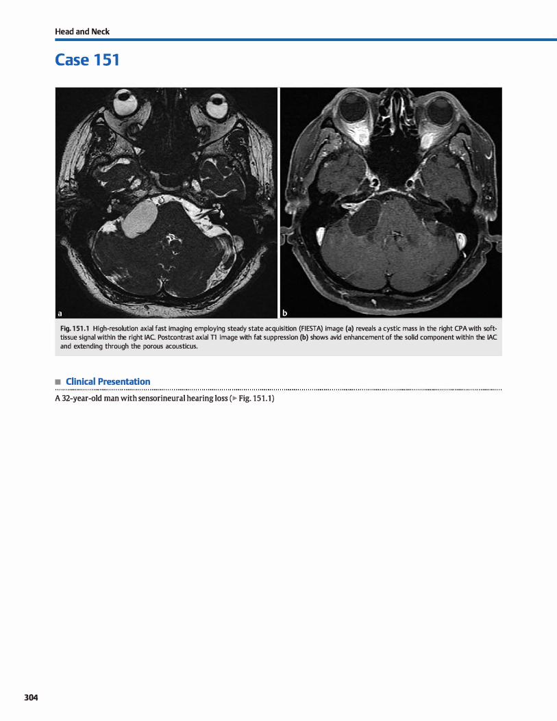
304
Head and Neck
Case 1 51
Fig. 1 51 .1 High-resolution axial fast imaging employing steady state acquisition (FIESTA) image (a) reveals a cystic mass in the right CPA with softtissue signal within the right IAC. Postcontrast axial Tl image with fat suppression (b) shows avid enhancement of the solid component within the IAC and extending through the porous acousticus.
• Clinical Presentation
A 32-year-old man with sensorineural hearing Joss (� Fig. 151.1)
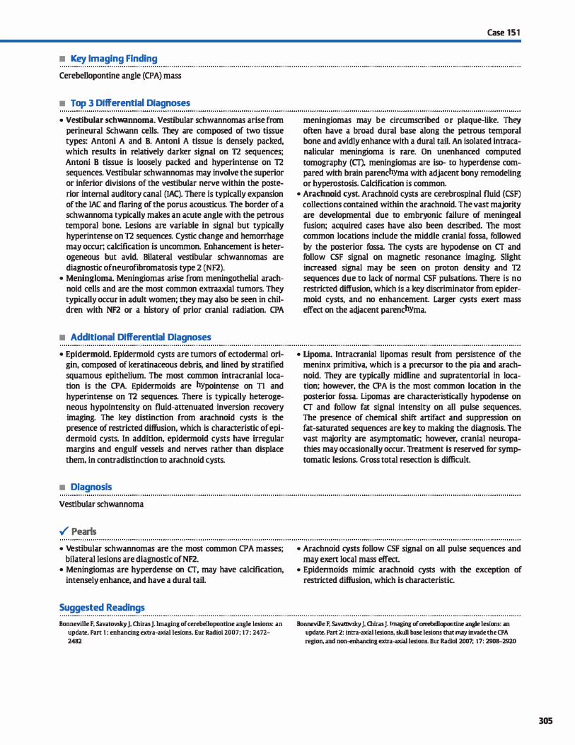
• Key lmaging Finding
Cerebellopontine angle (CPA) mass
• Top 3 Differential Diagnoses
• Vestibular schwannoma. Vestibular schwannomas arise from
perineural Schwann œlls. They are composed of two tissue types: Antoni A and B. Antoni A tissue is densely packed,
which results in relatively darker signal on T2 sequences; Antoni B tissue is loosely packed and hyperintense on T2 sequences. Vestibular schwannomas may involve the superior or inferior divisions of the vestibular nerve within the poste
rior internai auditory canal (IAC). There is typically expansion
of the IAC and flaring of the parus acousticus. The border of a schwannoma typically makes an acute angle with the petrous temporal bone. Lesions are variable in signal but typically
hyperintense on T2 sequences. Cystic change and hemorrhage may occur; calcification is uncommon. Enhancement is heterogeneous but avid. Bilateral vestibular schwannomas are
diagnostic ofneurofibromatosis type 2 (NF2).
• Meningioma. Meningiomas arise from meningothelial arach
noid cells and are the most common extraaxial tumors. They
typically occur in adult women; they may also be seen in children with NF2 or a history of prior cranial radiation. CPA
• Additional Differential Diagnoses
• Epidermoid. Epidermoid cysts are tumors of ectodennal ori
gin, composed of keratinaceous debris, and lined by stratified
squamous epithelium. The most common intracranial location is the CPA Epidermoids are hypointense on Tl and
hyperintense on T2 sequenœs. There is typically heteroge
neous hypointensity on fluid-attenuated inversion recovery imaging. The key distinction from arachnoid cysts is the presence of restricted diffusion, which is characteristic of epi
dermoid cysts. In addition, epidennoid cysts have irregular margins and engulf vessels and nerves rather than displace
them. in contradistinction to arachnoid cysts.
• Diagnosis
Vestibular schwannoma
� Pearts
• Vestibular schwannomas are the most common CPA masses;
bilateral lesions are diagnostic of NF2. • Meningiomas are hyperdense on CT, may have calcification,
intensely enhance, and have a durai tail.
Suggested Readings
Case 151
meningiomas may be circumscribed or plaque-Iike. They often have a broad durai base along the petrous temporal bone and avidly enhance with a durai tail. An isolated intracanalicular meningioma is rare. On unenhanœd computed
tomography (CT), meningiomas are iso- to hyperdense com
pared with brain parenchyma with adjacent bony remodeling or hyperostosis. calcification is common.
• Arachnoid cyst. Arachnoid cysts are cerebrospinal fluid (CSF) collections contained within the arachnoid. The vast majority are developmental due to embryonic failure of meningeal fusion; acquired cases have also been described. The most common locations indude the middle cranial fossa, followed
by the posterior fossa. The cysts are hypodense on CT and follow CSF signal on magnetic resonance imaging. Slight
increased signal may be seen on proton density and T2 sequences due to lack of normal CSF pulsations. There is no
restricted diffusion, which is a key discriminator from epidermoid cysts, and no enhancement Larger cysts exert mass effect on the adjacent parenchyma.
• Lipoma. Intracranial lipomas result from persistence of the meninx primitiva, which is a precursor to the pia and arach
noid. They are typically midline and supratentorial in location; however, the CPA is the most common location in the posterior fossa. Lipomas are characteristically hypodense on
CT and follow fat signal intensity on ail pulse sequences. The presence of chemical shift artifact and suppression on fat-saturated sequences are key to making the diagnosis. The vast majority are asymptomatic; however, cranial neuropathies may occasionally occur. Treatment is reserved for symp
tomatic lesions. Gross total resection is difficult.
• Arachnoid cysts follow CSF signal on ail pulse sequences and
may exert local mass effect. • Epidennoids mimic arachnoid cysts with the exception of
restricted diffusion, which is characteristic.
Bonneville F, Savatovsky J, Chiras J. lmaging of cerebellopontine angle lesions: an Bonneville F, Savatuvsky J, Clliras J. Imaging of cerebellopontine angle lesions: an
updaœ. Part 1: enhandng extra-axial lesions. Eur Radio! 2007; 17: 2472- updaœ. Part 2: intra-axial lesions, skull base lesions that may invade the CPA
2482 region, and non-enhandng extra-axial lesions. Eur Radiol 2007; 17: 2908-2920
305
306
Head and Neck
Case 1 52
Fig. 1 52.1 Coned-down axial CT images in bone window through the left inner ear (a-c) demonstrate abnormal configuration of the cochlea with lack of visualization of the normal 211.z turns (a), an enlarged vestibule (b), and enlargement of the vestibular aqueduct (c).
• Clinical Presentation
A 6-year-old boy with hearing Joss ( � Fig. 152.1 )
• Key lmaging Finding
Congenital inner ear malformation
• Top 3 Differential Diagnoses
• Large vestibular aqueduct syndrome {LVAS). LVAS is a common cause of sensorineural deafness in early childhood. Affected patients hear normally at birth with bilateral sensorineural hearing loss progressing over the first few months ta years of life. Unenhanced computed tomography (CT) reveals enlargement of the vestibular aqueducts (bilateral in > 90% of cases), measuring>l.S mm in diameter at its midpoint. The
size of the semicircular canais can also be used as an internai rough estimate of the expected size of the vestibular aqueduct. Associated abnormalities of cochlea and vestibule are common but may be subtle.
• Cystic cochleovestibular anomaly. Cystic cochleovestibular
anomaly is a result of arrested inner ear development, resulting in cystic dilatation of the cochlea and vestibule. No internai architecture or features can be identified. Patients have
• Additional Differential Diagnoses
• Cochlear aplasia. Cochlear aplasia results from arrest of normal development of the cochlea during embryogenesis. It may be unilateral or bilateral depending upon the type and timing of insult, and patients present with sensorineural hearing Joss at
• Diagnosis
Large vestibular aqueduct with associated cochlear and vestibular abnormalities
� Pearls
• LVAS presents with vestibular aqueducts > 1.5 mm in diameter: hearing Joss is bilateral and progressive.
• Cystic cochleovestibular anomaly bas a characteristic Mfigure 8" appearanœ of the cochlea and vestibule.
Suggested Reading
Joshi VM, Navlekar SK, Kishore GR, Reddy I<f, Kumar EC. cr and MR imaging of the inner ear and brain in children with amgenital sensarineural hearing loss. Radiographies 2012; 32: 683-698
Case 152
sensorineural hearing Joss at birth. On unenhanced axial CT and T2 magnetic resonanœ imaging, the dilated cochlea and vestibule demonstrate a Mfigure 8" appearance. Semidrcular canais are commonly dilated as well. The vestibular aqueduct is typically normal.
• Cystic common cavity. Cystic common cavity refers to lack of development of the cochlea and vestibule. The semicircular
canais are variably affected and are most often absent: occasionally, they may be dysplastic or even normal. As with cystic cochleovestibular anomaly, patients present with sensorineural hearing loss at birth. lmaging findings include a single cystic cavity in place of normal inner ear structures (cochlea, vestibule, and, commonly, semicircular canais). Middle ear structures and the vestibular aqueduct are typically normal.
birth. lmaging findings indude an absent cochlea. Remaining inner ear structures, induding the vestibule and semicircular canais, may be dysplastic or normal The middle ear structures and vestibular aqueduct are typically normal.
• Cystic common cavity refers ta a single cavity in place of normal inner ear structures.
307
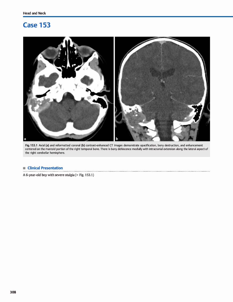
308
Head and Neck
Case 1 53
Fig. 1 53.1 Axial (a) and reformatted coronal (b) contrast-enhanced CT images demonstrate opacification, bony destruction, and enhancement centered on the mastoid portion of the right temporal bone. There is bony dehiscence medially with intracranial extension along the lateral aspect of the right cerebellar hemisphere.
• Clinical Presentation
A 6-year-old boy with severe otalgia (� Fig. 1 53.1 )
• Key lmaging Finding
Aggressive temporal bone mass in a child
• Top 3 Differential Diagnoses
• Coalescent otomastoiditis. Coalescent mastoiditis refers ta an acute infectious process of the middle ear cavity and mastoids with destruction of the bony mastoid septae and overlying cortex. It may occur as a primary infection or in assoàation with an acquired cholesteatoma. Patients present clinically with fever, hearing Joss, and otalgia. Computed tomography (CT) demonstrates opaàfication of the middle ear cavity and
mastoids with erosion of the mastoid septae and mastoid cortex; the ossides may also be involved. On magnetic resonanœ imaging (MRI), the inflammatory process is 12 hyperintense with relatively diffuse enhancement. Focal absœsses or an underlying cholesteatoma may be hyperintense on diffusionweighted imaging. Complications indude subperiosteal, Bezold (mastoid tip into stemocleidomastoid muscle), epidural, or brain parenchymal absœsses; meningitis; and durai venous sinus thrombosis.
• Langerhans cell histiocytosis (LCH). LCH refers ta abnormal proliferation of histiocytes. lt may be localized (formerly eosinophilic granuloma), multifocal (formerly Hand-SchüllerChristian disease), or disseminated (formerly Letterer-Siwe disease). The localized form is the most common and mildest
form. The calvarium is most often involved, followed by the spine. The classic radiological description is a well-defined lytic Iesion without sclerotic borders. A "beveled" edge appearance can result from greater involvement of the inner
• Diagnosis
Langerhans œll histiocytosis
� Pearts
• Coalescent mastoiditis refers ta acute infectious bony destruction of the mastoid septae and overlying cortex.
• LCH classically presents as a well-defined lytic Iesion with Mbeveled" edges; Temporal bone involvement may be diffuse.
Suggested Readings
Connor SEJ, Leung R, Naw s. Imaging of the petrous apex; a pictorial review. Br J Radiol2008; 81: 427-<43S
Case 153
than outer table of the skull MRI will show marrow replacement and avid soit-tissue enhancement. Temporal bone involvement typically occurs within the petrous apex or otic capsule. Temporal bone LCH may also present with diffuse bony destruction, mimicking a neoplastic or aggressive infectious/inflammatory proœss. The most common clinicat symptoms include pain, palpable mass, and/or systemic symptoms.
• Rhabdomyosarcoma. Rhabdomyosarcoma is a rare, aggressive malignancy of childhood that commonly involves the head and neck region. With temporal bone involvement, the mass often originates within the middle ear and extends into
the mastoids and externat auditory canal. More aggressive lesions extend intracranially, into the skull base, and/or into the extracranial soft tissues of the upper neck. Cervical nodal metastases may rarely be seen. CT demonstrates a soit-tissue mass centered within the middle ear cavity and mastoids with aggressive bony destruction. The mass is typically hypointense on Tl and hyperintense on 12 MRI sequences, nonspeàfic. There is heterogeneous but avid contrast enhancement; enhancement of the dura is indicative of intracranial extension. Perineural spread along cranial nerves VII and VIII may also be seen. Œnically, patients present in the first or second decades of life with symptoms mimicking recurrent, chronic otomastoiditis.
• Rhabdomyosarcoma is a childhood malignancy; the destructive mass is œntered in the middle ear and mastoids.
• LCH and rhabdomyosarcoma demonstrate soft-tissue rather than inflammatory { otomastoiditis) enhancement.
Razek AA, Huang BY, I.esions of the petrous apex: dassification and findings at cr
and MRimaging. Radiographies 2012; 32: 151-173
309
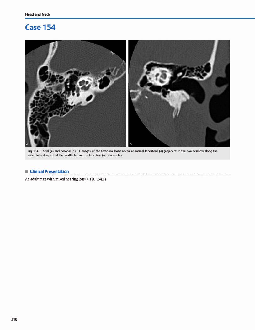
310
Head and Neck
Case 1 54
Fig. 1 54.1 Axial (a) and coronal (b) CT images of the temporal bone reveal abnormal fenesteral (a) (adjacent to the oval window along the anterolateral aspect of the vestibule) and pericochlear (a,b) lucencies.
• Clinical Presentation
An adult man with mixed hearing Joss ( � Fig. 154.1 )
• Key lmaging Finding
Otic capsule luœncies
• Top 3 Differential Diagnoses in Older Adults
• Otosclerosis. Otosderosis results in abnormal bony development and growth that characteristically involves the region of the stapes and oval window. Approximately 60% of cases are thought to be hereditary, whereas the remainder appear sporadic. Although patients may present anytime from adolescence to adulthood, most present as young adults with progressive, mixed hearing Joss. Computed tomography (CT)
is ideal for identifying the regions of abnormal mineralization that appear as otic capsule luœncies (of various morphologies), but the findings may be subtle. Approximately 85% of cases are termed fenesteral with abnormal otic lucencies in the region of the aval and round windows. Approximately 15% of cases are termed cochlear with abnormal pericochlear lucencies. Often times, there are components of both fenes
teral and cochlear otosderosis. Treatment options indude hearing aids for more mild cases and stapedectomy and prosthesis placement for more severe cases.
• Paget disease. Paget disease affects the elderly population and is more common in men. It results in bony expansion with cortical thickening and thickened trabeculae. Common
Case 154
locations indude the axial skeleton, pelvis, long bones, and calvarium. The most common forms of calvarial involvement consist of osteoporosis circumscripta, which presents as a prominent lucency, and the more diffuse "cotton wool" appearanœ, which refers to the radiographie appearance of the bony and trabeculae thidc:ening. Otic capsule involvement most often consists of bony thickening with diffuse lucency. The bony involvement extends beyond the otic capsule, which in conjunction with patient age, is a useful discriminator.
• Osteoradionecrosis. Radiation necrosis is a late complication that may be seen months or years after radiation therapy. It is thought to be caused by vascular injury and resultant
ischemia. The severity is dependent upon the region treated, the dose of treatment, length of time between treatments, and the duration of treatments. Imaging findings indude demineralization, manifested as abnormal lucencies that are typically diffuse. In severe cases, there may be franlc bony destruction. Common primary neoplasms ta involve the otic capsule in the radiation field indude skull base, nasopharyngeal, and brain neoplasms.
• Top 3 Differential Diagnoses in Children and Young Adults
• Otosderosis. See above. • Nonnal variation of mineralization. High-resolution CT in
infants and young children may occasionally demonstrate otic capsule lucencies in otherwise asymptomatic patients. On follow-up studies, many patients demonstrate interval ossification of the regions in question, suggesting a form of normal variation or delayed cartilaginous ossification. When presented with a case of suspected otosderosis in a young,
asymptomatic child, correlation with symptoms, audiometric testing, and possibly follow-up imaging may help distinguish normal variant ossification from otosderosis.
• Diagnosis
Otosderosis
� Pearls
• Otosderosis results in mixed hearing Joss with fenesteral (most common) and/or pericochlear lucencies on CT.
• Paget disease causes bony expansion in elderly patients; calvarial involvement extends beyond the otic capsule.
Suggested Reading
PeldmlaJ, Pitkaranta A,Jappel A et al. Localized pericochlear hypoattenuating foci at
temporal-bone tlùn-section cr in pediatric patients: nonpathologic differential
diagnostic entity? Radiology 2004; 230; 88-92
• Osteogenesis imperfect tarda. Osteogenesis imperfect (01) is a connective tissue disorder that results in fragile bones, as well as other connective tissue abnormalities. TYJ>e 1 is referred to as QI tarda, is the mildest form. and may present with otic capsule lucencies, in addition to nondeforming fragile bones and blue sciera. The disorder is dinically evident but may appear identical to otosderosis on CT imaging with otic capsule lucencies predominantly in the pericochlear regions. The diagnosis of QI should only be suggested in the appropriate clinicat setting due to its rarity.
• Osteoradionecrosis occurs in the treatment field and results from vascular injury and resultant bony ischemia.
• 01 tarda results in otic capsule lucencies similar to otosderosis, nondeformed fragile bones, and blue sciera.
31 1
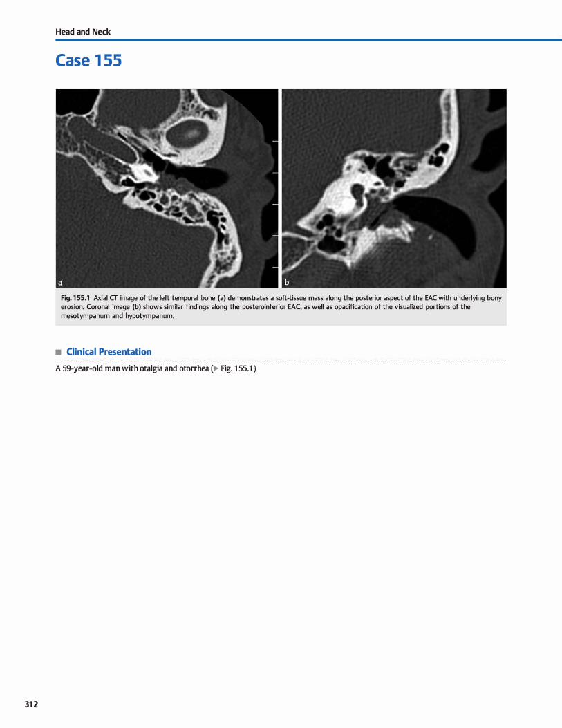
312
Head and Neck
Case 1 55
Fig. 1 55.1 Axial CT image of the left temporal bone (a) demonstrates a soft-tissue mass a long the posterior aspect of the EAC with underlying bony erosion. Coronal image (b) shows similar findings along the posteroinferior EAC, as well as opacification of the visualized portions of the mesotympanum and hypotympanum.
• Clinical Presentation
A 59-year-old man with otalgia and otorrhea (� Fig. 1 55.1 )
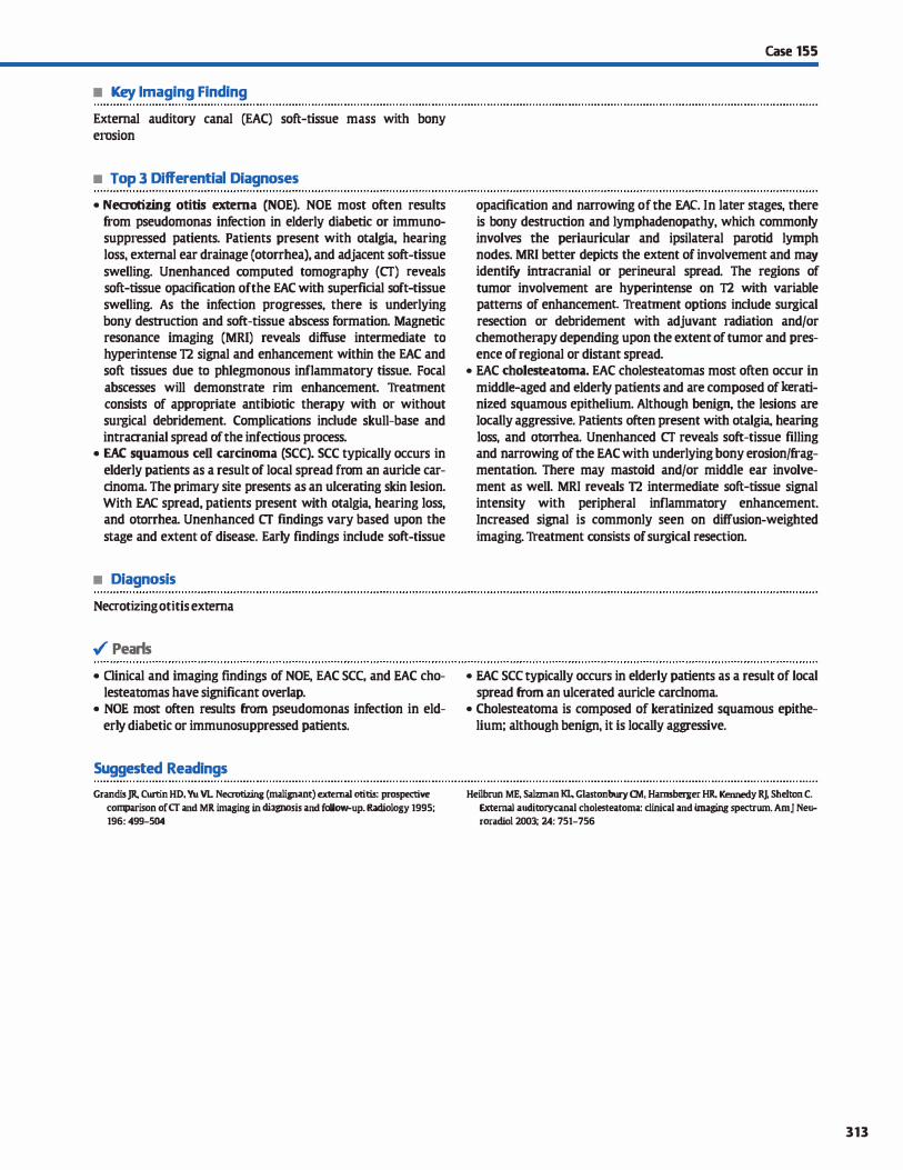
• Key lmaging Finding
Extemal auditory canal (EAC) soft-tissue mass with bony erosion
• Top 3 Differential Diagnoses
• Necrotizing otitis externa (NOE). NOE most often results from pseudomonas infection in elderly diabetic or immunosuppressed patients. Patients present with otalgia, hearing loss, extemal ear drainage {otorrhea). and adjacent soit-tissue swelling. Unenhanced computed tomography {CT) reveals sort-tissue opacification ofthe EAC with superficial soft-tissue swelling. As the infection progresses, there is underlying bony destruction and soft-tissue absœss formation. Magnetic resonance imaging (MRI) reveals diffuse intermediate ta hyperintense T2 signal and enhancement within the EAC and soft tissues due ta phlegmonous inflammatory tissue. Focal abscesses will demonstrate rim enhanœment. Treatment consists of appropriate antibiotic therapy with or without surgical debridement. Complications indude skull-base and intracranial spread of the infectious proœss.
• EAC squamous ceIJ carcinoma (SCC). SCC typically occurs in eJderJy patients as a result of local spread from an auride carcinoma. The primary site presents as an ulcerating skin Jesion. With EAC spread, patients present with otalgia, hearing Joss, and otorrhea. Unenhanced CT findings vary based upon the stage and extent of disease. Early findings indude soft-tissue
• Diagnosis
Necrotizing otitis extema
y' Pearts
• Oinical and imaging findings of NOE, EAC sec, and EAC cholesteatomas have significant overlap.
• NOE most often results from pseudomonas infection in elderly diabetic or immunosuppressed patients.
Suggested Readings
Grandis JR, Curtin HD, Yu VI. Necrotizing (malignant) extemal otitis: prospective
comparison of cr and MR imaging in diagnosis and follow-up. Radiology 1995: 196: 499-504
Case 155
opacification and narrowing of the EAC. In Jater stages, there is bony destruction and lymphadenopathy, which commonly involves the periauricular and ipsilateral parotid lymph nodes. MRI better depicts the extent of involvement and may identify intracranial or perineural spread. The regions of tumor involvement are hyperintense on T2 with variable patterns of enhancement Treatment options indude surgical resection or debridement with adjuvant radiation and/or chemotherapy depending upon the extent of tumor and presence of regional or distant spread.
• EAC cholesteatoma. EAC cholesteatomas most often occur in middle-aged and elderly patients and are composed of keratinized squamous epithelium. Although benign, the lesions are Iocally aggressive. Patients often present with otalgia, hearing Joss, and otorrhea. Unenhanced CT reveals soft-tissue filling and narrowing of the EAC with underlying bony erosion/fragmentation. There may mastoid and/or middJe ear invoJvement as well. MRI reveaJs T2 intermediate sort-tissue signal intensity with peripheral inflammatory enhanœment. Increased signal is commonly seen on diffusion-weighted imaging. Treatment consists of surgical resection.
• EAC sec typicalJy occurs in eJderly patients as a result of local spread from an ulcerated auricle cardnoma.
• Cholesteatoma is composed of keratinized squamous epithelium; although benign, it is locally aggressive.
Heilbrun ME, Salzman KI., Glastonbury CM, Hamsberger HR. Kennedy RJ, Shelton C.
Extemal auditorycanal cholesteatoma: dinical and imaging spectrum. AmJ Neuroradiol 2003; 24: 751-756
313
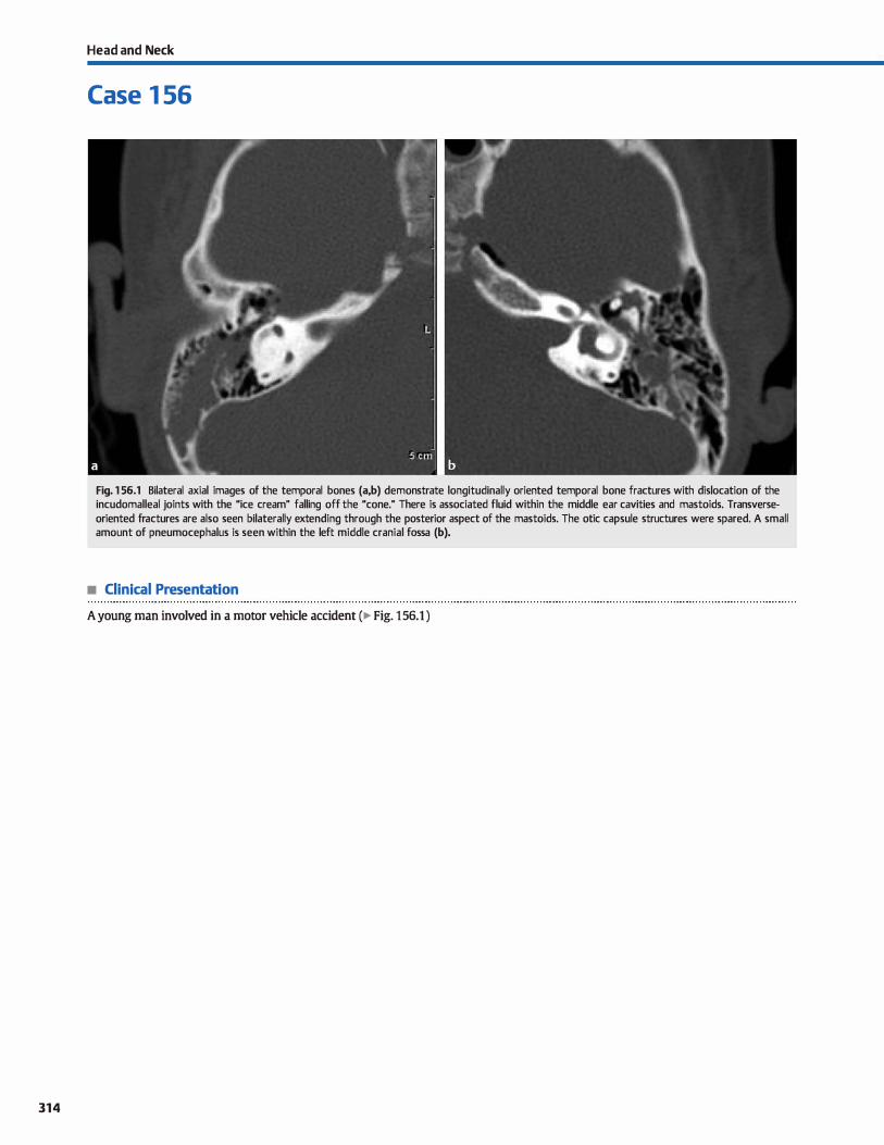
314
Head and Neck
Case 1 56
Fig. 1 56.1 Bilateral axial images of the temporal bones (a,b) demonstrate longitudinally oriented temporal bone fractures with dislocation of the incudomalleal joints with the "ice cream" falling off the "cane." There is associated fluid within the middle ear cavities and mastoids. Transverseoriented fractures are also seen bilaterally extending through the posterior aspect of the mastoids. The otic capsule structures were spared. A small amount of pneumocephalus is seen within the left middle cranial fossa (b).
• Clinical Presentation
A young man involved in a motor vehicle accident (� Fig. 156.1 )
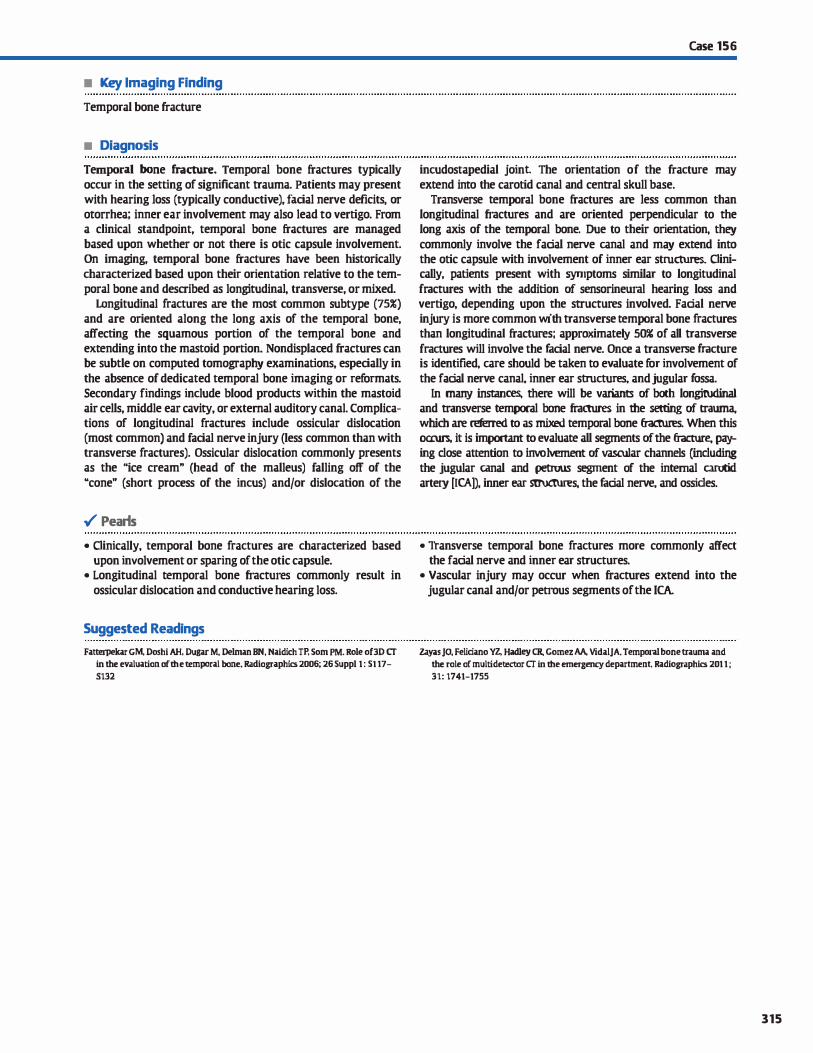
• Key lmaging Finding
Temporal bone fracture
• Diagnosis
Temporal bone fracture. Temporal bone fractures typically occur in the setting of significant trauma. Patients may present with hearing Joss {typically conductive), facial nerve deficits, or otorrhea; inner ear involvement may also lead to vertigo. From a clinicat standpoint. temporal bone fractures are managed based upon whether or not there is otic capsule involvement. On imaging. temporal bone fractures have been historically characterized based upon their orientation relative to the temporal bone and described as longitudinal, transverse, or mixed.
Longitudinal fractures are the most common subtype (75%)
and are oriented along the long axis of the temporal bone, affecting the squamous portion of the temporal bone and extending into the mastoid portion. Nondisplaced fractures can be subtle on computed tomography examinations, especially in the absence of dedicated temporal bone imaging or reformats. Secondary findings include blood products within the mastoid air cells, middle ear cavity, or externat auditory canal. Complications of longitudinal fractures include ossicular dislocation (most common) and facial nerve injury (Jess common than with transverse fractures). Ossicular dislocation commonly presents as the "ice cream" (head of the malleus) falling off of the "cone" (short process of the incus) and/or dislocation of the
� Pearls
• Oinically, temporal bone fractures are characterized based upon involvement or sparing of the otic capsule.
• Longitudinal temporal bone fractures commonly result in ossicular dislocation and conductive hearing loss.
Suggested Readings
Fatterpekar GM, Doshi AH, Dugar M, Delman BN, Naidich TP, Som PM. Role of3D CT
in the evaluation Dfthe t:emporal bone. Radiographies 2006; 26 Suppl 1 ; Sl 17-
S132
Case 156
incudostapedial joint The orientation of the fracture may extend into the carotid canal and central skull base.
Transverse temporal bone fractures are Jess common than longitudinal fractures and are oriented perpendicular to the long axis of the temporal bone. Due to their orientation, they commonly involve the facial nerve canal and may extend into the otic capsule with involvement of inner ear structures. Oinically, patients present with SYffiptoms similar to longitudinal fractures with the addition of sensorineural hearing Joss and vertigo, depending upon the structures involved. Facial nerve injury is more common with transverse temporal bone fractures than longitudinal fractures; approximately 50% of ail transverse
fractures will involve the facial nerve. Once a transverse fracture is identified, care should be taken to evaluate for involvement of the facial nerve canal, inner ear structures, and jugular Cossa.
In many instances, there will be variants of both longitudinal and transverse temporal bone fractures in the setting of trauma, which are referred to as mixed temporal bone fractures. When this occurs, it is important to evaluate all segments of the fracture, paying dose attention to involvement of vascular channels (induding
the jugular canal and petrous segment of the internai carotid artery [ICA]), inner ear structures, the facial nerve, and ossides.
• Transverse temporal bone fractures more commonly affect the facial nerve and inner ear structures.
• Vascular injury may occur when fractures extend into the jugular canal and/or petrous segments of the ICA.
Zayas JO, Feliciano YZ. Hadley CR, Gomez M VidalJA. Temporal bone trauma and
the role of multidet:ector cr in the emergency department. Radiographies 2011;
31: 1741-1755
315
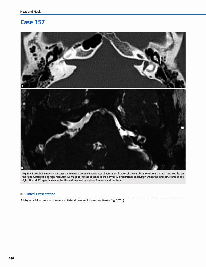
316
Head and Neck
Case 1 57
Fig. 1 57 .1 Axial CT image (a) through the temporal bones demonstrates abnormal ossification of the vestibule, semicircular canais, and cochlea on the right. Corresponding high-resolution T2 image (b) reveals absence of the normal T2 hyperintense endolymph within the inner structures on the right. Normal T2 signal is seen within the vestibule and lateral semicircular canal on the left.
• Clinical Presentation
A 28-year-old woman with severe unilateral hearing Joss and vertigo (� Fig. 157.1 )
• Key lmaging Finding
Abnormal mineralization ( computed tomography, cr) and Joss of signal intensity (magnetic resonance imaging, MRI) within labyrinthine structures
• Diagnosis
Labyrinthitis ossificans {W). W refers to ossification of the membranous labyrinth (vestibule, semidrcular canais, and/or cochlea) due to some form of insult, typically infectious or inflammatory. Meningitis and otitis media represent the most common infectious etiologies. Post-traumatic hemorrhage is a
Jess common cause. Findings may be unilateral or bilateral, depending upon the etiology and extent of insult.
LO occurs in three stages: acute, fibrous, and ossifying. The acute stage is characterized by focal or diffuse labyrinthine inflammation. Imaging in this stage may be normal or demonstrate enhanœment of labyrinthine structures on MRI. There is preservation of normal T2 hyperintense labyrinthine fluid
(MRI) initially and normal inner ear attenuation on cr. As the disease progresses into the fibrous stage, there is partial Joss of
y' Pearts
• LO refers to ossification of the membranous Iabyrinth and
often results from infectious or inflammatory insults. • Imaging reveals mineralization of inner structures on cr and
Joss of the normal T2 signal on MRI.
Suggested Reading
Plùllips GS, LoGerfo SE, Richardson Ml, Anzai Y. Interactive Web-based leaming
module on cr of the temporal bone: anatomy and pathology. R.adiographics
2012; 32: E85-E105
Case 157
signal intensity within the labyrinthine structures on T2-weighted MRI sequences and corresponding hazy increased attenuation on cr. During this phase, there may be residual labyrinthine enhancement on postcontrast T1 sequences. In the ossifying stage, mineralization results in Joss of the normal T2
signal intensity on MRI and increased attenuation of the Iaby
rinthine structures on cr. Patients with W present with sensorineural hearing loss up
to a year after an antecedent insult. Vertigo may also be seen clinically and can be severe. Cochlear implantation is the treatment of choice for cochlear involvement, espedally with bilat
eral disease. lt is important to describe fibrous or ossifying changes within the cochlea because these findings affect surgical management and prognosis.
• Patients present with sensorineural hearing Joss and vertigo, which can be debilitating.
317
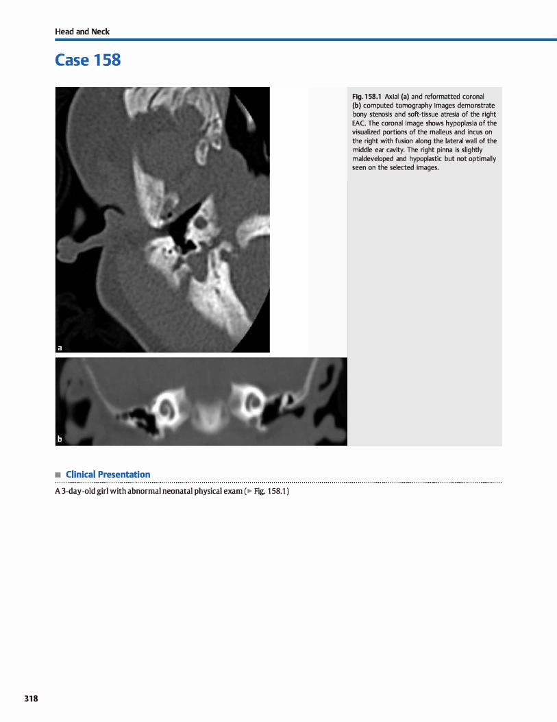
Head and Neck
Case 1 58
• Clinical Presentation
A 3-day-old girl with abnormal neonatal physical exam ( � Fig. 158.1 )
318
Fig. 1 58.1 Axial (a) and reformatted coronal (b) computed tomography images demonstrate bony stenosis and soft-tissue atresia of the right EAC. The coronal image shows hypoplasia of the visualized portions of the malleus and incus on the right with fusion along the lateral wall of the middle ear cavity. The right pinna is slightly maldeveloped and hypoplastic but not optimally seen on the selected images.
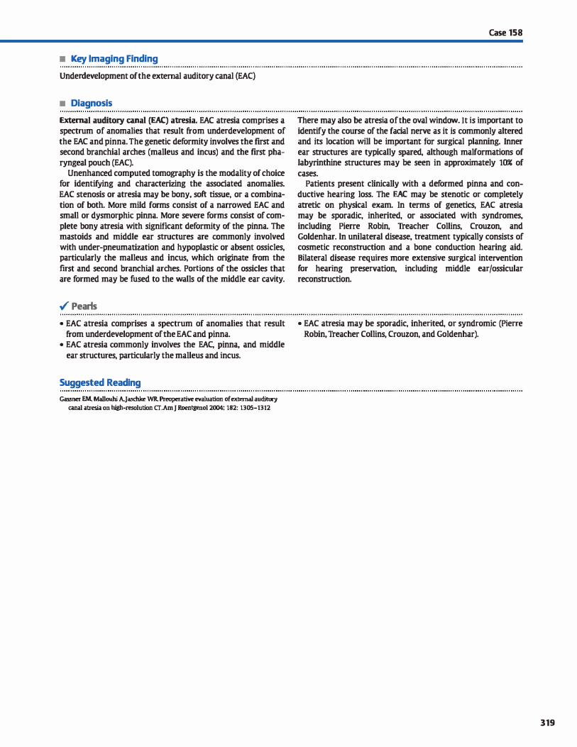
• Key lmaging Finding
Underdevelopment of the externat auditory canal (EAC)
• Diagnosis
Externat auditory canal {EAC) atresia. EAC atresia comprises a spectrum of anomalies that result from underdevelopment of
the EAC and pinna. The genetic deformity involves the first and second branchial arches (malleus and incus) and the first pha
ryngeal pouch (EAC). Unenhanœd computed tomography is the modality of choiœ
for identifying and characterizing the associated anomalies.
EAC stenosis or atresia may be bony, soft tissue, or a combina
tion of bath. More mild forms consist of a narrowed EAC and small or dysmorphie pinna. More severe forms consist of com
plete bony atresia with significant deformity of the pinna. The mastoids and middle ear structures are commonly involved
with under-pneumatization and hypoplastic or absent ossicles, particularly the malleus and incus, which originate from the
first and second branchial arches. Portions of the ossicles that are formed may be fused to the walls of the middle ear cavity.
y' Pearts
• EAC atresia comprises a spectrum of anomalies that result
from underdevelopment of the EAC and pinna. • EAC atresia commonly involves the EAC, pinna, and middle
ear structures, particularly the malleus and incus.
Suggested Reading
Giwner EM, Millloulù A.Jaschke WR. Preoperative evaluation of exœrnal auditmy
canal atresia on high-resolution cr.Am J Roentgenol 2004; 182: 1305-1312
Case 158
There may also be atresia of the oval window. It is important to
identify the course of the facial nerve as it is commonly altered and its location will be important for surgical planning. Inner
ear structures are typically spared, although malformations of labyrinthine structures may be seen in approximately 10% of
cases. Patients present clinically with a deformed pinna and con
ductive hearing loss. The EAC may be stenotic or completely
atretic on physical exam. In terms of genetics, EAC atresia may be sporadic, inherited, or associated with syndromes,
including Pierre Robin, Treacher Collins, Crouzon, and Goldenhar. ln unilateral disease, treatment typically consists of cosmetic reconstruction and a bone conduction hearing aid.
Dilaterai disease requires more extensive surgical intervention for hearing preservation, induding middle ear/ossicular reconstruction.
• EAC atresia may be sporadic, inherited, or syndromic (Pierre
Robin, Treacher Collins, Crouzon, and Goldenhar).
319
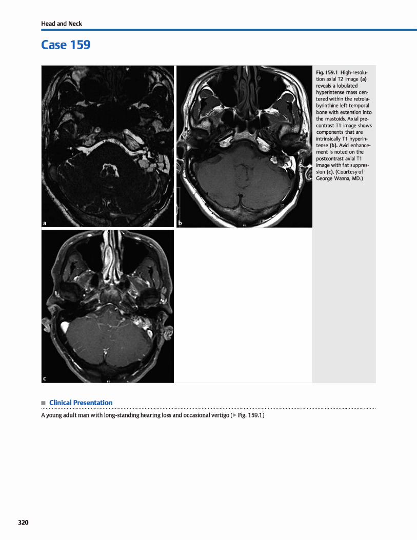
Head and Neck
Case 1 59
• Clinical Presentation
A young adult man with long-standing hearing Joss and occasional vertigo ( � Fig. 1 59.1 )
320
Fig. 1 59.1 High-resolution axial T2 image (a) reveals a lobulated hyperintense mass centered within the retrolabyrinthine left temporal bone with extension into the mastoids. Axial precontrast Tl image shows components that are intrinsically Tl hyperintense (b). Avid enhancement is noted on the postcontrast axial Tl image with fat suppression (c). (Courtesy of George Wanna, MD.)
• Key lmaging Finding
Enhancing mass with intrinsic Tl hyperintensity centered within the retrolabyrinthine temporal bone
• Diagnosis
Endolymphatic sac tumor. Endolymphatic sac tumor is a papillary adenomatous tumor that originates from the endolymphatic ductJsac. Most cases are sporadic, although there is an increased incidence and association with von Hippel-lindau (VHL) syndrome. Patients typically present with insidious onset of hearing loss. tinnitus, and vertigo; acute onset of symptoms may oa:asionally occur in the setting of intralesional hemonhage.
The tumor originates within the endolymphatic duct/sac along the retrolabyrinthine temporal bone and most often spreads medially and posteriorly into the cerebellopontine angle. Less common patterns of spread include lateral extension into the mastoids, middle ear cavity, and externat auditory
� Pearls
• Endolymphatic sac tumor is a papillary adenomatous tumor that originates from the endolymphatic duct/sac.
• Most cases are sporadic, although there is an increased incidence and association with VHL syndrome.
Suggested Readings
Chilpman PR. Shah R, Curé JK, Bag AK. Petrous apexlesions: pictorial review. AmJ Roentgenol 2011; 196Suppl: 1NS26-WS37, Q.uiz S40-S43
Connor SEJ, Leung R, Natas S. Imaging of the petrous apex: a pictorial review. Br j Radiol 2008;81: 427-435
Case 159
canal or anteriorly along the petrous ridge and into the clivus/ central skull base.
On computed tomography (CT), endolymphatic sac tumors present as sort-tissue masses with amorphous calcifications and aggressive bony destruction. On magnetic resonance imaging (MRI), lesions are T2 hyperintense with regions of increased Tl signal intensity due to intralesional hemorrhage; the intrinsic Tl hyperintensity and location is fairly characteristic. The tumors are hypervascular with intense nodular enhancement Larger lesions may demonstrate prominent flow voids.
Surgical resection with wide surgical margins is the treatment of choice.
• On CT, endolymphatic sac tumors demonstrate amorphous calcifications and aggressive bony destruction
• On MRI, lesions are T2 bright with regions of increased Tl signal due to hemorrhage; they intensely enhance.
Razek M Huang B'l. Lesions of the petrous apex: dassification and findings at cr
and MRimaging. Radiographies 2012; 32: 151-173
321
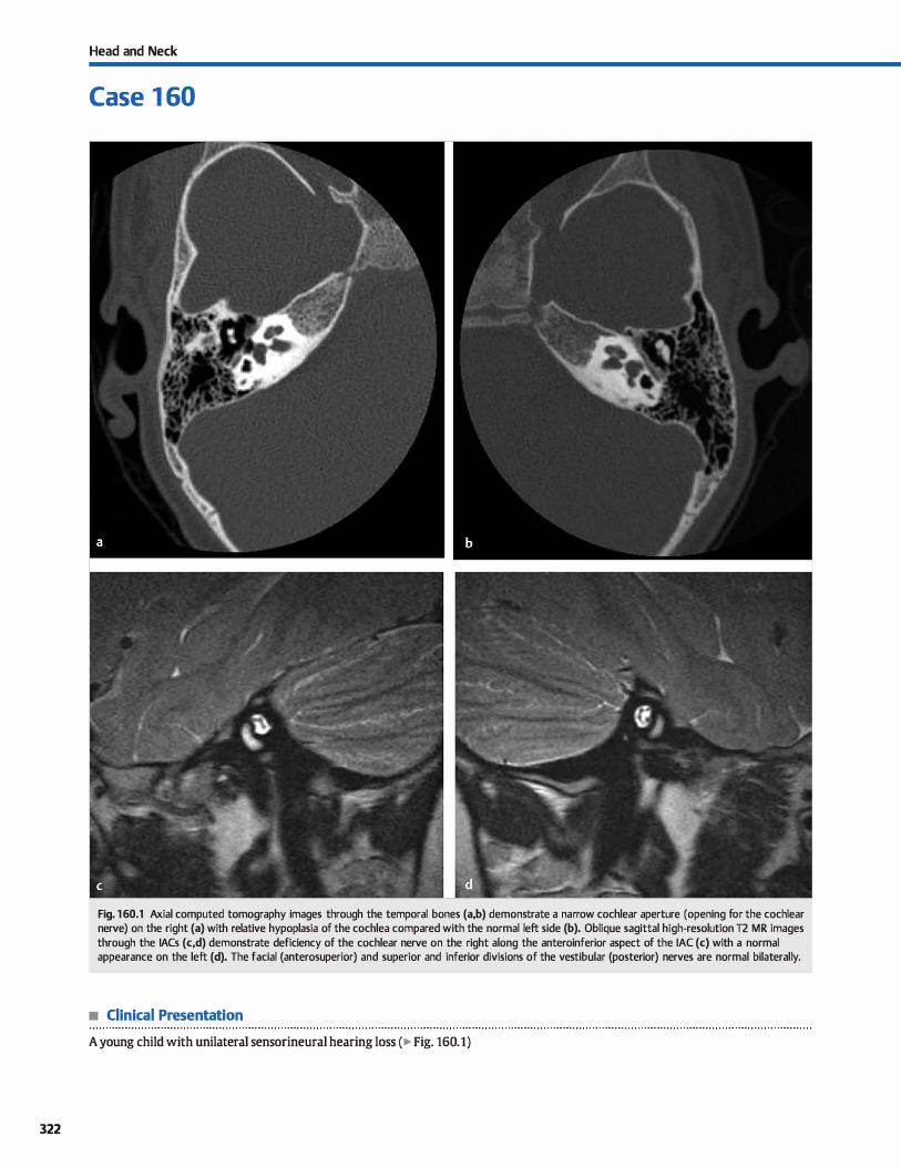
322
Head and Neck
Case 1 60
Fig. 160.1 Axial computed tomography images through the temporal bones (a,b) demonstrate a narrow cochlear aperture (opening for the cochlear nerve) on the right (a) with relative hypoplasia of the cochlea compared with the normal left side (b). Oblique sagittal high-resolution T2 MR images through the IACs (c,d) demonstrate deficiency of the cochlear nerve on the right along the anteroinferior aspect of the IAC (c) with a normal appearance on the left (d). The facial (anterosuperior) and superior and inferior divisions of the vestibular (posterior) nerves are normal bilaterally.
• Clinical Presentation
A young child with unilateral sensorineural hearing Joss ( � Fig. 160.1)
• Key lmaging Finding
Deficiency of the cochlear nerve with a narrow cochlear aperture
• Diagnosis
Cochlear nerve defidency. Cochlear nerve deficiency may be congenital (far more common) or acquired and results in sensorineural hearing Joss. On magnetic resonance imaging (MRI),
the cochlear nerve is best visualized on oblique sagittal highresolution 12 sequences oriented perpendicular to the internai auditory canal (IAC). ln this plane, the four Mquadrants" of the IAC are seen and anatomically divided into superior and inferior by the horizontally oriented crista falciformis and divided into anterior and posterior above the level of the crista falciformis at the IAC fundus by the vertically oriented Bill's bar. The facial nerve is the largest and is seen within the anterosuperior aspect
of the IAC. The cochlear nerve is Iocated anteriorly in the IAC inferior to the facial nerve (hence the mnemonic M7Up, Coke down"). Under normal circumstanœs, the cochlear nerve is smaller than the facial nerve but larger than the superior and inferior divisions of the vestibular nerve, which are located posteriorly within the IAC.
With cochlear nerve deficiency, the nerve is either imperceptible or significantly decreased in caliber compared with the
facial and vestibular nerves. With congenital causes of cochlear deficiency, there are associated osseous abnormalities because the bony development of the IAC and cochlea are affected by
y' Pearts
• Cochlear nerve deficiency is most often congenital and results in sensorineural hearing Joss.
• The cochlear nerve is seen along the anteroinferior IAC and is best visualized on oblique sagittal 12 sequences.
Suggested Reading
Glastonbury CM, Davidson HC. Harnsberger HR, Butler J, Kertesz TR, Shelton C. bnaging findings of codùear nerve defidency. AmJ Neuroradiol 2002; 23: 635-643
Case 160
development of the underlying nerves. In isolated cochlear nerve deficiency, the IAC and cochlear aperture ( opening from the IAC into the cochlea) are small in caliber. With vestibuloco
chlear deficiency, the IAC is nearly aplastic because it is only slightly larger than the facial nerve traversing through it. Sorne degree of cochlear dysplasia may be seen in a minority of congenital cases with modiolar deficiency being the most common abnormality. Acquired cases of cochlear nerve deficiency result from some form of insult (typically ischemic or traumatic) that occurs after normal development; therefore, the IAC and
cochlear aperture will be normal in size.
Treatment of cochlear nerve deficiency consists of cochlear implantation. Although complete absence of the cochlear nerve is a contraindication to cochlear implantation, it is best to describe the entity on MRI as cochlear nerve defidency rather than aplasia because the presence of even small nerve fibers that may be beyond imaging resolution may lead to sucœssful
surgical treatment. This is especially important in cases with bilateral findings. As is the case with many disease processes, the imaging findings must be correlated with clinical findings and evaluations, including nerve stimulation.
• Congenital cochlear nerve deficiency commonly results in hypoplasia of the IAC and cochlear aperture.
• Cochlear implantation is the treatment of choice for congenital cochlear nerve deficiency.
323
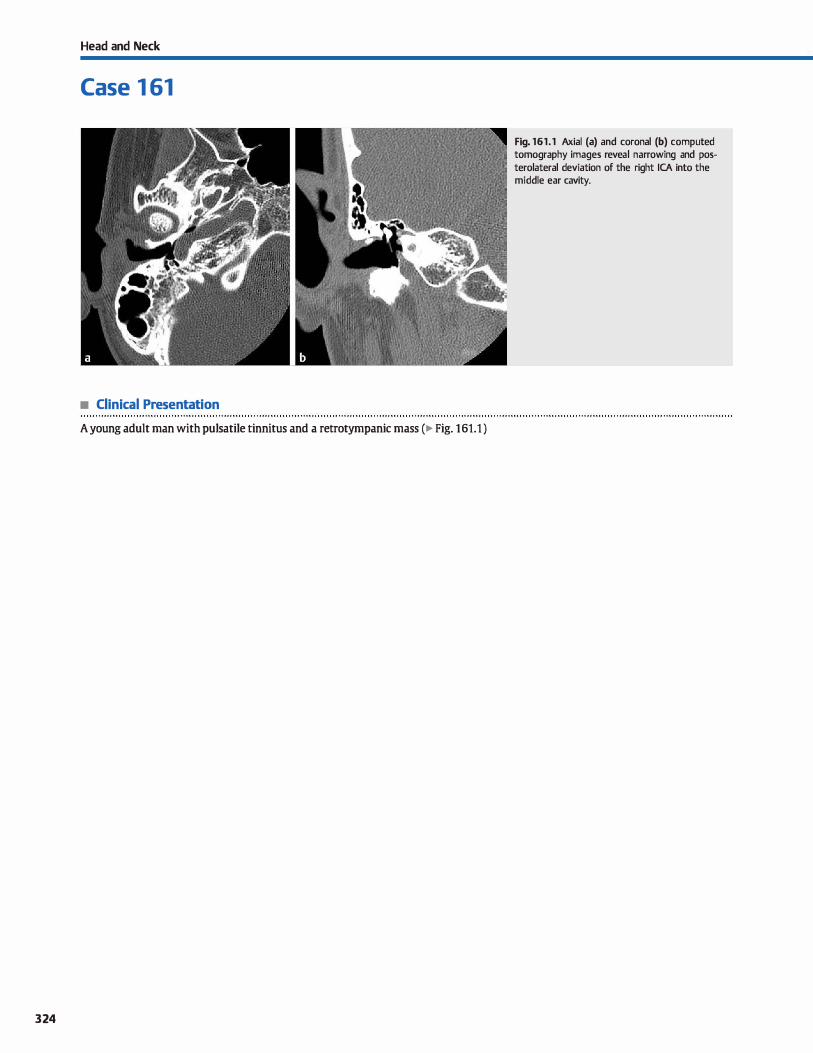
Head and Neck
Case 1 61
• Clinical Presentation
A young adult man with pulsatile tinnitus and a retrotympanic mass (� Fig. 161.1 )
324
Fig. 161.1 Axial (a) and coronal (b) computed tomography images reveal narrowing and posterolateral deviation of the right ICA into the middle ear cavity.
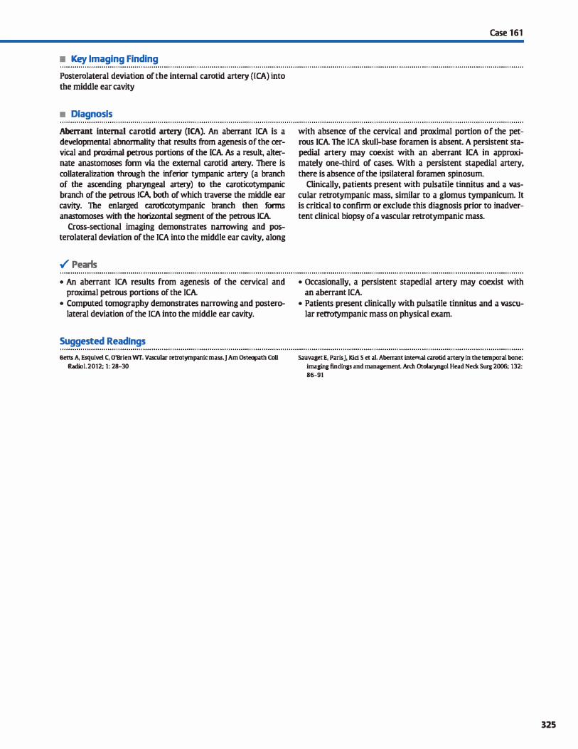
• Key lmaging Finding
Posterolateral deviation of the internai carotid artery (ICA) into the middle ear cavity
• Diagnosis
Aberrant internai carotid artery (ICA). An aberrant ICA is a developmental abnonnality that results from agenesis of the cervical and proximal petrous portions of the ICA As a result, alternate anastomoses fonn via the extemal carotid artery. There is
collateralization through the inferior tympanic artery (a branch of the asœnding pharyngeal artery) ta the caroticotympanic branch of the petrous ICA, bath of which traverse the middle ear cavity. The enlarged caroticotympanic branch then forms anastomoses with the horizontal segment of the petrous ICA
Cross-sectional imaging demonstrates narrowing and posterolateral deviation of the ICA into the middle ear cavity, along
� Pearts
• An aberrant ICA results from agenesis of the cervical and proximal petrous portions of the ICA
• Computed tomography demonstrates narrowing and posterolateral deviation of the ICA into the middle ear cavity.
Suggested Readings
Betts A, Esquivel C, O'Brien wr. Vascular retrotympanic mass, J Am 05teopath Coll
Radio!. 2012; 1: 28-30
Case 161
with absence of the cervical and proximal portion of the petrous ICA The ICA skull-base foramen is absent. A persistent stapediaI artery may coexist with an aberrant ICA in approximately one-third of cases. With a persistent stapedial artery,
there is absence of the ipsilateral foramen spinosum dinically, patients present with pulsatile tinnitus and a vas
cular retrotympanic mass, similar ta a glomus tympanicum. It is critical to confinn or exclude this diagnosis prior ta inadvertent dinical biopsy of a vascular retrotympanic mass.
• Occasionally, a persistent stapedial artery may coexist with an aberrant ICA.
• Patients present dinically with pulsatile tinnitus and a vascular retrotympanic mass on physical exam.
Sauvaget E, Parisj, !Cid S et al, Aberrant internai carotid artery in the temporal bene:
imaging findings and management. Ardt Otolaryngol Head Nede Surg 2006; 132:
8&-91
325
326
Head and Neck
Case 1 62
Fig. 162.1 Reformatted CT image through the plane of the superior semicircular canal (a) demonstrates dehiscence of the overlying superior bony margin of the semicircular canal. Corresponding axial CT image (b) shows uncovering of the superior semicircular canal along its anterosuperior margin.
• Clinical Presentation
An adult man with episodic "dizziness" associated with loud noises ( � Fig. 162.1 )
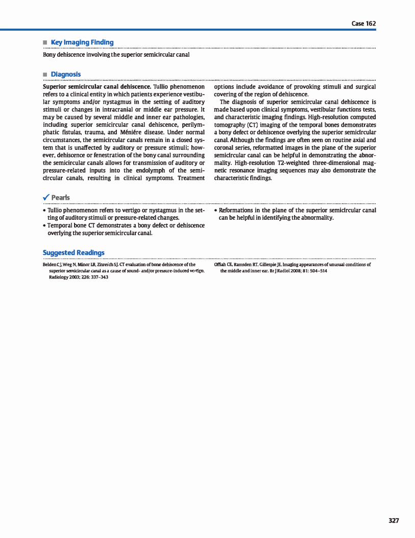
• Key lmaging Finding
Bony dehiscence involving the superior semicircular canal
• Diagnosis
Superior semicircular canal dehiscence. Tullio phenomenon refers to a clinical entity in which patients experience vestibular symptoms and/or nystagmus in the setting of auditory stimuli or changes in intracranial or middle ear pressure. lt may be caused by several middle and inner ear pathologies, induding superior semicircular canal dehiscence, perilymphatic fistulas, trauma, and Méniére disease. Under normal
circumstances, the semicircular canais remain in a dosed system that is unaffected by auditory or pressure stimuli; however, dehisœnce or fenestration of the bony canal surrounding
the semicircular canais allows for transmission of auditory or pressure-related inputs into the endolymph of the semidrcular canais, resulting in clinical symptoms. Treatment
� Pearls
• Tullio phenomenon refers to vertigo or nystagmus in the setting of auditory stimuli or pressure-related changes.
• Temporal bone CT demonstrates a bony defect or dehisœnœ overlying the superior semicircular canal.
Suggested Readings
Case 162
options indude avoidance of provoking stimuli and surgical covering of the region of dehiscence.
The diagnosis of superior semicircular canal dehiscence is made based upon clinical symptoms, vestibular functions tests, and characteristic imaging findings. High-resolution computed
tomography (CT) imaging of the temporal bones demonstrates a bony defect or dehiscence overlying the superior semidrcular
canal Although the findings are often seen on routine axial and coronal series, reformatted images in the plane of the superior semidrcular canal can be helpful in demonstrating the abnormality. High-resolution 12-weighted three-dimensional magnetic resonance imaging sequences may also demonstrate the characteristic findings.
• Reformations in the plane of the superior semidrcular canal can be helpful in identifying the abnormality.
Belden CJ, Weg N, Miner IB, Zinreich SJ. cr ew.luation of bene dehisœnœ of the Offiah CE, Ramsden RT, Gillespie JE. lmaging appearanœs of unusual conditions of
superior semidrcular canal as a CA use of sound- and/or pressure-induœd vertigo. the middle and inner ear. Br J Radiol 2008; 81; 504-514
Radiology 2003; 226: 337-343
327
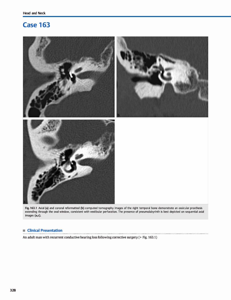
328
Head and Neck
Case 1 63
Fig. 163.1 Axial (a) and coronal reformatted (b) computed tomography images of the right temporal bone demonstrate an ossicular prosthesis extending through the aval window, consistent with vestibular perforation. The presence of pneumolabyrinth is best depicted on sequential axial images (a,c).
• Clinical Presentation
An adult man with recurrent conductive hearing Joss following corrective surgery (� Fig. 163.1 )
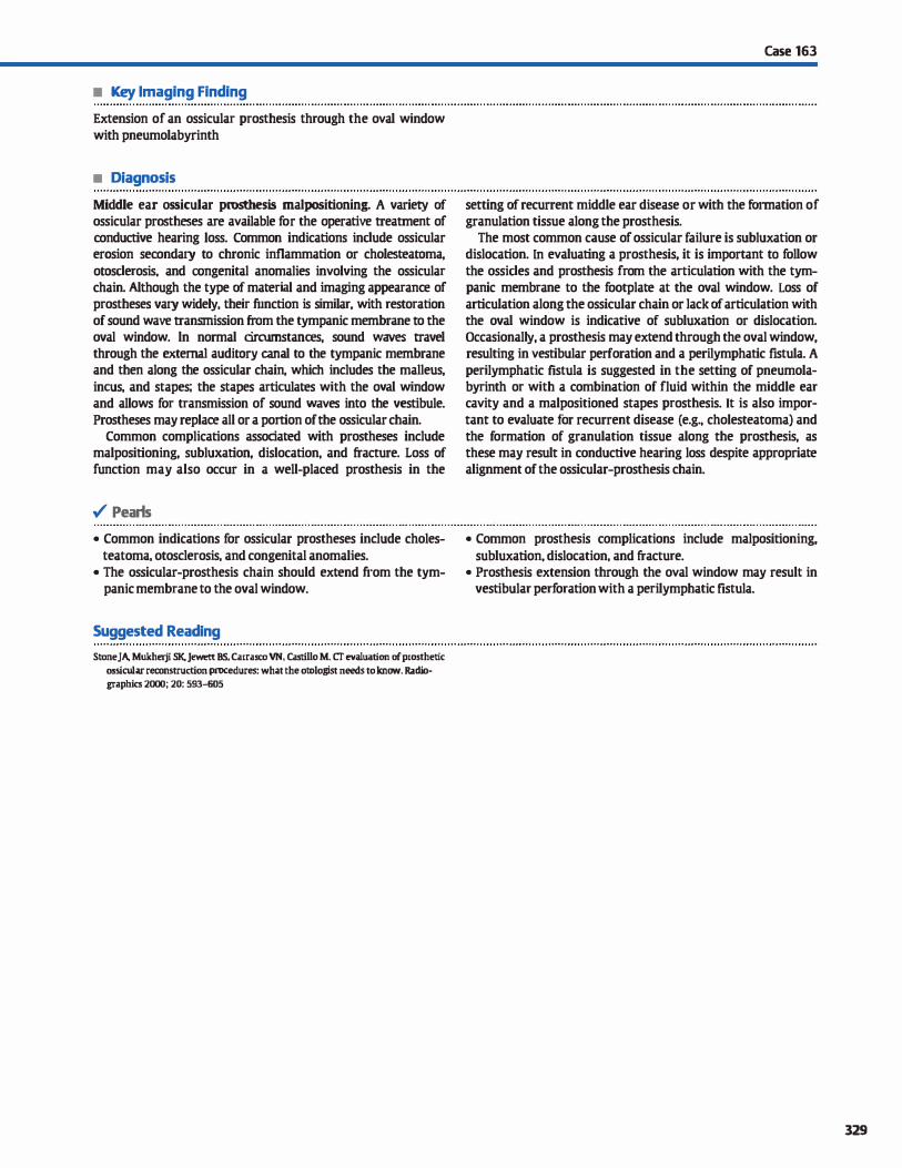
• Key lmaging Finding
Extension of an ossicular prosthesis through the oval window
with pneumolabyrinth
• Diagnosis
Middle ear ossicular prosthesis malpositioning. A variety of ossicular prostheses are available for the operative treatment of
conductive hearing Joss. Common indications indude ossicular
erosion secondary to chronic inflammation or cholesteatoma,
otosderosis, and congenital anomalies involving the ossicular
chain. Although the type of material and imaging appearanœ of
prostheses vary widely, their function is similar, with restoration
of sound wave transmission from the tympanic membrane to the
oval window. ln normal circumstanœs, sound waves travel
through the external auditory canal to the tympanic membrane
and then along the ossicular chain, which indudes the malleus,
incus, and stapes; the stapes articulates with the oval window
and allows for transmission of sound waves into the vestibule.
Prostheses may replace ail or a portion of the ossicular chain.
Common complications assoàated with prostheses indude
malpositioning, subluxation, dislocation, and fracture. Loss of function may also occur in a well-placed prosthesis in the
v" Pearls
• Common indications for ossicular prostheses indude choles
teatoma, otosderosis, and congenital anomalies.
• The ossicular-prosthesis chain should extend from the tym
panic membrane to the oval window.
Suggested Reading
Stone JA, Mukherji SK, Jewrtt BS. Carras cc VN, Castillo M. CT evaluation of prosthetic
ossicular reconstruction procedures: what the otologist needs to know. Radiographies 2000; 20; 593-605
Case 163
setting of recurrent middle ear disease or with the formation of
granulation tissue along the prosthesis.
The most common cause of ossicular failure is subluxation or
dislocation. ln evaluating a prosthesis, it is important ta follow
the ossides and prosthesis from the articulation with the tym
panic membrane to the footplate at the oval window. Loss of
articulation along the ossicular chain or Jack: of articulation with
the oval window is indicative of subluxation or dislocation.
Occasionally, a prosthesis may extend through the oval window,
resulting in vestibular perforation and a perilymphatic fistula. A
perilymphatic fistula is suggested in the setting of pneumola
byrinth or with a combination of fluid within the middle ear
cavity and a malpositioned stapes prosthesis. lt is also impor
tant to evaluate for recurrent disease (e.g., cholesteatoma) and
the formation of granulation tissue along the prosthesis, as
these may result in conductive hearing loss despite appropriate
alignment of the ossicular-prosthesis chain.
• Common prosthesis complications include malpositioning,
subluxation, dislocation, and fracture.
• Prosthesis extension through the oval window may result in
vestibular perforation with a perilymphatic fistula.
329
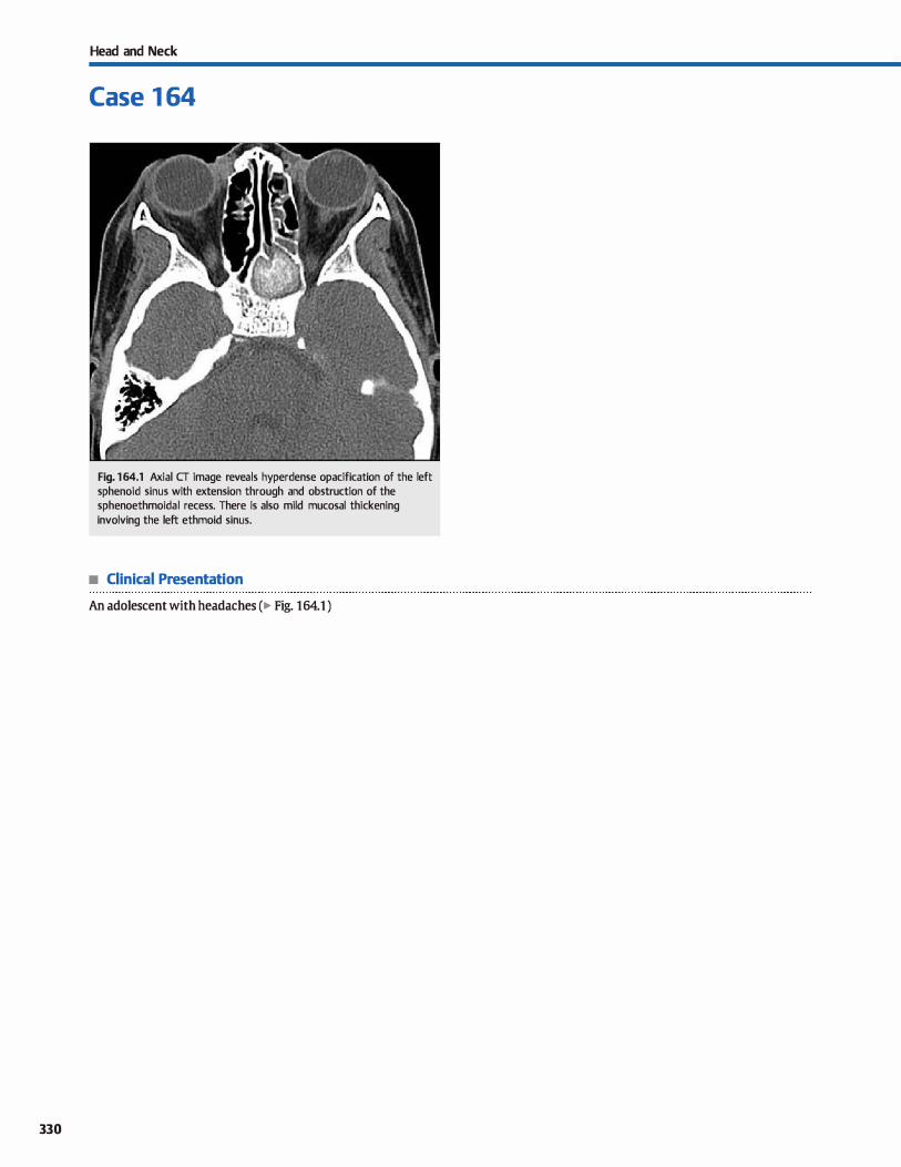
330
Head and Neck
Case 1 64
Fig. 164.1 Axial CT image reveals hyperdense opacification of the left sphenoid sinus with extension through and obstruction of the sphenoethmoidal recess. There is also mild mucosal thickening involving the left ethmoid sinus.
• Clinical Presentation
An adolescent with headaches (� Fig. 164.1 )
• Key lmaging Finding
Hyperdense sinus opacification
• Top 3 Differential Diagnoses
• lnspissated secretions. Inspissated secretions are seen in the setting of chronic sinusitis where sinus opacification becomes concentrated with increased proteinaceous and decreased water content. There is often chronic obstruction of the sinus outlet with or without underlying mucous retention cyst or mucoœle; occasionally, superimposed sinonasal polyposis syndromes are seen. On computed tomography (CT), as the
protein content increases and the water content decreases, there is increased density of the sinus opacification. Rarely, the sinus disease may begin to calcify and form a sinolith.
Chronic sinus wall thickening with osteitis may be seen. On magnetic resonance imaging (MRI), as protein content initially increases, there is a corresponding increase in T1 signal intensity; T2 signal intensity remains hyperintense. As the
protein content continues to increase, there is graduai decrease in the T2 signal intensity.
• Fungal sinusitis. Noninvasive fungal sinusitis occurs predominantly in immunocompetent patients and may present as a mycetoma in a solitary (usually) nonexpanded sinus or as allergie fungal sinusitis (AFS) with multiple expanded sinuses. AFS often occurs in patients with polyposis and/or multiple
• Diagnosis
Inspissated secretions
� Pearls
• Inspissated secretions are seen in chronic sinusitis and have increased protein and decreased water content.
• Fungal sinusitis results in hyperdense sinus opacification on CT due to the presence of fungal elements.
Suggested Reading
Aribandi M, MtCoy VA. Bazan C, Ill lmaging features of iINasive and noninvasive
fungal sinusitis; a review. Radiographies 2007; 27: 1283-1296
Case 164
allergies. Both processes result in hyperdense sinus opacification on CT due to the presenœ of fungal elements with or without superimposed inspissated secretions. calcifications and chronic sinus wall thickening with osteitis may be seen. On MRI, the fungal elements may result in decreased signal on bath T1 and T2 sequenœs, which may mimic an aerated
sinus. Chronic invasive fungal sinusitis may also present with hyperdense sinus opacification. as well as bony destruction and extension beyond the sinus margins. It is far Jess common and occurs most often in elderly patients with diabetes.
• TraumajHemorrhage. Fractures of the paranasal sinuses may result from direct facial trauma or orbital trauma or indirect
trauma with extension of calvarial or skull-base fractures into the paranasal sinuses. Resultant hemorrhage occurs, which
typically manifests as hyperdense air-fluid levels in a frac
tured sinus. At times, the fracture may not be readily seen but should be suspected in the appropriate clinicat setting with
hyperdense air-fluid levels. The maxillary and ethmoid sinuses are most often involved with direct orbital or facial trauma. The sphenoid sinus is most commonly involved with extension of skull-base fractures.
• On MRI, fungal elements may show decreased T1 and T2 signal intensity, mimidcing an aerated sinus.
• Fractures of the paranasal sinuses may result in hyperdense hemorrhagic air-fluid levels in the involved sinus.
331
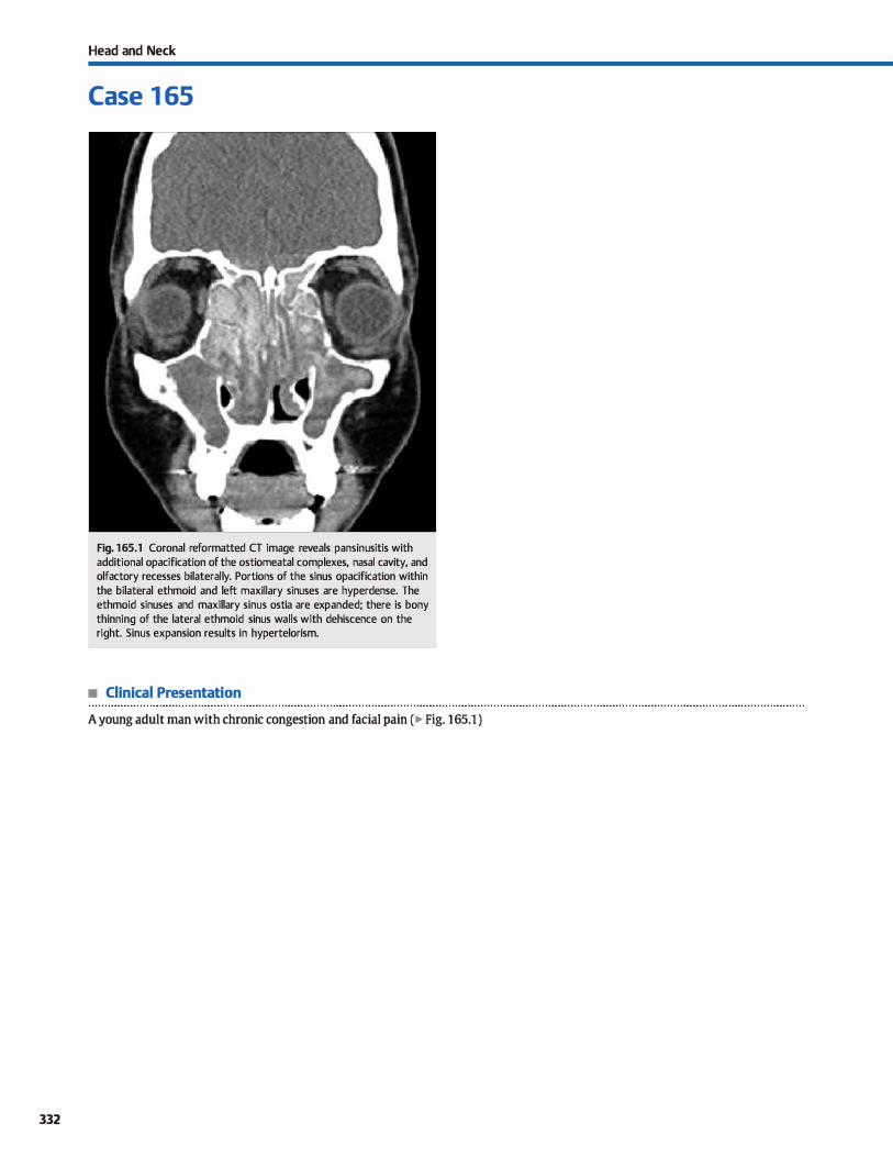
332
Head and Neck
Case 1 65
Fig. 165.1 Coronal reformatted CT image reveals pansinusitis with additional opacification of the ostiomeatal complexes, nasal cavity, and olfactory recesses bilaterally. Portions of the sinus opacification within the bilateral ethmoid and left maxillary sinuses are hyperdense. The ethmoid sinuses and maxillary sinus ostia are expanded; there is bony thinning of the lateral ethmoid sinus walls with dehiscence on the right. Sinus expansion results in hypertelorism.
• Clinical Presentation
A young adult man with chronic congestion and facial pain ( � Fig. 165.1 )
• Key lmaging Finding
Opacified expanded sinuses
• Top 3 Differential Diagnoses
• Allergie fungal sinusitis (AFS). AFS is a noninvasive fungal sinusitis that occurs predominantly in immunocompetent
patients. It represents the most common form of fungal sinusitis; is more prevalent in warm, humid dimates; and is thought to represent a hypersensitivity reaction to fungal elements. There is an increased inddenœ in patients with multiple allergies, atopy, asthma, and polyposis. The disease occurs bilaterally with involvement of multiple-if not all-sinuses; involved
sinuses are typically expanded On computed tomography ( CT), sinus opacification is hyperdense due to the presence of fungal elements with or without superimposed inspissated secretions. Calcifications and chronic sinus wall thickening with osteitis may be seen. On magnetic resonance imaging (MRI), the most common appearanœ is intermediate Tl and decreased T2 sig
nal intensity; the decreased 12 signal is thought to result
from manganese, iron, and magnesium content. Fungal elements may also result in decreased signal on bath Tl and T2 sequences, which may mimic an aerab!d sinus.
• Sinonasal polyposis. Sinonasal polyps result from hyperplasia of inflamed mucosa. They are associated with chronic inflam
mation and may occur in isolation or in association with allergie sinusitis or asthma. There is also an increased incidence in
children with cystic fibrosis and Kartagener syndrome ( ciliary dysmotility and situs inversus). One or more sinuses may be involved. Polyps often extend through and expand sinus ostia
• Additional Differential Diagnoses
• Sinonasal neoplasm. Squamous cell cardnoma (SSC) accounts for approximately 90% of sinus malignancies. The maxillary sinus is most commonly involved, followed by the ethmoid sinus. Patients present relatively late in the disease process
with obstructive sinus symptoms. On CT, SCC presents as a soft-tissue mass with bony destruction; extension beyond the
sinus margins may be seen. Involvement of the pterygopala-
• Diagnosis
Allergie fungal sinusitis
� Pearls
• AFS occurs bilaterally with involvement of multiple sinuses; involved sinuses are typically expanded.
• Sinonasal polyps result from hyperplasia of inflamed mucosa:
they expand involved sinuses and ostia.
Suggested Readings
Aribandi M, McCcy VA, Bazan c, Ill Imaging features of invasiW! and noninvasive
fungal sinusitis: a review. Radiographies 2007; 27: 1283-1296
Case 165
or outlets. On CT, polyps present as rounded lesions or complete sinus opacification. There may be extension into the nasal cavity, characteristic of an antrochoanal polyp. Bony findings include osteitis with sinus wall thickening due to
chronic inflammation, bony expansion with thinning of the sinus walls, or a more aggressive appearance with bony destruction and dehisœnœ. MRI reveals decreased Tl and increased T2 signal with surrounding mucosal enhancement. The lack of central enhanœment distinguishes polyposis from sinonasal carcinoma.
• Mucoœle. Mucoœles are cystic lesions that result in chronic sinus opacification and expansion due to sinus outlet obstruc
tion. 'JYpically, one or two adjacent sinuses are affected with the frontal, ethmoid, and sphenoid sinuses most often
involved (in decreasing order of frequency). Due to slow growth and indolent nature, patients often remain asymptomatic; when symptoms are present, pain or visual distur
bances are most common. The CT appearance depends upon the protein content within the obstructed sinus. As the protein content increases, there is associated increase in density. There is often bony thinning without destruction. On MRI, as
protein content initially increases, there is a corresponding increase in Tl signal intensity; T2 signal intensity remains hyperintense. As the protein content continues to increase, there is graduai decrease in the T2 signal intensity.
tine fossa allows for communication with the orbit and middle cranial fossa, most commonly through perineural spread of tumor. The tumor is hypointense on Tl and intermediate to hypointense on T2 sequenœs with heterogeneous
enhanœment. Less common sinonasal neoplasms include sinonasal undifferentiated cardnoma, adenocardnoma, rhabdomyosarcoma (most common in children), and lymphoma.
• Mucoceles are cystic lesions that result in sinus opacification and expansion due to sinus outlet obstruction.
Yousem DM. Imaging of sinonasil inflammatory disease. Radiology 1993; 188: 303-314
333
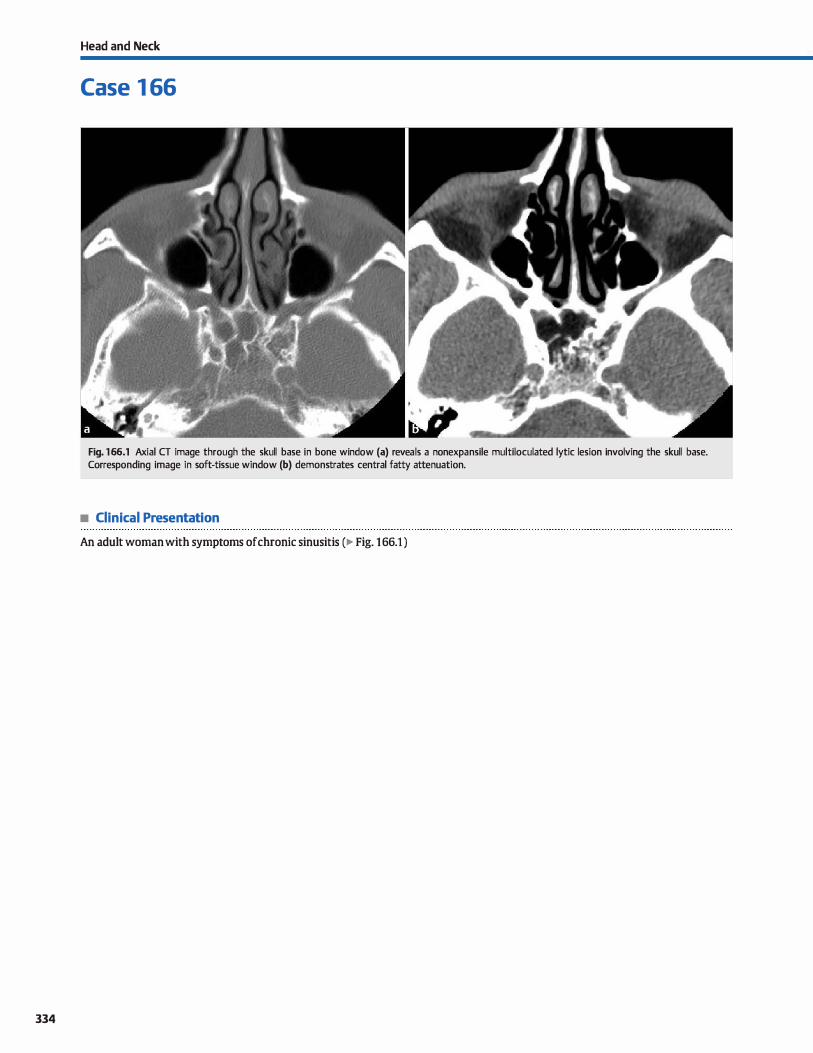
334
Head and Neck
Case 1 66
Fig. 166.1 Axial CT image through the skull base in bone window (a) reveals a nonexpansile multiloculated lytic lesion involving the skull base. Corresponding image in soft-tissue window (b) demonstrates central fatty attenuation.
• Clinical Presentation
An adult woman with symptoms of chronic sinusitis (� Fig. 166.1 )
• Key lmaging Finding
Lytic skull-base lesion
• Top 3 Differential Diagnoses
• Fibrous dysplasia {ID). FD occurs most often in young adults and is more common in women. In the head and neck the cal
varium, skull base, and facial bones are most often affected. The dassic appearance on computed tomography (CT) is focal bony expansion with "ground glass" attenuation of the diploic space; sderotic and cystic or lytic variants are Jess common. On magnetic resonance imaging (MRI), FD is typically hypoin
tense on both Tt and 12 sequences and demonstrates avid
enhancement; regions of cystic involvement, however, are 12 hyperintense. The MRI appearance can mimic aggressive lesions; therefore, CT is preferred in suspected cases ofFD.
• Malignancy. Metastatic and primary bone tumors may involve
the skull base and are often overlooked on imaging, especially when small. Breast Jung, and prostate carcinomas are the most common primary tumors to metastasize to the skull base and may result in expansile lytic or sderotic (prostate and occasionally breast) lesions on CT. Common primary tumors to involve the skull base indude chordomas, chondrosarcomas, lymphoma, and plasmacytoma. The key distinctions
for malignant lesions are bony expansion or erosion with
• Additional Differential Diagnoses
• Osteomyelitis. Osteomyelitis has a variety of imaging mani
festations, which range from a normal appearanœ to an aggressive appearanœ that mimics a neoplastic proœss. Bony destruction from osteomyelitis commonly results in Iytic
skull-base lesions that often have poorly defined, infiltrating margins. Subacute and chronic infections may have a lytic
• Diagnosis
Arrested pneumatization
� Pearls
• Fibrous dysplasia classically presents with focal bony expan
sion and "ground glass" attenuation. • Skull-base malignancies present with focal bony expansion or
erosion and enhancing soft-tissue components.
Suggested Readings
Geoigy BA, Snow RD, Brogdon BG et al Value ofbone window images in routine
brain CT; examinatiDIIS beyond trauma. Appl Radiol 1997; 2; 26-38
Case 166
enhancing soft-tissue components. The MRI appearanœ varies by tumor type with most malignancies being intermediate in signal on Tl and 12 sequenœs. Chordomas (midline) and chondrosarcomas (off-midline) are characteristically 12 hyper
intense. Heterogeneous enhanœment is most often seen. • Arrested pneumatization. During sinus development, there
is initially conversion of red marrow to fatty marrow with
subsequent pneumatization. When this proœss is arrested prior to pneumatization, regions of heterogeneous fatty marrow are seen, which may mimic pathological skull-base processes. Although arrested pneumatization may occur adjacent to any sinus or the mastoids, the sphenoid sinus is by far the most common location. On CT, arrested pneumatization presents as a nonexpansile region of heterogeneous density with
thin sclerotic margins and central fatty attenuation along the
margin of a relatively underdeveloped sinus. On MRI, arrested pneumatization is seen as a region of nonexpansile and nonenhancing fatty signal intensity along the margins of the involved sinus.
defect with a central dense nidus or sequestrum of intact
bone, referred to as a "button sequestrum." On MRI, osteomyelitis presents as regions of marrow infiltration or bony destruction with decreased T1 and intermediate to hyperintense 12 signal intensity, as well as variable enhancement
• Arrested pneumatization presents as a nonexpansile region
with thin sclerotic margins and central fat. • Skull-base osteomyelitis most often presents as a lytic lesion
with poorly defined, infiltrating margins.
Welker KM, Del.one DR. LaneJI. GilbertsonjR. Arrested pneumatization of the skull base; imagingcharactrr:istics.Arnj Roentgenol 2008; 190; 1691-1696
335
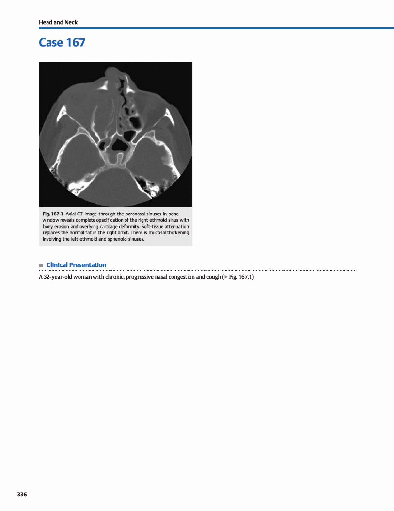
336
Head and Neck
Case 1 67
Fig. 167 .1 Axial CT image through the paranasal sin uses in bone window reveals complete opacification of the right ethmoid sinus with bony erosion and overlying cartilage deformity. Soft-tissue attenuation replaces the normal fat in the right orbit. There is mucosal thickening involving the left ethmoid and sphenoid sinuses.
• Clinical Presentation
A 32-year-old woman with chronic, progressive nasal congestion and cough (� Fig. 167.1 )
• Key lmaging Finding
Aggressive sinus disease/mass with bony destruction
• Top 3 Differential Diagnoses
• Invasive fungal sinusitis (IFS). IFS most commonly occurs in immunosuppressed patients, especially elderly patients with diabetes. Mucormycosis and Aspergillus are the most common causative agents, with aspergilosis also occurring in otherwise healthy individuals. Mucormycosis has a tendency to spread through the cavemous sinus and orbits, resulting in a life-threatening infection. Aspergilosis invades vasculature,
which results in mycotic aneurysm formation and vessel spasm or thrombosis. Fungal secretions are often hyperdense on computed tomography (CT) scans; calcifications are common. Hypointensity on both Tl and T2 (more so on T2) is characteristic of fungal infection. Bony erosion and extension beyond sinus margins are suggestive of an aggressive infection.
• Granulomatous disease (Wegener/sarcoid). Wegner granulo
matosis is a systemic vasculitis that primarily affects the kidneys and respiratory tract. Patients commonly present with recurrent sinonasal "infections,ft respiratory symptoms, and renal insufficiency. The necrotizing vasculitis of small and medium-size vessels results in sinus disease { usually within the maxillary sinus) with locally erosive features. Early in the
disease, the nasal septum is most involved. As the disease
• Additional Differential Diagnoses
• Lymphoma. Lymphoma of the paranasal sinuses is of the nonHodgkin variant The imaging appearance is nearly identical ta that of SSC with typically a unilateral sinus mass that invades bone and is intermediate to hypointense on T2 sequences. A key distinguishing feature is the presenœ of lymphadenopathy within the neck and involvement of the Waldeyer tonsillar ring (a pharyngeal ring of lymphoid tissue composed of the
adenoids, tubai tonsils, palatine tonsils, and lingual tonsils).
• Diagnosis
Granulomatous disease (Wegener granulomatosis)
� Pearls
• IFS most often occurs in immunosuppressed patients and
may be hyperdense (CT) and hypointense (magnetic resonance imaging).
• Wegner granulomatosis results in aggressive sinus disease, pulmonary symptoms, and renal insuffidency.
Suggested Readings
Case 167
progresses, there is formation of a sinus mass with extensive bony erosion and involvement of adjacent compartments, including the orbits. The soft-tissue mass is hypointense on Tl- and T2-weighted sequences and enhanœs. Sarcoid has a
similar appearance on imaging and nearly always has associated pulmonary disease, which manifests as hilar/mediastinal adenopathy and/or interstitial Jung disease.
• Sinonasal carcinoma. Squamous cell carcinoma (SSC)
accounts for approximately 90% of sinus malignandes. The maxillary sinus is most commonly involved, followed by the ethmoid sinus. Patients present relatively late in the disease process with obstructive sinus symptoms. On CT, SCC pres
ents as a soft-tissue mass with bony destruction; extension beyond the sinus margins may be seen. lnvolvement of the
pterygopalatine fossa allows for communication with the orbit and middle cranial fossa, most commonly through perineural spread of tumor. The tumor is hypointense on T1 and intermediate ta hypointense on T2 sequences with heterogeneous enhancement. Less common sinonasal neoplasms include sinonasal undifferentiated cardnoma, adenocarci
noma (associated with wood working and chemical exposures), and rhabdomyosarcoma (most common in children).
• Cocaïne nose. Chronic use of cocaine results in a granulomatous response with a soft-tissue mass and cartilaginous erosion. The local granulomatous response combined with the vasoconstrictive properties of cocaïne results in necrosis of the nasal septum. Continued insults lead to expansion of the septal perforation.
• sec (by far the most common) and lymphoma commonly involve the sinuses.
• Chronic cocaïne abuse can lead to ischemic necrosis with bony and cartilaginous destruction.
Allbery SM, Clialjub G, Clio NL, Rassekh Of, John SD, Guinto FC. MR imaging of nasal Valencia MP, Castillo M. Congerutal and acquired lesions of the nasal septum: a prac-
masses. Radiographies 1995; 15: 1311-1327 tical guide for differential diagnosis. Radiograplùcs 2008; 28: 205-224, quiz 326
337
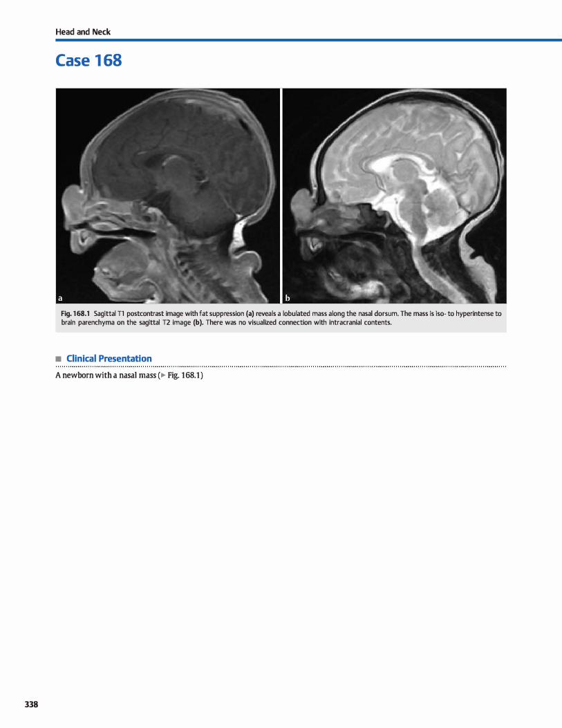
338
Head and Neck
Case 1 68
Fig. 168.1 Sagittal Tl postcontrast image with fat suppression (a) reveals a lobulated mass a long the nasal dorsum. The mass is iso- to hyperintense to brain parenchyma on the sagittal T2 image (b). There was no visualized connection with intracranial contents.
• Clinical Presentation
A newborn with a nasal mass (� Fig. 168.1 )
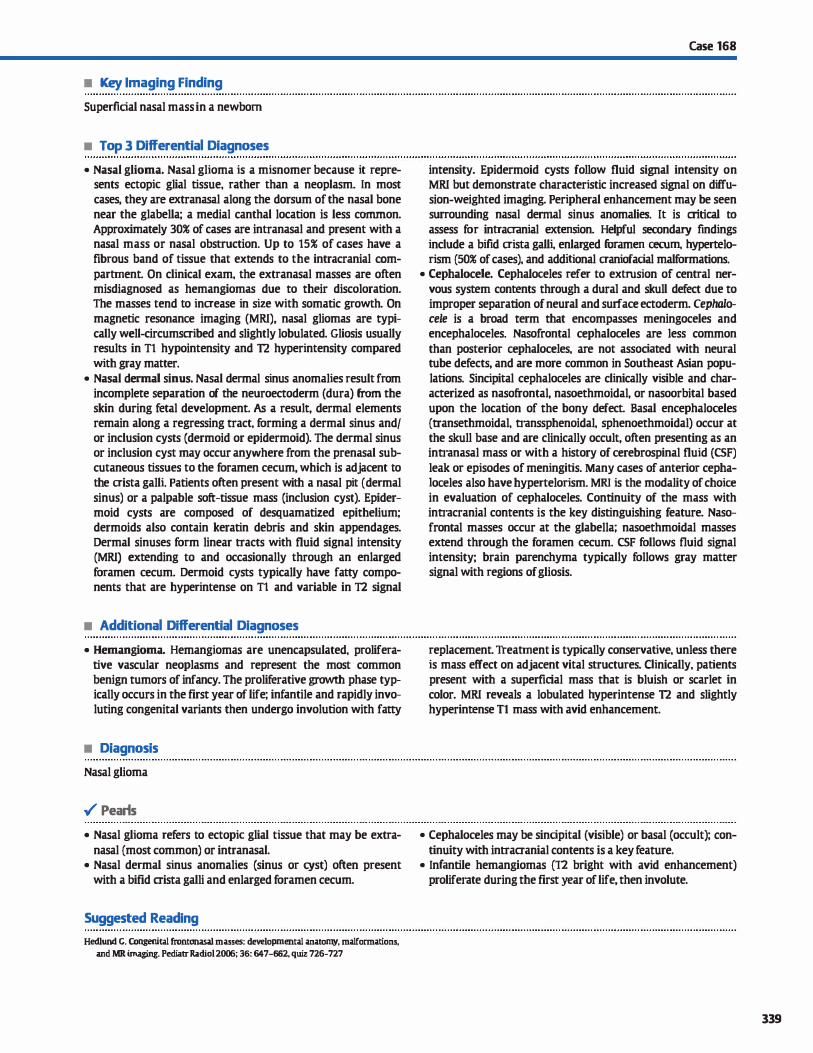
• Key lmaging Finding
Superficial nasal mass in a newbom
• Top 3 Differential Diagnoses
• Nasal glioma. Nasal glioma is a misnomer because it represents ectopie glial tissue, rather than a neoplasm. In most cases, they are extranasal along the dorsum of the nasal bone near the glabella; a medial canthal location is less comrnon. Approximately 30% of cases are intranasal and present with a nasal mass or nasal obstruction. Up to 15% of cases have a fibrous band of tissue that extends to the intracranial com
partment On clinicat exam, the extranasal masses are often misdiagnosed as hemangiomas due to their discoloration. The masses tend to increase in size with somatic growth. On magnetic resonanœ imaging (MRI), nasal gliomas are typically well-circumscribed and slightly lobulated. Gliosis usually results in T1 hypointensity and T2 hyperintensity compared with gray matter.
• Nasal dermal sinus. Nasal dermal sinus anomalies result from incomplete separation of the neuroectoderm (dura) from the skin during fetal development. As a result, dermal elements remain along a regressing tract, forming a dermal sinus and/ or inclusion cysts (dermoid or epidermoid). The dermal sinus or inclusion cyst may occur anywhere from the prenasal subcutaneous tissues to the foramen cecum, which is adjacent to the crista galli. Patients often present with a nasal pit { dermal sinus) or a palpable soft-tissue mass (inclusion cyst). Epidermoid cysts are composed of desquamatized epithelium; dermoids also contain keratin debris and skin appendages. Dermal sinuses form linear tracts with fluid signal intensity (MRI) extending to and occasionally through an enlarged
foramen cecum. Dermoid cysts typically have fatty components that are hyperintense on Tl and variable in T2 signal
• Additional Differential Diagnoses
• Hemangioma. Hemangiomas are unencapsulated, proliferative vascular neoplasms and represent the most common benign tumors of infancy. The proliferative growth phase typically occurs in the first year of life; infantile and rapidly involuting congenital variants then undergo involution with fatty
• Diagnosis
Nasal glioma
� Pearls
• Nasal glioma refers to ectopie glial tissue that may be extra
nasal {most common) or intranasal. • Nasal dermal sinus anomalies (sinus or cyst) often present
with a bifid crista galli and enlarged foramen cecum.
Suggested Reading
Hedlund c. Congenital frontonasal masses: developmental anatomy, malformations, and MR imaging. Pediatr Radiol 2006; 36: 647-662, quiz 726-727
Case 168
intensity. Epidermoid cysts follow fluid signal intensity on MRI but demonstrate characteristic increased signal on diffusion-weighted imaging. Peripheral enhancement may be seen surrounding nasal demial sinus anomalies. It is critical to
assess for intracranial extension. Helpful secondary findings include a bifid crista galli, enlarged foramen cecum, hypertelorism {50% of cases), and additional craniofacial malformations.
• Cephalocele. Cephaloceles refer to extrusion of central nervous system contents through a durai and skull defect due to improper separation of neural and surface ectoderm. Cephalocele is a broad term that encompasses meningoceles and encephaloceles. Nasofrontal cephaloœles are Jess common
than posterior œphaloceles, are not associated with neural tube defects, and are more common in Southeast Asian popu
lations. Sincipital cephaloœles are clinically visible and characterized as nasofrontal, nasoethmoidal, or nasoorbital based upon the location of the bony defect Basal encephaloceles {transethmoidal, transsphenoidal, sphenoethmoidal) occur at the skull base and are clinically occult, often presenting as an intranasal mass or with a history of cerebrospinal fluid (CSF)
leak or episodes of meningitis. Many cases of anterior cephaIoœles also have hypertelorism. MRI is the modality of choiœ in evaluation of cephaloceles. Continuity of the mass with intracranial contents is the key distinguishing feature. Nasofrontal masses occur at the glabella; nasoethmoidal masses extend through the foramen cecum. CSF follows fluid signal intensity; brain parenchyma typically follows gray matter signal with regions of gliosis.
replacement Treatment is typically conservative, unless there is mass effect on adjacent vital structures. Oinically, patients present with a superfidal mass that is bluish or scarlet in color. MRI reveals a lobulated hyperintense 12 and slightly hyperintense T1 mass with avid enhancement.
• Cephaloceles may be sincipital (visible) or basal (occult); con
tinuity with intracranial contents is a key feature. • Infantile hemangiomas {12 bright with avid enhancement)
proliferate during the first year of life, then involute.
339
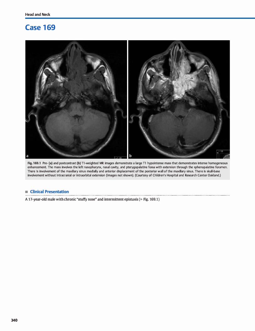
340
Head and Neck
Case 1 69
Fig. 169.1 Pre- (a) and postcontrast (b) Tl -weighted MR images demonstrate a large Tl hypointense mass that demonstrates intense homogeneous enhancement. The mass involves the left nasopharynx, nasal cavity, and pterygopalatine fossa with extension through the sphenopalatine foramen. There is involvement of the maxillary sinus medially and anterior displacement of the posterior wall of the maxillary sinus. There is skull-base involvement without intracranial or intraorbital extension (images not shown). (Courtesy of Children's Hospital and Research Center Oakland.)
• Clinical Presentation
A 1 7-year-old male with chronic "stuffy nose" and intermittent epistaxis ( � Fig. 169.1 )
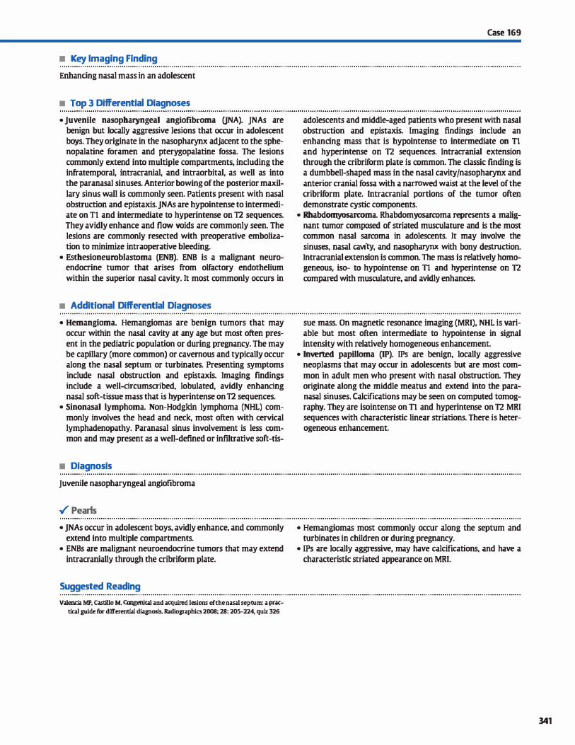
• Key lmaging Finding
Enhancing nasal mass in an adolescent
• Top 3 Differential Diagnoses
• juvenile nasopharyngeal angiofibroma UNA). JNAs are benign but locally aggressive lesions that occur in adolescent
boys. They originate in the nasopharyruc adjacent to the sphenopalatine foramen and pterygopalatine fossa. The lesions
commonly extend into multiple compartments, including the infratemporal, intracranial, and intraorbital, as well as into the paranasal sinuses. Anterior bowing of the posterior maxil
lary sinus wall is commonly seen. Patients present with nasal
obstruction and epistaxis. jNAs are hypointense to intermediate on T1 and intermediate to hyperintense on T2 sequences. They avidly enhance and flow voids are commonly seen. The
lesions are commonly resected with preoperative embolization to minimize intraoperative bleeding.
• Esthesioneuroblastoma (ENB). ENB is a malignant neuroendocrine tumor that arises from olfactory endothelium
within the superior nasal cavity. lt most commonly occurs in
• Additional Differential Diagnoses
• Hemangioma. Hemangiomas are benign tumors that may occur within the nasal cavity at any age but most often pres
ent in the pediatric population or during pregnancy. The may be capillary (more common) or cavernous and typically occur along the nasal septum or turbinates. Presenting symptoms
include nasal obstruction and epistaxis. Imaging findings
include a well-circumscribed, lobulated, avidly enhancing nasal soft-tissue mass that is hyperintense on T2 sequences.
• Sinonasal lymphoma. Non-Hodgkin lymphoma (NHL) com
monly involves the head and neck, most often with cervical lymphadenopathy. Paranasal sinus involvement is less common and may present as a well-defined or infiltrative soft-tis-
• Diagnosis
]uvenile nasopharyngeal angiofibroma
� Pearls
• JNAs occur in adolescent boys, avidly enhance, and commonly
extend into multiple compartments. • ENBs are malignant neuroendocrine tumors that may extend
intracranially through the cribriform plate.
Suggested Reading
Valencia MP, Castille M. Congenital and acquiœd lesions of the nasal septum: a pr.u:
tical guide for differential diagnosis. Radiographies 2008; 28; 205-224, quiz 326
Case 169
adolescents and middle-aged patients who present with nasal
obstruction and epistaxis. Imaging findings include an enhandng mass that is hypointense to intermediate on 11 and hyperintense on T2 sequences. Intracranial extension through the cribriform plate is common. The classic finding is a dumbbell-shaped mass in the nasal cavity/nasopharyruc and
anterior cranial fossa with a narrowed waist at the level of the cribriform plate. Intracranial portions of the tumor often
demonstrate cystic components. • Rhabdomyosarcoma. Rhabdomyosarcoma represents a malig
nant tumor composed of striated musculature and is the most common nasal sarcoma in adolescents. lt may involve the
sinuses, nasal cavity, and nasopharyruc with bony destruction.
lntracranial extension is common. The mass is relatively homogeneous, iso- to hypointense on 11 and hyperintense on T2
compared with musculature, and avidly enhances.
sue mass. On magnetic resonance imaging (MRI), NHL is variable but most often intermediate to hypointense in signal intensity with relatively homogeneous enhancement.
• lnverted papilloma (IP). IPs are benign, locally aggressive neoplasms that may occur in adolescents but are most com
mon in adult men who present with nasal obstruction. They originate along the middle meatus and extend into the paranasal sinuses. Calcifications may be seen on computed tomographY. They are isointense on 11 and hyperintense on T2 MRI
sequences with characteristic linear striations. There is heterogeneous enhancement.
• Hemangiomas most commonly occur along the septum and turbinates in children or during pregnancy.
• IPs are locally aggressive, may have calcifications, and have a
characteristic striated appearance on MRI.
341
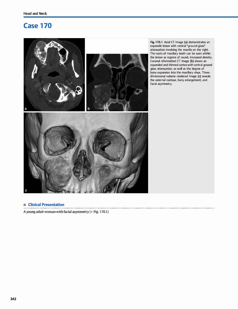
Head and Neck
Case 1 70
• Clinical Presentation
A young adult woman with facial asymmetry (� Fig. 170.1 )
342
Fig. 1 70.1 Axial CT image (a) demonstrates an expansile lesion with central "ground glass" attenuation involving the maxilla on the right. The roots of maxillary teeth can be seen within the lesion as regions of round, increased density. Coronal reformatted CT image (b) shows an expanded and thinned cortex with central ground glass attenuation, as well as the degree of bony expansion into the maxillary sinus. Threedimensional volume rendered image (c) reveals the external contour, bony enlargement, and facial asymmetry.
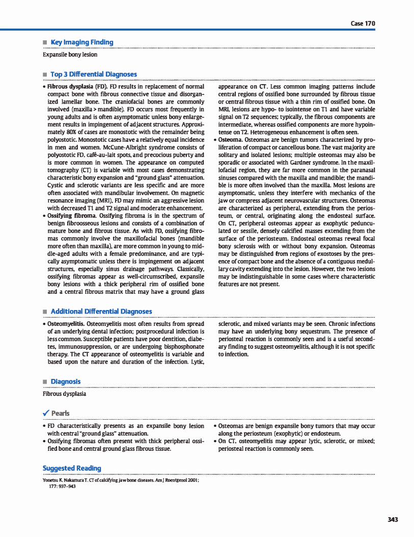
• Key lmaging Finding
Expansile bony lesion
• Top 3 Differential Diagnoses
• Fibrous dysplasia (FD). FD results in replacement of normal compact bone with fibrous connective tissue and disorganized lamellar bone. The craniofacial bones are commonly involved (maxilla > mandible). FD occurs most frequently in young adults and is often asymptomatic unless bony enlargement results in impingement of adjacent structures. Approximately 80% of cases are monostotic with the remainder being polyostotic. Monostotic cases have a relatively equal incidence in men and women. McCune-Albright syndrome consists of polyostotic FD, café-au-lait spots, and precocious puberty and is more common in women. The appearance on computed tomography (CT) is variable with most cases demonstrating characteristic bony expansion and "ground glass" attenuation. Cystic and sclerotic variants are less specific and are more often assodated with mandibular involvement. On magnetic resonance imaging (MRI), FD may mimic an aggressive lesion with decreased Tl and T2 signal and moderate enhancement.
• Ossifying fibroma. Ossifying fibroma is in the spectrum of benign fibroosseous lesions and consists of a combination of mature bone and fibrous tissue. As with FD, ossifying fibromas commonly involve the maxillofacial bones {mandible more often than maxilla), are more common in young to middle-aged adults with a female predominance, and are typically asymptomatic unless there is impingement on adjacent structures, espedally sinus drainage pathways. Oassically, ossifying fibromas appear as well-circumscribed, expansile bony lesions with a thick peripheral rim of ossified bone and a central fibrous matrix that may have a ground glass
• Additional Differential Diagnoses
• Osteomyelitis. Osteomyelitis most often results from spread of an underlying dental infection; postprocedural infection is less common. Susceptible patients have poor dentition, diabetes, immunosuppression, or are undergoing bisphosphonate therapy. The CT appearanœ of osteomyelitis is variable and based upon the nature and duration of the infection. Lytic,
• Diagnosis
Fibrous dysplasia
� Pearls
• FD characteristically presents as an expansile bony lesion with central uground glass" attenuation.
• Ossifying fibromas often present with thick peripheral ossified bone and central ground glass fibrous tissue.
Suggested Reading
Yonetsu K. Nakamura T. cr of caldfying jaw bone diseases. Am] RDentgenol 2001: 177: 937-943
Case 170
appearance on CT. Less common imaging patterns include central regions of ossified bone surrounded by fibrous tissue or central fibrous tissue with a thin rim of ossified bone. On MRI, lesions are hypo- to isointense on Tl and have variable signal on T2 sequences; typically, the fibrous components are intermediate, whereas ossified components are more hypointense on T2. Heterogeneous enhancement is often seen.
• Osteoma. Osteomas are benign tumors characterized by proliferation of compact or cancellous bone. The vast majority are solitary and isolated lesions; multiple osteomas may also be sporadic or assodated with Gardner syndrome. ln the maxillofadal region, they are far more common in the paranasal sinuses compared with the maxilla and mandible; the mandible is more often involved than the maxilla. Most lesions are
asymptomatic, unless they interfere with mechanics of the jaw or compress adjacent neurovascular structures. Osteomas are characterized as peripheral, extending from the periosteum, or central, originating along the endosteal surface. On CT, peripheral osteomas appear as exophytic pedunculated or sessile, densely caldfied masses extending from the surface of the periosteum. Endosteal osteomas reveal focal bony sclerosis with or without bony expansion. Osteomas may be distinguished from regions of exostoses by the presence of compact bone and the absence of a contiguous medullary cavity extending into the lesion. However, the two lesions may be indistinguishable in some cases where characteristic features are not present.
sclerotic, and mixed variants may be seen. Chronic infections may have an underlying bony sequestrum. The presence of periosteal reaction is commonly seen and is a useful secondary finding to suggest osteomyelitis, although it is not specific to infection.
• Osteomas are benign expansile bony tumors that may occur along the periosteum (exophytic) or endosteum.
• On CT, osteomyelitis may appear lytic, sclerotic, or mixed; periosteal reaction is commonly seen.
343
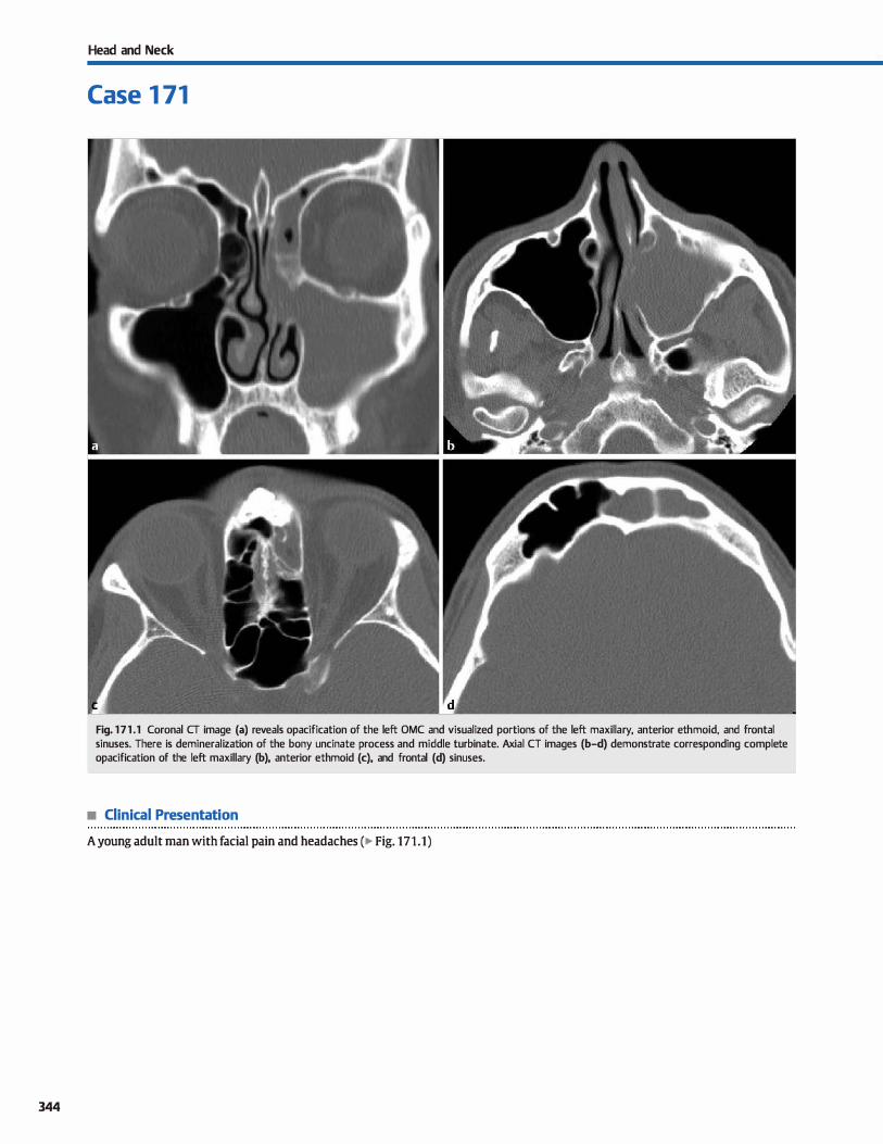
344
Head and Neck
Case 1 71
Fig. 17 1 .1 Coronal CT image (a) reveals opacification of the left OMC and visualized portions of the left maxillary, anterior ethmoid, and frontal sinuses. There is demineralization of the bony uncinate process and middle turbinate. Axial CT images (b-d) demonstrate corresponding complete opacification of the left maxillary (b), anterior ethmoid (c), and frontal (d) sinuses.
• Clinical Presentation
A young adult man with facial pain and headaches (� Fig. 171.1)
• Key lmaging Finding
Opacification of the ostiomeatal complex (OMC), and maxillary, anterior ethmoid, and frontal sinuses
• Diagnosis
Ostiomeatal complex (OMC) sinus disease. The OMC is the primary drainage pathway for the maxillary, anterior ethmoid, and frontal sinuses. It is best evaluated on coronal computed tomography (cr) images, and its outflow tract indudes the maxillary antrum, infundibulum, hiatus semilunaris, and middle meatus. The bony and soft-tissue borders of the OMC indude the uncinate proœss and middle turbinate medially and the inferior and medial orbital walls laterally. Anatomie variants that may narrow the outflow tract of the OMC indude Haller cells (ethmoidal air œlls along the orbital floor� pneumatized middle turbinate (concha bullosa), paradoxical rotation of the middle turbinate, and a deviated nasal septum.
Patients with chronic sinusitis or complications related ta recurrent sinusitis may require functional endoscopie sinus surgery (FESS) ta alleviate symptoms and restore the natural physiological drainage pathways. The OMC is the primary fucus of FESS. Preoperative cr imaging provides important landmarks for surgical planning and also affords the opportunity to identify anatomie variants that may predispose to surgical complications. Important structures to evaluate on the preoperative sinus cr may be recalled with the mnemonic CLOSE: �briform plate, !amina papyracea, presence of an Qnodi cell, �phenoid sinus pneumatization variants and relationship to the carotid canal, and location of the anterior �thmoidal artery.
� Pearls
• The OMC is the primary drainage pathway for the maxillary, anterior ethmoid, and frontal sinuses.
• FESS is designed to restore the natural physiological drainage pathways and is primarily directed at the OMC.
Suggested Reading
HoangjK, EastwoodJD, Tebbit Cl., Glastonbury CM. Multiplanar sinus CT; a system
atic approach to imaging before functional endoscopie sinus surgery. Amj Roent
genal 2010; 194; W527-36
Case 171
In evaluating the cribriform plate, the Keros dassification is used ta determine the depth of the olfactory fossa. A deeper fessa or asymmetry between right and left side places the patient at risk for penetration of the lateral lamella into the anterior cranial fossa. Medial orbital wall fractures cause medial deviation of the lamina papyracea into the ethmoid sinus. lnadvertent penetration through the lamina papyracea may result in injury to intraorbital contents. An Onodi cell refers to an anatomie variant with superior and posterior positioning of an ethmoid air cell above the sphenoid sinus. The optic nerve is typically located within an Onodi œll and is often dehisœnt, making it susceptible ta injury. In evaluating the sphenoid sinus, it is important ta comment on pneumatization extending inferior and posterior ta the sella because this pattern increases the risk for surgical penetration through the divus. Additionally, it is important to identify sphenoid sinus pneumatization that extends to and around the margins of the internai carotid artery because this places the artery at risk for injury. Finally, the anterior ethmoidal artery is identified by the notch along the media! orbital wall. When the notch abuts the lateral lamella or fovea ethmoidalis, it is typically well protected. However, the presence of pneumatized supraorbital ethmoid air cells surrounding the anterior ethmoidal artery notch renders the artery susceptible to intraoperative injury.
• Preoperative cr imaging provides surgical landmarks and identifies potential risks for surgical complications.
345
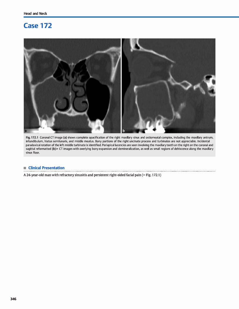
346
Head and Neck
Case 1 72
Fig. 1 72.1 Coronal CT image (a) shows complete opacification of the right maxillary sinus and ostiomeatal complex, induding the maxillary antrum, infundibulum, hiatus semilunaris, and middle meatus. Bony portions of the right uncinate process and turbinates are not appreciable. lncidental paradoxical rotation of the left middle turbinate is identified. Periapical lucencies are seen involving the maxillary teeth on the right on the coronal and sagittal reformatted (b) > CT images with overlying bony expansion and demineralization, as well as small regions of dehiscence along the maxillary sinus floor.
• Clinical Presentation
A 24-year-old man with refractory sinusitis and persistent right-sided facial pain (� Fig. 172.1 )

• Key lmaging Finding
Periapical dental lucencies, overlying bony demineralization, and maxillary sinus disease
• Diagnosis
Odontogenic sinus disease. Odontogenic sinusitis represents -10% of cases of maxillary sinusitis. It may result from significant dental caries or periodontal disease or as a result of dental intervention. Patients present with recurrent or persistent sinusitis despite what is assumed to be appropriate medical treatment However, typical antibiotic coverage for sinusitis is often inadequate because odontogenic sinusitis tends to be polymicrobial with both aerobic and anaerobic organisms.
Panoramic radiographs or sinus computed tomography (CT) are the modalities of choiœ in evaluating for suspected
� Pearls
• Odontogenic sinusitis may result from dental caries, periodontal disease, or as a result of dental intervention.
• cr reveals dental caries or periapical luœncies with bony thinning or dehiscenœ of the maxillary sinus floor.
Suggested Reading
Mehra P, Murad H. Maxillary sinus disease of odontogenic origin. Otolaryngol Oin
North Am 2004; 37: 347-364
Case 172
odontogenic sinusitis. Imaging reveals prominent dental caries or periapical luœncies along the floor of the maxillary sinus with overlying bony thinning or dehiscence. There is associated maxillary sinus mucosal thickening or opacification. With a region of large bony dehiscenœ, the involved tooth may hemiate into the sinus.
Treatment indudes broad spectrum antibiotic therapy, tooth extraction, and repair of the odontogenic-maxillary fistula, ifpresent.
• Treatment includes broad spectrum antibiotics, tooth extraction, and repair of the odontogenic-maxillary fistula.
347
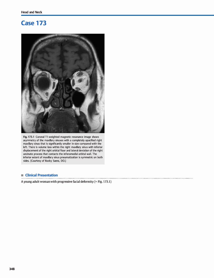
348
Head and Neck
Case 1 73
Fig. 1 73.1 Coronal Tl -weighted magnetic resonance image shows asymmetry of the maxillary sinuses with a completely opacified right maxillary sinus that is significantly smaller in size compared with the left. There is volume loss within the right maxillary sinus with inferior displacement of the right orbital floor and lateral deviation of the right uncinate process that contacts the inferomedial orbital wall. The inferior extent of maxillary sinus pneumatization is symmetric on both sides. (Courtesy of Rocky Saenz, DO.)
• Clinical Presentation
A young adult woman with progressive facial deformity (� Fig. 173.1 )
• Key lmaging Finding
Atelectatic maxillary sinus
• Diagnosis
Silent sinus syndrome. Silent sinus syndrome refers to atelectasis of a completely (most common) or partially opacified
maxillary sinus. Although the pathophysiology is not entirely
understood, it is believed that obstruction of the maxillary sinus outlet results in negative pressure within the maxillary
sinus and subsequent inward displacement of the sinus walls.
Patients often present with an acquired facial defonnity and
progressive enophthalmos on the involved side.
The imaging findings are fairly characteristic in the appropriate dinical setting. The maxillary sinus, antrum, and infun
dibulum are typically opadfied, and there is inward displaœment of the maxillary sinus walls, particularly the media! wall and roof (orbital floor) of the maxillary sinus. There is lateral
deviation of the uncinate process, which may contact the
media! orbital floor. The thickness of the walls of the maxillary
sinus is variable; they may be normal, thinned, or thickened.
Chronic sinus opacification may lead to inspissated secretions,
� Pearts
• Silent sinus syndrome refers to atelectasis of a completely
(usually) or partially opadfied maxillary sinus. • Patients present most often as young adults with a progres
sive, acquired fadai deformity and enophthalmos.
Suggested Reading
Hourany R, Aygun N, Della Santina CC, Zinreich SJ. Silent 5Îllm syndrome; an
acquired conditiOIL Amj Neuroradiol 2005; 26: 2390-2392
Case 173
which are hyperdense on CT, hyperintense on Tt, and relatively
hypointense on T2 magnetic resonance imaging sequences. The main differential diagnosis for silent sinus syndrome is
an opacified hypoplastic maxillary sinus, which is more common and does not present with a developing facial deformity.
Although it may be difficult to distinguish between the two based solely upon imaging, a useful criterion is the location of
the maxillary sinus floor. With a hypoplastic maxillary sinus,
there is incomplete development or pneumatization of the
maxillary sinus; therefore, the floor of the maxillary sinus is
thicker and more cephalad on the involved side. In silent sinus syndrome, there is relatively symmetric development of the
maxillary sinuses with subsequent atelectasis on the involved
side; therefore, inferior pneumatization of the maxillary sinuses
is similar. Occasionally, the two conditions may coexist with
acquired sinus atelectasis superimposed upon underlying
developmental hypoplasia.
• Imaging reveals inward displacement of the maxillary sinus
walls, particularly the media! wall and orbital floor. • The main differential diagnosis for silent sinus syndrome is an
opadfied hypoplastic maxillary sinus
349
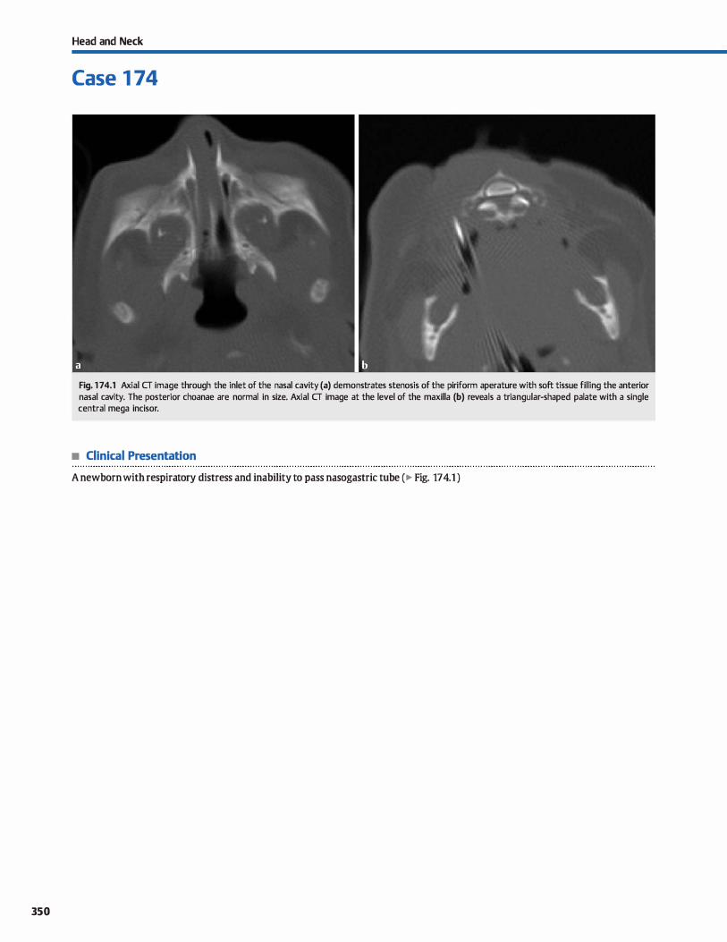
350
Head and Neck
Case 1 74
Fig. 1 74.1 Axial CT image through the inlet of the nasal cavity (a) demonstrates stenosis of the piriform aperature with soft tissue filling the anterior nasal cavity. The posterior choanae are normal in size. Axial CT image at the level of the maxilla (b) reveals a triangular-shaped palate with a single central mega incisor.
• Clinical Presentation
A newborn with respira tory distress and inability to pass nasogastric tube ( � Fig. 17 4.1 )
• Key lmaging Finding
Narrowing of the piriform aperature with a single mega inciser
• Diagnosis
Pirifonn aperature stenosis. The piriform aperature amstitutes the anterior bony opening or inlet of the nasal cavity and is
formed by the nasal and maxillary bones. Piriform aperature stenosis is relatively rare and results in respiratory distress in neonates, especially during feeding because they are obligate nasal breathers. The anomaly may occur in isolation or in association with syndromes, induding holoprosenœphaly. Common presen
tations indude cyanosis, respiratory distress. and/or the inability ta pass a nasogastric tube in a neonate.
y' Pearts
• The piriform aperature constitutes the anterior bony opening or inlet of the nasal cavity.
• Piriform aperature stenosis is a rare cause of respiratory
distress in neonates.
Suggested Readings
Belden CJ, Mancuso AA, Sdunalfuss !M. CT reatures of congenital llilSill pirifunn
aperture stenosis: initial experienœ. Radiology 1999; 213: 495-501
Case 174
Computed tomography (CT) is the modality of choiœ in
evaluating for piriform aperature stenosis. Characteristic findings indude inward bowing of the maxillary spines, resulting in a transverse piriform aperature measurement < 11 mm; a triangular-shaped palate; and in some cases, a single mega
central maxillary incisor. The presence of a single mega inciser bas a high association with additional anomalies.
Treatment is typically conservative with respiratory symptoms resolving after appropriate growth of the nasal inlet.
• CT findings indude a narrowed piriform aperature, a triangular-shaped patate, and a central mega inciser.
Lawe IR, Booth 1N,joglar JM, Rollins NK. Midfaœ anomalies in children. Radio
graphies 2000: 20: 907-922, quiz 1106-1107, 1112
351
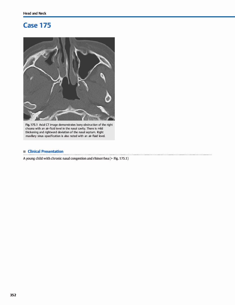
352
Head and Neck
Case 1 75
Fig. 1 75.1 Axial CT image demonstrates bony obstruction of the right choana with an air-fluid level in the nasal cavity. There is mild thickening and rightward deviation of the nasal septum. Right maxillary sinus opacification is also noted with an air-fluid level.
• Clinical Presentation
A young child with chronic nasal congestion and rhinorrhea (� Fig. 1 75.1 )

• Key lmaging Finding
Obstruction of the posterior nasal cavity/choana
• Diagnosis
Choanal atresia. Choanal atresia refers to congenital or developmental obstruction of the posterior nasal cavity and is the most common cause of nasal obstruction in neonates. The obstruction may be bony or membranous with bony atresia accounting for -90% of cases. The abnormality is most often unilateral and typically assodated with additional anomalies or syndromes. Approximately one-fourth of cases are isolated abnormalities.
Age and symptoms at the time of presentation depend upon
whether the atresia is unilateral or bilateral. Bilateral choanal atresia may be life-threatening and presents in the neonatal period or early infancy with respiratory distress, which is more evident during feeding and relieved by crying. An additional common presentation is the inability to pass a nasogastric tube. Respiratory distress during feedings results from the fact that
newboms and young infants are obligate nasal breathers; feeding prevents mouth breathing, and the choanal obstruction
y' Pearts
• Choanal atresia refers to congenital or developmental
obstruction of the posterior nasal cavity. • Choanal atresia may be bony (more common) or membra
nous, as well as unilateral (more common) or bilateral.
Suggested Reading
Lowe œ, Booth lN, Joglar JM, Rollins NK. Midfaœ anmnalies in children. Radio
graphies 2000; 20: 907-922, quiz 1106-1107, 1112.
Case 175
prevents nasal breathing. Crying relieves the symptoms because it forces mouth breathing. With unilateral atresia, symptoms are less severe, and patients often present later in childhood with a history of nasal stuftiness and chronic, unilateral rhinorrhea on the involved side.
Computed tomography (CT) is the modality of choice in evaluating for choanal atresia. Findings include narrowing and bony
or sort-tissue obstruction of the posterior nasal cavity/choana, often with soft tissue or fluid superiiàal to the level of obstruction. Additional findings include thickening of the vomer and medial bowing of the posterior maxilla. In neonates and infants, a quantitative measurement for choanal atresia includes a choanal opening < 3.4 mm.
Membranous choanal atresia may be treated with perforation of the membrane; bony atresia requires surgical correction with occasional reconstruction.
• cr demonstrates bony or membranous obstruction of the posterior nasal cavity with thickening of the vomer.
353
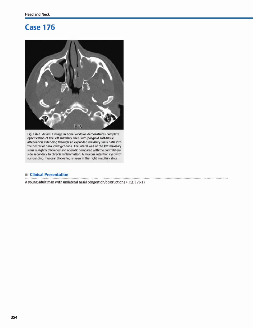
354
Head and Neck
Case 1 76
Fig. 1 76.1 Axial CT image in bone windows demonstrates complete opacification of the left maxillary sinus with polypoid soft-tissue attenuation extending through an expanded maxillary sinus ostia into the posterior nasal cavity/choana. The lateral wall of the left maxillary sinus is slightly thickened and sderotic compared with the contralateral side secondary to chronic inflammation. A mucous retention cyst with surrounding mucosal thickening is seen in the right maxillary sinus.
• Clinical Presentation
A young adult man with unilateral nasal congestion/obstruction (� Fig. 176.1 )
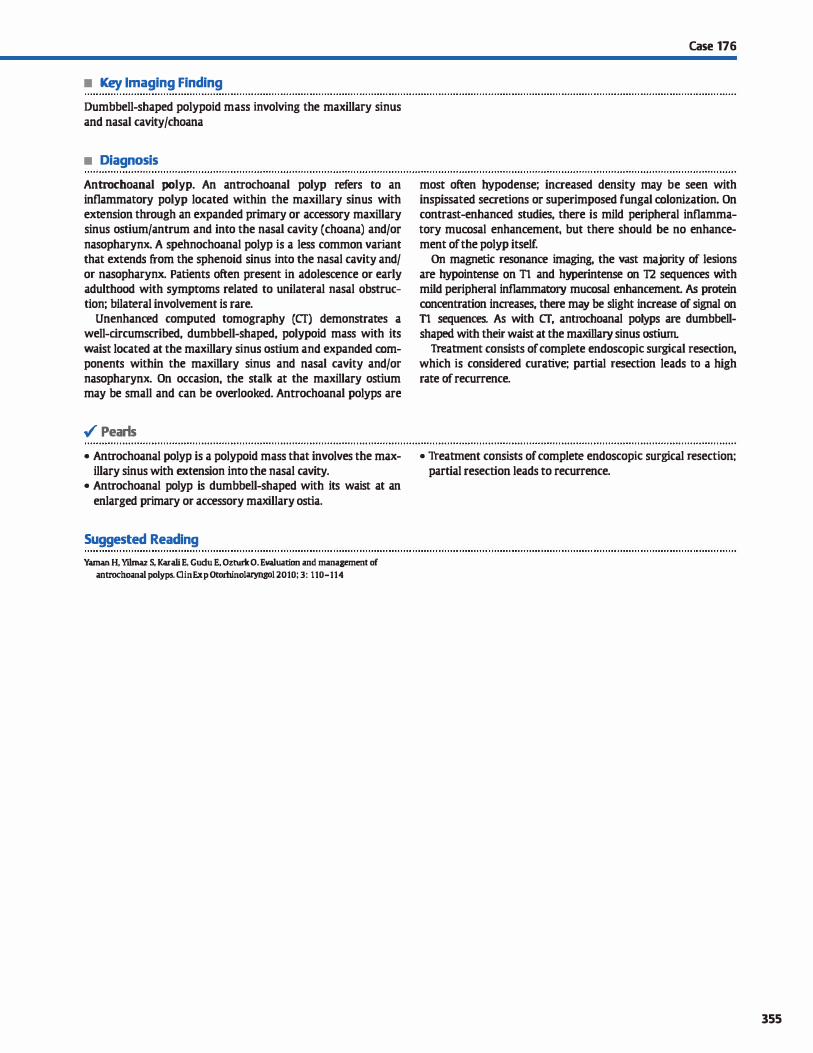
• Key lmaging Finding
Dumbbell-shaped polypoid mass involving the maxillary sinus and nasal cavity/choana
• Diagnosis
Antrochoanal polyp. An antrochoanal polyp refers to an inflammatory polyp located within the maxillary sinus with extension through an expanded primary or acœssory maxillary sinus ostium/antrum and into the nasal cavity ( choana) and/or
nasopharynx. A spehnochoanal polyp is a less common variant that extends from the sphenoid sinus into the nasal cavity and/ or nasopharynx. Patients often present in adolescence or early adulthood with symptoms related to unilateral nasal obstruction; bilateral involvement is rare.
Unenhanced computed tomography (CT) demonstrates a well-drcumscribed, dumbbell-shaped, polypoid mass with its
waist located at the maxillary sinus ostium and expanded components within the maxillary sinus and nasal cavity and/or nasopharynx. On occasion, the stalk at the maxillary ostium may be small and can be overlooked. Antrochoanal polyps are
y' Pearts
• Antrochoanal polyp is a polypoid mass that involves the maxillary sinus with extension into the nasal cavity.
• Antrochoanal polyp is dumbbell-shaped with its waist at an
enlarged primary or accessory maxillary ostia.
Suggested Reading
Yamilll H, Yilmaz S, Karali E. Gudu E. Ozturlt O. Evaluation and management of
antrochoanal polyps. Qin Exp Otorlùnolaryngol 2010; 3: 110-114
Case 176
most often hypodense; increased density may be seen with inspissated secretions or superimposed fungal colonization. On contrast-enhanced studies, there is mild peripheral inflammatory mucosal enhanœment, but there should be no enhancement of the polyp itself.
On magnetic resonance imaging, the vast majority of lesions are hypointense on TI and hyperintense on T2 sequences with mild peripheral inflammatory mucosal enhancement As protein concentration increases, there may be slight increase of signal on Tl sequences. As with CT, antrochoanal polyps are dumbbell
shaped with their waist at the maxillary sinus ostium. Treatment consists of complete endoscopie surgical resection,
which is considered curative; partial resection leads ta a high rate of recurrence.
• Treatment consists of complete endoscopie surgical resection; partial resection leads to recurrence.
355
356
Head and Neck
Case 1 77
Fig. 1 77 .1 Axial CT image reveals a well-circumscribed sclerotic/ calcified lesion in the right ethmoid sinus. Minimal ethmoid sinus mucosal thickening is also identified.
• Clinical Presentation
A 33-year-old man with intermittent sinusitis and headaches (� Fig. 1 77.1 )
• Key lmaging Finding
Sderotic/calcified paranasal sinus mass
• Diagnosis
Osteoma. Osteomas are slow-growing bone-fonning tumors that represent the most common benign tumors of the paranasal sinuses. They are dassified as compact (most common), cancellous, or mixed. They may occur at any age but are most common in middle-aged adults. The vast majority (> 95%) are asymptomatic and found incidentally. Those with symptoms often present with pain or sinusitis due ta sinus outlet obstruc
tion. A classic description is pain associated with air travel due to a frontal sinus osteoma. Rarely, osteomas may extend into the orbit or even intracranially, resulting in orbital emphysema
or pneumocephalus, respectively, due ta communication with the paranasal sinuses.
� Pearts
• Osteomas are bone-forming tumors that represent the most common benign tumors of the paranasal sinuses.
• Frontal/ethmoid sinuses are by far most commonly involved,
followed by the maxillary and sphenoid sinuses.
Suggested Reading
Priœ HI, Batnitzky s, Karlin CA et al. Compuœd tomography of benign disease of the
paranasahinuses. Radiographics 1983; 3: 107-140
Case 177
On computed tomography (CT), osteomas present as well-cir
cumscribed sclerotic or calcified lesions within the paranasal sinuses. The frontal and ethmoid sinuses are by far the most commonly involved, followed by the maxillary and sphenoid sin uses. Most are small, measuring < 1 cm. Lesions > 3 cm are
referred to as giant osteomas and are more likely ta be symptomatic. On magnetic resonanœ imaging, osteomas are typically
hypointense on ail sequences, mimicking an aerated sinus.
If asymptomatic, no treatment is neœssary. Symptomatic lesions are treated with open or endoscopie surgical resection.
• On cr, osteomas present as well-circumscribed sderotic or caldfied lesions within the paranasal sinuses.
357
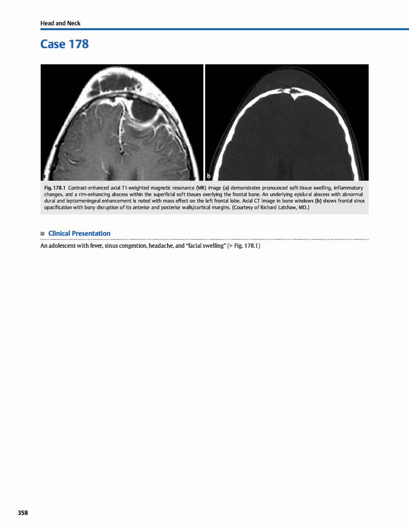
358
Head and Neck
Case 1 78
Fig. 1 78.1 Contrast-enhanced axial Tl -weighted magnetic resonance (MR) image (a) demonstrates pronounced soft-tissue swelling, inflammatory changes, and a rim-enhancing abscess within the superficial soft tissues overlying the frontal bone. An underlying epidural abscess with abnormal durai and leptomeningeal enhancement is noted with mass effect on the left frontal lobe. Axial CT image in bone windows (b) shows frontal sinus opacification with bony disruption of its anterior and posterior walls/cortical margins. (Courtesy of Richard Latchaw, MD.)
• Clinical Presentation
An adolescent with fever, sinus congestion, headache, and "facial swelling" (� Fig. 1 78.1 )
• Key lmaging Finding
Soft-tissue swelling overlying opacified frontal sinus
• Diagnosis
Pott puffy tumor. Pott puffy tumor is a relatively uncommon complication of frontal sinusitis and, less frequently, trauma.
Focal infection spreads through emissary veins, resulting in thrombophlebitis, frontal bone osteomyelitis, and subperiosteal abscess formation. Patients present with signs and symptoms related to sinusitis, as well as focal induration and swelling overlying the frontal bone.
Cross-sectional imaging demonstrates frontal sinus opacification, ill-defined luœncy of the surrounding inner and/or outer tables of the frontal bone, and superficial or deep subperiosteal phlegmon or abscess formation. Scalp soft-tissue swelling and
� Pearls
• Pott puffy tumor refers to subperiosteal absœss formation as
a complication of frontal sinusitis. • Imaging demonstrates frontal sinusitis, osteomyelitis, sub
periosteal abscess, and superfidal soft-tissue swelling.
Suggested Reading
Ludwig BJ, Foster BR, SaitD N, Nadgir RN, Castro-Aragon 1, Sakai O. Diagnostic imag
ing in nontrawnatic pediatric head and neck ernei:gendes. Radiograplùcs 2010;
30: 781-799
Case 178
inflammatory fatty infiltration is typically seen. Enhancement is
ill-defined with phlegmonous change and rim enhancing with abscess formation. It is important to dosely evaluate intracranial structures for evidence of intracranial spread, which is commonly seen with inner table involvement.
Complications of Pott puffy tumor indude intracranial spread with subsequent epidural absœss, subdural empyema, cerebri
tis or parenc:hymal abscess, and durai venous sinus or cavem
ous sinus thrombosis. Treatment consists of focal drainage of infectious collections and prolonged intravenous antibiotic therapy.
• Complications include underlying parenchymal and/or extra
axial infection and durai venous sinus thrombosis.
359
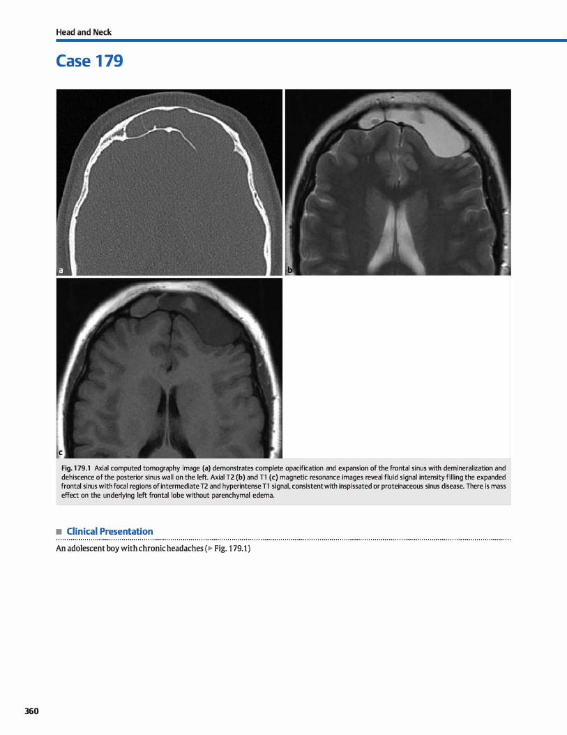
360
Head and Neck
Case 1 79
Fig. 1 79.1 Axial computed tomography image (a) demonstrates complete opacification and expansion of the frontal sinus with demineralization and dehiscence of the posterior sinus wall on the left. Axial T2 (b) and T1 (c) magnetic resonance images reveal fluid signal intensity filling the expanded frontal sinus with focal reg ions of intermediate T2 and hyperintense T1 signal, consistent with inspissated or proteinaceous sinus disease. There is mass effect on the underlying left frontal lobe without parenchymal edema.
• Clinical Presentation
An adolescent boy with chronic headaches (� Fig. 179.1 )
• Key lmaging Finding
Opacified, expanded sinus with bony demineralization
• Diagnosis
• Mucoœle. Mucoceles are mucosal-lined lesions that result from chronic obstruction of a sinus outlet or ostium. They most commonly involve the frontal sinus (roughly two-thirds of cases), followed by the ethmoid, maxillary, and sphenoid sinuses. Cross-sectional imaging reveals a completely opadfied, expanded sinus with benign bony remodeling and
� Pearls
• Mucoceles are mucosal-lined lesions that result from chronic obstruction of a sinus outlet or ostium.
• Imaging reveals a completely opadfied, expanded sinus with benign bony remodeling and demineralization.
Suggested Reading
Van Tasse! P, Lee YY,Jing BS, DePena CA. Mucoœles of the paranasal sinuses: MR
imaging with cr mrrelation Amj Rcentgenol 1989; 153; 407--412
Case 179
demineralization. Regions of bony dehiscence may be seen. Portions of the sinus opacification may demonstrate increased density or Tl signal intensity due to inspissated or proteinaceous secretions. Contrast-enhanced studies often demonstrate peripheral mucosal enhancement Infected mucoceles are referred ta as mucopyoceles.
• Inspissated or proteinaœous secretions demonstrate increased density or Tt signal intensity.
361
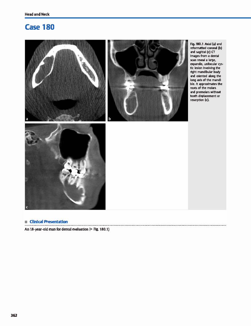
Head and Neck
Case 1 80
• Cllnlcal Presentatlon
An 18-year-old man for dental evaluation c.- Fig. 180.1)
362
Fig. 180.1 Axial (i) ;md reformatted coronal (b) and sagittal (c) cr Images from a dental 5Ci!n revNI a luge, expanslle, unllocular � tic leston lnvolvlng the rlght mandlbular body ;md oriented along the long axis of the mandlble. lt approxlmates the root:s of the molars ;md premolars without tooth displaœment or resorptlon (c).
• Key lmaging Finding
Circumscribed cystic lesion of the mandible
• Top 3 Differential Diagnoses
• Periapical (radicular) cyst A periapical cyst is the most common odontogenic cystic lesion. lt typically occurs in adults
with a history of dental caries or prior trauma. Inflammatory changes spread along the tooth to the apex with resultant periodontitis. Acutely, a periapical absœss may form; chronically, granulomatous changes result in a periapical cyst On computed tomography {CT), periapical cysts present as well
circumscribed lucencies involving the apex of the involved
tooth. Bony expansion, displacement of adjacent structures, and root absorption may occasionally be seen. Cortical dis
ruption with overlying sort-tissue inflammatory changes suggests abscess formation rather than a simple cyst Treannent options include tooth extraction or apical surgery.
• Dentigerous (follicular) cyst. A dentigerous cyst is the second most common mandibular cystic lesion and forms along the
crown of an unerupted tooth. They can grow quite large and may cause bony expansion. The vast majority of lesions occur
sporadically; bilateral lesions are rare and may be seen with deidocranial dysostosis or mucopolysaccharidosis. On CT, a
• Additional Differential Diagnoses
• Ameloblastoma. Ameloblastoma is a benign but locally aggressive cystic and solid tumor that may involve the mandi
ble {80%) or maxilla. They are most often large and multicystic (85%) with solid components. On CT, there is bony expansion, bony erosion, and characteristic pronounced root absorption of adjacent teeth. The Jess common unilocular cystic variant may mimic a dentigerous cyst when associated with an unerupted tooth. The more common multicystic variant has a relatively high recurrence rate.
• Odontogenic myxoma. Odontogenic myxoma is a benign, slow-growing tumor that may appear locally aggressive. lt
involves the mandible (most often the ramus) and maxilla equally. On CT, myxomas appear as poorly defined cystic
• Diagnosis
Keratocystic odontogenic tumor (formerly odontogenic keratocyst)
� Pearls
• Periapical cysts are the most common odontogenic cysts; they are caused by dental caries or prior trauma.
• Dentigerous cysts form along the crown of an unerupted tooth. typically the third molar.
Suggested Reading
Devenney-<:akir B, Subramaniam RM, Reddy SM, Imsande H, Gohel A, Sakai O. cystic
and cystic-appearing lesions of the mandible: review. Amj Roentgmol 2011; 196 Suppl: WS66-WS77
Case 180
dentingerous cyst presents as a well-circumscribed, uni
locular cystic lesion surrounding an unerupted crown, typically the third molar. The roots are usually outside of the cystic lesion. Cortical expansion and thinning is common, espedally with large lesions. The larger the lesion, the Jess
characteristic the imaging findings and the more overlap with odontogenic tumors.
• Keratocystic odontogenic tumor (KOf). A KOT is a benign
cystic neoplasm that may appear locally aggressive and has a high recurrence rate. It most often occurs in the mandibular body or ramus and is oriented along the long axis of the bone. The vast majority are solitary lesions; multiple lesions may be
seen in the setting of Carlin (basal cell nevus) syndrome. On CT, keratocystic odontogenic tumors may be unilocular
{more common) or multilocular and have Iobulated, corti
cated margins. There is often bony expansion with overlying bony thinning or erosion. Displaœment of adjacent teeth and root resorption may be seen.
Iesions with thin bony septations. There may be tooth displaœment and resorption.
• Nonodontogenic processes. Unicameral bone cysts are unilocular medullary lesions without tooth displacement or resorption. Aneurysmal bone cysts are multiloculated, expansile, and present with blood-fluid levels. Osteomyelitis is typically of dental origin and has a variety of appearances from Iuœnt to sderotic; periosteal reaction is a useful finding. Nonodontogenic tumors, such as metastases, multiple myeloma
{or plasmacytoma), lymphoma, and leukemia, rarely affect the mandible. These lesions demonstrate soft-tissue enhancement with bony destruction. Occasionally, fibrous dysplasia may appear cystic with bony expansion.
• KOT is a benign tumor of the mandibular body and/or ramus; it is oriented along the long axis of the bone.
• Ameloblastoma is often multicystic with solid components and characteristic root absorption of adjacent teeth.
363
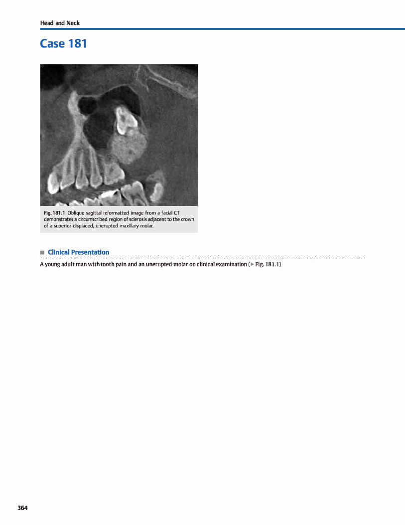
364
Head and Neck
Case 1 81
Fig. 1 81 .1 Oblique sagittal reformatted image from a facial CT demonstrates a circumscribed reg ion of sclerosis adjacent to the crown of a superior displaced, unerupted maxillary molar.
• Clinical Presentation
A young adult man with tooth pain and an unerupted molar on clinical examination ( � Fig. 181.1)
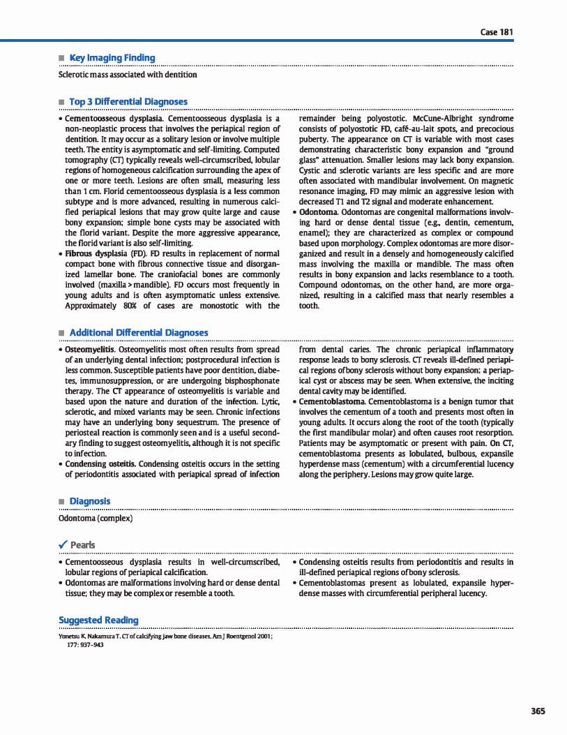
• Key lmaging Finding
Sderotic mass associated with dentition
• Top 3 Differential Diagnoses
• Cementoosseous dysplasia. Cementoosseous dysplasia is a non-neoplastic process that involves the periapical region of
dentition. It may occur as a solitary lesion or involve multiple teeth. The entity is asymptomatic and self-limiting. Computed tomography (CT) typically reveals well-circumscribed, lobular regions of homogeneous calcification surrounding the apex of one or more teeth. I.esions are often small, measuring less
than 1 cm. Florid cementoosseous dysplasia is a Jess common subtype and is more advanced, resulting in numerous calcified periapical Iesions that may grow quite large and cause bony expansion; simple bone cysts may be associated with the florid variant. Despite the more aggressive appearance, the florid variant is also self-limiting.
• Fibrous dysplasia (FD). FD results in replacement of normal compact bone with fibrous connective tissue and disorgan
ized lamellar bone. The craniofacial bones are commonly involved (maxilla > mandible). FD occurs most frequently in young adults and is often asymptomatic unless extensive. Approximately 80% of cases are monostotic with the
• Additional Differential Diagnoses
• Osteomyelitis. Osteomyelitis most often results from spread of an underlying dental infection; postprocedural infection is
less common. Susceptible patients have poor dentition, diabe
tes, immunosuppression, or are undergoing bisphosphonate therapy. The CT appearance of osteomyelitis is variable and based upon the nature and duration of the infection. Lytic, sderotic, and mixed variants may be seen. Chronic infections may have an underlying bony sequestrum. The presenœ of periosteal reaction is commonly seen and is a useful secondary finding to suggest osteomyelitis, although it is not spedfic
ta infection. • Condensing osteitis. Condensing osteitis occurs in the setting
of periodontitis associated with periapical spread of infection
• Diagnosis
Odontoma ( complex)
� Pearts
• Cementoosseous dysplasia results in well-drcumscribed, lobular regions of periapical calcification.
• Odontomas are malformations involving bard or dense dental
tissue; they may be complex or resemble a tooth.
Suggested Reading
Yonetsu K, Nalcamura T, CT of calcifying jaw bene diseases. Am J Roentgenol 2001 ;
177: 937-943
Case 181
remainder being polyostotic. McCune-Albright syndrome
consists of polyostotic FD, café-au-lait spots, and precocious puberty. The appearanœ on CT is variable with most cases demonstrating characteristic bony expansion and uground glass" attenuation. Smaller lesions may lack bony expansion.
Cystic and sderotic variants are Jess specific and are more often associated with mandibular involvement. On magnetic resonanœ imaging, FD may mimic an aggressive lesion with decreased Tl and T2 signal and moderate enhancement
• Odontoma. Odontomas are congenital malformations involving hard or dense dental tissue { e.g., dentin, cementum, enamel); they are characterized as complex or compound
based upon morphology. Complex odontomas are more disorganized and result in a densely and homogeneously calcified mass involving the maxilla or mandible. The mass often results in bony expansion and Jacks resemblance to a tooth. Compound odontomas, on the other band, are more organized, resulting in a calcified mass that nearly resembles a tooth.
from dental caries. The chronic periapical inflammatory response leads to bony sderosis. CT reveals ill-defined periapi
cal regions ofbony sderosis without bony expansion; a periapical cyst or abscess may be seen. When extensive, the indting
dental cavity may be identified. • Cementoblastoma. Cementoblastoma is a benign tumor that
involves the cementum of a tooth and presents most often in young adults. It occurs along the root of the tooth (typically the first mandibular molar) and often causes mot resorption.
Patients may be asymptomatic or present with pain. On CT, cementoblastoma presents as lobulated, bulbous, expansile hyperdense mass (cementum) with a drcumferential lucency along the periphery. I.esions may grow quite large.
• Condensing osteitis results from periodontitis and results in ill-defined periapical regions ofbony sderosis.
• Cementoblastomas present as lobulated, expansile hyperdense masses with circumferential peripheral Iucency.
365
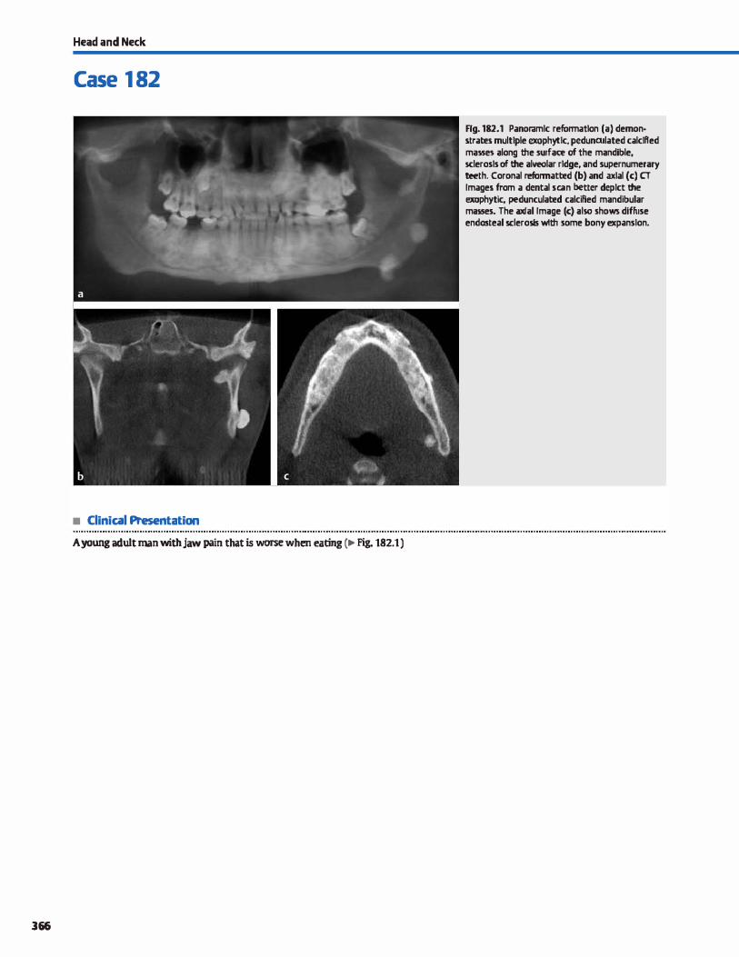
Head and Neck
Case 1 82
• Clinical Presentation
A young adult man with jaw pain that is worse when eating (� Fig. 182.1)
366
Rg. 182.1 Panoramlc refonnatlon (a) demon· strates multtple exophyttc, pedunailated calclfled massn along the surface of the mandible, sclerosls of the alveolar rldge, and supernumerary teeth. Corona! refonnatted (b) and iil>Clal (c) cr Images from a dental se.an better deptct the exophytic, pedunculated calcified mandibular masses. The axial Image (c) also shows diffuse endosteal sclerosls wllil some bony expansion.
• Key lmaging Finding
Multiple osteomas and supemumerary teeth
• Diagnosis
Gardner syndrome/multiple osteomas. Osteomas are benign tumors that are characterized by proliferation of compact or cancellous bone. The vast majority are solitary and isolated lesions; multiple osteomas may also be sporadic or associated
with Gardner syndrome. In the maxillofacial region, they are far more common in the paranasal sinuses compared with the maxilla and mandible; the mandible is more often involved
than the maxilla. Most Iesions are asymptomatic, unless they interfere with the mechanics of the jaw or compress adjacent neurovascular structures.
Osteomas are characterized as peripheral, extending from the periosteum, or central, originating along the endosteal surface. On computed tomography (CT), peripheral osteomas appear as exophytic pedunculated or sessile, densely calcified
masses extending from the surface of the periosteum. Endosteal
� Pearts
• Osteomas are benign tumors characterized by proliferation of compact or cancellous bone.
• Osteomas may be peripheral, extending from the periosteum, or central, originating along the endosteal surface.
Suggested Readlngs
Woldenbeig Y, Nash M, Bodner L Peripheral osteoma of the maxillofacial region. Diagnosis and management; a study of14 cases, Med Oral PalDI Oral Cir Ducal
2005; 10 Suppl 2: E139-E142
Case 182
osteomas reveal focal bony sclerosis with or without bony expansion. Osteomas may be distinguished from regions of exostoses by the presence of compact bone and the absence of a contiguous medullary cavity extending into the lesion. However, the two lesions may be indistinguishable in some cases
where characteristic features are not present. Gardner syndrome is an autosomal dominant syndrome
characterized by colorectal polyps, bone tumors, and soft-tissue tumors {e.g., abdominal desmoid tumors). It typically affects young to middle-aged adults. Characteristic maxillofacial findings indude multiple osteomas, supemumerary teeth, dental impaction, and bone cysts. Because sporadic cases of multiple osteomas are rare, patients found to have multiple craniofadal
osteomas should undergo colorectal screening to exdude Gardner syndrome.
• Supemumerary teeth, tooth impaction, and bone cysts are additional craniofacial findings of Gardner syndrome.
• Patients with multiple craniofacial osteomas should undergo colorectal screening to exclude Gardner syndrome.
367
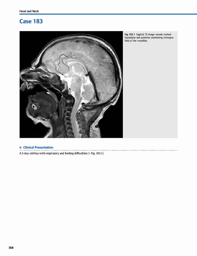
Head and Neck
Case 1 83
• Clinical Presentation
A 5-day-old boy with respira tory and feeding difficulties ( � Fig. 183.1 )
368
Fig. 1 83.1 Sagittal T2 image reveals marked hypoplasia and posterior positioning (retrognathia) of the mandible.

• Key lmaging Finding
Mandibular hypoplasia
• Diagnosis
Micrognathia. Micrognathia refers to underdevelopment or hypoplasia of the mandible. lt is dinically important due to the potential for airway compromise and difficulty with feeding. Micrognathia may occur as an isolated abnormality or in association with multiple syndromes. Associated syndromes indude Pierre Robin ( deft soft palate, high-arched palate, glossoptosis, natal teeth, and multiple ear infections), Treacher Collins (mandibulofacial dysostosis), Goldenhar (hemifacial microsomia, developmental delay, deft lip/palate, optic/orbital abnormalities, microtia, hearing Joss, vertebral anomalies, and cardiac and visœral anomalies), Cornelia de Lange (short stature dwarlism, microcephaly, mental retardation, low-set ears, facial anomalies, and cardiac and visœral anomalies), trisomy 13 (Patau syndrome-central nervous system malformations, mental
y' Pearts
• Micrognathia refers to mandibular hypoplasia; it may occur in isolation or in association with syndromes.
• Micrognathia is dinically important due to the potential for airway compromise and difficulty with feeding.
Suggested Readings
Paladini D, MDITa T, Teodoro A. LàIItberti A. Trernolaterr.1 F, Martinelli P. Oijertive diagnosis ofrnicrognathia in the fetus; thejaw index. Obsœt Gynecol 1999; 93;
382-386
Case 183
retardation, deft lip/palate, cardiac and visœral anomalies, and polydactyly), and trisomy 18 {Edward syndrome-cranial malformations, mental retardation, optic/orbital abnormalities, low-set ears, deafness, and cardiac and gastrointestinal anomalies).
Micrognathia may be identified objectively in utero. On fetal ultrasound, micrognathia is suggested on the sagittal profile by a small mandible and measured using the jaw index, which is calculated by dividing the anterior-posterior diameter of the mandible by the biparietal diameterx 1 OO; a value< 23 is sensitive and spedfic for micrognathia. Altematively, micrognathia may be suggested by measuring the frontal nasomental angle on the profile view; a measurement below the flfth perœntile (142 degrees) is suggestive ofmicrognathia/retrognathia.
• Micrognathia may be suggested objectively in utero using the jaw index or frontal nasomental angle.
Palit G, jacquemyn Y, Kerrernans M. An objective rneasurernent to diagnose rnicro
gnathia on prenatal ultrasourul. Oin Exp Obsœt Gynecol 2008; 35; 121-123
369
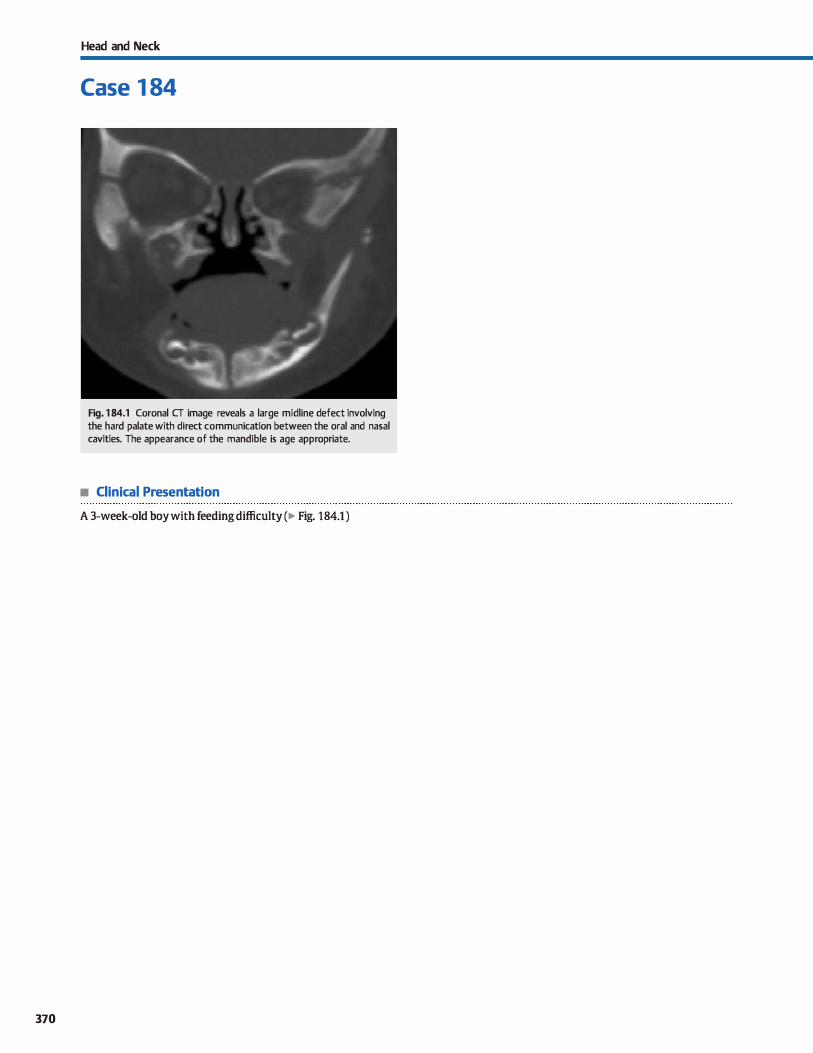
370
Head and Neck
Case 1 84
Fig. 1 84.1 Coronal CT image reveals a large midline defect involving the hard palate with direct communication between the oral and nasal cavities. The appearance of the mandible is age appropriate.
• Clinical Presentation
A 3-week-old boy with feeding difficulty (� Fig. 184.1 )
• Key lmaging Finding
Bony defect involving the hard palate
• Diagnosis
Oeft palate. Oeft lip and/or palate are caused by failure of fusion of paired structures. They are among the most common
craniofacial congenital anomalies and may occur in isolation or in association with numerous syndromes. There is an increased incidence in those with a first-degree relative with a cleft lip and/or palate. Most cases involve bath a cleft lip and cleft patate with unilateral abnonnalities being more common than bilat
eral. A cleft lip results from nonfusion of the media] nasal processes; it may be unilateral or bilateral. A cleft palate results
from nonfusion of the lateral palatine processes, nasal septum, and/or the median palatine processes and may affect the hard and/or soft palate. Cleft palates allow for direct communication between the oral and nasal cavity and pharynx. resulting in feeding difficulties for neonates.
Oefts may be found in utero during a fetal anatomy scan or identified clinically at birth. Prenatal anatomie imaging is per
formed around the 20th week of gestation. Part of the routine anatomy scan includes dedicated coronal views of the nose and
v" Pearls
• Oeft lip and/or palate are common craniofacial anomalies
caused by failure of fusion of paired structures. • Oeft palate allows for direct communication between the oral
and nasal cavity, resulting in feeding difficulties.
Suggested Reading
Stroustrup Smith A. Estroff JA. Bamewolt CE, Mulliken JB, Levine D. Prenatal diagno
sis of deft lip and deft palate using MRI. Am] RDentgenol 2004; 183: 229-235
Case 184
lips to evaluate for a facial cleft. The hard palate may be eval
uated in the axial plane. cases of deft lip and palate may be
very subtle on ultrasound, espedally when unilateral. ln cases of bilateral deft lip and deft palate, a paranasal echogenic premaxillary protrusion may be seen. The use of three-dimen
sional ultrasound further increases the sensitivity for identifying clefts. Fetal magnetic resonanœ imaging (MRI) is useful to
confirm or further evaluate suspected clefts because it provides superior soft-tissue detail.
Postnatal computed tomography (CT) imaging readily identi
fies a deft palate as bony (hard patate) and/or sort-tissue {soft palate) defects, which are best visualized on coronal reformats. A cleft lip is seen as a sort-tissue defect overlying the maxilla
and may be unilateral (more common) or bilateral. Oefts are treated surgically. Unilateral cleft lips often require
a single surgery in the neonatal period. More severe defts often require multiple surgeries to complete the repair.
• Oefts may be found during a fetal anatomy scan or MRI, noted
clinically at birth, or evaluated on postnatal CT.
371
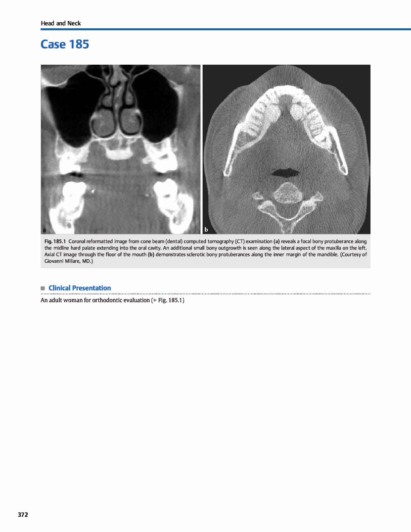
372
Head and Neck
Case 1 85
Fig. 1 85.1 Coronal reformatted image from cone beam (dental) computed tomography {CT) examination (a) reveals a focal bony protuberance a long the midline hard palate extending into the oral cavity. An additional small bony outgrowth is seen along the lateral aspect of the maxilla on the left. Axial CT image through the floor of the mouth (b) demonstrates sderotic bony protuberances along the inner margin of the mandible. {Courtesy of Giovanni Millare, MD.)
• Clinical Presentation
An adult woman for orthodontie evaluation ( � Fig. 185.1 )
• Key lmaging Finding
Bony protuberances along the bard patate/roof and floor of
mouth
• Diagnosis
Torus palatinus and mandibularis. Torus patatinus refers to a
tocalized bony protuberance or outgrowth along the surface of
the maxilla. It most often occurs midline in the hard patate
atong the roof of the mouth. Torus mandibutaris represents a
localized bony protuberance or outgrowth along the inner sur
face of the mandible. It is most often bilateral and occurs along
the floor of the mouth.
� Pearts
• Torus palatinus is a localized bony protuberance or outgrowth
of the bard patate within the roof of the mouth.
• Torus mandibutaris is a tocalized bony protuberance/out
growth of the mandible within the floor of the mouth.
Suggested Reading
Yonetsu K, Nakamura T. CT of calcifying jaw bene diseases. Am J Roentgenol 2001 ;
177: 937-943
Case 185
Tori are composed of compact bone, which ditferentiate them
from osteochondromas that maintain marrow continuity with
the parent bone. They are inddental findings but may interfere
with denture fabrication and placement.
• Tori are composed of compact bone, whereas osteochondro
mas maintain marrow continuity with the parent bone.
• Tori are typically incidentat findings but may interfere with
denture fabrication and placement.
373
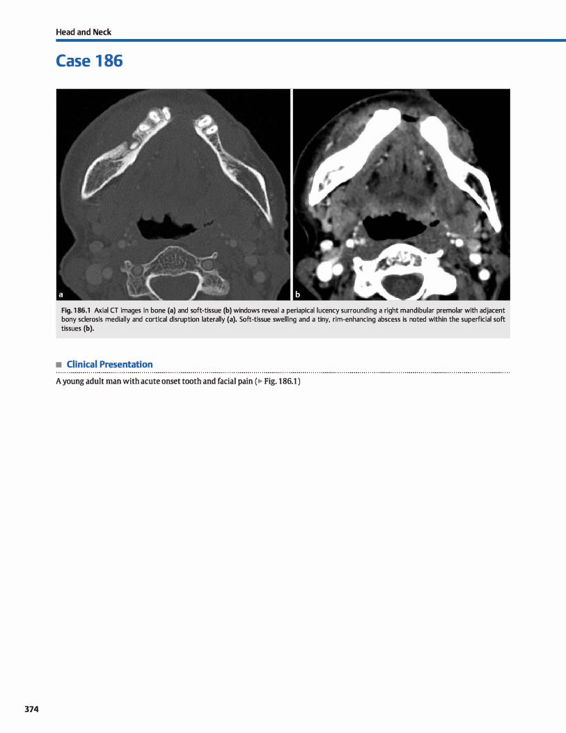
374
Head and Neck
Case 1 86
Fig. 1 86.1 Axial CT images in bone (a) and soft-tissue (b) windows reveal a periapical lucency surrounding a right mandibular premolar with adjacent bony sclerosis medially and cortical disruption laterally (a). Soft-tissue swelling and a tiny, rim-enhancing abscess is noted within the superficial soft tissues (b).
• Clinical Presentation
A young adult man with acute onset tooth and facial pain (� Fig. 186.1 )
• Key lmaging Finding
Periapical luœncy with cortical disruption
• Diagnosis
Periapical abscess. Periodontitis typically results from periapical spread of infectious and inflammatory components of a dental cavity. If the body's immune response effectively walls off the infection, a periapical granuloma occurs in the subacute setting with subsequent formation of a chronic periapical cyst In the absence of an organized cavity, a periapical abscess may result. Continued spread of the infectious proœss beyond the
periapical region may result in acute osteomyelitis or an intra
alveolar abscess.
On computed tomography (CT), periapical disease, induding granulomas, cysts, and absœsses, may be indistinguishable. Ali demonstrate periapical lucendes; cysts and granulomas are welldrcumscribed, whereas abscesses may be well-drcumscribed or
� Pearls
• Periodontitis results from periapical spread of infectious and inflammatory components of a dental cavity.
• On CT, periapical disease, induding granulomas, cysts, and abscesses, may be indistinguishable.
Suggested Readings
Case 186
ill defined. In the first 1 to 2 weeks of infection, a periapical
Iuœncy may be absent due to the Jack of bony resorption. Local spread of a periapical abscess may result in resorption of medullary and cortical bone, reactive bony sderosis, or a combination of both. These findings are typically not seen with periapical
granulomas or cysts. Further spread beyond the cortical margin may result in overlying soft-tissue inflammation and soft-tissue
abscess formation. Periapical cysts are typically asymptomatic and found
incidentally. Periapical abscesses, on the other band, are painful Treatment may consist of tooth extraction with evacuation of the absœss and cosmetic restoration or root canal
therapy.
• Periapical cysts and granulomas are drcumscribed, whereas abscesses may be circumscribed or ill defined.
• Spread of a periapical abscess may result in bony lucency and/ or sderosis, as well as soft-tissue involvement.
Devenney-Cakir B, Subramaniam RM, Reddy SM, Imsande H, Gohel A. Sakai O. Cystic Scheinfeld MH, Shifteh K. Avery U. Dym H, Dym RJ. Teeth: what radiologists should
and cystic-appearing Iesions of the mandible; review. Amj Rcentgenol 2011; 196 know. Radiographies 2012; 32; 1927-1944
Suppl: WSfi6-WS77
375
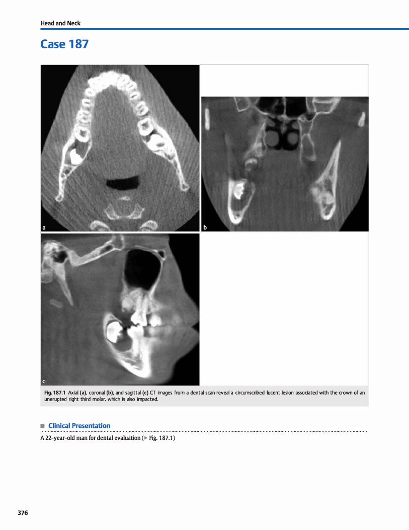
376
Head and Neck
Case 1 87
Fig. 1 87 .1 Axial (a), coronal (b), and sagittal (c) CT images from a dental scan reveal a circumscribed lucent lesion associated with the crown of an unerupted right third molar, which is also impacted.
• Clinical Presentation
A 22-year-old man for dental evaluation (� Fig. 187.1 )
• Key lmaging Finding
Cystic lesion associated with the crown of an unerupted molar
• Diagnosis
Dentigt!rous cyst. A dentigerous or follicular cyst is the second most common odontogt!nic cyst following a periapical cyst. lt is
associated with the crown of an unerupted tooth, typically the
third molars. The roots are usually outside of the lesion. Appraximately 75% occur in the mandible; 25% involve the maxilla.
Dentigerous cysts may be found at any age but are most oft:en detected during adolescence and young adulthood. Lesions may
grow quite large and cause bony expansion, although they are
typically asymptomatic. The vast majority of lesions occur spo-
y' Pearts
• Dentigerous cysts are the second most common odontogenic cysts, following periapical cysts.
• Dentigerous cysts are assodated with the crown of an uner
upted tooth. typically the third mandibular molar.
Suggested Reading
Devenney-ùkir B, Subramaniam RM, Reddy SM, Irnsande H, Gohel A. Sakai O. Cystic
and cystic-appearing Iesions of the rnandible: review. AmJ Roentgenol 2011; 196
SUppl: WSfE>-WS77
Case 187
radically; bilateral lesions are rare and may be seen with dei
docranial dysostosis or mucopolysaccharidosis. On computed tomography {CT), dentigerous cysts present as
well-circumscribed, unilocular cystic lesions surrounding the crown of an unerupted tooth, typically the third molar. There
may be displacement and root resorption of adjacent teeth. On magnetic resonance imaging (MRI), lesions appear cystic (Tl
hypointense and 12 hyperintense) without enhancement
• On cr, dentigerous cysts are well-drcumscribed and unilocular cystic lesions; they follow fluid signal on MRI.
377
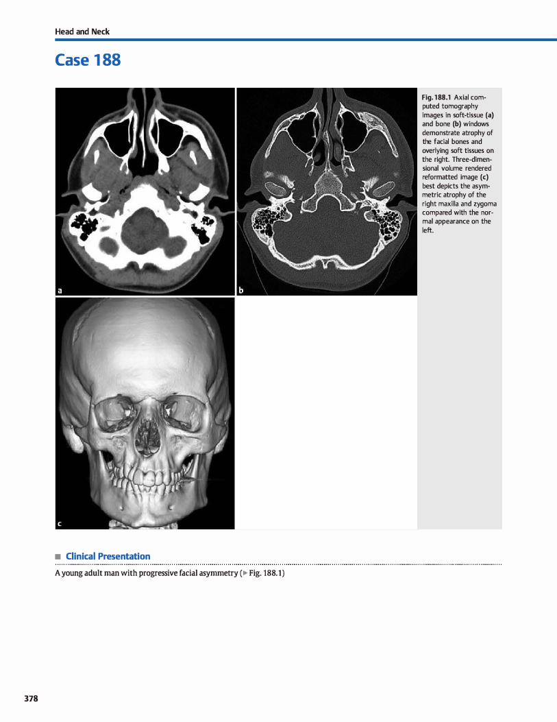
Head and Neck
Case 1 88
• Clinical Presentation
A young adult man with progressive facial asymmetry ( � Fig. 188.1)
378
Fig. 188.1 Axial computed tomography images in soft-tissue (a) and bone (b) windows demonstrate atrophy of the facial bones and overlying soft tissues on the right. Three-dimensional volume rendered reformatted image (c) best depicts the asymmetric atrophy of the right maxilla and zygoma compared with the normal appearance on the left.
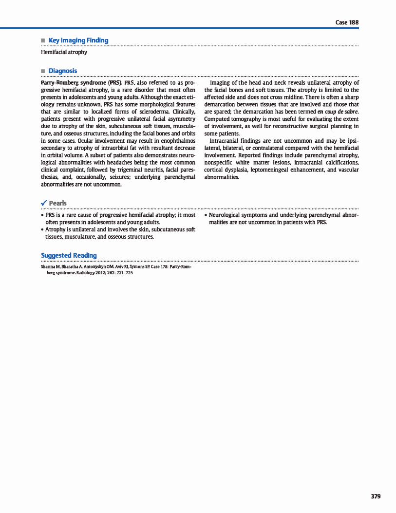
• Key lmaging Finding
Hemifacial atrophy
• Diagnosis
Parcy-Romberg syndrome (PRS). PRS, also referred to as progressive hemifacial atrophy, is a rare disorder that most often presents in adolescents and young adults. Although the exact etiology remains unknown, PRS has some morphological features that are similar to localized forms of scleroderma. Oinically, patients present with progressive unilateral facial asymmetry due to atrophy of the skin, sulxutaneous soft tissues, muscula
ture, and osseous structures, induding the facial bones and orbits in some cases. Ocular involvement may result in enophthalmos secondary to atrophy of intraorbital fat with resultant decrease in orbital volume. A subset of patients also demonstrates neurological abnormalities with headaches being the most common dinical complaint, followed by trigeminal neuritis, fadai paresthesias, and, occasionally, seizures; underlying parenchymal
abnormalities are not uncommon.
� Pearts
• PRS is a rare cause of progressive hemifacial atrophy; it most often presents in adolescents and young adults.
• Atrophy is unilateral and involves the skin, subcutaneous soft tissues, musculature, and osseous structures.
Suggested Reading
Shanna M, Bharatha A, Antonyshyn OM. Aviv RI, Symons SP. Case 178: Parry-Rom
berg syndrome. Radiology 2012; 262; 721-725
Case 188
Imaging of the head and neck reveals unilateral atrophy of the facial bones and soft tissues. The atrophy is limited to the affected side and does not cross midline. There is often a sharp demarcation between tissues that are involved and those that are spared; the demarcation has been termed en coup de sabre.
Computed tomography is most useful for evaluating the extent of involvement, as well for reconstructive surgical planning in
some patients. Intracranial findings are not uncommon and may be ipsi
lateral, bilateral, or contralateral compared with the hemifacial involvement Reported findings indude parenchymal atrophy, nonspecific white matter lesions, intracranial calcifications, cortical dysplasia, leptomeningeal enhancement, and vascular
abnormalities.
• Neurological symptoms and underlying parenchymal abnormalities are not uncommon in patients with PRS.
379
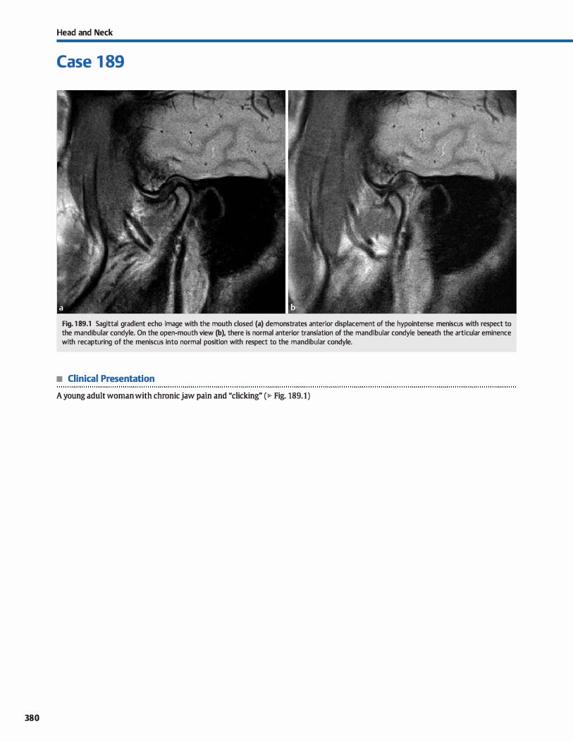
380
Head and Neck
Case 1 89
Fig. 1 89.1 Sagittal gradient echo image with the mouth dosed (a) demonstrates anterior displacement of the hypointense meniscus with respect to the mandibular condyle. On the open-mouth view (b), there is normal anterior translation of the mandibular condyle beneath the articular eminence with recapturing of the meniscus into normal position with respect to the mandibular condyle.
• Clinical Presentation
A young adult woman with chronicjaw pain and "clicking" (� Fig. 189.1)
• Key lmaging Finding
Displacement and recapturing of the temporomandibular joint (TMJ) meniscus
• Diagnosis
Temporomandibular joint (UvU) dysfonction. TMJ dysfunction is common in dinical practiœ and most often affects young to middle-aged patients with a female predominanœ. Common causes of TMJ dysfunction indude meniscal displaœment, joint effusions, and inflammatory or degenerative changes. It is important to correlate imaging findings clinically because findings of derangement may be seen in otherwise asymptomatic patients.
Articular dise or meniscal displaœment is the most common cause of TM] dysfunction. The meniscus is thinner at the œnter compared with the ends, which gives it a biconcave or "bow-tie" configuration on magnetic resonanœ imaging. A normal dise has homogeneous hypointensity within the anterior and intermediate bands and slightly hyperintense signal within the posterior band on 12, proton density, and gradient echo sequenœs. With meniscal degeneration, there is often increased T2 signal intensity within the meniscus, as well as meniscal thinning that is more pronounced anteriorly.
Under normal drcumstances, the meniscus articulates with the mandibular condyle within the condylar fossa when the mouth is closed and translates anteriorly with the mandibular condyle beneath the articular eminence of the temporal bone when the mouth is opened. Meniscal displaœment refers to an
� Pearts
• Articular dise or meniscal displaœment is the most common cause of TM] dysfunction; most occur anteriorly.
• On open-mouth views, the meniscus can remain displaced or undergo reduction/recapturing.
Suggested Reading
Tumas X. Pornes j, Berenguer j et al MR irnaging of temporornandibular joint dys
function: a pictorial review. Radiographies 2006; 26: 765-781
Case 189
abnormal relationship between mandibular condyle and the overlying meniscus. Displacement may occur in any direction but is most common anteriorly. Sagittal dosed-mouth views demonstrate anterior displaœment or positioning of the meniscus with respect to the mandibular condyle. On open-mouth views, the meniscus can either remain displaced anteriorly or return to its normal position with respect to the mandibular condyle, which is referred to as reduction or recapturing.
The TM] is a synovial joint and may also be affected by inflammatory and degenerative proœsses. Joint effusions are typically symptomatic and present with abnormal fluid within the joint spaœ. They may occur with or independent of inflammatory or degenerative proœsses. Osteoarthritis occurs more frequently in the elderly population and results in meniscal degeneration with joint spaœ narrawing, sderosis, and osteophyte formation. Inflammatory arthropathies such as rheumatoid arthritis may cause bony erosions with demineralization. Synovial proliferation or synovitis are commonly associated with inflammatory arthropathies and are best evaluated on postcontrast studies that show abnormal synovial enhanœment. Treatment of inflammatory or degenerative processes is geared toward the underlying etiology.
• Osteoarthritis results in meniscal degeneration with joint spaœ narrowing, sderosis, and osteophyte formation.
• Inflammatory arthropathies such as rheumatoid arthritis may cause bony erosions with demineralization.
381
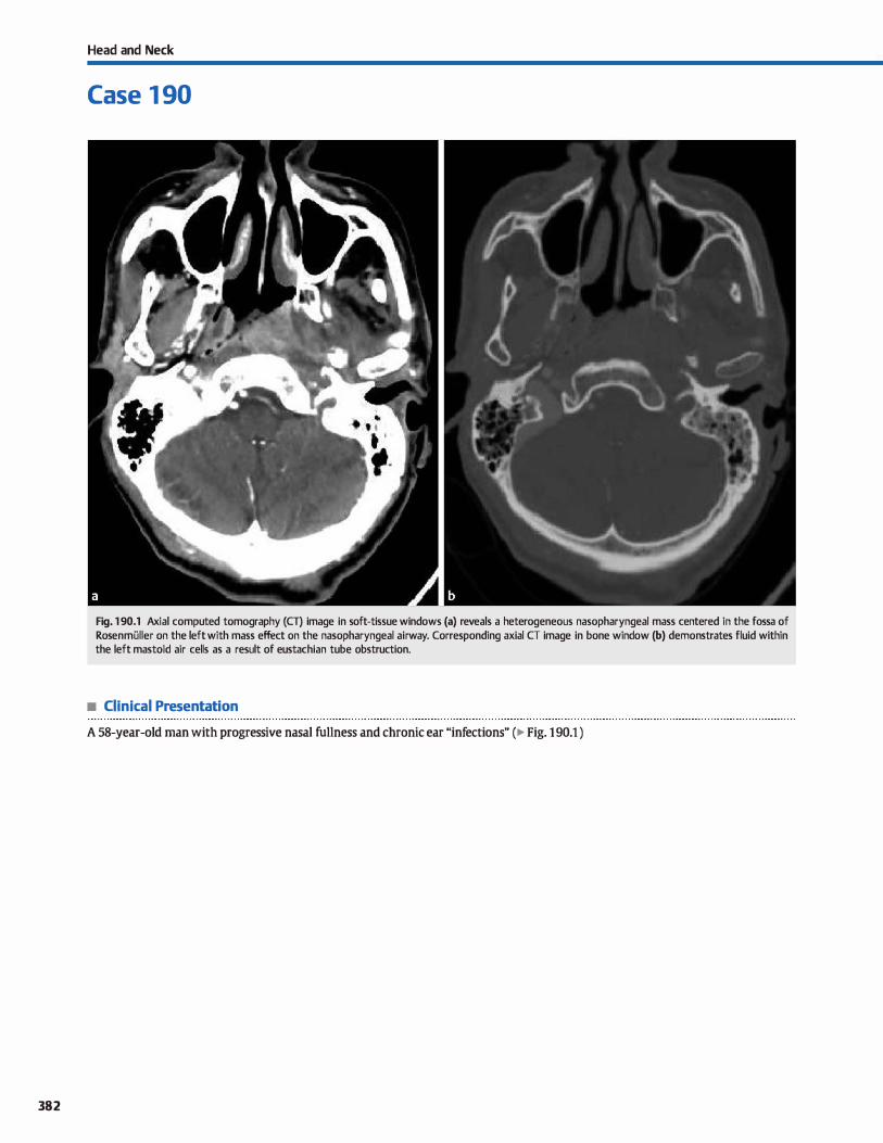
382
Head and Neck
Case 1 90
Fig. 1 90.1 Axial computed tomography (CT) image in soft-tissue windows (a) reveals a heterogeneous nasopharyngeal mass centered in the fossa of Rosenmüller on the left with mass effect on the nasopharyngeal airway. Corresponding axial CT image in bone window (b) demonstrates fluid within the left mastoid air cells as a result of eustachian tube obstruction.
• Clinical Presentation
A 58-year-old man with progressive nasal fullness and chronic ear "infections" (� Fig. 1 90.1 )
• Key lmaging Finding
Pharyngeal mucosal mass
• Top 3 Differential Diagnoses
• Squamous œll carcinoma (SCC). Sec is the most COillillOil pri
mary malignancy of the head and neck. lt COillillonly originates in the nasopharyruc or oropharynx. The lesions are dinically occult until late in the disease proœss. Nasopharyngeal carcinoma arises from the fossa of Rosenmilller. Patients may be asymptomatic or present with nasal obstruction or hearing deficits due to obstruction of the eustachian tube and subsequent
accumulation of fluid within the middle ear and mastoid air
œlls. Oropharyngeal Sec COillillonly extends laterally to involve the parapharyngeal spaœ. The imaging appearance is that of an ill-defined heterogeneously enhancing mass. There may be local spread and lymphatic spread to cervical lymph nodes, which characteristically demonstrate central necrosis. Treatment of nasopharyngeal and oropharyngeal carcinoma
depends upon staging, which is determined by the local extent of the primary tumor, size and location of regional lymph node spread, and presence or absence of distant metastases.
• lnfection/Abscess. Pharyngeal mucosal infections/absœsses most often result from adenoidal/tonsillar infections. Patients often present with fever and sore throat Tonsillitis often
• Additional Differential Diagnoses
• Thomwaldt cyst Thornwaldt cysts are benign developmental notochord remnants within the nasopharyruc. They are cystic and midline, situated between the longus coli musdes. The amount of proteinaceous material determines the imaging
appearanœs. "JYpically, they are hyperintense on T2 and variable but often hyperintense on Tl sequenœs. Although usually incidental fmdings, they may be associated with halitosis and may become superinfected. These can usually be differentiated from mucous retention cysts, which are typically off-midline and located within adenoidal tissue or pharyngeal mucosa.
• Diagnosis
Nasopharyngeal squamous cell cardnoma
� Pearts
• Nasopharyngeal SCC arises at the fossa of Rosenmüller and may obstruct the eustachian tube.
• Striated tonsillar enhanœment is seen with tonsillitis; absœsses show rim enhancement
Suggested Reading
ShinjH. Lee HK, Kim Si, 010i CG, Suh OC lmaging of parapharyngeal space lesiom:
focus on the prestyloid mmpartment. Amj Rcentgenol 2001; 177: 1465-1470
Case 190
demonstrates striated enhancement in a "tigroid" pattern without focal fluid collection. As the infection progresses and organizes, a rim-enhancing abscess may develop. With phlegmon, imaging often demonstrates ill-defined or partially organized inflammatory changes without complete rim
enhancement. It is important to realize that the presenœ of complete rim enhancement is not 100% specific for an abscess because failed aspiration/drainage may occur in -25% of cases
of suspected absœsses on computed tomography. Prompt treatment is neœssary to prevent retropharyngeal spread, which may allow for further spread to the mediastinum.
• Lymphoma. Non-Hodgkin lymphoma may involve the pha
ryngeal mucosa and appear similar to SCC. Patients with sys
temic lymphoma commonly have constitutional symptoms, along with abdominal lymphadenopathy, which are helpful discriminators. ln the absence of constitutional symptoms or widespread disease, the presence of multiple, enlarged cervical lymph nodes without necrosis is also suggestive of lymphoma. ln some cases biopsy may ultimately be neœssary to establish the diagnosis.
• Minar salivary gland tumor. Benign ( e.g., pleomorphic adenoma) and malignant (e.g., mucoepidermoid) minor salivary gland tumors may rarely involve the pharyngeal mucosal space. Imaging appearanœ is determined by the histology of the lesion. ln general, benign lesions tend to be wellcircumscribed, whereas more aggressive lesions are ill defined and heterogeneous with an imaging appearance that mimics SSC. Metastases and assodated lymphadenopathy are uncommon.
• The presenœ of constitutional symptoms or lymphadenopa
thy without necrosis suggests lymphoma. • Thomwaldt cysts are midline notochord remnants and are
often hyperintense on both Tl and T2 sequences.
383
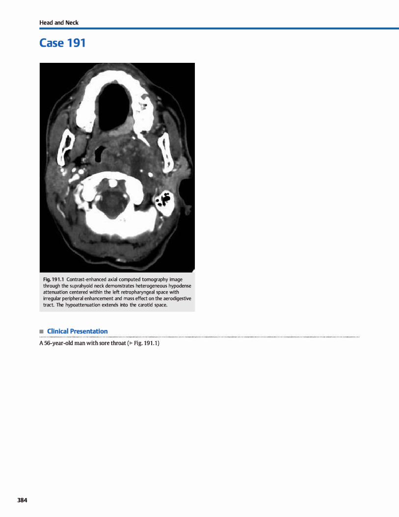
384
Head and Neck
Case 1 91
Fig. 191 .1 Contrast-enhanced axial computed tomography image through the suprahyoid neck demonstrates heterogeneous hypodense attenuation centered within the left retropharyngeal space with irregular peripheral enhancement and mass effect on the aerodigestive tract. The hypoattenuation extends into the carotid space.
• Clinical Presentation
A 56-year-old man with sore throat (� Fig. 191.1)
• Key lmaging Finding
Retropharyngeal mass
• Top 3 Differential Diagnoses
• lnfection/abscess. Patients with a retropharyngeal infection or abscess commonly present with fever, sore throat, and
elevated white blood cell count lnvolvement of the retropharyngeal spaœ most often occurs secondary to spread of infec
tion from tonsillar tissue. Lateral radiographs will show
prevertebral soft-tissue swelling, prompting cross-section
imaging. Contrast-enhanced computed tomography demon
strates soft-tissue swelling within the retropharyngeal space
with associated inflammatory changes. Adenitis refers to inflammatory changes within retropharyngeal lymph nodes,
which typically demonstrate enlargement and abnormal cen
tral hypoattenuation. Suppurative retropharyngeal adenitis
may appear identical to an extralymphatic retropharyngeal
abscess. If left untreated, suppurative adenitis may extend
beyond the nodal capsule and result in a true retropharyngeal
abscess. Phlegmon presents with ill-defined hypodensity and
variable enhancement. As the infectious process becomes
more organized, an absœss develops, demonstrating central
fluid density with complete, peripheral rim enhancement
Prompt treatment is necessary because the retropharyngeal
spaœ allows for contiguous spread of infection into the medi
astinum or within the danger spaœ.
• Nodal metastases. The vast majority of nodal metastases
within the retropharyngeal space result from spread of squa-
• Additional Differential Diagnoses
• Venolymphatic maHormation. Venolymphatic malformations represent a spectrum of venous and lymphatic develop
mental abnormalities. Pure venous malformations avidly enhance and may contain flow voids on T2 magnetic reso
nance imaging; the presence of phleboliths is pathognomonic.
Pure lymphatic malformations may be unilocular or multiloc-
• Diagnosis
Nodal metastases with extracapsular spread (SCC involving the
lateral retropharyngeal node ofRouviere)
� Pearls
• Retropharyngeal infections cause soft-tissue swelling and
inflammation; abscesses reveal rim enhancement
• sec is the most common cause of retropharyngeal nodal
metastases, followed by papillary thyroid cancer.
Suggested Reading
ShinJH, Lee HK, Kim SY. Clioi CG, Suh OC lmaging of parapharyngeal spaœ lesions:
foals on the prestyloid compartment. Am] Roentgenol 2001: 177: 1465-1470
Case 191
mous cell carcinoma (SCC), which is the most common neo
plasm of the pharyngeal mucosa. Involvement of the lateral retropharyngeal node of Rouviere is characteristic, and cen
tral necrosis is common. Extracapsular spread appears as disruption of the peripheral lymph node margin with ill-defined
hypoattenuation extending into the surrounding soft tissues
and portends a worse prognosis.Thyroid carcinoma (papillary
variant) may also metastasize to the retropharyngeal lymph
nodes, commonly resulting in central necrosis. Imaging of the
remainder of the head and neck will help identify the primary
lesion as originating from the pharyngeal mucosa (SCC) or
thyroid gland.
• Lymphoma. Non-Hodgkin lymphoma commonly presents
with lymphadenopathy within the neck. Patients with sys
temic lymphoma often have constitutional symptoms, along
with abdominal lymphadenopathy, which are helpful dis
criminators. ln the absence of constitutional symptoms or
widespread disease, the presence of multiple, enlarged cervi
cal lymph nodes without necrosis is suggestive of lymphoma.
Unlike other nodal chains within the neck, however, isolated
retropharyngeal lymph node enlargement may rarely be the
initial presentation of lymphoma.
ular and demonstrate fluid attenuation and signal intensity.
Fluid-fluid levels, most often due to hemorrhage, are charac
teristic. Many cases demonstrate characteristics of both.
Venolymphatic malformations often cross fascia] planes to
involve multiple spaœs of the neck.
• Non-Hodgkin lymphoma commonly presents with constitu
tional symptoms and diffuse lymphadenopathy.
• Venolymphatic malformations have varying degrees of cystic/
vascular components and cross fascial planes.
385
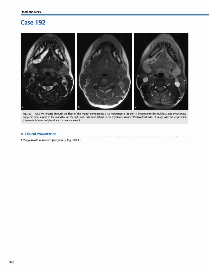
386
Head and Neck
Case 1 92
Fig. 1 92.1 Axial MR images through the floor of the mouth demonstrate a T2 hyperintense (a) and Tl hypointense (b) multiloculated cystic mass along the inner aspect of the mandible on the right with extension lateral to the mylohyoid muscle. Postcontrast axial T1 image with fat suppression (c) reveals intense peripheral and rim enhancement.
• Clinical Presentation
A 29-year-old man with jaw pain (� Fig. 1 92.1 )
• Key lmaging Finding
Floor-of-the-mouth mass
• Top 3 Differential Diagnoses
• Squamous cell carcinoma (SCC). SCC is the most common neoplasm of the aerodigestive tract and is highly associated with tobacco usage. Common locations indude the base of tangue, palate, and floor of the mouth. Patients are often asymptomatic until late in the disease process. The mylohyoid muscle divides floor-of-the-mouth cardnomas into sublingual (above) and submandibular (below) compartments.
SeC often presents as an ill-defined enhancing mass. Lymph
adenopathy, which may be centrally necrotic, is commonly se en.
• lnfection/abscess. Cellulitis and absœsses within the floor of the mouth are commonly due ta dental infections or proce
dures. Cellulitis presents with inflammatory changes that are typically ill defined, whereas abscesses are organized fluid
• Additional Differential Diagnoses
• Venolymphatic malformation. Venolymphatic malformations represent a spectrum of venous and lymphatic developmental abnormalities. Pure venous malformations avidly enhance and may contain flow voids on T2 magnetic resonance imaging (MRI); the presence of phleboliths is pathognomonic. Pure lym
phatic malformations may be unilocular or multilocular and demonstrate fluid attenuation and signal intensity. Fluid-fluid
levels, most often due ta hemorrhage, are characteristic. Many cases demonstrate characteristics of bath. Venolymphatic malformations often cross fasdal planes to inwlve multiple compartments and spaœs of the neck.
• Epidennoid/dennoid cyst. Epidermoid and dermoid cysts are developmental abnormalities that may involve the oral cavity, oropharynx, or floor of mouth. Epidennoids contain epithelial
• Diagnosis
Ranula { diving)
� Pearls
• sec is the most common malignancy of the aerodigestive tract and may have associated necrotic adenopathy.
• eellulitis and abscesses are typically of dental origin; Ludwig angina is severe and potentially life-threatening.
Suggested Reading
Coït WE, HarnsbergerHR, Osborn AG, SmolœrWR, Stevens MH, Lutkin RB. Rilllulas
and theirmimics; CTevaluation. Radiology 1987; 163; 211-216
Case 192
collections with rim enhancement. The term Ludwig angina refers to a severe, potentially life-threatening infection with abscess formation involving the sublingual, submental, and submandibular spaces. The life-threatening nature of the infection results from airway compression.
• Ranula. Ranulas are mucous retention cysts of the salivary glands in the floor of the mouth. When confined ta the sub
lingual space, they are referred ta as simple ranulas; when
they extend below the mylohyoid muscle into the submandibular space, they are referred to as diving or plunging
ranulas. The plunging ranulas lack true cyst walls. Ranulas typically follow fluid attenuation and signal intensity and are most often unilocular. Peripheral enhanœment may be seen, especially with superimposed infection.
elements, whereas dermoids contain epithelial elements along with dermal elements. Both appear cystic on imaging with low attenuation on computed tomography {CT) and T2 hyperintensity on MRI. There may be thin peripheral enhancement, if any. Dennoids may contain macroscopic fat,
which is a key discriminator when present Epidermoids demonstrate restricted diffusion.
• Hemangioma. Hemangiomas are benign vascular tumors that may involve the oral cavity or oropharynx; rarely, they
may present as a submucosal floor-of-mouth mass. capillary hemangiomas are common in infancy but naturally regress following a proliferative phase. CT and MRI demonstrate a well-drcumscribed, lobulated, hyperdense and T2 hyperintense mass with avid enhancement
• Ranulas are mucous retention cysts and are characterized based upon their relation ta the mylohyoid muscle.
• Venolymphatic malformations, epidermoid/dennoid cysts, and hemangiomas may involve the floor of the mouth.
387
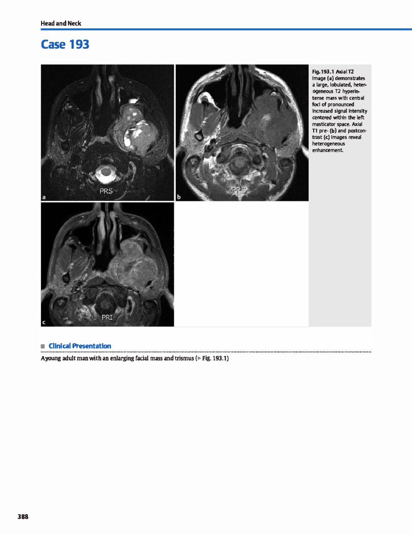
Head and Neck
Case 1 93
• Cllnlcal Presentatlon
Ayoung adult man with an enJarging facial mass and trismus {� Fig. 193.1)
388
Fig. 193.1 Axial T2 Image (a) demonstrates a large, lobulat:ed, heteP. ogeneous T2 hyperl� tense man with centr;il focl of pronounœd lncreased signal lntenslty œntered wlthln the left masticator space. Ailiil Tl pre· (b) and postcon· trast (c) Images reveal heœrogeneous enhancement

• Key lmaging Finding
Masticator spaœ mass
• Top 3 Differential Diagnoses
• lnfection/abscess. Most infections of the masticator spaœ evolve from a dental origin. Infections may present as osteomyelitis, cellulitis, or an organized abscess. Osteomyelitis has variable imaging appearances, induding Iucent, sderotic, or mixed, and often mimics an aggressive, infiltrative process. Periosteal reaction may be seen. care should be taken to evaluate the full extent of the infection because spread to other
spaœs and even the intracranial compartment can occur along nerve roots and fasàal planes. Bony erosion and marrow infiltration are important findings, as are drainable fluid
collections because these may alter clinicat management. • Sarcoma. Sarcomas within the masticator spaœ most often
arise from bone ( chondrosarcoma, osteosarcoma, or Ewing
sarcoma) or musde (rhabdomyosarcoma). Occasionally, squamous cell carànoma from a head and neck primary can
secondarily involve the masticator spaœ. Chondrosarcomas arise near the temporomandibular joint. There is typically a sort-tissue mass; the presence of a chondroid matrix is a helpful distinguishing characteristic. Osteosarcoma occurs anywhere in the mandible and is an aggressive lesion with bone
• Additional Differential Diagnoses
• Nerve sheath tumor. Both schwannomas and neurofibromas may occur within the masticator spaœ, typically involving the
mandibular division of the trigeminal nerve (V3). Schwanne>mas are usually more heterogeneous in appearance and may have regions of cystic degeneration. They are ofœn intermediate on Tl and hyperintense on T2 sequenœs with heterogeneous but avid enhanœment. Neurofibromas are generally
more homogeneous and occur in the setting of neure>fibromatosis type 1 ; they are commonly multiple and bilateral
MRI reveals intermediate Tl and hyperintense T2 signal intensity with heterogeneous enhanœment. The target sign, characterized by central decreased and peripheral increased 12 signal
intensity, is more frequently seen with neurofibromas.
• Diagnosis
Sarcoma ( chondrosarcoma)
� Pearts
• Most masticator space infections arise from a dental origin; look for signs of osteomyelitis or abscess.
• Masticator space sarcomas typically arise from bone or muscle; imaging findings often overlap.
Suggested Reading
ShinjH. Lee HK, Kim Si, 010i CG, Suh OC lmaging of parapharyngeal space lesiom:
fucus on the prestyloid mmpartment. Amj Roentgenol 2001: 177: 1465-1470
Case 193
formation and periosteal reaction. Ewing sarcoma typically presents within the first two decades of life with permeative bone destruction and a soft-tissue mass. The degree ofperiosteal reaction is variable. Rhabdomyosarcoma arises from the
muscles of mastication and typically occurs in children. On
magnetic resonance imaging (MRI), it may be difficult to distinguish between the types of sarcomas of the masticator spaœ, as they ail can be intermediate on Tl and variably hyperintense on T2 sequences and demonstrate heterogeneous enhancement
• Venolymphatic malfonnation. Venolymphatic malformations represent a spectrum of venous and lymphatic developmental
abnormalities. Pure venous malformations avidly enhanœ and may contain flow voids on T2 MRI; the presenœ of phleboliths
is pathognomonic. Pure lymphatic malformations may be uni
locular or multilocular and demonstrate fluid attenuation and signal intensity. Fluid-fluid levels, most ofœn due to hemor
rhage, are characteristic. Many cases demonstrate characteristics of both. Venolymphatic malformations often cross fascial
planes to involve multiple spaœs of the neck
• Hemangioma. Hemangiomas are unencapsulated, proliferative vascular neoplasms and represent the most common
benign tumors of infancy. The proliferative growth phase typically occurs in the first year of life; infantile and RICH
(rapidly involuting congenital hemangioma) variants then undergo involution with fatty replacement; NICH (noninvoluting congenital hemangioma) variants do not regress. Treatment is conservative unless there is mass effect on adjacent vital structures. dinically, patients present with a superficial
mass that is bluish or scarlet in color. MRI reveals a lobulated hyperintense 12 and slightly hyperintense Tl mass with avid enhancement.
• Venolymphatic malformations occur within the masticator spaœ and may involve multiple compartments.
• Nerve sheath tumors (neurofibromas and schwannomas) occur along the mandibular division of cranial nerve V.
389
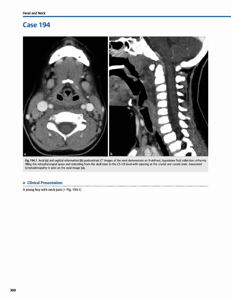
390
Head and Neck
Case 1 94
Fig. 1 94.1 Axial (a) and sagittal reformatted (b) postcontrast CT images of the neck demonstrate an il l-defined, hypodense fluid collection uniformly filling the retropharyngeal space and extending from the skull base to the C5-C6 level with tapering at the cranial and caudal ends. Associated lymphadenopathy is seen on the axial image (a).
• Clinical Presentation
A young boy with neck pain ( � Fig. 1 94.1 )
• Key lmaging Finding
Retropharyngeal fluid collection
• Top 3 Differential Diagnoses
• Abscess/phlegmon. The retropharyngeal spaœ lies posterior to the aerodigestive tract and anterior to the prevertebral musculature. It extends from the skull base to the mediastinum. Retropharyngeal infectious processes typically begin with suppurative lymphadenopathy that then extends into the retropharyngeal spaœ. Tonsillitis and pharyngitis are the most common indting events. Prior to absœss formation, ret
ropharyngeal œllulitis or phlegmon presents as an ill-defined fluid collection often with inflammatory changes. This may mimic retropharyngeal edema. As the process becomes more organized, a focal collection forms with local mass effect and peripheral enhancement. An absœss is suggested when there is complete rim enhancement. lt is important to describe the extent of the infectious process, as well as evaluate its effect
on the airway and adjacent vasculature. Phlegmon is treated conservatively with antibiotics, whereas abscesses are surgically drained.
• Retropharyngeal edema. Retropharyngeal edema refers to noninfectious fluid within the retropharyngeal spaœ. It is thought to represent lymphatic or inflammatory fluid, depending upon the etiology. Common causes indude reactive edema
secondary to infectious processes within the neck, radiation therapy, internai jugular venous occlusion, and calcific tendin-
• Diagnosis
Retropharyngeal edema secondary to pharyngitis
� Pearts
• Retropharyngeal infectious processes typically begin with suppurative lymphadenopathy.
• An absœss is suggested when there is a focal fluid collection with mass effect and rim enhancement.
Suggested Readings
HoangJK, Branstetter BF, IV, EastwoodjD, Glastonbury CM. Multiplanar Cl' and MRI
of collections in the retropharyngeal spaœ: is it an absœss7 Am J Roentgenol
2011; 196: W426-32
Case 194
opathy of the longus colli tendons. Unlike phlegmon or absœsses, retropharyngeal edema uniformly spreads throughout and fills the retropharyngeal spaœ. lt bas a characteristic "bow-tie" appearanœ on axial sequenœs and extends longitudinally throughout the retropharyngeal spaœ with tapering at
the cranial and caudal ends. There is no well-defined collection
or enhanœment. Calcific tendinopathy also demonstrates characteristic calcification of the longus colli tendons.
• Retropharyngeal hemorrhage. Retropharyngeal hematomas most often result from trauma or in the setting of an underlying coagulopathy. Spontaneous retropharyngeal hemorrhage or vascular rupture are less common etiologies. Retrophar
yngeal spaœ hemorrhage can be significant due to the Jack: of anatomie constraints to tamponade the source of bleeding; therefore, prompt diagnosis and treatment is important. Retropharyngeal hemorrhage often mimics infectious processes on computed tomography {CT), demonstrating a fluid collection with mass effect and variable enhancement. Magnetic resonanœ imaging {MRI) is more sensitive and specific for identifying blood products. Characteristic flndings indude T1
hyperintensity and susceptibility on gradient echo or susceptibility-weighted imaging. li"eatment is geared toward the underlying source of hemorrhage.
• Retropharyngeal edema is noninfectious fluid that uniformly fills the retropharyngeal spaœ.
• Retropharyngeal hemorrhage often mimics infectious proœsses on CT; MRI better identifies blood products.
Muiioz A. Fischbein Nj, de Vergas j, Crespo J. Alvarez-Vincent j. Spontaneom retro
pharyngeal hematoma: diagnosis by mr imaging. AmJ Newt>radiol 2001; 22:
1209-1211
391
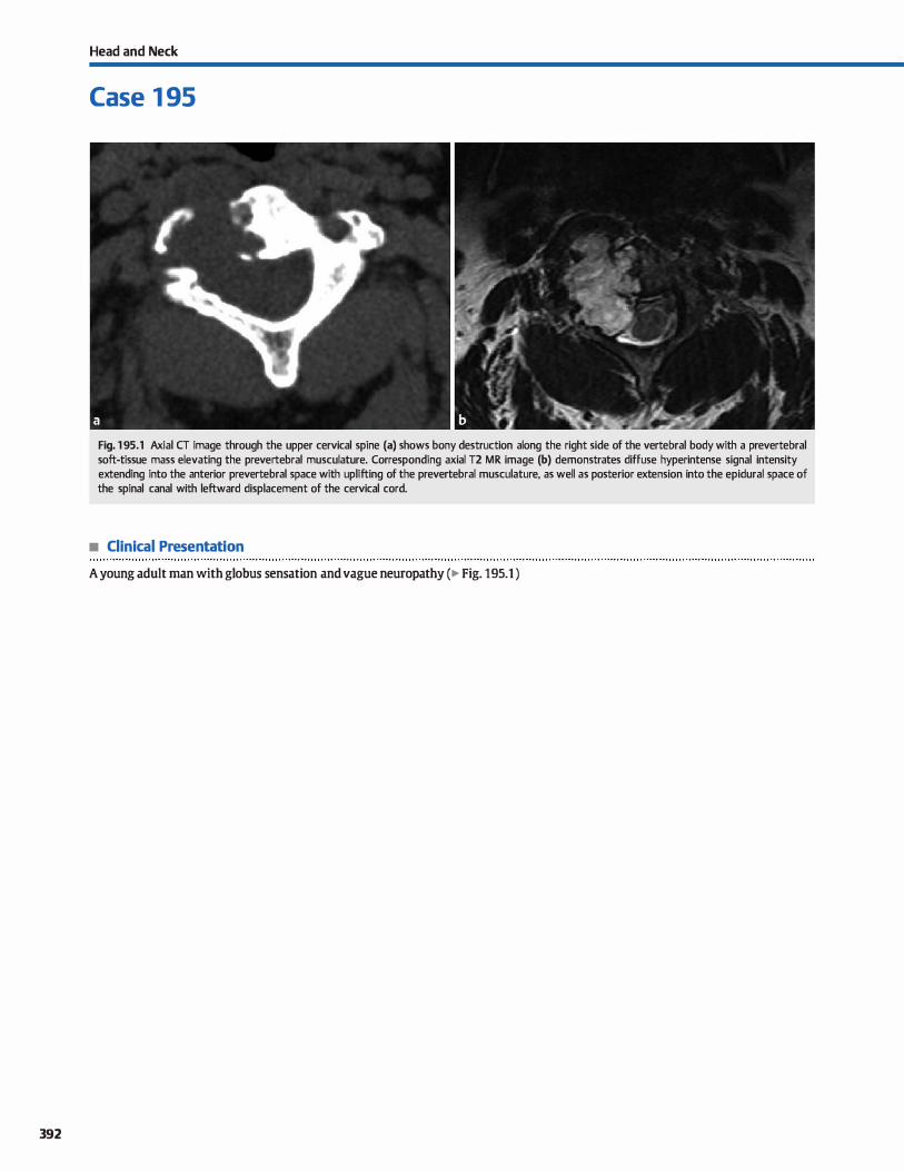
392
Head and Neck
Case 1 95
Fig. 1 95.1 Axial CT image through the upper cervical spine (a) shows bony destruction along the right side of the vertebral body with a prevertebral soft-tissue mass elevating the prevertebral musculature. Corresponding axial T2 MR image (b) demonstrates diffuse hyperintense signal intensity extending into the anterior prevertebral space with uplifting of the prevertebral musculature, as well as posterior extension into the epidural space of the spinal canal with leftward displacement of the cervical cord.
• Clinical Presentation
A young adult man with globus sensation and vague neuropathy (� Fig. 195.1)
• Key lmaging Finding
Prevertebral space mass
• Top 3 Differential Diagnoses
• Bony/epidural tumor. Metastatic disease commonly involves the spine and prevertebral spaœ. Breast and Jung carcinoma are most often lytic, along with renal and thyroid carcinomas. Prostate (and occasionally breast) carcinoma produœs sderotic metastases. Metastases are typically hypointense on Tt and hyperintense on T2 sequences with avid enhancement; multiple lesions are common. Multiple myeloma is the most com
mon primary bony malignancy in adults; plasmacytoma is the solitary variant Patients are often>40 years of age. Computed tomography (CT) demonstrates a poorly marginated, expansile lytic lesion. On magnetic resonanœ imaging (MRI), the lesions are typically isointense to cord on Tl and T2 sequenœs with homogeneous enhancement. Olordoma is an aggressive tumor that arises from notochordal remnants and is most common in
young to middle-aged adults. It most often occurs within the sacrum and craniocervical junction. Patients with craniocervical involvement present with headache and ophthalmoplegia or neck pain and neuropathy. cr reveals a midline or slightly off-midline, expansile lesion with hyperdense fod œntrally. Olordomas are dassically hyperintense on T2 and often hypointense on Tl sequenœs with heterogeneous but avid
• Additional Differential Diagnoses
• Vascular abnonnality. Common vascular abnormalities within the prevertebral space indude vertebral artery ectasia, aneurysms, and pseudoaneurysms. Although they may present a clinical dilemma on direct visualization, their vascular origin is typically readily identified on cross-sectional imaging studies, especially contrast-enhanced cr. Partially thrombosed aneurysms or pseudoaneurysms, however, may have a more compli
cated imaging appearance due to variant attenuation or signal intensity compared with the patent parent vessel.
• Degenerative discogenic disease. Degenerative changes of the cervical spine are common and progress with advancing
• Diagnosis
Bony tumor ( chordoma)
� Pearts
• Common prevertebral space tumors indude metastases, multiple myeloma, chordoma, lymphoma, and NSTs.
• Disc-osteomyelitis involves the dise space and adjacent end
plates with surrounding phlegmon or absœss.
Suggested Reading
Hamsberger HR, Osborn AG, Dift'erential diagnom ofhead and neck lesiom based
on their space of origin. 1. The suprahyoid part of the neck. Am j Rcentgenol
1991;157: 147-154
Case 195
enhancement Intratumoral hemorrhage is common. Lymphoma may involve the vertebral bodies and/or epidural spaœ and is often homogeneous in signal and enhanœment.
• Disc-osteomyelitis. Disc-osteomyelitis most often results from hematogenous spread of a distant bacterial infection; Staphylococcus aureus is the most common organism MRI shows fluid signal intensity and enhancement centered at the dise spaœ with adjacent endplate destruction and bony edema. Paraspinal phlegmon and absœss formation is typically seen. Tuberculous (IB) spondylitis is Jess common and typically occurs in immunocompromised patients. 1B spondylitis characteristically affects the vertebral bodies with relative sparing of the dise spaœ and is prone to subligamentous spread.
• Nerve sheath tumor (NSI'). NSTs indude schwannomas (most common) and neurofibromas; neurofibromas may be spo
radic or associated with neurofibromatosis type 1. Both are often circumscribed, Tl iso- to hypointense and T2 hyperintense with avid enhancement Schwannomas may demonstrate cystic change and hemorrhage. Neurofibromas are more likely ta demonstrate the utarget" sign with œntrally decreased and peripherally increased T2 signal intensity.
age. Anteriorly directed osteophytes project into the prevertebral space and often elevate the prevertebral musculature. The bony origin is readily identified on cross-sectional imaging. Anterior dise hemiations are a Jess common discogenic cause of a prevertebral spaœ mass and are often seen in the setting of trauma MRI best depicts the dise herniation, which is often contiguous and follows similar signal intensity com
pared with the parent dise. Additional findings associated with trauma include bony fractures, ligamentous and softtissue injuries, as well as possible cord injury.
• Discogenic disease (osteophytes and dise herniations) is a common cause of prevertebral spaœ masses.
393
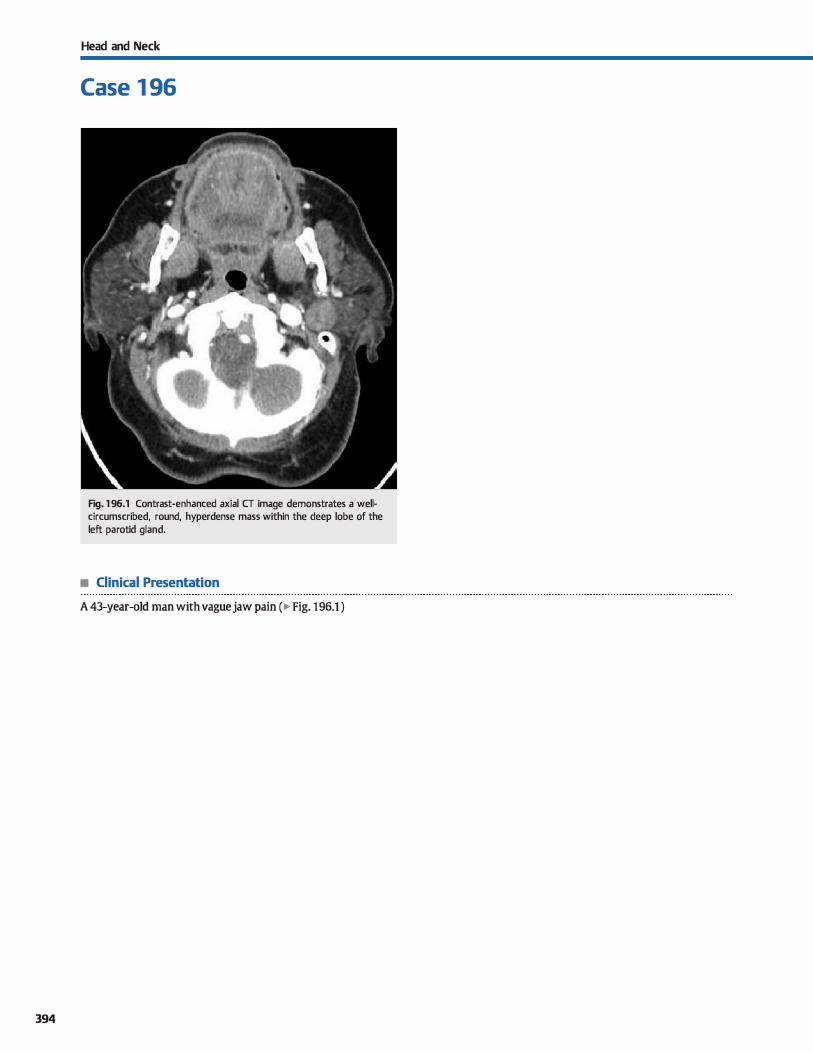
394
Head and Neck
Case 1 96
Fig. 1 96.1 Contrast-enhanced axial CT image demonstrates a wellcircumscribed, round, hyperdense mass within the deep lobe of the left parotid gland.
• Clinical Presentation
A 43-year-old man with vaguejaw pain (� Fig. 196.1 )
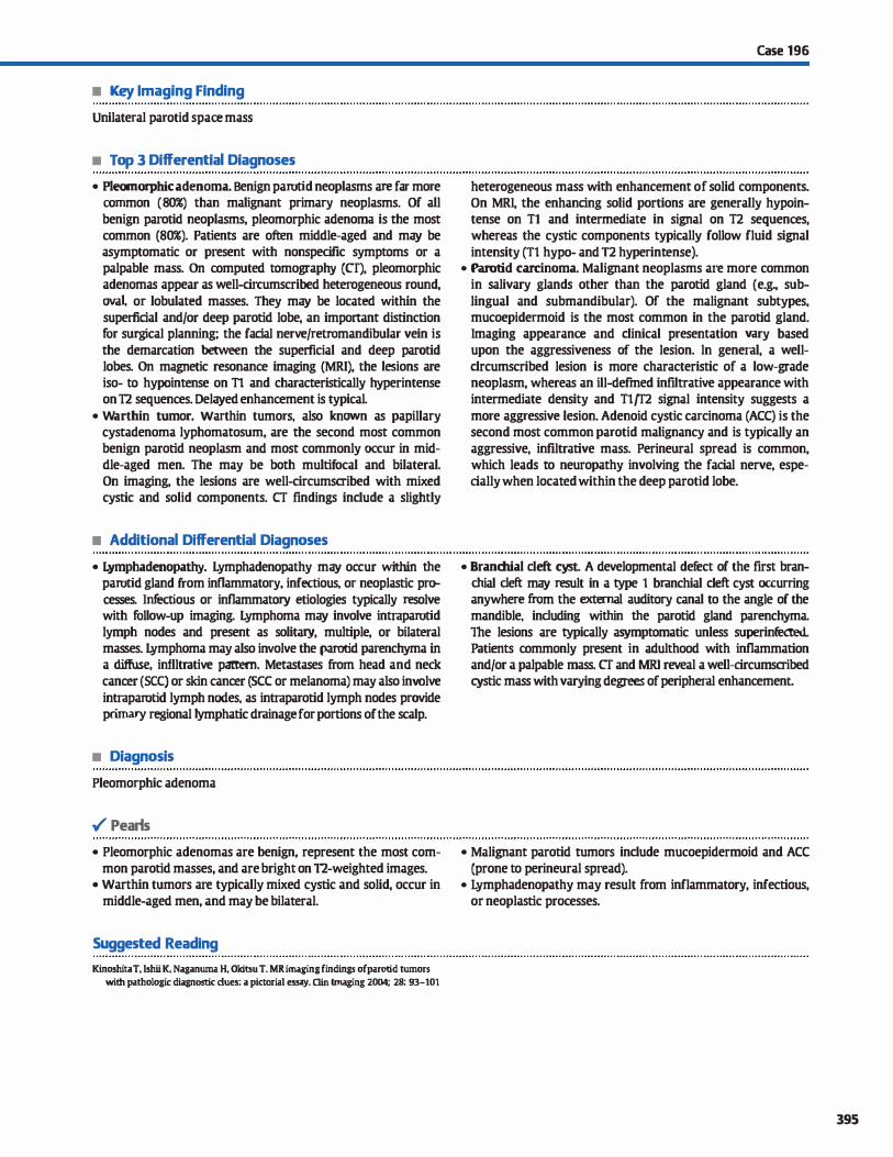
• Key lmaging Finding
Unilateral parotid space mass
• Top 3 Differential Diagnoses
• Pleomorphic adenoma. Benign parotid neoplasms are far more common ( 80%) than malignant primary neoplasms. Of all
benign parotid neoplasms, pleomorphic adenoma is the most common (80%). Patients are often middle-aged and may be asymptomatic or present with nonspecific symptoms or a palpable mass. On computed tomography {CT), pleomorphic adenomas appear as well-circumscribed heterogeneous round,
oval or lobulated masses. They may be located within the
superfiàal and/or deep parotid lobe, an important distinction for surgical planning; the faàal nerve/retromandibular vein is
the demarcation between the superticial and deep parotid lobes. On magnetic resonanœ imaging (MRI), the lesions are iso- to hypointense on Tl and characteristically hyperintense on T2 sequenœs. Delayed enhanœment is typical.
• Warthin tumor. Warthin tumors, also known as papillary cystadenoma lyphomatosum, are the second most common benign parotid neoplasm and most commonly occur in mid
dle-aged men. The may be bath multifocal and bilateral. On imaging, the lesions are well-circumscribed with mixed cystic and solid components. CT findings indude a slightly
• Additional Differential Diagnoses
• [Nmphadenopathy. Lymphadenopathy may occur within the parotid gland from inflammatory, infectious, or neoplastic pro
œsses. Infectious or inflammatory etiologies typically resolve with follow-up imaging. Lymphoma may involve intraparotid lymph nodes and present as solitary, multiple, or bilateral ma�es. Lymphoma may also involve the parotid parenchyma in a diffuse, inflltrative pattern. Metastases from head and neck cancer (SCC) or skin cancer (SCC or melanoma) may also involve
intraparotid lymph nodes, as intraparotid lymph nodes provide primary regional lymphatic drainage for portions of the scalp.
• Diagnosis
Pleomorphic adenoma
� Pearls
• Pleomorphic adenomas are benign, represent the most common parotid masses, and are bright on T2-weighted images.
• Warthin tumors are typically mixed cystic and solid, occur in middle-aged men, and may be bilateral.
Suggested Reading
Kinoshita T, lshü K. Naganurna H, Okitsu T. MR irnaging findings ofparotid turnors
with pathologie diagnostic dues; a pictorial essay. Oin lrnaging 2004; 28; 93-101
Case 196
heterogeneous mass with enhancement of solid components. On MRI, the enhanàng solid portions are generally hypointense on Tl and intermediate in signal on T2 sequenœs, whereas the cystic components typically follow fluid signal
intensity (Tl hypo- and T2 hyperintense). • Parotid c:arcinoma. Malignant neoplasms are more common
in salivary glands other than the parotid gland ( e.g., sub
lingual and submandibular). Of the malignant subtypes, mucoepidermoid is the most common in the parotid gland. Imaging appearance and dinical presentation vary based upon the aggressiveness of the lesion. ln general, a welldrcumscribed lesion is more characteristic of a low-grade neoplasm, whereas an ill-defmed infiltrative appearance with intermediate density and Tl ff2 signal intensity suggests a
more aggressive lesion. Adenoid cystic carcinoma (ACC) is the second most common parotid malignancy and is typically an
aggressive, infiltrative mass. Perineural spread is common, which leads to neuropathy involving the facial nerve, especially when located within the deep parotid lobe.
• Brandûal deft cyst. A developmental defect of the first bran
chial deft may result in a type 1 branchial deft cyst occurring anywhere from the externat auditory canal to the angle of the
mandible, induding within the parotid gland parenchyma. The lesions are typically asymptomatic unless superinfected. Patients commonly present in adulthood with inflammation and/or a palpable mass. CT and MRI reveal a well-circumscribed cystic mass with varying degrees of peripheral enhanœment
• Malignant parotid tumors indude mucoepidermoid and ACC {prone to perineural spread).
• Lymphadenopathy may result from inflammatory, infectious, or neoplastic proœsses.
395
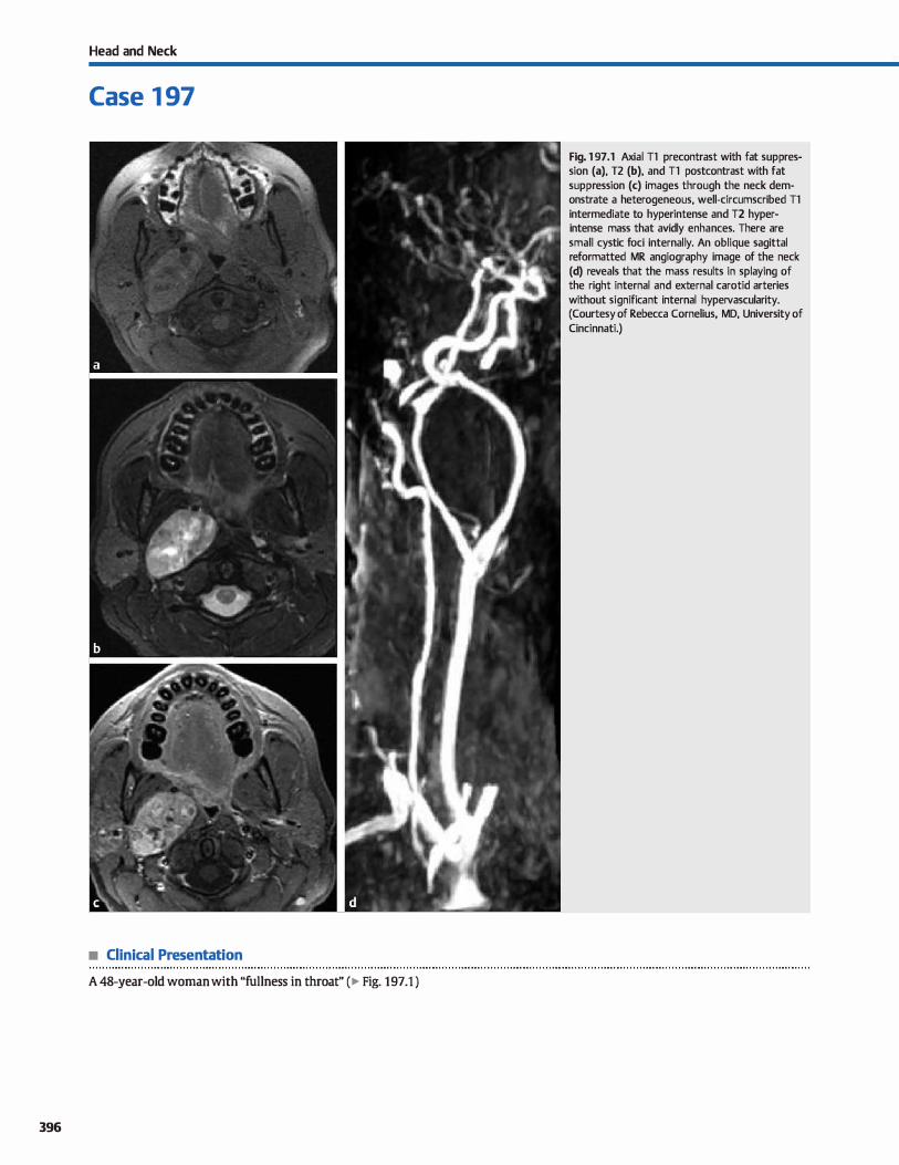
Head and Neck
Case 1 97
• Clinical Presentation
A 48-year-old woman with "fullness in throat" (� Fig. 197.1 )
396
Fig. 1 97 .1 Axial Tl precontrast with fat suppression (a), T2 (b), and Tl postcontrast with fat suppression (c) images through the neck demonstrate a heterogeneous, well-circumscribed Tl intermediate to hyperintense and T2 hyperintense mass that avidly enhances. There are small cystic foci internally. An oblique sagittal reformatted MR angiography image of the neck (d) reveals that the mass results in splaying of the right internai and external carotid arteries without significant internai hypervascularity. (Courtesy of Rebecca Cornelius, MD, University of
Cincinnati.)
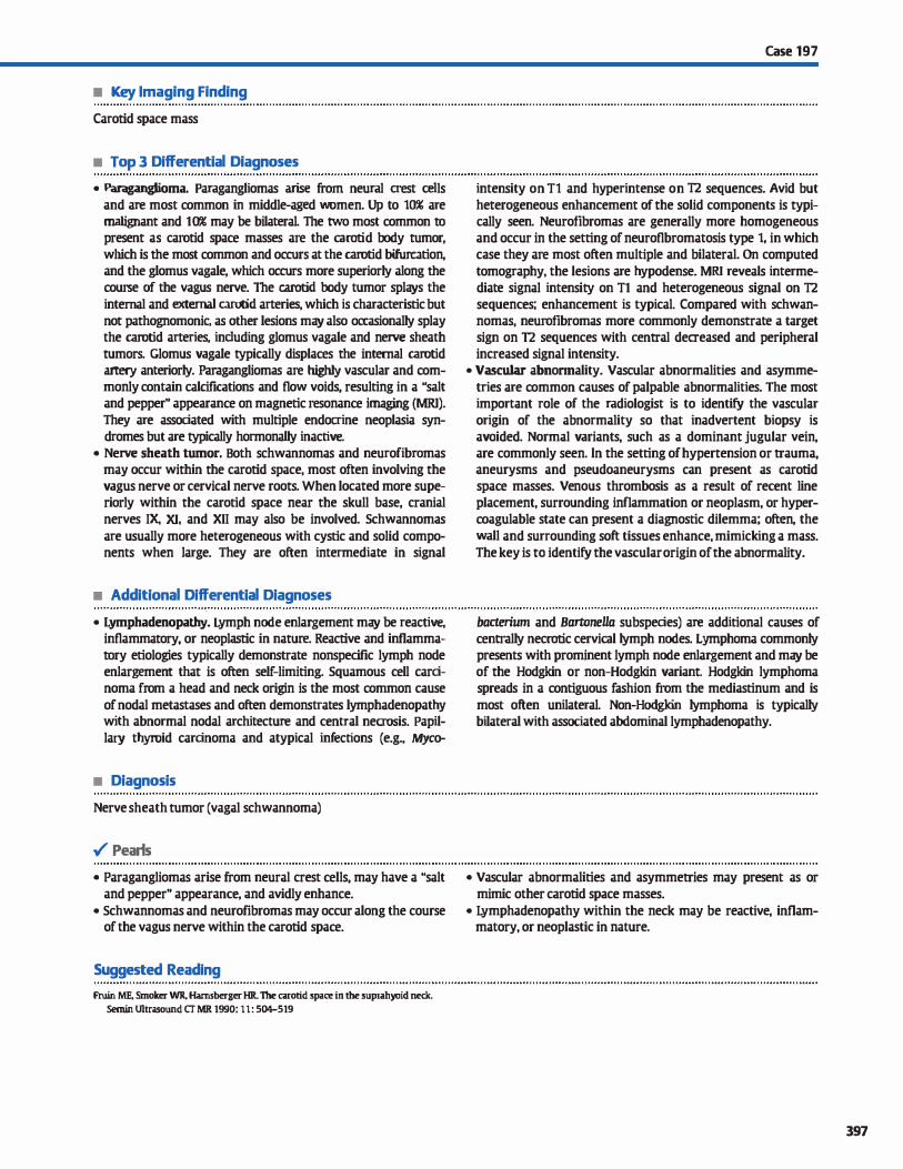
• Key lmaging Finding
Carotid space mass
• Top 3 Differential Diagnoses
• Paraganglioma. Paragangliomas arise from neural crest œlls and are most common in middle-aged women. Up to 10% are malignant and 1 C1% may be bilateral. Tue two most common to present as carotid spaœ masses are the carotid body tumor,
wlûch is the most common and occurs at the carotid bifurcation, and the glomus vagale, which occurs more superiorly along the course of the vagus nerve. Tue carotid body tumor splays the
intemal and extemal carotid arteries, which is characteristic but
not pathognomonic, as other lesions may also occasionally splay the carotid arteries, induding glomus vagale and nerve sheath
tumors. Glomus vagale typically displaœs the internai carotid artery anteriorly. Paragangliomas are highly vascular and commonly contain calcifications and flow voids, resulting in a "sait and pepper" appearanœ on magnetic resonanœ imaging (MRI).
They are associated with multiple endocrine neoplasia syndromes but are typically hormonally inactive.
• Nerve sheath tumor. Both schwannomas and neurofibromas may occur within the carotid space, most often involving the vagus nerve or cervical nerve roots. When located more superiorly within the carotid space near the skull base, cranial
nerves IX. XI, and XII may also be involved. Schwannomas
are usually more heterogeneous with cystic and solid components when large. They are often intermediate in signal
• Additional Differential Diagnoses
• Lymphadenopathy. Lymph node enlargement may be reactive, inflammatory, or neoplastic in nature. Reactive and intlamma
tory etiologies typically demonstrate nonspedfic lymph node enlargement that is often self-limiting. Squamous œll carcinoma from a head and neck origin is the most common cause
of nodal metastases and often demonstrates lymphadenopathy with abnormal nodal architecture and central necrosis. Papillary thyroid carcinoma and atypical infections (e.g., Myco-
• Diagnosis
Nerve sheath tumor (vagal schwannoma)
� Pearls
• Paragangliomas arise from neural crest œlls, may have a "sait
and pepper" appearance, and avidly enhance.
• Schwannomas and neurofibromas may occur along the course of the vagus nerve within the carotid spaœ.
Suggested Reading
Fruin ME, Smolœr WR, Hamsberger HR. The carotid spaœ in the suprahyoid neck.
Sernin Ultrasound CT MR 1990; 11: 504-519
Case 197
intensity on Tl and hyperintense on T2 sequences. Avid but heterogeneous enhancement of the solid components is typically seen. Neurofibromas are generally more homogeneous and occur in the setting ofneuroflbromatosis type 1, in which case they are most often multiple and bilateral. On computed
tomography, the lesions are hypodense. MRI reveals intermediate signal intensity on Tl and heterogeneous signal on T2 sequences; enhancement is typical. Compared with schwannomas, neurofibromas more commonly demonstrate a target sign on T2 sequences with central decreased and peripheral increased signal intensity.
• Vascular abnonnality. Vascular abnormalities and asymme
tries are common causes of palpable abnormalities. The most important role of the radiologist is to identify the vascular
origin of the abnormality so that inadvertent biopsy is avoided. Normal variants, such as a dominant jugular vein, are commonly seen. ln the setting of hypertension or trauma, aneurysms and pseudoaneurysms can present as carotid spaœ masses. Venous thrombosis as a result of recent line
placement, surrounding inflammation or neoplasm, or hypercoagulable state can present a diagnostic dilemma; often, the wall and surrounding soft tissues enhance, mimicking a mass.
The key is ta identify the vascular origin of the abnormality.
bacterium and BartoneUa subspecies) are additional causes of œntrally necrotic cervical Iymph nodes. Lymphoma commonly presents with prominent lymph node enlargement and may be of the Hodgkin or non-Hodgkin variant. Hodgkin lymphoma spreads in a contiguous fashion from the mediastinum and is
most often unilateral. Non-Hodgkin Iymphoma is typically bilateral with associated abdominal lymphadenopathy.
• Vascular abnormalities and asymmetries may present as or
mimic other carotid space masses.
• Lymphadenopathy within the neck may be reactive, inflammatory, or neoplastic in nature.
397
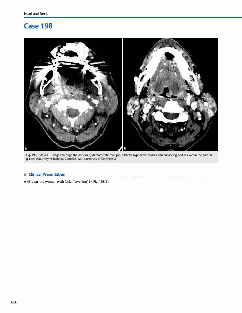
398
Head and Neck
Case 1 98
Fig. 1 98.1 Axial CT images through the neck (a,b) demonstrate multiple, bilateral hypodense masses and enhancing nodules within the parotid glands. (Courtesy of Rebecca Cornelius, MD, University of Cincinnati.)
• Clinical Presentation
A 43-year-old woman with facial "swelling" (� Fig. 198.1 )
• Key lmaging Finding
Bilateral parotid space masses
• Top 3 Differential Diagnoses
• Lymphoepithelial lesions. Benign lymphoepithelial lesions most often occur in patients with acquired immune defi
ciency syndrome (AIDS) who present with painless bilateral parotid gland enlargement. Pathologically, there is lympho
cytic infiltration of the gland (similar ta Sjtlgren syndrome)
with lymphoepithelial cyst formation. Computed tomography (CT) and magnetic resonance imaging (MRI) reveal bilateral
cystic and solid parotid lesions with diffuse gland enlarge
ment. Associated cervical lymphadenopathy and tonsillar hypertrophy is commonly seen.
• Sjügren syndrome. Sjtlgren syndrome is an autoimmune disease that most commonly occurs in middle-aged to elderly
women and results in lymphocytic infiltration of glandular tissue. The classic triad consists of parotid gland enlargement
with keratoconjunctivitis sicca (dry eyes) and xerostomia (dry mouth). On CT and MRI, Sjtlgren syndrome presents as
• Additional Differential Diagnoses
• Warthin tumors. Warthin tumors, also known as papillary cystadenoma lyphomatosum, are the second most common
benign parotid neoplasm and most commonly occur in middle-aged men. The may be both multifocal and bilateral. On imaging, the lesions are well-drcumscribed with mixed cystic
and solid components. CT findings indude a slightly heterogeneous mass with enhancement of solid components. On MRI, the enhandng solid portions are generally hypointense
on Tl and intermediate in signal on T2 sequences, whereas the cystic components typically follow fluid signal intensity (Tl hypo- and T2 hYPerintense).
• Diagnosis
Sjôgren syndrome
� Pearls
• Lymphoepithelial lesions occur in patients with AIDS and present with bilateral cystic and solid parotid lesions.
• Sjôgren syndrome is characterized by parotid enlargement/
lesions, dry eyes, and dry mouth.
Suggested Reading
Kinoshita T, lshü K. Naganuma H, Okitsu T. MR imaging finclings ofparotid tumors
with pathologie diagnostic dues: a pictorial essay. Oin Imaging 2004; 28: 93-101
Case 198
heterogeneous glandular enlargement with scattered enhancing nodules and fluid pockets. The appearance can mimic
benign lymphoepithelial lesions or lymphoma. Patients with long-standing Sjôgren syndrome are at increased risk for developing lymphoma.
• Lymphadenopathy. Lymphadenopathy may occur within the parotid gland from inflammatory, infectious, or neoplastic
processes. Infectious or inflammatory etiologies typically resolve with follow-up imaging. Lymphoma may involve
intraparotid lymph nodes and present as solitary, multiple, or
bilateral masses. Lymphoma may also involve the parotid parenchYma in a diffuse, infiltrative pattera Metastases from head and neck cancer (squamous cell carcinoma, SCC) or skin cancer (SCC or melanoma) may also involve intraparotid
lymph nodes, as intraparotid lymph nodes provide primary
regional lymphatic drainage for portions of the scalp.
• Sarcoidosis. Sarcoidosis is a multisystem granulomatous disorder that most commonly occurs in adult African Americans. Diffuse sarcoidosis can lead to granulomatous infiltration of
glandular tissue, including the parotid and lacrimal glands. Patients typically have elevated angiotensin-converting
enzyme levels and may also present with uveitis and cranial nerve deficits. On imaging, there is heterogeneous glandular enlargement that is usually bilateral and symmetric. Gallium
67 (Ga 67) scan will reveal the classic upanda sign" with increased uptake in the parotid and lacrimal glands.
• Warthin tumors are typically mixed cystic and solid, occur in middle-aged men, and may be bilateral.
• Sarcoidosis results in bilateral heterogeneous parotid enlarge
ment; the upanda sign" is seen on Ga 67 scans.
399
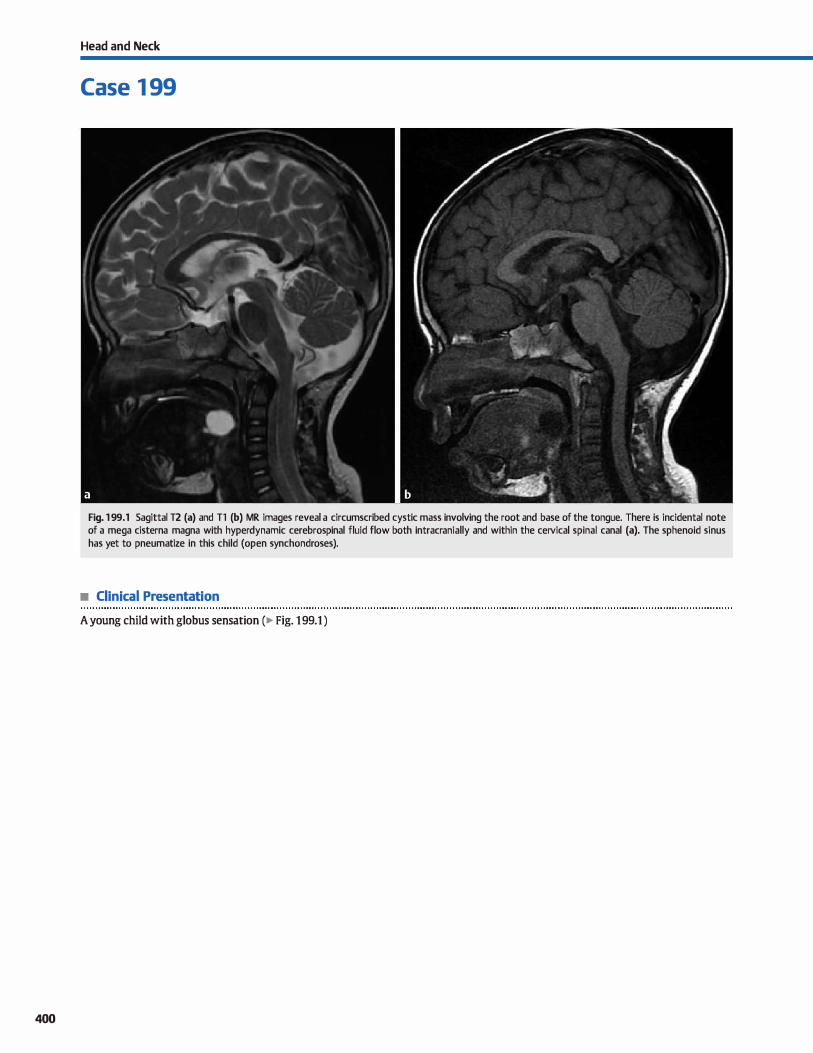
400
Head and Neck
Case 1 99
Fig. 1 99.1 Sagittal T2 (a) and Tl (b) MR images reveal a circumscribed cystic mass involving the root and base of the tongue. There is incidental note of a mega cisterna magna with hyperdynamic cerebrospinal fluid flow both intracranially and within the cervical spinal canal (a). The sphenoid sinus has yet to pneumatize in this child (open synchondroses).
• Clinical Presentation
A young child with globus sensation (� Fig. 1 99.1 )
• Key lmaging Finding
Base oftongue mass in a child
• Top 3 Differential Diagnoses
• Thyroglossal duct cyst Thyroglossal duct cysts represent the most common congenital neck lesions. The vast majority
(-85%) are midline. Approx.imately 25% are suprahyoid in
location with the remaining 75% being infrahyoid. Suprahyoid
lesions occur anywhere from the foramen cecum at the
tangue base to the hyoid bone; infrahyoid lesions may be
midline or paramedian and are characteristically embedded
within the strap muscles. Thyroglossal duct cysts are most
often well-circumscribed and may have lobulations or septa
tions. They typieally follow fluid attenuation (computed tomography, CT) or signal intensity (magnetic resonanœ
imaging, MRI). Increased protein content may result in
increased attenuation and Tt signal intensity. Superimposed
infection or hemorrhage results in a heterogeneous imaging
appearanœ, which may indude peripheral enhanœment.
• lingual thyroid. Lingual thyroid refers to ectopie thyroid
tissue located at the foramen cecum, involving the base and
root of the tangue; 90% of ectopie thyroid cases occur in this
location. It is important to evaluate the thyroid bed in the lower neck for the presence of additional thyroid tissue; in
approximately 75% of cases, the lingual thyroid represents the
• Additional Differential Diagnoses
• Hemangioma. Hemangiomas are benign vascular tumors that may involve the oral cavity or oropharynx. Capillary heman
giomas are common in infancy but naturally regress after a
proliferative phase. CT and MRI demonstrate a well-circum
scribed, lobulated, hyperdense and T2 hyperintense mass
with avid enhancement. • Tonsillar hypertrophy. The lingual tonsils are a component of
the Waldeyer ring and are located within the oropharynx
along the base of the tangue. Tonsillar hypertrophy refers to enlargement of the tonsils that may occur in isolation or secondary to a local infectious or inflammatory proœss. It is
most often symmetrie; if asymmetric, it may mimic a mass.
Contrast-enhanced CT demonstrates homogeneous hyperat-
• Diagnosis
Thyroglossal duct cyst
� Pearls
• Thyroglossal duct cysts are common congenital lesions that present as circurnscribed, midline cystic masses.
• Lingual thyroid is the most common form of ectopie thyroid; it is typically the only functioning thyroid tissue.
Suggested Reading
Fang ws, Wiggins RH, ID, lllner A et al. Primary lesions of the root of the tongue.
Radiographies 2011; 31: 1907-1922
Case 199
only functiorung thyroid tissue. Most patients are hypo
thyroid with the remainder of patients being euthyroid. On cross-sectional imaging, ectopie thyroid tissue follows thyroid
attenuation and signal intensity. It is hyperdense on CT with
moderate enhancement On MRI, it is hyperintense on Tt and
T2 sequences and demonstrates avid enhanœment. Nudear medicine thyroid imaging is also a useful adjunct in identify
ing functiorung thyroid tissue. Rarely, thyroid cancer may occur in approximately 1 to 3% of cases; conœming imaging
characteristies include soft-tissue nodular components or
calcification. • Epidennoid/dermoid cyst. Epidermoid and dermoid cysts
are developmental abnormalities that may involve the oral
cavity, oropharynx, or floor of the mouth. Epidermoids contain epithelial elements, whereas dermoids contain epithelial
elements along with dermal elements. Bath appear cystic on
imaging with low attenuation on CT and T2 hyperintensity on MRI. There may be thin peripheral enhancement, if
any. Dermoids may contain macroscopie fat, which is a key
discriminator when present. Epidermoids demonstrate
restricted diffusion.
tenuation of tonsillar tissue. On MRI, tonsillar tissue is T2
hyperintense with enhancement similar ta mucosal tissue.
• Venolymphatic malformation. Venolymphatic malformations represent a spectrum of venous and lymphatic develop
mental abnormalities. Pure venous malformations avidly
enhance and contain flow voids on T2 MRI; the presenœ of
phleboliths is pathognomonic. Pure lymphatic malformations
may be unilocular or multilocular and demonstrate fluid attenuation and signal intensity. Fluid-fluid levels, most often due to hemorrhage, are characteristic. Venolymphatic malfor
mations often cross fascia! planes to involve multiple spaœs of the neck.
• Dermoid cysts contain epithelial and dermal elements; the
presenœ of macroscopic fat is a key discriminator.
• Epidermoid cysts contain dermal elements; the presenœ of restricted diffusion is a key discriminator.
401
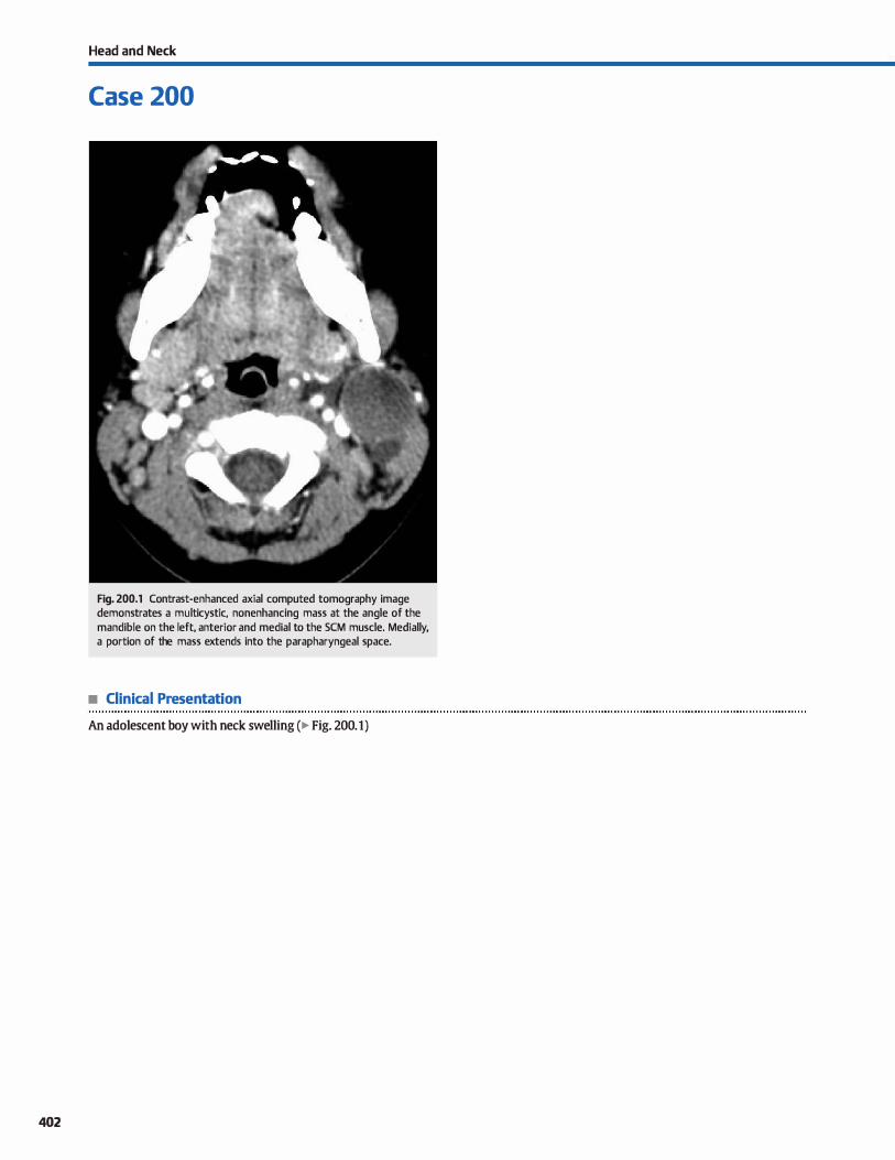
402
Head and Neck
Case 200
Fig. 200.1 Contrast-enhanced axial computed tomography image demonstrates a multicystic, nonenhancing mass at the angle of the mandible on the left, anterior and medial to the SCM muscle. Medially, a portion of the mass extends into the parapharyngeal space.
• Clinical Presentation
An adolescent boy with neck swelling (� Fig. 200.1)
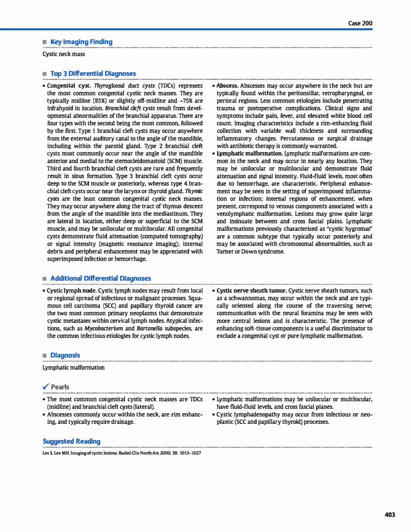
• Key lmaging Finding
Cystic neck mass
• Top 3 Differential Diagnoses
• Congenital cyst. Thyroglossal duct eysts (TDCs) represent the most common congenital cystic neck masses. They are typically midline (85%) or slightly off-midline and -75% are infrahyoid in location. Branchial cleft cysts result from developmental abnormalities of the branchial apparatus. There are four types with the second being the most common, followed by the first. Type 1 branchial cleft cysts may occur anywhere
from the externat auditory canal to the angle of the mandible, including within the parotid gland. Type 2 branchial cleft cysts most commonly occur near the angle of the mandible
anterior and medial to the sternodeidomastoid (SCM) muscle. Third and fourth branchial deft cysts are rare and frequently result in sinus formation. Type 3 branchial deft cysts occur deep to the SCM muscle or posteriorly, whereas type 4 bran
chial deft cysts occur near the larynx or thyroid gland. Thymie cysts are the least common congenital cystic neck masses. They may occur anywhere along the tract of thymus descent from the angle of the mandible into the mediastinum. They are lateral in location, either deep or superficial to the SCM muscle, and may be unilocular or multilocular. Ali congenital cysts demonstrate fluid attenuation (computed tomography)
or signal intensity (magnetic resonance imaging); internai debris and peripheral enhancement may be appreciated with superimposed infection or hemorrhage.
• Additional Differential Diagnoses
• Cystic Iymph node. Cystic lymph nodes may result from local or regional spread of infectious or malignant processes. Squamous cell carcinoma {SCC) and papillary thyroid cancer are the two most common primary neoplasms that demonstrate cystic metastases within cervical lymph nodes. Atypical infec
tions, such as Mycobacterium and Bartonella subspecies, are the common infectious etiologies for cystic lymph nodes.
• Diagnosis
Lymphatic malformation
� Pearls
• The most common congenital cystic neck masses are TDCs
(midline) and branchial deft cysts (lateral).
• Abscesses commonly occur within the neck, are rim enhancing, and typically require drainage.
Suggested Reading
Lev S. Lev MH. lrnaging of cystic lesiom. Radiol Clin North Am 2000; 38: 1013-1027
Case 200
• Abscess. Abscesses may occur anywhere in the neck but are typically found within the peritonsillar, retropharyngeal, or perioral regions. Less common etiologies include penetrating trauma or postoperative complications. Oinical signs and symptoms include pain, fever, and elevated white blood cell count. lmaging characteristics include a rim-enhandng fluid collection with variable wall thickness and surrounding
inflammatory changes. Percutaneous or surgical drainage with antibiotic therapy is commonly warranted.
• l,ymphatic malfonnation. Lymphatic malformations are common in the neck and may occur in nearly any location. They may be unilocular or multilocular and demonstrate fluid
attenuation and signal intensity. Fluid-fluid levels, most often due to hemorrhage, are characteristic. Peripheral enhance
ment may be seen in the setting of superimposed inflammation or infection; internai regions of enhancement, when present, correspond to venous components associated with a venolymphatic malformation. Lesions may grow quite large and insinuate between and cross fascia! plains. Lymphatic malformations previously characterized as �cystic hygromas"
are a common subtype that typically occur posteriorly and may be associated with chromosomal abnormalities, such as
Turner or Down syndrome.
• Cystic nerve sheath tumor. Cystic nerve sheath tumors, such as a schwannomas, may occur within the neck and are typically oriented along the course of the traversing nerve; communication with the neural foramina may be seen with
more central lesions and is characteristic. The presence of enhancing soft-tissue components is a useful discriminator to exdude a congenital cyst or pure lymphatic malformation.
• Lymphatic malformations may be unilocular or multilocular, have fluid-fluid levels, and cross fascial planes.
• Cystic lymphadenopathy may occur from infectious or neoplastic (SCC and papillary thyroid) processes.
403
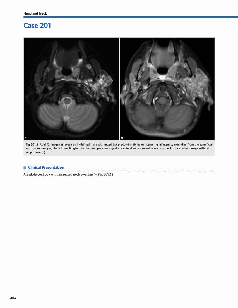
404
Head and Neck
Case 201
Fig. 201 .1 Axial T2 image (a) reveals an il l-defined mass with mixed but predominantly hyperintense signal intensity extending from the superficial soft tissues overlying the left parotid gland to the deep parapharyngeal space. Avid enhancement is seen on the Tl postcontrast image with fat suppression (b).
• Clinical Presentation
An adolescent boy with increased neck swelling (� Fig. 201 .1 )
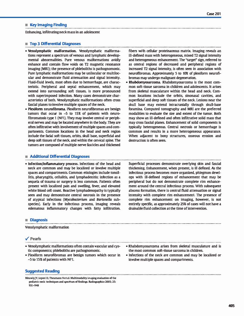
• Key lmaging Finding
Enhancing, infiltrating neck mass in an adolescent
• Top 3 Differential Diagnoses
• Venolymphatic malformation. Venolymphatic malforma
tions represent a spectrum of venous and lymphatic develop
mental abnormalities. Pure venous malformations avidly
enhance and contain flow voids on T2 magnetic resonanœ
imaging (MRI); the presence of phleboliths is pathognomonic.
Pure lymphatic malformations may be unilocular or multilocular and demonstrate fluid attenuation and signal intensity.
Fluid-fluid levels, most often due to hemorrhage, are charac
teristic. Peripheral and septal enhancement, which may
extend into surrounding soft tissues, is more pronounœd
with superimposed infection. Many cases demonstrate char
acteristics of both. Venolymphatic malformations often cross
fascia) planes to involve multiple spaces of the neck.
• Plexifonn neurofibrorna. Plexiform neurofibromas are benign tumors that occur in -5 to 15% of patients with neuro
fibromatosis type 1 (NFl). They may involve central or periph
eral nerves and may be Iocated anywhere in the body. They are often infiltrative with involvement of multiple spaœs and com
partments. Common locations in the head and neck region
indude the facial soft tissues, orbits, skull base, superficial and
deep soft tissues of the neck, and within the cervical spine. The
tumors are composed of multiple nerve fascides and thickened
• Additional Differential Diagnoses
• lnfection/inflammatory process. Infections of the head and neck are common and may be localized or involve multiple
spaces and compartments. Common etiologies include tonsil
litis, pharyngitis, cellulitis, and lymphadenitis; infection as a
sequela of trauma or surgery is Jess common. Patients often
present with localized pain and swelling, fever, and elevated
white blood cell count. Reactive lymphadenopathy is typically
seen and may demonstrate central necrosis in the presenœ
of atypical infections (Mycobacterium and BartoneUa sub
species ). Early in the infectious process, imaging reveals edematous inflammatory changes with fatty infiltration.
• Diagnosis
Venolymphatic malformation
� Pearls
• Venolymphatic malformations often contain vascular and cystic components; phleboliths are pathognomonic.
• Plexiform neurofibromas are benign tumors which occur in -5 to 15% of patients with NF1.
Suggested Reading
Meuwly JY, Lepori D, Theumann Net al. Multimodality imaging evaluation of the
pediatric neck: techniques and spectrum of findinp. Radiographies 2005; 25:
931-948
Case 201
fibers with cellular proteinaœous matrix. Imaging reveals an
ill-defined mass with heterogeneous, mixed T2 signal intensity
and heterogeneous enhancement The utarget" sign, referred to as central regions of decreased and peripheral regions of
increased T2 signal intensity, is often seen in association with
neurofibromas. Approximately 5 to 10% of plexiform neurofi
bromas may undergo malignant degeneration. • Rhabdomyosarcoma. Rhabdomyosarcoma is the most com
mon sort-tissue sarcoma in children and adolescents. It arises
from skeletal musculature within the head and neck. Com
mon locations include the orbits, sinonasal cavities, and
superfidal and deep soft tissues of the neck. I.esions near the
skull base may extend intracranially through skull-base foramina. Computed tomography and MRI are the preferred
modalities to evaluate the size and extent of the tumor. Both
may show an ill-defined and often infiltrative solid mass that
may cross fascia) planes. Enhancement of solid components is
typically heterogeneous. Central necrosis or hemorrhage is
common and results in a more heterogeneous appearance.
When adjacent to bony structures, osseous erosion and
destruction is often seen.
Superficial processes demonstrate overlying skin and fascia)
thickening. Enhanœment, when present, is il1 defined. As the
infectious process becomes more organized, phlegmon develops with ill-defined regions of enhanœment that may be
peripheral but do not demonstrate complete rim enhance
ment around the central infectious process. With subsequent
abscess formation, there is central fluid attenuation or signal
intensity with complete rim enhancement The presence of
complete rim enhancement on imaging, however, is not
entirely specific, as approximately 25% of cases will not have a
drainable fluid collection at the time of intervention.
• Rhabdomyosarcoma arises from skeletal musculature and is the most common soft-tissue sarcoma in children.
• Infections of the neck are common and may be localized or
involve multiple spaœs and compartments.
405
406
Head and Neck
Case 202
Fig. 202.1 Axial T2 image with fat suppression (a) demonstrates a circumscribed mass with heterogeneous increased signal intensity a long the lateral aspect of the right thyroid lobe. The medial portion of the mass is hyperintense on unenhanced Tl sequences (b), whereas the lateral aspect is hypointense.
• Clinical Presentation
A middle-aged woman with chronic neck pain (� Fig. 202.1)
• Key lmaging Finding
Thyroid lesion
• Top 3 Differential Diagnoses
• Thyroid adenoma. Benign thyroid adenomas are exceedingly common, representing roughly 75% of all thyroid nodules. They are often multiple and bilateral. The dassic appearance on ultrasound (US) consists of a peripheral hypoechoic halo surrounding a àrcumscribed nodule. The nodule itself may have internai coarse calàfications or cystic changes. Lesions are hypodense on computed tomography (CT); on magnetic resonance imaging (MRI), they are typically Tl hypointense and 12 hyperintense. Thyroid adenomas may be hot or cold on technetium 99 m or iodine 1 123 scans: malignant lesions are typically cold.
• Dilferentiated thyroid cardnoma. Ditferentiated thyroid carcinoma consists of papillary and follicular. Papillary carcinoma is the most common thyroid cancer, representing -80% of cases. Its dassic appearanœ on US consists of a thyroid lesion with numerous microcalàfications and/or cystic changes. Regional lymph node spread is often seen and commonly demonstrates cystic/necrotic lymph node enlargement with or without
• Additional Differential Diagnoses
• Undifferentiated thyroid carcinoma. Unditferentiated thyroid carcinomas are aggressive tumors and include medullary and anaplastie carcinomas. Medullary carcinoma is a neuroendocrine tumor that most commonly occurs in middle-aged patients. lt may occur sporadically or in association with multiple endocrine neoplasia type 2. The primary lesion on US is hypervascular and may demonstrate microcalcifications. The majority of patients have metastatic lymphadenopatby at the time of diagnosis. Positron emission tomography and octreoscan imaging are useful for characterization of the tumor and post-treatment surveillance. Anaplastie thyroid carcinoma is an extremely aggressive tumor that is most common in elderly women. In many cases, patients have underlying multinodular goiter. lts imaging appearance is often aggressive, demonstrating a large, ill-detined mass with or without calcifications or central necrosis. Heterogeneous enhancement is noted on CT and MRI. The mass typically metastasizes to local
• Diagnosis
Colloid cyst
y' Pearls
• Benign thyroid adenomas are exceedingly common; they are often multiple and bilateral.
• Differentiated thyroid carànoma indudes papillary {most common, cystic, microcalàfications) and follicular.
Suggested Reading
HoangjK, Lee WK, Lee M. Johnson D, Farrell S. US Features ofthyroid malignancy: pearls and pitfalls. Radiographies 2007; 27: 847-860, discussion 861-865
Case 202
calàfications. Follicular carcinoma is Jess common but carries a worse prognosis due to the propensity for distant metastases. Its imaging appearanœ is Jess characteristic and often nonspedfic. In the absence of cystic change, microcalàfications, extracapsular spread, or regional lymph node spread, differentiated carcinoma may be indistinguishable from tbyroid adenomas on imaging. 1Teatment indudes thyroidectomy with I 131
ablation. • Colloid cyst Colloid cysts are benign thyroid lesions that are
thought to result from cystic degeneration of adenomas. The presenœ of gelatinous colloid results in a characteristic US appearanœ, consisting of a cystic thyroid mass with internai hyperechoic fod and "cornet tait" artifact. lts appearanœ on CT and MRI varies based upon its protein concentration. Most lesions are hypodense on CT and hyperintense on 12
MRI sequenœs. As the protein concentration increases, its density on CT and signal intensity on Tl MRI sequences also increases.
lymph nodes and invades adjacent structures. Prognosis is dismal.
• Parathyroid adenoma. Paratbyroid adenomas represent benign functioning tumors and are the most common cause of primary hyperparathyroidism. Although they may occur
anywhere along the tract of developmental parathyroid desœnt and occasionally within the mediastinum, they are most commonly located along the posteroinferior margin of the thyroid gland. Preoperative localization is performed to identify the lesions and rule out the presenœ of multiple adenomas. On US, lesions are typically drcumscribed, hypoechoic, and vascular. MRI reveals characteristic 12 hyperintensity. Heterogeneous enhanœment is seen on both CT and MRI. Nuclear medicine sestamibi scans are useful in identifying parathyroid adenomas as foci of increased activity on delayed imaging. 1Teatment includes surgical resection.
• Undifferentiated thyroid carcinomas are aggressive tumors and indude medullary and anaplastie.
• Colloid cysts have a characteristic US appearanœ with internai hyperechoic fod and Mcomet tait" artifact.
407
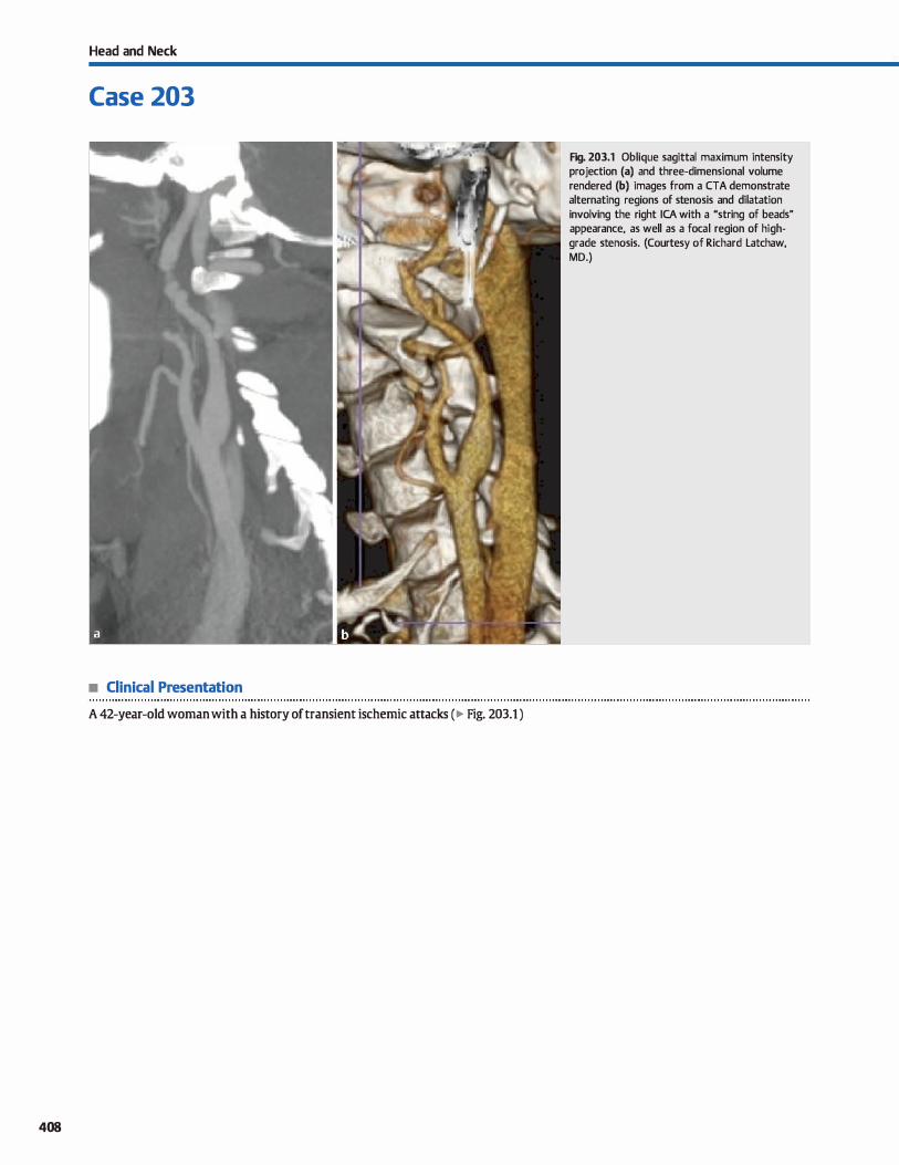
Head and Neck
Case 203
• Clinical Presentation
A 42-year-old woman with a history of transient ischemic attacks (� Fig. 203.1 )
408
Fig. 203.1 Oblique sagittal maximum intensity projection (a) and three-dimensional volume rendered (b) images from a CTA demonstrate alternating regions of stenosis and dilatation involving the right ICA with a "string of beads" appearance, as well as a focal region of highgrade stenosis. (Courtesy of Richard Latchaw, MD.)
• Key lmaging Finding
Carotid artery stenosis
• Top 3 Differential Diagnoses
• Atherosclerosis. Atherosderosis is the most common cause of carotid artery stenosis. lt typically occurs in aider individuals and is more advanced in patients with a smoking history, hypertension, hyperlipidemia, and diabetes. The carotid bifurcation and proximal internai carotid artery (ICA) are the most common sites involved, followed by the parasellar ICAs. Doppler ultrasound is the initial screening study for the cervical carotid arteries. fypical findings
indude visualization of plaque with vesse! stenosis, elevated velocities, and turbulent flow with aliasing. Magnetic resonance angiography (MRA) and computed tomography angiography (CTA) are used as confirmatory studies. carotid endarterectomy ( ŒA) remains the treatment of choice in low-risk surgical candidates. Current guidelines recommend
ŒA for symptomatic lesions with > 50% stenosis (data from the NASCET trial) or asymptomatic lesions with > 60% stenosis (data from the ACAS trial). Although many recent trials have shown a benefit of endovascular carotid stent placement with distal embolie protection devices in many patient populations, current guidelines recommend this treatment only for high-risk surgical patients with symptomatic lesions
of� 70% stenosis. High-risk surgical patients are defined
as follows: dinically significant cardiac disease, severe pulmonary disease (if general anesthesia will be used), recurrent stenosis after prior CEA, prior neck surgery or irradiation, contralateral recurrent laryngeal nerve paralysis,
• Additional Differential Diagnoses
• Iatrogenic. The most common iatrogenic causes of carotid stenosis include clamp injury during surgical procedures or
vasculitis secondary to radiation therapy for head and neck
• Diagnosis
Fibromuscular dysplasia
y' Pearts
• Atherosderosis is the most common cause of carotid artery stenosis; it typically occurs in older individuals.
• carotid stents are indicated for symptomatic lesions of� 70% stenosis in high-risk surgical patients.
Suggested Readings
Exerutive cmnmittee for the asymptmnatic carotid atherosclerosis study (ACAS).
Endarterectomy for asymptomatic carotid artery stenosis.JAMA 1995; 273:
1421-1428
Gwm HS, Yadav JS, Fayad P et al. SAPPHIRE InvestigatDrs. Long-term results of
carotid stenting versus endarterectomy in high-risk patients. N EnglJ Med 2008;
358: 1572-1579
Case 203
contralateral carotid artery occlusion, and surgically inaccessible lesions.
• Fibromuscular dysplasia {FMD). FMD is a dysplastic vascular disease that primarily affects young women. The renal arteries are most commonly involved, followed by the carotid arteries. The media! fibroplasia variant is the most common subtype and presents with the dassic Mstring of beads" appearance on
imaging (CTA, MRA, digital subtraction angiography [OSA]) due to altemating regions of stenosis and dilatation The disease results in vascular stenosis and subsequent hypertension (renal artery involvement) or stroke (ICA involvement). Patients are at increased risk for aneurysm formation and dissections. Symptomatic lesions are treated with angioplasty.
• Dissection. carotid artery dissection may result from trauma, hypertension, iatrogenia, or vasculopathies such as FMD.
Common imaging findings include an intramural hematoma and intimai flap. On unenhanced CT, acute dissections present with crescent-shaped increased density within the arterial wall. Unenhanced TI sequences with fat suppression show a peripheral hyperintense intramural hematoma. DSA findings indude luminal irregularity, tapered stenosis or occlusion, a
double lumen, or an intimai flap. Sorne dissections may be managed medically with anticoagulation therapy and followup imaging to ensure stability or resolution. Symptomatic lesions or lesions that progress despite medical management often require intErvention, induding stent placement
cancer. Treatment depends upon symptoms and morphology of the lesions with endovascular therapy ( angioplasty or stent
placement) favored over surgical intervention.
• FMD primarily affects young women; angioplasty is the primary treatment option for symptomatic lesions.
• Most dissections are due to hypertension or trauma; treatment depends upon morphology and symptoms.
North American Symptamatic Carotid Endarterectmny 1ttal Collaboraturs. Beneficial
elfect of carotid endarterectomy in symptnmatic patients with high-grade
carotid stenosis. N Englj Med 1991; 325; 445-453
RIJ!li M, Yadav JS. Carotid stenting. Circulation 2006; 114: e1-e4
409
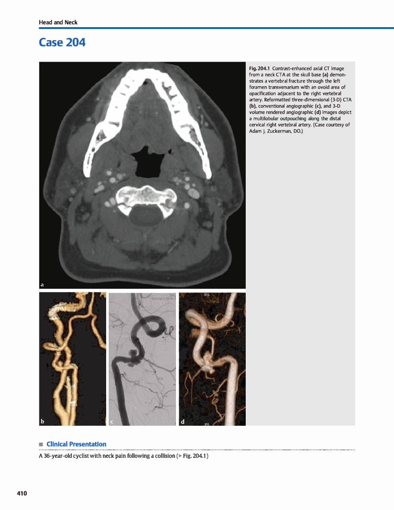
Head and Neck
Case 204
• Clinical Presentation
A 36-year-old cyclist with neck pain following a collision (� Fig. 204.1 )
410
Fig. 204.1 Contrast-enhanced axial CT image from a neck CTA at the skull base (a) demonstrates a vertebral fracture through the left foramen transversarium with an ovoid area of opacification adjacent to the right vertebral artery. Reformatted three-dimensional (3-D) CTA (b), conventional angiographie (c), and 3-D volume rendered angiographie (d) images depict a multilobular outpouching along the distal cervical right vertebral artery. (Case courtesy of Adam J. Zuckerman, DO.)

• Key lmaging Finding
Vascular neck injury
• Top 3 Differential Diagnoses
• Dissection. Arterial dissections are the result of intimai injury. They most commonly involve the cervical internai carotid and vertebral arteries; vertebral injury is most common in the V3 segment near the skull base where the vesse! is most mobile. Early dissection can often present with complete occlusion of the vesse! lumen preceded by a segment of tapered narrowing. Oassic angiographie findings include the
presenœ of an intimai flap or irregular segmental luminal narrowing. Computed tomography (cr) angiography reveals ecœntric luminal narrowing with relative enlargement of the cross-sectional area of the affected artery compared with contralateral side secondary to thickening from mural hematoma. Axial fat-suppressed T1 magnetic resonanœ imaging (MRI) demonstrates an ecœntric, cresœntic periarterial rim of sig
nal abnormality along the margin of a flow void. Dissections can be complicated by pseudoaneurysm, thromboembolic, or occlusive phenomena. First-line treatment involves anticoagulation with refractory cases necessitating endovascular or surgical intervention.
• Pseudoaneurysm. Pseudoaneurysms are typically due to penetrating or blunt trauma and are frequently associated with
• Additional Differential Diagnoses
• Arteriovenous fistula (AVF). AVFs are usually the result of a penetrating neck injury with partial transection of an artery and adjacent vein. AVFs may also occur spontaneously or be the result of blunt trauma with cervical spine fracture, infection, or iatrogenia. Patients often present with a neck bruit or ischemic symptoms; however, up to 30% of cases may be
• Diagnosis
Pseudoaneurysm of the right vertebral artery
� Pearls
• Dissections result from intimai injury; common findings include an intramural hematoma and intimai flap.
• Pseudoaneurysms commonly result from traumatic injuries; they are at risk of expansion and rupture.
Suggested Reading
Nt'lnez DB, Jr, Torres-Le6n M, Mûnera F. Vascular Ïl1iUries of the ned< and thoradc
inlet; helical cr-angiographie mrrelation. Radiographies 2004; 24; 1087-1098,
discussion 1099-1100
Case 204
arterial dissection. They can also be due to infectious or iatrogenic causes. Pseudoaneurysms are commonly ovoid in appearanœ and located at the distal aspect of a stenotic segment. Doppler ultrasound of a pseudoaneurysm may dem
onstrate the dassic "to and fro" appearance. Management is dependent upon size, location. and etiology. Patients are at risk for expansion of the lesion with compression of adjacent structures and possible rupture. They are also prone to thrombus formation with distant emboli. Treatment options include surgical resection/reconstruction or endovascular stent placement with or without coil embolization of the pseudoaneurysm.
• Vascular occlusion. Complete or partial vascular occlusion commonly occurs in association with blunt or penetrating
neck injuries. Vascular imaging with cr or conventional angiography demonstrates decreased or absent opacification of the involved vascular structures with or without other associated vascular injuries, including an underlying dissection or intramural hematoma.
asymptomatic. AVFs can be diagnosed angiographically with early venous filling during the arterial phase. cr and MRI provide additional detail on the relationship of an AVF to surrounding structures. Endovascular treatment aims to close the fistula while preserving flow in the parent artery.
• Complete or partial vascular occlusion commonly occurs in association with blunt or penetrating neck injuries.
• AVFs are direct communications between an artery and vein, typically due to penetrating injuries.
411
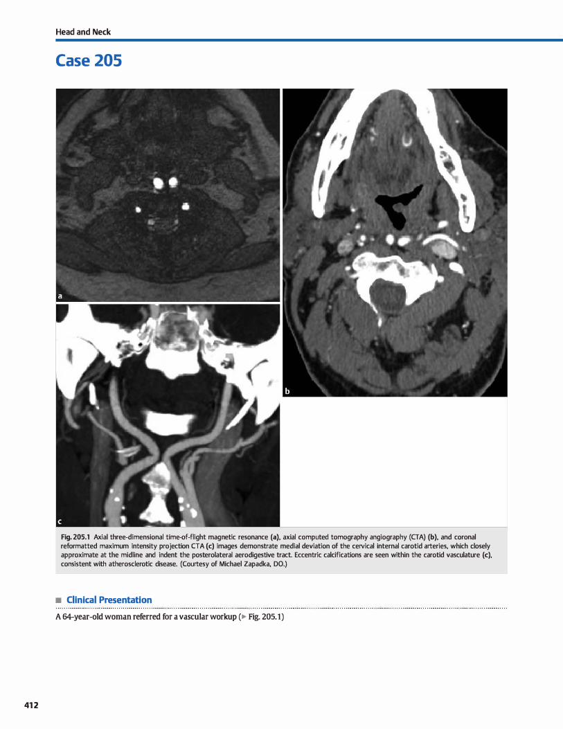
412
Head and Neck
Case 205
Fig. 205.1 Axial three-dimensional time-of-flight magnetic resonance (a), axial computed tomography angiography (CTA) (b), and coronal reformatted maximum intensity projection CTA (c) images demonstrate medial deviation of the cervical internai carotid arteries, which dosely approximate at the midline and indent the posterolateral aerodigestive tract. Eccentric calcifications are seen within the carotid vasculature (c), consistent with atherosderotic disease. (Courtesy of Michael Zapadka, DO.)
• Clinical Presentation
A 64-year-old woman referred for a vascular workup (� Fig. 205.1)
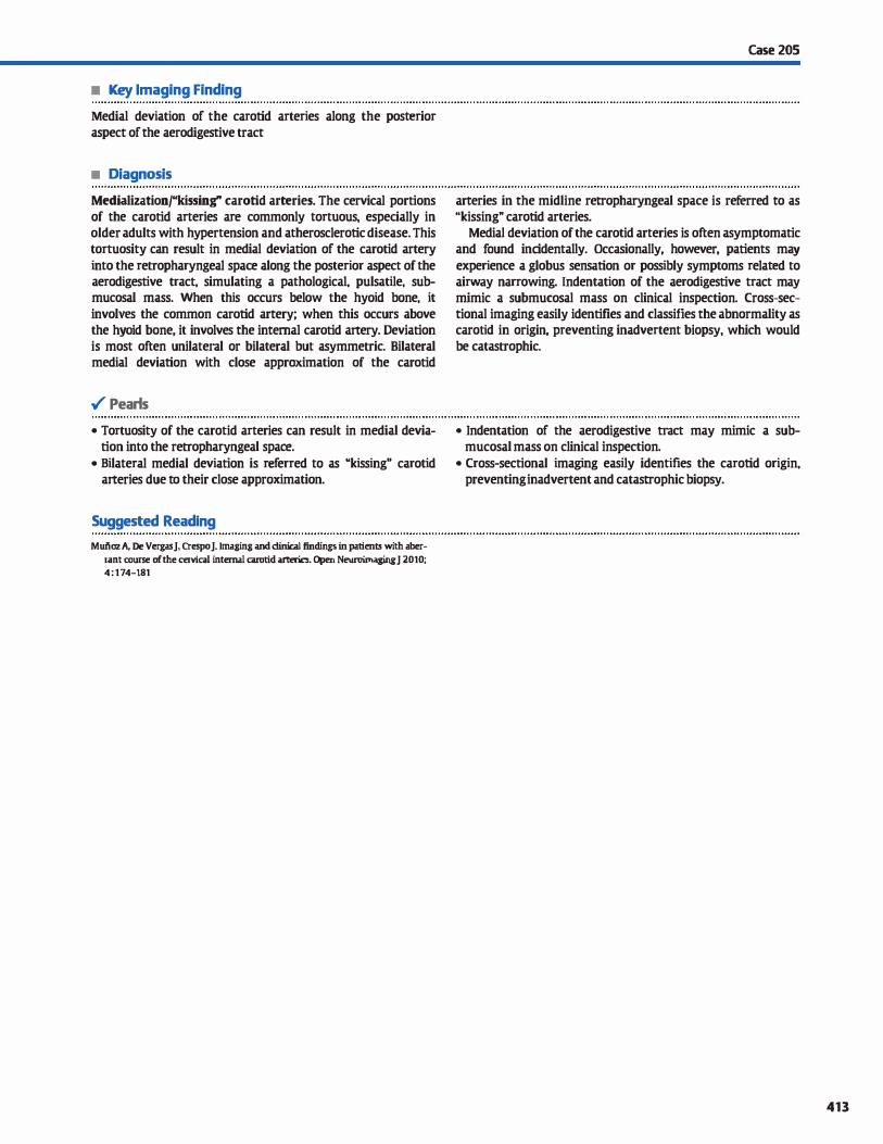
• Key lmaging Finding
Media! deviation of the carotid arteries along the posterior
aspect of the aerodigestive tract
• Diagnosis
Medialization/"kissing" carotid arteries. The cervical portions
of the carotid arteries are commonly tortuous, especially in
older adults with hypertension and atherosclerotic disease. This
tortuosity can result in medial deviation of the carotid artery
into the retropharyngeal space along the posterior aspect of the aerodigestive tract, simulating a pathological, pulsatile, submucosal mass. When this occurs below the hyoid bone, it
involves the common carotid artery; when this occurs above
the hyoid bone, it involves the internai carotid artery. Deviation
is most often unilateral or bilateral but asymmetric. Bilateral
medial deviation with close approximation of the carotid
� Pearts
• Tortuosity of the carotid arteries can result in medial devia
tion into the retropharyngeal spaœ.
• Bilateral medial deviation is referred to as "kissing" carotid arteries due to their close approximation.
Suggested Reading
Muilaz A, De Vergas J, crespo J, lmaging and dinical findings in patients with aber
rant course of the cemcal internai carotid arteries. Open NeuroimagingJ 2010;
4:174-181
Case 205
arteries in the midline retropharyngeal space is referred to as
"kissing" carotid arteries.
Media! deviation of the carotid arteries is often asymptomatic
and found inddentally. Occasionally, however, patients may
experience a globus sensation or possibly symptoms related to
airway narrowing. Indentation of the aerodigestive tract may
mimic a submucosal mass on clinical inspection. Cross-sec
tional imaging easily identifies and classifies the abnormality as carotid in origin, preventing inadvertent biopsy, which would
be catastrophic.
• Indentation of the aerodigestive tract may mimic a sub
mucosal mass on clinical inspection.
• Cross-sectional imaging easily identifies the carotid origin,
preventing inadvertent and catastrophic biopsy.
413
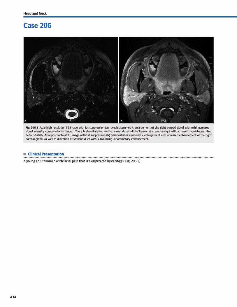
414
Head and Neck
Case 206
Fig. 206.1 Axial high-resolution T2 image with fat suppression (a) reveals asymmetric enlargement of the right parotid gland with mild increased signal intensity compared with the left. There is also dilatation and increased signal within Stenson duct on the right with an ovoid hypointense filling defect distally. Axial postcontrast Tl image with fat suppression (b) demonstrates asymmetric enlargement and increased enhancement of the right parotid gland, as well as dilatation of Stenson duct with surrounding inflammatory enhancement.
• Clinical Presentation
A young adult woman with facial pain that is exasperated by eating ( � Fig. 206.1 )
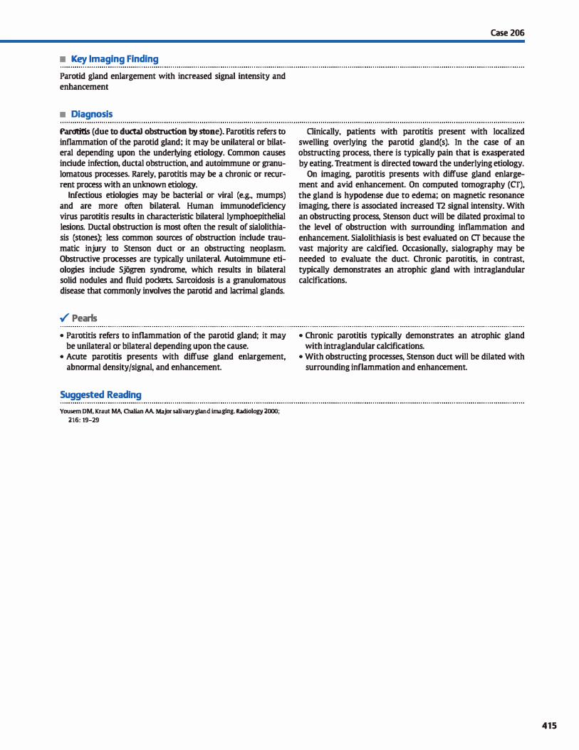
• Key lmaging Finding
Parotid gland enlargement with increased signal intensity and
enhanœment
• Diagnosis
Parotitis (due to ductal obstruction by stone). Parotitis refers to inflammation of the parotid gland; it may be unilateral or bilateral depending upon the underlying etiology. Common causes indude infection, ductal obstruction, and autoimmune or granu
lomatous processes.. Rarely, parotitis may be a chronic or recur
rent process with an unknown etiology. Infectious etiologies may be bacterial or viral (e.g., mumps)
and are more often bilateral Human immunodefidency virus parotitis results in characteristic bilateral lymphoepithelial lesions. Durtal obstruction is most often the result of sialolithia
sis (stones); Jess common sources of obstruction indude trau
matic injury to Stenson duct or an obstructing neoplasm. Obstructive processes are typically unilateral Autoimmune eti
ologies indude Sjôgren syndrome, which results in bilateral solid nodules and tluid pockets. Sarcoidosis is a granulomatous disease that commonly involves the parotid and lacrimal glands.
� Pearts
• Parotitis refers to inflammation of the parotid gland; it may be unilateral or bilateral depending upon the cause.
• Acute parotitis presents with diffuse gland enlargement, abnormal density/signal, and enhancement
Suggested Reading
Yousem DM, Kraut MA, Chalian AA Major salivary gland imaging. Jœliology 2000;
216: 19-29
Case 206
Oinically, patients with parotitis present with localized swelling overlying the parotid gland(s). In the case of an obstructing process, there is typically pain that is exasperated
by eating. Treatment is directed toward the underlying etiology. On imaging, parotitis presents with diffuse gland enlarge
ment and avid enhancement On computed tomography (CT),
the gland is hypodense due to edema; on magnetic resonanœ imaging, there is assodated increased T2 signal intensity. With
an obstructing process, Stenson duct will be dilated proximal ta the level of obstruction with surrounding inflammation and
enhancement. Sialolithiasis is best evaluated on cr because the vast majority are caldfied. Occasionally, sialography may be needed to evaluate the duct Chronic parotitis, in contrast.
typically demonstrates an atrophie gland with intraglandular calcifications.
• Chronic parotitis typically demonstrates an atrophie gland with intraglandular calcifications.
• With obstructing processes, Stenson duct will be dilated with surrounding inflammation and enhanœment
415
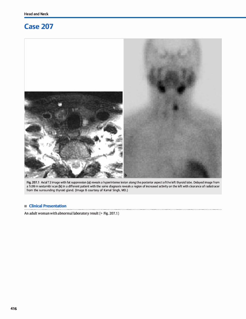
416
Head and Neck
Case 207
Fig. 207 .1 Axial T2 image with fat suppression (a) reveals a hyperintense lesion a long the posterior aspect of the left thyroid lobe. Delayed image from a Tc99 m sestamibi scan (b) in a different patient with the same diagnosis reveals a region of increased activity on the left with clearance of radiotracer from the surrounding thyroid gland. (Image B courtesy of Kamal Singh, MD.)
• Clinical Presentation
An adult woman with abnormal laboratory result (� Fig. 207.1 )
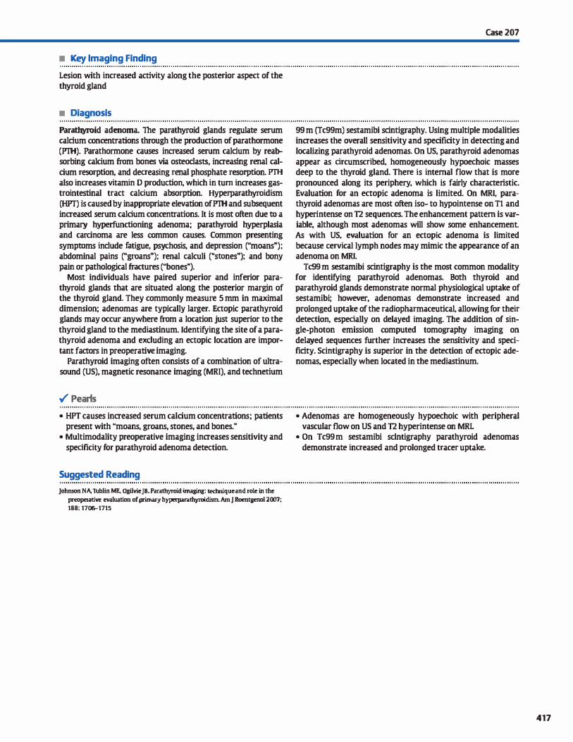
• Key lmaging Finding
Lesion with increased activity along the posterior aspect of the thyroid gland
• Diagnosis
Parathyroid adenoma. The parathyroid glands regulate serum calcium concentrations through the production of parathormone (PTH). Parathormone causes increased serum calcium by reabsorbing calcium from bones via osteodasts, increasing renal cal
cium resorption, and decreasing renal phosphate resorption. PTH also increases vitamin D production, which in tum increases gastrointestinal tract calcium absorption. Hyperparathyroidism
(HPT) is caused by inappropriate elevation of PTH and subsequent increased serum calcium concentrations. It is most often due to a primary hyperfunctioning adenoma; parathyroid hyperplasia and cardnoma are Jess common causes. Common presenting
symptoms indude fatigue, psychosis, and depression ("moans"); abdominal pains ("groans"); renal calculi ("stones"); and bony
pain or pathological fractures ("bones"). Most individuals have paired superior and inferior para
thyroid glands that are situated along the posterior margin of the thyroid gland. They commonly measure 5 mm in maximal dimension; adenomas are typically larger. Ectopie parathyroid glands may occur anywhere from a location just superior to the
thyroid gland to the mediastinum. Identifying the site of a parathyroid adenoma and exduding an ectopie location are impor
tant factors in preoperative imaging. Parathyroid imaging often consists of a combination of ultra
sound (US), magnetic resonance imaging (MRI), and technetium
� Pearls
• HPT causes increased serum calcium concentrations; patients present with "moans, groans, stones, and bones."
• Multimodality preoperative imaging increases sensitivity and
spedficity for parathyroid adenoma detection.
Suggested Reading
Johnson NA, Tublin ME, Ogilvie JB. Parathyroid imaging; teclmique and role in the
preoperative evaluation of primilry hyperparathyroidism. Am J Rœntgenol 2007;
188; 17Œ-1715
Case 207
99 m (Tc99m) sestamibi scintigraphy. Using multiple modalities increases the overall sensitivity and speciticity in detecting and localizing parathyroid adenomas. On US, parathyroid adenomas
appear as circumscribed, homogeneously hypoechoic masses deep to the thyroid gland. There is internai flow that is more
pronounced along its periphery, which is fairly characteristic. Evaluation for an ectopie adenoma is limited. On MRI, parathyroid adenomas are most often iso- to hypointense on T1 and hyperintense on T2 sequenœs. The enhanœment pattern is variable, although most adenomas will show some enhancement
As with US, evaluation for an ectopie adenoma is limited because cervical lymph nodes may mimic the appearance of an adenoma on MRI.
Tc99 m sestamibi scintigraphy is the most common modality for identifying parathyroid adenomas. Bath thyroid and parathyroid glands demonstrate normal physiological uptake of sestamibi; however, adenomas demonstrate increased and
prolonged uptake of the radiopharmaceutical, allowing for their detection, especially on delayed imaging. The addition of single-photon emission computed tomography imaging on
delayed sequences further increases the sensitivity and specificity. Scintigraphy is superior in the detection of ectopie adenomas, especially when located in the mediastinum.
• Adenomas are homogeneously hypoechoic with peripheral vascular flow on US and T2 hyperintense on MRL
• On Tc99 m sestamibi sdntigraphy parathyroid adenomas
demonstrate increased and prolonged tracer uptake.
417
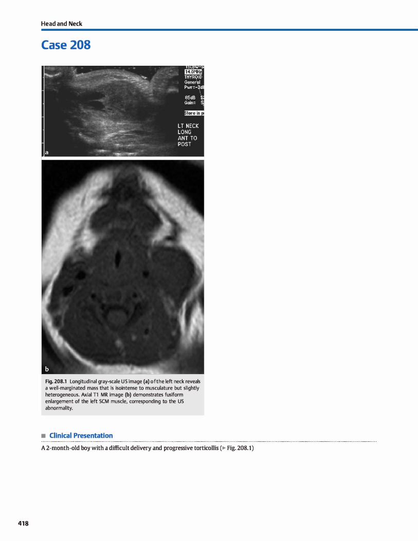
418
Head and Neck
Case 208
Fig. 208.1 Longitudinal gray-scale US image (a) of the left neck reveals a well-marginated mass that is isointense to musculature but slightly heterogeneous. Axial Tl MR image (b) demonstrates fusiform enlargement of the left SCM muscle, corresponding to the US abnormality.
• Clinical Presentation
A 2-month-old boy with a difficult delivery and progressive torticollis (� Fig. 208.1)
• Key lmaging Finding
Enlargement of the stemocleidomastoid (SCM) muscle in a child with torticollis
• Diagnosis
Fibromatosis colli. Fibromatosis colli is a rare disorder characterized by isolated fibromatosis involving the SCM muscle. It is typically undetectable at birth but progresses over the first few weeks or months of life. Although the exact etiology remains
unclear, a history of birth trauma or a difficult delivery is found in> 90% of cases. Most patients are asymptomatic and may present with a Iocalized neck mass; -20% of patients present with torticollis involving the atfected side.
Ultrasound (US) is the initial modality of choiœ and is often diagnostic. Most cases reveal a homogeneously enlarged SCM
� Pearls
• Fibromatosis colli is a rare disorder characterized by isolated
fibromatosis involving the SCM muscle. • Most cases are asymptomatic with or without a localized neck
mass; approximately 20% of patients present with torticollis.
Suggested Reading
Rabbin MR, Murphey MD, Temple HT, KransdorfMJ, Cl10ijJ. Imaging of musculo
skeletal fibromatosis. Radiographies 2001; 21: 585-600
Case 208
muscle. Less common findings include a focal, well-defined or ill-defined hypoechoic mass within the SCM muscle. The typical computed tomography appearanœ consists of homogeneous enlargement of the SCM muscle without a focal mass or
attenuation abnonnality. Magnetic resonanœ imaging {MRI) demonstrates similar findings with iso- to hypointense Tl and iso- to slightly hyperintense T2 signal intensity within the SCM.
• US is the modality of choice and is often diagnostic; most
cases show a homogeneously enlarged SCM muscle. • MRI demonstrates similar SCM enlargement with iso- to
slightly hyperintense T2 signal intensity.
419
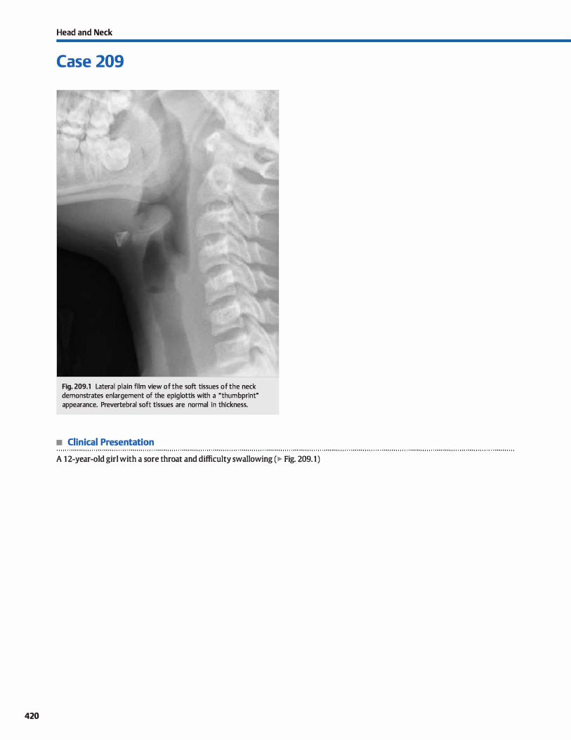
420
Head and Neck
Case 209
Fig. 209.1 Lateral plain film view of the soft tissues of the neck demonstrates enlargement of the epiglottis with a "thumbprint" appearance. Prevertebral soft tissues are normal in thickness.
• Clinical Presentation
A 12-year-old girl with a sore throat and difficulty swallowing (� Fig. 209.1)
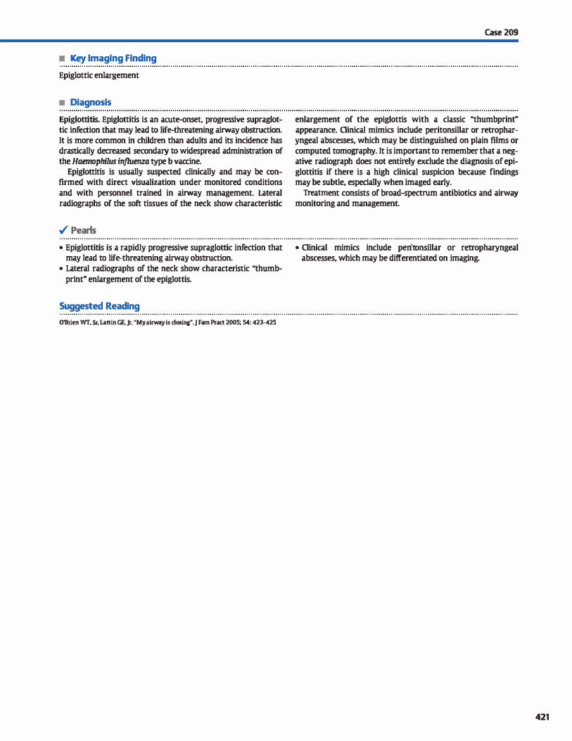
• Key lmaging Finding
Epiglottic enlargement
• Diagnosis
Epiglottitis. Epiglottitis is an aœte-onset, progressive supraglottic infection that may lead to life-threatening airway obstruction. It is more common in children than adults and its incidence has drastically decreased secondary to widespread administration of the Haemophilus influenza type b vaccine.
Epiglottitis is usually suspected clinically and may be confirmed with direct visualization under monitored conditions
and with personnel trained in airway management. Lateral radiographs of the soft tissues of the neck show characteristic
y' Pearts
• Epiglottitis is a rapidly progressive supraglottic infection that may lead ta life-threatening airway obstruction.
• Lateral radiographs of the neck show characteristic Mthumb
print" enlargement of the epiglottis.
Suggested Reading
O'Brien wr, Sr, Lattin GE, Jr. "My airway is dosing". J Fam Pract 2005; 54: 423-425
Case 209
enlargement of the epiglottis with a classic Mthumbprint"
appearance. Oinical mimics include peritonsillar or retropharyngeal abscesses, which may be distinguished on plain films or computed tomography. It is important to remember that a negative radiograph does not entirely exdude the diagnosis of epi
glottitis if there is a high clinical suspicion because findings may be subtle, especially when imaged early.
Treatment consists of broad-spectrum antibiotics and airway monitoring and management.
• Oinical mimics include peritonsillar or retropharyngeal abscesses, which may be differentiated on imaging.
421
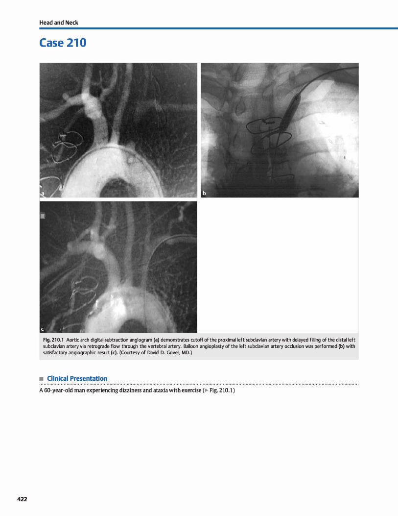
422
Head and Neck
Case 21 0
Fig. 21 0.1 Aortic arch digital subtraction angiogram (a) demonstrates cutoff of the proximal left subclavian artery with delayed filling of the distal left subclavian artery via retrograde flow through the vertebral artery. Balloon angioplasty of the left subclavian artery occlusion was performed (b) with satisfactory angiographie result (c). (Courtesy of David D. Gover, MD.)
• Clinical Presentation
A 60-year-old man experiencing dizziness and ataxia with exercise (� Fig. 210.1 )
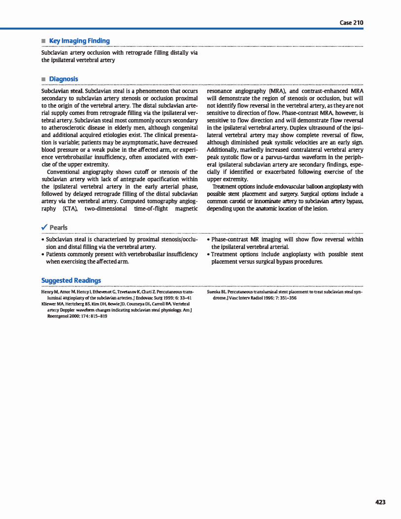
• Key lmaging Finding
Subclavian artery occlusion with retrograde filling distally via
the ipsilateral vertebral artery
• Diagnosis
Subdavian steal. Subdavian steal is a phenomenon that occurs secondary to subclavian artery stenosis or occlusion proximal to the origin of the vertebral artery. The distal subclavian arterial supply cornes from retrograde filling via the ipsilateral ver
tebral artery. Subclavian steal most commonly occurs secondary to atherosclerotic disease in elderly men, although congenital and additional acquired etiologies exist. The clinical presenta
tion is variable; patients may be asymptomatic, have decreased blood pressure or a weak pulse in the affected arm, or experience vertebrobasilar insuffidency, often associated with exerdse of the upper extremity.
Conventional angiography shows cutoff or stenosis of the subclavian artery with lack of antegrade opacification within
the ipsilateral vertebral artery in the early arterial phase, followed by delayed retrograde filling of the distal subdavian artery via the vertebral artery. Computed tomography angiog
raphy (CTA), two-dimensional time-of-flight magnetic
v" Pearls
• Subclavian steal is characterized by proximal stenosis/occlu
sion and distal filling via the vertebral artery. • Patients commonly present with vertebrobasilar insuffidency
when exercising the affected arm.
Suggested Readings
Herny M, Amor M, Henry I, E�ot G, Tzvetmuv K, Cliati Z. PercutilI!eous trans
luminal ;mgioplasty of the subdavian arteries.J Endovasc Surg 1999; 6: 33-41
Kliewer MA, Hertzberg BS, Kim DH, Bawie JD, Coumeya DL, Cm'oll BA. Vertebral
artery Doppler waveform changes indicating subclavian steal physiology. Am J
Roentgenol2000; 174: 815-819
Case 210
resonanœ angiography (MRA), and contrasHnhanced MRA will demonstrate the region of stenosis or ocdusion, but will not identify flow reversai in the vertebral artery, as they are not sensitive to direction of flow. Phase-contrast MRA. however, is
sensitive to flow direction and will demonstrate flow reversai in the ipsilateral vertebral artery. Duplex ultrasound of the ipsi
lateral vertebral artery may show complete reversai of flow, although diminished peak systolic velocities are an early sign. Additionally, markedly increased contralateral vertebral artery
peak systolic flow or a parvus-tardus waveform in the periph
eral ipsilateral subdavian artery are secondary findings, especially if identified or exaœrbated following exercise of the upper extremity.
lfeatment options indude endovascular balloon angioplasty with possible stent placement and surgery. Surgical options indude a common carotid or innominate artery to subdavian artery bypass, depending upon the anatomie location of the lesion.
• Phase-contrast MR imaging will show flow reversai within
the ipsilateral vertebral arterial. • Treatment options indude angioplasty with possible stent
placement versus surgical bypass procedures.
Sueoka BL PercutilI!eous transluminal stent placement to treat subclavian sœal syn
drome.] Vase lnterv R.adiol 1996; 7: 351-356
423
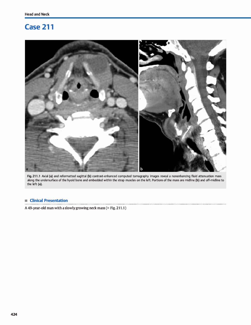
424
Head and Neck
Case 21 1
Fig. 21 1 .1 Axial (a) and reformatted sagittal (b) contrast-enhanced computed tomography images reveal a nonenhancing fluid attenuation mass along the undersurface of the hyoid bone and embedded within the strap muscles on the left. Portions of the mass are midline (b) and off-midline to the left (a).
• Clinical Presentation
A 49-year-old man with a slowly growing neck mass (� Fig. 211 .1 )
• Key lmaging Finding
Cystic neck mass embedded within strap muscles
• Diagnosis
Thyroglossal duct cyst (TGDC). The thyroid gland originally develops at the level of the foramen œcum along the floor of the primitive pharynx and descends to its normal position in the lower neck. Its migration path extends from the foramen œcum and passes anterior to and through the hyoid bone and strap musdes, as well as anterior to the laryngeal cartilage and thyroid membrane. The thyroglossal duct exists as a normal structure during thyroid desœnt, maintaining a amnection between the gland and the foramen œcum. It typically involutes toward the end of the first trimeste:r of development Should any portion of the duct persist, it may result in development of a TGDC.
TGDCs represent the most common congenital neck masses and most often present in children and young adults. Approximately 75% of cases occur at or below the hyoid bone with the
remainder being suprahyoid in location. Eighty-five percent are midline in location, whereas 15% of cases are slightly off-mid
line. They may be discovered incidentally or present as an enlarging neck mass. Oftentimes, superimposed infection results in growth and inflammation of the cyst with subsequent dinical presentation.
On imaging, TGDCs present as midline or slightly off-midline
cystic masses. The majority occur at or below the hyoid bone
� Pearls
• TGDCs are the most common congenital neck masses; they most often present in children and young adults.
• Approximately 75% of TGDCs occur at or below the hyoid bone; 85% are midline with the remainder slightly offmidline.
Suggested Reading
Koeller KK, A!ilIIIO I. Adair Cl', Smimiotopoulos JG. Congenital cystic masses of the
neck: radiologie-pathologie correlation Radiograplùcs 1999; 19: 121-146, quiz
152-153
Case 211
and are often characteristically embedded in the strap muscles. Suprahyoid TGDCs occur anywhere from the foramen cecum/ base or root of tangue to the hyoid bone. On ultrasound, the cysts are anechoic or hypoechoic with increased throughtransmission. Heterogeneity, including internai debris, is noted
with superimposed infection. Computed tomography demonstrates a well-circumscribed hypodense cystic mass extending along the path of craniocaudad descent. Enhancement, when
present, is peripheral and increased with superimposed infection. Magnetic resonanœ imaging demonstrates similar findings with internai fluid signal intensity (decreased Tl and increased T2) and occasional peripheral enhancement
Increased proteinaceous content may result in increased Tl signal intensity.
Treatment includes surgical resection using the Sistrunk procedure. When using this technique, recurrence rates are low. Rarely {< 1% of cases), thyroid carcinoma may occur within a TGDC due ta ectopie foci of thyroid tissue. The carcinoma is typically of the papillary subtype and is usually only discovered on pathological specimens. The presence of prospec
tive imaging findings to suspect carcinoma within a TGDC is uncommon.
• Cross-sectional imaging reveals a well-circumscribed cystic mass along the path of thyroid decent
• Appearance is heterogeneous with superimposed infection, including internai debris and peripheral enhancement
425
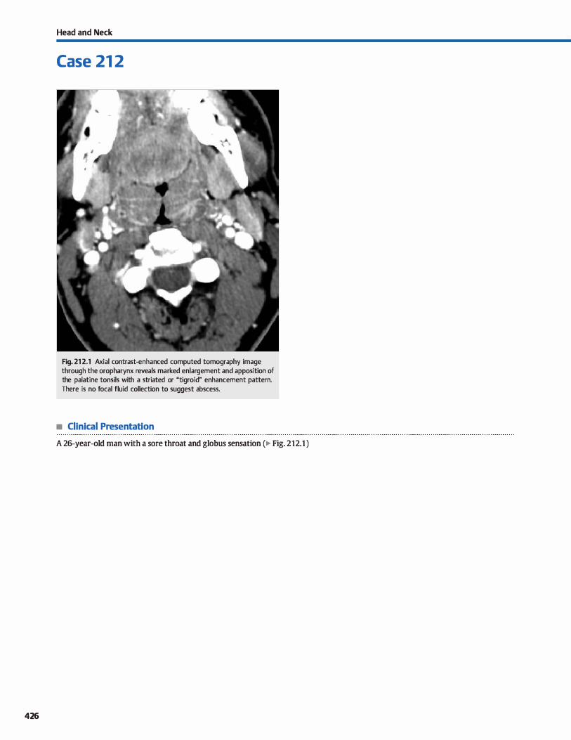
426
Head and Neck
Case 21 2
Fig. 21 2.1 Axial contrast-enhanced computed tomography image through the oropharynx reveals marked enlargement and apposition of the palatine tonsils with a striated or "tigroid" enhancement pattern. There is no focal fluid collection to suggest abscess.
• Clinical Presentation
A 26-year-old man with a sore throat and globus sensation (� Fig. 212.1 )
• Key lmaging Finding
Tonsillar enlargement with striated, "tigroid" enhancement
• Diagnosis
Tonsillitis. Tonsillitis represents the most common infection of the deep neck structures among children. adolescents, and young adults. Patients often present with fever, sore throat, odynophagia, dysphagia, and otalgia. Early on, there is tonsillar enlargement with or without exudates. As the infection progresses, it may spread to the peritonsillar region with ensuing phlegmon and potential abscess formation. A peritonsillar
abscess is far more common than an intratonsillar abscess. The most common inciting organisms are Steptococcus subspecies, Staphylocor:cus aureus, and Haemophilus influenza; polymicrobial etiology is common.
� Pearls
• Tonsillitis is the most common infection of the deep neck
structures in children, adolescents, and young adults. • Tonsillitis reveals tonsillar enlargement, apposition ("kissing
tonsils"), and striated or "tigroid" enhancement.
Suggested Reading
Capps EF, Kinsellajj, Gupta M, Bhatki AM, OpatIJwsky MJ. Emergency imaging
assessment of acute, nontraumatic conditions of the head and neck. Radio
graphies 2010; 30: 1335-1352
Case 212
On imaging, tonsillitis demonstrates tonsillar enlargement and apposition, referred to as the "kissing tonsils." There is characteristic linear, striated, or ''tigroid" enhancement. As the process spreads to the peritonsillar region, phlegmon may develop and present as relatively ill-defined regions of inflammatory changes with fat
stranding and variable degrees of enhancement A peritonsillar abscess is suggested when there is complete rim enhanœment; the
presence of rim enhancement, however, is not pathognomonic for
abscess formation because it has a specifidty of -75%. Tonsillitis is treated with antibiotics and supportive care;
abscesses require aspiration or surgical drainage.
• Peritonsillar abscess is suggested with complete, rim
enhanœment, which has a specificity of 75%. • Tonsillitis is treated with antibiotics and supportive care;
abscesses require aspiration or surgical drainage.
427

428
Head and Neck
Case 21 3
Fig. 213.1 Contrast-enhanced CT image through the oropharynx (a) reveals a rim-enhancing right peritonsillar abscess. Image through the lower neck (b) demonstrates a hypodense thrombus along the anterior aspect of the right internai jugular vein. Image through the chest in lung windows (c) reveals multiple peripheral pulmonary nodules. (Reprinted with permission: O'Brien WT, Cohen RA. Appl Radiol 201 1 ;40[1 ]:37-38.)
• Clinical Presentation
A 16-year-old girl with worsening sore throat and new onset cough and chest pain (� Fig. 213.1 )
• Key lmaging Finding
Peritonsillar absœss. jugular vein thrombus, and pulmonary
septic emboli
• Diagnosis
Lemierre syndrome. Lemierre syndrome is a rare entity charac
terized by an oropharyngeal infection with subsequent devel
opment of internai jugular venous (IJV) thrombosis and distant
septic emboli. The lungs are the most common site of septic
emboli; however, nearly any organ system may be involved.
Lemierre syndrome classically occurs in otherwise healthy
adolescents and young adults. Patients present with sepsis fol
lowing an antecedent oropharyngeal infection. Diagnosis is frequently delayed due to the indolent nature of the infection, as
well as the rarity of the syndrome. Diagnosis is made based upon a combination of characteristic imaging findings and con
firmatory blood cultures. Fusobaeterium necrophorum is the
most common causative organism; however, most cases are polymicrobial. Treatment indudes antibiotic therapy tailored to
� Pearts
• Lemierre syndrome is characterized by an oropharyngeal
infection, IJV thrombosis, and distant septic emboli.
• The lungs are the most common site of septic emboli; how
ever, nearly any organ system may be involved.
Suggested Reading
O'Brien Wf, Cohen RA. Lemierre's syndrome. Appl R.adiol 2011; 40: 37-38
Case 213
susceptibility analyses and drainage of any organized fluid col
lections. The use of anticoagulation therapy is controversial
because it may propagate the infection but is often used with sucœss.
The primary oropharyngeal infection is best evaluated by computed tomography (CT). Peritonsillar or retropharyngeal
absœsses present with rim-enhancing fluid collections that
exert mass etfect on the aerodigestive tract. IJV thrombosis may
be evaluated with CT or ultrasound; bath modalities reveal fill
ing defects within the internai jugular vein. Distant septic
emboli are most often evaluated with CT, especially with pul
monary involvement. Pulmonary septic emboli present as mul
tiple, peripheral pulmonary nodules that may demonstrate a
feeding vesse! extending into the nodules, as well as cavitation.
• Diagnosis is made based upon characteristic imaging findings
and confirmatory blood cultures.
• Treatment indudes antibiotic therapy and drainage of any
organized fluid collections.
429
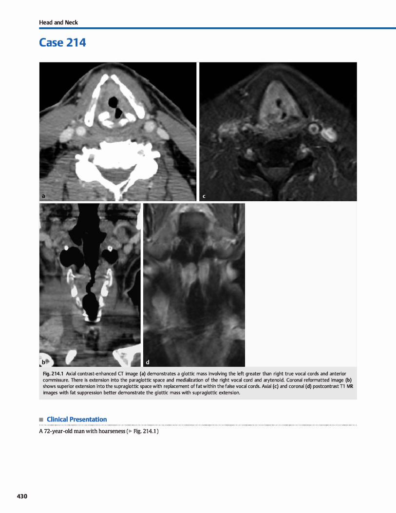
430
Head and Neck
Case 21 4
Fig. 214.1 Axial contrast-enhanced CT image (a) demonstrates a glottic mass involving the left greater than right true vocal cords and anterior commissure. There is extension into the paraglottic space and medialization of the right vocal cord and arytenoid. Coronal reformatted image (b) shows superior extension into the supraglottic space with replacement of fat within the fa Ise vocal cords. Axial (c) and coronal (d) postcontrast Tl MR images with fat suppression better demonstrate the glottic mass with supraglottic extension.
• Clinical Presentation
A 72-year-old man with hoarseness (� Fig. 214.1 )
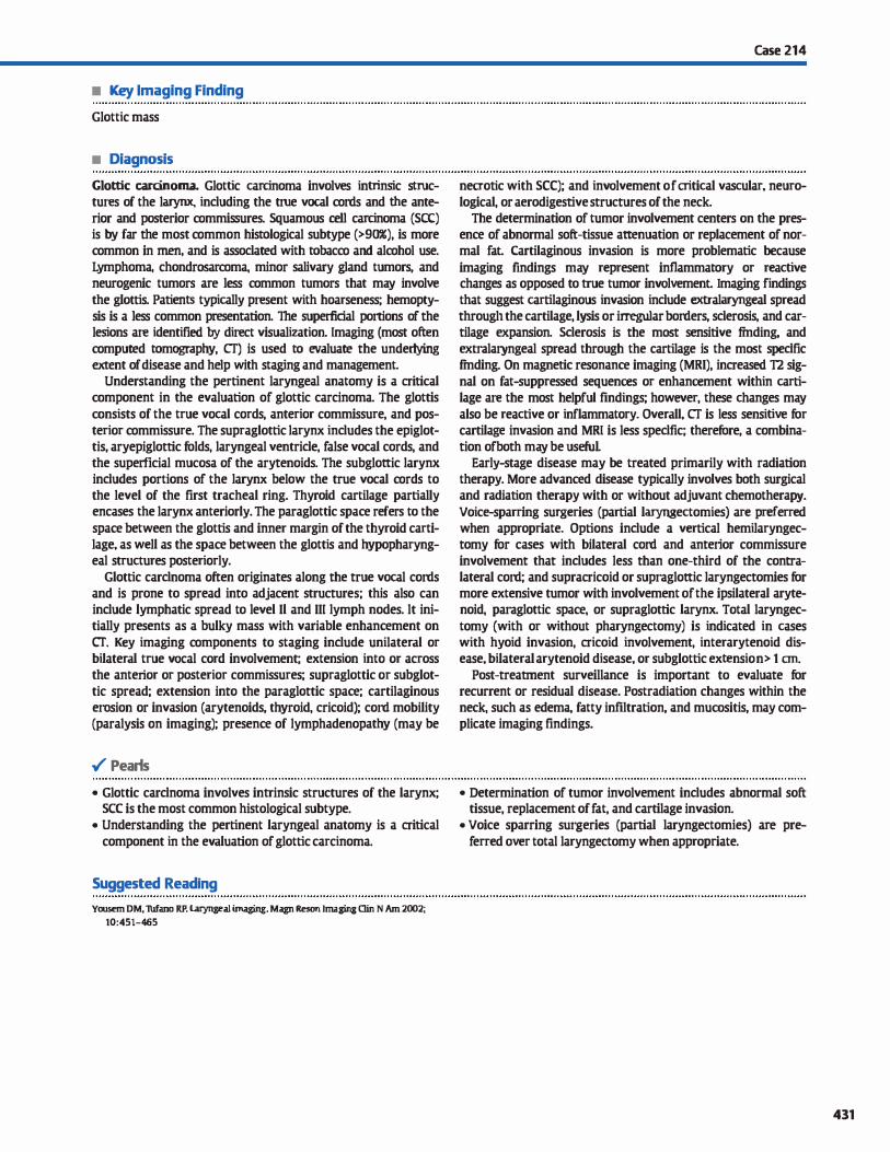
• Key lmaging Finding
Glottic mass
• Diagnosis
Glottic carcinoma. Glottic cardnoma involves intrinsic structures of the larynx, induding the true vocal cords and the anterior and posterior commissures. Squamous œll cardnoma (SCC) is by far the most common histological subtype (>90%), is more common in men, and is assodated with tobacco and alcohol use. Lymphoma, chondrosarcoma, minor salivary gland tumors, and neurogenic tumors are less common tumors that may involve
the glottis. Patients typically present with hoarseness; hemoptysis is a Jess common presentation. The superfiàal portions of the lesions are identified by direct visualization. Imaging (most often computed tomography, CT) is used to evaluate the underlying extent of disease and help with staging and management.
Understanding the pertinent laryngeal anatomy is a critical oomponent in the evaluation of glottic carcinoma. The glottis
oonsists of the true vocal cords, anterior commissure, and posterior commissure. The supraglottic larynx indudes the epiglottis, aryepiglottic folds, laryngeal ventride, false vocal cords, and the superticial mucosa of the arytenoids. The subglottic larynx indudes portions of the larynx below the true vocal cords to the level of the first tracheal ring. Thyroid cartilage partially encases the larynx anteriorly. The paraglottic spaœ refers to the
spaœ between the glottis and inner margin of the thyroid cartilage, as well as the spaœ between the glottis and hypopharyngeal structures posteriorly.
Glottic cardnoma often originates along the true vocal cords and is prone to spread into adjacent structures; this also can indude lymphatic spread to level Il and III lymph nodes. lt initially presents as a bulky mass with variable enhanœment on
CT. Key imaging components to staging indude unilateral or bilateral true vocal cord involvement; extension into or across the anterior or posterior commissures; supraglottic or subglottic spread; extension into the paraglottic spaœ; cartilaginous erosion or invasion (arytenoids, thyroid, cricoid); cord mobility (paralysis on imaging); presenœ of lymphadenopathy (may be
� Pearls
• Glottic cardnoma involves intrinsic structures of the larynx; sec is the most common histological subtype.
• Understanding the pertinent laryngeal anatomy is a critical
component in the evaluation of glottic carcinoma.
Suggested Reading
Yousem DM, lùfano RP. Laryngeal imaging. Magn Reson lmaging Clin N Am 2002;
10:451-465
Case 214
necrotic with SCC); and involvement of critical vascular, neurological, or aerodigestive structures of the neck.
The determination of tumor involvement centers on the presenœ of abnormal soft-tissue attenuation or replacement of normal fat cartilaginous invasion is more problematic because
imaging findings may represent inflammatory or reactive changes as opposed to true tumor involvement lmaging findings that suggest cartilaginous invasion indude extralaryngeal spread througlt the cartilage, lysis or irregular borders, sderosis, and cartilage expansion. Sderosis is the most sensitive fmding, and extralaryngeal spread through the cartilage is the most spedfic fmding. On magnetic resonanœ imaging (MRI), increased 12 sig
nal on fat-suppressed sequences or enhancement within cartilage are the most helpful findings; however, these changes may
also be reactive or inflammatory. Overall, CT is Jess sensitive for cartilage invasion and MRI is less spedfic; therefore, a combination ofboth may be useful
Early-stage disease may be treated primarily with radiation therapy. More advanœd disease typically involves bath surgical and radiation therapy with or without adjuvant chemotherapy.
Voice-sparring surgeries (partial laryrigectomies) are preferred when appropriate. Options indude a vertical hemilaryngectomy for cases with bilateral cord and anterior commissure involvement that indudes less than one-third of the contralateral cord; and supracricoid or supraglottic laryngectomies for more extensive tumor with involvement of the ipsilateral arytenoid, paraglottic spaœ, or supraglottic larynx. Total laryngec
tomy {with or without pharyngectomy) is indicated in cases with hyoid invasion, cricoid involvement, interarytenoid disease, bilateral arytenoid disease, or subglottic extension> 1 cm.
Post-treatment surveillance is important to evaluate for recurrent or residual disease. Postradiation changes within the neck, such as edema, fatty infiltration, and muoositis, may complicate imaging findings.
• Determination of tumor involvement indudes abnormal soft tissue, replacement of fat, and cartilage invasion.
• Voice sparring surgeries (partial laryngectomies) are pre
ferred over total laryngectomy when appropriate.
431
Head and Neck
Case 21 5
• Clinical Presentation
An adolescent boy involved in a motor vehicle crash (� Fig. 215.1 )
432
Fig. 21 5.1 Axial T2 MR image (a) demonstrates a crescent of hyperintense signal along the margin the right ICA with narrowing of the adjacent patent flow voici. The cross-sectional diameter of the right ICA is enlarged compared with the left. Axial Tl MR image with FS (b) reveals persistent eccentric hyperintensity along the margin of the right ICA lumen. Both images show fluid within the nasopharynx and sinus cavities secondary to support tubing (not imaged).
• Key lmaging Finding
Eccentric arterial wall hyperintensity with vessel enlargement and lurninal narrowing
• Diagnosis
Arterial dissection. Arterial dissections within the neck most often occur in the setting of trauma. Spontaneous dissections are more common in patients with underlying predispositions, such as those with hypertension, fibromuscular dysplasia, polycystic
kidney disease, vasculitis, or amnective tissue disorders. Dissections of the carotid arteJy typically involve the cervical portion of the internai carotid artery {ICA) up to the level of the pertrous segment Extension ftnther into the intracranial ICA is uncommon. Vertebral arteJy dissections most often involve the V3 segment near the cranioœrvical junction where the vessel is most mobile or the V2 segment, which is located within the transverse foramina
of the cervical spine, in the setting of vertebral fractures. Arterial injury may result in an intramural hematoma or an
intimai tear, which allows for blood to enter the media portion of the arterial wall. Extension to the adventitia layer may result in pseudoaneurysm formation. Intramural hematomas and diversion of blood flow into a false lumen may result in narrowing of the primary vascular channel. Possible dinical manifestations indude arterial stenosis, primary occlusion, or thromboembolic disease. Sorne patients may present with Homer syn
drome with involvement of the sympathetic chain along the course of the carotid artery. Headache and localized neck pain are seen in most patients.
On unenhanced computed tomography {CT1 acuœ dissections commonly present as cresœnt-shaped regions of increased density within the wall of the involved artery. Although the lumen
of the vesse! is often narrowed, the overall cross-sectional diameter of the artery is increased due to the presenœ of an intramural hematoma. Contrast-enhanced CT angiography may show
y' Pearts
• Dissections result from intimai injury; common findings include an intramural hematoma and intimai flap.
• On unenhanced CT, acute dissections present with cresœntshaped increased density within the arterial wall.
Suggested Reading
Rodallec MH, Marteau V, Gerber S. Desmottes I. Zins M. Craniocervical arœrial dis
section: spectrum of imaging findings and düferential diagnosis. Radiographies
2008; 28; 1711-1728
Case 215
luminal irregularity, narrowing, or a dissection flap. Occasionally, longitudinal extension of a dissection flap may result in tapered narrowing or even occlusion of the vessel
Conventional magnetic resonance imaging {MRI) sequences are helpful in evaluating the size and signal intensity of the artery and its wall. As with CT, the involved vessel typically bas an increased diameœr due to the presence of an intramural hematoma. The signal intensity of the hematoma varies based upon the stage of blood products and resultant paramagnetic effects. In the acute and chronic stages, the intramural hematoma is typically isointense compared with surrounding tissues,
making it Jess conspicuous. In the subacute phase (roughly 7 to 60 days): however, intrinsic Tl hyperintensity readily identifies the intramural hematoma and is best seen on unenhanced axial
Tl sequences with fat suppression (FS). Time-of-flight imaging and contrast-enhanced MR angiography will demonstraœ regions of lurninal irregularity, narrowing, or occlusion but are limited in evaluating the vessel wall.
Digital subtraction angiography (DSA) similarly evaluates the vesse) lumen but cannot evaluate the vessel wall. Findings of dissection on DSA indude luminal irregularity, tapered eccentric stenosis or occlusion (ustring" sign), a double lumen, or an intimai flap.
Patients with extracranial dissections of the neck and clinical and hemodynamic stability are typically treated initially with anticoagulation therapy. Endovascular or surgical treatment of
dissections may be required in patients who are unstable due to the dissection, fail medical management, have persistent thromboemboli, or have a dissecting pseudoaneurysm.
• In the subacute phase, unenhanced Tl sequences with FS show a peripheral hyperintense intramural hematoma.
• DSA findings indude luminal irregularity, tapered stenosis or occlusion, a double lumen, or an intimai flap.
433
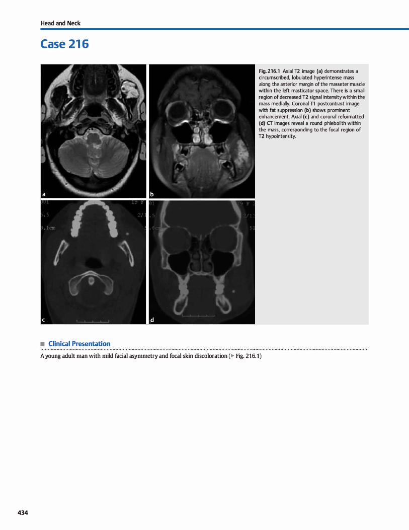
434
Head and Neck
Case 21 6
• Clinical Presentation
Fig. 216.1 Axial T2 image (a) demonstrates a circumscribed, lobulated hyperintense mass along the anterior margin of the masseter muscle within the left masticator space. There is a small reg ion of decreased T2 signal intensity within the mass medially. Coronal Tl postcontrast image with fat suppression (b) shows prominent enhancement. Axial (c) and coronal reformatted (d) CT images reveal a round phlebolith within the mass, corresponding to the focal region of T2 hypointensity.
A young adult man with mild facial asymmetry and focal skin discoloration (� Fig. 216.1)
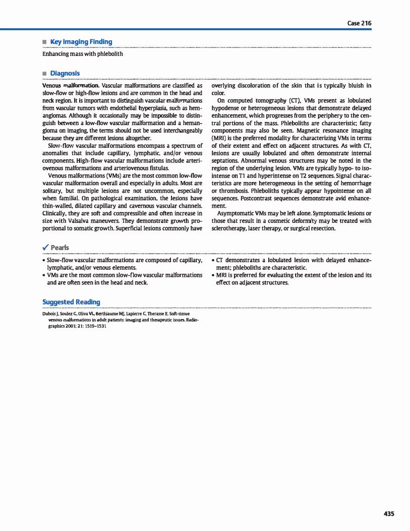
• Key lmaging Finding
Enhancing mass with phlebolith
• Diagnosis
Venous malformation. Vascular malformations are dassified as slow-flow or high-flow lesions and are common in the head and
nedt region. lt is important to distinguish vascular malformations from vascular tumors with endothelial hyperplasia, such as hemangiomas. Although it occasionally may be impossible to distinguish between a low-flow vascular malformation and a hemangioma on imaging, the terms should not be used interchangeably
because they are different lesions altogether.
Slow-flow vascular malformations encompass a spectrum of anomalies that indude capillary, lymphatic, and/or venous components. High-flow vascular malformations indude arteriovenous malformations and arteriovenous fistulas.
Venous malformations (VMs) are the most common low-flow vascular malformation overall and especially in adults. Most are
solitary, but multiple lesions are not uncommon, especially when familial. On pathological examination, the lesions have
tlùn-walled, dilated capillary and cavernous vascular channels. Clinically, they are soft and compressible and often increase in size with Valsalva maneuvers. They demonstrate growth proportional to somatic growth. Superficial lesions commonly have
� Pearls
• Slow-flow vascular malformations are composed of capillary, lymphatic, and/or venous elements.
• VMs are the most common slow-flow vascular malformations and are often seen in the head and neck.
Suggested Reading
Dubois J, Soulez G, Oliva VI., Berthiaume Mf, Lapierre C. Therasse E. Soit-tissue
vmous malfunnations in adult patients: imaging and therapeutic issues. Radiographies 2001; 21: 1519-1531
Case 216
overlying discoloration of the skin that is typically bluish in col or.
On computed tomography (CT), VMs present as lobulated hypodense or heterogeneous lesions that demonstrate delayed enhancement, which progresses from the periphery to the cen
tral portions of the mass. Phleboliths are characteristic; fatty components may also be seen. Magnetic resonance imaging (MRI) is the preferred modality for characterizing VMs in terms
of their extent and effect on adjacent structures. As with CT, lesions are usually lobulated and often demonstratE internai septations. Abnormal venous structures may be noted in the region of the underlying lesion. VMs are typically hypo- to iso
intense on T1 and hyperintense on T2 sequenœs. Signal characteristics are more hererogeneous in the setting of hemorrhage
or thrombosis. Phleboliths typically appear hypointense on ail sequenœs. Postcontrast sequences demonstrate avid enhancement.
Asymptomatic VMs may be left alone. Symptomatic lesions or those that result in a cosmetic deformity may be treaœd with sclerotherapy, laser therapy, or surgical resection.
• CT demonstraœs a lobulated lesion with delayed enhancement; phleboliths are characteristic.
• MRI is preferred for evaluating the extent of the lesion and its effect on adjacent structures.
435
436
Head and Neck
Case 21 7
• Clinical Presentation
Fig. 217 .1 Axial (a,b), sagittal formatted (c), and coronal reformatted (d) contrast-enhanced computed tomography images show postoperative changes consistent with a left modified radical neck dissection and placement of a myocutaneous flap. There is atrophy of the musculature within the flap, as well as regions of fatty infiltration from radiation therapy. No masslike enhancement is seen within the surgical bed or along the surgical margins. (Courtesy of Michael Zapadka, DO.)
A 61-year-old man post laryngectomy and modified left radical neck dissection with reconstruction (� Fig. 217.1 )
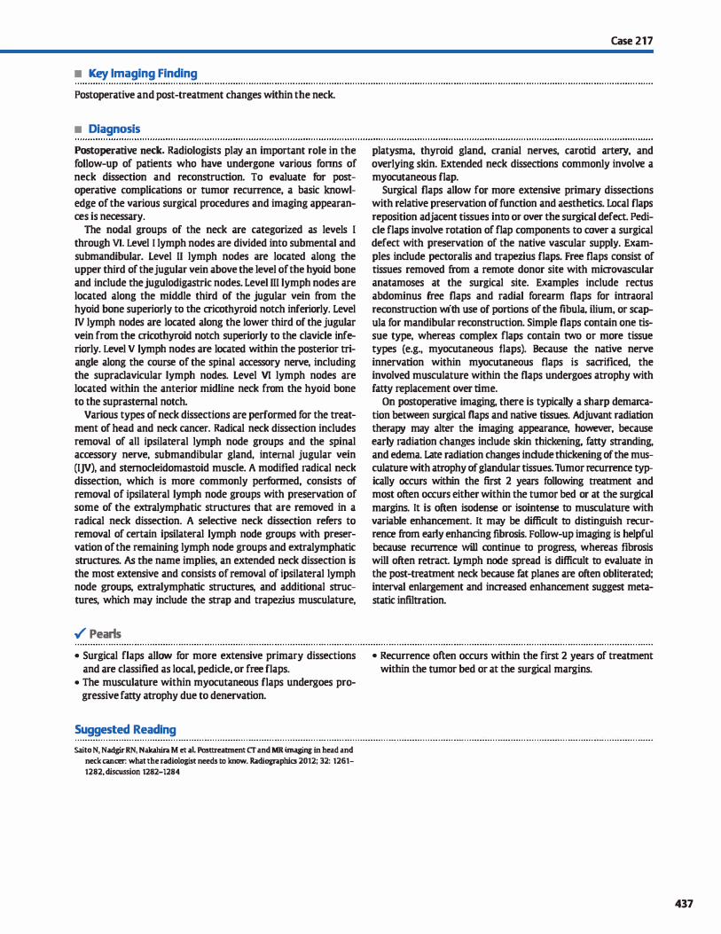
• Key lmaging Finding
Postoperative and post-treatment changes within the neck.
• Diagnosis
Postoperative neck. Radiologists play an important rote in the follow-up of patients who have undergone various fonns of
neck dissection and reconstruction. To evaluate for postoperative complications or tumor recurrence, a basic knowledge of the various surgical procedures and imaging appearances is necessary.
The nodal groups of the neck are categorized as levels I
through VI. Level I lymph nodes are divided into submental and
submandibular. Level II lymph nodes are located along the upper third of the jugular vein above the level of the hyoid bone and include the jugulodigastric nodes. Level III lymph nodes are located along the middle third of the jugular vein from the hyoid bone superiorly to the cricothyroid notch inferiorly. Level N lymph nodes are located along the lower third of the jugular
vein from the cricothyroid notch superiorly to the clavicle infe
riorly. Level V lymph nodes are located within the posterior triangle along the course of the spinal accessory nerve, including the supradavicular lymph nodes. Level VI lymph nodes are located within the anterior midline neck from the hyoid bone to the suprasternal notch.
Various types of neck dissections are performed for the treat
ment of head and neck cancer. Radical neck dissection includes removal of ail ipsilateral lymph node groups and the spinal accessory nerve, submandibular gland, internai jugular vein
(IJV), and sternocleidomastoid muscle. A modified radical neck dissection, which is more commonly perfonned, consists of removal of ipsilateral lymph node groups with preservation of some of the extralymphatic structures that are removed in a
radical neck dissection. A selective neck dissection refers to removal of certain ipsilateral lymph node groups with preservation of the remaining lymph node groups and extralymphatic structures. As the name implies, an extended neck dissection is the most extensive and consists of removal of ipsilateral lymph node groups, extralymphatic structures, and additional structures, which may include the strap and trapezius musculature,
� Pearts
• Surgical flaps allow for more extensive primary dissections
and are classified as local, pedide, or free flaps.
• The musculature within myocutaneous flaps undergoes progressive fatty atrophy due to denervation.
Suggested Reading
Saito N, Nadgir RN, Nalcahira M et al Posttreatment CT and MR imaging in head and
neck cancer. what the radiologist needs to know. Radiographies 2012; 32: 1261-
1282, discussion 1282-1284
Case 217
platysma, thyroid gland, cranial nerves, carotid artery, and overlying skin. Extended neck dissections commonly involve a myocutaneous flap.
Surgical flaps allow for more extensive primary dissections with relative preservation of function and aesthetics. Local flaps
reposition adjacent tissues into or over the surgical defect. Pedicle flaps involve rotation of flap components to caver a surgical defect with preservation of the native vascular supply. Exam
ples indude pectoralis and trapezius flaps. Free flaps consist of tissues removed from a remote donor site with microvascular anatamoses at the surgical site. Examples include rectus abdominus free flaps and radial forearm flaps for intraoral
reconstruction with use of portions of the fibula. ilium, or scapula for mandibular reconstruction. Simple flaps contain one tissue type, whereas complex flaps contain two or more tissue types (e.g., myocutaneous flaps). Because the native nerve innervation within myocutaneous flaps is sacrificed, the involved musculature within the flaps undergoes atrophy with fatty replacement over time.
On postoperative imaging, there is typically a sharp demarca
tion between surgical flaps and native tissues. Adjuvant radiation therapy may alter the imaging appearance, however, because early radiation changes indude skin thickening, fatty stranding, and edema. Late radiation changes indude thickening of the musculature with atrophy of glandular tissues. Tumor recurrence typically occurs within the first 2 years following treannent and most often occurs either within the tumor bed or at the surgical
margins. It is often isodense or isointense to musculature with variable enhanœment. It may be difficult to distinguish recurrenœ from early enhancing fibrosis. Follow-up imaging is helpful
because recurrenœ will continue to progress, whereas fibrosis will often retract Lymph node spread is difficult to evaluate in the post-treatment neck because fat planes are often obliterated; interval enlargement and increased enhanœment suggest meta
static infiltration.
• Recurrence often occurs within the first 2 years of treatment within the tumor bed or at the surgical margins.
437
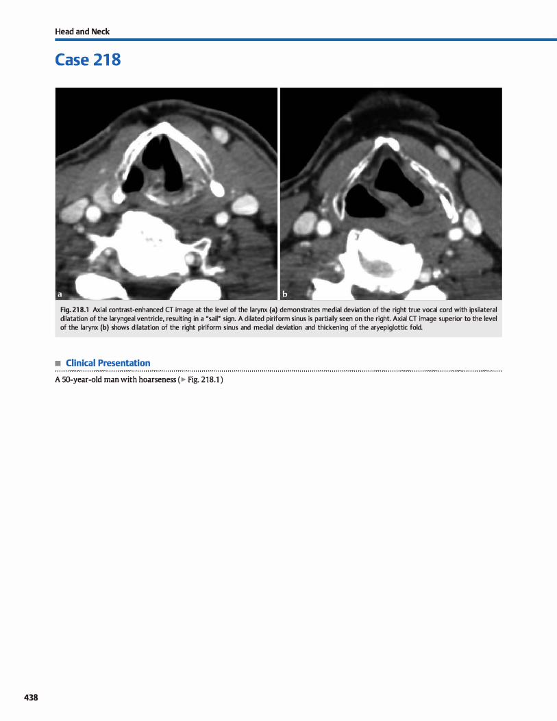
438
Head and Neck
Case 21 8
Fig. 21 8.1 Axial contrast-enhanced CT image at the level of the larynx (a) demonstrates medial deviation of the right true vocal cord with ipsilateral dilatation of the laryngeal ventricle, resulting in a "sail" sign. A dilated piriform sinus is partially seen on the right. Axial CT image superior to the level of the larynx (b) shows dilatation of the right piriform sinus and medial deviation and thickening of the aryepiglottic fold.
• Clinical Presentation
A 50-year-old man with hoarseness (� Fig. 218.1 )
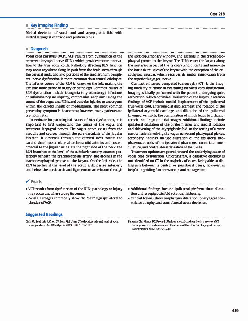
• Key lmaging Finding
Media! deviation of vocal cord and aryepiglottic fold with dilated laryngeal vennide and piriform sinus
• Diagnosis
Vocal cord paralysis (VCP). VCP results from dysfunction of the recurrent laryngeal nerve (RLN), which provides motor innervation to the true vocal cords. Pathology affecting RLN function may occur anywhere along its path from the brain stem, through
the cervical neck, and into portions of the mediastinum. Periph
eral nerve dysfunction is more common than central etiologies. The inferior course of the RLN is longer on the left, making the
left side more prone ta injury or pathology. Common causes of RLN dysfunction indude iatrogenia (thyroidectomy), infectious or inflammatory neuropathy, compressive neoplasms along the course of the vagus and RI.Ns, and vascular injuries or aneurysms
within the carotid sheath or mediastinum. The most common presenting symptom is hoarseness: however, many patients are asymptomatic.
Ta evaluate for pathological causes of RI.N dysfunction, it is important to first understand the course of the vagus and recurrent laryngeal nerves. The vagus nerve exists from the medulla and courses through the pars vascularis of the jugular foramen. lt descends through the cervical neck within the
carotid sheath posterolateral to the carotid arteries and posteromedial to the jugular veins. On the right side of the neck, the
RI.N branches at the level of the subdavian artery, courses posteriorly beneath the brachioœphalic artery, and ascends in the
tracheoesophageal groove ta the larynx. On the left side, the RI.N branches at the level of the aortic arch, passes anteriorly and below the aortic arch and ligamentum arteriosum through
� Pearls
• VCP results from dysfunction of the RLN; pathology or injury
may occur anywhere along its course. • Axial cr images commonly show the "sait" sign ipsilateral to
the side of VCP.
Suggested Readings
Chin SC. Edelstein S. Cl!.en C'l, Som PM. Using cr to localize si de and level of vocal
rord paralysis. AmJ Roentgmol 2003; 180: 1165-1170
Case 218
the aorticopulmonary window, and ascends in the tracheoesophageal groove to the larynx. The RI.Ns enter the larynx along the posterior aspect of the cricoarytenoid joints and innervate
the intrinsic musdes of the larynx with the exception of the cricothyroid muscle, which receives its motor innervation from
the superior laryngeal nerve. Contrast-enhanced computed tomography (CT) is the imag
ing modality of choiœ in evaluating for vocal cord dysfunction. Imaging is ideally performed with the patient undergoing quiet respiration, which optimizes evaluation of the larynx. Common
findings of VCP indude media! displaœment of the ipsilateral true vocal cord, anteromedial displacement and rotation of the ipsilateral arytenoid cartilage, and dilatation of the ipsilateral
laryngeal ventricle, the combination ofwhich leads to a characteristic "sait" sign on axial images. Additional fmdings indude ipsilateral dilatation of the piriform sinus and medial rotation and thickening of the aryepiglottic fold. In the setting of a more
central lesion involving the vagus nerve and pharyngeal plexus,
secondary findings indude dilatation of the ipsilateral oropharynx, atrophy of the ipsilateral pharyngeal constrictor mus
culature, and contralateral deviation of the uvula. Treatment options are geared toward the underlying cause of
vocal cord dysfunction. Unfortunately, a causative etiology is
not identified on cr in the majority of cases. Being able to distinguish between a central or peripheral cause, however, is
helpful in guiding further workup and management.
• Additional findings include ipsilateral piriform sinus dilata
tion and aryepiglottic fold rotation/thickening.
• Central lesions show oropharynx dilatation, pharyngeal constrictor atrophy, and contralateral uvula deviation.
Paquette CM, Manos DC, Psooy BJ. Unilateral vocal cord paralysis: a review ofCT
findings, mediastinal causes, and the course of the recurrent laryngeal nerves.
Radiograplùcs 2012; 32: 721-740
439
440
Head and Neck
Case 21 9
Fig. 219.1 Contrast-enhanced axial CT image demonstrates a bulky epiglottic mass, eccentric to the left, with heterogeneous enhancement. Subsequent images (not shown) revealed extension into the aryepiglottic folds.
• Clinical Presentation
A 51-year-old man with globus sensation and dysphagia (� Fig. 219.1 )
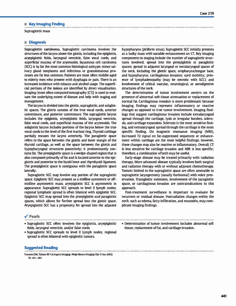
• Key lmaging Finding
Supraglottic mass
• Diagnosis
Supraglottic carcinoma. Supraglottic carcinoma involves the structures of the larynx abave the glottis, including the epiglottis, aryepiglottic folds, laryngeal ventride, false vocal cords, and superficial mucosa of the arytenoids. Squamous œll carcinoma (SCC) is by far the most common histological subtype; minor salivary gland neoplasms and infectious or granulomatous proœsses are far Jess common. Patients are most often middle-aged to elderly men who present with dysphagia or pain. There is an increased incidence with tobacco and alcohol usage. The supertidal portions of the Iesions are identified by direct visualization. lmaging (most often computed tomography [CT]) is used to evaluate the underlying extent of disease and help with staging and management.
The larynx is divided into the glottic, supraglottic, and subglottic spaœs. The glottis consists of the true vocal cords, anterior commissure, and posterior commissure. The supraglottic larynx indudes the epiglottis, aryepiglottic folds, laryngeal ventride, false vocal cords, and supertidal mucosa of the arytenoids. The subglottic larynx indudes portions of the larynx below the true vocal cords ta the level of the first tracheal ring. Thyroid cartilage partially encases the larynx anteriorty. The paraglottic spaœ refers to the space between the glottis and inner margin of the thyroid cartilage, as well as the spaœ between the glottis and hypopharyngeal structures posteriorly; it predominantly contains fat. The preepiglottic spaœ is a wedge-shaped region that is also composed primarily of fat and is located anterior to the epiglottis and posterior ta the hyoid bene and thyrohyoid ligament. The preepiglottic spaœ is contiguous with the paraglottic spaœ laterally.
Supraglottic sec may involve any portion of the supraglottic space. Epiglottic Sec may present as a midline symmetric or offmidline asymmetric mass; aryepiglottic sec is asymmetric in appearanœ. Supraglottic sec spreads to level II lymph nodes; regional lymphatic spread is often bilateral with epiglottic sec. Epiglottic sec may spread into the preepiglottic and paraglottic spaœs, which allows for further spread into the glottic spaœ. Aryepiglottic sec has a propensity for spread into the adjacent
� Pearts
Case 219
hypopharynx {piriform sinus� Supraglottic sec initially presents as a bulky mass with variable enhanœment on CT. Key imaging components ta staging indude the number of supraglottic structures involved; spread into the preepiglottic or paraglottic spaœs; spread to adjacent laryngeal or extralaryngeal spaœs of the neck, including the glottic spaœ, oropharynx/tangue base, and hypopharynx; cartilaginous invasion; cord mobility; presenœ of lymphadenopathy (may be necrotic with sec); and involvement of critical vascular, neurological, or aerodigestive structures of the neck.
The determination of tumor involvement centers on the presenœ of abnormal soit-tissue attenuation or replacement of normal fat. Cartilaginous invasion is more problematic because imaging findings may represent inflammatory or reactive changes as opposed to true tumor involvement. Imaging findings that suggest cartilaginous invasion indude extralaryngeal spread through the cartilage, lysis or irregular borders, sderosis, and cartilage expansion. Sderosis is the most sensitive finding, and extralaryngeal spread through the cartilage is the most spedfic finding. On magnetic resonance imaging (MRI), increased T2 signal on fat-suppressed sequences or enhancement within cartilage are the most helpful findings; however, these changes may also be reactive or inflammatory. OVerall, CT is Jess sensitive for cartilage invasion and MRI is Jess spedfic; therefore, a combination ofboth may be useful.
Early-stage disease may be treated primarily with radiation therapy. More advanced disease typically involves bath surgical and radiation therapy with or without adjuvant chemotherapy. Tumors limited to the supraglottic space are often amenable to supraglottic laryngectomy (usually horizontal) with voice preservation. Transglottic extension, involvement of the paraglottic spaœ, or cartilaginous invasion are contraindications to this approach.
Post-treatment surveillance is important to evaluate for recurrent or residual disease. Postradiation changes within the neclc, such as edema, fatty infiltration, and mucositis, may complicate imaging findings.
• Supraglottic SCC often involves the epiglottis, aryepiglottic • Determination of tumor involvement includes abnormal soft folds, laryngeal ventricle, and/or false cords. tissue, replacement of fat, and cartilage invasion.
• Supraglottic SCC spreads to level II lymph nodes; regional spread is often bilateral with epiglottic tumors.
Suggested Reading
Yousem DM, Tufano RP. Laryngeal imaging. Magn Reson lmaging Clin N Am 2002;
10: 451-465
441
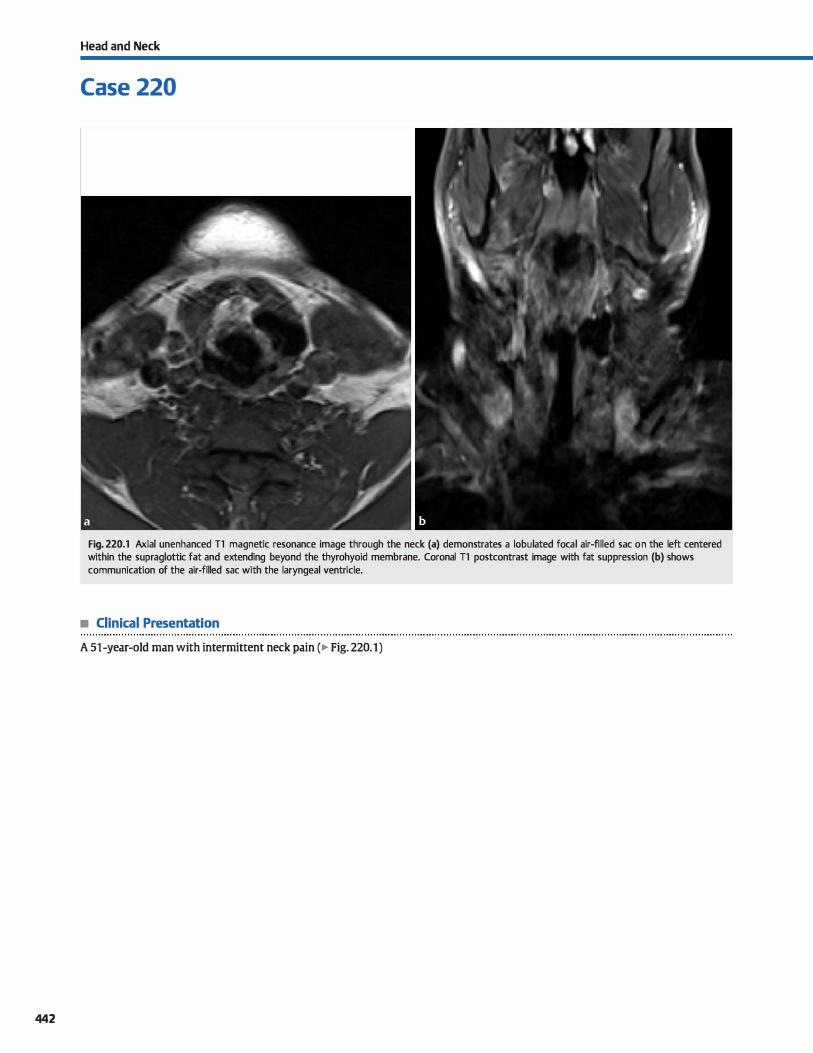
442
Head and Neck
Case 220
Fig. 220.1 Axial unenhanced Tl magnetic resonance image through the neck (a) demonstrates a lobulated focal air-filled sac on the left centered within the supraglottic fat and extending beyond the thyrohyoid membrane. Coronal Tl postcontrast image with fat suppression (b) shows communication of the air-filled sac with the laryngeal ventricle.
• Clinical Presentation
A 51-year-old man with intermittent neck pain (� Fig. 220.1)
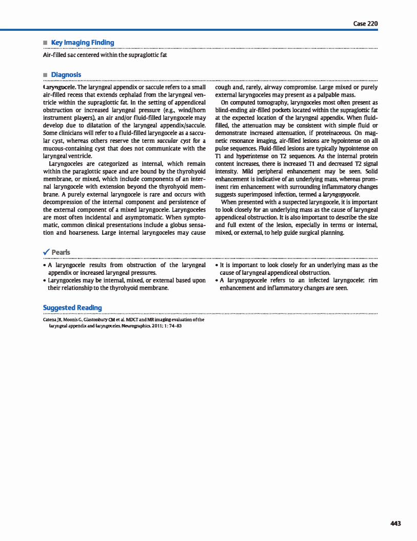
• Key lmaging Finding
Air-filled sac centered within the supraglottic fat
• Diagnosis
Laryngocele. The laryngeal appendix or saccule refers to a small air-filled recess that extends œphalad from the laryngeal ventricle within the supraglottic fat. ln the setting of appendiceal obstruction or increased laryngeal pressure (e.g., wind/horn instrument players), an air and/or fluid-filled laryngoœle may develop due to dilatation of the laryngeal appendix/saccule. Sorne clinicians will refer to a fluid-filled laryngocele as a saccu
lar cyst, whereas others reserve the term saccular cyst for a mucous-containing cyst that does not communicate with the laryngeal ventride.
Laryngoceles are categorized as internai, which remain within the paraglottic space and are bound by the thyrohyoid membrane, or mixed, which indude components of an internai laryngocele with extension beyond the thyrohyoid mem
brane. A purely externat laryngoœle is rare and occurs with decompression of the internai component and persistence of the externat component of a mixed laryngocele. Laryngoceles are most often incidental and asymptomatic. When symptomatic, common dinical presentations indude a globus sensation and hoarseness. Large internai laryngoœles may cause
� Pearls
• A laryngocele results from obstruction of the laryngeal appendix or increased laryngeal pressures.
• Laryngoceles may be internai, mixed, or extemal based upon their relationship ta the thyrohyoid membrane.
Suggested Reading
CatenaJR, Moonis G, Glastonbury CM et al. MDCT and MR imaging evaluation of the
larynSN1 appendix and laryngoceles. Neurographics. 2011; 1 : 74-83
Case 220
cough and, rarely, airway compromise. Large mixed or purely external laryngoceles may present as a palpable mass.
On computed tomography, laryngoœles most often present as blind-ending air-filled pockets located within the supraglottic fat at the expected location of the laryngeal appendix. When fluidfilled, the attenuation may be consistent with simple fluid or demonstrate increased attenuation, if proteinaœous. On mag
netic resonanœ imaging, air-filled lesions are hypointense on ail pulse sequenœs. Auid-filled lesions are typically bypointense on 11 and ltyperintense on T2 sequences. As the internai protein content increases, there is increased Tl and decreased T2 signal intensity. Mild peripheral enhanœment may be seen. Solid
enhanœment is indicative of an underlying mass, whereas prominent rim enhanœment with surrounding inflammatory changes suggests superimposed infection, termed a laryngopyocele.
When presented with a suspected laryngoœle, it is important to look closely for an underlying mass as the cause of laryngeal appendiceal obstruction. lt is also important to describe the size and full extent of the lesion, especially in terms or internai, mixed, or externat, to help guide surgical planning.
• 1t is important ta look closely for an underlying mass as the cause of laryngeal appendiceal obstruction.
• A laryngopyoœle refers ta an infected laryngocele: rim enhanœment and inflammatory changes are seen.
443
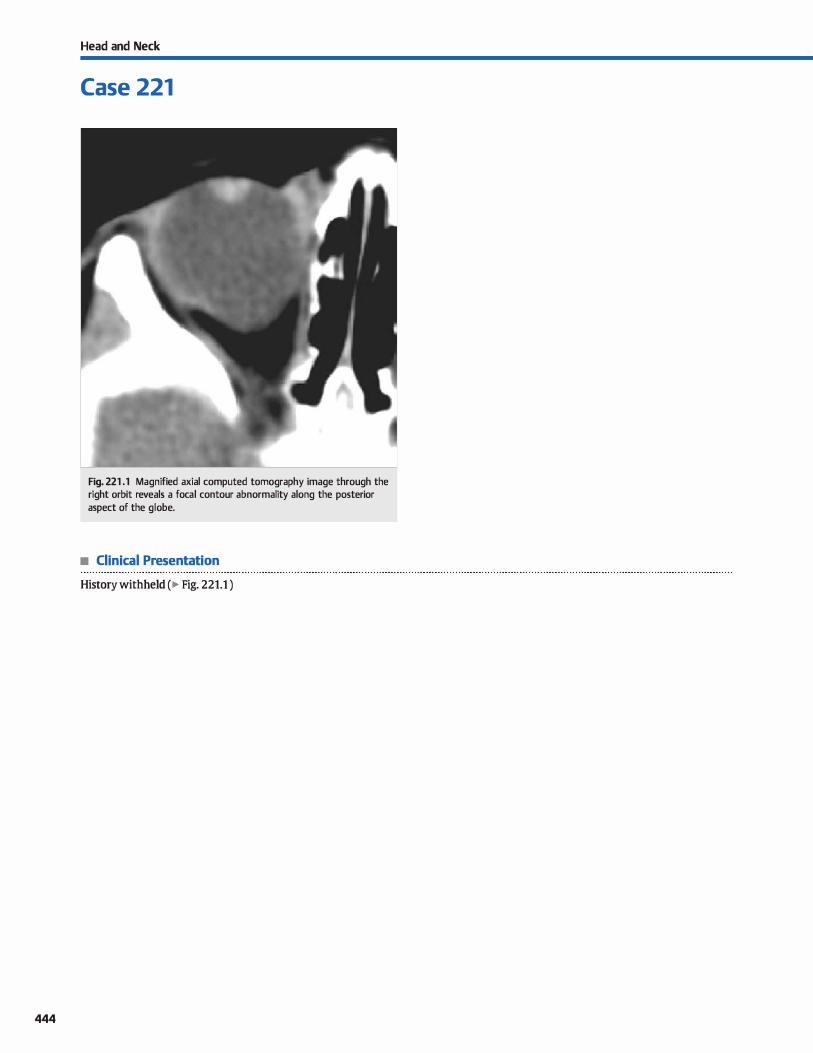
444
Head and Neck
Case 221
Fig. 221 .1 Magnified axial computed tomography image through the right orbit reveals a focal contour abnormality along the posterior aspect of the globe.
• Clinical Presentation
History withheld (� Fig. 221.1 )
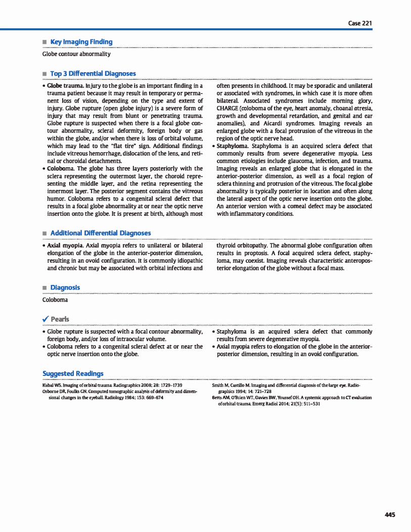
• Key lmaging Finding
Globe contour abnormality
• Top 3 Differential Diagnoses
• Globe trauma. lnjury to the globe is an important finding in a trauma patient because it may result in temporary or permanent loss of vision, depending on the type and extent of injury. Globe rupture {open globe injury) is a severe form of
injury that may result from blunt or penetrating trauma. Globe rupture is suspected when there is a focal globe contour abnormality, scierai deformity, foreign body or gas
within the globe, and/or when there is loss of orbital volume, which may lead to the "Hat tire" sign. Additional findings inciude vitreous hemorrhage, dislocation of the lens, and retinal or choroidal detachments.
• Coloboma. The globe has three layers posteriorly with the
sciera representing the outermost layer, the choroid representing the middle layer, and the retina representing the innermost layer. The posterior segment contains the vitreous humor. Coloboma refers to a congenital scierai defect that results in a focal globe abnormality at or near the optic nerve insertion onto the globe. lt is present at birth, although most
• Additional Differential Diagnoses
• Axial myopia. Axial myopia refers to unilateral or bilateral elongation of the globe in the anterior-posterior dimension, resulting in an ovoid configuration. lt is commonly idiopathie and chronic but may be associated with orbital infections and
• Diagnosis
Coloboma
y'° Pearts
• Globe rupture is suspected with a focal contour abnormality, foreign body, and/or loss of intraocular volume.
• Coloboma refers to a congenital scierai defect at or near the optic nerve insertion onto the globe.
Suggested Readings
Kubal WS. Imaging of orbital trawna. Radiographies 2008; 28: 1729-1739
Osborne DR. Foulks CN. Computed tomograplùc analysis of defornùty and dimen
sional changes in the eyl!ball. Radiology 1984; 153: 669-674
Case 221
often presents in childhood. It may be sporadic and unilateral or assodated with syndromes, in which case it is more often bilateral. Assodated syndromes inciude morning glory, CHARGE (coloboma of the eye, heart anomaly, choanal atresia, growth and developmental retardation, and genital and ear anomalies), and Aicardi syndromes. lmaging reveals an enlarged globe with a focal protrusion of the vitreous in the region of the optic nerve head.
• Staphyloma. Staphyloma is an acquired sciera defect that commonly results from severe degenerative myopia. Less common etiologies inciude glaucoma, infection, and trauma. Imaging reveals an enlarged globe that is elongated in the anterior-posterior dimension, as well as a focal region of sciera thinning and protrusion of the vitreous. The focal globe abnormality is typically posterior in location and often along the lateral aspect of the optic nerve insertion onto the globe. An anterior version with a comeal defect may be assodated with inflammatory conditions.
thyroid orbitopathy. The abnormal globe configuration often
results in proptosis. A focal acquired sciera defect, staphyloma, may coexist. lmaging reveals characteristic anteroposterior elongation of the globe without a focal mass.
• Staphyloma is an acquired sciera defect that commonly results from severe degenerative myopia.
• Axial myopia refers to elongation of the globe in the anteriorposterior dimension, resulting in an ovoid configuration.
Smith M, Castille M. lrnaging and differential diagnosis of the large eye. Radio
graplùcs 1994; 14: 721-728
Betts AM. O'Brien Wf, Davie5 JJW, Youssef OH. A systemic approach to CT evaluation
oforbital trauma. Ernerg Radio! 2014; 21(5): 511-531
445
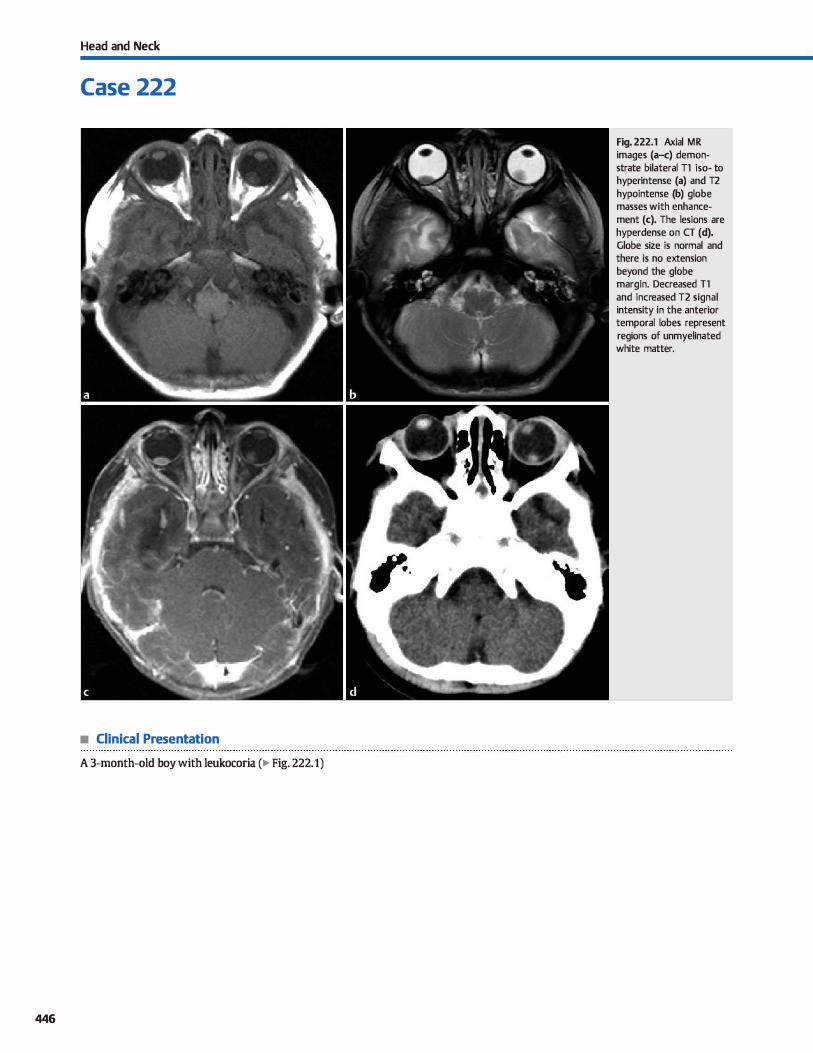
Head and Neck
Case 222
• Clinical Presentation
A 3-month-old boy with leukocoria (� Fig. 222.1)
446
Fig. 222.1 Axial MR images (a-c) demonstrate bilateral Tl iso- to hyperintense (a) and T2 hypointense (b) globe masses with enhancement (c). The lesions are hyperdense on CT (d). Globe size is normal and there is no extension beyond the globe margin. Decreased Tl and increased T2 signal intensity in the anterior temporal lobes represent regions of unmyelinated white matter.
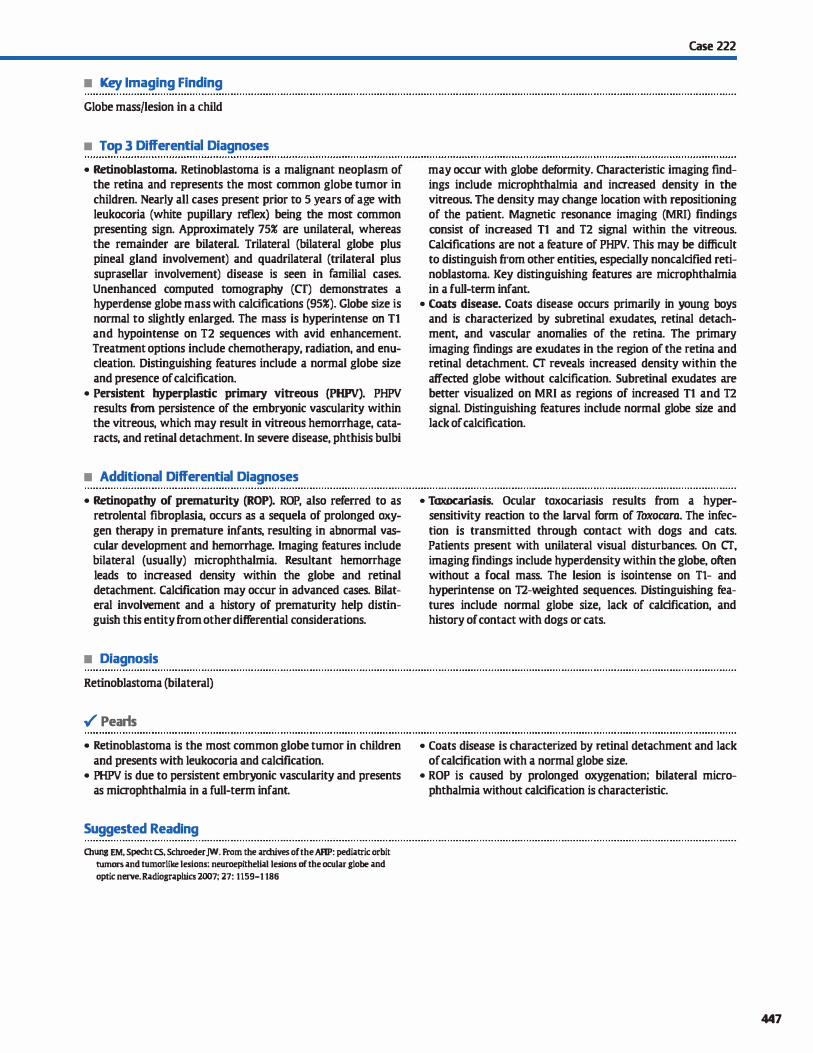
• Key lmaging Finding
Globe mass/lesion in a child
• Top 3 Differential Diagnoses
• Retinoblastoma. Retinoblastoma is a malignant neoplasm of the retina and represents the most common globe tumor in children. Nearly ail cases present prior ta 5 years of age with leukoooria (white pupillary reflex) being the most common presenting sign. Approximately 75% are unilateral, whereas the remainder are bilateral. Trilateral (bilateral globe plus pineal gland involvement) and quadrilateral (trilateral plus
suprasellar involvement) disease is seen in familial cases. Unenhanced computed tomography (CT) demonstrates a hyperdense globe mass with calcifications (95%). Globe size is normal to slightly enlarged. The mass is hyperintense on Tt and hypointense on T2 sequenœs with avid enhancement. Treatment options indude chemotherapy, radiation, and enudeation. Distinguishing features indude a normal globe size and presence of calcification.
• Persistent hyperplastic primary vitreous {PHPV). PHPV results from persistence of the embryonic vascularity within the vitreous, which may result in vitreous hemorrhage, cataracts, and retinal detachment. In severe disease, phthisis bulbi
• Additional Differential Diagnoses
• Retinopathy of prematurity (ROP). ROP, also referred to as retrolental fibroplasia, occurs as a sequela of prolonged oxy
gen therapy in premature infants, resulting in abnormal vascular development and hemorrhage. Imaging features indude bilateral (usually) microphthalmia. Resultant hemorrhage leads ta increased density within the globe and retinal detachment. calcification may occur in advanced cases. Bilateral involvement and a history of prematurity help distinguish this entity from other differential considerations.
• Diagnosis
Retinoblastoma (bilateral)
� Pearls
• Retinoblastoma is the most common globe tumor in children and presents with leukoooria and calcification.
• PHPV is due ta persistent embryonic vascularity and presents as microphthalmia in a full-term infant.
Suggested Reading
Chung EM, Specht CS, Schroeder JW. From the ardûves of the AFIP: pediatric orbit
tumors and tumorlilœ lesions; neuroepithelial lesions of the ocular globe and
optic nenre. Radiograplùcs ']J)07; 27: 1159-1186
Case 222
may occur with globe deformity. Characteristic imaging findings indude microphthalmia and increased density in the vitreous. The density may change location with repositioning of the patient. Magnetic resonance imaging (MRI) findings
consist of increased Tt and T2 signal within the vitreous. calcifications are not a feature of PHPV. This may be difficult ta distinguish from other entities, especially noncalcified retinoblastoma. Key distinguishing features are microphthalmia in a full-term infant.
• Coats disease. Coats disease occurs primarily in young boys and is characterized by subretinal exudates, retinal detachment, and vascular anomalies of the retina. The primary
imaging findings are exudates in the region of the retina and retinal detachment. CT reveals increased density within the
affected globe without calcification. Subretinal exudates are better visualized on MRI as regions of increased Tt and T2 signal. Distinguishing features include normal globe size and Jack of calcification.
• Toxocariasis. Ocular toxocariasis results from a hypersensitivity reaction to the larval form of Toxocara. The infec
tion is transmitted through contact with dogs and cats. Patients present with unilateral visual disturbances. On CT,
imaging findings include hyperdensity within the globe, often without a focal mass. The lesion is isointense on Tt- and hyperintense on T2-weighted sequences. Distinguishing features indude normal globe size, lack of calcification, and history of contact with dogs or cats.
• Coats disease is characterized by retinal detachment and Jack of calcification with a normal globe size.
• ROP is caused by prolonged oxygenation; bilateral microphthalmia without calcification is characteristic.
447
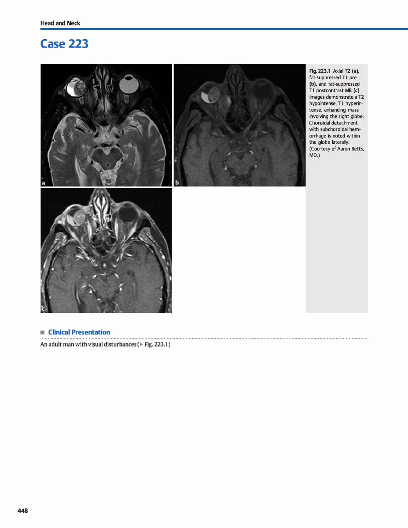
Head and Neck
Case 223
• Clinical Presentation
An adult man with visual disturbances ( � Fig. 223.1 )
448
Fig. 223.1 Axial T2 (a), fat-suppressed Tl pre(b), and fat-suppressed Tl postcontrast MR (c) images demonstrate a T2 hypointense, Tl hyperintense, enhancing mass involving the right globe. Choroidal detachment with subchoroidal hemorrhage is noted within the globe laterally.
(Courtesy of Aaron Betts, MD.)
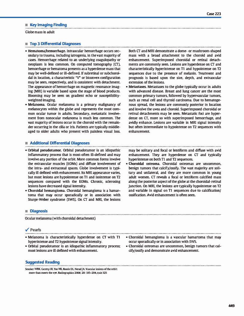
• Key lmaging Finding
Globe mass in adult
• Top 3 Differential Diagnoses
• Hematoma/hemorrhage. Intraocular hemorrhage occurs secondary ta trauma. including iatrogenia, in the vast majority of cases. Hemorrhage related to an underlying coagulopathy or neoplasm is Jess common. On computed tomography {CT),
hemorrhage or hematoma presents as a hyperdense mass that
may be well-defined or ill-defined. If subretinal or subchoroidal in location, a characteristic "V" or biconvex configuration
may be seen, respectively, and is consistent with detachment.
The appearance of hemorrhage on magnetic resonanœ imaging (MRI) is variable based upon the stage of blood products. Blooming may be seen on gradient echo or susceptibility
weighted imaging. • Melanoma. Ocular melanoma is a primary malignancy of
melanocytes within the globe and represents the most com
mon ocular tumor in adults. Secondary, metastatic involve
ment from nonocular melanoma is much less common. The vast majority of lesions occur in the choroid with the remain
der occurring in the àlia or iris. Patients are typically middleaged to aider adults who present with painless visual Joss.
• Additional Differential Diagnoses
• Orbital pseudotumor. Orbital pseudotumor is an idiopathie inflammatory process that is most-often ill-defined and may
involve any portion of the orbit. More common forms involve the extraocular muscles (EOMs) and diffuse involvement of the intra- and extraconal spaœs. Globe involvement is typi
cally ill defined with enhancement. Its MRI appearanœ varies, but most lesions are hypointense on Tl and isointense on T2 sequences compared with the EOMs. Chronic, sclerosing
lesions have decreased signal intensity.
• Choroidal hemangioma. Choroidal hemangioma is a hamartoma that may occur sporadically or in association with
Sturge-Weber syndrome {SWS). On CT and MRI, the lesions
• Diagnosis
Ocular melanoma (with choroidal detachment)
� Pearls
• Melanoma is characteristically hyperdense on CT with Tl hyperintense and T2 hypointense signal intensity.
• Orbital pseudotumor is an idiopathie inflammatory process; most lesions are ill defined with enhanœment.
Suggested Reading
Smoker WRK, Gentry IR, Yee NK, Reede DI. Nerad JA. Vascular lesions of the orbit:
more than meets the eyw;!. Radiograplùcs 2008; 28: 185-204, quiz 325
Case 223
Both CT and MRI demonstrate a dame- or mushroom-shaped mass with a broad attachment to the choroid and avid
enhanœment. Superimposed choroidal or retinal detachments are commonly seen. Lesions are hyperdense on CT and characteristically hyperintense on Tl and hypointense on T2
sequenœs due ta the presence of melanin. Treatment and prognosis is based upon the size, depth, and extraocular
extension of the lesions. • Metastases. Metastases to the globe typically occur in adults
with advanœd disease. Breast and Jung cancer are the most common primary tumors, followed by hypervascular tumors, such as renal cell and thyroid carànoma. Due to hematoge
nous spread. the lesions are commonly posterior in location
and involve the uvea and choroid. Superimposed choroidal or retinal detachments may be seen. Metastatic foci are hyper
dense on CT, more so with superimposed hemorrhage, and avidly enhanœ. Lesions are variable in MRI signal intensity but often intermediate to hypointense on T2 sequences with
enhancement.
may be solitary and focal or lentiform and diffuse with avid
enhanœment. They are hyperdense on CT and typically
hyperintense on bath Tl and T2 sequences. • Choroidal osteoma. Choroidal osteomas are uncommon,
benign tumors that calcify/ossify. The vast majority are soli
tary and unilateral, and they are more common in young
adult women. CT reveals a focal or lentiform calcified mass along the posterior aspect of the globe at the choroidal-retinal
junction. On MRI, the lesions are typically hypointense on T2 and variable in signal on Tl sequences due to calcification/
ossification. Avid enhanœment is often seen.
• Choroidal hemangioma is a vascular hamartoma that may occur sporadically or in association with SWS.
• Choroidal osteomas are uncommon, benign tumors that caldfy/ossify and demonstrate avid enhancement.
449
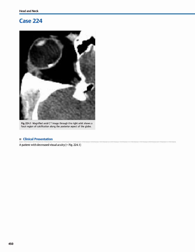
450
Head and Neck
Case 224
Fig. 224.1 Magnified axial CT image through the right orbit shows a focal region of calcification along the posterior aspect of the globe.
• Clinical Presentation
A patient with decreased visual acuity (� Fig. 224.1)
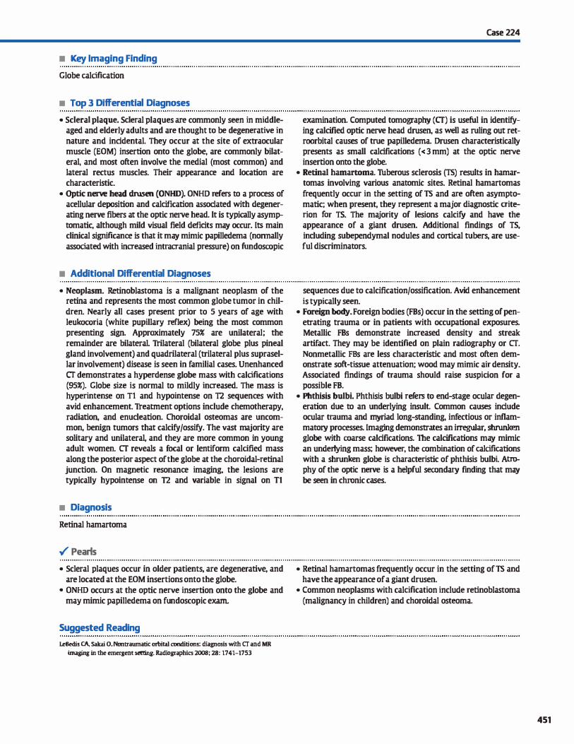
• Key lmaging Finding
Globe calcification
• Top 3 Differential Diagnoses
• Scierai plaque. Scierai plaques are commonly seen in middleaged and elderly adults and are thought ta be degenerative in nature and incidental. They occur at the site of extraocular muscle {EOM) insertion onto the globe, are commonly bilateral, and most often involve the medial (most common) and lateral rectus muscles. Their appearance and location are characteristic.
• Optic nerve head drusen (ONHD). ONHD refers to a process of acellular deposition and calcification assodated with degenerating nerve fibers at the optic nerve head. lt is typically asymptomatic, although mild visual field deficits may occur. lts main clinical significanœ is that it may mimic papilledema (nonnally
associated with increased intracranial pressure) on fundoscopic
• Additional Differential Diagnoses
• Neoplasm. Retinoblastoma is a malignant neoplasm of the retina and represents the most common globe tumor in chil
dren. Nearly ail cases present prior to 5 years of age with leukocoria (white pupillary reflex) being the most common presenting sign. Approximately 75% are unilateral; the remainder are bilateral. Trilateral (bilateral globe plus pineal gland involvement) and quadrilateral (trilateral plus suprasellar involvement) disease is seen in familial cases. Unenhanced cr demonstrates a hyperdense globe mass with calcifications (95%). Globe size is normal to mildly increased. The mass is hyperintense on T1 and hypointense on T2 sequences with avid enhancement. Treatment options include chemotherapy, radiation, and enudeation. Choroidal osteomas are uncommon, benign tumors that calcify/ossify. The vast majority are solitary and unilateral, and they are more common in young adult women. cr reveals a focal or lentiform calcified mass
along the posterior aspect of the globe at the choroidal-retinal junction. On magnetic resonance imaging, the lesions are typically hypointense on T2 and variable in signal on Tt
• Diagnosis
Retinal hamartoma
� Pearts
• Scierai plaques occur in older patients, are degenerative, and are located at the EOM insertions onto the globe.
• ONHD occurs at the optic nerve insertion onto the globe and may mimic papilledema on fundoscopic exarn.
Suggested Reading
I.eBedis CA, Sakai o. Nontraumatic orbital conditions: diagnasis with cr and MR
imaging in the ernexgent setting. Radiographies 2008; 28: 1741-1753
Case 224
examination. Computed tomography (CT) is useful in identifying calcified optic nerve head drusen, as well as ruling out retroorbital causes of true papilledema. Drusen characteristically presents as small calcifications ( < 3 mm) at the optic nerve insertion onto the globe.
• Retinal hamartoma. Tuberous sclerosis (TS) results in hamartomas involving various anatomie sites. Retinal hamartomas
frequently occur in the setting of TS and are often asymptomatic; when present, they represent a major diagnostic criterion for TS. The majority of lesions calcify and have the appearance of a giant drusen. Additional findings of TS, induding subependymal nodules and cortical tubers, are useful discriminators.
sequenœs due to calcification/ossification. Avid enhanœment
is typically seen. • Foreign body. Foreign bodies (FBs) occur in the setting of pen
etrating trauma or in patients with occupational exposures. Metallic FBs demonstrate increased density and streak artifact. They may be identified on plain radiography or cr. Nonmetallic FBs are less characteristic and most often demonstrate soft-tissue attenuation; wood may mimic air density. Associated findings of trauma should raise suspicion for a possible FB.
• Phthisis bulbi. Phthisis bulbi refers ta end-stage ocular degeneration due to an underlying insult. Common causes indude ocular trauma and myriad long-standing, infectious or inflammatory proœsses. lmaging demonstrates an irregular, shrunken globe with coarse calcifications. The calcifications may mimic an underlying mass; however, the combination of calcifications with a shrunken globe is characteristic of phthisis bulbi. Atrophy of the optic nerve is a helpful secondary finding that may be seen in chronic cases.
• Retinal hamartomas frequently occur in the setting of TS and have the appearanœ of a giant drusen.
• Common neoplasms with calcification include retinoblastoma
(malignancy in children) and choroidal osteoma.
451
452
Head and Neck
Case 225
Fig. 225.1 Coronal CT image (a) demonstrates bilateral en largement of the extraocular muscles, right greater than left, with more pronounced involvement of the inferior recti muscles. Axial CT in a different patient with the sa me diagnosis (b) reveals bilateral en largement of the bellies of the recti musculature with sparing of the myotendinous junctions, as well as radiological proptosis.
• Clinical Presentation
Middle-aged women with proptosis and the same underlying diagnosis (� Fig. 225.1 )
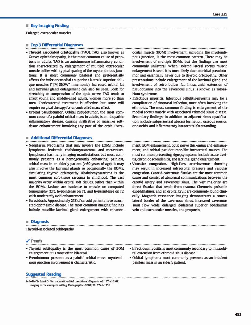
• Key lmaging Finding
Enlarged extraocular muscles
• Top 3 Differential Diagnoses
• Thyroid associated orbitopathy (TAO). TAO, also known as Graves ophthalmopathy, is the most common cause of proptosis in adults. TAO is an autoimmune inflammatory condition characterized by enlargement of multiple extraocular muscle bellies with typical sparing of the myotendinous junctions. It is most commonly bilateral and preferentially affects the inferior >media! > superior > lateral > superior oblique muscles ("!'M SLOW" mnemonic). Increased orbital fat and lacrimal gland enlargement can also be seen. Look for stretching or compression of the optic nerve. TAO tends to affect young and middle-aged adults, women more so than men. Corticosteroid treatment is effective, but some will require surgical therapy for uncontrolled mass effect.
• Orbital pseudotumor. Orbital pseudotumor, the most common cause of a painful orbital mass in adults, is an idiopathie inflammatory disease, causing infiltrative or masslike softtissue enhancement involving any part of the orbit. Extra-
• Additional Differential Diagnoses
• Neoplasm. Neoplasms that may involve the EOMs indude lymphoma, leukemia, rhabdomyosarcoma, and metastases. Lymphoma has many imaging manifestations but most commonly presents as a homogenously enhancing, painless, orbital mass in an elderly patient (>60 years of age). lt may also involve the lacrimal glands or occasionally the EOMs, simulating thyroid orbitopathy. Rhabdomyosarcoma is the most common soft-tissue sarcoma in childhood. The vast majority occur within orbital soft tissues, rather than within the EOMs. Lesions are isodense to muscle on computed tomography (CT), hypointense on Tl, and hyperintense on T2
with moderately avid enhanœment. • Sara>idosis. Approximately 25% of sarcoid patients have associ
ated ophthalmic disease. The most common imaging findings indude masslike lacrimal gland enlargement with enhance-
• Diagnosis
Thyroid-assodated orbitopathy
� Pearts
• Thyroid orbitopathy is the most common cause of EOM enlargement; it is most often bilateral.
• Pseudotumor presents as a painful orbital mass; myotendinous junction involvement is characteristic.
Suggested Reading
I.eBedis CA, Sakai o. Nontraumatic orbital conditions: diagnasis with cr and MR
imaging in the ernexgent setting. Radiographies 2008; 28: 1741-1753
Case 225
ocular muscle {EOM) involvement, including the myotendinous junction, is the most common pattern. There may be involvement of multiple EOMs, but the findings are most commonly unilateral. When isolated lateral rectus muscle enlargement is seen, it is most likely due to orbital pseudotumor and essentially never due to thyroid orbitopathy. Other presentations include enlargement of the lacrimal gland and involvement of retro bulbar fat. Intracranial extension of pseudotumor into the cavernous sinus is known as TolosaHunt syndrome.
• lnfectious myositis. lnfectious cellulitis-myositis may be a complication of sinonasal infection, most often involving the ethmoids. The most common finding is enlargement of the media! rectus muscle with associated ethmoid sinus disease. Secondary findings, in addition to adjacent sinus opacification, indude subperiosteal abscess formation, osseous erosion or osteitis, and inflammatory intraorbital fat stranding.
ment, EOM enlargement, optic nerve thickening and enhanœment, and orbital pseudotumor-like intraorbital masses. The most common presenting signs/symptoms indude acute uveitis, chronic dacroadenitis, and lacrimal gland enlargement.
• Vascular congestion. High-flow arteriovenous shunting may result in increased intraorbital pressure and vascular congestion. Carotid-cavemous fistulas are the most common cause and consist of abnormal communications between the carotid artery and cavernous sinus. The vast majority are direct fistulas that result from trauma. Chemosis, pulsatile exophthalmos, and an orbital bruit are commonly found clinically. Magnetic resonance imaging demonstrates a convex lateral border of the cavemous sinus, increased cavemous sinus flow voids, enlarged ipsilateral superior ophthalmic vein and extraocular muscles, and proptosis.
• Infectious myositis is most commonly secondary to intraorbital extension from ethmoid sinus disease.
• Orbital lymphoma most commonly presents as an indolent painless mass in an elderly patient.
453
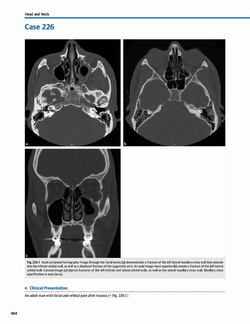
454
Head and Neck
Case 226
Fig. 226.1 Axial computed tomography image through the facial bones (a) demonstrates a fracture of the left lateral maxillary sinus wall that extends into the inferior orbital wall, as well as a displaced fracture of the zygomatic arch. An axial image more superior (b) reveals a fracture of the left lateral orbital wall. Coronal image (c) depicts fractures of the left inferior and lateral orbital walls, as well as the lateral maxillary sinus wall. Maxillary sinus opacification is seen (a-c).
• Clinical Presentation
An adult man with facial and orbital pain after trauma ( � Fig. 226.1 )
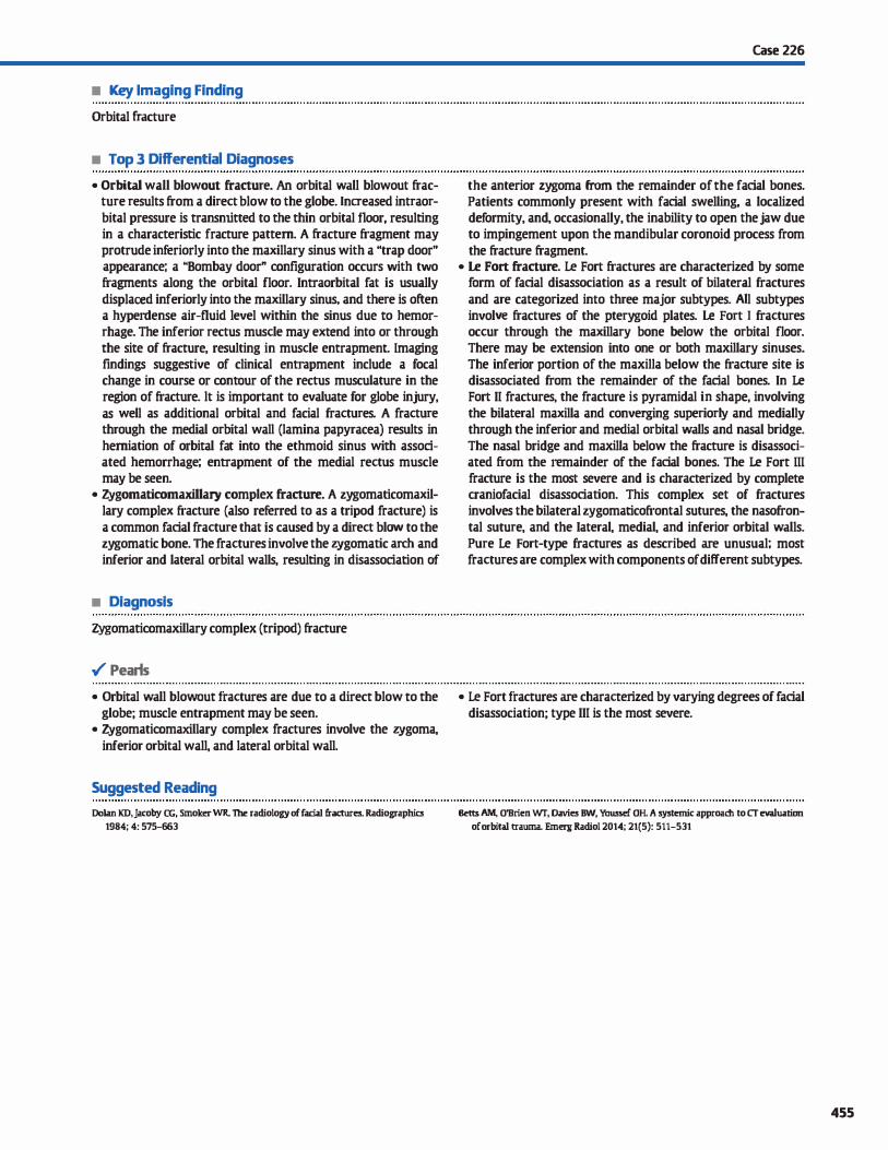
• Key lmaging Finding
Orbital fracture
• Top 3 Differential Diagnoses
• Orbital wall blowout fracture. An orbital wall blowout fracture results from a direct blow to the globe. Increased intraor
bital pressure is transnùtted to the thin orbital floor, resulting in a characteristic fracture pattern. A fracture fragment may protrude inferiorly into the maxillary sinus with a "trap door" appearance; a "Bombay door" configuration occurs with two fragments along the orbital floor. Intraorbital fat is usually
displaœd inferiorly into the maxillary sinus, and there is often a hyperdense air-fluid level within the sinus due to hemorrhage. The inferior rectus muscle may extend into or through the site of fracture, resulting in muscle entrapment lmaging findings suggestive of clinical entrapment include a focal change in course or contour of the rectus musculature in the region of fracture. lt is important to evaluate for globe injury,
as well as additional orbital and facial fractures. A fracture through the medial orbital wall (lamina papyracea) results in
herniation of orbital fat into the ethmoid sinus with assodated hemorrhage; entrapment of the media! rectus muscle may be seen.
• Zygomaticomaxillary complex fracture. A zygomaticomaxillary complex fracture (also referred to as a tripod fracture) is
a common facial fracture that is caused by a direct blow to the zygomatic bone. The fractures involve the zygomatic arch and inferior and lateral orbital walls, resulting in disassodation of
• Diagnosis
Zygomaticomaxillary complex (tripod) fracture
� Pearls
• Orbital wall blowout fractures are due to a direct blow to the globe; muscle entrapment may be seen.
• Zygomaticomaxillary complex fractures involve the zygoma,
inferior orbital wall, and lateral orbital wall.
Suggested Reading
Dolan KD, jacoby Œ, Smoker WR. The radiology of facial fractures. Radiographies
1984; 4: 575-663
Case 226
the anterior zygoma from the remainder of the facial bones. Patients commonly present with facial swelling, a localized deformity, and, occasionally, the inability to open the jaw due to impingement upon the mandibular coronoid process from
the fracture fragment. • Le Fort fracture. Le Fort fractures are characterized by some
form of facial disassociation as a result of bilateral fractures
and are categorized into three major subtypes. Ail subtypes involve fractures of the pterygoid plates. Le Fort 1 fractures occur through the maxillary bone below the orbital floor. There may be extension into one or both maxillary sinuses. The inferior portion of the maxilla below the fracture site is disassociated from the remainder of the fadai bones. In I.e Fort II fractures, the fracture is pyramidal in shape, involving
the bilateral maxilla and converging superiorly and medially through the inferior and medial orbital walls and nasal bridge.
The nasal bridge and maxilla below the fracture is disassociated from the remainder of the facial bones. The I.e Fort III fracture is the most severe and is characterized by complete craniofacial disassociation. This complex set of fractures involves the bilateral zygomaticofrontal sutures, the nasofron
tal suture, and the Iateral, medial, and inferior orbital walls.
Pure Le Fort-type fractures as described are unusual; most fractures are complex with components of different subtypes.
• Le Fort fractures are characterized by varying degrees of facial disassociation; type III is the most severe.
Betis AM, O'Brien WT, Davies BW, Youssef OH. A systemic approach to CT evaluation
of orbital trauma. Emerg Radiol 2014; 21(5): 511-531
455
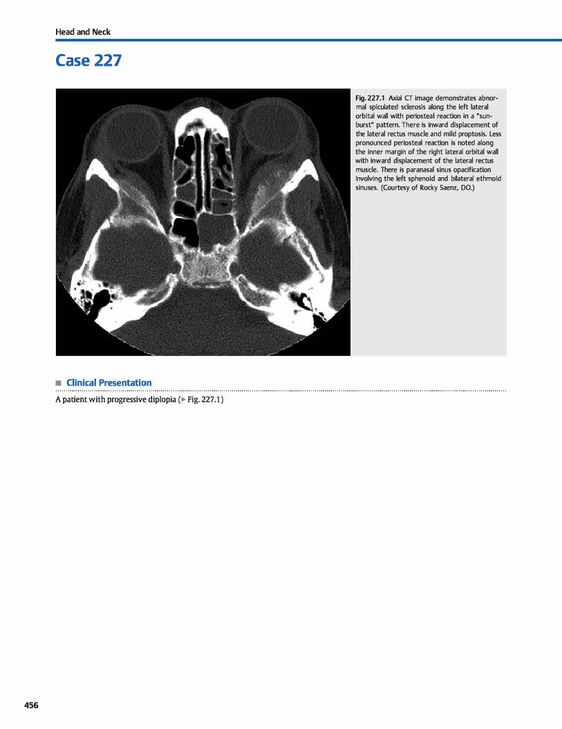
Head and Neck
Case 227
• Clinical Presentation
A patient with progressive diplopia (� Fig. 227 .1 )
456
Fig. 227.1 Axial CT image demonstrates abnormal spiculated sclerosis along the left lateral orbital wall with periosteal reaction in a "sunburst" pattern. There is inward displacement of the lateral rectus muscle and mild proptosis. Less pronounced periosteal reaction is noted along the inner margin of the right lateral orbital wall with inward displacement of the lateral rectus muscle. There is paranasal sinus opacification involving the left sphenoid and bilateral ethmoid sinuses. (Courtesy of Rocky Saenz, DO.)
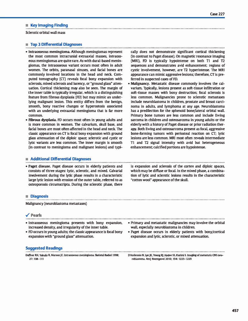
• Key lmaging Finding
Sderotic orbital wall mass
• Top 3 Differential Diagnoses
• lntraosseous meningioma. Although meningiomas represent the most common intracranial extraaxial masses, intraoss
eous meningiomas are quite rare. As with dural-based menin
giomas, the intraosseous variant occurs most often in adult
women. The orbits, paranasal sinuses, and facial bones are
commonly involved locations in the head and neck. Com
puted tomography {CT) reveals focal bony expansion with
sderosis, mixed sderosis and lucency, or "ground glass" atten
uation. Cortical thickening may aise be seen. The margin of the inner table is typically irregular, which is a distinguishing
feature from fibrous dysplasia (FD) but may mimic an under
lying malignant lesion. This entity differs from the benign, smooth, bony reactive changes or hyperostosis associated
with an underlying extraaxial meningioma that is far more
common.
• Fibrous dysplasia. FD occurs most often in young adults and
is more common in women. The calvarium, skull base, and
fadai bones are most often affected in the head and neck. The
classic appearance on CT is focal bony expansion with ground
glass attenuation of the diploic spaœ; sclerotic and cystic or
lytic variants are Jess common. The inner margin is smooth
(in contrast to meningioma and malignant lesions) and typi-
• Additional Differential Diagnoses
• Paget disease. Paget disease occurs in elderly patients and
consists of three stages: Iytic, sclerotic, and mixed. Calvarial
involvement during the lytic phase results in a characteristic
large lytic lesion with erosion of the outer table, referred to as osteoporosis circumscripta. During the sclerotic phase, there
• Diagnosis
Malignancy (neuroblastoma metastases)
� Pearls
• lntraosseous meningioma presents with bony expansion, increased density, and irregularity of the inner table.
• FD occurs in young adults; the dassic appearance is focal bony
expansion with "ground glass" attenuation.
Suggested Readings
Daffner RH, Y.lkulis R. Maroon JC. Intraosseous meningioma. Slœletal Radiol t 998; 27: 108-111
Case 227
cally does not demonstrate significant cortical thickening
{in contrast to Paget disease). On magnetic resonanœ imaging
{MRI), FD is typically hypointense on bath Tl and T2
sequences and demonstrates avid enhancement; regions of
cystic involvement, however, are T2 hyperintense. The MRI
appearance can mimic aggressive lesions; therefore, CT is pre
ferred in suspected cases of FD. • Malignancy. Metastatic disease commonly involves the cal
variurn. "JYpically, lesions present as soft-tissue infiltration or soft-tissue masses with bony destruction; focal sderosis is
Jess common. Malignancies prone ta sderotic metastases
include neuroblastoma in children, prostate and breast carci
noma in adults, and lymphoma at any age. Neuroblastoma bas a predilection for the sphenoid bone/lateral orbital wall.
Primary bene tumors are Jess common and indude Ewing
sarcoma in children and osteosarcoma in young adults or the
elderly with a history of Paget disease or prier radiation ther
apy. Both Ewing and osteosarcoma present as focal, aggressive
bone-forming tumors with periosteal reaction on CT; lytic lesions are Jess common. MRI most often reveals intermediate
Tl and T2 signal intensity with avid but heterogeneous
enhancement; calcified portions are hypointense.
is expansion and sderosis of the cortex and diploic spaœs,
which may be diffuse or focal. In the mixed phase, a combina
tion of lytic and sclerotic lesions results in the characteristic
"cotton wool'' appearance of the skull.
• Primary and metastatic malignancies may involve the orbital wall, espedally neuroblastoma in children.
• Paget disease occurs in elderly patients with bony/cortical
expansion and lytic, sclerotic, or mixed attenuation.
D'Ambrosio N, I,yo JK. YOIUlg lij, Haque SS, Karimi S. lmaging of metastatic CNS neu
roblastoma. Amj Roentgenol 2010; 194; 1223-1229
457
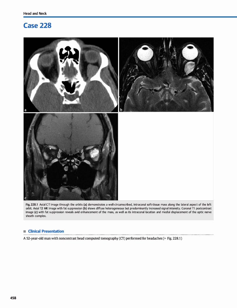
458
Head and Neck
Case 228
Fig. 228.1 Axial CT image through the orbits (a) demonstrates a well-circumscribed, intraconal soft-tissue mass a long the lateral aspect of the left orbit. Axial T2 MR image with fat suppression (b) shows diffuse heterogeneous but predominantly increased signal intensity. Coronal Tl postcontrast image (c) with fat suppression reveals avid enhancement of the mass, as well as its intraconal location and medial displacement of the optic nerve sheath complex.
• Clinical Presentation
A 52-year-old man with noncontrast head computed tomography (CT) performed for headaches (� Fig. 228.1 )

• Key lmaging Finding
Circumscribed, enhancing orbital mass
• Top 3 Differential Diagnoses
• Cavemous hemangioma. Orbital cavemous hemangiomas are truly non-neoplastic, encapsulated vascular malformations (VMs). They represent the most common intraorbital masses and often present with proptosis or pain. Lesions are well-circumscribed a with round, oval, or lobulated configuration. Most commonly, they are intraconal and retrobulbar in location, although they may occur anywhere within the orbit. They are typically hyperdense on cr. hypointense on T1, and hyperintense on T2 magnetic resonance imaging (MRI) sequences. Intense enhancement is the rule. Calcifications are occasionally seen and are characteristic. Larger lesions may remodel adjacent bony structures.
• Lymphatic: maHormation. Orbital lymphatic malformations are benign hamartomatous lesions that occur in children and
young adults. Patients typically present with proptosis as the lesion enlarges. Although they most commonly involve the extraconal compartment, orbital lymphatic malformations
• Additional Differential Diagnoses
• Metastases. Metastases may involve any portion of the orbit, including the bony orbit, globe, intraconal, and extraconal spaces. Breast and lung cancer are the most common primary tumors in adults, followed by prostate, gastrointestinal, genitourinary, and sort-tissue sarcomas. In c:hildren, neuroblastoma, leukemia, Wilms tumor, and Ewing sarcoma are most common. Larger lesions may result in proptosis and loss of visual acuity.
• Lymphoma. Lymphoma usually involves the orbit secondarily and is of the non-Hodgkin variant. Patients are often elderly and present with proptosis or painless swelling. Lymphomatous infiltrates are commonly well-circumscribed and may be
bilateral, a helpful distinguishing feature. cr may show increased density with enhancement. Lesions are low to intermediate in signal intensity on T1 and variable in signal
• Diagnosis
cavemous hemangioma
� Pearls
• Cavernous hemangiomasfVMs are the most common orbital masses, intensely enhance, and may have calcifications.
• Lymphatic malformations are often multilocular cystic lesions; hemorrhagic fluid-fluid levels are characteristic.
Suggested Reading
Tanak A, Mihara F, Yoshiura T, et al Differentiation of caven10us hemangioma from schwannoma of the orbit Am j Rœntgenol 2004; 183: 1799-1804
Case 228
may involve any and multiple compartments. They are typically multilocular, cystic, and prone ta hemorrhage. Fluidfluid levels are common and characteristic after episodes of hemorrhage. Enhanœment of septations is typically seen.
• Meningioma. Orbital meningiomas most often occur in middle-aged women; they may aise occasionally occur in children with neurofibromatosis type 2 (NF2). Extraconal involvement is typically due to intraorbital spread from a sphenoid wing or cavemous sinus meningioma. Optic nerve sheath meningiomas, on the other band, are true intraconal lesions that typically do not extend intracranially, a differentiating feature from an optic nerve glioma Meningiomas enhanœ avidly and may have calcifications. The enhanœment pattern is c:haracterized as a Mtram track" configuration on axial images (bright nerve
sheath/meningioma enhanœment peripherally and hypodense [cr] or hYPointense [Il MRI] optic nerve centrally). Meningiomas commonly are associated with eyperostosis.
intensity on T2 sequences. Common areas of involvement include the lacrimal gland and superior aspect of the orbit.
• Nerve sheath tumor. Both schwannomas and neurofibromas may occur within the orbit. Schwannomas are usually more heterogeneous and prone to cystic degeneration. They are often intermediate in signal intensity on T1 and hyperintense on T2 sequences with heterogeneous but avid enhancement. Neurofibromas are generally more homogeneous but Jess well defined and typically occur in the setting of NFl ; they are commonly multiple and bilateral. MRI reveals an intermediate T1 and hyperintense T2 signal intensity mass with heterogeneous enhanœment. The Mtarget" sign, c:haracterized by central decreased and peripheral increased T2 signal intensity, is more frequently seen with neurofibromas than schwannomas.
• Extraconal meningiomas involve the orbit secondarily; intraconal meningiomas occur along the optic nerve.
• Metastases and lymphoma involve the orbit secondarily and may involve multiple orbital structures.
459
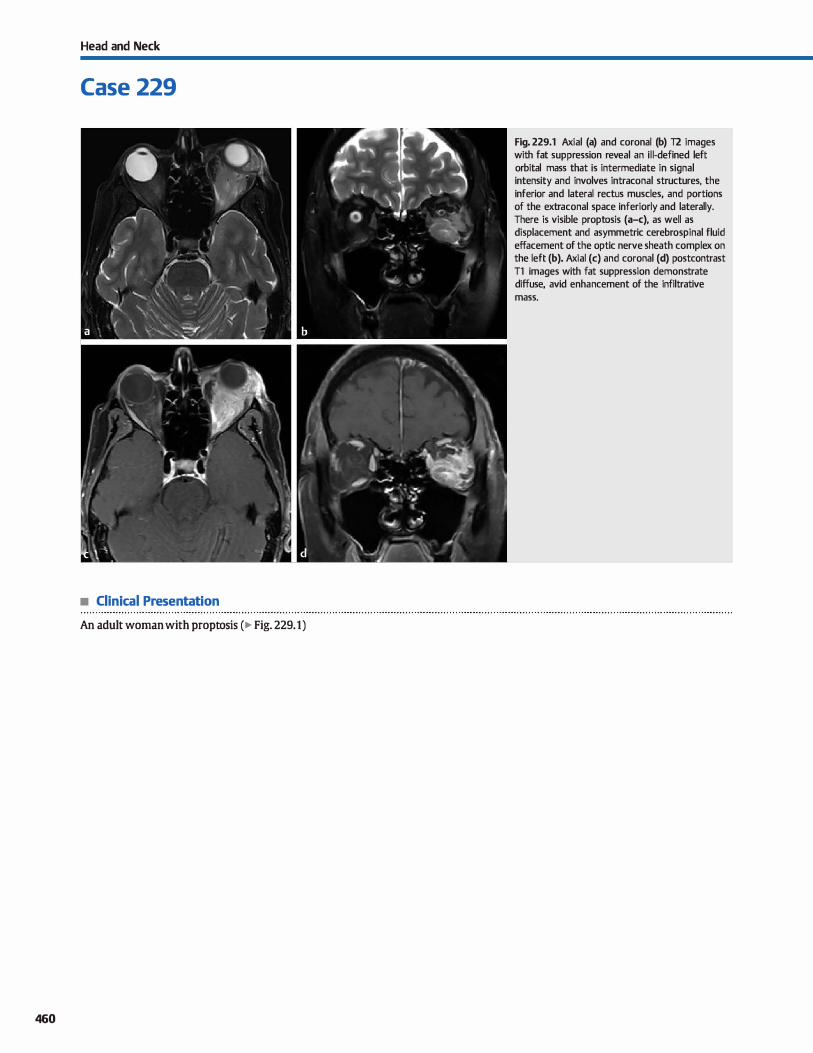
Head and Neck
Case 229
• Clinical Presentation
An adult woman with proptosis (� Fig. 229.1)
460
Fig. 229.1 Axial (a) and coronal (b) T2 images with fat suppression reveal an il l-defined left orbital mass that is intermediate in signal intensity and involves intraconal structures, the inferior and lateral rectus muscles, and portions of the extraconal space inferiorly and laterally. There is visible proptosis (a-c), as well as displacement and asymmetric cerebrospinal fluid effacement of the optic nerve sheath complex on the left (b). Axial (c) and coronal (d) postcontrast Tl images with fat suppression demonstrate diffuse, avid enhancement of the infiltrative mass.
• Key lmaging Finding
Infiltrative orbital lesion in an adult
• Top 3 Differential Diagnoses
• Orbital pseudotumor. Orbital pseudotumor, also referred to as idiopathie orbital inflammatory disease, is the most common cause of a painful orbital mass in adults. It commonly presents with infiltrative or masslike soft-tissue enhancement involving any part of the orbit or with extraocular muscle enlargement that includes the myotendinous junction; when isolated lateral rectus muscle enlargement is seen, it is most
likely due to orbital pseudotumor and essentially never due to thyroid orbitopathy. Other presentations include enlargement of the lacrimal gland and involvement of retro bulbar fat. Intracranial extension of pseudotumor into the cavernous sinus is known as Tolosa-Hunt syndrome.
• Leukemiaflymphoma. Orbital involvement of leukemia and lymphoma has a variety of presentations, which include ill
defined infiltration of the intra- and/or extraconal spaœs or a well-defined enhancing mass that is typically extraconal in location. Lacrimal gland involvement is also common. Because lymphoproliferative disorders are systemic processes,
• Additional Differential Diagnoses
• Infectious process. Orbital infections may result from adja
cent sinonasal infections, hematogenous spread of a distant infection, direct spread from an overlying cellulitis, or in cases of trauma or iatrogenia. Fungal infections are more common
in immunocompromised patients. Periorbital cellulitis refers to infection involving the presepta] soft tissues; orbital cellulitis refers to postseptal extension of the infectious process. The orbital septum consists of fibrous tissue that extends from the anterior bony periosteum of the orbit to the palprebal
soft tissues. lt is a barrier to the spread of infection from superficial preseptal soft tissues into the orbital compartment. Patients with postseptal orbital cellulitis commonly present with proptosis, ophthalmoplegia, altered visual acuity, and chemosis. lmaging reveals inflammatory changes with fatty infiltration and ill-defined regions of enhancement Focal absœsses demonstrate central fluid attenuation or
• Diagnosis
Orbital lymphoma
� Pearts
• Orbital pseudotumor commonly presents with infiltrative or
masslike soft-tissue enhancement. • Lymphoproliferative processes have a variety of presenta
tions, including ill- or well-defined orbital masses.
Suggested Reading
MQ!ler-Forell W, Pitz S. Orbital pathology. Eur J Radiol 2004; 49: 105-142
Case 229
lesions may be unilateral or bilateral. Lesions enhance fairly avidly. T2 magnetic resonance imaging (MRI) signal varies, ranging from hypo- to hyperintense depending upon the cellularity of the lesion.
• Granulomatous process. Sarcoidosis and Wegener granulomatosis commonly involve orbital structures. Sarcoidosis is a
systemic disease characterized by noncaseating granulomas; Wegener granulomatosis is a systemic vasculitis that primarily affects the kidneys and respiratory tract. Both processes often present with ill-defined and infiltrative enhandng orbital masses; scierai involvement is also commonly seen. Additional findings of sarcoid indude optic nerve involve
ment and abnormal intracranial meningeal enhancement Secondary findings for Wegener granulomatosis include adja
cent sinus involvement with bony erosions. Soft-tissue lesions on MRI are variable but commonly demonstrate intermediate to hypointense 11 and T2 signal intensity with enhancement
signal intensity with rim enhancement. Complications
include vision Joss, thrombosis of the superior ophthalmic vein, and intracranial spread of the infectious process. ail of which should be interrogated on cross-sectional imaging.
• Metastases. Metastases may involve any portion of the orbit, including the bony orbit, globe, intraconal, and extraconal spaces. Breast and Jung cancer are most common in adults, followed by prostate, gastrointestinal, genitourinary, and softtissue sarcomas. Schirrhous breast metastases may result in progressive enophthalmos. ln children, neuroblastoma, leukemia, Wilms tumor, and Ewing sarcoma represent the
most common primary neoplasms with orbital involvement. Intraorbital lesions may be well defined or ill defined with
infiltrative enhancement. Larger lesions may result in proptosis and Joss of visual acuity.
• Sarcoidosis and Wegener granulomatosis are systemic processes that often have helpful secondary findings.
• Patients with orbital cellulitis often present with fatty infiltration and ill-defined regions of enhancement
461
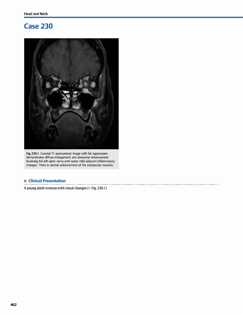
462
Head and Neck
Case 230
Fig. 230.1 Coronal Tl postcontrast image with fat suppression demonstrates diffuse enlargement and abnormal enhancement involving the left optic nerve with some mild adjacent inflammatory changes. There is normal enhancement of the extraocular muscles.
• Clinical Presentation
A young adult woman with visual changes (� Fig. 230.1 )
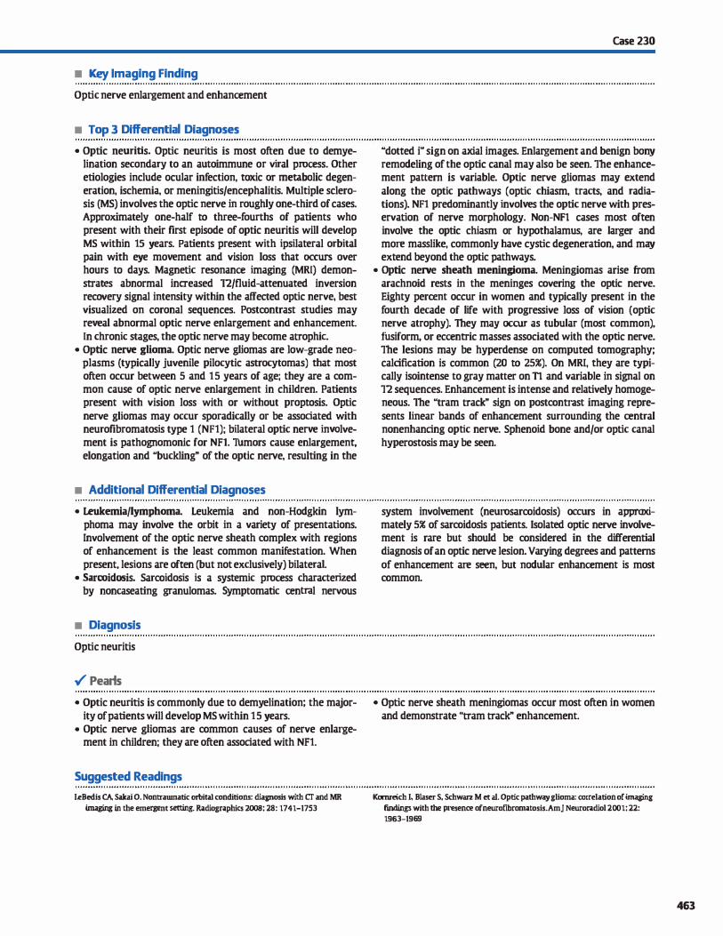
• Key lmaging Finding
Optic nerve enlargement and enhanœment
• Top 3 Differential Diagnoses
• Optic neuritis. Optic neuritis is most often due to demye
lination secondary to an autoimmune or viral proœss. Other
etioJogies include ocular infection, toxic or metaboJic degen
eration, ischemia, or meningitis/encephalitis. Multiple sclero
sis (MS) involves the optic nerve in roughly one-third of cases.
Approximately one-half to three-fourths of patients who
present with their first episode of optic neuritis will develop
MS within 15 years. Patients present with ipsilateral orbital
pain with eye movement and vision Joss that occurs over hours to days. Magnetic resonanœ imaging (MRI) demon
strates abnormal increased T2/fluid-attenuated inversion
recovery signal intensity within the affected optic nerve, best
visualized on coronal sequences. Postcontrast studies may
reveal abnormal optic nerve enlargement and enhancement
In chronic stages, the optic nerve may become atrophie.
• Optic nerve glioma. Optic nerve gliomas are Jow-grade neo
pJasms (typically juvenile pilocytic astrocytomas) that most
often occur between 5 and 15 years of age; they are a com
mon cause of optic nerve enlargement in children. Patients
present with vision Joss with or without proptosis. Optic
nerve gliomas may occur sporadically or be associated with
neurofibromatosis type 1 (NF1); bilateral optic nerve involve
ment is pathognomonic for NFl. Tumors cause enlargement,
eJongation and Mbuckling" of the optic nerve, resuJting in the
• Additional Differential Diagnoses
• Leukemiaflymphoma. Leukemia and non-Hodgkin Jym
phoma may involve the orbit in a variety of presentations.
Involvement of the optic nerve sheath complex with regions
of enhanœment is the least common manifestation. When
present, lesions are often (but not exclusively) bilateral
• Sara>idosis. Sarcoidosis is a systemic proœss characterized
by noncaseating granulomas. Symptomatic central nervous
• Diagnosis
Optic neuritis
� Pearls
• Optic neuritis is commonly due to demyelination: the major
ity of patients will develop MS within 15 years.
• Optic nerve gliomas are common causes of nerve enJargement in children; they are often assodated with NF1.
Suggested Readings
LeBedis CA, Sakai O. Nontrawnatic orbital conditions: diagnosis with CT and MR
imaging in the emergent setting. Radiographies 2008; 28: 1741-1753
Case 230
udotted i" sign on axial images. Enlargement and benign bony
remodeling of the optic canal may aise be seen. The enhance
ment pattern is variable. Optic nerve gliomas may extend
along the optic pathways ( optic chiasm, tracts, and radia
tions). NF1 predominantly involves the optic nerve with preservation of nerve morphoJogy. Non-NFl cases most often
invoJve the optic chiasm or hypothalamus, are larger and
more masslike, commonly have cystic degeneration, and may
extend beyond the optic pathways.
• Optic nerve sheath meningioma. Meningiomas arise from
arachnoid rests in the meninges covering the optic nerve.
Eighty percent occur in women and typically present in the
fourth decade of life with progressive Joss of vision ( optic
nerve atrophy). They may occur as tubular (most common),
fusiform, or ecœntric masses associated with the optic nerve.
The Jesions may be hyperdense on computed tomography;
calcification is common (20 to 25%). On MRI, they are typi
cally isointense to gray matter on Tl and variable in signal on
T2 sequences. Enhancement is intense and relatively homogeneous. The Mtram track" sign on postcontrast imaging repre
sents linear bands of enhanœment surrounding the central
nonenhandng optic nerve. Sphenoid bone and/or optic canal
hyperostosis may be seen.
system involvement (neurosarcoidosis) occurs in approxi
mately 5% of sarcoidosis patients. Isolated optic nerve involve
ment is rare but should be considered in the differential
diagnosis of an optic nerve lesion. Varying degrees and patterns
of enhanœment are seen, but nodular enhanœment is most common.
• Optic nerve sheath meningiomas occur most often in women
and demonstrate "tram track" enhancement.
Komreich L, Blaser S, Schwarz M et al. Optic pathway glioma: correlation of imaging
findings with the presenœ ofneurofibromatosis.AmJ Neuroradiol 2001; 22:
1963-1969
463
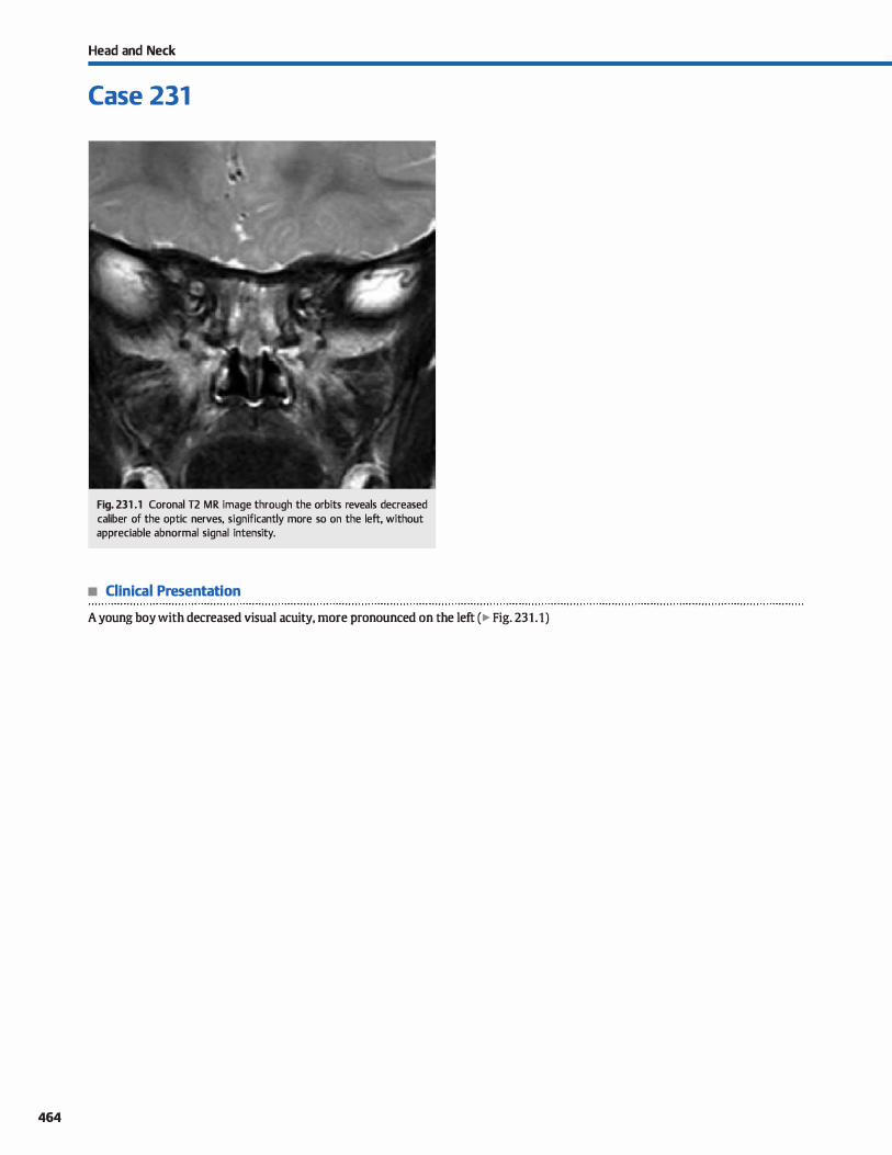
464
Head and Neck
Case 231
Fig. 231 .1 Coronal T2 MR image through the orbits reveals decreased caliber of the optic nerves, significantly more so on the left, without appreciable abnormal signal intensity.
• Clinical Presentation
A young boy with decreased visual acuity, more pronounced on the left ( � Fig. 231.1)
• Key lmaging Finding
Small-caliber optic nerve(s)
• Top 2 Differential Diagnoses
• Optic nerve atrophy. Optic nerve atrophy is often the end result of chronic optic nerve injury, typically associated with long-standing optic neuritis. In children and young adults, demyelinating processes are the most common etiology, whereas ischemic neuropathy is the most common cause in middle-aged and aider adults and is more pronounced in the setting of diabetes mellitus, hypertension, and hyperlipide
mia. Acutely, optic neuritis typically presents with optic nerve edema, abnormal signal intensity, and mild nerve enlargement on magnetic resonance imaging (MRI). Enhancement is commonly seen, especially during periods of active injury or inflammation. Chronically, there is optic nerve volume loss; the presence of abnormal signal or enhancement is variable in late-stage disease. Perineural inflammation or neuritis may
be seen with bath acute and chronic disease. • Optic nerve hypoplasia. Optic nerve hypoplasia {ONH) refers
ta congenital underdevelopment of the optic nerve. It is more
• Diagnosis
Optic nerve hypoplasia (SOD)
v" Pearls
• Optic nerve atrophy is often the end result of chronic optic
nerve injury, typically long-standing optic neuritis. • Demyelinating disease occurs more often in younger patients;
ischemic neuropathy occurs in older adults.
Suggested Reading
BilrkDvich AJ, Pram EK, Norman D. Sep!D-optic dysplasia; MR imaging. Radiology
1989; 171: 189-192
Case 231
often bilateral and is commonly associated with additional central nervous system malformations or syndromes, including septooptic dysplasia {SOD). Optic symptoms present in early childhood and include loss of visual acuity, nystagmus,
and strabismus. Nonorbital symptoms are dependent upon additional underlying malformations but commonly include developmental delay and endocrinopathies because hypothalamic-pituitary anomalies are commonly associated with optic nerve hypoplasia and SOD. MRI is the imaging modality of choice and demonstrates decreased caliber of one or both optic nerves and the optic chiasm. ln the setting of SOD, the septum pellucidum is partially or completely absent: schizencephaly, pituitary hypoplasia, and an ectopie posterior pituitary are often coexistent. Cortical malformations, heterotopias, and complete or partial agenesis of the corpus callosum may be seen.
• ONH refers to underdevelopment of the optic nerve, is most
often bilateral, and is associated with SOD.
465
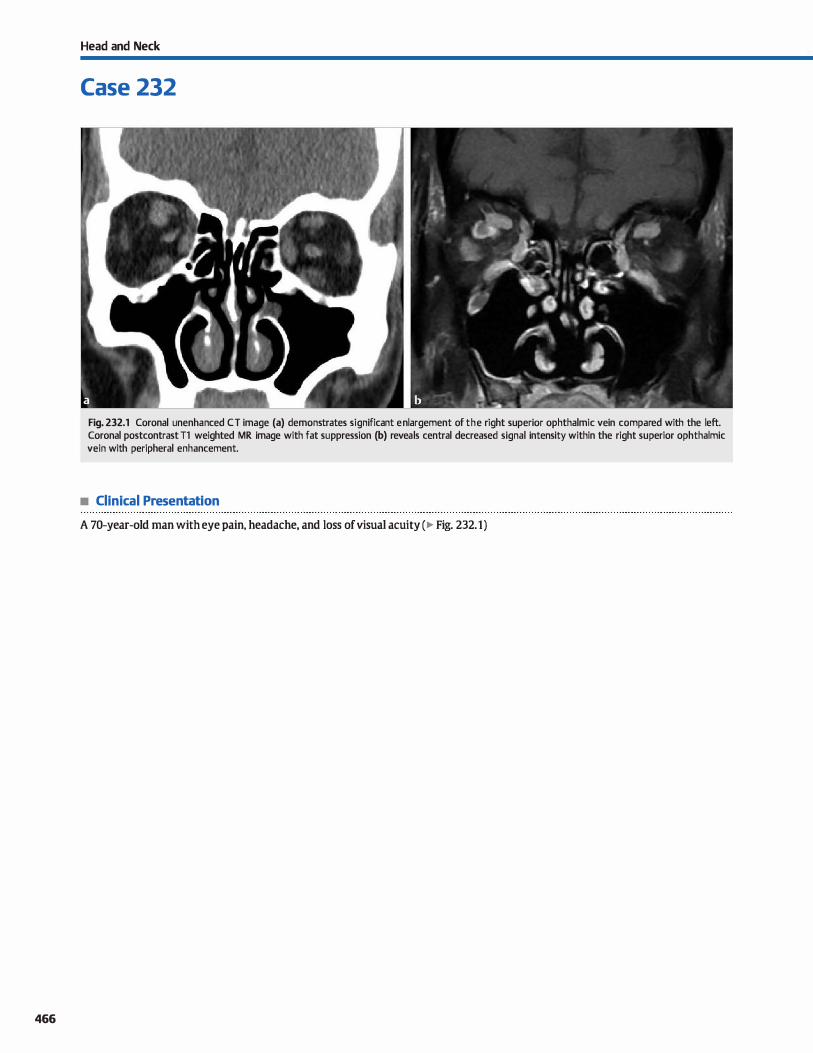
466
Head and Neck
Case 232
Fig. 232.1 Coronal unenhanced CT image (a) demonstrates significant en largement of the right superior ophthalmic vein compared with the left. Coronal postcontrast Tl weighted MR image with fat suppression (b) reveals central decreased signal intensity within the right superior ophthalmic vein with peripheral enhancement.
• Clinical Presentation
A 70-year-old man with eye pain, headache, and Joss ofvisual acuity (� Fig. 232.1)
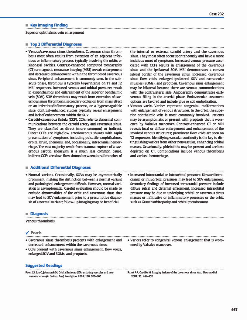
• Key lmaging Finding
Superior ophthalmic vein enlargement
• Top 3 Differential Diagnoses
• Venous/cavemous sinus thrombosis. Cavemous sinus thrombosis most often results from extension of an adjacent infectious or inflammatory proœss, typically involving the orbits or sinonasal cavities. Contrast-enhanœd computed tomography (CT) or magnetic resonanœ imaging (MRI) reveals enlargement and decreased enhancement within the thrombosed cavemous sinus. Peripheral enhancement is commonly seen. In the subacute phase, thrombus is typically hyperintense on T1 and T2
MRI sequenœs. Increased venous and orbital pressures result in exophthalmos and enlargement of the superior ophthalmic vein (SOV). sav thrombosis may result from extension of cavernous sinus thrombosis, secondary occlusion from mass effect or an infectious/inflammatory proœss, or a hypercoagulable state. Contrast-enhanœd studies typically reveal enlargement and lack of enhanœment within the SOV.
• CMotid-cavernous fistula (CCF). CCFs refer to abnormal communications between the carotid artery and cavernous sinus. They are classified as direct (more common) or indirect. Direct CCFs are high-flow arteriovenous shunts with rapid presentation of symptoms, including pulsatile exophthalmos, orbital bruit, chemosis, and, occasionally, intracranial hemorrhage. The vast majority result from trauma; rupture of a cavernous carotid aneurysm is a much less comrnon cause. Indirect CCFs are slow-flow shunts between durai branches of
• Additional Differential Diagnoses
• Nonnal variant. Occasionally, SOVs may be asymmetrically prominent, making the distinction between a normal variant and pathological enlargement difficult However, normal variation is asymptomatic. Careful evaluation should be made to exdude abnormalities of the orbit and cavernous sinus that may lead to SOV enlargement prior to a presumptive diagnosis of a normal variant; follow-up imaging may be beneficial.
• Diagnosis
Venous thrombosis
� Pearls
• Cavernous sinus thrombosis presents with enlargement and decreased enhancement within the cavernous sinus.
• CCFs present with cavemous sinus enlargement, flow voids, enlarged SOV and EOMs, and proptosis.
Suggested Readings
Poon CS, Sze G,Johnson MH. Orbital lesions: differentiating vascular and non
v.iscular etiologic factors. Am] RDentgenol 2008; 190: 956-965
Case 232
the internai or externat carotid artery and the cavernous sinus. They most often occur spontaneously and have a more insidious onset of symptoms. Increased venous pressure associated with CCFs results in enlargement of the cavemous sinus and the ipsilateral SOV. MRI demonstrates a convex lateral border of the cavemous sinus, increased cavemous sinus flow voids, enlarged ipsilateral SOV and extraocular muscles (EOMs). and proptosis. Cavemous sinus enlargement may be bilateral because there are venous communications with the contralateral side. Angiography demonstrates early venous filling in the arterial phase. Endovascular treatment options are favored and include glue or coil embolization.
• Venous varix. Varices represent congenital malformations with enlargement ofvenous structures. In the orbit, the superior ophthalmic vein is most commonly involved. Patients may be asymptomatic or present with proptosis that is worsened by Valsalva maneuver. Contrast-enhanœd CT or MRI reveals focal or diffuse enlargement and enhancement of the involved venous structures; prominent flow voids are seen on T2 sequenœs. Identifying vascular continuity is the key to distinguishing varices from other nonvascular, enhancing orbital masses. Occasionally, phleboliths may be present and are best depicted on cr. Complications include venous thrombosis and variœal hemorrhage.
• lncreased intracranial or intraorbital pressure. Elevated intracranial or intraorbital pressures may lead to sav enlargement Secondary tindings of increased intracranial pressure indude diffuse sulcal and cisternal effacement Increased intraorbital pressure may be due to underlying orbital or cavemous sinus masses or infiltrative or inflammatory proœsses or the orbit, such as Crave's orbitopathy and orbital pseudotumor.
• Varices refer to congenital venous enlargement that is worsened by Valsalva maneuver.
Razek M Castillo M. Imaging lesions of the �s sinus. Amj Neuroradiol
2009; 30: 444-452
467
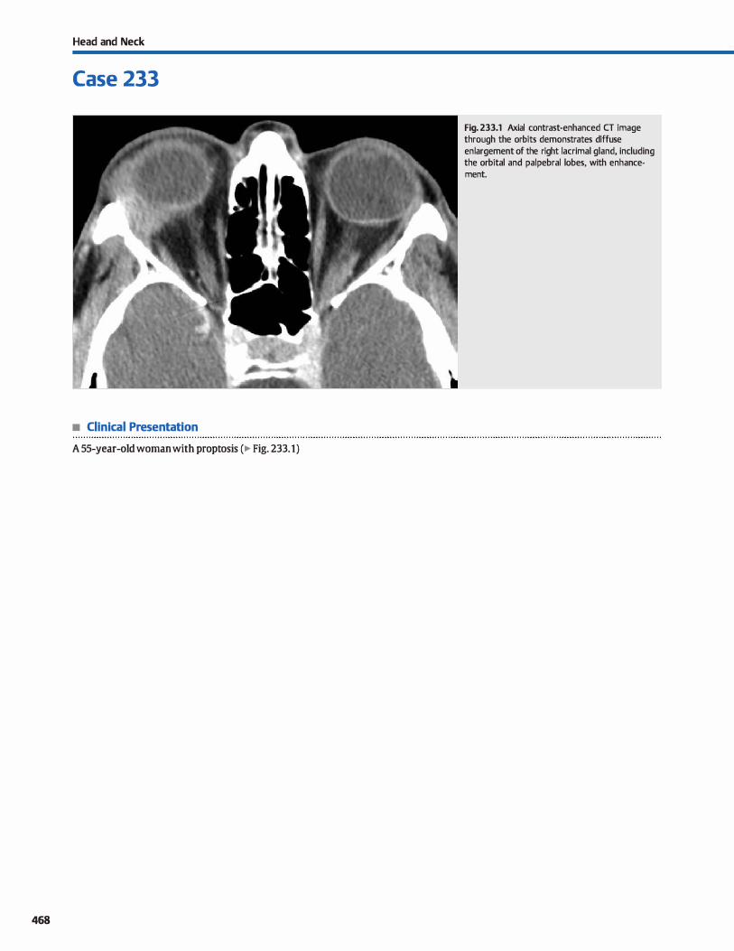
Head and Neck
Case 233
• Clinical Presentation
A 55-year-old woman with proptosis (� Fig. 233.1)
468
Fig. 233.1 Axial contrast-enhanced CT image through the orbits demonstrates diffuse en largement of the right lacrimal gland, induding the orbital and palpebral lobes, with enhancement.
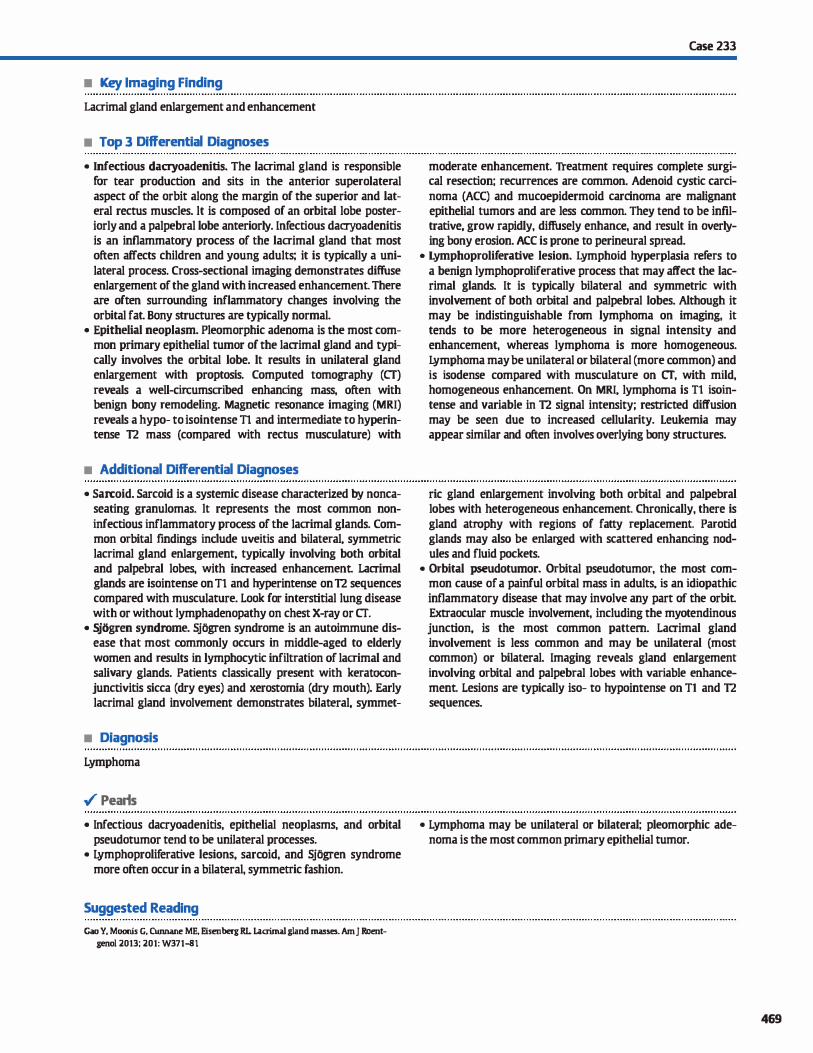
• Key lmaging Finding
Lacrimal gland enlargement and enhanœment
• Top 3 Differential Diagnoses
• lnfectious dacryoadenitis. The lacrimal gland is responsible for tear production and sits in the anterior superolateral aspect of the orbit along the margin of the superior and lateral rectus muscles. lt is composed of an orbital lobe posteriorly and a palpebral lobe anteriorly. Infectious dacryoadenitis is an inflammatory process of the lacrimal gland that most often affects children and young adults; it is typically a uni
lateral process. Cross-sectional imaging demonstrates diffuse enlargement of the gland with increased enhanœment There are often surrounding inflammatory changes involving the
orbital fat. Bony structures are typically normal. • Epithelial neoplasm. Pleomorphic adenoma is the most com
mon primary epithelial tumor of the lacrimal gland and typically involves the orbital lobe. lt results in unilateral gland enlargement with proptosis. Computed tomography (CT)
reveals a well-circumscribed enhancing mass, often with benign bony remodeling. Magnetic resonanœ imaging (MRI)
reveals a hypo- to isointense Tl and intermediate to hyperintense T2 mass (compared with rectus musculature) with
• Additional Differential Diagnoses
• Sarcoid. Sarcoid is a systemic disease characterized by noncaseating granulomas. lt represents the most common non
infectious inflammatory proœss of the Iacrimal glands. Common orbital findings indude uveitis and bilateral, symmetric lacrimal gland enlargement, typically involving both orbital and palpebral lobes, with increased enhanœment Lacrimal glands are isointense on Tl and hyperintense on T2 sequences compared with musculature. Look for interstitial lung disease with or without lymphadenopathy on chest X-ray or CT.
• Sjügren syndrome. Sjôgren syndrome is an autoimmune disease that most commonly occurs in middle-aged to elderly
women and results in lymphocytic infiltration of lacrimal and salivary glands. Patients classically present with keratocon
junctivitis sicca (dry eyes) and xerostomia (dry mouth). Early lacrimal gland involvement demonstrates bilateral, symmet-
• Diagnosis
Lymphoma
y' Pearls
• Infectious dacryoadenitis, epithelial neoplasms, and orbital pseudotumor tend to be unilateral processes.
• Lymphoproliferative lesions, sarcoid, and Sjôgren syndrome more often occur in a bilateral, symmetric fashion.
Suggested Reading
Gao Y, Moonis G, Cunnane ME, Eisenberg RI. Lacrimal gland masses. Amj Roent
genol 2013; 201: W371-81
Case 233
moderate enhanœment. li'eatment requires complete surgical resection; recurrenœs are common. Adenoid cystic carci
noma (ACC) and mucoepidermoid cardnoma are malignant epithelial tumors and are Jess common. They tend to be infiltrative, grow rapidly, diffusely enhanœ, and result in overlying bony erosion. ACC is prone to perineural spread.
• Lymphoproliferative lesion. Lymphoid hyperplasia refers to
a benign lymphoproliferative process that may affect the lacrimal glands. It is typically bilateral and symmetric with involvement of both orbital and palpebral lobes. Although it may be indistinguishable from lymphoma on imaging, it tends to be more heterogeneous in signal intensity and enhancement, whereas lymphoma is more homogeneous.
Lymphoma may be unilateral or bilateral (more common) and is isodense compared with musculature on CT, with mild, homogeneous enhanœment On MRI, lymphoma is T1 isointense and variable in T2 signal intensity; restricted diffusion may be seen due to increased œllularity. Leukemia may appear similar and often involves overlying bony structures.
rie gland enlargement involving both orbital and palpebral lobes with heterogeneous enhancement Chronically, there is
gland atrophy with regions of fatty replacement Parotid glands may also be enlarged with scattered enhancing nodules and fluid pockets.
• Orbital pseudotumor. Orbital pseudotumor, the most common cause of a painful orbital mass in adults, is an idiopathie inflammatory disease that may involve any part of the orbit Extraocular muscle involvement, including the myotendinous junction, is the most common pattern. Lacrimal gland involvement is Jess common and may be unilateral (most common) or bilateral. Imaging reveals gland enlargement
involving orbital and palpebral lobes with variable enhancement Lesions are typically iso- to hypointense on Tl and T2 sequences.
• Lymphoma may be unilateral or bilateral; pleomorphic adenoma is the most common primary epithelial tumor.
469
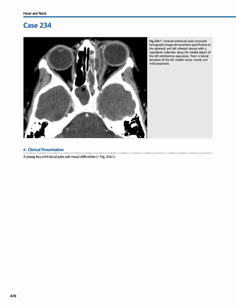
Head and Neck
Case 234
• Clinical Presentation
A young boy with facial pain and visual difficulties ( � Fig. 234.1 )
470
Fig. 234.1 Contrast-enhanced axial computed tomography image demonstrates opacification of the sphenoid and left ethmoid sinuses with a hypodense collection along the medial aspect of the left orbit/lamina papyracea. There is lateral deviation of the left medial rectus muscle and mild propotosis.
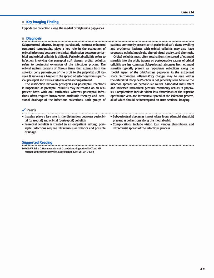
• Key lmaging Finding
Hypodense collection along the media! orbit/lamina papyracea
• Diagnosis
Subperiosteal abscess. Imaging, particularly contrast-enhanced computed tomography, plays a key raie in the evaluation of orbital infections because the dinical distinction between periorbital and orbital œllulitis is difficult Periorbital œllulitis refers to infection invoMng the preseptal soft tissues; orbital œllulitis refers to postseptal extension of the infectious proœss. The orbital septum consists of fibrous tissue that extends from the
anterior bony periosteum of the orbit ta the palprebal soft tissues. It serves as a barrier to the spread of infection from superficial preseptal soft tissues into the orbital compartment
The distinction between preseptal and postseptal infections is important, as preseptal œllulitis may be treated on an outpatient basis with oral antibiotics, whereas postseptal infections often require intravenous antibiotic therapy and occa
sional drainage of the infectious collections. Bath groups of
� Pearts
• Imaging plays a key raie in the distinction between periorbital (preseptal) and orbital (postseptal) cellulitis.
• Preseptal œllulitis is treated in an outpatient setting; postseptal infections require intravenous antibiotics and possible drainage.
Suggested Reading
LeBedis CA, Sakai O. Nontrawnatic orbital conditions: diagnosis with CT and MR
imaging in the emergent setting. Radiographies 2008; 28: 1741-1753
Case 234
patients commonly present with periorbital soft-tissue swelling and erythema. Patients with orbital cellulitis may also have proptosis, ophthalmoplegia, altered visual acuity, and chemosis.
Orbital œllulitis most often results from the spread of ethmoid sinusitis into the orbit; trauma or postoperative causes of orbital
œllulitis are Jess common. Subperiosteal absœsses from ethmoid sinusitis typically present as hypodense collections along the
media! aspect of the orbit/lamina papyracea in the extraconal spaœ. Surrounding inflammatory changes may be seen within the orbital fat. Bony destruction is not generally seen because the infection spreads via perivascular routes. Associated mass effect and increased intraorbital pressure commonly results in proptosis. Complications indude vision Joss. thrombosis of the superior
ophthalmic vein, and intracranial spread of the infectious process, ail of which should be interrogated on cross-sectional imaging.
• Subperiosteal absœsses (most often from ethmoid sinusitis) present as collections along the medial orbit.
• Complications indude vision Joss, venous thrombosis, and intracranial spread of the infectious process.
471
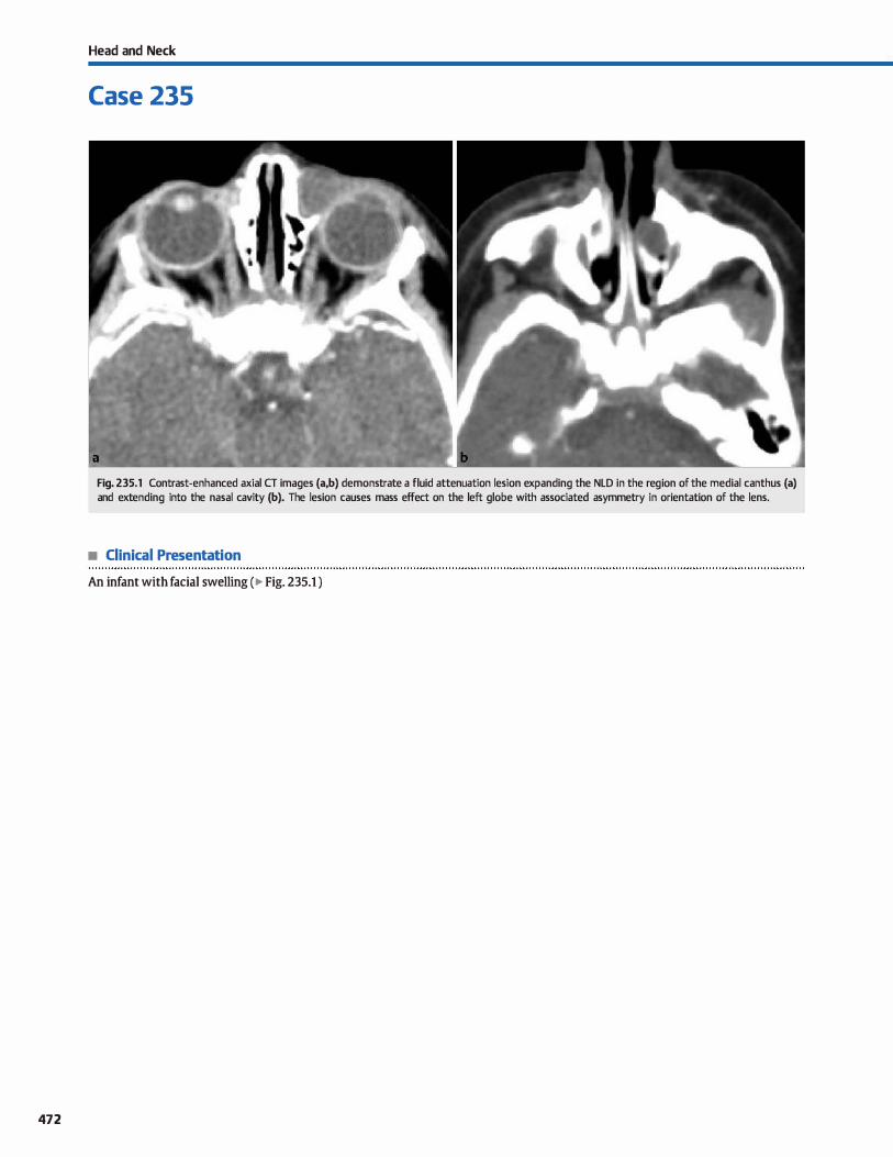
472
Head and Neck
Case 235
Fig. 235.1 Contrast-enhanced axial CT images (a,b) demonstrate a fluid attenuation lesion expanding the NLD in the reg ion of the medial canthus (a) and extending into the nasal cavity (b). The lesion causes mass effect on the left globe with associated asymmetry in orientation of the lens.
• Clinical Presentation
An infant with facial swelling ( � Fig. 235.1 )
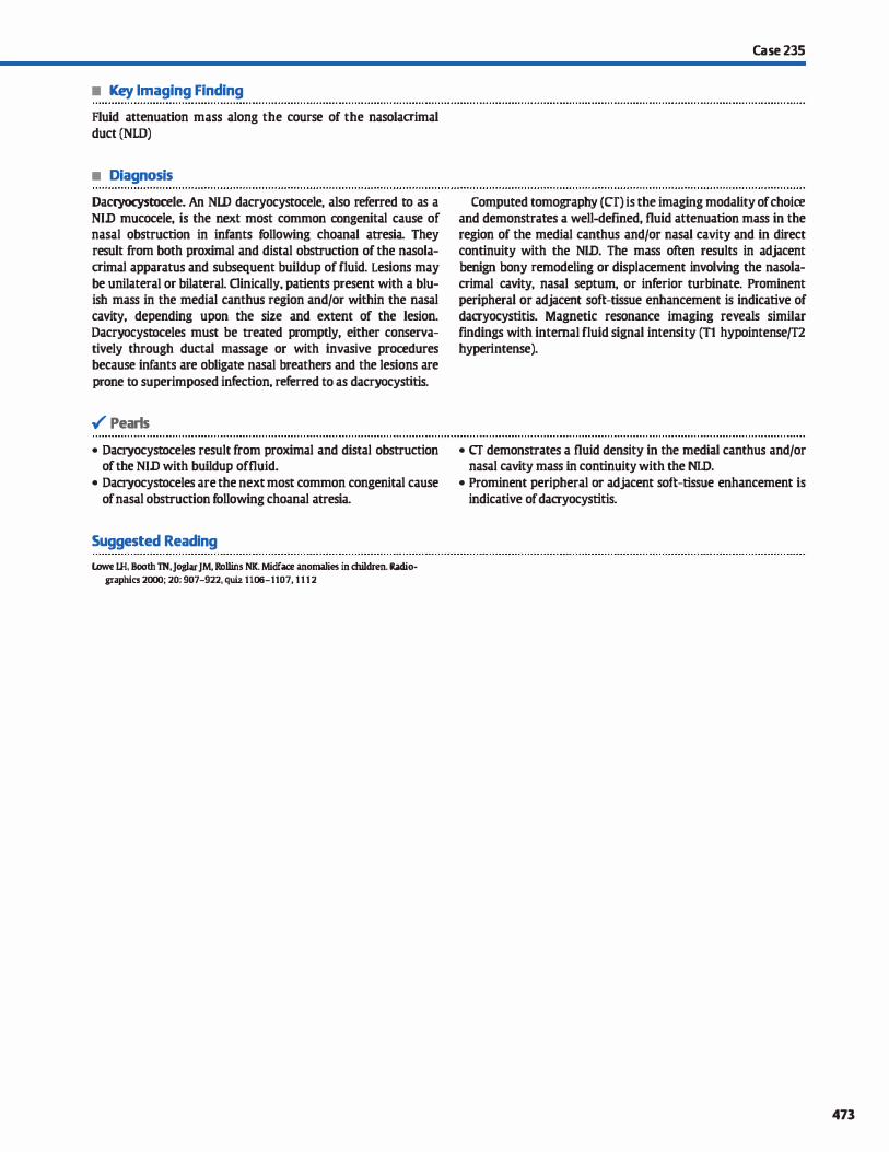
• Key lmaging Finding
Fluid attenuation mass along the course of the nasolacrimal
duct (NLD)
• Diagnosis
Dacryocystoœle. An NID dacryocystocele, also referred to as a NID mucocele, is the next most common congenital cause of nasal obstruction in infants following choanal atresia. They result from both proximal and distal obstruction of the nasola
crimal apparatus and subsequent buildup of fluid. Lesions may
be unilateral or bilateral. Oinically, patients present with a bluish mass in the medial canthus region and/or within the nasal
cavity, depending upon the size and extent of the lesion.
Dacryocystoceles must be treated promptly, either conservatively through ductal massage or with invasive procedures because infants are obligate nasal breathers and the lesions are
prone to superimposed infection, referred to as dacryocystitis.
� Pearls
• Dacryocystoceles result from proximal and distal obstruction of the NID with buildup offluid.
• Dacryocystoceles are the next most common congenital cause
of nasal obstruction following choanal atresia.
Suggested Reading
Lowe œ, Booth TN, Joglar JM, Rollins NK. Midfaœ anomalies in dilldren. R.ô1dio·
graphies 2000; 20; 907-922, quiz 1106-1107, 1112
Case 235
Computed tomography ( CT) is the imaging modality of choiœ and demonstrates a well-defined, fluid attenuation mass in the region of the medial canthus and/or nasal cavity and in direct continuity with the NID. The mass often results in adjacent
benign bony remodeling or displacement involving the nasola
crimal cavity, nasal septum, or inferior turbinate. Prominent
peripheral or adjacent soft-tissue enhancement is indicative of dacryocystitis. Magnetic resonance imaging reveals similar
findings with internai fluid signal intensity (Tt hypointense/T2 hyperintense ).
• CT demonstrates a fluid density in the medial canthus and/or nasal cavity mass in continuity with the NLD.
• Prominent peripheral or adjacent soft-tissue enhancement is
indicative of dacryocystitis.
473
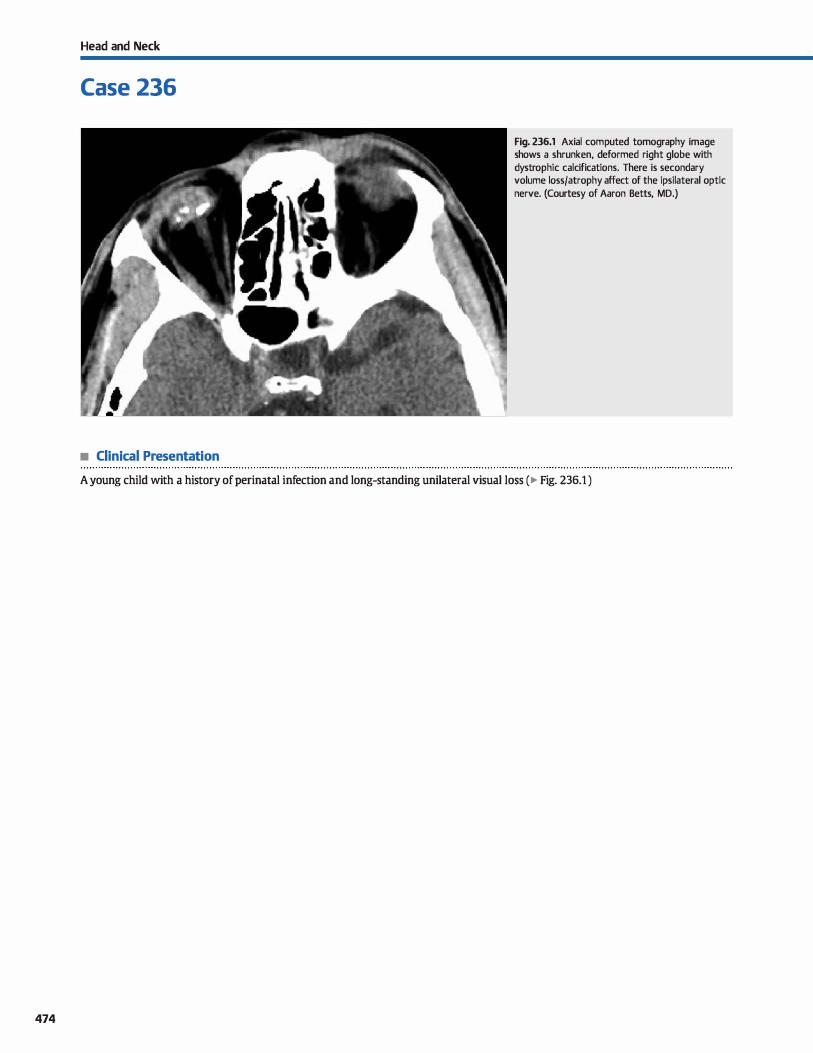
474
Head and Neck
Case 236
• Clinical Presentation
Fig. 236.1 Axial computed tomography image shows a shrunken, deformed right globe with dystrophie calcifications. There is secondary volume loss/atrophy affect of the ipsilateral optic nerve. (Courtesy of Aaron Betts, MD.)
A young child with a history of perinatal infection and long-standing unilateral visual Joss ( � Fig. 236.1 )
• Key lmaging Finding
Shrunlœn. deformed globe with dystrophie calcifications
• Diagnosis
Phthisis bulbi. Phthisis bulbi refers to end-stage ocular degeneration due to an underlying insult. Common causes indude ocular trauma and a myriad of long-standing, infectious, inflammatory, or autoirnmune proœsses. Rarely, phthisis bulbi may oa:ur in the setting of underlying tumor necrosis associated with retinoblastoma and melanoma Patients present with blindness in the
� Pearls
• Phthisis bulbi refers to end-stage ocular degeneration often due to trauma or an infectious/inflammatory insult.
• Computed tomography demonstrates an irregular, disorganized, shrunlœn globe with coarse calcifications.
Suggested Reading
LeBedis CA, Sakai O. Nontraumatic orbital conditions: diagnosis with cr and MR
imaging in the emergent setting. Radiographies 2008; 28: 1741-1753
Case 236
affected eye; treatment options indude enudeation with prosthesis placement
Imaging demonstrates an irregular, disorganized, shrunken globe with coarse calàfications. Atrophy of the optic nerve is a helpful secondary finding that may be seen in chronic cases. The assoàated volume loss often results in enophthalmos.
• Atrophy of the optic nerve and enophthalmos assoàated with volume Joss are helpful secondary findings.
475
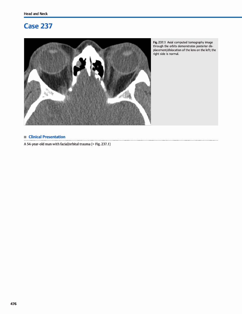
Head and Neck
Case 237
• Clinical Presentation
A 54-year-old man with facial/orbital trauma (� Fig. 237.1)
476
Fig. 237.1 Axial computed tomography image through the orbits demonstrates posterior displacement/dislocation of the lens on the left; the right side is normal.
• Key lmaging Finding
Displacement/dislocation of the Jens
• Diagnosis
Lens dislocation. The Jens represents the demarcation between the anterior and posterior segments of the globe. It is attached to the overlying sciera via zonular fibers. Lens dislocation results from tearing of the zonular fibers, which may result from trauma (most common) or underlying connective tissue disorders, such as Ehlers-Danlos syndrome and homocystinuria. The dislocation
may be partial or complete depending on the presenœ or
absence of intact zonular fibers.
� Pearts
• Lens dislocations may result from trauma or connective tissue disorders (Ehlers-Danlos syndrome and homocystinuria).
• Post-traumatic Jens dislocations are typically posterior; anterior is Jess common.
Suggested Readings
Kubal WS. Imaging of orbital trawna. Radiographies 2008; 28: 1729-1739
Case 237
Post-traumatic Jens dislocations are typically posterior. Com
puted tomography shows malpositioning/ dislocation of the hyperdense Jens. It is important to look for additional globe or orbital injuries. Spontaneous Jens dislocations assoàated with Marfan syndrome are commonly bilateral and superotemporal
in location. Those assoàated with homocystinuria are also bilateral but typically inferonasal in location. Treatment is ulti
mately surgical in most cases.
• Lens dislocations assoàated with Marfan syndrome are commonly bilateral and superotemporal in location.
• Lens dislocations assoàated with homocystinuria are typically bilateral and inferonasal in location.
Betts AM, O'Brien Wf, Davie! JJW, Youssef OH. A systemic approach to CT evaluation
oforbital trauma. Emerg Radiol 2014; 21(5): 511-531
477
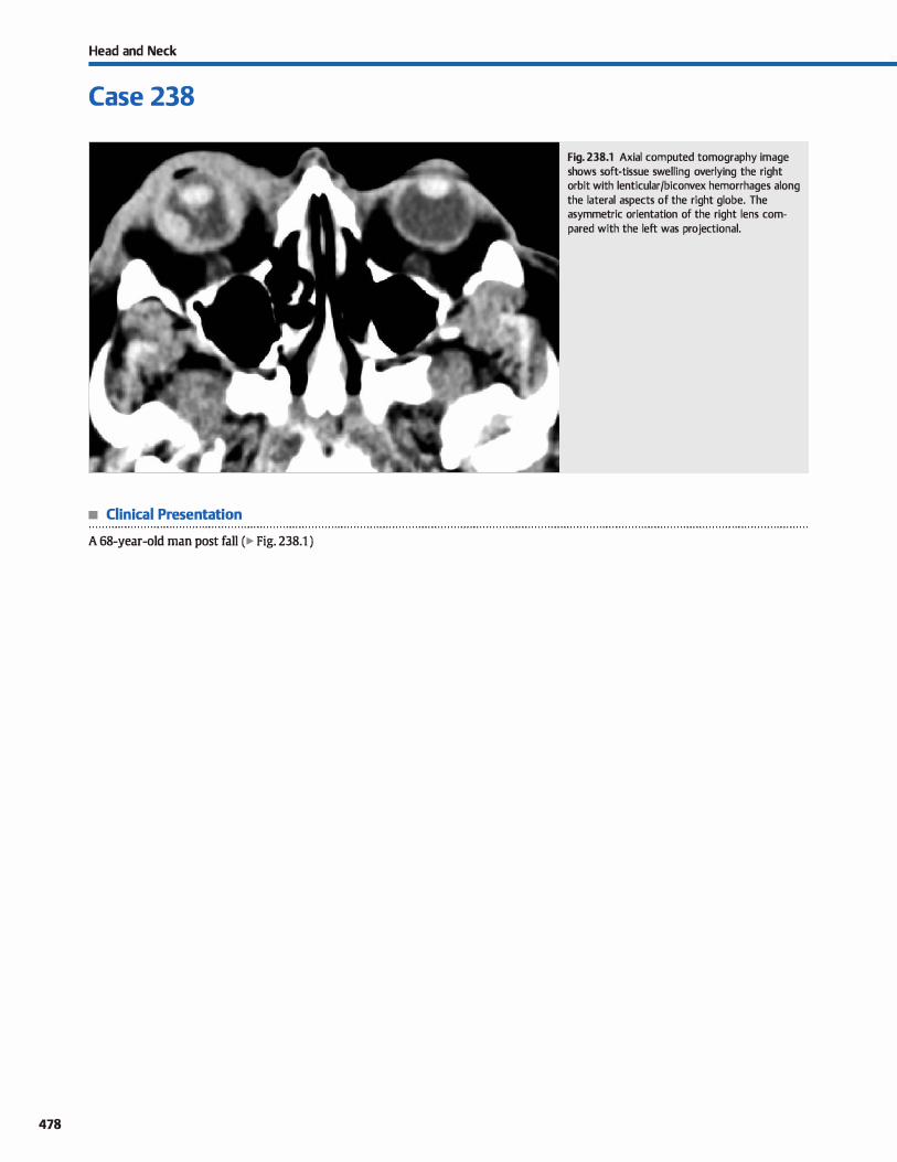
Head and Neck
Case 238
• Clinical Presentation
A 68-year-old man post fall (� Fig. 238.1 )
478
Fig. 238.1 Axial computed tomography image shows soft-tissue swelling overlying the right orbit with lenticular/biconvex hemorrhages along the lateral aspects of the right globe. The asymmetric orientation of the right lens compared with the left was projectional.
• Key lmaging Finding
Lenticular/biconvex hemorrhages along the lateral aspects of the globe
• Diagnosis
Choroidal detachment. The globe has three layers posteriorly with the sciera representing the outennost layer, the choroid representing the middle layer, and the retina representing the innennost layer. The choroid extends from the ora serrata ante
riorly to the optic nerve head posteriorly. Vascular structures allow for its attachment to the overlying sciera. With choroidal detachment, fluid (proteinaœous or hemorrhagic) accumulates between and results in separation of the choroid and sciera. Common causes include inflammatory processes, trauma, and iatrogenia, all of which result in decreased intraocular pressure.
� Pearls
• With choroidal detachment, fluid (proteinaœous or hemor
rhagic) accumulates between the choroid and sciera. • On imaging, choroidal detachments present as biconvex fluid
collections along the lateral margins of the globe.
Suggested Readings
Kubal WS. lmaging of orbital trauma. Radiographies 2008; 28; 1729-1739
Case 238
On imaging, choroidal detachments present as lenticular or biconvex fluid collections along the lateral margins of the globe. The collections are hyperdense compared with the vitreous on computed tomography. Unlike retinal detachments, a V-shaped configuration and extension to the optic dise are typically not seen, although choroidal detachments may occasionally mimic the appearance of a retinal detachment in the absence of the characteristic biconvex configuration.
• Unlike retinal detachments, a V-shaped configuration and
extension to the optic dise are typically not seen.
Betis AM, O'Brien Wf, Davies BW, Youssef OH. A systemic approach ta CT evaluation
of orbital trawna. EmergRadiol 2014; 21(5); 511-531
479
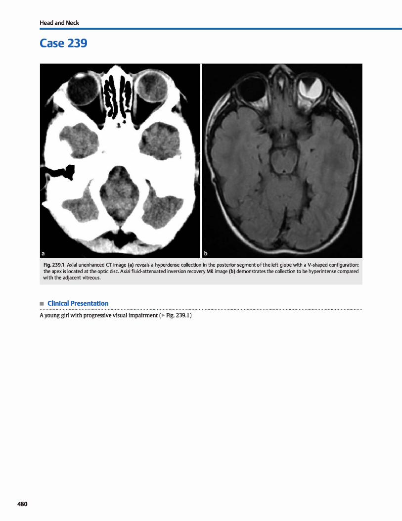
480
Head and Neck
Case 239
Fig. 239.1 Axial unenhanced CT image (a) reveals a hyperdense collection in the posterior segment of the left globe with a V-shaped configuration; the apex is located at the optic dise. Axial fluid-attenuated inversion recovery MR image (b) demonstrates the collection to be hyperintense compared with the adjacent vitreous.
• Clinical Presentation
A young girl with progressive visual impairment (� Fig. 239.1 )
• Key lmaging Finding
Ocular detachment with a V-shaped configuration
• Diagnosis
Retinal detachment. The globe has three layers posteriorly with the sciera representing the outermost layer, the choroid
representing the middle layer, and the retina representing the innermost layer. The retina is the sensory layer of the globe, and when damaged, significant visual impairment may result. lt has firm attachments anteriorly at the ora serrata and posteriorly at the optic dise. When fluid, typically hemorrhage, accu
mulates beneath the retina, there is uplifting of the more
loosely attached portions of the retina between the ora serrata and optic dise, which often gives rise to the characteristic V-shaped configuration of a retinal detachment with its apex located at the optic dise. Small, large, or long-standing retinal hemorrhages may have a less characteristic imaging appearance without the dassic V-shaped configuration.
y' Pearts
• The retina is the sensory layer of the globe, and when damaged, significant visual impairment may result.
• Retinal detachment often results in a characteristic V-shaped configuration with its apex at the optic dise.
Suggested Readings
Kubal WS. Imaging of orbital trawna. Radiographies 2008; 28: 1729-1739
Case 239
Common causes of retinal detachment indude inflammatory processes, neoplastic processes, and trauma. If noted in a young child with a questionable history, nonaccidental trauma should be a consideration. On computed tomography (CT), subretinal hemorrhage is typically hyperdense compared with the adja
cent vitreous. Its appearance on magnetic resonance imaging (MRI) is variable depending upon the stage of breakdown of
blood products. Most cases demonstrate increased signal on T1
and fluid-attenuated inversion recovery sequences with iso- to hyperintense T2 signal intensity. Treatment options indude scierai buckling or banding, placement of a silicone sponge, and intraocular gas or silicone oil tamponade.
• Retinal hemorrhage is hyperdense on CT; its MRI appearance is variable due to the stage of blood products.
Betts AM, O'Brien Wf, Davies JrW, YoW5sef OH. A systemic approach to CT evaluation
of orbital trauma. Emerg Radial 2014; 21 (5): 511-531
481

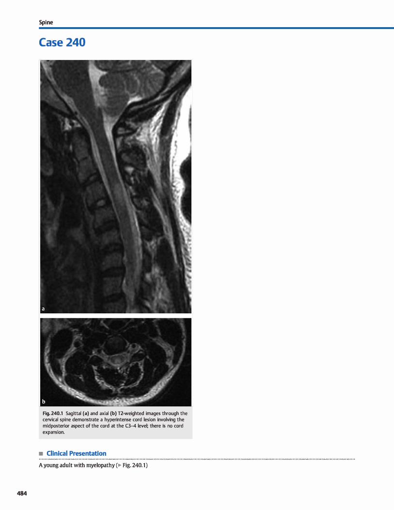
484
Spi ne
Case 240
Fig. 240.1 Sagittal (a) and axial (b) T2-weighted images through the ceivical spine demonstrate a hyperintense cord lesion involving the midposterior aspect of the cord at the C3-4 level; there is no cord expansion.
• Clinical Presentation
A young adult with myelopathy (� Fig. 240.1)
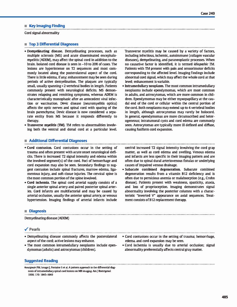
• Key lmaging Finding
Cord signal abnormality
• Top 3 Differential Diagnoses
• Demyelinating disease. Demyelinating processes, such as multiple sclerosis (MS) and acute disseminated encephalomyelitis (ADEM), may affect the spinal cord in addition to the brain. lsolated cord disease is seen in -1 O to 20% of cases. The lesions are hyperintense on T2 sequences and most com
monly located along the posterolateral aspect of the cord. There is little edema, if any; enhancement may be seen during periods of active demyelination. The plaques are typically smalt, usually spanning < 2 vertebral bodies in length. Patients commonly present with neurological deficits. MS demonstrates relapsing and remitting symptoms, whereas ADEM is
characteristically monophasic after an anteœdent viral infec
tion or vaccination. Devic disease (neuromyelitis optica) affects the optic nerves and spinal cord with sparing of the brain parenchyma; Devic disease is now considered a separate entity from MS because it responds differently to therapy.
• Transverse myelitis (lM). TM refers to abnormalities involving both the ventral and dorsal cord at a particular level.
• Additional Differential Diagnoses
• Conl contusion. Cord contusions occur in the setting of trauma and often present with acute onset neurological deficits. There is increased T2 signal intensity and edema within the involved segment( s) of the cord. Foci of hemorrhage and cord expansion may also be seen. Secondary findings ta suggest contusion include spinal fractures, marrow edema, ligamentous injury, and soft-tissue injuries. The cervical spine is
the most common portion of the spine inwlved.
• Conl ischemia. The spinal cord arterial supply consists of a single anterior spinal artery and paired posterior spinal arteries. Cord infarcts are multifactorial and may be caused by arterial ocdusion, usually the anterior spinal artery, or venous hypertension. lmaging findings of arterial infarcts indude
• Diagnosis
Demyelinating disease (ADEM)
� Pearls
• Demyelinating disease commonly affects the posterolateral
aspect of the cord; active lesions may enhance.
• The most common intramedullary neoplasms include ependymomas (adults) and astrocytomas (children).
Suggested Reading
Bourguuin PM, Lesage J, Fonmne S et al. A pattern approach ID the differential diag
nosis of intramedullary spinal rord lesions on MR imaging. Am j Roentgenol
1998; 170: 1645-1649
Case 240
Transverse myelitis may be caused by a variety of factors, including infectious, ischemic, autoimmune ( collagen vascular diseases), demyelinating, and paraneoplastic proœsses. When no causative factor is identified, it is termed idiopathie 1M. Patients with TM present with pain and sensorimotor deficits
corresponding to the affected level. Imaging findings include
abnormal cord signal, which may affect the whole cord at that level; enhanœment is variable.
• lntramedullary neoplasm. The most common intrarnedullary neoplasms indude ependymomas, which are most common in adults, and astrocytomas, which are more common in children. Ependymomas may be either myxopapillary at the cau
dal end of the cord or cellular within the central portion of
the cord. Both neoplasms may extend up to 4 vertebral bodies in length, although astrocytomas may rarely be holocord.
ln general, ependymomas are more circumscribed and heterogeneous; intratumoral cysts and cord edema are commonly seen. Astrocytomas are typically more ill defined and diffuse, causing fusiform cord expansion.
central increased T2 signal intensity inwlving the cord gray matter, as well as cord edema and swelling. Venous edema and infarcts are Jess specific in their imaging pattern and are often due to spinal durai arteriovenous flstulas or underlying causes of impaired venous drainage.
• Subacute combined degeneration. Subacute combined degeneration results from a vitamin 812 deficiency and is often due to pemidous anemia or malabsorption ( e.g., Crohn disease). Patients present with weakness, spasticity, ataxia,
and Joss of proprioception. lmaging demonstrates signal
abnormality involving the posterior columns with a characteristic uinverted V" appearance on axial sequences. Treatment consists of812 replacement therapy.
• Cord contusions occur in the setting of trauma; hemorrhage, edema. and conl expansion may be seen.
• Cord ischemia is usually due to arterial ocdusion; signal abnormality preferentially affects central gray matter.
485
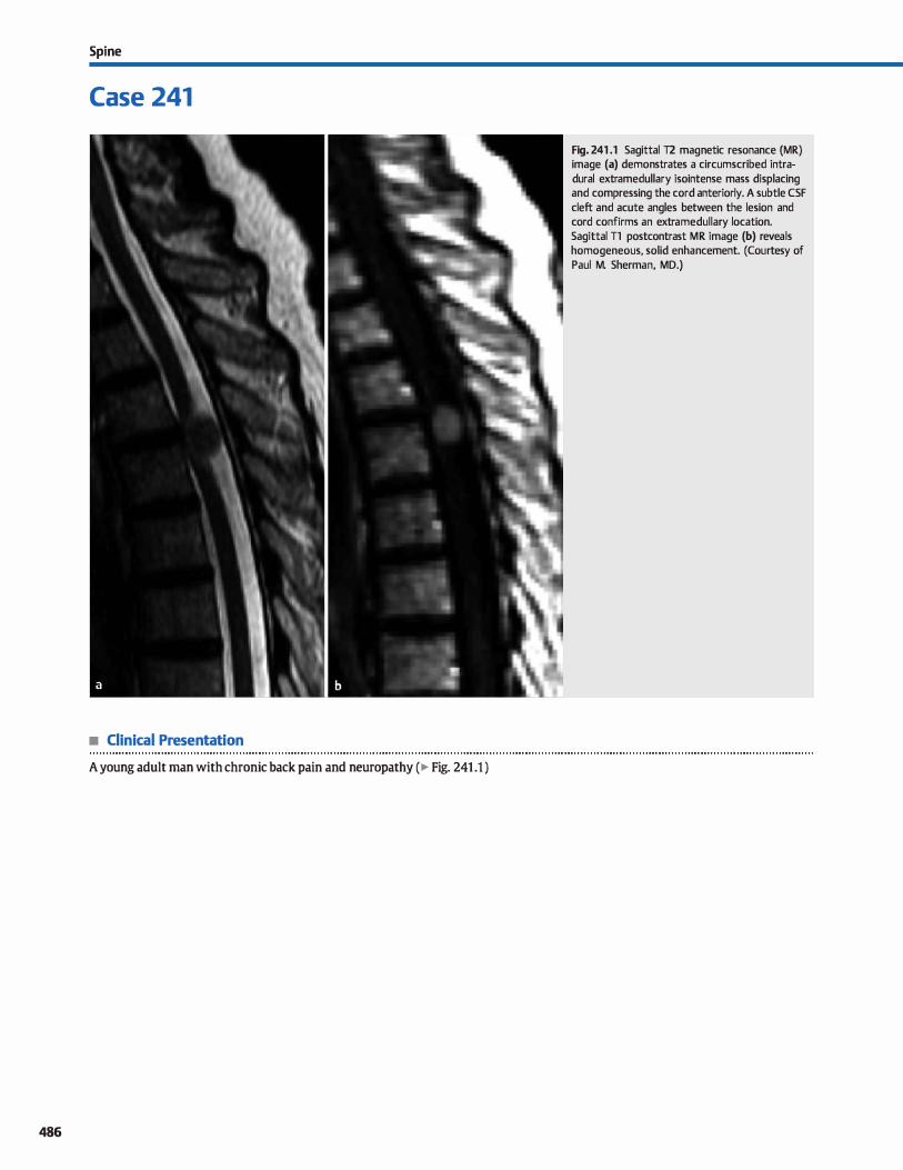
Spi ne
Case 241
• Clinical Presentation
A young adult man with chronic back pain and neuropathy ( � Fig. 241 .1 )
486
Fig. 241 .1 Sagittal T2 magnetic resonance (MR) image (a) demonstrates a circumscribed intradural extramedullary isointense mass displacing and compressing the cord anteriorly. A subtle CSF cleft and acute angles between the lesion and cord confirms an extramedullary location. Sagittal Tl postcontrast MR image (b) reveals homogeneous, solid enhancement. (Courtesy of Paul M. Sherman, MD.)
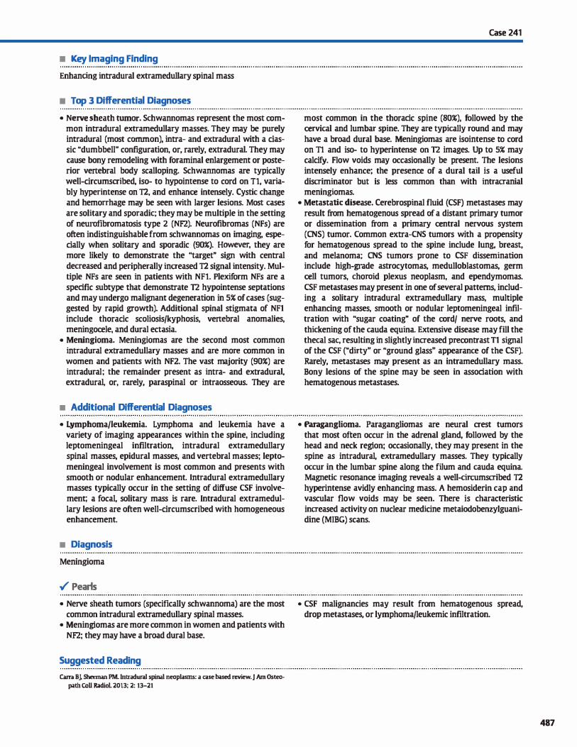
• Key lmaging Finding
Enhancing intradural extramedullary spinal mass
• Top 3 Differential Diagnoses
• Nerve sheath tumor. Schwannomas represent the most common intradural extramedullary masses. They may be purely intradural {most comrnon), intra- and extradural with a dassic udumbbell" configuration, or, rarely, extradural. They may cause bony remodeling with foraminal enlargement or posterior vertebral body scalloping. Schwannomas are typically well-drcumscribed, iso- to hypointense to cord on Tl, varia
bly hyperintense on T2, and enhance intensely. Cystic change and hemorrhage may be seen with larger lesions. Most cases are solitary and sporadic; they may be multiple in the setting of neurofibromatosis type 2 {NF2). Neurofibromas {NFs) are often indistinguishable from schwannomas on imaging, espedally when solitary and sporadic (90%). However, they are more likely ta demonstrate the "target" sign with central decreased and peripherally increased T2 signal intensity. Multiple NFs are seen in patients with NF1. Plexiform NFs are a specific subtype that demonstrate T2 hypointense septations and may undergo malignant degeneration in 5% of cases {suggested by rapid growth). Additional spinal stigmata of NFt include thoradc scoliosis/kyphosis, vertebral anomalies, meningoœle, and durai ectasia.
• Merûngioma. Meningiomas are the second most common intradural extramedullary masses and are more common in women and patients with NF2. The vast majority (90%) are intradural; the remainder present as intra- and extradural extradural, or, rarely, paraspinal or intraosseous. They are
• Additional Differential Diagnoses
• Lymphomajleukemia. Lymphoma and leukemia have a variety of imaging appearances within the spine, induding leptomeningeal infiltration, intradural extramedullary spinal masses, epidural masses, and vertebral masses; lepto
meningeal involvement is most common and presents with smooth or nodular enhancement. lntradural extramedullary masses typically occur in the setting of diffuse CSF involvement; a focal, solitary mass is rare. Intradural extramedullary lesions are often well-drcumscribed with homogeneous enhancement.
• Diagnosis
Meningioma
� Pearls
• Nerve sheath tumors {specifically schwannoma) are the most common intradural extramedullary spinal masses.
• Meningiomas are more common in women and patients with NF2; they may have a broad durai base.
Suggested Reading
Carra Bj, Sherman PM. lntradural spinal neoplasms: a case based review.J Am Osteo
path Coll RadioL 2013; 2: 13-21
Case 241
most common in the thoracic spine {80%), followed by the cervical and lumbar spine. They are typically round and may
have a broad durai base. Meningiomas are isointense ta cord on T1 and iso- ta hyperintense on T2 images. Up ta 5% may caldfy. Flow voids may occasionally be present. The lesions intensely enhance; the presenœ of a durai tail is a useful
discriminator but is Jess common than with intracranial meningiomas.
• Metastatic disease. Cerebrospinal fluid (CSF) metastases may result from hematogenous spread of a distant primary tumor or dissemination from a primary central nervous system {CNS) tumor. Common extra-CNS tumors with a propensity for hematogenous spread ta the spine include Jung. breast,
and melanoma; CNS tumors prone ta CSF dissemination include high-grade astrocytomas, medulloblastomas, germ cell tumors, choroid plexus neoplasm, and ependymomas.
CSF metastases may present in one of several patterns, including a solitary intradural extramedullary mass, multiple enhancing masses, smooth or nodular leptomeningeal infiltration with "sugar coating" of the cord/ nerve roots, and
thickening of the cauda equina. Extensive disease may fill the thecal sac, resulting in slightly increased precontrast Tt signal of the CSF ("dirty" or "ground glass" appearance of the CSF). Rarely, metastases may present as an intramedullary mass. Bony lesions of the spine may be seen in association with hematogenous metastases.
• Paraganglioma. Paragangliomas are neural crest tumors that most often occur in the adrenal gland, followed by the head and neck region; occasionally, they may present in the spine as intradural, extramedullary masses. They typically
occur in the lumbar spine along the filum and cauda equina. Magnetic resonance imaging reveals a well-circumscribed T2
hyperintense avidly enhancing mass. A hemosiderin cap and vascular flow voids may be seen. There is characteristic increased activity on nuclear medicine metaiodobenzylguanidine (MIBG) scans.
• CSF malignancies may result from hematogenous spread, drop metastases, or lymphoma/leukemic infiltration.
487
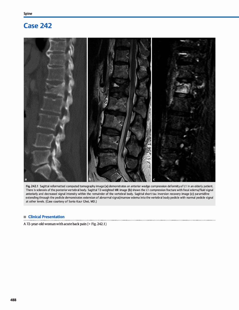
488
Spi ne
Case 242
Fig. 242.1 Sagittal reformatted computed tomography image (a) demonstrates an anterior wedge compression deformity of L 1 in an elderly patient. There is sclerosis of the posterior vertebral body. Sagittal T2-weighted MR image (b) shows the L 1 compression fracture with focal edema/fluid signal anteriorly and decreased signal intensity within the remainder of the vertebral body. Sagittal short tau inversion recovery image (c) paramidline extending through the pedicle demonstrates extension of abnormal signal/marrow edema into the vertebral body pedicle with normal pedicle signal at other levels. (Case courtesy of Sonia Kaur Ghei, MD.)
• Clinical Presentation
A 72-year-old woman with acute back pain (� Fig. 242.1 )
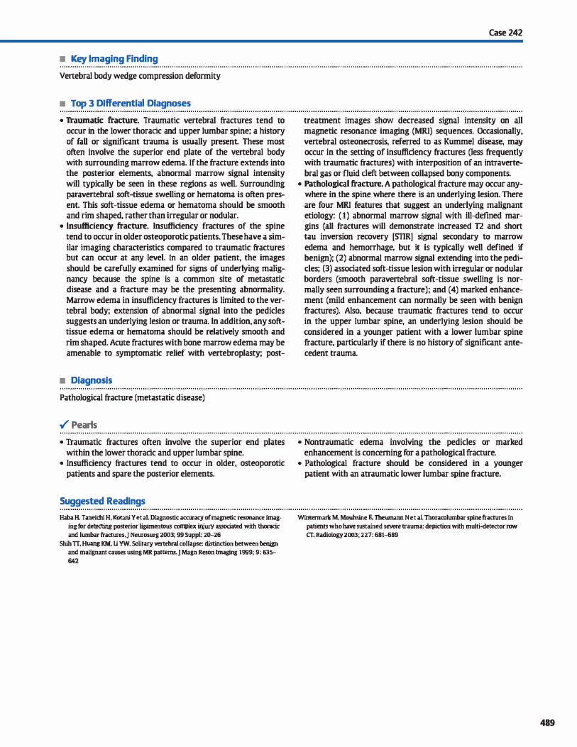
• Key lmaging Finding
Vertebral body wedge compression deformity
• Top 3 Differential Diagnoses
• li"aumatic fracture. Traumatic vertebral fractures tend to occur in the lower thoracic and upper lumbar spine; a history of fall or significant trauma is usually present These most often involve the superior end plate of the vertebral body with surrounding marrow edema. If the fracture extends into the posterior elements, abnormal marrow signal intensity will typically be seen in these regions as well. Surrounding
paravertebral soft-tissue swelling or hematoma is often pres
ent. This soft-tissue edema or hematoma should be smooth and rim shaped, rather than irregular or nodular.
• lnsufficiency fracture. Insufficiency fractures of the spine tend to occur in older osteoporotic patients. These have a sim
ilar imaging characteristics compared to traumatic fractures but can occur at any level. ln an older patient, the images
should be carefully examined for signs of underlying malignancy because the spine is a common site of metastatic disease and a fracture may be the presenting abnormality.
Marrow edema in insufficiency fractures is limited to the vertebral body; extension of abnormal signal into the pedicles suggests an underlying lesion or trauma. ln addition, any softtissue edema or hematoma should be relatively smooth and
rim shaped. Acute fractures with bone marrow edema may be amenable to symptomatic relief with vertebroplasty; post-
• Diagnosis
Pathological fracture (metastatic disease)
� Pearts
• Traumatic fractures often involve the superior end plates
within the lower thoracic and upper lumbar spine. • Insuffidency fractures tend ta occur in older, osteoporotic
patients and spare the posterior elements.
Suggested Readings
Haba H. Taneichi H, Kotani Y et al. Diagnostic accuracy of magnetic resonanœ imag
ing for detecting posterior ligamentous cornplex Îl\ÎUfY associated with thoracic
and lumbar ftactures.j Neurosurg 2003; 99 Suppl; 20-26
Shih TI, Huang KM, li YW. Solitary vertebral collapse: distinction between benign
and malignant causes using MR patterns. j Magn Reson Imaging 1999; 9; 635-
642
Case 242
treatment images show decreased signal intensity on ail magnetic resonanœ imaging (MRI) sequences. Occasionally, vertebral osteonecrosis, referred ta as Kummel disease, may occur in the setting of insufficiency fractures (Jess frequently with traumatic fractures) with interposition of an intraverte
bral gas or fluid cleft between collapsed bony components. • Pathological fracture. A pathological fracture may occur any
where in the spine where there is an underlying Iesion. There
are four MRI features that suggest an underlying malignant etiology: (1) abnormal marrow signal with ill-defined margins (ait fractures will demonstrate increased T2 and short tau inversion recovery [SllR] signal secondary to marrow
edema and hemorrhage. but it is typically well defined if benign); {2) abnormal marrow signal extending into the pedicles; (3) associated soft-tissue lesion with irregular or nodular borders {smooth paravertebral soft-tissue swelling is normally seen surrounding a fracture); and (4) marked enhancement (mild enhancement can normally be seen with benign fractures). Also, because traumatic fractures tend to occur in the upper lumbar spine, an underlying lesion should be
considered in a younger patient with a lower lumbar spine fracture, particularly if there is no history of significant ante
cedent trauma.
• Nontraumatic edema involving the pedicles or marked enhancement is conœming for a pathological fracture.
• Pathological fracture should be considered in a younger patient with an atraumatic lower lumbar spine fracture.
Wintennark M. Mouhsine E, Theumann Net al. Thoracolumbar spine fractures in
patients who have sustained sewre trauma: depiction with multi-detector row
cr. Radiology 2003; 227: 681-689
489
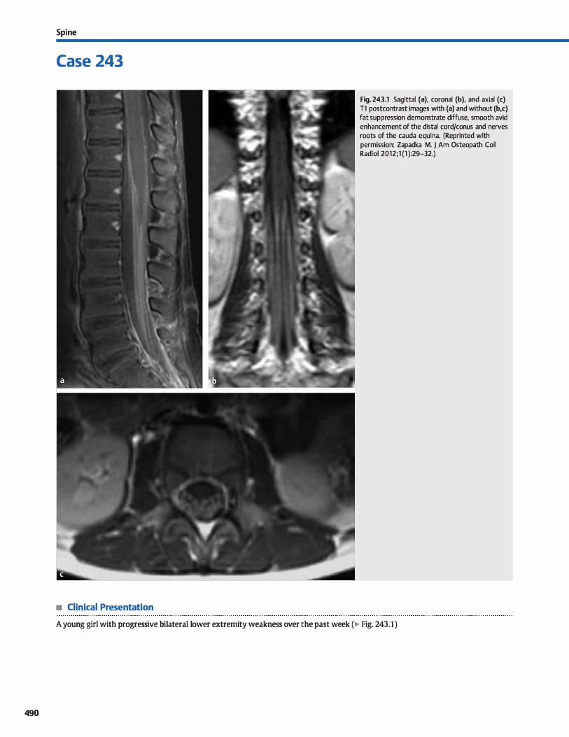
490
Spi ne
Case 243
• Clinical Presentation
Fig. 243.1 Sagittal (a), coronal (b), and axial (c) Tl postcontrast images with (a) and without (b,c) fat suppression demonstrate diffuse, smooth avid enhancement of the distal cord/conus and nerves roots of the cauda equina. (Reprinted with permission: Zapadka M. j Am Osteopath Coll Radiol 2012;1 (1 ):29-32.)
A young girl with progressive bilateral lower extremity weakness over the past week (� Fig. 243.1 )
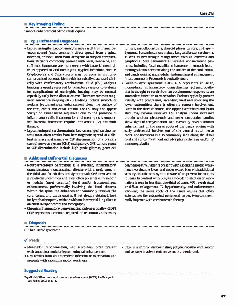
• Key lmaging Finding
Smooth enhancement of the cauda equina
• Top 3 Differential Diagnoses
• Leptomeningitis. Leptomeningitis may result from hematogenous spread (most common), direct spread from a spinal infection, or inoculation from iatrogenic or surgical complications. Patients commonly present with fever, headache, and stitf neck. Symptoms are more severe with bacterial meningitis as opposed to viral meningitis; atypical infections, such as Cryptococcus and Tuberculosis, may be seen in immunocompromised patients. Meningitis is typically diagnosed dinically with confirmatory cerebrospinal fluid (CSF) analysis. Imaging is usually reserved for refractory cases or to evaluate for complications of meningitis. Imaging may be norma� especially early in the disease course. The most common magnetic resonance imaging (MRI) findings indude smooth or nodular leptomeningeal enhancement along the surface of
the cord, conus, and cauda equina. The CSF may also appear Mdirty" on unenhanced sequences due to the presence of inflammatory cells. Treatment for viral meningitis is supportive; bacterial infections require intravenous (IV) antibiotic therapy.
• Leptomeningeal carcinomatosis. Leptomeningeal carcinomatosis most often results from hematogenous spread of a dis
tant primary malignancy or CSF dissemination of a primary central nervous system (CNS) malignancy. CNS tumors prone to CSF dissemination include high-grade gliomas, germ cell
• Additional Differential Diagnoses
• Neurosarcoidosis. Sarcoidosis is a systemic, inflammatory, granulomatous (noncaseating) disease with a peak onset in the third and fourth decades. Symptomatic CNS involvement is relatively uncommon and most often presents with smooth or nodular (most common) durai and/or leptomeningeal enhancement, preferentially involving the basal cisterns. Within the spine, the enhancement commonly involves the
cord. conus, and cauda equina. If not already obtained, look for lymphadenopathy with or without interstitial Jung disease on chest X-ray or computed tomography.
• Chronic inflammatory demyelinating polyneuropathy (CDP). ODP represents a chronic, acquired, mixed motor and sensory
• Diagnosis
Guillain-Barré syndrome
� Pearls
• Meningitis, carànomatosis, and sarcoidosis often present with smooth or nodular leptomeningeal enhancement.
• GBS results from an antecedent infection or vaccination and presents with ascending motor weakness.
Suggested Reading
Zapadka M. Diffuse cauda equina nerve root enhancement. JAOCRJ Am Osteopath
Coll RadioL 2012; 1: 29-32
Case 243
tumors, medulloblastoma, choroid plexus tumors, and ependymoma. Systemic tumors include Jung and breast carcinoma, as well as hematologic malignanàes such as leukemia and lymphoma. MRI demonstrates variable enhancement patterns, including focal masslike enhancement; smooth leptomeningeal enhancement along the surface of the cord, conus, and cauda equina; and nodular leptomeningeal enhanœment (most common). Prognosis is typically poor.
• Guillain-Barré syndrome {GBS). GBS represents an acute, monophasic inflammatory demyelinating polyneuropathy that is thought to result from an autoimmune response ta an antecedent infection or vaccination. Patients typically present initially with progressive, ascending weakness involving the lower extremities; there is often no sensory involvement. I.ater in the disease course, the upper extremities and brain stem may become involved. CSF analysis shows increased protein without pleocytosis and nerve conduction studies show signs of demyelination. MRI dassically reveals smooth enhancement of the nerve roots of the cauda equina with early preferential involvement of the ventral motor nerve roots. Enhancement is also commonly seen along the distal cord and conus. Treatment includes plasmapheresis and/or IV immunoglobulin.
polyneuropathy. Patients present with ascending motor weakness involving the lower and upper extremities with additional sensory disturbanœs; symptoms are often present for months ta years. In contrast with GBS, an anteœdent infection or vaccination is seen in Jess than one-third of cases. MRI reveals foc.al or diffuse enlargement, T2 hyperintensity, and enhanœment
involving the nerve roots of the cauda equina that often extends into the extraspinal peripheral nerves. Symptoms generally improve with corticosteroid therapy.
• aDP is a chronic demyelinating polyneuropathy with motor and sensory involvement; nerve roots are enlarged.
491
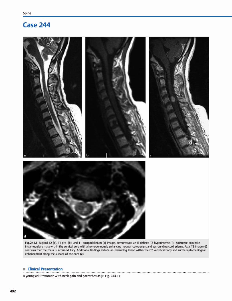
492
Spi ne
Case 244
Fig. 244.1 Sagittal T2 (a). Tl pre- (b). and T1 postgadolinium (c) images demonstrate an ill-defined T2 hyperintense, Tl isointense expansile intramedullary mass within the cervical cord with a homogeneously enhancing nodular component and surrounding cord edema. Axial T2 image (d) confirms that the mass is intramedullary. Additional findings indude an enhancing lesion within the C7 vertebral body and subtle leptomeningeal enhancement along the surface of the cord (c).
• Clinical Presentation
A young adult woman with neck pain and paresthesias (� Fig. 244.1)
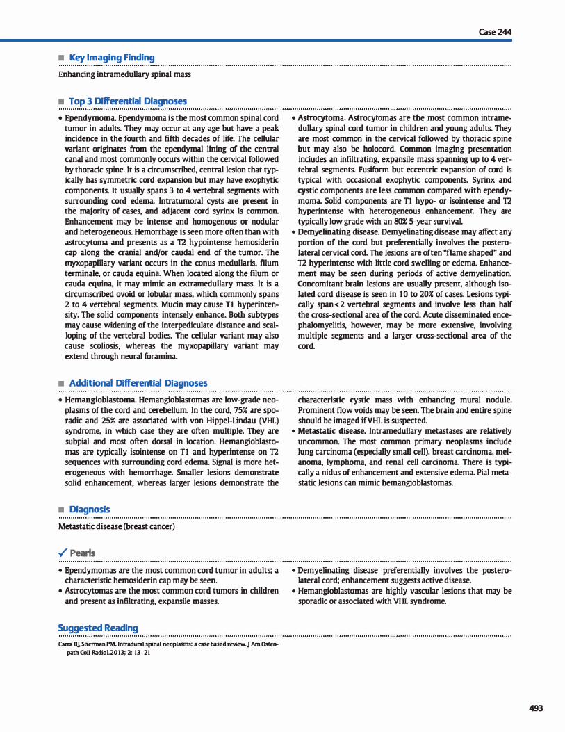
• Key lmaging Finding
Enhancing intramedullary spinal mass
• Top 3 Differential Diagnoses
• Ependymoma. Ependymoma is the most common spinal cord tumor in adults. They may occur at any age but have a peak incidence in the fourth and fifth decades of life. The cellular variant originates from the ependymal lining of the central canal and most commonly occurs within the cervical followed
by thoradc spine. lt is a drcumscribed, central lesion that typically has symmetric cord expansion but may have exophytic
components. It usually spans 3 to 4 vertebral segments with surrounding cord edema. lntratumoral cysts are present in the majority of cases, and adjacent cord syrinx is common.
Enhanœment may be intense and homogenous or nodular and heterogeneous. Hemorrhage is seen more often than with astrocytoma and presents as a T2 hypointense hemosiderin cap along the cranial and/or caudal end of the tumor. The
myxopapillary variant occurs in the conus medullaris, filum terminale, or cauda equina When located along the filum or cauda equina, it may mimic an extramedullary mass. lt is a circumscribed ovoid or lobular mass, which commonly spans 2 to 4 vertebral segments. Mudn may cause Tl hyperintensity. The solid components intensely enhance. Beth subtypes may cause widening of the interpediculate distance and scal
loping of the vertebral bodies. The cellular variant may also cause scoliosis, whereas the myxopapillary variant may extend through neural foramina.
• Additional Differential Diagnoses
• Hemangioblastoma. Hemangioblastomas are low-grade neoplasms of the cord and œrebellum. ln the cord, 75% are sporadic and 25% are assodated with von Hippel-Lindau {VHL) syndrome, in which case they are often multiple. They are
subpial and most often dorsal in location. Hemangioblastomas are typically isointense on Tl and hyperintense on T2 sequences with surrounding cord edema. Signal is more heterogeneous with hemorrhage. Smaller lesions demonstrate solid enhancement, whereas larger lesions demonstrate the
• Diagnosis
Metastatic disease (breast cancer)
� Pearls
• Ependymomas are the most common cord tumor in adults; a characteristic hemosiderin cap may be seen.
• Astrocytomas are the most common cord tumors in children and present as infiltrating. expansile masses.
Suggested Reading
Carra BJ, Sherman PM. Intradural spinal neoplasms: a case based review.J Am Osteo
path C.011 Radiol2013; 2: 13-21
Case 244
• Astrocytoma. Astrocytomas are the most common intramedullary spinal cord tumor in children and young adults. They
are most common in the cervical followed by thoracic spine but may also be holocord. Common imaging presentation indudes an infiltrating, expansile mass spanning up to 4 vertebral segments. Fusiform but eccentric expansion of cord is typical with occasional exophytic components. Syrinx and
cystic components are less common compared with ependymoma. Solid components are Tl hypo- or isointense and T2 hyperintense with heterogeneous enhanœment They are typically low grade with an 80% 5-year survival.
• Demyelinating disease. Demyelinating disease may affect any
portion of the cord but preferentially involves the posterolateral cervical cord. The lesions are often Mflame shaped" and T2 hyperintense with little cord swelling or edema. Enhancement may be seen during periods of active demyelination. Concomitant brain lesions are usually present, although isolated cord disease is seen in 1 O to 20% of cases. Lesions typically span < 2 vertebral segments and involve Jess than half the cross-sectional area of the cord. Acute disseminated encephalomyelitis, however, may be more extensive, involving
multiple segments and a larger cross-sectional area of the cord.
characteristic cystic mass with enhandng mural nodule. Prominent flow voids may be seen. The brain and entire spine should be imaged ifVHI. is suspected.
• Metastatic disease. lntramedullary metastases are relatively
uncommon. The most common primary neoplasms indude lung carcinoma ( especially small cell), breast carcinoma, melanoma, lymphoma, and renal œll carcinoma. There is typically a nidus of enhancement and extensive edema. Pial metastatic lesions can mimic hemangioblastomas.
• Demyelinating disease preferentially involves the posterolateral cord; enhancement suggests active disease.
• Hemangioblastomas are highly vascular lesions that may be sporadic or associated with VHL syndrome.
493
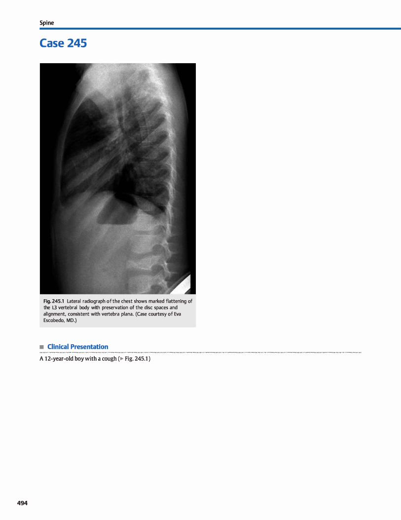
494
Spi ne
Case 245
Fig. 245.1 Lateral radiograph of the chest shows marked flattening of the L3 vertebral body with preservation of the dise spaces and alignment, consistent with vertebra plana. (Case courtesy of Eva Escobedo, MD.)
• Clinical Presentation
A 12-year-old boy with a cough (� Fig. 245.1 )
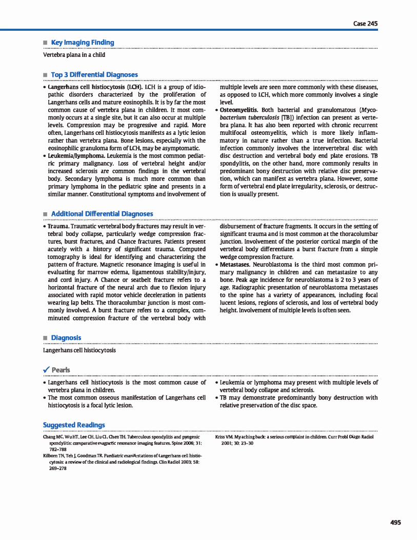
• Key lmaging Finding
Vertebra plana in a child
• Top 3 Differential Diagnoses
• Langerhans cell histiocytosis (LCH). LCH is a group of idiopathie disorders characterized by the proliferation of
Langerhans œlls and mature eosinophils. lt is by far the most common cause of vertebra plana in children. lt most commonly occurs at a single site, but it can also occur at multiple levels. Compression may be progressive and rapid. More often, Langerhans œll histiocytosis manifests as a lytic lesion
rather than vertebra plana. Bone lesions, especially with the eosinophilic granuloma form of LCH, may be asymptomatic.
• Leukemiaflymphoma. Leukemia is the most common pediat
ric primary malignancy. Loss of vertebral height and/or increased sclerosis are common findings in the vertebral
body. Secondary lymphoma is much more common than
primary lymphoma in the pediatrie spine and presents in a
similar manner. Constitutional symptoms and involvement of
• Additional Differential Diagnoses
• Trauma. Traumatic vertebral body fractures may result in ver
tebral body collapse, particularly wedge compression fractures, burst fractures, and Chance fractures. Patients present acutely with a history of significant trauma. Computed
tomography is ideal for identifying and characterizing the pattern of fracture. Magnetic resonanœ imaging is useful in
evaluating for marrow edema, ligamentous stability/injury,
and cord injury. A Chance or seatbelt fracture refers to a horizontal fracture of the neural arch due to flexion injury associated with rapid motor vehide deceleration in patients wearing lap belts. The thoracolumbar junction is most commonly involved. A burst fracture refers to a complex, com
minuted compression fracture of the vertebral body with
• Diagnosis
Langerhans œll histiocytosis
� Pearls
• Langerhans cell histiocytosis is the most common cause of
vertebra plana in children. • The most common osseous manifestation of Langerhans cell
histiocytosis is a focal lytic lesion.
Suggested Readings
Chang MC. Wu HT, Lee Œ, Liu Cl., Chen 1H. Tuberculous spondylitis and pyogenic
spondylitis; mmparative magnetic resonance imaging features. Spine 2006; 31;
782-788
Kilbom TN, Teh J, Goodman TR. Paediatric manifestations of Langerhans cell histio
cytosis; a review of the clinicat and r.adiological findings, Clin R.adiol 2003; 58;
269-278
Case 245
multiple levels are seen more commonly with these diseases, as opposed to LCH, which more commonly involves a single level.
• Osteomyelitis. Both bacterial and granulomatous (Mycobacterium tuberculosis [TB]) infection can present as vertebra plana. lt has also been reported with chronic recurrent multifocal osteomyelitis, which is more likely inflam
matory in nature rather than a true infection. Bacterial
infection commonly involves the intervertebral dise with dise destruction and vertebral body end plate erosions. TB spondylitis, on the other hand, more commonly results in predominant bony destruction with relative dise preservation, which can manifest as vertebra plana. However, some form ofvertebral end plate irregularity, sclerosis, or destruc
tion is usually present
disbursement of fracture fragments. lt occurs in the setting of significant trauma and is most common at the thoracolumbar junction. lnvolvement of the posterior cortical margin of the vertebral body differentiates a burst fracture from a simple
wedge compression fracture. • Metastases. Neuroblastoma is the third most common pri
mary malignancy in children and can metastasize ta any bone. Peak age incidence for neuroblastoma is 2 to 3 years of age. Radiographie presentation of neuroblastoma metastases
to the spine has a variety of appearances, including focal lucent lesions, regions of sclerosis, and Joss of vertebral body height. lnvolvement of multiple levels is often seen.
• Leukemia or lymphoma may present with multiple levels of vertebral body collapse and sclerosis.
• TB may demonstrate predominantly bony destruction with
relative preservation of the dise spaœ.
Kriss VM My aching back: a serious complaint in children. Curr Probl Diagn R.adiol
2001; 30; 23-30
495
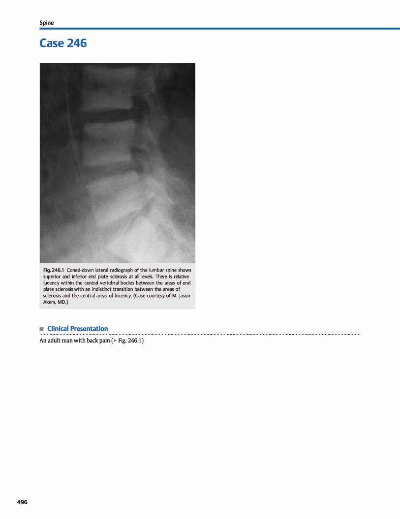
496
Spi ne
Case 246
Fig. 246.1 Coned-down lateral radiograph of the lumbar spine shows superior and inferior end plate sderosis at all levels. There is relative lucency within the central vertebral bodies between the areas of end plate sclerosis with an indistinct transition between the areas of sderosis and the central areas of lucency. (Case courtesy of M. Jason Akers, MD.)
• Clinical Presentation
An adult man with back pain ( � Fig. 246.1 )
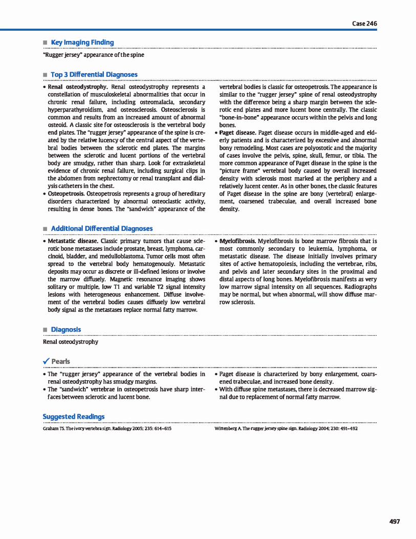
• Key lmaging Finding
"Rugger jersey" appearance of the spine
• Top 3 Differential Diagnoses
• Renal osteodystrophy. Renal osteodystrophy represents a constellation of musculoskeletal abnormalities that occur in
chronic renal failure, including osteomalada, secondary hyperparathyroidism, and osteosderosis. Osteosclerosis is common and results from an increased amount of abnormal osteoid. A dassic site for osteosderosis is the vertebral body end plates. The "rugger jersey" appearanœ of the spine is cre
ated by the relative luœncy of the central aspect of the verte
brai bodies between the sderotic end plates. The margins between the sderotic and luœnt portions of the vertebral body are smudgy, rather than sharp. Look for extraskeletal evidenœ of chronic renal failure, including surgical clips in the abdomen from nephrectomy or renal transplant and dialysis catheters in the chest.
• Osteopetrosis. Osteopetrosis represents a group of hereditary disorders characterized by abnormal osteodastic activity, resulting in dense bones. The "sandwich" appearance of the
• Additional Differential Diagnoses
• Metastatic disease. Classic primary tumors that cause sderotic bone metastases indude prostate, breast, lymphoma. cardnoid, bladder, and medulloblastoma. Tumor œlls most often spread to the vertebral body hematogenously. Metastatic
deposits may occur as discrete or ill-defined lesions or involve
the marrow diffusely. Magnetic resonanœ imaging shows solitary or multiple, low T1 and variable 12 signal intensity lesions with heterogeneous enhancement Diffuse involvement of the vertebral bodies causes diffusely low vertebral bodY signal as the metastases replace normal fatty marrow.
• Diagnosis
Renal osteodystrophy
� Pearls
• The "rugger jersey" appearance of the vertebral bodies in renal osteodystrophy bas smudgy margins.
• The "sandwich" vertebrae in osteopetrosis have sharp interfaces between sclerotic and lucent bone.
Suggested Readings
Graham TS. The ivory vertebra sign. R.adiology 2005; 235: 614-615
Case 246
vertebral bodies is classic for osteopetrosis. The appearance is similar to the "rugger jersey" spine of renal osteodystrophy with the difference being a sharp margin between the sderotic end plates and more luœnt bone œntrally. The classic
"bone-in-bone" appearance occurs within the pelvis and long bones.
• Paget disease. Paget disease occurs in middle-aged and elderly patients and is characterized by excessive and abnormal
bony remodeling. Most cases are polyostotic and the majority of cases involve the pelvis, spine, skull, femur, or tibia. The more common appearance of Paget disease in the spine is the "picture frame" vertebral body caused by overall increased
density with sderosis most marked at the periphery and a relatively lucent center. As in other bones, the dassic features of Paget disease in the spine are bony (vertebral) enlargement, coarsened trabeculae, and overall increased bone
density.
• Myelofibrosis. Myelofibrosis is bone marrow fibrosis that is most commonly secondary to leukemia, lymphoma, or metastatic disease. The disease initially involves primary sites of active hematopoiesis, induding the vertebrae, ribs,
and pelvis and later secondary sites in the proximal and distal aspects of long bones. Myelofibrosis manifests as very low marrow signal intensity on ail sequences. Radiographs may be normal, but when abnormal, will show diffuse mar
row sderosis.
• Paget disease is characterized by bony enlargement, coarsened trabeculae, and increased bone density.
• With diffuse spine metastases, there is decreased marrow signal due to replacement of normal fatty marrow.
Wittenberg A. The rugger jersey spine sign. Radiology 2004; 230: 491-492
497
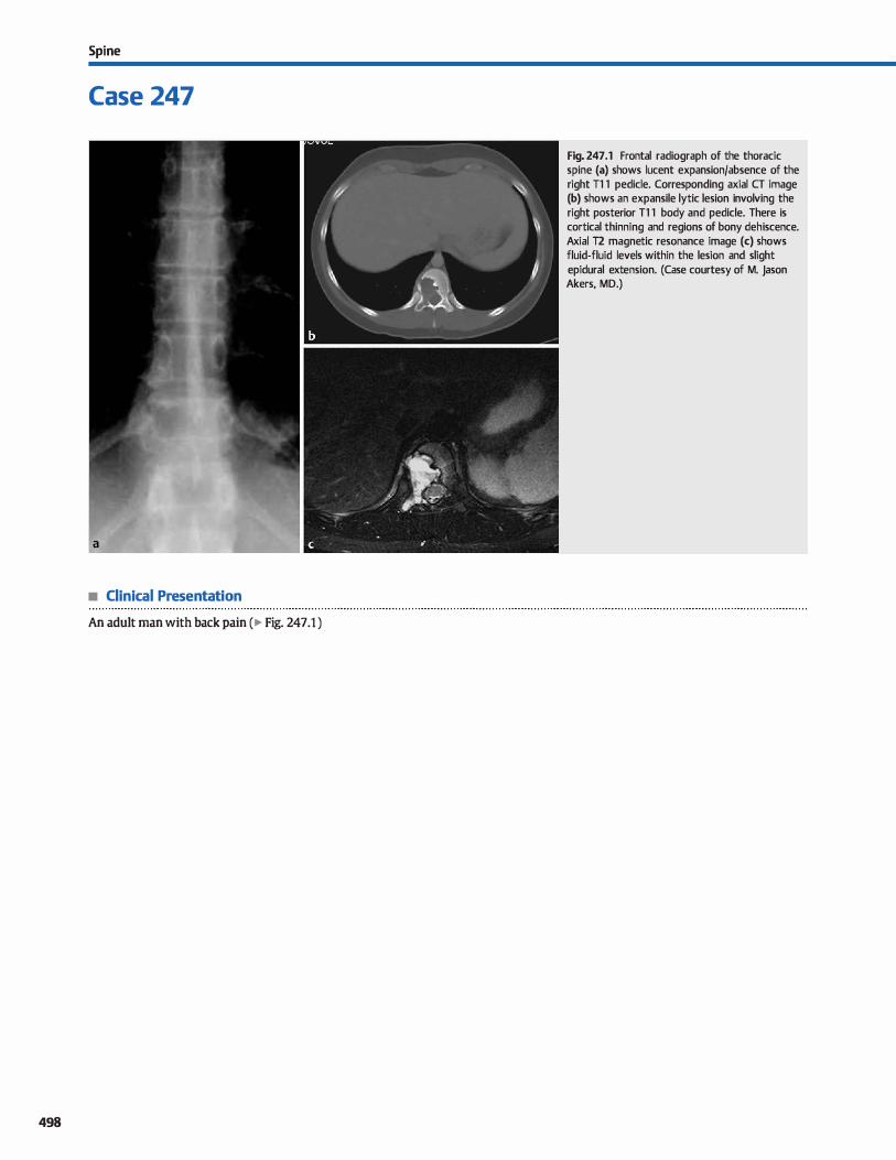
Spi ne
Case 247
• Clinical Presentation
An adult man with back pain ( � Fig. 24 7 .1 )
498
Fig. 247 .1 Frontal radiograph of the thoracic spine (a) shows lucent expansion/absence of the right Tl 1 pedicle. Corresponding axial CT image (b) shows an expansile lytic lesion involving the right posterior Tl 1 body and pedicle. There is cortical thinning and regions of bony dehiscence. Axial T2 magnetic resonance image (c) shows fluid-fluid levels within the lesion and slight epidural extension. (Case courtesy of M. Jason Akers, MD.)
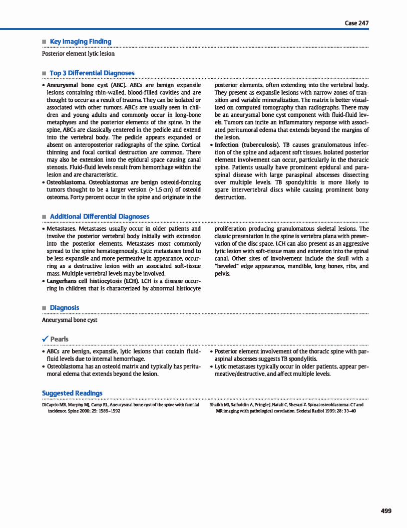
• Key lmaging Finding
Posterior element lytic lesion
• Top 3 Differential Diagnoses
• Aneurysmal bone cyst (ABC). ABCs are benign expansile lesions containing thin-walled, blood-filled cavities and are thought to occur as a result of trauma. They can be isolated or associated with other tumors. ABCs are usually seen in children and young adults and commonly occur in long-bone metaphyses and the posterior elements of the spine. In the spine, ABCs are dassically centered in the pedicle and extend
into the vertebral body. The pedicle appears expanded or absent on anteroposterior radiographs of the spine. Cortical thinning and focal cortical destruction are common. There may also be extension into the epidural space causing canal stenosis. Fluid-fluid levels result from hemorrhage within the
lesion and are characteristic.
• Osteoblastoma. Osteoblastomas are benign osteoid-fonning tumors thought to be a larger version (> 1.5 on) of osteoid
osteoma. Forty percent occur in the spine and originate in the
• Additional Differential Diagnoses
• Metastases. Metastases usually occur in older patients and involve the posterior vertebral body initially with extension into the posterior elements. Metastases most commonly spread to the spine hematogenously. Lytic metastases tend ta be Jess expansile and more perrneative in appearance, occur
ring as a destructive lesion with an associated soft-tissue
mass. Multiple vertebral levels may be involved. • Langerhans cell histiocytosis (LCH). LCH is a disease occur
ring in children that is characterized by abnormal histiocyte
• Diagnosis
Aneurysmal bone cyst
� Pearts
• ABCs are benign, expansile, lytic lesions that contain fluidfluid levels due to internai hemorrhage.
• Osteoblastoma has an osteoid matrix and typically has peritumoral edema that extends beyond the lesion.
Suggested Readings
Dicaprio MR, Murphy MJ, camp RL Aneurysmal bone cyst of the spine with familial
incidence. Spine 2000; 25: 1589-1592
Case 247
posterior elements, often extending into the vertebral body.
They present as expansile lesions with narrow zones of transition and variable mineralization. The matrix is better visualized on computed tomography than radiographs. There may be an aneurysmal bone cyst component with fluid-fluid levels. Tumors can incite an inflammatory response with associ
ated peritumoral edema that extends beyond the margins of the lesion.
• Infection (tuberculosis). TB causes granulomatous infection of the spine and adjacent soft tissues. Isolated posterior element involvement can occur, particularly in the thoracic spine. Patients usually have prominent epidural and para
spinal disease with large paraspinal abscesses dissecting over multiple levels. TB spondyltitis is more likely to spare intervertebral dises while causing prominent bony
destruction.
proliferation produdng granulomatous skeletal lesions. The classic presentation in the spine is vertebra plana with preservation of the dise space. LCH can aise present as an aggressive
lytic lesion with soft-tissue mass and extension into the spinal canal. Other sites of involvement indude the skull with a
"beveled" edge appearanœ, mandible, long bones, ribs, and pelvis.
• Posterior element involvement of the thoradc spine with paraspinal abscesses suggests TB spondylitis.
• Lytic metastases typically occur in older patients, appear permeative/destructive, and affect multiple levels.
Shaikh MI, Saifuddin A, PringleJ, Natali C, Sherazi Z. Spinal osteoblastoma; CT and
MR imaging with pathological correlation. Skeletal Radiol 1999; 28: 33-40
499
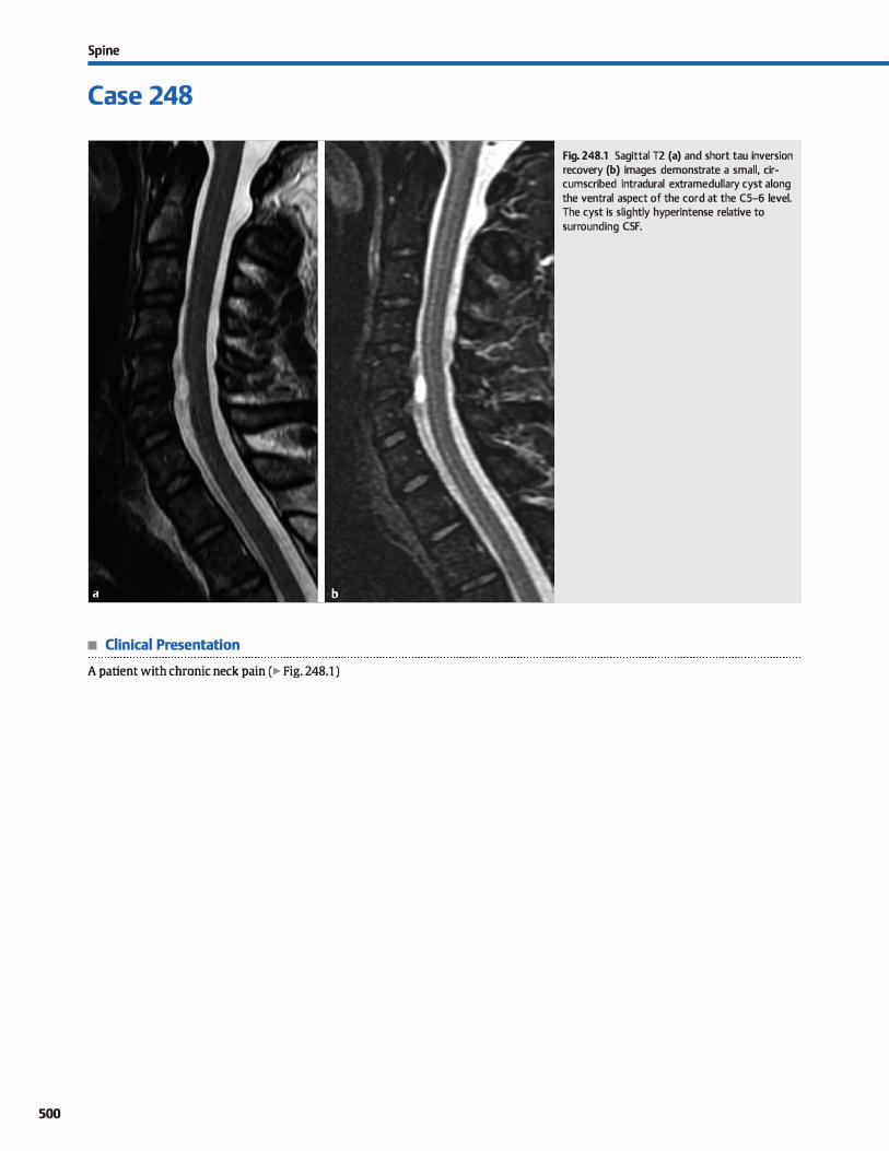
Spi ne
Case 248
• Clinical Presentation
A patient with chronic neck pain (� Fig. 248.1 )
500
Fig. 248.1 Sagittal T2 (a) and short tau inversion recovery (b) images demonstrate a small, circumscribed intradural extramedullary cyst along the ventral aspect of the cord at the CS-6 level. The cyst is slightly hyperintense relative to surrounding CSF.
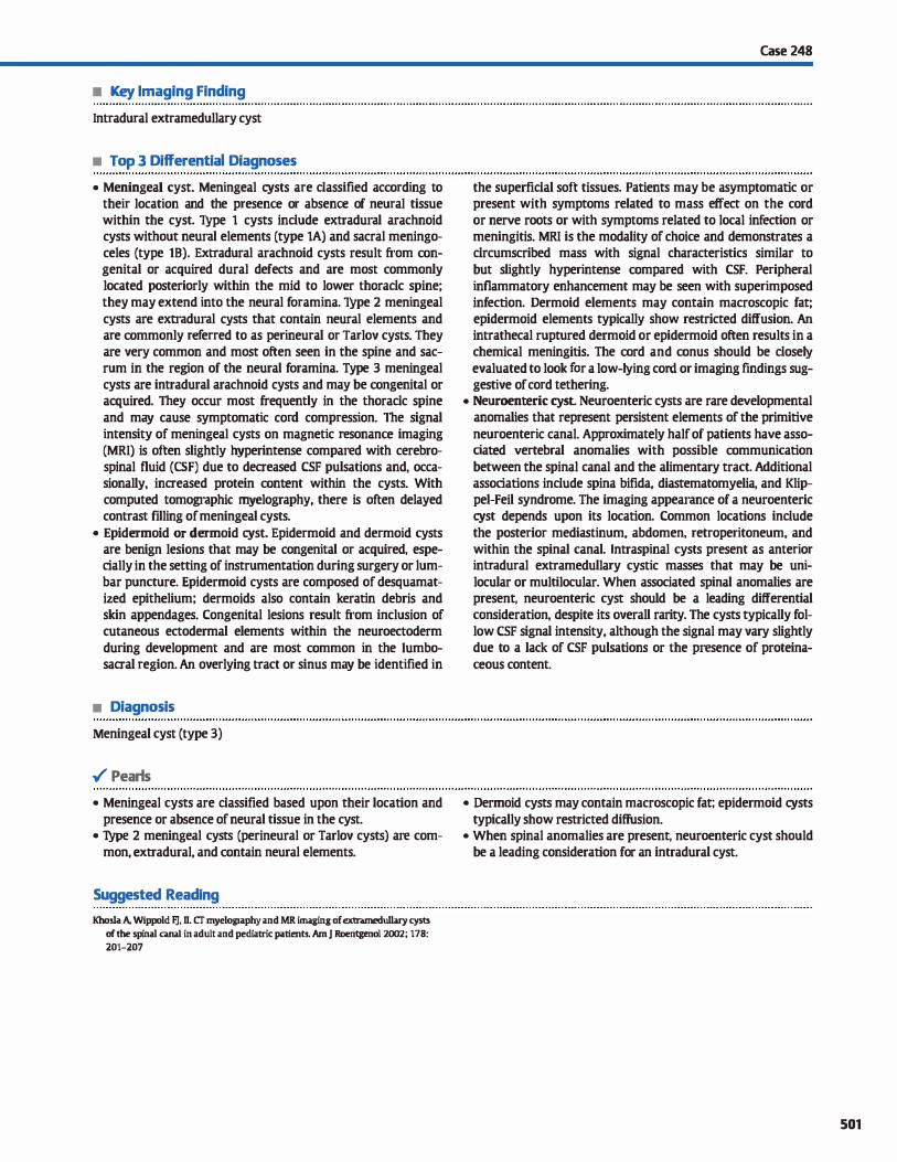
• Key lmaging Finding
lntradural extramedullary cyst
• Top 3 Differential Diagnoses
• Meningeal cyst. Meningeal cysts are dassified according to their location and the presenœ or absence of neural tissue within the cyst. Type 1 cysts indude extradural arachnoid cysts without neural elements (type lA) and sacral meningoceles {type 18). Extradural arachnoid cysts result from congenital or acquired durai defects and are most commonly located posteriorly within the mid to lower thoradc spine; they may extend into the neural foramina. Type 2 meningeal cysts are extradural cysts that contain neural elements and are commonly referred to as perineural or Tarlov cysts. They are very common and most often seen in the spine and sacrum in the region of the neural foramina. Type 3 meningeal cysts are intradural arachnoid cysts and may be congenital or acquired. They occur most frequently in the thoradc spine and may cause symptomatic cord compression. The signal intensity of meningeal cysts on magnetic resonanœ imaging (MRI) is often slightly hyperintense compared with cerebrospinal fluid {CSF) due to decreased CSF pulsations and, occasionally, increased protein content within the cysts. With computed tomographie myelography, there is often delayed contrast filling ofmeningeal cysts.
• Epidermoid or dermoid cyst. Epidermoid and dermoid cysts are benign lesions that may be congenital or acquired, especially in the setting of instrumentation during surgery or lumbar puncture. Epidermoid cysts are composed of desquamatized epithelium; dermoids also contain keratin debris and skin appendages. Congenital lesions result from inclusion of cutaneous ectodermal elements within the neuroectoderm during development and are most common in the Iumbosacral region. An overlying tract or sinus may be identified in
• Diagnosis
Meningeal cyst {type 3)
� Pearts
• Meningeal cysts are dassified based upon their location and presence or absence of neural tissue in the cyst.
• Type 2 meningeal cysts (perineural or Tarlov cysts) are common, extradural, and contain neural elements.
Suggested Reading
Khosla A. Wippold l'.J, n. cr myelography and MR irnaging of extramedullary cysts
of the spinal canal in adult and pediatric patients. Am J Roentgenol 2002; 178;
201-207
Case 248
the superfidal soft tissues. Patients may be asymptomatic or present with symptoms related to mass etfect on the cord or nerve roots or with symptoms related to local infection or meningitis. MRI is the modality of choiœ and demonstrates a drcumscribed mass with signal characteristics similar to but slightly hyperintense compared with CSF. Peripheral inflammatory enhancement may be seen with superimposed infection. Dermoid elements may contain macroscopic fat; epidermoid elements typically show restricted diffusion. An intrathecal ruptUred dermoid or epidermoid often results in a chemical meningitis. The cord and conus should be dosely evaluated to look for a low-lying cord or imaging findings suggestive of cord tethering.
• Neuroenteric cyst. Neuroenteric cysts are rare developmental anomalies that represent persistent elements of the primitive
neuroenteric canal. Approximately half of patients have associated vertebral anomalies with possible communication between the spinal canal and the alimentary tract. Additional associations indude spina bifida, diastematomyelia, and Klippel-Feil syndrome. The imaging appearance of a neuroenteric cyst depends upon its location. Common locations indude the posterior mediastinum, abdomen, retroperitoneum, and within the spinal canal. lntraspinal cysts present as anterior intradural extramedullary cystic masses that may be unilocular or multilocular. When associated spinal anomalies are present, neuroenteric cyst should be a leading differential consideration, despite its overall rarity. The cysts typically follow CSF signal intensity, although the signal may vary slightly due to a Jack of CSF pulsations or the presence of proteinaœous content.
• Dermoid cysts may contain macroscopic fat; epidermoid cysts typically show restricted diffusion.
• When spinal anomalies are present, neuroenteric cyst should be a leading consideration for an intradural cyst.
501
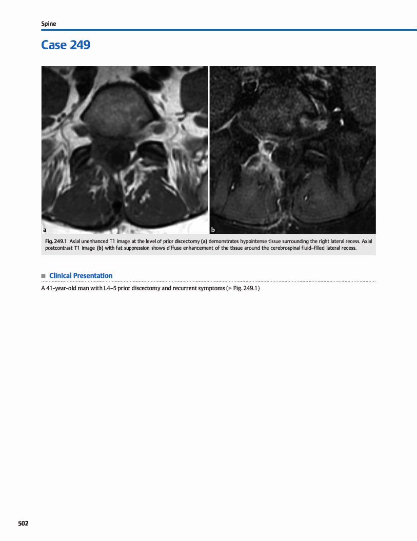
502
Spi ne
Case 249
Fig. 249.1 Axial unenhanced Tl image at the level of prior discectomy (a) demonstrates hypointense tissue surrounding the right lateral recess. Axial postcontrast T1 image (b) with fat suppression shows diffuse enhancement of the tissue around the cerebrospinal fluid-filled lateral recess.
• Clinical Presentation
A 41-year-old man with L4-5 prior discectomy and recurrent symptoms (� Fig. 249.1 )
• Key lmaging Finding
Postoperative enhanœment
• Top 3 Differential Diagnoses
• Granulation tissue. Granulation tissue or scar commonly
fonns following spinal surgery. lt is most pronounced in the
region of discectomy, which presents a diagnostic challenge
when attempting to distinguish granulation tissue from a
recurrent dise hemiation because bath entities appear similar
on unenhanœd magnetic resonanœ imaging sequences. Oassically, granulation or scar tissue enhances homogeneously,
allowing for differentiation from a recurrent dise. However,
within the first 3 to 6 months following surgery, granulation tissue may demonstrate peripheral rather than homogeneous
enhancement, making the distinction between scar tissue
and a recurrent dise impossible on imaging. A follow-up
examination will often reveal the more eharacteristie homogeneous enhanœment pattern associated with searring.
• Recurrent dise. Recurrent dise hemiation is a not uncommon
cause of recurrent back pain following surgieal discectomy.
The herniation often oecurs in the region of prior surgery.
On unenhaneed sequences, a recurrent dise typieally follows
similar signal intensity eompared with the parent dise; this
• Diagnosis
Granulation tissue
� Pearls
• Oassically, granulation or sear enhanees homogeneously,
allowing for differentiation from a recurrent dise.
• A recurrent dise hemiation characteristically demonstrates
peripheral rather than homogeneous enhancement.
Suggested Reading
Hancock CR, Quenœr RM, Falcone S. Challenges and pitfalls in postoperative spine
imaging. Appl Radiol 2008; 37; 23-34
Case 249
finding is not specifie, however, because granulation tissue
may have a similar appearanee. Therefore, postcontrast imaging is key to distinguishing between a reeurrent dise and
granulation or sear tissue. On postcontrast Tl sequences, dise
material characteristically demonstrates peripheral rather
than homogeneous enhancement The specificity of this find
ing increases with time from the original surgery.
• Infection. Infection is a dreaded complication of spinal sur
gery, especially in the presenee of hardware instrumentation.
Patients often present dinieally with back pain, fever, and ele
vated white blood cell eount and erythrocyte sedimentation rate. lmaging findings depend upon the stage of infection.
Initially, the infectious process results in ill-defined, inflammatory, or phlegmonous enhaneement. As the infection
becomes more organized, an abscess forms, demonstrating
central fluid signal intensity with a rim-enhancing capsule. ln
the early stages, medical management with antibiotics may
be adequate. Once an abscess forms, however, surgical drain
age is often neœssary.
• Early infection results in ill-defined, inflammatory enhance
ment; an absœss demonstrates rim enhanœment.
503
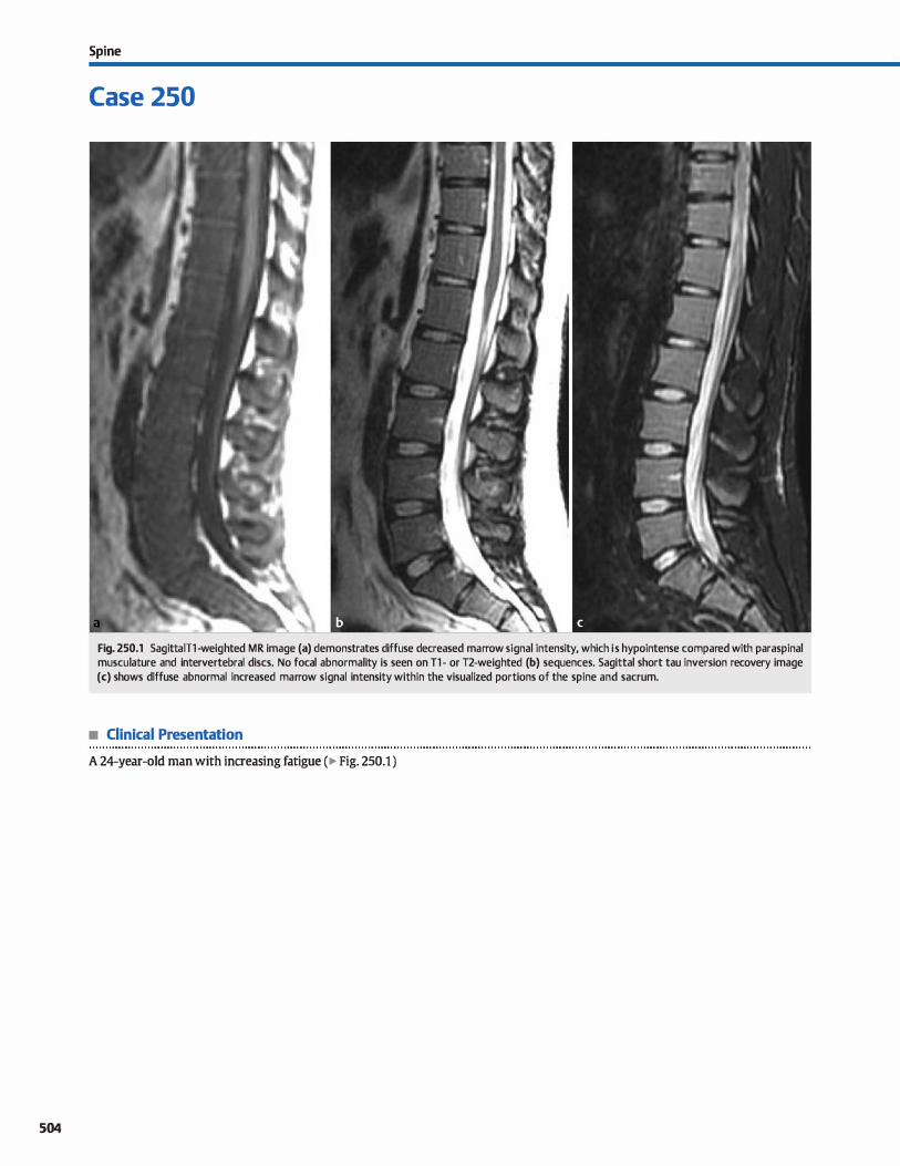
504
Spi ne
Case 250
Fig. 250.1 SagittalTl -weighted MR image (a) demonstrates diffuse decreased marrow signal intensity, which i s hypointense compared with paraspinal musculature and intervertebral dises. No focal abnormality is seen on Tl - or T2-weighted (b) sequences. Sagittal short tau inversion recovery image (c) shows diffuse abnormal increased marrow signal intensity within the visualized portions of the spine and sacrum.
• Clinical Presentation
A 24-year-old man with increasing fatigue (� Fig. 250.1 )
• Key lmaging Finding
Diffuse abnormal marrow signal intensity
• Top 3 Differential Diagnoses
• Hematopoietic marrow. In children and young adults, bone manuw progresses from predominandy hematopoietic at birth to predominandy fatty marrow in adulthood. In adults, residual hematopoietic marrow persists in the axial skeleton {spine and flat bones) and proximal shafts of the femur and hwnerus. 11-weighted magnetic resonanœ imaging {MRI) sequenœs are ideal in evaluating for marrow abnormalities. Normal 11 bone mar
row signal intensity in adults is brighter than adjacent paraspinal musculature. In the setting of chronic illness or anemia, however, manuw reconversion oa:urs with replacement of fatty marrow with hematopoietic marrow. Increased concentrations of hematopoietic marrow results in decreased 11 signal intensity that may be iso- to hypointense compared with paraspinal musculature. Fluid-sensitive fat-suppressed (FS) 12 and postcontrast FS
T1 sequenœs are helpful in distinguishing hematopoietic marrow from more aggressive marrow infiltrative proœsses, both of which may demonstrate decreased 11 signal intensity. On these sequenœs, hematopoietic marraw is typically hypointense, whereas infiltrative proœsses are most often hyperintense.
• Myeloproliferative disorder. Myeloproliferative disorders refer ta abnormal proliferation of marrow elements. Common
diffuse infiltrative processes indude multiple myeloma (plasma cells), leukemia {white blood cells), and myelofibrosis (supporting collagen and fibrous tissue). These proliferative disorders replace normal fatty marrow, resulting in diffuse decreased Tl signal intensity compared with paraspinal
• Additional Differential Diagnoses
• Metastases. Metastatic disease represents the most common
malignancy to involve bone. Bones with increased hematopoietic
manuw, induding the axial skeleton, are preferentially involved due to relatively increased blood flow. Common primary malig
nancies indude breast, Jung, and prostate carcinomas, as well as gastrointestinal and genitourinary neoplasms. Diffuse marrow infiltration in the setting of metastatic disease is uncommon; more common patterns of involvement indude discreet lesions
that may be solitary or multiple. The majority of metastatic foci are hypointense on 11 and hyperintense on FS 12 sequences with enhancement Sderotic lesions from breast or prostate primaries may be hypointense on ail sequenœs.
• Diagnosis
Myeloproliferative disorder (leukemia)
� Pearts
• Hematopoietic marrow occurs in the setting of chronic illness/anemia and is hypointense ta musculature on T1.
• Cellular marrow infiltrative processes result in decreased T1
and increased FS 12 signal with enhancement
Suggested Reading
Long 5.5, Yablon CM, Eisenberg RI. Bene marrow signal alteration in the spine and
sacrum. AmJ Roentgenol 2010; 195: W178-200
Case 250
musculature. Multiple myeloma is the most common primary bene malignancy and occurs in middle-aged and aider adults. lt results in bony resorption due to increased osteoclastic activity and inflammatory response. Any bone may be
involved, but the most common locations indude the spine and calvarium. Bony involvement may be focal or diffuse. Focal involvement results in �punched out" lucencies on plain radiographs. Diffuse involvement leads to marrow infiltration with decreased Tt and increased FS 12 signal intensity with enhancement. Leulcemia may occur in patients of any age but is commonly seen in adolescents and young adults. Diffuse
involvement results in decreased Tt and increased FS T2 signal intensity with enhancement Signal intensity must be evaluated dosely because the diffuse nature of marrow infil
tration may inadvertently be overlooked. Myelofibrosis is unique in that proliferation of flbrous elements results in decreased signal intensity on ail MRI sequences.
• cymphoma. Lymphomatous involvement of the spine may be primary or secondary (far more common). Lesions may be solitary, multiple, or diffuse. Diffuse involvement most often demonstrates imaging features similar to leukemia with dif
fuse decreased T1 and increased FS T2 signal intensity with enhancement. A characteristic feature of lymphoma is extraspinous spread into adjacent soft tissues and compartments without cortical disruption.
• Treatment-related marrow changes. Radiation, chemotherapy, and granulocyte-colony stimulating factor (G-CSF) are commonly used in the treatment of malignancies. In the acute setting ( 4-6 weeks ), radiation therapy results in marrow
edema with increased signal intensity on fat-suppressed 12 sequences. In the chronic setting, radiation induces fatty marraw replacement confined to the treatment field. Chemotherapy results in similar changes with less pronounced
chronic fatty marrow. G-CSF causes stimulation of marrow,
resulting in diffuse increased signal on fat-suppressed 12
sequences, which may mimic residual or recurrent disease.
• Myelofibrosis results in proliferation of marrow support elements with decreased signal on ail sequences.
• Metastatic foci typically present with discreet masses; diffuse marrow infiltration is uncommon.
505
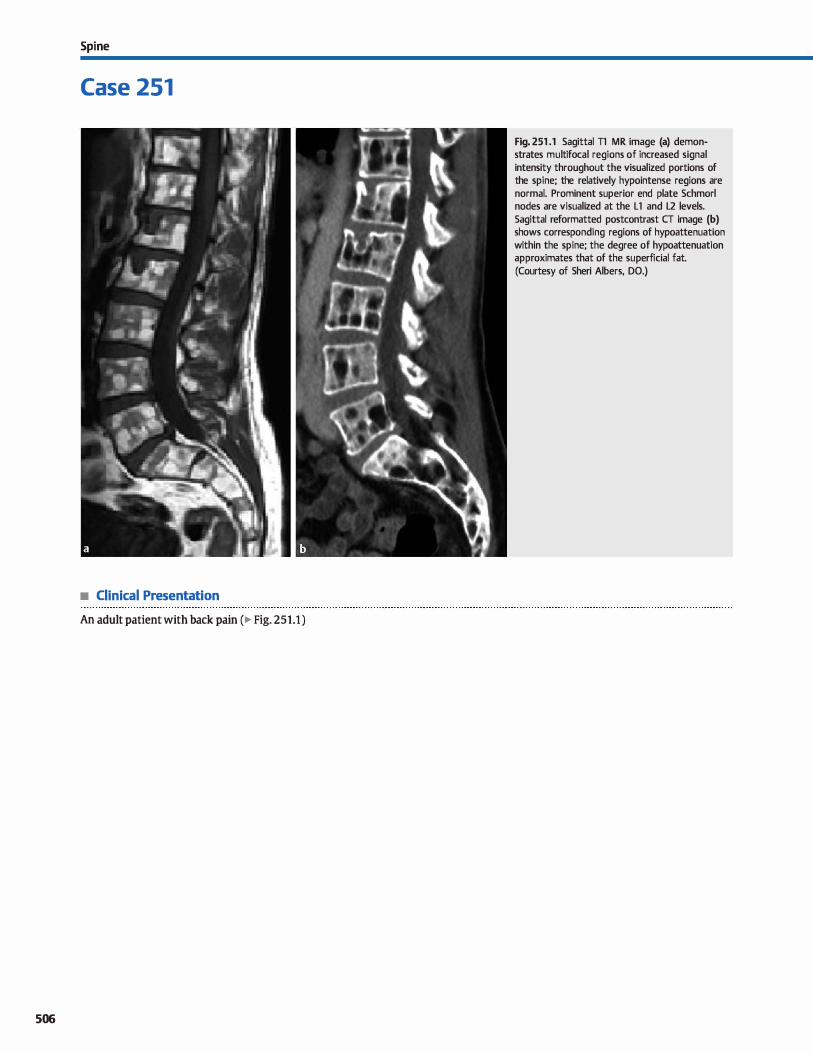
Spi ne
Case 251
• Clinical Presentation
An adult patient with back pain (� Fig. 251.1 )
506
Fig. 251 .1 Sagittal Tl MR image (a) demonstrates multifocal regions of increased signal intensity throughout the visualized portions of the spine; the relatively hypointense regions are normal. Prominent superior end plate Schmorl nodes are visualized at the L1 and L2 levels. Sagittal reformatted postcontrast CT image (b) shows corresponding regions of hypoattenuation within the spine; the degree of hypoattenuation approximates that of the superficial fat. (Courtesy of Sheri Albers, DO.)
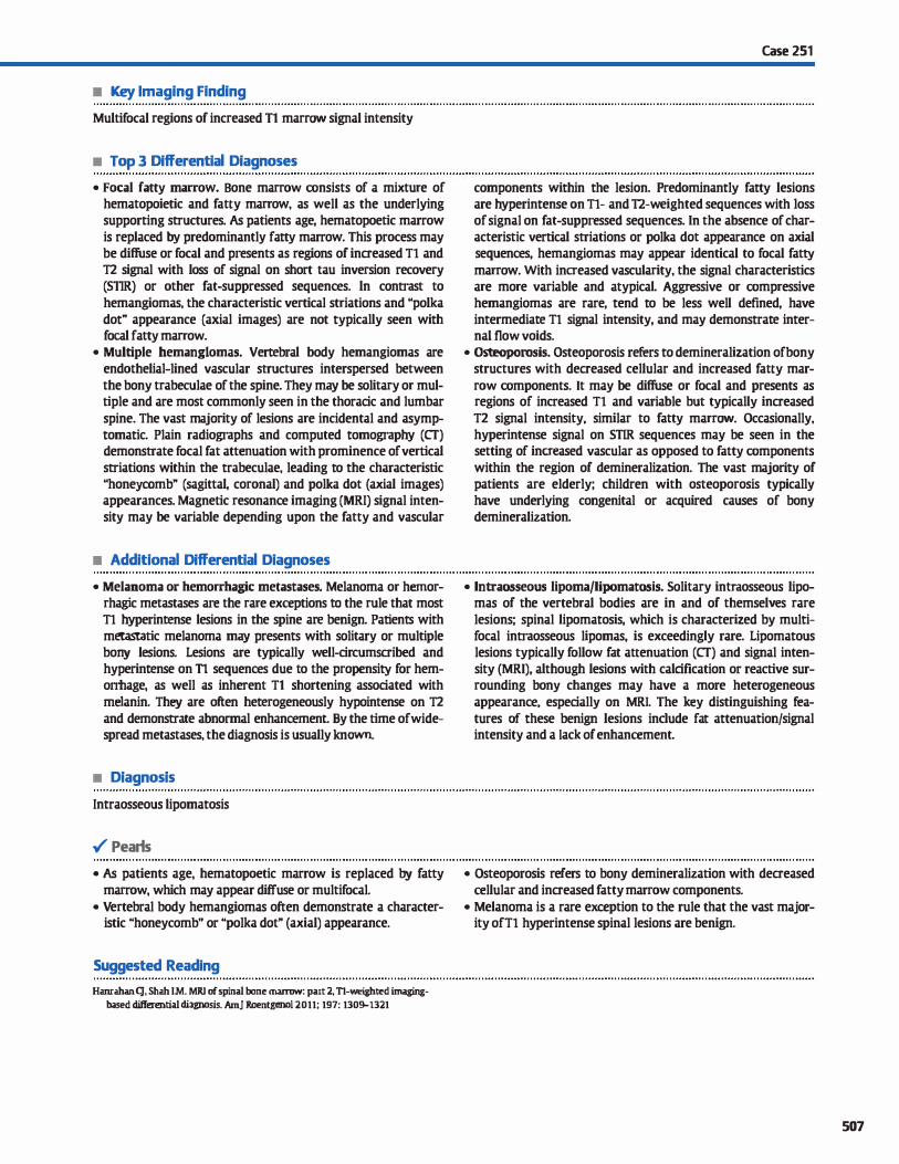
• Key lmaging Finding
Multifocal regions of increased Tl marrow signal intensity
• Top 3 Differential Diagnoses
• Focal fatty marrow. Bone marrow consists of a mixture of hematopoietic and fatty marrow, as well as the underlying
supporting structures. As patients age, hematopoetic marrow
is replaced by predominantly fatty marrow. This process may
be diffuse or focal and presents as regions of increased Tl and
T2 signal with loss of signal on short tau inversion recovery
(STIR) or other fat-suppressed sequenœs. ln contrast to
hemangiomas, the characteristic vertical striations and "polka
dot" appearance {axial images) are not typically seen with focal fatty marrow.
• Multiple hemangiomas. Vertebral body hemangiomas are
endothelial-lined vascular structures interspersed between
the bony trabeculae of the spine. They may be solitary or multiple and are most commonly seen in the thoradc and lumbar
spine. The vast majority of lesions are incidental and asymp
tomatic. Plain radiographs and computed tomography (CT) demonstrate focal fat attenuation with prominence of vertical
striations within the trabeculae, leading to the characteristic
"honeycomb" (sagittal, coronal) and polka dot (axial images)
appearances. Magnetic resonanœ imaging {MRI) signal inten
sity may be variable depending upon the fatty and vascular
• Additional Differential Diagnoses
• Melanoma or hemorrhagic metastases. Melanoma or hemor
rhagic metastases are the rare exceptions to the rule that most
Tl hyperintense lesions in the spine are benign. Patients with
metastatic melanoma may presents with solitary or multiple
bony lesions. Lesions are typically well-drcumscribed and
hyperintense on Tl sequenœs due to the propensity for hem
orrhage, as well as inherent Tl shortening assodated with
melanin. They are oft:en heterogeneously hypointense on T2
and demonstrate abnormal enhanœment By the time ofwidespread metastases, the diagnosis is usually known.
• Diagnosis
Intraosseous lipomatosis
� Pearls
• As patients age, hematopoetic marrow is replaced by fatty
marrow, which may appear diffuse or multifocal.
• Vertebral body hemangiomas often demonstrate a characteristic "honeycomb" or "polka dot" (axial) appearance.
Suggested Reading
Hanrahan CJ, Shah LM. MRI of spinal bene marruw: part 2, Tt-wcighted imaging
based düferential diagnosis. AmJ Roentgenol 2011; 197: 1309-1321
Case 251
components within the lesion. Predominantly fatty lesions
are hyperintense on Tl- and T2-weighted sequences with Joss
of signal on fat-suppressed sequences. In the absence of char
acteristic vertical striations or polka dot appearance on axial
sequenœs, hemangiomas may appear identical to focal fatty
marrow. With increased vascularity, the signal characteristics
are more variable and atypical. Aggressive or compressive
hemangiomas are rare, tend to be less well defined, have
intermediate Tl signal intensity, and may demonstrate internai flow voids.
• Ostroporosis. Osteoporosis refers to demineralization ofbony
structures with decreased cellular and increased fatty mar
row components. It may be diffuse or focal and presents as regions of increased Tl and variable but typically increased
T2 signal intensity, similar to fatty marrow. Occasionally,
hyperintense signal on STIR sequences may be seen in the setting of increased vascular as opposed to fatty components
within the region of demineralization. The vast majority of
patients are elderly; children with osteoporosis typically
have underlying congenital or acquired causes of bony
demineralization.
• lntraosseous lipomajlipomatosis. Solitary intraosseous lipomas of the vertebral bodies are in and of themselves rare
lesions; spinal lipomatosis, which is characterized by multi
focal intraosseous lipomas, is exceedingly rare. Lipomatous
lesions typically follow fat attenuation (CT) and signal inten
sity (MRI), although lesions with calcification or reactive surrounding bony changes may have a more heterogeneous
appearanœ, espedally on MRI. The key distinguishing fea
tures of these benign lesions indude fat attenuation/signal intensity and a Jack of enhancement.
• Osteoporosis refers to bony demineralization with decreased
cellular and increased fatty marrow components.
• Melanoma is a rare exception to the rule that the vast majority ofîl hyperintense spinal lesions are benign.
507
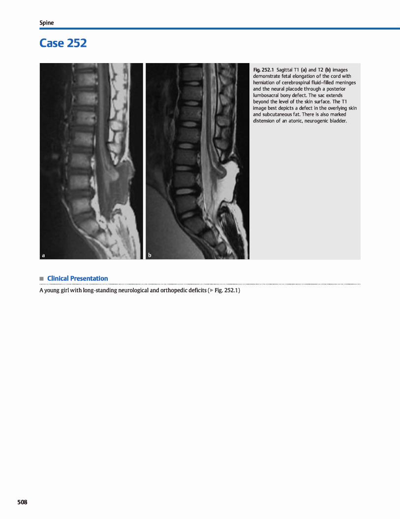
Spi ne
Case 252
• Clinical Presentation
A young girl with long-standing neurological and orthopedic deficits ( � Fig. 252.1 )
508
Fig. 252.1 Sagittal Tl (a) and T2 (b) images demonstrate fetal elongation of the cord with herniation of cerebrospinal fluid-filled meninges and the neural placode through a posterior lumbosacral bony defect. The sac extends beyond the level of the skin surface. The Tl image best depicts a defect in the overlying skin and subcutaneous fat. There is also marked distension of an atonie, neurogenic bladder.
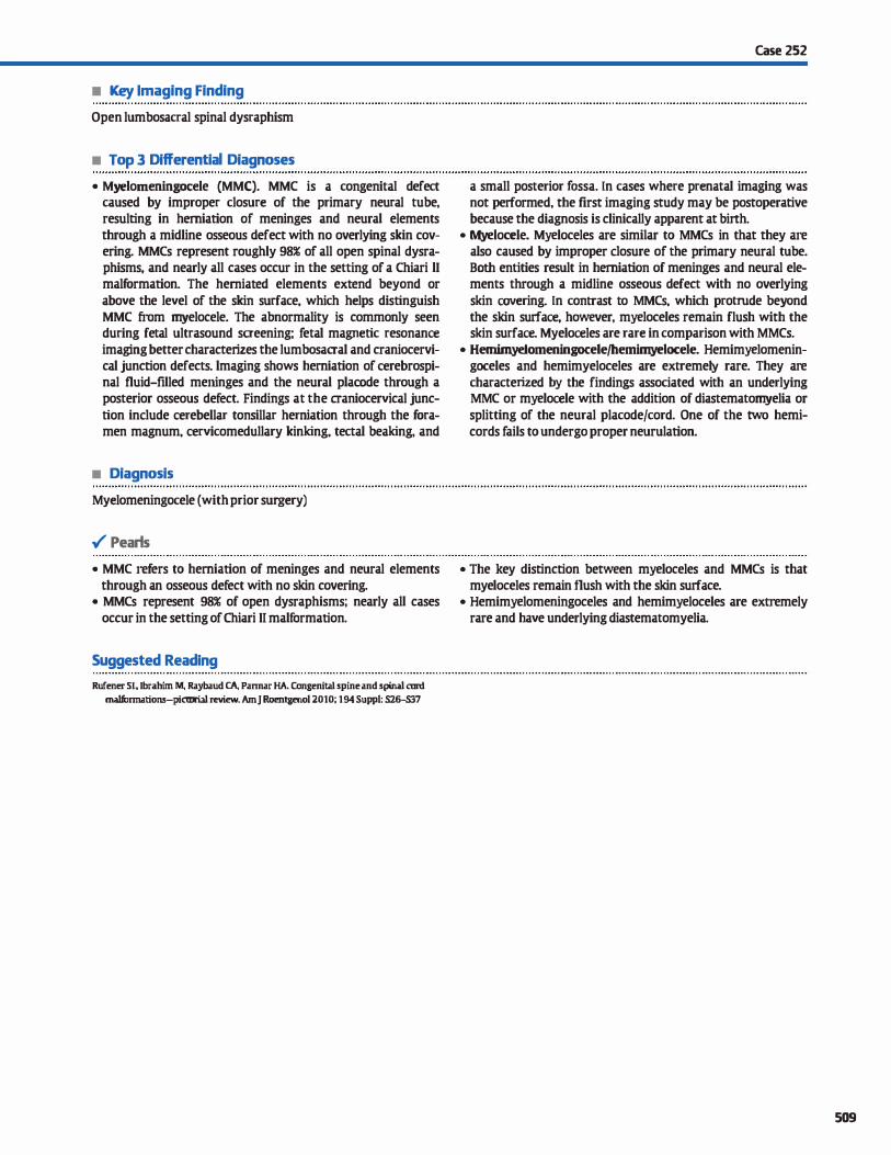
• Key lmaging Finding
Open lumbosacral spinal dysraphism
• Top 3 Differential Diagnoses
• Myelomeningocele (MMC). MMC is a congenital defect caused by improper closure of the primary neural tube,
resulting in herniation of merunges and neural elements
through a midline osseous defect with no overlying skin cov
ering. MMCs represent roughly 98% of ail open spinal dysra
phisms, and nearly ail cases occur in the setting of a Chiari li
malformation. The herniated elements extend beyond or
above the level of the skin surface, which helps distinguish
MMC from myelocele. The abnormality is commonly seen during fetal ultrasound screening; fetal magnetic resonance
imaging better characterizes the lumbosacral and craniocervi
cal junction defects. lmaging shows hemiation of œrebrospi
nal fluid-filled merunges and the neural placode through a
posterior osseous defect. Findings at the craniocervical junc
tion indude œrebellar tonsillar herruation through the fora
men magnum, œrvicomedullary kinking, tectal beaking, and
• Diagnosis
Myelomeningocele (with prior surgery)
� Pearts
• MMC refers ta hemiation of merunges and neural elements
through an osseous defect with no skin covering. • MMCs represent 98% of open dysraphisms; nearly ail cases
occur in the setting of Chiari Il malformation.
Suggested Reading
Rufe11er SI., Ibrahim M, Raybaud CA, Pannar HA. Congenital spine and spinal cord malfurmations-pictmial review. AmJ Rœntgenol 2010; 194 Suppl: S26-S37
Case 252
a small posterior fossa. In cases where prenatal imaging was
not performed, the first imaging study may be postoperative
because the diagnosis is clinically apparent at birth.
• Myeloœle. Myeloceles are similar ta MMCs in that they are
also caused by improper dosure of the primary neural tube.
Bath entities result in herniation of meninges and neural ele
ments through a midline osseous defect with no overlying
skin covering. In contrast to MMCs, which protrude beyond
the skin surface, however, myeloceles remain flush with the skin surface. Myeloceles are rare in comparison with MMCs.
• Hemimyelomeningocelefhemimyelocele. Hemimyelomenin
goceles and hemimyeloceles are extremely rare. They are
characterized by the findings associated with an underlying MMC or myelocele with the addition of diastematomyelia or
splitting of the neural placode/cord. One of the two hemi
cords fails ta undergo proper neurulation.
• The key distinction between myeloceles and MMCs is that
myeloceles remain flush with the skin surface.
• Hemimyelomeningoceles and hemimyeloceles are extremely
rare and have underlying diastematomyelia.
509
510
Spi ne
Case 253
Fig. 253.1 Frontal (a) and lateral (b) radiographs of the abdomen demonstrate a large noncalcified presacral mass displacing the bowel superiorly and anteriorly. The lower sacrum and coccyx are not identified. Sagittal T2-weighted MR image (c) defines the large, presacral cystic mass that extends posterior to the spine but is separate from the spinal canal. The bladder (containing a Foley catheter) is compressed anteriorly. (Case courtesy of Sandra L. Wootton-Gorges, MD.)
• Clinical Presentation
A full-term infant girl with a distended abdomen (� Fig. 253.1 )
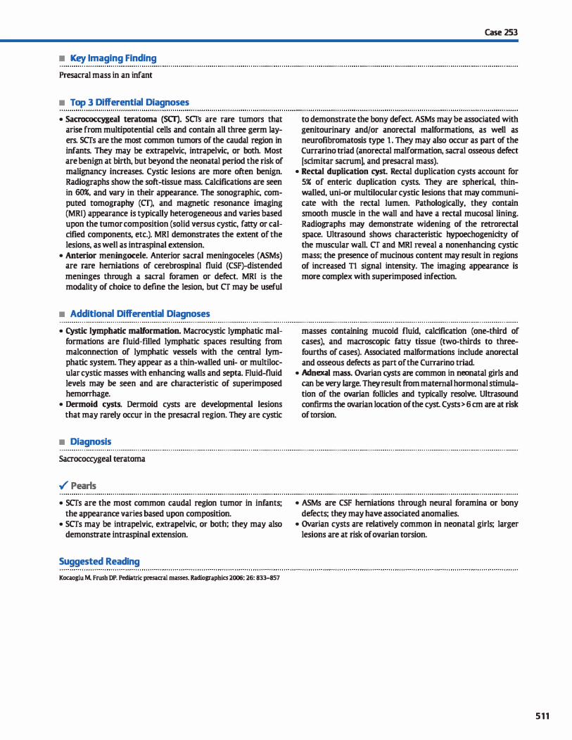
• Key lmaging Finding
Presacral mass in an infant
• Top 3 Differential Diagnoses
• Sacrococcygeal teratoma (SCT). sers are rare tumors that arise from multipotential cells and contain ail three germ layers. sers are the most common tumors of the caudal region in infants. They may be extrapelvic, intrapelvic, or bath. Most are benign at birth, but beyond the neonatal period the risk of malignancy increases. Cystic lesions are more often benign. Radiographs show the soft-tissue mass. calcifications are seen
in 60%, and vary in their appearance. The sonographic, computed tomography (er), and magnetic resonance imaging (MRI) appearance is typically heterogeneous and varies based upon the tumor composition (solid versus cystic, fatty or calcified components, etc.). MRI demonstrates the extent of the lesions, as well as intraspinal extension.
• Anterior meningocele. Anterior sacral meningoceles (ASMs) are rare hemiations of cerebrospinal fluid (CSF)-distended
meninges through a sacral foramen or defect. MRI is the modality of choiœ to define the Iesion, but er may be useful
• Additional Differential Diagnoses
• Cystic lymphatic maHormation. Macrocystic lymphatic malformations are fluid-filled lymphatic spaœs resulting from malconnection of lymphatic vessels with the central lymphatic system. They appear as a thin-walled uni- or multiloc
ular cystic masses with enhancing walls and septa. Fluid-fluid levels may be seen and are characteristic of superimposed hemorrhage.
• Dermoid cysts. Dermoid cysts are developmental lesions
that may rarely occur in the presacral region. They are cystic
• Diagnosis
Sacrococcygeal teratoma
� Pearls
• sers are the most common caudal region tumor in infants;
the appearance varies based upon composition. • sers may be intrapelvic, extrapelvic, or bath; they may also
demonstrate intraspinal extension.
Suggested Reading
Kocaoglu M, Frush DP. Pediatric pœsacral masses. Radiographies 2006; 26: 833-857
Case 253
to demonstrate the bony defect. ASMs may be associated with genitourinary and/or anorectal malformations, as well as neurofibromatosis type 1. They may also occur as part of the Currarino triad (anorectal malformation, sacral osseous defect [scimitar sacrum], and presacral mass).
• Rectal duplication cyst. Rectal duplication cysts account for 5% of enteric duplication cysts. They are spherical, thin
walled, uni-or multilocular cystic Iesions that may communicate with the rectal lumen. Pathologically, they contain smooth muscle in the wall and have a rectal mucosal lining.
Radiographs may demonstrate widening of the retrorectal space. Ultrasound shows characteristic hypoechogenidty of the muscular wall. er and MRI reveal a nonenhancing cystic mass; the presence of mucinous content may result in regions
of increased Tl signal intensity. The imaging appearance is more complex with superimposed infection.
masses containing mucoid fluid, calcification (one-third of cases), and macroscopic fatty tissue (two-thirds to three
fourths of cases). Assodated malformations include anorectal and osseous defects as part of the Currarino triad.
• Adnexal mass. Ovarian cysts are common in neonatal girls and can be very large. They result from maternai hormonal stimulation of the ovarian follicles and typically resolve. Ultrasound confirms the ovarian location of the cyst Cysts > 6 cm are at risk of torsion.
• ASMs are CSF hemiations through neural foramina or bony
defects; they may have associated anomalies. • Ovarian cysts are relatively common in neonatal girls; larger
lesions are at risk of ovarian torsion.
511
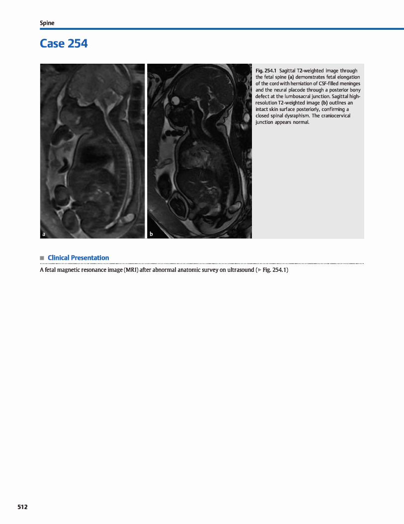
512
Spi ne
Case 254
• Clinical Presentation
Fig. 254.1 Sagittal T2-weighted image through the fetal spine (a) demonstrates fetal elongation of the cord with herniation of CSF-filled meninges and the neural placode through a posterior bony defect at the lumbosacral junction. Sagittal highresolution T2-weighted image (b) outlines an intact skin surface posteriorly, confirming a dosed spinal dysraphism. The craniocervical junction appears normal.
A fetal magnetic resonance image (MRI) after abnormal anatomie survey on ultrasound (� Fig. 254.1)
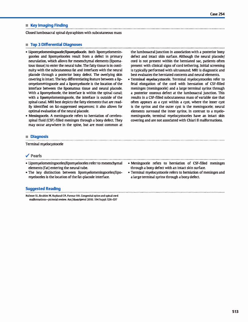
• Key lmaging Finding
dosed lumbosacral spinal dysraphism with subcutaneous mass
• Top 3 Differential Diagnoses
• lipomyelomeningoœleflipomydocele. Both lipomyelomeningoœles and lipomyeloœles result from a defect in primary neurulation, which allows for mesenchymal elements (lipomatous tissue) ta enter the neural tube. The fatty tissue is in continuity with the subcutaneous fat and interfaces with the neural placode through a posterior bony defect The overlying skin covering is intact The key differentiating feature between a lipomyelomeningoœle and a lipomyelocele is the location of the interface between the lipomatous tissue and neural placode. With a lipomyeloœle, the interface is within the spinal canal; with a lipomyelomeningocele, the interface is outside of the spinal canal. MRI best depicts the fatty elements that are readily identified on fat-suppressed sequenœs; it also allows for optimal evaluation of the neural placode.
• Meningocele. A meningocele refers to hemiation of cerebrospinal fluid (CSF)-filled meninges through a bony defect They may occur anywhere in the spine, but are most common at
• Diagnosis
Terminal myelocystocele
v" Pearls
• Llpomyelomeningoceles/lipomyeloceles refer to mesenchymal
elements (fat) entering the neural tube. • The key distinction between lipomyelomeningoceles/lipo
myeloceles is the location of the fat-placode interface.
Suggested Reading
Rufener SI., Ibrahim M, Raybaud CA, Pannar HA. Congenital spine and spinal curd
malformations-pictorial review. AmJ Roentgenol 2010; 194 Suppl: 526-537
Case 254
the lumbosacral junction in association with a posterior bony defect and intact skin surface. Although the neural placode/ cord is not present within the herniated sac, patients often present with clinical signs of cord tethering. Initial screening
is typically performed with ultrasound; MRI is diagnostic and best evaluates the herniated contents and neural elements.
• Terminal myelocystocele. Terminal myelocystoceles refer to fetal elongation of the cord with herniation of CSF-filled meninges (meningocele) and a large terminal syrinx through a posterior osseous defect at the lumbosacral junction. This results in a CSF-filled subcutaneous mass of variable size that often appears as a cyst within a cyst, where the inner cyst is the syrinx and the outer cyst is the meningocele; neural elements surround the inner syrinx. In contrast to a myelomeningocele, terminal myelocystoceles have an intact skin covering and are not assoàated with Chiari Il malformations.
• Meningocele refers to hemiation of CSF-filled meninges
through a bony defect with an intact skin surface. • Terminal myelocystocele refers ta herniation of meninges and
a large terminal syrinx through a bony defect.
513
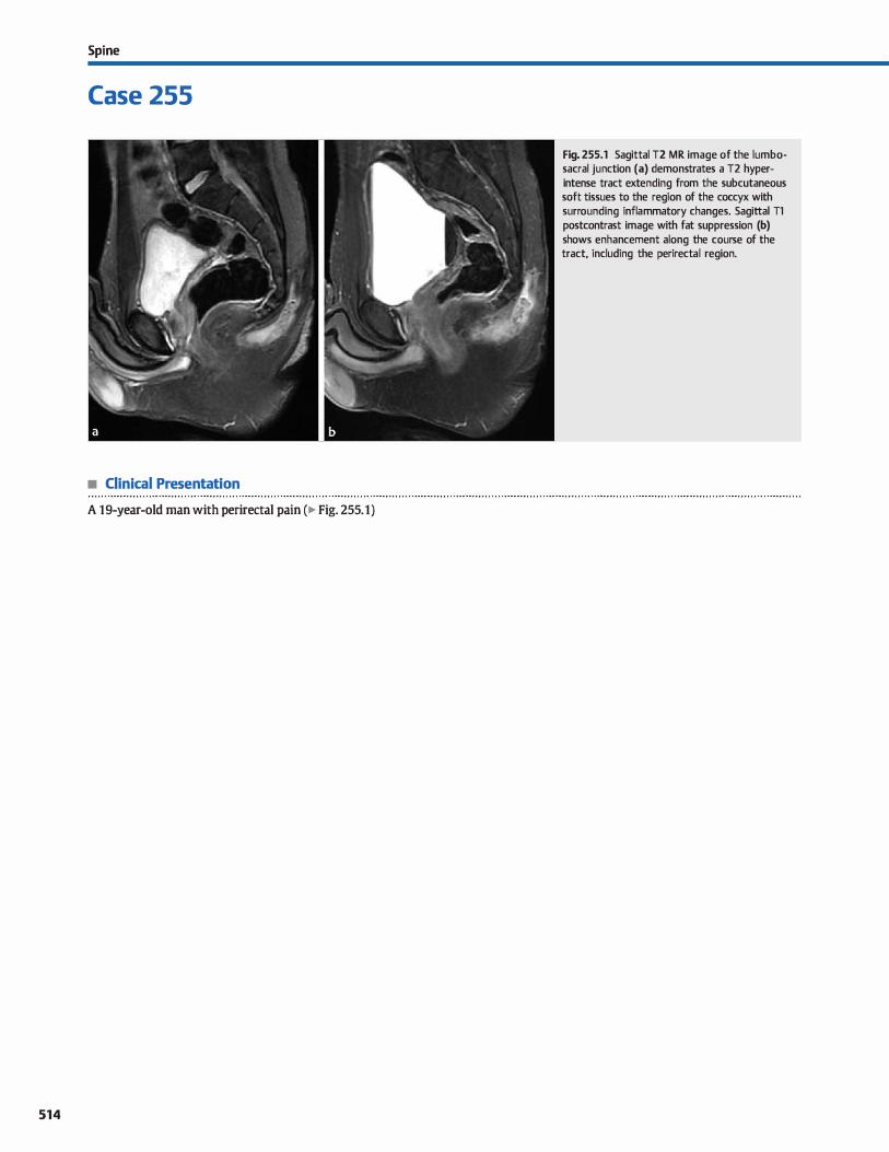
Spi ne
Case 255
• Clinical Presentation
A 19-year-old man with perirectal pain (� Fig. 255.1)
514
Fig. 255.1 Sagittal T2 MR image of the lumbosacral junction (a) demonstrates a T2 hyperintense tract extending from the subcutaneous soft tissues to the region of the coccyx with surrounding inflammatory changes. Sagittal Tl postcontrast image with fat suppression (b) shows enhancement along the course of the tract, including the perirectal region.
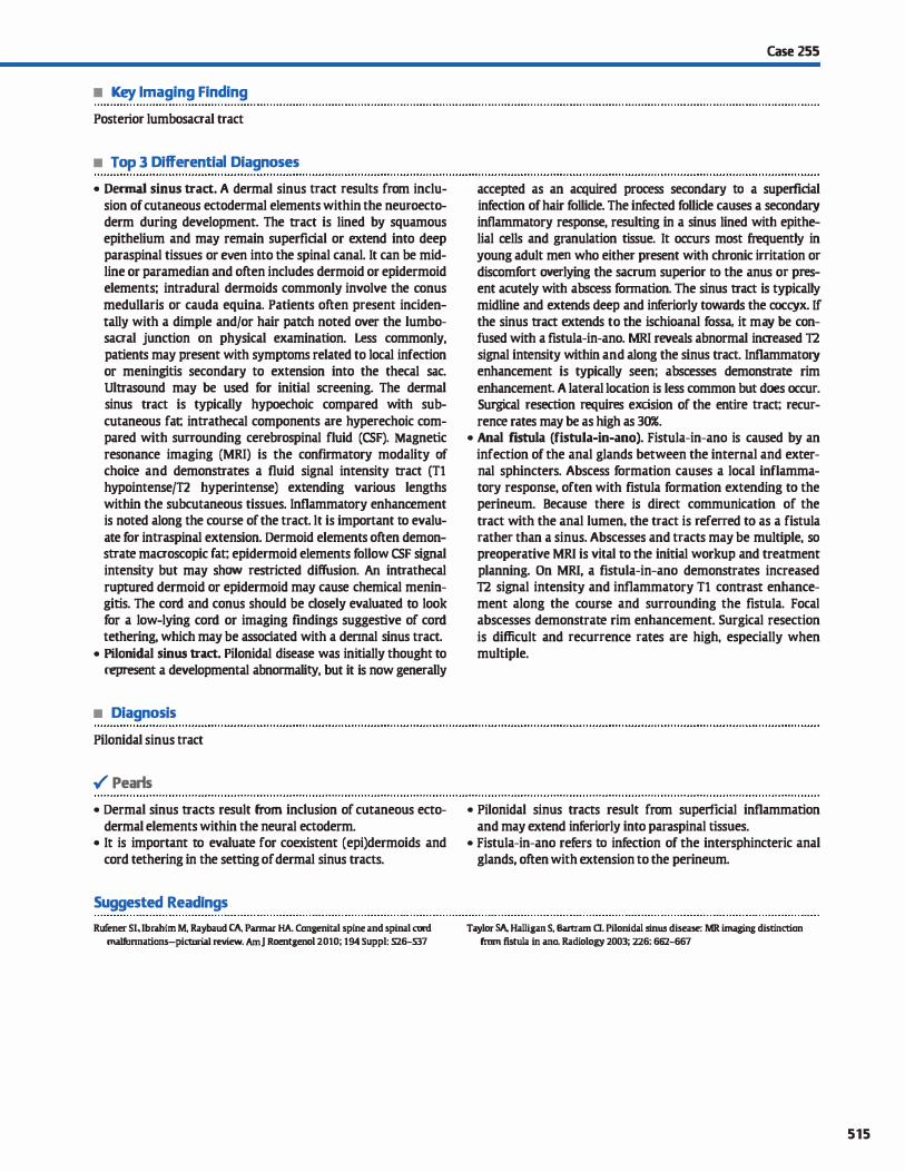
• Key lmaging Finding
Posterior lumbosacral tract
• Top 3 Differential Diagnoses
• Dermal sinus tract. A dermal sinus tract results from inclu
sion of cutaneous ectodermal elements within the neuroectoderm during development. The tract is lined by squamous epithelium and may remain superficial or extend into deep paraspinal tissues or even into the spinal canal. It can be midline or paramedian and often includes dermoid or epidermoid elements; intradural dermoids commonly involve the conus
medullaris or cauda equina. Patients often present inciden
tally with a dimple and/or hair patch noted over the lumbosacral junction on physical examination. Less commonly,
patients may present with symptoms related to local infection or meningitis secondary to extension into the thecal sac. Ultrasound may be used for initial screening. The dermal sinus tract is typically hypoechoic compared with sub
cutaneous fat; intrathecal components are hyperechoic compared with surrounding cerebrospinal fluid (CSF). Magnetic
resonance imaging {MRI) is the confinnatory modality of choiœ and demonstrates a fluid signal intensity tract (Tl hypointense(f2 hyperintense) extending various lengths
within the subcutaneous tissues. Inflammatory enhancement is noted along the course of the tract. lt is important to evalu
ate for intraspinal extension. Dermoid elements often demonstrate macroscopic fat; epidermoid elements follow CSF signal
intensity but may show restricted diffusion. An intrathecal
ruptured dermoid or epidermoid may cause chemical meningitis. The cord and conus should be dosely evaluated to look for a low-lying cord or imaging findings suggestive of cord tethering, which may be associated with a dennal sinus tract.
• Pilonidal sinus tract. Pilonidal disease was initially thought to represent a developmental abnormality, but it is now generally
• Diagnosis
Pilonidal sinus tract
� Pearts
• Dermal sinus tracts result from inclusion of cutaneous ecto
dermal elements within the neural ectoderm. • It is important to evaluate for coexistent ( epi)dermoids and
cord tethering in the setting of dermal sinus tracts.
Suggested Readings
Rufener SI. Ibrahim M, Raybaud CA, Pannar HA. Congenital spine and spinal conl rnaltbnnations-pictmial review. ArnJ Roentgenol 2010; 194 Suppl; S26-S37
Case 255
accepted as an acquired proœss secondary ta a superficial infection of hair follide. The infected follide causes a secondary inflammatory response, resulting in a sinus lined with epithelial cells and granulation tissue. It occurs most frequently in
young adult men who either present with chronic irritation or
discomfort overlying the sacrum superior to the anus or present acutely with absœss formation. The sinus tract is typically
midline and extends deep and inferiorly towards the coccyx. If the sinus tract ex.tends to the ischioanal fossa, it may be confused with a fistula-in-ano. MRI reveals abnormal increased 12 signal intensity within and along the sinus tract. Inflammatory enhancement is typically seen: abscesses demonstrate rim
enhancement A lateral location is Jess common but does occur. Surgical resection requires excision of the entire tract: recur
renœ rates may be as high as 30%. • Anal fistula (fistula-in-ano). Fistula-in-ano is caused by an
infection of the anal glands between the internai and external sphincters. Abscess formation causes a local inflammatory response, often with fistula formation extending ta the perineum. Because there is direct communication of the
tract with the anal lumen, the tract is referred ta as a fistula rather than a sinus. Abscesses and tracts may be multiple, so preoperative MRI is vital to the initial workup and treatment
planning. On MRI, a fistula-in-ano demonstrates increased 12 signal intensity and inflammatory Tl contrast enhance
ment along the course and surrounding the fistula. Focal abscesses demonstrate rim enhancement. Surgical resection
is difficult and recurrence rates are high, especially when
multiple.
• Pilonidal sinus tracts result from superficial inflammation
and may extend inferiorly into paraspinal tissues. • Fistula-in-ano refers ta infection of the intersphincteric anal
glands, often with extension to the perineum.
Taylor SA, Halligan S, Bartrarn a. Pilonidal sinus disease: MR imaging distinction
frmn fistula in ano. Radiology 2003; 226; 662-667
515
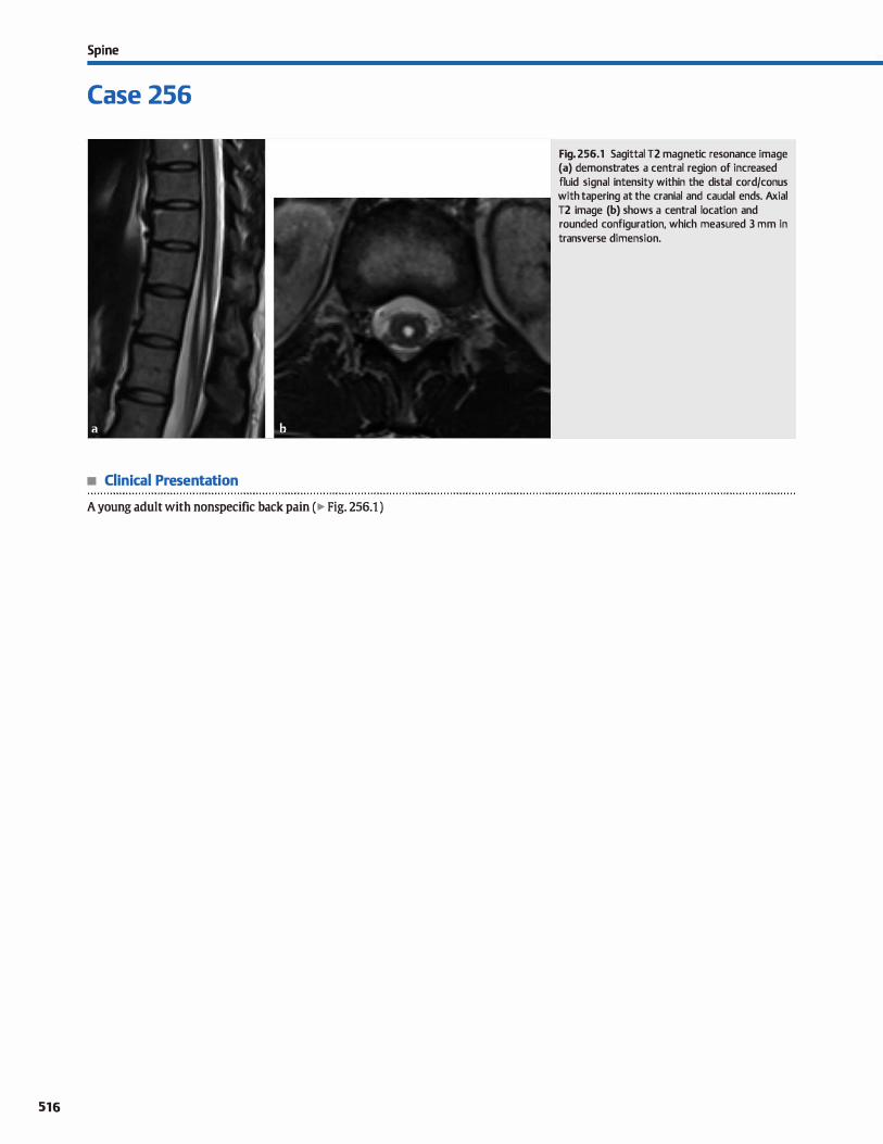
Spi ne
Case 256
• Clinical Presentation
A young adult with nonspecific back pain ( � Fig. 256.1 )
516
Fig. 256.1 Sagittal T2 magnetic resonance image (a) demonstrates a central region of increased fluid signal intensity within the distal cord/conus with tapering at the cranial and caudal ends. Axial T2 image (b) shows a central location and rounded configuration, which measured 3 mm in transverse dimension.
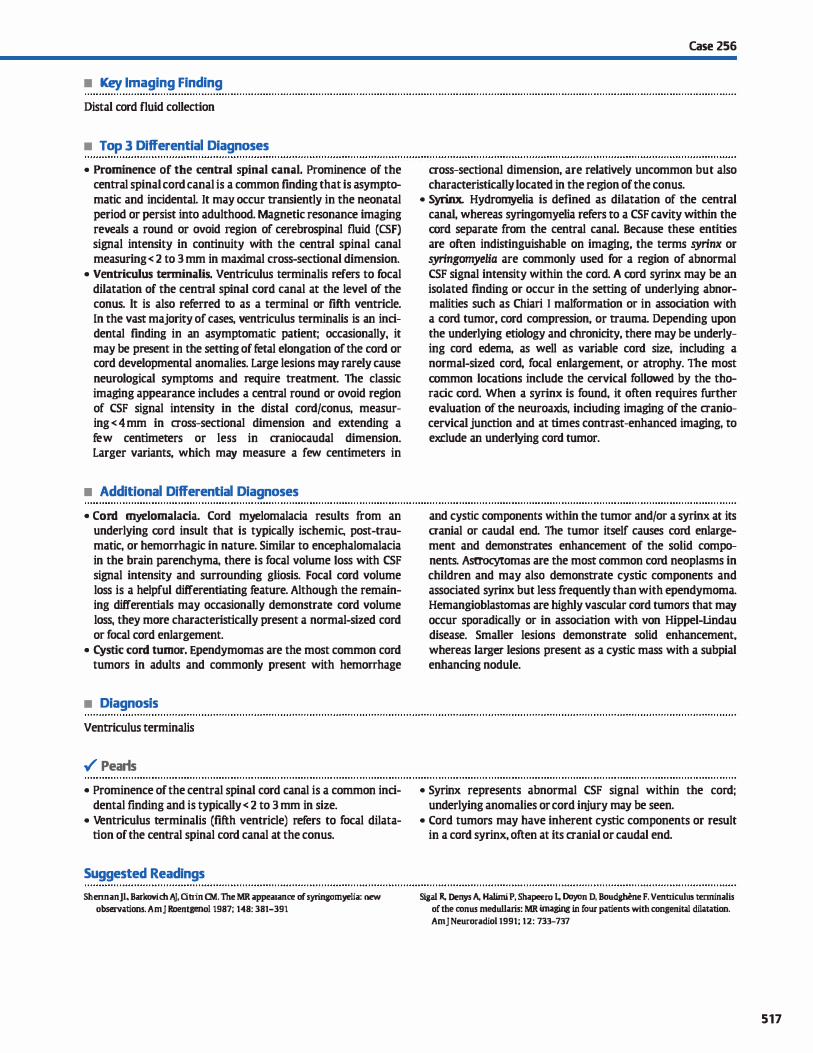
• Key lmaging Finding
Distal cord fluid collection
• Top 3 Differential Diagnoses
• Prominence of the central spinal canal. Prominence of the central spinal cord canal is a common finding that is asympto
matic and incidental. It may occur transiently in the neonatal
period or persist into adulthood. Magnetic resonance imaging
reveals a round or ovoid region of cerebrospinal fluid (CSF)
signal intensity in continuity with the central spinal canal
measuring< 2 to 3 mm in maximal cross-sectional dimension.
• Ventriculus tenninalis. Ventriculus terminalis refers to focal
dilatation of the central spinal cord canal at the level of the
conus. lt is also referred to as a terminal or fifth ventride.
ln the vast majority of cases, ventriculus terminalis is an ind
dental finding in an asymptomatic patient; occasionally, it
may be present in the setting of fetal elongation of the cord or cord developmental anomalies. Large lesions may rarely cause
neurological symptoms and require treatment. The classic imaging appearance includes a central round or ovoid region
of CSF signal intensity in the distal cord/conus, measuring < 4 mm in cross-sectional dimension and extending a
few centimeters or less in craniocaudal dimension. Larger variants, which may measure a few centimeters in
• Additional Differential Diagnoses
• Conl myelomalacia. Cord myelomalacia results from an underlying cord insult that is typically ischemic:, post-trau
matic, or hemorrhagic in nature. Similar to encephalomalacia
in the brain parenchyma, there is focal volume Joss with CSF
signal intensity and surrounding gliosis. Focal cord volume
loss is a helpful differentiating feature. Although the remain
ing differentials may occasionally demonstrate cord volume
loss, they more characteristically present a normal-sized cord
or focal cord enlargement.
• Cystic cord tumor. Ependymomas are the most common cord tumors in adults and commonly present with hemorrhage
• Diagnosis
Ventriculus terminalis
� Pearls
• Prominence of the central spinal cord canal is a common ind
dental finding and is typically< 2 to 3 mm in size.
• Ventriculus terminalis (fifth ventride) refers to focal dilatation of the central spinal cord canal at the conus.
Suggested Readings
Shennanjl. BarkovichAj, Qhin CM. The MR appearanœ of syringomydia: new
observations. AmJ Roentgenol 1987; 1-48: 381-391
Case 256
cross-sectional dimension, are relatively uncommon but also
characteristically located in the region of the conus. • Syrinx. Hydromyelia is defined as dilatation of the central
canal, whereas syringomyelia refers to a CSF cavity within the cord separate from the central canal. Because these entities
are often indistinguishable on imaging, the terms syrinx or
syringomyelia are commonly used for a region of abnormal
CSF signal intensity within the cord. A cord syrinx may be an
isolated finding or occur in the setting of underlying abnormalities such as Chiari 1 malformation or in association with
a cord tumor, cord compression, or trauma. Depending upon
the underlying etiology and chronidty, there may be underly
ing cord edema, as well as variable cord size, including a
normal-sized cord, focal enlargement, or atrophy. The most
common locations include the cervical followed by the thoracic cord. When a syrinx is found, it often requires further
evaluation of the neuroaxis, induding imaging of the cranio
cervical junction and at times contrast-enhanced imaging, to
exdude an underlying cord tumor.
and cystic components within the tumor and/or a syrinx at its
cranial or caudal end. The tumor itself causes cord enlarge
ment and demonstrates enhancement of the solid compo
nents. Astrocytomas are the most common cord neoplasms in
children and may aise demonstrate cystic components and
associated syrinx but Jess frequently than with ependymoma.
Hemangioblastomas are highly vascular cord tumors that may
occur sporadically or in association with von Hippel-Llndau
disease. Smaller lesions demonstrate solid enhancement,
whereas larger lesions present as a cystic mass with a subpial enhandng nodule.
• Syrinx represents abnormal CSF signal within the cord;
underlying anomalies or cord injury may be seen.
• Cord tumors may have inherent cystic components or result in a cord syrinx, often at its cranial or caudal end.
Sigal R. Denys A. Halimi P, Shapeero I. Doyen D, Boudghène F. Venbiculll5 tenninalis
of the conus medullaris: MR imaging in four patients with congenital dilatation.
Amj Neuroradiol 1991; 12: 733-737
517
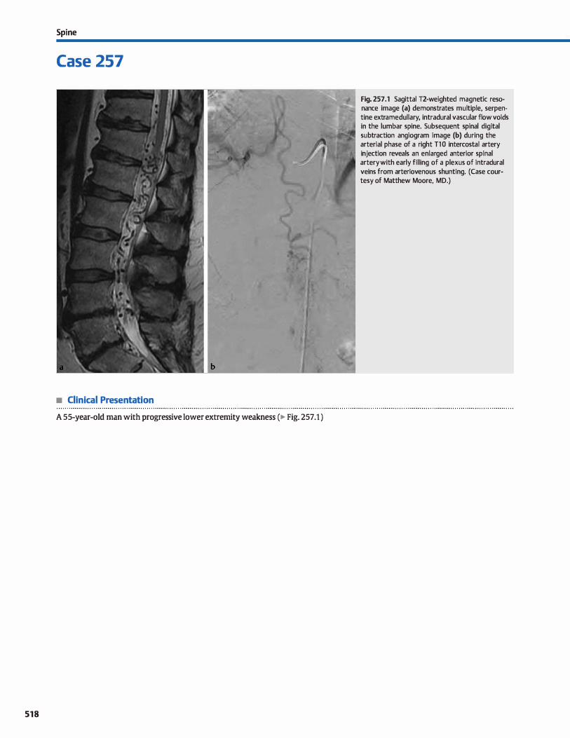
Spi ne
Case 257
• Clinical Presentation
A 55-year-old man with progressive lower extremity weakness (� Fig. 25 7 .1 )
518
Fig. 257 .1 Sagittal T2-weighted magnetic resonance image (a) demonstrates multiple, serpentine extramedullary, intradural vascular flow voids in the lumbar spine. Subsequent spinal digital subtraction angiogram image (b) during the arterial phase of a right Tl 0 intercostal artery injection reveals an enlarged anterior spinal artery with early filling of a plexus of intradural veins from arteriovenous shunting. (Case courtesy of Matthew Moore, MD.)
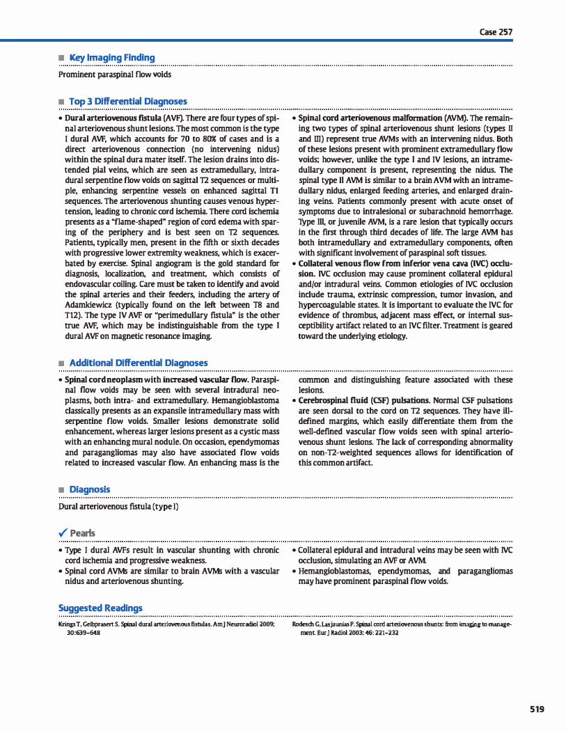
• Key lmaging Finding
Prominent paraspinal flow voids
• Top 3 Differential Diagnoses
• Durai arteriovenous fistula {AVF). There are four types of spi
nal arteriovenous shunt lesions. The most common is the type I durai AVF, which accounts for 70 to 80% of cases and is a direct arteriovenous connection (no intervening nidus) within the spinal dura mater itself. The lesion drains into distended pial veins, which are seen as extramedullary, intradural serpentine flow voids on sagittal T2 sequences or multi
ple, enhancing serpentine vessels on enhanced sagittal Tt sequences. The arteriovenous shunting causes venous hypertension, leading to chronic cord ischemia. There cord ischemia presents as a üflame-shaped" region of cord edema with sparing of the periphery and is best seen on T2 sequenœs. Patients, typically men, present in the fifth or sixth decades with progressive lower extremity weakness, which is exacer
bated by exercise. Spinal angiogram is the gold standard for diagnosis, localization, and treatment. which consists of endovascular coiling. care must be taken to identify and avoid the spinal arteries and their feeders, induding the artery of Adamkiewicz (typically found on the left between T8 and T12). The type IV AVF or �perimedullary fistula" is the other true AVF, which may be indistinguishable from the type 1
durai AVF on magnetic resonance imaging.
• Additional Differential Diagnoses
• Spinal cord neoplasm with inaeased vascular flow. Paraspinal flow voids may be seen with several intradural neo
plasms, both intra- and extramedullary. Hemangioblastoma dassically presents as an expansile intramedullary mass with serpentine flow voids. Smaller lesions demonstrate solid enhancement. whereas larger lesions present as a cystic mass with an enhancing mural nodule. On occasion, ependymomas
and paragangliomas may also have associated flow voids related to increased vascular flow. An enhandng mass is the
• Diagnosis
Durai arteriovenous fistula (type 1)
� Pearls
• Type 1 durai AVFs result in vascular shunting with chronic cord ischemia and progressive weakness.
• Spinal cord AVMs are similar to brain AVMs with a vascular nidus and arteriovenous shunting.
Suggested Readings
Krinp T, Geibprasert S. Spinal durai arteriovenous fistulas. AmJ Neuroradiol 2009;
30:639-648
Case 257
• Spinal cord arteriovenous malformation (AVM). The remaining two types of spinal arteriovenous shunt lesions {types II and III) represent true AVMs with an intervening nidus. Both of these lesions present with prominent extramedullary flow voids; however, unlike the type 1 and IV lesions, an intramedullary component is present, representing the nidus. The spinal type Il AVM is similar to a brain AVM with an intramedullary nidus, enlarged feeding arteries, and enlarged drain
ing veins. Patients commonly present with acute onset of symptoms due to intralesional or subarachnoid hemorrhage. "JYpe Ill, or juvenile AVM, is a rare lesion that typically occurs in the first through third decades of life. The large AVM bas both intramedullary and extramedullary components, often with significant involvement of paraspinal soft tissues.
• Collateral venous flow from inferior vena cava (IVC) occlusion. IVC ocdusion may cause prominent collateral epidural and/or intradural veins. Common etiologies of IVC occlusion indude trauma, extrinsic compression, tumor invasion, and hypercoagulable states. It is important to evaluate the IVC for evidence of thrombus, adjacent mass effect, or internai susœptibility artifact related ta an IVC filter. Treatment is geared
toward the underlying etiology.
common and distinguishing feature associated with these lesions.
• Cerebrospinal fluid (CSF) pulsations. Normal CSF pulsations are seen dorsal to the cord on T2 sequenœs. They have illdefined margins, which easily differentiate them from the well-defined vascular flow voids seen with spinal arterio
venous shunt lesions. The lack of corresponding abnormality
on non-T2-weighted sequences allows for identification of this common artifact.
• Collateral epidural and intradural veins may be seen with IVC occlusion, simulating an AVF or AVM.
• Hemangioblastomas, ependymomas, and paragangliomas may have prominent paraspinal flow voids.
Rodesch G, Lasjaunias P. Spinal cord arteriovenous shW1ts: from imaging to management Eur J R.adiol 2003; 46: 221-232
519
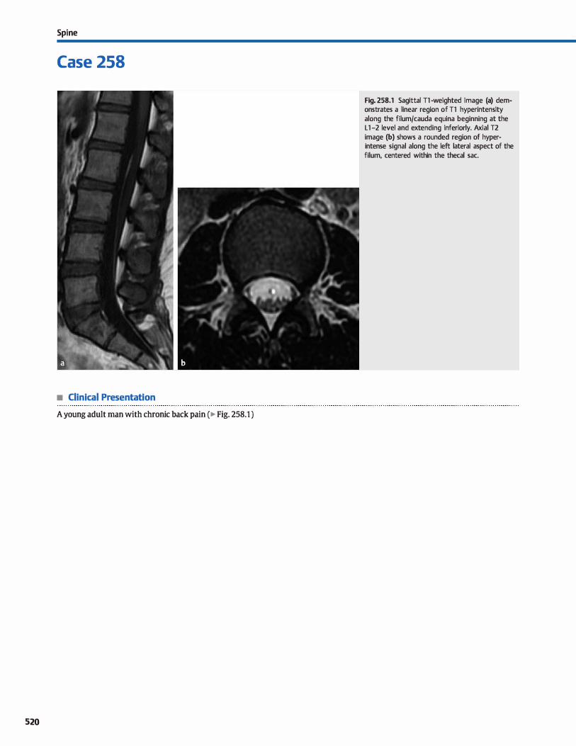
Spi ne
Case 258
• Clinical Presentation
A young adult man with chronic back pain ( � Fig. 258.1 )
520
Fig. 258.1 Sagittal Tl -weighted image (a) demonstrates a linear region of Tl hyperintensity along the filum/cauda equina beginning at the L 1 -2 level and extending inferiorly. Axial T2 image (b) shows a rounded region of hyperintense signal along the left lateral aspect of the filum, centered within the thecal sac.
• Key lmaging Finding
lntrathecal fat
• Top 3 Differential Diagnoses
• Fatty infiltration of the filum. Also known a fibrolipomatous thickening of the filum terminale or filar lipoma. fatty infiltra
tion of the filum is a relatively common finding on lumbar spine magnetic resonance imaging (MRI). Patients are often asymptomatic, although the lesion may be associated with clinical signs of cord tethering in a minority of patients. A low-lying cord, when present, is most often seen in sympto
matic patients. Imaging demonstrates characteristic linear
fatty signal intensity (increased signal on Tl {T2 and decreased signal on fat-suppressed sequences) along a normal-sized or mildly thickened filum. lt typically extends from the conus to the termination of the thecal sac and measures < 2 to 3 mm in transverse dimension. Chemical shift artifact may be seen.
• Subpial lipoma. A subpial lipoma represents focal fatty infiltration isolated to the cord. It may occur at any level, is most
• Additional Differential Diagnoses
• lipomyeloœle. Both lipomyelomeningoœles and lipomyeloceles result from a defect in primary neurulation, which
allows for mesenchymal elements (lipomatous tissue) to enter the neural tube. The fatty tissue is in continuity with the sub
cutaneous fat and interfaces with the neural placode through a posterior bony defect. The overlying skin covering is intact. The key differentiating feature between a lipomyelomeningo
cele and a lipomyelocele is the location of the interface between the lipomatous tissue and neural placode. With a lipomyelocele, the interface is within the spinal canal; with a lipomyelomeningocele, the interface is outside of the spinal canal. MRI best depicts the fatty elements, which are readily
identified on fat-suppressed sequences; it also allows for optimal evaluation of the neural placode.
• Diagnosis
Fatty infiltration of the filum
y' Pearls
• Fatty infiltration of the filum is linear, measures 2 to 3 mm in size, and is most often asymptomatic and incidental.
• Subpial lipoma represents focal fatty infiltration isolated to the cord; it does not extend along the filum.
Suggested Reading
Rufener SI. Ibrahim M, Raybaud CA, Pannar HA. Congenital spine and spinal conl malformations-pictmtù review. ArnJ Roentgenol 2010; 194 Suppl: 526-537
Case 258
common in the thoracic spine, and most often occurs dorsally. When located in the lumbar spine, it typically involves the distal cord or conus and does not extend along the filum terminale. The lipoma may vary significantly from a few millimeters to centimeters in size. Subpial lipomas may be incidental or associated with spinal dysraphism and neurological
deficits corresponding to the involved spinal level.
• Terminal lipoma. A terminal lipoma refers to fatty infiltration
along the distal cord and conus without an overlying spinal dysraphism. The cord is low lying, typically extending to the lumbosacral junction, and there is an indistinct junction between the cord and associated lipoma. Patients often pres
ent with clinicat signs of cord tethering and progressive neurological symptoms.
• Teratoma. A teratoma is a germ cell tumor that contains elements from ail three germ layers. It most often occurs at the sacrum (sacrococcygeal teratoma) and is commonly diagnosed prenatally or in the early neonatal period. lntrathecal
teratomas are exceedingly rare and have similar imaging characteristics to teratomas elsewhere. Lesions typically pres
ent with cystic and solid components, as well as characteristic macroscopic fat. Additional components include calcifications, teeth, and hair. Prognosis is dependent upon the classification in terms of internai or external location. MRI shows a large mass centered at the sacrum with variable extension into adjacent compartments and structures. Characteristic
calcifications, macroscopic fat, and fat-fluid levels are useful discriminators. Solid components heterogeneously enhance.
• Terminal lipoma involves the distal aspect of a low-lying cord without an overlying spinal dysraphism.
• Llpomyelocele occurs in the setting of a spinal dysraphism; the placode-lipoma interface is in the thecal sac.
521
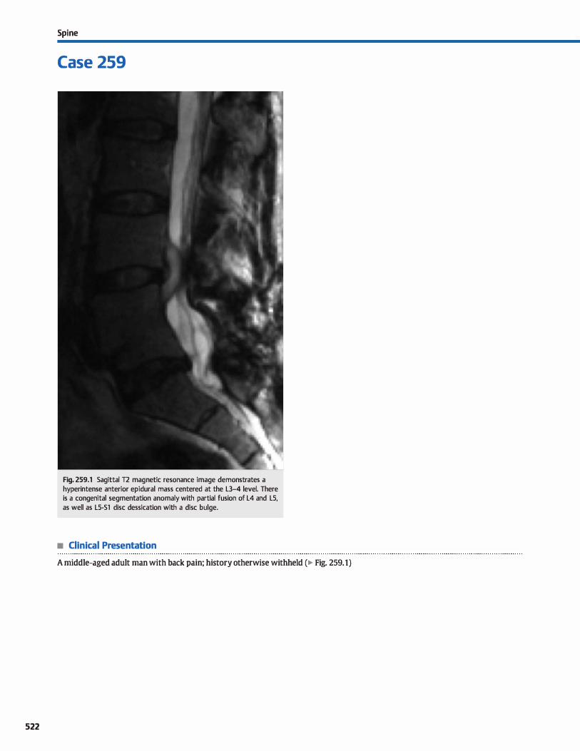
522
Spi ne
Case 259
Fig. 259.1 Sagittal T2 magnetie resonance image demonstrates a hyperintense anterior epidural mass centered at the L3-4 level. There is a eongenital segmentation anomaly with partial fusion of L4 and LS, as well as LS-51 dise dessication with a dise bulge.
• Clinical Presentation
A middle-aged adult man with back pain; history otherwise withheld (� Fig. 259.1)
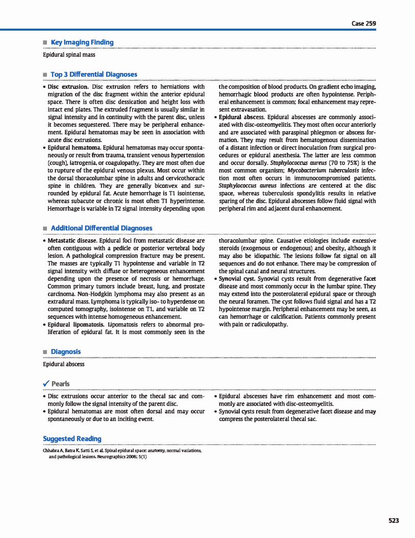
• Key lmaging Finding
Epidural spinal mass
• Top 3 Differential Diagnoses
• Dise extrusion. Dise extrusion refers to hemiations with migration of the dise fragment within the anterior epidural
space. There is often dise dessication and height Joss with
intact end plates. The extruded fragment is usually similar in signal intensity and in continuity with the parent dise, unless it becomes sequestered. There may be peripheral enhancement. Epidural hematomas may be seen in association with
acute dise extrusions.
• Epidural hematoma. Epidural hematomas may occur spontaneously or result from trauma, transient venous hypertension
(cough), iatrogenia, or coagulopathy. They are most often due to rupture of the epidural venous plexus. Most occur within
the dorsal thoracolumbar spine in adults and œrvicothoracic spine in children. They are generally biconvex and surrounded by epidural fat. Acute hemorrhage is T1 isointense,
whereas subacute or chronic is most often Tl hyperintense. Hemorrhage is variable in T2 signal intensity depending upon
• Additional Differential Diagnoses
• Metastatic disease. Epidural foci from metastatic disease are
often contiguous with a pedide or posterior vertebral body lesion. A pathological compression fracture may be present.
The masses are typically T1 hypointense and variable in T2
signal intensity with diffuse or heterogeneous enhanœment
depending upon the presenœ of necrosis or hemorrhage.
Common primary tumors indude breast, Jung, and prostate
cardnoma. Non-Hodgkin lymphoma may also present as an extradural mass. Lymphoma is typically iso- to hyperdense on computed tomography, isointense on Tt, and variable on T2
sequences with intense homogeneous enhancement.
• Epidural lipomatosis. lipomatosis refers ta abnormal proliferation of epidural fat. lt is most commonly seen in the
• Diagnosis
Epidural absœss
� Pearts
• Dise extrusions occur anterior to the thecal sac and com
monly follow the signal intensity of the parent dise. • Epidural hematomas are most often dorsal and may occur
spontaneously or due to an inciting event.
Suggested Reading
ClihabraA, Batra K. Satti S, et al Spinal epidural spaœ: anatmny, normal variations,
and pathological lesions. Neurographics 2006; 5(1)
Case 259
the composition of blood products. On gradient echo imaging, hemorrhagic blood products are often hypointense. Peripheral enhancement is common; focal enhancement may represent extravasation.
• Epidural abscess. Epidural abscesses are commonly associated with disc-osteomyelitis. They most often occur anteriorly
and are associated with paraspinal phlegmon or absœss formation. They may result from hematogenous dissemination
of a distant infection or direct inoculation from surgical proœdures or epidural anesthesia. The latter are less common and occur dorsally. StrJphylococcus aureus (70 to 75%) is the most common organism; Mycobacterium tuberculosis infec
tion most often occurs in immunocompromised patients. Staphylococcus aureus infections are centered at the dise
spaœ, whereas tuberculosis spondylitis results in relative
sparing of the dise. Epidural absœsses follow fluid signal with peripheral rim and adjacent durai enhanœment.
thoracolumbar spine. causative etiologies indude excessive steroids (exogenous or endogenous) and obesity, although it
may also be idiopathie. The lesions follow fat signal on ail
sequenœs and do not enhanœ. There may be compression of
the spinal canal and neural structures. • Synovial cyst. Synovial cysts result from degenerative faœt
disease and most commonly occur in the lumbar spine. They
may extend into the posterolateral epidural space or through
the neural foramen. The cyst follows fluid signal and has a T2 hypointense margin. Peripheral enhancement may be seen, as can hemorrhage or calcification. Patients commonly present
with pain or radiculopathy.
• Epidural abscesses have rim enhancement and most com
monly are associated with disc-osteomyelitis. • Synovial cysts result from degenerative facet disease and may
compress the posterolateral thecal sac.
523
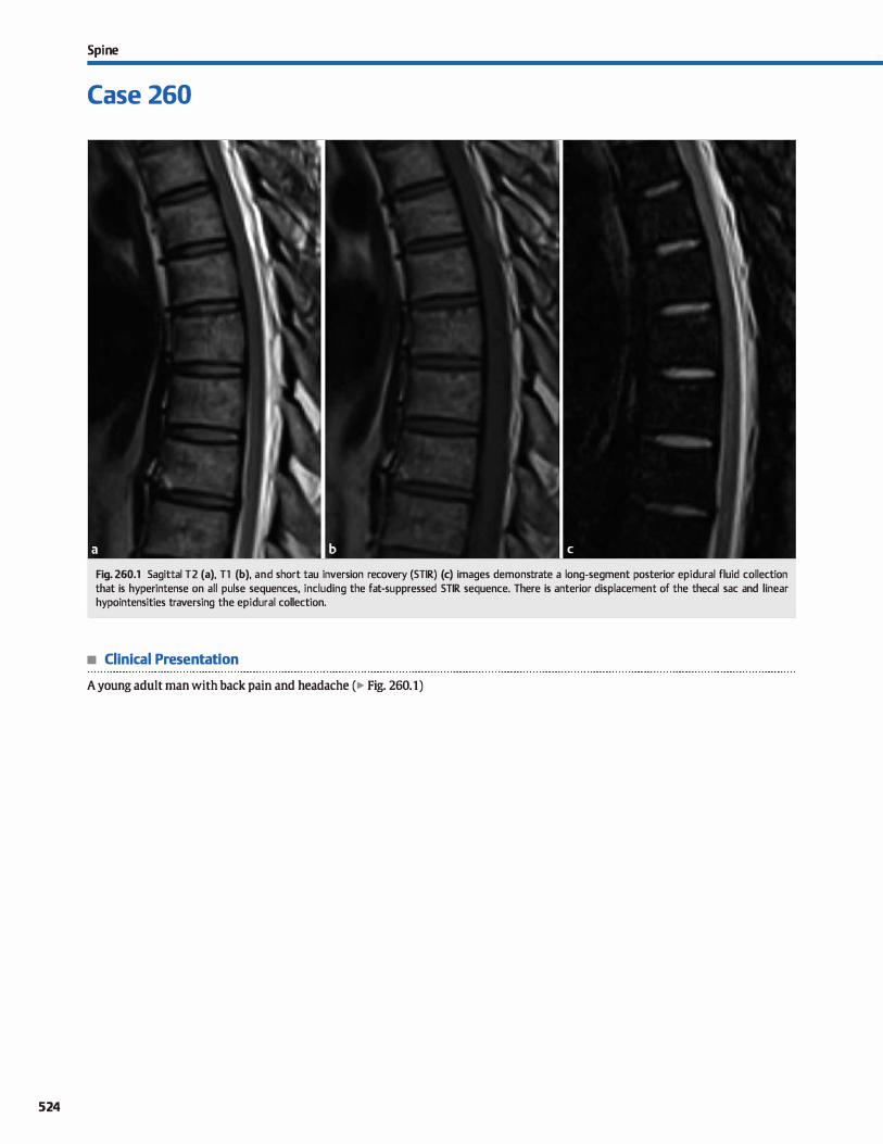
524
Spi ne
Case 260
Fig. 260.1 Sagittal T2 (a), Tl (b), and short tau inversion recovery (STIR) (c) images demonstrate a long-segment posterior epidural fluid collection that is hyperintense on all pulse sequences, including the fat-suppressed STIR sequence. There is anterior displacement of the thecal sac and linear hypointensities traversing the epidural collection.
• Clinical Presentation
A young adult man with back pain and headache (� Fig. 260.1)
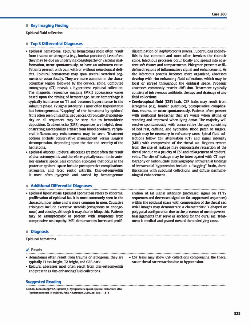
• Key lmaging Finding
Epidural fluid collection
• Top 3 Differential Diagnoses
• Epidural hematoma. Epidural hematomas most often result from trauma or iatrogenia (e.g., lumbar puncture). Less often, they may be due an underlying coagulopathy or vascular malformation, occur spontaneously, or have an unknown cause. Patients present with pain with or without neurological deficits. Epidural hematomas may span several vertebral segments or occur focally. They are more common in the thora
columbar region, followed by the cervical spine. Computed tomography (CT) reveals a hyperdense epidural collection. The magnetic resonance imaging (MRI) appearance varies based upon the timing of hemorrhage. Acute hemorrhage is typically isointense on Tt and becomes hyperintense in the subacute phase. T2 signal intensity is most often hyperintense but heterogeneous. �capping" of the hematoma by epidural
fat is often seen on sagittal sequenœs. Chronically, hypointensity on all sequences may be seen due to hemosiderin deposition. Gradient echo (GRE) sequences are helpful, demonstrating susceptibility artifact Crom blood products. Peripheral inflammatory enhancement may be seen. Treatment options indude conservative management versus surgical decompression, depending upon the size and severity of the hematoma
• Epidural abscess. Epidural abscesses are most often the result of disc-osteomyelitis and therefore typically occur in the anterior epidural space. Less common etiologies that occur in the posterior epidural space indude postoperative complications, iatrogenia, and faœt septic arthritis. Disc-osteomyelitis is most often pyogenic and caused by hematogenous
• Additional Differential Diagnoses
• Epidural lipomatosis. Epidural lipomatosis refers to abnormal proliferation of epidural fat. lt is most commonly seen in the
thoracolumbar spine and is more common in men. Causative etiologies include excessive steroids ( exogenous or endogenous) and obesity, although it may also be idiopathie. Patients may be asymptomatic or present with symptoms from compressive neuropathy. MRI demonstrates increased prolif-
• Diagnosis
Epidural hematoma
y' Pearts
• Hematomas often result Crom trauma or iatrogenia; they are
typically T1 iso-bright, T2 bright, and GRE dark. • Epidural abscesses most often result from disc-osteomyelitis
and present as rim-enhandng fluid collections.
Suggested Reading
Koch BI., Moosbrugger EA, E8eJboffJC. Symptomatic spinal epidural collections after
lumbar puncture in children. Amj Neuroradiol 2007; 28; 1811-1816
Case 260
dissemination of Staphylococcus aureus. Tuberculosis spondylitis is less common and most often involves the thoracic spine. lnfectious processes occur locally and spread into adjacent soft tissues and compartments. Phlegmon presents as illdefined regions of inflammatory signal and enhancement. As the infectious proœss becomes more organized, absœsses
develop with rim-enhancing fluid collections, which may be focal or spread throughout the epidural space. Pyogenic abscesses commonly restrict diffusion. Treatment typically consists of intravenous antibiotic therapy and drainage of any fluid collections.
• Cerebrospinal fluid (CSF) leak. CSF leaks may result Crom iatrogenia (e.g., lumbar puncture), postoperative complication, trauma, or occur spontaneously. Patients often present with positional headaches that are worse when sitting or standing and improved when lying down. The majority will resolve spontaneously with conservative therapy consisting of bed rest, caffeine, and hydration. Blood patch or surgical repair may be necessary in refractory cases. Spinal fluid collections follow CSF attenuation (CT) and signal intensity (MRI) with compression of the thecal sac. Regions remote from the site of leakage may demonstrate retraction of the
thecal sac due to a paucity of CSF and enlargement of epidural veins. The site of leakage may be interrogated with cr myelography or radionuclide cistemography. Intracranial findings
of intracranial hypotension include a üsagging" brain, durai thickening with subdural collections, and diffuse pachymeningeal enhancement.
eration of fat signal intensity (increased signal on Tl fT2 sequences and decreased signal on fat-suppressed sequences)
within the epidural space with compression of the thecal sac. Axial images may demonstrate a characteristic Y-shaped or polygonal configuration due to the presence of meningovertebral ligaments that serve as anchors for the durai sac. Treatment is medical and geared toward the underlying cause.
• CSF leaks may show CSF collections compressing the thecal sac or thecal sac retraction due to hypotension.
525
526
Spi ne
Case 261
Fig. 261 .1 Axial T2 (a) and T l postcontrast (b) MR images with incomplete fat suppression reveal abnormal increased T2 signal intensity and enhancement throughout the paraspinal musculature of the lumbar spi ne. There is also abnormal enhancement of the descending nerve roots of the cauda equina.
• Clinical Presentation
A 22-year-old man with chronic immunodeficiency and back pain (� Fig. 261.1)
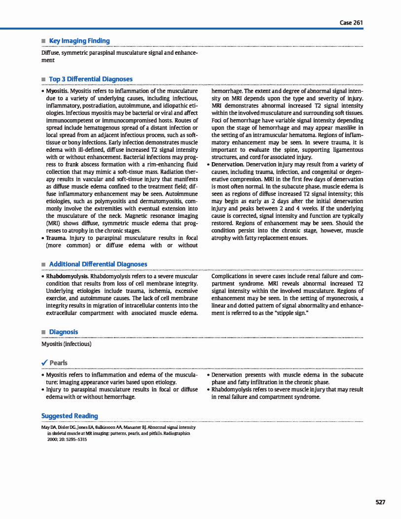
• Key lmaging Finding
Diffuse, symmetric paraspinal musculature signal and enhancement
• Top 3 Differential Diagnoses
• Myositis. Myositis refers to inflammation of the musculature due to a variety of underlying causes, including infectious, inflammatory, postradiation, autoimmune, and idiopathie etiologies. Infectious myositis may be bacterial or viral and affect
immunocompetent or immunocompromised hosts. Routes of spread include hematogenous spread of a distant infection or local spread from an adjacent infectious process, such as softtissue or bony infections. Early infection demonstrates muscle edema with ill-defined, diffuse increased T2 signal intensity with or without enhancement. Bacterial infections may progress to frank abscess formation with a rim-enhancing fluid
collection that may mimic a soft-tissue mass. Radiation therapy results in vascular and soft-tissue injury that manifests as diffuse muscle edema confined to the treatment field; diffuse inflammatory enhancement may be seen. Autoimmune etiologies, such as polymyositis and dermatomyositis, commonly involve the extremities with eventual extension into the musculature of the neck. Magnetic resonanœ imaging (MRI) shows diffuse, symmetric muscle edema that progresses to atrophy in the chronic stages.
• lrauma. Injury to paraspinal musculature results in focal (more common) or diffuse edema with or without
• Additional Differential Diagnoses
• Rhabdomyolysis. Rhabdomyolysis refers to a severe muscular condition that results from loss of cell membrane integrity. Underlying etiologies include trauma, ischemia, excessive exerdse, and autoimmune causes. The lack of cell membrane integrity results in migration of intracellular contents into the
extracellular compartment with associated muscle edema.
• Diagnosis
Myositis (infectious)
y' Pearts
• Myositis refers to inflammation and edema of the musculature; imaging appearance varies based upon etiology.
• Injury to paraspinal musculature results in focal or diffuse edema with or without hemorrhage.
Suggested Reading
May DA, Disler DG, Jones EA, Balkissoon AA, Manas ter Bj. Abnonnal signal intensity
in skeletal muscle at MR imaging: patterns, pearls, and pitfalls. Radiograplùcs
2000; 20; 5295-5315
Case 261
hemorrhage. The extent and degree of abnormal signal intensity on MRI depends upon the type and severity of injury. MRI demonstrates abnormal increased T2 signal intensity within the involved musculature and surrounding soft tissues.
Foci of hemorrhage have variable signal intensity depending upon the stage of hemorrhage and may appear masslike in the setting of an intramuscular hematoma. Regions of inflammatory enhancement may be seen. In severe trauma, it is important to evaluate the spine, supporting ligamentous structures, and cord for associated injury.
• Denervation. Denervation injury may result from a variety of
causes, including trauma, infection, and congenital or degenerative compression. MRI in the first few days of denervation is most often normal. ln the subacute phase, muscle edema is seen as regions of diffuse increased T2 signal intensity; this may begin as early as 2 days after the initial denervation injury and peaks between 2 and 4 weeks. If the underlying cause is corrected, signal intensity and function are typically restored. Regions of enhanœment may be seen. Should the condition persist into the chronic stage, however, muscle
atrophy with fatty replacement ensues.
Complications in severe cases include renal failure and compartment syndrome. MRI reveals abnormal increased T2
signal intensity within the involved musculature. Regions of enhancement may be seen. In the setting of myonecrosis, a linear and dotted pattern of signal abnormality and enhance
ment is referred to as the "stipple sign."
• Denervation presents with muscle edema in the subacute phase and fatty infiltration in the chronic phase.
• Rhabdomyolysis refers to severe muscle injury that may result in renal failure and compartment syndrome.
527
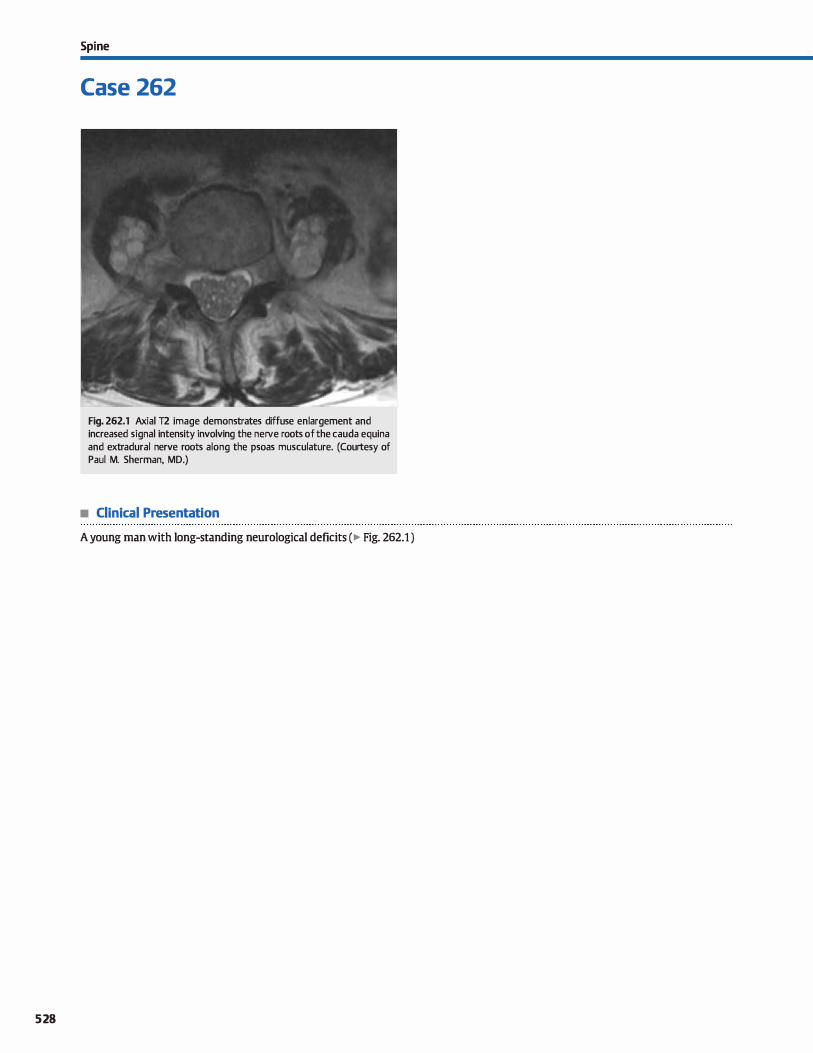
528
Spi ne
Case 262
Fig. 262.1 Axial T2 image demonstrates diffuse en largement and increased signal intensity involving the nerve roots of the cauda equina and extradural nerve roots along the psoas musculature. (Courtesy of Paul M. Sherman, MD.)
• Clinical Presentation
A young man with long-standing neurological deficits (� Fig. 262.1 )
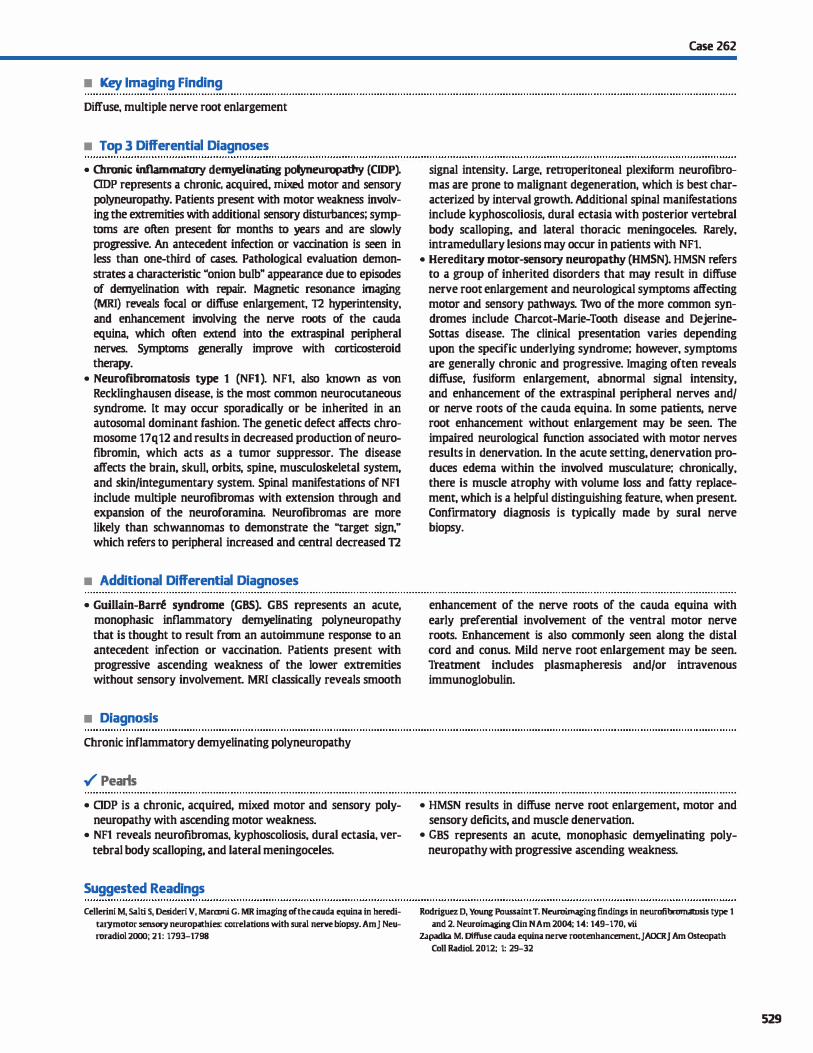
• Key lmaging Finding
Diffuse, multiple nerve root enlargement
• Top 3 Differential Diagnoses
• Chronic inflammatory demydinating polyneuropathy {ODP). ODP represents a chronic, acquired, mixed motor and sensory polyneuropathy. Patients present with motor weakness involving the extremities with additional sensory disturbanœs; symptoms are often present for months to years and are slowly progressive. An anteœdent infection or vaccination is seen in Jess than one-third of cases. Pathological evaluation demon
strates a characteristic Monion bulb" appearanœ due to episodes of demyelination with repair. Magnetic resonanœ imaging (MRI) reveals focal or diffuse enlargement, 12 hyperintensity, and enhanœment involving the nerve roots of the cauda equina, which often extend into the extraspinal peripheral nerves. Symptoms generally improve with corticosteroid therapy.
• Neurofibromatosis type 1 (NF1 ). NF1, also known as von Recklinghausen disease, is the most common neurocutaneous syndrome. It may occur sporadically or be inherited in an autosomal dominant fashion. The genetic defect affects chromosome 17q12 and results in decreased production of neurofibromin, which acts as a tumor suppressor. The disease affects the brain, skull, orbits, spine, musculoskeletal system,
and skin/integumentary system. Spinal manifestations of NF1 include multiple neurofibromas with extension through and expansion of the neuroforamina. Neurofibromas are more likely than schwannomas to demonstrate the ütarget sign," which refers to peripheral increased and central decreased 12
• Additional Differential Diagnoses
• Guillain-Barre syndrome (GBS). GBS represents an acute, monophasic inflammatory demyelinating polyneuropathy
that is thought to result from an autoimmune response to an anteœdent infection or vaccination. Patients present with progressive ascending weakness of the lower extremities without sensory involvement MRI classically reveals smooth
• Diagnosis
Chronic inflammatory demyelinating polyneuropathy
� Pearls
• ODP is a chronic, acquired, mixed motor and sensory polyneuropathy with ascending motor weakness.
• NF1 reveals neurofibromas, kyphoscoliosis, durai ectasia, ver
tebral body scalloping, and lateral meningoœles.
Suggested Readings
Cellerini M, Salti s, Desideri V, Marmni G, MR imaging of the cauda equina in heredi
tarymotor 5ell50I)' neuropathies: correlations with sural nerve biopsy.Amj Neu
roradiol 2000; 21: 1793-1798
Case 262
signal intensity. Large, retroperitoneal plexiform neurofibromas are prone to malignant degeneration, which is best characterized by interval growth. Additional spinal manifestations include kyphoscoliosis, durai ectasia with posterior vertebral
body scalloping, and lateral thoracic meningoceles. Rarely, intramedullary les ions may occur in patients with NF1.
• Hereditary motor-sensory neuropathy (HMSN). HMSN refers to a group of inherited disorders that may result in diffuse
nerve root enlargement and neurological symptoms affecting motor and sensory pathways. 1Wo of the more common syndromes include Charcot-Marie-Tooth disease and DejerineSottas disease. The clinical presentation varies depending
upon the specific underlying syndrome; however, symptoms are generally chronic and progressive. Imaging often reveals diffuse, fusiform enlargement, abnormal signal intensity, and enhancement of the extraspinal peripheral nerves and/ or nerve roots of the cauda equina. In some patients, nerve root enhancement without enlargement may be seen. The impaired neurological function associated with motor nerves results in denervation. In the acute setting, denervation pro
duœs edema within the involved musculature: chronically, there is muscle atrophy with volume loss and fatty replacement, which is a helpful distinguishing feature, when present Confirmatory diagnosis is typically made by sural nerve biopsy.
enhanœment of the nerve roots of the cauda equina with
early preferential involvement of the ventral motor nerve roots. Enhanœment is also commonly seen along the distal cord and conus. Mild nerve root enlargement may be seen. Treatment indudes plasmapheresis and/or intravenous immunoglobulin.
• HMSN results in diffuse nerve root enlargement, motor and sensory deficits, and muscle denervation.
• GBS represents an acute, monophasic demyelinating polyneuropathy with progressive ascending weakness.
Rodriguez D, Young Pous saint T, Neuroimaging findings in neurofibromatnsis type 1
and 2. Neuroirnaging Clin N Am 2004; 14: 149-170. vü Zapadka M. Diffuse cauda equina nerve rootenhanœmentjAOCRJ Am Osteopath
Coll Radial 2012; 1: 29-32
529
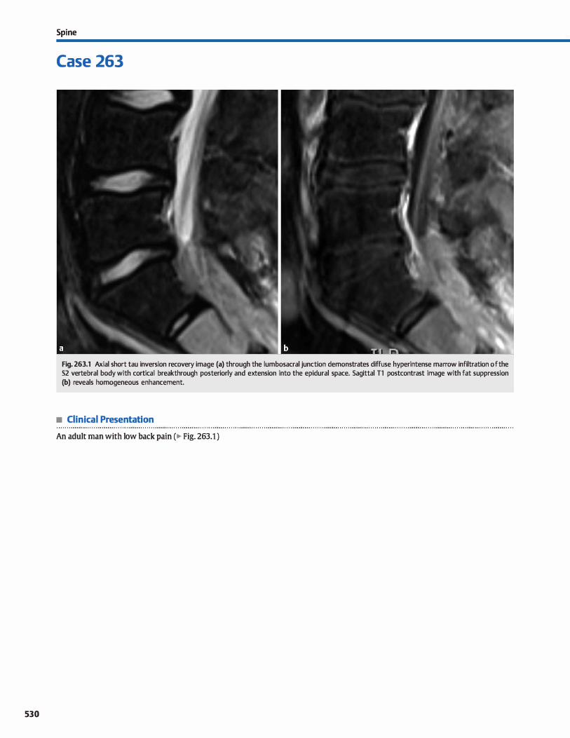
530
Spi ne
Case 263
Fig. 263.1 Axial short tau inversion recovery image (a) through the lumbosacral junction demonstrates diffuse hyperintense marrow infiltration of the 52 vertebral body with cortical breakthrough posteriorly and extension into the epidural space. Sagittal Tl postcontrast image with fat suppression (b) reveals homogeneous enhancement.
• Clinical Presentation
An adult man with low back pain (� Fig. 263.1 )
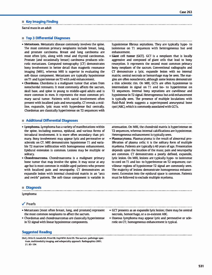
• Key lmaging Finding
Sacral mass in an adult
• Top 3 Differential Diagnoses
• Metastases. Metastatic disease commonly involves the spine. The most common primary neoplasms include breast, Jung, and prostate carcinoma. Breast and Jung cardnoma are most often lytic, along with renal and thyroid carcinomas. Prostate (and occasionally breast) cardnoma produces sderotic metastases. Computed tomography (CT) demonstrates bony involvement in better detail than magnetic resonance
imaging (MRI), whereas MRI is superior in evaluating the soft-tissue component. Metastases are typically hypointense on T1 and hyperintense on T2 with avid enhancement
• Chordoma. Chordoma is a malignant tumor that arises from notochordal remnants. It most commonly affects the sacrum,
skull base, and spine in young to middle-aged adults and is more common in men. It represents the most common pri
mary sacral tumor. Patients with sacral involvement often present with localized pain and neuropathy. CT reveals a midline, expansile, lytic mass with hyperdense fod centrally. Chordomas are dassically hyperintense on T2 sequences with
• Additional Differential Diagnoses
• Lymphoma. Lymphoma bas a variety of manifestations within
the spine, including osseous, epidural, and various forms of intradural involvement. It is more often secondary than primary. Bony involvement may appear lytic and permeative or sderotic on CT. MRI demonstrates hypointense Tl and variable T2 marrow infiltration with homogeneous enhancement. Epidural extension is common. Lesions may be multiple or solitary.
• Chondrosarcoma. Chondrosarcoma is a malignant primary bone tumor that may involve the spine. lt may occur at any
age but is most common in middle-aged patients who present with localized pain and neuropathy. CT demonstrates an expansile lesion with internai chondroid matrix in an "arcs and swirlsft pattern. The soft-tissue component is variable in
• Diagnosis
Lymphoma
� Pearts
• Metastases (most often breast, Jung, and prostate) represent the most common neoplasms to affect the sacrum.
• Chordomas and chondrosarcomas are classically hyperintense in T2 signal with linear hypointense components.
Suggested Reading
Dielj, Ortiz o, I.osada RA, Priœ DB, Hayt MW, Katz DS, lhe sacrum; pathologie spec
trum, multimodality imaging, and subspedalty approach. Radiographies 2001;
21: 83-104
Case 263
hypointense fibrous septations. They are typically hypo- to isointense on Tl sequences with heterogeneous but avid enhancement.
• Giant cell tumor (GCI'). GCT is a neoplasm that is locally aggressive and composed of giant œlls that lead to bony resorption. It represents the second most common primary bony neoplasm of the sacrum. Conventional radiographs and CT demonstrate a lytic, expansile lesion with no internai matrix; central necrosis or hemorrhage may be seen. The margins are often nonsderotic, although some lesions demonstrate a thin sderotic rim. On MRI, GCTs are often hypointense to intermediate in signal on Tl and iso- to hyperintense on T2 sequences. Internai bony septations are curvilinear and hypointense in T2 signal. Heterogeneous but avid enhanœment
is typically seen. The presence of multiple loculations with fluid-fluid levels suggests a superimposed aneurysmal bone cyst (ABC), which is commonly assodated with GCTs.
attenuation. On MRI, the chondroid matrix is hyperintense on T2 sequences, whereas internai calcifications are hypointense. Heterogeneous enhancement is typically seen.
• Plasmacytoma. Plasmacytoma is the result of abnormal proliferation of plasma cells; it is the solitary form of multiple myeloma. Patients are typically � 40 years of age. Presentation depends upon the location of the mass; pain and neuropathy are common. CT demonstrates a poorly detined, expansile, lytic lesion. On MRI, lesions are typically hypo- to isointense to cord on Tl and iso- to hyperintense on T2 sequences; cur
vilinear regions of hypointense T2 signal are commonly seen. The majority of lesions demonstrate homogeneous enhancement Extension into the epidural space is common. Patients must be followed to exclude multiple myeloma.
• GCT presents as an expansile lytic lesion; there may be central necrosis, hemorrhage, or a co-existent ABC.
• Osseous lymphoma may appear lytic and permeative or sclerotic on CT; homogeneous enhancement is typical.
531
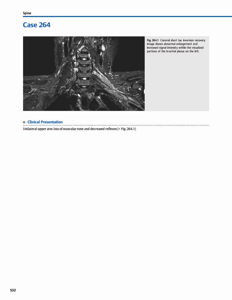
Spi ne
Case 264
• Clinical Presentation
Unilateral upper arm Joss of muscular tone and decreased reflexes (� Fig. 264.1)
532
Fig. 264.1 Coronal short tau inversion recovery image shows abnormal enlargement and increased signal intensity within the visualized portions of the brachial plexus on the left.
• Key lmaging Finding
Enlarged brachial plexus nerve roots
• Top 3 Differential Diagnoses
• li"auma. The brachial plexus is composed of the C5-T1 nerve
roots that extend into trunks, divisions, cords, and terminal
branches as they progresses laterally. Brachial plexus injuries
are most often seen in birth-related trauma and adolescents
involved in motor vehicle accidents. Birth-related injuries
commonly involve C5-7 (Erb-Duchenne palsy); C8-T1 involve
ment is Jess common (Klumpke palsy). Injuries range from
stretching (most common) to avulsions and are classified as
either preganglionic or postganglionic. Preganglionic injuries do not recover spontaneously, whereas postganglionic inju
ries often do. Stretching injuries result in nerve root enlarge
ment, increased T2 signal intensity, and enhancement Surrounding hematoma may be seen.
• Brachial plexitis. Brachial plexitis refers to inflammation
involving the brachial plexus. The most common cause is radi
ation therapy, typically used in the treatment of breast or apical Jung cancer. Less common causes include viral infec
tions, immune-mediated neuropathy, and idiopathie. Com
pared with neoplastic processes, radiation brachial plexitis
typically presents with diffuse thickening and indistinct
signal abnormality that is often confined to the treatment
field; fibrosis may be seen in the chronic setting. The viral,
immune-mediated, and idiopathie forms of brachial plexitis have a variety of imaging appearances, ranging from normal
to nerve root enlargement, abnormal signal intensity, and
• Additional Differential Diagnoses
• Hypertrophie neuropathy. Hypertrophie neuropathy refers
to a variety of disorders that result in enlargement and degen
eration of involved nerves. The condition may be acquired
( e.g., chronic inflammatory demyelinating polyneuropathy
[ODP]) or genetic (e.g., hereditary motor-sensory neuropathy
[HMSN]). l\vo of the more common congenital syndromes (HMSN) include Charcot-Marie-Tooth disease and Dejerine
Sottas disease. Symptoms depend upon the underlying
etiology; however, most patients present with a chronic,
• Diagnosis
Brachial plexitis
� Pearls
• Stretching injury causes enlargement, abnormal signal, and
enhancement; surrounding hematoma may be seen.
• Brachial plexitis refers to inflammation of the brachial plexus;
radiation therapy is the most common cause.
Suggested Reading
Aralasmak A. Karaali K. Cevikol c, Uysal H, Senol u. MR imaging findings in brachial
plexopathywith thoradcoutletsyndrome.AmJNeuroradiol 2010; 31: 410-417
Case 264
enhancement, depending upon the severity and underlying
etiology. Chronic plexitis of motor nerves may result in
denervation. In the acute setting, denervation produces
edema within the innervated musculature; chronically, there
is muscle atrophy with volume loss and fatty replacement.
• Neoplasm. Secondary neoplastic involvement of the brachial
plexus is more common than primary nerve sheath tumors
(NSfs). Common secondary causes include lymphatic spread
of breast cancer (most common), direct spread from Jung cancer (Pancoast tumor), lymphomatous involvement, and
hematogenous spread from distant primary or hematologic
malignandes. Magnetic resonanœ imaging (MRI) demon
strates focal or diffuse nerve root enlargement, increased T2
signal intensity, and abnormal enhancement, which may be masslike. Primary tumors include schwannomas and neurofi
bromas, as well as malignant peripheral NSTs in the setting
of NF1. These primary lesions appear more focal compared with secondary involvement and present as T2 hyperintense,
enhancing lesions. Schwannomas are more likely to undergo
cystic degeneration. Neurofibromas tend to be more uniform
in signal intensity and may demonstrate the "target sign,"
which refers to peripheral increased and central decreased T2
signal intensity. Central lesions may extend through and expand the neural formina.
progressive neuropathy with motor and sensory components.
MRI often reveals diffuse fusiform enlargement, abnormal
signal intensity, and enhancement of the extraspinal periph
eral nerves. The impaired neurological function associated
with motor nerves may result in denervation. In the acute set
ting, denervation produces edema within the involved mus
culature; chronically, there is muscle atrophy with volume
Joss and fatty replacement
• Neoplastic involvement of the brachial plexus may be second
ary (more common) or primary NSTs.
• Patients with hypertrophie neuropathy (CIDP, HMSN) com
monly present with chronic, progressive symptoms.
533
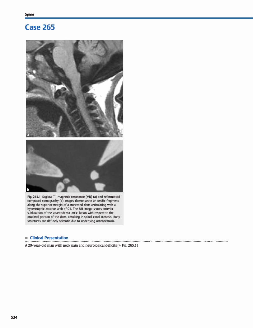
534
Spi ne
Case 265
Fig. 265.1 Sagittal Tl magnetic resonance (MR) (a) and reformatted computed tomography (b) images demonstrate an ossifie fragment along the superior margin of a truncated dens articulating with a hypertrophie anterior arch of Cl . The MR image shows anterior subluxation of the atlantodental articulation with respect to the proximal portion of the dens, resulting in spinal canal stenosis. Bony structures are diffusely sderotic due to underlying osteopetrosis.
• Clinical Presentation
A 20-year-old man with neck pain and neurological deficits (� Fig. 265.1 )
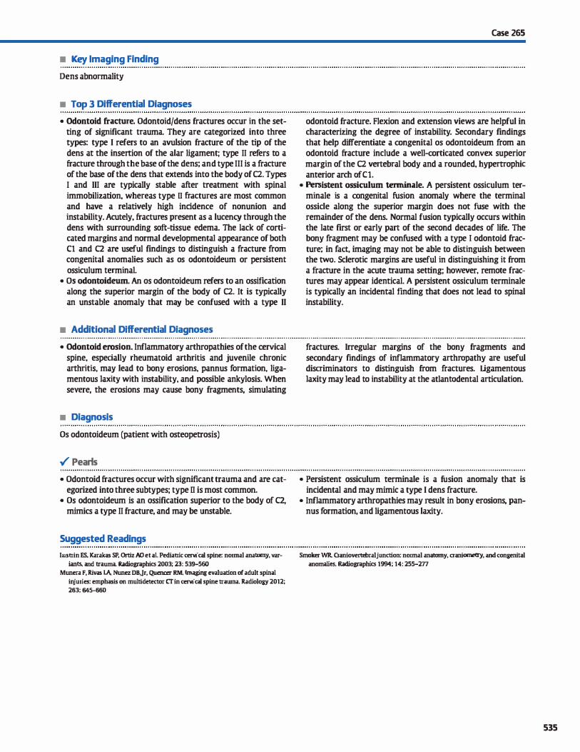
• Key lmaging Finding
Dens abnormality
• Top 3 Differential Diagnoses
• Odontoid fracture. Odontoid/dens fractures occur in the set
ting of significant trauma. They are categorized into three types: type I refers to an avulsion fracture of the tip of the
dens at the insertion of the alar ligament; type II refers ta a fracture through the base of the dens; and type III is a fracture of the base of the dens that extends into the body of C2. Types I and III are typically stable after treatment with spinal
immobilization, whereas type II fractures are most common and have a relatively high inàdence of nonunion and instability. Acutely, fractures present as a lucency through the dens with surrounding soft-tissue edema. The Jack of corticated margins and normal developmental appearance of bath Cl and C2 are useful findings to distinguish a fracture from congenital anomalies such as os odontoideum or persistent
ossiculum terminal. • Os odontoideum. An os odontoideum refers to an ossification
along the superior margin of the body of C2. It is typically an unstable anomaly that may be confused with a type II
• Additional Differential Diagnoses
• Odontoid erosion. Inflammatory arthropathies of the cervical
spine, espectally rheumatoid arthritis and juvenile chronic arthritis, may lead to bony erosions, pannus formation, liga
mentous Iaxity with instability, and possible ankylosis. When severe, the erosions may cause bony fragments, simulating
• Diagnosis
Os odontoideum (patient with osteopetrosis)
� Pearts
• Odontoid fractures occur with significant trauma and are cat
egorized into three subtypes; type II is most common. • Os odontoideum is an ossification superior to the body of C2,
mimics a type II fracture, and may be unstable.
Suggested Readings
Lustrin FS, Karakas SP, Ortiz AD et al. Pediatric cervical spine: normal anatumy, var
iants. and trauma. R.ô1diographics 2003; 23: 539-560
Munera F, Rivas Lo\ Nunez DB,Jr, Quencer RM. lmaging evaluation of adult spinal
injuries: ernphasis on rnultidetector CT in cervical spine trawna. Radiology 2012;
263; 645-660
Case 265
odontoid fracture. Flexion and extension views are helpful in characterizing the degree of instability. Secondary findings that help differentiate a congenital os odontoideum from an odontoid fracture include a well-corticated convex superior
margin of the C2 vertebral body and a rounded, hypertrophie
anterior arch ofC1. • Persistent ossiculum terminale. A persistent ossiculum ter
minale is a congenital fusion anomaly where the terminal
osside along the superior margin does not fuse with the remainder of the dens. Normal fusion typically occurs within the late first or early part of the second decades of life. The bony fragment may be confused with a type I odontoid frac
ture; in fact, imaging may not be able to distinguish between the two. Sclerotic margins are useful in distinguishing it from
a fracture in the acute trauma setting; however, remote fractures may appear identical. A persistent ossiculum terminale is typically an incidental finding that does not lead to spinal instability.
fractures. Irregular margins of the bony fragments and
secondary findings of inflammatory arthropathy are useful discriminators to distinguish from fractures. ügamentous Iaxity may Iead to instability at the atlantodental articulation.
• Persistent ossiculum terminale is a fusion anomaly that is
incidental and may mimic a type I dens fracture. • Inflammatory arthropathies may result in bony erosions, pan
nus formation, and ligamentous laxity.
Srnolœr WR. Craniovertl!braljunction: normal anatorny, craniometry, and congenital
anomalies. R.ô1diographics 1994; 14: 255-277
535
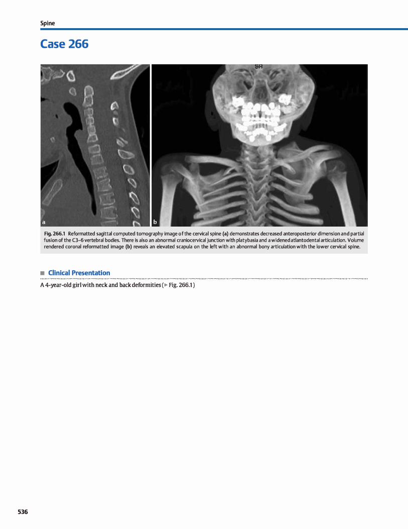
536
Spi ne
Case 266
Fig. 266.1 Reformatted sagittal computed tomography image of the cervical spine (a) demonstrates decreased anteroposterior dimension and partial fusion of the C3-6 vertebral bodies. There is also an abnormal craniocervical junction with platybasia and a widened atlantodental articulation. Volume rendered coronal reformatted image (b) reveals an elevated scapula on the left with an abnormal bony articulation with the lower cervical spine.
• Clinical Presentation
A 4-year-old girl with neck and back deformities ( � Fig. 266.1 )
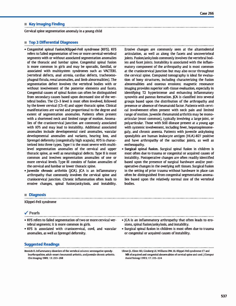
• Key lmaging Finding
Cervical spine segmentation anomaly in a young child
• Top 3 Differential Diagnoses
• Congenital spinal fusion/Klippel-Feil syndrome (KFS). KFS refers to failed segmentation of two or more cervical vertebral segments with or without associated segmentation anomalies of the thoradc and lumbar spine. Congenital spinal fusion is more common in girls and may be sporadic, familial, or associated with multisystem syndromes such as VACTERL (vertebral defects, anal atresia, cardiac defects, tracheoeso
phageal fistula, renal anomalies, and limb abnormalities). The segmentation defect involves the vertebral bodies with or without involvement of the posterior elements and facets. Congenital causes of spinal fusion can often be distinguished from secondary causes based upon decreased size of the vertebral bodies. The C2-3 level is most often involved, followed by the lower cervical (CS-6) and upper thoradc spine. Oinical
manifestations are varied and proportional to the degree and extent of segmentation anomalies. Patients often present
with a shortened neck and limited range of motion. Anomalies of the craniocervical junction are commonly associated with KFS and may lead to instability. Additional associated anomalies indude developmental cord anomalies, vascular developmental anomalies and variants, hearing loss, and
Sprengel deformity (congenitally high scapula). KFS is characterized into three types. fype 1 is the most severe with multilevel segmentation anomalies of the cervical and upper
thoradc spine, as well as neurological defects. fype Il is most common and involves segmentation anomalies of one or more cervical levels. Type Ill consists of fusion anomalies of the cervical and lumbar or lower thoracic spine.
• juvenile chronic arthritis QCA). JCA is an inflammatory arthropathy that commonly involves the cervical spine and
craniocervical junction. Chronic inflammation often leads to erosive changes, spinal fusion/ankylosis, and instability.
• Diagnosis
Klippel-Feil syndrome
� Pearts
• KFS refers ta failed segmentation of two or more cervical vertebral segments; it is more common in girls.
• KFS is assodated with craniocervical, cord, and vascular
anomalies, as well as Sprengel deformity.
Suggested Readings
Resnick D. Inflarnmatnry di.!orden of the vertebral column; seronegative spondy
loarthropathies, adult-onset rheumatoid arthritis, andjuVl!llÜe chronic arthritis. Qin Imaging 1989; 13; 253-268
Case 266
Erosive changes are commonly seen at the altantodental articulation, as well as along the facets and uncovertebral joints. Fusion/ankylosis commonly involves the vertebral bodies and facet joints. Instability is assodated with the inflam
matory component of the arthropathy and is most common at the craniocervical junction but may also occur throughout the cervical spine. Computed tomography is ideal for evalua
tion of bony structures, including characterizing the fusion abnormalities and osseous erosions; magnetic resonanœ imaging provides superior soft-tissue evaluation, especially in identifying T2 hyperintense and enhandng inflammatory synovitis and pannus formation. JCA is classified into several
groups based upon the distribution of the arthropathy and presence or absence of rheumatoid factor. Patients with cervi
cal involvement often present with neck pain and limited range of motion. juvenile rheumatoid arthritis may be monoarticular (most common), typically involving a large joint, or polyarticular. Those with Still disease present at a young age
with systemic involvement, including fever, hepatosplenomegaly, and chronic anemia. Patients with juvenile ankylosing spondylitis are human leukocyte antigen {HLA)-827 positive
and have arthropathy of the sacroiliac joints, as well as enthesopathy.
• Surgica.l spinal fusion. Surgical spinal fusion in children is most often due ta trauma or congenital or acquired causes of instability. Postoperative changes are often readily identified based upon the presence of surgical hardware and/or postoperative changes in the overlying soft tissues. Surgical fusion
in the setting of prior trauma without hardware in place can often be distinguished from congenital segmentation anoma
lies based upon the relatively normal size of the vertebral
bodies.
• JCA is an inflammatory arthropathy that often leads to erosions, spinal fusion/ankylosis, and instability.
• Surgical spinal fusion in children is most often due to trauma
or congenital or acquired causes of instability.
Ulmer JL, Elster AD, Ginsberg LE, Williams DW, m. Klippel-Feil syndrome; CT and
MR of acquired and cungenital abnormalities of cervical spine and cord. J Comput Assistlbmogr1993; 17: 215-224
537
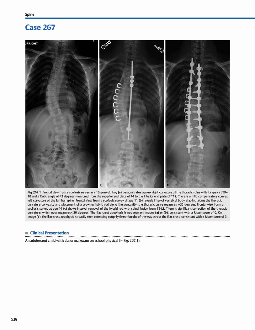
538
Spi ne
Case 267
Fig. 267 .1 Frontal view from a scoliosis survey in a 1 0-year-old boy (a) demonstrates convex right curvature of the thoracic spi ne with its apex at T9-1 0 and a Cobb angle of 42 degrees measured from the superior end plate of T4 to the inferior end plate of Tl 2. There is a mild compensatory convex left curvature of the lumbar spine. Frontal view from a scoliosis survey at age 1 1 (b) reveals interval vertebral body stapling along the thoracic curvature convexity and placement of a growing hybrid rod along the concavity; the thoracic curve measures -35 degrees. Frontal view form a scoliosis survey at age 14 (c) shows interval removal of the hybrid rod with spinal fusion from T2-L2. There is significant correction of the thoracic curvature, which now measures < 20 degrees. The iliac crest apophysis is not seen on images (a) or (b), consistent with a Risser score of O. On image (c), the iliac crest apophysis is readily seen extending roughly three-fourths of the way across the iliac crest, consistent with a Risser score of 3.
• Clinical Presentation
An adolescent child with abnormal exam on school physical (� Fig. 267.1)
• Key lmaging Finding
Lateral spinal curvature in a skeletally immature patient
• Diagnosis
Scoliosis. Scoliosis refers to one or more Iateral curvatures of the spine with a Cobb angle� 10 degrees, often with a rotatory component. Approx.imately 80% of cases are idiopathie, whereas the remainder have an underlying developmental or acquired cause. Idiopathie scoliosis may also be dassified based upon patient age as infantile (0 to 3 years), juvenile (4 to 10 years), adolescent (11 to 18 years), and adult. juvenile and adolescent
idiopathie scoliosis are far more common in girls and present
with a convex right curve (dextroscoliosis). Spinal curvature typieally increases or progresses during
periods of skeletal growth. Once skeletal maturity is achieved, curvatures < 30 degrees are often stable, whereas those > 30
degrees typically continue to progress, although at rates much Jess than those in skeletally immature patients. The Risser index
(Risser sign) is used on scoliosis studies to determine the degree of skeletal maturity and is helpful in determining appropriate management and treatment options. It is based upon the
degree of ossification of the iliac crest apophysis. The index ranges from 0 to 5, where 4 and 5 suggest skeletal maturity pre- and postapophyseal fusion, respectively. During the time of skeletal immaturity, follow-up imaging ranges from 4 to 12
months with an average of every 6 months; doser intervals are used for periods of rapid growth.
The apex of the curvature may be a vertebral body or intervertebral dise spaœ and is defined by the portion of the spine that is furthest away from midline. The end vertebrae are those with the maximal tilt at the cranial and caudal ends of the curve and are used to measure the angle of curvature. When measuring
the degree of curvature, the Cobb angle, the measurement is made along the superior end plate of the œphalad end vertebra and inferior end plate of the caudal vertebra. It is important to
measure the curvature at the same levels on subsequent examinations to properly evaluate its progression. There is some degree
� Pearts
• Scoliosis refers to lateral curvature of the spine with a Cobb angle � 1 O degrees, often with a rotatory component.
• Idiopathie scoliosis is more common in girls and has a convex
right curvature ( dextroscoliosis ).
Suggested Reading
Kim H, Kim HS, Moon ES et al. Scalimis imaging; what radiologists should know. Radiographies 2010; 30: 1823-1842
Case 267
of intraobserver variability, as well as intrinsie variability, in the
measurement of Cobb angles; maintaining consistency between studies is most important Secondary curves form as a compensatory mechanism for the primary curve and have a smaller angle of curvature.
Plain radiographs are the imaging modality of choiœ in diag
nosing and following scoliosis, especially idiopathie cases. Computed tomography and magnetic resonanœ imaging are useful
for atypical curvatures, such as those with an early onset, convex left curvature (levoscoliosis), or rapid progression, as well as to exclude or further evaluate underlying congenital or secondary causes of scoliosis, such as vertebral anomalies, tethered
cord, masses, etc. In contrast to idiopathie cases, secondary causes may have accompanying neurological deficits or pain.
Treatment options for idiopathie scoliosis depend upon the age of onset, curvature severity, and curvature progression. Extemal bracing is intended to stabilize the spinal curvature and is typically used for curves between 20 and 45 degrees. Extemal bracing has varying degrees of sucœss; an early age at presentation and rapid progression are Jess responsive to exter
nat bracing. Surgery is generally indicated for idiopathie scoliosis with a curvature > 45 degrees or in those with progressive
curvature who cannot tolerate external bracing or when externat bracing faits. The ultimate goal of surgery is fusion with correction of the curvature to the greatest degree possible. In skeletally immature patients, surgical options without fusion, including internai bracing (e.g., vertebral body stapling along
the convexity) or "growing" or lengthening fusion rods, are preferred until definitive fusion can be performed when skeletal maturity is reached. To preserve mobility, surgeons attempt to avoid fusion within the cervical and lower lumbar spine when possible. In cases of congenital or acquired causes of scoliosis, the surgical option depends upon the underlying etiology.
• A Risser index is used ta determine the degree of skeletal maturity and is helpful in predieting progression.
• Treatment options depend on curve severity and progression and indude externat bracing and surgical correction.
539
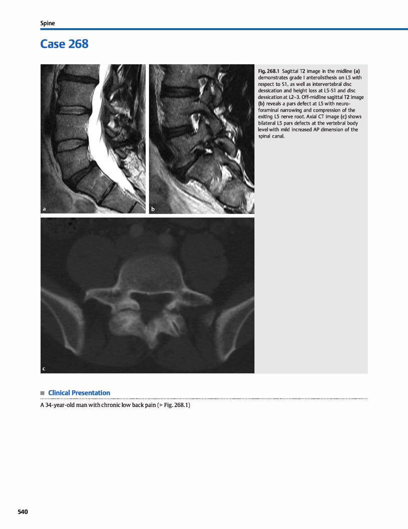
Spi ne
Case 268
• Clinical Presentation
A 34-year-old man with chronic low back pain (� Fig. 268.1)
540
Fig. 268.1 Sagittal T2 image in the midline (a) demonstrates grade 1 anterolisthesis on LS with respect to 51 , as well as intervertebral dise dessication and height loss at LS-51 and dise dessication at L2-3. Off-midline sagittal T2 image (b) reveals a pars defect at LS with neuroforaminal narrowing and compression of the exiting LS nerve root. Axial CT image (c) shows bilateral LS pars defects at the vertebral body level with mild increased AP dimension of the spinal canal.
• Key lmaging Finding
Bilateral pars defects with listhesis
• Diagnosis
Spondylolisthesis. Spondylolisthesis refers to abnormal dis
placement of a vertebral body with respect to another. lt most often occurs at the LS-Sl level, followed by the L4-5 level. causes of spondylolisthesis include isthmic (pars interarticularis defect), degenerative, dysplastic, traumatic, and pathological etiologies. Pars defects and degenerative changes are by far the most common causes of spondylolisthesis.
The pars interarticularis represents the portion of the neural
arch between the lamina and the pedide, facet, and transverse process. A fracture through this region is referred ta as spondy
lolysis. When there is associated displacement at the region of the pars defect, spondylolisthesis occurs. Spondylolysis occurs at the L5 level in -90% of cases; most of the remaining cases occur at the L4 level. Although the precise etiology of spondy
lolysis is not fully understood, it is widely accepted that repetitive injury during periods of development results in pars interarticularis stress fractures. The increased incidence in young athletes supports this theory.
Patients with spondylolisthesis typically present in adolescence or young adulthood with lower back pain and occasional radiculopathy. The L5 nerve root is most often involved, and
symptoms are typically more severe in patients with more pronounœd listhesis.
Spondylolisthesis is graded based upon the degree of subluxation. Grade I spondylolisthesis is defined as O to 25% anterior displacement of the involved vertebral body with respect to the segment below it. Grade Il is defined as 25 to 50%; grade III
� Pearls
• causes of spondylolisthesis indude pars defects, degenerative,
dysplastic, traumatic, and pathologie etiologies.
• Oblique radiographs demonstrate pars defects as the characteristic collar around a "Scottie dog's" neck
Suggested Reading
Gaaju A. Isthmic spondylolisthesis. Newt>Surg Focus 2002; 13{1 ):Et
Case 268
is defined as 50 to 75%; and grade IV is defined as> 75%. Complete (100%) spondylolisthesis is referred to as spondyloptosis.
Most pars defects can be identified on plain radiographs, especially with assodated listhesis. Occasionally, computed tomography (CT) may be neœssary to identify the bony defects
when subtle. On oblique views of the lumbar spine, pars defects characteristically present as a collar around a "Scottie dog's" neck between the pedide (head) and articular faœt (body/ front leg).
Magnetic resonance imaging (MRI) is superior to both plain
radiographs and CT in evaluating for nerve root compression. The pars defects may be identified as regions ofbony disruption
with or without edematous marrow signal changes on off-mid
line sagittal images. Axial images reveal pars defects that may initially be misinterpreted as faœt joints; however, pars defects have a different orientation than the more vertically oriented lumbar facets and are located at the vertebral body level, rather than at the level of the intervertebral dise spaœ. With associated subluxation, there is focal deformity and increased anteroposterior (AP) dimension of the spinal canal. Degenerative lis
thesis, on the other band, narrows the spinal canal because both the ventral and dorsal portions of the vertebral body are displaced anteriorly.
Most patients with spondylolisthesis are either asymptomatic or treated conservatively with medical management. In more severe cases, progressive cases, or in those with failed medical management, surgical fixation may be required.
• MRI is superior to both plain radiographs and CT in evaluating
for nerve root compression. • Axial images reveal pars defects at the vertebral body level
with increased AP dimension of the spinal canal.
541
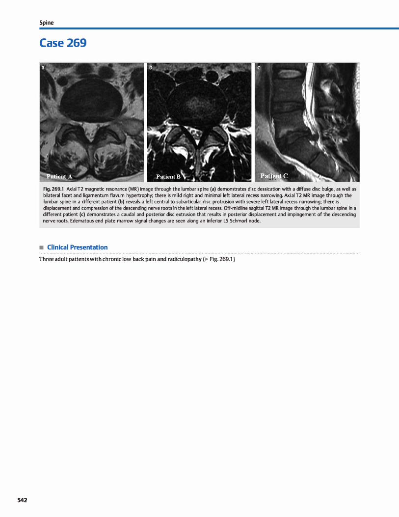
542
Spi ne
Case 269
Fig. 269.1 Axial T2 magnetic resonance (MR) image through the lumbar spi ne (a) demonstrates dise dessication with a diffuse dise bulge, as well as bilateral facet and ligamentum flavum hypertrophy; there is mild right and minimal left lateral recess narrowing. Axial T2 MR image through the lumbar spine in a different patient (b) reveals a left central to subarticular dise protrusion with severe left lateral recess narrowing; there is displacement and compression of the descending nerve roots in the left lateral recess. Off-midline sagittal T2 MR image through the lumbar spine in a different patient (c) demonstrates a caudal and posterior dise extrusion that results in posterior displacement and impingement of the descending nerve roots. Edematous end plate marrow signal changes are seen along an inferior LS Schmorl node.
• Clinical Presentation
Three adult patients with chronic low back pain and radiculopathy (� Fig. 269.1 )
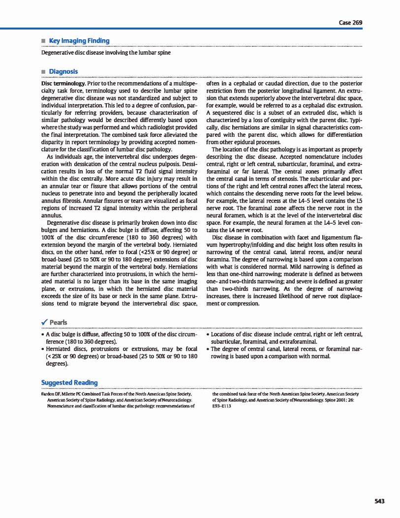
• Key lmaging Finding
Degenerative dise disease involving the lumbar spine
• Diagnosis
Dise terminology. Prior to the recommendations of a multispecialty task force, terminology used to describe lumbar spine degenerative dise disease was not standardized and subject to individual interpretation. This led to a degree of confusion, particularly for referring providers, because characterization of similar pathology would be described differently based upon where the study was performed and which radiologist provided
the final interpretation. The combined task force alleviated the disparity in report terminology by providing accepted nomenclature for the classification of Iumbar dise pathology.
As individuals age, the intervertebral dise undergoes degeneration with dessication of the central nucleus pulposis. Dessication results in loss of the normal T2 fluid signal intensity within the dise centrally. More acute dise injury may result in an annular tear or fissure that allows portions of the central nucleus to penetrate into and beyond the peripherally located annulus fibrosis. Annular fissures or tears are visualized as focal regions of increased T2 signal intensity within the peripheral annulus.
Degenerative dise disease is primarily broken down into dise bulges and hemiations. A dise bulge is diffuse, affecting 50 to 100% of the dise circumference {180 to 360 degrees) with extension beyond the margin of the vertebral body. Herniated dises, on the other band, refer to focal {<25% or 90 degree) or broad-based {25 to 50% or 90 to 180 degree) extensions of dise material beyond the margin of the vertebral body. Hemiations are further characterized into protrusions, in which the hemiated material is no larger than its base in the same imaging plane, or extrusions. in which the hemiated dise material exceeds the size of its base or neck in the same plane. Extrusions tend to migrate beyond the intervertebral dise space,
� Pearts
• A dise bulge is diffuse, affecting 50 to 100% of the dise circumference {180 to 360 degrees).
• Hemiated dises, protrusions or extrusions, may be focal { < 25% or 90 degrees) or broad-based {25 to 50% or 90 to 180 degrees).
Suggested Reading
Farcion DF, Milette PC Combined Task Forces of the North AmeriCilll Spine Society,
American Society of Spine Radiology, and American Society ofNeuroradiology.
Nomenclature and cla.!5ification of lumbar dise pathology; recommendations of
Case 269
often in a cephalad or caudad direction, due to the posterior restriction from the posterior longitudinal ligament. An extrusion that ex.tends superiorly above the intervertebral dise space, for example, would be referred to as a eephalad dise extrusion. A sequestered dise is a subset of an extruded dise, which is characterized by a Joss of contiguity with the parent dise. fypically, dise hemiations are similar in signal characteristics compared with the parent dise, which allows for differentiation from other epidural processes.
The location of the dise pathology is as important as properly describing the dise disease. Accepted nomenclature indudes central, right or left central, subarticular, foraminal, and extraforaminal or far Iateral. The central zones primarily affect the central canal in terms of stenosis. The subarticular and portions of the right and left central zones affect the lateral recess, which contains the descending nerve roots for the level below. For ex.ample, the lateral recess at the L4-5 level contains the L5 nerve root The foraminal zone affects the nerve root in the neural foramen, which is at the level of the intervertebral dise space. For example, the neural foramen at the L4-5 level contains the L4 nerve root
Dise disease in eombination with facet and ligamentum flavum hYPertrophy/infolding and dise height Joss often results in narrowing of the central canal, lateral recess, and/or neural foramina. The degree of narrowing is based upon a comparison with what is considered normal. Mild narrowing is defined as Jess than one-third narrowing; moderate is defined as between one- and two-thirds narrowing; and severe is defined as greater than two-thirds narrowing. As the degree of narrowing increases, there is increased likelihood of nerve root displacement or compression.
• Locations of dise disease indude central, right or left central, subarticular, foraminal, and extraforaminal.
• The degree of central canal, lateral recess, or foraminal narrowing is based upon a comparison with normal.
the combined task forœ of the North American Spine Society, American Society
of Spine Radiology, and American Society ofNeuroradiology. Spine 2001; 26:
E93-El 13
543
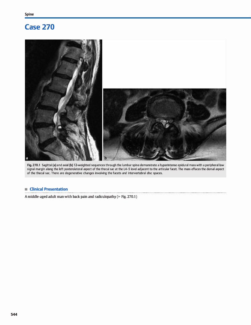
544
Spi ne
Case 270
Fig. 270.1 Sagittal (a) and axial (b) T2-weighted sequences through the lumbar spi ne demonstrate a hyperintense epidural mass with a peripheral low signal margin a long the left posterolateral aspect of the thecal sac at the L4-5 level adjacent to the articular facet. The mass effaces the dorsal aspect of the thecal sac. There are degenerative changes involving the facets and intervertebral dise spaces.
• Clinical Presentation
A middle-aged adult man with back pain and radiculopathy (� Fig. 270.1 )
• Key lmaging Finding
Extradural cystic mass adjacent to a degenerative facet
• Diagnosis
Synovial cyst. Synovial cysts are extradural Iesions that result
from faœt degenerative arthropathy. They occur most frequently in middle-aged and older adults, are more common in
women and in the setting of degenerative spondylolisthesis,
and most often involve the lower lumbar spine.
The majority of synovial cysts are asymptomatic and indden
tal, espedally when located along the posterior aspect of the facet
joint Anterior Iesions may cause symptoms related to nerve root compression within the central canal, lateral recess, or neural
foramen. The most common presenting symptom is painful radi
culopathy.
� Pearls
• Synovial cysts are extradural lesions that result from facet
degenerative arthropathy.
• The majority of synovial cysts are asymptomatic, although large lesions may cause nerve root compression.
Suggested Reading
Khan AM, Girardi F. Spinal lumbar synovial cysts. Diagnosis and management chal
lenge. Eur Spine J 2006; 1 S: 117&-1182
Case 270
Magnetic resonance imaging (MRI) is the modality of choice
and demonstrates an extradural cystic lesion with a peripheral
T2 hypointense rim in association with a degenerative facet
joint. Increased protein content within the cyst may lead to
slight increased signal intensity on Tl sequences. Peripheral
inflammatory signal and enhancement may be seen. as can hemorrhage or calcification.
Treatment options indude conservative management with
analgesics, percutaneous steroid injection and/or drainage, and
surgical resection in more severe cases.
• MRI demonstrates a cystic mass with peripheral T2 hypoin
tensity in proximity to a degenerative facetjoint
545
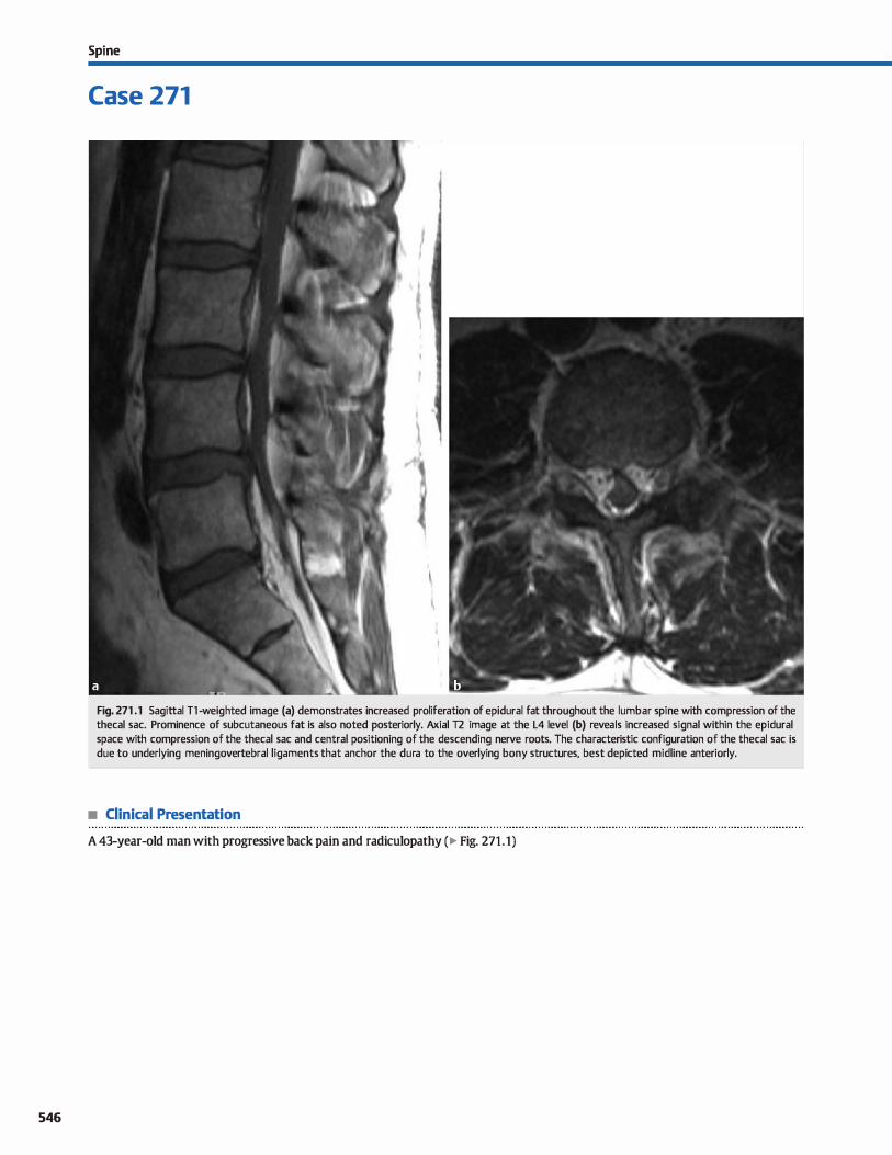
546
Spi ne
Case 271
Fig. 271 .1 Sagittal Tl -weighted image (a) demonstrates increased proliferation of epidural fat throughout the lumbar spi ne with compression of the thecal sac. Prominence of subcutaneous fat is also noted posteriorly. Axial T2 image at the L4 level (b) reveals increased signal within the epidural space with compression of the thecal sac and central positioning of the descending nerve roots. The characteristic configuration of the thecal sac is due to underlying meningovertebral ligaments that anchor the dura to the overlying bony structures, best depicted midline anteriorly.
• Clinical Presentation
A 43-year-old man with progressive back pain and radiculopathy (� Fig. 271.1)
• Key lmaging Finding
Increased epidural fat with compression of thecal sac
• Diagnosis
Epidural lipomatosis. Epidural lipomatosis refers to abnonnal proliferation of epidural fat It is most commonly seen in the
thoracolumbar spine and is more common in men. causative etiologies indude excessive steroids ( exogenous or endogenous) and obesity, although it may also be idiopathie. Patients may be asymptomatic or present with symptoms related to compressive neuropathy.
Magnetic resonanœ imaging (MRI) is the imaging modality of choiœ due to its superior evaluation of soft tissues. Findings indude increased proliferation of fat signal intensity (increased sig-
� Pearts
• Llpomatosis refers to abnonnal proliferation of epidural fat; etiologies indude obesity and steroids but may aise be idiopathie.
• MRI is the modality of choiœ, demonstrating increased proliferation of epidural fat and thecal sac compression.
Suggested Readings
Geer5 c, I.ecou'W!t FE, Behets C, Malghemj, Cosn.ml G, I.engelé BG, Polygonal defor
mation of the durai sac in lwnbar epidural lipomatmis: anatDmic explanation by the presenœ of meninguvertdlral ligaments. Amj Neuroradiol 2003; 24: 1276-1282
Case 271
nal on 11fr2 sequenœs and decreased signal on fat-suppressed sequenœs) within the epidural spaœ with associated compression of the thecal sac. Axial images may demonstrate a characteristic Yshaped or po1ygonal configuration due the presenœ of meningovertebral ligaments that serve as anchors for the durai sac.
Treatment is typically medical and geared toward weight reduction or cessation of the underlying cause of excessive ste
roids, when possible. Surgical decompression may be necessary in severe symptomatic cases where medical therapies are not viable or are unsuccessful.
• Axial images may show a characteristic Y-shaped or polygonal configuration due to meningovertebral ligaments.
Shaheen F, Singh M, Gojwari Tet al, Idiopathie epidural lipomatnsis, Appl Radio!
2008;37: 40A-40B
547
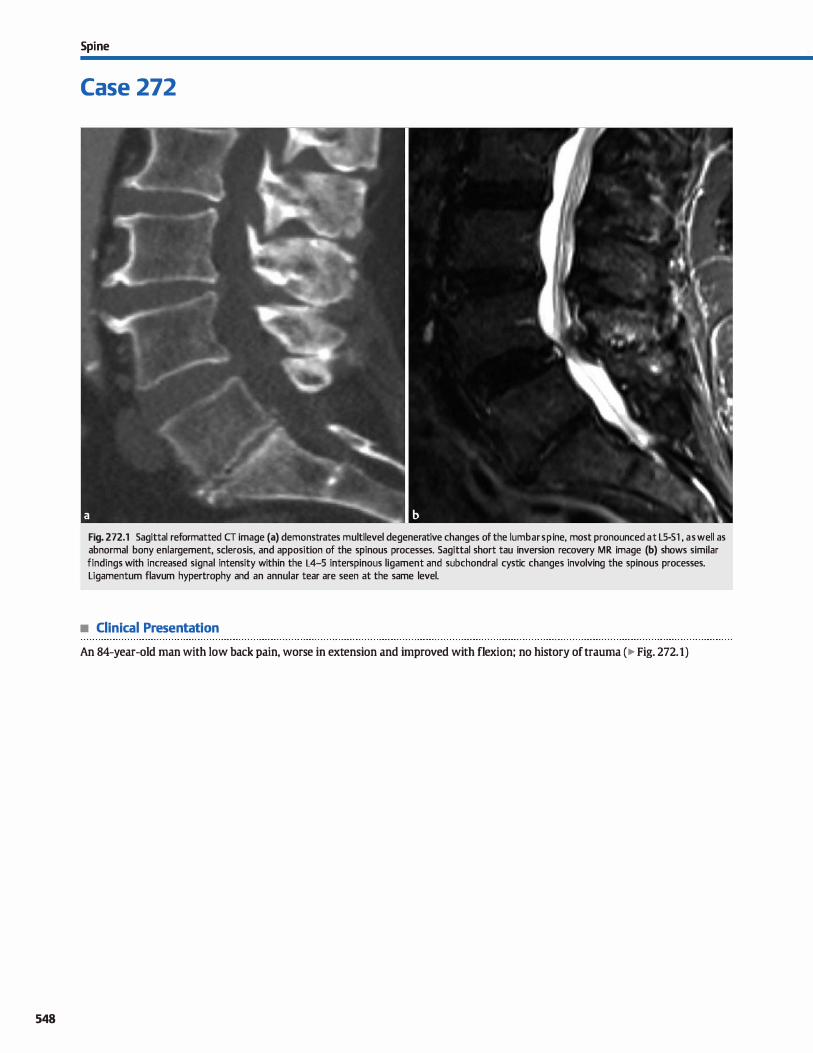
548
Spi ne
Case 272
Fig. 272.1 Sagittal reformatted CT image (a) demonstrates multilevel degenerative changes of the lumbar sp i ne, most pronounced at LS-Sl , as well as abnormal bony enlargement, sderosis, and apposition of the spinous processes. Sagittal short tau inversion recovery MR image (b) shows similar findings with increased signal intensity within the L4-5 interspinous ligament and subchondral cystic changes involving the spinous processes. Ligamentum flavum hypertrophy and an annular tear are seen at the same level.
• Clinical Presentation
An 84-year-old man with low back pain, worse in extension and improved with flexion; no history of trauma (� Fig. 272.1)
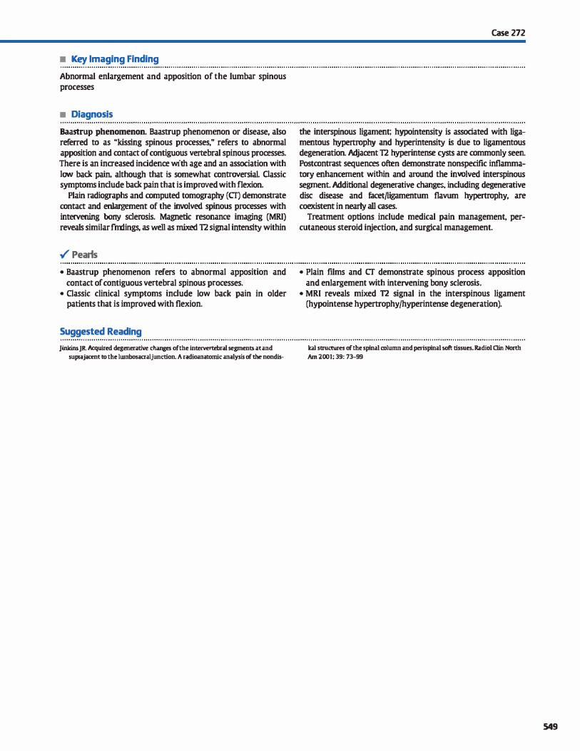
• Key lmaging Finding
Abnormal enlargement and apposition of the lumbar spinous processes
• Diagnosis
Baastrup phenomenon. Baastrup phenomenon or disease, also referred to as "kissing spinous proœsses," refers to abnormal apposition and contact of contiguous vertebral spinous processes. There is an increased incidence with age and an association with
Iow back pain, although that is somewhat controversial. Oassic symptoms indude back pain that is improved with flexion.
Plain radiographs and computed tomography (CT) demonstrate contact and enlargement of the involved spinous proœsses with intervening bony sderosis. Magnetic resonanœ imaging (MRI) reveals similar fmdings, as well as mixed T2 signal intensity within
� Pearls
• Baastrup phenomenon refers to abnormal apposition and
contact of contiguous vertebral spinous proœsses. • aassic clinical symptoms indude low back pain in older
patients that is improved with flexion.
Suggested Reading
Jinldns jR. Acquired degenerative changes of the intervertebral segments at and
suprajaœnt ta the lwnbosacraljunction. A radioanatomic analysis of the nondis-
Case 272
the interspinous ligament; hypointensity is associated with ligamentous hypertrophy and hyperintensity is due to ligamentous degeneration. Adjacent T2 hyperintense cysts are commonly seen. Postcontrast sequences often demonstrate nonspecific inflammatory enhanœment within and around the involved interspinous segment. Additional degenerative changes, induding degenerative dise disease and faœt/!igamentum flavum hypertrophy, are coexistent in nearly all cases.
Treatment options include medical pain management, percutaneous steroid injection, and surgical management.
• Plain films and cr demonstrate spinous process apposition and enlargement with intervening bony sclerosis .
• MRI reveals mixed T2 signal in the interspinous ligament (hypointense hypertrophy{hyperintense degeneration).
kal structures of the spinal mlumn and perispinal soft tissues. Radial Clin North
Am 2001; 39: 73-99
549
550
Spi ne
Case 273
Fig. 273.1 Sagittal Tl - (a) and axial T2-weighted image at the mid-L4 vertebral body level (b) demonstrate significant narrowing of the lumbar spinal canal secondary to shortened pedides. The anteroposterior measurement was -8 mm.
• Clinical Presentation
A 36-year-old man with chronic low back pain (� Fig. 273.1 )
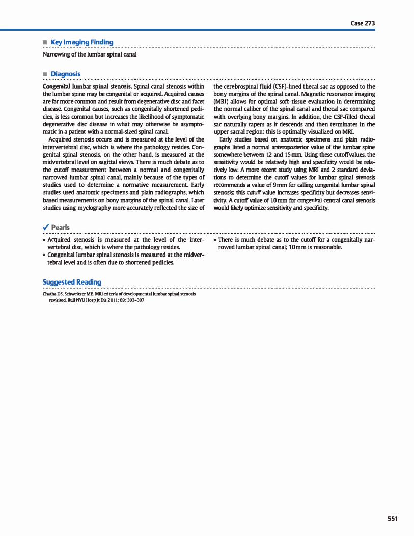
• Key lmaging Finding
Narrowing of the lumbar spinal canal
• Diagnosis
Congenital lumbar spinal stenosis. Spinal canal stenosis within
the lumbar spine may be congenital or aaiuired. Acquired causes
are far more common and result from degenerative dise and faœt
disease. Congenital causes, such as congenitally shortened pedi
des, is Jess common but increases the likelihood of symptomatic
degenerative dise disease in what may otherwise be asymptomatic in a patient with a normal-sized spinal canal
Acquired stenosis occurs and is measured at the Ievel of the
intervertebral dise, which is where the pathology resides. Con
genital spinal stenosis, on the other band, is measured at the
midvertebral level on sagittal views. There is much debate as ta
the cutoff measurement between a normal and congenitally narrowed lumbar spinal canal, mainly because of the types of
studies used ta determine a normative measurement. Early
studies used anatomie spedmens and plain radiographs, which
based measurements on bony margins of the spinal canal. Later
studies using myelography more accurately reflected the size of
� Pearts
• Aaiuired stenosis is measured at the level of the inter
vertebral dise, which is where the pathology resides.
• Congenital Iumbar spinal stenosis is measured at the midver
tebral level and is often due ta shortened pedides.
Suggested Reading
Cllatha DS, Schweitzer ME. MRI criteria of �lopmental lumbar spinal stenosis
revisiœd. Bull NYlJ Hosp Jt Dis 2011; 69: 303-307
Case 273
the cerebrospinal fluid {CSF)-lined thecal sac as opposed ta the
bony margins of the spinal canal. Magnetic resonanœ imaging
(MRI) allows for optimal soft-tissue evaluation in determining the normal caliber of the spinal canal and thecal sac compared
with overlying bony margins. ln addition, the CSF-filled thecal
sac naturally tapers as it descends and then terminates in the upper sacral region; this is optimally visualized on MRI.
F.arly studies based on anatomie spedmens and plain radio
graphs listed a normal anteroposterior value of the lumbar spine
somewhere between 12 and 15 mm Using these cutoffvalues, the
sensitivity would be relatively high and specifidty would be rela
tively low. A more ruent study using MRI and 2 standard devia
tions to determine the cutoff values for lumbar spinal stenosis
recommends a value of 9 mm for calling congenital lumbar spinal stenosis; this cutoff value increases specifidty but decreases sensi
tivity. A cutoff value of 1 O mm for congenita1 œntral canal stenosis
would likely optimize sensitivity and specifidty.
• There is much debate as to the cutoff for a congenitally nar
rowed lumbar spinal canal; lOmm is reasonable.
551
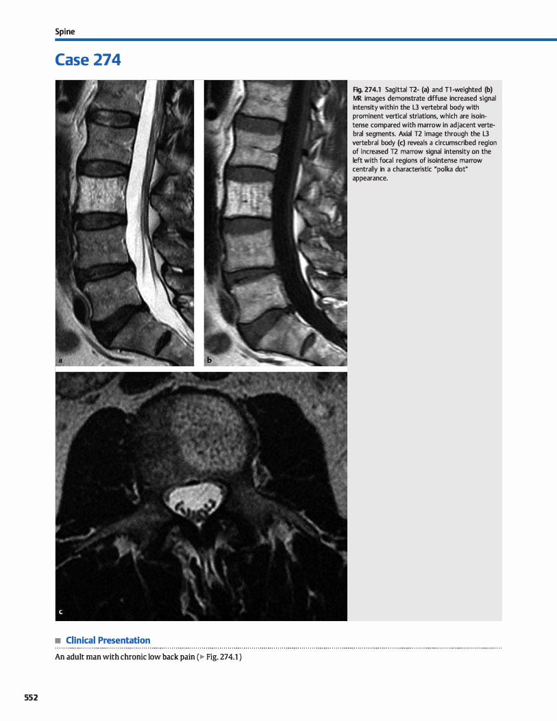
Spi ne
Case 274
• Clinical Presentation
An adult man with chronic low back pain (� Fig. 274.1 )
552
Fig. 274.1 Sagittal T2- (a) and Tl -weighted (b) MR images demonstrate diffuse increased signal intensity within the L3 vertebral body with prominent vertical striations, which are isointense compared with marrow in adjacent vertebrai segments. Axial T2 image through the L3 vertebral body (c) reveals a circumscribed region of increased T2 marrow signal intensity on the left with focal regions of isointense marrow centrally in a characteristic "polka dot" appearance.
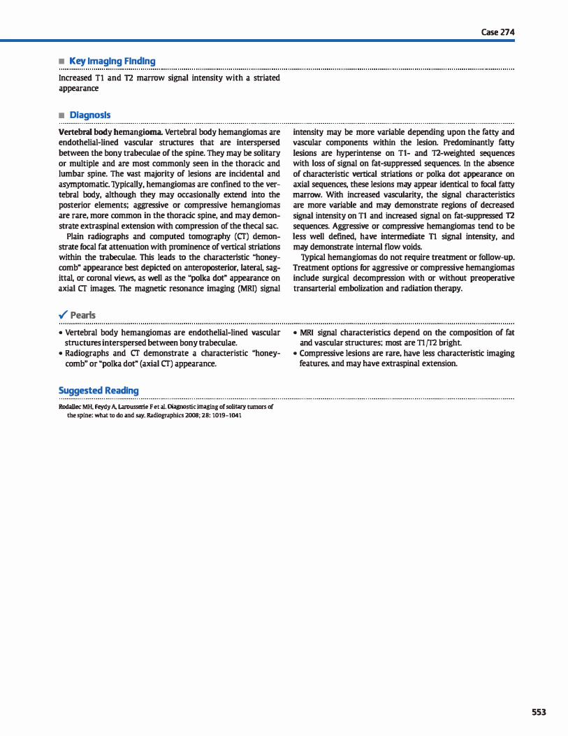
• Key lmaglng Flndlng
Increased Tl and T2 marrow signal intensity with a striated appearanœ
• Diagnosls
Vertebral body hemangiom.a. Vertebral body hemangiomas are endothelial-lined vascular structures that are interspersed
between the bony trabeculae of the spine. They may be solitary
or multiple and are most commonly seen in the thoradc and lumbar spine. The vast majority of lesions are incidental and
aSYJllptomatic. Typically, hemangiomas are confined ta the vertebral body, although they may occasionally extend into the posterior elements; aggressive or compressive hemangiomas are rare, more common in the thoracic spine, and may demon
strate extraspinal extension with compression of the thecal sac.
Plain radiographs and computed tomography (cr) demonstrate focal fat attenuation with prominenœ of vertical striations within the trabeculae. This leads to the characteristic "honeycomb" appearanœ best depicted on anteroposterior, lateral, sagittal, or coronal views, as well as the "polka dot" appearanœ on
axial cr images. The magnetic resonanœ imaging (MRI) signal
� Pearls
• Vertebral body hemangiomas are endothelial-lined vascular structures interspersed between bony trabeculae.
• Radiographs and cr demonstrate a characteristic "honey
comb" or "polka dot" (axial cr) appearanœ.
Suggested Reading
Rodallec MH, Feydy A. Larousserie F et al. Diagnostic imaging of solituy tumors of
thespine; what to do and say. Radiographies 2008; 28; 1019-1041
Case 274
intensity may be more variable depending upon the fatty and vascular components within the lesion. Predominantly fatty
lesions are hyperintense on Tl- and T2-weighted sequenœs with Joss of signal on fat-suppressed sequenœs. In the absence
of characteristic vertical striations or polka dot appearanœ on axial sequenœs, these lesions may appear identical ta focal fatty marrow. With increased vascularity, the signal characteristics are more variable and may demonstrate regions of decreased
signal intensity on T1 and increased signal on fat-suppressed T2 sequences. Aggressive or compressive hemangiomas tend to be Jess well defined, have intermediate 11 signal intensity, and may demonstrate internai flow voids.
Typical hemangiomas do not require treatment or follow-up. Treatment options for aggressive or compressive hemangiomas include surgical decompression with or without preoperative transarterial embolization and radiation therapy.
• MRI signal characteristics depend on the composition of fat and vascular structures; most are T1 /T2 bright
• Compressive lesions are rare, have Jess characteristic imaging features, and may have extraspinal extension.
553
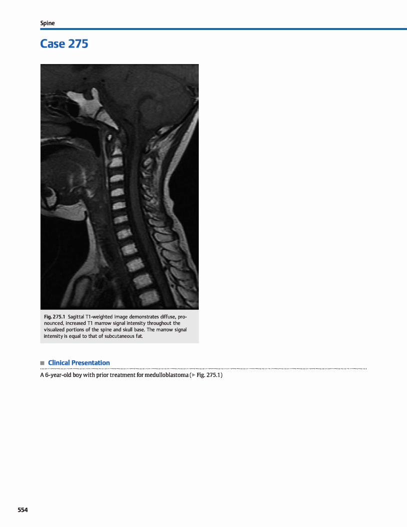
554
Spi ne
Case 275
Fig. 275.1 Sagittal Tl -weighted image demonstrates diffuse, pronounced, increased Tl marrow signal intensity throughout the visualized portions of the spine and skull base. The marrow signal intensity is equal to that of subcutaneous fat.
• Clinical Presentation
A 6-year-old boy with prior treatment for medulloblastoma (� Fig. 275.1 )
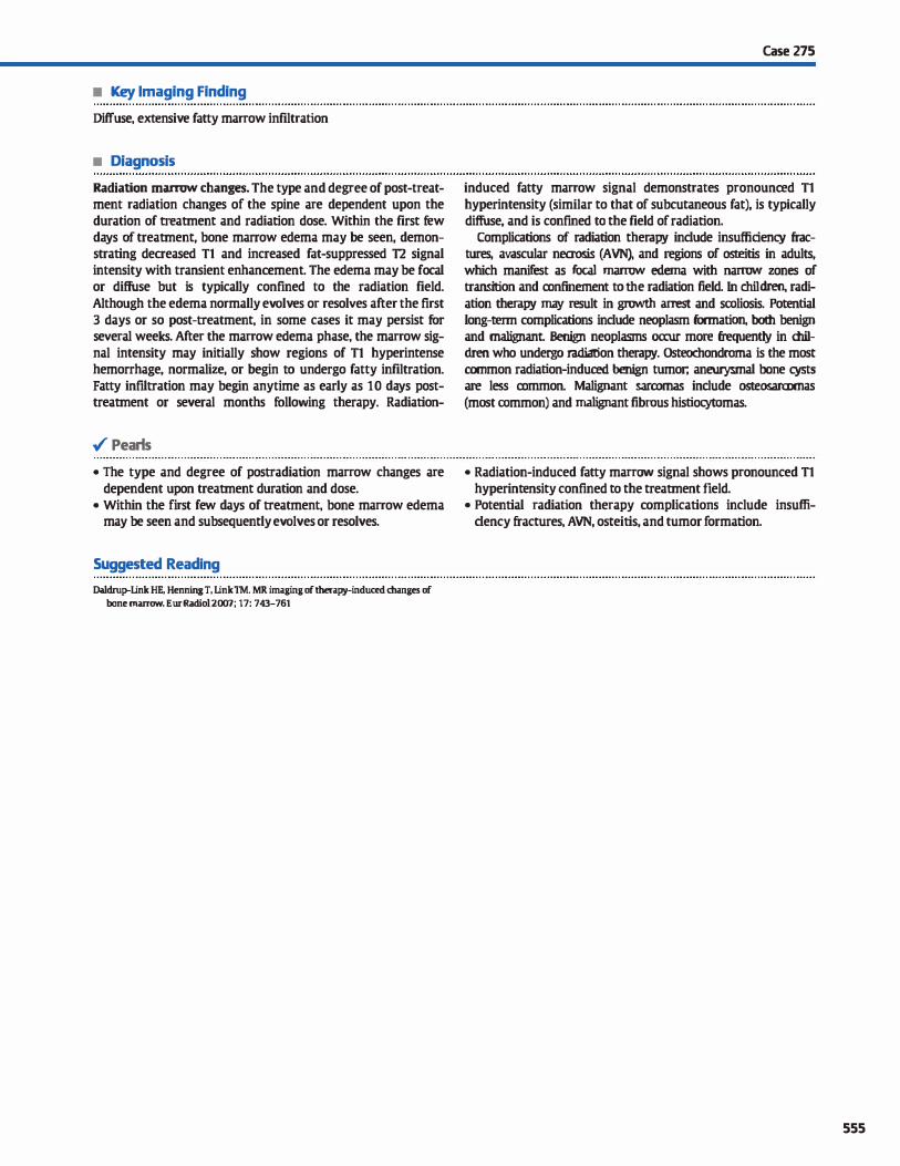
• Key lmaging Finding
Diffuse, extensive fatty marrow infiltration
• Diagnosis
Radiation marrow changes. The type and degree of post-treatment radiation changes of the spine are dependent upon the duration of treatment and radiation dose. Within the first few days of treatment, bone marrow edema may be seen, demonstrating decreased Tt and increased fat-suppressed T2 signal intensity with transient enhancement The edema may be focal or diffuse but is typically confined to the radiation field.
Although the edema normally evolves or resolves after the first
3 days or so post-treatment, in some cases it may persist for several weeks. After the marrow edema phase, the marrow sig
nal intensity may initially show regions of Tt hyperintense hemorrhage, normalize, or begin to undergo fatty infiltration. Fatty infiltration may begin anytime as early as 10 days posttreatment or several months following therapy. Radiation-
y' Pearts
• The type and degree of postradiation marrow changes are dependent upon treatment duration and dose.
• Within the first few days of treatment, bone marrow edema may be seen and subsequently evolves or resolves.
Suggested Reading
Dilldrup-Unk HE, Henning T, Llnk TM. MR imaging of therapy-induœd changes of
bene marrow. Eur Radiol 2007; 17; 743-761
Case 275
induced fatty marrow signal demonstrates pronounœd T1
hyperintensity (similar to that of subcutaneous fat), is typically diffuse, and is confined ta the field of radiation.
Complications of radiation therapy indude insuffiàency fractures, avascular necrosis (AVN1 and regions of osteitis in adults,
which manifest as focal marrow edema with narruw zones of transition and confinement to the radiation field. ln children, radi
ation therapy may result in growth arrest and scoliosis. Potential long-tmn complications indude neoplasm formation, both benign and malignant Benign neoplasms ocrur more frequently in children who undergo radiation therapy. Osteochondroma is the most common radiation-induœd benign tumor; aneurysmal bone cysts are Jess common. Malignant sarcomas indude osteosarannas
(most common) and malignant fibrous histiocytomas.
• Radiation-induced fatty marrow signal shows pronounced T1
hyperintensity confined to the treatment field. • Potential radiation therapy complications include insuffi
dency fractures, AVN, osteitis, and tumor formation.
555
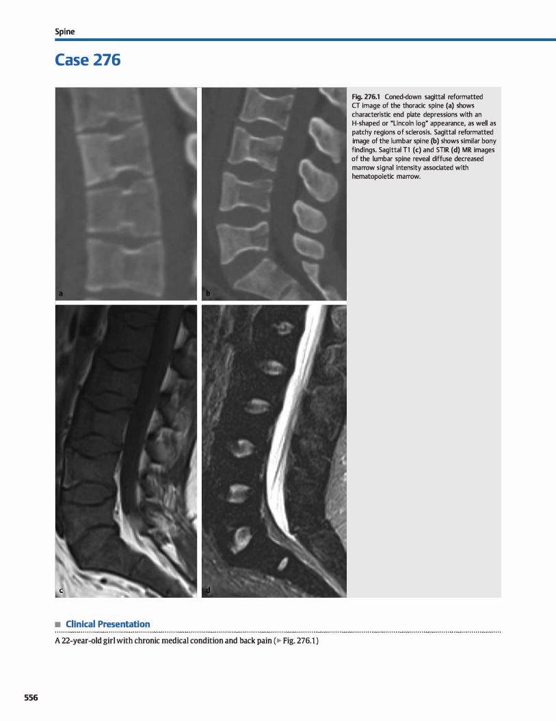
Spi ne
Case 276
• Clinical Presentation
A 22-year-old girl with chronic medical condition and back pain (� Fig. 276.1 )
556
Fig. 276.1 Coned-down sagittal reformatted CT image of the thoracic spine (a) shows characteristic end plate depressions with an H-shaped or "Lincoln log" appearance, as well as patchy regions of sclerosis. Sagittal reformatted image of the lumbar spi ne (b) shows similar bony findings. Sagittal T1 (c) and STIR (d) MR images of the lumbar spine reveal diffuse decreased marrow signal intensity associated with hematopoietic marrow.
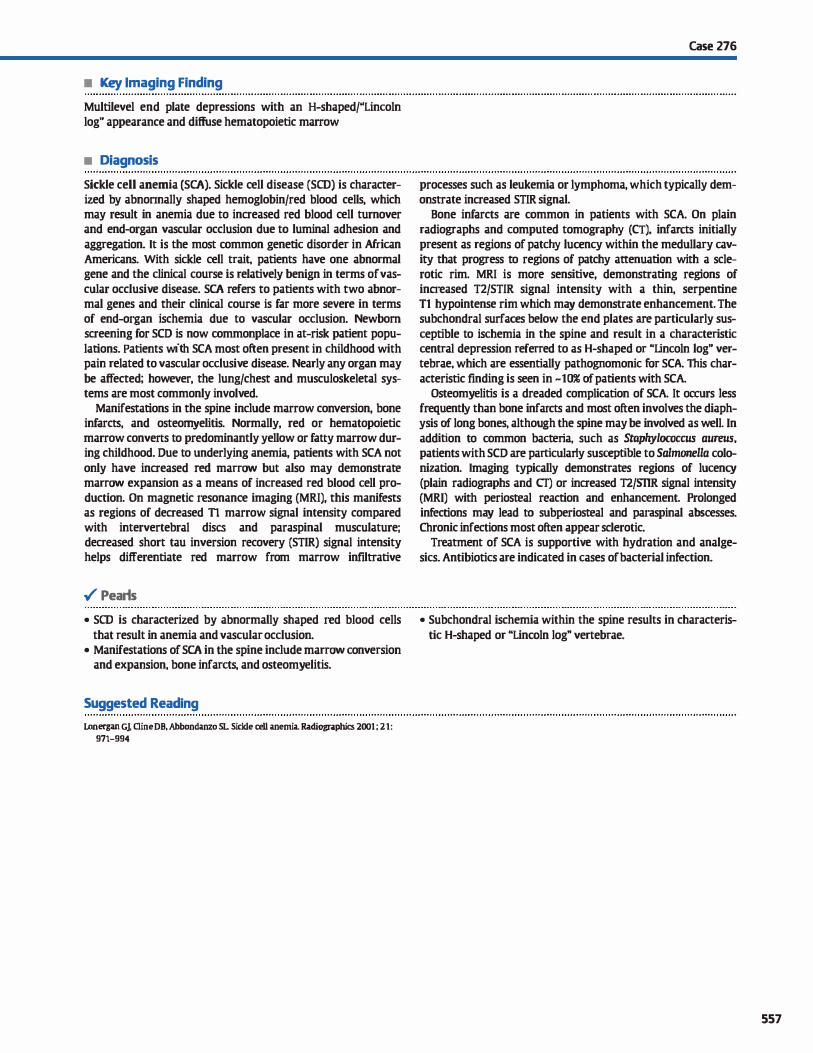
• Key lmaging Finding
Multilevel end plate depressions with an H-shapedtLincoln log" appearance and diffuse hematopoietic marrow
• Diagnosis
Sickle cell anemia (SCA). Sickle œll disease (SCD) is characterized by abnonnally shaped hemoglobin/red blood cells, which may result in anemia due to increased red blood œll turnover and end-organ vascular occlusion due ta luminal adhesion and
aggregation. lt is the most common genetic disorder in African
Americans. With sickle œll trait, patients have one abnormal gene and the clinical course is relatively benign in terms of vas
cular occlusive disease. SCA refers to patients with two abnormal genes and their clinicat course is far more severe in terms of end-organ ischemia due to vascular occlusion. Newborn screening for SCD is now commonplaœ in at-risk patient popu
lations. Patients with SCA most often present in childhood with pain related to vascular occlusive disease. Nearly any organ may
be affected; however, the lung/chest and musculoskeletal systems are most commonly involved.
Manifestations in the spine indude marrow conversion, bone infarcts, and osteomyelitis. Normally, red or hematopoietic marrow converts to predominantly yellow or fatty marrow during childhood. Due to underlying anemia, patients with SCA not
only have increased red marrow but also may demonstrate marrow expansion as a means of increased red blood cell production. On magnetic resonanœ imaging (MRI), this manifests as regions of decreased TI marrow signal intensity compared with intervertebral dises and paraspinal musculature; decreased short tau inversion recovery (STIR) signal intensity helps differentiate red marrow from marrow infiltrative
� Pearls
• SCD is characterized by abnormally shaped red blood cells
that result in anemia and vascular occlusion. • Manifestations of SCA in the spine indude marrow conversion
and expansion, bone infarcts, and osteomyelitis.
Suggested Reading
Lonergan GJ, Cine DB, Abbondanzo SL Sickle œll anemia. Radiographies 2001: 21:
971-994
Case 276
processes such as leukemia or lymphoma, which typically demonstrate increased STIR signal.
Bone infarcts are common in patients with SCA. On plain
radiographs and computed tomography (CT), infarcts initially present as regions of patchy lucency within the medullary cavity that progress to regions of patchy attenuation with a scle
rotic rim. MRI is more sensitive, demonstrating regions of increased T2/STIR signal intensity with a thin, serpentine Tl hypointense rim which may demonstrate enhancement. The subchondral surfaces below the end plates are particularly sus
ceptible to ischemia in the spine and result in a characteristic central depression referred to as H-shaped or "Lincoln log" vertebrae, which are essentially pathognomonic for SCA. This char
acteristic finding is seen in -10% of patients with SCA. Osteomyelitis is a dreaded complication of SCA. It occurs Jess
frequently than bone infarcts and most often involves the diaphysis of long bones, although the spine may be involved as well. In
addition to common bacteria, such as Stuphylococcus aureus, patients with SCD are particularly susceptible to Salmonella colonization. Imaging typically demonstrates regions of lucency
(plain radiographs and CT) or increased T2/STIR signal intensity (MRI) with periosteal reaction and enhanœment. Prolonged infections may lead to subperiosteal and paraspinal absœsses. Chronic infections most often appear sderotic.
Treatment of SCA is supportive with hydration and analge
sics. Antibiotics are indicated in cases of bacterial infection.
• Subchondral ischemia within the spine results in characteris
tic H-shaped or "Lincoln log" vertebrae.
557
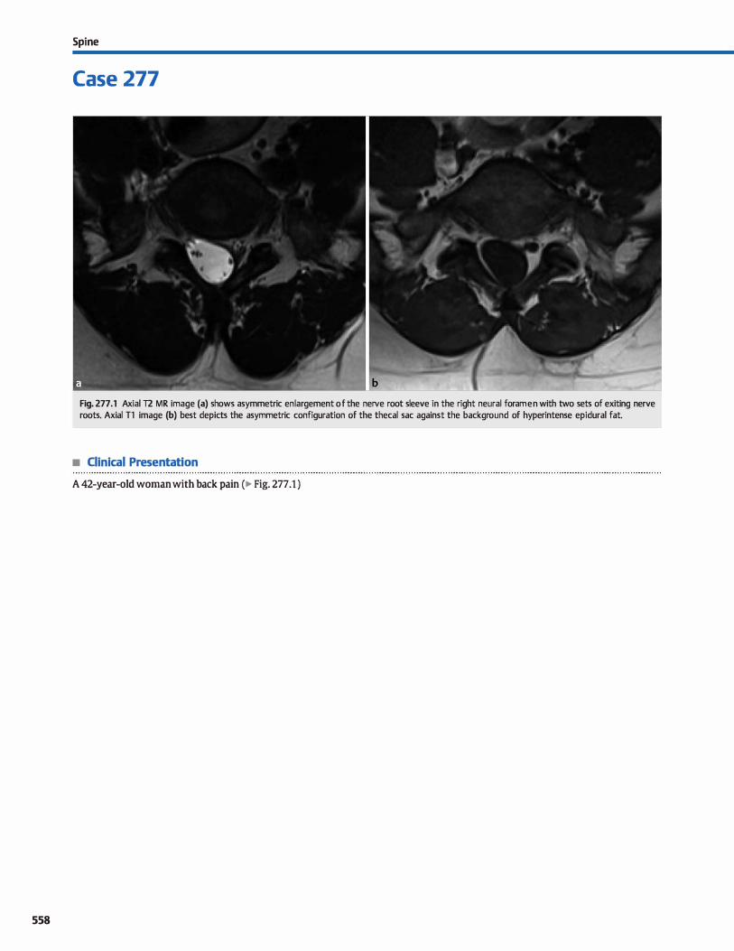
558
Spi ne
Case 277
Fig. 277 .1 Axial T2 MR image (a) shows asymmetric en largement of the nerve root sleeve in the right neural fora men with two sets of exiting nerve roots. Axial Tl image (b) best depicts the asymmetric configuration of the thecal sac against the background of hyperintense epidural fat.
• Clinical Presentation
A 42-year-old woman with back pain (� Fig. 277 .1 )
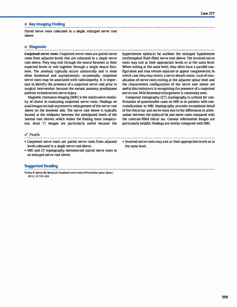
• Key lmaging Finding
Paired nerve roots colocated in a single, enlarged nerve root
sleeve
• Diagnosis
Conjoined nerve roots. Conjoined nerve roots are paired nerve roots from adjacent levels that are colocated in a single nerve root sleeve. They may exit through the neural foramen at their expected levels or exit together through a single neural fora
men. The anomaly typically occurs unilaterally and is most often incidental and asymptomatic; occasionally, conjoined nerve roots may be associated with radiculopathy. It is impor
tant to identify the presence of a conjoined nerve root prior to surgical intervention because the variant anatomy predisposes patients to inadvertent nerve injury.
Magnetic resonanœ imaging (MRI) is the noninvasive modal
ity of choice in evaluating conjoined nerve roots. Findings on
axial images include asymmetric enlargement of the nerve root
sleeve on the involved side. The nerve root sleeve is typically located at the midpoint between the anticipated levels of the normal root sleeves, which makes the finding more conspicuous. Axial T1 images are particularly useful because the
v" Pearls
• Conjoined nerve roots are paired nerve roots from adjacent
levels colocated in a single nerve root sleeve.
• MRI and CT myelography demonstrate paired nerve roots in
an enlarged nerve root sleeve.
Suggested Reading
Trimba R. Spivak JM, Bendo JA. Conjoined nerve roots of the lumbar spi ne. Spi ne j
2012; 12: 515-524
Case 277
hyperintense epidural fat outlines the enlarged hypointense cerebrospinal fluid-filled nerve root sleeve. The involved nerve roots may exit at their appropriate levels or at the same level. When exiting at the same level, they often have a parallel con
figuration and may remain separate or appear conglomerate, in which case they may mimic a nerve sheath tumor. Lack of visu
alization of nerve roots exiting at the adjacent spinal level and the characteristic configuration of the nerve root sleeve are useful discriminators in recognizing the presence of a conjoined nerve root. Mild foraminal enlargement is commonly seen.
Computed tomography (CT) myelography is utilized for confirmation of questionable cases on MRI or in patients with contraindications to MRI. Myelography provides exceptional detail
of the thecal sac and nerve roots due to the differences in attenuation between the epidural fat and nerve roots compared with the contrast-filled thecal sac. Corona! reformatted images are particularly helpful. Findings are similar compared with MRI.
• Involved nerve roots may exit at their appropriate levels or at
the same level.
559
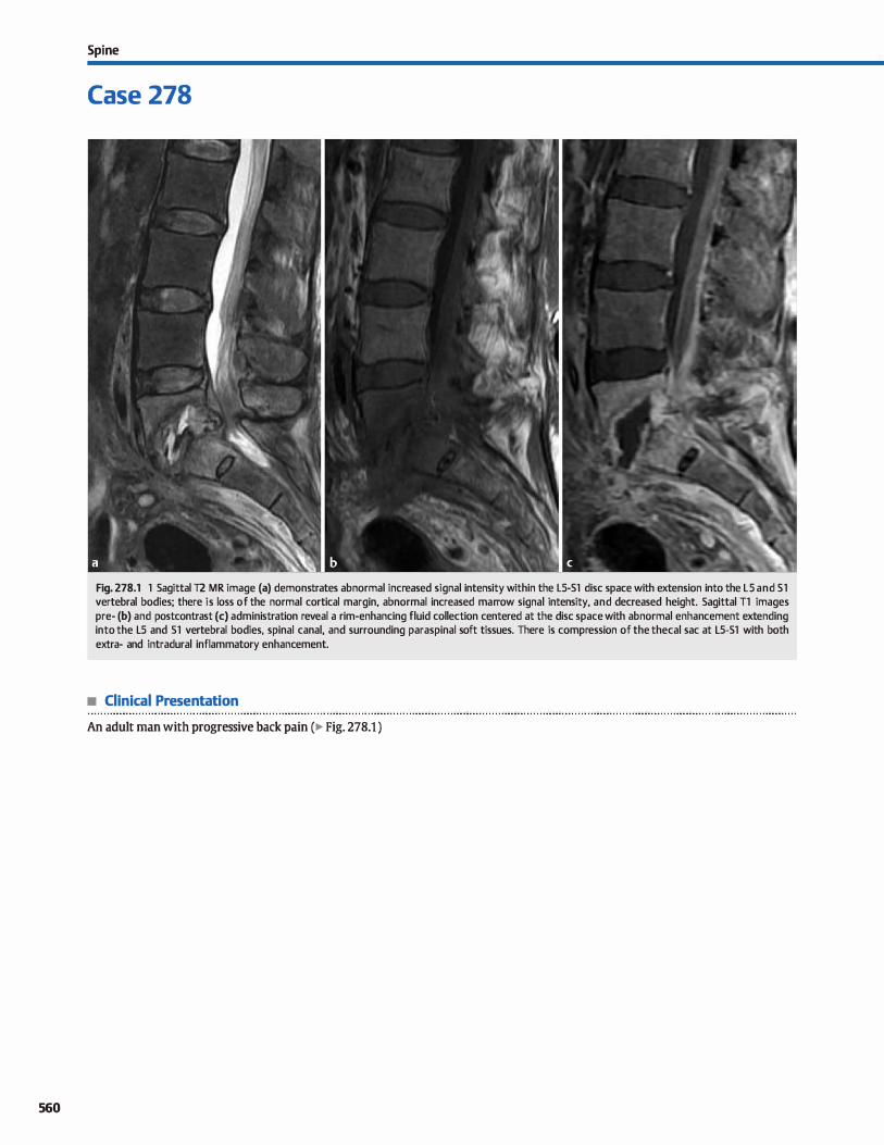
560
Spi ne
Case 278
Fig. 278.1 1 Sagittal T2 MR image (a) demonstrates abnormal increased signal intensity within the LS-Sl dise space with extension into the LS and Sl vertebral bodies; there is loss of the normal cortical margin, abnormal increased marrow signal intensity, and decreased height. Sagittal Tl images pre- (b) and postcontrast (c) administration reveal a rim-enhancing fluid collection centered at the dise space with abnormal enhancement extending into the LS and Sl vertebral bodies, spinal canal, and surrounding paraspinal soft tissues. There is compression of the thecal sac at LS-Sl with both extra- and intradural inflammatory enhancement.
• Clinical Presentation
An adult man with progressive back pain (� Fig. 278.1 )
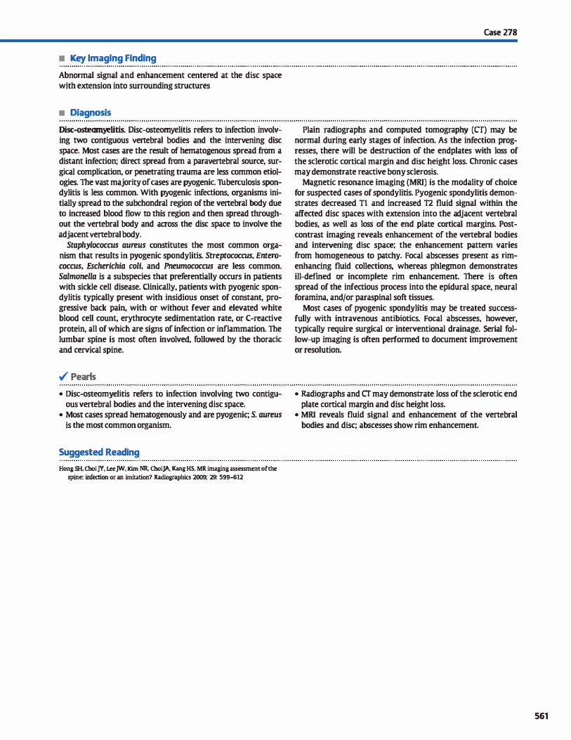
• Key lmaging Finding
Abnormal signal and enhancement centered at the dise spaœ with extension into surrounding structures
• Diagnosis
Disc-osteomyelitis. Disc-osteomyelitis refers ta infection involving two contiguous vertebral bodies and the intervening dise space. Most cases are the result of hematogenous spread from a distant infection; direct spread from a paravertebral source, sur
gical complication, or penetrating trauma are less common etiologies. The vast majority of cases are pyogenic. Tuberculosis spondylitis is Jess common. With pyogenic infections, organisms initially spread ta the subchondral region of the vertebral body due to increased blood flow ta this region and then spread throughout the vertebral body and across the dise space ta involve the adjacent vertebral body.
Staphylococcus aureus constitutes the most common organism that results in pyogenic spondylitis. StTeptococcus, Enterococcus, Escherichia coli, and Pneumococcus are Jess common. Salmonella is a subspecies that preferentially occurs in patients with sickle cell disease. Œnically, patients with pyogenic spondylitis typically present with insidious onset of constant, progressive back pain, with or without fever and elevated white blood cell count, erythrocyte sedimentation rate, or C-reactive
protein, ail of which are signs of infection or inflammation. The lumbar spine is most often involved, followed by the thoracic and cervical spine.
y' Pearts
• Disc-osteomyelitis refers to infection involving two contiguous vertebral bodies and the intervening dise space.
• Most cases spread hematogenously and are pyogenic; S. aureus is the most common organism.
Suggested Reading
Hong SH, Owi JY, Lee JW, Kim NR, OwiJA, Kang HS. MR imaging assessment of the
spine: infection or an imitation? Radiographies 2009; 29: 599-612
Case 278
Plain radiographs and computed tomography {CT) may be normal during early stages of infection. As the infection progresses, there will be destruction of the endplates with loss of
the sclerotic cortical margin and dise height Joss. Chronic cases may demonstrate reactive bony sclerosis.
Magnetic resonance imaging (MRI) is the modality of choiœ for suspected cases of spondylitis. Pyogenic spondylitis demonstrates decreased T1 and increased T2 fluid signal within the affected dise spaces with extension into the adjacent vertebral bodies, as well as Joss of the end plate cortical margins. Post
contrast imaging reveals enhancement of the vertebral bodies and intervening dise spaœ; the enhancement pattern varies from homogeneous to patchy. Focal abscesses present as rim
enhancing fluid collections, whereas phlegmon demonstrates ill-defined or incomplete rim enhancement There is often spread of the infectious process into the epidural space, neural foramina, and/or paraspinal soft tissues.
Most cases of pyogenic spondylitis may be treated successfully with intravenous antibiotics. Focal abscesses, however, typically require surgical or interventional drainage. Serial fol
low-up imaging is often perlormed to document improvement or resolution.
• Radiographs and cr may demonstrate loss of the sclerotic end plate cortical margin and dise height loss.
• MRI reveals fluid signal and enhancement of the vertebral bodies and dise; abscesses show rim enhancement.
561
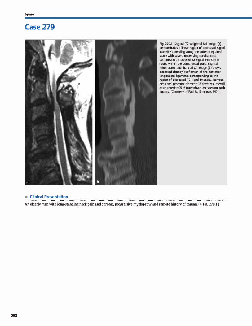
562
Spi ne
Case 279
• Clinical Presentation
Fig. 279.1 Sagittal T2-weighted MR image (a) demonstrates a linear region of decreased signal intensity extending along the anterior epidural space with severe underlying cervical cord compression. lncreased T2 signal intensity is noted within the compressed cord. Sagittal reformatted unenhanced CT image (b) shows increased density/ossification of the posterior longitudinal ligament, corresponding to the region of decreased T2 signal intensity. Remote dens and posterior element C2 fractures, as well as an anterior CS-6 osteophyte, are seen on both images. (Courtesy of Paul M. Sherman, MD.)
An elderly man with long-standing neck pain and chronic, progressive myelopathy and remote history of trauma (� Fig. 279.1 )
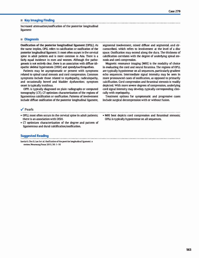
• Key lmaging Finding
Increased attenuation/ossification of the posterior longitudinal
ligament
• Diagnosis
Ossification of the posterior longitudinal ligament (OPU). As the name implies, OPIL refers to calcification or osmication of the posterior longitudinal ligament It most often occurs in the cervical spine in adult patients and is more common in Asia. There is a
fairly equal incidence in men and women Although the patho
genesis is not entirely dear, there is an association with diffuse idiopathie skeletal hyperostosis {DISH) and spondyloarthropathies.
Patients may be asymptomatic or present with symptoms related to spinal canal stenosis and cord compression. Common symptoms indude those related to myelopathy, radiculopathy, and occasionally bowel and bladder dysfunction; symptom
onset is typically insidious. OPPL is typically diagnosed on plain radiographs or computed
tomography { CT); CT optimizes characterization of the regions of
ligamentous calcification or ossification Patterns of involvement indude diffuse ossification of the posterior longitudinal ligament,
� Pearts
• OPIL most often occurs in the cervical spine in adult patients; there is an association with DISH.
• cr optimizes characterization of the degree and pattern of ligamentous and durai calcification/ossification.
Suggested Reading
Saetia K, Ote D, Lee Set al, Ossification of the poetrior longitudinal ligament: a
review. Neurosurg FoCU5 2011; 30: 1-16
Case 279
segmental involvement, mixed diffuse and segmenta� and circumsaibed, which refers to involvement at the level of a dise spaœ. Ossification may ex.tend along the dura. The thickness of calcification correlates with the degree of underlying spinal ste
nosis and cord compression. Magnetic resonance imaging (MRI) is the modality of choiœ
in evaluating the cord and neural foramina. The regions of OPLL are typically hypointense on all sequences, particularly gradient echo sequences. Intermediate signal intensity may be seen in
more pronounced cases of ossification, as opposed to primarily
calcification. Cord compression and foraminal stenosis is readily depicted. With more severe degrees of compression, underlying cord signal intensity may develop, typically corresponding clini
cally with myelopathy.
Treatment options for symptomatic and progressive cases indude surgical decompression with or without fusion.
• MRI best depicts cord compression and foraminal stenosis; OPIL is typically hypointense on all sequenœs.
563
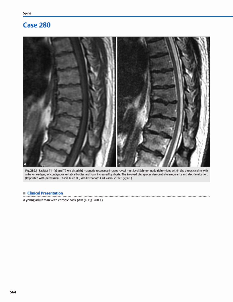
564
Spi ne
Case 280
Fig. 280.1 Sagittal Tl - (a) and T2-weighted (b) magnetic resonance images reveal multilevel Schmorl node deformities within the thoracic spi ne with anterior wedging of contiguous vertebral bodies and focal increased kyphosis. The involved dise spaces demonstrate irregularity and dise dessication. (Reprinted with permission: Tharin B, et al. J Am Osteopath Coll Radiol 2012;1 (2):40.)
• Clinical Presentation
A young adult man with chronic back pain ( � Fig. 280.1 )
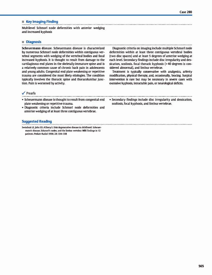
• Key lmaging Finding
Multilevel Schmorl node deformities with anterior wedging
and increased kyphosis
• Diagnosis
Scheuermann disease. Scheuermann disease is characterized
by numerous Schmorl node deformities within contiguous ver
tebral segments with wedging of the vertebral bodies and focal
increased kyphosis. lt is thought to result from damage to the
cartilaginous end plates in the skeletally immature spine and is
a relatively common cause of chronic back pain in adolescents and young adults. Congenital end plate weakening or repetitive
trauma are considered the most likely etiologies. The condition
typically involves the thoradc spine and thoracolumbar junc
tion. Pain is worsened by activity.
� Pearls
• Scheuermann disease is thought to result from congenital end
plate weakening or repetitive trauma. • Diagnostic criteria include Schmorl node deformities and
anterior wedging of at least three contiguous vertebrae.
Suggested Reading
SWisdtuk LE, John SD, Allbery S. Disk degenerative disease in childhood; Scheuer
mann's disease, Sdunorl's nodes, and the limbus vertebra: MRI findings in 12.
patients. Pediatr Radiol 1998; 28: 334-338
Case 280
Diagnostic criteria on imaging include multiple Schmorl node
deformities within at least three contiguous vertebral bodies
(two dise spaces) and at least 5 degrees of anterior wedging at
each level. Secondary findings include dise irregularity and des
sication, scoliosis, focal thoradc kyphosis (> 40 degrees is considered abnormal), and !imbus vertebrae.
Treatment is typically conservative with analgesics, activity
modification, pbysical therapy, and, occasionally, bracing. Surgical
intervention is rare but may be neœssary in severe cases with
exœssive kyphosis, intractable pain, or neurological defidts.
• Secondary findings include dise irregularity and dessication,
scoliosis, focal kyphosis, and limbus vertebrae.
565
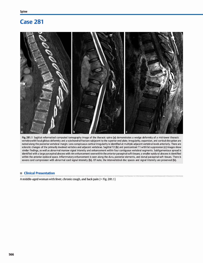
566
Spi ne
Case 281
Fig. 281 .1 Sagittal reformatted computed tomography image of the thoracic spi ne (a) demonstrates a wedge deformity of a mid-lower thoracic vertebra with focal gibbus deformity and a subchondral fracture subjacent to the superior end plate. lrregularity, expansion, and cortical disruption are noted a long the posterior vertebral margin. Less conspicuous cortical irregularity is identified at multiple adjacent vertebral levels anteriorly. There are sclerotic changes of the primarily involved vertebra and adjacent vertebrae. Sagittal T2 (b) and postcontrast Tl with fat suppression (c) images show similar findings, as well as abnormal marrow signal intensity and enhancement within four contiguous vertebral segments. Subligamentous spread is identified with a large paraspinal abscess with rim enhancement seen within the anterior paraspinal soft tissues; a smaller epidural abscess is identified within the anterior epidural space. lnflammatory enhancement is seen along the dura, posterior elements, and dorsal paraspinal soft tissues. There is severe cord compression with abnormal cord signal intensity (b). Of note, the intervertebral dise spaces and signal intensity are preserved (b).
• Clinical Presentation
A middle-aged woman with fever, chronic cough, and back pain (� Fig. 281 .1 )
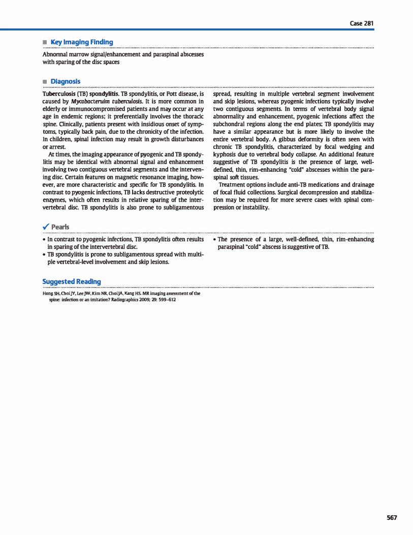
• Key lmaging Finding
Abnonnal marrow signal/enhanœment and paraspinal absœsses with sparing of the dise spaœs
• Diagnosis
Tuberculosis (TB) spondylitis. TB spondylitis, or Pott disease, is caused by Mycobacteruim tuberculosis. It is more common in elderly or immunocompromised patients and may occur at any age in endemic regions; it preferentially involves the thoracic
spine. Oinically, patients present with insidious onset of symptoms, typically back pain, due to the chronicity of the infection. In children, spinal infection may result in growth disturbances
or arrest. At times, the imaging appearance of pyogenic and TB spondy
litis may be identical with abnonnal signal and enhancement involving two contiguous vertebral segments and the interven
ing dise. Certain features on magnetic resonance imaging, however, are more characteristic and specific for TB spondylitis. In contrast to pyogenic infections, TB Jacks destructive proteolytic enzymes, which often results in relative sparing of the intervertebral dise. TB spondylitis is also prone to subligamentous
� Pearts
• In contrast to pyogenic infections, TB spondylitis often results in sparing of the intervertebral dise.
• TB spondylitis is prone to subligamentous spread with multiple vertebral-level involvement and skip lesions.
Suggested Reading
Hong SH, Otai ]Y, Lee JW, Kim NR, OtoiJA, Kang HS, MR imaging assemnent of the
spine: infection or an imitation? Radiographies 2009; 29: 599-612
Case 281
spread, resulting in multiple vertebral segment involvement and skip lesions, whereas pyogenic infections typically involve two contiguous segments. In tenns of vertebral body signal abnonnality and enhancement, pyogenic infections affect the
subchondral regions along the end plates; TB spondylitis may have a similar appearance but is more likely to involve the
entire vertebral body. A gibbus deformity is often seen with chronic TB spondylitis, characterized by focal wedging and kyphosis due to vertebral body collapse. An additional feature suggestive of TB spondylitis is the presence of large, well
defined, thin, rim-enhancing ucold" abscesses within the para
spinal soft tissues. Treatment options indude anti-TB medications and drainage
of focal fluid collections. Surgical decompression and stabilization may be required for more severe cases with spinal compression or instability.
• The presence of a large, well-defined, thin, rim-enhancing paraspinal ucold" abscess is suggestive of TB.
567
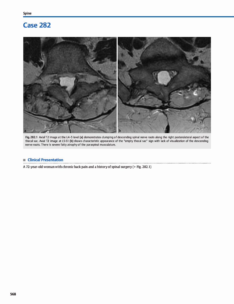
568
Spi ne
Case 282
Fig. 282.1 Axial T2 image at the L4-5 level (a) demonstrates dumping of descending spinal nerve roots along the right posterolateral aspect of the thecal sac. Axial T2 image at LS-51 (b) shows characteristic appearance of the "empty thecal sac" sign with lack of visualization of the descending nerve roots. There is severe fatty atrophy of the paraspinal musculature.
• Clinical Presentation
A 72-year-old woman with chronic back pain and a history of spinal surgery (� Fig. 282.1)
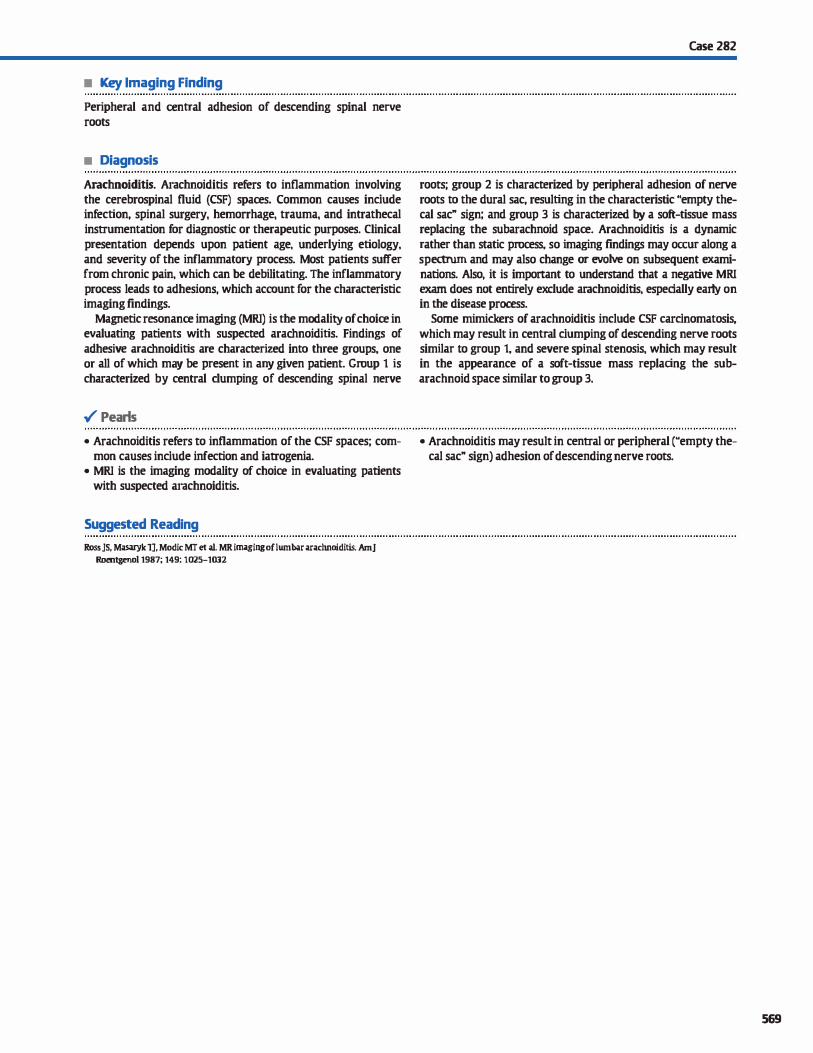
• Key lmaging Finding
Peripheral and central adhesion of descending spinal nerve roots
• Diagnosis
Arachnoiditis. Arachnoiditis refers to inflammation involving the cerebrospinal fluid (CSF) spaœs. Common causes indude infection, spinal surgery, hemorrhage, trauma, and intrathecal instrumentation for diagnostic or therapeutic purposes. Oinical presentation depends upon patient age, underlying etiology, and severity of the inflammatory process. Most patients suffer from chronic pain, which can be debilitating. The inflammatory process leads to adhesions, which account for the characteristic imaging findings.
Magnetic resonanœ imaging (MRI) is the modality of choiœ in evaluating patients with suspected arachnoiditis. Findings of adhesive arachnoiditis are characterized into three groups, one or all of which may be present in any given patient. Group 1 is characterized by central dumping of descending spinal nerve
� Pearts
• Arachnoiditis refers to inflammation of the CSF spaces; common causes indude infection and iatrogenia.
• MRI is the imaging modality of choiœ in evaluating patients with suspected arachnoiditis.
Suggested Reading
Ross JS, Masaryk lJ, Modic MT et al. MR imaging of lumbar arachnoiditis. AmJ
Roentgenol 1987; 149; 1025-1032
Case 282
roots; group 2 is characterized by peripheral adhesion of nerve roots to the durai sac, resulting in the characteristic "empty thecal sac" sign; and group 3 is characterized by a soft-tissue mass replacing the subarachnoid spaœ. Arachnoiditis is a dynamic rather than static proœss, so imaging findings may occur along a spectrum and may also change or evolve on subsequent examinations. Also, it is important to understand that a negative MRI exam does not entirely exclude arachnoiditis, espeàally early on in the disease proœss.
Sorne mimiclœrs of arachnoiditis include CSF cardnomatosis, which may result in central dumping of descending nerve roots similar to group 1, and severe spinal stenosis, which may result in the appearance of a soft-tissue mass replaàng the subarachnoid space similar to group 3.
• Arachnoiditis may result in central or peripheral ("empty thecal sac" sign) adhesion of descending nerve roots.
569
570
Spi ne
Case 283
Fig. 283.1 Sagittal T2 image shows a blunted con us located at the mid-Tl 2 level, as well as absence of the sacrum below the 52 level. A distended bladder is partially seen within the pelvis.
• Clinical Presentation
A 3-year-old girl with scoliosis (� Fig. 283.1 )
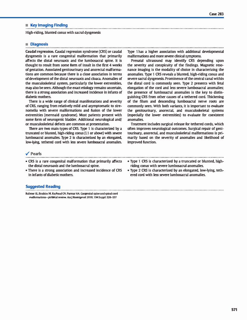
• Key lmaging Finding
High-riding, blunted conus with sacral dysgenesis
• Diagnosis
Caudal regression. Caudal regression syndrome (CRS) or caudal dysgenesis is a rare congenital malformation that primarily affects the distal neuroaxis and the lumbosacral spine. It is
thought to result from some form of insult in the frrst 4 weeks of gestation. Associated genitourinary and anorectal malformations are common because there is a close association in terms of development of the distal neuroaxis and cloaca. Anomalies of
the musculoskeletal system, particularly the Iower extremities, may also be seen. Although the exact etiology remains unœrtain, there is a strong association and increased incidence in infants of
diabetic mothers. There is a wide range of dinical manifestations and severity
of CRS, ranging from relatively mild and asymptomatic to sirenomelia with severe malformations and fusion of the lower
extremities (mennaid syndrome). Most patients present with some form of neurogenic bladder. Additional neurological and/
or musculoskeletal defects are common at presentation. There are two main types of CRS. 'JYpe 1 is characterized by a
truncated or blunted, high-riding conus (Ll or above) with severe Iumbosacral anomalies. Type 2 is characterized by an elongated, low-lying, tethered cord with Jess severe lumbosacral anomalies.
� Pearts
• CRS is a rare congenital malformation that primarily affects the distal neuroaxis and the lumbosacral spine.
• There is a strong association and increased incidence of CRS in infants of diabetic mothers.
Suggested Reading
Rufener SI., Ibrahim M. Raybaud CA, Pannar HA. Congenital spine and spinal cord
rnalfcnnatiom-pictmial review. AmJ Roentgenol 2010; 194 Suppl; 526-537
Case 283
'JYpe 1 has a higher association with additional developmental malformations and more severe clinical symptoms.
Prenatal ultrasound may identify CRS depending upon
the severity and conspicuity of the findings. Magnetic resonanœ imaging is the modality of choiœ in characterizing the
anomalies. 'JYpe 1 CRS reveals a blunted, high-riding conus and severe sacral dysgenesis. Prominenœ of the central canal within
the distal cord is commonly seen. 'JYpe 2 presents with fetal elongation of the cord and less severe Iumbosacral anomalies;
the presence of lumbosacral anomalies is the key to distinguishing CRS from other causes of a tethered cord. Thickening of the filum and descending lumbosacral nerve roots are
commonly seen. With bath variants, it is important to evaluate the genitourinary, anorectal, and musculoskeletal systems (especially the lower extremities) to evaluate for coexistent
anomalies. Treatment includes surgical release for tethered cords, which
often improves neurological outcomes. Surgical repair of genitourinary, anorectal, and musculoskeletal malformations is primarily based on the severity of anomalies and likelihood of improved function.
• 'JYpe 1 CRS is characterized by a truncated or blunted, highriding conus with severe lumbosacral anomalies.
• 'JYpe 2 CRS is characterized by an elongated, Iow-Iying, tethered corcl with Jess severe Iumbosacral anomalies.
571
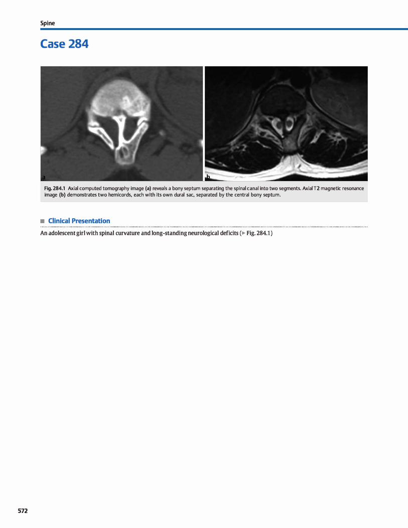
572
Spi ne
Case 284
Fig. 284.1 Axial computed tomography image (a) reveals a bony septum separating the spinal canal into two segments. Axial T2 magnetic resonance image (b) demonstrates two hemicords, each with its own durai sac, separated by the central bony septum.
• Clinical Presentation
An adolescent girl with spinal curvature and long-standing neurological deficits (� Fig. 284.1 )
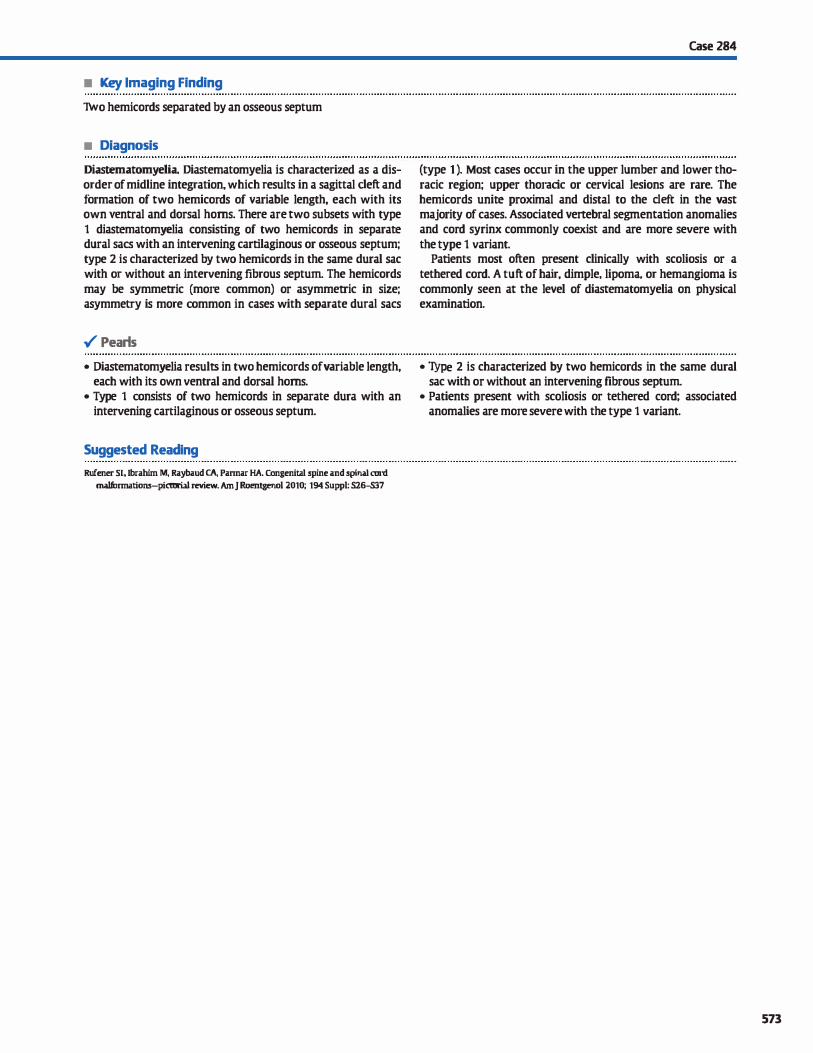
• Key lmaging Finding
lWo hemicords separated by an osseous septum
• Diagnosis
Diastematomyelia. Diastematomyelia is characterized as a disorder of midline integration, which results in a sagittal deft and formation of two hemicords of variable length, each with its own ventral and dorsal homs. There are two subsets with type 1 diastematomyelia consisting of two hemicords in separate durai sacs with an intervening cartilaginous or osseous septum; type 2 is characterized by two hemicords in the same durai sac with or without an intervening fibrous septum The hemicords may be symmetric (more common) or asymmetric in size; asymmetry is more common in cases with separate durai sacs
� Pearts
• Diastematomyelia results in two hemicords of variable length, each with its own ventral and dorsal homs.
• Type 1 consists of two hemicords in separate dura with an intervening cartilaginous or osseous septum.
Suggested Reading
Rufe11er SI., Ibrahim M, Raybaud CA, Pannar HA. Congenital spine and spinal cord
malfurmations-picturial review. AmJ Roentgenol 2010; 194 Suppl: 526-537
Case 284
(type 1 ). Most cases occur in the upper lumber and lower thoracic region; upper thoracic or cervical lesions are rare. The hemicords unite proximal and distal to the deft in the vast majority of cases. Associated vertebral segmentation anomalies and cord syrinx commonly coexist and are more severe with the type 1 variant.
Patients most often present clinically with scoliosis or a tethered cord. A tuft of haïr, dimple, lipoma. or hemangioma is commonly seen at the level of diastematomyelia on physical examination.
• lYPe 2 is characterized by two hemicords in the same durai sac with or without an intervening fibrous septum
• Patients present with scoliosis or tethered cord; associated anomalies are more severe with the type 1 variant.
573
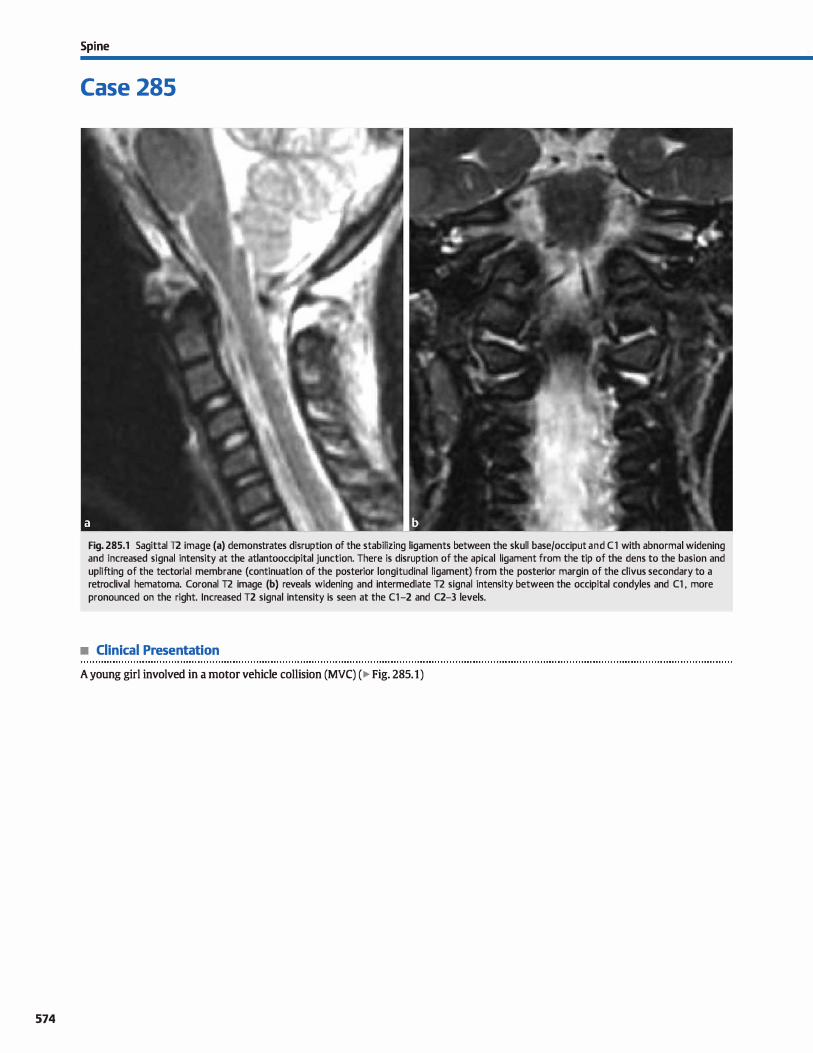
574
Spi ne
Case 285
Fig. 285.1 Sagittal T2 image (a) demonstrates disruption of the stabilizing ligaments between the skull base/occiput and Cl with abnormal widening and increased signal intensity at the atlantooccipital junction. There is disruption of the apical ligament from the tip of the dens to the basion and uplifting of the tectorial membrane (continuation of the posterior longitudinal ligament) from the posterior margin of the divus secondary to a retrodival hematoma. Coronal T2 image (b) reveals widening and intermediate T2 signal intensity between the occipital condyles and Cl , more pronounced on the right. lncreased T2 signal intensity is seen at the Cl-2 and C2-3 levels.
• Clinical Presentation
A young girl involved in a motor vehicle collision (MVC) ( � Fig. 285.1)
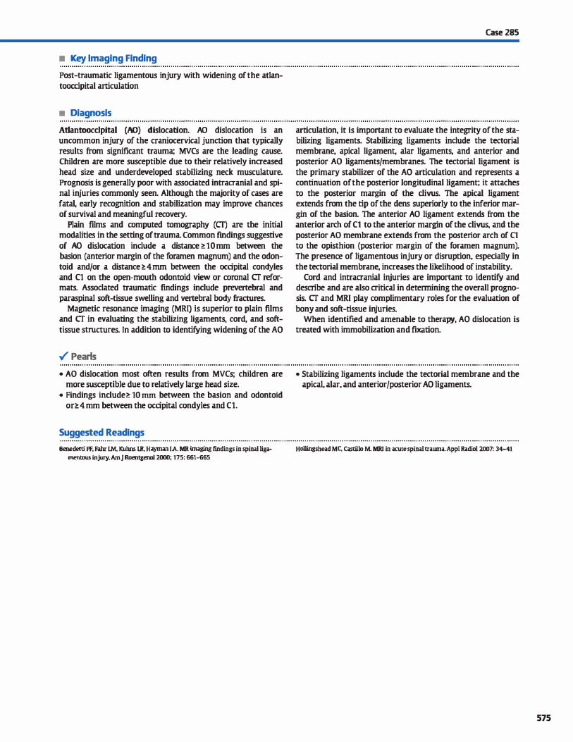
• Key lmaging Finding
Post-traumatic ligamentous injury with widening of the atlantooccipital articulation
• Diagnosis
Atlantoocdpital (AO) dislocation. AO dislocation is an uncommon injury of the craniocervical junction that typically results from significant trauma; MVCs are the leading cause. Children are more susceptible due to their relatively increased head size and underdeveloped stabilizing neck musculature. Prognosis is generally poor with associated intracranial and spinal injuries commonly seen. Although the majority of cases are fatal, early recognition and stabilization may improve chances of survival and meaningful recovery.
Plain films and computed tomography (CT) are the initial modalities in the setting of trauma. Common fmdings suggestive
of AO dislocation indude a distance � 10 mm between the basion (anterior margin of the foramen magnum) and the odontoid and/or a distance � 4 mm between the occipital condyles and Cl on the open-mouth odontoid view or coronal CT reformats. Assodated traumatic findings indude prevertebral and paraspinal soft-tissue swelling and vertebral body fractures.
Magnetic resonanœ imaging (MRI) is superior to plain films and CT in evaluating the stabilizing ligaments, cord, and soft
tissue structures. In addition to identifying widening of the AO
� Pearts
• AO dislocation most often results from MVCs; children are more susceptible due to relatively large head size.
• Findings indude � 10 mm between the basion and odontoid or� 4 mm between the occipital condyles and Ct.
Suggested Readings
Benedetti PF, Fahr LM, Kulms IR, Hayman LA. MR imaging findings in spinal liga
mentnus injury. AmJ Roentgenol 2000; 175; 661-665
Case 285
articulation, it is important to evaluate the integrity of the stabilizing ligaments. Stabilizing ligaments indude the tectorial membrane, apical ligament, alar ligaments, and anterior and posterior AO ligaments/membranes. The tectorial ligament is the primary stabilizer of the AO articulation and represents a continuation of the posterior longitudinal ligament; it attaches to the posterior margin of the diws. The apical ligament extends from the tip of the dens superiorly to the inferior margin of the basion. The anterior AO ligament extends from the anterior arch of Cl to the anterior margin of the cliws, and the posterior AO membrane extends from the posterior arch of Cl to the opisthion (posterior margin of the foramen magnum). The presenœ of ligamentous injury or disruption, especially in the tectorial membrane, increases the likelihood of instability.
Cord and intracranial injuries are important to identify and describe and are also critical in detennining the overall prognosis. CT and MRI play complimentary roles for the evaluation of
bony and soft-tissue injuries. When identified and amenable to therapy, AO dislocation is
treated with immobilization and flXéltion.
• Stabilizing ligaments indude the tectorial membrane and the apical, alar, and anterior/posterior AO ligaments.
Hollingshead MC. castillo M. MRI in acute spinal trauma. Appl Radiol 2007: 34-41
575
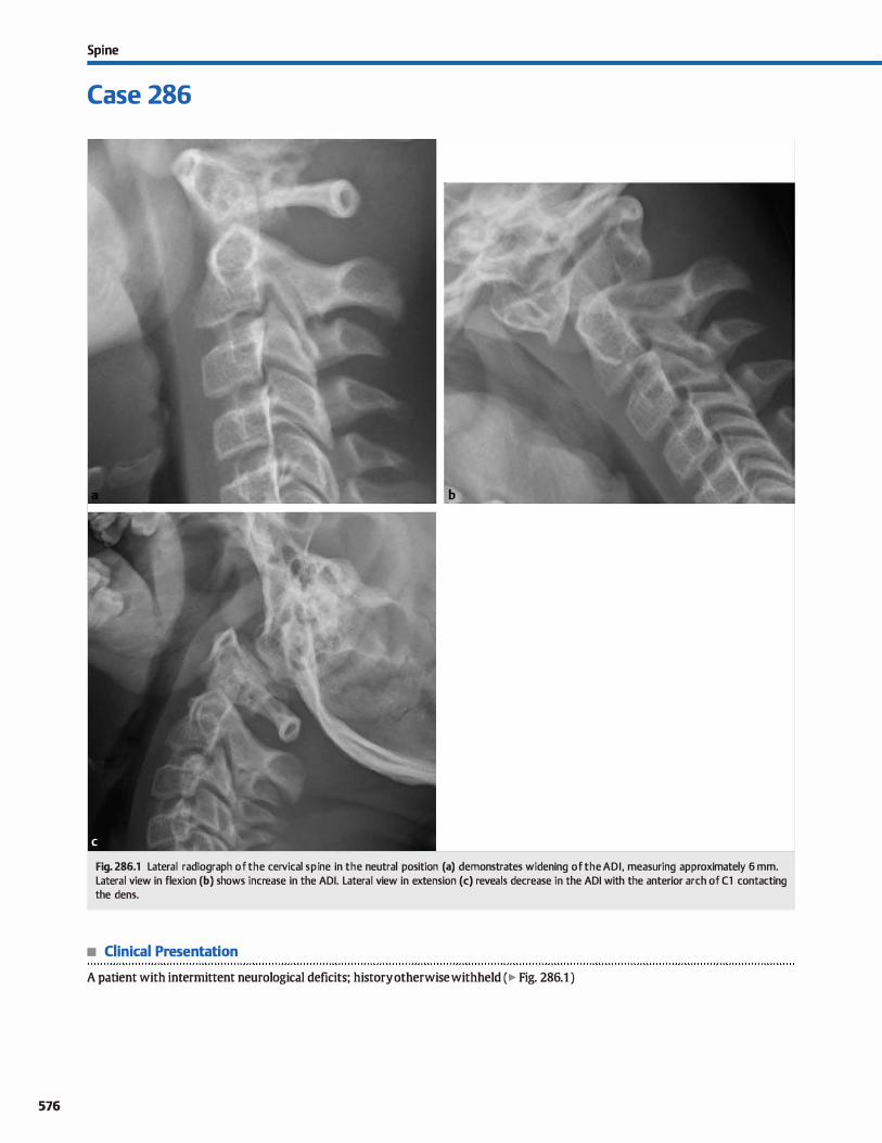
576
Spi ne
Case 286
Fig. 286.1 Lateral radiograph of the cervical spi ne in the neutral position (a) demonstrates widening of the ADI, measuring approximately 6 mm. Lateral view in flexion (b) shows increase in the ADI. Lateral view in extension (c) reveals decrease in the ADI with the anterior arch of Cl contacting the dens.
• Clinical Presentation
A patient with intermittent neurological deficits; history otherwise withheld (� Fig. 286.1 )
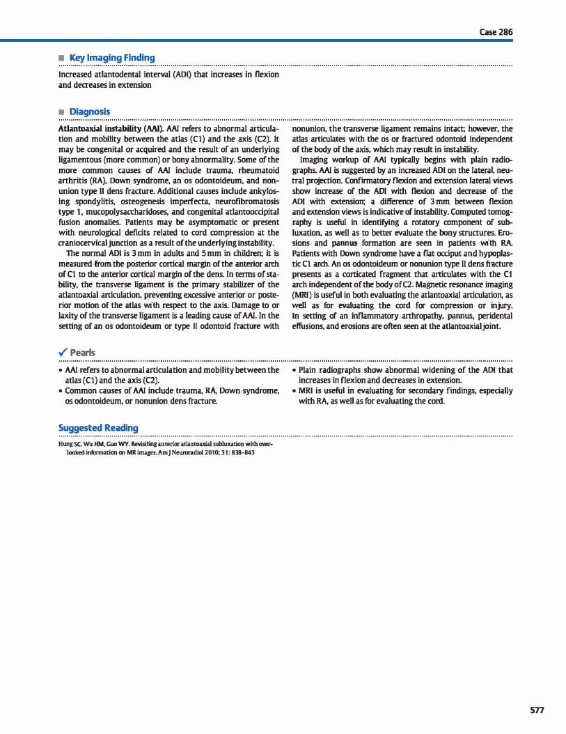
• Key lmaging Finding
Increased atlantodental interval (ADI) that increases in flexion and decreases in extension
• Diagnosis
Atlantoaxial instability (AAI). AAI refers to abnormal articulation and mobility between the atlas (Cl) and the axis (C2). lt may be congenital or acquired and the result of an underlying ligamentous (more common) or bony abnormality. Sorne of the
more common causes of AAI indude trauma, rheumatoid arthritis (RA), Dawn syndrome, an os odontoideum, and nonunion type Il dens fracture. Additional causes indude ankylosing spondylitis, osteogenesis imperfecta, neurofibromatosis type 1, mucopolysaccharidoses, and congenital atlantoocdpital fusion anomalies. Patients may be asymptomatic or present with neurological defidts related to cord compression at the craniocervical junction as a result of the underlying instability.
The normal ADI is 3 mm in adults and 5 mm in children; it is
measured from the posterior cortical margin of the anterior arch of Cl to the anterior cortical margin of the dens. ln terms of stability, the transverse ligament is the primary stabilizer of the atlantoaxial articulation, preventing excessive anterior or posterior motion of the atlas with respect to the axis. Damage to or laxity of the transverse ligament is a leading cause of AAI. In the setting of an os odontoideum or type Il odontoid fracture with
� Pearts
• AAI refers to abnormal articulation and mobility between the atlas (Cl) and the axis (C2).
• Common causes of AAI indude trauma, RA, Dawn syndrome, os odontoideum, or nonunion dens fracture.
Suggested Reading
Hung se, wu HM, Guo WY. Revisiting anterior atlantoaxial subluxation with over
looked information on MR images. AmJ Neuroradiol 2010; 31: 838-843
Case 286
nonunion, the transverse ligament remains intact; hawever, the atlas articulates with the os or fractured odontoid independent of the body of the axis, which may result in instability.
lmaging workup of AAI typically begins with plain radiographs. AAI is suggested by an increased ADI on the Iateral neutral projection. Confirmatory flexion and extension lateral views
show increase of the ADJ with flexion and decrease of the ADI with extension; a differenœ of 3 mm between flexion and extension views is indicative of instability. Computed tomography is useful in identifying a rotatory component of subluxation, as well as to better evaluate the bony structures. Erosions and pannus formation are seen in patients with RA. Patients with Dawn syndrome have a flat occiput and hypoplastic Cl arch. An os odontoideum or nonunion type Il dens fracture presents as a corticated fragment that articulates with the Cl arch independent of the body of C2. Magnetic resonanœ imaging (MRI) is useful in both evaluating the atlantoaxial articulation, as wel1 as for evaluating the cord for compression or injury. In setting of an inflammatory arthropathy, pannus, peridental effusions, and erosions are often seen at the atlantoaxialjoint.
• Plain radiographs show abnormal widening of the ADI that increases in flexion and decreases in extension.
• MRI is useful in evaluating for secondary findings, espedally with RA, as well as for evaluating the cord.
577
578
Spi ne
Case 287
Fig. 287 .1 Axial CT image (a) demonstrates the patient's head rotated to the left with leftward rotatory subluxation of Cl with respect to C2. The atlantodental interval or predental space is normal. Axial maximum intensity projection image (b) better depicts the rotation of Cl with respect to C2. Three-dimensional volume rendered image (c) again shows a rotatory component of Cl compared with the remainder of the cervical spine.
• Clinical Presentation
A 6-year-old boy with torticollis post-trauma ( � Fig. 287 .1)
• Key lmaging Finding
Abnormal rotation of Cl with respect to C2
• Diagnosis
Atlantoaxial rotatory subluxation. The Cl-2 articulation accounts for roughly half of the rotatory component of the entire
cervical spine. Atlantoaxial rotatory subluxation refers to abnormal rotation ofthe Cl-2 articulation at baseline, resulting in tor
ticollis. Undertying etiologies indude trauma, irûectious or inflammatory proœsses, congenital abnormalities, and spontaneous or idiopathie causes. In the vast majority of cases, rotatory
subluxation results in limited range of motion due to restriction or locking of the anterior Cl facet with respect to the C2 facet; rarely, fixation may occur. Underlying dislocation of Cl-2 may or may not be present and manifests as anterior displaœment of
the Cl with respect to C2. There may also be widening of the atlantodental articulation or predental spaœ. Atlantoaxial rotatory subluxation is dassified based upon the presenœ or absence
and degree of displaœment of Ct compared with C2. 'JYpe I is the most common variant and demonstrates rotation without dis
plaœment.
� Pearts
• Atlantoaxial rotatory subluxation refers to abnormal rotation of the Cl-2 articulation, resulting in torticollis.
• Underlying etiologies indude trauma, congenital abnormalities, inflammatory, and idiopathie causes.
Suggested Reading
wstrin ES, Karakas SP, Ortiz AD et al. Pediatric cervical spine; nonnal anatmny, var
iants, and trauma. Radiographies 2003; 23: 539-560
Case 287
Plain radiographs of the cervical spine demonstrate rotation of
Cl on the lateral view and disparity of the Cl lateral masses with respect to midline on the frontal or open-mouth odontoid views. Dynamic computed tomography (CT) is helpful in distinguishing
between atlantoaxial rotatory subluxation and fixation for cases
that do not resolve or respond to conservative management. Images are first obtained with the patient at rest. Subsequently,
imaging is performed with unassisted contralateral rotation of the head With subluxation, there is some independent rotation of Cl with respect to C2 between the two examinations. In the setting of fixation, however, Cl and C2 maintain their alignment and do not rotate independently.
In the absence of known trauma, it is important to look for
secondary findings that may identify an underlying cause of atlantoaxial rotatory subluxation, induding congenital
malformations or infectious processes of the head and neck
(Grise] syndrome).
• Plain radiographs demonstrate rotation of Cl and disparity of the Cl lateral masses with respect to midline.
• Dynamic CT is helpful in distinguishing between atlantoaxial rotatory subluxation and fixation.
579
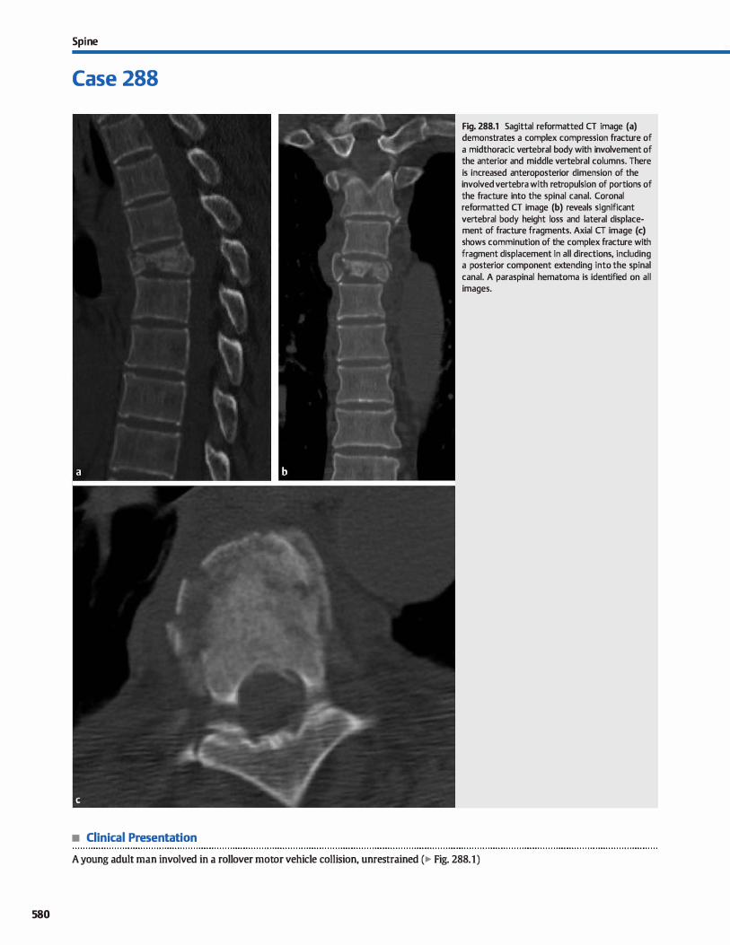
580
Spi ne
Case 288
• Clinical Presentation
Fig. 288.1 Sagittal reformatted CT image (a) demonstrates a complex compression fracture of a midthoracic vertebral body with involvement of the anterior and middle vertebral columns. There is increased anteroposterior dimension of the involved vertebra with retropulsion of portions of the fracture into the spinal canal. Coronal reformatted CT image (b) reveals significant vertebral body height loss and lateral displacement of fracture fragments. Axial CT image (c) shows comminution of the complex fracture with fragment displacement in all directions, induding a posterior component extending into the spinal
canal. A paraspinal hematoma is identified on all images.
A young adult man involved in a rollover motor vehicle collision, unrestrained (� Fig. 288.1)

• Key lmaglng Flndlng
Complex vertebral body fracture with height Joss and disbursement of fracture fragments
• Diagnosls
Burst fracture. A burst fracture refers to a complex, comminuted compression fracture of the vertebral body with disbursement of fracture fragments. lt occurs in the setting of significant trauma, most often with an axial Joad ( e.g., fall from height) and is most common at the thoracolumbar junction. Patients typically have neurological defidts corresponding to the level of injury. The degree of symptoms often correlates with the severity of injury, presenœ of instability, and extent of fragment displacement
Plain radiographs and computed tomography (CT) demonstrate a compression fracture of the vertebral body that extends to the posterior cortical margin, thus involving at least two vertebral columns. There is associated vertebral body height Joss and retropulsion of fracture fragments into the spinal canal. The involvement of the posterior cortical margin of the
� Pearts
• Burst fracture refers to a comminuted vertebral body com
pression in jury with disbursement ofbony fragments. • Imaging demonstrates a vertebral body compression fracture
that extends to the posterior cortical margin.
Suggested Reading
Atlas sw, Regenbagen V, Rogen LF, Kim KS, 1be radiographie characterization of
blll"St fractures of the spine. AmJ Roentgenol 1936; 147: 575-582
Case 288
vertebral body differentiates a burst fracture from a simple wedge compression fracture. Fracture components may also involve the posterior column (pedicles, faœts, lamina, spinous proœss and associated ligaments). In contrast to a Chance or seatbelt fracture, burst fractures are typically oriented in the sagittal plane, whereas Chance fractures are oriented in the axial plane due to the underlying mechanism of injury.
Anteroposterior plain films or coronal cr reformats may show widening of the interpedicular distance, which is indicative of underlying instability. With severe injury, there may also be malalignment of the vertebral column, as well as facet dislocation. Magnetic resonanœ imaging {MRI) is useful for evaluation of sort-tissue, ligamentous, and cord injury.
Treatment depends upon the clinical sœnario and stability of the injury and includes both conservative and surgical options.
• Widening of the interpedicular distance is indicative of
instability; spinal malalignment may also be seen. • MRI is useful for evaluation of sort-tissue, ligamentous, and
cord injury.
581
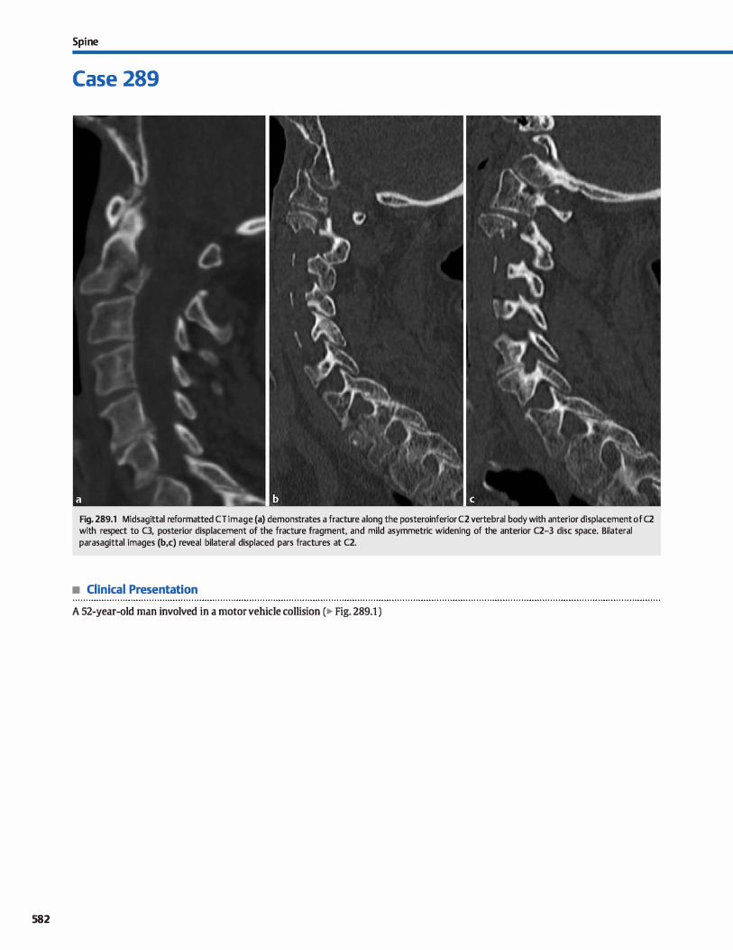
582
Spi ne
Case 289
Fig. 289.1 Midsagittal reformatted CT image (a) demonstrates a fracture a long the posteroinferior C2 vertebral body with anterior displacement of C2 with respect to C3, posterior displacement of the fracture fragment, and mild asymmetric widening of the anterior C2-3 dise space. Bilateral parasagittal images (b,c) reveal bilateral displaced pars fractures at C2.
• Clinical Presentation
A 52-year-old man involved in a motor vehicle collision (� Fig. 289.1 )
• Key lmaging Finding
C2 fractures with listhesis
• Diagnosis
Hanged man's fracture. Hyperextension injuries are most common in the cervical spine. When severe, they may result in frac
tures of the posterior elements and/or posterior vertebral body
with widening of the anterior dise spaœ. A hanged man's frac
ture refers to traumatic hyperextension injury of C2 with asso
dated spondylolisthesis. It most often results from motor
vehicle collisions and is considered an unstable injury.
The lateral view on plain radiographs demonstrates the C2
posterior element and/or vertebral body fractures with
y' Pearts
• Hyperextension injuries result in posterior element/vertebral
body fractures with widened anterior dise spaœ. • A hanged man's fracture refers ta traumatic hyperextension
injury of C2 with associated spondylolisthesis.
Suggested Reading
Munera F, RiVi15 LA. Nunez DB,Jr, Quenœr RM. Imaging evaluation of adult spinal
Îl\Îuries: emphasis on multidetector cr in cervical spine trauma. Radiology2012;
263;645-660
Case 289
Iisthesis. Computed tomography {CT) is superior ta plain films
in characterizing the underlying bony injuries. Magnetic reso
nanœ imaging {MRI) is useful in evaluating for underlying ligamentous and cord injuries. Unlike hyperflexion injuries, the
interlaminar line and interspinous articulations are typically
maintained in the setting of hyperextension injury.
• Lateral plain films demonstrate C2 posterior element and/or
vertebral body fractures with listhesis.
• cr is superior ta plain films in characterizing bony injuries;
MRI best evaluates ligamentous and cord injuries.
583
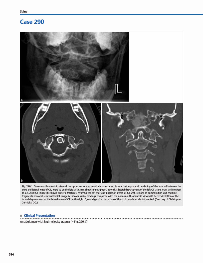
584
Spi ne
Case 290
Fig. 290.1 Open-mouth odontoid view of the upper cervical spi ne (a) demonstrates bilateral but asymmetric widening of the interval between the dens and lateral mass of Cl , more so on the left, with a small fracture fragment, as well as lateral displacement of the left Cl lateral mass with respect to C2. Axial CT image (b) shows bilateral fractures involving the anterior and posterior arches of Cl with regions of comminution and multiple fragments. Coronal reformatted CT image (c) shows similar findings compared with the open-mouth odontoid view with better depiction of the lateral displacement of the lateral mass of Cl on the right; "ground glass" attenuation of the skull base is incidentally noted. (Courtesy of Christopher Cerniglia, DO.)
• Clinical Presentation
An adult man with high-velocity trauma ( � Fig. 290.1 )
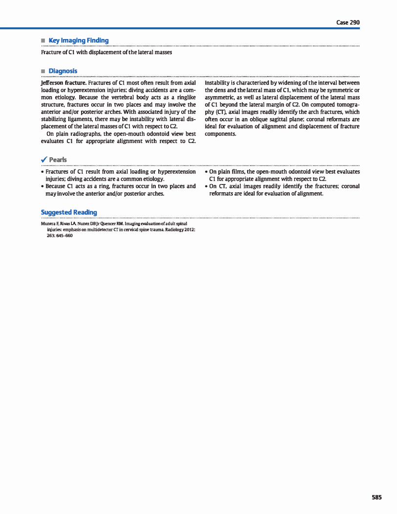
• Key lmaging Finding
Fracture of Cl with displaœment of the lateral masses
• Diagnosis
Jefferson fracture. Fractures of Cl most often result from axial loading or hyperextension injuries; diving accidents are a common etiology. Because the vertebral body acts as a ringlike
structure, fractures occur in two places and may involve the anterior and/or posterior arches. With associated injury of the stabilizing ligaments, there may be instability with lateral displacement of the lateral masses of Cl with respect to C2.
On plain radiographs, the open-mouth odontoid view best evaluates Cl for appropriate alignment with respect to C2.
y' Pearts
• Fractures of Cl result from axial loading or hyperextension
injuries; diving accidents are a common etiology. • Because Cl acts as a ring, fractures occur in two places and
may involve the anterior and/or posterior arches.
Suggested Reading
Munera F, RiVi15 LA. Nunez DB Jr Quenœr RM. lrnaging evaluation of adult spinal
Îl\Îuries: ernphasis on rnultidetector cr in cervical spine trauma. Radiology2012;
263; 645-660
Case 290
Instability is c:haracterized by widening of the interval between
the dens and the lateral mass of Cl, which may be symmetric or asymmetric, as well as lateral displacement of the lateral mass of Cl beyond the lateral margin of C2. On computed tomogra
phy (CT), axial images readily identify the arch fractures, whic:h
often occur in an oblique sagittal plane; coronal reformats are ideal for evaluation of alignment and displacement of fracture
components.
• On plain films, the open-mouth odontoid view best evaluates
Cl for appropriate alignment with respect to C2. • On cr, axial images readily identify the fractures; coronal
reformats are ideal for evaluation of alignment.
585
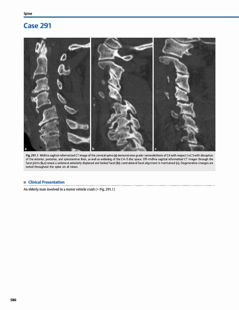
586
Spi ne
Case 291
Fig. 291 .1 Mid l i ne sagittal reformatted CT image of the cervical spi ne (a) demonstrates grade 1 anterolisthesis of C4 with respect to CS with disruption of the anterior, posterior, and spinolaminar lines, as well as widening of the C4-5 dise space. Off-midline sagittal reformatted CT images through the facet joints (b,c) reveal a unilateral anteriorly displaced and locked facet (b); contralateral facet alignment is maintained (c). Degenerative changes are noted throughout the spine on all views.
• Clinical Presentation
An elderly man involved in a motor vehicle crash (� Fig. 291 .1 )
• Key lmaging Finding
Cervical spine anterolisthesis with facet malalignment
• Diagnosis
Faœt dislocation. Flexion injuries commonly involve the cervical spine and include anterior teardrop fractures, facet dislocations (bilateral or unilateral), anterior wedge compression fractures, anterior subluxations, and the day-shoveler fracture. Motor vehide crashes are the most common underlying etiology. Anterior teardrop fractures and bilateral faœt dislocations are considered unstable. Unilateral faœt dislocation occurs in the setting of flexion with a rotatory component.
Bilateral faœt dislocation occurs as a result of flexion injury with disruption of both the anterior and posterior longitudinal ligaments. The inferior faœts at the involved segment are displaœd superiorly and anteriorly compared with the superior facets of the subjacent inferior vertebra. The degree of subluxation ranges from partial with mild displacement, complete where the inferior faœts at the involved level are perched on the superior faœts of the lower level, and locked where the faœt orientation is reversed with the inferior faœts at the involved level positioned in front of the superior faœts of the lower level. On lateral radiographs, there is disruption of the cervical lines with > 50% anterolisthesis at the involved level Associated fractures may be identified On axial computed tomography {CT) images, there is a
� Pearts
• Bilateral faœt dislocation occurs with flexion injury and disruption of stabilizing longitudinal ligaments.
• Unilateral faœt dislocation occurs in the setting of flexion injury with a rotatory component.
Suggested Readings
Ungawi SS. The naked faœt sign. Radiology 2001; 219: 366-367
Case 291
naked faœt sign in the setting of partial or complete {perched) subluxations, which refers to paired faœts at the involved levels without articulation with the adjacent level. Locked faœts demonstrate a reversai of the normal faœt articulation. Secondary fmdings of flexion injury indude focal kyphosis, widening of the posterior dise spaœ, and widening of the interspinous distance.
Unilateral faœt dislocations occur in the setting of flexion injury with rotation and commonly involve the mid ta lower cervical spine. Although both facets are typically injured to some degree, the dislocation is unilateral. As with bilateral faœt dislocation, the subluxation may be partial, complete (perched), or loclœd (reversed). Plain radiographs demonstrate loss of normal spinous process alignment on frontal views due to focal rotation at the involved level. Lateral views demonstrate focal Joss of the normal cervical lines and varying degrees of anterolisthesis, measuring < 50%. Associated faœt, vertebral body, and posterior element fractures may be seen. CT demonstrates similar findings compared with bilateral facet dislocation except that the findings are unilateral and there is a rotatory component at the involved level.
• Radiographs demonstrate > 50% anterolisthesis with bilateral and< 50% with unilateral facet dislocations.
• Naked faœt sign is seen on CT with partial or complete {perched) dislocations; locked facets are reversed.
Pimente! !., Diegelmann L Evaluation and management of acute cenrical spine
trauma. Emerg Med Oin North Am 2010; 28: 719-738
587
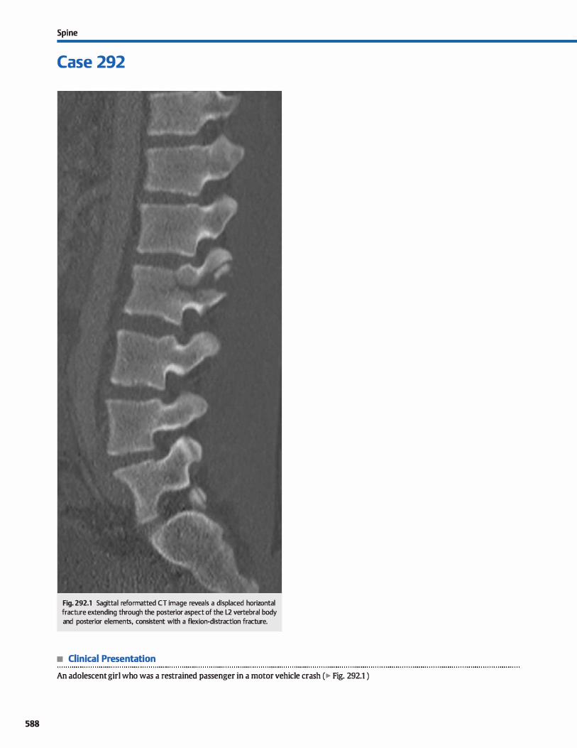
588
Spi ne
Case 292
Fig. 292.1 Sagittal reformatted CT image reveals a displaced horizontal fracture extending through the posterior aspect of the L2 vertebral body and posterior elements, consistent with a flexion-distraction fracture.
• Clinical Presentation
An adolescent girl who was a restrained passenger in a motor vehicle crash ( � Fig. 292.1 )
• Key lmaglng Flndlng
Horizontal displaced fracture through the lumbar posterior elements and vertebral body
• Diagnosls
Chance fracture. A Chance or seatbelt fracture refers to a horizontal fracture of the neural arch due to flexion injury asso
ciated with rapid motor vehicle deceleration in patients wearing lap belts. Due to the location of the lap belts, the thoracolumbar junction is most commonly involved. There is a high
incidence of associated visceral injuries that most often affect the small bowel and mesenteric vessels. Solid organ injuries of the pancreas, spleen. Iiver, kidneys, and uterus may also been seen. Interestingly, patients typically do not have neurological
deficits despite the mechanism of fracture, which is generally considered unstable, especially when there is involvement of
more than one vertebral column. Chance fractures invariably involve the posterior elements
with or without extension into the vertebral body. On frontal plain radiographs or coronal reformatted computed
� Pearts
• A Chance or seatbelt fracture refers ta horizontal fracture of
the neural arch due to flexion injury. • Chance fractures involve the posterior elements with or with
out extension into the vertebral body.
Suggested Reading
Berrustein MP, Mirvis SE, Shanmuganathan K. Chance-type fractures of the thara
columbar spine: imaging analysis in 53 patients. AmJ Rcentgenal 2006; 187:
859-868
Case 292
tomography (CT) images, the fractures are identified by horizontal lucencies through the pedides, as well as displaœment
of fracture fragments of the spinous process, which results
in the empty vertebral body sign. On the lateral or sagittal reformatted views, there may be distraction of the spinous process, and with severe injury, displacement of fracture components of the vertebral body. lnstability results from disruption of the anterior and posterior longitudinal ligaments. The interspinous
and posterior vertebral dise spaces may be widened, which is characteristic of flexion injuries.
Magnetic resonance imaging (MRI) is ideal for evaluation of ligamentous integrity and cord injury, as well as for evaluation of the intervertebral dise and paraspinal soft tissues. On both CT and MRI, it is important to evaluate visualized solid organs and hollow viscus.
• Frontal views often demonstrate transverse pedicle fractures
and displacement of the spinous process. • Lateral views show distraction of fracture fragments with
interspinous and posterior dise space widening.
589
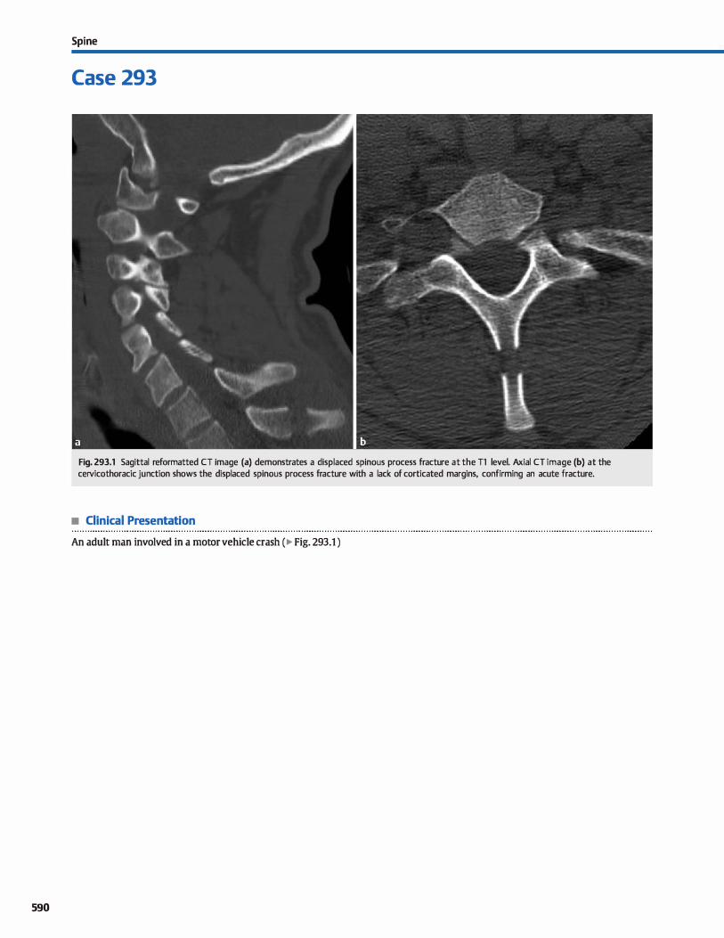
590
Spi ne
Case 293
Fig. 293.1 Sagittal reformatted CT image (a) demonstrates a displaced spinous process fracture at the Tl level. Axial CT image (b) at the cervicothoracic junction shows the displaced spinous process fracture with a lack of corticated margins, confirming an acute fracture.
• Clinical Presentation
An adult man involved in a motor vehicle crash ( � Fig. 293.1 )
• Key lmaging Finding
Displaœd spinous proœss fracture at the œrvicothoracic junction
• Diagnosis
Oay-shoveler fracture. A day-shaveler fracture refers to a spinous process fracture at the cervicothoradc junction as a result of hyperflexion injury; C7 is the most commonly involved level. Historically, the fracture was named for an injury that occurred
in workers shoveling day where the day would adhere to the shovel and cause abrupt flexion of the neck and spine. Nowadays, the most common mechanisms of injury include motor vehicle accidents and direct trauma.
y' Pearts
• Oay-shoveler fracture refers to a lower cervical {usually C7) spinous process fracture due ta hyperflexion.
• The most common present-day mechanisms of injury indude
motor vehide accidents and direct trauma.
Suggested Reading
Lee P, Hunter TB, Taljanovic M. Musruloskeletal colloquialisrns: how did we corne up with these narnes7 Radiograplùcs 2004; 24: 1009-1027
Case 293
Plain radiographs and computed tomography (CT) demonstrate a spinous process fracture involving the lower cervical or upper thoracic spine, often with some degree of displacement. Surrounding soft-tissue injury is commonly seen. In the acute
setting, the fracture lacks corticated margins. Remote fractures with nonunion will demonstrate corticated margins, simulating
an unfused apophysis.
• Plain radiographs and CT demonstrate a spinous process frac
ture often with some degree of displacement. • Remote fractures with nonunion will demonstrate corticated
margins, simulating an unfused apophysis.
591
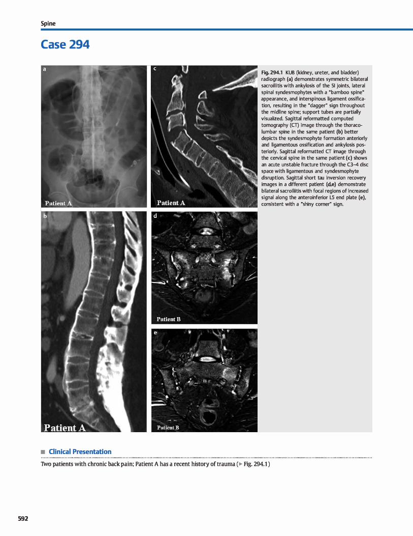
592
Spi ne
Case 294
• Clinical Presentation
Fig. 294.1 KUB (kidney, ureter, and bladder) radiograph (a) demonstrates symmetric bilateral sacroiliitis with ankylosis of the SI joints, lateral spinal syndesmophytes with a "bamboo spine" appearance, and interspinous ligament ossification, resulting in the "dagger" sign throughout the midline spine; support tubes are partially visualized. Sagittal reformatted computed tomography (CT) image through the thoracolumbar spine in the same patient (b) better depicts the syndesmophyte formation anteriorly and ligamentous ossification and ankylosis posteriorly. Sagittal reformatted CT image through the cervical spine in the same patient (c) shows an acute unstable fracture through the C3-4 dise space with ligamentous and syndesmophyte disruption. Sagittal short tau inversion recovery images in a different patient (d,e) demonstrate bilateral sacroiliitis with focal regions of increased signal along the anteroinferior LS end plate (e), consistent with a "shiny corner" sign.
Tvvo patients with chronic back pain; Patient A has a recent history of trauma (� Fig. 294.1 )
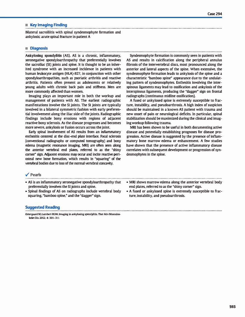
• Key lmaging Finding
Bilateral sacroiliitis with spinal syndesmophyte formation and ankylosis; acute spinal fracture in patient A
• Diagnosis
Ankylosing spondylitis (AS). AS is a chronic, inflammatory, seronegative spondyloarthropathy that preferentially involves the sacroiliac (SI) joints and spine. It is thought to be an inherited syndrome with an increased incidence in patients with
human leukocyte antigen (HLA)-B27, in conjunction with other spondyloarthropathies, such as psoriatic arthritis and reactive arthritis. Patients often present as adolescents or relatively young adults with chronic back pain and stiffness. Men are more commonly affected than women.
Imaging plays an important rote in both the workup and management of patients with AS. The earliest radiographie
manifestations involve the SI joints. The SI joints are typically involved in a bilateral symmetric fashion with early preferen
tial involvement along the iliac side of the joints. Radiographie findings include bony erosions with regions of adjacent reactive bony sderosis. As the disease progresses and becomes more severe, ankylosis or fusion occurs across the joint.
Early spinal involvement of AS results from an inflammatory enthesitis œntered at the dise-end plate interface. Focal sclerosis
(conventional radiographs or computed tomography) and bony edema (magnetic resonance imaging, MRI) are often seen along
the anterior vertebral end plates, referred to as the ushiny corner" sign. Adjacent erosions may occur and incite reactive periosteal new bone formation, which results in "squaring" of the vertebral bodies due to Joss of the normal vertebral concavity.
� Pearts
• AS is an inflammatory seronegative spondyloarthropathy that preferentially involves the SI joints and spine.
• Spinal findings of AS on radiographs indude vertebral body squaring, ubamboo spine," and the udagger" sign.
Suggested Reading
Ostergaard M, Lambert RGW. Imaging in ankylosing spondylilll. lber Adv Musculos
lœlet Dis 2012; 4: 301-311
Case 294
Syndesmophyte formation is commonly seen in patients with AS and results in calcification along the peripheral annulus fibrosis of the intervertebral dises, most pronounced along the
anterior and lateral aspects of the spine. When extensive, the syndesmophyte formation leads to ankylosis of the spine and a characteristic ubamboo spine" appearance due ta the undulating pattern of syndesmophytes. Enthesitis involving the interspinous ligaments may lead to ossification and ankylosis of the interspinous ligaments, produdng the udagger" sign on frontal radiographs (continuous midline ossification).
A fused or ankylosed spine is extremely susceptible to frac
ture, instability, and pseudoarthrosis. A high index of suspicion should be maintained in a known AS patient with trauma and new onset of pain or neurological defidts. In particular, spinal stabilization should be maximized during the dinical and imaging workup following trauma.
MRI has been shown to be useful in bath documenting active
disease and potentially establishing prognoses for disease progression. Active disease is suggested by the presence of inflammatory bene marrow edema or enhancement. A few studies
have shown that the presence of active inflammatory disease correlates with subsequent development or progression of syndesmophytes in the spine.
• MRI shows marrow edema along the anterior vertebral body end plates, referred to as the ushiny corner" sign.
• A fused or ankylosed spine is extremely susceptible to fracture, instability, and pseudoarthrosis.
593
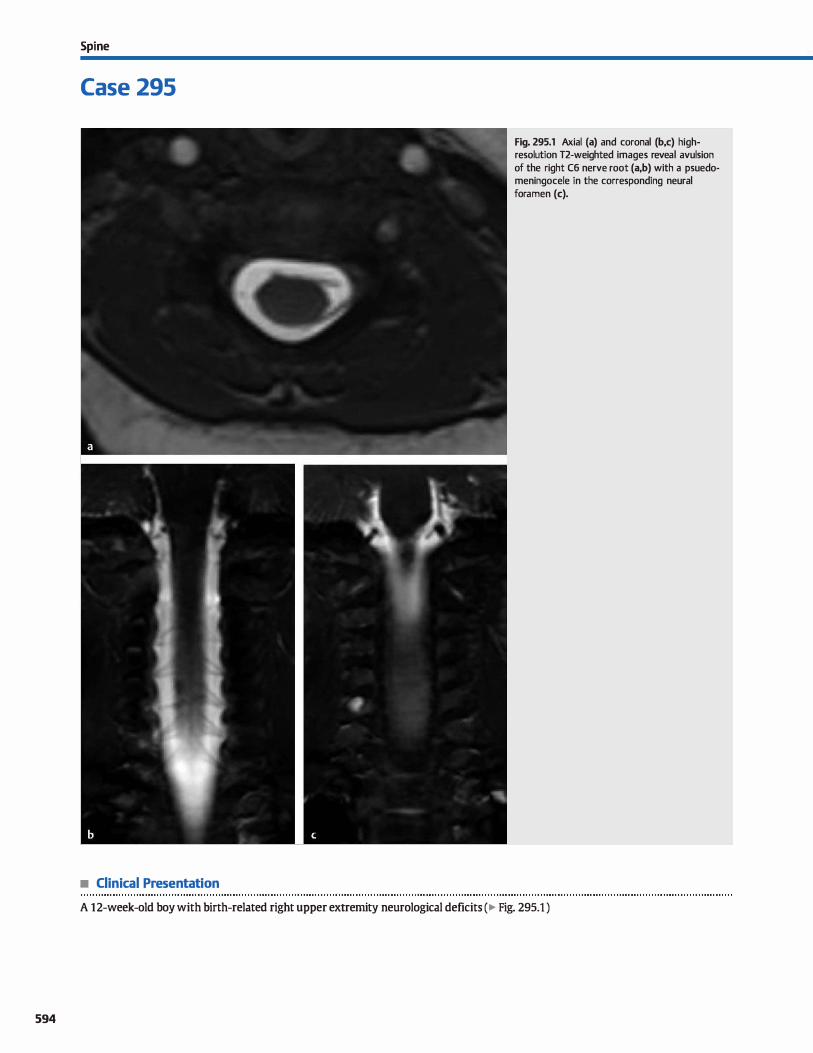
594
Spi ne
Case 295
• Clinical Presentation
Fig. 295.1 Axial (a) and coronal (b,c) highresolution T2-weighted images reveal avulsion of the right C6 nerve root (a,b) with a psuedomeningocele in the corresponding neural foramen (c).
A 12-week-old boy with birth-related right upper extremity neurological deficits (� Fig. 295.1 )
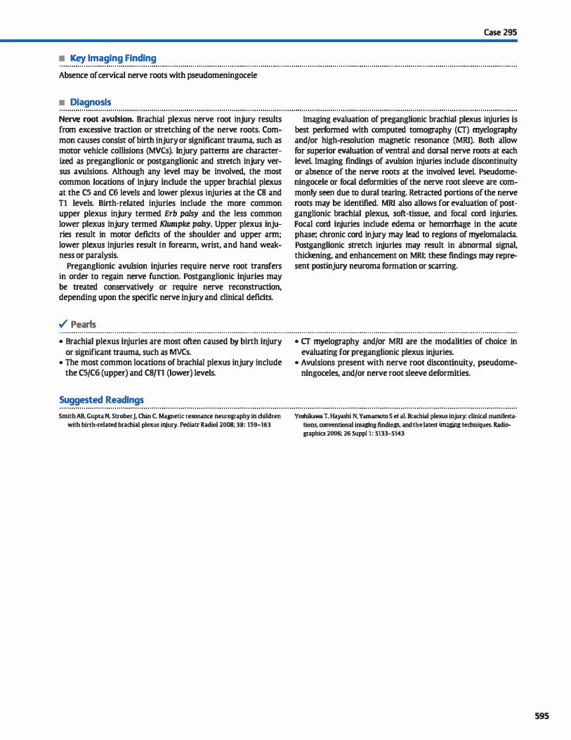
• Key lmaging Finding
Absence of cervical nerve roots with pseudomeningocele
• Diagnosis
Nel"W! root avulsion. Brachial plexus nerve root injury results from excessive traction or stretching of the nerve roots. Common causes consist of birth injury or significant trauma, such as motor vehicle collisions (MVCs). lnjury patterns are characterized as preganglionic or postganglionic and stretch injury versus awlsions. Although any level may be involved, the most common locations of injury indude the upper brachial plexus at the C5 and CG levels and lower plexus injuries at the CS and Tl levels. Birth-related injuries indude the more common upper plexus injury termed Erb palsy and the Jess common lower plexus injury termed Klumpke palsy. Upper plexus injuries result in motor deficits of the shoulder and upper arm; lower plexus injuries result in foreann, wrist, and hand weakness or paralysis.
Preganglionic awlsion injuries require nerve root transfers in order to regain nerve function. Postganglionic injuries may be treated conservatively or require nerve reconstruction, depending upon the specific nerve injury and dinical deficits.
v" Pearls
• Brachial plexus injuries are most often caused by birth injury or significant trauma, such as MVCs.
• The most common locations of brachial plexus injury include the C5/C6 (upper) and CS/Tt (lower) levels.
Suggested Readings
Case 295
lmaging evaluation of preganglionic brachial plexus injuries is best performed with computed tomography (CT) myelographY and/or high-resolution magnetic resonanœ (MRI). Both allow for superior evaluation of ventral and dorsal nerve roots at each level. lmaging findings of avulsion injuries indude discontinuity or absence of the nerve roots at the involved level. Pseudomeningocele or focal deformities of the nerve root sleeve are commonly seen due to durai tearing. Retracted portions of the nerve roots may be identified. MRI also allows for evaluation of postganglionic brachial plexus, soft-tissue, and focal cord injuries. Focal cord injuries indude edema or hemorrhage in the acute phase; chronic cord injury may lead to regions of myelomalacia. Postganglionic stretch injuries may result in abnormal signal thidœning, and enhanœment on MRI; these findings may represent postinjury neuroma formation or scarring.
• cr myelography and/or MRI are the modalities of choiœ in evaluating for preganglionic plexus injuries.
• Avulsions present with nerve root discontinuity, pseudomeningoœles, and/or nerve root sleeve deformities.
Smith AB, Gupta N, Strober J, Chio C. Magnetk resonance neurography in dùldren Yoshikawa T, Hayashi N, Yamamoto S et al. Brachial plexus injury: clinical manifesta-
with birth-related brachial plexus iojury. Pediatr Radio! 2008; 38: 159-163 lions, amventional imaging findings, and the latest imaging techniques. Radio
graphies 2006; 26 Suppl 1; S133-S143
595
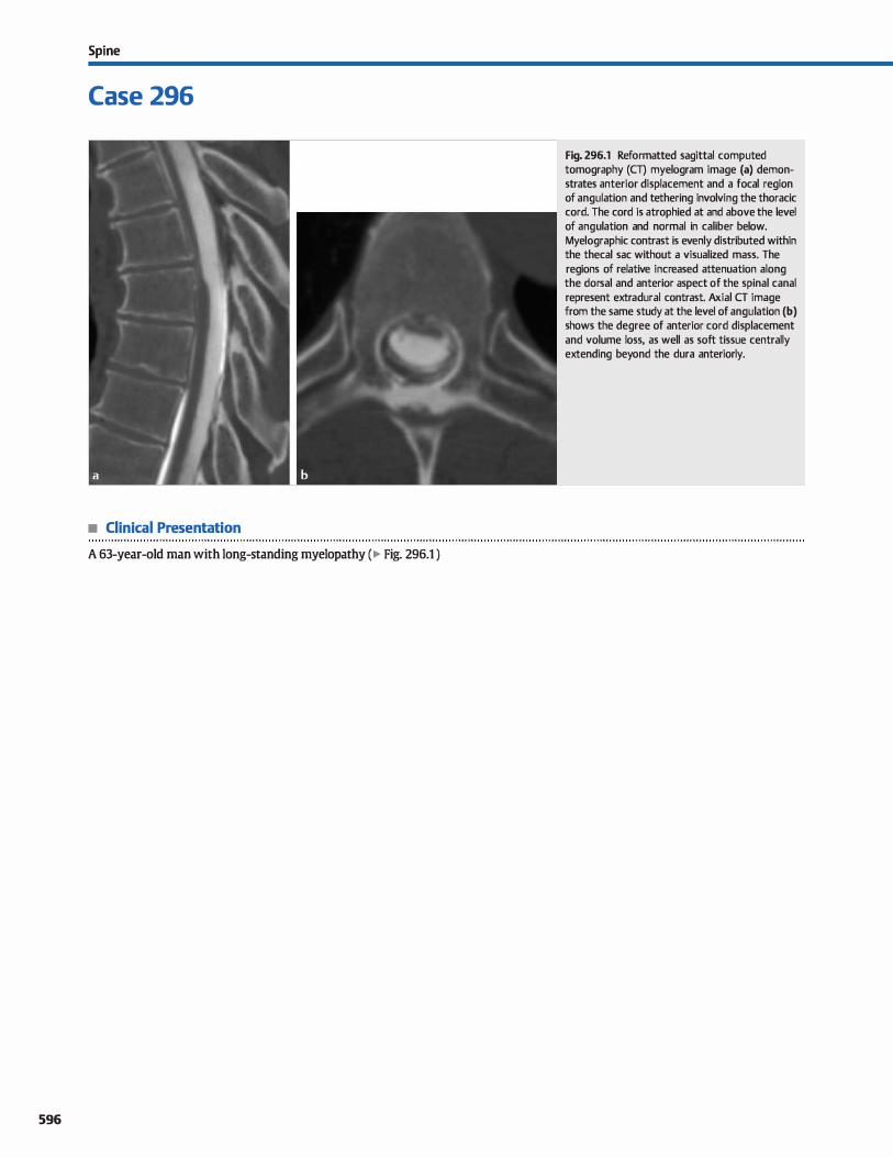
Spi ne
Case 296
• Clinical Presentation
A 63-year-old man with long-standing myelopathy (� Fig. 296.1 )
596
Fig. 296.1 Reformatted sagittal computed tomography (CT) myelogram image (a) demonstrates anterior displacement and a focal region of angulation and tethering involving the thoracic cord. The cord is atrophied at and above the level of angulation and normal in caliber below. Myelographic contrast is evenly distributed within the thecal sac without a visualized mass. The regions of relative increased attenuation along the dorsal and anterior aspect of the spinal canal represent extradural contrast. Axial CT image from the same study at the level of angulation (b) shows the degree of anterior cord displacement and volume loss, as well as soft tissue centrally extending beyond the dura anteriorly.
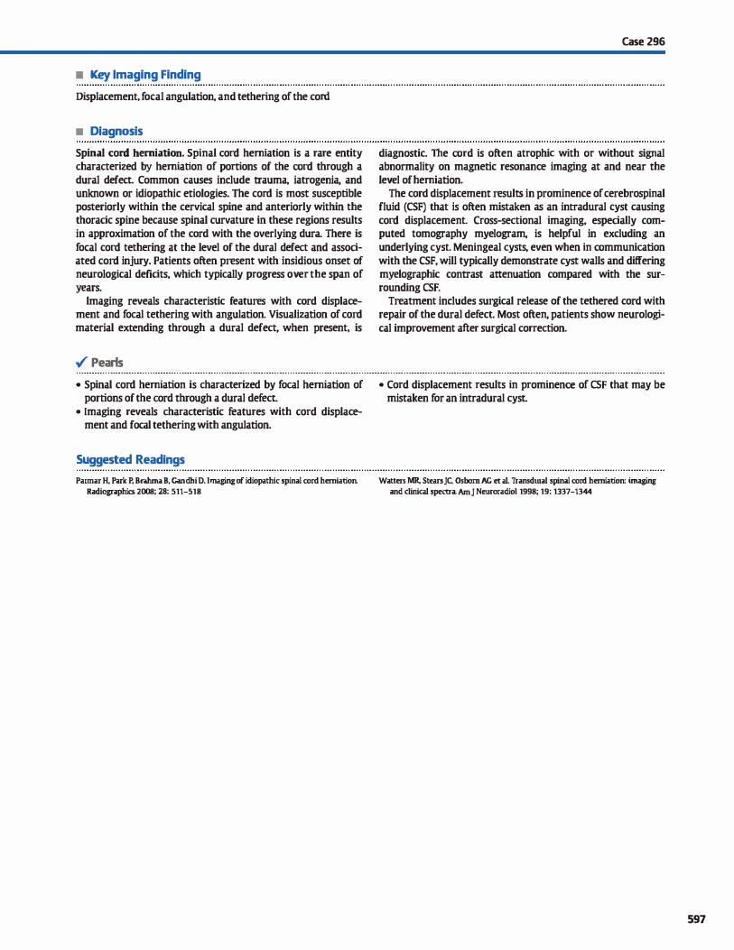
• Key lmaging Finding
Displacement. focal angulation, and tethering of the cord
• Diagnosis
Spinal cord hemiation. Spinal cord hemiation is a rare entity characterized by hemiation of portions of the cord through a durai defect Common causes indude trauma, iatrogenia, and unknown or idiopathie etiologies. The cord is most susceptible posteriorly within the cervical spine and anteriorly within the thoradc spine because spinal curvature in these regions results in approximation of the cord with the overlying dura There is
focal cord tethering at the level of the durai defect and assodated cord injury. Patients often present with insidious onset of neurological defidts, which typically progress over the span of years.
lmaging reveals characteristic features with cord displaœment and focal tethering with angulation. Visualization of cord material extending through a durai defect, when present. is
y' Pearts
• Spinal cord hemiation is characterized by focal hemiation of portions of the cord through a durai defect
• lmaging reveals characteristic features with cord displaœment and focal tethering with angulation.
Suggested Readings
Parmar H, Park P, Brahma B, Gilndhl D. lmaging of idiopathie spinal cord hemiation.
Radiographies 2008; 28; 511-518
Case 296
diagnostic. The cord is often atrophie with or without signal abnormality on magnetic resonanœ imaging at and near the level of hemiation.
The cord displacement results in prominence of cerebrospinal fluid (CSF) that is often mistaken as an intradural cyst causing
cord displacement Cross-sectional imaging, espedally computed tomography myelogram, is helpful in excluding an
underlying cyst Meningeal cysts, even when in communication
with the CSF, will typically demonstrate cyst walls and differing myelographic contrast attenuation compared with the surrounding CSF.
Treatment indudes surgical release of the tethered cord with repair of the durai defect. Most often, patients show neurologi
cal improvement after surgical correction.
• Cord displacement results in prominence of CSF that may be mistaken for an intradural cyst
Watten MR, Stears JC. Osborn N:. et al. Transdural spinal cord hemiation: imaging
and clinical spectra. Am] Neuroradiol 1998; 19; 1337-1344
597
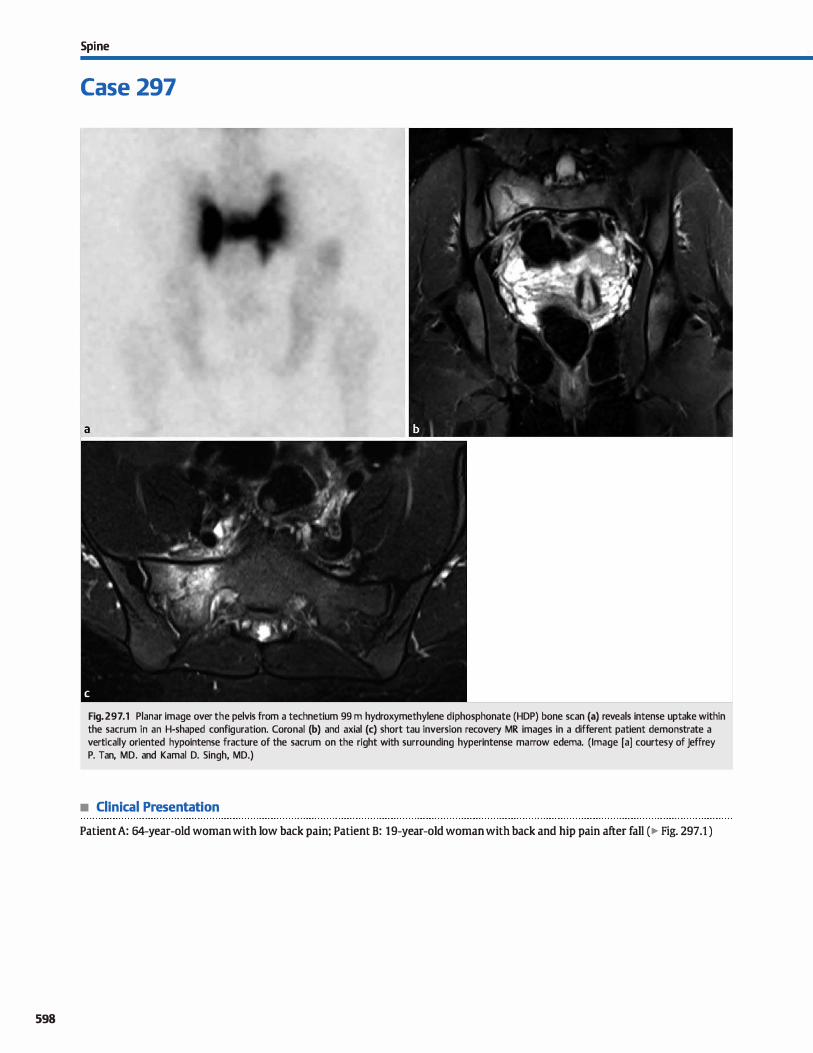
598
Spi ne
Case 297
Fig. 297 .1 Planar image over the pelvis from a technetium 99 m hydroxymethylene diphosphonate (HDP) bone scan (a) reveals intense uptake within the sacrum in an H-shaped configuration. Coronal (b) and axial (c) short tau inversion recovery MR images in a different patient demonstrate a vertically oriented hypointense fracture of the sacrum on the right with surrounding hyperintense marrow edema. (Image [a] courtesy of Jeffrey P. Tan, MD. and Kamal D. Singh, MD.)
• Clinical Presentation
Patient A: 64-year-old woman with low back pain; Patient B: 19-year-old woman with back and hip pain after fall (� Fig. 297.1 )
• Key lmaging Finding
Intense sacral uptake in an H-shaped configuration on bone scan and marrow edema on magnetic resonanœ imaging (MRI)
• Diagnosis
Sacral fracture. Sacral insuffiàency fractures result from normal stresses on weakened bones, whereas traumatic sacral fractures result from abnonnal stresses on nonnal bones. Insuffiàency fractures most often occur in elderly women with osteoporosis; other predisposing factors indude chronic cortimsteroid use, rheumatoid arthritis, and pelvic radiation therapy. Oinical symptoms associated with sacral fractures indude Iow back pain, which is exaœrbated by mavement, with radiation to the hip or groin. There may be point tEndemess over the sacrum.
On nudear mediàne imaging, the dassic findings indude uptake vertically along the sacral ala bilaterally with an adjoining transverse line, resulting in an "H" configuration that resembles the Honda automotive symbol. lt is therefore also referred to as the "Honda sign." Other variants indude the "half buttErtly" pattern where the superior aspects of the vertical lines are absent,
y' Pearts
• Sacral insufficiency fractures result from normal stresses on weakened bones.
• Traumatic sacral fractures result from abnormal stresses on
nonnal boues.
Suggested Readings
Case 297
the bar pattern where the transverse line is thickened, and the tramline pattEm where the transverse component of the "H" is absent.
Evaluation with conventional radiography may demonstratE the luœnt fracture Iine through the sacral ala, mrtical disruption, or sderotic bands; however, evaluation is usually limited by overlying bowel gas. ComputEd tomography may demonstrate the fracture lines or sderosis with greater sensitivity compared with conventional radiographs. MRI reveals a Tl hypointEnse fracture line with increased fat-suppressed T2 signal intensity within the marrovv from underlying edema. In addition, MRI is useful to
evaluate for an underlying pathological process. Management of sacral insuffiàency fractures consists of rest,
analgesia, antiinflammatory therapy, limited weight-bearing, and physical therapy.
• On bone scan, there is typically a characteristic H-shaped configuration, known as the "Honda sign."
• MRI depicts the fracture line and marrow edema; it may also evaluate for an underlying pathological process.
Blake SP, Connon AM. Sacral insufficiency fracture. Br j Radiol 2004; n: 891-896 White JH, Hague C, Nicolaou S. Gee R. Marchinlmw ID, Munk PL lmaging of sacral
Fajü M, Abe K. Hay.ishi K et al. Honda sign and variants in patients suspecte<! of hav- fractures. Clin Radiol 2003; 58: 914-921
ing a sacral insufficiency fracture. Oin Nucl Med 2005; 30: 165-169
599
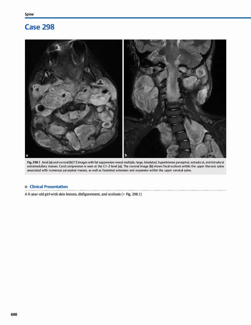
600
Spi ne
Case 298
Fig. 298.1 Axial (a) and coronal (b) T2 images with fat suppression reveal multiple, large, lobulated, hyperintense paraspinal, extradural, and intradural extramedullary masses. Cord compression is seen at the Cl-2 level (a). The coronal image (b) shows focal scoliosis within the upper thoracic spine associated with numerous paraspinal masses, as well as foraminal extension and expansion within the upper cervical spine.
• Clinical Presentation
A 9-year-old girl with skin lesions, disfigurement, and scoliosis (� Fig. 298.1)
• Key lmaging Finding
Multiple paraspinal, extradural, and intradural extramedullary masses
• Diagnosis
Neurofibromatosis type 1 (NFl). NFl, also known as von Recklinghausen disease, is the most common of the neurocutaneous syndromes. It may occur sporadically or be inherited in an autosomal dominant fashion. The genetic defect affects chromo
some 17q12 and results in decreased production of neurofibromin, which acts as a tumor suppressor. The disease affects the brain, skull, orbits, spine, musculoskeletal system, and skin/
integumentary system. Diagnostic criteria for NF1 indude the presence of two or more of the following: first-degree relative with NF1, six or more café-au-lait spots, two or more neurofibromas (NFs) or one plexiform NF (PNF), optic pathway glioma,
bony dysplasia, axillary or inguinal freckling, and two or more Llsch nodules.
Intracranial central nervous system (CNS) manifestations indude characteristic NF uspots" and law-grade neoplasms. The NF spots may wax and wane for the first decade of Iife and then regress in bath size and signal abnormality. The most common CNS neoplasms associated with NF1 are law-grade optic path
way gliomas (OPG). Bilateral optic nerve gliomas are pathogno
monic for NF1. Law-grade cerebellar, brain stem, tectal plate, and basal ganglia gliomas are also common in the setting of
NF1. In addition to OPG, orbital findings include sphenoid wing dysplasia with pulsatile exophthalmos, buphthalmos (globe
� Pearts
• NF1 is the most common neurocutaneous syndrome and results from a genetic defect affecting chromosome 17q12.
• Common spinal manifestations indude multiple NFs with foraminal extension and bony remodeling.
Suggested Readings
DiMario FJ,jr, Rarnsby G. Magnetic resonanœ irnaging lesion analym in neuro
fibromatosis type 1. Arcll Newlll 1998; SS: SOO-SOS Egelhoff JC, Dates DJ, Ross JS, Rothner AD, Cohen BH. Spinal MR findings in neuro
fibromatosis types 1 and :z. Am] Neuroradiol 1992.; 13: 1071-1077
Case 298
enlargement), and intraorbital extension of a PNF. Vascular abnormalities include regions of stenosis, moyamoya, and aneurysm formation. These are best visualized on magnetic res
onance angiography studies. Spinal manifestations of NF1 indude multiple bilateral NFs
with extension through and expansion of the neuroformina. NFs are more Iikely than schwannomas to demonstrate the utarget sign," which refers to peripheral increased and central decreased T2 signal intensity. Large retroperitoneal PNFs are prone to malignant degeneration, which is best characterized
by interval growth. Additional spinal manifestations indude
kyphoscoliosis, durai ectasia with posterior vertebral body scalloping, and lateral thoracic meningoceles. Rarely, intramedul
lary lesions, typically low-grade astrocytomas, may present in patients with NFl.
Extra-CNS and spinal manifestations indude cutaneous NFs, café-au-lait spots, extremity long-bone pseudoarthroses
and bowing deformities, "ribbon" ribs, and hypertrophy or overgrowth of ail or a portion of a limb. NF1 is associated with an increased incidence of several tumors, induding pheochromocytoma, medullary thyroid carcinoma, gastro
intestinal stromal tumors, melanoma, Wilms tumor, leukemia, and lymphoma.
• Scoliosis, durai ectasia with posterior vertebral body scalloping, and lateral meningoceles are common in NFl.
Rodriguez D. Young Pous saint T. Neuroimaging findings in neurofibrornatosis type 1
and :Z. Neuroirnaging Clin N Am 2004; 14: 149-170. vil
601
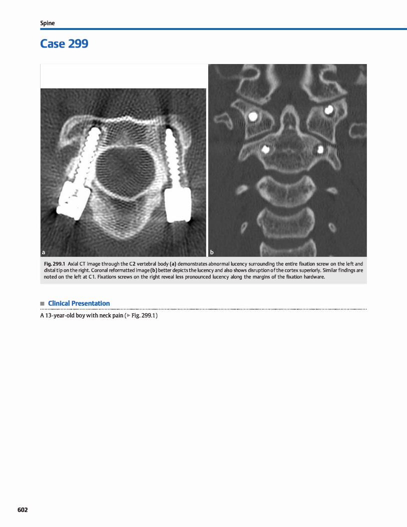
602
Spi ne
Case 299
Fig. 299.1 Axial CT image through the C2 vertebral body (a) demonstrates abnormal lucency surrounding the entire fixation screw on the left and distal tip on the right. Coronal reformatted image (b) better depicts the lucency and also shows disruption of the cortex superiorly. Similar findings are noted on the left at Cl . Fixations screws on the right reveal less pronounced lucency along the margins of the fixation hardware.
• Clinical Presentation
A 13-year-old boy with neck pain (� Fig. 299.1 )
• Key lmaging Finding
Lucency surrounding spinal fixation screw
• Diagnosis
Spinal hardware complication/loosening. Spinal stabilization
and fusion is perfonned to correct and restore anatomie alignment and function secondary to underlying congenital, posttraurnatic, neoplastic, or postinfectious/inflammatory defects. The surgical approach and hardware utilized depends upon bath the underlying condition and the individual surgeon. Complications may arise in the immediate postoperative period
or present years after the initial surgery.
Postoperative imaging typically consists of plain radiographs and computed tomography (cr) and is utilized to look for appropriate alignment, evidenœ of hardware complication, and
to evaluate for bony fusion, when applicable. Pedide screws are commonly used in the spine and require meticulous placement due to their proximity to critical neurovascular structures. Ide
ally, pedide screws will traverse along the medial cortex of the
� Pearts
• Spinal stabilization and fusion is performed to correct and
restore anatomie alignment and function. • Pedide screws require meticulous placement due to their
proximity to critical neurovascular structures.
Suggested Reading
Young PM, Beniuist rn, Bancroft LW, PetersonlJ. Complications of spinal instrumen
UtiDIL Radiographies 2007; 27; 775-789
Case 299
pedide because this location allows for maximal anchoring.
Care must be taken not to disrupt the medial cortex because inadvertent penetration may result in narrowing of the spinal canal With posterior fusion, the tip of the pedide screw should approach but not extend through the anterior cortex of the ver
tebral body. Improper (or less than optimal) screw placement indudes those that extend into the spinal canal, foramen trans
versarium, intervertebral dise spaœ, or paraspinal soft tissues. Properly plaeed screws may become fractured or loosened
over time. Fractured hardware is not difficult to identify, but it is important to comment on the degree of displaœment of the
hardware components. Loosening is best evaluated on cr and presents as a rim of lucency surrounding the pedide screw.
Loosening allows for migration of the hardware that may result in instability.
• Less than optimal placement indudes involvement of the
spinal canal, foramen transversarium, and dise space. • Hardware loosening is best evaluated on cr and presents as a
rim of lucency surrounding the pedide screw.
603
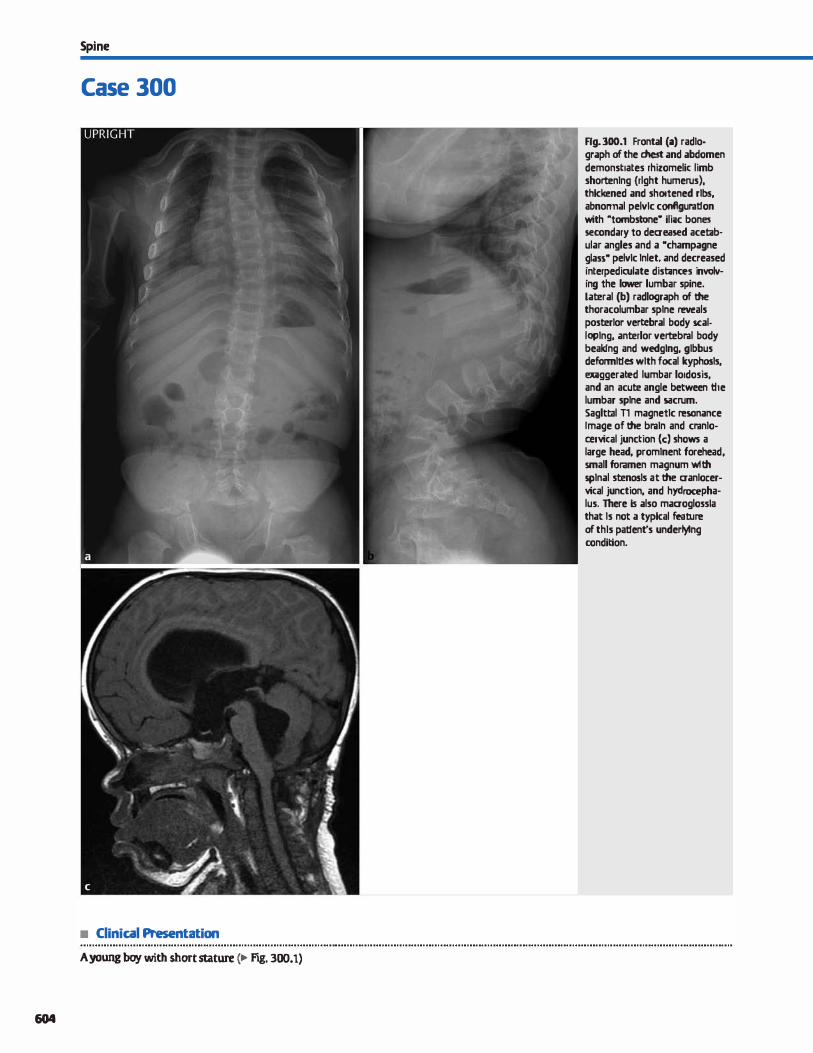
Spi ne
Case 300
• Clinical Presentation
A young boy with short stature (� Fig. 300.1)
604
Rg. 300.1 Frontal (a) radio· graph of the chest and abdomen demonstrates rhizomelic limb short:enlng {rlght humerus). thkkened and shortened rlbs, abnormal pelvlc configuration with "tombstone• ifü1c bones secondary to deaeased acetab· ular angles and a •champagne glass• pelvlc lnlet. and decreased interpediculate disl:inces involv· ing the lawer lumbar spine. Lateral (b) radlograph of the thoracolumbar splne reveals posterlor vert:ebral body scalloplng, anterlor vert:ebral body beaklng and wedglng, glbbus deformll1es wlth focal kyphosls, exilggerated lumbar lordosis, and an acute angle between the lumbar splne and sacrum. Saglttll Tl magnetlc resonance Image of the braln and cranloœl"Yical junction (c) shows a large head, promlnent forehead, small for.amen magnum wlth splnal stenosls at the aanlocervical junction, and hydrocepha· lus. There ls also maaoglossla that ls not a typkal feature of thls pal1ent's underlylng condition.
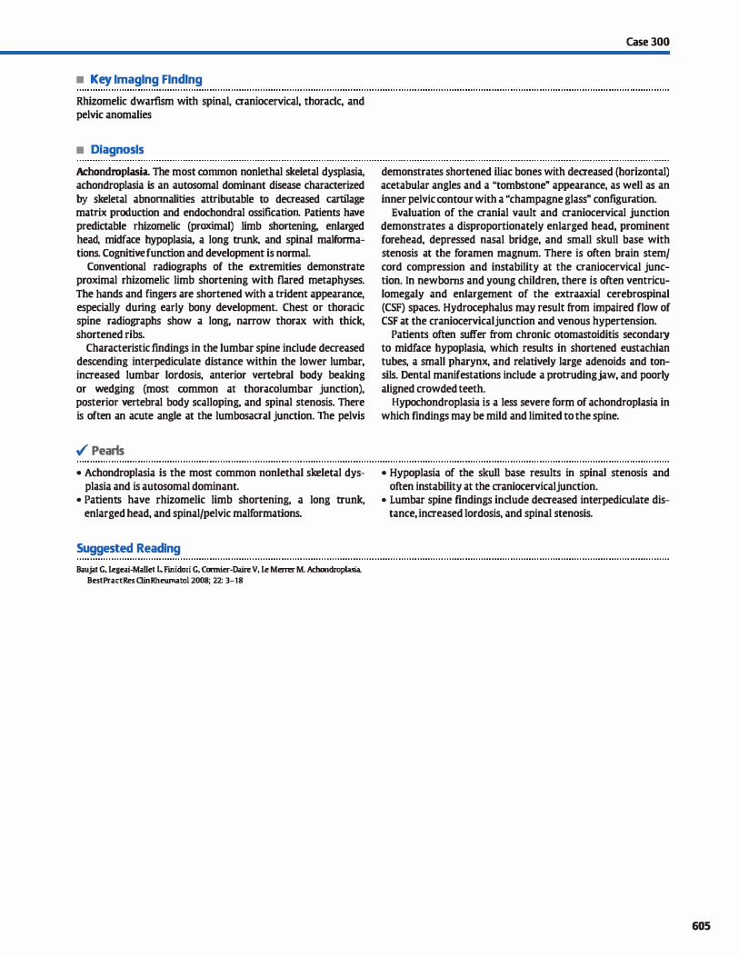
• Key lmaglng Flndlng
Rhizomelic dwartism with spinal, craniocervical, thoradc, and pelvic anomalies
• Diagnosls
Achondroplasia. The most COilllDon nonlethal skeletal dysplasia, achondroplasia is an autosomal dominant disease characterized
by skeletal abnonnalities attributable to decreased cartilage matrix production and endochondral ossification Patients have predictable rhizomelic (proximal) limb shortening, enlarged head, midface hypoplasia, a long trunk, and spinal malformations. Cognitive function and development is normal
Conventional radiographs of the extremities demonstrate proximal rhizomelic limb shortening with flared metaphyses.
The bands and fingers are shortened with a trident appearance, especially during early bony development. Chest or thoradc spine radiographs show a long, narrow thorax with thick,
shortened ribs. Characteristic findings in the lumbar spine indude decreased
descending interpediculate distance within the lower lumbar, increased lumbar lordosis, anterior vertebral body bealdng
or wedging (most common at thoracolumbar junction), posterior vertebral body scalloping, and spinal stenosis. There is often an acute angle at the lumbosacral junction. The pelvis
y'° Pearts
• Achondroplasia is the most common nonlethal skeletal dysplasia and is autosomal dominant.
• Patients have rhizomelic limb shortening, a long trunk, enlarged head, and spinal/pelvic malformations.
Suggested Reading
Baujat G, Legeai-Mallet I. Finidori G, Connier-Daire V, Le Merrer M. Achondroplasia.
BestPractRes ClinRheumatol 2008; 22; 3-18
Case 300
demonstrates shortened iliac bones with decreased (horizontal) acetabular angles and a "tombstone" appearance, as well as an
inner pelvic contour with a "champagne glass" configuration. Evaluation of the cranial vault and craniocervical junction
demonstrates a disproportionately enlarged head, prominent forehead, depressed nasal bridge, and small skull base with stenosis at the foramen magnum. There is often brain stem/
cord compression and instability at the craniocervical junction. In newboms and young children, there is often ventriculomegaly and enlargement of the extraaxial cerebrospinal (CSF) spaces. Hydrocephalus may result from impaired flow of CSF at the craniocervicaljunction and venous hypertension.
Patients often suffer from chronic otomastoiditis secondary to midface hypoplasia, which results in shortened eustachian tubes, a small pharynx, and relatively large adenoids and tonsils. Dental manifestations indude a protruding jaw, and poorly
aligned crowded teeth. Hypochondroplasia is a Jess severe form of achondroplasia in
which findings may be mild and limited to the spine.
• Hypoplasia of the skull base results in spinal stenosis and often instability at the craniocervicaljunction.
• Lumbar spine findings indude decreased interpediculate distance, increased lordosis, and spinal stenosis.
605

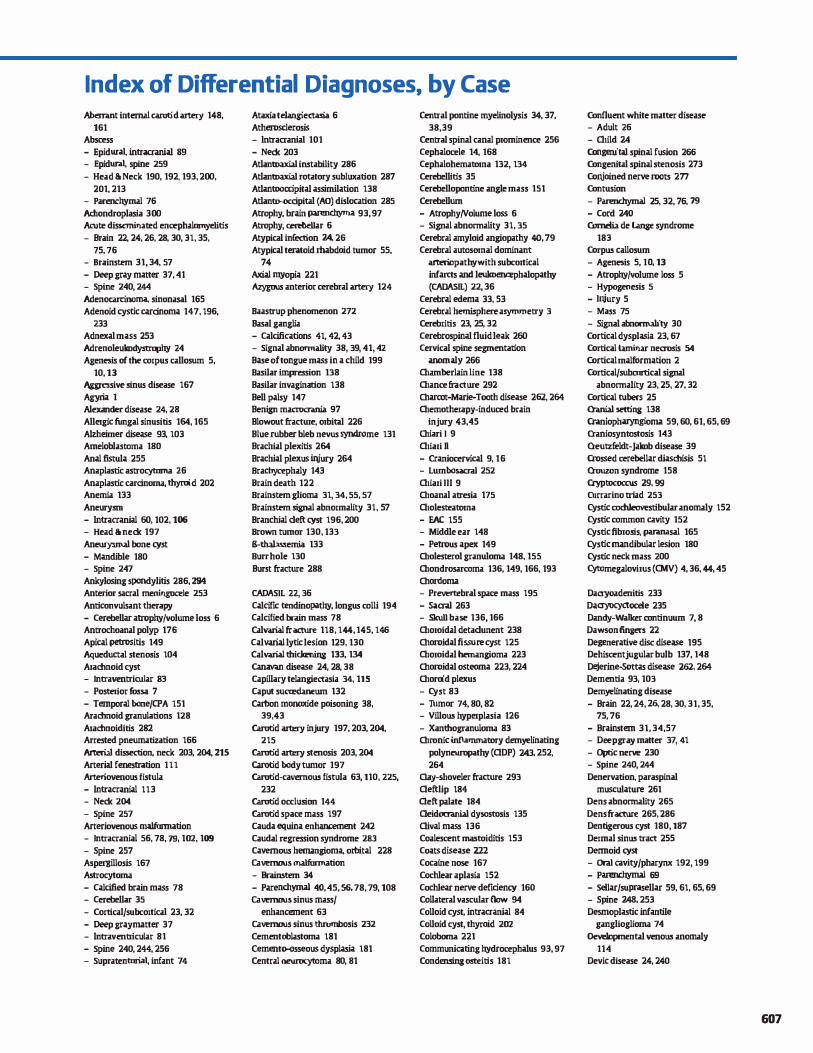
Index of Differential Diagnoses, by Case Aberrant internai carotid artery 148, Ataxia telangiectasia 6 Central pontine rnyelinolysis 34, 37, Omfluent white matter disease
161 Atherosderosis 38,39 - Adult 26
Absœss - lntracranial 101 Central spinal canal prominenœ 256 - Oiild 24 - EpidlU"ai, intracranial 89 - Neck 203 Cephaloœle 14, 168 Congenital spinal fusion 266 - EpidlU"ai, spine 259 AtlantDaxial iru5tability 286 Cephalohernatmna 132, 134 Omgenital spinal stenosis 273
- Head Ili Neck 190, 192, 193, 200, AtlantDaxial rotatory subluxation 287 Cerebellitis 35 Col\Îoined nerve roots 2n
201, 213 Atlantoocdpital assimilation 138 Cerebellopantine angle mass 151 Omtusion - Parenchymal 76 Atlantn-ocdpital (AO) dislocation 285 Cerebellum - Parenchyrnal 25, 32, 76, 79 Ad!ondroplasia 300 Atrophy, brain parenchyma 93, 97 - AtrophyfVolume Joss 6 - Cord 240 Arute disseminated encephalomyelitis Atrophy, œrebellar 6 - Signal abnorrnality 31, 35 Cornelia de Lange syndrome
- Brain 22, 24, 26, 28, 30, 31, 35, Atypical infection 24, 26 Cerebral arnyloid angiopathy 40, 79 183
75, 76 Atypical teratoid rhabdoid tumor 55, Cerebral autosmnal dominant Corpm callosum - Brainstern 31, 34, 57 74 arteriopathy with sulx:ortical - Agenesis 5, 1 o. 13
- Deep gray matter 37, 41 Axial myopia 221 infarcts and leukoencephalopathy - Atrophyfvolume Joss 5
- Spine 240, 244 Azygous anterior œrebral artery 124 (CADASIL) 22, 36 - Hypogenesis 5
Adenocarcinorna, sinonasal 165 Cerebral edema 33, 53 - 11\iury 5
Adenoid cystic cardnoma 147, 196, Baastrup phenornenon 272 Cerebral hernisphere asyrnmetry 3 - Mass 75
233 Basal ganglia Cerebritis 23, 25, 32 - Signal abnormality 30
Adnexal mass 253 - Calcifications 41, 42, 43 Cerebrospinal fluid leak 260 Cortical dysplasia 23, 67
Adrenoleukodystrophy 24 - Signal abnormality 38, 39, 41, 42 Cervical spine segmentation Cortical Iarninar necrosis 54
Agenesis of the corpus callosurn 5, Base of tongue mass in a child 199 anomaly 266 Cortical malformation 2
10, 13 Basilar impression 138 Oiarnberlain line 138 Cortical/subcortical signal Aggressive sinus disease 167 Basilar invagination 138 Oianœ fracture 292 abnorrnality 23, 25, 27, 32
Agyria 1 Bell palsy 147 Cllarmt-Marie-Tooth disease 262, 264 Cortical tubers 25
Alexander disease 24, 28 Benign rnacrocrania 97 Oiernotherapy-induœd brain franial setting 138
Aller-gic fungal sinusitis 164, 165 Blowout fracture, orbital 226 injury 43,45 Ctaniopharyngiorna 59, 60, 61, 65, 69
Alzheimer disease 93, 103 Blue rubber bleb nevus syndrome 131 Clliari 1 9 Ctaniosyntostosis 143
Ameloblastorna 180 Brachial plexitis 264 Oiiari Il <Ieutzfeldt-Jakob disease 39
Anal fistula 255 Brachial plexus Îl\Îury 264 - Cranioœrvical 9, 16 Crossed œrebellar diaschisis 51
Anaplastie astrocytmna 26 Brachyœphaly 143 - Lumbosacral 252 frouzon syndrome 158
Anaplastie cardnorna, thyroid 202 Brain death 122 Oiiari III 9 Cryptococcus 2.9, 99
Anernia 133 Brainstern glioma 31, 34, 55, 57 Clloanal atresia 175 G!rrarino triad 253
Aneurymt Brainstern signal abnormality 31, 57 Oiolesteatmna Cystic codtleovestibular anomaly 152
- lntracranial 60, 102, 106 Branchial deft cyst 196, 200 - EAC 155 Cystic cornrnon cavity 152
- Head Ili neck 197 Brown tumor 130, 133 - Middle ear 148 Cystic fibrosis, paranasal 165
Aneurysmal bene cyst S-thalassernia 133 - Petrous apex 149 Cystic rnandibular lesion 180
- Mandible 180 Burr hole 130 Cllolesterol granulorna 148, 155 Cystic neck mass 200
- Spine 247 Burst fracture 288 Oiondrosarcoma 136, 149, 166, 193 Cytornegalovirus (CMV) 4, 36, 44, 45
Ankylosing spondylitis 286, 294 Cllordorna Anterior sacral rneningocele 253 CADASIL 22, 36 - Prevertebral spaœ mass 195 Dacryoadenitis 233
Anticonvulsant therapy Calcific tendinopathy, !ongus colli 194 - Sacral 263 Daa:vocyctoœle 235
- Cerebellar atrophyfvolume Joss 6 Calcified brain mass 78 - Slrull base 136, 166 Dandy-Wa11œr mntinuum 7, 8
Antrochoanal polyp 176 Calvarial fracture 118, 144, 145, 146 Oioroidal detadunent 238 Dawson fingers 22
Apical petrositis 149 Calvarial lytic lesion 129, 130 Clloroidal fissure cyst 125 Degenerative dise disease 195
Aqueductal stenosis 104 Calvarial thidœning 133, 134 Oioroidal hernangiorna 223 Dehiscentjugular bulb 137, 148
Arachnoid cyst c:anavan disease 24, 28, 38 Clloroidal osteoma 223, 224 Dajerine-SOttas disease 262, 264
- lntraventricular 83 capillary telangiectasia 34, 115 Clloroid plexus Dernentia 93, 103
- Posterior fussa 7 caput sucœdaneurn 132 - Cyst 83 DernJll!linating disease - Temporal bone/CPA 151 Carbon rnonoxide poisoning 38, - Turner 74, 80, 82 - Brain 22, 24, 26, 28, 30, 31, 35,
Arachnoid granulations 128 39,43 - Villom hyperplasia 126 75, 76
Arachnoiditis 282 Carotid artery injury 197, 203, 204, - Xanthogranulorna 83 - Brainstem 31,34,57
Arrested pneumatization 166 215 Cllronic inflamrnatmy demJll!linating - Deepgraymatter 37, 41
Arterial dissection, neck 203, 204, 215 Carotid artery stenosis 203, 204 polyneuropathy (ODP) 243, 252, - Optic nerve 230
Arterial fenestration 111 Carotid body tumor 197 264 - Spine 240, 244
Arteriovenous fistula Carotid-cavemom fistula 63, 110, 225, Clay-filloveler fracture 293 Denervation, paraspinal - lntracranial 113 232 Oeftlip 184 musculature 261
- Neck 204 Carotid occlusion 144 Oeft palate 184 Dens abnorrnality 265
- Spine 257 Carotid spaœ mass 197 Oeidocranial dysostosis 135 Dens fracture 265, 286
Arteriovenous rnalforrnation cauda equina enhanœrnent 242 Oival mass 136 Dentigerous cyst 180, 187
- lntracranial 56, 78, 79, 102, 109 caudal regression syndrome 283 Coalescent rnastoiditis 153 Dermal sinm tract 255
- Spine 257 cavemous hernangioma, orbital 228 Coats disease 222 Derrnoid cyst Aspergillosis 167 ÙY51UJUS rnalforrnation Cocaïne nose 167 - Oral cavity/pharynx 192, 199
Astrocytorna - Brairu5tem 34 Cochlear aplasia 152 - Parenchyrnal 69
- Calcified brain mass 78 - Parem:hymal 40, 45, 56, 78, 79, 108 Cochlear nerve deficiency 160 - Sellar/suprasellar 59, 61, 65, 69
- Cerebellar 35 ÙY51UJUS sinus mass/ Collateral vascular flow 94 - Spine 248, 253
- Cortical/subcortical 23, 32 enhanœment 63 Colloid cyst, intracranial 84 Desrnoplastic infantile - Deepgrayrnatter 37 ùY51UJUs sinus thrombosis 232 Colloid cyst, thyroid 202 gangliogliorna 74
- lntraventricular 81 Cernentoblastorna 181 Coloborna 221 Developrnental venom anomaly - Spine 240, 244, 256 Cernento-osseous dysplasia 181 Cornrnunicating hydroœphalus 93, 97 114
- Supratentorial, infant 74 Central neurocytoma 80, 81 Condensing osteitis 181 Devic disease 24, 240
607
608
Index of Differential Diagnoses, by Case
Diastematomyelia 284
Diffuse axonal injury 30, 40
Diffuse infiltrating pontine glioma
(DIPG) 31, 34, SS, S7
Dise disease, spine
- Degenerative 259, 269
- Recurrent dise, postoperative 249
Disc-osteornyelitis 195, 249, 259, 278 Dise œnninology 269
Dolichocephaly 143
Down syndrome 135, 138, 286
Duplication cyst, rectal 2S3
Durai arteriovenous fistula - lntracranial 113
- Spine 257
Dural-based mass 71
Durai enhancernent 9S
Dylœ-Davidoff-Mason syndrome 3
Dysernbyroplastic neuroepithelial
turnor 23, 67
Dysrnyelinating disease 24, 28, 36,38
Ectopie posterior pituitary 6S
Encephalitis
- Brainstern 34
- Cortical/subcortical 23, 25, 32
- Deepgraymatter 37,39
- Rhombenœphalitis 31, 34
- Viral 37,39
Encephalomalaciil 3, 11
Enephaloœle 14
Encephalotrigeminal angiomatosis 3
Endolyrnphatic sac tumor 159
Enhancernent in surgical bed 77
Enhancing scalp lesion 131
Enlarged extra-axial CSF spaœs 97
Ependyrnal cyst 83
Ependyrnitis 66, 96
Ependyrnoma
- lntraventricular 80. 81, 82
- Parenchymal 78
- Posterior fessa 9, 55
- Spine 240, 244, 256
Epiderrnoid
- Calvarial, cyst 129, 130
- lntracranial 85
- Oral cavity/pharynx 192, 199
- Spine 248
- Temporal bone/CPA 151
Epidural abscess, intracranial 89
Epidural abscess, spinal 2S9, 260
Epidural fluid collection 260
Epidural hematnma, intracranial 90
Epidural hematnma, spine 259, 260
Epidural lipomatnsis 259, 260, 271
Epidural spinal mass 259
Epiglottitis 209
Erb-Duchenne pa1sy 264, 295
Esthesioneuroblastorna 169
Ewing saro:ima 129, 131, 134, 193, 227
Exostosis, EAC 150
Expanded paranasal sinuses 165
Expamile sinonasal bony lesion 170
Extraocular muscle enlarilement 225
External auditorycanal atresia 1S8
External au di tory canal calcification/
ossification 1 so External au di tory canal exnstosis 150
External au di tory canal mass with erosion 155
External auditorycanal osteoma 150
Extramedullary hematopoiesis 71
Extra-axial mass 72
Extrapontine myelinolysis 37, 38, 39
Facet dislocation 291
Fadai nerve enhancernent 147
Fadai nerve hernangioma 147
Fahr disease 41, 42, 43
Fatty infiltration of the filum 258
Fatty marrow signal intensity 251
Fenestration, arterial 111
Fetal alcohol syndrome 12
Fibromatosis ailli 208
Fibromuscular dysplasia 203
Fibrous dysplasia
- Calvarial 130, 133, 134
- Maxillofacial 180, 181
- OrbiW 227
- Sinonasal 165, 170
Filar lipoma 258
Fistula in ano 255
Floor of the mouth mass 192
Follicular carcinoma, thyroid 202
Follicular cyst 180, 187
Foramina of Monro mass 81
Foreign body, orbital 224
Fourth ventricle mass, adult 82
Friedreich ataxia 6
Frontotemporal dementia 93, 103
Fungal sinusitis 164, 165, 167
Fukuyama 1
Gangliogliorna 23, 67
Gardner syndrome 182
Genn cell tumor
- Pineal 58
- Sellar/suprasellar 59, 62, 65
Germinal matrix hemorrhage 44
Genninoma - Pineal 58
- Sellar/suprasellar 59, 62, 65
Giant œll turnor 263
Gigantism 133
Glioblastoma multiformans 30. 31, 66,
75, 76, n, 79, 102
Glioma {see astrocytoma)
Gliomatosis œrebri 26, 68
Gliosis 23
Globe calcification 224
Globe rontnur abnormality 221
Globe mass - Adult 223
- Child 222
Globe trauma 221
Glomusjugulare 137
Glomus tympanicurn 148
Glomus vagale 197
Glottic cardnoma 214
Glutarie adduria type 1 33
Goldenhar syndrome 158, 183
Gradient echo susceptibility 4D. 116
Gradenigo syndrome 149
Granulation tissue, postoperative
spine 249
Granulamatnus disease
- Sinonasal 167
- OrbiW 229
Grave's ophthalmopathy 225 Grise! syndrome 287
Guillain-Barré 100, 243, 262
Hanged man's fracture 289
Harlequin eye deformity 143
Hernangioblastoma
- Brain 56
- Spine 244, 256, 257
Hernangiama - Choroidal 223
- Fadai nerve 147
- MastieatDr spaœ 193
- Oral cavity/pharynx 192, 199
- Orbital 228
- Scalp 131
- Sinonasal 168, 169
- Spine 251, 274
Hernangiopericytorna 71, 72
Hematoma (see hernorrhage) Hernatopoietic marrow 250
Hernifadal atrophy 188
Hernirnegalenœphaly 2, 3
Hernirnyelocele 252
Hernimyelomeningoœle 252
Hemochromatosis 41
Hemoglobinopathy 133
Hemorrhage
- Epidural, intracranial 90
- Epidural, spine 259
- lntraparenchyrnal 40, 56, 69, 79
- lntrawntricular 96
- Ocular/orbital 223 Hepatolenticular degeneration
(see Wilson disease) Hereditary motor-sensary
neuropathy 262, 264 Hemiation, spinal cord 296
Hemiation, brain parenchymal 91
Hemiation, dise (see dise disease) Herpes encephalitis 68
Heterotopias
- Band 1,2
- Nodular 4
Holoprosencephaly s, 10, 98
Human immunodefidency virus
(HIV) 24, 26. 43
Huntingtondisease 41, 105
Hydranencephaly 10
Hydroœphalus 10, 93, 97, 127
Hydromyelia 256
Hyperdense sinus opacification 164
Hyperostosis frontilis inœrna 140
Hyperoxygenation 46
Hyperparathyroidism 41, 43, 45,
130. 133
Hypertensive hemorrhage 40, 56, 69 Hypervascular cerebral mass 102
Hypervascular turnors 102
Hypoglycemia 27
Hypothalamic glioma 59
Hypothalamic hamartoma 59
Hypothyraidism 41, 43, 45, 135
Hypaxic-ischemic enœphalopathy 12,
28, 33, 38, 39, 42, 43
lnferior vena cava occlusion 257
lnfiltrating neck mass 201
lnner ear rnalfonnation 152
lnspissated sinus seaetions 164
lntracranial hypertension 121
lntracranial hypoœnsion 9, 95
lntradural extramedullary cyst 248
lntradural extramedullary spinal
mass 241
lntradural fat 258
lntrarnedullary signal abnormality 240 lntramedullary spinal mass 244
lntraventricular hernorrhage 96
Invasive fungal sinusitis 167
Inverted papilloma 169
Ischernia/infai:rt
- Brainstern 34
- Corpus callosurn 30
- C.Ortical/Subcortical 25, 27, 32, 68
- Deep gray rnatter 37, 38, 39, 42, 43, 99
- Ernboli 70
- Hemorrhagic 79
- Posterior fussa 56
- Stroke imaging 52
- Subacuœ 76, 94
- White matter 26, 28
Jefferson fracture 290
Joubert syndrome 7, 8
Jugular bulb, dehisœnt 137, 148
Jugular bulb pseudolesion 137
Jugular foramen mass 137
jugular venous thrombosis 197
juvenile chronic arthritis 138, 265,
266, 286
Juvenile nasopharyngeal
angiofibroma 169
juvenile pilocytic astrocytoma 55
Kart<!gener syndrome, paranasal 165
Keratocystie odontogenie tumor 180
Kemicterus 38, 42
"Kissing" carotid arteries 205
Kleeblattschadel 143
Klippel-Feil syndrome 138, 266
Klippel-TrenaunayWeber 131
Klurnpke palsy 264, 295
Krabbe disease 28
Labyrinthitis ossificans 157
Laaimal gland enlargement 233
Larunar infarct 99
Langerhans œll histiocytosis - Ca.lvarial 129, 131
- Sellar/supmellar 62
- Spine 245, 247
- Temporal bene 153
Large wstibular aqueduct syndrome 152
Laryngoœle 220
Lateral medullary syndrome 123
Lateral ventricle mass 80 Lefort fracture 226
Leigh syndrome 31, 33, 38, 43
Lemierre syndrome 213
Lens dislocation 236
Leptomeningitis 46, 62, 94, 1 OO, 243
Leptomeningeal cardnornatosis 46, 94,
100,243
Leptomeningeal cyst 129
Leptomeningeal enhanœrnent 94
Leulœmia
- Ca.lvarial 129, 131
- Orbital 225, 229, 230
- Sellar/supmellar 62
- Spine 241, 245, 250
Leulccdystrophy 24, 28, 36, 38
lhemûtte-Dudos disease 88
Limbic encephalitis 68
Lingual thyroid 199
Lipoma - Pericallosal 87
- Sellar/suprasellar 65
- Spine 251, 258
- Temporal bone/CPA 151
Lipomatosis, spinal 251, 259, 260
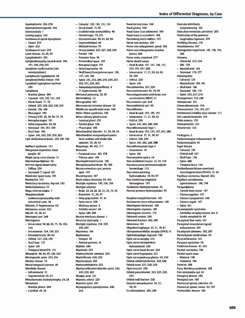
lipomyeloœle 2S4, 2S8
lipomyelomeningoœle 254
lissenœphaly 1
Ludwig angina 192 Lumbosacral spinal dysraphism - aosed 2S4
- Open 252
Lumbosacral tract 2S5 Lyme disease 22, 26, 29
Lymphadenitis 201
I.Ymphadenopathy, head & neck 196,
197' 198, 200, 201
I.Ymphatic malformation (see venalymphatic)
Lymphacytic hypaphysitis 64
I.Ymphaepithelial lesians 198
Lymphaid hyperplasia, Iaaimal
233
Lymphoma - Brachial plexus 264
- Calvarial 129, 130, 131, 134 - Dural-based 71, 72 - Orbital 225, 228, 229, 230, 233
- Parotid 196, 198 - Pharyngeal 190
- Primary CNS 26, 30, 66, 7S, 76 - Retropharyngeal 191
- Sellar/suprasellar 62, 64 - Sinanasal 165, 167, 169
- Skull base 166
- Spine 241, 24S, 2SO, 2S9, 263 Lytic skull/calvarial Jesim 129, 130, 166
Maffucd syndrome 131
Manganese depasitian, basal
ganglia 42 Maple syrup urine disease 31
Marchiafava-Bignami 30 MaITaw signal abrunmality - Diffuse 250
- lncreased Tl signal 2S1 Masticatar spaœ mass 193
Mastaiditis 153
Medullary cardnama, thyraid 202
Medullablastama SS
Mega cisterna magna 7
Megalencephalic leulœaenœphalapathy with subrortical cysts 36
Melanin, Tt hyperintense 69
Melanoma, acular 223
MEU\S 25, 38, 43
Meningeal cyst 248 Meningiama - lntracranial 58, 60, 63, 71, 72, 102,
134 - lntraasseaus 134, 136, 227
- lntraventricular 80, 82 - Orbital 227, 228, 230
- Skull base 137
- Spine 241 - Temporal bane/CPA 151
Meningitis 46, 94, 95, 100, 243 Meningoœle, spine 253, 254
Menkes disease 33
Mesial temporal sderosis 49 Metabalic disease - lnfratentorial 31 - Supratentnrial 28, 33
Metachromatic leukodystraphy 24, 28 Metastases - Brachial plexus 264
- Caldfied 45, 78
- Calvarial 129, 130, 131, 134 - Dural-based 71, 95
- Gradient echo susœptibility 40
- Hemarrhagic 79, 251
- Intraventricular 80, 81, 82, 96
- Leptameningeal 46 - Multiple lesims 70
- Ocular/orbital 223, 227, 228, 229 - Paratid 198
- Pasterior fossa 56
- Prevertebral spaœ 195 - Retropharyngeal 191
- Sellar/suprasellar 64
- Skull base/divus/petraus apex 136,
137, 149, 166
- Spine 241, 242, 244, 24S, 246, 247, 250, 251, 259, 263
- Subependymal/ependymal 4 - Tl hyperintense 69
Methanal paisaning 38, 39
Microœphaly 12
Micragnathia 183
Micravascular ischemic disease 22
Middle ear opadficatian mass 148
Miller-Fisher syndrome 100
Minar salivary gland tumor - Laaimal gland 233
- Paratid gland 196 - Pharyngeal 190
Mitochondrial disarder 31, 33, 38, 43
Mitochondrial enœphalapathywith lactic addosis and stralœ-like episades 25, 38, 43
Moyamoya 46, 102, 117
Mucaœle - Paranasal sinus 16S, 179 - Petraus apex 149
Mucoepidermoid tumor 196
Mumpalysaailaridases 99, 186
Mucaus retentian cyst, petraus apex 149
Mucarmycasis 167
Multi-infard dementia 93, 103
Multiple myelama 130, 136, 156
Multiple sderosis - Brain 22, 24, 26, 30, 31, 35, 75, 76 - Brainstem 31, 34, 57
- Deep y,r� matter 37, 41 - Optic nerve 230
- Marburg variant 4
- Schilder variant 24
- Spine 240, 244
Musd�brain disease 1 Muscular dystraphy 1
Multiple myelama 130, 195,
250, 263
Myœtorna 164
Myelination - Delayed 28
- Normal patterns 21
Myelitis 240 Myelaœle 252
Myelocystoœle, terminal 254 Myelofibrasis 246, 250
Myelamalada 256
Myelomeningocele 252
Myelapraliferative disarder, spine 241,
245, 250, 263
Myapia, axial 221
Myositis, orbital 225 Myositis, spine 261
Myxopapillary ependymorna 240,
244
Index of Differential Diagnoses, by Case
Nasal dermal sinus 168
Nasal gliorna 168
Nasal mass in an adolescent 169
Nasal mass in a newbom 168
Necrotizing otitis exœma tss Nerve roat avulsion 29S Nerve roat enlargement, spinal 262
Nerve roat enlargement, brachial plexus 264
Nerve roats, canjained 277
Nerve sheath tumor - Head & neck 137, 147, 148, 151,
193, 195, 197, 200
- Intracranial 17, 31, 38, 42, 63,
72, 100
- Orbital 228
- Spine 241
Neuroblastama 227, 229
Neuracystiœrcosis 45, 78, 99
Neurodegeneratian with brain iran acrumulatian (NBIA) 41
Neuroenteric cyst 248
Neuroepithelial cyst 99
Neurafibrorna - Head & neck 193, 195, 197
- Intracranial 17, 31, 38, 42 - Orbital 228
- Spine 241, 262, 286, 298 Neurafibrornatnsis iype 1
- Head & neck 193, 195, 197, 201, 286 - lntracranial 17, 31, 38, 42
- Orbital 228, 230
- Spine 241, 262, 286, 298
Neurafibrornatnsis iype 2
- Intracranial 18
- Spine 241 Neuromyelitis aptica 24
Non-accidentai trauma 12, 33, 118
Nananeurysmal perimesenœphalic hemonhage 112
Non-cammunicating hydroœphalus 10, 93, 97
Non-involuting congenital hemangioma 193
Nonlœtotic hyperglycinemia 33 Normal pressure hydroœphalus 93
Ocdpital condyle fractures 14S
Oculornator nerve enhanœment 100
Odontogenic lœratacyst 180
OdantDgenic myxcma 180
Odantagenic sinusitis 172 OdantDid erosian 265
OdantDid fracture 265, 286
Odantama 181 Oligadendragliamas 23, 31, 78, 81
Olivapantaœrebellar atraphy (OPCA) 6
Ophthalmaplegic migraine 1 DO Optic nerve atraphy 231
Optic nerve enlargement/ enhanœment 230
Optic nerve head drusen 224
Optic nerve hypaplasia 231
Optic nerve/pathway glioma 59, 230
Orbital œllulitis/infectian 229, 234
Orbital mass 227, 228, 229
Opticneuritis 230
Orbital pseudotumor 223, 225, 229,
233
Orbital wall fracture 226
Osmotic demyelinatian 34, 37,
38,39
Os odantoideum 265, 286
Ossicular prosthesis, malpasitioning 163
Ossiculum terminale, persistent 265
Ossification of the poste ri or longitudinal ligament 279
Ossifying fibroma 170
Osteoblastama 247
Osteogenesis imperfecta 135, 138, 154,
286
Osteorna - Cl!aroidal 223, 224
- EAC 150
- Maxillofadal 182 - Sinonasal 170, 177
Osteomyelitis - Calvarial 129
- Maxillofadal 180, 181
- Skull base 166 - Sinonasal 166, 170
- Spine 245, 247, 276 Osteopetrasis 133, 246
Osteoparosis 251
Osteoradionecrosis 1 S4
Osteosarcoma 134, 193, 227
Ostiomeatal complex sinus disease 171
Otic capsule lucencies 154 Otitis externa 155
Otomastoiditis 153 Otasderasis 154
Padtygyria 2
Pachymeningeal enhanœment 95
Pachymeningitis 9S Faget disease - Calvarial 130 - Orbital wall 227
- Skull base 138
- Spine 246
- Temporal bene 154
Panthothenate kinase-associated neuradegeneratian (PKAN) 41, 42
Papillary carcinama, thyroid 202
Papillary cystadenoma lyphamatasum 196, 198
Paraganglioma - Carotid body tumor 197
- Glamus jugulare 137
- Glamus tympanicum 148 - Glamus vagale 197
- Spine 241 Paraneaplastic syndrome - Cerebellar atraphyfvalume lass 6 - Iimbic enœphalitis 68
Paraspinal flow vaids 257
Paraspinal musculature signal/ enhanœment 261
Parathyroid adenoma 202, 207
Parenchymal caldficatians 44
Parietal foramina 141
Parinaud syndrome S8 Parldnsondisease 41, 103
Parotid cardnoma 196
Parotid spaœ mass - Bilateral 198
- Unilateral 196 Parotitis 206
Parry-Rmnberg syndrome 188
Pars intermedia cyst 61 Periapical absœss 186
Periapical cyst 180
Perineural spread, infection 63
Perineural spread, tumar 63, 147 Peritansillar abscess 190
609
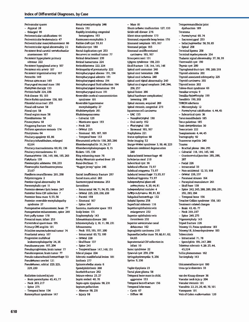
610
Index of Differential Diagnoses, by Case
Perivascular spaœs - Atypical 28
- Enlarged 99
Periventricular calcifications 44
Periventricular leukomalacia 47
Periventricular mass/enhanœment 66
Periventricular signal abnonnality 22
Persistent reta1 carotid-vertebrobasilar anastmnoses 107
Persistent hyperplastic primary
vitreous 222
Persistent hypoglossal artery 107
Persistent Dtic artery 107
Persistent trigeminal artery 107
Petrodtis 149
Petrous apex mass 149
Ph�al mucusal mass 190
Phenytoin therapy 133
Phthisis bulbi 224, 236
Pick disease 93, 103
Pierre Robin syndrome 158, 183
Pilorudal sinus tract 255
Pinea! œll tumor 58
Pinea! cyst 58
Pinea! region mass 58
Pineoblastoma 58
Pineocytmna 58
Pingpong fracture 142
Pirifonn aper.ature stenosis 174
Pituicytorna 64
Pituitary apoplexy 65, 86
Pituitary infundibulum, enlarged
62,64
Pituitary macroadenoma 60, 63, 136
Pituitary microadenorna 61
Plasmacytoma 136, 149, 166, 195, 263
Platybasia 139
Pleomorphic adenoma 196, 233
Pleomorphic xanthoastrocytoma
23,67
Plexiform neuro!ibroma 201, 298
Polymicrogyria 2
Pontine signal abnormality 34 Porenœphalic cyst 11
Posterior element lytic lesion 247
Posterior fossa CSF collection 7
Posterior fossa mass 9, 55, 56
Posterior reversible enœphalopathy syndrome 27
Postoperative enhanœment, br.Un 77 Postoper.ative enhanœment, spine 249
Pott pulfy tumor 178
Presacral mass, infant 253
Prevertebral spaœ mass 195
Primary CNS angiitis 101
Primitive neuroectDdermal tumor 74
Proatlantal artery 107
Progressive multifocal
leulroencephalopathy 24, 26
Pseudoaneurymt 197' 204
Pseudoprogression, brain tumor 77
Pseudoresponse, brain turnor 77 Pseudo-subarachnoid hemorrllage 53
Pseudotumor œrebri 121
Pseudotumor, orbital 223, 225, 229, 233
Radiation-induœd Îl\ÎUl'Y - Brain parenchyma 43, 45, 77
- Nede 203, 217
- Spine 275
- Temporal bone 154
RamseyHunt syndrome 147
Renal osteodystrophy 246
Ranula 192
Rapidly involuting congenital hemangiorna 193
Rasmussen encephalitis 3
Rathke deft cyst 59, 61
Radicularcyst 180
Rectal duplication cyst 253
Recurrent or residual tumor 77
Retinal detachment 239
Retinal hamartoma 224
Retinoblastoma 222, 224
Retinopathy of prematurity 222 Retrop� abscess 191, 194
Retrop� adenitis 191
Retropharyngeal edema 194
Retrop� fluid collection 194
Retropharyngeal hematomas 194
Retrop� mass 191
Revl!rsible cerebral vasoconstriction
syndrome 101
Revl!rsible hypertensive
enœphalopathy 27
Rhabdomyolysis 261
Rhabdmnyosarcoma
- Calv.uial 129
- Masticator space 193
- Nede 201
- Orbital 225
- Sinonasal 165, 167, 169
- Temporal bone 153
Rheurnatoid arthritis 138, 265, 286
Rhombenœphalitis 31, 34, 57
Rhombenœphalosynapsis 8, 15
Ridœts 135, 138
Ring enhandng mass 76
Rodcy Mountain spotted fever 29
Rosai-Dorfman 71
"Rugger jersey" spine 246
Sacral insuffidency fracture 297
Sacral mass, adult 263
Sacrococcygeal teratoma 253
Sarroidosis
- Intracranial 64, 71, 94, 95, 1 OO
- Orbital 225, 229, 230, 233
- Parotid 198
- Sinonasal 167
- Spine 243
Sarroma, masticator space 193
Scalp lesion 131
Scaphoœphaly 143
Scheuermann disease 280
Sdtizenœphaly 2, 10, 11, 98, 231
Schwannoma - Nede 193, 195, 197, 200
- Intracranial 63, 72, 1 OO
- Orbital 228
- Slrull base 137
- Spine 241
- Temporal bone 147, 148, 151
Scierai plaque 224
Sderotic maxillofadal lesion 181
Srolimis 277
Spinoœrebellar ataxia 6
Spondylolisthesis 268
Seatbelt fracture 292
Seizure edema 23, 27
Septic emboli 40, 70
Septo-optic dysplasia 98, 231
Septum pelluddum
- Absence 98, 231
- II\iury 98
- Mass 81
Shunt catheter malfunction 127, 133
Sidde œll disease 276
Silent sinus syndrome 173
Sinonasal, expansile bony lesion 170
Sinonasal neoplasm 165, 167
Sinonasal polyps 165
Sinonasal undifferentiated cardnoma 165, 167
Sinus pericr.mii 131
Sjllgren syndrome 198, 233
Slrull fracture 118, 144, 145, 146
Spinal cord contusion 240
Spinal cord hemiation 296
Spinal cord ischernia 240
Spinal cord signal abnormality 240
Spinal cord signal neoplasm 240, 244,
256, 257
Spinal fusion 266
Spinal hardware complication/ loosening 299
Spinal stenosis, acquired 269
Spinal stenosis, congenital 273
Squamous cell carcinoma
- EAC 155
- Naso�al 190
- Oral cavity 192
- Pharyngeal 190
- Sinonasal 165, 167
Staphylorna 221
Status epilepticus 68
Stoke imaging 52
Sturge-Weber syndrome 3, 19, 46, 223
Subacute combined degeneration
240
Subarachnoid hemorrhage 46
Subdavian steal 210
Subcortical cyst 36
Subdural effusion 73, 97
Subdural empyl!lllil 73, 97
Subdural hemorrhage 73, 95, 97
Subdural hygroma 73, 97
Subependymal giant œll
astrocytoma 4, 20, 44, 81
Subependymal nodules 4
Subependyrnorna 80, 81, 82
Subgaleal hemorrllage 132
Subpial lipoma 258
Superfidal siderosis 116
Superiorophthalmicvein enlargement 232
Superior ophthalmic vein thrombosis 232
Superior semicirrula.r canal
dehiscenœ 162
Supraglottic cardnorna 219
Suprasellar/sellar mass 59, 60, 61, 62,
64,65
Supratentorial CSF collection in
infant 10
Susac syndrome 22
Synovial cyst 259, 270
Syringohydro� 9, 256 Syrinx 9, 256
Taylor dysplasia 23
Tectal plate glioma 58
Temporal bone mass in child, aggressive 153
Temporal bone fracture 156
Temporal lobe mass
- Cystic 67
- Diffuse 68
Temporomandibular joint dysfunction 189
Teratoma - Parenchymal 69, 74
- Sacrocoa:ygeal 253
- Sellar/suprasellar 58, 59, 65
- Spinal 258
Terminal lipoma 258
Terminal m)ll!locystDcele 254
Thalamic signal abnonnality 37, 38, 39
Thomwaldt cyst 190
Thymie cyst 200
Tuyroglossal duct cyst 199, 200, 211
Tuyroid adenoma 202
Tuyroid associaœd orbitopathy 225
Tuyroid cardnorna 202
Thyroid lesion 202
Tolosa-Hunt syndrome 63
Tonsillar ectopia 9
Tonsillar hypertrophy 199
Tonsillitis 190, 212
TORŒI infections
- Microœphaly 12
- Parenchymal calcification 4, 44, 45
- Subcortical cysts 36
Torus mandibularis 185
Torus palatinus 185
Taxie demyelination 30
Tmmcariasis 222
Toxoplasmosis 4, 44, 45 Tractography 92 Transverse myelitis 240
Trauma - Brachial plexus 264, 295
- Calvarial 118, 144, 145, 146
- Craniocervicaljunction 285, 286,
287
- Fadai 226
- Intracranial 33
- Non-aa:idental 12, 33, 118
- Orbital 226, 237
- Paranasal sinuses 164
- Paraspinal musculature 261
- Slrull base 138
- Spine 242, 245, 288, 289, 290, 291,
292, 293, 294
- Tempor.il bene 156
TreacherCollins syndrome 158, 183
Treatrnent-related changes
- Brain 43, 45, n - Nede 203, 217
- Spine 249, 275
Trigonoœphaly 143
Tripod fracture 226
Triromy 13, Patau syndrome 183
Triromy18, Edwardsyndrome 183
Tuberculosis
- Intracranial 71, 78
- Spondylitis 195, 247, 2111
Tuberous sderosis 4, 20, 25, 44,
45,224
Tullio phenomenon 162
Turriœphaly 143
Unicameral bone cyst 180
llrea cycle disorders 33
v.m der Knaap disease 36
Vascular nedi: injury 204
Vascular stenosis 101
Vasculim 22, 25, 26, 40, 70, 101
Vasospasm 101
Vein of Galen malfurmation 120

Venolymphatic malformation - Oral cavity/pharynx 192, 199
- Masticator spaœ 193
- Neck 200, 201, 216
- Orbital 228
- Retropharyngeal 191
- Scalp 131
- Spine 253
Venous infarct
- Deep gray matter 38 Venous malfonnation
(see venolymphatic)
Venous sinus thrombosis 119
Venous varix, orbital 232
Ventricular cyst 83
Ventriculitis 66, 96 Ventriculomegaly 93
Ventriculus terminalis 256
Vennian hypoplasia 8
Verœbral arœry abnonnality 195
verœbral body wedging 242
Verœbral body insuffidency
fracture 242
Verœbra plana 245
Index of Differential Diagnoses, by Case
Vestibular aqueduct syndrome 152
Vestibular schwannoma 151
Villous hyperplasia of the choroid plexus 126
Virchow-Robin spaces 99
Vocal rord paralysis 218
YDll Hippel Lindau SYRdrome 244, 256
Walker-Warburg 1
Wallenberg SYRdrome 123
Wallerian degeneration 50
Warthin tumor 196, 198
Wegner granulomatosis 167,
229
Welcher basal angle 139
Werniclœ enœphalopathy 37,
48
Wilson disease 38, 39, 42 Wonnian bones 135
Zygomaticomaxillary complex fracture 226
611
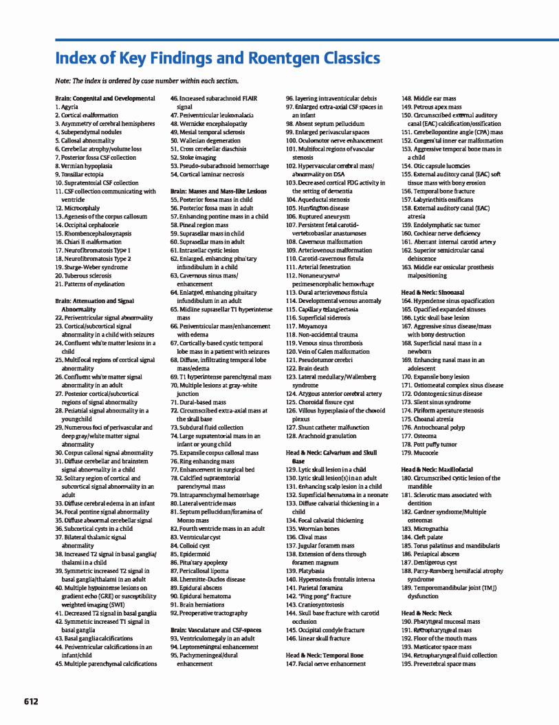
612
Index of Key Findings and Roentgen Classics Note: The index is ordered by case number within eadi section.
Brain: Coogenltal and DeveJopmental
1.Agyria
2. Cortical malfonnation
3. Asymmetty of œœbral hemispheœs
4. Subependymal nodules
5. Callosal abnormality
6. Cerebellar atrophyfvolume loss
7. Posterior fossa CSF collection
8. Vermian hypoplasia
9. Tonsillar ectopia
10. Supratmtorial CSF collection
11. CSF collection communicating with
ventride
12. Microœphaly
13. Agenesis of the corpus callosum
14. Occipital cephaloœle
15. Rhombenœphalosynapsis
16. Clllari Il malfonnation
17. Neurofibromatosis 1YPe 1
18. Neurofibromatosis '!YPe 2
19. Sturge-Weber syndrome
20. Tuberous sderosis
21. Patterns of myelination
Brain: Attenuation and Signal
Abnonnality
22. Periventricular signal abnorrnality
23. Cortical/subcortical signal
abnormality in a child with seizuœs
24. Confluent white matter lesions in a
child
25. Multifocal regions of cortical signal
abnormality
26. Confluent white matter signal
abnormality in an adult
27. Posterior cortical/subcortical
œgions of signal abnormality
28. Periatrial signal abnormality in a
youngchild
29. Numerous foci of perivascular and
deep gray/wlùte matter signal
abnormality
30. Corpus callosal signal abnormality
31. Diffuse œœbellar and brainstem
signal abnorrnality in a child
32. Solitary region of cortical and
subcortical signal abnorrnality in an
adult
33. Diffuse œœbral edema in an infant
34. Focal pontine signal abnormality
35. Diffuse abnorrnal œrebellar signal
36. Subcortical cysts in a child
37. Bilaœral thalarnic signal
abnormality
38. lncreased 12 signal in basal ganglia/
thalami in a dùld
39. Symmetric increased 12 signal in
basal ganglia/thalami in an adult
40. Multiple hypointense lesions on
gradient echo (GRE) or susœptibility
weighted imaging (SWI)
41. Deaeased T2 signal in basal ganglia
42. Symmetric increased Tl signal in
basal ganglia
43. Basal ganglia calcifications
44. Periventricular calcifications in an
infant/child
45. Multiple parenchymal calcifications
46. Increased subarachnoid FLAIR
signal
47. PeriW!lltricular leukornalacia
48. Wernidœ enœphalopat:h;y
49. Mesial temporal sderosis
50. Wallerian degeneration
51. Cross œrebellar diasdùsis
52. Stoke imaging
53. Pseudo-subarachnoid hemorrhage
54. Cortical laminar necrosis
Brain: Masses and Mass-lilœ Leslons
55. Posterior fossa mass in child
56. Posterior fessa mass in adult
57. Enhancing pontine mass in a child
58. Pinea! region mass
59. Suprasellar mass in child
60. Suprasellar mass in adult
61. Intrasellar cystic lesion
62. Enlarged, enhancing pituiUry infundibulum in a child
63. ù'W!rnDus sinus mass/
enhancement
64. Enlarged, enhancing pituiUry
infundibulum in an adult
65. Midline suprasellar Tl hyperintmse
mass
66. PeriW!lltricular mass/enhanœrnent
withedema
67. Cortically-based cystic temporal
lobe mass in a patient with seizures
68. Diffuse, infiltrating temporal lobe
mass/edema
69. Tl hyperintense parench;ymal mass
70. Multiple lesions at gray-white
junction
71. Dural-based mass
72. Circumscribed extra-axial mass at
the slrull base
73. Subdural fluid collection
74. Large supratmtorial mass in an
infant or :;cung child
75. Expansile corpus callosal mass
76. Ring enhancing mass
77. Enhancement in surgical bed 78. Caldfied supratentorial
parench;ymal mass
79. lntrapaœnchymal hemorrhage
80. Lateral ventride mass
81. Septum pelluddwn/forarnina of
Monro mass
82. Fourth W!lltride mass in an adult
83. Ventricular cyst
84. Colloid cyst
85. Epidermoid
86. Pituitary apoplexy
87. Pericallosal lipoma
88. Lhennitte-Dudos disease
89. Epidural absœss
90. Epidural hematoma
91. Brain herniations
92. Preoperative tractography
Brain: Vilsculature and CSF-spaces 93. Ventriculomegaly in an adult
94. Leptomeningeal enhanœment
95. Pachymeningeal/dural
enhancement
96. Laymng intraventricular debris
97. Enlarged extra-;uciai CSF spaœs in
an infant
98. Absent septum pelluddum
99. Enlarged perivascular spaces
1 OO. Oculornotor nerve enhancernent
101. Multifocal œgions ofvascular
stenosis
102. Hypervascular œrebral mass/
abnorrnality on DSA 103. Decreased cortical FDG activity in
the setting of dernentia
104. Aqueducul steno5is
105. Huntington disease
106. Ruptured aneurysm
107. Persistent fetal carotid-
vertebrobasilar anastomoses
108. Ca'W!rnDus malformation
109. Arteriovenous malformation
11 O. Carotid-cavernous fistula
111. Arterial fenestration
112. Nonaneurysmal
perimesenœphalic hemorrhage
113. Durai arteriovenous fistula
114. Developmental venous anomaly
115. Capillary tdangiectasia
116. Superficial siderosis
117. Moyarnoya
118. Non-accidentai trauma
119. Venous sinus thrombosis
120. Vein of Galen malformation
121. Pseudotumor œrebri
122. Brain death
123. Lateral medullary/Wallenberg
syndrome
124. Azygous anterior cerebral artery
125. Choroidal fissure cyst
126. Villous hyperplasia of the choroid
plexus
127. Shunt catheter malfunction
128. Arachnoid granulation
Head a Neck: Calvarium and Skull
Base 129. Lytic slrull lesion in a child
130. cytic slrull lesion(s) in an adult
131. Enharu:ing scalp lesion in a child
132. Superficial hematoma in a neonate
133. Diffuse calvarial thickening in a
child
134. Focal calvarial thickening
135. Wormian bones
136. Clivai mass
137.Jugular foramen mass
138. Extension of dens through
for.unen magnum
139. Platybasia
140. Hyperostosis fruntalis interna
141. Parietal foramina
142. "Pingpong" fracture
143. Craniosyntostosis
144. Slrull base fracture with carotid
ocdusion
145. Occipital condyle fracture
146. linear slrull fracture
Head a Neck: Temporal Bane
147. Facial nerve enhancernent
148. Middle ear mass
149. Petrous apex mass
150. Clrcumscribed exœrnal auditory
canal (EAC) calcification/ossification
151. Cerebellopontine angle (CPA) mass
152. Omgmital inner ear malformation
153. Aggressive temporal bone mass in
a child
154. Otic capsule luœncies
155. External auditory canal (EAC) soft tissue mass with bony erosion
156. Temporal bone fracture
157. Labyrinthitis ossificans
158. F.xternal auditory canal (EAC)
atresia
159. Endolymphatic sac tumor
160. Cochlear nerve deficiency
161. Aberrant internai carotid artery
162. Superior sernidrcular canal
dehisœnce
163. Middle ear ossicular prosthesis
malpositioning
Head a Neck: Slnonasal
164. Hyperdense sinus opacification
165. Opadfied expanded 5inuses
166. Lytic slrull base lesion
167. Aggressive sinus disease/mass
with bony destruction
168. Superficial nasal mass in a
newbom
169. Enhancing nasal mass in an
adolescent
170. Expansile bony lesion
171. Ostiomeatal complex sinus disease
172. Odontogeruc sinus disease
173. Silent sinus syndrome
174. Piriform aperature stenosis
175. Choanal atresia
176. Antrochoanal polyp
177. Osteoma
178. Pott pulfy tumor
179. Mucoœle
�da Neck: Maxlllofaclal
180. Clrcumscribed cystic lesion of the
mandible
181. Sclerotic mass associated with
dentition
182. Gardner syndrome/Multiple
osteomas
183. Micrognathia
184. Oeft palate
185. Torus palatinus and mandibularis
186. Periapical abscess
187. Dentigerous cyst
188. Parry-Romberg hernifacial atrophy
syndrome
189. Temporomandibular joint (TMJ)
dysfunction
Head li Neck: Neck
190. Pharyngeal mucosal mass
191. Retropharyngeal mass
192. Floor of the mouth mass
193. Masticator spaœ mass
194. Retropharyngeal fluid collection
195. Prevertebral spaœ mass
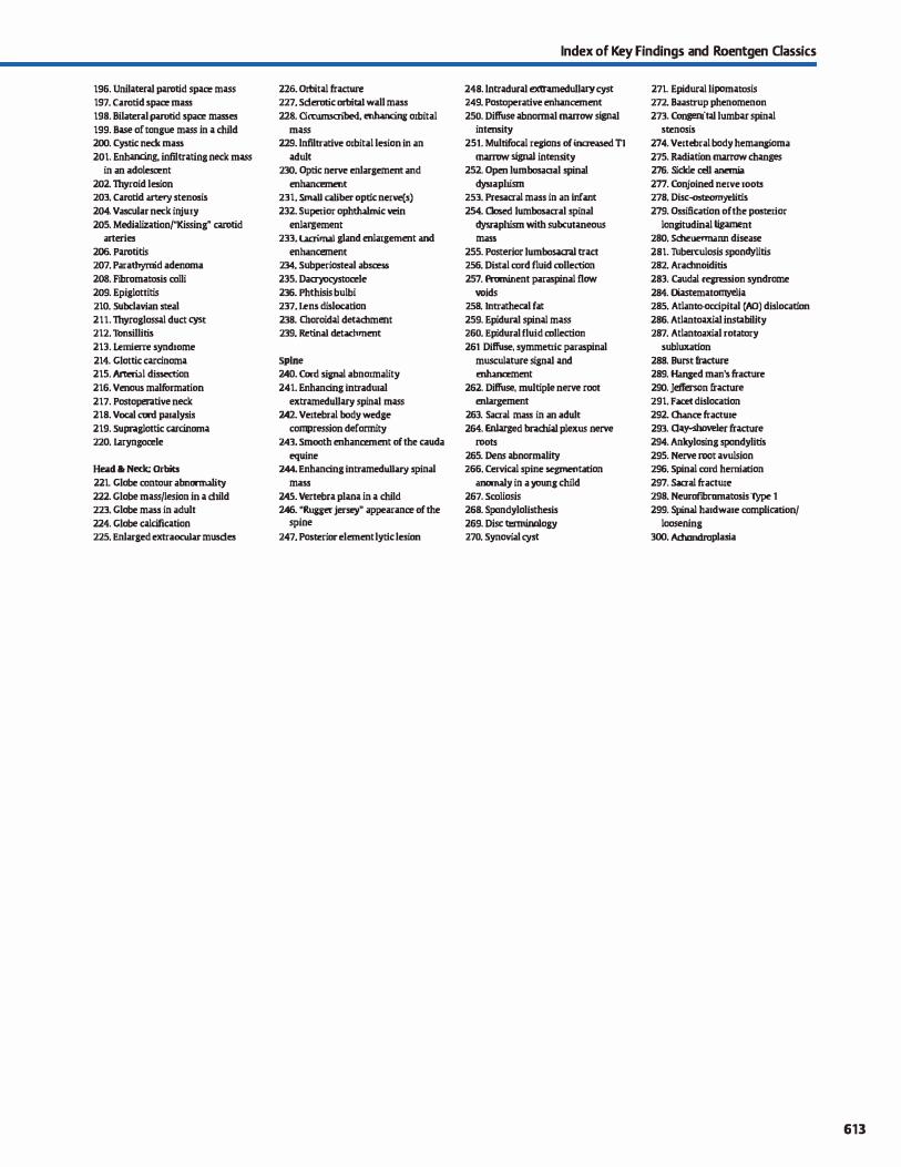
196. Unilateral parotid spaœ mass 197. ùrotid spaœ mass
198. Bilateral parotid spaœ masses
199. Base of tongue mass in a child 200. Cystic neck mass
201. Enhancing, infiltrating neck mass in an adolescent
202. Thyroid lesion 203, ùrotid artery stenosis
204. Vascular neck injury
205. Medializationj"Kissing" carotid arteries
206. Parotitis 207. Parathyroid adenoma
208. Fibrumatosis colli
209. Epiglottitis 210. Sulx:lavian steal
211. Thyroglossal duct cyst 212. llmsillitis
213. Lernierre syndrome
214. Glottic carcinoma 215. Arterial dissection
216. Venous malfurmation
217. Postoperative neck 218. Vocal conl paralysis
219. Supraglottic carcinoma 220. Laryngoœle
Head • Nede; Orbits
221. Globe contour abnormality
222. Globe mass/lesion in a dilld 223. Globe mass in adult
224. Globe calcification 225. Enlarged extraocular musdes
226. Orbital fractw'e 227. Sderotic mbit:al wall mass
228. Cirrumsc:ribed, enhandng orbital
mass 229. lnfiltrative orbital lesion in an
adult 230. Optic nerve enlargement and
en han cernent 231. Small caliber optic nerve(s)
232. Superior ophthalrnic vein
en largement 233. Lacrimal gland enlargernent and
en han cernent 234. Subperiosteal absœss
235. Dacryocystoœle
236. Phthisis bulbi 237. Lens dislocation
238. Cltoroidal detachment 239. Retinal detachment
Splne 240. Cord signal abnormality
241. Enhancing intradural
extramedullary spinal mass 242. Vertebral body wedge
compression deforrnity 243. Smooth enhanœment of the cauda
equine 244. Enhandng intramedullary spinal
mass
245. Vertebra plana in a child 246. "Rugger jersey" appearanœ of the
spine
247. Posterior elernent lytic lesion
Index of Key Findings and Roentgen Classics
248. lntradural extramedullary cyst 249. Postoperative enhancement
250. Diffuse abnormal marrow signal intensity
251. Multifocal regions ofincreased Tl
marrow signal intensity 252. Open lumbosacral spinal
dysraplùsm 253, Presacral mass in an infant
254. Oosed lumbosacral spinal
dysraplùsm with subcutaneous mass
255. Posterior lumbosacral tract 256, Distal cord fluid collection
257. Prominent paraspinal flow
voids 258. lntrathecal fat
259. Epidural spinal mass 260, Epidural fluid mllection
261 Diffuse, symmetric paraspinal
musculature signal and enhanœment
262. Diffuse, multiple nerve root enlargement
263. Sacral mass in an adult
264. Enlarged brachial plexus nerve roots
265. Dens abnormality 266, Cervical spine segmentation
anomaly in a young child
267. Scoliosis 268. Spondylolisthesis
269. Dise terminology 270. Synovial cyst
271. Epidural lipomatosis 272. Baastrup phenomenon
273. Congmital lumbar spinal stenosis
274. Vertebral body hemangioma
275. Radiation marrow changes 276. Sickle cell anernia 277. Conjoined nerve roots 278, Disc-osteomyelitis
279. Ossification of the posterior
longitudinal ligament 280. Scheuermann disease
281. Tuberculosis spondylitis 282. Arachnoiditis
283. Caudal regression syndrome
284. Diastematom;yelia 285. Atlanto-occipital (AO) dislocation
286. Atlantoaxial instability 287, Atlantoaxial rotatory
subluxation
288. Burst fractw'e 289. Hanged man's fracture
290. Jefferson fracture 291, Faœt dislocation
292. Chance fracture
293. Oay-shoveler fracture 294. Ankylosing spondylitis
295. Nerve root avulsion 296. Spinal cord hemiation
297. Sacral fracture 298. Neurofibrumatosis 'JYpe 1
299. Spinal hardware complication/
loosening 300, Admndroplasia
613


![[MS-RDPERP-Diff]: Remote Desktop Protocol - Microsoft](https://static.fdokumen.com/doc/165x107/633794f840a96001d40100b2/ms-rdperp-diff-remote-desktop-protocol-microsoft.jpg)