Thermal analysis on parchments I: DSC and TGA combined approach for heat damage assessment
6
Thermochimica Acta 447 (2006) 30–35 Thermal analysis on parchments I: DSC and TGA combined approach for heat damage assessment D. Fessas a,∗ , M. Signorelli a , A. Schiraldi a , C.J. Kennedy b , T.J. Wess b , B. Hassel c , K. Nielsen d a Dipartimento di Scienze e Tecnologie Alimentari e Microbiologiche, Universit` a di Milano, via Celoria, 2, 201333 Milano, Italy b Biophysics Division, School of Optometry and Vision Science, Cardiff University ,Cardiff CF10 3NB WALES, UK c The Royal Danish Academy of Fine Arts, School of Conservation, Esplanaden 34, DK-1263 Copenhagen K, Denmark d Department of Chemistry Kemitorvet Building 207, Technical University of Denmark, DK-2800 Kgs Lyngby, Denmark Received 24 January 2006; received in revised form 11 April 2006; accepted 20 April 2006 Available online 29 April 2006 Abstract Ancient, new and artificially aged parchments were investigated with both differential scanning calorimetry (DSC) and thermogravimetry (TGA). Criteria to define a quantitative ranking of the damage experienced by the bulk collagen of historical parchments were assessed. A damage-related correlation was found between the collagen denaturation temperature and the moisture content of the parchment. Qualitative rules for the evaluation of the damage at the nano-and mesoscopic level were achieved on the basis of peculiarities of the shape and width of the DSC signals and confirmed by small angle X-ray scattering patterns. © 2006 Elsevier B.V. All rights reserved. Keywords: Parchments; DSC; TGA; Damage assessment; SAXS 1. Introduction Due to the historical value of ancient parchments, damage assessment is very important in conservation practice. This arti- cle was developed in the frame of the “IDAP” (improved damage assessment of parchment) European project that aims at devel- oping new methods for the assessment of damage in histori- cal parchments at the macroscopic, microscopic, mesoscopic, nanoscopic and molecular level [1]. Ancient parchments are handcrafts of animal skin. The main component of the skin connective tissue matrix is collagen, a family of related proteins [2,3]. Different collagen types are present in skin, the most abundant ones being types I and III, which belong to the subfamily of fibril-forming collagens [4]. The structure formed has a characteristic coiled coil triple helical conformation that is stabilized by weak non covalent inter- chain bonds, mainly hydrogen bonds, and the result is a thin, long, rod-like molecule with a length of 300 nm and a diameter ∗ Corresponding author. Tel.: +39 0250316637; fax: +39 0250316632. E-mail address: [email protected] (D. Fessas). of about 1.1 nm. On a higher organization level, the collagen molecules are aligned side by side in a quarter staggered array with parallel main axis, forming fibrils. These supra-molecular structures at the fibril level are stabilized by further cross-links [5] and produce characteristic banding patterns viewed by elec- tron microscopy upon staining [6]. To date electron microscopy, X-ray diffraction and to a lesser extent atomic force microscopy have been the major tools used to investigate the structural hier- archies of collagen based tissues. Thermal analysis techniques (mainly calorimetric) are par- ticularly suitable to investigate the stability of biological macro- molecules (collagen in the case of parchment) as it allows detection of every state of modification within the system (e.g., protein denaturation and aggregation, gel-sol transitions, crys- tallization, fusion, glass transition, etc.) when it undergoes some temperature change. A number of works have so far been devoted to the study of collagen and its aging [7], but only few of them deal with parchments [8,9]. Furthermore, most of the literature data about thermal analysis of parchments concern DSC investigations on aqueous suspensions of minutely cut material. The information drawn is mainly related to the triple-helix-to-random-coil 0040-6031/$ – see front matter © 2006 Elsevier B.V. All rights reserved. doi:10.1016/j.tca.2006.04.007
Transcript of Thermal analysis on parchments I: DSC and TGA combined approach for heat damage assessment

Thermochimica Acta 447 (2006) 30–35
Thermal analysis on parchments I: DSC and TGA combinedapproach for heat damage assessment
D. Fessas a,∗, M. Signorelli a, A. Schiraldi a, C.J. Kennedy b, T.J. Wess b,B. Hassel c, K. Nielsen d
a Dipartimento di Scienze e Tecnologie Alimentari e Microbiologiche, Universita di Milano, via Celoria, 2, 201333 Milano, Italyb Biophysics Division, School of Optometry and Vision Science, Cardiff University ,Cardiff CF10 3NB WALES, UK
c The Royal Danish Academy of Fine Arts, School of Conservation, Esplanaden 34, DK-1263 Copenhagen K, Denmarkd Department of Chemistry Kemitorvet Building 207, Technical University of Denmark, DK-2800 Kgs Lyngby, Denmark
Received 24 January 2006; received in revised form 11 April 2006; accepted 20 April 2006Available online 29 April 2006
Abstract
Ancient, new and artificially aged parchments were investigated with both differential scanning calorimetry (DSC) and thermogravimetry (TGA).Ccob©
K
1
acaocn
cfpwTccl
0d
riteria to define a quantitative ranking of the damage experienced by the bulk collagen of historical parchments were assessed. A damage-relatedorrelation was found between the collagen denaturation temperature and the moisture content of the parchment. Qualitative rules for the evaluationf the damage at the nano-and mesoscopic level were achieved on the basis of peculiarities of the shape and width of the DSC signals and confirmedy small angle X-ray scattering patterns.
2006 Elsevier B.V. All rights reserved.
eywords: Parchments; DSC; TGA; Damage assessment; SAXS
. Introduction
Due to the historical value of ancient parchments, damagessessment is very important in conservation practice. This arti-le was developed in the frame of the “IDAP” (improved damagessessment of parchment) European project that aims at devel-ping new methods for the assessment of damage in histori-al parchments at the macroscopic, microscopic, mesoscopic,anoscopic and molecular level [1].
Ancient parchments are handcrafts of animal skin. The mainomponent of the skin connective tissue matrix is collagen, aamily of related proteins [2,3]. Different collagen types areresent in skin, the most abundant ones being types I and III,hich belong to the subfamily of fibril-forming collagens [4].he structure formed has a characteristic coiled coil triple helicalonformation that is stabilized by weak non covalent inter-hain bonds, mainly hydrogen bonds, and the result is a thin,ong, rod-like molecule with a length of 300 nm and a diameter
∗ Corresponding author. Tel.: +39 0250316637; fax: +39 0250316632.
of about 1.1 nm. On a higher organization level, the collagenmolecules are aligned side by side in a quarter staggered arraywith parallel main axis, forming fibrils. These supra-molecularstructures at the fibril level are stabilized by further cross-links[5] and produce characteristic banding patterns viewed by elec-tron microscopy upon staining [6]. To date electron microscopy,X-ray diffraction and to a lesser extent atomic force microscopyhave been the major tools used to investigate the structural hier-archies of collagen based tissues.
Thermal analysis techniques (mainly calorimetric) are par-ticularly suitable to investigate the stability of biological macro-molecules (collagen in the case of parchment) as it allowsdetection of every state of modification within the system (e.g.,protein denaturation and aggregation, gel-sol transitions, crys-tallization, fusion, glass transition, etc.) when it undergoes sometemperature change.
A number of works have so far been devoted to the studyof collagen and its aging [7], but only few of them deal withparchments [8,9]. Furthermore, most of the literature data aboutthermal analysis of parchments concern DSC investigations onaqueous suspensions of minutely cut material. The information
E-mail address: [email protected] (D. Fessas). drawn is mainly related to the triple-helix-to-random-coil
040-6031/$ – see front matter © 2006 Elsevier B.V. All rights reserved.oi:10.1016/j.tca.2006.04.007

D. Fessas et al. / Thermochimica Acta 447 (2006) 30–35 31
conformational transition of collagen, which is, however,strongly affected by the environmental conditions, namelyconcentration, pH and ionic strength of the medium, as well asby the content of conformationally restricted amino-acids, suchas proline and hydroxyproline [10].
Calorimetry on parchment samples in excess of water there-fore allows the assessment of the damage at molecular levelexperienced by the collagen especially when the DSC data (tran-sition enthalpy, transition temperature, shape of the signal) canbe correlated with specific changes of other chemical and physi-cal properties of the material (e.g., depletion of some amino acid,cross linking degree, etc.) determined with different techniques,such as HPLC and amino acid analysis [8]
Changes of the state of the parchment produced by a damag-ing process probably alter every level of the structural hierarchy.The changes at the mesoscopic and macroscopic level are mod-ified or lost when the sample is dispersed in water. Changesat these levels are however worth assessing since the macro-scopic integrity of parchment is dependent on the integrity atall the other structural levels and a more complete picture ofdamage can lead to better preservation techniques. The study ofparchment samples in the intact state (i.e. without any previoustreatment related to the experimental technique used), that canmodify their structure, is therefore desirable.
To this aim, ancient, new and artificially aged parchmentswere investigated, without further preparation, with both dif-f(tstp
idlp(t
2
2
mSamti1
2
a
The typical sample was a small (about 5 mg mass) piece of anew or artificially aged parchment sheet. Although small, sucha sample had both the grain and the flesh side of the parchment.Heating runs in the temperature range 20–150 ◦C at 1 and/or5 ◦C min−1 scanning rate were performed with at least two repli-cas for each run. The raw data were analysed with the softwareIFESTOS [11] in order to obtain the excess (with respect to thenative state) heat capacity of the sample, Cpexc (T), J K−1 g−1
(per gram of sample).
2.3. TGA investigations
TGA was used to assess the sample moisture and waterrelease during the temperature scan. The instrument used wasa TG-DSC 111 (SETARAM, France). Measurements were per-formed on new and artificially aged parchments at 2 ◦C min−1
heating rate in the 20–200 ◦C temperature range. A typical DTGtrace (i.e. the time derivative of the mass loss), of a parchmentsample shows a broad peak with an underlying area that cor-responds to the overall water content of the sample (the heatflow signal is used to check that the mass loss is related only towater release; decomposition phenomena start at higher temper-atures). The position of the DTG peak maximum and the shapeof the peak are related to the state of water within the sam-ple [12] and therefore are a reliable parameter when comparingdlotdw
2
sTodt1aww
3
hdewatc[
erential scanning calorimetry (DSC) and thermogravimetryTGA). Collected data encompassed a very wide range of mois-ure content up to conditions not far from those of the aqueoususpensions considered in previous studies. A bridge betweenhe results obtained from “dry” and “aqueous” parchment sam-les could therefore be envisaged.
The scope of the study was the assessment of criteria onntact parchment that allow definition of a quantitative damageegree in order to rank the damage experienced by the bulk col-agen of historical parchments. The nano/meso structure of thearchment was characterised with small angle X-ray scatteringSAXS), in order to correlate changes of structural features athis level with alterations in the thermochemical signals.
. Materials and methods
.1. Materials
Ancient (16th century), new and artificially aged calf parch-ent were supplied by the Royal Danish Academy of Fine Arts,chool of Conservation. The artificial accelerated aging wasimed at mimicking potential heat damages that ancient parch-ents can experience in their long shelf life. For the present work
he heat treatment of freshly prepared parchments was producedn drying oven for 48 h at 60, 80, 100, 120, 140, 150, 160, 165 and70 ◦C. Details of these procedures are reported elsewhere [8].
.2. DSC investigations
The instrument used was a DSC 6 (Perkin–Elmer, USA) oper-ting with sealed cells (an empty cell was used as the reference).
ifferent samples. To simplify this evaluation, the TGA data col-ected in the present work were normalized with respect to theverall moisture of the sample to 100 mg of water. Accordinglyhe DTG traces were expressed as milligrams of lost water peregree K (with reference to the scanning rate used). Each runas repeated at least twice.
.4. SAXS investigations
For small angle X-ray scattering (SAXS) measurements, theamples were loaded into the sample chamber of the NanoS-AR (Bruker AXS, Karlsruhe) X-ray facility at the Universityf Cardiff. The data collection procedure was the same as thatescribed in detail by Wess et al. [13] Scattering profiles wereaken over 6 h exposures using a sample-to-detector distance of.25 m. Collected data were corrected for camera distortions,background image was subtracted, and images were analyzedith an in-house software. The two-dimensional detector outputas converted into one-dimensional profile.
. Results
Fig. 1 reports the DSC traces of samples from two intactistorical parchments and represents a typical example of theata observed with this technique. Curve a shows only one sharpndothermic peak at a rather high temperature (above 100 ◦C),hereas curve b shows three phenomena, a high-T peak withshoulder in the rising branch, and a complex signal, at low
emperature (around 55 ◦C), with endothermic and exothermicomponents. Similar data have been reported in the literature14].
32 D. Fessas et al. / Thermochimica Acta 447 (2006) 30–35
Fig. 1. DSC traces of samples from two “as is” historical parchments (scanningrate 5 ◦C/min).
Fig. 2 shows DSC traces obtained from samples of new parch-ments (either of the same or of different animal origin), whichapparently are characterized by different position and intensityof the main endotherm.
To define the information related to the collagen thermal sta-bility, new parchment samples with different moisture contentswere compared (Fig. 3). Curves a–c concern excess, 64% andnative moisture content, respectively. The first curve is typicalof collagen aqueous suspensions, the endotherm concealing anumber of molecular details relevant to the unfolding and denat-uration of the collagen molecule. Detailed investigation on theseprocesses is beyond the scope of the present work. Here it issufficient to say that the position of the overall peak is shiftedtoward higher temperatures on decreasing the moisture content.The shape of the peak becomes without shoulders as in a coop-erative process. The enthalpy remains substantially unmodified(43 ± 2 J g−1) although a slight increase (about 2–3 J g−1) isfound in parchment samples with their original moisture con-tent (curve c in Fig. 3) These stabilizing effects can come fromthe close environment of the collagen, namely the local con-
FC
Fig. 3. DSC traces of new parchments samples with different moisture con-tents. Curves a–c concern excess, 64% and native moisture content, respectively(scanning rate 1 ◦C/min).
centration of macromolecules and the molecular architecture atmesoscopic level (see below).
In order to single out these effects from those related to pos-sible damages experienced by the material (e.g., for historicalparchments), the behaviour of new undamaged parchments wasassessed. To this end, TGA and DSC investigations were per-formed on parchment samples with different moisture contents.High moisture levels were obtained by adding distilled waterto the sample in the DSC pan, while lower moisture levels cor-responded to the endogenous water content of the parchments(which was assessed by TGA).
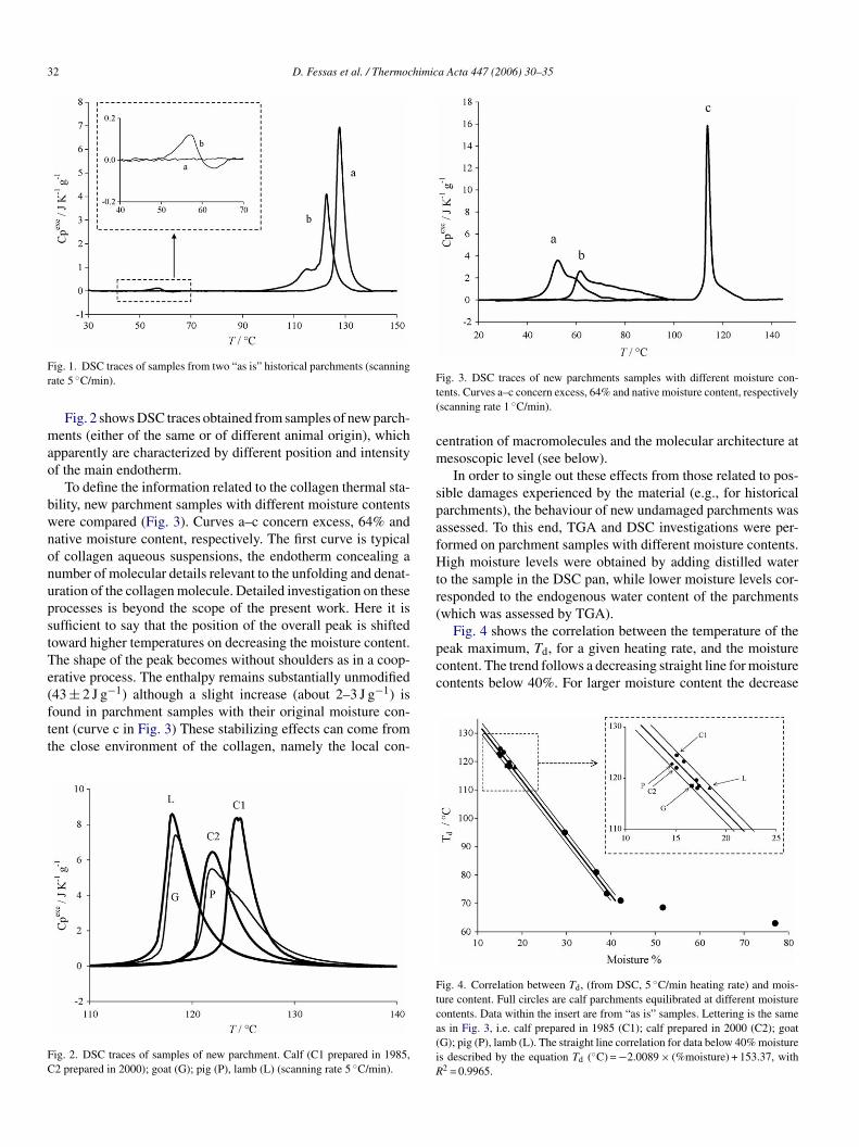
Fig. 4 shows the correlation between the temperature of thepeak maximum, Td, for a given heating rate, and the moisturecontent. The trend follows a decreasing straight line for moisturecontents below 40%. For larger moisture content the decrease
Ftca(iR
ig. 2. DSC traces of samples of new parchment. Calf (C1 prepared in 1985,2 prepared in 2000); goat (G); pig (P), lamb (L) (scanning rate 5 ◦C/min).
ig. 4. Correlation between Td, (from DSC, 5 ◦C/min heating rate) and mois-ure content. Full circles are calf parchments equilibrated at different moistureontents. Data within the insert are from “as is” samples. Lettering is the sames in Fig. 3, i.e. calf prepared in 1985 (C1); calf prepared in 2000 (C2); goatG); pig (P), lamb (L). The straight line correlation for data below 40% moistures described by the equation Td (◦C) = −2.0089 × (%moisture) + 153.37, with2 = 0.9965.

D. Fessas et al. / Thermochimica Acta 447 (2006) 30–35 33
Fig. 5. DSC traces from new parchments that had experienced heat damage ofvarious extent. The heavy line corresponds to the untreated reference parch-ment. From right to left, increasing treatment temperature. The insert showsthe increasing intensity of the low T signal that starts to appear for treatmenttemperatures above 120 ◦C. The traces reported in the insert correspond (frombottom to top) to treatments at 120, 140, 150, 160, 165 ◦C.
of Td becomes much weaker. The slope of the correlation linedepends on the DSC scanning rate. In view of a future applicationto a large number of historical parchments, we chose 5 ◦C/minas a compromise between resolution and measurement time.
Fig. 5 shows DSC thermograms obtained from new parch-ments that had experienced heat damage of various extent (seeSection 2). On increasing the heat damage, the main endothermicsignal moves toward lower temperature and its shape broadens.Up to a heat treatment temperature of 100 ◦C an almost con-stant value of the enthalpies (41 ± 2 J g−1, gram of dry matter)was observed. For higher temperature treatments a decreasingtrend was observed (the lowest enthalpy value, 12 ± 2 J g−1,was observed for the sample treated at the highest tempera-ture, 165 ◦C) which means that, beyond the 100 ◦C threshold,the artificially heat-treated samples contain smaller amounts ofresidual native collagen. Severely damaged samples showed anextra signal around 70 ◦C (see insert) the intensity of whichincreases with the damage extent.
The shift of the DSC peak maximum was correlated with themoisture content of the samples as assessed by TGA. Fig. 6shows the relevant DTG traces. The close similarity amongthese traces suggests that different heat damage did not causedifferences in the mechanism of dehydration and that water dis-tribution was homogeneous through the sample (Fig. 7.).
The Td versus moisture plot showed substantial deviationsfrom the straight-line trend observed for the undamaged materi-aTstTdToi
Fig. 6. DTG traces from the heat treated parchments. The moisture content ofthese samples is assessed after several months storage after the heat treatment.DSC analysis of the samples was performed at the same time.
Fig. 7. Td vs. moisture plot. Substantial deviations from the straight-line trend(see Fig. 4) were observed for the heat-treated parchments (lettering indicatesthe treatment temperature in ◦C). �T is the difference between the observed Td
and that expected according to the straight-line correlation.
Fig. 8. �T vs. the heat treatment temperature. The trend may be used as adamage index.
ls (see Figs. 4 and 7). The difference, �T, between the observedd and that expected according to the straight-line correlation ishown in Fig. 8 and may be used as a damage index. However, forreatment temperatures below 100 ◦C, �T became insignificant.his fact is in line with the above discussion on the enthalpiesata. Similar threshold effects are reported in the literature [15].his threshold temperature is just below the onset temperaturef the collagen denaturation observed by DSC in the referencentact parchment (see Fig. 5).

34 D. Fessas et al. / Thermochimica Acta 447 (2006) 30–35
Data on alteration in the collagen axial lattice came fromSAXS experiments performed on heat-treated samples whichshowed modulations to the characteristic diffraction pattern ofdry collagen [16,17]. The main features observed were the colla-gen meridional series, which give an indication of the periodicityand crystallinity within collagen fibril axial order. The axialspacing of dry collagen results in a 64 nm lattice with the exactvalue dependent on heating. Up to eleven orders of diffrac-tion were observed with the sixth and ninth orders displayingthe highest intensity. When associated in fibrils, the collagenmolecules (approximately 300 nm in length) display a molecu-lar alignment based on a relative stagger (D-spacing) of 65.5 nmin skin.
Measurements on the heat treated samples, in selected regionswhere some collagen is still bunched in fibrils, showed evi-dence of a reduced D-spacing after heat treatment, namely, 61.1,60.3, 59.96, 60, 59.96, 57.78, 57.58, 57.11 and 55.79 nm for:-untreated and 60, 80, 100, 120, 150, 160, 165 and 170 ◦C heattreated samples, respectively. The most obvious modifications tothe axial structure occurred at temperatures higher than 120 ◦C.The breadth of the diffraction peaks also gives important infor-mation about the extent of crystallinity or the variability of thelattice parameters. The paucity of the data observed here doesnot allow a conclusive decision to be made as to the physicalbasis for broadening. However variability of the collagen axialperiod is rarely observed and it is more likely that a signifi-cbzaoirac
4
tdiaoc
aaeec
itcrt
The data reported in Fig. 4 show a decreasing straight linecorrelation between the temperature of the peak maximum, Td,and the moisture content (for moisture contents below 40%) inthe case of new intact parchments. The slope of the correlationline depends on the DSC scanning rate since kinetic effects arepresent during the collagen denaturation in “as is” parchments(the transitions are irreversible) which makes the position of thesignal dependent on the heating rate of the DSC run.
The data (Td versus moisture) relevant to the thermogramsreported in Fig. 2 are aligned along the straight-line region, irre-spective of the animal origin of the parchments (see insert inFig. 4). This finding suggests that such a correlation may be usedas a rule to identify undamaged materials and attribute observeddeviations to extra damage experienced by the parchment.
The application of this method (Figs. 5, 7 and 8) to samplesof new parchments, that had experienced heat damage of vari-ous extent, allows definition of an onset temperature thresholdand quantitative evaluation (although phenomenological) of thedamage produced by the heat treatment (for a given treatmenttime).
This evidence may support the tentative interpretation thatthe denaturation of collagen observed with the DSC in the dam-aged parchment can take place within a changing environment: alower Td would reflect a decreased stability of the residual native(not denatured during the previous artificial damage) molecules,while the broad shape of the peak and/or the presence of shoul-dt
mt(oNass[
fadeoarFdd
5
babc
ant reduction in the lattice coherence contributes to Bragg peakroadening. In the sample here, fitting of the peak as a Loren-ian function and applying the Sherrer equation showed that thexial coherence of the scattering unit decreased as a functionf temperature. In the control sample, the extent of coherences greater than the sensitivity of the technique, however heatingegimes reduced the axial coherence to that of 350 nm at D60nd 250 nm at D170, the latter corresponding to about four unitells.
. Discussion
The data reported in the Figs. 1 and 2 show that, in spite ofhe common overall aspect of the DSC traces, some substantialifferences relevant to the temperature of the peak maximum andts enthalpy can be often found when parchment is of differentnimal origin, or from different lots of the same animal source,r different regions of a given skin are considered, even in thease of freshly produced materials.
This apparent discrepancy clearly indicates that the DSCpproach cannot be used blindly, but requires basic criteria thatllow a quantitative interpretation of the real phenomena and thelimination of, or accounting for parasite effects. For example,nthalpy is an extensive quantity, i.e. it depends on the actualontent of collagen in the skin samples considered.
Since the scope of the work is the definition of a damagendex based on DSC data collected from intact samples for his-orical parchments which may have largely different collagenontents (with no reliable “standard” material), it seemed moreeasonable to focus the attention principally on the denaturationemperature, which is an intensive parameter.
ers (Fig. 5) would correspond to an increased dispersion inerms of stability of the still intact fibres.
As for the signal at lower temperature, which increases inore severely damaged parchments, one has to take into account
hat it is indeed the convolution of endo-and exo-thermic effectssee insert in Fig. 5). The same kind of signal was alreadybserved [9] for the ancient parchments from the Libreriaazionale (Turin, Italy) which partially escaped the disaster of
n inferno that completely destroyed many valuable tomes. Thisignal was supposed to reflect modifications of the mesoscopictructure of the parchment, as confirmed by TEM investigations9].
As already mentioned, denaturation enthalpy, determinedrom DSC investigations of “as is” samples, is not an appropri-te heat damage index for historical parchments. Nonethelessenaturation enthalpy changes can be considered in differentxperimental conditions, e.g. microcalorimetry investigationsn parchment samples in excess of water aimed at achievingn estimation of the damage. However, also in this case, welleliable criteria must be defined to use the experimental data.or this reason a separate work is in preparation and has beenevoted to discuss these aspects on the basis of microcalorimetryata combined with those of amino acid analysis.
. Conclusions
Combined DSC and TGA data allow assessment of the overallulk damage experienced by parchments. The criteria to man-ge “as is” parchment samples have been defined. A correlationetween the denaturation temperature, Td, with the moistureontent allows a quantitative ranking of the damage experienced.

D. Fessas et al. / Thermochimica Acta 447 (2006) 30–35 35
Qualitative evaluation of the changes at the mesoscopic level canalso be achieved with SAXS investigations.
Acknowledgement
This work was carried out within the framework of theIDAP (Improved Damage Assessment of Parchment) EU ProjectEVK4-2001-00061.
References
[1] R. Larsen, D.V. Poulsen, F. Juchauld, H. Jerosch, M. Odlyha, J. deGroot, Q. Wang, C. Theodorakopoulos, T. Wess, J. Hiller, C. Kennedy,G. Della Gatta, E. Badea, A. Masic, S. Boghosian, D. Fessas. Damageassessment of parchment: complexity and relations at different structurallevels, in preprints ICOM-CC 14th Triennial Meeting, September 12-162005. The Hague, The Netherlands, pp 199-208.
[2] D.J. Prockop, K.I. Kivirikko, Annu. Rev. Biochem. 64 (1995) 403–434.[3] D. Parry, Advances in Protein chemistry: Fibrous Proteins: Coiled-Coils,
Collagen and Elastomers, vol. 70, Academic Press, 2005.[4] K.A. Holbrook, L.T. Smith, in: P.M. Royce, B. Steinmann (Eds.), Con-
nective Tissue and its Heritable Disorders. Molecular, Genetic andMedical Aspects, Wiley, New York, 1993, p. 51.
[5] D.R. Eyre, M.A. Paz, P.M. Gallop, Annu. Rev. Biochem. 53 (1984)717–748.
[6] J.A. Chapman, D.J.S. Hulmes, in: A. Ruggeri, P.M. Motta (Eds.),Ultrastructure of The Connective Tissue Matrix, Boston, Nijhoff, 1984,p. 205.
[7] F. Flandin, C. Buffevant, D. Herbage, Biochim. Biophys. Acta 791(1984) 205–211.
[8] B. Hassel. Examination of Heat Damaged Parchment’, Master Thesis.School of Conservation The Royal Danisch Academy of Fine Arts,Copenhagen, 2001.
[9] D. Fessas, A. Schiraldi, R. Tenni, L. Vitellaro Zuccarello, A. Bairati, A.Facchini, Thermochim. Acta 348 (2000) 129–137.
[10] P.L. Privalov, Adv. Protein Chem. 35 (1982) 1–104.[11] D. Fessas, A. Schiraldi, J. Therm. Anal. Calorim. 61 (2000) 411–
423.[12] D. Fessas, A. Schiraldi, Food Chem. 72 (2001) 237–244.[13] T.J. Wess, M. Drakopoulos, A. Snigirev, J. Wouters, O. Paris, P.
Fratzl, M. Collins, J. Hiller, K. Nielsen, Archaeomerty 43 (2001) 117–129.
[14] G. Della Gatta, E. Badea, R. Ceccarelli, T. Usacheva, A. Masic and S.Coluccia. Assessment of damage in old parchments by DSC and SEM.J. Therm. Anal. Calorim. 82 (2005) 637–649.
[15] R. Ronald, Ancient Skins, Parchments and Leathers, Seminar Press, NewYork, 1972.
[16] A.J. Hodge, J.A. Petruska, Recent studies with the electron micro-scope on ordered aggregates of the tropocollagen molecule, in: G.N.Ramachandran (Ed.), Aspects of Protein Structure, Academic Press, NewYork, 1963, pp. 289–300.
[17] T.J. Wess, J.P. Orgel, Thermochim. Acta. 365 (2000) 119–128.