The T-cell receptor is not hardwired to engage MHC ligands
8
The T-cell receptor is not hardwired to engage MHC ligands Stephen J. Holland a,b,1 , Istvan Bartok a , Meriem Attaf a , Raphael Genolet c , Immanuel F. Luescher c , Eleni Kotsiou a,d , Ashkenaz Richard a , Edward Wang a , Matthew White a , David J. Coe a , Jian-Guo Chai a , Cristina Ferreira a , and Julian Dyson a,1 a Molecular Immunology Section, Division of Immunology and Inflammation, Imperial College London, Hammersmith Hospital, London W12 0NN, United Kingdom; b Department of Developmental Immunology, Max Planck Institute of Immunology and Epigenetics, 79108 Freiburg, Germany; c Molecular Immunology Group, Ludwig Institute for Cancer Research, Lausanne Branch, 1066 Epalinges, Switzerland; and d Centre of Haemato-Oncology, Barts Cancer Institute, Queen Mary University of London, London EC1M 6BQ, United Kingdom Edited* by N. Avrion Mitchison, University College London Medical School, London, United Kingdom, and approved September 17, 2012 (received for review July 2, 2012) The bias of αβ T cells for MHC ligands has been proposed to be intrinsic to the T-cell receptor (TCR). Equally, the CD4 and CD8 coreceptors contribute to ligand restriction by colocalizing Lck with the TCR when MHC ligands are engaged. To determine the importance of intrinsic ligand bias, the germ-line TCR complemen- tarity determining regions were extensively diversified in vivo. We show that engagement with MHC ligands during thymocyte selec- tion and peripheral T-cell activation imposes remarkably little con- straint over TCR structure. Such versatility is more consistent with an opportunist, rather than a predetermined, mode of interface for- mation. This hypothesis was experimentally confirmed by expressing a hybrid TCR containing TCR-γ chain germ-line complementarity determining regions, which engaged efficiently with MHC ligands. MHC restriction | TCR T cells expressing an αβ T-cell receptor (TCR) are MHC- restricted, recognizing self- and foreign peptide epitopes pre- sented by MHC class I and II molecules during thymic development and peripheral activation, respectively. Two mechanisms are proposed to underlie this ligand bias. First, the CD8 and CD4 coreceptors have dual specificity for extracellular MHC and the intracellular proximal kinase Lck. Consequently, when MHC class I or II ligands are engaged, Lck is colocalized with the TCR/CD3 complex initiating signal transduction. The importance of this mechanism in disadvantaging non-MHC ligands is highlighted by the recovery of T-cell selection, where MHC and the coreceptors are both absent in comparison to the absence of MHC alone (1). In this setting, non-MHC ligands drive thymic positive selection and are recognized by peripheral T cells (2). The ability of αβ TCRs to recognize non-MHC ligands does not rule out an intrinsic bias of the TCR for MHC ligands. Indeed, evidence for such a hard- wired bias is suggested by pairwise interactions between TCR-β germ-line complementarity determining regions 1 and 2 (CDR1/2) and the MHC α-helices observed in several structures (3–7). Re- cently, such recurrent interactions have been shown to be de- pendent on the partner TCR-α chain, which can impose distinct modes of TCR-β engagement, suggesting they may not drive MHC specificity (8). Although the relatively limited set of TCR/MHC- peptide structures reveals a semiconserved docking geometry, the angle of TCR engagement varies by more than 60° and the gen- erally central docking position can shift toward the peptide amino- or carboxy-terminus (9). Likewise, conserved features of the MHC α-helices, including exposure of the polypeptide backbone and surface depressions, have been suggested to provide energetically favorable sites for CDR engagement (5). However, a crucial role for specific MHC residues in TCR docking has not emerged (10). The role of germ-line TCR structure in the bias to MHC ligands has thus been perplexing, especially given the structural variability of both components. To investigate this, we have applied a unique mutagenesis approach based on redirecting V(D)J re- combination, allowing extensive in vivo remodeling of the germ-line CDR regions. We find thymic T-cell selection and peripheral T-cell activation are not dependent on germ-line CDR structure, sug- gesting the TCR can adopt a highly versatile, antibody-like strategy for engaging MHC-peptide ligands. To test this hypothesis directly, the TCR-β germ-line CDR1 and CDR2 regions were replaced with TCR-γ chain CDRs. MHC class I and II are not natural ligands for γδ T cells, and their germ-line CDR regions have not coevolved with MHC molecules. This hybrid γβ TCR chain paired with the endogenous TCR-α repertoire and facilitated efficient recognition of MHC and thymic T-cell selection. These data demonstrate TCR-intrinsic specificity for MHC ligands is not an essential determinant of MHC restriction; rather, analogous to antibodies, the αβ TCR can use generic chemical features of the germ-line loops to engage ligands. Results TCR Germ-line MHC Contact Regions Are Structurally Diverse. Hard- wired specificity for MHC class I and II might constrain germ- line TCR CDR composition in comparison to Igs, which use their germ-line CDRs to engage an immense variety of ligands. To examine this, human and mouse Ig heavy/light and TCR- α/β germ-line CDR1 and CDR2 sequences were aligned and amino acid frequencies were determined for each position. For each CDR, the most common length was analyzed (Fig. S1 A and B). The highest sequence diversity is present in TCR-β CDR2 and αCDR1 rather than Ig germ-line regions (Fig. S1C). Further, CDR positions exhibiting strong amino acid preferences in the human and mouse are mostly apparent within Ig CDRs. TCR- intrinsic MHC reactivity would require most combinations of these structurally varied α- and β-germline regions to maintain MHC specificity. This analysis does not support a simple model for TCR-intrinsic MHC specificity in which the germ-line CDRs of TCR are more structurally constrained than their Ig equivalents. Positive Selection Does Not Constrain TCR-β Germ-line Structure. To determine whether cryptic MHC recognition codes are nonetheless present in the TCR germ-line CDRs, they were extensively mu- tated. The Vα8.3 and Vβ11 chains of the H2-K k /TENSGKDI– specific C6 TCR were used as the supporting frameworks because their variable (V) segments are selected efficiently (11, 12). Author contributions: S.J.H., I.B., and J.D. designed research; S.J.H., I.B., M.A., R.G., E.K., A.R., E.W., M.W., D.J.C., J.-G.C., and C.F. performed research; R.G. and I.F.L. contributed new re- agents/analytic tools; S.J.H., I.B., and J.D. analyzed data; and S.J.H. and J.D. wrote the paper. The authors declare no conflict of interest. *This Direct Submission article had a prearranged editor. 1 To whom correspondence may be addressed. E-mail: [email protected] or [email protected]. See Author Summary on page 18259 (volume 109, number 45). This article contains supporting information online at www.pnas.org/lookup/suppl/doi:10. 1073/pnas.1210882109/-/DCSupplemental. www.pnas.org/cgi/doi/10.1073/pnas.1210882109 PNAS | Published online October 17, 2012 | E3111–E3118 IMMUNOLOGY PNAS PLUS
Transcript of The T-cell receptor is not hardwired to engage MHC ligands
The T-cell receptor is not hardwired to engageMHC ligandsStephen J. Hollanda,b,1, Istvan Bartoka, Meriem Attafa, Raphael Genoletc, Immanuel F. Luescherc, Eleni Kotsioua,d,Ashkenaz Richarda, Edward Wanga, Matthew Whitea, David J. Coea, Jian-Guo Chaia, Cristina Ferreiraa,and Julian Dysona,1
aMolecular Immunology Section, Division of Immunology and Inflammation, Imperial College London, Hammersmith Hospital, London W12 0NN, UnitedKingdom; bDepartment of Developmental Immunology, Max Planck Institute of Immunology and Epigenetics, 79108 Freiburg, Germany; cMolecularImmunology Group, Ludwig Institute for Cancer Research, Lausanne Branch, 1066 Epalinges, Switzerland; and dCentre of Haemato-Oncology, Barts CancerInstitute, Queen Mary University of London, London EC1M 6BQ, United Kingdom
Edited* by N. Avrion Mitchison, University College London Medical School, London, United Kingdom, and approved September 17, 2012 (received for reviewJuly 2, 2012)
The bias of αβ T cells for MHC ligands has been proposed to beintrinsic to the T-cell receptor (TCR). Equally, the CD4 and CD8coreceptors contribute to ligand restriction by colocalizing Lckwith the TCR when MHC ligands are engaged. To determine theimportance of intrinsic ligand bias, the germ-line TCR complemen-tarity determining regions were extensively diversified in vivo. Weshow that engagement with MHC ligands during thymocyte selec-tion and peripheral T-cell activation imposes remarkably little con-straint over TCR structure. Such versatility is more consistent withan opportunist, rather than a predetermined, mode of interface for-mation. This hypothesis was experimentally confirmed by expressinga hybrid TCR containing TCR-γ chain germ-line complementaritydetermining regions, which engaged efficiently with MHC ligands.
MHC restriction | TCR
Tcells expressing an αβ T-cell receptor (TCR) are MHC-restricted, recognizing self- and foreign peptide epitopes pre-
sented by MHC class I and II molecules during thymic developmentand peripheral activation, respectively. Two mechanisms areproposed to underlie this ligand bias. First, the CD8 and CD4coreceptors have dual specificity for extracellular MHC and theintracellular proximal kinase Lck. Consequently, when MHC classI or II ligands are engaged, Lck is colocalized with the TCR/CD3complex initiating signal transduction. The importance of thismechanism in disadvantaging non-MHC ligands is highlighted bythe recovery of T-cell selection, where MHC and the coreceptorsare both absent in comparison to the absence of MHC alone (1).In this setting, non-MHC ligands drive thymic positive selectionand are recognized by peripheral T cells (2). The ability of αβ TCRsto recognize non-MHC ligands does not rule out an intrinsic biasof the TCR for MHC ligands. Indeed, evidence for such a hard-wired bias is suggested by pairwise interactions between TCR-βgerm-line complementarity determining regions 1 and 2 (CDR1/2)and the MHC α-helices observed in several structures (3–7). Re-cently, such recurrent interactions have been shown to be de-pendent on the partner TCR-α chain, which can impose distinctmodes of TCR-β engagement, suggesting they may not drive MHCspecificity (8). Although the relatively limited set of TCR/MHC-peptide structures reveals a semiconserved docking geometry, theangle of TCR engagement varies by more than 60° and the gen-erally central docking position can shift toward the peptide amino-or carboxy-terminus (9). Likewise, conserved features of the MHCα-helices, including exposure of the polypeptide backbone andsurface depressions, have been suggested to provide energeticallyfavorable sites for CDR engagement (5). However, a crucial rolefor specific MHC residues in TCR docking has not emerged (10).The role of germ-line TCR structure in the bias to MHC ligandshas thus been perplexing, especially given the structural variabilityof both components. To investigate this, we have applieda unique mutagenesis approach based on redirecting V(D)J re-combination, allowing extensive in vivo remodeling of the germ-line
CDR regions. We find thymic T-cell selection and peripheral T-cellactivation are not dependent on germ-line CDR structure, sug-gesting the TCR can adopt a highly versatile, antibody-like strategyfor engaging MHC-peptide ligands. To test this hypothesis directly,the TCR-β germ-line CDR1 and CDR2 regions were replaced withTCR-γ chain CDRs. MHC class I and II are not natural ligandsfor γδ T cells, and their germ-line CDR regions have notcoevolved with MHC molecules. This hybrid γβ TCR chain pairedwith the endogenous TCR-α repertoire and facilitated efficientrecognition of MHC and thymic T-cell selection. These datademonstrate TCR-intrinsic specificity for MHC ligands is not anessential determinant of MHC restriction; rather, analogous toantibodies, the αβ TCR can use generic chemical features of thegerm-line loops to engage ligands.
ResultsTCR Germ-line MHC Contact Regions Are Structurally Diverse. Hard-wired specificity for MHC class I and II might constrain germ-line TCR CDR composition in comparison to Igs, which usetheir germ-line CDRs to engage an immense variety of ligands.To examine this, human and mouse Ig heavy/light and TCR-α/β germ-line CDR1 and CDR2 sequences were aligned andamino acid frequencies were determined for each position. Foreach CDR, the most common length was analyzed (Fig. S1 A andB). The highest sequence diversity is present in TCR-β CDR2 andαCDR1 rather than Ig germ-line regions (Fig. S1C). Further,CDR positions exhibiting strong amino acid preferences in thehuman and mouse are mostly apparent within Ig CDRs. TCR-intrinsic MHC reactivity would require most combinations ofthese structurally varied α- and β-germline regions to maintainMHC specificity. This analysis does not support a simple modelfor TCR-intrinsic MHC specificity in which the germ-line CDRs ofTCR are more structurally constrained than their Ig equivalents.
Positive Selection Does Not Constrain TCR-β Germ-line Structure. Todetermine whether cryptic MHC recognition codes are nonethelesspresent in the TCR germ-line CDRs, they were extensively mu-tated. The Vα8.3 and Vβ11 chains of the H2-Kk/TENSGKDI–specific C6 TCR were used as the supporting frameworksbecause their variable (V) segments are selected efficiently (11, 12).
Author contributions: S.J.H., I.B., and J.D. designed research; S.J.H., I.B., M.A., R.G., E.K., A.R.,E.W., M.W., D.J.C., J.-G.C., and C.F. performed research; R.G. and I.F.L. contributed new re-agents/analytic tools; S.J.H., I.B., and J.D. analyzed data; and S.J.H. and J.D. wrote the paper.
The authors declare no conflict of interest.
*This Direct Submission article had a prearranged editor.1To whom correspondence may be addressed. E-mail: [email protected] [email protected].
See Author Summary on page 18259 (volume 109, number 45).
This article contains supporting information online at www.pnas.org/lookup/suppl/doi:10.1073/pnas.1210882109/-/DCSupplemental.
www.pnas.org/cgi/doi/10.1073/pnas.1210882109 PNAS | Published online October 17, 2012 | E3111–E3118
IMMUNOLO
GY
PNASPL
US
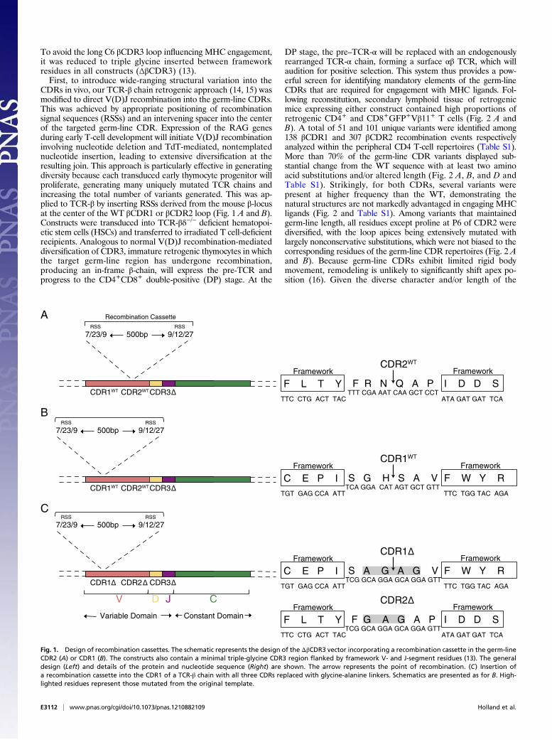
To avoid the long C6 βCDR3 loop influencing MHC engagement,it was reduced to triple glycine inserted between frameworkresidues in all constructs (ΔβCDR3) (13).First, to introduce wide-ranging structural variation into the
CDRs in vivo, our TCR-β chain retrogenic approach (14, 15) wasmodified to direct V(D)J recombination into the germ-line CDRs.This was achieved by appropriate positioning of recombinationsignal sequences (RSSs) and an intervening spacer into the centerof the targeted germ-line CDR. Expression of the RAG genesduring early T-cell development will initiate V(D)J recombinationinvolving nucleotide deletion and TdT-mediated, nontemplatednucleotide insertion, leading to extensive diversification at theresulting join. This approach is particularly effective in generatingdiversity because each transduced early thymocyte progenitor willproliferate, generating many uniquely mutated TCR chains andincreasing the total number of variants generated. This was ap-plied to TCR-β by inserting RSSs derived from the mouse β-locusat the center of the WT βCDR1 or βCDR2 loop (Fig. 1 A and B).Constructs were transduced into TCR-βδ−/− deficient hematopoi-etic stem cells (HSCs) and transferred to irradiated T cell-deficientrecipients. Analogous to normal V(D)J recombination-mediateddiversification of CDR3, immature retrogenic thymocytes in whichthe target germ-line region has undergone recombination,producing an in-frame β-chain, will express the pre-TCR andprogress to the CD4+CD8+ double-positive (DP) stage. At the
DP stage, the pre–TCR-α will be replaced with an endogenouslyrearranged TCR-α chain, forming a surface αβ TCR, which willaudition for positive selection. This system thus provides a pow-erful screen for identifying mandatory elements of the germ-lineCDRs that are required for engagement with MHC ligands. Fol-lowing reconstitution, secondary lymphoid tissue of retrogenicmice expressing either construct contained high proportions ofretrogenic CD4+ and CD8+GFP+Vβ11+ T cells (Fig. 2 A andB). A total of 51 and 101 unique variants were identified among138 βCDR1 and 307 βCDR2 recombination events respectivelyanalyzed within the peripheral CD4 T-cell repertoires (Table S1).More than 70% of the germ-line CDR variants displayed sub-stantial change from the WT sequence with at least two aminoacid substitutions and/or altered length (Fig. 2 A, B, and D andTable S1). Strikingly, for both CDRs, several variants werepresent at higher frequency than the WT, demonstrating thenatural structures are not markedly advantaged in engaging MHCligands (Fig. 2 and Table S1). Among variants that maintainedgerm-line length, all residues except proline at P6 of CDR2 werediversified, with the loop apices being extensively mutated withlargely nonconservative substitutions, which were not biased to thecorresponding residues of the germ-line CDR repertoires (Fig. 2 Aand B). Because germ-line CDRs exhibit limited rigid bodymovement, remodeling is unlikely to significantly shift apex po-sition (16). Given the diverse character and/or length of the
V D J
Constant Domain Variable Domain
C
7/23/9 9/12/27500bp
Recombination Cassette SSRSSR
CDR2WT CDR1WT CDR3F L T Y I D D S
TTC CTG ACT TAC ATA GAT GAT TCA
CDR2WT
F R N Q A P Framework Framework
TTT CGA AAT CAA GCT CCT
7/23/9 9/12/27500bpSSRSSR
CDR2WT CDR1WT CDR3
7/23/9 9/12/27500bpSSRSSR
CDR2 CDR1 CDR3
C E P I F W Y R TGT GAG CCA ATT TTC TGG TAC AGA
CDR1WT
S G H S A V Framework Framework
TCA GGA CAT AGT GCT GTT
C E P I F W Y R TGT GAG CCA ATT TTC TGG TAC AGA
CDR1
S A G A G V Framework Framework
TCG GCA GGA GCA GGA GTT
F L T Y I D D S TTC CTG ACT TAC ATA GAT GAT TCA
CDR2
F G A G A P Framework Framework
TCG GCA GGA GCA GGA GTT
A
B
C
Δ
Δ
Δ
Δ
ΔΔΔ
Fig. 1. Design of recombination cassettes. The schematic represents the design of the ΔβCDR3 vector incorporating a recombination cassette in the germ-lineCDR2 (A) or CDR1 (B). The constructs also contain a minimal triple-glycine CDR3 region flanked by framework V- and J-segment residues (13). The generaldesign (Left) and details of the protein and nucleotide sequence (Right) are shown. The arrow represents the point of recombination. (C) Insertion ofa recombination cassette into the CDR1 of a TCR-β chain with all three CDRs replaced with glycine-alanine linkers. Schematics are presented as for B. High-lighted residues represent those mutated from the original template.
E3112 | www.pnas.org/cgi/doi/10.1073/pnas.1210882109 Holland et al.
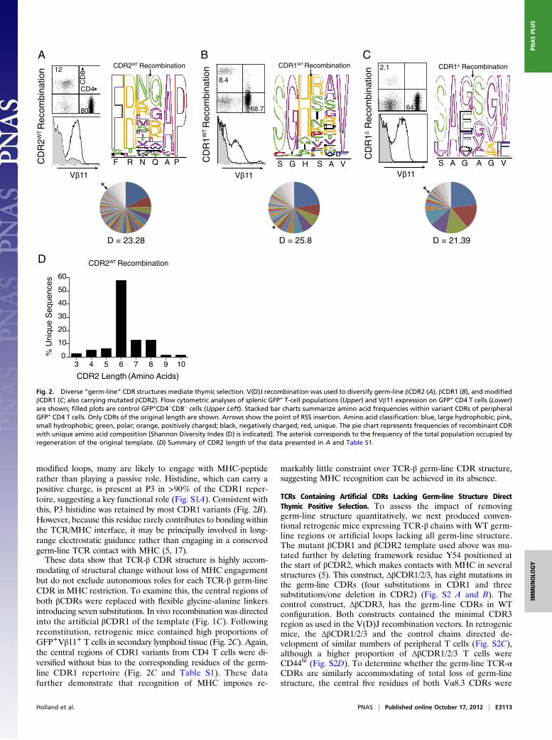
modified loops, many are likely to engage with MHC-peptiderather than playing a passive role. Histidine, which can carry apositive charge, is present at P3 in >90% of the CDR1 reper-toire, suggesting a key functional role (Fig. S1A). Consistent withthis, P3 histidine was retained by most CDR1 variants (Fig. 2B).However, because this residue rarely contributes to bonding withinthe TCR/MHC interface, it may be principally involved in long-range electrostatic guidance rather than engaging in a conservedgerm-line TCR contact with MHC (5, 17).These data show that TCR-β CDR structure is highly accom-
modating of structural change without loss of MHC engagementbut do not exclude autonomous roles for each TCR-β germ-lineCDR in MHC restriction. To examine this, the central regions ofboth βCDRs were replaced with flexible glycine-alanine linkersintroducing seven substitutions. In vivo recombination was directedinto the artificial βCDR1 of the template (Fig. 1C). Followingreconstitution, retrogenic mice contained high proportions ofGFP+Vβ11+ T cells in secondary lymphoid tissue (Fig. 2C). Again,the central regions of CDR1 variants from CD4 T cells were di-versified without bias to the corresponding residues of the germ-line CDR1 repertoire (Fig. 2C and Table S1). These datafurther demonstrate that recognition of MHC imposes re-
markably little constraint over TCR-β germ-line CDR structure,suggesting MHC recognition can be achieved in its absence.
TCRs Containing Artificial CDRs Lacking Germ-line Structure DirectThymic Positive Selection. To assess the impact of removinggerm-line structure quantitatively, we next produced conven-tional retrogenic mice expressing TCR-β chains with WT germ-line regions or artificial loops lacking all germ-line structure.The mutant βCDR1 and βCDR2 template used above was mu-tated further by deleting framework residue Y54 positioned atthe start of βCDR2, which makes contacts with MHC in severalstructures (5). This construct, ΔβCDR1/2/3, has eight mutations inthe germ-line CDRs (four substitutions in CDR1 and threesubstitutions/one deletion in CDR2) (Fig. S2 A and B). Thecontrol construct, ΔβCDR3, has the germ-line CDRs in WTconfiguration. Both constructs contained the minimal CDR3region as used in the V(D)J recombination vectors. In retrogenicmice, the ΔβCDR1/2/3 and the control chains directed de-velopment of similar numbers of peripheral T cells (Fig. S2C),although a higher proportion of ΔβCDR1/2/3 T cells wereCD44hi (Fig. S2D). To determine whether the germ-line TCR-αCDRs are similarly accommodating of total loss of germ-linestructure, the central five residues of both Vα8.3 CDRs were
D = 25.8 D = 21.39D = 23.28
D CDR2WT Recombination
3 4 5 6 7 8 9 100
10
20
30
40
50
60secneuqeS
euqinU
%
CDR2 Length (Amino Acids)
*
* *
CDR1∆ Recombination
S A G A G V
2.1
64.1
Vβ11
1R
DC
∆noit ani b
moceR
S G H S A V
CDR1WT Recombination
1R
DC
TW
noit ani bmoce
R
Vβ11
CDR2WT
F R N Q A P
12
80
2R
DC
TW
noitanibmoce
R
CD4C
D8
Vβ11
A B CRecombination
68.7
8.4
Fig. 2. Diverse “germ-line” CDR structures mediate thymic selection. V(D)J recombination was used to diversify germ-line βCDR2 (A), βCDR1 (B), and modifiedβCDR1 (C; also carrying mutated βCDR2). Flow cytometric analyses of splenic GFP+ T-cell populations (Upper) and Vβ11 expression on GFP+ CD4 T cells (Lower)are shown; filled plots are control GFP+CD4−CD8− cells (Upper Left). Stacked bar charts summarize amino acid frequencies within variant CDRs of peripheralGFP+ CD4 T cells. Only CDRs of the original length are shown. Arrows show the point of RSS insertion. Amino acid classification: blue, large hydrophobic; pink,small hydrophobic; green, polar; orange, positively charged; black, negatively charged; red, unique. The pie chart represents frequencies of recombinant CDRwith unique amino acid composition [Shannon Diversity Index (D) is indicated]. The asterisk corresponds to the frequency of the total population occupied byregeneration of the original template. (D) Summary of CDR2 length of the data presented in A and Table S1.
Holland et al. PNAS | Published online October 17, 2012 | E3113
IMMUNOLO
GY
PNASPL
US
replaced with flexible glycine-alanine linkers (ΔαCDR1/2, Fig.S3A). The WT and ΔαCDR1/2 constructs were transduced intoTCR-α−/− HSCs and transferred to T cell-deficient recipients.Following reconstitution, retrogenic mice contained similarnumbers of peripheral GFP+ T cells (Fig. S3B), demonstratingthat T-cell development and MHC engagement are also not de-pendent on germ-line TCR-α CDR structure.Increased CD44hi expression on ΔβCDR1/2/3 T cells may reflect
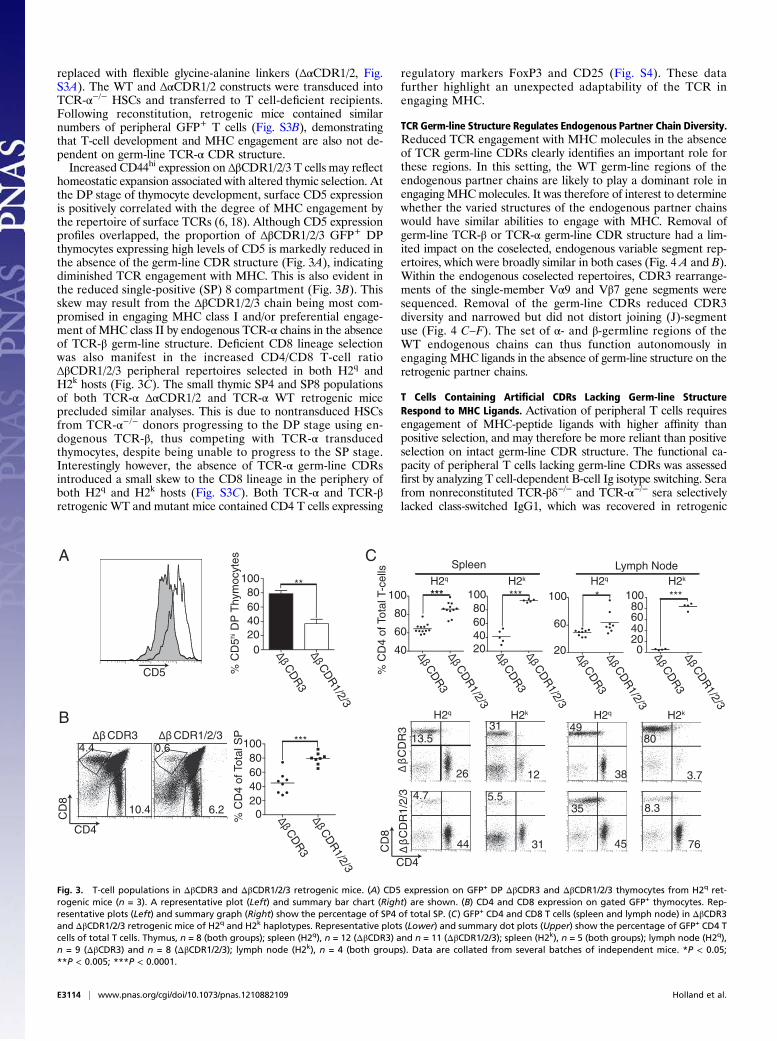
homeostatic expansion associated with altered thymic selection. Atthe DP stage of thymocyte development, surface CD5 expressionis positively correlated with the degree of MHC engagement bythe repertoire of surface TCRs (6, 18). Although CD5 expressionprofiles overlapped, the proportion of ΔβCDR1/2/3 GFP+ DPthymocytes expressing high levels of CD5 is markedly reduced inthe absence of the germ-line CDR structure (Fig. 3A), indicatingdiminished TCR engagement with MHC. This is also evident inthe reduced single-positive (SP) 8 compartment (Fig. 3B). Thisskew may result from the ΔβCDR1/2/3 chain being most com-promised in engaging MHC class I and/or preferential engage-ment of MHC class II by endogenous TCR-α chains in the absenceof TCR-β germ-line structure. Deficient CD8 lineage selectionwas also manifest in the increased CD4/CD8 T-cell ratioΔβCDR1/2/3 peripheral repertoires selected in both H2q andH2k hosts (Fig. 3C). The small thymic SP4 and SP8 populationsof both TCR-α ΔαCDR1/2 and TCR-α WT retrogenic miceprecluded similar analyses. This is due to nontransduced HSCsfrom TCR-α−/− donors progressing to the DP stage using en-dogenous TCR-β, thus competing with TCR-α transducedthymocytes, despite being unable to progress to the SP stage.Interestingly however, the absence of TCR-α germ-line CDRsintroduced a small skew to the CD8 lineage in the periphery ofboth H2q and H2k hosts (Fig. S3C). Both TCR-α and TCR-βretrogenic WT and mutant mice contained CD4 T cells expressing
regulatory markers FoxP3 and CD25 (Fig. S4). These datafurther highlight an unexpected adaptability of the TCR inengaging MHC.
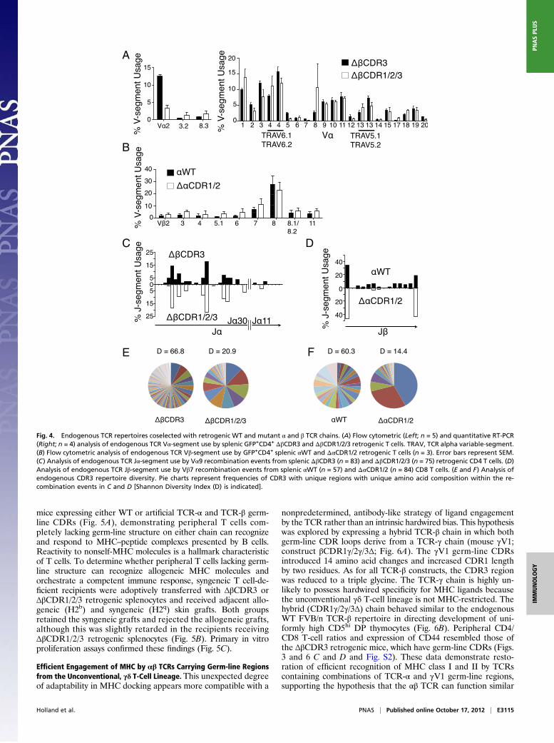
TCR Germ-line Structure Regulates Endogenous Partner Chain Diversity.Reduced TCR engagement with MHC molecules in the absenceof TCR germ-line CDRs clearly identifies an important role forthese regions. In this setting, the WT germ-line regions of theendogenous partner chains are likely to play a dominant role inengaging MHCmolecules. It was therefore of interest to determinewhether the varied structures of the endogenous partner chainswould have similar abilities to engage with MHC. Removal ofgerm-line TCR-β or TCR-α germ-line CDR structure had a lim-ited impact on the coselected, endogenous variable segment rep-ertoires, which were broadly similar in both cases (Fig. 4 A and B).Within the endogenous coselected repertoires, CDR3 rearrange-ments of the single-member Vα9 and Vβ7 gene segments weresequenced. Removal of the germ-line CDRs reduced CDR3diversity and narrowed but did not distort joining (J)-segmentuse (Fig. 4 C–F). The set of α- and β-germline regions of theWT endogenous chains can thus function autonomously inengaging MHC ligands in the absence of germ-line structure on theretrogenic partner chains.
T Cells Containing Artificial CDRs Lacking Germ-line StructureRespond to MHC Ligands. Activation of peripheral T cells requiresengagement of MHC-peptide ligands with higher affinity thanpositive selection, and may therefore be more reliant than positiveselection on intact germ-line CDR structure. The functional ca-pacity of peripheral T cells lacking germ-line CDRs was assessedfirst by analyzing T cell-dependent B-cell Ig isotype switching. Serafrom nonreconstituted TCR-βδ−/− and TCR-α−/− sera selectivelylacked class-switched IgG1, which was recovered in retrogenic
H2q
CD4
CD
8
CD5
PS latoT f o 4
DC
% 020406080
100 ***
**
020406080
100
5D
C %
ihset yco
myhT
PD
0.6
6.2
4.4
10.4
A
*** ***
20406080
100
40
60
80
100
sllec-T l at oT f o 4D
C %
Spleen
20
60
100 *
20406080
100
0
***
Lymph Node
4.7
44
13.5
26
31
12
5.5
31
H2q H2k
H2k
H2q H2k
49
38
35
45
8.3
76
80
3.7
H2q H2k
CD4
CD
8
B
C
ΔβCDR3 ΔβCDR1/2/3
ΔβC
DR
3Δβ
CD
R1/2/3
ΔβC
DR
3Δβ
CD
R1/2/3
ΔβC
DR
3Δβ
CD
R1/2/3
ΔβC
DR
3Δβ
CD
R1/2/3
ΔβC
DR
3Δβ
CD
R1/2/3
ΔβC
DR
3Δβ
CD
R1/2/3
βΔ
3R
DC
βΔ
3/ 2/ 1R
DC
Fig. 3. T-cell populations in ΔβCDR3 and ΔβCDR1/2/3 retrogenic mice. (A) CD5 expression on GFP+ DP ΔβCDR3 and ΔβCDR1/2/3 thymocytes from H2q ret-rogenic mice (n = 3). A representative plot (Left) and summary bar chart (Right) are shown. (B) CD4 and CD8 expression on gated GFP+ thymocytes. Rep-resentative plots (Left) and summary graph (Right) show the percentage of SP4 of total SP. (C) GFP+ CD4 and CD8 T cells (spleen and lymph node) in ΔβCDR3and ΔβCDR1/2/3 retrogenic mice of H2q and H2k haplotypes. Representative plots (Lower) and summary dot plots (Upper) show the percentage of GFP+ CD4 Tcells of total T cells. Thymus, n = 8 (both groups); spleen (H2q), n = 12 (ΔβCDR3) and n = 11 (ΔβCDR1/2/3); spleen (H2k), n = 5 (both groups); lymph node (H2q),n = 9 (ΔβCDR3) and n = 8 (ΔβCDR1/2/3); lymph node (H2k), n = 4 (both groups). Data are collated from several batches of independent mice. *P < 0.05;**P < 0.005; ***P < 0.0001.
E3114 | www.pnas.org/cgi/doi/10.1073/pnas.1210882109 Holland et al.
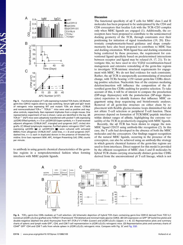
mice expressing either WT or artificial TCR-α and TCR-β germ-line CDRs (Fig. 5A), demonstrating peripheral T cells com-pletely lacking germ-line structure on either chain can recognizeand respond to MHC–peptide complexes presented by B cells.Reactivity to nonself-MHC molecules is a hallmark characteristicof T cells. To determine whether peripheral T cells lacking germ-line structure can recognize allogeneic MHC molecules andorchestrate a competent immune response, syngeneic T cell-de-ficient recipients were adoptively transferred with ΔβCDR3 orΔβCDR1/2/3 retrogenic splenocytes and received adjacent allo-geneic (H2b) and syngeneic (H2q) skin grafts. Both groupsretained the syngeneic grafts and rejected the allogeneic grafts,although this was slightly retarded in the recipients receivingΔβCDR1/2/3 retrogenic splenocytes (Fig. 5B). Primary in vitroproliferation assays confirmed these findings (Fig. 5C).
Efficient Engagement of MHC by αβ TCRs Carrying Germ-line Regionsfrom the Unconventional, γδ T-Cell Lineage. This unexpected degreeof adaptability in MHC docking appears more compatible with a
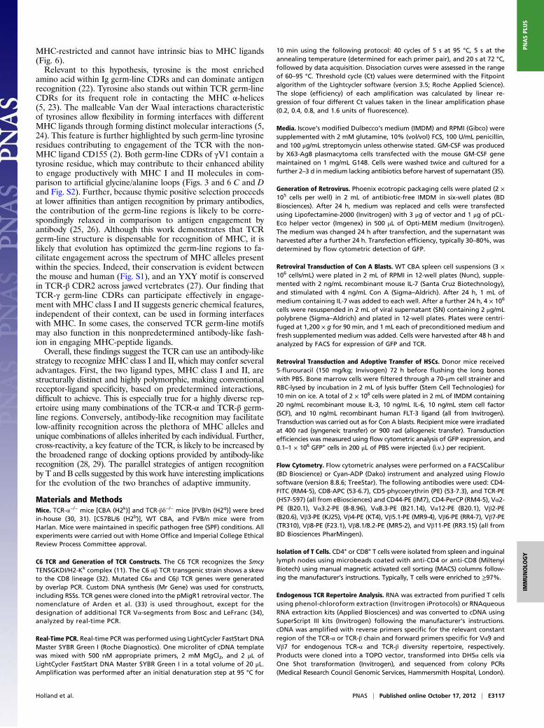
nonpredetermined, antibody-like strategy of ligand engagementby the TCR rather than an intrinsic hardwired bias. This hypothesiswas explored by expressing a hybrid TCR-β chain in which bothgerm-line CDR loops derive from a TCR-γ chain (mouse γV1;construct βCDR1γ/2γ/3Δ; Fig. 6A). The γV1 germ-line CDRsintroduced 14 amino acid changes and increased CDR1 lengthby two residues. As for all TCR-β constructs, the CDR3 regionwas reduced to a triple glycine. The TCR-γ chain is highly un-likely to possess hardwired specificity for MHC ligands becausethe unconventional γδ T-cell lineage is not MHC-restricted. Thehybrid (CDR1γ/2γ/3Δ) chain behaved similar to the endogenousWT FVB/n TCR-β repertoire in directing development of uni-formly high CD5hi DP thymocytes (Fig. 6B). Peripheral CD4/CD8 T-cell ratios and expression of CD44 resembled those ofthe ΔβCDR3 retrogenic mice, which have germ-line CDRs (Figs.3 and 6 C and D and Fig. S2). These data demonstrate resto-ration of efficient recognition of MHC class I and II by TCRscontaining combinations of TCR-α and γV1 germ-line regions,supporting the hypothesis that the αβ TCR can function similar
A
B
C
E F
D
Fig. 4. Endogenous TCR repertoires coselected with retrogenic WT and mutant α and β TCR chains. (A) Flow cytometric (Left; n = 5) and quantitative RT-PCR(Right; n = 4) analysis of endogenous TCR Vα-segment use by splenic GFP+CD4+ ΔβCDR3 and ΔβCDR1/2/3 retrogenic T cells. TRAV, TCR alpha variable-segment.(B) Flow cytometric analysis of endogenous TCR Vβ-segment use by GFP+CD4+ splenic αWT and ΔαCDR1/2 retrogenic T cells (n = 3). Error bars represent SEM.(C) Analysis of endogenous TCR Jα-segment use by Vα9 recombination events from splenic ΔβCDR3 (n = 83) and ΔβCDR1/2/3 (n = 75) retrogenic CD4 T cells. (D)Analysis of endogenous TCR Jβ-segment use by Vβ7 recombination events from splenic αWT (n = 57) and ΔαCDR1/2 (n = 84) CD8 T cells. (E and F) Analysis ofendogenous CDR3 repertoire diversity. Pie charts represent frequencies of CDR3 with unique regions with unique amino acid composition within the re-combination events in C and D [Shannon Diversity Index (D) is indicated].
Holland et al. PNAS | Published online October 17, 2012 | E3115
IMMUNOLO
GY
PNASPL
US
to antibody in using generic chemical characteristics of the germ-line regions in a nonpredetermined fashion when forminginterfaces with MHC-peptide ligands.
DiscussionThe functional specificity of αβ T cells for MHC class I and IImolecules has been proposed to be underpinned by the CD4 andCD8 coreceptors that localize Lck with the TCR–CD3 complexonly when MHC ligands are engaged (1). Additionally, the co-receptors have been proposed to contribute to the semiconserveddocking geometry of the TCR, through constraints over theirpositioning for initiation of signal transduction (19, 20). Elec-trostatic complementarity-based guidance and shape comple-mentarity have also been proposed to contribute to MHC biasand docking orientation. With ligand bias and docking orientationbeing conferred by these processes, the requirement for con-ventional ligand specificity based on predetermined interactionsbetween receptor and ligand may be relaxed (9, 17, 21). To in-vestigate this, we have used in vivo V(D)J recombination-basedmutagenesis and extensive remodeling of the germ-line regionsto investigate TCR-intrinsic structural requirements for engage-ment with MHC. We do not find evidence for such constraints.Rather, the αβ TCR is unexpectedly accommodating of structuralchange, with TCRs bearing >150 variant germ-line CDRs direct-ing positive selection. Nucleotide bias of the enzymes mediatingdeletion/insertion will influence the composition of the di-versified germ-line CDRs auditing for positive selection. To takeaccount of this, it will be of interest to compare the preselection(DP-stage thymocytes) with the postselection (SP-stage thymo-cytes) repertoires to identify features that influence MHC en-gagement using deep sequencing and bioinformatic analyses.Removal of all germ-line structure on either chain by re-placement with flexible glycine/alanine loops diminished but didnot ablate T-cell selection or peripheral T-cell function. Theseprocesses depend on formation of TCR/MHC-peptide interfaceswithin distinct ranges of affinity, highlighting the extreme ver-satility of the TCR in productively engaging with MHC ligands.Recently, the αβ TCR has been shown to engage the non-
MHC ligand CD155 using antibody-like recognition (2). In thiscase, the T cells had developed in the absence of both the MHCmolecules and the coreceptors. Our findings suggest recognitionof the natural MHC ligands, occurring in the presence of thecoreceptors, can also be achieved using an antibody-like strategyin which generic chemical features of the germ-line regions areused to form interfaces. Direct support for this model is providedby the efficient recognition of MHC class I and II molecules byhybrid TCR chains carrying structurally distinct germ-line CDRsderived from the unconventional γδ T-cell lineage, which is not
A
0.0
0.2
0.4
0.6
0.8
C57BL/6TCR -/-
-/-
IgM IgG1
OD
mn054-mn075
APC Number (x103)0 10 20 30 40
2
6
10
14
18
01x(M
PC
4 ))
%(l avi vr uStf ar
G
Time (Days)
0
25
50
75
100
0 20 40 60
CDR1/2
B
C
Fig. 5. Functional analysis of T cells expressingmutated TCR chains. (A) Mutantgerm-line CDR1/2 regions direct Ig class switching. Serum IgM and IgG1 levelsof retrogenic mice expressing WT and mutant TCRs are shown. C57BL/6and nonreconstituted TCR-α−/− TCR-βδ−/− mice were used as positive and neg-ative controls, respectively. Bars represent triplicates from a single mouse. Onerepresentative experiment of two is shown. Lanes are identified in the key. (B)TCR-βδ−/− (H2q) mice were adoptively transferred with pooled T cells expressingΔβCDR3 (filled symbols, n = 7) or ΔβCDR1/2/3 (open symbols, n = 7) and receivedadjacent allogeneic C57BL/6 (H2b, triangles) and syngeneic (H2q, circles) skingrafts. (C) Mixed lymphocyte response. Purified splenic retrogenic CD4 T cellsexpressing ΔβCDR3 (●) or ΔβCDR1/2/3 (■) were cultured with activatedBMDCs from allogeneic C57BL/6 (H2b, solid lines, n = 3) and syngeneic (H2q,dashed line, n = 3; each in duplicate) donors in the presence of titrated thy-midine. Error bars represent SEM. APC, Antigen Presenting Cell, CPM, countsper minute.
DCA
B
CDR3
SGGGTL SGGGTL
CDR1
SGHSAVSLPYFSNTAV
CDR2
FRNQAPVSTNYN
Y
33.8
22.2
CD4
CD
8
26
51.7
Spleen Lymph Node70.7
CD4
48.8
CD8
CD
44
020406080
100
sllec-T lat oT f o 4D
C %
CD5
N/B
VF 0
20406080
100
44D
C %
ihslle
C
Fig. 6. TCR-γ germ-line CDRs mediate αβ T-cell selection. (A) Schematic depiction of hybrid TCR chain containing germ-line CDR1/2 derived from TCR Vγ1(construct βCDR1γ/2γ/3Δ) grafted onto TCRVβ11 (framework Y54 deleted and minimal triple-glycine CDR3). (B) CD5 expression on GFP+ DP (solid line plots) anddouble-negative (dashed line plot) thymocytes from WT (TCR-β+δ+) FVB/n (Lower) and βCDR1γ/2γ/3Δ (Upper; n = 4). (C) Representative plots and summarycharts of splenic and lymph node GFP+, CD4, and CD8 T-cell representation in βCDR1γ/2γ/3Δ retrogenic mice. (D) Representative plots and summary charts ofCD44hi GFP+ CD4 and CD8 T cells from whole spleen in βCDR1γ/2γ/3Δ retrogenic mice. Compare with Fig. 3C and Fig. S3D.
E3116 | www.pnas.org/cgi/doi/10.1073/pnas.1210882109 Holland et al.
MHC-restricted and cannot have intrinsic bias to MHC ligands(Fig. 6).Relevant to this hypothesis, tyrosine is the most enriched
amino acid within Ig germ-line CDRs and can dominate antigenrecognition (22). Tyrosine also stands out within TCR germ-lineCDRs for its frequent role in contacting the MHC α-helices(5, 23). The malleable Van der Waal interactions characteristicof tyrosines allow flexibility in forming interfaces with differentMHC ligands through forming distinct molecular interactions (5,24). This feature is further highlighted by such germ-line tyrosineresidues contributing to engagement of the TCR with the non-MHC ligand CD155 (2). Both germ-line CDRs of γV1 contain atyrosine residue, which may contribute to their enhanced abilityto engage productively with MHC I and II molecules in com-parison to artificial glycine/alanine loops (Figs. 3 and 6 C and Dand Fig. S2). Further, because thymic positive selection proceedsat lower affinities than antigen recognition by primary antibodies,the contribution of the germ-line regions is likely to be corre-spondingly relaxed in comparison to antigen engagement byantibody (25, 26). Although this work demonstrates that TCRgerm-line structure is dispensable for recognition of MHC, it islikely that evolution has optimized the germ-line regions to fa-cilitate engagement across the spectrum of MHC alleles presentwithin the species. Indeed, their conservation is evident betweenthe mouse and human (Fig. S1), and an YXY motif is conservedin TCR-β CDR2 across jawed vertebrates (27). Our finding thatTCR-γ germ-line CDRs can participate effectively in engage-ment with MHC class I and II suggests generic chemical features,independent of their context, can be used in forming interfaceswith MHC. In some cases, the conserved TCR germ-line motifsmay also function in this nonpredetermined antibody-like fash-ion in engaging MHC-peptide ligands.Overall, these findings suggest the TCR can use an antibody-like
strategy to recognize MHC class I and II, which may confer severaladvantages. First, the two ligand types, MHC class I and II, arestructurally distinct and highly polymorphic, making conventionalreceptor-ligand specificity, based on predetermined interactions,difficult to achieve. This is especially true for a highly diverse rep-ertoire using many combinations of the TCR-α and TCR-β germ-line regions. Conversely, antibody-like recognition may facilitatelow-affinity recognition across the plethora of MHC alleles andunique combinations of alleles inherited by each individual. Further,cross-reactivity, a key feature of the TCR, is likely to be increased bythe broadened range of docking options provided by antibody-likerecognition (28, 29). The parallel strategies of antigen recognitionby T and B cells suggested by this work have interesting implicationsfor the evolution of the two branches of adaptive immunity.
Materials and MethodsMice. TCR-α−/− mice [CBA (H2k)] and TCR-βδ−/− mice [FVB/n (H2q)] were bredin-house (30, 31). [C57BL/6 (H2b)], WT CBA, and FVB/n mice were fromHarlan. Mice were maintained in specific pathogen free (SPF) conditions. Allexperiments were carried out with Home Office and Imperial College EthicalReview Process Committee approval.
C6 TCR and Generation of TCR Constructs. The C6 TCR recognizes the SmcyTENSGKDI/H2-Kk complex (11). The C6 αβ TCR transgenic strain shows a skewto the CD8 lineage (32). Mutated C6α and C6β TCR genes were generatedby overlap PCR. Custom DNA synthesis (Mr Gene) was used for constructs,including RSSs. TCR genes were cloned into the pMigR1 retroviral vector. Thenomenclature of Arden et al. (33) is used throughout, except for thedesignation of additional TCR Vα-segments from Bosc and LeFranc (34),analyzed by real-time PCR.
Real-Time PCR. Real-time PCR was performed using LightCycler FastStart DNAMaster SYBR Green I (Roche Diagnostics). One microliter of cDNA templatewas mixed with 500 nM appropriate primers, 2 mM MgCl2, and 2 μL ofLightCycler FastStart DNA Master SYBR Green I in a total volume of 20 μL.Amplification was performed after an initial denaturation step at 95 °C for
10 min using the following protocol: 40 cycles of 5 s at 95 °C, 5 s at theannealing temperature (determined for each primer pair), and 20 s at 72 °C,followed by data acquisition. Dissociation curves were assessed in the rangeof 60–95 °C. Threshold cycle (Ct) values were determined with the Fitpointalgorithm of the Lightcycler software (version 3.5; Roche Applied Science).The slope (efficiency) of each amplification was calculated by linear re-gression of four different Ct values taken in the linear amplification phase(0.2, 0.4, 0.8, and 1.6 units of fluorescence).
Media. Iscove’s modified Dulbecco’s medium (IMDM) and RPMI (Gibco) weresupplemented with 2 mM glutamine, 10% (vol/vol) FCS, 100 U/mL penicillin,and 100 μg/mL streptomycin unless otherwise stated. GM-CSF was producedby X63-Ag8 plasmacytoma cells transfected with the mouse GM-CSF genemaintained on 1 mg/mL G148. Cells were washed twice and cultured for afurther 2–3 d in medium lacking antibiotics before harvest of supernatant (35).
Generation of Retrovirus. Phoenix ecotropic packaging cells were plated (2 ×10⁵ cells per well) in 2 mL of antibiotic-free IMDM in six-well plates (BDBiosciences). After 24 h, medium was replaced and cells were transfectedusing Lipofectamine-2000 (Invitrogen) with 3 μg of vector and 1 μg of pCL-Eco helper vector (Imgenex) in 500 μL of Opti-MEM medium (Invitrogen).The medium was changed 24 h after transfection, and the supernatant washarvested after a further 24 h. Transfection efficiency, typically 30–80%, wasdetermined by flow cytometric detection of GFP.
Retroviral Transduction of Con A Blasts. WT CBA spleen cell suspensions (3 ×10⁶ cells/mL) were plated in 2 mL of RPMI in 12-well plates (Nunc), supple-mented with 2 ng/mL recombinant mouse IL-7 (Santa Cruz Biotechnology),and stimulated with 4 ng/mL Con A (Sigma–Aldrich). After 24 h, 1 mL ofmedium containing IL-7 was added to each well. After a further 24 h, 4 × 10⁶
cells were resuspended in 2 mL of viral supernatant (SN) containing 2 μg/mLpolybrene (Sigma–Aldrich) and plated in 12-well plates. Plates were centri-fuged at 1,200 × g for 90 min, and 1 mL each of preconditioned medium andfresh supplemented medium was added. Cells were harvested after 48 h andanalyzed by FACS for expression of GFP and TCR.
Retroviral Transduction and Adoptive Transfer of HSCs. Donor mice received5-flurouracil (150 mg/kg; Invivogen) 72 h before flushing the long boneswith PBS. Bone marrow cells were filtered through a 70-μm cell strainer andRBC-lysed by incubation in 2 mL of lysis buffer (Stem Cell Technologies) for10 min on ice. A total of 2 × 10⁶ cells were plated in 2 mL of IMDM containing20 ng/mL recombinant mouse IL-3, 10 ng/mL IL-6, 10 ng/mL stem cell factor(SCF), and 10 ng/mL recombinant human FLT-3 ligand (all from Invitrogen).Transduction was carried out as for Con A blasts. Recipient mice were irradiatedat 400 rad (syngeneic transfer) or 900 rad (allogeneic transfer). Transductionefficiencies was measured using flow cytometric analysis of GFP expression, and0.1–1 × 10⁶ GFP+ cells in 200 μL of PBS were injected (i.v.) per recipient.
Flow Cytometry. Flow cytometric analyses were performed on a FACSCalibur(BD Bioscience) or Cyan-ADP (Dako) instrument and analyzed using FlowJosoftware (version 8.8.6; TreeStar). The following antibodies were used: CD4-FITC (RM4-5), CD8-APC (53-6.7), CD5-phycoerythrin (PE) (53-7.3), and TCR-PE(H57-597) (all from eBiosciences) and CD44-PE (IM7), CD4-PerCP (RM4-5), Vα2-PE (B20.1), Vα3.2-PE (8-8.96), Vα8.3-PE (B21.14), Vα12-PE (B20.1), Vβ2-PE(B20.6), Vβ3-PE (KJ25), Vβ4-PE (KT4), Vβ5.1-PE (MR9-4), Vβ6-PE (RR4-7), Vβ7-PE(TR310), Vβ8-PE (F23.1), Vβ8.1/8.2-PE (MR5-2), and Vβ11-PE (RR3.15) (all fromBD Biosciences PharMingen).
Isolation of T Cells. CD4+ or CD8+ T cells were isolated from spleen and inguinallymph nodes using microbeads coated with anti-CD4 or anti-CD8 (MiltenyiBiotech) using manual magnetic activated cell sorting (MACS) columns follow-ing the manufacturer’s instructions. Typically, T cells were enriched to ≥97%.
Endogenous TCR Repertoire Analysis. RNA was extracted from purified T cellsusing phenol-chloroform extraction (Invitrogen iProtocols) or RNAqueousRNA extraction kits (Applied Biosciences) and was converted to cDNA usingSuperScript III kits (Invitrogen) following the manufacturer’s instructions.cDNA was amplified with reverse primers specific for the relevant constantregion of the TCR-α or TCR-β chain and forward primers specific for Vα9 andVβ7 for endogenous TCR-α and TCR-β diversity repertoire, respectively.Products were cloned into a TOPO vector, transformed into DH5α cells viaOne Shot transformation (Invitrogen), and sequenced from colony PCRs(Medical Research Council Genomic Services, Hammersmith Hospital, London).
Holland et al. PNAS | Published online October 17, 2012 | E3117
IMMUNOLO
GY
PNASPL
US

Sequences were analyzed with Four Peaks software Mekentosj (version 1.7.2).J-segment use was assigned using the ImMunoGeneTics database (33).
Recombination Cassette Repertoire Analysis. GFP+CD4+ cells derived fromspleen and lymph nodes of mice expressing retrogenic WT or mutated C6βTCR genes containing RSS were sorted on a FACSAria II (BD Biosciences). RNAextraction and repertoire analysis was carried out as described above usingprimers designed to amplify across the recombined Vβ11 CDR region.
Generation of Bone Marrow-Derived Dendritic Cells. The method of Inaba et al.was followed (36). Briefly, bone marrow was flushed from the long bones ofFVB/n TCR-βδ−/− and C57BL/6 mice using IMDM and 107 cells plated in 10 mLin 20-cm2
flasks (BD Falcon) with 10% (vol/vol) GM-CSF containing superna-tant. On day 3, half of the medium was replaced and fresh GM-CSF containingsupernatant was added to achieve 10% (vol/vol). Cells were harvested on day 6by scraping and replated in IMDM plus 10% GM-CSF at the initial density.On day 7, the bone marrow-derived dendritic cells (BMDCs) were activatedovernight with LPS (Sigma–Aldrich) at 100 ng/mL.
Mixed Lymphocyte Response. A mixed lymphocyte response was carried outas described (37). Briefly, 105 CD4 T cells purified as described above werecocultured with 0, 1 × 10⁴, 2 × 10⁴, or 4 × 10⁴ allogeneic or syngeneic irra-diated BMDCs in 200 μL of RPMI in round-bottomed, 96-well plates (Nunc).After 72 h, wells were pulsed with 1 μCi [3H] thymidine and left for a further18 h. Thymidine uptake was measured using a Wallac 1205 Betaplate liquidscintillation counter (PerkinElmer).
Skin Grafting. FVB TCR-βδ−/− (H2q) mice were adoptively transferred with 3 ×10⁶ splenocytes pooled from three TCR-βδ−/− (H2q) retrogenic mice express-ing either the ΔβCDR3 or ΔβCDR1/2/3 TCR chain. Cells were allowed to ex-pand for 4 wk. Mice then received adjacent syngeneic [FVB TCR-βδ−/− (H2q)]and allogeneic [C57BL/6 (H2b)] skin grafts from donor tail skin. Skin graftingwas carried out as described (38). Plasters were removed after 8 d, and graftintegrity was assessed every 2–3 d.
Ig Class Switch ELISA. An ELISA was performed using the Mouse Ig IsotypingELISA Kit (BD PharMingen) on serum following the manufacturer’s instruc-tions. Plates were read at 570 nm subtracted from 450 nm (SpectramaxM2;Molecular Devices).
Additional Software. The heat maps used in Fig. S1 were generated utilizingJColorGrid (39). The stack charts used in Fig. 2 were generated utilizingWebLogo (http://weblogo.berkeley.edu/logo.cgi).
Statistical Analysis. Statistical significance between groups of mice was de-termined using an unpaired Student t test (Prism, version 5.0c for Mac OSX;GraphPad). Statistically significant results are represented by: *P < 0.05,**P < 0.005, ***P < 0.0001, and not significant. Statistical analysis of Ig andTCR diversity in Fig. S1C was carried out using the protein variability serverwith the Shannon’s entropy analysis output for position-by-position mea-surement of diversity (40). The Shannon’s diversity index used in Figs. 2 and 4was calculated as described previously (41, 42).
ACKNOWLEDGMENTS. This work was funded by the Wellcome Trust and theUK Medical Research Council.
1. Van Laethem F, et al. (2007) Deletion of CD4 and CD8 coreceptors permits generationof alphabetaT cells that recognize antigens independently of the MHC. Immunity 27
(5):735–750.2. Tikhonova AN, et al. (2012) αβ T cell receptors that do not undergo major
histocompatibility complex-specific thymic selection possess antibody-like recognition
specificities. Immunity 36(1):79–91.3. Feng D, Bond CJ, Ely LK, Maynard J, Garcia KC (2007) Structural evidence for
a germline-encoded T cell receptor-major histocompatibility complex interaction
‘codon.’. Nat Immunol 8(9):975–983.4. Dai S, et al. (2008) Crossreactive T Cells spotlight the germline rules for alphabeta
T cell-receptor interactions with MHC molecules. Immunity 28(3):324–334.5. Marrack P, Scott-Browne JP, Dai S, Gapin L, Kappler JW (2008) Evolutionarily
conserved amino acids that control TCR-MHC interaction. Annu Rev Immunol 26:
171–203.6. Scott-Browne JP, White J, Kappler JW, Gapin L, Marrack P (2009) Germline-encoded
amino acids in the alphabeta T-cell receptor control thymic selection. Nature 458(7241):1043–1046.
7. Garcia KC, Adams JJ, Feng D, Ely LK (2009) The molecular basis of TCR germline bias
for MHC is surprisingly simple. Nat Immunol 10(2):143–147.8. Stadinski BD, et al. (2011) A role for differential variable gene pairing in creating T cell
receptors specific for unique major histocompatibility ligands. Immunity 35(5):
694–704.9. Rudolph MG, Stanfield RL, Wilson IA (2006) How TCRs bind MHCs, peptides, and
coreceptors. Annu Rev Immunol 24:419–466.10. Burrows SR, et al. (2010) Hard wiring of T cell receptor specificity for the major
histocompatibility complex is underpinned by TCR adaptability. Proc Natl Acad Sci
USA 107(23):10608–10613.11. Scott DM, et al. (1995) Identification of a mouse male-specific transplantation
antigen, H-Y. Nature 376(6542):695–698.12. Laouini D, et al. (2000) V beta T cell repertoire of CD8+ splenocytes selected on
nonpolymorphic MHC class I molecules. J Immunol 165(11):6381–6386.13. Bartok I, et al. (2010) T cell receptor CDR3 loops influence alphabeta pairing. Mol
Immunol 47(7-8):1613–1618.14. Furmanski AL, et al. (2008) Public T cell receptor beta-chains are not advantaged
during positive selection. J Immunol 180(2):1029–1039.15. Furmanski AL, et al. (2010) Peptide-specific, TCR-alpha-driven, coreceptor-independent
negative selection in TCR alpha-chain transgenic mice. J Immunol 184(2):650–657.16. Armstrong KM, Piepenbrink KH, Baker BM (2008) Conformational changes andflexibility
in T-cell receptor recognition of peptide-MHC complexes. Biochem J 415(2):183–196.17. Khan JM, Ranganathan S (2011) Understanding TR binding to pMHC complexes: How does
a TR scan many pMHC complexes yet preferentially bind to one. PLoS ONE 6(2):e17194.18. Azzam HS, et al. (1998) CD5 expression is developmentally regulated by T cell
receptor (TCR) signals and TCR avidity. J Exp Med 188(12):2301–2311.19. Yin Y, Wang XX, Mariuzza RA (2012) Crystal structure of a complete ternary complex
of T-cell receptor, peptide-MHC, and CD4. Proc Natl Acad Sci USA 109(14):5405–5410.20. Adams JJ, et al. (2011) T cell receptor signaling is limited by docking geometry to
peptide-major histocompatibility complex. Immunity 35(5):681–693.21. Stewart-Jones G, et al. (2009) Rational development of high-affinity T-cell receptor-
like antibodies. Proc Natl Acad Sci USA 106(14):5784–5788.
22. Fellouse FA, Wiesmann C, Sidhu SS (2004) Synthetic antibodies from a four-amino-acid code: A dominant role for tyrosine in antigen recognition. Proc Natl Acad Sci USA101(34):12467–12472.
23. Ofran Y, Schlessinger A, Rost B (2008) Automated identification of complementaritydetermining regions (CDRs) reveals peculiar characteristics of CDRs and B cellepitopes. J Immunol 181(9):6230–6235.
24. Yin L, et al. (2011) A single T cell receptor bound to major histocompatibility complexclass I and class II glycoproteins reveals switchable TCR conformers. Immunity 35(1):23–33.
25. Juang J, et al. (2010) Peptide-MHC heterodimers show that thymic positive selectionrequires a more restricted set of self-peptides than negative selection. J Exp Med 207(6):1223–1234.
26. Yin J, Beuscher AE, 4th, Andryski SE, Stevens RC, Schultz PG (2003) Structural plasticityand the evolution of antibody affinity and specificity. J Mol Biol 330(4):651–656.
27. Scott-Browne JP, et al. (2011) Evolutionarily conserved features contribute to αβ T cellreceptor specificity. Immunity 35(4):526–535.
28. Wooldridge L, et al. (2012) A single autoimmune T cell receptor recognizes more thana million different peptides. J Biol Chem 287(2):1168–1177.
29. Maynard J, et al. (2005) Structure of an autoimmune T cell receptor complexedwith classII peptide-MHC: Insights into MHC bias and antigen specificity. Immunity 22(1):81–92.
30. Philpott KL, et al. (1992) Lymphoid development in mice congenitally lacking T cellreceptor alpha beta-expressing cells. Science 256(5062):1448–1452.
31. Mombaerts P, et al. (1992) Mutations in T-cell antigen receptor genes alpha and betablock thymocyte development at different stages. Nature 360(6401):225–231.
32. Chai JG, et al. (1999) Critical role of costimulation in the activation of naive antigen-specific TCR transgenic CD8+ T cells in vitro. J Immunol 163(3):1298–1305.
33. Arden B, Clark SP, Kabelitz D, Mak TW (1995) Mouse T-cell receptor variable genesegment families. Immunogenetics 42(6):501–530.
34. Bosc N, Lefranc MP (2003) The mouse (Mus musculus) T cell receptor alpha (TRA) anddelta (TRD) variable genes. Dev Comp Immunol 27(6-7):465–497.
35. Karasuyama H, Kudo A, Melchers F (1990) The proteins encoded by the VpreB andlambda 5 pre-B cell-specific genes can associate with each other and with mu heavychain. J Exp Med 172(3):969–972.
36. Inaba K, et al. (1992) Generation of large numbers of dendritic cells from mouse bonemarrow cultures supplemented with granulocyte/macrophage colony-stimulatingfactor. J Exp Med 176(6):1693–1702.
37. Chai JG, Bartok I, Scott D, Dyson J, Lechler R (1998) T:T antigen presentation byactivated murine CD8+ T cells induces anergy and apoptosis. J Immunol 160(8):3655–3665.
38. Billingham R, Medawar PB (1951) The technique of free skin grafting in mammals.J Exp Biol 28:385–402.
39. Joachimiak MP, Weisman JL, May BCh (2006) JColorGrid: Software for the visualizationof biological measurements. BMC Bioinformatics 7:225.
40. Garcia-Boronat M, Diez-Rivero CM, Reinherz EL, Reche PA (2008) PVS: A web serverfor protein sequence variability analysis tuned to facilitate conserved epitopediscovery. Nucleic Acids Res 36(Web Server issue):W35–W41.
41. Ferreira C, et al. (2009) Non-obese diabetic mice select a low-diversity repertoire ofnatural regulatory T cells. Proc Natl Acad Sci USA 106(20):8320–8325.
42. Singh Y, Ferreira C, Chan AC, Dyson J, Garden OA (2010) Restricted TCR-alpha CDR3diversity disadvantages natural regulatory T cell development in the B6.2.16 beta-chain transgenic mouse. J Immunol 185(6):3408–3416.
E3118 | www.pnas.org/cgi/doi/10.1073/pnas.1210882109 Holland et al.