The skull and neck of a new flagellicaudatan sauropod from the Morrison Formation and its...
36
Journal of Systematic Palaeontology, iFirst 2012, 1–36 The skull and neck of a new flagellicaudatan sauropod from the Morrison Formation and its implication for the evolution and ontogeny of diplodocid dinosaurs Emanuel Tschopp ∗ and Oct´ avio Mateus Faculdade de Ciˆ encias e Tecnologia- CICEGe, Universidade Nova de Lisboa, Monte de Caparica, Portugal; Museu da Lourinh˜ a, Rua Jo˜ ao Lu´ ıs de Moura 95, 2530-158, Lourinh˜ a, Portugal (Received 27 July 2011; accepted 7 February 2012) A new taxon of diplodocid sauropod, Kaatedocus siberi gen. et sp. nov., is recognized based on well-preserved cervical vertebrae and skull from the Morrison Formation (Kimmeridgian, Late Jurassic) of northern Wyoming, USA. A phylogenetic analysis places it inside Diplodocinae (Sauropoda: Flagellicaudata: Diplodocidae), as a sister taxon to a clade uniting Tornieria africana and the classical diplodocines Barosaurus lentus and Diplodocus. The taxon is diagnosed by a unique combination of plesiomorphic and derived traits, as well as the following unambiguous autapomorphies within Diplodocidae: frontals separated anteriorly by a U-shaped notch; squamosals restricted to the post-orbital region; presence of a postparietal foramen; a narrow, sharp and distinct sagittal nuchal crest; the paired basal tuber with a straight anterior edge in ventral view; anterior end of the prezygapophyses of mid- and posterior cervical vertebrae is often an anterior extension of the pre-epipophysis, which projects considerably anterior to the articular facet; anterodorsal corner of the lateral side of the posterior cervical vertebrae marked by a rugose tuberosity; posterior margin of the prezygapophyseal articular facet of posterior cervical vertebrae bordered posteriorly by conspicuous transverse sulcus; posterior cervical neural spines parallel to converging. The inclusion of K. siberi and several newly described characters into a previously published phylogenetic analysis recovers the new taxon as basal diplodocine, which concurs well with the low stratigraphical position of the holotype specimen. Dinheirosaurus and Supersaurus now represent the sister clade to Apatosaurus and Diplodocinae and therefore the most basal diplodocid genera. The geographical location in the less known northern parts of the Morrison Fm., where K. siberi was found, corroborates previous hypotheses on faunal provinces within the formation. The probable subadult ontogenetic stageof the holotype specimen allows analysis of ontogenetic changes and their influence on diplodocid phylogeny. http://zoobank.org/urn:lsid:zoobank.org:pub:70181793-AA2D-4F14-BAD2-F12B9D095B3D Keywords: Kaatedocus siberi; new genus; Morrison Formation; Howe Quarry Introduction Diplodocids were most abundant and diverse during the Late Jurassic. Many specimens have been found in the USA, Portugal, Tanzania and possibly Asia (McIntosh 1990; Upchurch et al. 2004a; Upchurch & Mannion 2009; Mannion et al. 2012; Whitlock 2011a). The taxa of Late Jurassic Diplodocidae usually considered valid are: Apatosaurus ajax Marsh, 1877 (type species), A. excel- sus (Marsh, 1879), A. louisae Holland, 1915, A. parvus (Peterson & Gilmore, 1902), Barosaurus lentus Marsh, 1890, Dinheirosaurus lourinhanensis Bonaparte & Mateus, 1999, Diplodocus longus Marsh, 1878 (type species), D. carnegii Hatcher, 1901, D. hayi Holland, 1924, D. halli (Gillette, 1991), Supersaurus vivianae Jensen, 1985, Tornieria africana (Fraas, 1908), and probably Dyslo- cosaurus polyonychius McIntosh et al., 1992 (McIntosh ∗ Corresponding author. Email: [email protected] 1990; Upchurch et al. 2004a, b; Lucas et al. 2006). Most of these come from the Morrison Formation of southern Wyoming, Colorado or Utah, and only few skeletons are known from other parts of the world, even from northern Wyoming or Montana. The American Museum of Natu- ral History, New York (AMNH) conducted one of their most productive field seasons in the Morrison Formation in north central Wyoming (Brown 1935). According to Brown (1935), more than 3000 bones of primarily diplodocids were recovered from the Howe Quarry near Shell, Wyoming, but none of the specimens have since been properly described, and many of them were lost subsequently during a fire at the AMNH (Michelis 2004). The site was later abandoned and only reopened in 1990 by a team of the Sauriermuseum Aathal, Switzerland (Ayer 2000; Christiansen & Tschopp 2010). Among the several dozens of bones excavated in 1990 and 1991 was a well-preserved and partly articulated ISSN 1477-2019 print / 1478-0941 online Copyright C 2012 The Natural History Museum http://dx.doi.org/10.1080/14772019.2012.746589 http://www.tandfonline.com Downloaded by [American Museum of Natural History] at 06:32 15 December 2012
Transcript of The skull and neck of a new flagellicaudatan sauropod from the Morrison Formation and its...
Journal of Systematic Palaeontology, iFirst 2012, 1–36
The skull and neck of a new flagellicaudatan sauropod from the Morrison
Formation and its implication for the evolution and ontogeny
of diplodocid dinosaurs
Emanuel Tschopp∗ and Octavio Mateus
Faculdade de Ciencias e Tecnologia- CICEGe, Universidade Nova de Lisboa, Monte de Caparica, Portugal; Museu da Lourinha,Rua Joao Luıs de Moura 95, 2530-158, Lourinha, Portugal
(Received 27 July 2011; accepted 7 February 2012)
A new taxon of diplodocid sauropod, Kaatedocus siberi gen. et sp. nov., is recognized based on well-preserved cervicalvertebrae and skull from the Morrison Formation (Kimmeridgian, Late Jurassic) of northern Wyoming, USA. A phylogeneticanalysis places it inside Diplodocinae (Sauropoda: Flagellicaudata: Diplodocidae), as a sister taxon to a clade uniting Tornieriaafricana and the classical diplodocines Barosaurus lentus and Diplodocus. The taxon is diagnosed by a unique combinationof plesiomorphic and derived traits, as well as the following unambiguous autapomorphies within Diplodocidae: frontalsseparated anteriorly by a U-shaped notch; squamosals restricted to the post-orbital region; presence of a postparietal foramen;a narrow, sharp and distinct sagittal nuchal crest; the paired basal tuber with a straight anterior edge in ventral view; anteriorend of the prezygapophyses of mid- and posterior cervical vertebrae is often an anterior extension of the pre-epipophysis,which projects considerably anterior to the articular facet; anterodorsal corner of the lateral side of the posterior cervicalvertebrae marked by a rugose tuberosity; posterior margin of the prezygapophyseal articular facet of posterior cervicalvertebrae bordered posteriorly by conspicuous transverse sulcus; posterior cervical neural spines parallel to converging. Theinclusion of K. siberi and several newly described characters into a previously published phylogenetic analysis recoversthe new taxon as basal diplodocine, which concurs well with the low stratigraphical position of the holotype specimen.Dinheirosaurus and Supersaurus now represent the sister clade to Apatosaurus and Diplodocinae and therefore the mostbasal diplodocid genera. The geographical location in the less known northern parts of the Morrison Fm., where K. siberiwas found, corroborates previous hypotheses on faunal provinces within the formation. The probable subadult ontogeneticstage of the holotype specimen allows analysis of ontogenetic changes and their influence on diplodocid phylogeny.
http://zoobank.org/urn:lsid:zoobank.org:pub:70181793-AA2D-4F14-BAD2-F12B9D095B3D
Keywords: Kaatedocus siberi; new genus; Morrison Formation; Howe Quarry
Introduction
Diplodocids were most abundant and diverse during the
Late Jurassic. Many specimens have been found in the
USA, Portugal, Tanzania and possibly Asia (McIntosh
1990; Upchurch et al. 2004a; Upchurch & Mannion 2009;
Mannion et al. 2012; Whitlock 2011a). The taxa of
Late Jurassic Diplodocidae usually considered valid are:
Apatosaurus ajax Marsh, 1877 (type species), A. excel-
sus (Marsh, 1879), A. louisae Holland, 1915, A. parvus
(Peterson & Gilmore, 1902), Barosaurus lentus Marsh,
1890, Dinheirosaurus lourinhanensis Bonaparte & Mateus,
1999, Diplodocus longus Marsh, 1878 (type species),
D. carnegii Hatcher, 1901, D. hayi Holland, 1924, D.
halli (Gillette, 1991), Supersaurus vivianae Jensen, 1985,
Tornieria africana (Fraas, 1908), and probably Dyslo-
cosaurus polyonychius McIntosh et al., 1992 (McIntosh
∗Corresponding author. Email: [email protected]
1990; Upchurch et al. 2004a, b; Lucas et al. 2006). Most
of these come from the Morrison Formation of southern
Wyoming, Colorado or Utah, and only few skeletons are
known from other parts of the world, even from northern
Wyoming or Montana. The American Museum of Natu-
ral History, New York (AMNH) conducted one of their
most productive field seasons in the Morrison Formation in
north central Wyoming (Brown 1935). According to Brown
(1935), more than 3000 bones of primarily diplodocids were
recovered from the Howe Quarry near Shell, Wyoming, but
none of the specimens have since been properly described,
and many of them were lost subsequently during a fire at
the AMNH (Michelis 2004). The site was later abandoned
and only reopened in 1990 by a team of the Sauriermuseum
Aathal, Switzerland (Ayer 2000; Christiansen & Tschopp
2010). Among the several dozens of bones excavated in
1990 and 1991 was a well-preserved and partly articulated
ISSN 1477-2019 print / 1478-0941 online
Copyright C© 2012 The Natural History Museum
http://dx.doi.org/10.1080/14772019.2012.746589
http://www.tandfonline.com
Dow
nlo
aded
by [
Am
eric
an M
use
um
of
Nat
ura
l H
isto
ry]
at 0
6:3
2 1
5 D
ecem
ber
2012

2 E. Tschopp and O. Mateus
Figure 1. Quarry map of the holotype of Kaatedocus siberi, SMA 0004. Grey elements represent cervical vertebrae and disarticulatedskull elements. Two of the latter were found 15 and 75 cm to the right of this grid (see arrows on the lower right side). SMA 0004 wasassociated with dorsal ribs, an interclavicle, sternal ribs and chevrons of maybe another individual. Drawing by Esther Premru. Scale bar= 50 cm.
neck and associated skull bones including both braincase
and rostral elements (Fig. 1; Ayer 2000; Michelis 2004). As
the specimen (SMA 0004, nicknamed ‘HQ 2’) is relatively
small, it was usually considered to be a juvenile Diplodocus
(Ayer 2000), or Barosaurus (Michelis 2004). The present,
detailed study of the morphology of SMA 0004 revealed
that it can be clearly distinguished from both of these classi-
cal Late Jurassic diplodocines (see below). The new taxon
thereby increases both the taxonomic and morphological
diversity in the Morrison Formation.
Geological setting
The members of the Morrison Formation, as identi-
fied further south, are difficult to recognize in northern
Wyoming. The only layer that has been used for strati-
graphical correlation between the Howe Quarry and the
various quarries in southern Wyoming, Colorado, Utah,
South Dakota, Oklahoma and New Mexico is a clay change
that was interpreted to divide the Morrison Formation into
lower and upper parts (Turner & Peterson 1999; but see
Trujillo 2006 for critiques). Such a clay change is present
approximately 20 m above the Howe Quarry (Fig. 2). If
Turner & Peterson (1999) prove to be right in interpreting
this geological marker as useful for long distance corre-
lation of the sites in the Morrison Formation, the Howe
Quarry would be among the stratigraphically oldest fossil
sites of the entire Morrison Formation (Turner & Peter-
son 1999; J. Ayer pers. comm. 2005). However, several
authors propose a higher stratigraphical position for the
Howe Quarry (Dodson et al. 1980; Swierc & Johnson 1996;
Wilborn 2008). Swierc & Johnson (1996) dated the Howe
Quarry as being 145.7 Ma, but this date groups all of the
sites on the Howe Ranch together and does not take into
account that the different quarries are situated at varying
stratigraphical levels (see Fig. 2). Kvale et al. (2001) inter-
preted a second site approximately 10 m higher in stratig-
raphy (Howe-Stephens Quarry, see Schwarz et al. 2007b;
Christiansen & Tschopp 2010) as being of 147 Ma in age.
This implies a latest Kimmeridgian to earliest Tithonian
age for the Howe Quarry.
Due to the fact that SMA 0004 is the first specimen
of the Howe Quarry to be properly described and iden-
tified, previous synopses of the faunal assemblage of the
site have to be regarded as provisional. An updated list of
reported dinosaurs includes the sauropods Camarasaurus,
Apatosaurus, Kaatedocus and possibly Diplodocus and
Barosaurus (if they do not prove to be Kaatedocus
as well), the theropods Allosaurus and a smaller taxon
Dow
nlo
aded
by [
Am
eric
an M
use
um
of
Nat
ura
l H
isto
ry]
at 0
6:3
2 1
5 D
ecem
ber
2012

A new flagellicaudatan sauropod from the Morrison Formation 3
Figure 2. Geographical and geological setting of the Howe Quarry within the Upper Jurassic Morrison Formation. The Howe Quarry islocated in North Central Wyoming, and stratigraphically well below the clay change. Modified from Schwarz et al. (2007b).
(represented by footprints and shed teeth), and the ornitho-
pod Camptosaurus (Brown 1935; Foster 1998; Michelis
2004). Non-dinosaurian remains include carbonized wood
fragments and a dental plate of the dipnoid fish Ceratodus
robustus (Foster 1998; Michelis 2004).
Abbreviations
Institutional abbreviationsAMNH: American Museum of Natural History, New York,
NY, USA; ANS: Academy of Natural Sciences, Philadel-
phia, PA, USA; BYU: Brigham Young University, Verte-
brate Paleontology Collection, Provo, UT, USA; CM:
Carnegie Museum of Natural History, Pittsburgh, PA,
USA; CMC: Cincinnati Museum Center, Cincinnati, OH,
USA; DMNS: Denver Museum of Nature and Science,
Denver, CO, USA; GMNH: Gunma Museum of Natu-
ral History, Gunma, Japan; HMNS: Houston Museum of
Nature and Science, Houston, TX, USA; MB.R.: Museum
fur Naturkunde, Berlin, Germany; ML: Museu da Lour-
inha, Portugal; SMA: Sauriermuseum Aathal, Switzerland;
SNMB: Staatliches Naturhistorisches Museum, Braun-
schweig, Germany; UCL: University College, London,
Dow
nlo
aded
by [
Am
eric
an M
use
um
of
Nat
ura
l H
isto
ry]
at 0
6:3
2 1
5 D
ecem
ber
2012
4 E. Tschopp and O. Mateus
UK; USNM: United States National Museum, Washington,
DC, USA; WDC: Wyoming Dinosaur Center, Thermopo-
lis, WY, USA; YPM: Yale Peabody Museum, New Haven,
CT, USA.
Anatomical abbreviationsa: articular; acf: anterior condyle fossa; acdl: anterior
centrodiapophyseal lamina; adt: anterodorsal tuberosity;
apf: anterior pneumatic foramen; at: atlas; aof: antorbital
fenestra; avl: anteroventral lip; ax: axis; bns: bifid neural
spine; bpr: basipterygoid process; bt: basal tuber; bc: brain-
case; bo: basioccipital; cdf: centrodiapophyseal fossa; cpr:
crista prootica; cprf: centroprezygapophyseal fossa; cprl:
centroprezygapophyseal lamina; d: dentary; dp: diapoph-
ysis posterior process; epi: epipophysis; ex: exoccipital;
f: frontal; fm: foramen magnum; int sprl: interrupted
spinoprezygapophyseal lamina; la: lacrimal; ls: laterosphe-
noid; lsc: lateral spine cavity; ltf: laterotemporal fenestra;
lprzc: lateral prezygapophyseal cavity; mt: median tuber-
cle; m: maxilla; n: external nares; nc: neural canal; o: orbit;
os: orbitosphenoid; p: parietal; pap: parapophysis; paof:
preantorbital fossa; par bns: parallel bifurcated neural
spine; pcdl: posterior centrodiapophyseal lamina; pl: pleu-
rocoel; pm: premaxilla; po: postorbital; podl: postzygodi-
apophyseal lamina; poz: postzygapophysis; popr: paroc-
cipital process; ppf: posterior pneumatic fossa; ppfo: post-
parietal foramen; prz: prezygapophysis; pra: proatlas;
prcdf: prezygapophyseal centrodiapophyseal fossa; prdl:
prezygodiapophyseal lamina; pre: pre-epipophysis; pro:
prootic; prsl: prespinal lamina; ptf: posttemporal fenestra;
pts: prezygapophysis transverse sulcus; pvf: posteroventral
flanges; q: quadrate; qj: quadratojugal; sdf: spinodiapophy-
seal fossa; so: supraoccipital; sprl: spinoprezygapophyseal
lamina; sprl ab: spinoprezygapophyseal lamina anterior
bulge; spof: spinopostzygapophyseal fossa; spol: spino-
postzygapophyseal lamina; sq: squamosal; tpol: inter-
postzygapophyseal lamina; tprl: interprezygapophyseal
lamina; vk: ventral keel; vmc: ventral median constriction.
Systematic palaeontology
Dinosauria Owen, 1842
Sauropoda Marsh, 1878
Eusauropoda Upchurch, 1995
Neosauropoda Bonaparte, 1986
Diplodocoidea Marsh, 1884 (see Upchurch 1995)
Flagellicaudata Harris & Dodson, 2004
Diplodocidae Marsh, 1884
Kaatedocus gen. nov.
Type species. Kaatedocus siberi sp. nov.
Diagnosis. See diagnosis for type and only species below.
Kaatedocus siberi gen. et sp. nov.
(Figs 3–10; see also Online Supplementary Material)
Diagnosis. Diplodocid sauropod with the following
features not found in other sauropods: U-shaped notch sepa-
rating the frontals anteriorly (Fig. 5); a rugose tuberosity
that marks the anterodorsal corner of the lateral surface of
the posterior cervical vertebrae (Fig. 10); posterior margin
of the prezygapophyseal articular facet of posterior cervi-
cal vertebrae bordered posteriorly by a conspicuous trans-
verse sulcus, separating the facet from the prezygapophy-
seal process (Fig. 10).
The following features are unique to Kaatedocus
among Flagellicaudata or more inclusive clades: squamos-
als are restricted to the post-orbital region (unique for
Diplodocoidea; Fig. 3); a straight anterior margin of the
paired basal tuber in ventral view; anterior end of the
prezygapophyses in mid- and posterior cervical verte-
brae is formed by an accessory ventral process of the
pre-epipophysis, that projects considerably anterior to the
prezygapophyseal articular facet (Fig. 9).
The following features are unique to Kaatedocus
among Diplodocidae: postparietal foramen present (Fig.
6); narrow, sharp and distinct sagittal nuchal crest on the
supraoccipital (Figs 5, 6); and the narrowly diverging to
subparallel posterior cervical neural spines (Fig. 10).
Furthermore, the new taxon can be distinguished from
adult Apatosaurus and Diplodocus by its closed or very
reduced preantorbital fenestra (Fig. 4); the dorsal portion of
lateral edge of the lacrimal that bears a dorsoventrally short
laterally projecting spur (Fig. 6); the relatively rounded
snout (Fig. 5); a second small fossa in the quadrate, medi-
ally at the base of the pterygoid ramus; and the ratio
of length/maximum basal diameter of the basipterygoid
processes being less than four. In contrast to Apatosaurus,
Kaatedocus exhibits spinoprezygapophyseal lamina that
are reduced to a ridge, or totally interrupted at the base of
the prezygapophysis of anterior and mid-cervical vertebrae
(Figs 7, 8). Kaatedocus is different from Diplodocus due to
the presence of at least 12 maxillary and dentary teeth that
are not restricted to the anteriormost part of the jaw (Fig.
3). It can be distinguished from Diplodocus, Tornieria and
Barosaurus due to its relatively short mid-cervical centra
(Elongation Index (EI) = centrum length/height of poste-
rior cotyle < 4).
Etymology. ‘Kaate’ means small in the Crow (Absaroka)
language, one of the Native American tribes of northern
Wyoming. ‘Docus’ is an allusion to Diplodocus and the
Greek dokos/δoκoς ‘beam’. ‘Siberi’ is after Hans-Jakob
‘Kirby’ Siber, b. 1942, doctor honoris causa of the Univer-
sity of Zurich, Switzerland. Siber is the founder and director
of the Sauriermuseum Aathal, Switzerland, and organized
and funded the excavation, preparation and curation of the
holotype specimen of Kaatedocus siberi.
Holotype. SMA 0004: partial skull (right premaxilla, both
maxillae, left lacrimal, both frontals, both postorbitals, both
Dow
nlo
aded
by [
Am
eric
an M
use
um
of
Nat
ura
l H
isto
ry]
at 0
6:3
2 1
5 D
ecem
ber
2012
A new flagellicaudatan sauropod from the Morrison Formation 5
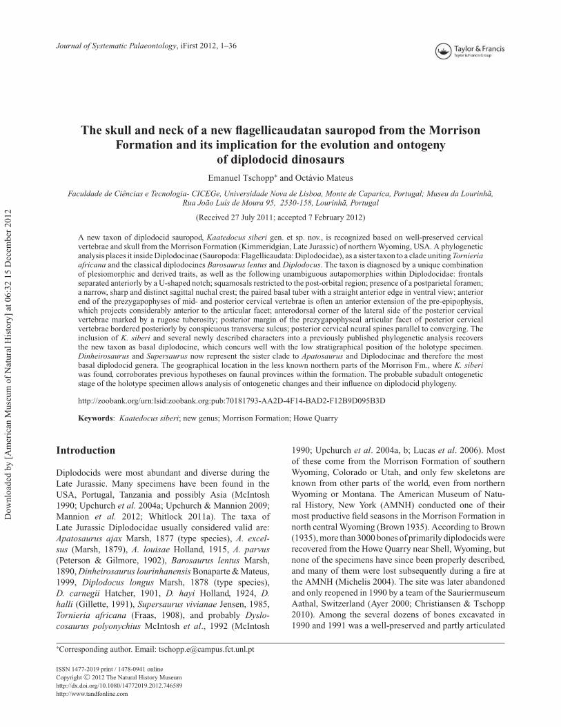
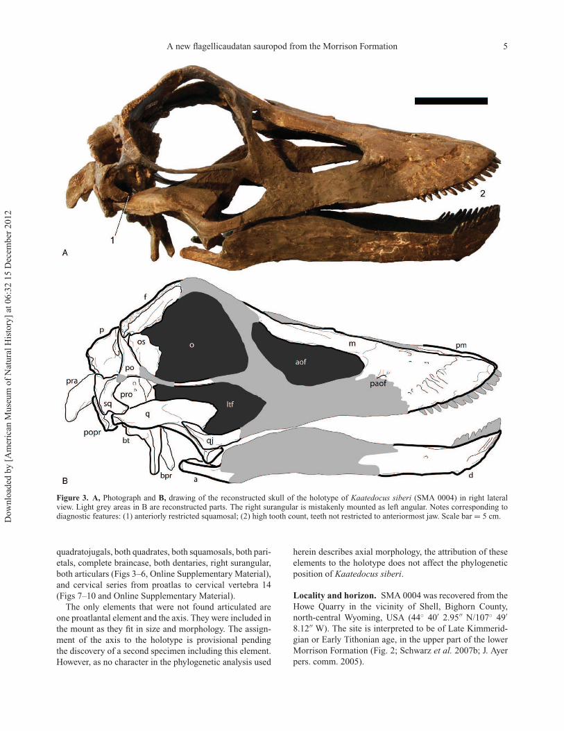
Figure 3. A, Photograph and B, drawing of the reconstructed skull of the holotype of Kaatedocus siberi (SMA 0004) in right lateralview. Light grey areas in B are reconstructed parts. The right surangular is mistakenly mounted as left angular. Notes corresponding todiagnostic features: (1) anteriorly restricted squamosal; (2) high tooth count, teeth not restricted to anteriormost jaw. Scale bar = 5 cm.
quadratojugals, both quadrates, both squamosals, both pari-
etals, complete braincase, both dentaries, right surangular,
both articulars (Figs 3–6, Online Supplementary Material),
and cervical series from proatlas to cervical vertebra 14
(Figs 7–10 and Online Supplementary Material).
The only elements that were not found articulated are
one proatlantal element and the axis. They were included in
the mount as they fit in size and morphology. The assign-
ment of the axis to the holotype is provisional pending
the discovery of a second specimen including this element.
However, as no character in the phylogenetic analysis used
herein describes axial morphology, the attribution of these
elements to the holotype does not affect the phylogenetic
position of Kaatedocus siberi.
Locality and horizon. SMA 0004 was recovered from the
Howe Quarry in the vicinity of Shell, Bighorn County,
north-central Wyoming, USA (44◦ 40′ 2.95′′ N/107◦ 49′
8.12′′ W). The site is interpreted to be of Late Kimmerid-
gian or Early Tithonian age, in the upper part of the lower
Morrison Formation (Fig. 2; Schwarz et al. 2007b; J. Ayer
pers. comm. 2005).
Dow
nlo
aded
by [
Am
eric
an M
use
um
of
Nat
ura
l H
isto
ry]
at 0
6:3
2 1
5 D
ecem
ber
2012
6 E. Tschopp and O. Mateus
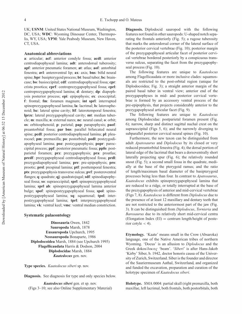
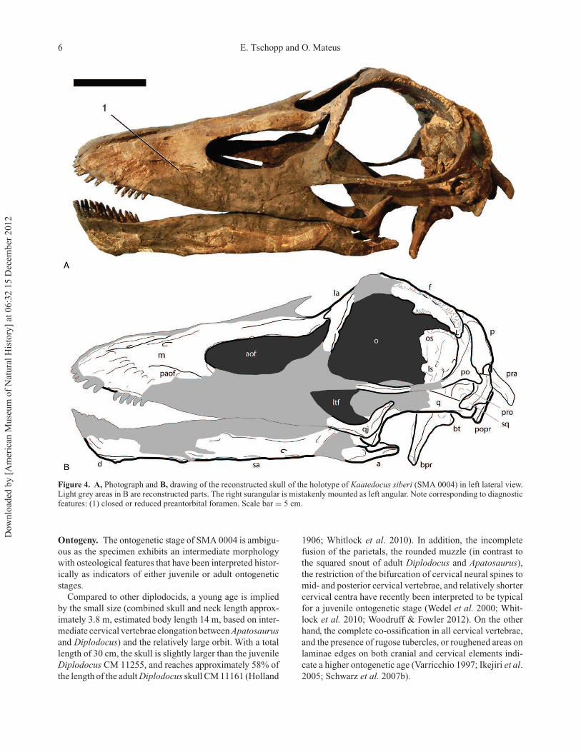
Figure 4. A, Photograph and B, drawing of the reconstructed skull of the holotype of Kaatedocus siberi (SMA 0004) in left lateral view.Light grey areas in B are reconstructed parts. The right surangular is mistakenly mounted as left angular. Note corresponding to diagnosticfeatures: (1) closed or reduced preantorbital foramen. Scale bar = 5 cm.
Ontogeny. The ontogenetic stage of SMA 0004 is ambigu-
ous as the specimen exhibits an intermediate morphology
with osteological features that have been interpreted histor-
ically as indicators of either juvenile or adult ontogenetic
stages.
Compared to other diplodocids, a young age is implied
by the small size (combined skull and neck length approx-
imately 3.8 m, estimated body length 14 m, based on inter-
mediate cervical vertebrae elongation between Apatosaurus
and Diplodocus) and the relatively large orbit. With a total
length of 30 cm, the skull is slightly larger than the juvenile
Diplodocus CM 11255, and reaches approximately 58% of
the length of the adult Diplodocus skull CM 11161 (Holland
1906; Whitlock et al. 2010). In addition, the incomplete
fusion of the parietals, the rounded muzzle (in contrast to
the squared snout of adult Diplodocus and Apatosaurus),
the restriction of the bifurcation of cervical neural spines to
mid- and posterior cervical vertebrae, and relatively shorter
cervical centra have recently been interpreted to be typical
for a juvenile ontogenetic stage (Wedel et al. 2000; Whit-
lock et al. 2010; Woodruff & Fowler 2012). On the other
hand, the complete co-ossification in all cervical vertebrae,
and the presence of rugose tubercles, or roughened areas on
laminae edges on both cranial and cervical elements indi-
cate a higher ontogenetic age (Varricchio 1997; Ikejiri et al.
2005; Schwarz et al. 2007b).
Dow
nlo
aded
by [
Am
eric
an M
use
um
of
Nat
ura
l H
isto
ry]
at 0
6:3
2 1
5 D
ecem
ber
2012

A new flagellicaudatan sauropod from the Morrison Formation 7
Figure 5. A, Photograph and B, drawing of the reconstructed skull of the holotype of Kaatedocus siberi (SMA 0004) in dorsal view.Light grey areas in B are reconstructed parts. Notes corresponding to diagnostic features: (1) U-shaped frontal notch; (2) rounded snout;(3) narrow, distinct sagittal nuchal crest. Scale bar = 5 cm.
Taking all of the above mentioned features into account,
the juvenile traits are generally seen as well on adult
specimens of other taxa (like the more rounded snout of
dicraeosaurids, or the less developed bifurcation of the
cervical vertebrae in Barosaurus; Janensch 1935; McIn-
tosh 2005), whereas the indicators for a subadult stage
of SMA 0004 (in particular the advanced co-ossification
and the conspicuous rugosities on both skull bones and
vertebrae) are not reported from any specimens of young
age, to our knowledge. In fact, adult alligators (Ikejiri
2012), and a juvenile Allosaurus (Birkemeier 2011) have
recently been reported to possess open neurocentral sutures
in cervical vertebrae, but fused centra and neural spines in
caudal and/or dorsal elements. This indicates that neuro-
central closure proceeds from the back to the front,
with the cervical vertebrae being the last to co-ossify
(Birkemeier 2011; Ikejiri 2012). On the other hand, the
subadult flagellicaudatan Suuwassea emilieae ANS 21122
was reported to have fused cervical arches, but unfused
mid-caudal vertebrae, which might contradict the devel-
opmental model supported by the above-mentioned taxa
(Harris 2006c). However, based on the available material,
we still interpret SMA 0004 as a subadult specimen that
retained a small body size. This is in agreement with the
Dow
nlo
aded
by [
Am
eric
an M
use
um
of
Nat
ura
l H
isto
ry]
at 0
6:3
2 1
5 D
ecem
ber
2012

8 E. Tschopp and O. Mateus
Figure 6. A, B, Photographs and C, D, drawings of the reconstructed skull of the holotype of Kaatedocus siberi (SMA 0004) inanteroventral (A, C), and posterodorsal (B, D) views. Light grey areas in C and D are reconstructed parts. Notes corresponding todiagnostic features: (1) lateral lacrimal spur; (2) postparietal foramen; (3) narrow, distinct sagittal nuchal crest. Scale bar = 5 cm.
ontogenetic stages as described for Camarasaurus in Ikejiri
et al. (2005). According to these authors, neurocentral
closure in cervical vertebrae, as well as the subdivi-
sion of cervical pleurocoels, happens in subadult to adult
stages.
Description
Terminology follows the standard nomenclature generally
used (anterior and posterior instead of cranial and caudal).
For the names of the vertebral lamina and fossae, we
Dow
nlo
aded
by [
Am
eric
an M
use
um
of
Nat
ura
l H
isto
ry]
at 0
6:3
2 1
5 D
ecem
ber
2012

A new flagellicaudatan sauropod from the Morrison Formation 9
follow Wilson (1999) and Wilson et al. (2011), respectively,
with two exceptions: instead of intrapre- and intrapostzy-
gapophyseal lamina we use the terms interpre- and inter-
postzygapophyseal lamina, as the prefix inter- describes
better the arrangement between the two zygapophyses. To
keep confusion to a minimum we keep the abbreviations
proposed by Wilson (1999): tprl (interprezygapophyseal
lamina) and tpol (interpostzygapophyseal lamina). Other
anatomical abbreviations used in the text are: CV (cervi-
cal vertebra), and EI (Elongation Index, ratio between the
centrum length and the height of the posterior cotyle;
Upchurch 1998).
SkullThe general shape of the skull is highly similar to
Diplodocus or Apatosaurus, elongated and having a
retracted external nares. Some obvious differences
compared to adult skulls (e.g. AMNH 969, CM 11161,
11162, USNM 2672, 2673) include the larger orbit and the
teeth that reach further backwards, although these are still
restricted to the anteriormost part of the skull (Figs 3, 4).
Premaxilla. The right premaxilla preserves the anterior
portion, lacking the anteriormost dorsal border and the
teeth. The main body is simple, without a sinuous curve
building a muzzle. It is broadest anteriorly, and its straight
lateral and medial edges slightly converge posteriorly,
including a very acute angle of approximately 10◦. The
dorsal surface does not bear any anteroventrally extend-
ing grooves as are present in Dicraeosaurus hansemanni
MB.R.2337. Four alveoli are visible, oriented such that the
teeth would be procumbent. There is no indication of an
anterior dorsoventral expansion as in Diplodocus USNM
2673 or Dicraeosaurus hansemanni MB.R.2337.
Maxilla. Both maxillae lack their posteroventral ramus and
teeth. From the main body, the lamina-like dorsal ramus
projects posteriorly and tapers until it meets the lacrimal.
Its ventral edge is convex, resulting in a concave dorsal
border of the antorbital fenestra as in most diplodocid
skulls (e.g. CM 3452, 11161, 11162, 11255; USNM 2672).
Slightly lateral to the border with the premaxilla, both the
subnarial and the anterior maxillary foramen are well visi-
ble and closely spaced. A third, small foramen is situated
just lateroventrally of the subnarial foramen. The prean-
torbital fossa is a longitudinal depression marked by an
acute step bordering it dorsally. Such a development has
been considered autapomorphic for Diplodocus (Mannion
et al. 2012), but there it roofs a relatively large preantor-
bital fenestra (e.g. CM 3452, 11161, 11255; Berman &
McIntosh 1978; Whitlock et al. 2010; pers. obs. 2011),
which is not present in SMA 0004. It does not open into
a fenestra as in Diplodocus (e.g. CM 3452, 11161, 11255;
Berman & McIntosh 1978). In this respect, Kaatedocus
siberi is very similar to Dicraeosaurus (MB.R.2336; pers.
obs. 2011), where a very reduced foramen-like opening is
present in the posteriormost extension of a dorsally well-
defined fossa. The posteriormost portion is not preserved
in SMA 0004 and could also exhibit such a small opening.
The rostral portion of the main body of the maxilla shows
the wavy surface typical for the part containing the replace-
ment teeth. At least 12 alveoli can be counted in the right
element.
Quadratojugal. Only the posterior half of both quadra-
tojugals has been preserved. They are L-shaped bones
that cover the quadrate laterally with the shorter dorsal
ramus, and would extend anteriorly to meet the jugal
and the maxilla if preserved entirely. The two arms of
the L are transversely compressed and form an angle of
approximately 110◦. The dorsal ramus projects dorsoposte-
riorly, as in Diplodocus or Apatosaurus, and curves slightly
more backwards in its upper half. From there, it tapers to
an acute tip, which does not extend far posteriorly, and
thus remains well separated from the anteroventral projec-
tion of the squamosal – a typical feature in diplodocids,
which is also present in Suuwassea (ANS 21122, pers. obs.
2011; contrary to the interpretation of Harris 2006a). The
preserved portion of the anterior ramus is dorsoventrally
shortest close to where it grades into the dorsal process,
and expands slightly towards the anterior end.
Lacrimal. The left lacrimal preserves the dorsal part that
articulates with the maxilla anteriorly, the nasal medially,
and the prefrontal posteriorly. The anterior edge of the
lacrimal is straight until it reaches the posterodorsalmost
end of the antorbital fenestra. The posterior border becomes
thicker towards the upper end, where it develops a concav-
ity that holds the lacrimal foramen. Lateral to this foramen,
the lacrimal develops a blunt bony spur projecting laterally,
a feature that is only seen in the juvenile diplodocid skulls
CM 3452 and 11255, but not in adults (CM 11161, 11162;
Berman & McIntosh 1978; pers. obs. 2011).
Frontal. Both frontals are preserved completely. Antero-
posterior length is about equal to maximum transverse
width. The dorsal surface is flat and relatively smooth. The
anterior and posterior margins are straight and subparallel at
their medial portions. The lateralmost quarter of the anterior
edge is marked by a distinct, acute V-shaped invagination
that would receive the posterior process of the prefrontal,
which does not extend far posteriorly as in Diplodocus, but
remains well distant from the parietal. The posterior edge
of the frontal also curves inward, but to a lesser degree than
the anterior one. This indentation is gently rounded and
forms the articulation facet for the dorsomedial process of
the postorbital. The medial edges are straight along their
posterior half and form an approximate right angle with the
frontal–parietal suture. Anteriorly, they curve inwards, so
that between the frontals a U-shaped notch develops. The
Dow
nlo
aded
by [
Am
eric
an M
use
um
of
Nat
ura
l H
isto
ry]
at 0
6:3
2 1
5 D
ecem
ber
2012

10 E. Tschopp and O. Mateus
edge is extremely thin, and neither exhibits any indication
of fracturing nor is undulose as sutures often are. The notch,
therefore, either enclosed a posterior process of the nasals,
having a straight suture, or remained open as a posterior
extension of the external nares, so that the nasals did not
contact each other medially. The only sauropod with a simi-
lar development is Spinophorosaurus nigerensis, which has
a narrow, V-shaped notch between the frontals (Knoll et al.
2012). The wider, U-shaped notch of Kaatedocus siberi can
thus be considered an unambiguous autapomorphy. The
straight lateral margins contribute to a major part of the
dorsal edge of the orbit, and exhibit a similar rugosity as is
typical for bone sutures, present in most diplodocoid skulls
(e.g. AMNH 969, 7530, CM 3452, MB.R.2386, 2387, pers.
obs. 2011). This indicates the existence of a cartilaginous
palpebral element (homologue to the ossified palpebral
bones in ornithischian dinosaurs; see Maidment & Porro
2010). In ventral view it becomes clear that the frontal
supports the end of the posterior process of the prefrontal
from below. From there, a conspicuous, sharp ridge passes
the ventral surface obliquely until it reaches the anterior
end of the articulation surface for the anterodorsal part of
the braincase, which is highly rugose and stands almost
perpendicularly to this ridge.
Postorbital. Both postorbitals preserve their dorsomedial
process and parts of the anterior process that forms the
ventral border of the orbit. It covers the frontal posteriorly,
and overlies the anterodorsalmost corner of the squamosal
laterally. The left element also shows the posterior process.
The dorsomedial process is relatively high dorsoventrally
and compressed anteroposteriorly. It extends medially to
reach the frontal–parietal suture, thereby excluding the
frontal from the margin of the supratemporal fenestra. It
is dorsoventrally convex posteriorly and concave anteri-
orly. The anterior process is a dorsoventrally compressed,
subtle structure that extends nearly straight and subparallel
to the dorsal margin of the orbit until it would reach the
jugal, which is not preserved in SMA 0004. Posteriorly, the
postorbital anterior process curves gently upwards, expand-
ing slightly transversely. The posterior ramus is very short
and acute. It projects almost straight posteriorly in dorsal
view, and curves to a small degree ventrally in lateral aspect.
Parietal. Both parietals are complete and not fused. They
contact the frontals anteriorly, the supraoccipital and exoc-
cipital medioventrally, and the postorbitals and squamos-
als laterally. The parietals are dorsally flat bones on the
posterior skull roof with a short anterolateral and a longer
posterolateral process, which together enclose a major
portion of the supratemporal fenestra. The frontal–parietal
suture is more or less straight, and partly obscured due to
the restoration and mounting of the skull, but appears to
have been open in lifetime (B. Pabst pers. comm. 2011).
There is no trace visible of a pineal foramen, which would
be situated where the paired frontals and parietals meet. A
postparietal foramen can be observed posteriorly between
the parietals and the supraoccipital, similar to the condition
in Dicraeosaurus, Amargasaurus and Suuwassea (Janen-
sch 1935; Salgado & Bonaparte 1991; Harris 2006a; Whit-
lock 2011a). The medial borders of the parietals bend
slightly laterally in their posterior half, somewhat ante-
rior to where they meet the supraoccipital, which they
overlap to a small degree. The posterior border of the
dorsal portion of the parietals is transversely concave in
dorsal view. From there, the posterolateral process extends
in a right angle ventrally, and laterally, following the
oblique dorsolateral edge of the supraoccipital, and forms
the posterior margin of the supratemporal fenestra. The
posterior aspect of this process has a subequal or greater
surface area than the flat dorsal portion. It is anteroposte-
riorly compressed, dorsomedially–ventrolaterally convex,
and dorsolaterally–ventromedially concave. The dorsal
edge extends straight dorsomedially–ventrolaterally, so that
the supratemporal fenestra faces somewhat posteriorly as
well, unlike the condition in Diplodocus CM 3452, but
similar to Apatosaurus CM 11162 (Berman & McIntosh
1978). The ventrolateral end of the posterolateral process is
rounded and overlaps the squamosal laterally. The anterior
surface gently curves into the anteroposteriorly concave
medial side of the parietal, which is well separated by a
distinct ridge from the dorsal flat area of the same bone.
In its anterior part, it bears the short anterolateral process,
which projects ventrolaterally forming the anterior edge of
the supratemporal fenestra, together with the postorbital.
Squamosal. The squamosals are complete except for
their dorsalmost part, which is not preserved on either
side. It connects with the postorbital anterodorsally, with
the quadrate anteroventrally, with the parietal dorsome-
dially, and with the paroccipital process posteroventrally.
Dorsally, the squamosal forms the posterolateral corner
of the supratemporal fenestra, ventromedially the dorso-
lateral edge of the posttemporal fenestra, and anteriorly
the posteriormost corner of the infratemporal fenestra. It is
a strongly transversely curved bone, with its convex side
facing outwards, forming part of the posterolateral edge of
the skull. In lateral view, the anterodorsal process of the
squamosal bears a dorsoventral concavity for the reception
of the postorbital. The ventral process tapers to a blunt tip
that points slightly anteriorly as well, but does not exceed
the posterior border of the orbit as in other diplodocoids
(e.g. Berman & McIntosh 1978; Salgado & Bonaparte
1991). Both its anterior and posterior borders are straight,
before they curve frontwards and backwards, respectively.
In the posterior margin this happens slightly earlier, and
the posterior process is thus dorsoventrally longer than
the anterior ramus. The squamosal therefore bears a short
posteroventral process but does not form such a distinct
‘prong’ as present in Amargasaurus (Salgado & Bonaparte
Dow
nlo
aded
by [
Am
eric
an M
use
um
of
Nat
ura
l H
isto
ry]
at 0
6:3
2 1
5 D
ecem
ber
2012
A new flagellicaudatan sauropod from the Morrison Formation 11
1991; Whitlock 2011a). In posterior view, the squamosals
are dorsoventrally convex, with the dorsomedial process
projecting medially to meet the parietal.
Quadrate. Both quadrates lack their anterodorsal portions
of the wing-like anterodorsal ramus, but are otherwise
complete. They are triradiate bones forming the jaw articu-
lation with the articular ventrally, contacting the pterygoids
anteromedially, and the squamosal posteriorly. The articu-
lar facet for the mandibular joint is subtriangular, lacking
a medial process as the one seen in some rebbachisaurids
(Mannion et al. 2012; Whitlock 2011a). It is located at the
ventral end of the relatively stout ventral ramus, which is
oriented at an angle of about 90◦ to the skull roof. The
anteroventral projection of the quadrate shaft is covered
laterally almost completely by the dorsal ramus of the
quadratojugal, and exhibits a shallow concavity on its poste-
rior side. This concavity extends onto the ventral side of the
posterior process, forming a shallow quadrate fossa as in
other diplodocids (Wilson 2002; Upchurch et al. 2004a).
Posteriorly, the ramus tapers both in dorsoventral and in
mediolateral directions. It curves slightly medially as well,
so that the whole lateral side of the quadrate becomes
anteroposteriorly slightly convex. The wing-like pterygoid
flange is a very thin lamina originating at the lateral edge
of the posterior ramus. Medially it borders another shallow
fossa that lies on the quadrate shaft and becomes deeper
anteriorly. Such a cavity has not been described in any
diplodocid sauropod, and personal observations showed it
to be absent in most diplodocid skulls (e.g. AMNH 969; CM
11161, 11162, 11255; USNM 2672, 2673). However, the
possible Diplodocus skull CM 3452, as well as the quadrates
assigned to the holotype of Apatosaurus ajax YPM 1860,
show similar features.
Braincase and occiputSupraoccipital and exoccipital-opisthotic complex. The
supraoccipital is complete and well fused to the
exoccipital–opisthotic complex so that sutures are difficult
to observe. The whole fused element is subtriangular, touch-
ing the parietals anterolaterally, bordering the posttemporal
fenestrae with the dorsolateral margin of the paroccipi-
tal processes, and contributing to the upper portion of the
occipital condyle ventrally. It roofs the braincase posteri-
orly, encloses the foramen magnum, and bears two oblique,
ellipsoid facets right dorsolaterally of the foramen magnum
for the articulation with the wing-like proatlas. Together,
these facets form an inverted V-shape and reproduce more
or less the angle included by the paroccipital processes.
Dorsal to these proatlantal facets, the supraoccipital bears
a narrow sagittal nuchal crest, very similar to the state
in Suuwassea amilieae ANS 21122 (Harris 2006a). This
ridge extends dorsally to the dorsomedial, rounded corner
of the supraoccipital, which also borders the postparietal
foramen posteriorly. From here, the oblique dorsolateral
borders extend ventrolaterally, bearing a short posttempo-
ral process at their outer ends that meets the squamosal, and
thereby excludes the parietal from the anterior margin of
the posttemporal fenestra. Close to the dorsolateral border,
at about midlength, there is a foramen like the one inter-
preted as an external occipital foramen in the Apatosaurus
BYU 17096 (Balanoff et al. 2010). Another small foramen
is situated between the latter and the proatlas facets, near
the base of the paroccipital process.
The paroccipital processes are anteroposteriorly flat
structures that project ventrolaterally, and slightly poste-
riorly to meet the squamosal and the quadrate. Their
dorsal and ventral margins are subparallel in posterior
view, and also parallel to a line projecting in continuation
from the dorsolateral edges of the supraoccipital. The
ventrolateral ends of the processes are expanded both
dorsally and ventrally. The dorsal expansion is more
abrupt and distinct than the ventral one, so that
the paroccipital process, together with the posttempo-
ral process of the supraoccipital–exoccipital–opisthotic
complex encloses the ellipsoid posttemporal fenestra on
three sides. The posterior side of the paroccipital process
is dorsolaterally–ventromedially convex, and bears a ridge
that originates at the dorsolateral corner of its base, and
extends almost vertically to where the slight ventral exten-
sion of the outer end begins. This ridge is weakly rugose and
might thus represent some muscle insertion. In ventral view,
the edge of the paroccipital process expands anteroposteri-
orly and develops distinct ridges to enclose a fossa, which
contains at least two deep foramina (probably for cranial
nerves IX–XI; Janensch 1935; Upchurch et al. 2004a). The
anteriormost crest extends onto the neighbouring bones of
the braincase to form the crista prootica.
Basioccipital and basisphenoid. The basioccipital forms
the main body of the occipital condyle. The sutures with
the exoccipital are unclear but appear to extend obliquely so
that the exoccipital contributes to only the laterodorsalmost
corners of the occiput, as is the case in all known sauropods
(Wilson & Sereno 1998; Upchurch et al. 2004a). The entire
condyle has a straight dorsal margin, so that the outline
becomes semicircular. Towards the foramen magnum, the
neck of the occipital condyle develops a very slight midline
concavity that leads into the endocranium. Two foramina are
placed lateroventrally on the base of the neck of the occipital
condyle, where also the paroccipital processes originate.
These foramina are usually interpreted as the openings for
cranial nerve XII (Janensch 1935; Upchurch et al. 2004a;
Harris 2006a). The ventral face of the occipital neck curves
gradually to form a deep and narrow U-shaped concavity
between the basal tubera and the occipital condyle, when
seen in lateral view.
Towards the paired basal tubera, the basisphenoid
expands laterally so that the paired tubera equal about twice
the width of the occipital condyle, which is considerably
Dow
nlo
aded
by [
Am
eric
an M
use
um
of
Nat
ura
l H
isto
ry]
at 0
6:3
2 1
5 D
ecem
ber
2012

12 E. Tschopp and O. Mateus
more than in any other diplodocid and might thus repre-
sent an additional autapomorphy of Kaatedocus siberi (see
Mannion 2011, table 1). From their ventrolateral corners,
the basipterygoid processes extend anteroventrally for a
short distance before curving outwards (forming an acute
angle of about 26◦) and finally exceed the width of the
basal tubera. The tubera are only slightly distinct in ventral
view, but appear as posteriorly projecting rugose knobs in
lateral aspect. They are parallel to each other in ventral view,
and separated by a narrow notch. Unlike in Apatosaurus
YPM 1860, Diplodocus hayi HMNS 175 (= CM 662) and
the flagellicaudatan braincase MB.R.2387, no foramen is
present in this notch (Holland 1906; Remes 2009; pers.
obs. 2011).
The bases of the slender basipterygoid processes are
subtriangular and about a third to half of the width of
their corresponding tuber. The processes become subcir-
cular more distally, but maintain more or less the same
width until they expand to a small degree transversely
at their distal end. Although the mounted skull gives the
impression that they would project directly ventrally, the
disarticulated braincase and frontals clearly show that they
actually were at an angle of about 45◦ to the skull roof.
The basipterygoid processes are united at their bases by
a thin bony sheet, originating at their ventral edges and
extending anteroventrally to the point where the processes
curve outwards. In anterodorsal view, this bony sheet bears
the ventralmost point of the parasphenoid rostrum, which
is broken off. From there, a thin but distinct ridge extends
posterodorsally, until it reaches a small midline foramen
around midlength of the entire braincase, posterior to the
larger opening for the optic nerve. This ridge borders two
large oval symmetrical fossae extending from the base of
the basipterygoid processes to a point slightly anteroventral
to the small foramen mentioned above. The fossae prob-
ably include the foramen for cranial nerve VI. They are
symmetrical, and laterally bordered by the crista prootica
that connects posterodorsally to the anterior side of the
paroccipital processes.
Orbitosphenoid. The orbitosphenoids are paired bones
that floor the braincase anteriorly and connect it with the
frontals dorsally. The oblique posterolateral edges of the
orbitosphenoids contact the laterosphenoids, but no clear
suture is visible. In ventral view, the two orbitosphenoids
form a transversely convex trapezoid structure with subpar-
allel anterodorsal and posteroventral margins. The longer
anterodorsal edge attaches to the frontals laterally and forms
the ventral margin of the opening for cranial nerve I medi-
ally. At its midlength, a deep narrow notch is well marked,
separating the two elements. The notch almost reaches
the foramina for the cranial nerve II, which are medially
conjoined and form a single opening, unlike Apatosaurus
BYU 17096 (Balanoff et al. 2010) but similar to Suuwassea
ANS 21122 (Harris 2006a; pers. obs. 2011).
Laterosphenoid. The laterosphenoids floor the braincase
lateroventrally, being capped by the frontals and parietals
dorsally, and contacting the orbitosphenoid and prootic
anteromedially and posteromedially, respectively. At the
posteriormost corner of the barely recognizable suture with
the orbitosphenoid, slightly dorsally to posterodorsally of
the foramen for the cranial nerve II, a smaller, ellip-
soid fossa appears to bear two foramina for the cranial
nerve III and IV. These foramen are usually thought to
mark the suture between orbitosphenoid and laterosphe-
noid (Upchurch et al. 2004a), but are located more ante-
riorly in, for example, Suuwassea emilieae ANS 21122
(Harris 2006a). The lateral edge of the laterosphenoid
bears a conspicuous lateroventrally projecting process that
tapers towards its end, and that in vivo would probably
have contacted the medial end of the ventral margin of the
postorbital dorsomedial process. This process marks the
origin of the crista antotica, which then extends ventrally
along the lateral side of the braincase to merge with the
crista prootica. The crista antotica thereby separates the
more posteriorly situated foramen for cranial nerve V from
the two anteroventrally placed foramina for cranial nerves
III and IV.
Prootic. The two prootics floor the braincase lateroven-
trally and do not show any midline contact. They meet the
basisphenoid ventrally, the laterosphenoid anteriorly, and
at least the exoccipital–opisthotic complex dorsally, and
are separated by the midline ridge originating at the paras-
phenoid rostrum, and the adjacent fossae for cranial nerve
VI. Its sutures are difficult to observe, the only hints to
them are the various foramina that have previously been
interpreted to pierce the prootic at the base of the paroc-
cipital process, towards the ventral midline of the braincase
and anterodorsally (see above and Upchurch et al. 2004a;
Carabajal et al. 2008). The prootic bears a major part of the
crista prootica that originates at the base of the paroccipi-
tal processes and from there passes laterally on the prootic
in the direction of the basal tubera, bordering the trigemi-
nal foramen posterodorsally on its way. Further ventrally, it
merges with the crista antotica and develops a thin bony
shelf lateral to the basal tubera, distinct, but without a
conspicuous lateral process as present in Dicraeosaurus
(Janensch 1935; Upchurch et al. 2004a) or Amargasaurus
(Salgado & Bonaparte, 1991; Upchurch et al. 2004a).
MandibleDentary. Both dentaries are only partially preserved, the
right element lacking a median portion of the tooth-bearing
dorsal edge, and the posteriormost part. The left element
only preserves the anteroventral portions. The bones are
labiolingually compressed, and slightly thicker dorsally
than ventrally, where they taper to a sharp edge. In ventral
view the dentaries gently curve medially at their ante-
rior ends, similar to the juvenile Diplodocus CM 11255
Dow
nlo
aded
by [
Am
eric
an M
use
um
of
Nat
ura
l H
isto
ry]
at 0
6:3
2 1
5 D
ecem
ber
2012

A new flagellicaudatan sauropod from the Morrison Formation 13
(Whitlock et al. 2010), but in contrast to the squared
shape of the lower jaw of Diplodocus CM 11161 (McIn-
tosh & Berman 1975; Whitlock 2011a). In lateral view, the
ventral margins of the dentaries develop a weak posteroven-
trally projecting process close to the symphysis, so that
the entire border is slightly concave anteroposteriorly. This
process does not form such a sharp ‘chin’ as described in
Diplodocus or Dicraeosaurus (Janensch 1935; Upchurch
1998; Whitlock 2011a), but is still distinctive. The symph-
ysis itself has a subrectangular outline, and is oriented
obliquely in a way that its dorsal end projects further ante-
riorly than the ventral ‘chin’. It marks also the highest
part of the dentaries, which become gradually constricted
dorsoventrally up to the posteriormost alveolus.
Although in both elements the total number of alveoli
is not preserved, the position of the posteriormost alveo-
lus in the right jaw and the accompanying grooves on the
ventral portion indicate that 12 or 13 dentary teeth were
present. They do not reach as far back as the maxillary
teeth, so that a crown-to-crown occlusion does not appear
to have occurred. Posteriorly, there is no indication of a
prominent coronoid eminence, as is the case within all
Diplodocoidea (Upchurch et al. 2004a). However, due to
its incomplete preservation, an abrupt dorsal expansion for
a shallow eminence similar to the one present in Diplodocus
CM 11161 (McIntosh & Berman 1975) cannot be excluded.
Surangular. The probable right surangular is mistakenly
mounted as a left angular. It is anteroposteriorly straight and
contributes the posterior part of the dorsal edge of the lower
jaw. This dorsal margin is mostly straight in lateral view,
only at its posteriormost fifth of the entire length it first
curves weakly laterally, before it turns to project ventrally
and slightly medially, to cover the outer surface of the artic-
ular, and to initiate the medial bowing of the retroarticular
process of the jaw. The ventral border is highly concave,
with the most constricted part close to the point where the
opposing edge bows ventrally. Slightly anterior to this point,
there is a well-developed foramen at the anterodorsal end
of a short oblique groove. Not far anterior to this foramen,
accompanied by the dorsoventral expansion of the anterior
portion of the surangular, a shallow dorsoventral concavity
develops, which extends up to the anteriormost visible part.
Articular. Both articulars of SMA 0004 are preserved.
They bear the articulation surface for the joint with the
quadrate, and bow medially in respect to the long axis of
one ramus of the lower jaw. The articular facet lies slightly
below the level of the tooth row in lateral view. In addition
to the concave facet for the articulation with the quadrate,
also the medial and lateral sides bear shallow dorsoventral
concavities. The lateral side is less high in this respect than
the medial one, but reaches further posteriorly in lateral
aspect, which is mostly due to the medial bowing of the
posterior end of the bone. The posterior end has a subtri-
angular cross section, with a very narrow ventral surface
and the two slightly inclined lateral and medial sides. This
inclination gives room for the further needed mediolateral
expansion towards its anterior end, where the articular facet
is situated.
Cervical seriesProatlas. SMA 0004 only preserves the left proatlases,
the mount at SMA includes a right element of a pair
of proatlases (Figs 3–6) found approximately 7 m east
of where the holotype of Kaatedocus siberi was found.
The left element lacks its distal tip but is otherwise
complete. To our knowledge, this is the first reported proat-
las of any diplodocid sauropod. The proatlas consists of
two symmetrical, wing-like bones with a relatively broad
base and bluntly pointed, backwards, outwards and down-
wards pointing distal ends. The proatlas attaches to the
exoccipital–opisthothic complex, just above the foramen
magnum. The general shape of the proatlas in SMA 0004
is very similar to proatlaseses of Dicraeosaurus, Giraf-
fatitan and Camarasaurus (Janensch 1929, 1950; Madsen
et al. 1995). The base of the proatlantal elements is broader
mediodorsally than lateroventrally, so that its cross section
is ovoid. Whereas the outer surface remains shallowly
convex, the inner side flattens after approximately one third
of its entire length and even becomes slightly concave
towards its distal end, where it caps the anteriormost part
of the atlantal neural arch. In dorsal view, the entire bone is
curved with its anterior and posterior edges being concave
and convex, respectively.
Atlas–axis complex. The atlas–axis complex is complete,
except for the ribs (Fig. 7). The general morphology
is similar to that of Apatosaurus louisae CM 3018
and Suuwassea emilieae ANS 21122 (Gilmore 1936;
Harris 2006b). The atlas anterior articulation tapers
anteroventrally, forming the acute ventral ‘lip’ typical for
flagellicaudatans (Fig. 7B; Mannion et al. 2012; Whitlock
2011a). In ventral view, the parapophyses project ventro-
posteriorly. Dorsal to this, small and shallow pneumato-
pores occupy the posterior half of the centrum. The centrum
is fused to the atlantal neuroapophysis. These have a wing-
like shape, with a long posterior and a very short anterior
projection. The neurapophyses do not meet at the midline.
The axial centrum is long (more than three times its
dorsoventral height), with a long, undivided pleurocoel
occupying most of the lateral aspect. In ventral view the
centrum is hourglass-shaped and flat. The anterior expan-
sion is confluent with the parapophysis, while the poste-
rior expansion has two small longitudinal ridges, which on
more posterior vertebrae form the posteroventral flanges.
The anterior condyle of the centrum has a midline dorsal
projection that articulates with the atlas. The neural arch
pedicels are short anteroposteriorly, occupying only half
of the dorsal side of the centrum. This is in contrast to
Dow
nlo
aded
by [
Am
eric
an M
use
um
of
Nat
ura
l H
isto
ry]
at 0
6:3
2 1
5 D
ecem
ber
2012

14 E. Tschopp and O. Mateus
Figure 7. Drawings of the atlas–axis complex of the holotype ofKaatedocus siberi (SMA 0004; assignment of axis uncertain, seetext) in A, dorsal, B, right lateral, C, ventral, D, posterior, and E,anterior views. Scale bar = 4 cm.
other diplodocids, such as Apatosaurus louisae CM 3018
or Diplodocus carnegii CM 84, where they cover almost
the entire centrum (Hatcher 1901; Gilmore 1936). The
posterodorsal and anterodorsal sections of the axial centrum
of SMA 0004 are therefore free and not attached to the
pedicels, unlike most diplodocids.
The axial neural arch is tall (more than 2.5 times the
height of the centrum). The neural spine summit has a
paired projection giving a bifid aspect. Anteriorly, there
is a midline prespinal lamina that is straight in lateral
view. The neural spine is inclined posterodorsally at an
angle of 45◦. Posteroventrally, the spinopostzygapophyseal
laminae enclose a deep spof. Small epipophyses and pre-
epipophyses are present. The diapophyses are mound-like
posterolateral projections situated at the base of the neural
arch and in the middle of the vertebra in lateral view.
Anterior cervical vertebrae (CV 3-5). The anterior cervi-
cal vertebrae are complete and only slightly deformed
(Fig. 8; Table 1). The cervical ribs are fused to the vertebrae,
only marked by a rugose and slightly expanded area. The
neurocentral suture is closed and not discernable. The verte-
brae are longer than high, with the cervical ribs not project-
ing far beneath the ventralmost point of the centrum. The
opisthocoelous centra have EI values between 3.1 and 3.6
(Table 2). The posterior extremities are higher than wide,
and broader dorsally than ventrally, forming a subtrape-
zoidal outline. Whereas the hemispherical anterior condyle
is continuous with the centrum in CV 3, it is separated from
the rest of the centrum by a shallow ridge in CV 4 and 5.
Its surface is slightly more irregular compared to the other
portions. The condyle of CV 5 shows two distinct invagi-
nations dorsomedially (Fig. 8, B5). As there is no other
element that bears such a structure on its condyle, a tapho-
nomic cause is probable, although the more dorsally located
indentation lies more or less on the midline and resembles a
foramen. However, a connection with the internal structures
cannot be identified with certainty.
The lateral sides of the centra of CV 3 to 5 are straight
dorsally but have a somewhat sinuous outline ventrally. The
ventral edge extends ventrally from the anterior condyle
backwards to where the parapophysis is situated. At the
posterior end of the parapophysis, it becomes strongly
concave, with the most constricted point slightly anterior
to the centrum midlength. The posterior portion is again
expanded ventrally but curves back dorsally to a very small
degree just before reaching the posteroventral corner. The
median constriction is more pronounced the more posterior
the element is in the cervical column. The lateral surfaces
of the centra bear a large pleurocoel, which is undivided
in CV 3 and 4 but separated into two by a median ridge in
CV 5. The pleurocoels are bordered medially by a very thin
wall and occupy almost the entire length of the vertebral
centra. Whereas the anterior border of the coels in CV 3
and 4 is clearly defined, the posterior end is created by the
gradual curvature of the cotyle. Following the overall aspect
of the lateral surface of the centra, the pleurocoels expand
dorsoventrally posteriorly. In CV 5, an oblique, shallow
ridge divides the pleurocoel into a shorter anterior and a
longer posterior pneumatic fossa (Fig. 8, B2). The anterior
depression has a very distinct and continuously rounded
anterior edge, while its posterior end is more pointed due
to the anterodorsally–posteroventrally extending ridge that
divides the pleurocoel. This ridge reaches the ventral margin
of the centrum at its most dorsoventrally constricted point
and is more or less continuous with the posterior part of this
concave portion of the ventral edge. The posterior pneu-
matic fossa of CV 5 has thus a subtriangular outline, with
Dow
nlo
aded
by [
Am
eric
an M
use
um
of
Nat
ura
l H
isto
ry]
at 0
6:3
2 1
5 D
ecem
ber
2012

A new flagellicaudatan sauropod from the Morrison Formation 15
Table 1. Measurements of CV 3–14 of the holotype of Kaatedocus siberi (SMA 0004).
Measurements (mm)
gl gh cl cmw wd wpr wpo ppl pph wct hct wcd hcd hns cl-wo-cd
CV 3 134 89 113 8 43 49 44 90 18 28 36 19 23 48 110CV 4 151 92 131 11 57 43 51 103 20 36 39 20 27 59 118CV 5 206 97 165 14 63 58 51 129 27 42 46 29 35 59 135CV 6 227 108 194 14 69 49 64 134 22 49 47 37 38 65 184CV 7 268 134 227 17 82 71 62 151 25 40 62 48 43 80 203CV 8 297 138 245 23 105 77 79 161 39 49 74 49 49 91 213.5CV 9 309 161 270 39 109 80 82 168 28 40 77 50 54 106 231
CV 10 338 183 273 40 126 94 92 176 30 60 91 62 63 120 241CV 11 324 202 298 50 125 74 92 172 37 78 100 68 84 124 253CV 12 327 221 314 60 138 108 108 169 35 85 102 71 95 147 264CV 13 309 250 322 67 182 116 98 182 28 84 125 76 102 172 269CV 14 302 273 312 68 203 113 112 155 28 84 118 70 110 182 244
Ratios
wd/gh wd/gl gh/cl cl/ wct hct/ wct cl-wo-cd/hct cl/hct hns/hct ppl/cl gl/cl hns/gl wd/wct hns/wd hns/gh gh/gl
CV 3 0.48 0.32 0.79 4.04 1.29 3.06 3.14 1.33 0.80 1.19 0.36 1.54 1.12 0.54 0.66CV 4 0.62 0.38 0.70 3.64 1.08 3.03 3.36 1.51 0.79 1.15 0.39 1.58 1.04 0.64 0.61CV 5 0.65 0.31 0.59 3.93 1.10 2.93 3.59 1.28 0.78 1.25 0.29 1.50 0.94 0.61 0.47CV 6 0.64 0.30 0.56 3.96 0.96 3.91 4.13 1.38 0.69 1.17 0.29 1.41 0.94 0.60 0.48CV 7 0.61 0.31 0.59 5.68 1.55 3.27 3.66 1.29 0.67 1.18 0.30 2.05 0.98 0.60 0.50CV 8 0.76 0.35 0.56 5.00 1.51 2.89 3.31 1.23 0.66 1.21 0.31 2.14 0.87 0.66 0.46CV 9 0.68 0.35 0.60 6.75 1.93 3.00 3.51 1.38 0.62 1.14 0.34 2.73 0.97 0.66 0.52
CV 10 0.69 0.37 0.67 4.55 1.52 2.65 3.00 1.32 0.64 1.24 0.36 2.10 0.95 0.66 0.54CV 11 0.62 0.39 0.68 3.82 1.28 2.53 2.98 1.24 0.58 1.09 0.38 1.60 0.99 0.61 0.62CV 12 0.62 0.42 0.70 3.69 1.20 2.59 3.08 1.44 0.54 1.04 0.45 1.62 1.07 0.67 0.68CV 13 0.73 0.59 0.78 3.83 1.49 2.15 2.58 1.38 0.57 0.96 0.56 2.17 0.95 0.69 0.81CV 14 0.74 0.67 0.88 3.71 1.40 2.07 2.64 1.54 0.50 0.97 0.60 2.42 0.90 0.67 0.90
Note: gl (greatest length); gh (greatest height); cl (centrum length); cmw (centrum minimum width); wd (width across diapophyses); wpr (width acrossprezygapophyses); wpo (width across postzygapophyses); ppl (pneumatopore length); pph (pneumatopore height); wct (width posterior cotyle); hct (heightposterior cotyle); wcd (width anterior condyle); hcd (height anterior condyle); hns (height neural spine); cl-wo-cd (centrum length without condyle).
its hypotenuse being formed by the separating ridge and the
ventral margin of the lateral surface. The horizontal dorsal
margin is more distinct than the ventral margin.
The ventral side of the centra of CV 3 to 5 is marked
by a strong constriction slightly anterior to midlength and
posterior to the parapophyses, so that the centrum has an
irregular hourglass-shaped outline in ventral aspect, which
is most pronounced in CV 4. Whereas the parapophy-
ses are attached to the anterior condyle in CV 3, they
become detached in the subsequent vertebrae. In all three
elements, the parapophyses project ventrolaterally and are
longer than wide. Together with the posterior end of the
condyle they enclose a shallow subtriangular concavity. The
dorsal surfaces of the parapophyses of CV 5 are concave,
so that the pneumatic fossa appears to be extended onto the
parapophysis. An indistinct ridge separates the fossa into
two smaller depressions. The transverse constriction of the
ventral sides occupies mainly the anterior second fourth of
the surface, and is flat to slightly convex in CV 3 and 4, but
concave in CV 5. Posterior to the constriction, the lateral
edges develop ventrally to lateroventrally projecting, thin
flanges that border a second, larger cavity. These flanges
become oriented subparallel in the last fourth of the ventral
surface, and gradually disappear shortly before they reach
Table 2. Elongation indices of the CV of the holotype ofKaatedocus siberi (SMA 0004).
EI mean EI
CV 3 3.14CV 4 3.36 3.36CV 5 3.59
CV 6 4.13CV 7 3.66CV 8 3.31 3.52CV 9 3.51CV 10 3.00
CV 11 2.98CV 12 3.08 2.88CV 13 2.58CV 14 2.64
Dow
nlo
aded
by [
Am
eric
an M
use
um
of
Nat
ura
l H
isto
ry]
at 0
6:3
2 1
5 D
ecem
ber
2012

16 E. Tschopp and O. Mateus
Figure 8. A, Photograph and B, C, drawings of the anterior cervical vertebrae of the holotype of Kaatedocus siberi (SMA 0004).Photographs in lateral view and to scale, elements shown in the drawings are indicated by an asterisk. Drawings of CV 5 (B), and CV3 (C) in dorsal (1), lateral (2), ventral (3), posterior (4) and anterior (5) views; scaled to the same centrum length, in order to highlightchanges of proportions. Scale bars = 4 cm.
the posterior edge of the centrum. In posterior view, they
almost reach the ventral level of the cervical rib shafts.
The neural arches of CV 3 to 5 exceed both the length
and the height of the centra by two to four centimetres. The
spine summit is elevated above the postzygapophyses in CV
3 and 4 but is at about the same height in CV 5. It is slightly
anteriorly inclined in CV 4 and 5. With the exception of
CV 3, where the prezygapophyses mark the widest point of
the vertebra, all cervical vertebrae are widest across their
diapophyses. The outline of the prezygapophyseal facets
is highly variable, being subrectangular in CV 3 (with the
longer diameter extending anteroposteriorly), subtriangu-
lar in CV 4 (with the anterior end pointed and the poste-
rior edge straight), and rather rounded in CV 5. The facets
are well separated from the prezygapophyseal process in
CV 3 but less so in the subsequent elements. As in all
Dow
nlo
aded
by [
Am
eric
an M
use
um
of
Nat
ura
l H
isto
ry]
at 0
6:3
2 1
5 D
ecem
ber
2012

A new flagellicaudatan sauropod from the Morrison Formation 17
Diplodocinae that preserve cervical vertebrae, the prezy-
gapophyseal facet surfaces are somewhat convex both
anteroposteriorly and transversely (e.g. Hatcher 1901;
McIntosh 2005). In anterior view they face inwards and
upwards, at an angle of about 45◦ to the horizontal. The
prezygapophyses are supported by a stout single centro-
prezygapophyseal lamina extending anteriorly as well as
dorsally and laterally until it curves to project almost
straight anteriorly. The cprl unites with the anterior end
of the prdl ventral to the posteriormost point of the artic-
ular facet in CV 3. This cannot be observed in CV 4 and
5, because the prdl disappears towards its anterior end. The
course of the prdl cannot be followed further than to a point
lateroventral to the posteriormost extension of the articu-
lar facets. The tprl develops approximately at midlength of
and ventral to the articular facets, and extends backwards to
the base of the process where the two portions of the right
and the left side unite in a very acute angle. Together with
the cprl and the neural canal roof, the tprl forms two small
centroprezygapophseal fossae (cprf).
The sprl originates on the posterolateral corner of the
prezygapophyseal facet but disappears shortly posteriorly.
Its course is difficult to follow in CV 3 and 4. In CV 3 a
median, slightly bifid crest originates posterior to where the
right and left parts of the tprl unite. It is posteriorly inclined
at about 50◦ to the base of the neural canal. At midlength,
the bifurcation becomes suddenly somewhat deeper and
the inclination of the sprl decreases to approximately 40◦
while proceeding to the spine summit. In CV 4 a trifid
median crest develops behind the union of the tprl. It is
slightly inclined posteriorly in its ventral portion, which
is longer than the corresponding structure of CV 3. The
median ridge on the trifid crest disappears dorsally, where
the lateral edges become suddenly more developed, and
even project anteriorly to a small degree, before they extend
straight posterodorsally to reach the spine summit. The sprl
of CV 5 are the easiest to observe. At the base of the
spine, they turn to proceed dorsally at an angle of 25–30◦
towards the spine summit. Shortly before the sprl meet at
the dorsalmost point of the neural spine, a median bony
structure appears and projects somewhat anteriorly (Fig. 8,
B2). From this point the sprl bend and proceed straight
dorsally before turning backwards to reach the uppermost
point of the vertebra. In all three anterior cervical verte-
brae the two sprl meet the spol at the spine summit where
they are interconnected such that the spine cannot be inter-
preted as being entirely bifid. This contrasts with the state
in Diplodocus and dicraeosaurids where the bifurcation of
the cervical vertebrae also affects the anteriormost elements
(e.g. Hatcher 1901; Janensch 1929).
The spine summit is transversely compressed and forms
a blunt apex in lateral view. It is located posterior to the
posteriormost point of the diapophysis. The lateral side of
the neural spine bears a distinct, dorsoventrally elongated
fossa posteriorly adjacent to the sprl. Additional, shallower
cavities are present in CV 4 and 5, ventral and posterior to
the distinct fossa, respectively. The diapophysis is situated
on the anterior second quarter of the vertebral centrum. It
is defined by the prdl anteriorly, the podl posterodorsally,
the nearly horizontal pcdl posteriorly, and a very short acdl
anteroventrally. The pcdl is relatively short compared to its
length in more posterior elements and disappears consider-
ably anterior to the posteriormost extension of the pleuro-
coels. In lateral view the diapophysis of CV 3 describes a
strong backwards curve due to a rounded posterior projec-
tion and the strongly anteroventrally protruding articular
end. Whereas the latter remains similar in the following
vertebrae, the posterior edge of the diapophyses of CV 4
and 5 bear subsequently less-developed posterior projec-
tions. The podl of CV 3 to 5 is gently curved. It originates
at an acute angle with the diapophysis (approximately 55◦
to the horizontal), and becomes almost horizontal at the
postzygapophyses.
The postzygapophyses are inclined posteriorly and bear
large and suboval facets, with their longest diameter
oriented anteroposteriorly. The facets face downwards and
somewhat outwards and backwards. They are concave
mediolaterally, and extend anteriorly to a point almost
straight above the posteriormost point of the pedicels of
the neural arch. In posterior view, the tpol continues the
curvature of the facets until the two sides unite, where they
meet the roof of the neural canal. Dorsally, the postzy-
gapophyseal facets are capped by very distinct epipophyses
that overhang the facets both laterally and posteriorly. Their
lateral margins are confluent with the podl. While the poste-
rior edge of the epipophysis is rounded in CV 3, it becomes
more and more pointed in the subsequent elements. Such
a strong development of the epipophyses is rarely seen in
other diplodocids. Whereas epipophyses are present in most
taxa, they usually do not form a pointed posterior end that
overhangs the articular facet (e.g. AMNH 6341; CM 84, 94,
11984; DNMS 492, 1494; Hatcher 1901; McIntosh 2005;
pers. obs. 2011). In CV 5 the spol and the podl unite at
the very acute posterior end of the epipophysis. The spol
extends straight and includes an acute angle of approxi-
mately 40◦ with the podl in lateral view in CV 3. In the
more posterior elements this angle decreases to about 35◦
in CV 4 and 30◦ in CV 5.
Mid-cervical vertebrae (CV 6-10). Some of the mid-
cervical vertebrae show minor compression, caused by
shearing (CV 7, 8) or weak crushing (CV 10), but are
preserved complete. They are well fused with the neural
arches and the cervical ribs. The vertebrae are longer than
high, and higher than wide (Table 1). The opisthocoelous
centra are axially long and somewhat higher than wide at
their posterior end. With a mean EI of 3.5, the mid-cervical
vertebrae constitute the most elongated centra of the series,
which lies between the values reported for Apatosaurus or
Diplodocus and Barosaurus (McIntosh 1990, 2005; Wedel
Dow
nlo
aded
by [
Am
eric
an M
use
um
of
Nat
ura
l H
isto
ry]
at 0
6:3
2 1
5 D
ecem
ber
2012

18 E. Tschopp and O. Mateus
Figure 9. A, Photograph and B, drawings of the mid-cervical vertebrae of the holotype of Kaatedocus siberi (SMA 0004). Photograph inlateral view and to scale, CV 8 shown in the drawings is indicated by an asterisk. Drawings of CV 8 (B) in dorsal (1), lateral (2), ventral(3), posterior (4) and anterior (5) views. Scale bars = 4 cm.
et al. 2000). CV 6 is the most elongated element of the
cervical column (Table 2).
In CV 7 to 10 the well-developed, hemispherical
anterior condyle is separated from the rest of the
centrum by a weak but easily discernible ridge extending
anterodorsally–posteroventrally (CV 7 to 9), or subverti-
cally (CV 10) on its lateral sides, and straight transversely
dorsally and ventrally. Whereas in CV 6 to 9 the outline of
the condyle is subcircular in anterior view, in CV 10 it is
broader dorsally than ventrally. The surface of the articular
ball and around its posterior ridge is irregular. The subdivi-
sion of the pleurocoel becomes subsequently more distinct
from anterior to posterior, with even a weak subdivision of
the anterior coel in CV 10. Here, faint ridges are located at
about midheight both anteriorly and posteriorly. Between
the two pleurocoels, a relatively wide bony shelf develops
in CV 8 (Fig. 9, B2) and more posterior elements, bear-
ing an oblique, anteriorly inclined ridge extending from
the posterodorsal corner of the anterior pneumatic fossa
downwards and backwards, before it diminishes in about
the middle of the bony shelf. The anterior pneumatic fossa
of CV 8 to 10 shows a very gradual transition onto the
bony shelf between the two depressions. Whereas in all
mid-cervical vertebrae the pneumatic fossae are completely
separated ventrally, a combined dorsal margin can still be
seen in CV 6 and 7. The posterior fossa becomes more
restricted posteriorly in more posterior elements, so that
both the anterior and posterior portion together occupy less
and less of the entire centrum length. The shape of their
outlines remains practically the same in all mid-cervical
vertebrae: the anterior coel is subcircular and relatively
short, while the posterior coel is more lens-shaped with
pointed ends anterodorsally and posteroventrally. In CV 8
the upper edge of the posterior pleurocoel is more curved
than the lower margin (Fig. 9, B2).
In ventral view the vertebral centrum is broadest ante-
riorly where the parapophyses are situated. The transverse
constriction of the mid-cervical centra migrates more poste-
riorly than the more anterior elements, so that it now
marks the middle third. Whereas in CV 6 this portion is
slightly transversely convex, it is flat to shallowly concave
in more posterior vertebrae. The depression between the
Dow
nlo
aded
by [
Am
eric
an M
use
um
of
Nat
ura
l H
isto
ry]
at 0
6:3
2 1
5 D
ecem
ber
2012

A new flagellicaudatan sauropod from the Morrison Formation 19
parapophyses is interrupted medially by a shallow ridge.
This is only visible between the posterior end of the
parapophyses and the narrowest point of the centrum but
becomes more pronounced in the more posterior elements
of the cervical series. The parapophyses point lateroven-
trally and are longer than wide. They are located beneath
the anterior pneumatic fossa in lateral view, which extends
slightly onto the dorsal surface of the parapophyses. At
the base of the parapophysis a shallow ridge divides this
extended coel into a dorsal portion (lying on the centrum)
and a ventral part lying on the parapophysis.
The neural arch of CV 6 to 8 is dorsoventrally shorter
relative to centrum length compared to the anterior cervi-
cal vertebrae and the more posterior elements. In all mid-
cervical vertebrae it exceeds the centrum in length anteri-
orly but only very little posteriorly. The prezygapophyses
project anteriorly, dorsally, and slightly laterally, beyond
the anterior condyle. The articular facets are anteroposte-
riorly elongated and well offset from the rest of the prezy-
gapophysis. The facets are transversely as well as antero-
posteriorly convex and are supported ventrally by the cprl.
Like the anterior cervical vertebrae the prezygapophyses of
CV 6 are laterally flat and smooth. On the other hand, CV
7 to 10 exhibit an initially shallow, but in more posterior
elements, pronounced horizontal ridge that extends ventral
and parallel to the articular facet, connecting the sprl with
the prdl. Together they form a distinct anteriorly projecting
spur that extends considerably beyond the anterior edge of
the prezygapophyseal articular facet. A similar ridge and
spur is also present in the holotype of Australodocus bohetii
(MB.R.2455; Remes 2007; pers. obs. 2011) and appears
homologous to the pre-epipophysis described in Euhelo-
pus and other titanosauriforms (Wilson & Upchurch 2009;
Whitlock 2011c). Whereas the ridge is also present in a
number of diplodocids (e.g. Apatosaurus sp. AMNH 550;
Diplodocus carnegii CM 84; Barosaurus sp. CM 11984;
pers. obs. 2011), it does not project beyond the anterior
margin of the facets in these taxa. The spur can thus be
considered a local autapomorphy of Kaatedocus siberi.
Posteroventral to the pre-epipophyseal ridge in CV 9
and 10 a distinct cavity marks the anteriormost exten-
sion of the sdf, which is relatively shallow. The cavity is
bordered dorsally by the posteriormost extension of the
pre-epipophysis and by the here laterally inclined anteri-
ormost portion of the sprl. In CV 10 a second, indistinct
crest developed subparallel to and between the facet and
the pre-epipophysis. The anterior spur in CV 7 to 10 is
ventrally confluent with the cprl as well, which borders the
deep cprf. The tprl of all mid-cervical vertebrae form a V-
shape in dorsal view, and meet the roof of the neural canal
at their posteriormost extension.
The sprl becomes reduced to a shallow ridge towards
the base of the prezygapophysis, before it curves dorsally
again (Fig. 9, B1). Anteriorly, it connects to the posterolat-
eral corner of the articular facet, as in the anterior cervi-
cal vertebrae. The curvature of the sprl in lateral view
becomes more pronounced in the more posterior elements.
The sudden posterior bend that marked the sprl of the previ-
ous cervical vertebrae also occurs in CV 6 and 7 but even
closer to the level of the neural spine top, so that the lamina
extends almost horizontally towards the dorsalmost point
of the spine. In CV 8 and 9 this last portion of the sprl
is oriented straight horizontally, and thus forms the spine
summit, whereas in CV 10 the sprl develops another back-
wards bend anteroventral to the summit. The lateral cavity
of CV 6 and 7 is situated more dorsally than in the anterior
cervical vertebrae but remains restricted to the lower half
of the sdf in CV 8 to 10. It is much more distinct than in
Diplodocus carnegii CM 84, and more ventrally located
compared to Barosaurus sp. CM 11984 (Hatcher 1901;
McIntosh 2005; pers. obs. 2011). Anterior and medial to the
sprl, the median ridge is more developed than in any previ-
ous elements, and bears a distinct anterior projection in CV
6 and 7. The ridge connects posteriorly with the now antero-
posteriorly elongated area, where both sprl and spol unite
to create a single neural spine summit. In CV 8 to 10, the
anterior projection becomes lost, and the sprl and spol begin
to develop dorsal projections that exceed the median ridge
laterally, so that CV 8 and more posterior elements are the
first to exhibit more and more bifurcated neural spines. This
configuration shows that the median ridge probably repre-
sents the true neural spine, and is equivalent to the struc-
ture termed by Schwarz et al. (2007a) the median tuberos-
ity, a flagellicaudatan synapomorphy (Mannion et al. 2012;
Whitlock 2011a). As with the lateral cavity within the sdf,
the median tuberosity becomes reduced again in CV 8 to
10, and both structures remain restricted to about the same
height.
The mid-cervical spine summit is rugose laterally and
posteriorly offset from the rest of the spol. In anterior view,
the transversely narrow metapophyses are subparallel to
slightly laterally inclined. In dorsal view the metapophyses
of CV 8 to 10 have a very transversely compressed but
subtriangular cross section, with a flat outer surface and an
angled medial side. From this medialmost point a step-like
structure extends anteroventrally and connects the top with
the posterior corner of the true neural spine. The summit
sits above the central third of the centrum length in CV 6
and 7, above the posterior second fifth in CV 8 and 9, and
slightly more anteriorly in CV 10.
The diapophysis overlaps the anterior second quarter
of the centrum. It is formed by the prdl anteriorly, the
podl posterodorsally, the pcdl posteriorly, and a short acdl
anteroventrally. The podl is anteroposteriorly concave in
dorsal view. In lateral aspect it is gently curved in CV 6
and 7 but straight in CV 8 to 10. The angle that the podl
forms with the base of the neural canal decreases from
approximately 25◦ in CV 6, to 20◦ in CV 7 to 9, and finally
to about 18◦ in CV 10. In the latter the podl is supported
by a small accessory lamina immediately after it originates
Dow
nlo
aded
by [
Am
eric
an M
use
um
of
Nat
ura
l H
isto
ry]
at 0
6:3
2 1
5 D
ecem
ber
2012

20 E. Tschopp and O. Mateus
on the base of the diapophysis. This accessory lamina is
well visible in posterolateral view, and its free edge faces
posteriorly. The acdl separates the well-developed prcdf and
the cdf. It extends upwards and backwards in its anterior
portion, but curves to become almost vertical below the
diapophysis. With the exception of CV 9 and the right side
of CV 8, the posterior edge of the mid-cervical diapophy-
ses develops a very short posterior projection similar to but
much less pronounced than in CV 3 and 4. The articulation
with the tuberculum in the mid-cervical vertebrae is weakly
posterodorsally inclined.
The postzygapophyses are transversely deeply concave
in posterior view. Whereas the postzygapophyses of CV 6
have rounded posterior edges in dorsal view, they are more
pointed in CV 7 to 10. The articular facets are subtrian-
gular, and wider than long. Their anterior edges mark the
posterior extent of the postzygapophyseal centrodiapophy-
seal fossa. The tpol unite medially straight above the poste-
rior edge of the centrum, and not slightly inset as in the
previous vertebrae. In lateral aspect the neural arch thus
describes a regular U-shape in its posteroventral corner,
before it bends dorsally to follow the posterior margin of
the postzygapophyseal articular facets. The epipophyses
are less developed than in the anterior cervical vertebrae,
but still overhang the articular facets both laterally and
posteriorly. As in the anterior cervical vertebrae, both spol
and podl insert in the epipophysis. The spol is straight
in CV 6 and 10, but slightly convex in CV 7 to 9 (most
so in the left spol of CV 7, where it forms a step-like
configuration).
Posterior cervical vertebrae (CV 11-14). The poste-
rior cervical vertebrae (Fig. 10) are complete and well
preserved, although the bone surface is mildly crushed
and slightly deformed in most elements. The vertebrae and
cervical ribs are well-fused and the transition is rugose but
not well marked by a line. The vertebrae as a whole are
longer than high, and higher than wide, exhibiting increas-
ing ratios towards a more posterior position in the cervical
column (Table 1). The centra are strongly opisthocoelous
and slightly higher than wide in posterior view. The EI of
the posterior cervical vertebrae is approximately 3 in CV
11 and 12, decreasing to 2.6 in CV 13 and 14 (Table 2).
The anterior condyle is very pronounced, hemispherical,
and bordered by a ridge that is slightly anteriorly inclined
(in CV 11 to 13) to almost vertical (in CV 14; Fig. 10,
B2), and conspicuous in lateral and dorsal views but absent
ventrally. The bone surface of the articular condyle is more
rugose than the centrum.
Laterally, the centra of the posterior cervical vertebrae
bear progressively more complex arrangements of laminae,
ridges and depressions. In CV 11 two large pneumatic
fossae form the main structure (Fig. 10, C2), together
occupying about 60% of the lateral side. The anterior
pneumatic fossa possesses two interior laminae that extend
horizontally, the dorsal lamina being longer and more
prominent. The anterodorsal rim of the pneumatic fossa
is deep and well defined, while the posteroventral part
becomes shallower and grades into the ventrolateral wall
of the centrum and the parapophysis. The upper part of
the pneumatic fossa is axially longer than the ventral part
and is pointed posteriorly. The posterior pneumatic fossa is
approximately 1.5 times longer than the anterior, and has
a lens-like shape. The posterior and anterior ends form an
acute angle and the entire pneumatic fossa is deep and well
invaginated. The two pneumatic fossae are separated by a
3 cm wide longitudinal shelf that progresses diagonally in
an anterodorsal–posteroventral direction and is reinforced
by a ridge.
The anterior pneumatic fossa of CV 12 is highly simi-
lar to its corresponding structure in CV 11. It has tall
margins anteriorly and dorsally, but grades onto a bony
shelf posteroventrally. It is extended ventrally to also occupy
the dorsal surface of the parapophysis, and subdivided into
three areas by two unequally developed horizontal ridges.
The posterior pneumatic fossa of CV 12, on the other hand,
is different from the previous one. Two depressions are
well separated from each other, but still united dorsally,
where an anteroposteriorly long margin extends from the
anterodorsalmost point of the posterior pneumatic fossa
backwards and slightly downwards until it disappears at
about mid-height on the centrum, below the posteriormost
point of the pedicels. The anterior half of this crest roofs
a very distinct, subtriangular (on the right side) to rhom-
boidal (on the left) depression. This depression is situated
almost entirely beneath the pcdl, and bordered anteroven-
trally by the oblique bony shelf that also in the previous
cervical vertebrae divides the anterior from the posterior
coel. Ventrally, a broad ridge separates the larger anterodor-
sal subdivision of the posterior pneumatic fossa from a
small oval depression posteroventral to it.
The lateral surfaces of CV 13 are marked by two deep
pneumatic fossae anteriorly and posteriorly, which are
subdivisions of more extended depressions that include
smaller cavities as well. The general arrangement of these
divided coels resembles the state in CV 12, except that
the two most dorsal fossae are the most distinct and more
anteroposteriorly elongated than in the previous element.
Contrary to the state in CV 12, the more posterior of
the two coels in CV 13 extends backwards beyond the
posteriormost point of the pcdl. The small fossa that was
clearly visible posteroventral to the subtriangular larger
part of the posterior pneumatic fossa of CV 12 is reduced
to a shallow concavity, of which the margins are unclear in
CV 13. The portion of the centrum length that is occupied
by the entire pleurocoelous structure decreases more or
less continuously within the cervical column from 80% in
CV 3 to 57% in CV 13.
In CV 14, two deeply invaginated pneumatic fossae
represent the most distinct structures of the lateral surface
Dow
nlo
aded
by [
Am
eric
an M
use
um
of
Nat
ura
l H
isto
ry]
at 0
6:3
2 1
5 D
ecem
ber
2012
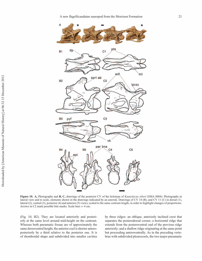
A new flagellicaudatan sauropod from the Morrison Formation 21
Figure 10. A, Photographs and B, C, drawings of the posterior CV of the holotype of Kaatedocus siberi (SMA 0004). Photographs inlateral view and to scale, elements shown in the drawings indicated by an asterisk. Drawings of CV 14 (B), and CV 11 (C) in dorsal (1),lateral (2), ventral (3), posterior (4) and anterior (5) views; scaled to the same centrum length, in order to highlight changes of proportions.Arrows in C2 mark possible bite marks. Scale bars = 4 cm.
(Fig. 10, B2). They are located anteriorly and posteri-
orly at the same level around mid-height on the centrum.
Whereas both pneumatic fossae are of approximately the
same dorsoventral height, the anterior coel is shorter antero-
posteriorly by a third relative to the posterior one. It is
of rhomboidal shape and subdivided into smaller cavities
by three ridges: an oblique, anteriorly inclined crest that
separates the posterodorsal corner; a horizontal ridge that
extends from the posteroventral end of the previous ridge
anteriorly; and a shallow ridge originating at the same point
but proceeding anteroventrally. As in the preceding verte-
brae with subdivided pleurocoels, the two major pneumatic
Dow
nlo
aded
by [
Am
eric
an M
use
um
of
Nat
ura
l H
isto
ry]
at 0
6:3
2 1
5 D
ecem
ber
2012
22 E. Tschopp and O. Mateus
fossae of CV 14 are separated from each other by a ventrally
oriented bony shelf. The latter bears a crest that origi-
nates at the posterodorsal corner of the anterior pneumatic
fossa, and then proceeds ventrally before curving back-
wards to reach the posteroventral corner of the bony shelf.
Here the ridge unites with the ventral edge of the posterior
pneumatic fossa, which is anteroposteriorly elongated and
slightly taller at the front than at the back. The dorsal edge
of the posterior pneumatic fossa is straight and develops
an almost horizontal bony strut at about midlength, which
then proceeds anteriorly. Posteriorly, the dorsal rim exceeds
the main coel to border a smaller depression behind the
posterior pneumatic fossa, as in CV 12 and 13. This smaller
cavity is subtriangular in CV 14, with an acute posterior
end, and an almost vertical anterior rim that separates it
from the main posterior pneumatic fossa. In the middle of
the latter coel, a narrow horizontal crest is visible that does
not connect to other morphological landmarks. It might thus
also represent taphonomic deformation as the median wall
separating the pleurocoels of the two sides at the centrum
midline is extremely thin.
In all posterior cervical vertebrae, a rugose tuberos-
ity is situated at the anterodorsal corner of the lateral
side of the centrum (Fig. 10, B2 and C2). This tuberos-
ity would be in line with a straight, imaginary extension
of the anterior portion of the acdl. In CV 12 and 13 a
weak striation connects these two structures. The only taxon
that shows a similar feature is the probable dicraeosaurid
Suuwassea emilieae (ANS 21122, pers. obs. 2011), but only
in mid-cervical vertebrae (posterior cervicals are unknown
in this taxon; Harris 2006b). The anterodorsal tuberosity
can thus be considered at least a local autapomorphy (within
Diplodocidae) of Kaatedocus siberi.
In ventral view the centrum of the posterior cervical
vertebrae of SMA 0004 is hourglass shaped with a medium
transverse constriction, which becomes less pronounced in
more posterior elements. The surface is marked by two
deep concavities, an anterior one bordered by the condyle
and the two parapophyses, and a posterior one enclosed
by the cotyle and two posteroventral flanges. The anterior
concavity is subdivided by a midline ridge, which is located
in the second quarter of the centrum length, and disappears
at or somewhat anteriorly to the most constricted portion
of the ventral surface. It is more distinct in more posterior
vertebrae. The pair of flanges projects ventrally from each
lateral side of the posteroventral corner of the centrum, but
does not extend to the ventralmost rim of the cotyle. The
flanges seem to clasp the distal end of the cervical ribs in
some elements, but to what degree this is caused by tapho-
nomic deformation is difficult to discern. If this represents
the actual morphology it would probably be another autapo-
morphy of the new taxon. The parapophyses are at least
twice as long as they are dorsoventrally high. They are posi-
tioned below the anterior pneumatic fossa in the first half of
the centrum, and project to a small degree ventrolaterally.
The neural arch undergoes considerable changes
throughout the posterior cervical vertebral series. It
becomes both anteroposteriorly shortened, as well as
dorsally elongated, and wider across the diapophyses
towards more posterior positions. Whereas in CV 11 and 12
the neural arch still slightly surpasses the anterior or poste-
rior rims of the centrum, CV 13 and 14 have subequally
long arches and centra. The neural arch height/greatest
length ratio remains around 30–40% in CV 3 to 11, but
increases to 45% in CV 12, 56% in CV 13, and 60% in the
last preserved vertebra. A similar increase can be observed
in the width across diapophyses/greatest length ratio: this
steadily rises from 30% in CV 6 to 39% in CV 11 and 42%
in CV 12, after which it rises significantly to 59% in CV
13 and 67% in CV 14. In lateral view, also the orientation
of the bifurcated neural spine changes within the posterior
cervical vertebrae. In CV 11 (as also in the mid-cervical
elements), the anterior corner of the spine summit marks
the posteriormost extension of the sprl (Fig. 10, C2). In CV
12, the summit is located more anteriorly, arriving verti-
cally above the posteriormost point of the now considerably
curved sprl. This trend continues in CV 13 and 14, where
the spine tops become even anteriorly inclined.
The prezygapophyseal facets are oblong to subtriangu-
lar, straight laterally and tapering somewhat medioposte-
riorly. They face dorsomedially and are slightly convex
transversely. In anterior view the facets thus form a V. The
distance between the two prezygapophyses is shorter than
the width of a single zygapophysis, but this might be due to
transverse taphonomic compression. The posterior rim of
the articular facet is well marked and bordered by a ridge
followed by a transverse sulcus. To our knowledge such a
sulcus is not present in any other diplodocid species, nor
has it been reported from other sauropod taxa. It can thus be
considered a true autapomorphy of Kaatedocus siberi. On
the lateral aspect of the prezygapophyses of the CV 11 and
12 of SMA 0004, a second, parallel ridge – dorsal to the pre-
epipophysis – progresses horizontally and subparallel to the
articular facets. This second ridge is less conspicuous than
the pre-epipophysis. The upper ridge is confluent with the
sprl posteriorly. As in CV 10, the sdf in CV 11 to 13 forms a
deep cavity posteroventrally to the pre-epipophysis and the
second ridge. The pre-epipophysis extends slightly anterior
to the prezygapophysis and forms the anteriormost point of
CV 11 to 13, while it gets slightly reduced in CV 14. Its ante-
rior end unites with the prdl in CV 11 and 12, but remains
independent in CV 13 and 14. Ventrally, the prezygapoph-
ysis is supported by strong cprl that remain undivided in
CV 11, but show shallow centroprezygapophyseal lamina
fossae in CV 12 to 14. The relative strengths of the medial
and lateral branches of the cprl change from one vertebra to
the other. In all three elements, the right cprl is more divided
than the left, forming a left–right asymmetry. In CV 12 both
branches are subequally developed, CV 13 has a stronger
medial, and CV 14 a slightly better built lateral portion.
Dow
nlo
aded
by [
Am
eric
an M
use
um
of
Nat
ura
l H
isto
ry]
at 0
6:3
2 1
5 D
ecem
ber
2012
A new flagellicaudatan sauropod from the Morrison Formation 23
The division of the cprl was considered synapomorphic
for Diplodocidae (Wilson 2002; Remes 2007; Sereno et al.
2007; Whitlock 2011a). Medially, the prezygapophyses are
united by a thin tprl.
Whereas the sprl is interrupted at the base of the prezy-
gapophysis in CV 11, it can be followed without problems
throughout its entire length in the more posterior elements.
The sprl gets progressively more curved from CV 11 to
14, and almost describes a U-shape in last two preserved
elements. At its dorsal end, just below the neural spine
summit, the sprl develops a rounded (CV 13 and 14) to
rather pointed (CV 11 and 12) anterior bulge, similar to the
state in Diplodocus carnegii CM 84 and Barosaurus lentus
YPM 429 (Hatcher 1901; pers. obs. 2011). This anterior
bulge can thus be considered a diplodocine synapomorphy.
Between the two metapophyses of the divided posterior
neural spine of SMA 0004 the true neural spine is atro-
phied into a median tubercle. Each one of the metapoph-
ysis summits is much longer than broad. In lateral view, the
summit is topped by a horizontal table. The metapophyses
are laminar, and very compressed transversely. Whereas
the lateral surface of the posterior neural spines does not
bear such distinct, dorsoventrally long cavities as in the
mid-cervical vertebrae, CV 11 still exhibits a small ante-
rior indentation, dorsal to the uppermost extension of the
median tubercle (Fig. 10, C2). This depression is somewhat
set back from the sprl and thus is probably not homolo-
gous to the elongated fossa on the posteroventral to the
sprl of the previous elements. More anteriorly, where the
homologous structure would be expected, there is a much
less defined and more rounded depression in CV 11, which
can also be observed in the more posterior elements. The
distal end of the metapophyses is laterally marked by a
rugose surface that extends ventrally to a height just below
the anterior bulge of the sprl. On their medial side the
metapophyses of all posterior cervical vertebrae bear a ridge
formed by a step like structure that extends to meet the
median tubercle. While the spine summit migrates anteri-
orly, the median tubercle remains located above the middle
of the centrum in lateral view. The ridge on the medial
surface of the metapophyses therefore changes its orienta-
tion within the series. In CV 11 and more anterior elements,
it extends anteroventrally to meet the posterior extension of
the median tubercle. In CV 12, it is oriented vertically, and
in CV 13 and 14, it is anteriorly inclined and connects to
the anterior end of the median tubercle.
The diapophysis of the posterior cervical vertebrae is
situated in the second quarter of the vertebra, above the
posterior portion of the anterior pneumatic fossa. It is
axially long but not very projected transversely and bends
ventrally towards its lateral end, forming a gentle curve. The
articular end projects slightly anteroventrally in CV 11 and
12, and vertically in CV 13 and 14. The diapophysis unites
four laminae: the prdl anterodorsally, the podl posterodor-
sally, the acdl anteroventrally, and the pcdl posteroventrally.
Whereas the pcdl as well as the prdl are oriented relatively
straight horizontally in CV 11 to 13, CV 14 has consider-
ably elevated diapophyses and prezygapophyses. Both the
prdl and pcdl are thus distinctly anteriorly inclined. In CV
13 and 14, the prdl borders a deep prcdf together with the
cprl medially and the acdl posteriorly. The prcdf is further
marked by one (CV 13) or two (CV 14) distinct depres-
sions on its medial wall. In all posterior cervical vertebrae,
the acdl originates on the centrum and proceeds subparal-
lel with the podl until it reaches the centre of the ventral
surface of the diapophysis. There it bends to extend verti-
cally outwards onto the diapophysis–tuberculum complex,
so that the dorsal portion of the latter has a subtriangular
cross section. No posterior projection is present in CV 11
and 12, and only a short but pointed spur or a slight rugosity
are visible in the left diapophysis of CV 14 and the right of
CV 13, respectively. The pcdl and the podl of CV 11 have
a small vertical lamina that unites them, with the free edge
facing posteriorly. This accessory lamina thus subdivides
the pocdf into two smaller cavities. The more posterior
elements do not show this feature, but exhibit short ridges
originating around midlength on the dorsal face of the pcdl
and extending vertically before disappearing on the lateral
wall of the neural canal. The cdf of CV 11 to 14 is deeply
invaginated, and marked by a progressively more distinct,
small cavity at its dorsomedial acute corner.
The postzygapophyses form a V in posterior view and
are strongly concave transversely. Dorsally to the postzy-
gapophyses the epipophysis is confluent with the spol, as
well as with a horizontal ridge connecting to the podl that
is subparallel to the postzygapophyseal facet. The epipoph-
ysis forms a pointed posterior projection overhanging the
zygapophysis in CV 11 to 13, but not in CV 14. The artic-
ular facets are distinct from the postzygapophyseal process
dorsally, but grade into the tpol ventrally. The latter connects
the two postzygapophyses ventrally above the midline of the
neural canal, but projects posteriorly and terminates above
the posterior edge of the centrum. It forms a U-shaped notch
in lateral view, with the rounded portion facing anteriorly,
and the two parallel sides being represented by the hori-
zontal dorsal centrum and the posteriorly projecting tpol.
The course of the spol is bipartite in CV 11 and 12, being
straight along most of its extent in both lateral and dorsal
view, until it curves dorsally (in lateral view) to support
the horizontal table of the neural spine summit. In CV 13
and 14, the spol extends anteriorly in a very acute angle to
the podl, to a position above the posteriormost extension of
the pcdl, at about half of its length. There it curves more
dorsally and becomes somewhat convex. Below the summit
table, it bends straight dorsally as in CV 11 and 12.
Cervical ribsThe ribs are well fused to the vertebra, which renders the
distinction between the tuberculum and diapophysis diffi-
cult. The cervical ribs are usually shorter than the centrum
Dow
nlo
aded
by [
Am
eric
an M
use
um
of
Nat
ura
l H
isto
ry]
at 0
6:3
2 1
5 D
ecem
ber
2012

24 E. Tschopp and O. Mateus
length. However, in CV 3, 12 and 14, they protrude slightly
beyond the posterior centrum wall (Fig. 8, C2, C3; Fig. 10,
B2, B3). Due to the dorsoventrally higher posterior than the
anterior end of the vertebral centrum, the ribs – although
protruding somewhat ventrally at the parapophyses – do
not increase the maximum height of the vertebrae to a
significant degree. However, overhanging cervical ribs are
absent in other diplodocid taxa apart from Dinheirosaurus.
The latter and Kaatedocus siberi can thus be considered
plesiomorphic for this trait. Whereas in the anterior cervi-
cal ribs the tuberculum is inclined distinctly posteriorly to
meet the diapophysis, in mid-cervical and posterior cervi-
cal ribs the inclination decreases and CV 13 and 14 have
an almost vertical diapophysis–tuberculum complex. The
proximal end of the tuberculum is slightly subtriangular in
cross section initially but becomes transversely flattened
ventrally. The tuberculum projects at least three times more
than the capitulum which is axially long, dorsoventrally
short and moderately projected medially. All cervical ribs
have a pronounced anterior projection that represents from
about one-fifth of the length of the rib in CV 3, 7 and 11, to
approximately one-third in CV 5 and 8. The rib shaft is often
somewhat deformed but appears to be slightly bowed, with
the anterior and posterior extremes curving dorsally and
often ending above the ventralmost point of the posteroven-
tral flanges of the centrum. In anterior to mid-cervical ribs
the shaft is slender and subcircular to subtriangular in cross
section with the longer side facing medially, and the shorter
sides laterodorsally and ventrolaterally. Posterior elements
are more robust and mediolaterally flattened. The depth of
the shaft is constant in lateral view, so its dorsal and ventral
rims are subparallel until they taper at their ends. The only
exceptions are CV 8 to 10 where the cervical ribs show
a distinct dorsoventral expansion shortly after midlength,
before they taper towards their posterior extreme. A few
cervical ribs show special morphologies that do not appear
on other elements: cervical rib 4 exhibits a distinct bend
and slightly rugose area that probably represents reactive
bone growth after a fracture of the cervical rib shaft (see
Online Supplementary Material). Furthermore, the lateral
side of cervical rib 11 exhibits two slot-like depressions on
the right ventrally to the tuberculum (Fig. 10, C2, arrows).
These might represent bite marks, which would be the only
ones in the specimen.
Phylogenetic analysisThe phylogenetic analysis is based on Whitlock (2011a;
modified by Mannion et al. 2012), with the addition of some
characters from Upchurch et al. (2004b), Harris (2006c),
Lovelace et al. (2007), Whitlock & Harris (2010), Whit-
lock (2011a) and 23 newly defined characters based on
the description of SMA 0004 (see Online Supplementary
Material). Previously performed preliminary analyses using
unchanged existing matrices (Upchurch et al. 2004b; Harris
2006c; Whitlock 2011a) recovered SMA 0004 consistently
inside Diplodocinae (ET, unpublished). Therefore, several
changes concerning terminal taxa were introduced. The
incomplete basal diplodocoid Haplocanthosaurus and all
rebbachisaurids except for Limaysaurus were omitted as
their positions in the past have been respectively contro-
versial (Wilson 2002; Upchurch et al. 2004a) or poorly
resolved (Mannion et al. 2012). The relatively complete
Nigersaurus and Brachytrachelopan were deleted from the
matrix because they represent very specialized forms within
Rebbachisauridae and Dicraeosauridae, respectively, and
therefore bear little additional information on diplodocid
ingroup relationships. Furthermore, outgroup taxa with
respect to Diplodocoidea were reduced to the very well-
known basal macronarian Camarasaurus and the non-
neosauropod eusauropod Omeisaurus. Within Diplodoci-
dae, the multi-species genera Apatosaurus and Diplodocus
are considered monophyletic, which was supported for
Apatosaurus in the specimen-based phylogenetic analysis
of Upchurch et al. (2004b). Additional coding for modified
and new characters of Apatosaurus was based on A. louisae,
as this species is the most complete and best reported
in the genus. In contrast, Diplodocus is still pending a
detailed review of its species-level taxonomy. As this would
exceed the scope of the current study, we have adopted the
coding of the original studies for the unmodified characters
(with some changes mentioned and explained in the Online
Supplementary Material) but concentrate on D. carnegii
in the coding for the modified and new characters. The
final analysis thus includes 14 taxa and 234 characters. The
sources for the modified scoring in original characters as
well as the coding in the modified and new characters are
given in Online Supplementary Material Table S1.
Two different analyses were conducted using the heuris-
tic search in WinClada 1.00.08 with 50 replicates. In the
first, all multistate characters were treated as unordered,
whereas in the second run, nine of the total 22 multi-
state characters were ordered, seven based on the fact that
they describe either continuous ratios or vertebral counts.
The remaining two ordered characters are 34 and 97. The
former describes the laterally projecting spur, which is
most probably an extension of the dorsoventrally elon-
gated lateral ridge (character state 1) present in, for exam-
ple, Diplodocus. Character 97 describes the bifurcation of
the cervical vertebrae, which starts in anterior elements in
Diplodocus but more posteriorly in SMA 0004 and other
taxa. The bifurcation is considered to happen continuously
from the back to the front (Woodruff & Fowler 2012) during
ontogeny. Basing on Haeckel’s rule that phylogeny recapitu-
lates ontogeny, the development of this bifurcation in adult
specimens should replay this continuous change, and an
ordering of this character therefore appears reasonable.
Both analyses produced the same results (Fig. 11). Kaate-
docus siberi is therefore recovered within Diplodocinae, as
sister group to a polytomy including Tornieria africana +
Barosaurus lentus + Diplodocus. The recovered Nelsen
Dow
nlo
aded
by [
Am
eric
an M
use
um
of
Nat
ura
l H
isto
ry]
at 0
6:3
2 1
5 D
ecem
ber
2012

A new flagellicaudatan sauropod from the Morrison Formation 25
Fig
ure
11
.N
else
nC
on
sen
sus
tree
ob
tain
edfr
om
ah
euri
stic
sear
chin
Win
Cla
da
(six
mo
stpar
sim
on
iou
str
ees;
tree
len
gth
=388;
CI
=6
4%
;R
I=
57%
).D
ots
indic
ate
ambig
uous
(whit
e)an
dunam
big
uous
(bla
ck)
synap
om
orp
hie
s,an
dau
tapom
orp
hie
sof
the
resp
ecti
ve
clad
e.T
he
corr
espondin
gch
arac
-te
rnum
ber
and
scori
ng
isin
dic
ated
abov
ean
dbel
owth
edots
,re
spec
tivel
y.M
ain
clad
esar
ein
dic
ated
by
thei
rnam
e,an
dboots
trap
asw
ell
asB
rem
erS
upport
val
ues
are
giv
enfo
rea
chn
od
e(B
rem
erS
up
po
rtin
squ
are
bra
cket
s).
Ka
ate
do
cus
sib
eri
isre
solv
edas
Dip
lodoci
nae
more
bas
alth
anTorn
ieri
aafr
ica
na
,B
aro
sau
rus
len
tus
and
Dip
lodocu
s.
Dow
nlo
aded
by [
Am
eric
an M
use
um
of
Nat
ura
l H
isto
ry]
at 0
6:3
2 1
5 D
ecem
ber
2012
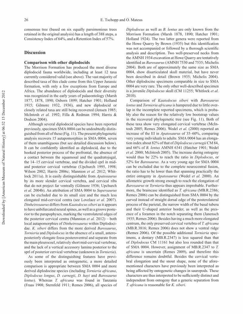
26 E. Tschopp and O. Mateus
consensus tree (based on six equally parsimonious trees
retained in the original analysis) has a length of 388 steps, a
Consistency Index of 64%, and a Retention Index of 57%.
Discussion
Comparison with other diplodocidsThe Morrison Formation has produced the most diverse
diplodocid fauna worldwide, including at least 12 taxa
currently considered valid (see above). The vast majority of
described taxa of this clade come from this Upper Jurassic
formation, with only a few exceptions from Europe and
Africa. The abundance of diplodocids and their diversity
was recognized in the early years of palaeontology (Marsh
1877, 1878, 1890; Osborn 1899; Hatcher 1901; Holland
1915; Gilmore 1932, 1936), and new diplodocid or
closely related taxa are still being recovered (Jensen 1985;
McIntosh et al. 1992; Filla & Redman 1994; Harris &
Dodson 2004).
Although several diplodocid species have been reported
previously, specimen SMA 0004 can be undoubtedly distin-
guished from all of these (Fig. 11). The present phylogenetic
analysis recovers 15 autapomorphies in SMA 0004, seven
of them unambiguous (but see detailed discussion below).
It can be confidently identified as diplodocid, due to the
hooked posterior process of the prefrontal, the absence of
a contact between the squamosal and the quadratojugal,
the 14–15 cervical vertebrae, and the divided cprl in mid-
and posterior cervical vertebrae (Upchurch 1995, 1998;
Wilson 2002; Harris 2006c; Mannion et al. 2012; Whit-
lock 2011a). It is easily distinguishable from Apatosaurus
by its more slender cervical vertebra, and cervical ribs
that do not project far ventrally (Gilmore 1936; Upchurch
et al. 2004b). An attribution of SMA 0004 to Supersaurus
can be excluded due to its small size and the much less
elongated mid-cervical centra (see Lovelace et al. 2007).
Dinheirosaurus differs from Kaatedocus siberi as it appears
to have unbifurcated neural spines, as well as a groove poste-
rior to the parapophyses, marking the ventrolateral edges of
the posterior cervical centra (Mannion et al. 2012) – both
local autapomorphies of Dinheirosaurus within Diplodoci-
dae. K. siberi differs from the more derived Barosaurus,
Tornieria and Diplodocus in the absence of a small, antero-
posteriorly elongate fossa posteroventral and separate from
the main pleurocoel, relatively short mid-cervical vertebrae,
and the lack of a vertical accessory lamina posterior to the
sprl of posterior cervical vertebrae (unknown in Tornieria).
As some of the distinguishing features have previ-
ously been interpreted as ontogenetic, a more detailed
comparison is appropriate between SMA 0004 and more
derived diplodocine species (including Tornieria africana,
Diplodocus longus, D. carnegii, D. hayi and Barosaurus
lentus). Whereas T. africana was found in Tanzania
(Fraas 1908; Sternfeld 1911; Remes 2006), all species of
Diplodocus as well as B. lentus are only known from the
Morrison Formation (Marsh 1878, 1890; Hatcher 1901;
Holland 1924). The two latter genera were reported from
the Howe Quarry by Brown (1935) but this identification
was not accompanied or followed by a thorough scientific
analysis and description. Two well-preserved necks from
the AMNH 1934 excavation at Howe Quarry are tentatively
identified as Barosaurus (AMNH 7530 and 7535; Michelis
2004). Both are of approximately the same size as SMA
0004, show disarticulated skull material, but have never
been described in detail (Brown 1935; Michelis 2004).
Other diplodocine specimens comparable in size to SMA
0004 are very rare. The only other well-described specimen
is a juvenile Diplodocus skull (CM 11255; Whitlock et al.
2010).
Comparison of Kaatedocus siberi with Barosaurus
lentus and Tornieria africana is hampered due to little over-
lap in the incomplete reported specimens, which is proba-
bly also the reason for the relatively low bootstrap values
in the recovered phylogenetic tree (see Fig. 11). Both of
these taxa show very elongated cervical vertebrae (McIn-
tosh 2005; Remes 2006). Wedel et al. (2000) reported an
increase of the EI in Apatosaurus of 35–60%, comparing
very young individuals to adults. SMA 0004 has an elonga-
tion index about 82% of that of Diplodocus carnegii CM 84,
and 66% of B. lentus AMNH 6341 (Hatcher 1901; Wedel
et al. 2000; McIntosh 2005). The increase during ontogeny
would thus be 22% to reach the ratio in Diplodocus, or
52% for Barosaurus. As a very young age for SMA 0004
can be excluded due to the complete neurocentral fusion,
the ratio has to be lower than that spanning practically the
entire ontogeny in Apatosaurus (Wedel et al. 2000). An
allometric growth strong enough to reach the elongation of
Barosaurus or Tornieria thus appears improbable. Further-
more, the braincase identified as T. africana (MB.R.2386;
Remes 2006) can be distinguished from SMA 0004 by the
curved instead of straight dorsal edge of the posterolateral
process of the parietal, the narrow width of the basal tubera
and their U-shaped anterior border, as well as the pres-
ence of a foramen in the notch separating them (Janensch
1935; Remes 2006). Besides having a much more elongated
centrum, the only preserved cervical vertebra of T. africana
(MB.R.3816; Remes 2006) does not show a ventral ridge
(Remes 2006). Of the possible additional Tornieria spec-
imens, a dentary (MB.R.2347) is less squared than that
of Diplodocus CM 11161 but also less rounded than that
of SMA 0004. However, assignment of MB.R.2347 to T.
africana is uncertain (Remes 2009), and therefore this
difference remains doubtful. Besides the cervical verte-
bral elongation and the snout shape, none of the afore-
mentioned characters have previously been interpreted as
being affected by ontogenetic changes in sauropods. These
characters are thus interpreted to be sufficiently distinct and
independent from ontogeny that a generic separation from
T. africana is reasonable for K. siberi.
Dow
nlo
aded
by [
Am
eric
an M
use
um
of
Nat
ura
l H
isto
ry]
at 0
6:3
2 1
5 D
ecem
ber
2012

A new flagellicaudatan sauropod from the Morrison Formation 27
A separation of Kaatedocus siberi from Barosaurus
lentus is equally well supported. Apart from the improb-
able enormous allometric growth necessary for the cervical
vertebrae of SMA 0004 to reach the elongation index of B.
lentus (McIntosh 2005), three more morphological charac-
ters can be put forward to distinguish these two taxa (plus
the unambiguous autapomorphies of K. siberi). B. lentus
YPM 429 exhibits a bifurcate anterior end of the pcdl, and
postzygapophyses that terminate anterior to the posterior
margin of the posterior cervical centra (Lull 1919; pers.
obs. 2011). Furthermore, the ventral keels in the cervi-
cal vertebrae of the B. lentus holotype YPM 429 show a
quite different morphology from the single anterior ridge
in SMA 0004: in YPM 429 two crests extend obliquely from
between the parapophyses posterolaterally to unite with the
posteroventral flanges (Lull 1919; pers. obs. 2011). Adding
the recovered autapomorphies of K. siberi, a generic distinc-
tion from Barosaurus can be justified.
Diplodocus is the most abundant diplodocine sauropod in
the Morrison Formation. The initial provisional identifica-
tion of SMA 0004 as Diplodocus by Ayer (2000) shows that
a separation from this taxon is the most difficult. Diplodocus
is the only diplodocine for which a juvenile skull has been
reported (CM 11255), and this specimen superficially looks
much like SMA 0004 (Whitlock et al. 2010). Differences
between CM 11255 and Diplodocus skulls of older individ-
uals (CM 3452 and 11161) include a rounder snout shape,
maxillary teeth that reach further posteriorly, and a rela-
tively larger orbit (Whitlock et al. 2010). These are traits
that also distinguish Kaatedocus siberi from Diplodocus.
More detailed comparisons of SMA 0004 with the subadult
and juvenile Diplodocus skulls CM 3452 and 11255 show
that some of the recovered autapomorphies of K. siberi are
actually shared with the latter specimens, and might there-
fore be ontogenetic. These include the lateral spur on the
lacrimal, the ridge on the paroccipital processes, and the
straight orientation of the anterior edge of the basal tubera,
traits present in both CM 3452 and 11255 but absent in
adult Diplodocus skulls (AMNH 969; CM 11161; USNM
2672, 2673; pers. obs. 2011). Furthermore, the shallow
fossa medial to the pterygoid ramus is also observable
in CM 3452, but neither in CM 11255 nor in the above-
mentioned adult specimens (pers. obs. 2011). Other more
widespread features shared with the juvenile and absent
in the adult stages are the prefrontal that does not reach
far posteriorly, and relatively more elongate frontals in CM
3452 and SMA 0004 (both unknown in CM 11255). The
shallow groove that accommodates both the subnarial and
the anterior maxillary foramen in CM 3452, 11161, as well
as USNM 2672, is lacking in CM 11255 and SMA 0004
(Whitlock et al. 2010; pers. obs. 2011). The basipterygoid
processes of SMA 0004 resemble more their correspond-
ing structures in CM 11255, than in CM 3452 and 11161.
In the latter, subadult to adult specimens, the processes
are straight along their entire extent, and without a curved
shelf that connects the base of the processes. In SMA 0004
and CM 11255, such a shelf is present, and throughout its
extent, it keeps the processes subparallel in ventral view,
before they curve laterally.
Other osteological features in SMA 0004 more closely
resemble adult Diplodocus skulls than CM 11255. The
squamosal of CM 11255 was shown to have an elon-
gated anterior process that almost contacts the quadrato-
jugal, whereas in adult skulls (and also in SMA 0004)
these two bones are well separated (Whitlock et al. 2010;
CM 11161; USNM 2672, 2673, pers. obs. 2011). Further-
more, the dorsal margin of the quadrate is concave in lateral
view in CM 11255 but fairly straight in adult Diplodocus
and Kaatedocus siberi (Whitlock et al. 2010; CM 11161;
USNM 2672; pers. obs. 2011). The foramen present on the
surangular in SMA 0004 appears to be lacking in CM 11255
(Whitlock et al. 2010), which indicates that in Diplodocus
this foramen only develops during ontogeny.
Despite these similarities, several features present in
SMA 0004 but absent in any Diplodocus skull indicate that
the specimen described here is distinct from Diplodocus.
The location of the frontal–parietal suture is more similar
to its position in MB.R.2386 than in the Diplodocus skulls
CM 11161 and 11255. Whereas in the Diplodocus skulls
the suture is quite anteriorly placed with respect to the
supratemporal fenestra, in both SMA 0004 and MB.R.2386
it is situated more posteriorly, around the centre of the
opening in dorsal view (Remes 2006; Whitlock et al. 2010;
pers. obs. 2011). The basal tubera are closer to the occip-
ital condyle in SMA 0004, resembling more the state in
Suuwassea emilieae ANS 21122 than in Diplodocus skulls
CM 11161 and 11255 (Harris 2006a; Whitlock et al. 2010;
pers. obs. 2011). There is no indication of a basipterygoid
recess posterior to the basal tubera, a trait previously used
to distinguish Apatosaurus from Diplodocus where such a
recess is present (Wilson 2002; Whitlock et al. 2010). Addi-
tionally, SMA 0004 has a closed preantorbital fossa, similar
to the state in Dicraeosaurus hansemanni MB.R.2336. As
both Apatosaurus (CM 11162) and Diplodocus (includ-
ing the juvenile CM 11255) show a distinct, open, oval
preantorbital fenestra (Berman & McIntosh 1978; Whit-
lock et al. 2010), the retention of the plesiomorphic state
in SMA 0004 can be considered taxonomically important.
Furthermore, the tooth count in both the maxillae and the
dentaries of SMA 0004 is higher than usual for Diplodocus
(12–13 versus 9–11; Holland 1924; Barrett & Upchurch
1994; Calvo 1994; Whitlock et al. 2010; CM 11161, 11255,
pers. obs. 2011), and appears equal to Apatosaurus CM
11162 (Berman & McIntosh 1978; Calvo 1994; pers. obs.
2011). Although a reduction in the number of teeth during
ontogeny was proposed for Camarasaurus (McIntosh et al.
1996), the fact that CM 11255 shows the average number
of teeth known in adult Diplodocus skulls indicates that
the higher number in SMA 0004 most probably represents
taxonomic diversity.
Dow
nlo
aded
by [
Am
eric
an M
use
um
of
Nat
ura
l H
isto
ry]
at 0
6:3
2 1
5 D
ecem
ber
2012

28 E. Tschopp and O. Mateus
Possible diplodocid species from North America that
were not included in this analysis are Eobrontosaurus
yahnahpin, Dystrophaeus viaemalae and Dyslocosaurus
polyonychius. However, E. yahnahpin has previously been
interpreted as being a camarasaurid (Upchurch et al.
2004a), and thus most probably cannot be considered a
diplodocine. Moreover, a distinction is easily made using
the elongated neural spines in the anterior cervical verte-
brae and the widely transversely projecting parapophyses
(Filla & Redman 1994; P. Mannion, pers. comm. 2011).
Comparisons with the other two taxa are impossible as they
are very fragmentary and do not show any overlap (Cope
1877; McIntosh et al. 1992). Nonetheless, both remains
are of larger animals than SMA 0004, and the difference
appears too much to be explained by individual or sexual
variation, even considering that SMA 0004 might still have
been in the growth phase.
Autapomorphies of Kaatedocus siberi. The recovered
autapomorphies of Kaatedocus siberi are discussed in detail
below. As the discussion will show, some of these features
are actually shared with farther related taxa that were not
included into the present phylogenetic analysis, or with
single specimens of included genera. They were therefore
excluded or defined as local autapomorphies in the diagno-
sis of K. siberi (see above).
The U-shaped notch anteriorly between the frontals is
recovered as an unambiguous autapomorphy. Diplodocid
skulls usually have frontals that touch and fuse along their
entire medial edge so that their anterior borders build one
single straight line that connects to the nasals (Berman &
McIntosh 1978; Wilson & Sereno 1998; Whitlock et al.
2010). In SMA 0004 the medial margin of the frontals
curve laterally in their anterior half. A similar morphol-
ogy can be seen in the partial skull of Spinophorosaurus
nigerensis (Knoll et al. 2012), the holotype specimen of
Diplodocus hayi HMNS 175 but in these specimens it is
V- and W shaped, respectively, and not U-shaped as in
Kaatedocus siberi. D. hayi has previously been doubted
as congeneric with Diplodocus (Foster 1998). The differ-
ence in the frontals indicates that this hypothesis might
prove to be correct, and its similarity to SMA 0004 could
imply that HMNS 175 should group with Kaatedocus
siberi. On the other hand several differences in the rest
of the skull (e.g. orientation of the basipterygoid processes)
and also the cervical vertebrae (e.g. dorsally expanded
bifid neural spines already in anterior cervical vertebrae)
preclude an assignment of SMA 0004 to D. hayi (Holland
1906, 1924; HMNS 175, pers. obs. 2010). The only partially
conjoined frontals in SMA 0004 could also be interpreted
as not entirely fused, and indicate an early juvenile age
for the animal. However, embryonic skulls of the basal
sauropodomorph Massospondylus show tightly appressed
right and left frontals along their entire medial edge (Reisz
et al. 2005), and the subadult skull of the titanosauriform
Bonitasaura salgadoi has a frontal with an entirely straight
medial margin (Gallina & Apesteguıa 2011). This indicates
that the outwards curve in the frontal of SMA 0004 is not an
ontogenetic feature but is instead taxonomically significant
and an unambiguous autapomorphy of K. siberi.
The laterally projecting spur is another unambiguous
autapomorphy as recovered from the phylogenetic analy-
sis. However, as stated above, the juvenile and subadult
Diplodocus skulls CM 11255 and 3452 also show this
feature, and therefore the influence of ontogeny cannot be
ruled out, even though bony spurs and an increased develop-
ment of ridges and crests are usually interpreted to be typical
of older individuals (Varricchio 1997). Given that lacrimals
are unknown in Suuwassea, Supersaurus, Dinheirosaurus,
Tornieria and Barosaurus, a decision on the taxonomic
importance of this morphological feature remains diffi-
cult. Furthermore, a similar spur is present in the cama-
rasaurid SMA 0002 and some specimens mentioned in
Madsen et al. (1995) as ‘Camarasaurus-like’. An interpre-
tation of this lacrimal spur as locally autapomorphic within
Diplodocoidea might thus be possible but must await further
finds of definitively adult specimens of K. siberi, or juvenile
skulls of more diplodocid taxa.
A third recovered unambiguous autapomorphy of Kaate-
docus siberi is the small fossa present medially to the sheet-
like pterygoid ramus of the quadrate. However, this char-
acter is also present in the subadult skull CM 3452. Its
absence in both juvenile and adult Diplodocus specimens
(CM 11161, 11255; USNM 2672, 2673; pers. obs. 2011)
might imply that this feature is only developed in subadult
specimens. A similar development can also be seen in the
large quadrates belonging to the holotype of Apatosaurus
ajax (YPM 1860), which also appears to be a juvenile to
subadult specimen (McIntosh 1990). The development of
such a medial quadrate cavity in subadult stages might thus
be a synapomorphy of the entire Diplodocidae, and its inter-
pretation as an autapomorphy of Kaatedocus siberi cannot
be entertained with certainty at present.
The short anterior process of the squamosal appears as
a local autapomorphy within Diplodocoidea. This process
exceeds the posterior border of the orbit considerably in
all known diplodocoid skulls, and even extends beyond
the anterior orbital edge in the rebbachisaurs Limaysaurus
tessonei and Nigersaurus taqueti (Calvo & Salgado 1995;
Sereno et al. 2007). The retention of a short anterior process
therefore appears to be a real local autapomorphy of Kaate-
docus siberi.
The presence of a postparietal foramen is an ambigu-
ous autapomorphy shared with Dicraeosaurus, Amar-
gasaurus and Suuwassea in the present data matrix. It has
thus been traditionally interpreted as a synapomorphy of
Dicraeosauridae (Salgado & Bonaparte 1991; Remes 2009;
Whitlock 2011a). Upchurch et al. (2004a) also reported
a postparietal foramen in Tornieria but of the braincases
found at Tendaguru, Tanzania, only MB.R.2387 shows such
Dow
nlo
aded
by [
Am
eric
an M
use
um
of
Nat
ura
l H
isto
ry]
at 0
6:3
2 1
5 D
ecem
ber
2012

A new flagellicaudatan sauropod from the Morrison Formation 29
a foramen (Janensch 1935; Remes 2009; pers. obs. 2011).
MB.R.2387 has subsequently been identified as Flagelli-
caudata indet. as it could not be confidently referred to
Tornieria based on the situation of the quarry, and because
it shows a mix of dicraeosaurid and diplodocid charac-
ters (Remes 2009). The morphology of the foramen in
SMA 0004 strongly resembles its corresponding structure
in MB.R.2387, where it is considerably smaller than in
Suuwassea and Dicraeosaurus. The presence of this fora-
men in SMA 0004 might also be due to incomplete fusion
of the parietals in this specimen and thus be ontogenetic.
However, the small Diplodocus skull CM 11255 does not
show such a foramen, which is yet another characteristic
that helps distinguish these two taxa. Awaiting a definitive
taxonomic assignment of MB.R.2387, and finds of skulls
of Supersaurus, Dinheirosaurus and Barosaurus, the post-
parietal foramen in K. siberi is provisionally regarded as a
local autapomorphy within Diplodocidae.
The distinct oblique posterior ridges on the paroccipital
processes of SMA 0004 are another recovered unambiguous
autapomorphy, which is actually shared with the juvenile
Diplodocus specimens (see above). A detailed analysis of
the development and distribution of this character among
juvenile to subadult individuals of different species has thus
to be postponed until more material is found and described.
As with the postparietal foramen, the narrow and distinct
sagittal nuchal crest is also a shared feature of Kaate-
docus siberi and Dicraeosauridae (Salgado & Bonaparte
1991; Harris 2006a; Mannion et al. 2012; Whitlock
2011a). Furthermore, the indeterminate flagellicaudatan
MB.R.2388 also exhibits a similar shape of the nuchal
crest (Remes 2009). The state of this character is unknown
in Supersaurus, Dinheirosaurus and Barosaurus as their
skeletons are only known from postcranial material. The
occurrence of such a distinct nuchal crest in SMA 0004
is the first reported for any diplodocid, and like the pres-
ence of a postparietal foramen, this feature was previously
interpreted as a synapomorphy of the Dicraeosauridae.
However, the low and broad nuchal crest of Apatosaurus
and diplodocines more derived than K. siberi indicates that
either the acquisition of this feature, or its loss, happened
twice independently within Flagellicaudata. Interestingly,
in contrast to the adult Diplodocus skull CM 11161, the
juvenile Diplodocus CM 11255 does show a more devel-
oped sagittal nuchal crest (Whitlock et al. 2010). This
implies that the high nuchal crest can also be an onto-
genetic character that decreased in size during growth. This
would mean that the well-developed crest in SMA 0004
could still become somewhat weaker and broader during
ontogeny, and thus approach the state in Tornieria africana
or Diplodocus. However, the development of the crest in
CM 11255 does not equal its counterpart in SMA 0004,
and such a pronounced change from a subadult to the adult
stage seems improbable. Moreover, the opposite develop-
ment has been shown to happen in Massospondylus and
Psittacosaurus (Gow 1990; Varricchio 1997), and the
closely related dicraeosaurids also show a well-developed
crest in adult individuals. Therefore, even though it seems
to be an ontogenetic feature in Diplodocus this might be
different in K. siberi, of which the derived state can be
considered locally autapomorphic within Diplodocidae.
The straight anterior edge of the basal tuber is shared with
Limaysaurus tessonei in the present analysis. However, it is
equally expressed in both juvenile and subadult Diplodocus
(CM 11255, 3452, pers. obs. 2011), as well as Nigersaurus
taqueti (Sereno et al. 2007). It might thus be that juve-
niles and subadult Diplodocus retain the plesiomorphic
trait present in the more basal rebbachisaurs. The deci-
sion on how the appearance of this character in Kaatedocus
siberi should be treated (ontogenetic character or retained
plesiomorphy) should await future finds of adult Kaatedo-
cus or juvenile basal diplodocid specimens.
The more rounded snout shape, with a
premaxilla–maxilla index (PMI; see Whitlock 2011b
for a detailed explanation) of less than 70% is shared
with the juvenile specimen CM 11255 and CMC VP 8300
(Whitlock et al. 2010). With a PMI of 68%, Kaatedocus
siberi is slightly beneath the border set by Whitlock
(2011a) to define the plesiomorphic state. Based on the
reconstructions of Whitlock (2011b), the PMI in SMA
0004 is very close to that of Tornieria (71%; Whitlock
2011b), which falls in the gap of the borders set for this
character, but would rather group with the plesiomorphic
taxa. Suuwassea is considered to have a slightly higher
index of 74% by the same author, and is thus closer to
the apomorphic state. These ratios are not included in this
analysis as the reconstructions in Whitlock (2011b) are
based on incomplete material. Nonetheless, they describe
a general trend towards gradually more squared snouts
during the evolution of Diplodocoidea, becoming extreme
in Nigersaurus, Apatosaurus and Diplodocus (Whitlock
2011b). The retention of the plesiomorphic state in K.
siberi and probably Tornieria therefore appears to be the
exception to this rule, but the rounded snout in the juvenile
Diplodocus skulls CM 11255 and CMC VP 8300 indicates
that this might also be an ontogenetic feature (Whitlock
et al. 2010). CM 11255 has a PMI of only 56%, which
is even lower than in Camarasaurus or Brachiosaurus
(Whitlock et al. 2010; Whitlock 2011b). Even though this
might be partly due to transverse compression of the skull,
it implies that juvenile Diplodocus develop the typical
squared snout only during ontogeny. However, as Tornieria
appears to show a similar value, a coding of K. siberi as
plesiomorphic in this character can be justified, and can be
regarded as local autapomorphy of K. siberi, and perhaps
Tornieria too.
The anteriorly projecting pre-epipophysis that forms the
anteriormost point of the entire mid-cervical vertebrae
is another unambiguous autapomorphy in the phyloge-
netic analysis. Its distribution is unknown in Supersaurus,
Dow
nlo
aded
by [
Am
eric
an M
use
um
of
Nat
ura
l H
isto
ry]
at 0
6:3
2 1
5 D
ecem
ber
2012

30 E. Tschopp and O. Mateus
Dinheirosaurus and Tornieria, but the preserved vertebrae
in the first two of these taxa indicate that such a spur was
probably not present. This feature was described as autapo-
morphic within Diplodocidae in Australodocus by Remes
(2007), where it is very pronounced, and is also present in
Haplocanthosaurus (Hatcher 1903). As Australodocus is
currently considered a titanosauriform, the spur is herein
interpreted to be a local autapomorphy of K. siberi within
the clade uniting all diplodocoids more derived than Haplo-
canthosaurus. An alternative interpretation of this spur as
a juvenile character is improbable as such traits usually
develop late in ontogeny (Varricchio 1997).
The small, rugose tuberosity placed anterodorsally to
the anterior pneumatic fossa on the lateral surfaces of
the posterior cervical vertebrae represents another unam-
biguous autapomorphy of Kaatedocus siberi. Comparisons
with various diplodocid specimens (e.g. Apatosaurus CM
3018; Diplodocus CM 84, DMNS 492, 1494, HMNS 175;
Barosaurus AMNH 6341, YPM 429) indicate that this
feature is unique in K. siberi. The only taxon with a simi-
lar trait is the basal dicraeosaurid Suuwassea, where the
mid-cervical vertebrae bear a tubercle in the same posi-
tion (ANS 21122, pers. obs. 2011). The fragmentary poste-
rior cervical centra of ANS 21122 do not preserve this
region in enough detail to discern their state. Therefore,
an interpretation of this feature as local autapomorphy of
K. siberi within Diplodocidae is relatively well supported,
only lacking information about its distribution in Super-
saurus, Dinheirosaurus and Tornieria. Dinheirosaurus ML
414 shows a fractured surface in this region, and the only
preserved cervical vertebra of Tornieria africana is badly
crushed, so the presence or absence of this tubercle cannot
be determined in these taxa.
The sulcus posterior to the prezygapophyseal facets of
the posterior cervical vertebrae is considered an addi-
tional unambiguous autapomorphy in the present analy-
sis. Personal observations showed this trait to be absent
in Suuwassea, Apatosaurus, Diplodocus and Barosaurus
(AMNH 550; ANS 21122; CM 84, 94, 3018; YPM 429,
1860). In the holotype material of Dinheirosaurus lourin-
hanensis (ML 414) the prezygapophysis is partly covered
by matrix and somewhat distorted but such a sulcus does
not appear to be present. Furthermore, the detailed descrip-
tion of the cervical vertebrae of Dicraeosaurus (Janensch
1929) does not mention any similar structure in these taxa.
In Nigersaurus, as a representative of a more distantly
related diplodocoid, the distinct articular facets of the
prezygapophysis are well offset from the prezygapophy-
seal process, but no transverse sulcus is present (Sereno
et al. 2007). Neither the presence nor the absence of such
sulci has previously been considered valuable for charac-
terizing either adults or juveniles. This autapomorphy of
Kaatedocus siberi can thus be assumed to unambiguous.
The posteriorly facing accessory lamina between the
pcdl and the podl is shared between Kaatedocus siberi and
Dinheirosaurus. However, such a lamina is also present in
the Apatosaurus specimens UW 15556, YPM 1840, 1860
and 1861 (Gilmore 1936; Wedel & Sanders 2002; pers. obs.
2011). This character is thus not considered an autapomor-
phy of K. siberi.
The narrowly diverging neural spines of SMA 0004
resemble more closely the state in dicraeosaurids than
in diplodocids. The possibility cannot be excluded that
this character was affected and exaggerated by taphon-
omy; therefore, a question mark has to be placed over
its designation as a local autapomorphy within Diplodoci-
dae. However, as the diapophysis of CV 14 in particular
does not seem to be highly deformed, a narrower angle
between the metapophyses can still be assumed. On the
other hand, the supposed Barosaurus sp. CM 11984 also
exhibits less widely diverging neural spines compared to
other diplodocids. A decision on the validity of this autapo-
morphy is thus not possible yet.
The cervical ribs that in some vertebrae exceed the poste-
rior end of the centra are another ambiguous autapomor-
phy of Kaatedocus siberi. The only other diplodocoids
with the same trait are Supersaurus and Dinheirosaurus,
which are recovered as a sister group to Diplodocidae in the
present phylogenetic analysis (contrary to previous analy-
ses, see below). Elongated cervical ribs were also reported
in Eobrontosaurus yahnahpin, the taxonomic affinity of
which has yet to be resolved. At present, this feature thus
represents a local autapomorphy within Diplodocidae.
To summarize, Kaatedocus siberi can be confidently
identified by three general unambiguous autapomorphies:
(1) the U-shaped notch between the frontals; (2) the lateral
rugose tubercle on anterodorsal corner of posterior cervical
centra; and (3) the sulcus bordering the prezygapophyseal
facets posteriorly in posterior cervical vertebrae. Further-
more, the diagnosis is strengthened with local autapomor-
phies or retained plesiomorphies (see above).
Phylogenetic implicationsIn the majority of the phylogenetic analyses on Sauropoda,
the clade Diplodocidae was well resolved and easily distin-
guishable from other clades. This was mainly due to the
inclusion of a limited number of diplodocid taxa, using
only the best-known genera Apatosaurus, Diplodocus and
Barosaurus (Upchurch 1995, 1998; Wilson & Sereno 1998;
Wilson 2002, 2005; Upchurch et al. 2004a). Recent stud-
ies have looked in detail at the intra-relationships of this
group, including more incomplete taxa, even single speci-
mens such as Supersaurus, ‘Seismosaurus’ (Lovelace et al.
2007), and different apatosaur species (Upchurch et al.
2004b). Only two detailed phylogenetic analyses have
included a larger set of ingroup taxa, representing the
most inclusive diplodocid phylogenies published to date
(Mannion et al. 2012; Whitlock 2011a). Although Whitlock
(2011a) helped in resolving uncertainties within the more
basal diplodocoid clade Rebbachisauridae, Diplodocidae
Dow
nlo
aded
by [
Am
eric
an M
use
um
of
Nat
ura
l H
isto
ry]
at 0
6:3
2 1
5 D
ecem
ber
2012
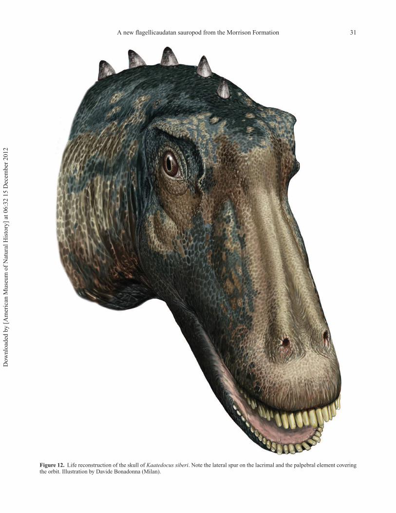
A new flagellicaudatan sauropod from the Morrison Formation 31
Figure 12. Life reconstruction of the skull of Kaatedocus siberi. Note the lateral spur on the lacrimal and the palpebral element coveringthe orbit. Illustration by Davide Bonadonna (Milan).
Dow
nlo
aded
by [
Am
eric
an M
use
um
of
Nat
ura
l H
isto
ry]
at 0
6:3
2 1
5 D
ecem
ber
2012

32 E. Tschopp and O. Mateus
was not fully resolved, with Dinheirosaurus, Tornieria and
a clade comprising the classical diplodocines Diplodocus
and Barosaurus forming a trichotomy. Mannion et al.
(2012), based on Whitlock’s (2011a) matrix, redescribed
Dinheirosaurus in detail and were therefore able to update
and correct some character states used in the earlier
study. Dinheirosaurus was recovered as a sister taxon
to Supersaurus, and together they form the most basal
subclade within Diplodocinae, followed by Tornieria and
(Diplodocus + Barosaurus). The addition of Kaatedocus
and several new characters to the matrix corroborates this
result in parts (Fig. 11): in the Nelsen consensus tree, the
clade comprising Dinheirosaurus and Supersaurus is recov-
ered more basal than Apatosaurus – and would therefore
form the basalmost clade within Diplodocidae (following
the definitions recommended by Taylor & Naish 2005). This
corroborates the assumption of Mannion et al. (2012) that
the assignment of Dinheirosaurus to Diplodocinae has still
to be considered uncertain, as this taxon shares a number of
traits with more basal diplodocoids. However, the low boot-
strap values imply that more detailed studies are still needed
in order to resolve diplodocid phylogeny in a convincing
way. The fact that Kaatedocus exhibits features previously
identified as dicraeosaurid synapomorphies indicates that
some of them were actually already present in basal flagel-
licaudatans and subsequently lost in diplodocids other than
Kaatedocus.
The basal position of Kaatedocus siberi in the diplodocid
clade is consistent with the low stratigraphical position
(Fig. 2), considered older than most sauropod occur-
rences in the Morrison Formation. K. siberi thus makes
a nice example of Cope’s Rule as well, as a small sized,
more basal taxon, compared with the large, advanced
diplodocines Barosaurus and Diplodocus. However, as
already mentioned, the stratigraphy of the northern expo-
sures of the Morrison Formation is ambiguous, and long
distance correlation with the better known southern sites
is difficult (Turner & Peterson 1999; Trujillo 2006). As
the few reported specimens from northern Wyoming or
Montana comprise previously unknown or rare species
(Wilson & Smith 1996; Harris 2006c), long distance corre-
lation by proposed faunal zones (see Foster 1998; Turner
& Peterson 1999) appears difficult as well. The recent
descriptions of new taxa from these areas (e.g. Harris &
Dodson 2004; this study) therefore highlights the impor-
tance of a more detailed exploration and analysis of the
northern Morrison Formation, both for a better understand-
ing of diplodocid phylogeny and stratigraphical correlation
of the various fossil sites and faunal changes within the
formation.
Skull reconstructionSome of the skull elements bear features that are reported
for the first time in diplodocid sauropods. The majority
of these characters would have been hidden in vivo, like
the posterior expansion of the nasal opening, the anteriorly
restricted squamosal, and the morphology of the braincase
and the quadrate. However, the lacrimal spur and the rugose
lateral surface of the frontal, which indicates the presence
of a palpebral element, are traits that affected the outward
aspect of the living Kaatedocus. Fig. 12 shows a skull recon-
struction, undertaken in cooperation with the Italian artist
Davide Bonadonna, showing these features.
Conclusions
Kaatedocus siberi is a new diplodocid sauropod from
the little-known northern exposures of the Upper Juras-
sic Morrison Formation of Wyoming, USA. It was found
at the historic Howe Quarry, relatively low in the stratig-
raphy, and therefore fills both a spatial as well as temporal
gap from which only few sauropod specimens have been
reported. K. siberi represents a basal diplodocine, and forms
the sister taxon to a clade including Tornieria africana,
Barosaurus lentus and the multi-species genus Diplodocus.
With its smaller size compared to the more derived taxa, it
is an example of Cope’s Rule, which predicts a body size
increase during evolution. The holotype comprises a disar-
ticulated but nearly complete skull and an associated cervi-
cal vertebral column (including the first record of a proatlas
in diplodocid dinosaurs), and is interpreted as a subadult
individual. Newly identified, possibly ontogenetic features
include a shallow excavation on the quadrate shaft, medial
to the pterygoid ramus, oblique ridges on the external side
of the paroccipital processes, and a straight anterior
margin of the basal tubera in ventral view. K. siberi
furthermore shares characters that were previously inter-
preted as dicraeosaurid synapomorphies. The presence
of a postparietal foramen, as well as the narrow but
distinct sagittal nuchal crest, therefore probably repre-
sents the plesiomorphic state in Flagellicaudata. An
updated phylogenetic analysis recovers the clade unit-
ing Supersaurus and Dinheirosaurus as the most basal
Diplodocidae.
The highly rugose lateral margins of the frontal are
considered equivalent to the attachment sites of palpe-
bral bones on frontals in ornithischian dinosaurs. This is
the first such interpretation for sauropod dinosaurs, and
a skull reconstruction of Kaatedocus siberi was therefore
attempted, which shows a palpebral element covering the
eye anterodorsally. SMA 0004 is the only specimen from
the historic Howe Quarry to be completely described and
properly identified to date. The fact that it represents a
new diplodocid taxon highlights the importance of this
site, but further studies are needed to understand better the
implications for diplodocid phylogeny as well as faunal
changes in the Morrison Formation through space and
time.
Dow
nlo
aded
by [
Am
eric
an M
use
um
of
Nat
ura
l H
isto
ry]
at 0
6:3
2 1
5 D
ecem
ber
2012

A new flagellicaudatan sauropod from the Morrison Formation 33
Acknowledgements
We thank Hans-Jakob Siber (SMA) for the organi-
zation of the excavation, collection, preparation and
curation of the holotype specimen, and for the opportu-
nity to study it. We are indebted to Davide Bonadonna
(Milan, Italy) who provided the highly accurate skull recon-
struction. Comments on an early version of the manuscript
by Phil Mannion, John Whitlock and Paul Upchurch
improved the quality of this paper considerably. Thomas
Bollinger (SMA), Amy Henrici, Matthew Lamanna (CM),
Ralf Kosma, Ulrich Joger (SNMB), Carl Mehling and
Mark Norell (AMNH) provided access to collections.
Michael Taylor and Mathew Wedel shared pictures and
more detailed information about their work. Jose Carballido
and Phil Mannion shared information about unpublished
studies. Laura Redish was of great help concerning the
Crow/Absaroka language and the naming of the new genus.
Many thanks go to Esther Premru and Ben Papst for the
preparation of the specimen and the quarry map, and the
reconstruction of the skull, to Lara Couldwell (ML) and
Holly Barden (University of Manchester) for correcting the
English, as well as to Joao Marinheiro and Matthias Meier
for assistance during the description. Emanuel Tschopp
is supported by Fundacao para a Ciencia e a Tecnologia
doctoral fellowship SFRH/BD/66209/2009 (Ministerio da
Ciencia, Tecnologia e Ensino superior, Portugal). Travel
subsidies in order to see the collection of the Museum fur
Naturkunde, Berlin, were kindly provided by a Synthesys
grant (DE-TAF-1150) to ET.
Supplementary material
Supplementary material is available online DOI:
10.1080/14772019.2012.746589
References
Ayer, J. 2000. The Howe Ranch Dinosaurs. SauriermuseumAathal, Aathal, 96 pp.
Balanoff, A. M., Bever, G. S. & Ikejiri, T. 2010. The braincaseof Apatosaurus (Dinosauria: Sauropoda) based on ComputedTomography of a new specimen with comments on variationand evolution in sauropod neuroanatomy. American MuseumNovitates, 3677, 1–32.
Barrett, P. M. & Upchurch, P. 1994. Feeding mechanisms ofDiplodocus. Gaia, 10, 195–203.
Berman, D. S. & McIntosh, J. S. 1978. Skull and relation-ships of the Upper Jurassic sauropod Apatosaurus (Reptilia,Saurischia). Bulletin of the Carnegie Museum of NaturalHistory, 8, 1–35.
Birkemeier, T. 2011. Neurocentral suture closure in Allosaurus(Saurischia: Theropoda): sequence and timing. Journal ofVertebrate Paleontology, Program and Abstracts, 2011, 72A.
Bonaparte, J. F. 1986. The early radiation and phylogeneticrelationships of the Jurassic sauropod dinosaurs, based onvertebral anatomy. Pp. 247–258 in K. Padian (ed.) The
Beginning of the Age of Dinosaurs. Cambridge UniversityPress, Cambridge.
Bonaparte, J. F. & Mateus, O. 1999. A new diplodocid,Dinheirosaurus lourinhanensis gen. et sp. nov., from the LateJurassic beds of Portugal. Revista del Museo Argentino deCiencias Naturales, 5(2), 13–29.
Brown, B. 1935. Sinclair dinosaur expedition, 1934. NaturalHistory, 36, 2–15.
Calvo, J. O. 1994. Jaw mechanics in sauropod dinosaurs. Gaia,10, 183–193.
Calvo, J. O. & Salgado, L. 1995. Rebbachisaurus tessonei sp.nov. a new sauropod from the Albian-Cenomanian ofArgentina; new evidence on the origin of the Diplodocidae.Gaia, 11, 13–33.
Carabajal, A. P., Coria, R. A. & Chiappe, L. M. 2008. Anincomplete Upper Cretaceous titanosaur (Sauropoda) brain-case: new insights on the dinosaurian inner ear and endocra-nium. Cretaceous Research, 29, 643–648.
Christiansen, N. A. & Tschopp, E. 2010. Exceptional stegosaurintegument impressions from the Upper Jurassic MorrisonFormation of Wyoming. Swiss Journal of Geosciences, 103,163–171.
Cope, E. D. 1877. On a dinosaurian from the Trias of Utah.Proceedings of the American Philosophical Society, 16(99),579–584.
Dodson, P., Behrensmeyer, A. K., Bakker, R. T. & McIntosh,J. S. 1980. Taphonomy and paleoecology of the dinosaurbeds of the Jurassic Morrison Formation. Paleobiology, 6(2),208–232.
Filla, B. J. & Redman, P. D. 1994. Apatosaurus yahnahpin:a preliminary description of a new species of diplodociddinosaur from the Late Jurassic Morrison Formation of South-ern Wyoming, the first sauropod dinosaur found with acomplete set of “belly ribs”. Pp. 159–178 in G. E. Nelson(ed.) The Dinosaurs of Wyoming. Wyoming Geological Asso-ciation 44th Annual Field Conference Guidebook. WyomingGeological Association, Casper.
Foster, J. R. 1998. Aspects of vertebrate paleoecology, taphon-omy, and biostratigraphy of the Morrison Formation (UpperJurassic), Rocky Mountain region, western United States.Unpublished PhD dissertation, University of Colorado, 466pp.
Fraas, E. 1908. Ostafrikanische Dinosaurier. Palaeontographica,15, 105–144.
Gallina, P. A. & Apesteguıa, S. 2011. Cranial anatomyand phylogenetic position of the titanosaurian sauropodBonitasaura salgadoi. Acta Palaeontologica Polonica, 56,45–60.
Gillette, D. D. 1991. Seismosaurus halli, gen. et sp. nov., anew sauropod dinosaur from the Morrison Formation (UpperJurassic/Lower Cretaceous) of New Mexico, USA. Journal ofVertebrate Paleontology, 11, 417–433.
Gilmore, C. W. 1932. On a newly mounted skeleton of Diplodocusin the United States National Museum. Proceedings of theUnited States National Museum, 81, 1–21.
Gilmore, C. W. 1936. Osteology of Apatosaurus: with specialreference to specimens in the Carnegie Museum. Memoirs ofthe Carnegie Museum, 11, 175–300.
Gow, C. E. 1990. Morphology and growth of the Massospondy-lus braincase (Dinosauria, Prosauropoda). PalaeontologiaAfricana, 27, 59–75.
Harris, J. D. 2006a. Cranial osteology of Suuwassea emilieae(Sauropoda: Diplodocoidea: Flagellicaudata) from the UpperJurassic Morrison Formation of Montana, USA. Journal ofVertebrate Paleontology, 26, 88–102.
Dow
nlo
aded
by [
Am
eric
an M
use
um
of
Nat
ura
l H
isto
ry]
at 0
6:3
2 1
5 D
ecem
ber
2012

34 E. Tschopp and O. Mateus
Harris, J. D. 2006b. The axial skeleton of the dinosaur Suuwasseaemilieae (Sauropoda: Flagellicaudata) from the Upper Juras-sic Morrison Formation of Montana, USA. Palaeontology, 49,1091–1121.
Harris, J. D. 2006c. The significance of Suuwassea emilieae(Dinosauria: Sauropoda) for flagellicaudatan intrarelation-ships and evolution. Journal of Systematic Palaeontology, 4,185–198.
Harris, J. D. & Dodson, P. 2004. A new diplodocoid sauro-pod dinosaur from the Upper Jurassic Morrison Forma-tion of Montana, USA. Acta Palaeontologica Polonica, 49,197–210.
Hatcher, J. B. 1901. Diplodocus (Marsh): its osteology, taxon-omy, and probable habits, with a restoration of the skeleton.Memoirs of the Carnegie Museum, 1, 1–61.
Hatcher, J. B. 1903. Osteology of Haplocanthosaurus, withdescription of a new species and remarks on the proba-ble habits of the Sauropoda and the age and origin of theAtlantosaurus beds. Memoirs of the Carnegie Museum, 2,1–72.
Holland, W. J. 1906. The osteology of Diplodocus Marsh.Memoirs of the Carnegie Museum, 2, 225–264.
Holland, W. J. 1915. A new species of Apatosaurus. Annals ofthe Carnegie Museum, 10, 143–145.
Holland, W. J. 1924. The skull of Diplodocus. Memoirs of theCarnegie Museum, 9, 378–403.
Ikejiri, T. 2012. Histology-based morphology of the neurocen-tral synchondrosis in Alligator mississippiensis (Archosauria,Crocodylia). The Anatomical Record, 295, 18–31.
Ikejiri, T., Tidwell, V. & Trexler, D. L. 2005. New adult speci-mens of Camarasaurus lentus highlight ontogenetic variationwithin the species. Pp. 154–179 in V. Tidwell & K. Carpenter(eds) Thunder-lizards: the Sauropodomorph dinosaurs. Indi-ana University Press, Bloomington.
Janensch, W. 1929. Die Wirbelsaule der Gattung Dicraeosaurus.Palaeontographica, 2 (Supplement 7), 38–133.
Janensch, W. 1935. Die Schadel der Sauropoden Brachiosaurus,Barosaurus und Dicraeosaurus aus den TendaguruschichtenDeutsch-Ostafrikas. Palaeontographica, 2 (Supplement 7),145–298.
Janensch, W. 1950. Die Wirbelsaule von Brachiosaurus brancai.Palaeontographica, 3 (Supplement 7), 27–93.
Jensen, J. A. 1985. Three new sauropod dinosaurs from the UpperJurassic of Colorado. Great Basin Naturalist, 45, 697–709.
Knoll, F., Witmer, L. M., Ortega, F., Ridgely R. C. &Schwarz-Wings, D. 2012. The braincase of the basal sauro-pod dinosaur Spinophorosaurus and 3D reconstructions ofthe cranial endocast and inner ear. PLoS ONE, 7(1), e30060.DOI:10.1371/journal.pone.0030060
Kvale, E. P., Hasiotis, S. T., Mickelson, D. L. & Johnson,G. D. 2001. Middle and Late Jurassic dinosaur fossil-bearinghorizons: implications for dinosaur paleoecology, northeast-ern Bighorn Basin, Wyoming. Guidebook for the Field Trips:Museum of the Rockies Occasional Paper, 3, 16–45.
Lovelace, D. M., Hartman, S. A. & Wahl, W. R. 2007.Morphology of a specimen of Supersaurus (Dinosauria,Sauropoda) from the Morrison Formation of Wyoming, anda re-evaluation of diplodocid phylogeny. Arquivos do MuseuNacional Rio de Janeiro, 65, 527–544.
Lucas, S. G., Spielmann, J. A., Rinehart, L. F., Heckert,A. B., Herne, M. C., Hunt, A. P., Foster, J. R. & Sullivan,R. M. 2006. Taxonomic status of Seismosaurus hallorum,a Late Jurassic sauropod dinosaur from New Mexico. NewMexico Museum of Natural History and Science Bulletin, 36,149–161.
Lull, R. S. 1919. The sauropodous dinosaur Barosaurus Marsh.Memoirs of the Connecticut Academy of Arts and Sciences, 6,1–42.
Madsen, J. H., McIntosh, J. S. & Berman, D. S. 1995. Skulland atlas-axis complex of the Upper Jurassic sauropodCamarasaurus Cope (Reptilia: Saurischia). Bulletin ofCarnegie Museum of Natural History, 31, 1–115.
Maidment, S. C. R. & Porro, L. B. 2010. Homology of thepalpebral and origin of supraorbital ossifications in ornithis-chian dinosaurs. Lethaia, 43, 95–111.
Mannion, P. D. 2011. A reassessment of Mongolosaurushaplodon Gilmore, 1933, a titanosaurian sauropod dinosaurfrom the Early Cretaceous of Inner Mongolia, People’sRepublic of China. Journal of Systematic Palaeontology, 9,355–378.
Mannion, P. D., Upchurch, P., Mateus, O., Barnes, R. &Jones, M. E. H. 2012. New information on the anatomyand systematic position of Dinheirosaurus lourinhanen-sis (Sauropoda: Diplodocoidea) from the Late Jurassicof Portugal, with a review of European diplodocoids.Journal of Systematic Palaeontology, 10, 521–551.DOI:10.1080/14772019.2011.595432
Marsh, O. C. 1877. Notice of some new dinosaurian reptiles fromthe Jurassic Formation. American Journal of Science, Series3, 14, 514–516.
Marsh, O. C. 1878. Principal characters of American Jurassicdinosaurs, Part I. American Journal of Science, Series 3, 16,411–416.
Marsh, O. C. 1879. Notice of new Jurassic reptiles. AmericanJournal of Science, Series 3, 18, 510–505.
Marsh, O. C. 1884. Principal characters of American Jurassicdinosaurs. Part VII. On the Diplodocidae, a new family ofthe Sauropoda. American Journal of Science, Series 3, 27,160–168.
Marsh, O. C. 1890. Description of new dinosaurian reptiles.American Journal of Science, Series 3, 39, 81–86.
McIntosh, J. S. 1990. Species determination in sauro-pod dinosaurs with tentative suggestions for their clas-sification. Pp. 53–69 in K. Carpenter & P. J. Currie(eds) Dinosaur Systematics: Perspectives and Approaches.Cambridge University Press, New York.
McIntosh, J. S. 2005. The Genus Barosaurus Marsh (Sauropoda,Diplodocidae). Pp. 38–77 in V. Tidwell & K. Carpenter(eds) Thunder-lizards: the Sauropodomorph dinosaurs. Indi-ana University Press, Bloomington.
McIntosh, J. S. & Berman, D. S. 1975. Description of thepalate and lower jaw of the sauropod dinosaur Diplodocus(Reptilia: Saurischia) with remarks on the nature of the skullof Apatosaurus. Journal of Paleontology, 49, 187–199.
McIntosh, J. S., Coombs, W. P. & Russell, D. A. 1992. Anew diplodocid sauropod (Dinosauria) from Wyoming, USA.Journal of Vertebrate Paleontology, 12, 158–167.
McIntosh, J. S., Miles, C. A., Cloward, K. A. & Parker, J.R. 1996. A new nearly complete skeleton of Camarasaurus.Bulletin of the Gunma Museum of Natural History, 1, 1–87.
Michelis, I. 2004. Taphonomie des Howe Quarry’s (Morrison-Formation, Oberer Jura), Bighorn County, Wyoming, USA.Unpublished PhD thesis, Institute of Palaeontology, Univer-sity of Bonn, 41 pp.
Osborn, H. F. 1899. A skeleton of Diplodocus. Memoirs of theAmerican Museum of Natural History, 5, 191–214.
Osborn, H. F. & Mook, C. C. 1921. Camarasaurus, Amph-icoelias, and other sauropods of Cope. Memoirs of theAmerican Museum of Natural History, New Series, 3,249–387.
Dow
nlo
aded
by [
Am
eric
an M
use
um
of
Nat
ura
l H
isto
ry]
at 0
6:3
2 1
5 D
ecem
ber
2012

A new flagellicaudatan sauropod from the Morrison Formation 35
Owen, R. 1842. Report on British Fossil Reptiles Pt. II. Report ofthe British Association for the Advancement of Science, 1841,60–204.
Peterson, O. A. & Gilmore, C. W. 1902. Elosaurus parvus; anew genus and species of the Sauropoda. Annals of theCarnegie Museum, 1, 490–499.
Reisz, R. R., Scott, D., Sues, H.-D., Evans, D. C. & Raath,M. A. 2005. Embryos of an Early Jurassic prosauropoddinosaur and their evolutionary significance. Science, 309,761–764.
Remes, K. 2006. Revision of the Tendaguru sauropod dinosaurTornieria africana (Fraas) and its relevance for sauropodpaleobiogeography. Journal of Vertebrate Paleontology, 26,651–669.
Remes, K. 2007. A second Gondwanan diplodocid dinosaur fromthe Upper Jurassic Tendaguru beds of Tanzania, East Africa.Palaeontology, 50, 653–667.
Remes, K. 2009. Taxonomy of Late Jurassic diplodocid sauropodsfrom Tendaguru (Tanzania). Fossil Record, 12, 23–46.
Salgado, L. & Bonaparte, J. F. 1991. Un nuevo sauropodoDicraeosauridae, Amargasaurus cazaui gen. et sp. nov., dela Formacion La Amarga, Neocomiano de la provincia delNeuquen, Argentina. Ameghiniana, 28, 333–346.
Schwarz, D., Frey, E. & Meyer, C. A. 2007a. Pneumaticityand soft-tissue reconstructions in the neck of diplodocid anddicraeosaurid sauropods. Acta Palaeontologica Polonica, 52,167–188.
Schwarz, D., Ikejiri, T., Breithaupt, B. H., Sander, P. M. &Klein, N. 2007b. A nearly complete skeleton of an earlyjuvenile diplodocid (Dinosauria: Sauropoda) from the LowerMorrison Formation (Late Jurassic) of north central Wyomingand its implications for early ontogeny and pneumaticity insauropods. Historical Biology, 19, 225–253.
Sereno, P. C., Wilson, J. A., Witmer, L. M., Whitlock, J. A.,Maga, A., Ide, O. & Rowe, T. A. 2007. Structural extremesin a Cretaceous dinosaur. PloS ONE, 2, e1230.
Sternfeld, R. 1911. Zur Nomenklatur der Gattung GigantosaurusFraas. Sitzungsberichte der Gesellschaft NaturforschenderFreunde zu Berlin, 1911, 398.
Swierc, J. E. & Johnson, G. D. 1996. A local chronostratigra-phy for the Morrison Formation, northeastern Bighorn Basin,Wyoming. Pp. 315–327 in C. E. Bowen, S. C Kirkwood &T. S Miller (eds) Resources of the Bighorn Basin: Forty-seventh annual field conference guidebook. Wyoming Geolog-ical Association, Casper.
Tang, F., Jing, X., Kang, X. & Zhang, G. 2001. Omeisaurusmaoianus: a complete sauropod from Jingyuan, Sichuan.China Ocean Press, Beijing. [In Chinese with Englishsummary].
Taylor, M. P. & Naish, D. 2005. The phylogenetic taxonomyof Diplodocoidea (Dinosauria: Sauropoda). PaleoBios, 25(2),1–7.
Trujillo, K. C. 2006. Clay mineralogy of the Morrison Formation(Upper Jurassic - ?Lower Cretaceous), and its use in longdistance correlation and paleoenvironmental analyses. NewMexico Museum of Natural History and Science Bulletin, 36,17–23.
Turner, C. E. & Peterson, F. 1999. Biostratigraphy of dinosaursin the Upper Jurassic Morrison Formation of the WesternInterior, USA. Pp. 77–114 in D. D. Gillette (ed.) VertebratePaleontology in Utah. Utah Geological Survey MiscellaneousPublication, Salt Lake City.
Upchurch, P. 1995. The evolutionary history of sauropoddinosaurs. Philosophical Transactions of the Royal Societyof London. Series B, 349, 365–390.
Upchurch, P. 1998. The phylogenetic relationships of sauropoddinosaurs. Zoological Journal of the Linnean Society, 124,43–103.
Upchurch, P. & Mannion, P. D. 2009. The first diplodocid fromAsia and its implications for the evolutionary history of sauro-pod dinosaurs. Palaeontology, 52, 1195–1207.
Upchurch, P., Barrett, P. M. & Dodson, P. 2004a. Sauropoda.Pp. 259–322 in D. B. Weishampel, P. Dodson & H. Osmolska(eds) The Dinosauria. Second edition. University of Califor-nia Press, Berkeley.
Upchurch, P., Tomida, Y. & Barrett, P. M. 2004b. Anew specimen of Apatosaurus ajax (Sauropoda: Diplodoci-dae) from the Morrison Formation (Upper Jurassic) ofWyoming, USA. National Science Museum Monographs, 26,1–108.
Varricchio, D. J. 1997. Growth and embryology. Pp. 282–288in P. J. Currie & K. Padian (eds) Encyclopedia of Dinosaurs.Academic Press, London.
Wedel, M. J. & Sanders, R. K. 2002. Osteological corre-lates of cervical musculature in Aves and Sauropoda(Dinosauria: Saurischia), with comments on the cervical ribsof Apatosaurus. PaleoBios, 22(3), 1–6.
Wedel, M. J., Cifelli, R. L. & Sanders, R. K. 2000. Osteol-ogy, paleobiology, and relationships of the sauropod dinosaurSauroposeidon. Acta Palaeontologica Polonica, 45, 343–388.
Whitlock, J. A. 2011a. A phylogenetic analysis of Diplodocoidea(Saurischia: Sauropoda). Zoological Journal of the LinneanSociety, 161, 872–915.
Whitlock, J. A. 2011b. Inferences of diplodocoid (Sauropoda:Dinosauria) feeding behavior from snout shape and microwearanalyses. PloS ONE, 6(4), e18304.
Whitlock, J. A. 2011c. Re-evaluation of Australodocus bohetii, aputative diplodocoid sauropod from the Tendaguru Formationof Tanzania, with comment on Late Jurassic sauropod faunaldiversity and palaeoecology. Palaeogeography, Palaeoclima-tology, Palaeoecology, 309, 333–341.
Whitlock, J. A. & Harris, J. D. 2010. The dentary of Suuwasseaemilieae (Sauropoda: Diplodocoidea). Journal of VertebratePaleontology, 30, 1637–1641.
Whitlock, J. A., Wilson, J. A. & Lamanna, M. C. 2010.Description of a nearly complete juvenile skull of Diplodocus(Sauropoda: Diplodocoidea) from the Late Jurassic ofNorth America. Journal of Vertebrate Paleontology, 30,442–457.
Wilborn, B. K. 2008. Paleoecology and stratigraphy of theMorrison and Cloverly Formations, Bighorn Basin, Wyoming.Unpublished PhD Dissertation, Conocophillips School ofGeology and Geophysics, University of Oklahoma, 291 pp.
Wilson, J. A. 1999. A nomenclature for vertebral laminae insauropods and other saurischian dinosaurs. Journal of Verte-brate Paleontology, 19, 639–653.
Wilson, J. A. 2002. Sauropod dinosaur phylogeny: critique andcladistic analysis. Zoological Journal of the Linnean Society,136, 215–275.
Wilson, J. A. 2005. Overview of sauropod phylogeny and evolu-tion. Pp. 15–49 in K. A. Curry Rogers & J. A. Wilson (eds)The Sauropods: Evolution and Paleobiology. University ofCalifornia Press, Berkeley.
Wilson, J. A., D’Emic, M. D., Ikejiri, T., Moacdieh, E. M. &Whitlock, J. A. 2011. A nomenclature for vertebral fossae insauropods and other saurischian dinosaurs. PLoS ONE, 6 (2),e17114.
Wilson, J. A. & Sereno, P. C. 1998. Early evolution and higher-level phylogeny of sauropod dinosaurs. Journal of VertebratePaleontology, 18 (Supplement 2), 1–79.
Dow
nlo
aded
by [
Am
eric
an M
use
um
of
Nat
ura
l H
isto
ry]
at 0
6:3
2 1
5 D
ecem
ber
2012

36 E. Tschopp and O. Mateus
Wilson, J. A. & Smith, M. B. 1996. New remains of Amph-icoelias Cope (Dinosauria: Sauropoda) from the Upper Juras-sic of Montana and diplodocid phylogeny. Journal of Verte-brate Paleontology, 16 (Supplement), 73A.
Wilson, J. A. & Upchurch, P. 2009. Redescription and reassess-ment of the phylogenetic affinities of Euhelopus zdanskyi
(Dinosauria: Sauropoda) from the Early Cretaceous of China.Journal of Systematic Palaeontology, 7, 199–239.
Woodruff, C. & Fowler, D. W. 2012. Ontogenetic influenceon neural spine bifurcation in diplodocoidea (Dinosauria:Sauropoda): A critical phylogenetic character. Journal ofMorphology, 273, 754–764.
Dow
nlo
aded
by [
Am
eric
an M
use
um
of
Nat
ura
l H
isto
ry]
at 0
6:3
2 1
5 D
ecem
ber
2012