The role of hydrogen bonds in the crystals of 2-amino-4-methyl-5- nitropyridinium trifluoroacetate...
10
The role of hydrogen bonds in the crystals of 2-amino-4-methyl-5-nitropyridinium trifluoroacetate monohydrate and 4-hydroxybenzenesulfonate – X-ray and spectroscopic studies I. Bryndal a,b,⇑ , M. Marchewka c , M. Wandas a , W. Sa ˛ siadek a , J. Lorenc a , T. Lis b , L. Dymin ´ ska a , E. Kucharska a , J. Hanuza a,c a Department of Bioorganic Chemistry, Faculty of Engineering and Economics, Wrocław University of Economics, 118/120 Komandorska, 53-345 Wrocław, Poland b Faculty of Chemistry, University of Wrocław, 14 Joliot-Curie, 50-383 Wrocław, Poland c Institute of Low Temperature and Structure Research, Polish Academy of Sciences, Okólna 2, 50-422 Wrocław, Poland highlights Two new organic–organic salts, AMNP-TFA and AMNP-HBS, were obtained and characterized. NAHO bonds are formed between the acidic unit and pyridinium cation in both salts. OAHO bonds are formed between H 2 O molecules and COO and NO 2 groups in AMNP-TFA. The SO 3 groups of adjacent acidic unit are linked by OAHO bonds in AMNP-HBS. X-ray, IR and Raman studies and DFT calculations show the existence of intramolecular NAHO bonds. article info Article history: Received 16 July 2013 Received in revised form 21 November 2013 Accepted 5 December 2013 Available online 19 December 2013 Keywords: 2-Amino-4-methyl-5-nitropyridinium trifluoroacetate monohydrate 2-Amino-4-methyl-5-nitropyridinium 4-hydroxybenzenesulfonate Hydrogen bonds IR and Raman spectra X-ray study Quantum chemical calculations abstract Two new organic–organic salts, 2-amino-4-methyl-5-nitropyridinium trifluoroacetate monohydrate (AMNP-TFA), and 2-amino-4-methyl-5-nitropyridinium 4-hydroxybenzenesulfonate (AMNP-HBS), were obtained and characterized by means of FT-IR, FT-Raman and single crystal X-ray crystallography. In the former crystal, the cations, anions and water molecules are linked into layers by three types of hydro- gen bonds, N P AHO, N A AHO and OAHO. These layers are connected by weaker CAHO hydrogen bonds. In the latter crystal, the cations and anions form one-dimensional structure through a number of hydrogen-bonding interactions involving the OH, NH + and NH 2 groups as donors. In this case the N P- AHO and N A AHO hydrogen bonds are formed. The combination of interactions between cations and anions results in the formation of columns. Additionally, there are p–p stacking interactions between the columns. The obtained X-ray structural data are related to the vibrational spectra of the studied crystals. Ó 2013 Elsevier B.V. All rights reserved. Introduction Nitrogen in a heterocyclic ring plays an important role in numerous drugs and heterocyclic compounds [1]. The nitrogen atom is an element of saturated heterocyclic rings in aliphatic het- erocyclic compounds and the lone pair of electrons is accessible for a reaction with protons, e.g. piperidine. Typical pK a values of 8–9 are representative for compounds of this type that are similar in base strength to their open-chain aliphatic analogues. Lone pairs on the nitrogen atoms are involved in interaction with electrons of the aromatic ring in the case of aromatic heterocyclic com- pounds. Pyrrole is a good example; the lone pair supplies the aro- matic sextet and does not react with protons. The pK a value for pyrrole is low because pyrrole is a very weak base. Similarly, the six-membered nitrogen heterocycle pyridine is a weak base too. However, in the case of pyridine, only one electron from the nitro- gen supplies the aromatic sextet. As a consequence, unshared pair of electrons can accept proton. Pyridine is basic with the pK a value of 5.2. It seems worthwhile to mention here that this value is close to that in aromatic amines such as aniline (aminobenzene). There is a huge number of articles dealing with pyridine deriv- atives starting from the paper by Pechmann and Welsh on 1386-1425/$ - see front matter Ó 2013 Elsevier B.V. All rights reserved. http://dx.doi.org/10.1016/j.saa.2013.12.018 ⇑ Corresponding author at: Department of Bioorganic Chemistry, Faculty of Engineering and Economics, Wrocław University of Economics, 118/120 Koman- dorska, 53-345 Wrocław, Poland. Tel.: +48 71 368 0299; fax: +48 71 368 0292. E-mail addresses: [email protected], [email protected] (I. Bryndal). Spectrochimica Acta Part A: Molecular and Biomolecular Spectroscopy 123 (2014) 342–351 Contents lists available at ScienceDirect Spectrochimica Acta Part A: Molecular and Biomolecular Spectroscopy journal homepage: www.elsevier.com/locate/saa
-
Upload
independent -
Category
Documents
-
view
0 -
download
0
Transcript of The role of hydrogen bonds in the crystals of 2-amino-4-methyl-5- nitropyridinium trifluoroacetate...

Spectrochimica Acta Part A: Molecular and Biomolecular Spectroscopy 123 (2014) 342–351
Contents lists available at ScienceDirect
Spectrochimica Acta Part A: Molecular andBiomolecular Spectroscopy
journal homepage: www.elsevier .com/locate /saa
The role of hydrogen bonds in the crystalsof 2-amino-4-methyl-5-nitropyridinium trifluoroacetate monohydrateand 4-hydroxybenzenesulfonate – X-ray and spectroscopic studies
1386-1425/$ - see front matter � 2013 Elsevier B.V. All rights reserved.http://dx.doi.org/10.1016/j.saa.2013.12.018
⇑ Corresponding author at: Department of Bioorganic Chemistry, Faculty ofEngineering and Economics, Wrocław University of Economics, 118/120 Koman-dorska, 53-345 Wrocław, Poland. Tel.: +48 71 368 0299; fax: +48 71 368 0292.
E-mail addresses: [email protected], [email protected] (I. Bryndal).
I. Bryndal a,b,⇑, M. Marchewka c, M. Wandas a, W. Sasiadek a, J. Lorenc a, T. Lis b, L. Dyminska a,E. Kucharska a, J. Hanuza a,c
a Department of Bioorganic Chemistry, Faculty of Engineering and Economics, Wrocław University of Economics, 118/120 Komandorska, 53-345 Wrocław, Polandb Faculty of Chemistry, University of Wrocław, 14 Joliot-Curie, 50-383 Wrocław, Polandc Institute of Low Temperature and Structure Research, Polish Academy of Sciences, Okólna 2, 50-422 Wrocław, Poland
h i g h l i g h t s
� Two new organic–organic salts, AMNP-TFA and AMNP-HBS, were obtained and characterized.� NAH� � �O bonds are formed between the acidic unit and pyridinium cation in both salts.� OAH� � �O bonds are formed between H2O molecules and COO� and NO2 groups in AMNP-TFA.� The SO�3 groups of adjacent acidic unit are linked by OAH���O bonds in AMNP-HBS.� X-ray, IR and Raman studies and DFT calculations show the existence of intramolecular NAH���O bonds.
a r t i c l e i n f o
Article history:Received 16 July 2013Received in revised form 21 November 2013Accepted 5 December 2013Available online 19 December 2013
Keywords:2-Amino-4-methyl-5-nitropyridiniumtrifluoroacetate monohydrate2-Amino-4-methyl-5-nitropyridinium4-hydroxybenzenesulfonateHydrogen bondsIR and Raman spectraX-ray studyQuantum chemical calculations
a b s t r a c t
Two new organic–organic salts, 2-amino-4-methyl-5-nitropyridinium trifluoroacetate monohydrate(AMNP-TFA), and 2-amino-4-methyl-5-nitropyridinium 4-hydroxybenzenesulfonate (AMNP-HBS), wereobtained and characterized by means of FT-IR, FT-Raman and single crystal X-ray crystallography. Inthe former crystal, the cations, anions and water molecules are linked into layers by three types of hydro-gen bonds, NPAH� � �O, NAAH� � �O and OAH� � �O. These layers are connected by weaker CAH� � �O hydrogenbonds. In the latter crystal, the cations and anions form one-dimensional structure through a number ofhydrogen-bonding interactions involving the OH, NH+ and NH2 groups as donors. In this case the NP-
AH� � �O and NAAH� � �O hydrogen bonds are formed. The combination of interactions between cationsand anions results in the formation of columns. Additionally, there are p–p stacking interactions betweenthe columns. The obtained X-ray structural data are related to the vibrational spectra of the studiedcrystals.
� 2013 Elsevier B.V. All rights reserved.
Introduction
Nitrogen in a heterocyclic ring plays an important role innumerous drugs and heterocyclic compounds [1]. The nitrogenatom is an element of saturated heterocyclic rings in aliphatic het-erocyclic compounds and the lone pair of electrons is accessible fora reaction with protons, e.g. piperidine. Typical pKa values of 8–9are representative for compounds of this type that are similar inbase strength to their open-chain aliphatic analogues. Lone pairs
on the nitrogen atoms are involved in interaction with electronsof the aromatic ring in the case of aromatic heterocyclic com-pounds. Pyrrole is a good example; the lone pair supplies the aro-matic sextet and does not react with protons. The pKa value forpyrrole is low because pyrrole is a very weak base. Similarly, thesix-membered nitrogen heterocycle pyridine is a weak base too.However, in the case of pyridine, only one electron from the nitro-gen supplies the aromatic sextet. As a consequence, unshared pairof electrons can accept proton. Pyridine is basic with the pKa valueof 5.2. It seems worthwhile to mention here that this value is closeto that in aromatic amines such as aniline (aminobenzene).
There is a huge number of articles dealing with pyridine deriv-atives starting from the paper by Pechmann and Welsh on

I. Bryndal et al. / Spectrochimica Acta Part A: Molecular and Biomolecular Spectroscopy 123 (2014) 342–351 343
formation of pyridine derivatives from malic acid [2]. Numerous 2-aminopyridine salts with strong mineral [3–5] and organic acidsare also known [6,7]. Due to complete proton transfer betweenacid and pyridine, pyridinium cation and acidic anion are formed.Furthermore, aromatic amino and amino-nitro systems are inten-sively examined in relation to their application as potentially use-ful non-linear optical materials. In this account, some hydrogen-bonded salts comprising 2-amino-3-nitropyridinium [8,9] or 2-amino-5-nitropyridinium cations [10–12] were also reported. Sev-eral organic–inorganic salts have been prepared in our laboratoryand characterized by means of structural and spectroscopic studies[13,14]. Additionally, the crystal structures and vibrational proper-ties of a hybrid organic–organic salt formed by 2-amino-4-methyl-3-nitropyridine with trifluoroacetic acid has also been described[15]. Some of these salts undergoes a reversible order–disordertype phase transition [15,16]. Our studies are devoted to the pre-paring some pyridine derivatives that exhibit potential applica-tions in the synthesis of optically active materials. For instance,2-amino-4-methyl-3-nitropyridine has been reported as an excel-lent compound that can be used in the production of functionalsalts with organic and inorganic acids [16].
As an extension of our searches for new hybrid salts exhibitingnonlinear second order coefficients, we prepared of 2-amino-4-methyl-5-nitropyridinium trifluoroacetate monohydrate (AMNP-TFA),and 2-amino-4-methyl-5-nitropyridinium 4-hydroxybenzenesulf-onate (AMNP-HBS), by a direct reaction of 2-amino-4-methyl-5-nitropyridne with trifluoroacetic and 4-hydroxybenzenesulfonicacids in an aqueous solution. Another reason for preparing thepresent salts is to better understand the role of NAH� � �N, NAH� � �Oand OAH� � �O hydrogen bonds (HBs) and p� � �p or CAH� � �p interac-tions leading to an association of ions in the crystal structures.Unfortunately, both present salts are centrosymmetric and there-fore do not generate the second harmonic generation. It spite ofthis it seemed to be worthwhile to characterize AMNP-TFA andAMNP-HBS by the single X-ray diffraction, spectroscopic studiesand quantum chemical calculations, as well as to discuss in termsof possibility of hydrogen bonds formation.
Experimental
Synthesis
The starting compound, 2-amino-4-methyl-5-nitropyridine(AMNP), was obtained in our laboratory [17,18]. The title saltswere prepared by mixing 2-amino-4-methyl-5-nitropyridine andtrifluoroacetic acid (Fluka, >98%) or 4-hydroxybenzenesulfonicacid (Aldrich, 65 wt.% in H2O, 99.95%) in the stoichiometric ratio1:1.
Trifluoroacetic acid was dissolved in water and this solutionwas added to the aqueous solution of 2-amino-4-methyl-5-nitro-pyridine. The mixture was then slowly evaporated at room tem-perature within a few days until good quality colorless AMNP-TFA crystals (m.p. 185(1) �C) were formed.
Dissolved 4-hydroxybenzenesulfonic acid in water was addedto the solution of 2-amino-4-methyl-5-nitropyridine. The solutionwas slowly evaporated during a few days at room temperature.Crystals of AMNP-HBS (m.p. >330 �C (dec.)), suitable for X-ray dif-fraction studies, were obtained by re-crystallization from metha-nol–water mixture (2:1 v/v).
X-ray studies and data collection
Details of the data collections, analyses and refinements for2-amino-4-methyl-5-nitropyridinium trifluoroacetate monohy-drate (AMNP-TFA) and 2-amino-4-methyl-5-nitropyridinium 4-hydroxybenzenesulfonate (AMNP-HBS) are given in Table S1
(Supplementary data). Crystallographic measurements were per-formed at 100 K, using graphite-monochromated Mo Ka (k =0.71073 Å) radiation on Oxford Xcalibur PX j-geometry diffrac-tometer (x-scan). The instrument was equipped with OxfordCryosystems low-temperature devices. Lattice parameters weredetermined from least-squares analysis, and reflection data wereintegrated using the CrysAlis software [19]. For AMNP-TFA andAMNP-HBS, no absorption correction was applied; only Lorentzand polarization effects were taken into account.
The structures were solved by direct methods and refined on F2
by full-matrix least squares with anisotropic thermal parametersfor all non-H-atoms using SHELXL-97 [20]. The H atoms bound toC atoms were included in geometrically calculated positions, withthe C–H distances of 0.95–0.98 Å, and refined using riding model,with Uiso(H) = 1.2Ueq(Caryl) and Uiso(Hmethyl) = 1.5Ueq(C). The Hatoms bonded N and O atoms were located from difference Fouriermaps and were freely refined with Uiso(H) = 1.2 Ueq(N) and1.5 Ueq(O), respectively. The structures were drawn using XP pro-gram [21].
Crystallographic data for the structural analysis have beendeposited with the Cambridge Crystallographic Data Centre [Ref.CCDC 802342 and 802343]. These data can be obtained free ofcharge via www.ccdc.cam.ac.uk/data_request/cif (or from theCambridge Crystallographic Data Centre, 12 Union Road, Cam-bridge CB2 1EZ, UK; fax: (+44) 1223 336033; e-mail:[email protected]).
IR and Raman measurements
IR spectra were recorded at room temperature in Nujol suspen-sion in the 4000–50 cm�1 range and potassium bromide pellet inthe 4000–400 cm�1 range. The spectra were measured using a FTIRBiorad 575C spectrometer with the resolution of 2.0 cm�1.
Raman spectra were measured in back scattering geometry inthe 4000–80 cm�1 range using a FTRaman Bruker 110/S spectrom-eter. The resolution was 2.0 cm�1. The YAG:Nd3+ laser was used asan excitation source: excitation wavelength was 1064 nm.
Quantum chemical calculations
The geometry optimization of the molecular structures of thestudied compounds was performed using the Gaussian 03 pro-gramme package [22]. All calculations were accomplished by den-sity functional three-parameters hybrid (B3LYP) methods [23–25]with the 6-31G(d,p) basis set [26,27]. The calculated and experi-mental values were compared using scaling factors to correct theevaluated wavenumbers for vibrational anharmonicity and defi-ciencies inherent to the applied computational level. The PotentialEnergy Distribution (PED) of the normal modes among the respec-tive internal coordinates was calculated for the all studied com-pounds using the BALGA [28] program.
The vector displacements of the atoms from their equilibriumpositions during the vibration and the pictures of these displace-ments were prepared using Chemcraft program that also visualizesparticular modes in an animated way [29].
The theoretical Raman intensities were calculated using theRAINT computer program [30] reported in [31].
Results and discussion
Crystal structure of 2-amino-4-methyl-5-nitropyridiniumtrifluoroacetate monohydrate
The determination of the crystal structure of AMNP-TFA showsthat it crystallizes in the monoclinic P21/m space group. The

344 I. Bryndal et al. / Spectrochimica Acta Part A: Molecular and Biomolecular Spectroscopy 123 (2014) 342–351
asymmetric unit of AMNP-TFA contains 2-amino-4-methyl-5-nitropyridinium cation, trifluoroacetate anion and water molecule(Fig. 1). All atoms (with expect one F-atom of the CF3 and one H-atom of the CH3 groups) lie on a crystallographic mirror plane.As a consequence, the cation skeleton and anion are strictly planar.Selected bond lengths, bond angles and torsion angles are pre-sented in Table S2 (Supplementary data). The C–N–C angles of pyr-idine are sensitive to protonation, which is indicated by theenlarged C2–N1–C6 angle [121.6(2)�] and the reduced N1–C2–C3angle [118.4(2)�] in AMNP-TFA in comparison with those in its par-ent pyridine. The angles and distances in the 2-amino-4-methyl-5-nitropyridinium cation are normal with N1–C2 [1.358(2) Å] longerand N2–C2 [1.319(2) Å] shorter than those observed in unproto-nated 2-amino-4-methyl-5-nitropyridine [17] and 2-amino-4-methyl-3-nitropyridine [17,32]. The bond distances and angles inAMNP-TFA are in the normal ranges and are comparable to the cor-responding values observed in 2-amino-5-nitropyridinium salts[12,33–35].
In the crystal structure of AMNP-TFA, the cation, anion andwater molecules are linked into layer (Fig. 2) by two types ofhydrogen bonds, NAH� � �O and OAH� � �O (Table 1). The hydrogenatoms of NH+ and NH2 groups of the cations together with onehydrogen atom of the water molecule are involved (as donors) inthe formation of an eight-membered ring motif, denoted as R2
3(8)graph set motif [36]. Such motifs are further interlinked by inter-molecular OAH� � �O and NAH� � �O hydrogen bonds involving O1and O10 atoms (as acceptors) of the cation and anion, respectively(Table 1). Thus, the combination of hydrogen bonds leads to theformation of the layers parallel to the ac plane. These layers areconnected by weaker CAH� � �O hydrogen bonds involving themethyl C7 atom as a donors and the nitro O2 atom as an acceptor,respectively.
Fig. 1. The crystal structure and atom-numbering scheme of 2-amino-4-methyl-5-nitropyridinium cation, trifluoroacetate anion and water molecule joined byhydrogen bonds (dashed lines) in AMNP-TFA. Displacement ellipsoids are drawnat the 50% probability level and H atoms are shown as small spheres of arbitraryradii. (Symmetry code: (�) x, �y + 1/2, z).
Crystal structure of 2-amino-4-methyl-5-nitropyridinium4-hydroxybenzenesulfonate
The determination of the crystal structure of AMNP-HBS showsthat it crystallizes in the monoclinic P21/n space group. The asym-metric unit of AMNP-HBS contains 2-amino-4-methyl-5-nitropy-ridinium cation and 4-hydroxybenzenesulfonate anion (Fig. 3).Protonation of N1 atom of pyridine ring results in an increasingof the C2–N1–C6 angle to 123.0(1)� and a reducing of the N1–C2–C3 angle to 118.0(1)� (Table S3 – Supplementary data). Bondlengths and angles may be regarded as normal and they are com-parable with the corresponding values observed for AMNP-TFA.The pyridinium ring is essentially planar (the average deviationfrom least-squares plane is 0.009 Å and also the methyl C7 atomand the amino group are lying approximately in this plane, witha displacement of �0.003(4) and �0.066(5) Å for C7 and N2 atoms,respectively. In contrast to AMNP-TFA, the nitro group is twistedwith respect to the pyridine ring, the dihedral angle between theseplanes is 12.3(6)�. The other geometrical parameters for AMNP-HBS may be regarded as normal and are comparable with the cor-responding values observed for AMNP-TFA and also for 2-amino-4-methyl-5-nitropyridine [17].
The sulfonate group is deprotonated and the structural featureof the 4-hydroxybenzenesulfonate anion is similar to those foundin other crystals [13,37]. The sulfonate group has a slightly dis-torted tetrahedral geometry with the SAO bond lengths of1.455(1)–1.463(1) Å (Table S3).
The anions and cations form two-dimensional structurethrough a number of hydrogen-bonding interactions involvingthe OH, NH+ and NH2 groups as donors. Details of the hydrogenbond and short contacts geometry are given in Table 2. TheOAH� � �O interactions lead to the formation of chains extendingalong the a direction. The 2-amino-4-methyl-5-nitropyridiniumcations are sandwiched between these chains and linked to anionsthrough NAH� � �O interactions. The combination of interactions be-tween cations and anions results in the formation of columns.Within each column characteristic motifs from Bernstein’s consid-eration [36] are present: R1
2(6) as well as R44(12) (Fig. 4). Addition-
ally, there are p–p interactions between the columns.
IR and Raman studies
The IR and Raman spectra of the studied compounds are pre-sented in Figs. 5 and 6. Experimental wavenumbers of the all stud-ied compounds are collected in Table 3, where the assignment ofthe vibrational normal modes to the respective bands is proposed.These results present PED combinations for the calculated wave-numbers in Table S4. For the bands in the ranges 3070–3610 and2900–3065 cm�1 the scaling factors 0.93 and 0.95 were used,respectively. For the other bands, the scaling factor was 0.94 [38].
Trifluoroacetate anion (TFA)The vibrational characteristics of the trifluoroacetate anion
agree with that described in the literature [39–41]. The vibrationsof the ACF3 group are observed at the following IR (and Raman)wavenumbers: mas(CF3) 1291, 1207, 1198 (1291, 1191), m(CACF3)coupled with ms(CF3) 1187, 1182 (1141), ms(CF3) 1187; d(CF3)789, 714, 668; q(CF3) 448 (445), x(CF3) 259 (269, 255) cm�1. Thenormal modes of the ACOO� group are observed in the spectralranges typical for the carboxylate unit engaged in the HB withthe pyridine nitrogen. The respective bands are observed at the fol-lowing IR wavenumbers: m(C@O) 1707; m(CAO) 1385; m(CACOO)1187; c(COOH) 789; c(OCO) 583; q(OCO) 412 and s(CACOO)299 cm�1.

Fig. 2. A view on the (a) ac plane and (b) bc plane of AMNP-TFA. Intermolecular interactions are shown with dashed lines.
Table 1Hydrogen-bond geometry (Å, �) for AMNP-TFA.
DAH���A DAH H� � �A D� � �A DAH� � �A
N1AH1� � �O20 0.87 (2) 1.77 (2) 2.636 (3) 177 (2)N2AH21� � �O1W 0.88 (2) 1.94 (2) 2.821 (3) 174 (2)N2AH22� � �O10ii 0.82 (2) 2.04 (2) 2.858 (3) 178 (2)N2AH22� � �F2ii 0.82 (2) 2.45 (2) 2.836 (2) 110 (2)O1WAH1W� � �O20 0.89 (2) 1.98 (2) 2.715 (2) 139 (2)O1WAH2W� � �O1iii 0.82 (2) 2.16 (2) 2.967 (2) 166 (2)C6AH6� � �O2 0.95 2.29 2.637 (2) 101C7AH72� � �O2iv 0.98 2.58 3.514 (2) 160
Symmetry codes: (ii) x+1, y, z; (iii) x, y, z�1; (iv) �x+1, �y, �z+2; (v) x+1, y, z+1.
I. Bryndal et al. / Spectrochimica Acta Part A: Molecular and Biomolecular Spectroscopy 123 (2014) 342–351 345
4-Hydroxybenzenesulfonate anion (HBS)The asymmetric stretching vibrations of SO�3 group in hydroxy-
benzenesulfonate anion were observed in the IR spectrum at 1218,1172 and 1125 cm�1 [42]. For the studied by us salt the respectivebands appear at 1238, 1203, 1180, 1174 and 1126 cm�1. Their Ra-man counterparts are observed at 1239, 1202, 1176 and1126 cm�1. The respective symmetric vibrations of the SO�3 groupoccur at 1032 cm�1 as the strong Raman line and at 1034 cm�1
as the strong IR band. The doublet at 567 and 555 cm�1 in the IRspectrum and medium intensity Raman bands at 573 and558 cm�1 correspond to the SO�3 bending vibrations.
The vibrations of the benzene ring in this anion are observed inthe IR (and Raman) spectrum at the following wavenumbers:
m(CH) – 3076 (3074), 3064 (3063), 3047 (3046); m(/) – 1607,1589, 1580 (1636, 1607, 1589); d (CH) – 1256 (1256), 1180,(1193), c(CH) – 768, 760 (760) cm�1.
The assignment of the hydroxyl group vibrations is discussedbelow, in the part where the vibrational characteristics of thehydrogen bonds in the studied systems are discussed.
2-Amino-4-methyl-5-nitropyridinium cationIn our previous paper the crystal structures of 2-amino-4-
methyl-3-nitropyridine, 2-amino-4-methyl-3,5-dinitropyridine and2-amino-4-methyl-5-nitropyridine were reported [17]. It was sta-ted that these compounds crystallize in the monoclinic P21/n, tri-clinic P-1 and monoclinic C2/c space groups, respectively. Allthese structures are stabilized by a combination of NAH� � �N andNAH� � �O hydrogen bonds and exhibit layered arrangement witha dimeric NAH� � �N motif in which the molecular units are relatedby inversion centre. The molecular structures of the studiedcompounds were determined using the DFT B3LYP/6-31G(d,p)approach and compared to those derived from X-ray studies. TheIR and Raman wavenumbers were calculated in this workfrom the optimized geometry of monomers and dimers formedin the unit cell and compared to the experimental values fromthe spectra [17]. In the cited paper the detailed analysis of the IRand Raman spectra for 2-amino-4-methyl-5-nitropyridine wereperformed and these data were used in the present work for thestudied salts.

Fig. 3. The crystal structure and atom-numbering scheme of 2-amino-4-methyl-5-nitropyridinium cation and 4-hydroxybenzenesulfonate anion joined by hydrogenbonds (dashed lines) in AMNP-HBS. Displacement ellipsoids are drawn at the 50%probability level and H atoms are shown as small spheres of arbitrary radii.
Table 2Hydrogen-bond geometry (Å, �) for AMNP-HBS.
DAH���A DAH H���A D���A DAH���A
O40AH40���O20i 0.89 (2) 1.82 (2) 2.694 (2) 168 (2)N1AH1���O10 0.78 (2) 2.03 (2) 2.755 (2) 155 (2)N2AH21���O10 0.82 (2) 2.31 (2) 2.995 (2) 142 (2)N2AH22���O30ii 0.91 (2) 2.00 (2) 2.888 (2) 166 (2)C6AH6���O30iii 0.95 2.51 3.208 (2) 131C20AH20���O30iii 0.95 2.52 3.461 (3) 170C6AH6���O2 0.95 2.29 2.636 (2) 100C60AH60���O20 0.95 2.51 2.897 (2) 104
Symmetry codes: (i) x�1, y, z; (ii) �x+2, �y+1, �z+1, (iii) �x+1, �y+1, �z+1.
346 I. Bryndal et al. / Spectrochimica Acta Part A: Molecular and Biomolecular Spectroscopy 123 (2014) 342–351
Hydrogen bonds
These theoretical data were used in the discussion of the IR andRaman spectra of the studied here pyridinium salts and for thecharacterization of the hydrogen bonds in these compounds.
Three types of hydrogen bonds appear in AMNP-TFA. These are:(1) three bonds in which amine NA@N2 atom interacts with O10atom of the TFA anion, with O1 atom of the water molecule andwith F2 atom of the TFA anion; (2) one bond formed betweenthe pyridine NP@N1 atom and O20 atom of TFA anion, and (3)two bonds formed by the Oaq@O1 atom of the water molecule –one with the O20 atom of the TFA anion and the second, withthe O1 atom of the cationic nitro-group. The formation of threeNAH� � �O hydrogen bonds is postulated by the X-ray data (Table 1).Comparing the structural parameters of these HBs, i.e. N� � �O orN� � �F distances, with the IR bands positions in the 3000–3450 cm�1 range, i.e. stretching m(NAH� � �acceptor vibration), thefollowing pairs could be combined: NpAH1� � �O20TFA 2.636 Å –3120 cm�1; NAAH21� � �O1aq 2.821 Å – 3268 cm�1; NAAH22� � �F2TFA
2.836 Å – 3324 cm�1; NAAH22� � �O10TFA 2.858 Å – 3407 cm�1.Using these parameters, the relationships between the donor–
acceptor distances and the respective wavenumbers could be illus-trated (see Fig. 7). This dependence is approximated by the linearequation: mN–H���O = 1119.81 � dN���O + 159.74.
Four NAH� � �O hydrogen bonds appearing in the structure of thestudied compound should be classified to the category of moderatehydrogen bonds [43,44]. The interaction between the donor andacceptor is mostly electrostatic with the admixture of the covalentcoupling.
Similar pairs of parameters could be composed for the O� � �Odistances and the respective vibrations of the HB interactions:O1aqAH1� � �O20TFA 2.715 Å – 3068 cm�1; O1aqAH2� � �O1NO2
2.967 Å – 3177 cm�1. The inter-molecular OAH� � �O hydrogenbonds with the D� � �A distance of 2.60–2.80 Å should be classifiedas strong for O1aqAH� � �O20TFA HB, due to the significant shift oftheir vibrational wavenumber up to 2532 cm�1; and as moderatefor O1aqAH� � �O1NO2 HB.
Other characteristic vibrations of the NAH� � �O hydrogen bondsof this compound are predicted by the DFT calculations at the fol-lowing wavenumbers (Table S4): 2071 cm�1 [d(NAAH� � �Oaq)],1531 cm�1 [d(NPAH� � �O)], 1100 cm�1 [c(NPAH� � �O)]; 828 and778 cm�1 [c(NA�H� � �Oaq)], 180 cm�1 [m(OaqAH� � �O)], 168 cm�1
[m(NAAH� � �Oaq)]. The theoretical values, essentially, agree withthe experimental data. The broad contours are observed in the IRspectra with the maxima at about 1600, 1450, 1180 and 1140cm�1. These bands appear as a clear rising of the spectral base linein the IR spectrum. They correspond to the [d(NA�H� � �O)] and[d(NPAH� � �O)] in-plane bending vibrations of the NAH� � �O type.On the same manner, the out-of-plane c(NAAH� � �O) and c(NP-
AH� � �O) bending vibrations should be assigned to the very broadIR contours at about 790 and 300 cm�1.
The stretching vibrations of the H� � �O bonds, i.e. them(NAH� � �O) and m(OAH� � �O) modes, according to the results ofquantum chemical calculations, are expected in the range 200–100 cm�1. These values fit to the wavenumbers observed at 187and 123 cm�1. The shape of all these broad contours is typical tothose observed for other hydrogen bond systems [43,44].
In the structure of AMNP-HBS hybrid salt three intermolecularhydrogen bonds of the NAH� � �O type and one of the OAH� � �O typehave been distinguished on the basis of structural X-ray studies(Table 2). They could be easily characterized by means of structuraland spectroscopic parameters. For the NPAH1� � �O10 bond theD� � �A distance is 2.755 Å (angle 155�), for NAAH22� � �O30 2.888 Å(166�) and for NAAH21� � �O10 2.995 Å (142�). These hydrogenbonds could be attributed to the following bands observed in theIR spectra: 3391, 3304 and 3163 cm�1, respectively. The relationbetween these structural and vibrational parameters is shown inFig. 7. It could be approximated by the linear equation mN–
H� � �O = 944.89 � dN� � �O + 562.01, similar to that developed forAMNP-TFA.
The formation of the O40-H40� � �O20i HB in AMNP-HBS (Table 2)is confirmed by the presence of the very broad contour in the1800–3000 cm�1 range of IR spectrum. Its maximum could be locatedat about 2500 cm�1 from the deconvolution of this contour intoLorentz components.
Remaining vibrations corresponding to the HBs formed in AMNP-HBS could be assigned to the following bands: d(NAAH� � �O) �1680 cm�1; d(NPAH� � �O) � 1480 cm�1; c(NPAH� � �O) � 1130 cm�1;c(NAAH� � �O)� 850 cm�1; [m(NA�H� � �O)]� 141 cm�1; [m(NPAH� � �O)]�121 cm�1.
Conclusions
Comparing the structural and vibrational data obtained forAMNP-TFA and AMNP-HBS the following conclusions could bederived:
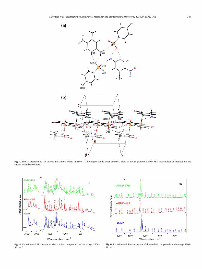
Fig. 4. The arrangement (a) of cations and anions joined by NAH� � �O hydrogen bonds types and (b) a view on the ac plane of AMNP-HBS. Intermolecular interactions areshown with dashed lines.
Fig. 5. Experimental IR spectra of the studied compounds in the range 3700–50 cm�1.
Fig. 6. Experimental Raman spectra of the studied compounds in the range 3600–80 cm�1.
I. Bryndal et al. / Spectrochimica Acta Part A: Molecular and Biomolecular Spectroscopy 123 (2014) 342–351 347

Table 3Experimental IR and Raman wavenumbers for AMNP, AMNP-HBS and AMNP-TFA.
AMNP AMNP-HBS AMNP-TFA AMNP AMNP-HBS AMNP-TFA
IR RS IR RS IR RS
3570 vw 3605 w m(NH2) mas(H2O)3473 w ms(H2O)3466 vw mas(NH2)
3391 m m(NH2) m(NA-H � � � O)3408 m 3405 w 3407 m m(NH2)+ m(N–
H� � �NP)m(N-H O)
3325 w 3304 m 3324 w m(N–H� � �NP) m(NA-H F)3275 vw 3263 sh 3268 w m(N–H� � �NP) m(NH2� � �O) m(NA-H� � �Oaq)
3255 w ms(NH2)3206 sh 3213 sh m(CH) m(CH)a3163 sh 3159 sh 3166 m 3177 w 3183 vw combination m(NH2� � �O) m(CH)a +m(Oaq-
3145 sh H� � �ONO2)3126 m 3111 w 3116 vw 3120 m m(CH) m(NH2� � �O) m(CH)a + m(NP-
H� � �O)3104 sh
3098 vw 3093 vw m(CH)a m(CH)a3076 vw 3074 w 3084 vw m(CH)/ masCH3
3066 sh 3066 w 3064 sh 3063 sh 3068 m 3065 vw mas(CH3) m(CH)/ m(Oaq-H� � �O)3047 vw 3046 vw m(CH)/3030 vw m(CH)a2999 vw 2998 vw m(CH)/
2989 w 2991 w 2989 vw 2990 vw 2990 w 2999 sh mas(CH3) mas(CH3) mas(CH3) + m(Oaq-H� � �O)
2992 vw2965 vw mas(CH3)
2936 vw 2936 w 2937 vw 2937 w 2939 vw 2940 vw ms(CH3) ms(CH3) ms(CH3) + m(Oaq-H� � �O)
2870 vw m(OH� � �O)2532 w m(Oaq-H� � �O)2377 w combination1707 sh m(C=O)
1668 sh 1661 vw 1685 sh 1681 vw 1669 s d(C-NH2)HB d(NH� � �O) d(NH2)1672 vs
1655 m 1640 m 1655 s 1664 vw d(C-NH2)HB d(NH2) m(a) + d(H2O) +d(NAH� � �Oaq)
1645 w d(H2O) + d(NH2)1632 sh 1636 vw 1636 sh m(a) + d(NH2) m(a) + d(NAH� � �Oaq)1624 sh
1609 m 1610 w 1607 w 1607 vw 1609 s 1609 vw m(a)+ mas(NO2) m(/) + d(CH)/ mas(NO2) + m(a)1591 sh 1589 m 1589 vw 1578 sh m(a)+ mas(NO2) m(/) mas(NO2) +
d(NPH� � �O)1580 sh 1580 sh1544 m 1539 vw 1533 m 1533 w 1544 m m(a)+
mas(NO2)+d(CH)mas(NO2) d(NPH� � �O)
1528 sh 1521 vw 1528 sh m(a)+mas(NO2)+d(CH)
d(NPH� � �O) +d(CNH)
1505 vw 1500 m 1503 w 1504 vw 1504 sh 1518 w mas(NO2)+m(a)+d(CH)
mas(NO2) + m(a) m(a)
1493 m 1494 sh 1496 sh 1494 vw1493 m 1495 w m(a)
1482 w 1479 sh 1478 w 1477 m 1481 sh das(CH3) d(NH� � �O) d(CH)a + m(C-NH2)1476 sh1458 sh 1458 w 1452 m das(CH3) + ms(NO2) das(CH3) + m(a) +1453 m d(NPH� � �O)
1443 m 1442 vw 1445 sh 1442 vw d(CH)/ + m(/) das(CH3)1429 w 1427 w 1428 sh 1430 vw 1427 m 1428 w ds(CH3) +m(a) m(a) + d(CH)(a) das(CH3)1426 sh + d(NH2)HB
1406 sh1385 w 1382 w m(C-O) + m(CCF3) +
ds(CH3)1373 m 1372 w 1371 sh 1372 sh 1373 m 1373 vw ds(CH3) das(CH3) m(a) + ds(CH3)1352 sh 1353 sh 1366 w 1362 vw d(CH) + m(a) d(S-OH) + m(C-
NH2)+ d(NH2)HB
1341 sh 1341 m 1347 sh 1347 sh 1345 sh d(CH) + m(a) +d(NH2)HB
ms(NO2) +ds(CH3)
d(CH)a + m(a)
1340 vs1333 m 1335 m 1332 s 1334 vs ms(NO2) ms(NO2)
1328 sh 1327 sh ms(NO2) + m(a)1303 sh 1300 vw m(/) + d(OH)
1291 s 1292 vs 1288 sh 1288 vw ms(NO2) + m(a) m(a) + d(CH)a1291 s 1291 m mas(CF3)
1274 vs 1273 s 1277 m 1278 vw 1272 vs 1275 m m(/)+d(CH)+ms(NO2) m(a) + ms(NO2) d(CH)a + m(a) +m(C-NO2)1274 vw
348 I. Bryndal et al. / Spectrochimica Acta Part A: Molecular and Biomolecular Spectroscopy 123 (2014) 342–351

Table 3 (continued)
AMNP AMNP-HBS AMNP-TFA AMNP AMNP-HBS AMNP-TFA
IR RS IR RS IR RS
1245 sh 1243 sh 1256 w 1256 m 1260 sh m(/)+ ms(NO2) d(CH)/ + m(/) m(a) + m(C-NO2)1238 w 1239 sh mas(SO3) + m(C-
OH) + d(/) +d(CH)/,a
1214 sh 1219 vw 1203 m 1202 vw m(/)+ ms(NO2) mas(SO3)1207 sh mas(CF3)1198 m 1191 m mas(CF3) + das(CF3)
1188 w 1190 m 1180 m 1193 w 1187 m 1179 sh m(/)+d(CH) mas(SO3) +d(CH)/
m(C-CF3) + ms(CF3) +m(C-COO) + d(NA-H� � �O) + d(NP-H� � �O)
1174 sh 1176 w 1182 m
1153 w 1165 sh 1154 sh 1158 vw 1141 m d(a)+m(C-NO2)+m(C-NH2)HB
d(CH)a + m(a) ms(CF3)
1150 sh1110 m 1111 m 1126 m 1126 m q(NH2)HB+m(a) d(CH)/ + d(OH)
+ c(NH���O)1103 w 1104 w 1110 m 1110 w d(a) + c(N-
H���O) + d(CH)/c(a) + m(C-NO2) +m(C-CH3) + x(CH3)+ c(NPH� � �O)
1096 sh 1103 sh 1102 sh1084 sh 1081 sh 1083 vw q(CH3)+m(a) d(a) + mas(SO3)
1070 w 1069 w d(CH)a + q(NH2) d(CH)a + q(NH2)1044 vw 1041 vw 1034 s 1032 m 1041 w 1039 sh q(CH3)+m(a) ms(SO3) +
q(NH2) + m(a)q(CH3)
1028 w 1020 sh 1021 vw 1028 w 1025 w q(CH3)+m(a) ms(SO3) + m(/) q(CH3)1014 w 1013 m 1013 w 1014 vw c(CH)+c(a) ms(SO3) + d(a) +
q(NH2)q(CH3)
1009 m 1007 vw q(CH3)975 vw 966 w 966 w c(CH)+c(a) q(CH3) +
q(NH2)955 m 953 m 958 vw 957 sh 954 m 950 w m(a) d(/) c(CH)a
941 w 940 sh ds(SO3) + d(a) +d(a)
m(C-CH3) + m(a)
898 sh m(a)873 w q(H2O) + c(CH)a
860 w 857 sh c(NAH� � �O)848 sh 842 m 844 m 843 w 848 sh 844 w d(NO2) + d(a) d(a) +
c(NAH� � �O)d(a)
842 m 842 w 841 m827 w m(SO3)
789 w c(COOH) + d(CF3) +c(NAH� � �O) +c(NPH� � �O)
769 sh 768 vw 769 sh 764 w x(NO2) +c(a)+s(h)HB
c(CH)/ q(H2O) + c(CH)a
764 w765 w 764 m 760 vw 760 m s(h)HB+x(NO2) c(CH)/750 vw 752 w 754 m 755 w d(a) + m(C-CH3) d(a) + m(C-CH3) c(COOH)
736 vw x(NO2) +c(a)+c(CH)
717 w 720 sh 724 w 726 vw x(NO2) + c(a) x(NO2) + c(a) c(NO2) + c(a)704 sh 705 w
714 sh d(CF3) + d(OCC) +d(H2O� � �O)
676 w 668 vw c(a) + c(/) d(CF3)668 w
653 w 645 vw 639 w 645 sh 652 w 645 w d(a)+d(NO2) +breathing hHB
c(NAH� � �O) +c(NPH� � �O) +c(NH2)
d(a) + d(NO2) + m(C-NO2)
636 sh 637 w 640 w c(C-CH3) + c(a)617 sh 616 vw d(a)
599 w 603 sh d(OaqH� � �O) +d(NAH� � �Oaq)
596 w 588 w 592 vw 595 vw 583 w 588 w q(NO2) + d(C-NH2)HB +d(a)
d(/) c(a) + c(NH2) +c(CH3) + c(OCO)
582 m 591 sh 572 sh565 sh 573 m 573 vw q(NO2) + d(C-
NH2)HB + d(a)c(SO3)
567 sh555 w 558 vw c(SO3)
(continued on next page)
I. Bryndal et al. / Spectrochimica Acta Part A: Molecular and Biomolecular Spectroscopy 123 (2014) 342–351 349

Table 3 (continued)
AMNP AMNP-HBS AMNP-TFA AMNP AMNP-HBS AMNP-TFA
IR RS IR RS IR RS
532 w 529 w 527 w 525 w 531 w 529 w d(a)+ m(C-CH3) c(NAH� � �O) +c(NPH� � �O) +d(/) + c(SO3)
d(a) + m(C-CH3) +d(C-CH3)
511 vw 503 vw 506 vw c(SO3) +c(NPH� � �O) +c(OH) + d(a)
d(/)
454 sh 453 vw d(a) + c(C-CH3) +c(NH� � �NP
448 w 440 vw 447 w 446 vw 448 w 445 vw q(NO2) + d(C-NH2)HB
c(NAH� � �O) +c(NPH� � �O) +c(SO3) + c(/)
q(CF3)
441 w 441 vw434 vw
410 vw 395 vw 412 w 408 vw x(NH2)HB d(C-OH) + c(/) s(NH2) + q(OCO)379 vw 377 w x(NH2)HB + c(a)
364 vw 367 sh 374 vw c(NH2) +c(OH) + d(SO3)
d(a) + m(C-NO2) +d(C-CF3)
356 sh336 vw 337 w 335 vw 339 sh 337 vw 334 vw d(C-CH3)+ d(a) c(SO3) +
c(NAH� � �O) +c(NPH� � �O) +d(C-CH3)
d(C-CH3) + d(a)
331 vw303 w d(C-CH3)
280 vw 288 vw 289 vw 293 sh 299 w 293 vw c(C-CH3) m(C-S) c(NH2) + s(a) + s(C-COO)
279 vw256 vw 265 sh 259 vw 269 vw d(C-NO2) d(C-NO2) +
q(NO2)x(CF3) + q(NO2) +q(H2O)
255 vw 255 vw247 vw
206 vw 219 w 219 sh 220 vw c(a) + c(C-CH3) c(/) + s(CH3) s(CH3)215 vw
184 vw 196 vw 187 vw 180 sh c(a) + c(C-CH3) m(OaqH� � �O)141 w 149 vw 146 w m(NAH� � �O) m(N-H� � �Oaq) + s(a)
130 vw 122 m 121 vw 123 vw 120 w c(C-NO2) + c(a) +c(C-NH2)HB
m(NPH� � �O) +c(/)
s(a) + m(N-H� � �Oaq)
90 vw 99 m 107 sh 100 w c(C-NO2) s(a) + m(N-H� � �Oaq)68 vw
a – pyridine ring, / – benzene ring, h – ring with hydrogen bonds within dimer. In-plane vibrations: m – stretching; d – bending; q – rocking; out-of-plane vibrations: c –torsional; x – wagging; s – twisting. NP – nitrogen atom of pyridine ring; NA – nitrogen atom of –NH2 group; Oaq – oxygen atom of H2O; ONO2 – oxygen atom of –NO2 group.
Fig. 7. The relationships between the donor–acceptor distances and the respectivewavenumbers for AMNP-TFA and AMNP-HBS.
350 I. Bryndal et al. / Spectrochimica Acta Part A: Molecular and Biomolecular Spectroscopy 123 (2014) 342–351
1. Several types of NAH� � �O hydrogen bonds are formed betweenthe acidic unit and pyridinium cation. These HB’s are responsi-ble to the ribbon formation in the unit cells. These NAH� � �Ointermolecular interactions should be classified to the categoryof moderate hydrogen bonds and are mostly electrostatic withthe admixture of the covalent coupling.
2. Another type of intermolecular interactions also appears. Theseare OAH� � �O hydrogen bonds between the water molecules andcarboxylate- and nitro-groups in AMNP-TFA and between theoxygen atoms of adjacent benzenesulfonate units in AMNP-HBS. Their D� � �A distances of 2.60–2.72 Å classifies these inter-actions as moderate strong, also due to the significant shift oftheir vibrational wavenumber up to 2500 cm�1.
3. The NAH� � �O hydrogen bonds in AMNP-HBS are weaker in com-parison with those in AMNP-TFA. For the former compound thelinear dependence between the D� � �A distances and the respec-tive IR band wavenumbers can be proposed whereas for theAMNP-TFA exponential dependence should be used.
Acknowledgement
This work was financially supported by Ministry of Science andHigher Education (Grant No. PBZ/MEiN/01/2006/21).
Appendix A. Supplementary material
Supplementary material associated with this article can befound, in the online version, at http://dx.doi.org/10.1016/j.saa.2013.12.018.

I. Bryndal et al. / Spectrochimica Acta Part A: Molecular and Biomolecular Spectroscopy 123 (2014) 342–351 351
References
[1] T. Eicher, S. Hauptmann, The Chemistry of Heterocycles: Structure, Reactions,Syntheses, and Applications, second ed., Wiley-VCH Verlag GmbH, Weinheim,2003.
[2] H.O. Pechmann, W. Welsh, J. Chem. Soc. Trans. 47 (1885) 145–155.[3] Z. Czapla, S. Dacko, A. Waskowska, J. Phys. 15 (2003) 3793–3803.[4] I.R. Evans, J.A.K. Howard, J.S.O. Evans, Cryst. Growth Des. 8 (2008) 1635–1639.[5] S.R. Jebasa, T. Balasubramanian, R. Pescharb, J. Fraanjeb, Acta Cryst. E62 (2006)
o2606–o2607.[6] J.A. Bis, M.J. Zaworotko, Cryst. Growth Des. 5 (2005) 1169–1179.[7] M.J. Prakash, T.P. Radhakrishnan, Cryst. Growth Des. 5 (2005) 721–725.[8] S.T. Akriche, M. Rzaigui, N. Al-Hokbany, R.M. Mahfouz, Acta Cryst. E66 (2010)
o300.[9] S.T. Akriche, M. Rzaigui, Acta Cryst. E65 (2008) m123.
[10] S.T. Akriche, M. Rzaigui, Acta Cryst. E65 (2009) o3009–o3010.[11] J. Pecaut, J.P. Levy, R. Masse, J. Mater. Chem. 3 (1993) 999–1003.[12] H. Koshima, M. Hamada, I. Yagi, K. Uosaki, Cryst. Growth Des. 5 (2001) 467–
471.[13] J. Lorenc, I. Bryndal, M. Marchewka, W. Sasiadek, T. Lis, J. Hanuza, J. Raman
Spectrosc. 39 (2008) 569–581.[14] J. Lorenc, I. Bryndal, M. Marchewka, E. Kucharska, T. Lis, J. Hanuza, J. Raman
Spectrosc. 39 (2008) 863–872.[15] J. Lorenc, I. Bryndal, W. Syska, M. Wandas, M. Marchewka, A. Pietraszko, T. Lis,
M. Maczka, J. Hanuza, Chem. Phys. 374 (2010) 1–14.[16] I. Bryndal, E. Kucharska, M. Wandas, J. Lorenc, K. Hermanowicz, M. Maczka, T.
Lis, M. Marchewka, J. Hanuza, Spectrochim. Acta A 117 (2014) 434–441.[17] I. Bryndal, E. Kucharska, W. Sasiadek, M. Wandas, T. Lis, J. Lorenc, J. Hanuza,
Spectrochim. Acta A 96 (2012) 952–962.[18] T. Talik, Z. Talik, Rocz. Chem. 42 (1968) 1647–1661.[19] Oxford Diffraction, CrysAlis CCD and CrysAlis RED in Xcalibur PX Software
Version 1.171, Oxford Diffraction Ltd., Abington, England, 2007.[20] G.M. Sheldrick, Acta Cryst. A64 (2008) 112–122.[21] XP – Interactive Molecular Graphics, Version 4.3 for MSDOS Siemens
Analytical X-ray Inst. Inc., 1992.[22] M.J. Frisch, G.W. Trucks, H.B. Schlegel, G.E. Scuseria, M.A. Robb, J.R. Cheeseman,
J.A. Montgomery Jr., T. Vreven, K.N. Kudin, J.C. Burant, J.M. Millam, S.S. Iyengar,J. Tomasi, V. Barone, B. Mennucci, M. Cossi, G. Scalmani, N. Rega, G.A.Petersson, H. Nakatsuji, M. Hada, M. Ehara, K. Toyota, R. Fukuda, J. Hasegawa,M. Ishida, T. Nakajima, Y. Honda, O. Kitao, H. Nakai, M. Klene, X. Li, J.E. Knox,H.P. Hratchian, J.B. Cross, C. Adamo, J. Jaramillo, R. Gomperts, R.E. Stratmann,O. Yazyev, A.J. Austin, R. Cammi, C. Pomelli, J.W. Ochterski, P.Y. Ayala, K.
Morokuma, G.A. Voth, P. Salvador, J.J. Dannenberg, V.G. Zakrzewski, S.Dapprich, A.D. Daniels, M.C. Strain, O. Farkas, D.K. Malick, A.D. Rabuck, K.Raghavachari, J.B. Foresman, J.V. Ortiz, Q. Cui, A.G. Baboul, S. Clifford, J.Cioslowski, B.B. Stefanov, G. Liu, A. Liashenko, P. Piskorz, I. Komaromi, R.L.Martin, D.J. Fox, T. Keith, M.A. Al-Laham, C.Y. Peng, A. Nanayakkara, M.Challacombe, P.M.W. Gill, B. Johnson, W. Chen, M.W. Wong, C. Gonzalez, J.A.Pople, Gaussian 03, Revision A.1, Gaussian Inc., Pittsburgh, PA, 2003.
[23] D. Becke, J. Chem. Phys. 104 (1996) 1040–1046.[24] C. Lee, W. Yang, R.G. Parr, Phys. Rev. B 37 (1988) 785–789.[25] R.G. Parr, W. Yang, Density-Functional Theory of Atoms and Molecules, Oxford
University Press, New York, 1989.[26] A.D. McLean, G.S. Chendler, J. Chem. Phys. 72 (1980) 5639–5648.[27] R. Krishnan, J.S. Binkley, R. Seeger, J.A. Pople, J. Chem. Phys. 72 (1980) 650–654.[28] M.J. Nowak, L. Lapinski, BALGA computer program for PED calculations
(H. Rostkowska, L. Lapinski, M.J. Nowak, Vib. Spectrosc. 49 (2009) 43–51.[29] G.A. Zhurko, D.A. Zhurko, Chemcraft graphical program for visualization of
computed results. <http://www.chemcraftprog.com>.[30] D. Michalska, RAINT (Raman Intensities) – a computer program for calculation
of Raman intensities from the Gaussian outputs, Wrocław University ofTechnology, Wrocław, 2002 (see Ref. [24]).
[31] D. Michalska, R. Wysokinski, Chem. Phys. Lett. 403 (2005) 211–217 (andreferences therein).
[32] M.A. Khan, M.N. Tahir, A.Q. Ather, M. Shaheen, R.A. Khan, Acta Cryst. E65(2009) o1615.
[33] O. Watanabe, T. Noritake, Y. Hirose, A. Okada, T. Kurauchi, J. Mater. Chem. 3(10) (1993) 1053–1057.
[34] Y. Le Fur, M. Bagieu-Beucher, R. Masse, J.F. Nicoud, J.P. Lévy, Chem. Mater. 8(1996) 68–75.
[35] H. Koshima, H. Miyamoto, Mol. Cryst. Liq. Cryst. 420 (2004) 79–89.[36] J. Bernstein, R.E. Davis, L. Shimoni, N.-L. Chang, Angew. Chem. Int. Ed. Engl. 34
(1995) 1555–1573.[37] J. Janczak, G.J. Perpetuo, Acta Cryst. C57 (2001) 873–875.[38] M.A. Palafox, V.K. Rastogi, Spectrochim. Acta A 58 (2002) 411–440.[39] C.V. Berney, J. Amer. Chem. Soc. 95 (3) (1973) 708–716. and references therein.[40] R.L. Redington, Spectrochim. Acta A 31 (1975) 1699–1705.[41] M. Drozd, Spectrochim. Acta A 69 (2008) 1223–1234.[42] B. Sun, Y. Zhao, J.G. Wu, Q.C. Yang, G.X. Xu, J. Mol. Struct. 471 (1998) 63–66.[43] G.A. Jeffrey, An Introduction to Hydrogen Bonding, Oxford University Press,
1997.[44] T. Steiner, The hydrogen bond in the solid state, Angew. Chem. Int. Ed. 41
(2002) 49–76.


![4* • Pharingeal grooves/cleft : 4 • [Pharyngeal membrane]](https://static.fdokumen.com/doc/165x107/6334ea00b9085e0bf5093ec7/4-pharingeal-groovescleft-4-pharyngeal-membrane.jpg)