The plausibility of maternal nutritional status being a contributing factor to the risk for fetal...
22
The Plausibility of Maternal Nutritional Status Being a Contributing Factor to the Risk for Fetal Alcohol Spectrum Disorders: The Potential Influence of Zinc Status as an Example Carl L. Keen a,b,* , Janet Y. Uriu-Adams a , Anatoly Skalny d , Andrei Grabeklis d , Sevil Grabeklis d , Kerri Green a , Lyubov Yevtushok e , W. W. Wertelecki f , and Christina D. Chambers g a Department of Nutrition, University of California, Davis, Davis, CA 95616, USA b Internal Medicine, University of California, Davis, Davis, CA 95616, USA c Department of Psychology, San Diego State University, San Diego, CA 92116, USA d Institute for Biotech Medicine, Moscow, Russia e Rivne Diagnostic Center, Rivne 33028, Ukraine f Department of Medical Genetics, University of South Alabama, Mobile, AL 36688, USA g Departments of Pediatrics and Family and Preventive Medicine, University of California, San Diego, La Jolla, CA 92093, USA Abstract There is increasing evidence that human pregnancy outcome can be significantly compromised by suboptimal maternal nutritional status. Poor diet results in a maternal-fetal environment in which the teratogenicity of other insults such as alcohol might be amplified. As an example, there is evidence that zinc (Zn) can interact with maternal alcohol exposure to influence the risk for fetal alcohol spectrum disorders (FASD). Studies with experimental animals have shown that the teratogenicity of alcohol is increased under conditions of Zn deficiency, while its teratogenicity is lessened when animals are given Zn supplemented diets or Zn injections prior to the alcohol exposure. Alcohol can precipitate an acute phase response resulting in a subsequent increase in maternal liver metallothionein, which can sequester Zn and lead to decreased Zn transfer to the fetus. Importantly, the teratogenicity of acute alcohol exposure is reduced in metallothionein knockout mice, which can have improved Zn transfer to the conceptus relative to wild-type mice. Consistent with the above, Zn status has been reported to be low in alcoholic women at delivery. Preliminary data from two basic science and clinical nutritional studies that are ongoing as part of the international Collaborative Initiative on Fetal Alcohol Spectrum Disorders (CIFASD) support the potential role of Zn, among other nutritional factors, relative to risk for FASD. Importantly, the nutrient levels being examined in these studies are relevant to general clinical populations and represent suboptimal levels rather than severe deficiencies. These data suggest that moderate deficiencies in single nutrients can act as permissive factors for FASD, and that adequate nutritional status or intervention through supplementation may provide protection for some of the effects of prenatal alcohol exposure. * Corresponding author Carl L. Keen, Ph.D., University of California, Davis, One Shields Avenue, Davis, CA 95616, Tel.: +1-530-752-6331; fax: +1-530-752-8966. [email protected]. NIH Public Access Author Manuscript Biofactors. Author manuscript; available in PMC 2011 March 1. Published in final edited form as: Biofactors. 2010 ; 36(2): 125–135. doi:10.1002/biof.89. NIH-PA Author Manuscript NIH-PA Author Manuscript NIH-PA Author Manuscript
Transcript of The plausibility of maternal nutritional status being a contributing factor to the risk for fetal...
The Plausibility of Maternal Nutritional Status Being aContributing Factor to the Risk for Fetal Alcohol SpectrumDisorders: The Potential Influence of Zinc Status as an Example
Carl L. Keena,b,*, Janet Y. Uriu-Adamsa, Anatoly Skalnyd, Andrei Grabeklisd, SevilGrabeklisd, Kerri Greena, Lyubov Yevtushoke, W. W. Werteleckif, and Christina D.Chambersg
aDepartment of Nutrition, University of California, Davis, Davis, CA 95616, USAbInternal Medicine, University of California, Davis, Davis, CA 95616, USAcDepartment of Psychology, San Diego State University, San Diego, CA 92116, USAdInstitute for Biotech Medicine, Moscow, RussiaeRivne Diagnostic Center, Rivne 33028, UkrainefDepartment of Medical Genetics, University of South Alabama, Mobile, AL 36688, USAgDepartments of Pediatrics and Family and Preventive Medicine, University of California, SanDiego, La Jolla, CA 92093, USA
AbstractThere is increasing evidence that human pregnancy outcome can be significantly compromised bysuboptimal maternal nutritional status. Poor diet results in a maternal-fetal environment in whichthe teratogenicity of other insults such as alcohol might be amplified. As an example, there isevidence that zinc (Zn) can interact with maternal alcohol exposure to influence the risk for fetalalcohol spectrum disorders (FASD). Studies with experimental animals have shown that theteratogenicity of alcohol is increased under conditions of Zn deficiency, while its teratogenicity islessened when animals are given Zn supplemented diets or Zn injections prior to the alcoholexposure. Alcohol can precipitate an acute phase response resulting in a subsequent increase inmaternal liver metallothionein, which can sequester Zn and lead to decreased Zn transfer to thefetus. Importantly, the teratogenicity of acute alcohol exposure is reduced in metallothioneinknockout mice, which can have improved Zn transfer to the conceptus relative to wild-type mice.Consistent with the above, Zn status has been reported to be low in alcoholic women at delivery.Preliminary data from two basic science and clinical nutritional studies that are ongoing as part ofthe international Collaborative Initiative on Fetal Alcohol Spectrum Disorders (CIFASD) supportthe potential role of Zn, among other nutritional factors, relative to risk for FASD. Importantly, thenutrient levels being examined in these studies are relevant to general clinical populations andrepresent suboptimal levels rather than severe deficiencies. These data suggest that moderatedeficiencies in single nutrients can act as permissive factors for FASD, and that adequatenutritional status or intervention through supplementation may provide protection for some of theeffects of prenatal alcohol exposure.
*Corresponding author Carl L. Keen, Ph.D., University of California, Davis, One Shields Avenue, Davis, CA 95616, Tel.:+1-530-752-6331; fax: +1-530-752-8966. [email protected].
NIH Public AccessAuthor ManuscriptBiofactors. Author manuscript; available in PMC 2011 March 1.
Published in final edited form as:Biofactors. 2010 ; 36(2): 125–135. doi:10.1002/biof.89.
NIH
-PA Author Manuscript
NIH
-PA Author Manuscript
NIH
-PA Author Manuscript
KeywordsAlcohol; FAS; FASD; zinc; micronutrients; pregnancy
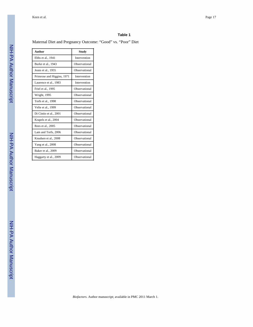
1. IntroductionIt is estimated that, conservatively, 50% of human concepti are lost before or duringimplantation, and of those that successfully implant, an additional 15–20% are lost prior todelivery [1,2]. With respect to completed pregnancies, approximately 3% result in a childwith one or more severe malformations. These numbers, as discouraging as they are, do nottake into account the increased risk many children can have for chronic diseases later in life,including obesity, diabetes, hypertension and vascular disease, as a consequence of selectprenatal or early postnatal insults [3,4]. While a diversity of factors can contribute to theoccurrence of developmental abnormalities, there is increasing evidence that humanpregnancy outcome can be significantly compromised by suboptimal maternal nutritionalstatus, and that poor nutrition may be a leading cause of preventable birth defects. A numberof investigators since the early 1940’s have reported that women who consume diets that canbe classified as “poor” have an increased risk for pregnancy complications compared towomen who consume diets that can be classified as “good” (Table 1). It is worth noting thatthis association has been reported over a period of time where many would argue as to whatconstitutes a good versus a poor diet; however, a consistent theme over the past sevendecades has been that “good” diets are characterized by high micronutrient content.Importantly, data from a variety of nutrition intervention trials, ranging from the provisionof whole foods, to multivitamin-mineral supplements, to single nutrients such as folate andiodine, provide evidence that improvements in a mother’s diet can result in markedreductions in her risk for a complicated pregnancy [5–11]. When viewed in its totality, thedata supporting the concept that maternal nutritional status is a key predictor of humanpregnancy outcome seem overwhelming; however, a complication in the story is that whilethe diets of women who consume “poor’ diets may not be ideal, pronounced essentialnutrient deficiencies are rare. This observation has led to the idea that the increased risk forpregnancy complications observed in this group of women may, in many cases, be more dueto the fact that the poor diet results in a maternal-fetal environment in which theteratogenicity of other insults is amplified, than due to a simple deficit of one or moreessential nutrients in the diet.
In the current paper, we explore this concept using alcohol as an example of adevelopmental insult whose teratogenicity can be modulated by maternal diet. It is nowwidely accepted that excessive maternal alcohol intake during pregnancy can result in anumber of developmental abnormalities commonly referred to as the Fetal AlcoholSyndrome (FAS), or more recently, the Fetal Alcohol Spectrum Disorder (FASD) [12].FASD is now thought to be one of the most common causes of developmental mentalretardation in the human population. Due to space constraints we will focus our discussionon the ability of zinc (Zn) to modulate the teratogenic expression of alcohol. In the lastsection of this paper, preliminary data from an ongoing multi-nutrient supplementation trialin Russia and the Ukraine with women who are at high risk for having a child with FASDwill be discussed. We would like to emphasize that while our comments are focused on Zn,there is good evidence that numerous other nutrients, including copper (Cu), iron (Fe),magnesium (Mg), selenium (Se), methionine, choline, vitamin B12 and folate, can modulatealcohol’s developmental toxicity. Owing to space constraints, review articles are cited inseveral instances and the reader is directed to them for additional references.
Keen et al. Page 2
Biofactors. Author manuscript; available in PMC 2011 March 1.
NIH
-PA Author Manuscript
NIH
-PA Author Manuscript
NIH
-PA Author Manuscript
2. Alcohol-Zn InteractionsAlthough the concomitant effects of alcohol and specific nutrient deficiencies on thedeveloping brain are not well understood, animal studies have shown that compromisednutrition can exacerbate ethanol’s teratogenic effects. Many adverse effects of prenatalalcohol exposure including low birth weight [13], physical anomalies [14], brain damage[15] and reduced IGF levels [16] have been reported to be more severe when the alcohol isconsumed along with poor diets; however, a complication of this finding is that bloodalcohol levels are often higher among malnourished subjects than in subjects thought to becharacterized by similar alcohol intakes, but better diets. On the positive side, results fromearly animal studies suggest that select nutritional supplements can attenuate some of theeffects of prenatal alcohol exposure, although the effectiveness depends on many factors,including the level of alcohol exposure and the outcome measure [17]. The contribution ofselect nutritional factors to the risk for FASD in humans has not been well characterized,however, the overall nutritional status of heavy drinkers is generally recognized to be poor[18]. It is critically important to define the role that malnutrition or under-nutrition plays inthe risk for FASD. Of particular interest in the pregnant heavy drinker is assessing the statusfor those micronutrients that are critical for normal neurulation and development of thecentral nervous system (CNS). From work with experimental animals, it is well documentedthat deficiencies of certain nutrients, including folate, vitamin B12, Zn, Fe and Cu, duringpregnancy can result in abnormal CNS development, and deficiencies of these nutrients arecommonly noted in alcoholics [18–23].
With respect to the above nutrients, the hypothesis that maternal Zn status is an importantpredictor of the risk for FASD has received particular attention. Over 25 years ago, Flynn etal reported that maternal plasma Zn and fetal cord plasma Zn were lower in pregnant womenwho consumed alcohol versus non-alcohol drinking women [24]. Importantly, theseinvestigators reported that there were negative correlations between maternal plasma Znconcentrations and the severity and frequency of birth defects in the infants, suggesting anetiologic role for Zn deficiency in human FASD. Consistent with the idea that maternal Znstatus might be a predictor of the risk for FASD are the early data of Miller et al [25]; theseinvestigators reported that the reproductive toxicity of alcohol in rats was elevated in damsfed marginal Zn diets (10 µg Zn/g diet) compared to that in dams fed diets with controlamounts of Zn (45 µg/g) [25]. It is important to note that in the above study by Miller et al,the amount of Zn that was provided in the diet of the marginal Zn group would not beviewed as “deficient”, as the consumption of this diet throughout pregnancy would nottypically result in marked developmental anomalies. Consistent with the data of Miller et al,the teratogenicity of alcohol has been reported to be amplified in pregnant rats and mice fedZn deficient diets (1 µg Zn/g) [26–28].
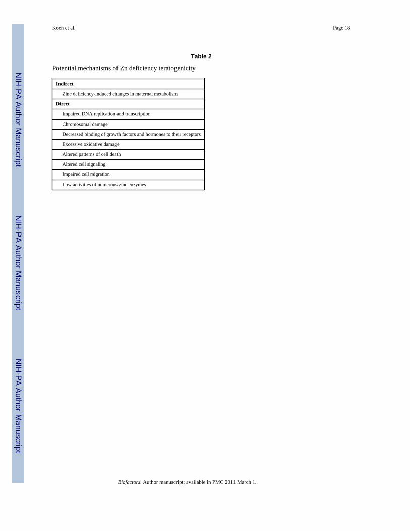
The relevance of the above work by Miller et al is underscored by the observation that incertain human populations, dietary Zn intake during pregnancy can be well below currentrecommended dietary intakes [29–32], suggesting that suboptimal Zn status is common inthe human population. The concept that a deficit of Zn during early development presents arisk to the human conceptus is reasonable, given that in experimental animals, theteratogenicity of severe Zn deficiency is well documented, resulting in malformations thataffect multiple organ systems including the CNS [33]. Even moderate deficiencies of thisnutrient during development can result in persistent adverse effects on the immune systemand neurobehavioral abnormalities [2,34,35]. Mechanistically, Zn deficiency is thought toinfluence embryonic and fetal development through multiple mechanisms includingabnormal nucleic acid metabolism, reduced protein synthesis, impaired cell migration andcell signaling due to alterations in the cytoskeleton secondary to impaired tubulinpolymerization, excessive cellular oxidative stress, reduced binding of transcription factors
Keen et al. Page 3
Biofactors. Author manuscript; available in PMC 2011 March 1.
NIH
-PA Author Manuscript
NIH
-PA Author Manuscript
NIH
-PA Author Manuscript
and hormones that are dependent on Zn-finger regions, and reductions in insulin and IGF-signaling [2,23,34,36–40] (Table 2; Figure 1).
It is important to note that mechanistically, one would predict multiple synergisticinteractions between an alcohol insult and a condition of marginal Zn deficiency. As anexample of the above, it is well documented that Zn contributes to the oxidant defensesystem through multiple means, including through its ability to: (1) regulate Cu-Znsuperoxide dismutase (CuZn SOD) activity; (2) regulate metallothionein levels; (3) protectsulfhydryl groups from oxidation; (4) modulate intracellular thiol groups; and (5) inhibit thebinding of redox active metals, such as Fe and Cu, to intracellular sites where they cangenerate free radical reactions (e.g., Fenton-type reactions). Given the above, it is evidentthat one functional consequence of Zn deficiency is an increased susceptibility to exogenousoxidative stressors, such as smoking, endotoxin challenge, and, particularly germane to thispaper, alcohol [27,39,41]. The consequences of excessive tissue oxidative stress in theembryo can include lipid, protein and DNA oxidative damage, and an increase in apoptosis,all of which can trigger abnormal development. It is important to note that all of the aboveare common findings in animal models for FASD [23].
Another potential point of interaction between Zn deficiency and an alcohol challengeinvolves sonic hedgehog (Shh) signaling. Shh signaling is critical for polarizing activity, andShh null fetuses are characterized by a postaxial forelimb ectrodactyly in mouse models[42]. Shh is a Zn-dependent developmental trigger [43], and reduced Shh expression hasbeen implicated in ethanol-induced postaxial forelimb ectrodactyly in the mouse [44].Schreiner and co-workers [45] have suggested that a state of embryonic Zn deficiencysecondary to an alcohol-induced acute-phase response (see below; [46–48]) in the motherresults in reduced Shh signaling with subsequent dysmorphology [45]. This is an interestinghypothesis that merits further investigation. Importantly, if it is shown to be correct, thereare numerous other Zn-dependent developmental proteins, many in the hedgehog signalingpathway (e.g., Gli Zn finger transcription regulators), that might also be affected through themechanism described above.
A third finding that is common to experimental Zn deficiency, and alcohol challenges duringpregnancy, is a reduction in IGF levels and action [49,50]. Zn deficiency can alter IGF-1 andIGF-binding protein metabolism in maternal and fetal blood [49] and may contribute togrowth retardation of the offspring. Growth deficit is also commonly noted in offspring ofalcohol-exposed women. Administration of ethanol by intubation (5.25 g/kg/day) onpostnatal days 4–9 results in motor coordination impairments that are significantly improvedwith intranasal administration of IGF on postnatal days 10–13 [50].
Finally, a common finding in animal models for developmental Zn deficiency and FASD isan elevated occurrence of apoptosis in the embryo and fetus. Multiple mechanisms havebeen implicated in the inducing-effects of alcohol and Zn deficiency on apoptosis includingdisruptions in growth factor signal transduction pathways mediated by receptor tyrosinekinases, an increase in the expression of caspase-3, a down-regulation of NF-κB-dependentanti-apoptotic genes, and an increase in cellular oxidative stress. [36,38,51].
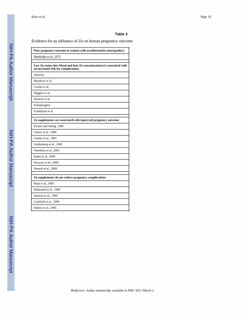
Evidence that severe Zn deficiency presents a reproductive risk in humans is provided by theobservation that women with a congenital disorder in Zn absorption (acrodermatitisenteropathica; AE) have a very high risk for pregnancy complications unless they are givendietary Zn supplements [2]. It is now recognized that the AE is due to a defective Zntransporter. The analogue of this transporter in mice is Zip4, and consistent with the humanliterature, homozygous Zip4-knockout mouse embryos die during early embryogenesis andare characterized by multiple defects [52]. As is depicted in Table 3, a number of studies
Keen et al. Page 4
Biofactors. Author manuscript; available in PMC 2011 March 1.
NIH
-PA Author Manuscript
NIH
-PA Author Manuscript
NIH
-PA Author Manuscript
have reported that low plasma Zn concentrations in the first or third trimester is associatedwith an increased risk for several pregnancy complications, including birth defects andgrowth retardation. In further support of the concept that maternal Zn status can be apredictor of pregnancy complications, numerous investigators have reported that even innon-AE populations, the provision of dietary Zn supplements during pregnancy results in areduced risk for pregnancy complications (Table 3). However, as is also provided in Table 3,there are several studies in which the provision of Zn supplements during pregnancy had nomeasurable positive effects. Indeed, in a recent Cochrane Review on the effects of Znsupplementation on pregnancy outcome [53], the authors concluded that with the exceptionof the risk for prematurity, there was no consistent positive effect of Zn supplements duringpregnancy. In our opinion, the somewhat negative finding by Mahomed et al [53] is due tothe fact that in many cases, the Zn supplementation trials have been done with relativelyhealthy, non-stressed populations. This may be important since, in addition to low dietaryZn intake, stressor-induced changes in the metabolism of Zn and other nutrients are oftensecondary to an acute phase response (APR), which can be triggered by a diverse set ofcytokines that are released following tissue injury.
2.1. Acute Phase Response-Induced Fetal Zn DeficiencyIt has been hypothesized that the developmental toxicity of a wide variety of toxicants andenvironmental insults is mediated in part through the induction of the APR, which increasesmaternal hepatic metallothionein synthesis and Zn sequestration, leading to reduced Zntransfer to the conceptus. If severe enough, this can result in abnormal development[2,46,54–56] (Figure 1). The potential human relevance of the above work withexperimental animals is illustrated by the finding that pregnant women who are infectedwith cytomegalovirus, a common etiologic agent of intrauterine infection, are characterizedby high cytokine levels including TNF-α and IL-6, and lower than normal plasma Znconcentrations [57]. Underscoring the potential significance of the above finding is therecent report by Collier et al [58] that maternal infections during pregnancy are common. Inrats, disease and environmental factors that reduce maternal serum Zn concentrations candisrupt Zn-dependent processes in the embryo and produce developmental defects, evenwhen the mother consumes a Zn “adequate” diet [54,59]. Significantly, the teratogeniceffects of the APR-induced hypozincemia can be amplified by marginal Zn diets, andreduced with Zn supplementation [46,54]. Critical to the current paper, in rodent models,acute alcohol exposure is associated with APR-induced reductions in fetal Zn uptake[48,54–56,60,61]. That the above reductions in fetal Zn uptake are functionally significant issuggested by the observation that the teratogenicity of alcohol is lessened when animals aregiven Zn prior to the alcohol exposure [60–63]. Moreover, the teratogenicity of acutealcohol exposure is reduced in metallothionein knockout mice compared to metallothioneinwild-type mice [55].
It is important to note that in the above discussion, the focus has been on the potentialpositive effects of Zn supplementation on the expression of FASD in the offspring. Znsupplementation has also been reported to be of value in attenuating ethanol-induced liverdamage in the adult [64–66] and mitigating lung epithelial and macrophage dysfunctioninduced by chronic alcohol intake [67]. Thus, it can be speculated that Zn supplementationof the high risk patient could directly benefit the mother, as well as her child.
In addition to pregnant women who are exposed to alcohol, infants with FASD have beenreported to have low plasma Zn concentrations and higher twenty-four hour urinary Znexcretions compared to normal controls, indicating altered Zn homeostasis [68].Interestingly Murillo-Fuentes and coworkers [69] reported that rat pups whose mothers weregiven alcohol during the period of lactation were also characterized by higher than normallevels of urinary Zn excretion. It is important to stress that the above reports are preliminary
Keen et al. Page 5
Biofactors. Author manuscript; available in PMC 2011 March 1.
NIH
-PA Author Manuscript
NIH
-PA Author Manuscript
NIH
-PA Author Manuscript
in nature. However, if the finding can be replicated, it represents a critical observation as itcould provide one explanation for the persistent immunological abnormalities that have beenreported for some children with FASD [70,71]. If these persistent immunologicalabnormalities are secondary in part to persistent abnormalities in Zn metabolism, they couldbe responsive to Zn supplements. In support of this concept, Zn given by injection [72], aswell as the feeding of diets high in Zn [73], have been reported to reduce neurobehavioralabnormalities in mouse and rat offspring of ethanol-exposed dams. While these data arecompelling, others, using a rat model, did not observe a protective effect of Znsupplementation on alcohol-induced developmental brain abnormalities [74]. In addition, itis important to note that there can be adverse interactions among nutrients. For example,supplementation of an alcohol-containing liquid diet with 300 µg/ml of Zn resulted in severefetal Cu deficiencies relative to controls [75]. Cu deficiency during pregnancy is alsoteratogenic, affecting cardiac, vascular, neurological, pulmonary, skeletal, and immunesystems [21,76,77].
Ethanol exposure has also been shown to alter Fe regulation and homeostasis. Chronicethanol consumption increases body stores of Fe and is associated with a significant risk ofFe overload [78–81]. Levels of non-transferrin-bound Fe are higher in alcohol abusers [78],which could contribute to reactive oxygen species formation via its involvement in Fe-catalyzed Fenton reactions [82]. When antioxidants, Fe chelators or sulfhydryl compoundsthat increase cellular glutathione are administered, ethanol toxicity is significantly reduced[83]. Miller and coworkers [84] reported that ethanol consumption by rat dams perturbs thetemporal patterns between Fe concentrations and Fe-regulatory proteins in brain regions ofoffspring. That fetal alcohol exposure can result in low Fe stores in the human infant hasbeen reported by Carter et al [85].
It is critical to note that the metabolism of Fe, Zn and Cu are interrelated, and it has beendemonstrated in experimental animal models that a maternal deficit of any one of the aboveelements can result in alterations in the metabolism of the other elements in the mother aswell as the fetus [86]. The above observation underscores the potential risk of focusing on asignal nutrient when one is studying alcohol-nutrient interactions with respect to the risk forFASD. In this regard, it is important to note that while hypozincemia is a hallmark of anAPR, as is depicted in Figure 1, an APR can also result in marked disturbances in themetabolism of Fe, Cu, folate and vitamins, as well as other essential nutrients. The extent towhich APR-induced changes in theses nutrients might affect fetal development has beenlargely unexplored.
3. Testing of the Zn/Alcohol Hypothesis: The Need for Large-ScaleProspective Trials
There is a need for multiple prospective studies investigating whether alterations in maternalZn status can affect fetal outcome in alcohol-exposed women. These studies should bedesigned in a way that they could later be analyzed via meta-analysis as is done in Cochranereviews to determine whether suboptimal Zn status is a predictor of adverse fetal outcomes.Moreover, maternal nutrient supplementation could be instituted as this is easily modifiable.Towards this goal, a longitudinal prospective study that aims to examine maternal nutritionalstatus and its contribution to risk for FASD, as well as to test a multi-micronutrientintervention with or without choline supplementation, is currently underway at sites inUkraine, as part of the Collaborative Initiative on Fetal Alcohol Spectrum Disorders(CIFASD). The study protocol was approved by institutional review boards in Russia andUkraine and institutional review boards at the University of California, San Diego and theUniversity of California, Davis; all study participants provided informed consent.
Keen et al. Page 6
Biofactors. Author manuscript; available in PMC 2011 March 1.
NIH
-PA Author Manuscript
NIH
-PA Author Manuscript
NIH
-PA Author Manuscript
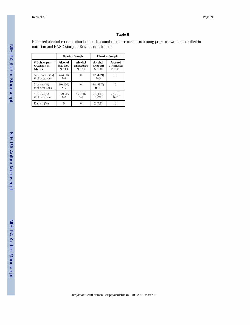
In the above study, alcohol-exposed and comparison group subjects are selected fromprenatal patients at each of the two sites who were screened by prenatal care providers atfirst prenatal visit, using a short standardized screening tool. Subjects are considered eligiblefor the alcohol-exposed group based on quantity and frequency of alcohol consumptionduring pregnancy. A positive screen for quantity and frequency of alcohol consumption isdefined as at least four episodes of five or more standard drinks, at least five episodes of 3–4standard drinks, or at least 10 episodes of 1–2 standard drinks either in the month around thetime of conception, or the most recent month of pregnancy. Subjects are considered eligiblefor the comparison group based on minimal to no alcohol consumption during either timeperiod, as reported at the time of the initial screening.
All eligible alcohol-exposed women are invited to enroll, and comparison group women arerecruited in an approximate 1:1 ratio. This is accomplished following enrollment of eachalcohol-exposed woman by approaching the next pregnant woman presenting for prenatalcare who reports low to no alcohol exposure in response to the screening questionnaire andwho agrees to participate in the study.
As part of the pregnancy follow-up procedure, enrolled women participate in comprehensivestandardized interviews, newborn physical examinations, and standard neurobehavioraltesting of infants. For the nutrition component of this study, all subjects are randomized toreceive a nutritional intervention involving three arms: multi-micronutrient supplementprovided from time of enrollment to delivery, multi-micronutrient supplement with cholinesupplement provided from the time of enrollment to delivery, or no treatment (currentstandard of care in Russia and Ukraine). Maternal blood samples are drawn at the time ofenrollment to establish baseline nutritional status, and again in the 3rd trimester to evaluatechange in status, and to validate the impact of treatment group on change in nutritionalstatus.
The samples collected to date in Russia have been analyzed in the laboratory of one of theauthors (AS) at the Institute for Biotech Medicine in Moscow. The samples collected to datefrom the Ukraine have been analyzed by three of the authors in the U.S. at the University ofCalifornia, Davis (CLK, JYUA, KG). Due to potential variability in the laboratoryprocedures, and to differences in the characteristics of subjects, preliminary data from thesetwo sites are presented separately. Baseline maternal blood samples were collected inheparinized tubes and analyzed for Zn, Cu, Mg, Fe and Ca by inductively coupled plasmaoptical emission spectrometry (ICP) (Trace scan; Thermo Elemental, Franklin, MA) [87].Certified reference solutions (QC 21, Spec Centri Prep, Metuchen, NJ) were used togenerate standard curves for each element. National Bureau of Standards reference sampleswere included with each run to ensure accuracy and reproducibility.
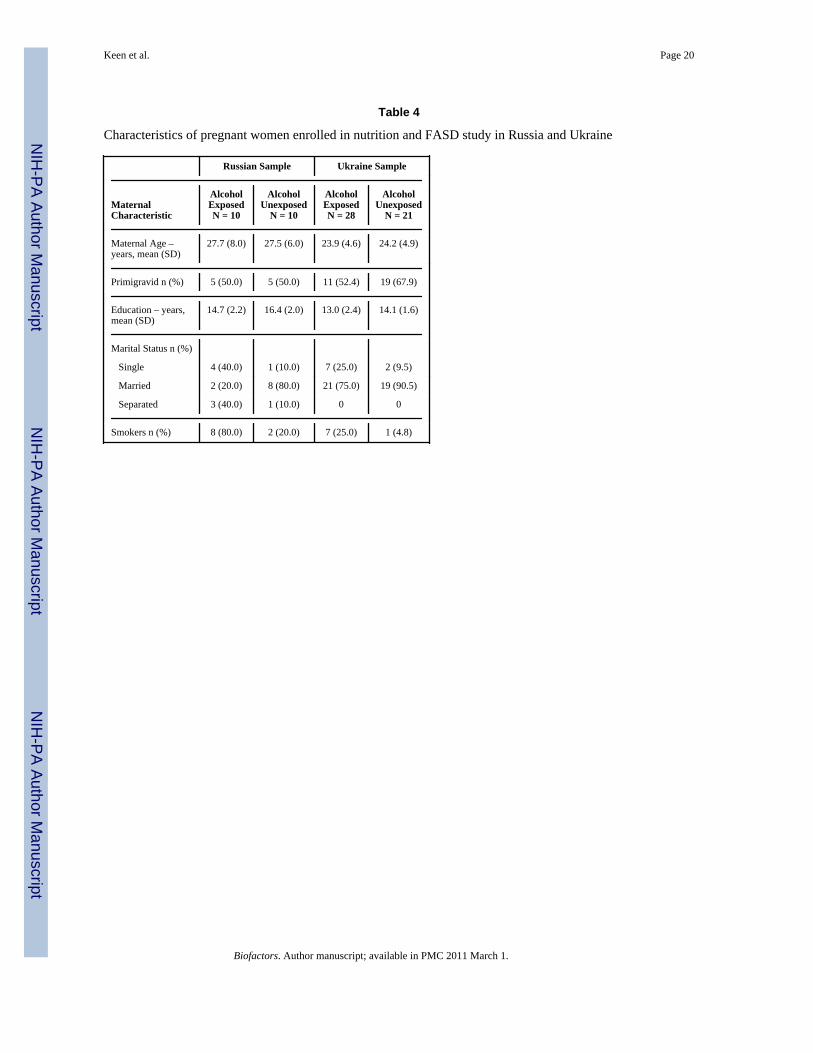
Preliminary data on analysis of mineral status at the time of enrollment is available for atotal of 69 subjects from the two sites. Women in the alcohol-exposed groups at the two siteswere similar in age, primigravidity, and years of education to their respective no or low-exposed groups, but more likely to be single mothers and to be current smokers relative totheir comparison groups (Table 4).
Table 5 describes the characteristics of maternal alcohol use as reported at the time ofenrollment regarding the month around the time of conception. Consistent with the groupselection criteria, women in the alcohol-exposed groups were predominately binge drinkers,with almost no women reporting daily drinking, even in small amounts.
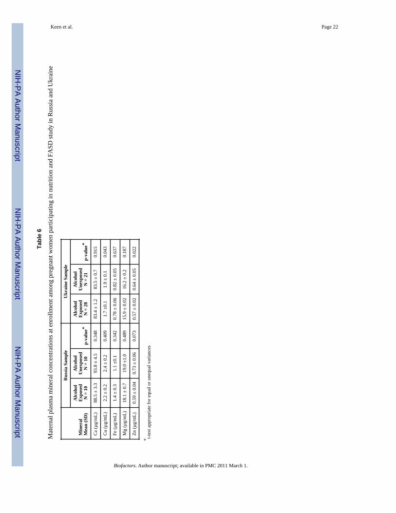
As shown in Table 6, although numbers are small in each sample, for most of the minerals,consistently lower mean values were observed in the alcohol-exposed groups than in the
Keen et al. Page 7
Biofactors. Author manuscript; available in PMC 2011 March 1.
NIH
-PA Author Manuscript
NIH
-PA Author Manuscript
NIH
-PA Author Manuscript
controls; these differences were statistically significant for Zn at both sites, and for Cu at theUkraine site.
4. Summary and Concluding CommentsThe above comments have been aimed at the overarching hypothesis that the occurrence ofnumerous features of FASD, including growth deficiency, structural features, persistentimmunological defects and neurobehavioral impairment, is influenced by the nutritionalstatus of the mother, as well as the conceptus. Current data support the concept that selectmicronutrient deficiencies increase the risk for the occurrence of FASD in high-riskpopulations. In theory, these nutritional deficiencies can arise as a consequence of poordiets, as well as a consequence of tissue injury-induced alterations in the metabolism ofselect nutrients. If the above concepts are correct, it is reasonable to predict that the use ofselect micronutrient supplements could reduce the frequency and severity of FASD in thesepopulations.
Consistent with this hypothesis, preliminary data from the ongoing studies in Ukraine andRussia show that plasma Zn and Cu concentrations are low in pregnant women who reporthigh alcohol intakes. In addition to the mounting evidence that certain micronutrients canaffect normal structural development, it is evident that prenatal nutrition is an importantfactor both in prenatal growth and in postnatal cognitive performance [88,89]. As describedabove, Zn, as well as Cu, is involved in multiple biochemical pathways that are critical forbrain growth and function.
As the toxicity of alcohol is thought to be due, in part, to free radical-induced oxidativedamage [83], deficits of Zn and Cu would both be predicted to increase the sensitivity of thedeveloping conceptus to alcohol, given that these nutrients contribute to the oxidativedefense system [90]. The reported blunting of alcohol’s adverse effects by folate, vitaminB12, choline and Zn supplementation indicates that there may be some commonalitiesamong these nutrients [22,91–93]. For example, all of these nutrients can affect redox stress,as well as gene methylation patterns, underscoring the fact that none of these nutrientsshould be looked at in isolation. Important future research directions include determinationof the mechanisms underlying the developmental effects of the “suboptimal” nutritionalstatus that can occur with alcohol exposure as well as delineation of how specific, persistent,and important these effects are.
A major aim of the ongoing CIFASD trial is to evaluate nutritional risk modifiers for FASDand to determine if maternal micronutrient supplementation during pregnancy in drinkingwomen has substantial promise as an easily accomplished environmental manipulation thatshould substantially improve pregnancy outcome for both the mother and infant. Largeprospective FASD studies that utilize well-“validated” biomarkers for maternal nutritionalstatus (acute and chronic) prior to and during early pregnancy are a critical research need. Inthese studies, maternal nutritional status should be evaluated at regular intervals to captureenvironment-induced changes. These large prospective nutrient supplementation trialsshould be implemented with populations of pregnant women who are at a high risk forFASD. Moreover, nutrient supplementation trials should also be done with FASD childrento determine whether nutritional intervention can alleviate long-term adverse outcomes inthe offspring.
In closing, it should be noted that the concept that women who abuse alcohol duringpregnancy should be encouraged to use multivitamin, multimineral supplements is not new.Indeed, in 1990, the American Institute of Medicine identified this group of women whoshould be particularly encouraged to use these supplements during pregnancy [94]. To date,
Keen et al. Page 8
Biofactors. Author manuscript; available in PMC 2011 March 1.
NIH
-PA Author Manuscript
NIH
-PA Author Manuscript
NIH
-PA Author Manuscript
our experience in Russia, as well as in Ukraine, is that women at high risk for having aFASD child are very receptive to using supplements. Obviously, the most appropriatemessage to these women is to stop drinking. However, on a practical level, combining thismessage with one that is aimed at improving their overall nutritional status would seem to bean appropriate public health strategy.
AcknowledgmentsThese studies were funded by NIAAA Grant numbers U01AA014835 and U24AA014811, and NIH Grant numbersHD 01743 and HD 26777. Part of this work was done in conjunction with the Collaborative Initiative on FetalAlcohol Spectrum Disorders (CIFASD), which is funded by grants from the National Institute on Alcohol andAlcohol Abuse (NIAAA). Additional information about CIFASD can be found at www.cifasd.org.
References1. MACDP: 40th Anniversary Edition Surveillance Report; Birth Defects Res. A Clin. Mol. Teratol.
2007. p. 66-186.2. Keen CL, Clegg MS, Hanna LA, Lanoue L, Rogers JM, Daston GP, Oteiza P, Uriu-Adams JY. The
plausibility of micronutrient deficiencies being a significant contributing factor to the occurrence ofpregnancy complications. J. Nutr. 2003; 133:1597S–1605S. [PubMed: 12730474]
3. Wadhwa PD, Buss C, Entringer S, Swanson JM. Developmental origins of health and disease: briefhistory of the approach and current focus on epigenetic mechanisms. Semin. Reprod. Med. 2009;27:358–368. [PubMed: 19711246]
4. Christian P, Stewart CP. Maternal Micronutrient Deficiency, Fetal Development, and the Risk ofChronic Disease. J. Nutr. 2010 [Epub ahead of print].
5. Ebbs JH, Tisdall FF, Scott WA. The influence of prenatal diet on the mother and child. J. Nutr.1941; 22:515–526.
6. Keen CL, Zidenberg-Cherr S. Should vitamin-mineral supplements be recommended for all womenwith childbearing potential? Am. J. Clin. Nutr. 1994; 59:532S–538S. discussion 538S–539S.[PubMed: 8304292]
7. Laurence, KM.; Campbell, H.; James, NE. The role of improvement in the maternal diet andpreconceptional folic acid supplementation in the prevention of neural tube defects. In: Dobbing, J.,editor. Prevention of Spina Bifida and Other Neural Tube Defects. New York: Academic Press;1983. p. 85-125.
8. Primrose T, Higgins A. A study in human antepartum nutrition. J. Reprod. Med. 1971; 7:257–264.[PubMed: 5128963]
9. Goh YI, Bollano E, Einarson TR, Koren G. Prenatal multivitamin supplementation and rates ofcongenital anomalies: a meta-analysis. J. Obstet. Gynaecol. Can. 2006; 28:680–689. [PubMed:17022907]
10. Picciano MF, McGuire MK. Use of dietary supplements by pregnant and lactating women in NorthAmerica. Am. J. Clin. Nutr. 2009; 89:663S–667S. [PubMed: 19073789]
11. Bodnar LM, Tang G, Ness RB, Harger G, Roberts JM. Periconceptional multivitamin use reducesthe risk of preeclampsia. Am. J. Epidemiol. 2006; 164:470–477. [PubMed: 16772374]
12. Rasmussen SA, Erickson JD, Reef SE, Ross DS. Teratology: from science to birth defectsprevention. Birth Defects Res. A Clin. Mol. Teratol. 2009; 85:82–92. [PubMed: 19067401]
13. Wiener SG, Shoemaker WJ, Koda LY, Bloom FE. Interaction of ethanol and nutrition duringgestation: influence on maternal and offspring development in the rat. J. Pharmacol. Exp. Ther.1981; 216:572–579. [PubMed: 7205638]
14. Weinberg J, D'Alquen G, Bezio S. Interactive effects of ethanol intake and maternal nutritionalstatus on skeletal development of fetal rats. Alcohol. 1990; 7:383–388. [PubMed: 2222841]
15. Wainwright P, Fritz G. Effect of moderate prenatal ethanol exposure on postnatal brain andbehavioral development in BALB/c mice. Exp. Neurol. 1985; 89:237–249. [PubMed: 4007107]
16. Shankar K, Hidestrand M, Liu X, Xiao R, Skinner CM, Simmen FA, Badger TM, Ronis MJ.Physiologic and genomic analyses of nutrition-ethanol interactions during gestation: Implications
Keen et al. Page 9
Biofactors. Author manuscript; available in PMC 2011 March 1.
NIH
-PA Author Manuscript
NIH
-PA Author Manuscript
NIH
-PA Author Manuscript
for fetal ethanol toxicity. Exp. Biol. Med. (Maywood). 2006; 231:1379–1397. [PubMed:16946407]
17. Weinberg J. Effects of ethanol and maternal nutritional status on fetal development. Alcohol Clin.Exp. Res. 1985; 9:49–55. [PubMed: 3887968]
18. Gloria L, Cravo M, Camilo ME, Resende M, Cardoso JN, Oliveira AG, Leitao CN, Mira FC.Nutritional deficiencies in chronic alcoholics: relation to dietary intake and alcohol consumption.Am. J. Gastroenterol. 1997; 92:485–489. [PubMed: 9068475]
19. Georgieff MK. Nutrition and the developing brain: nutrient priorities and measurement. Am. J.Clin. Nutr. 2007; 85:614S–620S. [PubMed: 17284765]
20. Keen CL, Taubeneck MW, Daston GP, Rogers JM, Gershwin ME. Primary and secondary zincdeficiency as factors underlying abnormal CNS development. Ann. N.Y. Acad. Sci. 1993; 678:37–47. [PubMed: 8494291]
21. Keen CL, Uriu-Hare JY, Hawk SN, Jankowski MA, Daston GP, Kwik-Uribe CL, Rucker RB.Effect of copper deficiency on prenatal development and pregnancy outcome. Am. J. Clin. Nutr.1998; 67:1003S–1011S. [PubMed: 9587143]
22. Xu Y, Li L, Zhang Z, Li Y. Effects of folinic acid and Vitamin B12 on ethanol-induceddevelopmental toxicity in mouse. Toxicol. Lett. 2006; 167:167–172. [PubMed: 17052868]
23. Chen CP. Syndromes, disorders and maternal risk factors associated with neural tube defects (VI).Taiwan J. Obstet. Gynecol. 2008; 47:267–275. [PubMed: 18935988]
24. Flynn A, Miller SI, Martier SS, Golden NL, Sokol RJ, Del Villano BC. Zinc status of pregnantalcoholic women: a determinant of fetal outcome. Lancet. 1981; 1:572–551. [PubMed: 6110817]
25. Miller SI, Del Villano BC, Flynn A, Krumhansl M. Interaction of alcohol and zinc in fetaldysmorphogenesis. Pharmacol. Biochem. Behav. 1983; 18 Suppl 1:311–315. [PubMed: 6634846]
26. Ruth RE, Goldsmith SK. Interaction between zinc deprivation and acute ethanol intoxicationduring pregnancy in rats. J. Nutr. 1981; 111:2034–2038. [PubMed: 7197711]
27. Dreosti IE, Partick EJ. Zinc, ethanol, and lipid peroxidation in adult and fetal rats. Biol. TraceElem. Res. 1987; 14:179–191.
28. Keppen LD, Pysher T, Rennert OM. Zinc deficiency acts as a co-teratogen with alcohol in fetalalcohol syndrome. Pediatr. Res. 1985; 19:944–947. [PubMed: 4047764]
29. Briefel RR, Bialostosky K, Kennedy-Stephenson J, McDowell MA, Ervin RB, Wright JD. Zincintake of the U.S. population: findings from the third National Health and Nutrition ExaminationSurvey, 1988–1994. J. Nutr. 2000; 130:1367S–1373S. [PubMed: 10801945]
30. Harley K, Eskenazi B, Block G. The association of time in the US and diet during pregnancy inlow-income women of Mexican descent. Paediatr. Perinat. Epidemiol. 2005; 19:125–134.[PubMed: 15787887]
31. Shah D, Sachdev HP. Effect of gestational zinc deficiency on pregnancy outcomes: summary ofobservation studies and zinc supplementation trials. Br. J. Nutr. 2001; 85 Suppl 2:S101–S108.[PubMed: 11509097]
32. Hess SY, Lonnerdal B, Hotz C, Rivera JA, Brown KH. Recent advances in knowledge of zincnutrition and human health. Food Nutr. Bull. 2009; 30:S5–S11. [PubMed: 19472599]
33. Keen CL, Taubeneck MW, Daston GP, Gershwin ME, Ansari A, Rogers JM. Primary andsecondary zinc deficiency as factors contributing to abnormal central nervous systemdevelopment. Dev. Brain Dysfunct. 1995; 8:79–89.
34. Maret W, Sandstead HH. Possible roles of zinc nutriture in the fetal origins of disease. Exp.Gerontol. 2008; 43:378–381. [PubMed: 18031964]
35. Golub MS, Keen CL, Gershwin ME. Moderate zinc-iron deprivation influences behavior but notgrowth in adolescent rhesus monkeys. J. Nutr. 2000; 130:354S–357S. [PubMed: 10721905]
36. Cummings JE, Kovacic JP. The ubiquitous role of zinc in health and disease. J. Vet. Emerg. Crit.Care (San Antonio). 2009; 19:215–240. [PubMed: 19691507]
37. Duffy JY, Overmann GJ, Keen CL, Clegg MS, Daston GP. Cardiac abnormalities induced by zincdeficiency are associated with alterations in the expression of genes regulated by the zinc-fingertranscription factor GATA-4. Birth Defects Res. B Dev. Reprod. Toxicol. 2004; 71:102–109.[PubMed: 15098203]
Keen et al. Page 10
Biofactors. Author manuscript; available in PMC 2011 March 1.
NIH
-PA Author Manuscript
NIH
-PA Author Manuscript
NIH
-PA Author Manuscript
38. Clegg MS, Hanna LA, Niles BJ, Momma TY, Keen CL. Zinc deficiency-induced cell death.IUBMB Life. 2005; 57:661–669. [PubMed: 16223705]
39. Oteiza PI, Mackenzie GG, Verstraeten SV. Metals in neurodegeneration: involvement of oxidantsand oxidant-sensitive transcription factors. Mol. Aspects Med. 2004; 25:103–115. [PubMed:15051320]
40. Jansen J, Karges W, Rink L. Zinc and diabetes--clinical links and molecular mechanisms. J. Nutr.Biochem. 2009; 20:399–417. [PubMed: 19442898]
41. Keen, CL.; Uriu-Adams, JY. Assessment of zinc, copper and magnesium status: Currentapproaches and promising new directions. In: Institute of Medicine. , editor. Mineral Requirementsfor Military Personnel. Washington, D.C: The National Academies Press; 2006. p. 304-315.
42. Chiang C, Litingtung Y, Harris MP, Simandl BK, Li Y, Beachy PA, Fallon JF. Manifestation ofthe limb prepattern: limb development in the absence of sonic hedgehog function. Dev. Biol. 2001;236:421–435. [PubMed: 11476582]
43. Krezel A, Hao Q, Maret W. The zinc/thiolate redox biochemistry of metallothionein and thecontrol of zinc ion fluctuations in cell signaling. Arch. Biochem. Biophys. 2007; 463:188–200.[PubMed: 17391643]
44. Chrisman K, Kenney R, Comin J, Thal T, Suchocki L, Yueh YG, Gardner DP. Gestational ethanolexposure disrupts the expression of FGF8 and Sonic hedgehog during limb patterning. BirthDefects Res. A Clin. Mol. Teratol. 2004; 70:163–171. [PubMed: 15108242]
45. Schreiner CM, Bell SM, Scott WJ Jr. Microarray analysis of murine limb bud ectoderm andmesoderm after exposure to cadmium or acetazolamide. Birth Defects Res. A Clin. Mol. Teratol.2009; 85:588–598. [PubMed: 19274763]
46. Daston GP, Overmann GJ, Baines D, Taubeneck MW, Lehman-McKeeman LD, Rogers JM, KeenCL. Altered Zn status by alpha-hederin in the pregnant rat and its relationship to adversedevelopmental outcome. Reprod. Toxicol. 1994; 8:15–24. [PubMed: 8186620]
47. Keen CL, Peters JM, Hurley LS. The effect of valproic acid on 65Zn distribution in the pregnantrat. J. Nutr. 1989; 119:607–611. [PubMed: 2495343]
48. Taubeneck MW, Daston GP, Rogers JM, Keen CL. Altered maternal zinc metabolism followingexposure to diverse developmental toxicants. Reprod. Toxicol. 1994; 8:25–40. [PubMed:8186621]
49. Hanna LA, Clegg MS, Ellis-Hutchings RG, Niles BJ, Keen CL. The influence of gestational zincdeficiency on the fetal insulin-like growth factor axis in the rat. Exp. Biol. Med. 2010 In press.
50. McGough NN, Thomas JD, Dominguez HD, Riley EP. Insulin-like growth factor-I mitigates motorcoordination deficits associated with neonatal alcohol exposure in rats. Neurotoxicol. Teratol.2009; 31:40–48. [PubMed: 18755266]
51. Adamo AM, Zago MP, Mackenzie GG, Aimo L, Keen CL, Keenan A, Oteiza PI. The role of zincin the modulation of neuronal proliferation and apoptosis. Neurotox. Res. 17:1–14. [PubMed:19784710]
52. Andrews GK. Regulation and function of Zip4, the acrodermatitis enteropathica gene. Biochem.Soc. Trans. 2008; 36:1242–1246. [PubMed: 19021533]
53. Mahomed K, Bhutta Z, Middleton P. Zinc supplementation for improving pregnancy and infantoutcome. Cochrane Database Syst. Rev. 2007 CD000230.
54. Taubeneck MW, Daston GP, Rogers JM, Gershwin ME, Ansari A, Keen CL. Tumor necrosisfactor-alpha alters maternal and embryonic zinc metabolism and is developmentally toxic in mice.J. Nutr. 1995; 125:908–919. [PubMed: 7722694]
55. Carey LC, Coyle P, Philcox JC, Rofe AM. Ethanol decreases zinc transfer to the fetus in normalbut not metallothionein-null mice. Alcohol Clin. Exp. Res. 2000; 24:1236–1240. [PubMed:10968663]
56. Carey LC, Coyle P, Philcox JC, Rofe AM. Maternal ethanol exposure is associated with decreasedplasma zinc and increased fetal abnormalities in normal but not metallothionein-null mice.Alcohol Clin. Exp. Res. 2000; 24:213–219. [PubMed: 10698374]
57. Zhang JP, Li F, Yu XW, Sheng Q, Shi XW, Zhang XW. Trace elements and cytokine profile incytomegalovirus-infected pregnancies: a controlled study. Gynecol. Obstet. Invest. 2008; 65:128–132. [PubMed: 17957100]
Keen et al. Page 11
Biofactors. Author manuscript; available in PMC 2011 March 1.
NIH
-PA Author Manuscript
NIH
-PA Author Manuscript
NIH
-PA Author Manuscript
58. Collier SA, Rasmussen SA, Feldkamp ML, Honein MA. Prevalence of self-reported infectionduring pregnancy among control mothers in the National Birth Defects Prevention Study. BirthDefects Res. A Clin. Mol. Teratol. 2009; 85:193–201. [PubMed: 19086018]
59. Coyle P, Philcox JC, Carey LC, Rofe AM. Metallothionein: the multipurpose protein. Cell. Mol.Life Sci. 2002; 59:627–647. [PubMed: 12022471]
60. Carey LC, Coyle P, Philcox JC, Rofe AM. Zinc supplementation at the time of ethanol exposureameliorates teratogenicity in mice. Alcohol Clin. Exp. Res. 2003; 27:107–110. [PubMed:12544014]
61. Summers BL, Rofe AM, Coyle P. Dietary zinc supplementation throughout pregnancy protectsagainst fetal dysmorphology and improves postnatal survival after prenatal ethanol exposure inmice. Alcohol Clin. Exp. Res. 2009; 33:591–600. [PubMed: 19183140]
62. Coyle P, Martin SA, Carey LC, Summers BL, Rofe AM. Ethanol-mediated fetal dysmorphologyand its relationship to the ontogeny of maternal liver metallothionein. Alcohol Clin. Exp. Res.2009; 33:1051–1058. [PubMed: 19302082]
63. Seyoum G, Persaud TV. Protective influence of zinc against the deleterious effects of ethanol inpostimplantation rat embryos in vivo. Exp. Toxicol. Pathol. 1995; 47:75–79. [PubMed: 7719124]
64. Szuster-Ciesielska A, Plewka K, Daniluk J, Kandefer-Szerszen M. Zinc supplementation attenuatesethanol- and acetaldehyde-induced liver stellate cell activation by inhibiting reactive oxygenspecies (ROS) production and by influencing intracellular signaling. Biochem. Pharmacol. 2009;78:301–314. [PubMed: 19376089]
65. Kang YJ, Zhou Z. Zinc prevention and treatment of alcoholic liver disease. Mol. Aspects Med.2005; 26:391–404. [PubMed: 16099027]
66. Kang X, Zhong W, Liu J, Song Z, McClain CJ, Kang YJ, Zhou Z. Zinc supplementation reversesalcohol-induced steatosis in mice through reactivating hepatocyte nuclear factor-4alpha andperoxisome proliferator-activated receptor-alpha. Hepatology. 2009; 50:1241–1250. [PubMed:19637192]
67. Joshi PC, Mehta A, Jabber WS, Fan X, Guidot DM. Zinc deficiency mediates alcohol-inducedalveolar epithelial and macrophage dysfunction in rats. Am. J. Respir. Cell. Mol. Biol. 2009;41:207–216. [PubMed: 19109243]
68. Assadi FK, Ziai M. Zinc status of infants with fetal alcohol syndrome. Pediatr. Res. 1986; 20:551–554. [PubMed: 3714366]
69. Murillo-Fuentes ML, Artillo R, Ojeda ML, Delgado MJ, Murillo ML, Carreras O. Effects ofprenatal or postnatal ethanol consumption on zinc intestinal absorption and excretion in rats.Alcohol Alcohol. 2007; 42:3–10. [PubMed: 17068010]
70. McGill J, Meyerholz DK, Edsen-Moore M, Young B, Coleman RA, Schlueter AJ, WaldschmidtTJ, Cook RT, Legge KL. Fetal exposure to ethanol has long-term effects on the severity ofinfluenza virus infections. J. Immunol. 2009; 182:7803–7808. [PubMed: 19494304]
71. Zhang X, Sliwowska JH, Weinberg J. Prenatal alcohol exposure and fetal programming: effects onneuroendocrine and immune function. Exp. Biol. Med. (Maywood). 2005; 230:376–388.[PubMed: 15956767]
72. Summers BL, Rofe AM, Coyle P. Prenatal zinc treatment at the time of acute ethanol exposurelimits spatial memory impairments in mouse offspring. Pediatr. Res. 2006; 59:66–71. [PubMed:16326994]
73. Summers BL, Henry CM, Rofe AM, Coyle P. Dietary zinc supplementation during pregnancyprevents spatial and object recognition memory impairments caused by early prenatal ethanolexposure. Behav. Brain Res. 2008; 186:230–238. [PubMed: 17884190]
74. Chen WJ, Berryhill EC, West JR. Zinc supplementation does not attenuate alcoholinducedcerebellar Purkinje cell loss during the brain growth spurt period. Alcohol Clin. Exp. Res. 2001;25:600–605. [PubMed: 11329502]
75. Zidenberg-Cherr S, Benak PA, Hurley LS, Keen CL. Altered mineral metabolism: a mechanismunderlying the fetal alcohol syndrome in rats. Drug Nutr. Interact. 1988; 5:257–274. [PubMed:3240709]
76. Gambling L, McArdle HJ. Iron, copper and fetal development. Proc. Nutr. Soc. 2004; 63:553–562.[PubMed: 15831127]
Keen et al. Page 12
Biofactors. Author manuscript; available in PMC 2011 March 1.
NIH
-PA Author Manuscript
NIH
-PA Author Manuscript
NIH
-PA Author Manuscript
77. Penland JG, Prohaska JR. Abnormal motor function persists following recovery from perinatalcopper deficiency in rats. J. Nutr. 2004; 134:1984–1988. [PubMed: 15284387]
78. De Feo TM, Fargion S, Duca L, Cesana BM, Boncinelli L, Lozza P, Cappellini MD, Fiorelli G.Non-transferrin-bound iron in alcohol abusers. Alcohol Clin. Exp. Res. 2001; 25:1494–1499.[PubMed: 11696670]
79. Harrison-Findik DD, Schafer D, Klein E, Timchenko NA, Kulaksiz H, Clemens D, Fein E,Andriopoulos B, Pantopoulos K, Gollan J. Alcohol metabolism-mediated oxidative stress down-regulates hepcidin transcription and leads to increased duodenal iron transporter expression. J.Biol. Chem. 2006; 281:22974–22982. [PubMed: 16737972]
80. Ioannou GN, Dominitz JA, Weiss NS, Heagerty PJ, Kowdley KV. The effect of alcoholconsumption on the prevalence of iron overload, iron deficiency, and iron deficiency anemia.Gastroenterology. 2004; 126:1293–1301. [PubMed: 15131790]
81. Whitfield JB, Zhu G, Heath AC, Powell Lw, Martin NG. Effects of alcohol consumption onindices of iron stores and of iron stores on alcohol intake markers. Alcohol Clin. Exp. Res. 2001;25:1037–1045. [PubMed: 11505030]
82. Halliwell B. Oxidative stress, nutrition and health. Experimental strategies for optimization ofnutritional antioxidant intake in humans. Free Radic. Res. 1996; 25:57–74. [PubMed: 8814444]
83. Cederbaum AI. Introduction-serial review: alcohol, oxidative stress and cell injury. Free Radic.Biol. Med. 2001; 31:1524–1526. [PubMed: 11744324]
84. Miller MW, Roskams AJ, Connor JR. Iron regulation in the developing rat brain: effect of in uteroethanol exposure. J. Neurochem. 1995; 65:373–380. [PubMed: 7790882]
85. Carter RC, Jacobson SW, Molteno CD, Jacobson JL. Fetal alcohol exposure, irondeficiencyanemia, and infant growth. Pediatrics. 2007; 120:559–567. [PubMed: 17766529]
86. Gambling L, Andersen HS, McArdle HJ. Iron and copper, and their interactions duringdevelopment. Biochem. Soc. Trans. 2008; 36:1258–1261. [PubMed: 19021536]
87. Mackenzie GG, Zago MP, Keen CL, Oteiza PI. Low intracellular zinc impairs the translocation ofactivated NF-kappa B to the nuclei in human neuroblastoma IMR-32 cells. J. Biol. Chem. 2002;277:34610–34617. [PubMed: 12089148]
88. Avchen RN, Scott KG, Mason CA. Birth weight and school-age disabilities: a population-basedstudy. Am. J. Epidemiol. 2001; 154:895–901. [PubMed: 11700243]
89. Faden VB, Hanna E, Graubard BI. The effect of positive and negative health behavior duringgestation on pregnancy outcome. J. Subst. Abuse. 1997; 9:63–76. [PubMed: 9494939]
90. Keen, CL. Teratogenic effects of essential trace metals: deficiencies and excesses. In: Chang, LW.;Magos, L.; Suzuki, T., editors. Toxicology of Metals. New York: CRC Press; 1996. p. 977-1001.
91. Xu Y, Li Y, Tang Y, Wang J, Shen X, Long Z, Zheng X. The maternal combined supplementationof folic acid and Vitamin B(12) suppresses ethanol-induced developmental toxicity in mousefetuses. Reprod. Toxicol. 2006; 22:56–61. [PubMed: 16439097]
92. Thomas JD, Abou EJ, Dominguez HD. Prenatal choline supplementation mitigates the adverseeffects of prenatal alcohol exposure on development in rats. Neurotoxicol. Teratol. 2009; 31:303–311. [PubMed: 19616089]
93. Thomas JD, Biane JS, O'Bryan KA, O'Neill TM, Dominguez HD. Choline supplementationfollowing third-trimester-equivalent alcohol exposure attenuates behavioral alterations in rats.Behav. Neurosci. 2007; 121:120–130. [PubMed: 17324056]
94. Subcommittee on Nutritional Status and Weight Gain During Pregnancy. , editor. Institute ofMedicine. Nutrition During Pregnancy: Part I: Weight Gain, Part II: Nutrient Supplements.Washington, D.C: National Academy Press; 1990.
95. Burke BS, Beal VA, Kirkwood SB, Stuart HC. The Influence of nutrition during pregnancy uponthe condition of the infant at birth: Two figures. J. Nutr. 1943; 26:569–583.
96. Jeans PC, Smith MB, Stearns G. Incidence of prematurity in relation to maternal nutrition. J. Am.Diet. Assoc. 1955; 31:576–581. [PubMed: 14381174]
97. Friel JK, Frecker M, Fraser FC. Nutritional patterns of mothers of children with neural tube defectsin Newfoundland. Am. J. Med. Genet. 1995; 55:195–199. [PubMed: 7717417]
98. Wright ME. A case-control study of maternal nutrition and neural tube defects in Northern Ireland.Midwifery. 1995; 11:146–152. [PubMed: 7565158]
Keen et al. Page 13
Biofactors. Author manuscript; available in PMC 2011 March 1.
NIH
-PA Author Manuscript
NIH
-PA Author Manuscript
NIH
-PA Author Manuscript
99. Torfs CP, Lam PK, Schaffer DM, Brand RJ. Association between mothers' nutrient intake and theiroffspring's risk of gastroschisis. Teratology. 1998; 58:241–250. [PubMed: 9894673]
100. Velie EM, Block G, Shaw GM, Samuels SJ, Schaffer DM, Kulldorff M. Maternal supplementaland dietary zinc intake and the occurrence of neural tube defects in California. Am. J. Epidemiol.1999; 150:605–616. [PubMed: 10490000]
101. Di Cintio E, Parazzini F, Chatenoud L, Surace M, Benzi G, Zanconato G, La Vecchia C. Dietaryfactors and risk of spontaneous abortion. Eur. J. Obstet. Gynecol. Reprod. Biol. 2001; 95:132–136. [PubMed: 11267735]
102. Krapels IP, van Rooij IA, Ocke MC, West CE, van der Horst CM, Steegers-Theunissen RP.Maternal nutritional status and the risk for orofacial cleft offspring in humans. J. Nutr. 2004;134:3106–3113. [PubMed: 15514283]
103. Rees GA, Doyle W, Srivastava A, Brooke ZM, Crawford MA, Costeloe KL. The nutrient intakesof mothers of low birth weight babies - a comparison of ethnic groups in East London, UK.Matern. Child. Nutr. 2005; 1:91–99. [PubMed: 16881884]
104. Lam PK, Torfs CP. Interaction between maternal smoking and malnutrition in infant risk ofgastroschisis. Birth Defects Res. A Clin. Mol. Teratol. 2006; 76:182–186. [PubMed: 16498669]
105. Knudsen VK, Orozova-Bekkevold IM, Mikkelsen TB, Wolff S, Olsen SF. Major dietary patternsin pregnancy and fetal growth. Eur. J. Clin. Nutr. 2008; 62:463–470. [PubMed: 17392696]
106. Yang W, Shaw GM, Carmichael SL, Rasmussen SA, Waller DK, Pober BR, Anderka M. Nutrientintakes in women and congenital diaphragmatic hernia in their offspring. Birth Defects Res. AClin. Mol. Teratol. 2008; 82:131–138. [PubMed: 18181217]
107. Hambidge KM, Neldner KH, Walravens PA. Letter: Zinc,acrodermatitis enteropathica, andcongenital malformations. Lancet. 1975; 1:577–578.
108. Jameson S. Variations in maternal serum zinc during pregnancy and correlation to congenitalmalformations, dysmaturity, and abnormal parturition. Acta Med. Scand. Suppl. 1976; 593:21–37. [PubMed: 1067746]
109. Meadows NJ, Ruse W, Smith MF, Day J, Keeling PW, Scopes JW, Thompson RP, Bloxam DL.Zinc and small babies. Lancet. 1981; 2:1135–1137. [PubMed: 6118578]
110. Cavdar AO, Bahceci M, Akar N, Erten J, Bahceci G, Babacan E, Arcasoy A, Yavuz H. Zincstatus in pregnancy and the occurrence of anencephaly in Turkey. J. Trace Elem. ElectrolytesHealth Dis. 1988; 2:9–14. [PubMed: 2980795]
111. Neggers YH, Cutter GR, Alvarez JO, Goldenberg RL, Acton R, Go RC, Roseman JM. Therelationship between maternal serum zinc levels during pregnancy and birthweight. Early Hum.Dev. 1991; 25:75–85. [PubMed: 1860432]
112. Srinivas M, Gupta DK, Rathi SS, Grover JK, Vats V, Sharma JD, Mitra DK. Association betweenlower hair zinc levels and neural tube defects. Indian J. Pediatr. 2001; 68:519–522. [PubMed:11450382]
113. Scheplyagina LA. Impact of the mother's zinc deficiency on the woman's and newborn's healthstatus. J. Trace Elem. Med. Biol. 2005; 19:29–35. [PubMed: 16240669]
114. Kynast G, Saling E. Effect of oral zinc application during pregnancy. Gynecol. Obstet. Invest.1986; 21:117–123. [PubMed: 3710284]
115. Cherry FF, Sandstead HH, Rojas P, Johnson LK, Batson HK, Wang XB. Adolescent pregnancy:associations among body weight, zinc nutriture, and pregnancy outcome. Am. J. Clin. Nutr.1989; 50:945–954. [PubMed: 2816802]
116. Cavdar AO, Bahceci M, Akar N, Erten J, Yavuz H. Effect of zinc supplementation in a Turkishwoman with two previous anencephalic infants. Gynecol. Obstet. Invest. 1991; 32:123–125.[PubMed: 1748322]
117. Goldenberg RL, Tamura T, Neggers Y, Copper RL, Johnston KE, DuBard MB, Hauth JC. Theeffect of zinc supplementation on pregnancy outcome. J.A.M.A. 1995; 274:463–468.
118. Osendarp SJ, van Raaij JM, Darmstadt GL, Baqui AH, Hautvast JG, Fuchs GJ. Zincsupplementation during pregnancy and effects on growth and morbidity in low birthweightinfants: a randomised placebo controlled trial. Lancet. 2001; 357:1080–1085. [PubMed:11297959]
Keen et al. Page 14
Biofactors. Author manuscript; available in PMC 2011 March 1.
NIH
-PA Author Manuscript
NIH
-PA Author Manuscript
NIH
-PA Author Manuscript
119. Hunt IF, Murphy NJ, Cleaver AE, Faraji B, Swendseid ME, Browdy BL, Coulson AH, Clark VA,Settlage RH, Smith JC Jr. Zinc supplementation during pregnancy in low-income teenagers ofMexican descent: effects on selected blood constituents and on progress and outcome ofpregnancy. Am. J. Clin. Nutr. 1985; 42:815–828. [PubMed: 4061343]
120. Mahomed K, James DK, Golding J, McCabe R. Zinc supplementation during pregnancy: a doubleblind randomised controlled trial. B.M.J. 1989; 299:826–830.
121. Jonsson B, Hauge B, Larsen MF, Hald F. Zinc supplementation during pregnancy: a double blindrandomised controlled trial. Acta Obstet. Gynecol. Scand. 1996; 75:725–729. [PubMed:8906006]
122. Caulfield LE, Zavaleta N, Figueroa A, Leon Z. Maternal zinc supplementation does not affectsize at birth or pregnancy duration in Peru. J. Nutr. 1999; 129:1563–1568. [PubMed: 10419991]
123. Hafeez A, Mehmood G, Mazhar F. Oral zinc supplementation in pregnant women and its effecton birth weight: a randomised controlled trial. Arch. Dis. Child. Fetal Neonatal Ed. 2005;90:F170–F171. [PubMed: 15724045]
Keen et al. Page 15
Biofactors. Author manuscript; available in PMC 2011 March 1.
NIH
-PA Author Manuscript
NIH
-PA Author Manuscript
NIH
-PA Author Manuscript
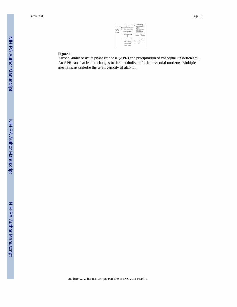
Figure 1.Alcohol-induced acute phase response (APR) and precipitation of conceptal Zn deficiency.An APR can also lead to changes in the metabolism of other essential nutrients. Multiplemechanisms underlie the teratogenicity of alcohol.
Keen et al. Page 16
Biofactors. Author manuscript; available in PMC 2011 March 1.
NIH
-PA Author Manuscript
NIH
-PA Author Manuscript
NIH
-PA Author Manuscript
NIH
-PA Author Manuscript
NIH
-PA Author Manuscript
NIH
-PA Author Manuscript
Keen et al. Page 17
Table 1
Maternal Diet and Pregnancy Outcome: “Good” vs. “Poor” Diet
Author Study
Ebbs et al., 1941 Intervention
Burke et al., 1943 Observational
Jeans et al., 1955 Observational
Primrose and Higgins, 1971 Intervention
Laurence et al., 1983 Intervention
Friel et al., 1995 Observational
Wright, 1995 Observational
Torfs et al., 1998 Observational
Velie et al., 1999 Observational
Di Cintio et al., 2001 Observational
Krapels et al., 2004 Observational
Rees et al., 2005 Observational
Lam and Torfs, 2006 Observational
Knudsen et al., 2008 Observational
Yang et al., 2008 Observational
Baker et al., 2009 Observational
Haggarty et al., 2009 Observational
Biofactors. Author manuscript; available in PMC 2011 March 1.
NIH
-PA Author Manuscript
NIH
-PA Author Manuscript
NIH
-PA Author Manuscript
Keen et al. Page 18
Table 2
Potential mechanisms of Zn deficiency teratogenicity
Indirect
Zinc deficiency-induced changes in maternal metabolism
Direct
Impaired DNA replication and transcription
Chromosomal damage
Decreased binding of growth factors and hormones to their receptors
Excessive oxidative damage
Altered patterns of cell death
Altered cell signaling
Impaired cell migration
Low activities of numerous zinc enzymes
Biofactors. Author manuscript; available in PMC 2011 March 1.
NIH
-PA Author Manuscript
NIH
-PA Author Manuscript
NIH
-PA Author Manuscript
Keen et al. Page 19
Table 3
Evidence for an influence of Zn on human pregnancy outcome
Poor pregnancy outcome in women with acrodermatitis enteropathica
Hambidge et al., 1975
Low Zn status (low blood and hair Zn concentrations) is associated withan increased risk for complications
Jameson
Meadows et al.
Cavdar et al.
Neggers et al.
Srinivas et al.
Scheplyagina
Golalipour et al.
Zn supplements are associated with improved pregnancy outcome
Kynast and Saling, 1986
Cherry et al., 1989
Cavdar et al., 1991
Goldenberg et al., 1995
Osendarp et al., 2001
Saaka et al., 2009
Hozyasz et al., 2009
Danesh et al., 2009
Zn supplements do not reduce pregnancy complications
Hunt et al., 1985
Mahomed et al., 1989
Jonsson et al., 1996
Caulfield et al., 1999
Hafeez et al., 2005
Biofactors. Author manuscript; available in PMC 2011 March 1.
NIH
-PA Author Manuscript
NIH
-PA Author Manuscript
NIH
-PA Author Manuscript
Keen et al. Page 20
Table 4
Characteristics of pregnant women enrolled in nutrition and FASD study in Russia and Ukraine
Russian Sample Ukraine Sample
MaternalCharacteristic
AlcoholExposedN = 10
AlcoholUnexposed
N = 10
AlcoholExposedN = 28
AlcoholUnexposed
N = 21
Maternal Age –years, mean (SD)
27.7 (8.0) 27.5 (6.0) 23.9 (4.6) 24.2 (4.9)
Primigravid n (%) 5 (50.0) 5 (50.0) 11 (52.4) 19 (67.9)
Education – years,mean (SD)
14.7 (2.2) 16.4 (2.0) 13.0 (2.4) 14.1 (1.6)
Marital Status n (%)
Single 4 (40.0) 1 (10.0) 7 (25.0) 2 (9.5)
Married 2 (20.0) 8 (80.0) 21 (75.0) 19 (90.5)
Separated 3 (40.0) 1 (10.0) 0 0
Smokers n (%) 8 (80.0) 2 (20.0) 7 (25.0) 1 (4.8)
Biofactors. Author manuscript; available in PMC 2011 March 1.
NIH
-PA Author Manuscript
NIH
-PA Author Manuscript
NIH
-PA Author Manuscript
Keen et al. Page 21
Table 5
Reported alcohol consumption in month around time of conception among pregnant women enrolled innutrition and FASD study in Russia and Ukraine
Russian Sample Ukraine Sample
# Drinks perOccasion inMonth
AlcoholExposedN = 10
AlcoholUnexposed
N = 10
AlcoholExposedN = 28
AlcoholUnexposed
N = 21
5 or more n (%)# of occasions
4 (40.0)0–5
0 12 (42.9)0–3
0
3 or 4 n (%)# of occasions
10 (100)2–5
0 24 (85.7)0–10
0
1 or 2 n (%)# of occasions
9 (90.0)0–7
7 (70.0)0–3
28 (100)1–28
7 (33.3)0–2
Daily n (%) 0 0 2 (7.1) 0
Biofactors. Author manuscript; available in PMC 2011 March 1.
NIH
-PA Author Manuscript
NIH
-PA Author Manuscript
NIH
-PA Author Manuscript
Keen et al. Page 22
Tabl
e 6
Mat
erna
l pla
sma
min
eral
con
cent
ratio
ns a
t enr
ollm
ent a
mon
g pr
egna
nt w
omen
par
ticip
atin
g in
nut
ritio
n an
d FA
SD st
udy
in R
ussi
a an
d U
krai
ne
Min
eral
Mea
n (S
D)
Rus
sia
Sam
ple
Ukr
aine
Sam
ple
Alc
ohol
Exp
osed
N =
10
Alc
ohol
Une
xpos
edN
= 1
0p-
valu
e*
Alc
ohol
Exp
osed
N =
28
Alc
ohol
Une
xpos
edN
= 2
1p-
valu
e*
Ca
(µg/
mL)
88.5
± 3
.393
.8 ±
4.5
0.34
883
.4 ±
1.2
83.5
± 0
.70.
915
Cu
(µg/
mL)
2.2
± 0.
22.
4 ±
0.2
0.40
91.
7 ±0
.11.
9 ±
0.1
0.04
3
Fe (µ
g/m
L)1.
4 ±
0.3
1.1
±0.1
0.34
20.
78 ±
0.0
60.
82 ±
0.0
50.
637
Mg
(µg/
mL)
18.1
± 0
.719
.0 ±
1.0
0.48
915
.9 ±
0.0
216
.2 ±
0.2
0.18
7
Zn (µ
g/m
L)0.
59 ±
0.0
40.
73 ±
0.0
60.
073
0.57
± 0
.02
0.64
± 0
.05
0.02
2
* t-tes
t app
ropr
iate
for e
qual
or u
nequ
al v
aria
nces
Biofactors. Author manuscript; available in PMC 2011 March 1.