The Pan-Pacific entomologist
424
Vol. 64 January 1988 THE Pan-Pacific Entomologist No. 1 PARKER, F. D. —Nesting biology of two North American species of Chelostoma (Hymenoptera: Megachilidae). 1 CORPUS, L. D.—Immature stages of Tachytrechus auratus (Diptera: Dolichopodidae) . 9 PARRELLA, M. P. and J. A. BETHKE—Larval development and leafmining activity of Liriomyza trifolli (Burgess) (Diptera: Agromyzidae). 17 POLHEMUS, D. A. and J. T. POLHEMUS—A new ant mimetic mirid from the Colorado Tundra (Elemiptera: Miridae) . 23 SHE, S. and D. YOU—A new species of Isonychia from China (Ephemeroptera: Oligoneuriidae) . 29 EHLER, L. E. and H. P. SAUTER—Observations on Eupelmus inyoensis Girault (Hymenoptera: Eupelmidae) . 33 WEISENBORN, W. D. and J. G. MORSE—The mandible and maxillary stylets of Scirtothrips citri (Moulton) (Thysanoptera: Thripidae). 39 GRISWOLD, T. L. and F. D. PARKER—New Perdita (Perdita) oligoleges of Mentzelia with notes on related species of the ventralis group (Hymenoptera: Andrenidae) . 43 WILSON, S. W. and J. H. TSAI—Descriptions of adults and nymphs of the Taro planthopper, Tarophagusproserpina taiwanensis ssp. n., from Taiwan (Homoptera: Delphacidae). 53 TEPEDINO, V. J.—Incidence of pre-emergence sib-mating in Monodontomerus obsoletus, Pteromalus venustus, and Tetrastichus megachilidis, three chalcid parasites of the alfalfa leafcutting bee, Megachile rotundata (Hymenoptera: Chalcididae) . 63 TEPEDINO, V. J.—Aspects of host acceptance by Pteromalus venustus Walker and Monodontomerus obsoletus Fabricius, parasitoids of Megachile rotundata (Fabricius), the alfalfa leafcutting bee (Hymenoptera: Chalcididae) . 67 O’BRIEN, M. F. and A. M. O’BRIEN—Biology of Ammophila evansi and A. mediata in northern Michigan (Hymenoptera: Sphecidae) . 73 PEMBERTON, R. W.—The use of the Thai giant waterbug, Lethocerus indicus (Hemiptera: Belostomatidae), as human food in California. 81 WEHLING, W. F. and G. L. PIPER—Efficacy diminution of the rush skeletonweed gall midge, Cystiphora schmidti (Diptra: Cecidomyiidae), by an indigenous parasitoid . 83 DALY, H. V. and G. R. ELSE—Lectotype designations for the African bees of the genus Ceratina described by T. D. A. Cockerell (Hymenoptera: Apoidea). 87 RATHMAN, R. J. and J. F. BRUNNER—Observations on the biology of a new species of Dilyfa (Hymenoptera: Charipidae) from Washington State . 93 SCIENTIFIC NOTES . 8,98 SAN FRANCISCO, CALIFORNIA • 1988 Published by the PACIFIC COAST ENTOMOLOGICAL SOCIETY in cooperation with THE CALIFORNIA ACADEMY OF SCIENCES
-
Upload
khangminh22 -
Category
Documents
-
view
0 -
download
0
Transcript of The Pan-Pacific entomologist

Vol. 64 January 1988
THE Pan-Pacific Entomologist
No. 1
PARKER, F. D. —Nesting biology of two North American species of Chelostoma (Hymenoptera: Megachilidae). 1
CORPUS, L. D.—Immature stages of Tachytrechus auratus (Diptera: Dolichopodidae) . 9
PARRELLA, M. P. and J. A. BETHKE—Larval development and leafmining activity of Liriomyza trifolli (Burgess) (Diptera: Agromyzidae). 17
POLHEMUS, D. A. and J. T. POLHEMUS—A new ant mimetic mirid from the Colorado Tundra (Elemiptera: Miridae) . 23
SHE, S. and D. YOU—A new species of Isonychia from China (Ephemeroptera: Oligoneuriidae) . 29
EHLER, L. E. and H. P. SAUTER—Observations on Eupelmus inyoensis Girault (Hymenoptera: Eupelmidae) . 33
WEISENBORN, W. D. and J. G. MORSE—The mandible and maxillary stylets of Scirtothrips citri (Moulton) (Thysanoptera: Thripidae). 39
GRISWOLD, T. L. and F. D. PARKER—New Perdita (Perdita) oligoleges of Mentzelia with notes on related species of the ventralis group (Hymenoptera: Andrenidae) . 43
WILSON, S. W. and J. H. TSAI—Descriptions of adults and nymphs of the Taro planthopper, Tarophagusproserpina taiwanensis ssp. n., from Taiwan (Homoptera: Delphacidae). 53
TEPEDINO, V. J.—Incidence of pre-emergence sib-mating in Monodontomerus obsoletus, Pteromalus venustus, and Tetrastichus megachilidis, three chalcid parasites of the alfalfa leafcutting bee, Megachile rotundata (Hymenoptera: Chalcididae) . 63
TEPEDINO, V. J.—Aspects of host acceptance by Pteromalus venustus Walker and Monodontomerus obsoletus Fabricius, parasitoids of Megachile rotundata (Fabricius), the alfalfa leafcutting bee (Hymenoptera: Chalcididae) . 67
O’BRIEN, M. F. and A. M. O’BRIEN—Biology of Ammophila evansi and A. mediata in northern Michigan (Hymenoptera: Sphecidae) . 73
PEMBERTON, R. W.—The use of the Thai giant waterbug, Lethocerus indicus (Hemiptera: Belostomatidae), as human food in California. 81
WEHLING, W. F. and G. L. PIPER—Efficacy diminution of the rush skeletonweed gall midge, Cystiphora schmidti (Diptra: Cecidomyiidae), by an indigenous parasitoid . 83
DALY, H. V. and G. R. ELSE—Lectotype designations for the African bees of the genus Ceratina described by T. D. A. Cockerell (Hymenoptera: Apoidea). 87
RATHMAN, R. J. and J. F. BRUNNER—Observations on the biology of a new species of Dilyfa (Hymenoptera: Charipidae) from Washington State . 93
SCIENTIFIC NOTES . 8,98
SAN FRANCISCO, CALIFORNIA • 1988 Published by the PACIFIC COAST ENTOMOLOGICAL SOCIETY in cooperation with THE CALIFORNIA ACADEMY OF SCIENCES

The Pan-Pacific Entomologist
EDITORIAL BOARD J. A. Chemsak, Editor
R. S. Lane, Associate Editor W.J. Pulawski, Treasurer J.T. Doyen R. M. Bohart J. A. Powell J. E. Hafernik, Jr.
Published quarterly in January, April, July, and October with Society Proceedings appearing in the October number. All communications regarding nonreceipt of num¬ bers, requests for sample copies, and financial communications should be addressed to
the Treasurer, Dr. Wojciech J. Pulawski, California Academy of Sciences, Golden Gate Park, San Francisco, CA 94118-9961.
Application for membership in the Society and changes of address should be ad¬ dressed to the Secretary, Mr. Vincent F. Lee, California Academy of Sciences, Golden
Gate Park, San Francisco, CA 94118-9961.
Manuscripts, proofs, and all correspondence concerning editorial matters should be addressed to Editor, Pacific Coast Entomological Society, 201 Wellman Hall, Univer¬ sity of California, Berkeley, CA 94720. See back cover for instructions.
The annual dues, paid in advance, are $15.00 for regular members of the Society, $7.50 for student members, $30.00 or more for sponsoring members, or $20.00 for sub¬ scription only. Members of the Society receive The Pan-Pacific Entomologist. Single copies of recent numbers are $5.00 each or $20.00 a volume. See back cover for prices of earlier back numbers. Make checks payable to the Pacific Coast Entomological Soci¬ ety.
Pacific Coast Entomological Society
OFFICERS FOR 1988
Alan I. Kaplan, President Wojciech J. Pulawski, Treasurer Thomas J. Zavortink, President-Elect Leslie S. Saul, Recording Secretary
Vincent F. Lee, Managing Secretary
THE PAN-PACIFIC ENTOMOLOGIST (ISSN 0031-0603) is published quarterly (January, April, July,
and October) for $20.00 per year by the Pacific Coast Entomological Society, California Academy of Sciences,
Golden Gate Park, San Francisco, California 94118-9961. Second-class postage paid at San Francisco, Califor¬
nia, and at additional mailing offices. POSTMASTER: Send address changes to THE PAN-PACIFIC EN¬
TOMOLOGIST, California Academy of Sciences, Golden Gate Park, San Francisco, California 94118-9961.
This issue mailed March 18,1988
The Pan-Pacific Entomologist (ISSN 0031-0603)
PRODUCED BY A-R EDITIONS, INC., MADISON, WI 53703, U.S.A.

PAN-PACIFIC ENTOMOLOGIST 64(1), 1988, pp. 1-7
Nesting Biology of Two North American Species of Chelostoma (Hymenoptera: Megachilidae)
Frank D. Parker
USD A, ARS, Bee Biology & Systematics Laboratory, Utah State University, Logan, Utah 84322-5310
Abstract.—Nests of Chelostoma phaceliae Michener and C. minutum Crawford are described for the first time. C. phaceliae nested in abandoned burrows that other insects bored in stems of elderberry (Sambucus). C. minutum nests were recovered from 2 mm diameter borings in trap blocks. Both species separated their cells and plugged the nest entrance with soil. Data on next architecture, provisions, cocoons, sex ratio, and nest associates are presented. Chalkbrood, a fungal disease of bees, killed 4.7% of the C. minutum larvae.
Bees in the genus Chelostoma Latreille occur in both the old and new worlds, and their zoogeographical distribution includes the Holarctic and Oriental regions (Griswold 1985). Adults are small, non-metallic, and slender. Information on nesting biology is limited. Parker and Bohart (1966) noted C. phaceliae Michener
was reared from trap-stems, Stephen et al. (1969) gave information on cell linings and material used in nest construction by C. minutum Crawford, and Eickwort
(1981) discussed the biology of two adventive species now established in New York.
In Europe, both of these latter species utilized beetle borings and one was reared
from trap-stems (Bonelli 1967). Hurd (1979) reported that native North American Chelostoma were oligoleges of Hydrophyllaceae because of the numerous collections made from Phacelia and Eriodictyon flowers. This study presents more detailed data on the nesting biology of C. phaceliae and C. minutum, including nest architecture, factors affecting mortality of immatures, sex ratios, and identity of pollen used to provision cells.
Methods and Materials
Nests of C. phaceliae were obtained from stems removed from live plants of elderberry (Sambucus) that grew near the banks of the Truckee River north of Verdi, Nevada (Washoe Co.). The stems were collected during the winters of 1961-1964. Nest contents from such stems were recorded and individually placed in gelatin capsules and reared after a 2-month cold treatment at 5°C. In these earlier
studies, placement of sexes within the nest and weight of the adults were not recorded.
Nests of C. minutum were recovered from trap blocks placed at several sites in the mountains near Logan, Utah. At each site, 10 trap blocks (Fig. 1) with 5 layers of
drilled wood (each layer had 10 holes, 2 each with diameter of 2, 4, 6, 8, and 10 mm
1

2 PAN-PACIFIC ENTOMOLOGIST
Figure 1. Trap nest utilized in this study.
for a total of 50 holes/trap) were individually attached to wood stakes placed 50 m
apart. The stakes were driven into the ground and traps were held about 1 m above
the soil. The experiment began in May and the traps were removed in early October. Methods of recording data and rearing contents from traps were described earlier
(Parker 1985). Data on adult weight (separated by sex) from each site were analyzed by analysis
of variance. If P < 0.05, Fisher’s LSD multiple comparison test was used.
Chelostoma minutum
Nesting Sites.—In Logan Canyon, 10 sites were established and numbered from the entrance to the summit of the canyon. Elevation differed by about 100 m between adjacent sites. At sites 3,4, and 8, nests of C. minutum were recovered; at the lower sites (1876 to 1976 m), the dominant trees were juniper, scrub maple, and box elder. At site #8 (2134 m), the dominate trees were fir, aspen, and mountain mahogany. Two other sites on the west side of Cache Valley in the Wellsville Mountains (1982 m), where the dominant trees were aspen, maple and box elder, also had numerous Chelostoma nests.
Nest Construction.—All nests were obtained exclusively from 2 mm wide borings.
A total of 90 nests was recovered from all the sites. A total of 509 cells was produced and the average number of cells/nest was 5.65 (range of 1-14, SD = 3.6). The
percentage of 2 mm borings utilized at sites where C. minutum nested ranged from 3 to 62.
Females of Chelostoma rarely used the rear section of the 100 mm deep borings.
Instead, nests were begun an average of 46.2 mm (SD = 29.6 mm, n = 57) above the inner end. The partitions separating the cells and forming the base of the first cell were made from thin discs of soil which averaged 0.7 mm thick (SD = 0.2 mm,
n = 172). The average length of cells (by emergent sexes) was: female = 6.2 mm

VOLUME 64, NUMBER 1 3
Figure 2. X-radiograph of nests of C. minutum. White areas (arrows) indicate nest plugs and cell
partitions.
long, (SD - 1.0 mm, n = 56), male - 6.1 mm long (SD = 1.1mm, n = 56). Often,
the last cell in a nest series was longer than cells below it because the entrance plug served as the top of the cell. Average length of these long cells was 36.3 mm (SD = 21.5 mm, n = 26). A vestibular cell was made in some (13%) of the nests; average length of these cells was 15.7 mm (SD = 19.5). The entrance to the boring was usually closed (83% of the nests) with a thicker (2.6 mm, SD = 1.1 mm, n = 75) plug of soil (Fig. 2). Often, the entrance plug had several larger pieces of gravel stuck together with sand and probably a salivary secretion. Some plugs also contained small amounts of organic matter.
Provisions.—The moist pollen-nectar provisions were packed into the lower 3/4 of the cell. The top of the provision was slanted and formed a shelf on which the egg was deposited. Allium pollen grains made up the majority of the provisions and ranged from 97.5 to 100% of the plant species used by C. minutum at all but one location. At two sites in Logan Canyon, the provisions were exclusively pollen from Allium. At the highest elevation in Logan Canyon, no Allium pollen was found in the nests; only Phacelia and Sedum (?) pollens were identified.
Feces.—The shape of the fecal pellets formed by C. minutum larvae was variable; some were globular and others elongate. Most pellets were deposited beneath the
partition at the top and along the sides of the cell (Fig. 3).
Cocoon.—The cell walls below the partition were closely lined with a thin, transluscent layer of white silk that held the fecal pellets against the side and beneath the cell partition. This first layer formed a hood over the actual barrel-shaped
cocoon. Cocoons were made from a single layer of silk, were very thin, and often ripped apart when the boring was split open.
Overwintering.—The overwintering stage was a prepupal larva. A few larvae
(0.8%) failed to develop further the first year, but all died during the second year and
before they reached the adult stage. Sex Ratio and Adult Weights.—Combined sex ratio from all sites was 1.05 males: 1
female. However, the sex ratio differed among sites. For example, at one site in

4 PAN-PACIFIC ENTOMOLOGIST
Figure 3. Cocoons formed by C. phaceliae larvae.
Logan Canyon, the sex ratio was 2.0 males: 1 female, but across the valley there were
more females than males—0.88 males: 1 female. Average weight of adults differed among sites and between sexes. At site #4 in Logan Canyon, males averaged 3.06 mg
and females weighed an average of 3.75 mg. These differences were significant (P < 0.05, male n = 44, female n = 22, ANOVA). At the Wellsville site across the valley, males also weighed less (average of 2.69 mg) than females (3.19 mg, P < 0.05, male n = 53, female n = 78, ANOVA). The weight of the respective sexes differed significantly between these sites (P < 0.05, ANOVA). Thus, individuals from nests made at higher elevations weighed less than those from nests at lower elevations. There were no significant differences in the expected sex ratio calculated from data on adult weights (see Torchio and Tepedino, 1980, for methods). The expected ratio
was 1.2 males: 1 female at Logan Canyon and Wellsville, which was similar to the
actual sex ratio based on combined data (see above). Placement of the sexes within the nest was typical of most bees that nest in linear series; females were in the first cells constructed and males were in cells constructed later (Table 1). In unusually long nests (13-14 cells), some females were in the outer cells. Placement of the sexes
in long nests may have resulted from supersedure. Mortality.—Death from unknown causes averaged 36.3% of the immature stages.
The pine wood used to construct the nests may have been an important factor in this unusually high larval mortality. Resins from the wood surrounding the small boring
seeped often into the nest and soaked into the provisions. Bee cells in such nests
contained provisions with dead eggs or dead young larvae. Within-nest temperatures
may have also affected larval mortality. In some nests, the provisions lost their shape and flowed the length of the cell, thus submerging eggs and/or young bee larva.
Nest Associates.—No parasites were found in any cells of C. minutum, but the common nest-destroying larvae of the beetle, Trichodes ornatus Say, consumed

VOLUME 64, NUMBER 1 5
Table 1. Contents of Chelostoma minutum cells from trap blocks placed in the vicinity of Logan, Utah 1984.
Cell
Position Females Males Dead Ascosphaera Trichodes Misc. *
a 33 5 33 7 4 8 b 25 19 30 8 3 4
c 22 33 24 4 2 3
d 10 18 18 4 2 2
e 7 18 16 1 1 3
f 10 9 19 0 1 0
g 7 14 13 0 1 0 h 6 12 8 0 0 0
i 3 8 11 0 0 0
j 3 8 5 0 0 0
k 2 7 3 0 0 0
1 0 4 2 0 0 2
m 1 2 0 0 0 0
n 1 0 0 0 0 0
Total 137 130 182 24 TT 22
*Cells partially finished, missing data, or larvae injured during rearing.
2.8% of the cells. An important disease organism, the fungus Ascosphaera which causes chalkbrood in leafcutting bees (McManus, 1983), was found in 4.7% of the cells. Parasitism in the first cells constructed was higher than in cells constructed later (Table 1).
Chelostoma phaceliae
Nesting Site.—Nests of C. phaceliae were found only in borings in stems attached to living elderberry plants. The bees appropriated the burrows of other aculeates (Ceratina and Ectemnius). C. phaceliae did not nest in elderberry trap stems at many localities near Verdi (unpublished data from another experiment). These traps were
placed vertically in the ground.
Nest Construction.—Only six nests of C. phaceliae were found during these studies although thousands of nests of other aculeates were recovered from borings in
elderberry stems (Parker and Bohart, 1966). The number of cells/nest ranged from 2 to 10 and averaged 4.3 (SD = 3.0). Most of the nest material was lost subsequent to these studies, and complete data on nest measurements were not available. The length of cells containing males averaged 6 mm and those containing females averaged 7 mm. In one nest made in a 3 mm wide boring, the entrance plug was 4 mm thick. This nest was initiated above the bottom of the boring. The cell partitions and the entrance plug were made from small grains of sand that had been stuck together with a salivary secretion.
Feces and Cocoons.—There were no discernible differences in the shape of the fecal pellets and the construction of the cocoon between C. phaceliae and C.
minutum. The irregular shaped fecal pellets of C. phaceliae are shown in Fig. 2. Provisions.—The single nest sampled had 100% Phacelia pollen in the provisions.
Sex Ratio.—No data were recorded on sex ratio and adult weight.

6 PAN-PACIFIC ENTOMOLOGIST
Discussion
Griswold (1985), in a revision of the systematics of bees in the tribe Osmiini, suggested that Prochelostoma should be synonymized with Chelostoma. Biological data also support this suggestion since nesting characteristics, i.e. material used for nest construction, occurrence of natural nests in beetle burrows, formation of the cocoons (Krombein, 1967), are very similar in these genera. The gross morphology of Chelostoma prepupae (Fig. 2) is similar to those of Hoplitis; prepupae of both are linear rather than the curled or c-shape typical of Heriades and Ashmeadiella
prepupae. Cocoons formed by larvae of both Hoplitis and Chelostoma had a hood that holds the fecal pellets against the upper cell walls and cell partition.
Hurd (1979) reported Chelostoma females were oligoleges of Hydrophyllaceae, but in light of this study and data referenced by Eickwort (1980) on adventive species, the range of floral resources utilized by Chelostoma for nest provisions is broader than was previously believed. In Europe, Kapyla (1978) reported that C. campanularum Kirby and C. fulginosum were oligoleges of Campanula (Campanulaceae). In New York, these inventive populations also were associated with Campanulum (Eickwort, 1981). The range of floral resources used by C. minutum appears to be made on the basis of availability rather than specialization as previously believed (Hurd, 1979), since provisions at nesting sites in Utah contained
Liliaceae pollen. Nesting sites chosen by species of Chelostoma appear to be specialized, both in
location and in size of the boring. The great number of traps with larger borings set out each year by researchers in our laboratory, in which no Chelostoma were captured (unpublished data), indicates that hole size is an important factor in choice
of nesting sites. Hole sizes larger than 2 mm were never used by Chelostoma. In
Utah, it was not uncommon to locate natural nests of C. minutum made in old beetle
exit holes in standing dead trees. Traps placed on such trees were readily accepted by Chelostoma (unpublished data). Trap stems, however, placed at the same sites where Chelostoma nested in block traps during an eight-year study (unpublished
data), were not utilized by Chelostoma. Stephen et al. (1969) reported that C. minutum lined its cells with pitch and gravel
and “lines its burrow with a transparent varnish that appears to be secreted.” None of the nests examined in this study contained resin and the transparent burrow linings
were not apparent. The presence of the fungus, Ascosphaera, in cells of Chelostoma is noteworthy.
Ascosphaera spp. causes chalkbrood, a general term for such diseases of both honey bees and leafcutting bees. A modern classification of these fungi is urgently needed because many species or forms have been associated recently with solitary bees (unpublished data) and little information concerning identification or host susceptibility is available. At this point, it is difficult to determine whether these diseases are spreading from infected populations of the alfalfa leafcutting bee, which are produced in enormous numbers each summer throughout the western portion of
the United States, or if the fungi associated with Chelostoma and other solitary bees
are new species. Little research has concerned the cross-infectivity or the influence of hosts on morphological and biological characteristics used to distinguish species of
Ascosphaera.

VOLUME 64, NUMBER 1 7
Acknowledgments
Thanks are due D. Veirs, R. Butler, R. Griswold, and V. Rhea for constructing the trap nests, recording the data, and curating the specimens. G. Eickwort (Cornell) and V. Tepedino of this Laboratory reviewed the manuscript.
This is a contribution from the Utah Agricultural Experiment Station, Utah State University, Logan, Utah 84322-4810, Journal Paper No. 3347, and
USD A-Agricultural Research Service-Bee Biology and Systematics Laboratory, Utah State University, Logan, Utah 84322-5310.
Literature Cited
Bonelli, B. 1967. Osservazioni biologiche sugli Imenotteri melliferi e predatori della Val di Fiemme.
XXVI. Boll. 1st. Entomol. Univ. Bologna, 28:305-317.
Eickwort, G. C. 1981. Two European species of Chelostoma established in New York State. Psyche,
87:315-323.
Griswold, T. L. 1985. A generic and subgeneric revision of the Heriades genus-group. Ph.D dissertation,
Utah State University, Logan.
Hurd, P. D., Jr. 1979. Superfamily Apoidea. In: K. V. Krombein, P. D. Hurd, Jr., D. R. Smith andB. D.
Burks (eds.), Catalog of Hymenoptera in America North of Mexico, Vol. 2, Apocrita, pp.
1741-2209. Smithsonian Institution Press, Washington, D.C.
Kapyla, M. 1978. Bionomics of five wood-nesting solitary species of bees (Hymn., Megachilidae), with
emphasis on flower relationships. Biol. Res. Rep. Univ. Jyvaskyla, 5:3-89.
Krombein, K. V. 1967. Trap-nesting wasps and bees: Life histories, nests and associates. Smithsonian
Press, Washington, D.C., 570 pp.
McManus, W. R. 1983. In vivo development of Ascosphaera aggregata in the alfalfa leafcutting bee,
Megachile rotundata: a light and electron microscope study. M.S. thesis, Utah State University,
Logan.
Parker, F. D. 1985. Nesting habits of Osmia grinnelli Cockerell. Pan-Pac. Entomol., 61:155-159.
Parker, F. D. and R. M. Bohart. 1966. Host-parasite associations in some twig-nesting Hymenoptera
from western North America. Pan-Pac. Entomol., 42:91-98.
Stephen, W. P., G. E. Bohart, and P. F. Torchio. 1969. The biology and external morphology of bees,
with a synopsis of the genera of northwestern America. Agric. Exper. Stat., Oregon St. Univ.,
Corvallis, 140 pp.
Torchio, P. F. and V. J. Tepedino. 1980. Sex ratio, body size and seasonality in a solitary bee, Osmia
lignariapropinqua Cresson. Evolution, 34:993-1003.

PAN-PACIFIC ENTOMOLOGIST 64(1), 1988, pp. 8
Scientific Note
Arctophyto marginalis Curran (Diptera: Tachinidae): Parasite of a Cephaloon tenuicornis LeConte Larva
(Coleoptera: Cephaloidae) in Northern Idaho
Parasites of Cephaloidae have apparently not been previously reported. Here, we record the occurrence of the tachinid fly Arctophyto marginalis Curran as a larval parasite of Cephaloon tenuicornis LeConte. This also appears to be the first report of
a host for A. marginalis. Three prepupal larvae of C. tenuicornis were collected at: IDAHO, Clearwater
County, Isabella Landing, 43km NNE Headquarters, Clearwater National Forest,
T41N R7E s.31 NESW, el. 518m, 8. V. 1985, from the rhizoid layer of a thick, loose mat of the moss Rhytidiadelphus triquestris (Hedw.) Warnst. The moss was growing on a well-rotted conifer stump in a mesic grove of western red cedar (Thuja plicata Donn.). This is a rather typical habitat for C. tenuicornis in northern Idaho as larvae are generally found deep inside barkless, rotted conifer stumps and logs, usually Douglas fir (Pseudotsuga menziesii (Mirb.) Franco or grand fir (Abies grandis
(Dougl.) Forbes. In these hosts, they burrow through the delignified, blocky wood,
nearing the surface in spring and pupating in moss rhizoids or lower portions of the
gametophyte. Larvae were returned to the laboratory alive and stored for several days in a
moss-filled salve tin at ca. 4°C. Upon checking the specimens prior to intended
preservation, one specimen was noted to have died, and there was a dipteran puparium nearby. Remaining beetle larvae were examined revealing no apparent
evidence of parasitism. They were left for several days without further mortality, and subsequently preserved. The puparium was left in the salve tin and transferred to a small environmental chamber at ca. 15°C, with a 12 hr light/dark photoperiod. A
recently eclosed adult fly was observed on 12. VI. 1985, 35 days after field collection. Determination of the fly was made by B. E. Cooper through the assistance of
D. M. Wood (Agriculture Canada, Ottawa) and P. H. Arnaud, Jr. (California Academy of Sciences, San Francisco); our thanks are expressed for their assistance. The adult and puparium of A. marginalis are deposited in the collection of the California Academy of Sciences. Larval specimens of C. tenuicornis have been deposited in the William F. Barr Entomological Museum, University of Idaho, and the collection of P. J. Johnson.
Our thanks to W. J. Turner and J. P. McCaffrey for reading the manuscript. This
paper is published with permission of the director of the University of Idaho Agricultural Experiment Station as research paper number 87710.
Paul J. Johnson and James B. Johnson, Department of Plant, Soil and Entomological Sciences, University of Idaho, Moscow, Idaho 83843
8

PAN-PACIFIC ENTOMOLOGIST 64(1), 1988, pp. 9-15
Immature Stages of Tachytrechus auratus (Diptera: Dolichopodidae)1
Larry D. Corpus
Department of Entomology, Washington State University, Pullman, Washington 99164-6432. Present address: Department of Entomology, Kansas State University, Manhattan, Kansas 66506
Abstract.—Tachytrechus auratus (Aldrich) is a dolichopodid which inhabits freshet seeps and mud flats in east-central Washington. Development time from first instar to adult was 23 to 32 days. Pupal development was 4 to 7 days and maximum adult life span was 7 days. The third larval instar, larval mouth parts, pupa, and
several different cocoons utilized by the pupae are illustrated with line drawings and photographs.
The dolichopodid genus Tachytrechus is one of 27 genera in the large subfamily Dolichopodinae (Ulrich 1980) and is represented in North America by 33 species (Robinson and Vockeroth 1981). Most of the biological and morphological information available about the genus is derived from data concerning several Palearctic species. These include T. insignis Stannius and T. planitarsis Becker (Vaillant 1951), and T. notatus Stannius (Vaillant 1949). To date, the only biological information for any Nearctic Tachytrechus is that presented by Kuenzel and Wiegert
(1977) in their study of the energetics of T. angustipennis Loew.
Materials and Methods
Tachytrechus auratus adults and larvae were collected from Boyer Seep and Crum Seep, 44.9 and 37.6 km respectively, SW of Pullman, Whitman County, Washington.
The characteristics of these sites were detailed by Corpus (1983,1986). Mud samples
from these two sites were sieved through a series of brass screens, and the subsequent detritus was submerged in saline solution to extract active larvae. Individual larvae were set into separate dishes of fresh mud for further development. Dishes were kept under a 16L:8D photoperiod regime and checked daily. Newly emerged adults were
collected and identified. Larval and pupal exuviae as well as intact pupae were also collected and preserved for description. Pupal cocoons were extracted after adult emergence, air dried, and sprayed with hair spray to preserve their structure. Descriptions and terminologies follow those of Dyte (1967), Smith (1952), and Beaver (1966).
Life History Observations
Adults of T. auratus are active from late April to mid-September. Under
laboratory conditions the development time from first instar to emergent adult was
9

10 PAN-PACIFIC ENTOMOLOGIST
23 to 32 days (n = 7; x = 27.5). Larvae remained within the mud substrate to feed and complete development. Precise developmental periods for each instar were not determined since molting occurred within the mud, and larval exuviae were extracted only by sieve screening.
Tachytrechus auratus pupal development varied from 4 to 7 days (n = 7; x = 5.3). Pupae were detected by daily viewing of the mud surface in each dish under a dissecting scope and noting where respiratory horns protruded above the mud. The tips extended approximately 1-2 mm beyond the mud and moved in a scissoring motion when touched. After adult emergence, pupal exuviae remained wedged into the cocoon exit hole, although on occasion they were discarded on the surface of the mud.
Adult Tachytrechus fed readily upon small chironomid and muscid larvae placed
into the rearing containers. Adult flies held prey against the substrate and rasped the
prey integument until body fluids seeped out. They then fed on the exudate. When freshly killed cockroach nymphs were opened and placed into the rearing containers,
several adult dolichopodids converged and commenced feeding on the body exudates. Adult longevity of T. auratus, in the laboratory, varied from 5-7 days (n = 7; x = 5.9).
Several other dolichopodids were reared from the same mud as T. auratus. These included T. olympiae (Aldrich), Calyxochaetus sobrinus (Wheeler), Chrysotus argentatus Van Duzee, C. arcuatus Van Duzee, and Pelastoneurus vagans Loew. In addition, a number of unidentified stratiomyid, tipulid, tabanid, ceratopogonid, chironomid, and sciarid larvae were extracted.
Descriptions of Immature Stages
Egg.—Length 0.8-0.9 mm; width 0.40-0.45 mm; elliptical; white; chorion finely sculptured. (Based on 27 eggs, dissected from 4 females having 6, 6, 7, and 8 eggs, respectively.)
First larval instar.—Length 1.0-1.6 mm; maximum width 0.29-0.31 mm;
12-segmented; translucent to white; mouth parts black; caudal dorsolateral and ventrolateral lobes short, nearly equal in length; metapneustic; posterior spiracles
indistinct, borne near tips of dorsoventral lobes. (Based on 3 larvae and 3 larval exuviae.)
Second larval instar.—Length 3.2-6.1 mm; maximum width 0.60-0.71 mm;
12-segmented; white; mouth parts black; ventrolateral lobes longer than dorsolateral lobes; amphipneustic; anterior spiracles on segment 2, minute; posterior spiracles black, minute, located near bases of dorsolateral lobes. (Based on 3 larvae and 3 larval exuviae.)
Third larval instar (Fig. 1).—Length 10.5-11.1 mm; maximum width 1.4-1.5 mm; 12-segmented; white to pale yellow; amphipneustic; mouth parts and posterior spiracles dark brown to black; body tapered anteriorly; truncate posteriorly; lateral body surface with fine, longitudinal striae; anterior spiracles 0.04 mm long,
short-stalked; segments 4-11 with ventral crawling ridges composed of transverse rows of tiny, brown setulae and fleshy protuberances. Segment 12 with 6
caudally-directed lobes (Fig. 2); 2 dorsolateral lobes each bearing 3 hair tufts, tuft at apex of lobe comprised of 20-25 black, flattened hairs; lateral tufts each comprised of 20-30 hairs; Each ventrolateral lobe with 2 hair tufts; tuft at apex comprised of
20-25 hairs; tuft near lobe base comprised of 15-20 hairs. Lateral lobes small,

VOLUME 64, NUMBER 1 11
Figures 1-4. Tachytrechus auratus. 1. Larva, lateral view. 2. Posterior spiracular disc of segment 12 with
enlarged hair tuft. 3. Larval mouth parts, dorsal view. 4. Same, lateral view. Abbreviations: (HS)
hypopharyngeal sclerite. (LM) labrum. (MD) mandible. (MP) median plate. (MR) metacephalic rod.
(TR) tentorial rod.
triangular, asetose. Perianal pad on venter of segment 12 elliptical, swollen; posterior spiracles located near bases of dorsolateral lobes, 0.54-0.60 mm apart; diameter of each spiracle 0.15-0.17 mm. (Based on 7 larvae.)
Larval mouth parts (Figs. 3, 4).—Labrum with acute tip; lateral arms of median piece with acute tips, projecting laterally and curving forward; hypopharyngeal sclerite 0.65-0.70 mm long, caudal tip acute, amber colored; metacephalic rods
0.75-0.81 mm long, black, caudal tips enlarged, acutely pointed toward meson;
tentorial rods 0.70-0.74 mm long, black, caudal tips broad, spatulate, curved
mesally. (Based on 5 third instar head capsules.)

12 PAN-PACIFIC ENTOMOLOGIST
3.0 mm
Figures 5-7. Tachytrechus auratus. 5. Pupa, ventral view. 6. Same, lateral view. 7. Same, dorsal view.
Abbreviations: (ACT) apical cephalic tubercle. (AS) antennal sheath. (FFS) frontal facial suture. (RH)
respiratory horn.
Pupa (Figs. 5, 6, 7).—Total length 6.4-6.8 mm from abdominal tip to apical cephalic tubercle; thorax in dorsal view 1.5-1.9 mm wide; body amber; sutures, tubercles, and respiratory horn bases dark brown; prothoracic respiratory horns 2.0-2.3 mm long, directed forward, slightly curved, unsegmented, terminating in
sharp points, distal tip of each horn appears to bear minute pores. Frontal region swollen; frontofacial sutures straight; apical cephalic tubercle large, comprised of 4
blunt points. Male pupa with antennal sheaths free and movable, 1.4-1.5 mm long;
reaching to pedothecae 1, enlarged at tips to accommodate apical lamellae of adult male antennae. Dorsal surface of thorax with 3 setae on each side of midline;
pterothecae smooth, asetose. Pedothecae 1 extend to posterior edge of abdominal segment 2; pedothecae 2 extend to abdominal segment 4; pedothecae 3 extend to
VOLUME 64, NUMBER 1 13
10 mm
Figures 8-9. Tachytrechus auratus. 8. Pupal cocoons of soil. 9. Same, composed of soil and plant roots.
Arrows indicate emergence holes.

14 PAN-PACIFIC ENTOMOLOGIST
Figures 10-11. Tachytrechus auratus. 10. Pupal cocoons of sand grains. 11. Pupal exuvium with cocoon
cap surrounding respiratory horns. Arrows indicate emergence holes.

VOLUME 64, NUMBER 1 15
abdominal segment 5, tips slightly separated. Abdomen 9-segmented, curved,
tapered and blunt posteriorly; transverse spiniferous bands on dorsal surfaces of segments 2-8, spines increasing in size mesally. (Based on 2 pupae and 8 pupal exuviae.)
Cocoon (Figs. 8, 9, 10).—Length 10.5-15.3 mm; width 8.2-12 mm; irregularly shaped, composed of either fine soil particles, plant roots, or sand grains from substrate; color variable, but generally gray; inner surfaces glassy and smooth when fresh; outer surface often textured. Cocoon tightly encapsulates pupa, leaving only respiratory horns exposed; opening in cocoon for adult emergence 1.6-2.3 mm diam.; cap from exit hole often left on respiratory horns of pupal exuvium (Fig. 11). (Based on 23 cocoons.)
Acknowledgments
I would like to acknowledge the assistance and suggestions provided by R. D.
Akre, E. P. Catts, J. W. Crane, and R. S. Zack during this study. I also thank R. D.
Akre and E. C. Klostermeyer for providing funding for this project.
Literature Cited
Beaver, R. 1966. The biology and immature stages of two species of Medetera (Diptera: Dolichopodidae)
associated with the bark beetle Scolytus scolytus (F.). Proc. R. Entomol. Soc. (A), 41:145-154.
Corpus, L. D. 1983. Observations on the behavior, morphology, and distribution of six species of
long-legged flies (Diptera: Dolichopodidae). M. S. Thesis. Wash. St. Univ. 85 pp.
-. 1986. Biological notes and descriptions of the immature stages of Pelastoneurus vagans Loew
(Diptera: Dolichopodidae). Proc. Entomol. Soc. Wash., 88:673-679.
Dyte, C. E. 1967. Some distinctions between the larvae and pupae of the Empididae and Dolichopodidae
(Diptera). Proc. R. Entomol. Soc. (A), 42:119-128.
Kuenzel, W. J. and R. G. Wiegert. 1977. Energetics of an insect predator, Tachytrechus angustipennis
(Diptera). Oikos, 28:201-209.
Robinson, H. and J. R. Vockeroth. 1981. Dolichopodidae. p. 625-639. In J. McAlpine et al. A manual of
the Nearctic Diptera. Vol. 1. Biosystematics Research Institute, Monogr. 27. Ottawa. 674 p.
Smith, M E. 1952. Immature stages of the marine fly, Hypocharassuspruinosis Wh., with a review of the
biology of immature Dolichopodidae. Am. Midi. Nat., 48:421-432.
Ulrich, H. 1980. Zur systematischen Gliederung der Dolichopodiden (Diptera). Bonn. Zool. Beitr.,
31:385-402.
Vaillant, F. 1949. Les premiers stades de Tachytrechus notatus Stann. et de Syntormon zelleri Lw.
(Dolichopodidae). Bull. Soc. Zool. Fr., 74:122-126.
-. 1951. Notes biologiques sur quelques Tachytrechus d’Afrique (Dipteres Dolichopodidae). Bull.
Soc. Zool. Fr., 76:379-383.


PAN-PACIFIC ENTOMOLOGIST 64(1), 1988, pp. 17-22
Larval Development and Leafmining Activity of Liriomyza trifolii (Burgess) (Diptera: Agromyzidae)
Michael P. Parrella and James A. Bethke
Department of Entomology, University of California, Riverside, CA 92521
Abstract.—The larval development of Liriomyza trifolii (Burgess) (Diptera:
Agromyzidae) was investigated using chrysanthemum (Dendranthema grandiflora Tzvelev, cultivar ‘Hurricane’) as the host. With a mean temperature of 28.4°C, median duration was 0.85, 1.23, and 1.42 days, respectively, for first, second, and third instars. Third instars mined the greatest area, ca. 4- and 30-fold that of second and first instars, respectively. Measurements (length) of the cephalopharyngeal skeleton followed the Brooks-Dyar rule of geometric growth, with a growth ratio of 2.03, and mean skeletal lengths (mm) were 0.10 (firsts), 0.17 (seconds), and 0.27 (thirds). Both serpentine and blotchlike mines were observed. Mean mine lengths of serpentine mines (mm) from the point of egg hatch to larval location were 5.28,9.85, and 21.57 for first, second, and third instars, respectively. Third instars mined at a rate that was ca. 9.4-fold that of first instars and 4.5-fold that of seconds; the mining rate of second instars more than doubled that of firsts.
A serpentine leafminer, Liriomyza trifolii (Burgess) (Diptera: Agromyzidae) has been the focus of considerable research during the past 5 years (Parrella and Robb,
1985) as a consequence of its sudden rise to major world-wide pest status on numerous ornamental and vegetable crops (Lindquist, 1983). Several recent studies
have reported selected aspects of the biology of L. trifolii (Charlton and Allen, 1981; Parrella et al., 1983; Mora and Mosquera, 1984; Bodri and Oetting, 1985; see Parrella [1987] for a thorough review). However, few of these studies examined the
larval development of this leafminer in detail. Often no discrimination of larval instars has been made; many studies report larval development as if there were only one instar, and sometimes even combine egg and larval development.
Accurate larval development data are necessary for a more thorough understanding of the biology of L. trifolii on chrysanthemum and other hosts. Furthermore, our studies and others have emphasized the use of parasites for control of this leafminer on ornamentals (Jones et al., 1986; Woets and van der Linden, 1985;
see Minkenberg and van Lenteren [1986] for a thorough review). Corollary studies with these parasites (i.e., searching behavior, instar preference with respect to oviposition and host feeding, factors influencing sex ratio, etc.) necessitate detailed data on larval development of L. trifolii.
This study was undertaken to evaluate the larval development and leafmining
activity of these leafminers in chrysanthemum. In particular, we were interested in
documenting instar size, stadial duration (Jones, 1978), and the amount of leaf area
mined by each instar.
17

18 PAN-PACIFIC ENTOMOLOGIST
Materials and Methods
Eighty rooted chrysanthemum plants (cultivar ‘Hurricane’) were grown in 9.67 cm2 pots on raised benches in the greenhouses at the University of California, Riverside. Standardized chrysanthemum plants (same age and size) were used to avoid the bias that may be associated with larval development on leaves of different ages on the same plant. The soil mix used consisted of 1 part vermiculite, 1 part peat,
and 2 parts soil and the plants were given Osmocote® (14-14-14) (2.5 g/pot) ca. 4 days after planting. Supplemental lighting maintained a 13:11 photoperiod. Plants were grown for 5 weeks, then pinched to 10 leaves before exposure to colonies of L. trifolii. This colony is maintained on chrysanthemum (cultivar ‘Hurricane,’ ‘Florida Marble,’ ‘Blue Chip’) and wild flies from commercial chrysanthemum greenhouses in California were added at approximately monthly intervals. Details of colony maintenance are provided elsewhere (Parrella, 1983; Parrella et al., 1983).
Plants were exposed to a leafminer colony (ca. 2,000 flies) for V2 hour at 12 Noon on 18 November. All subsequent larvae hatched from eggs laid during this period. After exposure plants were placed in an environmental chamber of 28.4 ± 0.06°C (x ± SE), 13:11 L:D photoperiod and 60% RH. Plants were observed for egg hatching and larval development at 6-h intervals (V4 days) beginning on day 2.5. At each 6-h interval, through 12 Noon, 24 November (6 days), 10 leaves with nonintersecting mines were removed from plants and one mine per leaf was selected
and photographed, using a Wild® photomacroscope at 8 x or 16 x . Although larvae of L. trifolii prefer to mine the upper palisade misophyll of a chrysanthemum leaf
(Parrella et al. 1985), they occasionally mine the spongy mesophyll. Only larvae
mining the upper palisade meosphyll were used. Large third-instar mines sometimes
required several photographs to encompass the entire mine. These photographs were joined together for analysis.
After photographing, larvae were removed from the leaves and placed in dilute alcohol for later mounting on microscopic slides in Hoyer’s mounting media. The cephalopharyngeal skeleton of each larva was then measured using a microscope fitted with an ocular micrometer. From the photographs, serpentine mine length was calculated as was the width of each mine at the location of the larva. Mine area was determined using a TRS® 80 computer with High Pad® digitizing software; as the area encompassing a mine is outlined, the area is calculated.
Statistical Analysis
The Brooks-Dyar rule of geometric growth (Hutchinson and Tongring, 1984; Daly, 1985) was used to determine the number of larval instars. The Ln (cephalopharyngeal skeletal length) [dependent variable] was regressed against the presumed instar number (independent variable) (Ray, 1982).
Because larvae were killed for instar determination at 6-h intervals, the total
development time of and area mined by any one larva from egg hatch to pupation was not recorded. Duration of each larval stadia was determined by linear regression of the proportion of successive instars (dependent variable) against time (independent
variable), thus providing the median (e.g. 50% firsts—50% seconds) time of transition between instars. At such time the preceding instar population will also have mined its maximum before transition. Therefore, by linear regression of log
(area mined) against time and selecting the area mined at time of first instar

VOLUME 64, NUMBER 1 19
Table 1. Duration, area mined, and size of the cephalopharyngeal skeleton for the instars of L. trifolii.
Instar
Stadial duration1
(days)
Area mined 2
(cm2)
x cephalopharyngeal
size (mm) (± SE)
First1
N = 28
0.85 0.07 0.100 ± 0.0
Second
N = 48
1.23 0.29 0.172 ± 0.0
Third
N = 50
1.42 1.36 0.267 ± 0.0
Calculations were initiated after an egg stage of 2.5 days.
2Method of calculation explained in text.
transition, the approximate area mined during the first larval stadium was obtained. A similar procedure was followed with each instar. At this point, subtracting the area mined by first instars from seconds and second instars from thirds, an approximate area mined per instar was calculated.
To determine rates of mining between instars, regression analysis was used where
mean mine area of each instar (dependent variable) was regressed against time (dependent variable). The slope of the lines gives the rate at which instar mined a
leaf.
Results and Discussion
The presence of 3 larval instars within the leaf was confirmed. Regression of the natural log of mean cephalopharyngeal skeletal lengths against presumed instar number was represented by Y = 5.64 + 2.03 x, R2=0.99. Mean cephalopharyngeal skeletal lengths (mn) were 0.10, 0.17, and 0.26 for first, second, and third instars, respectively (Table 1). These are pictured in Mora and Mosquera (1984). Cephalopharyngeal skeletal size increased by a constant factor (slope = 2.03) which follows the Brooks-Dyar rule of geometric growth (Hutchinson and Tongring, 1984; Daly, 1985).
The duration of the first larval stadia was less than one day while second and third stadia required greater than one day (Table 1). The area mined increased with each instar, with the third instar mining ca. 4- and 20-fold the area mined by the second and first instars, respectively. This is considerably different from data reported by Fagoonee and Tory (1984). Differences in host plant as well as temperature could
possibly explain this discrepancy. In addition, Fagoonee and Tory (1984) did not report how instar determination was made nor how often larval development was checked.
In the chrysanthemums and other hosts (Suss et al., 1984), L. trifolii does not always made serpentine mines; occasionally blotchlike mines are observed. The length of the mine and, in particular, mine width, are affected by the type of mine created. Blotch mines are generally shorter than serpentine mines (i.e., the larva moves a smaller distance in the leaf over the same time period); however, they are
usually much wider (Table 2). The distance traveled by a larva approximately doubled with each successive instar. The amount of leaf area mined per day for each

20 PAN-PACIFIC ENTOMOLOGIST
Table 2. Selected measurements (x ± SE mm) of the larval mining behavior of L. trifolii.
Instar
Serpentine
mine width1
Serpentine
mine length1
Widest point
of a
blotch mine
Blotch
mine length
First 0.264 ± 0.012 5.28 ± 0.36 1.017 ± 0.090 0.746 ± 0.097
(17)b (28) (14) (9)
Second 0.492 ± 0.030 9.85 ± 0.74 1.676 ± 0.144 0.852 ± 0.084
(27) (46) (35) (15)
Third 1.126 ± 0.056 21.57 ± 1.28 3.78 ± 0.28 3.049 ± 0.244
(37) (43) (28) (12)
’Measured at the location of the larva.
2(N).
instar (Fig. 1) clearly shows that the third instar develops the most rapidly and
consumes the greatest amount of leaf material compared to the other instars. Linear
regression of mine area of each instar against time (Fig. 1) produced Y = 0.0074 + 0.0385 x , R2= 0.96 for first instars, Y = - 0.0033 + 0.08 x for second instars, R2 = 0.89 and Y = - 0.0319 + 0.365 x for third instars, R2 = 0.64. Based on the slopes of the regressions, the third instar creates a mine ca. 9.4-fold the
rate of a first instar and ca. 4.5-fold that of a second instar. The second instar approximately doubled the mining rate of a first instar.
The ability of a parasite to find the larva of a leafminer may be related to the distance between its antennae among other factors (Sugimoto, 1977); adult females continually tap a leaf with their antennae in search of a mine. Parasites may be able to
locate mines that have a diameter smaller than the width between their antennae by coming upon a mine perpendicularly. However, they may be unable to orient along this narrow mine when initiating a search for the leafmining larva. Thus, size of the mine created by leafmining larvae may influence the degree of susceptibility of each instar. Knowledge of the area mined by each instar may be a useful predictor (together with detailed studies on parasite searching, and other behaviors) of
whether a particular instar will be attacked. Such information (together with intrinsic
rate of increase, overall searching efficacy, ease of mass rearing, etc.) may be
important in making decisions as to what parasite species may be the best to use for
biological control of L. trifolii. Those parasites attacking younger instars would be preferred in both ornamental and vegetable crops. In ornamental crops they would stop mine development at an early stage thus reducing aesthetic injury and in vegetable crops small mines are likely to make less of an impact on plant yield compared to larger mines.
Acknowledgments
The critical review of John Sanderson (Dept, of Entomology, Cornell University)
was greatly appreciated. This research was supported in part by the American
Florists Endowment. The generous donation of chrysanthemums from Yoder Brothers of Florida, Inc. and the California Plant Corporation is gratefully acknowledged.

AR
EA
SQ
MM
VOLUME 64, NUMBER 1 21
DAYS Figure 1. Amount of chrysanthemum leaf area mined and mining rate through time for each larval instar
of L. trifolii at 28.4°C. Solid lines represent least squares linear regression between areas mined and time
for each instar (see text).
Literature Cited
Bodri, M. S., and R. E. Oetting. 1985. Assimilation of radioactive phosphorus by Liriomyza trifolii
(Burgess) (Diptera: Agromyzidae) from feeding at different temperatures on different
chrysanthemum cultivars. Proc. Entomol. Soc. Wash. 87:770-776.
Charlton, C. A., and W. W. Allen. 1981. The biology of Diglyphus begini and its performance in caged
releases on chrysanthemums, pp. 75-81. In D. J. Schuster (ed.) Proc. IFAS—Industry Conf. Biol.
Cont. Liriomyza Leafminers, Lake Buena Vista, FL. 235 pp.
Daly, W. V. 1985. Insect Morphometries. Ann. Rev. Ent. 30:415-438.
Fagoonee, I., and V. Tory. 1984. Contribution to the study of the biology and ecology of the leafminer
Liriomyza trifolii and its control by Neem. Insect. Sci. Appl. 5:23-30.
Hutchinson, G. E., and N. Tongring. 1984. The possible adaptive significance of the Brooks-Dyar rule. J.
Theor. Biol. 106:437-451.
Jones, J. C. 1978. A note on the use of the terms instar and stage. Ann. Entomol. Soc. Am. 71:491-492.
Jones, V. P., M. P. Parrella, and D. R. Hodel. 1986. Biological control of Liriomyza trifoli on greenhouse
chrysanthemums. Calif. Agric. 40:10-12.
Lindquist, R. K. 1983. New greenhouse pests, with particular reference to the leafminer, Liriomyza
trifolii. Proc. 10th Int. Congr. Plant Prot., Brighton, England 3:1087-1094.
Minkenberg, O. P. J. M., and J. C. van Lenteren. 1986. The leafminers Liriomyza bryoniae and L. trifolii
(Diptera: Agromyzidae), their parasites and host plants: A review. Agric. Univ. Wageningen.
86-2, 50 pp.
Mora, M. H. R., and F. Mosquera P. 1984. Biologia del minador de las najas del crisantemo Liriomyza
trifolii (Burgess). Rev. Colomb. Entomol. 10:45-49.

22 PAN-PACIFIC ENTOMOLOGIST
Parrella, M. P. 1983. Intraspecific competition among larvae of Liriomyza trifolii (Diptera:
Agromyzidae): Effects on colony production. Environ. Entomol. 12:1412-1414.
Parrella, M. P. 1987. Biology of Liriomyza. Ann. Rev. Entomol. 32:201-224.
Parrella, M. P., V. P. Jones, R. R. Youngman, and L. M. Lebeck. 1985. Effect of leafmining and leaf
stippling of Liriomyza spp. on photosynthetic rates of chrysanthemum Ann. Entomol. Soc. Am.
78:90-93.
Parrella, M. P., and K. L. Robb. 1985. Economically important members of the genus Liriomyza Mik: A
selected bibliography. Misc. Publ. Entomol. Soc. Am. 59:1-26.
Parrella, M. P., K. L. Robb, and J. A. Bethke. 1983. Influence of selected host plants on the biology of
Liriomyza trifolii (Diptera: Agromyzidae). Ann. Entomol. Soc. Am. 76:112-115.
Ray, A. A. 1982. SAS User’s Guide, Statistics. SAS Institute, Raleigh, N.C. 584 pp.
Sugimoto, T. 1977. Ecological studies on the relationship between the Ranunculus leafmining fly,
Phytomyza ranunculi Schrank (Diptera; Agromyzidae) and its parasite, Kratochviliana sp.
(Hymenoptera: Eulophidae) from the viewpoint of spatial structure. I. Analysis of searching and
attack behaviors of the parasite. Appl. Entomol. Zool. 12:87-103.
Suss, L., G. Agosti, andM. Costanzi. 1984. Liriomyza trifolii, note dibiologia. Inf. Fitopatol. 2:8-12.
Woets, J., and A. van der Linden. 1985. First experiments on Chrysocharis parksi Crawford
(Hymenoptera: Eulophidae) as a parasite for leafminer control (Liriomyza spp.) (Diptera:
Agromyzidae) in European greenhouse tomatoes. Med. Fac. Landbouww. Rijksunv. Gent.
50:763-768.
PAN-PACIFIC ENTOMOLOGIST 64(1), 1988, pp. 23-27
A New Ant Mimetic Mirid From the Colorado Tundra (Hemiptera: Miridae)
Dan A. Polhemus and John T. Polhemus
Univ. of Colorado Museum, 3115 S. York St., Englewood, Colo. 80110
In 1980, while examining a lot of ant mimetic Miridae kindly loaned to us by Dr. Joseph C. Schaffner of Texas A&M University we came across two female specimens of an unusual Coquillettia species taken by Dr. Schaffner near Rollins Pass, Colorado. These females possessed small wing pads, a character state found in no other Coquillettia species except C. numata Bliven from California. Having no males at hand we hesitated to describe this new species, but the knowledge of its existence caused us to collect more carefully on the high tundra grasslands above timberline. In 1982 one of us (DAP) discovered this insect at over 3600 meters (approx. 12,000 ft.) on Mt. Goliath, a spur of the Mt. Evans massif west of Denver. Subsequent collecting at this locality produced a good series of both males and females, from which the species may now be described.
We thank Dr. Schaffner for the generous loan of material held in the collections of Texas A&M University, College Station (TAM). Types are deposited in the United States National Museum of Natural History, Washington, D.C. (USNM). Paratypes
are held in the above collections and in the J. T. Polhemus collection, Englewood,
Colorado (JTPC). All measurements are in millimeters.
Coquillettia (Procoquillettia) alpina n. subg. & n. sp.
Description—Macropterous male: Of moderate size, form elongate (fig. 2), length 6.24 mm; width across base of pronotum 1.44 mm. General coloration black; hemelytra brown with scattered white markings.
Head black, eyes dark reddish, frons and vertex set with scattered short pallid setae; tylus produced, vertical; frons convex, with oblique striations to either side of midline; width of vertex 0.43, subequal to 1.5 times the dorsal width of an eye; eyes protruberant, bulging, bearing scattered very short pale setae; antennae dark brown, clothed with very short recumbent pale setae intermixed with longer partially recumbent bristly black setae, lengths of segments I-IV:0.41; 1.80; 1.35; 0.77; segment two gradually enlarged apically, distal diameter equal to that of segment I.
Pronotum black, surface finely rugose with irregular transverse striae, bearing fine short recumbent pale setae; width of anterior collar subequal to diameter of antennal segment I; calli indistinct; lateral margins weakly concave, posterior margin weakly
convex, posterolateral angles acute. Scutellum black, bearing short recumbent pale setae; mesoscutum broadly exposed, raised, bearing two shallow circular depressions to either side of midline basally.
Hemelytra brown, darker basally, bearing scattered short black bristly setae;
yellowish white areas present on basal half of corium between clavus and costal
23

24 PAN-PACIFIC ENTOMOLOGIST
margin, on basal third of cuneus, and at extreme basal tip of membrane; remainder of membrane fumate.
Ventral surface black, abdomen bearing short recumbent pale setae; rostrum length 2.02 mm, attaining middle coxae; ostiolar peritreme black, narrow, vertical. Legs long, slender, dark brown, covered with short recumbent stout black setae intermixed with scattered erect black spines on tibiae and tarsi, length of tibial spines equal to diameter of middle tibia; claws slender, gently curving, parempodia hair-like, weakly convergent apically, pulvilli large, triangular, attached only at base of claw and reaching to tip.
Male genitalia typical phyline type, twisted to left in capsule as viewed from above; right paramere cup-like, with acuminate process (fig. 3); left paramere leaf-like, with small point at tip (fig. 4); vesica sclerotized, U-shaped, tip bearing small pointed process (fig. 5).
Micropterous female: Of moderate size, ant-like (fig. 1), length 4.13 mm; width across pronotum 0.77 mm; width across abdomen 1.44 mm. General coloration dull black.
Head black, vertex and frons bearing short pale setae; tylus produced, oriented vertically; frons convex, elongate, with oblique striations; vertex width .50, over 1.8 times the dorsal width of an eye; eyes not as protrusive as in male, not bulging beyond general outline of head, bearing a few minute pale setae; antennae brown, segments III and IV darker, all segments clothed with short pale setae intermixed with slightly longer semirecumbent black bristly setae, lengths of segments I-IV:0.32; 1.39; 1.06; 0.72; segment II slightly enlarged apically, distal diameter equal to that of segment I.
Pronotum black, finely rugose, bearing short pale setae, shape roughly quadrate, convex dorsally and on lateral margins; anterior collar present but weakly defined; calli obscure; posterior margin weakly concave. Scutellum black, bearing short recumbent pale setae, convex, rising anteriorly to meet pronotum. Metanotum plate-like, posterior margin raised to a narrow lip, light brown, anterior margin reflexed downward beneath scutellum. Wing pads small, brown, rounded, bearing short pale setae, central portions weakly depressed; arising under lateral edges of scutellum and extending onto flattened metanotum.
Abdomen globose, constricted basally, black, connexival margins and posterior margin of tergite I white, surface bearing recumbent pale setae; tergites I and II forming pedicel, tergite I narrow with conspicuous transverse medial fold, broadly convex on anterior portion, tergite II narrow anteriorly, broadly expanded posteriorly, shape trapezoidal; tergites III-VIII arched convexly to form globose structure; conspicuous pleural fold present along lateral margins of tergites I-VII.
Ventral surface black, abdomen bearing pale recumbent setae; rostrum length 2.16, reaching nearly to base of ovipositor sheath; ostiolar peritreme brownish, narrow, vertical; ovipositor sheath beginning at posterior margin of abdominal ventrite V and extending to tip of abdomen. Legs brown, coxae blackish, all segments covered with short recumbent stout black setae; short erect bristly black setae present distally on femora; scattered erect black spines present on tibiae and tarsi.
Discussion.—Coquillettia (Procoquillettia) alpina n. sp. stands apart in the genus Coquillettia on the basis of several unusual morphological characters that we consider plesiomorphic, e.g. the short stout antennae, short abdomen, and differently formed female basal abdominal segments. It does not settle comfortably

VOLUME 64, NUMBER 1 25
Figures 1-5. Cbquillettia (Procoquillettia) alpina n. sp. 1. Female, dorsal habitus. 2. Male, dorsal habitus, legs and antennae omitted. 3. Male right paramere. 4. Male left paramere. 5. Male vesica.
into the genus in spite of the similarities in general facies and structures of the head, pronotum and genitalia. The presence of wing pads is reminiscent of the closely
related genus Orectoderus, but in that genus the wing pads are sharply reflexed upward and pointed at their apices while in C. alpina they are rounded and unreflexed. The vesica of C. alpina is typical of Coquillettia, being U-shaped with a

26 PAN-PACIFIC ENTOMOLOGIST
small hooked tip and a poorly developed gonopore located near the apex, and the claws are also of the Coquillettia type, with the long triangular pulvilli being attached to the claw only at the base, while in Orectoderus they are attached for a considerable distance along the inner edge of the claw. A few other Coquillettia species have relatively short antennae (e.g. granulata, jessiana) and females of numata have wing
pads that are larger and better developed than those of C. alpina. The morphology of the basal abdominal segments is also somewhat plastic in the genus, since in females of C. ajo the anterior portion of segment I is produced sharply upward into an acute conical projection. C. alpina, however, differs sufficiently from these and all other Coquillettia species that we propose the subgenus Procoquillettia to hold it, with alpina as the type and only included species. The following will separate the two
subgenera:
Character Coquillettia Procoquillettia
Length of abdominal tergites
I—II combined
Abrupt widening of female
abdomen (dorsal view)
Rostrum reaching:
Female
Male
Male abdomen; ratio
narrowest/widest
Ratio; length antennal
segment II/pronotum
longer than pronotum
commences with segment III
between mid coxae
middle of mesosternum
0.60 max.
male, 1.75 min,
female, 2.0 min.
much shorter than pronotum
commences with segment II
onto base of abdomen
between mid coxae
0.75
male & female, 1.7
In many respects Coquillettia (Procoquillettia) alpina would seem to be annectant between Coquillettia and Orectoderus, and would clearly be placed basally on any cladogram of Coquillettia species. The only other Coquillettia to show such marked
annectant trends is C. nicholi from Wyoming, which has a campanulate pronotum
quite similar to that of many Orectoderus species. This latter species, however, is
known from but a single male type, thus an analysis of female characters to clarify its
position within Coquillettia is at present impossible. Coquillettia alpina occurs among tundra grasses and sedges in the cold and
windswept areas above timberline on the Colorado mountains. The habitat is extremely inhospitable and the growing season very short, so that in many years there appears to be only a brief span of time during which the insects may be found. In 1982 we were able to collect the species from mid-July through mid-August, but during the same time period in 1984 and 1986 we found no sign of it. Like the flowering plants of the alpine tundra, C. alpina is probably adapted to
opportunistically exploit whatever short and unpredictable summer season occurs at these very high elevations.
Several other mirids were found on the tundra, including Chlamydatus wilkinsoni
Douglas and Scott, Labops burmeisteri Stal, and an unidentified Hadronema species. The former two taxa are palearctic species that have been previously recorded in North America only from the northern regions of the continent, indicating that the

VOLUME 64, NUMBER 1 27
high peaks of the Rockies have provided refugia for cold adapted taxa that formerly had more southerly ranges during the Pleistocene glaciations.
Etymology.—The name “alpina” refers to the high mountain habitat of this species.
Materials examined.—Holotype, male, and allotype, female: COLORADO, Clear Creek Co., Mt. Goliath nature area on Mt. Evans rd., 3658 m (12,000 ft.),
VIII-21-82, D. A. and J. T. Polhemus (USNM). Paratypes: 1 male, 21 females, same data as types (USNM, JTPC); 4 males, 7 females, same locality as types, VII-14-82, D. A. and J. T. Polhemus (JTPC); 2 females, Gilpin Co., 1 mi. E. of
Rollins Pass, VIII-16-69, J. C. Schaffner (TAM).


PAN-PACIFIC ENTOMOLOGIST 64(1), 1988, pp. 29-31
A New Species of Isonychia From China (Ephemeroptera: Oligoneuriidae)
She Shusheng and You Dashou
Department of Biology, Nanjing Teachers University, 122 Ninghai Road, Nanjing, China
Abstract.—A new species, Isonychia hainanensis, is described and illustrated from specimens collected in Hainan Island, Guangdong Province, China.
Presently three species of Isonychia are known from China (Gui 1985). During studies of the mayflies of Hainan Island, a distinctive undescribed species was collected.
Isonychia hainanensis, n. sp.
Figs. 1-11
Male Imago (in alcohol): Length of body 14 mm, forewing length 13 mm, hindwing
length 5 mm. Head: Base of ocelli dark brown; scape and flagellum light brown. Thorax: Pronotum brown, bordered with white anteriorly and laterally; mesonotum and metanotum dark reddish-brown; thoracic pleura and coxae marked with contrasting areas of dark reddish-brown and white. Fore femora and tibia brown, tarsi light brown (Fig. 3). Femur of meso and meta-thoracic legs yellowish, tarsi paler (Fig. 4). Tarsal ratio: 0.8: 0.7: 0.6: 0:5: 0.4. Gill remnants present at bases of forecoxae. Forewings (Fig. 1) hyaline, veins light brown and stigmatic area dull white. MA forked beyond middle. CuA 3 branched and two unbranched veins to the margin. Vein MP of hind wing forked near margin, stem longer than fork (Fig. 2). Abdomen. Reddish-brown with white lateral margins. A pale middorsal stripe on
terga 1-9 (Fig. 7); terga 2-9 with a pair of small submedian streaks, Pleural fold pale. Cerci reddish-brown basally, becoming paler distally. Genitalia. Light brown. Sub genital plate with a deep posteromedian emargination (Fig. 5 and 6). Basal joint of forceps enlarged, segment 2 longer than segment 3, segments 2 and 3 subequal to segment 1. Penis lobes truncate and apically separate (Fig. 5 and 6). In ventral aspect
a small projection at base of penis lobe (Fig. 6). Female Imago (in alcohol): Length of body 13-14 mm, forewing length 13.5-14
mm hindwing length 6 mm. Marking similar to male, but paler. Subanal plate with deep posteromedian emargination (Fig. 11). Caudal filaments marked as male. Legs as colored as male (Figs. 8-10).
Nymph. —Unknown.
Holotype: Male Imago, Allotype, female imago, Maoyang, Qongzhong County (19°N, 109°36'E), 2 June 1986. ..
29

30 PAN-PACIFIC ENTOMOLOGIST
Figures 1-7. Isonychia hainanensis n. sp., male imago. 1. Forewing; 2. hindwing; 3. foreleg; 4.
metathoracic leg; 5. genitalia, dorsal; 6. genitalia, ventral; 7. abdomen, dorsal.
Paratypes: 1 male imago, Diaoluo Mountains, Linshui County, (18°40'N, 109°45'E), 26 May 1986, 3 male and 7 female imagoes, same data as holotype; 34
subimagoes, Jianfeng, Luodong County (18°45'N, 108°40'E) 24-27 April 1986; 1 male subimago, Bawanglin, Baisha County (19°06'N 109°05'E) 1 May 1986.
All specimens were collected by the first author and Mr. Zhang Jun and are
deposited in the collection of the Department of Biology, Nanjing Teachers University.
Biology.—All specimens were collected as subimagoes, which were attracted to a gas light. These specimens were allowed to transform to adults in special subimago boxes.
The type locality, Maoyang River is a clear and slow moving stream approximately
70 m wide. The substrata is sand and small stones. The river is bordered by forests and farmland.
Diagnosis.—Isonychia hainanensis apparently belongs to the subgenus Isonychia (Kondratieff and Voshell, 1983) and may be easily distinguished from other Isonychia known from China (I. formosana (Ulmer), I. japonica Ulmer, and I.

VOLUME 64, NUMBER 1 31
Figures 8-11. Ionychia hainanensis, n. sp., female imago. 8. foreleg; 9. mesothoracic leg; 10.
metathoracicleg; 11. subanal plate.
kiangsiensis Hsu) by the following combination of characters: ventral base of each penis lobe with a small projection and abdomen reddish-brown with a pale middorsal stripe and pair of submedian curved streaks.
Acknowledgments
We are greatly indebted to B.C. Kondratieff, Department of Entomology,
Colorado State University, Fort Collins, Colorado, who has kindly provided us with some literature for reference, and who offered some suggestions and corrections to
our manuscript. I also wish to thank Mr. Noah S. Brannen for his assistance in
checking the manuscript for linguistic accuracy.
Literature Cited
Gui, H. 1985. A catalog of the Ephemeroptera of China. J. Nanjing Normal Univ. 85:79-97.
Kondratieff, B. C. and J. R. Voshell, Jr. 1983. Subgeneric and species-group classification of the mayfly
genus Isonychia in North America (Ephemeroptera: Oligoneuriidae). Proc. Entomol. Soc. Wash.
85:128-138.


PAN-PACIFIC ENTOMOLOGIST 64(1), 1988, pp. 33-38
Observations on Eupelmus inyoensis Girault (Hymenoptera: Eupelmidae)
L. E. Ehler and H. P. Sauter
Department of Entomology, University of California, Davis, California 95616
Abstract.—Eupelmus inyoensis Girault is a native, facultative secondary ectoparasite which has been reared from eight host species, representing six families
and three orders of insects. This species is generally associated with the gall midge Rhopalomyia californica Felt on Baccharis pilularis DC in California; however, it is relatively rare and probably does not have a major impact on population dynamics of the midge. Analysis of galls containing E. inyoensis from the Jasper Ridge Biological Preserve and adjacent areas revealed that most contained only one individual of this species, regardless of the number of available hosts per gall. The evidence suggests that E. inyoensis has a relatively low reproductive capability; however, its broad host range and intrinsic competitive ability presumably enable it to persist in nature. Despite its rareness, this species appears well suited for coexistence in competitive parasite guilds.
Eupelmus inyoensis was described by Girault (1916) from specimens reared by Koebele from a dipterous gall on Artemesia at Inyo, California (Smith and Compere,
1928).1 This native eupelmid is a solitary ectoparasite of various insect species and can develop as either a primary or secondary parasite, presumably depending on the host encountered. According to Burks (1979), it is known only from Utah and
California. The literature on E. inyoensis is somewhat anecdotal and there is very little information on its field ecology. In the course of our investigations on the parasites of Rhopalomyia californica Felt in northern California, we accumulated a considerable amount of field data on E. inyoensis. The purpose of the present paper is to summarize this information. We also speculate on how this rather unusual
parasitic species is able to persist in nature and coexist in competitive parasite guilds.
Host Range
Eupelmus inyoensis has been reared from eight host species, representing six families and three orders of insects (Table 1). These hosts represent both nonparasitic (phytophagous) and parasitic species, affirming that E. inyoensis is a
1 According to G. A. P. Gibson (personal communication), current generic concepts in the Eupelmidae
are unsatisfactory and most genera are in need of revision. When Eupelmus Dalman is revised, E.
inyoensis Girault will be transferred to the genus Brasema Cameron. In view of this, we have deposited
voucher specimens of our material at the following locations: Bohart Museum, University of California,
Davis; U.S. National Museum, Washington, D.C.; and Biosystematics Research Center, Agriculture
Canada, Ottawa.
33

34 PAN-PACIFIC ENTOMOLOGIST
Table 1. Hosts of Eupelmus inyoensis.
Genus, species and
Order Family authority Reference
Coleoptera Bruchidae Bruchus sp. Smith and Compere
(1928)
Diptera Cecidomyiidae Rhopalomyia Doutt (1961), Present
californica Felt paper
Asphondylia Smith and Compere
adenostema Felt (1928)
Hymenoptera Ichneumonidae Pimplopterus sp. Tilden (1951b, 1951c)
Torymidae Torymus koebelei
(Huber) Present paper
Encyrtidae Metaphycus lounsburyi Armitage (1923), (Howard) Compere (1925), Smith
and Compere (1928)
Diversinervus smithi
Compere3
Flanders (1952)
Platygastridae Platygaster californica
(Ashmead) Present paper
apparently not established in California.
facultative secondary parasite. The nonparasitic hosts include a bruchid and two cecidomyiids, and it is likely that other nonparasitic hosts exist in nature. The
primary parasites exploited by E. inyoensis are associated with three host species: Gnorimoschema baccharisella Busck., a gall-forming gelechiid on B. pilularis, parasitized by Pimplopterus sp.; R. californica, parasitized by T. koebelei and P. californica; and Saissetia oleae (Olivier) (black scale), parasitized by M. helvolus and
D. smithi. Black scale and its respective parasites are introduced and thus have no coevolutionary history with E. inyoensis. This is probably a conservative estimate of the host range because E. inyoensis may very well exploit other species in the respective guilds. For example, our records of secondary parasitization by E. inyoensis in the parasite guild associated with R. californica consist of those cases where the primary parasite could be identified with some degree of certainty; in many cases, E. inyoensis was observed parasitizing an immature parasite (of another species) whose identity could not be ascertained. It is also likely that E. inyoensis can parasitize other species in the parasite guild associated with black scale.
These findings suggest that facultative secondary parasites might be divided into two broad categories. There are species such as E. inyoensis which exploit more than
one parasite guild (including nonparasitic hosts in some cases). Because black scale also occurs on B. pilularis (Kennett, 1986), E. inyoensis is capable of exploiting three
different guilds, all on the same host plant. (Whether or not it parasitizes black scale and G. baccharisella has not been determined.) In contrast, other facultative species
may be relatively “guild specific,” exploiting only one phytophagous host and some or all of its primary parasites. Zatropis capitis Burks, a pteromalid ectoparasite in the guild associated with R. californica, may be an example.

VOLUME 64, NUMBER 1 35
Table 2. Frequency of Eupelmus inyoensis in dissected galls of Rhopalomyia californica.
Site County
Date of
collection
Subspecies
of host
Galls Dissected
With
Total Eupelmus
Chambers Dissected
With
Total Eupelmus
1 Yolo 24 Apr. 86 consanguinea 9 0 76 0 2 Solano 24 Apr. 86 consanguinea 10 0 123 0 3 Solano 21 May 86 consanguinea 12 6 143 14 4 Solano 28 July 86 consanguinea 13 9 151 34 5 Solano 7 Apr. 86 pilularis 19 0 209 0 6 Yolo 14 Apr. 86 pilularis 8 0 70 0 7 Yolo 6 Aug. 86 pilularis 10 1 128 1
81 16 900 49
Frequency in Dissected Calls
Larvae of R. calif ornica develop in terminal galls on both subspecies of Baccharis pilularis DC. The galls are usually multichambered, and can contain over 100 chambers per gall. Each chamber houses a single midge larva, along with whatever parasite progeny have been deposited therein. Additional aspects of the natural
history and population ecology of the midge were given by Tilden (1951a), Doutt
(1961), Force (1974), and Ehler (1982,1987). In order to determine the frequency of occurrence for E. inyoensis, we collected galls from seven field sites during spring and summer of 1986. Four sites contained naturally occurring stands of ssp. consanguinea whereas the remaining three were urban plantings of ssp. pilularis. The sites containing ssp. consanguinea represented “endemic” midge populations (i.e., less than one gall per 100 terminals); the three urban sites displayed midge outbreaks (i.e., more than ten galls per 100 terminals). Overall, 81 galls were dissected. Of these, 16 (19.8%) contained at least one E. inyoensis; however, this species was present in only 49 of the 900 dissected chambers (Table 2). About 5% of the chambers were empty. Although parasitization by E. inyoensis was relatively high at site 4, the overall rate of parasitization was low (5.4%), and this is consistent with previous investigations (Doutt, 1961; Force, personal communication; Hopper, 1984; Ehler et al., 1984; Ehler, 1987).
At sites three and four, 48 chambers contained a total of 50 E. inyoensis. Of the 50, 17 occurred singly—i.e., developing as solitary primary parasites on midge larvae. Thirty occurred in chambers with other species of parasites—i.e., representing either
multiple parasitization or hyperparasitization. The remaining 3 occurred in the same chamber with another species of parasite; this represents a case of both super- and multiple parasitization. No additional superparasitization was detected. Although preliminary, these data suggest that E. inyoensis shows little or no restraint with respect to multiple parasitization. (A possible exception involves hosts parasitized by Torymus baccharidis (Huber) [Force, personal communication]). In contrast, the
data are consistent with a pattern of almost total restraint in the case of superparasitization. An alternative explanation for the latter pattern would be that
ovipositing females simply do not have enough eggs immediately available to

36 PAN-PACIFIC ENTOMOLOGIST
superparasitize. We also recognize that, in a relatively rare parasite, superparasitization could be virtually absent by random expectation alone. In any event, because E. inyoensis is a facultative secondary parasite, it would not be surprising to find that it does indeed avoid superparasitization, but not multiple parasitization.
Distribution of Progeny
Eupelmus inyoensis may be relatively rare in the field because it has a low capacity for increase. Although we were unable to test this hypothesis directly (i.e., by calculating rc or rm), we were able to assess indirect evidence which is consistent with the hypothesis. In a previous study, Ehler et al. (1984) collected over 3000 galls of R. californica at the Jasper Ridge Biological Preserve and the adjoining suburban areas of Woodside and Portola Valley. Galls were held in individual containers in the laboratory so that numbers of emerging midges and parasites could be recorded. E. inyoensis occurred in only 97 of 3023 galls (3.2%). This is probably a conservative estimate of actual occurrence because galls were removed prematurely from the
field, and because there was usually some mortality of gall occupants in the laboratory. Nevertheless, the data again attest to the general rareness of E. inyoensis under natural field conditions.
The distribution of progeny was relatively consistent—i.e., an average of 1-2.5 E. inyoensis per exploited gall, regardless of the number of chambers (hosts) per gall (Figure 1). Although the regression in Figure 1 is significant, there is little slope to the regression line, and it is reasonable to assume that ovipositing females of E. inyoensis do not distribute their progeny in a manner which would result in a strong,
direct-density dependent response. An inverse density-dependent response would be expected. The females evidently lack the reproductive capability to fully exploit the hosts in those galls in which they deposit progeny. For example, 71 of the 97 galls (73%) contained a single E. inyoensis, whereas 83 (85%) contained either one or two per gall. The highest number per gall was six (n = 1). Apparently, a large proportion of the exploited galls were exploited by only one ovipositing female. Statistical analysis of all 97 data points (as opposed to group means in Figure 1), gave essentially the same result as shown in the Figure, except for the expected lower coefficient of determination (Y = 1.0271 + 0.0326 X, r2 = 0.08, P = 0.002).
Discussion
In the parasite guild associated with Rhopalomyia calif ornica, Eupelmus inyoensis is a relatively rare species. This is presumably due to a relatively low reproductive
rate, as opposed to its being suppressed through interspecific competition or hyperparasitization. Nevertheless, this species is able to persist and this must be due in large measure to its flexible life style. As a facultative secondary parasite, E. inyoensis is a member of at least three parasite guilds associated with two native and
one exotic phytophagous species on Baccharis pilularis. Within the guild associated with R. calif ornica, it can develop as either a primary parasite of midge larvae, or as a
secondary parasite of primary parasites such as T. koebelei and P. californica. In cases of multiple parasitization, it is evidently the superior competitor. (In this case, we would view hyperparasitization as “competition induced. ”) In the dissected galls, E. inyoensis was frequently observed parasitizing other parasite species, including older, more developed individuals. It may also avoid superparasitization, further

VOLUME 64, NUMBER 1 37
Figure 1. Mean number of E. inyoensis per gall as a function of number of hosts (chambers) per gall.
Samples sizes (± SEM) for group means left to right: 11 (0.09), 23 (0.24), 19 (0.19), 19 (0.17), 9 (0.29), 9
(0.60), and 7 (0.60). Sample dates as given in Ehler et al. (1984) except for Woodside (only dates 4, 5, and
6).
enhancing the survival of its relatively few progeny. In summary, this rather unusual parasitic species appears well suited for coexistence in competitive parasite guilds.
The role of facultative secondary parasites in structuring parasite guilds is in need of investigation. In the case of R. californica, the parasite guild consists of over 10 species, but only seven are regularly collected throughout the host’s range. Of the seven, three are facultative secondary parasites—i.e., E. inyoensis, Z. capitis, and an undescribed pteromalid in the genus Mesopolobus. The latter two can be relatively abundant at times. Primary parasites in the guild evidently do not oviposit in chambers containing either species (see Force, 1974). Also, evidence suggests that both Z. capitis and Mesopolobus preferentially parasitize certain primary parasites
(Force, 1974; Hopper, 1984). Thus, these two species of facultative secondary parasites must have a considerable influence on the structure of the parasite guild. In contrast, E. inyoensis may have little influence on guild structure because it is so rare.

38 PAN-PACIFIC ENTOMOLOGIST
Acknowledgments
We thank R. W. Carlson for confirming our determination of Eupelmus inyoensis and D. C. Force, G. A. P. Gibson, and K. Thorarinsson for critical review of the manuscript.
Literature Cited
Armitage, H. M. 1923. The present status of Aphycus lounsburyi How. in southern California. J. Econ.
Entomol., 16:511-516.
Burks, B. D. 1979. Eupelmidae. Pp. 878-889 in Catalog of Hymenoptera in America North of Mexico.
Smithsonian, Washington. (Vol. 1).
Compere, H. 1925. New chalcidoid (Hymenopterous) parasites and hyperparasites of the black scale,
Saissetia oleae Bernard. Univ. Calif. Publ. Entomol., 3:295-326.
Doutt, R. L. 1961. The dimensions of endemism. Ann. Entomol. Soc. Am., 54:46-53.
Ehler, L. E. 1982. Ecology of Rhopalomyia californica Felt (Diptera: Cecidomyiidae) and its parasites in
an urban environment. Hilgardia, 50:1-32.
Ehler, L. E. 1987. Ecology of Rhopalomyia californica Felt at Jasper Ridge (Diptera: Cecidomyiidae).
Pan-Pac. Entomol., 63:237-241.
Ehler, L. E., P. C. Endicott, M. B. Hertlein, and B. Alvarado-Rodriquez. 1984. Medfly eradication in
California: impact of malathion-bait sprays on an endemic gall midge and its parasitoids. Entomol.
Exp. Appl., 36:201-208.
Flanders, S. E. 1952. Biological observations on parasites of the black scale. Ann. Entomol. Soc. Am.,
45:543-549.
Force, D. C. 1974. Ecology of insect host-parasitoid communities. Science, 184:624-632.
Girault, A. A. 1916. Descriptiones Hymenopterorum Chalcidoidicorum variorum cum observationibus.
III. Entomol. News, 27:223-228.
Hopper, K. R. 1984. The effects of host-finding and colonization rates on abundances of parasitoids of a
gall midge. Ecology, 65:20-27.
Kennett, C. E. 1986. A survey of the parasitoid complex attacking black scale, Saissetia oleae (Olivier), in
central and northern California (Hymenoptera: Chalcidoidea; Homoptera: Coccidae). Pan-Pac.
Entomol., 62:363-369.
Smith, H. S. and H. Compere. 1928. A preliminary report on the insect parasites of black scale, Saissetia
oleae (Bern.). Univ. Calif. Publ. Entomol. 4:231-334.
Tilden, J. W. 1951a. Observations on Rhopalomyia californica Felt (Diptera: Itonididae). Pan-Pac.
Entomol., 27:20-22.
Tilden, J. W. 1951b. The insect associates of Baccharis pilularis De Candolle. Microentomology,
16:149-188.
Tilden, J. W. 1951c. Microlepidoptera associated with Baccharis pilularis. II. Tortricidae, Phaloniidae,
Gelechiidae. Wasmann J. Biol., 9:239-254.

PAN-PACIFIC ENTOMOLOGIST 64(1), 1988, pp.39-42
The Mandible and Maxillary Stylets of Scirtothrips citri (Moulton) (Thysanoptera: Thripidae)
W. D. Wiesenborn and J. G. Morse
Department of Entomology, University of California, Riverside, California 92521
Abstract.—The external morphology of the mandible and maxillary stylets of Scirtothrips citri (Moulton) are examined with scanning electron microscopy. The morphology of the mandible concurs with recent studies on other Thysanoptera indicating that it punctures the substrate. A method for directional control of the maxillary stylets without engagement of the apices is discussed.
The unusual, asymmetrical mouthparts of Thysanoptera include two structures capable of piercing food. One is the left mandible modified into a stout, sharp piercing organ that is hollow but lacking an aperture. The second piercing structure is the maxillary stylets comprised of paired styliform lacineae interlocked into a single
feeding tube (Mound 1971). Protracted only during feeding, both structures
otherwise are retracted into a mouth cone at the ventral-caudal margin of the head. During feeding by phytophagous Haplothrips verbasci (Osborn), suborder
Tubulifera, (Heming 1978) and Limothrips cerealium (Haliday), suborder Terebrantia, (Chisholm and Lewis 1984), the mandible punctures the leaf cuticle and
epidermis, and the maxillary stylets penetrate the wound to transport the contents of underlying mesophyll cells to the hypopharynx.
One of the most serious arthropod pests of citrus in California is the citrus thrips, Scirtothrips citri (Moulton), a species of Thysanoptera (suborder Terebrantia) whose
feeding damages foliage and scars fruit. Although Horton (1918) described fruit scarring as the cell walls of punctured cells forced outward by the growth of surrounding tissue, exactly how S. citri feeding causes scarring is not well understood. To better understand this process, the present study examines the
external morphology of the mandible and maxillary stylets.
Materials and Methods
S. citri were collected from a navel orange grove at the University of California Lindcove Field Station near Exeter, California, and reared in a glasshouse on Rhus laurina Nuttal seedlings (Tanigoshi and Nishio-Wong 1981). Voucher specimens
were deposited in the Entomology Museum, University of California, Riverside.
First instar, second instar, and adult female mandible and maxillary stylets were protracted by modifying the method of Milne and Manicom (1978). A specimen
container was constructed by cutting the pyramidal end from a #00 tissue embedding
capsule (Electron Microscopy Sciences, Ft. Washington, PA) and covering both
ends of the capsule with 102 fxm polyester mesh (Tetco Inc., Elmsford, NY). The
39

40 PAN-PACIFIC ENTOMOLOGIST
fabric was held in place with capsule caps each with a hole slightly smaller than their
diameter to permit adding and withdrawing fluid. Leaves infested with S. citri were
agitated in 1% Liqui-Nox detergent (Scientific Products, Evanston, IL), and the immersed thrips were transferred by Pasteur pipette into the specimen container. After the container dried and the enclosed thrips were moving, it was filled with amyl
acetate to induce mouthpart protraction. After standing for 30 min, the container was immersed in 1% Liqui-Nox solution to remove residual amyl acetate. The thrips were dehydrated in progressive concentrations of ethanol, critical-point dried in carbon dioxide, and sputter coated with gold. Specimens were examined with a
JEOL JSM-35C scanning electron microscope and photographed with Polaroid Type 55 Positive-Negative Film. Mandible and maxillary stylet dimensions were measured on the photographs and divided by the photographic magnification to convert to
actual size. Because amyl acetate induced differing amounts of mandible and maxillary stylet protraction, only the maximum dimensions observed in each life
stage are presented.
Results and Discussion
Surrounding the mandible and maxillary stylets, the mouth cone is comprised of the cephal labrum, caudal labium, and lateral paraglossae covered apically with
sensilla (Fig. 1). Beneath the paraglossae the mandible and maxillary stylets pass through the labral ring (Fig. 1). During feeding by L. cerealium, the paraglossae
move laterad and the labral ring is placed against the substrate (Chisholm and Lewis
1984). Oral papillae (Milne and Manicom 1978) are visible caudal to the labral ring
(Fig. 1). Immersion in amyl acetate ineffectively induced mouthpart protraction, and the
maxillary stylets were more often protracted than the mandible. Of the 230 specimens examined, five protracted both the mandible and stylets, 13 protracted only the stylets, and two protracted only the mandible. Although the maxillary stylets of Thysanoptera are protracted by direct muscular action (Matsuda 1965), the mandible of L. cerealium is fused to the exoskeleton and protraction occurs only by contracting muscles that compress the mouth cone (Chisholm and Lewis 1984). If the mandible of S. citri is similarly fused, this may explain the lesser success of inducing mandible protraction.
In all life stages examined, the mandible is a stout cylinder tapering to a point (Figs. 3-5) suggesting that it punctures plant tissue as observed in H. verbasci (Heming 1978) and L. cerealium (Chisholm and Lewis 1984). A puncturing rather
than rasping (Metcalf et al. 1962) mode of action agrees with the leakproof feeding punctures observed when S. citri was fed through an artificial membrane (Wiesenborn and Morse 1985). The greatest length A diameter of the protracted mandible (and number of specimens measured) was 10.0 |xm in first instars (2), 20.8
fxm A 1.3 p.m in second instars (2), and 14.8 |xm A 1.4 |xm in adult females (3).
The maxillary stylets are longitudinally grooved on the inner surface (Figs. 4 & 6) to
interlock into a single piercing and sucking organ with a sub-apical aperture (Fig. 2).
The greatest length X diameter of the protracted stylets (and number of specimens
measured) was 32.8 |xm A 1.0 |xm in first instars (9), 34.5 |xm A 1.3 |xm in second
instars (6), and 22.8 pan A 1.0 |xm in adult females (3). An internal median ridge and asymmetrical circular indentations are visible on the inner surface of a single stylet at
the feeding aperture (Fig. 6). Similar morphology of the feeding aperture in

VOLUME 64, NUMBER 1 41
Figures 1-6. Scanning electron micrographs of Scirtothrips citri mouthparts: 1. First instar,
ventral-lateral aspect of mouth cone showing labium (L), labial palp (LP), paraglossae (Pg), sensillum (S),
labrum (Lr), labral ring (LrR), oral papillae (OP), mandible (M), and partially separated maxillary stylets
(MSt). 2. First instar, interlocked maxillary stylets. 3. Second instar, lateral aspect of mandible. 4. Second
instar, ventral aspect of mandible (M) and separated, unequally-protracted maxillary stylets (MSt). 5.
Adult female, ventral aspect of mandible (M) and interlocked maxillary stylets (MSt) with one stylet
protracted slightly beyond the other. 6. Adult female, inner surface of a single maxillary stylet at the apex
showing the groove (g) that interlocks with the opposite stylet.
Scirtothrips aurantii Faure (Milne and Manicom 1978) supports its congeneric ranking with S. citri.
Heming (1978) found in H. verbasci that the apex of one maxillary stylet was held within a socket at the apex of the other. He hypothesized that stylet tip engagement
42 PAN-PACIFIC ENTOMOLOGIST
would cause the interlocked stylets to bend if either set of left or right style protractor muscles contracted more than the other. In contrast, Chisholm and Lewis (1984) suggested that the stylet apices of L. cerealium are less complex than in the Tubulifera and do not engage, allowing either stylet to protract beyond the other. In S. citri, the morphology of the stylets near the apex appear intermediate in
complexity between H. verbasci and L. cerealium, and a socket as described in H. verbasci is not present. However, Heming described the movement of the maxillary stylets of H. verbasci as a “jabbing-whipping motion,” movement we also have observed in S. citri feeding through an artificial membrane. Therefore, engagement of the apices may not be required for the maxillary stylets to bend by differences in muscle tension. Instead, directional control of the stylets may be similar to that proposed for the phytophagous Hemiptera (Pollard 1969), whereby the proximal portion of each maxillary stylet is flexible, and the distal portion is rigid and curved
inwards. Protraction of one stylet beyond the other initiates bending toward the shorter stylet. When the shorter stylet is then equally protracted, support from the
plant tissue guides it along the same curved path. In this manner, different plant cells at the same depth can be evacuated from a single entry site on the leaf surface, an ability that Chisholm and Lewis (1984) observed in L. cerealium. In S. citri, a separated maxillary stylet protracted beyond the other stylet is seen to curve inward (Fig. 4) concurring with Pollard’s hypothesis. In our observations of S. citri feeding, the rapid swinging of the paired stylets may have been due to their sliding alternately past each other. Examination of stylet movement into plant tissue or an artificial substrate (e.g., agar) would help to substantiate that the method of maxillary stylet movement proposed for phytophagous Hemiptera also applies to terebrantian Thysanoptera.
Acknowledgment
The research reported is a portion of a thesis submitted by W. D. W. to the University of California, Riverside, in partial fulfillment of the requirements for the
M.S. degree in Entomology. This research was funded in party by a grant from the
California Citrus Research Board.
Literature Cited
Chisholm, I. F., and T. Lewis. 1984. A new look at thrips (Thysanoptera) mouthparts, their action and
effects of feeding on plant tissue. Bull. Entomol. Res., 74:663-675.
Heming, B. S. 1978. Structure and function of the mouthparts in larvae of Haplothrips verbasci (Osborn)
(Thysanoptera, Tubulifera, Phlaeothripidae). J. Morph., 156:1-38.
Horton, J. R. 1918. The citrus thrips. U.S. Dep. Agric. Bur. Entomol. 616, 42 pp.
Matsuda, R. 1965. Morphology and evolution of the insect head. Mem. Amer. Entomol. Inst., 4:1-334.
Metcalf, C. L., W. P. Flint, and R. L. Metcalf. 1962. Destructive and Useful Insects. McGraw-Hill, New
York. 1087 pp.
Milne,D. L.,andB. Q.Manicom. 1978. Feeding apparatus of the South African citrus thrips, Scirtothrips
aurantii Faure. Citrus Subtropical Fruit J., 535:6-11.
Mound, L. A. 1971. The feeding apparatus of thrips. Bull. Entomol. Res., 60:547-548.
Pollard, D. G. 1969. Directional control of the stylets in phytophagous Hemiptera. Proc. R. Entomol.
Soc.Lond. (A), 44:173-185.
Tanigoshi, L. K., and J. Y. Nishio-Wong. 1981. Greenhouse rearing of citrus thrips Scirtothrips citri for
experimental testing. J. Econ. Entomol., 74:213-214.
Wiesenborn, W. D., and J. G. Morse. 1985. Feeding rate of Thysanoptera estimated using 14C inulin. J.
Econ. Entomol., 78:1515-1518.

PAN-PACIFIC ENTOMOLOGIST 64(1), 1988, pp. 43-52
New Perdita (Perdita) Oligoleges of Mentzelia, with Notes on Related Species of the Ventralis Group
(Hymenoptera: Andrenidae)
Terry L. Griswold and Frank D. Parker
USD A, ARS, Bee Biology & Systematics Laboratory, Utah State University, Logan, Utah 84322-5310
Contribution from Utah Agricultural Experiment Station, Utah State University, Logan, UT 84322-4810, Journal Paper No. 3210, and USDA, Agricultural Research Service, Bee Biology & Systematics Laboratory, Utah State University, Logan, UT 84322-5310.
Abstract.—Two new Mentzelia oligoleges of Perdita (Perdita) are described: P. multiflorae Parker from the San Rafael Desert of Utah and P. kiowi Griswold from the Great Plains. P. viridinotata Timberlake is synonymized with P. wootonae Cockerell. New distributional records are provided for P. wootonae and P. holoxantha Timberlake.
While conducting a survey of aculeate Hymenoptera in the San Rafael Desert of central Utah, the nest of a species of Perdita was discovered. This Perdita, which
proved to be undescribed, was one of two species common on Mentzelia in the late afternoon when the flowers opened. Study of related species of Perdita (Perdita) of the Ventralis Group oligolectic on Mentzelia showed that there was a second
undescribed species and a previously unrecognized synonymy. Here we describe
these new species, give new records for closely related species, and present a key to the included forms.
The Perdita here discussed constitute a small assemblage of mostly pale Perdita (Perdita) of the Ventralis Group. In these species, the body color is largely pale yellow or off-white, with minimal dark markings. The scutum has, at most, weak dark marks laterally and the dorsum of the abdomen is either entirely light or with brownish markings restricted to the first two metasomal terga. Similarly colored species exist in the Octomaculata Group (P. luteola Cockerell, P. xanthodes Timberlake, P. xanthochroa Timberlake) and Sphaeralceae Group (P. genalis Timberlake, P. luciae Cockerell, P. punctosignata Cockerell, P. stathamae Timberlake, P. triangulifera Timberlake), but do not forage on Mentzelia. The species treated here differ morphologically from these similarly colored species as follows: Ventralis males have a distinct lateral furrow on the pronotal collar and a strong, laterally ridged pygidial region on tergum VII which is not scooped in lateral
view. Male Ventralis further differ from Octomaculata males by their long scimitar-like mandibles which reach beyond the far lateral margin of the labrum, the
43

44 PAN-PACIFIC ENTOMOLOGIST
ventrally toothed or angled gena, the lateral brush of stout hair on sternum VI, and
the entirely light hindtarsomeres II-V. Ventralis males differ from Sphaeralceae males in the apically pointed rather than truncate sternum VIII. Female Ventralis differ from females in the other groups by the presence of a distinct inner tooth on the mandible (may be absent in worn specimens) and by the presence of hooked hairs on
the foretibia, at least basally. The pygidium of Ventralis females is straight with a truncate, unnotched apex while in Octomaculata females, the lateral margin is
curved and the apex notched. The scopa on the hind tibia of Ventralis females is more
dense on the outer and anterior surfaces than in Sphaeralceae females, with the hairs
shorter and more strongly curved.
Key to included species of Perdita
Males
1. Sutures between scutum, scutellum, metanotum, and propodeum with dark markings, and/or propodeum with dark markings; hair of vertex and anterior part
of scutum longer than pedicel .2 Sutures between scutum, scutellum, metanotum, and propodeum without dark markings; propodeum without dark markings; hair of vertex and anterior part of scutum not longer than pedicel .holoxantha Timberlake
2. Dark markings present on tergum I; outer subantennal suture distinctly curved throughout except in macrocephalous individuals; lateral suture of clypeus just
beneath juncture of outer subantennal suture oblique, forming low curve with
rest of lateral suture; head length less than width .3
Dark markings absent from tergum I; outer subantennal sutures straight except
near juncture with clypeus; lateral suture of clypeus just beneath juncture of outer
subantennal suture vertical, forming strong curve with rest of lateral suture; head length greater than or equal to width .kiowi Griswold
3. Mesopleuron ventrally with large metallic green spot; propodeal marks metallic .wootonae Cockerell
Mesopleuron without ventral mark or with small brown non-metallic spot; propodeal marks non-metallic .multiflorae Parker
Females 1. Head width slightly greater than length (Figs. 1-3), face in part yellow; sutures
between scutum, scutellum, metanotum, and propodeum without dark marks; post-axillary marks absent, or if present, brown not black.2 Head length distinctly greater than width (Fig. 4), face off-white, without yellow
areas; sutures between scutum, scutellum, metanotum, and propodeum with dark marks; black post-axillary marks present .kiowi Griswold
2. White on head restricted to clypeus. Mesopleuron without ventral mark or with
submetallic brown spot.3
Face at least white up to antennal base. Mesopleuron with metallic green ventral
spot .wootonae Cockerell
3. Facial fovea linear (Fig. 3); mesopleuron without dark ventral spot. .holoxantha Timberlake
Facial fovea tear-shaped (Fig. 2); mesopleuron with dark ventral spot.
.multiflorae Parker

VOLUME 64, NUMBER 1 45
Figures 1-4. Heads of females: 1, P. wootonae\ 2, P. multiflorae', 3, P. holoxantha; 4, P. kiowi.
Perdita multiflorae Parker, New Species
(Figs. 2, 5, 9)
Holotype female: Length, 5 mm. Yellow except: mandible red apically; basal half of mandible, labrum, lower half of clypeus white; antennal flagellomeres light brown dorsally, flagellomeres II-IV with dark brown transverse marks; large dark brown ventral patch with faint greenish reflections on mesopleuron; hind tarsi tinged with
dark brown; facial fovea, posterior margin of median ocellus, mark inside base of hind wing, lateral line of tergum II black; tegula except basally, wing veins except brownish subcosta and stigma clear; tergum VI amber.
Head broader than long (Fig. 2); hair on vertex equal to length of antennal pedicel; facial fovea slightly tear-shaped, angled toward eye dorsally, upper end opposite
indention in eye; outer subantennal suture not strongly curved below; line of lateral
clypeal suture scarcely interrupted at juncture of outer subantennal suture; mandible with inner subapical tooth; gena in lateral view much narrower than eye, widest point of gena above middle of eye.
Scutum shagreened, shiny; scutal hair short, longest anteriorly where equal to
length of pedicel; forecoxa, ventral mesopleuron covered with long, dense, apically hooked hair; forefemur with simple, apically hooked hair on posterior surface;

46 PAN-PACIFIC ENTOMOLOGIST
ventral margin of midfemur forming right angle basally; hindtibia with length of hair
on dorsal margin approximately equal to width of segment. Pygidium with carinate margins, surface flat, strongly shagreened, dull, apex
narrowly truncate without medial notch. Male: Length 4-4V2 mm. Color as in female except: lower half of clypeus pale
yellow; antennal flagellomeres without dark markings; facial fovea, vertex around ocelli, scutum usually posterolaterally, postaxillary mark, sutures between scutum, scutellum, metanotum, and propodeum, anteriorly expanded V-shaped mark on disc of propodeum dark brown; tergum I basally and apically, often with median
longitudinal connecting line, tergum II with lateral line, lateral preapical spot light brown; no black markings; no ventral dark patch on mesopleuron, occasionally indistinct light brown patch; no amber on terga.
Head slightly broader than long; hair on vertex longer than pedicel; facial fovea
round to slightly oblong; outer subantennal suture distinctly curved except in macrocephalous individuals; lateral suture of clypeus just beneath juncture of outer subantennal suture oblique, forming low curve with rest of lateral suture; angle on lower gena usually present, acutely tuberculate in macrocephalous individuals.
Scutum highly polished, shagreening indistinct; mesopleuron ventrally with short,
dense plumose hair; forefemur not greatly enlarged, length approximately twice width.
Tergum II with linear lateral line scarcely attenuated on the ends; tergum VII broadly rounded apically; sternum VIII as in Fig. 5; genitalia as in Fig. 9.
Variation: Females vary in the extent and darkness of the ventral mesopleural mark; most specimens lack greenish reflections. In some individuals the white portion of the head extends dorsally beyond the lower half of the clypeus.
As is common in this group, males vary greatly in the shape of the head. Macrocephalous individuals have a more quadrate head, the clypeal margin is more pronounced medially, the outer subantennal sutures are not distinctly curved, and the gena is expanded and bears a large posteriorly-directed tubercle below. There do not appear to be discrete classes of individuals; rather, there is a cline in all the above characters. Variation in the degree of dark maculations appears independent of the head structure.
Type Material.—Holotype female: “UTAH Emery Co. 5100' 2 mi E Little Gilson Bt VIII-24/26-81 Veirs/Griswold/Parker.” Paratypes: UTAH, Emery Co.: 33 males, 59 females same data as holotype; 2 males, 14 females, same except from nest,
26-VIII-81; 7 males, 11 females, San Rafael Desert, 5000'-5100', near Little Gilson
Butte, 24/27-VIII-80, A. S. Menke, F. D. Parker, K. A. Menke; 3 females, 1h air
miles NE Little Gilson Butte, 12-IX-83, Parkers, Griswold; 1 female, Wild Horse Cr., 4900', N of Goblin Vly., 21-IX-82, F. D. Parker, J. H. Parker; 1 female, Wild Horse Cr., 4800', W of Goblin Vly., 23-IX-82, F. D. Parker, J. H. Parker; 1 female, 4 air miles N Gilson Butte, 5100', 12/14-IX-83, Parkers, Griswold. Holotype is in
the collection of the U.S. National Museum; paratypes in the AMNH, BBSL, and UK collections.
Additional Material.—UTAH, Wayne County: 1 female, E edge Capitol Reef, 15-IX-79, F. Parker, D. Veirs.
Range.—Known only from the San Rafael Desert and adjacent areas of the Colorado Plateau in Utah.

VOLUME 64, NUMBER 1 47
Discussion.—P. multiflorae coexists with P. holoxantha in the San Rafael Desert and both have been collected on Mentzelia multiflorae at the same date and location. In addition to the characters given in the key, P. multiflorae differs from P. holoxantha by the more shiny scutum, in the female by the longer length of the pubescence anteriorly on the scutum, and in the male by the more broadly rounded apical margin of tergum VII.
Perdita kiowi Griswold, New Species
(Figs. 4, 8,12)
Holotype female: Length, 6 mm. Chalk-white except: mandible red apically; antennal flagellomeres brown dorsally, flagellomeres II-IV with dark brown transverse marks; subantennal sutures, facial fovea, small mark above antennal socket, posterior margin of median ocellus, mark inside base of hind wing, post-axillary mark, scutellar-metanotal suture black; irregular area around lateral
ocellus, thin transverse line on pronotum, scutum along margin with tegula and scutellum, metanotal-propodeal suture, small ventral mesopleural spot, lateral line of tergum II dark brown; yellowish cast on scutum, lower mesopleuron; tegula
except basally, wing veins except yellowish subcosta and stigma clear; tergum VI
light amber. Head longer than broad (Fig. 4); hair on vertex longer than antennal pedicel; facial
fovea long, linear, parallel to inner eye margin, upper end above indentation in eye; outer subantennal suture distinctly arced; lateral clypeal suture distinctly angled at juncture of outer subantennal suture; mandible with inner subapical tooth; gena in lateral view as wide as eye, widest point of gena below middle of eye.
Scutum shagreened, shiny; scutal hair short, longest anteriorly where equal to length of pedicel; forecoxa, ventral mesopleuron covered with long, dense, apically hooked hair; forefemur with simple hair on posterior surface, some hooked apically; ventral margin of midfemur forming right angle basally; hindtibia with length of hair on dorsal margin approximately equal to width of segment.
Pygidium with carinate margins, surface flat, strongly shagreened, dull, apex narrowly truncate with shallow median notch.
Male: Length, 5 mm. Pale yellow except: mandible basally, face below level of antennal bases, pronotal lobe, tegula basally, scutellum, metanotum, subcostal vein, stigma whitish; mandible red apically; posterior margin of median ocellus (rarely
expanded to lateral ocellus), mark inside base of hind wing, post-axillary mark black; clypeal and subantennal sutures, facial fovea, irregular area around lateral ocellus,
mesopleuron beneath, sutures between scutum, scutellum, metanotum, and propodeum, subalar pit of mesopleuron, propodeum occasionally, median apical fovea of propodeum, lateral mark on tergum II dark brown; mesopleuron ventrally usually without mark or with small, indistinct brown mark, mark infrequently large, dark brown with metallic reflections; tegula except basally, wing veins except subcosta and stigma clear.
Head usually quadrate, length approximately equal to width; hair on vertex much longer than pedicel; facial fovea distinctly oblong; outer subantennal suture nearly straight, without distinct curve; lateral suture of clypeus just beneath juncture of
outer subantennal suture vertical, at a distinct angle to rest of lateral suture; angle on
lower gena usually present, acutely tuberculate in macrocephalous individuals.

48 PAN-PACIFIC ENTOMOLOGIST
Scutum highly polished, shagreening indistinct; mesopleuron ventrally with long, loose plumose hair; forefemur not greatly enlarged, length approximately twice width.
Tergum II with linear lateral line attenuated on the ends; tergum VII broadly rounded apically; sternum VIII as in Fig. 8; genitalia as in Fig. 12.
Variation: There is considerable variation in the dark markings on the head and thorax of the females. The ventral mesopleural spot varies from nearly absent to occupying the entire ventral surface and is occasionally submetallic, while the facial marks vary in extent and range in color from light brown to black. The population from El Paso County, Colorado contains individuals with much more highly
developed dark markings than any other known populations. Some of these females have the frons, vertex, scutum, and propodeum mostly dark green, but we can find no structural differences indicating that these represent a sibling species.
Males vary in the shape of the head as in P. multiflorae, but not to the extent found in the latter. Variability in the extent of dark markings is much less pronounced than
in females. Some males do have a dark mark surrounding the ocelli. Type Material.—Holotype female: “USA, Nebraska: Dawes Co., Fort Robinson,
VIII-12-1971, collected on Mentzelia between 5:30-6:00 P.M., J. G., B.L., and K. C. Rozen collectors.” Paratypes: NEBRASKA, 22 males, 21 females, same data as holotype; 7 males, 7 females, same except ll-VIII-71,7:00-7:45 P.M.; 4 males, 2 females, same except no floral data, 9/11-VII-72, J. G. Rozen, K. C. Rozen, R. McGinley; 7 males, same except R. McGinley; Garden County: 7 females, Oshkosh, 8 miles NE, 12-VIII-55, Mentzelia, W. E. LaBerge; 4 females, same except C. W.
Rettenmeyer; Keith County: 5 males, 5 females, Cedar Point Biological Station, l-IX-79, Mentzelia nuda, K. H. Keeler; Sheridan County: 1 female, Gordon, 10 miles SE, 9-VIII-55, Mentzelia, W. E. LaBerge; Sioux County: 1 male, 13-VIII-06, H.S. Smith; 1 male, Glen, 10-VIII-55, Mentzelia, C. W.
Rettenmeyer; 1 female, same except 18-VIII-06, M. H. Swenk; 2 females, same except 4000' Mentzelia, no collector; 16 males, 2 females, Glen, 3 miles E, 10-VIII-55, Mentzelia, W. E. LaBerge. KANSAS, 29 males, 19 females,
Charleston, 5-IX-49, Mentzelia decapetela, Michener, Beamer; Barber Co.: 8 males, 11 females, Aetna, 5 miles SSW, 6-VIII-62, Mentzelia decapetela, W. B. Kerfoot; Finney Co.: 3 males, 10 females, Garden City, 2 miles S, 3-IX-51, Mentzelia, C. D. Michener, W. E. LaBerge; Hamilton Co.: 1 male, Syracuse, 22-VII-50, Mentzelia decapetela, C. D. Michener; Scott Co.: 1 male, 1 female, State
Park, 8-VIII-64, C. D. Michener. Holotype is in the collection of the American Museum of Natural History, paratypes in the collections of AMNH, INHS, UCM, UK, and BBSL.
Additional Material.—COLORADO, 1 female, Denver, 29-VII-22, Eriogonum effusum, L. O. Jackson; Bent County: 8 males, 8 females, Caddoa, 2-VIII-57, Mentzelia, C. D. Michener; 1 male, Clay Ranch Gate, 8-VIII-57, Mentzelia, H. G. Rodeck; 6 males, 1 female. Hasty, 2 miles S, 12-VIII-74, U. N. Lanham; El Paso County: 2 males, 2 females, Foster Ranch, T15S R65W Sec. 22 NE/4, 6-VIII-77, Mentzelia-, 2 males, 12 females, same except 7-VIII-77; 1 male, same except
12-VIII-77. TEXAS, 5 males, 3 females, 7 miles E Memphis, 5-IX-63, Mentzelia, G E. Bohart; Crosby County: 2 females, Cap Rock, 9 miles E, 29-VII-76, Mentzelia
nuda; Lipscomb County: 2 females, Higgins, 18-IX-70, Mentzelia nuda, Baker, Kamm, Michener; Mitchell County: 1 female, Tex. Rd. 670, 22-VII-76, Mentzelia
VOLUME 64, NUMBER 1 49
nuda. NEW MEXICO, Roosevelt County: 11 males, 2 females, Oasis State Park, near Portales, 18-IX-70, Mentzelia nuda, Baker, Kamm, Michener. Specimens are in the BBSL, LACM, and UK collections.
Range.—Found on the Great Plains from Nebraska to Texas and New Mexico. All the records under P. wootonae given by Timberlake (1962) apparently belong here. This is the most distinctive species of the group. In addition to the characters in the key, the female differs from females of the other three species in this group in the greatly elongate facial foveae and the more strongly angled dorsolateral margin of the clypeus. It further differs from P. multiflorae and P. holoxantha in being off-white rather than pale yellow.
Perdita wootonae Cockerell (Figs. 1,6,10)
Perdita wootonae Cockerell, 1898. Entomol. News 9:215. (Holotype female: Tularosa, New Mexico; ANSP.)
Perdita viridinotata Timberlake, 1962. Univ. Calif. Pubs. Entomol. 28:16. (Holotype female: Alamogordo, Otero Co., New Mexico; CAS.) New Synonymy.
Systematics.—Study of the types of P. wootonae and P. viridinotata showed them to be the same species. It appears that Timberlake never examined the type of P. wootonae. The description of P. wootonae given by Timberlake (1962), as well as all the records included, are referrable to P. kiowi.
Range.—Known only from Otero County, New Mexico. All localities are from the vicinity of White Sands.
New Records.—NEW MEXICO, Otero County: 2 males, 1 female, White Sands Monument area, 4000', 9-IX-62, H. A. Scullen; 5 males, 16 females, White Sands Natl. Mon., 20-VIII-62, Chrysothamnus sp., H. V. Weems, Jr. Specimens are in the CAS, FSC, UCR, and BBSL collections.
Perdita holoxantha Timberlake (Figs. 3, 7,11)
Perdita holoxantha Timberlake, 1962. Univ. Calif. Pubs. Entomol. 28:17. (Holotype male: St. George, Washington County, UTAH; AMNH.)
Systematics.—This species is closely related to P. multiflorae. (For a discussion of the two, see the latter.) It is possible that two species are included under P. holoxantha. Males from Washington County, Utah (the type locality) and adjacent Clark County, Nevada have a more broadly rounded seventh tergum than males from the Colorado Plateau. There are no females from the type locality, but a single female from Clark County, Nevada appears to differ slightly from more eastern specimens in the shape of the facial fovea and in the shape of the head. Additional specimens are needed to determine if these differences are significant.
Range.—Known from southern Nevada, southern Utah, northern Arizona and New Mexico. One of the female paratypes of P. viridinotata is actually P. holoxantha.
New Records.—ARIZONA, Coconino County: 1 female, Cameron, 9-VI-77, R. C. Miller. NEVADA, Clark County: 5 males, 1 female, Riverside, 11/21—V-83, F. D., J.H. Parker. NEW MEXICO, Otero County: 1 female, Alamogordo, 13-IX-37, R. H. Crandall. UTAH, Emery County: 5 males, 33 females, 2 miles E Little Gilson Butte, 5100', 24/26—VIII-81, Veirs, Griswold, Parker; 6 males, 10

50 PAN-PACIFIC ENTOMOLOGIST
Figures 5-8. Male eighth sterna: 5, P. multiflorae’, 6, P. wootonae\ 7, P. holoxantha; 8, P. kiowi.
females, same except 15/17-IX-80, T. Griswold; 15 males, 21 females, San Rafael Desert, near Little Gilson Butte, 5000'-5100', A. S. Menke, F. D. Parker, K. A. Menke; 2 females, V2 air miles NE Little Gilson Butte, 12-IX-83, Parkers,
Griswold; 1 male, 2 females, Wild Horse Cr., N of Goblin Vly., 4900', 23-IX-82, F. D. Parker, J. H. Parker; 7 males, 1 female, same except 3-VI-82, Parker,
Griswold; 2 males, same except 14-VI-83, F. D. Parker, J. H. Parker; 2 males, 6
females, same except 21/23-VII-81, Veirs, Parker, Griswold; 1 female, same except
■ 25/28-VII-83, Parkers, Griswold; 2 males, Buckskin Spr., N of Goblin Vly., 5150’,
25-VIII-81, Parker, Veirs, Griswold; 1 male, 3.2 air miles NE Little Gilson Butte,
14-VI-83, F. D. Parker, J. H. Parker; 1 male, 1 female, IV2 miles NE Little Gilson Butte, 23-VII-81, Parker, Veirs, Griswold; 1 male, Goblin Vly., sand dunes,
20-VI-80, F. D. Parker; Garfield County: 1 male, 7 females, Cane Springs Desert,

VOLUME 64, NUMBER 1 51
Figures 9-12. Male genitalia: 9, P. multiflorae-, 10, P. wootonae\ 11, P. holoxantha\ 12, P. kiowi.

52 PAN-PACIFIC ENTOMOLOGIST
10 miles N Bullfrog, 4000', 16-VI-83, T. Griswold; San Juan County: 3 females, Monument Valley, 7-VIII-69, T. Griswold; Washington County: 4 males, Santa Clara, 30-V-73, Mentzelia, F. Parker, P. Torchio. Specimens recorded here are in the UCD and BBSL collections.
Acknowledgements
Our thanks for the loan of material to J. G. Rozen, Jr. and M. Favreau, American Museum of Natural History (AMNH); W. Pulawski, California Academy of Sciences (CAS); G. Eickwort, Cornell University (CUIC); J. Wiley, Florida State Collection of Arthropods (FSC); W. LaBerge, Illinois Natural History Survey (INHS); R. Snelling, Los Angeles County Museum (LACM); F. Werner, University of Arizona (UA); R. O. Schuster, University of California at Davis (UCD); U. Lanham, University of Colorado Museum (UCM); J. Hall, University of California at Riverside (UCR); C. Michener, University of Kansas (UK); and the late P. Hurd, Jr., U.S. National Museum (USNM). Other records are from the Bee Biology and
Systematics Laboratory (BBSL). Illustrations of the female heads were prepared by Q. Truong; those of the male
sterna and genitalia by D. Broemeling.
Literature Cited
Timberlake, P. H. 1962. A revisional study of the bees of the genus, Perdita F. Smith, with special reference to the fauna of the Pacific Coast. Part V. Univ. Calif. Pubs. Entomol, 28:1-123.

PAN-PACIFIC ENTOMOLOGIST 64(1), 1988, pp. 53-61
Descriptions of Adults and Nymphs of the Taro Planthopper, Tarophagusproserpina taiwanensis ssp. n., from Taiwan (Homoptera: Delphacidae)1
Stephen W. Wilson and James H. Tsai
(SWW) Department of Biology, Central Missouri State University, Warrensburg, MO 64093; (JHT) Fort Lauderdale Research and Education Center, University of Florida, IFAS, 3205 College Avenue, Fort Lauderdale, FL 33314
Abstract.—A new subspecies of taro planthopper, Tarophagus proserpina taiwanensis Wilson, is described for specimens from Taiwan. Male and female genitalia and first through fifth instar nymphs are described and illustrated. Features
useful in separating nymphal instars include differences in body size and proportions, spination of metatibiae, metatibial spurs, and metatarsomeres, and number of
metatarsomeres and body pits.
The taro planthopper, Tarophagus proserpina (Kirkaldy), has been reported from Hawaii south to Tahiti and west to Guam, Japan, Philippine Islands, Taiwan, northern Australia, Indonesia, Vietnam, and Malaysia (Fennah, 1978; Metcalf, 1943; Mitchell and Maddison, 1983; Wu and Yang, 1985; Zimmerman, 1948). It is a major pest of taro (Colocasia esculenta (L.) Schott) causing yellowing and stunting of leaves by adult and nymphal feeding and oviposition damage to stems (Mitchell and Maddison, 1983) and has been implicated as the vector of Alomae and Bobone rhabdoviruses in the Pacific Islands (Francki et al., 1981; Mitchell and Maddison, 1983; Ooka, 1983).
The biology of the taro planthopper has been studied by Fullaway (1937) who
made some brief remarks about its life history in the Philippines and Matsumoto (1964) who reared it, studied the field biology of it and its egg predator Cyrtorhinus
fulvus Knight (Hemiptera: Miridae) in Hawaii, and provided very brief descriptions of eggs and nymphs. Wu and Yang (1985) briefly described the fifth instar of T.
proserpina and seven other delphacid species. This paper presents descriptions and illustrations of male and female genitalia, and
1st through 5th instar nymphs of T. proserpina taiwanensis ssp. n. from Taiwan.
Descriptions
Specimens were preserved in 70% ethyl alcohol. The adult male and female genitalia and the 5th instar are described in detail but only major differences are described for preceeding instars. Measurements are given in mm as mean ± SD. Length was measured from apex of vertex to terminus of abdomen, thoracic length
along the midline from anterior margin of the pronotum to posterior margin of the
metanotum, and width across the widest part of the body. One specimen of each
Tlorida Agric. Exp. Stn. Journal Series No. 8200.
53

54 PAN-PACIFIC ENTOMOLOGIST
nymphal instar was cleared in 6% KOH in order to examine distribution and number of body pits.
The collecting data for specimens used for description are: REPUBLIC OF CHINA: Taiwan, Tsao-Tun, Taichung, 1 October 1986, coll. J. H. Tsai, ex. taro (2-lst instars, 21-2nd, 5-3rd, 4-4th, 8-5th, 28 8 99$ brachypterous adults). Holotype 8 and allotype of subspecies in Bernice P. Bishop Museum, Honolulu, Hawaii. Paratypes (28,8 $ ) and nymphs in collection of S. W. Wilson.
Tarophagus proserpina taiwanensis Wilson, New Subspecies
Adult.—Kirkaldy (1907) described and illustrated the head, and genital capsule of T. proserpina from Fiji. Zimmerman (1948) provided illustrations of the adult
habitus of a brachypter, head, antenna, forewing from a macropter, and several views of the male genitalia and very brief descriptions of Hawaiian specimens. Matsumoto (1964) included body measurements and a short description of specimens from Hawaii. Fennah (1965) described macropterous T. proserpina australis Fennah from Queensland, Australia; this subspecies differs from the nominate subspecies in features of the vertex and male genitalia. In describing T. p. australis Fennah (1965) suggested that T. proserpina is polytypic in the form of the lobes of the pygofer, the major distinguishing feature of T. p. taiwanensis. T. p. taiwanensis agrees with the detailed description of T. p. australis given by Fennah (1965) except for the number of teeth on the metatibial spur (18-23 in T. p. taiwanensis, 36 in T. p. australis) and features of the male genitalia. Descriptions and illustrations of female genitalia have not been published.
Male genitalia—(Figs. 1-8). Pygofer subcylindrical; 1 median and 2 lateral caudodorsally directed quadrate processes; median process narrower than lateral
ones. Anal tube subcylindrical, 1 acute process on either side extending from caudoventral angle. Styles widest in middle, apices diverging, acute. Aedeagus
laterally compressed, curved, elongate; gonopore on dorsal aspect near apex; 2
sinuate acute processes extending anterodorsally from posterior V3, left process longer than right. Aedeagal chamber with wings T-shaped (see Asche, 1985 for
terminology). T. proserpina taiwanensis differs from specimens described and illustrated by
Kirkaldy (1907) and Zimmerman (1948) in the width and shape of the lateral lobes of the pygofer—in T. p. taiwanensis the lobes are at most 2 x the width of the median lobe and are quadrate; in T. p. proserpina (Kirkaldy and sensu Zimmerman) the lateral lobes are much more than 2x the width of the median lobe and are subtriangular. Additional differences include the shape of the median lobe of the pygofer (quadrate in T. p. taiwanensis, rounded in T. p. proserpina), the length of anal tube spines (longer in T. p. taiwanensis), and apices of the styles (acute in T. p.
taiwanensis, blunt in T. p. proserpina). T. p. taiwanensis differs from T. p. australis in the shapes of the pygofer median and lateral lobes (median lobe knoblike and lateral lobes subacute in T. p. australis) and aedeagus (processes equal in T. p. australis).
Female genitalia—(Figs. 9, 10). Segment 9 extending anteroventrally, slightly
swollen, longer than wide. Anal tube cylindrical. Valvifer 1 strongly sinuate on inner
margin, terminating in strong hook like ventrally directed process anteriorly.
Valvula 3 elongate, slender, widest in basal V3. Valvula 2 elongate, saber shaped,
minute teeth on dorsal aspect. Genital scale ovoid, strong medial notch giving appearance of two plates.

VOLUME 64, NUMBER 1 55
Figures 1-8. T. proserpina taiwanensis holotype male genitalia. 1. Pygofer, left lateral view of pygofer.
2. Pygofer, ventro-caudal view. 3. Anal tube, left lateral view. 4. Styles. 5. Aedeadus and anal tube and
style connectives, left lateral view. 6. Aedeagus, right lateral view. 7. Aedeagus, dorsal view of apex. 8.
Aedeagal chamber, anterior view. Bar = 0.5 mm.
Fifth instar—(Figs. 11-13, 18). Length 3.0 ± 0.23; thoracic length 0.9 ± 0.05; width 1.3 ± 0.11 (N = 8).
Form elongate, subcylindrical, slightly flattened dorsoventrally, widest across mesothoracic wingpads. Body mottled light to dark brown and cream, legs pale with dark brown apices.
Vertex quadrate, length subequal to width at base, posterior margin convex; carina on each side extending anteromedially from inner margin of compound eye and continuing onto frons as inner carina; with weak, shallow v-shaped transverse carina between lateral carinae, one shallow depression just anterior to transverse carina and two shallow depressions posterior to carina. Frons subrectangular; widest in upper V3, width ca. 0.75 x length; carinate lateral margins slightly convex, these outer carinae extending from vertex to near clypeal border and paralleled by pair of inner carinae; 9 pits between each inner and outer carina and 4 pits between each outer carina and eye; small ocellus or blemma ventral to posteroventral pit. Clypeus narrowing distally, consisting of subconical basal postclypeus and cylindrical distal
anteclypeus. Beak extending almost to bases of metacoxae; 3-segmented, segment 1 obscured by anteclypeus, segment 2 slightly longer than 3; apex of segment 3 dark
brown. Eyes red. Antennae 3-segmented; scape cylindrical, slightly longer than
wide; pedicel subcylindrical, ca. 2x longer than wide and ca. 2x length of scape,
with 10 pitlike sensoria; flagellum bulbous basally, with elongate, bristle-like
extension distally, bulbous base ca. 0.25 x length of pedicel.

56 PAN-PACIFIC ENTOMOLOGIST
Figures 9-10. T. proserpina taiwanensis female genitalia. 9. Complete genitalia, ventral view. 10.
Valvula2, left lateral view. Bar = 0.5 mm.
Thoracic nota divided by middorsal line into three pairs of plates. Pronotal plates subrectangular; anterior margin following posterior margin of head, posterior
border sinuate; each plate with slightly curved, oblique posterolaterally directed carina originating on anterior margin in median and terminating in middle of
plate, carina bordered along inner margin by row of 7 pits extending posterolaterally to lateral border of plate (lateralmost pits not visible in dorsal view). Mesonotal median length ca. 1.5-2 x that of pronotum; each plate bearing an elongate lobate wingpad extending nearly to apex of metanotal wingpad in macropter or covering lateral half of metanotal wingpad in brachypter; with posterolaterally directed carina
originating on anterior margin in median V4 and terminating on posterior margin; 2
pits, one on each side of carina and 3 pits in lateral 1h. Metanotal median length ca. 0.7 x that of mesonotum; each plate bearing an elongate lobate wingpad extending
almost to anterior margin of tergite 4 in macropter and tergite 3 in brachypter; with weak longitudinal carina originating on anterior margin in median 1U and
terminating on posterior margin; 1 pit just lateral to carina. Pro- and mesocoxae elongate, posteromedially directed; metacoxae fused to sternum. Metatrochanter
subcylindrical, with row of many minute teeth on posteromedial aspect. Metatibia
with 2 black-tipped spines on lateral aspect of shaft, an apical transverse row of 5
black-tipped spines on plantar surface and a subtriangular, flattened movable spur
with row of 15-19 teeth on lateral aspect. Pro- and mesotarsi with 2 dark brown
tarsomeres; tarsomere 1 wedge-shaped; tarsomere 2 subconical, curved, and with
pair of apical claws and median membranous pulvillus. Metatarsi with 3 tarsomeres;

VOLUME 64, NUMBER 1 57
Figure 11. T. proserpina taiwanensis fifth instar. Bar = 1 mm.
tarsomere 1 cylindrical with apical transverse row of 7 black-tipped spines on plantar surface; tarsomere 2 cylindrical, ca. 0.25 x length of tarsomere 1, with apical
transverse row of 4 black-tipped spines on plantar surface; tarsomere 3 subconical,
similar to terminal tarsomere of other legs.

58 PAN-PACIFIC ENTOMOLOGIST
Figures 12-13. T. proserpina taiwanensis fifth instar. 12. Ventral view of apex of abdomen of female. 13.
Ventral view of apex of abdomen of male. Bar = 0.5 mm.
Abdomen 9 segmented; slightly flattened dorsoventrally, widest across segments 4 and 5. Tergites 1 and 2 reduced; tergite 5 with 1 pit and tergites 6-8 each with 3 pits on either side of midline (lateralmost pits not always visible in dorsal view). Segment 9 surrounding anus; with 3 pits on each side; female with 1 pair of subacute dark brown processes extending caudally from juncture of sternites 8 and 9; males lacking
processes. In their description of the 5th instar nymph, Wu and Yang (1985) indicate only 8
pits between each outer and inner carina on the frons. Nine pits are present on our specimens; one of the pits is in the anterolateral corner of the frons and is difficult to find unless the specimen is cleared and oriented correctly. In his extensive study of delphacid nymphs, Vilbaste (1968) also described 9 pits on the frons.
Fourthinstar—(Figs. 17,18). Length 2.1 ± 0.16; thoracic length 0.7 ± 0.03;width
0.9 ± 0.04 (N = 4). Beak with segment 2 ca. 1.5 x length of 3. Antennal pedicel with 7 pitlike sensoria. Mesonotal median length ca. 1.5 x that of pronotum; wingpad covering lateral
half of metanotal wingpad in all specimens. Metanotal median length subequal to
that of mesonotum. Metatibial spur convex on both sides, with row of 7-11 teeth on lateral aspect. Metatarsi with 2 tarsomeres; tarsomere 1 with apical transverse row of 6 black-tipped spines on plantar surface; tarsomere 2 subconical, similar to terminal tarsomere of other legs, ca. 0.5-0.7 x length of tarsomere 1, partially subdivided in middle, with transverse row of 3 very weak black-tipped spines in middle on plantar surface.
Abdominal subacute processes of female concolorous with sternum, poorly developed.
Third instar—(Figs. 16,18). Length 1.7 ± 0.16; thoracic length 0.6 ± 0.03; width 0.7 ± 0.2 (N = 5).
Antennal pedicel with 4 very weak pitlike sensoria; bulbous portion of flagellum ca. 0.33X length of pedicel.
Pronotal plates each with row of 6 pits. Mesonotal wingpads weakly developed, covering metanotal wingpad laterally at base. Metatibia with apical transverse row of
4 black-tipped spines on plantar surface; spur with row of 2-4 teeth on lateral aspect

VOLUME 64, NUMBER 1 59
Figures 14-17. T. proserpina taiwanensis first through fourth instars. 14. First instar. 15. Second instar.
16. Third instar. 17. Fourth instar. Bars = 1 mm.
and 1 apical tooth. Metatarsomere 1 with apical transverse row of 5 black-tipped spines on plantar surface; tarsomere 2 lacking spines in middle.
Abdominal subacute processes of female apparently absent.
Second instar—(Figs. 15, 18). Length 1.4 ± 0.13; thoracic length 0.4 ± 0.03;
width 0.5 ± 0.02 (N = 21).

60 PAN-PACIFIC ENTOMOLOGIST
Figure 18. T. proserpina taiwanensis apices of metathoracic legs, plantar surface. I-V = nymphal
instars. Bars = 0.1 mm.
Body white, occasionally with very light brown markings; apices of legs white to light brown.
Head with 3 pits between outer carina of frons and eye. Antennal pedicel with pits not apparent.
Mesonotal wingpads not developed, not covering metanotum laterally. Metatibia
with apical transverse row of 3 black-tipped spines on plantar surface; spur with spike
like, ca. 2X length of longest tibial spine, bearing 1 terminal black-tipped tooth. Metatarsomere 1 with apical transverse row of 4 black-tipped spines on plantar surface.
First instar—(Figs. 14, 18). Length 1.15-1.20; thoracic length 0.35; width 0.35-0.45 (N = 2).
Body and legs white; pits very obscure. Antenna with bulbous portion of flagellum ca. 0.5X length of pedicel. Metatibia lacking lateral spines on shaft; spur weakly developed, slightly longer
than longest tibial spine.
Key to Nymphal Instars
1. Metatarsi with 3 tarsomeres or with tarsomere 2 partially subdivided and bearing 3 weak spines in middle on plantar surface (Fig. 18 IV, V) .2 Metatarsi with 2 tarsomeres; tarsomere 2 lacking spines (Fig. 181—III) .3
2. Metatibial spur with 15 or more teeth; metatarsi with 3 tarsomeres, tarsomere 1 with apical transverse row of 7 spines, tarsomere 2 with apical transverse row of 4 spines (Fig. 18 V) .Fifth instar Metatibial spur with fewer than 12 teeth; metatarsi with 2 tarsomeres, tarsomere 1 with apical transverse row of 6 spines, tarsomere 2 partially subdivided and with 3
weak spines in middle (Fig. 18IV) .Fourth instar
3. Metatibial spur with 2 or more marginal teeth and 1 apical tooth; metatarsomere 1 with apical transverse row of 5 spines (Fig. 18 III) .Third instar Metatibial spur lacking marginal teeth, with 1 apical tooth; metatarsomere 1 with
apical transverse row of 4 spines (Fig. 18,1, II) .4

VOLUME 64, NUMBER 1 61
4. Metatibial spur ca. 2x length of longest metatibial spine (Fig. 18 II); metatibia with 2 lateral spines on shaft (Fig. 15) .Second instar Metatibial spur less than 2x length of longest metatibial spine (Fig. 18 I); metatibia lacking lateral spines on shaft (Fig. 14) .First instar
Acknowledgments
We thank Drs. M. Asche and H. Hoch, Fachbereich Biologie-Zoologie, Philipps Universitat, Marburg, West Germany for supplying us with a copy of the Matsumoto thesis.
Literature Cited
Asche, M. 1985. Zur Phylogenie der Delphacidae Leach, 1815 (Homoptera Cicadina Fulgoromorpha).
Marburger Entomol. Publ., 2 (1): 1-910.
Fennah, R. G. 1965. Delphacidae from Australia and New Zealand (Homoptera: Fulgoroidea). Bull.
British Mus. (Nat. Hist.) Entomol., 17:3-59.
Fennah, R. G. 1978. Fulgoroidea (Homoptera) from Vietnam. Ann. Zool. (Warsaw), 34:207-279.
Francki, R. I. B., E. W. Kitajima, and D. Peters. 1981. Rhabdoviruses. Pp. 455-489. In: E. Kurstak
(ed.), Handbook of plant virus infection and comparative diagnosis. Elsevier North Holland
Biomedical Press.
Fullaway, D. T. 1937. Notes on the taro leafhopper (Megamelusproserpina Kirk.) (Delphacidae). Proc.
Hawaiian Entomol. Soc., 9:405-406.
Kirkaldy, G. W. 1907. Leafhoppers supplement (Hemiptera). Bull. Hawaiian Sugar Planter’s Assoc.
Div. Entomol., 3:1-186.
Matsumoto, B. M. 1964. Predator-prey relationships between the predator, Cyrtorhinus fulvus Knight,
and the prey, the taro leafhopper, Tarophagusproserpina (Kirkaldy). M. S. Thesis, University of
Hawaii, 70 pp.
Metcalf, Z. P. 1943. General catalogue of the Hemiptera. Fasc. IV. Fulgoroidea, Part 3. Araeopidae
(Delphacidae), 552 pp.
Mitchell, W. C. and P. A. Maddison. 1983. Pests of taro. Pp. 180-235. In: J. Wang (ed.), Taro, a review of
Colocasia esculenta and its potentials. University of Hawaii Press, Honolulu.
Ooka, J. J. 1983. Taro diseases. Pp. 236-257. In: J. Wang (ed.), Taro, a review of Colocasia esculenta and
its potentials. University of Hawaii Press, Honolulu.
Vilbaste, J. 1968. Preliminary key for the identification of the nymphs of North European Homoptera
Cicadina. I. Delphacidae. Ann. Entomol. Fenn., 34:65-74.
Wu, R. H. and C. T. Yang. 1985. Nymphs of Delphacidae from Taiwan. (Homoptera: Fulgoroidea). J.
Taiwan Mus., 38:95-112.
Zimmerman, E. C. 1948. Homoptera: Auchenorhyncha. Ins. Hawaii, 4:1-268.


PAN-PACIFIC ENTOMOLOGIST 64(1), 1988, pp. 63-66
Incidence of Pre-Emergence Sib-Mating in Monodontomerus
obsoletus, Pteromalus venustus, and Tetrastichus megachilidis,
Three Chalcid Parasitoids of the Alfalfa Leaf cutting Bee, Megachile rotundata
(Hymenoptera: Chalcididae)
Vincent J. Tepedino
USDA, ARS, Bee Biology and Systematics Laboratory, Utah State University,
Logan, Utah 84322-5310
Contribution from Utah Agricultural Experiment Station, Utah State University,
Logan, Utah 84322-4310, Journal Paper No. 3356, and USDA-Agricultural Research Service-Bee Biology and Systematics Laboratory, Utah State University,
Logan, Utah 84322-5310.
Abstract.—Three species of chalcid wasps (Monodontomerus obsoletus, Pteromalus venustus, and Tetrastichus megachilidis) that parasitize the immature stages of the alfalfa leafcutting bee (.Megachile rotundata) were studied to determine if females of these spanandrous species are inseminated before they emerge from the host cocoon. In contrast to earlier reports, moderate pre-emergence mating occurred in M. obsoletus and P. venustus but not in T. megachilidis.
Introduction
Hamilton (1967) constructed what he called “a biofacies of extreme inbreeding and arrhenotoky” for small haplodiploid arthropods in which mating seems normally to take place between siblings. Among the important characteristics Hamilton
(1967) listed were: 1) gregarious development of a clutch of siblings; 2) a sex ratio strongly biased towards females (spanandry); 3) protandry; 4) mating immediately
after (or occasionally before) females eclose, with males capable of multiple mating and of fertilizing a female’s entire egg production with a single insemination; and 5)
sperm storage and single mating by females. Hamilton’s (1967) explanation for this biofacies hinged upon restricted opportunities for mating in what he termed “viscous” or small, subdivided populations. When the probability of mating between siblings is high, then female parents should produce only as many sons as are necessary to inseminate their daughters.
Three species of hymenopterous protelean parasitoids that attack Megachile
rotundata (Fabricius) (MR), the alfalfa leafcutting bee, fit Hamilton’s (1967) list of attributes quite well; Monodontomerus obsoletus Fabricius (Torymidae) (MO),
Pteromalus venustus Walker (Pteromalidae) (PV), and Tetrastichus megachilidis
Burks (Eulophidae) (TM). They are haplodiploid and all sting immature MR, usually at the prepupal stage, and then lay numerous eggs either in (TM) or on (MO,
63

64 PAN-PACIFIC ENTOMOLOGIST
PV) the host. The sex ratios of clutches of parasitoids hatching from individual hosts are spanandrous for MO and TM, but are less so for PV. Mating of at least some
females by their protandrous brothers occurs immediately after eclosion as the parasitoids emerge from the host cocoon. Females store sperm and most, if not all, of their egg production can be fertilized by a single insemination. (See Krombein 1967, Hobbs 1968, Hobbs and Krunic 1971, Eves 1970, Whitfield and Richards 1985 for
additional details). Aside from thelytoky, the most extreme development in Hamilton’s (1967,1979)
biofacies is for sib-mating to occur within the host cell before the parasitoids emerge. Under such circumstances, opportunities for outbreeding would be all but
eliminated. Indeed, if inbreeding is the most favorable option for such species, then pre-emergence mating should be quite common. Hamilton (1967,1979) cites several
examples. The purpose of this study was to determine the incidence of
pre-emergence mating in MO, PV and TM.
Methods
Cocoons of MR that had been parasitized by one of the parasitoid species were removed from stock cultures maintained at 29°C on the morning of expected emergence, isolated in clean petri dishes, and placed under constant observation. Several females were removed as soon as they emerged from the host cell, usually in the order of emergence. The remaining adult progeny from each host cell were counted later.
After removal, experimental females were isolated in individual petri dishes and maintained for three days (or prior expiration) at 29°C, and 16L:8D photoperiod with three fresh MR cocoons containing prepupae. Because females will parasitize hosts before being fed, food was not supplied. MR cocoons had previously been radiographed (Stephen and Undurraga 1976) so that those with dead larvae could be detected and removed. After three days, cocoons were placed in individual gelatin
capsules and incubated at 29°C, 82% RH until parasites or host emerged. At that time, the number and sex of progeny was recorded.
Results
Monodontomerus obsoletus: Emergence of parasitoids was monitored for 13 parasitized prepupae. In nine (69.2%), males emerged before females. However,
the difference in emergence time between the sexes was slight: frequently males and females emerged within seconds of each other; occasionally, males emerged a few minutes earlier. Thus, with respect to emergence from the host cocoon, MO is marginally protandrous.
Seventy-one females were removed and used for the study: 6 females were removed from each of nine cocoons, five were removed from one cocoon, and four were removed from each of three cocoons. These females parasitized 186 of the 213 cocoons provided (87.3%). The 27 unparasitized cocoons contained dead MR larvae when subsequently examined.
Only five of the 71 emergent females (7.0%), from two of the 13 cocoons, had mated prior to the emergence. In one cocoon, only the fifth of six females was mated. She parasitized all three host cocoons offered and produced a total of ten males and
15 females. Female progeny were produced in two of the three cocoons. In the other cocoon, all four females had mated. Together, they parasitized eight of the 12

VOLUME 64, NUMBER 1 65
cocoons offered and produced 12 males and 53 females. It may be relevant to note that the cocoon from which these females emerged had the highest ratio of males to females (1:2) of any of the 13 cocoons used (the next highest ratio was 1:4). However, the only other cocoon which produced a mated female had a male:female ratio of 1:8.
Pteromalus venustus: Protandry appears to be absent in PV: females emerged before males from six of the 13 cocoons monitored. Fifty-eight females were removed for study. PV females displayed much greater variation in the number of hosts parasitized than did MO: 16 females (27.6%) did not parasitize any host cocoons, while ten, 19 and 13 females parasitized one, two, and three cocoons, respectively. Of the 174 cocoons offered, only 87 (50.0%) were parasitized; of the 87
unparasitized cocoons, 44 (50.6%) subsequently produced healthy adult bees. The
reasons for this large number of seemingly acceptable, but unattacked, hosts are unclear.
Of the 42 females that did parasitize, 5 (11.9%), from four different cocoons, produced some female progeny. The numbers of adults (males:females) from these four cocoons ranged from 1:8 to 4:4. Together, the five females produced 121 males and 59 females in 13 parasitized cocoons.
Tetrastichus megachilidis: For TM, only four cocoons were monitored. Males emerged from these cocoons before females in three cases. A total of 25 females (10, 5,5,5) were removed and isolated with host cocoons. Of these, five parasitized two cocoons, 18 parasitized one cocoon, and two did not parasitize. Thus, of the 75 cocoons supplied, only 28 (37.3%) were used. Of the 47 unused cocoons, 38 (80.9%) subsequently produced live adult bees. TM is the smallest of the parasitoids and the only endoparasite; smaller individuals may have difficulty reaching the host through the cocoon with their ovipositor.
None of the 28 cocoons parasitized produced females.
Discussion
In contrast to previous findings, based on much smaller sample sizes, of no pre-emergence mating in MO or PV (Eves 1970, Hobbs and Krunic 1971), both species displayed a moderate level (7-12%) of within-host cocoon mating. No evidence of pre-emergence mating was found for TM but females from only four cocoons were monitored. For at least two species, however, the behavioral characteristics necessary for complete inbreeding to develop are currently present should such an option become selectively advantageous. Hamilton (1979) has discussed some situations under which higher levels of inbreeding might evolve.
Acknowledgements
I thank K. Ruggeri for conducting the observations and experiments; E. Klomps and S. Jennings for aid with data tabulation; E. E. Grissell for confirming the identity of MO; and E. E. Grissell, F. D. Parker and W. P. Stephen for commenting on the manuscript.
Literature Cited
Eves, J. D. 1970. Biology of Monodontomerus obscurus Westwood, a parasite of the alfalfa leafcutting
bee, Megachile rotundata Fabricius (Hymenoptera: Torymidae; Megachilidae). Melanderia
4:1-18.
Hamilton, W. D. 1967. Extraordinary sex ratios. Science 156:477-488.

66 PAN-PACIFIC ENTOMOLOGIST
Hamilton, W. D. 1979. Wingless and fighting males in hg wasps and other insects. Pp. 167-220 in M. S.
Blum and N. A. Blum (eds.), Sexual Selection and Reproductive Competition in Insects.
Academic Press, New York.
Hobbs, G. A. 1968. Controlling insect enemies of the alfalfa leafcutter bee, Megachile rotundata. Can.
Entomol. 100:781-784.
Hobbs, G. A. and M. D. Krunic. 1971. Comparative behavior of three chalcidoid (Hymenoptera)
parasites of the alfalfa leafcutter bee, Megachile rotundata, in the laboratory. Can. Entomol.
103:674-685.
Krombein, K. V. 1967. Trap-nesting wasps and bees: Life histories, nests and associates. Smithsonian
Press, Washington, D.C.
Stephen, W. P. and J. M. Undurraga. 1976. X-radiography, an analytical tool in population studies of the
leafcutter bee, Megachilepacifica. J. Apic. Res. 15:81-87.
Whitfield, G. H. and K. W. Richards. 1985. Influence of temperature on survival and rate of development
of Pteromalus venustus (Hymenoptera: Pteromalidae), a parasite of the alfalfa leafcutter bee
(Hymenoptera: Megachilidae). Can. Entomol. 117:811-818.

PAN-PACIFIC ENTOMOLOGIST 64(1), 1988, pp. 67-71
Aspects of Host Acceptance by Pteromalus venustus Walker and Monodontomerus obsoletus Fabricius,
Parasitoids of Megachile rotundata (Fabricius), the Alfalfa Leafcutting Bee
(Hymenoptera: Chalcididae)
Vincent J. Tepedino
USD A, ARS, Bee Biology & Systematics Laboratory, Utah State University, Logan, Utah 84322-5310
Abstract.—Preliminary experiments were conducted to: 1) determine the cues used by two species of chalcid parasitoids (.Monodontomerus obsoletus [MO], Pteromalus venustus [PV]) to accept immature alfalfa leaf cutter bees as hosts; and 2) compare the suitability and vulnerability to the parasitoids of different aged hosts. Initially, parasitoids accepted only fully authentic hosts. After 24 hours with hosts that were artificial in one or more respects, host acceptance behavior expanded to include some previously unacceptable hosts. However, the species differed in the kind of artificial hosts accepted with PV being more selective than was MO. All
immature stages from prepupae to late pupae were acceptable as hosts to both species.
Introduction
Parasitoid wasps accept prospective hosts as food for rearing their offspring only after those hosts have satisfied a combination of stringent requirements. Stimuli that lead to oviposition by parasitoids can be physical or chemical and may include host size, shape, texture, activity and odor. The acceptance process, from host encounter to oviposition, is usually hierarchical and may consist of several steps (see Arthur 1981 for a review).
This report presents a preliminary description of host acceptance by two species of chalcid parasitoids. Pteromalus venustus Walker (Pteromalidae) and Monodontomerus obsoletus Fabricius (Torymidae) (subsequently referred to as PV and MO, respectively) were apparently introduced accidentally to North America with one of their several hosts, the alfalfa leafcutting bee (ALCB), Megachile rotundata (Fabricius). They are gregarious external parasitoids of the immature
stages of bees and, perhaps some wasps, and can be abundant pests of the ALCB, the most important commercial pollinator of alfalfa, (Medicago sativa L.) in the Pacific
northwest. The parasitoids immobilize the host by stinging and then deposit eggs between host and inner cocoon lining (Eves 1970, Hobbs and Krunic 1971). Immature individuals of any stage are vulnerable, but attack prior to the prepupal stage is usually fatal to both host and parasitoid offspring (Eves 1970). The eggs hatch
in 36 to 48 hours and the first instar larvae begin to feed on the host. At 29°C,
oviposition to adult eclosion takes about 21 days for MO (Eves 1970) and 12 days for PV (Whitfield and Richards 1985). Both species are multivoltine; individuals
67

68 PAN-PACIFIC ENTOMOLOGIST
overwinter as prepupae within host cocoons. (The species studied by Eves [1970] and Hobbs and Krunic [1971] was identified as Monodontomerus obscurus Westwood. Recent taxonomic work strongly suggests that M. obsoletus was actually the species
studied [E. E. Grissell, pers. comm.; see below]). Information was sought on two aspects of host acceptance/choice. First, the effect
on host acceptance of manipulating several general characteristics of host larvae and cocoon was assessed. In the case of MO, these experiments both replicate and extend the work of Eves (1970). A second question, engendered by the more rapid developmental rates of parasitoids than hosts, relates to the suitability of ALCB pupae as hosts. When ALCB prepupae are incubated by beekeepers in anticipation of alfalfa bloom in late spring/early summer, adult parasites emerge first and may then attack other unparasitized, maturing ALCBs. Thus, it is of interest to determine if developing bee pupae of different ages are as attractive and suitable as hosts as are the more commonly parasitized prepupae.
Methods
All experiments were conducted using freshly emerged, unfed parasites from
stock populations maintained at this laboratory. ALCB prepupae were removed from overwinter storage at 4-5°C and held at room temperature (~25°C) for 24 hours
before being used in experiments. Experiments were conducted in clean glass petri
dishes at 29°C and 16L:8D photoperiod. To determine the importance of the cocoon and prepupae, 10 to 20 females of each
species were each isolated with two specimens of one of the following five types of “host” until they either oviposited or expired: a) unencased (naked) ALCB prepupae; b) ALCB cocoons from which the prepupae had been removed through a
partial slit at one end; c) ALCB prepupae inside #2 gelatin capsules; and, agar “prepupae” inside either d) empty ALCB cocoons slit as in (b), or e) #2 gelatin
capsules. Hosts encased by cocoons or #2 gelatin capsules (18mm x 6mm) were fixed to filter paper in the bottom of the dishes by applying a small drop of non-toxic glue to the encasement. Unencased hosts, i.e., naked larvae, were not anchored to the substrate. Care was taken to avoid handling the hosts. Artificial “bees” were made from plain agar in dimensions of leafcutter bee prepupae (8mm x 4mm
diam.). Hosts were monitored several times each day to record the time and number of eggs laid.
The second part of the study was designed to compare acceptance and suitability of hosts at different developmental stages. Overwintering ALCB prepupae were removed from storage at 4-5°C and incubated at 29°C for 8 or 16 days (8D, 16D).
Individual freshly emerged parasites were offered two hosts each from 8 day, 16 day
and unincubated groups for 24 hours under 16L:8D photoperiod. To minimize effects of host size on acceptance, all prepupae were weighed and only those within
10% confidence limits of the mean were used for experiments. Hosts were incubated at 29°C for another 7 days and then cocoons were opened and the contents recorded.
Results
The majority of females of both species did not lay eggs in treatments in which
either prepupae or cocoon were manipulated (Table 1). The only hosts parasitized by
PV females were naked prepupae. Seven of 10 females parasitized the naked

VOLUME 64, NUMBER 1 69
a 03 o a
<L> bJQ
<L> 0) X)
i—i cd b£ 03
■*->
X)
<S o
<D
15 6 ,o
<L> X)
3 £
c o
3
*3 03
O X
T3 a Cd
a o o c_> O o C o
§ o E N
•a ii <U K_ a Z X O o
o | ■8 &
<D
Pi
T-H C
ii ■§
H §
GO to oo o Q C/D
+1 a 03 a>
W
&J
-a ~ <L> c/5 N
E
<D X 6
o+ o+
oo «« go o w -13
Q C/3
+1
C "§ o c ©
S'!
T3 : <U
</> N
X> 6 3 Z
CN iri + 1
+1
on o o o o
r- o o o o
no G\ in m CO H
O ° +1 }| +1 CO CO CO oo u6 c6
o
ON
o +1 ON
CN CN
CN 00 o
+1 co CO
O H Nj- 00
O 7—< ON CO no
ccj Oh o Oh <u (-1 a
03 a Oh -3c co e a o
Oh 03 o "S -$? Dh
G Oh <D
c O O O
D o on o
a o3 O
<u 0)
-O JD UH kH 03 03 Ofl OO 03 03
T3 G :S
•o U N
zn <73 kH 03 a
o -G ‘kH o kH o
x> E 3 G
tS o • —^ l-H d> Oh
o X 'd- 04
0) > o bfi g
’n £
its c c/5 <D cd fi £ £ ct S
Uc > P-,
X C cd
W__| v-c S .S
G _o ’+-» cd X
o c
O a> C/i
cd uh
E <2
<4-1 O
° =5 »-l CZJ (U bJD X bfl
C! <D
<U X
<N S •g S g § ^ 0)
3
Q C/D
+1 a cd 0) s
&0 O bJD 5 w E
N o in io NO Tt +1 +1 +1 CO O
OO 'cf ON
(N CO CO CN 7-< 7-<
oo co O
Q C/1
0D S HO S
W K c cd <D
lo X) <N rH t-H +1 +1 +1 OO 7—1 T-1
CO 7-f- of
r- r- CN oT
<N ot-
N h O M M N
ID
e 03 <U H
TD <D
-*—> cd X o
a NO
prepupae, and those seven laid fewer eggs per host and took longer on average to lay them, than has been observed for unfed females (Tepedino 1987a, see below).
Naked prepupae (the only treatment accepted by PV females) were completely rejected as prospective hosts by female MO. All other host types were accepted by at least one female (Table 1). Significantly more prepupae in gelatin capsules were parasitized than any other treatment (X2 = 46.1), d.f. = 1, P< 0.001); and
significantly more agar “bees” in cocoons were parasitized than naked prepupae, empty cocoons and agar “bees” in gelatin capsules combined (X2 = 9.72, d.f. = l’

70 PAN-PACIFIC ENTOMOLOGIST
P < 0.005). Females also deposited the most eggs per host in the treatment with the
most acceptable hosts, i.e., prepupae in gelatin capsules. Although MO females oviposited in three times as many hosts as did PV females,
there is evidence they did so under duress. First, with the exception of the prepupa in gelatin capsules treatment, females rarely oviposited more than once; and second, the average time to initial oviposition was more than 48 hours after the experiment began. This is hardly typical behavior as unfed female MO commonly parasitize more than one host within the first 24 hours (Tepedino 1987a, b, see below).
In contrast to the outright rejections of, or reluctance to oviposit in, hosts that were artificial in some aspect, all developmental stages were accepted as hosts by
both species of parasitoid within 24 hours (Table 2). Flowever, the species differed in their acceptance criteria. MO females oviposited significantly less frequently in
unincubated cocoons than in 8D or 16D hosts (X2 = 7.9, P < 0.005), but did not distinguish among treatments in the number of eggs laid per host (ANOV, P > 0.50). Conversely, PV females oviposited more frequently in unincubated hosts, but this distinction was not significant (X2 = 3.6, d.f. = 2, P > 0.10). They did, however, lay significantly more eggs/host in 8D hosts than in the others.
Discussion
This study illustrates both the flexibility of host acceptance behavior in two parasitoid species and how those species may differ in the combination of cues used to accept a host individual. Initially, adult females of both species required that both cocoon and prepupae be authentic for a host to be accepted (Table 1). Unlike the prompt parasitization of authentic hosts of different developmental ages (Table 2), few artificial hosts were parasitized by any females of either species during the first 24
hours. Subsequently, PV females expanded their acceptance behavior, but only to include hosts in the naked prepupae category. The fact that authentic prepupae in
gelatin capsules were ignored suggests that some characteristic of the cocoon other
than size and shape is necessary for probing with the ovipositor to begin. Chemical
and textural properties of the cocoon need to be examined. In contrast, MO females were less selective in their behavior: after 24 hours, they
began to accept hosts composed of a representative cocoon and prepupae so long as either of these was authentic. Thus, MO females replaced an obligatory initial requirement for cocoon and prepupal authenticity with a conditional requirement that only one be authentic. How ‘‘inauthentic” the artificial component of the cocoon-prepupal pair can be before becoming unstimulating or even repellent remains to be investigated as does the combination of cues that will support continued host investigation or oviposition. In any case, host acceptance behavior in MO appears to be more complicated than typical depictions of insect automatons reacting in simple concatenated sequences (Eibl-Eibesfeldt 1975).
Not only was the behavior of MO and PV females distinctive, but MO females treated empty cocoons differently from the behavior reported by Eves (1970). Largely because the females he observed invariably oviposited into empty cocoons, Eves (1970) concluded that the cocoon was the primary stimulus to oviposition and
that the presence of a prepupae or larvae was of secondary importance. In contrast,
none of the MO females in this study oviposited into empty cocoons and host
acceptance seemed to depend on a complex interaction of individually flexible cues.

VOLUME 64, NUMBER 1 71
The most facile explanation for these differences is that Eves (1970) and I studied different species, but this seems improbable (E. E. Grissell, pers. comm.).
Both PV and MO also exhibited the ability to distinguish among authentic hosts of different developmental stages. Females of both species preferred immature stages of a particular (but different) age when given a choice. However, all stages from
prepupae to late pupae were both acceptable and suitable. Unless appropriate control measures are taken (Richards 1984), unparasitized bees incubated for release into alfalfa fields are vulnerable to attack by female parasitoids which emerge
well before maturing bees are ready to eclose.
Acknowledgements
I thank K. Ruggeri for conducting the experiments, M. E. Klomps and S. Jennings for help with data tabulation and analysis, and D. R. Frohlich and F. D. Parker for reviewing the manuscript.
This is a contribution from the Utah Agricultural Experiment Station, Utah State University, Logan, Utah 84322-4810, Journal Paper No. 3397, and USDA- Agricultural Research Service-Bee Biology and Systematics Laboratory, Utah State University, Logan, Utah 84322-5310.
Literature Cited
Arthur, A. P. 1981. Host acceptance by parasitoids. Pp. 97-120 in: D. A. Nordlund, R. L. Jones and
W. J. Lewis (eds.), Semiochemicals, Wiley & Sons, New York.
Eibl-Eibesfeldt, I. 1975. Ethology. Holt, Rinehart & Winston, New York.
Eves, J. D. 1970. Biology of Monodontomerus obscurus Westwood, a parasite of the alfalfa leafcutting
bee, Megachile rotundata Fabricius (Hymenoptera: Torymidae; Megachilidae). Melanderia,
4:1-18.
Hobbs, G. A. and M. D. Krunic. 1971. Comparative behavior of three chalcidoid (Hymenoptera)
parasites of the alfalfa leafcutter bee, Megachile rotundata, in the laboratory. Can. Entomol.,
103:674-685.
Richards, K. W. 1984. Alfalfa leafcutter bee management in western Canada. Agriculture Canada
Publication, 1495/E:53pp.
Tepedino, V. J. 1987a. Notes on the reproductive biology of Monodontomerus obsoletus, Pteromalus
venustus, and Tetrastichus megachilidis, three chalcid parasites of the alfalfa leafcutting bee,
Megachile rotundata. Pan-Pac. Entomol., 63: (in press).
Tepedino, V. J. 1987b. Host discrimination in Monodontomerus obsoletus Fabricius (Hymenoptera:
Torymidae), a parasite of the alfalfa leafcutting bee, Megachile rotundata (Fabricius)
(Hymenoptera: Megachilidae). Ann. Entomol. Soc. Am. (in press).
Whitfield, G. H. andK. W. Richards. 1985. Influence of temperature on survival and rate of development
of Pteromalus venustus (Hymenoptera: Pteromalidae), a parasite of the alfalfa leafcutter bee
(Hymenoptera: Megachilidae). Can. Entomol., 117:811-818.


PAN-PACIFIC ENTOMOLOGIST 64(1), 1988, pp.73-79
Biology of Ammophila evansi and A. mediata in Northern Michigan (Hymenoptera: Sphecidae)
Mark F. O’Brien and Adrienne M. O’Brien
Insect Division, Museum of Zoology, University of Michigan, Ann Arbor, MI 48109; University of Michigan Matthaei Botanical Gardens, 1800 N. Dixboro Rd., Ann Arbor, MI 48105.
Abstract.—The sphecid wasps Ammophila evansi Menke and A. mediata Cresson were observed in the Huron Mountain region of Marquette County, Michigan. The eastern A. evansi is restricted to exposed rocky slopes, while the western A. mediata occupies level sand plains dominated by Pinus banksiana. Both species prey on arboreal caterpillars, utilizing one prey per one-celled nest. Both species are at the limits of their ranges in Michigan, but do not occupy the same habitats when they
occur together.
Introduction
The genus Ammophila in North America has been relatively well studied ethologically (Evans 1959, 1965; Powell 1964; Parker et. al. 1980; Hager and Kurczewski 1985, 1986) and taxonomically (Menke 1965), but many gaps remain in our knowledge of the behavior of the majority of the species. For many taxa, all that is known is their recorded distribution from which some inferences can be made, such as the type of habitat in which they are likely to occur. Usually though, a mere label cannot impart the full spectrum of desirable data. A case in point is our study of Ammophila evansi Menke and A. mediata Cresson made at the Huron Mountain Club in northwestern Marquette County, Michigan.
A. mediata is a predominantly western montane and boreal Canadian species (Krombein 1979) reaching its easternmost U.S. distribution in northern Michigan (Fig. 1). A. evansi, however, is an eastern U.S. species reaching its westernmost distribution in the upper peninsula (UP) of Michigan (Fig. 1). Extensive collecting
within the state has failed to yield any A. evansi specimens from the lower peninsula (LP). Were one to be guided by county records alone, it might seem that A. evansi and A. mediata are sympatric in the UP. However, upon closer inspection, we find that the two species are actually microallopatric in this region, differing greatly in
their habitat preferences, which apparently do not overlap. These differences are detailed in the observations that follow.
Ammophila evansi
A. evansi was observed on exposed granitic outcrops on Breakfast Roll Mountain
in full sunlight from 1400-1700 h on August 4, 1985, and briefly on June 27, 1986.
Ambient temperature during the observation periods was 26°-28° C, and the surface
73

74 PAN-PACIFIC ENTOMOLOGIST
Figure 1. Map of Michigan, showing the distribution of Ammophilaevansi and A. mediata. Open circles denote evansi; closed circles, mediata.
temperature of the rock was 30°-38° C. The rock outcrops ranged from 25 to 100 m above the flat areas of the Huron Mountain Club.
Males were seen flying 10-30 cm above the ground, and visiting flowers of Solidago hispida, which was flowering profusely on the rocky slopes in August, and Rhus glabra inflorescences in June. Even with a fairly stiff breeze, the males were quite active, flying from one flower clump to another, and searching for females.

VOLUME 64, NUMBER 1 75
A. evansi females were seen running over bare rock and sparsely vegetated, moss-covered rocks. Several females flew to low shrubs (Rhus sp.) and examined the branches before flying off, while others were seen obtaining nectar from S. hispida and R. glabra. The only female found nesting (85-2) had begun her nest closure at 1545 h. The nest was situated in a moss-covered depression surrounded by rock on a SE facing slope. The soil was very dry, sandy-organic, and quite dark. Making at least 14 trips to gather filler material from the immediate area, she filled the entrance, using some soil, but mostly pieces of dried moss, small twigs, and pieces of dead sumac leaves. Bits of organic debris used for fill were less than two times the diameter of her head. Most of the sand removed during the excavation of the burrow was
scattered on the moss; thus she did not use much of it for fill. She did, however, fly up with one load of soil from what little tumulus was present, and dropped it on the moss
away from the entrance. At 1607 h she flew off to feed on Solidago flowers, and returned three minutes later to pull a dry leaf over the burrow entrance, and then
departed. She returned at 1615 h, searched briefly around the nest area, picked up small twigs of sumac, discarding them until she found one of the proper size, which she then dropped over the entrance. Appearing to be finished at 1620, she began to
leave the area and was captured. The nest of 85-2 was a typical “Ammophila-type”; a single cell at an oblique angle
to the burrow. The dimensions were as follows: Entrance diameter = 1.0 cm, cell depth = 3.7 cm, with the cell 2.4 cm long x 1.3 cm wide. The prey, a single large noctuid of the genus Zale, was curled up in the cell with the wasp’s egg on the right side of the third abdominal segment. Four miltogrammine fly larvae were on the wasp’s egg, which measured 3.0 x 0.6 mm. The flies were not reared.
Ammophila mediata
A. mediata was commonly encountered at the Huron Mountain Club, especially in
flat, sandy areas dominated by Jack pine (Pinus banksiana) or near the Lake
Superior shore at Conway Bay. Nesting areas were situated where the soil was sand
to sand-gravel, friable, and not compacted or stony. Based on hand collecting and Malaise trap catches from 1983 to 1985, A. mediata
was the most common species of Ammophila at the Huron Mountain Club, accounting for 42% of the total (266 specimens), followed by A. urnaria Dahlbom (29%), A. azteca Cameron (25%) and A. evansi (4%). Since most Malaise traps were placed in open sandy areas, the catches may be somewhat biased towards A. mediata.
No A. evansi were caught in the Malaise traps from these areas. Several female A. mediata were observed hunting for arboreal caterpillars among
the branches of Jack pines bordering the nesting areas. One wasp spent 15 min searching along the small branches and twigs of a Jack pine, flying up and down from branch to branch, and then walking along the length of the branches, tapping the bark with her antennae. Although prey capture was not seen, two females provisioned their nests with Zale sp. larvae (The “pine false-looper”, family Noctuidae) that are mottled gray and brown and resemble the color of Jack pine
branches, where they presumably were captured.
On Aug. 4, a female A. mediata (85-4) was excavating a nest at 2023 h at the edge
of a depression along a power line right-of-way. She brought out a load of sand every
15-20 s, and dropped it on the surface while flying up and away from the entrance. At

76 PAN-PACIFIC ENTOMOLOGIST
2046 h she temporarily closed the nest and left the site. The ambient temperature was
24°C. On Aug. 5, we returned to the site of 85-4’s nest and at 1059 h spotted the wasp
carrying a large caterpillar back to the nest. The prey was carried in the usual ammophiline fashion (mandibles and mid-legs used to carry the prey venter-up). She stopped for 10 s, 1.5 m from the nest, dropped the prey amidst some dead branches
and picked it up again. She resumed her trek to the nest, pausing for 2 s when 20 cm
from the entrance. She then dropped the prey at the entrance, removed the temporary closure, went inside, and pulled the prey inside headfirst. She exited the nest 43 s later and began closure at 1108 h, finishing three minutes later.
The wasp used some sand during closure but mostly bits of organic debris,
choosing pieces that were about the size of her head to fill the burrow. The fill was tamped in with her head, and then she removed any remaining tumulus.
On Aug. 6, a female (85-9) was caught carrying a geometrid larva along the powerline right-of-way at 1920 h. She had carried her prey in a straight line through several small brush piles for a distance of 16 m before being netted.
Wasp 85-17 was nesting at the edge of a sparsely vegetated area surrounded by patches of open sand in an old aspen clear-cut on Aug. 7. The nest site was adjacent to a stand of Jack pines, and aspens were regenerating in clones not far from the open sand. The wasp was first observed at 1600 h carrying her prey 6 m from the nest. Travelling in a straight line, she dropped her prey at the entrance, removed the closure, went inside, and then pulled the prey into the nest. The prey, a notodontid of the genus Gluphisia, feeds on poplars and willows (G. Godfrey, in litt.), suggesting
that the wasp may have caught the prey within a short distance from the nest.
In a Jack pine forest on Aug. 8, another female (85-22) was seen carrying a Zale
sp. larva at 1200 h, just prior to placing it in the nest. The nest was situated in a sloping edge of a sand-gravel road in an open sandy clearing bordered by pines.
Because of the gravelly soil, the entrance was irregularly shaped. At 1201 h the wasp dropped her prey near the entrance, removed the temporary closure, went inside, and came out headfirst to pull the caterpillar inside. She took 84 s to oviposit and
return to the entrance to start closure. At 1600 h the same day, another female (85-28) was found clasping a large noctuid
on a wave-thrown tree trunk about 20 m from the shore of Lake Superior (Conway Bay). The ambient temperature was 18°C. The wasp did not attempt to move when approached, and stayed motionless when attempts were made to disturb her. After walking for only a few cm with prey, the wasp and prey were taken for identification.
Wasp 86-3 was found digging her nest in loose sand near bracken fern and
blueberries along the power line right-of-way at 1400 h on June 22. She walked backwards with each load of soil, dropping it 10-20 cm from the entrance. A load would be removed every 10-12 s. At 1414 h she made a temporary closure, filling the entrance with a small pebble, pieces of moss, and other organic debris; briefly oriented, and walked off. The burrow was checked at 2000 h, and again the next day. No provisioning had occurred. We theorize the wasp may have been captured in a
nearby Malaise trap. Another wasp of this species (86-5) was observed at 1900 h, as
she was carrying her geometrid prey back to the nest. After following a meandering route for 35-40 m in 12 min, she dropped the prey alongside the nest entrance at the
periphery of a blueberry bush near a sand road. She removed the temporary closure,

VOLUME 64, NUMBER 1 77
went inside and pulled the prey into the burrow. She emerged 60 sec later to close the nest.
Wasp 86-7 was observed on June 23, near the aspen clear cut area. The wasp was first seen with her prey in a grass clump at 1300 h, and was dragging the caterpillar
towards the entrance of her nest, 4 m away. A miltogrammine fly was stationed nearby, but apparently did not oviposit on the prey. The female emerged 40 sec after
provisioning and ovipositing to complete final closure of the nest. Organic debris was mostly used to fill the burrow.
We observed one case of suspected nest usurpation. On June 23, at the aspen clear cut, a wasp (86-8) opened up a fully provisioned nest and removed the prey and late instar wasp larva, which she proceeded to discard to one side, as if removing debris from a closure. It is unknown if she was the originator of the nest. Unfortunately, we disturbed her and she did not return to the site.
Many male A. mediata were found flying close to the ground, and inspecting low shrubs in open sunlit areas of Jack pine woods near the aspen clear cut. One male approached a female from the rear, clasped her with his legs and mandibles (behind her head), and repeatedly stroked her abdomen from side to side with his. They remained coupled while still on the ground.
Table 1 lists all of the nest dimensions, egg placement, and species of prey from the A. mediata nests. No miltogrammine maggots were found on the prey. Three eggs (P85-4,17, 22) each measured 3.0 x 0.6 mm.
Discussion
Although A. evansi and mediata appear sympatric at the Huron Mountain Club
and in other areas of the UP, they are still microallopatric, due to their different habitat requirements. A. evansi, most often encountered in the eastern U.S., has been collected on rqcky limestone outcrops (MFO pers. obs.), boulder-strewn ridges and river edges (Menke, in litt. 1985), and dry, rocky hillsides (M. Arduser, in litt.
1986). Thus, it is no surprise that its distribution in the UP corresponds to exposed rocky areas (Fig. 1). The paucity of these sites in the LP explains its absence there. Conversely, A. mediata is a western-montane and trans-boreal species that seems to be limited to flat sandy areas where conifers predominate. In Michigan, such areas are widespread in the UP and northern LP.
Although the two species are placed in the azteca species group (Menke 1965),
they are quite similar in behavior to A. urnaria Dahlbom, (in the urnaria species group) in that they provision each nest with a single large caterpillar, rather than using multiple small caterpillars in the manner of A. azteca Cameron. Prey are transported on the ground due to their large size, rather than aerially, as with azteca.
Due to the limited number of observations on evansi, it is premature to compare the behaviors of evansi and mediata.
Thus far, Michigan and Quebec (Finnamore 1982) are the only areas where the ranges of mediata and evansi are known to overlap, and the Huron Mountain area is
the only locale where the species have been studied biologically. Observations on the behavior of each species at the other ends of their ranges could be compared to data
from Michigan, to see if any displacement of behavior has occurred where they contact each other. Morphologically, mediata and evansi appear to be sibling species

00
Table 1. Summary oiAmmophila mediata nest data.
WASP Entrance
Diam. (mm) Burrow
Length (cm) Cell Depth
(cm) Cell Size
L x W (cm) Egg1
Position Prey
85-4 ' 6.0 2.6 2.0 1.6 x 1.0 R-4 Melanolophia sp. (Geometridae)
85-9 — — — - - — Ectropis crepuscularia (D.&S.) (Geometridae)
85-17 8.0 2.5 1.8 1.2 x 1.0 L-4 Gluphisia sp. (Notodontidae)
85-22 12.0 4.2 3.5 2.6 x 1.3 L-3 Zale sp. (Noctuidae)
85-28 — — — — — — Zale sp.
86-5 6.0 3.2 3.7 1.8 x 0.6 L-4 Geometridae
86-7 6.0 2.4 2.7 1.5 x 0.7 L-4 Geometridae
^eft (L) or right (R) side and abdominal segment number.
PA
N-P
AC
IFIC
EN
TO
MO
LO
GIS
T

VOLUME 64, NUMBER 1 79
(see Menke 1965), and our behavioral observations point toward this direction as well. Future studies should consider this aspect when analyzing behavioral data.
Acknowledgments
We thank the Huron Mountain Wildlife Foundation for their generous support, and for providing the research facilities at the Huron Mountain Club. D. C. L.
Gosling provided many of the specimens from his Malaise traps, which have been extremely valuable for this and other studies.
We thank the following specialists for providing identifications of the prey: G. L. Godfrey, Illinois Natural History Survey; R. L. Heitzman and D. Weisman of the Smithsonian Institution. Special thanks go to F. E. Kurczewski, SUNY College of Environmental Science and Forestry, for critically reviewing the manuscript.
The wasps, prey, and associated field notes have been deposited in the University of Michigan Museum of Zoology.
Literature Cited
Evans, H. E. 1959. Observations on the nesting behavior of digger wasps of the genus Ammophila. Amer.
Midi. Nat. 62:449-473.
-. 1965. Simultaneous care of more than one nest by Ammophila azteca Cameron. Psyche 72:8-23.
Finnamore, A. T. 1982. The Sphecoidea of southern Quebec (Hymenoptera). Lyman Ent. Mus. and Res.
Lab. Mem. 11:348 pp.
Hager, B. J. and F. E. Kurczewski. 1985. Reproductive behavior of male Ammophila harti (Fernald)
(Hymenoptera: Sphecidae). Proc. Entomol. Soc. Wash. 87:597-605.
-and-. 1986. Nesting behavior of Ammophila harti (Fernald) (Hymenoptera: Sphecidae).
Amer. Midi. Nat. 116:7-24.
Krombein, K. V. 1979. Sphecidae, pp. 1575-1594. In: Krombein, K. V., P. D. Hurd, Jr., D. R. Smith
andB. D. Burks, eds. Catalog of Hymenoptera in America North of Mexico. Volume 2, Aculeata.
Smithsonian Inst. Press, Washington, D.C.
Menke, A. S. 1965. A revision of the North American Ammophila (Hymenoptera, Sphecidae). Univ.
Microfilms, Ann Arbor. 247 pp.
Parker, F. D., V. J. Tepedino, and D. L. Vincent. 1980. Observations on the provisioning behavior of
Ammophila aberti Haldeman (Hymenoptera: Sphecidae). Psyche 87:249-258 (1984).
Powell, J. A. 1964. Additions to the knowledge of the nesting behavior of North American Ammophila
(Hymenoptera: Sphecidae). J. Kansas Entomol. Soc. 37:240-258.


PAN-PACIFIC ENTOMOLOGIST 64(1), 1988, pp. 81-82
The Use of the Thai Giant Waterbug, Lethocerus indicus (Hemiptera: Belostomatidae), As Human Food in California
Robert W. Pemberton
Plant Protection Research Unit, Agricultural Research Service, United States Department of Agriculture, 800 Buchanan Street, Albany, California 94710
Lethocerus indicus (Le Peletier & Serville) is a large (60-80 mm) belostomatid
that is native to Southeast Asia (Menke 1960). This bug is eaten by people in Burma, China, India, Indonesia, Laos, Thailand and Vietnam (Mitsuhashi 1984). In 1969,1 observed Thais gather large quantities of a belostomatid (probably L. indicus) during their dispersal flights at Korat on Thailand’s paddy rice growing plain. The preserved whole bugs were subsequently common in Korat’s food markets.
In 19861 was surprised to find L. indicus (Figure 1,A) for sale at a Thai food shop (Oy’s Market) in Berkeley, California. The bugs, called “mangda” in Thai, had been preserved by boiling in salt water and were priced at $1.50 each. Oy Sanok, the market’s owner, says (pers. comm.) that the bugs, which she imports from Thailand, have been met with enthusiasm from her Thai and Laotian customers, who use L. indicus to make bug-paste condiments. The Thai bug-paste, called “nam prik mangda,” is usually prepared by combining and mashing a whole bug with salt, sugar, garlic, shallots, fish sauce, lime juice and hot Thai capsicum peppers in a mortar and pestle. Some cooks remove the bug’s eyes, wings and other sclerotized
parts before using. “Nam prik mangda” is commonly used as a vegetable dip and as a topping for cooked rice. The bug has been reported (Bodenheimer 1951) to have a
strong gorganzola cheese taste, a flavor not very discernable in the red pepper
dominated “nam prik mangda.” A commercial preparation of the bug-paste (Figure 1,B) was found in a San
Francisco Thai market. In addition, clear alcohol extracts of the bug (Figure 1,C) called “Mangdana essence” were being sold in Southeast Asian markets in Berkeley, Oakland and San Francisco. A few drops of “mangdana essence” is used as a
substitute for a whole bug in the preparation of “nam prik mangda. ” The commercial bug-paste and the bug extracts, which are made in Thailand, are considered to be inferior in taste to home-made “nam prik mangda” which uses whole bugs. These
products cost between 790 and $1.20. It is interesting to note that no mention of the bug was made on the English
language ingredient labels of these products. Are the manufacturers of these products concerned that openly marketed insect foods might prove culturally
offensive and harmful to business? I have been unable to find L. indicus being sold in California’s Chinese markets, as
observed by Usinger (1956) in San Francisco, but suspect that the bug is still used by
some California Chinese. The presence of L. indicus and its products in California markets is indicative of
the great diversity of Asian food entering America.
81

82 PAN-PACIFIC ENTOMOLOGIST
Figure 1, A. The Thai giant water bug, Lethocerus indicus, B., a commercial preparation of a food paste made from L. indicus, C., a commercial alcohol extract of L. indicus for use in the preparation of the food paste.
Acknowledgements
I thank: A. Menke, USDA-Smithsonian, for identifying L. indicus', Oy Sanok, Berkeley, CA for the information on the use of L. indicus and reviewing the manuscript; Neitnapa Puckbang, Berkeley, CA for preparing “nam prik mangda” for me; R. Wong and G. Johnson, USDA, Albany, California for photography; and D. Light and B. Chan, USDA, Albany for reviewing the manuscript.
Literature Cited
Bodenheimer, F. S. 1951. Insects as human foodp. 254. W. Junk Pub., The Hague. Menke, A. S. 1960. A review of the genus Lethocerus (Hemiptera: Belostomatidae) in the eastern
hemisphere with the description of a new species from Australia. Austral. J. Zool. 8:285-288. Mitsuhashi, J. 1984. Eatable insects of the world, p. 35. Kokinshoin, Tokyo. Usinger, R. L. 1956. Aquatic insects of California p. 204. Univ. of Calif. Press, Berkeley and Los
Angeles.

PAN-PACIFIC ENTOMOLOGIST 64(1), 1988, pp. 83-85
Efficacy Diminution of the Rush Skeletonweed Gall Midge, Cystiphora schmidti (Diptera:
Cecidomyiidae), by an Indigenous Parasitoid
W. F. Wehling and G. L. Piper
Department of Entomology, Washington State University, Pullman, WA 99164-6432
Rush skeletonweed, Chondrilla juncea L. (Asteraceae), a herbaceous perennial plant of Eurasian origin, was inadvertently introduced into eastern Washington during the late 1930s (Piper, 1985). The weed has since spread to infest over 810,000 ha of rangeland, semiarid pasture land, transportation rights-of-way, residential properties, and cropland in 12 counties. Heaviest infestations are confined to rangeland where C. juncea displaces livestock and wildlife forage plants but its encroachment into dryland wheat production areas has increased considerably during the last decade. The plant’s extensive root system enables it to effectively
compete with grain crops for moisture and nutrients. The fibrous, latex-exuding stems of mature plants can obstruct harvest machinery, making harvesting costly and
difficult, if not impossible. Herbicidal control of rush skeletonweed in non-cropland is often uneconomical
because of low productivity of the land infested and the high cost of effective chemicals. Consequently, attention has been focused on the importation and release of exotic, host-specific biological control organisms. Natural enemies successfully utilized thus far include the rust fungus, Puccinia chondrillina Bubak and Sydow (Uredinales: Pucciniaceae), a gall mite, Eriophyes chondrillae (G. Canestrini) (Acari: Eriophyidae), and a gall midge, Cystiphora schmidti Riibsaamen (Diptera:
Cecidomyiidae) (Lee, 1986). The initial release of C. schmidti in 1976 was made with adults supplied by
USDA-ARS Biological Control of Weeds Laboratory, Albany, CA, personnel (Piper, 1985). Detailed accounts of the biology and ecology of the gall midge in Europe and North America have been provided by Caresche and Wapshere (1975), Littlefield (1980), and Mendes (1981). The insect is active from April until late October and normally completes 4 to 5 generations during this time, the number being dependent upon environmental conditions. Females insert eggs beneath the epidermis of rosette and cauline leaves and stems. Reddish-purple, blister-like galls
ca. 2.0-3.0 mm in length soon develop around the larvae feeding upon underlying leaf mesophyll or stem parenchyma. Pupation normally occurs within the galls.
Pupae rupture the gall tissue with their cephalic horns to facilitate adult emergence. The life cycle, from egg to adult, may be completed in 24 to 44 days. Mature larvae, prepupae or pupae overwinter within stem or rosette leaf galls or as pupae in the soil
in a state of temperature-induced quiescence.
The midge damages C. juncea in both the rosette and flowering stem stages. The
production of anthocyanescent, hyperplasic gall tissue coupled with the feeding damage inflicted by the larvae decreases the quantity of photosynthate available for
83

84 PAN-PACIFIC ENTOMOLOGIST
plant growth and maintenance (Caresche and Wapshere, 1975). Leaf and stem tissues are injured or destroyed, resulting in premature yellowing, desiccation, and death. Bolted plants can be so severely infested that almost no part remains ungalled. Such plants are stunted and produce up to 63% fewer flower buds than do unattacked plants of comparable size and age. Seeds that are produced exhibit decreased weight and viability (Mendes, 1981).
Mortality of both spring and fall produced rosettes, especially seedling rosettes, has been observed (Littlefield, 1980). C. schmidti is most damaging to fall rosettes, typically destroying in excess of 50% of the leaves produced and thus impair the weed’s overwintering and competitive abilities. Variables affecting damage expression include rosette leaf size, plant vigor, and midge population density.
Exotic natural enemies introduced for biological weed control are frequently adversely impacted by indigenous predators, parasitoids, or pathogens (Goeden and Louda, 1976). In 1979, a parasitoid identified as Mesopolobus sp. (Hymenoptera: Pteromalidae), was first reared from midge galls collected at Nine Mile Falls, WA.
Since then, the hymenopteran has been recovered from most sites where Cystiphora
now occurs. The life history and ethology of the wasp have been described by Wehling (1984).
Mesopolobus sp. is a primary, solitary ectoparasitoid of C. schmidti third-stage larvae, prepupae, and pupae. The pteromalid completes 4 or sometimes 5 generations a year in eastern Washington. First generation adults emerge in mid-July, second generation adults appear during the first week of August, and each successive generation emerges ca. every 20 days thereafter until mid- to late October. Mature larvae, prepupae, and pupae overwinter inside stem galls.
The entomofaunas of 37 plant species found growing in association with rush skeletonweed throughout eastern Washington and suspected of harboring the
indigenous/alternate host of the parasitoid were examined. No Mesopolobus sp. adults were recovered however.
The gall midge typically completes two generations before parasitoid activity becomes apparent in the field. Cecidomyiid galls produced on rosette leaves are not subject to attack by Mesopolobus sp. in nature even though the parasitoid will develop in such galls under greenhouse conditions. Studies conducted between 1981
and 1986 have shown that the parasitization rate of midge galls on stems is less than 10% in July, rises to between 45 and 65% by late August, approaches 85% during September, and may exceed 95% by mid-October prior to C. juncea stem senescence. This level of parasitism is similar to that recorded for C. schmidti in its
area of indigenity where the parasitic eulophids, Achrysocharella formosa
(Westwood) and Tetrastichus sp., together destroy in excess of 95% of the late season midge population (Caresche and Wapshere, 1975). In Australia, where the midge was also introduced for C. juncea suppression, parasitization by a native Tetrastichus sp. has reached 100% in some areas (J. Cullen, pers. comm.). It is most uncommon for an introduced weed phytophage to be parasitized by native Hymenoptera to the extent that Cystiphora is (Goeden and Louda, 1976).
A solitary, larval-pupal endoparasitoid also has been reared from midge-infested rush skeletonweed in California (L. Andres, pers. comm.) and Idaho (Littlefield,
1980) where it has attained densities like those experienced in Washington. It is not
known if the wasp is Mesopolobus sp. or another species. In those areas of the western United States where biological control of C. juncea is being attempted, the

VOLUME 64, NUMBER 1 85
high incidence of parasitism, intensified by attrition of midge immatures in stem galls resulting from summer and early fall grasshopper predation (Littlefield and Barr, 1981) and mortality of overwintering pupae in the soil attributable to excessive moisture influx during late winter and early spring, has appreciably reduced the midge force available to infest the small, highly vulnerable fall and/or spring rosettes. In spite of the occurrence of Mesopolobus sp., C. schmidti is still able to reach population levels injurious to the weed and is regarded as being an important biological control agent of rush skeletonweed.
Acknowledgments
Appreciation is extended to Drs. Z. Boucek, Commonwealth Institute of Entomology, British Museum (Natural History), London, and C. M. Yoshimoto, Biosystematics Research Institute, Canada Agriculture, Ottawa, for providing the parasitoid identification. Scientific paper no. 7756. Work conducted under
Project 0582, Washington State University, College of Agriculture and Home Economics Research Center, Pullman.
Literature Cited
Caresche,L. A., and A. J. Wapshere. 1975. The Chondrilla gall midge Cystiphora schmidti (Rubsaamen)
(Diptera, Cecidomyiidae). II. Biology and host specificity. Bull. Entomol. Res. 65:55-64.
Goeden, R. D., and S. M. Louda. 1976. Biotic interference with insects imported for weed control.
Annu. Rev. Entomol. 21:325-342.
Lee, G. A. 1986. Integrated control of rush skeletonweed (Chondrilla juncea) in the western U.S. Weed
Sci. 34 (Suppl. l):2-6.
Littlefield, J. L. 1980. Bionomics of Cystiphora schmidti (Rubsaamen) (Diptera: Cecidomyiidae), an
introduced biological control agent of rush skeletonweed, Chondrilla juncea L., in Idaho. M.S.
thesis, Univ. Idaho, Moscow. 76 pp.
Littlefield, J. L., and W. F. Barr. 1981. Impact of grasshoppers on the rush skeletonweed gall midge in
southwestern Idaho, pp. 595-597. In E. S. Delfosse (ed.), Proc. V Int. Symp. Biol. Contr. Weeds,
22-29 July, 1980, Brisbane, Australia. CSIRO, Melbourne.
Mendes, R. S. 1981. Effectiveness of Cystiphora schmidti (Rubsaamen) (Diptera: Cecidomyiidae) as a
biological control agent of rush skeletonweed, Chondrilla juncea L., in eastern Washington. M.S.
thesis, Wash. State Univ., Pullman. 70 pp.
Piper, G. L. 1985. Biological control of weeds in Washington: status report, pp. 817-826. In E. S.
Delfosse (ed.), Proc. VI Int. Symp. Biol. Contr. Weeds, 19-25 August, 1984, Vancouver, Canada.
Agriculture Canada.
Wehling, W. F. 1984. Biology and ethology of Mesopolobus sp. (Hymenoptera: Pteromalidae), a
parasitoid of Cystiphora schmidti (Rubsaamen) (Diptera: Cecidomyiidae), a biocontrol agent of
rush skeletonweed, Chondrilla juncea L., in eastern Washington. M.S. thesis, Wash. State Univ.,
Pullman. 93 pp.


PAN-PACIFIC ENTOMOLOGIST 64(1), 1988, pp. 87-91
Lectotype Designations for African Bees of the Genus Ceratina Described by T. D. A. Cockerell
(Hymenoptera: Apoidea)
Howell V. Daly and George R. Else
Department of Entomological Sciences, University of California, Berkeley,
California 94720, U.S.A.; Department of Entomology, British Museum (Natural History), London SW7 5BD, England.
T. D. A. Cockerell proposed 106 new species group names for bees of the genus Ceratina in Africa and the Seychelles Islands. Except for 1 name, specimens from his type series exist in the British Museum (Natural History) (BMNH), American Museum of Natural History (AMNH), or Musee Royal de l’Afrique Centrale (MRAC). Usually one specimen from each series bears his hand written label with the species group name, “Ckll” (usually underlined), and “TYPE.” Such specimens have been accorded Holotype status by the museums in which they are deposited. When both sexes were represented, he often stated the sex of the type. His descriptions include the location(s), date(s), and collector(s) for the specimen(s) he examined and sometimes one location out of several is stated to be the type locality. Unless more than one specimen is stated, the reader would assume that only one specimen was represented in the collection. Comparison of his text with specimens in
museums supports this assumption. Despite this information about his intentions, he
definitely identified the Holotype specimen with a clear statement in only 3 of his descriptions.
We were not able to locate the male specimen on which Ceratina roseoviridis
(Cockerell, 1937a:31) is based. A female from Kampala is mentioned by Cockerell and is in BMNH, but the description is clearly based on the male.
Following the provisions of Article 73(a)(ii) of the International Code of Zoological Nomenclature (1985), we have inferred that the following species group names were based on a single specimen or that Cockerell’s intended Holotype can be identified by the description. Each putative Holotype specimen matches the
description and bears the appropriate collection and identification labels. We made these decisions under the following circumstances:
1) The Holotype is explicitly identified or only one sex is described, one collection is listed, and no statement is made that more than one specimen was examined.
The following are 36 names proposed in 1937a and the Holotypes are in BMNH (type numbers in parenthesis): alberti (17B-241), albinasis (17B-239), braunsiana delta (17B-248), breviplicata (17B-293), citrinifrons (17B-272), concinnula (17B-294), cupreotincta (17B-266), decipiens (17B-276), electron (17B-251),
elisabethae (17B-283), levisulcata (17B-292), leucostoma (17B-290), leucostoma
rufigastra (17B-291), liberica (17B-286), lucifera sidifera (17B-269), mediolucens
(17B-278), moerenhouti somereni (17B-257), natalensis (17B-277), nasalis arida
(17B-1254), nilotica miranda (17B-302), ogilviei (17B-264), ordinaria (17B-263), pads (17B-295), pallidipes (17B-267), pembana (17B-306), perpolita (17B-304),
87

88 PAN-PACIFIC ENTOMOLOGIST
perpolita rowlandi (17B-305), pondoensis (17B-307), punctifera (17B-289), rugosifrons (17A-2812), speculigera (17B-273), speculina (17B-318), subulatella
(17B-315), tenkeana (17B-265), turned (17B-1193), xanthorhina (17B-245).
The following are 23 other names proposed in various publications and the types are in various museums: albopicta (1937a:3, MRAC), atopura (1937a:5, MRAC), chrysorhina (1939:42, BMNH17B-261), dallatorreana albosticta (1931:351, BMNH
17B-198), diloloensis (1932a:276, BMNH 17B-250), durbanensis (1934:1, AMNH), fimbriatula (1920:248, BMNH 17B-297), geigeriae (1908:35, BMNH 17B-252),
holomelas (1937b:548, BMNH 17B-298), langi (1934:2, AMNH), mimula (1920:247, BMNH 17B-284), nitidella (1937c:10, AMNH), nodosiventris (1912:35, BMNH 17B-227), politifrons (1937a:29, MRAC), ruficauda (1932b:12, AMNH), rugosissima (1932a:275, BMNH 17B-280), samburuensis (1910:219, BMNH 17B-317), speculifrons (1920:247, BMNH 17B-221), spilota (1932a:274, BMNH 17B-311), tabescens (1912:36, BMNH 17B-228), tridontura (1939:43, BMNH 17B-296), viridifrons (1934:3, BMNH 17B-321), viridis kivuensis (1932a:273, BMNH 17B-320).
2) One sex is described, one locality is stated or indicated to be the type locality (if other localities are also listed), and no statement is made that more than one specimen was examined from the type locality.
The following are 9 names proposed in 1937a and the Holotypes are in BMNH: braunsiana gamma (17B-249), cabalica (17B-254), excavata (17B-270), littoralis
(17B-274), lobulifera (17B-287), lobulifera subvirens (male, 11B-288), perobscura
(17B-285), ruwenzorica (17B-259), viridis goetzeni (17B-319). 3) Both sexes are described, one sex is stated to be the type, one locality is stated to
be the type locality or only one locality is listed, and no statement is made that more
than one specimen of the type’s sex was examined from the type locality. The following are 8 names proposed in 1937a and the Holotypes are in BMNH: albonota (17B-242), lucifera (17B-1255), microsoma (17B-275), nativitatis (17B-279), nilotica (11B-300), politula (17A-2813),politula griseipennis (17B-262), ruficornis (17B-309).
The following are 3 other names proposed: liliputana (1932a:278, BMNH 11B-255),penicillifera (1932a:274, BMNH 17B-303), subulata (1932a:277, BMNH 17B-314).
The descriptions for the remaining 25 names are each based on several specimens with no definite information to identify the Holotype. In these instances, Lectotypes must be designated from the extant type series as was previously done for C. aloes Cockerell (1932a:272, BMNH) and some species group names proposed by other authors for Ceratina (Daly, 1973). To preserve Cockerell’s original concept for each
species group name, we have chosen the specimens labelled as types by him
whenever possible. Winglength was measured from the base of the costal vein (not including the costal sclerite and tegula) to the maximum curvature of the wing tip. Commas have been added to the collection data for clarity.
Unless otherwise indicated the labels are rectangular and on white paper.
Lectotypes in BMNH each have a round top label with purple margin and “Lectotype” printed. Beneath is a second label with “B. M. TYPE, HYM.” printed and with the type number handwritten. The type number is listed below in
parenthesis. Some specimens also bear a label printed “T. D. A. Cockerell, Exp. to Africa, 1931. Pres, by Imp. Inst. Ent. B. M. 1932-291” which refers to the British

VOLUME 64, NUMBER 1 89
Museum accessions register. These are indicated below by the abbreviation “TDAC.”
Lectotypes are designated for the following:
1. aliceae 1937a:4. Lectotype female here designated; wing length, 3.3 mm. Labels: 1) written “Ceratina aliceae Ckll. TYPE”; 2) printed “S. Africa, Durban, The Bluff, x-1931,” and written “Miss A. Mackie”; 3) TDAC. (BMNH 17B-243).
2. alpha 1937a:6. Lectotype male here designated; wing length, 5.2 mm. Labels: 1) written “Ceratina braunsiana v. alpha Ckll TYPE”; 2) printed “S. Africa: Yokeskei R., Johannesburg, 31.iii.1932, J. Ogilvie”; 3) TDAC. (BMNH 17B-246).
3. beta 1937a:6. Lectotype male here designated; wing length, 4.6 mm. Labels: 1) written “Ceratina braunsiana v. beta Ckll TYPE”; 2) printed “Natal: National Park, 3-15.iii.1932, J. Ogilvie”; 3) TDAC. (BMNH 17B-247).
4. bilobata 1937a:5. Lectotype male here designated; wing length, 4.8 mm. Labels: 1) printed “S. W. Africa, Aus, Jan 1930”; 2) printed “R. E. Turner, Brit. Mus. 1930-117”; 3) written “Ceratina bilobata Ckll TYPE.” (BMNH 17B-1194).
5. bukavana 1932a:276. Lectotype male here designated; wing length, 5.0 mm. Labels: 1) printed and written “S. of Bukavu, 28. viii. 1931, Miss A. Mackie”; 2)
printed “Belgian Congo”; 3) written “Ceratina bukavana Ckll male (symbol)”; 4) printed “Pres, by Imp. Inst. Ent. B. M. 1935-492.” (BMNH 17B-282). No specimen from the type series with Cockerell’s type label was found.
6. carinifera 1937a:8. Lectotype female here designated; wing length, 5.2 mm. Labels: 1) written “Ceratina carinifera Ckll TYPE”; 2) written “Tanganyika T., Dar-es-Salaam, 9.vi.l932, J. Ogilvie”; 3) TDAC. (BMNH 17B-258).
7. cephalica 1931:350. Lectotype female here designated; wing length, ca. 6.6 mm. Labels: 1) written “Ifran, Morocco, 27 Aug, A. Mackie”; 2) printed “Brit. Mus. 1933-567”; 3) written “Ceratina callosa cephalica Ckll TYPE.” (BMNH 17B-199).
8. citriphila 1935:1. Lectotype female here designated; wing length, 4.9 mm. Labels (1-4 in P. Timberlake’s handwriting): 1) “at orange bloom”; 2) “Martini’s Concession”; 3) “Eritraea,” “Mar 23, 30”; 4) “H. Compere Coll.”; 5) Cockerell’s handwriting “Ceratina citriphila Ckll TYPE”; 6) different handwriting “ac. 33583.” (AMNH).
9. crassula 1937a:9. Lectotype female here designated; wing length, 4.5 mm. Labels: 1) printed “Kimilolo Riv., Elisabethville, Katanga B Cgo, 11.45S 27.40E, 6.xi.l920”; 2) printed “Mith. Bequaert Coll.”; 3) written “Ceratina crassula Ckll Cotype.” (BMNH 17B-271).
The specimen bearing Cockerell’s type label had lost all other original labels. We therefore chose another female from the type series.
10. dolichorhina 1937b:545. Lectotype female here designated; wing length, 5.2 mm. Labels: 1) written “Ceratina dolichorhina Ckll TYPE”; 2) printed “S. W. Africa: Usakos, 164.1934, J. Ogilvie, B. M. 1934-142.” (BMNH 17B-260).
11. duponti 1912:35. Lectotype female here designated; wing length, 3.0 mm. Labels: 1) written “Ceratina fryeri duponti Ckll TYPE”; 2) printed “Aldabra I.,
Seychelle Is., P. R. DuPont, 1907-72.” (BMNH 17B-225). 12. fryeri 1912:34. Lectotype female here designated; wing length, 3.6 mm. Labels:
1) printed on mounting block “Aldabra, ’08-9, J. C. F. Fryer”; 2) written

90 PAN-PACIFIC ENTOMOLOGIST
“Ceratina fryeri Ckll TYPE”; 3) printed “Seychelles Expd., Pres, by Committee of the Percy Sladen Trust Fund, 1911-43.” (BMNH 17B-226).
13. insuta 1937a:13. Lectotype female here designated; wing length, 4.6 mm. Labels: 1) written “Ceratina insuta female (symbol) Ckll TYPE”; 2) printed “DuRiver, Camp No 3, Liberia”; 3) TDAC. (BMNH 17B-253).
14. macrospila 1937a: 18. Lectotype male here designated; wing length, ca. 4.4 mm. Labels: 1) written “Ceratina macrospila Ckll TYPE”; 2) printed “Okahandja, 17-23.ii. 1928”; 3) printed “S. W. Africa, R. E. Turner, Brit. Mus. 1928-144.”
(BMNH 17B-256). Cockerell gives the collection date of his type series as Jan. 1-12, 1928 and lists two males and one female. Turner collected other bees such as C. rhinura at Okahandja on Jan. 1-12, 1928, but no C. macrospila have been found among them. In BMNH are 4 specimens of C. macrospila collected by Turner on Feb.
17-23, 1928. One male is labeled TYPE by Cockerell, a second male is labeled COTYPE, and one of two females is labeled COTYPE with the second female
not so labeled. Cockerell described a male with an abdomen. The abdomens of both males are now missing. We assume an error was made in the published date
and that the Lectotype was later damaged.
15. maculipes 1937a:25. Lectotype male here designated; wing length, 6.1 mm. Labels: 1) written “Ceratina nasalis maculipes Ckll TYPE”; 2) printed “Belgian Congo, Katanga, tenke, 30.vii.-9.viii.1931, J. Ogilvie”; 3) TDAC. (BMNH 17B-301).
This name was published as C. nilotica maculipes. Note error in the species name on Cockerell’s handwritten label.
16. matopina 1937a:3. Lectotype female here designated; wing length 4.4 mm. Labels: 1) written “Ceratina albonota matopina Ckll. TYPE”; 2) TDAC; 3) printed, “S. Rhodesia, Matopo Hills, 17-30.iv. 1932, Miss A. Mackie.” (BMNH 17B-244).
17. musarum 1934:3. Lectotype female here designated; wing length, 4.6 mm.
Labels: 1) printed “Banana, Congo, 6°S., 12°20’E., ix 1915”; 2) printed “Lang &
Chapin, Collectors”; 3) written “Ceratina musarum Ckll TYPE.” (AMNH). 18. mutescens 1937a:22. Lectotype female here designated; wing length, 4.9 mm.
Labels: 1) written “Ceratina mutescens Ckll TYPE (female holotype)”; 2) printed “Pres, by Imp. Inst. Ent. B. M. 1936-30”; 3) written “Mombasa, Apr. 23,1927.” (BMNH 17B-322).
Cockerell’s statement “both sexes, including a mating pair” is indefinite as to the number of specimens in the type series. The Lectotype is mounted on one pin with a male.
19. pileifera 1937a:28. Lectotype male here designated; wing length, 5.0 mm. Labels: 1) written “Ceratina pileifera male (symbol) Ckll TYPE”; 2) TDAC; 3)
written “Portug. E. Africa, Beira, 4.vi.l932, Miss A. Mackie.” (BMNH
17B-313). 20. rhinura 1937a:30. Lectotype male here designated; wing length, 3.6 mm. Labels:
1) written “Ceratina rhinura Ckll TYPE”; 2) printed “Okahandja, 1-12.i.1928”; 3) printed “S. W. Africa, R. E. Turner, Brit, Mus. 1928-61.”
(BMNH 17B-308).

VOLUME 64, NUMBER 1 91
21. rhodura 1937b:544. Lectotype female here designated; wing length, 3.4 mm. Labels: 1) written “Ceratina rhodura Ckll TYPE”; 2) printed “S. W. Africa, Cape Town, Beaufort West, 30.vi. 1934, J. Ogilvie”; 3) printed “Brit. Mus. 1934-78.” (BMNH 17B-268).
22. scintilla 1931:351. Lectotype female here designated; wing length, 2.1 mm. Labels: 1) written “Ceratina scintilla Ckll TYPE”; 2) written “Asni, Morocco, Aug 11 (Cockerell).” (BMNH 17B-200).
23. subscintilla 1937a:34. Lectotype female here designated; wing length, 2.4 mm. Labels: 1) written “Ceratina subscintilla Ckll TYPE”; 2) printed “S. Africa, R. E. Turner, Brit. Mus., 1925-44.”; 3) printed “Cape Province, Ceres, Dec. 1924.” (BMNH 17B-312).
24. umtalica 1937a:36. Lectotype male here designated; wing length, 4.6 mm. Labels: 1) written “Ceratina umtalica Ckll TYPE”; 2) printed “S. Rhodesia,
Xmas Pass, Umtali, 20-21.V.1932, L. Ogilvie”; 3) TDAC. (BMNH 17B-281). 25. viridior 1937a:24. Lectotype male here designated; wing length 6.2 mm. Labels:
1) written “Ceratina nasalis viridior Ckll TYPE male (symbol)”; 2) printed “Pres by Imp. Inst. Ent. B. M. 1935-492”; 3) printed “Cape Province, Ceres, Mitchell’s Pass, 9-17.ii.1932”; 4) written “I. Ogilvie.” (BMNH 17B-299).
Acknowledgements
We are indebted to M. Day for his advice and to the following curators for their cooperation in pursuing this task: J. G. Rozen, Jr., American Museum of Natural History, New York; and J. Decelle, Musee Royal de l’Afrique Centrale, Tervuren.
This research was supported in part by research grants to HVD from the National Science Foundation (GB-7933, GB-34089).
Literature Cited
Cockerell, T. D. A. 1908. Some bees collected by Dr. F. C. Wellman in West Africa. The Entomologist
41:34-36.
-. 1910. New and little-known bees. Trans. Amer. Ent. Soc. 36:199-249.
-. 1912. No. II—Hymenoptera, Apoidea. Trans. Linn. Soc. London 2nd series, Zool 15:29-41.
-. 1920. On South African bees chiefly collected in Natal. Ann. Durban Mus. 2:247-262.
-. 1931. Descriptions and records of bees. CXXVII. Ann. Mag. Nat. Hist. (10) 7:344-351.
-. 1932a. Descriptions and records of bees. CXXXVI. Ann. Mag. Nat. Hist. (10) 10:267-278.
-. 1932b. Records of African bees. I. Amer. Mus. Novitates. 547:1-16.
-. 1934. Some bees of the genus Ceratina from Africa. Amer. Mus. Novitates 736:1-4.
-. 1935. Bees from Eritrea, Northwest (sic) Africa. Amer. Mus. Novitates 809:1-6.
-. 1937a. African bees of the genera Ceratina, Halictus and Megachile. British Museum, London 254
pp.
-. 1937b. Descriptions and records of bees. CLXV. Ann. Mag. Nat. Hist. (10) 20:544-548.
-. 1937c. Records of African bees. Amer. Mus. Novitates 929:1-12.
-. 1939. Descriptions and records of bees. CLXX. Ann. Mag. Nat. Hist. (11) 3:36-43.
Daly, H. V. 1973. Lectotype designations of African bees of the genus Ceratina and allies (Hymenoptera,
Apoidea). Pan-Pacific Entomol. 49:383-388.


PAN-PACIFIC ENTOMOLOGIST 64(1), 1988, pp. 93-97
Observations on the Biology of a New Species of Dilyta (Hymenoptera: Charipidae) from Washington State1,2
R. J. Rathman and J. F. Brunner
Washington State University, Tree Fruit Research Center, Wenatchee, WA 98801
Introduction
The members of the superfamily Cynipoidea are thought to be predominantly phytophagous, but several families, including the Charipidae, are parasitoids. Most species of the family Charipidae belong in the subfamily Alloxystinae, and all are hyperparasites on aphids. The few species of the subfamily Charipinae have been reared from Psyllidae.
The genus Dilyta belongs in the subfamily Charipinae, a small assemblage of species divided among 2 genera (Fergusson 1986). Dilyta is represented in Europe by a single species, D. subclavata Foerster, reared from Psylla pyri (L.). Dilyta is recorded from North America in the Hymenoptera Catalog (Krombein et al. 1979), but A. S. Menke3 (in litt) has examined the types of necans Keiffer, the only species
listed, and has concluded that “Dilyta” necans belongs in the Alloxystinae. Ferris and Hyatt (1923) and Jensen (1957) reported on “Alloxysta” from psyllids
in California, Eritrea (Ethiopia), and Italy. These were almost certainly misidentifications of a Dilyta species (Menke, in litt). We have reared a genuine species of Dilyta (det A. S. Menke) from a psyllid, however, thus confirming the presence of the genus in the New World. This insect is a new species, and it will be described by Evenhuis and Menke (in prep).
Methods and Materials
Dilyta adults were reared from nymphs of Psylla alba Crawford (det D. R. Miller). Fig. 1 shows a female Dilyta sp. reared from P. alba in central Washington. A character common to the genera Alloxysta and Dilyta is the dorsal, triangular
depressed area of the mesopleuron. The abdomen of the Alloxysta has 2 or more visible terga; the visible abdomen of Dilyta consists of 1 large tergum. Other characteristics of the genus Dilyta are: small size (<2 mm) and abdominal base (tergum 1) with a ring of dense setae.
Psyllid nymphs were collected from narrow leaf willow, Salix exigua L., 13 km
north of Wenatchee, WA (elevation 180 m) during 1985 and 1986 (Table 1). This site
is in a 15 m ravine along the Nahahum Canyon road adjacent to highway 97.
Dominant vegetation included S. exigua, Rosa spp., and Cornus stolonifera
deceived 1986
Scientific paper number 7757, College of Agriculture and Home Economics Research Center,
Washington State University, Pullman, WA 99164. Research supported by the Washington State Tree
Fruit Research Commission and conducted under project 5090.
3A. S. Menke, Systematic Entomology Laboratory, USDA. U.S. National Museum NHB 168,
Washington D.C. 20560.
93

94 PAN-PACIFIC ENTOMOLOGIST
Table 1. Collection and emergence dates for Dilyta n. sp. reared from Psylla alba in 1985 and 1986 in central Washington.
Collection Date Emergence Date Number Sex1
19 AUG 85 NR2 3 F
31 MAY 86 18 JUN 86 2,1 M,F
31 MAY 86 21 JUN 86 2 F
4 JUL 86 27 JUL 86 1 F
4 JUL 86 28 JUL 86 1 M
9 JUL 86 NR 1 M
9 JUL 86 27 JUL 86 1 M
9 JUL 86 31 JUL 86 1 F
8 AUG 86 12 AUG 86 1 M
8 AUG 86 NR 3,8,1 M,F,U
8 AUG 86 15 AUG 86 1 F
8 AUG 86 21 AUG 86 1 U
8 AUG 86 22 AUG 86 3 F
8 AUG 86 25 AUG 86 2 U
8 AUG 86 27 AUG 86 2 U
2 SEP 86 8 SEP 86 1 F
1 F = female, M = male, U = undetermined.
2 NR = not recorded.
Michaux. Collections of S. exigua with associated psyllid nymphs were made along the east-facing slope and bottom of the ravine.
S. exigua branches containing P. alba nymphs were clipped and placed in plastic bags. In the laboratory collected plant material was placed inside 250 ml glass beakers containing ca 25 ml water. Beakers were placed in cylindrical cardboard ice
cream containers (3.8 liter). A plastic vial (2.5 cm diam) extended through the lid of
the cardboard container into a 100 ml clear plastic specimen cup containing cotton moistened with sugar water. Emerging Dilyta entering the specimen cup were collected daily with an aspirator. This technique kept plant material fresh for 7-10 days. Laboratory observations of Dilyta adults were made of the insects inside 35 ml glass shell vials with cork stoppers held at room temperature (25°C) and 16:8 photoperiod.
Dilyta specimens were also collected in 2 unsprayed pear orchards by G. Paulson4
in May and July, 1986. Pear trees were sampled using a beating tray technique, and
insects were collected from the beating tray with an aspirator. One orchard was located 6.6 km south of Cashmere, (Chelan Co.), WA in Yaxon Canyon (elevation 242 m), and the other was located 8.3 km southeast of Leavenworth, (Chelan Co.), WA in Derby Canyon (elevation 390 m).
To obtain electron micrographs adult Dilyta were killed after emergence by freezing, and then air-dried. Specimens were coated with gold (approximately 30 nm) using a Technics Hummer Sputter Coater and then examined with an ETEC
Autoscan U-l electron microscope.
4G. Paulson, Dept. Entomology, Washington State University, Pullman, WA 99164.

VOLUME 64, NUMBER 1 95
Results and Discussion
Field Collections
On 19 August 1985, 3 female Dilyta adults were reared from P. alba nymphs. In 1986 a total of 33 specimens (18 females, 9 males, 6 of undetermined sex) were reared
from P. alba from May through September (Table 1). Salix growing at the Nahahum Canyon site maintained lush growth through
August in 1986. The leafrollers, Pandemis pyrusana Kearfott, Choristoneura rosaceana Harris and Filatima saliciphaga (Keiffer) were common on the terminal shoots of S. exigua. P. alba nymphs were frequently found inside both abandoned and occupied leafroller feeding sites.
Host Searching/Oviposition
On 30 July, 2 newly emerged Dilyta adults (male and female) were placed into a shell vial containing 6 nymphs of P. alba. Within 5 min the Dilyta female, with ovipositor visible, positioned herself on the dorsal surface of a psyllid nymph.
On 30 August, 3 Dilyta adults (1 male and 2 females), approximately 3 hrs
post-emergence, were placed into a shell vial containing early and late nymphal instars of P. alba. Within 5 min both Diltya females were observed feeding on
honeydew excreted by the psylla nymphs. Ten min after feeding, one female Dilyta positioned herself on the dorsal surface of a fourth instar. Both parasitoid and host were oriented in the same direction. Antennal contact between host and parasitoid was followed by probing behavior and penetration of the ovipositor into the psyllid abdomen. Oviposition lasted 2 min. In a 30 min period 4 psyllid nymphs were attacked by the female Dilyta. Psyllids moved back and forth apparently trying to
dislodge the parasitoids as they attempted to oviposit. Dilyta adults emerging 2 September (1 male and 1 female) were placed into a shell
vial with various nymphal instars of P. alba. Behavior similar to that observed 30 August was noted. Feeding on honeydew was followed by searching, antennal contact with the host, and penetration with the ovipositor. Duration of a single oviposition attempt was approximately 2 min. The female died within 24 hrs following oviposition, but the male survived for 6 days.
Host searching and oviposition behavior observed in Dilyta sp. is similar to the described behavior of two aphid hyperparasite genera within the subfamily Alloxystinae, Phaenoglyphis and Alloxysta. Phaenoglyphis americana Baker has
been studied extensively in California (Andrews 1978). P. americana palpates a host
aphid with its antennae for several seconds, mounts the host, and palpates again while positioned on the dorsum of the aphid. If the aphid is accepted as a host, the
parasitoid orients itself, head to head, and inserts its ovipositor into the abdomen of the aphid. The ovipositor may be inserted several times in the same host, with an insertion lasting as long as 5 min. A female Alloxysta will approach a live, parasitized aphid and rapidly palpate its surface. She then mounts the dorsum of the aphid, with her abdomen slightly bent, and inserts her ovipositor through the aphid cuticle, depositing the egg inside the still feeding primary parasitoid larva (Sullivan 1987).
Host Associations
All members of the subfamily Alloxystinae with known host associations are hyperparasites, attacking primary parasitoids belonging to the Aphidiinae

96 PAN-PACIFIC ENTOMOLOGIST
Figure 1. A new species of Dilyta (female) reared from Psylla alba in central Washington, x 66.
(Braconidae) and Aphelininae (Gordh 1981). Recent European authors disagree as to whether members of the Charipinae are primary or secondary parasitoids. Fergusson (1986) reported that D. subdavata, the single European species of the genus, is a primary parasitoid of psyllids. In southern France, however, D. subdavata is recorded as a hyperparasite of Psylla pyri through the encyrtid, Prionomitus mitratus (Dahlman) (Herhard 1986). P. mitratus is one of the key parasitoid enemies of P. pyri in Europe. It is Holarctic in distribution (Krombein et al. 1979) and has been recorded from several states in the USA on many species of psyllids (Jensen 1957). We have been unable to determine if our new Dilyta sp. is a primary psyllid parasitoid or a hyperparasite. Collection of Dilyta sp. from unsprayed pear orchards in central Washington indicates that this species may be an
important component of the parasitoid complex of pear psylla, Psylla pyricola
Foerster. Rearing records (Table 1) and collection of adults in the field during 1986
indicate the parasitoid was active from May through September.

VOLUME 64, NUMBER 1 97
Acknowledgments
We thank W.J. Turner, R.D. Akre and A. S. Menke for review of the manuscript. We also are grateful to A. S. Menke and D. R. Miller, Systematic Entomology Lab, Agricultural Research Service, USDA, Washington, D.C. for identification of Dilyta sp. and P. alba, respectively.
Literature Cited
Andrews, F. 1978. Taxonomy and host specificity of Nearctic Alloxystinae with a catalog of the world
species (Hymenoptera: Cynipidae). Occas. Papers Entomol. State of California, Dept, of Food
and Agriculture (Sacramento). No. 25,128 pp.
Ferris, G. F. and P. Hyatt. 1923. The life history of Euphyllura arbuti Schwarz (Hemiptera: Chermidae).
Can. Entomol. 55:88-92.
Fergusson, N. D. M. 1986. Charipidae, Ibalidae & Figitidae, Hymenoptera: Cynipoidea. Royal
Entomol. Soc. London 8:7-15.
Gordh, G. 1981. The phenomenon of insect hyperparasitism, and its taxonomic occurrence in the Insecta.
Pp. 10-18. In Rosen, D. The Role of Hyperparasitism in Biological Control: a Symposium.
University of Calif. Division of Agricultural Sciences. 52 pp.
Herhard, F. 1986. Annotated list of the entomophagous complex associated with pear psylla, Psylla pyri
(L.) (Horn.: Psyllidae) in France. Agronomie 6:1-34.
Jensen, D. D. 1957. Parasites of the Psyllidae. Hilgardia 27:76-99.
Krombein, K. V., P. D. Hurd, Jr., D. R. Smith, and B. D. Burks. 1979. Catalog of Hymenoptera in
America North of Mexico, Vol. 1, Symphyta and Apocrita. Smithson. Inst. Press, Washington,
D.C. 1198 pp.
Sullivan, D. J. 1987. Insect hyperparasitism. Ann. Rev. Entomol. 32:49-70.

PAN-PACIFIC ENTOMOLOGIST 64(1), 1988, pp. 98-99
Scientific Note
Records of the Palearctic Tortricid, Clepsis consimilana, In the Pacific Northwest: Can An Urban Moth be Overlooked
for Half a Century?
In June, 1985,1 collected a specimen of the widespread Palearctic species, Clepsis consimilana (Hiibner) (= unifasciana Dup.) (Lepidoptera: Tortricidae, Archipini),
at Eugene, Lane Co., Oregon (Powell, 1986, Pan-Pacific Entomol., 62:165). I speculated that the record represented a well established colony, rather than the improbable coincidence of an isolated introduction having been encountered by a lepidopterist during a brief visit to the area. This theory appears to have been
corroborated by the discovery of this species at Longview, Cowlitz Co., Washington,
in 1986. My second collection of C. consimilana in the Pacific Northwest also was
serendipitous: during an overnight visit to Longview, I found three dead males which had been trapped in a light fixture in a motel. Only slight fading of their colors suggested that they had been entombed only 2-3 months, since the preceding flight season in June or July, 1986. (NY-CN collection records span May 30 to July 22; Powell & Burns, 1971, Psyche, 78:45). The new locality is near the Columbia River,
which is a likely avenue of introduction, via overseas shipping. The site is ca. 164
airline km north of Eugene, OR, and occurrence of C. consimilana at Longview indicates this immigrant has colonized widely in the Willamette Valley.
Adults of C. consimilana are commonly attracted to lights, and other collectors have recorded several additional species of Palearctic tortricids in the Pacific
Northwest in recent years (Powell, 1986, loc. cit.); hence populations ought to have been discovered if resident in the region for many years. However, records of the introduction and spread of adventive insects in North America characteristically are sporadic in time and space. For example, C. consimilana has been established in the Long Island-Connecticut area for about 50 years, but it has been sampled at only 6 sites, in 9 seasons, including an 11-year gap (Powell & Burns, 1971, loc. cit.; USNM records), and not at all during the past 20 years. I contacted resident entomologists in Oregon for possible additional records but without success. However, a thorough
search of unidentified material at the Smithsonian Institution in November, 1986, revealed a single worn male of C. consimilana which had been collected by J. F. G. Clarke at Portland, OR, more than half a century ago, June 22,1931!
Thus available records in the northwestern U.S. render a picture so sketchy that
we cannot tell if the species entered the region once early in the century and persisted, or died out and then colonized a second time. A second invasion might
have come from the Old World, or via secondary transport from New York. The Portland collection in 1931 precedes by 8 years the earliest known eastern U.S.
record.
The species lives in urban situations, where the larvae feed on Privet (Ligustrum)
and may be polyphagous in some circumstances. Entomologists in the Pacific Northwest are urged to intensify sampling of moths, particularly in urban areas, in
98

VOLUME 64, NUMBER 1 99
order to better focus our view of the colonization by Clepsis consimilana and other introduced microlepidoptera.
I thank V. M. Carolin, Jr., Portland, OR, and R. L. Westcott, Oregon Dept. Agric., Salem, who made efforts to locate C. consimilana specimens in collections under their care.
J. A. Powell, Department of Entomological Sciences, University of California, Berkeley, CA 94720


THE PAN-PACIFIC ENTOMOLOGIST Information for Contributors
Members are invited to submit manuscripts on the systematic and biological phases of entomology, including short notes or artic¬ les on insect taxonomy, morphology, ecology, behavior, life history, and distribution. Non-members may submit manuscripts for publication, but they should read the information below regarding editing and administrative charges. Manuscripts of less than a printed page will be published as space is available, in Scientific Notes. All manuscripts will be reviewed before acceptance. Manu¬ scripts for publication, proofs, and all editorial matters should be addressed to the editor.
General.—The metric system is to be used exclusively in manuscripts, except when citing label data on type material, or in direct quotations when cited as such. Equivalents in other systems may be placed in parentheses following the metric, i.e. “1370 m (4500 ft.) elevation.”
Typing.—Two copies of each manuscript must be submitted (original and one xerox copy or two xerox copies are suitable). All manuscripts must be typewritten, double-spaced throughout, with ample margins, and be on bond paper or an equivalent weight. Carbon copies or copies on paper larger than 8V2 x 11 inches are not acceptable.
Underscore only where italics are intended in the body of the text. Number all pages consecutively and put authors name on each sheet. References to footnotes in text should be numbered consecutively. Footnotes must be typed on a separate sheet.
Manuscripts with extensive corrections or revisions will be returned to the author for retyping.
First Page.—The page preceding the text of the manuscript must include (1) the complete title, (2) the order and family in paren¬ theses, (3) the author’s name or names, (4) the institution with city and state or the author’s home city and state if not affiliated (5) the complete name and address to which proof is to be sent.
Names and descriptions of organisms.—The first mention of a plant or animal should include the full scientific name with the author of a zoological name not abbreviated. Do not abbreviate generic names. Descriptions of taxa should be in telegraphic style. The International Code of Zoological Nomenclature must be followed.
Tables.—Tables are expensive and should be kept to a minimum. Each table should be prepared as a line drawing or typed on a separate page with heading at top and footnotes below. Number tables with Arabic numerals. Number footnotes consecu¬ tively for each table. Use only horizontal rules. Extensive use of tabular material requiring typesetting may result in increased charges to the author.
Illustrations.—No extra charge is made for line drawings or halftones. Submit only photographs on glossy paper and original drawings. Authors must plan their illustrations for reduction to the dimension of the printed page (117 x 181 mm; 45/s x 7Vs inches). If possible, allowance should be made for the legend to be placed beneath the illustration. Photographs should not be less than the width of the printed page. Photographs should be mounted on stiff card stock, and bear the illustration number on the face.
Loose photographs or drawings which need mounting and/or numbering are not acceptable. Photographs to be placed to¬ gether should be trimmed and abut when mounted. Drawings should be in India Ink, or equivalent, and at least twice as large as the printed illustration. Excessively large illustrations are awkward to handle and may be damaged in transit. It is recom¬ mended that a metric scale be placed on the drawing or the magnification of the printed illustration be stated in the legend where applicable. Arrange figures to use space efficiently. Lettering should reduce to no less than 1 mm. On the back of each illustration should be stated (1) the title of the paper, (2) the author's complete name and address, and (3) whether he wishes the illustration returned to him. Illustrations not specifically requested will be destroyed. Improperly prepared illustrations will be returned to the author for correction prior to acceptance of the manuscript.
Figure legends.—Legends should be typewritten double-spaced on separate pages headed EXPLANATION OF FIGURES and placed following LITERATURE CITED. Do not attach legends to illustrations.
References.—All citations in text, e.g., Essig (1926) or (Essig 1958), must be listed alphabetically under LITERATURE CITED in the following format:
Essig, E. O. 1926. A butterfly migration. Pan-Pac. Entomol., 2:211-212. Essig, E. O. 1958. Insects and mites of western North America. Rev. ed. The Macmillan Co., New York, 1050 pp.
Abbreviations for titles of journals should follow a recent volume of Serial Sources for the Biosis Data Base, BioSciences Information Service. For Scientific Notes the citations to articles will appear within the text, i.e. . . .“Essig (1926, Pan-Pac. Entomol., 2:211-212) noted. . . .”
Proofs, reprints, and abstracts.—Proofs and forms for the abstract and reprint order will be sent to authors. Changes in proof will be charged to the author.
Editing and administrative charges.—Papers by members of the Pacific Coast Entomological Society are charged at the rate of $30.00 per page. Members without institutional or grant funds may apply for a society grant to cover a maximum of one-half of these charges. Non-members will be charged at the rate of $60.00 per page. Editing and administrative charges are in addition to the charge for reprints and do not include the possible charges for author’s changes after the manuscript has been sent to the printer.

PUBLICATIONS OF THE
PACIFIC COAST ENTOMOLOGICAL SOCIETY
PROCEEDINGS OF THE PACIFIC COAST ENTOMOLOGICAL SOCIETY.
Vol. 1 (16 numbers, 179 pages) and Vol. 2 (9 numbers, 131 pages). 1901-1930. Price $5.00 per volume.
THE PAN-PACIFIC ENTOMOLOGIST.
Vol. 1 (1924) to Vol. 51 (1975), price $10.00 per volume of 4 numbers, or $2.50 per single issue. Vol. 52 (1976) to Vol. 57 (1981), price $15.00 per volume, or $3.75 per single issue, except for Vol. 57, no. 1, $10.00. Vol. 58 (1982) and subse¬
quent issues, $20.00 per volume or $5.00 per single issue.
MEMOIRS OF THE PACIFIC COAST ENTOMOLOGICAL SOCIETY.
Volume 1. The Sucking Lice by G. F. Ferris. 320 pages. Published October 1951. Price $10.00 (plus $1.00 postage and handling).*
Volume 2. The Spider Mite Family Tetranychidae by A. Earl Pritchard and Edward W. Baker. 472 pages. Published July 1955. Price $10.00 (plus $1.25 post¬ age and handling).*
Volume 3. Revisionary Studies in the Nearctic Decticinae by David C. Rentz and James D. Birchim. 173 pages. Published July 1968. Price $4.00 (plus $0.75 post¬ age and handling).*
Volume 4. Autobiography of an Entomologist by Robert L. Usinger. 343 pages. Published August 1972. SPECIAL PRICE $5.00 (plus $1.00 tax, postage, and
handling for California orders, $0.70 postage and handling for non-California U.S. orders, or $1.70 for foreign orders). No members discount at this special
price.
Volume 5. Revision of the Millipede Family Andrognathidae in the Nearctic Re¬
gion by Michael R. Gardner. 61 pages. Published January 21, 1975. Price $3.00 (plus $0.75 postage and handling).*
*(Add 6% sales tax on all California orders (residents of Alameda, Contra Costa, San Francisco, Santa Clara and Santa Cruz counties add 6l/2%). Members of the Society will receive a 20% discount on the price of the memoirs.)
Send orders to: Pacific Coast Entomological Society % Cahfornia Academy of Sciences Golden Gate Park San Francisco, Cahfornia 94118-9961 U.S.A.

Vol. 64 April 1988
THE Pan-Pacific Entomologist
No. 2
GARCIA-VIDAL, M.—The genus Phyllophaga Harris, 1826 (Coleoptera: Scarabaeidae) in Cuba. III. Descriptions of six new species and the male of Ph. cardini Chapin. 101
GAMBINO, P. and G. M. THOMAS—Fungi associated with two Vespula (Hymenoptera: Vespidae) species in the eastern San Francisco Bay Area . 107
SORENSON, J. T.—Three new species of Essigella (Homoptera: Aphididae). 115
CHRISTIANSEN, T. A. and J. A. LOCKWOOD—Winter arthropods in selected habitats of northern mixed-grass prairie. 127
KUKUK, P. F. and M. SCHWARZ—Macrocephalic male bees as functional reproductives and probable guards . 131
GRUEV, B. and I. S. ASKEVOLD—New species of Alticinae (Coleoptera: Chrysomelidae) from South India in the genera Taizonia and Longitarsus . 139
CAMARGO, J. M. F., J. S. MOURE and D. W. ROUBIK—Melipona yucatanica new species (Hymenoptera: Apidae: Meliponinae); stingless bee dispersal across the Caribbean Arc and post-Eocene vicariance . 147
BURGER, J. F.—Notes on Tabanidae (Diptera) of the Oriental Region III. New and little-known Tabanidae from Borneo . 159
GORDH, G.—A new species of Goniozus Foerster from India (Hymenoptera: Bethylidae) used in biological control of Diaphania indica (Saunders) (Lepidoptera: Pyralidae) . 173
LANHAM, U. N. and M. J. WEISSMANN—Scaphandrena and Elandrena (Hymenoptera: Andrenidae) . 183
RATHMAN, R. J., R. D. AKRE and J. F. BRUNNER—External morphology of a species of Metajapyx (Diplura: Japygidae) from Washington . 185
SCIENTIFIC NOTES. 114
PROCEEDINGS OF THE PACIFIC COAST ENTOMOLOGICAL SOCIETY, 1987. 193
FINANCIAL STATEMENT. 199
SAN FRANCISCO, CALIFORNIA • 1988 Published by the PACIFIC COAST ENTOMOLOGICAL SOCIETY in cooperation with THE CALIFORNIA ACADEMY OF SCIENCES

The Pan-Pacific Entomologist
EDITORIAL BOARD J. A. Chemsak, Editor
R. S. Lane, Associate Editor W. J. Pulawski, Treasurer J.T. Doyen R. M. Bohart J. A. Powell J. E. Hafernik, Jr.
Published quarterly in January, April, July, and October with Society Proceedings appearing in the October number. All communications regarding nonreceipt of num¬ bers, requests for sample copies, and financial communications should be addressed to the Treasurer, Dr. Wojciech J. Pulawski, California Academy of Sciences, Golden Gate Park, San Francisco, CA 94118-4599.
Application for membership in the Society and changes of address should be ad¬ dressed to the Secretary, Mr. Vincent F. Lee, California Academy of Sciences, Golden
Gate Park, San Francisco, CA 94118-4599.
Manuscripts, proofs, and all correspondence concerning editorial matters should be addressed to Editor, Pacific Coast Entomological Society, 201 Wellman Hall, Univer¬ sity of California, Berkeley, CA 94720. See back cover for instructions.
The annual dues, paid in advance, are $15.00 for regular members of the Society, $7.50 for student members, $30.00 or more for sponsoring members, or $20.00 for sub¬ scription only. Members of the Society receive The Pan-Pacific Entomologist. Single copies of recent numbers are $5.00 each or $20.00 a volume. See back cover for prices
of earlier back numbers. Make checks payable to the Pacific Coast Entomological Soci¬ ety.
Pacific Coast Entomological Society
OFFICERS FOR 1987
Stanley C. Williams, President Wojciech J. Pulawski, Treasurer
Alan I. Kaplan, President-Elect Vincent F. Lee, Secretary
Statement of Ownership
Title of Publication: The Pan-Pacific Entomologist.
Location of Office of Publication, Business Office of Publisher and Owner: Pacific Coast Entomological
Society, California Academy of Sciences, Golden Gate Park, San Francisco, California 94118-4599.
Editor: J. A. Chemsak, 201 Wellman Hall, University of California, Berkeley, California 94720.
Managing Editor and Known Bondholders or other Security Holders: None.
This issue mailed August 15,1988
The Pan-Pacific Entomologist (ISSN 0031-0603)
PRODUCED BY A-R EDITIONS, INC., MADISON, WI 53703, U.S.A.

PAN-PACIFIC ENTOMOLOGIST 64(2), 1988, pp.101-106
The Genus Phyllophaga Harris, 1826 (Coleoptera: Scarabaeidae) in Cuba. III. Descriptions of Six New Species and the
Male of Ph. cardini Chapin.
Miguel Garcia-Vidal
853 Arguello Blvd., San Francisco, CA 94118.
Introduction
This is the third part of a series on the genus Phyllophaga in Cuba, which I started a few years ago. It was in press when I left Cuba but it was never published. In the original the pictures were made by Heriberto Maza, to whom I express my gratitude. Also I wish to thank Dr. Alan Hardy for the pictures of the male genitalia. The female genitalia were made by the author of this paper.
Phyllophaga hrevicornis, New Species
Female.—Submedium. Head and pronotum piceous; elytra and underparts dark castaneous; ovoid. Frons very sparsely punctured, with a depression on both sides of median line; a tiny horn between median line and depressions. Clypeus narrow;
punctures fine; margins abruptly reflexed; median indentation deep and angulate. Pronotum moderately punctured; margins entire; anterior angles acute; posterior curved; sides broadly medially dilated. Scutellum sparsely punctured, disc impunctate. Elytra more densely punctured than pronotum; punctures of different sizes, irregularly distributed; sutural margin slightly tumid, wider posteriorly. Pygidium punctured as elytra, but punctures finer; apex subtruncate; piceous spot on central, anterior region. Antennal club small, 3-segmented, long as three previous segments together. Sternites with short hairs, fine punctures. Urosternites densely, finely punctured; fifth segment with a slight depression on each side. Protibia tridentate; distal tooth elongate, median closer to distal. Longer calcar of metatibia slender, acuminate. Length: 13 mm.
Holotype: Female, Vinales, Pinar del Rio, 6-1964, at light, col. unknown. In Zayas collection.
This species is very close to Ph. scaramuzzai Garcia but is easily recognizable by its characteristics. When I tried the genitalia, they were missing and the pygidium was
loose.
Phyllophaga cardini Chapin
Male.—Submedium, pruinose. Pronotum dark castaneous, head piceous, rest of the body dark brown; slightly ovoid. Frons densely, coarsely, irregularly punctured, base inpunctate; depression on disc forming an almost equilateral triangle with one apex pointing toward the base. Clypeus more densely, coarsely punctured; punctures rather large; margin slightly reflexed; median indentation deep,
subangulate. Pronotum less densely, coarsely punctured; punctures more regularly distributed; margins entire; anterior angles acute, posterior obtuse; sides broadly,
medially dilated with long reflexed hairs. Scutellum sparsely punctured, disc impunctate. Elytra more densely punctured than pronotum but less than head;
101

102 PAN-PACIFIC ENTOMOLOGIST
punctures finer; sutural margins slightly conspicuous; apical callus shining. Pygidium punctured about as elytra, but with finer punctures; apex broadly curved. Antennal club 3-segmented, about as long as stem. Sternites pubescent; hairs long and thick; punctures fine; metasternite less pubescent, hairs shorter, finer. Urosternites densely punctured; punctures fine; some short, fine hairs present; tuft of long hairs, laterally on fifth segments; last segment narrow, grooved almost completely. Protibia tridentate, teeth equidistant. Coxal plates with few thick, straight hairs. Longer calcar of metatibia acuminate, sable-shaped, longer than first tarsal segment. Claws strongly curved, especially the distal tooth. Length: 14-14.5 mm. Fig. 1,5.
Female.—Frons more coarsely, densely punctured; depression more irregular; margins of clypeus broadly reflexed. Antennal club about as long as three previous segments together. Pygidium more narrowly curved, with a broad longitudinal depression. Tuft of hairs on fifth urosternite longer; the last convex. Length: 14 mm. Fig. 12.
Material Examined
One male, the Neallotype, col. F. de Zayas, from Turquino, Sierra Maestra, 6-1964, deposited in USNM; one male, col. J. Acuna, from Loma de Cala to P. Mocha, S. Maestra, May 16/48,3600 to 3900 feet altitude, in Zayas Collection; male, col. F. de Zayas, Loma del Gato, Oriente, 6-1964, in Zayas Collection; female, col. F. de Zayas, Loma del Gato, Oriente, 6-1964; female, col. P. Cardin, Baracoa, Oriente, April 19,1916, both in Zayas Collection.
Note.—When I started studying these specimens, at first I thought it was a new species, although it was very close to Ph. cardini Chapin. Since Chapin based his description upon a female and the genitalia were not published I was not sure if the material I was studying belonged to Chapin’s species or not. Fortunately I found a female that belonged to the material that Chapin used for his description. When I studied the genitalia of my material, they were the same as Chapin’s species from
Baracoa. However, it called to my attention that Chapin included as paratypes specimens from Jaronu, Camaguey. Since these species are very restricted to specific
areas, at least the majority, it seemed very probable that the specimens from Jaronu
and Baracoa were different species. I was very lucky also in finding one specimen
from Jaronu, collected by L. C. Scaramuzza. The genitalia of both groups were different. Consequently, I considered the specimens from Jaronu as a new species and the paratypes of Ph. cardini Chapin should be included in it. The description is as follows.
Phyllophaga laboriosa New Species
Very close to Ph. cardini Chapin. The main differences are: margins of the clypeus more elevated and median indentation deeper. Frons more strongly punctured but equally irregularly; urosternites less pubescent; underparts lighter than upperparts, which are piceous. The genitalia are completely different. Length: 12.5-14.5 mm. Fig. 11.
Holotype.—Female, col. L. C. Scaramuzza, at light, 5-6-30, No. C.S.C. Ent. 4027. In CAS, No. 14308.
Paratype.—Same data, in UNSM.

VOLUME 64, NUMBER 2 103
Figure 1. Phyllophaga cardini Chapin, male: Aedeagus, lateral view. Figure 2. Phyllophaga sandersoni,
n.sp. male: Aedeagus, lateral view. Figure 3. Phyllophaga scaramuzzai, n.sp. male: Aedeagus, lateral
view. Figure 4. Phyllophaga ahlbrandti, n.sp. male: Aedeagus, lateral view. Figure 5. Phyllophaga cardini
Chapin, male: Aedeagus, dorso-frontal view. Figure 6. Phyllophaga sandersoni, n.sp. male: Aedeagus,
dorso-frontal view. Figure 7. Phyllophaga scaramuzzai, n.sp. male: Aedeagus, dorso-frontal view. Figure
8. Phyllophaga ahlbrandti, n.sp. male: Aedeagus, frontal view.
Phyllophaga siboneyensis New Species
This species is also very close to Ph. cardini Chapin and to Ph. laboriosa but differs from both as follows: rufocastaneous, disc of pronotum darker; margins of clypeus very slightly reflexed, almost flat, median indentation angulate; frons with no
depression on disc; lateral margins of pronotum subcrenulate; scutellum punctured
as pronotum; sutural margins conspicuous but not elevated; apical apex of pygidium
broadly curved; antennal club trisegmented, about as long as four preceding

104 PAN-PACIFIC ENTOMOLOGIST
segments; longer clacar of metatibia about twice as long as first tarsal segment;
genitalia different. Length: 13 mm. Fig. 10. Holotype.—Female, from Siboney, Cuba, Dec. Col. unknown. Deposited in
UNSM.
Phyllophaga ahlbrandti New Species
Male.—Large, shining, pubescent, elongate, wider posteriorly, castaneous ferrugineous, head dark brown. Frons densely punctured; punctures of different sizes, irregularly distributed. Clypeus punctured as frons; margins broadly reflexed;
median indentation broad, deep, subangulate. Antellan club 3-segmented, long as funicle. Pronotum less densely punctured than frons; big punctures with long, thick, straight hairs; lateral margins crenulate, with long, thick, reflexed hairs; sides broadly, medially dilated; anterior, posterior margins entire; anterior angles slightly acute, narrowly reflexed; posterior rounded. Scutellum sparsely punctured on margins, disc impunctate, with a few short, thick hairs. Elytra more densely punctured than pronotum but less than head; big punctures with thick, straight hairs; sutural margin tumid. Pygidium densely punctured; fine punctures regularly distributed; hairs fine, short, straight; apex sub truncate with long, fine, straight hairs. Sternites, specially mesosternite, densely punctured; fine punctures with fine, long hairs; metasternite less densely punctured. Urosternites punctured as sternites, but with short, fine hairs. Coxal plates sparsely punctured; with thick, short, long hairs. Protibia tridentate; proximal tooth apart from others; distal stronger. Longer calcar of metatibia acuminate, strong, about as long as first tarsal segment with
spines. Claws strongly curved; median tooth stronger than distal. Length: 26 mm. Figs. 4, 8.
Holotype.—Male, col. J. Acuna, F. Valdes B. and C. Fortun, from Rio Yara,
Oriente, Cuba, May 15-20/1948,125-1000 feet altitude aprox. In CAS No. 14370. Note.—Very characteristic and easily recognized by its pubescence and the
aedeagus, which is very complex. From the internal sac protrudes a long strong spine, curved laterally; on it internal margin shorter spines are present. This is dedicated to Mr. Jerry Ahlbrandt.
Phyllophaga sandersoni New Species
Male.—Medium. Castaneous-ferrugineous to yellowish brown. Frons densely punctured; punctures fine; base impunctate. Clypeus slightly more densely punctured; punctures fine, more orderly distributed; margins broadly reflexed; median indentation deep, angulate. Pronotum less densely, more orderly punctured than head; punctures bigger; margins entire; sides medially, slightly dilated, forming a broad curve; anterior angles almost straight, posterior obtuse; thick, long, refiexed hairs on sides. Scutellum punctured about as pronotum, but punctures finer; sutural margins tumid; few hairs posteriorly. Pygidium less densely punctured than elytra;
apex broadly rounded. Antennal club with three complete segments, the fourth slightly shorter and a fifth shorter than half of fourth, longer than funicle. Protibia
tridentate; teeth almost equidistant. Coxal plates pubescent. Sternites pubescent,
specially mesosternite; hairs fine, long. Urosternites sparsely punctured; punctures fine with short hairs; last segment shallowly grooved with some long, fine hairs.
Longer calcar of metatibia slender, acuminate, longer than first tarsal segment. Claws curved, median tooth stronger. Length: 18-19.5 mm. Figs. 2, 6.

VOLUME 64, NUMBER 2 105
10
11 Figure 9. Phyllophaga sandersoni, n.sp. Female genitalia. Figure 10. Phyllophaga siboneyensis, n.sp.
Female genitalia. Figure 11. Phyllophaga laboriosa, n.sp. Female genitalia. Figure 12. Phyllophaga car-
dini Chapin. Female genitalia.
Female.—Pygidium more narrowly rounded; antennal club shorter but 5-segmented also, with same proportions; longer calcar of metatibia wider, about twice as long as first tarsal segment, acuminate. Length 18.5-19.5. Fig. 9.
Holotype.—Male, col. F. de Zayas, Loma del Gato, Oriente, 6/1964, in CAS, No. 14309.
Allotype.—Col. M. W. Sanderson, Cuba, Loma (Pico) del Gato, Sierra Maestra,
Prov. Oriente, May 26-28,1959. In CAS No, 14309.
Paratypes.—Male, col. F. de Zayas, Sierra Maestra, Oriente, 5/1965; female, col. F. de Zayas, Loma del Gato, Oriente, 8/1966, both in Zayas collection, Havana. Three males and three female, col. M. W. Sanderson, Cuba, Loma (Pico) del Gato, Sierra Maestra, Prov. Oriente, May 26-28, 1959. One male and one female
deposited in CAS, USNM and Illinois Natural History Survey, Urbana, Ill. Note.—This species is dedicated with my respects and appreciation to Dr. M. W.
Sanderson. The aedeagus presents a long, curved, strong spine and is asymmetric.

106 PAN-PACIFIC ENTOMOLOGIST
Phyllophaga scaramuzzai New Species
Male.—Submedium. Elytra, disc of pronotum and head dark castaneous; underparts, legs and lateral margins of pronotum yellowish brown. Frons densely, coarsely punctured; big, irregularly distributed punctures on disc; fine, orderly
distributed on sides; base impunctate. Clypeus slightly less densely punctured;
punctures fine; margins abruptly reflexed; median indentation almost obsolete. Antennal club 3-segmented, about as long as funicle. Pronotum less densely punctured than head; a little bigger punctures, more orderly distributed; sides medially, broadly dilated; margins entire; anterior angles almost straight, posterior rounded; depression central on sides, on the light area. Scutellum densely punctured, disc impunctate; punctures as on pronotum. Elytra punctured as pronotum, but punctures slightly smaller, very regularly distributed; sutural margins tumid, feebly carinate, epipleura narrow. Pygidium moderately punctured on disc, sparsely on sides; castaneous spot on center; apex subtruncate. Sternites densely punctured, pubescent, with fine, long hairs and fine punctures. Urosternites scarcely punctured, punctures fine; with few fine, short hairs. Coxal plates very sparsely
punctured, disc impunctate, with few thick, long, hairs. Protibia tridentate, teeth equidistant; distal strong. Longer calcar of metatibia slender, acuminate, longer than first tarsal segment. Claws slightly curved; median tooth very small, apical slender. Length: 14 mm. Figs. 3,7.
Holotype.—Male, col. I. Acuna from Las Martinas, Peninsula Guanacahabibes, Pinar del Rio, 15-6-1943. In CAS, No. 14310.
Note.—This species is very interesting by its general resemblance to Ph. explanicollis Blanch. However it is far smaller and the aedeagus is completely different. It is close also to Ph. hardyi Garcia. It is dedicated to the Cuban entomologist Luis C. Scaramuzza.

PAN-PACIFIC ENTOMOLOGIST 64(2), 1988, pp. 107-113
Fungi Associated with Two Vespula
(Hymenoptera: Vespidae) Species in the Eastern San Francisco Bay Area
Parker Gambino and Gerard M. Thomas
Department of Entomological Sciences, University of California, Berkeley, California 94720.
Abstract.—Fungi associated with two Vespula species were isolated from insects or nests from four sites in the eastern San Francisco Bay area. A total of twenty-two species representing six genera were recovered, including at least four facultative
pathogens taken from dead insects. Fungi in the genera Aspergillus and Penicillium were most common. Successful fungal invasion of colonies usually occurs during
late-season decline.
Introduction
A social insect colony presents a rich sequestration of resources susceptible to exploitation by non-colony organisms. Colonies of the honey bee Apis mellifera Linnaeus, the most widely studied social insect, are plundered by a variety of saprophytes, pathogens and predators ranging in size and complexity from viruses to bears (Morse, 1978). In colonies of vespine wasps, nutritive resources available for exploitation include structural paper, silk, the insects themselves, and their waste products.
The abundant literature concerning the organisms associated with, and presumably benefitting from, vespine colonies, is reviewed by Spradbery (1973) and Edwards (1980). Most emphasis is on arthropods. Although there is a modest number of references to vespine fungal associates, many are brief, mentioning little more than the co-occurrence of two organisms. Several original records have been cited repeatedly in subsequent works, inflating the literature in proportion to the actual number of specimens. Furthermore, since many fungi are facultative
pathogens, also able to develop as saprophytes, a report of a vespine cadaver supporting fungal growth does not indicate the nature of the relationship between insect and fungus. Consequently our knowledge of fungal associates of the Vespinae is incomplete.
The earliest mention of fungal growth on a vespine is Gray’s (1858) account of Hymenostilbe sphecophila (Ditmar) Petch on an adult of Vespa crabro Linnaeus. Additional records of this fungus, all on adult wasps, are provided by Smith (1882; 1884), Cooke (1892), Petch (1932; 1948), and Leatherdale (1970). Other fungi associated with vespine adults include Cordyceps sphecocephala (Klotzsch) (Cooke,
1892; Petch, 1932; Petch 1948; Poelt and Jahn, 1963; Leatherdale, 1970; Edwards,
1980); Paecilomyces farinosus (Dickson) Smith et Brown (Petch, 1932; Leatherdale, 1970; Kmitowa, 1982); Beauveria tenella Delacroix (Leatherdale, 1970); and
107

108 PAN-PACIFIC ENTOMOLOGIST
Beauveria bassiana (Balsamo) Vuillamin (Leatherdale, 1970; Thomas and Poinar, 1973). Unidentified fungi associated with adults are mentioned by Duncan (1939),
Spradbery (1973), and Edwards (1980). Fungi are less commonly noted to occur in association with larvae. Nakahara
(1980) recovered a Beauveria species growing on larvae in a nest of Vespula pensylvanica (Saussure) on the island of Hawaii. Other records of unidentified fungi associated with larvae are found in Duncan (1939) and Akre and Reed (1981).
Old nest material commonly supports fungal growth. MacDonald (1977) found Aspergillus and Penicillium species growing on abandoned subterranean nests. Sagara and Kobayashi (1979) and Sagara et. al. (1985) found basidiocarps of
Hebeloma spoliatum (Fries) Karst and H. radicosum (Fries) Ricken growing from abandoned ground nests of Vespula flaviceps lewisii Cameron. They speculated that the nitrogen-rich meconia provided nutrients otherwise lacking in the soil, thus
allowing the mushrooms to develop to fruition at these specific sites. Other unidentified fungi, apparently involved in the decomposition of nest materials, have
been noted by Spradbery (1973) and Edwards (1980). Durrell (1965) proposed that hyphal ramifications of certain fungi may strengthen
the paper of active nests by binding the fibers together, thus benefitting the wasps. He recovered Aureobasidium pullulans (deBary) Arnaud, Fusarium roseum Link, Mucor varians Povah, Alternaria tenuis Auct., Stemphylium ilicis Teng., and Phoma sp. from paper of an aerial Vespula (or more likely, Dolichovespula) nest. Additional support for this theory is provided by the findings of Botrytis cinerea Persoon in vespine nests by Acolat (1953) and Cymorek (1978), and of Aureobasidium pullulans
by Edwards (1980). Some yellowjacket species are significant economic pests (Akre and MacDonald,
1986). Because of the possible usefulness of fungi as biological control agents, we investigated those representatives that occur in association with two common
Californian pest species, Vespula vulgaris (Linnaeus) and V. pensylvanica.
Materials and Methods
Fungi associated with Vespula wasps or their nests were collected from four east San Francisco Bay (California, U.S.A.) localities: Orinda (ORI; Contra Costa County), Berkeley (BER; Alameda County), Albany (ALB; Alameda County), and Tilden Park (TIL; Contra Costa County).
The ORI nests had housed active colonies during the early summer of 1983, but at the time of excavation no living adults were present and there were no immatures, alive or dead, in any of the cells. Fungi had grown and sporulated on the backs (tops) of some of the upper combs (Figure 1), in some cases covering nearly the entire comb. The nests were brought to the laboratory where fungal spores were inoculated onto Sabaraud dextrose agar + 0.2% yeast extract (Poinar and Thomas, 1984), or potato dextrose agar, in 100 x 15 mm Petri dishes. These standard artificial growth media were used to culture fungi throughout this study.
The BER (except BER-5), ALB, and TIL colonies were active immediately prior to excavation. Flying adults were removed by placing a funnel trap over the surface entrance hole of the tunnel leading to the nest. After removing the trap, the
remaining wasps were anaesthetized by injecting ethyl ether into the hole before excavating the nest.

VOLUME 64, NUMBER 2 109
Intact nests were brought to the laboratory, where the envelope was removed and the combs separated. Combs containing brood, but free of adults, were installed in sealed plastic food containers and kept in an incubator at 28 ± 1.5°C. Larvae were reared using a modification of the methods of Parrish and Roberts (1983). Fungal growth was not evident on these combs at the time of installation. After one day, sporulating growth was apparent on a wall of a vacant cell of a comb of ALB-1. The capped cell opposite this cell wall contained a dead, fully formed adult worker that also supported some fungal growth. This adult and the portion of cell wall to which it was fused were removed from the comb and placed on growth medium. Combs from colonies BER-3 and BER-4 showed fungal growth after five days. Hyphae and
spores were transferred from the combs to culture medium. Fungi were also recovered from individual insects. A larva of colony BER-1
turned red one day after excavation. It was removed from the comb and placed on moist filter paper in a 30 mm Petri dish. It hardened, and after 5 days, a thin white fungal growth appeared over its surface.
Fifteen workers newly emerged from a comb of colony BER-2 were allowed to remain on the comb for up to 12 hours. All fifteen were transferred to a sterile 400 ml tissue culture flask, which was sealed and returned to the incubator. These wasps died after several days, and sporulating fungi issued from the cadavers after approximately 10 days (Figure 2).
Colony BER-5 was not excavated for this study. However, two adult workers partly covered with fungal growth (Figure 3) were recovered from moist leaf litter just outside the entrance hole.
To isolate fungi from the guts of larvae of colonies TIL-1 and BER-6, apparently healthy larvae were removed from cells of the excavated nests and placed on sterile filter paper in petri dishes. The next day larvae that had not defecated were
individually surface-sterilized in two rinses of 70% EtOH. The gut was dissected out in sterile 0.7% NaCl saline solution and passed through four rinses of sterile distilled
water. The gut was ground in sterile saline with mortar and pestle, and inoculum was transferred via loop to growth medium.
Wasps designated CAL-1 were dead adult workers supporting fungal growth, which were collected from an unspecified northern California locality and submitted to us for diagnosis.
Fungal cultures were examined with a stereo dissecting microscope. Sporulating portions were mounted in Guegen’s medium for examination under a compound microscope.
Results and Discussion
Table 1 lists fungi recovered in this study. Because it was often difficult to distinguish between fungal growth arising from comb paper, silk, and meconia at the bottom of otherwise empty cells, these three sources of nutrition are collectively referred to as “comb” in the table.
Nearly all species of fungi recovered can be considered common saprophytic soil organisms (Thom and Raper, 1945; Raper and Thom, 1949; Barnett and Hunter,
1972; Samson, 1974). The occurrence of soil fungi in subterranean wasp nests is
hardly surprising. Yellow jacket workers enlarge their nest cavity by removing soil pellets, and foragers must pass through a tunnel in the soil to return to the nest. The

110 PAN-PACIFIC ENTOMOLOGIST
Figure 1. Sporulating heads of Aspergillus oryzae on back (top) of a Vespula vulgaris comb. Figure 2.
Sporulating heads of Aspergillus niger on V. vulgaris adult. Figure 3. Beauveria bassiana spores on a P.
vulgaris adult. Figure 4. Fungi on a V. vulgaris comb. Growth is present in empty cells (A) and those
containing dead larvae (B), but absent in cells containing healthy larvae (C).
spread of fungal spores throughout the nest would thus seem inevitable. Some spores are ingested, but retain their viability, as indicated by the recovery of fungi from
larval gut contents. Such spores, passed out with the voided meconium, would also account for the initiation of growth on this substrate.
Some of the saprophytic fungi that were recovered are also known to be facultative insect pathogens. Those recovered from insect cadavers during this study are implicated in this role. Two species, Paecilomyces farinosus and Beauveria bassiana attack many insects (Steinhaus, 1949), and have previously been associated with vespine adults (Leatherdale, 1970; Thomas and Poinar, 1973; Kmitowa, 1982). Both Aspergillus flavus Link and A. niger Bloch, which produce stonebrood in Apis mellifera (Morse, 1978), also have wide host ranges.
Although the flora varied between nests and sites, species of Penicillium and
Aspergillus were the most common associates. Despite the single reference to these genera in the literature (MacDonald, 1977), their occurrence on old nests is probably widespread, and merely underreported. Members of the genus Aspergillus, which
produce cellulolytic enzymes (Thom and Raper, 1945) would seem especially well adapted to derive nutrition from the structural paper of a nest.
Most subterranean colonies probably contain fungal spores, but these usually do not germinate and grow in active healthy colonies. The insects themselves may somehow inhibit fungal growth, as suggested by MacDonald (1977). In our rearing

VOLUME 64, NUMBER 2 111
Table 1. Fungi Associated with California Vespula Species.
Colony Date Species Source Fungus
CAL-1 6.ix.72 pensylvanica Adult Paecilomyces farinosus
(Dicks.) Brown et Smith
ORI-1 21.ix.83 vulgaris Comb Aspergillus restrictus G. Smith
Penicillium brevi-compactum Dierckx
ORI-2 21.ix.83 vulgaris Comb Aspergillus niveus Bloch
Aspergillus niger van Tieghem
Penicillium steckii Zaleski
Penicillium stoloniferum Thom
ORI-3 21. ix. 83 vulgaris Comb Aspergillus oryzae (Ahlberg) Cohn
BER-1 ll.xi.83 pensylvanica Larva Aspergillus terreus Thom
BER-2 26.x.84 vulgaris Adult Aspergillus niger
Aspergillus flavus Link
BER-3 26. x.84 vulgaris Comb Aspergillus niger
Aspergillus niveus
Aspergillus oryzae
Aspergillus restrictus
Cladosporium Link sp.
Scropulariopsis brevicaulis
(Sacchardo) Bainer
Penicillium steckii
Penicillium citrinum Thom
Penicillium thomii Maire
Larvae Aspergillus niveus
Aspergillus oryzae
Aspergillus restrictus
Penicillium steckii
Penicillium citrinum
BER-4 8. xi. 84 vulgaris Comb Aspergillus oryzae
Penicillium steckii
BER-5 5. ix. 85 vulgaris Adult Beauveria bassiana
(Bals.) Vuill.
ALB-1 29.x.85 pensylvanica Adult Penicillium corylophilum Dierckx
TIL-1 16.x. 86 pensylvanica Larval Penicillium restrictum Gilman
Gut Penicillium phoenicium van Beyma
Penicillium chermesinum Biorgue
Penicillium decumbens series
close to P. citreo-viride Biorgue
Penicillium humili van Beyma
Penicillium lanosum Westling
Aspergillus wentii group
BER-6 16. xii. 86 vulgaris Larval Penicillium aurantio-candidum Dierckx
Gut
chambers, fungal growth was not visible in comb cells containing healthy larvae, even when growth and sporulation was abundant in adjacent cells (Figure 4).
When colonies enter late season decline, the depletion of the worker force reduces
the efficiency of colony defense and sanitation, and regulation of temperature and
humidity become irregular. Conditions within the nest then become more favorable for fungal spore germination and growth. With the overall health of the colony

112 PAN-PACIFIC ENTOMOLOGIST
weakened, facultative pathogens can overcome the defenses of live individuals, and saprophytes can spread over the abundant non-living material.
Acknowledgments
F. Ennik, provided the CAL-1 specimens. Translation assistance was furnished by T. Hukuhara and H. Gordon. H. Daly, J. DeBenedictis and G. Poinar read an earlier draft of the manuscript and offered suggestions for its improvement. The East Bay Municipal Utility District permitted research on its properties, and support was provided in part by the northern California chapter of the Achievement Rewards for College Scientists (ARCS) Foundation, Inc.
Literature Cited
Acolat, L. 1953. Les materiaux des nids de guepes. Annales Scient. Univ. Besancon 8:39-43.
Akre, R. D., and J. F. MacDonald. 1986. Biology, economic importance and control of yellowjackets,
pp. 353-412. In S. B. Vinson (ed.), Economic impact and control of social insects. Praeger, New
York.
Akre, R. D. and H. C. Reed. 1981. Population cycles of yellowjackets (Hymenoptera: Vespinae) in the
Pacific Northwest. Environ. Entomol. 10(3):267-274.
Barnett, H. L. and B. B. Hunter. 1972. Illustrated genera of imperfect fungi, 3rd ed. Burgess,
Minneapolis.
Cooke, M. C. 1892. Vegetable wasps and plant worms. E. & J. B. Young & Co., New York.
Cymorek, S. 1978. Uber Wespen als Holzverderber Schaden, Ursachen, Bekampfung. Praktische
Schadlingsbekampfer 30:53-61.
Durrell, L. W. 1965. Fungi in nests of paper wasps. Amer. Midland Naturalist 73(2):501-503.
Duncan, C. D. 1939. A Contribution to the biology of North American vespine wasps. Stanford Univ.
Publ., Biological Sciences 8(1): 1-272.
Edwards, R. 1980. Social Wasps. Rentokil Ltd., W. Sussex.
Gray, G. R. 1858. Notices of insects that are known to form the bases of fungoid parasites. Privately
printed, London.
Kmitowa, K. 1982. Characteristics of strains of Paecilomyces farinosus (Dicks.) Brown et Smith. Pol.
Ecol. Stud. 8(3-4):419-431.
Leatherdale, D. 1970. The arthropod hosts of entomogenous fungi in Britain. Entomophaga
15(4): 419-435.
MacDonald, J. 1977. Comparative and adaptive aspects of vespine nest construction. Proceedings of the
8th International Congress IUSSI: 169-172.
Morse, R. A. 1978. Honeybee pests, predators, and diseases. Cornell Univ. Press, Ithaca.
Nakahara, L. M. 1980. Survey report on the yellowjackets Vespulapensylvanica (Saussure) and Vespula
vulgaris (L.) in Hawaii, Attachment #4. Hawaii Dept. Agric.
Parrish, M. D., and R. B. Roberts. 1983. Insect growth regulators in baits: methoprene acceptability to
foragers and effect on larval eastern yellowjackets (Hymenoptera: Vespidae). J. Econ. Entomol.
76(1): 109-112.
Petch, T. 1932. A list of the entomogenous fungi of Great Britain. Trans. Brit. Mycol. Soc. 17:170-178.
Petch, T. 1948. A revised list of British entomogenous fungi. Trans. Brit. Mycol. Soc. 31:286-304.
Poelt, J., and H. Jahn. 1963. Mitteleuropaische Pilze (plate #6). Kronen-Verlag, Hamburg.
Poinar, G. O. Jr., and G. M. Thomas. 1984. Laboratory guide to insect pathogens and parasites. Plenum,
New York.
Raper, K. B., and C. Thom. 1949. A manual of the penicillia. Williams and Wilkins, Baltimore.
Sagara, N., and T. Kobayashi. 1979. Hebeloma spoliatum appeared from abandoned nest-chambers of
Vespula lewisii, a ground wasp. Trans. Mycol. Soc. Japan 20:266-267.
Sagara, N., Y. Kitamoto, R. Nishio, and S. Yoshimi. 1985. Association of two Hebeloma Species with
decomposed nests of vespine wasps. Trans. Br. Mycol. Soc. 84(2):349-352.
Samson, R. A. 1974. Paecilomyces and some allied Hyphomycetes. Studies in Mycology #6.
Centraalbureau voor Schimmelcultures Baarn.
Smith, W. G. 1882. Disease of grass. Gardener’s Chronicle 17 (429 n.s.): 377-378.
Smith, W. G. 1884. Diseases of field and garden crops. MacMillan & Co., London.

VOLUME 64, NUMBER 2 113
Spradbery, J. P. 1973. Wasps. Univ. Washington Press, Seattle.
Steinhaus, E. A. 1949. Principles of insect pathology. McGraw Hill, New York.
Thom, C. and K. B. Raper. 1945. A manual of the aspergilli. Williams and Wilkins, Baltimore.
Thomas, G. M. andG. O. Poinar, Jr. 1973. Report of diagnoses of diseased insects, 1962-1972. Hilgardia
42(8): 261-360.

PAN-PACIFIC ENTOMOLOGIST 64(2), 1988, p.114
Scientific Note
New Record of Lardoglyphus konoi (Sasa and Asanuma), on Dried Lizard Carcasses
(ACARIN A: AC ARID AE)
Lardoglyphus konoi (Sasa and Asanuma 1951) is a widespread and serious mite pest of dried fish and shellfish in Asia and the East Indies. Many Asian Pacific Rim
countries export these dried seafood products to the United States. At the port
facilities in the greater Los Angeles area, the U.S. Food and Drug Administration (FDA) routinely examines these products for adulteration by stored product pests such as L. konoi. The FDA has previously intercepted L. konoi in dried seafood products arriving at Los Angeles port facilities from Hong Kong, Indonesia, Japan, Korea, Malaysia and the Philippine Islands. Although Hughes (1976, The mites of stored food and houses. Ministr. Agric. Fisheries and Food Tech. Bull. 9, London 400 pp.) reports that L. konoi. can be cultured on dried heart muscle and was once found on butcher’s offal in England, this mite has not otherwise been recorded as infesting anything except dried seafood products.
On August 4,1987, an FDA investigator in Los Angeles intercepted a shipment of dried lizards that had been infested with L. konoi. This is the first record of L. konoi infesting a reptile carcass and the first record of L. konoi on a non-seafood product from Asia. The shipment of dried lizards originated in Hong Kong and arrived by ship at the port of Long Beach on July 11,1987. It consisted of 700 lbs. of dried gecko carcasses, Gekko gecko L., packaged in polyethylene bags containing two lizards each. Six packages were randomly sampled from the shipment by Consumer Safety Inspector James D. Marton and examined by myself at the FDA Los Angeles District Laboratory. Two of these packages contained L. konoi mites in numbers,
ranging from approximately 200 to 300 mites per package. Although all the mites
were dead at the time of sampling, presumably from a prior fumigation, it was
evident from the numbers and condition of mite specimens that a serious infestation had occurred. Approximately one-half of the specimens consisted of immature stages. All the immature stages of the life cycle of L. konoi were observed including larvae, protonymphs, deutonymphs and tritonymphs. Many gravid females as well as typical heteromorphic males were also found. Most of the mite specimens appeared undamaged and were flexible to a limited degree. The specimens that were examined at 200 x magnification did not exhibit the invasive mold growth in their body cavities that is typical of older, desiccated specimens. To all appearances, this represented an infestation that had been viable up to a relatively short time prior to the collection of FDA samples. All of the packages examined contained larval cast skins, integument fragments or setae of a dermestid beetle, Dermestes sp., that is a frequent phoretic host for the deutonymph of L. konoi. The presence of these dermestids accounts for the mite infestation in some of the lizard carcasses. No adult beetles were found. This interception record is the first evidence that L. konoi will infest carcasses of food products other than fish or shellfish under natural conditions. It is the first record of
any species of the genus Lardoglyphus Oudemans 1927 from the carcass of a reptile.
I thank Ron Crombie (USNMNH) for the determination of G. gecko carcasses
and Mary E. Roberson (FDA Los Angeles) for assistance in extracting mite specimens from the lizard carcasses.
Alan R. Olsen, U.S. Food and Drug Administration, 1521 W. Pico Blvd., Los Angeles, CA 90015.
114

PAN-PACIFIC ENTOMOLOGIST 64(2), 1988, pp.115-125
Three New Species of Essigella (Homoptera: Aphididae)
John T. Sorensen
Insect Taxonomy Laboratory, California Dept, of Food & Agriculture,
Sacramento, California.
Essigella Del Guercio is a needle-feeding, linear-bodied genus of nearctic conifer aphids occurring on the Pinaceae. The genus has long been the subject of neglect and confusion among lachnines; currently 21 species names exist (Eastop and Hill Ris Lambers, 1976). After the generic description (Del Guercio, 1909), based upon Lachnus californica (Essig) (Essig, 1909), three species were added to the genus: E. pini Wilson (Wilson, 1919), E. hoerneri Gillette & Palmer and E. fusca Gillette &
Palmer (Gillette and Palmer, 1924). The genus then sat taxonomically dormant until Hottes (1957, 1958) described 17 additional species. Unfortunately, all but four of Hottes’ names are synonyms, and his key (Hottes, 1957) does not work because he failed to understand the complexity of character variation in the genus.
In response, Sorensen (in prep, a) revises the genus and provides a functional key, based upon multivariate analyses (Sorensen, 1983). Sorensen (1987) deduces a phylogeny for Essigella, based upon a new technique of using discriminant analysis
with maximum-likelihood network generation algorithms. Most new species to be
added to the genus (Sorensen, in prep, a) are members of species-complexes or groups, and require quantitative circumscription using multivariate techniques (Sorensen, in prep. b). Three, however, are not members of groups involving existing species and are described here. Measurements are given in ranges, followed by means and standard deviations, from individuals throughout the range of each species.
Essigella kathleenae Sorensen, New Species
(Figure 1)
Type Series—USA, California, San Bernardino Co., 3 km S. jet. Hwy. 38 & Jenks Lake Rd., San Bernardino Mts., 2200 m, 16 SEP 1977, J. T. Sorensen, Pinus lambertiana; Sorensen 77138. Holotype—vivip. apt.; on slide with 3 paratype vivip. apt., holotype at upper left (11 o’clock position). Paratypes—30 vivip. apt., same
data, on 7 slides including holotype slide. Holotype retained in Sorensen collection, eventually to be deposited B.M.(N.H.), London; Paratype slides deposited: 1 slide
in N.M.N.H., Washington DC, and 9 slides in Sorensen collection.
Viviparous Apterae—Morphology (n = 20): Body length: 1.35-2.01 (1.67 ± 0.18) mm. HEAD: Primary rhinarium on terminal antennal segment (V) not exceptionally distad, distance from tip of processus terminalis to distal face of
rhinarial rim greater than 0.5 times diameter of rhinarium; distal face of rhinarial rim
usually oblique to longitudinal axis of antennal segment; rhinarial membrane not conspicuously protuberant. Length of antennal segment V: 85-113 (102 ±. 7) jjl,
processus terminalis: 28-43 (40 ± 4) jjl; IV: 60-90 (75 ± 9) (jl; III: 98-135 (118 ± 11)
115

116 PAN-PACIFIC ENTOMOLOGIST
Figure 1. Essigella kathleenae n. sp. (viviparous aptera), type locality, 16 Sept. 1977 on Pinus
lambertiana (JTS 77138). Body setae omitted except frontals and setal positions on dorsum of abdomen.
Measurement reference bar (upper left) = 0.2 mm.
pi; II: 55-68 (62 ± 4) pi. Longest frontal hairs: 8-25 (17 ± 6) pi, tips incrassate. Width of head: 215-258 (242 ± 11) pi. Length of stylets: 428-653 (581 ± 64) pi; ultimate rostral segment: 55-78 (66 ± 5) pi, rostral tip reaching metathorax to abdominal segment 3. Length of head + pronotum: 286-377 (334 ± 31) pi. THORAX: Length of mesothorax: 214-306 (280 ± 31) pi; metathorax: 93-133 (119 ± 12) pi. Meso- and metathorax not fused. ABDOMEN: Maximum distal width of flange on siphunculi: 23-38 (32 ± 4) pi; siphunculi flush to truncated conical, protrusion to 0.5 times maximum distal width. Ventral abdominal sclerites
on segments II-IV subcircular, subquadrate to subelliptical; length: 36-60 (48 ± 8) pi, 1.3-2.1 times diameter of metatibiae. Dorsal hairs on abdominal terga II-IV: 11-14 (12 ± 1), tips sharp, in 2 irregular rows; marginal hairs 4-5 per segment each
side. Hairs on abdominal tergum VIII: 7-13 (10 ± 2), length: 5-40 (14 ± 10) pi, tips incrassate to sharp, in 2 irregular rows. Cauda rounded; caudal protuberance moderately developed, to infrequently nearly absent; length of longest caudal hairs: 40-93 (61 ± 16) pi, tips sharp. LEGS: Length of metafemora: 316-541 (448 ± 67) pi;
metatibiae: 428-704 (569 ± 77) pi; longest dorsal hairs on central one-third of metatibiae: 5-23 (13 ± 6) pi, 0.1-0.8 times diameter of metatibiae, tips incrassate; approximately equal or very gradually increasing distally, no hair length

VOLUME 64, NUMBER 2 117
dimorphism; longest ventral hairs on metatibiae: 10-25 (19 ± 5) p, tips sharp.
Length of metabasitarsus: 60-95 (79 ± 10) p; metadistitarsus: 135-180 (162 ± 12) p. Ratio of metadistitarsus to metabasitarsus averaging 2.05:1, greater than 1.9:1, and usually greater than 2.0:1.
Pigmentation:Co\or in life: Pale yellow throughout. Prepared specimens: Background of body dorsum pale (usually to 10, sometimes to 30, percent pigment density), unicolorous. Frontal hair bases and dorsal hair bases of abdomen
concolorous with surrounding terga. Thoracic muscle attachment plates and dorsal muscle attachment plates of abdomen, pale, inconspicuous. Spiracular plates and ventral abdominal sclerites pale. Siphunculi concolorous with surrounding terga. Cauda, anal and subgenital plates pale, concolorous with abdominal terga, to slightly darker. Antennal segments V and IV pale, only very subtly darker than body dorsum, III very pale to distal one-third pale as V and IV; II concolorous with proximal III; I concolorous with frons. Pro-, meso- and metatibiae usually pale, concolorous with body dorsum, to very subtly darker. Distitarsi entirely pale to subtly dusky on distal one-third.
Ultimate Stadium Nymphs of Viviparous Apterae—Prepared specimens: Non-morphometrics as described for viviparous apterae except lacking body dorsum
pigmentation syndrome; abdominal terga membranous with dorsal hairs, between muscle attachment plates, arising from distinct scleroites. Mesonotum with 2 sclerotized plates extending from muscle attachment sites to engulf neighboring hair
bases; plates usually vague, faintly pigmented, diameter approximately equaling eye length.
Oviparae—Prepared specimens: Non-morphometrics as described for viviparous apterae, abdominal terga 1-7 fused, lightly to moderately sclerotic, including pleural areas, tergum 8 free; dorsal demarcations of anterad terga not evident; siphunculi incorporated into sclerotic dorsum; dorsal abdominal muscle attachment plates pale, unicolorous. Pseudorhinaria on metatibiae irregular, difficult to distinguish, 5-9.
Viviparous Alatae, Males, Fundatrices—Unknown. Diagnosis—Essigella kathleenae can very usually be identified by the unique,
exceptionally long metadistitarsus and short metabasitarsus. The length ratio of the metadistitarus to metabasitarus usually exceeds 2.0:1, and only rarely approaches 1.9:1, the upper value for all other Essigella, except some E. kirki (see: E. kirki diagnosis) Essigella kathleenae is consistently pale and yellow in life.
Discussion—E. kathleenae occurs in California and southwestern Oregon on
Pinus lambertiana. It is considered monophagous, although rare collections have been made on other pine species, probably in part though my collection error. E.
kathleenae is most closely related to E. kirki, which together are the most primitive group of Essigella (Sorensen, 1987). The species is named for my wife Kathleen H.
Sorensen, who accompanied me in the field as my botanist, and encouraged the
upcoming revision of this genus. Her help is greatly appreciated. Material Examined—CALIFORNIA. CALAVERAS Co.: 18 km E. Arnold,
Hwy. 4, 1680 m, 17-vii-1977, J. T. Sorensen (ITS 77G45), P. lambertiana, (apt.). DEL NORTE Co.: Panther Flat Cmpgd., Six Rivers Nat’l. Forest, Pioneer Rd. & Hwy. 199, E. Gasquet, 4-vii-1978, J. T. Sorensen (ITS 78G7), P. monticola, (apt.). ELDORADO Co.: Lake Tahoe, Emerald Bay, 1980 m, 16-vii-1977, J. T. Sorensen
(JTS 77G30), P. lambertiana (apt.). FRESNO Co.: jet. Hwy. 180 & Sequoia Lake turnoff, nr. Pinehurst, 1710 m, 13—viii—1977, J.T. Sorensen (JTS 77H10), P.

118 PAN-PACIFIC ENTOMOLOGIST
lambertiana, (apt.). KERN Co.: Tiger Flat Rd., N. Hwy. 155, nr. Alta Sierra, 1890 m, 20-ix-1977, J. T. Sorensen (JTS 77164), P. lambertiana, (apt.); ibid., (JTS
77166), P. jeffreyi, (apt.). LOS ANGELES Co.: 3 km S.E. Big Pines, Hwy. 2, E. Blue Ridge Summit, 2200 m, 17-ix-1977, J.T. Sorensen (JTS 77148), P.
lambertiana, (apt., ovip.). MARIPOSA Co.: YosemiteNat’l. Park, 13 km W. Crane Flat, jet. Hwy. 120,2140 m, 1—viii—1977, J. T. Sorensen (JTS 77H6), P. lambertiana, (apt.). MENDOCINO Co.: Fish Rock Rd., 27 km E. Hwy. 1, 490 m, 23-vii-1977, J. T. Sorensen (JTS 77G49), P. lambertiana, (apt.). MONTEREY Co.: Cone Peak Rd., 13 km N. jet. Nacimento-Fergusson Rd., Los Pardes Nat’l. Forest, 1310 m, 4-ix—1977, J. T. Sorensen (JTS 77110), P. lambertiana, (apt.). PLACER Co.: 5 km S.W. Whitmore, Hwy. 80, 1430 m, 25-vi-1977, J.T. Sorensen (JTS 77F2), P. lambertiana (apt.). PLUMAS Co.: 6 km W. jet. Hwy. 36 & 89 on 36, 1460 m, 10-vii-1977, J. T. Sorensen (JTS 77G22), P. lambertiana, (apt.); 8 km E. Chester,
Hwy. 36, 1520 m, 4-vii-1977, J.T. Sorensen (JTS 77G16), P. lambertiana, (apt.).
RIVERSIDE Co.: South Ridge Rd., nr. Idyllwild, 1770 m, 9—ix—1977, J.T. Sorensen (JTS 77121), P. lambertiana, (apt.). SAN BERNARDINO Co.: (type
series) San Bernardino Mts., 3 km S. jet. Hwy. 38 & Jenks Lake Rd., 2200 m, 16-ix-1977, J. T. Sorensen (JTS 77138), P. lambertiana, (apt.); ibid., 3 km S. Lake Gregory, 1490 m, 17-ix-1977, (JTS 77145). SISKIYOU Co.: Mt. Shasta Ski Bowl Rd., 2450 m, 2-vii-1977, J.T. Sorensen & D.J. Voegtlin (JTS 77G8), P. lambertiana, (apt.). TEHAMA Co.: Lanes Valley Rd., nr. jet. Hwy. 36, 490 m, 4-vii-1977, J. T. Sorensen (JTS 77G17), P. sabiniana, (apt.). TRINITY Co.: E. of County Line Rd., 5 km S. Buckhorn Summit of Hwy. 299, W. Tower, 1530 m, 20-viii-1977, J.T. Sorensen (JTS 77H19), P. lambertiana, (apt.). TUOLUMNE Co.:2kmE. Groveville,Hwy. 120,910m,30-vii-1977, J. T. Sorensen(JTS77G62),
P. lambertiana, (apt.); ibid., (JTS 77G63), P. ponderosa, (apt.). VENTURA Co.: Reyes Peak Rd., 10 km E. Pine Mt. Summit of Hwy. 33, 2200 m, 19-ix-1977, J. T. Sorensen (JTS 77158), P. lambertiana, (apt.). OREGON. JACKSON Co.: 15 km S. Union Creek, Hwy. 62, 850 m, 5-vii-1978, J.T. Sorensen (JTS 78G17), P. lambertiana, (apt.).
Essigella alyeska Sorensen, New Species
(Figure 2)
Type Series—USA, Alaska, College (Univ. Alaska campus), nr. Fairbanks, 24 JUN 1979, J. T. Sorensen, Picea glauca; Sorensen 79F1. Holotype—vivip. apt.; on slide with 1 paratype vivip. apt., holotype on top (12 o’clock position).
Paratypes—25 vivip. apt. same data, on 13 slides including holotype slide. Holotype retained in Sorensen collection, eventually to be deposited B.M.(N.I-L), London; Paratype slides deposited: 1 slide in N.M.N.H., Washington DC, and 12 slides in Sorensen collection.
Viviparous Apterae—Morphology (n = 10): Body length: 1.42-1.65 (1.51 ± 0.07) mm. HEAD: Primary rhinarium on terminal antennal segment (V) not exceptionally distad, distance from tip of processus terminalis to distal face of rhinarial rim greater than 0.5 times diameter of rhinarium; distal face of rhinarial rim
usually oblique to longitudinal axis of antennal segment; rhinarial membrane not conspicuously protuberant. Length of antennal segment V: 100-120 (108 ± 8) |jl,
processus terminalis: 28-38 (34 ± 4) (jl; IV: 83-98 (86 ± 5) p,; III: 138-170 (151 ± 11) (jl; II: 63-73 (67 ± 3) |jl. Longest frontal hairs: 33—53 (41 ± 7) |jl, tips

VOLUME 64, NUMBER 2 119
Figure 2. Essigella alyeska n. sp. (viviparous aptera), Perrault Falls, Ontario, 17 July 1963 on Pinus
banksiana (APV 63-147-0). Body setae omitted except frontals and setal positions on dorsum of
abdomen. Measurement reference bar (upper left) = 0.2 mm.
incrassate, rarely sharp. Width of head: 286-301 (292 ±6) (jl. Length of stylets: 561-775 (600 ± 69) |x; ultimate rostral segment: 63-85 (74 ± 8) |x, rostral tip reaching metathorax to abdominal segment 1. Length of head + pronotum: 337-388 (361 ± 16) (jl. THORAX: Length of mesothorax: 265-316 (298 ± 17) |x; metathorax: 102-118 (108 ± 7) |x. Meso- and metathorax not fused. ABDOMEN: Maximum distal width of flange on siphunculi: 43-48 (46 ± 2) fx; siphunculi strongly protuberant, protruding 0.7-1.1 times maximal distal width. Ventral abdominal sclerites on segments II-IV irregular, to subcircular when large; length: 26-40 (35 ± 5) |x, 0.8-1.4 times diameter of metatibiae. Dorsal hairs on abdominal terga II-IV: 7-9, usually 8, tips sharp, in 1 row; marginal hairs 2 each side, per segment. Hairs on abdominal tergum VIII: 6-8, length: 15-45 (36 ± 10) fx, tips incrassate to
sharp, in 1 row. Cauda broadly rounded; caudal protuberance poorly developed to
absent; length of longest caudal hairs: 83-100 (91 ± 7) |x, tips sharp. LEGS: Length

120 PAN-PACIFIC ENTOMOLOGIST
of metafemora: 428-520 (488 ± 33) (jl; metatibiae: 663-785 (731 ± 44) jjl; longest dorsal hairs on central one-third of metatibiae: 30-45 (38 ± 5) |x, 0.7-1.5 times diameter of metatibiae, tips incrassate, rarely sharp; approximately equal or very gradually increasing distally, no hair length dimorphism; longest ventral hairs on metatibiae: 23-33 (28 ± 7) jjl, tips sharp. Length of metabasitarsus: 95-103 (99 ± 2) jjl; metadistitarsus: 135-158 (147 ± 8) jjl. Ratio of metadistitarsus to metabasitarsus averaging 1.48:1, less than 1.9:1.
Pigmentation: Color in life: Body gray-green, head yellow-orange. Prepared specimens: Background of body dorsum pale to light brown (to 20 percent pigment density), unicolorous. Frontal hair bases and dorsal hair bases of abdomen concolorous with surrounding terga. Thoracic muscle attachment plates and dorsal muscle attachment plates of abdomen pale, inconspicuous, to moderate brown, conspicuous. Spiracular plates and ventral abdominal sclerites pale, to dark brown, conspicuous. Siphunculi concolorous with surrounding terga, to subtly darker, especially distally near flange. Cauda, anal and sub genital plates light to moderate brown, subtly to substantially darker than abdominal terga. Antennal segments V and IV light to moderate brown, IV sometimes proximally pale; III pale if proximal IV pale, to dusky on distal one-half, if IV entirely darker; II subtly darker than proximal III; I as dark as V, or nearly so, and subtly darker than frons. Pro-, meso- and metatibiae usually concolorous, pale, equivalent to body dorsum, sometimes slightly dusky on distal tip, entire tibiae infrequently slightly darker. Distitarsi entirely dusky.
Ultimate Stadium Nymphs of Viviparous Apterae—Prepared specimens: Non-morphometrics as described for viviparous apterae except lacking body dorsum pigmentation syndrome; abdominal terga membranous with dorsal hairs, between muscle attachment plates, arising from distinct scleroites. Mesonotum lacking 2 sclerotized plates extending from muscle attachment sites to engulf neighboring hair bases; area surrounding muscle attachment sites membranous.
Viviparous Alatae—Prepared specimens: Non-morphometrics as described for viviparous apterae except lacking body dorsum pigmentation syndrome; abdominal terga normally membranous, dorsal hairs between muscle attachment plates sometimes arising from distinct scleroites; antennal segments often as dark as tibiae, except proximal one-fifth of III pale. Antennal segment III with 0-2, IV with 0, secondary rhinaria. Epicranial suture absent to weakly developed. Forewing medius with furcation arising on central one-third of vein; cubital base usually arising distad, uncommonly proximad, on subcosta with distance between anal and cubital bases on subcosta usually relatively large, ca. 20-40 percent or more of anal vein length; medius, especially cubitus and anal veins usually distinct, except infrequently proximad 10-15 percent vague. Abdominal terga lacking irregular sclerites which engulf or join muscle attachment plates and dorsal hair bases or scleroites.
Oviparae, Males, Fundratrices—Unknown. Diagnosis—Essigella alyeska requires the combination of all the following
characters for identification, since the species may be confused with other pale Essigella: 8 dorsal and 2 marginal hairs on abdominal terga 2-4; 6, probably rarely to 8, hairs on abdominal ter gum 8; presence of (usually) small, often asterisk-like, (instead of always large, sub elliptical) ventral abdominal sclerites on segments 2-4; absence of a lateral fusion of the meso- and metathoraces; absence of an exceptionally protuberant primary rhinarium; small non-invasive mesonotal muscle attachment plates on later stadia nymphs of apterae.

VOLUME 64, NUMBER 2 121
Discussion—E. alyeska is an uncommon species ranging from the interior of Alaska, on Picea glauca, to lower eastern Canada, on Pinus banksiana. My sampling of Pinus, Picea glauca, and other Picea, over their ranges in western Canada and the western U.S. have not yielded the aphid; nor was it easily found sampling Picea
glauca throughout central Alaska. Samples from Quebec and Ontario exist, however, on Pinus banksiana. I anticipate that E. alyeska will be found in the
northern Rocky Mountains in the U.S., and across Canada, wherever the hosts occur. E. alyeska is a broad-shaped species (see: Sorensen, in prep, a), but body width characteristics are not suggested here because of the measurement error often associated with non-standardized (compressed) slides made by others; I have attempted to standardize my Essigella slides for non-compression (Sorensen, 1983). E. alyeska is related to the E. knowltoni complex (E. knowltoni and E. braggi, plus an undescribed member) that feed on the western members of Pinus (Pinus) Subsection Contortae (Sorensen, 1987), of which P. banksiana is an eastern member
(Little and Critchfield, 1969). The limited collections of E. alyeska (4) preclude adequate understanding of probable variation over its range. The aphid’s species name is the Athabascan Indian term for “Alaska.”
Material Examined—ALASKA. 20 km N. of E. entrance Mt. McKinley Nat’l. Park, 15-vii-1979, J. T. Sorensen (JTS 79G1), Picea glauca, (apt.); (type series) College, (Univ. Alaska Campus), nr. Fairbanks, 24-vi-1979, J. T. Sorensen (JTS 79F1), Picea glauca, (apt.). ONTARIO. Perrault Falls, 17-vii-1963, G. A. Bradley (63-147-0-APV), Pinus banksiana. QUEBEC. St. Bruno, Lac St. Jean,
10—xiii—1985, A. St. Hilaire, Pinus banksiana.
Essigella kirki Sorensen, New Species
(Figure 3)
Type Series—USA, New Mexico, Santa Fe Co., ca. 30 km N.E. Santa Fe, Hwy. 475, 3100 m, 10 AUG 1978, J. T. Sorensen, Pinus flexilis; Sorensen 78H55. Holotype—vivip. apt.; on slide with 3 paratype vivip. apt., holotype at lower left (8 o’clock position). Paratypes—19 vivip. apt., same data, on 5 slides including holotype slide. Holotype retained in Sorensen collection, eventually to be deposited B.M.(N.H.), London; Paratype slides deposited: 1 slide in N.M.N.H., Washington DC, and 3 slides in Sorensen collection.
Vivparous Apterae—Morphology (n = 20): Body length: 1.73-2.13 (1.92 ± 0.13) mm. HEAD: Primary rhinarium on terminal antennal segment (V) not exceptionally distad, distance from tip of processus terminalis to distal face of rhinarial rim greater than 0.5 times diameter of rhinarium; distal face of rhinarial rim usually oblique to longitudinal axis of antennal segment; rhinarial membrane not conspicuously protuberant. Length of antennal segment V: 95-133 (117 ± 10) |x, processus terminalis: 28-45 (37 ± 5) (jl; IV: 70-91 (82 ± 7) [x; III: 141-188
(157 ± 15) |jl; II: 63-73 (68 ± 3) fx. Longest frontal hairs: 10-43 (28 ± 9) |x, tips
incrassate. Width of head: 245-316 (285 ± 19) |x. Length of stylets: 530-694 (608 ± 55) |x; ultimate rostral segment: 68-83 (76 ± 5) jjl, rostral tip reaching metathorax to abdominal segment 1. Length of head + pronotum: 367-439 (399 ± 24) |x. THORAX: Length of mesothorax: 296-388 (347 ± 28) |jl;
metathorax: 112-163 (138 ± 15) [x. Meso- and metathorax not fused. ABDOMEN:
Maximum distal width of flange on siphunculi: 45-55 (50 ± 4) |x; siphunculi nearly flush to truncated conical, protruding to 1.0 times maximal distal width. Ventral abdominal sclerites on segments II-IV subquadrate, subcircular to subelliptical;

122 PAN-PACIFIC ENTOMOLOGIST
Figure 3. Essigella kirki n. sp. (viviparous aptera), type locality, 10 Aug 1978 on Pinus flexilis (JTS 78H55). Body setae omitted except frontals and setal positions on dorsum of abdomen. Measurement reference bar (upper left) = 0.2 mm.
length: 50-68 (59 ± 6) |x,l.2-2.0 times diameter of metatibiae. Dorsal hairs on
abdominal terga II-IV: 10-14 (11 ± 1), tips sharp, in 2 irregular rows, lateral-most hair usually in anterad row; marginal hairs 4-6 per segment each side. Hairs on abdominal tergum VIII: 10-14 (11 ± 1), length: 5-43 (23 ± 11) |x, tips incrassate to rarely sharp, in 2 irregular rows. Cauda rounded; caudal protuberance moderately developed to frequently nearly absent; length of longest caudal hairs: 70-103 (86 ± 10) |x, tips sharp. LEGS: Length of metafemora: 500-663 (578 ± 53) jjl;
metatibiae: 622-900 (755 ± 70) jjl; longest dorsal hairs on central one-third of
metatibiae: 20-30 (24 ± 3) jjl, 0.1-0.6 times diameter of metatibiae, tips incrassate, approximately equal or very gradually increasing distally, no hair length dimorphism; longest ventral hairs on metatibiae: 13-28 (23 ± 4) |x, tips sharp. Length of metabasitarsus: 93-118 (104 ± 7) |jl; metadistitarsus: 165-213 (188 ± 13)

VOLUME 64, NUMBER 2 123
(jl. Ratio of metadistitarsus to metabasitarsus averaging 1.81:1, usually less than 1.9:1, rarely reaching 2.0:1 or slightly more.
Pigmentation: Color in life: Gray-green, occasionally pale yellow throughout. Prepared specimens: Background of body dorsum pale (to 10 percent pigment density), unicolorous. Frontal hair bases and dorsal hair bases of abdomen concolorous with surrounding terga. Thoracic muscle attachment plates pale, inconspicuous to conspicuous. Dorsal muscle attachment plates of abdomen conspicuous, pale, infrequently dusky. Spiracular plates and ventral abdominal sclerites usually light brown, slightly darker than background of abdominal terga, to pale. Siphunculi concolorous with surrounding terga. Cauda, anal and sub genital plates concolorous with abdominal terga. Antennal segments V and IV slightly to moderately dusky over entire segment, to moderately brown distally; III pale; II and I concolorous with frons. Pro-, meso- and metatibiae usually pale, concolorous, equivalent to body dorsum; frequently tibiae subtly dusky at distal tip, rarely entire tibiae moderately dusky, slightly darker than body dorsum. Distitarsi usually subtly dusky distally to moderate brown, varying with antennae, infrequently entirely dusky with tibiae.
Ultimate Stadium Nymphs of Viviparous Apterae—Prepared specimens:
Non-morphometrics as described for viviparous apterae except lacking body dorsum pigmentation syndrome; abdominal terga membranous with dorsal hairs, between
muscle attachment plates, arising from distinct scleroites. Mesonotum with 2 sclerotized plates extending from muscle attachment sites to engulf neighboring hair bases; plates usually vague, faintly pigmented, diameter approximately equaling eye length.
Oviparae—Prepared specimens: Non-morphometrics as described for viviparous apterae except abdominal terga 1-6 fused, lightly to moderately sclerotic, including pleural areas, while 7 and 8 free; dorsal demarcations of anterad terga not evident; siphunculi usually incorporated into sclerotic dorsum, to free; dorsal abdominal muscle attachment plates pale, unicolorous except those between terga 6-7 darker. Pseudorhinaria on metatibiae irregular, difficult to distinguish, 7-11.
Viviparous Alatae, Males, Fundatrices—Unknown. Diagnosis—Essigella kirki can easily be confused with other pale individuals of
Essigella and requires the following character combination for identification: 10 or
more dorsal hairs on abdominal terga 2-4 in 2 rows with (usually) the lateral-most
hair in the anterad row; usually 10 or more hairs on the dorsum of abdominal tergum 8; lacking a protuberant, exceptionally distad primary rhinarium; having a
metadistitarsus to metabasitarsus ratio of usually less than 1.9:1, but rarely to 2.0:1 (mean: 1.81:1 for E. kirki, 2.05:1 for E. kathleenae). Even with these attributes, E. kirki can still be confused with pale specimens of E. fusca and E. braggi (as well as other undescribed Essigella [Sorensen, in prep, a]). Most pale E. fusca and some pale E. braggi separate by having the longest dorsal hairs on the central part of the mesotibia more than 0.7 times tibial diameter, and usually less than 10 hairs on the dorsum of abdominal tergum 8. All observed E. braggi with 10 or more dorsal hairs on abdominal terga 2-4 and 8 differ from E. kirki by having the longest dorsal metatibial hairs in excess of 1.0 times tibial diameter. Rare confusing E. braggi are anticipated, and could be separated by their broad head on non-compressed slides,
and by usually longer frontal hairs (see: Sorensen in prep, a, 1983). Discussion—E. kirki is a common, very nearly monophagous species, which feeds
on Pinus flexilis and P. strobiformis. It ranges with these hosts throughout the Rocky

124 PAN-PACIFIC ENTOMOLOGIST
Mountains, from Montana to Arizona and New Mexico; southern Sierra Nevada (east slope) and White Mountains of California; presumably into Mexico. E. kirki is closest in relationship to E. kathleenae, but differs in bivariate plots of (a) head width between the outer antennal socket rims vs body length and (b) metadistitarus vs metabasitarsus lengths, and in discriminant function and principal component delimitations employing the 26 characters used by Sorensen (in prep, a, 1983) to circumscribe all Essigella species. E. kirki is relatively homogenous morphologically, and always pale unlike several other Essigella species which can grade from pale to fully pigmented syndromes; in this respect it resembles E. kathleenae. E. kirki’s only
apomorphy, a bipartite sclerotic dorsum of the oviparae differs from more fused oviparae shield of E. kathleenae, and appears to be a homoplasy with the Essigella fusca complex (see: Sorensen in prep a, 1983). The species is named for my son Kirk H. Sorensen.
Material Examined—ARIZONA. APACHE Co.: Lake Harney Rd. (Hwy. 473), nr. McNary, 2440m, ll-ix-1978, J. T. Sorensen (JTS78114), P. strobiformis, (apt.). COCHISE Co.: nr. Rustler Park, Chiracahua Mts., 2500 m, 16-ix-1978, J. T. Sorensen (JTS 78150), P. strobiformis, (apt.). CALIFORNIA. INYO Co.: Lake Sabrina, nr. Bishop, 2750 m, 1—viii—1977, J. T. Sorensen (JTS 77H2), P. flexilis, (apt.). INYO Co.: Onion Valley Cmpgd., 24 km W. Independence, 2770 m, 4-viii-1978, J. T. Sorensen (JTS 78H13), P. flexilis, (apt.). COLORADO. SAN JUAN Co.: 20 km N. Purgatory, 3020 m, 8—viii—1978, J. T. Sorensen (JTS 78H47), P. flexilis, (apt.). MONTANA. CARBON Co.: Red Lodge, 1770 m, 20-viii-1978, J.T. Sorensen (JTS 78H115), P. flexilis, (apt.). NEVADA. WHITE PINE Co.: Wheeler Peak, 3140 m, 26-viii-1978, J.T. Sorensen (JTS 78H147), P. flexilis, (apt., ovip.). NEW MEXICO. OTERO Co.: 3 km W. Cloudcroft, Hwy. 82, 2560 m, 13-ix-1978, J. T. Sorensen (JTS 78122), P. strobiformis, (apt., ovip.). SANTA FE Co.: (type series) 30 km N.E. Santa Fe, Hwy. 475, 3100 m, 10—viii—1978, J.T. Sorensen (JTS 78H55), P. flexilis, (apt.). SIERRA Co.: Emory Pass, Hwy. 90, W. Kingston, 2470 m, 14-ix-1978, J. T. Sorensen (JTS 78134), P. strobiformis, (apt.).
UTAH. DUCHESNE Co.: 19 km N.E. Castle Gate, Hwy. 33,2770 m, 25—viii—1978, J. T. Sorensen (JTS 78H144), P. flexilis, (apt.). WYOMING. ALBANY Co.: 5 km
S.W. Woods Landing, Hwy. 230,2560 m, 15-viii-1978, J. T. Sorensen (JTS78H92), P. flexilis, (apt.).
Essigella Relationships and Nomenclatural Clarification
As noted Sorensen (in prep, a) revises Essigella, and details relationships discussed in Sorensen (1983, 1987). While Sorensen (1983) circumscribed all Essigella species using multivariate statistical treatments, I consider that work (a thesis) as unpublished for taxonomic nomenclatural purposes under I.C.Z.N. rules (1985: Articles 8-A1, 8-A3, 8-B presently, 8-C, 9-2, 9-3, 9-4 and 9-6). I have thus avoided publishing nomens which could be considered valid under applicable I.C.Z.N. rulings to this point. To clarify matters, however, it should be noted that the new species described here are referred to by the following manuscript names in Sorensen (1983): (1) “E. kathleenae” [acronym “KATH”], (2) “E. alyeska”
[acronym “ALYE”] and (3) E. kirki as “E. hottesi” [acronym HOTT]. Sorensen
(1987) details the proposed phylogeny for the genus, and treats E. kathleeni as “sp. J”, E. kirki as “sp. K” and E. alyeska as “sp. D’\

VOLUME 64, NUMBER 2 125
Acknowledgments
I thank: R. Foottit (Biosystematics Research Institute, Canada Agriculture, Ottawa, Ontario) and F. W. Quednau (Laurentian Forest Research Centre, Environment Canada, Ste. Foy, Quebec) for providing eastern Canadian samples of E. alyeska; K. W. Philip (Institute of Arctic Biol., Univ. Alaska, Fairbanks, Alaska) for making my sampling of Picea, north of Fairbanks, possible for E. alyeska; D. J. Voegtlin (Illinois Natural History Survey, Champaign, Illinois) for help during the discovery of E. kathleenae; Kathleen H. Sorensen for constant aid in field research; S. Sawyer and T. Kono (Insect Taxon. Lab., C.D.F.A.) for the illustrations, and for
comments on the manuscript, respectively; and D. Hille Ris Lambers (Bennekom,
The Netherlands), V. F. Eastop (British Museum [Natural History], London) and J. A. Powell (Dept. Entomol. Sci., Univ. Calif., Berkeley) for guidance during research on Essigella. The patient tutoring of Dirk Hille Ris Lambers in Berkeley and Bennekom is especially appreciated.
Literature Cited
Del Guercio, G. 1909. Intorno a due nuovi generi e a tre specie nuove di afidi di California. Riv. Patol.
Veg., 3:328-332.
Eastop, V. F. and D. Hille Ris Lambers. 1976. Survey of the Worlds Aphids. Dr. W. Junk b. v.,
Publishers: The Hague, The Netherlands. 573 p.
Essig, E. O. 1909. Aphididae of southern California, I. Pomona College J. Entomol., 1:1-10.
Gillette, C. P. and M. A. Palmer. 1924. New Colorado Lachnini. Ann. Entomol. Soc. Amer., 17:1-44.
Hottes, F. C. 1957. A synopsis of the genus Essigella (Aphidae). Proc. Biol. Soc. Wash., 70:69-110.
Hottes, F. C. 1958. A new species of Essigella from Oregon (Aphidae). Proc. Biol. Soc. Wash.,
71:155-156.
International Code of Zoological Nomenclature. 1985. (3rd ed.) International Trust for Zoological
Nomenclature (B.M.[N.H.]). Univ. California Press: Berkeley, California. 338 p.
Little, E. L. Jr., and W. B. Critchfield. 1969. Subdivisions of the genus Pinus (Pines). U.S.D.A. Forest
Service Misc. Publ. No. 1144. 51 p.
Sorensen, J. T. 1983. Cladistic and phenetic analysis of Essigella aphids: Systematics and phylogeny in
relation to their Pinaceae host plants (Homoptera: Aphididae, Lachninae). Ph.D. Thesis, Univ. of
California, Berkeley. 605 p. (unpublished).
Sorensen, J. T. (1987). A multivariate statistical approach to deduction of phylogeny within Essigella
(Aphididae: Lachninae). Pp. 243-260. In: Population Structure, Genetics and Taxonomy of
Aphids and Thysonoptera. Proc. Internat. Symp. Smolenice, Czechoslovakia, Sept. 9-14, 1985.
(J. Holman, J. Pelikga, A.G.F. Dixon and L. Weismann ed’s.) SPB Academic Publishing; The
Hague, Netherlands. 542 pp.
Sorensen, J. T. (in prep, a) A revision of the genus Essigella (Homoptera: Aphididae): Phenetic and
phylogenetic relationships with reference to its Pinaceae hostplants.
Sorensen, J. T. (in prep, b) Differences in multivariate evolution of character suites in the aphid genus
Essigella with special references to the E. californica, E. knowltoni and E. fusca complexes.
Wilson, H. F. 1919. Three new lachnids with comparative notes on three others (Homop.). Entomol.
News, 30:1-7.


PAN-PACIFIC ENTOMOLOGIST 64(2), 1988, pp.127-130
Winter Arthropods in Selected Habitats of Northern Mixedgrass Prairie
Tim A. Christiansen and Jeffrey A. Lockwood
Entomology Section, University of Wyoming, Laramie, Wyoming 82071.
Abstract.—Arthropods were collected from February through April in southeastern Wyoming using pitfall traps set under cattle dung pats in open habitats and near snow fences. Arthropods were active throughout the winter months. Only adults were collected; beetles were the predominant organisms and spiders were the most numerous predators. Greater arthropod activity occurred near snow banks than in open grassland habitat. Predator abundance increased, while prey abundance decreased, with time. Thus, predator:prey ratios increased from early to late winter.
Studies of arthropods in adverse conditions have been conducted in several ecosystems. There is considerable information concerning arthropod fauna in arctic and high altitude habitats (Laws, 1984; Gottingen, 1968) and chronic cold is perhaps
the most intensively studied adverse condition. Several insects have evolved physiological or behavioral mechanisms for protection against cold temperatures. Antifreeze agents, like those in polar fish, accumulate in the hemolymph of various species of insects, particularly overwintering beetles (Duman, 1979; Patterson and Duman, 1979). A variety of insects engage in behavioral thermoregulation in order to function in both excessively hot and cold habitats (Casey, 1981). However, many
insects, such as ants and termites, are not well adapted for the cold conditions associated with high latitudes and elevations (Jeanne and Davidson, 1984). While frost resistance and tolerance are taxonomically incongruous (Heinrich, 1981), cold hardiness manifests some ecological consistency. For example, arthropod predators tend to have thermal activity thresholds higher than their prey (Campbell, 1974). Indeed, ecologically stable communities apparently exist and function throughout the winter in subnivean habitats (Aitchison 1984a, b).
Despite a substantial amount of literature on insect ecology in chronically cold
habitats, relatively little data are available on the arthropods of periodically cold habitats, such as winter fauna in temperate zones. Specifically, there appears to be no
information concerning arthropod activity during winter months in the western rangeland of the United States. This study was undertaken to determine which, if any, arthropod taxa were active during the winter in selected habitats of Wyoming
rangeland and to examine the quantitative relationships between predators and prey.
Materials and Methods
Forty pitfall traps were randomly placed under individual cattle dung pats, on northern mixedgrass prairie, 14 km northwest of Laramie, Wyoming. Twenty traps
127

128 PAN-PACIFIC ENTOMOLOGIST
were placed 3 m apart along two intersecting transects in: 1) an open area which was fully exposed to environmental conditions and 2) an area which was protected from wind by an adjacent snow fence and associated snow banks. Traps placed in an open area were subjected to more wind and less snow accumulation than traps placed near snow fences/banks. Pitfall traps were checked every two weeks beginning February 5 and ending April 31. Specimens were placed into vials of 70% ethanol and brought to the laboratory for identification. Thus, the degree of influence of specific habitat conditions on arthropod abundance and overwintering behavior was determined.
Results and Discussion
The winter of 1987 was sporadic in terms of temperature and snowfall (Fig. 1). Temperatures ranged from -15 to 9°C and were above freezing for a number of days. Snowfall was irregular and the exposed areas were clear of snow for long periods of time. However, snow banks formed around snow fences in February and did not melt
until mid-April. In the course of the study, 106 adult arthropods, representing four orders and five
families and species were collected from pitfall traps (Table 1). Coleoptera was the
most abundant order, followed by Araneae, Homoptera and Acarina. The greatest number of arthropods collected was in March, followed by February and April. The curculionid, Hyperodes macuicolli (Kby), was found only in February; the cicadellid, Cuerna alpina Melickar, occurred in February and March; the tenebrionid, Eleodes extricatus (Say), was found only in April, and the spider, Schizocosa sp. and the elaterid, Anthracopteryx hiemali Hern, were present February through April. Acari and Coleoptera (Carabidae, Cantharidae and Staphylinidae) are known to .be winter-active in subnivean habitats (Aitchison 1979a, b), although this appears to be the first report of winter-active Homoptera, Elateridae and Curculionidae on western rangeland. Lycosid spiders were the only predators found throughout the course of this study. These spiders have also been found to be winter-active in subnivean habitats (Aitchison, 1983,1984).
Arthropods were ca. 12 times more abundant in protected habitats (n = 98) than in exposed habitats (n = 8), with the greatest differences occurring in February and March. Thus, it appears that the presence of snow banks increased the local abundance of arthropods; the climate in this microhabitat may have been less severe
than that of exposed areas. The role of snow banks in increasing arthropod abundance is further supported by the corresponding decrease in arthropod numbers
and snow banks in April, despite an increase in the daily mean temperature. A. hiemali and E. extricatus were the only arthropods found in both exposed and protected habitats. A. hiemali was collected from traps placed near snow banks 97%
of the time, indicating a strong predilection for protected habitats. E. extricatus was found near snow banks only 57% of the time. The apparent lack of preference for protected habitats by E. extricatus is reasonable given the decreasing availability of snow banks in April, the time at which this species is active. The remaining
arthropods, H. macuicolli, C. alpina and Schizocasa sp., were found only in protected habitats.
The predator:prey ratio was heavily biased towards prey in February and March (1:12) but reversed to favor predators (2:1) in April. The change in the ratio was due to both an increase in the abundance of Schizocosa sp. (from an average of two in

Julian
date
VOLUME 64, NUMBER 2 129
Temperature ( C)
I I I co ro h- o o o o o
Precipitation (cm)
Fig. 1 Mean daily temperature (solid line) and precipitation (broken line) from February through April,
1987 in Albany County, Wyoming.

130 PAN-PACIFIC ENTOMOLOGIST
Table 1. Abundance and predator:prey ratios of rangeland winter adult arthropods collected from selected habitats
in Wyoming.
Month Habitat Predator:
Prey
Taxa present
No. Order Family Genus and Species
Feb. Exposed 0:1 Coleoptera Elateridae Anthracopteryx hiemali Hern 2
TOTAL 2
Protected 1:16 Coleoptera Elateridae Anthracopteryx hiemali 20
Coleoptera Curculionidae Hyperodes macuicolli (Kby) 2
Homoptera Cicadellidae Cuema alpina Melickar 10
Araneae Lycosidae Schizocosa sp. 2
TOTAL 34
Mar. Exposed 0:0 TOTAL 0
Protected 1:10 Coleoptera Elateridae Anthracopteryx hiemali 36
Homoptera Cicadellidae Cuema alpina 2
Araneae Lycosidae Schizocosa sp. 2
TOTAL 40
Apr. Exposed 0:1 Coleoptera Tenebrionidae Eleodes extricatus (Say) 3
Acari 3
TOTAL 6
Protected 2:1 Coleoptera Elateridae Anthracopteryx hiemali 3
Coleoptera Tenbrionidae Eleodes extricatus 4
Araneae Lycosidae Schizocosa sp. 15
TOTAL 22
February and March to 15 in April) and a decrease in the abundance of prey species
(from an average of 36 in February and March to seven in April). This shift in
predatory:prey ratio is consistent with the general trend of predators having higher
thermal activity thresholds than their prey (Campbell, 1974).
Literature Cited
Aitchison, C. W. 1979a. Winter-active subnivean invertebrates in southern Canada II Coleoptera.
Pedobiologia 19:121-128.
-. 1979b. Winter-active subnivean invertebrates in southern Canada III Acari. Pedobiologia
19:153-160.
-. 1983. The ecology of spiders under snow. Proc. Ninth Internat. Congr. Arachnol. Panama 1983.
15-18.
-. 1984a. The phenology of winter-active spiders. J. Arachnol. 12:249-272.
-. 1984b. Low temperature feeding by winter-active spiders. J. Arachnol. 12:297-306.
Campbell, A., B. D. Frazer, N. Gilbert, A. P. Gutierrez and M. Mackauer. 1974. Temperature
requirements of some aphids and their parasites. J. Appl. Ecol. 11:431-438.
Casey, T. M. 1981. Behavioral mechanisms of thermoregulation. In B. Heinrich [ed.], Insect
thermoregulation, Wiley, New York, pp. 79-114.
Duman, J. G. 1979. Thermal-hysteresis factors in overwintering insects. J. Insect Physiol. 25:805-810.
Gottingen, E. S. 1968. High altitude insects. Junk, Prague.
Heinrich, B. 1981. Insect thermoregulation. Wiley, New York.
Jeanne, R. L., andD. W. Davidson. 1984. Population regulation in social insects. In C. B. Huffaker and
R. L. Rabb [eds.], Ecological entomology. Wiley, New York.
Laws, R. M. 1984. Antarctic ecology. Vol. 1. Academic Press, New York.
Patterson, J. L. and J. G. Duman. 1979. Composition of protein antifreeze from larvae of the beetle,
Tenelerio molitor. J. Exp. Biol. 210:1187-1191.

PAN-PACIFIC ENTOMOLOGIST 64(2), 1988, pp.131-137
Macrocephalic Male Bees as Functional Reproductives and Probable Guards
Penelope F. Kukuk1 and Michael Schwarz2
department of Entomology, Cornell University, Ithaca, NY 14853, USA;2 School of Zoology, Monash University, Clayton, Victoria 3168 Australia.
Abstract.—In the Australian halictine bee Lasioglossum (Chilalictus) erythrurum, males are dimorphic; both typical males and large, disproportionately shaped, flightless males occur. The large, macrocephalic males fight with one another for exclusive occupancy of a communal nest and mate with females contained therein. They may also guard against predatory ants, and appear to be fed via oral trophallaxis by females.
More or less isometric size variation among male bees occurs in thousands of species (e.g., Bego and de Camargo, 1984; Alcock et al., 1977). In males of some ground-dwelling, halictine and andrenid bees, however, size variation is accompanied by striking variation in shape (Sakagami et al., 1966; Knerer and Schwarz, 1978; Houston, 1970; Rozen, 1970; Hirashima, 1965). The extreme morphs are on one hand a typical male bee and, on the other, a morph with a disproportionately large head and mandibles, reduced wings, and a broad thorax
(Sakagami et al., 1966; Houston, 1970). In some species, such as Lasioglossum (Chilalictus) erythrurum, variation in shape takes the form of two discrete morphs and only the extremes are found. In these cases the large, flightless, macrocephalic morph dwells in the nest.
Speculation concerning the function of these unusual males has suggested several hypotheses: (a) the presence of flightless, macrocephalic males is due to a behavioral “mistake” whereby a female places an unfertilized (male-producing) egg on a large, female-sized provision mass (Knerer and Schwarz, 1976, 1978), (b) these males represent a “soldier caste” (Houston, 1970), or (c) macrocephalic males are
functional reproductives similar to those found in fig wasps (Hamilton, 1979). Here we present evidence concerning the role of the flightless, macrocephalic morph in nests of L. erythrurum.
Methods
We excavated 10 colonies of L. erythrurum. Each consisted of 10 or more adult females, female pupae, and zero (n = 1), one (n = 8), or two (n = 1) macrocephalic males. No larvae or male pupae were present. One nest (containing a single macrocephalic) also contained the only normal male excavated. Presumably, many are present earlier in the season (a single, large nest excavated by M. S. in Nov. 1981 contained 15 adult females, 17 female pupae, 61 normal male pupae and 7
macrocephalic male pupae, 7 prepupae, and 63 larvae). Similarly, within the same species complex, Knerer and Schwarz (1978) report that for L. (Chilalictus), sp. 1,
131

132 PAN-PACIFIC ENTOMOLOGIST
one nest contained 19 female pupae, 13 normal male pupae and 10 macrocephalic pupae and a second nest contained 44 female pupae, 23 normal male pupae and 17
macrocephalic male pupae). For this study, nests were taken from a loose
aggregation on the Monash University campus near Melbourne, Australia during late February and March 1985, the time of year when mating and preparations for winter normally occur. The adults and pupae from these field colonies were used to
establish artificial colonies in the laboratory, to provide a sample of females for
dissection and as subjects in simple experiments. Seven artificial nests (Michener and Brothers, 1971) were constructed to house
colonies consisting of 10 females and a single macrocephalic male all from the same field nest. Two similar nests constructed of plaster-of-paris were used to obtain
photographs upon which the line drawings in Figures 1, 2 and 3 are based. After 10 days for equilibration, four nests were scan sampled every half hour from
9:00 a.m. to 4:00 p.m. for 5 days, and the first seen activity and location was recorded for each macrocephalic male. Continuous observation of 7 nests was carried out for a
total of 27 hours over a three-week period. Two experiments were conducted involving introduction of intruders into
laboratory nests (see methods cited in Bell et al., 1974). First, macrocephalic males were introduced into nests already containing a resident macrocephalic male (n = 5)
and the interactions observed for 5 minutes. In one case, a male intruder was allowed by the experimenter to remain in the nest. Second, heterospecific intruders, female ants of the genus Rhytidoponera, were introduced into nests containing macrocephalic males (n = 3) to determine if macrocephalic males would guard the
nests against them. Interactions between macrocephalic males and ants were
terminated after one minute to prevent injuries. Virgin females (collected in the field as pupae and reared in the laboratory) were
marked with LPC Office Products correction fluid and introduced into each of 3 nests that contained one macrocephalic male each (total = 6 females, two per nest). After 24 hours they were removed and dissected to determine the contents of the spermathecae. These dissections were conducted on freshly frozen females in Ringer’s solution using a stereomicroscope. In addition, up to 10 females from each of 5 natural colonies were dissected (n = 37) and the contents of the spermatheca recorded; similarly, the reproductive system of two adult macrocephalic males was examined (see also Houston, 1970).
Results
Scan samples of macrocephalic male behavior within nests revealed that they spent
most of their time sitting at a location where females or intruders entering or leaving the nest could be encountered, that is 3-5 cm below the nest entrance at the junction
of the main tunnel with the first main side branch, but only 6% of their time at the
nest entrance (see Table 1), the typical station for a female guard in other halictine species (Bell et al., 1974). Continuous nest observation and simple experimentation
revealed qualitative information concerning male-male interactions and male-female interactions.
Encounters between macrocephalic males always resulted in fighting (Fig. 1). One intruder was allowed to remain in the nest, it was forced by the occupant to the
bottom of the nest, and died within two days. Both macrocephalic males taken from a single field nest were placed in the same laboratory nest where they fought

VOLUME 64, NUMBER 2 133
Table 1. The percentage of time spent at particular locations in the nest and the percentage of time spent performing
specific activities for macrocephalic male Lasioglossum (Chilalictus) erythrurum in artificial nests.
Location in Nest % Behaviors %
Feeding chamber 4 Sitting 79
Nest entrance 6 Walking 9
Main tunnel
Intersection of main tunnel
32 Interacting with females 5
with upper branch 45 Tunneling 5
Side tunnel containing females
Side tunnel not containing
6 Grooming 2
females 1
Not seen 6
Figure 1. Macrocephalic Lasioglossum (Chilalictus) erythrurum males fighting within the nest.
repeatedly until one finally left the nest. These interactions explain why only one
macrocephalic male was found in most field nests even though many are produced in each nest, and why dead macrocephalics were frequently found in nests during excavation. Macrocephalic males fight each other, apparently to the death, for the exclusive occupancy of a communal nest.
Observations of male-female interactions suggested that macrocephalic males are
fed within the nest by females via oral trophallaxis (n = 3; Fig. 2). More importantly, we observed mating taking place within the plaster nests (n = 2; Fig. 3). Sperm was

134 PAN-PACIFIC ENTOMOLOGIST
Figure 2. Apparent food exchange between male and female Lasioglossum (Chilalictus) erythrurum in
the nest.
transferred during such matings. Of the 6 virgin females that were introduced into observation nests containing macrocephalic males, sperm was present in the spermathecae of all fully sclerotized (adult) females. Two teneral females with soft cuticles and very soft wings remained uninseminated. Most adult females collected from field nests occupied by a macrocephalic male contained sperm in their spermathecae (73%, n = 14), while none from the field colony lacking a macrocephalic male were inseminated (n = 8). Dissection of 2 macrocephalic males showed that they have normal-appearing reproductive systems containing motile sperm. This evidence shows that macrocephalic males of L. erythrurum are reproductively active within the nest.
There is also evidence that males may contribute to the welfare of the colonies with which they share a nest. They do a small amount of maintenance work within the next. A small proportion of their time (5%) is spent in tunnel repair, both tamping
earth into the side wall or moving it toward the entrance. More importantly, they
may act as guards against heterospecific intruders. Macrocephalic males fought with intruding ants (n = 3). On two occasions, macrocephalic males were seen to move toward intruding ants with open mandibles before contact was made.
Discussion
In light of these results, the previously stated hypotheses can be evaluated. The first hypothesis set forth to explain this unusual situation is ontogenetic, asserting that the presence of flightless, macrocephalic males is due to a “mistake” whereby a female places an unfertilized (male-producing) egg on a large, female-sized provision mass, thus producing a morph for which no function exists. Since
allometric growth patterns also occur in female halictine bees (Sakagami and Moure, 1965; Sakagami and Wain, 1966) and many highly social Hymenoptera (Wilson,
1953,1985; Houston, 1976), the potential for developmental polymorphism appears

VOLUME 64, NUMBER 2 135
Figure 3. Lasioglossum (Chilalictus) erythrurum mating within the nest.
to be widespread. The mechanisms involved have received considerable attention, and studies support the hypothesis that allometric polymorphisms are due to environmental influences (e.g., nutrition) on development (Wilson, 1985).
Halictine bees are mass provisioners; within a brood cell a female prepares a provision mass from pollen and nectar upon which she deposits a single egg. Each cell contains only one individual, and the provision mass is completely consumed by the developing larva. The size of the provision mass and its composition determine larval nutrition and therefore control the size and perhaps morphology of the developing individual (Kamm, 1974).
Thus, this explanation is plausible at the mechanistic level. However, the presence of the extreme macrocephalic morph in certain species and its absence in closely related species (congeners) with similar sexual size dimorphism, plus the occurrence
of intermediate forms in other halictine species (Sakagami et al., 1966) strongly suggest that the behavioral “mistake” hypothesis is not a full explanation.
The second, evolutionary-level, hypothesis suggests that the large morph persists because these males with large, powerful mandibles are an altruistic “soldier” caste, acting as “guards” for the communal nests they inhabit. This implies that such males are selected for via kin selection (Hamilton, 1964a, 1964b, 1972) through enhanced reproductive output of their relatives. Recent population genetics theory suggests that male altruism could evolve in the Hymenoptera under certain circumstances
(Pamilo, 1984). However, in this case the evidence suggests that the contributions of flightless macrocephalic males to colony life through guarding behavior may be mate
guarding, or a form of parental investment that provide individual fitness benefits
with secondary benefits due to inclusive fitness. If a macrocephalic male has mated with females in the nest, he could be protecting his mates and through them his

136 PAN-PACIFIC ENTOMOLOGIST
offspring. Guarding by males occurs in solitary wasps in the genera Oxybelus (Peckham, 1977) and Trypoxylon (Brockman, 1980), and it reduces cleptoparasitism and appears to be associated with a direct reproductive role on the part of the guarding male. On the other hand, if females in the nest are relatives of the macrocephalic male, perhaps even mated to other males, he also could be increasing his inclusive fitness by protecting them.
Thus, it appears that the macrocephalic male morph gains in individual and inclusive fitness. It is of interest to note that extreme macrocephalic males are often found in the nests of other communal species such as L. dimorphum and Perdita portalis (Knerer and Schwarz, 1976, 1978; Houston, 1970; Rozen, 1970). A communal colony consists of a large number of females, not necessarily close relatives, each of which mates and produces brood. Perhaps mating is taking place within the nests of all these species and macrocephalic males are functional
reproductives. With the possible exception of certain species of meliponine bees
(Michener, 1974), and one halictine (Plateau-Quenu, 1959), it is commonly held that bees only mate outside their nests, a view that is supported by a large body of literature concerning the mating behavior of bees (Eickwort and Ginsberg, 1980;
Alcock et al., 1978). Flightless macrocephalic males in the communal nests of L. erythrurum, and
perhaps other species of halictine and andrenid bees, are not “mistakes”; rather they are functional reproductives and perhaps also guards within their communal nests. Speculation concerning the evolution of this complex adaptation must wait until more information is available. Clearly, it involves selection acting on females who
must allocate their reproductive effort. In L. erythrurum, females must produce an advantageous mixture of macrocephalic males, normal males and females. The role
of normal males is not clear at present. Perhaps they obtain matings with females from nests that do not contain macrocephalic males or with females that remain unmated even from a nest occupied by a macrocephalic male. If there is a “penalty” for inbreeding, such as diploid male production, production of normal males might remain advantageous on the part of a female. In the halictine species Lasioglossum
zephyrum diploid males are known to occur (Kukuk, unpublished data). An additional question arises concerning the evolution of altruism in females. If
many of the females in a single nest are inseminated by the same male, their offspring
would be paternal half-sisters or full sisters and the relatedness among them would be at least 0.5. Since these females apparently then occupy and are reproductively active in the same nest, one might expect that a more hierarchical social system involving
reproductive altruism would evolve under some cost/benefit situations; for example, if the costs of independent colony initiation are high. Detailed studies of the reproductive and social biology of communal species with intranidal mating are needed.
Acknowledgements
We thank R. H. Crozier, Y. C. Crozier, and G. C. Eickwort for their support of
this research; B. Alexander, R. H. Crozier, J. Dickinson, G. C. Eickwort, Y. Maeta, C. D. Michener, M. Raveret, S. F. Sakagami, W. Wcislo and J. Wenzel for reading the manuscript; Y. Maeta and S. F. Sakagami for providing important information concerning macrocephalic male bees found in Japan; and R. J.
McGinley for identification of vouchers. The line drawings were prepared by B.

VOLUME 64, NUMBER 2 137
Alexander. Funded in part by the National Science Foundation grant #BSR-84059792 (P. F. Kukuk and G. C. Eickwort, Principal Investigators).
Literature Cited
Alcock, J., C. E. Jones, and S. L. Buchmann. 1977. Male mating strategies in the bee Centrispallida Fox
(Anthophoridae: Hymenoptera). Amer. Nat. 111:145155.
-, E. M. Barrows, G. Gordh, L. J. Hubbard, L. Kirkendall, D. W. Pyle, T. L. Ponder, and F. G.
Zalom. 1978. The ecology and evolution of reproductive behavior in the bees and wasps. Zool. J.
Linn. Soc. 64:293-296.
Bego, L. R., and C. A. de Camargo. 1984. On the occurrence of giant males in Nannotrigona
(Scaptotrigona) postica Latreille (Hymenoptera, Apidae, Meliponinae). Bolm. Zool., Univ. S.
Paulo 8:11-16.
Bell, W.J., M. D. Breed, K. W. Richards, and C. D. Michener. 1974. Social, stimulatory and
motivational factors involved in intraspecific nest defense of a primitively eusocial halicitine bee. J.
Comp. Physiol. 93:173-181.
Brockman, H. J. 1980. Diversity in the mating behavior of mud-daubers (Trypoxilon politum Say:
Sphecidae). Florida Ent. 63:53-64.
Eickwort, G. C. and H. S. Ginsberg. 1980. Foraging and mating behavior in Apiodea. Ann. Rev. Syst.
Ecol. 25:421-446.
Hamilton, W. D. 1964a. The genetical evolution of social behavior. I. J. Theor. Biol. 7:1-16.
-. 1964b. The genetical evolution of social behavior. II. J. Theor. Biol. 7:17-52.
-. 1972. Altruism and related phenomena, mainly in social insects. Ann. Rev. Ecol. Syst.
3:192-232.
-. 1979. Wingless and fighting males in fig wasps and other insects. Pp. 167-220 in M. S. Blum and
N. A. Blum (eds.), Sexual selection and reproductive competition in insects. Academic Press,
N.Y.
Hirashima, Y. 1965. Systematic and biological studies of the family Andrenidae of Japan (Hymenoptera:
Apoidea). Part 2. Systematics. J. Fac. Agr., Kyushu Univ. 13:493-500.
Houston, T. 1970. Discovery of an apparent male soldier caste in a nest of a halictine bee (Hymenoptera:
Halictidae) with some notes on the nest. Aust. J. Zool. 18:345-351.
-. 1976. New Australian allodapine bees (Subgenus Exonurella Michener) and their immatures
(Hymenoptera: anthophoridae). Trans. R. Soc. Sci. Aust. 100:15-28.
Kamm, D. R. 1974. Effects of temperature, day length, and number of adults on the sizes of cells and
offspring in a primitively social bee (Hymenoptera: Halictidae). J. Kans. Ent. Soc. 47(1):8—18.
Knerer, G., and M. Schwarz. 1976. Halictine social evolution: the Australian enigma. Science
194:445-448.
-, and-. 1978. Beobachtungen an australischen furchenbeinin (Hymenoptera: Halictidae).
Zool. Anz. 200:321-333.
Michener, C. D. 1974. The social behavior of the bees. Harvard Univ. Press, Cambridge, MA, 404 pp.
Michener, C. D., and D. J. Brothers. 1971. A simplified observation nest for burrowing bees. 1971. J.
Kans. Ent. Soc. 44:236-239.
Pamilo, P. 1984. Genetic relatedness and the evolution of insect sociality. Behav. Ecol. Sociobiol.
15:241-248.
Peckham, D. J. 1977. Reduction of miltogramine cleptoparasitism by male Oxybelus sublatus
(Hymenoptera: Sphecidae). Ann. Ent. Soc. Amer. 70:823-828.
Plateux-Quenu, C. 1959. Un nouveau type de societe d’insectes: Halictus marginatus Brulle
(Hymenoptera, Apoidea). Annee Biol. 35:325-344.
Rozen, J. 1970. Department of Entomology Report. 102 Ann. Rept. Am. Mus. Nat. Hist. 102:4-5.
Sakagami, S. F. and J. S. Moure. 1965. Cephalic polymorphism in some neotropical halictine bees
(Hymenoptera—Apoidea). Anais da Acad. Brasilerira de Ciencias 37:303-313.
-, and F. L. Wain. 1966. Halictus latisignatus Cameron: a polymorphic Indian halictine bee with
caste differentiation (Hymenoptera, Halictidae). J. Bombay Nat. Hist. Soc. 63(l):57-73.
-, Y. Hirashima, and Y. Ohe. 1966. Bionomics of two new Japanese halictine bees (Hymenoptera,
Apoidea). J. Fac. Agric. Kyushu Univ. 15(4):673-703.
Wilson, E. O. 1953. The origin and evolution of polymorphism in ants. Quart. Rev. Biol. 28:136-156.
-. 1985. The sociogenesis of insect colonies. Science 228:1489-1495.

PAN-PACIFIC ENTOMOLOGIST 64(2), 1988, pp. 139-145
New Species of Alticinae (Coleoptera: Chrysomelidae) from South India in the Genera Taizonia and Longitarsus
Blagoy Gruev and Ingolf S. Askevold
University of Plovdiv, Bulgaria; Department of Entomology, University of Manitoba, Winnipeg, Manitoba, Canada R3T 2N2.1
Abstract.—Three new species of Alticinae, Taizonia obrieni, Taizonia indica, and Longitarsus gilli are described from South India. Taizonia is recognized for the first
time from India, and a key to the species of Taizonia of the Indian subcontinent is given. Chabria minima Scherer and Schereria martensi Medvedev are transferred to the genus Taizonia from Schereria Medvedev 1984, which is newly synonymized
with Taizonia. An existing key to genera of Alticinae of the region is modified to include Taizonia.
Abstract.—Drei Alticinen—Arten, Taizonia obrieni, T. indica, und Longitarsus gilli aus Sudindien werden neu beschrieben. Erstmals wird ein Vorkommen von Taizonia in Indien berichtet. Wir stellen hier einen Bestimmungsschliissel fur die Taizonia Arten des indischen Subkontinentes vor. Chabria minima Scherer und Schereria martensi Medvedev werden zur Gattung Taizonia versetzt, aus Schereria Medvedev 1984, die hier neu als Synonym zu Taizonia gestellt wird. Ein Bestimmungsschliissel der Alticinen-Gattungen des indischen Subkontinentes wird modifiziert, um Taizonia aufzunehmen.
Introduction
In 1986, ISA sent to BG some Alticinae of India for determination. Among these specimens, BG found two new species, one each of Taizonia and Longitarsus. Subsequently, ISA obtained additional material from C. W. O’Brien, among which was found another undescribed species of Taizonia. Dr. Gerhard Scherer informed ISA that Chabria minima Scherer also belongs in Taizonia, and that Schereria Medvedev (type species C. minima) must therefore be a junior subjective synonym of Taizonia Chen 1934 (type species T. bella Chen). Schereria martensi Medvedev must also be transferred to Taizonia.
With description of two new species, and transferral of two others, Taizonia is recorded for the first time from India. Taizonia uenoi Kimoto (1970) and T. bella Chen (1934) from Taiwan, T. maculata Gressitt et Kimoto (1963) from China: Fukien, T. andreevi Gruev (1985) from Nepal, and T. minima Scherer (1969) from Nepal and India, have elytral maculations (see Gressitt et Kimoto, 1963:836,
Author to whom correspondence and requests for reprints should be addressed.
139

140 PAN-PACIFIC ENTOMOLOGIST
Kimoto, 1970:299, Gruev, 1985a:42, Scherer 1969:217-218, and Medvedev 1984:58 for illustrations). Three other species, T. ochracea Gressitt et Kimoto (1963) (China: Kiangsi), T. castanea Gruev (1985b) (China: Da-laen-saen), and T. martensi
(Medvedev 1984) (Nepal) lack elytral markings. Description here of T. indica and T. obrieni, without and with elytral markings, respectively, brings the total known
species of the genus to 10. A key to separate the species of the Indian subcontinent is presented, and an existing key to the genera of Alticinae of the Indian subcontinent is
modified to include Taizonia. Additionally, a new species of Longitarsus, L. gilli, is here also described as new,
and compared with similar species.
Taizonia Chen 1934:182
TYPE SPECIES: Taizonia bella Chen 1934, Taiwan. Schereria Medvedev 1984:60. (TYPE SPECIES Chabria minima Scherer 1969)
New Synonymy
Scherer (1969) gave keys for identification of genera and species of Alticinae of the Indian subcontinent, which were known to him. Since Taizonia is now newly known from this region, it will be useful to future students of Alticinae to be able to recognize this interesting genus. The key to genera given by Scherer was in both English and German, but only an English addition to this key is presented here. The genus Taizonia keys to couplet #46 in Scherer (1969, p. 15), and the following couplet should be inserted immediately thereafter:
46'(46") Metasternum with anterior margin produced, fitting between
mesocoxae; first abdominal sternite similarly produced, fitting between metacoxae .Taizonia
46"(46') Meso- and metasternum normal .47
Key to species of Taizonia of the Indian subcontinent, with all known non-maculate species of the genus included: la. Elytra and pronotum unicolourous (pronotum at most laterally slightly paler) 2 lb. Elytra with yellow markings at least laterally, the disc at least in part, but not
entirely, black or blackish brown; pronotum at least basally black or dark brown, laterally and anteriorly yellow .5
2a. Dorsal surface metallic, “dark pitchy bronze,” sides of pronotum “a little lighter” (based on Medvedev 1984:61, specimens not seen) .
.T. martensi Medvedev) 2b. Dorsal surface pale yellow-brown, or pale ochraceous, not metallic, entirely unicolourous.3
3a. Dorsal surface extremely finely punctured, most punctures of elytra 0.05 or less the size of intervening spaces .4
3b. Dorsal surface deeply and densely punctured, the elytral punctures larger in
diameter than intervening spaces. Body yellow-brown; tarsal segments slightly darkened; seven apical segments of antennae blackish. Length 2.1 mm, width 1:45 mm .T. indica Gruev & Askevold
4a. Larger (length 3.0 mm, width 2.2 mm). Pale ochraceous above, except anterior
portion of head; labrum and anterior portion of frontoclypeus chestnut-brown;
antennae with segments 1-3 ochraceous, 4-5 slightly tinged with brown, 6-10 pale brown, 11 brown, with apex slightly paler; ventral surface ochraceous; legs evenly pale ochraceous .T. ochracea Gressitt and Kimoto.

VOLUME 64, NUMBER 2 141
4b. Smaller (length 1.7-2.0 mm, width 1.3-1.4 mm). Chestnut-brown dorsally and ventrally; margins of pronotum paler in some specimens; apical five segments of antennae brown, the basal ones pale; legs yellow-brown ... T. castanea Gruev
5a. Elytra yellow, with large oval black macula almost exactly in the middle of each elytron (based on Scherer 1969:217, specimen not seen) . T. minima (Scherer)
5b. Elytra yellow laterally, black along suture and basally, this extending onto pronotum at least basally .6
6a. Disc of elytra not uniformly dark, yellow colour enlarged onto disc, triangularly at humerus and at midlength, and as vitta posteriorly, and with sickle shaped macula near scutellum; pronotum with dark area extending to near anterior margin .T. andreevi Gruev
6b. Disc of elytra uniformly dark, laterally with yellow band enlarged triangularly only at apex; pronotum with dark area only along basal margin. .T. obrieni Gruev & Askevold
Taizonia obrieni New Species
(Figures 3, 6-8)
Type material.—Holotype female, India, Tamil Nadu, 32 km E Kodaikanal, 1050 m, 29.ix.1985, C. W. and L. B. O’Brien, collectors, deposited in Zoologische Staatssammlung, Munchhausenstrasse 21, D-800 Munchen 60, West Germany.
Diagnosis.—T. obrieni is distinguished from other known Taizonia species by the lateral yellow vitta on elytra, basal band of pronotum, labrum and 10 apical
antennomeres black, apical antennomere broad, with apex oblique.
Description.—(of female) body hemispherical, shiny. Elytron black with
epipleuron and broad lateral band yellow, the yellow band uniformly wide from inside humerus to apex, where it abruptly widens, basally as wide as epipleuron. Thorax yellow, with basal black band not extending past black area of elytra; disc imperceptibly shagreened medially; punctulae very fine, less distinct than those of elytra; anterior pronotal bead entire and distinct; surface of anterolateral tubercles finely and irregularly wrinkled; posterolateral corner of disc slightly swollen and smooth, separated from disc by fine sinuate groove. Antennae black except basal segment piceous; apical segments distinctly widened, compressed, apical segment uniformly wide. Head shiny, yellow, labrum black; vertex distinctly punctulate, without microsculpture; frontoclypeal suture indistinct between eyes; occiput near eyes with longitudinal grooves or costae. Legs light brown, except tarsi piceous. Elytra without humeral tubercles, punctures fine, confused, smaller in diameter than intervening spaces. Metasternum pentagonal, margined laterally and anteriorly. Metatibiae strongly curved in dorsal view, outer dorsal edge with 3-4 distinct teeth, each bearing a stout, short seta; outer surface with strong, elongated microsculpture,
the meshes in same direction as axis of metatibia. Size: length, 2.68 mm, width, 2.04 mm.
Taizonia indica New Species
(Figures 7, 8)
Type material.—Holotype female, India, Karnataka, 12 km S.W. of Yellapur, 500
m, 7.vii-14.viii.l984, leg. B. Gill. The specimen is deposited in Zoologische Staatssammlung, Munchhausenstrasse 21, D-8000 Munchen 60, West Germany.

Figure 1. Antenna of Longitarsus gilli. Figure 2, a & b, ventral and lateral views, respectively, of median
lobe of Longitarsus gilli.
Diagnosis.—T. indica is distinguished from other described species by the relatively deeply and densely punctured dorsal surface, and by the entirely
yellow-brown color of the body, head, and legs, without any markings. Description.—(of female) Body hemispherical, shining, yellow-brown. Tarsal
segments slightly darkened. Antennae with apical seven segments blackish (assumed, apical 3 segments missing in type). Head, including eyes, narrower than pro thorax, vertex convex, its surface finely shagreened and finely but indistinctly punctulate; frontoclypeal suture between eyes a A -shaped transverse furrow, above which are four large punctures; frontal tubercles indistinct; interantennal space about three times transverse diameter of antennal socket; labrum deeply, narrowly, triangularly emarginate medially; clypeus with anterior margin slightly rounded-emarginate; antennae less than half body length, segment 2 shortest, thick, nearly as broad as segment 1, segment 3 longer than 2. Pronotum 2.5 times as broad
as long, broadest basally, disc shagreened, densely punctured, anterior pronotal
bead indistinct medially, not entire, surface of anterior tubercles smooth, not with
fine wrinkles. Elytra without humeral tubercles, punctures of disc deep and dense (deeper than those of pronotum), larger in diameter than the intervening spaces, and distinct to the apex. Scutellum broader than long, triangular. Metasternum
pentagonal, margined anteriorly and laterally. Metatibiae strongly curved, spur long, as long as first antennal segment, dorsal outer edge of tibia not toothed, at most slightly irregular, microsculpture of outer surface oblique to axis of tibia, the meshes elongate, appearing as fine incised lines. Length 2.1 mm, width 1.45 mm.
Longitarsus gilli New Species
(Figures 1 & 2)
Type material.—Male holotype and five male paratypes, India, Kerala, Munnar area, Eravikulam, 2100 m, 26-30.vii. 1984, leg. B. Gill. Holotype deposited in

VOLUME 64, NUMBER 2 143
0.5 mm
Figure 3. Taizonia obrieni, left metatiba. Figure 4. Taizonia indica, left metatibia. Figure 5. Head of
Taizonia indica. Figure 6. Head of Taizonia obrieni. Figure 7. Taizonia obrieni, leftside of pronotum and
elytron. Figure 8. Taizonia obrieni, antenna.
Zoologische Staatssammlung, Miinchhausenstrasse 21, D-8000 Mimchen 60, West
Germany. Two paratypes retained by B. Gruev, one by I. S. Askevold, and one paratype each deposited in Zoological Survey of India, Calcutta, and British Museum, London.

144 PAN-PACIFIC ENTOMOLOGIST
Diagnosis.—This new species belongs to the group of small brown species of Longitarsus, but no other oriental species has the following combination of characters: body apterous, pronotum almost quadratic, frontal tubercles distinctly delimited, middle antennal segments (3-7) dark, antennae nearly as long as body with segments much elongated (4-7 mostly 4 times as long as wide), and its unique aedeagus shape.
The only other species with dark middle antennal segments is L. ochraceicornis Maulik (1926), which differs from L. gilli in having indistinct frontal tubercles and antennal segment 2 not much shorter than 1, and somewhat longer than 3.
Description.—Body apterous, small, convex, brown. Legs yellow. Antennae with the four apical and two basal segments yellow, clearly contrasting darker segments
(3-7) between them. Head dark chestnut-brown, mouth area light brown, with vertex shining and impunctate, frontal tubercles narrow and clearly delimited,
interantennal ridge narrow and sharp. Antennae almost as long as body, segment two shortest, half as long as 1, 3 a little longer than 2, segments 4, 6-10 almost equal in length, longer than 3, segments 5 and 11 equal in length, longest; antennal segments from 7 to apex slightly broadened (Figure 1). Pronotum about 1.2 times as wide as long, broadened in front, narrowed behind, sides nearly straight, surface finely shagreened and finely punctured, the basal punctures larger and more distinct. Elytra convex, without humeral tubercles, strongly rounded, widest in the middle, surface indistinctly shagreened, densely punctured, the punctures larger than those
of pronotum. Length 1.4 mm, width 0.7 mm. Male.—Basal protarsomere moderately broadened. Apical abdominal sternite
with depression indistinct. Median lobe of aedeagus with apex triangular, underside behind the middle raised medially, with two convergent ridges (Figure 2a).
Female. —unknown.
Acknowledgments
ISA is grateful to Dr. Bruce Gill, Department of Entomology, Carleton
University, Ottawa, Ontario, and to Drs. Charles W. and Lois B. O’Brien,
Department of Entomology, Florida A. & M. University, Tallahassee, Florida, for collecting the Chrysomelidae upon which this paper is based, and especially Dr. Gerhard Scherer for sharing his knowledge of C. minima and allowing us to publish this information here. BG prepared illustrations of L. gilli, and descriptions of L. gilli and T. indica, ISA prepared description of T. obrieni, figures of Taizonia, and key to Taizonia species. Work on the part of ISA leading to this publication has been supported by NSERCC grant #A0428 held by Dr. Robert E. Roughley of the University of Manitoba, and by NSERCC PGS scholarship held by ISA.
Literature Cited
Chen, S. H. 1934. Coleopteres Halticinae recueillis par M. H. Sauter a Formose. Ann. Soc. Ent. France
103:175-185.
Gressitt, J. L. and Kimoto,S. 1963. Chrysomelidae (Coleopt.) of China and Korea, part 2. Pacific Insects
Monographs 1B:3011026.
Gruev, B. 1985a. Leaf-beetles collected by P. BeronandS. Andreev from the National Museum (Natural
History) -Sofia in India, Sri Lanka, Nepal, Burma, and Thailand in 1981 and 1984 (Coleoptera,
Chrysomelidae). Entomological Review of Japan XL(l):35-47.
- 1985b. two new taxa of Alticinae from China (Coleoptera, Chrysomelidae) ibidem
XL(2): 125-127.

VOLUME 64, NUMBER 2 145
Kimoto, S. 1970. Notes on the Chrysomelidae of Taiwan. V. Kontyu, Japan. 38(4):292-313. Maulik, S. 1926. Chrysomelidae: Chrysomelinae and Halticinae in: The Fauna of British India. London.
442 pp. Medvedev, L. N. 1984. Chrysomelidae from the Nepal Himalayas 1. Alticinae (Insecta: Coleoptera).
Senckenbergiana Biologica 65(1—2):47—63. Scherer, G. 1969. Die Alticinae des indischen Subkontinentes (Coleoptera-Chrysomelidae). Pacific
Insects Monograph 22:1-251.


PAN-PACIFIC ENTOMOLOGIST 64(2), 1988, pp. 147-157
Melipona yucatanica New Species (Hymenoptera: Apidae: Meliponinae); Stingless Bee Dispersal Across the Caribbean Arc and
Post-Eocene Vicariance
J. M. F. Camargo1, J. S. Moure2, D. W. Roubik3
Abstract.—The second insular continental species of Melipona north of Panama is described. Its similarity to workers of the M. favosa group and the single meliponine species restricted to Caribbean islands, M. variegatipes of the Lesser Antilles, is analyzed in conjunction with the male genital capsules described here. The South American species/hvos'a differs slightly but very clearly from variegatipes: yucatanica differs greatly from both, and also from all Melipona of Central America. Such features in the favosa group, and current distributions of Melipona in Central America, suggest that Melipona occupied South America and Yucatan before the late Pliocene connection via Panama. The Panamanian land bridge was crossed by other Melipona found both in Colombia and Costa Rica, but an ecological barrier between Costa Rica and Panama has prevented movement by competing species pairs that we suggest share common ancestors—M. favosa—M. yucatanica and M. beecheii—M. compressipes. We propose that ancestral meliponines of these and other taxa dispersed across the Caribbean arc and cannot colonize territories of their sister species. After bee dispersal, tectonic activity in the Caribbean separated populations and led to insular species in Central America and the Caribbean. Miocene fossils of Trigonini from the Greater Antilles and southern Mexico, and the broken distribution of Ptilotrigona, also suggest dispersal across the proto-Antillean archipelago prior to the Panama land bridge.
The colonial bee described here, Melipona yucatanica, is known by the Mayan inhabitants of the Yucatan peninsula as “Ts’ets” (Gonzalez 1983). It is one of the smaller bees of the genus, about 8 mm in length, and its colonies are very small (< 200 workers). Here we propose this bee is a member of the M. favosa group and demonstrate that bees from the Lesser Antilles and thought to be a geographic race of M. favosa are an insular species. We review biogeography of neotropical meliponines and show why current species ranges, the distribution of Miocene fossils, and parapatry in sister species of Melipona each suggest dispersal across a proto-Antillean archipelago between South America and Mexico before the Panama landbridge existed.
Schwarz (1932) characterized M. favosa as distinguishable from congenerics by the relatively large inter-orbital distance, notably greater than the length of the eye. He
^epartamento de Biologia, Faculdade de Filosofia, Ciencias e Letras de Ribeirao Preto, Universidade
de Sao Paulo, 14100, Ribeirao Preto, S.P., Brasil.
2Departamento de Zoologia, Universidade Federal do Parana, 80000, Curitiba, Parana, Brasil.
3Smithsonian Tropical Research Institute, APDO 2072 Balboa, Panama and APO Miami 34002-0011,
USA.
147
148 PAN-PACIFIC ENTOMOLOGIST
indicated a wide distribution of M. favosa, from Argentina to Panama. No related species was known from Central America or Mexico. Typical Melipona favosa (Fabricius) was described from French Guiana and occurs in the Guianas, Venezuela, Colombia and Trinidad; M.f orbignyi (Guerin) was described from Chiquitos, Bolivia and occurs also in Paraguay and Brazil (Mato Grosso, Maranhao and Ceara); M.f. phenax Cockerell is known from Ecuador and Panama; M.f. baeri
Vachal exists from Tucuman, Argentina to northern Bolivia, M.f. lunulata Friese is known from Yungas, Bolivia (Espia Rio Bopi and Canamina), and M.f. peruviana was described from Huancabamba, Peru. The systematic treatment of M. favosa by Schwarz may be in need of some modification. The northeastern Brazilian
Melipona subnitida Ducke should be included, since it too has an unusually wide superior interorbital area. Three of the subspecies mentioned above, orbignyi, baeri and lunulata, probably are sympatric in Bolivia. If more intensive collecting in Bolivia reveals no hybrids, then these bees, like many geographic races recognized
by Schwarz, are likely distinct species. Although the distribution of M.f. phenax
appears anomalous, this may be due to lack of collection between Ecuador and Panama.
An insular species, M. variegatipes Gribodo, is known only from Guadeloupe, Dominica and Montserrat, of the Lesser Antilles. It is one of the two native species of
Melipona in the Caribbean, and it appears closely related to M. favosa (Schwarz 1932; Moure 1960). Schwarz (1932) synonomized variegatipes withfavosa and made it a subspecies of this taxon. We examined the male and genital capsule of variegatipes from Montserrat and find differences hom favosa of Colombia (Figs. 10-23, and Discussion). The other Caribbean Melipona is M. beecheiifulvipes (sensu
Schwarz 1932), which is slightly smaller than M. beecheii beecheii and is found in Belize, Yucatan, Jamaica and Cuba. Michener (1982) suggests that M. beecheii fulvipes has been brought to the islands from the mainland by indigenous inhabitants of the Caribbean, which seems to us reasonable in light of its mainland distribution and the extensive beekeeping with this species practiced by the Maya.
Description of Melipona yucatanica sp.n. Melipona fulvipes; Wille, 1976, Rev. Biol. Trop., 24(1): 134—135, nec. Guerin, 1835 Melipona sp. Mayan name “Ts’ets”, Gonzalez 1983:193.
Worker (Figs. 1,2); total length approximately 7.9 mm, wing length from apex to costal scelerite 6.04 mm; maximum head width 3.36 mm; maximum abdominal width 3.05 mm.
Integumental coloration.—For the most part dark brown to almost black. Mid and hind femora and tibiae light ferruginous or reddish with black markings on lower face of femora, variegately on mid tibiae, and lower third of posterior tibiae. Tarsi dark, having small light brown-ochraceous spots on posterior basitarsus; mid basitarsus
bicolorous. Sterna dark. Mandible largely yellow, slightly brown and darkened in apical fifth, and having a black spot near the internal basal articulation (Fig. 2). Labrum bright yellow. Face displaying large yellow maculation in the form of an inverted ‘T’ on clypeus; supraclypeal maculation crescentic; paraocular maculations large, widest at tentorial foveae and narrowing along orbit, terminating slightly above the level of the superior alveolus. Antennae brownish, basal flagellomere slightly yellow. Scutellum and axillae yellow. Mesoscutum displaying fine yellow
striation laterally, beginning at the level of the mid tegula and broadened backwards. Tergites 1 to 5 displaying yellow marginal bands, bordered by fine translucent areas

VOLUME 64, NUMBER 2 149
1 mm 05 mm
Figures 1-2. Melipona yucatanica, holotype worker from Merida, Yucatan, Mexico; head and mandi¬
ble.
but lacking typical lateral spots of favosa (seusu stricto)\ width of bands approximately equal to diameter of mid ocellus. Tegulae ferruginous-light yellow; wing staining the same color, slightly more pronounced in the marginal and radial cell.
Vestiture.—Densely plumose, branched hairs on the head, terminating at facial
maculations below the alveoli, becoming almost glabrous on paraocular areas below tentorial foveae and extremely short and sparse on most of the clypeus, and interspersed on genae with longer hairs on the basal fifth. Hairs on labrum shorter
than more yellow hairs on mandibles. Hairs yellowish on mesoscutum, scutellum, pronotal lobes and basal area of porpodeum; branched on mesepisternum, ventral
portion of thorax, femora, pro- and mesothoracic tibiae and basal two-thirds of the anterior and all of the posterior marger of the metatibiae; light yellow-ferrugineous on tarsi and anterior margins of pro- and mesothoracic tibiae and distal anterior third of metathoracic tibiae. Yellow on the metathoracic tergites, sparse in the basal two and progressively longer and denser on following segments, black hairs also appearing on last tergite.
Punctation.—Face dull due to a very dense micropunctation; intercalating, sparse piligerous punctures on clypeus, slightly sparser on genae, primarily found within lower one-third; the same large punctures near orbit; microreticulation replacing
punctation toward the hypostoma. Mesoscutum having sparse piligerous punctures (5 to 6 per ocellar diameter), separated by slightly more than their width;

150 PAN-PACIFIC ENTOMOLOGIST
micro-punctation dull, except small area on disc where punctures are more separated and the integument is slightly shiny. Mesepisternal piligerous punctures slightly larger than those of mesoscutum. Scutellar punctures wide, twice as large as those of mesoscutum, separated by 2 to 3 diameters, smooth and shiny; basal area of propodeum having similar punctures, some wider than those of scutellum but not separated, slightly shining, and progressively dull microtessellated toward sides. Tergites displaying micropunctation similar to that of clypeus, although smooth at the base of the second tergite, piligerous punctures less distinct. Legs having superficial microreticulation, slightly more shining and punctures more evident.
Form and proportions.—(measurements given in mm) Head broader than long
(3.36: 2.68); eyes longer than twice width (2.04: 0.88), slightly shorter than superior
interorbital distance and strongly convergent below (2.13: 2.24: 1.78). Clypeus shorter than two-thirds its width and this slightly larger than clypeo-ocellar distance (0.98: 1.48: 1.40). Interalveolar distance subequal to diameter of alveolus and smaller than alveorbital, the lateral alveocellar distance a little more than twice this (0.30: 0.32: 0.50: 1.10). Lateral interocellar distance over twice diameter of median ocellus, ocellorbital distance smaller (0.66: 0.26: 0.52). Malar space equal to half the distance from the inferior lower edge of the clypeus to the orbit and barely two-fifths
diameter of the median ocellus (0.10: 0.20: 0.26). Length of scape little more than six times its diameter and greater than the lateral alveo-ocellar distance (1.16: 0.18: 1.10); length of first three flagellomeres 0.20, 0.24, 0.24, diameter of the third 0.18. Mandibles having narrow incision on apical border, approximately at three-fifths from external apex, followed by a tiny denticle greatly separated from the inner edge by a simple emargination. Forewings having marginal cell five times longer than wide, narrowly open at apex; first submarginal cell well defined; veins M and Ml extending almost to wing margin, first m-cu almost complete. Mesotibia longer than its basitarsus (2.00: 1.20); metatibia 2.5 x longer than greatest width (2.56: 1.02),
terminating in a sharp angle but not forming tooth; anterior penicillus weak; metabasitarsus twice as long as wide (1.32: 0.68), having slightly convex posterior border, ending in a prominent downward-projecting angle.
Male.—(Figs. 3-9). Total length approximately 8.13 mm; fore wing length from apex to costal sclerite 6.32 mm; maximum head with 3.08 mm; maximum abdominal width 3.36 mm.
Integumental coloration.—Present material callow and incompletely pigmented; yellow maculation scarcely evident, particularly on clypeus and paraocular area. Axillae and margins of mesoscutum clearly lacking yellow markings. Metasomal bands and predominant dark body color similar to that of worker.
Vestiture.—Density and color patterns like that of workers except at the superior margins of the clypeus and inferior paraocular areas, where the dense, plumose gray pubescence is well developed.
Punctation.—Similar to that of worker.
Form and proportions.—Head wider than long (3.08: 2.44); eyes slightly longer than twice their width (1.90: 0.92), subequal to superior interorbital distance and very convergent below (1.92:1.80:1.36). Clypeus slightly shorter than three-fourths its width and this slightly greater than distance between the clypeus and median
ocellus (0.96: 1.32: 1.12). Interalveolar distance little less than diameter of antennal
alveolus and near five-sevenths that of the alveorbital distance; lateral alveocellar
distance 2.6 times longer than this (0.24: 0.32: 0.34: 0.88). Distance between lateral

VOLUME 64, NUMBER 2 151
ocelli more than twice diameter of median ocellus and little less than the ocelloorbital distance (0.60: 0.24: 0.46). Malar space very short, one-fourth diameter of median ocellus (0.06: 0.24). Length of scape 3.8 times greatest diameter and slightly less than lateral alveocellar distance (0.84: 0.22: 0.88); length of first three flagellomeres 0.08, 0.32, 0.32 and diameter of third 0.18. Mandibles edentate. Wings similar to those of workers, fore wing 2.8 x longer than width (6.32: 2.28). Metatibia 3.4 x longer than
wide (2.04: 0.60) with the apex evenly rounded and narrowed posterior and
anteriorly. Metabasitarsi 2.5 x longer than wide (1.20: 0.48). Eighth sternite very elongate and narrow (1.20: 0.32); hooks of valve almost straight and shorter than gonostylus (1.00:1.40). Further details and comparison with M.f. favosa are given in
Figs. 3-9 and 10-16. Variation.—In several specimens the clypeus presents a yellow marking along the
median line and one on each inferior edge, not completing the inverted “T” of the holotype worker. In some workers the band of the first metasomal tergite is slightly interrupted medially. Metric variation is indicated in Table 1.
Types and type locality.—Holotype worker and two paratypes of the same caste with labels “Merida, Yucatan, Mexico, 16.XII.80, J. G. Acereto leg.,” 26 additional paratypes from the same collector and locality, II. 1987 and 4 with the labels
“Merida, Yucatan, Mexico, XII. 1981, Camargo leg.,” the holotype and paratypes labelled “Merida” were taken from a nest carried to Merida from Baxac, 20 km southwest of Tzucacab, Yucatan (Michener and J. Gonzalez Acereto, personal communication), 8 paratype workers, the allotype and an additional male also taken from a nest from Yucatan collected by J. Gonzalez A. bearing the labels “Mexico, Quintana Roo, F. Carrillo Puerto, 10 km N, 10 Oct. 86, D. Roubik coll.; nest from Yucatan, J. Gonzalez.” Additional paratype workers (4) housed in the Snow Entomological Museum, University of Kansas, from the University of Kansas
Mexico expedition; Guerrero State, 42 km N. Acapulco, and Oaxaca State, 49 km W Tehuantapec; and from Costa Rica, Puntarenas Province, 7 mi. S. Platanares. Holotype bearing red label and paratypes with yellow labels deposited in the collection of the Department of Biology of the Faculty of Philosophy, Science and Letters at the University of Sao Paulo in Ribeirao Preto. Allotype bearing red label and paratypes deposited with the Instituto de Biologia, Universidad Nacional Autonoma de Mexico (UNAM) in Mexico city. Paratypes also deposited at the Smithsonian Institution, Washington, D.C.; the Smithsonian Tropical Research Institute, Balboa, Panama, the collection of the Department of Zoology of the Federal University of Parana, and the Snow Entomological Museum at the University of Kansas, Lawrence.
Distribution and bionomic notes.—According to Gonzalez (1983:193) this bee is found in the southern portion of the Yucatan peninsula, in forest remaining in the border region of Yucatan, Quintana Roo and Campeche. Its total distribution is considerably larger, as was probably its distribution in Yucatan before massive deforestation. Specimens in the collection of DWR and the University of Kansas
Snow Entomological Museum are from Puntarenas, Costa Rica; Oaxaca, Mexico, and Guerrero, Mexico.
Discussion.—Considering its pronounced facial maculation, M. yucatanica is
similar to the typical form of M. favosa but shares no part of its range (Schwarz 1932).
The former differs principally in displaying yellow markings at the supralar margins of the mesoscutum and axillae, continuous yellow bands from the first to fifth

152 PAN-PACIFIC ENTOMOLOGIST
Table 1. Melipona yucatanica, morphometries (in mm) of worker hologype, paratypes and allotype male from Merida, Yucatan, Mexico and male morphometries of M. variegatipes from Montserrat, Lesser
Antilles.
variable*. n range mean s.d. holotype allotype variegatipes
1 10 3.12-3.36 3.27 0.090 3.36 3.08 2.82
2 10 2.00-2.16 2.09 0.046 2.12 1.92 1.80
3 10 2.14-2.28 2.2 0.049 2.24 1.80 1.60
4 10 1.68-1.80 1.75 0.043 1.78 1.36 1.32
5 10 1.88-2.04 1.97 0.047 2.04 1.90 1.64
6 10 0.82-0.88 0.85 0.023 0.88 0.92 0.78
7 10 1.28-1.40 1.33 0.049 1.40 1.12 1.12
8 10 0.88-0.98 0.93 0.034 0.98 0.96 0.90
9 10 1.24-1.50 1.44 0.076 1.48 1.32 1.20
10 10 0.30-0.36 0.32 0.020 0.30 0.24 0.26 11 10 0.48-0.52 0.5 0.015 0.50 0.34 0.36
12 10 0.34-0.40 0.38 0.022 0.34 0.44
13 10 0.50-0.56 0.53 0.021 0.52 0.46 0.46
14 10 0.64-0.68 0.65 0.016 0.66 0.60 0.52
15 10 1.02-1.10 1.07 0.030 1.10 0.88 0.80
16 10 0.24-0.26 0.24 0.007 0.26 0.24 0.26
17 10 0.10-0.12 0.11 0.009 0.10 0.06 0.10
18 10 2.24-2.48 2.36 0.069 2.42 3.20 3.04
19 10 1.12-1.20 1.15 0.025 1.16 0.84 0.78
20 10 1.28-1.38 1.34 0.030 1.36 1.04 0.92
21 10 5.76-6.28 6.02 0.159 6.04 6.32 6.24
22 10 2.04-2.32 2.21 0.086 2.22 2.28 2.20
23 10 2.24-2.56 2.44 0.107 2.56 2.04 2.04
24 10 0.96-1.06 1.00 0.036 1.02 0.60 0.60 25 10 1.16-1.32 1.25 0.049 1.32 1.20 1.24 26 10 0.64-0.72 0.69 0.025 0.68 0.48 0.48 27 10 2.14-2.42 2.31 0.096 2.36 2.44 2.20 28 10 2.36-2.64 2.49 0.105 2.64 2.52 2.28 29 10 0.64-0.72 0.68 0.031 0.72 0.68 0.76 30 10 1.40-1.60 1.52 0.072 1.56 1.60 1.52 31 10 9-11 10 10 10 10
^variables: 1) maximum head width; 2) superior inter-orbital distance; 3) maximum interorbital distance; 4) inferior interorbital distance; 5) compound eye length; 6) compound eye width; 7) distance from clyp- eus to median ocellus; 8) clypeus length; 9) clypeus width; 10) interalveolar distance; 11) alveorbital dis¬ tance; 12) ocelloccipital distance (measured from median ocellus in dorsal view); 13) oxellorbital distance (lateral ocellus to orbit); 14) distance between lateral ocelli; 15) distance from alveolus to lateral ocellus; 16) median ocellus diameter; 17) length of malar area; 18) length of flagellum and pedicel; 19) length of scape; 20) length of mandible; 21) fore wing length; 22) fore wing width; 23) metatibia length; 24) maxi¬ mum metatibial width; 25) metabasitarsal length; 26) metabasitarsal width; 27) length of mesoscutum; 28) width of mesoscutum; 29) length of scutellum; 30) width of scutellum; 31) number of hamuli.
metasomal tergites, gray pilosity on the mesepisternum and the dull micro-punctation of the discal area of the mesoscutum. Melipona favosa lacks yellow
maculations on the mesoscutum and axillae, as do other forms of this group; the
yellow band across the first metasomal tergite is largely interrupted medially; the mesepisternum and mesoscutum are covered with fulvous hairs, and the discal area of the mesoscutum is faintly lustrous. Structure of the male metasomal sternites and

VOLUME 64, NUMBER 2 153
genitalia are very distinct (Figs. 3-16). The male of M. yucatanica is unique among the known Melipona in possessing the elongate, narrow eighth metasomal sternite (Figs. 5,6). The corresponding sternite of M. favosa is about 2.2 x as long as wide,
while that of M. yucatanica is over 4.3 x its width. This marked difference may
represent a very long separation from the favosa group and even a separate origin. Following the key in Schwarz (1932), M. yucatanica could be confused with M. variegatipes due to the shared characteristic of extensively yellow-ferruginous hind legs, but this bee has no yellow maculations on any part of the body.
It is now clear that M. beecheii beecheii and M. b. fulvipes are distinct from M. yucatanica and their continental ranges appear to overlap with it completely. Both forms of M. beecheii are larger than M. yucatanica, and both possess whitish-gray plumose hairs on the clypeus, lacking in M. yucatanica. The type specimen of M. b. fulvipes, described from Cuba, is supposedly in the Paris Museum. We have only examined two worker bees of the collection of the Portici Museum, Italy. They are labelled Santiago, Las Veras, Cuba, April 10, ’26 (interpreted by Schwarz [1932] as 1926). We also examined specimens determined by Schwarz from Cabanas, Cuba; all are very similar to M.b. fulvipes from Yucatan and Quintana Roo, Mexico, and also Belize. Typical M.b. beecheii is larger than M.b. fulvipes (Schwarz 1932), corresponding to bees we have examined from Costa Rica, Nicaragua, Honduras,
and Nayarit and Jalisco States in Mexico. Wille (1976: 135) provides a detailed morphometric comparison of Costa Rican M. b. beecheii with what he thought to be
M. fulvipes; its size corresponded closely to those of M. yucatanica and it is thus very likely the same bee that we describe. Although Wille states that its distribution ranges from Mexico to Costa Rica and also includes the islands of Cuba and Jamaica, neither he nor we have evidence of its presence on these islands. Wille (1976) reports collecting workers along the Panamerican Highway between Rio Terraba and Palmar Norte. Palmar Norte is approximately 100 km from the Pacific border with Panama.
Comparative distributional data of meliponines and evidence provided by amber meliponine fossils suggest Meliponinae occupied the Caribbean islands and Mexico well before the land bridge of Panama was completed at 3 mybp (Simpson and Neff 1985). Many modern groups of meliponines are certain to have moved across the Isthmus of Panama; some may have crossed during the initial formation for the isthmus in the upper Pliocene (Raven and Axelrod 1975), and there is a suggestion, with little evidence, that some dispersal of contential fauna may have occurred across the Panama gap during the Oligocene-Miocene, 22 to 27 mybp (Halffter 1978). On
the other hand, there is substantial geological and biological evidence that an archipelago dispersal route was available in the early Eocene between northwestern
South America and Yucatan; sometime in the upper Eocene this connection was broken by the movement of Cuba away from the mainland (Rosen 1985). Fossil evidence definitively attests to the presence of meliponines north of Central America before the Miocene. The modern supraspecific group Nogueirapis was described from middle Miocene amber of Chiapas, southern Mexico (Wille 1959,1962,1964). The Nogueirapis group now occurs from Brazil to Costa Rica. Proplebeia dominicana (Wille and Chandler) is known from the Greater Antilles in amber of the
Oliogocene-Miocene (Michener 1982). This extinct genus has not been found
elsewhere and was first described as a modern Hypotrigona-Liotrigona or a Plebeia (Wille and Chandler 1964; Moure and Camargo 1982a). The related Plebeia group exists from southern Brazil to north central Mexico.

154 PAN-PACIFIC ENTOMOLOGIST
Figures 3-9. Melipona yucatanica, allotype male from Merida, Yucatan, Mexico. (3,4): 7th metasomal
sternite, ventral view and profile; (5,6): 8th metasomal sternite, ventral view and profile; (7-9): genital
capsule, profile, dorsal and ventral views. Figures 10-16. Melipona f. favosa, male collected in Santa
Marta, Magdalena Department, Colombia by G. E. Bohart. (10, 11): 8th metasomal sternite, ventral
view and profile; (12, 13) 9th metasomal sternite, ventral view and profile; (14-16): genital capsule,
profile, dorsal and ventral views.
The above information could signify that certain meliponine groups were common to Africa, Central and South America in Cretaceous times. Little or no dispersal by island-hopping across the Caribbean is necessary to explain this, and it is one of a number of predictions from cladistic analysis of Caribbean geohistory (Rosen 1985).
However, almost no supraspecific groups of meliponines below tribal level occur
both in Africa and the neotropics. Despite some close resemblances between

VOLUME 64, NUMBER 2 155
Figures 17-23. Melipona variegatipes, male from “Mntserrat, 3.3 WI, H. G. Hubbard Collector.” (17-
19): genital capsule, profile, dorsal and ventral views; (20-21): 7th metasomal sternite, ventral view and
profile; (22, 23): 8th metasomal sternite, ventral view and profile.
African Hypotrigona and the neotropical Trigonisca group, and certain groups related to Plebeia in southern Brazil and in Africa, meliponine evolution of higher taxonomic groups proceeded independently on the two continents after the upper Cretaceous. Eocene or Miocene exchanges across an archipelago between North and South America seem to better explain the dispersion patterns of Melipona. Exchanges by this route are reasonably well established for other organisms, including narrowly specialized solitary bee species and their host plants (Moure and Camargo 1982b; Michener 1982; Simpson and Neff 1985; Graham 1985; Gentry 1985). Furthermore, speciation by another bee group, Bombus seems to have been too extensive in South America to result from dispersal across the Panama land bridge. The isthmus was a filter that excluded the passage of species native to xeric or
highland habitats, which should have included Bombus (Simpson and Neff 1985). The presence of meliponine fossils from the Greater Antilles and the insular
existence of M. variegatipes in the Lesser Antilles imply an ancient connection between North and South America. The relationship to South America is demonstrated by similarities in M. variegatipes and M. favosa genitalic structure and worker morphology (Figs. 10-23, Table 1 and Moure 1960,1971).
The second part of our general argument is that vicariance, the splitting up of previously contiguous populations, led to mutually exclusive distributions of sister species of Melipona in Panama and Costa Rica. There are no major vegetational or climatic differences setting these regions apart. All of the Melipona of the isthmian area live in lowland forest and build their nests in cavities in living trees (Roubik 1983 and in press). Their foraging habits are broadly similar, but detailed information exists only on the pollen utilization by three species (Roubik and Moreno, unpublished data). We propose that Melipona were separated from parent populations during the Eocene or Miocene, diverged ecologically and also specifically in Central America and South America, yet now retain sufficient similarity to competitively exclude their sister species in the isthmian region. The
sister species were geographically separated until the late Pliocene. Considering Melipona of the region, eight species in all, only one extends from Mexico to South
America, M. fasciata. Two more range from Costa Rica to Brazil (M. fuliginosa and

156 PAN-PACIFIC ENTOMOLOGIST
M. marginata). One bee, M. crinita, is restricted to eastern Panama and western South America. The other four species form two closely-related pairs, M. compressipes—M. beecheii and M. favosa—M. yucatanica. The first two species have similar male genitalia and their workers are strongly similar in appearance (Schwarz 1932). The ranges of each pair are mutually exclusive in Panama and Costa Rica. Furthermore, a clearly distinct form of M. compressipes is found on the large Pacific
Panamanian island of Coiba, and M. favosa exists on numerous small islands in the Bay of Panama (Roubik in press). These two taxa have several races in South America (Schwarz 1932). Both are certainly adapted to living on islands, some very
small, isolated from mainland Panama since the upper Pleistocene. Only 3 of the more than 40 Melipona species are not South American, and it seems
likely that a northward dispersal of bees took place across the Caribbean arc. However, the state of our knowledge does not allow recognition of the relatively
more primitive or derived taxa, thus does not exclude the possibility of southward dispersal.
Other meliponines indicate wide distribution of bee fauna before formation of the isthmus. Similar vicariant speciation has likely occurred in Central and South American populations. Many supraspecific groups of the Trigonini and certain individual species are distributed throughout the neotropics and probably also inhabited the proto-Antillean archipelago. These include Lestrimelitta, Partamona, Nannotrigona, Scaptotrigona, Plebeia, Paratrigona, Trigonisca, Dolichotrigona, Tetragonisca, Frieseomelitta, Trigona and Cephalotrigona. Since their distributions range from southern Brazil to Mexico and at least Cephalotrigona capitata,
Tetragonisca angustula ( = T. jaty) and Lestrimelitta limao are found throughout this range, it seems that relatively few neotropical meliponine species spread widely after the Panama gap was bridged. The timing of their dispersal may have allowed
speciation but not the initiation of insular supraspecific groups. Alternatively, many of the supraspecific groups might have crossed the isthmus only after the formation of
the Panama land bridge. As mentioned earlier for Bombus, a few dozen of which are restricted to South America, such a high degree of speciation seems unlikely to have occurred since the Pleistocene. So far as we know, at least one or two insular Trigonini in each of the abovementioned supraspecific groups are found north of Panama, making their rate of speciation similar to that of Melipona. Therefore, higher Trigonini may have existed in South America and along the proto-Antiliean archipelago during the Eocene. Fossil amber Nogueirapis of southern Mexico (a bee that generally builds nests in the ground and cannot disperse by rafting or transport in logs) and its current absence north of Costa Rica suggest some regional extinctions of higher meliponine groups. In addition, another South American group having several species there but only one north of Panama, Ptilotrigona, is found in southeastern Costa Rica but is absent in the rest of Central America and Panama. These continental taxa trace the demise of previously wide-ranging groups. The
distinct distributions of four isthmian Melipona provide further evidence of dispersal between North and South America, and subsequent vicariance, preceding formation
of the Panamanian land connection.
Acknowledgments
We wish to acknowledge the field support and generous contributions of bee
colonies by Jorge Gonzalez Acereto of the University of Yucatan, and also the information supplied by Charles D. Michener and Ricardo Ayala. The first authors

VOLUME 64, NUMBER 2 157
acknowledge Brazilian grants from CNPq 406235/84 ZO and CNPq 30.0014/84 BM, respectively; the first author also appreciates funding from FAPESP, project 85/2842-7 Bot. We are especially pleased to contribute this paper in honor of E. Gorton Linsley, whose work on island biogeography and many aspects of bee
biology opened paths for others to follow.
Literature Cited
Gentry, A. H. 1985. Contrasting phytogeographic patterns of upland and lowland Panamanian plants,
pp. 147-160 in W. G. D’Arcy and M. D. Correa A., eds. The botany and natural history of
Panama. Missouri Bot. Gard., St. Louis, Mo.
Gonzalez Acereto, J. 1983. Acerca de la regionalization de la nomenclatura Maya de abejas sin aguijon
(Melipona sp.) en Yucatan. Rev. de Geografia Agricola, Univ. Autonoma Chapingo. 5-6:190-193.
Graham, A. 1985. Vegetational paleohistory studies in Panama and adjacent Central America, pp.
161-176 in W. G. D’Arcy and M. D. Correa A., eds. The botany and natural history of Panama.
Missouri Bot. Gard., St. Louis, Mo.
Halffter, G. 1978. Unnuevo patron de dispersion en la zona de transition Mexicana: el Mesoamericana de
Montana. Folia Ent. Mexicana 39-40:291-226.
Michener, C. D. 1982. A new interpretation of fossil social bee from the Dominican Republic.
Sociobiology 7:37-45.
Moure, J. S. 1960. Abelhas da regiao neotropical descritas por G. Gribodo (Hymenoptera, Apoidae).
Bol. Univ. Fed. Parana, Zool. 1:1-18.
Moure, J. S. 1971. Notas sobre algumas especies duvidosas de Melipona (Hymenoptera, Apidae). Arq.
Museu Nacional, Rio de Janeiro. 54:193-201.
Moure, J. S., and J. M. F. Camargo. 1982a. A fossil stingless bee from copal. J. Kansas. Entomol. Soc.
51:560-566.
Moure, J. S., and J. M. F. Camargo. 1982b. Partamona (Nogueirapis) minor, nova especie de
Meliponinae (Hymenoptera, Apidae) do Amazonas e notas sobre Plebeia variicolor (Ducke). Bol.
Mus. Paraense Emilio Goeldi. 120:1-10.
Raven, P. H., and D. I. Axelrod. 1975. History of the flora and fauna of Latin America. Amer. Sci.
63:420-429.
Rosen, D. E. 1985. Geological hierarchies and biogeographic congruence in the Caribbean. Ann.
Missouri Bot. Gard. 72:636-659.
Roubik, D. W. 1983. Nest and colony characteristics of stingless bees from Panama. J. Kansas Entomol.
Soc. 56:327-355.
Roubik, D. W. in press. Stingless bees (Apidae, Meliponinae) of Panama: taxonomy and nesting biology,
in D. Quintero, ed. Studies on the systematics of Panamanian insects, in honor of Graham Bell
Fairchild.
Schwarz, H. F. 1932. The genus Melipona: the type genus of the Meliponidae or stingless bees. Bull.
Amer. Mus. Nat. hist. 63:231-460.
Simpson, B. B., and J. L. Neff. 1985. Plants, their pollinating bees, and the great American interchange,
pp. 427-452. in. F. G. Stehli and S. D. Webb, eds. The great American biotic interchange. Plenum,
New York.
Wille, A. 1959. A new fossil stingless bee (Meliponini) from the amber of Chiapas, Mexico. J. Paleont.
33:849-852.
Wille, A. 1962. A revision of the subgenus Nogueirapis, an archaic group of stingless bees (Hymenoptera,
Apidae). J.N. Y. Ent. Soc. 70:218-234.
Wille, A. 1964. Notes on a primitive stingless bee Trigona (Nogueirapis) mirandula. Rev. Biol. Trop.
12:117-151.
Wille, A. 1976. Las abejas jicotes del genero Melipona (Apidae: Meliponini) de Costa Rica. Rev. Biol.
Trop. 24:123-147.
Wille, A., and L. C. Chandler. 1964. A new stingless bee from the tertiary amber of the Dominican
Republic. Rev. Biol. Trop. 12:187-195.


PAN-PACIFIC ENTOMOLOGIST 64(2), 1988, pp.159-172
Notes on Tabanidae (Diptera) of the Oriental Region III. New and Little-Known Tabanidae from Borneo1
John F. Burger
Department of Entomology, Nesmith Hall, University of New Hampshire, Durham, NH 03824 U.S.A.
Abstract.—Fourteen species of Tabanidae were examined in three small collections from Sabah, Malaysia (North Borneo) and southeastern Borneo (Indonesia). Five new species, Haematopota quadrifenestrata, Tabanus atratoides, Tabanus atropilosus, Tabanus samawangensis, and Tabanus transversus, are described from Sabah, and one new species, Tabanus atristylatus, is described from
southeastern Borneo. Two species, Tabanus brevicallus and T. stantoni are recorded
from northern Borneo (Sabah) for the first time. Chrysops translucens, known from
Sarawak and Brunei in northern Borneo, is here recorded from Sabah. Collection records are summarized, and observed variation in taxonomic characters is discussed.
The tabanid fauna of peninsular Malaysia and Borneo has remained unstudied since the review by Philip (1960a, 1960b). A recent small collection of 33 specimens from Sabah, Malaysia, in northern Borneo, by scientists from the National Museum of Natural History, Washington, D.C., yielded 11 species of Tabanidae, 5 of which proved to be undescribed, and an undescribed genus in the Pangoniini, the first representative of this tribe known from the Oriental Region. An additional 11 specimens from Sabah in the Museum of Comparative Zoology, Harvard University, and a specimen from southeastern Borneo in the Cornell University collection, yielded 3 additional undescribed species of Tabanus. Two species in the new genus of
Pangoniini are treated in a separate paper. The notes and descriptions provided here supplement the review by Philip.
The relatively large number of undescribed species in these small collections suggests that the tabanid fauna of Borneo is more diverse than presently realized,
and that a significant percentage of the fauna remains to be discovered. This is probably true for most of the Oriental Region, as suggested by Burton (1978). Unless a major effort is made to adequately sample the horse fly fauna of Malaysia and Indonesia, it is unlikely that we will attain a good understanding of many species groups in Tabanidae before destruction of tropical forests and other natural habitats extirpates the indigenous fauna.
Chrysops translucens Macquart, 1838
Material examined.—MALAYSIA: Sabah; Weston, 11 August 1983, G. F. Hevel &W.E. Steiner (1 9).
Scientific Contribution Number 1509 from the New Hamphsire Agricultural Experiment Station.
159

160 PAN-PACIFIC ENTOMOLOGIST
This is the first record of this species from Sabah, but the site of collection on the northwestern coast is near Brunei and Sarawak, where translucens has been recorded
(Philip, 1960b). The female seen here is 7.6 mm long, and agrees well with published descriptions. Philip states that translucens is a “larger” species (9 mm), but other characters, particularly the reddish yellow frontoclypeus, swollen dark brown tibiae, posteriorly divided crossband of the wing, and the yellow basal bands on abdominal terga 1 and 2 will separate this species from other Chrysops in the Oriental Region.
Haematopotapendleburyi Stone & Philip, 1974
Material examined.—MALAYSIA: Sabah; 1 km. s. Kundasang, el. 1530 m., 26
Aug. 1983, G. F. Hevel & W. E. Steiner (3 $ ), 22 Aug. 1983 (1 2 ), 25 Aug. 1983, (1 2 ); Kinabalu National Park, Headquarters area, el. 1560 m., 7 Sept. 1983, (1 2),
9 Sept. 1983 (3 2 ), G. F. Hevel & W. E. Steiner. The specimens examined here agree well with the original description by Stone &
Philip (1974). Length of specimens 8-9.6 mm, width of the front at vertex 0.6-0.8 height, and the apical palpomere of the maxillary palpi gray to brown tomentose. One female from Kundasang has a narrower frontal callus than usual and lacks a dorsal extension. Stone & Philip did not mention the rather strikingly bicolored abdomen. Terga 1-3 are light brown, terga 4-7 are black, the transition usually occurring abruptly at the apex of tergum 3. The ventral surface is similar, except the anterior sterna are more yellowish. The posterior margins of all terga are narrowly gray, slightly wider at the lateral margins. The gray bands on the ventral surface are narrow medianly, abruptly widened laterally, especially on posterior segments. Specimens from Mount Kinabalu average slightly larger than those from Kundasang.
Haematopota quadrifenestrata Burger, New Species
(Fig- 1)
Holotype Female.—Length 9.6 mm; wing length 9.2 mm; antenna 2.2 mm. Front
parallel-sided, taller than wide, width at vertex 0.7 height, dark gray tomentose, bearing large paired irregular velvety black spots laterally, broadly contiguous with eye margins, narrowly separated from upper margins of callus, submedian paired
subshining black spots below vertex, and small pale grayish spot at apex of dorsal extension of callus; callus about 2.5 times broader than tall (excluding dorsal extension), narrowly contiguous with eye margins at ventrolateral corners, upper margin evenly convex, with broadly triangular median dorsal extension extending
nearly two-thirds distance to vertex; interantennal spot large; face and parafacials gray, except for sharply delimited velvety black transverse stripe beneath antennae that extends along ventral margin of eyes; scape of antenna shining dark brown, cylindrical, four times longer than thick, densely clothed with semi-erect black setae, flagellum slender, black, 1.5 times longer than scape; maxillary palpi gray-black,
apical palpomere relatively stout, bearing black setae and scattered pale hairs along ventral margin.
Mesonotum dark brown tomentose, except postpronotal lobes and area behind head gray, bearing paired slender sublateral gray stripes expanding to strong spots at transverse suture, and paired gray crescentic spots anterior to scutellum, mostly black pilose with some silvery hairs intermixed laterally and posteriorly; scutellum
wholly brown tomentose and black setose; pleuron gray tomentose and white pilose
except dark brown along lower margin of mesanepisternum and upper margin of

VOLUME 64, NUMBER 2 161
*MV/
Figure 1. Haematopota quadrifenestrata Burger, female. Front, lateral view of head, wing.
katepisternum; coxae gray basally, blackish apically, femora black, fore and hind tibiae white on basal one-third, otherwise black, mid tibiae white on basal half, tarsi
black, pale and dark areas of legs white and black pilose respectively; wings dark brown, except paler in radial and medial cells, normal pattern of rosettes reduced to four hyaline spots, three occurring anteriorly, medianly, and posteriorly in line just past middle of wing, and subapical streak in first submarginal cell, anterior spot in middle of marginal cell, narrowly extended into first submarginal cell, median spot
elongate-oblique, extending from middle of first posterior cell through apical fourth
of discal cell, narrowly extending into upper part of fourth posterior cell, posterior
triangular spot in fifth posterior cell at hind margin of wing; halteres white, except base of stalk brownish.

162 PAN-PACIFIC ENTOMOLOGIST
Abdomen dark shining brown, all terga with narrow apical gray tomentose bands, that of tergum 1 broader than those on remaining terga, tergum 1 entirely gray tomentose laterally; sternum 1 entirely gray tomentose and white pilose, remaining
sterna with conspicuous apical gray tomentose bands that are widened laterally, broadest on sterna 2-3, progressively narrower on posterior segments.
Holotype Female.—MALAYSIA: Sabah; Kinabalu National Park, Poring, el. 570 m., 18 Sept. 1983, G. F. Hevel & W. E. Steiner. Deposited in the National Museum
of Natural History, Washington, D.C., U.S.A. This species would key to Haematopota bizonata Schuurmans Stekhoven, from
North Borneo [Sabah], in Stone & Philip (1974). It differs from bizonata in having the callus darker and more broadly convex above, more attenuated laterally and with a broadly triangular median dorsal extension, larger paired spots above the callus and conspicuous submedian black spots below the vertex, silvery hairs across the hind margin of the mesonotum, scutellum entirely dark brown tomentose and black pilose, wing pattern with four isolated hyaline spots, halteres white, mid- and hind tibiae white on basal half and one-third respectively, all abdominal terga bearing narrow apical gray tomentose bands, and sterna 2-7 dark brown basally, bearing
apical gray tomentose bands. H. quadrifenestrata appears to be a montane species, and may be restricted to the
higher elevations of the Crocker Range in western Sabah. H. bizonata is recorded from the southeastern part of Sabah, near the coast. H. pendleburyi also occurs in montane habitats, but is immediately recognizable by the normal pattern of rosettes on the wing.
Tabanus atratoides Burger, New Species
(Fig- 2)
Holotype Female.—Length 12.8 mm; wing length 10.8 mm. Front narrow, strongly diverging toward vertex, index 10.5, index of divergence 2.0, dark gray tomentose; basal callus black, inverted V-shaped, separated from eye margins along lower border; subcallus and frontoclypeus dark brown, black pilose; beard black; scape and pedicel black, gray tomentose, basal flagellomere brown, dorsal angle obtuse, 2.5 times longer than height, terminal annulations black, combined length one-half that of basal flagellomere; maxillary palpi black, wholly black pilose; eyes bare, without color pattern (revived).
Mesonotum, pleuron and scutellum black with bluish pruinose tones, entirely black pilose, except a tuft of white hairs on upper posterior margin of mesanepisternum; legs black; anterior third of wing brown tinted, extending into basal half of first posterior cell, rest of wing subhyaline, R4 without spur; halteres dark brown.
Abdomen entirely black, black pilose, concolorous with thorax.
Holotype Female.—B.N. Borneo [Sabah, Malaysia], Mt. Kinabalu, Kenokok, 3300 ft., 22 April 1929, H. M. Pendlebury coll., F. M. S. Museum. Deposited in the Museum of Comparative Zoology, Cambridge, Massachusetts, U.S.A.
T. atratoides is close to Tabanus stekhoveni Philip ( = T. elegans Schuurmans Stekhoven), originally described from western Sarawak by Schuurmans Stekhoven (1926). It differs, however, in having a less abruptly tapered dorsal extension of the
basal callus, stouter palpi, infuscated anterior part of the wing strongly contrasting with the posterior hyaline area, and the femora not paler apically.

VOLUME 64, NUMBER 2 163
Tabanus atristylatus Burger, New Species
(Fig. 3)
Holotype Female.—Length 16 mm; wing length 12.8 mm. Front relatively narrow,
slightly diverging toward vertex, index 8.5, index of divergence 1.5, yellowish gray tomentose, black pilose, with small irregular denuded brown spot at vertex, basal callus brown, narrowly triangular, well-separated from eye margins, with a broad
dorsal extension; subcallus and adjacent frontoclypeus yellowish tomentose, otherwise frontoclypeus light gray tomentose, beard white; antennal scape, pedicel and basal flagellomere brown, terminal flagellomeres black, basal flagellomere length twice height, dorsal tooth strong; maxillary palpi light brown tomentose, apical palpomere rather slender basally, blunt apically, pale pilose at extreme base, remainder black pilose.
Mesonotum and scutellum brownish gray tomentose, black pilose, with scattered pale hairs intermixed, notopleural lobes reddish tinged, anterior half of mesonotum with indistinct brown median and sublateral stripes (best seen from behind); pleuron light gray tomentose, white pilose except scattered black hairs dorsally on posterior half of mesanepisternum; coxae light gray tomentose, white pilose, fore femora black, gray tomentose on outer surface, mid- and hind femora brown, gray tomentose, tibiae brown, except apical half of fore tibiae and extreme apices of mid-
and hind tibiae darkened, fore tarsi black, mid- and hind tarsi brown basally, apical tarsomeres black; costal cell of wing yellow-brown tinted, rest of wing pale brown
tinted along wing veins, subhyaline in interior of cells and along apico-posterior margin; halteres with base of knob dark brown, apex yellowish.
Abdomen brown dorsally, with terminal 3 segments slightly darkened, yellow-brown ventrally, all terga black pilose except white pilose on lateral margins, pale yellowish spot of hairs medianly on tergum 1, and small median yellow pilose triangles on terga 2-5 not underlain by pale integument; sterna 2-5 with large median dark-haired patches, sterna 6-7 entirely black.
Holotype Female.—S. E. BORNEO: 17-46 km. W. Batulitjin, 28 June-2 July 72, lowland rainforest, W. L. Brown. Deposited in the Cornell University Collection, Ithaca, New York, U.S.A.
T. atristylatus is in the Tabanus malayensis group, and is closest to T. angustitriangularis Schuurmans Stekhoven, known from Malaya, Sumatra and Java. It differs from angustitriangularis in being slightly larger, the wholly brown basal flagellomere, black fore femora, paler abdominal terga, smaller median triangles on terga 2-5, lateral borders of all terga broadly pale pilose, and sternum 6, as well as 7, wholly black. Precise definition of species limits for taxa related to T. malayensis Ricardo, and those of the Tabanus immanis/fumifer groups, is difficult. These groups need a thorough review to determine limits of variation within and between
described species from Malaysia and Indonesia.
Tabanus atropilosus Burger, New Species
(Figs. 4, 5)
Female.—Length 11.2-12.4 mm; wing length 9.6-10.8 mm. Front narrow, diverging toward vertex, index 7.0, index of divergence 1.7-1.8, dark grayish brown
tomentose and black pilose, basal callus brown, rectangular, about twice as tall as
wide, contiguous with eye margins, median callus black, narrowly lanceolate to oval, widely separated from basal callus; subcallus denuded, shining brown; frontoclypeus

164 PAN-PACIFIC ENTOMOLOGIST
Figure 2. Tabanus atratoides Burger, female. Antenna, front, apical palpomere. Figure 3. Tabanus
atristylatus Burger, female. Antenna, front, apical palpomere.
gray tomentose, white pilose, parafacials brownish tinged and black pilose laterad of subcallus along ventral margin of eyes, beard white; antennae brown, scape enlarged dorsally, shining, basal flagellomere elongate, with obtuse dorsal tooth, length twice height, and twice length of terminal flagellomeres; maxillary palpi light gray tomentose with some bluish gray tones, apical palpomere stout basally; eyes bare, pattern (revived) a narrow median transverse purple stripe on bright green ground.
Mesonotum brown tomentose with some grayish tones anteriorly and laterally, black pilose, scutellum concolorous, postpronotal lobes gray tomentose, notopleural lobes reddish gray; pleuron gray tomentose, white pilose, with some black hairs
intermixed on mesanepisternum. Coxae gray tomentose, fore femora black, gray tomentose on outer surface and mixed black and white pilose, mid- and hind femora
brown, gray tomentose, tibiae yellowish white basally, white pilose, apical one-third of fore tibiae and apical one-fourth of mid- and hind tibiae contrastingly black, tarsi black; anterior part of wing brownish tinged, sometimes concentrated in the marginal and first submarginal cell along veins R2+3 and R4, remainder of wing subhyaline, R4 without spur, basal half of Cu vein forming lower margin of median cell with row of setulae; halteres yellowish to dark brown.
Abdomen reddish brown anteriorly, progressively darkened on posterior
segments, black pilose, ventral surface light brown with a poorly-defined dark grayish tomentose, black-haired median stripe, paler areas yellowish pilose.
Male.—Length 10.4-11.6 mm; wing length 9.6-10 mm. Apical palpomere elongate-oval, gray tomentose, bearing mixed black and pale hairs; large and small

5 /V k- i
Figure 4. Tabanus atropilosus Burger, female. Front, antenna, apical palpomere. Figure 5. Tabanus
atropilosus Burger, male. Front of head, antenna, apical palpomere.
eye facets sharply differentiated along lower one-third of eye, area of small facets continuing around posterior margin of eye nearly to vertex; postocular fringe of black hairs very long and curved forward over eyes, otherwise similar to female.
Holotype Female.—MALAYSIA: Sabah; Kinabalu National Park, Headquarters
area, el. 1560 m, 9 Sept. 1983, G. F. Hevel & W. E. Steiner. Deposited in the National Museum of Natural History, Washington, D.C., U.SA.

166 PAN-PACIFIC ENTOMOLOGIST
Paratypes.—2 9,4c?; 1 $, same data as holotype; 7 Sept. 1983 (1 $ ), 5 Aug. 1983 (2 S); Poring, 8 Sept. 1983 (1 S); Mt. Kinabalu, MareiParei, 5000ft, April 27,1929, F. M. Pendlebury Coll., F. M. S. Museums (1 S).
One female has a trace of a spur on the R4 vein of the right wing only. The male from Marei Parei is darker than the others, with pale tibial markings very indistinct, and the ventral surface of the abdomen entirely black pilose. In all other respects, however, it is similar to the other males and thus is considered conspecific.
T. atropilosus is in the ceylonicus group, based on the narrow front, greatly elongate basal flagellomere, bare subcallus, strongly bicolored legs, and absence of
contrasting markings on the mesonotum and abdomen. It also has a row of setulae on the basal portion of the Cu vein (most species in the ceylonicus group have at least
2-4 scattered setulae on the cu vein). I agree with Burton (1978) that although the ceylonicus group has some features in common with the biannularis group of Tabanus, they are distinct entities, based on present knowledge.
Tabanus brevicallus Philip, 1959
Material examined.—MALAYSIA: Sabah; Tanjung Aru Beach, 29 August 1983, G. F. Hevel& W. E. Steiner (1 $ );N. Borneo, Labuanls., June30,1927, C.B.K. &
H. M.P. [H. M. Pendlebury] (1 $ ). This species has been recorded from the islands of Palawan and Mindanao,
adjacent to Sabah, as well as other islands of the Philippine Archipelago. The records
listed here are the first from Malaysia. The female from Tanjung Aru Beach is in excellent condition. It agrees well with
the original description of T. brevicallus (Philip, 1959), except for the narrower front (index 5.0), smaller size (length: 10 mm), the apical palpomere with more white hair mixed with black, and the entirely hyaline wing. The female from Labuan Island is
somewhat rubbed and discolored but agrees closely with the Tanjung Aru Beach specimen.
Tabanus brunnicolor Group
Four species of Tabanus related to T. brunnicolor Philip (= T. brunneus Macquart) were separated by Philip (1960b) from other Malaysian species of the Tabanus fumifer-malayensis groups in his key by the blackish brown legs and abdomens, dark “antealar tubercles” (= notopleural lobes), broader fronts (index: 7.0-8.5), eyes green on lower half, and size (length at least 16 mm). Included in this
group of species are Tabanus audyi Philip, T. brunnicolor, T. parabrunneus Schuurmans Stekhoven, and T. parallelifrons Schuurmans Stekhoven. Philip used the color of the basal callus, color of tibial pilosity, size of median abdominal spots, extent of excision and tooth height on the dorsal margin of the basal flagellomere, the frontal index, color of the mesonotum, and color of the ventral abdominal vestiture to separate these taxa. Unfortunately, these characters have not proven satisfactory for accurate identification. Especially variable are the frontal indices (the index of the holotype of audyi is 9.7), color of the tibiae (black in Malayan specimens,
variably reddish basally in most Bornean specimens), mesonotal color, and color of
the ventral abdominal vestiture. The eye colors cannot always be revived, especially when specimens have been in spirit. Clearly, these taxa need additional study to define the limits of variation in Malaya and in the northern part of Borneo. The

VOLUME 64, NUMBER 2 167
following identifications of specimens from Sabah are based on comparison with determined specimens, and from original descriptions in the literature.
Tabanus brunnicolor Philip, 1960
Material examined.—MALAYSIA: Sabah; 1 km. S. Kundasang, el. 1530 m, G. F. Hevel & W. E. Steiner, 21 Aug. 1983 (1 9 ), 11 Sept. 1983 (1 9 ).
T. brunnicolor can be recognized by the uniformly dark chocolate-brown
mesonotum and concolorous scutellum, black-brown abdominal terga bearing conspicuous lemon-yellow median triangles not connected to incisural bands, and
wings subhyaline to lightly brown tinted. The basal flagellomere of the antenna is wholly black, moderately excised, and bears a moderate-sized blunt tooth. Tergum 1
of the abdomen has a conspicuous spot of yellow hairs in the middle; terga 2-5 bear small equilateral lemon-yellow median triangles apically that extend about one-third the length of each tergum; tergum 6 has a small median spot of yellow hairs on the apical margin; the lateral margins of terga 1-6 are broadly yellow-brown and golden pilose. The ventral surface of the abdomen is bright yellowish brown with golden hairs and black half-moon-shaped median black patches of hair basally on sterna 2-6, that on sternum 3 largest, becoming progressively smaller on succeeding sterna. Sternum 7 is entirely black.
A specimen of T. brunnicolor from Perak, Malaya, has the tibiae entirely black and black haired. Both females from Sabah have the mid- and hind tibiae reddish and pale pilose ventrally on the basal two-thirds, entirely black elsewhere. The fore tibiae of one female is entirely black, with scattered pale hairs; the other female has the fore
tibiae reddish on the basal one-fourth, and pale hairs on the basal half of the outer margin. The frontal index of both females is 8.0, as is the determined specimen from Perak.
Tabanus parabrunneus Schuurmans Stekhoven, 1932
Material examined.—B.N. Borneo [Sabah, Malaysia], Mt. Kinabalu, Lumu
Lumu, 5,000ft., 6.4.1929, H. M. Pendlebury coll. (3 $ ), 7.4.1929(1 9 );Kiau, 3,000 ft., 17.4.1929 (1 9).
I am not convinced that T. parabrunneus is specifically distinct from T. brunnicolor or T. parallelifrons Schuurmans Stekhoven. Characters previously used by Philip (1960b) and Schuurmans Stekhoven (1932) are too variable to be reliable. Pending additional study of more material, however, I tentatively recognize parabrunneus as distinct. The deeply excised basal flagellomere with a tall, acute dorsal tooth, lighter brown anterior abdominal terga contrasting with darker posterior segments, the pale median triangle on tergum 2 with a dark spot above the apex, and the paler segmentations of the abdominal terga are characteristic of parabrunneus, as presently defined.
The Mount Kinabalu specimens are 16.4-19.2 mm long, frontal indices 7.0-9.0, basal callus tall rectangular, not drop-shaped, basal flagellomere with a deep dorsal excision, mesonotum grayish brown, fore tibiae reddish on basal two-thirds of the
outer margin, mid- and hind tibiae with basal two-thirds to nearly entire length reddish, darker at the apex. One female has the fore tibiae entirely black, and the
mid- and hind tibiae reddish on the basal third of the ventral surface. The abdominal
color and pattern is similar to T. brunnicolor, except for the basal 3 terga paler chestnut brown, contrasting with the blackish posterior segments, the paler

168 PAN-PACIFIC ENTOMOLOGIST
segmentations on terga 2-6, the narrower yellowish brown markings on the lateral margins of terga 1-5, and the paler yellow vestiture ventrally. One female has the
pale segmentations of the abdominal dorsum overlain by yellow hairs; the others
have only black hairs overlying the paler segmentations. The wings are more heavily
tinted than in T. brunnicolor, but this feature may be variable.
Tabanus parallelifrons Schuurmans Stekhoven, 1926
Material examined.—MALAYSIA: Sabah; Kinabalu National Park,
Headquarters area, el. 1560 m, G. F. Hevel & W. E. Steiner, 5 Aug. 1983 (1 9 ), 8 Sept. 1983 (1 9 ); B. N. Borneo [Sabah, Malaysia], nr. Kinabalu, Tenompok Pass,
4,700 ft., Mar. 18,1929, H. M. Pendlebury coll. (1 9 ). This species is very close to T. parabrunneus. Characters used by Philip (1960b)
and Schuurmans Stekhoven (1926) to distinguish this species from relatives are too variable to be reliable, but pending a thorough review of this species and related taxa, I recognize it as a distinct species. The females examined here agree with descriptions of parallelifrons in having the excision of the basal flagellomere rather shallow, its dorsal tooth obtuse, and its extreme base reddish brown, the front paler grayish to gray-brown below, contrastingly darker above, and the mesonotum brown to reddish brown. For the specimens at hand, the frontal indices are 7.0-7.5, somewhat broader than cited by Philip and Stekhoven, the anterior abdominal terga are darker than in parabrunneus, with no trace of paler segmentations, the pale median triangles on terga 2-5 are smaller, the median dark markings on the ventral surface of the abdomen are larger than in T. parabrunneus and T. brunnicolor,
extending the width of each segment, thus the individual sterna are blackish basally and pale yellowish brown apically. Leg markings are indistinguishable from T. parabrunneus. One female has white hairs overlying the median abdominal triangles
instead of the usual yellowish ones. T. audyi also has relatively small abdominal triangles, but also has the legs entirely black, with no reddish traces, a much narrower front, with a drop-shaped basal callus, the basal flagellomere entirely black, with a very shallow dorsal excision and low dorsal tooth, and the basal segments of the abdomen dark chocolate brown, not paler than posterior segments.
Tabanus partitus Walker, 1857
Material examined.—MALAYSIA: Sabah; Tanjung Aru Beach, 3-4 August 1983, G.F. Hevel & W. E. Steiner (1 6), 20 August 1983 (1 9 ); 1 km. S. Kundasang, el. 1530 m, Aug. 1983 (1 9 ).
These specimens agree well with the description of T. partitus by Burger &
Thompson (1981). The female from near Kundasang has the outer margin of the
median abdominal stripe more irregularly serrated than the specimen from Tanjung Aru Beach.
Tabanus samawangensis Burger, New Species
(Fig. 6)
Holotype Female.—Length 12 mm; wing length 9.6 mm. Front narrow, diverging toward vertex, index 8.0, index of divergence 1.6, reddish gray tomentose, basal
callus brown, parallel-sided below, tapering to slender dorsal extension; subcallus and adjacent upper frontoclypeus yellowish gray tomentose, remainder whitish
tomentose, beard white; antennae orange-brown, terminal flagellomeres dark

Figure 6. Tabanus samawangensis Burger, female. Antenna, front, apical palpomere. Figure 7. 7a-
banus stantoni Ricardo, female. Antenna, front, apical palpomere.
brown, basal flagellomere with moderate dorsal angle, about 1.5 times longer than broad; maxillary palpi pale yellowish gray, apical palpomere pale haired at base and on ventral surface, mostly black haired elsewhere; eyes bare, dark green (revived).
Mesonotum and scutellum light brown, gray tomentose, bearing mixed black and brassy yellow hairs, notopleural lobes reddish; pleuron gray tomentose, pale pilose,
except posterior half of mesanepisternum with reddish tones; legs orange-brown except apical third of fore tibiae and the fore tarsi black, tibiae yellow haired
ventrally, black haired dorsally, hind tibial fringe black; wings hyaline, R4 without spur; halteres with knob yellow, stalk light brown.
Abdomen brown basally, gradually darkened apically, bearing mixed black and yellow hairs forming no discernible pattern, pale yellowish brown ventrally, gray pollinose and pale yellow pilose.
Holotype Female.—N. Borneo, Samawang, near Sandakan [Sabah, Malaysia], in jungle, 7th July 1927, C.B.K. & H.M.P. [H. M. Pendlebury], F. M. S. Museums.
Deposited in the Museum of Comparative Zoology, Harvard University,
Cambridge, Massachusetts, U.S.A. The type locality is situated at approximately 5° 50'N, 118° E, on the northeastern coast of Sabah.

170 PAN-PACIFIC ENTOMOLOGIST
Tabanus stantoni Ricardo, 1911 (Fig. 7)
Material examined.—Br. N. Borneo [Sabah, Malaysia], Papar, VI-7-51, Collr. G. W. Angalet (19).
A female in the Cornell University Collection agrees well with specimens from the British Museum collection, from Batu Tiga, except in having more extensive yellow hair on the hind femora and tibiae, and some golden hairs laterally on abdominal terga 1-3. Stone (1975) incorrectly synonymized this species with T. immanis Wiedemann. Since there has been some confusion about the identity of this species, and it has not been adequately figured, an illustration is provided.
Tabanus transversus Burger, New Species
(Figs. 8, 9)
Holotype Female.—Length 9.6 mm; wing length 9.0 mm. Front relatively narrow, diverging toward vertex, index 6.0, index of divergence 1.6, yellowish gray pollinose near subcallus, blackish beyond level of median callus, basal callus rectangular, reddish brown below, black above, separated from denuded subcallus and from eye margins, median callosity black, nearly as broad as basal callus and narrowly joined to it; subcallus denuded, dark brown; frontoclypeus yellowish gray tomentose except dark brown from lateral margins of subcallus along lower margin of eyes, beard white. Antennae reddish brown, terminal flagellomeres slightly darker, basal flagellomere elongate, slender, dorsal angle low, length 1.6 times longer than terminal flagellomeres; maxillary palpi dark bluish gray tomentose, basal palpomere densely long yellow pilose, apical palpomere stout basally, densely black setose, except for some pale hairs at extreme base; eyes bare, pattern (revived) a broad green diagonal median stripe on a dark purple ground.
Mesonotum and scutellum yellowish gray tomentose, yellow pilose, a broad subshining black transverse band present between the wings extending from notopleural lobes to but not including prescutellum; pleuron gray tomentose and pale yellow pilose; coxae gray tomentose, white pilose, femora black, gray tomentose, mostly pale pilose, distal outer margin of fore femora black pilose, tibiae pale yellowish white basally, white pilose, distal one-fifth sharply black, tarsi black; wings lightly brown tinted in costal cell and along anterior margin of wing, otherwise hyaline, R4 vein without spur; halteres light brown.
Dorsum of abdomen black, black pilose, terga 1-2 yellowish gray tomentose and yellow pilose laterally, terga 3-4 similarly colored only on posterolateral margins, terga 3-4 with conspicuous median half-moon-shaped spots on posterior margin of each segment, extending about half length of segments, tergum 6 with pale apical fringe of hairs; ventral surface yellowish gray tomentose, pale yellowish pilose except sterna 2-3 narrowly brown basally and sternum 7 wholly black.
Holotype Female.—MALAYSIA: Sabah; 1 km. S. Kundasang, el. 1530 m, 24 Aug. 1983, G. F. Hevel & W. E. Steiner. Deposited in the National Museum of Natural History, Washington, D.C., U.S.A.
T. transversus is in the Tabanus biannularis group. It keys to Tabanus macdonaldi Philip, from Selangor, Malaya (Philip, 1960b). It differs from macdonaldi in having a broader front, no flat, bare tubercle at the vertex, broader median callus, more grayish frontoclypeus that is sharply brownish laterad to the subcallus, pleuron gray

VOLUME 64, NUMBER 2 171
Figure 8. Tabanus transversus Burger, female. Front, antenna, apical palpomere. Figure 9. Tabanus
transversus Burger, adult female, dorsal view.

172 PAN-PACIFIC ENTOMOLOGIST
tomentose, not yellow, yellow half-moon-shaped spots on terga 3-4 and a narrow apical yellow band of hair on tergum 6. T. transversus is quite distinct from Tabanus auricircus Philip, described from southeastern Sabah (Philip, 1979).
As indicated by Burton (1978), who described 6 new species of the biannularis group from Thailand and Laos, this is an extraordinarily diverse group of species that, despite considerable study, remains incompletely known. It is likely that many
species remain to be described, since most species seem to occupy limited ranges. A thorough study of the interspecific variation in this group is needed.
Acknowledgments
I wish to thank Mr. John Chainey, British Museum (Natural History) for sending me specimens of Malaysian Tabanus for reference, and D. S. Chandler and Scott Sherman, University of New Hampshire for reviewing the manuscript. My special thanks to Tess Feltes, Portsmouth, New Hampshire, for her execution of the illustrations.
References Cited
Burger, John F. and F. Christian Thompson. 1981. The Tabanus striatus complex (Diptera: Tabanidae):
A revision of some Oriental horse fly vectors of Surra. Proc. Entomol. Soc. Wash. 83:339-358.
Burton, John J. S. 1978. Tabanini of Thailand above the Isthmus of Kra (Diptera: Tabanidae).
Entomological Reprint Specialists, Los Angeles, 165 p.
Philip, Cornelius B. 1959. Philippine Zoological Expedition 1946-1947: Tabanidae (Diptera). Fieldiana:
Zoology 33:543-625.
Philip, Cornelius B. 1960a. Malaysian Parasites XXXV. Descriptions of some Tabanidae (Diptera) from
the Far East. Studies Inst. Med. Res., Malaya 29:1-32.
Philip, Cornelius B. 1960b. Malaysian Parasites XXXVI. A summary review and records of Tabanidae
from Malaya, Borneo, and Thailand. Studies Inst. Med. Res., Malaya 29:33-78.
Philip, Cornelius B. 1979. Further notes on Far Eastern Tabanidae (Diptera) VI. New and little-known
species from the Orient and additional records, particularly from Malaysia. Pacific Insects
21:179-202.
Schuurmans Stekhoven, J. H., Jr. 1926. The bloodsucking arthropods of the Dutch East Indian
Archipelago VII. The tabanids of the Dutch East Indian Archipelago (including those of some
neighboring countries). Treubia 6 (Suppl.): 1-552.
Schuurmans Stekhoven, J. H., Jr. 1932. Tabanids from Sumatra and Borneo. (Fifth supplement to my
monograph: The tabanids of the Dutch East Indian Archipelago). Tijdsch. v. Ent. 75 (Supl.):
78-83.
Stone, Alan. 1975. Family Tabanidae, pp. 43-81. In\ Delfinado, Mercedes D. andD. Elmo Hardy, eds. A
Catalog of the Diptera of the Oriental Region, Volume II, Suborder Brachycera through Division
Aschiza, Suborder Cyclorrhapha. The University Press of Hawaii, Honolulu.
Stone, Alan and Cornelius B. Philip. 1974. The Oriental species of the tribe Haematopotini (Diptera,
Tabanidae). U.S. Dep. Agr. Tech. Bull. No. 1489, 240 p.

PAN-PACIFIC ENTOMOLOGIST 64(2), 1988, pp.173-182
A New Species of Goniozus Foerster from India (Hymenoptera: Bethylidae) used in biological control of Diaphania indica (Saunders)
(Lepidoptera: Pyralidae).
Gordon Gordh
Division of Biological Control, Department of Entomology, University of California, Riverside, California 92373
Abstract.—Goniozus sensorius NEW SPECIES is described. The wasp parasitizes the Pyralid Diaphania indica (Saunders) in Chingleput District, Madras, India.
Introduction
Bethylids submitted for identification to Dr. Z. Boucek, CIE, by Mr. Clement Peter, Frederick Institute of Plant Protection, Chingleput District, Madras, India, represented a species of Goniozus. Boucek suggested that Mr. Peter send the material to me for study. The specimens from India represent another undescribed
species of Goniozus. A name is made available for the wasp here so an account of its biology can be published by Mr. Peter in connection with his research.
Goniozus sensorius Gordh, New Species
Female.—3.15 mm long (Holotype). Body black; antenna yellow with apical three segments very faintly infuscated in some plays of light. Mandible concolorous with antennal scape. Legs reddish brown with anterior face of forecoxa and forefemur somewhat darker; tarsal claws black. Wings hyaline; fore wing veins pale colored, stigma blackish.
Head in dorsal (frontal) aspect about as long as wide with uniform, rather weak,
reticulate microsculpture and numerous shallow, setigerous punctations (Fig. 02); setae along vertexal margin weakly (not strongly) recurved and not conspicuously larger than other setae on head (Fig. 01); vertexal margin acute. Shallow depression anteriad of median ocellus ill-defined but apparent when specimen rotated in diffuse or concentrated light; lateral ocellus at vertexal margin. Fronto-clypeal median longitudinal carina acute (Fig. 01, FC), and projecting above ventral margin of compound eye (Fig. 02, FC). Scrobal impression rather shallow and short, with dorsal margin broadly rounded and not well defined. Head in lateral aspect (Fig. 01) not strongly prognathous (Fig. 06). Compound eye minutely and very sparsely setose; setae evident only at high magnification (Fig. 03, S). Mandibles symmetrical, each four toothed with uppermost tooth truncate, remaining teeth apically pointed,
third tooth longest (Fig. 04). Antenna as illustrated (Fig. 08); flagellar segments
apparently lacking multiporous plate sensilla (rhinaria, tyloids) under light microscopy, ro vestigal with SEM (Fig. 09, MPS); club apically somewhat truncate
(Fig. 08). Maxillary palpus five-segmented; labial palpus three-segmented.
173

174 PAN-PACIFIC ENTOMOLOGIST
Figure 1. Goniozussensorius n. sp., female head, lateral aspect (110X). (FC. Frontal Carina). Figure 2.
Goniozus sensorius n. sp., female head, frontal aspect (94X). (FC. Frontal Carina). Figure 3. Goniozus
sensorius n. sp., female compound eye, frontal aspect (180X). (S. Seta). Figure 4. Goniozus sensorius n.
sp., female mandible, ventromedial aspect (320X). Figure 5. Goniozus sensorius n. sp., female
mesosoma, dorsal aspect (66X). (Mn. Metanotum; MPC. Median Propodeal Carina. Ms. Mesoscutum;
Pn. Pronotum; PT. Propodeal Triangle; PS. Parapsidal Suture; PSp. Parapsidal Spiracle; SP. Scutellar
Pit; Sc. Scutellum; TPC. Transverse Propodeal Carina.) Figure 6. Goniozus sensorius n. sp., female
mesosoma, lateral aspect (72X). Figure 7. Goniozus sensorius n. sp., female habitus, lateral aspect (26X).
Figure 8. Goniozus sensorius n. sp., female antenna (130X). Figure 9. Goniozus sensorius n. sp., female
antennal flagellar segments 1-3 (400X). (MPS. Multiporous Plate Sensilla).

VOLUME 64, NUMBER 2 175
Mesosoma in dorsal aspect (Fig. 05) with pronotum (Pn) moderately setose, weakly and uniformly reticulate except smooth along posteromedial margin.
Mesoscutum (Ms) moderately setose (only sockets evident in micrograph) with sculpture less pronounced; parapsidal sutures complete but weakly developed (PS). Scutellum sparsely setose (only sockets evident in micrograph); polished, with very minutely and weakly reticulate sculpture between scutellar pits; scutellar pits (Fig. 05 SP) well developed along transscutal suture but transverse groove absent. Metanotum (Mn) with a few setae and weak reticulate sculpture in meson and well-developed pits in lateron. Anteromedial propodeal triangle large, polished and conspicuously elevated (Fig. 05 PT), continuing posteriad as a median carina (MPC) which reaches complete transverse carina (TPC) marking junction of dorsal and posterior faces; dorsal face predominantly reticulate; lateral face entirely reticulate (Fig. 07); posterior face reticulate with pattern evanescent medially. Propodeal spiracle exceedingly inconspicuous, obliquely slit-like, and positioned along anterolateral margin of propodeum (Fig. 05, PSp). Mesosoma in lateral aspect not
conspicuously compressed (Figs. 06,07); mesopleuron with conspicuous vestiture of
pale setae along anterior surface (Fig. 07). Wings flat, not curved to form to the
curvature of the metasomal dorsum; forewing with basal vein short and not forming an areolet.
Metasoma polished, weakly transversely reticulate along anterolateral margin of Terga II-V; respiratory spiracles on Terga II—VI; Terga III-IV with a few setae laterad; Tergum V with vestiture along posterior half; Tergum VI moderately setose. Sterna II-VI moderately setose with individual setae rather long; apical sternum conspicuously more setose than preceding sterna, when viewed in lateral aspect the setae are decidedly shorter than setae on other sterna and form an erect mat projecting from the integument.
Male.—2.22 mm long (Allotype) (Fig. 13). Similar to female in coloration and habitus. Taxonomically important details include: Head in dorsal aspect (Fig. 11) more transverse, sculpture and chaetotaxis similar to female, but ocelli allometrically (disproportionately) larger. Head in lateral aspect as illustrated (Fig. 10). Antenna (Figs. 19, 21, 23) similar in form to female antenna, with multiporous plate sensilla (Fig. 21 MPS) evanescent. Mandible shorter and wider than female mandible, but with similar sensilla (Fig. 12, third tooth broken).
Mesosoma (Figs. 13, 14) slightly shorter than female mesosoma. Scutellar and
metanotal pits as female in shape, position and size (Fig. 16). Propodeal spiracles decidedly larger than female (Fig. 14 PSp). Pretarsal claws “bifid”, each with a
subapical tine apically truncate (Figs. 27, 28). Subgenital shield conspicuously setose and with a medial notch along the posterior
margin (Figs. 29, 30). Genitalia (Figs. 31, 32) as illustrated.
Etymology
The specific epithet, sensorius, refers to the curious atrophy of multiporous plate sensilla found on the flagellar segments of the antenna.
Variation
Female.—Body length ranges from 2.66-4.40 mm in the type-series. Specimens display variation in the coloration of the femora. Some specimens show a decidedly

176 PAN-PACIFIC ENTOMOLOGIST
Figure 10. Goniozus sensorius n. sp., male head, lateral aspect (130X). Figurell. Goniozus sensorius n.
sp., male head, frontal aspect (100X). Figure 12. Goniozus sensorius n. sp., male mandible, ventromedial
aspect (360X). Figure 13. Goniozus sensorius n. sp., male habitus, dorsal aspect (40X). Figure 14. Go¬
niozus sensorius n. sp., male mesosoma, dorsal aspect (86X). (PSp. Propodeal Spiracle). Figure 15. Go¬
niozus sensorius n. sp., male habitus, lateral aspect (44X). Figure 16. Goniozus sensorius n. sp., male
mesosoma, enlargement of tegula, axillary region, scutellar pits, and metanotal pits (180X). Figure 17.
Goniozus sensorius n. sp., male mesosoma, lateral aspect (110X). Figure 18. Goniozus sensorius n. sp.,
male petiole, ventral aspect (480X).

VOLUME 64, NUMBER 2 177
Figure 19. Goniozus sensorius n. sp., male antenna (150X). Figure 20. Goniozus sensorius n. sp., pupal
cocoon (emergence hole at left) (20X). Figure 21. Goniozus sensorius n. sp., male antennal funicular
segments 1-3 (540X). (MPS. Multiporous Plate Sensilla). Figure 22. Goniozus sensorius n. sp., pupal
cocoon (magnification of threads) (200X). Figure 23. Goniozus sensorius n. sp., male antennal funicular
segments 4—6 (480X). Figure 24. Goniozus sensorius n. sp., pupal cocoon (magnification of thread
junction) (2000X). Figure 25. Goniozus sensorius n. sp., male metasoma, lateral aspect (78X). Figure 26.
Goniozus sensorius n. sp., male metasoma, ventral aspect (86X).

178 PAN-PACIFIC ENTOMOLOGIST
Figure 27. Goniozus sensorius n. sp. male hind leg pretarsus, frontal view (1000X). (Ar. Arolium; SaT.
Subapical Tine. TC. Tarsal Claw). Figure 28. Goniozus sensorius n. sp. male hind leg pretarsus, lateral
aspect (600X). Figure 29. Goniozus sensorius n. sp., male subgenital plate (sternum VII) and sternum VI
(620X). Figure 30. Goniozus sensorius n. sp., male subgenital shield (sternum VII) (600X). Figure 31.
Goniozus sensorius n. sp., male genitalia with parameters expanded (600X). Figure 32. Goniozus senso¬
rius n. sp., male genitalia with parameters contracted (600X).

VOLUME 64, NUMBER 2 179
darker base of each femur, with the apical half yellow. All of the legs on a specimen show the same color pattern.
Male.—Body length ranges from 2.00-2.44 mm in the type-series. Some males display a flush of pale coloration behind the compound eye. The character appears
correlated with a general castaneous coloration of the first and second metasomal terga, and bright yellow coloration of the legs. Such coloration differences
sometimes motivated early bethylid taxonomists to propose new names, but in the present context, all of the type-material comes from a homogeneous series of lab-reared siblings.
Male G. sensorius are invariably smaller than conspecific females. That is, in the type-series there is no male larger than the smallest female. It is difficult to provide biometrical data on body length, the simplest measure of body size, because wasps of both sexes typically die in a flexed position.
Material Examined
Holotype.—Intact, point-mounted female with collection information reading: INDIA, Padappai, Tamil Nadu, Madras; 12-1-87, Coll. C. Peter; Ex. Diaphania indica.
Allotype.—Intact, point-mounted male taken at the same locality as the holotype, and pin bearing identical information.
Paratypes.—73 females, 34 males lab-reared by Mr. Clement Peter at Padappai
from Diaphania indica (Saunders) taken on Coccinia grandis (L.) Voight (reported as Coccinia indica Wight and Arnott). All specimens are point-mounted and carry the same label information as the holotype. Additionally, three female and four male
specimens from this series have been dissected and mounted in Canada Balsam. Six females and five males have been dissected and mounted on SEM stubs.
Numerous supplemental specimens have been supplied by Mr. Peter and examined by me. These have not been included in the type-series although they were collected from the same host plant and pyralid host. The type-series was restricted to exclude the inclusion of sibling species, and to provide a homogeneous series for subsequent morphometric analysis specifically addressing genetic variability in type-series.
Holotype, allotype, four female and four male paratypes deposited in USNM. Two male and two female paratypes each, deposited in the following institutions: Canadian National Collection, Ottawa; American Museum of Natural History, New York; California Academy of Sciences, San Francisco; Zoological Institute, Soviet Academy of Sciences, Leningrad; South African National Insect Collection, Pretoria; Australian National Insect Collection, Canberra; Entomological
Laboratories, Ehime University, Matsuyama. The remainder of the type-series is deposited in the Entomological Collections at the University of California, Riverside.
Discussion
Goniozus is a cosmopolitan genus with nearly 150 nominal species. It is among the largest genera of Bethylidae, and clearly the largest genus in the Bethylinae. A world
catalog has been developed which suggests all species are external parasites of
Lepidoptera larvae (Gordh, unpublished).

180 PAN-PACIFIC ENTOMOLOGIST
Our knowledge of the Indian Bethylidae is limited to few studies, the most extensive by Kurian (1952,1954a, 1954b, 1955), and a revised key prepared by Ram and Subba Rao (1967). More recent work has developed isolated descriptions of new species based on material taken in Pakistan and India (Ram 1969, Samad 1973, and Gordh 1984). Elsewhere I (Gordh, accepted) have summarized knowledge of Goniozus from the Indian Subcontinent and treated nomenclatural problems with Goniozus from India.
The species described here belongs to Goniozus in the restricted sense of earlier workers. Features in combination which I regard as most distinctive and diagnostic include: Pale colored mandible and coxae; head as wide as long; antenna with evanescent multiporous plate sensilla; scutellar pits not connected by a transverse sulcus; slit-like propodeal spiracles; propodeum with well-formed, polished, triangular antero-median elevation with continuous longitudinal carina to complete
posterior transverse carina. Comparing G. sensorius with other Indian species which may be closely related is
difficult. Type-specimens are not available for study, many original descriptions lack adequate detail, and existing keys are misleading. Further, five described species are
known only from the male. These males may be conspecific with females which stand
under other names. Kurian (1955) published a key to Indian Goniozus. This key was modified by Ram
and Subba Rao (1967). The first couplet in both keys creates a dichotomy based on head width to length. Goniozus sensorius females display a head as wide as long. Among species with a non-elongate head, G. sensorius keys to G. triangulifer
Kieffer. This species was described twice (Kieffer 1914a, 1914b) from Laguna, Los Banos, Luzon in the Philippines. The type-series cannot be located. I have identified material as such from Lae, New Guinea and Luzon. If my identification of this species is correct, then triangulifer has black coxae and dark femora, a comparatively
long, thin longitudinal carina extending posteriad from the propodeal triangle, and the multiporous plate sensilla of the antennal funicular segments are more conspicuous. The name triangulifer has been comparatively frequently published in the literature (cf. Paine 1961, Paine 1964, Catley 1966). Hosts include Cnaphalocrocis medinalis (Guenee), Marasima patnalis, Nacoleia octasema
(Meyrick), and a pyraustid leaf roller on rice. Appendage coloration has been used to provide differentiae for species of
Goniozus, but this is frequently an unreliable character. Coloration of the tagma and
mandibles may be more reliable. Female G. sensorius may be distinguished from the female of G. chatterjii by the apparently reddish-brown abdomen (metasoma) of the latter species. Both species display a triangular elevation on the dorsal face of the propodeum which continues posteriad as a carina to the junction of the dorsal and posterior propodeal faces.
Bethylids frequently possess subapical claws, tines and setal modifications on the pretarsus of the legs. Goniozus females typically have “bifid” pretarsi and the males have trifid pretarsi. These features have been used taxonomically in some recent bethylid studies (vide Evans 1978; Krombein 1987), but have been generally ignored. These structures are sometimes striking or bizarre, but they have not been analyzed or carefully described in detail within Goniozus. The truncate subapical
tine on the pretarsi of G. sensorius is not readily evident or appreciated with light microscopy, but it is similarly developed in the male and female. The tine appears to

VOLUME 64, NUMBER 2 181
develop from the unguis and not the arolium. The functional significance of this character state is not understood.
That females are invariably larger than conspecific males is a condition common among the Bethylidae, and parasitic Hymenoptera in general. The present example should provide data for morphologists interested in allometry, and biologists interested in resource allocation. Additionally, this species of Goniozus also skews its sex ratio in a female bias, with one to a few males produced per brood. This phenomenon has been discussed by Greene et al. (1982).
The subgenital shield (apical sternum) of male Goniozus must be studied comprehensively to determine its importance in the taxonomy of the genus. Many species, including G. sensorius exhibit a modified posterior margin. The notched surface probably serves as a guide for the intromittent portion of the aedaegus.
Features of the pupal cocoon have not been used in bethylid taxonomy, but this aspect of development is potentially rich in characters. The cocoon of G. sensorius is
typically brown but occasionally white cocoons are produced. The difference in color
probably is correlated with availability of tyrosine. The cocoon formed by G. sensorius is relatively tightly woven and brown (Figs. 20, 22, 24). Many species of Goniozus spin cocoons which are white. The significance of color is unknown, but
study of the phenomenon should reveal an interesting, potentially important, aspect of the biology of these wasps. All members of a brood construct cocoons of the same color. Each individual constructs its own cocoon, but all members of a brood construct their cocoons in a compact mass with each individual contributing a few threads which bind all of the cocoons together. Each cocoon is about two times longer than wide (Fig. 20), with the strands loosely woven and of similar diameter at all levels of construction (Figs. 22, 24). Emergence by the adult is from the polar end nearest the head of the developing pupa.
Literature Cited
Catley, A. 1966. Parasites and predators of some insects recorded from the territory of Papua and New
Guinea. Dept. Agric. Res. Bull. 2,18 pp.
Evans, H. E. 1978. The Bethylidae of America North of Mexico. Mem. Amer. Entomol. Inst. 27, 332 pp.
Gordh, G. 1984. Goniozus pakmanus (Hymenoptera: Bethylidae), a new species imported into
California for the biological control of pink bollworm, Pectinophora gossypiella (Lepidoptera:
Gelechiidae). Entomol. News 95 (5):207—211.
Gordh, G. A new species of Goniozus from India and taxonomic notes on related species (Hymenoptera:
Bethylidae). Indian Jour. Entomol. (Accepted)
Green, R. F. et al. 1982. Precise sex ratios in highly inbred parasitic wasps. Amer. Nat. 120:653-665.
Kieffer, J. J. 1914a. Enumeration des serphides (Proctotrupoides) des lies Philippines avec description de
genres nouveaux et d’especes nouvelles. Philip. Jour. Sci. 9:285-311.
Kieffer, J. J. 1914b. Diagnoses de quelques nouveau bethlides exotiques (Hym.). Bull. Soc. Entomol. Fr.
1914, p.158.
Krombein, K. V. 1987. Biosystematic studies of Ceylonese wasps, XVIII: The species of Trachepyris
Kieffer (Hymenoptera: Bethylidae: Epyrinae). Pan-Pac. Entomol. 63 (2):135-144.
Kurian, C. 1952. Descriptions of four new and record of one known Bethyloidea (parasitic Hymenoptera)
from India. AgraUniv. Jour. Res. 1:63-72.
Kurian, C. 1954a. Catalog of Oriental Bethyloidea. Agra Univ. Jour. Res. (Sci.) 2:253-288.
Kurian, C. 1954b. Descriptions of five new and records and redescriptions of two known Bethyloidea
(parasitic Hymenoptera) from India. Agra Univ. Jour. Res. (Sci.) 3:417-439.
Kurian, C. 1955. Bethyloidea (Hymenoptera) from India. Agra Univ. Jour. Res. 4:67-156.
Lai, K. B. 1939. Some new species of Hymenoptera from India. Indian Jour. Entomol. 1:49-58.
Muesebeck, C. W. F. 1940. Two new hymenopterous parasites of sugarcane borers in India. Proc.
Entomol. Soc. Wash. 42: 120-122.

182 PAN-PACIFIC ENTOMOLOGIST
Paine, R. W. 1961. Observations on the banana scab moth in the territory of Papua and New Guinea.
Papua New Guinea Agric. Jour. 14:45-47.
Paine, R. W. 1964. The banana scab moth Nacoleia octasema (Meyrick): its distribution, ecology and
control. Tech. Paper So. Pac. Comm. 145, 65 pp.
Pruthi, H. S. and M. S. Mani 1942. Distribution, hosts and habits of the Indian Serphoidea and
Bethyloidea. Mem. Indian Mus. 13:405-444.
Ram, A. 1969. Description of Goniozus delhiensis n. sp. (Bethylidae: Hymenoptera), a primary larval
parasite of Dichocrocis punctiferalis (Guen.) (Pyralidae: Lepidoptera). Bull. Entomol. Res.
10:68-70.
Ram, A. and B. R. Subba Rao 1967. The description of Goniozus stomopterycis n. sp., (Bethylidae:
Hymenoptera), a primary larval parasite of Stomopteryx nerteria (Meyrick) in South India and a
revised key to Oriental species of Goniozus Foerster. Bull. Entomol. Res. 8:73-78.
Samad, K. 1973. Systematics and biology of Goniozus rugosus—new species (Bethylidae: Hymenoptera)
a larval parasite of micro-lepidoptera in Pakistan. Agric. Pakistan 24:195-200.

PAN-PACIFIC ENTOMOLOGIST 64(2), 1988, pp.183-184
Scaphandrena and Elandrena (Hymenoptera: Andrenidae)
U. N. Lanham and M. J. Weissmann
University of Colorado Museum, Campus Box 218, Boulder, Colorado 80309
Ribble (1974) synonymized Scaphandrena and Elandrena, a pair of the fifty or so
subgenera of North American bees of the genus Andrena, the former having page
precedence. His version of the reasons for describing them as separate subgenera in the first place is not entirely accurate: he states that “Elandrena has been distinguished from Scaphandrena by the lack of abdominal fasciae and the presence of dark pleural hairs.” The original description of the two subgenera (Lanham 1949) specifically sequestered them only on the basis of the females; in the diagnostic key the males were not separated. The strategy of the 1949 classification of Andrena into subgenera was (p. 194) to base it primarily on the females, and assumed that some useful and biologically significant subgenera would often not be distinguishable in the males. In fact, the true situation may be obscured by requiring that the males be diagnosed at the subgeneric level. Ribble intergrades the two subgenera by shifting from male to female characters as required.
An important difference lies in the conspicuous white appressed hair bands at the
apices of the abdominal terga of female Scaphandrena, absent in females of Elandrena. Impressions gained from field collecting suggest that this banding functions as mimicry for several species of halictine bees which are similarly banded
and about the same size. These sting readily with a fiery sting even when a hand is
brushed lightly against them in the net. Andrena is unable to penetrate the human skin with its sting.
The European Truncandrena probably are Elandrena; A. (T.) ferrugineicrus Dours, abundant in North Africa is much like Elandrena, more robust than our
Scaphandrena, and without conspicuous light tergal hair bands. The vestiture is predominantly long and red, shading to dark brown and black in places. Since Elandrena is in North America limited to the western half, it possibly had its origin in
Eurasia, coming to America by way of the Beringian land bridge. Scaphandrena in the strict sense seems to be absent from the Old World, but is widespread in North America except the South East and the Great Plains. It has considerable morphological diversity, and includes a unique hybrid complex (Ribble 1973, Lanham 1974).
At one of the sites where A. (Scaphandrena) montrosensis Viereck and Cockerell was frequent (campground of the Colorado National Monument) it was in mid-May
of 1985 a co-dominant on flowers of Lepidium montanum with A. (Elandrena) bruneri Viereck and Cockerell, a striking bee with all the hairs black, the
midnight-blue integument coarsely punctate on the terga (black and dull in montrosensis), and more robust than montrosensis. It was interesting that the robust,
red-haired, blue males of bruneri (first collected at this site on 5-6 May, 1987) had
almost the same facial pattern of yellow cuticle as montrosensis, with the entirely yellow clypeus and large irregularly shaped parocular patches. It is possible that this
183

184 PAN-PACIFIC ENTOMOLOGIST
could provide a joint bi-specific recognition mark for the females for these two
species, which are rather uncommon, so that pooling their resources would seem to
be adaptive. On 6 May (mid-afternoon) at a small patch of Cardaria near the Monument Headquarters, kept alive in this dry area because it formed the edge of a
sprinkled lawn, a small crowd of these species (5 male and 2 female montrosensis and a female of bruneri taken) was flying actively about over the flowers, with males hovering over the females and striking at them, knocking them to the ground and grappling. In this instance a lek (possibly bi-specific) was not well defined, but with the large dorsal field of vision provided by the compound eyes, the females might have been able to recognize males hovering above them as Andrena of interest by the wide yellow facial patches. Then final selection at closer range could be made on the basis of the color of the integument, presence or absence of dorsal light hair bands, or other stimuli, such as scent.
Literature Cited
Lanham, U. N. 1949. A subgeneric classification of the New World bees of the Andrena. Univ. California
Publ. Entomol., 8:371-446.
Ribble, D. W. 1973. Hybridization between three species of andrenid bees. Ann. Ent. Soc. America,
66:1281-1286.
-. 1974. A revision of the genus Andrena of the Western Hemisphere: subgenus Scaphandrena.
Trans. American Ent. Soc., 100:101-189.

PAN-PACIFIC ENTOMOLOGIST 64(2), 1988, pp. 185-192
External morphology of a species of Metajapyx (Diplura: Japygidae) from Washington1
R. J. Rathman2, R. D. Akre3 and J. F. Brunner2
Abstract.—Scanning electron microscopy was used to aid in describing the external morphology of a species of Metajapyx collected in 1986 from southeastern Washington State. Specimens had extremely setaceous, 26 segmented antennae with trichobothria present on segments 4 and 5. Forcep-like cerci were armed with toothlike projections on the inner surface. Genital papillae were observed on the 8th abdominal sternum of a single specimen. Setose subcoxal organs were visible between the 1st and 2nd abdominal sterna. The mouth parts had several unusual modifications. The mandibles were elongate and toothed. The maxillae were composed of 3 segmented palps, lacinia with a strongly curved, sclerotized entire lamina, and 5 pectinate inner laminae; the galea was reduced. The mouth parts are probably specialized for feeding on soil micro-flora and fauna and for grooming the antennae and body surface.
Introduction
Few North American insects are as poorly known as those in the order Diplura. Diplurans have been found under stones, in dead wood, among fallen leaves, and in soil (Paelt 1957).
As of 1941 the family Japygidae was represented by less than a dozen species from the United States (Fox 1941). To date most studies on the family Japygidae have been either taxonomic descriptions or collecting records (Silvestri 1947, Young 1951, Chandler 1957, Smith and Bolton 1964). An extensive review of dipluran morphology can be found in Denis and Bitsch (1973) and Matsuda (1979), with frequent reference to the genera Campodea and Japyx. There are no records of specimens of the genus Metajapyx ever having been collected from the Pacific Northwest. Little information on biology has been published.
The purpose of this paper is to present a morphological examination of a
Metajapyx species, family Japygidae, common to southeastern Washington.
Materials and Methods
All examined Metajapyx specimens were collected 3.2 km north of Lower Granite
Dam along the Snake River (Garfield Co.), Washington in loose soil (elevation 240 m). Collections were made on 23 January, 6 February, and 27 April 1986. Length of collected specimens from 23 January was 7.80 ± 0.01 mm (n = 20)4.
Scientific paper number 7746. College of Agriculture and Home Economics Research Center,
Washington State University, Pullman, Washington. Work conducted under project number 1090.
Tree Fruit Research Center, 1100 N. Western Avenue, Wenatchee, WA 98801.
department of Entomology, Washington State University, Pullman, WA 99164.
4Mean ± SE (N).
185

186 PAN-PACIFIC ENTOMOLOGIST
Ten specimens were examined with scanning electron microscopy (SEM) using an ETEC Autoscan U-l, and photographs were taken with 55 Polaroid P/N film. Live Metajapyx specimens were killed with ether or placed directly into boiling water, then preserved in 70% alcohol. Mouth parts were examined intact or dissected from the head prior to fixation. After rehydration for 24 hours, specimens were air-dried
or fixed with 2% osmium tetroxide (0s04). Specimens were coated with gold (30 nm) using a Technics Hummer Sputter Coater.
Light microscopy was used to make morphometric measurements on Metajapyx specimens collected 6 February and preserved in 70% alcohol. Head width was measured from the base of the diverging frontal sutures, and head length was measured from between the antennal scapes to the base of the coronal suture (Figs, la-a and la-b). Mandibles dissected from specimens collected 6 February were
mounted on microscope slides in Hoyer’s solution. All measurements were made using either a Wild M 5A binocular microscope or an Olympus BH2 compound microscope connected to a Houston Highpad digitizer and Apple He
microcomputer. Laboratory observations of Metajapyx behavior were made to support
interpretations of the functional morphology revealed by SEM. Specimens were obtained 21 February 1987, along the Snake River and kept in the laboratory at room temperature in 1 liter glass containers filled with loose soil.
Results and Discussion
Head and Antennae.—The head capsule of Metajapyx is without compound eyes or ocelli, and is rectangular. Epicranial and postoccipital sutures were visible on the dorsal surface. Head width was 732.14 ±41.23 \xm (n = 8), and head length was 683.03 ±71.42 pm (n = 8). Antennae are filiform and 26 segmented with dense setae on segments 5 through 26 (Fig. 2). The numerous antennal setae are probably important in prey location, in orientation through the soil cavities, and in mating. Trichobothria (sensory setae) were visible on segments 4 and 5 (Fig. 2a). These structures originate from deeper pits than adjacent setae; only a single trichobothrium was located on each segment. Trichobothria in Catajapyx aquilonaris
(Silvestri) occur on segments 4-6 and may function in orientation and reception of air currents (Nosek et al. 1974).
Mouth Parts.—Mouth parts are entognathous and reduced. The labium is subdivided into a pre- and postmentum, with postmentum clearly divided into a distal mentum and proximal submentum (Figs, lb, 3, and 4). Labial palpi are 1-segmented, 3 times as long as wide, and have 10-11 setae (Fig. 3a). Admental plates (Silvestri 1933) are lateral to the prementum (Figs, lb-b and 3b).
The maxillae have 3-segmented palps with approximately 16 setae/palp (Fig. 4a), flap-like, unsclerotized galeae (Figs. 4b and 5b), and highly modified laciniae (Figs. 4c, 5c, and 8). Mandibles are elongate and monocondylic (Figs. 6 and 7). Right
mandible length was 609.27 ± 48.45 pm (n = 2); left mandible length was 625.77 ± 34.02 pm (n = 2). Twenty minutes of laboratory observations of Metajapyx
specimens revealed that the mandibles can extend forward in front of the maxillae during feeding. The inner mandibular surface is concave (Fig. 6) and has a prominent dorsal tooth, while five apical teeth are apparent on the outer mandibular surface (Fig. 7). Careful dissection of the area around the mandibles revealed that they are retracted into the head dorsad to the maxillae. The laciniae are highly modified and

VOLUME 64, NUMBER 2 187
Figure la. Dorsal view of a Metajapyx specimen from Washington State. Head width was measured
from the base of the diverging frontal sutures (A), head length from between the antennal scapes to the
base of the coronal suture (B). Cerci were measured on the dorsal surface from the most distal point to the
point of articulation with abdominal tergum 10. Figure lb. Line drawing of the ventral surface of the head
of a Metajapyx specimen. A = labial palp; B = admental plate; C = prementum; D = postmentum;
E = submentum.

188 PAN-PACIFIC ENTOMOLOGIST
Figure 2. Rectangular-shaped head of a Metajapyx specimen without compound eyes or ocelli. Anten¬
nae are 26 segmented with trichobothria (A) on the 4th and 5th antennal segments, x 63. Figure 3. Ventral
surface of a Metajapyx specimen head. Visible structures are labial palp (A), admental plate (B), premen-
tum (C), postmentum (D) and submentum (E). x 208. Figure 4. Entognathous mouth parts of a Metaja¬
pyx specimen, ventral view. A = maxillary palp; B = galea; C = outer lamina of lacina; D = labial palp, x 167. Figure 5. Enlargement of mouth parts shown in Figure 3. x 280.
consist of an outer knife-like structure that is long, curved and heavily sclerotized
(Fig. 8a) and an inner section subdivided into 5 pectins; the dorsal pectin with 5 distal teeth, the ventral pectins each with 20 teeth extending proximad to the base (Fig. 8c).
The order Diplura contains a few taxa which are thought to be carnivorous. For
example, in central Europe japygids feed on collembola (Onychiuridae) and campodeids (Manton 1972). Two hours of observations of 20 Metajapyx specimens in the laboratory were made to record prey capturing behavior. These observations

VOLUME 64, NUMBER 2 189
Figure 6. Inner surface of right mandible of a Metajapyx specimen showing 4 well-developed teeth, x 875. Figure 7. Outer surface of left mandible of a Metajapyx specimen with 5 teeth visible, x 1166. Figure
8. Left lacinia of maxilla of a Metajapyx specimen with 5 pectinate laminae visible on the mesal surface.
A = outer lamina of lacinia; B = inner lamina of lacinia; C = pectin of inner lamina, x 470. Figure 9.
Enlargement of pectinate laminae of Figure 7. x 2560.
failed to reveal a single individual capturing prey, even though collembola (Onychiuridae) were abundant. One individual was observed for 10 minutes
scraping soil particles with extended mandibles. Metajapyx is probably a scavenger on dead arthropods, fungal mycelia, and organic debris. The highly modified maxillae indicate that Metajapyx may feed by filtering micro-flora and fauna through
the pectinate laminae of the lacinae (Fig. 9c). Detailed examination of the mouth parts with SEM combined with laboratory observations support this hypothesis.

190 PAN-PACIFIC ENTOMOLOGIST
Conclusive determination of feeding habits of Metajapyx requires an analysis of gut contents. Individuals kept in a laboratory culture had brown digestive tracts,
suggesting that soil was being filtered through the alimentary canal. Denis (1949) reviewed the feeding behavior of diplurans, especially the campodeids. Marten
(1939) and Wygodzinsky (1941) noted animal fragments in the gut of Campodea lankesteri Silvestri, and they observed C. lankesteri attacking and feeding on sciarid larvae 5-7 mm long.
Valentine and Glorioso (1978) studied grooming behavior in Metajapyx. Their conclusions were that the complex lacinia with 5 pectinate laminae may function in antennal cleaning. Laboratory observations of the Washington Metajapyx specimens revealed frequent passage of the antennae through the mouth parts, as often as 10 times/min. Mouth parts were also used in grooming the body. Two individuals were observed using their mouth parts to clean body surfaces from the thorax to the cerci.
Cerci.—The family Japygidae is characterized by unisegmented, compact cerci which are strongly sclerotized and forcep-like. In Metajapyx, cerci, appendages of the 11th segment, are asymmetrical and armed with numerous sharp projections on the inner surface (Fig. la). Viewed dorsally, 2 rows of teeth are visible on the left arm (Fig. 10a). One row of teeth is visible on the right arm (Fig. 10b). Length [right cercus: 637.50 ± 32.89 |jim (n = 8) and left cercus: 651.02 ± 28.84 pm (n = 7)] and shape were approximately equal on all examined specimens. Setae are present on both dorsal and ventral surfaces of the cerci, suggesting the forceps may be used in food and/or mate recognition.
Kosaroff (1935) reported that cerci hold prey that are then attacked by using the
mouth parts. In C. aquilonaris cerci are adapted for grasping prey. SEM of the cerci of C. aquilonaris showed numerous small pits and scale-like bristles which may serve
as mechanoreceptors (Nosek et al. 1974). Abdominal Appendages.—Lateral styliform appendages were present on the
sterna of segments 1 through 7 (Figs, lib and 12b). Just medial to the styliform appendages of abdominal segment 1 were a pair of setose subcoxal organs occupying approximately 2/3 the distance between the styli (Fig. 11a). Setae of the subcoxal organs (approximately 50/side) were arranged in a single row and were approximately 1/3 the length of the abdominal styli. The function of the setose subcoxal organs is unknown. A median glandular structure sometimes protrudes between these structures (Smith and Bolton 1964), occupying Vio the distance between the styli. In the 10 specimens examined from Washington this gland was never visible.
Paired eversible vesicles resembling those of Thysanura are found on abdominal
segments 2-7 of Campodeidae and Anajapygidae, on sterna 2 and 3 of the Parajapygidae, and on sterna 1-7 in the Japygidae (Richards and Davis 1977). In Campodea the vesicles may function in water uptake (Drummond 1953). In Metajapyx sp. paired eversible vesicles were not seen.
Very setaceous papillae associated with the gonopore of a male Metajapyx were present between the 8th and 9th abdominal sterna (Figs. 12a and 13). Von Orelli (1956) demonstrated that male campodeids deposit spermatophores at random. The
female locates the spermatophore and positions it within her gonopore. Sperm
transfer has not been observed in the Japygidae; however, the structure of the papillae indicates that spermatophore deposition may not be a random process. The
male may use sensory setae on the papillae to position the spermatophore either in the soil substrate or within the female gonopore.

VOLUME 64, NUMBER 2 191
Figure 10. Forcep-like cerci of a Metajapyx specimen, dorsal view. A = denticles of left forcep;
B = denticles of right forcep. x 62. Figure 11. Setose subcoxal organ (A) of a Metajapyx specimen be¬
tween abdominal sterna 1 and 2. B = lateral styliform appendage, x 118. Figure 12. Reduced 9th abdomi¬
nal sternum and setose genital papillae (A) of male Metajapyx specimen. Lateral styliform appendages
(B) are visible on abdominal sternum 6. x 90. Figure 13. Enlargement of genital papillae of Figure 12. x
336.
Acknowledgments
We thank C. M. Davitt, J. T. Beneski, and J. L. Sutphin Davis for EM assistance,
J. C. Rodriguez for the line drawings and R. S. Zack for assistance in obtaining and identifying specimens for the study. R. S. Zack and D. A. Suomi are thanked for
critical review of the manuscript.

192 PAN-PACIFIC ENTOMOLOGIST
References
Chandler, L. 1957. The orders Protura and Diplura in Indiana. Proc. Indiana Acad. Sci. 66:112-114.
Denis, R. 1949. Sous-classe des Apterygotes: Anatomie, Biologie, Systematique. In Traite de Zoologie:
Anatomie, Systematique, Biologie. Tome IX, Masson. Paris.
Denis, R. and J. Bitsch. 1973. Structure cephalique dans les ordres des insectes. In Traite de Zoologie:
Anatomie, Systematique, Biologie. Tome VIII (I), Masson, Paris.
Drummond, F. H. 1953. The eversible vesicles of Campodea (Thysanura). Proc. R. Entomol. Soc. Lond.,
(A), 28:145-148.
Fox, I. 1941. New or little known North American Japygidae (Thysanura). Can Entomol. 73:28-31.
Kosaroff, G. 1935. Beobachtungen iiber die Ernahrung der Japygidae. Izvest. Carsk. Prirodonaue. Inst.
8:181-185.
Manton, S. M. 1972. The evolution of arthropod locomotory mechanisms. Part 10. Locomotory habits,
morphology and evolution of the hexapod classes. Zool. J. Linn. Soc. 51:203-400.
Marten, W. 1939. Zur Kennthis von Campodea. Z. Morphol. Oekol. Tiere 36:40-88.
Matsuda, R. 1979. Morphologie du thorax et des appendices thoraciques des insectes. In Traite de
Zoologie: Anatomie, Systematique, Biologie. Tome VIII (II), Masson, New York.
Nosek, J.,M. Sixl, andH. Waltinger. 1974. Sensory hairs and function of antennae in Japyx. Pedobiologia
14:113-117.
Orelli, M. von. 1956. Untersuchungen zur postembryonalen Entwicklung von Campodea (Insecta,
Apterygota), Verh. Naturforsch. Ges. Basel, 67:501-574.
Paelt, J. 1957. Diplura. Genera Insectorum 212:123 pp.
Richards, O. W. and R. G. Davis. 1977. Imm’s General Textbook of Entomology. 10th Ed. Vol. 1.
Structure, Physiology and Development. Wiley and Sons, NY. 418 pp.
Silvestri, F. 1933. Sulle appendici del capo degli “Japygidae” (Thysanura Entrotropha) e rispettivo
confronto quelle dei Chilopodi, dei Diplopodi e dei Crostacei. Fifth Internat. Cong., Paris 1932,
2:329-343.
Silvestri, F. 1947. On some Japygidae in the Museum of Comparative Zoology. Psyche 54:209-229.
Smith, L. M. and C. L. Bolton. 1964. Japygidae of North America 9. The genus Metajapyx. J. Kans.
Entomol. Soc. 37:126-138.
Valentine, B. D. andM. J. Glorioso. 1978. Grooming behavior in Diplura (Insecta: Apterygota). Psyche
85:191-200.
Wygodzinsky, P. W. 1941. Beitrage zur Kenntnis der Dipluren und Thysanuren der Schweiz. Denkschar.
Schweiz. Naturforsch. Ges. 74:113-227.
Young, F. N. 1951. Metajapyx subterraneus (Packard) in Indiana (Aptera: Japygidae). Proc. Indiana
Acad. Sci. 60:332-333.

PAN-PACIFIC ENTOMOLOGIST 64(2), 1988, pp.193-198
Proceedings of the Pacific Coast Entomological Society, 1987
FOUR HUNDRED AND FIFTIETH MEETING
The 450th meeting was held on Friday, 16 January 1987, at 8:20 p.m., in Morrison Auditorium, Cali¬
fornia Academy of Sciences, San Francisco, with President Dr. Stanley C. Williams presiding.
The minutes of the meeting held 12 December 1986 were read and accepted. Four persons were pro¬
posed by Mr. Daniel F. Gross and elected as new regular members: Mrs. Karen M. Downs, Mr. Ted C.
MacRae, Mr. Demetrios G. Skedros, and Dr. Molly W. Stock.
Dr. Williams announced the new chairs of the program and refreshment committees: Dr. Thomas J.
Zavortink and Mr. Warren E. Savary, respectively. Ms. Leslie S. Saul mentioned that the Insect Zoo of
the San Francisco Zoological Society will be presenting monthly entomological lectures in 1987. Mr. Vin¬
cent F. Lee announced that the 18th International Congress of Entomology will be held in Vancouver in
July 1988. Dr. Williams announced that the Association of Biologists for Computing will be hosting a
series of seminars at California State University, Fullerton, 23-24 January 1987. He also announced that
there will be an insect photo day with dinner in March 1987, to be coordinated by Dr. John E. Hafernik Jr.
and Mr. Savary and that a workshop on arachnids is being planned by Dr. Thomas S. Briggs in the fall. Mr.
Benjamin Keh announced with regret that Mr. Gail Grodhaus, a former colleague of his at the California
Department of Health, Berkeley, had undergone major surgery in December, and encouraged the audi¬
ence to sign a get-well card for him. Mr. Keh said that a group photo and summary of the Esperanto
meeting held in the People’s Republic of China last year will be shown at the social hour. Dr. Paul H.
Arnaud Jr. announced with sad regret that Dr. Cornelius B. Philip, a member and former president of the
Society, research associate in the Department of Entomology at the California Academy of Sciences, and
world renowned tabanidologist, rickettsiales specialist, and medical entomologist, died unexpectedly on
11 January 1987. Dr. Arnaud showed slides of Dr. Philip from his youth through his professional entomo¬
logical career. Mr. Savary showed a new newsletter Backyard Bug Watch, published by Sonoran Arthro¬
pod Studies, Inc., of Tucson, Arizona. Dr. Edward L. Smith showed a brochure on the Eagle Lake Field
Station and mentioned its availability for visiting scientists.
Dr. Harvey I. Scudder showed slides of semi-social orb-weaving spiders taken near Kunming, People’s
Republic of China. Dr. Smith mentioned that the sawfly Pontania pacifica Marlatt (Hymenoptera: Ten-
thredinidae) were just beginning to emerge from its host, the arroyo willow (Salix lasiolepsis Benth. Var.
Bigelovii (Torr.) Bebb), in the Bay Area.
The featured speaker, Mr. Joseph W. Fox, PhD candidate at the University of California, Berkeley,
presented “Host and Conspecific Discrimination in Two Ips Bark Beetles.” He showed slides of his expe¬
riences as a smoke jumper which led to his interest in the cause of tree kill by bark beetles. Ips confusus
(LeConte) and I. paraconfusus Lanier were studied in the San Bernardino National Forest, infesting their
primary hosts, Firms monophylla Torr. & Frem. and P. Coulteri D. Don, respectively. He presented the
life histories of the beetles, and the research he and his colleagues had done on premating isolating mecha¬
nisms. Interestingly, the pheromones are similar in the two beetles, and tests so far have not demonstrated
absolute premating isolating mechanisms.
The social hour was held in the entomology conference room following adjournment of the meeting.
The following 36 persons were present. 26 members: P. H. Arnaud Jr., A. M. Balmy, L. G. Bezark,
T. S. Briggs, P. Buickerood, R. Buickerood, J. S. Chinn, J. G. Edwards, S. V. Fend, L. D. French, D. F.
Gross, J. E. Hafernik Jr., A. I. Kaplan, B. Keh, R. L. Langston, V. F. Lee, G. J. Mallick, N. D. Penny,
L. S. Saul, W. E. Savary, H. I. Scudder, E. L. Smith, D. Ubick, G. W. Ulrich, S. C. Williams, T. J.
Zavortink. 10 guests: M. Arnaud, R. Berlin, V. Blanchard, J. W. Fox, C. French, N. Gershenz, T. F.
Hlavac, W. C. Rauscher, S. Renkes, S. S. Shanks.—V. F. Lee, Secretary.
FOUR HUNDRED AND FIFTY-FIRST MEETING
The 451st meeting was held on Friday, 20 February 1987, at 8:15 p.m., in Morrison Auditorium, Cali¬
fornia Academy of Sciences, San Francisco, with President Dr. Stanley C. Williams presiding.
The minutes of the meeting held 16 January 1987 were read and accepted. Five persons were proposed
by Mr. Daniel F. Gross and elected as new members: Ms. Wendy D. Cole and Ms. Joan Leong as student
members, and Mr. Bennett T. Berke, Ms. Colleen A. Dibble, and Ms. Sandra S. Shanks as regular mem¬
bers.
193

194 PAN-PACIFIC ENTOMOLOGIST
Several guests were introduced by Dr. Ronald E. Stecker and by Dr. Williams. Dr. John E. Hafernik
Jr. and Mr. Warren E. Savary presented additional details of the Insect Photography Day that will be held
28 March 1987. Dr. Williams encouraged the members to contribute noteworthy information to the Socie¬
ty’s newsletter, Bits & PCES. Mr. Vincent F. Lee announced that the second Insect Zoo Lecture will be
held on 28 February 1987. Mr. Alan I. Kaplan gave more information about the 18th International Con¬
gress of Entomology and suggested that the Society serve as a co-sponsor. Mrs. Lenore M. Bravo men¬
tioned that a hobby beekeepers class will be starting soon at the Josephine D. Randall Junior Museum and
that the International Beekeepers Congress will be held in Warsaw, Poland, this year.
The featured speaker, Dr. Robert V. Dowell, Primary State Entomologist of the California Depart¬
ment of Food and Agriculture, Sacramento, presented “Invertebrate Pests Recently Introduced into Cali¬
fornia.” His slide-illustrated talk focused on some of the insect and snail pests established in California
within the last twenty years and attempts by the state to monitor, control, and/or eliminate them. He noted
that the state averages about six new establishments per year since 1965. He discussed in detail some of
these pests, including Phoracantha semipunctata (Fabricius), a beautiful but very destructive cerambycid
of eucalyptus trees; the oriental fruit fly (Dacus dorsalis Hendel), a tephritid shown to be correlated with
increased airhne flights from Hawaii; the peach fruit fly (D. zoriatus (Saunders)); the guava fly (D. corre-
ctus (Bezzi)); and the honey bee mite (Acarapis woodi (Rennie), Tarsonemidae), which infests the tra¬
cheae of honey bees. The Africanized honey bee, which was given much publicity, was first discovered in
the Lost Hills area of Kern County in July 1985. The colonies there were subsequently destroyed, but re¬
invasion of the “killer bees” into California is expected. Dr. Dowell then discussed the life history of the
gypsy moth (Lymantria dispar (Linnaeus)) and its spread into California, with details on how it was intro¬
duced and on the methods used in its elimination. However, this pest is also expected to be re-introduced.
The costs of the monitoring, control, and elimination of all pests in California and the losses to agriculture
were presented.
The social hour was held in the entomology conference room following adjournment of the meeting.
The following 48 persons were present. Thirty-four members: P. H. Arnaud Jr., B. T. Berke, L. G.
Bezark, T. S. Briggs, H. E. Carr, J. S. Chinn, J. S. Cope, H. K. Court, C. A. Dibble, R. V. Dowell, J. G.
Edwards, S. S. Ferguson, W. E. Ferguson, P. V. Gambino, D. F. Gross, J. E. Hafernik Jr., A. I. Kaplan,
B. Keh, M. P. Kennedy, V. F. Lee, J. Leong, P. A. Luft, W. A. Maffei, G. J. Mallick, W. W. Pitcher,
W. E. Savary, S. S. Shanks, R. E. Stecker, C. Y. Takahashi, J. E. Tobler I, M. L. Utheim, D. A.
Wagner, S. C. Williams, T. J. Zavortink. Fourteen guests: R. Bandar, L. Bettini, L. M. Bravo, J. E.
Court, K. Dabney, L. V. Dubay, J. Lammis, T. Ohsumi, P. Pitcher, D. Pline, W. C. Rauscher, D. Solari,
M. S. Wertz, A. Wilsdon.—V. F. Lee, Secretary.
FOUR HUNDRED AND FIFTY-SECOND MEETING
The 452nd meeting was held on Friday, 10 April 1987, at 8:10 p.m., in the Goethe Room, California
Academy of Sciences, San Francisco, with President-elect Mr. Alan I. Kaplan presiding.
The minutes of the meeting held 20 February 1987 were read and accepted. Seventeen persons were
proposed by Mr. Daniel F. Gross and elected as new members: Mr. Mark S. Burnell, Mr. Benny Fouche,
Ms. Nancy Martin-MacKinnon, Ms. Patricia Reitman, and Ms. Marilyn M. Tierney as student members,
and Dr. Robert L. Bugg, Ms. Rebecca Fall, Mr. Frank W. Furry, Mr. Norman E. Gershenz, Ms. Sandy E.
Jordan, Ms. L. Patricia Kite, Mr. Michael J. Murphy, Ms. Hannah Nadel, Dr. Kevin M. O’Neill, Mr.
Jeffrey D. Prill, Ms. Saray Puyans, and Mr. Richard T. Wion as regular members.
Mr. Warren E. Savary summarized the very successful Insect Photography Day that the Society hosted
on 28 March 1987 at San Francisco State University. Dr. Robert S. Lane introduced two guests, Mrs.
Mabel McKenney and Dr. William R. Lower. Mr. Vincent F. Lee announced that schmitt boxes and in¬
sect cabinets were for sale and that the California Academy of Sciences is offering a post-doctoral Tilton
Fellowship. Dr. Lane announced that a symposium on arborviruses will be held on 11 April 1987 at the
University of California at Berkeley, in honor of the retirement of Dr. William C. Reeves.
Mr. Lee presented a note on the discovery of a hitherto unreported find of a “living fossil” of a psocid
(family Sphaeropsocidae), a Sphaeropsocus specimen collected from Quercus agrifolia Nee Leaf litter in
Sonoma County, California. The only other published record of a member of the genus is from the Baltic
amber.
The featured speaker, Dr. Robert S. Lane, Associate Professor at the University of California, Berke¬
ley, presented “Lyme Disease and Other Tickborne Borrelioses in California.” His slide-illustrated talk
gave a general account of the importance of ticks in the transmission of diseases in the United States and
specifically on the role they play as vectors of spirochetes causing relapsing fever and Lyme disease in

VOLUME 64, NUMBER 2 195
California. Relapsing fever is transmitted by Ornithodoros soft-bodied ticks (Argasidae) above 5,000 feet elevation in California, with chipmunks (Eutamais) serving as amplifying hosts. This disease is primarily associated with summer cabins that have chipmunks nesting in them. Lyme disease (Borrelia burgdorferi Johnson et al.) in the United States and specifically California was discussed in detail. Its increased inci¬ dence seems to be correlated with increased recreational use of federal and state park lands. The symp¬
toms, treatment, and prevention of the disease; the life history of the only known vector in California, the western black-legged tick (Ixodes pacificus Cooley and Kohls); and research on the possible mammalian hosts, other possible tick vectors, and seasonal incidence of the disease were discussed.
The social hour was held in the entomology conference room following adjournment of the meeting. The following 29 persons were present. Twenty members: J. R. Anderson, P. H. Arnaud Jr., R. L.
Brett, T. S. Briggs, J. S. Chinn, H. K. Court, J. T. Doyen, D. F. Gross, L. Haimowitz, W. G. litis, A. I. Kaplan, B. Keh, R. S. Lane, V. F. Lee, G. J. Mallick, L. S. Saul, W. E. Savary, S. S. Shanks, M. M. Tierney, T. J. Zavortink. Nine guests: M. M. Arnaud, B. Atkinson, J. E. Court, T. F. Hlavac, W. R. Lower, M. MacKenney, W. C. Rauscher, M. Snively, B. A. Wilson.—V. F. Lee, Secretary.
FOUR HUNDRED AND FIFTY-THIRD MEETING
The 453rd meeting was held on Friday, 15 May 1987, at 8:15 p.m., in Morrison Auditorium, California Academy of Sciences, San Francisco, with President Dr. Stanley C. Williams presiding.
The minutes of the meeting held 10 April 1987 were read and accepted. Three persons were proposed by Mr. Daniel F. Gross and elected as new student members: Mr. Keith Dabney, Mr. Jeffrey Honda, and Mr. David P. Pline.
Several new guests were introduced. Ms. Leslie S. Saul announced the May and June lectures of the San Francisco Insect Zoo lecture series and the new insect sculptures on display there. Mr. Warren E. Savary gave further details about the arachnid special interest group and the arachnid workshop scheduled for September. Dr. Williams announced several job openings for entomologists.
Mr. Savary presented a note on a first record of a walking stick Parabacillus hesperus Hebard, taken at Zzyzyx Springs, San Bernardino County, California, being parasitized by an assamyiine tachnid. Ms. Ka¬ ren Clayton showed scanning electron micrographs of the spiracles of the scorpions Hadrurus arizonensis Stahnke and Uroctonus mordax Thorell. Dr. Kirby W. Brown showed coins with beehives as subject. Mr. Curtis Y. Takahashi noted that the spotted asparagus beetle stridulates. Mr. Alan I. Kaplan gave further details about the AAAS-Pacihc Division and International Congress of Entomology meetings. Dr. Wil¬ liams presented a Certificate of Achievement from the Society to Dr. Edward S. Ross for his contributions to entomology as a world-renowned photographer, teacher, and entomologist.
The featured speaker, Dr. Edward S. Ross, Curator Emeritus in the Department of Entomology of the California Academy of Sciences, presented “Recent Field Work in Panama and Venezuela.” His slide- illustrated talk gave an account of a 44-day trip to these countries in search of embiids, with suggestions for logistics to get into the remote areas.
The social hour was held in the entomology conference room following adjournment of the meeting. The following 52 persons were present. Thirty-one members: P. H. Arnaud Jr., F. L. Blanc, T. S.
Briggs, K. W. Brown, H. E. Carr, J. S. Chinn, W. D. Cole, K. Dabney, J. G. Edwards, B. Ehreth, S. V. Fend, M. Garcia-Vidal, D. F. Gross, J. Honda, A. I. Kaplan, R. L. Langston, V. F. Lee, P. A. Luft, L. B. Mak, G. J. Mallick, N. D. Penny, D. P. Pline, E. S. Ross, L. S. Saul, W. E. Savary, R. E. Stecker, C. Y. Takahashi, M. L. Utheim, J. S. Wasbauer, M. S. Wasbauer, S. C. Williams. Nineteen guests: M. M. Arnaud, R. Bandar, K. Clayton, P. R. Craig, W. Harrington, K. S. Horn, J. Johnston, A. Jung, G. Y. Leung, J. C. Leung, S. Leung, T. K. Ohsumi, A. M. Penny, R. Penny, W. C. Rauscher, S. Renkes, P. Stecker, B. A. Wilson, G. Wong. 2 unsigned.—V. F. Lee, Secretary.
FOUR HUNDRED AND FIFTY-FOURTH MEETING
The 454th meeting was held as a joint meeting with the San Francisco Microscopical Society on Friday, 18 September 1987, at 8:10 p.m. in Morrison Auditorium, California Academy of Sciences, San Fran¬ cisco, with President Dr. Stanley C. Williams presiding.
Six persons were proposed by Mr. Daniel F. Gross and elected as new members: Mr. Folke K. Larsson as student member; and Mr. Charles R. Ash, Mrs. Debbie S. Brennan, Mr. Anthony H. Cobb, Mr. Frank T. Hovore IV, and Mr. Lawrence J. Pinter as regular members.
Dr. Williams introduced Dr. Gregory A. Antipa, President of the San Francisco Microscopical Soci¬
ety. Ms. Leslie S. Saul invited members of the Society and their guests to a special tour of the Insect Zoo on Saturday 19 September and announced some of the forthcoming fall lectures in the Insect Zoo Lecture

196 PAN-PACIFIC ENTOMOLOGIST
series. Dr. Williams announced that the Western Apiculture Society will be hosting a meeting at San Fran¬
cisco State University in August 1989. Mr. Larry G. Bezark announced two job openings with the Califor¬
nia Department of Food and Agriculture and an insect swap at the November meeting. Dr. Ronald E.
Stecker added that he will organize a literature swap at the same meeting. Mr. Darrell Ubick announced
that an arachnid workshop is scheduled for 22 November.
Mr. Ubick presented photographic slides showing the larval, pupal, and adult stages of Dermatobia
hominis (Linnaeus) (Diptera: Cuterebridae), which he incubated in himself after contracting a larva on a
collecting trip to Costa Rica in March. Mr. Warren E. Savary showed slides of several species of solpugids,
including first instar larvae hatched from eggs.
Dr. Antipa introduced the featured speaker, Dr. Thomas K. Golder, research scientist at the Interna¬
tional Centre of Insect Physiology and Ecology, Nairobi, Kenya, who presented “The Tsetse Fly (Glos-
sina)—Field/Microscopical Studies in Kenya.” He gave an account of the importance of the fly as a vector
of trypanosomes and how trypanosomiasis significantly affects the economy and health of the peoples and
animals of Africa. The vectors and mammalian hosts, life histories of the vector, methods of control, anti¬
genic variation in the trypanosomes, and the results of the research by his team in Nkurumar, Kenya, were
discussed. Dr. John Thayer then discussed the medical symptoms and methods of treatment of the sleep¬
ing sickness disease in humans and showed microscope slides of the trypanosomes during the social hour.
The social hour was held in the entomology conference room following adjournment of the meeting.
The following 55 persons were present. Thirty-four members: P. H. Arnaud Jr., L. G. Bezark, F. L.
Blanc, T. S. Briggs, D. J. Burdick, H. E. Carr, J. S. Chinn, W. D. Cole, J. S. Cope,K. Dabney, L. Dong,
J. G. Edwards, F. Ennik, S. V. Fend, S. S. Ferguson, W. E. Ferguson, N. E. Gershenz, D. F. Gross,
J. E. Hafernik Jr., B. Keh, R. L. Langston, V. F. Lee, J. Leong, L. B. Mak, G. J. Mallick, N. D. Penny,
D. P. Pline, L. S. Saul, W. E. Savary, R. E. Stecker, C. Y. Takahashi, M. M. Tierney, D. Ubick, S. C.
Williams. Twenty-one guests: G. A. Antipa, M. M. Arnaud, T. D. Cuneo, L. J. Diggs, T. K. Golder,
C. F. Hagar, D. Hayward, T. Henry, J. Koh, J. Langridge, K. Letsch, D. Longanecker, D. A. Moon, E.
Page, W. C. Rauscher, S. Renkes, P. Rice, J. Runner, H. Schott, J. Thayer, A. Wilson.—V. F. Lee,
Secretary.
FOUR HUNDRED AND FIFTY-FIFTH MEETING
The 455th meeting was held on Friday, 16 October 1987, at 8:10 p.m., in Morrison Auditorium, Cali¬
fornia Academy of Sciences, San Francisco, with President Dr. Stanley C. Williams presiding.
The minutes of the 18 September meeting were read, emended, and accepted. Three persons were
proposed by Mr. Daniel F. Gross and elected as new student members: Mr. John F. Barthell, Ms. Terry
D. Cuneo, and Mr. Alan R. Robinson.
Several guests were introduced. Mr. Darrell Ubick announced the arachnid workshop scheduled for
22 November. Dr. David L. Wagner announced that Dr. Ernst Mayr will be lecturing at the University of
California at Berkeley and at Davis within the next two weeks. Dr. Paul H. Arnaud Jr. mentioned that the
Executive Board approved an annual $50.00 organizational membership in the American Association for
Zoological Nomenclature and also the purchase of archival boxes and file folders for the Society’s histori¬
cal materials. Mr. Vincent F. Lee announced that an Italian entomologist is willing to exchange insect
specimens with interested individuals and that the University of California Press is having a special dis¬
count sale on some of their books. Dr. Williams reminded that the November meeting will feature an
insect and literature swap.
Dr. Harvey I. Scudder showed a slide of a beautifully preserved tipulid from the paper shales of Ste¬
wart Valley, Nevada. Mr. Warren E. Savary showed slides of predation by the black widow spider Lactro-
dectus hesperus Chamberlin and Ivie (Araneae: Theridiidae) on the scorpion Serradigitus gertschi striatus
Hjelle (Scorpiones: Vaejovidae). Mr. Keith Dabney displayed a box of insects, mostly of Missour an mem-
bracids, that he had received from a member of the Young Entomologists Society. Dr. Williams elicited
comments regarding the new printer of our journal, A-R Editions. He also appointed Dr. Arnaud as chair
of the nominating committee, with Dr. John T. Doyen and Dr. Ronald E. Somerby as members. He men¬
tioned that the Executive Board will experiment with splitting the secretary’s position into a recording
secretary and a managing secretary for one year. He solicited volunteers to edit the newsletter Bits &
PCES. He also sppointed Mr. H. Vannoy Davis as chair of the auditing committee, with Dr. Arnaud and
Mrs. Helen K. Court as members.
Dr. Williams then introduced the featured speaker Mr. Benjamin Keh, retired medical entomologist
with the California Department of Health Services, who presented “Selected Topics in Forensic Entomol¬
ogy.” He discussed how forensic entomology is part of the larger field, forensic biology. He mentioned

VOLUME 64, NUMBER 2 197
several cases in which entomological evidence was used in solving both criminal and non-criminal cases.
Trace evidence was shown to be very important but entomological clues among them are sometimes over¬
looked, ignored, or rejected by forensic specialists. The identification of insects in determining origins of
plant material (e.g., marijuana) has shown to be a valuable tool in non-homicidal crimes. He concluded
with comments on the need to expand training of specialists in forensic entomology.
The social hour was held in the entomology conference room following adjournment of the meeting.
The following 43 persons were present. 32 members: P. H. Arnaud Jr., J. S. Chinn, H. K. Court,
T. D. Cuneo, K. Dabney, L. Dong, W. A. Doolin, J. G. Edwards, F. Ennik, S. V. Fend, B. Fouche, M.
Garcia-Vidal, D. F. Gross, J. E. Hafernik Jr., A. I. Kaplan, B. Keh, R. L. Langston, V. F. Lee, G. J.
Mallick, D. P. Pline, L. S. Saul, W. E. Savary, H. I. Scudder, S. S. Shanks, E. L. Smith, C. Y. Taka-
hashi, D. Ubick, G. W. Ulrich, M. L. Utheim, D. L. Wagner, S. C. Williams, T. J. Zavortink. 11 guests:
C. Buskirk, J. E. Court, P. R. Craig, T. F. Davis IV, D. Giuliani, K. Griffin, D. Hayward, R. Heglar, A.
Horn, S. Renkes, B. A. Wilson.—V. F. Lee, Secretary.
FOUR HUNDRED AND FIFTY-SIXTH MEETING
The 456th meeting was held on Friday, 20 November 1987, at 8:10 p.m., in Morrison Auditorium,
California Academy of Sciences, San Francisco, with President Dr. Stanley C. Williams presiding.
The minutes of the 16 October meeting were read on behalf of Mr. Vincent F. Lee by Mr. Warren E.
Savary and were accepted as read. Mr. Daniel F. Gross reported that there were no new nominations for
membership.
Mr. Larry G. Bezark announced that an insect swap would take place during the social hour following
the meeting. Dr. Ronald E. Stecker announced that a literature swap would also take place following the
meeting and that he had brought along for free distribution or trade a wealth of California Academy of
Sciences publications which had resided at San Jose State University for the past 15 years. Dr. Paul H.
Arnaud Jr. noted that the publications had been given to San Jose State University when the CAS Depart¬
ment of Entomology first moved to its present quarters on the second floor of Wattis Hall. Mr. Darrell
Ubick announced that the arachnid workshop had elicited an overwhelming response and noted that indi¬
viduals who had not pre-registered were still welcome to attend the lectures, but could not be accommo¬
dated in the identification labs. Dr. William E. Ferguson noted that a program which recently aired on
PBS television mentioned the use of dried scorpions and centipedes in Chinese medicine. Dr. Williams
commented that dried centipedes are also utilized in Viet Nam, and Dr. Edward L. Smith recalled the use
of scorpions in olive oil by the Greeks.
Mr. Bezark showed slides of the displays at the Entomology Museum of Osaka Prefecture in Min-o
Park, Osaka, which he visited during a recent trip to Japan. During the social hour, he displayed an assort¬
ment of Japanese insects, including syrphid flies and cetoniine scarab beetles, and several nice entomolog¬
ical publications which he acquired during his travels.
Dr. Williams then yielded the podium to Dr. Edward S. Ross, who introduced the featured speaker of
the evening, Mr. Jae C. Choe, a PhD candidate from Harvard University, who presented an illustrated
lecture entitled “Sexual Selection and Social Evolution in Zoraptera.” Mr. Choe noted that this little-
known order of insect, which contains a single family (Zorotypidae), a single genus (Zorotypus), and 27
known species, may provide clues useful in understanding the development of sociality in termites, to
which they may be related. He detailed the courtship strategies of two species (Z. barberi Gurney and an
undescribed Zorotypus which Mr. Choe will describe) which he collected from under the bark of fallen
trees on Barro Colorado Island, Panama. He noted that the courtship of Z. barberi, a polygynandrous and
promiscuous species, involves female choice as a selective force, with the male producing “nuptial gifts” in
the form of an exudate from a specialized cephalic gland which is contacted by the female prior to mating.
He contrasted this with the comparative lack of courtship in the polygynous new species in which the males
may be divided into a dominance hierarchy, with the top male defending a harem of females. In the new
species, the cephalic gland is lacking, and males present no “nuptial gift,” suggesting that male-male com¬
petition becomes a dominant selective force. Mr. Choe concluded his presentation by answering the many
questions of those in attendance.
The social hour was held in the entomology conference room following adjournment of the meeting.
Mr. Jett S. Chinn provided the refreshments.
The following 39 persons were present. 25 members: P. H. Arnaud Jr., L. G. Bezark, T. S. Briggs,
H. E. Carr, J. S. Chinn, J. S. Cope, K. Dabney, W. A. Doolin, S. S. Ferguson, W. E. Ferguson, D. F.
Gross, J. E. Hafernik Jr., J. Honda, R. L. Langston, P. A. Luft, G. J. Mallick, N. D. Penny, W. J. Pu-
lawski, E. S. Ross, W. E. Savary, H. I. Scudder, E. L. Smith, R. E. Stecker, D. Ubick, S. C. Williams. 14
guests: M. M. Arnaud, A. Cheng, H. Cheng, H.-K. Choe, J. C. Choe, B. Chu, S. Chu, C. Dingman, N.
198 PAN-PACIFIC ENTOMOLOGIST
Doolin, D. M. Horowitz, E. Johnson, N. Johnson, W. C. Rauscher, 1 illegible signature.—W. E. Savary,
Acting Secretary.
FOUR HUNDRED AND FIFTY-SEVENTH MEETING
The 457th meeting was held on Friday, 11 December 1987, at 8:15 p.m., in Morrison Auditorium,
California Academy of Sciences, San Francisco, with President Dr. Stanley C. Williams presiding.
The minutes of the 20 November meeting were read, emended, and accepted. Mr. Daniel F. Gross
proposed one nominee for membership in 1987 and seven for 1988, and the members present approved.
These new members included Ms. Dyvon M. Havens, Mr. Akira Nishiyama, Mr. Felipe Noguera, Mr.
Charles L. Staines Jr. (for 1987), and Dr. Michael J. Weissmann as regular members, and Ms. Marie
Demers, Ms. Elizabeth Jakob, and Ms. Susan W. Miller as student members. Several guests were intro¬
duced.
Mr. Darrell Ubick announced that an arachnid identification workshop was tentatively planned for 22
January 1988. Ms. Sharon S. Mead announced a full-time position available with the Alameda County
Mosquito Abatement District.
Mr. Wesley A. Maffei presented a series of slides showing the ultrastructure of scales of several species
of Morpho butterflies and an interesting error in the identification of a chrysopid as a dragonfly in the San
Jose Mercury News. Mr. Alan I. Kaplan showed a portrait and gave a biography of Thomas Say, the father
of American entomology, on the occasion of the bicentennial of Say’s birthday. Mr. Vincent F. Lee an¬
nounced that Fiji issued a first day cover with five different stamps depicting Fijian beetles in September
1987 and displayed a cover at the social hour.
Dr. Williams presented Distinguished Service Awards to Dr. John E. Hafernik Jr. for his participation
in the Insect Photography Day, Mr. Darrell Ubick for his supervision of the Arachnid Workshop Day, Mr.
Vincent F. Lee for his service as secretary from 1982 through 1987, Dr. John A. Chemsak for being editor
of The Pan-Pacific Entomologist, and Mr. Warren E. Savary for working behind the scenes for the two
workshops and helping with the refreshments.
Dr. Williams then asked for committee reports. Mr. Gross, chair of the Membership Committee, re¬
ported that the 1987 membership roll consists of 471 members, which included 52 new members added in
1987 (33 regular and 19 student members), of which 11 were a result of the two workshops. The break¬
down of the membership is: 349 regular, 77 student, and 45 sponsoring and sponsoring family members.
Dr. Paul H. Arnaud Jr. read a report from the Treasurer, Dr. Wojciech J. Pulawski, who was not able to
attend. Mrs. Helen K. Court read a statement by the auditor, Mr. H. Vannoy Davis, whose audit showed
that the financial status of the Society is favorable. Dr. Edward L. Smith asked that the Society acknowl¬
edge Mr. Davis’s service through several years of auditing the Society’s records without compensation.
Dr. Arnaud, chair of the Historical Committee, reported that the committee transferred correspondence
and other materials into Hollinger Archival File Folders and Document Boxes. These included 19 boxes of
material from Frank E. Blaisdell Sr., 9 from Owen Bryant, 1 from Laurence I. Hewes, 7 from Ralph
Hopping, 7 from Mary I. McMcracken, 5 from Frank W. Nunenmacher, 9 from Paul A. Opler, and 1 from
Edwin C. Van Dyke. This material was then transferred to the Archives of the California Academy of
Sciences. Dr. Arnaud, chair of the Nominating Committee, proposed these nominees for the offices of the
Society for 1988: Mr. Alan I. Kaplan as President, Dr. Thomas J. Zavortink as President-elect, Dr. Wo¬
jciech J. Pulawski as Treasurer, Ms. Leslie S. Saul as Recording Secretary, and Mr. Vincent F. Lee as
Managing Secretary. The members in attendance unanimously elected these nominees as the new officers.
Dr. Williams then handed the gavel over to the new president, Mr. Kaplan.
Mr. Kaplan introduced Dr. Williams, who presented the presidential address, “The Biology of Scorpi¬
ons.” He gave a description of scorpion morphology, their geological history, life history, neurobiology,
and excretory biology. Dr. Williams proposed some interesting speculations on why scorpions fluores¬
cence, why there are only a few families extant today as contrasted to the greater numbers of families
before the Permian, and on the use of venom as an exocrine secretion for mating purposes.
The social hour was held in the entomology conference room following adjournment of the meeting.
The following 46 persons were present. 35 members: P. H. Arnaud Jr., B. T. Berlce, L. G. Bezark,
T. S. Briggs, J. S. Chinn, W. D. Cole, H. K. Court, K. Dabney, J. G. Edwards, S. V. Fend, E. M. Fisher,
D. F. Gross, J. E. Hafernik Jr., A. I. Kaplan, B. Keh, C. Y. Kitayama, R. L. Langston, V. F. Lee, J.
Leong, P. A. Luft, W. A. Maffei, G. J. Mallick, S. S. Mead, L. S. Saul, W. E. Savary, H. I. Scudder,
S. S. Shanks, E. L. Smith, R. E. Somerby, R. E. Stecker, C. Y. Takahashi, D. Ubick, D. L. Wagner,
S. C. Williams, T. J. Zavortink. 11 guests: M. M. Arnaud, L. M. Bravo, B. Chinn, J. E. Court, P. R.
Craig, L. V. Dubay, R. Kitayama, G. Kurtovich, W. C. Rauscher, S. Renkes, D. Solari.—V. F. Lee,
Secretary.
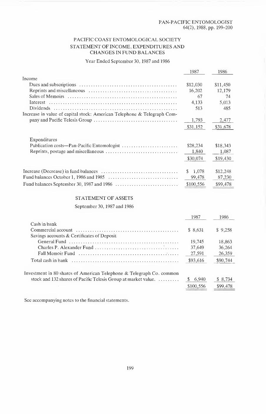
PAN-PACIFIC ENTOMOLOGIST 64(2), 1988, pp. 199-200
PACIFIC COAST ENTOMOLOGICAL SOCIETY
STATEMENT OF INCOME, EXPENDITURES AND
CHANGES IN FUND BALANCES
Year Ended September 30,1987 and 1986
1987 1986
Income
Dues and subscriptions . $12,030 $11,450
Reprints and miscellaneous . 16,202 12,179
Sales of Memoirs . 67 74
Interest . 4,133 5,013
Dividends . 513 485
Increase in value of capital stock: American Telephone & Telegraph Com¬
pany and Pacific Telesis Group. 1,793 2,477
$31,152 $31,678
Expenditures
Publication costs—Pan-Pacific Entomologist. $28,234 $18,343
Reprints, postage and miscellaneous. 1,840 1,087
$30,074 $19,430
Increase (Decrease) in fund balances . $ 1,078 $12,248
Fund balances October 1,1986 and 1985 . 99,478 87,230
Fund balances September 30,1987 and 1986 . $100,556 $99,478
STATEMENT OF ASSETS
September 30,1987 and 1986
1987 1986
Cash in bank
Commercial account . $ 8,631 $ 9,258
Savings accounts & Certificates of Deposit
General Fund . 19,745 18,863
Charles P. Alexander Fund. 37,649 36,264
Fall Memoir Fund . 27,591 26,359
Total cash in bank . $93,616 $90,744
Investment in 80 shares of American Telephone & Telegraph Co. common
stock and 132 shares of Pacific Telesis Group at market value. $ 6,940 $ 8,734
$100,556 $99,478
See accompanying notes to the financial statements.
199

PACIFIC COAST ENTOMOLOGICAL SOCIETY
NOTES TO THE FINANCIAL STATEMENTS
Year Ended September 30,1987
Summary of Significant Accounting Policies
Accounting Method: Income and expenses are recorded by using the cash basis of accounting. Capital
Expenditures: Annual capital expenditures of $5,000 or less are charged to expense. Marketable Securities:
American Telephone & Telegraph Co. and Pacific Telesis Group common stock are carried at market
value. Increases and decreases in value are reflected in income. Income Tax: The Society is exempt from
Federal income and California franchise tax. Undeposited Receipts—$0. Accounts Receivable—$1,963.
Accounts Payable—$0.
As Chairman of the Auditing Committee, and in accordance with its bylaws, I have reviewed the financial
records of the Society.
During the course of this review nothing was noted which indicated any material inaccuracy in the forego¬
ing statements.
H. Vannoy Davis
Chairman of the Auditing Committee
200

THE PAN-PACIFIC ENTOMOLOGIST Information for Contributors
Members are invited to submit manuscripts on the systematic and biological phases of entomology, including short notes or artic¬ les on insect taxonomy, morphology, ecology, behavior, life history, and distribution. Non-members may submit manuscripts for publication, but they should read the information below regarding editing and administrative charges. Manuscripts of less than a printed page will be published as space is available, in Scientific Notes. All manuscripts will be reviewed before acceptance. Manu¬ scripts for publication, proofs, and all editorial matters should be addressed to the editor.
General.—The metric system is to be used exclusively in manuscripts, except when citing label data on type material, or in direct quotations when cited as such. Equivalents in other systems may be placed in parentheses following the metric, i.e. “1370 m (4500 ft.) elevation.”
Typing.—Two copies of each manuscript must be submitted (original and one xerox copy or two xerox copies are suitable). All manuscripts must be typewritten, double-spaced throughout, with ample margins, and be on bond paper or an equivalent weight. Carbon copies or copies on paper larger than 8V2 x 11 inches are not acceptable.
Underscore only where italics are intended in the body of the text. Number all pages consecutively and put authors name on each sheet. References to footnotes in text should be numbered consecutively. Footnotes must be typed on a separate sheet.
Manuscripts with extensive corrections or revisions will be returned to the author for retyping.
First Page.—The page preceding the text of the manuscript must include (1) the complete title, (2) the order and family in paren¬
theses, (3) the author’s name or names, (4) the institution with city and state or the author’s home city and state if not affiliated (5) the complete name and address to which proof is to be sent.
Names and descriptions of organisms.—The first mention of a plant or animal should include the full scientific name with the author of a zoological name not abbreviated. Do not abbreviate generic names. Descriptions of taxa should be in telegraphic style. The International Code of Zoological Nomenclature must be followed.
Tables.—Tables are expensive and should be kept to a minimum. Each table should be prepared as a line drawing or typed on a separate page with heading at top and footnotes below. Number tables with Arabic numerals. Number footnotes consecu¬ tively for each table. Use only horizontal rules. Extensive use of tabular material requiring typesetting may result in increased charges to the author.
Illustrations.—No extra charge is made for line drawings or halftones Submit only photographs on glossy paper and original drawings. Authors mujrplan their illustrations for reduction to the dimension of the printed page (117 x 181 mm; 4% x 7Vs inches). If possible, allowance should be made for the legend to be placed beneath the illustration. Photographs should not be less than the width of the printed page. Photographs should be mounted on stiff card stock, and bear the illustration number on the face.
Loose photographs or drawings which need mounting and/or numbering are not acceptable. Photographs to be placed to¬ gether should be trimmed and abut when mounted. Drawings should be in India Ink, or equivalent, and at least twice as large as the printed illustration. Excessively large illustrations are awkward to handle and may be damaged in transit. It is recom¬ mended that a metric scale be placed on the drawing or the magnification of the printed illustration be stated in the legend where applicable. Arrange figures to use space efficiently. Lettering should reduce to no less than 1 mm. On the back of each illustration should be stated (1) the title of the paper, (2) the author’s complete name and address, and (3) whether he wishes the illustration returned to him. Illustrations not specifically requested will be destroyed. Improperly prepared illustrations will be returned to the author for correction prior to acceptance of the manuscript.
Figure legends.—Legends should be typewritten double-spaced on separate pages headed EXPLANATION OF FIGURES and placed following LITERATURE CITED. Do not attach legends to illustrations.
References.—All citations in text, e.g., Essig (1926) or (Essig 1958), must be listed alphabetically under LITERATURE CITED in the following format:
Essig, E. O. 1926. A butterfly migration. Pan-Pac. Entomol., 2:211-212. Essig, E. O. 1958. Insects and mites of western North America. Rev. ed. The Macmillan Co., New York, 1050 pp.
Abbreviations for titles of journals should follow a recent volume of Serial Sources for the Biosis Data Base, BioSciences Information Service. For Scientific Notes the citations to articles will appear within the text, i.e. . . .“Essig (1926, Pan-Pac. Entomol., 2:211-212) noted. . . .”
Proofs, reprints, and abstracts.—Proofs and forms for the abstract and reprint order will be sent to authors. Changes in proof will be charged to the author.
Editing and administrative charges.—Papers by members of the Pacific Coast Entomological Society are charged at the rate of $30.00 per page. Members without institutional or grant funds may apply for a society grant to cover a maximum of one-half of these charges. Non-members will be charged at the rate of $60.00 per page. Editing and administrative charges are in addition to the charge for reprints and do not include the possible charges for author’s changes after the manuscript has been sent to the printer.

PUBLICATIONS OF THE
PACIFIC COAST ENTOMOLOGICAL SOCIETY
PROCEEDINGS OF THE PACIFIC COAST ENTOMOLOGICAL SOCIETY.
Vol. 1 (16 numbers, 179 pages) and Vol. 2 (9 numbers, 131 pages). 1901-1930. Price $5.00 per volume.
THE PAN-PACIFIC ENTOMOLOGIST.
Vol. 1 (1924) to Vol. 51 (1975), price $10.00 per volume of 4 numbers, or $2.50 per single issue. Vol. 52 (1976) to Vol. 57 (1981), price $15.00 per volume, or $3.75 per single issue, except for Vol. 57, no. 1, $10.00. Vol. 58 (1982) and subse¬ quent issues, $20.00 per volume or $5.00 per single issue.
MEMOIRS OF THE PACIFIC COAST ENTOMOLOGICAL SOCIETY.
Volume 1. The Sucking Lice by G.F. Ferris. 320 pages. Published October 1951. Price $10.00 (plus $1.00 postage and handling).*
Volume 2. A Revision of the Spider Mite Family Tetranychidae by A. Earl Prit¬ chard and Edward W. Baker. 472 pages. Published July 1955. OUT-OF-PRINT.
Volume 3. Revisionary Studies in the Nearctic Decticinae by David C. Rentz and James D. Birchim. 173 pages. Published July 1968. Price $4.00 (plus $0.75 post¬ age and handling).*
Volume 4. Autobiography of an Entomologist by Robert L. Usinger. 343 pages. Published August 1972. SPECIAL PRICE $5.00 (plus $1.00 tax, postage, and handling for California orders, $0.70 postage and handling for non-California U.S. orders, or $1.70 for foreign orders). No members discount at this special price.
Volume 5. Revision of the Millipede Family Andrognathidae in the Nearctic Re¬ gion by Michael R. Gardner. 61 pages. Published January 21, 1975. Price $3.00 (plus $0.75 postage and handling).*
*(Add 6% sales tax on all California orders (residents of Alameda, Contra Costa, and San Francisco
counties add 6lh%). Members of the Society will receive a 20% discount on the price of the memoirs,
except vol. 4.)
Send orders to: Pacific Coast Entomological Society % California Academy of Sciences Golden Gate Park San Francisco, California 94118-4599 U.S.A.

Vol. 64 July 1988
THE No. 3
Pan-Pacific Entomologist
BROEMELING, D. K. and A. S. MOALIF—A revision of the Nomada subgenus Pachynomada (Hymenoptera: Anthophoridae) . 201
ROBERTSON, J. A.—Descriptions of the immatures of Typocerus serraticornis (Coleoptera: Cerambycidae), and new observations on biology, including “Varnish” production and usage by the larva. 228
GRACE, J. K. and W. W. WILCOX—Isolation and trail-following bioassay of a decay fungus associated with Reticulitermes hesperus Banks (Isoptera: Rhinotermitidae) . 243
HESSEIN, N. A. and J. A. McMURTRY—Observations on Megaphragma mymaripenne Timberlake (Hymenoptera: Trichogrammatidae), an egg parasite of Heliothrips haemorrhoidalis Bouche (Thysanoptera: Thripidae) . 250
RYTI, R. T.—Geographic variation in cooperative colony foundation in Veromessor pergandei (Hymenoptera: Formicidae). 255
MASON, R. R. and H. G. PAUL—Predation on larvae of Douglas-fir Tussock Moth, Orgyia pseudotsugata (Lepidoptera: Lymantriidae), by Metaphidippus aeneolus (Araneae: Salticidae) . 258
LIU, T. P.—The morphology of the tarsal sensilla in the female mite Varroa jacobsoni . 261
FROHLICH, D. R., W. H. CLARK, F. D. PARKER, T. L. GRISWOLD—The xylophilous bees and wasps of a high, cold desert: Leslie Gulch, Oregon (Hymenoptera: Apoidea, Vespoidea) . 266
MIDDLEKAUFF, W. W.—A new species of Neurotoma from Michigan (Hymenoptera: Symphyta, Pamphiliidae) . 270
LINSLEY, E. G.—Further notes on halictine bees (Hymenoptera: Apoidea) visiting Isomeris arborea in southern California . 275
SANTIAGO-BLAY, J. A. and H. V. DALY—The Apoidea Collection of the California In¬ sect Survey (C.I.S.) at the Essig Museum of Entomology, University of California, Berkeley . 281
PAGE, Jr., R. E. and R. A. METCALF—A population estimate of Mdh Allozyme frequencies for the honey bee, Apis mellifera L. (Hymenoptera: Apidae) . 285
CHEMSAK, J. A., E. G. LINSLEY, and F. HOVORE—A concentration site for Cerambycidae in Jalisco, Mexico (Coleoptera). 291
SAN FRANCISCO, CALIFORNIA • 1988 Published by the PACIFIC COAST ENTOMOLOGICAL SOCIETY in cooperation with THE CALIFORNIA ACADEMY OF SCIENCES

The Pan-Pacific Entomologist
EDITORIAL BOARD J. A. Chemsak, Editor
R. S. Lane, Associate Editor W. J. Pulawski, Treasurer J.T. Doyen R.M. Bohart J. A. Powell J.E. Hafernik, Jr.
Published quarterly in January, April, July, and October with Society Proceedings appearing in the October number. All communications regarding nonreceipt of num¬ bers, requests for sample copies, and financial communications should be addressed to the Treasurer, Dr. Wojciech J. Pulawski, California Academy of Sciences, Golden Gate Park, San Francisco, CA 94118-4599.
Application for membership in the Society and changes of address should be ad¬ dressed to the Secretary, Mr. Vincent F. Lee, California Academy of Sciences, Golden Gate Park, San Francisco, CA 94118-4599.
Manuscripts, proofs, and all correspondence concerning editorial matters should be addressed to Editor, Pacific Coast Entomological Society, 201 Wellman Hall, Univer¬ sity of California, Berkeley, CA 94720. See back cover for instructions.
The annual dues, paid in advance, are $15.00 for regular members of the Society,
$7.50 for student members, $30.00 or more for sponsoring members, or $20.00 for sub¬ scription only. Members of the Society receive The Pan-Pacific Entomologist. Single copies of recent numbers are $5.00 each or $20.00 a volume. See back cover for prices of earlier back numbers. Make checks payable to the Pacific Coast Entomological Soci¬ ety.
Pacific Coast Entomological Society
OFFICERS FOR 1988
Alan I. Kaplan, President Wojciech J. Pulawski, Treasurer
Thomas J. Zavortink, President-Elect Vincent F. Lee, Managing Secretary Leslie S. Saul, Recording Secretary
Statement of Ownership
Title of Publication: The Pan-Pacific Entomologist.
Location of Office of Publication, Business Office of Publisher and Owner: Pacific Coast Entomological
Society, California Academy of Sciences, Golden Gate Park, San Francisco, California 94118-4599.
Editor: J. A. Chemsak, 201 Wellman Hall, University of California, Berkeley, California 94720.
Managing Editor and Known Bondholders or other Security Holders: None.
This issue mailed December 12,1988
The Pan-Pacific Entomologist (ISSN 0031-0603)
PRODUCED BY A-R EDITIONS, INC., MADISON, WI 53703, U.S.A.

PAN-PACIFIC ENTOMOLOGIST 64(3), 1988, pp. 201-227
A Revision of the Nomada Sebgenus Pachynomada (Hymenoptera: Anthophoridae)
Derek K. Broemeling1 and Ali S. Moalif2
USDA-Agricultural Research Service, Bee Biology & Systematics Laboratory, Utah State University, Logan, Utah 84322-5310.
Abstract.—The Nomada subgenus Pachynomada is revised. Fourteen species and subspecies are recognized, five of which are new to science. New species described are Nomada bohartorum Moalif n. sp., N. dreisbachorum Moalif n. sp., N. tepoztlan Moalif n. sp., N. utahensis Moalif n. sp., and N. saltillo Broemeling n. sp., Nomada vincta heterochroa Cockerell is placed in synonymy with N. vincta Say. Pachynomada is kleptoparasitic upon the Andrena subgenus Callandrena.
The kleptoparasitic bee genus Nomada is found on nearly every continent.
Pachynomada is a new world subgenus of late summer to fall bees, ranging from southern Canada to the mountains of southern Mexico. They are most frequently collected on or around large flowered composites, especially species of Helianthus. Males are often found in the afternoon, flying amongst the stems of Helianthus, possibly in search of females (pers. obs.). Nomada (Pachynomada) utahensis n. sp. has been reared from the nests of Andrena (Callandrena) helianthi Robertson (Parker and Bohart, 1982). This same Nomada species has been observed leaving an unplugged burrow of Andrena (Callandrena) haynesi Viereck and Cockerell in southeastern Utah (Parker and Griswold, 1983).
Rodeck (1945) described Pachynomada as a segregate of the subgenus Holonomada, with Nomada vincta Say as the type species. Pachynomada was defined as having “the dorsum of the pronotum rounded-carinate, slightly depressed medially, the scape of the male antenna globular and swollen, the apex of the hind tibiae with 4-6 widely spaced short acute spines.” Moalif (1979) revised the subgenus Pachynomada and his redefinition of the subgenus was based upon unique genitalic characters and the form of the hind basitarsus.
The morphological terminology used in this study generally follows that of Michener (1944) and Stephen, et al., (1969). The following is a list of abbreviations and new or uncommon morphological terminology used:
—acetabular carina (see Fig. 13); a lamellate projection at the upper base of the
mandible, especially prominent in male Nomada. —prelobar carina (see Fig. 18); connects posterior margin of pronotal ridge with
anterior margin of pronotal lobe. —paraocular carina; raised area running along interior margin of compound eye and
crossing from base of eye to anterior mandibular articulation.
'Department of Biology, Utah State University, Logan, Utah
2Plant Protection Institute, University of Basrah, Basrah, Iraq.
201

202 PAN-PACIFIC ENTOMOLOGIST
Figures 1-19. Figures 1-10. Male antennal scape. Figure 1. Bohartorum. Figure 2. Tepoztlan. Figure 3.
Dreisbachorum. Figure 4. Besseyi. Figure 5. Victrix. Figure 6. Asteris. Figure 7. Aztecorum. Figure 8.
Utahensis. Figure 9. Zebrata. Figure 10. Vincta. Figure 11. Utahensis, abdomen of female. Figure 12.
Vincta, abdomen of female. Figure 13. Nomada (Pachynomada) sp., mandible, with acetabular carina.
Figure 14. Aztecorum, labrum. Figure 15. Suffossa, labrum. Figure 16. Victrix, hind coxa. Figure 17.
Zebrata, mesopleuron. Figure 18. Nomada (Pachynomada), pronotum with prelobar carina. Figure 19.
Aztecorum, scutellum and metanotum.

VOLUME 64, NUMBER 3 203
—hypoepimeral area; dorsal posterior quarter of mesopleuron. (see Michener, 1944, p. 305)
—IOD = interocellar distance; —OOD = ocellocular distance; —MLOD = mid-lateral ocellar distance; —MOD = mid-ocellar diameter;
—MOOM = mid-ocellar occipital margin distance; —IPS = interpunctural surface.
In the species descriptions the rims of the scutal punctures are referred to as “rounded” or “angulate.” An angulate puncture rim is one in which the scutal surface appears to have been shaved off, leaving the punctures incomplete. Tergum and sternum refer to the metasomal sclerites only. Thoracic sterna will be referred to as mesosternum or metasternum.
Subgenus Pachynomada Rodeck
Pachynomada Rodeck, 1945, Entomol. News 56:180.
Type species: Nomada vincta Say, original designation
Diagnosis.—Penis valve with conspicuous ventral subapical hook, eighth sternum elongated apically; occipital margin gently rounded, posterior basitarsus expanded medially (Fig. 44); posterior tibia with 4—8 apical bristles; minimum length of first flagellar segment longer than maximum length of second; propodeum lacking dorso-lateral angle behind spiracles; supraclypeal area distinctly protuberant; mandibles simple; female with paraocular carina.
Description.—Length 7.4-13.3 mm, forewing length 5.3-10.4 mm, hindwing length 4.1-8.1 mm; minimum length of first flagellar segment greater than maximum length of second; antennal scape in males often inflated to globose; pre-occipital ridge rounded, not reflexed or strongly angulate; supraclypeal area distinctly protruberant between antennal insertions, with a flattened median carina; margin of labrum sharply angulate; females with well developed paraocular carina; mandibular
acetabular carina lamellate in males; mandibles lacking subapical tooth; head densely punctate with smooth shiny IPS (exceptions noted in species descriptions); Thorax: prelobar carina short, prominent; apex of pronotal ridge usually rounded,
broadly depressed medially; scutum deeply, contiguously punctured, occasionally reticulate; rim of punctures frequently angulate, giving them a “shaved” appearance, IPS smooth and shiny; tegulae shallowly punctured with glassy surface; scutellum weakly to strongly bilobate, postero-medial region with distinctly microrugose IPS; propodeal disk rugose medio-apically and latero-apically, shagreened ventrally; sides of propodeum somewhat swollen posterior to spiracle (but not angulate), supraspiracular ridge undeveloped; mesopleuron densely, circularly punctured (sometimes reticulate), IPS glassy ventrally roughened
dorsally, hypoepimeral area often distinctly protruding; procoxa often bearing a rudimentary posterior spine, usually showing as a sharp bump or angulation; hind tibial apex with a row of 4 to 8 thickened bristles; hind basitarsi distinctly widened medially, with curved anterior and posterior margins; forewing with 2 or 3
submarginal cells; sternum 8 of males with narrow, elongated distal process; penis valves with distinct ventral subapical projection; body sparsely clothed with moderately long pubescence; COLOR: Various combinations of black, yellow,

204 PAN-PACIFIC ENTOMOLOGIST
ferruginous, and rufo-ferruginous. Female differs from male as follows: Scape cylindrical; short, wide pseudopygidium present, anterior tarsal brushes absent; apex of sternum 8 with conspicuous lateral tufts of long inward curving hairs.
Most Pachynomada species fall within 3 distinct species groups, the members of which are quite similar morphologically, although they may differ greatly in coloration. Most of the species are related closely to vincta. Three species are assigned to monotypic species groups.
Vincta Group: vincta Say, 1837; suffossa Cockerell, 1922;
aztecorum aztecorum Cockerell, 1903; utahensis n. sp.; aztecorumpratensis Cockerell, 1919; zebrata Cresson, 1878;
saltillo n. sp.;
Bohartorum Group: bohartorum n. sp.; tepoztlan n. sp.;
Vitticollis Group: dreisbachorum n. sp.; vitticollis Cresson, 1878;
Asteris Group: asteris Swenk, 1913;
Besseyi Group: besseyi Swenk, 1913;
Victrix Group:
victrix Cockerell, 1911;
Key to the Species of Pachynomada
Males: 1. 2 submarginal cells in forewing .2
— 3 submarginal cells in forewing .3 2. hind coxa with a dorsal posterior carina extending almost V2 the length of the
coxa (Fig. 16) ..victrix Cockerell — hind coxa not as above, antennal scape with hairs longer than 2h length of scape
dreisbachorum n. sp. 3. propodeum appearing naked, pubescence little longer than the diameter of a
puncture .besseyi Swenk
— propodeum with long lateral pubescence .4 4. propodeum with extensive yellow or ferruginous lateral marks, covering most of
propodeal sides .5 — sides of propodeum black, rarely with a small yellow posterior lateral mark, not
much larger than spiracle .8 5. small bee (7.5 mm), entire body a uniform light brown color with greatly reduced
ornamentation .tepoztlan n. sp. — larger bees (9.0 mm), or with bodies not a uniform light brown color and having
extensive yellow maculations on thorax, head and abdomen .6
6. antennal scape globose or ovate (Fig. 9,10) .7 — antennal scape not globose, somewhat swollen apically (Fig. 8) utahensis n. sp.

VOLUME 64, NUMBER 3 205
7. mesopleuron with a longitudinal yellow band extending from anterior margin to posterior margin, narrowed medially (Fig. 17) .zebrata Cresson
— mesopleuron without a complete longitudinal yellow band, usually only an anterior triangular patch .vincta Say
8. antennal scape globose .9
— antennal scape narrow apically.bohartorum n. sp. 9. antennal scape uniformly ferruginous, scutellum cream colored, without median
black area .asteris Swenk — antennal scape black, or if ferruginous, with dark posterior markings, scutellum
with median black area .10 10. labrum with broad impunctate median welt and basal lateral welts, curving
downward from center to apex (Fig. 14).11 — labrum without impunctate lateral welts, entire apex strongly upturned (Fig.
15) .suffossa Cockerell 11. yellow bands on abdominal terga narrow, at midpoint less than V3 total length of
tergum, legs usually black or with extensive dark areas . . aztecorum aztecorum Cockerell
— yellow bands on abdominal terga wide, at midpoint more than V2 total length of tergum, legs completely ferruginous .aztecorumpratensis Cockerell
Females: 1. hind coxa with a dorsal, posterior carina extending almost V2 length of coxa (Fig.
16), 2 submarginal cells in forewing .victrix Cockerell — hind coxa not as above, 3 submarginal cells in forewing.2
2. abdominal terga with several complete transverse yellow or cream-colored bands.3
— abdominal terga never with complete transverse yellow bands, body
ferruginous .asteris Swenk 3. propodeum appearing naked, pubescence not longer than the diameter of a
puncture .besseyi Swenk — propodeum with long lateral pubescence .4 4. propodeum with extensive yellow or ferruginous lateral markings.8 — propodeum black, or with small yellow mark, not much larger than spiracular
area .5
5. scutellum with yellow markings widely separated by dark areas, at least posteriorly, hind tibial bristles not flattened or curved apically (Fig. 19, 20) .. 6
— scutellum with yellow markings not separated, hind tibia with 4 or 5 apical bristles, flattened, strongly curved (Fig. 21) .bohartorum n. sp.
6. labrum with broad impunctate median and lateral welts, curving downwards at sides (Fig. 14) .7
— labrum evenly punctate, may have impunctate median line, but not raised, apical margin with a strongly upcurved lip (Fig. 15) .suffossa Cockerell
7. yellow bands on abdominal terga narrow, at midpoint less than V3 length of tergum, legs usually black or with extensive dark areas . . aztecorum aztecorum
Cockerell — yellow bands on abdominal terga wide, at midpoint more than V2 length of
tergum, legs completely ferruginous .aztecorum pratensis Cockerell

206 PAN-PACIFIC ENTOMOLOGIST
Figures 20-43. Figure 20. Vincta, posterior tibial apex. Figure 21. Bohartorum, posterior tibial apex.
Figures 22-32, sternum VIII of Male. Figure 22. Bohartorum. Figure 23. Tepoztlan. Figure 24.Asteris.
Figure 25. Besseyi. Figure 26. Victrix. Figure 27. Dreisbachorum. Figure 28. Aztecorum. Figure 29.
Suffossa. Figure 30. zebrata. Figure 31. Utahensis. Figure 32. Vincta. Figures 33-43, Sternum VII of
Male. Figure 33. Bohartorum. Figure 34. Tepoztlan. Figure 35. A Vera. Figure 36. Besseyi. Figure 37.
Victrix. Figure 38. Dreisbachorum Figure 39. Aztecorum. Figure 40. Suffossa. Figure 41. Utahensis. Fig¬
ure 42. Zebrata. Figure 43. Vincta.

VOLUME 64, NUMBER 3 207
Figures 44-56. Figure 44. Vincta, hind basitarsus. Figures 45-55. Male genital capsule (ventral view).
Figure45. Bohartorum. Figure46. Tepoztlan. Figure47. Victrix. Figure 48. Asteris. Figure 49. besseyi.
Figure 50. dreisbachorum. Figure 51. Aztecorum. Figure 52. Suffossa. Figure 53. Utahensis. Figure 54.
Zebrata. Figure 55. Vincta. Figure 56. Vincta, genital capsule (dorsal view).

208 PAN-PACIFIC ENTOMOLOGIST
8. pronotum, mesopleuron, metapleuron, and propodeum almost entirely yellow vitticollis Cresson
— thorax with extensive ferruginous or black areas.9 9. mesopleuron with a longitudinal yellow band from anterior to posterior border,
narrowed medially, (Fig. 17) .zebrata Cresson — mesopleuron with at most a small anterior yellow patch .10 10. sides of propodeum ferruginous, with a basal posterior yellow spot, not much
larger than spiracular area, maculations cream-colored.saltillo n. sp. — sides of propodeum with extensive yellow markings, maculations yellow ... 11 11. tergum II with narrow median transverse band, extensive dark areas anteriorly
and posteriorly (Fig. 12) .vincta Say — tergum II with broad median transverse yellow band, almost no dark area
anteriorly (Fig. 11) .utahensis n. sp.
Descriptions of the Species
Vincta Group:
Nomada vincta Say, 1837. Boston Jour. Nat. Hist. 1:401. Nomada vincta heterochroa Cockerell, 1921. Amer. Mus. Novit. 24:1. NEW
SYNONYMY
Type.—Apparently lost. Diagnosis.—3 submarginal cells, complete transverse median bands on terga,
sides of propodeum yellow with long pubescence, antennal scape obovate, mesopleuron lacking longitudinal yellow band (if present, greatly expanded anteriorly, reaching to pronotal lobe), punctation below mesopleural scrobe with
interpunctural spaces up to half a puncture diameter. Male.—Length 9.6-12.4 mm, forewing length 7.4-9.3 mm, hindwing length
5.8-6.8 mm; antennal scape globose, densely punctate, IPS shiny, slightly roughened; IOD = OOD =MOOM, MLOD = MOD; labrum densely punctate, with impunctate medio-apical strip produced into a weak beak; prelobar carina thickened, pronotal ridge rounded anteriorly; scutum deeply, contiguously punctured with puncture rims angulate, IPS shiny; scutellum depressed medially;
metanotum with shiny roughened IPS; propodeal disk moderately rugose dorso-medially, remainder shagreened; propodeum clothed with long hairs (0.3
mm); tegulae densely, shallowly punctate, glassy IPS; metapleuron with lower half smooth, moderately punctate; procoxal spine rudiment reduced to slight bump; hind tibial apex with 5 evenly spaced ferruginous bristles; forewing with 3 submarginal
cells; COLOR: antennal scape ferruginous, blending to light yellow, flagellum light ferruginous, darker apically; supraclypeal area, clypeus, sides of face extending to apex of compound eyes, basal two-thirds of mandibles, malar space, gena, ring behind compound eyes, pronotal ridge and lobes, tegulae, axillary sclerites (in part), scutellum, metanotum, sides of propodeum, triangular anterior mesopleural patch, apex of fore- and mid-coxae, hind coxae, remainder of legs, median transverse band on abdominal terga, spot on first sternum, large transverse bands on remaining sterna, light yellow; remainder of body fuscous to black.
Females.— Length 8.1-11.5 mm, forewing length 6.5-9.3 mm, hindwing length 5.2-7.3 mm; similar to males except: antennal scape not inflated, sparsely punctate,
with shiny, shagreened IPS; markings on face usually ferruginous, transverse median yellow bands on abdominal terga narrower.

VOLUME 64, NUMBER 3 209
Discussion.—Nomada vincta is the most common and widespread Pachynomada species. Nomada vincta heterochroa is based upon some unusually colored specimens with the normally black markings being ferruginous. It occurs at the same locality as normal vincta which rules it out as a valid subspecies, it is therefore relegated to synonymy. The type series of Nomada vincta appears to be lost. However, the type locality of Indiana has only two possible species of Pachynomada, and N. besseyi is not likely to have been confused with N. vincta. Since there seem to be no doubts about the identity Nomada vincta, the requirements of the International Code of Zoological Nomenclature for the designation of a neotype (Article 75) are not met.
Specimens examined.—CANADA: ALBERTA: 4.8 km (3mi) SE Picture Butte, 1 6,6-VIII-1978 Helianthuspetiolaris (C. D. Michener) SMEK; Medicine Hat, 1 2 ,
(J.R. Malloch), 1 2, 13-VIII-1939 (E. H. Strickland), 16,19, 16-VIII-1924 (F. S. Carr), 4 5 6,8 $ $, 23-VIII-1919 (Sladen), 19,7-IX-1925 (F. S. Carr), 1 9 , 7-VIII-1938 (E.H. Strickland), 1 2, 9-IX-1939, (J.L. Carr); MANITOBA: Aweme, 1 2, 14-IX-1924 (N. Criddle); U.S.A.: COLORADO: 1 2, (Snow) ANSP; 14.5 km (9 mi) S Wray, 1 2,2-IX-1951 (Paul P. ?) USNM; Fort Collins, 1 (5, 1 2,12-IX-03 BBSL; Wray 40 O'N 102 10'W, 1130 m (3700'), 1 6,17—19/VIII/1919
CAS, 2 2 2, 17—19/VIII/1919, 1 2, 17-19/VIII/1919 Boul, 1 2, 17-19/VIII/1919 BBSL, 13,12,17-19/VIII/1919 USNM; ILLINOIS: Algonquin, 1 2,13-IX-1894; Chicago 1 2 , (A. L. Melander); Macoupin Co., Carlinville, (Charles Robertson), 1 2,2-IX-1895 Helianthus grosseseratus, 5 2 2,21-IX-1895 Aster ericoides villosus, 1 2,5-IX-1895 Helianthus grosseseratus, 1 2,9-IX-1895 Helianthus grosseseratus, 1 5; INDIANA: Lafayette, 15,12, (Geo. G. Ainslie); Plymouth, 1 2,4-IX-1917 (M. R. Smith); IOWA: Clarence, 1 2,3-IX-1953 Helianthus (N. F. Stage); Sioux City, 1 5, l-IX-1922 Solidago (C. N. Ainslie), 1 5,5-IX-1927 (C. N. Ainslie), 1 5, 3 2 2, 8-IX-1920 (C. N. Ainslie); KANSAS: 2 5 5, 2589.5, 2589.4; Manhattan, (O. A. Stevens), 1 2, 24-VIII-1908 Helianthus tuberosus, 2 5 5 , 25-VIII-1908 Helianthus tuberosus; Marysville, 1 2, 12-IX-1920 Heliopsis scabra (Edna M. Stevens); Topeka, 1 2 , -IX- (J. E. Taylor); Clay Co., 1 5; Douglas Co., 2 5 5,1 2, 25-VIII-1949 Silphiumperfoliatum, (Michener, Beamer); MINNESOTA: Detroit,
1 2,25-VIII-1913 Astersagittifolius (O. A. Stevens); Hastings, 1 5,26-VIII-1928, 3 5 5, 28-VIII-1928, 1 2, 30-VIII-1928 (H. A. Scullen); Lake Park, 1 2, 22-VIII-1911 Rudbeckia laciniata (C. H. Waldron); Miesville, 1 2, 4-IX-1951 Helianthus maximiliani (Roland Fischer); Stanton, 12, 28-IX-1957 Helianthus maximiliani (Roland L. Fischer) Mich; U. Farm, 1 2, 2-IX-1951 Grindelia squarrosa (Roland L. Fischer) Mich; MONTANA: 2 5 5,1 +, ANSP; Huntley, 1 5, 16—VIII—1916; NORTH CAROLINA: Black Mountains, 1 5, 1 2, 1911; NORTH DAKOTA: Bismarck, 1 2, 12-IX-1920 Helianthus tuberosus (O.A. Stevens) Boul, 1 2,23-VIII-1946 (Richard L. Post); Fargo, (O. A. Stevens), 1 5, 10-VIII-1912 Grindelia squarrosa NEBR, 2 5 5, 12-VIII-1910 Grindelia squarrosa, 1 5, 13-VIII-1910 Solidago serotina, 1 2, 13-VIII-1911 Helianthus (cult.), 2 5 5, 17-VIII-1911 Grindelia squarrosa, 3 5 5, 17-VIII-1911 Grindelia squarrosa, 2 2 2,18-IX-1915 Helianthus tuberosus, 1 5, 18-VIII-1912 Helianthus maximiliani, 1 5 5, 29-VIII-1917 Helianthus maximiliani Boul, 1 2,4-VIII-1911 Helianthus (cult.), 2 2 2, 8-IX-1913 Helianthus tuberosus, 1 5, 8-VIII-1920
Sonchus arvensis, Boul; Mandan, 1 2, ll-IX-1920 Helianthus tuberosus (O.A. Stevens); Mott, 1 5, 20-VIII-1914 Solidago rigida (J.R. Campbell) NEBR; Schafer, 1 2, 5-IX-1914 Grindelia squarrosa (O.A. Stevens) NEBR; Sheldon,
(O.A. Stevens) 1 5,10-VIII-1969 Helianthus petiolaris AMNH, 1 2,20-VIII-1949

210 PAN-PACIFIC ENTOMOLOGIST
Figure 57. Distribution of Pachynomada species of the vincta group.

VOLUME 64, NUMBER 3 211
Helianthus petiolaris; Williston, (O.A. Stevens), 1 <3, 15-VIII-1915 Grindelia squarrosa Boul, 1 6,15-VIII-1915 Grindelia squarrosa AMNH; Ransom Co., 11.3 km (7 mi) SE Sheldon, 1 <J, 19-VIII-1980 (J. R. Powers); NEBRASKA: Lincoln, 1
<3, NEBR, 1 9, -IX-, 1 9, 18-IX-1903 Grindelia (M. H. Swenk), 1 9, 29- VIII-1936 (R. E. Hill); Mitchell, 1 <3, 12-VIII-1915 (E. M. Partridge) NEBR, 2 9 9, 12-VIII-1916 (C. E. Mickel), 1 9, 24-VIII-1916 (R. W. Dawson), 1 9, 25-VIII-1916 (R. W. Dawson), 1 9,29-VII-1913 (L. M. Gates), 1 9,5-IX-1916 (R. W. Dawson), 1 9, 8-VIII-1913 Helianthus annuus (L. M. Gates), 1 <3, 9-VIII-1915 (E. M. Partridge) NEBR; Morrill, 1 <3, ll-VIII-1930 (D. B. Whelan) Boul; Omaha, (L. T. Williams), 1 <3, 13-VIII-19—, Id, 1 9, 16-VIII-1913, 1 9, 16-VIII-1913 Helianthus, 1 c3, 17-VIII-1913, 1 9, 2-IX-1913, 1 c3, 4 9 9, 22-VIII-1913 Helianthus, 1 9, 23-VIII-1913, 1 9, 29-VIII-1913; West Point, 2 c3d, 6 9 9, -VIII-1887, 1 d, 12-IX-1903 Solidago (J. C. Crawford), 1 9, 2-IX-1900 Helianthus, 1 d, 4-IX-1903 Helianthus (J. C. Crawford), 1 9, 27-VIII-1903 (J.C. Crawford); Sioux Co., Glen, 1 d, 12-VIII-1906 Helianthus (H. S. Smith), 1 9,20-VIII-1906 Cleome (H. S. Smith); Holt Co., Atkinson, 1 d, 30- VIII-1954 Helianthus petiolaris (L. W. Quate); Lancaster Co., Lincoln, 1 d, 1 9 , -VIII-, 1 9, ll-IX-1954 jap beetle trap, 1 9, U-IX-1954, 1 9, 17-IX-1917 Grindelia squarrosa (L. Bruner), 1 9, 20-IX-1915 (C. E. Mickel), 1 d, 30-VIII-1964 Helianthus (D. W. Ribble), 1 9,4-IX-1904 Solidago, 1 9,6-IX-1964 Helianthus (D. W. Ribble), 3 9 9, 7-IX-1964 Helianthus (D. W. Ribble); NEW
JERSEY: Ramsey, 2 d d, 1 + , 23-VIII-1921, 1 9 , 3-IX-1917, 1 9,6-IX-1917; NEW YORK: Cooperstown, 1 9,30-VII-1927 (L. Lacey); SOUTH DAKOTA: 4.8
km (3 mi) W. Dallas, 1 d, 15-VIII-1958 Helianthus annuus (W. E. LaBerge) NEBR; Brookings, (H. C. Severin), 1 d, 23-VIII-1933 LACM, 5 9 9, 25-VIII-1921 USNM; Dallas, 2 9 9, 15-VIII-1958 Helianthus annuus (W. E. LaBerge); Gettysburg, 1 d, 12-VIII-1924 USNM; Wobridge, 1 d, 15-VIII-1924 USNM; WISCONSIN: Buffalo Co., Fountain City, 1 d, 12-17/VIII/1910;
Nomada (Pachynomada) aztecorum aztecorum Cockerell
Nomada aztecorum Cockerell, 1903. Ann. Mag. Nat. Hist. 12:211. Lectotype, female: “Amecameca, Mex. IX, Type No. 10145.” type Depository, Academy of Natural Sciences of Philadelphia.
Diagnosis.—3 submarginal cells, propodeum black (at most with a small posterior basal yellow spot) with long pubescence, terga with complete yellow transverse bands, antennal scape globose, mesopleural punctation separated by half a puncture diameter or more in posterior ventral area.
Males.—Length 8.7-13.3 mm, forewing length 7.0-10.1 mm, hindwing length 5.8-7.7 mm; antennal scape globose; acetabular carina strongly lamellate, otherwise similar to females.
Females.—Length 9.6-12.3 mm, forewing length 8.7-9.8 mm, hindwing length
6.4-7.8 mm; antennal scape sparsely punctate basally, dense apically, IPS roughened basally, smooth apically; face densely, deeply punctured, many deformed, IPS roughened, rather dull over most of head; IOD < OOD < MOOM, MLOD < MOD; labrum with strongly carinate margin, surface highly polished and
deeply punctate laterally, impunctate welt basolaterally and medially, protruding into a beak; prelobar carina strong; pronotal ridge rounded anteriorly, broadly depressed medially, impunctate medially; scutum contiguously punctured with much

212 PAN-PACIFIC ENTOMOLOGIST
deformation; scutellum elevated, flattened dorsally, slightly depressed medially, postero-medial IPS coarsely roughened; propodeal sides deeply punctured, hairs 0.4 mm long; mesopleuron densely punctured (0.06 mm diameter), with dull, coarsely
roughened IPS; metapleuron with ventral half shiny, rugose; procoxal spine rudiments short; hind tibial apex with 6 staggered bristles, pygidium with impunctate median welt; forewing with three submarginal cells: COLOR: scape, pedical, first flagellar segment ferruginous anteriorly, remainder of antennae brown; apical half of clypeus, sides of face to about top of scape, labrum, mandibles, malar space, pronotal ridge and lobes, two spots on scutellum, metanotum, anterior rectangle on mesopleuron, posterior lateral spot near propodeal spiracle, apex of fore- and mid coxae, hind coxae, attenuated transverse median band on tergum 1-5, spot on sternum 1, band of sternum 2, yellow; remainder of body fuscous to black.
Discussion.—Nomada aztecorum is a southern Pachynomada, to date found only in Mexico. The northern specimens from Chihuahua have more extensive ferruginous markings, the transverse yellow bands on the abdomen are much wider, and the body is more robust than the typical form. Moalif (1979) commented that this was a highly variable species and proposed synonomy of aztecorum pratensis and suffossa with aztecorum. Nomada suffossa is a distinct species, with a differently
shaped labrum and denser mesopleural punctation. The elimination of specimens of suffossa which were confused with aztecorum leaves very little intergradation in coloration of aztecorum aztecorum and aztecorum pratensis. Nomada aztecorum pratensis is therefore retained as a distinct subspecies.
Specimens examined.—MEXICO: Atlacomulco, 2600 m (8550'), 1 <3, 30-VIII-1963 (Scullen & Bolinger); HIDALGO: 38.0 km (23.6 mi) NW Zacatlan, 2030 m (6650'), (U. Kans. Mex. Exped.) SMEK, 2 S c3, 1 + 22-VIII-1962; Tepeapulco, 1 9,18-IX-1974 (W. Hanson, G. Bohart) BBSL; MEXICO: 29.0 km (18 mi) S. of Mexico City, 2800 m (9200'), (H. A. Scullen), 2 33, 10-IX-1957
ORSU, 333, 10-IX-1957 ORSU, 3 33, 10-IX-1957, 1 3,1 $, 2750 m (9000'), 14-IX-1957, 3 3 3, 14-IX-1957 ORSU; 64.4 km (40 mi) E. Mexico City, 3010 m (9900'), (H. A. Scullen), 1 <3, 7-IX-1957 ORSU, 3 3 3, 7-IX-1957; 0.6 km W Bosencheve, 2514 m., (C.D. George, R. R. Snelling) LACM, 1 c3, 14-IX-1976 Simsia amplexicaulis, 3 <3 <3,2 9 9,14-IX-1976; MORELOS: Tres Cumbres, 1 c3, 20-IX-1938 (L. J. Lipovsky) SMEK; NAYARIT: Santa Teresa, 2073 m., 2 9 9, 22-25/X-1979 (D. E. & J. A. Breedlove); PUEBLA: 6.9 km (4.3) mi W. Huauchinango, 1720 m (5650'), 1 <3,21-VIII-1962 (U. Kans. Mex. Exped.); 64.4
km (40 mi) W of Puebla, 2800 m (9200')(H. A. Scullen), 3 c3 c3,1 + 7-IX-1957; 9.7 km (6 mi) NW Zacatlan, 2440 m (8000'), 1 c3,22-VIII-1962 Heterotheca (Ordway & Roberts);
Nomada (Pachynomada) aztecorum pratensis Cockerell
Nomada aztecorum pratensis Cockerell, 1919. Proc. U.S. Natl. Mus., 55:180
Holotype Female.—“Meadow Vy, Mex. Collector Townsend, Type No. 20213”. Type Depository, United States National Museum.
Males.—Length 9.8-12.9 mm, forewing length 8.4-9.3 mm, hindwing length 6.4-7.6 mm; differs from nominate subspecies by much thicker yellow bands on abdominal terga, legs almost completely ferruginous, antennal scape more ferruginous, mesopleural mark ranging from a spot to a longitudinal band;

VOLUME 64, NUMBER 3
asteris besseyi
■ bohartorum d reisbachorum tepoztlan
<>victrix ° vitticoSlis
Figure 58. Distribution of Pachynomada species exclusive of the vincta group

214 PAN-PACIFIC ENTOMOLOGIST
Females.—Length 11.0-11.9 mm, fore wing length 8.6-9.8 mm, hindwing length 6.7-7.7 mm; differs from males by supraclypeal area ferruginous, mesopleural mark forming a longitudinal band, mesopleuron ferruginous in the type.
Specimens examined.—MEXICO: Meadow Vy., 3 S 8, (Townsend) USNM; CHIHUAHUA: 6.4 km (4 mi) S. Santo Tomas, 1 6,22-VIII-1950 (Ray F. Smith); Creel, (Sears, Gardner, Glaser) 1 6,1 9, 24-VIII-1969,1 9,8-IX-1969;
Nomada (Pachynomada) saltillo Broemeling New Species
Holotype Female.—“Mexico, Coah. Saltillo, 32miS., VIII-23-1957, D. Spencer, R., J. & A. Ryckman Collectors.” Type Depository, Natural History Museum of
Los Angeles County. Diagnosis.—3 submarginal cells, abdomen with transverse medial bands, sides of
propodeum ferruginous (with small cream-colored posterior basal spot) with long pubescence, lacking longitudinal mesopleural band.
Males.—Unknown Females.—Length 10.8 mm, fore wing length 9.9 mm, hindwing length 7.6 mm;
antennal scape sparsely, shallowly punctate, shiny shagreened IPS; IOD = OOD = MOOM, MLOD < MOD; raised impunctate hump behind mid-ocellus; head densely punctate, polished IPS; sides of labrum slightly downturned, (not as sharply as aztecorum), broad, impunctate median welt, apex upturned, forming a wedge; prelobar carina thick, rounded; pronotal ridge rounded anteriorly, broadly shallowly depressed medially, impunctate at very middle; scutum densely, deeply punctured, interpunctural spaces reduced to knife-like ridge; tegulae densely, shallowly punctured, IPS glassy; scutellum moderately depressed medially; metanotum somewhat flattened medially; mesopleuron with interpunctural spaces reduced to thin sharpened ridges; metapleuron shiny, punctate ventrally; pro-coxal spine rudiment virtually absent; hind tibial apex with six ferruginous, evenly spaced, straight bristles; forewing with 3 submarginal cells, apex darkly infuscated: COLOR: lower sides of face, malar space, pronotal ridge and
lobes, most of tegulae, lobes of scutellum, metanotum, small postero-baso-lateral
propodeal spots, anterior spot on mesopleuron, apices of mid- and hind-coxae, broken transverse median band on tergum 1, complete but very narrow medially on tergum 2-4, extensive band on tergum 5, broken band on sternum 3, emarginate band on sternum 4, creamy white; head except vertex and area directly above
antennal insertions, antennae, scutum (except posterior medial patch), mesopleuron and mesosternum, sides of propodeum, legs, terga and abdominal sterna, rufo-ferruginous; remainder fuscous to black.
Discussion.—Nomada saltillo is similar morphologically to aztecorum and suffossa, but body coloration and conformation of the labrum are different. Extensive collecting might show this species to be merely an aberrant specimen. Nomada saltillo is also similar to zebrata, but has paler maculations, lacks a mesopleural band and has a more robust form.
Specimens examined.—This species is known only from the type.
Nomada (Pachynomada) suffossa Cockerell
Nomada (Holonomada) suffossa Cockerell, 1922. Proc. U.S. Natl. Mus. 60:16.
Holotype Male.—“Mex 2320, Collection C. F. Baker; Type No. 24893.” Type Depository, United States National Museum.

VOLUME 64, NUMBER 3 215
Diagnosis.—3 submarginal cells, terga with yellow transverse medial bands, sides of propodeum black (rarely with small posterior basal spot) with long pubescence, antennal scape globose, labrum concave in cross-section, strongly recurved at apex, evenly punctate.
Males.—Length 10.4-12.1 mm, forewing length 8.9-10.4 mm, hindwing length 6.9-7.9 mm; antennal scape globose, sparsely punctate anteriorly, more dense along outside margin, IPS polished; IOD = OOD <MOOM, MLOD<MOD; face
densely punctate, glassy IPS, raised impunctate bump behind mid-ocellus; labrum bordered by a carina, broad impunctate median welt, much wider apically, apex of labrum strongly bent upward along entire apical margin; prelobar carina thickened;
pronotal ridge rounded anteriorly; scutum deeply, densely, contiguously punctured with sharp, narrow interpunctural spaces; scutellum slightly depressed medially; metanotum not flattened medially; sides of propodeum deeply contiguously punctured, interpunctural spaces very narrow, clothed with long hairs (0.3 mm); mesopleuron deeply contiguously punctured, interpunctural spaces often reduced to a sharp ridge, IPS smooth, shiny ventrally, roughened dorsally; hypoepimeral prominence distinct; metapleuron ventrally smooth, shiny, sparsely punctate; procoxal spine rudiment absent; hind tibial apex with six straight, pale ferruginous bristles; fore wing with 3 submarginal cells: COLOR: posterior of scape and first flagellar segment light ferruginous, anterior scape dark brown; two spots on supraclypeal area, sides of face halfway up to antennal socket (higher along compound eye), labrum, basal two-thirds of mandibles, pronotal ridge and lobes, outer two-thirds of tegulae, lobes of scutellum, metanotum, anterior mesopleural spot, apices of mid- and hind coxae, median lateral marks on tergum 1, transverse median band (narrowed medially) on tergum 2-4, slightly narrowed on tergum 5,
lateral medial patches on sternum 3 and 4, yellow: remainder of body dark brown to black.
Females.—Length 10.9-11.9 mm, forewing length 9.3-10.0 mm, hindwing lengfh 7.4-8.1 mm; differs from male in narrower abdominal banding, facial markings more ferruginous.
Discussion.—See Nomada aztecorum. Specimens examined.—MEXICO: DURANGO: Hidalgo, 1 6,24-IX-1938 (L. J.
Lipovsky) SMEK; JALISCO: 9.7 km (6 mi) S. Ojuelos, 2130 m (6975'), (U. Kans. Mex. Exped.), SMEK, 2 6 6, 25-VII-1962Heterotheca; Mexico: 35.4 km (22 mi) N.
Toluca, 2590 m (8500'), 3 6 S, 17-VIII-1954 (U. Kans. Mex. Exped.) SMEK; 12.9 km (8 mi) N. Atlacomulco, 2520m (8250'), 1 $ , 30-VIII-1963 (Scullen & Bolinger), ORSU; Atlacomulco, 2610 m (8550'), 3 <3 <J, 1 + 30-VIII-1963 (Scullen & Bolinger) ORSU; MICHOACAN: 6.4 km (4 mi) N. Morelia, 1830 m (6000'), 1 6, 28-VII-1962 (U. Kans. Mex. Exped.) SMEK;
Nomada (Pachynomada) utahensis Moalif New Species
Holotype Male, Allotype Female.—Pinned with holotype “Topaz Ut. VIII-12-1949 Geo. E. Bohart, Helianthus annuus.” Type Depository, United States National Museum.
Diagnosis.— 3 sub-marginal cells, terga with yellow transverse median bands,
sides of propodeum yellow, with long pubescence, antennal scape not globose
(widest at apex or just below), yellow mesopleural band if complete (rare) is expanded upwards to pronotal lobe, mesopleural interpunctural spaces below scrobal suture reduced to thin lines.
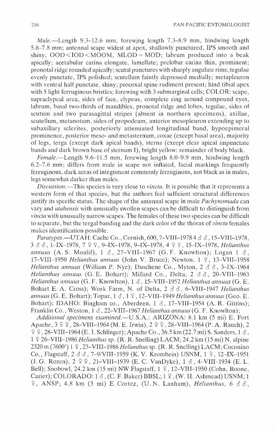
216 PAN-PACIFIC ENTOMOLOGIST
Male.—Length 9.3-12.6 mm, forewing length 7.3-8.9 mm, hindwing length 5.8-7.8 mm; antennal scape widest at apex, shallowly punctured, IPS smooth and shiny; OOD<IOD<MOOM, MLOD = MOD; labrum produced into a beak apically; acetabular carina elongate, lamellate; prelobar carina thin, prominent; pronotal ridge rounded apically; scutal punctures with sharply angulate rims; tegulae evenly punctate, IPS polished; scutellum faintly depressed medially; metapleuron with ventral half punctate, shiny; procoxal spine rudiment present; hind tibial apex with 5 light ferruginous bristles; forewing with 3 submarginal cells; COLOR: scape, supraclypeal area, sides of face, clypeus, complete ring around compound eyes, labrum, basal two-thirds of mandibles, pronotal ridge and lobes, tegulae, sides of scutum and two parasagittal stripes (absent in northern specimens), axillae, scutellum, metanotum, sides of propodeum, anterior mesopleuron extending up to subaxillary sclerites, posteriorly attenuated longitudinal band, hypoepimeral prominence, posterior meso- and metasternum, coxae (except basal area), majority of legs, terga (except dark apical bands), sterna (except clear apical impunctate bands and dark brown base of sternum I), bright yellow: remainder of body black.
Female.—Length 9.6-11.5 mm, forewing length 8.0-9.9 mm, hindwing length 6.2-7.6 mm; differs from male in scape not inflated, facial markings frequently ferruginous, dark areas of integument commonly ferruginous, not black as in males, legs somewhat darker than males.
Discussion.—This species is very close to vincta. It is possible that it represents a western form of that species, but the authors feel sufficient structural differences justify its specific status. The shape of the antennal scape in male Pachynomada can vary and utahensis with unusually swollen scapes can be difficult to distinguish from vincta with unusually narrow scapes. The females of these two species can be difficult to separate, but the tergal banding and the dark color of the thorax of vincta females makes identification possible.
Paratypes.—UTAH: Cache Co., Cornish, 600,7-VIII-1978 4 6 c3,15-VIII-1978, 3 (5 c7, l-IX-1978, 7 9 9, 9-IX-1978, 9-IX-1978, 4 9?, 15-IX-1978, Helianthus annuus (A. S. Moalif), 1 <3, 27-VIII-1967 (G. F. Knowlton); Logan 1 <3, 17-VIII-1950 Helianthus annuus (John V. Bruce); Newton, 1 9, 13—VIII—1958 Helianthus annuus (William P. Nye); Duschene Co., Myton, 2 6 c3, 3-IX-1964 Helianthus annuus (G. E. Bohart); Millard Co., Delta, 2 8 <3, 20-VIII-1963 Helianthus annuus (G. F. Knowlton), 1 <3, 15-VIII-1952 Helianthus annuus (G. E. Bohart E. A. Cross); Work Farm, N. of Delta, 2 6 6, 6-VIII-1947 Helianthus annuus (G. E. Bohart); Topaz, 1 8,1 9,12-VIII-1949 Helianthus annuus (Geo. E. Bohart); IDAHO: Bingham co., Aberdeen, 1 <7, 17-VIII-1954 (A. R. Gittins); Franklin Co., Weston, 1 6,22-VIII-1967 Helianthus annuus (G. F. Knowlton);
Additional specimens examined.—U.S.A.: ARIZONA: 8.1 km (5 mi) E. Fort Apache, 3 9 9,28-VIII-1964 (M. E. Irwin), 2 9 9,28-VIII-1964 (P. A. Rauch), 2 9 9,28-VIII-1964 (E. I. Schlinger); Apache Co., 36.5km (22.7mi) S. Sanders, 1 (7, 1 9 26-VIII-1986 Helianthus sp. (R. R. Snelling) LACM; 24.2 km (15 mi) N. alpine 2320 m (7600') 1 9,23-VIII-1986 Helianthus sp. (R. R. Snelling) LACM; Coconino Co., Flagstaff, 2 c7 c7, 7-9/VIII-1959 (K. V. Krombein) USNM, 1 9, 12-IX-1951 (J. G. Rozen), 2 9 9, 21-VIII-1939 (E. C. VanDyke), 1 (7, 4-VIII-1934 (E. L. Bell); Snobowl, 24.2 km (15 mi) NW Flagstaff, 1 9,12-VIII-1950 (Cohn, Boone, Cazier); COLORADO: 1 c7, (C. F. Baker) BBSL; 1 9 , (W. H. Ashmead) USNM; 1 9, ANSP; 4.8 km (3 mi) E Cortez, (U. N. Lanham), Helianthus, 6 c7 c7,

VOLUME 64, NUMBER 3 217
14-VIII-1976, 9 9$, 14-VIII-1976, 5 3 3, 15-VIII-1976, 2 $ $, 15-VIII-1976; Boulder, 1 $, Heliopsis (W. P. Cockerell), 1 $, 15-VIII-Helianthus coronatus-x (Cockerell) Boul, 1 $, 2-VII-1929 (C. S. Williams) Boul, 1 $, 25-VIII-21
Lepadenia marginata (L. O. Jackson), 1 $, 1650 m (5400') 27-VIII-1979 (Kristi
Neff) Boul, 1 (7, 3-VIII-1927 (E. C. Nelson), 1 $, 1650 m (5400') 5-IX-1976 Grindelia (P. Robinson) Boul 12:30-13:45 p.m.; Canfield, 2 33, 15-VIII-1922; Denver, 1 $ , -VII- 29 Boul, 1 3, -VII-1929; Fort Collins, 1 d, ll-VIII-1974,1 8, 23-31/VII1-1974, 1 $, 23-31/VIII-1974, 1 $, 23-31/VIII-1974 Ft. C, 1 $, 25-VIII-1975 Ft. C, 1 8,26-VIII-1973,1 $ , 5-IX-1973 Ft. C (all H. E. Evans), 1 $ ,
5-VIII-99 USNM, 1 $, 12-VIII-00 USNM; J.Martin Dam, Hasty, 1 $, 22- VIII-1960 (R., K. Dreisbach); Pikes Peak, 2440 m (8000'), 2 3 <3, -VIII-1932 (Lee Jeppson) BYU; Pingree Park, 1 $, (C. Lynn Hayward) BBSL; Pueblo, 1 $, 23- VIII-1931 (H. G. Rodeck) BBSL; Rock Creek vie. Colorado Springs, 1 8,
19—VIII—1937; Boulder Co., 2 3 8, 14-VIII-1925 (chas. H. Hicks) Boul; Fremont Co., 9.7 km (6 mi) NE Florence, 1 $, ll-VIII-1964 (J. G., B. L., K. C. Rozen); Larimer Co., 3.2 km (2 mi) E. Wellington, 2 $ $, 29-VIII-1976 Helianthus (C. Lanham) Boul; Las Animas Co., Kim 51.7 km (32.1 mi) SW, 1 $, 22-VIII-1967 (R. R. Snelling) BBSL; Montezuma Co., 4.8 km (3 mi) W Arriola, 1830 m (6000'), 1 $ ,-IX-1975 (T. Marquardt) Malaise Trap; Pueblo Co., 16.1 km (10 mi) W. Pueblo, 1 $, ll-VIII-1964 (J. G., B. L., K. C. Rozen); IDAHO: Oneida Co., 6.1 km (3.8 mi.) W. Holbrook Summit, 1 <3, 26-VIII-1969 (G. F. Knowlton); Holbrook Summit, 1 3,2-IX-1969 (G. F. Knowlton), 1 3,26-VIII-1969 (G. F. Knowlton); Ireland Canyon, 1 3,2-IX-1969 (G. F. Knowlton), 13,2$$, 26-VIII-1969 (G. F. Knowlton); NEW MEXICO: 20.9 km (13 mi) W. of Chama, 1 3, 24-VIII-1947 (Hugo G. Rodeck); Beulah, 2440 m (8000'), 1 3 (Cockerell), 1 $, 17-VIII- (H. Skinner); Cimarron, 1 $, 17-22/VIII-1914 (W. R. Walton); Koehler, 1 $, 12- VIII-1914 (W. R. Walton); Raton, 1 3,6-VIII-1952 (R. R. Dreisbach) Mich, 1 $ , 6-VIII-1952 (R. R. Dreisbach); WhiteMtns. S. Fk. Eagle Creek, 2440m (8000'), 1 $ , 8-16 (Townsend); Mckinley Co., 30.6km (19mi) N. Gallup, 1 3,14-VIII-1972 (J. G. Rozen, R. McGinley); Valencia Co., Cubero, 1 3, 18-VIII-1948 (C&P Vaurie) USNM; San Mateo, 2130 m (7000'), 4 $ $ , 18-VIII-1962 (R&K Dreisbach) Mich, 5 3 3,18-VIII-1962 (R&K. Dreisbach); UTAH: Bryce Canyon, 13, (Vasco M. Tanner); Hooper, 1 $, 17-IX-1937 (D. E. Hardy); Sandy, 1 3, 27-VIII-1947 (E. I. Taylor); Zion Pk Junction, 1 $, 9-VIII-1936 (M. B. Jackson) SMEK; Box Elder Co., Collinston, 1 3, 2-IX-1908 (E. S. G. Titus); Curlew Valley, 1 3, l-IX-1970 (G. F. Knowlton, J. H. Judd); Hansel Mtns., 2 3 3,28-VIII-1974 (G. F. Knowlton); Snowville, 3 3 3, l-IX-1970 (G. F. Knowlton, J. H. Judd), 1 3,
16-VII-1953 (W. G. Firestone), 1 3,26-VIII-1953 (W. G. Firestone); Cache Co., 6.4 km (4 mi) NW Logan, (D. K. Broemeling), 8 3 3, 10-VIII-1985 Helianthus
annuus, 6 3 3, 10-VIII-1985, 2 3 3, 1 $, 22-VIII-1985 , 3 3 3 , 22-VIII-1985 Helianthus annuus; Cornish, 4 3 3,20-VIII-1970 (G. E. Bohart) 1 3,24-VIII-1973
(P.F. Torchio), 4 3 3, 27-VIII-1967 (G. F. Knowlton), 6 3 3, 7-IX-1968 Helianthus (C. D. Michener) SMEK; Hyrum Dam, (D. K. Broemeling), 1 $, 13- VIII-1985, 1 3,9—VIII—1985 Helianthus annuus, 1 3,9-VIII-1985, Helianthus annuus, 1 $, 9-VIII-1985; Logan, 13,1$, 18-VIII-1949 (G. E. Bohart), 1 3,
30—VIII—1955 (W. J. Hanson) SMEK; Petersboro, 3 3 3, 11—VIII—1947 Helianthus annuus (G. E. Bohart); Duchesne Co., Myton, Helianthus annuus, 1 3, 1 $, 3-IX-1964 (G. E. Bohart), 1 $ , 8—V111—1966 (Bohart, Cross); Emery Co., 0.8 km

218 PAN-PACIFIC ENTOMOLOGIST
(0.5 mi) e Little Gilson Butte, 1 3,1 9 , 15-IX-1985 (D. K. Broemeling); 0.8 km (V2
air mi) NE Little Gilson Bt, 1540 m (5050'), 2 3 3,12-IX-1983 (Parkers/Griswold); 0.8 km (V2 mi) E Little Gilson Butte, 1550 m (5100'), Helianthus anomalous 1 9 , 27-VIII-1985 (T. L. Griswold), 4 31, 27-VIII-1985 (T. L. Griswold), 3 33,2 9 $ , 27-VIII-1985 (D. K. Broemeling); 3.2 km (2 air mi) W Little Gilson Butte, 2 B d, 1 9, 15/17-IX-1980 (T. Griswold), 1 c3, 15/17-IX-1980 (F. Parker), 1 9, 15/17-IX-1980 (D. Veirs); 4.8 km (3 mi) N Little Gilson Butte, 1 9,29-IX-1984 (D. K. Broemling); 5.2 km (3.2 air mi) NE Little Gilson Bt., 1520 m (5000'), 1 9 ,
13-IX-1983 (Parkers/Griswold); 4.8km (3mi) SSETempleMt. SanRafaelBes, 1620 m (5300'), 1 9,23-IX-1982 (FD/JH Parker); 6.4 km (4 air mi) N Gilson Bt, 1550 m (5100'0, 4 3 3, 4 9 9,12/14-IX-1983 (Parkers/Griswold), 1 c3, 16/17-IX-1980 (T. Griswold), 2 c3 c3, 1 9, 26-VIII-1985 (B. K. Broemeling), 6 c3 c3, 26-VIII-1985 (F.B. Parker), 1 c3, 26-VIII-1985 (T. L. Griswold), 1 <3, 16/17-IX-1980 (T. Griswold); Gilson Bt. Well, 1550 m (5100'), 1 9,20-IX-1985 Helianthus anomalous
(W. P. Nye); Goblin Valley in Sand Dunes, 2 <3 c3, 3 9 9, 16-IX-1979 (F.D. Parker/B. Veirs); 6 9 9,16-IX-1979 (C. Hatley/G. Briggs); Little Gilson Butte E. side, 1590 m (5200'), 3 9 9, 22-IX-1982 (FB/JH Parker); San Rafael Desert nr.
Goblin Vly., 1 <3, -IX-1980 (G. E. Bohart); WildHorse Cr. N Goblin Vly, 4 c3 c3,1 9,13-IX-1983 (Parkers/Griswold); Garfield Co., 3.2 km (2 mi) NE Henrieville, 1 9,30-VIII-1985 (F. B. Parker), 1 9,30-VIII-1985 (B. K. Broemeling); 9.7 km (6 mi) SE Escalante, 1 <3, 1 9,30-VIII-1985 (B. K. Broemeling); Sandy Cr. SSE of Notom, 1590 m (5200'), 1 3,16-IX-1983 (FParker/TGriswold), 2 9 9,16-IX-1983 (FParker/TGriswold), 2 9 9,16-IX-1983 (I. H. Parker); Millard Co., Delta, 2 3 3, 15- VIII-1952 Helianthus annuus (G. E. Bohart, E.A. Cross), 3 3 3, 1 9, 20-VIII-1963 Helianthus annuus (G. E. Bohart), 2 3 3, 5-VIII-1948 (G. E. Bohart); Work Farm, N. Delta, 4 3 3, 6-VIII-1947 Helianthus annuus (G. E. Bohart); Piute Co., Circleville, 1 9,8-IX-1978 (I. B. Karren); Salt Lake Co., Salt Lake City, 1 3, ll-VIII-1951 (R. B. Selander); San Juan Co., 8.1 km (5 mi) N Blanding, 1 3,24-VIII-1967 (I. C. Hall) UCR; Uintah Co., Lower Jensen, 4.8 km (3 mi) SW Jensen, (Ian L. Bell), 1 3, 16-VIII-1949, 1 3,9-IX-1949; Vernal, 1 3, 16- VIII-1949 (Lynn R. Nielson), 1 3, 18-VI-1936, 1 9,20-VIII-1949 (Lynn R. Nielson); Utah Co., Provo, 1 3,16-VIII-1947 (G. F. Knowlton); Washington Co.,
1 3, 12-VIII-1959 (G.F. Knowlton); Zion Park, 1 9, 6-VIII-1966 (J. Kefuss); Wayne Co., 6.4 km (4 mi) SE Hanksville, 1 3, 28-IX-1985 Helianthus annuus (D. K. Broemeling); WYOMING: 19.3 km (12 mi) E. of Gillette, 1 3,31-VIII-1962 Helianthus (S. M. Walder); Fremont Co., Baldwin Creek 4.8 km (3 mi) NW of Lander, 1680 m (5500'), 4 3 3,10-VIII-1963 Helianthus (W. E. LaBerge);
Nomada (Pachynomada) zebrata Cressom
Nomada zebrata Cresson, 1978. American Hymenoptera. Amer. Ent. Soc. Trans. 7:73.
Lectotype Male.—“Col. 2589. ” Type Depository, Academy of Natural Sciences of Philadelphia.
Diagnosis.—3 submarginal cells, terga with yellow transverse median bands, antennal scape globose in males, sides of propodeum ferruginous with posterior yellow mark, long pubescence, mesopleuron with a complete longitudinal yellow band, slightly narrowed medially, not expanded anteriorly towards pronotal lobe.

VOLUME 64, NUMBER 3 219
Male.—Length 9.9-11.6 mm, forewing length 8.1-9.2 mm, hindwing length 6.1-6.9 mm; antennal scape globose, densely punctured on anterior outside half, sparsely punctate interiorly, IPS smooth and shiny, posterior virtually impunctate; first flagellar segment 1.4 times as long as second; IOD = OOD = MOOM, MLOD = MOD; head densely, evenly punctate, smooth shiny IPS; IPS dull, roughened within ocellar triangle; labrum glassy, impunctate median strip, produced slightly into a broad apical beak; acetabular carina strongly lamellate; prelobar carina short, heavy; pronotal ridge rounded anteriorly, broadly depressed medially, inner third impunctate; scutum deeply contiguously punctured, interpunctural spaces sharpened apically; tegulae with densely, shallowly, evenly punctate surface, glassy; metanotum flattened, broadened medially; propodeal disk reticulate rugose, with long transverse dorsal rugae, curling downward; propodeum swollen laterally,
densely contiguously punctured, impunctate roughened band below spiracle; metapleuron with punctate ventral half; procoxal spine rudiments lacking; hind tibial
apex with 7 weakly curved, ferruginous bristles in single row; forewing with 3 submarginal cells: COLOR: base color ferruginous; sides of face to antennal insertion, supraclypeal area, labrum, basal two-thirds of mandibles, pronotal ridge
and lobes, tegulae, scutellum, metanotum, sides of propodeum, longitudinal mesopleural band (narrowed medially), apices of coxae, distal mid- and hind femora, proximal tibiae, transverse median bands on all terga (interrupted on tergum 1), pygidium, spot on sternum 1, extensive bands on remaining sterna,
creamy yellow. Females.—Length 9.9-12.4 mm, forewing length 8.1-8.7 mm, hindwing length
6.3-6.6 mm; differs from males by antennal scape not inflated, supraclypeal area ferruginous instead of yellow.
Discussion.—Nomada zebrata seems at present to be restricted to the southwestern United States, but it will probably be found in northern Mexico. It is characterized by the very swollen antennal scape of the males and a complete longitudinal yellow mesopleural band. Its range overlaps greatly with utahensis, and slightly with vincta in eastern Colorado. Specimens reported from Kansas and the Great Plains have proven to be misidentified vincta. Cresson’s type series includes at
least 3 vincta. One of the cotypes of Cockerell’s vincta heterochroa is actually a specimen of zebrata. Some specimens from a recent series collected in Texas have
some of the ferruginous body markings replaced with black. Specimens examined.—U.S.A.: Brevolt Co. 25.0 km (15.5 mi) E. ?, 1 6,
15-IX-1961 Verbesina (Timberlake) LACM; ARIZONA: 32.2 km (20 mi) E. Pearce, 19 2, l-X-1967 (F. Werner) UNAr; Apache Co., 36.5 km (22.7 mi) S. of Sanders, 1 8 28-VIII-1986 Sphaeralcea sp. (R. R. Snelling); Springerville, 1 8, 25- VIII-1970 (Bill Apperson); Cochise Co., 6.4 km (4 mi) W. Portal, 12 2, 26- IX-1982 (W. I. Pulawski); Onion Saddle, Chiricahua Mtns., 12 2,3-IX-1959 (J. R. Powers); Coconino Co., 11.3 km (7 mi) S Flagstaff, 2 9 2, 12-16/IX/1961 Grindelia (G. Butler), 1 8, 12-16/IX/1961 Grindelia (G. Butler); 32.2 km (20 mi) SW of Flagstaff 1 8, 18-IX-1966 (R. S. Beal); Flagstaff, 12 2,9-IX-1951 (A. T. McClay); Greenlee Co., 62.8 km (39 mi) S Hannagan Meadows, 1 8, 1 2, 13-IX-1985 Helianthus (d. K. Broemeling); Yavapai Co., Paulden, 3.2 km (2 mi
N.), 1 8,14-IX-1961 Gutierrezia microcephala (P. D. Hurd); COLORADO: Cabin Canon, 1 2,31-VIII-1921 (Hugo G. Rodeck); Denver, 1 8,10-VI-1960; Boulder
Co., Boulder, 1650 m (5400'), 1 2, 5-IX-1976 Grindelia (P. Robinson); N. of

220 PAN-PACIFIC ENTOMOLOGIST
Marshall, 1 9, 12-IX-1946 Grindelia (Hugo G. Rodeck); El Paso Co., Colorado Springs, 1 2 ; Rock Creek vie. Colorado Sprgs, 1 3,19-VIII-1937; Fremont Co., 8.1 km (5 mi) S. Coaldale, 2230 m (7300'), 1 <3, 13-VIII-1969 (C. D. Michener); Las Animas Co., Kim, 37.0 km (23 mi) N., 1 2, 22-VIII-1967 Haplopappus (R. R. Snelling); NEW MEXICO: Grant Co., Hachita, 12.9 km (8 mi) W., 1 3,5-IX-1972 (R.R. Snelling); Otero Co., Cloudcroft, 2590-2740 m (8500-9000'), 2 3 3, 6-IX-1963 (H. V. Weems, Jr.); Torrance Co., Town of Gran Quivira, 1990 m (6500'), 1 3, 20-VIII-1967 (Hugh B. Leach); TEXAS: Jeff Davis Co., High
Lonesome Ridge, Davis Mts. 2320 m (7600') 17 3 3,3 $ 2,6-IX-1986 Verbesina encelioides (R.R. Snelling), 1 3 same but no floral record; Madera Can. rest stop on
Rd. 118, 1 2, 16-X-1977 (James R. Zimmerman), NMex; Upper Limpia Cyn., Davis Mts. 1710 m (5600') 30-VIII-1986 (R. R. Snelling), 3 3 3, (one each on Bidens sp. and Verbesina encelioides.
Bohartorum Group
Nomada (Pachynomada) bohartorum Moalif New Species
Holotype Female.—“Mex. Jalisco, Tepatitlan, X-3-66, GE & AS Bohart.” Type Depository, United States National Museum.
Diagnosis.—3 sub-marginal cells, antennal scape not globose (slightly expanded apically), terga with complete transverse median bands, sides of propodeum black with long pubescence, thorax black or black with ferruginous (never entirely ferruginous), females with 5 hind tibial apical bristles, strongly curved, somewhat flattened.
Male.—Length 8.2 mm, forewing length 6.9 mm, hindwing length 5.3 mm;
antennal scape moderately punctate anteriorly, impunctate posteriorly, IPS polished, somewhat roughened anteriorly, posterior quarter of scape apilose; first
flagellar segment twice length of second; IOD<OOD<MOOM, MLOD<MOD; head densely, deeply punctured, glassy IPS; raised impunctate bump posterior to line between lateral ocelli; labrum distended into large flattened beak medio-apically; acetabular carina with distinct lamella; prelobar carina thin; pronotal ridge somewhat angulate anteriorly, broadly depressed medially; scutum with typical punctation; tegulae lightly punctured; scutellum distinctly depressed medially; sides of propodeum densely punctate, clothed with fine, long (0.15 mm) pubescence, broad, shiny, virtually impunctate band along lower margin, reaching dorsally to spiracular depression; mesopleuron with glassy IPS, roughened dorsal to hypoepimeral prominence; ventral half of metapleuron glassy, impunctate; slight procoxal spine rudiment present; hind tibial apex with 3 clear, long, rather strongly curved bristles; forewing with 3 submarginal cells: COLOR: face up to level of clypeus, short extension dorsally along compound eyes, basal half of mandibles, pronotal ridge and lobes, outer two-thirds of tegulae, scutellum, anterior of fore-femora, abdominal terga (T-l divided medially), yellow; remainder of body dark brown to black.
Female.—Length 8.5 mm; forewing length 7.3 mm; hindwing length 5.6 mm;
scape shallowly punctured, with shiny, shagreened interpunctural areas; IOD < OOD = MOOM, MLOD <MQD; impunctate, roughened median hump
behind ocellar triangle; labrum produced into an apical wedge, bearing a narrow
median impunctate band; acetabular carina strong, angulate; prelobar carina thick;

VOLUME 64, NUMBER 3 221
pronotal ridge rounded apically with smooth impunctate median area; scutal puncture rims angulate; tegulae evenly, shallowly punctate; scutellum depressed medially; sides of propodeum covered with long hairs (0.17 mm); hypoepimeral area not prominently swollen; metapleuron with ventral half highly polished, impunctate; pro-coxal spine rudiments lacking; hind tibial apex with 5 ferruginous spines, anterior 3 strongly curved, flattened; forewing with 3 submarginal cells: COLOR:
scape, pedicel, first flagellar segment, ring around compound eyes, face to base of clypeus, labrum, basal two-thirds of mandibles, tegulae, most of mesopleuron, apices of coxae, legs, medial spot on sternum I, remaining sterna, ferruginous: pronotal ridge and lobes, scutellum, anterior mesopleural spot, divided median transverse band on tergum 1, tergum 2 and 3 except baso-medial inclusion and apical band, tergum 4 and 5 except apical band, transverse bands on sternum 2-5, yellow; remainder of body black.
Discussion.—Nomada bohartorum is a small western Mexican species. The flattened hind tibial bristles which are strongly curved apically are its most distinctive feature. These bristles are less pronounced in the males. The male is very similar morphologically to tepoztlan, but the integument of bohartorum is black rather than light brown, and the genital capsules are somewhat different.
Specimens examined.—MEXICO: DURANGO: 9.7 km (6 mi) W. Paraiso, 2010 m (6600'), 1 9, 23-IX-1950 (Ray F. Smith); SINALOA: El Palmito on Rt. 40, 1830-1990 m (6000-6500'), 1 M, 3-IX-1963 (H. V. Weems, Jr.);
Nomada (Pachynomada) tepoztlan Moaiif New Species
Holotype Male.—“Tepotzlan(l), Morelos, Mex. 9-26-57, R. & K. Dreisbach.” Type Depository, Michigan State University.
Diagnosis.—3 submarginal cells, antennal scape not globose, abdominal terga with complete transverse median bands, sides of propodeum with long pubescence, head and thorax a uniform light brown.
Male.—Length 7.4 mm; forewing length 6.2 mm, hindwing length 4.8 mm; scale slightly swollen apically, moderately punctate, IPS shiny, shagreened in places;
IOD < OOD = MOOM, MLOD < MOD; labrum thickened medio-apically, narrow impunctate strip extending down middle; acetabular carina prominent; prelobar carina abbreviated; pronotal ridge polished, sparsely punctate, shallowly depressed medially, apex rounded, broad impunctate medial area; tegulae evenly,
shallowly punctured, glassy; scutal punctures with angulate rims; scutellum slightly depressed medially; metanotum flattened; propodeum with highly polished impunctate lateral ventral band extending upwards to spiracle; ventral band extending upwards to spiracle; ventral half of metapleuron impunctate, highly polished; procoxal spine rudiment absent; hind tibial apex with 5 light bristles, curved at about half their length; forewing with 3 submarginal cells: COLOR: uniform light brown, except; clypeus, face below clypeo-frontal suture, labrum,
basal half of mandibles, pronotal ridge and lobes, tegulae, scutellum, metanotum, apices of fore- and mid femora, tibial bases, fore- and mid tibial apices, fore- and mid tarsi, medially narrowed transverse band on tergum 1-5, broad transverse band on sternum 1, yellow.
Discussion.—This is the smallest of the species of Pachynomada. It is no larger
than most Nomadita species, but it has all the distinguishing characteristics of the
subgenus Pachynomada.

222 PAN-PACIFIC ENTOMOLOGIST
Vitticollis Group
Nomada (Pachynomada) dreisbachorum Moalif NEW SPECIES
Holotype Male.—“Amecameca, Mex. 9-25-57, R. & K. Dreisbach.” Type Depository, Michigan State University.
Diagnosis.—2 submarginal cells, antennal scape not at all swollen, propodeum and scape clothed with very long hair (more than two-thirds length of scape), body black with greatly reduced yellow markings, IPS of face dull and quite roughened, pro-coxal spine rudiment strong, hind basitarsus only slightly expanded medially.
Male.—Length 9.6-12.7 mm; forewing length 8.9-10.1 mm; hindwing length 6.9-8.1 mm; antennal scape not inflated (fig. 3), covered with unusually shallow, flattened punctures, IPS dull, coarsely shagreened; IOD < OGD < MOOM, MLOD < MOD; head covered with round, shallow punctures, IPS dull and coarsely roughened; labrum with IPS quite polished, apex produced into thick, prominent beak; acetabular carina unusually short but strongly lamellate; prelobar carina reduced to an angulation; pronotal ridge apex protruberant but. rounded; IPS of scutum somewhat roughened; scutellum flattened dorsally, slightly depressed medially, entire IPS roughened; sides of propodeum densely, shallowly punctate, IPS quite roughened, covered with long slender hairs (0.22 mm); mesopleuron more sparsely punctured, punctures shallow, IPS dull, quite roughened; metapleuron with ventral half impunctate, shiny, only slightly roughened; pro-coxal spine rudiment strongly developed; hind tibial apex with long, thin, pale bristles, difficult to distinguish from surrounding hairs; forewings with two submarginal cells, but some specimens may bear a short rudiment of first transverse cubital vein, wings dark around outer margin, clear interiorly; sternum 1 with traces of a median longitudinal carina: COLOR: spot at apex of clypeus and at apex of compound eye, tips of pronotal lobes, pronotal ridge, two spots on scutellum, metanotum, thin transverse median band on abdominal terga, patches on sterna 1-3, yellow; remainder of body black.
Discussion.—This species is the furthest from the norm of Pachynomada. The genitalia are clearly that of Pachynomada, although the gonostylus is unusually short. One specimen examined shows a trace of the first transverse cubital vein in the forewing. This may mean that long series of dreisbachorum would contain specimens with 2 or 3 submarginal cells. The genus Hypochrotaenia (formerly the subgenus Micronomada) shows great variability in the number of submarginal cells, and there are a number of specimens which have 3 cells on one forewing and two cells on the other (pers. obs.). Nomada dreisbachorum can still be distinguished by the long pubescence on the antennal scape (which is not globose, as in many male Pachynomada species), and the dull, shagreened interpunctural surface of the head.
Paratypes.—4 males with same data as holotype. Three deposited at Michigan State University, and one in the Bee Biology & Systematics Lab in Utah.
Additional specimens examined.—MEXICO: 64.4 km (40 mi) W. of Puebla Pue., 2800 m (9200'), 1 d 7-IX-1957 (H. A. Scullen) ORSU; D. F.: 29.0 km (18 mi) S. of Mexico City, 2800 m (9200'), 1 d, 10-IX-1957 (H. A. Scullen) ORSU;
Nomada (Pachynomada) vitticollis Cressoe
Nomada vitticollis Cresson, 1878. Trans. Amer. Entomol. Soc. 7:78.
Holotype Female.—“Mex. 2562. ” Type Depository, Academy of Natural Sciences of Philadelphia.

VOLUME 64, NUMBER 3 223
Diagnosis.—3 submarginal cells, thorax almost entirely yellow except for brown
sutures, sides of propodeum with long pubescence, interpunctural surface of face smooth, shiny.
Male.—Unknown. Female.—Length 8.9-12.1 mm, forewing length 8.0-9.6 mm, hindwing length
6.2-7.3 mm; antennal scape sparsely, shallowly punctured, IPS shagreened; first flagellar segment subequal to second; IOD = OOB = MOOM, MLOB < MOB; head densely, evenly punctate with some deformation, IPS shiny, somewhat roughened; labrum sparsely punctate, shiny impunctate apico-medially, protruding wedge-shaped beak; acetabular carina small, distinctly lamellate; mandibles virtually impunctate; prelobar carina abbreviated, pronotal ridge rounded anteriorly, broadly depressed medially; roughened IPS postero-medially; metanotum shallowly and broadly punctured; sides of propodeum shallowly and broadly punctured, IPS shagreened, dull, pubescence 0.3 mm long; hypoepimeral area not strongly protruberant; ventral half of metapleuron glassy, impunctate; procoxal spine rudiment absent; hind tibial apex with 8 bristles; forewing with 3 submarginal cells: COLOR: scape, supraclypeal area, spot anterior to mid-ocellus, ring around compound eyes, pronotum (except anterior to pronotal ridge and
between ridge and collar), lateral margins of scutum and two median longitudinal stripes, scutellum, metanotum, tegulae, axillary sclerites, propodeum (except median stripe), mesopleuron and metapleuron except sutures, legs (except dorsal stripe on fore- and mid femora, ventral hind femora, interior of hind tibiae), abdomen, bright yellow; remainder of body dark brown.
Discussion.—This species is similar to dreisbachorum in form; both species have a long, fairly thin body, unusually long pubescence and similar punctation patterns. Nomada vitticollis differs from dreisbachorum by its almost completely yellow head and thoracic pleura, 3 submarginal cells in forewings, distinctly inflated hind basitarsus, and the IPS of the face (shiny and smooth in vitticollis, dull and shagreened in dreisbachorum). The holotype is quite a bit smaller than the other specimens examined, and the majority of future specimens will probably be closer in size to the large specimens.
Specimens examined.—MEXICO: SAN LUIS POTOSI: 83.7 km (52 mi) S. of Tamazunchale, 1710 m (5600'), 1 9, 7-X-1957 (H. A. Scullen) ORSU; TAMAULIPAS: Rancho del Cielo 11.3 km (7 mi) W. Gomez Farias, 1 2, 4-6/XI/1972 (J. A. Gillaspy);
Asteris Group
Nomada (Pachynomada) asteris Swenk
Nomada (Homonamada) asteris Swenk, 1913. Nebr. Univ. Studies 12:89.
Holotype Female.—“Manhattan, Kansas, September 19, 1908, on Aster paniculatus (O. A. Stevens, No. 1203).” Type Bepository, University of Nebraska, Lincoln.
Diagnosis.—3 submarginal cells, female rufo-ferruginous, abdomen lacking complete transverse bands; male antennal scape globose, abdomen with complete
transverse median bands, sides of propodeum black with long pubescence, antennal scape uniform ferruginous color, mesopleuron ferruginous.
Male.—Length 8.9-10.4 mm, forewing length 5.9-7.0 mm, hindwing length 4.9-5.9 mm; antennal scape globose, densely punctate, glassy IPS, tuft of hair on posterior apical shelf; IOB = OQB, MLOB < MOB; labrum lacking median

224 PAN-PACIFIC ENTOMOLOGIST
impunctate strip, produced into small beak apically; prelobar carina somewhat thickened; pronotal ridge sharply angulate anteriorly; interpunctural spaces of scutum slightly rounded; tegulae moderately punctate, glassy; scutellum flattened, glassy, only slightly depressed medially, metanotum rather strongly rounded medially, roughened posteromedial interpunctural surface reduced; surface of metanotum glassy; propodeal sides deeply punctured, dull, rough IPS, pubescence long (0.2 mm); mesopleural surface quite shiny, only slightly roughened in places; metapleuron glassy, punctate; procoxal spine rudiment virtually absent; hind tibial apex with 7 or 8 thin, pale ferruginous bristles; metasternum grooved medially; pygidium rather broad, shallowly emarginate medially; forewing with 3 submarginal cells, apex somewhat darkened: COLOR: supraclypeal area, sides of face up to and
around antennal sockets (extending along sides of compound eyes), labrum, clypeus, basal two-thirds of mandibles, malar space, ring extending behind compound eyes
almost to vertex, pronotal lobes, ridge, tegulae, scutellum, metanotum, small anterior mesopleural spot, apices of coxae, transverse median bands on terga (emarginate antero-medially), creamy-white; antennae, legs, most of mesopleuron, abdominal sterna, ferruginous; remainder of body dark fuscous to black.
Female.—Length 7.7 mm; antennal scape heavily punctate, IPS shagreened to shiny, IOD = OOD, MLOD <MOD, head densely evenly punctate, IPS highly polished, supra-clypeal area prominent, labrum with strong triangular median sub-apical beak extending into a sub-apical ridge laterally; mandibles robust with a strong dorsal ridge; acetabular carina prominent, semicircular, slightly lamellate; pre-malar carina strong dorsally, undifferentiated ventrally; pre-occipital ridge rounded; pronotal ridge very thin medially; apex rounded; pre-lobar carina thick, sloping gradually; scutum deeply punctured (honey-combed), punctural rims
sharply angulate, tegulae polished, densely punctured; scutellum highly polished, glassy, sparsely punctate, very slightly depressed medially; postero-medial IPS smooth, not shagreened or micro-rugose; metanotum very shiny, sparsely punctate; supra-spiracular ridge undeveloped; hypo-spiracular band impunctate with light vertical rugae, hairs 0.15 mm long, sparse; hypoepimeral prominence flattened;
mesopleuron deeply, nearly contiguously punctured with smooth-shiny IPS; metapleuron shiny, punctate; pro-coxal spine rudiments lacking; hind tibial apex with 5 fine, evenly spaced, pale ferruginous bristles; hind basitarsus not as inflated as
other Pachynomada, widest point closer to base; hindwings with 3 sub-marginal cells, smokey with clear sub-apical crescents; abdominal sterna punctate, heavily shagreened; terga densely, evenly punctate, heavily shagreened IPS; apical impunctate bands narrow: COLOR: body is uniform, light rufo-ferruginous; apex of mandibles, posterior-medial triangle on scutum, areas of abdomen darkened to fuscous, pronotal lobes, apices of pronotal ridge, scutellum, metanotum, medial lateral patches on terga 2,3, and 4 creamy white.
Discussion.—Nomada asteris and victrix are sexually dimorphic, which is unusual in this subgenus. This rare species has only been collected in Kansas.
Specimens examined.—U.S.A.: KANSAS: Reece, 12 <3 6, 7-IX-1949 Amphiachyris dracunculoides (Michener-Beamer);
Nomada (Pachynomada) besseyi Swenk
Nomada (Holonomada) besseyi Swenk. 1913. Nebr. Univ. Studies 12:85.
Holotype Male.—“Manhattan, Kansas, August 24,1908, on Helianthus tuberosus (O. A. Stevens, No. 933).” Type Depository, University of Nebraska, Lincoln.

VOLUME 64, NUMBER 3 225
Diagnosis.—3 sub-marginal cells, antennal scape globose in males, propodeum black and appearing naked (pubescence is sparse and no longer than a puncture diameter).
Male.—Length 9.0-11.3 mm, forewing length 6.7-8.1 mm, hindwing length 5.0-5.9 mm; antennal scape globose, sparsely punctate, with smooth shiny IPS; labrum with small median wedge, apical margin upturned; acetabular carina strongly lamellate; prelobar carina thickened, sloping gradually to pronotal lobe; pronotal ridge rounded apically, very shallowly depressed, impunctate medially; scutum with contiguous, frequently reticulate punctures, punctural rims angulate; scutellum deeply depressed medially; metanotum protruberant; propodeum appearing naked (pubescence very sparse and short (0.05 mm)); mesopleural IPS glassy, shagreened
dorsal to hypoepimeral prominence; ventral half of metapleuron glassy, impunctate; pro-coxal spine rudiment reduced to slight bump; hind tibial apex with six straight,
pale bristles; forewing with 3 submarginal cells, brown with pale subapical area: Color: fuscous to black; with antennal base, interantennal area, sides of face to vertex, clypeus (except sutures and median band), labrum, small basal mandibular spot, partial ring behind eye, pronotal ridge and lobes, outer four-fifths of tegulae, lobes of scutellum, metanotum, longitudinal mesopleural band, apices of coxae, postero-apical stripe on femora, exterior of tibiae, posterior stripe on hind basitarsus, thin transverse median band on abdominal terga, sterna 2 and 3, broken band on sterna 4 and 5, yellow; antennal scape (in part), first flagellar segment, ferruginous.
Females.—Length 8.9-11.3 mm, forewing length 7.4-8.1 mm, hindwing length 5.5-6.2 mm; virtually identical to male except for terminalia and antennal scape not inflated.
Note.—There is a variant form with thicker tergal banding, middle of clypeus and supraclypeal area yellow. It has been collected from the same date and location as the typical form, so it does not constitute a subspecies.
Discussion.—Nomada besseyi is similar to the Vincta group, but the extremely short and sparse pilosity of the propodeum differs from that group. It is similar in this
aspect to asteris and victrix, which have short pubescence. Their propodeal pubescence however, is fairly dense and about twice as long as that of besseyi.
Specimens examined.—U.S.A.: MARYLAND: near Plummers Id., 1 6, 29-VIII-1915 (R. C. Shannon); MICHIGAN: Kalamazoo Co., Gull Lake Bio. Sta.,
1 7,12-VIII-1969 (Roland L. Fischer), 1 6, 27-VII-1963 (G. C. Eickwort), 16,1 7, 6-VIII-1959 (Roland L. Fischer), 1 7, 5-VIII-1969 (Roland L. Fischer); MINNESOTA: Carver Co., Zumbra Heights, 1 6, 31-VIII-1921 (Irwin C. Alfonsus); NORTH CAROLINA: Bryson City, Helianthus atrorubens, (J. C. Crawford), 16,1 7,20-VIII-1923, 1 7,26-VIII-1923, 1 6, 31-VIII-1923 (J. C. Crawford) LACM, 5 6 6, 4—IX-1923 Rudbeckia laciniata; Marion, 4 6 6, 1 7, 2-IX-1929 NCSt, 1 (5,27-VIII-1930 NCSt, 1 6 28-VIII-1930,1 7 28-VIII-1931, 2 6 6, 29-VIII-1928 Heliopsis (T.B. Mitchell), 1 7, 29-VIII-1929 Boul, q 6, 29- VIII-1930 Rudbeckia lanceolata NCSt, 1 6, 29-VIII-1930 NCSt, 2 33, 30- VIII-1931,1 6,31-VIII-1929 NCSt; Raleigh, 1 7,10-IV-1961; Valley of Black Mts., 1 7, 4—IX-1906 (W. Beutenmuller); Haywood Co., Lake Junaluska, 1 6,
5—VIII—1975 (H. V. Weems, Jr.); NEW JERSEY: Haddon Hts., 1 (5, 10-IX-1939;
NEW YORK: Armonk, 1 7, 25-VIII-1936 (L. L. Pechuman); West Point, 1 6, 2-IX-1923 (W. Robinson); VIRGINIA: Hunter, 1 7,12-18/IX (R. A. Cushman);
WISCONSIN: Milwaukee, 1 7, (W. H. Ashmead);

226 PAN-PACIFIC ENTOMOLOGIST
Victrix Group
Nomada (Pachynomada) victrix Cockerell
Nomada victrix Cockerell, 1911. Proc. U.S. Nat. Mus. 39:647.
Holotype Female.—“Victoria Tex, Nov. 6-'4, on Aster; Type no. 13436.” Type Depository, United States National Museum.
Diagnosis.—2 submarginal cells, hind coxae with a posterior dorsal carina extending more than half the length of coxa, females rufo-ferruginous, abdominal terga with yellow maculations, males darker, scape somewhat swollen, sides of
propodeum with dense, short pubescence. Male.—Length 7.3-9.9 mm; forewing length 5.3-6.3 mm, hindwing 4.1-4.9 mm;
differs from female in wide transverse yellow bands on all terga, sometimes narrowly interrupted medially; clypeus and sides of face, pronotal ridge and lobes, scutellum, yellow; antennal scape swollen, body fusco-ferruginous to black.
Female.—Length 8.9-9.3 mm, forewing length 5.9-6.1 mm, hindwing length 4.7-4.8 mm; antennal scape sparsely, shallowly punctured, IPS, shagreened; IOD = OOD = MOOM, MLOD<MOD; clypeus polished, more sparsely punctate than remainder of head; labrum carinate around margin, produced into a sharp ridge apically, lacking median impunctate strip, but apico-medial area impunctate; acetabular carina reduced to small basal lamella only; prelobar carina reduced; pronotal ridge rounded anteriorly; scutum with normal punctation; tegulae glassy, virtually impunctate; scutellum flattened, unusually sparsely punctate, highly polished, posteromedial portion without roughened interpunctural surface;
metanotum not flattened; propodeal disk with delicate, arching apical rugae; sides of propodeum heavily punctured, clothed with short hairs (0.1 mm); metapleuron with ventral half highly polished, virtually impunctate; pro-coxal spine rudiment absent;
hind tibial apex with six ferruginous, straight bristles; hind coxae with pronounced dorsal posterior carina, extending over one-half length of coxa; abdominal terga with punctures very shallow; forewing with two submarginal cells: COLOR: light ferruginous, scutum somewhat darker, terga 2, 3, and 4 with small, lateral medial cream colored patches.
Discussion.—The strong carina on the hind coxa of this species is unique within the subgenus. It can be immediately separated on this character without reference to the two submarginal cells of the forewing. This may be primarily a Mexican species whose range barely extends into the United States.
Specimens examined.—MEXICO: JALISCO: Lagos de Moreno, 1 8, 19-VIII-1960 (P. H. Arnaud, E. S. Ross, D. C. Rentz), CAS; VERACRUZ: Rio Metlac, Fortin de las Flores, 1 6,3-IX-1975 (I. Powell) UCB; U.S.A.: TEXAS:
Roosevelt, 1 8, 25-IX-1906 Amphiachyris, (F. C. Pratt); Victoria, 4 9 9,
6-XI-1904 Aster;
Acknowledgements
I extend my sincere thanks to Frank D. Parker and Terry L. Griswold of the
USDA-ARS Bee Biology & Systematics Laboratory (BBSL) in Logan for advice and encouragement. I am indebted to the following persons for the loan of specimens and types in their care: roy R. Snelling, Natural History Museum of Los Angeles County (LACM); Donald Azuma, Academy of Natural Sciences of Philadelphia

VOLUME 64, NUMBER 3 227
(ANSPO; Brett C. Ratcliffe, University of Nebraska, Lincoln (UNEB); Wojiech J. Pulawski, California Academy of Science (CAS); Ronald J. McGinley, National Museum of Natural History-Smithsonian Institution (USNM); Marjorie Favreau,
American Museum of Natural History (AMNH); Kathryn C. McGiffen, Illinois Natural History Survey (INHS); Url N. Lanham, University of Colorado, Boulder (UCM); Charles D. Michener, Snow Entomological Museum, University of Kansas
(SMEK); Howard E. Evans, Colorado State University, Fort Collins (Ft. C); Saul I. Frommer, University of California, Riverside (UCR); John A. Chemsak, University of California, Berkeley (CIS); Robert O. Schuster, University of California, Davis (UCD); Louise Dumouchel, Biosystematics Research Institute, Agriculture Canada
(CNC); George C. Eickwort, Cornell University (CORN); Carol S. Parron, North Carolina State University (NCSR); Sammie J. Merritt, Texas A & M University (TAMU); James E. Gillaspy, Texas A & I University (TAI); Richard S. Zack, Washington State University, Pullman (WSU); Robert C. Bechtel, Nevada Department of Agriculture, Reno (NDAg);
This is a contribution from the Utah Agricultural Experiment Station, Utah State University, Logan, Utah 84322-4810, Journal Paper No. 3481, and USDA-Agricultural Research Service, Bee Biology and Systematics Laboratory, Utah State University, Logan, Utah 84322-5310.
Literature Cited
Cockerell, T. D. A. 1903. New American Hymenoptera, mostly of the genus Nomada. Ann. Mag. Nat.
Hist., 12:200-214.
-. 1911. Bees in the collection of the United States National Museum-1. Proc. U.S. Natl. Mus.,
39:635-658.
-. 1919. Bees in the collection of the United States National Museum-3. Proc. U.S. Natl. Mus.,
55:167-221.
-. 1921. Western bees obtained by the American Museum Expeditions. Am. Mus. Novit., 24:1-15.
-. 1922. Bees in the collection of the United States National Museum-4. Proc. U.S. Natl. Mus.,
60:1-20.
Cresson, E. T. 1878. Descriptions of new North American Hymenoptera in the collection of the
American Entomological Society. Trans. Am. Entomol. Soc. (Phila.), 7:61-136.
Michener, C. D. 1944. Comparative external morphology, phylogeny and a classification of the bees
(Hymenoptera). Bull. Am. Mus. Nat. Hist., 82:151-326.
Moalif, A. S. 1979. A revision of the subgenus Pachynomada of the genus Nomada (Hymenoptera:
Anthophoridae). Thesis, Utah State University, Logan, 54 pp.
Parker, F. D., G. E. Bohart. 1982. Notes on the biology of Andrena (Callandrena) helianthi Robertson
(Hymenoptera: Andrenidae). Pan-Pac. Entomol., 58:111-116.
-, T. Griswold. 1982. Biological notes on Andrena (Callandrena) haynesi (Viereck and Cockerell
(Hymenoptera: Andrenidae). Pan-Pac. Entomol., 58:284-287.
Rodeck, H. G. 1945. Two new subgenera of Nomada Scopoli (Hym.: Apoidea). Entomol. News,
LVI: 179-181.
Say, T. 1837. Descriptions of new species of North American Hymenoptera, and observations on some
already described. Boston Journal of Natural History, 1:361-416.
Snelling, R. R. 1986. Contributions toward a revision of the new world nomadine bees. A partitioning of
the genus Nomada (Hymenoptera: Anthophoridae). Nat. Hist. Mus. Los. Ang. Cty. Contrib. Sci.,
376:32 pp.
Swenk, M. H. 1913. Studies of North American bees. I. Family Nomadidae. University Studies,
University of Nebraska, 12:1-113.

PAN-PACIFIC ENTOMOLOGIST 64(3), 1988, pp. 228-242
Descriptions of the Immatures of Typocerus serraticornis (Coleoptera: Cerambycidae), and New Observations on Biology,
Including “Varnish” Production and Usage by the Larva
James A. Robertson
10839 Kelmore St., Culver City, California 90230
Abstract.—The larva, pupa and egg of Typocerus serraticornis are described. Observations on the host plant Oryzopsis hymenoides, mating, oviposition, larval feeding and pupal cell construction follows. Parasitization of the larva by a Dipteran maggot Arctophyto borealis and the production and usage of “varnish” by the larva are also discussed.
Introduction
Typocerus serraticornis Linsley and Chemsak, 1976, was described from 27 specimens collected over a period of 35 years in the Great Basin and on the western slope of the Rocky Mountains. In the description the authors mentioned one male and two females having emerged from Oryzopsis hymenoides (Roem. and Schult.) Ricker. These three specimens had been sent to John Chemsak by L. Guerra S., a person working at the Desert Experimental Range, 27 miles west of Milford, Utah, [with no further contact being made (Chemsak, pers. comm.)]. In 1979 R. L. Penrose recorded the collection of T. serraticornis by himself and R. L. Westcott in southeastern Oregon on flowers of Sphaeralcea grossulariaefolia growing intermixed with O. hymenoides (for which the common name “Sand dropseed” was used in error; Westcott, pers. comm.). Additionally he mentions collecting the beetles at a point 22 mi NW (sic, should be “W,” according to R. L. Westcott, in litt.) of Denio Junction, Humboldt Co. Nevada, with beetles abundant, having been on an “area of sand, covered almost exclusively with O. hymenoides. They were observed flying slowly about, and sitting and mating on this grass. Occasionally, females were noted
crawling on the surface of the sand around the plant in search of oviposition sites. Larvae were found at both localities boring in the culm bases, below the soil surface, indicating at least a two-year life cycle.”
Being fortunate in finding specimens of T. serraticornis in the field myself, I can now relate the association between plant and insect and other biological features. On
June 13,1985 L. Ford and I collected and made biological observations on the species at Larkin Dry Lake in Mono Co., California and 0.7 mi E of Larkin Dry Lake in
Mineral Co., Nevada. During June 13-21 and December 27-30 of 1986 we collected at the two sites again plus 3 others: Green Springs, 7 mi E of Gabbs, Nye Co. and the SE shoreline of Smith Creek Dry Lake in Lander Co., Nevada; Tinemaha Creek,
Inyo Co. California, and in Mono Co. California, 11 mi W of the state line, on Highway 167. In the first week in June, 1987, we returned for additional field work,
228

VOLUME 64, NUMBER 3 229
but because of overgrazing, the host plant at this last site was temporarily lost and we chose a new site 4.5 mi W of the state line also on Highway 167. Conditions at the Larkin Dry Lake site were normal and beetles were present. The occurrence of this species at three of the California sites are new records for the state.
Descriptions of the Early Stages
Mature larva.—Larvae were found at four sites, Larkin Dry Lake and its proximity, the sites 11 mi and 4.5 mi W of the state line, and at the Smith Creek Dry Lake site. This description is based on three living larvae varying in length from 13.8 mm (9/16 in) to 17.3 mm (11/16 in), one from each site, and 4 larval skins from Larkin Dry Lake.
General. Usual cerambycid form, fleshy, elongate, nearly circular in cross-section, sub-cylindrical, tapering, widest just behind the head, somewhat compressed laterally. Living larva saffron-yellow (probably carotenoids from the host plant), the color concentrated in the adipose tissue. Integument transparent, shining, pale ivory in color. Prothorax unicolorous. Intersegmental constrictions deep, well defined.
Lateral epipleural folds pronounced, a single large callosity on each fold. Eight pairs
of spiracles on abdominal segments I—VIII and one pair on mesothorax; spiracles lightly pigmented, measurements (through vertical axis) abdominal .08 to .1 mm, thoracic .12 mm. Larval dorsum and venter somewhat wrinkled. Dorsum sparsely
clothed with erect copper-colored setae, these somewhat more dense on sides, almost absent ventrally. Anal segment 3-lobed, unmodified and without plates or special modifications (Fig. 1). Asperites absent. Transverse ampullae on ventral side, from mesothorax to seventh abdominal segment, each consisting of two indistinct rows of callosities; and on the dorsal side of abdominal segments I-VII, each with three distinct rows; interstices depressed, sparse sensilla on periphery only (Fig. 2-3).
Head. Sub-orbicular, 2.9 mm wide, 2.6 mm long, top of epicranium to bottom of clypeus, sclerotized, mostly unpigmented, frons to buccal opening clothed with semi-erect setiform sensilla. Clypeus, buccal opening and mandibles medium to dark reddish brown, antennae and palpi pale to medium reddish brown. In frontal view, buccal opening oblong, antennal sockets above mid-line on genae. Labium and labrum ivory to pale reddish brown, lower lateral margins dark reddish brown. Stemmata absent. (Fig. 4 and Fig. 25).
Mandible. Short, thick-prismatic in shape, concave on inner surface, with transverse cleft on side near labrum, anterior and posterior articular knobs prominent, apodeme broadly attached (Fig. 5 and Fig. 26). (Extremely hard, as shown by “flaking.”)
Antenna. Retractile, moving in and out of socket in living larva. Short, 3-segmented, segments cylindrical, abruptly narrower distally (stepped or “castled”), 2nd segment two-thirds diameter of first, third tapered, one-third diameter of second, steps with chaetiform sensilla, outer end with one large coniform and numerous setiform sensilla (Fig. 6 and Fig. 27).
Labrum. Semi-circular, half as long as wide, outer surface with scattered setiform sensilla, these more dense along the circular margin, upper portion with generally small and oval alveoliform sensilla, two conspicuously larger and circular; inner
surface densely covered with chaetiform sensilla along the circular margin and semi-erect acutiform sensilla arranged radially with apices pointing toward the middle (Fig. 7-8 and 28).

230 PAN-PACIFIC ENTOMOLOGIST
CAL
EPC
MAM
1,8mm
CNS
Figures 1-6. Typocerus serraticornis, larva. Figure 1. Larva, lateral view. ANS, anal segment; ANT,
antenna; CAL, callosity; DA, dorsal ampullae; EPF, epipleural fold; GEN, gena; H, head; ISC, interseg-
mental constriction; L, leg; S, seta; SP, spiracle; VA, ventral ampullae. Figure 2. Ventral ampullae. CAL,
callosity; IS, interstices. Figure 3. Dorsal ampullae. 4. Head, anterior view (labrum re¬
moved). ANT, antenna; BO, anterior margin of buccal opening; CL, clypeus; EPC, epicranium; FR,
frons; MAN, mandible; SES, setiform sensilla. Figure 5. Mandible, left, inner view. AK, articular knob;
AP, apodeme; SA, scissorial area; TC, transverse cleft. Figure 6. Antenna. BA, basal articulation; CHS,
chaetiform sensilla; CNS, coniform sensilla; GEN, gena of head capsule.

VOLUME 64, NUMBER 3 231
Figures 7-11. T. serraticornis larval structures. All figures to the same scale. Figure 7. Labrum, anterior
view. ALS, alveoliform sensilla; SES, setiform sensilla. Figure 8. Labrum, posterior view. ACS, acuti-
form sensilla; CHS, chaetiform sensilla. Figure 9. Labium, anterior view. ACS, acutiform sensilla; CNS,
coniform sensilla; CR, central ridge; MT, microtrichia; PAL, palpi; RHS, rhabdiform sensilla. Figure 10.
Maxilla, right, anterior view. ALS, alveoliform sensilla; CAR, cardo; CHS, chaetiform sensilla; LAC,
lacinia; LB, lateral boss; PAL, palpus. Figure 11. Thoracic leg, left, anterior view. CHS, chaetiform sen¬
silla; COX, coxa; DE, distal end; FE, femur; SES, setiform sensilla; TAR, tarsus; TR, trochanter; TT,
tibiotarsus.

232 PAN-PACIFIC ENTOMOLOGIST
Labium. Linguiform, margins with chaetiform sensilla, acutiform, rhabdiform and digitiform sensilla arranged radially with apices pointing toward the middle. A patch of micro-trichia present on the central ridge (Fig. 9 and Fig. 28).
Palpus. Short, 2-segmented, segments cylindrical, abruptly narrower distally (stepped or “castled”), first segment two times diameter of second, with many coniform and several chaetiform sensilla on step, and numerous coniform sensilla on outer end of second segment (Fig. 9 and Fig. 29).
Maxilla, broadly triangular, angled slightly outward, forming a protruding lateral boss bearing the palpus; upper and inner portions pigmented, upper portion with digitiform sensilla, chaetiform sensilla present on the rest of surface including boss (Fig. 10 and Fig. 29).
Palpus. Three segmented, segments cylindrical, abruptly narrower distally (stepped or “castled”), truncated conical, second segment two-thirds diameter of first, third segment bluntly tapered, one-half diameter of second, more darkly pigmented, slightly longer than lacinia, with digitiform and chaetiform sensilla (Fig. 10).
Labrum, labium, maxilla and palpi tumid in living larva (Fig. 29). Legs. All three pairs equal, .87 mm long, (coxa .1, trochanter .15, femur .3,
tibiotarsus .17, tarsus .15); coxa dome-shaped, trochanter, femur, tibiotarsus all cylindrical, tarsus acutely tapered, distal end sclerotized, pigmented. All segments bearing chaetiform and setiform sensilla (Fig. 11).
Pupa. Pupal development begins in early May in the field. In grass samples collected at the sites in June and December 1986, placed in sand in screened cages for further observation at my home (Fig. 14), pupae began developing during late March and early April 1987. This description is of a live male pupa.
General. Typical exarate cerambycid pupa; pupal sac loosely bound, abdomen gyrates when pupa disturbed. Caudal margins of abdominal tergites bearing spines, in sub-medial groups on segments one through seven, a complete series across segments eight and nine; mesothorax with sub-medial patches. Each femur with a row of transverse spines on apex. Each tarsus with a single lateral spine on terminal segment. Antenniferous callosities and frons spined, spines sparse on prothorax. Head with a small carina on posterior margin (lacking in adult). Although adult beetles “squeak,” the mesothoracic stridulatory ridge appears lacking (Fig. 12). Saffron-yellow color of larva carries over into pupa. Pupa initially un-pigmented except for eyes, pigmentation commencing in tarsi on tenth day; on legs, head and thorax on twelfth day, on abdomen on fourteenth day. Pupa moults at the end of the third week.
Egg. Based on eggs removed from a virgin gravid female preserved in 75% ethanol. Size.—3.12 mm x 1.03 mm. Shape.—Elongate fusiform (Fig. 30a). Ridges of chorion forming polygons (Fig. 30b). Color.—Whitish.
Given the female’s swollen size (Fig. 16), I thought the number of eggs would be considerable. However, only 33 eggs were found. Her swollen body was the result of large egg size.
By comparison to other cerambycids, the egg is unusually large for this small lepturine. Many species of this and larger size have greater numbers of noticeably smaller eggs (Chemsak and Linsley, 1971:154; Leech, 1963:187; Hess, 1920:372, 377). The eggs are tightly packed linearly into the abdominal cavity, extending into the mesothorax.

VOLUME 64, NUMBER 3 233
Figure 12. Typocerus serraticornis pupa. Figure 12a. Dorsal aspect. CRN, carina; FS, femoral spines
SMS, sub-medial tergal spines. Figure 12b. Lateral aspect. FRS, frontal spines; TS, tarsal spine; TRS
tergal spines. Figure 12c. Ventral aspect. AC, antenniferous callosities.

234 PAN-PACIFIC ENTOMOLOGIST
Biological Observations
The study sites. Six study sites were examined to ascertain what factors were conducive to beetle habitation (Fig. 13). (Soil terminology follows Stewert, et al.
1977; Pleistocene Lake geology follows Snyder, et al. 1964.)
Larkin Dry Lake is an extension of the northeastern corner of Pleistocene Mono
Lake. At 2060 m (6,760 ft), it is confined on three sides by steep slopes of volcanic and upper volcanic rocks, mostly basalt, rhyolite and sparse andesite. Coarse sandy
alluvial fans blend into playa deposits of soft fine sand. Artemisia tridentata Nutt, is the predominant plant with large patches of Oryzopsis hymenoides present. Smaller amounts of another grass, Distichlis spicata (L.) Greene, are interspersed with
Chrysothamnus nauseosus (Pall) Britton, C. viscidiflorus (Hook) Nutt., Haplopappus cooperi (Gray) Hall, Elymus sp., and sparse A. spinescens. Sparser still, is Eriogonum umbellatum Torr. ssp. Many larvae and adults were found here.
Green Spring is located in the Paradise Range of the Toiyabe National Forest at
1829 m (6,000 ft). It is a narrow rocky canyon with steep slopes on either side composed of sedimentary, volcanic and intrusive rocks, with claystone, shale, siltstone and sandstone. The slopes are covered with Pinus monophylla Torr. and
Frem., and Juniperus osteosperma (Torr.) Little. The stoney bottom contains O. hymenoides, Purshia tridentata (Pursh.) D.C., Ephedra viridis Cov., Ribes velutinum Greene, C. nauseosus, C. viscidiflorus, Artemisia arbuscula Nutt., Gutierrezia
sarothrae (Pursh.) Britt, and Rusby, and Asclepias sp. Livestock had been feeding on the Indian Rice grass and the rocky soil was not built up around the culms. There was
no evidence of T. serraticornis here. Smith Creek Dry Lake at 1829 m (6,000 ft) is in the middle of a large valley which
was itself a much larger Pleistocene lake. The shore line is composed of playa deposits of soft sand with numerous dunes back of the shore, notably on the
southeast side. Further back the soil is made up of coarse gravel, small rocks and occasional areas of desert pavement. Patches of O. hymenoides are found mixed with Artemisia tridentata, A. arbuscula, C. viscidiflorus, infrequent Purshia tridentata, and Selaginella sp. hugging the sand beneath the sage. Many larvae were found here. Much grazing on Ricegrass was evident.
The Mono Co. sites on Highway 167 are at 1966 m (6,450 ft). The sites are located between the present-day shoreline of Mono Lake and the ancient shoreline on a soft playa deposit. The plant associations are like those of Larkin Dry Lake. Numerous larvae of various instars were found. In early June 1987 at the 4.5 mi site we found teneral adults in pupal cells (Fig. 24) and active males and swollen gravid females were found on the foliage (Fig. 15 and 16). Live gravid females vary in length from 15 to 19 mm (somewhat greater than the dessicated specimens used in the original
description). The Tinemaha Creek site is situated on the alluvial slopes of the east side of the
Sierra Nevada at 1219 m (4,000 ft). Because of past volcanism in the Owens Valley much lava and red and black cinder is abundant and “Apache Tears” (obsidian) are
frequently found; the sand is coarse. Grazing was evident, possibly by elk which were
occasionally seen. No evidence of T. serraticornis was found. Higher average temperatures occur in this area compared to the other sites and may well prove to be the modifying factor that precludes the beetle’s occurrence in this area (Howden
1963). My findings indicate that the beetle is cold adapted. This may also be true in
other areas where Ricegrass is found but the beetle has not been found, as yet.

VOLUME 64, NUMBER 3 235
Figure 13. Distribution of Oryzopsis hymenoides in the United States (shaded). Recorded collecting
sites of Typocerusserraticornis (black circles) and the study sites (open circles). Distribution of grass based
on standard floras of states shown.
Host Plant. Oryzopsis hymenoides, commonly called Indian Ricegrass, is found on deserts and prairies, in foothills and mountains, from an elevation of 425 to 2745 m (1,400 to 9,000 feet). Its distribution in United States is shown in Fig. 13. It is a
densely tufted perennial bunchgrass with leaves or blades mostly at the base. These
are involutely wrapped around the stems which are somewhat spreading, forming a
thick culm (30 to 70 cm tall) (Crampton, 1974:119). Great numbers of these culms grow tightly together forming a grass clump (Fig. 14). Typical of bunchgrasses, the

1->W»
236 PAN-PACIFIC ENTOMOLOGIST

VOLUME 64, NUMBER 3 237
center of the clump dies out leaving an outside ring of green and active growth spreading from the previous season’s crown. These rings expand over the years, eventually overlapping their neighbors. Abundant seed production assures survival in a given area and its spread to other regions.
It is a native grass, well adapted to arid or semi-arid regions, sometimes abundant on light or sandy soils, especially the shore lines and dunes of dry Pleistocene lakes. In an area of soft sand the lower end of the culm will be covered with 4 to 9 cm (1.2 to 3.5 inches) of sand due to wind action (Fig. 17). Where it grows in harder or rocky soil, the lower portion of the culm is exposed (Fig. 18). The former situation encourages beetle survival because it is here that adults and larvae are readily found. They are not found in the latter.
Mating and oviposition. At collecting sites I observed males and females of T. serraticornis flying among the stems and seed heads of the Ricegrass in the typical bouncing manner of lepturines. Females alighting on the stems were rapidly joined
by males. Upon assuming the superior position, the male curves the genital segment forward and inserts the phallus. Moving backwards he roughly extracts the female genitalia exposing the last three segments. Returning forward he causes the enjoined
genitalia to be at right angles between them; then copulation is completed. Pairs copulated quickly in some instances, the males departing immediately afterwards. In others, coupling lasted for 10 to 15 minutes. A few minutes after separation the female slowly retracts the ovipositor.
Two methods of oviposition were seen: (1) following the male’s departure, the female crawls up and down the stem portion of the culm, searching for an oviposition site. While facing upward at this site she chews part way through the stem, causing the part beyond the cut to break, and hang down. The female reverses her position on the stem to a downward facing attitude. She backs up to the cut, extends her ovipositor to it and curls and wipes it across the open break in several directions, depositing a single egg (apparently, eggs not seen) in the break. (2) the female crawls down the stem and culm to a point near the sand and turns about, facing up. She extends her ovipositor and pushes backwards with her forelegs forcing herself into the grass debris and sand to lay the egg (Fig. 19). The female then crawls to another stem or flies to another plant nearby, repeating the process.
Larval fee ding. As the larva feeds within the culm, it travels up and down inside the tightly wrapped blades from the base of the culm to a point in the upper end of the narrowing blades. It does not eat through the outer blade and blade sheath. The length of the galleries will generally be below the level of the top of the surrounding
-
Figures 14-24. Figure 14. Oryzopsis hymenoides, Indian Ricegrass in rearing cage. Figures 15 and 16.
T. serraticornis male and gravid female on host plant. Figure 17. O. hymenoides, showing height to which
wind-blown sand rises on the culms. Figure 18. O. hymenoides growing on rocky soil and fed on by live¬
stock. Figure 19. Female T. serraticornis ovipositing on culm buried in litter. Figure 20. Opened pupal
cell with empty skin of beetle larva and puparia of parasitoid fly. Note cell plug at lower left. Figure 21.
Compacted and “varnished” wall of pupal cell. Figure 22. Adult parasitoid fly, Arctophyto borealis. (Fig¬
ures 21 and 22 photography by C. L. Hogue). Figure 23. Opened pupal cell containing wasp pupa. Figure
24. T. serraticornis male adult in pupal cell.

238 PAN-PACIFIC ENTOMOLOGIST
sand. Returning to the lower end it bores laterally through the culm wall to an adjacent culm, feeding again. In this manner, five to a dozen culms will be fed through, with the galleries being loosely filled with chewed frass. Fecal material
generated by the larva was not found. Pupal cell and pupation. The cell is made by the larva from short fibrous leaf
particles or frass produced by chewing adjacent outer blades at the base of several adjoining culms. The frass is forced upward between the culms as a densely packed elongate oval mass. The inside cavity of the cell is shaped by the force of the larval body as it pushes the structure into place (Fig. 20). The structure is fragile at this
time, and if the grass culms are severely disturbed it falls apart. A close look at the wall of a finished cell reveals that fine sand grains are present also, and that the frass and sand are cemented together with a film of what is undoubtedly a larval secretion.
This cementing process strengthens the wall and waterproofs it to prevent moisture penetration (Fig. 21). This may also shield against attack by fungi. The film is not present during construction, this would hinder the pushing action of the larva, but only after the cell has been occupied. The construction requires eight months to a year. Smaller instar larvae were found starting cells a year prior to pupation. After occupying the cell the larva closes it with a stout frass plug (Fig. 20).
Cells vary 3.2 to 7 cm (1V4 to 2V2 in.) in length, apparently in respect to the depth of the sand around the plant, the deeper the sand, the longer the cell. Thus the adult, upon emerging through the end of the cell, will crawl through only a minimum of sand to escape.
Enemies: Parasitoid and Livestock Grazing. At the Larkin Dry Lake site and the two sites on Highway 167, 55% of the pupal cells found showed successful
emergences as shown by a hole cut in the outer end of the cell and the cell being empty. The other 45% were found closed on the outer end with an emergence hole
on the bottom end. Inside the cell was the fed out skin of the larva and the empty
puparia of a parasitoid fly (Fig. 20). Some puparia still contained pupae and adult flies have emerged in my rearing cages (The fly’s emergence time lags the beetle’s five to 10 days.) The fly was identified by B. Cooper as Arctophyto borealis (Coq.), a tachinid of the subfamily Dexiinae (Fig. 22). No evidence of the fly was found at the other collecting sites.
At what point in time of the beetle larval development does the adult fly deposit its egg? My findings, based upon the sizes of beetle larvae found, is that the larval stage lasts more than two years, certainly three and possibly four. This is entirely feasible because if one culm of grass dies out, the larva simply bores into an adjacent newly developing culm. Are the flies also as long lived, or are they placed in the feeding gallery or cell at the second or third year?
Indian Ricegrass is very nutritious and it is valuable livestock forage. In many areas of open range country the grass has been seriously overgrazed (Crampton, 1974) (as noted earlier at the 11 mile site on Highway 167). This greatly reduces the stand of grass and allows invasion of hardier plants. The results doubtlessly lower beetle populations (Fig. 18). Recovery is slow from the small amount of seed left behind because of unpredictable moisture and rodent or ant activity.
Associations. At the Larkin Dry Lake and Smith Creek Dry Lake sites some of the
empty beetle pupal cells contained the intact “mud” pupal chambers of a predatory wasp and remnants of spider parts (Fig. 23). Adult wasps which emerged have been
tentatively identified as Pisonopsis occidentalis Williams (Sphecidae) (known spider

VOLUME 64, NUMBER 3 239
predators) by R. Snelling. This wasp deposits its eggs and prey in existing cavities and apertures, in this case, the empty beetle pupal cell.
Discussion
Grass-feeding Cerambycids. Grass-feeding in cerambycids is rare. Cerambycid larva are wood borers, feeding in dead, dying, and occasionally, living trees. A few species feed on small woody annual and perennial plants and wild flowers, sometimes
in the roots, sometimes in the stems, and larvae of the sub-genus Homaesthesis (.Prionus) feed on roots of sod-forming grasses (Linsley, 1959:103). In instances recorded of grass-feeding, different parts of the plant are used by different species. The literature includes mention only of: “probably” feeding on the roots of Indian Ricegrass by Prionus emarginatus (Gwynne and Hostetler, 1978); Typocerus octonotatus (mis-identified as T. sinuatus, see Linsley and Chemsak, 1976:73) as feeding out the crown of Andropogon scoparius Michx., commonly known as Little Blue-stem (Wade, 1922); and Derobrachus brevicollis listed as a pest of bahia grass (Paspalum notatum), with larvae burrowing in soil and feeding on roots and stolons (Morgan, Tippens and Beckham, 1962). Having collected extensively in Japan, in the Holarctic faunal region, I know of numerous species in several genera of cerambycids that feed on the inside walls of several species of bamboo (Gramineae, as is Indian Ricegrass) (see Kusama, 1973).
Beetle and grass distribution and effects of temperature and moisture. Published collecting records of T. serraticornis show it occurs from 762 m (2500 ft) (Strike Lake, Idaho) to 2745 m (9000 ft) (Rico, Colorado). O. hymenoides has the same upper limit but occurs as low as 425 m (1400 ft) (Fig. 13). I think future collecting in areas further
north of those recorded will find beetles at even lower elevations, because of generally lower temperatures. Based on Howden’s postulates I do not think that beetles will occur in the southern extremes of the range of the grass at lower elevations because of the barrier of warmer temperatures. When collecting in December, with mid-day temperatures at 8°C (47°F) ambient, 3.3°C (38°F) on the ground (with patchy snow) and the ground frozen at 11 cm (4.3") below the surface, I found larvae actively feeding and constructing pupal cells. The night-time low was -7.2°C (19°F). Examination of monthly average temperature/rainfall data for a thirty year period from Fallon, Nevada, at 1250 m (4100 ft) shows a low of -0.4°C (31.2°F) in January to a high of 22.7°C (72.9°F) in July, with a steady change of about
4°C (7.2°F) per month. Rainfall averages 12.7 cm (4.95") per year, with some rain occurring each month. The direst months are July and August and heaviest rainfall occurs in May, 18.5 mm (.72" (Rust, 1986). (May of 1987 had a record of 1.71 inches.)
Capture records show the heaviest flights of adults occur the last few days in May and the first week in June. Thus the beetles have emerged, mated, oviposited and larvae are boring into the host following the wettest season (agreeing with Howden, 1963) and 5 to 6 weeks before the warmest weather occurs.
These adaptations to cooler conditions may very well date back to Pleistocene
times. Indeed, the beetles and grass may have been more abundant than now, with the current upper elevations of each being the result of increasing warmth and
dryness. Pluvial paleoclimates for this region have been estimated as approximately
2.77°C (5°F) cooler than present with an increase in precipitation averaging 68% above the present Great Basin averages (Mifflin and Wheat, 1979).

240 PAN-PACIFIC ENTOMOLOGIST
“Varnish” usage and production. In the-section on larval pupal cell construction mention was made of “cementing” of the pupal cell wall. Whether the material can
be called “varnish” or “silk” or not, is a matter of conjecture. The material is generated by the larva. Arthropod silks come in many forms, including those described here, and have a number of highly complex formulae; there are several beetle families that are known to produce silk (Rudall and Kenchington, 1977:73-75) (in particular, definition). To my best knowledge, this is the first record of a cerambycid doing so, albeit tentative. Some Moneilema (Lamiinae) are known to combine secretions with the moisture of decaying cactus to cement the inner walls of their pupal cells (Linsley and Chemsak, after Raske, 1984:20).
The material used on the pupal cell wall is laid down as a thin film with infrequent stranding onto the short fibers forming the wall to act as binder, stiffener and protective sheathing. It shows itself as a somewhat glossy surface on the grass fibers and as a fillet radius at the interface of adjoining fibers. SEM photographs of the cell wall are shown in Fig. 31 and Fig. 32. In the rearing cage in one instance, two larvae in close proximity, having started separate cells merged into one. In the ensuing struggle which occurred between the two, both were liberally covered with the same substance and subsequently died. The same situation was found in the field in June of 1987.
Careful dissection of a freshly killed larva and close examination, including SEM
photography, of the head and mouth parts (Figs. 25 and 26); and the anal opening and Malpighian tubules has failed to find any obvious origin for the “varnish.”
Observation of the living larvae has not disclosed details of the process.
Conclusion
T. serraticornis occupies an ecological niche that ensures success of the species. Its eggs are large to reduce moisture evaporation, and egg-production is minimal.
Its host plant is wide-spread and abundant; adults and larvae are present in large numbers. The beetle is suited to its harsh environment.
Disposition of Materials
Specimens used for this study are deposited at the following institutions: California Academy of Sciences and the Canadian National Collection (tachinids, puparia and beetle larvae skins); Essig Museum of Entomology, Berkeley (eggs, beetle larva, tachinids and puparia); Los Angeles County Museum (slides, vials,
eggs, larvae, pupae and adults of beetles, flies and wasps; pupal cells and puparia); and in the collection of the author.
- Figures 25-32. SEM photographs of larval structures, egg and “silk” (photography by M. Obika). Fig¬
ure 25. Upper frontal aspect of larval head. Figure 26. Mandibular attachment, left side. Figure 27. Larval
antenna, left side. Figure 28. Looking into mouth. Labrum at upper left. Note discoidal flaking on left
mandible, probably from biting on sand grains. Figure 29. Mouth parts, left and right maxilla and labium.
Figure 30a. Egg of T. serraticornis. Figure 30b. Chorionic detail, showing polygonal shapes. Figure 31.
“Silk” film with infrequent strands laid onto the grass fibers in pupal cell wall. Figure 32. 10 x magnifica¬
tion of center of prior figure showing strand and film detail at interface of fibers.

VOLUME 64, NUMBER 3 241
500ua

242 PAN-PACIFIC ENTOMOLOGIST
Acknowledgments
I would like to thank the following persons for their untiring help: John A. Chemsak, Essig Museum of Entomology, University of California, Berkeley, for encouraging me to publish my findings; Robert Gustafson and Gary Wallace, Los Angeles County Museum, and David Verity, University of California, Los Angeles, for the many plant identifications; Paul H. Arnaud, Jr., California Academy of Sciences and Bruce C. Cooper, Biosystematics Research Center, Agriculture Canada, Ottawa, for fly identification; Roy Snelling, Los Angeles County Museum, wasp identification; Masataka Obika, Keio University, Yokohama, Japan, for SEM photography; lastly and most importantly, Charles L. Hogue, Los Angeles County Museum for assistance with manuscript preparation and Lowell Ford, Torrance, California, for his endeavors in the field on behalf of this project.
Literature Cited
Chemsak, J. A., andE. G. Lindsley, 1971. Some aspects of adult assembly and sexual behavior of Rosalia
funebris Motschulsky under artificial conditions. Pan-Pac. Entomol. 47 (20):149-154.
Crampton, B. 1974. Grasses in California. University of California Press. 1-178.
Gwynne, D. T. and B. B. Hostetler, 1978. Mass Emergence of Prionus emarginatus (Say). Col. Bull.
32(4): 347-348.
Hess, W. N., 1920. The ribbed pine borer. Cornell Univ. Exp. Sta. Mem. 33:367-381.
Howden, H. F., 1963. Speculations on some beetles, barriers, and climates during the Pleistocene and
pre-Pleistocene periods in some non-glaciated portions of North America. Syst. Zool. 12:178-201.
Kusama, K. 1973. Ecology and distribution of Japanese cerambycids, a complete list. 1-156. Uchida
Rokakuho Pub. Co. LTD. Tokyo, Japan.
Leech, H. B., 1963. Centrodera spurca (LeConte) and two new species resembling it, with biological and
other notes. Proc. Calif. Acad. Sci., (4) 32:187.
Linsley, E. G. 1959. Ecology of cerambycidae. Ann. Rev. Entomol. 4:99-138.
-, and J. A. Chemsak, 1976. Cerambycidae of North America, Part VI, No. 2, Taxonomy and
classification of the subfamily Lepturinae. Univ. Calif. Publ. Entomol. 80:1186. 1984.
Cerambycidae of North America, Part VII, No. 1: Taxonomy and classification of the subfamily
Lamiinae, tribes Parmenini through Acanthoderini. Univ. Calif. Publ. Entomol. 102:1-258.
Mifflin, M. D., and M. M. Wheat, 1979. Pluvial lakes and estimated Pluvial climates on Nevada. Nevada
Bureau of Mines bulletin, 94:1-57.
Morgan, L. W., H. H. Tippins and C. M. Beckham, 1962. Experiments for the control of Derobrachus
brevicollis (Serv.) a pest of Bahia grass. Ga. Ag. Exp. Sta. Mim. Ser. 154,10 pp.
Penrose, R. L. 1979. Notes on three Oregon lepturine cerambycidae. Pan-Pac. Entomol. 55(2):159-160.
Rudall, K. M. and W. Kenchington, 1971. Arthropod silks; the problem of fibrous proteins in animal
tissues. Ann. Rev. of Entomol. 16:73-96.
Rust, R. W., 1986. Seasonal distribution, trophic structures and origin of sand obligate insect
communities in the Great Basin. Pan-Pac. Entomol. 62(l):44-52.
Snyder, C. T. and G. Hardman and F. F. Zdenek, 1964. Pleistocene lakes in the Great Basin, Map No.
1-416, miscellaneous geologic investigations, Dept, of the Int. U.S. Geological Survey, Wash.
DC.
Stewart, J. H. and J. E. Carlson, 1977. Million-scale Geologic map of Nevada, Map 57, Nev. Bur. of
Mines and Geology, Univ. of Nev.
Wade, J. S. 1922. Observations on Typocerus sinuatus, Newman, as a forage plant pest. Bull. Brook.
Entomol. Soc. 17:27-29.

PAN-PACIFIC ENTOMOLOGIST 64(3), 1988, p.243-249
Isolation and Trail-Following Bioassay of a Decay Fungus Associated with Reticulitermes hesperus Banks
(Isoptera: Rhinotermitidae)
J. Kenneth Grace and W. Wayne Wilcox
(JKG) Faculty of Forestry, University of Toronto, Toronto, Ontario M5S 1A1, Canada; (WWW) Forest Products Laboratory, University of California, Berkeley, California 94720, U.S.A.
Abstract.—The brown-rot decay fungus Oligoporus balsameus (Pick) (Basidiomycetes: Polyporaceae) was isolated from a Douglas-fir timber inhabited by the western subterranean termite Reticulitermes hesperus Banks. Decay tests with white fir and red alder blocks were performed on malt agar media. Decayed white fir blocks were extracted by sequential soaking in petroleum ether, chloroform, and
methanol, and these extracts assayed individually and in combination for their ability to induce trail-following in R. hesperus workers. The chloroform fraction, and combinations containing that fraction, elicited significant trail-following. However, the level of activity was much less than that reported for extracts of termite body parts containing trail pheromone, indicating either quantitative or qualitative
differences in the active compound(s).
Introduction
Termite behavior and survival on cellulosic materials can be affected by the presence of decay fungi. For example, Hendee (1935) found that Zootermopsis
angusticollis (Hagen) fed on decayed Monterey pine, Pinus radiata D. Don, and Douglas-fir, Pseudotsuga menziesii (Mirb.) Franco, consumed more wood and sustained less mortality than those fed on sound wood. Smythe et al. (1971) also reported greater feeding by Reticulitermes flavipes (Kollar) on decayed than on sound wood, but found that effects on termite survival varied greatly depending upon the species of wood, species of fungus, extent of decay, and whether or not the mycelium had been killed by oven drying. Reticulitermes spp. have also been observed feeding on basidiocarps in rotten logs (Waller et al., 1987), indicating a direct dietary role.
Effects on termite survival may be due to the nutritive value of the fungus itself, increased availability of nitrogen and soluble carbohydrates in the decayed wood, or neutralization by the fungus of toxic substances in the wood (Becker, 1971; La Fage and Nutting, 1977; Smythe et al., 1971). In tests with both sound wood and wood decayed by the brown-rot fungus Gloeophyllum trabeum (Pers. exFr.) Murr., Carter et al. (1972) and Carter and Smythe (1973) noted that differences in diet were reflected by differences in the composition of free amino acids and fatty acids in R.
flavipes. Whatever the nutritional significance of fungal decay, effects on termite behavior
are well documented and have been reviewed by Amburgey (1979), Becker (1976), and Sands (1969). Esenther et al. (1961) first reported that an aqueous extract of
243

244 PAN-PACIFIC ENTOMOLOGIST
Pinus monticola Dougl. wood decayed by G. trabeum attracted or arrested R. flavipes, Reticulitermes virginicus Banks, and Nasutitermes columbicus (Holmgren). Several attractive compounds are present in G. trabeum extracts (Smythe et al., 1967b; Ritter and Coenen-Saraber, 1969; Watanabe and Casida, 1963). One of these
compounds, (Z,Z,E) 3, 6, 8-dodecatrien-l-ol, is considered identical to a trail pheromone isolated from R. virginicus (Matsumura et al., 1968,1969).
Although many termite species are attracted or arrested by extracts of G. trabeum,
Reticulitermes hesperus Banks is affected to a lesser extent than other Reticulitermes species (Allen et al., 1964; Matsumura et al., 1972; Smythe et al., 1967a). R. hesperus is the most common subterranean termite along the west coast of North America, and a serious structural pest. Possible behavioral effects on R. hesperus by fungi co-occurring with this termite have not previously been investigated.
The chemical ecology of R. hesperus is not only of heuristic value. Naturally occurring attractants and repellents offer possible alternatives to the current reliance upon soil treatments using toxic chemicals for termite control. The development of toxic baits attractive to subterranean termites is one promising example (Esenther and Beal, 1979). Successful application of such techniques to the control of R. hesperus is dependent upon the isolation and identification of appropriate behavioral chemicals. Reported here is the isolation of the decay fungus Oligoporus balsameus (Peck) ( = Polyporus balsameus) (Basidiomycetes: Polyporaceae) (Gilbertson and Ryvarden, 1985) from wood inhabited by R. hesperus, and the laboratory trail-following bioassay of solvent extracts of wood decayed by this fungus.
Materials and Methods
Source of Fungus and Insects.—The decay fungus was collected and cultured in August 1982, from a Douglas-fir (P. menziesii) 2x8 inch form board along a residential driveway in Berkeley (Alameda County), California. This board was
embedded in the soil and inhabited by western subterranean termites, R. hesperus. These termites were used in on-going studies in our laboratory, necessitating the collection of a second colony for the behavioral assays with fungal extracts.
The second colony of R. hesperus was collected from Douglas-fir floor joists in a residence in Oakland (Alameda County), California, in July 1983. These were maintained in a humidity chamber (Grace, 1986) until behavioral assays were performed in May 1984. Because of their predominance in foraging activities, only workers (externally undifferentiated individuals older than the third instar, as determined by size) were used in these assays.
Fungal isolations were performed from visibly sound internal wood ca. 2 mm below the surface of R. hesperus galleries. Thin slivers of wood (ca. 5x5x2 mm) were surface sterilized by a 30 second emersion in 0.5% sodium hypochlorite. These were placed on 2% malt agar in petri dishes. Seven days after inoculation, white mycelial growth (1-2 cm diameter) had radiated outward from some of the slivers.
The leading edge of the mycelium was transferred to a clean plate (2% malt agar). Hyphal tips were retransferred four more times at 7-14 day intervals before transfer
to slant tubes, which were refrigerated for 11 months before the decay tests. Under
microscopic examination, clamp connections characteristic of basidiomycete fungi were clearly visible on the hyphae.
Decay Tests.—The ability of the fungus to decay wood was determined in agar-block decay tests, in which blocks of wood were placed on agar containing active fungal mycelia.

VOLUME 64, NUMBER 3 245
Glass bottles containing 75 ml of 2% malt agar were inoculated with 25 mm2 slices of agar from fungal culture plates. Sterile strips (ca. 30 x 35 mm) of blotting paper (feeder strips) were placed on the surface of the agar as a nutrient source. Ten days
after the fungal inoculation, sterile 26 x 26 x 10 mm blocks of white fir (Abies concolor (Gord. & Glend.) Lindl. ex Hildebr.) (Pinaceae) sapwood (n = 21, av.
wt. = 2.29 ± 0.04 g) were placed flat on the feeder strips with the end grain fully exposed to the fungal mycelium. White hr was the standard gymnosperm used in these tests. 18 blocks were exposed to the fungus and 3 blocks were placed on sterile agar as controls. Blocks were oven-dried before and after exposure to measure the weight loss due to fungal decay.
Single treatment blocks were removed at 2, 4, 6, and 12 weeks and the weight loss was measured. At 12 weeks, an additional treatment block and a control block were also removed, oven-dried and weighed to confirm the degree of decay.
Ten white hr treatment blocks were removed at 12 weeks, air-dried for 6 weeks on a wire rack, and weighed after desiccation for 24 hours. Five of these air-dried blocks were then extracted with solvents in order to determine whether behaviorally active compounds were present. The remaining white hr treatment blocks were removed at 15 and 17 weeks to extend the decay test to four months.
A second agar-block decay test was performed simultaneously with red alder (.Alnus rubra Bong.) (Betulaceae) sapwood blocks to test the ability of the fungus to decay a representative hardwood (angiosperm). Six blocks were inoculated as previously described and removed and weighed at 4, 7,10, and 12 weeks.
Solvent Extraction and Behavioral Assays.—Five white hr blocks, decayed to ca. 19% weight loss (av. wt. = 1.77 ± 0.04 g) by a 12 week exposure in the agar-block decay test and air-dried, were extracted by sequential soaking in petroleum ether, chloroform, and methanol for 48 hours each at room temperature (22-24°C). These solvent extracts were assayed for their ability to induce trail-following in R. hesperus as described by Grace et al. (1988). Assays were performed on a glass surface, uniformly illuminated by overhead fluorescent lighting (13.5-19.5 foot-candles), at room temperature (22-24°C).
In each assay, a straight 200 mm artihcial trail (1-2 mm in width) was drawn on Monroe No. 41 tracing paper with a microliter syringe containing 4 jjlI (microliters) of solution. The solvent was allowed to evaporate for ca. 15 seconds, and a single R. hesperus worker was deposited from a glass vial onto one end of the trail. As a single measure of recruitment to the trail and orientation upon it, the distance traveled on the trail in a 30 second interval was recorded. Each insect and each trail were used only once to preclude trail reinforcement or behavioral conditioning.
The three solvent fractions were assayed both individually and in combination with each other to test for synergistic responses. Control trails drawn with a 1:1:1 mixture of the three solvents were also assayed. Distances traveled by 25 workers in each treatment were analyzed by the analysis of variance (ANOVA), and means
compared by the Ryan-Einot-Gabriel-Welsch Multiple F Test, a = 0.05 (SAS Institute, 1982).
Results and Discussion
The field-collected basidiomycete rapidly decayed both the gymnosperm and angiosperm (Table 1), the decayed wood had a brown cubical appearance, and the culture tested oxidase negative by no color change when drops of gum guaiac solution (0.5 g per 30 ml ethanol) were applied, all suggesting that it was a brown-rot
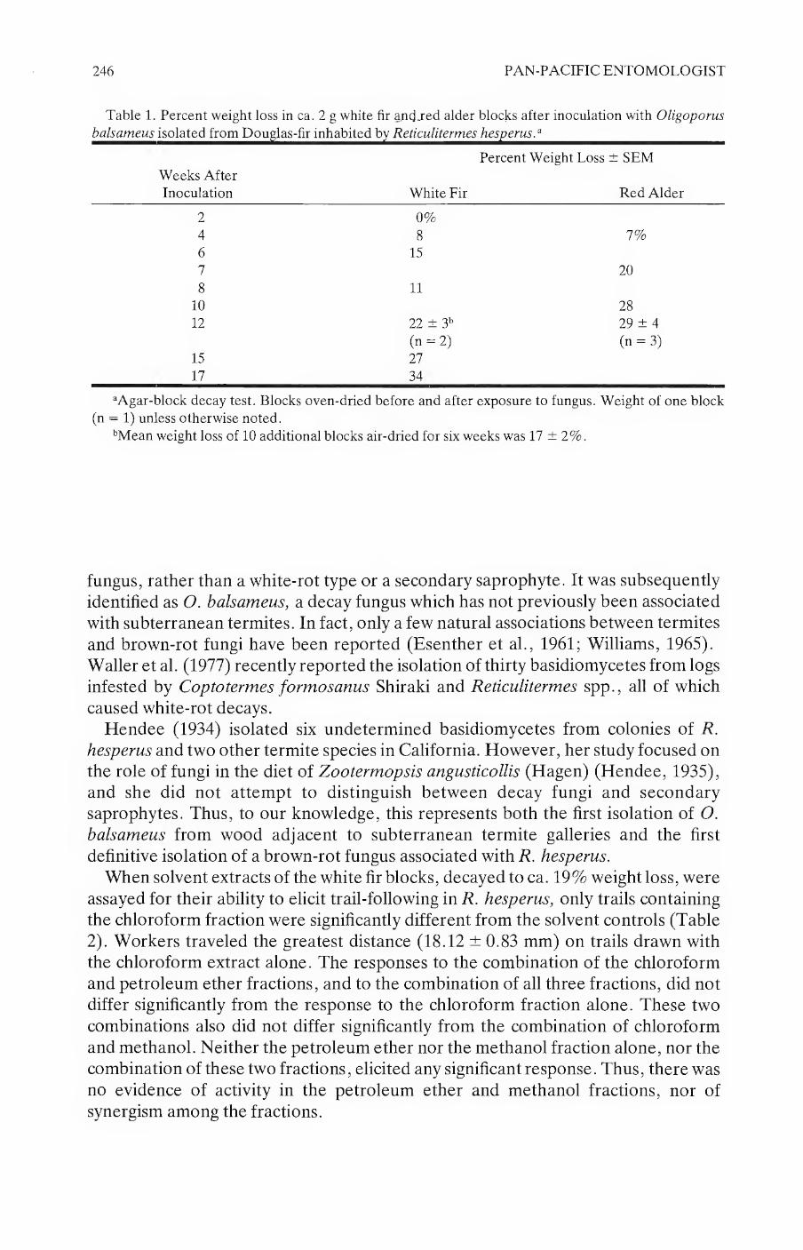
246 PAN-PACIFIC ENTOMOLOGIST
Table 1. Percent weight loss in ca. 2 g white fir and jed alder blocks after inoculation with Oligoporus
balsameu^solat£^ron^ou^as-hiu^iabitedb^^eticulitemie^ies^em^^^^^^^^^^^^^^^^^^^
Weeks After
Inoculation White Fir
Percent Weight Loss ± SEM
Red Alder
2 0%
4 8 7%
6 15
7 20
8 11
10 28
12 22 ± 3b 29 ± 4
(n = 2) (n = 3)
15 27
17 34
aAgar-block decay test. Blocks oven-dried before and after exposure to fungus. Weight of one block
(n = 1) unless otherwise noted.
bMean weight loss of 10 additional blocks air-dried for six weeks was 17 ± 2%.
fungus, rather than a white-rot type or a secondary saprophyte. It was subsequently identified as O. balsameus, a decay fungus which has not previously been associated with subterranean termites. In fact, only a few natural associations between termites
and brown-rot fungi have been reported (Esenther et al., 1961; Williams, 1965). Waller et al. (1977) recently reported the isolation of thirty basidiomycetes from logs infested by Coptotermes formosanus Shiraki and Reticulitermes spp., all of which caused white-rot decays.
Hendee (1934) isolated six undetermined basidiomycetes from colonies of R. hesperus and two other termite species in California. However, her study focused on the role of fungi in the diet of Zootermopsis angusticollis (Hagen) (Hendee, 1935), and she did not attempt to distinguish between decay fungi and secondary saprophytes. Thus, to our knowledge, this represents both the first isolation of O. balsameus from wood adjacent to subterranean termite galleries and the first definitive isolation of a brown-rot fungus associated with R. hesperus.
When solvent extracts of the white fir blocks, decayed to ca. 19% weight loss, were assayed for their ability to elicit trail-following in R. hesperus, only trails containing the chloroform fraction were significantly different from the solvent controls (Table
2). Workers traveled the greatest distance (18.12 ± 0.83 mm) on trails drawn with the chloroform extract alone. The responses to the combination of the chloroform and petroleum ether fractions, and to the combination of all three fractions, did not differ significantly from the response to the chloroform fraction alone. These two
combinations also did not differ significantly from the combination of chloroform and methanol. Neither the petroleum ether nor the methanol fraction alone, nor the combination of these two fractions, elicited any significant response. Thus, there was
no evidence of activity in the petroleum ether and methanol fractions, nor of synergism among the fractions.

VOLUME 64, NUMBER 3 247
Table 2. Mean distance traveled in 30 seconds by Reticulitermes hesperus workers on artificial trails
drawnwithsolventfractionsofO/^o^ora^flfeamen^deca^edwhite^firblocks^^^^^^^^^^^^^^
Solvent Fraction(s) on Trail1
Solvent Petroleum
Control Ether CHC13 ch3oh Mean Distance ± SEMI
X 3.28 0.28 c
X 18.12 ± 0.83 a
X 5.76 ■± 0.42 be
X X X 14.92 ± 0.47 ab
X X 10.24 0.53 abc
X X 7.12 0.40 be
X X 2.48 0.20 c
X 1.84 0.17c
blocks were extracted by sequential soaking in Pet. ether, CHC13, and CH3OH.
2Mean of 25 assays with individual R. hesperus workers.
3Means followed by different letters are significantly different (ANOV, REGW Multiple F Test,
a = 0.05).
Although statistically significant, the trail-following response elicited by the chloroform fraction was still substantially less than that reported by Grace et al. (1988) with extracts of R. hesperus sternal glands. On the basis of the weight of material extracted, the activity of the decayed white fir would be ca. 1/120,000 that of
extracted insect sternites. This low level of trail-following activity may have either a qualitative or
quantitative basis. Extraction of decayed sawdust, rather than intact 2 g blocks, may yield more of the active material. Fungal culturing on Douglas-fir rather than the white fir test substrate employed as a standard in this study, may also enhance activity. Production of fungal compounds is known to vary with the wood substrate (Esenther and Beal, 1979) and with the specific fungal isolate (Amburgey and Smythe, 1977).
Most studies of associations between fungi and subterranean termites (Rhinotermitidae) have investigated the effects of known fungi from laboratory
cultures or previously identified fungal metabolites, rather than using fungi isolated from field-collected wood to examine natural associations. However, it was field
observations of R. flavipes foraging behavior and the subsequent isolation of eight fungi (five basidiomycetes) by Esenther et al. (1961) that led to the identification of
the potent trail-following compound (Z,Z,E) 3, 6, 8-dodecatrien-l-ol. From both a basic and applied standpoint, examinations of natural fungal associations with termite species may prove more fruitful than screening known compounds or laboratory fungal cultures.
Acknowledgments
We are grateful to M. Blackwell (Department of Botany, Louisiana State
University, Baton Rouge) and R. L. Gilbertson (Department of Plant Pathology,
University of Arizona, Tucson) for the fungus species determination. We also thank N. D. Oldham (Forest Products Laboratory, UCB) andT. Matsumoto (Department

248 PAN-PACIFIC ENTOMOLOGIST
of Entomological Sciences, UCB) for theirexpert technical assistance. D. L. Wood, I. Kubo, and G. W. Frankie provided laboratory facilities, valuable advice, and comments on early drafts of the manuscript. This research was partially supported by the California Agricultural Experiment Station, the Division of Entomology and Parasitology (UCB), and National Science Foundation Grant BNS-8518195 (D. L.
Wood and I. Kubo, principal investigators).
Literature Cited
Allen, T. C., R. V. Smythe, and H. C. Coppel. 1964. Response of twenty-one termite species to aqueous
extracts of wood invaded by the fungus Lenzites trabea Pers. ex Fr. J. Econ. Ento.mol.
57:1009-1011.
Amburgey, T. L. 1979. Review and checklist of the literature on interactions between wood-inhabiting
fungi and subterranean termites: 1960-1978. Sociobiology 4:279-296.
-, andR. V. Smythe. 1977. Factors influencing termite feeding on brown-rotted wood. Sociobiology
3:3-12.
Becker, G. 1971. Physiological influences on wood-destroying insects of wood compounds and substances
produced by microorganisms. Wood Sci. Technol. 5:236-246.
-. 1976. Termites and fungi. Mater. Organismen Beiheft 3:465-478.
Carter, F. L., L. A. Dinus, and R. V. Smythe. 1972. Effect of wood decayed by Lenzites trabea on the
fatty acid composition of the eastern subterranean termite, Reticulitermes flavipes. J. Ins. Physiol.
18:1387-1393.
-, and R. V. Smythe. 1973. Effect of sound and Lenzites-decayed wood on the amino acid
composition of Reticulitermes flavipes. J. Ins. Physiol. 19:1623-1629.
Esenther, G. R., andR. H. Beal. 1979. Termite control: decayed wood bait. Sociobiology 4:215-222.
-, T. C. Allen, J. E. Casida and R. D. Shenefelt. 1961. Termite attractant from fungus-infected
wood. Science 134 (3471):50.
Gilbertson, R. L., and L. Ryvarden. 1985. Some new combinations in the Polyporaceae. Mycotaxon
22:363-365.
Grace, J. K. 1986. A simple humidity chamber for maintaining subterranean termites (Isoptera:
Rhinotermitidae) in the laboratory. Pan-Pac. Entomol. 62:221-223.
-, D. L. Wood, and G. W. Frankie. 1988. Trail-following behavior of Reticulitermes hesperus Banks
(Isoptera: Rhinotermitidae). J. Chem. Ecol. 14:653-667.
Hendee, E. C. 1934. The association of termites and fungi, pp. 105-116, in C. A. Kofoid (ed.), Termites
and Termite Control. 2nd ed. Univ. Calif. Press, Berkeley.
-. 1935. The role of fungi in the diet of the common damp-wood termite, Zootermopsis angusticollis.
Hilgardia 9:499-525.
La Fage, J. P., and W. L. Nutting. 1977. Nutrient dynamics of termites, pp. 165-232, in M. V. Brian
(ed.), Production Ecology of Ants and Termites. Inter. Biol. Prog. 13. Cambridge Univ. Press.,
Cambridge.
Matsumura, F., H. C. Coppel and A. Tai. 1968. Isolation and identification of termite trail-following
pheromone. Nature 219:963-964.
-, A. Tai and H. C. Coppel. 1969. Termite trail-following substance, isolation and purification from
Reticulitermes virginicus and fungus infected wood. J. Econ. Entomol. 62:599-603.
-, D. M. Jewett and H. C. Coppel. 1972. Interspecific response of termites to synthetic
trail-following substances. J. Econ. Entomol. 65(2):600-602.
Ritter, F. J., and C. M. A. Coenen-Saraber. 1969. Food attractants and a pheromone as trail-following
substance for the saintonge termite: multiplicity of the trail-following substances in L. trabea
infected wood. Entomol. Exp. & Appl. 12:611-622.
Sands, W. A. 1969. The association of termites and fungi, pp. 495-524, in K. Krishna and F. M. Weesner
(eds.), Biology of Termites, Vol. 1. Academic Press, New York.
SAS Institute. 1982. SAS User’s Guide: Statistics. SAS Institute Inc., Cary, North Carolina.
Smythe, R. V., F. L. Carter, and C. C. Baxter. 1971. Influence of wood decay on feeding and survival of
the eastern subterranean termite, Reticulitermes flavipes (Isoptera: Rhinotermitidae). Ann.
Entomol. Soc. Am. 64:59-62.

VOLUME 64, NUMBER 3 249
-, H. C. Coppel, and T. C. Allen. 1967a. The responses of Reticuliterm.es spp. and Zootermopsis
angusticollis (Isoptera) to extracts from woods decayed by various fungi. Ann. Entomol. Soc. Am.
60:8-9.
-, H. C. Coppel, S. H. Lipton, and F. M. Strong. 1967b. Chemical studies of attractant associated
with Reticulitermes flavipes and R. virginicus. J. Econ. Entomol. 60:228-233.
Waller, D. A., J. P. La Fage, R. L. Gilbertson, and M. Blackwell. 1987. Wood-decay fungi associated
with subterranean termites (Rhinotermitidae) in Louisiana. Proc. Entomol. Soc. Wash.
89:417-424.
Watanabe, T., and J. E. Casida. 1963. Response of Reticulitermes flavipes to fractions from fungus
infected wood and synthetic chemicals. J. Econ. Entomol. 56:300-307.
Williams, R. M. C. 1965. Infestation of Pinus caribaea by the termite Coptotermes niger Snyder. Proc.
12th Int. Congr. Entomol. [1964], London, pp. 675-676.

PAN-PACIFIC ENTOMOLOGIST 64(3), 1988, pp. 250-254
Observations on Megaphragma mymaripenne Timberlake (Hymenoptera: Trichogrammatidae), an egg parasite of Heliothrips
haemorrhoidalis Bouche (Thysanoptera: Thripidae)
Nawal A. Hessein and J. A. McMurtry
Department of Entomology, University of California, Riverside, California 92521.
Abstract.—Preliminary biological studies were conducted on the trichogrammatid, Megaphragma mymaripenne Timberlake, a native egg parasite of the greenhouse thrips in California. The searching and oviposition behavior of this parasite are described. Although only uniparental reproduction was observed in the laboratory, one male was collected from the field, suggesting the occurrence of deuterotoky. The time required for development from egg to adult averaged 41.4 days. Thrips parasitization was estimated by taking monthly leaf samples from an avocado orchard in Santa Barbara County and one in Orange County, and recording numbers of thrips “egg blisters” showing emergence of thrips, of the parasite, or no emergence. Egg blisters showing evidence of parasitization ranged from 22-41 % and 3-51% in the Santa Barbara and Orange County orchards, respectively. In both
locations, highest parasitization occurred in the months of November and January.
The greenhouse thrips, Heliothrips haemorrhoidalis Bouche, is one of the major pests of avocado in California (Boyce and Mabry 1937, Ebeling 1959). Feeding by thrips on the fruit causes a brown scarring on the surface that results in reduced market value.
Few natural enemies have been reported for this pest (Boyce and Mabry 1937, Ebeling 1959, McMurtry 1961, McMurtry and Johnson 1963, Lewis 1973, Mound and Walker 1982, Ananthakrishnan 1984). The trichogrammatid, Megaphragma mymaripenne Timberlake, was first reported as a parasite of greenhouse thrips in California by Boyce and Mabry (1937). McMurtry and Johnson (1963) and Ebeling (1959) noted that the parasite sometimes attacks a high percentage of greenhouse thrips eggs, but it was not known whether it had a controlling effect. Taxonomic
reports on M. mymaripenne include those of Timberlake (1923), Ghesquiere (1939), Doutt and Viggiani (1968), and Rao (1969). Doutt and Viggiani (1968) stated that M.
mymaripenne is probably the smallest of all insect species. No information was available concerning its biology, effect as a controlling agent, or on how to culture it in the laboratory.
To better understand this parasite and its impact on thrips populations, preliminary biological studies were conducted in the laboratory, and field
observations on percentage parasitization of H. haemorrhoidalis were made in two different localities.
250

VOLUME 64, NUMBER 3 251
Materials and Methods
Cultures were maintained and biological studies were conducted in the laboratory at 22-23°C and 10-42% RH. In order to culture M. mymaripenne, avocado leaves
were placed in stainless steel pans, upper side up, on plastic foam pads soaked with distilled water. The leaves were bordered with strips of cellucotton® to reduce leaf dehydration and prevent the escape of thrips or parasites. Twenty field-collected adults of the greenhouse thrips were placed on each leaf. Three days later, five adult parasites that emerged from egg blisters on field-collected leaves were placed on the infested leaves.
A similar technique was used for biological studies of the parasite except that only one adult parasite was placed on each of 15 leaves. Egg blisters of the greenhouse thrips that were stung by the parasite were marked and dated. Behavior of the adults and developmental time was observed and recorded. Some adult parasites were occasionally mounted in Hoyer’s media to determine their sex (presence or absence of an ovipositor).
Seasonal activity of the greenhouse thrips and the parasite were monitored in an unsprayed avocado orchard at Santa Barbara and a similar orchard on the University of California’s South Coast Field Station, Irvine. The Santa Barbara orchard was surveyed from June, 1984, until May, 1985; the South Coast Field Station orchard
from June, 1985 to May, 1986. Both orchards contained various varieties and hybrids previously used in breeding studies. A sample of 100 leaves was collected randomly from infested avocado trees from both orchards monthly. Ten infested leaves were taken from each sample to determine the percentage of emergence of both thrips and
parasite. These leaves were placed dorsal side up on foam pads soaked in distilled water. An area of 2 cm2 containing egg blisters of the thrips was marked on each leaf. Egg blisters were marked with different colors according to whether they showed no exit holes, thrips exit holes (evidenced by part of the egg chorion at the side of the blister), or parasite exit holes (large, round holes, usually in the middle of the blisters). Percentage of thrips and parasite emergence was calculated when leaves were brought from the field. Any subsequent emergence was determined by weekly examination of the egg blisters, previously showing no emergence of thrips or parasites.
Results and Discussion
Observations on the biology of M. mymaripenne: After emergence from the thrips egg blister, the adult parasites took about 45-60
minutes to clean their bodies before starting to search for hosts and oviposit. No mating was observed. All 20 adults that were mounted from the culture were
females. Even though uniparental reproduction was observed in the laboratory, one male
was collected from the field, indicating that M. mymaripenne occasionally exhibits deuterotoky. Like some other trichogrammatids, uniparental reproduction normally occurs but an occasional male is produced (Clausen 1956). Flanders (1945) stated that uniparental bisexuality probably is due to environmental factors such as type of
habitat, season, temperature or nutrition. He reported that females of many hymenopterous species, influenced by environmental conditions, produce two kinds

252 PAN-PACIFIC ENTOMOLOGIST
of eggs; one yielding only uniparental females and another either uniparental or, if
fertilized, biparental females. The thrips fecal material seemed to be one of the main factors for illiciting probing
or oviposition responses by the female. Dried fecal material sometimes induced a brief probing or oviposition response. However, egg blisters without fecal material
were sometimes stung. Before ovipositing, the female walked around the egg blister searching with her
antennae for the softer exposed parts where stinging usually occurred. Eggs that were completely covered with fecal material were also stung, but a longer time was required. Of the 14 individuals observed, oviposition time ranged from 0.5-7.5 min., average, 3.2 min. No parasites emerged when oviposition time was less than 2.5 min. One adult female oviposited in eight egg blisters in 30 min. Another one took about two hours to oviposit in the same number of egg blisters, with intervals of drinking water and cleaning its body. During oviposition, the adult female puts its weight
mainly on the hind legs and the wings, curving its abdomen forward and then inserting the ovipositor into the side or sometimes in the middle of the egg blister. The same egg blister was occasionally stung again by the same or a different parasite, suggesting the occurrence of superparasitism. After ovipositing in several egg blisters, the adult female cleaned her body and then walked or flew to another part of
the leaf, where she resumed searching. The developmental time (egg to adult) of 18 M. mymaripenne ranged from 36-46
days; average 41.4 days. Under similar conditions, the greenhouse thrips has a shorter developmental period, with a range of 24—36 days for 80 individuals, averaging 31 days (Hessein and McMurtry, unpubl.). Adult parasites lived approximately 48 hr. Adult thrips longevity ranged from 20-58 days, averaging 40.6 days for 24 individuals. The long time required for the parasite to complete its development might limit its ability to suppress increasing populations of thrips.
Parasitization by M. mymaripenne at two field sites: In Santa Barbara (Table 1), the percentage emergence of thrips larvae from egg
blisters varied from 20.2-42.2%; average 32.1%. The highest percentages occurred in November, 1984, and January, 1985. The percentage emergence of the parasite varied from 21.7-41.1%; average 33.4%. As with the greenhouse thrips, the highest percentages occurred in November, 1984, and January, 1985. There was always a
substantial percentage (average 34.5%) of egg blisters from which neither thrips nor parasites emerged. Because the age of the egg blisters or the time of emergence cannot be determined on field-collected leaves, emergence in the laboratory may give a better indication of present activity. Parasite and thrips emergence was comparable except for August, December, February, and May, when the percentage of thrips emergence was distinctly higher than that of the parasite.
At the South Coast Field Station, the thrips emergence was generally higher and parasite emergence lower, compared to Santa Barbara. The percentage emergence of thrips larvae from egg blisters varied from 28.9-63.5%; average 41.7%. The percentage emergence of parasites varied from 3.2-51.0%; average 21.4%. The highest percentages occurred in November, 1984, and January, 1986. The average
percentage of egg blisters from which neither thrips or parasites emerged (35.3%) was similar to that at Santa Barbara. Percentage emergence in the laboratory was

VOLUME 64, NUMBER 3 253
Table 1. Percentage emergence of the greenhouse thrips, Heliothrips haemorrhoidalis Bouche, and its
egg parasite, Megaphragma mymaripenne Timberlake, from infested avocado leaves collected at Santa
Barbara, 1984-1985.
% Thrips Emergence % Parasite Emergence
Month Field Laboratory Total Field Laboratory Total emergence
June 1984 16.5 8.0 24.5 28.3 9.1 37.4 38.1
July 26.4 2.6 28.9 32.3 2.9 35.2 35.9
August 27.3 6.7 34.0 24.7 2.5 27.3 38.8
September 30.4 7.2 37.6 29.8 5.6 35.4 27.0
October 18.9 1.3 20.2 37.2 1.6 38.8 41.0
November 36.3 4.1 40.4 33.7 7.4 41.1 18.5
December 25.2 12.1 37.2 23.4 6.0 29.4 33.3
January 1985 28.2 14.1 42.2 27.4 11.9 39.3 18.5
February 27.3 8.4 35.6 28.1 2.1 30.2 34.1
March 24.9 2.5 27.4 30.5 3.2 33.7 39.0
April 22.9 1.9 24.8 28.1 2.9 31.0 44.2
May 28.1 4.6 32.7 20.8 0.9 21.7 45.6
Mean 26.0 6.1 32.1 28.7 4.7 33.4 34.5
Table 2. Percentage emergence of the greenhouse thrips, Heliothrips haemorrhoidalis Bouche, and its
egg parasite, Megaphragma mymaripenne Timberlake, from infested avocado leaves collected at the
SouthCoasMhekU>tation^Dnmge^ount^^)8^1986^^^^^^^^^^^^^^^^^^^^^^^^^^^^
% Thrips Emergence % Parasite Emergence % xt0
Month Field Laboratory Total Field Laboratory Total emergence
June 1985 50.8 7.4 58.2 12.7 0.5 13.2 29.1
July 25.0 7.4 32.4 13.0 3.1 16.1 51.5
August 35.6 6.1 61.7 2.9 0.3 3.2 35.1
September 54.4 4.5 58.8 2.6 0.8 3.4 37.7
October 39.6 2.8 42.4 9.0 4.4 13.4 44.2
November 18.3 10.6 28.9 43.3 7.7 51.0 20.2
December 31.9 3.5 35.4 28.1 8.7 36.8 27.8
January 1986 30.0 2.6 32.5 37.3 9.7 46.7 20.5
February 40.6 2.9 43.5 15.3 2.6 17.9 38.5
March 30.9 3.5 34.3 20.7 2.5 23.3 42.4
April 25.0 4.2 29.2 10.6 2.2 12.8 58.1
May 48.9 14.6 63.5 11.4 6.7 18.1 18.4
Mean 35.9 5.8 41.7 17.2 4.1 21.4 35.3
usually higher for thrips than for parasites but the reverse was true in October, December and January.
Both of these orchards had heavy infestations of greenhouse thrips in the areas sampled but the populations were consistently higher at the South Coast Field Station orchard. Although parasitization by M. mymaripenne was higher in the Santa Barbara orchard, our observations suggest that percent parasitization did not increase proportionally with thrips population density to effect a decline in thrips
populations. Therefore, it appears doubtful that this parasite, by itself, is a regulating
factor of thrips populations in California.

254 PAN-PACIFIC ENTOMOLOGIST
Acknowledgments
We thank H. G. Johnson for obtaining leaf samples and Robert Velten for collecting and identifying the male Megaphragma.
Literature Cited
Ananthakrishman, T. N. 1984. Bioecology of Thrips. Indira Publ. House. 233 pp.
Boyce, A. M. and J. Mabry. 1937. The greenhouse thrips on oranges. Calif. Citrogr. 33(1): 19—20, 28-29.
Clausen, C. P. 1962. Entomophagous Insects. McGraw-Hill Co., Inc. 688 pp.
Doutt, R. L. and G. Viggiani. 1968. The classsification of the Trichogrammatidae (Hymenoptera:
Chalcidoidea). Proc. Calif. Acad. Sci., 4th Series, 35(20):477-586.
Ebeling, W. 1959. Subtropical Fruit Pests. Univ. Calif. Div. Agr. Sci. 436 pp.
Ebeling, W. and R. J. Pence. 1953b. Avocado Pests. Calif. Agr. Expt. Stn. Cir. 428:1-35.
Flanders, S. E. 1945. The bisexuality of uniparental Hymenoptera, a function of the environment. Am.
Nat. 79:122-141.
Ghesquiere, J. 1939. Contributions a l’etude des Hymenopteres du Congo Beige. VI. Description d’un
Mymaride nouveau et remarques sur le Gn. Megaphragma Timb. (Trichogrammidae). Rev. Zool.
Bot. Afr. 33(1):33—41.
Lewis, T. 1973. Thrips, Their Biology, Ecology and Economic Importance. Acad. Press, London, New
York. Harcourt Brace Jovanovich, Publ. 349 pp.
McMurtry, J. A. 1961. Current research on biological control of avocado insect and mite pests. Yearbk.
Calif. Avocado Soc. 45:104-106.
McMurtry, J. A. and H. G. Johnson. 1963. Progress report on the introductions of a thrips parasite from
the West Indies. Yearbk. Calif. Avocado Soc. 47:48-51.
Mound, L. A., and A. K. Walker. 1982. Terebrania (Insects: thysanoptera) fauna of New Zealand.
1:1-88.
Rao, B. R. S. 1969. A new species of Megaphragma (Hymenoptera: Trichogrammatidae) from India.
Proc. Roy. Entomol. Soc. London (B) 38(7—8): 114—116.
Timberlake, P. H. 1923. Descriptions of new chalcid flies from Hawaii and Mexico. Hawaiian Entomol.
Soc. 5:395-417.

PAN-PACIFIC ENTOMOLOGIST 64(3), 1988, pp. 255-257
Geographic Variation in Cooperative Colony Foundation in Veromessor pergandei
(Hymenoptera: Formicidae)
Randall T. Ryti*
Department of Biology, C-016, University of California San Diego, La Jolla, CA 92093.
^present address: Department of Biology, Montana State University, Bozeman, MT 59717.
Abstract.—Veromessor pergandei queens are strictly haplometrotic at a site in California, while queens at sites in Arizona will found nests cooperatively. Possible explanations for this geographic difference include: higher success of brood raiding in Arizona, higher predation rates on young colonies in Arizona, and greater relatedness of colonies in Arizona.
Introduction
The desert seed-harvester ant, Veromessor pergandei (Mayr), founds colonies cooperatively in Arizona (Pollock and Rissing 1985); 68% of starting nests (n = 132)
contained more than one queen. Starting colonies with multiple queens successfully brood raid and defeat singly founded colonies (Rissing and Pollock 1987). If proximity of starting nests affects the frequency of brood raiding, then a clumped distribution of young colonies would increase the frequency of brood raiding between young colonies. Thus spacing of starting nests could affect the success of single queen nests. Here I describe patterns of V. pergandei queen behavior and clumping at sites in the California and Arizona deserts. Differences between these sites may provide insight into factors regulating cooperative colony foundation by V. pergandei queens.
Methods
The Arizona site was along the Tonopah Salome Highway 60 km west of Phoenix. Nests were censused in February 1987 along a 5 km roadside strip. Although the adjoining area was native Sonoran Desert habitat with Larrea tridentata, Carnegia gigantea, and Prosopis velutina as the dominant perennials, new nests were concentrated along the berm of a recently graded road. The California studies were conducted in February 1986 at the Boyd Deep Canyon Reserve, a part of the University of California Natural Reserve System. The dominant perennial plants
were Larrea tridentata, Hyptis emoryi, and Cercidiumfioridum. The study site was a relatively homogeneous 3 ha area of alluvial fan habitat. At both sites, the starting colonies were visually located and censused by excavation. A typical V. pergandei
starting nest is a semi-circular pile of soil or sand approximately 5 cm by 2 cm. These
255

256 PAN-PACIFIC ENTOMOLOGIST
diggings are most obvious a few days after a rainstorm, since the excavated soil will be darker than the background soil. Since V. pergandei is claustral, the oldest
diggings will start to blend in with the background soil with each successive storm.
Results and Discussion
V. pergandei at Deep Canyon is strictly haplometrotic, colonies are founded by a single queen (N = 181), while 28 of the 98 Arizona nests had multiple foundresses
(x2 = 43.6 , d.f. = 1, p < 0.001). Although the frequency of multiple queen starting nests in my Arizona site is not as high as the 90 of 132 nests with more than one queen reported by Pollock and Rissing (1985, Figure 2), it indicates that my excavation methods can detect the presence of multiple queens in starting nests. Queen tolerance for additional foundresses also varies at the two sites. Deep Canyon queens will fight when placed in the same vial, while I have never observed this behavior in queens collected from the Arizona site.
V. pergandei nests at Deep Canyon are initially clumped, then after the first workers emerge, the remaining nests are randomly spaced (Ryti and Case 1988). It is possible that some of these young colonies could have been destroyed through brood raiding (Rissing and Pollock 1987), or other factors, including: predation by long established conspecifics, predation by other ant species, or predation by spiders or
rodents. Note that the spatial arrangement changing from clumped to random does
not necessarily imply that there was selective attrition to clumps of starting colonies. Equal survivorship of the originally clumped starting nests would also produce
randomly dispersed young colonies (Ryti and Case 1988). Newly founded nests are
also clumped in Arizona (n = 42, p < 0.001, Rissing pers. comm.). There are three potential selective mechanisms that may account for the
geographical difference in the evolution of colony foundation. 1) Brood raiding affects young colony survivorship in Arizona and not in California. The frequency of brood raiding could be related to the spatial dispersion of nests when the first workers emerge. If Arizona colonies are clumped when workers emerge, then brood raiding could be a significant factor. The existing spacing data does not support this explanation, since both Deep Canyon and Arizona nests are initially clumped. However, colonies may be clumped in Arizona because of microhabitat selection. Pollock and Rissing (1985) noted that most queens found nests in wash bottoms. Arizona clumps may persist because of “better” physical conditions for brood rearing. Such microhabitat differences are not obvious between clump and non-clump areas at the Deep Canyon site. 2) Arizona single-queen nests are more vulnerable to predation. Colony predation rates may be higher in Arizona, and
young colonies with more workers may survive predation episodes. 3) Cooperative colony foundation only occurs with queens that are closely related. Deep Canyon queens could be aggressive because outbreeding is relatively more common in California populations than in Arizona populations.
As a first step towards distinguishing between these possibilities, the spacing of foundress colonies in Arizona needs to be examined, especially after workers first
emerge. These data, together with observations on the importance of predation, by ants and other species, and data on relatedness among colonies at these sites could explain the geographic differences in V. pergandei cooperative colony foundation.

VOLUME 64, NUMBER 3 257
Acknowledgements
I thank the staff of the Boyd Deep Canyon Research Center for logistical support during this project. G. Wheeler and J. Wheeler kindly verified that both the Deep Canyon and Arizona queens were ordinary V. pergandei. T. Case and R. Radtkey helped census founding nests. I thank S. Rissing and K. O’Neill for their suggestions on this manuscript.
References
Pollock, G. B. and S. W. Rissing. 1985. Mating season and colony foundation of the seed-harvester ant
Veromessorpergandei. Psyche 92:125-134.
Rissing, S. W. and G. B. Pollock. 1987. Queen aggression, pleometrotic advantage and brood raiding in
the ant Veromessor pergandei (Hymenoptera: Formicidae). Animal Behaviour 35:975-981.
Ryti, R. T. and T. J. Case. 1988. The regeneration niche of desert ants: effects of established colonies.
Oecologia 75:303-306.
PAN-PACIFIC ENTOMOLOGIST 64(3), 1988, pp. 258-260
Predation on Larvae of Doeglas°ir Tussock Mofli9 Orgyia pseudotsugata (Lepidoptera: Lymantriidae)9 by Metaphidippus
aeneolus (Araneae: Salticidae)
R. R. Mason and H. G. Paul
Forestry and Range Sciences Laboratory, Pacific Northwest Research Station, U.S. Department of Agriculture, Forest Service, La Grande, Oregon 97850
Spiders are well-known predators of a variety of important forest insects (Allen et al. 1970; Bosworthetal. 1971; Jennings andPase 1975; Loughton et ah 1963; Warren et ah 1967). Although spiders have occasionally been seen preying on small larvae of the Oouglas-fir tussock moth, Orgyia pseudotsugata (McDunnough) (Wickman 1977), most field evidence of their predation on tussock moth has been either circumstantial (Dahlsten et al. 1977; Mason and Torgersen 1983; Mason et al. 1983) or from serological analyses (Fichter and Stephen 1984).1 One of the most common arboreal spiders in fir forests of the Pacific Northwest is the salticid Metaphidippus aeneolus (Curtis) (Moldenke et al. 1987). These are small (<5 mm in length), gray and black spiders that are free-living and hunt for prey amongst the foliage. They are polyphagous predators and expert at stalking and pouncing on their prey. In extensive samplings of foliage of Douglas-fir, Pseudotsuga menziesii var. glauca (Beissn.) Franco, and the true firs, Abies spp., we found this species was ubiquitous in the mixed-conifer forests of the Pacific Northwest. We also observed that mature Metaphidippus spiders readily preyed on instars I and II of the Douglas-fir tussock moth when both species were confined together in a petri dish (Fig. 1). To examine further the predatory abilities of this group, we conducted an additional test under field conditions.
In late June 1981, shortly after natural egg-hatch of tussock moth, we selected nine white fir, Abies concolor (Gord. and Glend.) Lindl. ex Hildebr., in a mixed-conifer forest near Fort Klamath, Oregon. White fir is the principal host of the Douglas-fir tussock moth in that area. On each tree, we vigorously shook two 50-cm branches to remove all arthropods and then enclosed each branch separately in a nylon mesh cage slipped over the end of the branch. Five laboratory-reared tussock moth larvae (instars I—II) were stocked in each cage. We also added an adult Metaphidippus aeneolus in one of the two cages on each tree. All spiders were field-collected from the same stand. The cages were then closed at the base of the branch with a wire tie and left undisturbed.
’Fichter, B.L., W. P. Stephen, A. R. Moldenke, and D. L. Dahlsten. 1982. Arboreal arthropod
predation on Douglas-fir tussock moth larvae (Orgyia pseudotsugata) (Lepidoptera: Lymantriidae)
as detected by ELISA. USDA For. Serv. Coop. Aid Agreement. Oreg. State Univ., Corvallis. Final
Rep. 42 p.
258

VOLUME 64, NUMBER 3 259
1 mm h-1
Figure 1. Metaphidippus aeneolus feeding on larva (instar I) of the Douglas-fir tussock moth.
After 3 weeks, we examined the contents of each cage over a drop-cloth. The
results were:
Number of Number of
larvae stocked larvae surviving Percent Treatment in nine cages after three weeks mortality
Larvae + spider 45 6 86.7 Larvae only 45 40 11.1
Surviving larvae developed normally and had grown to instars III-IV when reexamined. Of five larvae lost in the control cages without spiders, three were missing and two died of unknown causes. No larvae survived in six of the nine cages with Metaphidippus spiders; cadavers of preyed-on larvae were recovered in most of these cages. Four of the six larvae that survived with Metaphidippus were in a cage in which the spider had spun a silken retreat and laid a cluster of eggs. Egg laying and subsequent guarding of eggs may have reduced the rate of her predation (Krafft
1982).

260 PAN-PACIFIC ENTOMOLOGIST
These results clearly show predatiorr on the Douglas-fir tussock moth by Metaphidippus aeneolus. The degree of feeding on tussock moth may have been exaggerated, however, because the spiders were confined and other potential or favored prey excluded. Mortality of larvae in the natural population, as determined in other studies, averaged 77 percent for several years during the same period (Mason and Torgersen, 1987). Because of its abundance in the foliage,
Metaphidippus aeneolus could have been responsible for much of this loss. We and
others have suspected for some time that spider predation is a leading cause of mortality of small larvae in low-density populations of tussock moth (Mason and Torgersen 1983; Mason et al. 1983).2 These results provide further support for that
hypothesis. We thank R. C. Beckwith, D. T. Jennings, W. Maddison, A. R. Moldenke, and
T. R. Torgersen for their constructive reviews of an earlier draft.
Literature Cited
Allen, D. C., F. B. Knight, and J. L. Foltz. 1970. Invertebrate predators of the jack-pine budworm,
Choristoneurapinus, in Michigan. Ann. Entomol. Soc. Am. 63:59-64.
Bosworth, A. B., H. G. Raney, E. D. Sturgeon, R. D. Morrison, andR. D. Eikenbary. 1971. Population
trends and location of spiders in loblolly pines, with notes of predation on the Rhyacionia complex
(Lepidoptera: Olethreutidae). Ann. Entomol. Soc. Am. 64:864-870.
Dahlsten, D. L., R. F. Luck, E. I. Schlinger, J. M. Wenz, and W. A. Copper. 1977. Parasitoids and
predators of the Douglas-fir tussock moth, Orgyiapseudotsugata (Lepidoptera: Lymantriidae), in
low to moderate populations in central California. Can. Entomol. 109:727-746.
Fichter, B.L., and W.P. Stephen. 1984. Time-related decay of prey antigens ingested by arboreal spiders
as detected by ELISA. Environ. Entomol. 13:1583-1587.
Jennings, D. T., and H. A. Pase III. 1975. Spiders preying on Ips bark beetles. The Southwestern Nat.
20:225-229.
Krafft, B. 1982. The significance and complexity of communication in spiders, p. 33 In Witt, P. N. and
J. S. Rovner (Eds.), Spider communication, mechanisms and ecological significance. Princeton
University Press, Princeton, NJ.
Loughton,B. G., C. Perry, and A. S. West. 1963. Mortality of larvae in stocked cohorts of the Douglas-fir
tussockmoth, Orgyia pseudotsugata (Lepidoptera: Lymantriidae). Can. Entomol. 115:1119-1127.
Mason, R. R., and T. R. Torgersen. 1983. Mortality of larvae in stocked cohorts of the Douglas-fir
tussock moth, Orgyia pseudotsugata (Lepidoptera: Lymantriidae). Can. Entomol. 115:
1119-1127.
Mason, R. R., and T. R. Torgersen. 1987. Dynamics of a nonoutbreak population of the Douglas-fir
tussock moth, Orgyia pseudotsugata (McD.) (Lepidoptera: Lymantriidae) in southern Oregon.
Environ. Entomol. 16:1217-1227.
Mason, R. R., T. R. Torgersen, B. E. Wickman, and H. G. Paul. 1983. Natural regulation of a
Douglas-fir tussock moth (Lepidoptera: Lymantriidae) population in the Sierra Nevada. Environ.
Entomol. 12:587-594.
Moldenke, A. R., B. L. Fichter, and W. P. Stephen. 1987. A key to the arboreal spiders of Douglas-fir
and true fir forests of the Pacific Northwest. USDA For. Serv. Gen. Tech. Rep. PNW-GTR-207,
Pac. Northwest Research Stn., Portland, OR. 48 p.
Warren, L. O., W. B. Peck, and M. Tadie. 1967. Spiders associated with the fall webworm, Hyphantria
cunea (Lepidoptera: Arctiidae). J. Kan. Entomol. Soc. 40:382-395.
Wickman, B. E. 1977. Observations on spider predation of early instar larvae of Douglas-fir tussock
moth, Orgyia pseudotsugata (McDunnough) (Lepidoptera: Lymantriidae). Pan-Pac. Entomol.
53(1):46.
2See footnote 1.

PAN-PACIFIC ENTOMOLOGIST 64(3), 1988, pp. 261-265
The Morphology of the Tarsal Sensilla in the Female Mite Varroa jacobsoni
T. P. Liu
Bee Diseases Research Laboratory, Agriculture Canada Research Station, Box
29, Beaverlodge, Alberta, Canada, TOH 0C0
Abstract.—The group of hairs at the distal end and dorsal surface of the tarsus,
consisted of varying size and shaped sensillae. In the center of this hair group was a pit containing a minute dome-like protrusion centrally located at the bottom. On the rim of this pit were small, fine, and differently shaped pegs measuring 3-4 um in height. The pit was surrounded by six sensillae, two of which were slender, curved, round tipped and porous, three of the remaining four were stout and one had a pointed tip. All pegs and six sensillae were stained with crystal violet, and pores were visible on the walls of the two slender and curved sensillae. Surrounding these small sensillae were 12-16 large hairs measuring 12-79 um in height. Two to three of the large hairs were slender, curved, and round tipped. They were also stained with crystal violet. Seven to eight stout, straight hairs, measuring 16-32 um in height were
stained with crystal violet only at the tip. Three to four of the longest hairs measured
68-79 um in height and were not stained with crystal violet.
Introduction
The mite Varroa jacobsoni is a serious pest of the honeybee (Ritter, 1981), and although chemical control agents have been developed in Europe, effective control is difficult. Therefore, new approaches such as the use of pheromones and attractants for managing this pest, should be explored. Feeding and reproduction of the mite takes place in the brood cells, and drone larvae are preferred to that of the workers. This preference suggests that there are chemical cues in the drone cells which attract the mite, and it must be assumed that the mite is equipped with sense organs of relatively high specialization. A study of the olfactory system in V. jacobsoni would be desirable, since little attention has been given to the morphology and possible function of the different hairs in the sensorial field and tarsal organ (Langhe et al., 1976). This study attempts to describe the variated cuticular morphology of the tarsal sensillae, in the sensorial field at the first pair of legs on the female mite, and determine their possible function.
Materials and Methods
The female mites were collected in Fireburg, West Germany, and kept in 8% EM grade gluteraldehyde (Polyscience, Warrington, U.S. A.). Specimens were prepared for scanning electron microscopy as described in a previous publication (Liu and Liu,
1984).
The crystal violet stain technique of Slifer and Brescia (1960) was employed for testing the permeability of the sensilla as described previously (Liu and Liu, 1984).
261

262 PAN-PACIFIC ENTOMOLOGIST
Gluteraldehyde fixed mites were washed with distilled water and again fixed in freshly prepared 5% gluteraldehyde in distilled water, after which they were then washed with distilled water alone. Washed specimens were twice immersed in 0.5% crystal violet in distilled water for 10 and 30 minutes respectively. Mites were removed from the stain, washed with distilled water, and blot-dried with filter paper. Dried mites were immersed in xylene for 60 minutes, and then mounted on glass slides with permount for microscopic examination. The sensilla height was measured from negatives or photographs by use of a video image analysis system equipped with a Kurta series 2 graphic tablet.
Results
There is a group of sensillae ventrally located at the distal end of the tarsus behind the pretarsus (Figs. 1 and 2). In the center of this sensillae group was a pit with a minute dome-like protrusion centrally situated at the bottom with pores visible on its surface (Figs. 3 and 4). The dome-like protrusion was surrounded by five fine smaller pegs 3-4 um in height, and they were situated on the rim of the pit (Figs. 2 and 5). One of these pegs was slender with a pointed tip, while the other was stout with a flat tip. The remaining pegs were stout with pointed tips (Figs. 2 and 5). All pegs stained brightly after 10 minutes in the crystal violet solution. There were six sensillae measuring 5.5-6.3 um in height, surrounding the pit (Figs. 2 and 5). Two of these sensillae were slender, curved, and round tipped (Fig. 5). Pores were visible on their surface (Fig. 6) and their diameter was from 0.08-0.12 um. Of the four remaining sensillae three were stout and one had a pointed tip (Figs. 2 and 5). All six sensillae were readily stained with crystal violet after 10 minutes in the crystal violet solution. There were 12-16 large hairs surrounding the small sensillae (Figs. 2 and 3), 2-3 of them appearing slender and curved, measuring 14-17 um in height. They
were stained with crystal violet throughout the whole length, but no pores were visible. Seven to eight stout straight hairs with round tips measured 16-32 um in height. These hairs were stained with crystal violet only at the tip., Three to four of the longest hairs measured 68-79 um in height, but were not stained with crystal violet.
Discussion
According to Langhe and Natzkii (1977) the tarsus on the first pair of legs of V. jacobsoni possess numerous grouped sensillae at the distal end and dorsal surface. This hair group is referred to as the sensorial field, and the tarsal organ is located at its
center (Langhe et al., 1976). The present study reveals that sensillae in the sensorial field consists of both thick, and thin walled sensillae, along with longer tactile hair. The dome-like protrusion in the center of the pit may be similar to the coeloconic sense organ of insects (Mclver, 1973). In the honeybee, coeloconic sensillae are sensitive to carbon dioxide, temperature, and humidity (Lacher, 1964). The five small pegs on the rim of the pit are thin walled sensillae which resemble the sensilla
basiconica of insects. This type of sensillae may have external morphological differences, but all possess a multiporous wall. Therefore, Slifer et al. (1959) classified them together. Sensilla basiconica has been observed responding to a variety of chemicals except sex pheromones (Kaissling, 1971; Priesner, 1968;
Schneider, 1965; Schneider and Steinbrecht, 1968; Schneider etal., 1964). Slifer etal. (1959) also considers this type of sensillae to be the major olfactory organ used for locating odorous food. The six small sensillae surrounding the pit were all stained

VOLUME 64, NUMBER 3 263
Figures 1-2. Figure 1. The sensorial field (arrow) is located at the distal end and dorsal surface of the
tarsus, behind the pretarsus (S) sucker. Bar = 20 pm. Figure 2. The centrally located pit displays five
small pegs located on the rim (arrows). Bar = 10 pm.
with crystal violet, which readily indicated that they are porous. Pores were also visible on the surface of the two curved sensillae which are different in shape and may resemble the sensilla trichodea (Steinbrecht, 1973; Albert and Seabrook, 1977;
Albert et al, 1974; Liu and Liu, 1984). This type of sensillae contains pheromone receptors (Steinbrecht, 1973; Albert and Seabrook, 1973; Albert et al., 1974). Two of the larger sensillae in the sensorial field are porous, and the external morphology is also similar to that of the sensillae trichodea. The large hairs which were stained with

264 PAN-PACIFIC ENTOMOLOGIST
Figures 3-6. Figure 3. The sensorial field consists of a hair group which contains sensillae of varying
sizes and shapes. In the center of the sensorial field is a pit, a minute dome-like protrusion (arrow) is
centrally located at the bottom of the pit, and five small pegs are located on the rim of the pit (shown in
Figure 4). There are six small sensillae surrounding the pit itself. Bar = 10 |xm. Figure 4. The dome-like
protrusion (asterisk) at the bottom of the pit is porous (arrow heads). Small pegs (P) on the rim of the pit.
Bar = 2 |im. Figure 5. On the rim of the pit are small pegs (arrows), while outside the rim are small
sensillae. Two of these sensillae (small arrows) are slender, curved, round tipped and porous (pores are
present on the surface of the two curved sensillae), while the others are stout and blunt tipped
(arrowhead). Bar = 5 fjim. Figure 6. Pores are present on the surface of the two curved sensillae shown in
Figure 5. Bar = 1 jxm.

VOLUME 64, NUMBER 3 265
crystal violet only at the blunt tips, indicated the presence of an apical pore. This type
of sensillae may resemble the thick walled sensilla chaetica, which functions as a contact receptor (Slifer, 1970). The three longest hairs were not stained with crystal
violet, hence indicating that they may be tactile hairs (Slifer, 1979). Langhe et al. (1976) suggested that the sensillae in the sensorial field, are thin walled chemoreceptors which may assist the mite in locating its phoretic host and suitable brood cells. The present study indicates that the sensorial field of V. jacobsoni consists of different sensillae types which may perform a variety of functions.
Acknowledgments
The author wishes to thank Dr. W. Ritter, Tierhygienisches Institut Freiburg, German Federal Republic for collecting the specimens and to G. E. Braybrook, Department of Entomology, University of Alberta for assistance in electron microscopy. To Greg Myrick for editing and Faye Swanson for typing the manuscript.
Literature Cited
Albert, P. J. andW. D. Seabrook. 1973. Morphology and histology of the male Eastern spruce budworm,
Choristoneura fumiferana (Clem.) (Lepidoptera:Tortricidae). Can. J. Zool., 4:433-448.
Albert, P. J., W. D. Seabrook and U. Paim. 1974. Isolation of a sex pheromone receptor in males of the
Eastern spruce budworm Choristoneura fumiferana (Chem.) (Lepidoptera:Tortricidae). J. Comp.
Physiol., 91:79-89.
Kaissling, K. E. 1971. Insect olfaction. In L. M. Deidler (ed.), Olfaction, Springer-Verlag, New York.
Lacher, V. 1964. Elektrophysiologische Untersuchungen an einzelnen Rezeptoren fur Geruch.
Kohlendioxyd, Luftfeuchtigkeit und Temperatur auf den antennen der Arbeitsbiene und der
Drohne {Apis mellifica L.). Z. Vergl. Physiol., 48:587-623.
Langhe, A. B. and K. V. Natzkii. 1977. The mite Varroa and the methods of controlling it. In E. V.
Harnaj (ed.), Varroasis a honeybee disease. Apimondia Publishing House, Bucharest.
Langhe, A. B., K. V. Natzkii and V. M. Tatzii. 1976. Klechteh Varroa {Varroa jacobsoni', Oudemans,
1904) i podkhody k razrabotke sredstv. Ptchelovodstro, 13:16 - 20.
Liu, H. J. and T. P. Liu. 1984. Sensilla on the antennal flagellum of the Bertha Armyworm Moth,
Mamestra configurata Walker (Lepidoptera:Noctuidae): A scanning electron microscope study.
Ann. Entomol. Soc. Am., 77:236-245.
Mclver, S. B. 1973. Fine structure of antennal sensilla coeloconica of culicine mosquitoes. Tissue and
Cell, 5:105-112.
Priesner, E. 1968. Die interspezifischen Wirkungen der Sexuallockstotte der Saturniid (Lepidoptera). Z.
Vgl. Physiol., 61:263-297.
Ritter, W. 1981. Varroa disease of the honeybee A/ns mellifera. Bee Wld., 62:141-153.
Schneider, D. 1965. Chemical sense communication in insects. Symp. Soc. Exp. Biol., 20:273-297.
Schneider, D. and R. A. Steinbrecht. 1968. Checklist of insect olfactory sensilla. Symp. Zool. Soc.
London, 23:279-297.
Schneider, D., V. Lacher and K. E. Kaissling. 1964. DieReaktionsweise und das Reaktionsspektrum von
Riechzellen bei antheraeapernyi (Lepidoptera, Saturniidae). Z. Vgl. Physiol., 48:632-662.
Slifer, E. H. 1970. The structure of arthropod chemoreceptors. Ann. Rev. Entomol., 15:121-142.
Slifer, E. H. 1979. Sense organs on the antennal flagella of four species of Chrysopa
(Neuroptera:Chrysopidae). Ann. Entomol. Soc. Am., 72:529-531.
Slifer, E. H. and V. T. Brescia. 1960. Permeable sense organ on the antenna of the yellow fever mosquito,
Aedes aegypti (Linnaeus). Entomol. News, 71:221-225.
Slifer, E. H., J. J. Prestage, and H. W. Beams. 1959. The chemoreceptors and other sense organs on the
antennal flagellum of the grasshopper (Orthopera:Acrididae). J. Morphol., 101:359-397.
Steinbrecht, R. A. 1973. Der Feinbau Olfaktorischer Sensillen des Seidenspinners (Insecta,
Lepidoptera). Rezeptortsutze und Reizleitender Apparat. Z. Zellforsch., 130:533-565.

PAN-PACIFIC ENTOMOLOGIST 64(3), 1988, pp. 266-269
The Xylophilous Bees and Wasps of a High, Cold Desert: Leslie Gulch, Oregon (Hymenoptera: Apoidea, Vespoidea)
D. R. Frohlich, W. H. Clark, F. D. Parker, T. L. Griswold
(D.R.F.) Department of Biology, Utah State University, Logan, Utah 84322-5305; (W.H.C.) Orma J. Smith Museum of Natural History, College of Idaho, Caldwell, Idaho 83605; (F.D.P. and T.L.G.) USD A, Agricultural Research Service, Bee Biology and Systematics Laboratory, Utah State University, Logan, Utah 84322-5310
Abstract.—The wood-loving bees of a high desert area in eastern Oregon were surveyed with trap nests. Eight species of bees and wasps, previously unknown from this part of the country, were recorded.
Introduction
The Owyhee region of Malheur County, Oregon, is a geologically unique area that contains a relatively large number of endemic plants. Within the region several state parks have been established to promote recreational use and to preserve portions of the Owyhee upland desert. Leslie Gulch has been designated by the Bureau of Land Management as an Area of Critical Environmental Concern (ACEC). The road passing through Leslie Gulch divides two wilderness study areas, thus the area has wilderness status until a final decision is made by the BLM. In 1983, we (DRF and WHC), began a study of the pollination biology of a rare and endangered rose, Ivesia rhypara (Ertter and Reveal) within Leslie Gulch. Since the gulch is known to contain at least eight rare plant species (Grimes 1984), we conducted a trap nest survey in order to estimate the composition of the species pool of available pollinators. This paper describes the results of that survey and reports range extensions for eight species of bees and wasps.
Site Description
Leslie Gulch is a drainage of approximately 90 km that empties into Owyhee Reservoir, Malheur County, Oregon and has a complicated stratigraphy of volcanic rocks and sediments. The gulch’s talus slopes are composed of sparse quartz and sanidine phenoclasts in a vitroclastic matrix and are named the Leslie Gulch Ash-Flow Tuff Member of the Sucker Creek Formation (Grimes 1984). Major components of the sparse vegetation in the canyon are: Poa sandbergii, Agropyron spicatum, Eriogonum strictum, Physaria chambersii, Astragalus sterilis, Linum perenne, Penstemon acuminatus, Eriophyllum lanatum, Juniperus osteosperma, and Purshia tridentata (Ertter and Reveal 1977). The three trapnest localities were: Leslie Gulch mouth at Owyhee Reservoir, T26S R44E S3 (elev. 825 m); Leslie Gulch at Juniper Gulch, T26S R44E S13 (elev. 1050 m); Leslie Gulch State Park at Runaway Gulch, T26S R45E S9 (elev. 1430 m).
266

VOLUME 64, NUMBER 3 267
Methods
Trap nests were made from 46.0 cm long Sambucus (elderberry) stems into which had been drilled a hole approximately 0.32 cm in diameter by 10.2 cm in length. The
undrilled end was sharpened to facilitate placement in the ground (Parker and Bohart 1966). Sticks at each site were placed every 3 M in a straight line at each site
and inspected periodically over the summer to ensure that none had been knocked over. Nests were placed at the following sites within Leslie Gulch: 99 sticks on 7 June 1984 at Owyhee Reservoir, 52 sticks on 23 April 1983 and 102 sticks on 7 June 1984 at Juniper Gulch, 51 sticks on 24 April 1983 and 99 sticks on 7 June 1984 at Runaway Gulch. After collection in the fall (12 November 1983 and 30 September 1984), nests were stored outdoors until they were dissected the following winter. The contents were placed in gelatin capsules (size 00) and incubated at 35°C until all specimens had emerged. Pinned insects were then identified and voucher specimens deposited at the USDA Bee Biology and Systematics Laboratory, Utah State University, Logan, Utah, and Orma J. Smith Museum of Natural History, College of Idaho, Caldwell,
Idaho.
Results and Discussion
Of 403 stems, 392 were recovered and dissected. Table 1 details the species and numbers that were collected at each site. Overall, such a composition is fairly typical for high deserts in Western North America but eight of these species have not before
been reported from this area. Ceratochrysis enhuycki Cooper has previously only been known from the east (NY
to FL) and southwest (TX, UT, AZ). Its host at Leslie Gulch is unknown but is probably one of the species of Leptochilus (Krombein 1959). Both Leptochilus washo Parker and L. trachysomus (Bohart) are known from the southwest (Krombein 1979, Parker 1966) but neither has been recorded this far north. Ancistrocerus simulator Cameron was previously reported only from Nevada and California (Krombein 1979), as was Pisonopsis clypeata Fox with the addition of a Wyoming locality (Evans 1969). The use of burrows in plant stems by wasps is unusual but is not entirely unknown (Evans 1969).
Little is known about Hylaeus polifolii (Cockerell), but to date it has been reported only from California (Hurd 1979). Two megachilids exhibit the greatest range extensions. Previously, Ashmeadiella meliloti (Cockerell) was thought to be confined to the deserts of the southwestern U.S. and northern Mexico. Stelis lateralis Cresson has not been reported on this side of the continental divide but has been collected from Maine and Ontario to North Dakota and south to Georgia and Texas (Hurd 1979). The host of S. lateralis at Leslie Gulch is not known.
The difference between the 3 sites in species composition and numbers are
probably not significant and likely reflect sample size. Altitude varies only about 600 meters between the 3 sites and vegetation is similar. The Owyhee Reservoir site may have yielded fewer species and numbers because of a relatively greater abundance of grasses and paucity of vegetation. The species reported herein are only those that will build nests in wooden stems and are part of a much larger bee and wasp fauna. The
entire Owyhee Uplift is home to a large number of rare and endemic plant species. In
order to understand the reproductive ecology of those populations, it is necessary to account for both pollinators and other insect associates. This study has been part of
that effort.

268 PAN-PACIFIC ENTOMOLOGIST
Table 1. Number of individuals and species trapped at all sites. 1983 and 1984 are combined.
TAX A STUDY SITE
Owyhee
Runaway Gulch Juniper Gulch Reservoir
LEUCOSPIDAE
Leucospis affinis Say 11
CHRYSIDIDAE
Hedychridium solierellae 2
Bohart and Brumley
Chrysis derivata Buysson 1 1
Chrysissp. #1 3 1
Chrysis sp. #2 3
Chrysissp. #3 6
Chrysura sp. 5
Ceratochrysis enhuycki Cooper 5 5 3
MUTILLIDAE
Sphaeropthalma s. 1. 1
EUMENIDAE
Leptochilus periallis Parker 4 1
L. trachysomus (Bohart) 1 9
L. washo Parker 7 26
Stenodynerus blandoides Bohart 4 1
Parancistrocerus acarigaster Bohart 6 5 3
Ancistrocerus simulator Cresson 17 2
PEMPHREDONIDAE
Pemphredon lethifer (Shuckard) 2
LARRIDAE
Solierella peckhami (Ashmead) 1
Pisonopsis clypeata Fox 3
Trypoxylon sculleni Sandhouse 1
COLLETIDAE
Hylaeus polifolii (Cockerell) 3
MEGACHILIDAE
Stelis lateralis Cresson 11 12 12
Hoplitis albifrons (Kirby) 3 3 3
H. hypocrita (Cockerell) 66 19 23
H. grinnelli Cockerell 11 7
H. sambuci Titus 1 1
H. uvualis (Cockerell) 10
Anthophora copelandica (Cockerell) 6
Ashmeadiella melilotti (Cockerell) 25 1 110
Osmia bruneri Cockerell 2
O. kincaidii Cockerell 16 119
0. cyanella Cockerell 6
O. sp. nov. 86 14
Megachile montivaga Cresson 9 44
Coelioxys banksi Crawford 17 27
ANTHOPHORIDAE
Ceratina pacifica Smith 3 3
Total Species 28 25 8

VOLUME 64, NUMBER 3 269
Acknowledgments
M. Clark, C. Clark, and E. Clark assisted with field work. The United States Department of Interior, BLM, Vale District gave permission to conduct the research. We had valuable discussions concerning local flora with B. Ertter, J. Grimes, E. Joyal, and P. Packard. This research was supported by the Utah Agricultural Experiment Station, Utah State University, Logan, Utah. Journal paper number 3488.
Literature Cited
Ertter, B. J. and J. Reveal. 1977. A new species of Ivesia (Rosaceae) from southeastern Oregon.
Madrono, 24:224-227.
Evans, H. E. 1969. Notes on the nesting behavior of Pisonopsis clypeata and Belomicrus forbesii. J.
Kansas Entomol. Soc., 42:117-125.
Grimes, J. W. 1984. Notes on the flora of Leslie Gulch, Malheur County, Oregon. Madrono, 31:80-85.
Hurd, P. D. Jr. 1979. Superfamily Apoidea, pp. 1741-2209. In K. V. Krombein, P. D. Hurd Jr., D. R.
Smith andB. D. Burks [eds.], Catalog of Hymenoptera in America North of Mexico. Washington,
D.C.: Smithsonian Institution Press.
Krombein, K. V. 1959. Biological notes on Chrysis (Ceratochrysis) enhuycki Cooper, and its host,
Leptochilus republicanus zendaloides (Robertson). Entomol. News, 70:17-23.
- 1979. Vespoidea, pp. 1469-1522. In: K. V. Krombein, P. D. Hurd Jr., D. R. Smith and B. D.
Burks [eds.]. Catalog of Hymenoptera in America North of Mexico. Washington, D.C.:
Smithsonian Institution Press.
Parker, F. D. 1966. A revision of the North American species in the genus Leptochilus. Misc. Publ.
Entomol. Soc. Amer., 5:151-229.
Parker, F. D. and R. M. Bohart. 1966. Host-parasite associations of some twig-nesting Hymenoptera
from Western North America. Pan-Pac. Entomol., 42:91-98.

PAN-PACIFIC ENTOMOLOGIST 64(3), 1988, pp. 270-274
A New Species of Neurotoma From Michigan (Hymenoptera: Symphyta, Pamphiliidae)
Woodrow W. Middlekauff
University of California, Berkeley 94720
Abstract.—A new species of sawfly is described in the small pamphiliid genus Neurotoma Konow. Only 3 male specimens are available, all from the same collection in Michigan. Males of two closely related species, N. crataegi and N. willi are figured and compared. A key to North American species is included.
The sawfly genus Neurotoma Konow is Holarctic in distribution with the presently described species adding [one] to the four previously described in the Nearctic fauna (Middlekauff, 1940,1958,1966; Smith, 1979). The remaining twelve in the genus are found from Europe to Japan and Korea. With but one exception, a Japanese species found on Quercus (Shinohara, 1980), all others, where the host(s) are known, feed on the foliage of rosaceous trees and shrubs of the genera Amelanchier, Cotoneaster,
Crataegus, Prunus, Pyrus and/or Sorbus. This article includes the description of the new species, a key to Nearctic species of
Neurotoma and comparative notes on the males of two closely related species.
Key to Nearctic species of Neurotoma
The new species nigra will run to willi in Middlekauffs 1958 key, thus necessitating a new one.
1. Tegula black (Fig. 13) .crataegi Middlekauff — Tegula completely or mostly pale (Figs. 3, 26) .2 2. Femora and tibiae entirely pale .3 — Femora and basally & tibiae apically marked with black .4 3. Wings hyaline, perhaps a very faint infuscated band below stigma; legs beyond
coxae mostly pale reddish brown .inconspicua (Norton) — Basal two-thirds of wings heavily infuscated; legs beyond coxae pale yellow ...
...fasciata (Norton) 4. Pronotum (Figs. 3), mesepisternum (Fig. 6), prescutum (Fig. 3), and lateral
margins of abdominal tergites (Fig. 5) with white areas; cell 1M as in Fig. 7; vertex wider than long (Fig. 1); paraantennal field finely rugose (Fig. 2) .
.willi Middlekauff — Pronotum (Fig. 26), mesepisternum (Fig. 24), prescutum (Fig. 26), and lateral
margins of abdominal tergites (Fig. 27) entirely black; cell 1M as in Fig. 19;
vertex longer than wide (Fig. 20); paraantennal field smooth, shining (Fig. 21)
.nigra n.sp.
270

VOLUME 64, NUMBER 3 271
Figures 1-10. Neurotoma willi Middlekauff, male. Figure 1, head, dorsal view. Figure 2, Inset, paraantennal field. Figure 3, Thorax, dorsal view. Figure 4, Hind femur and tibia, lateral view. Figure 5, Abdominal segments III-V, dorsal view. Figure 6, Mesepisternum. Figure 7, Cell 1M, forewing. Figure 8, Penis valve. Figure 9, Antennal segments 1-5. Figure 10, Right half of genital capsule, ventral view.
Figures 11-14. Neurotoma crataegi Middlekauff, male. Figure 11, Head, dorsal view. Figure 12, Inset,
paraantennal field. Figure 13, Thorax, dorsal view. Figure 14, mesepisternum.

272 PAN-PACIFIC ENTOMOLOGIST
Neurotoma nigra, new species
(Figs. 20-28)
Male, holotype. Head black with whitish yellow markings only on frons and in the
supra ocular areas as in Fig. 20. Antenna entirely black, the scape and pedicel darker. Mouthparts dark brown (alaglossa and paraglossa), with the apical 3 segments of the maxilla and 2 of the labium, whitish yellow. Mandible whitish yellow at base, darker brown to black in middle and reddish brown apically.
Thorax except for the whitish yellow tegula, entirely black (Fig. 26). Coxa black basally, whitish yellow apically. Trochanter whitish yellow. Fore and mid femur basally, black. Hind femur with a black spot basally as in Fig. 28, remainder of femur whitish yellow. Tibia whitish yellow becoming dark brown to black over a lesser area from the fore to hind leg as in Fig. 28. Tarsus brownish the posterior ones somewhat darker. Wings hyaline. Venation, except for the whitish yellow base of 1A, dark brown, the stigma black. Abdomen entirely black (Fig. 27). Harpes pale yellowish brown.
Clypeus rounded in front. Frons swollen, forming an elongate rounded ridge extending onto the swollen clypeus. Median fovea absent. Ocellar basin distinct, crescent shaped in front. Lateral transverse suture becoming faint and disappearing
behind lateral ocellus (Fig. 20). Lateral suture diverging so that a line extending it forward would miss the lateral ocellus by two ocellar widths (Fig. 20). Post ocellar area longer than that of willi (Figs. 20,1). Post genal carina distinct extending around vertex to within 2 ocellar widths of lateral suture. Entire head and face, except for shining impunctate paraantennal fields (Fig. 21), with distinct punctures becoming almost coriaceous. The postocular and vertex areas also punctate, but the spaces between these punctures polished. Antenna 19-segmented; A (segment) IILAIV 2.3:1; AIII:AIV + V 1.1.16 (Fig. 25).
Prescutum slightly rugose. Mesoscutum impuctate except for a narrow mid band of shallow punctures, otherwise shining, pebbled under higher magnification. Mesoscutellum depressed, flattened, punctate, antero-laterally carinate but not posteriorly where it rounds off towards the post-tergite. Mesepisternum with numerous shallow punctures each with a long, single hair giving it a distinctly hairy appearance.
Genital capsule (lateral half) as in Fig. 22. Penis valve as in Fig. 23. Measurements of holotype (in mm): body length 7.9; forewing 7; head width 2.25;
antennal scape length 0.56; pedicel 0.25; segment III 0.94; segment IV, 0.37; segment V, 0.37.
Female. —U nkno wn. Host(s). —Unknown. Distribution.—Michigan.
Type.—Holotype, male: Roscommon County, Michigan, 5-31-57 (R. & K. Dreisbach). Paratypes: Two males with same data as holotype. In Michigan State
University, East Lansing and Essig Museum, University of California, Berkeley. The holotype will be deposited in the collection, U.S. National Museum, Washington, D.C.
Discussion.—This species is quite distinct from the related N. willi (Middlekauff, 1958) and N. crataegi (Middlekauff, 1940). Except for the frons and a small supraocular spot the head is entirely black as are also the thorax and abdomen;

VOLUME 64, NUMBER 3 273
Figures 15-19. Neurotoma crataegi (con’t.). Figure 15, Abdominal segments III-V, dorsal view. Figure 16, Left half of genital capsule, ventral view. Figure 17, Penis valve. Figure 18, Hind femur and tibia. Figure 19, Cell 1M, forewing. Figures 20-28. Neurotoma nigra, n.sp., male. Figure 20, Head, dorsal view. Figure 21, Inset, paraantennal field. Figure 22, Right half of genital capsule, ventral view. Figure 23, Penis valve. Figure 24, Mesepisternum. Figure 25, Antennal segments 1-5. Figure 26, Thorax, dorsal
view. Figure 27, Abdominal segments III-V, dorsal view. Figure 28, Hind femur and tibia.

274 PAN-PACIFIC ENTOMOLOGIST
antennal segment III is longer than segments IV and V combined; the lateral sutures on the vertex are divergent, not parallel; the postocular area is longer; there are more numerous and larger punctures on the head; the 1st medial cell (discoidal)is 2X longer than the basal width, willi is only 1.5X longer.
The name nigra is given because of the predominant black coloration of the head
thorax and abdomen.
Neurotoma willi Middlekauff (Figs. 1-10)
Comparative comments: N. willi is similar to nigra in some respects but differs in
the points given in the key to species and shown in Figs. 1-10. In addition the post ocular area is finely grained (or pebbled) and lacks pits; some specimens have one or two small white spots laterally on the mesonotum; a few specimens have the tegula completely black; the basal two-thirds of vein 1A is black; base of mandible black; the lateral white spot on each abdominal tergite extends ventrally on the posterior third of the spiracle bearing lateral tergites; sternites III-VII, each has a white area
on the mid-apical border; harpes black, never yellowish brown; frons and clypeus impunctate; the mesoscutellum is distinctly carinate except for a small medial posterior area.
Neurotoma crataegi Middlekauff (Figs. 11-19)
Comparative comments: N. crataegi is also similar to nigra in some respects, but is apparently not quite as closely related as is willi. Many of the differences are given in the key to species and in Figs. 11-19. In addition to these the clypeus and frons are rugose, lacking distinct pits; the post ocular area and vertex are also rugose with a few scattered shallow pits; rarely is the pronotum completely black; occasional specimens have small white spots on the prescutum while others may have the mesoscutellum completely black; all wing veins are completely brownish black and
the wings are entirely lightly infuscated; base of mandible is black; the lateral white spot on each abdominal tergite extends from the spiracle as a narrow band, only expanding slightly on posterior border; harpes black; the mesoscutellum is distinctly carinate laterally.
Acknowledgments
I am indebted to Dr. David R. Smith for sending me the above specimens and to Christina Jordan for the illustrations.
Literature Cited
Middlekauff, W. W. 1940. The Nearctic sawflies of the genus Neurotoma (Hymenoptera: Pamphiliidae). Canad. Ent., 72:201-206.
- 1958. The North American sawflies of the genera Acantholyda, Cephalcia and Neurotoma
(Hymenoptera: Pamphiliidae). Univ. Calif. Pub. Ent., 14:51-168. - 1966. Description of the previously unknown female of Neurotoma willi (Hymenoptera:
Pamphiliidae). J. N. Y. Ent. Soc., 73(4):238-239. Shinohara, A. 1980. East Asian species of the genus Neurotoma (Hymenoptera: Pamphiliidae). Trans.
Shikoku Ent. Soc., 15(1-2):87-117. Smith, D. R. 1979. Suborder Symphyta. In Krombein et al., eds, Catalog of Hymenoptera in America
north of Mexico, 1:14.

PAN-PACIFIC ENTOMOLOGIST 64(3), 1988, pp. 275-279
Further Notes on Halictine Bees (Hymenoptera: Apoidea) Visiting Isomeris arborea in Southern California
E. Gorton Linsley
Department of Entomological Sciences, University of California, Berkeley, CA 94720
In a brief earlier report (Linsley, 1986) records of three species of small halictine bees visiting Isomeris arboreus at Desert Hot Springs, California, on December 25,
1985, were provided. In early February 1987, further samples from the same site revealed some interesting comparisons with the earlier collections.
Isomeris arboreus is a member of the Caparidaceae (caper family) and is most commonly known as “bladder-pod” because of the elongate, inflated seed pods. It is particularly abundant along the western edge of the California deserts but occurs elsewhere including southern California sea cliffs. In the desert it is said to bloom whenever it rains. The plant is a densely branched shrub, growing mostly four to five feet tall when mature. When in bloom the numerous flowers with their four yellow petals make the plant conspicuous from a distance. The four stamens and the pistil are equal in length (see photograph). The thread-like staminal filaments cannot support honeybees, which visit the flowers primarily for nectar, but easily sustain the small halictines. These bees land directly on the anthers and frequently crawl from anther to anther by reaching out with the forelegs. Nectar is obtained from the base of the flower. The significance of this plant in the present study is that, as a perennial, it is one of the first plants to bloom in the desert and thus able to provide forage for
these early emerging polylectic bees. Moldenke and Neff (1974) list seven species of small halictines taking nectar from
Isomeris. In addition to those referred to below, they recorded Lasioglossum sisymbrii (Cockerell) (3 ?), L. trizonatum (3 9), and Evylaeus argemonis (Cockerell) (2$), and Dialictus microlepoides (Ellis) (3$), each from Riverside County, California; and Dialictus hyalinus (Crawford) from Los Angeles County. They also list a female of Augochlorella pomoniella (Cockerell) taking nectar at this plant in San Diego County. Moure and Hurd (1977) include these records in their tabulations, adding only one more halictine, Agapostemon mexicanum from Baja California and Sonora.
Between February 2 and 9,1987, eight samples of bees were taken from plants at the exact site that was utilized in the December 1985 collections. This area is in an old wash bordered on the west side by a high embankment, and is subject to strong winds which may temporarily remove the bees from the plants or complicate their flight patterns. However, the bees persist in collecting pollen, even in the face of winds which, when gusting, made it difficult for me to control the net. The site was bordered by pavement laid out for future development and the plants were
concentrated near the edge of the paved areas, but were also scattered on the land
nearby. The surface soil was composed of coarse sand and gravel with a few large
275

276 PAN-PACIFIC ENTOMOLOGIST
boulders—an unpromising area for ground nesting bees. In fact a casual search revealed no sign of nesting nearby.
The small sample (12 specimens) taken in December contained six females of Dialictus microlepoides (Ellis) and one female of Evylaeus amicus (Cockerell) which were gathering pollen and five females of Evyaeus pulveris (Cockerell)1 which were taking only nectar. However, species abundance in the early February samples was
reversed. The 248 females of E. pulveris collected were all gathering pollen, but the two females each of Dialictus microlepoides and Evylaeus amicus were taking only nectar, as were one each of Dialictus albohirtus (Crawford) and D. hyalinus (Crawford) (all kindly identified by George C. Eickwort). Earlier, Moldenke and Neff (1974) had recorded 18 females of Evylaeus ruficornis (Crawford) (det. Timberlake) taking nectar from Isomeris in Riverside County, California, but did not give the dates of capture1. Our 1987 collections and associated data (temperatures averaged for each period) were as follows:
Evylaeus pulveris Feb. 2, 23 $ P (1230-1300, broken sky, variable wind, temp. 24°C) Feb. 3,11 $P (1830-0900, overcast, windy, temp. 20°C) Feb. 5,19 $ P (0830-0900, overcast, variable wind, temp. 19°C) Feb. 6, 95 9 P (0930-1030, clear, no wind, temp. 27°C) Feb. 7,19 $P (0630-0700, overcast, no wind, temp. 18°C) Feb. 8, 27 $ P (1015-1030, overcast, no wind, temp. 20°C) Feb. 8, 8 $ P (1500-1515, broken sky, no wind, temp. 27°C)
Feb. 9, 26 $P (1015-1030, broken sky, no wind, few rain drops, temp. 20°C) Evylaeus amicus
Feb. 2,1 $N (as above)
Feb. 8,1 $N (as above) Dialictus albohirtus
Feb. 6,1 $N (as above) Dialictus hyalinus
Feb. 2,1 $N (as above)
Dialictus microlepoides Feb. 2,19 N (as above) Feb. 7,1 $N (as above)
Bees of the genus Evylaeus are eusocial although they differ in levels of sociality (Packer and Knerer, 1985; Eickwort, 1986). In those species which have been studied, fertilized females overwinter to initiate the populations of the following year (usually two generations). Thus the data recorded here and previously are based upon early emergence of overwintering females and the absence of males is to be expected.
Most previous records for the species listed above are to be found in Moldenke and Neff (1974). They list 214 females of Evylaeus amicus from 33 flowering plants
Donald McGinley (in litt.) has indicated that there are unresolved problems with the identity of this species. Both he and George Eickwort suggest that the Moldenke and Neff (1974) records under the name E. ruficornis also apply here. However, for the sake of consistency and future reference we have continued to use the name “pulveris” until the matter is cleared up.

VOLUME 64, NUMBER 3 277
Upper: wind-blown plants of Isomeris at Desert Hot Springs in foreground; Mt. San Gorgonio in
background (Photographs by J. M. Linsley). Lower: Female Evylaeus pulveris taking pollen from Isomeris flowers (Photograph by Kim Hoelmer).

278 PAN-PACIFIC ENTOMOLOGIST
(including Isomeris arborea) but taking-pollen only from Adenostoma, Ceanothus and Clarkia, and record the flight period as from February 21 to May 11. No males were reported. The name “Evylaeus pulveris” does not appear in their listings, nor does that of Dialictus albohirtus. For Dialictus hyalinus, they include an extensive list of flowers visited for nectar by males and/or females during a flight period extending
from February 18 to November 13. Although they do not give dates with the individual records, presumably the males were taken during the latter part of the flight period. The same is true for Dialictus microlepoides, although among the
nectar plants they include Isomeris arboreus. From our data at this site, there is no indication as to the number of generations
involved in the annual cycle of Evylaeus pulveris, but the females that are active in December must represent the overwintering generation newly emerged and not yet collecting pollen. They became extremely active by early February. A visit to the area in early June, 1987, revealed that the plants were no longer in bloom, and no sign of the halictines or honey bees was evident on any other flowers in the area. Unfortunately, the precise site of our collections was being bulldozed for housing
construction and will be unavailable for further study. (This calamity has overtaken
us in numerous desert localities in the Southwest!) With regard to these same species we have taken males previously in Arizona and
published records are also available for some: Evylaeus amicus have been collected by us in large numbers at flowers of
Cercidium microphyllum at Gila Bend, Arizona, on May 11,1978, where they were present throughout the day. The females, as expected, are polylectic and we have found them taking pollen from Larrea divaricata, Cercidium microphyllum, and Parkinsonia aculeata in Arizona and southern California deserts.
We have captured males of E. pulveris (identified by Timberlake) at flowers of
Cercidium microphyllum in the Arizona desert, once at Gila Bend and once near the Colorado River in Yuma County—both collections in mid-May.
Aldred (1969) reported the capture of 33 males and three females of Dialictus albohirtus at the Nevada Test Site. He lists a variety of herbaceous plants and shrubs from which collections were made in June (mostly) and July, but does not provide data as to sexes associated with individual plants. He states that the most common host was Asclepias erosa. This is a good nectar plant but, of course, not a source of pollen for bees.
In earlier collections by us, a male of Dialictus hyalinus was taken with a few pollen- and nectar-gathering females at flowers of Cercidium microphyllum at Gila
Bend, Arizona, on May 11, 1978. Four other males were captured at Tamarix aralensis at Keeler, Inyo County, California, on June 15. Our earliest capture of a male in Arizona was on May 12,1978. Pollen-gathering females were found at Larrea tridentata in Arizona and southern California in March and April.
Dialictus microlepoides is a widespread common polylectic species, especially in the southwestern deserts and northwestern Mexico. Aldred listed males and females from the Nevada Test Site in June (mostly) and July, most commonly at Viguieria multiflora. We have captured males with females taking pollen in Oro Valley, Pima
County, Arizona, on May 28, 1976, from Cercidium microphyllum, and large numbers of males (41) along with pollen- and nectar-seeking females at the same
host, on May 21,1978. At Palm Springs, California, only females taking pollen were present at flowers of Larrea tridentata on July 6,1975.

VOLUME 64, NUMBER 3 279
It is regretted that circumstances beyond our control prevent further attempts to
clarify the seasonal pattern of activity of these bees in the vicinity of Desert Hot Springs, but we provide these limited data in the hope that future studies may clarify their occurrence in the western Colorado Desert during the summer, fall and early winter.
Acknowledgments
Acknowledgment is due to George C. Eickwort, for patiently determining the specimens collected and for comments on the manuscript, to Ronald McGinley for calling attention to problems with the identification of Evylaeus pulveris, Howell V. Daly, who sampled honey bees at the site while we were there, for reading the manuscript. Thanks are also due to Kim Hoelmer for the photograph of this species taking pollen from the anthers of Isomeris. Finally, I wish to express my appreciation to my wife, Juanita M. Linsley, who accompanied me on each of my sampling trips and collected assiduously, adding materially to the field data and providing one of the
photographs of Isomeris plants accompanying this article.
Literature Cited
Aldred, Donald M. 1969. Bees of the Nevada Test Site. The Great Basin Naturalist, 29:20-24. Eickwort, G. C. 1986. First steps into eusociality: The sweat bee Dialictus lineatulus. Florida
Entomologist, 69:742-754,5 figs. Linsley, E. G. 1986. Early seasonal records for three halictine bees on Isomerus arboreus in southern
California (Hymenoptera: Apoidea). Pan-Pacific Ent., 62:310. Moldenke, A. R. and J. L. Neff. 1974. The bees of California: a catalogue with special relevance to
pollination and ecological research. Origin and Structure of Ecosystems, IRP-IBP Technical Report, 74(5), 189 pp. University of California, Santa Cruz.
Moure, J. S. and P. D. Hurd Jr. 1987. An annotated catalogue of the halictid bees of the Western Hemisphere (Hymenoptera: Halictidae). Smithsonian Institution Press, Washington, D.C. 405 pp.
Packer, L. and G. Knerer. 1985. Social evolution and its correlates in bees of the subgenus Evylaeus
(Hymenoptera: Halictidae). Behavioral Ecology and Sociobiology, 17:143-149.


PAN-PACIFIC ENTOMOLOGIST 64(3), 1988, pp. 281-284
The Apoidea Collection of the California Insect Survey (C.I.S.) at the Essig Museum of Entomology,
University of California, Berkeley
J. A. Santiago-Blay and H. V. Daly
Department of Entomological Sciences, University of California, Berkeley, CA 94720
History of the Apoidea collection
The insect collection of the University of California at Berkeley was established in 1940 as a project of the California Agricultural Experiment Station and officially designated as the “California Insect Survey”. Prior to 1940, A. T. McClay had managed the collection. At the urging of E. G. Linsley and R. L. Usinger, the insect collection was given formal recognition by E. O. Essig, who was Chair of the Department of Entomology during 1943-1952. P. D. Hurd Jr. supervised the collection from 1947-1962 and from 1963 to the present it has continued under the leadership of J. A. Powell. Since 1967 the collection has been housed in parts of three floors remodeled for this purpose in Wellman Hall on the main campus in Berkeley. The parasitic Hymenoptera are maintained by the Division of Biological Control at the Gill Tract in Albany, CA. In 1972 the collection and associated facilities were named the Essig Museum of Entomology. For further historical information see: Linsley, E. G. [1955. The Department of Entomology and Parasitology
(Berkeley-Davis). Entomol. News 66:133-138], Powell, J. A. [1972. The California Insect Survey. Div. Entomol. Agric. Exp. Sta. Univ. Calif., Berkeley 22 pp. (mimeo)] and Powell, J. A., et al. (1973. Entomological collections of the University of California. Bull. Entomol. Soc. Amer. 19:100-102).
The principal contributors to the growth of the bee collection were E. G. Linsley, and his students P. D. Hurd and J.M. MacSwain. National Science Foundation grants awarded to Linsley and MacSwain during the 1950’s promoted apidology, particularly pollination biology. As part of their doctoral dissertations, collections were deposited at the C.I.S. by J. S. Rozen (now at Amer. Mus. Nat. Hist.) and R. W. Thorp (now at Univ. Calif., Davis), both Linsley’s students, and G.I. Stage
(now at Stafford Springs, CT), Hurd’s student. These early contributions have been supplemented by the efforts of J. A. Powell, J. T. Doyen, J. A. Chemsak, H. V. Daly and others. In addition to P. H. Timberlake (Univ. Calif., Riverside), other researchers who have identified considerable portions of our bee collection are: C. D. Michener (Univ. Kansas, Lawrence) and R. R. Snelling (Los Angeles County Museum, CA) (Colletidae); G. Hackwell (Turlock, CA), W. LaBerge (Ill. Nat.
Hist. Surv. Insect Ident., Urbana), R. Thorp (Andrenidae); G. Eickwort (Cornell
Univ., Ithaca, NY), R. McGinley (USNM, Washington, DC), Michener and R. R.
Roberts (Rutgers Univ., New Brunswick, NJ) (Halictidae); A. A. Grigarick (Univ. Calif., Davis), G. E. Bohart, T. Griswold and F. Parker (all at the Bee Biol. Lab.,
281

282 PAN-PACIFIC ENTOMOLOGIST
Logan, UT), Michener and T. B. Mitchell (Univ. North Carolina, Raleigh) (Megachilidae); Griswold and Thorp (Apidae); Bohart, R. W. Brooks (Univ.
Kansas), LaBerge and Snelling (Anthophoridae) and others.
Holdings and their curatorial condition
The Apoidea collection houses about 314,000 bees in 521 drawers including 114 drawers (22% of the total) that contain material identified only to genus or family. Most specimens on loan have not been included in the count. The C.I.S. ranks among the three largest bee collections in North America. In terms of identified species/subspecies it probably has the most extensive holdings of bees from western North America.
Western North America is defined here as the area circumscribed by the states or provinces of: Alaska, Northwest Territories, British Columbia, Alberta, Washington, Oregon, Idaho, Montana, Wyoming, California, Nevada, Utah, Colorado, New Mexico, Arizona and Baja California. The Essig Museum of Entomology has specimens of 54% of the reported species/subspecies for western North American bees (47% Colletidae, 100% Oxaeidae, 53% Andrenidae, 58% Melittidae, 55% Halictidae, 71% Megachilidae, 42% Anthophoridae, 80% Apidae) and 47% of the reported species/subspecies for North America (55% Colletidae, 100% Oxaeidae, 47% Andrenidae, 44% Melittidae, 41% Halictidae, 66% Megachilidae, 34% Anthophoridae, 79% Apidae) [Krombein, et al. 1979. Catalog
of the Hymenoptera in America North of Mexico. Vol. 2. Apocrita (Aculeata).
Smithsonian Institution Press. Washington, D.C.]. C.I.S. holdings are abundant for the Nearctic region, especially southwest United
States. Mexico and Central America, particularly Costa Rica, are also well represented; there are lesser collections from other Neotropical areas. Collections received incidentally from the Palearctic, Ethiopian, Oriental and Australian regions, usually have been deposited at the California Academy of Sciences or at the University of California, Davis. Table 1 summarizes the estimated number of specimens, number of genera and species/subspecies, as well as the amount with type material, voucher specimens and nest materials per family. The 224 name-bearing types are on loan of indefinite duration to the California Academy of Sciences, San Francisco, CA.
Hurd and Linsley supervised major curatorial work, primarily undertaken by K. Sorensen during the 1960’s and 1970’s. Most of our holdings are at curatorial level 6 but not all geographical codes have been added (curatorial level 6 = properly curated, United States National Museum classification; McGinley, pers. comm.). Holdings, types, and loans are currently being placed in a computer information retrieval system to facilitate multiple access (level 7).
Loan Policy
Currently, loans are made for three years and are renewable following notification. Loan requests should be addressed to Dr. John A. Chemsak (same address as authors).

Table 1. Summary of the Apoidea holdings of the Essig Museum of Entomology, University of California, Berkeley
Species and subspecies
Estimated Number with type specimens Number with Number with Family number of Number of Total number vouchers3 nest materials
specimens genera Name-bearing1
type
Allotype1 Paratype2
Colletidae 6,000 13 118 6 5 11 0 0 Oxaeidae 1,000 4 13 2 3 4 0 0 Andrenidae4 70,000 17 786 165 87 284 7 9 Mellitidae 3,000 1 14 0 0 0 0 0 Halictidae 36,000 33 291 9 3 10 0 0 Megachilidae 73,000 29 519 5 2 23 2 2
Anthophoridae 85,000 51 678 37 28 67 21 12 Apidae 40,000 30 246 0 0 5 0 0
Totals 314,0005 177 2,665 224 128 404 30 23
1. Deposited on indefinite loan at the California Academy of Sciences, San Francisco, as of December, 1986.
2. In Essig Museum
3. Mostly of pollination biology studies
4. Also 21 species with stylopized representatives
5. Not included in our summary are about 20,000 bees in Schmitt boxes and most of the specimens on loan to specialists.
N> oo CO
VO
LU
ME
64
, NU
MB
ER
3

284 PAN-PACIFIC ENTOMOLOGIST
Acknowledgments
We wish to thank J. A. Chemsak for allowing JASB to undertake the inventory/curation of the bees while working as Research Assistant at the Essig Museum of Entomology. E. G. Linsley and J. A. Powell provided most of the historical information and reviewed the typescript.

PAN-PACIFIC ENTOMOLOGIST 64(3), 1988, p.285-289
A Population Estimate of Mdh Allozyme Frequencies for the Honey Bee, Apis mellifera L. (Hymenoptera: Apidae)
Robert E. Page, Jr.1 and Robert A. Metcalf2
Abstract
Gene frequencies of allozymes of malate dehydrogenase (Mdh-1) were presented for a population of honey bees located in Davis, California. Two different estimates
were made that represented population gene frequencies in 1977 and 1978.
Genotype frequencies did not differ significantly from those expected under Hardy-Weinberg equilibrium conditions. Frequencies were compared with an additional estimate made prior to 1977. There were no significant differences between these three generations. The combined gene frequency estimate is 0.77, 0.08, and 0.16 for the slow, medium, and fast alleles, respectively. These frequencies differ significantly from those reported for populations of Africanized honey bees in Brazil.
Introduction
The process of “Africanization” of honey bee populations in South and Central America has become an issue of major importance in entomology because of its potential impact on agriculture and public health in the United States and also because of its significance as a natural study of population biology (Taylor 1985; cf Rinderer 1986). The genetic mechanisms of Africanization remain virtually unknown due to a lack of population genetic studies. The discovery in the summer of 1985 of a presumed Africanized honey bee colony in southern California, and the extensive attempt to determine the impact of that colony on the genetic structure of the local honey bee population (Gary et al. 1985), demonstrates the need for data on
the genetics of honey bee populations. Africanization of the southern United States is expected to begin between 1988
and 1990 (Taylor 1985). Allozyme analysis may be a useful method for studying gene flow from South American populations, however, at this time we lack the necessary baseline population gene frequency data. Preliminary data suggest that sufficient differences may exist in frequencies of different allozymes of malate dehydrogenase (Mdh) between North American and South American populations for them to be useful markers for measuring gene flow (Sylvester 1976; Contel et al. 1977; Nunamaker and Wilson 1981). Gene frequency data are needed for areas where populations are currently free from introgression, but likely to become Africanized, so that comparisons can be made during and after changes in behavior occur that are associated with Africanized honey bees.
department of Entomology, The Ohio State University, 1735 Neil Avenue, Columbus, Ohio 43210
department of Pathology, Univerity of Wisconsin Hospital and Clinics, ES-324, 600 Highland Ave.,
Madison, Wisconsin 53706
285

286 PAN-PACIFIC ENTOMOLOGIST
In this paper, we present an estimate of allozyme frequencies of Mdh-1 for a honey bee population in Davis, California, an area likely to become Africanized. We demonstrate that the frequencies of alleles in this population are stable over several years and are significantly different from those reported for Africanized honey bees in South America.
Methods
Two independent methods yielded 11 distinct estimates of population gene
frequencies of alleles of Mdh-1: 1. The gene frequencies in 20 queens were determined by analyzing haploid sons, and 2. the frequencies of males were determined by analyzing worker daughters of 10 queens.
Study site
This work was conducted at the Bee Biology Facility, University of California, Davis, California during 1979. At that time, the University of California maintained
approximately 200 experimental colonies. Most queens of these colonies were produced annually and mated with local drones. Gary (1963) identified a single drone congregating area near the research apiaries where at least some local queens and drones mate.
Davis is located in the Central Valley of California at the southern limit of what consitutes the commercial queen-rearing region of northern California, an area likely to become Africanized (Taylor 1985). This area has a high concentration of commercial beekeepers engaged in the business of raising and selling queens and
packages of bees. It is likely that there is considerable gene flow between local commercial populations and those maintained at the University.
Genotypes of queens
Honey bee larvae and adults express 6 distinct electrophoretic phenotypes for Mdh-1 corresponding to homo and heterodimeric forms of 3 allozymes designated MdhA, MdhB, and Mdhc (Contel et al. 1977) and redesignated as Mdh1-00, Mdh-63, and Mdh50 based on their electrophoretic mobilities (Sylvester 1976). (Sheppard and
Berlocher [1984] reported an additional allozyme Mdh-187 at very low frequency in a European population, but this allele was not present in any of our samples.) We will refer to the three different allozymes present in our study as the fast (F), medium (M), and slow (S) alleles.
Twenty colonies, each with a single 1-year-old, naturally-mated queen were selected at random from among 4 apiaries located within about 5 km of the Bee Biology Facility. The colonies used in this study were not part of ongoing experiments and had not been used in research for at least 2 years. Six drone larvae were collected from each colony and were analyzed for allozymes of Mdh-1 using the
buffer systems described by Sylvester (1976) and the starch preparation and
electrophoresis apparatus of Metcalf et al. (1971). Male honey bees are haploid and are derived directly from the egg gametes of the queen. Since the queens lay the vast
majority of the eggs in a colony, both male and female, this sampling method yields a 96% probability of correctly determining the genotype of each queen.

VOLUME 64, NUMBER 3 287
Genotypes of mates
Queens mate with many different drones and have mechanisms to mix the spermatozoa (Page 1986). Therefore, assuming random mating, each queen carries within her spermatheca a sample of the male gene pool that can be sampled independently by the following method:
Ten queens that were determined to be homozygous for the slow (most common) allele were selected for further sampling. The number of workers that were analyzed
varied among colonies (see Table 2). Since each queen in this study was homozygous for the slow allele, each of her diploid (worker) progeny expressed one slow allozymic form (allele) and an additional allele that she inherited from her own father, either slow, medium, or fast. Therefore, the genotypes of male mates are easily determined.
Samples between years
The sampling methods used allowed comparisons of gene frequencies between years. Queens were raised in 1978 and represent 1978 gene frequencies. The drones with which they mated were produced by queens of the generation belonging to the mothers of the 1978 queens and were probably raised in 1977. Therefore, the male frequencies represent the queen frequencies of the previous year.
Results and Discussion
The frequencies of the fast (F), medium (M), and slow (S) alleles estimated from
queen genotypes as determined by the genotypes of their drone progeny were 0.075, 0.075, and 0.85 respectively (Table 1). The observed frequencies of homozygotes and heterozygotes were not significantly different from those expected under standard Hardy-Weinberg conditions (X2 = 0.00, P>0.05 with 1 degree of freedom) suggesting that the population was panmictic.
The frequency of F, M, and S allozymes estimated from the genotypic frequencies of worker progeny of SS queens did not differ significantly from those estimated from queen genotypes (Table 2). Both estimates were similar to those of 0.20, 0.11, and 0.70 for the F, M, and S allozymes, respectively, found by Sylvester (1976). The consistency of these estimates with different years suggests that they may reflect a true “population” estimate of the allozyme frequencies at this particular locale. The combined average of three independent estimates (giving equal weighting to each)
GENOTYPE
SS
SM
SF
MM + FF + MF
EXPECTED
14
3
3
0
OBSERVED
14
3
3
0
Table 1. Expected (under Hardy-Weinberg conditions [see Crow and Kimura 1970]) and observed
genotypes of queens.

288 PAN-PACIFIC ENTOMOLOGIST
Table 2. Worker genotype frequencies for 10 queens that were homozygous SS. Frequencies of
genotypes SS, SM, and SF correspond to allele frequencies of S, M, and F, respectively. The progeny of
each queen constitute a separate sample of alleles from the drone gene pool. The asymmetrical confidence
limits on gene frequencies were derived by first performing an angular transformation on the data,
multiplying the transformed standard error by 2.262 (t for alpha = 0.05 with 9 degrees of freedom), then
back transforming (Sokal and Rohlf 1969, pp. 386-387).
Queen Worker Genotype Frequencies
SS SM SF Total Counts
1 0.85 0.02 0.15 52
2 0.64 0.06 0.30 321
3 0.58 0.15 0.26 65
5 0.88 0.12 0.00 8
7 1.00 0.00 0.00 23
8 0.27 0.11 0.62 352
11 1.00 0.00 0.00 8
14 0.43 0.00 0.57 7
15 0.89 0.02 0.09 53
16 0.94 0.04 0.02 52
Mean 0.75 0.05 0.20
S. E. 0.080 0.018 0.074
95% confidence limits of allele frequencies
S M F
u 0.96 0.08 0.31
u 0.58 0.00 0.01
Table 3. Estimates
frequencies shown are
(see Table 2).
of Mdh-1 allozyme frequencies of Africanized honey bees from Brazil. The
all outside of the 95% confidence limits on the estimation from Davis, California
No. of Allele Frequencies
colonies S M F Reference
34 0.01 0.16 0.84 Sylvester (1976)
78 0.03 0.20 0.77 Contel et al. (1977) 12 0.04 0.03 0.93 Nunamaker and
Wilson (1981)
for the three alleles are 0.16 ± 0.042 E., 0.08 ± 0.017, and 0.77 ± 0.044 for F, M, and S, respectively (95% confidence limits determined for the angular transformed
data are 0.02-0.38, 0.02-0.17, and 0.55-0.93, respectively).
These estimates differ considerably from those published for Africanized honey bees in South America where the F allele is the most common (Table 3). They may be useful as indicators of gene frequency changes within the population as a consequence of Africanization of the honey bee population if it occurs as predicted (Taylor 1985; Rinderer 1986). Page and Erickson (1985) point out the major
difficulties with using allozyme data for identification of colonies of Africanized bees,

VOLUME 64, NUMBER 3 289
however, allozyme frequencies may still be useful indicators of gene flow providing
population estimates exist prior to changes in the gene pool. Gene frequency
changes, if they occur, could be a consequence of differential gene migration from South to North America or a consequence of a physical replacement of North American colonies of bees by those from South America. Current data are insufficient to distinguish between these alternative potential mechanisms of Africanization.
It is of course possible that commercial beekeeping practices around our study site could alter the population gene frequencies before Africanization occurs. However, if commercial populations throughout North America have similar gene frequencies there will be no net change. A general survey of North American gene frequencies is needed to determine if the large differences in gene frequencies observed between Davis, California and Brazilian Africanized bees is “typical”.
Acknowledgement
We thank Gene Robinson for his editorial suggestions on an earlier draft of this manuscript.
Literature Cited
Contel, E. P. B., M. A. Mestriner, and E. Martins. 1977. Genetic control and developmental expression
of malate dehydrogenase in Apis mellifera. Biochem. Genet., 15: 859-876.
Crow, J. F. and M. Kimura. 1970. An Introduction to Population Genetic Theory. Burgess Publishing Co.,
Minneapolis, Minnesota.
Gary, N. E. 1963. Observations of mating behaviour in the honeybee. J. Apic. Res., 2: 3-13.
Gary, N. E., H. V. Daly, S. Locke, and M. Race. 1985. The Africanized honey bee: ahead of schedule.
Calif. Agri., 39: 4-7.
Metcalf, R. A., G. S. Whitt, W. F. Childers, and R. L. Metcalf. 1971. An analysis of the tissue esterases
of the white Crappie (Pomoxis annularis Rafinesque) and black Crappie (Pomoxis nigromaculatus
Lesueur) by electophoresis and selective inhibitors. Comp. Biochem. Physiol., 41B: 27-38.
Nunamaker, R. A. and W. T. Wilson. 1981. Comparison of Mdh allozyme patterns in the African honey
bee {Apis mellifera adansonii L.) and the Africanized populations of Brazil. J. Kansas Entomol.
Soc., 54: 704-710.
Page, R. E. 1986. Sperm utilization in social insects. Ann. Rev. Entomol., 31:297-320.
Page, R. E. and E. H. Erickson. 1985. Identification and certification of Africanized honey bees. Ann.
Entomol. Soc. Am., 78:149-158.
Rinderer, T. E. 1986. Africanized bees: the Africanization process and potential range in the United
States. Bull. Entomol. Soc. Am. 32: 222-227.
Sheppard, W. S. and S. H. Berlocher. 1984. Enzyme polymorphism in Apis mellifera from Norway. J.
Apic. Res., 23: 64-69.
Sokal, R. R. and F. J. Rohlf. 1969. Biometry. W. H. Freeman and Company, San Francisco.
Sylvester, H. A. 1976. Allozyme variation in honeybees {Apis mellifera L.). Ph.D. dissertation.
University of California, Davis, California.
Taylor, O. R. 1985. African bees: potential impact in the United States. Bull. Entomol. Soc. Am., 31:
15-24.

PAN-PACIFIC ENTOMOLOGIST 64(3), 1988, pp. 291-295
A Concentration Site for Cerambycidae in Jalisco, Mexico (Coleoptera)
John A. Chemsak1, E. G. Linsley1 and F. Hovore2
Early in July, 1987, J. A. Chemsak, E. G. Linsley and J. M. Linsley visited the Estacion de Biologia Chamela in Jalisco, Mexico to gather additional data for the expanding list of Cerambycidae already known from the environs of the Estacion. At this time the rainy season had not yet begun at the Estacion and the deciduous trees were mostly bare and in the surrounding area poinciana (introduced ornamental) and plumeria (a white-flowered ornamental used in other tropical countries but not an attractive anthophilous insect plant) provided about the only color in the environment. Under the circumstances, any hope for finding diurnal flower and leaf inhabiting cerambycids was out of the question and efforts at the station were directed to nocturnal (light) collecting, which was only moderately productive.
We were informed by Steve Bullock that the region immediately south had received more rain and that the season had progressed much more than at the station. He also suggested that the sign along the highway advertising the Fiesta Americana Hotel might be worth checking for specimens. This sign is about 5.5 meters high and 7.5 meters wide and is located off the highway at the entrance road to the hotel to the right heading south (40 km S. of the station, 21 km N Melaque junction). The sign has colored lettering on a white background and is situated 6-7 meters above the side road. Two large white lights illuminate the sign all night until about 7:00 AM.
We first stopped at this location on July 9 at about 9:00 AM. At this time the sun was fully shining on the surface. It was immediately apparent from the road that many insects were still present on the sign. This initial effort produced 54 specimens of 19 species of Cerambycidae. Subsequently, six early morning (6:00-6:30 AM) visits were made to this site.
F. Hovore arrived at the station on July 15 and was informed of the site and its attractiveness to Cerambycidae. Subsequently he made daily collections for another six days and his results have been incorporated in the table.
As a result, the thirteen days of collecting at the sign produced nearly 1700 specimens representing 101 species. Among these, the largest numbers of specimens are in the tribe Elaphidiini which accounted for 1059 individuals of 35 species. Significantly, the period of Hovore’s sampling produced about 400 more individuals but only about 20 more species. Seasonal progression and intervening rainfall probably account for the increased numbers.
Also of interest is the fact that the first series of collections contained about 15 species not collected by Hovore and the second series included about 20 species not encountered earlier.
University of California, Berkeley 94720 2Placerita Canyon Nature Center, Newhall, CA 91321
291

Daily totals of Cerambycidae collected at Fiesta Americana Hotel sign during 9
9 10 11
Eburia nigrovittata Bates 1
Eburia sp. 6
Eburodacrys callixantha Bates 1
Austrophanes robustum Chemsak & Linsley 1
Consosphaerion concolor concolor Linsley 1
Pseudoperiboeum subarmatum Linsley 1
Eutrichophoroides jansoni (Bates) 11
Elaphidion mimeticum Schaeffer 1
Orwellion gibbulum gibbulum (Bates) 3
Anelaphus nitidipennis Chemsak & Linsley 1
Ironeus submetallicus Chemsak & Linsley 11
Neocompsa puncticollis asperula Martins & Chemsak 4
Obrium ruficolle Bates 1
Gnaphalodes trachyderoides Thomson 1
Hippopsis sp. 1
Aegomorphus chamelae Chemsak & Giesbert 1
Aegomorphus sp. 6
Olenosus serrimanus Bates 1
Lepturges angulatus (LeConte)? 1
Strongylaspis corticaria (Erichson)
Eburia laticollis Bates
Eburia juanitae Chemsak & Linsley
Eburodacrys hesperidis Chemsak & Linsley
Peranoplium sp.
Ironeus pulcher Bates
N. sp. nearIroneus
N. gen., N.sp. near Ironeus
Psyrassa cylindricollis Linsley
Psyrassa nigricornis Bates
Elytroleptus scabricollis Bates
Aegomorphus sp.
Lepturges limpida Bates
3 2
2 2 2
5 2
1 1 3
2 1 2 4
3
1
1 3 1
1 3
1 1 1 1 1 1 2 1 1 1 2
1 2
1
1
1
2
22 July, 1987.
12 13 14 15 17
2 1
5 3 1 12
2 1 2
2 1 1
3 1 5
1 1 1 8
16 23 32 12 38
1
2 1 5
2 2 1 1
16 9 6 3 18
22 11 5 2 3
8 3 3 1
1 1
3 3 1 5
1 1
1 1
4 3 7
5 5 2 2
1
1
15 5 6 3 8
3 2 2 2
12 10 15 9 10
7 4 5 3 7
1 4 4
1
1 2 1
19 20 21 22 Total
1 6
14 9 7 3 80
2 1 2 1 13
9
3 2 2 2 24
5 3 5 4 35
37 45 28 21 310
3
1 1 1 2 20
1 2 13
29 14 13 9 150
3 2 3 5 66
2 5 6 4 36
2
1
4 5 3 1 17
1 2 2 28
1 1 1 6
1 1 9
2 2 4 34
4 3 5 2 32
2
2 2 1 9
1
17 11 15 13 116
11
11 10 12 6 107
3 1 1 3 41
1 1 13
1
1 1 5
6
18
1 15
1
2 1 6
40
16
3
2 1
1
3
9
3
1
20
9
4
1
1
29
2
PA
N-P
AC
IFIC
EN
TO
MO
LO
GIS
T

Cymatonycha n.sp. 4 2 13
Derobrachus sulcicornis LeConte 2
Peranoplium sp. 1
Eutrichophoroides albisparsus (Bates) 1
Neotrichophoroides decipiens (Bates) 1
Micropsyrassa pilosella (Bates) 3 2
Psyrassa sthenias Bates 1 3
Psyrassa sp. 4
Anopliomorpha reticolle (Bates) 2 1
Psyrassa sp. 1
Triacetelus sericatus Bates 1
Lepturgessp. 1 3
Leptostylus sp. 1
Stenodontes lobigenus Bates 2
Malacopterus tenellus (Fabricius) 2
Eburia perezi Chemsak & Giesbert 1
Xeranoplium puncticolle Chemsak & Linsley 1
Gymnopsyra sp. 1
Stenosphenus sp. 2
Micropsyrassa doyeni Chemsak & Giesbert 2
Psyrassa sp. 1
Aneflomorpha rectilinea rectilinea Casey 2
Neocompsa exclamationis (Thomson) 1
Obrium giesberti Hovore & Chemsak 1
Cacostola sp. 1
Aegomorphus sp. 1
Eutrichillus comus (Bates) 1
Eburia sp.
Peranoplium sp.
Ironeus mutatus Bates
Psyrassa aliena Linsley
Psyrassa sp.
Heterachthes sp.
Cosmisoma reticulata Bates
Lochmaeocles n.sp.
2 6 4 3
3 2 3 1
7 2
3 1 3
1 2 2 2
1 1 1 1 3
3 1
2 1
2 2 5 3 1 1
1 1 2 7 16 3 2
2 2 111
3 6 12 5
2 3
1 2 5
2 6 6
1 1 1 2 1 1 1 1
2 1 2
3
46
22 1 3
19
10 45
17
44
1 1
11 2 6
25
4
4
30
8 15
1 19
3
6 2 1 1
1
1
2 17
5
2 1 4
to
VO
LU
ME
64
, NU
MB
ER
3

Stenodontes dasytomus dasytomus (Say)
Eburia hatsueae Chemsak & Giesbert
Eburia paraegrota Chemsak & Linsley
Xeranoplium pubescens Chemsak & Giesbert
Aneflomorpha giesberti Chemsak & Linsley
Plagiohammus imperator (Thomson)
Alcyopis chalcea Bates
Estoloides sp.
Oncideres sp.
Acanthocini
Erosida yucatana Giesbert
Aneflus rugicollis Linsley
Stenosphenus sp.
Elaphidiini sp.
Compsibidion vanum Thomson
Lochmaeocles cornuticeps pacificus Dillon & Dillon
Megapsyrassa linsleyi Chemsak & Giesbert
Schwarzerion holochlorum holochlorum (Bates)
Ornithia mexicana (Sturm)
Achryson surinamum (Linnaeus)
Aegomorphus sp.
Atrypanius conspersus (Germar)
Antodice sp.
Elaphidiini sp.
Neocompsa alacris (Bates)
Acyphoderes amoena Chemsak & Linsley
Ecyrus lineicollis Chemsak & Linsley
Leptostylus gibbulosus Bates
Stizocera plicicollis (Germar)
Neocompsa agnosta Martins
Oreodera glauca glauca (Linnaeus)
Eupogonius sp.
Ptericoptus sp.
Urgleptes sp.
1 1 1 1 1 1
1 1
1 1 1 1 2 1 1
1 1 5 1 1 1 1 1 1 1 1 1 1 1
1 1 1
1
1 2
1 1
1 4
1 1
3
2
1 1 1 1
1 1 1 1 1
ro VO
1664
PA
N-P
AC
IFIC
EN
TO
MO
LO
GIS
T

VOLUME 64, NUMBER 3 295
Figure 1. The sign after cerambycidae had been collected.
In order to further determine the productivity of this collecting site, J. A. Chemsak & J. A. Powell visited Chamela from October 16 to 22, 1987. An early
morning (October 17) trip to the sign provided disappointing results. Only 25 specimens representing 7 species were encountered. One more attempt on October 22 produced 26 specimens (9 species). Since all of these species (Aneflus bullocki, Malacopterus tenellus, Olenosus serrimanus, Aegomorphus sp., Lochmaeocles sp., Brasilianus mexicanus, Derobrachus sulcicomis, and 4 species of Acanthocini) were present in greater numbers at the Estacion, further attempts to collect at the sign were not made.
In general, during this beginning of the dry season, light collecting for Cerambycidae was rather poor in the entire region. That is, poor in relation to the peak of the season in July. The species composition also differs with only a few of the July species being present in October.
We gratefully acknowledge the authorities of the Instituto de Biologia, UNAM and Luis Alfredo Perez J., Chief of the Estacion de Biologia Chamela, for making the excellent facilities of the station available for our use. Juanita M. Linsley provided the photography as well as valuable field assistance.


THE PAN-PACIFIC ENTOMOLOGIST Information for Contributors
Members are invited to submit manuscripts on the systematic and biological phases of entomology, including short notes or artic¬ les on insect taxonomy, morphology, ecology, behavior, life history, and distribution. Non-members may submit manuscripts for publication, but they should read the information below regarding editing and administrative charges. Manuscripts of less than a printed page will be published as space is available, in Scientific Notes. All manuscripts will be reviewed before acceptance. Manu¬ scripts for publication, proofs, and all editorial matters should be addressed to the editor.
General.—The metric system is to be used exclusively in manuscripts, except when citing label data on type material, or in direct quotations when cited as such. Equivalents in other systems may be placed in parentheses following the metric, i.e. “1370 m (4500 ft.) elevation.”
Typing.—Two copies of each manuscript must be submitted (original and one xerox copy or two xerox copies are suitable). All manuscripts must be typewritten, double-spaced throughout, with ample margins, and be on bond paper or an equivalent weight. Carbon copies or copies on paper larger than 8V2 x 11 inches are not acceptable.
Underscore only where italics are intended in the body of the text. Number all pages consecutively and put authors name on each sheet. References to footnotes in text should be numbered consecutively. Footnotes must be typed on a separate sheet.
Manuscripts with extensive corrections or revisions will be returned to the author for retyping.
First Page.—The page preceding the text of the manuscript must include (1) the complete title, (2) the order and family in paren¬ theses, (3) the author’s name or names, (4) the institution with city and state or the author’s home city and state if not affiliated (5) the complete name and address to which proof is to be sent.
Names and descriptions of organisms.—The first mention of a plant or animal should include the full scientific name with the author of a zoological name not abbreviated. Do not abbreviate generic names. Descriptions of taxa should be in telegraphic style. The International Code of Zoological Nomenclature must be followed.
Tables.—Tables are expensive and should be kept to a minimum. Each table should be prepared as a line drawing or typed on a separate page with heading at top and footnotes below. Number tables with Arabic numerals. Number footnotes consecu¬ tively for each table. Use only horizontal rules. Extensive use of tabular material requiring typesetting may result in increased charges to the author.
Illustrations.—No extra charge is made for line drawings or halftones. Submit only photographs on glossy paper and original drawings. Authors must plan their illustrations for reduction to the dimension of the printed page (117 x 181 mm; 45/s x 7]/s inches). If possible, allowance should be made for the legend to be placed beneath the illustration. Photographs should not be less than the width of the printed page. Photographs should be mounted on stiff card stock, and bear the illustration number on the face.
Loose photographs or drawings which need mounting and/or numbering are not acceptable. Photographs to be placed to¬ gether should be trimmed and abut when mounted. Drawings should be in India Ink, or equivalent, and at least twice as large as the printed illustration. Excessively large illustrations are awkward to handle and may be damaged in transit. It is recom¬ mended that a metric scale be placed on the drawing or the magnification of the printed illustration be stated in the legend where applicable. Arrange figures to use space efficiently. Lettering should reduce to no less than 1 mm. On the back of each illustration should be stated (1) the title of the paper, (2) the author’s complete name and address, and (3) whether he wishes the illustration returned to him. Illustrations not specifically requested will be destroyed. Improperly prepared illustrations will be returned to the author for correction prior to acceptance of the manuscript.
Figure legends.—Legends should be typewritten double-spaced on separate pages headed EXPLANATION OF FIGURES and placed following LITERATURE CITED. Do not attach legends to illustrations.
References.—All citations in text, e.g., Essig (1926) or (Essig 1958), must be listed alphabetically under LITERATURE CITED in the following format:
Essig, E. O. 1926. A butterfly migration. Pan-Pac. Entomol., 2:211-212. Essig, E. O. 1958. Insects and mites of western North America. Rev. ed. The Macmillan Co., New York, 1050 pp.
Abbreviations for titles of journals should follow a recent volume of Serial Sources for the Biosis Data Base, BioSciences Information Service. For Scientific Notes the citations to articles will appear within the text, i.e. . . .“Essig (1926, Pan-Pac. Entomol., 2:211-212) noted. . . .”
Proofs, reprints, and abstracts.—Proofs and forms for the abstract and reprint order will be sent to authors. Changes in proof will be charged to the author.
Editing and administrative charges.—Papers by members of the Pacific Coast Entomological Society are charged at the rate of $30.00 per page. Members without institutional or grant funds may apply for a society grant to cover a maximum of one-half of these charges. Non-members will be charged at the rate of $60.00 per page. Editing and administrative charges are in addition to the charge for reprints and do not include the possible charges for author’s changes after the manuscript has been sent to the printer.

PUBLICATIONS OF THE
PACIFIC COAST ENTOMOLOGICAL SOCIETY
PROCEEDINGS OF THE PACIFIC COAST ENTOMOLOGICAL SOCIETY.
Vol. 1 (16 numbers, 179 pages) and Vol. 2 (9 numbers, 131 pages). 1901-1930. Price $5.00 per volume.
THE PAN-PACIFIC ENTOMOLOGIST.
Vol. 1 (1924) to Vol. 51 (1975), price $10.00 per volume of 4 numbers, or $2.50 per single issue. Vol. 52 (1976) to Vol. 57 (1981), price $15.00 per volume, or $3.75 per single issue, except for Vol. 57, no. 1, $10.00. Vol. 58 (1982) and subse¬ quent issues, $20.00 per volume or $5.00 per single issue.
MEMOIRS OF THE PACIFIC COAST ENTOMOLOGICAL SOCIETY.
Volume 1. The Sucking Lice by G.F. Ferris. 320 pages. Published October 1951. Price $10.00 (plus $1.00 postage and handling).*
Volume 2. A Revision of the Spider Mite Family Tetranychidae by A. Earl Prit¬ chard and Edward W. Baker. 472 pages. Published July 1955. OUT-OF-PRINT.
Volume 3. Revisionary Studies in the Nearctic Decticinae by David C. Rentz and James D. Birchim. 173 pages. Published July 1968. Price $4.00 (plus $0.75 post¬ age and handling).*
Volume 4. Autobiography of an Entomologist by Robert L. Usinger. 343 pages. Published August 1972. SPECIAL PRICE $5.00 (plus $1.00 tax, postage, and handling for California orders, $0.70 postage and handling for non-California U.S. orders, or $1.70 for foreign orders). No members discount at this special price.
Volume 5. Revision of the Millipede Family Andrognathidae in the Nearctic Re¬ gion by Michael R. Gardner. 61 pages. Published January 21, 1975. Price $3.00 (plus $0.75 postage and handling).*
*(Add 6% sales tax on all California orders (residents of Alameda, Contra Costa, and San Francisco
counties add 6V2%). Members of the Society will receive a 20% discount on the price of the memoirs,
except vol. 4.)
Send orders to: Pacific Coast Entomological Society % California Academy of Sciences Golden Gate Park San Francisco, California 94118-4599 U.S.A.

Vol. 64 October 1988 No. 4
THE Pan-Pacific Entomologist
ARNAUD, P. H. and C. D. MacNEILL—In Memoriam—Pedro W. Wygodzinsky. 297
HUNTER, A. S.—High altitude flower-breeding Drosophila (Diptera: Drosophilidae). 299
WHITFIELD, J. B.—Two new species of Paradelius (Hymenoptera: Braconidae) from North America with biological notes . 313
BROEMELING, D. K.—A revision of the Nomada Subgenus Nomadita of North America (Hymenoptera: Anthophoridae) . 321
GOEDEN, R. D.—Life history of Trupanea imperfecta (Coquillett) on Bebbia juncea (Bentham) Greene in the Colorado Desert of Southern California (Diptera: Tephritidae) . 345
DOSDALL, L. M. and D. M. LEHMKUHL—The nymph of Utacapnia trava (Nebeker and Gaufin) (Plecoptera: Capniidae) . 353
BIGGAM, R. C. and M. W. STOCK—Pronotal stripes and wing length in Gerris incurvatus Drake and Hottes (Hemiptera: Gerridae) . 359
ARNAUD, P. H.—Cornelius Becker Philip. 365
CHANDLER, D. S.—New Barrojuba with a revised key to species (Coleoptera: Pselaphidae) . 371
KONDRATIEFF, B. C. and R. W. BAUMANN—Taeniopteryz of western North America (Plecoptera: Taeniopterygidae) . 381
PINTO, J. D. and E. R. OATMAN—Trichogramma species in a chaparral community of southern California, with a description of a new species (Hymenoptera: Trichogrammatidae) . 391
INDEX TO VOLUME 64 .. 403
SAN FRANCISCO, CALIFORNIA • 1988 Published by the PACIFIC COAST ENTOMOLOGICAL SOCIETY in cooperation with THE CALIFORNIA ACADEMY OF SCIENCES

The Pan-Pacific Entomologist
EDITORIAL BOARD J. A. Chemsak, Editor
R. S. Lane, Associate Editor W. J. Pulawski, Treasurer J.T. Doyen R.M. Bohart J. A. Powell J.E. Hafernik, Jr.
Published quarterly in January, April, July, and October with Society Proceedings appearing in the October number. All communications regarding nonreceipt of num¬ bers, requests for sample copies, and financial communications should be addressed to the Treasurer, Dr. Wojciech J. Pulawski, California Academy of Sciences, Golden
Gate Park, San Francisco, CA 94118-4599.
Application for membership in the Society and changes of address should be ad¬ dressed to the Secretary, Mr. Vincent F. Lee, California Academy of Sciences, Golden
Gate Park, San Francisco, CA 94118-4599.
Manuscripts, proofs, and all correspondence concerning editorial matters should be addressed to Editor, Pacific Coast Entomological Society, 201 Wellman Hall, Univer¬
sity of California, Berkeley, CA 94720. See back cover for instructions.
The annual dues, paid in advance, are $15.00 for regular members of the Society, $7.50 for student members, $30.00 or more for sponsoring members, or $20.00 for sub¬ scription only. Members of the Society receive The Pan-Pacific Entomologist. Single copies of recent numbers are $5.00 each or $20.00 a volume. See back cover for prices of earlier back numbers. Make checks payable to the Pacific Coast Entomological Soci¬ ety.
Pacific Coast Entomological Society
OFFICERS FOR 1988
Alan I. Kaplan, President Wojciech J. Pulawski, Treasurer Thomas J. Zavortink, President-Elect Vincent F. Lee, Managing Secretary Leslie S. Saul, Recording Secretary
Statement of Ownership
Title of Publication: The Pan-Pacific Entomologist.
Location of Office of Publication, Business Office of Publisher and Owner: Pacific Coast Entomological
Society, California Academy of Sciences, Golden Gate Park, San Francisco, California 94118-4599.
Editor: J. A. Chemsak, 201 Wellman Hall, University of California, Berkeley, California 94720.
Managing Editor and Known Bondholders or other Security Holders: None.
This issue mailed March 17, 1989
The Pan-Pacific Entomologist (ISSN 0031-0603)
PRODUCED BY A-R EDITIONS, INC., MADISON, WI 53703, U. S. A.

PAN-PACIFIC ENTOMOLOGIST 64(4), 1988, pp. 297
IN MEMORIAM Pedro W. Wygodzinsky
1916-1987
Dr. Pedro W. Wygodzinsky, world authority on Reduviidae, Dipsocoroidea, Si- muliidae, Microcoryphia, and Thysanura, and Curator of Entomology at the Ameri¬ can Museum of Natural History, died at a nursing home in Middletown, New York, on January 27,1987, after several years of declining health, at age 70. Born in Bonn,
Germany on October 5,1916, of Czechoslovak parents, he recalled an early interest in Helix snails, at the age of 4 or 5, that was nurtured by his mother, and “from there to insects was a small step.” Attending the University of Basel, in Switzerland, from 1935-1939, he received his Doctorate degree under the direction of Prof. Eduard Handschin. His first research and publications dealt with the Diplura and Thysanura. He was a linguist and published his scientific papers in English, French, German, Portuguese, and Spanish.
In 1941, Wygodzinsky decided to emigrate to Brazil, first working in the Malaria Service. From 1943 to 1948 he was employed as Systematic Entomologist with the Ministry of Agriculture, Rio de Janeiro. The first of his numerous publications on Reduviidae appeared in 1943. In 1948, in a move to Argentina, he was appointed
Pedro W. Wygodzinsky, taken at Tucuman, Argentina, in 1951 (Edward S. Ross photo).

298 PAN-PACIFIC ENTOMOLOGIST
Chief of the Department of Entomology, Instituto de Medicina Regional, at the Uni¬ versity of Tucuman, and from 1959-1962, as Professor of Entomology on the Faculty of Exact and Natural Sciences at the National Museum of Buenos Aires. In 1949, he commenced a series of papers on Argentine and Neotropical Simuliidae. Wygod- zinsky held two Guggenheim Fellowships to study in the United States, in 1955-56, and 1960-61, at which time he was associated with Dr. Robert L. Usinger, at the University of California, Berkeley.
Appointed an Associate Curator in Entomology at the American Museum of Nat¬ ural History in 1962, Wygodzinsky was promoted to Curator in 1966. He was very dedicated to his research, paying no attention to an eight hour working day, which permitted his publishing over 250 papers that included several thousand of his own
detailed line drawings. Wygodzinsky made available to the English reading public one of the works of Willi Hennig through his translation from the German of “The Diptera Fauna of New Zealand as a Problem in Systematics and Zoogeography,” published as Pacific Insects Monograph Number 9 in 1966.
This warm man with the sparkle of humor always in his eyes was much admired by students and colleagues, many of whom became his good friends. A man of great personal and professional integrity, Pedro (Petr, Pete, Wygo) is fondly remembered for his humanity as well as for his accomplishments.
Paul H. Arnaud, Jr. Department of Entomology California Academy of Sciences Golden Gate Park San Francisco, California 94118
C. Don MacNeill Department of Natural Sciences Oakland Museum 1000 Oak Street Oakland, California 94607

PAN-PACIFIC ENTOMOLOGIST 64(4), 1988, pp.299-312
High Altitude Flower-Breeding Drosophila (Diptera: Drosophilidae)
Alice S. Hunter
University of the Pacific, Stockton, CA 95211
Abstract.—Four new species of Drosophila (Diptera: Drosophilidae) which breed in flowers in the region of Bogota, Colombia are described and named here. D. chi- saca breeds only in the flowers of Espeletia hartwegiana. D. acuminanus breeds in Liabum megacephalum while D. colmenares and D. franii breed in both L. mega- cephalum and Bidens rubifolia. They are characterized by wide ovipositor plates with many stubby teeth. The eggs lack filaments and are laid between the buds of the disc flowers of these composites. The 4 species belong to the group of six anthophilic Dro¬ sophila previously described from the same region.
Over 100 species of Drosophila have been reported to breed in flowers (Brncic, 1983). Many of these utilize decaying flowers and the larvae feed on microorganisms.
For example, cosmopolitan species such as D. immigrans Sturtevant and D. busckii Coquillett are polyphagous and the larvae develop in a variety of decaying plants.
Drosophila which require living flowers for larval development have been described by Pipkin (1964,1966), Heed (1968), Lachaise (1974) and other workers listed in the review by Brncic (1983). I have described 5 new species of obligate flower breeders from altitudes of over 2,000 m. in Colombia (1979). Relatively few collections of Drosophila have been made at higher elevations, but Heed, Carson and Carson (1960) reported associations of Drosophilidae with flowers of the Bogota region of Colombia. In continuing these studies I have found 4 more new species which breed in the flowers of several species of composites. Their descriptions follow.
Drosophila chisaca, New Species
(Figs. 1&2)
External characters of imagines.—Arista with 3 dorsal and 1 ventral branches in addition to terminal fork. Basal antennal segments pale gray; 3rd segment light tan (brown on allotype female); 1 medium and 1 long bristle on 2nd segment. Frontal and ocellar triangles pale grey (darker on allotype female). Anterior proclinate or¬ bital bristle 2/3 length of posterior reclinate; anterior reclinate 1/s of posterior. Face very pale grey; carina high, narrow, slightly sulcate. Cheeks gray, wide; 1 long oral bristle; 2nd bristle V2 length of 1st. Distance from border of eye to base of 1st oral Vs of greatest diameter of eye. Eyes light plum; fine yellow pile; eye index (height di¬
vided by width) 1.1. Palpi pale gray with 1 long, several medium length hairs.
Acrostichal hairs in 5-6 rows between dorsocentrals; no prescutellars; anterior scutellars divergent. Thorax, including pleurae and scutellum uniform gray (allotype

300 PAN-PACIFIC ENTOMOLOGIST
Figure 1. Drosophila chisaca, male genitalia.
female darker steel gray); halteres very pale gray. Anterior sternopleural bristle 2h
length of posterior; 1 short hair between them. Legs pale yellow except brown terminal tarsal segment. Apical and pre-apical bris¬
tles on 1st and middle tibiae; pre-apicals only on third tibiae; bristles of 1st and 3rd legs very pale and delicate; several long, pale yellow bristles on front femora.
Wings pale yellow with slightly darker veins. Costal index 3.9, 4th vein index 1.8, 4c index 0.7,5x index 1.0. Thicker hairs continue along anterior border to basal V2 of 3rd section of costa.
Abdomen gray; each tergite with broad, dark gray band. Allotype female darker. Body length, males, 2.6 mm; females, 3.0 mm. Wing length, males, 3.3 mm; fe¬
males, 4.0 mm. Genitalia of D. chisaca (Figs. 1 & 2).—Aedeagus pale tan; slightly curved. Blunt
apex appears chopped off in lateral view; inverted V-shaped trough open ventrally;

VOLUME 64, NUMBER 4 301
Figure 2. Drosophila chisaca, a. left ovipositor plate, b. male claspers, c. Espeletia hartwegianae in the
paramo of Chisaca.
small, sharp point at both ends and apex of V; apodeme very slightly curved. Hook of genital arch articulates over pointed posterior corner of hypandrium on each side. Genital arch fused to anal plate; tuft of 8 yellow bristles on anterior, medial apex; row of 7-8 yellow bristles on posterior edge extends over clasper. Claspers with 6-7 long, thin, black primary teeth and many short, fine, yellow hairs all over outer sur¬ face; long, yellow hairs on inner surface. Claspers united by wide, dorsal bridge. Hy¬ pandrium with long, medio-ventral bristle on each side; narrow, finger-like gona-
pophyses with 3-4 long, yellow hairs.
Spermathecae dark brown, ovoid. Narrow ovipositor plates with 2 short, 1 me¬ dium length hairs at tip; 2 longer hairs on dorsal surface; 50-65 short, stubby, black
302 PAN-PACIFIC ENTOMOLOGIST
teeth on each ovipositor plate. Ovipositor plates extend posteriorly from last seg¬ ment; internal tube extends out beyond the plates.
Other characteristics of D. chisaca.—The adult body shape is narrow like that of Scaptomyza. The eggs are without filaments; one female was observed extruding a larva. Eggs, larvae and pupae are found in flowers of the composite, Espeletia hartwegiana Cuatrecasas. Drosophila chisaca was found only in these plants in the paramo of Chisaca, about 50 kilometers south of Bogota on the unpaved road of El Hato. It was not found in the other species of Espeletia, nor in the other paramos. Drosophila freilejoni Hunter were also growing in the same flowers along with D. chisaca. Sweeping with a net, or attempts at aspiration were not successful because no flies were around the flowers or resting on them. Dry flowers containing pupae were collected and the adults emerged several days later. Drosophila chisaca is most
closely related to the anthophilic species, D. freilejoni, D. desbaratabaile Hunter, D. arboloco Hunter, D. bomarea Hunter and D. margarita Hunter.
Holotype male emerged from flowers of E. hartwegiana collected in the paramo of
Chisaca, 50 km south of Bogota, VIII-29-80, A. S. Hunter. Paratypes, same locality and date. Holotype #15855 and paratypes deposited in California Academy of Sci¬
ence.
Drosophila acuminanus, New Species
(Figs. 3 & 4)
External characters of imagines.—Arista with 2 dorsal and 1 ventral branches in addition to terminal fork; wide separation between 2 dorsal branches. Antennal seg¬ ments brown with 1 medium length and 2 short bristles on 2nd segment. Frontal and ocellar triangles black. Anterior proclinate orbital bristle 2h length of posterior recli- nate; anterior reclinate 2h of proclinate bristle. Face brown; carina medium height, not sulcate. Cheeks brown, narrow; 1 long oral bristle. Distance from border of eye to base of first oral bristle Vs of greatest diameter of eye. Eyes dark wine-red; eye index 1.1. Palpi tan with 1 long, several medium hairs.
Acrostichal hairs in 6-7 rows between dorsocentrals; anterior dorsocentral 2/3
length of posterior; no prescutellars; anterior scutellars divergent. Thorax shiny black, pleurae duller; halteres tannish yellow. Anterior sternopleural bristle 2h length of posterior; middle, V4 length of first sternopleural.
Legs with black coxae; femora mostly black except paler distally; tibiae and tarsi tannish yellow except terminal black tarsal segment. Delicate, short pre-apical bris¬ tles on first and third legs; black, strong pre-apical and apical bristles on middle legs;
few long bristles on first femora. Wings pale tan, veins tan. Costal index 3.8, 4th vein index 1.8, 4c index 0.6, 5x
index 1.2. Thicker, heavier hairs continue along border to almost basal half of third
section of costa. Abdomen brown, each tergite with wide black band, complete laterally; sternites
gray; abdomen relatively short, fat. Body length, males 2.6; females, 2.8 mm. Wing length, males 3.0 mm; females,
3.2 mm. Genitalia ofD. acuminanus (Figs. 3 & 4)—Aedeagus tan, very slight dorso-ventral
curve, trough-shaped shaft with serrated borders; at apex, lateral extensions form
pointed tips. Penis curves asymmetrically left; apodemelong, thick, straight. Genital
arch joined to anal plate dorso-posteriorly. Hypandrium articulates with genital

VOLUME 64, NUMBER 4 303
Figure 3. Drosophila acuminanus, male genitalia.
arch; latter with thick tuft of 10-12 medium length, black bristles on long toe; 18-20 long bristles on genital arch overlap claspers. Unusual claspers lack typical teeth; covered with fine, short, yellow hairs; cluster of long, thin yellow bristles at antero¬ medial apex; middle of each clasper with curved row of about 10 pegs, 3 small teeth at anterior end of row; lateral surface of each clasper with curved, elevated ridge of several irregular rows of about 50 short teeth. Claspers united by narrow bridge with median projection. Hypandrium with long, medio-ventral bristle on each side; gona- pophyses broad, each with a single bristle.
Spermathecae dark brown, pear-shaped. Ovipositor plates brown, curved, with
many small fine teeth all over; 15 longer teeth dorsally; 3 apical hairs project from
each plate.

304 PAN-PACIFIC ENTOMOLOGIST
Figure 4. Drosophila acuminanus, a. male anal sternite, b. tip of aedeagus, c. right ovipositor plate.
Other characteristics of D. acuminanus.—The adult body looks short and fat com¬ pared to other Drosophila. The eggs lack filaments and are pointed anteriorly. The adults emerge from flowers of Liabum megacephalum Schultze, which is a yellow- flowered composite. They rest on these flowers and other related composites in the
vicinity. They occur in a wooded area in the aqueduct drainage of the mountain, Monserrate. This species was not found anywhere else in extensive collections in the Bogota region.
Drosophila acuminanus is most closely related to the other anthophilic Droso¬ phila, such as D. margarita, associated with composite flowers in the Bogota region. The presence of many teeth all over the ovipositor plates, the asymmetry of the aede¬ agus and lack of egg filaments are characteristic of this anthophilic group of Drosoph¬ ila from the Bogota region. The pointed apex of the anal sternite which covers over

VOLUME 64, NUMBER 4 305
the external genitalia and the lack of teeth on the claspers are characteristics which distinguish this species from the other related anthophilic flies. The species is named
for the unusual anal sternite. Holotype male swept over flowers of L. megacephalum along the river in the aque¬
duct drainage of the mountain, Monserrate in Bogota, VTII-15-80, A. S. Hunter. Paratypes from same locality. Holotype #15852 and paratypes deposited in Califor¬
nia Academy of Sciences.
Drosophila colmenares, New Species
(Figs. 5 & 6)
External characters of imagines.—Arista with 2-3 dorsal and 2 ventral branches ventral branches in addition to terminal fork. Basal antennal segments brown; 2 long bristles on 2nd segment. Frontal and ocellar triangles dark brown. Anterior procli- nate orbital bristle 2/3 length of posterior reclinate; anterior reclinate 2/3 of anterior proclinate; orbital region dark brown. Face brown; carina moderately high, sloping lower ventrally; not sulcate but with a medial, pale line. Cheeks brown, average width, with 1 large oral bristle. Distance from border of eye to base of 1st oral 1U greatest diameter of eye. Eyes sepia with fine pile; eye index 1.2. Palpi tan with many
fine hairs and 2 slightly longer hairs. Acrostichals in 6 rows between dorsocentrals; no prescutellars; anterior scutellars
divergent. Thorax, including pleurae and scutellum shiny brown; halteres tan. Ante¬ rior sternopleural bristle 2h length of posterior; middle sternopleural bristle very fine and 1h length of first.
Legs pale yellow except brown terminal tarsal segment. Apical and pre-apical bris¬ tles on 1st and middle tibiae; 1st apical bristle thin and small; fine pre-apical bristles
on posterior tibiae; 5 long, dark bristles on first femora. Wings pale grey with tan veins. Costal index 3.6, 4th vein index 1.7, 4c index 0.7,
5x index 1.2. Thicker, heavier hairs continue along border to basal 2/s of 3rd section
of costa. Abdomen tan with dark brown bands, wider on anterior segments and in midline,
thin laterally, especially on posterior segments. Allotype female with darker abdo¬ men, more pronounced bands extending laterally.
Body length, males, 2.8 mm; females, 3.0 mm. Wing length, males, 3.0 mm; fe¬ males, 3.2 mm.
Genitalia ofD. colmenares (Figs. 5 & 6).—Aedeagus tan; C-shaped. Apex sharply pointed dorsally; another dorsal spike immediately anterior to tip; ventral apex much broader, paler and spoon-shaped. Apodeme slightly curved. Genital arch fused to anal plate; an irregular row of 8 long, black bristles extends from medial border of arch to overlap claspers; 5 long, black hairs on anterior medial apex. Clasp¬ ers with 10 primary teeth and many short, fine, yellow hairs over external surface; 3
yellow bristles on internal surface. Claspers united by wide, dorsal bridge. Hypan- drium with long, medio-ventral bristle on each side; short, wide-based gonapophyses with 3 long, yellow hairs.
Spermathecae dark brown, ovoid. Narrow ovipositor plates curved ventrally at blunt, dark end, 3 hairs at ventral, apical surface and 1 stouter hair posterior to them;
4 hairs on dorsal apical surface; 44-48 short, stubby teeth on each plate. Ovipositor
plates extend out posteriorly beyond last segment; non-chitinized internal tube ex¬
tends posteriorly beyond plates.

306 PAN-PACIFIC ENTOMOLOGIST
Figure 5. Drosophila comenares, male genitalia.
Other characteristics of D. colmenares.—The eggs are without filaments. They are laid in between the disc flowers of 2 composites in which the larvae feed and pupae are formed. The 2 species of plants were identified by Dr. Enrique Forero of the
Universidad Nacional as Liabum megacephalum and Bidens rubifolia Humboldt. These plants grow in the watershed of the Bogota aqueduct between the mountains of Monserrate and Guadelupe as well as the paramo of Choachi. Bidens rubifolia is
also found south of Bogota on the road to El Hato. These composites are hosts for other species of Drosophila as well as other insects. Drosophila can be seen flying around the flowers sometimes. Drosophila colmenares has the ovipositor studded with many teeth, the asymmetrical aedeagus and lack of egg filaments which are characteristic of related anthophilic species such as D. freilejoni and D. arboloco. This new species is named for Miguel Angel Colmenares who spent many hours col¬ lecting anthophilic Drosophila.
Holotype male swept from flowers of L. megacephalum in the aqueduct watershed
of the mountain Monserrate in Bogota, VIII-13-80, A. S. Hunter. Paratypes from same locality. Holotype #15857 and paratypes deposited in California Academy of Sciences.

VOLUME 64, NUMBER 4 307
Figure 6. Drosophila colmenares, a. aedeagus, b. left ovipositor plate.
Drosophila franii, New Species (Figs. 7 & 8)
External characters of imagines.—Arista with 3 dorsal and 1 ventral branches in addition to terminal fork. Basal antennal segments light brown; 3rd segment dark brown. One medium and one long hair on 2nd segment. Frontal and ocellar triangles dark brown surrounded by gray-brown. Anterior proiclinate orbital bristle 3U length of posterior reclinate; anterior reclinate V2 of posterior. Face brown; carina medium height, not sulcate. Cheeks wide, grayish brown; 1 large oral bristle. Distance from border of eye to base of 1st oral 2/s of greatest diameter of eye. Eyes sepia; eye index 1.1. Palpi rust colored with 3 medium length hairs. Many fine hairs.
Acrostichal hairs in 7 rows between dorsocentrals; no prescutellars; anterior scu- tellars slightly divergent. Thorax light brown with central darker stripe; pleurae dark
brown; halteres pale tan. Anterior sternopleural bristle 3U length of posterior; mid¬
dle very small.

308 PAN-PACIFIC ENTOMOLOGIST
Figure 7. Drosphila franii, male genitalia.
Legs yellowish-orange except last, black tarsal segment. Apical and pre-apical bristles on middle legs; pre-apicals on 1st and 3rd. First femora with 6 bristles ar¬ ranged medium, long, short, medium, medium, short.
Wings pale tan with darker veins. Costal index 3.1,4th vein index 1.6, 4c index 0.8, 5x index 1.1. Thicker, heavier hairs continue along border to basal 3/s of third section of costa.
Abdomen yellowish-orange with tan bands in posterior of each segment, wider in midline, fading out laterally, paler on last 3 segments.
Body length, males, 2.7 mm; females, 2.9 mm. Wing length, males, 3.2; females, 3.4.
Genitalia of D. franii (Figs. 7 & 8).—Aedeagus brown, bilaterally symmetrical
with wide, C-shaped opening, at ventral apex. In lateral view apex bifurcated into

VOLUME 64, NUMBER 4 309
Figure 8. Drosphila franii, a. aedeagus; b. left ovipositor plate.
ventral spike and blunt, open, dorsal channel; latter partially enclosed by incom¬ plete, transparent sheath. Apodeme lighter brown, straight. Genital arch fused to anal plate; toe curves medially, with 12 bristles; laterally, 5 long, black hairs, 7 widely spaced toward posterior region where fused with anal plate. Claspers with 8-9 long, closely-placed, black teeth and many, short, fine, yellow hairs all over outer surface; internally, 6 medium length, thick hairs. Claspers united by a wide, shallow bridge. Hypandrium with long, paramedian spines; short, finger-like gonapophyses, each with 3-4 yellow bristles.
Spermathecae oval to rectangular, dark brown. Ovipositor short, broad with 90- 100 stubby, black teeth; dorsal apex with 1 long bristle, 4 short, yellow bristles along
posterior border, 5 medium length black bristles in subapical, ventral border.
Other characteristics of D. franii. Eggs with anterior point, but no filaments. Adults emerge from flowers of Bidens rubifolia and Liabum megacephalum of the

310 PAN-PACIFIC ENTOMOLOGIST
Figure 9. Flower breeding Drosphila. a. right ovipositor plate of D. freilejoni, b. left ovipositor plate of
D. arboloco, c. right ovipositor plate of D. desbaratabaile, d. right ovipositor plate of D. carablanca, e.
right ovipositor plate of D. margarita.
family Compositae. They can be swept or aspirated from these and other composite flowers in the paramo of Choachi and the wooded watershed of the aqueduct on the mountain, Monserrate. This species has not been found in other parts of Colombia
outside of the environs of Bogota. It has the characteristic ovipositor, egg structure and male genitalia of the anthophilic group of Drosophila from the Bogota environs. This includes the 3 other species described in this paper and 6 previously described Drosophila (Hunter 1979). Drosophila franii is named after my late husband, Francis
R. Hunter, a dedicated physiologist who also gave a lot of time to collecting Dro¬ sophila with me.
Holotype male aspirated from flowers of B. rubifolia in the aqueduct watershed on the mountain, Monserrate, in Bogota, VIII-15-80, A. S. Hunter. Paratype, same
locality and date. Holotype #15858 and paratypes deposited in California Academy
of Science.

VOLUME 64, NUMBER 4 311
Discussion
The 4 species of Drosophila described here are related to 6 flower-breeding species previously described from the Bogota, Colombia region (Hunter, 1979). They have several morphological and ecological characteristics in common. The ovipositors of 5 of the previously described Drosophila are shown in Figure 9 for comparison with those in Figures 2,4,6 & 8. The ovipositor plates are wide and the lateral surfaces are covered with short teeth or bristles. In Duda’s 1925 paper there is an illustration of the ovipositor of D. onychophora Duda (from Peru and Bolivia) which is similar to those of the Bogota group. Some of the flower-breeding Drosophila described by Pipkin (1964) from Panama have ovipositors with lateral teeth. These, along with that of D. paraguma Okada and Larson from New Guinea (Okada & Carson, 1980), are similar to but not precisely the same as the ovipositors of the Bogota group. The latter are broader and have more teeth. The ovipositors of D. paraguma and the Pan¬ ama species are more acuminate and have fewer lateral teeth or hairs.
Another distinctive feature of the 4 new species described here, as well as 6 previ¬ ously described from the same region, is the lack of egg filaments. Other flower¬ breeding Drosophila, such as those of the subgenus Phloridosa, also lack filaments. Also, the flower-breeding group flavopilosa is characterized by very short egg fila¬ ments. This may be related to the nature of the substrate in which the eggs are laid. There is no problem of the egg submerging into a soft mass within the flower buds where they are deposited, so filaments have no adaptive value.
Viviparity was observed in 7 of the 10 flower-breeding species of the Bogota re¬ gion. This has also been noted in some species of the flavopilosa group (Wheeler et al., 1962) as well as some of the Panamanian species (Pipkin et al. 1966). Another characteristic of the Bogota flower-breeding Drosophila is the low number of ova- rioles in the females, which has also been reported for other flower-breeding dro- sophilids. It has been suggested that these traits are of adaptive value in the flower niche, with its limited food supply (Brncic, 1983).
Most of the flower-breeding species of Drosophila are found in the tropics. Per¬ haps flowers are available several times during the year, rather than the limited an¬ nual flowering in the temperate zone. The Bogota flower-breeding Drosophila live in a cool region where the temperature fluctuates around a mean of 15°C. Although Bogota is close to the equator, it is cool because it is at an altitude of 2,600 m. Flower¬ ing is affected by the rainfall which has 2 peaks in a year. Many plants flower in December-January and again in April-May. However, the flowers usually survive for a relatively long time in the cool climate. The Drosophila in the Bogota region have
long life cycles which are correlated with the 2 rainy seasons (Hunter, 1966). It will be of interest to study the duration of the stages of the life cycle of the flower-breeding species.
These 4 new species of Drosophila along with those previously described from Bo¬ gota are obligate flower-breeders. My coworkers and I have collected Drosophila for many years in the Bogota region. A broad variety of possible breeding sites have been checked, with particular attention more recently to the flowers. None of these anthophilic species are ever attracted to yeasted baits. Seven of these Drosophila utilize composite flowers as the breeding site. These are not typical host flowers for
Drosophila. D. chisaca is monophagous and has been found in only one of the several species of Espeletia of the Bogota environs, while D. freilejoni is breeding in the same flowers with D. chisaca and also in 3 different species of Espeletia. D. colmenares and

312 PAN-PACIFIC ENTOMOLOGIST
D. franii both breed in the same composite flowers of 2 different genera, Bidens and Liabum. To date, D. acuminanus has only been found breeding in Liabum, but rela¬ tively few adults (about 20) were collected emerging from these flowers over several seasons of flowering.
The 4 new species of flower-breeding Drosophila described here belong in the an- thophilic group previously described from the Bogota region (Hunter, 1979). At present they cannot be placed in any subgenus, but are most closely related to Phlori- dosa.
Literature Cited
Brncic, Danko. 1983. Ecology of flower-breeding Drosophila. Pp. 333-382 in Ashburner, Carson & Thompson, eds. The Genetics & Biology of Drosophila. Vol. 3d. Academic Press, New York.
Duda, O. 1925. Die sudamerikanischen Drosophiliden (Dipteren) unter Berucksichtigung auch der anderen enotropeschen sowie der nearktischen Arten. Arch. Naturgesch. 91:1-228.
Heed, W. B. 1968. Ecology of the Hawaiian Drosophilidae. Univ. Texas Publ. 6818, 387-419. Heed,W. B.,H. L. Carson andM. S. Carson, 1960. A list of flowers utilized by drosophilids in the Bogota
region of Colombia. Dros. Inf. Serv. 34: 84. Hunter, A. S. 1966. High altitude Drosophila of Colombia (Diptera: Drosophilidae). Ann. Entomol.
Soc. Am. 59: 413-423. Hunter, A. S. 1979. New anthrophilic Drosophila of Colombia. Ann. Entomol. Soc. Am. 72: 372-383. Lachaise, D. 1974. Les Drosophilides des savanes preforestieres de la region tropicale de Lam to (cote
D’Ivoire). V. Les regimenes alimentairs. Ann. Soc. Entomol. Fr., 10: 3-50. Okada, T. and H. L. Carson. 1980. Drosophilidae associated with flowers in Papua New Guinea II.
Alocasia (Araceae). Pac. Ins. 22: 217-236. Piplin, S. B. 1964. New flower-breeding species of Drosophila. Entomol. Soc. Wash. 66: 217-245. Pipkin, S. B., R. L. Rodriguez and J. Leon. 1966. Plant host specificity among flower-feeding neotropical
Drosophila. Amer. Nat. 100:135-156. Wheeler, M. R., H. Takada and D. Brncic. 1962. The flavopilosa species group of Drosophila. Univ.
Texas Publ. 6205:395-413.

PAN-PACIFIC ENTOMOLOGIST 64(4), 1988, pp. 313-319
Two New Species of Paradelius (Hymenoptera: Braconidae) from North America
with Biological Notes
James B. Whitfield
Department of Biology, University of Missouri, St. Louis, MO 63121
Introduction
It has been known for some time that an undescribed species of braconid resem¬ bling Paradelius ghesquierei de Saeger (from the Belgian Congo, now Zaire), exists in western North America. This is reflected in the inclusion of Paradelius in the key to Nearctic genera of Braconidae by Marsh et al. (1987), despite there being no de¬ scribed North American species. I have recently had the opportunity to rear this spe¬ cies in good numbers from one of its hosts and have received reared specimens from two additional hosts. In addition, a second, morphologically quite distinct species has been collected in Texas. Below I describe both species and discuss their apparent relationships with the previously known African species.
Genus Paradelius de Saeger Paradelius de Saeger, 1942: 313. Type-species: Paradelius ghesquierei de Saeger,
original designation.
De Saeger (1942) based this genus on a single species from the Belgian Congo, questionably reared from Enarmonia sp. (Lepidoptera: Eucosmidae). As all reliably reared species of Adeliinae have been associated with leafmining Lepidoptera, espe¬ cially Nepticulidae, the host record is somewhat suspect.
Nixon (1965) remarked that in the British Museum (Natural History) there are
several undescribed South African species apparently belonging to Paradelius, one of which differs in having a strongly sclerotized “carapace” incorporating metasomal tergites I-III, all rugose, and in having the metacarp absent. The second species de¬ scribed below matches this African species in the first feature. I have chosen not to recognize a new genus for these two species, as the metasomal structure seems to me to be only an extreme form of the sculpturing trends found in other Paradelius.
All of the species of Paradelius can be easily distinguished from Adelius Haliday (see Mason, 1985 for nomenclatural discussion of Adelius versus Acaelius) in having the first two metasomal tergites coarsely sculptured, usually with a combination of rugosity and an overlay of longitudinal ridging (especially on the second tergite). In Adelius, there is no macrosculpturing on the anterior tergites at all and the line sepa¬ rating the first two is often difficult to distinguish.
Paradelius rubra sp. n. (Figs. 1,3, 5, 6)
Female. Body length 1.8-2.3 mm. Fore wing length 1.6-1.9 mm.
Head entirely orange-brown, surface strongly granular. Frons 1.2-1.4x as broad as long, weakly bulging medially. Clypeus small, strongly convex. Scape as long as
313

314 PAN-PACIFIC ENTOMOLOGIST
1 2
Figures 1,2. Propodeaand anterior metasomal tergites of: 1, Paradelius rubra, sp. n., 2, California, ex
Stigmella variella; 2, P. nigra, sp. n., 8, Texas. Scale-line = 0.5 mm.
or longer than pedicel and first flagellomere combined. Flagellum 18-segmented, or¬ ange basally, becoming dark brown over distal 0.7-0.8. Distal flagellomeres each swollen anteriorly with denser patch of small hairs. Postgenae and occiput well- developed; head broadest just behind eyes. Occipital carina well-developed and
complete. Palpi paler yellow-brown. Mesosoma mostly orange-brown, becoming dark brown to black dorsally pos¬
terior to mid-mesoscutum and ventrally anterior to middle coxae. Pronotum with only weak ventral groove and shallow granular/longitudinally aciculate sculpturing. Mesoscutum broad, strongly granular, matte, without notauli. Scutoscutellar scrobe deeply but finely crenulate. Mesopleuron weakly granular to dorsally more acicu¬ late, with distinct, weakly sinuate, crenulate longitudinal groove (fig. 3). Disc of scu-
tellum strongly triangular. Propodeum rugose, with superimposed carinae as in fig. 1. Metapleura usually orange-brown and strongly contrasting with dark brown pro¬ podeum.
Legs. Prothoracic and mesothoracic legs orange-brown, with slightly darker
tibiae. Inner midtibial spur large, 1.5x as long as outer. Hind legs darker brown. Hind tibiae enlarged, swollen subapically to approximate thickness of hind femora

VOLUME 64, NUMBER 4 315
4 i_—i
Figures 3,4. Mesopleura of: 3, P. rubra, same specimen as above; 4, P. nigra (same specimen as above).
Scale-line = 0.5 mm.
(fig. 6), rounded apically, with inner spur slightly longer than outer. Outer surface of hind tibiae armed with numerous minute, short, black spines.
Wings. Venation as in fig. 5 with three infuscate bands. Tegulae orange-brown. Metasoma. Dorsum entirely dark brown to black, venter paler. Tergites I & II (fig.
1) broad, covering entire dorsal width of metasoma, with spiracles near lateral edges.
Tergite I almost as long as apically broad; tergite II nearly twice as broad as medially long. Both tergites, as well as the anterior 0.2-0.4 of the third, longitudinally striate and separated from one another by thin crenulate grooves. Tergite III sculptured

316 PAN-PACIFIC ENTOMOLOGIST
Figures 5,6.5: Fore wing of P. rubra, 9 . Scale-line = 0.5 mm. 6: Hind leg of P. rubra, 9, lateral view. Scale line = 0.5 mm.
only over anterior 0.2-0.4. Tergites IV-VI also partially visible in dorsal view, un¬
sculptured. Hypopygium not reaching apex of metasoma. Ovipositor and sheaths short, subexserted; sheaths hairy on apical expanded portions. In most dried speci¬ mens the metasoma is strongly dorso-ventrally flattened.
Male. Body length 1.6-2.1 mm. Fore wing length 1.4-1.5 mm. Coloration and
sculpturing usually very similar to female. Metasoma slightly narrower in dorsal view than in female.
Cocoons. Pupates within cocoon of host nepticulid, after emerging from host larva/prepupa.

VOLUME 64, NUMBER 4 317
Type material. HOLOTYPE $ : CALIFORNIA: Alameda Co., Del Valle Lake Rec. Area, 11-11-1984, JBW no. 84B21, ex Stigmella on Quercus agrifolia, emerged
8-III-1984 (Whitfield) (USNM). PARATYPES: 11$, lid, same data as holotype except emergence dates vary
from 2-18-III-1984 (USNM, CNC, TAMU, HKT); CALIFORNIA: Alameda Co., Berkeley, 2 $ 1-II-1984, JBW no. 84B1, ex Stigmella on Quercus agrifolia (Whitfield & Wagner) (USNM); 1 $ , 12-III-1983, JAP no. 83C6, exNepticula on Quercus agri¬
folia, emerged 8-IV-83 (Wagner) (CNC); 1$, 11-III-1958, J. Powell no. 58C5, ex leaf miner on Quercus (Powell) (CNC); Berkeley Hills, Strawberry Cyn., 1 $ , 25-11- 1961, J. Powell no. 61B15, exNepticula variella, emerged 20-III-61 (Powell) (CNC); Tilden Park, Id, 22-11-1980, no. L34II80, from Quercus agrifolia, emerged l-IV-80 (Wagner) (USNM); El Dorado Co., 3mi. SW Somerset, 1 $ , 14-XI-1977 (Wharton) (RAW). OREGON: Pinehurst, Id, 29-VI-1978 (Townes) (HKT).
Additional material examined. CALIFORNIA: Contra Costa Co., Marsh Cr. Rd. nr. Mt. Diablo, Id, 12-1-1983, J. Powell No. 83A11, ex Stigmella on Rhamnus cro- cea, emerged 30-1-83 (Wagner); Marin Co., S. P. Taylor St. Pk., Id, 19-III-1983, JAP no. 83C28, ex Stigmella on Lithocarpus, emerged 19-IV-83 (Wagner), Mendo¬ cino Co., Van Damme St. Pk., Id, 26-IV-1984, JAP no. 84D73, ex Stigmella on Lithocarpus, emerged 18-V-84 (Wagner). These last three specimens all in author’s collection.
Hosts. Stigmella variella (Braun) on Quercus agrifolia Nee, Stigmella spp. on Lithocarpus densiflora (H. & A.) and Rhamnus crocea Nuttall. These hosts are not apparently closely related within Stigmella (Newton & Wilkinson, 1982), but the host
plants do overlap in habitat distribution. Biology. The series from Stigmella variella emerged from a very large collection of
mined Quercus leaves, virtually all picked from sucker shoots at the bases of mature trees or stumps. The adults emerge from the Stigmella cocoons in all cases I have yet seen. The adult Paradelius run about rapidly on the oak foliage, resembling ants while in motion, a resemblance which is heightened somehow by the infuscate bands on the wings, which are held flat over the metasoma. When I aspirated six or seven of the wasps into a glass vial at one time, the wasps seemed to emit a choking, formic acid-like chemical into the air. I was unable to subsequently obtain enough wasps to have the chemical and its origin analyzed. It would be interesting if the substance were actually formic acid, as this would strongly enhance the ant mimicry.
Comments. This species differs from P. ghesquierei in, among other features, the more parallel-sided second metasomal tergite and the continuation of the longi¬ tudinal sculpturing onto the third tergite.
Paradelius nigra sp. n. (Figs. 2, 4)
Male. Body length 2.1 mm, fore wing length 1.8 mm.
Head. Entirely black (entire body black except all trochanters, all of prothoracic legs, and middle tarsi, which are lighter brown). Frons finely granular, weakly con¬ vex. Clypeus small, strongly convex, about 1.5x as broad as long. Scape slightly shorter than length of pedicel and first flagellar segment combined. Flagellum 18-
segmented, entirely dark brown to black. Postgenae and occiput well-developed,
head broadest in dorsal view at middle of eyes and just behind eyes. Occipital carina

318 PAN-PACIFIC ENTOMOLOGIST
strongly developed, complete. Palpi medium brownish but not fully visible in speci¬
men (head attached separately to point). Mesosoma. Pronotum with strong, crenulate, sinuate ventral groove, otherwise
granular. Mesoscutum densely punctate, without notauli. Scutoscutellar scrobe nar¬ row, crenulate. Mesopleuron punctate ventrally and anteriorly, smooth and polished posterodorsally, with narrow, crenulate, strongly sinuate longitudinal groove (fig. 4). Disc of scutellum triangular. Propodeum rugose with superimposed carinae as in
figure 2. Legs. Inner midtibial spur long, 1.5x as long as outer. Hind femora and tibiae swol¬
len subapically but not so extremely as in P. rubra. Outer faces of hind tibiae with numerous minute black, short spines (difficult to see against dark legs). Inner hind tibial spurs 1.3x as long as outer.
Wings. Very similar to P. rubra (fig. 5), but with infuscate banding less pro¬ nounced. Infuscation strongest in large cloud under stigma. Tegulae dark brown.
Metasoma. Tergites I-III entirely longitudinally aciculorugose, with spiracles near lateral margins. Tergite I 0.8x as long as apically broad, broadening posteriorly.
Tergite II 2.4x as broad as medially long. Tergite III 3.Ox as broad as medially long. The three anterior tergites fused into a pseudo-carapace, separated by two crenulate grooves that arch posteriorly medially (fig. 2). Tergites IV-VI visible in dorsal view as narrow strips. Entire metasoma strongly dorsoventrally flattened.
Female. Unknown.
Cocoon. Unknown. Type material. HOLOTYPE S: TEXAS: Gonzales Co., Palmetto State Park, 17-
IV-1970 (Board) (USNM).
Hosts. Unknown. Comments. The shape of the strongly sculptured tergites I-III and the striking col-
orational differences will easily distinguish this species from the preceding one. It appears to be closest to an undescribed South African species in the British Museum, but has at least a short metacarp, rather than none at all.
Acknowledgments
I would like to thank Jerry A. Powell (University of California, Berkeley), and David L. Wagner (University of Vermont), for helping locate the excellent site for Stigmella variella. Loans of specimens from the following individuals and institutions
are also appreciated: W. R. M. Mason, Canadian National Collection (CNC); Henry K. Townes, American Entomological Institute (HKT); David L. Wagner;
and Robert A. Wharton, Texas A&M University (TAMU, RAW). Norman F. John¬ son and Sydney A. Cameron read and offered useful comments on an earlier draft of this paper. Examination of specimens at the British Museum (Natural History) was done during tenure of a fellowship awarded by the North Atlantic Treaty Organiza¬ tion in 1985; the assistance of Mr. Tom Huddleston at the Museum is greatly appreci¬ ated.
Literature Cited
Marsh, P. M., S. R. Shaw and R. A. Wharton. 1987. An identification manual for the North American genera of the family Braconidae (Hymenoptera). Mem. Entomol. Soc. Wash. 13. 98 pp.
Mason, W. R. M. 1985. The correct spelling for Adelius Haliday (Hymenoptera: Braconidae). Proc.
Entomol. Soc. Wash. 87(4): 896.

VOLUME 64, NUMBER 4 319
Newton, P.J. & C. Wilkinson. 1982. A taxonomic revision of the North American species of Stigmella
(Lepidoptera: Nepticulidae). Syst. Entomol. 7: 367-463. Nixon, G. E. J. 1965. A reclassification of the tribe Microgasterini (Hymenoptera: Braconidae). Bull.
Brit. Mus. (Nat. Hist.) Entomol. Suppl. 2. 284 pp. de Saeger, H. 1942. Paradelius, genre nouveau de Microgasterinae (Hymen. Braconidae). Rev. zool. bot.
Afric. 36: 313-316.


PAN-PACIFIC ENTOMOLOGIST 64(4), 1988, p. 321-344
A Revision of the Nomada Subgenus Nomadita of North America (Hymenoptera: Anthophoridae)
Derek K. Broemeling1
Bee Biology & Systematics Laboratory, Utah State University, Logan, Utah 84322-5310
Abstract.—The North American portion of the Nomada subgenus Nomadita is re¬ vised. Six species, including one new species (Nomada timberlakei), are recognized. Nomada antonita Cockerell is placed in synonymy with Nomada snowii Cresson. No¬ mada rodecki Mitchell is removed from the subgenus Nomadita and placed in the subgenus Nomada. The Eurasian species of Nomadita are discussed. A new subge¬ nus, Asteronomada is described based upon Nomada adducta Cresson and three new species (N. brewsterae, N. durangoae, and N. portalensis) are described.
Introduction
Bees of the genus Nomada Scopoli (1770) are brightly colored (combinations of red, black, and yellow or white) and sparsely pubescent, with a wasp-like appear¬ ance. Over 900 names have been proposed in the genus (pers. obs.). Representatives of this genus occur on nearly every continent (Hurd, in Krombein, et al., 1979). Most species are believed to be kleptoparasites of Andrena, but Halictus, Agapostemon, Nomia, and Exomalopsis are also known hosts of Nomada sensu lato. This study was
undertaken because Nomadita has only recently been recognized as holarctic. There is one new species in North America, and two species have been incorrectly included in the subgenus.
Historical Review
The genus Nomadita was described by Mocsary (1894) to accommodate a single new European species, Nomadita montana Mocsary. Dusmet (1913) synonymized Nomadita with Nomada. Nomadita is not mentioned in the literature again until Snelling (1986) revived it as a senior synonym of Callinomada Rodeck (1945). Present European workers do not use subgenera within the genus Nomada.
Rodeck (1945) described the subgenus Callinomada based upon several species that had previously been included in the subgenus Holonomada Robertson. He des¬ ignated N. antonita Cockerell as the type of Callinomada and included N. aquilarum Cockerell, N. mutans Cockerell, N. placida Cresson, N. snowii Cresson and N. vere- cunda Cresson. The subgenus Callinomada was revised by Rodeck (1949). No new species were described, but N. omahaensis Swenk was synonymized with N. snowii,
department of Biology, Utah State University, Logan, Utah

322 PAN-PACIFIC ENTOMOLOGIST
and N. dacotensis Swenk and N. cockerelli Graenicher were synonymized with N. aquilarum. Mitchell (1962) described Nomada (Callinomada) rodecki for an eastern
species.
Snelling (1986) revised the new world Nomadini. He revived Nomadita as a subge¬ nus of Nomada with Callinomada as a junior synonym. Three European species were included in the subgenus, N. montana, N. roberjeotiana Panzer, and N. rufipes Fabri-
cius. Tentatively assigned to the subgenus was N. adducta Cresson. Schwarz (1986) synonymized N. montana with N. roberjeotiana Panzer. Nomada obtusifrons Nylan- der, N. errans Lepeletier and N. palmeni Morawitz were included in the subgenus Nomadita. A number of names belonging in Nomadita have been overlooked by these authors.
Callinomada was characterized (Rodeck, 1945) as having the pronotum sharp- carinate, the antennal scape obconic in males, the hind tibiae with 3-5 short stout spines and the apex of metasomal sternum seven broad. Rodeck (1949) differenti¬ ated Callinomada from Holonomada by size, season of flight, sparse pubescence, and shape of the male genitalia. Moalif (1979) clarified the distinguishing characteris¬ tics of Callinomada, which included the penis valve without apical ventral hooks, the gonostylus clothed with long dense hair, and the gonocoxites separated from each other by a distance greater than the width of the gonostylus. Snelling (1986) charac¬ terized Nomadita by “minimum length of 1st flagellar segment equal to, or exceed¬ ing, maximum length of second, propodeum not swollen mesad of spiracle; genal margin subcarinate; male sternum 8 rather broad, margins tapering distally; female metabasitarsis broadest near base; propodeum bare or nearly so, with little or no erect hair.”
The subgenus Nomadita is Holarctic and the type species is European. A number of names have been proposed and the correct synonymy is unclear. The old world species should be revised, but that is beyond the scope of this study. The following is a list of names known or believed to belong to the subgenus Nomadita.
roberjeotiana Panzer, 1799 panzeriana de Walckenaer, 1802
neglecta Herrich-Schaeffer, 1839 roberjeotiana var. alpina Morawitz, 1867 dybovskii Radoszkovsky, 1876 montana Mocsary, 1894 tormentillae Alfken, 1901
aino Tsuneki, 1973 errans Lepeletier, 1841
errans var. korleviciana Friese, 1921 errans var. sibirica Friese, 1921
momoglonis Tsuneki nom. nov. for melanura Tsuneki 1973 nec
Mocsary 1883 momoglonis hakusana Tsuneki nom. nov. for melanura hakusana Tsuneki 1973
nec Mocsary 1883
obtusifrons Nylander, 1848 okamotonis Matsumura, 1912 okamotonis var. kaiensis Tsuneki, 1976
palmeni Morawitz, 1888 rufipes Fabricius, 1793

VOLUME 64, NUMBER 4 323
vaga Panzer, 1798 solidaginis Panzer, 1799
solidaginis var. picta Kirby, 1802
solidaginis var. rufopicta Kirby, 1802 solidaginis var. punctulifera Friese, 1921
solidaginis var. minutula Friese, 1921 sempiterna Morawitz, 1894
Cockerell (1928) described four Nomada species from Siberia, and stated that they were closely related to N. roberjeotiana. His descriptions indicate that N. olhae and N. jasnitskii probably belong in the subgenus Nomadita. Nomada scheviakovi might also be included. Nomada belikovi is probably not a species of Nomadita because Cockerell said the first antennal flagellar segment was shorter than the second, which is not true of Nomadita.
Both the Eurasian and the North American species of Nomadita are midsummer to fall flying bees, and are found mainly on small-flowered composites. The first flagellar antennal segment is longer than the second and the occipital margin is sharply angulate (almost carinate in some species). The shape of the male genital
capsule and eighth gastric sternum provide strong evidence for the synonymy of No¬ madita and Callinomada. The differences between the Eurasian species (Figs. 7-9,
17-19) and the American species (Figs. 1-6, 10-16) are minor. The shape of the eighth sternum is unusual within the genus Nomada in that the apex is broadly rounded (as in Figures 1-9). Eurasian species usually have a pro-coxal spine, which is rudimentary or absent in North American species. Nomada obtusifrons, N. rufipes, N. errans and N. okamotonis have pro-coxal spines. Nomada okamotonis and N. ob¬ tusifrons have a very prominent supraclypeal area, which is flattened dorsally and bordered by a thickened carina (scrobe). The supraclypeal area of N. roberjeotiana is quite prominent and forms a sharp angle with the frons above the antennal inser¬ tions, but it is not flattened or bordered by a carina. These differences are small com¬ pared with the overall similarity between the North America and Eurasian species of Nomadita and are not sufficient to warrant the maintenance of the subgenus Callino¬ mada.
The first antennal flagellar segment of Nomada rodecki Mitchell, unlike Noma¬ dita, is shorter than the second. The eighth sternum is elongated apically and not rounded, and the gonostyli of the genital capsule bear dense, long, curled hairs. This species is active in May, which is almost two months earlier than any species of No¬ madita. Nomada rodecki belongs in the subgenus Nomada as defined by Snelling
(1986), and is closely related to N. beulahensis Cockerell and N. banksi Cockerell. Nomada adducta Cresson was removed from the subgenus Pachynomada by Snell¬
ing (1986) and tentatively placed in Nomadita. The swollen antennal scape of the male, the general body shape, and the form of the eighth sternum are similar to Pa¬ chynomada. The genital capsule (especially the lack of a ventral subapical projection on the penis valves) and the form of the hind basitarsus of N. adducta are similar to those of Nomadita. Nomada adducta differs from Nomadita by the following: the shape of the eighth sternum; the lack of a sharply carinate occipital margin; the
dense, appressed plumose hairs on the hind coxa and propodeum; and the globose
antennal scape. Because N. adducta does not fit with either Pachynomada or Noma¬ dita, I removed it from Nomadita and designated it as the type species of a new subge-

324 PAN-PACIFIC ENTOMOLOGIST
nus, Asteronomada. The subgenus Asteronomada contains three new species in addi¬
tion to the type species.
Biology
Little is known about the biology of Nomadita. It is found only from late summer to fall, and usually visits small-flowered composites for nectar, especially Solidago and Chrysothamnus. It has been suggested (Snelling, 1986) that the host of Nomadita is the Andrena subgenus Cnemidandrena. Attempts to determine the host of this sub¬ genus have been unproductive due to the scarcity of the bee. Perkins (1919) reported the hosts of all the British Nomadita. Andrenafuscipes is the host of N. fiavipes Illiger {solidaginis Panzer), A. tarsata for N. tormentillae, A. coitana forN. obtusifrons, and A. denticulata and A. fuscipes for N. rufipes.
Terminology
The morphological terminology used in this paper generally follows that of Mi- chener (1944) or Stephen, et al. (1969). The following section explains some terms
and abbreviations that may be unfamiliar. Acetabular carina: Distinct lamella at the dorsal base of the mandible, especially
prominent in male Nomada. Flagellar: Referring to the antennal flagellum. Hypospiracular band: Area along the ventral margin of the propodeum, extending
upwards to the propodeal spiracle. IOD: Interocellar distance (distance between lateral ocelli). IPS: Interpunctural surface (areas of integument between punctures). MLOD: Mid to lateral ocellar distance. MOD: Middle ocellar diameter. MOOM: Distance between mid-ocellus and occipital margin. OOD: Ocellocular distance (distance between lateral ocellus and apex of com¬
pound eye). Pre-lobar carina: Ridgelike continuation of the posterior margin of the pronotum,
which tapers down to the anterior of the pronotal lobe. Pre-occipital ridge: Posterior margin of the head is sharply angulate, in some cases
sub-carinate.
Sternum: See tergum. Supraspiracular ridge: Propodeal spiracle sunken, with ridge-like thickening dor¬
sal to it. In some species it may protrude noticeably, being flat and shelf-like dorsally. Tergum or sternum as used here refers to metasomal sclerites only.
SUBGENUS NOMADITA Nomada Subgenus Nomadita Mocsary
Nomadita Mocsary, 1894, Termes. Fuzetek 17:37. Type species: Nomadita montana Mocsary, 1894.
Nomada (Callinomada) Rodeck, 1945, Entomol. News LVI:181.
Type species: Nomada antonita Cockerell, 1909. Diagnosis: The short, broadly rounded apex of the male eighth sternum immedi¬
ately separates Nomadita from all related subgenera. Nomadita, P achy nomada, and
Asteronomada all differ from Nomada s A. by having sparse, straight, relatively short pubescence on the gonostyli of the male genital capsule, and by having the apex of

Figs. 1-21, subgenus Nomadita
Figs. 1-9, male sternum 8; 1) mutans, 2) placida, 3) timberlakei, 4) verecunda, 5) aquilarum, 6) snowii,
7) roberjeotiana, 8) rufipes, 9) okamotonis; Figs. 10-15, genital capsule of North American species (dorsal); 10) mutans, 11) placida, 12) timberlakei, 13) verecunda, 14) aquilarum, 15) snowii; Fig. 16) snowii (ventral); Figs. 17-19, genital capsule of Eurasian species (dorsal); 17) roberjeotiana, 18) rufipes,
19) okamotonis; Fig. 20) abdomen of snowii, Fig. 21) abdomen of aquilarum
the eighth sternum not being narrow, elongated and parallel-sided. Nomadita differs
from Nomada s.s. by having the first flagellar segment longer than the second, the apex of the mandibles simple, or lacking long, thin hind tibial apical bristles. The hind tibial apex lacks the dense cluster of fine setae present in Holonomada and
Laminomada. The sharply angulate to subcarinate occipital margin and the short, broad apex of the eighth sternum which bears heavy lateral setae separates Nomadita from Pachy nomada, Asteronomada, and Phelonomada. Nomadita also differs from
326 PAN-PACIFIC ENTOMOLOGIST
Asteronomada by the lack of dense appressed pubescence on the sides of the propo- deum and dorsum of the hind coxae which obscures the integument in Asterono¬
mada.
Description: Length 5.9-11.1 mm, forewing length 4.0-8.4 mm; minimum length of first flagellar segment greater than or equal to maximum length of second, anten¬ nal scape never globose, flagellum lacking prominent ridges or teeth; occipital mar¬ gin sharply angulate, subcarinate in some species; supra-clypeal area distinctly prom¬ inent between antennal insertions, with prominent frontal carina; labrum with
transverse subapical row of uneven denticles; mandibles simple; head densely punc¬ tate with smooth, shiny interpunctural surfaces; pronotal ridge apex sharply angulate anteriorly, abruptly depressed medially; pre-lobar carina prominent, sloping gradu¬ ally to pronotal lobe; scutum contiguously punctured, not reticulate, interpunctural surface glassy, puncture rims rounded; tegulae glassy, sparsely punctate; propodeal sides densely punctate, apilose, swollen posterior to spiracle but not angulate; meso- pleuron densely punctate, with rounded punctural rims, IPS smooth and shiny; pro- coxal spine rudiment ranging from sharp angulation to prominent spine; hind tibial
apex with 4 to 10 distinct bristles arranged in one to three uneven rows; forewing with three submarginal cells; abdominal terga densely punctate, apical impunctate band width approximately two puncture diameters; male sternum 8 with short broad apex bordered by stout bristles; genital capsule (dorsally) with gonocoxites apically sepa¬ rated by broad emargination; gonostylus with long apical hairs (twice width of gonos- tylus); penis valve without ventral subapical projection.
KEY TO THE NORTH AMERICAN SPECIES OF NOMADITA
1) Integumental maculations lemon-yellow to orange yellow .2 — Integumental maculations white or ivory.5 2) First tergum without a complete transverse median yellow band, usually having
no light maculations at all, terga 2 & 3 with bands strongly narrowed medially, usually interrupted (as in fig. 21) .placida Cresson
— First tergum with a complete yellow transverse median band and/or bands on re¬ maining terga are thick, not strongly narrowed medially (never interrupted). 3
3) Legs and entire mesopleuron with bright rufo-ferruginous markings, contrasting strongly with dark areas of thorax (southern Colorado) .snowii Cresson
— Legs and mesopleuron brown to black, not contrasting strongly with dark areas of thorax, never more than small ferruginous mark on mesopleuron (California, Or¬
egon, western Nevada) .4 4) Scutellum protruberant, interpunctural surface dull, apex of pronotal ridge
rounded; males with entire anterior surface of antennal scape yellow, and meso- pleural maculation always with a process reaching at least to pronotal lobe, usu¬ ally extending to dorsal margin of pleuron; females with axillae and/or scutum always with at least a small yellow maculation.timberlakei n.sp.
— Scutellum flattened, sparsely punctate, interpunctural surface shiny, apex of pro¬ notal ridge sharp, knifelike; males with anterior surface of scape with at least a
basal brown patch, mesopleural maculation only rarely extending upwards be¬ hind pronotal lobe (usually only a small anterior triangle present); females with axillae and/or scutum usually without any yellow maculations.
..verecunda Cresson 5) Legs with bright rufo-ferruginous markings, contrasting sharply with dark por¬
tions of thorax; all of the following areas with at least some white marking (prono-

VOLUME 64, NUMBER 4 327
tal ridge and lobes, tegulae, scutellum and metanotum) .snowii Cresson — Legs black to dark brown (sometimes light brown, but if so, then thorax also light
brown); any or all of the following areas may have white marking (pronotal ridge and lobes, tegula, scutellum and metanotum).6
6) Scutellum prominent, tergum 1 without a complete white band (usually com¬ pletely black), at least one tergal band interrupted medially, supraspiracular ridge strong, shelf-like dorsally, mesopleuron black .aquilarum Cockerell
— Scutellum flattened, shiny, tergum 1 usually with a complete transverse median band, other tergal bands usually entire, supraspiracular ridge weak, mesopleuron usually with white maculations .mutans Cresson
THE NORTH AMERICAN SPECIES OF NOMADITA Notnada (Nomadita) aquilarum Cockerell
Nomada aquilarum Cockerell, 1903. Ann. Mag. Nat. Hist. 12:208-209. Holotype,
male: “No. 13183, 8 . 18, NM, S Fk Eagle Cr, Abt 8000 ft., Coll. Townsend, on fls Erigeron macranthus”. Type Depository, United States National Museum.
Nomada cockerelli Graenicher, 1911. Bull. Pub. Mus. Milwaukee 1:221-249. Holotype, male: “No. 37769, Hudson, St. Croix Co., between July 6 and 12,1910”. Type Depository, Pub. Mus. Milwaukee.
Nomada dacotensis Swenk, 1913. Nebr. Univ. Studies 12:88. Holotype, female: “2803, Fargo, N. D. Aug. 17,1911, O. A. Stevens, Grindelia squarrosa’\ Type De¬ pository, University of Nebraska, Lincoln.
Diagnosis: Differs from placida, timberlakei, and verecunda by the presence of white instead of yellow maculations. Differs from snowii by the lack of ferruginous
markings on the legs or thorax and broken transverse median maculations on the abdominal terga. Differs from mutans by the presence of a strong denticle on the pro¬ coxae, a prominent scutellum which is not flattened or highly polished, and the shelf- like supra-spiracular ridge. It differs from most mutans by having the first tergum all black and several terga with medially interrupted transverse maculations.
Male: Length 6.3-9.4 mm, forewing length 4.7-6.2 mm, hindwing length 3.8-4.9 mm; scape densely, almost contiguously punctured, IPS shiny, somewhat sha- greened; IOD 0.31 mm, OOD 0.38 mm, MLOD 0.09 mm, MOD 0.17 mm, MOOM 0.39 mm; IPS within ocellar triangle roughened; labrum with broken subapical trans¬ verse carina; acetabular carina distinctly lamellate; pre-lobar carina sharp, gently sloping; pronotal ridge with abruptly angulate, deeply punctate apex; tegulae very sparsely punctate, glassy; scutellum protruberant, bilobate, coarsely punctate, scuto-scutellar suture deeply depressed, posterior interpunctural surface coarsely, transversely micro-rugose; propodeal sides with very rough IPS, supraspiracular ridge strong, forming distinct flattened shelf above spiracular opening, distinct verti¬ cal groove posterior to spiracle; metapleuron with ventral half shallowly punctate; short but stout pro-coxal spine rudiment present; hind tibial apex with 5 bristles, pos¬ terior 2 clear and quite thin; forewing with 3 submarginal cells (left wing of type with incomplete 1st intercubitus), infuscated with clear subapical crescent: COLOR:
lower half of clypeus, sides of face along compound eyes halfway to antennal inser¬ tion, malar space, basal half of mandibles, labrum (in part), pronotal lobes, lateral
spots on tegulae, apex of scutellar lobes, apical and basal portions of tibiae, apex of
hind-femora, lateral median triangular patches on terga 2-4, complete transverse median bands on terga 5-6, apical lateral crescents on sterna 3-5, creamy-white; an¬ tennae ferruginous; remainder of body dark brown to black.

328 PAN-PACIFIC ENTOMOLOGIST
Figure 33. Distribution of the species of the subgenus Nomadita
Female: Length 6.2-8.1 mm, forewing length 5.1-5.6 mm, hindwing length 4.2- 4.4 mm; very similar to male.
Discussion: This species is widely distributed along the Rocky Mountains. It has the most strongly developed pro-coxal spines of any of the North American species, but they are not as pronounced as those of the Eurasian species.

VOLUME 64, NUMBER 4 329
Material Examined: CANADA: Alberta: Aspen Beach, 1$ 24-VIII-1944 (O.
Peck) CNC; Beaverlodge, Id 9-VII-1931 (O. Peck) UCM; Jasper, Id 21-VII-1938 (E. H. Strickland) UCM; Manitoba: Pilot Mound, Id 31-VII-1958 (A. & J. Brooks) CNC; Northwest Terr.: Fort Smith, Id 31-VII-1950 (J. B. Wallis) CNC; Saskatchewan: Elbow, 1 d 18-VII-1960 (A. R. Brooks) CNC; Prince Albert, 1 d 23- VII-1959 (A. & J. Brooks) CNC; Rutland, Id 26-VII-1940 (A. R. Brooks) CNC, Id 26-VIII-1940 (A. R. Brooks) BBSL; Yukon Terr.: Whitehorse, 19 20-VIII- 1959 (R. Madge) CNC; U.S.A.: Minnesota: Polk Co., Maple Lake, Id 18-VII-1968 Cirsium sp. (W. E. LaBerge) BBSL; New Mexico: Otero Co., 4.0 km (2.5 mi) E. of Cloudcroft, 2770 m (9100'), 2d d , 1 9 9-VIII-1947 (B. Valentine) AMNH; Silver Spr. Cyn. Mesc. Apa. I. Res., 3d d ,4 9 9 26-VII-1970 BBSL; North Dakota: Fargo, (O. A. Stevens), Id 13-VIU-1910SolidagoserotinaUNEB, 19 17-VIII-1911 Grin- delia squarrosa 19 19-VIII-1923 Solidago canadensis UCM, 19 20-VIII-1917 Soli- dago canadensis UCM, 19 25-VIII-1917 Solidago canadensis UCM, Id 26-VIII- 1917 Solidago canadensis UCM; Grand Forks, 2d d 19-VIII-1917 (P. W. Fattig) UCM; Granville, Id 7-VIII-1915 Grindeliasquarrosa (O. A. Stevens) UCM; Stuts¬ man Co., Jamestown, Id 7-VIII-1962 (J. G. & B. L. Rozen) BBSL; Wisconsin: Milwaukee, Id 5-VII-1908 (S. Graenicher) UCM; Clark Co., Worden Township, Id UCM, Id AMNH27-VII-1919; Wyoming: YellowstoneN.P., Id 23-VII-1930 UCM;
Nomada (Nomadita) mutans Cockerell
Nomada (Holonomada) mutans Cockerell, 1910. Psyche 17:91-92, 98. Holotype, female: “No. 13192: W. M. Mann, Pullman, Wash., VIII:9:08”. Type Depository,
United States National Museum. Diagnosis: Differs from placida, timberlakei, and verecunda by having white in¬
stead of yellow maculations. Differs from aquilarum by having the apex of the pro¬ coxae smoothly rounded, a flattened, sparsely punctate, shiny scutellum, and the ab¬ sence of prominent supraspiracular ridges. Differs from snowii by the lack of rufo-ferruginous markings on the legs which contrast strongly with the thorax. If the legs of mutans are light brown to ferruginous, the propodeum and thorax are usually
the same color. Male: Length 6.3-7.7 mm, forewing length 4.5-5.9 mm, hindwing length 3.5-4.0
mm; very similar to female.
Female: Length 5.9-8.9 mm, forewing length 4.0-5.9 mm, hindwing length 3.0- 4.7 mm; scape with dense shallow punctures, sparse posteriorly, IPS smooth, shiny; IOD 0.35 mm, OOD 0.34 mm, MLOD 0.16 mm, MOD 0.17 mm, MOOM 0.39 mm; pre-occipital ridge acutely angulate; subapical transverse ridge of labrum broken, with distinct median nipple; acetabular carina reduced to small basal; lamella; pre- lobar carina distinct, gradually sloping; pronotal ridge strongly angulate, depressed medially; scutal punctures with rims rounded, not angulate; tegulae sparsely punc¬ tate with highly polished IPS; metanotum not flattened medially, not laterally ex¬ panded; supraspiracular ridges not prominent; metapleuron sparsely punctate; pro- coxal spine rudiments lacking; metasternum ridged medially; hind tibial apex with 5 heavy, dark bristles in an overlapped double row; forewing with 3 submarginal cells:
COLOR: antero-apical patch on scape, clypeus (except basal margin), labrum, basal
half of mandibles, malar space, sides of face to apex of scape, ring behind compound eye to same height, pronotal ridge and lobes, tegulae in part, scutellum, metanotum, crescent behind pronotal lobe, anterior and posterior mesopleural triangles, apical

330 PAN-PACIFIC ENTOMOLOGIST
patches on meso- and meta-sternum, spot on pro-coxae, larger on mid- and hind- coxae, ventral apical stripe on pro-femora, basal and apical bands on tibiae, apical band on mid-femora, spot on hind-femora, incomplete apical and basal bands on mid- and hind-tibiae, complete transverse bands on ter gum 1-5, transverse bands on sterna 2-4, white; remainder of body piceo-fuscous.
Discussion: Nomada mutans contains the smallest North American Nomadita. This species ranges from the northern coastal areas inland to Montana and Utah. The black integument is brownish in some specimens. Specimens from Utah show a re¬ duction in the amount and extent of white maculations, including an interruption of the abdominal bands, which makes them strongly resemble N. aquilarum.
Material Examined: CANADA: British Columbia: H.-Steele, 19 15-VIII-1921
(W. B. Anderson) CNC; Oliver, 2d d 15-IX-1923 (C. B. Garrett) CNC; Nicola, Id 3-VIII-1923 (E. R. Buckell) CNC; U.S.A.: Arizona: Kaibab Forest, 19 9-VIII-
1936 (R. H. Beamer) SMEK; California: Siskiyou Co., Gazelle, AS S ll-IX-1950 (M.F. McClay) UCD; Klamath Lake, Id 21-VIII-1954 (G. Ferguson); Idaho: Moscow, 19 (J. M. Aldrich) BBSL; Bonneville Co., Selander Park 16.9 km (10.5 mi) SW Idaho Falls, 19 17-21-VIII-1979 (J. E. Slansky) CSDA; Owyhee Co., Ho- medale, 3 d d 24-VIII-1953 (R. M. Bohart) UCD; Valley Co., Lunch Creek, 19 3- VIII-1964 (R. L. Westcott) BBSL; Montana: Gallatin Co., Hebgen Lake, 19 30- VII- 1974 (T. Griswold); Nevada: Austin, Id 12-VIII-1940 (E. E. Kenaga) SMEK;
Clark Co., Kyle Canyon 2130 m (7000'), Id (F. D. Parker), Id (R. C. Bechtel), 11- VIII- 1959 Solidago NDAg; Elko Co., Wild Horse 1830 m (6000'), 29 9 25-VII- 1962 (R & K Dreisbach); Oregon: North Powder, 6d d , 4 9 9 24-VII-1938 Solidago (H. A. Scullen) ORSU; Powder River, 41.9 km (26 mi) E. Baker, 910 m (3000'), Id 9-VIII-1937 (Bolinger-Jewett) ORSU; Queen Mine above Cornucopia, 1520 m (5000'), Id 2-VIII-1937 (Bolinger-Jewett) ORSU; Baker Co., Baker, 19 6-IX- 1963 (J. S. Puckett) BBSL; Harney Co., AntelopeMt., 1980m (6500'), 19 14-VIII- 1931 (D. K. Frewing) ORSU; Utah: Box Elder Co., Snowville, Id 26-VIII-1969 Chrysothamnus viscidiflorus (G. F. Knowlton); Willard Basin, Id l-VIII-1961 (G. E. Bohart); Willard Pk., Id 24-VIII-1964 (P. F. Torchio); Emery Co., 6.4 km (4 air mi) N. Gilson Butte 1550 m (5100'), Id, 19 26-VIII-1985 (D. K. Broemel-
ing), 1 d 16/17-IX-1980 (T. Griswold); Goblin Vly. trn.off in sand dunes, 19 16-IX- 1979 (F. D. Parker D. Veirs); Huntington Cr. 2680 m (8800'), Id ll-VIII-1973 (T. Griswold); Rich Co., Limber Pine, 2d d ,29 9 12-VIII-1984 (D. K. Broemeling); Utah Co., Mt. Nebo Loop, 19 4-VIII-1977 (G. F. Knowlton); Washington: Bay Center, 19 27-VII-1937 ORSU; Pullman, (W. M. Mann), 19 30-VIII-08 USNM, 29 9 9-VIII-08 UCM, 19 9-VIII-08 USNM; Wyoming: Jackson, 19 17-VIII-1961 (J. E. R. Stainer) CNC;
Nomada (Nomadita) placida Cresson
Nomada placida Cresson, 1863. Proc. Ent. Soc. Phil. 2:291-292. Lectotype, fe¬ male: “Penn., 2600”. Allotype, male, “Penn., 2600”. Type Depository, Academy of Natural Sciences of Philadelphia.
Diagnosis: Differs from aquilarum, mutans and snowii by having yellow macu¬ lations instead of white. Differs from snowii by the lack of extensive ferruginous markings on the mesopleuron and legs and by having medially interrupted transverse maculations on at least one abdominal tergum. Differs from timberlakei and vere-
cunda by the presence of one or more medially interrupted maculations on the ab¬ dominal terga and by the absence of maculations on the first tergum.

VOLUME 64, NUMBER 4 331
Male: Length 7.4-7.7 mm, forewing length 5.2-5.5 mm, hindwing length 4.0-4.3 mm; does not differ significantly from female.
Female: Length 5.9-7.9 mm, forewing length 4.7-5.4 mm, hindwing length 3.7- 4.1 mm; scape moderately punctate; IOD 0.33 mm, OOD 0.35 mm, MLOD 0.13 mm, MOD 0.17 mm, MOOM 0.48 mm; occipital margin sharply angulate; labrum with 5 distinct teeth in transverse subapical row; acetabular carina nearly absent; pre-lobar carina strong; pronotal ridge with sharply angulate anterior margin, de¬ pressed medially, deep, large punctures; scutal punctures often reticulate; tegulae punctate anteriorly, nearly impunctate posteriorly, IPS glassy; scutellum faintly bilo- bate; supraspiracular ridge angulate; hypoepimeral area not strongly protruberant; metapleuron with dense, even punctation; pro-coxae with weak spine rudiment; hind tibial apex with 5 heavy, dark bristles in a staggered row; fore wing with 3 sub¬ marginal cells: COLOR: clypeus (except basally), sides of face extending upwards along compound eye to vertex, pronotal ridge and lobes, traces on tegulae, scutel¬ lum, metanotum, small anterior mesopleural triangle, tibiae dorsally, apex of hind
coxa, abdomen as in illustration, yellow; supraclypeal area, antennae, base of clyp¬ eus, tegulae, legs, light brown; remainder of body black.
Discussion: This species appears to be restricted to the eastern half of the country. Its range does not overlap with any other yellow maculated species. Its closest rela¬ tive would seem to be N. verecunda, but in structure and patterning it bears a strong resemblance to N. aquilarum.
Material Examined: CANADA: Ontario: Marmora, Id 19-VIII-1952 (E. H. N. Smith) CNC, Id 3-VIII-1952 (C. Boyle) CNC; U.S.A.: Illinois: Carlinville, (Ro¬ bertson), Id ,12 AMNH; Lincoln, 2d d , 12 -IX-UNEB; Piatt Co., 3.2 km (2 mi) W White Heath Pres., 12 22-IX-1982 (E. R. Miliczky) INHS; Vermilion Co., 8.1 km (5 mi) SE Westville Forest Glen Pres., 12 30-IX-1980 (E. Miliczky) INHS; Indi¬ ana: Lafayette, 12 4-IX-1930 (Geo. G. Ainslie) UCM; Kansas, Topeka: 1 d -IX-J. (E. Taylor) ANSP; Yates Center, 12 7-IX-1949 Amphiachyris dracunculoides (Michener-Beamer); Maryland: Glen Echo, Id 21-IX-'30 Aster ericoides (J. C. Bridwell) USNM; Prince Geo. Co., Beltsville, 12 24-IX-1964 (P. H. Arnaud, Jr.,) CAS; Missouri: Ashland, Id 30-VII-1966 (Poe) CORN; Nebraska: Lincoln, 2d d , 12 -IX-UNEB; New Jersey: Montclair, 12 4-IX-1931 (M. A. Cazier) UCM; Tren¬ ton, 1 d 2-IX-1906 (Harbeck) USNM; Union Co., Watchung Res. ,222 4-IX-1954 (G. Ferguson) BBSL; New York: Ft. Montgomery, Id 9-IX-1917 (F. M. Schott)
AMNH; Oswego Co., Granby Center, 12 25-VIII-1950 CORN; Tompkins Co., (R. A. Morse) CORN, Id 12-VIII-1960 Solidago sp., Id , 12 17-VIII-1960 Soli¬ dago sp., 12 12-VIII-1962 Solidago sp., Id 22-VII-1962 Daucus carota, Id 22- VIII-1958 Solidago sp.; Ohio: Hocking Co., Id -VIII-192? (C. H. Kennedy) LACM; Pennsylvania: 12 ANSP paratype 2600.4; Id ANSP paratype 2600.3; Car¬ lisle Jc., Id 28-VIII-1909, (W. S. Fisher) USNM; Virginia: E. Falls Ch., Id 19-IX- 1920 (S. A. Rohwer) USNM; Glen Carlyn, 12 20-IX-1930 (Timberlake) LACM; Minors Hill Falls Ch., 1 d 13-IX-1912 (C. T. Greene) UCM; Fairfax Co., Scott Run, 12 6-IX-1969 (A. S. Menke, D. R. Miller) LACM.
Nomada (Nomadita) snowii Cresson
Nomada snowii Cresson, 1878. American Hymenoptera 75. Lectotype, female: “Col. Snow, 2597”. Type Depository, Academy of Natural Sciences of Philadelphia.
Nomada antonita Cockerell, 1909. Canadian Entomologist 41:35-36. Holotype,

332 PAN-PACIFIC ENTOMOLOGIST
male: “No. 29473, Antonito, Col., 8-5-00”. Type Depository, United States Na¬ tional Museum. NEW SYNONYMY
Nomada (Holonomada) omahaensis Swenk, 1915. University Studies XV: 17. Holotype, male: “Omaha, Nebraska, August 29,1914, on Solidago canadensis L. T. Williams”. Type Depository, University of Nebraska, Lincoln.
Diagnosis: Differs from the other species by the rufo-ferruginous coloring on the legs strongly contrasting with the black propodeum. Differs from aquilarum and pla- cida by having uninterrupted medial transverse maculations on the abdominal terga (except tergum 1 which often lacks any light maculation). Differs from placida, tim-
berlakei and verecunda by the presence of white rather than yellow maculations. Male: Length 6.3-11.1 mm, fore wing length 4.8-8.4 mm, hindwing length 3.6-6.7
mm; scape moderately punctate, interior and posterior punctation very sparse, inter- punctural surface highly polished; IOD 0.48 mm, OOD 0.43 mm, MLOD 0.18 mm, MOD 0.19 mm, MOOM 0.56 mm; pre-occipital ridge thick, subcarinate; labrum with apical transverse carina, broken into distinct denticles; pre-lobar carina thick¬ ened, gently sloping to pronotal lobe; pronotal ridge apex sharply angulate; rims of scutal punctures angulate; tegulae sparsely, shallowly punctate, highly polished; scu- tellum slightly depressed medially, glossy; metanotum not flattened or laterally ex¬ panded medially; propodeum with thick, rounded supra-spiracular ridge, not promi¬ nent; hypo-spiracular area shiny, sparsely punctate; metapleuron with ventral half densely punctate; pro-coxal spine rudiment present, not prominent; hind tibial apex with single row of light ferruginous bristles; forewing with 3 sub-marginal cells, apex clear to somewhat clouded; pygidium entire with broadly rounded sides and flat¬ tened apex: COLOR: clypeus, sub-antennal area, ring around eye (broken dor- sally), labrum, basal two-thirds of mandibles, pronotal ridge and lobes, tegulae, first axillary sclerite, scutellum, metanotum, anterior mesopleuron with posteriorly di¬
rected projection, coxal apices, ventral apical stripe on fore- and mid-femora, exte¬ rior of mid- and fore-tibiae, broad apical and basal patches on hind tibiae, transverse median band on tergum 2 broadly emarginate anteriorly, slightly narrowed trans¬ verse median bands on tergum 3-5, broad apical band on tergum 6, rectangle on
sternum 1, broad transverse bands on remaining sterna, white; legs, ferruginous; re¬ mainder of body black.
Female: length 6.5-9.6 m, forewing length 4.9-6.1 mm, hindwing length 3.7-4.6 mm; similar to males except: acetabular carina virtually absent, pro-coxal spine rudi¬ ments virtually absent, hind tibial apex with 4 heavy bristles and long thin posterior bristle, supra-clypeal area and basal half of clypeus ferruginous, posterior ventral mesopleuron ferruginous, tergum 1 ferruginous with white posterolateral patch, pro¬ podeum ferruginous.
Discussion: The type of Nomada antonita (except for sexual differences) is virtu¬ ally indistinguishable from the female lectotype of N. snowii. The characters used by Rodeck to distinguish N. antonita in his revision (1949) are all variable in Nomada. I am therefore placing N. antonita Cockerell in synonymy with N. snowii Cresson. Northern specimens of N. snowii (= omahaensis Swenk) usually have a complete white band on tergum 1, stronger bands on the remaining terga, and a somewhat
smaller pro-coxal spine rudiment than typical specimens. Both forms have been col¬ lected at the same time and place in both Colorado and South Dakota. I feel the two
forms represent a clinal variation rather than distinct taxa. There is a specimen from Arriola in Montezuma Co., Colorado which differs from typical N. snowii in having

VOLUME 64, NUMBER 4 333
light lemon yellow maculations instead of white to cream-colored markings. There are no apparent structural differences between this specimen and typical N. snowii. In the absence of more specimens of this form it seems unadvisable to recognize it as a separate taxon.
Material Examined: CANADA: Alberta: 4.8 km (3 mi) SE Picture Butte, Id 6- VIII-1978, 3dd ,29 9 6-VIII-1978 Solidago missouriensis (C. D. Michener)
SMEK; Lethbridge 2d d , 1 $ 28-VII-16, (Sladen) CNC; Manyberries, Id , 19 11- VIII-1939, (E.H. Strickland), UCM; Medicine Hat, 1? 17-IX-1939 (J.L. Carr) UCM, 4d d , 29 9 20-VIII-16,19 23-VIII-19 (Sladen) CNC; Saskatchewan: Eas-
tend, 29 9 10-IX-1939 (A. R. Brooks) CNC, BBSL; Swiftcurrent, 19 23-VIII-16, (Sladen) CNC; MEXICO: Mexico: 39.5 km (24.5 mi) NWToluca, Id 30-VII-1962, (Naumann & Marston) BBSL;U.S.A.: Colorado: Id 1930, (H. G. Rodeck) UCM; Boulder, 19 18-VIII-1929, (W. W. Greulich) UCM; Fremont Co., Coaldale 1980 m (6500'), Id ll-VIII-1964 Helianthus annuus BBSL, 2d d ,19 11-VIII-l968 Chry- sopsis villosa SMEK (C. D. Michener); Larimer Co., Chimney Rock 2440m (8000'),
2dd 30-VIII-1984 Ft.C, 19 5-IX-1976 BBSL, 19 23-VIII-1975, (H. E. Evans) Ft.C; Moffat Co., Mt. Hamilton, 16.1 km (10 mi S), 2d d ll-VIII-1962, Solidago, (E. G. Linsley) CIS; Montezuma Co., 9 3 mi. W. Arriola 6000', IX-1975, Malaise
Trap, (T. Marquardt) BBSL; Nebraska: Omaha, Id 29-VIII-1914, Solidago cana¬ densis, (L. T. Williams) UNEB, paratype; North Dakota: Bottineau, Id 19-VIII- 1923, Id 23-VIII-1923 (C. N. Ainslie) AMNH; Valley City, 19 15-VIII-1912, BBSL; Grand Forks Co., 17.7 km (11 mi) NW Inkster, 19 17-VIII-1955, Grindelia squarrosa, (W. E. LaBerge) SMEK; New Mexico: Colfax Co., 8.1 km (5 mi) S Ea¬ gles Nest, Id 14-VIII-1971, (J. G., B. L., & K. C. Rozen) AMNH; South Dakota:
Custer, Id , 19 UNEB; Utah: Wayne Co., 27.4 km (17 mi) S. Bicknell, 19 9-VIII-
1950, (T. Cohn, P. Boone, M. Cazier) AMNH;
Nomada (Nomadita) timberlakei New Species
Holotype, male: “BaldwinLake, Cal., Sept. 1. 36,TimberlakeColl.,”. Allotype, female: “Valley of the Falls, Cal., Sp.7.35 nr. Eriogonum subs cap osum, Timberlake Coll.” Type Depository, University of California, Riverside.
Diagnosis: Differs from aquilarum, mutans and snowii by having bright yellow in¬ stead of white maculations. Differs fromplacida by having complete transverse ma¬ culations on all abdominal terga. Differs from verecunda by having a prominent, dull scutellum (as opposed to flattened, sparsely punctate and highly polished), and the apex of the pronotal ridge is rounded. A difference in reflectivity of the abdominal terga is the best (but most difficult to describe) character for differentiating timberla¬ kei from verecunda. The abdominal terga of timberlakei are closely punctate, shining a bright light upon the yellow portions of the abdomen will produce a diffuse reflec¬ tion, giving the tergum a dull appearance. This same light will produce a sharp, circu¬ lar reflection on verecunda. Nomada timberlakei has much closer punctation on the terga, with a dull interpunctural surface. Nomada verecunda has more widely spaced punctation (1 to several puncture widths apart) and a polished interpunctural sur¬
face. The yellow abdominal maculations of timberlakei are usually lighter colored than verecunda's.
Males: Length 6.7-10.4 mm, forewing length 5.1-6.7 mm, hindwing length 3.7-
5.2 mm; scape densely punctured with smooth shiny IPS; IOD 0.45 mm, OOD 0.38 mm, MLOD 0.17 mm, MOD 0.20 mm, MOOM 0.46 mm; pre-occipital ridge

334 PAN-PACIFIC ENTOMOLOGIST
present, not sharply angulate; sub-apical transverse ridge of labrum broken into 5 teeth; acetabular carina strong, lamellate; pre-lobar carina strong, sloping gently;
pronotal ridge not angulate apically, depressed medially; scutal punctures with
sharply angulate rims; tegulae sparsely punctate, glassy; scutellum bilobate; metano- tum somewhat laterally inflated; supraspiracular ridge not prominent; metapleuron punctate; pro-coxal spine rudiment absent; hind tibial apex with 3 short anterior bris¬ tles, one long posterior bristle, fuscous, in strong contrast to yellow tibiae; yellow bands on abdomen not highly reflective: COLOR: anterior of scape, first flagellar segment, supraclypeal area (except subantennal sutures), clypeus, sides of face up to apex of compound eyes, malar space, labrum, basal two-thirds of mandibles, sub¬ mandibular area and broad band extending halfway to vertex behind eyes, pronotal ridge and lobes, tegulae, axillary sclerites in part, mesopleuron except epimeral and posterior areas, posterior medial patch on meso- and metasternum, scutellum, me- tanotum, coxae (except bases), trochanters in part, femora (except basal areas), fore- and mid-tibiae, hind-femora (except medial annular ring), tarsi, extensive transverse medial bands on all terga, large patch on first sternum, almost all of re¬ maining sterna, bright lemon yellow; remainder of body dark brown to black.
Females: Length 6.7-8.9 mm, forewing length 4.7-6.1 mm, hindwing length 3.7- 4.7 mm; very similar to males, except with more extensive yellow markings.
Discussion: This species is difficult to separate from verecunda. It has frequently been misidentified as that species. Nomada verecunda is found in the mountains of northern California, Nevada, and southern Oregon. Nomada timberlakei has only been found in central and southern California.
Paratypes: U.S.A.: California: San Bern. Co., Barton Flat, So. Fork Camp, 4d d 12-IX-1944, Id 21-IX-1944, Id , 17 24-IX-1944, 4dd 3-IX-1944 (A. L. Melan- der) UCR; Id , 17 E. Fk. Santa Ana River, 19-IX-1945 (A. L. Melander) UCR;
Riverside, lOd d , 1? 10-X-1931 Baccharis emoryi, Id 10-X-1931 Isocoma verno- nioides, Id 16-X-1927 Baccharis emoryi, Id , 2+ + 17-X-l931, Baccharis emoryi BBSL, 4dd 18-IX-1927 Isocoma vernonioides, 17 20-X-? Isocoma vernonioides,
Id 22-IX-19A9 Isocoma vernonioides, Id 23-X-1933 Ericameriapalmeri, 2dd , 1? 8-X-1932 Isocoma vernonioides; So. Fork Camp, San Bernardino Mts., Id 24—IX-
1946 Solidago californica, Id 2-IX-1946 Solidago californica BBSL (Timberlake); So. Fork Camp, San Bernardino Mts., 1890 m (6200'), (Timberlake) Id 9-IX-1947 Chrysothamnus, Id 2-IX-1946 Solidago californica, Id 4—IX-1946 Gutierrezia Cali¬ fornia',So. Fork Santa Ana R., S. Bern. Mts., 1890 m (6200'), (Timberlake) Id 13- IX-1962 Chrysothamnus nauseosus, 1 d 14-IX-1962; Valley of the Falls, Eriogonum subscaposum, Id 29-VIII-1935, 17 7-IX-1935; Id 27-VIII-1935; 3.2 km (2 mi.) SW Seven Oaks, Id 3-X-1967 (P. A. Rude) CIS; Upper Santa Ana River, (A. L.
Melander), Id 14-IX-1949, Id ,17 15-IX-1949,17 21-IX-1949, Id 23-IX-1947, 1 d , 17 7-IX-1948, (Grace H. and John L. Sperry), 17 19-IX-1946 Chrysothamnus parryi, Id 22-IX-1946 Chrysothamnus parryi, Id 24—VIII-1946, 3d d 6-IX-1946 Gutierrezia sarothrae, Id 6-IX-1946 Chrysothamnus nauseosus, Id 6-IX-1946
Senecio ionophallus, (Paul H. Arnaud, Jr.), 5d d 9-IX-1958 CAS; Additional Material Examined: U.S.A.: California: Antioch, Id 18-VIII-1974
(B. Villegas) UCD; San Pedro, Id , 25-X-1909 (G. R. Pilate) CAS; Contra Costa Co., Antioch sand dunes, 1 d 7-X-1967 (T. W. Davies) CAS; Inyo Co., CSDA; 19.3
km (12 mi) SW Little Lake, Id , 17 16-IX-1969 Chrysothamnus sp. (M. Wasbauer) CSDA; Kern Co., 3.2 km (2 mi.) W Frazier Park, Id 6-X-1967 Chrysothamnus (P. A. Opler), CIS; Santa Clara Co., Id CIS;

VOLUME 64, NUMBER 4 335
Nomada (Nomadita) verecunda Cresson
Nomada verecunda Cresson, 1879. American Hymenoptera 203. Lectotype, male: “Nev., 2565”. Type Depository, Academy of Natural Sciences of Philadelphia.
Diagnosis: Differs from aquilarum, mutans, and snowii by having yellow rather than white maculations. Differs from placida by the lack of medially interrupted
transverse maculations on the abdominal terga. Differs from timberlakei by the flat¬ tened, sparsely punctate, highly polished scutellum, sharply angulate apex of the pronotal ridge sublaterally, and the highly reflective integumental maculations of the abdominal terga.
Male: Length 6.3-8.9 mm, forewing length 4.7-5.9 mm, hindwing length 3.6-4.5 mm; scape densely punctate anteriorly, sparser interiorly; IOD 0.42 mm, OOD 0.41 mm, MLOD 0.14 mm, MOD 0.18 mm, MOOM 0.49 mm; pre-occipital ridge sharply angulate; labrum with median sub apical nipple; acetabular carina strong; pre-lobar carina somewhat flattened; pronotal ridge not sharply angulate anteriorly, depressed medially; tegulae punctate, sparsely along margins, glassy; scutellum flattened, slightly depressed medially; metapleuron evenly punctate; pro-coxal spine rudi¬ ments lacking; hind tibial apex with 6 bristles in an offset double row; fore wing with 3 submarginal cells: COLOR: clypeus, anterior of scape, sides of face extending to
vertex, labrum, mandibles (except tips), pronotal ridge and lobes; tegulae, scutel¬ lum, anterior margin of mesopleuron and longitudinal mesopleural band, coxae, api¬
cal portions of femora, tibiae, tarsi, meso- and meta-sternal apices, transverse me¬ dian bands on terga and sterna, orangeish-yellow; remainder of body dark brown to black.
Female: length 6.2-8.5 mm, forewing length 4.4-5.6 mm, hindwing length 3.6- 4.4 mm; very similar to males, but with more extensive yellow markings.
Discussion: See timberlakei. Material Examined: U.S.A.: California: Boca Dam 17.7 km (11 mi) E Truckee,
Id 24-VIII-1956 Chrysothamnus nauseosus ssp. speciosus (E. G. Linsley) CIS; Lake Tahoe, IS (Thompson) CIS; Mt. Lassen N P, Id 10-VIII-1940 (Richard L. Post) ORSU; Mt. Lassen Nat. Park, 2290 m (7500'), Id 30-VII-1947 (R. M. Bo- hart) USNM, 2320 m (7600'), Id 30-VII-1947 (R. M. Bohart) UCD; Nr. Lk. Eleanor Yos. Park, 1 9 29-VII-1930 (E. C. Zimmerman) CAS; Sagehen nr. Hobart Mills, Id 10-VIII-1951 Eupatorium occidentale (E. G. Linsley) CIS; Sonora Pass, 1 $ 30-VII-1954 (J. C. Downey) UCD; Tahoe, 1 9 -VII-1925 (F.X.W.) LACM; Al¬
pine Co., Winnemucca Lk., Id 30-VIII-1959 (P. M. Marsh) UCD; Eldorado Co., Echo Summit, 1$ 4-VIII-19 (T. R. Haig) BBSL; Fresno Co., Huntington Lake
2130 m (7000'), 1 d 30-VII-19 (E. P. VanDuzee) CAS; Mono Co., 32.2 km (20 mi) E Jet. Hwy. 120-395,19 9-IX-1958 (A. D. Telford) UCD; Nevada Co., Sagehen Crk,
1 $ 23-VII-1968 (C. J. Horning) UCD; Placer Co., Brockway, 1 d -VII-1941 (G. E. Bohart) BBSL; Plumas Co., 12.9 km (8 mi) NW Chester, Id 18-VIII-1958 (J. Po¬ well) CIS; Shasta Co., Lassen Natl. Pk., 19 16-VIII-1961 (R. F. Wilkey) CSDA; Old Station, 8.1 km (5 mi.) SW, Id 25-VIII-1958 Haplopappus bloomeri ssp. angus- tatus (J. Powell) CIS; Sierra Co., Gold Lake, 1950 m (6400'), Id 30-VIII-1977 (H. K. Court) CAS; nr. Gold Lake, 19 13-VIII-1963 (R. L. Westcott) LACM; We¬
ber Lake, 19 4-VIII-1951 (E.I. Schlinger) BBSL; Yuba Pass, Id 30-VII-1958 (A. A. Grigarick) UCD; Siskiyou Co., Castle Lake, 19 29-VIII-1958 (J. Powell) CIS; Isinglass Lk T43N R11W Sec 1,1980 m (6500'), 1 d 12-VIII-1979 (T. Griswold) BBSL; Medicine Lake, Id 5-IX-1955 (Joe Schuh) BBSL; Trinity Co., Lower Cyn

336 PAN-PACIFIC ENTOMOLOGIST
Cr Lk T36N R10W Sec21,16 29-VII-1979 (T. Griswold) BBSL; Tulare Co., Min¬ eral King, 16 7-VII-1942 (R. Bohart) BBSL; Tuolumne Co., Sonora Pass, IS 4-
VIII-1948 (P. D. Hurd & J. W. MacSwain) CIS; Nevada: Id ANSP paratype 2565.6; Id ANSPparatype 2565.7; Id ANSP paratype 2565.10; 1$ ANSPparatype 2565.5; 40d d ANSP; 1 $ USNM; 2d 6 UCM; Id ANSP paratype 2565.4; Id ANSP paratype 2565.9; Id ANSP paratype 2565.3; Id , 1? Fallon, 10-VI-1960 BBSL; Oregon: 8.1 km (5 mi) W. Suttle Lake, 1$ 30-VII-1939 (Gray & Schuh) ORSU; Crater Lake Park, Pole Bridge Meadow, 1800 m (5900'), Id ll-VIII-1935 (H. A. Scullen) UCM, Id , 4$ $ ll-VIII-1935 (Geo. Ferguson) ORSU, 12.9 km (8 mi.) out Medford Rd., 1680 m (5500'), 2$ $ 10-VIII-1930 Solidago sp. (H. A. Scullen) ORSU, East entrance, 1680 m (5500'), 1? 28-VIII-1930 (H. A. Scullen) ORSU, Lost Creek, 1830 m (6000'), Id 14-VIII-1930 Solidago sp. (H. A. Scullen) ORSU,
Id 14-VIII-1930 Solidago sp. (F. Lyle Wynd) ORSU, Sun Creek Meadow, 1980- 2130 m (6500-7000'), 1$ 26-VIII-1930 (H. A. Scullen) ORSU; Mt. Hood, 910-
1830m (3000-6000'), 2$ $ 4-VIII-1925,7$ $ 5-VIII-1925, (C. L. Fox) CAS; Des¬ chutes Nat’l. Forest, Elk Lake, Id, 21-VIII-1937, Sparks Lake, 1$ 21-VIII-1937 (Bolinger Jewett) ORSU; Three Sisters, nr.Lava, 19 15-VIII-1926 (H. A. Scullen) ORSU; Klamath Co., Lake of the Woods, Id 6-VIII-1959 (P. F. Torchio) BBSL;
SUBGENUS ASTERONOMADA
Nomada subgenus Asteronomada New Subgenus
Type species Nomada adducta Cresson, 1878. Diagnosis: Differs from Nomada s.s. by lack of bidentate mandibles, the first
flagellar antennal segment distinctly longer than second (up to twice as long), or the rufous color found in both sexes. Differs from Holonomada and Laminomada by having the hind tibial apex with a single row of stout bristles instead of a dense cluster of pale setae. Differs from Phelonomada by rufous coloration and short, stout hind tibial apical bristles. Differs from P achy nomada and Nomadita by the presence of dense, appressed pubescence on the dorsum of the hind coxa and sides of the propo- deum (completely hiding the underlying integument). Differs from Pachynomada by the lack of ventral subapical hooks on the penis valves. Differs from Nomadita by the
elongated, narrow apex of the eighth sternum in males, and the presence of fine setae on the margins of the sternum (Nomadita has stout bristles on the margin).
Description: Length 6.7-11.6 mm, forewing length 4.9-8.4 mm; first flagellar seg¬ ment significantly longer than second; occipital margin rounded to slightly angulate; supraclypeal area distinctly protruding between antennal insertions with distinct frontal carina; labrum with a subapical transverse ridge, in some species broken into irregular denticles; acetabular carina present; mandibles simple; head densely punc¬ tate with smooth, shiny IPS: Pronotal ridge apex sharply angulate anteriorly, very narrow medially; pre-lobar carina reduced or absent; scutum distinctly higher than pronotum, curved abruptly downward to it, densely punctured, puncture rims rounded, IPS smooth, shiny; tegulae sparsely punctate, glassy; axillae rounded dor- sally; sides of propodeum and hind coxae densely covered with short, appressed, sil¬ very white, plumose hairs; mesopleuron with dense, but not contiguous punctation, hypoepimeral area not prominent; hind tibial apex with a single row of stout bristles; fore wing with three submarginal cells; abdominal terga with a distinct apical de¬ pressed area which is often impunctate, this area can occupy up to one-third width of terga; male sternum 8 with long thin apex (broad basally in adducta) and fine lateral

VOLUME 64, NUMBER 4 337
Figs. 22-22, subgenus Asteronomada
Figs. 22-25, male sternum 8; 22) portalensis, 23) durangoae, 24) brewsterae, 25) adducta-, Figs. 26-30, genital capsule; 26) adducta (dorsal), 27) adducta (ventral), 28) portalensis (dorsal), 29) durangoae (dorsal), 30) brewsterae (dorsal); Fig. 31) brewsterae (dorsal view of vertex); 32) durangoae (dorsal view of vertex)

338 PAN-PACIFIC ENTOMOLOGIST
setae; genital capsule (dorsally) with gonocoxites apically separated by a broad emargination; penis valves lacking ventral subapical projection.
Discussion: This subgenus occupies a position intermediate between Pachyno¬ mada and Nomadita. Nomada adducta has much in common with Pachynomada, in¬ cluding the swollen male antennal scape (a feature it shares with brewsterae, but not with durangoae and portalensis). Asteronomada differs from Pachynomada by the lack of ventral subapical hooks on the penis valves, the hind basitarsus is flattened, not inflated medially, and is not widest at the midpoint, and Aster onomada species have a patch of dense appressed hair (completely hiding the integument beneath) on the sides of the propodeum. Asteronomada differs from Nomadita by the lack of a subcarinate angulation of the occipital margin, by the shape of the eighth sternum, and the appressed hairs of the propodeum which completely cover the integument beneath them. The rufo-ferruginous body color of Asteronomada species differs from all Nomadita species.
KEY TO THE SPECIES OF ASTERONOMADA
1) Vertex raised, forming a prominent transverse post-ocellar ridge .2 Vertex not forming a prominent transverse post-ocellar ridge .3
2) Males without globose antennal scape, post-ocellar ridge bordered anteriorly by a shiny, impunctate groove, joining a longitudinal groove between ocelli to form a “T” .durangoae n.sp. Males with antennal scape globose, post-ocellar ridge not bordered anteriorly by a shiny, impunctate groove joined to a longitudinal groove between ocelli . .brewsterae n.sp.
3) Males with globose antennal scape, apical impunctate band of tergum 3 narrow, (about 3 puncture diameters in width) .adducta Cresson Males without globose antennal scape, apical impunctate band of tergum 3 wide (one-third total width of tergum) .portalensis n. sp.
The Species of Asteronomada
Nomada (Asteronomada) adducta Cresson NEW COMBINATION Nomada ad¬ ducta Cresson, 1878. American Hymenoptera 73. Holotype, male: “Col., 2591”. Type Depository, Academy of Natural Sciences of Philadelphia.
Diagnosis: Differs from portalensis and durangoae by the presence of a densely punctate, globose antennal scape in males. Differs from portalensis by the narrow apical impunctate band of tergum 3. Differs from durangoae and brewsterae by the lack of a prominent raised, transverse ridge behind the ocellar triangle.
Male: Length 8.9-10.3 mm, forewing length 6.4-6.9 mm, hindwing length 5.0-5.2 mm; antennal scape globose, heavily punctate, smooth, shiny IPS, posterior densely micro-punctate; IOD 0.44 mm, OOD 0.46 mm, MLOD 0.17 mm, MOD 0.20 mm, MOOM 0.54 mm; occipital margin smoothly rounded; labrum with broken trans¬ verse subapical carina, and median nipple; acetabular carina strongly lamellate; pre- lobar carina sharp, short; pronotal ridge strongly angulate apically; scutum deeply reticulately punctured; scutellum distinctly bilobate, postero-medial region with roughened, dull IPS; metanotum flattened; hind coxae and sides of propodeum cov¬ ered with short, densely appressed, plumose hairs; ventral metapleuron punctate; pro-coxal spine rudiment lacking; hind tibial apex with 4 stout, ferruginous bristles; basitarsus not medially inflated, angulate anterior margin; forewing with 3 submargi-

VOLUME 64, NUMBER 4 339
nal cells, apices slightly infuscated: COLOR: rufous; clypeus, sides of face, labrum,
basal half of mandibles, creamy-yellow; pronotal ridge and lobes, tegulae, metano- tum, scutellum, legs, ferruginous; propodeum dark ferruginous; vertex and apical
tergal segments fuscous.
Female: Length 8.7-10.4 mm, fore wing length 6.1-7.3 mm, hindwing length 4.6- 6.1; facial markings more ferruginous than yellow. Antennal scape not globose.

340 PAN-PACIFIC ENTOMOLOGIST
Discussion: This species is widely distributed, but uncommon. This may be due in
part to its late season of flight when most collectors are not active. From brief field observations, its behavior patterns seem similar to Pachynomada (pers. obs.), and it is often collected in the same areas.
Material Examined: U.S.A.: Arizona: Cochise Co., Apache 0.8 km (0.5) mi N, Id 4—IX-1972 Helianthus (R. R. Snelling) LACM; Kansas: Marysville, Id 12-IX- 1920 Solidago (Edna M. Stevens) UCM; Douglas Co., 2d d 25-VIII-1949 Helian¬ thus annuus (Michener-Beamer) SMEK; Phillips Co., 590 m (1940'), 2$ 2 30-VIII-
1912 (F. X. Williams) UNEB; Nebraska: 20.9 km (13 mi) N. of Harrison, 2d d 28-VIII-1959 BBSL; Bennet, 12 21-VIII-1936 LACM; Lincoln, 1$ -IX- UCM, 2$2 17-VIII-1902 Solidago (M. H. Swenk) UNEB, Id 27-VIXI-1902 Solidago (M. H. Swenk) UNEB, 3dd 28-VIII-190Q (I. C. Crawford) UCM, 12 30-VIII- 1900Helianthus sp. (Crawford) UCM, 42 2 3Q-VIII-1900 (I. C. Crawford), 1 d , 12 30-VIII-1900 (J. C. Crawford) USNM, Id 3Q-VIII-1961 Helianthus (W. E. La- Berge) BBSL, Id 30-VIII-1961 Helianthus (W. E. LaBerge); Mitchell, 12 12- VIII-1916 (C. E. Mickel) UNEB; Omaha, 12 25-VIII-1914 (L. T. Williams) UNEB; Sioux Co., 12.9 km (8 mi.) N Harrison, Id 9-VIII-1971 (J. G., B. L., K. C.
Rozen) BBSL; Utah: Garland Id l-IX-1932 (G. F. Knowlton, M. J. Janes) BBSL; Salt Lake, Id 6-VIII-1974 Helianthus Timberlake LACM; Salt Lake City, Id 1923 (H. R. Hagan) BYU; Cache Co., Hyrum Dam, 28-VIII-1987, Id (N. N. Youssef) 12 (D. K. Broemeling), Grindelia; Logan, Id 27-VIII-1947 BBSL; Garfield Co., 9.7 km (6 mi.) SE Escalante, Id 30-VIII-1985 Helianthus sp. (D. K. Broemeling) BBSL; Utah Co., 1 d -IX-1932 (Vasco M. Tanner) BYU;
Nomada (Asteronomada) brewsterae New Species
Holotype, male: “Brewster Co., 10-8-35, TX, 6228”. Type Depository, Univer¬ sity of Colorado, Boulder.
Diagnosis: Differs from adducta and portalensis by the presence of a prominent transverse ridge behind the ocellar triangle. Differs from adducta by having wide api¬ cal impunctate bands on the abdominal terga (much greater than 3 puncture diame¬ ters). Differs from durangoae by the globose antennal scape of the males, and by the lack of a shiny impunctate groove posterior to the ocellar triangle, joined to a longi¬ tudinal groove between the ocelli.
Males: Length 11.1 mm, fore wing length 8.4 mm, hindwing length 5.9 mm; anten¬
nal scape globose, very sparsely, shallowly punctate, highly polished, lacking poste¬ rior apical shelf of Pachynomada; IOD 0.45 mm, OOD 0.51 mm, MLOD 0.16 mm,
MOD 0.24 mm, MOOM 0.55 mm; occipital margin not angulate; vertex behind ocel¬ lar triangle forming distinct ridge; area within ocellar triangle flattened, not grooved; vertex lacking micro-punctures (see durangoae); frontal carina flattened apically, like a Pachynomada; clypeus gently rounded; labrum with strong transverse subapi- cal carina, forming medial beak; acetabular carina strongly lamellate; pre-lobar ca¬ rina short, reduced; pronotal ridge sharply angulate-carinate anteriorly; scutum densely reticulately punctured, puncture rims angulate; axillae flattened dorsally, but with rounded exterior margin; scutellum bilobate, punctate, shiny; metanotum
expanded medially; hind coxae and sides of propodeum covered with short, ap- pressed, highly plumose hairs; pro-coxal spine rudiment present, not strong; hind- tibial apex with at least 5 clear bristles; basitarsus with gently curved posterior mar¬ gin; tergal impunctate bands wide; forewing with 3 submarginal cells: COLOR:

VOLUME 64, NUMBER 4 341
rufous; lower face, clypeus, mandibles, malar space, pronotal lobes, outer area of tegulae, scutellar lobes, metanotum, yellow (discolored by cyanide in type): supra- clypeal area, post-antennal area up to vertex, expanded laterally along occipital mar¬ gins, pronotum (except apex of ridge), anterior margin of mesopleuron, median lon¬ gitudinal scutal band, scutal margins, propodeum and metapleuron, coxal bases, bases of terga, and bases of sterna, posterior basal mid-femora, posterior hind-
femora, black. Females: Unknown. Discussion: Like N. adducta, this species resembles a Pachynomada. However,
the penis valves lack a ventral subapical hook, and the hind basitarsus is not widest medially as in Pachynomada.
Material Examined: This species is known only from the type.
Nomada (Asteronomada) durangoae New Species
Holotype, male: “MEX. Durango, Bermejillo, X-5-66, GE & AS Bohart, near Helenium”. Type Depository, United States National Museum.
Diagnosis: Differs from all other Asteronomada species by having a prominent
transverse ridge behind the ocellar triangle which is bordered anteriorly by a pol¬ ished, impunctate groove, connected to a median longitudinal groove to form a “T”. Differs from adducta and hrewsterae males by the lack of a swollen, globose antennal scape. Differs from adducta by having wide apical impunctate bands on the abdomi¬ nal terga (much wider than 3 puncture diameters). The presence of tiny punctures interspersed with normal sized punctures on the vertex are unique within the subge¬
nus. Males: Length 11.6 mm, fore wing length 8.0 mm, hindwing length 6.1 mm; anten¬
nal scape densely punctate with smooth shiny IPS, not globose; IOD 0.44 mm, OOD 0.55 mm, MLOD 0.19 mm, MOD 0.24 mm, MOOM 0.65 mm; occipital margin slightly angulate; distinct impunctate transverse groove behind lateral ocelli, joined by a median groove leading to mid-ocellus; sub-antennal area at same level as clyp¬ eus, not curving downwards; deep, large (0.04 mm) punctures on vertex interspersed with shallow micro-punctures (0.01 mm); subapical transverse carina of labrum not interrupted; mandibular bases clearly punctate; acetabular carina with thickened la¬ mella; pre-lobar carina flattened, indistinct; pronotal ridge angulate anteriorly, very thin medially; scutum distinctly higher than pronotal ridge or lobes, with dense
round punctures, rounded rims, smooth shiny IPS; tegulae densely punctate basally, postero-lateral area virtually impunctate, glassy; axillae with gently rounded dorsal surface; scutellum depressed medially, punctate, polished; hind coxae and sides of propodeum covered with short, appressed, highly plumose pubescence; scrobal and pre-episternal sutures shallow, indistinct; ventral metapleuron shiny, micro- punctate; pro-coxal spine rudiments lacking; hind tibial apex with 3 dark, thick bris¬ tles; terga deeply, sharply punctate, IPS dull, shagreened; apical impunctate bands quite wide (0.11-0.20 mm); forewing with 3 sub-marginal cells, darkened apex: COLOR: rufo-ferruginous; supra-clypeal area above termination of frontal carina, frons behind antennal insertion through vertex, median longitudinal band on scu¬ tum, entire propodeal disk, base of mid-coxae and propodeum anterior to spiracles,
black; apex of clypeus, malar space, mandibular bases, somewhat yellowish.
Females: Unknown. Discussion: This species is as large as N. adducta or N. brewsterae, but lacks the

342 PAN-PACIFIC ENTOMOLOGIST
globose antennal scape in males. It resembles N. portalensis but is significantly
larger, in addition to the other differentiating characters given. Material Examined: This species is known only from the type.
Nomada (Asteronomada) portalensis New Species
Holotype, male: “Kirkland Jet., Yavapai Co., Ariz., IX-15-61, Gutierrezia mi-
crocephala, P. D. Hurd Collector”. Type Depository, University of California, Berkeley. Allotype, female: “Arizona, 4 mi E. Portal, 1 19-IX-1962 J. Willcox”. Type Depository, Natural History Museum of Los Angeles County.
Diagnosis: Differs from males of adducta and brewsterae by the lack of a globose antennal scape. Differs from brewsterae and durangoae by the absence of a promi¬ nent transverse ridge posterior to the ocellar triangle. Differs from adducta by the
wide apical impunctate bands on the abdominal terga (much greater than 3 puncture
diameters). Males: Length 6.1-9.6 mm, fore wing length 4.9-5.9 mm, hindwing length 3.6-4.3
mm; antennal scape sparsely punctate, IPS shiny, smooth, not globose; IOD 0.33 mm, OOD 0.35 mm, MLOD 0.13 mm, MOD 0.18 mm, MOOM 0.48 mm; occipital margin smoothly rounded; vertex flattened within ocellar triangle, sparsely punc¬ tate, smooth but not shiny; supraclypeal area not as protruberant as Pachy nomada or Nomadita, frontal carina knife-edged; subantennal area protruding and flattened, curving down slightly to clypeus; clypeus flattened; subapical transverse carina of la- brum broken into flattened denticles; acetabular carina strongly lamellate; mandibu¬ lar basal area virtually impunctate; pre-lobar carina fairly strong, gradually sloping; pronotal ridge with sharply angulate apex, extremely thin (dorsoventrally) medially; scutum distinctly humped above pronotal ridge and lobes; scutal punctation nearly contiguous, punctures round, IPS glassy, rims of punctures rounded; axillae smoothly rounded apically; scutellum faintly bilobate, highly polished surface; me- tanotum polished, punctate, protruding somewhat medially; supraspiracular ridge lacking; sides of propodeum and hind coxae densely covered with short (0.07 mm) appressed, highly plumose hairs; mesopleuron contiguously punctured, round, IPS
glassy, rims of punctures rounded, hypoepimeral area not distinctly protruberant;
pro-coxal spine rudiment lacking; hind tibial apex with 4 pale bristles; terga densely punctate with dull shagreened interpunctural surface, apical impunctate band on
terga 3-5 very wide (0.19 mm); forewing with 3 submarginal cells: COLOR: light rufo-ferruginous; malar space, apex of clypeus, basal half of mandibles, labrum, yel¬ lowish; ocellar triangle, middle of propodeal disk, base of mid-coxae, black.
Female: same as in male.
Discussion: This is the smallest Asteronomada species, being about the size of a small Nomadita. The extent of fuscous to black markings is quite variable in this spe¬ cies.
Paratypes: U.S.A.: New Mexico: 14 km W. Animas, IS 20-25/VIII-1979 (J. v.d. Vecht); Grants, IS 20-VIII-1963 Tamarix gallica\ Hidalgo Co., 11.3 km (7 mi) SE Portal AZ, 1S 12-IX-1985 (D. K. Broemeling); Texas: 32.2 km (20 mi) E Kent, 1S 28-IX-1957 (W. Nutting & F. Werner);
Acknowledgments
I extend my sincere thanks to Frank D. Parker and Terry L. Griswold of the
USDA-ARS Bee Biology & Systematics Laboratory (BBSL) in Logan for their as-

VOLUME 64, NUMBER 4 343
sistance in this project. I am indebted to the following persons for the loan of speci¬ mens and types in their care: Roy R. Snelling, Natural History Museum of Los Angeles County (LACM); Donald Azuma, Academy of Natural Sciences of Phila¬ delphia (ANSP); Brett C. Ratcliffe, University of Nebraska, Lincoln (UNEB); Wo- jiech J. Pulawski, California Academy of Science (CAS); Ronald J. McGinley, Na¬ tional Museum of Natural History-Smithsonian Institution (USNM); Marjorie Favreau, American Museum of Natural History (AMNH); Url N. Lanham, Univer¬ sity of Colorado, Boulder (UCM); Charles D. Michener, Snow Entomological Mu¬ seum, University of Kansas (SMEK); Howard E. Evans, Colorado State University, Fort Collins (Ft.C); Saul I. Frommer, University of California, Riverside (UCR);
John A. Chemsak, University of California, Berkeley (CIS); Robert O. Schuster,
University of California, Davis (CD); Louise Dumouchel, Biosystematics Research Institute, Agriculture Canada (CNC); George C. Eickwort, Cornell University (CORN); Carol S. Parron, North Carolina State University (NCSR); Robert C. Be¬ chtel, Nevada Department of Agriculture, Reno (NDAg); and Chris O’Toole, Hope Entomological Collections, Oxford, ENGLAND.
This is a contribution from the Utah Agricultural Experiment Station, Utah State University, Logan, Utah 84322-4810, Journal Paper No. 3499, and USDA-Agri- cultural Research Service, Bee Biology and Systematics Laboratory, Utah State University, Logan, Utah 84322-5310.
Literature Cited
Alfken, J. D. 1901. Nomada roberjeotiana Panz., eine in zwei Formen auftretende Art. Zeitschr. Syst.
Hym. Dipt., 1:221-222.
-. 1926. Uber eine bemerkenswerte varietat von Nomada rufipes. Verh. Ver. Naturw. Unterh.
Hamburg, 8:49.
Cockerell, T. D. A. 1903. New American hymenoptera, mostly of the genus Nomada. Ann. Mag. Nat.
Hist, (series 7) 12:200-214,
-. 1909. Two new bees. Can. Entomol., 41:35-37.
-. 1910. Some bees of the genus Nomada from Washington state, Psyche, 17:91-98.
-. 1928. Bees collected in Siberia in 1927. Ann. Mag. Nat. Hist., (10)1:345-361.
Cresson, E. T. 1863. On the North American species of the genus Nomada. Proc. Entomol. Soc. Phil.,
2:280-312.
-. 1878. Descriptions of new North American Hymenoptera in the collection of the American
Entomological Society. Trans. Am. Entomol. Soc., 7:61-136.
-. 1879. Descriptions of new North American Hymenoptera in the collection of the American
Entomological Society. Trans. Am. Entomol. Soc., 7:201-214.
Dusmet, J. M. 1913. Sobre algunas anomalias en las alas de los Himenopteros (el gen. Nomadita Mocs. y
el gen Biareolina Dours). Trans. Congr. Entomol., Oxford, 2:90-92.
Graenicher, S. 1911. Bees of northwestern Wisconsin. Bull. Pub. Mus. Milwaukee, 1:221-249.
Fabricius, J. C. 1793. Entomologia systematica emendata et aucta, secundum classes, ordines, genera,
species, adjectis synonymis, loris, observationibus, descriptionibus. Hafniae, Proft. 8:1-519.
Friese, H. 1921. Neue arten der schmarotzerbienen. Deutsch. ent. Zeitschr.:251-266.
Herrich-Schaeffer, G. A. W. 1839. Auseinandersetzung der Europaischen artenBienengattung. Gattung
Nomada. Zeitschr. Entomol., 1:267-288. Hurd, P. D. 1979. Apoidea. [in] Krombein, K. V., P. D. Hurd, Jr., D. R. Smith, and B. D. Barks.
Catalog of Hymenoptera in America north of Mexico. Volume 2. Smithsonian Institution Press,
Washington, D.C., 2209 pp.
Kirby, W. 1802. Monographia Apum Angliae. Ipswich, White. 2 Vols.
Lepeletier, A. 1841. Histoire naturelle des insectes Hymenopteres. Tome II. Paris. 680 pp.
Matsumura, S. 1912. Thousand insects of Japan. Tokyo, Supplement 4, 247 pp.
Michener, C. D. 1944. Comparative external morphology, phylogeny and a classification of the bees
(Hymenoptera). Bull. Am. Mus. Nat. Hist., 82:151-326.

344 PAN-PACIFIC ENTOMOLOGIST
Mitchell, T. B. 1962. Bees of the eastern United States. North Carolina Agricultural Experiment Station,
Technical Bulletin, 152:1-557.
Moalif, A. S. 1979. A revision of the subgenus Pachynomada of the genus Nomada (Hymenoptera:
Anthophoridae). Thesis, Utah State University, 55 pp.
Mocsary, A. 1894. E fauna apidarum Hungariae. Termes. Fuzetek, 17:34-37.
Morawitz, F. 1867. Ein beitrag zur Hymenopteren-Fauna des Ober-Engadins. Horae Soc. Entomol.
Rossi., 5:39-71.
-. 1888. Hymenoptera aculeata nova. Horae Soc. Entomol. Rossi., 22:224-302.
-. 1894. Supplement zur BienenfaunaTurkestans. Horae Soc. Entomol. Rossi., 28:1-87.
Nylander, W. 1848. Adnotationes in expositionem monographicum Apum borealium. Acta Soc. Sci.
Fenn., 2:165-282 (Notiser 1).
Panzer, G. W. F. 1798. V. Faunae Insectorum Germanicae initia oder Deutschlands Insecten. Nurburg.
-. 1799. VI. Faunae Insectorum Germanicae initia oder Deutschlands Insecten. Nurnburg.
Perkins, R. C. L. 1919. The british species of Andrena and Nomada. Trans, entomol. Soc. London:
218-317.
Radoszkovsky, 0.1876. Materiaux pour servir a une faune hymenopterologique de la Russie. Horae Soc.
Entomol. Rossi., 12:82-110, 333-335.
Rodeck, H. G. 1945. Two new subgenera of Nomada Scopoli (Hym.: Apoidea). Entomol. News,
56:179-181.
-. 1949. North American bees of the genus Nomada, subgenus Callinomada. (Hymenoptera:
Apoidea). Ann. Entomol. Soc. Am., 42:174-186.
Scopoli, J. A. 1770. Historico naturalis. Leipzig, annus 4,150 pp.
Schwarz, M. 1986. Zur kenntnis der gattung Nomada Scopoli. 2. (Hymenoptera, Apoidea).
Entomofauna 7:453-467.
Snelling, R. R. 1986. Contributions toward a revision of the new world nomadine bees. A partitioning of
the genus Nomada (Hymenoptera: Anthophoridae). Contrib. Sci. (Los Ang.), 376:32 pp.
Stephen, W. P., G. E. Bohart, P. F. Torchio. 1969. The biology and external morphology of bees, with a
synopsis of the genera of northwestern America. Agricultural Experiment Station, Oregon State
University, Corvallis, 140 pp.
Swenk, M. H. 1913. Studies of North American bees. I. Family Nomadidae. University Studies,
University of Nebraska, 12:1-113.
-. 1915. Studies of North American bees III. Families Nomadidae and Stelididae. University
Studies, University of Nebraska, 15:155-193.
Tsuneki, K. 1973. Studies on Nomada of Japan (Hym., Apidae). Etizenia, 66(1)1-83.
-. 1975. Corrigenda and addenda to the studies on Nomada (Hymenoptera, Apidae) of Japan.
Kontyu 43:463-477.
-. 1976. Additions to the Nomada-tanna of Japan (Hymenoptera, Apidae). Kontyu 44:150-158.
de Walckenaer, C. A. 1802. Faune Parisienne. Histoire abregee des insectes des environs de Paris, classes
d’apres le systeme de Fabricius, precedee d’un discours sur les Insectes en general, pour servir
d’introduction a l’e tutede de l’entomologie. Dentu. An, 11, 2 vol.

PAN-PACIFIC ENTOMOLOGIST 64(4), 1988, pp. 345-351
Life History of Trupanea imperfecta (Coquillett) on Bebbia juncea (Bentham) Greene in the Colorado Desert of Southern California
(Diptera: Tephritidae)
Richard D. Goeden
Department of Entomology, University of California, Riverside, CA 92521
Abstract.—Trupanea imperfecta is monophagous on the desert shrub, Bebbia jun¬ cea (Asteraceae), in southern California. Each female deposits one to three eggs sin¬ gly or in a small cluster in a young capitulum. The larvae feed on the floral tubes and achenes and sometimes score the receptacle of the immature florets. The larvae pu- pariate towards the center of the heads individually or in clusters. Adults emerge from mature heads shedding achenes. This tephritid may be either univoltine or bi- voltine; consequently, the adults may live as long as a year. Adults are thought to avoid the dry desert summers when their host plants are dormant by foraging as adults in reproductive diapause along streams and in meadows in the mountains.
Eurytoma sp. and E. vernonia Bugbee (Eurytomidae), Pteromalus purpureiventris (Ashmead) (Pteromalidae), and Torymus sp. (Torymidae) are reported as primary,
solitary, hymenopterous parasites of the larvae and pupae. Horismenus sp. (Hymen- optera: Eulophidae) is reported as a hyperparasite; whereas, Mesopolobus sp. (Hy- menoptera: Pteromalidae) functions as a primary/hyperparasite.
As presently known, Trupanea is the largest and most commonly encountered ge¬ nus in the subfamily Tephritinae in California (Foote and Blanc 1963, Goeden un- publ. data). But knowledge of most species of Trupanea is scant and restricted mainly to their taxonomy (Foote 1960, Blanc and Foote 1987), distributions (Foote 1960, Foote and Blanc 1963), and host-plant affinities (Wasbauer 1957, Goeden 1985). The biologies of only a few species have been studied, i.e., T. actinobola (Stegmaier 1968), T. bisetosa (Coquillett) (Cavender and Goeden 1982), and T. con- juncta (Adams) (Goeden 1987). Herein, I describe the life history of T. imperfecta (Coquillett).
Taxonomy.—First described as Urellia imperfecta by Coquillett (1902), T. imper¬ fecta additionally was described and illustrated in part by Malloch (1942) (as a Trypa-
nea) and by Foote (1960) and Foote and Blanc (1963) (as a Trupanea). Distribution and hosts.—Foote (1960) and Foote and Blanc (1963) recorded this
tephritid from Arizona, California (mainly), and Nevada. However, like its principal host plant, Bebbia juncea (Bentham) Greene (Asteraceae), T. imperfecta may range into southern New Mexico, western Texas, and adjacent parts of northern Mexico (Benson and Darrow 1981).
During a faunistic survey of B. juncea from 1983 to 1986, T. imperfecta consistently was reared from mature capitula sampled throughout the range of this plant species in the Colorado Desert of southern California (Goeden and Ricker, unpublished
345

346 PAN-PACIFIC ENTOMOLOGIST
Fig. 1. Life stages of Trupanea imperfecta, (a) Two eggs in young capitulum, 22x. (b) Third instar, 13.5x.
(c) Puparium in center of head of Bebbia juncea, 6x. (d) Two empty puparia attached to mature capitulum
that has shed its achenes, 7x. (e) Adult male, 7x. (f) Adult female, 8x.
data and rearing records listed below). Moreover, no other tephritid was reared from these capitula, which supported the suggestion by Goeden (1985) that the record for Trupanea pseudovicina Hering from B. juncea in Wasbauer (1957) probably was er¬ roneous, or atypical and rare.
Desert locations for B. juncea capitula sampled on the dates indicated, and from which T. imperfecta were reared, are listed chronologically and by county: Imperial
Co.: Signal Mountain, 29 xi 1984; Wister, 29 xi 1984,13 iii 1986; Painted Gorge, 12 xii 1984,19 iii 1986; Sunrise Butte, 12 xii 1984,30 i 1985,20 iii 1986; Black Mountain, 7 i
1985; Indian Well, 12 iii 1986; Tumco, 13 iii 1986; Travertine Rock, 19 iii 1986; Impe¬ rial Highway, 19 iii 1986; Palm Canyon Wash, 20 iii 1986; Riverside Co.: West Brad¬ shaw Trail, 12 i 1983,23 iii 1983; Chino Canyon, 9 iii 1983,13 iv 1983,27 iv 1983,13 v

VOLUME 64, NUMBER 4 347
1983,25 v 1983,7 vi 1983,21 vi 1983,7 vii 1983,19 vii 1983,5 viii 1983,19 viii 1983,19 ix 1983, 3 x 1983,1 xi 1983,15 xi 1983, 29 xi 1983,13 xii 1983, 4 i 1984,18 i 1984, 31 i 1984,14 ii 1984,29 ii 1984,13 iii 1984,29 iii 1984,11 iv 1984,24 iv 1984, 8 v 1984,21 v 1984; Berdoo Canyon, 3 i 1985, 9 iii 1986; Salt Creek Wash, 28 xi 1984; Big Morongo Canyon, 9 ix 1986; Mission Creek, 9 ix 1986; Desert Center, 16 ix 1986; Painted Can¬ yon, 16 ix 1986; San Diego Co.: Clark Dry Lake, 26 i 1983.
Goeden (1985) reported a single female of T. imperfecta reared from a mature ca- pitulum of Ambrosia ilicifolia (Grey) Payne, although bulk samples of these heads commonly yielded only Euaresta stigmatica Coquillett or Euarestoides acutangulus (Thomson) (Goeden and Ricker 1976). Similarly, a single female of T. imperfecta was recorded by Goeden (1985) from Coreopsis gigantea (Kellogg) Hall, the capitula of which usually only contained Dioxyna picciola (Bigot) (Goeden and Blanc 1986). Thus, both records involved atypical hosts for T. imperfecta. “Mistaken” oviposition in heads of normally unattacked plant species growing closely to and stimulated by numerous capitula of a favored host species probably at least accounted for the
atypical record for A. ilicifolia, i.e., representing what Goeden (1985) called a “spill¬ over effect.”
Replicated rearings of bulk samples of mature capitula from 93 genera and 238
species of California Asteraceae since 1980 suggest that T. imperfecta essentially is monophagous on B. juncea (Goeden unpublished data). Foote and Blanc (1963) listed only one rearing record for T. imperfecta from B. juncea in California, but re¬ ported records for adults swept from species of Chrysothamnus, Dicoria, Encelia, Eriogonum (Polygonaceae), Gnaphalium, Gutierrezia, Hyptis (Lamiaceae), andLe- pidospartum. Besides discounting the two non-Asteraceae as obvious non-hosts, the fact that none of the remaining genera have yet yielded a single reared specimen of T. imperfecta demonstrates the misleading nature of these sweep records as indicators of the larval food plants of this tephritid. This limitation of sweep records also was discussed by Goeden et al. (1987) relative to hosts of Neotephritis finalis (Loew) and to tephritids swept from Hymenoclea salsola Torrey and Gray by Goeden and Ricker (1986).
Biology
The biology of T. imperfecta was studied largely in the field, or in the laboratory with field-collected material, during the faunistic survey of B. juncea noted above.
Additional field observations were made in conjunction with my study of T. con- juncta (Goeden 1987) in Chino Canyon, 1 km NW of Palm Springs, Riverside Co., during 1983-1986.
Egg.—Newly laid eggs (Fig. la) are smooth, shiny, white and elongate-ellipsoidal, with a reduced, button-like, anterior pedicel, like those of T. bisetosa and T. con- juncta (Cavender and Goeden 1982, Goeden 1987). Fifty-one field-collected eggs av¬ eraged 0.68 ± 0.005 (± SE) mm in length and 0.19 ± 0.001 mm in greatest width. The pedicels averaged 0.02 ± 0.000 mm in length. Thus, the eggs and pedicels are slightly shorter, but about as wide as those of T. bisetosa and T. conjuncta.
Most eggs were oviposited into immature heads of B. juncea that measured about 3 mm wide and 3 to 5 mm externally from base to apex, where the tips of the outer
phyllaries converged. Eggs were laid loosely, and singly or in laterally touching
groups of two to five (Fig. la). The orientation of their long axes ranged from perpen¬ dicular to parallel with the receptacle surfaces, and at various angles in between, but

348 PAN-PACIFIC ENTOMOLOGIST
mostly nearest the former plane, with their pedicels directed away from the recepta¬ cles (Fig. la). Some eggs were inserted between and parallel to the outer ranks of phyllaries, some alongside and parallel to the receptacle bracts, some pierced these bracts, and still others were oviposited within the open space above the layer of tiny buttonlike, unelongated floral tubes beneath the overlapping phyllaries.
Seventeen field-collected immature heads collected at Travertine Rock, Imperial Co., on 26 i 1983, contained an average of 5 (range, 2 to 9) eggs of T. imperfecta. Forty-six immature heads collected at Chino Canyon on 26 ii 1985 also contained an
average of 5 (range, 1 to 14) eggs. These eggs were laid individually or in small clus¬ ters at slightly different angles (Fig. 1). In some cases, the eggs showed obvious dif¬ ferences in embryony between clusters in the same heads, indicating that they proba¬
bly were oviposited by different females. Larva.—Dissections of field-collected heads indicated that a newly hatched first
instar usually entered a floral tube initially and fed on its contents. If the larva already was located centrally in the head and alone there, it continued to excavate this floret, and by the second or early third instar, entered the achene and completed its larval development there or in an adjacent achene (Fig. lb). If the larva hatched on the periphery of a head, it tended to tunnel through a succession of floral tubes and bracts inward toward the center of the head where it passed the third instar feeding on a centrally located achene. When several larvae infested the same head, instead of feeding gregariously clustered in a single, central chamber like T. conjuncta (Goeden 1987), they remained separated but centralized while feeding.
The mature heads of B. juncea are small, i.e., 1-2 cm in dia. (Benson and Darrow 1981). Twenty-three capitula collected from Fossil Canyon, Imperial Co., on 11 i 1987 contained an average of 17 (range, 9 to 27) achenes. However, usually at least some achenes in a head, even one infested by several larvae, remained unattacked. Some receptacles were shallowly scarred by larval feeding, a phenomenon noted and currently being assessed quantitatively with Paracantha gentilis Hering in capitula of
Cirsium californicum Gray (D. H. Headrick and R. D. Goeden, unpublished data). As with other Trupanea studied (Cavender and Goeden 1982, Goeden 1987), larvae of imperfecta usually oriented with their heads directed outward away from the re¬ ceptacles after they stopped feeding before pupariating (Fig. lb and lc).
Pupa.—The puparium (Fig. lc and Id) is black, ellipsoidal, smoothly rounded at both ends, superficially smooth, but distinctly segmented, much like T. conjuncta (Goeden 1987). Thirty-one, field-collected puparia measured 2.6 ± 0.04 (range, 2.1-3.3) mm in length by 1.2 ± 0.02 (range, 0.9-1.3) mm in greatest width. Thus, the puparia of T. imperfecta are shorter and narrower than those of both T. bisetosa and T. conjuncta (Cavender and Goeden, 1982, Goeden 1987).
Puparia from which adults emerged remain attached for months to the dried, open heads that have shed their achenes (Fig. Id), thus forming units readily sampled and counted to determine infestation rates. Accordingly, a total of 555 infested heads, collected from three locations, contained an average of 2 (range, 1 to 9) puparia. Most puparia were borne centrally, < 1 mm above and perpendicular to the recepta¬
cle surface, frequently with their posterior ends tightly appressed within the concave
basal remains of one or two achenes (Fig. lc). In heads that bore more than one pu¬ parium, these cases usually were glued together lengthwise and staggered (Fig. Id),
or were affixed to opposite sides of centrally located, receptacle bracts. Although
most puparia were located centrally, a few were formed on the peripheries of multi- infested heads.

VOLUME 64, NUMBER 4 349
Adult.—Adults of T. imperfecta are readily recognized by their sexually dimorphic wing patterns. This pattern is considerably reduced in the male (Figs, le, If).
Males and females, newly emerged from mature heads, are sexually immature and apparently do not mate, much like T. conjuncta (Goeden 1987). From analysis of rearing records for 555 males and 509 females from 33 samples of capitula collected at Chino Canyon during 1983-84, the sex ratio appears to be slightly male biased, as
reported for T. conjuncta (Goeden 1987), although statistically not significant [p = 0.478% 9 9, SE (p) = 0.015, 95% c.l. = 0.508-0.448]. Males emerged along with females throughout their emergence periods, as determined from daily records of adults reared from the 33 samples, and as also reported for T. conjuncta by Goeden (1987).
Normally, flowering by B. juncea terminates and the plants go dormant in the low- elevation Colorado Desert in late spring (June). At this time, the newly emerged adults migrate to higher elevations, many following drying water courses upward into surrounding mountains. There, these long-lived adults pass the summer forag¬ ing along streams and in meadows, as they apparently remain in reproductive diapause. On three different years, both sexes were swept in low numbers from damp, grassy meadows at ca. 3000 m on San Gorgonio Mountain in mid-summer (July and August), at locations well removed from B. juncea inhabiting canyons and washes on the southern and eastern, basal slopes of this mountain.
During autumn, perhaps in response to shorter daylengths or lower temperatures in the mountains, the flies migrate downward and congregate on and near B. juncea, which by December and January already is responding to winter rainfall and resum¬ ing vegetative growth and flower bud production. Presumably mating occurs at this time, although this behavior was not observed in the field or in insectary cagings.
Oviposition was observed in the field on several occasions. The female usually ini¬ tiated oviposition and obtained purchase on a young capitulum by placing the tip of her oviscape downward in the notch formed by two overlapping outer phyllaries. Re¬ peated thrusts of the ovipositor brought its tip to one of the sites within the head previously described, where from one to three eggs were laid at a single insertion. Ovipositions by nine different females lasted an average of 45 (range, 28 to 93) sec. These females remained motionless with their wings held tightly overlapped, hori¬ zontal, and backward over their dorsa during oviposition. Oviposition was observed in the field during the warmest parts of winter days (between 10:00 and 14:00 in Feb¬ ruary and March, 1985 and 1986). After ovipositing, the females characteristically rubbed their oviscapes and partly exserted ovipositors with their hind tarsi. During this post-ovipositional grooming, one female repeatedly touched the tip of her ovi¬ positor to the apex of the bud in which she had just oviposited. This behavior may have involved deposition of a short term oviposition deterrent, as recently observed with P. gentilis (Headrick and Goeden, unpublished data), and needs to be ad¬ dressed experimentally.
Seasonal history.—As documented above by the sample dates for Chino Canyon, more than one generation may be produced per year. In 1983, flowering by B. juncea as well as reproduction by T. imperfecta continued locally all year on plants growing close to a continuously flowing stream. However, more typically, and as had oc¬
curred with plants growing above and away from the stream in 1983, reproduction by T. imperfecta and its host plants dwindled then ceased in June along with the wa- terflow following a winter with less rainfall in 1984. This demonstrated that reproduc¬ tion by T. imperfecta is facultative and closely associated with and stimulated by the

350 PAN-PACIFIC ENTOMOLOGIST
presence of its flowering host plant. Facultative voltinism was reported for other stenophagous, desert-inhabiting Tephritiade, i.e., a gall-forming Procecidochares sp. (Silverman and Goeden 1980) and T. conjuncta (Goeden 1987).
Mortality factors.—As reported for T. conjuncta (Goeden 1987), jumping spiders
(Araneida: Salticidae) and crab spiders (Araneida: Thomisidae) appeared to be the most common predators of adults observed on the preblossom and flowering host plants. Several species of parasitic Hymenoptera were reared from capitula of B. jun- cea infested by T. imperfecta. Species positively identified as parasites of T. imper¬ fecta include: Horismenus sp. (Eulophidae), a solitary, hyperparasite; Eurytoma sp. and E. vernonia Bugbee (Eurytomidae), solitary, primary, larval or larval-pupal en- doparasites; Pteromalus (Habrocytus) purpureiventris (Ashmead) (Pteromalidae), a solitary, primary, larval ectoparasite; Mesopolobus sp. (Pteromalidae), a solitary, facultative primary/hyperparasite; and Torymus sp. (Torymidae), a rare, solitary, larval ectoparasite. Eurytoma sp., E. vernonia, and P. purpureiventris also parasi¬ tized T. conjuncta in heads of Trixus californica (Adams) at desert locations where both plant species were found (Goeden 1987).
Acknowledgments
My thanks to D. W. Ricker for technical assistance, including the insect photogra¬ phy involved in Fig. 1. Thanks also toF.L. Blanc, R. H. Foote, G. Gordh, andE. R. Oatman for their helpful comments on early drafts of the manuscript. The parasites were identified by John LaSalle, Division of Biological Control, Department of En¬
tomology, University of California, Riverside, and E. E. Grissell, Systematic Ento¬ mology Laboratory, BBII, USDA, ARS, Beltsville, Maryland.
Literature Cited
Benson, L., and R. A. Darrow. 1981. Trees and shrubs of southwestern deserts. Univ. Arizona Press,
Tucson. 416 pp.
Blanc, F. L., and R. H. Foote. 1987. Taxonomic observations on United States Tephritidae (Diptera),
with descriptions of new species. Proc. Entomol. Soc. Wash., 89:425-439.
Cavender, G. L., and R. D. Goeden. 1982. Life history of Trupanea bisetosa (Diptera: Tephritidae) on
wild sunflower in southern California. Ann. Entomol. Soc. Am., 75:400-406.
Coquillett, D. W. 1902. New acalypterate Diptera from North America. J. New York Entomol. Soc.,
10:177-191.
Foote, R. H. 1960. A revision of the genus Trupanea in America north of Mexico. USDA Tech. Bull.
1214,29 pp.
-, and F. L. Blanc. 1963. The fruit flies or Tephritidae of California. Bull. Calif. Insect Survey
7:1-117.
Goeden, R. D. 1985. Host-plant relations of Trupanea spp. (Diptera: Tephritidae) in southern
California. Proc. Entomol. Soc. Wash., 87:564-571.
-. 1987. Life history of Trupanea conjuncta (Adams) on Trixus californica Kellogg in southern
California (Diptera: Tephritidae). Pan-Pac. Entomol., 63:284-291.
-, and F. L. Blanc. 1986. New synonymy, host-, and California records in the genera Dioxyna and
Paraoxyna (Diptera: Tephritidae). Pan-Pac. Entomol., 62:88-90.
-, T. D. Cadatal, and G. L. Cavender. 1987. Life history of Neotephritis finalis (Loew) on native
Asteraceae in southern California (Diptera: Tephritidae). Proc. Entomol. Soc. Wash., 89:
552-558.
-, and D.W. Ricker. 1976. The phytophagous insect fauna of the ragweeds, Ambrosia
chenopodiifolia, A. eriocentra, and A. ilicifolia, in southern California. Environ. Entomol.,
5:923-930.
-. 1986. The phytophagous insect fauna of the desert shrub, Hymenoclea salsola, in southern
California. Ann. Entomol. Soc. Am., 79:39-47.

VOLUME 64, NUMBER 4 351
Malloch, J. R. 1942. Notes on two genera of American flies of the family Trypetidae. U.S. Nat. Mus.
Proc., 92:1-20.
Silverman, J., and R. D. Goeden. 1980. Life history of a fruit fly, Procecidochares sp., on the ragweed,
Ambrosia dumosa (Gray) Payne, in southern California (Diptera: Tephritidae). Pan-Pac.
Entomol., 56:283-288.
Stegmaier, C. E. 1968. Notes on the biology of Trupanea actinobola (Diptera: Tephritidae). Florida
Entomol., 51:95-99.
Wasbauer, M. W. 1957. An annotated host catalog of the fruit flies of America north of Mexico (Diptera:
Tephritidae). Calif. Dept. Agric., Bur. Entomol., Occas. Papers, 19:1-172.
Fig. 1. Life stages of Trupanea imperfecta, (a) Two eggs in young capitulum, 22x. (b) Third instar, 13.5x.
(c) Puparium in center of head of Bebbia juncea, 6x. (d) Two empty puparia attached to mature capitulum
that has shed its achenes, 7x. (e) Adult male, 7x. (f) Adult female, 8x.


PAN-PACIFIC ENTOMOLOGIST 64(4), 1988, pp. 353-357
The Nymph of Utacapnia trava (Nebeker and Gaufin) (Plecoptera: Capniidae)
L. M. Dosdall
Canada Biting Fly Centre, Department of Entomology, University of Manitoba, Winnipeg, Manitoba. R3T 2N2
and
D. M. Lehmkuhl
Department of Biology, University of Saskatchewan, Saskatoon, Saskatchewan. S7N 0W0
Abstract.—The mature nymph of Utacapnia trava (Nebeker and Gaufin) is de¬ scribed from specimens collected in Alberta, Canada. Habitus, mouthparts, setal profile and supraanal lobe of the male nymph are illustrated. Underwater thermo¬ regulation by adults is reported.
Species of the winter stonefly genus Utacapnia occur primarily in small streams of western North America. The nymphs of four of 10 species have been described in this genus (Nebeker and Gaufin 1965; Harper and Hynes 1971). Utacapnia trava, origi¬ nally described by Nebeker and Gaufin (1965), ranges from Alberta and Saskatche¬ wan to Idaho and Montana (Stark et al. 1986). This first nymphal description is based on a collection of 110 nymphs from Battle Creek near Reesor Lake in Cypress Hills,
Alberta from which 23 males and 20 females were reared to the adult stage in labora¬
tory aquaria. Specimens have been deposited in the Canadian National Collection, Ottawa and in the authors’ personal collections.
Description of Nymph: Total length of mature nymph: 7.2-9.0 mm (excluding an¬ tennae and cerci). General colour light to medium brown, venter and appendages lighter. Conspicuous color pattern in very mature nymphs which is an underlying pre-adult pattern (Fig. 1): head with 2 dark spots near posterior ocelli which extend from each ocellus along the ecdysial line nearly to antennae; two dark dots bracket anterior ocellus. Anterior margin of head with 3 dark dots near base of each antenna. Pronotum nearly uniformly dark above, or with darker areas interspersed in a reticu¬ late pattern, but with a darker median line extending laterad at anterior and poste¬ rior ends. Meso- and metanota with dark, basal, V-shaped markings. Anterior mar¬ gins of abdominal segments 1-8 in males with dark medial markings which become wider (approximately 1U the mid-dorsal tergum length) laterally. Subtriangular dark spots below lateral markings; submedian dark dots on abdominal segments 1-7. Male ninth and tenth abdominal terga with no lateral markings; male supraanal lobe
as in Fig. 6. Mature females without dark line across anterior margins of abdominal terga; segments 1-7 with subrectangular dark markings on anterolateral segmental
353

354 PAN-PACIFIC ENTOMOLOGIST
Fig. 1. Utacapnia trava mature male nymph, habitus.

Figs. 2-6. Mouthparts, male supraanal lobe and setae of Utacapnia trava nymph. 2, maxilla; 3, labium;
4, mandible; 5, setae seen in profile on the top of the head and pronotum; 6, male supraanal lobe and
abdominal setae in profile.
margins and a large (about V2 the mid-dorsal length of the tergum) and small (about V6 the mid-dorsal tergum length) subcircular spot beneath each subrectangular
marking. Abdominal segments 8-9 with subcircular dark markings only; segment 10 without markings.
Body clothed with short, sparse hairs. Head with longer hairs near eyes and at an¬ terior angle of frons (Fig. 5). Margins of pronotum and posterior margins of tergites
with a few short, erect bristles: on abdomen bristles are about 1U the mid-dorsal tergum length (Fig. 6). Legs covered with short bristles; longer hairs on tibiae and tarsi (Fig. 1).

356 PAN-PACIFIC ENTOMOLOGIST
Maxilla (Fig. 2) with slender lacinia and 4 teeth at apex. Twelve to 15 bristles on
inner apical margin and 8-10 shorter bristles along a parallel apical ridge. Galea
equal in length to lacinia with a single apical spine and 2-3 smaller spines below. Seven peg-like projections along lateral margin of galea. Palpi with last segment
longer than penultimate segment. Labium (Fig. 3) with glossae and paraglossae equal in length. Glossae with an apical fringe of short hairs; paraglossae with 8 long hairs along outer lateral margins. Four peg-like projections at tips of glossae and paraglossae. Palpi short with apical and penultimate segments equal in length; apical segment tapering to a blunt point. Mandible (Fig. 4) with 2 sharp outer cusps and one blunt inner cusp. Inner cusp with a fringe of short bristles.
Remarks
It is presently inadvisable to try to identify generic characteristics because few spe¬ cies have been described, and descriptions by Nebeker and Gaufin (1965) have not included patterns of setation. Potential diagnostic characters to distinguish species may include setal pattern, number of apical teeth on the lacinia of the maxilla and number and arrangement of peg-like projections on the maxilla and labium. Nymphs of U. trava differ from descriptions of Utacapnia columbiana (Claassen), JJtacapnia lemoniana (Nebeker and Gaufin) and Utacapnia poda (Nebeker and Gaufin) in Ne¬
beker and Gaufin (1965) by the presence of four, rather than three, apical teeth on the lacinia. Nymphs of Utacapnia labradora (Ricker), tentatively described by Harper and Hynes (1971), have abdominal bristles nearly as long as the mid-dorsal length of the tergum. In U. trava nymphs, these bristles extend only one-quarter the mid-dorsal length.
On the date nymphs were collected (4 March 1986), all ice had disappeared from the stream and water temperature was 2°C. Adults were also present, although not abundant. Due to an unusually early spring, this adult record is considerably earlier than emergence dates reported previously for this species at the same site (Dosdall and Lehmkuhl 1979). It is noteworthy that four adult males were collected in stream benthic samples taken with a standard sweep net at an approximate water depth of 25 cm. Furthermore, both males and females were occasionally observed to crawl be¬ neath the water surface of laboratory rearing aquaria (5 ± 1 °C; 12h light: 12h dark) for periods of up to 50 minutes. Tozer (1979) observed similar behavior by adults of Zapada cinctipes (Banks) (Nemouridae) and considered it to be a means of thermo¬ regulation during severe temperatures. Adults of U. trava appear to employ the same
adaptive strategy to prevent freezing in an environment where air temperatures at
emergence time can drop far below freezing (for example, night temperatures of -15°
C are common at this time of year), but where water temperatures remain relatively constant and above freezing.
The gut contents of 10 nymphs were examined and found to contain filamentous algae and coarse particles of plant material. This corresponds to general descriptions of members of Capniidae as detrivorous shredders (Harper and Stewart 1984).
Acknowledgments
We thank Shirley Brodsky for drawing the nymph and Dr. P. P. Harper for critical review of the manuscript. D. W. Parker and B. J. Jarvis provided assistance in the field. The research was supported by the Natural Sciences and Engineering Research Council of Canada.

VOLUME 64, NUMBER 4 357
Literature Cited
Dosdall, L. M. and D. M. Lehmkuhl. 1979. Stoneflies (Plecoptera) of Saskatchewan. Quaest. Ent. 15:
3-116.
Harper, P. P. and H. B. N. Hynes. 1971. The Capniidae of eastern Canada (Insecta; Plecoptera). Can. J.
Zool. 49: 921-940.
Harper, P. P. and K. W. Stewart. 1984. Plecoptera, pp. 182-230. In Merritt, R. W. and K.W. Cummins
(eds.). An introduction to the aquatic insects of North America (2nd ed.). Kendall/Hunt Publ. Co.,
Dubuque, Iowa. 722 pp.
Nebeker, A. V. and A. R. Gaufin. 1965. The Capnia columbiana complex of North America (Capniidae:
Plecoptera). Trans. Amer. Ent. Soc. 91: 467-487.
Stark, B. P., Szczytko, S. W. and R. W. Baumann. 1986. North American stoneflies (Plecoptera):
systematics, distribution, and taxonomic references. Great Basin Natural. 46: 383-397.
Tozer, W. 1979. Underwater behavioural thermoregulation in the adult stonefly, Zapada cinctipes.
Nature 281: 566-567.


PAN-PACIFIC ENTOMOLOGIST 64(4), 1988, pp. 359-363
Pronotal Stripes and Wing Length in Gerris incurvatus Drake and Hottes (Hemiptera: Gerridae)1
Russell C. Biggam and Molly W. Stock
Department of Plant, Soil, and Entomological Sciences and Department of Forest Resources, University of Idaho, Moscow, Idaho 83843
Abstract.—The discovery of what appeared to be brachypterous female Gerris in¬ curvatus with an atypical pronotal stripe led to a study to determine if the presence or absence of this stripe was linked to season, sex, and/or brachyptery, and if the bra¬ chypterous striped individuals might be a hybrid between G. incurvatus and G. buenoi. Results show that the striped brachypterous form of G. incurvatus is a sea¬ sonal form of the species found only in mid-summer and not later in the season. How¬ ever, linkage of the pronotal stripe with either sex or brachyptery is not complete. Electrophoretic comparisons show that brachypterous striped and macropterous un¬ striped female G. incurvatus are conspecific and clearly distinct from G. buenoi, not a hybrid.
Insects of the family Gerridae, often referred to as water striders, water skippers, pond skaters, or wherrymen, are found on the surfaces of most bodies of fresh water throughout the world with the exception of the polar regions. Gerrids have consider¬
able intraspecific variation in wing length, ranging from apterous, brachypterous to macropterous. Most members of the subfamily Gerrinae are either bivoltine or have
short development times that suggest that two generations per year are possible (Ca¬
labrese 1978). Some populations exhibit differences in wing length between genera¬
tions. Previous studies have suggested that environmental factors such as day length,
temperature, and habitat stability affect wing length (Brinkhurst, 1959; Vep- salainen, 1971a, 1971b). For this reason, wing length has not been widely used as a taxonomic character in the Gerrinae. However, the apparent relationship between certain body markings and wing length has posed a problem in identification of G. incurvatus Drake and Hottes and G. comatus Drake and Hottes. In recent keys (Polhemus and Chapman, 1979; Stonedahl and Lattin, 1982), G. incurvatus is pri¬ marily distinguished from the sympatric species G. buenoi Kirkaldy by its lack of a silvery or rufous stripe on the anterolateral margins of the pronotum. However, bra¬ chypterous females of G. incurvatus with a pronotal stripe have been discovered and their identity confirmed in collections from northern Idaho.
A similar problem was encountered by Drake and Hottes (1925) who described mickeli as a variety of G. comatus from five brachypterous females with a russet-
brown anterolateral pronotal stripe. Typical G. comatus lack this stripe. The variety
was later recorded from Minnesota, Oregon, and Colorado; all specimens were bra-
Tublished as Idaho Agricultural Experiment Station research paper no. 87764.
359

360 PAN-PACIFIC ENTOMOLOGIST
Table 1. Number and percentage of adult forms of Gerris incurvatus Drake and Hottes collected on five dates at
Morton Slough, Bonner County, Idaho. M = macropterous, B = brachypterous
Date FEMALES MALES
M B B w/stripe M B B w/stripe
7/18/84 3 (30%) 7 (70%) 7 (100%) 3 (37%) 5 (63%) 0
8/25/84 7 (100%) 0 0 11 (100%) 0 0
7/20/85 11 (12%) 84 (88%) 67 (80%) 17 (30%) 40 (70%) 0
7/18/87 5L(8%) 55 (92%) 53 (96%) 3 (6%) 49 (94%) 6 (12%)
8/25/87 0 0 0 2 (100%) 0 0
Tncludes one macropterous female with an anterolateral stripe on the pronotum.
chypterous females (Drake and Harris 1928). These authors suggested that males of this variety, when found, might prove it to be a distinct species. However, to date, no males have been discovered. Drake and Harris (1934) later suggested that mickeli be retained as a variety of comatus, not a distinct species, but they did not allow for the variety in their key.
The questions addressed by this study were: 1) To what degree is the presence or absence of a pronotal stripe linked to season, sex and/or brachyptery in G. incurva¬ tus?; 2) Is what appears to be striped brachypterous G. incurvatus actually a hybrid between G. incurvatus and G. buenoi or simply a seasonal form of G. incurvatus?; and 3) What inferences can be made about the taxonomic status of G. comatus var.
mickeli? To help answer these questions we used starch gel electrophoresis, a tech¬
nique that has proven value as an adjunct to more traditional taxonomic methods for elucidating relationships among closely related and/or morphologically ambiguous or indistinguishable groups.
Methods and Results
Insects for this study were collected with an aquatic D-net on Morton Slough on the Pend Oreille River in Bonner County, Idaho, during summer 1984, 1985, 1987 (Table 1). Species were identified using keys in Drake and Harris (1934) and Stone- dahl and Lattin (1982). On July 18, 1984,18 G. incurvatus (8 males and 10 females) were collected (along with specimens of the gerrids G. buenoi and Limnoporus nota- bilis Drake and Hottes). Seven (70%) of the females were brachypterous and could not be correctly identified using established keys; all possessed an anterolateral stripe not found on the three macropterous females or on the males. A subsequent collection on August 25, 1984, yielded 18 more G. incurvatus (11 males and 7 fe¬ males). All females were macropterous and lacked the pronotal stripe as did the males. No brachypterous females were found. A collection the following year on July 20,1985, yielded 152 specimens of G. incurvatus (57 males and 95 females). Of the 95 females, 84 (88%) were brachypterous. Of these 67 (80%) had the pronotal stripe. Only 17 (20%) lacked the pronotal stripe and fit the standard species description. Additional G. incurvatus were collected from the site on July 18, 1987. Of the 60
females collected, 55 (92%) were brachypterous. Of these, 53 (96%) had a pronotal stripe. Only 2 (4%) lacked the pronotal stripe.
G. buenoi collected with G. incurvatus showed similar changes in wing length with season. July collections of G. buenoi contained both micropterous and macropterous forms, while specimens collected in August were all macropterous.

VOLUME 64, NUMBER 4 361
Figure 1. Representative enzyme variations assayed in G. buenoi (B), macropterous unstriped G.
incurvatus (I/u), and brachypterous striped G. incurvatus (I/s).
Living specimens from the July 20,1985, collection were returned alive to the labo¬ ratory in an ice chest and then frozen for electrophoretic analysis. Five female speci¬ mens of G. buenoi, 10 macropterous, stripeless female G. incurvatus, and five bra¬ chypterous, striped female G. incurvatus were subjected to electrophoretic analysis
following procedures described by Higby and Stock (1982) and Bentz and Stock (1986). Seven enzyme types were assayed. Of these, assays for esterase (EST), isoci¬ trate dehydrogenase (IDH), malate dehydrogenase (MDH), peptidase (PEP-gl), and phosphomannose isomerase (PMI) followed methods described in the literature
cited above. Assays for adenylate kinase (AK) and glyceraldehyde-3-phosphate- dehydrogenase (GAPDH) are those we are currently using for taxonomic studies of Ips bark beetles:
AK: Aminopropyl morpholine/citric acid buffer system (Clayton and Tretiak 1972), 100 mg ADP, 10 mg NADP (nicotinamide adenine dinucleotide phosphate), 200 mg glucose, 100 units hexokinase, stain mixture (10 mg each nitroblue tetrazolium and MTT tetrazo- lium, 3 mg phenazine methosulfate, and 1 ml 0.1 M MgC12 solu¬ tion), in 30 ml Ridgway gel buffer (Ridgway et al. 1970).
GAPDH: Ridgway buffer system (Ridgway et al. 1970), 10 mg NADP, 35 mg mannose-6-phosphate, 100 units glucose-6-phosphate dehydro¬ genase, 60 units glucose phosphate isomerase, stain mixture (see AK above), in 35 ml Ridgway gel buffer.
The esterase assays revealed two loci in G. buenoi and three loci in G. incurvatus. This is not surprising because esterase is highly polymorphic in virtually all insect species that have been tested to date. The six other assays revealed only one locus per gerrid species. At all loci, allozyme banding patterns of brachypterous striped and macropterous stripeless G. incurvatus were identical, and banding patterns for in¬
sects identified as G. buenoi were very different (Fig. 1). No overlap was seen in banding patterns for AK, EST (second and third loci for G. incurvatus), GAPDH, MPI, and PEP-gl. Some overlap in banding patterns was seen in IDH and MDH.

362 PAN-PACIFIC ENTOMOLOGIST
Discussion
The striped, brachypterous form of G. incurvatus appears to be a seasonal form of this species. It is found only in mid-summer (July) and is totally replaced by the un¬ striped, macropterous form by late August. However, not all brachypterous females have the stripe, and even a few brachypterous males and one macropterous female have been observed with the stripe. Thus, although stripes and short wings are more characteristic of females, these features are not invariably linked nor completely sex- linked. The electrophoretic comparisons show that brachypterous striped and ma¬ cropterous unstriped female G. incurvatus are conspecific and clearly distinct from G. buenoi, not a hybrid. The totally different electrophoretic patterns seen in these two species at five of the seven loci are clearly diagnostic of complete species separa¬ tion.
Although a comparative electrophoretic study of G. comatus and G. comatus mickeli has not yet been performed, it is possible that the brachypterous striped vari¬ ety of G. comatus mickeli is also a conspecific seasonal variant of G. comatus. Keys to species of Gerridae should have a qualifying statement added to any couplet using pronotal stripes as the sole distinguishing characteristic for variable taxa such as G. incurvatus and G. comatus. In addition, features of the male genitalia will usually distinguish taxa that are confused because of intraspecific variation in color patterns and wing length. We also suggest that, when collecting gerrids, a moderate to large series be obtained in the event there is more than one variant form of a species present in the area.
Acknowledgments
We thank Dr. Merlyn A. Brusven and Paul E. Blom for help with collections and Sandra J. Gast for help with the electrophoretic analyses. Dr. Gary Stonedahl, American Museum of Natural History, New York, confirmed the identity of the Gerris species collected in July 1987. Drs. Gary Stonedahl, Diane Calabrese, Wil¬ liam Turner, Merlyn Brusven, and James Johnson provided helpful reviews of the manuscript.
Literature Cited
Bentz, B. J. and M. W. Stock. 1986. Phenetic and phylogenetic relationships among ten species of Dendroctonus bark beetles (Coleoptera:Scolytidae). Ann. Entomol. Soc. Am. 79:527-534.
Brinkhurst, R. 0.1959. Alary polymorphism in the Gerroidea. J. Anim. Ecol. 28:211-230. Calabrese, D. M. 1978. Life history data for ten species of waterstriders (Hemiptera:
Heteroptera:Gerridae). Trans. Kansas Acad. Sci. 81:257-264. Drake, C. J. and F. C. Hottes. 1925. Five new species and a new variety of waterstriders from North
America (Hemiptera-Gerridae). Proc. Biol. Soc. Wash. 38:69 - 73. Drake, C. J. and H. M. Harris. 1928. Concerning some North American waterstriders with descriptions
of three new species. Ohio J. Sci. 28:269 - 276. Drake, C. J. and H. M. Harris. 1934. The Gerrinae of the Western Hemisphere (Hemiptera). Ann.
Carnegie Museum 23:179 - 241.
Higby, P. K. and M. W. Stock. 1982. Genetic relationships between two sibling species of bark beetle (Coleoptera:Scolytidae), Jeffrey pine beetle and mountain pine beetle, in northern California. Ann. Entomol. Soc. Am. 75:668 - 674.
Polhemus, J. T. and H. C. Chapman. 1979. Family Gerridae. In: A. S. Menke (ed.), The Semiaquatic and Aquatic Hemiptera of California (Heteroptera:Hemiptera), pp. 58-69. Bull. Calif. Insect Survey, vol. 21. Berkeley, California: University of California Press.

VOLUME 64, NUMBER 4 363
Stonedahl, G. M. and J. D. Lattin. 1982. The Gerridae or water striders of Oregon and Washington (Hemiptera:Heteroptera). Tech. Bull. 144, Corvallis: Oregon State University, 36 pp.
Vepsalainen, K. 1971a. The roles of photoperiodism and genetic switch in alary polymorphism in Gerris.
Acta Entomol. Fenn. 28:101-102. Vepsalainen, K. 1971b. The role of gradually changing daylength in determination of wing length, alary
dimorphism, and diapause in a Gerris odontogaster (Zett.) population in south Finland. Ann. Acad. Sci. Fenn. (A) IV Biologica 183:1- 25.


PAN-PACIFIC ENTOMOLOGIST 64(4), 1988, pp. 365-370
Cornelius Becker Philip (1900-1987)
Cornelius (“Neil”) Becker Philip, distinguished medical entomologist and world authority on Tabanidae, died at his home at Parkmerced, San Francisco, California, on the afternoon of January 8,1987, at the age of 86, after an illness of several months duration.
Cornelius Philip, the first of four sons of Smith Durie Philip (1874-1970) and Mat- tie Newcomb Philip (nee Shoemaker, 1876-1931), was born in Fort Lupton, Colo¬ rado, on June 12, 1900, on the family farm that his paternal grandfather, George
Gilfillan Philip, had homesteaded upon his emigration from Scotland in the early 1870’s. Attending public schools in Colorado, Idaho, and California, Philip was reg¬ istered for his Sophomore to Senior years at Long Beach Polytechnic High School, in southern California, where he was also active on the basketball team. He was en¬ couraged to study insects and natural history by his biology teacher. After graduation in 1918, Philip enlisted in the Student’s Army Training Corps at the University of Nebraska. His first stay in the military lasted less than two months with his honorable discharge on December 15 as a Private, U.S. Army.
After receiving his Bachelor of Science degree from the University of Nebraska in 1923 (studying with Professors Ralph W. Dawson and Clarence E. Mickel), Philip commenced graduate studies at the University of Minnesota (studying with Profes¬ sors William A. Riley and R. N. Chapman). In order to recoup finances, Philip moved to Bozeman, Montana in 1926, for a year’s employment as Assistant Ento¬ mologist in the Experiment Station, Montana State College. Employment in the
summer of 1927 with the Montana State Board of Health in mosquito control pro¬ jects provided background experience with mosquitoes for his later assignment in Nigeria. Philip’s first scientific publication “Diurnal Fluctuations in the Hydrogen
Ion Activity of a Minnesota Lake” was published in Ecology in 1927, and it alerted readers to the fact that ecological classifications of “acid lakes” could depend on the time of day that readings were taken.
Prior to the completion of his doctorate, and beginning in March, 1928, Philip ac¬ cepted a temporary appointment with the Rockefeller West African Yellow Fever Commission in Lagos, Nigeria, which was a hazardous assignment. Dr. Hideyo No¬ guchi died from yellow fever shortly after Philip’s arrival, and a total of four of the 20 Americans assigned to Lagos died within 20 months from the disease or other causes. Before his departure from Lagos Dr. Ralph R. Parker cabled Philip to offer him a position as Medical Entomologist in the newly organized Spotted Fever Laboratory in Hamilton, Montana, to which Philip promptly accepted. He received his doctorate in 1930 and published his thesis “The Tabanidae (Horseflies) of Minnesota with Spe¬ cial Reference to Their Biologies and Taxonomy” in 1931.
Philip’s original mission at the Laboratory was to study the mysterious differences in virulence of spotted fever rickettsiae on the west versus the east side of the Bitter¬ root Valley. In 1933, Philip was sent on special assignment to St. Louis to help in the
fight to stem the ravages of encephalitis. In 1937, he undertook a parasitological re¬
connaissance to south-central Alaska and southwest Yukon Territory (Lake Ben¬
nett), and the suspected presence of Francisella tularense in Alaska was confirmed. In 1941, the first demonstration of the epizootic SLE virus in horses was published by
365

366 PAN-PACIFIC ENTOMOLOGIST
Fig. 1. Dr. Cornelius B. Philip at office window, Rocky Mountain Laboratory, Hamilton, Montana,
March, 1970, looking south-southwest with the Como Peaks of the Bitterroot Mountains in the background.
C. B. Philip, H. R. Cox, and J. H. Fountain and also the experimental susceptibility of horses to this virus was published by H. R. Cox, C. B. Philip, and J. W. Kilpatrick.
With the entry of the United States into World War II, Philip joined the Sanitary Corps, U.S. Army, as a Major, and served from July, 1942, through September, 1946, with assignments in the American, European, and Southwest Pacific Theaters. After first participating in the training of medical officers at the Walter Reed Medical Center, Washington, D.C., Philip was assigned to the Jamaican Typhus Commis¬ sion, which reported the first occurrence of murine typhus in Jamaica. In 1943, when assigned to the U.S. Army Neurotropic Virus Commission, headquartered in Cairo, with Major A. B. Sabin and Dr. J. R. Paul, Philip studied Phlebotomus papatasi (Scopoli), a local vector of sand fly fever. Also as part of this assignment Phleboto¬ mus spp. were also studied in the Calcutta area of India and in northern Burma (at
Myitkyina on the Lido Road). Transferring to the U.S.A. Typhus Commission in Italy in early December, 1943, Philip participated in the operation of the successful program of dusting the civilian population of Naples with the new insecticide DDT, dispensed with both power and hand dusters, and thus for the first time a potential typhus epidemic in civilian as well as military personnel was aborted under combat
conditions. Philip was then transferred to the Southwest Pacific Theater where he served in
Australia, New Guinea, Owi Island, Admiralty Islands, Philippine Islands, and Ja¬ pan. His assignment to a U.S. A. Typhus Commission team helped demonstrate sev¬
eral new foci of mite-borne typhus in parts of New Guinea, Owi Island, the Philip-

VOLUME 64, NUMBER 4 367
pines, and even beyond the classic areas within Japan. In August, 1945, Lieutenant
Colonel Philip was presented the U.S. A. Typhus Commission Medal at a ceremony in Manila.
After the end of the war, Philip returned to the Rocky Mountain Laboratory to continue his studies of rickettsial diseases. In the following years he participated as a member or chairman of many national and international committees and congresses, presented lectures, and served as officer of various societies, including:
From 1946 through 1955, Associate Member of the Commission on Virus and Rickettsial Diseases, Army Service Forces, Army Epidemiological Board of the U.S. Army. In 1947, Secretary to both the International Northwestern Conference on Diseases in Nature Communicable to Man and the International Conference of Great Plains Entomologists. In 1948, presented the Theobald Smith Lecture at the New York Academy of Tropical Medicine. Also reactivated as Colonel to serve as member of the Scrub Typhus Unit, U.S. Army, in Malaya headed by Dr. Joseph E. Smadel, where in a short time the efficacy of Chloromycetin® (Chloramphenicol) was demonstrated in the treatment of scrub typhus.
In 1950, presented the Invitational Public Address entitled “Tick Talk” to the 45th
Entomological Society of America joint meeting with the American Association of Economic Entomologists, at Denver, Colorado. From 1950-1962, Assistant Direc¬
tor of the Rocky Mountain Laboratory, U. S. Public Health Service, Hamilton, Mon¬ tana. From 1951-1961, Consultant to the Chemical Corps Biological Warfare Labo¬ ratory, Fort Detrick, Maryland. In 1952, President of the International Northwest¬ ern Conference on Diseases in Nature Communicable to Man. From 1952 to 1956, Consultant to the Air Forces Arctic Aeromedical Laboratory, Fairbanks, Alaska. In 1953, President of the American Society of Parasitologists. In 1955, Secretary to Sec¬ tion D, Medical and Veterinary Entomology, Entomological Society of America. From 1955 to 1970, Consultant to the U.S. Naval Medical Research Unit No. 3, in Cairo; researched under their auspices in Kenya in 1953 and in Ethiopia in 1963. From 1955 through 1970, Research Associate in the Department of Insects and Spi¬ ders, American Museum of Natural History, New York. In 1956, Chairman to Sec¬ tion D, Medical and Veterinary Entomology, Entomological Society of America. From 1958 to 1966, Member of the International Committee of Bacteriological No¬ menclature. From 1959 to 1962, Member of the Board of Governors, Entomological Society of America. From 1959 to 1963, Member of the Interagency Advisory Com¬ mittee, Dugway Proving Grounds, Utah. From 1959 to 1970, Member of the Expert
Panel on Tickborne Diseases, Food and Agricultural Organization of the United Na¬ tions.
In 1960, Member and Delegate of the Entomological Society of America, Xlth International Congress of Entomology, Vienna. From 1960 to 1969, Consultant to the Biological and Chemical Defense, Department of Defense. From 1962 to 1964, Director of the Rocky Mountain Laboratory, U.S. Public Health Service. In 1963, Co-chairman of the World Health Organization Conference on Rickettsioses, Ge¬ neva, July 8-13. From 1963 to 1966, Member of the Judicial Commission, Interna¬ tional Committee of Bacteriological Nomenclature. In 1964, Member of the First International Congress of Parasitology, Rome, September 21-26. In 1964 and 1965,
field work in Ecuador, Chile, and Argentinian-Bolivian border for Pan American Health Organization, two separate trips. In 1966, Chairman of the Third Expert Panel on Tick-borne Diseases of Livestock, Hamilton, Montana, August 8-15, and

368 PAN-PACIFIC ENTOMOLOGIST
Chairman of the Tsutsugamushi Disease Symposium, Eleventh Pacific Science Con¬ gress, Niigata, September 5.
Philip retired at age 70, on June 30, 1970, from the United States Public Health Service, Rocky Mountain Laboratory, after forty and one-half years of service. The Philips moved to San Francisco, California, and he began his second career, primar¬ ily dedicated to the study of the Tabanidae, with emphasis on the Mexican and Neo¬ tropical faunas, as a Research Associate in the Department of Entomology, Califor¬ nia Academy of Sciences. In August Philip delivered the R. R. Parker Memorial Lecture at the 25th Annual Meeting of the International Northwestern Conference on Diseases in Nature Communicable to Man, at Pullman, Washington.
In 1971 Philip began the donation of his personal collection of Tabanidae to the California Academy of Sciences. The collection contained over 17,000 specimens in¬
cluding 124 holotypes, 87 allotypes and neallotypes, 976 paratypes, and 25 syntypes.
More than 35 papers, mostly on Neotropical Tabanidae, were published during his
retirement, and systematic studies were discontinued only when serious eye prob¬ lems prevented his use of a binocular microscope. In spite of his diminishing eyesight Philip continued to type his own letters and drafts of manuscripts.
In 1974, Philip served as President of the Pacific Coast Entomological Society. He faithfully attended all meetings of this society during his residency in San Francisco and was always an active participant in the discussion period that followed the talks of the evening. Elected a Fellow of the California Academy of Sciences in 1972, he was always in attendance at the annual meetings and dinners of that fellowship.
In 1980 Philip was elected an Honorary Member of the American Society of Rick-
ettsiologists and Rickettsial Diseases in recognition of his contributions to rickettsi- ology. Throughout the years he received many other honors that included: Fellow of the Entomological Society of America, in 1951; Honorary Doctor of Sciences degree from the University of Nebraska, in 1951; Outstanding Alumnus Achievement Award, from the Regents of the University of Minnesota, conferred June 4, 1960; Superior Service Award, Department of Health, Education, and Welfare, received May 28,1966; and Honorary Life Member, International Northwestern Conference on Diseases in Nature Communicable to Man, in 1970.
Dr. Philip was also active in civic and fraternal organizations, especially in the years when he resided in Hamilton, Montana. These included the Elks (serving as
Scholarship Chairman), the Lions Club, the American Legion (serving as Post Com¬ mander), Western Montana Association of Federal Business Men (President in 1957), and the Boy Scouts of America. He was fond of fishing and hunting and espe¬ cially enjoyed the trout fishing along Girds Creek on the property of the Bitterroot Stock Farm founded by Marcus Daly. Philip also enjoyed elk and antelope hunting on a ranch in the central portion of Montana when permits could be obtained. A favorite retreat was Blue Nose Peak, at an elevation of 8,000 feet, on the Montana- Idaho divide about 80 miles south of Hamilton, where he held title to the use, for some years, to a deactivated Forest Service lookout cabin. This area that Philip looked forward to visiting with family and colleagues, which was especially produc¬
tive for the collection of mountain topping Diptera such as male Tabanidae, Cutere- bra, and the illusive Cephenemyia, he chose to be his final resting place.
During his career that spanned 60 years, Philip published 350 scientific articles with 3,595 pages of text and illustrations in parasitology and the biosystematics of
Tabanidae, including the descriptions of 574 taxa (12 rickettsiae, 8 Acari, 2 Culi-
VOLUME 64, NUMBER 4 369
cidae, 2 Pelecorhynchidae, and 550 Tabanidae). He and coauthors proposed 18
genus-group names and 532 species-group names in the Tabanidae which constitute approximately 15% of the tabanid species recognized worldwide. Philip was gener¬ ous in naming new taxa after his colleagues. One hundred twenty-seven individuals are included in the 156 patronymical names proposed.
Dr. Philip’s many achievements have been documented by Jellison and Kohls
(1973, Exp. Parasitol. 33:407-423), Collins (1976, J. Parasitol. 62:504-509), and in a Festschrift coedited by Arnaud and Lane (1985, Myia 3:1-714). In the Festschrift Lane and Arnaud provided bibliographies of Philip’s parasitological and tabani- dological publications, respectively, and the following five titles will complete Phil¬ ip’s bibliography:
1985
[From our Readers.] [Giardiasis.] Alaska Magazine 51(11):52.
[Abstract.] Tests in the field in western Montana of mechanical transmission of tularemia by biting flies between immobilized laboratory animals. Proc. Pac. Div., Amer. Assoc. Advanc. Sci. 4(1):38. [By Philip and W. L. Jellison.]
Tularemia and other problems in livestock induced by wood ticks in Montana. Bull. Soc. Vector Ecol. 10:45-47. [By Philip and S. C. Williams.]
1986
A collection of four species of tabanid flies taken from an anaconda snake in Peru in May 1984. Pan-Pac. Entomol. 62:63.
Field tests in western Montana of mechanical transmission of tularemia by biting flies (Diptera) between immobilized laboratory animals. Bull. Soc. Vector Ecol. 11:197—198. [By Philip and W. L. Jellison.]
Dr. Philip’s extensive library of books and reprints on Tabanidae were donated to the Department of Entomology, California Academy of Sciences, by his family,
while a bound set of seven volumes of Philip’s publications was donated to the Acad¬
emy by his son, Dr. Gordon W. Philip. The Philip correspondence files, pictures, and related materials are deposited in the California Academy of Sciences’ Archives.
In 1922 Philip married Gladys Helen Hill who steadfastly supported his scientific endeavors for the next 64 years. He is survived by his wife Gladys, two daughters— Bonnie Dee Hasselbeck of Troy, Ohio, and Jo Joyce Dratz of Missoula, Montana, two sons—Dr. Robert N. Philip of Hamilton, Montana, and Dr. Gordon W. Philip of Silverdale, Washington, his brother George C. Philip of Denver, Colorado, 15 grandchildren, and six great-grandchildren. As quoted from a recent obituary writ¬ ten by Dr. Robert S. Lane: “He also leaves behind many colleagues and friends who benefited immeasurably from his constant encouragement and his generosity in shar¬ ing with them his time and broad knowledge. Moreover, Neil’s keen sense of humor and his sage counsel will be missed sorely by all of us who were privileged to have known him.” Dr. Philip requested that there be no funeral or memorial services.
Some taxa named in honor of Cornelius Becker Philip: Arachnida: Acari: Ixodidae:
Amblyommaphilipi Cooley and Kohls, 1939 (USA: Texas; Mexico).
Ixodes philipi Keirans and Kohls, 1970 (Japan).
Arachnida: Araneae: Salticidae:

370 PAN-PACIFIC ENTOMOLOGIST
Pellenesphilipi Gertsch and Jellison, 1939 (USA: Montana). Insecta: Diptera: Culicidae:
Culex (Culex) philipi Edwards, 1929 (Nigeria). Insecta: Diptera: Tabanidae:
Philipomyia Olsufjev, 1964 (Palearctic). Philipotabanus Fairchild, 1943 (Neotropical).
Anerythropsphilipi Barretto, 1948 (Brazil). Apatolestesphilipi Pechuman, 1985 (USA: Texas). Bolbodimyiaphilipi Stone, 1954 (Guatemala). Chry sops philipi Burger, 1985 (Sarawak). Cydistomyiaphilipi Burger, 1981 (Sri Lanka).
Haematopotaphilipi Chvala, 1969 (Nepal). Hybomitraphilipi (Stone, 1939, as Tabanus) (USA: Washington). Leucotabanus cornelianus Fairchild, 1985 (Ecuador). Lissimasphilipi Mackerras, 1964 (New Guinea). Silvius (Zeuximyia)philipi Pechuman, 1938 (USA: Oregon).
—Paul H. Arnaud, Jr., Department of Entomology, California Academy of Sci¬ ences, Golden Gate Park, San Francisco, California 94118.

PAN-PACIFIC ENTOMOLOGIST 64(4), 1988, pp. 371-380
New Barrojuba with a revised key to species
(Coleoptera: Pselaphidae)1
Donald S. Chandler
Department of Entomology, University of New Hampshire, Durham, NH 03824
Abstract.—Eight new species of the Neotropical genus Barrojuba are described: simplicinota n. sp. from Colombia;pedunculata n. sp. from Trinidad; and lenticornis n. sp., simpliciventris n. sp., afoveata n. sp.,plaumanni n. sp.,prolongicornis n. sp., and campbelli n. sp. from Brazil. Five of these new species have females associated with males, while females are not known for any of the previously described species. Characteristics of the genus are noted, and a key to the fourteen species now in¬ cluded in the genus is presented. New records for albertae Park, tuberosa Chandler, and varia Chandler are included. Members of the genus have now been collected from southern Mexico to southern Brazil.
Subsequent to my revision of Barrojuba (Chandler 1983), I had the opportunity to sort the large pselaphid collections of three North American institutions and discov¬ ered eight undescribed species belonging to this genus. Five of these have females associated with them, while females were unknown for the six species covered in my
earlier paper. Previously the genus was known only from Panama, with the new spe¬ cies and records in this paper extending the range from southern Mexico to southern
Brazil. The new species are described, a revised key to the species of the genus is presented, and new records for three other species are included.
Members of Barrojuba are readily recognized among the Jubini by the lack of ver- texal foveae and sulci, and the incomplete transverse antebasal sulcus of the prono- tum. Members of the genus are further distinguished by the baso-medial pair of foveae on tergites II-IV and sternites II-V. I have not seen these particular foveae present in members of any other genus in the tribe. These foveae are only readily visible on cleared specimens mounted on slides. Males have large eyes of 35-60 facets, an elongate antennal club of 4-5 flagellomeres, and the antebasal area of the pronotum is impressed and often tuberculate. Females have 5-10 facets forming the eyes, a comparatively much shorter 3-4 segmented antennal club (one segment less than that of their respective males, except possibly in pedunculata n. sp.), and the antebasal area of the pronotum has only a feeble medial remnant of the transverse sulcus present. At this time only males may be identified to species.
All measurements are in millimeters. Descriptions are compiled from cleared, dis¬ articulated specimens on slides, and from specimens on points. Holotypes are
cleared, disarticulated, and mounted on slides in Canada balsam. Deposition of specimens are indicated by the following acronyms:
CNCI. Canadian National Collection of Insects, Ottawa, Canada. DSC. Private collection of author, Durham, NH.
Scientific contribution Number 1539 of the New Hampshire Agricultural Experiment Station.
371

Fig. 1. Dorsal view Barrojuba albertae. Figs. 2-3. A dorsal view antenna; B, dorsal view pronotum; C, ventral view sternite VI; D, dorsal and left lateral view aedeagus; 2. B. simplicinota; 3. B. lenticornis

VOLUME 64, NUMBER 4 373
FMNH. Field Museum of Natural History, Chicago, IL. MCZC. Museum of Comparative Zoology, Cambridge, MA.
Key to males of Barrojuba
Males may be recognized by the large eyes (35-60 facets), depression and/or tu¬ bercle in the antebasal region of the pronotum, and the impression of sternite VI. In
females the eyes are small (5-10 facets), the antebasal region of the pronotum bears only a slight medial remnant of the antebasal sulcus, and all sternites are convex.
1. Antennal club formed by apical five flagellomeres, flagellomere V at least half again as long as IV (Fig. 2A) .2 Antennal club formed by apical four flagellomeres, V and IV subequal in length (Fig. 6A) .5
2. (1) Tubercle in antebasal impression with tuft of setae at apex, tubercle visi¬ ble in lateral view (Fig. 1, Chandler 1983) .3 Tubercle barely developed in antebasal impression and lacking apical tuft of setae, or tubercle absent, tubercle not visible in lateral view .4
3. (2)
4. (2)
5. (1)
6. (5)
7. (6)
8. (7)
Flagellomere V longer than wide; pronotal tubercle barely visible in lat¬ eral view (Fig. 1, Chandler 1983); Panama .uliginosa Chandler Flagellomere V transverse; prontoal tubercle easily visible in lateral view (Fig. 2, Chandler 1983); Mexico to Panama.tuberosa Chandler
Flagellomeres V-VIII elongate (Fig. 2A); pronotum with paired weak antebasal impressions (Fig. 2B); Colombia .simplicinota n. sp. Flagellomeres V-VIII transverse (Fig. 3A); pronotum with distinct circu¬ lar antebasal impression (Fig. 3B); Brazil .lenticornis n. sp.
Flagellomere VI with widest portion as long as wide or longer (Fig. 5A) .6 Flagellomere VI with widest portion clearly transverse (Fig. 7A) ... 10
Pronotum with prominent acute tubercle on anterior margin of antebasal impression (Fig. 5B); Brazil .simpliciventris n. sp. Pronotum with tubercle on posterior margin of antebasal impression, or inconspicuous.7
Sternites III-V narrowly flattened medially; pronotum with vague bitu- berculate rhomboidal raised area on anterior margin of antebasal impres¬ sion, setate tubercle near posterior margin of impression and inconspicu¬ ous, bearing a single seta; flagellomere VI only slightly longer than wide (Fig. 1); Panama .albertae Park Sternites III-V widely flattened or impressed; pronotal modifications of antebasal impression absent or different, tubercle easily visible, bearing tuft of setae at apex; flagellomere VI twice as long as wide (Fig. 6, Chan¬ dler 1983) .8
Pronotum with antero-medial margin of antebasal impression slightly gibbous, longest setae on disc originating from this area (Fig. 6, Chandler
1983); Panama.gibbosa Chandler Pronotum lacking any development of anterior margin of antebasal im¬ pression, longest setae over basal half of pronotum .9

374 PAN-PACIFIC ENTOMOLOGIST
9. (8) Sternite VI with preapical row of pointed setae (Fig. 5, Chandler 1983); Panama.woldai Chandler
Sternite VI with preapical row of rectangular setae (Fig. 4, Chandler 1983) .varia Chandler
10. (5) Pronotum with stout pedunculate tubercle near middle of circular im¬ pression, tubercle lacking apical tuft of setae (Fig. 4B); Trinidad . .pedunculata n. sp.
Pronotum with tubercle on anterior margin of vague U or Y-shaped ante- basal impression (Figs. 6B, 7B), tubercle with apical tuft of setae ... 11
11. (10) Pronotum with broad prominent tubercle, posterior face granulate (Fig. 6B); Brazil .afoveata n. sp.
Pronotum with conical tubercle, posterior face not granulate (Fig. 7B) . .12
12. (11) Pronotum with conspicuous punctures, separation of punctures about equal to puncture diameter; body length greater than 2.1 mm; Brazil ... .plaumanni n. sp.
Pronotum with punctures closer and indistinct; body length less than 1.9
mm.13
13. (12) Sternite VI with two preapical rows of short flattened setae interrupted at middle (Fig. 9C); Brazil.campbelli n. sp. Sternite VI with uninterrupted medial row of long flattened setae (Fig. 8C); Brazil.prolongicornis n. sp.
Barrojuba simplicinota n. sp.
(Fig. 2)
Length 2.55. Males with head densely and coarsely punctate, pronotum less densely punctate; eyes with about 60 facets, antennal club 4-segmented, flagellomere V twice as long as IV; pronotum with two shallow antebasal impressions, lacking medial tubercle; sternites II-V convex, VI with transverse impression, cluster of large flattened seate at center of impression.
Females unknown.
Male holotype: head 0.48 long, flagellomere IV 0.05 long, 0.05 wide, V 0.09 long, 0.06 wide; pronotum 0.46 long, 0.53 at widest point; elytra 0.78 long; aedeagus 0.21 long.
Specimen examined: HOLOTYPE male, COLOMBIA, Cundinamarca, Tequen- dama, 7600’, VII-6-1970, J. M. Campbell (CNCI). The name is derived from the relatively simple pronotal modifications.
Comments: Simplicinota is similar to uliginosa and tuberosa in the antennal club of five elongate flagellomeres, but is readily separated by the lack of a pronotal tuber¬ cle, the loosely clumped flattened setae of sternite VI, and the simple rather than lobed penis apex.
Barrojuba lenticornis n. sp.
(Fig. 3)
Length 1.80. Males with head and pronotum densely and smoothly punctate; eyes
with about 50 facts, antennal club 5-segmented, flagellomere IV two-thirds as long as V; pronotum with rounded rhomboidal impression, small tubercle at center of im-

VOLUME 64, NUMBER 4 375
Figs. 4-6. A, dorsal view antenna; B, dorsal view pronotum; C, ventral view sternite VI; D, dorsal and left lateral view aedeagus; 4. B. pedunculata; 5. B. simpliciventris; 6. B. afoveata.

376 PAN-PACIFIC ENTOMOLOGIST
pression lacking apical setae; sternite III medially flattened, IV-V medially im¬ pressed, VI with deep transverse impression, with preapical row of flattened pointed setae.
Females with sculpture of head and pronotum similar, eyes with 5 facets, antennal
club 4-segmented, flagellomere VI twice as long as V; sternites convex. Male holotype: head 0.32 long, flagellomere V 0.03 long, 0.05 wide, VI 0.05 long,
0.06 wide; pronotum 0.35 long, 0.44 at widest point; elytra 0.60 long; aedeagus 0.22
long. Specimens examined, 2: HOLOTYPE male, BRAZIL, Amazonas, Floresta de
Tijuca, Guanabara, 11-1960, C. A. Campos Seabra (FMNH). PARATYPE: 1 fe¬ male , eutopotypical (FMNH). The name is derived from the form of the apical flagel- lomeres.
Comments: Distinct among the species with an antennal club of five flagellomeres, by the small size, transverse apical flagellomeres, and the short asetate pronotal tu¬ bercle. B. uliginosa is perhaps most similar by the short setate pronotal tubercle, sternite VI with a transverse row of flattened setae, and the penis apex with a series of
small lobes.
Barrojuba pedunculata n. sp. (Fig. 4)
Length 1.35-1.57. Head densely punctate, rugosely sculptured, pronotum less densely punctured; eyes with about 45 facets, antennal club apparently 4-segmented,
flagellomere VI twice as long as V; pronotum with distinct circular antebasal impres¬ sion, with short pedunculate tubercle near middle; sternites II-V convex, VI trans¬ versely impressed, with a few large pointed setae in preapical transverse row.
Females with antennal club 4-segmented, flagellomere V less than half length of VI, eyes with 5-6 facets; pronotal disc smoothly convex, lacking antebasal modifi¬ cations, weak transverse antebasal sulcus present only medially; all sternites convex.
Male holotype: head 0.27 long, flagellomere V 0.03 long, 0.05 wide, VI 0.05 long, 0.06 wide; pronotum 0.31 long, 0.39 at widest point; elytra 0.48 long; aedeagus 0.12 long.
Specimens examined, 7: HOLOTYPE male, TRINIDAD, Curepe, XI-28/29- 1937, W. R. M. Mason (CNCI). PARATYPES: all from Trinidad, 2 males, 2 fe¬ males, St. Augustine, VII-17-1935, VIII-3-1935 (2), IX-24-1935, N. A. Weber (DSC, MCZC); 1 female, Narriva Swamp, XII-5-1935, N. A. Weber, rainforest (MCZC); 1 female, Rocks off Galera Point, V-19-1935, N. A. Weber (MCZC). The name is derived from the form of the pronotal tubercle.
Comments: This species is best placed in the group with five flagellomeres in the male antennal club, since the female has a 4-segmented club. Male flagellomere V is
definitely wider than IV, but only half the length of VI, which gives a 4-segmented appearance with flagellomere V wide enough but not long enough to be included. Pedunculata shares with lenticornis the asetate tubercle centered in the pronotal im¬
pression, transverse flagellomeres forming the antennal club, transverse row of
pointed setae on sternite VI, and the apical lobes of the penis. The larger, peduncu¬ late pronotal tubercle and shorter flagellomere V serve to separate this species from any others.

VOLUME 64, NUMBER 4 377
Barrojuba simpliciventris n. sp.
(Fig. 5)
Length 1.59. Males with head densely and coarsely punctate, pronotum less densely and coarsely punctate; eyes with about 60 facets, antennal club 4-segmented, V one-third length of VI, vertexal foveae indented between antennal bases and eyes, forming basally constricted antennal rostrum; pronotum with broad U-shaped ante- basal impression, sharp tubercle on anterior margin bearing spine at apex; sternites II-V convex, VI transversely impressed, lacking modified setae.
Females unknown. Male holotype: head 0.27 long, flagellomere V 0.03 long, 0.04 wide, VI 0.09 long,
0.05 wide; pronotum 0.29 long, 0.36 at widest point; elytra 0.48 long; aedeagus 0.22 long.
Specimen examined: HOLOTYPE male, BRAZIL, Para, Belem, IPEAN, HI- 23—1970, J. M. & B. A. Campbell (CNCI). The name is derived from the simple last sternite.
Comments: Simpliciventris shares with camp belli the pronotal tubercle on the an¬ terior margin of the impression, and the general form of the simple aedeagus. It is readily separated from campbelli by the elongate flagellomeres of the antennal club, and the lack of any modified setae on sternite VI.
Barrojuba afoveata n. sp. (Fig. 6)
Length 1.98-2.22. Males with head coarsely and closely punctate, rugulose, pro¬ notum smoothly rugulose; eyes with 35-45 facets, antennal club 4-segmented, flagel¬ lomere VI twice as long as V; pronotum with quadrate tubercle on anterior margin of weakly defined Y-shaped antebasal impression, posterior face of tubercle granulate, bearing subapical tuft of setae angled posteriorly; sternites III-V lightly flattened at middle, VI transversely impressed, with preapical row of large rectangular setae in¬ terrupted at middle.
Females with antennal club 3-segmented, flagellomeres VII-VIII each twice as long as VI, eyes with about 8 facets; pronotal disc smoothly convex, lacking anteba¬ sal modifications, weak transverse antebasal sulcus present only medially; all sterni¬ tes convex.
Male holotype: head 0.42 long, flagellomere V 0.04 long, 0.06 wide, VI 0.06 long, 0.07 wide; pronotum 0.40 long, 0.49 at widest point; elytra 0.63 long; aedeagus 0.16 long.
Specimens examined, 61: HOLOTYPE male, BRAZIL, Santa Catarina, Nova Teutonia, VII-1957, F. Plaumann (FMNH). PARATYPES: 22 males, 31 females, same data except dates from VIII-1953 to X-1957 (DSC, FMNH); 1 male, 1 female,
Iramy, VII-1958, F. Plaumann (FMNH); 4 females, Seara, VIII-1958, F. Plaumann (FMNH). One female specimen bears an additional label, “ex: berlese leafmold.” The name is derived from the manuscript name of Orlando Park for this species.
Comments: Afoveata is placed near the group of species with the 4-segmented an¬ tennal club formed by transverse flagellomeres, and the setate tubercle on the ante¬ rior margin of the pronotal impression. However, it is readily distinguished by the
broad rather than conical pronotal tubercle, the medially divided row of setae on sternite VI, and the apical lobes of the penis.

378 PAN-PACIFIC ENTOMOLOGIST
Barrojubaplaumanni n. sp.
(Fig. 7)
Length 2.13-2.37. Males with head coarsely and densely punctate, pronotum with moderately dense umbilicate punctures; eyes with about 45 facets, antennal club 4- segmented, VI over twice as long as V; pronotum with shallow U-shaped antebasal impression, with stout tubercle on anterior margin bearing tuft of setae at apex; sternites II-V convex, VI transversely impressed, with medial transverse row of acute large flattened setae.
Females with sculpture of head and pronotum similar to males; antennal club 3- segmented, eyes with 6 facets; pronotum with indistinct transverse impression at middle near base; sternites convex.
Male holotype: head 0.39 long, flagellomere V 0.04 long, 0.06 wide, VI 0.08 long, 0.07 wide; pronotum 0.44 long, 0.51 at widest point; elytra 0.66 long; aedeagus 0.25 long.
Specimens examined, 2: HOLOTYPE male, BRAZIL, Santa Catarina, Nova Teutonia, 27°11', 52°23', 770 m, 1-1977, F. Plaumann (MCZC). PARATYPE: 1 fe¬
male, eutopotypical (MCZC). The name is proposed in honor of the collector of the type series.
Comments: Placed near campbelli and prolongicornis by the transverse flagel- lomeres of the 4-segmented and antennal club, and the setate tubercle on the ante¬ rior margin of the pronotal impression. Plaumanni is perhaps closest to campbelli by sharing the medial row of pointed setae on sternite VI, and is distinguished by the larger body size and form of the aedeagus.
Barrojuba prolongicornis n. sp.
(Fig. 8)
Length 1.62-1.83. Males with head closely and coarsely punctate, pronotum fee¬ bly and sparsely punctate; eyes with about 60 facets, antennal club 4-segmented, flagellomere VI twice as long as V; pronotum with short tubercle projecting poste¬ riorly on anterior margin of deep transverse impression, tubercle with short tuft of setae at apex; sternites III-V smoothly convex, VI transversely impressed, with ir¬ regular row of large pointed setae near apex.
Female with similar sculpture of head and pronotum; eyes with five facets, anten¬ nal club 3-segmented, flagellomere VII twice as long as VI; pronotum with antebasal sulcus restricted to weak medial V-shaped impression; sternites convex.
Male holotype: head 0.27 long, flagellomere V 0.02 long, 0.04 wide, VI 0.05 long, 0.05 wide; pronotum 0.26 long, 0.34 at widest point; elytra 0.44 long; aedeagus 0.18 long.
Specimens examined, 3: HOLOTYPE male, BRAZIL, D.F. (now Rio de Ja¬ neiro), Corcovado, Rio de J(aneiro), VIII-18-1946 (FMNH). PARATYPES: 1 male, 1 female, Sao Paulo, Caraguatatuba, 1-29/30-1970, J. M. & B. A. Campbell (CNCI). The name is derived from the apically produced terminal flagellomeres.
Comments: Prolongicornis shares with plaumanni and campbelli the transverse flagellomeres of the 4-segmented antennal club, and the acute setate tubercle on the
anterior margin of the pronotal impression. It is closest in size to campbelli, but is most similar to the larger species, plaumanni, by the medial row of long acute setae on sternite VI.

VOLUME 64, NUMBER 4 379
Figs. 7-9. A, dorsal view antenna; B, dorsal view pronotum; C, ventral view sternite VI; D, dorsal and left lateral view aedeagus; 7, B. plaumanni; 8, B. prolongicornis; 9, B. campbelli.
Barrojuba campbelli n. sp.
(Fig. 9)
Length 1.74. Males with head smoothly and rugosely punctate, pronotum with less
dense and weak puncation; eyes with 40 facets, antennal club 4-segmented, VI al¬ most twice as long as V; pronotum with shallow U-shaped antebasal impression, with short blunt tubercle on anterior margin bearing apical tuft of setae; sternite V lightly

380 PAN-PACIFIC ENTOMOLOGIST
flattened at middle, VI transversely impressed, with subapical row of short flattened
setae briefly interrupted medially. Females unknown. Male holotype: head 0.31 long, flagellomere V 0.03 long, 0.05 wide, VI 0.05 long,
0.06 wide; pronotum 0.33 long, 0.41 at widest point; elytra 0.51 long; aedeagus 0.21 long.
Specimen examined: HOLOTYPE male, BRAZIL, Parana, Alexandra, 20 km W Paranagua, 80 m, 11-13-1970, J. M. & B. A. Campbell (CNCI). The name is pro¬
posed in honor of the collector of the holotype. Comments: Near plaumanni andprolongicornis by sharing the features discussed
under those two species. Closest in body size to prolongicornis, but separated from
both species by the two preapical rows of short setae on sternite VI.
New Records
Barrojuba albertae Park
PANAMA: Bocas del Toro, Almirante, III—27—1959, H. S. Dybas, berlese cone, floor litter (FMNH).
Barrojuba tuberosa Chandler
PANAMA: Chiriqui, 24 km NNE San Felix, 81°50', 8°50', 1300 m, VI-24-1980, J. A. Wagner, berlese floor litter & root mat (FMNH). MEXICO: Chiapas, Finca Esperanza, 710 m, IV-12-1938, R. Nettel, at light (FMNH). Veracruz, 2 mi S Acay- ucan, Rd. 185, VII-10-1962, J. M. Campbell (CNCI).
Barrojuba varia Chandler
PANAMA: Chiriqui, Boquete, X-2-1975, D. S. Chandler, sift coffee tree litter
(DSC).
Acknowledgments
I would like to thank the following individuals for the loan of specimens in their care: J. M. Campbell, Agriculture Canada, Ottawa, Ontario, Canada; L. E. Wa- trous, Field of Museum of Natural History, Chicago, IL; and S. R. Shaw, Museum of Comparative Zoology, Harvard University, Cambridge, MA. I would also like to thank J. F. Burger and R. M. Reeves, University of New Hampshire, Durham, NH, for reading the manuscript.
Literature Cited
Chandler, D. S. 1983. A revision of Barrojuba (Coleoptera: Pselaphidae). Entomologische Arbeiten Museum G. Frey 31/32:97-108.

PAN-PACIFIC ENTOMOLOGIST 64(4),1988, pp.381-390
Taeniopteryx of Western North America (Plecoptera: Taeniopterygidae)
Boris C. Kondratieff
Colorado State University, Department of Entomology, Fort Collins, Colorado 80523
Richard W. Baumann
Brigham Young University and Monte L. Bean Life Science Museum, Department of Zoology, Provo, Utah 84602
Abstract.—Of the 11 valid North American species of Taeniopteryx, 3 have distri¬ butions including western North America: T. burksi Ricker and Ross, T. nivalis (Pic¬ tet), and T. parvula (Banks). Taeniopteryxpecos Baumann and Jacobi is considered conspecific with T. parvula. Keys to the adults and nymphs are presented for the above species.
There are presently twelve species of Taeniopteryx recognized from North Amer¬ ica (Stark et al., 1986); of these, only four have been recorded from western North
America: T. burksi Ricker and Ross, T. nivalis (Fitch), T. parvula Banks, and T. pecos Baumann and Jacobi. Additionally, Canton et al., (1981) reported on several populations of an unidentified species of Taeniopteryx occurring in northcentral Col¬ orado. Adults of these and others from the North Platte River drainage in Wyoming
were successfully reared by us. In order to identify these specimens a study of the Taeniopteryx species that occur west of the Mississippi River was necessary. Previ¬ ously, only T. nivalis was recognized from the Rocky Mountains by Baumann, et al., (1977).
The following institutions or individuals made specimens available for study: S. P. Canton, Littletown, Colorado; J. F. Flannagan, Freshwater Institute, Winnipeg, Manitoba; O. S. Flint, Jr., Smithsonian Institution, Washington, D.C.; G. R. Fiala, Gresham, Oregon; D. Funk, Avondale Pennsylvania; W. J. Hanson, Utah State University; P. P. Harper, University of Montreal, Montreal, Quebec; S. G. Jewett. Jr., West Linn, Oregon; R. F. Kirchner, Huntington, West Virginia; J. Z. Jacobi, New Mexico Highlands University; A. V. Nebeker, Corvallis, Oregon; N.D.
Penny, California Academy of Sciences; S. D. Smith and R. N. Vinyard, Central
Washington State University; G. B. Wiggins, Royal Ontario Museum, Toronto; B.
Wisseman, Oregon State University; and R. S. Zack and W. L. Turner, Washington
State University.
381

382 PAN-PACIFIC ENTOMOLOGIST
Taeniopteryx burksi Ricker and Ross
Taeniopteryx burksi Ricker and Ross 1968: 1425 (description of adults). Taeniopteryx burksi, Harper and Hynes 1971: 943; Fullington and Stewart 1980: 244
(description of nymph).
The male of this species is very similar to the eastern T. maura (Pictet) and the transcontinental T. nivalis, all three species having straight, bluntly pointed para- procts (Fig. 3), and a vesicle. Males of T. burksi may be distinguished from its west¬ ern relative, T. nivalis most reliably by the vesicle being 3-5 times as long as wide and by the distinctive extruded aedeagus having both dorso- and ventrolateral lobes long (Fig. 5). Females as similar to T. parvula, bu the V-shaped notch of the 8th sternum is usually uniformily sclerotized in older individuals.
Taeniopteryx burksi is a common and widely distributed species east of the Rocky
Mountains (Ricker and Ross, 1968; Kondratieff and Ward, 1987). The presence of this species in a few streams of the Great Plains of eastern Colorado is no doubt the result of western dispersal from the East during glacial periods (Stewart et. al., 1974).
Western records examined: COLORADO: Yuma Co., Chief Cr., 31 1 1986, B. C. Kondratieff & J. V. Ward, 15<3,119,1N; N Fork Republican R., Wray, 3111986, B. C. Kondratieff and J. V. Ward 183,179,20 N; Kit Carson Co., S Fork Republi¬ can R., Rt. 385, 3111986, B. C. Kondratieff & J. V. Ward, 2 N.
Taeniopteryx nivalis (Fitch)
Nemoura nivalis Fitch 1847: 279.
Taeniopteryx nivalis, Ricker and Ross, 1968: 1434; Dosdall and Lehmkuhl, 1979:22 (description of adults).
Taeniopteryx nivalis, Harper and Hynes 1971: 945; Fullington and Stewart 1980: 253 (description of nymph).
The male of T. nivalis can only be confused with T. burksi in the West. The short vesicle (Fig. 2) (2-3 times as long as wide) and the extruded aedeagus with only short ventrolateral lobes easily distinguishes this species (Fig. 6). Previously, in other ex¬ isting keys to the North American species (Ricker and Ross, 1968; Hitchcock, 1974), the character “hairs of the hind margin of the 9th sternite pointing downward and forward, usually much shorter than those situated more anteriorly on the sternite” is used to separate T. nivalis from T. burksi (and T. maura). However, for many west¬ ern specimens of T. nivalis, this character is difficult to use, with specimens appearing intermediate or not definite. Western specimens of T. nivalis examined are similar in all respects to eastern specimens examined in vesicle and aedeagal characteristics.
Females of T. nivalis are distinguished by the U-shaped notch of the 8th sternum with strongly sclerotized shoulders.
The distribution of T. nivalis in the West has been discussed by Ricker (1964). He suggested that there was a postglacial or interglacial dispersal across the northern plains, and southward down the chain of the Rocky Mountains followed by extinc¬ tion^) in northcentral North America.
Western records examined: CANADA, Manitoba, Little Ochre R., 4IV 1984, J. F. &P. M. Flannagan, 233,3 9 ; Roseau River, 25 III 1977, J. F. Flannagan, 4N; CAL¬ IFORNIA: Plumas Co., Middle Fork Feather, R., Hwy 70, above Portola, 14 II
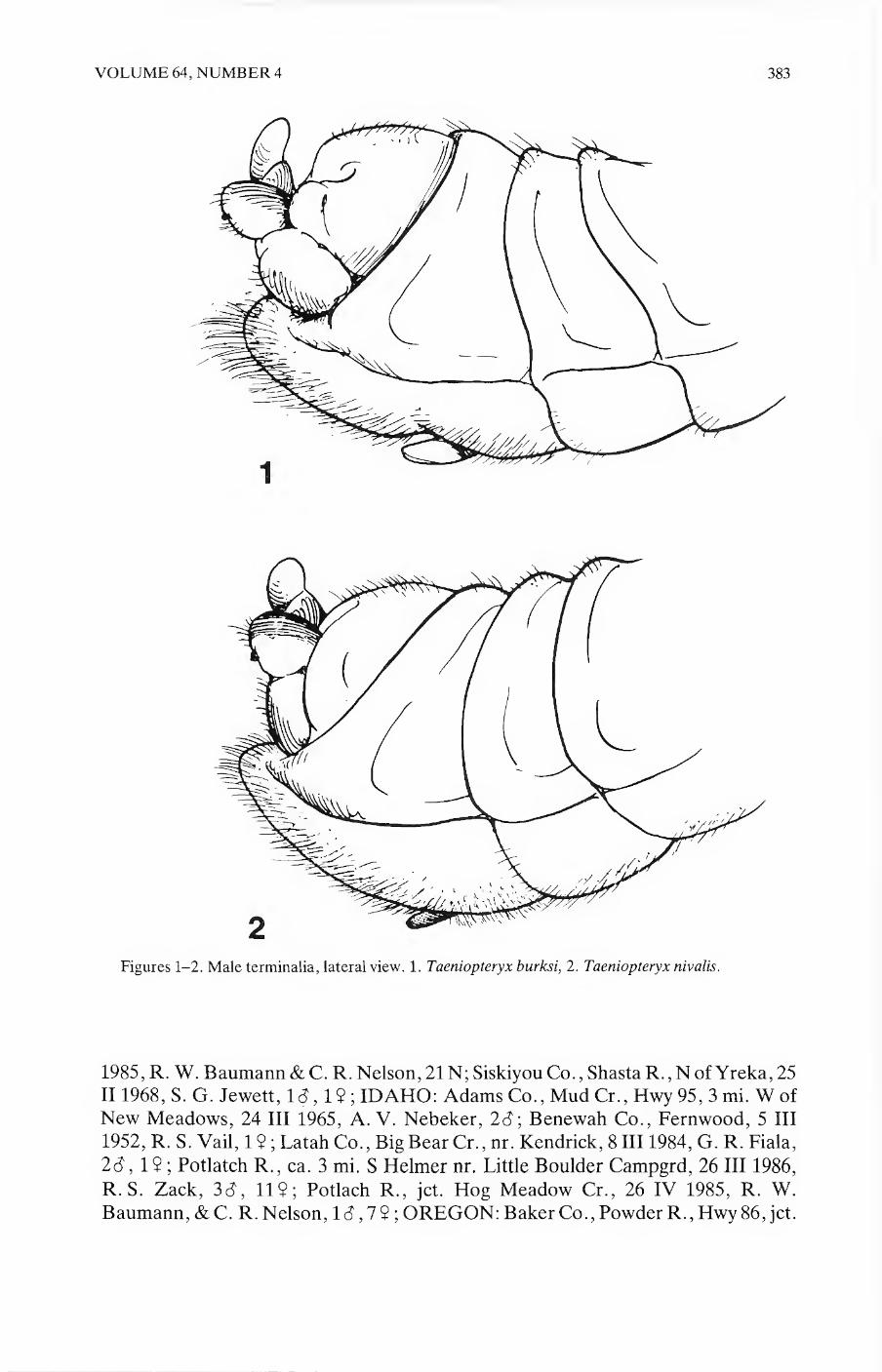
VOLUME 64, NUMBER 4 383
Figures 1-2. Male terminalia, lateral view. 1. Taeniopteryx burksi, 2. Taeniopteryx nivalis.
1985, R. W. Baumann & C. R. Nelson, 21N; Siskiyou Co., ShastaR.,N of Yreka, 25 II 1968, S. G. Jewett, Id, 1$ ; IDAHO: Adams Co., Mud Cr., Hwy 95, 3 mi. W of New Meadows, 24 III 1965, A. V. Nebeker, 2d; Benewah Co., Fernwood, 5 III 1952, R. S. Vail, 1 ? ; Latah Co., Big Bear Cr., nr. Kendrick, 8 III 1984, G. R. Fiala,
2d, 1$ ; Potlatch R., ca. 3 mi. S Helmer nr. Little Boulder Campgrd, 26 III 1986,
R. S. Zack, 3d, 11$; Potlach R., jet. Hog Meadow Cr., 26 IV 1985, R. W. Baumann, &C. R. Nelson, Id, 7$ ; OREGON: Baker Co., Powder R., Hwy 86, jet.

Figures 3-4. Paraprocts. 3. T. burksi, 4. T. parvula.
Figures 5-7. Extruded aedeagus, a. from behind; b. lateral. 5. T. butksi, 6. T. nivalis, 7. T. parvula.

VOLUME 64, NUMBER 4 385
Figures 8-9. Female terminalia, ventral. 8. T. nivalis, 9. T. parvula.
Spring Cr., 24 IV 1985, R. W. Baumann & C. R. Nelson, Id; Spring Cr., Hwy 86, jet. Powder R., 3 III 1984, R. W. Baumann &C. R. Nelson, 3$ ; Benton Co., Muddy Cr. 17II1985, G. R. Fiala, 19 ; UTAH, Box Elder Co., Raft R., Upper Narrows, 15
II 1979, R. W. Baumann & G. M. Webb, 1 N; same location, 28 III 1979, R. W. Baumann & G. M. Webb, 7d,59; WASHINGTON, Kittitas Co., Ellensburg,
CWU Campus, 10 III 1981, M. Rush, 19 ; Lewis Co., N Fork Newaukum R., 16 II 1987, G. R. Fiala, 19 ; Scatter Cr., 9 mi N Centralia, 10 III 1964, S. G. Jewett, Jr., 19 ; Whitman Co., S Fork Palouse R., nr. Albion, 7 III 1984, G. R. Fiala, 19 ; Lyle Grove, 8 mi SW Pullman, 3 III 1973, M. C. Hunter, 8<J.
Taeniopteryx parvula Banks
Taeniopteryx parvula Banks 1918: 7. Taeniopteryx parvula, Ricker and Ross 1968: 1436 (description of adults). Taeniopteryx parvula, Harper and Hynes 1971: 946; Fullington and Stewart 1980: 254 (description of nymph). Taeniopteryxpecos Baumann and Jacobi 1984: 147. New Synonym
Baumann and Jacobi (1984) described T. pecos from a series of specimens from the
Pecos River, San Miguel Co., New Mexico. They indicated that this species was
“very close to T. parvula. ” The characters used to separate T. pecos from T. parvula,

386 PAN-PACIFIC ENTOMOLOGIST
Figures 10-11. Nymphal habitus, dorsal. 10. T. burksi, 11. T. parvula.

VOLUME 64, NUMBER 4 387
11

388 PAN-PACIFIC ENTOMOLOGIST
hi 11 (fAKJ
f i'H-I-Hit
12 \
Figures 12-14. Nymphal eighth abdominal tergum. 12. T. burski, 13. T. nivalis, 14. T. parvula.
size and curvature of the paraproctal hooks and size of the epiproct are relative and agree with specimens from Colorado and Wyoming and typical T. parvula from east¬ ern North America. The aedeagus (Fig. 7) of T. parvula is very distinctive and was first illustrated by Frison (1942: Fig. 7). Males of T. parvula are easily separated from all western Taeniopteryx by the lack of a vesicle, and the distally hooked paraprocts (Fig. 4). Females can be distinguished by the pale medial portion of the 8th sternum and notch shape (Fig. 9).
The nymphs of T. parvula, as with T. nivalis, have two forms, striped (at least with a partial stripe) (Fig. 11) or lacking a middorsal stripe. Fullington and Stewart (1980) only treated T. parvula as being the latter. In the series of nymphs examined from Colorado and Wyoming, individuals varied from lacking a middorsal stripe to having at least a partial stripe. This same condition has been observed by Kirchner (personal communication) in populations from West Virginia.

VOLUME 64, NUMBER 4 389
The occurrence of T. parvula in the Rocky Mountains is probably also the result of
postglacial and interglacial dispersal across the northern plains. The emergence of
this species in isolated areas of the northcentral Rockies in March and often under ice, may account for the rarity of specimens in collections.
Western records examined: COLORADO, Jackson Co., Grizzly Cr., Rt 14, 14 III
1987, B. C. Kondratieff, 10<?,32, 15 N; Roaring Fork, lmi. above jet. N. Platte R., 22 X 1987, D. Rees, 1 N; NEW MEXICO, San Miguel Co., Pecos R. Hwy. 119, Tecolotito, 27II1979, G. Z. Jacobi, 4<3,22 (paratypes); Guadalupe Co., Pecos R., Hwy. 119, Anton Chico, 5 1 1980, G. Z. Jacobi, 9 N; WYOMING, Carbon, Co., N Platte R. 2 mi. above jet. Big Cr., 24 X1987, D. Rees, 1N; same location 15 III 1988,
id,12.
Key to the males of western North American Taeniopteryx
1. Ninth sternum with a vesicle; paraprocts straight and bluntly pointed (Fig. 3) . 2 Vesicle absent; tips of paraprocts hooked (Fig. 4) .parvula
2. Aedeagus with short dorsolateral lobes (Fig. 6); vesicle short, 2-3 times as long as
wide .nivalis Aedeagus with long dorso- and ventrolateral lobes (Fig. 5); vesicle long, 3-5 times as long as wide.burksi
Key to the females of western North American Taeniopteryx
1. Middle portio of 8th sternum somewhat pale, not as heavily pigmented as central plate (Fig. 9) .parvula Middle portion of 8th sternum as darkly pigmented as central plate .2
2. Notch with strongly sclerotized shoulders (Fig. 8) .nivalis Notch without strongly sclerotized shoulders .burksi
Key to the Nymphs of western North American Taeniopteryx
1. Thorax and abdomen without middorsal pale stripe.2 Thorax and abdomen with at least a partial pale middorsal stripe (Figs. 10 & 11)
.3 2. Posterior margins of abdominal terga with short blunt setae (Fig. 13) ... nivalis
Posterior margins of abdominal terga with long setae, apically curved and an oc¬ casional long hair (Fig. 14) .parvula
3. Abdominal terga with mostly short blunt bristles, posterior margins with short blunt setae (Fig. 13) .nivalis Abdominal terga with long bristles, many apically curved, posterior margins with long setae curved apically or short blunt setae (Figs. 12 & 14) .4
4. Posterior margins of abdominal terga with long setae curved and an occasional hair (Fig. 14) .parvula Posterior margins of abdominal terga with short blunt setae (Fig. 12) ... burksi
Acknowledgments
We thank Dave Carlson, Colorado State University for the illustrations. Ralph F. Kirchner is thanked for advice concerning Taeniopteryx nymphs.

390 PAN-PACIFIC ENTOMOLOGIST
Literature Cited
Banks, N. 1918. New neuropteroid insects. Bull. Mus. Comp. Zool. 62: 3-22.
Baumann, R. W., A. R. Gaufin, and R. F. Surdick. 1977. The stoneflies (Plecoptera) of the Rocky
Mountains. MemAmer. Entomol. Soc. 31:1-208.
Baumann, R. W. and G. Z. Jacobi. 1984. Two new species of stoneflies (Plecoptera) from New Mexico.
Proc. Entomol. Soc. Wash. 86: 147-154.
Canton, S., J. Chadwick, andL. Britton. 1981. A new distributional record for Taeniopteryx (Plecoptera:
Taeniopterygidae) from Colorado. Entomol. News 92:155-157.
Dosdall, L. and D. M. Lemkuhl. 1979. Stoneflies (Plecoptera) of Saskatchewan. Quaest. Entomol. 15:
3-116.
Fitch, A. 1847. Winter insects of eastern New York. Amer. J. Agri. Sci. 274-284.
Frison, T. H. 1942. Studies of North American Plecoptera, with special reference to the fauna of Illinois.
Bull. Ill. Nat. Hist. Surv. 22: 233-355.
Fullington, K. E. and K. W. Stewart. 1980. Nymphs of the stonefly genus Taeniopteryx (Plecoptera:
Taeniopterygidae) of North America. J. Kansas Entomol. Soc. 53: 237-259.
Harper, P. P. and H. B. N. Hynes. 1971. The nymphs of the Taeniopterygidae of eastern Canada
(Insecta; Plecoptera). Can. J. Zool. 49: 941-947.
Hitchcock, S. W. 1974. Guide to the insects of Connecticut: Part VII. The Plecoptera or stoneflies of
Connecticut. Bull. State Geol. Nat. Hist. Surv. Conn. 107: 1-262.
Kondratieff, B. C. and J. V. Ward. 1987. Taeniopteryx burksi (Plecoptera: Taeniopterygidae) in
Colorado, with notes on aquatic insects of plains streams. Entomol. News 98: 13-16.
Ricker, W. E. 1964. Distribution of Canadian stoneflies. Gewasser Abwasser 34/35: 50-71.
Ricker, W. E. and H. H. Ross. 1968. North American species of Taeniopteryx (Plecoptera, Insecta). J.
Fish. Res. Bd. Canada. 25:1423-1439.
Stark, B. P., S. W. Szczytko and R. W. Baumann. 1986. North American stoneflies (Plecoptera):
systematics, distribution, and taxonomic references. Great Basin Nat. 46: 383-397.
Stewart, K. W., R. W. Baumann and B. P. Stark. 1974. The distribution and past dispersal of
southwestern United States Plecoptera. Trans. Amer. Entomol. Soc. 99: 507-546.

PAN-PACIFIC ENTOMOLOGIST 64(4), 1988, pp. 391-402
Trichogramma species in a chaparral community of southern California, with a description of a new species
(Hymenoptera: Trichogrammatidae)
John D. Pinto and Earl R. Oatman
Department of Entomology, University of California, Riverside, CA 92521
Abstract.—Five species of Trichogramma were collected during a two year period in a chaparral community in southern California. Only T. deion Pinto and Oatman,
and T. funestum, sp. n. were common. Both species occur together throughout the year but apparently are dominant in different habitats. T. funestum is described. It is most similar to T. exiguum Pinto and Platner, and T. fuentesi Torre.
Introduction
Our knowledge of Trichogramma systematics in North America primarily rests on collections from agricultural ecosystems. This is as expected considering the long¬ standing and widespread use of this genus of egg parasites in biological control (Ridgway et al., 1977). In an attempt to reverse this bias we have begun surveying a variety of natural areas, especially those adjacent to agriculture. By such efforts we hope not only to better estimate the taxonomic diversity of Trichogramma in North America but also to gain a better appreciation of intraspecific habitat diversity, a fac¬
tor that should be considered when selecting agents for biological control.
Herein are reported results of a survey from 1985-1987 in a chaparral community
in southern California. Although hundreds of collections in nearby agricultural areas have been made, this is the first effort at a site dominated by natural vegetation. Five species were collected but only two were common. One of these, T. funestum, is new and described below. The other species, T. deion, only recently described (Pinto et al., 1986), is the most commonly collected species in the western United States. Sea¬ sonal and habitat distribution of these two species within chaparral are reported here.
Methods
The sampling site is located in hills immediately west of Menifee Valley, ca. 10 km E. Lake Elsinore in SW Riverside Co. Most collections were restricted to an area of 2.0 hectares along a north-south running ridge (33°39' N, H7°13'W; 550 m. el.). The few additional collections made elsewhere were within 1 km. of this site.
The study area is dominated by chaparral with a strong influence of Coastal Sage Scrub vegetation (see Munz, 1959 for characterization of these communities). As is typical of the low hills bordering the hot, dry interior valleys of southern California,
perennial vegetation is relatively sparse. About 50% of the surface is covered by per¬
ennial canopy; the remainder is bare ground, much of it strewn with large granitic boulders. Dominant perennials at the site are Salvia spp. (primarily S. mellifera
391

392 PAN-PACIFIC ENTOMOLOGIST
Greene), Eriogonum fasciculatum Bentham, Adenostoma fasciculatum Hooker & Arnott, and Ceanothus crassifolius Torrey. Vegetation composition varies consid¬ erably with slope, slope exposure, and substrate. Contribution to total vegetation cover by the dominant perennials in areas sampled for Trichogramma is as follows:
Salvia spp., 2-28%; Eriogonum, 14-21%; Adenostoma 2-18%, Ceanothus 1-4%. The study site is bordered on all sides by vast areas of similar vegetation. Dwellings
with small concentrations of domestic plants are scattered, but occur primarily to the east and west. Grain fields and greater human population density occur 2.5 km. W. of the site. A dwelling and a small irrigated vegetable garden (10 m. x 10 m.) sur¬ rounded by natural vegetation occur on the study site. Collections were made both in the garden and in natural vegetation at least 20 m. from all domestic plants. A collec¬ tion was made once immediately adjacent to the garden on Ceanothus.
Almost all Trichogramma were collected by placing eggs of laboratory-reared Tri- choplusia ni (Hiibner) and Plodia interpunctella (Hiibner) on vegetation (host egg
traps). A few were taken in yellow-pan traps, sweeping vegetation, and by collecting naturally occurring Lepidoptera eggs. For host egg traps, eggs of both species were attached to individual strips of stiff index card (6.5 x 1.8 cm). Each card held ap¬ proximately 150 T. ni eggs and 2500 P. interpunctella eggs. T. ni eggs were laid on paper towelling which was then cut into strips and glued to the index card with white glue. Plodia eggs were attached to the card with Scotch® double stick tape. Cards were clipped directly to vegetation. Eggs of Trichoplusia were irradiated with Cobalt 60 (12 krads for 5 min.) prior to use to prolong time of acceptability to Tricho¬
gramma. The Plodia eggs were not irradiated. 5169 cards were placed on various plants over 76 sampling dates (ca. every 1.5 wk)
from 6 April 1985-19 May 1987, an average of 68 cards/sampling date. Cards were left in the field 1-2 da., depending on weather and levels of egg predation. Predation often could not be avoided. The use of Tanglefoot® around stems deterred ants and
other crawling predators but did not preclude flying forms such as vespids, various Coleoptera and chrysopids. Predators rarely consumed all eggs on a given card, and often the few remaining eggs were parasitized by Trichogramma.
Egg cards were placed primarily on Eriogonum, Adenostoma, Ceanothus and gar¬ den plants (usually tomato or/and pepper). They were placed on Eriogonum and gar¬ den plants on almost every sampling date. Adenostoma and Ceanothus were used during time of bloom the first year but on almost every sampling date the second year of the study. Other plants sampled infrequently via egg cards were Salix sp. (twice), Quercus agrifolia Nee (4), Lotus scoparius (Nutt.) Otley (2), Keckiella antirrinoides (Benth.) (6), Salvia mellifera (3), and Prunus ilicifolia (Nutt.) Walp. (1).
A limited supply of laboratory-reared host eggs and relatively low parasitization rates by Trichogramma in the field prevented use of an adequate number of cards on all major perennials throughout the study. Adenostoma, Eriogonum and Ceanothus were chosen for extensive sampling because they were common in the area and are evergreen throughout the year. Several of the other plants (e.g. Keckiella, Salvia mellifera) are drought-deciduous and lack green foliage much of the time from May- November.
Limited numbers of laboratory eggs also resulted in some variation in the number of cards placed on particular plants on each date. For example, fewer egg cards were usually placed in the garden since they were more commonly parasitized than those on native plants. Uncontrollable levels of egg predation also necessitated placing

VOLUME 64, NUMBER 4 393
more cards on certain plants at certain times of the year. The number of cards placed on plants on each date ranged from 7-20 on garden plants, 10-50 on Eriogonum, 8-
40 on Adenostoma, and 9-20 on Ceanothus. The number of times each plant was sampled also varied considerably but all were sampled at least once during every month (Table 3). This variation precluded a quantitative treatment of much of the data but was allowed since we were attempting to maximize parasitization in a given habitat.
All plants sampled were in close proximity. However, egg cards were always at¬ tached to those that did not contact other species. Distances between species in sam¬
pling areas varied as follows Ceanothus-Eriogonum, 1-10 m.; Adenostoma- Eriogonum, 8-80 m.; Ceanothus-Adenostoma, 1-50 m. The garden was 1, 8 and 12 m. from the nearest Ceanothus, Eriogonum and Adenostoma, respectively.
Host trap cards were returned to the laboratory and held for observation. If one or more eggs on a card was parasitized (evidenced by its black appearance) the Plodia and/or Trichoplusia portion was isolated and placed in a vial with fresh host eggs. A sample of emerging adults was mounted from each collection. Most cultures were discontinued once the identity of the collection was determined. Although rarely oc¬ curring, parasitization of eggs of one of the hosts on a card by more than one species of Trichogramma was detected soon after emergence by obvious color and antennal
differences.
Results
Five species of Trichogramma were collected (Table 1). T. thalense Pinto and Oat-
man, and T. brevicapillum Pinto and Platner were collected once in pan traps. T. platneri Nagakatti was taken from host trap eggs twice, once in the garden (on to¬ mato) and once on Keckiella. T. deion Pinto and Oatman, and T. funestum were commonly collected from parasitized trap host eggs throughout the year, and both also were retrieved from sweep samples of natural vegetation. The latter two were the only species consistently parasitizing trap host eggs. Of the 5169 egg cards uti¬ lized, 409 (7.9%) were parasitized by either T. deion or T. funestum. T. deion also was reared from natural host eggs, Manduca sexta (L.) (Sphingidae) on tomato, Pieris rapae (L.) (Pieridae) on wild mustard, and Plebejus acmon (Westwood and Hewitson) (Lycaenidae) on Lotus scoparius. Natural hosts of T. funestum were not collected at the site. However, it has been recorded from eggs of Vanessa cardui (L.) (Nymphalidae) and Plebejus emigdionis (Grinnell) (Lycaenidae) at other locales (see below).
Although T. deion and T. funestum frequently were reared from trap eggs, the two apparently were not randomly distributed in the chaparral. T. deion was the only one to parasitize natural eggs and trap eggs in the garden; it also was more commonly retrieved from cards placed on Eriogonum. In comparison, T. funestum was com¬ monly reared from cards placed on Adenostoma and Ceanothus; T. deion rarely was. The dramatic differences in habitat distribution in the chaparral is illustrated by com¬ paring numbers of trap host cards parasitized by each species at the different posi¬ tions (Table 2) and by the number of sampling dates/mo. each species was collected on each plant throughout the study (Table 3).
The association of T. deion and T. funestum on other plants in chaparral has not
been adequately studied. However, limited additional host trap sampling on other plants retrieved both species from Keckiella antirrhinoides; T. deion from Salix sp.

394 PAN-PACIFIC ENTOMOLOGIST
Table 1. Species of Trichogramma in southern California chaparral and their method of collection.
Method of Collection1
Species A B C D
T. brevicapillum2 - + - -
T. deion + + + +
T. platneri + - - -
T. funestum + - + -
T. thalense3 — + — —
3A = Egg host trapping; B = Yellow pan traps; C = sweeping; D = reared from natural host eggs (see text).
2 A single female collected V-8/15-1982. 3One male collected XII-1981 beneath canopy of Quercus agrifolia.
and Prunus ilicifolia; and T. funestum from Quercus agrifolia. Also, as already indi¬ cated, T. deion was taken from lycaenid eggs on Lotus scoparius.
Since eggs of Trichoplusia and Plodia were available on each card, relative host preferences by females of the two dominant Trichogramma species could be esti¬ mated. T. funestum more commonly parasitized Plodia [(on 66% of 145 cards parasi¬ tized x2 = 14.0; P <''i 0.01)], whereas T. deion more commonly parasitized Tnchoplu~ sia (68% of 295 cards parasitized); x2 = 38.8; P < 0.01. Laboratory rearings suggest that T. funestum is more successful on Plodia. T. deion, however, does well on both.
The percentage of cards parasitized by T. deion and T. funestum in 1986 is shown in Fig. 1. Percentages refer to cards parasitized per month. Data for garden plants and Eriogonum are separated for T. deion because parasitization in the garden was usu¬ ally much higher. Percentages for T. funestum refer to cards placed on Ceanoths and Adenostoma since parasitization rates on both were similar. These data suggest that Trichogramma activity was highest from April-August, but continued at low levels in the winter and spring months. Although only T. funestum was retrieved from Oct.-Dec. in 1986, T. deion parasitized egg cards at low levels (1.7%-5.0%) on both
Eriogonum and garden plants throughout this period in 1985. T. deion is more active on garden plants than on Eriogonum. Cards placed on the three native chaparral plants were less commonly parasitized than those on garden plants, except early and
late in the year. These results must be considered tentative since the degree of ac¬ ceptability of laboratory reared hosts relative to natural hosts is unknown, and in¬
deed, may vary throughout the year as the quantity and species of natural hosts vary.
Discussion
T. deion and T. funestum were the only species collected commonly. T. deion is a
common species in western North America in disturbed and natural habitats (Pinto et al., 1986). T. funestum was collected for the first time in this study and has since been taken at other sites in California (see below).
Both species occur together in chaparral throughout the year (Fig. 1, Table 3). Although occurring together, they apparently are not evenly distributed. T. deion

VOLUME 64, NUMBER 4 395
Table 2. Percentage of parasitized trap host egg cards parasitized by T. deion and T. funestum on three chaparral
perennial shrubs and in an adjacent vegetable garden from April 1985-May 1987.
Trap host egg card placement
Garden Eriogonum Adenostoma Ceanothus
235
Total number cards parasitized
46 61 67
100.0%
% Parasitized by T. deion
93.5% 6.6% 10.4%
0%
% Parasitized by T. funestum
6.5% 93.4% 89.6%
was most commonly taken on Eriogonum; T. funestum was more frequent on Ceano- thus and Adenostoma. T. deion also was abundant in the vegetable garden, far more so than it was on Eriogonum. T. funestum was never collected in the garden, even though this small plot of disturbed land was surrounded by chaparral and was within 5 m. of Ceanothus on which this species was collected. Both species parasitized trap eggs, but frequency of parasitization depended on the specific habitat (plants) the hosts were placed in. The absence of T. funestum from the garden plot correlates with our general experience collecting Trichogramma in California. Although the species is common in chaparral it never has been taken in the hundreds of parasitized eggs collected in agricultural and other disturbed areas at locales close to the study site.
Previous studies of Trichogramma have stressed the importance of habitat prefer¬ ence in partially explaining varying levels of parasitization on given host eggs (Flan¬ ders, 1937; Salt, 1935). Some studies implicate vertical stratification. For example, Thomas (1966), and Kemp and Simmons (1978) found T. minutum parasitizing more eggs of the spruce budworm {Choristoneura fumiferana) in the upper crown of bal¬ sam fir than in the lower crown. That levels of parasitism can be correlated with height is supported by Thorpe (1985), who showed that T. pretiosum and T. minutum had no obvious habitat preference within uncultivated and soybean fields, but that they did differentially parasitize eggs placed at varying heights.
Studies in crop systems by Norlund et al. (1984) showed that Trichogramma spe¬ cies were more common on Heliothis on tomato and beans than on corn. Further studies appear to explain thesq differences in distribution (Norlund et al., 1985a, b) showing that females of T. pretiosum responded to extract and to volatile synamones from tomato, whereas corn apparently does not possess the same attracting proper¬ ties.
We have not conducted the studies to explain the differential distribution of T. deion and T. funestum in chaparral. Vertical stratification is a possible, but unlikely,
explanation. Ceanothus and Adenostoma do represent higher habitats than either
Eriogonum or garden plants. Yet egg cards were placed at varying heights on both Adenostoma and Ceanothus, levels which broadly overlapped those on Eriogonum

OJ NO On
Table 3. Frequency of occurrence of T. deion and T. funestum in various vegetation components of chaparral as indicated by number of sampling dates per month each was
collected1 from 6 April 1985-19 May 1987.
Plants sampled
Adenostoma Ceanothus Eriogonum Garden2
Month deion funestum
No. times
sampled deion funestum
No. times
sampled deion funestum
No. times
sampled deion funestum
No. times
sampled
Jan 0 1 2 2 2 6 2 1 4 2 0 6
Feb 0 2 2 0 1 5 0 0 4 2 0 5
Mar 0 2 4 0 1 3 0 0 4 0 0 3
Apr 1 8 8 1 3 3 1 0 3 2 0 5
May 0 1 9 2 2 3 4 1 9 3 0 7
Jun 1 1 3 0 2 3 3 0 7 3 0 7
Jul 0 0 3 0 2 3 3 0 6 6 0 8 Aug 0 0 2 0 1 2 2 0 5 5 0 5
Sep 0 0 1 1 0 2 0 0 5 3 0 6 Oct 0 1 1 0 0 1 1 0 5 1 0 5
Nov 0 0 2 0 0 2 1 0 6 1 0 6 Dec 0 1 2 0 0 4 1 1 6 1 0 5
^ased on parasitized laboratory-reared eggs (trap host cards).
2Usually only tomato and/or pepper plants sampled.
PA
N-P
AC
IFIC
EN
TO
MO
LO
GIS
T

%
Par
asit
ized
egg
card
s
VOLUME 64, NUMBER 4 397
Months
garden plants
Eriogonum
on Adenostoma & Ceanothus
y-
o o CO o o *- O O CD
r- CM
*•
CM V <0 CM ^ in
m 1 0 N 1 D
Fig. 1. Percentage of host egg trap cards parasitized per month by T. deion and T. funestum in various
vegetation components of chaparral during 1986. Solid black = T. deion on garden plants; oblique
lines = T. deion on Eriogonum; solid grey = T. funestum on Adenostoma and Ceanothus. Numbers
above bars = total no. cards percentage is based on.
and garden plants. In fact, T. funestum was collected several times at positions on Adenostoma (less than 0.5 m.) which were as low or lower than sampling sites in the
garden. More rigorous investigations of the possible effect of height are needed, however. The possibility that T. deion and T. funestum are initially attracted to plants of different height prior to host searching also should be investigated.
The differential distribution of both species possibly is dictated by natural host preferences. There is little value speculating on this possibility, considering our lack of knowledge of Trichogramma host preferences. That it is not an obvious explana¬ tion is suggested by the broad host range documented for T. deion (Pinto et al., 1986), and the common occurrence of the lycaenid, Philotes battoides (Behr), on Eri¬ ogonum fas ciculatum (Emmell and Emmell, 1973), a plant rarely frequented by T. funestum. Although little host data are available for T. funestum, it has been col¬ lected from eggs of related lycaenids (see below).
Collections in this study were made in an area of considerable plant intermixture. All Adenostoma and Ceanothus sampled approximated Eriogonum plants and vice-
versa; and all three plants grew near the garden plot. This proximity notwith¬
standing, T. deion and T. funestum overlapped minimally. Whether even this small
degree of overlap occurs in the homogenous stands of Adenostoma and Eriogonum near the study site would be a logical follow-up study.

398 PAN-PACIFIC ENTOMOLOGIST
Three of the five species of Trichogramma collected in this study were rare. Their rarity may accurately reflect level of activity in chaparral or is attributable to the fail¬ ure of our sampling methods to adequately sample all components of the plant com¬ munity. Of the three rare species, T. platneri is the most common in southern Califor¬ nia. It frequently is collected on Lepidoptera eggs on apple and avocado, and is commercially reared and released for biological control of these pests on avocado
(Oatman and Platner, 1985). We also have collected it several times on codling moth eggs on apple (unpubl.). T. brevicapillum is abundant in parts of western United
States but not in the southwest. In California, we have collected it only at three sites south of the San Bernardino Mts. North of there it is common. The third species, T. thalense, although rarely collected, is also known throughout much of western North America. Within California it has been taken primarily in the San Joaquin and Sacra¬ mento valleys. The single specimen collected in this study is the only record for southern California.
One of the surprising results of this study is the absence of T. pretiosum. A species parasitising a variety of hosts in disturbed habitats in California, we collect it com¬ monly at sites within 10-20 km. from the study area (e.g. Lake Elsinore, Temecula, Perris). We noted in an earlier paper (Pinto et al., 1986) that T. pretiosum had never been taken in natural habitats in California. We were surprised, however, not to en¬
counter it in the garden collections despite over 2 yrs. of host trapping and collecting
naturally occurring eggs. The absence of appropriate host eggs is certainly not an explanation since T. pretiosum readily parasitizes eggs of the two laboratory-reared species utilized here, as well as eggs of Vanessa and Manduca, both of which oc¬ curred. Its absence apparently is explained by the isolation of the small garden within an area of primarily natural vegetation. T. funestum represents the opposite end of the spectrum regarding general habitat association. It is fairly common in chaparral but has never been collected in adjacent agricultural or other disturbed habitats. Un¬ like T. pretiosum and T. funestum, T. deion is much more plastic in habitat associa¬ tion. It is commonly collected in a variety of habitats throughout the western United States (Pinto et al., 1986).
Trichogramma funestum, New Species
(Figs. 2, 3)
Description based on Pi and Fi material from various collections at the type local¬
ity. Color data were taken from critical point dried F2 culture material reared at 23- 27°C on T. ni eggs and at ca. 50% RH. Quantitative data are based on 10 randomly selected Pi specimens of each sex.
Terminology for genitalic structures below follows that used in earlier papers (e.g. Pinto and Oatman, 1985). A more detailed explanation of the formula for the basi- conic capitate peg sensilla on the antenna was introducted in Pinto et. al. (1986).
Color sexually dimorphic. Female: thorax orange yellow except pronotum brown; head orange yellow above and in front, light brown to brown behind and below eye;
gaster brown; legs and antenna light yellow. Male: as in female except mid-lobe of
mesoscutum brown, concolorous to gaster. Length: 0.4-0.5 mm. Hind tibial length:
averaging 0.158 ± 0.02 (0.13-0.18) mm. in both sexes (n = 20). Male.—Antenna (Fig. 2). Flagellum moderately elongate, averaging 0.165 mm
(0.14-0.20) long, slightly arcuate, 5.59 ± 0.37 (5.1-6.2) as long as wide, 1.05 ± 0.04 (1.0-1.1) as long as hind tibia; setae relatively short, stout, abruptly tapered at apex,

VOLUME 64, NUMBER 4 399
longest seta 2.13 ± 0.12 (2.0-2.3) maximum width of flagellum, ca. 34-45 in num¬ ber; formula for basiconic capitate peg sensilla (BCPS) = 1—1—2—1—1, infrequently with only 1 sensilla at third position. Forewing. Vein tracts well defined, moderate number of setae between tracts, 7-37 setae in area between 4th and 5th tracts; length of longest postapical seta on margin ca. 2x maximum width of hind tibia and 0.158 ± 0.03 (0.11-0.20) greatest wing width. Hindwing. Posterior vein tract with 5- 7 setae, extending to ca. V2 the length of middle tract; anterior tract with 2 small setae Mesoscutellum. Anterior pair of setae short, ca. 1U the length of posterior pair. Geni¬
tal capsule (Fig. 3). Moderately narrow, widest at base of dorsal expansion of gono- base (DEG), then evenly convergent to base and apex, sides straight, not distinctly sinuate near base of gonostyli (GS), 0.315 ± 0.01 (0.29-0.33) as wide as long, dis¬ tance from apex of gonostyli to base of median ventral projection (MVP) composing 0.246 ± 0.01 (0.22-0.27) length of genital capsule; DEG subtriangular, only slightly notched at base, sides relatively straight to apex of posterior projection, not dis¬ tinctly sinuate; apex of DEG attaining 0.886 ± 0.02 (0.85-0.92) length of genital
capsule, usually attaining same level as MVP, but below apex of chelate structures
(CS); MVP elongate, narrow, distinctly pointed apically, attaining 0.517 ± 0.06 (0.46-0.58) length of gonostyli; CS slightly to distinctly longer than MVP, attaining 0.689 ± 0.04 (0.62-0.75) length of GS; chitinized ridge (CR) distinct, relatively

400
F,s 3. Trick ogmmrna,funes,um:
A ,Vfale genital capsuJe (dorsal
Vie^ B.Aedeagus.

VOLUME 64, NUMBER 4 401
elongate, extending 0.463 ± 0.03 (0.43-0.53) distance from MVP to base of genital capsule. Aedeagus. Usually slightly longer than genital capsule, 0.913 ± 0.04 (0.84- 0.98) as long as hind tibia; apodemes comprising ca. V2 its total length.
Female.—Antenna. Funicle segments subquadrate; BCPS formula differing from
male, 1—1—1—1—1, a single peg sensilla at apex of each funicle segment and three un¬ paired sensilla on club; four placoid sensilla and ca. 22 apicoventral setae arranged in 6 transverse rows. Ovipositor. 1.037 ± 0.05 (1.00-1.15) as long as hind tibia.
Etymology of specific name.—Latin: “causing death.” Types. Holotype S and allotype $ from CALIFORNIA, Riverside Co., Menifee
Valley, hills on W. end (33°39' N, 117°13' W; 550 m. el.). Types represent Fi mate¬ rial; parentals reared from trap host eggs (T. ni) placed on Ceanothus crassifolius’, 28
January 1986; J. D. Pinto, collr.; deposited in the collection of the National Museum of Natural History, Washington, D.C. Eight additional Fi siblings designated as paratypes; deposited as follows: Id, 19, British Museum (Natural History); Id,
19 , Canadian National Collection; Id, University of California, Berkeley; 2dd, 19 , University of California, Riverside. All type material mounted on glass slides in Canada Balsam.
Geographic Distribution.—California from Glenn Co., S. to western Riverside Co.
Records and Hosts.—UNITED STATES. California. Glenn Co. Alder Springs, 16dd,3 June 1987, trap host (Plodia on Pinus), J. Pinto. Los Angeles Co. Solemint, Id, 3 June 1982, ex. Plebejus emigdionis egg (Lycaenidae) on Atriplex canescens, G. Pratt. Riverside Co. Hemet, E. of at 4000' el., Id, 30 June 1983, undetermined egg
on Adenostoma fasciculatum, R. Velten. Menifee Valley, hills on W. end (33°39' N, 117°13' W; 550 m. el.), 168 d d, 17 9 9, numerous dates 1985-87 (see above), trap host eggs (Plodia and Trichoplusia) on Adenostoma fasciculatum, Ceanothus crassi¬ folius, Quercus agrifolia, and Keckiella antirrhinoides, J. Pinto. San Bernardino Co. Summit Valley (2 mi. E. Hwy. 15), Id, 7 May 1982, sweeping, J. Woolley. Ventura Co. Dome Springs Campground, Los Padres National Forest, 2d d, 7 June 1986, ex. Plebejus emigdionis egg on Atriplex canescens, G. Pratt. Dome Springs Camp¬ ground, ca. 4 mi. SW on Lockwood Valley Rd., Los Padres National Forest, 4d d, 3
9 9 , ex. Plebejus emigdionis eggs on Atriplex canescens, 10 May 1985, G. Pratt; ex. undetermined Geometridae eggs on Atriplex canescens, and Vanessa cardui egg on bush lupine, 22 May 1986, E. Oatman and J. Pinto.
Notes.—T. funestum is most similar to T. exiguum Pinto and Platner, and T. fuen- tesi Torre. These two species were recently treated by Pinto et al. (1983). In all three the flagellar setae of males are relatively short (length less than 2.5 the maximum width of flagellum) and stout, tapering noticeably at the apex only. T. funestum is separated from both by specifics of the genitalia. In the latter two the genital capsule is somewhat broader and more abruptly narrowed apically (Pinto et al., 1983: Figs, lc, d). Also, the sides of the genital capsule are sinuate at the base of the gonostyli and the sides of the DEG also are distinctly sinuate. In T. funestum the sides of the genital capsule and the DEG are relatively straight (Fig. 3). T. fuentesi is further sep¬ arated from T. funestum by its shorter CR and subequally long CS and MVP. T. ex¬ iguum is further separated by its lighter color (light yellow-brown abdomen) and
BCPS formula of the male flagellum. In T. funestum the basal two positions have only a single basiconic peg sensilla. In T. exiguum there usually are two sensilla at
each of the two basal positions.
402 PAN-PACIFIC ENTOMOLOGIST
T. fuentesi and T. exiguum are known from central and southeastern United States and are allopatric to T. funestum. However, a currently undescribed species, ex¬ tremely close to T. exiguum is known in California and has been collected at one of the same locales as T. funestum. The latter is separated from this species by the same traits cited for T. exiguum.
A culture of T. funestum from the type locality successfully crossed with the sam¬ ple from Adler Springs, Glenn Co., CA. It did not cross with T. exiguum.
Acknowledgments
Culture maintenance, specimen preparation and other laboratory assistance was provided by G. R. Platner and R. K. Velten. Collections of T. funestum from Ly- caenidae eggs were made by G. Pratt, who also identified the lycaenid hosts. Figs. 2
and 3 were prepared by Linda Bobbitt. This study was supported by Grant No. BSR-8415378 from the National Science
Foundation (J.D.P., Principal Investigator).
Literature Cited
Emmel, T. C. and J. F. Emmel. 1973. The butterflies of southern California. Nat. Hist. Mus. of Los
Angeles Co. Mus., Sci. Ser. 26,148 pp.
Flanders, S. E. 1937. Habitat selection by Trichogramma. Ann. Entomol. Soc. Am., 30: 208-210.
Kemp, W. P. and G. A. Simmons. 1978. The influence of stand factors on parasitism of spruce budworm
eggs by Trichogramma minutum. Environ. Entomol., 7: 685-688.
Munz, P. A. 1959. A California flora. Univ. Calif. Press. 1681 pp.
Norlund, D. A., R. B. Chalfant, and W. J. Lewis. 1984. Arthropod populations, yield and damage in
monocultures and polycultures of corn, beans and tomatoes. Agr. Ecosystems and Environ., 11:
353-367.
Norlund, D. A., R. B. Chalfant and W. J. Lewis. 1985a. Response of Trichogrammapretiosum females
to extracts of two plants attacked by Heliothis zea. Agric. Ecosystems and Environ., 12:127-133.
Norlund, D. A., R. B. Chalfant and W. J. Lewis. 1985b. Response of Trichogramma pretiosum females
to volatile synomones from tomato plants. J. Entomol. Sci., 20: 372-376.
Oatman, E. R. and G. R. Platner. 1985. Biological control of two avocado pests. Calif. Agr., 39 (11 & 12):
21-23.
Pinto, J. D. and E. R. Oatman. 1985. Additions to nearctic Trichogramma (Hymenoptera:
Trichogrammatidae). Proc. Entomol. Soc. Wash., 87:176-186.
Pinto, J. D., E. R. Oatman and G. R. Platner. 1983. The identity of two closely related and frequently
encountered species of New World Trichogramma (Hymenoptera: Trichogrammatidae). Proc.
Entomol. Soc. Wash., 85: 588-593.
Pinto, J. D., E. R. Oatman and G. R. Platner. 1986. Trichogramma pretiosum and a new cryptic species
occurring sympatrically in southwestern North America. Ann. Entomol. Soc. Am. ,79:1019-1028.
Ridgway, R. L., E. G. King and J. R. Carillo. 1977. Augmentation of natural enemies for control of plant
pests in the Western Hemisphere, pp. 379-428. In: Ridgway, R. L. and S. B. Vinson [eds.],
Biological control by augmentation of natural enemies. Plenum, N.Y.
Salt, G. 1935. Experimental studies in insect parasitism. III. Host selection. Proc. Roy. Soc. London Ser.
B., 117: 413-435.
Thomas, H. A. 1966. Parasitism by Trichogramma minutum (Hymenoptera: Trichogrammatidae) in the
spruce budworm outbreak in Maine. Ann. Entomol. Soc. Am., 59: 723-725.
Thorpe, K. W. 1985. Effects of height and habitat type on egg parasitism by Trichogramma minutum and
T. pretiosum (Hymenoptera: Trichogrammatidae). Agr. Ecosystems and Environ., 12:117-126.

The Pan-Pacific Entomologist Contents for Volume 64
AKRE, R. D.—See Rathman, R. J. 185
ARNAUD, P. H.—Cornelius Becker Philip. 365
ARNAUD, P. H. and C. D. MacNEILL—In Memoriam, Pedro W. Wygodzinsky . 297
ASKEVOLD, I. S.—See Gruev, B. 139
BAUMANN, R. W.— See Kondratieff, B. C. 381
BETHKE, J. A.—See Parrella, M. P.. 17
BIGGAM, R. C. and M. W. STOCK—Pronotal stripes and wing length in Gerris incurvatus Drake and Hottes (Hemiptera: Gerridae) . 359
BROEMELING, D. K.—A revision of the Nomeda subgenus Nomadita of North America (Hymenoptera: Anthophoridae) . 321
BROEMELING, D. K. and A. S. MOALIF—A revision of the Nomada subgenus Pachynomada (Hymenoptera: Anthophoridae) . 201
BRUNNER, J. F.—See Rathman, R. J. 93
BRUNNER, J. F— See Rathman, R. J. 185
BURGER, J. F.—Notes on Tabanidae (Diptera) of the Oriental Region III. New and little-known Tabanidae from Borneo . 159
CAMARGO, J. M. F., J. S. MOURE and D. W. ROUBIK—Melipona yucatanica new species (Hymenoptera: Apidae: Meliponinae); stingless bee dispersal across the Caribbean Arc and post-Eocene vicariance . 147
CHANDLER, D. S.—New Barrojuba with a revised key to species (Coleoptera: Pselaphidae) . 371
CHEMSAK, J. A., E. G. LINSLEY, and F. HOVORE—A concentration site for Cerambycidae in Jalisco, Mexico (Coleoptera) . 291
CHRISTIANSEN, T. A. and J. A. LOCKWOOD—Winter arthropods in selected habitats of northern mixed-grass prairie. 127
CLARK, W. H.—See Frohlich, D. R. 266
CORPUS, L. D.—Immature stages of Tachytrechus auratus (Diptera: Dolichopodidae) . 9
DALY, H. V. and G. R. ELSE—Lectotype designations for the African bees of the genus Ceratina described by T. D. A. Cockerell (Hymenoptera: Apoidea). 87
DALY, H. V.—See Santiago-Blay, J. A. 281
DOSDALL, L. M. and D. M. LEHMKUHL—The nymph of Utacapnia trava (Nebeker and Gaufin) (Plecoptera: Capniidae) . 353
EHLER, L. E. and H. P. SAUTER—Observations on Eupelmus inyoensis Girault (Hymenoptera: Eupelmidae) . 33
ELSE, G. R. and H. V. DALY. 87
FINANCIAL STATEMENT. 199
FROHLICH, D.R., W. H. CLARK, F. D. PARKER, and T. L. GRISWOLD—The xylophilous bees and wasps of a high, cold desert: Leslie Gulch, Oregon (Hymenoptera: Apoidea, Vespoidea). 266
GAMBINO, P. and G. M. THOMAS—Fungi associated with two Vespula (Hymenoptera: Vespidae) species in the eastern San Francisco Bay Area . 107
GARCIA-VIDAL, M.—The genus Phyllophaga Harris, 1826 (Coleoptera: Scarabaeidae) in Cuba. III. Descriptions of six new species and the male of Ph. cardini Chapin. 101
GOEDEN, R. D.—Life history of Trupanea imperfecta (Coquilett) on Bebbia juncea (Bentham) Greene in the Colorado Desert of Southern California (Diptera: Tephritidae) . 345
GORDH, G.—A new species of Goniozus Foerster from India (Hymenoptera: Bethylidae) used in biological control of Diaphania indica (Saunders) (Lepidoptera: Pyralidae) . 173
403

404 PAN-PACIFIC ENTOMOLOGIST
GRACE, J. K. and W. W. WILCOX—Isolation and trail-following bioassay of a decay fungus associated with Reticulitermes hesperus Banks (Isoptera: Rhinotermitidae) . 243
GRISWOLD, T. L. and F. D. PARKER—New Perdita (Perdita) oligoleges of Mentzelia with notes on related species of the ventralis group (Hymenoptera: Andrenidae) . 43
GRISWOLD, T. L.—See Frohlich, D. R. 266
GRUEV, B. and I. S. ASKEVOLD—New species of Alticinae (Coleoptera: Chrysomelidae) from South India in the genera Taizonia and Longitarsus . 139
HESSEIN, N. A. and J. A. McMurtry—Observations on Megaphragma mymaripenne Timberlake (Hymenoptera: Trichogrammatidae), an egg parasite of Heliothrips haemorrhoidalis Bouche (Thysanoptera: Thripidae) . 250
HOVORE, F.— See Chemsak, J. A. 291
HUNTER, A. S.—High altitude flower-breeding Drosophila (Diptera: Drosophilidae). 299
JOHNSON, J. B.—See Johnson, P. J. 8
JOHNSON, P. J. and J. B. JOHNSON—Arctophyto marginalis Curran (Diptera: Tachinidae): parasite of a Cephaloon tenuicornis LeConte larva (Coleptera: Cephaloidae) in northern Idaho . 8
KONDRATIEFF, B. C. and R. W. BAUMANN—Taeniopteryx of western North America (Plecoptera: Taeniopterygidae) . 381
KUKUK, P. F. and M. SCHWARZ—Macrocephalic male bees as functional reproductives and probably guards . 131
LANFLAM, U. N. and M. J. WEISSMANN—Scaphandrena and Elandrena (Hymenoptera: Andrenidae) . 183
LEHMKUHL, D. M.—See Dosdall, L. M. 353
LINSLEY, E. G.—Further notes on halictine bees (Hymenoptera: Apoidea) visiting Isomeris arborea in southern California . 275
LINSLEY, E. G.—See Chemsak, J. A. 291
LIU, T. P.—The morphology of the tarsal sensilla in the female mite Varroa jacobsoni . 261
LOCKWOOD, J. A.—See Christiansen, T. A. 127
MacNEILL, C. D.—See Arnaud, P. H. 297
MASON, R. R. and H. G. PAUL—Predation on larvael of Douglas-fir Tussock Moth, Orgyia pseudotsugata (Lepidoptera: Lymantriidae), by Metaphidippus aeneolus (Araneae: Salticidae) . 258
McMURTRY, J. A.—See Hussein, N. A. 250
METCALF, R. A.—See Page, R. E. 285
MIDDLEKAUFF, W. W.—A new species of Neurotoma from Michigan (Hymenoptera: Symphyta, Pamphiliidae) . 270
MOALIF, A. S.—See Broemeling, D. K. 201
MORSE, J. G.—See Weisenborn, W. D. 39
MOURE, J. S.—See Camargo, J. M. F. 147
OATMAN, E. R.—See Pinto, J. D. 391
O’BRIEN, A. M.—See O’Brien, M. F. 73
O’BRIEN, M. F. and A. M. O’BRIEN—Biology of Ammophila evansi and A. mediata in northern Michigan (Hymenoptera: Sphecidae) . 73
OLSEN, A. R.—New record of Lardoglyphus konoi (Sasa and Asanuma), on dried lizard carcasses (Acarina: Acaridae) . 114
PAGE, Jr., R. E. and R. A. METCALF—A population estimate of Mdh Allozyme frequencies for the honey bee, Apis mellifera L. (Hymenoptera: Apidae) . 285
PARKER, F. D.—Nesting biology of two North American species of Chelostoma (Hymenoptera: Megachilidae). 1
PARKER, F. D.—See Griswold, T. L. 43
PARKER, F. D.—See Frohlich, D. R. 266
PARRELLA, M. P. and J. A. BETHKE—Larval development and leafmining activity of Liriomyza trifolli (Burgess) (Diptera: Agromyzidae). 17

VOLUME 64, NUMBER 4 405
PAUL, H. G.—See Mason, R. R. 258
PEMBERTON, R. W.—The use of the Thai giant waterbug, Lethocerus indicus (Hemiptera: Belostomatidae), as human food in California. 81
PINTO, J. D. and E. R. OATMAN—Trichogramma species in a chaparral community of southern California, with a description of a new species (Hymenoptera: Trichogrammatidae) . 391
PIPER, G. L.—See Wehling, W. F. 83
POLHEMUS, D. A. and J. T. POLHEMUS—A new ant mimetic mirid from the Colorado Tundra (Hemiptera: Miridae) . 23
POLHEMUS, J. T.—See Polhemus, D. A. 23
PROCEEDINGS OF THE PACIFIC COAST ENTOMOLOGICAL SOCIETY, 1987 . 193
RATHMAN, R. J., R. D. AKRE and J. F. BRUNNER—External morphology of a species of Metajapyx (Diplura: Japygidae) from Washington . 185
RATHMAN, R. J. and J. F. BRUNNER—Observations on the biology of a new species of Dilyta (Hymenoptera: Charipidae) from Washington State . 93
ROBERTSON, J. A.—Descriptions of the immatures of Typocerus serraticornis (Coleoptera: Cerambycidae), and new observations on biology, including “Varnish” production and usage by the larva. 228
ROUBIK, D. W.—See Camargo, J. M. F. 147
RYTI, R. T.—Geographic variation in cooperative colony foundation in Veromessor pergandei (Hymenoptera: Formicidae). 255
SANTIAGO-BLAY, J. A. and H. V. DALY—The Apoidea Collection of the California Insect Survey (C.I.S.) at the Essig Museum of Entomology, University of California, Berkeley . 281
SAUTER, H. P.—See Ehler, L. E. 33
SCHWARZ, M.—See Kukuk, P. F. 131
SHE, S., and D. YOU—A new species of Isonychia from China (Ephemeroptera: Oligoneuriidae) . 29
SORENSON, J. T.—Three new species of Essigella (Homoptera: Aphididae). 115
STOCK, M. W.—See Biggam, R. C. 359
TEPEDINO, V. J.—Incidence of pre-emergence sib-mating in Monodontomerus obsoletus, Pteromalus venustus, and Tetrastichus megachilidis, three chalcid parasites of the alfalfa leafcutting bee, Megachile rotundata (Hymenoptera: Chalcididae) . 63
TEPEDINO, V. J.—Aspects of host acceptance by Pteromalus venustus Walker and Monodontomerus obsoletus Fabricius, parasitoids of Megachile rotundata (Fabricius), the alfalfa leafcutting bee (Hymenoptera: Chalcididae) . 67
THOMAS, G. M.—See Gambino, P. 107
TSAI, J. H.—See Wilson, S. W. 53
WEHLING, W. F. and G. L. PIPER—Efficacy diminution of the rush skeletonweed gall midge, Cystiphora schmidti (Diptera: Cecidomyiidae), by an indigenous parasitoid . 83
WEISENBORN, W. D. and J. G. MORSE—The mandible and maxillary stylets of Scirtothrips citri (Moulton) (Thysanoptera: Thripidae). 39
WEISSMANN, M. J.—See Lanham, U. N. 183
WHITFIELD, J. B.—Two new species of Paradelius (Hymenoptera: Braconidae) from North America with biological notes .. 313
WILCOX, W. W.—See Grace, J. K. 243
WILSON, S. W. and J. W. TSAI—Descriptions of adults and nymphs of the Taro planthopper, Tarophagus proserpina taiwanensis ssp. n., from Taiwan (Homoptera: Delphacidae). 53
YOU, D.—See She, S. 29

The Pan-Pacific Entomologist
Index to Volume 64
(New taxa in boldface)
Acaridae, 114 Acarina, 114 Agromyzidae, 17 Ammophila evansi, 73; mediata, 75
Andrenidae, 43,183 Anthophoridae, 201
Aphididae, 115 Apidae, 285 Apis mellifera, 285 Apoidea, 87, 266, 275, 281 Araneae,258 Arctophyto marginalis, 8 Barrojuba, 371; simplicinota, 374;
lenticornis, 374; pedunculata, 376; simpliciventris, 377; afoveata, 377; plaumanni, 378; prolongicornis, 378;
campbelli, 379 Belostomatidae, 81 Bethylidae, 173 Braconidae, 313 Capniidae, 353 Cecidomyiidae, 83
Cephaloidae, 8 Cephaloon tenuicornis, 8
Cerambycidae, 228, 291 Ceratina, 87 Chalcididae, 63, 67 Charipidae, 93 Chelostoma, 1; minutum, 2; phaceliae, 5
Chrysomelidae, 139 Clepsis consimilana, 98 Coleoptera, 8,101,139, 228, 291, 371 Coquillettia (Procoquillettia) alpina, 23 Cystiphora schmidti, 83
Delphacidae, 53 Dilyta, 93 Diplura, 185
Diptera, 8, 9,17, 83,159, 299, 345 Dolichopodidae, 9 Drosophila chisaca, 299; acuminanus,
302; colmenares, 305; franii, 307
Drosophilidae, 299 Elandrena, 183 Ephemeroptera, 29
Essigella kathleenae, 115; alyeska, 118; kirki, 121
Eupelmidae, 33
Eupelmus inyoensis, 33 Formicidae, 255 Gerridae, 359
Gerris incurvatus, 359
Goniozus sensorius, 173 Haematopota quadrifenestrata, 160 Halictidae, 275
Heliothrips haemorrhoidalis, 250 Hemiptera, 23, 81, 359 Homoptera, 53,115
Hymenoptera, 1, 33, 43, 63, 67, 73, 87, 93,107,131,147,173,183, 201, 250, 255, 266, 270, 275,281, 285, 313, 321,391
Isomeris arborea, 275 Is onychia hainanensis, 29 Isoptera, 243
Japygidae, 185
Lardoglyphus konoi, 114
Lasioglossum (Chilatictus) erythrurum, 131
Lepidoptera, 98,173, 258 Lethocerus indicus, 81 Liriomyza trifolii, 17 Longitarsus gilli, 143 Lymantriidae, 258 Megachile rotundata, 63, 67 Megachilidae, 1
Megaphragma mymaripenne, 250 Melipona yucatanica, 147 Meliponinae, 147 Metajapyx, 185
Metaphidippus aeneolus, 258 Miridae, 23
Monodontomerus obsoletus, 63, 67
406

VOLUME 64, NUMBER 4 407
Neurotoma, 270; nigra, 272 Nomada (Asteronomada), 336; adducta,
338; brewsterae, 340; durangoae, 341; portalensis, 342
Nomada (Nomadita), 321; aquilarum, 327; mutans, 329; placida, 330; snowii, 331; timberlakei, 333; verecunda, 335
Nomada (P achy nomada), 203; saltill®, 214; utahensis, 215; bohartorum, 220; tepoztlan, 221
Oligoneuriidae, 29 Orgyia pseudotsugata, 258 Pamphiliidae, 270 Paradelius, 313; rubra, 313; nigra, 317 Perdita, 43; multiflorae, 45; kiowi, 47;
wootonae, 49; holoxantha, 49 Philip, Cornelius Becker, 365 Phyllophaga brevicornis, 101; cardini,
101; laboriosa, 102; siboneyensis, 103; ahlbrandti, 104; sandersoni, 104; scaramuzzai, 106
Plecoptera, 353, 381 Pselaphidae, 371 Pteromalus venustus, 63, 67 Pyralidae, 173
Reticulitermes hesperus, 243 Rhinotermitidae, 243
Salticidae, 258 Scaphandrena, 183
Scarabaeidae, 101 Sphecidae, 73 Tabanidae, 159 Tabanus atratoides, 162; atristylatus,
163; atropilosus, 163; samawangensis, 168; transversus, 170
Tachinidae, 8 Tachytrechus auratus, 9 Taeniopterygidae, 381
Taeniopteryx, 381; burksi, 382; nivalis, 382; parvula, 385
Taizonia, 140; obrieni, 141; indica, 141 Tarophagus proserpina taiwanensis, 54 Tephritidae, 345 Tetrastichus megachilidis, 63 Thripidae, 39, 250 Thysanoptera, 39 Tortricidae, 98 Trichogramma funestum, 398 Trichogrammatidae, 250, 391 Trupanea imperfecta, 345 Typocerus serraticornis, 228 Utacapnia trava, 353 Varroa jacobsoni, 261 Veromessor pergandei, 255 Vespidae, 107 Vespoidea, 266 Vespula, 107 Wygodzinsky, P. W., 297


THE PAN-PACIFIC ENTOMOLOGIST Information for Contributors
Members are invited to submit manuscripts on the systematic and biological phases of entomology, including short notes or artic¬ les on insect taxonomy, morphology, ecology, behavior, life history, and distribution. Non-members may submit manuscripts for publication, but they should read the information below regarding editing and administrative charges. Manuscripts of less than a printed page will be published as space is available, in Scientific Notes. All manuscripts will be reviewed before acceptance. Manu¬ scripts for publication, proofs, and all editorial matters should be addressed to the editor.
General.—The metric system is to be used exclusively in manuscripts, except when citing label data on type material, or in direct quotations when cited as such. Equivalents in other systems may be placed in parentheses following the metric, i.e. “1370 m (4500 ft.) elevation.”
Typing.—Two copies of each manuscript must be submitted (original and one xerox copy or two xerox copies are suitable). All manuscripts must be typewritten, double-spaced throughout, with ample margins, and be on bond paper or an equivalent weight. Carbon copies or copies on paper larger than 8V2 X 11 inches are not acceptable.
Underscore only where italics are intended in the body of the text. Number all pages consecutively and put authors name on each sheet. References to footnotes in text should be numbered consecutively. Footnotes must be typed on a separate sheet.
Manuscripts with extensive corrections or revisions will be returned to the author for retyping.
First Page.—The page preceding the text of the manuscript must include (1) the complete title, (2) the order and family in paren¬ theses, (3) the author’s name or names, (4) the institution with city and state or the author’s home city and state if not affiliated (5) the complete name and address to which proof is to be sent.
Names and descriptions of organisms.—The first mention of a plant or animal should include the full scientific name with the author of a zoological name not abbreviated. Do not abbreviate generic names. Descriptions of taxa should be in telegraphic style. The International Code of Zoological Nomenclature must be followed.
Tables.—Tables are expensive and should be kept to a minimum. Each table should be prepared as a line drawing or typed on a separate page with heading at top and footnotes below. Number tables with Arabic numerals. Number footnotes consecu¬ tively for each table. Use only horizontal rules. Extensive use of tabular material requiring typesetting may result in increased charges to the author.
Illustrations.—No extra charge is made for line drawings or halftones. Submit only photographs on glossy paper and original drawings. Authors must plan their illustrations for reduction to the dimension of the printed page (117 x 181 mm; 4-Vs x 71/8 inches). If possible, allowance should be made for the legend to be placed beneath the illustration. Photographs should not be less than the width of the printed page. Photographs should be mounted on stiff card stock, and bear the illustration number on the face.
Loose photographs or drawings which need mounting and/or numbering are not acceptable. Photographs to be placed to¬ gether should be trimmed and abut when mounted. Drawings should be in India Ink, or equivalent, and at least twice as large as the printed illustration. Excessively large illustrations are awkward to handle and may be damaged in transit. It is recom¬ mended that a metric scale be placed on the drawing or the magnification of the printed illustration be stated in the legend where applicable. Arrange figures to use space efficiently. Lettering should reduce to no less than 1 mm. On the back of each illustration should be stated (1) the title of the paper, (2) the author’s complete name and address, and (3) whether he wishes the illustration returned to him. Illustrations not specifically requested will be destroyed. Improperly prepared illustrations will be returned to the author for correction prior to acceptance of the manuscript.
Figure legends.—Legends should be typewritten double-spaced on separate pages headed EXPLANATION OF FIGURES and placed following LITERATURE CITED. Do not attach legends to illustrations.
References.—All citations in text, e.g., Essig (1926) or (Essig 1958), must be listed alphabetically under LITERATURE CITED in the following format:
Essig, E. O. 1926. A butterfly migration. Pan-Pac. Entomol., 2:211-212. Essig, E. O. 1958. Insects and mites of western North America. Rev. ed. The Macmillan Co., New York, 1050 pp.
Abbreviations for titles of journals should follow a recent volume of Serial Sources for the Biosis Data Base, BioSciences Information Service. For Scientific Notes the citations to articles will appear within the text, i.e. . . .“Essig (1926, Pan-Pac. Entomol., 2:211-212) noted. . . .”
Proofs, reprints, and abstracts.—Proofs and forms for the abstract and reprint order will be sent to authors. Changes in proof will be charged to the author.
Editing and administrative charges.—Papers by members of the Pacific Coast Entomological Society are charged at the rate of $30.00 per page. Members without institutional or grant funds may apply for a society grant to cover a maximum of one-half of these charges. Non-members will be charged at the rate of $60.00 per page. Editing and administrative charges are in addition to the charge for reprints and do not include the possible charges for author’s changes after the manuscript has been sent to the printer.

PUBLICATIONS OF THE
PACIFIC COAST ENTOMOLOGICAL SOCIETY
PROCEEDINGS OF THE PACIFIC COAST ENTOMOLOGICAL SOCIETY.
Vol. 1 (16 numbers, 179 pages) and Vol. 2 (9 numbers, 131 pages). 1901-1930. Price $5.00 per volume.
THE PAN-PACIFIC ENTOMOLOGIST.
Vol. 1 (1924) to Vol. 51 (1975), price $10.00 per volume of 4 numbers, or $2.50 per single issue. Vol. 52 (1976) to Vol. 57 (1981), price $15.00 per volume, or $3.75 per single issue, except for Vol. 57, no. 1, $10.00. Vol. 58 (1982) and subse¬ quent issues, $20.00 per volume or $5.00 per single issue.
MEMOIRS OF THE PACIFIC COAST ENTOMOLOGICAL SOCIETY.
Volume 1. The Sucking Lice by G.F. Ferris. 320 pages. Published October 1951. Price $10.00 (plus $1.00 postage and handling).*
Volume 2. A Revision of the Spider Mite Family Tetranychidae by A. Earl Prit¬ chard and Edward W. Baker. 472 pages. Published July 1955. OUT-OF-PRINT.
Volume 3. Revisionary Studies in the Nearctic Decticinae by David C. Rentz and James D. Birchim. 173 pages. Published July 1968. Price $4.00 (plus $0.75 post¬ age and handling). *
Volume 4. Autobiography of an Entomologist by Robert L. Usinger. 343 pages. Published August 1972. SPECIAL PRICE $5.00 (plus $1.00 tax, postage, and handling for California orders, $0.70 postage and handling for non-California U.S. orders, or $1.70 for foreign orders). No members discount at this special price.
Volume 5. Revision of the Millipede Family Andrognathidae in the Nearctic Re¬ gion by Michael R. Gardner. 61 pages. Published January 21, 1975. Price $3.00 (plus $0.75 postage and handling).*
*(Add 6% sales tax on all California orders (residents of Alameda, Contra Costa, and San Francisco counties add 61/2%). Members of the Society will receive a 20% discount on the price of the memoirs, except vol. 4.)
Send orders to: Pacific Coast Entomological Society % California Academy of Sciences Golden Gate Park San Francisco, California 94118-4599 U.S.A.