The neuropharmacology of centrally-acting analgesic medications in fibromyalgia
25
The neuropharmacology of centrally-acting analgesic medications in fibromyalgia Srinivas G. Rao, MD, PhD Cypress Bioscience, 4350 Executive Drive, Suite 325, San Diego, CA 92131, USA Chronic, widespread pain represents the sine qua non of the fibromyalgia syndrome (FMS), a fact reflected in the requirements of the American College of Rheumatology’s 1990 diagnostic criteria for FMS [1 –3]. Patients with FMS display abnormalities in pain perception in the form of both allodynia (pain with innocuous stimulation) and hyperalgesia (increased sensitivity to painful stimuli) [4,5]. Such abnormalities, which are also found in other forms of chronic pain, imply that the ‘‘gain’’ of nociceptive processing in these patients is increased [1]. Some early theories of FMS pathophysiology posited peripheral abnormal- ities (particularly alterations in skeletal muscle) as underlying the pathophysi- ology of FMS pain [6]. More recent studies, however, have generally failed to confirm the presence of such alterations [6–8]. The lack of peripheral abnorma- lities, coupled with the widespread nature of the pain, has shifted the focus away from the periphery and towards the central nervous system (CNS) [1,9,10]. In particular, it is currently thought that ‘‘central sensitization’’ may underlie the abnormal sensitivity to pain in FMS patients [1,4,9]. In this context, central sensitization has been operationally defined as a generalized heightened pain sensitivity due to pathological nociceptive processing within the central nervous system [1,11]. An important caveat to bear in mind when considering theories of central sensitization, however, is that FMS patients report a number of other symptoms—sleep abnormalities, fatigue, perceived swelling of their extremities, and irritable bowel syndrome—in addition to pain [1,3]. How the pathophysi- ology of such symptoms is related, causally, to central sensitization is still an area of active research [1]. The focus of this article is on the neuropharmacology of the central nervous system pain pathways, emphasizing the spinal to midbrain sites-of-action of medications commonly used in FMS. The review begins with an overview of the ascending and descending pain pathways, with a particular emphasis on their respective neuropharmacology. 0889-857X/02/$ – see front matter D 2002, Elsevier Science (USA). All rights reserved. PII:S0889-857X(01)00004-7 E-mail address: [email protected] (S.G. Rao). Rheum Dis Clin N Am 28 (2002) 235 – 259
-
Upload
independent -
Category
Documents
-
view
4 -
download
0
Transcript of The neuropharmacology of centrally-acting analgesic medications in fibromyalgia

The neuropharmacology of centrally-acting
analgesic medications in fibromyalgia
Srinivas G. Rao, MD, PhDCypress Bioscience, 4350 Executive Drive, Suite 325, San Diego, CA 92131, USA
Chronic, widespread pain represents the sine qua non of the fibromyalgia
syndrome (FMS), a fact reflected in the requirements of the American College of
Rheumatology’s 1990 diagnostic criteria for FMS [1–3]. Patients with FMS
display abnormalities in pain perception in the form of both allodynia (pain with
innocuous stimulation) and hyperalgesia (increased sensitivity to painful stimuli)
[4,5]. Such abnormalities, which are also found in other forms of chronic pain,
imply that the ‘‘gain’’ of nociceptive processing in these patients is increased [1].
Some early theories of FMS pathophysiology posited peripheral abnormal-
ities (particularly alterations in skeletal muscle) as underlying the pathophysi-
ology of FMS pain [6]. More recent studies, however, have generally failed to
confirm the presence of such alterations [6–8]. The lack of peripheral abnorma-
lities, coupled with the widespread nature of the pain, has shifted the focus away
from the periphery and towards the central nervous system (CNS) [1,9,10]. In
particular, it is currently thought that ‘‘central sensitization’’ may underlie the
abnormal sensitivity to pain in FMS patients [1,4,9]. In this context, central
sensitization has been operationally defined as a generalized heightened pain
sensitivity due to pathological nociceptive processing within the central nervous
system [1,11]. An important caveat to bear in mind when considering theories of
central sensitization, however, is that FMS patients report a number of other
symptoms—sleep abnormalities, fatigue, perceived swelling of their extremities,
and irritable bowel syndrome—in addition to pain [1,3]. How the pathophysi-
ology of such symptoms is related, causally, to central sensitization is still an
area of active research [1].
The focus of this article is on the neuropharmacology of the central nervous
system pain pathways, emphasizing the spinal to midbrain sites-of-action of
medications commonly used in FMS. The review begins with an overview of the
ascending and descending pain pathways, with a particular emphasis on their
respective neuropharmacology.
0889-857X/02/$ – see front matter D 2002, Elsevier Science (USA). All rights reserved.
PII: S0889 -857X(01 )00004 -7
E-mail address: [email protected] (S.G. Rao).
Rheum Dis Clin N Am 28 (2002) 235–259

Central pain pathways
It has been recognized for some time that nociception is not a passive, one-
directional process [12]. Rather, a complex interaction between ascending and
descending pathways (Fig. 1) exists with the ability to dramatically alter the
relationship between stimulus and response [13]. In addition, over the past
decade, it has become apparent that chronic pain is quite different, clinically
and pharmacologically, from acute pain [14]. As described later, a variety of
alterations occurring both within the central nervous system and at the periphery
may contribute to the chronic pain state.
S.G. Rao / Rheum Dis Clin N Am 28 (2002) 235–259236

Several excellent reviews summarize the current knowledge of pathways that
underlie nociceptive processing [15,16]. Rather than duplicate these efforts, a
brief review of the pathways and their pharmacology specifically as it relates to
drug therapy is presented here.
Ascending systems
Nociceptive information from peripheral nociceptors is relayed to the CNS via
primary afferent fibers (PAF, either unmyelinated C fibers or myelinated Adfibers) that terminate within specific laminae of the dorsal horn (Fig. 2). These
fibers synapse onto several classes of dorsal horn interneurons and projection
neurons, including nociceptive-specific and wide-dynamic range neurons [16].
The latter class of neurons receives input from both nociceptive and non-
nociceptive afferents. A subset of dorsal horn neurons project supraspinally
(via the spinothalamic tract and other pathways), and such connectivity forms the
basis of pain perception [16]. Significant local interconnectivity is present,
however, within the spinal cord, and such connections underlie aspects of a
number of phenomena, including spinal motor reflexes, wind-up, and diffuse
noxious inhibitory controls (DNIC). The spinal motor reflexes (i.e., tail-flick
reflex) are mediated by connections between nociceptive dorsal horn neurons and
anterior horn motor neurons. ‘‘Wind-up’’ refers to a very specific augmentation in
the response of a dorsal horn neuron that results from tonic, peripheral
nociceptive input [17–19]. Wind-up has been demonstrated to occur on both
nociceptive-specific and wide-dynamic range neurons, and wind-up of the latter
cells may be critically important to the development of allodynia [20,21]. Finally,
the DNIC serves, in some ways, the opposite role of wind-up: its function is
manifest in the observation that nociceptive stimuli applied to one area of the
body can actually suppress the activity of nociceptive neurons corresponding to
other body areas [22–24].
The neuropharmacology of the dorsal horn neuron is complicated, as a
staggering array of excitatory and inhibitory peptidergic and amino acid neuro-
Fig. 1. An overview of both the ascending and descending pain pathways. The ascending and
descending pathways are respectively shown by arrows. The periaqueductal gray (PAG) represents a
central structure in this system, linking the cortex and other higher structures with the dorsal horn and
processing both ascending and descending nociceptive information. Nociceptive input from the
periphery is relayed via Ad and C peripheral afferent fibers (PAF) to the dorsal horn of the spinal cord.
After significant local processing, signals are relayed to higher centers via the spinothalamic tract (STT)
to the nuclei gracilis and cuneiformis (NG/NC) and then on to the thalamus. The PAG has excitatory
connections to the rostral ventromedial medulla (RVM) and dorsolateral pontine catecholamine cell
groups (DLP). The former includes the nucleus magnus raphe and the reticular formation and is the
primary source of spinal 5-HT, which, in turn, is primarily inhibitory at the level of the dorsal horn
(dashed line). The DLP consists of the locus ceruleus, the subceruleus, and the Kolliker-Fuse nucleus,
and this system represents the major source of descending inhibitory NE spinal innervation. The 5-HT
and NE pathways descend in the spinal cord primarily within the dorsolateral funiculus (DLF).
S.G. Rao / Rheum Dis Clin N Am 28 (2002) 235–259 237

S.G. Rao / Rheum Dis Clin N Am 28 (2002) 235–259238

transmitters are present [13]; a subset of these neurotransmitters is presented
in Fig. 2. The primary excitatory neurotransmitter released by the PAF is gluta-
mate. The latter acts on both ion-channel (i.e., AMPA [a-amino-3-hydroxy-
5-methylisoxazole-4-propionate], kainate, and NMDA [N-methyl-D-aspartate])
and G-protein linked receptors located on the dorsal horn neurons. Because of
blockade by a magnesium ion, the NMDA receptors become functional only at
relatively high levels of neuronal activation [25]. This point is important, as a
number of studies suggest NMDA activation represents a critical step in initiating
wind-up and related phenomena in dorsal horn neurons [17,18,26–29]. The
downstream mediators of NMDA activation include nitric oxide (NO) and
phosphokinase C (PKC), both of which are activated by increased intracellular
levels of calcium that results from activation of the NMDA receptor [30–32].
As shown in Fig. 2, a number of neuropeptides—including substance P (SP),
neurokinin A, calcitonin gene-related peptide (CGRP), and somatostatin—are
colocalized with glutamate at the PAF-dorsal horn neuron synapse, and their
effects on dorsal horn neurons appear to be cooperative [13,14,33]. The effects of
SP, the best characterized of these neuropeptides [34,35], are primarily mediated
via postsynaptic NK1 receptors [36–39]. As described below, SP-mediated neuro-
transmission has been found to play a role in animal pain models, particularly
those replicating chronic pain [40]. Note, however, that the role of SP-mediated
neurotransmission in human nociceptive processing is still controversial [41].
At the level of the dorsal horn, noxious stimulation results in the release of SP
[33], and the application of SP to dorsal horn neurons has been shown to augment
NMDA-induced wind-up [42,43]. Elevated levels of SP are found in the CSF of
FMS patients [44,45], and such elevations are also found in some other chronic
pain conditions, including chronic headache [46], trigeminal neuralgia [47] and
painful osteoarthritis [48], although not in others, including painful diabetic
polyneuropathy [49]. Interestingly, intrathecally administering SP to experi-
mental animals also results in a state of hyperalgesia and allodynia [50–52],
and such pain sensitivity can be relieved by intrathecal administration of selective
Fig. 2. An expanded view of the dorsal horn. The critical component at this level is the dorsal horn
neuron, which can be either of the nociceptive-specific or wide-dynamic range (WDR) variety. Such
neurons project both locally within the spinal cord and to high centers via the spinothalamic tract
(STT). The local connections may occur either within the dorsal horn (mediating DNIC function, for
example) or in the anterior horn (mediating spinal motor reflexes). The main excitatory transmitter
from the PAF to the dorsal horn neuron is glutamate (Glu) acting at postsynaptic AMPA, kainate, and
NMDA receptors. In addition, neurokinins including substance P (SP), calcitonin gene-related peptide
(CGRP), and neurokinin A (NKA) are co-released with Glu, and SP and NKA act via postsynaptic
NK1 receptors. The PAF terminal itself receives modulatory input from a number of receptors,
including a2 (inhibitory, black box), m-opioid, and 5-HT3 (excitatory, white box). Activation of the
inhibitory inputs causes a reduction in Glu and SP release, whereas 5-HT3– receptor activation causes
enhanced release. The dorsal horn neuron is also subject to GABA, NE, 5-HT, and opiate inhibitory
input, acting respectively at GABAA/B, a2, 5-HT1A, and m-opioid receptors. In particular, note that
whereas 5-HT has an excitatory effect on the PAF terminals, it can inhibit the dorsal horn both directly
(via 5-HT1A activation) or indirectly (through activation of a GABAergic interneuron).
S.G. Rao / Rheum Dis Clin N Am 28 (2002) 235–259 239

S.G. Rao / Rheum Dis Clin N Am 28 (2002) 235–259240

NK1 antagonists [53] or NMDA antagonists [54]. Further evidence of the
cooperative effects of NMDA and SP in the generation of pain states can be
found in a study looking at transgenic mice that overexpress nerve growth factor
[55]. Such animals display spontaneous hyperalgesia and allodynia and show
evidence of increased SP production. Once again, intrathecal treatment with
either NMDA or NK1 antagonists was shown to normalize pain responses.
Inhibition of the PAF terminals and dorsal horn neurons is a result of glycine,
g-amino butyric acid (GABA), and opioid-mediated neurotransmission [13,16,
56]. Glycine receptors and GABAA receptors are both ligand-gated anion
channels, and their activation results in a rapid hyperpolarization of the post-
synaptic neuron [57,58]. The GABAB receptor, on the other hand, is G-protein
linked, and the activation of these receptors results in a slower hyperpolarization
mediated through increases in potassium conductance [58]. At the level of the
spinal cord, the m-opioid receptor is predominant, and it is mainly located
presynaptically on the PAF terminal [59]. Activation of this receptor tends to
hyperpolarize the PAF terminal, thus causing a reduction in glutamate and SP
release [60] and a subsequent reduction in wind-up [61].
Descending systems
The major components of the descending nociceptive system are shown in
Fig. 1. The periaquductal gray (PAG) represents a central structure in this system,
linking the cortex and other higher structures with the dorsal horn and processing
both ascending and descending nociceptive information [62]. Stimulation of the
PAG results in the inhibition of dorsal horn neurons [63,64]. These effects of the
PAG on the dorsal horn are primarily mediated via connections from the PAG to
components of the rostral ventromedial medulla (RVM) [65] and to cells within
the dorsolateral pontine (DLP) catecholamine cell groups [66]. As described
below, the monoamines serotonin (5-HT) and norepinephrine (NE, also referred
to as noradrenaline) play a central role in descending pain modulation. The RVM
(which includes nucleus magnus raphe and the reticular formation) represents the
major source of spinal 5-HT, whereas the dorsolateral pons is the primary source
of CNS NE.
Three functional categories of neurons are found in the RVM: on cells, off
cells, and neutral cells [67] (Fig. 3). In acute pain models, activation of on cells is
Fig. 3. An expanded view of the RVM. Three functional categories of neurons are found in the RVM: on
cells, off cells, and neutral cells. In acute pain models, activation of on cells is generally pronociceptive,
whereas off-cell activation is antinociceptive. The activity of these two cell classes is generally
reciprocal, and it is possible that at least some on cells are inhibitory GABAergic neurons that synapse
onto off cells. Off cells also appear to receive excitatory glutamatergic input from the PAG, and may, in
part, be responsible for descending 5-HT projections. On cells receive excitatory input from dorsal horn
neurons and from CCK-B–mediated input. The effects of NE on on cells are complex, having both
stimulatory and excitatory effects depending on whether the neurotransmission is being mediated by
postsynaptic a1 or a2 receptors. Finally, m-opioid– receptor agonists are profoundly inhibitory to on
cells, presumably as a result of the presence of m receptors on these neurons.
S.G. Rao / Rheum Dis Clin N Am 28 (2002) 235–259 241

generally pronociceptive, whereas off-cell activation is antinociceptive. Neutral
cells, as their name implies, show no activity modulation with pain testing.
Subsets of both the on-and off-cell populations have been shown to project
directly to the dorsal horn [68,69]. A component of the off-cell population is
likely responsible for the serotonergic innervation of the dorsal horn [67,70]. In
contrast, a subset of on cells may be GABAergic interneurons that provide tonic
inhibitory input to nearby off cells [67,71]. This theory is supported by the
observation that the application of GABAA agonists and antagonists at the level
of the RVM is pro- and antinociceptive, respectively [72]. Presumably, these
effects are the result of off cells being further inhibited by the GABAA agonist
and, conversely, being disinhibited by the antagonist.
Data suggest that the disinhibition of off cells may be important for the
analgesic effects of opiates. It has been shown that the application of opioid
receptor agonists directly into the RVM causes antinociception via suppression of
on-cell activity and a reciprocal increase in off-cell activity [73–75]. As
activation of opioid receptors is typically inhibitory, a parsimonious explanation
for this phenomenon is that off cells are being disinhibited from tonic on-cell
inhibitory input [73]. Data also supports the hypothesis that opioid-induced
analgesia is, in part, mediated via descending 5-HT pathways [76]. It has been
shown, for example, that direct injection of opiates into the RVM causes an
increased release of 5-HT in the spinal cord, and that the analgesia thus induced
can be augmented by increasing local 5-HT concentrations by selectively block-
ing its reuptake [70].
Disinhibition may also play an important role in the hyperpolarization seen in
dorsal horn neurons as a result of high-level electrical stimulation of the RVM
[77]. At the level of the dorsal horn, such hyperpolarization can be blocked by the
local application of 5-HT3 antagonists [78]. Further, the selective destruction of
5-HT3 receptors reduces the analgesic effectiveness of intrathecal 5-HT [79]. As
the 5-HT3 receptor is a ligand-gated cation channel, application of 5-HT has an
excitatory effect on neurons expressing this receptor [80]. Thus, the hyper-
polarizing effects of PAG-stimulation may be explained partly by the presence of
the 5-HT3 receptors on GABAergic interneurons within the dorsal horn (Fig. 2)
[81]. Activation of interneurons bearing these receptors by local 5-HT release
(which, in turn, is induced by PAG stimulation) will cause hyperpolarization of
dorsal horn neurons postsynaptic to these interneurons. In keeping with this
theory, GABAA antagonists can also block PAG stimulation induced hyper-
polarization, confirming the presence of an indirect, GABA-mediated effect
[82,83]. Note, however, that studies suggest that 5-HT3 receptors are also found
on PAF terminals [84], and the activation of such receptors has been shown to be
pronociceptive due to depolarization of the terminal [85–87]. Finally, in addition
to the indirect, GABA-mediated pathway described above, 5-HT likely has direct
hyperpolarizing effects on dorsal horn neurons, perhaps as a result of 5-HT1A
receptor activation [88–90].
The dorsolateral pontine catecholamine cell groups (DLP, Fig. 2) from which
the major NE descending fibers originate include the locus ceruleus, the
S.G. Rao / Rheum Dis Clin N Am 28 (2002) 235–259242

subceruleus, and the Kolliker-Fuse nucleus [16,91,92]. Chemical or electrical
stimulation of the dorsolateral pons results in an a2-mediated analgesia and direct
inhibition of dorsal horn neurons [16]. Microionotophoretic application of NE at
the level of the dorsal horn also results in the inhibition of local neurons [63], and
intrathecal administration of NE or of an a2 agonist results in inhibition of dorsal
horn neurons and a pronounced behavioral analgesia [93–95]. Data suggest that
such analgesia may be particularly relevant against mechanical allodynia [96]. In
addition to its direct effect on dorsal horn neurons, spinal NE has also been
shown to reduce SP release from PAFs [97,98] and the dorsal horn in general
[99] and may thus help prevent or reduce wind-up–like phenomena (Fig. 3).
Both a1- and a2-class receptors may play a role in mediating such effects
[100,101]. The DLP also sends projections to the RVM [102,103], and data
suggest that NE may serve to modulate the activity of the RVM [104–106]. In
particular, application of the a2 agonist clonidine into the RVM results in an
inhibition of on-cell firing [67]. Conversely, direct injection of NE is actually
pronociceptive and associated with a transient increase in on-cell activity [67].
Such effects are thought to be a1-mediated.
In summary, the 5-HT and NE systems originating in the RVM and dorso-
lateral pons, respectively, are thought to represent the primary mediators of
descending nociceptive modulation. At the level of the spinal cord, the
projections from the RVM appear to have both pro-and antinociceptive compo-
nents, whereas the pontine projections appear to be mainly antinociceptive.
These two systems are tightly interconnected [107]; in fact, the analgesic effects
of spinal 5-HT are partially dependent on NE [81], although their respective
antinociceptive effects appear to be additive under some circumstances [108].
The role of the descending systems in chronic pain is an area of active
research. One may hypothesize that either disabling one or more antinocicep-
tive pathways or activating a pronociceptive one can lead to a behavioral state
of central sensitization. Indeed, recent studies have demonstrated that reduced
spinal NE outflow results in a chronic hyperalgesic state in laboratory animals
(L Jasmin, personal communication, 2001). Likewise, a significant body of
research has implicated the RVM in maintaining the hyperalgesia state. For
example, inactivation of the RVM (by lesion, injection of lidocaine, or spinal
transaction) can reverse the allodynia and hyperalgesia seen in animal models
of chronic pain [31]. Recent data suggest that such effects are mediated spec-
ifically by the on cells of the RVM. Selective ablation of m-opioid–receptor-positive cells in the RVM (presumed on cells [60,67]) reverses hyperalgesia
caused by experimental nerve injury [109]. Further, the application of CCK-B
receptor antagonists (which selectively block the activation of on cells)
reverses the mechanical allodynia seen in animal models of chronic, neuro-
pathic pain [110].
Studies have demonstrated that NMDA receptors at the level of the RVM may
also play a role in maintaining a hyperalgesic state. Direct injection of NMDA
antagonists into the RVM is effective in reversing hyperalgesia caused by several
chronic pain paradigms [111–113]. Presumably, the on cells are the recipients of
S.G. Rao / Rheum Dis Clin N Am 28 (2002) 235–259 243

the NMDA-mediated input, possibly directly from the dorsal horn, in these
hyperalgesic states (Fig. 3).
FMS: classes of therapeutic agents
As reviewed by Andre Barkhuizen in this issue, a wide variety of medications
are used in clinical practice to treat the symptoms of FMS [114–117]. The classes
of agents that are used for their analgesic effects include the antidepressants,
opiates, antiepileptic drugs, and antispasticity agents. Other agents, such as
sedatives and/or hypnotics, have not been shown to be effective in treating the
pain of FMS, although they may have a role in treating other associated
symptoms (see later). Finally, whereas NSAIDs may be used in some clinical
settings to treat FMS, their effectiveness in as analgesics in FMS has not been
demonstrated [118,119]. The pharmacology of the agents and their respective
drug classes is summarized in Table 1 and detailed below.
Antidepressants
Antidepressants of all varieties represent a common form of therapy for
many chronic pain conditions, including FMS [114–117,120]. All of the
antidepressants described here increase 5-HT- and/or NE-mediated neurotrans-
mission, either directly or indirectly, within the CNS. As discussed in the pre-
vious section, increasing the spinal concentrations of either 5-HT or NE has
been shown to be antinociceptive in a number of animal models. In particular,
increasing 5-HT–mediated neurotransmission has the effect of hyperpolarizing
dorsal horn neurons, both by direct effects possibly mediated by 5-HT1A and by
indirect, 5-HT3 effects (see Fig. 2). Increasing NE a1-and a2-mediated neuro-
transmission has also been shown to hyperpolarize both dorsal horn neurons
and PAF terminals. This latter activity may counteract the potential pronoci-
ceptive effects of 5-HT on PAF terminals noted previously. Conversely,
whereas increasing levels of NE in the RVM appears to be pronociceptive
[67], the effects of 5-HT–reuptake inhibition in the RVM may serve to
counteract such effects [70,76]. Such complementary actions may explain, in
part, why increasing both 5-HT and NE levels simultaneously have additive
effects on analgesia.
Antidepressants drugs can be classified on both historical and pharmacological
grounds as tricyclic, selective serotonin reuptake inhibitors (SSRIs), and atypical
agents (see Table 1) [121]. The specific pharmacology for each of these anti-
depressant classes are discussed in turn.
Classes of antidepressants
The tricyclic antidepressants (TCA) represent the oldest class of mood elevating
agents. The use of these drugs in the treatment of FMS is well established, and
S.G. Rao / Rheum Dis Clin N Am 28 (2002) 235–259244

specific agents in common use within the United States today for this indication
include amitryptyline, doxepin, and cyclobenzaprine [114–117]. Note that
whereas the latter agent is commonly classified as a muscle relaxant rather than
an antidepressant, it is tricyclic in structure and has effects on both the NE and 5-HT
systems [122,123].
Members of the TCA drug class may reduce pain by increasing CNS concen-
trations of 5-HT and/or NE by blocking their respective reuptake; however, they
also have prominent antagonist effects on histaminergic and cholinergic neuro-
Table 1
Drug classes: mechanism of potential analgesic actions
Drug class Specific agents Analgesic mechanisms
Tricyclic antidepressants Amitriptyline NE-reuptake inhibition
Doxepin 5-HT–reuptake inhibition
Cyclobenzaprine NMDA antagonist?
Cation channel blockade?
SSRI antidepressants Fluoxetine 5-HT–reuptake inhibition
Sertraline
Citalopram
SNRI antidepressants Venlafaxine NE-reuptake inhibition
Milnacipran 5-HT–reuptake inhibition
Duloxetine
RIMA antidepressants Moclobemide Reversible inhibition of MAO-A
Pirlindole
NARI antidepressants Reboxetine NE-reuptake inhibition
Other antidepressants Nefazadone 5-HT2 antagonist
NE-reuptake inhibition (weak)
5-HT–reuptake inhibition (weak)
Mirtazipine a2A (autoreceptor) antagonist
5-HT2 antagonist
5-HT3 antagonist
Buproprion Dopamine reuptake inhibition
5-HT–reuptake inhibition
NE reuptake inhibiton
Opiates Morphine m-opioid agonist
Tramadol m-opioid agonist
5-HT–reuptake inhibition
NE-reuptake inhibition
Antiepileptics Gabapentin Cation channel blockade
Lamotrigine Enhanced GABA neurotransmission
Topiramate
Tiagabine
Carbamazepine
Antispasticity agents Tizandine a2 agonist
Baclofen GABAB agonist
Diazepam Enhanced GABAA neurotransmission
Lorazepam
Other drugs Ketamine NMDA antagonists
Dextromethorphan
Tropisetron 5-HT3 antagonists
Ondansetron
S.G. Rao / Rheum Dis Clin N Am 28 (2002) 235–259 245
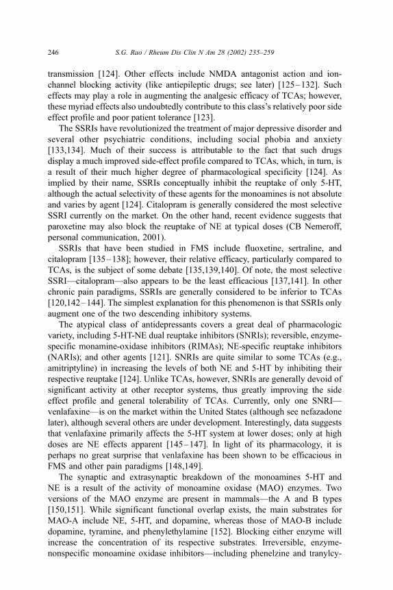
transmission [124]. Other effects include NMDA antagonist action and ion-
channel blocking activity (like antiepileptic drugs; see later) [125–132]. Such
effects may play a role in augmenting the analgesic efficacy of TCAs; however,
these myriad effects also undoubtedly contribute to this class’s relatively poor side
effect profile and poor patient tolerance [123].
The SSRIs have revolutionized the treatment of major depressive disorder and
several other psychiatric conditions, including social phobia and anxiety
[133,134]. Much of their success is attributable to the fact that such drugs
display a much improved side-effect profile compared to TCAs, which, in turn, is
a result of their much higher degree of pharmacological specificity [124]. As
implied by their name, SSRIs conceptually inhibit the reuptake of only 5-HT,
although the actual selectivity of these agents for the monoamines is not absolute
and varies by agent [124]. Citalopram is generally considered the most selective
SSRI currently on the market. On the other hand, recent evidence suggests that
paroxetine may also block the reuptake of NE at typical doses (CB Nemeroff,
personal communication, 2001).
SSRIs that have been studied in FMS include fluoxetine, sertraline, and
citalopram [135–138]; however, their relative efficacy, particularly compared to
TCAs, is the subject of some debate [135,139,140]. Of note, the most selective
SSRI—citalopram—also appears to be the least efficacious [137,141]. In other
chronic pain paradigms, SSRIs are generally considered to be inferior to TCAs
[120,142–144]. The simplest explanation for this phenomenon is that SSRIs only
augment one of the two descending inhibitory systems.
The atypical class of antidepressants covers a great deal of pharmacologic
variety, including 5-HT-NE dual reuptake inhibitors (SNRIs); reversible, enzyme-
specific monamine-oxidase inhibitors (RIMAs); NE-specific reuptake inhibitors
(NARIs); and other agents [121]. SNRIs are quite similar to some TCAs (e.g.,
amitriptyline) in increasing the levels of both NE and 5-HT by inhibiting their
respective reuptake [124]. Unlike TCAs, however, SNRIs are generally devoid of
significant activity at other receptor systems, thus greatly improving the side
effect profile and general tolerability of TCAs. Currently, only one SNRI—
venlafaxine—is on the market within the United States (although see nefazadone
later), although several others are under development. Interestingly, data suggests
that venlafaxine primarily affects the 5-HT system at lower doses; only at high
doses are NE effects apparent [145–147]. In light of its pharmacology, it is
perhaps no great surprise that venlafaxine has been shown to be efficacious in
FMS and other pain paradigms [148,149].
The synaptic and extrasynaptic breakdown of the monoamines 5-HT and
NE is a result of the activity of monoamine oxidase (MAO) enzymes. Two
versions of the MAO enzyme are present in mammals—the A and B types
[150,151]. While significant functional overlap exists, the main substrates for
MAO-A include NE, 5-HT, and dopamine, whereas those of MAO-B include
dopamine, tyramine, and phenylethylamine [152]. Blocking either enzyme will
increase the concentration of its respective substrates. Irreversible, enzyme-
nonspecific monoamine oxidase inhibitors—including phenelzine and tranylcy-
S.G. Rao / Rheum Dis Clin N Am 28 (2002) 235–259246

promine—have been on the US market for over 20 years; however, concerns
about potentially fatal interactions with other medications and with certain
foods containing tyramine have limited their widespread usage [151,152].
Newer agents that reversibly inhibit MAO-A—so-called RIMAs—have a much
improved safety profile compared with older drugs [151]. Currently, no RIMAs
are available in the United States, but at least two such agents are available in
parts of Europe—moclobemide and pirlindole. Pharmacologically, these agents
have effects that resemble those of SNRIs, and, thus, one would expect
reasonable efficacy in chronic pain; however, early data with moclobemide has
been unimpressive, with the agent demonstrating poor analgesic efficacy in
cases of neuropathic pain [153] and poor efficacy compared to amitriptyline in
FMS [154]. The data for pirlindole in FMS, however, shows more promise.
In a recent 4-week, randomized, double-blind controlled trial, Ginsberg et al.
found that pirlindole may be beneficial for certain symptoms of FMS, in-
cluding pain [155].
As stated above, NARIs specifically inhibit the reuptake of only norepineph-
rine [121]. While no NARIs are currently sold within the US marketplace, one
such agent, reboxetine, is marketed as an antidepressant in other parts of the
world [156]. The results of research into reboxetine’s efficacy in chronic pain
have yet to be published. Theoretically, one may expect analgesic efficacy in
chronic pain for this class to be perhaps slightly superior to that of SSRIs and
below that of SNRIs and TCAs. As is the case for SSRIs, only one descending
antinociceptive system is being activated. Unlike the projections from the RVM,
however, the pontine NE projections are thought to be entirely antinociceptive at
the level of the dorsal horn. Note, however, that the effects on unopposed NE
reuptake inhibition in RVM may actually be pronociceptive, as discussed
previously (see Fig. 3) [67].
Other atypical agents include nefazodone, mirtazipine, and buproprion.
Nefazodone is a potent 5-HT2 antagonist, although it also weakly blocks the
reuptake of both 5-HT and NE like an SNRI [157,158]. The 5-HT2 antagonist
actions appear important for increasing 5-HT1A–mediated neurotransmission in
animal models [158], and such activity may be useful in this agent’s potential
role as an analgesic [90]. There are no data on the efficacy of this agent in pain
syndromes at the present time; however, trazadone, an agent related to nefa-
zadone, has been relatively ineffective in the treatment of various pain syn-
dromes, including FMS [14]. Mirtazipine blocks a2 autoreceptors (mainly a2A)
and 5-HT2 and 5-HT3 receptors [159]. This agent has shown some potential in
some clinical pain conditions [160], and such analgesic activity may be
mediated by this agent’s ability to increase NE levels (by a2A blockade) and
increase 5-HT1A neurotransmission. As discussed later, the blockade of 5-HT3
receptors has both pro- and antinociceptive actions. Finally, buproprion is
thought to be a nonspecific monoamine reuptake inhibitor, preferentially block-
ing the reuptake of dopamine, with lesser effects on 5-HT and NE [142]. A
recent trial suggests that this agent may be effective in certain neuropathic pain
states [161].
S.G. Rao / Rheum Dis Clin N Am 28 (2002) 235–259 247

Opiates
Three different opioid receptors have been isolated within the CNS—the m, k,and d receptors—and all three appear to play a role in analgesia [60]. As
discussed previously, opiates act on both the ascending and descending pain
pathways. For example, it has been shown that m agonists such as morphine both
reduce transmitter release from the PAF terminals, and activate off cells within
the RVM [59–61,73].
In general, concerns about side effects and addiction have limited the chronic
use of opiates in FMS, particularly as the latter is not a life-threatening condition
[114]; however, one particularly interesting agent with modest opiate activity, in
widespread use, and with demonstrated efficacy in FMS is tramadol [162]. This
agent is unique in that it combines m-opiate–receptor agonist activity with 5-HT
and NE reuptake inhibition [163,164]. This combination of activities allows
tramadol to act at both ascending and descending sites [165], including those
mentioned previously for both m agonists and for antidepressants. Further, new
research suggests that the 5-HT1A receptor may also be involved in tramadol’s
analgesic effects [166]. Interestingly, tramadol may also be effective in psychi-
atric conditions including depression and obsessive compulsive disorder [167–
169], a fact consistent with its ability to block monoamine reuptake.
Antiepileptic drugs
A number of antiepileptic drugs (AEDs) have seen substantial use outside of
their primary indication, including in chronic pain and as mood stabilizers
[120,170]. Specific examples of agents within this class include gabapentin,
lamotrigine, topiramate, tiagabine, phenytoin, benzodiazepines (such as diaze-
pam), valproic acid, and carbamazepine. Pharmacologically, many of these
agents—including gabapentin, lamotrigine, topiramate, carbamazepine, and val-
proic acid—are cation channel (mainly sodium and calcium) blockers [170]. In
addition, many of these agents also have enhancing effects on GABAergic
neurotransmission; such agents include benzodiazepines, tiagabine, topiramate,
and valproic acid [171]. While the details vary, AEDs as a class have the potential
for relatively broad pharmacological effects across many components of the
peripheral and central nervous systems, generally decreasing excitability, reduc-
ing ectopic discharge, and reducing neurotransmitter release [171]. In particular,
the effects of AEDs on the ascending pathways may include reducing glutamate/
SP release from PAF terminals, directly decreasing the activation of dorsal horn
neurons, and increasing GABAergic input onto these neurons (Fig. 2). The
pharmacology of AEDs may be particularly suited for chronic pain due to nerve
injury, as such injury appears to lead to the expression of particular cation
channels that may play a role in ectopic discharge [172].
In some neuropathic pain paradigms, such as trigeminal neuralgia, AEDs
represent the first line of treatment [170]. However, only anecdotal data supports
the use of most of these agents in FMS, although two exceptions do exist.
S.G. Rao / Rheum Dis Clin N Am 28 (2002) 235–259248
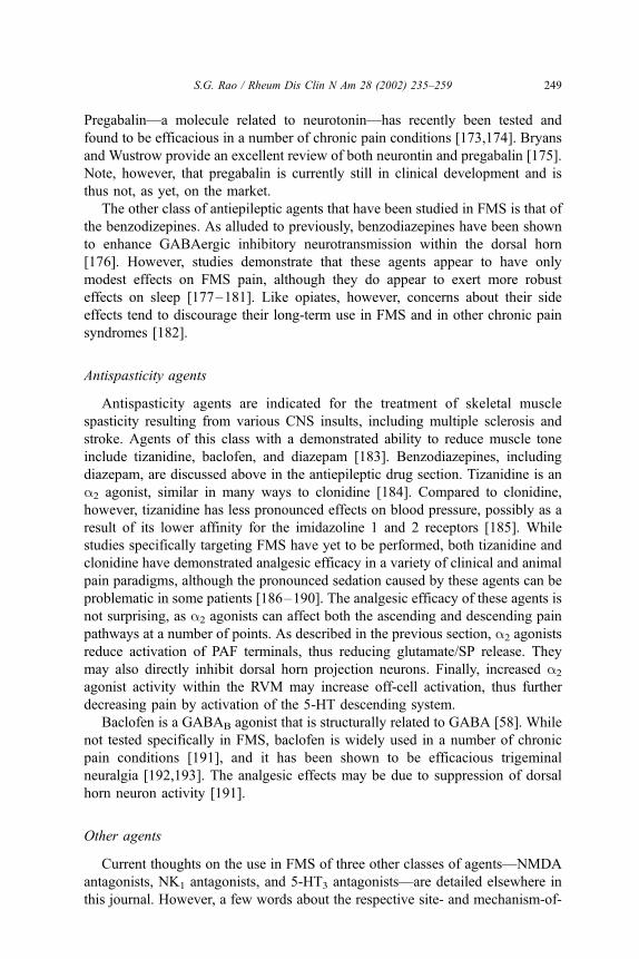
Pregabalin—a molecule related to neurotonin—has recently been tested and
found to be efficacious in a number of chronic pain conditions [173,174]. Bryans
and Wustrow provide an excellent review of both neurontin and pregabalin [175].
Note, however, that pregabalin is currently still in clinical development and is
thus not, as yet, on the market.
The other class of antiepileptic agents that have been studied in FMS is that of
the benzodizepines. As alluded to previously, benzodiazepines have been shown
to enhance GABAergic inhibitory neurotransmission within the dorsal horn
[176]. However, studies demonstrate that these agents appear to have only
modest effects on FMS pain, although they do appear to exert more robust
effects on sleep [177–181]. Like opiates, however, concerns about their side
effects tend to discourage their long-term use in FMS and in other chronic pain
syndromes [182].
Antispasticity agents
Antispasticity agents are indicated for the treatment of skeletal muscle
spasticity resulting from various CNS insults, including multiple sclerosis and
stroke. Agents of this class with a demonstrated ability to reduce muscle tone
include tizanidine, baclofen, and diazepam [183]. Benzodiazepines, including
diazepam, are discussed above in the antiepileptic drug section. Tizanidine is an
a2 agonist, similar in many ways to clonidine [184]. Compared to clonidine,
however, tizanidine has less pronounced effects on blood pressure, possibly as a
result of its lower affinity for the imidazoline 1 and 2 receptors [185]. While
studies specifically targeting FMS have yet to be performed, both tizanidine and
clonidine have demonstrated analgesic efficacy in a variety of clinical and animal
pain paradigms, although the pronounced sedation caused by these agents can be
problematic in some patients [186–190]. The analgesic efficacy of these agents is
not surprising, as a2 agonists can affect both the ascending and descending pain
pathways at a number of points. As described in the previous section, a2 agonists
reduce activation of PAF terminals, thus reducing glutamate/SP release. They
may also directly inhibit dorsal horn projection neurons. Finally, increased a2
agonist activity within the RVM may increase off-cell activation, thus further
decreasing pain by activation of the 5-HT descending system.
Baclofen is a GABAB agonist that is structurally related to GABA [58]. While
not tested specifically in FMS, baclofen is widely used in a number of chronic
pain conditions [191], and it has been shown to be efficacious trigeminal
neuralgia [192,193]. The analgesic effects may be due to suppression of dorsal
horn neuron activity [191].
Other agents
Current thoughts on the use in FMS of three other classes of agents—NMDA
antagonists, NK1 antagonists, and 5-HT3 antagonists—are detailed elsewhere in
this journal. However, a few words about the respective site- and mechanism-of-
S.G. Rao / Rheum Dis Clin N Am 28 (2002) 235–259 249

action of these agents are in order. As discussed above in the pain pathways
section, NMDA-mediated neurotransmission may play an important role in
mediating wind-up and related phenomena in at least two sites in the pain
pathways: at the PAF-dorsal horn neuron synapse and at the glutamatergic
synapses onto on cells within the RVM. In addition, NMDA antagonist may
help normalize SP-mediated neurotransmission, a feature that may be particularly
relevant to FMS (see below). In fact, three recent studies have demonstrated that
NMDA antagonists improve pain symptoms in FMS patients [194–196]. A poor
side-effect profile, however, represents a significant problem for this class of
agents [197].
One group has extensively studied the use of tropisetron, a 5-HT3 antagonist,
in the treatment of fibromyalgia [80,198–201]. Overall, this agent was found to
be modestly effective only within certain range of doses, with a loss of efficacy at
both lower and high levels [198]. A possible explanation of this phenomenon lies
in the fact that the blockade of 5-HT3 receptors has both pro- and antinociceptive
effects due to the presence of these receptors on both PAF terminals and
inhibitory dorsal horn interneurons (see Fig. 2). Thus, the balance of pro- and
antinociceptive effects may be highly dose-dependent, a fact that may lead to
unpredictable results in clinical practice.
The rationale for the use of NK1 antagonists in FMS is linked, in part, to the
observation that SP levels within the CSF of FMS patients are routinely elevated
[44,45]. As discussed previously, SP-mediated neurotransmission from the PAF
to the dorsal horn neuron has been shown to be important in the generation of
wind-up, although its role in the maintenance of such phenomena is unclear
[17,18,28]. NK1 antagonists have demonstrated analgesic efficacy in a number of
preclinical pain paradigms, particularly those modeling chronic pain. To date,
there have been no published reports of the use of NK1 antagonists in FMS;
however, the track record of this class of agents in human acute-and chronic-pain
studies has been extremely poor [41].
Summary
As demonstrated above, the anatomy and neuropharmacology of the pain
pathways within the CNS, even to the level of the midbrain, are extraordinarily
complex. Indeed, discussions of the effects of these agents on the neuropharma-
cology of the thalamus, hypothalamus, and cortex were excluded from this
review owing to their adding further to this complexity. Also, the dearth of data
regarding FMS pain pathophysiology necessitated a relatively generic analysis of
the pain pathways. As mentioned in the introduction, the current thought is that
central sensitization plays an important role in FMS. However, we see in this
chapter that the behavioral state of central sensitization may be a result of
alterations in either the ascending systems or in one or more descending systems.
Studies to assess the presence or relative importance of such changes in FMS are
difficult to perform in humans, and to date there are no animal models of FMS.
S.G. Rao / Rheum Dis Clin N Am 28 (2002) 235–259250

Accepting these limitations, it is apparent that many drugs considered to date
for the treatment of FMS do target a number of appropriate sites within both the
ascending and descending pain pathways. The data regarding clinical efficacy on
some good candidate agents, however, is extremely preliminary. For example, it
is evident from the present analysis that SNRIs, a2 agonists, and NK1 antagonists
may be particularly well suited to FMS, although current data supporting their
use is either anecdotal or from open-label trials [114,149]. Other sites within the
pain pathways have not yet been targeted. Examples of these include the use of
CCKB antagonists to block on-cell activation or of nitric oxide synthetase
antagonists to block the downstream mediators of NMDA activation. Efficacy
of such agents may give considerable insight into the pathophysiology of FMS.
Finally, as indicated previously, FMS consists of more than just chronic
pain, and the question of how sleep abnormalities, depression, fatigues, and so
forth tie into disordered pain processing is being researched actively. Future
research focusing on how the various manifestations of FMS relate to one
another undoubtedly will lead to a more rational targeting of drugs in this
complex disorder.
References
[1] Clauw DJ, Chrousos GP. Chronic pain and fatigue syndromes: overlapping clinical and neuro-
endocrine features and potential pathogenic mechanisms. Neuroimmunomodulation 1997;4:
134–53.
[2] Goldenberg DL. Fibromyalgia syndrome a decade later: what have we learned? Arch Intern
Med 1999;159:777–85.
[3] Wolfe F, Smythe HA, Yunus MB, Bennett RM, Bombardier C, Goldenberg DL, et al. The
American College of Rheumatology 1990 criteria for the classification of fibromyalgia. Report
of the Multicenter Criteria Committee. Arthritis Rheum 1990;33:160–72.
[4] Arroyo JF, Cohen ML. Abnormal responses to electrocutaneous stimulation in fibromyalgia.
J Rheumatol 1993;20:1925–31.
[5] Staud R, Vierck CJ, Cannon RL, et al. Abnormal sensitization and temporal summation of
second pain (wind-up) in patients with fibromyalgia syndrome. Pain 2001;91:165–75.
[6] Olsen NJ, Park JH. Skeletal muscle abnormalities in patients with fibromyalgia. Am J Med Sci
1998;315:351–8.
[7] Simms RW. Fibromyalgia is not a muscle disorder. Am J Med Sci 1998;315:346–50.
[8] Simms RW, Roy SH, Hrovat M, et al. Lack of association between fibromyalgia syndrome
and abnormalities in muscle energy metabolism [see comments]. Arthritis Rheum 1994;37:
794–800.
[9] Bennett RM. Emerging concepts in the neurobiology of chronic pain: evidence of abnormal
sensory processing in fibromyalgia. Mayo Clin Proc 1999;74:385–98.
[10] Yunus MB. Towards a model of pathophysiology of fibromyalgia: aberrant central pain mech-
anisms with peripheral modulation. J Rheumatol 1992;19:846–50.
[11] Woolf CJ. Windup and central sensitization are not equivalent. Pain 1996;66:105–8.
[12] Melzack R, Wall PD. Pain mechanisms: a new theory. Science 1965;150:971–9.
[13] Dickenson AH. Spinal cord pharmacology of pain. Br J Anaesth 1995;75:193–200.
[14] Coderre TJ, Katz J, Vaccarino AL, et al. Contribution of central neuroplasticity to pathological
pain: review of clinical and experimental evidence. Pain 1993;52:259–85.
[15] Crofford LJ, Casey KL. Central modulation of pain perception. Rheum Dis Clin North Am
1999;25:1–13.
S.G. Rao / Rheum Dis Clin N Am 28 (2002) 235–259 251

[16] Willis WD, Westlund KN. Neuroanatomy of the pain system and of the pathways that modulate
pain. J Clin Neurophysiol 1997;14:2–31.
[17] Eide PK. Wind-up and the NMDA receptor complex from a clinical perspective. Eur J Pain
2000;4:5–15.
[18] Herrero JF, Laird JM, Lopez-Garcia JA. Wind-up of spinal cord neurones and pain sensation:
much ado about something? Prog Neurobiol 2000;61:169–203.
[19] Woolf CJ. Evidence for a central component of post-injury pain hypersensitivity. Nature
1983;306:686–8.
[20] Hanai F. C fiber responses of wide dynamic range neurons in the spinal dorsal horn. Clin
Orthop 1998;349:256–67.
[21] Pertovaara A, Wei H, Hamalainen MM. Lidocaine in the rostroventromedial medulla and
the periaqueductal gray attenuates allodynia in neuropathic rats. Neurosci Lett 1996;218:
127–30.
[22] Le Bars D, Dickenson AH, Besson JM. Diffuse noxious inhibitory controls (DNIC). I. Effects
on dorsal horn convergent neurones in the rat. Pain 1979;6:283–304.
[23] Le Bars D, Dickenson AH, Besson JM. Diffuse noxious inhibitory controls (DNIC). II. Lack of
effect on non-convergent neurones, supraspinal involvement and theoretical implications. Pain
1979;6:305–27.
[24] Morgan MM, Heinricher MM, Fields HL. Inhibition and facilitation of different nocifensor
reflexes by spatially remote noxious stimuli. J Neurophysiol 1994;72:1152–60.
[25] Cull-Candy S, Brickley S, Farrant M. NMDA receptor subunits: diversity, development and
disease. Curr Opin Neurobiol 2001;11:327–35.
[26] Davies SN, Lodge D. Evidence for involvement of N-methylaspartate receptors in ‘wind-up’ of
class 2 neurones in the dorsal horn of the rat. Brain Res 1987;424:402–6.
[27] Dickenson AH. A cure for wind up: NMDA receptor antagonists as potential analgesics. Trends
Pharmacol Sci 1990;11:307–9.
[28] Dickenson AH, Sullivan AF. Electrophysiological studies on the effects of intrathecal morphine
on nociceptive neurones in the rat dorsal horn. Pain 1986;24:211–22.
[29] Dougherty PM, Palecek J, Paleckova V, et al. The role of NMDA and non-NMDA excitatory
amino acid receptors in the excitation of primate spinothalamic tract neurons by mechanical,
chemical, thermal, and electrical stimuli. J Neurosci 1992;12:3025–41.
[30] Meller ST, Pechman PS, Gebhart GF, et al. Nitric oxide mediates the thermal hyperalgesia
produced in a model of neuropathic pain in the rat. Neuroscience 1992;50:7–10.
[31] Urban MO, Gebhart GF. Supraspinal contributions to hyperalgesia. Proc Natl Acad Sci USA
1999;96:7687–92.
[32] Yaksh TL, Hua XY, Kalcheva I, et al. The spinal biology in humans and animals of pain states
generated by persistent small afferent input. Proc Natl Acad Sci USA 1999;96:7680–6.
[33] Duggan AW, Hendry IA, Morton CR, et al. Cutaneous stimuli releasing immunoreactive sub-
stance P in the dorsal horn of the cat. Brain Res 1988;451:261–73.
[34] Quartara L, Maggi CA. The tachykinin NK1 receptor. Part I: ligands and mechanisms of
cellular activation. Neuropeptides 1997;31:537–63.
[35] Quartara L, Maggi CA. The tachykinin NK1 receptor. Part II: distribution and pathophysiolo-
gical roles. Neuropeptides 1998;32:1–49.
[36] Maggi CA. Principles of tachykininergic co-transmission in the peripheral and enteric nervous
system. Regul Pept 2000;93:53–64.
[37] Otsuka M, Yoshioka K. Neurotransmitter functions of mammalian tachykinins. Physiol Rev
1993;73:229–308.
[38] Saria A. The tachykinin NK1 receptor in the brain: pharmacology and putative functions. Eur J
Pharmacol 1999;375:51–60.
[39] Yashpal K, Dam TV, Quirion R. Quantitative autoradiographic distribution of multiple neuro-
kinin binding sites in rat spinal cord. Brain Res 1990;506:259–66.
[40] Walpole C, Ko SY, Brown M, et al. 2-Nitrophenylcarbamoyl-(S)-prolyl-(S)-3-(2-naphthyl)
alanyl-N-benzyl-N-methylamide (SDZ NKT 343), a potent human NK1 tachykinin receptor
S.G. Rao / Rheum Dis Clin N Am 28 (2002) 235–259252

antagonist with good oral analgesic activity in chronic pain models. J Med Chem 1998;
41:3159–73.
[41] Hill R. NK1 (substance P) receptor antagonists: why are they not analgesic in humans? Trends
Pharmacol Sci 2000;21:244–6.
[42] Dougherty PM, Palecek J, Zorn S, et al. Combined application of excitatory amino acids and
substance P produces long-lasting changes in responses of primate spinothalamic tract neurons.
Brain Res Brain Res Rev 1993;18:227–46.
[43] Dougherty PM, Willis WD. Enhancement of spinothalamic neuron responses to chemical
and mechanical stimuli following combined micro-iontophoretic application of N-methyl-D-
aspartic acid and substance P. Pain 1991;47:85–93.
[44] Russell IJ, Orr MD, Littman B, et al. Elevated cerebrospinal fluid levels of substance P in
patients with the fibromyalgia syndrome. Arthritis Rheum 1994;37:1593–601.
[45] Vaeroy H, Helle R, Forre O, et al. Elevated CSF levels of substance P and high incidence of
Raynaud phenomenon in patients with fibromyalgia: new features for diagnosis. Pain 1988;
32:21–6.
[46] Sarchielli P, Alberti A, Floridi A, et al. Levels of nerve growth factor in cerebrospinal fluid of
chronic daily headache patients. Neurology 2001;57:132–4.
[47] Strittmatter M, Grauer M, Isenberg E, et al. Cerebrospinal fluid neuropeptides and monoami-
nergic transmitters in patients with trigeminal neuralgia. Headache 1997;37:211–6.
[48] Larson AA, Giovengo SL, Russell IJ, et al. Changes in the concentrations of amino acids in the
cerebrospinal fluid that correlate with pain in patients with fibromyalgia: implications for nitric
oxide pathways. Pain 2000;87:201–11.
[49] Tsigos C, Diemel LT, White A, et al. Cerebrospinal fluid levels of substance P and calcitonin-
gene-related peptide: correlation with sural nerve levels and neuropathic signs in sensory
diabetic polyneuropathy. Clin Sci (Colch) 1993;84:305–11.
[50] Cridland RA, Henry JL. Comparison of the effects of substance P, neurokinin A, physalaemin
and eledoisin in facilitating a nociceptive reflex in the rat. Brain Res 1986;381:93–9.
[51] Dirig DM, Yaksh TL. In vitro prostanoid release from spinal cord following peripheral inflam-
mation: effects of substance P, NMDA and capsaicin. Br J Pharmacol 1999;126:1333–40.
[52] Moochhala SM, Sawynok J. Hyperalgesia produced by intrathecal substance P and related
peptides: desensitization and cross desensitization. Br J Pharmacol 1984;82:381–8.
[53] Hua XY, Chen P, Marsala M, et al. Intrathecal substance P-induced thermal hyperalgesia and
spinal release of prostaglandin E2 and amino acids. Neuroscience 1999;89:525–34.
[54] Okano K, Kuraishi Y, Satoh M. Pharmacological evidence for involvement of excitatory amino
acids in aversive responses induced by intrathecal substance P in rats. Biol Pharm Bull 1993;
16:861–5.
[55] McLeod AL, Ritchie J, Cuello AC, et al. Transgenic mice over-expressing substance P exhibit
allodynia and hyperalgesia which are reversed by substance P and N-methyl-D-aspartate
receptor antagonists. Neuroscience 1999;89:891–9.
[56] Pasternak GW, Wood PJ. Multiple mu opiate receptors. Life Sci 1986;38:1889–98.
[57] Barry PH, Schofield PR, Moorhouse AJ. Glycine receptors: what gets in and why? Clin Exp
Pharmacol Physiol 1999;26:935–6.
[58] Chebib M, Johnston GA. The ‘ABC’ of GABA receptors: a brief review. Clin Exp Pharmacol
Physiol 1999;26:937–40.
[59] Besse D, Lombard MC, Zajac JM, et al. Pre- and postsynaptic distribution of mu, delta and
kappa opioid receptors in the superficial layers of the cervical dorsal horn of the rat spinal cord.
Brain Res 1990;521:15–22.
[60] Yaksh TL. Pharmacology and mechanisms of opioid analgesic activity. Acta Anaesthesiol
Scand 1997;41:94–111.
[61] Duale C, Raboisson P, Molat JL, et al. Systemic morphine reduces the wind-up of trigeminal
nociceptive neurons. Neuroreport 2001;12:2091–6.
[62] Behbehani MM. Functional characteristics of the midbrain periaqueductal gray. Prog Neurobiol
1995;46:575–605.
S.G. Rao / Rheum Dis Clin N Am 28 (2002) 235–259 253

[63] Millar J, Williams GV. Effects of iontophoresis of noradrenaline and stimulation of the peri-
aqueductal gray on single-unit activity in the rat superficial dorsal horn. J Comp Neurol
1989;287:119–33.
[64] Zhang DX, Owens CM, Willis WD. Two forms of inhibition of spinothalamic tract neurons
produced by stimulation of the periaqueductal gray and the cerebral cortex. J Neurophysiol
1991;65:1567–79.
[65] Kwiat GC, Basbaum AI. Organization of tyrosine hydroxylase-and serotonin-immunoreactive
brainstem neurons with axon collaterals to the periaqueductal gray and the spinal cord in the rat.
Brain Res 1990;528:83–94.
[66] Ennis M, Behbehani M, Shipley MT, et al. Projections from the periaqueductal gray to the
rostromedial pericoerulear region and nucleus locus coeruleus: anatomic and physiologic stud-
ies. J Comp Neurol 1991;306:480–94.
[67] Fields HL, Heinricher MM, Mason P. Neurotransmitters in nociceptive modulatory circuits.
Annu Rev Neurosci 1991;14:219–45.
[68] Fields HL, Malick A, Burstein R. Dorsal horn projection targets of ON and OFF cells in the
rostral ventromedial medulla. J Neurophysiol 1995;74:1742–59.
[69] Porreca F, Burgess SE, Gardell LR, Vanderah TW, Malan TP Jr, Ossipov MH, et al. Inhibition
of neuropathic pain by selective ablation of brainstem medullary cells expressing the micro-
opioid receptor. J Neurosci 2001;21(14):5281–8.
[70] Vasko MR, Pang IH, Vogt M. Involvement of 5-hydroxytryptamine-containing neurons in
antinociception produced by injection of morphine into nucleus raphe magnus or onto spinal
cord. Brain Res 1984;306:341–8.
[71] Bowery NG, Hudson AL, Price GW. GABAA and GABAB receptor site distribution in the rat
central nervous system. Neuroscience 1987;20:365–83.
[72] Drower EJ, Hammond DL. GABAergic modulation of nociceptive threshold: effects of
THIP and bicuculline microinjected in the ventral medulla of the rat. Brain Res 1988;450:
316–24.
[73] Fields HL, Vanegas H, Hentall ID, et al. Evidence that disinhibition of brain stem neurones
contributes to morphine analgesia. Nature 1983;306:684–6.
[74] Heinricher MM, Roychowdhury SM. Reflex-related activation of putative pain facilitating
neurons in rostral ventromedial medulla requires excitatory amino acid transmission. Neuro-
science 1997;78:1159–65.
[75] Pan ZZ, Williams JT, Osborne PB. Opioid actions on single nucleus raphe magnus neurons
from rat and guinea-pig in vitro. J Physiol 1990;427:519–32.
[76] Llewelyn MB, Azami J, Roberts MH. The effect of modification of 5-hydroxytryptamine
function in nucleus raphe magnus on nociceptive threshold. Brain Res 1984;306:165–70.
[77] Oliveras JL, Guilbaud G, Besson JM. A map of serotoninergic structures involved in stimula-
tion producing analgesia in unrestrained freely moving cats. Brain Res 1979;164:317–22.
[78] Peng YB, Lin Q, Willis WD. The role of 5-HT3 receptors in periaqueductal gray-induced
inhibition of nociceptive dorsal horn neurons in rats. J Pharmacol Exp Ther 1996;276:116–24.
[79] Paul D, Yao D, Zhu P, et al. 5-hydroxytryptamine3 (5-HT3) receptors mediate spinal 5-HT
antinociception: an antisense approach. J Pharmacol Exp Ther 2001;298:674–8.
[80] Wolf H. Preclinical and clinical pharmacology of the 5-HT3 receptor antagonists. Scand J
Rheumatol 2000;113:37–45.
[81] Sawynok J, Reid A. Interactions of descending serotonergic systems with other neurotransmit-
ters in the modulation of nociception. Behav Brain Res 1996;73:63–8.
[82] Lin Q, Peng Y, Willis WD. Glycine and GABAA antagonists reduce the inhibition of primate
spinothalamic tract neurons produced by stimulation in periaqueductal gray. Brain Res 1994;
654:286–302.
[83] Peng YB, Lin Q, Willis WD. Effects of GABA and glycine receptor antagonists on the activity
and PAG-induced inhibition of rat dorsal horn neurons. Brain Res 1996;736:189–201.
[84] Hamon M, Gallissot MC, Menard F, et al. 5-HT3 receptor binding sites are on capsaicin-
sensitive fibres in the rat spinal cord. Eur J Pharmacol 1989;164:315–22.
S.G. Rao / Rheum Dis Clin N Am 28 (2002) 235–259254

[85] Greenshaw AJ, Silverstone PH. The non-antiemetic uses of serotonin 5-HT3 receptor antago-
nists. Clinical pharmacology and therapeutic applications. Drugs 1997;53:20–39.
[86] Saria A, Javorsky F, Humpel C, et al. 5-HT3 receptor antagonists inhibit sensory neuropeptide
release from the rat spinal cord. Neuroreport 1990;1:104–6.
[87] Yaksh TL, Wilson PR. Spinal serotonin terminal system mediates antinociception. J Pharmacol
Exp Ther 1979;208:446–53.
[88] Bardin L, Tarayre JP, Koek W, et al. In the formalin model of tonic nociceptive pain, 8-OH-
DPAT produces 5-HT1A receptor-mediated, behaviorally specific analgesia. Eur J Pharmacol
2001;421:109–14.
[89] Furst S. Transmitters involved in antinociception in the spinal cord. Brain Res Bull 1999;48:
129–41.
[90] Lin Q, Peng YB, Willis WD. Antinociception and inhibition from the periaqueductal gray are
mediated in part by spinal 5-hydroxytryptamine(1A) receptors. J Pharmacol Exp Ther 1996;276:
958–67.
[91] Westlund KN, Coulter JD. Descending projections of the locus coeruleus and subcoeruleus/
medial parabrachial nuclei in monkey: axonal transport studies and dopamine-beta-hydroxylase
immunocytochemistry. Brain Res 1980;2:235–64.
[92] Westlund KN, Zhang D, Carlton SM, et al. Noradrenergic innervation of somatosensory tha-
lamus and spinal cord. Prog Brain Res 1991;88:77–88.
[93] Headley PM, Duggan AW, Griersmith BT. Selective reduction by noradrenaline and 5-hydrox-
ytryptamine of nociceptive responses of cat dorsal horn neurones. Brain Res 1978;145:185–9.
[94] Reddy SV, Maderdrut JL, Yaksh TL. Spinal cord pharmacology of adrenergic agonist-mediated
antinociception. J Pharmacol Exp Ther 1980;213:525–33.
[95] Reddy SV, Yaksh TL. Spinal noradrenergic terminal system mediates antinociception. Brain
Res 1980;189:391–401.
[96] Kuraishi Y, Hirota N, Satoh M, et al. Antinociceptive effects of intrathecal opioids, noradrena-
line and serotonin in rats: mechanical and thermal algesic tests. Brain Res 1985;326:168–71.
[97] Kuraishi Y, Hirota N, Sato Y, et al. Noradrenergic inhibition of the release of substance P from
the primary afferents in the rabbit spinal dorsal horn. Brain Res 1985;359:177–82.
[98] North RA, Yoshimura M. The actions of noradrenaline on neurones of the rat substantia
gelatinosa in vitro. J Physiol 1984;349:43–55.
[99] Bourgoin S, Pohl M, Mauborgne A, et al. Monoaminergic control of the release of calcitonin
gene-related peptide and substance P-like materials from rat spinal cord slices. Neuropharma-
cology 1993;32:633–40.
[100] Hord AH, Denson DD, Stowe B, et al. alpha-1 and alpha-2 adrenergic antagonists relieve
thermal hyperalgesia in experimental mononeuropathy from chronic constriction injury. Anesth
Analg 2001;92:1558–62.
[101] Lee YH, Ryu TG, Park SJ, et al. Alpha1-adrenoceptors involvement in painful diabetic neuro-
pathy: a role in allodynia. Neuroreport 2000;11:1417–20.
[102] Takagi H, Yamamoto K, Shiosaka S, et al. Morphological study of noradrenaline innervation
in the caudal raphe nuclei with special reference to fine structure. J Comp Neurol 1981;203:
15–22.
[103] Young WS 3rd, Kuhar MJ. Noradrenergic alpha 1 and alpha 2 receptors: light microscopic
autoradiographic localization. Proc Natl Acad Sci USA 1980;77:1696–700.
[104] Hammond DL, Levy RA, Proudfit HK. Hypoalgesia following microinjection of noradrenergic
antagonists in the nucleus raphe magnus. Pain 1980;9:85–101.
[105] Hammond DL, Levy RA, Proudfit HK. Hypoalgesia induced by microinjection of a norepi-
nephrine antagonist in the raphe magnus: reversal by intrathecal administration of a serotonin
antagonist. Brain Res 1980;201:475–9.
[106] Sagen J, Proudfit HK. Evidence for pain modulation by pre- and postsynaptic noradrenergic
receptors in the medulla oblongata. Brain Res 1985;331:285–93.
[107] Jones SL, Gebhart GF. Spinal pathways mediating tonic, coeruleospinal, and raphe-spinal
descending inhibition in the rat. J Neurophysiol 1987;58:138–59.
S.G. Rao / Rheum Dis Clin N Am 28 (2002) 235–259 255

[108] DeLander GE, Hopkins CJ. Interdependence of spinal adenosinergic, serotonergic and nor-
adrenergic systems mediating antinociception. Neuropharmacology 1987;26:1791–4.
[109] Porreca F, Burgess SE, Gardell LR, et al. Inhibition of neuropathic pain by selective ablation of
brainstem medullary cells expressing the micro-opioid receptor. J Neurosci 2001;21:5281–8.
[110] Kovelowski CJ, OssipovMH, SunH, et al. Supraspinal cholecystokininmay drive tonic descend-
ing facilitation mechanisms to maintain neuropathic pain in the rat. Pain 2000;87: 265–73.
[111] Coutinho SV, Urban MO, Gebhart GF. Role of glutamate receptors and nitric oxide in the rostral
ventromedial medulla in visceral hyperalgesia. Pain 1998;78:59–69.
[112] Urban MO, Coutinho SV, Gebhart GF. Involvement of excitatory amino acid receptors and
nitric oxide in the rostral ventromedial medulla in modulating secondary hyperalgesia produced
by mustard oil. Pain 1999;81:45–55.
[113] Urban MO, Gebhart GF. The glutamate synapse: a target in the pharmacological management of
hyperalgesic pain states. Prog Brain Res 1998;116:407–20.
[114] Bennett RM. Pharmacological treatment of fibromyalgia. Journal of Functional Syndromes 2001;
1:79–92.
[115] Buskila D. Drug therapy. Baillieres Best Pract Res Clin Rheumatol 1999;13:479–85.
[116] Lautenschlager J. Present state of medication therapy in fibromyalgia syndrome. Scand J
Rheumatol 2000;113:32–6.
[117] Leventhal LJ. Management of fibromyalgia. Ann Intern Med 1999;131:850–8.
[118] Goldenberg DL, Felson DT, Dinerman H. A randomized, controlled trial of amitriptyline and
naproxen in the treatment of patients with fibromyalgia. Arthritis Rheum 1986;29:1371–7.
[119] Russell IJ, Fletcher EM, Michalek JE, et al. Treatment of primary fibrositis/fibromyalgia syn-
drome with ibuprofen and alprazolam. A double-blind, placebo-controlled study. Arthritis
Rheum 1991;34:552–60.
[120] Sindrup SH, Jensen TS. Efficacy of pharmacological treatments of neuropathic pain: an update
and effect related to mechanism of drug action. Pain 1999;83:389–400.
[121] Kent JM. SNaRIs, NaSSAs, and NaRIs: new agents for the treatment of depression. Lancet
2000;355:911–8.
[122] Commissiong JW, Karoum F, Reiffenstein RJ, et al. Cyclobenzaprine: a possible mechanism of
action for its muscle relaxant effect. Can J Physiol Pharmacol 1981;59:37–44.
[123] Kobayashi H, Hasegawa Y, Ono H. Cyclobenzaprine, a centrally acting muscle relaxant, acts on
descending serotonergic systems. Eur J Pharmacol 1996;311:29–35.
[124] Sanchez C, Hyttel J. Comparison of the effects of antidepressants and their metabolites on
reuptake of biogenic amines and on receptor binding. Cell Mol Neurobiol 1999;19:467–89.
[125] Cai Z, McCaslin PP. Amitriptyline, desipramine, cyproheptadine and carbamazepine, in con-
centrations used therapeutically, reduce kainate and N-methyl-D-aspartate-induced intracellular
Ca2+ levels in neuronal culture. Eur J Pharmacol 1992;219:53–7.
[126] Eisenach JC, Gebhart GF. Intrathecal amitriptyline acts as an N-methyl-D-aspartate receptor
antagonist in the presence of inflammatory hyperalgesia in rats. Anesthesiology 1995;83:
1046–54.
[127] Kuo CC. Imipramine inhibition of transient K+ current: an external open channel blocker
preventing fast inactivation. Biophys J 1998;75:2845–57.
[128] McCaslin PP, Yu XZ, Ho IK, et al. Amitriptyline prevents N-methyl-D-aspartate (NMDA)-
induced toxicity, does not prevent NMDA-induced elevations of extracellular glutamate, but
augments kainate-induced elevations of glutamate. J Neurochem 1992;59:401–5.
[129] Nakazawa K, Inoue K, Ohno Y. Block and unblock by imipramine of cloned and mutated P2X2
receptor/channel expressed in Xenopus oocytes. Neurosci Lett 1999;264:93–6.
[130] Park TJ, Shin SY, Suh BC, et al. Differential inhibition of catecholamine secretion by amitripty-
line through blockage of nicotinic receptors, sodium channels, and calcium channels in bovine
adrenal chromaffin cells. Synapse 1998;29:248–56.
[131] Reynolds IJ, Miller RJ. Tricyclic antidepressants block N-methyl-D-aspartate receptors: sim-
ilarities to the action of zinc. Br J Pharmacol 1988;95:95–102.
[132] Watanabe Y, Saito H, Abe K. Tricyclic antidepressants block NMDA receptor-mediated syn-
S.G. Rao / Rheum Dis Clin N Am 28 (2002) 235–259256

aptic responses and induction of long-term potentiation in rat hippocampal slices. Neurophar-
macology 1993;32:479–86.
[133] Brunello N, den Boer JA, Judd LL, et al. Social phobia: diagnosis and epidemiology, neuro-
biology and pharmacology, comorbidity and treatment. J Affect Disord 2000;60:61–74.
[134] Nutt DJ, Forshall S, Bell C, et al. Mechanisms of action of selective serotonin reuptake
inhibitors in the treatment of psychiatric disorders. Eur Neuropsychopharmacol 1999;9
(Suppl 3):S81–6.
[135] Celiker R, Cagavi Z. Comparison of amitriptyline and sertraline in the treatment of fibromyal-
gia syndrome. Poster presented at: 20th Annual Scientific Meeting of the American Pain
Society; April 20, 2001.
[136] Goldenberg D, Mayskiy M, Mossey C, et al. A randomized, double-blind crossover trial of
fluoxetine and amitriptyline in the treatment of fibromyalgia. Arthritis Rheum 1996;39:1852–9.
[137] Norregaard J, Volkmann H, Danneskiold-Samsoe B. A randomized controlled trial of citalo-
pram in the treatment of fibromyalgia. Pain 1995;61:445–9.
[138] Wolfe F, Cathey MA, Hawley DJ. A double-blind placebo controlled trial of fluoxetine in
fibromyalgia. Scand J Rheumatol 1994;23:255–9.
[139] Arnold LM, Keck PE Jr, Welge JA. Antidepressant treatment of fibromyalgia. A meta-analysis
and review. Psychosomatics 2000;41:104–13.
[140] O’Malley PG, Balden E, Tomkins G, et al. Treatment of fibromyaglia with antidepresants: a
mete-analysis. J Gen Intern Med 2000;15:659–66.
[141] Anderberg UM, Marteinsdottir I, von Knorring L. Citalopram in patients with fibromyalgia: a
randomized, double-blind, placebo-controlled study. Eur J Pain 2000;4:27–35.
[142] Ansari A. The efficacy of newer antidepressants in the treatment of chronic pain: a review of
current literature. Harv Rev Psychiatry 2000;7:257–77.
[143] Atkinson JH, Slater MA, Wahlgren DR, et al. Effects of noradrenergic and serotonergic anti-
depressants on chronic low back pain intensity. Pain 1999;83:137–45.
[144] Jung AC, Staiger T, Sullivan M. The efficacy of selective serotonin reuptake inhibitors for the
management of chronic pain [see comments]. J Gen Intern Med 1997;12:384–9.
[145] Beique JC, Lavoie N, de Montigny C, et al. Affinities of venlafaxine and various reuptake
inhibitors for the serotonin and norepinephrine transporters. Eur J Pharmacol 1998;349:
129–32.
[146] Harvey AT, Rudolph RL, Preskorn SH. Evidence of the dual mechanisms of action of ven-
lafaxine. Arch Gen Psychiatry 2000;57:503–9.
[147] Roseboom PH, Kalin NH. Neuropharmacology of venlafaxine. Depress Anxiety 2000;12:20–9.
[148] Barkin RL, Fawcett J. The management challenges of chronic pain: the role of antidepressants.
Am J Ther 2000;7:31–47.
[149] Dwight MM, Arnold LM, O’Brien H, et al. An open clinical trial of venlafaxine treatment of
fibromyalgia. Psychosomatics 1998;39:14–7.
[150] Lenders JW, Eisenhofer G, Abeling NG, et al. Specific genetic deficiencies of the A and B
isoenzymes of monoamine oxidase are characterized by distinct neurochemical and clinical
phenotypes. J Clin Invest 1996;97:1010–9.
[151] Livingston MG, Livingston HM. Monoamine oxidase inhibitors. An update on drug interac-
tions. Drug Saf 1996;14:219–27.
[152] Krishnan KRR. Monoamine oxidase inhibitors. In: Schatzberg AF, Nemeroff CB, editors.
Textbook of psychopharmacology, 2nd edition. Washington, DC: American Psychiatric Press;
1998. p. 239–49.
[153] Menkes DB, Fawcett JP, Busch AF, et al. Moclobemide in chronic neuropathic pain: prelimi-
nary case reports. Clin J Pain 1995;11:134–8.
[154] Hannonen P, Malminiemi K, Yli-Kerttula U, et al. A randomized, double-blind, placebo-
controlled study of moclobemide and amitriptyline in the treatment of fibromyalgia in females
without psychiatric disorder. Br J Rheumatol 1998;37:1279–86.
[155] Ginsberg F, Joos E, Geczy J, et al. A pilot randomized placebo-controlled study of pirlindole in
the treatment of primary fibromyalgia. J Musculoskeletal Pain 1998;6:5–17.
S.G. Rao / Rheum Dis Clin N Am 28 (2002) 235–259 257

[156] Berzewski H, Van Moffaert M, Gagiano CA. Efficacy and tolerability of reboxetine compared
with imipramine in a double-blind study in patients suffering from major depressive offsodes.
Eur Neuropsychopharmacol 1997;7(Suppl 1):S37–47 [discussion S71–3].
[157] Bolden-Watson C, Richelson E. Blockade by newly-developed antidepressants of biogenic
amine uptake into rat brain synaptosomes. Life Sci 1993;52:1023–9.
[158] Eison AS, Eison MS, Torrente JR, et al. Nefazodone: preclinical pharmacology of a new
antidepressant. Psychopharmacol Bull 1990;26:311–5.
[159] de Boer T. The effects of mirtazapine on central noradrenergic and serotonergic neurotrans-
mission. Int Clin Psychopharmacol 1995;10(Suppl 4):19–23.
[160] Brannon GE, Stone KD. The use of mirtazapine in a patient with chronic pain. J Pain Symptom
Manage 1999;18:382–5.
[161] Semenchuk MR, Sherman S, Davis B. Double-blind randomized trial of bupropion sustained
release for the treatment of neuropathic pain. Neurology 2001;57(9):1583–8.
[162] Russell IJ, Kamin M, Bennett R, et al. Efficacy of tramadol in treatment of pain in fibromyalgia.
J Clin Rheumatol 2000;6:250–7.
[163] Driessen B, Reimann W. Interaction of the central analgesic, tramadol, with the uptake and
release of 5-hydroxytryptamine in the rat brain in vitro. Br J Pharmacol 1992;105:147–51.
[164] Reimann W, Hennies HH. Inhibition of spinal noradrenaline uptake in rats by the centrally
acting analgesic tramadol. Biochem Pharmacol 1994;47:2289–93.
[165] Raffa RB, Friderichs E, Reimann W, et al. Opioid and nonopioid components independently
contribute to the mechanism of action of tramadol, an ‘atypical’ opioid analgesic. J Pharmacol
Exp Ther 1992;260:275–85.
[166] Rojas-Corrales MO, Ortega-Alvaro A, Gibert-Rahola J, et al. Pindolol, a beta-adrenoceptor
blocker/5-hydroxytryptamine(1A/1B) antagonist, enhances the analgesic effect of tramadol.
Pain 2000;88:119–24.
[167] Markowitz JS, Patrick KS. Venlafaxine-tramadol similarities. Med Hypotheses 1998;51:167–8.
[168] Rojas-Corrales MO, Gibert-Rahola J, Mico JA. Tramadol induces antidepressant-type effects in
mice. Life Sci 1998;63:L175–80.
[169] Spencer C. The efficacy of intramuscular tramadol as a rapid-onset antidepressant. Aust N Z J
Psychiatry 2000;34:1032–3.
[170] Tremont-Lukats IW, Megeff C, Backonja MM. Anticonvulsants for neuropathic pain syn-
dromes: mechanisms of action and place in therapy. Drugs 2000;60:1029–52.
[171] Czuczwar SJ, Patsalos PN. The new generation of GABA enhancers. Potential in the treatment
of epilepsy. CNS Drugs 2001;15:339–50.
[172] Woolf CJ, Mannion RJ. Neuropathic pain: aetiology, symptoms, mechanisms, and manage-
ment. Lancet 1999;353:1959–64.
[173] Corbin AE, Remmers AE, LaMoreaux L, et al. Pregabalin reduces pain in patients
with postherpetic neuralgia. Presented at the American Pain Society 20th Annual Meeting.
Phoenix, AZ, April 19–22, 2001.
[174] Sharma U, Iacobellis D, Glessner C, et al. Two studies show pregabalin effectively relieves pain
in patients with painful diabetic peripheral neuropathy (studies 1008–14/-29). Presented at the
19th American Pain Society Annual Meeting, Atlanta, GA, November 2–5, 2000.
[175] Bryans JS, Wustrow DJ. 3-substituted GABA analogs with central nervous system activity: a
review. Med Res Rev 1999;19:149–77.
[176] Kohno T, Kumamoto E, Baba H, et al. Actions of midazolam on GABAergic transmission in
substantia gelatinosa neurons of adult rat spinal cord slices. Anesthesiology 2000;92:507–15.
[177] Drewes AM, Andreasen A, Jennum P, et al. Zopiclone in the treatment of sleep abnormalities in
fibromyalgia. Scand J Rheumatol 1991;20:288–93.
[178] Russell IJ, Fletcher EM, Michalek JE, McBroom PC, Hester GG. Treatment of primary fibro-
sitis/fibromyalgia syndrome with ibuprofin and alprazolam double-blind, placebo-controlled
study. Arthritis Rheum 1991;34(5):552–60.
[179] Holman AJ: Effect of lorazepam on pain score for refractory fibromyalgia. Arthritis Rheum
1998;41(Suppl):S259.
S.G. Rao / Rheum Dis Clin N Am 28 (2002) 235–259258

[180] Moldofsky H, Lue FA, Mously C, et al. The effect of zolpidem in patients with fibromyalgia: a
dose ranging, double blind, placebo controlled, modified crossover study. J Rheumatol 1996;
23:529–33.
[181] Quijada-Carrera J, Valenzuela-Castano A, Povedano-Gomez J, et al. Comparison of tenoxicam
and bromazepan in the treatment of fibromyalgia: a randomized, double-blind, placebo-
controlled trial. Pain 1996;65:221–5.
[182] Reddy S, Patt RB. The benzodiazepines as adjuvant analgesics. J Pain Symptom Manage 1994;
9:510–4.
[183] Shakespeare DT, Young CA, Boggild M. Anti-spasticity agents for multiple sclerosis. (Co-
chrane Review). In: The Cochrane Library, 1, 2002. Oxford: Update Software.
[184] Wagstaff AJ, Bryson HM. Tizanidine. A review of its pharmacology, clinical efficacy and
tolerability in the management of spasticity associated with cerebral and spinal disorders. Drugs
1997;53:435–52.
[185] Bousquet P, Dontenwill M, Greney H, et al. Imidazoline receptors in cardiovascular and
metabolic diseases. J Cardiovasc Pharmacol 2000;35:S21–5.
[186] Berry H, Hutchinson DR. A multicentre placebo-controlled study in general practice to evaluate
the efficacy and safety of tizanidine in acute low-back pain. J Int Med Res 1988;16:75–82.
[187] Coward DM. Tizanidine: neuropharmacology and mechanism of action. Neurology 1994;
44:S6–S10 [discussion S10–1].
[188] Gray AM, Pache DM, Sewell RD. Do alpha2-adrenoceptors play an integral role in the anti-
nociceptive mechanism of action of antidepressant compounds? Eur J Pharmacol 1999;
378:161–8.
[189] Kameyama T, Nabeshima T, Matsuno K, et al. Comparison of alpha-adrenoceptor involvement
in the antinociceptive action of tizanidine and clonidine in the mouse. Eur J Pharmacol 1986;
125:257–64.
[190] Nabeshima T, Matsuno K, Sugimoto A, et al. Antinociceptive activity induced by tizanidine
and alpha 2-adrenoreceptors. Neuropharmacology 1987;26:1453–5.
[191] Fromm GH. Baclofen as an adjuvant analgesic. J Pain Symptom Manage 1994;9:500–9.
[192] Fromm GH, Terrence CF, Chattha AS. Baclofen in the treatment of trigeminal neuralgia:
double-blind study and long-term follow-up. Ann Neurol 1984;15:240–4.
[193] Steardo L, Leo A, Marano E. Efficacy of baclofen in trigeminal neuralgia and some other
painful conditions. A clinical trial. Eur Neurol 1984;23:51–5.
[194] Clark SR, Bennett RM. Supplemental dextromethorphan in the treatment of fibromyalgia: a
double-blind, placebo-controlled study of efficacy and side-effects. Arth Rheum 2000; 43:S333.
[195] Graven-Nielsen T, Aspegren Kendall S, Henriksson KG, et al. Ketamine reduces muscle pain,
temporal summation, and referred pain in fibromyalgia patients. Pain 2000;85:483–91.
[196] Sorensen J, Bentgtsson A, Backman E. Pain analysis in patients with fibromyalgia: effects of
intravenous morphine, lidocaine, and ketamine. Scand J Rheumatol 1995;24:360–5.
[197] Fisher K, Coderre TJ, Hagen NA. Targeting the N-methyl-D-aspartate receptor for chronic pain
management. Preclinical animal studies, recent clinical experience and future research direc-
tions. J Pain Symptom Manage 2000;20:358–73.
[198] Farber L, Stratz T, Bruckle W, et al. Efficacy and tolerability of tropisetron in primary fibro-
myalgia—a highly selective and competitive 5-HT3 receptor antagonist. German Fibromyalgia
Study Group. Scand J Rheumatol 2000;113:49–54.
[199] Haus U, Varga B, Stratz T, et al. Oral treatment of fibromyalgia with tropisetron given over 28
days: influence on functional and vegetative symptoms, psychometric parameters and pain.
Scand J Rheumatol 2000;113:55–8.
[200] Muller W, Stratz T. Results of the intravenous administration of tropisetron in fibromyalgia
patients. Scand J Rheumatol 2000;113:59–62.
[201] Samborski W, Stratz T, Lacki JK, et al. The 5–HT3 blockers in the treatment of the primary
fibromyalgia syndrome: a 10-day open study with Tropisetron at a low dose. Mater Med Pol
1996;28:17–9.
S.G. Rao / Rheum Dis Clin N Am 28 (2002) 235–259 259