The molecular architecture of major enzymes from ajmaline biosynthetic pathway
20
Abstract The biosynthetic pathway leading to the monoterpenoid indole alkaloid ajmaline in Rauvolfia serpentiin serpentina is one of the most studied in the field of natural product biosynthesis. Ajmaline has a complex structure which is based on a six-membered ring system harbouring nine chiral carbon atoms. There are about fifteen enzymes involved, including some involving the side reactions of the ajmaline biosynthetic pathway. All enzymes exhibit pro- nounced substrate specificity. In the recent years isolation and sequencing of their cDNAs has allowed a detailed sequence analysis and com- parison with functionally related and occasion- ally un-related enzymes. Site-directed mutations of several of the ajmaline-synthesizing enzymes have been performed and their catalytic resi- dues have been identified. Success with over- expression of the enzymes was an important step for their crystallization and structural analysis by X-ray crystallography. Crystals with sufficient resolution were obtained from the major enzymes of the pathway. Strictosidine synthase has a 3D-structure with a six-bladed b- propeller fold the first time such a fold found in the plant kingdom. Its ligand complexes with tryptamine and secologanin, as well as struc- ture-based sequence alignment, indicate a pos- sible evolutionary relationship to several primary sequence-unrelated structures with this fold. The structure of strictosidine glucosidase was determined and its structure has as a (b/a) 8 barrel fold. Vinorine synthase provides the first 3D structure of a member of BAHD enzyme super-family. Raucaffricine glucosidase involved in a side-route of ajmaline biosynthesis has been crystallized. The ajmaline biosynthetic pathway is an outstanding example where many enzymes 3D-structure have been known and where there is a real potential for protein engineering to yield new alkaloid. Keywords Rauvolfia alkaloids Strictosidine synthase Strictosidine glucosidase Vinorine synthase 3D-structures X-ray crystallography J. Sto ¨ ckigt (&) M. Ruppert L. Barleben X. Ma E. Loris M. Hill Department of Pharmaceutical Biology, Institute of Pharmacy, Johannes Gutenberg-Universita ¨t, Staudinger Weg 5, D-55099 Mainz, Germany e-mail: [email protected] S. Panjikar European Molecular Biology Laboratory Hamburg Outstation Deutsches Elektronen-Synchrotron, Notkestrasse 85, D-22603 Hamburg, Germany J. Sto ¨ ckigt College of Pharmaceutical Sciences, Zhejiang University, 353 Yan An Road, Hangzhou 310031, China Phytochem Rev (2007) 6:15–34 DOI 10.1007/s11101-006-9016-2 123 The molecular architecture of major enzymes from ajmaline biosynthetic pathway Joachim Sto ¨ ckigt Santosh Panjikar Martin Ruppert Leif Barleben Xueyan Ma Elke Loris Marco Hill Received: 10 May 2006 / Accepted: 8 July 2006 / Published online: 27 February 2007 ȑ Springer Science+Business Media B.V. 2007
-
Upload
independent -
Category
Documents
-
view
0 -
download
0
Transcript of The molecular architecture of major enzymes from ajmaline biosynthetic pathway

Abstract The biosynthetic pathway leading to
the monoterpenoid indole alkaloid ajmaline in
Rauvolfia serpentiin serpentina is one of the
most studied in the field of natural product
biosynthesis. Ajmaline has a complex structure
which is based on a six-membered ring system
harbouring nine chiral carbon atoms. There are
about fifteen enzymes involved, including some
involving the side reactions of the ajmaline
biosynthetic pathway. All enzymes exhibit pro-
nounced substrate specificity. In the recent years
isolation and sequencing of their cDNAs has
allowed a detailed sequence analysis and com-
parison with functionally related and occasion-
ally un-related enzymes. Site-directed mutations
of several of the ajmaline-synthesizing enzymes
have been performed and their catalytic resi-
dues have been identified. Success with over-
expression of the enzymes was an important
step for their crystallization and structural
analysis by X-ray crystallography. Crystals with
sufficient resolution were obtained from the
major enzymes of the pathway. Strictosidine
synthase has a 3D-structure with a six-bladed b-
propeller fold the first time such a fold found in
the plant kingdom. Its ligand complexes with
tryptamine and secologanin, as well as struc-
ture-based sequence alignment, indicate a pos-
sible evolutionary relationship to several
primary sequence-unrelated structures with this
fold. The structure of strictosidine glucosidase
was determined and its structure has as a (b/a)8
barrel fold. Vinorine synthase provides the first
3D structure of a member of BAHD enzyme
super-family. Raucaffricine glucosidase involved
in a side-route of ajmaline biosynthesis has been
crystallized. The ajmaline biosynthetic pathway
is an outstanding example where many enzymes
3D-structure have been known and where there
is a real potential for protein engineering to
yield new alkaloid.
Keywords Rauvolfia alkaloids Æ Strictosidine
synthase Æ Strictosidine glucosidase Æ Vinorine
synthase Æ 3D-structures Æ X-ray crystallography
J. Stockigt (&) Æ M. Ruppert Æ L. Barleben Æ X. Ma ÆE. Loris Æ M. HillDepartment of Pharmaceutical Biology, Institute ofPharmacy, Johannes Gutenberg-Universitat,Staudinger Weg 5, D-55099 Mainz, Germanye-mail: [email protected]
S. PanjikarEuropean Molecular Biology Laboratory HamburgOutstation Deutsches Elektronen-Synchrotron,Notkestrasse 85, D-22603 Hamburg, Germany
J. StockigtCollege of Pharmaceutical Sciences, ZhejiangUniversity, 353 Yan An Road, Hangzhou 310031,China
Phytochem Rev (2007) 6:15–34
DOI 10.1007/s11101-006-9016-2
123
The molecular architecture of major enzymesfrom ajmaline biosynthetic pathway
Joachim Stockigt Æ Santosh Panjikar ÆMartin Ruppert Æ Leif Barleben Æ Xueyan Ma ÆElke Loris Æ Marco Hill
Received: 10 May 2006 / Accepted: 8 July 2006 / Published online: 27 February 2007� Springer Science+Business Media B.V. 2007

Introduction
During the last decade, research in the field of
alkaloid biosynthesis has been growing steadily.
The progress is mainly based on the systematic
application of efficient biological systems, such as
plant cell suspension cultures, on the implemen-
tation of biochemical methodologies, for example
experiments focused on enzyme isolation and
characterization and on the application of
molecular biology and genetic approaches, such
as isolating the cDNAs coding for enzymes in-
volved in long and complex biosynthetic path-
ways. All these methodologies were helpful in
order to explore details of alkaloid biosynthesis in
the Indian plant Rauvolfia serpentina Benth ex.
Kurz, already used about 2500 years ago in Ay-
urvedic medicine.
The aim of our research on Rauvolfia is to
elucidate not only the mechanism of each enzyme
involved in the multi-step pathway leading to the
anti-arrhythmic alkaloid ajmaline but also to
investigate the complete enzymatic scenario at
the molecular level which is a challenging task
because of the structural complexity of this alka-
loid (Fig. 1). The structural complexity is bio-
synthetically built up by a great number of
different enzymes of various classes. The ajmaline
structure consists of a six-membered ring system
and in addition to the chiral Nb nitrogen the
alkaloid harbors nine additional asymmetric car-
bon atoms at the positions 2, 3, 5, 7, 15, 16, 17, 20,
21 (see ajmaline in Fig. 1).
Detection, isolation and characterization of
these enzymes in Rauvolfia cell culture was
investigated allowing a detail description of each
step of the pathway including the putative sub-
strates and products of each enzyme (Ruppert
et al. 2005).
Although there existed some general knowl-
edge of ajmaline biosynthesis, the analysis of a
pure enzyme in term of biochemical properties
and primary structure was not previously possi-
ble. The application of the molecular genetics and
molecular biology has changed the situation. The
cDNAs of soluble enzymes of the pathway can
now be isolated and functionally expressed using
the ‘‘reverse genetic approach’’ starting with
partial amino acid sequences obtained from
purified Rauvolfia enzymes after partial digestion
and sequencing of the resulting peptides.
The overexpression of several Rauvolfia pro-
teins enabled us to produce enzymes in a highly
purified form and most of them were suitable for
crystallizations and the diffracting crystals could
be used for X-ray analysis in order to determine
their 3D-structure and to explore mechanistic and
evolutionary aspects of these catalysts. Crystalli-
zation of enzymes is not in general an easy task
requiring many crystallization trials and mg
amounts of protein to achieve a final crystalliza-
tion condition and further to optimize the crys-
tallization conditions. One of our major aims is to
investigate structurally the majority of the en-
zymes we have detected in the pathway leading to
the Rauvolfia alkaloid ajmaline. This aim would
previously have been difficult because the amount
of pure enzymes available from natural plant
biosynthesis are low and the success rate of
solving a 3D-structure of protein is, even for
soluble proteins, modest. A recent study by De-
Lucas et al. (2005) summarized crystallization
results from the NIH Structural Genomics Ini-
tiative. The analysis of seven NIH structural ge-
nomics centers in USA revealed that the success
rates for 3D-structure determination is around
5%, although most of the proteins came from
prokaryotic sources. There seems to be two major
‘‘bottlenecks’’. Firstly the success of producing
soluble proteins is low, since only 1/3 of cloned
cDNAs gave enough soluble protein to proceed to
crystallization. Secondly only 23% of the solubly
expressed proteins have been crystallized. Success
rates for proteins from eukaryotic systems would
be expected to be much lower. In spite of the fact
that sophisticated instruments for high-throughput
crystallization have been developed which can
carry out thousands of crystallization experiments
(Krupka et al. 2002) using small amounts of pro-
tein. The limitation is ability to obtain well-ordered
crystals for X-ray analysis. Once X-ray data is
collected and processed the structure determina-
tion is usually successful. In recent years, speed of
structure determination has been enhanced by the
development of advanced crystallographic soft-
ware and assembling these programmes into a so-
called pipeline for crystallography i.e. structure
determination. An example is Auto-Rickshaw the
16 Phytochem Rev (2007) 6:15–34
123

automatic crystal structure determination platform
used at the EMBL Hamburg Outstation (Panjikar
et al. 2005). It is used at protein crystallography
beamlines for fast validation of the X-ray diffrac-
tion experiment while the crystal is still in the beam
or near the beamline. It can ensure that the X-ray
data of sufficient quality and quantity for successful
structure determination at latter stage. Crystallo-
graphic refinement is not yet the part of the plat-
form therefore after the initial model building, user
intervention is necessary to complete the model
building and refinement. This pipeline has been
instrumental in solving the structure of enzymes
from the ajmaline biosynthetic pathway.
The present article summarizes the results of
the 3D-structure determination of major enzymes
of the ajmaline biosynthetic pathway as well as
some substrate complexes.
The ajmaline pathway is summarized in the
Fig. 1. The pathway is initiated by the condensa-
tion of the biosynthetic precursors—tryptamine
and secologanin a process catalyzed by strict-
osidine synthase (STR1). Tryptamine is an
b-arylethylamine whereas secologanin is a mon-
oterpenoid aldehyde. Such a condensation of an
amine and aldehyde is called a Pictet–Spengler
type reaction (Pictet and Spengler 1911), it is
completely stereo-selective and functions as the
Fig. 1 Enzymatic biosynthesis of the antiarrhythmicindole alkaloid ajmaline in Rauvolfia serpentina cellsuspensions including some side reactions of the pathway.STR1 = Strictosidine synthase (EC 4.3.3.2), SG = Strict-osidine glucosidase (EC 3.2.1.105), SBE = Sarpaganbridge Enzyme, PNAE = Polyneuridine aldehyde esterase(EC 3.1.1.78), VS = Vinorine Synthase (EC 2.3.1.160),VH = Vinorine hydroxylase (EC 1.14.13.75), CPR = Cy-tochromeP450 reductase, VR = Vomilenine reductase
(EC 1.5.1.32), DHVR = 1,2-Dihydrovomilenine reductase(EC 1.5.1.73), AAE = Acetylajmalan esterase,NAMT = Norajmalan methyltransferase, PR = Perakinereductase, RG = Raucaffricine glucosidase (EC 3.2.1.125).Abbreviations in green letters means that the appropriatecDNA has been heterologously overexpressed and thedotted line indicates the side routes of the pathway. Chiralcarbon atoms of six-membered structure of the monoterp-enoid indole alkaloid ajmaline are numbered
Phytochem Rev (2007) 6:15–34 17
123

biosynthetic entry to around 2000 monoterpenoid
indole alkaloids in higher plants. Of these alka-
loids several have medical and pharmacological
applications. Examples are antihypertensive
compounds ajmalicine and reserpine, the anti-
cancer compounds vinblastine, vincristine, cam-
ptothecin, the Vinca alkaloid vincamine used
against disturbed cerebral blood flow or the
antiarrhythmic drug (Creasey 1994) discussed
here. In conclusion, STR1 and the enzyme which
follows STR1 in the pathway, strictosidine glu-
cosidase (SG), occupy key positions in all the
pathways leading to all these therapeutic agents.
The third enzyme described here in detail, vino-
rine synthase (VS), is responsible for generation
of the basic carbon skeleton of the target com-
pound ajmaline.
Strictosidine synthase (STR1) crystallization
and X-ray analysis
The cDNA of this extraordinary enzyme was
cloned from R. serpentina first such example in
the entire field of alkaloid biosynthesis (Kutchan
1988; Bracher and Kutchan 1992). Later on, the
primary structure of the STR1s from two other
sources, Catharanthus roseus G. Don (DeWaal
et al. 1995) and Ophiorrhiza pumila Champ.
(Yamazaki et al. 2003) were obtained. The en-
zyme has been reviewed several times during the
last one and a half decades (Kutchan 1993; Rup-
pert and Stockigt 1999) resulting in STR1 being
one of the best described enzymes of alkaloid
metabolism in general and Rauvolfia in particular.
STR1 was overexpressed in Escherichia coli.
The enzyme was purified to high purity and on
the mg-scale using nickel-NTA affinity chroma-
tography. Enzymatic digestion then delivered the
His6-tag-free enzyme which could be crystallized
after performing a broad screening of crystalli-
zation conditions. The best crystals were grown
using the hanging-drop vapor diffusion method as
described recently (Ma et al. 2004a; Koepke et al.
2005). Crystals were cryoprotected with glycerol
before being flash frozen at 100 K temperature
and the X-ray diffraction data were collected on a
synchrotron beam line. This methodology was the
same for most of the other Rauvolfia enzymes we
have crystallized and those which are described in
this article. Crystals of STR1 (Fig. 2a) belong to
space group R3. There were no protein structures
available showing significant sequence homology
to STR1 and consequently the enzyme was la-
beled with seleno-methionine in order to use the
multiple wavelength anomalous dispersion
(MAD) approach to obtain experimental phases.
The structure was solved using Auto-Rickshaw
pipeline (Panjikar et al. 2005) as described in (Ma
et al. 2006) and refined to 2.4 A resolution.
Overall structure of STR1
The overall architecture of STR1 has a six-bladed
b-propeller fold (Ma et al. 2006). The b-propeller
architecture which consists of 5 classes, has been
described several times in literature, especially
because of their different functions and their
importance in human diseases (Pons et al. 2003).
In STR1 each blade is formed by four-stranded
anti-parallel b-strands. All blades are arranged
around six-fold axis (Fig. 2b). Such an overall
propeller structure with various numbers of blades
has been found in nature to be relatively common.
But the enzymes with the 6-bladed fold exhibit
completely different catalytic functions (Scharff
Fig. 2 Strictosidine synthase (STR1) (a) Crystals of STR1obtained using the hanging drop vapour diffusion tech-nique. (b) Overall structure of STR1 resembling a six-bladed b-propeller fold, a fold which has been detected forthe first time in the plant kingdom. Four-stranded anti-parallel b-sheets are arranged around one axis forming thecatalytic center in which the Pictet–Spengler condensationbetween tryptamine and secologanin is catalyzed. (c)Tryptamine-binding pocket of STR1 with electron densitymap (Fo – Fc map contoured at 4r) and surroundingamino acid residues. The structure of the complex isobtained from a co-crystallization experiment of STR1 andtryptamine. (d) Secologanin binding-site of STR1 withelectron density map (Fo – Fc map contoured at 4r) andsurrounding amino acid residues. The structure of thecomplex was obtained after soaking of secologanin intoSTR1 crystals. (e) Structure-based sequence alignment ofSTR1 from Rauvolfia cells with other six-bladed b-propeller structures from different origin. Putative cata-lytic residues are marked by a black dot above thesequence and the new detected common motif indicatingevolutionary relationship of these enzymes is indicated byblack triangles (Ma et al. 2006)
c
18 Phytochem Rev (2007) 6:15–34
123

Phytochem Rev (2007) 6:15–34 19
123
et al. 2001; Harel et al. 2004) and are not of plant
origin. STR1 is, therefore, the first example of a 6-
bladed b-propeller found in the whole plant
kingdom. It is also the first time that the 3D-
structure of an enzyme catalyzing a Pictet–Spen-
gler reaction, well established in the chemical
synthesis of alkaloids for nearly one hundred years
(Pictet and Spengler 1911, for recent reviews see
Cox and Cook 1995; Chrzanowska and Ro-
zwadowska 2004) has been determined. The
structure of STR1 (Fig. 2b) contains only 3 heli-
ces. Two of them are connected with a disulfide
‘‘bridge’’, which seems to be a common feature in
the STR1 enzyme family. This ‘‘bridge’’ between
Cys89 and Cys101 is conserved in all the known
members of this family and when Cys89 was mu-
tated to Ser the resulting enzyme completely lost
its catalytic activity. From the 3D-structure it can
be deduced that the S–S bond is important for
both the overall architecture and the shape of the
binding pocket indicating it is structurally rather
than functionally important. Two further struc-
tural features are important for the shape of the
binding pocket, a helix between two b-sheets (b-B
and b-C) in blade 3 and a connecting loop between
b-B and b-C in blade 5. Both of them form a cap
over the active site which could influence the entry
of substrate to the site.
Earlier work on STR1 from Catharanthus
indicated the occurrence of several isoforms of
the enzyme in this species (Pfitzner and Zenk
1989; DeWaal et al. 1995), which might be
formed by different glycosylations of the protein.
But in contrast, such isoforms have not been de-
tected in Rauvolfia. Sequence analysis of both
enzymes showed that STR1 from Rauvolfia con-
tains only one putative glycosylation site (Asn91),
which is located on the enzyme surface between
the a-1 and a-2 helices. This site is conserved
throughout all the STR1 family members. STR1
from Catharanthus, however, shows a second
putative glycosylation site (Asn187) which is re-
placed by Asp in Rauvolfia and which is located
on the a-3 helix. The 3D-structure of the Catha-
ranthus enzyme which is not known at the present
time, should now been easier to obtain after the
structure of the Rauvolfia enzyme became avail-
able, as these two enzymes shares high sequence
homology.
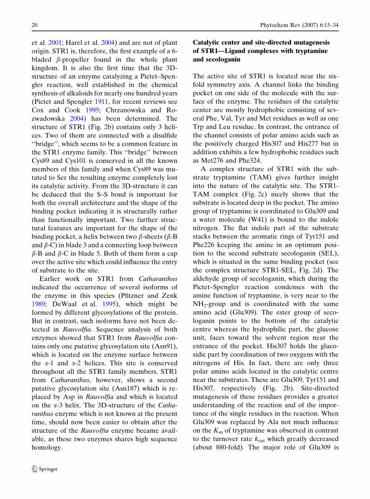
Catalytic center and site-directed mutagenesis
of STR1—Ligand complexes with tryptamine
and secologanin
The active site of STR1 is located near the six-
fold symmetry axis. A channel links the binding
pocket on one side of the molecule with the sur-
face of the enzyme. The residues of the catalytic
center are mostly hydrophobic consisting of sev-
eral Phe, Val, Tyr and Met residues as well as one
Trp and Leu residue. In contrast, the entrance of
the channel consists of polar amino acids such as
the positively charged His307 and His277 but in
addition exhibits a few hydrophobic residues such
as Met276 and Phe324.
A complex structure of STR1 with the sub-
strate tryptamine (TAM) gives further insight
into the nature of the catalytic site. The STR1-
TAM complex (Fig. 2c) nicely shows that the
substrate is located deep in the pocket. The amino
group of tryptamine is coordinated to Glu309 and
a water molecule (W41) is bound to the indole
nitrogen. The flat indole part of the substrate
stacks between the aromatic rings of Tyr151 and
Phe226 keeping the amine in an optimum posi-
tion to the second substrate secologanin (SEL),
which is situated in the same binding pocket (see
the complex structure STR1-SEL, Fig. 2d). The
aldehyde group of secologanin, which during the
Pictet–Spengler reaction condenses with the
amine function of tryptamine, is very near to the
NH2-group and is coordinated with the same
amino acid (Glu309). The ester group of seco-
loganin points to the bottom of the catalytic
centre whereas the hydrophilic part, the glucose
unit, faces toward the solvent region near the
entrance of the pocket. His307 holds the gluco-
sidic part by coordination of two oxygens with the
nitrogens of His. In fact, there are only three
polar amino acids located in the catalytic centre
near the substrates. These are Glu309, Tyr151 and
His307, respectively (Fig. 2b). Site-directed
mutagenesis of these residues provides a greater
understanding of the reaction and of the impor-
tance of the single residues in the reaction. When
Glu309 was replaced by Ala not much influence
on the Km of tryptamine was observed in contrast
to the turnover rate kcat which greatly decreased
(about 880-fold). The major role of Glu309 is
20 Phytochem Rev (2007) 6:15–34
123
obviously de-protonation of the tryptamine ami-
no function which then condenses with the alde-
hyde function of secologanin which Schiff-base
formation followed by ring-closure and synthesis
of the enzyme product 3a(S)-strictosidine. Glu309
must, therefore, be an essential amino acid for the
catalytic process. Indeed, the Asp/Glu-selective
inhibitor N, N¢-dicyclohexylcarbodiimide (DCC)
knocked out the STR1 activity completely, veri-
fying the catalytic role of Glu309. Displacement
of the other polar residues, Tyr151 and His307, by
Phe and Ala respectively, caused less influence on
enzymatic activity. The Km for tryptamine in-
creased about 3-fold in the first case, however,
there was a 130-fold increase for secologanin in
the second case. As suggested above, His307
plays a significant role in binding secologanin by
keeping the best structural orientation for seco-
loganin in the pocket. At this stage of investiga-
tion it is clear that Glu309 has been identified as
the essential catalytic residue in the binding
pocket of STR1 and is the amino acid necessary
for a Pictet–Spengler reaction (Falbe and Regitz
1991).
As known from earlier work strictosidine
synthase from Catharanthus or Rauvolfia is a
highly substrate specific enzyme (Treimer and
Zenk 1979). It does not, for instance, accept
dopamine, which is a precursor of monoterpe-
noid isoquinoline alkaloids as a substrate or
isoquinolines generated by deacetylipecoside and
norcoclaurine synthase. These later enzymes
should be similar to STR1, especially deacety-
lipecoside and deacetylisoipecoside synthase,
which have been recently detected and described
(De-Eknamkul et al. 1997; De-Eknamkul et al.
2000). Since the amino acid sequence and the
3D-structure of these proteins is not known, it is
not possible at the present time to conclude
whether all these enzymes, each catalyzing a
Pictet–Spengler type condensation, evolved from
the same ancestor. Although, the amino acid
sequence of norcoclaurine synthase an enzyme
which also catalyses a Pictet–Spengler reaction
(Samanani et al. 2004), is not apparently related
to STR1 and so differences in enzymatic mech-
anism cannot be described. It seems on the
basis of sequence that norcoclaurine synthase
belongs to a different enzyme family (the
pathogenesis-related (PR) 10 and Betv1 family),
but it is still possible so far that this synthase
also uses the same reaction mechanism as STR1.
If so, the different families of enzymes catalyzing
Pictet–Spengler reaction are examples of con-
vergent evolution. A clear-cut answer to this
question can only come from inspection of the
3D-structure of norcoclaurine synthase, which is,
not yet available.
Family members and evolutionary aspects
of STR1
There are a number of gene-related members in
the STR1 family, but up to now primary struc-
tures of only four STR1s are known; two from
Rauvolfia species and one each from the genus
Catharanthus and Ophiorrhiza. Sequence align-
ment studies reveal that all the major elements
such as the hydrophobic core of the b-propeller,
the hydrophobic binding pocket, the characteris-
tic disulfide bond and the catalytic Glu309 are
conserved throughout these family members. It is
therefore very likely that during evolutionary
divergence maintained their active site architec-
ture. Moreover, it seems very likely that nature
has used the STR1 scaffold for a great variety of
functions, because STR1-like genes are found in
plants and animals which are not able to perform
alkaloid biosynthesis. In Arabidopsis a whole
STR1-like gene family is known with 26 to 39%
sequence identity to STR1, but functions of these
genes have not been determined. Therefore de-
tailed structural and functional comparisons with
and within this gene-related family are impossible
at the present time.
The 6-bladed b-propeller is a well known pro-
tein fold, although not isolated from plants. Be-
cause of this fact it was interesting to compare the
STR1 structure with other b-propellers in order to
search for any evolutionary relationships. It is
noteworthy that STR1 has no significant sequence
identities or any functional similarities with other
6-bladed propellers. There are only very few en-
zymes known showing significant structural simi-
larities such as diisopropylfluorophosphatase
(DFPase, Scharff et al. 2001), serum paroxonase
(PON1, Harel et al. 2004), brain tumor NHL
Phytochem Rev (2007) 6:15–34 21
123

domain (NHL, Edwards et al. 2003) or the low
density lipoprotein receptor (LDLR, Jeon et al.
2001), but structure based alignments with the
STR1 structure indicated, only 11% to 16%
identity between them.
Inspection of the amino acid sequence in
LDLR identified a repetitive X-Tyr-Trp-Thr-Asp
(X is a hydrophobic amino acid) motif. In the
sequence of the remaining enzymes, five similar
motifs were found in the STR1 sequence which
might point to an evolutionary relationship of
these proteins. Although, two sequences in STR1,
X-Leu-Val-Ala-Glu and X-Trp-Val-Ser-Ser on b-
B4 and b-B5 seem to be not related to the con-
served X-Tyr-Trp-Thr-Asp repeat, closer inspec-
tion of these sequences would suggest a common
motif (X-X-X-#-§) for all of the b-propeller folds
discussed here (Ma et al. 2006). This motif is a
cluster of three hydrophobic amino acids (X-X-
X) followed by a small (#, Ser/Thr/Ala/Val/Gly)
amino acid and a hydrophilic residue (§, mostly
Asp or Glu).
This observation points to an evolutionary
relation of all these structurally similar proteins.
It is likely that STR1, LDLR, DFPase, NHL and
PON1 have evolved from one ancestral gene.
Duplication of such a b-sheet gene might lead to
the 6-bladed b-propeller structure containing the
X-Tyr-Trp-Thr-Asp repeat. During the evolution
single mutations might have shaped the binding
pocket and generated the different functions of
these proteins but the overall architecture of the
fold has been maintained. Structure-based
sequence alignment of STR1 and other 6-bladed
b-propellers from none-plant origin together with
the common motif X-X-X-#-§ described are here
displayed in Fig. 2e.
Based on the pronounced amino acid sequence
diversity and the different functions of ‘‘unre-
lated’’ propeller structures, it has been suggested
that these enzymes have evolved from distinct
ancestral b-sheet genes (Jawad and Paoli 2002).
The 3D-structure of STR1 and the structure-
based alignment between STR1 and the other
b-propeller genes mentioned here allow us to
instead propose an evolutionary relationship
between these apparently unrelated 6-bladed
b-propeller structures.
Strictosidine glucosidase (SG), crystallization and
X-ray analysis
The enzyme which follows STR1 in the pathway
is strictosidine glucosidase (SG) (Fig. 1). This
glucosidase ‘‘activates’’ the first intermediate,
strictosidine, by de-glucosylation. The resulting
aglycone is highly reactive and, cannot be directly
isolated. It is a precursor for all the pathways
leading to the entire family of monoterpenoid
indole alkaloids. Therefore the second enzyme of
the pathway also occupies an important role at
early stages of monoterpenoid indole alkaloid
biosynthesis of which ajmaline is one. Initially SG
has been detected and partly characterized from
cell suspension cultures of Catharanthus roseus by
Treimer and Zenk (1979) and was subsequently
described several times. The SG cDNA from
R. serpentina was isolated using the homology
cloning technique (Gerasimenko et al. 2002),
which was based on the SG cDNA sequence from
C. roseus reported by Geerlings et al. (2000). The
enzyme shows high substrate specificity and,
based on its primary structure, it belongs to the
family 1 of glycosyl hydrolases. Since a detailed
explanation of the enzyme reaction and the sub-
strate acceptance could not be found from se-
quence alignment studies we planned to
crystallize SG after overexpression in E. coli in
order to establish the 3D-structure of the enzyme
from R. serpentina. The cloning of the cDNA of
SG was based on primer development from the
SG sequence of C. roseus and raucaffricine glu-
cosidase (RG) of R. serpentina. The expression
vector PTYB1 resulted in low expression in
E. coli, and consequently the vector pQE-2 was
used in E. coli M15 cells. This strategy delivered
sufficient amounts of pure SG (up to 5 mg en-
zyme per liter bacterial culture) after His-tag
cleavage and MonoQ chromatography for crys-
tallization experiments. After testing most of the
commercially available crystallization kits it
turned out that formation of needles (Fig. 3a) was
easily achieved but the size and quality of these
crystals were not useful for X-ray analysis. Fur-
ther screening of crystallization conditions finally
gave good diffracting crystals (Fig. 3b) (Barleben
et al. 2005). Crystals of SG belonged to space
22 Phytochem Rev (2007) 6:15–34
123

group P4212. The structure of SG was determined
by using the molecular replacement technique
since SG was predicted to be a (b/a)8 barrel. This
so-called TIM-barrel is named after the structure
of triosephosphate isomerase, which was the en-
zyme which structure defining this fold (Henrissat
and Bairoch 1996; Gerlt and Raushel 2003). This
barrel is widely distributed in nature and the
Fig. 3 Strictosidine glucosidase (SG) (a) Needle crystalsof SG which were not useful for X-ray analysis. (b) Gooddiffracting crystal of SG resulting in resolution of 2.48 A.(c) The overall 3D-structure of Rauvolfia SG determined
from crystallized enzyme functionally expressed in E. coli.The structure represents a TIM-barrel fold in a gradientstarting from N-terminal to C-terminal in blue to redcolour
Phytochem Rev (2007) 6:15–34 23
123

enzymes with this fold catalyze many different
reactions and show only low sequence homology
(Henrissat and Bairoch 1996; Gerlt and Raushel
2003).
This fold is characterized by eight parallel
b-sheets located inside the enzyme. Connection
between the b-strands is reached by a-helices. The
overall topology is similar to a barrel. The loops
at the tops of the barrel determine the substrate
specificity. The glycoside hydrolase (GH) enzyme
super-family has been divided into four clans GH-
A, GH-D, GH-H and GH-K with a total of 24
different families. SG has been classified as
belonging to family 1 the GH-A clan. In this
family 16 enzymes have been structurally defined
of which only six are from plants, including the
two glucosidases from Rauvolfia, SG and RG. In
SG catalytic acid/base (Glu-416) is localized on
strand b-7 and the nucleophile Glu-207 in strand
b-4. Such an arrangement is typical for clan GH-
A glycoside hydrolases. The detail structural
analysis of SG will be reported in the near future.
Vinorine synthase (VS) sequence alignmentand site-directed mutations
The first enzyme generating the complete carbon
skeleton of ajmaline is named vinorine synthase
(VS). This enzyme is located in the middle of the
pathway (Fig. 1) and catalyzes an acetylCoA-
dependent ring closure of the product (16-epi-
vellosimine) of the preceding enzyme, polyneuri-
dine aldehyde esterase (PNAE). The product of
VS is the ajmalane-type alkaloid, vinorine, exhib-
iting the basic six-membered ring system of ajma-
line. VS has been previously detected in
R. serpentina cell culture. These cultures contain a
great number of acetylated alkaloids, such as vin-
orine, vomilenine and acetyl norajmaline. Acety-
lation in general should be dependent on activated
acetic acid and indeed, only in the presence of
acetylCoA are protein preparations from Rauvol-
fia cells able to convert 16-epi-vellosimine into
vinorine. VS has recently been highly enriched
(~1000-fold) by a five-step chromatographic pro-
cedure and its cDNA could be isolated from
R. serpentia · Rhazya stricta hybrid culture.
Functional expression in E. coli (Gerasimenko
et al. 2004; Bayer et al. 2004) was then possible.
Availability of the primary structure of VS allowed
the first sequence alignment studies. Figure 4 rep-
resents the alignment of VS with recently cloned
acetyltransferases of enzymes participating in
the biosynthesis of various highly valuable plant-
derived natural products such as acetylated
salutaridinol, an intermediate of morphine bio-
synthesis, vindoline which is important for the
formation of vinblastine and vincristine or acety-
lation of taxadienol on the biosynthetic way to the
cytotoxic diterpenoid alkaloid taxol. It includes
also other acyltransferases and highlighted the
conserved His-X-X-X-Asp sequence together with
Asp-Phe-Gly-Trp-Gly motif.
All these cDNA-derived proteins clearly
showed their sequence relationship by highly
conserved motifs such as Asp362-Phe-Gly-Trp-
Gly366 in VS. This motif is completely conserved
in all the above mentioned enzymes which are
involved in acetylCoA-dependent transforma-
tions during the above mentioned pathways. Also
the His160-X-X-X-Asp164 motif is 100% con-
served in these sequences in addition to the motif
Ser29-X-L/I-Asp32. In the latter case one excep-
tion exists which is for the enzyme deacetylvind-
oline acetyltransferase which has asparagine
instead of leucine or isoleucine in the motif at
N-terminus.
These sequence analyses indicated that VS
belongs to a small but constantly growing group
of enzymes, which have been called as the BAHD
enzyme super family (for a recent review see
D’Auria 2006). BAHD is the abbreviation for the
first four enzymes assigned to this family. These
enzymes were named Benzylalcohol acetyl-,
Anthocyanin-O-hydroxy-cinnamoyl-, anthrani-
late-N-Hydroxy-cinnamoyl/benzoyl- and Deace-
tylvindoline acetyl transferase. Our extensive
mutation experiments on VS have clearly identi-
fied His160 and Asp164 as the most important
amino acids for the catalytic process, because
their replacement by Ala completely knocked out
the vinorine synthase activity (Bayer et al. 2004).
Fig. 4 Structure based sequence alignment of the acetyl-CoA- dependent enzymes from plants belonging to theBAHD enzyme super family including putative acyltrans-ferases
c
24 Phytochem Rev (2007) 6:15–34
123

Phytochem Rev (2007) 6:15–34 25
123

26 Phytochem Rev (2007) 6:15–34
123

Moreover, Asp362 located in the consensus
sequence must also possess an important function
(see previous paragraph). Its replacement by Ala
significantly reduced the enzyme activity. The
same observation was made when the amino acids
Ser29 and Asp32, members of the newly desig-
nated consensus sequence at the N-terminus of
the enzyme, were exchanged by Ala (Bayer et al.
2004).
In a recently proposed classification of acyl-
transferases, all these enzymes were divided into
four evolutionary sequence clusters A–D (Hoff-
mann et al. 2003). In this classification system VS
would belong to the class C which includes
enzymes catalyzing acetylation of unrelated nat-
ural products. Class C is still very small but is
expected to increase since acetylation is a basic
reaction occurring frequently in biosynthesis. In
the Arabidopsis genome project already more
than 60 genes have been identified which also
might be members of the BAHD enzyme family
although they are still awaiting identification of
their metabolic functions.
The detailed knowledge recently accumulated
on catalytic domains, on novel motifs and on the
participation of single amino acids in the forma-
tion of vinorine will help to understand the acyl-
transfer reaction of the remaining enzymes of the
BAHD super-family. However, the mutation
experiments we carried out clearly suggested a
complicated reaction mechanism because we
identified at least seven amino acids reducing the
VS activity by varying degree. Unraveling the
catalytic process of acetyltransfer should now be a
major aim for enzymes of this particular family.
Crystallization of VS and X-ray analysis
Sequence comparison yields limited information
on reaction mechanism and because there was no
3D-structure known in this particular enzyme
family which could help us to understand the
mechanism, we developed crystallization condi-
tions for VS in order to analyze the molecular
structure of the transferase by X-ray crystallog-
raphy. The protein for the crystallization trials
was obtained after expression of the His-tagged
proteins in E. coli and purification by Ni-NTA
chromatography. Removal of the His-tag was
followed by ion exchange and size-exclusion
chromatography. Crystallization experiments by
hanging drop vapor-diffusion were performed
using, the commercially available Crystal Screen
and Crystal Screen 2 kits from Hampton Re-
search (USA). Only small multiple crystals, not
useful for X-ray measurements, were formed in
presence of ammonium sulfate and polyethylene
glycol (PEG) 400 at room temperature. Optimi-
zation trials varying precipitant concentrations,
type of PEG, enzyme concentration, buffer,
temperature and pH turned out to be essential for
diffraction quality crystal formation. The best
crystals with dimensions of about 0.3 · 0.15 ·0.1 mm were grown at typical conditions namely
at 32�C and at very low enzyme concentration
(2 mg ml–1) only (Ma et al. 2004b). It is interest-
ing to note, that in the Biological Macromolecule
Crystallization Database (Gilliland et al. 1994) no
example exists where a protein has been crystal-
lized at such low enzyme concentration combined
with such a high temperature. Crystallization of
VS also provided a nice example to the question
whether His6-tagged or the native proteins should
be used for crystallization trials, because both
were available in our case. In fact, it turned out
that the His6-tag enzyme precipitated or formed
needle clusters whatever conditions were used,
whereas the native synthase gave single crystals
that diffracted to a resolution of 2.8 A (Fig. 5a).
Dynamic light scattering experiments were per-
formed to explain this phenomenon. Results
indicated that VS exists as a monodisperse species
whereas the His6-tag VS tends to form aggregates
which do not crystallize.
Data for the flash-frozen VS crystals which
belongs to space group P212121, were measured,
however the 3D-structure determination was not
possible using molecular replacement. In order to
use the multiple-wavelength anomalous disper-
sion (MAD) approach to obtain experimental
phases, selenomethionyl VS (Se-Met VS) was
produced by adding selenomethionine to the cul-
ture broth (van Duyne et al. 1993). The Se-Met
substituted VS could be crystallized (Ma et al.
2005a) under similar conditions as for native VS
and X-ray measurements gave a complete set of
Fig. 4 continuedb
Phytochem Rev (2007) 6:15–34 27
123

data to 3.2 A at each of three wavelengths around
the selenium edge, allowing elucidation of the
3D-structure of the synthase using Auto-Rickshaw
pipeline (Panjikar et al. 2005) as described in (Ma
et al. 2005b) and refined to 2.8 A resolution.
Overall structure of VS
The atomic model of VS exhibits all amino acids
except residues 1–3 at the N-terminus and 5–6
residues in a loop located at the surface of the
enzyme (Ma et al. 2005b). In a total, this novel
protein structure exhibits 13 helices (a1–a13), 14b-
strands (b1–b14) and consists of two major do-
mains (A and B) with nearly identical size. Both
domains are connected by a large loop formed by
residues 201–213 (Fig. 5b). Six b sheets, b1, b2, b5,
b6, b7 and b12, represent domain A which is a
mixed six-stranded b sheet structure, covered on
both sides by seven helices (a1–a7). Domain A
also has a pair of b strands (b3 and b4) on the
enzyme surface. The second domain B, is a mixed
6-stranded b-sheet with b8–b11 and b13–b14. A
loop from domain B extends into domain A and
both domains have a very similar backbone fold,
but exhibit different topology.
A solvent channel running through the whole
VS molecule is formed by two loops, loop ‘‘a’’ and
loop ‘‘b’’. Loop ‘‘b’’ lies between strands b11 and
Fig. 5 Vinorine synthase (VS). (a) Single crystals of VSobtained with 2.0 M ammonium sulphate, 2% PEG 400and 100 mM Tris–HCl. (b) Overall structure of VS withCoA modelled into the active site. Helices and b-strandsincluding loops ‘‘a’’ and ‘‘b’’ are labelled. (c) Front view
down into the reaction channel of VS. Blue colourindicates the His160 residue necessary for the reaction.(d) Back view into the reaction channel with the bluecoloured His160 residue and each domain surface is shownin two different colours
28 Phytochem Rev (2007) 6:15–34
123

b13 of domain B but in addition includes b12 of
domain A. The second loop ‘‘b’’ is located between
b9 and b10. In loop ‘‘a’’ the motif Asp362-Phe-Gly-
Trp-Gly366 is situated. The Gly-Asn motif, also
completely conserved in the family, can be found in
loop b. The catalytic site motif His160-X-X-X-
Asp164 is located between both domains, so that
the catalytic His160 is in principal accessible from
both sides of the solvent channel (Fig. 5c–d).
The 3D-structure of VS together with the
mutational analysis yield detailed knowledge and
understanding of the catalytic mechanism, espe-
cially allowing differentiation between amino acid
residues important for catalytic and structural
reasons. The catalytic His160 of the His160-X-X-
X-Asp164 motif is located directly in the center of
the solvent channel. Therefore both reaction
partners, acetylCoA and 16-epi-vellosimine, may
approach the catalytic center independently from
each side of the channel. Previous kinetic data on
enzyme-catalyzed vinorine synthesis suggested
formation of a ternary complex between VS, sub-
strate and co-substrate by independent binding of
the ligands (Pfitzner et al. 1986). The structure of
VS is in excellent agreement with these biochem-
ical data. Asp164, the second crucial residue for
enzyme activity, points away from the active site
and is involved in a salt-bridge with Arg279.
Obviously, Asp164 has a structural function by
maintaining the geometry of the binding pocket.
Indeed this structural rather than catalytic signifi-
cance for Asp164 has been also discussed for other
acyltransferases (Buglino et al. 2004; Gibbs et al.
1990). The full conservation of the His-X-X-X-
Asp motif for all BAHD family members, they all
suggest that have a similar structural arrangement
of these residues and a similar reaction mecha-
nism. For a clear cut decision on their catalytic
functions and the structural properties of the
binding sites the 3D-structures of additional
BAHD members would be a prerequisite. The
second conserved motif of VS (Asp362-Phe-Gly-
Trp-Gly366) is unique to the BAHD super family
and was suggested to have functions in catalysis or
in binding of the co-substrate CoA (St-Pierre and
DeLuca 2000; Suzuki et al. 2003; Bayer et al.
2004). The 3D-structure presented here shows that
this motif is far from the active site pocket and is,
therefore not directly involved in catalysis. Instead
the motif is localized in a turn of a large loop
between b strands 11 and 12 in domain B. Most
probably this arrangement helps to keep the loop
in position which extends up to the pocket, thus
maintaining the shape of the enzyme’s catalytic
site and giving substrate specificity. In order to
attain more detailed and direct evidence on ligand
binding and atomic details of the reaction mech-
anism of VS, the crystal structures of substrate-
and product-bound complexes must be deter-
mined, work which has now been performed in our
laboratory.
Structural relationships of VS
The structure of VS can be classified in a general
way, as a member of CoA-dependent acyltrans-
ferases. A structural alignment in addition to a
sequence alignment offers the opportunity to find
relationships to other enzymes especially of those
of different origin and with different functions.
When the entire VS molecule is aligned using a
secondary structure matching servers (http://
www.ebi.ac.uk/msd-srv/ssm/cgi-bin/ssmserver) the
most related structure found to be polyketide
synthase associated protein 5 (PDB code 1q9j)
from Mycobacterium tuberculosis, followed by
condensation domains of vibriobactine synthetase
(PDB code 115a), rat choline acetyltransferase
(PDB code 1q6x) and human carnitine acetyl-
transferase (PDB code 1nm8). When isolated A
and B domain of VS were used as a search model
the highest structural similarity was to chloram-
phenicol acetyltransferase (PDB code 1cla) and
dihydrolipoyl transacetylase (PDB code 1ead and
1eab) respectively. In fact, all these proteins are
CoA-dependent acyltransferases and the align-
ment results further confirm that VS is a member
of BAHD acyltransferase enzyme super family.
Details of the structural analysis of VS have been
recently described (Ma et al. 2005b).
Raucaffricine glucosidase (RG), crystallizationand X-ray analysis
Raucaffricine glucosidase (RG) is one of the en-
zymes of a side routes to the ajmaline biosyn-
thetic pathway. RG delivers in a one-step reaction
Phytochem Rev (2007) 6:15–34 29
123

the pathway intermediate vomilenine by de-glu-
cosylation of raucaffricine (Fig. 1). The concen-
tration of raucaffricine in Rauvolfia cell
suspension cultures exceeds that in the plant by a
factor of 67 and might accumulate to levels of up
to 1.6 g/l (Ruyter and Stockigt 1991).
RG was first detected and characterized two
decades ago (Schubel et al. 1986). Subsequently
RG has been purified from R. serpentina cell
suspension cultures with a 1200-fold enrichment.
The isolated enzyme suggests a precise value of a
molecular mass of 61 kDa and is not glycosylated.
After digestion with the endoproteinase LysC, six
peptide fragments were sequenced with a length
of 6–24 amino acids (Warzecha et al. 1999).
Based on these partial sequences the cDNA for
RG with an open reading frame of 1623 nucleo-
tides was isolated (Warzecha et al. 2000). The
corresponding protein consists of 540 amino acids
with a calculated molecular weight of 60931 Da in
agreement with molecular weight of the enzyme
isolated from the cell suspension. Comparison
with known glucosidase sequences shows, that
RG belongs to the family 1 of glycosyl hydrolases
(Henrissat 1991) and shares a sequence identity of
56% with SG.
For heterologous expression, RG cDNA was
cloned into the pSE280 vector (Warzecha et al.
2000). The recombinant RG shows high substrate
specificity with raucaffricine as the best substrate
(Km 1.3 mM, Vmax 0.5 nkat/lg protein). Only
some structurally related alkaloids like 1,2-di-
hydroraucaffricine and strictosidine are also de-
glucosylated. This observation is interesting since
RG is able to hydrolyse the substrate of strictos-
idine glucosidase (SG) where as SG is unable to
de-glucosylate raucaffricine, the substrate of RG
(Warzecha et al. 2000; Gerasimenko et al. 2002).
No hydrolysis for structurally different glucosides
such as loganin, secologanin, arbutin, vanillin-
b-D-glucoside or 4-nitrophenyl-b-D-glucoside has
been observed.
To obtain optimum expression RG cDNA was
cloned into the pQE-2 vector, which has been an
efficient vector in our work and expressed in the
E. coli strain M15 (Ruppert et al. 2006). For
easier isolation RG was expressed with an
N-terminal His6-tag and purified by using a
Ni-NTA column. With this method up to 5 mg
pure RG could be isolated per litre of bacterial
culture, and was directly suitable for systematic
crystallization experiments. Crystals of RG were
obtained by the hanging-drop vapour diffusion
technique at 20�C with 0.3 M ammonium sul-
phate 0.1 M sodium acetate buffer pH 4.6 and
11% PEG 4000 as precipitant (Ruppert et al.
2006). Crystals reach a maximum dimensions of
about 0.2 · 0.15 · 0.05 mm (Fig. 6a) belong to
space group I222 and diffract to 2.30 A (Fig. 6b)
with unit-cell parameters of a = 102.8 A,
b = 127.3 A and c = 215.8 A. The crystals were
treated with cryoprotectant, and were flash-
cooled to 100K prior to X-ray diffraction data
collection. Self-rotation analysis showed two
molecules per crystallographic asymmetric unit
although RG functions as a monomer in solution.
The preliminary X-ray analysis has been carried
out recently (Ruppert et al. 2006) and the detail
analysis of the enzyme is underway.
Summary, conclusions and future prospects
Phytochemical work in the previous years has
extensively broadened our knowledge on the
impressive structural complexity and diversity of
monoterpenoid indole alkaloids generated in
Rauvolfia cell and tissue cultures. With the iso-
lation and identification of more than 35 alkaloids
occurring in amounts in ranging from the lower
mg range up to 1.6 g per litre cell suspension. The
Rauvolfia systems represent one of the best
investigated biological systems of alkaloid for-
mation. This phytochemical characterization was
the prerequisite to reach the stage of identifying
and describing the alkaloidal metabolism of
Rauvolfia in detail, because the proteins involved
in catalyzing each step in the main route and in a
number of the side reactions of the ajmaline
biosynthetic pathway could be detected. From a
biosynthetic point of view a straightforward
metabolic route to the end product (e.g. ajmaline)
cannot in general, be expected. All examples
known so far show highly branched pathways,
resulting in a complicated metabolic network
which allows the metabolic diversity of secondary
products in nature. One possibility to steer a
pathway into the direction of a desired product,
30 Phytochem Rev (2007) 6:15–34
123

would be blocking specific side routes with a
specific inhibitor at the enzyme or cDNA stage
and generating in vivo the end product of the
main pathway, for instance ajmaline in the
Rauvolfia system. However, the specific genes or
enzymes must be known in order to utilize this
strategy.
The part of the Rauvolfia genome responsible
for alkaloid biosynthesis is now becoming avail-
able by use of a reverse genetic approach. The
cDNAs coding for soluble enzymes have been
isolated allowing heterologously overexpression
and functional characterization.
In most cases expression was achieved in
E. coli and judging from functional assays no
problems with incorrect protein folding could be
detected. Moreover, in spite of a few cases where
formation of inclusion bodies was observed, there
was always sufficient active enzyme produced for
the work summarized in this article. In fact, the
generation of a single enzyme of the ajmaline
pathway at a high purity (>98%) at mg-levels has
proven not to be problematic.
Having substantial amounts of these re-
combinant enzymes in hand one could think
about an enzyme directed biochemical synthesis
of intermediates of the pathway on preparative
scales, up to 100 mg or even higher. Compared
with classical product isolation from plants, a
step-wise synthesis with matrix bound enzymes
using regenerated coenzymes might be an alter-
native source of natural products in future. Such
an in vitro approach would however be exclu-
sively applicable for valuable products for which
natural sources are limited or those occurring
only in trace amounts in nature. If such an enzy-
matic process is developed, a serious restriction
could be the long term stability of the enzymes.
The development of stable, artificial enzymes
should be taken as a future challenge in many
research fields, including enzyme catalyzed syn-
thesis of natural products. The prerequisite of
such a strategy must be the elucidation of the
three dimensional structures followed by recog-
nition of the catalytic center of each catalyst to-
gether with the identification of any reaction
channel and an understanding of the reaction
mechanism. Understanding substrate recognition
and conversion at an atomic level could then lead
to a rational modulation of substrate specificity by
Fig. 6 Raucaffricine glucosidase (RG). (a) RG crystalfrom Rauvolfia serpentina and corresponding diffractionimage. (a) A close view of a single RG crystal obtained
after three days, with the dimensions of 0.2 · 0.15 ·0.05 mm and (b) image of X-ray diffraction pattern
Phytochem Rev (2007) 6:15–34 31
123

site-directed mutagenesis of the corresponding
cDNA. Enzymes in alkaloid biosynthesis are in
general highly substrate specific. One of our fu-
ture targets is to change this specificity in order to
obtain a desired substitution pattern at the indole
binding site of alkaloids. As discussed in this
chapter we have solved 3D-structures of several
of the major enzymes of ajmaline biosynthesis
since 2004, including the enzymes strictosidine
synthase and strictosidine glucosidase. Both en-
zymes are of an outstanding importance because
they function as the biosynthetic gate to the entire
monoterpenoid indole alkaloid family in higher
plants. The rational engineering and design of
both catalysts is now under investigation in our
laboratories.
Acknowledgements We are very thankful to Mrs. DorisRohr of our research group for cultivation of all plant cellsystems, to Prof. F. Lottspeich and his coworkers Mrs. I.Mathes and Mr. R. Mentele (Max-Planck-Institute ofBiochemistry, Martinsried, Germany) for their enthusiastichelp in sequencing pre-purified Rauvolfia enzymes and toProf. H. Michel, Dr. G. Fritzsch and Dr. J. Koepke (Max-Planck-Institute of Biophysics, Frankfurt/Main, Germany)for introducing us to structural biology research.
Our research was also supported by the DeutscheForschungsgemeinschaft (Bonn, Bad-Godesberg, Ger-many), Fonds der Chemischen Industrie (Frankfurt/Main,Germany) and the Bundesministerium fur Bildung undForschung (BMBF), Bonn, Germany). Support in theform of access to synchrotron facilities by the EuropeanCommunity (Research Infrastructure Action under theFP6 ‘‘Structuring the European Research Area Pro-gramme’’, contact number RII3/CT/2004/5060008) isalso acknowledged and we thank Dr. Paul Tucker(EMBL Hamburg, Germany) for critically reading themanuscript.
References
Barleben L, Ma X, Koepke J, Peng G, Michel H, Stockigt J(2005) Expression, purification, crystallization andpreliminary X-ray analysis of strictosidine glucosi-dase, an enzyme initiating biosynthetic pathways to aunique diversity of indole alkaloid skeletons. BiochimBiophys Acta 1747:89–92
Bayer A, Ma X, Stockigt J (2004) Acetyltransfer in naturalproduct biosynthesis – functional cloning and molec-ular analysis of vinorine synthase. Bioorg Med Chem12:2787–2795
Bracher D, Kutchan TM (1992) Strictosidine synthasefrom Rauvolfia serpentina: analysis of a gene involvedin indole alkaloid biosynthesis. Arch Biochem Bio-phys 294:717–723
Buglino J, Onwueme KC, Ferreras JA, Quadri LEN, LimaCD (2004) Crystal Structure of PapA5, a PhthiocerolDimycocerosyl Transferase from Mycobacteriumtuberculosis. J Biol Chem 279:30634–30642
Chrzanowska M, Rozwadowska MD (2004) Asymmetricsynthesis of isoquinoline alkaloids. Chem Rev104:3341–3370
Cox ED, Cook JC (1995) The Pictet–Spengler condensa-tion: a new direction for an old reaction. Chem Rev95:1791–1842
Creasey WA (1994) Pharmacology, biochemistry andclinical applications of the monoterpenoid alkaloids.In: Saxton JE (ed) Monoterpenoid Indole Alka-loids, Supplement to part 4. John Wiley & Sons,Chichester, New York, Brisbane, Toronto, Singa-pore, pp 715–753
D’Auria JC (2006) Acyltransferases in plants: a good timeto be BAHD. Curr Opin Plant Biol 9:331–340
De-Eknamkul W, Ounaroon A, Tanahashi T, Kutchan T,Zenk MH (1997) Phytochemistry 45:477–484
De-Eknamkul W, Suttipanta N, Kutchan TM (2000)Purification and characterization of deacetylipecosidesynthase from Alangium lamarckii. Thw. Phyto-chemistry 55:177–181
DeLucas LJ, Hamrick D, Cosenza L, Nagy L, McCombsD, Bray T, Chait A, Stoops B, Belgovskiy A, WilsonWW, Parham M, Chernov N (2005) Protein crystalli-zation: virtual screening and optimization. Prog Bio-phys Mol Biol 88:285–309
DeWaal A, Meijer AH, Verpoorte R (1995) Strictosidinesynthase from Catharanthus roseus: purification andcharacterization of multiple forms. Biochem J306:571–580
Edwards TA, Wilkinson BD, Wharton RP, Aggarwal AK(2003) Model of the brain tumor-pumilio translationrepressor complex. Genes Dev 17:2508–2513
Falbe J, Regitz M (1991) In: ROMPP Chemie Lexikon,Georg Thieme Verlag Stuttgart-New York, Vol. 4, pp34–37
Geerlings A, Ibanez MM, Memelink J, van Der Heijden R,Verpoorte R. (2000) Molecular cloning and analysisof strictosidine beta-D-glucosidase, an enzyme in ter-penoid indole alkaloid biosynthesis in Catharanthusroseus. J Biol Chem 275:3051–3056
Gerasimenko I, Sheludko Y, Ma X, Stockigt J (2002)Heterologous expression of a Rauvolfia cDNAencoding strictosidine glucosidase, a biosynthetic keyto over 2000 monoterpenoid indole alkaloids. Eur JBiochem 269:2204–2213
Gerasimenko I, Ma X, Sheludko Y, Mentele R, LottspeichF, Stockigt J (2004) Purification and partial aminoacid sequences of the enzyme vinorine synthase in-volved in a crucial step of ajmaline biosynthesis.Bioorg Med Chem 12:2781–2786
Gerlt JA, Raushel FM (2003) Evolution of function in(b/a)8 barrel enzymes. Curr Opin Chem Biol 7:252–264
Gibbs MR, Moody PCE, Leslie AGW (1990) CrystalStructure of the ASP-199 asparagine mutant of chl-oramphenicol acetyltransferase to 2.35. A resolution:structural consequences of disruption of a buried saltbridge. Biochemistry 29:11261–11265
32 Phytochem Rev (2007) 6:15–34
123

Gilliland GL, Tung M, Blakeslee DM, Ladner J (1994)The biological macromolecule crystallization data-base, version 3.0: new features, data, and the NASAarchive for protein crystal growth data. Acta Crys-tallogr D 50:408–413
Harel M, Aharoni A, Gaidukov L, Brumshtein B, Kher-sonsky O, Meged R, Dvir H, Ravelle RBG, McCarthyA, Toker L, Silman L, Sussman JL, Tawfik DS (2004)Structure and evolution of the serum paraoxonasefamily of detoxifying and antiatherosclerotic enzymes.Nat Struct Mol Biol 11:412–419
Henrissat B (1991) A classification of glycosyl hydrolasesbased on amino acid sequence similarities. Biochem J280:309–316
Henrissat B, Bairoch A (1996) Updating the sequence-based classification of glycosyl hydrolases. J Biochem316:695–696
Hoffmann L, Maury S, Martz F, Geoffry P, Legrand L(2003) Purification, cloning, and properties of anacyltransferase controlling shikimate and quinateester intermediates in phenylpropanoid metabolism.J Biol Chem 278:95–103
Jawad Z, Paoli M (2002) Novel sequences propel familiarfolds. Structure 10:447–454
Jeon H, Meng W, Takagi J, Eck MJ, Springer TA,Blacklow SC (2001) Implications for familial hyper-cholesterolemia from the structure of the LDLreceptor YWTD-EGF domain pair. Nat Struct Biol8:499–504
Koepke J, Ma X, Fritzsch G, Michel H, Stockigt J (2005)Crystallization and preliminary X-ray analysis ofstrictosidine synthase and its complex with the sub-strate tryptamine. Acta Crystallogr Sect D 61:690–693
Krupka HI, Rupp B, Segelke BW, Lekin TP, Wright D,Wu H-C, Todd P, Azarani A (2002) The high-speedHydra-Plus-One system for a automated high-throughput protein crystallography. Acta CrystallogrD 58:1523–1526
Kutchan TM, Hampp N, Lottspeich F, Beyreuther K,Zenk MH (1988) The cDNA clone for strictosidinesynthase from Rauvolfia serpentina. DNA sequencedetermination and expression in Escherichia coli.FEBS Lett 237:40–44
Kutchan TM (1993) Strictosidine: from alkaloid to enzymeto gene. Phytochemistry 32:493–506
Ma X, Koepke J, Fritzsch G, Diem R, Kutchan TM, Mi-chel H, Stockigt J (2004a) Crystallization and pre-liminary X-ray Crystallographic Analysis ofstrictosidine synthase from Rauvolfia – the firstmember of a novel enzyme family. Biochim BiophysActa 1702:121–124
Ma X, Panjikar S, Koepke J Loris E, Stockigt J (2006) Thestructure of Rauvolfia serpentina stritosidine synthaseis a novel six-bladed beta-propeller fold in plantproteins. The Plant Cell 18:907–920
Ma X, Koepke J, Bayer A, Linhard V, Fritzsch G, ZhangB, Michel H, Stockigt J (2004b) Vinorine synthasefrom Rauvolfia: the first example of crystallization andpreliminary X-ray diffraction analysis of an enzyme ofthe BAHD superfamily. Biochim Biophys Acta1701:129–132
Ma X, Koepke J, Bayer A, Fritzsch G, Michel H, StockigtJ (2005a) Crystallization and preliminary X-ray anal-ysis of native and selenomethionyl vinorine synthasefrom Rauvolfia serpentina. Acta Crystallogr Sect D61:694–696
Ma X, Koepke J, Panjikar S, Fritzsch G, Stockigt J (2005b)Crystal structure of vinorine synthase, the first rep-resentative of the BAHD superfamily. J Biol Chem280:13576–13583
Panjikar S, Parthasarathy V, Lamzin VS, Weiss MS,Tucker PA (2005) Auto-Rickshaw: an automatedcrystal structure determination platform as an effi-cient tool for the validation of an X-ray diffractionexperiment. Acta Cryst D 61:449–457
Pfitzner A, Polz L, Stockigt J (1986) Properties of VinorineSynthase – the Rauvolfia Enzyme involved in theFormation of the Ajmaline Skeleton. Z Naturforsch41c:103–114
Pfitzner M, Zenk MH (1989) Homogenous strictosidinesynthase isoenzymes from cell suspension cultures ofCatharanthus roseus. Planta Med 55:525–530
Pictet A, Spengler T (1911) Uber die Bildung von Iso-chinolin-derivaten durch Ubertragung von Methylalauf Phenyl-athylamin, Phenyl-alanin und Tyrosin. BerDtsch Chem Ges 44:2030–2036
Pons T, Gomez R, Chinea G, Valencia A (2003) Beta-propellers: associated functions and their role in hu-man diseases. Curr Med Chem 10:505–524
Ruppert M, Stockigt J (1999) Strictosidine – the biosyn-thetic key to monoterpenoid indole alkaloids. In:Sir Derek Barton, Nakanishi K (eds) Comprehen-sive Natural Products Chemistry Vol 4. Elsevier,Amsterdam, Lausanne, New York, Oxford, Shannon,Singapore, Tokyo, pp 109–138
Ruppert M, Ma X, Stockigt J (2005) Alkaloid biosynthesisin Rauvolfia–cDNA cloning of major enzymes of theajmaline pathway. In: Knolker H-J (eds) Current OrgChem 9:1431–1444
Ruppert M, Panjikar S, Barleben L, Stockigt J (2006)Heterologous expression, purification, crystallizationand preliminary X-ray analysis of raucaffricineglucosidase, a plant enzyme specifically involved inRauvolfia alkaloid biosynthesis. Acta Crystallogr F62:257–260
Ruyter CM, Stockigt J (1991) Enzymatic formation ofraucaffricine, the major indol alkaloid of Rauwolfiaserpentina cell-suspension cultures. Helv Chim Acta74:1707–1712
Samanani N, Liscombe DK, Facchini PJ (2004) Molecularcloning and characterization of norcoclaurinesynthase, an enzyme catalyzing the first committedstep in benzylisoquinoline alkaloid biosynthesis. PlantJ 40:302–313
Scharff EI, Koepke J, Fritzsch G, Lucke C, Ruterjans H(2001) Crystal structure of diisopropylfluorophospha-tase from Loligo vulgaris. Structure 9:493–502
Schubel H, Stockigt J, Feicht R, Simon H (1986) Partialpurification and characterization of raucaffricineb-D-glucosidase from plant cell-suspension cul-tures of Rauwolfia serpentina. Helv Chim Acta69:538–547
Phytochem Rev (2007) 6:15–34 33
123
St-Pierre B, DeLuca V (2000) Evolution of acyltransferasegenes: origin and diversification of the BAHD super-family of acyltransferases involved in secondarymetabolism. In: John RI, Romeo T, Varin L, DeLucaV (eds) Recent Advances in Phytochemistry, Evolu-tion of Metabolic Pathways Vol 34. Elsevier Science,Oxford, pp 285–315
Suzuki H, Nakayama T, Nishino T (2003) Proposedmechanism and functional amino acid residues ofmalonyl-CoA: anthocyanin 5-O-glucoside-6’’’-O-malonyltransferase from flowers of Salvia splendens, amember of the versatile plant acyltransferase family.Biochemistry 42:1764–1771
Treimer JE, Zenk MH (1979) Purification and propertiesof strictosidine synthase, the key enzyme in indolealkaloid formation. Eur J Biochem 101:225–233
Van Duyne GD, Standaert RF, Karplus A, Schreiber SL,Clardy J (1993) Atomic structures of the human
immunophilin FKBP-12 complexes with FK506 andRapamycin. J Mol Biol 229:105–124
Warzecha H, Obitz P, Stockigt J (1999) Purification, par-tial amino acid sequence and structure of the productof raucaffricine-O-beta-D-glucosidase from plant cellcultures of Rauvolfia serpentina. Phytochemistry50:1099–1109
Warzecha H, Gerasimenko I, Kutchan TM, Stockigt J(2000) Molecular cloning and functional bacterialexpression of a plant glucosidase specifically in-volved in alkaloid biosynthesis. Phytochemistry54:657–666
Yamazaki Y, Sudo H, Yamazaki M, Aimi N, Saito K(2003) Camptothecin biosynthetic genes in hairyroots of Ophiorrhiza pumila: cloning, characteriza-tion and differential expression in tissues andby stress compounds. Plant Cell Physiol 44:395–403
34 Phytochem Rev (2007) 6:15–34
123