The Internally Truncated LRP5 Receptor Presents a Therapeutic Target in Breast Cancer
10
The Internally Truncated LRP5 Receptor Presents a Therapeutic Target in Breast Cancer Peyman Bjo ¨ rklund 1 , Jessica Svedlund 1 , Anna-Karin Olsson 2 , Go ¨ ran A ˚ kerstro ¨m 1 , Gunnar Westin 1 * 1 Department of Surgical Sciences, Endocrine Unit, Uppsala University, Uppsala University Hospital, Uppsala, Sweden, 2 Department of Medical Biochemistry and Microbiology, Uppsala University, Uppsala Biomedical Center, Uppsala, Sweden Abstract Background: Breast cancer is a common malignant disease, which may be caused by a number of genes deregulated by genomic or epigenomic events. Deregulated WNT/b-catenin signaling with accumulation of b-catenin is common in breast tumors, but mutations in WNT signaling pathway components have been rare. An aberrantly spliced internally truncated LRP5 receptor (LRP5D666–809, LRP5D) was shown recently to be resistant to DKK1 inhibition, and was required for b- catenin accumulation in hyperparathyroid tumors and parathyroid tumor growth. Methodology/Principal Findings: Here we show, by reverse transcription PCR and Western blot analysis, that LRP5D is frequently expressed in breast tumors of different cancer stage (58–100%), including carcinoma in situ and metastatic carcinoma. LRP5D was required in MCF7 breast cancer cells for the non-phosphorylated active b-catenin level, transcription activity of b-catenin, cell growth in vitro, and breast tumor growth in a xenograft SCID mouse model. WNT3 ligand, but not WNT1 and WNT3A augmented the endogenous b-catenin activity of MCF7 cells in a DKK1-insensitive manner. Furthermore, an anti-LRP5 antibody attenuated b-catenin activity, inhibited cell growth, and induced apoptosis in LRP5D-positive MCF7 and T-47D breast cancer cells, but not in control cells. Conclusions/Significance: Our results suggest that the LRP5D receptor is strongly implicated in mammary gland tumorigenesis and that its aberrant expression present an early event during disease progression. LRP5 antibody therapy may have a significant role in the treatment of breast cancer. Citation: Bjo ¨ rklund P, Svedlund J, Olsson A-K, A ˚ kerstro ¨ m G, Westin G (2009) The Internally Truncated LRP5 Receptor Presents a Therapeutic Target in Breast Cancer. PLoS ONE 4(1): e4243. doi:10.1371/journal.pone.0004243 Editor: Toru Ouchi, Northwestern University, United States of America Received August 22, 2008; Accepted December 9, 2008; Published January 22, 2009 Copyright: ß 2009 Bjo ¨ rklund et al. This is an open-access article distributed under the terms of the Creative Commons Attribution License, which permits unrestricted use, distribution, and reproduction in any medium, provided the original author and source are credited. Funding: This work was supported by the Swedish Research Council, Swedish Cancer Society, and Lions Fund for Cancer Research. The funders had no role in study design, data collection and analysis, decision to publish or the preparation of the manuscript. Competing Interests: A patent is filled (USA) for ‘‘LRP5 and LRP6 receptors in cancer treatment’’, number WO2005048913 (PB, GA ˚ , GW). The authors declare that they have no other competing interests. * E-mail: [email protected] Introduction Deregulated Wnt signaling with accumulation of b-catenin in the cytoplasm/nucleus plays an important role in a variety of human cancers. The binding of WNT ligand to frizzled and LRP5/6 cell surface receptors normally leads to inhibition of a ‘‘destruction complex’’ consisting of APC/Axin/GSK-3b/Ck1/Dvl and other factors, with subsequent accumulation of dephosphorylated stabi- lized b-catenin, and regulation of its target genes [1–7]. Wnt signaling is involved in mammary gland development and mouse WNT1, WNT3, and WNT10b are clearly implicated in MMTV- induced breast tumorigenesis [8,9]. The LRP5 receptor was recently shown to be required for mammary ductal stem cell activity and WNT1-induced tumorigenesis in the mouse [10]. This may be of particular importance since subtypes of breast tumors have been suggested to originate from stem cell populations [11–13]. Aberrant activation of Wnt signaling by cytoplasmic/nuclear b- catenin has been reported in over 60% of human breast cancers [14–19]. Mutation of APC, AXIN or CTNNB1 (b-catenin), frequently observed in colorectal cancer, is however rare in breast cancer [9,20]. Other mechanisms proposed to be involved include increased expression of WNT ligands or DVL1, and epigenetic inactivation of WNT pathway antagonists SFRPs, DKK1, and WIF1 [13,21–25]. A novel mechanism for aberrant activation of Wnt signaling was recently disclosed by us in parathyroid tumors [26]. An aberrantly spliced internally truncated LRP5 receptor (LRP5D666–809, LRP5D) was found to be expressed in the majority of analyzed parathyroid tumors of both primary and secondary origin, which all displayed cytoplasmic/nuclear b-catenin. The LRP5D receptor responded strongly to WNT3 ligand and was shown to be required for accumulation of nonphosphorylated transcriptionally active b- catenin, MYC expression, parathyroid cell growth in vitro, and parathyroid tumor growth in vivo in SCID mice. Furthermore, LRP5D and stabilizing mutation of CTNNB1 was found to be mutually exclusive in those tumors [26,27]. The 142 amino acid (666–809) extracellular truncation of LRP5D overlaps a binding domain for the LRP5 antagonist DKK1 [28–31], and conse- quently LRP5D was found to be insensitive to inhibition by the DKK1 ligand [26]. We have now extended our analysis of aberrant Wnt signaling to breast carcinoma, and demonstrate a fundamental role for the internally truncated LRP5 receptor in deregulated b-catenin signaling and tumor growth. PLoS ONE | www.plosone.org 1 January 2009 | Volume 4 | Issue 1 | e4243
Transcript of The Internally Truncated LRP5 Receptor Presents a Therapeutic Target in Breast Cancer

The Internally Truncated LRP5 Receptor Presents aTherapeutic Target in Breast Cancer
Peyman Bjorklund1, Jessica Svedlund1, Anna-Karin Olsson2, Goran Akerstrom1, Gunnar Westin1*
1Department of Surgical Sciences, Endocrine Unit, Uppsala University, Uppsala University Hospital, Uppsala, Sweden, 2Department of Medical Biochemistry and
Microbiology, Uppsala University, Uppsala Biomedical Center, Uppsala, Sweden
Abstract
Background: Breast cancer is a common malignant disease, which may be caused by a number of genes deregulated bygenomic or epigenomic events. Deregulated WNT/b-catenin signaling with accumulation of b-catenin is common in breasttumors, but mutations in WNT signaling pathway components have been rare. An aberrantly spliced internally truncatedLRP5 receptor (LRP5D666–809, LRP5D) was shown recently to be resistant to DKK1 inhibition, and was required for b-catenin accumulation in hyperparathyroid tumors and parathyroid tumor growth.
Methodology/Principal Findings: Here we show, by reverse transcription PCR and Western blot analysis, that LRP5D isfrequently expressed in breast tumors of different cancer stage (58–100%), including carcinoma in situ and metastaticcarcinoma. LRP5D was required in MCF7 breast cancer cells for the non-phosphorylated active b-catenin level, transcriptionactivity of b-catenin, cell growth in vitro, and breast tumor growth in a xenograft SCID mouse model. WNT3 ligand, but notWNT1 and WNT3A augmented the endogenous b-catenin activity of MCF7 cells in a DKK1-insensitive manner. Furthermore,an anti-LRP5 antibody attenuated b-catenin activity, inhibited cell growth, and induced apoptosis in LRP5D-positive MCF7and T-47D breast cancer cells, but not in control cells.
Conclusions/Significance: Our results suggest that the LRP5D receptor is strongly implicated in mammary glandtumorigenesis and that its aberrant expression present an early event during disease progression. LRP5 antibody therapymay have a significant role in the treatment of breast cancer.
Citation: Bjorklund P, Svedlund J, Olsson A-K, Akerstrom G, Westin G (2009) The Internally Truncated LRP5 Receptor Presents a Therapeutic Target in BreastCancer. PLoS ONE 4(1): e4243. doi:10.1371/journal.pone.0004243
Editor: Toru Ouchi, Northwestern University, United States of America
Received August 22, 2008; Accepted December 9, 2008; Published January 22, 2009
Copyright: � 2009 Bjorklund et al. This is an open-access article distributed under the terms of the Creative Commons Attribution License, which permitsunrestricted use, distribution, and reproduction in any medium, provided the original author and source are credited.
Funding: This work was supported by the Swedish Research Council, Swedish Cancer Society, and Lions Fund for Cancer Research. The funders had no role instudy design, data collection and analysis, decision to publish or the preparation of the manuscript.
Competing Interests: A patent is filled (USA) for ‘‘LRP5 and LRP6 receptors in cancer treatment’’, number WO2005048913 (PB, GA, GW). The authors declare thatthey have no other competing interests.
* E-mail: [email protected]
Introduction
Deregulated Wnt signaling with accumulation of b-catenin in the
cytoplasm/nucleus plays an important role in a variety of human
cancers. The binding of WNT ligand to frizzled and LRP5/6 cell
surface receptors normally leads to inhibition of a ‘‘destruction
complex’’ consisting of APC/Axin/GSK-3b/Ck1/Dvl and other
factors, with subsequent accumulation of dephosphorylated stabi-
lized b-catenin, and regulation of its target genes [1–7]. Wnt
signaling is involved in mammary gland development and mouse
WNT1, WNT3, and WNT10b are clearly implicated in MMTV-
induced breast tumorigenesis [8,9]. The LRP5 receptor was recently
shown to be required for mammary ductal stem cell activity and
WNT1-induced tumorigenesis in the mouse [10]. This may be of
particular importance since subtypes of breast tumors have been
suggested to originate from stem cell populations [11–13].
Aberrant activation of Wnt signaling by cytoplasmic/nuclear b-
catenin has been reported in over 60% of human breast cancers
[14–19]. Mutation of APC, AXIN or CTNNB1 (b-catenin),
frequently observed in colorectal cancer, is however rare in breast
cancer [9,20]. Other mechanisms proposed to be involved include
increased expression of WNT ligands or DVL1, and epigenetic
inactivation of WNT pathway antagonists SFRPs, DKK1, and
WIF1 [13,21–25].
A novel mechanism for aberrant activation of Wnt signaling was
recently disclosed by us in parathyroid tumors [26]. An aberrantly
spliced internally truncated LRP5 receptor (LRP5D666–809,
LRP5D) was found to be expressed in the majority of analyzed
parathyroid tumors of both primary and secondary origin, which
all displayed cytoplasmic/nuclear b-catenin. The LRP5D receptor
responded strongly to WNT3 ligand and was shown to be required
for accumulation of nonphosphorylated transcriptionally active b-
catenin, MYC expression, parathyroid cell growth in vitro, and
parathyroid tumor growth in vivo in SCID mice. Furthermore,
LRP5D and stabilizing mutation of CTNNB1 was found to be
mutually exclusive in those tumors [26,27]. The 142 amino acid
(666–809) extracellular truncation of LRP5D overlaps a binding
domain for the LRP5 antagonist DKK1 [28–31], and conse-
quently LRP5D was found to be insensitive to inhibition by the
DKK1 ligand [26].
We have now extended our analysis of aberrant Wnt signaling
to breast carcinoma, and demonstrate a fundamental role for the
internally truncated LRP5 receptor in deregulated b-catenin
signaling and tumor growth.
PLoS ONE | www.plosone.org 1 January 2009 | Volume 4 | Issue 1 | e4243

Results and Discussion
The LRP5D receptor is expressed in breast carcinomaTo investigate whether the internally truncated LRP5D666–809
receptor (LRP5D) was expressed in breast carcinoma we initially
analyzed nineteen breast cancer specimens (T1–T19) that had
been randomly selected in a previous study regarding 25-
hydroxyvitamin D3 1a-hydroxylase [32]. RT-PCR analysis, using
primers located in exons 9 and 13 of LRP5 [26], revealed
expression of LRP5D in 15 out of 17 ductal breast carcinoma and
1 out of 2 lobular carcinoma (Figure 1A). Thus, 84% of the
analyzed tumors expressed LRP5D. Normal LRP5 (LRP5wt)
transcripts were seen in all tumors, and in normal breast tissue
specimens (N1–N4) as anticipated (Figure 1A) [26]. Immunopre-
cipitation and Western blot analysis confirmed expression of
LRP5wt and LRP5D in the tumors (Figure 1B).
In order to relate accumulation of b-catenin to expression of
LRP5D, Western blotting analysis was done on cryosections of the
Figure 1. The LRP5D receptor is expressed in breast tumors. Accumulation of active b-catenin. (A) PCR analysis of cDNA from normal breasttissue (N1–N4) and primary breast cancer (T1–T19). Primers were located in exons 9 and 13 of LRP5 as described [26]. The PCR reaction is notquantitative as the primers compete for the two fragments. A total of sixty-two LRP5 truncated fragments (see also Table 1) were directly sequencedand all contained the same in-frame deletion of 142 amino acids (D666–809). (B) Immunoprecipitation and Western blot analysis of LRP5. Tissuenumbering corresponds to panel A. Transiently expressed LRP5wt and LRP5D shown as markers. (C) Western blot analysis of non-phosphorylatedactive b-catenin. Tissue numbering as in panel A.doi:10.1371/journal.pone.0004243.g001
LRP5D in Breast Cancer
PLoS ONE | www.plosone.org 2 January 2009 | Volume 4 | Issue 1 | e4243

tumor material, including two normal breast tissue specimens. The
sixteen tumors with LRP5D showed increased accumulation of
non-phosphorylated active b-catenin when compared to the
normal breast tissues, and the tumors expressing LRP5wt only
(T3, T9, T11) showed similar relative amounts (Figure 1C). Thus,
correlation of LRP5D expression and aberrant accumulation of
non-phosphorylated active b-catenin was observed in these breast
tumors. Stabilizing mutation in b-catenin exon 3 was not found in
the nineteen breast carcinomas (not shown).
Encouraged by the above results we then screened a
commercially available cDNA panel of 96 breast tissue specimens
that covered eight cancer stages. LRP5wt and LRP5D was
observed in 79 out of 95 (83%) samples (Table 1). The remaining
samples expressed only LRP5wt and one sample was negative.
LRP5D was detected in all disease stages including carcinoma in
situ and metastases. Thus, expression of LRP5D was very common
in carcinoma of the breast and may be an early event during
cancer progression.
LRP5D causes active b-catenin signaling in MCF7 cellsIn order to study the consequences of LRP5D receptor
expression we employed the widely used MCF7 mammary
adenocarcinoma cell line which expressed the LRP5D receptor
but not LRP5wt (Figure 2A), and has been shown to accumulate
b-catenin in the nucleus [25,33]. We first chose to test the
TOPFLASH/FOPFLASH and the pTOPGlow/pFOPGlow TCF
luciferase reporters, although conflicting results regarding detec-
tion of active b-catenin signaling using these reporters in breast
cancer cell lines have been published [14,34,35].
Control siRNAs and three highly specific siRNAs directed
against LRP5 mRNA were transfected to MCF7 cells. The
specificity and silencing potential of the LRP5 siRNAs were
ascertained at the mRNA and protein level in MCF7 cells
(Figure 2B), as we have shown previously in sHPT-1 parathyroid
tumor cells [26]. Transfection of siLRP5D, specific for the
internally truncated LRP5 receptor, as well as of siLRP5tot
directed against exon 13 present in both LRP5 wt and LRP5D
transcripts, resulted in markedly reduced non-phosphorylated
active b-catenin level, compared to control siRNAs and siLRP5wt
(Figure 2C). siLRP5wt was directed to exon 10, not included in the
LRP5D transcript. sib-catenin was included as positive control.
Similarily, the endogenous b-catenin activity in MCF7 (Figure 2D,
left panel), as measured by using the TOPFLASH/FOPFLASH or
the pTOP/Glow/pFOPGlow TCF/b-catenin luciferase reporters
(6–8 fold), was dependent on maintained expression of LRP5D
and b-catenin (Figure 2D, right panels). The various siRNAs
displayed no effect on FOPFLASH [26] or pFOPGlow reporter
activities (data not shown), which contain mutated TCF binding
elements in their promoters [34]. The experiments were done
using our in house strain of MCF7 or fresh MCF7 cells from
ATCC, which similarily expressed LRP5D and showed b-catenin
activity by the TOPFLASH and pTOPGlow assays.
Next we employed the natural b-catenin responsive DKK1
promoter [36–38] instead of the synthetic minimal promoters of
TOPFLASH and pTOPGlow. Clearly, the DKK1 promoter
activity in transfected MCF7 cells was dependent on the TCF
binding elements (TBEs), and also on maintained expression of
LRP5D and b-catenin (Figure 3A). Thus, this confirmed the results
obtained with the TOPFLASH and pTOPGlow reporters
(Figure 2D). Furthermore, we transfected the various siRNAs to
MCF7 cells and determined the endogenous DKK1 mRNA
expression level. In accordance with the above results, transfection
of siLRP5D and siLRP5tot, but not of siLRP5wt or Control
siRNA resulted in significantly reduced endogenous DKK1
expression (Figure 3B).
In an attempt to address possible explanations for conflicting
results regarding detection of b-catenin activity by the TOP-
FLASH and pTOPGlow assays [14,34,35], we performed
transfections in the presence of varying cell densities. We routinely
plate cells rather sparsely (26105 cells/35 mm dish, 104 cells/96-
well microplates) as compared to for example 80% confluency
[34]. As expected, the transfection efficiency (b-galactosidase
activity) was reduced with increasing cell density (not shown), and
clearly the TOPFLASH/FOPFLASH as well as the pTOPGlow/
pFOPGlow ratios decreased with increasing cell density
(Figure 3C). Thus, cell density seemed to be an important
determinant of WNT/b-catenin signaling constituting one possible
explanation for published inconsistencies.
In summary, the results showed that maintained expression of
the internally truncated LRP5 receptor in MCF7 cells appeared
necessary for accumulation of transcriptionally active b-catenin.
WNT3 ligand and LRP5D activate transcriptionsynergistically in a DKK1-insensitive mannerWe reported previously that WNT3 ligand conditioned medium
(CM), but not WNT1 and WNT3A, further activated endogenous
b-catenin driven TOPFLASH reporter transcription in sHPT-1
parathyroid tumor cells. WNT1, WNT3 and WNT3A are
expressed in MCF7 cells [25], and their effects were determined
by transient cotransfections of the TOPFLASH reporter, expres-
sion plasmids for LRP5wt or LRP5D, and incubation with WNT
CMs (Figure 4A). Only WNT3 CM activated the endogenous b-
catenin activity in MCF7 (10-fold). Transfection with LRP5D
increased the endogenous b-catenin activity by 8-fold, and this was
further strongly enhanced by WNT3 (12-fold), to a total of 96-fold
activation compared to control transfected and unstimulated cells
(1.0). Transfection of LRP5wt and stimulation with WNT3 CM
resulted in 8-fold activation. WNT1 and WNT3A CM activated
transcription in the presence of cotransfected LRP5wt (2.5 and 9-
fold) and LRP5D (3.5 and 2.5-fold). Thus, WNT1 activated
transcription in the presence of cotransfected LRP5D in these
breast tumor cells, while only WNT3 activity was observed in
parathyroid tumors cells [26]. This may reflect the collection of
expressed frizzled receptors or other cofactors in the two cell lines.
The LRP5 antagonist DKK1 requires several amino acid
residues included in the LRP5D truncation, and indeed the DKK1
ligand could not inhibit endogenous LRP5D-induced transcrip-
tional activity in parathyroid tumor cells or in LRP5D-transfected
Table 1. Expression of LRP5D in breast tumor specimens ofdifferent cancer stages.
Stage LRP5D %
0 7 (n = 12) 58%
I 17 (n = 20) 85%
IIA 17 (n = 21) 81%
IIB 10 (n = 13) 77%
IIIA 16 (n = 16) 100%
IIIB 2 (n = 2) 100%
IIIC 6 (n = 7) 86%
IV 4 (n = 4) 100%
Total 79 (n = 95) 83%
doi:10.1371/journal.pone.0004243.t001
LRP5D in Breast Cancer
PLoS ONE | www.plosone.org 3 January 2009 | Volume 4 | Issue 1 | e4243

Figure 2. Endogenous expression of LRP5D in MCF7 breast cancer cells is required for accumulation of non-phosphorylated activeb-catenin and transcriptional activation by b-catenin. (A) PCR of MCF7 cDNA as in panel A. sHPT-1 parathyroid cDNA [26] was used as marker.Direct sequencing confirmed the in-frame deletion of 142 amino acids (D666–809). (B) Specificity and efficiency of siRNAs transiently transfected to
LRP5D in Breast Cancer
PLoS ONE | www.plosone.org 4 January 2009 | Volume 4 | Issue 1 | e4243

MCF7 cells. siLRP5wt is directed to wild type transcripts, siLRP5D to the truncated transcript, and siLRP5tot to both transcripts [26]. Quantitative real-time PCR of both LRP5 transcripts (LRP5tot, left panel) and immunoprecipitation and Western blot analysis of LRP5 (right panel). (C) Western blotanalysis of non-phosphorylated active b-catenin after siRNA transfection. (D) Transient cotransfections of TOPFLASH/FOPFLASH or pTOPGlow/pFOPGlow TCF/b-catenin reporter, the CMV-LacZ reference plasmid (left panel), and the indicated siRNAs (right panels) to MCF7 cells. FOPFLASH andpFOPGlow contain mutated binding sites for TCFs, while TOPFLASH and pTOPGlow do not. Luciferase activities were normalized to b-galactosidaseactivities. The siRNAs displayed no effect on pFOPGlow (not shown) and FOPFLASH reporter activity [26].doi:10.1371/journal.pone.0004243.g002
Figure 3. Endogenous expression of LRP5D in MCF7 breast cancer cells is required for transcriptional activation of DKK1 by b-catenin. (A) Transient cotransfections of DKK1 promoter luciferase constructs WT short or TBE3,4mut short [38] and CMV-LacZ to MCF7 cells (leftpanel). WT short, CMV-LacZ, and the indicated siRNAs were transfected (right panel). (B) Endogenous DKK1 mRNA expression in siRNA transfectedMCF7 cells as indicated. (C) Transient cotransfections of TOPFLASH/FOPFLASH (left panel) or pTOPGlow/pFOPGlow (right panel) and CMV-LacZ to theindicated number of plated MCF7 cells.doi:10.1371/journal.pone.0004243.g003
LRP5D in Breast Cancer
PLoS ONE | www.plosone.org 5 January 2009 | Volume 4 | Issue 1 | e4243

LRP5D in Breast Cancer
PLoS ONE | www.plosone.org 6 January 2009 | Volume 4 | Issue 1 | e4243

HEK 293T cells [26]. Similarily, the presence of DKK1 CM did
not inhibit WNT3-induced transcriptional activation in MCF7
cells, while inhibition of WNT3-induced endogenous b-catenin
activity was observed as expected in HEK 293T control cells
(Figure 4B). Thus, the DKK1 ligand failed to inhibit b-catenin
signaling in vitro, and this may contribute to the total aberrant
signaling level in LRP5D-positive breast tumors. DKK1 was found
to be expressed in all (n = 16) analyzed breast tumors, as
determined by immunohistochemistry (not shown).
The data presented so far provide compelling evidence for a
major role of the internally truncated LRP5 receptor in sustained
aberrant b-catenin signaling in breast cancer.
LRP5D is required for breast tumor cell growth in vitro
and in vivo in SCID miceControl siRNA, siLRP5wt, siLRP5D, and siLRP5tot were
transfected to MCF7 cells and the cell viability was determined
24 hrs and 48 hrs later. Growth inhibition and induction of cell
death were seen only with siLRP5D, and siLRP5tot at both time
points (Figure 5A). Reduced expression of LRP5D also lead to
growth inhibition and cell death in sHPT-1 parathyroid tumor cells,
but not in HeLa cells that do express LRP5wt and not LRP5D [26].
Tumor growth was then evaluated in a xenograft SCID mouse
model. Tumor growth was significantly reduced in transplants of
MCF7 cells pretransfected with siLRP5D and siLRP5tot, but not
with control siRNA when compared to non-transfected cells
(Figure 5B). Thus, LRP5D appeared to be necessary for breast
tumor cell growth both in cell culture and in SCID mice.
If breast tumor cell growth is dependent on LRP5D, as strongly
suggested by the above experiments, an appropriate anti-LRP5
antibody may reduce cell viability as well as b-catenin activity. The
anti-LRP5 goat polyclonal antibody, that immunoprecipitated
both LRP5wt and LRP5D (Figure 1B, Figure 2B, and Figure 5C),
significantly attenuated the non-phosphorylated active b-catenin
level and the b-catenin activity in MCF7 cells, and caused reduced
cell viability (Figure 5D). This was not observed in HeLa cells that
only express LRP5wt (Figure 5D). We also determined effects of
the LRP5 antibody on T-47D breast cancer cells, since these cells
expressed both LRP5wt and LRP5D, showed nuclear accumula-
tion of b-catenin [25], and displayed endogenous b-catenin
activity as determined by the TOPFLASH assay (Figure 5C)
[14]. The LRP5 antibody significantly inhibited cell growth and
attenuated b-catenin activity also in these cells (Figure 5D).
Treatment with the anti-LRP5 antibody induced significant
apoptosis in both breast cancer cell lines, and not in HeLa cells
(Figure 5E).
Thus, breast tumor cell growth was dependent on maintained
expression of LRP5D and continued b-catenin signaling by the
receptor. The latter result is in line with the observation that WNT
antagonist SFRPs were shown to suppress MCF7 and T-47D cell
colony formation [25]. Compared to LRP5wt, LRP5D activated
b-catenin driven transcription more strongly in the presence of
WNT3 ligand and in a DKK1-insensitive way, both likely
contributing to the oncogenic potential of LRP5D. In addition
to dephosphorylation/phosphorylation, specific ubiquitination by
Rad6B has been suggested to control b-catenin stabilization in
MDA-MB-231 breast cancer cells [35]. LRP5D was found not to
be expressed in this cell line (not shown).
Although the mechanism by which the LRP5D receptor is made
and how b-catenin signaling is activated remain to be understood,
the results presented here strongly support an important role of the
truncated LRP5 receptor in mammary gland tumorigenesis, and
its expression may present an early event during disease
progression. Our results furthermore suggest that antibody therapy
directed against LRP5D, possibly in combination with chemo-
therapy [39–41], present future treatment options of breast cancer.
Materials and Methods
Tissue specimensCryosections of nineteen breast cancer specimens, including
seventeen invasive ductal carcinoma and two invasive lobular
carcinoma [32] were analyzed. Four apparently normal breast tissue
specimens from patients with breast cancer were also included in the
analyses. Written informed consent was obtained from the patients
and approval was obtained from the local ethical committee,
Uppsala. cDNA panels containing 96 breast samples covering eight
(0, I, IIA, IIB, IIIA, IIIB, IIIC, IV) cancer stages (American Joint
Committee on Cancer) were screened for LRP5wt and LRP5D
(TissueScan Breast Cancer Tissue qPCR arrays BCRT101 and
BCRT102, OriGene Technologies, INC. Rockville, MD, USA).
Detection of LRP5D and DNA sequencingDNA-free total RNA was prepared from breast tissue cryosec-
tions and from MCF7 or T-47D cells, and analyzed by RT-PCR
using primary and nested primers spanning positions 1992–2932 of
LRP5 mRNA as described [26]. RT-PCR analysis of the two breast
tissue cDNA panels was performed using the same primers as above,
and in addition the nested PCRwas also done using a novel forward
primer specific for the D666–809 truncation: forward primer
(position 2012), 59-TCCACAGGATCTCCCTCGAGACCAA-
TAACAACGACC-39; and reverse (position 2543), 59-TTGGAC-
GACTCGATCATGTTGGTGTCCAGGTCGGTC-39 (Gen-
Bank accession number AF064548; http://www.ncbi.nlm.nih.
gov/Genbank). The underlined C residue in the forward primer
is unique to the D666–809 truncation point [26]. The PCR resulted
in a 105 base pair fragment. The following conditions were used for
the LRP5D-specific PCR: The nested PCR amplification was
performed with 5 ul primary PCR product, 10 pmol of each
primer, 0.2 mM dNTPs, 16 PCR buffer, 1,5 mM MgCl2 and
0.25 U Platinum Taq DNA polymerase (Invitrogen Corporation,
Carlsbad, California, USA). Denaturation at 95uC for 60 s,
followed by 40 cycles of denaturation for 10 s, annealing at 60uC
for 20 s and extension at 72uC for 10 s and a final extension at 72uC
for 7 min. The LRP5D-specific PCR is more sensitive since no
competition between LRP5wt and LRP5D fragments occurs during
the PCR. Additional positive tumors of the cDNA panel was
detected by this protocol. A total of sixty-two LRP5 truncated
fragments were directly sequenced on ABI 373A using the ABI
Prism Dye Terminator Cycle Sequencing Ready Reaction kit
(Applied Biosystems, Foster City, California, USA), and all
contained the same in-frame deletion (D666–809).
Figure 4. WNT3 ligand and LRP5D activate transcription in a DKK1 insensitive manner. (A) MCF7 cells cotransfected with TOPFLASH,LRP5wt or LRP5D666–809 expression vectors, CMV-LacZ reference plasmid, followed by incubation in WNT1, WNT3 or WNT3A conditioned medium(CM). CM was from HEK293T cells transiently transfected with expression vectors for the various WNT and DKK1 ligands [26]. The 8-fold (Figure 2D)endogenous b-catenin activity is set to 1 (unstimulated, empty vector). (B) Cotransfection of TOPFLASH and CMV-LacZ reference plasmid to MCF7cells (left panel) or HEK293T control cells (right panel). Incubation in WNT3 and DKK1 CM. HEK293T cells do not express the LRP5D receptor.doi:10.1371/journal.pone.0004243.g004
LRP5D in Breast Cancer
PLoS ONE | www.plosone.org 7 January 2009 | Volume 4 | Issue 1 | e4243

LRP5D in Breast Cancer
PLoS ONE | www.plosone.org 8 January 2009 | Volume 4 | Issue 1 | e4243
Cell growth determination and apoptosisMCF7 cells (26105) were distributed onto 35-mm dishes in
DMEM/10% fetal bovine serum and harvested at 24 and 48 hours
after siRNA transfection. The number of viable and nonviable cells
were determined by using the NucleoCounter (ChemoMetec A/S,
Allerod, Denmark) or by using 96-well microplates (104 cells) and
the cell proliferation reagent WST-1 (Roche Diagnostics GmbH,
Mannheim, Germany). Cells were incubated for 96 hours without
serum, with normal goat serum (DakoCytomation, Glostrup,
Denmark, # X0907) or with the anti-LRP5 goat polyclonal
antibody (Santa Cruz Biotechnology INC., Santa Cruz, CA, # sc-
21390). Quantification of cytoplasmic histone-associated-DNA-
fragments (mono- and oligonucleosomes) was done using the Cell
Death Detection ELISA PLUS kit (Roche Diagnostics). Campto-
thecin at 0.1 mg/ml was used as positive control of apoptosis.
Transfection experiments, Western blotting andimmunoprecipitationMCF7 breast tumor cells were transfected with siRNA at least
in triplicates at 26105 cells/35 mm dish with jetSI-ENDO
according to the manufacturers recommendations (Poly-Plus-
Transfection SAS, Illkirch, France). The various siRNAs have
been described previously [26]. MCF7 cells were transfected with
a transfection efficiency of approximately 90% (data not shown).
Conditioned medium was produced in transiently transfected
HEK293T cells, and TOPFLASH/FOPFLASH (Upstate, Lake
Placid, USA), pTOPGlow/pFOPGlow [34] TCF luciferase
reporter genes or DKK1 promoter luciferase constructs WT short
and TBE3,4mut short [38] were transfected to MCF7 cells as
described for sHPT-1 cells [26]. The CMV-LacZ reference
plasmid was cotransfected and luciferase activities were normal-
ized to b-galactosidase activities. We emphasize that the
conditioned medium of WNT1, WNT3, WNT3A, and DKK1
used here might contain uncharacterized signaling molecules
induced by the WNTs or DKK1. MCF7, T-47D, and HeLa cells
were transfected in 96-well microplates (104 cells) with 0.15 mg
TOPFLASH, 0.1 mg CMV-LacZ using 0.5 ml jetPEI (Poly-Plus-
Transfection SAS). After 24 h, the cells were incubated further for
24 h without serum, with normal goat serum (DakoCytomation,#
X0907) or the anti-LRP5 goat polyclonal antibody (Santa Cruz
Biotechnology INC., # sc-21390). Western blotting and immu-
noprecipitation analyses were done as described [26] using the
anti-active-b-catenin [42] mouse monoclonal antibody (Upstate,
Lake Placid, USA, # 05-665) and the anti-LRP5 goat polyclonal
antibody (Santa Cruz Biotechnology INC., # sc-21390).
Quantitative PCR analysis of LRP5tot and DKK1 mRNAcDNA from siRNA transfected MCF7 cells was prepared as
described above. The following mRNA-specific PCR primers and
labeled probe (59FAM-sequence-39TAMRA) were used for quanti-
tative real-time RT-PCR analysis. For LRP5tot: forward, 59-
ATCGACTGTATCCCC GGGGC-39; reverse, 59-CACCACG-
CGCTGGCACACAA-39; and probe, 59-CGGACTGTG ACGC-
CATCTGCC TGC-39. For 28S rRNA, the Ribosomal RNA
Control Reagents (VIC probe) was used (Applied Biosystems, Foster
City, California, USA). The following PCR primers were used for
DKK1: forward, 59-TTCTCCCTCTTGAGTCCTTCTG-39; and
reverse, 59-AGGAGTTCACTGCATTTGGAT-39. PCR reactions
were performed on MyiQ Single-Color Real-Time PCR Detection
System (Bio-Rad Laboratories, Inc., Hercules, California, USA) using
the TaqMan PCR core Reagent Kit (Applied Biosystems) or the iQ
SYBR Green Supermix (Bio-Rad Laboratories, Inc.). Each cDNA
sample was analyzed in triplicate. Standard curves for the expressed
genes were established by amplifying a purified PCR fragment
covering the sites for probes and primers.
Mouse xenograft modelTwo to three week old female Fox Chase severe combined
immunodeficient mice (SCID) were used (Taconic, Denmark).
The mice were anesthetized with isoflurane (Forene; Abbott,
Abbott Park, IL, USA) during the manipulations. Both flanks of
each animal were injected subcutaneously (total 200 ml) with
MCF7 cells together (1:1) with BD Matrigel Matrix (BD
Biosciences Clontech, Palo Alto, California, USA), after transfec-
tion of 106 cells for 24 hours. The animals were monitored every
day and sacrificed after 5 weeks. The animal experiments was
approved by the Uppsala University board of animal experimen-
tation and was performed according to the United Kingdom
Coordinating Committee on Cancer Research guidelines for the
welfare of animals in experimental neoplasia [43].
Statistical analysisUnpaired t test was used for all statistical analyses. Values are
presented as arithmetrical mean6SEM. A p value of ,0.05 was
considered significant.
Acknowledgments
We are grateful to Drs. H. Clevers and A. Niida for the pTOPGlow/
pFOPGlow and DKK1 promoter constructs, respectively. We thank B.
Bondeson and P. Lillhager for skilful and extensive technical assistance.
Author Contributions
Conceived and designed the experiments: PB GW. Performed the
experiments: PB JS AKO. Analyzed the data: PB JS GA GW. Contributed
reagents/materials/analysis tools: AKO. Wrote the paper: GW.
References
1. Giles RH, van Es JH, Clevers H (2003) Caught up in a Wnt storm: Wnt
signaling in cancer. Biochim Biophys Acta 1653: 1–24.
2. Lustig B, Behrens J (2003) The Wnt signaling pathway and its role in tumor
development. J Cancer Res Clin Oncol 129: 199–221.
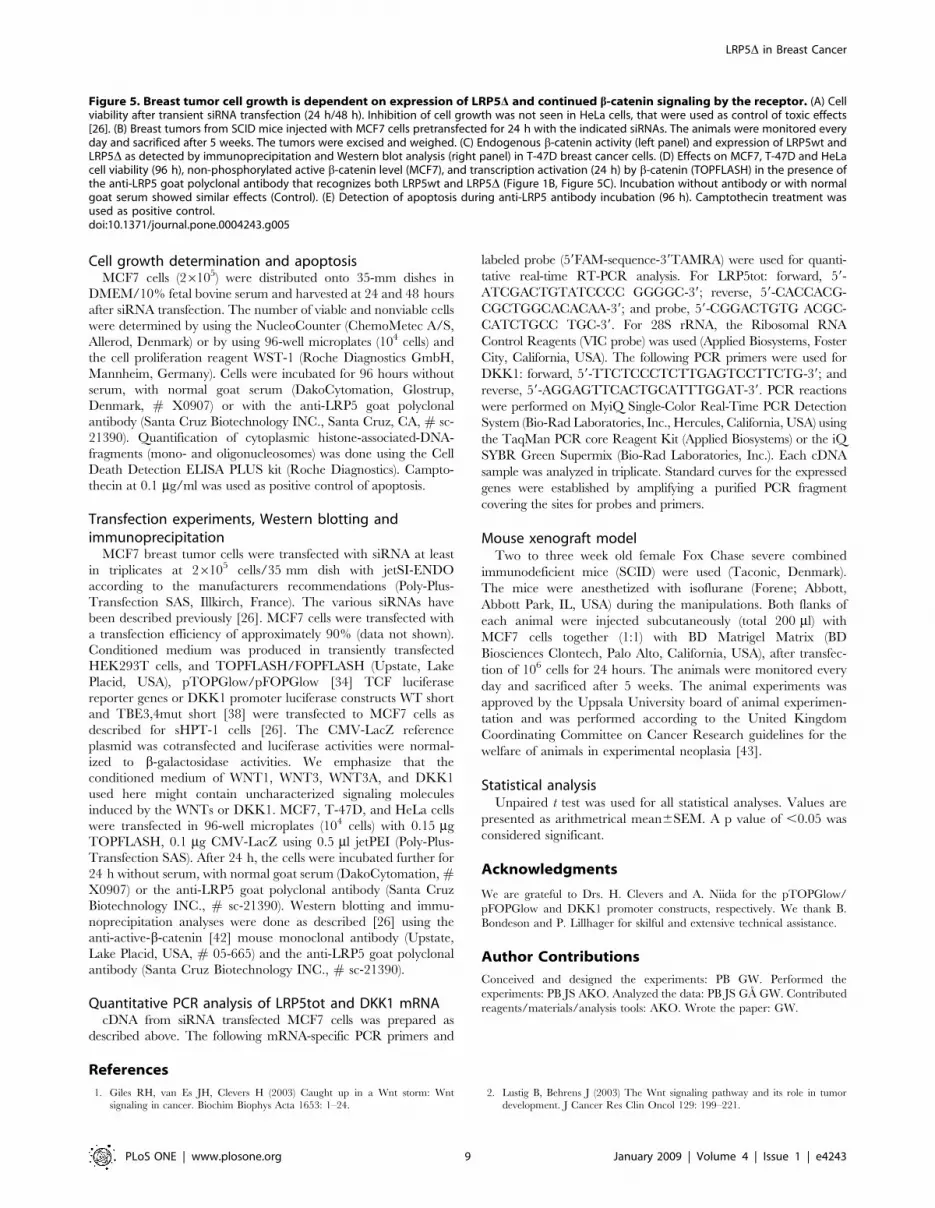
Figure 5. Breast tumor cell growth is dependent on expression of LRP5D and continued b-catenin signaling by the receptor. (A) Cellviability after transient siRNA transfection (24 h/48 h). Inhibition of cell growth was not seen in HeLa cells, that were used as control of toxic effects[26]. (B) Breast tumors from SCID mice injected with MCF7 cells pretransfected for 24 h with the indicated siRNAs. The animals were monitored everyday and sacrificed after 5 weeks. The tumors were excised and weighed. (C) Endogenous b-catenin activity (left panel) and expression of LRP5wt andLRP5D as detected by immunoprecipitation and Western blot analysis (right panel) in T-47D breast cancer cells. (D) Effects on MCF7, T-47D and HeLacell viability (96 h), non-phosphorylated active b-catenin level (MCF7), and transcription activation (24 h) by b-catenin (TOPFLASH) in the presence ofthe anti-LRP5 goat polyclonal antibody that recognizes both LRP5wt and LRP5D (Figure 1B, Figure 5C). Incubation without antibody or with normalgoat serum showed similar effects (Control). (E) Detection of apoptosis during anti-LRP5 antibody incubation (96 h). Camptothecin treatment wasused as positive control.doi:10.1371/journal.pone.0004243.g005
LRP5D in Breast Cancer
PLoS ONE | www.plosone.org 9 January 2009 | Volume 4 | Issue 1 | e4243

3. Moon RT, Kohn AD, DeFerrari GV, Kaykas A (2004) WNT and beta-cateninsignalling: diseases and therapies. Nat Rev Genet 5: 689–701.
4. Gordon MD, Nusse R (2006) Wnt signaling: multiple pathways, multiplereceptors, and multiple transcription factors. J Biol Chem 281: 22429–22433.
5. Katoh M, Katoh M (2007) WNT signaling pathway and stem cell signalingnetwork. Clin Cancer Res 13: 4042–4045.
6. Polakis P (2007) The many ways of Wnt in cancer. Curr Opin Genet Dev 17:45–51.
7. Fuerer C, Nusse R, Ten Berge D (2008) Wnt signalling in development anddisease. Max Delbruck center for molecular medicine meeting on Wnt signalingin development and disease. EMBO Rep 9: 134–138.
8. Roelink H, Wagenaar E, Nusse R (1992) Amplification and proviral activation ofseveral Wnt genes during progression and clonal variation of mouse mammarytumors. Oncogene 7: 487–492.
9. Brennan KR, Brown AM (2004) Wnt proteins in mammary development andcancer. J Mammary Gland Biol Neoplasia 9: 119–131.
10. Lindvall C, Evans NC, Zylstra CR, Li Y, Alexander CM, et al. (2006) The Wntsignaling receptor Lrp5 is required for mammary ductal stem cell activity andWnt1 induced tumorigenesis. J Biol Chem 281: 35081–35087.
11. Al-Hajj M, Wicha MS, Benito-Hernandez A, Morrison SJ, Clarke MF (2003)Prospective identification of tumorigenic breast cancer cells. Proc Natl Acad SciUSA 100: 3983–3988.
12. Fodde R, Brabletz T (2007) Wnt/beta-catenin signaling in cancer stemness andmalignant behaviour. Curr Opin Cell Biol 19: 150–158.
13. Lindvall C, Bu W, Williams BO, Li Y (2007) Wnt signaling, stem cells, and thecellular origin of breast cancer. Stem Cell Rev 3: 157–168.
14. Lin SY, Xia W, Wang JC, Kwong KY, Spohn B, et al. (2000) Beta-catenin, anovel prognostic marker for breast cancer: its roles in cyclin D1 expression andcancer progression. Proc Natl Acad Sci USA 97: 4262–4266.
15. Ryo A, Nakamura M, Wulf G, Liou YC, Lu KP (2001) Pin1 regulates turnoverand subcellular localization of beta-catenin by inhibiting its interaction withAPC. Nat Cell Biol 3: 793–801.
16. Chung GG, Zerkowski MP, Ocal IT, Dolled-Filhart M, Kang JY, et al. (2004)beta-Catenin and p53 analyses of a breast carcinoma tissue microarray. Cancer100: 2084–2092.
17. Nakopoulou L, Mylona E, Papadaki I, Kavantzas N, Giannopoulou I, et al.(2006) Study of phospho-beta-catenin subcellular distribution in invasive breastcarcinomas in relation to their phenotype and the clinical outcome. Mod Pathol19: 556–563.
18. Shekhar MP, Tait L, Gerard B (2006) Essential role of T-cell factor/beta-cateninin regulation of Rad6B: a potential mechanism for Rad6B overexpression inbreast cancer cells. Mol Cancer Res 4: 729–745.
19. Prasad CP, Gupta SD, Rath G, Ralhan R (2007) Wnt signaling pathway ininvasive ductal carcinoma of the breast: relationship between beta-catenin,dishevelled and cyclin D1 expression. Oncology 73: 112–117.
20. Candidus S, Bischoff P, Becker KF, Hofler H (1996) No evidence for mutationsin the alpha- and beta-catenin genes in human gastric and breast carcinomas.Cancer Res 56: 49–52.
21. Ai L, Tao Q, Zhong S, Fields CR, Kim WJ, et al. (2006) Inactivation of Wntinhibitory factor-1 (WIF1) expression by epigenetic silencing is a common eventin breast cancer. Carcinogenesis 27: 1341–1348.
22. Turashvili G, Bouchal J, Burkadze G, Kolar Z (2006) Wnt signaling pathway inmammary gland development and carcinogenesis. Pathobiol 73: 213–223.
23. Veeck J, Niederacher D, An H, Klopocki E, Wiesmann F, et al. (2006) Aberrantmethylation of the Wnt antagonist SFRP1 in breast cancer is associated withunfavourable prognosis. Oncogene 25: 3479–3488.
24. Gebeshuber CA, Sladecek S, Grunert S (2007) Beta-catenin/LEF-1 signalling in
breast cancer–central players activated by a plethora of inputs. Cells Tissues
Organs 185: 51–60.
25. Suzuki H, Toyota M, Caraway H, Gabrielson E, Ohmura T, et al. (2008)
Frequent epigenetic inactivation of Wnt antagonist genes in breast cancer.
Br J Cancer 98: 1147–1156.
26. Bjorklund P, Akerstrom G, Westin G (2007) An LRP5 receptor with internal
deletion in hyperparathyroid tumors with implications for deregulated WNT/b-
catenin signaling. PLoS Med 4: e328.
27. Bjorklund P, Lindberg D, Akerstrom G, Westin G (2008) Stabilizing mutation of
CTNNB1/beta-catenin and protein accumulation analyzed in a large series of
parathyroid tumors of Swedish patients. Mol Cancer 7: 53.
28. Bafico A, Liu G, Yaniv A, Gazit A, Aaronson SA (2001) Novel mechanism of
Wnt signalling inhibition mediated by Dickkopf-1 interaction with LRP6/
Arrow. Nat Cell Biol 3: 683–686.
29. Mao B, Wu W, Li Y, Hoppe D, Stannek P, et al. (2001) LDL-receptor-related
protein 6 is a receptor for Dickkopf proteins. Nature 411: 321–325.
30. Semenov MV, Tamai K, Brott BK, Kuhl M, Sokol S, et al. (2001) Head inducer
Dickkopf-1 is a ligand for Wnt coreceptor LRP6. Curr Biol 11: 951–961.
31. Zhang Y, Wang Y, Li X, Zhang J, Mao J, et al. (2004) The LRP5 high-bone-
mass G171V mutation disrupts LRP5 interaction with Mesd. Mol Cell Biol 24:
4677–4684.
32. Segersten U, Holm PK, Bjorklund P, Hessman O, Nordgren H, et al. (2005) 25-
Hydroxyvitamin D3 1alpha-hydroxylase expression in breast cancer and use of
non-1alpha-hydroxylated vitamin D analogue. Breast Cancer Res 7: R980–986.
33. Benhaj K, Akcali KC, Ozturk M (2006) Redundant expression of canonical Wnt
ligands in human breast cancer cell lines. Oncol Rep 15: 701–707.
34. van de Wetering M, Barker N, Harkes IC, van der Heyden M, Dijk NJ, et al.
(2001) Mutant E-cadherin breast cancer cells do not display constitutive Wnt
signaling. Cancer Res 61: 278–284.
35. Shekhar MP, Gerard B, Pauley RJ, Williams BO, Tait L (2008) Rad6B is a
positive regulator of beta-catenin stabilization. Cancer Res 68: 1741–1750.
36. Chamorro MN, Schwartz DR, Vonica A, Brivanlou AH, Cho KR, et al. (2004)
FGF-20 and DKK1 are transcriptional targets of beta-catenin and FGF-20 is
implicated in cancer and development. EMBO J 24: 73–84.
37. Gonzalez-Sancho JM, Aguilera O, Garcıa JM, Pendas-Franco N, Pena C, et al.
(2004) The Wnt antagonist DICKKOPF-1 gene is a downstream target of beta-
catenin/TCF and is downregulated in human colon cancer. Oncogene 24:
1098–1103.
38. Niida A, Hiroko T, Kasai M, Furukawa Y, Nakamura Y, et al. (2004) DKK1, a
negative regulator of Wnt signaling, is a target of the beta-catenin/TCF
pathway. Oncogene 23: 8520–8526.
39. Barker N, Clevers H (2007) Mining the Wnt pathway for cancer therapeutics.
Nat Rev Drug Discov 5: 997–1014.
40. Pfeiffer P, Qvortrup C, Eriksen JG (2007) Current role of antibody therapy in
patients with metastatic colorectal cancer. Oncogene 26: 3661–3678.
41. Zafir-Lavie I, Michaeli Y, Reiter Y (2007) Novel antibodies as anticancer agents.
Oncogene 26: 3714–3733.
42. van Noort M, Meeldijk J, van der Zee R, Destree O, Clevers H (2002) Wnt
signaling controls the phosphorylation status of beta-catenin. J Biol Chem 277:
17901–17905.
43. Workman P, Balmain A, Hickman JA, McNally NJ, Rohas AM, et al. (1988)
UKCCCR guidelines for the welfare of animals in experimental neoplasia. Lab
Anim 22: 195–201.
LRP5D in Breast Cancer
PLoS ONE | www.plosone.org 10 January 2009 | Volume 4 | Issue 1 | e4243