Producing Solidarity, Inequality and Exclusion Through Insurance
Upload
khangminh22Category
view
1download
0
1
TECHNISCHE UNIVERSITÄT MÜNCHEN 2
Fakultät für Sport- und Gesundheitswissenschaft 3
Abteilung und Poliklinik für Sportorthopädie, Klinikum rechts der Isar 4
5
6
The influence of a new plate fixator on valgus producing open wedge 7
high tibial osteotomy and its outcome in sporting activity 8
9
Matthias Cotic 10
11
Vollständiger Abdruck der von der Fakultät für Sport- und Gesundheitswissenschaft der 12
Technischen Universität München genehmigten Dissertation zur Erlangung des akade- 13
mischen Grades eines 14
15
Doktors der Naturwissenschaften (Dr. rer. nat.) 16
17
18
Vorsitzender: Univ.- Prof. Dr. Veit St. Senner 19
Prüfer der Dissertation: 1. Univ.- Prof. Dr. Andreas B. Imhoff 20
2. Univ.- Prof. Dr. Ansgar Schwirtz 21
3. Prof. Dr. Peter Augat, 22
Paracelsus Medizinische Privatuniversität Salzburg/ 23
Österreich (nur schriftliche Beurteilung); 24
Univ.- Prof. Dr. Joachim Hermsdörfer (nur mündliche Prü- 25
fung) 26
27
Die Dissertation wurde am 17.09.2014 bei der Technischen Universität München einge- 28
reicht und durch die Fakultät für Sport- und Gesundheitswissenschaft am 09.12.2014 29
angenommen. 30
31

2
32
Table of Contents
3
Table of Contents
1 Introduction ...................................................................................................... 5 1.1 Medial compartment osteoarthritis and varus malalignment of the knee ....... 5
1.1.1 Sports related causes for medial compartment osteoarthritis and varus
malalignment of the knee ............................................................................................. 6 1.2 Valgus-producing open wedge high tibial osteotomy (ow HTO) ..................... 7
1.2.1 Fixation devices for valgus-producing ow HTO ............................................... 8 1.2.1.1 The “standard” plate fixator TomoFix™ plate ........................................................ 9 1.2.1.2 The 1st generation PEEKPower HTO-Plate® ...................................................... 11 1.2.1.3 The new 2nd generation PEEKPower HTO-Plate® .............................................. 12
1.3 Research Questions and Purposes .................................................................. 13
Part A: A matched-pair comparison between a standardized titanium plate (TomoFix™ plate) and a new peek-carbon composite plate (1st generation PEEKPower HTO-Plate®).20 ................................................................................ 14
2 Materials and Methods of part A .................................................................. 14 2.1 Patient selection and study design ................................................................... 14
2.1.1 Inclusion and Exclusion Criteria ..................................................................... 15 2.1.2 Baseline Demographics ................................................................................. 16
2.2 Surgical technique of ow HTO for part A and part B ....................................... 17 2.3 Postoperative rehabilitation program ............................................................... 20 2.4 Clinical evaluation .............................................................................................. 20 2.5 Radiographic evaluation .................................................................................... 21
2.5.1 Loss of correction .......................................................................................... 21 2.5.2 Consolidation of the osteotomy gap .............................................................. 22
2.6 Statistical analysis .............................................................................................. 23
3 Results of part A ............................................................................................ 24 3.1 Clinical results .................................................................................................... 24 3.2 Radiographic results .......................................................................................... 27
3.2.1 Loss of correction .......................................................................................... 27 3.2.2 Consolidation of the osteotomy gap .............................................................. 28
3.3 Complications ..................................................................................................... 29
4 Discussion of part A ..................................................................................... 33
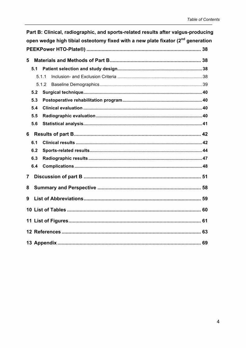
Table of Contents
4
Part B: Clinical, radiographic, and sports-related results after valgus-producing open wedge high tibial osteotomy fixed with a new plate fixator (2nd generation PEEKPower HTO-Plate®) ................................................................................... 38
5 Materials and Methods of Part B .................................................................. 38 5.1 Patient selection and study design ................................................................... 38
5.1.1 Inclusion- and Exclusion Criteria ................................................................... 38 5.1.2 Baseline Demographics ................................................................................. 39
5.2 Surgical technique .............................................................................................. 40 5.3 Postoperative rehabilitation program ............................................................... 40 5.4 Clinical evaluation .............................................................................................. 40 5.5 Radiographic evaluation .................................................................................... 40 5.6 Statistical analysis .............................................................................................. 41
6 Results of part B ............................................................................................ 42 6.1 Clinical results .................................................................................................... 42 6.2 Sports-related results ......................................................................................... 44 6.3 Radiographic results .......................................................................................... 47 6.4 Complications ..................................................................................................... 48
7 Discussion of part B ..................................................................................... 51
8 Summary and Perspective ........................................................................... 58
9 List of Abbreviations ..................................................................................... 59
10 List of Tables ................................................................................................. 60
11 List of Figures ................................................................................................ 61
12 References ..................................................................................................... 63
13 Appendix ........................................................................................................ 69
Introduction
5
1 Introduction
1.1 Medial compartment osteoarthritis and varus malalignment of the knee
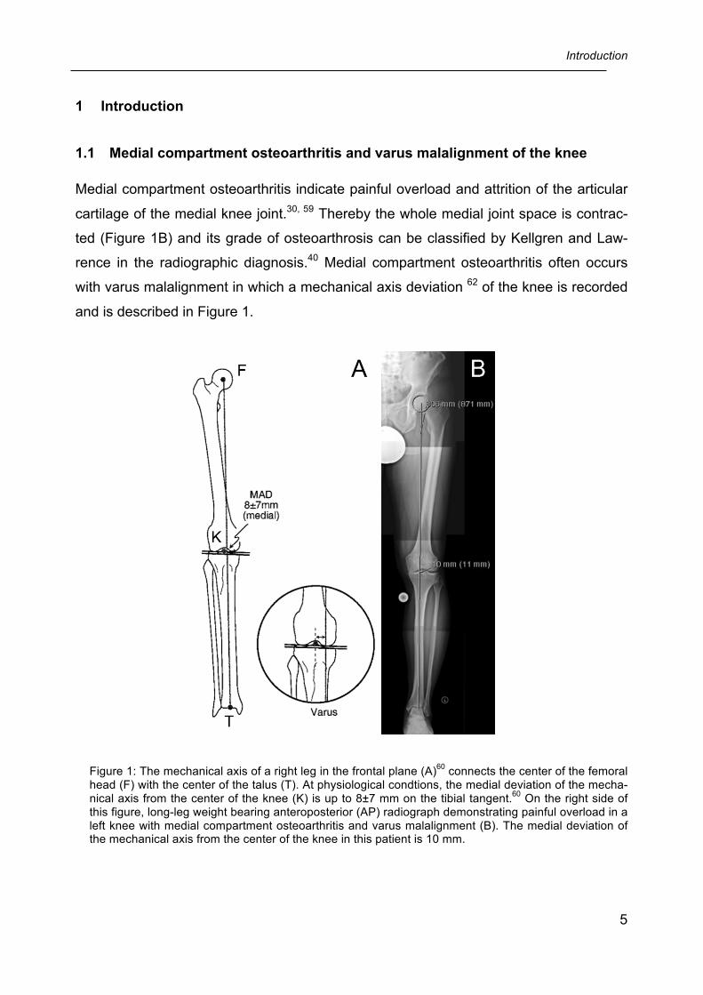
Medial compartment osteoarthritis indicate painful overload and attrition of the articular
cartilage of the medial knee joint.30, 59 Thereby the whole medial joint space is contrac-
ted (Figure 1B) and its grade of osteoarthrosis can be classified by Kellgren and Law-
rence in the radiographic diagnosis.40 Medial compartment osteoarthritis often occurs
with varus malalignment in which a mechanical axis deviation 62 of the knee is recorded
and is described in Figure 1.
Figure 1: The mechanical axis of a right leg in the frontal plane (A)60 connects the center of the femoral head (F) with the center of the talus (T). At physiological condtions, the medial deviation of the mecha-nical axis from the center of the knee (K) is up to 8±7 mm on the tibial tangent.60 On the right side of this figure, long-leg weight bearing anteroposterior (AP) radiograph demonstrating painful overload in a left knee with medial compartment osteoarthritis and varus malalignment (B). The medial deviation of the mechanical axis from the center of the knee in this patient is 10 mm.
Introduction
6
1.1.1 Sports related causes for medial compartment osteoarthritis and varus ma-
lalignment of the knee
In the athlete or the active patient, repetitive microtraumata of the knee are associated
with the regular participation of high performance training and competition over years.87
Sports that requires repetitive running, cutting and jumping like soccer, handball, volley-
ball, basketball and others can place significant loads across weight bearing joints.
These stresses can exert microtraumata on the ligaments, the menisci and the articular
cartilage which lead to further joint degeneration with secondary malalignment of the
knee.3, 36, 39, 55, 85
Like the repetitive microtraumata, acute traumata are also responsible for medial knee
joint degeneration and varus malalignment.85 Traumatic ligamentous ruptures, meniscal
tears or osteochondral lesions can lead to punctual osteochondral attrition and focal
degeneration. If such an injury remains untreated, it leads to secondary malalignment
with further additional degeneration. 3, 36, 39, 55
Moreover, if such microtraumata and acute injuries occur together with congenital varus
malalignemt, it exacerbates over time.85
Conservative treatment modalities for osteoarthritis in young and active patients are
including anti-inflammatory pharmaceuticals, modification of the activity of daily living,
custom orthotics and the use of hyaluronic acid or steroid injections.10, 85 Within physical
therapy and medical training therapy, proprioceptive training of the weight bearing line
as well as the strengthening of the musculus quatriceps femoris via biofeedback are
often used.22 Shoe insoles rarely relieve the pain in the knee joint.64
However, young and active patients with heavy pain and failed conservative treatment
as well as athletes with a threatened career often ask for surgical options.
Introduction
7
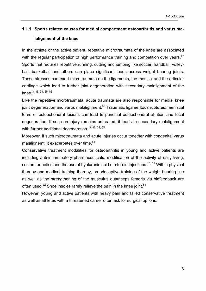
1.2 Valgus-producing open wedge high tibial osteotomy (ow HTO)
Valgus-producing high tibial osteotomy (HTO) is an established intervention for young
and active patients with painful medial compartment osteoarthritis and varus malalign-
ment of the knee.2, 34, 52, 73, 85 The principle of this procedure is to unload the diseased
medial compartment by shifting the mechanical axis of the lower limb more laterally (Fi-
gure 2).2, 5, 34, 52, 73
Figure 2 23 Long-leg weight bearing AP radiographs preoperatively (left) and 6 months postoperatively (right) demonstrating the principle of ow HTO. Line I represents the preoperative mechanical axis. Line Ia represents the new mechanical axis which was shifted from medial to lateral.
How much the new mechanical axis must be shifted to lateral, depends on the concomi-
tant pathology of the knee joint and is described in the literature.32
Introduction
8
Unloading of the medial compartment not only reduces pain and improves knee func-
tion, but it is also a preventive intervention which delays the progression of medial de-
generation and the need for unicompartimental or total knee arthroplasty. 13 2, 34, 52, 73
Although different osteotomy techniques exist, the medial open wedge HTO (ow HTO)
has replaced the lateral closed wedge HTO over the course of the last ten years. This is
due to the fact that the lateral closed wedge technique is associated with a number of
complications, including changes in tibial inclination, increase of tibial offset, patella ba-
ja, non-union, peroneal nerve palsy, loss of correction and bone stock. 38, 53, 86 Moreo-
ver, valgus-producing ow HTO for varus malalignment is indicated in a tibia with a de-
creased medial proximal angle (MPTA).50, 60
Valgus-producing ow HTO reduces the need for muscular detachment and risk of neu-
rovascular complications, and precise intraoperative axial corrections can be made. 45
The major concern in ow HTO is a completely secure fixation device (screw-plate
construct) for stabilization of the proximal tibia while, at the same time, leaving the oste-
otomy gap open.
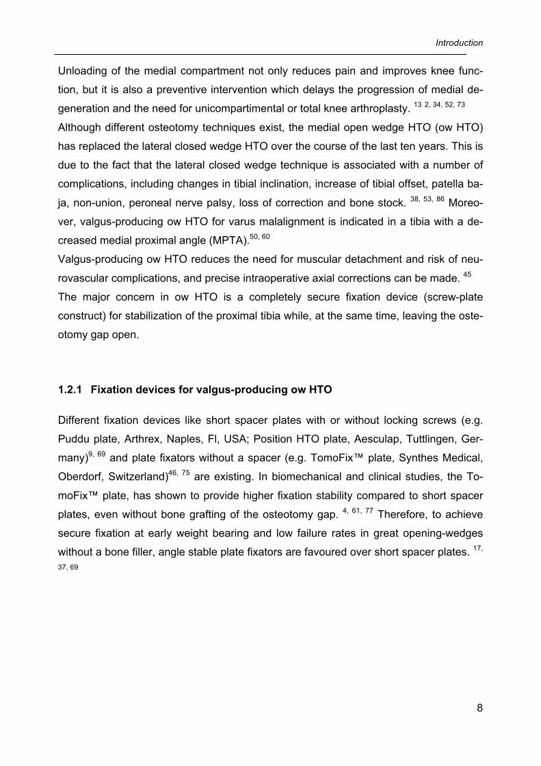
1.2.1 Fixation devices for valgus-producing ow HTO
Different fixation devices like short spacer plates with or without locking screws (e.g.
Puddu plate, Arthrex, Naples, Fl, USA; Position HTO plate, Aesculap, Tuttlingen, Ger-
many)9, 69 and plate fixators without a spacer (e.g. TomoFix™ plate, Synthes Medical,
Oberdorf, Switzerland)46, 75 are existing. In biomechanical and clinical studies, the To-
moFix™ plate, has shown to provide higher fixation stability compared to short spacer
plates, even without bone grafting of the osteotomy gap. 4, 61, 77 Therefore, to achieve
secure fixation at early weight bearing and low failure rates in great opening-wedges
without a bone filler, angle stable plate fixators are favoured over short spacer plates. 17,
37, 69
Introduction
9
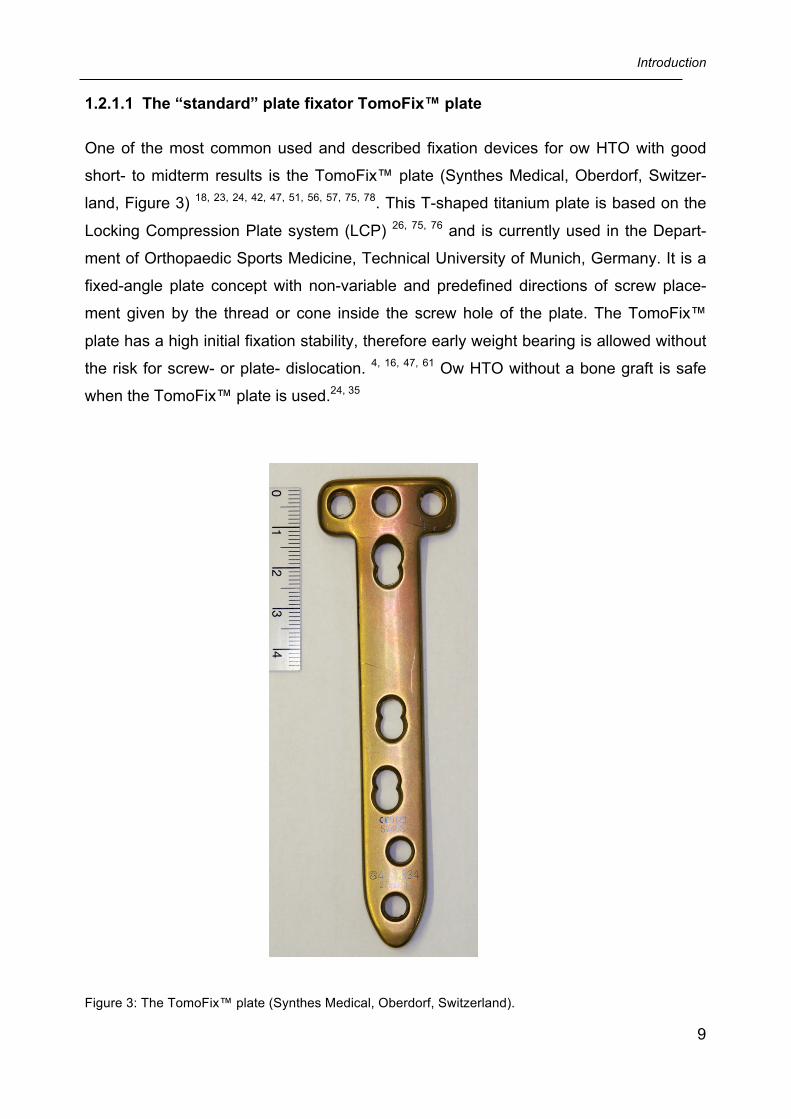
1.2.1.1 The “standard” plate fixator TomoFix™ plate
One of the most common used and described fixation devices for ow HTO with good
short- to midterm results is the TomoFix™ plate (Synthes Medical, Oberdorf, Switzer-
land, Figure 3) 18, 23, 24, 42, 47, 51, 56, 57, 75, 78. This T-shaped titanium plate is based on the
Locking Compression Plate system (LCP) 26, 75, 76 and is currently used in the Depart-
ment of Orthopaedic Sports Medicine, Technical University of Munich, Germany. It is a
fixed-angle plate concept with non-variable and predefined directions of screw place-
ment given by the thread or cone inside the screw hole of the plate. The TomoFix™
plate has a high initial fixation stability, therefore early weight bearing is allowed without
the risk for screw- or plate- dislocation. 4, 16, 47, 61 Ow HTO without a bone graft is safe
when the TomoFix™ plate is used.24, 35
Figure 3: The TomoFix™ plate (Synthes Medical, Oberdorf, Switzerland).
Introduction
10
Disadvantages of the TomoFix™ plate
One disadvantage is the fixed-angle plate concept with its non-variable and predefined
directions of screw placement given by the thread or cone inside the screw hole of the
plate. Thus, combined procedures like Crucriate Ligament reconstructions are associa-
ted with intraoperative limitations because the screws of the plate can not always be
placed to allow a reconstruction tunnel.
Another record with the internal fixed-angle system is, that even with the optimal screw-
in drill guide when these are removed from the plate there is a likelyhood to loose the
drill holes directions. And at the time, when the identical axis of the plate hole and screw
is not achieved, cross threading may accrue and secure fixation is compromised.
It is well-known that patients complain about hardware irritations until removal of the
implant which was demonstrated in recent reports 56, 57. The authors concluded, that
direct local irritation of the hamstring tendons caused by the relative bulky implant are
responsible for a protracted clinical course showing no significant improvement in func-
tion in the first 12 months after HTO, but instead from 12 to 24 months, starting after
removal of the implant.
An alternative to the TomoFix™ plate is the PEEKPower HTO-Plate®.
Introduction
11
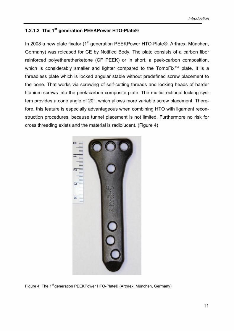
1.2.1.2 The 1st generation PEEKPower HTO-Plate®
In 2008 a new plate fixator (1st generation PEEKPower HTO-Plate®, Arthrex, München,
Germany) was released for CE by Notified Body. The plate consists of a carbon fiber
reinforced polyetheretherketone (CF PEEK) or in short, a peek-carbon composition,
which is considerably smaller and lighter compared to the TomoFix™ plate. It is a
threadless plate which is locked angular stable without predefined screw placement to
the bone. That works via screwing of self-cutting threads and locking heads of harder
titanium screws into the peek-carbon composite plate. The multidirectional locking sys-
tem provides a cone angle of 20°, which allows more variable screw placement. There-
fore, this feature is especially advantageous when combining HTO with ligament recon-
struction procedures, because tunnel placement is not limited. Furthermore no risk for
cross threading exists and the material is radiolucent. (Figure 4)
Figure 4: The 1st generation PEEKPower HTO-Plate® (Arthrex, München, Germany)
Introduction
12
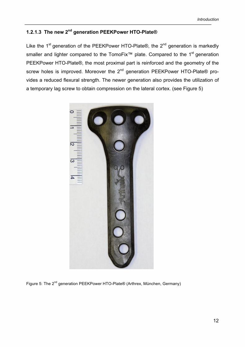
1.2.1.3 The new 2nd generation PEEKPower HTO-Plate®
Like the 1st generation of the PEEKPower HTO-Plate®, the 2nd generation is markedly
smaller and lighter compared to the TomoFix™ plate. Compared to the 1st generation
PEEKPower HTO-Plate®, the most proximal part is reinforced and the geometry of the
screw holes is improved. Moreover the 2nd generation PEEKPower HTO-Plate® pro-
vides a reduced flexural strength. The newer generation also provides the utilization of
a temporary lag screw to obtain compression on the lateral cortex. (see Figure 5)
Figure 5: The 2nd generation PEEKPower HTO-Plate® (Arthrex, München, Germany)
Introduction
13
1.3 Research Questions and Purposes
Part A: Based on the only existing clinical data about the 1st generation PEEKPower HTO-
Plate® (Cotic et. al., see Appendix) 19, there are currently no clinical reports regarding
its safety and effectiveness compared to the commonly used and analog plate fixator
TomoFix™ plate. Above all, there is less knowledge if a 1st generation PEEKPower
HTO-Plate®, which is indeed lighter and smaller than an equal used TomoFix™ plate,
also cause fewer subjective hardware irritations in the plate bed. Moreover, the advan-
tage of its radiolucency have never been prooved yet.
Therefore, the first purpose of this study was to compare the clinical and radiograph-ic outcome of the TomoFix™ plate and the 1st generation PEEKPower HTO-Plate® in a matched-pair analysis of patients undergoing valgus-producing ow HTO. The se-
cond purpose was to objectively evaluate the consolidation of the osteotomy gap,
which is fixed by the 1st generation PEEKPower HTO-Plate®.
Part B: Currently, it is not described why the 2nd generation PEEKPower HTO-Plate® has been
introduced. The effectiveness and safety of this modified implant has not been investi-
gated yet. Above all, sporting activity after valgus-producing ow HTO using the PEEK-
Power HTO-Plate® has never been studied before. However, since ow HTO became
more popular in young and active patients 6, 44, the functional pretension, including
sporting activities, has increased after this procedure 14, 28, 56, 68, 85.
Therefore the third purpose of this study was to prospectively evaluate the clinical, radiographic and sports-related results at 24 months after valgus-producing ow
HTO without bone grafting using the 2nd generation PEEKPower HTO-Plate®.
Part A: Materials and Methods
14
Part A: A matched-pair comparison between a standardized titanium plate
(TomoFix™ plate) and a new peek-carbon composite plate (1st generation
PEEKPower HTO-Plate®).20
2 Materials and Methods of part A
2.1 Patient selection and study design
This study was approved by the Ethics Committee of the Faculty of Medicine of the Mu-
nich University of Technology (Project-No.: 5392/12, the ethics approval is attached to
the Appendix). All enrolled patients provided informed consent to participate in this stu-
dy (the patient informed consent is a part of the questionnaire which is attached to the
Appendix). Between May 2008 and December 2011, 186 consecutive patients were
treated with valgus-producing ow HTO at the Department of Orthopaedic Sports Medi-
cine, Technical University of Munich, Germany.
The 1st generation PEEKPower HTO-Plate® was used in 26 consecutive patients, which
were part of a prospective single group study. The TomoFix™ plate was used in the
remaining 160 patients taken from the prospectively guided and institutional SAP- (Sys-
tem Analysis and Program Development, © SAP AG, 1993-2013) database. A matched-
pair analysis was conducted in April 2012 via the SAS macro “match” software12 (avail-
able at: http://mayoresearch.mayo.edu/mayo/re-search/biostat/sasmacros.cfm). Match-
ing criteria were: gender, age (20-29, 30-39, 40-49, 50-59 years), body mass index (±5
kg/m²), preoperative knee pain (Visual Analog Scale ±2)29, preoperative medial com-
partment osteoarthritis (Kellgren Score ±0)40, preoperative mechanical axis (femoro-
tibial varus angle, ±2°)60 and the new valgus position of the transverse diameter of the
tibial plateau (62 vs 55% coordinate). 41, 32 If the intraoperative noted valgus position
was not listed in the surgery record, the preoperative planned valgus position was tak-
en. A patient summary is listed in the Appendix and includes the anonymized patient
numbers and matching values.
The 26 patients (26 knees) treated with the PEEKPower HTO-Plate® were assigned to
group I. Of the 160 patients treated with the TomoFix™ plate, 26 patients (26 knees)
were matched to the patients of group I and subsequently assigned to group II. The de-
tailed patient characteristics of both groups are provided in Table 2.
Part A: Materials and Methods
15
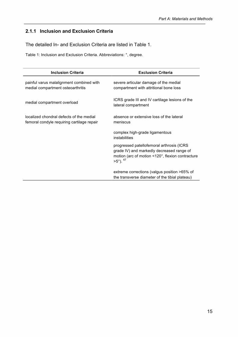
2.1.1 Inclusion and Exclusion Criteria
The detailed In- and Exclusion Criteria are listed in Table 1.
Table 1: Inclusion and Exclusion Criteria. Abbreviations: °, degree.
Inclusion Criteria Exclusion Criteria
painful varus malalignment combined with medial compartment osteoarthritis
severe articular damage of the medial compartment with attritional bone loss
medial compartment overload ICRS grade III and IV cartilage lesions of the lateral compartment
localized chondral defects of the medial femoral condyle requiring cartilage repair
absence or extensive loss of the lateral meniscus
complex high-grade ligamentous instabilities
progressed patellofemoral arthrosis (ICRS grade IV) and markedly decreased range of motion (arc of motion <120°, flexion contracture >5°). 67 extreme corrections (valgus position >65% of the transverse diameter of the tibial plateau)
Part A: Materials and Methods
16
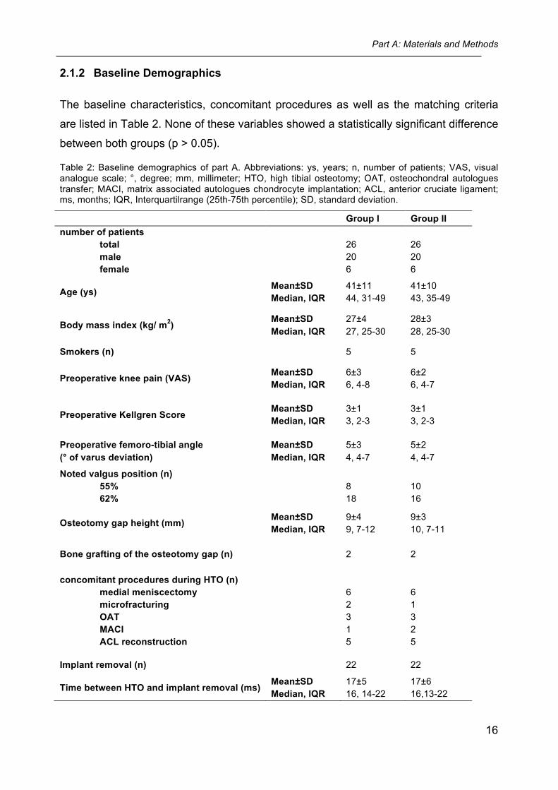
2.1.2 Baseline Demographics
The baseline characteristics, concomitant procedures as well as the matching criteria
are listed in Table 2. None of these variables showed a statistically significant difference
between both groups (p > 0.05).
Table 2: Baseline demographics of part A. Abbreviations: ys, years; n, number of patients; VAS, visual analogue scale; °, degree; mm, millimeter; HTO, high tibial osteotomy; OAT, osteochondral autologues transfer; MACI, matrix associated autologues chondrocyte implantation; ACL, anterior cruciate ligament; ms, months; IQR, Interquartilrange (25th-75th percentile); SD, standard deviation.
Group I Group II number of patients
total male female
26 20 6
26 20 6
Age (ys) Mean±SD Median, IQR
41±11 44, 31-49
41±10 43, 35-49
Body mass index (kg/ m2) Mean±SD Median, IQR
27±4 27, 25-30
28±3 28, 25-30
Smokers (n) 5 5
Preoperative knee pain (VAS) Mean±SD Median, IQR
6±3 6, 4-8
6±2 6, 4-7
Preoperative Kellgren Score Mean±SD Median, IQR
3±1 3, 2-3
3±1 3, 2-3
Preoperative femoro-tibial angle (° of varus deviation)
Mean±SD Median, IQR
5±3 4, 4-7
5±2 4, 4-7
Noted valgus position (n) 55% 62%
8 18
10 16
Osteotomy gap height (mm) Mean±SD Median, IQR
9±4 9, 7-12
9±3 10, 7-11
Bone grafting of the osteotomy gap (n) 2 2
concomitant procedures during HTO (n) medial meniscectomy microfracturing OAT MACI ACL reconstruction
6 2 3 1 5
6 1 3 2 5
Implant removal (n) 22 22
Time between HTO and implant removal (ms) Mean±SD Median, IQR
17±5 16, 14-22
17±6 16,13-22
Part A: Materials and Methods
17
2.2 Surgical technique of ow HTO for part A and part B
Preoperatively, the osteotomy was planned digitally using appropriate computer soft-
ware (mediCAD®, Hectec GmbH, Germany). 70 All operations were performed or direct-
ly supervised by Univ.- Prof. Dr. med. Andreas B. Imhoff.
A biplanar osteotomy, consisting of an osteotomy in the axial plane and an osteotomy in
the frontal plane was performed in all patients. 25, 31, 45 First, the frontal plane osteotomy,
starting in the anterior one-third of the proximal tibia underneath the tibial tuberosity was
performed using an oscillating saw. According to the status of the patellofemoral joint,
the frontal osteotomy exited the bone either distal or proximal the tuberosity. To avoid
decrease in patellar height in patients with preoperative patellofemoral pain25, 31, the
osteotomy in part A of this dissertation was exited distally in 7 patients of both groups,
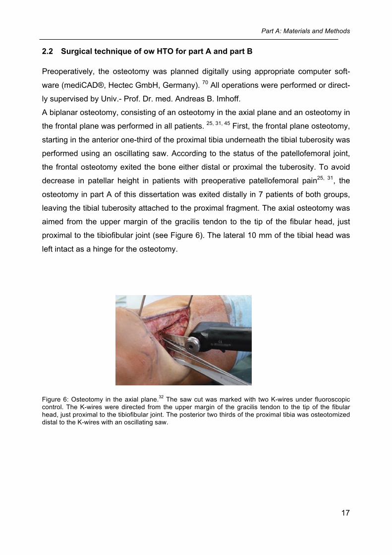
leaving the tibial tuberosity attached to the proximal fragment. The axial osteotomy was
aimed from the upper margin of the gracilis tendon to the tip of the fibular head, just
proximal to the tibiofibular joint (see Figure 6). The lateral 10 mm of the tibial head was
left intact as a hinge for the osteotomy.
Figure 6: Osteotomy in the axial plane.32 The saw cut was marked with two K-wires under fluoroscopic control. The K-wires were directed from the upper margin of the gracilis tendon to the tip of the fibular head, just proximal to the tibiofibular joint. The posterior two thirds of the proximal tibia was osteotomized distal to the K-wires with an oscillating saw.
Part A: Materials and Methods
18
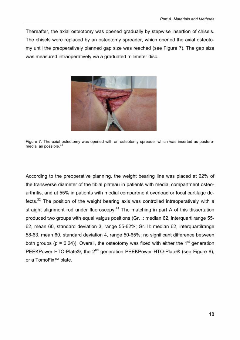
Thereafter, the axial osteotomy was opened gradually by stepwise insertion of chisels.
The chisels were replaced by an osteotomy spreader, which opened the axial osteoto-
my until the preoperatively planned gap size was reached (see Figure 7). The gap size
was measured intraoperatively via a graduated milimeter disc.
Figure 7: The axial osteotomy was opened with an osteotomy spreader which was inserted as postero-medial as possible.32
According to the preoperative planning, the weight bearing line was placed at 62% of
the transverse diameter of the tibial plateau in patients with medial compartment osteo-
arthritis, and at 55% in patients with medial compartment overload or focal cartilage de-
fects.32 The position of the weight bearing axis was controlled intraoperatively with a
straight alignment rod under fluoroscopy.41 The matching in part A of this dissertation
produced two groups with equal valgus positions (Gr. I: median 62, interquartilrange 55-
62, mean 60, standard deviation 3, range 55-62%; Gr. II: median 62, interquartilrange
58-63, mean 60, standard deviation 4, range 50-65%; no significant difference between
both groups (p = 0.24)). Overall, the osteotomy was fixed with either the 1st generation
PEEKPower HTO-Plate®, the 2nd generation PEEKPower HTO-Plate® (see Figure 8),
or a TomoFix™ plate.
Part A: Materials and Methods
19
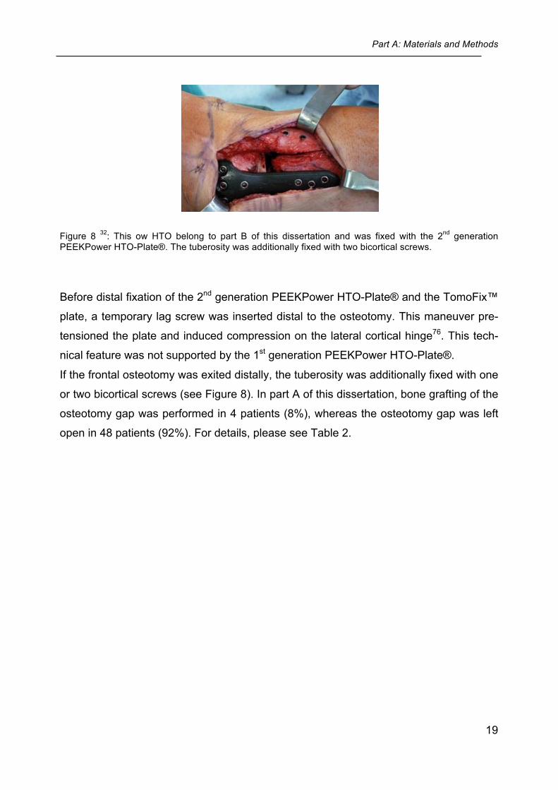
Figure 8 32: This ow HTO belong to part B of this dissertation and was fixed with the 2nd generation PEEKPower HTO-Plate®. The tuberosity was additionally fixed with two bicortical screws.
Before distal fixation of the 2nd generation PEEKPower HTO-Plate® and the TomoFix™
plate, a temporary lag screw was inserted distal to the osteotomy. This maneuver pre-
tensioned the plate and induced compression on the lateral cortical hinge76. This tech-
nical feature was not supported by the 1st generation PEEKPower HTO-Plate®.
If the frontal osteotomy was exited distally, the tuberosity was additionally fixed with one
or two bicortical screws (see Figure 8). In part A of this dissertation, bone grafting of the
osteotomy gap was performed in 4 patients (8%), whereas the osteotomy gap was left
open in 48 patients (92%). For details, please see Table 2.
Part A: Materials and Methods
20
2.3 Postoperative rehabilitation program
The postoperative rehabilitation program for isolated HTO, HTO combined with partial
meniscectomy and HTO combined with ACL reconstruction involved limited weight
bearing with 20 kg for the first 6 weeks. After 6 weeks full weight bearing was allowed
after radiographic controll. In patients with concomitant cartilage repair weight bearing
was not allowed for 6 weeks.
2.4 Clinical evaluation
All patients were followed prospectively. Clinical outcomes were assessed preoperati-
vely and at a minimum follow-up of 24 months postoperatively using the WOMAC Score 11, Lysholm Score 48, and the visual analogue scale (VAS Score) for pain 29. The
WOMAC Score was assessed according to the KOOS User´s Guide (available at
http://www.koos.nu/KOOSGuide2003.pdf). Standardized answer options were given as
5 Likert boxes and each question got a score from 0-4. A normalized percentage score
(100 indicating no problems and 0 indicating extreme problems) was calculated. At the
last follow-up, pain of the plate bed was also asked via VAS Score for the timepoint 2
days before implant removal. Postoperative complications were noted during the whole
study period. The whole patient questionnaire is attached to the Appendix.
Part A: Materials and Methods
21
2.5 Radiographic evaluation
2.5.1 Loss of correction
For the radiographic analysis of correction loss, the Picture Archiving and Communica-
tion System (PACS, Philips Medical Systems, Sectra Imtec AB, Sweden) was used.
Anteriorposterior (AP) and lateral radiographs were obtained two days after HTO (base-
line measurements) and two days after removal of the implant (follow-up measure-
ments). If the implant was not removed during the study period (n = 8), the last available
follow-up radiographs were used (mean time between HTO and last follow-up radio-
graphs 10±5 months).
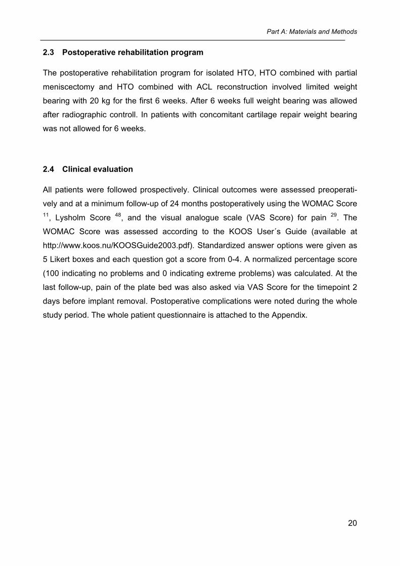
Loss of correction in the frontal plane was assessed by comparing the medial proximal
tibial angle (MPTA) on baseline and follow-up radiographs. The MPTA was defined as
the angle between the proximal anatomical axis of the tibia and a tangent along the arti-
cular surface of the tibial plateau (see Figure 9). 50, 84 Loss of correction in the sagittal
plane was assessed by comparing the tibial slope on baseline and follow-up radio-
graphs using the method described by Brazier et al. 15 (see Figure 9).
Figure 9: On the left side (A) AP radiograph two days after removal of the implant showing the method for measurements of the medial proximal tibial angle (angle a, MPTA). On the right side (B) lateral radio-graph two days after removal of the implant demonstrating the method for measurements of the tibial slope (angle b).
Part A: Materials and Methods
22
2.5.2 Consolidation of the osteotomy gap
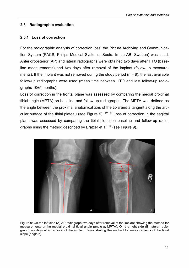
The percentage of consolidation of the osteotomy gap was objectively determined in
group I. For this purpose, an in-house developed threshold segmentation plug-in for
ImageJ 1 was used on the available AP radiographs (DICOM files). First, a region-of-
interest (ROI) was selected, which was the osteotomy gap. Additionally, a second ROI
proximal to the gap in a healthy region of the bone was selected. In this second ROI,
the mean and standard deviation of pixelation were determined. These measurements
were used to define the threshold for the segmentation. For segmenting bone and air in
the gap, a threshold criterion was applied: the pixels (voxel) having intensities above the
threshold were considered to be bone, and those below the threshold as air. Image
quality and x-ray exposure was also verified. Finally, the pixels (voxel) segmented as
bone or air were reported and then used to determine the percentage of bone in the gap
(representative ImageJ reconstruction is shown in Figure 10).
Figure 10: Postoperative AP radiograph (A) at the inpatient stay after ow HTO with the radiolucent PEEKPower HTO-Plate®. ImageJ reconstruction (B) showing the voxel (blue color) for bone in the osteo-tomy gap.
Part A: Materials and Methods
23
2.6 Statistical analysis
Statistical analysis was performed using IBM SPSS Statistics for Windows version 19.0
(IBM-SPSS, New York, USA) and SAS software version 9.2 (SAS Institute Inc., Cary,
NC, USA.). The nonparametric Wilcoxon-test for two related samples was used to com-
pare the pre- and postoperative values within each group. The nonparametric Mann-
Whitney-U-test for independent samples was used to compare patient characteristics,
follow-up, clinical scores, and radiographic data between the two groups. All statistical
tests were performed two sided. Statistical significance was considered at p < 0.05.
Part A: Results
24
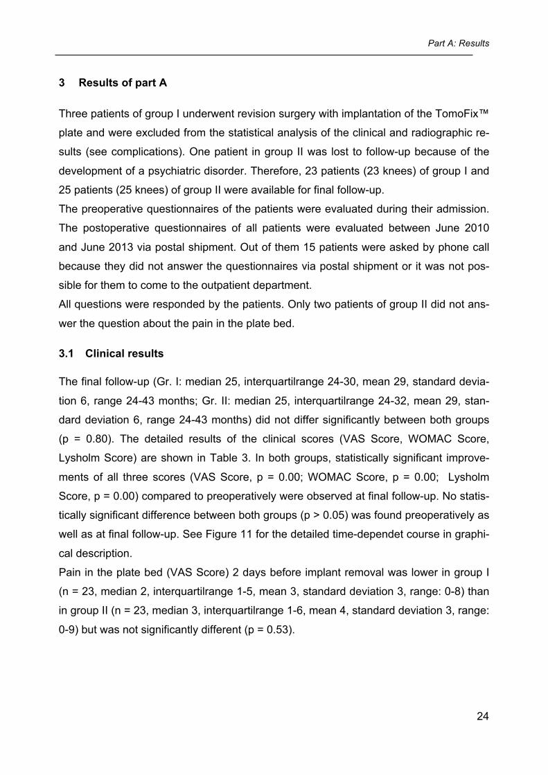
3 Results of part A
Three patients of group I underwent revision surgery with implantation of the TomoFix™
plate and were excluded from the statistical analysis of the clinical and radiographic re-
sults (see complications). One patient in group II was lost to follow-up because of the
development of a psychiatric disorder. Therefore, 23 patients (23 knees) of group I and
25 patients (25 knees) of group II were available for final follow-up.
The preoperative questionnaires of the patients were evaluated during their admission.
The postoperative questionnaires of all patients were evaluated between June 2010
and June 2013 via postal shipment. Out of them 15 patients were asked by phone call
because they did not answer the questionnaires via postal shipment or it was not pos-
sible for them to come to the outpatient department.
All questions were responded by the patients. Only two patients of group II did not ans-
wer the question about the pain in the plate bed.
3.1 Clinical results
The final follow-up (Gr. I: median 25, interquartilrange 24-30, mean 29, standard devia-
tion 6, range 24-43 months; Gr. II: median 25, interquartilrange 24-32, mean 29, stan-
dard deviation 6, range 24-43 months) did not differ significantly between both groups
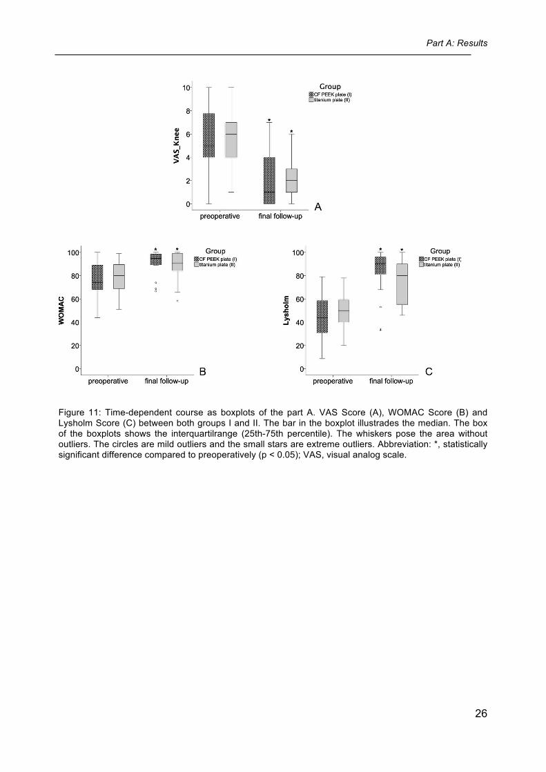
(p = 0.80). The detailed results of the clinical scores (VAS Score, WOMAC Score,
Lysholm Score) are shown in Table 3. In both groups, statistically significant improve-
ments of all three scores (VAS Score, p = 0.00; WOMAC Score, p = 0.00; Lysholm
Score, p = 0.00) compared to preoperatively were observed at final follow-up. No statis-
tically significant difference between both groups (p > 0.05) was found preoperatively as
well as at final follow-up. See Figure 11 for the detailed time-dependet course in graphi-
cal description.
Pain in the plate bed (VAS Score) 2 days before implant removal was lower in group I
(n = 23, median 2, interquartilrange 1-5, mean 3, standard deviation 3, range: 0-8) than
in group II (n = 23, median 3, interquartilrange 1-6, mean 4, standard deviation 3, range:
0-9) but was not significantly different (p = 0.53).
Part A: Results
25
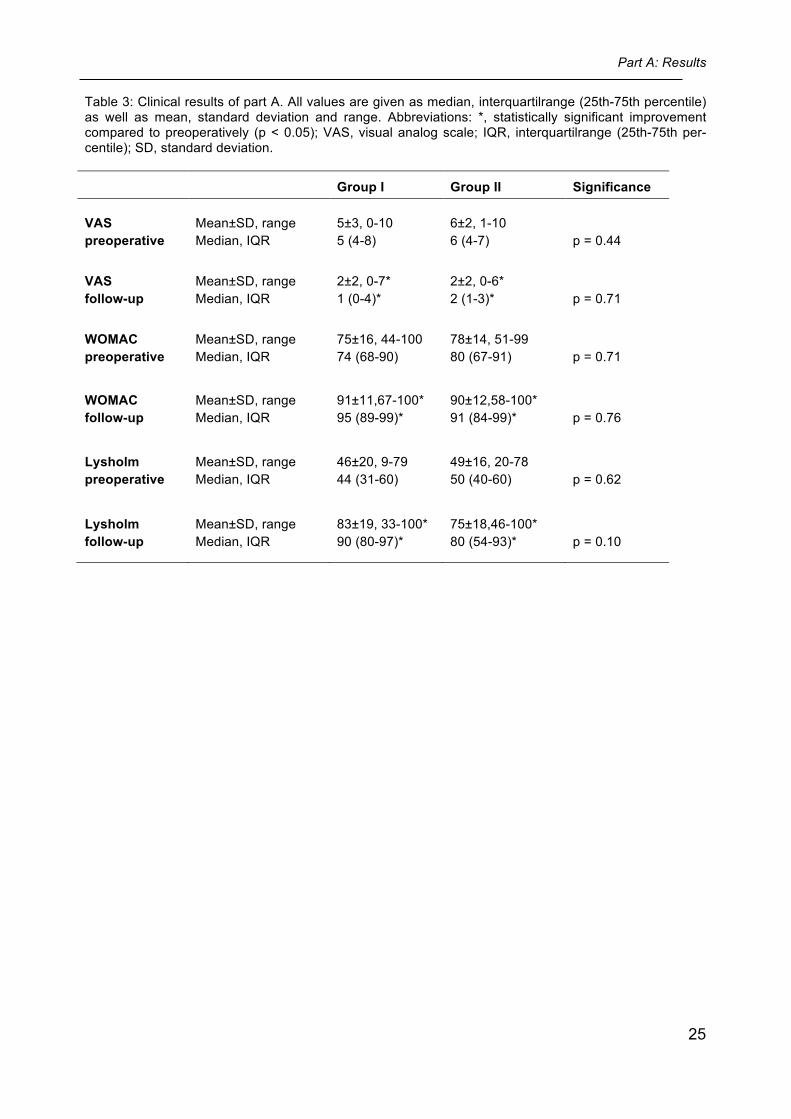
Table 3: Clinical results of part A. All values are given as median, interquartilrange (25th-75th percentile) as well as mean, standard deviation and range. Abbreviations: *, statistically significant improvement compared to preoperatively (p < 0.05); VAS, visual analog scale; IQR, interquartilrange (25th-75th per-centile); SD, standard deviation.
Group I Group II Significance
VAS preoperative
Mean±SD, range Median, IQR
5±3, 0-10 5 (4-8)
6±2, 1-10 6 (4-7)
p = 0.44
VAS follow-up
Mean±SD, range Median, IQR
2±2, 0-7* 1 (0-4)*
2±2, 0-6* 2 (1-3)*
p = 0.71
WOMAC preoperative
Mean±SD, range Median, IQR
75±16, 44-100 74 (68-90)
78±14, 51-99 80 (67-91)
p = 0.71
WOMAC follow-up
Mean±SD, range Median, IQR
91±11,67-100* 95 (89-99)*
90±12,58-100* 91 (84-99)*
p = 0.76
Lysholm preoperative
Mean±SD, range Median, IQR
46±20, 9-79 44 (31-60)
49±16, 20-78 50 (40-60)
p = 0.62
Lysholm follow-up
Mean±SD, range Median, IQR
83±19, 33-100* 90 (80-97)*
75±18,46-100* 80 (54-93)*
p = 0.10
Part A: Results
26
Figure 11: Time-dependent course as boxplots of the part A. VAS Score (A), WOMAC Score (B) and Lysholm Score (C) between both groups I and II. The bar in the boxplot illustrades the median. The box of the boxplots shows the interquartilrange (25th-75th percentile). The whiskers pose the area without outliers. The circles are mild outliers and the small stars are extreme outliers. Abbreviation: *, statistically significant difference compared to preoperatively (p < 0.05); VAS, visual analog scale.
Part A: Results
27
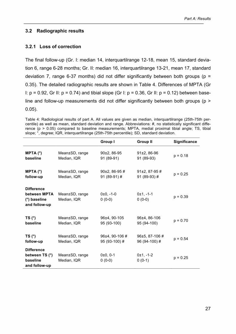
3.2 Radiographic results
3.2.1 Loss of correction
The final follow-up (Gr. I: median 14, interquartilrange 12-18, mean 15, standard devia-
tion 6, range 6-28 months; Gr. II: median 16, interquartilrange 13-21, mean 17, standard
deviation 7, range 6-37 months) did not differ significantly between both groups (p =
0.35). The detailed radiographic results are shown in Table 4. Differences of MPTA (Gr
I: p = 0.92, Gr II: p = 0.74) and tibial slope (Gr I: p = 0.36, Gr II: p = 0.12) between base-
line and follow-up measurements did not differ significantly between both groups (p >
0.05).
Table 4: Radiological results of part A. All values are given as median, interquartilrange (25th-75th per-centile) as well as mean, standard deviation and range. Abbreviations: #, no statistically significant diffe-rence (p > 0.05) compared to baseline measurements; MPTA, medial proximal tibial angle; TS, tibial slope; °, degree; IQR, interquartilrange (25th-75th percentile); SD, standard deviation.
Group I Group II Significance
MPTA (°) baseline
Mean±SD, range Median, IQR
90±2, 86-95 91 (89-91)
91±2, 86-96 91 (89-93) p = 0.18
MPTA (°) follow-up
Mean±SD, range Median, IQR
90±2, 86-95 # 91 (89-91) #
91±2, 87-95 # 91 (89-93) #
p = 0.25
Difference between MPTA (°) baseline and follow-up
Mean±SD, range Median, IQR
0±0, -1-0 0 (0-0)
0±1, -1-1 0 (0-0)
p = 0.39
TS (°) baseline
Mean±SD, range Median, IQR
96±4, 90-105 95 (93-100)
96±4, 86-106 95 (94-100) p = 0.70
TS (°) follow-up
Mean±SD, range Median, IQR
96±4, 90-106 # 95 (93-100) #
96±5, 87-106 # 96 (94-100) #
p = 0.54
Difference between TS (°) baseline and follow-up
Mean±SD, range Median, IQR
0±0, 0-1 0 (0-0)
0±1, -1-2 0 (0-1) p = 0.25
Part A: Results
28
3.2.2 Consolidation of the osteotomy gap
Prior to implant removal, AP radiographs were analyzed to evaluate the percentage of
consolidation relative to the osteotomy gap at various intervals postoperatively: at the
inpatient stay (n = 23), at a mean of 6±2 months (n = 19), at a mean of 12±1 months (n
= 17) and at a mean of 17±2 months (n = 11). Image J analysis showed a mean of
14±11%, a mean of 52±26%, a mean of 78±22% and a mean of 85±9% respectively of
bone relative to the osteotomy gap. The mean pixels (voxel) segmented as bone or air
as well as the mean threshold of group I for every timepoint is shown in Table 5. An il-
lustrated result is shown in Figure 10.
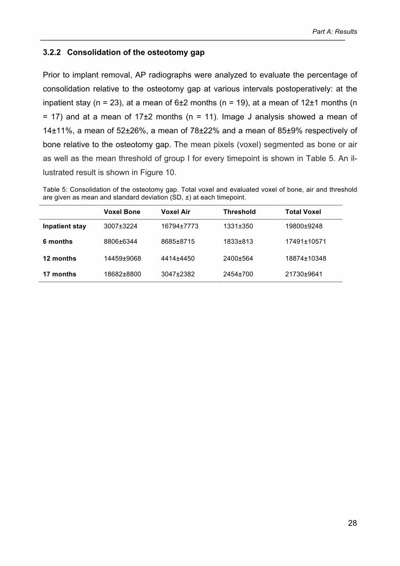
Table 5: Consolidation of the osteotomy gap. Total voxel and evaluated voxel of bone, air and threshold are given as mean and standard deviation (SD, ±) at each timepoint.
Voxel Bone Voxel Air Threshold Total Voxel
Inpatient stay 3007±3224 16794±7773 1331±350 19800±9248
6 months 8806±6344 8685±8715 1833±813 17491±10571
12 months 14459±9068 4414±4450 2400±564 18874±10348
17 months 18682±8800 3047±2382 2454±700 21730±9641
Part A: Results
29
3.3 Complications
The complications of both groups are listed in Table 6.
Table 6: Complications of part A (group I, group II and the remaining TomoFix™ plate fixations). Abbrevi-ations: n.r., not reported; %, percentage; n, number of patients.
Group I (n = 26)
Group II (n = 26)
The remaining Tomo-Fix™ fixations (n = 134)
Overall complication rate 5 (19%) 3 (12%) n.r.
Screw loosening/ breakage 1 (4%) 0 1 (1%)
Non-unions 3 (12%) 0 6 (5%)
Superficial or deep wound infections 1 (4%) 3 (12%) n.r.
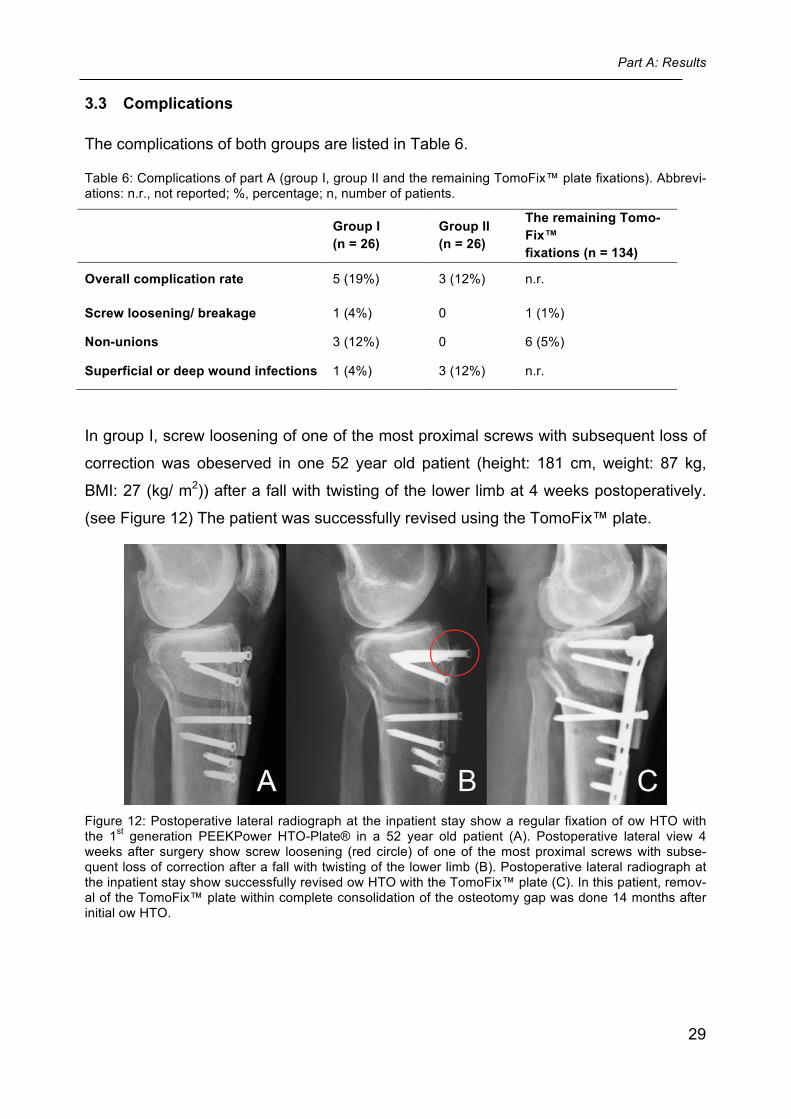
In group I, screw loosening of one of the most proximal screws with subsequent loss of
correction was obeserved in one 52 year old patient (height: 181 cm, weight: 87 kg,
BMI: 27 (kg/ m2)) after a fall with twisting of the lower limb at 4 weeks postoperatively.
(see Figure 12) The patient was successfully revised using the TomoFix™ plate.
Figure 12: Postoperative lateral radiograph at the inpatient stay show a regular fixation of ow HTO with the 1st generation PEEKPower HTO-Plate® in a 52 year old patient (A). Postoperative lateral view 4 weeks after surgery show screw loosening (red circle) of one of the most proximal screws with subse-quent loss of correction after a fall with twisting of the lower limb (B). Postoperative lateral radiograph at the inpatient stay show successfully revised ow HTO with the TomoFix™ plate (C). In this patient, remov-al of the TomoFix™ plate within complete consolidation of the osteotomy gap was done 14 months after initial ow HTO.
Part A: Results
30
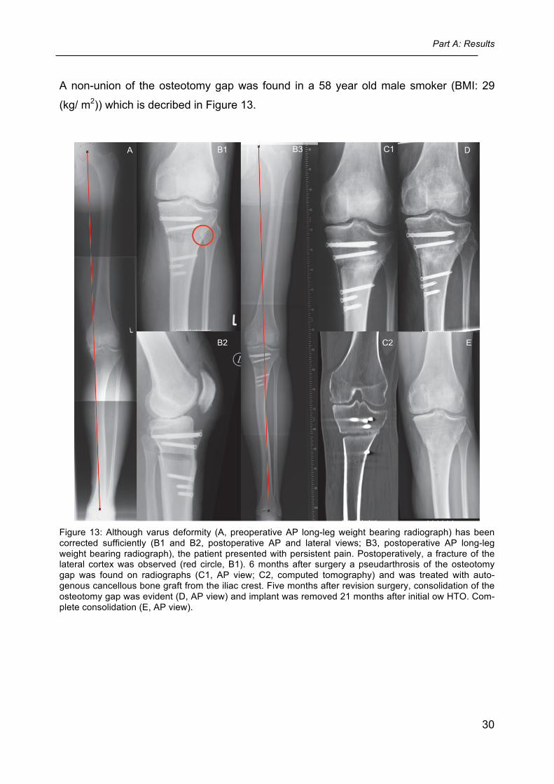
A non-union of the osteotomy gap was found in a 58 year old male smoker (BMI: 29
(kg/ m2)) which is decribed in Figure 13.
Figure 13: Although varus deformity (A, preoperative AP long-leg weight bearing radiograph) has been corrected sufficiently (B1 and B2, postoperative AP and lateral views; B3, postoperative AP long-leg weight bearing radiograph), the patient presented with persistent pain. Postoperatively, a fracture of the lateral cortex was observed (red circle, B1). 6 months after surgery a pseudarthrosis of the osteotomy gap was found on radiographs (C1, AP view; C2, computed tomography) and was treated with auto-genous cancellous bone graft from the iliac crest. Five months after revision surgery, consolidation of the osteotomy gap was evident (D, AP view) and implant was removed 21 months after initial ow HTO. Com-plete consolidation (E, AP view).
Part A: Results
31
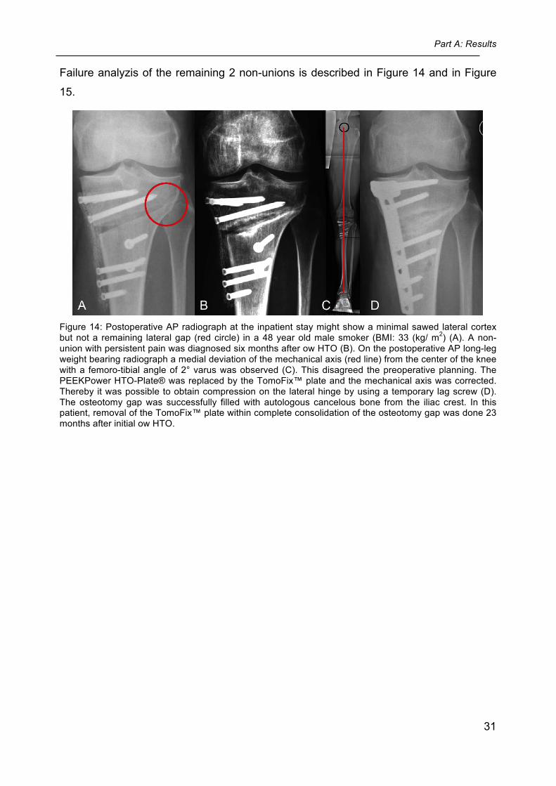
Failure analyzis of the remaining 2 non-unions is described in Figure 14 and in Figure
15.
Figure 14: Postoperative AP radiograph at the inpatient stay might show a minimal sawed lateral cortex but not a remaining lateral gap (red circle) in a 48 year old male smoker (BMI: 33 (kg/ m2) (A). A non-union with persistent pain was diagnosed six months after ow HTO (B). On the postoperative AP long-leg weight bearing radiograph a medial deviation of the mechanical axis (red line) from the center of the knee with a femoro-tibial angle of 2° varus was observed (C). This disagreed the preoperative planning. The PEEKPower HTO-Plate® was replaced by the TomoFix™ plate and the mechanical axis was corrected. Thereby it was possible to obtain compression on the lateral hinge by using a temporary lag screw (D). The osteotomy gap was successfully filled with autologous cancelous bone from the iliac crest. In this patient, removal of the TomoFix™ plate within complete consolidation of the osteotomy gap was done 23 months after initial ow HTO.
Part A: Results
32
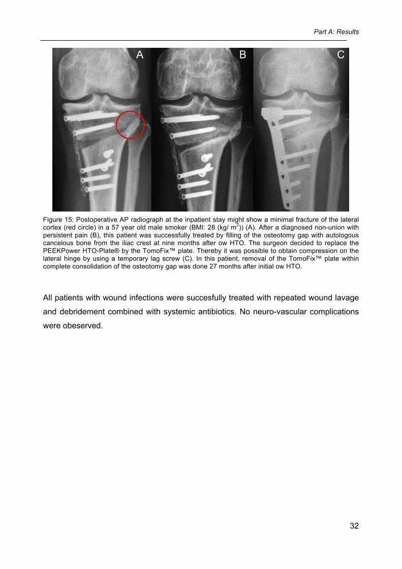
Figure 15: Postoperative AP radiograph at the inpatient stay might show a minimal fracture of the lateral cortex (red circle) in a 57 year old male smoker (BMI: 28 (kg/ m2)) (A). After a diagnosed non-union with persistent pain (B), this patient was successfully treated by filling of the osteotomy gap with autologous cancelous bone from the iliac crest at nine months after ow HTO. The surgeon decided to replace the PEEKPower HTO-Plate® by the TomoFix™ plate. Thereby it was possible to obtain compression on the lateral hinge by using a temporary lag screw (C). In this patient, removal of the TomoFix™ plate within complete consolidation of the osteotomy gap was done 27 months after initial ow HTO.
All patients with wound infections were succesfully treated with repeated wound lavage
and debridement combined with systemic antibiotics. No neuro-vascular complications
were obeserved.
Part A: Discussion
33
4 Discussion of part A
Summary: In the present study, for osteotomy fixation in valgus-producing ow HTO, twenty-six 1st
generation PEEKPower HTO-Plates® were compared with twenty-six TomoFix™ pla-
tes. The main finding was that the 1st generation PEEKPower HTO-Plate® provided the
same clinical and radiographic results as the TomoFix™ plate at a minimum follow-up
of 24 and 12 months respectively. However, more implant related complications oc-
cured with the 1st generation PEEKPower HTO-Plate®.
Clinical evaluation The literature provides, that valgus-producing ow HTO using the TomoFix™ plate is a
safe procedure with promising short- to midterm results 18, 23, 24, 42, 57, 75, 78. However
several drawbacks regarding implant design and surgical technique have been recog-
nized with the TomoFix™ plate because it has a relative bulky design. Niemeyer et al. 56, 57 found a high percentage of patients complained of local irritation associated with
this implant and which disappeared after the removal. In contrast, the 1st generation
PEEKPower HTO-Plate® is considerably smaller, lighter and has another material
compared to the TomoFix™ plate. Aside from the first published clinical results of the 1st
generation PEEKPower HTO-Plate® (Cotic et. al., see Appendix) 19, there are currently
no clinical reports regarding its outcome and safety compared to a commonly used and
standard plate fixator in literature. Therefore this study presents the first results of a
matched-pair comparison between this new implant and the standardized TomoFix™
plate.
Although the 1st generation PEEKPower HTO-Plate® has a smaller design and another
material, differences regarding clinical scores between both groups in the present study
were not observed. Respective the pain in the plate bed, also no significant differences
between both groups were found. If the smaller implant design of the PEEKPower HTO-
Plate® may provide more patient comfort and may avoid the need for implant removal
must be proved by further comparative studies. It can be concluded that, the TomoFix™
plate and the 1st generation PEEKPower HTO-Plate® are viable implants for osteotomy
fixation in valgus-producing ow HTO in terms of the clinical results 24 months after sur-
gery.
Part A: Discussion
34
Removal of the Implant In this prospective study, decision of complete consolidation and indication for implant
removal was made by the surgeon who performed the HTO. In group II the surgeon
indicated all implant removals because of patient complaints about soft tissue irritation
around the plate. In contrast, implant removal in group I was primarily recommended to
the patients by the surgeon because of unknown biologic behaviour of CF PEEK.
Observation of the consolidation of the osteotomy gap
Cross-sectional consolidation of the osteotomy gap was analyzed objectively with the
use of ImageJ. This observation showed consolidation of 52%, 78% and 85% at 6, 12,
and 17 months after sugery, respectively and was not shown in the literature before.
Analyses with a titanium implant were not possible because of its radiological artifacts.
Therefore, in this observation it was not possible to evaluate the influence of any im-
plant on bone healing after ow HTO. It is concluded that for the objective bony-
evaluation after ow HTO, a radiolucent PEEKPower HTO-Plate® is superior to a
TomoFix™ plate on standardized AP radiographs.
Biomechanical observation In a preclinical biomechanical evaluation, a TomoFix™ plate as well as the 1st genera-
tion PEEKPower HTO-Plate® were tested in a comparable worst-case compression
bending test (white paper, published by Arthrex, available at: http://www.arthrex.com;
attached to the Appendix). For both specimen groups lifetime decreased with increasing
loads. Compared to the peek-carbon composite results the titanium results reveal de-
creased lifetimes at lower loads, but show a tendency to compensate this mismatch at
higher load levels. Therefore, the PEEKPower HTO-Plate® shows increased flexural
strength compared to a TomoFix™ plate with increased flexural deformation. That could
be attributed to the different degree of rigidity between both plate-materials. Titanium
has got the characteristic that it is more flexible than PEEK and carbon and therefore
has got the tendency to be more elastic and to compensate loadings with increased
flexural deformation. In contrast, the PEEKPower HTO-Plate® showed its characteristi-
cal increased flexural strength through a higher accepted alternation of load on the
plate.
Part A: Discussion
35
Loss of correction In the present study, no statistically significant differences between baseline and follow-
up measurements were observed for MPTA and tibial slope in both groups. Further-
more, there were no significant differences between both groups in the timepoints of
complete consolidation, in the median delta of MPTA as well as in the median delta of
slope. It is concluded that both plates are stable enough to maintain the amount of cor-
rection until complete ossification without significant differences between each other.
Complications Non-Unions:
In the clinical comparison analysis were no incidences of any nonspecific complications,
such as nerve injury, vessel injury or necrosis of the tibial plateau. There were 3 in-
cidences (12%) of delayed consolidation of the osteotomy gap occurred in patients with
a 1st generation PEEKPower HTO-Plate®. Patients with a TomoFix™ plate showed not
any non-union (0%).
Compared to the literature, smokers and patients with a fracture of the lateral cortex
after HTO 56, 75, 83 are showing inferior consolidation. These findings are concordant to
the present study, because it was about three smokers who developed the non-union of
the osteotomy gap and were treated with autogenous cancellous bone graft from the
iliac crest after initial therapy. Additionally, a fracture of the lateral cortex was also
found.
Nevertheless, also implant related factors might have negatively influenced bone heal-
ing. Compared to the TomoFix™ plate, the 1st generation PEEKPower HTO-Plate®
does not provide the use of a temporary lag screw. By inserting this screw through the
first distal hole below the osteotomy, interfragmentary compression of the lateral cortex
is induced. Studies about instable osteotomies due to fractures of the lateral cortex re-
sulting in non-union showed, that a lag screw was not used 56. Furthermore, it is fact
that well-controlled micromotion at the interface of a screw-shaped implant stimulates
bone healing 82 and therefore flexural deformation of the hardware is needed. However,
the 1st generation PEEKPower HTO-Plate® has a higher flexural strength compared to
the TomoFix™ plate (data provided by Arthrex and attached to the Appendix). There-
fore, insufficient flexural deformation of the PEEKPower HTO-Plate® might also be a
reason for the higher non-union rate in group I. The non-union rate of the remaining 134
patients treated with the TomoFix™ plate during the study period was 5% (Table 6).
Part A: Discussion
36
Other authors found non-union rates of 0-7% after ow HTO fixed with the TomoFix™
plate. 18, 24, 51, 81 Therefore, the risk for non-union seems to be higher with the 1st gen-
eration PEEKPower HTO-Plate®.
Screw-loosening:
In one patient of group I (4%), loosening of one of the most proximal screws with ac-
companying loss of the corrected MPTA was observed. Whereas no screw backouts
were found in group II (0%). The literature described screw breakages during extraction
but no screw looesenings with the TomoFix™ plate. 81 Therefore, the following implant
related failures might also be responsible for the screw loosening of the 1st generation
PEEKPower HTO-Plate®. According to the biomechanical evaluation mentioned above
(data provided by Arthrex, see Appendix), the increased flexural strength of the peek-
carbon composite plate did not allow as much flexural deformation as a titanium plate.
Therefore this new plate accepted a higher alternation of load on the plate without frac-
turing compared to the titanium plate. Accordingly that could be the reason for in-
creased loads not on the peek-carbon plate but in the bone anchoring with ejection of
the screws. These findings suggest that the 1st generation PEEKPower HTO-Plate®
has a too thin and weak hole-bed for a save screw fixation via the self-cutting threads
and locking heads of the screws into the plate and a too less flexural deformation at the
same time.
Interestingly, in the remaining 134 patients treated with the TomoFix™ plate, a screw
breakage due to a fall onto the lower limb was found (Table 6). Though the hole-bed of
the TomoFix™ plate is save, it does not prevent from screw breakages needing revision
surgery through unphysiological high impacts.
Histological Evaluation
The novel implant investigated in this study is made of CF PEEK, which is increasingly
used for trauma, orthopedic, and spinal implants. 43 This non-metallic biomaterial provi-
des the high strength of metals combined with good biocompatibility and imaging com-
patibility of polymers. 43 Because of the locking concept of the PEEKPower HTO-
Plate®, abrasion of the plate material commonly occurs during osteotomy fixation. In
this study, histological evaluation of CF PEEK wear was performed in 15 cases by a
pathologist of the Institute of Pathology, Technical University of Munich, Germany. The
observation did not show acute or chronic inflammation or tissue necrosis.19, 20 This fin-
Part A: Discussion
37
ding is in line with other studies, which found good biocompatibility of CF PEEK.66, 71
Furthermore, it has been shown that carbon-carbon composites have low wear rates
and no osteolytic or cytotoxic potential in cell cultures33; It has to be concluded that ma-
terial abrasion seen in this technique has no influence on the postoperative outcome
and has to be accepted when stabilizing HTO with a peek-carbon composite plate.
Therefore, from a biological point, the PEEKPower HTO-Plate® can be safely used for
HTO and plate removal would not be mandatory.
Limitations of the Study
The patients were not randomized preoperatively. Nevertheless, a matched-pair design
was chosen to achieve adequate comparability by minimizing confounding factors.
ImageJ analysis was limited by user interpretation, image quality, and by the fact that
2D projections were used and not 3D data. However, compared to simple subjective
and visual inspection, as used in the literature 74, this is a robust method to objectively
analyze the consolidation on standardized AP radiographs.
Concerning the method to determine the amount of correction loss, serial long-leg
weight bearing AP radiographs or radiostereometric analysis might have been more
accurate in every patient. Unfortunately, these methods were not allowed by the ethics
commitee due to the associated radiation exposure. However, by evaluating correction
loss in two standard planes (frontal and sagittal), the accuracy of the radiological analy-
sis increased.
Conclusion
Considering the drawbacks of the 1st generation PEEKPower HTO-Plate® discussed
above, it is recommended to adapt this 1st generation peek-carbon composite plate and
to create a new plate allowing as much micromotion as is necessary to achieve no im-
plant failure and screw loosening at the same time. A 2nd generation has been intro-
duced by the manufacturer in the meanwhile. The new plate provides an improved ge-
ometry of the screw holes and a reduced flexural strength. The newer generation also
provides the utilization of a temporary lag screw to obtain compression on the lateral
cortex. Further studies must prove whether these modifications result in less implant
related complications.
Part B: Materials and Methods
38
Part B: Clinical, radiographic, and sports-related results after valgus-
producing open wedge high tibial osteotomy fixed with a new plate fixator
(2nd generation PEEKPower HTO-Plate®)
5 Materials and Methods of Part B
5.1 Patient selection and study design
Between March 2010 and July 2011, 25 consecutive patients were treated with valgus-
producing ow HTO without bone grafting using the 2nd generation PEEKPower HTO-
Plate® at the Department of Orthopaedic Sports Medicine, Technical University of Mu-
nich, Germany. All enrolled patients provided informed consent to participate in this stu-
dy.
5.1.1 Inclusion- and Exclusion Criteria
The Inclusion- and Exclusion criteria are listed under 2.1.1. Only patients without bone
grafting were included in the present study.
Part B: Materials and Methods
39
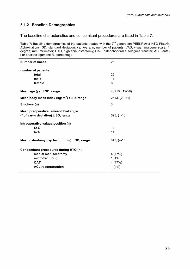
5.1.2 Baseline Demographics
The baseline characteristics and concomitant procedures are listed in Table 7.
Table 7: Baseline demographics of the patients treated with the 2nd generation PEEKPower HTO-Plate®. Abbreviations: SD, standard deviation; ys, years; n, number of patients; VAS, visual analogue scale; °, degree; mm, millimeter; HTO, high tibial osteotomy; OAT, osteochondral autologues transfer; ACL, ante-rior cruciate ligament; %, percentage.
Number of knees 25
number of patients total male female
25 17 8
Mean age (ys) ± SD, range 45±10, (19-58)
Mean body mass index (kg/ m2) ± SD, range 25±3, (20-31)
Smokers (n) 3
Mean preoperative femoro-tibial angle (° of varus deviation) ± SD, range
5±3, (1-16)
Intraoperative valgus position (n) 55% 62%
11 14
Mean osteotomy gap height (mm) ± SD, range 8±3, (4-15)
Concomitant procedures during HTO (n) medial meniscectomy microfracturing OAT ACL reconstruction
4 (17%) 1 (4%) 4 (17%) 1 (4%)
Part B: Materials and Methods
40
5.2 Surgical technique
The detailed surgical technique is described under 2.2. In 7 cases, out of this patient
cohort with the 2nd generation PEEKPower HTO-Plate®, the osteotomy was exited dis-
tally, leaving the tibial tuberosity attached to the proximal fragment. In these cases, the
tuberosity was additionally fixed with one or two bicortical screws. In the remaining pa-
tients, the osteotomy was exited proximally, leaving the tuberosity attached to the distal
fragment. As mentioned above, bone grafting of the osteotomy gap was not performed.
5.3 Postoperative rehabilitation program
The postoperative rehabilitation program is listed under 2.3.
5.4 Clinical evaluation
All patients were evaluated preoperatively and at 12 and 24 months postoperatively. For
clinical evaluation, knee pain, osteoarthritic symptoms and function of the affected leg
were assessed prospectively using the visual analogue scale (VAS Score) 29 for pain,
the WOMAC Score 11 and the Lysholm Score 48. For the WOMAC Score, standardized
answer options were given as 5 Likert boxes and each question got a score from 0-4. A
normalized percentage score (100 indicating no problems and 0 indicating extreme
problems) was calculated (for details, see: http://www.koos.nu/KOOSGuide2003.pdf).
During the whole study period, postoperative complications were also noted.
The sports-related outcome was evaluated using the Tegner activity scale80 and a self
designed questionnaire, which assessed the number of sport disciplines, sports fre-
quency (defined as sessions per week) and sports duration (defined as hours per week)
one year before surgery and 24 months postoperatively. The whole questionnaire is
attached to the Appendix.
5.5 Radiographic evaluation
Radiographic analysis was performed by using the Picture Archiving and Communicati-
on System (PACS, Philips Medical Systems, Sectra Imtec AB, Sweden). AP and lateral
Part B: Materials and Methods
41
radiographs were obtained two days after ow HTO (baseline measurements) and two
days after implant removal (follow-up measurements). If the implant was not removed
during the study period (n = 9), the last available follow-up radiographs were used (me-
an time between HTO and last follow-up radiograph: 10±6 months). Lateral radiographs
were taken with the knee in 30° of flexion.
The assessment for the loss of correction in the frontal plane as well as in the sagittal
plane is described in detail under 2.5.1.
5.6 Statistical analysis
Statistical analysis was performed using IBM SPSS Statistics for Windows version 21.0
(IBM Inc., NY, USA). To compare the clinical scores between each follow-up examinati-
on, the nonparametric Friedman-test for related samples was used. If the test showed
significant differences over time, the nonparametric Wilcoxon-test for two related samp-
les was used to compare the values between two different time points. All statistical
tests were performed two sided. Statistical significance was considered at p < 0.05.
Part B: Results
42
6 Results of part B
Three patients underwent revision surgery with implantation of another implant and we-
re excluded from the statistical analysis of the clinical and radiographic results (see
complications). Therefore, 22 patients were available for the 12 (mean 12±1) and 24
(mean 24±2) months clinical follow-up evaluation.
The preoperative questionnaires of the patients were evaluated during their admission.
The postoperative questionnaires of all patients were evaluated between February 2011
and July 2013 via postal shipment. Out of them 13 patients were asked by phone call
for the 24 months scores. This was due to the fact that these patients did not answer
the questionnaires or it was not possible for them to come to the outpatient department.
Out of them, 1 patient did not sign the consent form of the questionnaire but still partici-
pated in the telephone survey.
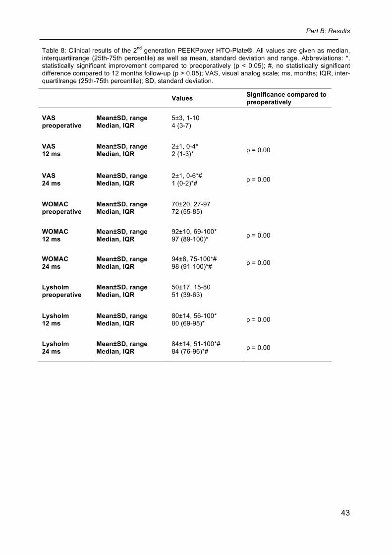
6.1 Clinical results
The detailed results of the clinical scores (VAS Score, WOMAC Score, Lysholm Score)
are shown in Table 8. Compared to preoperatively, statistically significant improvements
(p < 0.05) of all three scores were observed at the 12 and 24 months follow-up. The
time-dependent course of the clinical scores is shown in Figure 16. No significant diffe-
rences were found between the 12 and 24 months follow-up (VAS, p = 0.29; WOMAC, p
= 0.22; Lysholm, p = 0.11). Out of the 22 patients implant removal was done in 14 pati-
ents. The mean time between HTO and implant removal was 17±5 months.
Part B: Results
43
Table 8: Clinical results of the 2nd generation PEEKPower HTO-Plate®. All values are given as median, interquartilrange (25th-75th percentile) as well as mean, standard deviation and range. Abbreviations: *, statistically significant improvement compared to preoperatively (p < 0.05); #, no statistically significant difference compared to 12 months follow-up (p > 0.05); VAS, visual analog scale; ms, months; IQR, inter-quartilrange (25th-75th percentile); SD, standard deviation.
Values Significance compared to preoperatively
VAS preoperative
Mean±SD, range Median, IQR
5±3, 1-10 4 (3-7)
VAS 12 ms
Mean±SD, range Median, IQR
2±1, 0-4* 2 (1-3)* p = 0.00
VAS 24 ms
Mean±SD, range Median, IQR
2±1, 0-6*# 1 (0-2)*# p = 0.00
WOMAC preoperative
Mean±SD, range Median, IQR
70±20, 27-97 72 (55-85)
WOMAC 12 ms
Mean±SD, range Median, IQR
92±10, 69-100* 97 (89-100)* p = 0.00
WOMAC 24 ms
Mean±SD, range Median, IQR
94±8, 75-100*# 98 (91-100)*# p = 0.00
Lysholm preoperative
Mean±SD, range Median, IQR
50±17, 15-80 51 (39-63)
Lysholm 12 ms
Mean±SD, range Median, IQR
80±14, 56-100* 80 (69-95)* p = 0.00
Lysholm 24 ms
Mean±SD, range Median, IQR
84±14, 51-100*# 84 (76-96)*# p = 0.00
Part B: Results
44
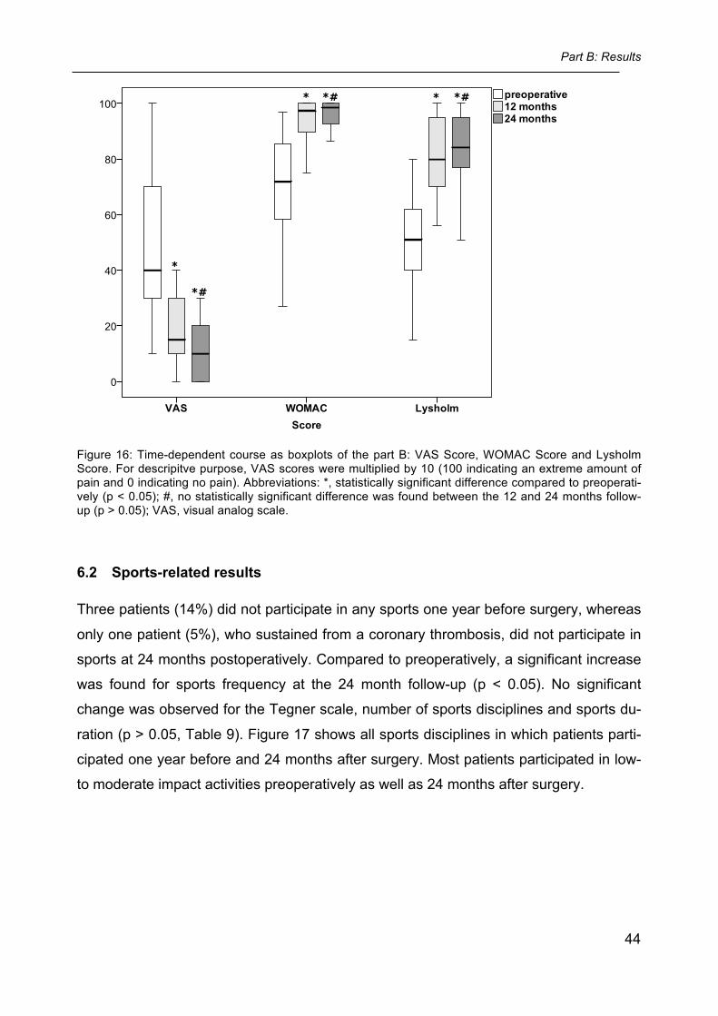
Figure 16: Time-dependent course as boxplots of the part B: VAS Score, WOMAC Score and Lysholm Score. For descripitve purpose, VAS scores were multiplied by 10 (100 indicating an extreme amount of pain and 0 indicating no pain). Abbreviations: *, statistically significant difference compared to preoperati-vely (p < 0.05); #, no statistically significant difference was found between the 12 and 24 months follow-up (p > 0.05); VAS, visual analog scale.
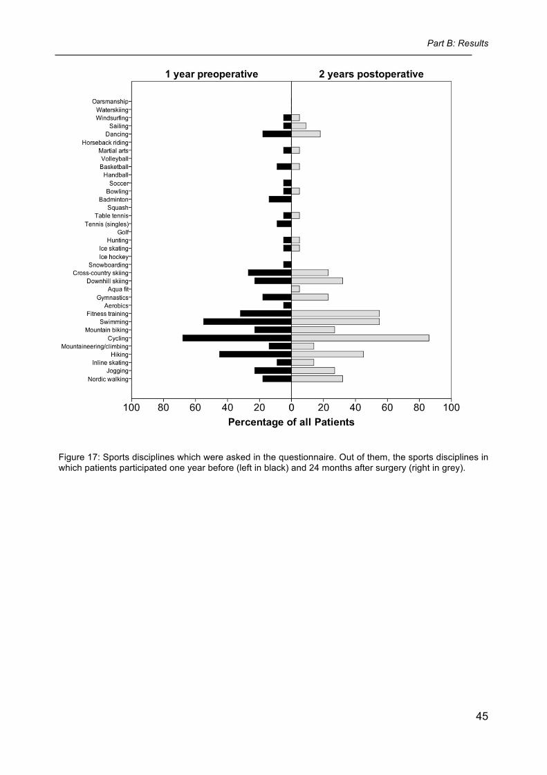
6.2 Sports-related results
Three patients (14%) did not participate in any sports one year before surgery, whereas
only one patient (5%), who sustained from a coronary thrombosis, did not participate in
sports at 24 months postoperatively. Compared to preoperatively, a significant increase
was found for sports frequency at the 24 month follow-up (p < 0.05). No significant
change was observed for the Tegner scale, number of sports disciplines and sports du-
ration (p > 0.05, Table 9). Figure 17 shows all sports disciplines in which patients parti-
cipated one year before and 24 months after surgery. Most patients participated in low-
to moderate impact activities preoperatively as well as 24 months after surgery.
Part B: Results
45
Figure 17: Sports disciplines which were asked in the questionnaire. Out of them, the sports disciplines in which patients participated one year before (left in black) and 24 months after surgery (right in grey).
Part B: Results
46
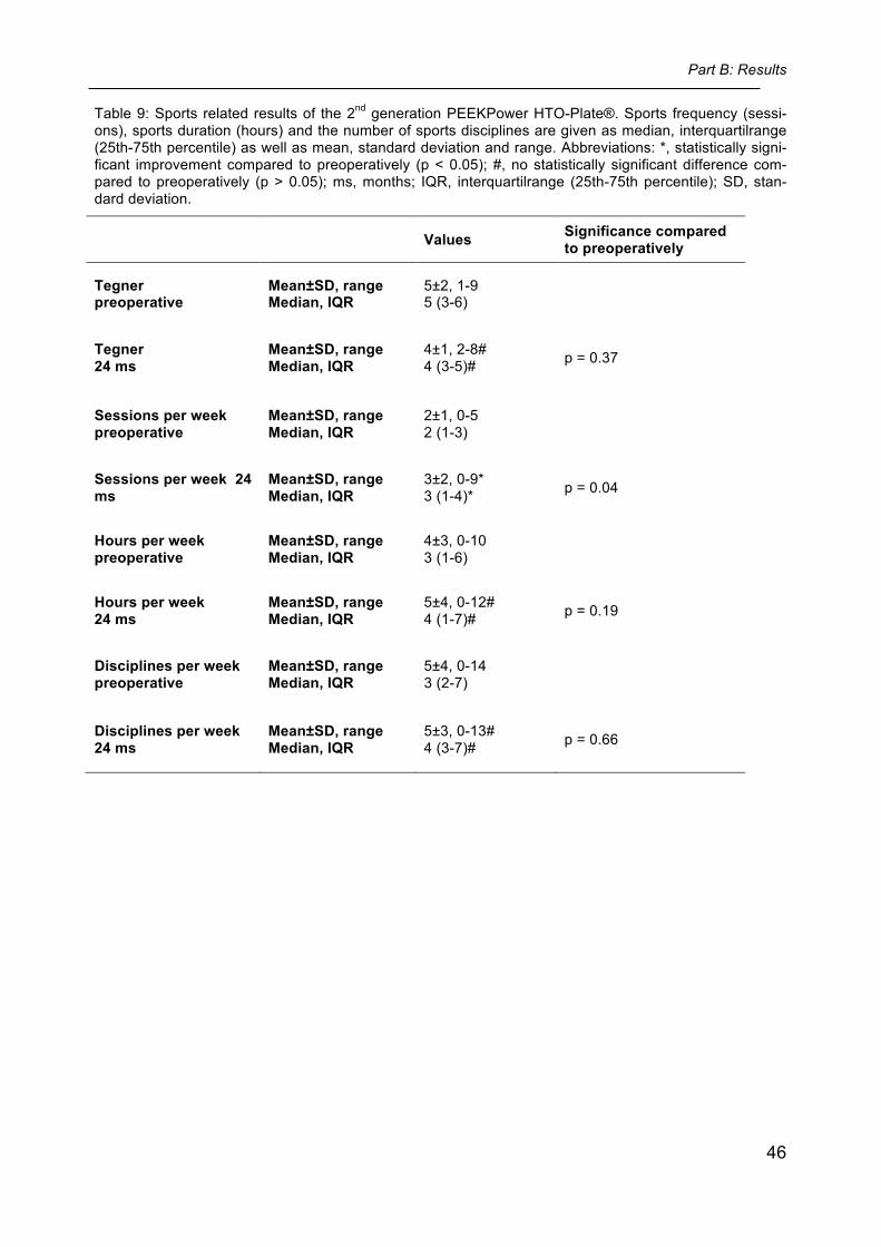
Table 9: Sports related results of the 2nd generation PEEKPower HTO-Plate®. Sports frequency (sessi-ons), sports duration (hours) and the number of sports disciplines are given as median, interquartilrange (25th-75th percentile) as well as mean, standard deviation and range. Abbreviations: *, statistically signi-ficant improvement compared to preoperatively (p < 0.05); #, no statistically significant difference com-pared to preoperatively (p > 0.05); ms, months; IQR, interquartilrange (25th-75th percentile); SD, stan-dard deviation.
Values Significance compared to preoperatively
Tegner preoperative
Mean±SD, range Median, IQR
5±2, 1-9 5 (3-6)
Tegner 24 ms
Mean±SD, range Median, IQR
4±1, 2-8# 4 (3-5)# p = 0.37
Sessions per week preoperative
Mean±SD, range Median, IQR
2±1, 0-5 2 (1-3)
Sessions per week 24 ms
Mean±SD, range Median, IQR
3±2, 0-9* 3 (1-4)* p = 0.04
Hours per week preoperative
Mean±SD, range Median, IQR
4±3, 0-10 3 (1-6)
Hours per week 24 ms
Mean±SD, range Median, IQR
5±4, 0-12# 4 (1-7)# p = 0.19
Disciplines per week preoperative
Mean±SD, range Median, IQR
5±4, 0-14 3 (2-7)
Disciplines per week 24 ms
Mean±SD, range Median, IQR
5±3, 0-13# 4 (3-7)# p = 0.66
Part B: Results
47
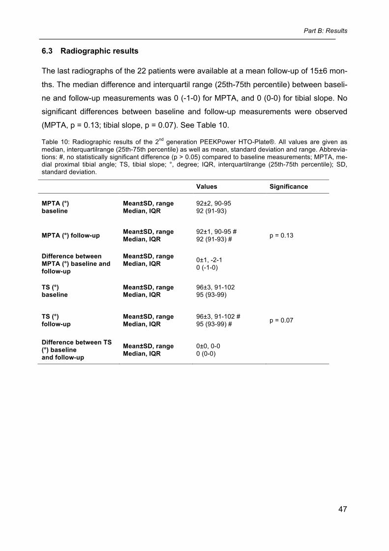
6.3 Radiographic results
The last radiographs of the 22 patients were available at a mean follow-up of 15±6 mon-
ths. The median difference and interquartil range (25th-75th percentile) between baseli-
ne and follow-up measurements was 0 (-1-0) for MPTA, and 0 (0-0) for tibial slope. No
significant differences between baseline and follow-up measurements were observed
(MPTA, p = 0.13; tibial slope, p = 0.07). See Table 10.
Table 10: Radiographic results of the 2nd generation PEEKPower HTO-Plate®. All values are given as median, interquartilrange (25th-75th percentile) as well as mean, standard deviation and range. Abbrevia-tions: #, no statistically significant difference (p > 0.05) compared to baseline measurements; MPTA, me-dial proximal tibial angle; TS, tibial slope; °, degree; IQR, interquartilrange (25th-75th percentile); SD, standard deviation.
Values Significance
MPTA (°) baseline
Mean±SD, range Median, IQR
92±2, 90-95 92 (91-93)
MPTA (°) follow-up Mean±SD, range Median, IQR
92±1, 90-95 # 92 (91-93) # p = 0.13
Difference between MPTA (°) baseline and follow-up
Mean±SD, range Median, IQR
0±1, -2-1 0 (-1-0)
TS (°) baseline
Mean±SD, range Median, IQR
96±3, 91-102 95 (93-99)
TS (°) follow-up
Mean±SD, range Median, IQR
96±3, 91-102 # 95 (93-99) # p = 0.07
Difference between TS (°) baseline and follow-up
Mean±SD, range Median, IQR
0±0, 0-0 0 (0-0)
Part B: Results
48
6.4 Complications
The complications are listed in Table 11. Table 11: The Number of complications of the 2nd generation PEEKPower HTO-Plate®. Abbreviations: %, percentage.
Overall complication rate 3 (12%)
Screw loosening 2 (8%)
Non-unions 1 (4%)
Superficial or deep wound infections 0 (0%)
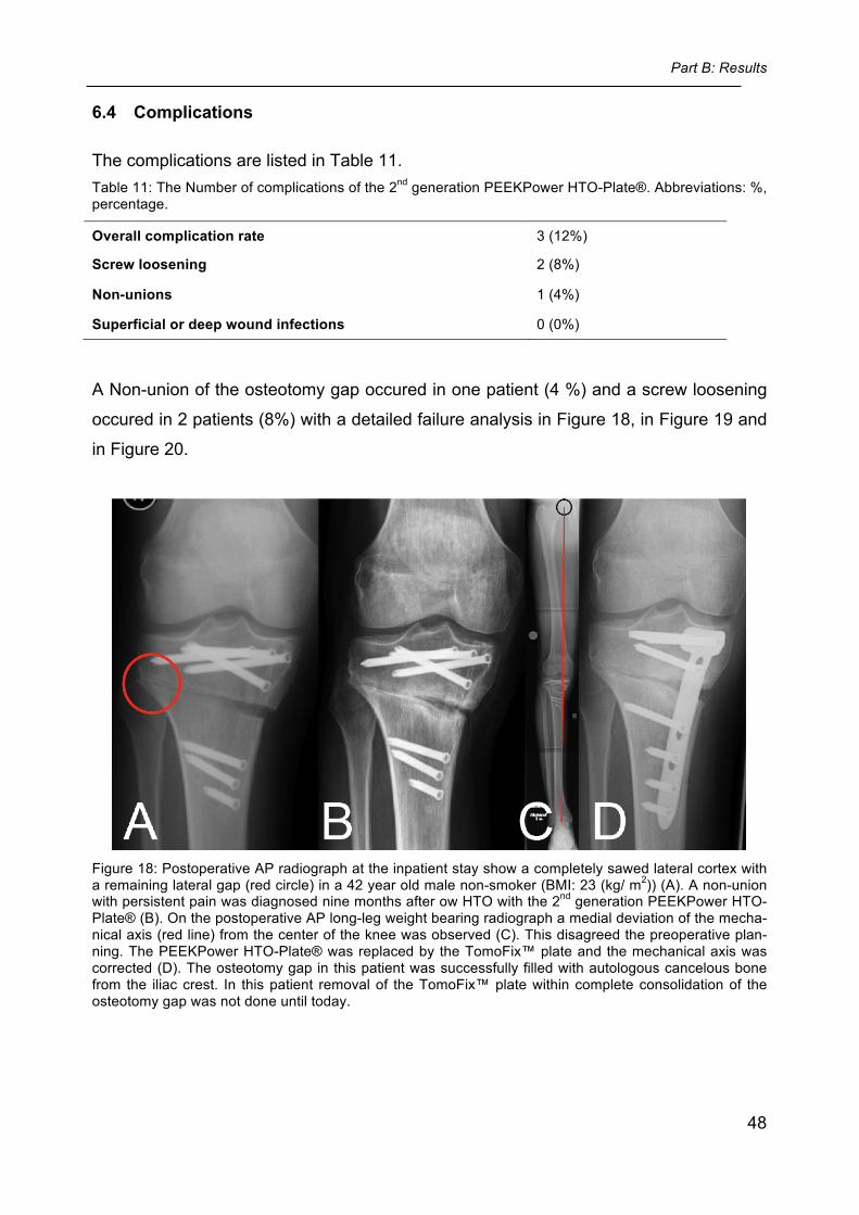
A Non-union of the osteotomy gap occured in one patient (4 %) and a screw loosening
occured in 2 patients (8%) with a detailed failure analysis in Figure 18, in Figure 19 and
in Figure 20.
Figure 18: Postoperative AP radiograph at the inpatient stay show a completely sawed lateral cortex with a remaining lateral gap (red circle) in a 42 year old male non-smoker (BMI: 23 (kg/ m2)) (A). A non-union with persistent pain was diagnosed nine months after ow HTO with the 2nd generation PEEKPower HTO-Plate® (B). On the postoperative AP long-leg weight bearing radiograph a medial deviation of the mecha-nical axis (red line) from the center of the knee was observed (C). This disagreed the preoperative plan-ning. The PEEKPower HTO-Plate® was replaced by the TomoFix™ plate and the mechanical axis was corrected (D). The osteotomy gap in this patient was successfully filled with autologous cancelous bone from the iliac crest. In this patient removal of the TomoFix™ plate within complete consolidation of the osteotomy gap was not done until today.
Part B: Results
49
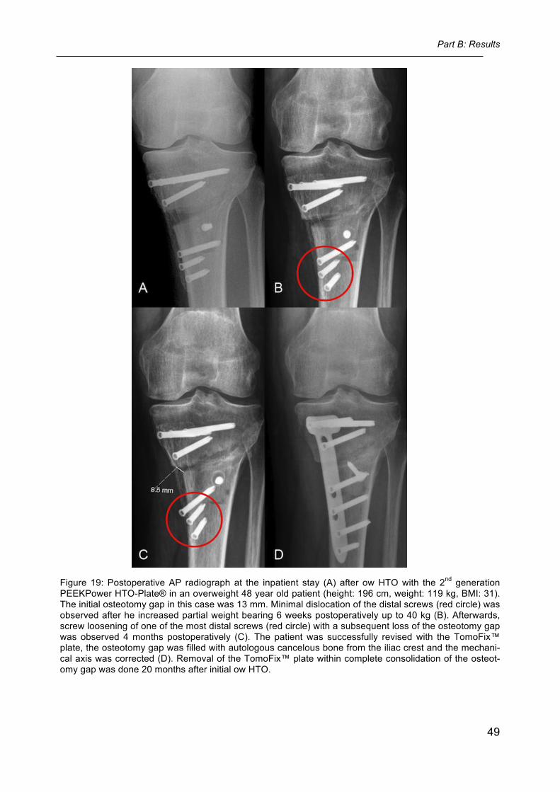
Figure 19: Postoperative AP radiograph at the inpatient stay (A) after ow HTO with the 2nd generation PEEKPower HTO-Plate® in an overweight 48 year old patient (height: 196 cm, weight: 119 kg, BMI: 31). The initial osteotomy gap in this case was 13 mm. Minimal dislocation of the distal screws (red circle) was observed after he increased partial weight bearing 6 weeks postoperatively up to 40 kg (B). Afterwards, screw loosening of one of the most distal screws (red circle) with a subsequent loss of the osteotomy gap was observed 4 months postoperatively (C). The patient was successfully revised with the TomoFix™ plate, the osteotomy gap was filled with autologous cancelous bone from the iliac crest and the mechani-cal axis was corrected (D). Removal of the TomoFix™ plate within complete consolidation of the osteot-omy gap was done 20 months after initial ow HTO.
Part B: Results
50
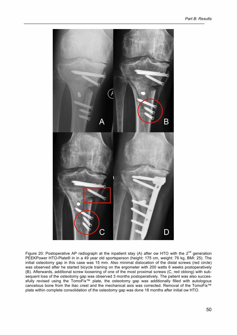
Figure 20: Postoperative AP radiograph at the inpatient stay (A) after ow HTO with the 2nd generation PEEKPower HTO-Plate® in in a 49 year old sportsperson (height: 175 cm, weight: 76 kg, BMI: 25). The initial osteotomy gap in this case was 15 mm. Also minimal dislocation of the distal screws (red circle) was observed after he started bicycle training on the ergometer with 200 watts 6 weeks postoperatively (B). Afterwards, additional screw loosening of one of the most proximal screws (C, red oblong) with sub-sequent loss of the osteotomy gap was observed 3 months postoperatively. The patient was also succes-sfully revised using the TomoFix™ plate, the osteotomy gap was additionally filled with autologous cancelous bone from the iliac crest and the mechanical axis was corrected. Removal of the TomoFix™ plate within complete consolidation of the osteotomy gap was done 18 months after initial ow HTO.
Part B: Discussion
51
7 Discussion of part B
Summary: The most important finding of part B of this dissertation was that valgus-producing ow
HTO without bone grafting in osteotomy gaps of up to 12 mm using the 2nd generation
PEEKPower HTO-Plate® showed significantly improved knee function and pain situa-
tion as early as 12 months after surgery. Moreover, ow HTO with the use of this new
plate allowed active patients to return to their sports-status 1 year before surgery with a
higher frequency. No significant loss of correction between baseline and follow-up radi-
ographs was observed in osteotomy gaps of up to 12 mm. With an overall complication
rate of only 4% in osteotomy gaps of up to 12 mm, this new implant can be considered
as a safe fixation device for ow HTO. However, because of two plate failures in osteot-
omy gaps of more than 12 mm without bone grafting, valgus-producing ow HTO does
not work with the 2nd generation PEEKPower HTO-Plate® in these cases.
Existing Implants described in the literature Several fixation devices for valgus-producing ow HTO are currently available, including
an inlay system (iBalance medial opening wedge HTO system, Arthrex, Naples, Fl,
USA)27, short spacer plates with or without locking screws (e.g. Puddu plate, Arthrex,
Naples, Fl, USA; Position HTO plate, Aesculap, Tuttlingen, Germany) and plate fixators
without a spacer (e.g. TomoFix™ plate, Synthes Medical, Oberdorf, Switzerland;
PEEKPower HTO-Plate®, Arthrex, Fl, USA).4, 37, 69, 77 19
iBalance medial opening wedge HTO system
Short spacer plates for ow HTO provide the advantage of a small design and therefore
might provide low pain with fast improvements in clinical scoring. Moreover, a new inlay
system (iBalance medial opening wedge HTO system) for valgus-producing ow HTO
was introduced which is even smaller than a short spacer plate and therefore might
provide better clinical results. It was shown that the clinical KOOS Score of a short spa-
cer plate and the new inlay system improved significantly as early as 6 months after
surgery without significant differences between each other.27 Therefore it was con-
cluded that an inlay system have no impact on clinical scoring compared to a short spa-
cer plate 6 and 12 months after surgery.27
Part B: Discussion
52
Puddu plate
The main advantage of the Puddu plate is its low profile design, which might avoid the
need for implant removal.7, 49 By using this plate, the functional Lysholm Score and
WOMAC Score reach mean values up to 90 and 75 twenty-four months after surgery,
respectively.21, 58, 63 49 However, this plate shows high failure rates for ow HTO without a
bone graft for augmentation of the osteotomy gap: In mean opening-wedges up to 11
mm, implant failures vary from 6% to 16%, osteotomies with broken screws were found
in up to 21% and the pseudarthrosis rate rise up to 22%.54 72 Secure fixation, low failu-
res and little non-unions of the osteotomy gap fixed with a short spacer locking plate
was only found with the use of a bone graft.49 9, 21
Position HTO plate
Another short spacer, the Position HTO plate (Aesculap, Tuttlingen, Germany) showed
in a clinical study a significantly (p > 0.05) improved functional mean Lysholm Score
twelve months after surgery (73) compared to preoperatively (56). The mean Tegner
activity scale improved from 3 to 4 at the same timepoints.69 However, high complicati-
on rates in a mean gap size of about 8 mm without a bone graft was also found: Screw
failures with non-union occured in about 6% and the loss of correction rate was about
3%.37, 69
Finally, it is proposed that short spacer plates should only be used for small osteotomy
gaps (up to 8 mm) as well as with a bone graft regardless of the correction to prevent
hardware related complications.8, 49 The reason is, that especially in cases of larger cor-
rections, short spacer plates offer limited stability since they can not adequately elimina-
te the tremendous lever arm forces acting on the osteotomy gap.45, 54, 72
TomoFix™ plate
Therefore, it is suggested, that angle stable plate fixators are favoured over short spac-
er plates to achieve a secure fixation after ow HTO without a bone graft. 4, 37, 61, 77 In bi-
omechanical and in clinical studies, it was shown that short spacer plates with or with-
out locking screws provide inferior axial and torsional stability and a loss of correction
compared to the plate fixator TomoFix™ plate.4, 37, 61, 65, 77 Pape et al.61 compared the
TomoFix™ plate and the 2nd generation Puddu plate over a 2 year period using radio-
stereometric analysis. Compared to patients treated with the TomoFix™ plate, a signifi-
Part B: Discussion
53
cant higher lateral translation of the distal tibia and a significantly increased subsidence,
varus and internal rotation of the tibial head was observed in patients treated with the
2nd generation Puddu plate. In clinical studies, valgus-producing ow HTO using the To-
moFix™ plate has shown that the postoperative mean Lysholm Scores can improve
significantly compared to preoperatively and can reach up to about 90 points out of
100.23, 56, 57
However, with the increasing use of this implant, several drawbacks were observed.
One disadvantage is the relative bulky design, which causes soft tissue irritation in most
patients, leading to a protracted clinical course. 56, 57 A further disadvantage is the pre-
defined and non-variable direction of the locking screws, which is dictated by threads
inside the screw holes.
1st generation PEEKPower HTO-Plate®
The 1st generation PEEKPower HTO-Plate® was introduced to overcome these draw-
backs. The plate consists of a CF PEEK composition and is considerably smaller and
lighter compared to a TomoFix™ plate. Furthermore this system provides multidirec-
tional locking by threading the harder titanium screw heads into the none-threaded plate
holes. Based on the first clinical data about the 1st generation PEEKPower HTO-Plate®
(Cotic et. al., see Appendix) 19, significant improvements of the VAS Score, WOMAC
Score, IKDC Score and Lyshom Score after ow HTO were recognized. According to
part A of this work, the 1st generation PEEKPower HTO-Plate® provided the same clini-
cal and radiographic results compared to the TomoFix™ plate. However, more implant
related complications occured with the 1st generation PEEKPower HTO-Plate® (see
part A). It was about 1 (4%) early screw loosening and 3 (12%) non-unions out of 26
patients. Implant related factors were thought to be a (too) high flexural strength, too
shallow plate holes and the lag of a temporary lag screw.
Part B: Discussion
54
The clinical results of the 2nd generation PEEKPower HTO-Plate® For these reasons, a 2nd generation of the implant has been developed. The new plate
provides an improved anatomical shape, an improved geometry of the screw holes and
a reduced flexural strength. Furthermore, the newer generation also provides utilization
of a lag screw to obtain compression on the lateral cortex.
In clinical studies, valgus-producing ow HTO using the TomoFix™ plate has shown that
preoperative mean Lysholm Scores started at 50 and improved significantly up to about
90, twenty-four months postoperatively (p < 0.05). 23, 56, 57 In the present work, mean
VAS Score, WOMAC Score and Lysholm Score after 2nd generation PEEKPower HTO-
Plate® fixation were 2, 94 and 84 respectively and improved significantly compared to
preoperatively (5, 70, 50 respectively; p < 0.05; see Table 8). Therefore, it is concluded
that both, the TomoFix™ plate and the 2nd generation PEEKPower HTO-Plate® are vi-
able implants for osteotomy fixation in valgus-producing ow HTO without bone grafting
in osteotomy gaps up to 12 mm in terms of clinical results at 24 months after surgery.
Although the implant was removed in 61% of the patients after a mean of 17 months, no
significant differences in clinical scoring between the 12 and 24 months follow-up were
found. In other words, removal of the 2nd generation PEEKPower HTO-Plate® did not
result in further clinical improvements and the clinical end-point of the patients was
reached as early as 12 months postoperatively. In contrast, Niemeyer et al. 56, 57 found
significant improvements of the clinical scores between 12 and 24 months after ow HTO
using the TomoFix™ plate. The authors attributed this finding to implant removal after
12 months. In their series, a high percentage of patients complained of local discomfort
and pain associated with the implant, which disappeared after implant removal. It might
be possible that the smaller size and the different material of the 2nd generation PEEK-
Power HTO-Plate® causes less local irritation and does not provide a protracted clinical
course. Therefore, implant removal could be nonessential with the 2nd generation
PEEKPower HTO-Plate®.
Sporting Activity after ow HTO with the 2nd generation PEEKPower HTO-Plate® In this study, 95% of patients were engaged in sporting activities, compared with 86%
before surgery (p = 0.32). The number of different sport disciplines, the activity duration
per week as well as the sporting intensity according to the Tegner scale did not change
significantly from the year before surgery to 24 months after HTO (Table 9, p > 0.05).
Only the sports frequency (sessions per week) changed significantly (p < 0.05) from
Part B: Discussion
55
preoperative to postoperative (see Table 9). Regarding the ability for sporting activity
after valgus-producing ow HTO with the TomoFix™ plate, Salzmann et. al.68 found that
91% of patients were engaged in sports and recreational activities, compared with 88%
before surgery (p = 0.18). The number of different sporting activities declined from one
year preoperatively to thirty-six months after surgery (from 4 to 3 respectively) without
significant differences (p > 0.05). Also, there were no significant differences in the
sports frequency per week (2 session) and the activity duration per week (4 hours) pre-
operatively compared to postoperatively (2, p = 0.21; and 4 hours, p = 0.71, respec-
tively). Declines were also noted in the Tegner scale (5±2 to 4±2, p < 0.05). These re-
sults indicate that valgus-producing ow HTO with the plate fixators 2nd generation
PEEKPower HTO-Plate® and the TomoFix™ plate allow active patients to return to
sports similar to their preoperative level. Moreover, if patients are motivated enough,
they can held their sporting intensity and can also improve their sports frequency (ses-
sions per week) without increasing pain in low- to moderate impact activities (only
showed with the PEEKPower HTO-Plate®, see Table 9 and Figure 17). However, ac-
cording to Bonnin et. al.14, patients which are highly motivated to increase their pre-
operative sporting intensity must be informed, that strenuous activities after ow HTO will
lead to pain and not to a recovery of the pre-pathology. In the Department of Orthopae-
dic Sports Medicine, Technical University of Munich, Germany, this information is gen-
erally given to a patient receiving ow HTO. Therefore, most patients in this study parti-
cipated in low- to moderate impact activities 24 months after surgery (see Figure 17).
Despite the fact that the patients were not allocated to do sports more often, their sports
frequency improved significantly (see Table 9). In contrast, that was not shown in the
patients with the TomoFix™ plate indicating even less sports frequency (sessions per
week) and a decline of the sporting intensity (Tegner scale).68 Whether the shorter and
lighter PEEKPower HTO-Plate® is responsible for these better sports-related results
was not observed. This situation must be proved by further comparative studies.

Part B: Discussion
56
Loss of correction Significant differences between baseline and follow-up measurements for MPTA and
tibial slope were not recorded, indicating that loss of correction did not occure. How-
ever, only patients with an osteotomy gap of ≤ 12 mm were included in this evaluation.
Loss of correction after ow HTO using the TomoFix™ plate was reported in 0-6% of
patients 18, 37, 75, 78, 79 with osteotomy gap sizes up to 20 mm. It is therefore concluded
that the 2nd generation PEEKPower HTO-Plate® provides at least equal fixation stability
without bone grafting if the osteotomy gap height is ≤12 mm. However, the performance
of this plate in osteotomy gaps of >12 mm is poor (see complications).
Complications Non-union
One non-union (4%) occured in a patient with an iatrogenic fracture of the lateral hinge.
The surgeon decided to replace the 2nd generation PEEKPower HTO-Plate® by the
TomoFix™ plate. Because a 2nd generation PEEKPower HTO-Plate® was not used
again, it might be reasonable, that the surgeon in this case had less than full confidence
in this system at this timepoint.
No further non-unions were observed. In part A of this dissertation, a non-union rate of
12% was observed in the previous series with the 1st generation PEEKPower HTO-
Plate®. These findings suggest that in terms of bone healing, the 2nd generation per-
formed better than the 1st generation, possibly due to the reduced flexural strength of
the plate. Compared to the non-union rate of the TomoFix™ plate, the literature reports
incidences of 0-7%. 18, 23, 24, 37, 42, 51, 56, 57, 75, 78, 79, 81 It is therefore concluded that a 2nd
generation PEEKPower HTO-Plate® and a TomoFix™ plate demonstrate nearly the
same rate of non-unions after ow HTO.
Screw-loosening:
In two patients (8%), loosenings of one of the screws with accompanying loss of the
corrected MPTA were observed. In both cases the osteotomy gaps of >12 mm experi-
enced higher forces than designated for the early postoperative rehabilitation period of
this procedure. According to part A of this dissertation, the 2nd generation PEEKPower
HTO-Plate® has been introduced to avoid screw loosenings. That was done by improv-
ing the geometry of the screw holes as well as by reducing the flexural strength of the
plate. Moreover, the literature described screw breakages during extraction but no
Part B: Discussion
57
screw looesenings of the TomoFix™ plate after ow HTOs with osteotomy gap sizes up
to 20 mm. 81 With critical analysis, the modified screw hole geometry and the changed
flexural strength of the 2nd generation PEEKPower HTO-Plate® do not endure in-
creased loads in the bone anchoring after ow HTO with osteotomy gaps of >12 mm.
Therefore it is concluded that the safety of the 2nd generation PEEKPower HTO-Plate®
is at least equal to the TomoFix™ plate, as long as the osteotomy gap is not larger than
12 mm.
Study limitations There is no control group existing. Nevertheless, the data of part B of this dissertation
were compared to the current results of the literature for valgus-producing ow HTO. Be-
cause the patients were only followed for 24 months, no conclusion about the further
clinical course of the patients can be drawn. However, a follow-up of 24 months was
adequate because significant improvements of the clinical scores after ow HTO are
within the first 24 months and afterwards they are unchanged. 2, 34, 52, 57, 73 Concerning
the method to determine the amount of correction loss, serial long-leg weight bearing
AP radiographs or radiostereometric analysis might have been more accurate in every
patient. Unfortunately, these methods were not allowed by the ethics commitee. How-
ever, by evaluating the correction loss in two standard planes (frontal and sagittal), the
accuracy of the radiological analysis increased.
Conclusion The 2nd generation PEEKPower HTO-Plate® is a safe and efficient implant for valgus-
producing open wedge high tibial osteotomy without bone grafting and in osteotomy
gaps of up to 12 mm. Valgus-producing ow HTO with this implant demonstrates favor-
able clinical results as early as 12 months after surgery and allows active patients to
return to their preoperative sports-status with a higher frequency. Compared to a stan-
dardized TomoFix™ plate, it might be possible that the 2nd generation PEEKPower
HTO-Plate® causes less local irritation and therefore implant removal could be no-
nessential. For ow HTO with osteotomy gaps of >12 mm, it is recommended to use a
bone graft or to use the standardized TomoFix™ plate for fixation.
Summary and Perspective
58
8 Summary and Perspective
In the first part (part A) of this dissertation, for osteotomy fixation in valgus-producing ow
HTO, twenty-six 1st generation PEEKPower HTO-Plates® were compared with twenty-
six TomoFix™ plates. The main finding was that the 1st generation PEEKPower HTO-
Plate® provided the same clinical and radiographic results as the TomoFix™ plate at a
minimum follow-up of 24 and 12 months respectively. However, more implant related
complications occured with the 1st generation PEEKPower HTO-Plate®. Considering
the drawbacks of the 1st generation PEEKPower HTO-Plate®, a 2nd generation PEEK-
Power HTO-Plate® was introduced to achieve no implant failure.
Therefore, the purpose of the second part of this work (part B) was to prospectively
evaluate if valgus-producing ow HTO without bone grafting using the 2nd generation
PEEKPower HTO-Plate® is safe. The most important finding was that ow HTO without
bone grafting in osteotomy gaps of up to 12 mm showed significantly improved knee
function and pain situation as early as 12 months after surgery. Ow HTO allowed active
patients to return to their sports-status 1 year before surgery with a higher frequency.
Compared to the TomoFix™ plate in the literature, there is a tendency that the 2nd gen-
eration PEEKPower HTO-Plate® causes less local irritation and therefore implant re-
moval could be nonessential. With an overall complication rate of 4% in osteotomy gaps
of up to 12 mm, this new implant can be considered as a safe fixation device for ow
HTO. Because of two plate failures in osteotomy gaps >12 mm without bone grafting,
valgus-producing ow HTO does not work with the 2nd generation PEEKPower HTO-
Plate® in these cases. For ow HTO with osteotomy gaps >12 mm, it is recommended to
use a bone graft or to use the standardized TomoFix™ plate for fixation.
Abbreviations
59
9 List of Abbreviations
ACL anterior cruciate ligament
AP anteroposterior
BMI body mass index
CF PEEK carbon fiber reinforced polyetheretherketone
e.g. for example
et al. abbreviation from Latin et alii (and others)
Gr Group
HTO high tibial osteotomy
IQR interquartilrange (25th-75th percentile)
ys years
kg/ m2 kilogram/ square meter
MACI matrix associated autologues chondrocyte implantation
mm millimeter
MPTA anatomical medial proximal tibial angle
ms months
n number of patients
n.r. not reported
OAT osteochondral autologues transfer
ow open wedge
SD standard deviation
TS tibial slope
VAS visual analogue scale
* statistical significant difference (p < 0.05)
# no statistical significant difference (p > 0.05)
° degree
% percentage
List of Tables
60
10 List of Tables
Table 1: Inclusion and Exclusion Criteria. Abbreviations: °, degree. ............................................... 15
Table 2: Baseline demographics of part A. Abbreviations: ys, years; n, number of patients; VAS, visual analogue scale; °, degree; mm, millimeter; HTO, high tibial osteotomy; OAT, osteochondral autologues transfer; MACI, matrix associated autologues chondrocyte implantation; ACL, anterior cruciate ligament; ms, months; IQR, Interquartilrange (25th-75th percentile); SD, standard deviation. ................................................................................................................................................ 16
Table 3: Clinical results of part A. All values are given as median, interquartilrange (25th-75th percentile) as well as mean, standard deviation and range. Abbreviations: *, statistically significant improvement compared to preoperatively (p < 0.05); VAS, visual analog scale; IQR, interquartilrange (25th-75th percentile); SD, standard deviation. ............................................ 25
Table 4: Radiological results of part A. All values are given as median, interquartilrange (25th-75th percentile) as well as mean, standard deviation and range. Abbreviations: #, no statistically significant difference (p > 0.05) compared to baseline measurements; MPTA, medial proximal tibial angle; TS, tibial slope; °, degree; IQR, interquartilrange (25th-75th percentile); SD, standard deviation. ................................................................................................................................. 27
Table 5: Consolidation of the osteotomy gap. Total voxel and evaluated voxel of bone, air and threshold are given as mean and standard deviation (SD, ±) at each timepoint. ................................... 28
Table 6: Complications of part A (group I, group II and the remaining TomoFix™ plate fixations). Abbreviations: n.r., not reported; %, percentage; n, number of patients. ................................ 29
Table 7: Baseline demographics of the patients treated with the 2nd generation PEEKPower HTO-Plate®. Abbreviations: SD, standard deviation; ys, years; n, number of patients; VAS, visual analogue scale; °, degree; mm, millimeter; HTO, high tibial osteotomy; OAT, osteochondral autologues transfer; ACL, anterior cruciate ligament; %, percentage. ..................................................................... 39
Table 8: Clinical results of the 2nd generation PEEKPower HTO-Plate®. All values are given as median, interquartilrange (25th-75th percentile) as well as mean, standard deviation and range. Abbreviations: *, statistically significant improvement compared to preoperatively (p < 0.05); #, no statistically significant difference compared to 12 months follow-up (p > 0.05); VAS, visual analog scale; ms, months; IQR, interquartilrange (25th-75th percentile); SD, standard deviation. .... 43
Table 9: Sports related results of the 2nd generation PEEKPower HTO-Plate®. Sports frequency (sessions), sports duration (hours) and the number of sports disciplines are given as median, interquartilrange (25th-75th percentile) as well as mean, standard deviation and range. Abbreviations: *, statistically significant improvement compared to preoperatively (p < 0.05); #, no statistically significant difference compared to preoperatively (p > 0.05); ms, months; IQR, interquartilrange (25th-75th percentile); SD, standard deviation. ............................................ 46
Table 10: Radiographic results of the 2nd generation PEEKPower HTO-Plate®. All values are given as median, interquartilrange (25th-75th percentile) as well as mean, standard deviation and range. Abbreviations: #, no statistically significant difference (p > 0.05) compared to baseline measurements; MPTA, medial proximal tibial angle; TS, tibial slope; °, degree; IQR, interquartilrange (25th-75th percentile); SD, standard deviation. ............................................ 47
Table 11: The Number of complications of the 2nd generation PEEKPower HTO-Plate®. Abbreviations: %, percentage. ............................................................................................................................. 48
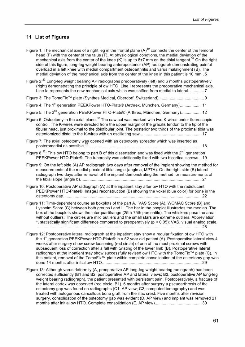
List of Figures
61
11 List of Figures
Figure 1: The mechanical axis of a right leg in the frontal plane (A)60 connects the center of the femoral head (F) with the center of the talus (T). At physiological condtions, the medial deviation of the mechanical axis from the center of the knee (K) is up to 8±7 mm on the tibial tangent.59 On the right side of this figure, long-leg weight bearing anteroposterior (AP) radiograph demonstrating painful overload in a left knee with medial compartment osteoarthritis and varus malalignment (B). The medial deviation of the mechanical axis from the center of the knee in this patient is 10 mm. . 5
Figure 2:23 Long-leg weight bearing AP radiographs preoperatively (left) and 6 months postoperatively (right) demonstrating the principle of ow HTO. Line I represents the preoperative mechanical axis. Line Ia represents the new mechanical axis which was shifted from medial to lateral. ............ 7
Figure 3: The TomoFix™ plate (Synthes Medical, Oberdorf, Switzerland). ...................................... 9
Figure 4: The 1st generation PEEKPower HTO-Plate® (Arthrex, München, Germany) ................... 11
Figure 5: The 2nd generation PEEKPower HTO-Plate® (Arthrex, München, Germany) .................. 12
Figure 6: Osteotomy in the axial plane.32 The saw cut was marked with two K-wires under fluoroscopic control. The K-wires were directed from the upper margin of the gracilis tendon to the tip of the fibular head, just proximal to the tibiofibular joint. The posterior two thirds of the proximal tibia was osteotomized distal to the K-wires with an oscillating saw. ..................................................... 17
Figure 7: The axial osteotomy was opened with an osteotomy spreader which was inserted as posteromedial as possible.32 ................................................................................................... 18
Figure 8 32: This ow HTO belong to part B of this dissertation and was fixed with the 2nd generation PEEKPower HTO-Plate®. The tuberosity was additionally fixed with two bicortical screws. . 19
Figure 9: On the left side (A) AP radiograph two days after removal of the implant showing the method for measurements of the medial proximal tibial angle (angle a, MPTA). On the right side (B) lateral radiograph two days after removal of the implant demonstrating the method for measurements of the tibial slope (angle b). ......................................................................................................... 21
Figure 10: Postoperative AP radiograph (A) at the inpatient stay after ow HTO with the radiolucent PEEKPower HTO-Plate®. ImageJ reconstruction (B) showing the voxel (blue color) for bone in the osteotomy gap. ........................................................................................................................ 22
Figure 11: Time-dependent course as boxplots of the part A. VAS Score (A), WOMAC Score (B) and Lysholm Score (C) between both groups I and II. The bar in the boxplot illustrades the median. The box of the boxplots shows the interquartilrange (25th-75th percentile). The whiskers pose the area without outliers. The circles are mild outliers and the small stars are extreme outliers. Abbreviation: *, statistically significant difference compared to preoperatively (p < 0.05); VAS, visual analog scale. ................................................................................................................................................ 26
Figure 12: Postoperative lateral radiograph at the inpatient stay show a regular fixation of ow HTO with the 1st generation PEEKPower HTO-Plate® in a 52 year old patient (A). Postoperative lateral view 4 weeks after surgery show screw loosening (red circle) of one of the most proximal screws with subsequent loss of correction after a fall with twisting of the lower limb (B). Postoperative lateral radiograph at the inpatient stay show successfully revised ow HTO with the TomoFix™ plate (C). In this patient, removal of the TomoFix™ plate within complete consolidation of the osteotomy gap was done 14 months after initial ow HTO. ...................................................................................... 29
Figure 13: Although varus deformity (A, preoperative AP long-leg weight bearing radiograph) has been corrected sufficiently (B1 and B2, postoperative AP and lateral views; B3, postoperative AP long-leg weight bearing radiograph), the patient presented with persistent pain. Postoperatively, a fracture of the lateral cortex was observed (red circle, B1). 6 months after surgery a pseudarthrosis of the osteotomy gap was found on radiographs (C1, AP view; C2, computed tomography) and was treated with autogenous cancellous bone graft from the iliac crest. Five months after revision surgery, consolidation of the osteotomy gap was evident (D, AP view) and implant was removed 21 months after initial ow HTO. Complete consolidation (E, AP view). ........................................ 30
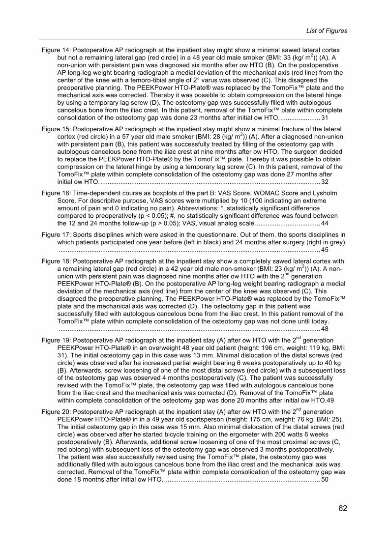
List of Figures
62
Figure 14: Postoperative AP radiograph at the inpatient stay might show a minimal sawed lateral cortex but not a remaining lateral gap (red circle) in a 48 year old male smoker (BMI: 33 (kg/ m2)) (A). A non-union with persistent pain was diagnosed six months after ow HTO (B). On the postoperative AP long-leg weight bearing radiograph a medial deviation of the mechanical axis (red line) from the center of the knee with a femoro-tibial angle of 2° varus was observed (C). This disagreed the preoperative planning. The PEEKPower HTO-Plate® was replaced by the TomoFix™ plate and the mechanical axis was corrected. Thereby it was possible to obtain compression on the lateral hinge by using a temporary lag screw (D). The osteotomy gap was successfully filled with autologous cancelous bone from the iliac crest. In this patient, removal of the TomoFix™ plate within complete consolidation of the osteotomy gap was done 23 months after initial ow HTO. ...................... 31
Figure 15: Postoperative AP radiograph at the inpatient stay might show a minimal fracture of the lateral cortex (red circle) in a 57 year old male smoker (BMI: 28 (kg/ m2)) (A). After a diagnosed non-union with persistent pain (B), this patient was successfully treated by filling of the osteotomy gap with autologous cancelous bone from the iliac crest at nine months after ow HTO. The surgeon decided to replace the PEEKPower HTO-Plate® by the TomoFix™ plate. Thereby it was possible to obtain compression on the lateral hinge by using a temporary lag screw (C). In this patient, removal of the TomoFix™ plate within complete consolidation of the osteotomy gap was done 27 months after initial ow HTO. ......................................................................................................................... 32
Figure 16: Time-dependent course as boxplots of the part B: VAS Score, WOMAC Score and Lysholm Score. For descripitve purpose, VAS scores were multiplied by 10 (100 indicating an extreme amount of pain and 0 indicating no pain). Abbreviations: *, statistically significant difference compared to preoperatively (p < 0.05); #, no statistically significant difference was found between the 12 and 24 months follow-up (p > 0.05); VAS, visual analog scale. ................................... 44
Figure 17: Sports disciplines which were asked in the questionnaire. Out of them, the sports disciplines in which patients participated one year before (left in black) and 24 months after surgery (right in grey). ................................................................................................................................................ 45
Figure 18: Postoperative AP radiograph at the inpatient stay show a completely sawed lateral cortex with a remaining lateral gap (red circle) in a 42 year old male non-smoker (BMI: 23 (kg/ m2)) (A). A non-union with persistent pain was diagnosed nine months after ow HTO with the 2nd generation PEEKPower HTO-Plate® (B). On the postoperative AP long-leg weight bearing radiograph a medial deviation of the mechanical axis (red line) from the center of the knee was observed (C). This disagreed the preoperative planning. The PEEKPower HTO-Plate® was replaced by the TomoFix™ plate and the mechanical axis was corrected (D). The osteotomy gap in this patient was successfully filled with autologous cancelous bone from the iliac crest. In this patient removal of the TomoFix™ plate within complete consolidation of the osteotomy gap was not done until today. ................................................................................................................................................ 48
Figure 19: Postoperative AP radiograph at the inpatient stay (A) after ow HTO with the 2nd generation PEEKPower HTO-Plate® in an overweight 48 year old patient (height: 196 cm, weight: 119 kg, BMI: 31). The initial osteotomy gap in this case was 13 mm. Minimal dislocation of the distal screws (red circle) was observed after he increased partial weight bearing 6 weeks postoperatively up to 40 kg (B). Afterwards, screw loosening of one of the most distal screws (red circle) with a subsequent loss of the osteotomy gap was observed 4 months postoperatively (C). The patient was successfully revised with the TomoFix™ plate, the osteotomy gap was filled with autologous cancelous bone from the iliac crest and the mechanical axis was corrected (D). Removal of the TomoFix™ plate within complete consolidation of the osteotomy gap was done 20 months after initial ow HTO.49
Figure 20: Postoperative AP radiograph at the inpatient stay (A) after ow HTO with the 2nd generation PEEKPower HTO-Plate® in in a 49 year old sportsperson (height: 175 cm, weight: 76 kg, BMI: 25). The initial osteotomy gap in this case was 15 mm. Also minimal dislocation of the distal screws (red circle) was observed after he started bicycle training on the ergometer with 200 watts 6 weeks postoperatively (B). Afterwards, additional screw loosening of one of the most proximal screws (C, red oblong) with subsequent loss of the osteotomy gap was observed 3 months postoperatively. The patient was also successfully revised using the TomoFix™ plate, the osteotomy gap was additionally filled with autologous cancelous bone from the iliac crest and the mechanical axis was corrected. Removal of the TomoFix™ plate within complete consolidation of the osteotomy gap was done 18 months after initial ow HTO. ...................................................................................... 50
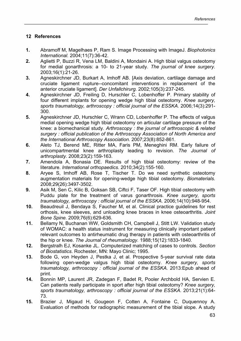
References
63
12 References
1. Abramoff M, Magelhaes P, Ram S. Image Processing with ImageJ. Biophotonics International. 2004;11(7):36-42.
2. Aglietti P, Buzzi R, Vena LM, Baldini A, Mondaini A. High tibial valgus osteotomy for medial gonarthrosis: a 10- to 21-year study. The journal of knee surgery. 2003;16(1):21-26.
3. Agneskirchner JD, Burkart A, Imhoff AB. [Axis deviation, cartilage damage and cruciate ligament rupture--concomitant interventions in replacement of the anterior cruciate ligament]. Der Unfallchirurg. 2002;105(3):237-245.
4. Agneskirchner JD, Freiling D, Hurschler C, Lobenhoffer P. Primary stability of four different implants for opening wedge high tibial osteotomy. Knee surgery, sports traumatology, arthroscopy : official journal of the ESSKA. 2006;14(3):291-300.
5. Agneskirchner JD, Hurschler C, Wrann CD, Lobenhoffer P. The effects of valgus medial opening wedge high tibial osteotomy on articular cartilage pressure of the knee: a biomechanical study. Arthroscopy : the journal of arthroscopic & related surgery : official publication of the Arthroscopy Association of North America and the International Arthroscopy Association. 2007;23(8):852-861.
6. Aleto TJ, Berend ME, Ritter MA, Faris PM, Meneghini RM. Early failure of unicompartmental knee arthroplasty leading to revision. The Journal of arthroplasty. 2008;23(2):159-163.
7. Amendola A, Bonasia DE. Results of high tibial osteotomy: review of the literature. International orthopaedics. 2010;34(2):155-160.
8. Aryee S, Imhoff AB, Rose T, Tischer T. Do we need synthetic osteotomy augmentation materials for opening-wedge high tibial osteotomy. Biomaterials. 2008;29(26):3497-3502.
9. Asik M, Sen C, Kilic B, Goksan SB, Ciftci F, Taser OF. High tibial osteotomy with Puddu plate for the treatment of varus gonarthrosis. Knee surgery, sports traumatology, arthroscopy : official journal of the ESSKA. 2006;14(10):948-954.
10. Beaudreuil J, Bendaya S, Faucher M, et al. Clinical practice guidelines for rest orthosis, knee sleeves, and unloading knee braces in knee osteoarthritis. Joint Bone Spine. 2009;76(6):629-636.
11. Bellamy N, Buchanan WW, Goldsmith CH, Campbell J, Stitt LW. Validation study of WOMAC: a health status instrument for measuring clinically important patient relevant outcomes to antirheumatic drug therapy in patients with osteoarthritis of the hip or knee. The Journal of rheumatology. 1988;15(12):1833-1840.
12. Bergstralh EJ, Kosanke JL. Computerized matching of cases to controls. Section of Biostatistics. Rochester, MN: Mayo Clinic; 1995.
13. Bode G, von Heyden J, Pestka J, et al. Prospective 5-year survival rate data following open-wedge valgus high tibial osteotomy. Knee surgery, sports traumatology, arthroscopy : official journal of the ESSKA. 2013:Epub ahead of print.
14. Bonnin MP, Laurent JR, Zadegan F, Badet R, Pooler Archbold HA, Servien E. Can patients really participate in sport after high tibial osteotomy? Knee surgery, sports traumatology, arthroscopy : official journal of the ESSKA. 2013;21(1):64-73.
15. Brazier J, Migaud H, Gougeon F, Cotten A, Fontaine C, Duquennoy A. Evaluation of methods for radiographic measurement of the tibial slope. A study
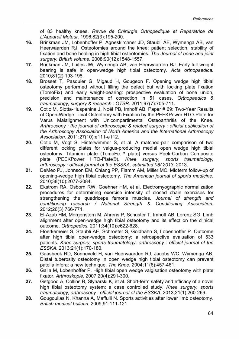
References
64
of 83 healthy knees. Revue de Chirurgie Orthopedique et Reparatrice de L'Appareil Moteur. 1996;82(3):195-200.
16. Brinkman JM, Lobenhoffer P, Agneskirchner JD, Staubli AE, Wymenga AB, van Heerwaarden RJ. Osteotomies around the knee: patient selection, stability of fixation and bone healing in high tibial osteotomies. The Journal of bone and joint surgery. British volume. 2008;90(12):1548-1557.
17. Brinkman JM, Luites JW, Wymenga AB, van Heerwaarden RJ. Early full weight bearing is safe in open-wedge high tibial osteotomy. Acta orthopaedica. 2010;81(2):193-198.
18. Brosset T, Pasquier G, Migaud H, Gougeon F. Opening wedge high tibial osteotomy performed without filling the defect but with locking plate fixation (TomoFix) and early weight-bearing: prospective evaluation of bone union, precision and maintenance of correction in 51 cases. Orthopaedics & traumatology, surgery & research : OTSR. 2011;97(7):705-711.
19. Cotic M, Slotta-Huspenina J, Noël PB, Imhoff AB. Paper # 69: Two-Year Results of Open-Wedge Tibial Osteotomy with Fixation by the PEEKPower HTO-Plate for Varus Malalignment with Unicompartimental Osteoarthritis of the Knee. Arthroscopy : the journal of arthroscopic & related surgery : official publication of the Arthroscopy Association of North America and the International Arthroscopy Association. 2011;27(10):e111-e112.
20. Cotic M, Vogt S, Hinterwimmer S, et al. A matched-pair comparison of two different locking plates for valgus-producing medial open wedge high tibial osteotomy: Titanium plate (TomoFix™ plate) versus Peek-Carbon Composite plate (PEEKPower HTO-Plate®). Knee surgery, sports traumatology, arthroscopy : official journal of the ESSKA, submitted 08/ 2013. 2013.
21. DeMeo PJ, Johnson EM, Chiang PP, Flamm AM, Miller MC. Midterm follow-up of opening-wedge high tibial osteotomy. The American journal of sports medicine. 2010;38(10):2077-2084.
22. Ekstrom RA, Osborn RW, Goehner HM, et al. Electromyographic normalization procedures for determining exercise intensity of closed chain exercises for strengthening the quadriceps femoris muscles. Journal of strength and conditioning research / National Strength & Conditioning Association. 2012;26(3):766-771.
23. El-Azab HM, Morgenstern M, Ahrens P, Schuster T, Imhoff AB, Lorenz SG. Limb alignment after open-wedge high tibial osteotomy and its effect on the clinical outcome. Orthopedics. 2011;34(10):e622-628.
24. Floerkemeier S, Staubli AE, Schroeter S, Goldhahn S, Lobenhoffer P. Outcome after high tibial open-wedge osteotomy: a retrospective evaluation of 533 patients. Knee surgery, sports traumatology, arthroscopy : official journal of the ESSKA. 2013;21(1):170-180.
25. Gaasbeek RD, Sonneveld H, van Heerwaarden RJ, Jacobs WC, Wymenga AB. Distal tuberosity osteotomy in open wedge high tibial osteotomy can prevent patella infera: a new technique. The Knee. 2004;11(6):457-461.
26. Galla M, Lobenhoffer P. High tibial open wedge valgisation osteotomy with plate fixator. Arthroskopie. 2007;20(4):291-300.
27. Getgood A, Collins B, Slynarski K, et al. Short-term safety and efficacy of a novel high tibial osteotomy system: a case controlled study. Knee surgery, sports traumatology, arthroscopy : official journal of the ESSKA. 2013;21(1):260-269.
28. Gougoulias N, Khanna A, Maffulli N. Sports activities after lower limb osteotomy. British medical bulletin. 2009;91:111-121.
References
65
29. Gould DD, Kelly DD, Goldstone LL, Gammon JJ. Examining the validity of pressure ulcer risk assessment scales: developing and using illustrated patient simulations to collect the data. Journal of Clinical Nursing. 2001;10(5):697-706.
30. Grelsamer RP. Unicompartmental osteoarthrosis of the knee. The Journal of bone and joint surgery. American volume. 1995;77(2):278-292.
31. Hinterwimmer S, Beitzel K, Paul J, et al. Control of posterior tibial slope and patellar height in open-wedge valgus high tibial osteotomy. The American journal of sports medicine. 2011;39(4):851-856.
32. Hinterwimmer S, Feucht MJ, Imhoff A. Hohe tibiale Osteotomie bei Varusgonarthrose. Arthroskopie. 2012:1-10.
33. Howling GI, Sakoda H, Antonarulrajah A, et al. Biological response to wear debris generated in carbon based composites as potential bearing surfaces for artificial hip joints. Journal of biomedical materials research. Part B, Applied biomaterials. 2003;67(2):758-764.
34. Hui C, Salmon LJ, Kok A, et al. Long-term survival of high tibial osteotomy for medial compartment osteoarthritis of the knee. The American journal of sports medicine. 2011;39(1):64-70.
35. Jacobi M, Wahl P, Jakob RP. Avoiding intraoperative complications in open-wedge high tibial valgus osteotomy: technical advancement. Knee surgery, sports traumatology, arthroscopy : official journal of the ESSKA. 2010;18(2):200-203.
36. Johnson RJ, Kettelkamp DB, Clark W, Leaverton P. Factors effecting late results after meniscectomy. The Journal of bone and joint surgery. American volume. 1974;56(4):719-729.
37. Jung WH, Chun CW, Lee JH, Ha JH, Kim JH, Jeong JH. Comparative Study of Medial Opening-Wedge High Tibial Osteotomy Using 2 Different Implants. Arthroscopy : the journal of arthroscopic & related surgery : official publication of the Arthroscopy Association of North America and the International Arthroscopy Association. 2013.
38. Kaper BP, Bourne RB, Rorabeck CH, Macdonald SJ. Patellar infera after high tibial osteotomy. The Journal of arthroplasty. 2001;16(2):168-173.
39. Kaplan FS HW, Keaveny TM, Boskey A, Einhorn TA, Iannotti, JP. Form and function of bone. In: SR S, ed. Orthopaedic Basic Science. Rosemont, IL: American Academy of Orthopaedic Surgeons; 1994:127-184.
40. Kellgren JH, Lawrence JS. Radiological assessment of osteo-arthrosis. Annals of the rheumatic diseases. 1957;16(4):494-502.
41. Kim SJ, Koh YG, Chun YM, Kim YC, Park YS, Sung CH. Medial opening wedge high-tibial osteotomy using a kinematic navigation system versus a conventional method: a 1-year retrospective, comparative study. Knee surgery, sports traumatology, arthroscopy : official journal of the ESSKA. 2009;17(2):128-134.
42. Kolb W, Guhlmann H, Windisch C, Kolb K, Koller H, Grutzner P. Opening-wedge high tibial osteotomy with a locked low-profile plate. The Journal of bone and joint surgery. American volume. 2009;91(11):2581-2588.
43. Kurtz SM, Devine JN. PEEK biomaterials in trauma, orthopedic, and spinal implants. Biomaterials. 2007;28(32):4845-4869.
44. Lewold S, Robertsson O, Knutson K, Lidgren L. Revision of unicompartmental knee arthroplasty: outcome in 1,135 cases from the Swedish Knee Arthroplasty study. Acta orthopaedica Scandinavica. 1998;69(5):469-474.
References
66
45. Lobenhoffer P, Agneskirchner JD. Improvements in surgical technique of valgus high tibial osteotomy. Knee surgery, sports traumatology, arthroscopy : official journal of the ESSKA. 2003;11(3):132-138.
46. Lobenhoffer PP, Agneskirchner JJ, Zoch WW. [Open valgus alignment osteotomy of the proximal tibia with fixation by medial plate fixator]. Der Orthopäde. 2004;33(2):153-160.
47. Luites JW, Brinkman JM, Wymenga AB, van Heerwaarden RJ. Fixation stability of opening- versus closing-wedge high tibial osteotomy: a randomised clinical trial using radiostereometry. The Journal of bone and joint surgery. British volume. 2009;91(11):1459-1465.
48. Lysholm J, Gillquist J. Evaluation of knee ligament surgery results with special emphasis on use of a scoring scale. The American journal of sports medicine. 1982;10(3):150-154.
49. Maffulli N, Loppini M, Longo UG, Denaro V, Oliva F. Bovine xenograft locking Puddu plate versus tricalcium phosphate spacer non-locking Puddu plate in opening-wedge high tibial osteotomy: a prospective double-cohort study. International orthopaedics. 2013;37(5):819-826.
50. Matar WY, Boscariol R, Dervin GF. Open wedge high tibial osteotomy: a roentgenographic comparison of a horizontal and an oblique osteotomy on patellar height and sagittal tibial slope. The American journal of sports medicine. 2009;37(4):735-742.
51. Meidinger G, Imhoff AB, Paul J, Kirchhoff C, Sauerschnig M, Hinterwimmer S. May smokers and overweight patients be treated with a medial open-wedge HTO? Risk factors for non-union. Knee surgery, sports traumatology, arthroscopy : official journal of the ESSKA. 2011;19(3):333-339.
52. Mont MA, Stuchin SA, Paley D, et al. Different surgical options for monocompartmental osteoarthritis of the knee: high tibial osteotomy versus unicompartmental knee arthroplasty versus total knee arthroplasty: indications, techniques, results, and controversies. Instructional course lectures. 2004;53:265-283.
53. Naudie D, Bourne RB, Rorabeck CH, Bourne TJ. The Install Award. Survivorship of the high tibial valgus osteotomy. A 10- to -22-year followup study. Clinical orthopaedics and related research. 1999(367):18-27.
54. Nelissen EM, van Langelaan EJ, Nelissen RG. Stability of medial opening wedge high tibial osteotomy: a failure analysis. International orthopaedics. 2010;34(2):217-223.
55. Neyret P, Donell ST, Dejour H. Results of partial meniscectomy related to the state of the anterior cruciate ligament. Review at 20 to 35 years. The Journal of bone and joint surgery. British volume. 1993;75(1):36-40.
56. Niemeyer P, Koestler W, Kaehny C, et al. Two-year results of open-wedge high tibial osteotomy with fixation by medial plate fixator for medial compartment arthritis with varus malalignment of the knee. Arthroscopy : the journal of arthroscopic & related surgery : official publication of the Arthroscopy Association of North America and the International Arthroscopy Association. 2008;24(7):796-804.
57. Niemeyer P, Schmal H, Hauschild O, von Heyden J, Sudkamp NP, Kostler W. Open-wedge osteotomy using an internal plate fixator in patients with medial-compartment gonarthritis and varus malalignment: 3-year results with regard to preoperative arthroscopic and radiographic findings. Arthroscopy : the journal of arthroscopic & related surgery : official publication of the Arthroscopy Association
References
67
of North America and the International Arthroscopy Association. 2010;26(12):1607-1616.
58. Orsel S, Altun M, Bekmezci T, Tonbul M, Yalaman O. [Early results of medial opening wedge osteotomy in varus gonarthrosis]. Acta orthopaedica et traumatologica turcica. 2006;40(3):193-198.
59. Paley D, Maar DC, Herzenberg JE. New concepts in high tibial osteotomy for medial compartment osteoarthritis. The Orthopedic clinics of North America. 1994;25(3):483-498.
60. Paley D, Pfeil J. [Principles of deformity correction around the knee]. Der Orthopäde. 2000;29(1):18-38.
61. Pape D, Kohn D, van Giffen N, Hoffmann A, Seil R, Lorbach O. Differences in fixation stability between spacer plate and plate fixator following high tibial osteotomy. Knee surgery, sports traumatology, arthroscopy : official journal of the ESSKA. 2013;21(1):82-89.
62. Pape D, Madry H. The preclinical sheep model of high tibial osteotomy relating basic science to the clinics: standards, techniques and pitfalls. Knee surgery, sports traumatology, arthroscopy : official journal of the ESSKA. 2013;21(1):228-236.
63. Pascale W, Luraghi S, Perico L, Pascale V. Do microfractures improve high tibial osteotomy outcome? Orthopedics. 2011;34(7):e251-255.
64. Penny P, Geere J, Smith TO. A systematic review investigating the efficacy of laterally wedged insoles for medial knee osteoarthritis. Rheumatology international. 2013.
65. Raja Izaham RMA, Abdul Kadir MR, Abdul Rashid AH, Hossain MG, Kamarul T. Finite element analysis of Puddu and Tomofix plate fixation for open wedge high tibial osteotomy. Injury. 2012;43(6):898-902.
66. Rivard CH, Rhalmi S, Coillard C. In vivo biocompatibility testing of peek polymer for a spinal implant system: a study in rabbits. Journal of biomedical materials research. 2002;62(4):488-498.
67. Rossi R, Bonasia DE, Amendola A. The role of high tibial osteotomy in the varus knee. The Journal of the American Academy of Orthopaedic Surgeons. 2011;19(10):590-599.
68. Salzmann GM, Ahrens P, Naal FD, et al. Sporting activity after high tibial osteotomy for the treatment of medial compartment knee osteoarthritis. The American journal of sports medicine. 2009;37(2):312-318.
69. Schroter S, Gonser CE, Konstantinidis L, Helwig P, Albrecht D. High complication rate after biplanar open wedge high tibial osteotomy stabilized with a new spacer plate (Position HTO plate) without bone substitute. Arthroscopy : the journal of arthroscopic & related surgery : official publication of the Arthroscopy Association of North America and the International Arthroscopy Association. 2011;27(5):644-652.
70. Schroter S, Ihle C, Mueller J, Lobenhoffer P, Stockle U, van Heerwaarden R. Digital planning of high tibial osteotomy. Interrater reliability by using two different software. Knee surgery, sports traumatology, arthroscopy : official journal of the ESSKA. 2013;21(1):189-196.
71. Scotchford CA, Garle MJ, Batchelor J, Bradley J, Grant DM. Use of a novel carbon fibre composite material for the femoral stem component of a THR system: in vitro biological assessment. Biomaterials. 2003;24(26):4871-4879.
72. Spahn G. Complications in high tibial (medial opening wedge) osteotomy. Archives of orthopaedic and trauma surgery. 2004;124(10):649-653.
References
68
73. Sprenger TR, Doerzbacher JF. Tibial osteotomy for the treatment of varus gonarthrosis. Survival and failure analysis to twenty-two years. The Journal of bone and joint surgery. American volume. 2003;85-A(3):469-474.
74. Staubli AE. Radiologische heilungsvorgänge nach öffnender kniegelenksnaher Tibiaosteotomie. In: Lobenhoffer A, ed. Kniegelenksnahe Osteotomien. New York: Georg Thieme Verlag; 2006:67-78.
75. Staubli AE, De Simoni C, Babst R, Lobenhoffer P. TomoFix: a new LCP-concept for open wedge osteotomy of the medial proximal tibia--early results in 92 cases. Injury. 2003;34 Suppl 2:B55-62.
76. Staubli AE, Jacob HA. Evolution of open-wedge high-tibial osteotomy: experience with a special angular stable device for internal fixation without interposition material. International orthopaedics. 2010;34(2):167-172.
77. Stoffel KK, Stachowiak GG, Kuster MM. Open wedge high tibial osteotomy: biomechanical investigation of the modified Arthrex Osteotomy Plate (Puddu Plate) and the TomoFix Plate. Clinical Biomechanics. 2004;19(9):7-7.
78. Takeuchi R, Ishikawa H, Aratake M, et al. Medial opening wedge high tibial osteotomy with early full weight bearing. Arthroscopy : the journal of arthroscopic & related surgery : official publication of the Arthroscopy Association of North America and the International Arthroscopy Association. 2009;25(1):46-53.
79. Takeuchi R, Ishikawa H, Kumagai K, et al. Fractures around the lateral cortical hinge after a medial opening-wedge high tibial osteotomy: a new classification of lateral hinge fracture. Arthroscopy : the journal of arthroscopic & related surgery : official publication of the Arthroscopy Association of North America and the International Arthroscopy Association. 2012;28(1):85-94.
80. Tegner Y, Lysholm J. Rating systems in the evaluation of knee ligament injuries. Clinical orthopaedics and related research. 1985(198):43-49.
81. Valkering KP, van den Bekerom MP, Kappelhoff FM, Albers GH. Complications after tomofix medial opening wedge high tibial osteotomy. The journal of knee surgery. 2009;22(3):218-225.
82. Vandamme K, Naert I, Geris L, Vander Sloten J, Puers R, Duyck J. The effect of micro-motion on the tissue response around immediately loaded roughened titanium implants in the rabbit. European journal of oral sciences. 2007;115(1):21-29.
83. W-Dahl AA, Toksvig-Larsen SS. Cigarette smoking delays bone healing: a prospective study of 200 patients operated on by the hemicallotasis technique. Acta orthopaedica Scandinavica. 2004;75(3):347-351.
84. Wang JH, Bae JH, Lim HC, Shon WY, Kim CW, Cho JW. Medial open wedge high tibial osteotomy: the effect of the cortical hinge on posterior tibial slope. The American journal of sports medicine. 2009;37(12):2411-2418.
85. Warme BA, Aalderink K, Amendola A. Is there a role for high tibial osteotomies in the athlete? Sports Health. 2011;3(1):59-69.
86. Westrich GH, Peters LE, Haas SB, Buly RL, Windsor RE. Patella height after high tibial osteotomy with internal fixation and early motion. Clinical orthopaedics and related research. 1998(354):169-174.
87. Wolf BR, Amendola A. Impact of osteoarthritis on sports careers. Clinics in sports medicine. 2005;24(1):187-198.
Appendix
69
13 Appendix
Publications:
Cotic M, Slotta-Huspenina J, Noël PB, Imhoff AB. Paper # 69: Two-Year Result of
Open-Wedge Tibial Osteotomy with Fixation by the PEEKPower HTO-Plate for Varus
Malalignment with Unicompartimental Osteoarthritis of the Knee. Arthroscopy : the
journal of arthroscopic & related surgery : official publication of the Arthroscopy
Association of North America and the International Arthroscopy Association.
2011;27:e111-e112.
Ethics approval
Patient summary of part A
Patient questionnaire of part A
White paper about the biomechanical observation provided by Arthrex
Patient questionnaire of part B
Publication
70
osteotomy (OWHTO), we shall investigate two variables:implant stability and integrity of the opposite cortex.Methods: Experimental models of polyurethane fiberssimulating the proximal segment of the tibia were fixedwith DCP ®, T-LCP ® and Tomofix ® plates. Conven-tional and locking head screws were used to stabilize themedial side. Opening wedges were made to simulate thedistraction of high tibial osteotomy in the medial side.Tests were also performed on models in the lateral sidewith normal(NL) and broken cortex (GAP) within spacebetween the cortices to simulate fractures. The lateralcortices in the GAP groups were fixed with 2 differenttypes of screws to check the stability of the system incases where it unintentionaly breaks. Torsion and axialcompression tests were assessed by simulating a levelwalk. Compression load was measured at a point at 62%of the medial margin corresponding to an overcorrectionof the mechanical axis. According to the assemblies 10different groups were formed. The mechanical tests re-ported the relative stiffness and deformation percentage.Results: The lateral compression or position stabiliza-tion screw was as rigid as normal cortical (NL) (p!0.001) in torsion tests. Measures of compression ob-tained in the group with non damage cortical (NL) weresimilar between the models with conventional fixationand fixed-angle (p" 0.05). The lateral compression orposition stabilization screw with broken cortex werestatistically lower when compared with the group ofnormal lateral cortex implants (p !0.001).
The Tomofix® plate showed superior torsion stability(p !0.001) with normal lateral (NL) and broken cortex(GAP). In axial compression tests, the implants showedsimilar mechanical outcomes between the groups.Conclusion: In an experimental model, using differentmethods of fixation in OWHTO, DCP ®, T-LCP ® andTomofix ® plates proved to be a stable option for mechan-ical forces to simulate a level walk. The lateral cortex isrelevant to the stability of the system in OWHTO.
Paper # 68: Effect of High Tibial Osteotomy on Me-dial Meniscus Degenerative Root Tear and VarusJONG-KEUN SEON, MD, KOREAKYUNG DO KANG, SOUTH KOREACHAN HEE PARK, SOUTH KOREATAE MIN LEE, KOREA · Center for Joint Diseases, Chon-nam Nat. Univ. HospHawsun, Jeonnam, SOUTH KOREA
Summary:Open high tibial osteotomy should be combined with
meniscectomy to improve functional outcomes
Abstract:Purpose: We compared the clinical and radiological resultsof meniscectomy with HTO or without HTO for medialmeniscus degenerative root tear with varus deformity.Methods: Fifty-four patients who had medial meniscusdegenerative root tear with varus deformity more than 3degrees were included for this study. Among them, 30patients were performed meniscectomy combined withopen wedge HTO and 24 patients were performed me-niscectomy without HTO. The mean follow-up periodwas 52.5 months (36-76.6). The clinical results wereevaluated based on symptom improvement, patient’s sat-isfaction and HSS score. Mechanical femorotibial angleand osteoarthritic progression were evaluated on preop-erative and at last follow-up radiograph.Results: Symptom improvement was achieved in83.3%(25 cases) with HTO group and 66.7% (16 cases)without HTO group and it showed statistically significantdifference(p#0.04). Patient’s satisfaction was achievedin 83.3% (25 cases) with HTO group and 58.3% (14cases) without HTO group, which showed significantdifference (p#0.03). The HSS score was improved from56.9 preoperatively to 90.8 at last follow up with HTOgroup and 67.9 preoperatively to 89 at last follow upwithout HTO group with no significant difference(p#0.95). Mechanical femorotibial angle was correctedfrom mean 6.5°(3.1$12.4) varus preoperatively to2.6°(0.3$6.4) valgus with HTO group and from mean5.6°(3.6$9.9) varus preoperatively to 6.6°(4.6$10.6)varus without HTO group. Osteoarthritis progressionwith HTO group was 7 cases (23%) and 4 cases (33%)without HTO group (p#0.07).Conclusions: Meniscectomy with HTO group showedsignificantly better results in symptom improvement andsatisfaction. However, HSS score and OA progressionshowed no significant difference between two groups.Summary: For degenerative posterior root tear of me-dial meniscus with varus deformity, open high tibialosteotomy should be combined with meniscectomy toimprove functional outcomes.
Paper # 69: Two-Year Results of Open-Wedge TibialOsteotomy with Fixation by the PEEKPower HTO-Plate for Varus Malalignment with Unicompartimen-tal Osteoarthritis of the Knee MATTHIAS COTIC, MSC,GERMANYJULIA SLOTTA-HUSPENINA, MD, GERMANYPETER BENJAMIN NOËL, PHD, GERMANYANDREAS B. IMHOFF, MD, PROF., GERMANY · Departmentof Orthopaedic Sports MedicineMunich, Bavaria, GERMANY
e111ABSTRACTS
Publication
71
Summary:In this prospective study it was the challenging subject
to stabilize open-wedge tibial osteotomy with a newinternal fixation system which does not predefine theplacement of the screws in a multimodal concept of jointpreservation.
Abstract:Objectives: According to varus malalignment with uni-compartimental osteoarthritis of the knee, open-wedgehigh tibial osteotomy (HTO) is an established interven-tion to treat young and active patients. To stabilize theosteotomy cleft, current internal fixation devices offerimprovements but do still show aspects of weakness. Thenew PEEKPower HTO-Plate (Arthrex, Karlsfeld, Ger-many) was developed and authorized to improve intra-operative handling and postoperative outcome by com-bining the advantages of a multi directional system andeliminating the disadvantages of cross threading.Methods: We present a prospective study of open-wedge HTO with fixation by the first clinical proven andavailable PEEKPower HTO-Plate. Thereby we acceptvarus malalignment with mild to severe unicomparti-mental osteoarthritis of the knee. After predrilling with a4.3 mm drill, the anatomically shaped, nonbendable andthreadless peek plate (length: 95 mm; thickness: 3.2 mm)is fixed to the bone with seven 5.0 mm titanium lockinghead screws with threaded conical heads. Therefore wecreate a locked and angular stable screw-plate constructwhile the plate only has contact with its rim to theperiosteum.Results: From May until October 2008 a total of 25patients (5 female, 20 male) with a mean age at surgeryof 40.4!11.4 years underwent open-wedge osteotomy(gap: 9.5!3.5mm) stabilized with the PEEKPowerHTO-Plate. According to the underlying pathology,twenty-five cases were combined with 5 cruciate liga-ment reconstructions, 10 medial chondral resurfacingsand 6 medial meniscectomies.
Analysis of the clinical program after HTO revealed thatall patients showed significant improvement (p"0.05) 2years after surgery based on the following scores: VisualAnalog Scale (pre-op 5.3!2.6, post-op 2.2!2.3), Lysholm(pre-op 46.0!18.8, post-op 80.9!20.2), IKDC (pre-op50.2!16.3, post-op 70.4!15.7) and WOMAC (pre-op23.5!14.7, post-op 9.5!10.2).Conclusions: The number of subjects evaluated in thisstudy is small. However, all patients (100%) stabilizedwith the PEEKPower HTO-Plate had improved scores inthis multimodal concept of joint preservation. Addition-ally the artifact free radiological data show a safe fixatoruntil complete ossification. Within the histological tissue
analysis no negative findings such as a severe or specificinflammation were found.
Paper # 70: Ibalance High Tibial Osteotomy withConcomitant Meniscal Scaffold Implantation KONRAD
SLYNARSKI, MD, PHD, POLANDEMILIA KUROWSKA, PT, POLANDTADEUSZ TOMASZ SCINSKI, MD, PHD, POLAND · CMSSports Medicine CenterWarszawa, POLAND
Summary:High tibial osteotomy with concomitant medial menis-
cus scaffold repair results in higher level of knee func-tion comparing to group treated with osteotomy anddebridement alone.
Abstract:Purpose: Medial open wedge high tibial osteotomy(MOWHTO) is common procedure of treatment optionfor patients with symptomatic medial compartment kneearthritis associated with varus osseous deformity.
MOWHTO correct only knee alignment, but does notcorrect intraarticular pathologies of medial compartmen-t.Our hypothesis was that patients with malalignment andmedial meniscus loss would benefit with concomitantMOWHTO and meniscus repair with bioresorbable me-niscus implants.Material and Methods: In this study, we evaluated 30patients aged between 39 to 65 years, treated for symp-tomatic early medial compartment knee arthritis. All ofthem underwent medial open wedge high tibial osteot-omy with innovative Ibalance system using PEEK wedgeimplant and anchors. Patients were subdivided into threegroups, each consisting of ten patients, in respect to typeof treatment for medial meniscus loss. First group ofpatients underwent medial opening wedge osteotomywith concomitant arthroscopic medial meniscus repairwith collagen implants. Second group underwent thesame procedure using polyurethane meniscus implants .Each of the patients was case-matched to a control pa-tient from third group who had undergone MOWHTOusing the same system with concomitant debridementarthroscopy. Weight-bearing status, range of motion re-strictions, muscle strengthening, proprioceptive retrain-ing and soft tissue care (dealing with swelling and theavoidance of fat pad contraction) are the main goals ofrehabilitation. Patients were evaluated with specific qual-ity of life (KOOS), SF 36 and radiographic assessmentsof union and maintenance of correction. Clinical unionwas measured by the patient’s ability to full weight bearand walk without the use of crutches.
e112 ABSTRACTS
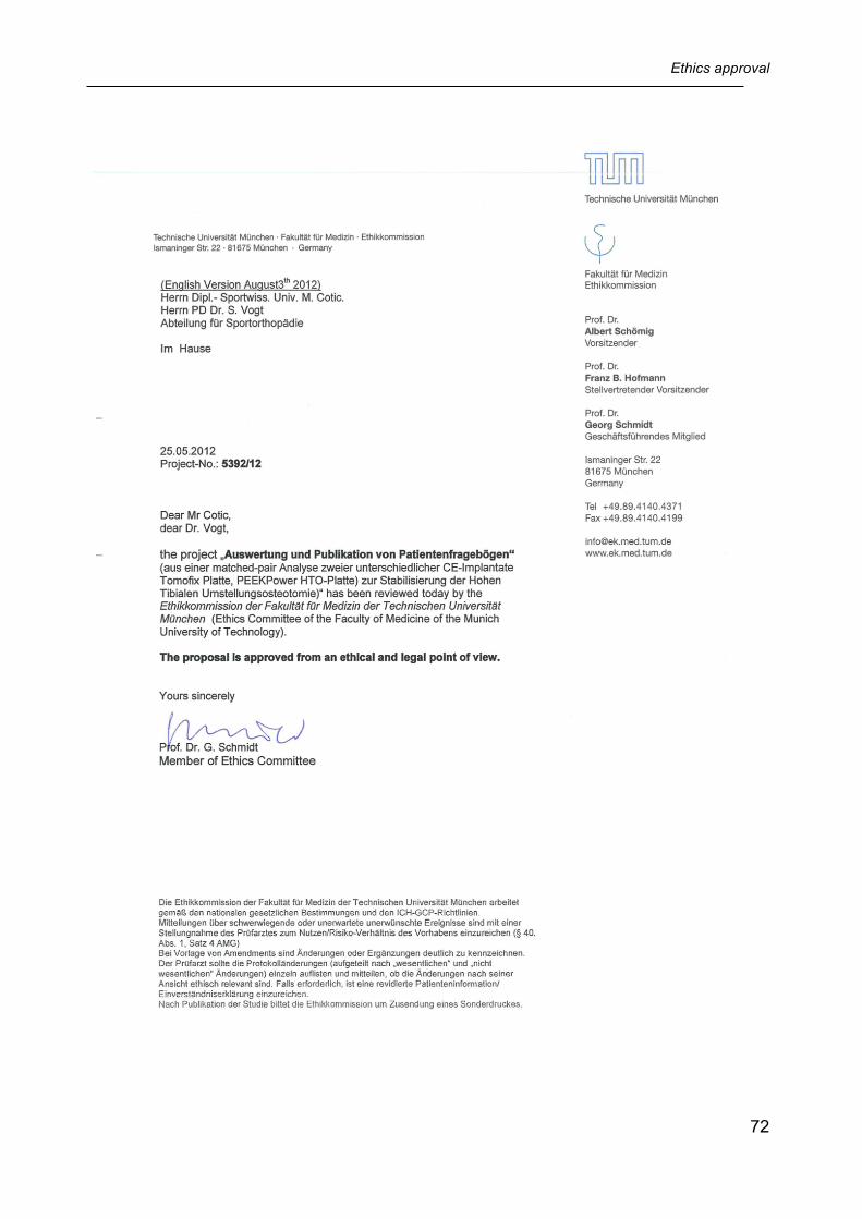
Ethics approval
72
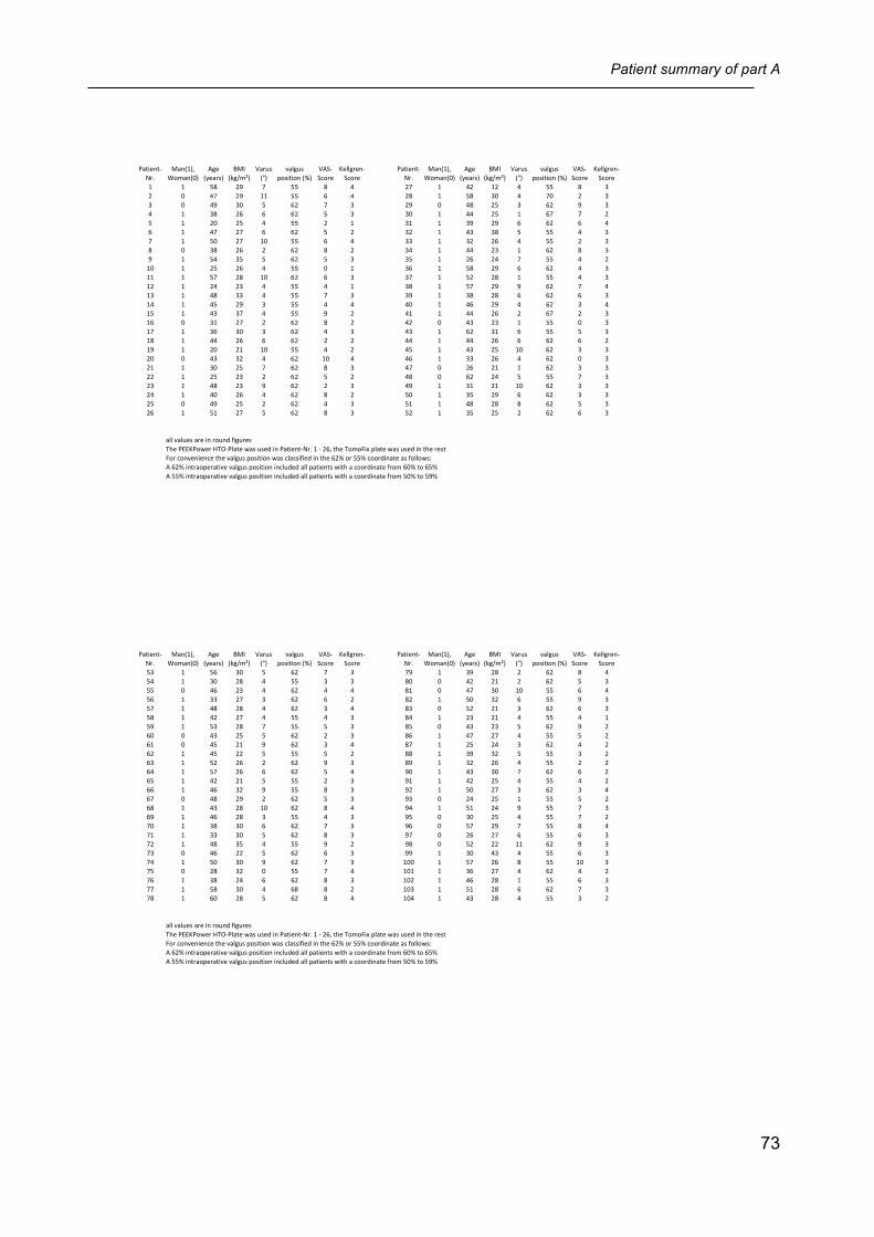
Patient summary of part A
73
Patient'Nr.
Man(1),0Woman(0)
Age0(years)
BMI0(kg/m²)
Varus0(°)
valgus0position0(%)
VAS'Score
Kellgren'Score
Patient'Nr.
Man(1),0Woman(0)
Age0(years)
BMI0(kg/m²)
Varus0(°)
valgus0position0(%)
VAS'Score
Kellgren'Score
1 1 58 29 7 55 8 4 27 1 42 12 4 55 8 32 0 47 29 11 55 6 4 28 1 58 30 4 70 2 33 0 49 30 5 62 7 3 29 0 48 25 3 62 9 34 1 38 26 6 62 5 3 30 1 44 25 1 67 7 25 1 20 25 4 55 2 1 31 1 39 29 6 62 6 46 1 47 27 6 62 5 2 32 1 43 38 5 55 4 37 1 50 27 10 55 6 4 33 1 32 26 4 55 2 38 0 38 26 2 62 8 2 34 1 44 23 1 62 8 39 1 54 35 5 62 5 3 35 1 26 24 7 55 4 210 1 25 26 4 55 0 1 36 1 58 29 6 62 4 311 1 57 28 10 62 6 3 37 1 52 28 1 55 4 312 1 24 23 4 55 4 1 38 1 57 29 9 62 7 413 1 48 33 4 55 7 3 39 1 38 28 6 62 6 314 1 45 29 3 55 4 4 40 1 46 29 4 62 3 415 1 43 37 4 55 9 2 41 1 44 26 2 67 2 316 0 31 27 2 62 8 2 42 0 43 23 1 55 0 317 1 36 30 3 62 4 3 43 1 62 31 6 55 5 318 1 44 26 6 62 2 2 44 1 44 26 6 62 6 219 1 20 21 10 55 4 2 45 1 43 25 10 62 3 320 0 43 32 4 62 10 4 46 1 33 26 4 62 0 321 1 30 25 7 62 8 3 47 0 26 21 1 62 3 322 1 25 23 2 62 5 2 48 0 62 24 5 55 7 323 1 48 23 9 62 2 3 49 1 31 21 10 62 3 324 1 40 26 4 62 8 2 50 1 35 29 6 62 3 325 0 49 25 2 62 4 3 51 1 48 28 8 62 5 326 1 51 27 5 62 8 3 52 1 35 25 2 62 6 3
all0values0are0in0round0figuresThe0PEEKPower0HTO'Plate0was0used0in0Patient'Nr.010'026,0the0TomoFix0plate0was0used0in0the0restFor0convenience0the0valgus0position0was0classified0in0the062%0or055%0coordinate0as0follows:A062%0intraoperative0valgus0position0included0all0patients0with0a0coordinate0from060%0to065%A055%0intraoperative0valgus0position0included0all0patients0with0a0coordinate0from050%0to059%
Patient'Nr.
Man(1),0Woman(0)
Age0(years)
BMI0(kg/m²)
Varus0(°)
valgus0position0(%)
VAS'Score
Kellgren'Score
Patient'Nr.
Man(1),0Woman(0)
Age0(years)
BMI0(kg/m²)
Varus0(°)
valgus0position0(%)
VAS'Score
Kellgren'Score
53 1 56 30 5 62 7 3 79 1 39 28 2 62 8 454 1 30 28 4 55 3 3 80 0 42 21 2 62 5 355 0 46 23 4 62 4 4 81 0 47 30 10 55 6 456 1 33 27 3 62 6 2 82 1 50 32 6 55 9 357 1 48 28 4 62 3 4 83 0 52 21 3 62 6 358 1 42 27 4 55 4 3 84 1 23 21 4 55 4 159 1 53 28 7 55 5 3 85 0 43 23 5 62 9 260 0 43 25 5 62 2 3 86 1 47 27 4 55 5 261 0 45 21 9 62 3 4 87 1 25 24 3 62 4 262 1 45 22 5 55 5 2 88 1 39 32 5 55 3 263 1 52 26 2 62 9 3 89 1 32 26 4 55 2 264 1 57 26 6 62 5 4 90 1 43 30 7 62 6 265 1 42 21 5 55 2 3 91 1 42 25 4 55 4 266 1 46 32 9 55 8 3 92 1 50 27 3 62 3 467 0 48 29 2 62 5 3 93 0 24 25 1 55 5 268 1 43 28 10 62 8 4 94 1 51 24 9 55 7 369 1 46 28 3 55 4 3 95 0 30 25 4 55 7 270 1 38 30 6 62 7 3 96 0 57 29 7 55 8 471 1 33 30 5 62 8 3 97 0 26 27 6 55 6 372 1 48 35 4 55 9 2 98 0 52 22 11 62 9 373 0 46 22 5 62 6 3 99 1 30 43 4 55 6 374 1 50 30 9 62 7 3 100 1 57 26 8 55 10 375 0 28 32 0 55 7 4 101 1 36 27 4 62 4 276 1 38 24 6 62 8 3 102 1 46 28 1 55 6 377 1 58 30 4 68 8 2 103 1 51 28 6 62 7 378 1 60 28 5 62 8 4 104 1 43 28 4 55 3 2
all0values0are0in0round0figuresThe0PEEKPower0HTO'Plate0was0used0in0Patient'Nr.010'026,0the0TomoFix0plate0was0used0in0the0restFor0convenience0the0valgus0position0was0classified0in0the062%0or055%0coordinate0as0follows:A062%0intraoperative0valgus0position0included0all0patients0with0a0coordinate0from060%0to065%A055%0intraoperative0valgus0position0included0all0patients0with0a0coordinate0from050%0to059%
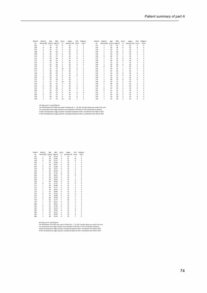
Patient summary of part A
74
Patient'Nr.
Man(1),0Woman(0)
Age0(years)
BMI0(kg/m²)
Varus0(°)
valgus0position0(%)
VAS'Score
Kellgren'Score
Patient'Nr.
Man(1),0Woman(0)
Age0(years)
BMI0(kg/m²)
Varus0(°)
valgus0position0(%)
VAS'Score
Kellgren'Score
105 1 47 25 9 62 3 3 131 1 42 24 6 62 5 4106 0 39 25 2 62 7 2 132 1 50 26 9 62 8 4107 0 45 27 3 62 0 2 133 1 55 27 7 55 7 3108 1 37 26 1 62 6 2 134 1 53 28 4 62 2 3109 0 22 22 4 55 4 3 135 1 53 32 5 55 3 2110 0 41 27 0 55 5 3 136 0 40 27 4 55 7 2111 0 53 28 2 55 8 2 137 1 41 22 6 55 4 2112 1 44 29 2 62 5 3 138 1 58 23 9 55 3 2113 1 51 28 4 62 5 4 139 1 51 25 7 55 7 4114 1 52 28 5 55 0 2 140 1 50 30 6 62 2 2115 1 27 30 4 55 2 1 141 0 56 22 7 62 2 4116 1 31 27 13 55 8 3 142 1 43 32 1 55 4 2117 1 43 29 4 62 5 2 143 1 52 26 7 62 7 4118 1 46 29 4 55 5 3 144 1 44 33 8 55 6 3119 1 53 32 4 55 8 3 145 1 43 27 6 55 3 2120 0 44 22 0 62 7 4 146 1 43 35 2 62 6 2121 1 25 28 4 55 1 1 147 1 68 24 8 55 6 2122 1 61 27 10 62 7 3 148 0 43 22 4 55 7 2123 1 35 25 3 55 2 2 149 1 48 30 5 62 6 2124 0 50 26 5 62 3 2 150 1 51 25 3 62 3 3125 0 47 28 5 62 6 3 151 0 47 24 4 55 8 2126 1 55 25 4 55 7 4 152 0 31 24 5 55 2 3127 1 56 25 9 55 8 4 153 1 47 22 5 55 5 3128 0 51 23 4 55 3 3 154 1 42 28 3 55 5 3129 1 50 23 9 55 8 3 155 1 24 28 3 55 8 3130 1 57 24 12 62 3 4 156 1 52 26 3 55 6 3
all0values0are0in0round0figuresThe0PEEKPower0HTO'Plate0was0used0in0Patient'Nr.010'026,0the0TomoFix0plate0was0used0in0the0restFor0convenience0the0valgus0position0was0classified0in0the062%0or055%0coordinate0as0follows:A062%0intraoperative0valgus0position0included0all0patients0with0a0coordinate0from060%0to065%A055%0intraoperative0valgus0position0included0all0patients0with0a0coordinate0from050%0to059%
Patient'Nr.
Man(1),0Woman(0)
Age0(years)
BMI0(kg/m²)
Varus0(°)
valgus0position0(%)
VAS'Score
Kellgren'Score
157 1 46 24,44 2 55 4 3158 0 50 34,66 5 62 10 4159 0 28 22,21 2 55 8 3160 1 53 24,69 3 62 7 3161 1 34 26,12 4 62 5 3162 0 15 22,49 5 55 2 0163 0 46 26,37 4 62 3 3164 1 45 26,30 3 55 5 4165 1 46 27,10 8 55 4 2166 1 43 28,41 3 62 8 3167 1 40 23,92 2 55 7 2168 1 49 28,34 6 62 2 4169 1 60 28,38 9 55 6 4170 0 41 27,22 4 55 6 3171 1 48 26,88 4 55 3 2172 1 40 27,77 3 55 2 3173 1 54 29,41 4 55 7 3174 1 47 28,04 9 55 0 4175 0 45 25,34 6 55 6 3176 1 33 28,08 6 55 6 3177 0 60 34,49 5 62 7 4178 1 63 27,77 7 55 2 3179 1 43 28,72 4 55 2 3180 1 41 28,31 5 55 7 3181 0 41 22,49 8 55 2 3182 1 45 24,14 2 55 4 3183 0 41 19,03 4 55 8 3184 1 34 24,15 2 55 7 2185 1 62 29,94 9 62 8 4186 1 46 32,14 6 55 7 4
all0values0are0in0round0figuresThe0PEEKPower0HTO'Plate0was0used0in0Patient'Nr.010'026,0the0TomoFix0plate0was0used0in0the0restFor0convenience0the0valgus0position0was0classified0in0the062%0or055%0coordinate0as0follows:A062%0intraoperative0valgus0position0included0all0patients0with0a0coordinate0from060%0to065%A055%0intraoperative0valgus0position0included0all0patients0with0a0coordinate0from050%0to059%

Patient questionnaire of part A
75
Abteilung und Poliklinik für Sportorthopädie des Kl in ikum rechts de r Isa r
de r Technischen U niversi tät München
Ans ta l t des öf fen tl i chen Rechts
Vorstand: Univ.-Prof. Dr. A. B. Imhoff
Fragebogen zu Ihrer Umstellungsoperation am Kniegelenk
Sehr geehrte Patientin, sehr geehrter Patient,
ihr freundliches Einverständnis vorausgesetzt, würden wir Sie bzw. Ihr Kniegelenk gerne mit Hilfe der
folgenden Fragebögen (s.u.) beurteilen. Falls Sie andere Beschwerden, z.B. an anderen Gelenken
haben, so blenden Sie diese für die Beantwortung des Fragebogens bitte aus.
Die Teilnahme an dieser Befragung ist völlig freiwillig und hat keinerlei Auswirkung auf Art und Qualität
ihrer Behandlung. Selbstverständlich können Sie Ihre erteilte Zustimmung ohne Angabe von Gründen
jederzeit widerrufen.
Die Befragung und die daraus resultierenden Ergebnisse sind Inhalt einer klinischen Studie, die später
von uns veröffentlicht wird. Die Veröffentlichung der Daten geschieht völlig anonym. Wenn Sie sich
etwa fünfzehn Minuten Zeit nehmen, um die folgenden Fragebögen auszufüllen, würden Sie der
universitären Wissenschaft einen großen Schritt weiterhelfen.
Die Befragungstermine finden an den regulären Kontrollterminen in unserer Ambulanz statt oder es
werden Ihnen die Fragebögen incl. frankiertem Rückumschlag per Post zugesandt. Dies alles soll für
Sie keine Mehrbelastung bedeuten.
Wenn Sie mit der anonymen Befragung einverstanden sind, bitten wir Sie, mit Ort, Datum und
Unterschrift Ihr Einverständnis schriftlich niederzulegen.
Vielen Dank für Ihre Kooperation.
Ort, Datum, Unterschrift

Patient questionnaire of part A
76
1. Allgemeine Daten
Name ______________________________________
Vorname ________________________________________
Telefon ______________________________________
Email ______________________________________
Adresse ______________________________________
Geb. Datum ____ __________________________________________________
Alter Größe Gewicht
2. Allgemeine Fragen Nehmen Sie derzeit Schmerzmedikamente wegen des Knies?
Ja, regelmässig Ja, bei Bedarf Nein
Wenn Sie Sport treiben/ Rehabilitationstraining machen, müssen Sie dann zur Durchführung
Schmerzmedikamente nehmen?
Ja, regelmässig Ja, bei Bedarf Nein
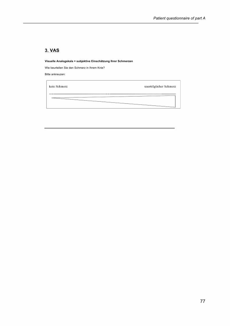
Patient questionnaire of part A
77
3. VAS Visuelle Analogskala = subjektive Einschätzung Ihrer Schmerzen Wie beurteilen Sie den Schmerz in Ihrem Knie? Bitte ankreuzen:
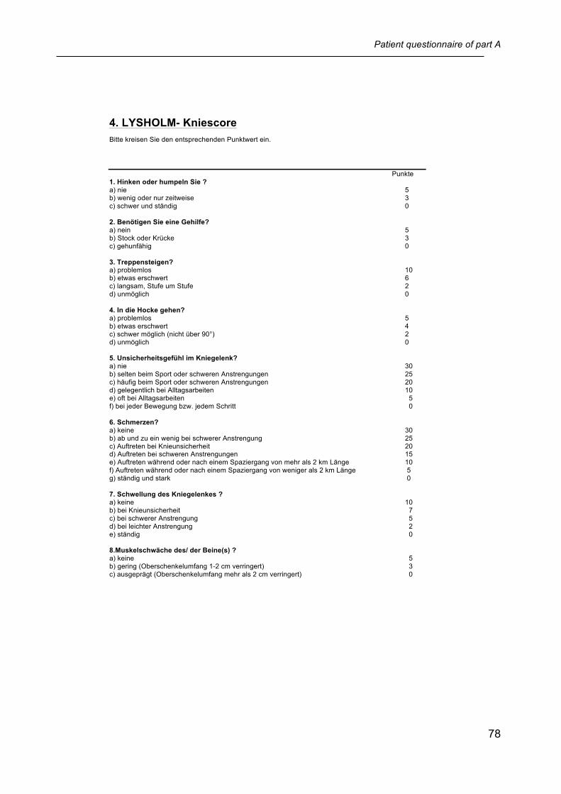
Patient questionnaire of part A
78
4. LYSHOLM- Kniescore Bitte kreisen Sie den entsprechenden Punktwert ein.
Punkte 1. Hinken oder humpeln Sie ? a) nie 5 b) wenig oder nur zeitweise 3 c) schwer und ständig 0 2. Benötigen Sie eine Gehilfe? a) nein 5 b) Stock oder Krücke 3 c) gehunfähig 0 3. Treppensteigen? a) problemlos 10 b) etwas erschwert 6 c) langsam, Stufe um Stufe 2 d) unmöglich 0 4. In die Hocke gehen? a) problemlos 5 b) etwas erschwert 4 c) schwer möglich (nicht über 90°) 2 d) unmöglich 0 5. Unsicherheitsgefühl im Kniegelenk? a) nie 30 b) selten beim Sport oder schweren Anstrengungen 25 c) häufig beim Sport oder schweren Anstrengungen 20 d) gelegentlich bei Alltagsarbeiten 10 e) oft bei Alltagsarbeiten 5 f) bei jeder Bewegung bzw. jedem Schritt 0 6. Schmerzen? a) keine 30 b) ab und zu ein wenig bei schwerer Anstrengung 25 c) Auftreten bei Knieunsicherheit 20 d) Auftreten bei schweren Anstrengungen 15 e) Auftreten während oder nach einem Spaziergang von mehr als 2 km Länge 10 f) Auftreten während oder nach einem Spaziergang von weniger als 2 km Länge 5 g) ständig und stark 0 7. Schwellung des Kniegelenkes ? a) keine 10 b) bei Knieunsicherheit 7 c) bei schwerer Anstrengung 5 d) bei leichter Anstrengung 2 e) ständig 0 8.Muskelschwäche des/ der Beine(s) ? a) keine 5 b) gering (Oberschenkelumfang 1-2 cm verringert) 3 c) ausgeprägt (Oberschenkelumfang mehr als 2 cm verringert) 0

Patient questionnaire of part A
79
5. WOMAC-Score Bitte kreuzen Sie das entsprechende Feld an. Teil A Wie schwer sind Ihre Schmerzen?
Teil B 1. Wie schwer ist die Gelenksteifheit nach dem morgendlichen Aufstehen? keine leicht mittelschwer schwer sehr schwer 2. Wie schwer ist die Gelenksteifheit nach dem Sitzen, Liegen oder Ruhen während des Tages? keine leicht mittelschwer schwer sehr schwer Teil C Wie schwierig ist für Sie ?
keine leichte mittelschwer schwer sehr schwer 1.Gehen auf flacher Ebene 2.Treppensteigen 3.Während der Nachtruhe 4.Sitzend oder liegend 5.Aufrecht stehend
einfach eingeschränkt mittel schwer sehr schwer 1. Treppen hinabsteigen 2. Treppen hinaufsteigen 3. Aufstehen aus dem Sitzen
4. Stehen 5. Zum Boden bücken 6. Gehen auf flachen Boden
7. In/Aus dem Auto steigen 8. Einkaufen gehen 9. Strümpfe anziehen 10. Aufstehen aus dem Bett
11. Strümpfe ausziehen 12. Im Bett liegen 13. In/Aus der Badewanne steigen
14. Sitzen 15. Auf die Toilette Setzen/ wieder Aufstehen
16. Schwere Hausarbeit 17. Leichte Hausarbeit
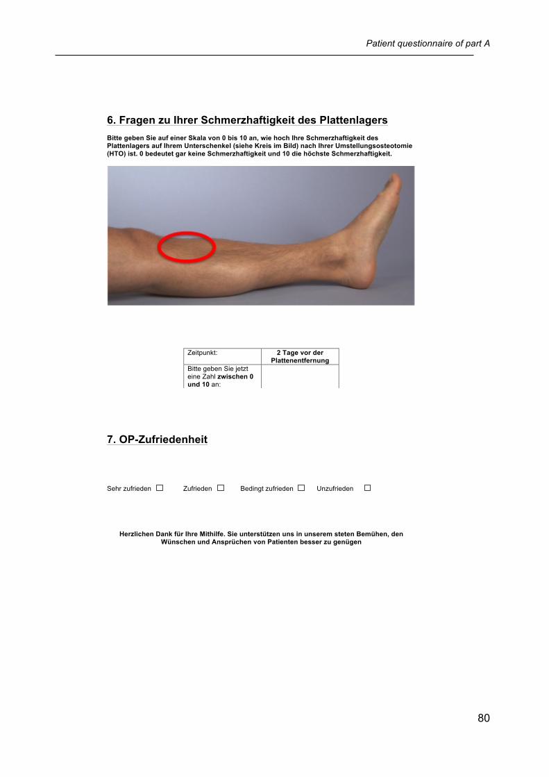
Patient questionnaire of part A
80
6. Fragen zu Ihrer Schmerzhaftigkeit des Plattenlagers Bitte geben Sie auf einer Skala von 0 bis 10 an, wie hoch Ihre Schmerzhaftigkeit des Plattenlagers auf Ihrem Unterschenkel (siehe Kreis im Bild) nach Ihrer Umstellungsosteotomie (HTO) ist. 0 bedeutet gar keine Schmerzhaftigkeit und 10 die höchste Schmerzhaftigkeit.
7. OP-Zufriedenheit Sehr zufrieden Zufrieden Bedingt zufrieden Unzufrieden
Herzlichen Dank für Ihre Mithilfe. Sie unterstützen uns in unserem steten Bemühen, den Wünschen und Ansprüchen von Patienten besser zu genügen
Zeitpunkt:
2 Tage vor der Plattenentfernung
Bitte geben Sie jetzt eine Zahl zwischen 0 und 10 an:
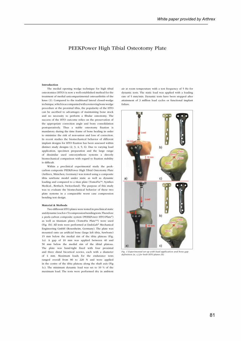
White paper provided by Arthrex
81
PEEKPower High Tibial Osteotomy Plate
IntroductionThe medial opening wedge technique for high tibial
osteotomies (HTO) is now a well-established method for the treatment of medial unicompartimental osteoarthritis of the knee (1). Compared to the traditional lateral closed-wedge technique, which is accompanied with a removing bone wedge procedure at the proximal tibia, the popularity of the HTO can be ascribed to advantages of maintaining bone stock and no necessity to perform a fibular osteotomy. The success of the HTO outcome relies on the preservation of the appropriate correction angle and bony consolidation postoperatively. Thus a stable osteotomy fixation is mandatory during the time frame of bone healing in order to minimize the risk of non-union and loss of correction. In recent studies the biomechanical behavior of different implant designs for HTO fixation has been assessed within distinct study designs (2; 3; 4; 5; 6). Due to varying load application, specimen preparation and the large range of dissimilar used osteosynthesis systems a directly biomechanical comparison with regard to fixation stability is difficult.
Within a preclinical experimental study the peek-carbon composite PEEKPower High Tibial Osteotomy Plate (Arthrex, München, Germany) was tested using a composite tibia sawbone model under static as well as dynamic loading and compared to a titan plate (TomoFix™, Synthes Medical., Bettlach, Switzerland). The purpose of this study was to evaluate the biomechanical behavior of these two plate systems in a comparable worst case compression bending test design.
Material & Methods
Two different HTO plates were tested in preclinical static and dynamic (each n = 5) compression bending tests. Therefore a peek-carbon composite system (PEEKPower HTO-Plate®) as well as titanium plates (TomoFix Plate™) were used (Fig. 1b). All tests were performed at EndoLab® Mechanical Engineering GmbH (Rosenheim, Germany). The plate was mounted onto an artificial bone (large left tibia, Sawbone) 15 mm below the medial rim of the tibia plateau (Fig. 1a). A gap of 10 mm was applied between 40 and 50 mm below the medial rim of the tibial plateau. The plate was hand-tight fixed with four proximal and three distal bicortical screws, each with a diameter of 4 mm. Maximum loads for the endurance tests ranged overall from 80 to 220 N and were applied in the centre of the tibia plateau along the shaft axis (Fig 1c). The minimum dynamic load was set to 10 % of the maximum load. The tests were performed dry in ambient
air at room temperature with a test frequency of 5 Hz for dynamic tests. The static load was applied with a loading rate of 5 mm/min. Dynamic tests have been stopped after attainment of 3 million load cycles or functional implant failure.
Fig. 1 Experimental set-up with load application and bone gap definition (a, c) for both HTO plates (b)
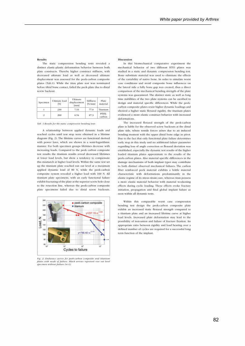
White paper provided by Arthrex
82
ResultsThe static compression bending tests revealed a
distinct elastic-plastic deformation behavior between both plate constructs. Thereby higher construct stiffness, with decreased ultimate load as well as decreased ultimate displacement was assessed for the peek-carbon composite plates (Tab.1). While the titan plate test was terminated before tibial bone contact, failed the peek plate due to distal screw backout.
SpecimenUltimate load
[N]
Ultimate displacement
[mm]
Stiffness [N/mm]
Plate material
1 230 7.53 77.0 Titanium
2 200 6.54 87.3PEEK-carbon
Tab. 1:Results for the static compression bending tests
A relationship between applied dynamic loads and reached cycles until test stop were obtained in a lifetime diagram (Fig. 2). The lifetime curves are functional derived with power laws, which are shown in a semi-logarithmic manner. For both specimen groups lifetimes decrease with increasing loads. Compared to the peek carbon composite test results the titanium results reveal decreased lifetimes at lower load levels, but show a tendency to compensate this mismatch at higher load levels. Within the same test set up the titanium plate reached run out level at a maximum applied dynamic load of 80 N, while the peek-carbon composite system revealed a higher load with 160 N. All titanium plate specimens, with an early functional failure exhibit fracturing of the plate at the superior screw hole close to the resection line, whereas the peek-carbon composite plate specimens failed due to distal screw backouts.
Fig. 2: Endurace curves for peek-carbon composite and titanium plates with mode of failure. Black arrows represent run out level specimen without failure (n=1).
DiscussionIn this biomechanical comparative experiment the
mechanical behavior of two different HTO plates was studied in a static and dynamic compression bending test. Bone substitute material was used to eliminate the effects of the variability of native bone. In order to simulate worst case conditions and avoid composite bone influences on the lateral side a fully bone gap was created, thus a direct comparison of the mechanical bending strength of the plate systems was guaranteed. The distinct static as well as long time stabilities of the two plate systems can be ascribed to design and material specific differences. While the peek-carbon composite plates resist higher dynamic loadings and showed a higher static flexural rigidity, the titanium plates evidenced a more elastic construct behavior with increased deformations.
The increased flexural strength of the peek-carbon plate is liable for the observed screw backouts at the distal plate side, where tensile forces arises due to an induced bending moment with the upper distal bone edge as pivot. Due to the fact that only functional plate failure determines early stop in this study and no additional failure parameter regarding loss of angle correction or flexural deviation was established, especially the dynamic test results of the higher loaded titanium plates approximate to the results of the peek-carbon plates. Also material specific differences in the damage mechanisms of both implant types may contribute to both distinct observed mechanical failures. The carbon fiber reinforced peek material exhibits a brittle material characteristic with deformations predominantly in the elastic regime of its stress-strain cure, whereas titan possess a more elastic material behavior with material weakening effects during cyclic loading. These effects evoke fracture initiation, propagation and final global implant failure as seen within all dynamic tests.
Within this comparable worst case compression bending test design the peek-carbon composite plate exhibit an increased static flexural strength compared to a titanium plate and an increased lifetime curve at higher load levels. Increased plate deformation may lead to the possibility of non-union and failure of fracture fixation. An appropriate ratio between rigidity and load bearing over a defined number of cycles are required for a successful long term function of the implant.
White paper provided by Arthrex
83
© Copyright Arthrex Medizinische Instrumente GmbH, 2013. All rights reserved. LA2-0118-EN_A
Literature1. Naudie D, Bourne RB, Rorabeck CH, Bourne TJ. The Install Award. Survivorship of the high tibial valgus osteotomy. A 10 to 22-year follow-up study. Clinical Orthopaedics. 367:18–27, 1999.
2. Takeuchi R, Bito H, Akamatsu Y, Shiraishi T, Morishita S, Koshino T, Saito T. In vitro stability of open wedge high tibial osteotomy with synthetic bone graft. The Knee. 17:217–220, 2010.
3. Stoffel K, Stachowiak G, Kuster M. Open wedge high tibial osteotomy: biomechanical investigation of the modified Arthrex Osteotomy Plate (Puddu Plate) and the TomoFix Plate. Clinical Biomechanics. 19:944–950, 2004.
4. Spahn G, Mückley T, Kahl E, Hofmann GO. Biomechanical investigation of different internal fixations in medial opening-wedge high tibial osteotomy. Clinical Biomechanics. 21:272–278, 2006.
5. Agneskirchner JD, Freiling D, Hurschler C., Lobenhoffer P. Primary stability of four different implants for opening wedge high tibial osteotomy. Knee Surg Sports Traumatol Arthrosc. 14:291–300, 2006.
6. Zhim F, Laflamme GY, Viens H, Saidane K, Yahia H. Biomechanical stability of high tibial opening wedge osteotomy: Internal fixation versus external fixation. Clinical Biomechanics. 20:871–876, 2005.
Patient questionnaire of part B
84
Abteilung und Poliklinik für Sportorthopädie des Kl in ikum rechts der Isa r
de r Technischen U niversi tät München
Ans ta l t des öf fen tl i chen Rechts
Vorstand: Univ.-Prof. Dr. A. B. Imhoff
Fragebogen zu Ihrer Umstellungsoperation am Kniegelenk
Sehr geehrte Patientin, sehr geehrter Patient,
ihr freundliches Einverständnis vorausgesetzt, würden wir Sie bzw. Ihr Kniegelenk gerne mit Hilfe der
folgenden Fragebögen (s.u.) beurteilen. Falls Sie andere Beschwerden, z.B. an anderen Gelenken
haben, so blenden Sie diese für die Beantwortung des Fragebogens bitte aus.
Die Teilnahme an dieser Befragung ist völlig freiwillig und hat keinerlei Auswirkung auf Art und Qualität
ihrer Behandlung. Selbstverständlich können Sie Ihre erteilte Zustimmung ohne Angabe von Gründen
jederzeit widerrufen.
Die Befragung und die daraus resultierenden Ergebnisse sind Inhalt einer klinischen Studie, die später
von uns veröffentlicht wird. Die Veröffentlichung der Daten geschieht völlig anonym. Wenn Sie sich
etwa fünfzehn Minuten Zeit nehmen, um die folgenden Fragebögen auszufüllen, würden Sie der
universitären Wissenschaft einen großen Schritt weiterhelfen.
Die Befragungstermine finden an den regulären Kontrollterminen in unserer Ambulanz statt oder es
werden Ihnen die Fragebögen incl. frankiertem Rückumschlag per Post zugesandt. Dies alles soll für
Sie keine Mehrbelastung bedeuten.
Wenn Sie mit der anonymen Befragung einverstanden sind, bitten wir Sie, mit Ort, Datum und
Unterschrift Ihr Einverständnis schriftlich niederzulegen.
Vielen Dank für Ihre Kooperation.
Ort, Datum, Unterschrift

Patient questionnaire of part B
85
1. Allgemeine Daten
Name ______________________________________
Vorname ________________________________________
Telefon ______________________________________
Email ______________________________________
Adresse ______________________________________
Geb. Datum ____ __________________________________________________
Alter Größe Gewicht
2. Allgemeine Fragen Nehmen Sie derzeit Schmerzmedikamente wegen des Knies?
Ja, regelmässig Ja, bei Bedarf Nein
Wenn Sie Sport treiben/ Rehabilitationstraining machen, müssen Sie dann zur Durchführung
Schmerzmedikamente nehmen?
Ja, regelmässig Ja, bei Bedarf Nein

Patient questionnaire of part B
86
3. VAS Visuelle Analogskala = subjektive Einschätzung Ihrer Schmerzen Wie beurteilen Sie den Schmerz in Ihrem Knie? Bitte ankreuzen:

Patient questionnaire of part B
87
4. LYSHOLM- Kniescore Bitte kreisen Sie den entsprechenden Punktwert ein.
Punkte 1. Hinken oder humpeln Sie ? a) nie 5 b) wenig oder nur zeitweise 3 c) schwer und ständig 0 2. Benötigen Sie eine Gehilfe? a) nein 5 b) Stock oder Krücke 3 c) gehunfähig 0 3. Treppensteigen? a) problemlos 10 b) etwas erschwert 6 c) langsam, Stufe um Stufe 2 d) unmöglich 0 4. In die Hocke gehen? a) problemlos 5 b) etwas erschwert 4 c) schwer möglich (nicht über 90°) 2 d) unmöglich 0 5. Unsicherheitsgefühl im Kniegelenk? a) nie 30 b) selten beim Sport oder schweren Anstrengungen 25 c) häufig beim Sport oder schweren Anstrengungen 20 d) gelegentlich bei Alltagsarbeiten 10 e) oft bei Alltagsarbeiten 5 f) bei jeder Bewegung bzw. jedem Schritt 0 6. Schmerzen? a) keine 30 b) ab und zu ein wenig bei schwerer Anstrengung 25 c) Auftreten bei Knieunsicherheit 20 d) Auftreten bei schweren Anstrengungen 15 e) Auftreten während oder nach einem Spaziergang von mehr als 2 km Länge 10 f) Auftreten während oder nach einem Spaziergang von weniger als 2 km Länge 5 g) ständig und stark 0 7. Schwellung des Kniegelenkes ? a) keine 10 b) bei Knieunsicherheit 7 c) bei schwerer Anstrengung 5 d) bei leichter Anstrengung 2 e) ständig 0 8.Muskelschwäche des/ der Beine(s) ? a) keine 5 b) gering (Oberschenkelumfang 1-2 cm verringert) 3 c) ausgeprägt (Oberschenkelumfang mehr als 2 cm verringert) 0

Patient questionnaire of part B
88
5. WOMAC-Score Bitte kreuzen Sie das entsprechende Feld an. Teil A Wie schwer sind Ihre Schmerzen?
Teil B 1. Wie schwer ist die Gelenksteifheit nach dem morgendlichen Aufstehen? keine leicht mittelschwer schwer sehr schwer 2. Wie schwer ist die Gelenksteifheit nach dem Sitzen, Liegen oder Ruhen während des Tages? keine leicht mittelschwer schwer sehr schwer Teil C Wie schwierig ist für Sie ?
keine leichte mittelschwer schwer sehr schwer 1.Gehen auf flacher Ebene 2.Treppensteigen 3.Während der Nachtruhe 4.Sitzend oder liegend 5.Aufrecht stehend
einfach eingeschränkt mittel schwer sehr schwer 1. Treppen hinabsteigen 2. Treppen hinaufsteigen 3. Aufstehen aus dem Sitzen
4. Stehen 5. Zum Boden bücken 6. Gehen auf flachen Boden
7. In/Aus dem Auto steigen 8. Einkaufen gehen 9. Strümpfe anziehen 10. Aufstehen aus dem Bett
11. Strümpfe ausziehen 12. Im Bett liegen 13. In/Aus der Badewanne steigen
14. Sitzen 15. Auf die Toilette Setzen/ wieder Aufstehen
16. Schwere Hausarbeit 17. Leichte Hausarbeit
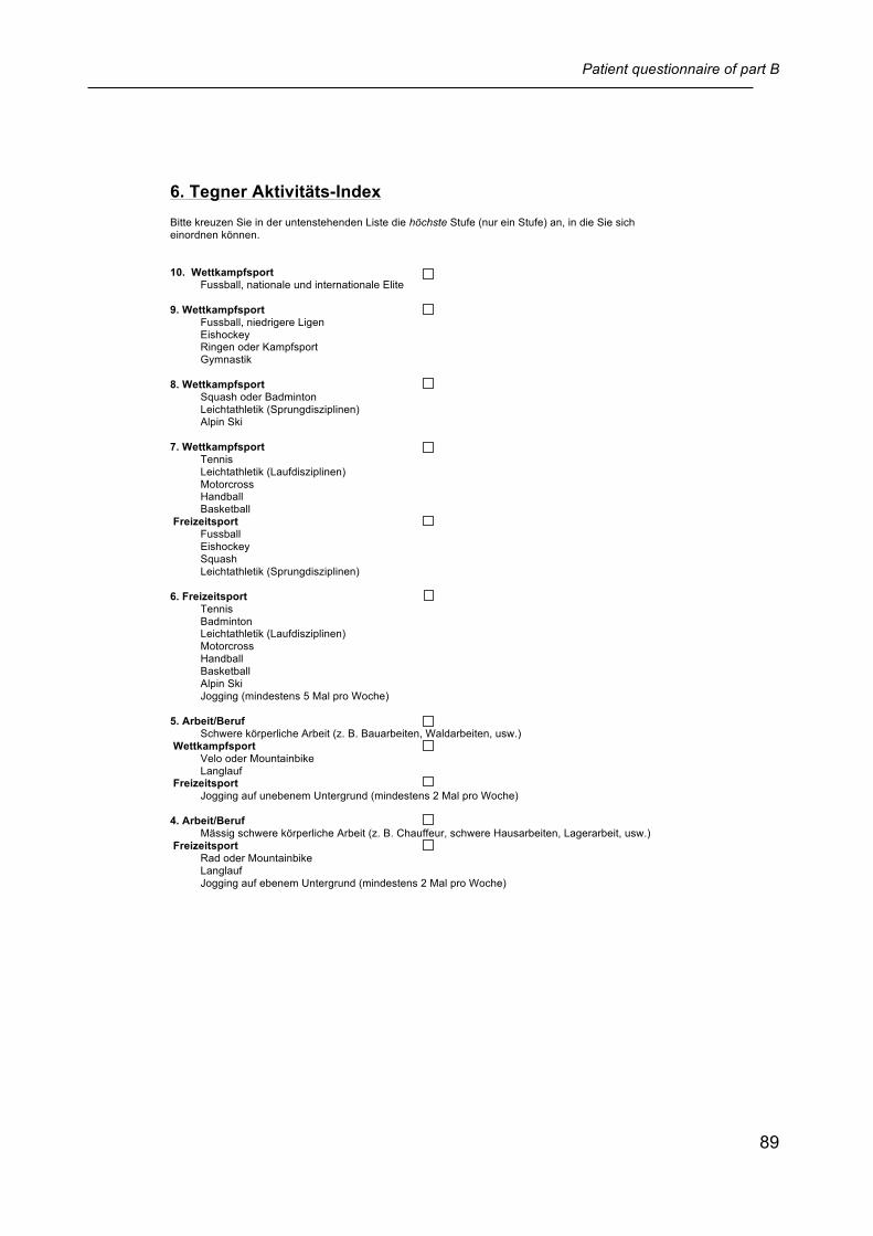
Patient questionnaire of part B
89
6. Tegner Aktivitäts-Index Bitte kreuzen Sie in der untenstehenden Liste die höchste Stufe (nur ein Stufe) an, in die Sie sich einordnen können. 10. Wettkampfsport Fussball, nationale und internationale Elite 9. Wettkampfsport Fussball, niedrigere Ligen Eishockey Ringen oder Kampfsport Gymnastik 8. Wettkampfsport Squash oder Badminton Leichtathletik (Sprungdisziplinen) Alpin Ski 7. Wettkampfsport Tennis Leichtathletik (Laufdisziplinen) Motorcross Handball Basketball Freizeitsport Fussball Eishockey Squash Leichtathletik (Sprungdisziplinen) 6. Freizeitsport Tennis Badminton Leichtathletik (Laufdisziplinen) Motorcross Handball Basketball Alpin Ski Jogging (mindestens 5 Mal pro Woche) 5. Arbeit/Beruf Schwere körperliche Arbeit (z. B. Bauarbeiten, Waldarbeiten, usw.) Wettkampfsport Velo oder Mountainbike Langlauf Freizeitsport Jogging auf unebenem Untergrund (mindestens 2 Mal pro Woche) 4. Arbeit/Beruf Mässig schwere körperliche Arbeit (z. B. Chauffeur, schwere Hausarbeiten, Lagerarbeit, usw.) Freizeitsport Rad oder Mountainbike Langlauf Jogging auf ebenem Untergrund (mindestens 2 Mal pro Woche)
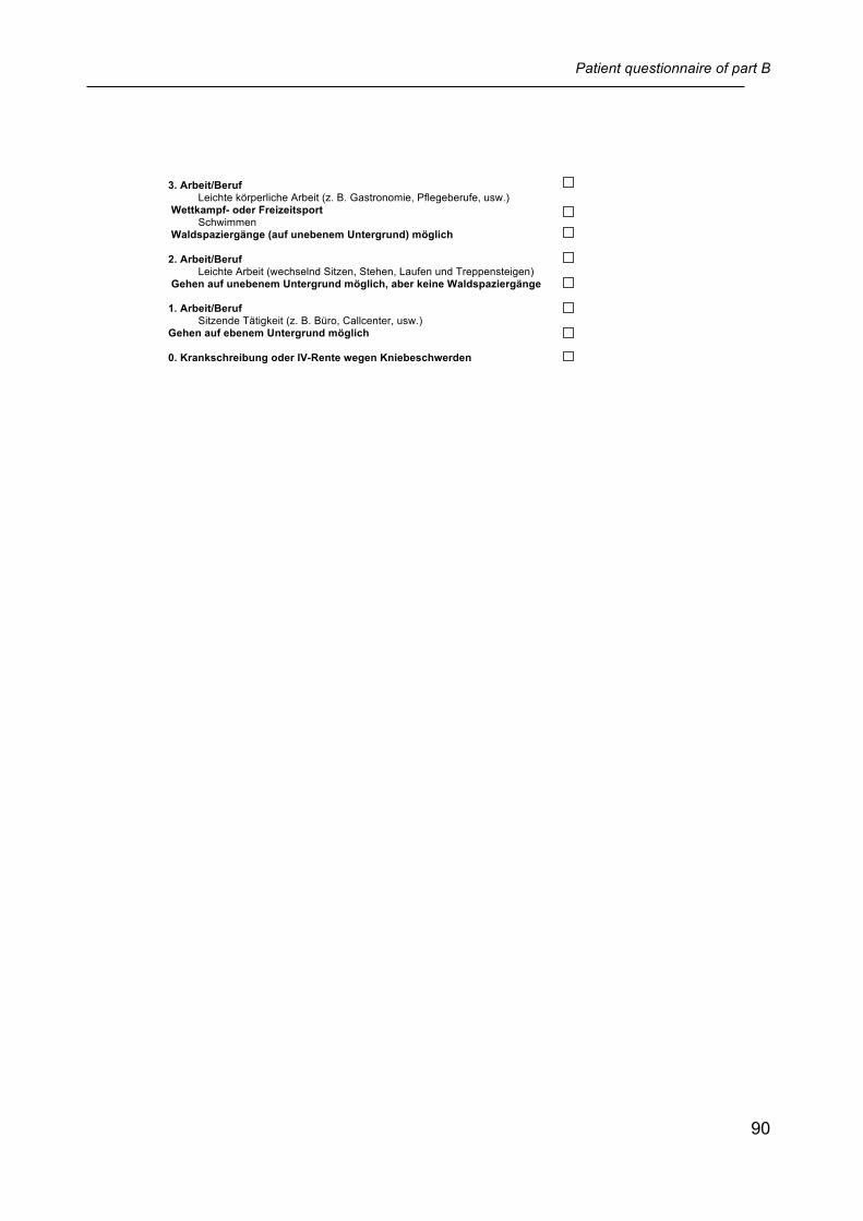
Patient questionnaire of part B
90
3. Arbeit/Beruf Leichte körperliche Arbeit (z. B. Gastronomie, Pflegeberufe, usw.) Wettkampf- oder Freizeitsport Schwimmen Waldspaziergänge (auf unebenem Untergrund) möglich 2. Arbeit/Beruf Leichte Arbeit (wechselnd Sitzen, Stehen, Laufen und Treppensteigen) Gehen auf unebenem Untergrund möglich, aber keine Waldspaziergänge 1. Arbeit/Beruf Sitzende Tätigkeit (z. B. Büro, Callcenter, usw.) Gehen auf ebenem Untergrund möglich 0. Krankschreibung oder IV-Rente wegen Kniebeschwerden
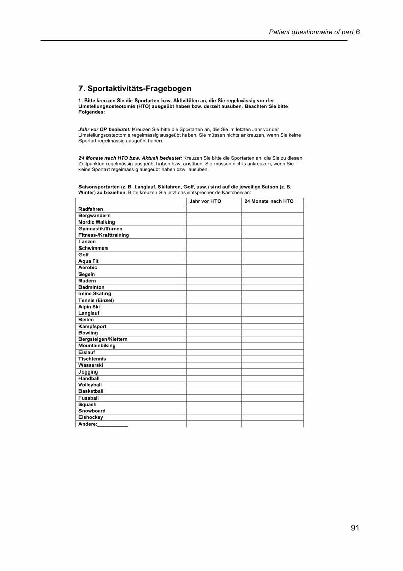
Patient questionnaire of part B
91
7. Sportaktivitäts-Fragebogen 1. Bitte kreuzen Sie die Sportarten bzw. Aktivitäten an, die Sie regelmässig vor der Umstellungsosteotomie (HTO) ausgeübt haben bzw. derzeit ausüben. Beachten Sie bitte Folgendes: Jahr vor OP bedeutet: Kreuzen Sie bitte die Sportarten an, die Sie im letzten Jahr vor der Umstellungsosteotomie regelmässig ausgeübt haben. Sie müssen nichts ankreuzen, wenn Sie keine Sportart regelmässig ausgeübt haben. 24 Monate nach HTO bzw. Aktuell bedeutet: Kreuzen Sie bitte die Sportarten an, die Sie zu diesen Zeitpunkten regelmässig ausgeübt haben bzw. ausüben. Sie müssen nichts ankreuzen, wenn Sie keine Sportart regelmässig ausgeübt haben bzw. ausüben. Saisonsportarten (z. B. Langlauf, Skifahren, Golf, usw.) sind auf die jeweilige Saison (z. B. Winter) zu beziehen. Bitte kreuzen Sie jetzt das entsprechende Kästchen an:
Jahr vor HTO 24 Monate nach HTO Radfahren Bergwandern Nordic Walking Gymnastik/Turnen Fitness-/Krafttraining Tanzen Schwimmen Golf Aqua Fit Aerobic Segeln Rudern Badminton Inline Skating Tennis (Einzel) Alpin Ski Langlauf Reiten Kampfsport Bowling Bergsteigen/Klettern Mountainbiking Eislauf Tischtennis Wasserski Jogging Handball Volleyball Basketball Fussball Squash Snowboard Eishockey Andere:___________
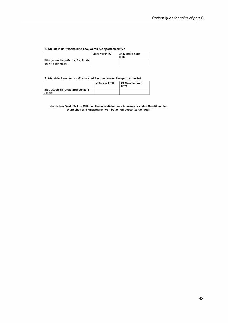
Patient questionnaire of part B
92
2. Wie oft in der Woche sind bzw. waren Sie sportlich aktiv?
3. Wie viele Stunden pro Woche sind Sie bzw. waren Sie sportlich aktiv?
Herzlichen Dank für Ihre Mithilfe. Sie unterstützen uns in unserem steten Bemühen, den Wünschen und Ansprüchen von Patienten besser zu genügen
Jahr vor HTO 24 Monate nach HTO
Bitte geben Sie je 0x, 1x, 2x, 3x, 4x, 5x, 6x oder 7x an:
Jahr vor HTO 24 Monate nach HTO
Bitte geben Sie je die Stundenzahl (h) an:
Copyright © 2022 FDOKUMEN