The Independent Medical Expert Group (IMEG) - GOV.UK
80
The Independent Medical Expert Group (IMEG) Report and recommendations on medical and scientific aspects of the Armed Forces Compensation Scheme March 2015
-
Upload
khangminh22 -
Category
Documents
-
view
1 -
download
0
Transcript of The Independent Medical Expert Group (IMEG) - GOV.UK
The Independent Medical Expert Group(IMEG)
Report and recommendations on medical and scientific aspects of the Armed Forces Compensation Scheme
March 2015

Ministry of Defence Main Building (06.M.06) Whitehall London SW1A 2HB United Kingdom Telephone [MOD]: Facsimile [MOD]: E-mail:
+44 (0)20 7218 9571 +44 (0)20 7807 8068 PERSTrg-Rem-AFCPol3 @mod.uk
A Non-Departmental Public Body Sponsored by the Ministry of Defence
27th February, 2015 Minister of State for Defence Personnel Welfare and Veterans MOD Whitehall London SW1A 2HB Dear Minister I have pleasure in submitting the third report of the Independent Medical Expert Group (IMEG). It is now nine years since the Armed Forces and Reserve Forces (Compensation Scheme) Order was established, leading to the introduction of the Armed Forces Compensation Scheme (AFCS). With the increased scope of this scheme, as well as of combat operations during this period, claims rates have risen steadily. IMEG was created in 2010, following a recommendation of Lord Boyce’s review of the Scheme, becoming a Non-Departmental Body in 2012. Its purpose continues to be to provide you with medical and scientific advice on the AFCS, to ensure it reflects contemporary medical and scientific knowledge and addresses the needs of Service Personnel. Our previous reports were published in 2011 and 2013. Several topics for our third report have been suggested by the claimants themselves, their representatives, the chain of command, the charities, health and other support professionals. They reflect combat and deployed service as well as injury sustained during sport and adventurous training. IMEG particularly values its visits to Hasler Company, part of the Royal Navy recovery pathway, run by Royal Marines, and the Defence Medical Rehabilitation Centre, Headley Court, providing the opportunity for us to discuss issues with claimants and their insights into the impact and the challenges of their injuries. The approach of IMEG for this Report has been to identify and appraise the relevant evidence, systematically reviewing the published peer-reviewed international literature as well as discussing topics with recognised military and civilian experts. Following outbreaks of ‘Helmand Fever’ in Afghanistan, we were asked to consider infectious diseases in the context of deployment, notably Q fever and its fatiguing sequelae. We have made recommendations about descriptors and awards, which you accepted and were incorporated into the legislation from 7 April 2014. The recent conflicts have been characterised by blast trauma from improvised explosive devices, typically leading to complex lower limb trauma with retention and reconstruction or loss of the limb.
While combat related blast injury is not new, it is only in this generation, with advances in forward surgery, anaesthesiology, aero-evacuation and digitised prosthetics, that such profound injury has been survivable, giving many individuals high levels of function, including sporting and adventurous activity. An issue raised with IMEG, on more than one occasion, has been that AFCS awards for limb amputation are generous as compared to retained, reconstructed limbs because successful prosthetic rehabilitation allows useful function to be regained. Also the ability to undertake civilian employment at or around service termination is more common, even amongst multiple amputees, than occurs where limbs are retained and reconstructed, which are often complicated by chronic infection and the need for further surgery. Full evaluation of the new generation of microprocessor prosthetics could not take place prior to the recent wars and IMEG strongly supports the proposal for multidisciplinary longitudinal studies of complex lower limb trauma. Ideally this should involve cooperation between Defence, Health Departments and academic departments, e.g. bio-engineering, with twenty or more years of follow-up. This will give the best opportunity to provide knowledge of long term outcome in these cases. Against this background, to provide an understanding of current knowledge, our report includes two papers which review the literature to date on the cardiovascular and non-cardiovascular sequelae of lower limb amputation and a third paper which considers current and recent experience of retained reconstructed complex lower limb trauma. You asked IMEG to investigate the scientific and medical aspects of mesothelioma to inform Defence policy options. Our response is included in this report. This followed representations from the Royal British Legion about the position of ex service personnel without eligible dependents when compared to the provisions of the Mesothelioma Act (2014). We have continued our work on possible Recognised Diseases and have considered testis cancer, diabetes mellitus and the leukaemias. Lastly, the May 2013 IMEG Report included a section on Mild Traumatic Brain Injury. Many unresolved issues remain and since May 2013, in the context of military, civilian and sporting trauma, both professional and amateur, several international studies have been published. Some evaluate new investigative techniques, with the aim of early identification of mTBI sub groups with a poor prognosis who might benefit from specific interventions. As yet no definite conclusions have been reached. I would propose IMEG undertakes a further review of mild Traumatic Brain Injury for the next IMEG report. All IMEG Members have taken part in our discussions and have agreed our findings and conclusions. I believe our conclusions and recommendations fairly reflect the current evidence and are in line with the intentions of the Scheme. During this year a number of members have left the Group. These included Professor David Alexander FRC Psych from August 2013, Colonel Fiona Gardner, CBE, and Brigadier Robin Cordell, both from October 2013. I am most grateful to them all for their contributions to our work and wish them well for the future. We have been joined by Professor Peter White, FRC Psych, Colonel John Ridge, and Brigadier Hugh Williamson, all from December 2013. I am grateful to colleagues who helped with our discussions particularly Dr E.H.N. Oakley for his generous input to the Non Freezing Cold Injury paper. I would also wish to thank our Secretariat for their willingness and commitment to IMEG and, without whom, our work could not be done. Yours sincerely
Professor Sir Anthony Newman Taylor, CBE, FRCP, FFOM, FMedSci Chairman Independent Medical Expert Group (IMEG)
1The IMEG report and recommendations on medical and scientific aspects of the Armed Forces Compensation Scheme
While combat related blast injury is not new, it is only in this generation, with advances in forward surgery, anaesthesiology, aero-evacuation and digitised prosthetics, that such profound injury has been survivable, giving many individuals high levels of function, including sporting and adventurous activity. An issue raised with IMEG, on more than one occasion, has been that AFCS awards for limb amputation are generous as compared to retained, reconstructed limbs because successful prosthetic rehabilitation allows useful function to be regained. Also the ability to undertake civilian employment at or around service termination is more common, even amongst multiple amputees, than occurs where limbs are retained and reconstructed, which are often complicated by chronic infection and the need for further surgery. Full evaluation of the new generation of microprocessor prosthetics could not take place prior to the recent wars and IMEG strongly supports the proposal for multidisciplinary longitudinal studies of complex lower limb trauma. Ideally this should involve cooperation between Defence, Health Departments and academic departments, e.g. bio-engineering, with twenty or more years of follow-up. This will give the best opportunity to provide knowledge of long term outcome in these cases. Against this background, to provide an understanding of current knowledge, our report includes two papers which review the literature to date on the cardiovascular and non-cardiovascular sequelae of lower limb amputation and a third paper which considers current and recent experience of retained reconstructed complex lower limb trauma. You asked IMEG to investigate the scientific and medical aspects of mesothelioma to inform Defence policy options. Our response is included in this report. This followed representations from the Royal British Legion about the position of ex service personnel without eligible dependents when compared to the provisions of the Mesothelioma Act (2014). We have continued our work on possible Recognised Diseases and have considered testis cancer, diabetes mellitus and the leukaemias. Lastly, the May 2013 IMEG Report included a section on Mild Traumatic Brain Injury. Many unresolved issues remain and since May 2013, in the context of military, civilian and sporting trauma, both professional and amateur, several international studies have been published. Some evaluate new investigative techniques, with the aim of early identification of mTBI sub groups with a poor prognosis who might benefit from specific interventions. As yet no definite conclusions have been reached. I would propose IMEG undertakes a further review of mild Traumatic Brain Injury for the next IMEG report. All IMEG Members have taken part in our discussions and have agreed our findings and conclusions. I believe our conclusions and recommendations fairly reflect the current evidence and are in line with the intentions of the Scheme. During this year a number of members have left the Group. These included Professor David Alexander FRC Psych from August 2013, Colonel Fiona Gardner, CBE, and Brigadier Robin Cordell, both from October 2013. I am most grateful to them all for their contributions to our work and wish them well for the future. We have been joined by Professor Peter White, FRC Psych, Colonel John Ridge, and Brigadier Hugh Williamson, all from December 2013. I am grateful to colleagues who helped with our discussions particularly Dr E.H.N. Oakley for his generous input to the Non Freezing Cold Injury paper. I would also wish to thank our Secretariat for their willingness and commitment to IMEG and, without whom, our work could not be done. Yours sincerely
Professor Sir Anthony Newman Taylor, CBE, FRCP, FFOM, FMedSci Chairman Independent Medical Expert Group (IMEG)
Independent Medical Expert Group (IMEG) – List of MembersChairmanProfessor Sir Anthony Newman Taylor CBE, FRCP, FFOM, FMedSci
Expert Members Professor David Alexander MA(Hons), C.Psychol, PhD, FBPS, FRSM(Hon), FRC Psych (until August 2013)
Dr Anne Braidwood CBE, MRCP, MRCGP, FFOM
Professor Linda Luxon CBE, FRCP
Professor James Ryan OBE, OStJ, MB, BCh, BAO(NUI), FRC(Eng), MCh(NUI), Hon FCEM, DMCC(SoA)
Dr John Scadding OBE, MD, FRCP
Professor David Snashall MSc, FRCP, FFOM, LLM
Professor Peter White BSc, MD, FRCP, FRCPsych (from September 2013)
Lay Members Lt Col Jerome Church OBE
Col Robin Vickers (until December 2010)
Col Fiona Gardner CBE (March 2011 until October 2013)
Col David Richmond CBE (until May 2012)
Maj Steve McCulley (from November 2012)
Col John Ridge (from December 2013)
SecretariatManmeet Gill (until August 2012)
Dr Richard Thompson (August 2012 until February 2014)
Mr Andrew Bates (from February 2014)
Karen Hollingdale
ObserverBrig Robin Cordell MBA, MRCGP, FFOM (until October 2013)
Brig Hugh Williamson QHS, MB ChB, MRCGP, MSc, MFOM (from December 2013)

2 The IMEG report and recommendations on medical and scientific aspects of the Armed Forces Compensation Scheme
3The IMEG report and recommendations on medical and scientific aspects of the Armed Forces Compensation Scheme
ContentsTopic 1 - Infectious diseases and sequelae in recent deployed service 5
Deployed service and infectious disease 5Q fever 5Are post infective fatiguing illnesses including QFS the same disorder as chronic fatigue of spontaneous onset? 7Treatment and prognosis of fatiguing illness 8AFCS approach to infections and their sequelae 8Suggested descriptors for Q fever and QFS 10
Topic 2 - Mesothelioma Report 13Letter from Chairman to Min (DPWV) setting out terms of reference and subsequent report 131. Medical and scientific aspects of mesothelioma 162. Can mesothelioma be considered unique and deserving of special arrangements? 193. Are the War Pension and Armed Forces Compensation Schemes appropriate and adequate for mesothelioma diagnosed on or after 27 July 2012? 194. Other medical matters raised in the tRBL letter to Min (DPWV) of 18 December 2013 22
Topic 3 - Compensation aspects of Non freezing cold injury (NFCI) 24History and Background 24Small Fibre Peripheral Neuropathy and Neuropathic Pain 27Findings and recommendations on diagnosis and clinical course 28Definitions of Acute and Chronic NFCI 30Recommended descriptors 31
Topic 4 - Outcome after traumatic extremity amputation 33A. Non cardiovascular effects 33Specific effects 34B. Cardiovascular effects 41
Topic 5 - Compensation aspects of Combat Related Complex Lower Limb Injuries 48Introduction 48Complex lower limb injuries – Initial Clinical management 49AFCS aspects 50Foot and Ankle Injuries 52AFCS aspects 53Comparison of AFCS awards for permanent combat related lower limb injuries 56
Topic 6 – Recognised Diseases 59A. Diabetes mellitus 61B. Testicular cancer 64C. The leukaemias 67

4 The IMEG report and recommendations on medical and scientific aspects of the Armed Forces Compensation Scheme
5The IMEG report and recommendations on medical and scientific aspects of the Armed Forces Compensation Scheme
Topic 1 - Infectious diseases and sequelae in recent deployed service1. Following several claims for deployment related febrile illness and their sequelae, IMEG was asked by
Minister to investigate and report on the AFCS approach to these disorders, with a particular focus on Q fever and post Q fever fatigue syndrome (QFS). Our short report was informed by a literature search and discussion with relevant military and civilian experts.
2. Despite a significant body of published scientific and medical literature on fatiguing illness, there remain many uncertainties and gaps in the evidence, particularly on post–infective fatiguing illness and QFS. While a Dutch group has recently published a protocol for a prospective cohort study on the health impact of Q fever up to four years from clinical onset of the acute illness (1), there is currently no planned cohort study reported with follow-up beyond about 26 months (2). These evidential limitations constrain IMEG’s findings and recommendations.
Deployed service and infectious disease 3. Undifferentiated febrile illnesses (known as “Helmand Fever” when occurring in Afghanistan),
meningitis, encephalitis and gastroenteritis seem to be the commonest infectious causes of long-term symptoms following deployments. To-day, most deployment-related febrile illness is self-limiting, lasting at most a few weeks with low rates of morbidity and mortality in the acute phase. In 2008 a small study identified 26 cases of “Helmand Fever “ diagnosed clinically over six months and, to identify their cause, applied a standard protocol which included acute and convalescent serology (3). In about 10% of cases no firm diagnosis was made. 52% of the remaining cases were viral due to sand fly fever; 22% due to rickettsial infections, commonly typhus, and 26% were bacterial due to Q fever. Of these, only Q fever is known to be associated with significant disabling illness and sequelae.
Q fever 4. Q fever was first described in Queensland, Australia in 1937. Notable outbreaks have since occurred
in Birmingham in 1989 and in Holland between 2007 and 2010. It is a zoonosis caused by Coxiella burnetii infection, transmitted especially from parturient animals. It is highly infectious and spread by inhalation from wind borne spores (4). Q fever occurs around the world with slightly different clinical symptoms and patterns. The Public Health Laboratory Service reports about 100 sporadic cases per annum in the UK.
5. In the current Afghanistan deployment, 3.4% of troops have serological evidence of new Coxiella burnetii infection each year with about half (340 per year) being asymptomatic or having very mild symptoms. The other half have a flu like illness with fever, myalgia, arthralgia, tiredness or atypical pneumonia. The acute phase is not usually life threatening and the majority of cases make a good recovery in a few weeks. About 10-15% have varying degrees of persisting fatigue and functional limitation - post Q fever fatigue syndrome (QFS). This occurs most commonly where fatigue is a prominent symptom at the beginning of the illness. These symptoms may be accompanied by muscle pain with fasciculation and night sweats. Some 16% of military cases of acute Q fever are unable to pass a military fitness test at a year after the acute illness. Chronic Q fever, a discrete entity, diagnosed
6 The IMEG report and recommendations on medical and scientific aspects of the Armed Forces Compensation Scheme
serologically, which affects between 1 and 5% of those infected usually presents as endocarditis. No military cases have been reported from Afghanistan. In Australia, where the disorder occurs particularly amongst stockmen and abattoir workers, a Q fever vaccination programme was introduced in 2001. No vaccine is yet licensed for use in the UK. UK military clinical management of Q fever includes empirical use of doxycycline for two weeks. Clinically this seems to reduce the severity of the acute illness and to lower the risk of QFS. In different Q fever outbreaks there are core symptoms / features with variations. Most reported outbreaks include patients with fatigue during both the acute illness and longer term. While variation in bacterial strain may be relevant, there is at present no clear explanation for the different clinical patterns. It is also unclear whether persistent fatigue in QFS is a long term manifestation of Q fever or a specific consequential disorder.
6. In the 1989 Birmingham Q fever outbreak in which 147 cases occurred in a month, the infection source was birth by-products from ewes lambing in the fields south of the M42. Spores were spread due to unusual weather conditions with, on one April day, southerly gales up to 80 mph. The acute disease was severe, often requiring hospitalization. Symptoms included dramatic weight loss of up to one stone in a week. Chest symptoms were prominent, with a range of radiographic change which included lobar pneumonia. Neurological symptoms were also common, with headache and visual problems. At six month follow-up, a third of patients were still symptomatic and complaining of fatigue. Of the 147 cases seen in the acute phase, two had myocarditis and two subsequently developed chronic Q fever with endocarditis (5).
7. Five years after the Birmingham outbreak, amid evidence of continuing poor health, 142 of the original 147 patients were traced and asked to complete a postal questionnaire. The controls, who had not complained of symptoms during the outbreak, were matched on age, sex and geographical location. The study investigated 71 patients and 142 matched controls. Symptoms such as fatigue, sweating, breathlessness, blurring of vision were more common in cases than controls although there was a high symptom prevalence in the controls. No serology was available for the controls, so it is possible that they may have included some mild or asymptomatic cases of Q fever (6).
8. Further follow-up of this cohort at ten years post-infection included hospital interview, clinical examination and a standard battery of tests including serology. Controls matched for age, sex and smoking habit were selected from GP lists. The protocol included the administration of the Chalder fatigue questionnaire and psychological symptoms were measured by the General Health Questionnaire (GHQ). Fatigue symptoms were again more common in cases than controls with GHQ case criteria met in 47% of cases and 23% controls (7). 10% of cases had persisting fatigue and functional limitation. It should be appreciated that the infected population in Birmingham was not of similar age or gender as the military population, with the average age at the time of infection in this outbreak being in the forties.
9. Between 2007 and 2010 the Netherlands had the largest outbreak of Q fever yet reported, with 3,523 notified cases of infection (8). Study of the Dutch patients show that, as with the Birmingham outbreak, following acute Q fever many patients had disabling symptoms, most commonly QFS, 12 to 26 months after initial infection (2).
10. Other infectious agents associated with post-infectious fatigue relevant to military populations include infectious mononucleosis (glandular fever), viral hepatitis, viral meningitis, parvovirus and non-viral diseases including Lyme disease. In post-infective fatigue states in addition to generic symptoms, specific infections can be associated with particular symptoms such as nausea and fatty food intolerance in hepatitis, sore throat and painful cervical lymphadenopathy in infectious mononucleosis. Research findings suggest that some 10 to 13% of cases of these infections go on to develop post infective fatiguing state (9). Factors which have been suggested to increase the risk of developing these symptoms include:
7The IMEG report and recommendations on medical and scientific aspects of the Armed Forces Compensation Scheme
i) pre-morbid fatigue and depression.
ii) severe initial infection.
iii) the patient’s belief that the illness will be prolonged with difficult recovery, so (s)he needs to rest, with resultant physical deconditioning.
In addition there may be possible links to:
iv) abnormal autonomic nervous system function, e.g. low heart rate beat to beat variability.
v) down-regulation of the hypothalamic-pituitary-adrenal axis (low cortisol levels may be a factor in some types of Chronic Fatigue Syndrome (CFS), but have not been shown in post infective states).
vi) immune abnormality (findings in post-infective fatigue states are inconsistent).
vii) host genetic factors.
Are post infective fatiguing illnesses including QFS the same disorder as chronic fatigue of spontaneous onset? 11. Cases with QFS usually meet the general case definition for spontaneous Chronic Fatigue Syndrome
(CFS) (10). However, information on the natural course, average duration and prognosis of QFS, whether treated or untreated, is sparse. It is not known whether chronic fatigue following infection is the same entity or different from CFS of spontaneous onset.
12. The large majority of patients in most studies of chronic fatiguing illness of spontaneous onset are women. In contrast, while the UK Afghanistan military and Australian abattoir studies of QFS are occupationally based, and therefore with men predominantly affected; there was also a clear preponderance of working age men in the Birmingham outbreak, where no links to occupation were identified. Three quarters of those affected were employed working aged males. Just one child was infected and only three non-white people.
13. CFS is usually a diagnosis of exclusion. Of patients referred to secondary care CFS clinics, with six months or more of abnormal fatigue, poor concentration and sleep, myalgia and arthralgia of unknown aetiology, about half do not have CFS but other diagnoses such as depression and sleep apnoea. CFS is often associated with other disorders such as fibromyalgia, migraine and irritable bowel syndrome. These associations are considerably less common with post-infectious fatigue syndrome. Some patients with spontaneous CFS also have comorbid psychiatric disorders, but there is no evidence that post-infectious fatigue states are particularly associated with specific psychiatric diagnoses.
14. Although, as referenced above, overall study numbers are small with inconsistent results which are difficult to interpret in terms of cause or consequence, a number of studies on the mechanism of fatiguing illness suggest that post-infectious CFS may be different from spontaneous onset CFS (11). Studies from Australia and Birmingham have shown that in QFS, following Q fever, persistent symptoms are associated with either antigen or organism DNA retained in tissues, particularly the bone marrow of these patients (12). Potential immunological mechanisms and host genetic influences are emerging as research topics which may in the future provide improved understanding of chronic fatigue following acute infection (13).
8 The IMEG report and recommendations on medical and scientific aspects of the Armed Forces Compensation Scheme
Treatment and prognosis of fatiguing illness 15. A variety of treatments, ranging from steroids to anti-microbial treatment has been provided for
fatiguing illness following infection, but as yet there is no consistent evidence to support their use. In general, for all types of persistent fatigue state, optimal management is based on: i) accurate diagnosis of all disorders including co-morbid sleep problems, depression and pain; ii) treatment of co-morbid conditions; iii) focus on the fatiguing illness with active rehabilitation therapies. Research findings show that individually (not group) delivered Cognitive Behavioural Therapy (CBT) and Graded Exercise Therapy (GET), when compared to specialist medical care alone, are moderately effective with effect sizes of 0.5 to 0.8, when added to specialist medical care and delivered in courses of suitable intensity and duration by appropriately qualified, trained and supervised therapists (14). Cochrane reviews generally support the efficacy and safety of these therapies (15) (16).
16. The published literature on the natural course, duration, prognosis and effective interventions for fatiguing illness of all types is limited. Disability, functional outcomes and employability have not been a major focus of studies and comparison of studies and interpretation is hindered by different case definitions and whether patients are drawn from primary or specialist care settings, the latter usually being the more severe cases. The prognosis for patients receiving specialist care for persisting fatiguing illness without specific treatment is poor. A 2005 meta-analysis of 14 studies, with sample sizes of between 20 to 3,201, with defined entry criteria, published between 1991 and 2002, followed for between one and five years showed in untreated cases, a median full recovery rate of 5% (with a range across the studies of 0 to 31%), with symptomatic improvement at follow-up in a median of 39.5% cases (range 8 to 63%). Better outcomes were associated with less severe fatigue at the onset, patients having a sense of control over their symptoms, absence of past or comorbid mood disorders, and not attributing illnesss to a physical cause (17). The limited literature on mortality associated with CFS suggests there is no increased risk (17) (18) (19).
17. In contrast to CFS, the prognosis of post-infectious fatigue states is better (18). There have been a number of follow up studies of cohorts of confirmed infectious cases, which all suggest the longer the follow up period the greater the reduction in prevalence of both symptoms and disability (9) (19) (20) (21). Following up the outcome of three different infections, one of which was Q fever, Hickie and colleagues found the prevalence of an established post-infectious fatigue syndrome was 27% at three months after the onset of infection, 12% at six months and 9% at 12 months, with no significant differences between infections (9). A recent follow up study of Q fever found the prevalence of abnormal symptomatic fatigue, rather than an established fatigue syndrome, fell from 73% at three months, to 60% at twelve months and to 37% by 24 months (22).
AFCS approach to infections and their sequelae 18. The armed forces population is on average younger and fitter, with a higher proportion of men,
than the general employed population and AFCS claims for physical disorders are unusual. It was anticipated that infections might be an issue for the Scheme and the legislation sets out the circumstances, where benefit may be payable for an exogenous infection. These are first deployed service in a temperate region, where there has been an outbreak of the infection in service accommodation / workplace.
9The IMEG report and recommendations on medical and scientific aspects of the Armed Forces Compensation Scheme
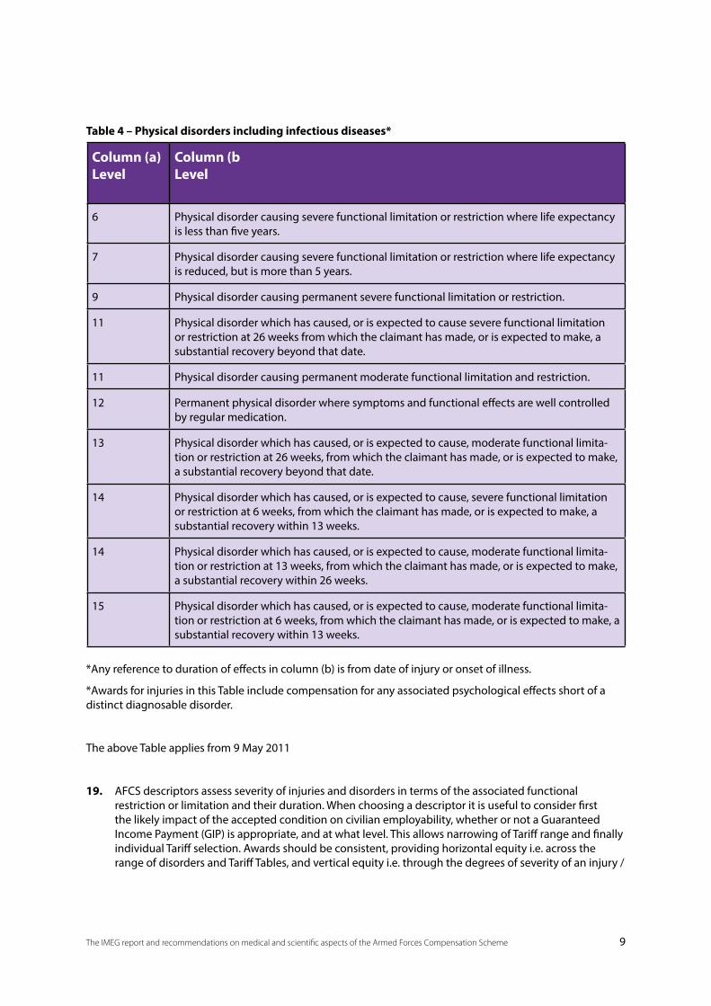
Column (a)Level
Column (bLevel
6 Physical disorder causing severe functional limitation or restriction where life expectancy is less than five years.
7 Physical disorder causing severe functional limitation or restriction where life expectancy is reduced, but is more than 5 years.
9 Physical disorder causing permanent severe functional limitation or restriction.
11 Physical disorder which has caused, or is expected to cause severe functional limitation or restriction at 26 weeks from which the claimant has made, or is expected to make, a substantial recovery beyond that date.
11 Physical disorder causing permanent moderate functional limitation and restriction.
12 Permanent physical disorder where symptoms and functional effects are well controlled by regular medication.
13 Physical disorder which has caused, or is expected to cause, moderate functional limita-tion or restriction at 26 weeks, from which the claimant has made, or is expected to make, a substantial recovery beyond that date.
14 Physical disorder which has caused, or is expected to cause, severe functional limitation or restriction at 6 weeks, from which the claimant has made, or is expected to make, a substantial recovery within 13 weeks.
14 Physical disorder which has caused, or is expected to cause, moderate functional limita-tion or restriction at 13 weeks, from which the claimant has made, or is expected to make, a substantial recovery within 26 weeks.
15 Physical disorder which has caused, or is expected to cause, moderate functional limita-tion or restriction at 6 weeks, from which the claimant has made, or is expected to make, a substantial recovery within 13 weeks.
*Any reference to duration of effects in column (b) is from date of injury or onset of illness.
*Awards for injuries in this Table include compensation for any associated psychological effects short of a distinct diagnosable disorder.
The above Table applies from 9 May 2011
19. AFCS descriptors assess severity of injuries and disorders in terms of the associated functional restriction or limitation and their duration. When choosing a descriptor it is useful to consider first the likely impact of the accepted condition on civilian employability, whether or not a Guaranteed Income Payment (GIP) is appropriate, and at what level. This allows narrowing of Tariff range and finally individual Tariff selection. Awards should be consistent, providing horizontal equity i.e. across the range of disorders and Tariff Tables, and vertical equity i.e. through the degrees of severity of an injury /
Table 4 – Physical disorders including infectious diseases*
10 The IMEG report and recommendations on medical and scientific aspects of the Armed Forces Compensation Scheme
disorder category in a single Table. To provide financial certainty for claimants when they leave service, the Scheme aims to make full and final awards as early as possible. Ideally, this is when the injury or disorder is in a steady state of maximum medical improvement, following an adequate course of best practice treatment. When the disorder is not in a steady state, an interim award may be paid for up to four years after initial notification. Functional limitation or restriction is considered permanent where an injury has reached a steady state of maximum medical improvement with no further improvement expected.
20. Tables 3 and 4 of the Tariff relate to Mental Disorders and Physical Disorders – including infectious diseases. The Tables do not list specific diagnoses but are generic. Table 3 has previously been reviewed by IMEG and Table 4 descriptors and Tariff Levels were informed by civil awards where currently (2013) a highly malignant life-limiting disease such as mesothelioma would attract a general damages award of about £100,000. This compares with AFCS Tariff Level 8 which is £60,000; Level 7 £90,000 and Level 6, £140,000. Items 1 and 2 of Table 4 apply to disorders with reduced life expectancy, which is not an issue with post–infective fatiguing illness. Where Table 4 Items 1 and 2 are paid, death and dependents’ benefits will also apply. For both Tables 3 and 4 the highest GIP band is Band B based on 75% service salary at service termination. Injuries attracting AFCS band A i.e. 100% salary base include full thickness burns affecting 70% or more body area; several categories of severe polytrauma and amputations; severe brain and spinal injuries and loss of senses. The descriptors aim to reflect injuries and disorders relevant to the military population and potentially attributable to AFCS service. The most severe and enduring mental health disorders in terms of very severe functional compromise and employability are the psychotic disorders which, in line with contemporary medical understanding are not on the balance of probabilities due to AFCS service.
21. As discussed, for post infective fatiguing illness of all types, including QFS, there remain uncertainties, which include best practice treatment and prognosis, but recent research suggest that prognosis is better than for CFS which does not follow an identified infection. It should be remembered that end points and outcomes used in the few published studies are variable, often expressed as self-reported symptoms, and not using an objective functional measure. The available evidence indicates that the majority of people with corroborated post-infectious fatigue states do recover in time, and without treatment. Treatments appropriate for CFS may also help to improve prognosis in post-infectious states.
Suggested descriptors for Q fever and QFS22. As listed above the Table 4 descriptors applicable from May 2011 do not sufficiently reflect the range of
QFS functional limitation or restriction and the following additions are suggested:
• Physical disorder causing permanent very severe functional limitation or restriction Level 6
• Physical disorder causing permanent severe functional limitation or restriction Level 8
The existing Item 5 Level 11 should remain
• Physical disorder causing permanent moderate functional limitation or restriction Level 11
In the footnote to Table 4 in respect of physical disorders
11The IMEG report and recommendations on medical and scientific aspects of the Armed Forces Compensation Scheme
“very severe” Permanent functional limitation or restriction is very severe when the claimant is unable to undertake work appropriate to experience, qualifications and skills, following best practice treatment and at best thereafter is able only to undertake work sporadically and in physically undemanding jobs.
“severe” Permanent functional limitation or restriction is severe where the claimant is unable to undertake work appropriate to experience, qualifications or skills at the time of onset of the disorder and over time able to work only in physically less demanding jobs.
“moderate” Permanent functional limitation or restriction is moderate where the claimant is unable to undertake work appropriate to qualifications skills and experience at the time of onset of the illness but in time able to work regularly in a less physically demanding job.
23. To maintain coherence the Table 4 descriptors and definitions have a similar format to those in Table 3, Mental disorders. Awards for Physical disorders include psychological symptoms but do not include primary cognitive, mood or behavioural symptoms and are generally paid lower awards than equivalent mental disorders. Factors taken into account in valuing awards for mental disorders include the associated vulnerability and compromised relationships with family, friends and at work. In AFCS terms the current epidemiological findings on the likely better prognosis where a fatiguing illness follows a confirmed infection are not robust enough to allow different descriptors for fatiguing states that post date a confirmed infection compared with spontaneous illness. In addition the descriptors proposed above for Q fever and its sequelae will also apply to other disorders, including infections and primary physical disorders and their sequelae.
References: (1) van Loenhout, J.A.F. et al Assessing the long-term health impact of Q-fever in the Netherlands: a prospective cohort study started in 2007 on the largest documented Q-fever outbreak to date. BMC Infectious Diseases 2012; 12: 280
(2) Morroy, G. et al The health status of Q-fever patients after long-term follow-up. BMC Infectious Diseases 2011; 11: 97
(3) Bailey, M.S. et al Undifferentiated febrile illnesses amongst British troops in Helmand, Afghanistan. JR Army Med Corps 2011; 57; 150-155
(4) Parke, N.R. et al Q fever. Lancet 2006; 367: 679-688
(5) Smith, D.L. et al A large Q fever outbreak in the West Midlands; clinical aspects. Resp Med 1986; 87: 509-516
(6) Ayres, J.G. et al Post infection fatigue syndrome following acute Q fever: follow-up study of patients involved in the 1989 outbreak in the West Midlands. QJ Med 1998; 91: 105-123
(7) Ayres, J.G. et al Long term follow-up of patients from the 1989 fever outbreak no evidence of excess cardiac disease in those with fatigue. Q J Med 2002; 95: 539-46
(8) Roest, H .I. J. et al The Q fever epidemic in the Netherlands: history onset, response and reflection. Epidemiol. Infec 2011; 139: 1-12
12 The IMEG report and recommendations on medical and scientific aspects of the Armed Forces Compensation Scheme
(9) Hickie, I. et al Post-infective and chronic fatigue syndromes precipitated by viral and non-viral pathogens: prospective cohort study. BMJ,doi:10.1136/bmj.38933.5875764.AE
(10) Fukuda, K. et al The chronic fatigue syndrome: a comprehensive approach to its definition and study Ann Int Med 1994; 121: 953-59
(11) Wilson, A. et al What is chronic fatigue syndrome? Heterogeneity within an international multicentre study. Aust NZ J Psych 2001; 35: 520-7
(12) Marmion, B.P. et al Long term persistence of Coxiella burnetii after acute primary Q fever. Q J Med 2005; 98: 7-20
(13) Pentilla, I.A. et al Cytokine dysregulation in the post-Q-fever fatigue syndrome. Q J Med 1998; 91: 549-560
(14) White, P.D. et al Comparison of adaptive pacing therapy, cognitive behaviour therapy, graded exercise therapy, and specialist medical care for chronic fatigue syndrome (PACE): a randomised trial. Lancet 2011; 377: 823-36.
(15) Larun, L. et al Exercise therapy for chronic fatigue syndrome. Cochrane Review F08, in press, 2015.
(16) Price, J.R. et al Cognitive behaviour therapy for chronic fatigue syndrome in adults. Cochrane Database of Systematic Reviews 2008, Issue 3. Art. No.: CD001027. DOI: 10.1002/14651858.CD001027.pub2.
(17) Cairns, R. et al A systematic review describing the prognosis of chronic fatigue syndrome. Occup Med 2005; 55: 20- 31
(18) Smith, W.R. et al Mortality in a cohort of chronically fatigued patients. Psychol Med 2006; 36: 1301-1306. DOI: 10.1017/S0033291706007975.
(19) Joyce, J. et al The prognosis of chronic fatigue and chronic fatigue syndrome: a systematic review. Q J Med 1997; 90: 223-233.
(20) Hamilton, W.T. et al The prognosis of different fatigue diagnostic labels: a longitudinal survey. Family Practice 2005; 22: 383-388.
(21) Petersen, I. et al Risk and predictors of fatigue after infectious mononucleosis in a large primary-care cohort. QJM 2006; 99: 49-55
(22) Hanevik, K. et al Irritable Bowel Syndrome and Chronic Fatigue 6 Years After Giardia Infection: A Controlled Prospective Cohort Study. Clin Infect Dis, 2014; 59: 1394-1400
(23) van Loenhout, J.A.F. et al Q-fever patients suffer from impaired health status long after the acute phase of the illness: Results from a 24-month cohort study 2014 J Infect http://dx.doi.org/10.1016/j.jinf.2014.10.010
13The IMEG report and recommendations on medical and scientific aspects of the Armed Forces Compensation Scheme
Topic 2 - Mesothelioma Report
1
From: Professor Sir Anthony Newman Taylor
19th December, 2014 Minister of State for Defence Personnel Welfare and Veterans MOD Whitehall London SW1A 2HB Dear Minister IMEG advice on Medical and Scientific aspects of mesothelioma I have pleasure in attaching the IMEG advice on mesothelioma which you requested in light of the Diffuse Mesothelioma Act 2014 and on which I updated you at CAC last week. You asked IMEG to consider (1) the medical and scientific aspects of mesothelioma: (2) whether mesothelioma can be considered unique and deserving of special compensation arrangements: (3) whether the War Pensions and Armed Forces Compensation Schemes are appropriate and adequate for mesothelioma diagnosed on or after 27 July 2012 and finally, (4) to comment on any other medical matters raised in tRBL letter to you. Asbestos is the cause of fibrosis of the lung (asbestosis), lung cancer and mesothelioma. Mesothelioma is a malignant tumour of the lining of the lungs or abdomen, with a median life expectation from diagnosis of between one to two years. To date there has been no effective treatment and none is emerging. The UK has the highest incidence of mesothelioma in the world, with almost all cases in men attributable to asbestos exposure some 30-50 years before the onset of
Chairman Independent Medical Expert Group 6.M.06 MOD Main Building Whitehall LONDON SW1A 2HB United Kingdom Telephone (MOD): +44 (0) 20 7218 9571 Facsimile(MOD): +44 (0) 20 7807 8068 Email: [email protected]
14 The IMEG report and recommendations on medical and scientific aspects of the Armed Forces Compensation Scheme
2
symptomatic disease. Asbestos was widely used as insulation material on ships and more recently in buildings. The use of asbestos in naval ships increased greatly before and during the World Wars and naval service, especially sea-going service and ship re-fitting between 1939 and 1973, was considered a high risk activity. The association between mesothelioma and asbestos exposure was first described in 1960 in those working and living in the vicinity of asbestos mines in South Africa. From 1963 the Royal Navy began to replace asbestos insulation materials with glass fibre and calcium silicate and introduced respiratory protection. A unit was set up to study the health of the workforce at Devonport dockyard, which included a study of the number of cases of mesothelioma over time. A study in 1980 showed an increasing number of cases of mesothelioma from 1962 to 1977. A follow-up study looked at cases between 1979 and 1999: deaths from mesothelioma further increased from 1979 with a peak in 1991; cases then began to decline and by 1999 had reduced by a third. The predicted continuing high rates of mesothelioma until 2050 predominantly relate to those working in the construction industry, where exposure to asbestos continued until the 1980s Mesothelioma is unique in 3 respects: the specific causal relation to asbestos; the long latent period between exposure and symptoms; and the low level of exposure to asbestos which can cause mesothelioma. The clinical course and prognosis are poor, but in this respect mesothelioma is not unique. Other disorders including asbestos related lung cancer, some other cancers and leukaemias which can be accepted under the War Pensions Scheme also have a similarly short life expectation. We have looked carefully at the adequacy and appropriateness of War Pensions Scheme and Armed Forces Compensation Scheme arrangements. The greatly improved control of asbestos exposure and the long latent interval of more than 20 years before the development of disease mean that mesothelioma should only very rarely, and only in the future, be an issue for AFCS covering disorders, caused on or after 6 April 2005. The War Pensions Scheme, dating back to 1917, recognises service personnel who make sacrifice and suffer personal injury by making awards to them and their dependents. This is primarily by payment of an income stream dependent on the level of disablement. We understand the income stream was introduced, at least in part, because of the preponderance of young men for whom the major consequence of injury was incapacity for paid work and inability to support their family. We recognise that special arrangements in terms of presumption of a service link were made where there is Royal Navy service of any duration on sea-going ships between 1939 and 1973 and, where entitlement is certified, disablement will be assessed at 100% from the outset. This approach is shared with the Industrial Injuries Disablement Benefit (IIDB) scheme. IIDB does not however include provision for dependents. As we have noted in the attached paper, dependents’ benefits can be very significant. The Legion draws attention to cases of mesothelioma who are without eligible dependents. We recognise this. In terms of any changes to the War Pensions Scheme the suggested numbers of eligible single veterans would be very small compared with overall numbers of pensioners in the scheme. This issue arises, not because of the unique features of
15The IMEG report and recommendations on medical and scientific aspects of the Armed Forces Compensation Scheme
3
mesothelioma, but because of its poor prognosis, which it shares with some other pensionable disorders. You asked us to comment on any other medical matter raised by the Legion. The Legion letter suggests that the Diffuse Mesothelioma Scheme should cover all asbestos-related disorders. This is not the case. In that regard, war pensioners may be considered relatively advantaged as war pension for disablement and death are likely also to be payable for asbestosis and lung cancer with or without asbestosis. The Legion included in their letter projected numbers of mesotheliomas in men who had served in the Royal Navy between now and 2047. The number, estimated by Professor Peto’s group at London School of Hygiene and Tropical Medicine, is about 2,500, with up to a third of these estimated by the Legion to be single. It is this group, which the Legion identified as at a disadvantage, with income stream only payable for a short period during life. Defence Statistics (DS) show that disablement and death payments over the last ten years have been fairly constant and significantly less than the Legion projections. This may, in part, reflect a failure to claim because of lack of awareness of the Scheme. I understand that as part of its campaign the Legion is raising awareness of the War Pension provisions for mesothelioma and other asbestos-related diseases. Yours sincerely
Professor Sir Anthony Newman Taylor, CBE, FRCP, FFOM, FMedSci Chairman Independent Medical Expert Group (IMEG)
16 The IMEG report and recommendations on medical and scientific aspects of the Armed Forces Compensation Scheme
1. Medical and scientific aspects of mesothelioma 1. Mesothelioma is a malignant tumour of the lining of the lungs (pleura) or abdomen (peritoneum). It
is a rapidly growing tumour with, in many cases, a length of life from the time of diagnosis of one to two years. To date no treatment has had an important impact on improving this. More than 90% of cases of mesothelioma in men in UK at present are attributable to the inhalation of asbestos fibres. The relationship of mesothelioma to asbestos is specific, so that the geographical distribution of mesothelioma can be used as a reliable indicator of industrial asbestos exposure 30 to 50 years earlier. The great majority of mesothelioma hotspots in UK have been major sites of shipbuilding and repair; these include Barrow, Glasgow, Belfast and Plymouth.
2. Asbestos is the name given to fibrous silicates of commercial value. There are 2 major types of asbestos: white, wavy, serpentine fibres – chrysotile; and long straight amphibole fibres – crocidolite (blue) and amosite (brown) asbestos. Asbestos is fire resistant and has been widely used as an insulation material including for boilers, pipework and bulkheads in Royal Navy ships before, during and after the Second World War until the early 1970’s.
3. The death rate from mesothelioma in Great Britain is the highest in the world with, at present, some 2,500 cases each year. The annual incidence is expected to continue to rise each year until about 2017. It is anticipated that, by 2050, 90,000 cases of mesothelioma will have occurred, 65,000 of whom will have been after 2001 (1). The continuing increase in incidence is occurring particularly in those who worked in the construction industry, primarily due to the large numbers exposed until the early 1980’s to amosite asbestos used in insulation board. Whereas the traditional sources of asbestos exposure, asbestos products manufacture, asbestos lagging and in shipyards, were subject to increasing regulation from the mid-1960’s, the degree of exposure, particularly to amosite in the construction industry, was not recognised until the early 1980’s. In a large case-control study of 622 mesothelioma patients and 1,420 population controls, published in 2009 (2), the 2 groups with the highest risks were identified:
1) Construction workers, which included carpenters, plumbers, electricians and painters. Of these, carpenters had the highest risk (OR = 36, a lifetime risk of 6%) probably attributable to sawing and drilling amosite asbestos insulation board (AIB) used as fireproofing under the building regulations of 1965.
2) Traditional high risk jobs, which included asbestos factory workers, laggers, shipbuilding, ship breaking and dockyard workers, naval personnel and others working on board ships. Of the 102 cases in this group, 46 had worked in docks, shipyards, and on non-naval ships. 26 had been in the Royal Navy.
4. In recent years, as a consequence of the increasingly effective controls in the jobs traditionally associated with asbestos exposure from the mid-1960’s to early 1970’s, an increasing proportion of cases of mesothelioma in UK have worked in the construction industry (3).
5. The association of mesothelioma with exposure to asbestos was first described by Wagner in 1960 (4). He reported 33 cases of mesothelioma, all but one of whom had worked or lived in the vicinity of the crocidolite asbestos mines in North Western Cape Province, South Africa. The interval from initial exposure to the development of disease was between 18 and 44 years and, of particular importance, in the majority of cases, (18 of 33), exposure to asbestos was environmental (neighbourhood), not occupational, indicating the level of exposure to asbestos necessary to cause the disease need not be high. This observation was reinforced by the findings of a case-control study by Newhouse et al (5) of 83 cases of mesothelioma diagnosed at the London Hospital. A clear excess of cases had worked in,
17The IMEG report and recommendations on medical and scientific aspects of the Armed Forces Compensation Scheme
or lived with someone working in, an asbestos factory, but in those without occupational or domestic exposure more than twice as many cases as controls (11 v 5) lived within ½ mile of the Cape Asbestos factory in Barking.
6. Mesothelioma is one of several diseases caused by asbestos, of which pulmonary fibrosis (asbestosis), lung cancer and mesothelioma are clinically the most important. Asbestosis and lung cancer can be caused by chrysotile, crocidolite and amosite asbestos. The risk of mesothelioma is highest in those exposed to amphibole asbestos, crocidolite and amosite, although it also occurs in those exposed to industrial chrysotile.
7. The major source of exposure to asbestos in those employed by the Ministry of Defence has occurred in men, employed in dockyards in Barrow, Glasgow, Belfast, Rosyth and in the Royal Naval dockyards in Devonport. The volume of asbestos used in naval ships increased substantially before and during the world wars. From 1944-63 there was extensive use of crocidolite for insulation and fire protection and from 1950-1963 amosite was used for machinery insulation.
8. The workforce of the Royal Naval dockyard in Devonport has been the subject of particular study since the early 1960’s. Some 19,000 civilians were employed in Devonport Naval dockyards at the end of the Second World War but by the mid-1960’s that number had fallen to about 15,000. The majority of the workforce were civilian employees of MoD, with a minority of members of the Royal Navy. Royal Naval personnel will have included marine engineers, shipwrights and artificers working on board ship, the ship’s company living on board during refits and Royal Naval personnel working ashore in the dockyard, where exposure was generally lower than on board ship. The work in the Devonport dockyard was predominantly refitting, which involved the removal and stripping of asbestos lagging, often in cramped and poorly ventilated areas, exposing asbestos laggers, strippers, as well as any others working or passing through the vicinity, to finely divided, respirable, airborne asbestos fibres. Trades not directly involved with asbestos, such as electricians, painters, welders and burners were therefore also exposed to airborne asbestos during refits. Labourers were exposed intermittently through stripping lagging, sweeping asbestos and bagging it, when they would have been exposed to high airborne concentrations. Sheers (1960) (6) estimated that of the 15,000 men employed in the dockyard in the mid-1960’s, fewer than 3% were continuously involved in the handling of asbestos products, but about 50% of the rest of the workforce (some 7,000 men) had been exposed intermittently to widely varying concentrations of asbestos dust. The maximum exposures to asbestos among the dockyard workforce are considered to have occurred between 1950 and 1963. The mean interval from initial exposure to asbestos to diagnosis of mesothelioma reported in the Devonport population was 37.6 years (7).
9. From 1963, glass fibre and calcium silicate began to replace asbestos as insulation material, but asbestos continued to be removed in refit work during the 1970’s and 1980’s. Respiratory protection for more heavily exposed workers was introduced in the 1960’s, with protective measures subsequently extended to include all potentially exposed workers.
10. In 1980 Sheers described 100 cases of mesothelioma (8), which had occurred up to 1979, in employees working in the dockyard or with service in naval vessels. He found the annual number of cases increased steadily, from 2 cases in 1962 to 12 in 1977. Deaths occurred in jobs with continuous or intermittent exposure, below deck or in dockside workshops, but also in those working in any occupation within the dockyard wall. Deaths in the surrounding city of Plymouth were not increased. In a follow-up study, Hilliard, Lovett and McGavin (9) reported 301 cases in Devonport dockyard workers between 1979 and 1999. They reported an increase in mesothelioma deaths from 1964 with a peak in 1991. The number of cases then began to decline, with a reduction of one third by 1999. The authors attributed this to a) the reduction in the number of dockyard workers during the previous 50 years, although the reduction in the annual case number was too large to be explained solely by
18 The IMEG report and recommendations on medical and scientific aspects of the Armed Forces Compensation Scheme
the smaller population at risk. Other factors they cited included b) substitution of asbestos by other insulation materials in the mid-1960’s and c) improved hygiene measures in the dockyards, from the mid-1960’s. This study was the first to record a decline in mesothelioma deaths in any UK work force, military or civilian. Although a single study, it is consistent with a similar reducing incidence of mesothelioma reported in the USA (10), where asbestos exposure controls were put in place at about the same time as in the Naval dockyards.
11. The greatest risk of mesothelioma amongst ex-service personnel has occurred in men who served in the Royal Navy. These have included those employed in naval dockyards, those on board ship during refits, marine engineers and engineering mechanics, whose role might include plumbing and joinery duties while at sea when they might need to remove and replace asbestos insulation to gain access to boilers or pipework. Some cases of mesothelioma have also occurred in the Royal Air Force and Army. The circumstances of exposure here relate to work on aircraft maintenance as well as contact in accommodation. For the Army, war pension claims have been received from people working in transport and in accommodation or office buildings. Of 100 war pension disablement cases recently reviewed, 86 were from the Royal Navy, 6 from the Army and 8 from the Royal Air Force. The median age group at diagnosis in all the three services was 75 – 80 years closely followed by 70 - 75 years. Duration of service was varied with some long service personnel (twenty two years plus) and others who had only done two years National Service. Most commonly, service included the decade 1955-65 i.e. 50 to 60 years ago. The principle service occupations in the majority of the Royal Naval personnel were work in boiler rooms and in engineering trades. None had job descriptions or described circumstances of exposure associated with the construction trades.
12. More recently, and for the future, deployed service, especially in developing countries, whether conflict or disaster relief, remains a potential source of asbestos exposure. This is relevant to all three services particularly the Army and special forces. Soldiers may well work amongst demolished buildings containing asbestos, an identified hazard in the World Trade Centre 9/11 destruction (11).
References: (1) Peto, J. et al Continuing increases in mesothelioma mortality in Britain. Lancet 1995; 345: 535-539
(2) Rake, C. et al Occupational domestic and environmental mesothelioma risks in the British population: a case control study. Brit J Cancer 2009; 100: 1175-1183
(3) Mc Elvenny, D.M. et al Mesothelioma mortality in GB from 1968-2001. Occ Med 2005; 55: 70-87
(4) Wagner, J.C. et al Diffuse pleural mesothelioma and asbestos exposure in the North West Cape Province. Br J Ind Med 1960; 17: 26-271
(5) Newhouse, M.L. et al Mesothelioma of pleura and peritoneum following exposure to asbestos in the London area. Br J Ind Med 1965; 22: 261-269
(6) Sheers, G. Asbestos associated disease in employees of Devonport dockyard. New York Academy of Science 1979; 330: 281-287
(7) Lumley, K.P.S. A proportional study of cancer registrations of dockyard workers. Br J Indust Med 1976; 33: 108-114.
(8) Sheers, G. et al Mesothelioma risks in a naval dockyard. Arch Env Health 1980; 35: 276-282
(9) Hilliard, A.K. et al The rise and fall in incidence of malignant mesothelioma from a British Naval Dockyard, 1979-99. Occ Med 2003; 53: 209-212
(10) Weill, H. et al Changing Trends in US mesothelioma incidence. Occup Env Med 2004; 61: 438-441
(11) Landrigan, P.J. et al Health and environmental consequences of the World Trade Center disaster. Environ Health Perspect 2004; 112(6): 731-739
19The IMEG report and recommendations on medical and scientific aspects of the Armed Forces Compensation Scheme
2. Can mesothelioma be considered unique and deserving of special arrangements? 1. Mesothelioma is unique in relation to other asbestos-related diseases, asbestosis and lung cancer,
in 3 particular ways: 1) the specificity of its relationship to asbestos. This is unlike lung cancer, also caused by asbestos, whose overwhelming cause in the population is cigarette smoking, which with asbestos interacts to increase the risk of lung cancer in asbestos exposed individuals. 2) the long latent interval, in some cases more than 50 years, which can occur between initial exposure and the onset of symptoms and diagnosis. It is primarily for this reason that the companies and their insurers for whom mesothelioma patients have worked may no longer be in business. 3) in the low levels of exposure necessary to cause the disease; unlike asbestosis and attributable lung cancer, where the diseases are primarily recognised in those with occupational exposures, those living with asbestos workers and those living in the neighbourhood of an asbestos factory are also at risk of developing mesothelioma. In addition the prognosis of mesothelioma is very poor, with a median life expectation of less than one year. However, sadly, while poor, this is not unique: the prognosis for lung cancer, particularly for those with co-existing asbestosis is similar. For this reason, similar arrangements for expediting decision making and access to benefits for lung cancer attributable to asbestos have been put in place in Industrial Injuries Disablement Benefit. The War Pensions Scheme is an individual jurisdiction which can make awards for any disorder: these include some other solid cancers and leukaemias with a similarly poor prognosis.
3. Are the War Pension and Armed Forces Compensation Schemes appropriate and adequate for mesothelioma diagnosed on or after 27 July 2012? 1. Mesothelioma diagnosed at any time from 27 July 2012 in ex-military personnel will be due to asbestos
exposure no later than the mid-1970s, at a time when Crown immunity applied and compensation is through the War Pensions Scheme. In considering the adequacy of the War Pensions and Armed Forces Compensation Scheme we looked at the intention of the Scheme; how it applies to mesothelioma and, the likely financial outcomes.
2. The War Pensions Scheme recognises the sacrifice of military personnel who suffer personal injury due to military service by making awards to them and their dependents. Originally restricted to war injury, the scheme was extended after the Second World War to include any adverse health effect due to peace-time service. The scope of the Scheme is wide and anyone who has served can claim any “disablement” causally linked to service at any time from service termination. The standard of proof is not on the balance of probabilities but “reasonable doubt”. Where an injury or disorder has onset in service there is no onus on the claimant and entitlement will be given, unless there is evidence, “beyond reasonable doubt”, that service has played no part in cause or course. For claims made seven or more years from service termination the onus is on the claimant to raise a reasonable doubt by reliable evidence of a causal link to service.
3. “Disablement” is defined in the legislation as “physical or mental injury or damage or the loss of physical or mental capacity”. In terms of WHO ICIDH terminology, “disablement” best equates to “impairment”, a relatively objective concept implying a measurable loss of function and suited to a basic award covering a range of injuries and disorders. The Service Pensions Order specifies “disablement due to an injury” or “death due to or hastened by an injury”, where “injury” includes wound or disease. To decide entitlement, medical advisers first determine the “injury” or disorder underlying the claimed disablement and then, based on the case facts and applicable law, decide its causal link to service.
20 The IMEG report and recommendations on medical and scientific aspects of the Armed Forces Compensation Scheme
4. Benefit paid depends on the medically assessed degree of disablement. This is obtained by comparing the condition of the disabled person “with a normal person of the same age and sex” without taking into account earning capacity or the effect of any individual factors or extraneous circumstances. Disablement level is expressed as a percentage, where 100% attracts maximum award. Awards for less than 20% are paid as lump sum gratuities and an income stream is paid for centile assessments between 20% and 100%. The legislation includes a Table of Statutory Scheduled Assessments; these are important for their own sake and also act as signposts for all other assessments in the Scheme. The disabling effects of injury are addressed by supplementary allowances, covering mobility, care and employability. Paid instead of the civilian social security equivalents they attract slightly higher benefit rates.
5. It is long established war pensions policy for mesothelioma that where ex-service personnel have sea-going service of any duration between 1939 and 1973, it is presumed that they were exposed to asbestos. Mesothelioma accepted as attributable to service is assessed at 100% basic pension from the date of claim with supplementary benefits as appropriate. War Pension Widow(er) benefits also apply.
6. The reason that assessments of 20% or more attract an income stream, as opposed to a lump sum, is not clear. Financial considerations, given the numbers of casualties in the Great War are likely to be relevant; also, the great majority of casualties were young men, for whom the major consequence of injury or disability was incapacity for paid work with a reduced ability to support their family. War pension above all provided a secure regular income. Commutation of disablement war pension, but not dependents’ awards, was permitted by law: the Secretary of State was empowered to allow a war pensioner to commute his final assessment war pension by the payment of a lump sum, calculated relative to age at diagnosis and life expectancy, in accordance with the tables set out in the legislation. After the Great War commutation was sometimes allowed for house repairs or setting up a small business. This was considered to provide a more flexible working pattern for the disabled person than the terms and conditions of being an employed earner. However a survey in 1935 showed that, despite careful consideration, some pensioners sustained serious loss in business ventures; at the start of the Second Word War the then minister suspended commutation, since when the practice has not been restored.
7. Annual disablement and supplementary allowance awards for mesothelioma in life can be up to £32,000 tax free and taking an average life expectancy of 18 months, the income stream paid will be about £50,000. Widow(er)‘s pension is also payable. Office of National Statistics (ONS) data shows that on average men of the relevant generation are three years older than their wives at marriage. Based on average longevity, a widow aged 52 years pension may be in payment for over 30 years while for one aged 72 years at her husband’s death an award is likely to run for 13 years.
Based on 2014 war pensions rates and assuming service ended before 31 March 1973, the maximum amount of War Pension a Widow aged 52 would receive up until the age of 86 is Total £427,748.78 This is broken down as:
52-65 (13 Years @ 11,630.40) £151,195.88
65 (5 years @ 12,322.98) £61,614.90
70 + (10 Years @ £13,156.60) £131,566.60
80+ (6 years @ £13,895.40 in higher rate supplementary pension) £83,372.40
21The IMEG report and recommendations on medical and scientific aspects of the Armed Forces Compensation Scheme
A widow aged 72 will receive a Total of £188,625.20 this is broken down as:
Aged 72–80 (8 years @ £13,156.60) £105,252.80
Ages 80 (6 years @ £13,895.40) £83,372.40
These do not take account of annual war pensions up-rating, which in recent years has used CPI.
These compare with payments under the Diffuse Mesothelioma Payment Scheme (2014):
diagnosed at age 55 years £172,722 is payable
diagnosed at age 75 years £113,482 is payable
8. Were commutation of the service pension to be considered as an option for cases of mesothelioma attributable to service the lump sum generated would be:
About £35,000 for an estimated life expectation of just over 1 year from diagnosis.
About £60,000 for an estimated life expectation of 2 years from diagnosis.
In addition, dependent benefits would remain payable.
9. In their submission to minister, the Legion focuses on mesothelioma sufferers who are single, widowed or divorced and who will receive only the income stream paid during their life-time. The Mesothelioma 2014 scheme either makes awards to sufferers in life or to spouses or dependents. In contrast to the 2008 Diffuse Mesothelioma Scheme and the Pneumoconiosis Act 1990, the amount paid to the sufferer or dependent under the 2014 Act is the same. While numbers of new cases of mesothelioma will diminish over time, they are projected to continue to occur until about 2050. Similarly changing social mores and increasing longevity are likely to mean increased numbers of single men in this century. The Legion’s snapshot figure of 31% males over aged 65 single, widowed or divorced may not be applicable over the whole future period. Relevant Influences are complex and act in different directions with cohort effects. The ONS statistical bulletin “Marital status population projections – 2008 based” looks at actual data for 2008 England and Wales on marital status and opposite sex cohabitation and makes projections to 2033. In the period 2008-2033 the proportion of those males aged 65 plus “never married” is predicted to increase from 7% in 2008 to 13% in 2033. This contrasts with 71% “married” in 2008 and predicted 62% in 2033. Professor Peto has projected that new Royal Navy mesothelioma case numbers will reduce beyond 2013 with a total of about 2,500 by 2050. Based on 30%, single pensioners without eligible dependents, the total cases in this group might be about 800. Currently 160,000 war disablement pensions are in payment, a number which will itself decline over the period.
10. The War Pensions Scheme applies to personal injury caused by service up to 6 April 2005 and the introduction of the Armed Forces Compensation Scheme, for injury and disorder due to service on or after that date. The Armed Forces Compensation Scheme can be claimed in service and awards are made where, on balance of probabilities, the claimed injury or disorder is causally linked to service. The Scheme is Tariff Based with a lump sum paid at 15 levels for pain and suffering; for the more serious injuries, affecting function, particularly civilian employability, there is an additional Guaranteed Income Payment. Armed Forces Compensation Scheme award recipients can sue in tort with adjustment in monies paid to avoid double compensation. There are time limits to claim but the legislation also includes a provision for late onset long latency disorders. Military exposure to asbestos at dates covered by the new Scheme should not occur other than very exceptionally overseas in combat or humanitarian missions or by accident. If a claim for mesothelioma was accepted as caused by service,
22 The IMEG report and recommendations on medical and scientific aspects of the Armed Forces Compensation Scheme
the likely outcome under the Armed Forces Compensation Scheme would be award of a lump sum of £140,000 (Level 6) and a guaranteed income payment based on 75% of service salary at service termination paid from date of claim for life. As in the War Pensions Scheme, dependents’ benefits apply.
11. In medical terms military occupational exposure to asbestos on or after 6 April 2005 should rarely be an issue although the AFCS structure with its lump sum, income stream and dependents’ benefits is well suited to mesothelioma claims. The War Pensions Scheme originally applied only to war injury with focus on young working age men whose civilian employability was compromised by their accepted injury. The regular income stream structure addresses this. Because of the very poor prognosis for the majority of cases of mesothelioma, that structure means that only limited benefit is paid in life to the sufferer. However unlike Industrial Injuries Disablement Benefit, the War Pensions Scheme maintains support to eligible dependents after the pensioner’s death, by payment of tax free dependents’ benefits. In summary, while the War Pensions Scheme provides benefit to those who develop mesothelioma for only a short period, the awards to eligible dependents mean that the Scheme overall meets the stated purpose of providing recompense for sacrifice to claimants and their eligible dependents.
4. Other medical matters raised in the tRBL letter to Min (DPWV) of 18 December 2013 1. The letter to Minister implies that the Legion would want all asbestos related disorders to be covered
by the new Scheme. The 2014 Act is restricted to mesothelioma diagnosed on or after 27 July 2012. Ex-service personnel with other asbestos related disorders including asbestosis, lung cancer with or without asbestosis etc are likely to be eligible for War Pension, with both disablement and death benefits.
2. The Legion letter to the Minister indicates that Professor Julian Peto, London School of Hygiene and Tropical Medicine, has estimated that about 2,500 Royal Navy veterans will die from mesothelioma between 2013 and 2047. This estimate was based on his 2009 case control study of occupational risks in the British population, where lifetime occupational histories were provided at telephone interview with over 500 male mesothelioma patients (2). In successive five year periods from 2013-2017 to 2043-47 the number of Royal Naval mesothelioma cases is estimated to reduce from:
986 in 2013-17
656 in 2018-22
486 in 2023-27
269 in 2028-22
151 in 2033-37
42 in 2038-42 and
16 cases in 2043-47
This rate of decline is steeper than that projected for the general population. This probably reflects the controls put in place in the 1960’s in the Royal Naval dockyards to prevent exposure to asbestos, 10 years or more earlier than in the construction industry. From 2038 all projected deaths are in those
23The IMEG report and recommendations on medical and scientific aspects of the Armed Forces Compensation Scheme
aged 75 years or more, while for the whole period between 2013 and 2047 two thirds of the deaths occur in those aged 80 years or more.
3. Predicting the future burden of disease is necessarily subject to uncertainty. The magnitude and timing of the current mesothelioma epidemic in the UK were predicted by Professor Peto in his 1995 Lancet paper. The increase in UK mesothelioma cases, projected to peak in 2013-2017, is being driven by the continuing exposure to asbestos, particularly amosite, in the construction industry, until the 1980’s. Although the 2003 investigation of naval dockyard workers, showing a peak in the number of mesothelioma cases in 1991, is a single small study, the reduction in the number of cases of mesothelioma after 1991 is consistent with the measures taken to control asbestos exposures in the Royal Naval dockyards in the 1960’s, through the replacement of asbestos by non-asbestos substitutes and the increasingly widespread use of respiratory protection. These observations suggest that the risk to Royal Naval personnel reduced some 10 years earlier than for those working in the construction industry. Unfortunately the case records of Royal Naval personnel with mesothelioma do not provide sufficient information to know about their particular occupations and how many had subsequently worked in construction trades; a robust estimate of the future burden of mesothelioma is therefore difficult.
4. At present there are about 70 war disablement pensions being paid in life to veterans from the three services. Compared with the projections this number is small. Defence Statistics (DS) data show that over the period 2004-2014, the numbers of cases of mesothelioma receiving War Pension has ranged from 53 in 2004 to 73 in 2014. This is about a third of cases projected over the same period. Similarly the number of new awards each year has fluctuated between 18 in 2007 to a high of 40 in 2011. The average age of war pensioners with mesothelioma over the period has remained stable at 75 years. There are limits to these data including human error e.g. incorrect diagnosis entered. The cases were identified by Defence Statistics using the medical diagnosis code field and the free text field so that some records could have been missed. It is also true that a deceased person may still be entered on the data base while the estate is being administered. Data on numbers of war widow(er)s’ benefits paid where the deceased has died from mesothelioma are not available; information is not collected on the basis of cause of death of spouse.
5. There are several possible explanations for the small DS numbers as compared with Professor Peto’s projections. These include failure to claim because of lack of awareness of the Scheme or, in the individual case, of a possible service source of asbestos exposure. It may be that sufferers and their families have other priorities, given the inexorable rapid decline in the sufferer’s health. In line with its campaign, the Legion has issued an alert to their officers, members and volunteers highlighting the War Pensions provisions for asbestos related diseases. We are aware that a similar publicity campaign in the context of civilian work led to an increased number of successful claims for Industrial Injuries Disablement benefit.
24 The IMEG report and recommendations on medical and scientific aspects of the Armed Forces Compensation Scheme
Topic 3 - Compensation aspects of Non freezing cold injury (NFCI)History and Background1. Non-freezing cold injury (NFCI) has been recognised in the military context from Roman times. While
the conditions in the 1982 Falklands conflict predictably led to cases in Royal Marines, in recent memory NFCI has not been a major disabling disorder amongst serving UK troops nor in UK civilian practice. However from the winter of 2005/6 onwards more cases began to appear with consequences for operational capability, healthcare and compensation. Typically NFCI was occurring in army recruits undertaking winter training in UK in the Brecon Beacons, North York Moors and Northumbria. Despite refresher training and new instructions on prevention and management for soldiers and the chain of command, and a new Surgeon General Policy Letter on clinical care, cases continued to present during subsequent winters. During the Falklands conflict, Royal Marine personnel were almost all UK-born Caucasian males while those affected more recently are typically foreign and Commonwealth troops, British born Afro-Caribbeans or Caucasians born and raised in Africa. In January 2012 the Surgeon General set up an independent expert Review Group chaired by Prof Hugh Montgomery, University College London to examine all aspects of NFCI. The report was completed in February 2013 (1).
2. NFCI was rarely claimed under the War Pension Scheme nor was it a common reason for medical discharge even in Royal Marines who fought in the Falklands. Of the 2,000 – 3,000 men exposed to the cold conditions of that campaign only about 12 left service in the following two years because of NFCI.
3. From 2006 however, the picture in the UK military, notably army personnel, is different. The 2013 Report on the Health of the Armed Forces records that in 2012/2013 there were 604 referrals to the Cold Injury Clinic, Institute of Naval Medicine, the great majority being army (2). Defence Statistics confirm that the number of UK personnel medically discharged due to NFCI as the principal or contributory cause from 6 April 2005 until 31 March 2014 is 518. Of these about 330 have AFCS awards for NFCI at some level of severity. In addition to no fault compensation, 707 claims for civil damages have been made between May 2007 and 31 December 2014. 470 have been settled with 197 remaining active. The cost so far is £17.2 million damages and £11.3 million claimant legal costs.
4. If medical discharge occurs due to NFCI, armed forces ill-health pension award will usually be appropriate. Most personnel affected are members of the Armed Forces Pension Scheme 2005 (AFPS 05) scheme where benefits are awarded as a lump sum if the invaliding disorder does not compromise civilian employability. For more serious injuries and disorders, a pension is paid at two levels (Tiers) dependent on the degree of functional compromise for suitable civilian work.
5. The AFCS no fault compensation figures above are a minimum because claims may be accepted up to seven years from “the day on which the injury occurs”. In addition the early editions of the Tariff included no NFCI descriptors. Claims in the early period were few and Tariff Table 4 Physical disorders descriptors were used. Cold injury descriptors (two) were introduced into Table 2 in September 2008:
Item 66 Level 15 - Cold injury which has caused, or is expected to cause, symptoms and significant functional limitation and restriction at 6 weeks, with substantial recovery beyond that date.
Item 62 Level 14 - Cold injury with persisting symptoms and significant functional limitation and restriction.
25The IMEG report and recommendations on medical and scientific aspects of the Armed Forces Compensation Scheme
and the three current descriptors for NFCI at levels 14, 13 and 10 followed IMEG’s consideration and recommendations in the first report, January 2011:
Item 65 Level 14 - Non freezing cold injury which has caused, or is expected to cause, neuropathic pain and significant functional limitation or restriction at 6 weeks with substantial recovery beyond that date.
Item 55 Level 13 - Non freezing cold injury which has caused or is expected to cause neuropathic pain and significant functional limitation or restriction at 26 weeks, with substantial recovery beyond that date.
Item 27 Level 10 - Non-freezing cold injury with persistent local neuropathic pain and severe compromise of mobility or dexterity, and evidence of permanent damage to small nerves on thermal threshold testing.
Footnote for all three is
A descriptor for non-freezing cold injury refers to either unilateral or bilateral damage to the upper or lower extremities.
6. The AFCS aims to make consistent and equitable awards for listed injuries and disorders. The likely prognosis and impact of the treated condition on functional capacity for civilian employment over the person’s working lifetime is then used to assess injury severity and award level. Injuries and disorders should be diagnosed when they meet defined generally accepted medical criteria e.g. according to the World Health Organisation (WHO) International Classification of Diseases (ICD) and the intention is to make awards full and final following best practice treatment at optimum functional state.
7. The findings of the Montgomery report call into question whether these criteria are met by the current NFCI descriptors. The report found a lack of reliable scientific evidence in the international literature on many aspects of NFCI, including its definition, a systematic description of its clinical features, its natural history, the role of specialist tests in diagnosis and assessment of severity, its prognosis and best practice treatment. The pathogenesis also remains incompletely understood. There is experimental evidence that impaired blood flow due to vasoconstriction leads to ischaemic neural damage. In turn the neural and vascular elements interact causing further ischaemic damage. There is also good experimental evidence that cold directly damages nerve fibres, independent of vascular factors (1).
8. The natural history of NFCI requires further research, but from clinical experience it is possible to state that in the majority of those who develop NFCI, the symptoms and signs resolve completely over weeks or a few months, while pain persists in a minority. Available evidence strongly suggests that this pain is due to damage to small diameter sensory nerve fibres supplying the skin and deeper tissues in the affected extremities (small fibre peripheral neuropathy, SFN paragraph14-17 inclusive). This neuropathic pain, NP, may be continuous and is frequently exacerbated by mechanical and thermal stimulation of the affected extremities (paragraph18-19 inclusive). In addition, sensitivity to cold, due to abnormal vascular reactivity, is a common consequence of an acute episode of NFCI, and in many subjects, this persists and can be easily demonstrated using thermography. However, the symptoms arising as a result of such cold sensitivity only develop when the affected extremities are exposed to cold, they are usually mild, and they resolve rapidly on re-warming. Abnormal thermography, on the other hand may continue long after symptoms have resolved completely (1).
9. A major recommendation of the Montgomery report which IMEG strongly endorses, is for further research on several other aspects of NFCI. To better understand the natural history of NFCI, a systematic prospective longitudinal study starting at the time of recruitment to the services is needed.
26 The IMEG report and recommendations on medical and scientific aspects of the Armed Forces Compensation Scheme
The Montgomery report also recommended studies on how NFCI is investigated, diagnosed and treated. Such research will take time and for the foreseeable future military no fault compensation claims for NFCI will be informed by clinical information similar to that available at present. Detailed physical examination findings, general and neurological, are not routinely and systematically recorded in Service primary care medical records, nor at the specialist Cold Injury Clinic, Institute of Naval Medicine (CIC, INM).
10. Based on the limited published peer-reviewed evidence base, IMEG has reviewed and made recommendations on the NFCI descriptors and tariff levels. Another source of information has been discussion of clinical matters with Dr Howard Oakley, recently Head of Survival and Thermal Medicine, INM. Dr Oakley ran the CIC there for many years, assessing up to 1,000 patients with suspected NFCI each year. He has given IMEG access to a number of unpublished papers, including his own 2014 audit of clinical features and observations on patients with NFCI seen at INM during the preceding sixteen months (3). IMEG also had the benefit of a paper from Major-General RP Craig, L/RAMC prepared as an introduction for the Royal British Legion (RBL)’s Medical Advisory Committee (4).
11. Of the total 644 new cases recorded in Dr Oakley’s 2014 analysis, there were 271 where injury was sustained before 1 September 2012. The average time interval between injury and the date seen in the CIC was 7.8 months (SD 2.8), with a range of 2 to 15 months. Medical diagnoses other than cold injury were recorded in 89 cases i.e. 14% of the total, with the majority having primary Raynaud’s disorder. Of the 284 winter 2012/13 NFCI cases, most were army junior ranks with an average age of less than 30 years; the majority of injuries occurred in the UK. Half had NFCI of hands and feet; in 36% feet only were affected and in 6% hands only. Mixed freezing and non-freezing cold injuries occurred but were rare.
12. The most frequent and troublesome presenting symptom was pain, more or less continuous and lasting more than two hours on re-warming after cold exposure. The proportion of patients experiencing pain diminished exponentially with time from cold exposure, but 23, (8%) were still experiencing pain 32 weeks after the injury and two patients reported having pain 4 years after the initial cold exposure. In addition to pain, numbness was reported in 25% and cold sensitivity i.e. feeling pain on cold exposure by 68%. Other non-neurological signs such as change in skin colour, blistering and skin peeling were uncommon. At INM, diagnosis of NFCI depends on the given history and special investigations, including infrared thermography (IRT) and thermal threshold testing (TTT). However, while abnormal results may support the clinical diagnosis of NFCI, neither of these tests can be regarded as diagnostic. In normal subjects there is a wide reference range of responses to a cold challenge. Abnormal results may be demonstrable in the absence of symptoms and in the case of thermography, even without a history of cold exposure. In the 2014 INM series IRT was near-normal in 55% of those tested and showed mild to moderate cold sensitivity in 39%. Cold sensitivity was moderate in 22% and worse than that in 12%. TTT was more likely to be normal in the hands (67%) while 59% had abnormal thresholds in the feet.
13. Cases at INM are seen on referral from primary care ideally at three to four months after cold exposure. The recent increase in case numbers, without a commensurate increase in clinical resources, has resulted in many cases being seen only at around nine months or longer, after cold exposure, as noted above (para 11). Where the time interval coincides with the warmer temperatures of spring and early summer there may be natural improvement in symptoms. In terms of prognosis, in Dr Oakley’s experience about a third of new cases overall can be recommended for immediate return to full duties; about 55% are advised to keep extremities warm and dry and use foot spa treatment. Normal practice is then for their review at about six months, when most are found to be fit to return to duty in some degree. Neuropathic pain management is needed from the initial visit in about 10% cases and about 4% at the first visit are thought to be so severely affected by pain as to be likely to require medical discharge.
27The IMEG report and recommendations on medical and scientific aspects of the Armed Forces Compensation Scheme
Small Fibre Peripheral Neuropathy and Neuropathic Pain14. Small Fibre Peripheral Neuropathy (SFN). A small fibre neuropathy results from structural damage
selectively affecting small diameter unmyelinated and myelinated fibres. The unmyelinated fibres include both sensory fibres, carrying sensations of heat, mechanical or chemical stimuli, and sometimes all three, and autonomic motor fibres controlling blood vessels and sweat glands. SFN may complicate systemic diseases such as diabetes mellitus, sarcoidosis, vasculitis, rheumatoid arthritis and other auto-immune conditions as well as infections including HIV and Hepatitis C; it may also occur in alcohol misuse and be associated with certain therapeutic drugs and rare genetic pain syndromes (5). Most SFNs in these conditions are length-related, affecting the feet initially and then the hands, progressing proximally in a glove and stocking distribution.
15. The most severe symptom of SFN is neuropathic pain (NP), often continuous and described as burning, gnawing, stinging or raw, with paroxysmal lightning, stabbing or electric shock-like pains superimposed. Normally harmless stimuli such as light touch or gentle stroking, wearing socks or the bed-sheets can also evoke pain, a symptom and sign known as allodynia (5). In its most extreme form, seen most commonly in some alcoholic and diabetic patients, neuropathic pain may severely compromise function, preventing weight-bearing, walking and use of the hands. On neurological examination, signs of SFN are often difficult to detect. However, with careful sensory testing, impairment of temperature and pin prick (pain) sensation may be detected, while light touch, vibratory and joint position sensations, mediated by larger myelinated nerve fibres, are normal.
16. Diagnostic tests for SFN in routine clinical practice are limited. Standard nerve conduction tests (NCS) measure conduction in large myelinated fibres and so are normal. Thermal Threshold Testing (TTT)yields data on perception of warm and cold, but is a psychophysical test that relies on consistent and accurate subjective report, and is open to error. Within the last decade, skin biopsy and measurement of the reduced density of free nerve endings in the epidermis, using standard histochemical techniques has emerged as the first validated measure of damage to small nerve fibres, and there are now agreed diagnostic criteria. Skin punch biopsy of the lower limb is suitable for most patients and has a low complication rate. International normative standards of nerve fibre density in skin epidermis are now available and the diagnosis of SFN is made when the patient’s value lies below the fifth centile for age and sex matched controls (6). The test correlates well with clinical symptoms and signs and has high sensitivity and specificity (7). Quality diagnostic services with samples sent by courier are now becoming more widely available in UK central neuropathology laboratories (5).
17. In summary, SFN can be a difficult diagnosis to make with confidence, requiring a careful and detailed clinical assessment by specialist neurologists. A clinical expert panel has set out criteria for the diagnosis of SFN in diabetes, based on clinical features coupled with special investigation (8). More recently another group has recommended that these criteria should apply in any case of clinically suspected neuropathy (9). Improved access to skin biopsy in routine clinical practice will greatly increase diagnostic accuracy.
18. Neuropathic Pain (NP). In the peripheral neuropathy of NFCI, neuropathic pain is distributed according to the parts affected in the initial episode of NFCI. Both in its symptoms and pathogenesis NP is distinct from nociceptive pain, the type of pain experienced normally, which has an essential protective function and is signalled by an intact nervous system (10) (11). Damage to small sensory nerve fibres in SFN causes a range of abnormal properties in the damaged nerve fibres and secondary changes in the spinal cord. NP is variable in severity and may be continuous or intermittent. At its worst, it is unremitting and debilitating, severely limiting normal dexterity or mobility or both. Treatment of NP is difficult. A wide range of interventions may be employed, including local measures,
28 The IMEG report and recommendations on medical and scientific aspects of the Armed Forces Compensation Scheme
systemic drug therapy, and occasionally surgery (10). For NP related to NFCI, most commonly used systemic drugs include amitriptyline, gabapentin or pregabalin. Doses should be titrated carefully to the patient, but at best they commonly produce only partial pain relief, and medication related adverse effects, typically dizziness, sedation and fatigue, are common.
19. As expected with any painful condition, low mood, disturbed sleep and anxiety are common. Longer term consequences of NP include social isolation, reduced physical function and employability, relationship difficulties and substance misuse (10) (12). Robust studies on discrete psychiatric diagnoses in NP are limited by study design, numbers of subjects, lack of controls and because studies often consider pain in general, not solely or specifically NP. The evidence is that in NP the prevalence of a discrete diagnosable mental health disorder, meeting ICD 10 criteria is relatively low although more common than in control populations who do not suffer NP (13). There is a link between a person’s attitude to their pain and the development of mood disorder or anxiety state. The risk is highest in those who are pessimistic, feel hopeless, believe that they will be unable to cope and feel overwhelmed by the pain. These findings have led to a biopsychosocial model and approach to treatment of the disorder, rather than simply a biomedical one. Psychological interventions, notably cognitive behaviour therapy are increasingly recognised as having a part to play in improving the patient’s emotional state.
Findings and recommendations on diagnosis and clinical course20. Consideration of the published evidence, the unpublished papers and discussion has led to the
following findings and recommendations on NFCI diagnosis and clinical course.
a. Diagnosis.
NFCI is a pathological state arising from the sustained cooling of peripheral tissue with temperatures in the range from just above freezing to about 20 degrees C. Non-freezing temperatures are often associated with exposure to persistent wetness, and it is ususlly the combination of cold and wet that leads to the development of NFCI. The temperature at which NFCI may develop has been extended to around 20 degrees C because symptoms and signs similar to NFCI have been reported in warm climates following prolonged foot immersion (1). However the vast majority of those affected in the military around the world have been reported in those with prolonged exposure to colder non-freezing temperatures associated with persistent wetness of the affected extremities (1) and this relates directly to the UK experience of NFCI. Symptoms of acute NFCI are always maximal at the distal parts of the affected digit or limb and are due to prolonged vasoconstriction and direct tissue damage. As long ago as 1945, Ungley described four discrete stages in the presentation and evolution of NFCI (14) but symptoms and signs often overlap, the time course of transition through the various stages is variable and not all symptoms are present in every patient. NFCI should be diagnosed from the combination of clinical history, clinical examination and special tests. There should be
1) A history of cold exposure, with the onset of appropriate symptoms during cold exposure and typical re-warming symptoms and signs. In those with persistent symptoms first seen at intervals of up to several months after the cold exposure it is important to seek corroborative evidence from contemporaneous medical records.
2) Physical examination should include inspection of skin, a vascular assessment and neurological examination of large and small peripheral nerve fibre function. Symptoms and signs of acute NFCI resolve completely in the majority of cases but abnormal vascular reactivity may lead to ongoing abnormal cold sensitivity, which may be asymptomatic. Abnormal signs attributable
29The IMEG report and recommendations on medical and scientific aspects of the Armed Forces Compensation Scheme
to large nerve fibre dysfunction usually resolve within weeks of injury, with restoration of light touch, vibration and proprioception and normal tendon reflexes, while abnormal small nerve fibre functions may persist, with impairment of sensitivity to pin prick and temperature.
3) The special investigations, IRT and TTT, may be difficult to interpret. As commented already TTT is a psychophysical test dependent on reliable subject report, and can yield abnormal results in asymptomatic individuals. With IRT there is a wide range of responses to a cold challenge and abnormal results can be seen in individuals with no history of NFCI (1). Thermal thresholds are however always abnormal in those with small fibre neuropathy. In those with the vascular sequelae of NFCI, i.e. cold sensitivity, thermal thresholds are typically normal in the hands. In the feet thresholds are normal to cooling but abnormal, i.e. slow, to re-warming (Dr Oakley, personal communication). Both the Montgomery Report and discussion with Dr Oakley suggest that especially in Afro-Caribbean personnel there must be doubts about the use of Infra-Red Thermography for claims assessment at all. Thermal Threshold Testing can be used as an adjunct but alone, it does not establish the diagnosis nor confirm NFCI severity.
b. Clinical course of NFCI
The characteristic clinical features of NFCI include:
1) NUMBNESS. Numbness occurs as an almost universal symptom in acute cold exposure.
2) PAIN. During re-warming after acute cold exposure, the feet become hot and pain (often described as burning in quality) is reported in most cases. In Dr Oakley’s experience, pain usually develops within 2 weeks of cold exposure and almost never at an interval of more than 4 weeks; the longest interval he would accept for the onset of NFCI-related pain that then persists is 12 weeks. Pain starting de novo after this time would not, in his view, be due to NFCI. The numbers of patients with pain diminishes with time after cold exposure. Almost all those with acute NFCI recover within 6 months and do not develop functionally limiting symptoms, though cold sensitivity may be demonstrable.
3) OTHER SYMPTOMS. Longer term additional neurological symptoms reported in a minority of cases, and in descending order of frequency, are numbness, paraesthesiae and allodynia. These symptoms are transient and resolve on re-warming. While the prevalence of psychological symptoms in NFCI is unknown, such symptoms and functional limitations are well recognised in patients with other types of peripheral neuropathy. A relatively small minority of those with a peripheral neuropathy may also suffer from a discrete mood disorder.
4) BODY AREAS AFFECTED. In the Falklands cohort almost all cases had NFCI of the feet and hands were only very rarely involved. By contrast, in the 2014 audit, half had NFCI affecting hands and feet; in about 40% only feet were affected and in less than 10%, hands only. Where hands and feet were both involved, typically severity was different in the two body areas.
5) OTHER CLINICAL ISSUES. Current INM treatment of acute NFCI includes avoidance of further cold exposure and foot spa therapy. The latter has not been subjected to study in a well-controlled clinical trial. Neuropathic pain is treated with either amitriptyline or pregabalin, but again, use of these drugs in NFCI is not based on controlled clinical trials. Dr Oakley is of the view that there are probably some individuals who, when exposed to repeated ‘low level’ cold, may develop features of chronic NFCI in a cumulative fashion without a clear prior clinical episode of acute symptoms and signs. The number likely to be affected in this way is not known, but in his opinion is probably small. While intuitively those with a history of a previous episode of NFCI might seem likely to be at increased risk of another episode and it is sensible for those with NFCI to avoid further cold exposure, as yet the evidence is unclear. A recent UK military study found men of Afro-Caribbean origin to be about 30 times more likely to develop NFCI than Caucasians (15). The chronic sequelae of NFCI tend to be either mainly vascular or neurological. There are
30 The IMEG report and recommendations on medical and scientific aspects of the Armed Forces Compensation Scheme
racial differences, with Caucasians much more frequently developing vascular sequelae (95%) compared with 5% neurological sequelae. By contrast, in Dr Oakley’s experience, 25% of Afro-Caribbeans develop neurological sequelae.
Definitions of Acute and Chronic NFCI 21. Because in most cases the acute features of NFCI resolve completely within 12 weeks of symptom
onset, with only about 10% going on to develop chronic problems, IMEG takes the view that, for clinical and compensation purposes, it is useful to separate the acute and chronic phases of NFCI. In the current state of knowledge and reflecting clinical documentation in military records the following definitions are recommended.
Acute NFCI results from exposure to sustained cooling of a limb or limbs at temperatures ranging from just above freezing to 20 degrees C. Symptoms develop during the period of cold exposure and include initial coldness and numbness in the affected part and changing colouration of the extremity. Signs include limb(s) cold to the touch, reduced or absent peripheral pulses, impaired cutaneous sensation and reduced or absent tendon reflexes. Pain, often severe, develops at an interval of days to weeks after the incident event, but not longer than 12 weeks following cold exposure.
22. In those who do not go on to develop persistent disabling clinical features, abnormal vascular reactivity may be demonstrable in the longer term. Abnormalities of sweating, particularly hyperhidrosis, skin blistering and swelling are common but not universal features of acute NFCI.
23. The clinical features of chronic NFCI include persistent abnormal vascular thermal reactivity and a sensory neuropathy affecting solely or predominantly small nerve fibres and giving rise to chronic continuous or intermittent neuropathic pain, frequently accompanied by cold allodynia.
Chronic sequelae of NFCI (Chronic NFCI) comprise symptoms and signs persisting at more than twelve weeks following an episode of cold exposure and the features of acute NFCI. Symptoms include neuropathic pain, accompanied by signs consistent with abnormal vascular reactivity to cold, and a sensory small fibre peripheral neuropathy, characteristically associated with cold allodynia.
24. In the AFCS, the award aims to take account of the disabling effects of the injury or disorder over a lifetime. In NFCI, serious challenges include the lack of objectivity in diagnosis, particularly in those with persistent symptoms, and the limited current understanding of the natural history or treated course of NFCI. Current understanding is that in the great majority of cases, there is complete resolution of symptoms and restoration of function. Dr Oakley estimates that about 10% have persisting symptoms and variable functional compromise, but even then and at up to twelve years post exposure, there can be marked improvement in symptoms and function. The INM experience is that in Caucasians, except those from tropical regions, ongoing symptoms, if present, are likely to be vascular (95%) i.e. cold sensitivity and only mildly disabling. Only approximately 5% have ongoing troublesome neurological symptoms, notably neuropathic pain. The proportions for Afro-Caribbeans with persistent neurological features are 25%; mixed vascular and neurological features are seen in 10%; and vascular features alone in 65%. Dr Oakley reports that in a small number of those assessed at INM (less than 2-3%), symptoms and signs are consistent with the development of Raynaud’s phenomenon (i.e. secondary Raynaud’s phenomenon due to NFCI). It is concluded that this may occur as a result of an episode of acute NFCI, but it has not yet been the subject of prospective systematic investigation.
31The IMEG report and recommendations on medical and scientific aspects of the Armed Forces Compensation Scheme
Recommended descriptors25. The recommended scope, format and elements for the three new descriptors are:
1) to cover both acute NFCI which resolves by 12 weeks, and acute NFCI with symptoms persisting after 12 weeks, but with recovery at 26 weeks:
• acute NFCI with resolution of symptoms and signs within 26 weeks of symptom onset
2) for chronic NFCI with persistent cold sensitivity:
• acute NFCI progressing to chronic NFCI at 12 weeks of symptom onset with persistent cold sensitivity beyond 26 weeks.
3) for chronic NFCI with persistent cold sensitivity, neuropathic pain and severe functional limitation of feet or hands or both:
• acute NFCI progressing to chronic NFCI within 12 weeks of symptom onset with verified small fibre neuropathy, persistent cold sensitivity, neuropathic pain and severe functional limitation or restriction beyond 26 weeks.
26. Recommended revised AFCS descriptors
Item 65 Level 14
Non-freezing cold injury which has caused pain in the feet or hands or both, with functional limitation or restriction at 6 weeks and substantial recovery by 12 weeks. Continuing cold sensitivity may be present beyond 12 weeks.
Item 55 Level 13
Non-freezing cold injury which has caused neuropathic pain in the feet or hands or both, with significant functional limitation or restriction at 26 weeks and substantial recovery beyond that time. Continuing cold sensitivity may be present beyond 26 weeks.
Item 27 Level 9
Non-freezing cold injury in feet or hands or both, with small fibre neuropathy diagnosed clinically and by appropriate tests* with continuing neuropathic pain beyond 26 weeks, and severely compromised mobility and, or dexterity.
*diagnosis should be by a non-treating consultant neurologist
The “acute” NFCI definition in paragraph 21 above above applies to the Level 14 descriptor while the other two descriptor categories should meet the criteria for the “chronic” definition discussed above in paragraph 23. These definitions should be included in footnote to the Table 2 as well as criteria for neuropathic pain. As with all descriptors in the AFCS tariff those for NFCI and the associated awards take account of recognised psychological consequences of NFCI short of a discrete diagnosable disorder.
32 The IMEG report and recommendations on medical and scientific aspects of the Armed Forces Compensation Scheme
References:(1) The non-freezing cold injury (NFCI) review group report on Non Freezing Cold Injury. London MOD: 2013.
(2) Report on the Health of the Armed Forces. London MOD: 2014.
(3) Oakley, E.H.N. Results of an audit of cases seen in the INM Cold Injury Clinic in the 16 months to January 2014. London MOD: 2014.
(4) Craig, R.P. A paper on NFCI for the Medical Advisory Committee. London the Royal British Legion; 2013
(5) Themistocleous A.C. et al The clinical approach to small fibre neuropathy and painful channelopathy. Pract Neurol 2014; 14: 368-379.
(6) Lauria, G. et al et al Intraepiderrmal nerve fiber density at the distal leg: a world wide normative reference study. J Peripher Nerv Syst 2010; 15: 202-207
(7) Lauria, G. et al Skin biopsy as a diagnostic tool in peripheral neuropathy. Nat Clin Pr Neurol 2007; 3: 546-557
(8) Tesfaye, S. et al Diabetic neuropathies: update on definitions, diagnostic criteria, estimation of severity and treatments. Diabetes Care 2010; 33: 2285-93
(9) Lauria, G. Small fibre neuropathies. Current Opinion in Neurology 2005; 18: 591-597
(10) Scadding, J.W. Neuropathic Pain. In: Brain’s Diseases of the Nervous System. M Donaghy. (ed) Twelfth edition. Oxford OUP. Chapter 17, pp 453-475. 2009
(11) Treede, R.D. et al Redefinition of neuropathic pain and a grading system for clinical use: consensus statement on clinical and research diagnostic criteria. Neurology 2008; 70: 1630–5
(12) Jensen, M.P. et al The impact of neuropathic pain on health related quality of life. Neurology 2007; 68: 1178-82
(13) Radat, F. et al Psychiatric co-morbidities in patients with chronic peripheral neuropathic pain: A multicentre cohort study. European Journal of Pain 2013; 17: 1532-2149
(14) Ungley, C.C.B. et al The Immersion Foot Syndrome. Brit J Surg 1945; 33: 17-31
(15) Burgess, J. et al Retrospective analysis of the ethnic origins of male British army soldiers with peripheral cold weather injury. J R Army Med Corps 2009; 155(1); 11-15
33The IMEG report and recommendations on medical and scientific aspects of the Armed Forces Compensation Scheme
Topic 4 - Outcome after traumatic extremity amputationA. Non cardiovascular effects1. Since the introduction of the AFCS, we have been continuously involved in conflict; even so the
majority of claims and awards under the Scheme are for non-combat related injuries due to physical and adventure training and sport.
2. The AFCS is an individual jurisdiction which makes awards where on balance of probabilities claimed injuries and disorders are caused by service. The Scheme pays particular attention to the effects on function rather than simply diagnosis and aims to make awards full and final as early as possible. In that way claimants, especially the most seriously injured, may have early financial certainty and be able to focus on recovery and where possible, reintegration into families and community including paid employment. Article 5 of the AFCS Order (2011) sets out how descriptors are to be interpreted. A descriptor is to be construed as encompassing the expected effects of the primary injury and its appropriate clinical management, short of a discrete diagnosable disorder, including, but not limited to:
Pain and suffering due to the primary injury, the effect of operative intervention including pain, discomfort and scarring; the effects of therapeutic drug treatment; the use of appropriate aids and appliances and associated psychological effects.
3. Where a diagnosable disorder is consequential to the primary injury a separate stand-alone award will be considered.
4. An important function of IMEG is to ensure that the descriptors and associated awards for the various categories of injuries reflect contemporary medical understanding and best practice treatment and that horizontal and vertical equity is maintained. Horizontal equity refers to the need for different categories of injury with similar functional effects to attract similar award levels. Vertical equity means that within a single injury category, the award level should increase appropriately with injury severity and functional compromise. For the first report (January 2011) IMEG investigated relativities for upper and lower limb amputations, recommending a revision to the previously held position in UK public no fault compensation schemes that upper and lower limb loss should be considered precisely equivalent. Amongst the casualties of 20th century wars and industrial accidents, amputees were the most seriously injured survivors with the highest statutory assessments and awards. Assessments in UK public schemes are in a treated state and take account of suitable aids. An issue raised recently with IMEG is whether the functional impact of recent advances in prosthetics should lead to downward revision of the awards for lower limb amputation.
5. AFCS awards aim to be full and final taking account of the long-term functional effect of injuries and disorders. Understanding of the impact of current digitised prosthetics and new rehabilitation approaches will become available only many years from now, following major longitudinal study. For the present we are dependent on the extant published literature. This paper is informed by review of the literature and considers the effects of lower limb amputation other than cardiovascular.
6. In the military context, extremity injuries have been an issue since at least the Great War. To-day lower limb amputations from roadside bombs or improvised explosive devices (IED) are the typical injuries of
34 The IMEG report and recommendations on medical and scientific aspects of the Armed Forces Compensation Scheme
Iraq and Afghanistan. While survival from battlefield injuries in the Second World War was around 70%, with improved body armour, pre-hospitalization care and aeroevacuation that figure has now risen to almost 90%, in the recent Iraq conflict (1).
7. Upper limb amputations are usually more seriously disabling for the individual than the equivalent lower limb loss but because they are more common the overall impact of lower limb amputations on care and disability benefit costs etc are much greater. Similarly, in Western societies, most amputations are for vascular disease, with traumatic amputations only accounting for about 10% of incidence but, occurring mainly in young people, the prevalence of traumatic amputation is much greater. A recent US figure quoted in the paper is 48% (2).
8. There is a significant literature dating back to the 1950s on the effects over time of upper and lower limb amputations in both the military and civilian contexts, including both vascular and traumatic causes of amputation. The military studies cover casualties from the Great War onwards and are mainly from US, UK, mainland Europe (including Finland, the Netherlands, Germany) and Israel. Although true longitudinal studies are rare, the literature and expert discussion that have informed this report provides overall evidence that, the effects of limb loss are serious and prolonged with need for continuing care (3) (4). In terms of recent combat related injuries there are, however, limitations. To-day’s amputations are rarely isolated injuries, but part of complex multiple injury, caused by several mechanisms, often affecting different body zones. High energy trauma to the lower limbs disrupts almost all tissues and structures and there is high risk of fragmentation, contamination and infection. A major clinical challenge is the decision as to whether a limb is salvageable. To date despite extensive research no valid decision-making tool is available. Individual decisions based on overall evidence continue to be required.
Specific effects 9. Pain - Chronic pain is common following traumatic lower limb amputation. There are several types
of pain and it can be long-lasting with psychological effects and adverse impact on function and employability. Phantom limb sensation is included here for completeness.
10. Phantom limb sensation occurs early in almost all traumatic lower limb amputees and is regarded as normal. It is the sensation that the amputated part is still present, or the feeling that the missing part is still moving or able to adopt certain postures, sometimes with associated itching or tingling. These sensations are not themselves painful, and rarely pose any clinical problem (5) (6). Over time there is typically gradual shrinkage of the phantom limb (telescoping), so that in the case of lower limb amputation the foot may feel as if it is located within the stump.
11. Phantom Limb Pain (PLP) occurs in 60 to 80% of amputees regardless of reason for amputation, age at amputation, number or level of amputation, though pre-amputation pain increases the risk of development of PLP (6) (7). PLP is neuropathic in type, and not yet fully understood. It is often intermittent, frequently occurs in severe paroxysms and, over time, the frequency and intensity of the pain usually lessens. While in amputees overall, severe PLP is reported to persist life-long in 5 to 10% of cases (6), in a study of British military veterans, the proportion with persistent PLP of varying severity was about 50% (8). Functional Magnetic Resonance Imaging (MRI) has now shown that changes in the central nervous system may occur post amputation and in those with PLP (9). These include a shift in cortical representation of neighbouring anatomical areas and structures into the cortical amputation zone. For example with upper limb loss, mouth and face areas which are situated close to the part of the cerebral cortex concerned with upper limb function move into the arm area. As a result chewing or blinking, touching the face or nose etc can elicit PLP (10). These changes suggest that PLP might diminish if these cortical effects can be reduced or reversed and normality restored. Mirror therapy,
35The IMEG report and recommendations on medical and scientific aspects of the Armed Forces Compensation Scheme
motor imagery and sensory discrimination have now shown some success at reducing or abolishing PLP (11). These are areas of active study. Medical treatment of PLP is unsatisfactory. There is limited and inconsistent evidence from controlled clinical trials of some degree of effective pain control with tricyclic antidepressants, gabapentin, morphine and ketamine (6).
12. Stump pain (residual limb pain) is pain in the stump, often extending proximally in the limb above the level of the amputation itself. Mechanical stimulation of the stump, for example by a prosthesis, may provoke stump pain and phantom limb sensations, and in some amputees may trigger episodes of PLP (6). Amputees often find it difficult to separate stump pain and PLP and the two are strongly correlated. In one study of 648 amputees, stump pain was present in 61% of amputees with PLP but in only 39% of those without PLP (12). The prevalence of persistent stump pain varies considerably in published series, but is greater in those with traumatic amputations. Overall, stump pain persists in about 68% of all amputees and is severe in about 14% (6). Stump pain can be related to neural factors, particularly neuromas that form on major severed limb nerves, and which may be irritated by mechanical stimulation, notably by prostheses (6). It may also occur where there are traumatic or surgical scars, soft tissue damage or problems with muscle reconstruction or flaps. In general the non-neural causes can be treated successfully (13). Treatment of the neuropathic components of stump pain includes topical local anaesthetic and transcutaneous electrical stimulation, and systemic drug treatment. Clinical trials lend some support to the use of anti-depressants, some anticonvulsants, and opioids (14).
13. Heterotopic Ossification (HO) - Another cause of residual limb pain seen in the recent conflicts in high energy traumatic injury which leads to amputation, especially where the mechanism of injury is blast, is heterotopic ossification, is the formation of bone outwith the skeleton (15). The causes of HO are poorly understood and multifactorial. HO also occurs in other circumstances. As well as very rare genetic causes, such as fibrodysplasia ossificans progressiva, it can occur in relation to burns, hip and elbow fractures, especially those requiring surgery, and where wounds are closed through the level of injury; total hip arthroplasty and traumatic spinal cord and brain injury. In the military context the most common situation is combat related blast damage. Bone in HO is formed at a much faster rate than normal and is very active metabolically. In a recent US series of combat related amputations 63% of residual limbs had radiographic HO (16). Many patients had no symptoms; in some, pain was relieved by manipulation of the prosthesis, but 25 required surgical excision of the bone. This produced good symptomatic effect, reduction in pain and low recurrence of bone formation up to 12 months follow-up.
14. Although, HO was known as early as the American Civil War and reported in successive wars and in civilian practice, the fact that there is little discussion of the condition in the literature suggests it was rarely clinically problematic. In terms of the Iraq and Afghanistan series quoted above, the occurrence of HO in amputated limbs was predicted by the fact that blast injury had led to the amputation and that the amputation level was in the zone of the initial injury. Zone of injury, but not blast mechanism of injury also correlated with the degree of HO. In this population, as the authors point out, other factors such as possible occult mild traumatic brain injury (mTBI) cannot be completely ruled out as influencing the development of HO.
15. In terms of treatment of HO complicating amputation, historically conservative treatment with modification of activity and prosthesis alteration was followed by a waiting period to permit maturation of the HO bone. There is no evidence of the efficacy of such an approach and in an amputee population prolonged inactivity potentially delays mobilisation and rehabilitation. The current US military treatment of traumatic amputation–related HO is to proceed with surgical excision should conservative management and prosthesis modification fail to resolve the problem quickly. The symptomatic and functional results of surgical excision following traumatic and combat related amputation are reported to have low recurrence, decreased medication requirement and low complication rate (16). Although full evaluation has not been carried out in any context.
36 The IMEG report and recommendations on medical and scientific aspects of the Armed Forces Compensation Scheme
16. Prophylaxis of HO has been studied in joint replacement surgery. Primary preventive measures (i.e. to prevent initial development of HO) involve non steroidal anti-inflammatory drugs (NSAID) and local radiotherapy, used singly but with some evidence of a synergistic effect if both are used together. That is standard practice after elective total hip arthroplasty and operative treatment of acetabular and elbow fractures. These measures are not used in US military practice in acute combat trauma because the amputations are so often a part of multiple critical injuries affecting several body zones.
17. Development of HO in a small UK combat related amputee population was compared with the US group above (17). The overall prevalence of HO and its severity in the two groups was similar but none of the UK patients required excision of symptomatic lesions. The presence of HO in UK amputees was not predicted by zone of injury but did correlate with traumatic brain injury, Injury Severity Score (ISS) and use of topical negative pressure wound treatment.
18. Back and joint pain - Back pain is common in the general community and a cross sectional survey of 255 amputees, six months or more post amputation showed more than half complaining of low back pain. Most reported pain intermittently, describing it as mild to moderate and not interfering with function. About 25% described significant interference with function (18). Back pain has been especially common after traumatic amputation (13). Studies are inconsistent, but there is often some relation with amputation site and level, back pain being more common with above knee amputation. Biomechanical change, such as altered gait and energy use when using a prosthesis, and myofascial changes may be risk factors and pain can also occur in the remaining joints of the residual limb or in the unaffected limb (19). Pain in the opposite knee occurs in more than half of lower limb amputees and is more common in above knee compared with below knee cases. Residual limb knee pain is uncommon in transtibial amputees (20). Prosthetic factors such as material, weight and shape, may be relevant and it will be important to establish whether the 21st century prosthetics, usually lighter and with different anchoring systems make a difference.
19. Energy cost of walking - A number of early studies investigated the energy cost of walking in amputees, but generally the studies involved small numbers, looked at only one level of amputation or speed of walking at a single time point after amputation. A more definitive study in 1976 compared the energy cost of prosthetic walking in 70 amputees with unilateral traumatic and vascular amputations at various levels (21). These were compared with each other and normal age sex-matched controls. Speed of walking and energy expenditure varied with amputation level (22). Vascular amputees walked more slowly than traumatic amputees in the main because of their greater age. The true net energy cost of walking measured as oxygen uptake per metre showed a clear effect of amputation level. This will also be affected by type of prosthesis.
20. Skin disorders – hyperhidrosis (increased sweating) is an almost universal problem in amputees with other skin disorders which can adversely impact quality of life from skin adhesion to bone, folliculitis, contact dermatitis and skin ulcers (23). Lower limb amputation sites, especially transtibial, can develop painful bursae, due to inadequate myodesis i.e. stabilisation of muscle distal to the amputation by suturing muscle or tendon to bone so that soft tissue moves directly over the tibia. Bursae can also be caused by poorly fitting prostheses. Bursae and bursitis are usually successfully treatable conservatively but may require surgery (24).
21. Psychological disorders - The emotional response to traumatic loss of a limb follows the usual pattern of bereavement reactions with grief, disbelief, despondency, anger followed by acceptance and then sadness and despair. In the military context survivor guilt can also be present. Against this background, formal psychiatric disorders are not inevitable but are common. These include disturbances of mood, adjustment, post-traumatic stress disorder and body image problems (25). The extent of psychological problems depends on issues related to the injury, the amputation and the extent of post-operative rehabilitation (26) as well as factors unrelated to these (27). Depression and anxiety are common in the
37The IMEG report and recommendations on medical and scientific aspects of the Armed Forces Compensation Scheme
first two years after amputation and then reduce in prevalence; body image concerns may persist (26). A good psychological outcome is associated with the time elapsed since amputation, social support, premorbid resilience, satisfaction with the prosthesis, a lower anatomical level of amputation, and less phantom and stump pain (28) (26).
22. These psychological reactions are found in civilian and military populations although in different studies there is a range of incidence and prevalence. It is important to be alert to the possible development of psychological symptoms and illness, perhaps years after the original injury and as the person ages. A large study of 796 mainly male UK service veterans, (mean (SD) age of 74 (12), range 26 – 92,) who had lost a lower limb a mean (SD) of 43 (21) years previously, used a hierarchical regression analysis to investigate the associations of psychological distress (29). They confirmed the importance of time elapsed since amputation predicting better adjustment, but also found that coping styles were a strong association of outcome, with fear avoidance being most strongly associated with a poor outcome; problem solving and seeking social support were associated with a better outcome. These findings support the value of treatments such as cognitive behaviour therapy.
23. There is limited research into the psychological sequelae of upper limb amputation, but a small case control study suggested that this is associated with more depression and Post Traumatic Stress Disorder (PTSD) than lower limb amputation (30).
24. Sexual and psychosexual problems are most common after genital injury but one would anticipate limitations in sexual activities in some amputees. In traumatic amputation, difficulty may result from physical limitation or pain or negative self-image. A recent Dutch review confirmed that the literature is sparse with studies of small numbers, using different terminology and outcomes, making comparisons difficult. Overall, studies show varying levels of dissatisfaction with sexual function; there is some evidence that being married or having a partner is associated with fewer problems. It was also noted that little support or treatment is provided and the authors recommended routine periodic enquiry on sexual function in amputees (31).
25. A number of studies have assessed physical and mental outcomes using self-report measures such as SF 36 (32) comparing amputees with a control population. Results suggest that below knee amputation is not associated with adverse mental health outcomes. Mental and emotional health was significantly worse with transfemoral traumatic amputation, alone or accompanied by another significant injury, while following below knee amputations this was only recorded where there was accompanying severe injury (28).
26. Employability – For most military amputees, who are young people with a disability but in most cases without continuing illness, the aim of medical care and rehabilitation is more than a prosthesis to allow them the minimums of life. An important goal, to achieve high levels of physical and psychosocial well-being wherever possible, is successful integration into civilian paid work. Unemployment has adverse effects on physical and mental health and evidence is beginning to accumulate that paid employment, especially full time work, has a positive effect on health and well being (33).
27. There is a considerable international literature on the relationship between traumatic amputation (civilian and military) and subsequent employment, but the findings are diverse and often appear contradictory. Studies differ markedly in size and power; whether they are looking at upper or lower limb loss; the date of amputation and length of study follow-up; and the presence of other injuries. While international best practice clinical management of amputation is broadly similar, there are wider influences, at least as important, to successful job integration, beyond the control of clinicians, support workers and the patients themselves. These include economic climate, availability and type of social support and vocational rehabilitation arrangements in different countries, societal attitude to disability and employer responsibility for disabled workers.
38 The IMEG report and recommendations on medical and scientific aspects of the Armed Forces Compensation Scheme
28. Employability studies in amputees are almost all cross sectional in design; case selection is often subject to bias and in many series there are no controls. A particular omission is the absence of an adequately powered longitudinal study that will allow exploration of issues such as how long people remain in jobs; what leads to change of job or becoming unemployed; and if and how these issues might be addressed.
29. Individual patient factors identified in the present literature as influencing employability in amputees include, age at amputation, pre injury skills and training, the presence of associated injuries, residual limb length, prosthetic fit and comfort and number of hours worn per day and mobility (34). Other factors include social support and disability vocational rehabilitation systems and economic conditions.
30. There are limits to the relevance of existing amputee employability studies for to-day’s military amputees and future civilian work. A major factor is the changing nature of UK employment during the last thirty years. The reduction in unskilled industrial employments has been balanced by an increased number of jobs in the service sector and jobs requiring high levels of skill and training. Digitisation has meant fewer ancillary / support jobs and there are no longer protected employments for people with disability. In addition, worker requirements and aspirations for high quality jobs have changed. Studies from US and Europe after the Second World War in general reported high levels of long term civilian employment for both upper and lower limb amputees, no matter the level, which included heavy work such as farming and fishing (35). The numbers who “return to work” in the individual studies is variable. However most published studies have short follow-up and contain little detailed information on job content, changes of job over time, whether work is full-time or part-time and what modifications have been made to workplace access or work station. There is also rarely comment on work pattern, or what help / support is provided by other workers.
31. Most studies have found that the average time to return to work for a person with a single amputation is at least a year (2). The reasons for this include the need for job re-training after medical rehabilitation. Civilian studies have also found that when people are not at work for a long period, in addition to financial detriment, they can lose touch with colleagues and involvement with the world of work. This in turn can have a negative impact on self-esteem, confidence and mental health and further delay return to work. The nature and ethos of military life and the fact that salary is maintained throughout medical downgrading regardless of duration, means that these may be less prominent issues for the military, while in service.
32. In most series, amputees return to a physically less demanding job than previously but many are able to work full-time and long term. Where people try to return to demanding physically active jobs, including their own pre injury job, outcomes are less successful (35). In some but not all studies there is a relation between successful return to work and level of amputation (36). In the 2003 study of 46 Vietnam trans-femoral amputees, followed up for approximately 28 years after amputation, over 80% had been employed on average more than 20 years (28). The paper however provides no information on the range of jobs or actual functions or tasks involved or whether people remained in the same job throughout.
33. Published studies rarely report on amputees’ perspectives and to date there are few studies of microprocessor controlled prosthetics. A small study of eight civilian patients with trans-femoral amputation looked at both of these (37). All eight had about twenty years use of a prosthesis, with an average of 21 months using a microprocessor –controlled prosthetic knee joint. The study looked at personal satisfaction with the prosthesis, functional independence in role performance and body image. A significant relationship was found between use of the prosthesis, functional role performance and self-efficacy, social integration and personal relationships, with an inverse relation between use of a digitised prosthesis and psychological distress. The amputees reported improvement in walking,
39The IMEG report and recommendations on medical and scientific aspects of the Armed Forces Compensation Scheme
climbing stairs, sports and recreation, work and social activity. They had an improved body image believing their gait to be more normal, with greater stability and less risk of falls. The study has limits but is encouraging and provides a reminder that successful prosthetic fitting and rehabilitation has the potential to transform lives. Survival of extremity trauma resulting in amputation or retention and reconstruction of a severely damaged limb, are hallmarks of the recent conflicts owing much to advances in forward surgery, aeroevacuation, anaesthesiology and digital processor techniques. There remain many gaps in understanding and to ensure sustainable solutions, IMEG strongly recommends urgent setting up of international collaborative studies, jointly by Defence, Health Departments and academia.
References: (1) Mazurek, M.T. et al The scope of wounds encountered in the global war on terrorism: from the battlefield to the tertiary treatment facility. J Am Acad Orthop Surg 2006; 14: S18-S23.
(2) Perkins, Z.B. et al Factors affecting outcome after traumatic limb amputation. Br J Surg 2012; 99 (Suppl): 75-86
(3) Pape, H.C. et al Evaluation and outcome of patients after polytrauma: can patients be recruited for long term follow-up. Injury 2006; 37: 1197-1203
(4) Dougherty, P.J. Long term follow-up study of bilateral above the knee amputees from the Vietnam War. J Bone Joint Surg Am 1999; 81: 1384-1390
(5) Ketz, A.K. The experience of phantom limb pain in patients with combat-related traumatic amputations. Arch Phys Med Rehabil 2008; 89: 1127-1132
(6) Nikolasjen, L. Phantom limb. In: Wall and Melzack’s Textbook of Pain. 6th edition, McMahon SB et al (eds) Elsevier Saunders, Philadelphia. 2013 Chapter 64, pp 915-925
(7) Flor, H. Phantom limb pain: characteristic causes and treatment. Lancet 2002; 1:182-189
(8) Wartan, S.W. et al Phantom pain and sensation among British veteran amputees. Brit J Anaesth 1997; 78: 652-659
(9) MacIver, K. et al Phantom limb pain, cortical re-organization and the therapeutic effect of mental imagery. Brain 2008; 131: 2181-2191
(10) Flor, H. Cortical re-organisation and chronic pain: implications for rehabilitation. J Rehab Med 2008; 41: 66-72
(11) Sherman, R.A. et al Psychological factors influencing chronic phantom limb pain: an analysis of the literature. Pain 1987;28: 285-295
(12) Sherman, R.A. et al Prevalence and characteristics of chronic phantom limb pain among American veterans. Results of a trial survey. Am J Phys Med 1983; 62, 227-38.
(13) Ephraim, P.L. et al Phantom pain, residual limb pain and back pain in amputees: results of a national survey. Arch Phys Med Rehabil 2005; 86: 1910-1919
(14) Toelle, T.R. et al Pharmacological therapy of neuropathic pain. In: Wall and Melzack’s Textbook of Pain. McMahon SB et al. (eds) 6th ed Elsevier Saunders, Philadelphia. 2013 Chapter 70 pp 1003-1011
40 The IMEG report and recommendations on medical and scientific aspects of the Armed Forces Compensation Scheme
(15) Tintle, S.M. et al Re-operation after combat-related major lower extremity amputations. J Ortho. Traum 2014; 28: 232-237
(16) Potter, B. et al Heterotopic ossification following traumatic and combat-related amputations. J Bone and Joint Surg 2007; 89A: 476-486
(17) Brown, K.V. et al Comparison of development of heterotopic ossification in injured US and UK armed services personnel with combat-related amputations: preliminary findings and hypotheses regarding causality. Trauma 2010; 69: S116-S122
(18) Ehde, D.M. et al Back pain as a secondary disability in persons with lower limb amputations. Arch Phys Med Rehabil 2001; 82: 731-734
(19) Smith, D.G. et al Phantom limb, residual limb and back pain after lower extremity amputation. Clin Orthop Relat Res 1999; (361): 29-38
(20) Norvell, D.C. et al The prevention of knee pain and symptomatic knee osteoarthritis among veteran traumatic amputees and non-amputees. Arch Phys Med Rehabil 2005; 86: 487-93
(21) Waters, R.L. et al Energy cost of walking of amputees: the influence of level of amputation. J Bone and Joint Surg 1976; 58-A: 42-46
(22) Gonzales, E.G. et al Energy expenditure in below knee amputees: correlation with stump length. Arch Phys Med Rehabil 1974; 55: 1033-1036
(23) Almassi, F. et al Dermatosis contributory factors in bilateral lower limb war amputees. Pak J Biol Sci 2010; 13(2): 78-82
(24) Ursone, R.L. Unique complications of foot and ankle injuries secondary to warfare. Foot Ankle Clin N Am 2010; 15: 210-208
(25) Gallager, P. et al Psychoprosthetics. Springer Ltd, London, 2008.
(26) Horgan, O. et al Psychosocial adjustment to lower-limb amputation: A review. Disability and Rehabilitation 2004; 26: 14-15, 837-850.
(27) Ferguson, A.D. et al Psychological factors after traumatic amputation in landmine survivors: The bridge between physical healing and full recovery. Disability and Rehabilitation 2004; 26: 14-15, 931-938.
(28) Dougherty, P.J. Long term follow up of unilateral transfemoral amputees from the Vietnam war. J Trauma 2003; 54: 718-23.
(29) Desmond, D.M. et al Coping strategies as predictors of psychosocial adaptation in a sample of elderly veterans with acquired lower limb amputations. Social Science & Medicine 2006; 62: 208–216.
(30) Cheung, E. et al Psychological distress in workers with traumatic upper or lower limb amputations following industrial injuries. Rehabilitation Psychology 2003; 48: 109-112.
(31) Geertzen, J.H.B. et al Sexuality and amputation: a systematic literature review. Disability and Rehabilitation 2009 31: 7, 522-527
(32) Pezzin, L.E. et al Rehabilitation and the long term outcome of persons with trauma related amputations. Arch Phys Med Rehabil 2000; 81: 292-300
41The IMEG report and recommendations on medical and scientific aspects of the Armed Forces Compensation Scheme
(33) Mackenzie, E.J. et al Functional outcomes following trauma related low extremity amputation. J Bone Joint Surg Am 2004; 86A-1636-1645
(34) Ross, C. et al Does employment affect health? J Health and Social Behaviour 1995; 36: 230-243
(35) Whyte, A.S. et al A preliminary examination of the relationship between employment, pain and disability in an amputee population. Dis and Rehab 2002;.24: 462-470
(36) Schoppen, T. et al Factors relating to successful job re-integration of people with a lower limb amputation. Arch Phys Med Rehabil 2001;.82: 1425-1431
(37) Swan, E. et al Function and body image levels in individuals with transfemoral amputations using the C-leg. J Prosthet Orthot 2005; 17: 80-84
B. Cardiovascular effects1. Evidence suggesting that amputees might be at higher risk for cardiovascular diseases emerged as
Great War amputee veterans began to age and since the 1940s a significant literature has emerged. The impact of to-day’s new surgical techniques, the revolution in digitised prosthetics on the long term effects of amputation, with recent understanding of cardiovascular risk factors and the benefits of modern prevention measures, will only become apparent in the future following suitable cohort prospective study.
2. For the present, evidence to inform medical aspects of AFCS policy must be based on the literature to date. Limitations of some of the early studies on possible cardiovascular effects in amputees include study design, small numbers, omission of or inadequate choice of control groups. Studies variously consider mortality, morbidity and cardiovascular risk factors with often inconsistent results. In this regard it is important to recall the very different approaches of some countries to post amputation treatment and rehabilitation, including in relation to atherogenesis, diet and life style. This paper aims to provide a brief overview of key papers and evidence from the 1950’s onwards.
3. There were an estimated 27,000 UK amputees as result of the Great War and in 1954 Sir Ernest Rock Carling was invited by the then Chief Medical Officer, Ministry of Pensions and National Insurance, to consider cardiovascular disorders and mortality in amputees. Carling’s committee compared mortality of lower limb amputees with that of veterans with seriously wounded, but retained, lower limbs and the general population. In their conclusion the Committee advised the Chief Medical Officer:-
“Limb amputations and the subsequent wearing of a prosthesis do not, in time, produce effects on the body as a whole which may initiate, or aggravate, cardiovascular disorders to any significant extent.
There is no material difference between the mortality rates of amputees, by reason of amputation and that of the corresponding rates for pensioners who have suffered wounds not leading to amputation. Such excess as there is in both classes over that in the general population is quite small.” (1)
4. Following the Second World War, Finland had about 5,000 traumatic amputees, the majority being lower limb and below knee. The average age at amputation was 18 years. In the early 1960s, twenty years after injury, a number of descriptive and other studies were carried out.
42 The IMEG report and recommendations on medical and scientific aspects of the Armed Forces Compensation Scheme
5. Solonen (2) in 1965 reported that, in general, the men were in good condition. More overweight persons were found in the lower limb amputee group especially amongst those with high amputations. This finding was at variance with a study in 1956 (3) of 1,100 mid thigh amputees who did not have an increased tendency to be overweight. Similarly, Loos in 1957 (4) suggested no difference in numbers of overweight persons amongst lower limb amputees compared with the general population. The Solonen study also looked at resting ECGs and found 7% to be abnormal, a similar number to normal controls. For all upper or lower limb amputees at any level the prevalence of hypertension was similar to controls. A further study by Meyeringh et al (5) in contrast found that obese amputees had higher blood pressure than those of normal body weight.
6. In 1969 George Bakalim observed that death rates amongst Finnish Second World War amputees had been rising steadily in the previous ten years. His study considered the main certified causes of death in a group of 4,782 amputees from 1946 until the end of 1965. The age distribution of the Finnish male population and that of the amputees was very different and so an equivalent theoretical comparator population was constructed using two methods. Overall death rates from all causes in the amputee and control populations were similar but when cerebrovascular and cardiovascular causes were investigated separately, the death rates in the amputees were significantly higher than in the comparator population. For cerebrovascular deaths overall, the numbers of deaths in amputees was 71% higher than expected,while for cardiac deaths the excess was 63%. For both types of disorder the excess mortality was highest in the lower age groups. Suicide rates were also increased (40%) particularly in the immediate aftermath of the war and again amongst younger amputees (6).
7. Two factors emerge as possible explanations for these findings. The category “cardiovascular diseases” included myocardial infarction, pulmonary infarction, peripheral embolism, cardiac failure and atherosclerosis. Many deaths occurred in younger amputees and in the immediate post war period, including when they were still hospitalized. The mortality from cardiovascular diseases in the 25 - 44 year age group was almost 200% higher than expected when compared with the control general population group. The causes of death were thought likely to include thromboembolic disease. A second issue is the control population. Military personnel even in conscripted service are a selected population on average fitter, physically and mentally, than the general population and it would have been informative to have had a military group comparator.
8. The 1976 Hrubec Ryder US report, in response to Congressional decree addressed these issues (7). The study sample was derived from army hospitalizations during 1944 - 45 yielding over 12,000 potential subjects. Three main groups were assembled for evaluation, from all military personnel.
i) Proximal amputation i.e. amputation at or above knee and amputation at or above elbow
ii) Disfigurement i.e. disfigurement other than head, face or skull or disfiguring scar adherent / painful
iii) Distal amputation i.e. loss of part of hand or foot
9. There were about 4,000 personnel in the first two groups and almost 3,000 distal amputations. The groups were matched by age and length of service and followed up from January 1946 until April 1977. Mortality rates for the groups for various causes of death were computed over the period and compared with the US Standard Mortality Rate (SMR) for 1976.
• The overall death rates amongst disfigurement and distal amputations were less than the that expected from the US general population.
• Differences in overall mortality between the proximal amputees and disfigurement groups appeared early in the follow-up period and remained fairly constant over the period.
43The IMEG report and recommendations on medical and scientific aspects of the Armed Forces Compensation Scheme
• The early all cause mortality of both proximal and distal amputees was similar to the US general normal population, but the mortality rate of the proximal group increased in comparison to US general normal population markedly with time.
• The distal group by contrast maintained its similarity to the general community rate throughout while the disfigurement group which at the outset had considerably lower overall mortality than the general population comparator eventually caught up.
10. Looking at specific causes of death, in particular cardiovascular diseases in the early follow-up period, mortality rates were similar in proximal and distal amputees and only a little higher than in the disfigurement or US normal control population. Over time the difference increased, primarily due to increased cardiovascular mortality amongst the proximal amputees.
11. In the disfigurement group, the mortality rate was constant over time, and slightly lower than expected as compared to the general population, while amongst the distal amputees cardiovascular death rates eventually moved upwards towards that of the general community.
12. Over the entire follow-up period, age and length of service matched death rates from all causes for proximal amputees were 1.4 times higher than those with disfigurement and 1.3 times more likely than for distal amputees. Cardiovascular mortality was highest in bilateral above knee amputees being 3.5 times higher than the group with disfigurement.
13. Amongst the groups, neither age at injury nor the presence of complicating wound infection / osteomyelitis / gas gangrene / or sub-acute bacterial endocarditis affected mortality.
14. The study did not investigate possible reasons for the findings but in the conclusion section, the authors discuss the then known coronary risk factors including
i) non modifiable risk factors of age sex and family history:
ii) modifiable risk factors listed as serum lipids, diet, hypertension, cigarette smoking and diabetes mellitus and
iii) minor modifiable risk factors obesity: sedentary life style, personality type and psychosocial tension.
15. None of these factors was expressly addressed in the study but the authors pointed out that since there was no increased risk of deaths in any of the cohorts from lung cancer, this suggested that cigarette smoking was not a confounder. They had no information on lipid levels or the presence of abnormal glucose metabolism or frank diabetes mellitus. They in fact remarked that at that date there were no published studies on diabetes and traumatic amputation.
16. They concluded by suggesting that the increased rates of cardiovascular disease in high-level lower limb amputees might be due to the associated sedentary life style and risk of chronic psychosocial / emotional stress.
17. More recently an Iranian cross sectional study investigated cardiovascular risk factors in 327 Iranian bilateral lower limb amputee veterans of the Iran-Iraq war followed up twenty years post injury. The war lasted eight years with 11,000 combat related lower limb amputees of whom about 5% were bilateral (8).
18. There are limitations to this study: only 56% of those invited took part and comparisons were with the general Iranian population and based only on documentary data from earlier published studies. The average age at the time of injury was 20 years and 42 years at follow-up. Some 40% had had below
44 The IMEG report and recommendations on medical and scientific aspects of the Armed Forces Compensation Scheme
knee amputations. Clinically the rates of cardiac symptomatic disease were similar to the general Iranian population. The study also looked at abdominal obesity, hypertension and hyperlipidaemia. 83% of the amputees had abdominal obesity compared with 14% of the general Iranian population, 32% smoked cigarettes, while that figure was 22% in the general population. There were also higher rates of hypertension (28% compared with 20%) and hyperlipidaemia, 37% compared with 19% in the general population.
19. A German cross sectional study of Second World War veterans compared them with veterans who had not lost a limb. They found an increased risk of abdominal aortic aneurysm in above knee amputees which they related to altered haemodynamics (unilateral flow reduction in the iliac artery) at the aortic bifurcation (9). This finding was not however confirmed in a later prospective study of German veteran amputees and controls which found similar rates of aortic aneurysm in both groups (10).
20. There is then a body of evidence which suggests that lower limb amputation, especially proximal amputation, is associated with an increased risk of cardiovascular disease. The mechanism of the observed increased rates of cardiovascular diseases remain unknown, but in the period since Hrubec Ryder a number of studies have explored the issues.
21. Insulin has been considered to have a possible role in atherogenesis for many years, especially in the obese or those with abnormal glucose metabolism. A role for insulin in cardiovascular disease in amputees was first proposed by Rose in 1986. This was in a small study originally designed to throw some light on the findings of the Hrubec Ryder report (7). In particular that increased cardiovascular mortality was greatest in bilateral above knee amputations and appeared early. The Rose study investigated cardiovascular disease and risk factors in 19 Vietnam veterans with bilateral above knee amputees (BAKA) (11) (12). These were compared with a group of 12 unilateral below elbow amputees (UBEA). 10 of the BAKA were hypertensive and 9 normotensive. One UBEA was a known hypertensive. Prior to injury and at entry to service the BAKA amputees were all taller and heavier than the UBEAs. There were no differences in age at injury, years since injury, race or years of education. None of the veterans in either group, had symptoms of coronary artery disease, ECG and exercise stress testing were also normal as were serum lipids.
22. The normotensive BAKA amputees and the normotensive UBEA had normal lean body mass while the hypertensive BAKA group who although big men had body weight at maturity within normal range but had become obese post injury. Weight gain was not universal but where present it was noted to have occurred in the first two years after service termination. It is of note from the wider literature e.g. Framingham and Manitoba studies (13) (14) that while obesity is not clearly linked to risk of atherosclerosis in older men it is a predictor of cardiovascular disease and death in men younger than 40.
23. The hypertensive obese BAKA group at the time of the study had abnormal glucose tolerance and were markedly hyperinsulinaemic in response to glucose challenge. The glucose levels did not fulfil the criteria for a diagnosis of pre-diabetes, but the increase in blood glucose in response to oral glucose load was very marked.
24. Another possible factor increasing the risk of coronary disease in BAKA is the fact that they are heavily dependent on their arms. Arm exercise is generally accepted as potentially hazardous to able bodied men with underlying ischaemic heart disease. Even static upper body exercise can increase heart rate, blood pressure and blood catecholamine levels. However training may overcome these effects to an extent and the BAKA in this study had levels of VO2 max equal to that of the control subjects using leg exercise.
45The IMEG report and recommendations on medical and scientific aspects of the Armed Forces Compensation Scheme
25. Amongst conventional risk factors smoking cigarettes, blood lipid abnormality and decreased cardiovascular fitness were not different in the two groups, upper and lower limb amputees. The study concluded that the long term cardiac risks of amputation might relate to metabolic and haemodynamic sequelae of excessive weight gain in young men immobilized by loss of their lower limbs and over time having prolonged exposure to cardiovascular risk factors.
26. As well as hyperinsulinaemia, increased sympathetic activity has been associated with atherosclerotic cardiovascular disease and its risk factors. Following the Rose studies in 1995 an Israeli group published a report looking at the relation between insulin resistance and autonomic function in unilateral traumatic lower limb amputees (15). A difficulty in studying autonomic function is its measurement in a robust and repeatable way. This study used heart rate variability and plasma catecholamine levels.
27. The study compared life style, indices of autonomic function and cardiovascular disease risk factors in 52 subjects and 53 controls. The subjects were male unilateral lower limb traumatic amputees aged 50 - 65 years who had been wounded between 1948 and 1974. Age matched controls were drawn from a nation wide longitudinal study on obesity, hypertension and glucose intolerance. Both groups had similar rates of ischaemic heart disease symptoms. The amputees were slightly younger (56 years compared with 59 years) and had lower calorie and fat consumption than controls. The two groups had similar levels of the conventional risk factors for ischaemic heart disease including blood pressure, lipids, physical activity, cigarette smoking, BMI etc.
28. The amputees had higher mean insulin at baseline (including after accounting for BMI, physical activity, diet, smoking). There was at base line a slight tendency to enhanced low and high frequency power of heart rate variability amongst amputees. During glucose tolerance test the amputees had significantly higher insulin levels than controls at all time points. Glucose levels were the same in the 2 groups i.e. the Insulin:Glucose ratio was raised. Insulin resistance in amputees was independent of risk factors such as obesity, hypertension etc.
29. This study differs from Rose’s small Vietnam study in that hyperinsulinaemia is reported in unilateral amputees who are not obese, hyperlipidaemic or hypertensive. Part of the study’s aim was to determine whether the insulin resistance in lower limb amputees was associated with abnormal autonomic function. The study unfortunately was unable to throw light on the sympathetic hypothesis. It would be expected that loss of a leg due to a war injury might be an ongoing emotional stressor. In turn emotional stress activates the sympathetic nervous system and the adrenomedullary system, whose hormones antagonize insulin effects. At rest autonomic activity in both control and amputee groups was similar. Heart rate variability and plasma catecholamine increased in both groups in response to oral glucose. Glucose load would be expected to trigger insulin release, in turn reducing glucose levels and stimulating adreno-medullary secretion.
30. At one and two hour after glucose ingestion plasma catecholamine levels were higher in amputees than in controls. These findings suggest that amputees in this study might then have increased sympatho neural responsiveness and / or reduced suppression of adrenomedullary secretion during glucose challenge.
31. In an extension of this study using the same control population, Modan et al (16) evaluated the 24 year mortality of 201 of the male unilateral traumatic lower limb amputees and compared them with 1,832 from the nationwide longitudinal national study (15) described at paragraph 26-28 inclusive above. Mortality was significantly higher in amputees than controls. The figures were 22% and 12 %. This was mainly due to cardiovascular deaths. When cardiovascular risk factors were looked at in the groups, matched by age and ethnicity the amputees had higher plasma insulin, both fasting and in response to glucose challenge and increased blood coagulation activity. No difference were found in clinical ischaemic heart disease symptoms or classical risk factors e.g. obesity, hypertension, lipoprotein, physical activity.
46 The IMEG report and recommendations on medical and scientific aspects of the Armed Forces Compensation Scheme
32. This study adds increased coagulability to the previously established factors, increased insulin resistance and autonomic activity function which might influence cardiovascular disease. The authors suggest, in conclusion, that hyperinsulinaemia and the other differences may not be secondary to changes in lifestyle, diet, decreased cardiac fitness or reduced active muscle tissue occurring as a result of the amputation but rather they are functions of the amputation itself.
33. A final paper from Israel (17) in 2008 adds psychosocial factors and haemodynamic alterations proximal to the amputation as further possible risk factors contributing to the observed increased rates of ischaemic heart disease in amputees, and points out that current cardiac risk factors and algorithms for assessing risk typically account for no more than 50% of cardiovascular events, with many people suffering ischaemic symptoms or events having no conventional risk factors. Current algorithms still take no account of haemodynamic nor psychosocial factors. These omissions might have some bearing on the potentially underestimated cardiovascular risk in lower limb amputees.
34. In conclusion,
1) Although the evidence is not wholly consistent there is a growing body of evidence which suggests the risk of cardiovascular disease is increased, particularly in amputees with above knee amputations.
2) The mechanisms underlying this are unclear, but may relate to reduced exercise, obesity and hyperinsulinaemia.
References: (1) Ultimate conclusions of the advisory committee on cardiovascular disorders and mortality in amputees. Min Pensions and National Insurance HMSO London 1954
(2) Solonen, K.A. et al Late sequelae of amputation: the health of Finnish war veterans. Ann Chir Gynaec Fenn 1965; Supp 138
(3) Meyeringh, H. et al Bestecht nach einer amputation des Oberschenkels eine Neigung zur Adipositas und zur hyperextension? Deutsch Med Wschr. 1956; 81: 10
(4) Loos, H.M. Klinische und statistiche Ergebnesse des Blutdruckverhaltens bei amputierten. Medizinische 1957; 29: 1050
(5) Meyeringh, H. et al Herz und amputation: Eine Clinische EKG studie. Deutsch Med Wschr 1960; 85: 9:
(6) Bakalim, G. Causes of death in a series of 4,738 Finnish war amputees. Artificial Limbs 1969; no 1: 27-36
(7) Hrubec, Z. et al Traumatic limb amputations and subsequent mortality from cardiovascular disease and other causes. J Chron Dis 1980; 33: 239-250
(8) Shahriar, S.H. et al Cardiovascular risk factors among males with war-related bilateral lower limb amputation. Mil Med 2009; 174.10: 1108
(9) Wollmar, J.F. et al Aortic aneurysm as late sequelae of above knee amputation. Lancet 1989; 11: 834-835
(10) Lorenz, M. et al Lower limb amputation prevalence of abdominal aortic aneurysm and atherosclerotic risk factors. Brit J Surg 1994; 81: 839-840
47The IMEG report and recommendations on medical and scientific aspects of the Armed Forces Compensation Scheme
(11) Rose, H.G. et al Insulin as a potential factor influencing blood pressure in amputees. Hypertension 1986; 6: 793-800
(12)) Rose, H.G. et al Cardiovascular disease risk factors in combat veterans after traumatic leg amputation. Arch Phys Med Rehabil 1987; 68: 20-23
(13) Hubert, H.B. et al Obesity as an independent risk factor for cardiovascular disease: 26 year follow-up of participants in the Framingham heart study. Circulation 1983; 67: 968-977
(14) Rabkin, S.W. et al Relation of body weight to development of ischaemic heart disease in cohort of young North American men after 26 year observation period; Manitoba study. Am J Cardiol 1977; 39: 452-458
(15) Peles, E. et al Insulin resistance and autonomic function in traumatic lower limb amputees. Clin Autonomic Research 1995; 5, 279-88
(16) Modan, M. et al Increased cardiovascular disease mortality rates in traumatic lower limb amputees. Am J Cardiol 1998; 82: 1242-1247
(17) Nachitz, J.E. et al Why traumatic leg amputees are at increased risk for cardiovascular diseases. 2008 Q J Med 2008;101: 251-259
48 The IMEG report and recommendations on medical and scientific aspects of the Armed Forces Compensation Scheme
Topic 5 - Compensation aspects of Combat Related Complex Lower Limb Injuries Introduction1. The recent conflicts in Iraq and Afghanistan have been characterised by novel blast related injury from
improvised explosive devices (IED), typically causing brain injury, amputations, perineal trunk and abdominal damage and complex lower limb injury where the limb is retained and reconstructed. Such injuries have occurred in previous wars, but with high mortality. Recent advances have transformed survival but present further challenges including lack of knowledge of the natural history and how best to treat such injuries. Because of the mechanism of injury, wound contamination and deep seated chronic infection of soft tissue and bone including osteomyelitis are common and a stable steady optimum state may never be reached. As the scheme aims to make full and final awards as early as possible, this situation also presents a challenge for the Armed Forces Compensation Scheme (AFCS).
2. Following the Boyce Review of AFCS, which highlighted some unanticipated gaps in the Scheme’s cover for combat related injuries, the First IMEG Report (January 2011) made recommendations on combat related traumatic injury to genitalia and to paired organs, including limb amputation. These were accepted by ministers and enacted in legislation. Awards for upper limb loss were revalorised to recognise their greater disabling effects relative to lower limb loss and the less advanced state of upper limb prosthetics. Since that time representations have been made, including during IMEG’s visit to Headley Court in March 2014, about the relative values of awards for combat related lower limb, including foot injury, where the damaged limb is retained but seriously compromised with permanent functional limitation or restriction, as compared with awards made for lower limb loss.
3. In Iraq and Afghanistan advances in combat casualty care, opportunity for forward surgery and aero-evacuation have led to unprecedented survival rates from serious injury. In US soldiers in Operation Iraqi Freedom (OIF) the ratio of deaths to wounds has been 1:7, amputations to wounds, 1:45 and amputations to deaths, 1:6. This compares with Second World War figures of deaths to wounds, 1: 1.7, amputations to wounds, 1:90 and amputations to deaths ,1:54 (1).
4. The recent pattern of combat related wounds has been broadly as in previous conflicts but with higher rates of head and neck injury and a lower proportion of chest injuries. Extremity injuries, especially lower limb are the most common survivable very serious injury but best practice clinical management, expected progress and long term prognosis is as yet unknown. In particular, despite now a body of research on the issues, criteria for limb retention and reconstruction as opposed to amputation are not presently available. It would also be a mistake to assume that for future conflicts there will be a similar pattern of injuries.
5. The most common mechanism of injury in Iraq and Afghanistan coalition casualties responsible for 78% injuries in one 2008 series of US troops is blast, particularly due to under vehicle IEDs (2). Primary blast injuries are due to sudden increase in air pressure following an explosion. If casualties are close to detonations, primary blast injury occurs with high mortality and severe damage to air containing organs and structures i.e. chest and abdomen and middle ear. Secondary blast damage occurs when bomb fragments or debris cause penetrating injury: and is the most common clinical presentation in survivors. Tertiary blast damage is caused by rapid displacement within the blast environment and
49The IMEG report and recommendations on medical and scientific aspects of the Armed Forces Compensation Scheme
injury by collision with objects and structures in their path. Quaternary blast injury arises from thermal injury and inhalational effects. These different mechanisms and effects occur to greater or lesser extent in combat lower extremity wounds, dependent on both blast and environmental factors. These include the size of the explosive device, the casualty’s proximity to the detonation, structural factors such as whether the explosion occurs in a confined or closed space or in a solid or semi-rigid space such as a ship or a vehicle. The incidence and extent of injury is also affected by blast detection devices as well as vehicle and personal protection. For example, the recent relatively low levels of chest injury in survivors is due to enhanced body armour. Amongst Iraq and Afghanistan survivors because of high mortality, overall primary blast and heat damage effects have been uncommon with secondary blast fragmentation and collision damage in motor vehicles the most frequent mechanisms of extremity injury (3). In in-vehicle foot and ankle injuries collision damage (tertiary blast effect) is the major damage mechanism. This was also seen in ship explosions in the Second World War.
6. High energy transfer blast injuries are rarely confined to a single limb or part of a limb but affect multiple anatomical sites and range in severity from superficial low-energy wounds due to small fragments, some of which can be managed conservatively, to high-energy bullet and fragment wounds when as many as half of the wounds are associated with fractures, and often significant damage to soft tissue, bone, nerve trunks and blood vessels particularly in the lower limbs. Multiple fractures, penetrating head injury and prolonged hypotension are associated with poor prognosis and high mortality in close proximity blast injury (4).
Complex lower limb injuries – Initial Clinical management7. Wherever, in civilian or military practice, there are complex lower limb injuries the decision whether
or not to amputate presents a difficult choice between an irreversible loss of a limb and an attempt at operative salvage, which may involve lengthy revascularisation and widespread wound excision to remove contaminated soft tissues. There are well-recognised short and medium term risks of both approaches. The consequences of limb retention and salvage include operative mortality, increased because of the high likelihood of multiple procedures and lengthy rehabilitation, and widespread wound contamination, driven in deeply by blast. In a recent retrospective cohort study, over 80 British military Iraq and Afghanistan casualties with severe lower limb injury undergoing salvage were followed up from the time of injury to management on return to the UK. lnfection occurred in a quarter of the casualties and was associated, on multivariate analysis, with use of tourniquet in the field, fasciotomy undertaken for developing compartment syndrome and the use of antibiotics during evacuation and in the operating room. In this series a proportion of the infected blast extremity injuries developed deep seated post operative wound infection and, in a few cases (6%), osteomyelitis (1). Failed or only partially successful reconstruction procedures can result in prolonged hospitalisation, multiple operative interventions, lengthy and difficult rehabilitation, psychological trauma, chronic disability and pain, and often ultimately interval amputation.
8. In this situation several scoring systems have been developed in both civilian and military contexts to help early accurate decision-making on clinical management. The Mangled Extremities Severity Score (MESS) is generally regarded as most useful for combat casualties and is also the most simple taking account of factors such as whether the soft tissue / musculoskeletal injury was low or high energy, the presence and degree of limb ischaemia and hypovolaemic shock and the patient’s age (5). Unfortunately the results of application are inconsistent. A 2009 UK study on 77 military patients with 85 ballistic mangled extremity injuries found that MESS was not predictive of the need for primary amputation. Matters were also complicated by overlap in MESS score in the range 7-9 in some patients who had a primary amputation and others who had attempted salvage. However the authors were
50 The IMEG report and recommendations on medical and scientific aspects of the Armed Forces Compensation Scheme
able to distinguish those requiring early primary amputation and a second group where salvage should be attempted. The factors which assigned the patient to the two groups were the presence or absence of hypovolaemic shock and an ischaemic limb. They also found that a very low MESS score had a negative predictive power for amputation and that such patients should have limb salvage. Age was not relevant (6). Surgical teams must still take difficult decisions in extremity trauma due to ballistic injury. Each situation presents unique challenges to be addressed in the light of all the injuries sustained by the patient, other casualties and available resources.
AFCS aspects 9. AFCS claims can be made in service at any time after injury is sustained or disease presents. The
intention is for a full and final award reflecting the life- time disabling effects of the injury / disorder on function which should be made as early as possible, so that the person can have financial certainty and focus on recovery, rehabilitation and reintegration with family and community. Complex lower limb injuries resulting in permanent functional compromise will attract a GIP which ideally should come into payment at service termination and be paid for life.
10. The many uncertainties about management, clinical course and progress mean that even at service termination such injuries may not be in a steady state of maximum medical improvement and the prognosis is unclear. In such circumstances an interim award for two, or a maximum of four years may be appropriate. At review the award will be finalised and the person will have a right of appeal (Art 52 AFCS Order 2011). Usually the interim award will be maintained or increased when the difference between monies already paid and now due are awarded. Occasionally at finalisation, a lower award is appropriate. In that situation no amount of benefit paid, (lump sum or income stream) to that date is recoverable.
11. In a full and final scheme opportunities for review of awards are limited, taking place only when certain criteria are met. In addition to review on the grounds of error (Article 59 AFCS Order 2011). AFCS legislation provides Service Termination, Exceptional review within 10 years, and Final review powers (Article 55: 56 and 57 AFCS Order 2011). These are designed to ensure fair consistent and equitable outcomes particularly as in this situation, where there can be novel or complex circumstances. In terms of tariff descriptors and awards payable, the most relevant Tables are Table 2 Injury wounds and scarring, and Table 8 Fractures and dislocations. It is departmental policy that DBS Vets operational medical advice, based on the case facts and contemporary medical understanding will routinely inform compensation decisions in these serious injury cases. Unlike amputations, where generally a single descriptor adequately addresses the injury, another feature of these injuries is that several descriptors are the norm for the physical traumatic effects of any retained blast related extremity injury.
12. If we consider a severe complex injury to the lower limb resulting in permanent functional restriction or limitation and involving the area hip to ankle, or hip to knee / knee to ankle, with or without involvement of the foot, the following may be relevant:-
Table 2 - Injury wound and scarring
Award Level 5 Complex injury covering all or most of the area from thigh to ankle or shoulder to wrist, with complications, causing permanent significant functional limitation or restriction.
Award Level 6 Complex injury covering all or most of the area from thigh to knee, knee to ankle, shoulder to elbow or elbow to wrist, with complications, causing permanent significant functional limitation or restriction.
51The IMEG report and recommendations on medical and scientific aspects of the Armed Forces Compensation Scheme
Award Level 7 Complex injury covering all or most of the area from thigh to ankle or shoulder to wrist, causing permanent significant functional limitation or restriction.
Award Level 8 Complex injury covering all or most of the area from thigh to knee, knee to ankle, shoulder to elbow or elbow to wrist, causing permanent significant functional limitation or restriction.
Award Level 7 High energy transfer gunshot wound, deeply penetrating missile fragmentation or other penetrating injury (or all or any combination of these) with clinically significant damage to bone, soft tissue structures and vascular or neurological structures of the head and neck, torso or limb, with complications, which have required, or are expected to require, operative treatment with residual permanent significant functional limitation or restriction.
Award Level 9 High energy transfer gunshot wound, deeply penetrating missile fragmentation or other penetrating injury (or all or any combination of these) with clinically significant damage to soft tissue structures and vascular or neurological structures of the head and neck, torso or limb, which have required, or are expected to require, operative treatment with residual permanent significant functional limitation or restriction.
Award Level 11 High energy transfer gunshot wound, deeply penetrating missile fragmentation or other penetrating injury (or all or any combination of these) with clinically significant damage to soft tissue structures of the head and neck, torso or limb, which have required, or are expected to require, operative treatment with residual permanent significant functional limitation or restriction.
Footnote:
(*) When applied to a limb injury the expression “complex injury” means that the injury affects all or most of the following structures: skin, subcutaneous tissues, muscle, bone, blood vessels and nerves.
(*) When applied to a limb injury the expression “with complications” means that the injury is complicated by at least one of septicaemia, osteomyelitis, clinically significant vascular or neurological injury, avascular necrosis, gross shortening of the limb, mal-united or non-united fracture, or the fact that the claimant has required, or is expected to require, a bone graft.
(*) When applied to a limb injury, the expression “injury covering all or most of the area” means external injury causing direct damage to contiguous areas of the limb circumference. In the case of a lower limb this may include direct damage to the buttocks.
(*) When applied to an injury in this Table, the term “torso” means any part of the chest, back or abdomen including pelvis and perineum.
(*) When applied to any injury, the expression “vital structures” includes major nerve or blood vessels.
(*) An award for injury to a limb or the torso includes compensation for related scarring and damage to, or removal of structures (including skin, subcutaneous tissue, muscle, bone, tendons, ligaments, blood vessels, lymphatics and nerves).
These descriptors apply only from hip to ankle taking no account of damage to the foot.
52 The IMEG report and recommendations on medical and scientific aspects of the Armed Forces Compensation Scheme
For comparison, loss of limb (including the foot) is covered by Table 5.
Table 5 - Amputations
Award Level 3 Loss of one leg above knee (hip disarticulation or hemipelvectomy).**
Award Level 5 Loss of one leg at or above knee (trans-femoral or knee disarticulation).
Award Level 6 Loss of one leg below knee (trans-tibial).***
Award Level 8 Loss of one foot at ankle distal to the calcaneum.
** also applies where stump length or condition precludes satisfactory fitting of a prosthesis
*** includes loss of foot with loss of all or part of calcaneum
Foot and Ankle Injuries 13. The improved survival rates have produced significant numbers of combat related primary complex
foot and ankle blast injuries, particularly following under vehicle explosions. When an explosive detonates below a vehicle the first blast pulse delivers an acute short lived high pressure wave which may rupture or deflect the floor of the vehicle impacting the occupants’ lower limbs, heel, tibia and knee and spine. This is followed by the second wave which comprises soil and other debris again under pressure. This injury, in its mechanism and effects, mirrors the so-called “deck slap injury” with resulting lower limb fractures first reported in the Second World War when small ships were mined at sea.
14. The pattern of injuries seen in these circumstances differs from that in conventional blast attacks. Barr and colleagues (7) described a series of 50 US naval casualties from mine attacks during the Normandy invasion. 15 sustained calcaneal fractures and other lower limb fractures. Vertebral column bony injury was also common in this series. The adverse effects of primary or secondary blast usually seen in air or water blast were not seen but rather tertiary blast damage accounted for the majority of injuries. Outcomes in these patients were poor. These injuries arise because the high energy blast is conducted through the solid floor of the ship causing rapid deflection or rupture of the floor with physical as well as blast trauma to lower limbs and vertebral column.
15. It has generally been observed that, in patients with multiple injuries in any context, military or civilian, that those with foot and ankle injuries have greater disability than those without foot damage (8). Calcaneal injury is especially associated with high complication rate and poor functional outcome. From January 2006 until December 2008 the records of all UK service personnel sustaining a fractured calcaneum from vehicle explosion were identified for in depth review and follow-up. 40 calcaneal fractures were identified including 10 bilateral and 20 single fractures. In 9 there was an additional spinal fracture of lumbar or thoracic vertebrae. 18 limbs were amputated. Of these 11 were primary amputations in the field; 3 were done within a week of return to the UK and a further 4 had a delayed amputation, mean 19.5 months post injury and for chronic intractable pain. 35 fractures were interarticular; 33 had involvement of the sub-talar joint, 27 calcaneo-cuboid joint and in 25, both joints were involved. The majority of the fractures required fixation and 9 injuries required soft tissue coverage. Infection was common in those undergoing initial limb salvage at the field hospital particularly where there were open fractures. Open fractures, complicated by vascular injury, required amputation much more often than closed fractures. At follow-up, on average thirty three months post incident, only two were able to return to full military duty; 23 were fit for sedentary work or unfit for any military duty. Of those unfit or with limited fitness for military duty there was no difference in the proportion who had amputation or attempted reconstruction (9).
53The IMEG report and recommendations on medical and scientific aspects of the Armed Forces Compensation Scheme
16. A further study of the UK personnel with foot and ankle injuries between January 2006 and Dec 2008 categorized injuries as 1) fore foot 2) mid foot 3) hind foot and 4) tibia. Note was taken of the presence of fractures, open or closed and with or without vascular injury as well as occurrence of infection, end point traumatic osteoarthritis and the need and circumstances of amputation. Clinical end points at final review were:-
• Persisting clinical infection 12 months after injury
• Delayed fracture healing more than 12 months after injury
• Symptomatic post traumatic osteoarthritis or osteomyelitis
• Amputation (of note this might not improve pain, its main indication)
17. There were 69 casualties with 89 foot and ankle injuries. Mean follow-up was 33 months with a standard deviation (SD) of 10.8 months. 22 had isolated lower limb injuries and in over 90% the most severe injury was to the lower limb. Injuries to the hind foot and tibia were most common. Of the 89, 6 (7%) had soft tissue damage only; 38 (43%) had closed fractures and 45 (50%) had open fractures, of which 17 had vascular injury. 13 limbs were amputated at the field hospital and of the 76 injured limbs returning to the UK, 7 required amputation within a week. A further 6 had interval amputation on average 18 months from injury and due to intractable pain. Of the original 69 salvaged limbs 29 (40%) had infections; 15 (22%) had non-union at 12 months post injury and 23 (33%) had symptomatic post traumatic osteoarthritis. At study end, of those with salvaged limbs, 40 had ongoing symptoms and only 23 were asymptomatic. In terms of the injured limbs overall i.e. amputation and salvaged groups, 66 (74%) had symptoms which required continuing clinical interventions, rehabilitation, analgesia; only 9 (14%) were able to return to military duty. Logistic regression analysis confirmed that the presence of infection, open fracture and vascular injury were associated with amputation (10).
18. In addition to deck slap injury the foot may also be damaged by anti-personnel mine (APM) injuries. Anti-personnel mines are small explosive devices, which are designed to maim, not kill the dismounted soldier on foot patrol. The aim is to result in an immediate amputation of the foot or produce such destruction to the soft and bony tissues as to make a delayed amputation inevitable. Controversy can equally arise concerning the decision to amputate and the site of amputation. In addition to the foot destruction there is unseen injury involving the lower limb, perhaps extending as high as the knee. This unseen injury compromises devitalised tissue and gross foreign body contamination. Failure to appreciate the extent of hidden injury may result in a too low amputation leading to wound breakdown and a second amputation at a higher level.
AFCS aspects 19. The considerations discussed in relation to the Scheme above also apply to foot injury. It is important
to note that almost always serious injury to the foot or feet, certainly combat related blast injury, is part of a multi injury complex affecting other parts of the lower limb and often other parts of the body. Foot injuries themselves are not life threatening but studies both in the Second World War and since confirm high rates of ongoing life limiting disability in the military context. Applicable descriptors and awards for the foot injury itself include:-
Table 2 - Injury wounds and scarring
Award Level 10 Complex injury covering all or most of the foot, with complications, causing permanent significant functional limitation or restriction.
Award Level 7 High energy transfer gunshot wound, deeply penetrating missile fragmentation or other penetrating injury (or all or any combination of these) with clinically significant damage to bone,
54 The IMEG report and recommendations on medical and scientific aspects of the Armed Forces Compensation Scheme
soft tissue structures and vascular or neurological structures of the head and neck, torso or limb, with complications, which have required, or are expected to require, operative treatment with residual permanent significant functional limitation or restriction.
Award Level 9 High energy transfer gunshot wound, deeply penetrating missile fragmentation or other penetrating injury (or all or any combination of these) with clinically significant damage to soft tissue structures and vascular or neurological structures of the head and neck, torso or limb, which have required, or are expected to require, operative treatment with residual permanent significant functional limitation or restriction.
Award Level 11 High energy transfer gunshot wound, deeply penetrating missile fragmentation or other penetrating injury (or all or any combination of these) with clinically significant damage to soft tissue structures of the head and neck, torso or limb, which have required, or are expected to require, operative treatment with residual permanent significant functional limitation or restriction.
Footnote:-
(*) When applied to a limb injury the expression “complex injury” means that the injury affects all or most of the following structures: skin, subcutaneous tissues, muscle, bone, blood vessels and nerves.
(*) When applied to a limb injury the expression “with complications” means that the injury is complicated by at least one of septicaemia, osteomyelitis, clinically significant vascular or neurological injury, avascular necrosis, gross shortening of the limb, mal-united or non-united fracture, or the fact that the claimant has required, or is expected to require, a bone graft.
(*) When applied to a limb injury, the expression “injury covering all or most of the area” means external injury causing direct damage to contiguous areas of the limb circumference. In the case of a lower limb this may include direct damage to the buttocks.
(*) When applied to any injury, the expression “vital structures” includes major nerve or blood vessels.
(*) An award for injury to a limb or the torso includes compensation for related scarring and damage to, or removal of structures (including skin, subcutaneous tissue, muscle, bone, tendons, ligaments, blood vessels, lymphatics and nerves).
Table 8 Fractures and dislocations
Award Level 10 Fractured heels of both feet causing permanent significant functional limitation or restriction.
Award Level 11 Fractured heel of one foot causing permanent significant functional limitation or restriction.
For comparison we have
Table 5 - Amputations
Award Level 6 Loss of one leg below knee (trans-tibial).***
Award Level 8 Loss of one foot at ankle distal to the calcaneum.
*** includes loss of foot with loss of all or part of calcaneum.
55The IMEG report and recommendations on medical and scientific aspects of the Armed Forces Compensation Scheme
20. It is particularly difficult to study and compare outcomes in lower limb injury where the issue of interest is the effect on functional outcome and hence civilian employability and the comparison is of amputation of limb with retention and reconstruction. Foot and ankle injuries are rarely isolated in a combat context and because outcomes and function are assessed in global terms it can be very difficult to define the contribution of any single injury / management to a person’s overall functional compromise. In the calcaneal fracture study at follow-up there was no difference in the proportion of casualties fit for sedentary work and unfit for any military duty between the amputee and salvaged limb groups (9).
21. The UK casualty studies generally assume that amputation equates to a poor outcome and low prospect of return to military duty (9) (10). As the authors acknowledge, particularly in the short term, that may be inaccurate. The longer term position is of course yet to be determined as there is little follow-up study of the use of digitised prosthetics and rehabilitation programmes, especially for multiple limb loss, as amputees age in any population. The important issue for the AFCS is function in relation to civilian employability. Military service requires a particularly high level of physical and mental fitness. Equality legislation applies in civilian employment, placing responsibilities on employers in relation to access, job and work station modifications. It is generally accepted that civilian foot and ankle injuries are less severe than combat related injury (10) but even for single lower limb amputation, follow-up employability or functional outcome studies are rare. While no explanation is available, it is of note that one outcome study of civilians with foot and ankle injuries which were salvaged and followed up for 76 months had a 40% rate of return to work (11) while the LEAP (Lower Extremity Assessment Project) in a civilian population with foot and ankle injuries, treated with either reconstruction or amputation, confirmed the military finding (9) that functional outcomes were similar in both amputee and limb retention groups (12).
22. In terms of equity AFCS is an individual jurisdiction and when a causal link to service can be accepted on the balance of probabilities, selection of a descriptor is informed by case specific details. In the context of blast related combat injury although there may be an identified compensable injury, isolated single injuries are uncommon. Set out in the table below is a list of primary lower limb amputations and awards and for comparison the equivalent retained reconstructed limb descriptors. These injuries are all associated with permanent significant functional restriction or limitation.
23. Higher (Levels 1 - 11) AFCS lump sum awards attract an additional income stream the Guaranteed Income Payment (GIP) paid for life in recognition of reduced civilian employability. Article 34 of the legislation Armed Forces and Reserve Forces Compensation Scheme Order (AFCS) 2011 sets out how the GIP is calculated. GIP is paid in four bands, Band A is based on 100% military salary and relates to Tariff Levels 1 - 4; Band B 75% military salary corresponds to Tariff Levels 5 and 6; Band C is 50% military salary and is triggered by an award at Tariff 7 or 8 and awards at Tariff Levels 9, 10 and 11 attract a Band D 30% GIP. Where multiple injuries are sustained in the one incident Article 34(4) provides that where the first and second descriptors are in the same GIP Band, the GIP awarded is the Band immediately above the Band where the descriptors are specified. The exception is if the descriptors are in Band A when 100% salary still applies. Where the descriptors are in different Bands the highest applicable Band is paid. The military and civilian evidence discussed above strongly suggest that since functional outcomes for equivalent amputated and retained reconstructed lower limbs are broadly similar and it would be equitable if comparable injuries attracted the same GIP band.
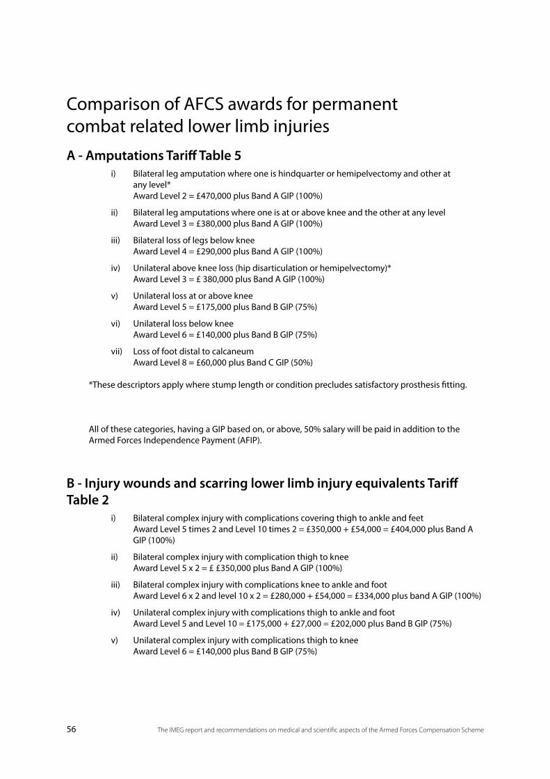
56 The IMEG report and recommendations on medical and scientific aspects of the Armed Forces Compensation Scheme
Comparison of AFCS awards for permanent combat related lower limb injuries A - Amputations Tariff Table 5
i) Bilateral leg amputation where one is hindquarter or hemipelvectomy and other at any level* Award Level 2 = £470,000 plus Band A GIP (100%)
ii) Bilateral leg amputations where one is at or above knee and the other at any level Award Level 3 = £380,000 plus Band A GIP (100%)
iii) Bilateral loss of legs below knee Award Level 4 = £290,000 plus Band A GIP (100%)
iv) Unilateral above knee loss (hip disarticulation or hemipelvectomy)* Award Level 3 = £ 380,000 plus Band A GIP (100%)
v) Unilateral loss at or above knee Award Level 5 = £175,000 plus Band B GIP (75%)
vi) Unilateral loss below knee Award Level 6 = £140,000 plus Band B GIP (75%)
vii) Loss of foot distal to calcaneum Award Level 8 = £60,000 plus Band C GIP (50%)
*These descriptors apply where stump length or condition precludes satisfactory prosthesis fitting.
All of these categories, having a GIP based on, or above, 50% salary will be paid in addition to the Armed Forces Independence Payment (AFIP).
B - Injury wounds and scarring lower limb injury equivalents Tariff Table 2
i) Bilateral complex injury with complications covering thigh to ankle and feet Award Level 5 times 2 and Level 10 times 2 = £350,000 + £54,000 = £404,000 plus Band A GIP (100%)
ii) Bilateral complex injury with complication thigh to knee Award Level 5 x 2 = £ £350,000 plus Band A GIP (100%)
iii) Bilateral complex injury with complications knee to ankle and foot Award Level 6 x 2 and level 10 x 2 = £280,000 + £54,000 = £334,000 plus band A GIP (100%)
iv) Unilateral complex injury with complications thigh to ankle and foot Award Level 5 and Level 10 = £175,000 + £27,000 = £202,000 plus Band B GIP (75%)
v) Unilateral complex injury with complications thigh to knee Award Level 6 = £140,000 plus Band B GIP (75%)
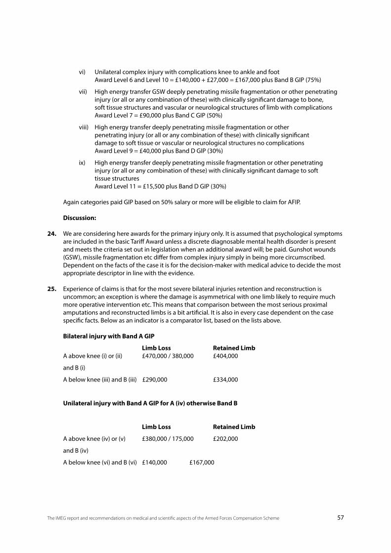
57The IMEG report and recommendations on medical and scientific aspects of the Armed Forces Compensation Scheme
vi) Unilateral complex injury with complications knee to ankle and foot Award Level 6 and Level 10 = £140,000 + £27,000 = £167,000 plus Band B GIP (75%)
vii) High energy transfer GSW deeply penetrating missile fragmentation or other penetrating injury (or all or any combination of these) with clinically significant damage to bone, soft tissue structures and vascular or neurological structures of limb with complications Award Level 7 = £90,000 plus Band C GIP (50%)
viii) High energy transfer deeply penetrating missile fragmentation or other penetrating injury (or all or any combination of these) with clinically significant damage to soft tissue or vascular or neurological structures no complications Award Level 9 = £40,000 plus Band D GIP (30%)
ix) High energy transfer deeply penetrating missile fragmentation or other penetrating injury (or all or any combination of these) with clinically significant damage to soft tissue structures Award Level 11 = £15,500 plus Band D GIP (30%)
Again categories paid GIP based on 50% salary or more will be eligible to claim for AFIP.
Discussion:
24. We are considering here awards for the primary injury only. It is assumed that psychological symptoms are included in the basic Tariff Award unless a discrete diagnosable mental health disorder is present and meets the criteria set out in legislation when an additional award will; be paid. Gunshot wounds (GSW), missile fragmentation etc differ from complex injury simply in being more circumscribed. Dependent on the facts of the case it is for the decision-maker with medical advice to decide the most appropriate descriptor in line with the evidence.
25. Experience of claims is that for the most severe bilateral injuries retention and reconstruction is uncommon; an exception is where the damage is asymmetrical with one limb likely to require much more operative intervention etc. This means that comparison between the most serious proximal amputations and reconstructed limbs is a bit artificial. It is also in every case dependent on the case specific facts. Below as an indicator is a comparator list, based on the lists above.
Bilateral injury with Band A GIP
Limb Loss Retained Limb A above knee (i) or (ii) £470,000 / 380,000 £404,000
and B (i)
A below knee (iii) and B (iii) £290,000 £334,000
Unilateral injury with Band A GIP for A (iv) otherwise Band B
Limb Loss Retained Limb
A above knee (iv) or (v) £380,000 / 175,000 £202,000
and B (iv)
A below knee (vi) and B (vi) £140,000 £167,000
58 The IMEG report and recommendations on medical and scientific aspects of the Armed Forces Compensation Scheme
The Band A GIP for unilateral above knee loss hip disarticulation / hemipelvectomy reflects the inability to wear a prosthesis.
In conclusion although perhaps not readily appreciated, the Tariff Levels for amputation and for retained limbs with similar degree of injury attract similar levels of AFCS compensation, consistent with current evidence of outcome. Survival of these complex extremity injuries is a hallmark of the Iraq and Afghanistan conflicts. Yet there remain many unanswered questions. IMEG strongly supports urgent collaborative longitudinal studies into best practice management, and outcomes, and will maintain routine oversight of developments.
References: (1) Brown, K.V. et al Infectious complications of combat-related mangled extremity injuries in the British Military. J Trauma 2010; 69: S109-115
(2) Owens, B.D. et al Combat Wounds in Operation Iraqi Freedom and Operation Enduring Freedom. J Trauma 2008; 64: 295-99
(3) Ramasamy, A. et al Injuries from Roadside Improvised Explosive Devices. J Trauma 2008; 65: 910-914
(4) Nelson, T.J. et al Close proximity blast injury patterns from Improvised Explosive Devices in Iraq: a report of 18 cases. J Trauma 2008; 65; 212-217
(5) Rush, R.M. et al Application of the mangled extremity severity score in a military setting. Mil Med 2007; 172: 777-781
(6) Brown, K.V. et al Predicting the Need for Early Amputation in Ballistic Mangled Extremity Injuries. J Trauma 2009; 66: S93-S98.
(7) Barr, J.S. et al Blast personnel injury: a clinical study. Mil Surg 1946; 98:1-12
(8) Tran, T. et al Functional outcome of multiply injured patients with associated foot injury. Foot, Ankle Inj 2002; 23: 340-343
(9) Ramasamy, A. et al The Modern “Deck Slap” Injury-Calcaneal Blast Fractures from Vehicle Explosions. J Trauma, 2011; 71: 1694-1698.
(10) Ramasamy, A. et al Outcomes of IED foot and ankle blast injuries. J Bone Joint Surg Am 2013; 95: e25 (1-7)
(11) Ferreira, R.C. et al Long term results of salvage surgery to severely injured feet. Foot Ankle Int 2010; 31(2): 113-23
(12) Bosse, M.J. et al An analysis of outcomes of reconstruction or amputation after leg-threatening injuries. N Eng J Med 2002; 12: 347(24): 1924-31

59The IMEG report and recommendations on medical and scientific aspects of the Armed Forces Compensation Scheme
Topic 6 – Recognised Diseases Ahead of the papers on diabetes mellitus, testicular cancer and leukaemias we have reproduced the introduction to Recognised Diseases included in the May 2013 IMEG Report.
1. Lord Boyce in his review of the AFCS raised the issue that while under the War Pensions scheme the majority of medical discharge cases suffering from physical disorders receive entitlement to war pension; this is not the case under the AFCS. This is a reflection of the different standards of proof required in the two schemes. The standard of proof in AFCS is ‘on the balance of probabilities’ (or ‘more likely than not’), which is the standard of proof in both civil compensation and the statutory compensation scheme for civilian occupational injury and disease, the Industrial Injuries Scheme.
2. At its inception in 1917, the standard of proof used in the War Pensions Scheme was “on the balance of probabilities”. This was changed in 1943, at the height of the Second World War, when for injuries and disorders arising in service, the burden of proof, transferred to MoD to demonstrate that a service cause was “beyond reasonable doubt” not the cause of the disease. The change was introduced at this time because inadequate record keeping was leading to large numbers of claimants unfairly not receiving compensation.
3. In his report, Lord Boyce proposed that the IMEG should develop a list of Recognised Diseases for the AFCS. By this he meant that IMEG should review the medical literature and receive evidence from experts to provide guidance about the circumstances when “on the balance of probabilities”, a disease having onset in or around service was more likely than not to be attributable to service in the Armed Forces.
4. The normal burden of proof in civil compensation and other statutory compensation schemes such as the Industrial Injuries Disablement Benefit (IIDB) Scheme is “on the balance of probabilities”. For claims under AFCS this implies demonstrating that military service is more likely than not (more than 50:50) the predominant cause of the injury or disease in the individual case. In the Industrial Injuries Disablement Benefit Scheme, for those conditions where there is sufficient evidence that this level of proof is satisfied, the disease is ‘prescribed’, i.e. attributable in the individual case to the particular cause in relation to clearly specified circumstances of exposure.
5. In the individual case, attribution is usually based on sufficient evidence to answer the questions:
• Does the particular agent or exposure cause the disease, at least in some circumstances?
• If so, were the circumstances of the individual case such that the agent or exposure is more likely than not to have been the cause of the disease?
6. Recognition of a particular agent as the cause of a disease, and attribution in the individual case, is most clear when the cause is specific to the disease, or nearly so, and the probability of causation is high. Such conditions are now relatively uncommon but a relevant example is occupational asthma, asthma whose primary cause is an agent inhaled at work. The majority of cases of occupational asthma are due to the development of an allergic reaction to the specific cause encountered in the workplace (e.g. flour in a baker). Asthma develops after an initial symptom-free period of exposure and recurs on re-exposure to the specific cause, in concentrations which do not cause respiratory symptoms in others similarly exposed or previously in the affected individual. Inhalation testing with the specific agent will provoke an asthmatic reaction in the sensitised individual (but not in others not sensitised). Also, for many agents evidence of a specific immunological reaction (i.e. specific IgE antibody) will be found. In principle, the specific cause of asthma can be demonstrated in the individual case.
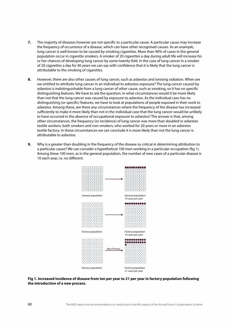
60 The IMEG report and recommendations on medical and scientific aspects of the Armed Forces Compensation Scheme
7. The majority of diseases however are not specific to a particular cause. A particular cause may increase the frequency of occurrence of a disease, which can have other recognised causes. As an example, lung cancer is well known to be caused by smoking cigarettes. More than 90% of cases in the general population occur in cigarette smokers. A smoker of 20 cigarettes a day during adult life will increase his or her chances of developing lung cancer by some twenty-fold. In the case of lung cancer in a smoker of 20 cigarettes a day for 40 years we can say with confidence that it is likely that the lung cancer is attributable to the smoking of cigarettes.
8. However, there are also other causes of lung cancer, such as asbestos and ionising radiation. When are we entitled to attribute lung cancer in an individual to asbestos exposure? The lung cancer caused by asbestos is indistinguishable from a lung cancer of other cause, such as smoking, so it has no specific distinguishing features. We have to ask the question: in what circumstances would it be more likely than not that the lung cancer was caused by exposure to asbestos. As the individual case has no distinguishing (or specific) features, we have to look at populations of people exposed in their work to asbestos. Among these, are there any circumstances where the frequency of the disease has increased sufficiently to make it more likely than not in the individual case that the lung cancer would be unlikely to have occurred in the absence of occupational exposure to asbestos? The answer is that, among other circumstances, the frequency (or incidence) of lung cancer was more than doubled in asbestos textile workers, both smokers and non smokers, who worked for 20 years or more in an asbestos textile factory. In these circumstances we can conclude it is more likely than not the lung cancer is attributable to asbestos.
9. Why is a greater than doubling in the frequency of the disease so critical in determining attribution to a particular cause? We can consider a hypothetical 100 men working in a particular occupation (fig 1). Among these 100 men, as in the general population, the number of new cases of a particular disease is 10 each year, i.e. no different.
General population
Factory population
Factory population
New Process
General population10 cases per year
Factory population10 cases per year
Factory population21 cases per year
Fig 1. Increased incidence of disease from ten per year to 21 per year in factory population following the introduction of a new process.
61The IMEG report and recommendations on medical and scientific aspects of the Armed Forces Compensation Scheme
Some time later, after the introduction of a new process, the number of cases of the disease in these 100 men increases to 21 each year, i.e. more than 2 times the previous frequency. We cannot distinguish the additional 11 cases from the 10 in whom the disease would otherwise have occurred. What we can say is that in any particular individual among the 21 cases, there is a more than 50:50 chance, or a greater than doubling of risk, that the disease would not have occurred without exposure to the particular cause. On the balance of probabilities it is therefore more likely than not that the disease is attributable to the particular cause in the individual case. We can say that ‘but for’ his working in this factory it is unlikely the man would have developed the disease. The balance of probabilities has shifted to ‘more likely than not’ and in this circumstance the disease can be attributed to the particular cause.
10. In the case of Recognised Diseases in the AFCS, we are therefore looking for evidence that service in the Armed Forces is consistently associated with an increase in the frequency of a particular disease or illness and whether there are circumstances where the frequency is more than doubled, making it more likely than not in the individual case that the disease was attributable to a cause in service.
11. It is also important to distinguish “all or none” diseases from “more or less” diseases. A well-recognised “all or none” physiological condition is pregnancy: one cannot be a bit pregnant. In contrast, many important conditions including high blood pressure, hearing loss and mental health disorders are ‘more or less’ conditions. These have a continuum of frequency of symptoms without a clear distinction between those with and without the condition. The definition of disease is therefore less clear and subject to expert opinion.
12. The epidemiological evidence informing these determinations should be of high quality, drawn from several independent studies and sufficiently consistent and robust that further research at a later date would be unlikely to overturn it.
A. Diabetes mellitus1. The term diabetes mellitus (DM) applies to syndromes of abnormal carbohydrate metabolism
characterised by raised blood glucose levels (hyperglycaemia) sustained sufficiently long to cause damage to kidneys, eyes, nerves and arteries. DM is associated with a relative or absolute impairment in insulin secretion and varying degrees of resistance to the action of insulin. DM is usually considered as primary or secondary.
2. Primary diabetes is divided into Type 1 (T1DM) and Type 2 (T2DM). Type 1, previously known as insulin dependent or juvenile onset diabetes, is responsible for 5-15% of cases while Type 2 (T2DM) accounts for some 90% of cases. Type 2 was previously called non-insulin dependent or maturity onset diabetes. These terms are however inaccurate. Treatment for both types of primary diabetes depends on severity and both types may require insulin. Equally T2DM, usually presenting in middle age or older, is now increasingly being diagnosed in overweight children and young adults.
3. T1DM typically, but not always, has its clinical onset in childhood or young adulthood. In recent years more cases have been diagnosed under the age 5 years. Up to 50% of cases are diagnosed after age 18 years while about 5% of newly diagnosed Caucasian diabetics over the age of 65 years have type 1. Before puberty T1DM is equally common in males and females. After puberty, there is a preponderance of males although less than a twofold increase in risk.
4. T1DM is considered to involve an autoimmune mediated progressive destruction of the ß (beta) cells of the pancreas, which secrete insulin, leading to an absolute deficiency of insulin. Auto-antibodies to pancreatic tissue can be detected in the blood. This process typically develops over many years, with the pre-diabetic phase being asymptomatic. T1DM probably occurs when environmental factors, whose precise aetiology is unknown, act in a genetically susceptible person. Genetic factors
62 The IMEG report and recommendations on medical and scientific aspects of the Armed Forces Compensation Scheme
account for about 40% of the risk with multiple genes involved, so a clear familial pattern is not always observed. In terms of environmental factors involved, the evidence is conflicting and the mechanism as yet unknown. Viruses are believed to be the most likely environmental triggers; these include Coxsackie B, mumps, cytomegalovirus and rubella. Serological studies confirm that Coxsackie B infection is relatively common in patients newly diagnosed with T1DM. 20% children who survive intra-uterine rubella develop T1DM with autoimmune markers. It may be that the virus is part of the final common pathway to clinical T1DM enhancing the established autoimmune activity and onset of clinical symptoms. Other possible trigger factors include bovine serum albumin from cows’ milk and toxins, e.g. found in food, may also be involved (1) (2).
5. High susceptibility to T1DM is found in European populations throughout the world, while African and East Asian populations are relatively spared. The incidence is higher in Northern European countries and declines progressively towards the equator. Diagnosis in children is more frequent in the winter months and in many countries there have been marked increases in incidence over 10 - 20 year periods (3) (4).
6. T2DM is a more heterogeneous disorder characterised by raised blood glucose in the absence of the features of T1DM, with a relative insulin deficiency caused by resistance to the action of insulin. The tendency to T2DM is again considered to be a function of genetic and environmental factors. It occurs typically in middle aged and older adults with rates rising with age. It now affects 3-4% of the white population in most countries with rates rising to 8-11% in Eastern Europe and North America. It is very common in some immigrant populations living in affluent countries including in the UK. In UK the prevalence in the white adult population is 4% and 10-15% in adult Asian or Afro-Caribbeans. As childhood obesity rates rise, the diagnosis is becoming more common in younger people.
7. The geographical spread of T2DM is different from that of T1DM and prevalence is rising especially rapidly in developing countries. This is related to Westernization and the obesity epidemic. The thrifty gene hypothesis proposes that excess energy is stored as fat in anticipation of lean times; however in an age of fast food, motor cars, decreased physical labour and recreational exercise the deposited fat is not consumed as energy but accumulates, causing obesity.
8. T2DM is also a progressive disorder; over time the ability to produce insulin reduces, in some cases with beta cell failure and, ultimately, requirement for insulin replacement as with T1DM.
9. T2DM appears to be polygenic with several gene loci now identified as having an influence on insulin secretion or resistance and obesity. T2DM is not inherited by simple Mendelian rules and the recent explosion in numbers of cases strongly suggests that environmental factors are key both in explaining the increasing incidence and in offering potential factors for modification. Obesity and pregnancy increase insulin resistance while chronic hyperglycaemia can impair insulin sensitivity and inhibit insulin release.
Specific risk factors for T2DM10. Obesity especially its level, distribution and the age at which fat is laid down. Once BMI reaches 28
kg / m2 the risks increase steeply and at 35 kg / m2 the risk may be 40 times greater than for a person with a BMI of 22 kg / m2. Truncal and visceral fat is especially hazardous as is weight gain in the early twenties.
11. Physical inactivity appears to be an independent risk factor. This is due to insulin resistance with sedentary people three times as likely as those taking regular physical exercise to be affected.
63The IMEG report and recommendations on medical and scientific aspects of the Armed Forces Compensation Scheme
12. The Barker hypothesis suggests that poor foetal growth can lead to abnormal metabolic effects “the metabolic syndrome” with hyperglycaemia, hypertension and dyslipidaemia. This is particularly the case in those who are underweight at birth and subsequently become obese. Animal studies suggest that malnutrition in utero may lead to reduced pancreatic cell mass and so impair insulin secretory reserve.
13. Other causes of hyperglycaemia which can present during military service include diabetes as a complication of pregnancy, both pre and post gestation. Several predisposing factors may be involved.
14. Lastly there is a rare condition now called Maturity Onset Diabetes of the Young. Cases of this have a family history of diabetes diagnosed young; with no family history of obesity and importantly no pancreatic beta cell autoantibodies as found in T1DM. This disorder has several variants and is caused by mutations in the glucokinase genes. It responds well to treatment with sulphonylureas.
15. Secondary diabetes arises from a variety of congenital and acquired causes. For the purposes of AFCS, congenital syndromes are unlikely to be an issue. The relevant chromosomal disorders e.g. Down’s syndrome: Turner’s syndrome: and Klinefelter’s syndrome, typically result in significant disability from birth or childhood and are incompatible with military enlistment. Acquired causes relevant to military service include pancreatic disease, pancreatitis, or cancer. Pancreatic tissue can also be reduced or destroyed by trauma including surgery.
16. Secondary diabetes may also follow endocrine disorders where there is excess production of hormones that oppose the action of insulin e.g. thyrotoxicosis: acromegaly and Cushing’s syndrome. Again these will be rare in the military population and unlikely to be causally related to service.
17. A third major cause of secondary diabetes is therapeutic drugs, in particular glucocorticoid steroids. Circumstances where corticosteroids are used orally and long term are unlikely to be consistent with military service even in a downgraded capacity e.g. chronic lung disease: inflammatory bowel disease and joint disease. Another major group of therapeutic drugs are those used to lower blood pressure including diuretics and beta blockers. Finally drugs used to treat HIV infection.
Recognised disease status 18. There is no published study of incidence of primary diabetes mellitus of either T1DM or T2DM in any
occupational or other identifiable group including military personnel and veterans. While both for service entry and retention in service following diagnosis of diabetes, cases are considered on their merits, it is unlikely that T1DM is compatible with full-time fully deployable regular service in any branch of the military. The position with T2DM, especially if well controlled by diet or diet with oral hypoglycaemics may be less rigid but again fully deployable regular service in a forward role is unlikely. As discussed above primary T1DM remains a disorder of unknown aetiology and so cannot, on balance of probabilities, be attributable to service. In relation to T2DM the major non genetic and modifiable factors are matters of lifestyle and personal choice. Military diet and restrictions e.g. on operational deployment rations are advised by professional dieticians and are not diabetogenic.
19. In terms of secondary DM, two main mechanisms are involved. First loss or damage to pancreatic tissue and the beta cells which secrete insulin. This might arise through combat related abdominal injury or other service-related trauma. Destruction of pancreatic tissue could be due to infection when Article 12 of the 2011 AFCS Order refers. It could also occur due to a tumour itself or arising from its treatment. There are few circumstances where the cause of pancreatic cancer in an individual is known e.g. it is accepted as a radiogenic disorder, but if the cancer is due to service, and diabetes mellitus is a
64 The IMEG report and recommendations on medical and scientific aspects of the Armed Forces Compensation Scheme
consequence, it too will be accepted as due to service. Surgery for a condition which is not attributable to service, even where it takes place during service, is not itself, nor are any expectable consequences, due to service. A frequent cause of pancreatitis, especially chronic pancreatitis is alcohol which is expressly excluded under the Scheme.
20. In conclusion diabetes mellitus can be accepted as due to service, on balance of probabilities where pancreatic tissue has been damaged by service related traumatic injury; surgical excision or irradiation for service related physical disorder or by the progress of a service-accepted physical disorder of the pancreas. The appropriate descriptor will come from Table 4 Physical disorders – illnesses and infectious diseases and will vary dependent on the specific case details.
References:(1) Dayan, C. et al Diabetes In: Disorders of Glucose Homeostasis. Oxford Textbook of Medicine 5th ed Oxford OUP 2010 pps 1987-2049
(2) American Diabetic Association Diagnosis and classification of diabetes mellitus. Diabetes Care 2009; 32 supp 1 S1 51-61
(3) Gale, E.A.M. The rise of childhood Type 1 diabetes mellitus in the 20th century. Diabetes 2002; 51: 3353-61
(4) Pickup, J.C. et al (eds) Textbook of diabetes 3rd ed Oxford Blackwell 2002
B. Testicular cancer 1. Although the incidence of testicular cancer in the UK has increased in recent years, it remains relatively
rare with about 2000 cases diagnosed a year. It represents about 1% of all male cancers and mortality rate has been declining since the mid 1970s and introduction of platinum based chemotherapy. The disorder, as other testicular problems such as low sperm count, poor, quality sperm and maldescent of testis, has been increasing in incidence over the last forty years in almost all parts of the world. There is marked geographical and racial variation and the tumour is most common in Northern Europe and in higher socioeconomic groups with much lower rates in Asian, African and African Americans. This pattern suggests a causal role for both genetic and environmental factors (1).
2. Testis cancers are predominantly germ cell tumours (GCT) and a single tumour often includes a variety of cell types. This pathological complexity leads to different classification systems, which are difficult to align. In clinical terms testis tumours are usefully sub-divided into three groups, all derived from germ cells at different stages of development. The age specific incidence of testis tumours is unusual. There is a small peak in infancy and tumours are then uncommon until puberty when the incidence begins to rise and testis tumours are the most common solid tumour of young men in the 15 - 35 years age group. There is a sharp decline in incidence in older men. This pattern suggests that causative factors may operate in utero or early life while the increased incidence after puberty suggests that hormonal influences also play a part in tumour development (2).
3. Testis cancer is diagnosed clinically and where there is a painless solid lump within the testis, the diagnosis is made, until proven otherwise. In today’s more open and better educated society, which includes men’s health as well as high profile cases involving sporting celebrities, awareness and early diagnosis of the disorder amongst young men has helped improve prognosis. A small number of patients continue to present with metastatic spread e.g. with respiratory symptoms; bone pain; neurological symptoms or venous thrombosis. Investigation in suspected cases includes scrotal
65The IMEG report and recommendations on medical and scientific aspects of the Armed Forces Compensation Scheme
ultrasound, radiology of chest, abdomen and pelvis. Three serum tumour markers are also measured. Serum markers do not make the diagnosis but very high values are rare in the absence of cancer. Treatment advances in radiotherapy and chemotherapy mean that mortality is improving and many patients can be expected to achieve a normal life span. Relatively high dose chemotherapy and radiotherapy may be required and with improved prognosis, long term toxicity from these is a risk. The most frequent sequelae of treatment are cardiovascular effects: second cancers and reduced fertility. The latter is usually temporary, with sperm count recovering with time from treatment.
4. High oestrogen exposure in utero has been suggested as a casual influence. However evidence on incidence of testis cancer in the sons of women receiving diethyl stilboestrol has been inconsistent and there is no direct link between maternal oestrogen levels during pregnancy and testis cancer in humans (3). In 1994 it was suggested that the emerging testis cancer epidemic might correlate with increased maternal smoking during pregnancy. Several studies have investigated this hypothesis with conflicting results (4).
5. Testis tumours run in families with relative risk increased 6 - 10 fold in brothers or sons of affected men. Chromosomal abnormalities are common in GCT and a specific mutation has been associated with familial testis cancer, especially bilateral disease (5).
6. Undescended or maldescended testes (cryptorch(id)ism) are a strong risk factor for testicular cancer (6). If only one testis is maldescended the increased risk of cancer applies to both testes. Undescended or maldescended testes are also associated with long term consequences on testis function including spermatogenesis. The rate of cryptorch(id)ism in pre-pubertal boys from prospective studies is between 2 and 8 %. Low birth weights, prematurity, being small for gestational age, are risk factors and low maternal oestrogen levels and placental insufficiency may be relevant (6). Other postulated influences include maternal diabetes including gestational diabetes. A recent Californian study suggested that maternal prenatal DDT exposure might have a role (7). It is not known whether these associations of cryptorch(id)ism share common aetiological factors with testis cancer or might be themselves direct causes of testis cancer.
7. Several studies in different populations suggest that taller adult height but not increasing body weight is correlated with risk of testicular cancer (8). Other studies of varying power and rigour have looked at occupational exposure to wood dust, degreasing agents, chromate and azo-based dyes, and dimethyl formamide with inconclusive results (2). Few studies have considered ionising or non ionising radiation as causal factors for testis cancer. Those which have done so include few cases and their results are inconsistent. BEIR, UNSCEAR and UK Health Protection Agency Advisory Group on Ionising Radiation (AGIR) have considered the evidence too weak to come to firm conclusions on radiogenicity (9). In AFCS terms this means that at this date, on the balance of probabilities, no dose of ionising radiation can be considered to cause testicular cancer.
8. Of relevance to the Armed Forces population, is evidence on exercise / sport, and testicular trauma. Most of the work exploring a role for trauma has been case-control in design with the risk of recall bias in terms of identifying trauma in the period leading to diagnosis. Similarly, studies which attempt to correlate cancer development with previous sporting activity have produced inconsistent results (10) (11).
9. Several studies report raised relative risks associated with post pubertal mumps but case numbers were small and the 1994 UK Testicular Cancer Study Group found no relation between mumps and testis cancer (6). In AFCS terms, mumps arising in service is unlikely to be accepted as attributable to service (see Article 12 AFCS Order 2011).
66 The IMEG report and recommendations on medical and scientific aspects of the Armed Forces Compensation Scheme
10. Testicular cancer has not been thought of or widely studied as an occupational disorder and where associations with employment have been reported e.g. farmers, painters and tanners, their results have been inconsistent, case numbers small and causative agents / mechanism unknown. Military studies have included a large study of US naval personnel comparing the incidence of testicular cancer with the US national cancer surveillance statistic. Age adjusted incidence of testis cancer for the navy overall was not significantly different from the general US population. However naval aviation support equipment technicians and enginemen, with tasks similar to civilian motor mechanics, had raised incidence rates both relative to the US navy overall and the civilian population (12). Studies of Vietnam veterans have produced conflicting results. A case control study which dichotomised, simply on the basis of service in Vietnam, showed a significant increased risk of testis cancer (13) in army, navy, and air force veterans but not in marines. It was proposed that exposure to Agent Orange was the potential cause (14). A later study used a series of surrogates to reflect exposure to Agent Orange e.g. combat troop: ground troop and geographical location. In this study only naval personnel had increased risk of testis cancer, the group least likely to have Agent Orange exposure (15).
11. A case control study was published in 1997 on testis cancer in Royal Navy personnel (16). Cases between 1976 and 1994 were identified and controls, four to each case, were matched on date of birth and length of service. In total 110 cases of testis cancer were identified. Five cases had maldescended testes, all surgically corrected, and two had atrophic testis. A radiation history was obtained for each case and service branch noted i.e. general: submarine: Fleet Air Arm: Royal Marine and finally, rank was noted. The results suggested an increased risk in Fleet Air Arm relative to other branches of the service and specifically the risk was in air engineers but not aircrew. There was no increased risk in submariners or when length of service or time served on nuclear vessels was considered. The overall numbers of cases, especially in the subgroups were small and the study statistical power very low. The findings are of interest but a further larger study is required.
12. In conclusion while a number of associations are established, none is related to occupation and at this date, testis cancer remains a disorder of unknown aetiology. There is no reliable evidence of increased risk associated with UK military service in general, nor service in navy, army or air force. Equally no link is identified with any specific service occupation or exposure. No circumstances can presently be identified where testis cancer could be accepted as a recognised disorder for AFCS purposes.
References: (1) Horwich, A. et al Testicular tumours. in Peckham, M. et al (eds) Oxford Textbook of Oncology Oxford 1407-1439 1997
(2) Khan, O et al Testis cancer. Post grad Med J 2007; 83: 624-632
(3) Depue, R.H. Maternal and gestational factors affecting the risk of cryptorchodism and inguinal hernia. Int J Epid 1984; 13: 311-314
(4) Pettersson, A. et al Women smoking and testicular cancer: one epidemic causing another? Int J Cancer 2004;109(6)941-944
(5) Rapley, E. et al Localization to Xq27 of a susceptibility gene for testicular germ cell tumours. Nat Genet 2000; 24: 197-200
(6) United Kingdom Testicular Cancer Study Group The aetiology of testicular cancer: association with congenital abnormalities, age at puberty, infertility and exercise. Br Med J 1994; 308: 1393-9
67The IMEG report and recommendations on medical and scientific aspects of the Armed Forces Compensation Scheme
(7) Cohn, B. Pre-natal DDT exposure and testicular cancer: a nested case-control study. Arch Env and Occ Health 2010; 65: 127-133
(8) Dieckmann, K.P. et al Is risk of testicular cancer related to body size? Eur Urol 2002; 42: 564-9
(9) AGIR Report of the independent Advisory Group on Radiation. Risk of solid cancers following radiation exposure: Estimates for the UK population. Health Protection Agency 2011
(10) Coldman, A.J. et al Sports activities and risk of testicular cancer. Brit J Cancer 1982; 46: 749-756
(11) Forman, D. et al Aetiology of testicular cancer: association with congenital abnormalities, age at puberty, infertility and exercise. Br Med J 1994; 306: 1393-99
(12) Garland, F.C. et al Testicular cancer in US navy personnel. 1988 Am.J Epidemiol 1988; 127: 411-414
(13) Pottern, L.M. et al Testicular cancer risk among young men: rate of cryptorchodism and inguinal hernia. J Natl Cancer Inst 1985; 74: 377-81
(14) Lerda, D. et al Study of reproductive function in persons occupationally exposed to 2:4 dichloro-phenoxy-acetic acid. Mutat Res 1991; 262: 47-50
(15) Bullman, T.A. et al Cancer associated with surrogate measures of Agent Orange exposure among Vietnam veterans on the Agent Orange register. Ann Epidemiol 1994 Jan; 4(1): 11-6
(16) Ryder, S.J. et al Is testicular cancer an occupational disease? A case control study of Royal Naval personnel. J R Nav Med S 1987; 83: 130 -146.
C. The leukaemias 1. The leukaemias are cancers of the white blood cells. They are described as acute or chronic and
dependent on cell lineage, there are two major groups, myeloid and lymphoid. They arise when stem and progenitor cells in the bone marrow are genetically altered giving rise to malignant transformation. The abnormal cells do not mature into normal cells but continue to proliferate and expand in the marrow at the expense of healthy cells and, over time, lead to low red cell and platelet counts. The abnormal cells enter the blood stream giving a high white cell count and may also be found in lymph nodes, spleen and liver (1).
2. As yet the cause and pathogenesis of the leukaemias remains incompletely understood. Established potentially occupation-related causal factors include ionising radiation and exposure to chemicals (particularly benzene), but in the individual case, cause is rarely established. The leukaemias are classified into sub-groups, now based on molecular genetics. This approach means classifications are not settled but are amended over time to reflect emerging understanding and emphases (2). The focus of this note is occupational causes of the most common types, Acute lymphatic leukaemia (ALL): Chronic lymphatic leukaemia (CLL): Acute myeloid leukaemia (AML) and Chronic myeloid leukaemia (CML).
68 The IMEG report and recommendations on medical and scientific aspects of the Armed Forces Compensation Scheme
Sub-Types of leukaemia
3. Acute lymphoblastic leukaemia (ALL). 85% of cases of ALL occur in children and present with pallor, bleeding, bruising, anaemia, infection and fever. Recent years have seen significant advances in therapy for childhood ALL, particularly for those aged between 1 and 10 years. There is a much poorer prognosis in adults with this diagnosis and current evidence suggests that the disorders in children and adults are different biologically and genetically. It is now recognised that in most childhood cases the first genetic events in key stem cells arise in utero (3) with further environmental exposures / events required to trigger this pre-leukaemic state into a clinical disorder. The evidence suggests that infection may be an important trigger in the development of childhood clinical ALL. It is not known if infection plays any part in the development of adult ALL (4).
4. Acute myeloid leukaemia (AML). By contrast 80% of patients with AML are adults and only 20% children. In adults while it may be seen at any age, the median age of clinical onset is 68 years. In older patients it may be associated with, and evolve as, an end stage from the myelodysplastic syndrome. Clinically and biologically, AML in children and adults seems to be the same disorder. It most typically presents with anaemia and bleeding due to bone marrow failure. In most cases there is no obvious cause but exposure to chemicals, notably benzene, cytotoxic drugs and ionising radiation can be relevant. As survival rates for solid tumours continue to improve, an increasing source of cases is adults who have survived radiotherapy / chemotherapy for other cancers (5).
5. Chronic lymphatic leukaemia (CLL). This is the most common lymphoid neoplasm in Europe and North America and its cause remains unknown. The parent cell is the mature B lymphocyte. The diagnosis is usually made fortuitously on discovery of a raised blood lymphocyte count. There is no standard therapeutic regime and present best practice is to defer active treatment until patients are symptomatic or there are signs of bone marrow failure. The disease can occur in families with about 10% of cases having a family history in first degree relatives. Some studies have suggested an association with industrial and agricultural chemicals but results are inconsistent and study size, design and exposure ascertainment of varying quality. It has generally been accepted that, in contrast to other types of leukaemia, there was no causal relation to ionising radiation (6). However some post Chernobyl case reports and larger studies have suggested that radiation exposure might increase the risk. More evidence is awaited. In these new studies it should be noted the disease is occurring in younger adults, appears clinically more aggressive and the morphology of the cells is different. At present no causal link between ionising radiation and CLL is established (7) (8).
6. Chronic myeloid leukaemia (CML). This accounts for 15% of adult cases of leukaemia but less than 5% of childhood leukaemia. It occurs when the tip of chromosome 9 translocates on to chromosome 22 (Philadelphia chromosome) producing the fusion gene bcr-abl which drives the disease. It is a heterogeneous disorder with an initial chronic indolent phase followed on average at 3 - 6 years with transformation to blast cells and acute leukaemia, often in previous years heralding only a few months’ survival. Patients may be asymptomatic at diagnosis or may present with fatigue, sweats fever, pruritus (itch) or abdominal heaviness due to an enlarged spleen. There is no evidence of familial predisposition nor association with particular HLA or other genotypes (9). In terms of a causal role for ionising radiation, studies of British radiologists and technicians in the first half of last century showed increased rates of CML (10) (11) and similarly the 2005 15 countries’ nuclear workers’ study showed a small excess of CML (12). New treatments particularly the advent of tyrosine kinase inhibitors e.g. imatinib, which act on the protein product of bcr-abl, a tyrosine kinase, have transformed the outlook. First line management with imatinib achieves complete remission in over 90% of patients, a much better result than with previous cytotoxics. Imatinib has side effects in some patients and there is risk of teratogenicity so the drug should be avoided in pregnancy. In patients under 50, if the response to imatinib is poor or there are contra-indications and the patient remains in chronic phase, allogeneic stem cell transplantation is usually offered. For advanced disease the options are combination chemotherapy or a trial of a new generation tyrosine inhibitor.
69The IMEG report and recommendations on medical and scientific aspects of the Armed Forces Compensation Scheme
Occupational and service related exposures known or suspected of a causal link to the leukaemias
7. This section considers occupational exposures known or suspected of being causally related to the leukaemias.
Ionising radiation
8. Exposure to ionising radiation in all its forms is part of being alive. Ionising radiation is taken to mean radiation of high enough energy to displace electrons from atoms and includes cosmic rays, gamma rays, X-rays, alpha and beta radiation. Tissues vary in their sensitivity to ionising radiation and different types of ionising radiation have different capacity to cause tissue damage and hence adverse health effects. Bone marrow is very sensitive to ionising radiation.
9. Evidence that ionising radiation can cause human cancer including leukaemia has come from several sources. These include follow-up of patients therapeutically irradiated for malignant conditions, such as cancer of the cervix, and non-malignant conditions like ankylosing spondylitis (13), nuclear industry worker follow-up studies (12) and most notably from the Japanese atomic bomb survivor studies (14) (15).
10. Cancers induced by ionising radiation are indistinguishable from those due to other more common risk factors such as diet, tobacco, alcohol etc. In addition to the dose of radiation delivered, the type of radiation, its duration of exposure i.e. an acute high dose or a chronic low or fractionated dose, the particular tissue irradiated and the age of the individual at the time of the radiation as well as at time of clinical onset of the malignancy are all known to be important. Taking the overall evidence on these matters into account and in the absence of a positive threshold dose of ionising radiation, the convention is to accept that no dose of ionising radiation is completely free from risk of cancer and that the risk increases linearly with dose. The Japanese atomic bomb survivor data demonstrates evidence of an increase in cancer incidence in individuals exposed to levels of ionising radiation of 50 mSv and above. These studies show that leukaemia appears first after whole body irradiation with a latent period of two years and a peak at six to seven years post exposure. Risk is highest in children and young adults.
11. The Japanese survivor data are generally used for risk estimates but in 2002 they were revised downward (by about 8%) to take into account the small contribution of neutron exposure (16). There is a standard international approach to estimation of the probability that a particular cancer in a particular patient is causally linked to ionising radiation (17). This also takes into account the factors above and acknowledges that the atomic bomb exposure was high dose, short duration while most occupational exposure is low dose over a long period of time. There are some animal studies that suggest that for the same overall dose, high acute exposure is more harmful than delivery over a prolonged period of time.
12. While Health and Safety statute law does not apply to the Armed Forces, Defence workplace practice aims to meet or exceed mandatory and best practice protective and surveillance measures. Radiation occupational exposure limits apply and classified radiation workers are routinely monitored by the Defence Radiological Protection Service. Statutory limits for classified radiation workers are whole body 20 mSv per annum and 6 mSv for unclassified workers. This compares with 2.6 mSv per annum for average UK background exposure for all man made exposure. Potential military enclosed sources of ionising radiation include smoke alarms, compasses and helicopter emergency lighting. There are few military personnel who are classified radiation workers. This designation applies where annual exposure of 6 mSv per annum or more can be anticipated and includes medical, dental and industrial radiologists and technicians. The ship’s company on nuclear submarines are not classified radiation workers.
70 The IMEG report and recommendations on medical and scientific aspects of the Armed Forces Compensation Scheme
13. In the late 1990s following public and media concern over adverse health and environmental effects of depleted uranium munitions, the Royal Society set up an expert working group to investigate the issues. Depleted uranium is both toxic and weakly radiogenic. The Working Group reviewed the literature, consulted widely and estimated intakes of depleted uranium over a wide range of typical battlefield exposures. They went on to calculate potential health risks. Their 2002 Report (18), found that munitions containing penetrator rods of depleted uranium do not pose any increased detectable risk of developing fatal cancers over the general risk of dying from cancer over a normal life-time.
14. As discussed in the Recognised Diseases Introductory Section, to accept a disorder as a recognised disease under the AFCS we need to identify service circumstances where, in the individual case, its frequency has increased sufficiently to make it, more likely than not, that it would not have developed without the service exposure. For leukaemia that means establishing a minimum dose level of ionising radiation that increases the risk of the leukaemia by a factor of two or more. If an exposure doubles risk, for every 50 cases occurring naturally in the population there will be another 50 cases due to the exposure. So for an individual case out of the resultant 100 it is impossible to say whether or not it is due to radiation unless the risk is more than doubled. Only then can we consider the individual case facts i.e. actual dose, age at exposure, and attained age.
15. What then is the minimum dose to double a person’s risk? This is the dose that gives an Excess Relative Risk (ERR) of 1. Because risk varies with age at exposure, sex and time between exposure and clinical onset of disease, the value is not constant over time. If we assume male sex, exposure at age 18 and latency for leukaemia of two years, using the US National Academy of Sciences risk model derived from the atomic bomb survivor cohort and accepting that US and UK background risk is similar, the minimum doubling dose is 50 mSv (19). If we set age at exposure at 30, 40 or 50 years the minimum doubling dose rises to 230 mSv but is constant over these ages. No dose approaching these levels, single acute or cumulative, has been recorded by Defence Radiological Protection Services from 6 April 2005.
Non-ionising radiation
16. Extremely low frequency electric and magnetic fields (EMF) – Occupational. EMF exposures are ubiquitous being found around all electric conductors. Occupational studies have tended to focus on occupations most likely to be at risk with little information on other “electrical” jobs where tasks may expose personnel to electric and magnetic fields at least occasionally. Present understanding is that electricians, electrical engineers and especially power cable workers and welders are at highest risk. The risks of leukaemia from these occupational studies and meta-analyses is generally low although results have been inconsistent and no dose response relationship is demonstrable (20) (21). A 2008 updated meta analysis considering occupational studies published between 1993 and 2007 found an overall increase in risk of 17%. The analysis considered excess risks for the specific leukaemias but it is notable that the early studies found CLL to be most common while the later work identified ALL as the most likely type (22). We conclude that present overall evidence does not support a causal link between exposure to electromagnetic fields and the leukaemias.
17. Radiofrequency waves. These are a form of non-ionizing radiation with frequencies ranging from a few kHz to several hundred Ghz. General community exposure is associated with radio and television broadcast antennae, satellite navigation systems, mobile phones and masts while occupational exposures include radar system operation, dielectric heaters, use or maintenance of broadcasting and telecommunications equipment. There are also medical uses including magnetic resonance imaging (MRI), diathermy and electrocautery. It is widely recognised that radiofrequency field exposure causes heating in body tissues, skin erythema and burns. Acute intense heat might damage the lens although cataract formation is not observed in humans following low dose chronic exposure. Heat effects are used to establish Radiofrequency Exposure guidelines. The present evidence on other adverse health
71The IMEG report and recommendations on medical and scientific aspects of the Armed Forces Compensation Scheme
effects including carcinogenicity in humans is limited. There is yet no evidence of carcinogenesis or leukaemogenesis in humans (23). There are a number of military occupational studies looking at radiofrequency exposure and cancer including leukaemia. In one Polish study based on service records there appeared to be increased risk in of several types of cancer (24). A later paper criticised the study attributing the results to inadequate methodology and not a real effect (25). Two US navy studies evaluated radar exposed naval personnel and veterans for cancer mortality and found no risk increases for cancer in general, leukaemia or brain cancer. In one of the US studies an increased risk of non lymphatic leukaemia was noted but this was not replicated in the other study and was observed in only one of three highly exposed sub groups (26) (27).
Chemicals
18. The nature of service life and principle service occupations are unlikely to precisely replicate civilian industrial worker experience in terms of potential or,actual exposure to chemical agents causally linked to the leukaemias. Formaldehyde is a colourless gas used mainly in the manufacture of resins for wood, paper and textiles and in the synthesis of a range of organic chemicals. Formaldehyde is most familiar as an aqueous solution of formalin, clear colourless and with a strong odour. Aqueous solutions of formalin are used as germicides and formaldehyde is contained in disinfectants and preservatives as well as engine exhaust fumes. It is considered that formaldehyde can cause nasopharyngeal cancer in humans, although the evidence is conflicting. Findings in studies on leukaemia both in domestically and occupationally exposed populations have been inconsistent. The overall evidence was examined in the 2006 International Agency for Research on Cancer (IARC) monograph (28) when it was concluded that there was not sufficient evidence for a causal association between leukaemia and occupational exposure to formaldehyde. This was revisited in a 2012 Monograph (29) when the Working Group was not in full agreement. A small majority found the evidence of a causal association “sufficient” but a minority considered it “limited” and concluded that more evidence was required. In particular, replication of a then pre-publication study that reported changes in the blood of exposed workers characteristic of myeloid leukaemia and myelodysplastic syndrome (29). A recent study of 14,000 chemical workers from six factories exposed to formaldehyde, many to high levels of exposure, found no increased risk of myeloid leukaemia (30).
19. Benzene was traditionally used in a variety of manufacturing processes involving leather, and rubber manufacture, paint spraying and removing, dry cleaning and printing. It has now been overtaken as a solvent by non aromatic chemicals but is still present in coal derivatives and petrol distillates. It was the first chemical shown to cause leukaemia, specifically AML (30). Later reports suggest that other types of leukaemia may also be due to benzene exposure and a dose response effect is now established between benzene exposure and the occurrence of AML. Studies have explored the mechanism of benzene haematotoxicity as well as the dose levels and exposure duration required for development of leukaemia. It is now clear that effects vary widely between individuals and that early benzene toxicity is reversible if exposure ceases. Adverse effects may also have a latent period of years before onset of symptoms or clinical disease (31). The evidence is that chronic exposure is more likely than acute dose to be associated with the development of acute myeloid leukaemia. One study (32) established that exposure to benzene at more than 20 ppm for more than six years was required for the development of AML. High level (more than 100 ppm) but short term exposures may cause transient blood changes with no long term effects. Long term low dose exposure (above 20 ppm) can cause reduced blood cell counts (33). Removal from benzene results in the peripheral blood picture returning to normal over a few months but longer term risk of leukaemia in this circumstance is unknown.
20. In conclusion, circumstances in which on balance of probabilities and post 6 April 2005, the leukaemias may be considered as recognised diseases, due to service occupational exposure are limited. This is because of the nature of principle service occupations and the working patterns, high standards of Health and Safety practice and prevention. Most individual cases are of unknown aetiology
72 The IMEG report and recommendations on medical and scientific aspects of the Armed Forces Compensation Scheme
and cannot by their very nature be due to service. A possible occupational causal link including a formal Probability of causation calculation would be explored on the case facts where a minimum occupational dose of 50 mSv ionising radiation or more is registered. A chemical causal link to the leukaemias is most unlikely in the military context. Where, exceptionally, an award under AFCS is appropriate, the descriptor and award in the 2011 Order will be Table 4 Item 1 or 2. This will provide an award at Level 6 i.e. £140,000 and, from service termination for life, a GIP band B, based on 75% service salary.
References: (1) Gutierrez, A. et al Cell and molecular biology of human leukaemias. in Oxford Textbook of Medicine Warrell, D. et al (ed) 5th ed Oxford OUP 2010 Chap 22: 3: 1: 4214-4221
(2) Swerdlow, S.H. et al WHO classification of tumours of haematopoietic and lymphoid tissues. IARC Lyon 2008
(3) Wiemels, I.I. et al Prenatal origin of ALL in children. Lancet 1999; 354: 1499-1503
(4) Pui, C.H. et al Acute Lymphoid leukaemia-mechanism of disease. N Eng J Med 2004; 350: 1535-1548
(5) Kell, J. et al Acute myeloid leukaemia. in Oxford Textbook of Medicine Warrell, D. et al (eds) 5th ed Oxford OUP 2010 Chap 22: 3: 4: 4233-4240
(6) Dighieri, G. et al Chronic lymphocytic leukaemia. Lancet 2008; 371: 1017-1029
(7) Richardson, D.B. et al Ionizing Radiation and Chronic Lymphocytic Leukaemia. Env Health Persp 2005; 113: 1-5
(8) Zablotska, L.B. et al Radiation and the risk of Chronic lymphocytic and other leukaemias among Chernobyl Cleanup Workers. Env Health Perspectives 2013; 121: 59-65
(9) Baccarani, M. et al Chronic Myeloid leukaemia: an update on concepts and management recommendations of European Leukaemia Net. J Clin Oncol 2009; 27: 6041-6051
(10) Smith, G. et al Mortality from cancer and all causes among British radiologists. Brit J Rad 1981; 54: 187-198
(11) Matanoski, G.M. et al Cancer risks in radiologists and radiation workers. In Boice, J.D. et al (eds) Radiation cataractogenesis: epidemiology and biological significance New York Raven Press 1984 Pps 83-96
(12) Cardis, E. et al Risk of cancer after low doses of ionising radiation: retrospective cohort study in 15 countries. Brit Med J 2005; 331: 77
(13) Darby, S.C. et al Long term mortality after a single treatment with X-rays in patients treated for ankylosing spondylitis. Brit J Cancer 1987; 55: 179-190
(14) Preston, D.L. et al Solid cancer incidence in atomic bomb survivors. 1958-98 Radiat Res 2007; 168: 1-64
(15) Toranosuke, I. et al Leukaemia in atomic bomb survivors Hiroshima and Nagasaki 1950-1966. Radiat Res 1971; 45(1) 216-33
73The IMEG report and recommendations on medical and scientific aspects of the Armed Forces Compensation Scheme
(16) Preston, D.L. et al Effect of recent changes in atomic bomb survivor dosimetry on cancer mortality risk estimates. Radiat Res 2004; 160: 377-89
(17) International Atomic Energy Agency Methods for estimating the probability of causation from occupational radiation exposure. IAEA –TECDOC-870 Vienna IAEA 1996
(18) The Royal Society The health hazards of depleted uranium. Part 1 and 2 Royal Society London 2001
(19) BEIR V11 Committee Health risks from exposure to low levels of ionising radiation. BEIR V11 Phase 2 US Nat Acad of Sciences NRC Washington DC 2006
(20) Kheifets, L.J. et al Occupational electric and magnetic field exposure and leukaemia: a meta- analysis. J Occ and Env Med 1997; 39: 1074-1091
(21) Kheifets, L.J. et al Occupational electric and magnetic field exposure and brain cancer: a meta-analysis. J Occ Med and Env Med 1995; 135: 1327-1341
(22) Kheifets, L.J. et al Occupational EMF and leukaemia and brain cancer: an update to two meta analyses. J Occ and Env Med 2008; 6: 677- 88
(23) Mezei, G. et al Radiofrequency fields. In :Hunter’s Diseases of Occupations Baxter, PJ et al (eds) 10th ed Chap 56: 675-681 2010
(24) Szmigielski, S. et al Cancer morbidity in subjects occupationally exposed to high frequency (radiofrequency and microwave) electromagnetic radiation. Science of the total environment 1996; 180: 9-17
(25) Szmigielski, S. et al Carcinogenic potency of microwave radiation overview of the problem and results of epidemiological studies in Polish military personnel. European J of Oncology 2001; 6: 193-9
(26) Garland, F.C. et al Incidence of leukaemia in occupations with potential electromagnetic field exposure in US navy personnel. Am J Epid 1990; 132: 293-303
(27) Groves, F.D. et al Cancer in Korean War navy technicians: mortality after 40 years. Am J Epid 2002; 155: 810-818
(28) Formaldehyde: in Formaldehyde, 2- butoxyethanol and 1-tert-butoxy-propan-2-ol. in IARC Monograph on the evaluation of carcinogenic risks to humans vol 88 Lyon IARC 37-325 2006
(29) A review of human carc, part F: Chemical agents and related occupations Highlights and summary of evaluations. IARC Monographs on the evaluation of carcinogenic risks in humans vol 100F Lyon IARC 2012.
(30) Coggon, D. et al Upper Airway cancer, myeloid leukaemia, and other cancers in a cohort of British chemical workers exposed to formaldehyde. Am J Epidemiol 2014; 179(11): 1301-1311
(31) Kelsey, K.T. Perspectives in research and practice in occupational and environmental health: the case of benzene. Occ and Environmental Med 2010: 67: 745-75
(32) Natelson, E.A. Benzene induced AML: a clinician’s perspective. Am J Haematol 2007: 82: 826-630
(33) Schnatter, A.R. et al Determination of leukaemogenic benzene exposure concentrations: Refined analysis of the pliofilm cohort. Risk Analysis 1996; 16: 833-4
(34) Kipen, H.M. et al Haematologic effects of benzene: a 35 year longitudinal study of rubber workers. Toxicology and Industrial Health 1988; 4: 411-30:

74 The IMEG report and recommendations on medical and scientific aspects of the Armed Forces Compensation Scheme

75The IMEG report and recommendations on medical and scientific aspects of the Armed Forces Compensation Scheme

© Crown Copyright 2015Published by the Ministry of Defence UK
Designed by DDC Content Services (Graphics) - Ref DDC 00884 - 14/15This document is also available at www.gov.uk