The “imprisoned illness:” Motor tic disorder in Rainer Maria Rilke's Notebooks of Malte Laurids...
20
Brief Reports LRRK2 Variation and Parkinson’s Disease in African Americans Owen A. Ross, PhD, 1 * Greggory J. Wilhoite, BS, 1 Justin A. Bacon, BS, 1 Alexandra Soto-Ortolaza, BS, 1 Jennifer Kachergus, BS, 1 Stephanie A. Cobb, BA, 1 Andreas Puschmann, MD, 1 Carles Vilarin ˜o-Gu ¨ell, PhD, 1 Matthew J. Farrer, PhD, 1 Neill Graff-Radford, MD, 2 James F. Meschia, MD, 2 and Zbigniew K. Wszolek, MD 2 1 Department of Neuroscience, Mayo Clinic College of Medicine, Jacksonville, Florida, USA; 2 Department of Neurology, Mayo Clinic College of Medicine, Jacksonville, Florida, USA Abstract: The global impact of LRRK2 mutations is yet to be realized with a lack of studies in specific ethnic groups, including those of Asian and African descent. Herein, we investigated the frequency of common LRRK2 variants by complete exon sequencing in a series of publicly available African American Parkinson’s disease patients. Our study identified three novel synonymous exonic variants and 13 known coding variations; however, there did not appear to be any frequent (>5%) pathogenic mutations. Given the ethnic-specific LRRK2 variation previously identified in PD further studies in under-represented populations are warranted. Ó 2010 Movement Disorder Society Key words: parkinsonism; leucine-rich repeat kinase 2; genetics Leucine-rich repeat kinase 2 (LRRK2) mutations are recognized as the most frequent genetic cause of Par- kinson’s disease (PD) identified to date. Although only a small number of the variations within the gene are known to affect disease risk (n 5 8), the pathogenicity of many others remains equivocal. LRRK2 mutations highlight many of the phenomena associated with late- onset sporadic PD including pleomorphism in pheno- typic presentation and a large variation in the disease penetrance. This has been exemplified by carriers of Lrrk2 p.R1441C and p.G2019S with a diverse range in age-at-onset of PD. 1,2 Another prominent feature of LRRK2 parkinsonism is the relatively high frequency of specific mutations observed in ethnically distinct populations. 3–6 Mutations in LRRK2 are distributed across the gene and affect different domains of the protein, which makes screening an expensive and time-consuming proposal. For this reason, the great majority of samples sequenced for the full 51 exon LRRK2 gene have been Caucasian samples from Western Europe or North America. Only a relatively small number of samples have been sequenced in African or Asian populations, although the latter have been fruitful identifying two common risk factors (Lrrk2 p.R1628P and p.G2385R). 3,6,7 Genetic studies of PD in sub-Saharan Africa are rare 8–11 ; however, a number of studies have recently been published regarding the observed high frequency of Lrrk2 p.G2019S (30–40% of patients) in the Maghreb region of North Africa. 2,4,12,13 To assess the full impact of LRRK2 mutations on the global PD community, it is crucial that full gene sequencing is performed on different ethnic groups. In this study, we set out to sequence subjects of African American declared ethnicity with PD to assess the fre- quency of common LRRK2 variation in this population. SUBJECTS AND METHODS DNA samples from African American patients with PD were obtained from the Coriell Cell Repository collection (Coriell Institute for Medical Research, Camden, NJ) under the appropriate ethical approval. Our series contained 22 patients (12 female and 10 male) with an average age-at-onset of 50.6 years (range 27–65 years) and an average age-at-study of 58.7 years (range 40–72 years). A family history of parkinsonism was reported in 6 patients (27%). Unfortunately given the nature of samples (Coriell Cell Repository), the clinical records are limited. To estab- lish allele frequencies, we screened 92 African *Correspondence to: Dr. Owen A. Ross, Molecular Genetics Labo- ratory and Core, Department of Neuroscience, Morris K. Udall Par- kinson’s Disease Research Center of Excellence, Mayo Clinic, 4500 San Pablo Road, Jacksonville, Florida 32224. E-mail: [email protected] Potential conflict of interest: None. Received 13 October 2009; Revised 19 February 2010; Accepted 17 March 2010 Published online 28 July 2010 in Wiley Online Library (wileyonlinelibrary.com). DOI: 10.1002/mds.23163 1973 Movement Disorders Vol. 25, No. 12, 2010, pp. 1973–1992 Ó 2010 Movement Disorder Society
-
Upload
independent -
Category
Documents
-
view
1 -
download
0
Transcript of The “imprisoned illness:” Motor tic disorder in Rainer Maria Rilke's Notebooks of Malte Laurids...

Brief Reports
LRRK2 Variation and Parkinson’sDisease in African Americans
Owen A. Ross, PhD,1* Greggory J. Wilhoite, BS,1
Justin A. Bacon, BS,1 Alexandra Soto-Ortolaza, BS,1
Jennifer Kachergus, BS,1 Stephanie A. Cobb, BA,1
Andreas Puschmann, MD,1 Carles Vilarino-Guell, PhD,1
Matthew J. Farrer, PhD,1 Neill Graff-Radford, MD,2
James F. Meschia, MD,2 and Zbigniew K. Wszolek, MD2
1Department of Neuroscience, Mayo Clinic College ofMedicine, Jacksonville, Florida, USA; 2Department of
Neurology, Mayo Clinic College of Medicine,Jacksonville, Florida, USA
Abstract: The global impact of LRRK2 mutations is yet tobe realized with a lack of studies in specific ethnic groups,including those of Asian and African descent. Herein, weinvestigated the frequency of common LRRK2 variants bycomplete exon sequencing in a series of publicly availableAfrican American Parkinson’s disease patients. Our studyidentified three novel synonymous exonic variants and 13known coding variations; however, there did not appearto be any frequent (>5%) pathogenic mutations. Giventhe ethnic-specific LRRK2 variation previously identifiedin PD further studies in under-represented populationsare warranted. � 2010 Movement Disorder Society
Key words: parkinsonism; leucine-rich repeat kinase 2;genetics
Leucine-rich repeat kinase 2 (LRRK2) mutations are
recognized as the most frequent genetic cause of Par-
kinson’s disease (PD) identified to date. Although only
a small number of the variations within the gene are
known to affect disease risk (n 5 8), the pathogenicity
of many others remains equivocal. LRRK2 mutations
highlight many of the phenomena associated with late-
onset sporadic PD including pleomorphism in pheno-
typic presentation and a large variation in the disease
penetrance. This has been exemplified by carriers of
Lrrk2 p.R1441C and p.G2019S with a diverse range in
age-at-onset of PD.1,2 Another prominent feature of
LRRK2 parkinsonism is the relatively high frequency
of specific mutations observed in ethnically distinct
populations.3–6
Mutations in LRRK2 are distributed across the gene
and affect different domains of the protein, which
makes screening an expensive and time-consuming
proposal. For this reason, the great majority of samples
sequenced for the full 51 exon LRRK2 gene have been
Caucasian samples from Western Europe or North
America. Only a relatively small number of samples
have been sequenced in African or Asian populations,
although the latter have been fruitful identifying two
common risk factors (Lrrk2 p.R1628P and
p.G2385R).3,6,7 Genetic studies of PD in sub-Saharan
Africa are rare8–11; however, a number of studies have
recently been published regarding the observed high
frequency of Lrrk2 p.G2019S (30–40% of patients) in
the Maghreb region of North Africa.2,4,12,13
To assess the full impact of LRRK2 mutations on the
global PD community, it is crucial that full gene
sequencing is performed on different ethnic groups. In
this study, we set out to sequence subjects of African
American declared ethnicity with PD to assess the fre-
quency of common LRRK2 variation in this population.
SUBJECTS AND METHODS
DNA samples from African American patients with
PD were obtained from the Coriell Cell Repository
collection (Coriell Institute for Medical Research,
Camden, NJ) under the appropriate ethical approval.
Our series contained 22 patients (12 female and 10
male) with an average age-at-onset of 50.6 years
(range 27–65 years) and an average age-at-study of
58.7 years (range 40–72 years). A family history of
parkinsonism was reported in 6 patients (27%).
Unfortunately given the nature of samples (Coriell Cell
Repository), the clinical records are limited. To estab-
lish allele frequencies, we screened 92 African
*Correspondence to: Dr. Owen A. Ross, Molecular Genetics Labo-ratory and Core, Department of Neuroscience, Morris K. Udall Par-kinson’s Disease Research Center of Excellence, Mayo Clinic, 4500San Pablo Road, Jacksonville, Florida 32224.E-mail: [email protected]
Potential conflict of interest: None.Received 13 October 2009; Revised 19 February 2010; Accepted
17 March 2010Published online 28 July 2010 in Wiley Online Library
(wileyonlinelibrary.com). DOI: 10.1002/mds.23163
1973
Movement DisordersVol. 25, No. 12, 2010, pp. 1973–1992� 2010 Movement Disorder Society

American control subjects (67 female and 25 male)
obtained through the Coriell Repository or collected at
Mayo Clinic Florida with an average age-at-study of
41.9 years (range 20–71 years). In addition, a series of
420 US Caucasians controls collected at Mayo Clinic
Florida (196 female and 224 male) were genotyped to
provide allelic frequencies; this series had an average
age-at-study of 73.4 years (range 34–93 years).
Primers were designed for sequencing of the entire
coding region of the LRRK2 gene (51 exons) and
exon-intron boundaries (Available on request). PCR
products were generated on 384 well plates using
standard protocols. A Biomek FX robot allows highly
efficient, easily automated magnetic-bead based PCR
purification system (AMPure beads, Agencourt, Bev-
erly, MA) that requires no sample transfer, centrifuga-
tion, or filtration steps. The same bead technology
(CleanSEQ beads, Agencourt) allows for sequencing
reactions to be performed in 10 lL volumes containing
5lL of clean PCR product, 1:32 dilution of BigDye
Terminator and 3.2 pmol of primer. After the removal
of dye-terminator using the Biomek FX robot and mag-
netic-bead technology, sequencing products are electro-
phorized on an ABI 3730 capillary array and the data
analyzed with SeqScape software (ABI).
RESULTS
We identified 16 exonic variants in the 22 African
American PD patients, three novel synonymous
changes (p.A75A, p.V674V, and p.S1721S) were
observed and are shaded in Table 1. The three com-
mon variants highlighted with an asterisk represent a
switch of the minor and major allele designations from
that observed in Caucasian samples. Control frequen-
cies are provided for a series of 92 African American
and 420 Caucasian US subjects. Linkage disequili-
brium measures are provided as r2 values within the
African American subjects (Fig. 1).14 In addition, there
was no evidence of any intron-exon boundary variants
that would result in alternate splicing.
DISCUSSION
Ongoing studies by our group and others are
attempting to resolve the pathogenicity, prevalence and
penetrance of LRRK2 coding variants in PD. There is a
gap in our knowledge regarding the frequency and
impact of LRRK2 mutations on PD in distinct ethnic
groups of people. To this goal, we performed sequenc-
ing of 22 African American PD patients for the entire
LRRK2 gene identifying a number of known and novel
exonic variations (Table 1). There was neither evi-
dence of any common (>5%) pathogenic mutations
nor of known variants of high (e.g., p.G2019S) or low
penetrance (e.g., p.R1628P) in these samples from
African American PD patients.2,6
This study does not rule out a role for LRRK2 muta-
tions in PD patients of African descent but does show
that unlike some populations from North Africa there
TABLE 1. Observed 16 exonic variants in the LRRK2 sequencing of 22 African American PD patients
Exon SNP c.LRRK2 p.Lrrk2Carriers(n 5 22)
MAF in AAPD (n 5 44)
MAF in AAcontrols (n 5 184)
MAF in CaucasianUS controls (n 5 840)
Exon 1 rs2256408 149A>G H50R 5 0.11 0.09 0.001Exon 2 rs75054132 225G>A A75A 1 0.02 0.01 0.00Exon 5 rs10878245 457T>C L153L 17 0.39 0.43 0.64Exon 14 rs7308720 1653C>G N551K 9 0.20 0.12 0.07Exon 17 rs72546319 2022A>C V674V 1 0.02 0.00 0.00Exon 22 rs7966550 2857T>C L953L 1 0.02 0.02 0.12Exon 30 rs7133914 4193G>A R1398H 6 0.14 0.12 0.07Exon 30 rs11175964 4269G>A K1423K 2 0.05 0.03 0.06Exon 34 ars1427263 4872A>C G1624G 9 0.20 0.24 0.33Exon 34 rs11176013 4911A>G K1637K 21 0.48 0.59 0.57Exon 34 rs11564148 4939T>A S1647T 7 0.16 0.19 0.31Exon 35 rs79909111 5163A>G S1721S 1 0.02 0.00 0.004Exon 37 rs10878371 5457T>C G1819G 20 0.45 0.61 0.43Exon 43 rs10878405 6324G>A E2108E 6 0.14 0.16 0.30Exon 48 rs33962975 7155A>G G2385G 4 0.09 0.05 0.14Exon 49 ars3761863 7190C>T T2397M 18 0.41 0.46 0.34
Novel synonymous variants identified in this study are shaded.The MAF for each variant is shown for the 22 African American patients, 92 African American controls, and 420 Caucasian controls (all sub-
jects are US citizens).MAF, minor allele frequency; AA, African American.aThe minor and major allele designations have switched from that normally observed in Caucasians.
1974 O.A. ROSS ET AL.
Movement Disorders, Vol. 25, No. 12, 2010

is no highly prevalent pathogenic mutation. Our sample
size although relatively small (n 5 22) reflects the
comprehensive sequencing approach of the all 51
exons of the LRRK2 gene and represents a large body
of data. However, this study does not account for pos-
sible variation in noncoding regions of the gene which
may account for disease risk. Gene sequencing studies
are important as we examine different ethnic groups
for LRRK2 variants given the knowledge of the ethnic
frequency differences that already exists.
These results represent our preliminary studies to
characterize the frequency of LRRK2 variants in the
African American population. Differences are observed
for the minor allele frequencies of a number of LRRK2variants between African American and Caucasian sub-
jects which will be an important consideration for
future association studies (Table 1). To date, over 75
nonsynonymous substitutions have been reported with
the pathogenicity and frequency of these yet to be
established in a number of different populations. The
novel variants identified in this study in these African
American patients will be included in our future
LRRK2 genetic studies.
Acknowledgments: Mayo Clinic Jacksonville is a MorrisK. Udall Parkinson’s Disease Research Center of Excellence(NINDS P50 #NS40256). MJF and OAR are supported by a
grant from the Michael J. Fox foundation. ZKW is also par-tially funded by NIH P01 AG017216, R01 NS057567, R01AG015866, CIHR 121849, and PARF C06-01. This studyused samples from the NINDS Human Genetics ResourceCenter DNA and Cell Line Repository (http://ccr.coriell.org/ninds), as well as clinical data. NINDS Repository samplenumbers corresponding to the samples used are: ND04381;ND05017; ND00165; ND20308; ND01131; ND01078;ND02892; ND00079; ND05171; ND14044; ND06940;ND01137; ND05602; ND06487; ND14506; ND14907;ND01985; ND14630; ND05460; ND06105; ND01177;ND04160. We would like to thank all those who have contrib-uted to our research, particularly Jill A Searcy for assistance.
Financial Disclosures: Owen A. Ross: Employment:Mayo Clinic; Grants: Michael J. Fox Foundation and Ameri-can Heart Association. Greggory J. Wilhoite, Justin A. Bacon,Alexandra Soto-Ortolaza, Jennifer Kachergus, Stephanie A.Cobb, Andreas Puschmann, Carles Vilarino-Guell: Employ-ment: Mayo Clinic. Matthew J. Farrer: Dr. Farrer reports a USprovisional patent application for a device that treats neurode-generative diseases, which has been licensed to Alnylam Phar-maceuticals Inc. Dr Farrer also reports an honorarium for aseminar from H. Lundbeck A/S, GlaxoSmithKline, Elan Phar-maceuticals and Genzyme; Advisory Boards: Michael J. FoxFoundation; Employment: Mayo Clinic; Grants: NIH and Mi-chael J. Fox Foundation. James F. Meschia: Employment:Mayo Clinic; Grants: NINDS and the Hanley Foundation(DC). Neill Graff-Radford: Employment: Mayo Clinic; Scien-tific Advisory Board: Codman. Zbigniew K. Wszolek:Employment: Mayo Clinic; Grants: NIH and PARF.
FIG. 1. The linkage disequilibrium between the 16 LRRK2 variants observed within the African American samples is highlighted in a Haploviewplot with r2 values shown and represented in shading.14 All African American patients and controls were used to generate the plot.
1975LRRK2-PARKINSONISM IN AFRICAN AMERICANS
Movement Disorders, Vol. 25, No. 12, 2010

Author Roles: Owen A. Ross was involved in funding,conception, and organization of research project; drafting,review and critique of manuscript. Greggory J. Wilhoite, Jus-tin A. Bacon, Alexandra I. Soto-Ortolaza, Jennifer Kacher-gus, Stephanie A. Cobb, and Andreas Puschmann wereinvolved in execution of research project; review and critiqueof manuscript. Carles Vilarino-Guell was involved in concep-tion and organization of research project, review and critiqueof manuscript. Matthew J. Farrer was involved in fundingand conception of research project, review and critique ofmanuscript. Neill Graff-Radford was involved in funding,sample collection, review and critique of manuscript. JamesF. Meschia was involved in funding, sample collection,review and critique of manuscript. Zbigniew K. Wszolek wasinvolved in funding, conception, and organization of researchproject; sample collection; review and critique of manuscript.
REFERENCES
1. Haugarvoll K, Rademakers R, Kachergus JM, et al. Lrrk2R1441C parkinsonism is clinically similar to sporadic Parkinsondisease. Neurology 2008;70(16 Part 2):1456–1460.
2. Hulihan MM, Ishihara-Paul L, Kachergus J, et al. LRRK2Gly2019Ser penetrance in Arab-Berber patients from Tunisia: acase-control genetic study. Lancet Neurol 2008;7:591–594.
3. Di Fonzo A, Wu-Chou YH, Lu CS, et al. A common missensevariant in the LRRK2 gene, Gly2385Arg, associated with Parkin-son’s disease risk in Taiwan. Neurogenetics 2006;7:133–138.
4. Lesage S, Durr A, Tazir M, et al. LRRK2 G2019S as a cause ofParkinson’s disease in North African Arabs. N Engl J Med 2006;354:422–423.
5. Ozelius LJ, Senthil G, Saunders-Pullman R, et al. LRRK2G2019S as a cause of Parkinson’s disease in Ashkenazi Jews. NEngl J Med 2006;354:424–425.
6. Ross OA, Wu YR, Lee MC, et al. Analysis of Lrrk2 R1628Pas a risk factor for Parkinson’s disease. Ann Neurol 2008;64:88–92.
7. Mata IF, Kachergus JM, Taylor JP, et al. Lrrk2 pathogenic sub-stitutions in Parkinson’s disease. Neurogenetics 2005;6:171–177.
8. Atadzhanov M, Zumla A, Mwaba P. Study of familial Parkin-son’s disease in Russia, Uzbekistan, and Zambia. Postgrad Med J2005;81:117–121.
9. Okubadejo N, Britton A, Crews C, et al. Analysis of Nigerianswith apparently sporadic Parkinson disease for mutations inLRRK2, PRKN and ATXN3. PLoS ONE 2008;3:e3421.
10. Okubadejo NU. An analysis of genetic studies of Parkinson’s dis-ease in Africa. Parkinsonism Relat Disord 2008;14:177–182.
11. Okubadejo NU, Bower JH, Rocca WA, Maraganore DM. Parkin-son’s disease in Africa: a systematic review of epidemiologicand genetic studies. Mov Disord 2006;21:2150–2156.
12. Ishihara L, Gibson RA, Warren L, et al. Screening for Lrrk2G2019S and clinical comparison of Tunisian and North Ameri-can Caucasian Parkinson’s disease families. Mov Disord 2007;22:55–61.
13. Ishihara L, Warren L, Gibson R, et al. Clinical features of Par-kinson disease patients with homozygous leucine-rich repeat ki-nase 2 G2019S mutations. Arch Neurol 2006;63:1250–1254.
14. Barrett JC. Haploview: visualization and analysis of SNP geno-type data. CSH Protoc 2009;2009(10):pdb ip71.
Impaired Insulin Sensitivity andSecretion in NormoglycemicPatients with Spinocerebellar
Ataxia Type 1
Nebojsa M. Lalic, MD, PhD,1
NatasA Dragasevic, MD, PhD,2
Elka Stefanova, MD, PhD,2
Aleksandra Jotic, MD, PhD,1
Katarina Lalic, MD, PhD,1 Tanja Milicic, MD,1
Igor Petrovic, MD,2 Marija Macesic, MD,1
and Vladimir S. Kostic, MD, PhD2*
1Clinical Center of Serbia, Institute for Endocrinology,Diabetes and Metabolic Diseases, Belgrade, Serbia;2Clinical Center of Serbia, Institute of Neurology,
Belgrade, Serbia
Abstract: We have recently shown an impairment in insu-lin sensitivity and insulin secretion in normoglycemicpatients with Huntington disease (HD). To investigatewhether such observations are HD-specific or may becommon to other polyglutamine diseases, glucose homeo-stasis was studied in 12 unrelated, untreated normogly-cemic patients with spinocerebellar ataxia type 1 (SCA1),another entity from the family of polyglutamine diseases,and 24 healthy, matched controls. Metabolic investiga-tions included (a) glucose tolerance assessment on the ba-sis of glucose curve during oral glucose challenge; (b) in-sulin sensitivity assessment by the homeostasis modelassessment (HOMA) and the euglycemic insulin clamp (Mvalue); and (c) insulin secretion by acute insulin response(AIR) and insulinogenic index. The evaluation of insulinsensitivity demonstrated higher HOMA-insulin resistanceindices, and lower M values (P < 0.001 and P < 0.05,respectively), while both the AIR and the insulinogenicindex were lower in patients with SCA1 compared to con-trols (P < 0.001 and P < 0.05, respectively). Our datasuggested an impairment in insulin secretion capacity, aswell as simultaneous decrease in insulin sensitivity, withan increase in insulin resistance level in patients withSCA1. � 2010 Movement Disorder Society
Key words: spinocerebellar ataxia; insulin resistance
State on ghost-writing: There is no ghost-writing by any one notnamed on the author list
*Correspondence to: Vladimir S. Kostic, Institute of NeurologyCCS, Ul. Dr Subotica 6, Belgrade 11000, Serbia.E-mail: [email protected].
Potential conflict of interest: Nothing to report.Received 4 December 2009; Revised 2 February 2010; Accepted
22 March 2010Published online 28 July 2010 in Wiley Online Library
(wileyonlinelibrary.com). DOI: 10.1002/mds.23176
1976 N.M. LALIC ET AL.
Movement Disorders, Vol. 25, No. 12, 2010

The disease-causing mutation of the autosomal dom-
inant spinocerebellar ataxia type 1 (SCA1) is the
expansion of an unstable CAG trinucleotide repeat
(normal range: 6–44; range in affected alleles: 39–91)
within the coding region of the gene on the short arm
of chromosome 6, producing an expanded tract of
glutamine amino acids within the SCA1 product,
ataxin-1.1
An increasing number of neurodegenerative disor-
ders are frequently associated with diabetes mellitus
(DM) or its antecessors, insulin resistance and/or
impaired glucose tolerance.2 Hypothetically, these find-
ings may be nonspecific and can be attributed to the
chronic illness rather than to neurodegenerative disease
per se. We have recently shown an impairment in insu-
lin sensitivity and insulin secretion in normoglycemic
patients with HD.3 To investigate whether such obser-
vations are HD-specific or may be common to other
polyglutamine (polyQ) diseases, we studied possible
changes in insulin sensitivity and secretion in a group
of consecutive normoglycemic patients with SCA1.
PATIENTS AND METHODS
After providing informed consent, 12 consecutive
unrelated nondiabetic patients (according to the WHO
criteria for the diagnosis of DM)4 with genetically veri-
fied SCA1, who were not bed- or wheelchair-bound
[Activities of Daily Living (ADL) scores5 between 60
and 90 in all the patients], were recruited from 12 dif-
ferent families identified as previously described.6
Except for physical therapy and vitamins, none of
them received any treatment that may influence insulin
homeostasis (ie, neuroleptics). The control group com-
prised 24 healthy subjects matched by age (62 years),
sex, and socioeconomic background (Table 1). The
study was approved by the Ethics Committee of the
Clinical Center of Serbia.
Assessments of the body mass index (BMI), body
fat, and fat-free mass (bioimpedance balance), waist
circumference and glucose homeostasis were done as
previously described3 and included: (1) glucose toler-
ance assessment based on the blood glucose curve dur-
ing the oral glucose tolerance test (OGTT); (2) insulin
sensitivity assessment using two different methods: (i)
homeostasis model assessment (HOMA) based on basal
plasma glucose (PG), and plasma insulin (PI) levels,
primarily evaluating hepatic insulin sensitivity, and (ii)
the euglycaemic insulin clamp determining predomi-
nantly peripheral insulin sensitivity; and (3) estimation
of the early phase insulin secretion using two different
methods: (i) the acute insulin response (AIR), deter-
mined during the intravenous glucose tolerance test
(IVGTT), and (ii) the insulinogenic index, determined
during the OGTT.
Shortly, the HOMA-insulin resistance (HOMA-IR)
index, describing insulin resistance as a reciprocal
value of insulin sensitivity, was calculated from the
following equation: (PG x PI)/22.5.3
The IVGTT was performed by infusing 0.3 g/kg
body weight of 50% glucose in 30 seconds and the
AIR was determined as the average increment of PI
higher than basal values for samples obtained in the
first 10 minutes of the IVGTT.
The insulinogenic index was determined during the
OGTT as the ratio of the increment of PI to that of PG
determined 30 minutes after a glucose load (DPI (0–30minutes)/DPG (0–30 minutes).
Euglycemic insulin clamp was performed by using
insulin (40 mU/min21/m22) and glucose infusion
simultaneously to achieve and maintain steady state at
target PG values at 5.0 mmol/L (615%). The steady-
state period was between 80 and 120 minutes. Insulin
sensitivity was expressed as a M value averaged over
the final 40 minutes of the two-hour clamp.7
The results were evaluated using a statistical soft-
ware program SPSS 10.0 (SPSS, Chicago, Illinois).
Comparisons of the mean values between groups were
performed using the unpaired t test for continuous vari-ables or the Mann-Whitney test for the insulinogenic
index as a variable found to be without normal distri-
bution. Spearman correlations were calculated to deter-
mine the relationship between the variables.
TABLE 1. Characteristics of patients with SCA1 andhealthy controls
SCA 1 Controls
Number of patients 12 24Gender (male/female) 7/5 13/11Age (yr) 37.7 6 1.66 36.9 6 1.16Disease duration (yr) 5.37 6 0.79 –BMI (kg/m2) 22.85 6 1.17 22.95 6 0.64Fat mass (kg) 17.09 6 2.06 15.59 6 1.26Fat-free mass (kg) 50.55 6 3.32 55.62 6 2.77Waist circumference (cm) 86.08 6 3.02 82.87 6 2.40FPG (mmol/L) 4.77 6 0.16 4.85 6 0.11Total cholesterol (mmol/L) 5.62 6 0.34a 4.69 6 0.16LDL cholesterol (mmol/L) 3.69 6 0.31a 2.91 6 0.15HDL cholesterol (mmol/L) 1.19 6 0.04 1.28 6 0.07Triglycerides (mmol/L) 1.39 6 0.16 1.04 6 0.17Insulin (mU/L) 10.55 6 0.89b 5.65 6 1.12
aP < 0.05.bP < 0.01 vs controls.BMI, body mass index; FPG, fasting plasma glucose levels; LDL,
low-density lipoprotein; HDL, high-density lipoprotein. Data areexpressed as means 6 SEMs.
Movement Disorders, Vol. 25, No. 12, 2010
1977INSULIN SENSITIVITY AND SECRETION IN SCA1

RESULTS
Insulin levels (P < 0.001), as well as total (P < 0.05)
and LDL cholesterol levels (P < 0.05), were signifi-
cantly higher in patients with SCA1 compared to con-
trols (Table 1). The analysis of the PG curves (not pre-
sented) during the OGTT has demonstrated that in both
groups they stayed within nondiabetic range. None of
the patients was excluded from the study because of the
results of OGTT or the presence of DM.
Evaluation of insulin sensitivity demonstrated that the
mean (6SEM) HOMA-IR index was significantly
higher in patients with SCA1 (2.61 6 0.21) compared to
controls (1.19 6 0.11, P < 0.001) (Fig. 1a). Further
analysis of insulin sensitivity using M value, being a
measure of total insulin-induced glucose uptake,
revealed significantly lower M value in patients with
SCA1 (6.71 6 0.74 mg/kgbw/min) compared to controls
(10.02 6 0.84 mg/kgbw/min; P < 0.05) (Fig. 1b).
The mean (6SEM) AIR values were significantly
lower in patients with SCA1 compared to controls
(30.68 6 7.53 vs 74.67 6 6.87 mU/L; P < 0.001)
(Figure 1c). Values of the insulinogenic index were
also lower in patients with SCA1 than in controls (5.11
6 1.54 vs 8.95 6 1.00; P < 0.05) (Figure 1d).
These differences remained significant after adjust-
ments for BMI and waist circumference.
In patients with SCA1 no significant correlations
were found between insulin sensitivity parameters (Mvalue, HOMA-IR) and anthropometric features (BMI,
fat mass and waist) (M value with BMI [r 5 20.140,
P 5 0.665], fat mass [r 5 20.259, P 5 0.417], and
waist [r 5 20.382, P 5 0.221], and HOMA-IR with
BMI [r 5 0.224, P 5 0.484], fat mass [r 5 0.084,
P 5 0.795] and waist [r 5 0.420, P 5 0.174]), while
in controls some of these correlations were detected
(M value with BMI [r 5 20.537, P < 0.05], fat mass
[r 5 20.611, P < 0.01], and waist [r 5 20.461, P <0.05], and HOMA-IR with BMI [r 5 0.414, P < 0.05]
and fat mass [r 5 0.431, P < 0.05]).
The mean number of CAG repeats (CAGn) in
patients with SCA1 was 52.8 (range, 44–64). No corre-
lation was observed between the CAGn and the
HOMA-IR index, the AIR, the sensitivity index and
the insulinogenic index, as well as between the impair-
FIG. 1. Results of the evaluation of insulin sensitivity using (a) the homeostasis model assessment–insulin resistance (HOMA-IR) index and (b)M value, and of the early phase insulin secretion using (c) the acute insulin response (AIR), and (d) the insulinogenic index in patients withSCA1 and controls. Values are given as means (6 SEMs). *P < 0.001 vs controls; **P < 0.05 vs controls.
1978 N.M. LALIC ET AL.
Movement Disorders, Vol. 25, No. 12, 2010

ments in glucose homeostasis and the onset or duration
of the disease.
DISCUSSION
The main finding is the impairment in the insulin
sensitivity in normoglycemic patients with SCA1, both
on the level of hepatic and peripheral tissues. Analysis
of early phase insulin secretion also demonstrated the
decrease in insulin secretion capacity after intravenous
and after a more potent oral glucose challenge that
involved the cephalic phase of hormone release (Fig-
ures 1). The lack of correlation between any of the
described parameters and the CAGn suggested (a) that
they might not be affected by the polymorphism of the
mutated SCA1 gene, (b) that this may be also due to a
limited range of expansions observed in this study
(within the first half of the range of expanded alleles),
or (c) a limited number of included patients.1
These data are similar, although not the same as
those we obtained in another polyQ disease, HD.3
Some of the differences between SCA1 and HD
related to cholesterol and triglyceride levels, as well
as a different trend of changes of BMI, may be a
consequence of higher energy expenditure caused by
involuntary movements in HD in comparison to
patients with SCA1, characterized with a more seden-
tary life style. However, except for some anecdotal
reports on increased glycemia levels in patients with
SCA1,8 we have no clinical studies focused on glu-
cose homeostasis in SCA1. In polyQ diseases some
of the abnormalities in peripheral tissues are not sec-
ondary to brain dysfunction, but most seem to be
directly caused by expression of the mutant protein in
peripheral tissues.9 The histopathologic hallmark of
HD, intranuclear inclusion, is also present in pancre-
atic cells.10 The accumulation of such insoluble nu-
clear protein aggregates in islets was temporally asso-
ciated with selective impairment of expression of
transcriptional regulatory proteins essential for glu-
cose-responsive insulin gene expression.11 Both wild
and mutant ataxin-1 were also expressed in non-neu-
ronal tissues, but specific data for pancreatic b-cellsin patients with SCA1 are lacking.12
Pathogenic and clinical significance of observed
impairments in glucose homeostasis in patients with
SCA1, if exists at all, is highly speculative. Recent
data point to the insulin/insulin-like growth factor 1
(IGF1) signaling (IIS) pathway as a major candidate to
link proteotoxicity (including polyQ aggregates),
aging, and late-onset neurodegenerative diseases.2
Cohen and Dillin2 suggested that each organism has
an optimal level of IIS and that both higher or lower
than optimal levels of IIS interfere with the metabo-
lism and proteotoxic mechanisms, with insulin resist-
ance seeming to be analogous to reduced insulin sig-
naling. One of the major IIS pathways mediating sur-
vival signals in neuronal cells, phosphatidylinositol-3-
kinase/Akt signaling, also acts through phosphorylation
of ataxin-1, promotiong its binding to 14–3-3 protein,
which in turn leads to ataxin-1 accumulation and neu-
rodegeneration.13 Insulin and IGF-I modulate macroau-
tophagy, which has been shown to participate in
the degradation and clearance of polyQ-containing
proteins.2
Among the limitations of our study is the small
number of SCA1 patients. Even among healthy per-
sons, measurements of insulin resistance may signifi-
cantly vary due to differences of weight, physical fit-
ness, and potential inflammatory processes, or reduced
motor activities of our patients due to ataxia. However,
we found that in the SCA1 patients, in contrast to the
healthy controls, there was no correlation between in-
sulin resistance and weight parameters including fat
mass and waist. Thus, we believe that our results are
still valuable as they are based on relatively homoge-
nous group of rare, unrelated patients according to age,
disease duration, BMI, waist circumference, and ADL
values (Table 1).
The present phenomenologic data of the simultane-
ous presence of insulin resistance and decreased early
phase b-cell secretion in normoglycemic patients with
SCA1 do not imply any causative or modifying
effect. Further longitudinal studies are necessary to
elucidate (1) the progression of observed impairments,
and (2) whether the modulation of insulin sensitivity
may also influence the course and expression of the
disease.
Acknowledgments: This work was funded by the Ministryof Science, Republic of Serbia (projects no. 145057 and145089).
Financial Disclosures: Natasa Dragasevic, Elka Stefa-nova, Tanja Milicic, Igor Petrovic, Marija Macesic: none;Nebojsa M. Lalic received Honoraria for lectures for SanofiAventis, Novo Nordisk, Servier, Merck Serono, and MSD;Aleksandra Jotic received Honoraria for lectures for NovoNordisk, Servier and Merck Serono; Katarina Lalic receivedHonoraria for lectures for Novo Nordisk, Servier and MerckSerono; Vladimir S. Kostic, member of Regional South-Eastern European Advisory Board of Boehringer Ingelheim,received Honoraria for lectures for Novartis, BoehringerIngelheim, Libra (Merck), Lundbeck and Glaxo-Smith-Kline.
Author Roles: Nebojsa M. Lalic was involved in concep-tion and organization of research project, design, execution,
1979INSULIN SENSITIVITY AND SECRETION IN SCA1
Movement Disorders, Vol. 25, No. 12, 2010

and review and critique of statistical analysis, writing of thefirst draft, and review and critique of manuscript; NatasaDragasevic was involved in organization and executionresearch project, and review and critique of manuscript;Elka Stefanova was involved in organization of researchproject, review and critique of statistical analysis, andreview and critique of manuscript; Aleksandra Jotic wasinvolved in organization and execution of research project,execution of statistical analysis, writing of the first draft ofmanuscript; Katarina Lalic was involved in organization andexecution of research project, execution of statistical analy-sis; writing of the first draft of manuscript; Tanja Milicicwas involved in organization and execution of research pro-ject and execution of statistical analysis; Igor Petrovic wasinvolved in organization and execution of research projectand execution of statistical analysis; Marija Macesic wasinvolved in organization and execution of research projectand execution of statistical analysis; Vladimir S. Kostic wasinvolved in conception and organization of research project,design, execution, and review and critique of statisticalanalysis, writing of the first draft and review and critique ofmanuscript.
REFERENCES
1. Orr HT, Zoghbi HY. Trinucleotide repeat disorders. Annu RevNeurosci 2007;30:575–621.
2. Cohen E, Dillin A. The insulin paradox: proteotoxicity and neu-rodegeneration. Nat Rev Neurosci 2008;9:759–767.
3. Lalic NM, Maric J, Svetel M, et al. Glucose homeostasis in Hun-tington disease. Abnormalities in insulin sensitivity and early-phase insulin secretion. Arch Neurol 2008;65:476–480.
4. World Health Organization, Definition and diagnosis of diabetesmellitus and intermediate hyperglycemia: report of a WHO/IDFconsultation. Geneva: World Health Org; 2006.
5. Schwab RS, England AC. Parkinsonism due to various specificcauses. In: Vinken PJ, Bruyn GW, editors. Handbook of clini-cal neurology, Vol. 6. Amsterdam, North Holland: 1968. p203–233.
6. Dragasevic N, Culjkovic B, Klein C, et al. Frequency analysisand clinical characterization of different types of spinocerebellarataxia in Serbian patients. Mov Disord 2006;21:187–191.
7. Ferrannini E, Mari A. How to measure insulin sensitivity.J Hypertens 1998;16:895–906.
8. Uchihara T, Takeda Y, Kobayashi T, et al. Unexpected clinico-pathological phenotype linked to small elongation of CAG repeatin SCA1 gene. J Neurol 2006;253:396–398.
9. van der Burg JMM, Bjorqvist M, Brundin P. Beyond the brain:widespread pathology in Huntington’s disease. Lancet Neurol2009;8:765–774.
10. Sathasivam K, Bobbs C, Turmaine M, et al. Formation of poly-glutamine insclusions in non-CNS tissue. Hum Mol Genet 1999;8:813–822.
11. Andreassen DA, Dedeoglu A, Stanojevic V, et al. Huntington’sdisease of the endocrine pancreas: insulin deficiency and diabetesmellitus due to impaired insulin gene expression. Neurobiol Dis2002;11:410–424.
12. Servadio A, Koshy B, Armstrong D, Antalffy B, Orr HT, ZoghbiHY. Expression analysis of the ataxin-1 protein in tissues fromnormal and spinocerebellar ataxia type 1 individuals. Nat Genet1995;10:94–98.
13. Chen H-K, Fernandez-Funez P, Acevedo SF, et al. Interaction ofAkt-phosphorylated ataxin-1 with 14-3-3 mediates neurodegenera-tion in spinocerebellar ataxia type 1. Cell 2003;113:457–468.
The ‘‘Imprisoned Illness:’’ MotorTic Disorder in Rainer MariaRilke’s Notebooks of Malte
Laurids Brigge
Andrea Eugenio Cavanna, MD,1,2*Maria Grazia Pattumelli,3 Tiziana Quarto,1,4
Fizzah Ali,1 and Hugh Rickards, MD1
1Department of Neuropsychiatry, University of Birminghamand BSMHFT, Birmingham, United Kingdom; 2Sobell
Department of Movement Disorders, Institute of Neurology,London, United Kingdom; 3La Sapienza University
Medical School, Rome, Italy; 4Department ofNeuroscience, University of Bari, Italy
Abstract: Rainer Maria Rilke’s novel The Notebooks ofMalte Laurids Brigge contains a reference of interest forthe catalog of literary portrayals of tiqueurs. In this arti-cle, we report his description of a Parisian character dis-playing multiple motor tic symptoms, along with a briefcommentary. � 2010 Movement Disorder Society
Key words: motor tic disorder; tics; Tourette syndrome;Rainer Maria Rilke; The Notebooks of Malte Laurids Brigge
Tic disorders of varying severity are increasingly
recognized as relatively common neuropsychiatric con-
ditions and have not escaped writers’ quest for inspira-
tion.1 To date, a number of literary portrayals of sub-
jects presenting with tic symptoms have been
reported.2,3 Moreover, James Boswell’s biography of
Dr. Samuel Johnson4 contains overt literary elements,
even though this account is based on a true person. In
all cases, the characters depicted displayed multiple
motor and vocal tics, thus suggesting a diagnosis of
Tourette syndrome. In this article, we show how Ger-
man writer Rainer Maria Rilke (1875–1926) docu-
mented the clinical features of severe motor tic disor-
der in his semiautobiographical novel, The Notebooksof Malte Laurids Brigge, published in 1910:5
*Correspondence to: Dr. Andrea Eugenio Cavanna, Department ofNeuropsychiatry, Birmingham and Solihull Mental Health NHSFoundation Trust, Barberry Building, Birmingham B15 2FG, UK.E-mail: [email protected]
Potential conflict of interest: Nothing to report.Received 14 April 2009; Revised 12 November 2009; Accepted 30
March 2010Published online 28 July 2010 in Wiley Online Library
(wileyonlinelibrary.com). DOI: 10.1002/mds.23203
1980 A.E. CAVANNA ET AL.
Movement Disorders, Vol. 25, No. 12, 2010
I expected that as soon as I had a better view I would see
some extraordinary and striking figure; but it turned out that
there was no one in front of me except [. . .] a tall, emaci-
ated man in a dark coat, with a soft black hat on his short,
faded-blond hair. I was sure there was nothing laughable
about this man’s clothing or behaviour, and was already try-
ing to look past him down the boulevard, when he tripped
over something. Since I was walking close behind him I was
on my guard, but I came to the place there was nothing
there, absolutely nothing. We both kept walking, he and I,
with the same distance between us. [. . .] Now there was an
interception; the man in front of me hopped down from the
sidewalk on one leg, the way children, when they are happy,
will now and then hop or skip as they walk. On the other
side of the street, he simply took one long step up onto the
side walk. But almost immediately he raised one leg slightly
and hopped on the other, once, quite high, and then again
and again. This time too you might easily have thought the
man had tripped over some small object on the corner, a
peach pit, a banana peel, anything; and the strange thing was
that he himself seemed to believe in the presence of an ob-
stacle: he turned around every time and looked at the offend-
ing spot with that half-annoyed, half-reproachful expression
people have at such moments. Once again some intuition
warned me to cross the street, but I didn’t listen to it; I con-
tinued to follow this man, concentrating all my attention on
his legs. I must admit I felt very relieved when for about
twenty steps the hopping didn’t recur; but as I looked up I
noticed that something else had begun to annoy the man.
[. . .] His coat collar had somehow popped up; and as hard
as he tried to fold it back in place, forced with one hand,
then with both at once, it refused to budge. This kind of
thing can happen. It didn’t upset me. But then I saw, with
boundless astonishment, that in his busy hands there were
two distinct movements: one a quick, secret movement, that
flipped up the collar, while the other one, elaborate, pro-
longed, exaggeratedly spilled out, was meant to fold it back
down. This observation disconcerted me so greatly that two
minutes passed before I recognised in the man’s neck,
behind his hunched-up coat and his nervously scrambling
hands, the same horrible, bisyllabic hopping that had just left
his legs. From this moment I was bound to him. I saw that
the hopping was wandering through his body, trying to break
out here or there. I understood why he was afraid of people,
and I myself began to examine the passersby, cautiously, to
see if they noticed anything. A cold twinge shot down my
spine when his legs suddenly made a small, convulsive leap;
but no one had seen it, and I decided that I would also trip
slightly if anyone began to look. That would certainly be a
way of making them think there had been some small, im-
perceptible object on the side walk, which both of us had
happened to step on. [. . .] I forgot to mention that he had a
cane [. . .] In his anxious searching he had hit upon the idea
of holding this cane against his back, at first with one hand
[. . .] right along his spine, pressing it firmly into the small
of his back and sliding the curved end under his collar [. . .]
At the next intersection two hops escaped, two small, half-
suppressed hops, but they didn’t amount to anything; and the
one really visible leap was so skilfully timed [. . .] that therewas nothing to be afraid of. Yes, everything was still all
right; from time to time his other hand too seized the cane
and pressed it in more firmly, and right away the danger was
again overcome. [. . .] I knew that as he walked and with in-
finite effort tried to appear calm and detached, the terrible
spasms were accumulating inside his body; I could feel the
anxiety he felt as the spasms grew and grew, and I saw how
he clutched his cane when the shaking began inside him.
[. . .] Several times we were caught between two carriages,
and then he would take a breath and relax a bit, and there
would be a bit of hopping and nodding. Perhaps that was the
trick by which the imprisoned illness hoped to subdue him.
[. . .] His left hand gently let go of the cane, and rose so
slowly that I could see it tremble in air. He pushed his hat
back slightly and drew his hand across his brow. He turned
his head slightly, and his gaze wobbled over sky, houses,
and water, without grasping a thing. And then he gave in.
The cane was gone, he stretched out his hands as if he were
trying to fly, and some kind of elemental force exploded
from him and bent him forward and dragged him back and
made him keep nodding and bowing and flung a horrible
dance out of him into the midst of the crowd.
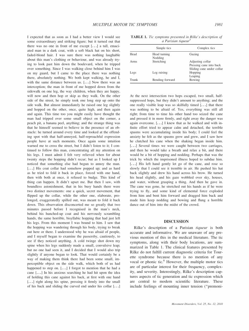
DISCUSSION
Rilke’s description of a Parisian tiqueur is both
accurate and informative. We are unaware of any pre-
vious mention of this in the medical literature. The tic
symptoms, along with their body locations, are sum-
marized in Table 1. The clinical features presented by
Rilke do not fulfill current diagnostic criteria for Tour-
ette syndrome because there is no mention of any
vocal or phonic tic.6 However, the multiple motor tics
are of particular interest for their frequency, complex-
ity, and severity. Interestingly, Rilke’s description cap-
tures aspects of tic generation and tic expression which
are central to modern scientific literature. These
include feelings of mounting inner tension (‘‘premoni-
TABLE 1. Tic symptoms presented in Rilke’s description ofa Parisian tiqueur5
Simple tics Complex tics
Head Head turning GazingNodding
Arms Stretching Adjusting collarPressing cane into backSliding cane under collar
Legs Leg raising HoppingLeaping
Trunk Bending forward Bowing
1981MULTIPLE MOTOR TIC SYMPTOMS
Movement Disorders, Vol. 25, No. 12, 2010

tory urges’’ or ‘‘sensory tics’’) temporarily relieved by
tic expression7 and the social stigma elicited by dem-
onstration of tic symptoms in public spaces and cam-
ouflaging techniques adopted by individuals with
chronic tics.8
Acknowledgments: We wish to thank Tourettes Action,UK for continuing support.
Financial Disclosures: None.
Author Roles: Research project: conception (AEC, MGP),organization (AEC), execution (AEC, TQ). Manuscript: writ-ing of the first draft (AEC, TQ), review and critique (MGP,FA, HR).
REFERENCES
1. Robertson MM, Cavanna AE. Tourette syndrome: the facts.Oxford: Oxford University press; 2008.
2. Hurst MJ, Hurst DL. Tolstoy’s description of Tourette syndromein Anna Karenina. J Child Neurol 1994;9:366–367.
3. Kaplan RE. Tourette’s syndrome in George Eliot’s Silas Marner.J Neuropsy Clin Neurosci 1998;10:367.
4. Murray TJ. Dr Samuel Johnson’s movement disorder. Br Med J1979;1:1610–1614.
5. Rilke R.M. The notebook of Malte Laurids Brigge [transl. by S.Mitchell]. New York: Limited Editions Club; 1987.
6. American Psychiatric Association. Diagnostic and statistical man-ual of mental disorders, Fourth ed., Text Revision (DSM-IV-TR).Washington, DC: American Psychiatric Press; 2000.
7. Prado HS, Rosario MC, Lee J, Hounie AG, Shavitt RG, Miguel EC.Sensory phenomena in obsessive-compulsive disorder and tic disor-ders: a review of the literature. CNS Spectr 2008;13: 425–432.
8. Davis KK, Davis JS, Dowler L. In motion, out of place: the publicspace(s) of Tourette syndrome. Soc Sci Med 2004;59:103–112.
Prevalence of THAP1 SequenceVariants in German Patients with
Primary Dystonia
Anne S. Sohn, PhD,1 Nicola Glockle,1
Andrea Duarte Doetzer,1 Gunther Deuschl, MD,2
Ute Felbor, MD,3 Helge R. Topka, MD,4
Ludger Schols, MD,5 Olaf Riess, MD,1
Peter Bauer, MD,1 Ulrich Muller, MD,6
and Kathrin Grundmann, MD1*
1Department of Medical Genetics, Institute of HumanGenetics, University of Tuebingen, Tubingen, Germany;2Department of Neurology, University of Kiel, Kiel,Germany; 3Institute of Human Genetics, University of
Greifswald, Greifswald, Germany; 4Department of Neurologyand Clinical Neurophysiology, Academic Hospital
Bogenhausen, Technical University of Munich, Munich,Germany; 5Department of Neurodegenerative Diseases,Hertie-Institute for Clinical Brain Research, University of
Tuebingen, Germany; 6Institute of Human Genetics,Justus-Liebig-University, Giessen, Germany
Abstract: Primary dystonias are a clinically and geneticallyheterogeneous group of movement disorders, but only fortwo of them, i.e., dystonia 1 and dystonia 6, the disease caus-ing gene has been identified. Dystonia 1 is characterized byan early onset and is caused by a mutation in the TOR1Agene. Only recently, mutations in THAP1 have been shown tobe the cause of DYT6 dystonia. We analyzed 610 patientswith various forms of dystonia for sequence variants in theTHAP1 gene by means of high resolution melting to delineatethe prevalence of sequence variants and phenotypic variabili-ty. We identified seven sequence variants in patients and onesequence variant in a control. The sequence variants werenot detected in 537 healthy controls. Four patients presentwith generalized dystonia with speech involvement of earlyonset, another three patients suffered exclusively from cervi-cal dystonia of adult onset. These findings suggest thatTHAP1 sequence variations seem to be associated with differ-ent ages of onset and distribution of symptoms. Conse-quently, the phenotypic spectrummight be broader than pre-viously assumed. � 2010 Movement Disorder Society
Key words: dystonia; THAP1; high-resolution melting;DYT6
Additional Supporting Information may be found in the online ver-sion of this article.
The first two authors contributed equally to this work.*Correspondence to: Kathrin Grundmann, Institute of Human
Genetics, University of Tubingen, Calwerstrasse 7, D-72076 Tubin-gen, Germany. E-mail: [email protected]
Potential conflict of interest: Nothing to report.Received 28 January 2010; Revised 15 March 2010; Accepted 31
March 2010Published online 28 July 2010 in Wiley Online Library
(wileyonlinelibrary.com). DOI: 10.1002/mds.23207
Movement Disorders, Vol. 25, No. 12, 2010
1982 A.S. SOHN ET AL.

Dystonias are a heterogeneous group of movement
disorders characterized by sustained muscle contrac-
tions and abnormal postures.1 They can be subclassi-
fied in primary and secondary forms. Many primary
forms have a genetic etiology and can be subdivided
into ‘‘pure’’ forms, dystonia plus syndromes, and par-
oxysmal dystonias. To date at least 17 distinct types of
monogenic primary dystonias have been recognized.
Six of them are ‘‘pure’’ forms, where dystonia is the
sole or main symptom. The disease gene has been
identified in two of them, i.e., dystonia 1 (locus
DYT1) and dystonia 6 (locus DYT6). Both are inher-
ited as autosomal dominant traits.2 In both dystonia 1
and dystonia 6, onset is commonly during childhood or
adolescence. However adult-onset has been described
in both conditions as well.3,4 The disease gene in dys-
tonia 1, i.e., TOR1A was identified more than a decade
ago5 and one mutation within this gene (GAG deletion)
accounts for the majority of monogenic ‘‘pure’’ pri-
mary dystonias. More recently, the gene THAP1 was
found to be mutated in dystonia 6.6 Extensive mutation
analyses in various populations have demonstrated that
cranial and cervical involvement are common in
patients with THAP1 mutations.7–11 We performed
comprehensive mutation analysis of THAP1 in a large
cohort of German patients with primary dystonia in
order to determine the prevalence of dystonia 6 and to
further delineate its phenotype.
PATIENTS AND METHODS
Recruitment of Patients and Controls
610 patients with dystonia were studied. They were
recruited at neurological centers in Germany and clini-
cally diagnosed by specialists according to current cri-
teria.12 Patients were divided into two groups. Group I
(n 5 193) included patients with focal, segmental,
multifocal, or generalized dystonia and absence of
additional signs and symptoms. Age of onset, site of
onset, disease duration, and family history were estab-
lished from patients’ self-reports and clinical records.
Demographic data are presented in Table 1. Patients of
group II (n 5 417) presented with different forms of
pure dystonia and dystonia plus. Additional symptoms
suggestive of secondary dystonia were excluded by
neurological examination. In patients of both groups
mutations in the genes TOR1A (DYT1) and GCH1(DYT5) had been excluded (for details see Supporting
Information). All patients were of German origin. The
537 controls included 348 CEPH samples13 and 189
Germans without neurological disorders.
This study was approved by the local ethics commit-
tee. After obtaining informed consent, blood samples
were drawn for DNA extraction.
Genetic Analysis
PCR and HRM of the coding exons of THAP1 were
performed on LightCycler 480 (Roche Diagnostics,
Germany) using LightCycler1 480 HRM Master
according to the manufacturer’s instructions (Roche
Applied Science, Germany). Samples showing diver-
gent HRM profiles were sequenced on both strands
(for details see Supporting Information).
RESULTS
THAP1 sequence variants were found in 4 patients
of Group 1 (2%) and in three subjects of Group 2
(0.7%; Supporting Information Figure 1). The overall
frequency of THAP1 sequence variants was 1.14% (7/
610). None of the patients with a THAP1 sequence
variants had a positive family history. Clinical findings
and THAP1 sequence variants are given in Table 1.
The sequence variants comprised five missense muta-
tions, one 2-bp deletion, and one nonsense mutation.
All are located in the coding region of THAP1 (Fig. 1)
and were not discovered in any of the 537 European
controls. An additional sequence change, a one 2-bp
deletion (c.197_198delAG) was discovered in a control
subject (Supporting Information Figure).
THAP1 is a DNA-binding protein which contains an
N-terminal THAP domain, a zinc finger domain, a cen-
tral proline-rich region, a coiled-coil region and a bi-
partite nuclear localization sequence in its C-terminal
half (Fig. 1). The sequence variant p.H57N
(c.169C>A) is located in the zinc finger domain of the
THAP1 protein. It affects a highly conserved histidine
at position 57, which forms a zinc binding site together
with three cysteine residues. Apart from generalized
dystonia, the patient presented with a mild speech dis-
ability (Table 2). The sequence variant p.Y137C
(c.410A>G) is located 3 amino acids upstream of the
nuclear localisation signal. The patient developed oro-
bulbar symptoms during pregnancy followed by cervi-
TABLE 1. Characteristics of the study populationof Group I
Type ofdystonia
No. ofpatients
Sexmale/female
Mean 6 SDAge at onset
Family historyPositive no. (%)
Generalized 14 6/8 16 6 11 1 (7)Multifocal 8 4/4 22 6 12 1 (12)Segmental 28 11/17 42 6 18 3 (11)Focal 143 53/90 47 6 14 6 (4)
1983THAP1 SEQUENCE VARIANTS IN PRIMARY DYSTONIA
Movement Disorders, Vol. 25, No. 12, 2010

cal dystonia (Table 2). The sequence variant p.Q124X
(c.370C>T) produces a stop codon immediately before
the nuclear localization sequence. Symptoms in the
patient started in the lower limbs during early child-
hood. Cranial symptoms manifested as Meige syn-
drome, anterocollis, and pharyngolaryngeal dystonia
(Table 2). The sequence variant c.388_389delTC is
also located immediately before the nuclear localiza-
tion sequence and results in an early stop codon
(p.S130fsX133). The patient suffered exclusively from
primary cervical dystonia (Table 2). The remaining
three sequence variants [c.427A>G, (p.M143V),
c.574G>A (p.D192N), c.247T>C (p.C83R)] were
detected in patients presenting with cervical dystonia
as the only clinical sign. Age of onset was 19, 46, and
30 years of age and none of these patients developed
other dystonic symptoms during a course of several
years (Table 2). These sequence changes affect differ-
ent regions of the gene. The p.M143V (c.427A>G)
sequence variant is located in the coiled-coil region of
the protein, the p.D192N (c.574G>A) sequence variant
in the C-terminal end of the protein, and the pC83R
(c.247T>C) sequence variant affects an amino acid ad-
jacent to the THAP domain.
DISCUSSION
This study describes 7 THAP1 sequence variants in
patients with primary dystonia. All sequence variations
except c.388_389delTC have not been recognized in a
patient before.7–11 The overall frequency of sequence
variations was �1%, and corresponds to the frequency
reported in previous studies.7–9 The sequence variations
are spread over the entire gene and affect among other
regions the THAP and the coiled-coil domain (Fig. 1).
Regarding the clinical manifestation, 3 of 7 patients
(patient 1, c.169C>A; patient 2, c.410A>G; patient 3,
c.370C>T; Table 2), presented with a phenotype con-
sistent with the typical phenotype described for dysto-
nia 6 starting early in life with involvement of the cra-
niocervical region and affection of pharyngolaryngeal
or orobulbar muscles.14 Of note, the other four patients
suffered exclusively from cervical dystonia (Table 2).
The mean age of onset of 32.5 years is earlier than
reported for the classical craniocervical dystonia,
which has a mean age of onset of 54.3 years.15
The sequence changes reported are the likely cause
of dystonia in these patients. None of these changes
was found in any of the 537 controls; two sequence
changes (p.Q124X and p.S130fsX133) caused a stop
codon and thus truncation of important functional
domains of the THAP1 protein; the c.169C>A variant
(p.H57N) abolishes DNA- and zinc-binding activity in
vitro;16,17 and the c.388_389delTC variant has previ-
ously been reported in another dystonia patient with
early onset (9 years) who presented with speech prob-
lems followed by writer’s cramp.9
We identified one sequence change (c.197_198delAG)
in a German control that results in truncation of
THAP1 (p.E66fsX84). As the controls studied are
anonymous we were not able to retrace the mutation.
The mutation carrier might have shown very mild
symptoms, which would not have been reported. A
pathologic phenotype might also have been absent
due to reduced penetrance which has been reported in
dystonia 6.14
Given variable expression and reduced penetrance in
dystonia 6, additional genetic and environmental fac-
tors appear to contribute to disease manifestation and
severity of signs and symptoms. In fact, variable
expression and reduced penetrance might explain ab-
sence of a positive family history in all mutation car-
riers reported here. Unfortunately, this possibility could
not be tested experimentally since parents were not
available for clinical examination or mutation analysis.
In this study, we used HRM for mutational analy-
sis. This technique might not detect homozygous
FIG. 1. Scheme of the THAP1 protein. The THAP domain is shown in orange, the proline rich region in green. The nuclear localization sitewithin the coiled-coil domain (blue) is shown in red. All identified sequence variations are shown (arrows). [Color figure can be viewed in theonline issue, which is available at wileyonlinelibrary.com.]
Movement Disorders, Vol. 25, No. 12, 2010
1984 A.S. SOHN ET AL.

mutations, exon rearrangements, whole exon dele-
tions, and intronic changes. Nonetheless, frequency
of mutations detected is in concordance with pub-
lished data.7–9
In conclusion, the findings in the large cohort of
patients studied confirm previous observations of pref-
erential cranio-cervical involvement in dystonia 6 and
emphasize the importance of THAP1 mutation analysis
in patients with cranio-cervical involvement, affection
of speech, and tendency to generalize. Our study also
extends the phenotypic spectrum associated with
THAP1 sequence variations, as we identified THAP1sequence variations in patients with focal cervical dys-
tonia. The clinical manifestation of cervical dystonia in
DYT6 mutation carriers has also been reported by Xiao
et al. 2009 who identified THAP1 sequence variations
in a cohort of mainly adult-onset primary dystonia.11
Consequently THAP1 sequence variations seem to be
associated with different ages of onset and distribution
of symptoms and thus the phenotypic spectrum might
be broader than previously assumed.
Author Roles: A. Sohn: conception and design, executionof the project, data analysis; N. Glockle: conception anddesign, execution of the project, data analysis; A. Duarte Dot-zer: execution of the project; L. Schols: data acquisition; G.Deuschl: data acquisition; U. Felbor: data acquisition; H.Topka: data acquisition; U. Muller: data acquisition and editingand revising of the text; P. Bauer: conception and design, dataanalysis, revising of the text; O. Riess: conception and design,data analysis, revising of the text; and K. Grundmann: concep-tion and design, data analysis, drafting, editing and revising ofthe text.
Financial Disclosures: Anne S. Soehn: Grants—Fortuenefoundation; Employment—Government employee. N.Gloeckle: Employment—Government employee. AndreaDuarte Doetzer: Grants—received funding from CNPq(n:155 61 2108 9000); Employment—Government em-ployee. G. Deuschl: Honoraria—Medtronic, Lundbeck, Teva;Grants—German Research Council, German Ministry of Edu-cation and Research, Medtronic; Employment—Governmentemployee; Royalties—Thieme publisher. U Felbor: Grants—UF is supported by the Bavarian Genome Network (Bay-Gene) and the Deutsche Forschungsgemeinschaft/Graduier-tenkolleg 1048; Employment—Government employee. L.Schols: Grants—has received funding from the EU forEUROSCA (LSHM-CT-2004-503304), the Bundesministie-rium fur Bildung und Forschung for RISCA (01GM0820),EUROSPA (01GM0807), LEUKONET (01GM0838) andmitoNET (01GM0864) as well as from the Deutsche For-schungsgemeinschaft (SCHO754/4-1) in the last 12 months;Employment—Government employee. O. Riess: AdvisoryBoards—Centogene; Grants—funding from the EU forEUROSCA (LSHM-CT-2004-503304), RATstream (037846),GENEPARK (037544), Mitotarget (HEALTH-F2-2008-223388), Neuromodel (215618-2), NEURASYN (238316),Techgene (223143), MarkMD (230596) the Bundesministie-rium fur Bildung und Forschung for RISCA (01GM0820),EUROSPA (01GM0807), NGFNplus ‘‘Parkinson’’ (01GS08134)and MRNET (01GS08162) as well as from the Deutsche For-schungsgemeinschaft (RI 682/10-1); Employment—Govern-ment employee. P. Bauer: Advisory Boards—Centogene;Honoraria—Roche Diagnostics; Grants—research grants ofthe German Research Council (BMBF) to GeNeMove(01GM0603), EUROSPA (01GM0807) and RISCA(09GM0820) as well as from the EU for EUROSCA (LSHM-CT-2004-503304), MarkMD (FP7-People PIAP-2008-230596) and TECHGENE (FP7-Health 2007-B 223143);received funding from the HSP-Selbsthilfegruppe Deutsch-land; Employment—Government employee. U. Muller:Grants—received funding from the PSP-Society; Employ-
TABLE 2. List of THAP1 sequence variations identified by HRM in a cohort of 610 dystonia patients
Case Study group Sex Clinical diagnosis Exon Nucleotide change Effect on protein level
1 II f Generalized dystonia 2 c.169 C>A p.H57NStarting in lower limbMild dysarthriaOnset in early childhood
2 II f Cervical dystonia, Meige syndrome(oro-mandibular dystonia),pharyngolaryngeal dystonia
3 c.410A>G p.Y137C
3 II Generalized dystonia 3 c.370C>T p.Q124XStarting in lower limbMeige syndrome (oro-mandibular dystonia),Onset during pregnancy
4 I f Cervical dystonia 3 c.388_389delTC p.S130fsX133Onset at age 35
5 I f Cervical dystonia 3 c.427A>G p.M143VOnset at age 46
6 I m Cervical dystonia 3 c.574G>A p.D192NOnset at age 19
7 I f Cervical dystonia 2 c.247T>C p.C83ROnset at age 34
8 II f Control, no symptoms of dystonia reported 2 c.197_198delAG p.E66fsX84
1985THAP1 SEQUENCE VARIANTS IN PRIMARY DYSTONIA
Movement Disorders, Vol. 25, No. 12, 2010

ment—Government employee. K. Grundmann: Grants—hasreceived funding from the Dystonia Medical Research Foun-dation; Employment—Government employee.
REFERENCES
1. Fahn S. Concept and classification of dystonia. Adv Neurol 1988;50:1–8.
2. Muller U. The monogenic primary dystonias. Brain 2009;132(Part 8):2005–2025.
3. Almasy L, Bressman S, de L, Risch N. Ethnic variation in theclinical expression of idiopathic torsion dystonia. Mov Disord1997;12:715–721.
4. Grundmann K, Laubis-Herrmann U, Bauer I, et al. Frequencyand phenotypic variability of the GAG deletion of the DYT1gene in an unselected group of patients with dystonia. Arch Neu-rol 2003;60:1266–1270.
5. Ozelius LJ, Hewett JW, Page CE, et al. The early-onset torsiondystonia gene (DYT1) encodes an ATP-binding protein. NatGenet 1997;17:40–48.
6. Fuchs T, Gavarini S, Saunders-Pullman R, et al. Mutations in theTHAP1 gene are responsible for DYT6 primary torsion dystonia.Nat Genet 2009;41:286–288.
7. Bonetti M, Barzaghi C, Brancati F, et al. Mutation screening of theDYT6/THAP1 gene in Italy. Mov Disord 2009;24:2424–2427.
8. Bressman SB, Raymond D, Fuchs T, et al. Mutations in THAP1(DYT6) in early-onset dystonia: a genetic screening study. LancetNeurol 2009;8:441–446.
9. Djarmati A, Schneider SA, Lohmann K, et al. Mutations in THAP1(DYT6) and generalised dystonia with prominent spasmodic dyspho-nia: a genetic screening study. Lancet Neurol 2009; 8:447–452.
10. Houlden H, Schneider SA, Paudel R, et al. THAP1 mutations(DYT6) are an additional cause of early-onset dystonia. Neurology2010;74:846–850.
11. Xiao J, Zhao Y, Bastian RW, et al. Novel THAP1 sequence var-iants in primary dystonia. Neurology 2010;74:229–238.
12. Fahn S, Eldridge R. Definition of dystonia and classification of thedystonic states. Adv Neurol 1976;14:1–5.
13. Dausset J, Cann H, Cohen D, et al. Centre d’etude du polymor-phisme humain (CEPH): collaborative genetic mapping of thehuman genome. Genomics 1990;6:575–577.
14. Saunders-Pullman R, Raymond D, Senthil G, et al. Narrowing theDYT6 dystonia region and evidence for locus heterogeneity in theAmish-Mennonites. Am J Med Genet A 2007;143A:2098–2105.
15. Defazio G, Abbruzzese G, Girlanda P, et al. Does sex influenceage at onset in cranial-cervical and upper limb dystonia? J NeurolNeurosurg Psychiatry 2003;74:265–267.
16. Bessiere D, Lacroix C, Campagne S, et al. Structure-function anal-ysis of the THAP zinc finger of THAP1, a large C2CH DNA-bind-ing module linked to Rb/E2F pathways. J Biol Chem 2008;283:4352–4363.
17. Clouaire T, Roussigne M, Ecochard V, Mathe C, Amalric F, Gi-rard JP. The THAP domain of THAP1 is a large C2CH modulewith zinc-dependent sequence-specific DNA-binding activity. ProcNatl Acad Sci USA 2005;102:6907–6912.
Specific Pattern of EarlyWhite-Matter Changes in PureHereditary Spastic Paraplegia
Thomas Duning, MD,1*Tobias Warnecke, MD,1
Anja Schirmacher, PhD,1Hagen Schiffbauer, MD,2
Hubertus Lohmann, PhD,1Siawoosh Mohammadi, PhD,1
Peter Young, MD,1 and Michael Deppe, PhD1
1Department of Neurology, University Hospital of Munster,Munster, Germany; 2Department of Clinical Radiology,
University Hospital of Munster, Munster, Germany
Abstract: Hereditary spastic paraplegias (HSP) are geneti-cally and clinically heterogeneous neurodegenerative dis-orders. Most MR studies on HSP include very heterogene-ous samples of patients, and findings were inconsistent.Here, we examined six patients with pure HSP and SPG4mutations by clinical evaluation, detailed neuropsychologi-cal testing, and neuroimaging analyses, including conven-tional MRI, diffusion tensor imaging (DTI), and brainvolumetry. Differences of voxel-wise statistics and ROI-based analysis of DTI data between patients and 32healthy volunteers were evaluated. Although conventionalMRI and brain volumetry were normal, DTI revealedwidespread disturbance of white matter (WM) integrity(P < 0.001), mainly affecting the corticospinal tract. Withlonger disease duration, frontal regions were alsoinvolved. The WM changes were also present in subclini-cal subjects harbouring the pathogenic mutation. Thesesubtle WM abnormalities have functional relevancebecause they correlated with clinical symptoms. Thus,early alterations of nerve fibres, which can be detected byDTI, might serve as a biological marker in HSP, in par-ticular with respect to future longitudinal studies. � 2010Movement Disorder Society
Key words: hereditary spastic paraplegia; diffusion tensorimaging; white-matter changes; SPG4
Hereditary spastic paraplegia (HSP) is a clinically
and genetically heterogeneous neurodegenerative disor-
der characterized by progressive spasticity and weak-
ness of the lower limbs occurring in the absence (pure
HSP) or presence (complicated HSP) of additional
The first two authors contributed equally to this work.*Correspondence to: Dr. Thomas Duning, MD, Department of
Neurology, University Hospital of Muenster, Albert-Schweitzer-Str.33, 48149 Muenster, Germany. E-mail: [email protected]
Potential conflict of interest: Nothing to report.Received 21 December 2009; Revised 11 March 2010; Accepted
31 March 2010Published online 28 July 2010 in Wiley Online Library
(wileyonlinelibrary.com). DOI: 10.1002/mds.23211
1986 DUNING ET AL.
Movement Disorders, Vol. 25, No. 12, 2010

major clinical features such as cognitive impairment
and extrapyramidal or cerebellar symptoms.1 To date,
45 genetic loci (SPG1-45) have been mapped and 17
genes identified.2,3
In the diagnostic work-up of HSP, magnetic reso-
nance imaging (MRI) of the brain has been mainly
used to exclude other diseases, included in the differ-
ential diagnosis of HSP. Conventional MRI examina-
tions of HSP patients suggest a loss of volume in the
corpus callosum and a higher incidence of white matter
lesions in some cases.4–7 However, most MRI studies
on HSP include very heterogeneous samples of patients
with clinical or less often genetically proven HSP
forms, and results were inconsistent.1,6–8 Only a few
studies have employed advanced neuroimaging techni-
ques in the evaluation of HSP patients, e.g., diffusion
tensor imaging (DTI).9–12 DTI is a new MRI technique
sensitive to white matter microstructure integrity.13
This study combined high-resolution DTI with statis-
tical parametric mapping (SPM) to determine subtle
microstructural abnormalities across the entire brain in
patients with genetically proven pure HSP (SPG4) and
normal conventional MRI. The purpose was to identify
specific patterns of an early cerebral affection in HSP
and its association with clinical symptoms.
METHODS
Subjects
Six SPG4-HSP patients from two families (median
age 53.3 years, SD 6 14.8 years, range, 34–74 years)
were recruited from our outpatient clinic. Every subject
was examined by experienced neurologists, and a
detailed medical history was obtained. All patients per-
formed a comprehensive neuropsychological test bat-
tery (Table 1).12
The control group was composed of 32 healthy vol-
unteers (17 women, median age 52.1 years, SD 6 11.8
years, range, 36–61 years). All control subjects had a
normal neurologic examination, no evidence of neuro-
logic disease, and normal MRI. All individuals
gave written informed consent prior to examinations.
The study protocol was approved by the local ethics
committee.
MRI Acquisition and Image Analysis
Image data were obtained on a 3.0-T system (Gyro-
scan Intera, Philips Medical System) with a high-reso-
lution structural T1-weighted 3D turbo-field-echo
sequence for brain volumetry (field-of-view of 25.6 320.5 3 16 cm3, reconstructed after zero filling to 512
3 410 3 320 cubic voxels with an edge length of 0.5
mm), and T2-weighted and FLAIR imaging. For DTI,
we employed echo planar imaging (EPI) with 20 diffu-
sion directions (two b-factors, 0 s/mm2 and 1000 s/
mm2, TR 5 9.8 s/TE 5 98 ms, voxel size: 0.89 mm
3 0.89 mm 3 3.6 mm, 2 averages).
The diffusion tensor and fractional anisotropy (FA)
maps (FA value of each voxel) were calculated from
spatially normalized images. The method has been
described in detail previously.14 Voxel-based analysis
of the FA image was performed using SPM5 (Well-
come Department of Imaging Neuroscience, London).
Differences of FA values between the patients and con-
trols were statistically evaluated by analysis of covari-
ance, modeling the factor age as co-variable to account
for the age dependency of the FA (P < 0.001). Addi-
tionally, the linear regression tool of the software was
employed to correlate maps of decreased FA with dis-
ease duration. After controlling for age, statistical
threshold for the correlation analysis was set at P 50.01 for multiple comparisons (FWE).
To assess the magnitude of regional FA alterations,
we also performed additional quantitative region-of-in-
terest (ROI) analyses. Mean FA values were calculated
within 11 defined ROIs, which were derived from an
averaged and symmetrized (x-axis) mask of the healthy
individuals with FA-values > 0.4 by deleting voxels
not associated with the respective structures. To put
the results more into perspective, FA values of the
SPG4 patients were also compared with FA values of
previously reported group of complicated HSP patients
with proven SPG7 mutations.12
Brain tissue volumes, normalized for subject head
size, were calculated from the high-resolution T1-
weighted images, using the well-established cross-sec-
tional version of the Structural Imaging Evaluation of
Normalized Atrophy (SIENA) software (SIENAx).15
Differences between patients and healthy controls were
evaluated by analysis of covariance (P < 0.05).
RESULTS
Subject Characteristics
Table 1 summarizes the detailed findings of genetic,
clinical, and neuropsychological evaluation. The indi-
viduals V and VI were not complaining about any neu-
rological symptoms, but neurological examination of
both revealed pathological hyperreflexia. The sequence
variation c.1070T > G (patient II and VI) was a novel
pathogenic SPG4 mutation. Neuropsychological evalu-
ation revealed slight to moderate disturbance of atten-
1987SPECIFIC DTI CHANGES IN PURE HSP
Movement Disorders, Vol. 25, No. 12, 2010

TABLE
1.Clinicalan
dneurop
sycholog
ical
finding
sof
thesixSP
G4-pa
tients
PatientNo.
III
III
IVV
VI
Mutationat
DNA
level
c.839_840delAG;
PTC110aa
c.1070T>G;
p.I357S
c.839_840delAG;
PTC110aa
c.839_840delAG;
PTC110aa
c.839_840delAG;
PTC110aa
c.1070
T>G;p.I357S
Sex
Male
Male
Fem
ale
Fem
ale
Fem
ale
Fem
ale
Onsetage(years)
40
37
51
42
34
38
Disease
duration(yr)
10
30
10
7<1
<1
Upper
limb
Spasticity
22
21
22
Weakness
22
22
22
Ataxia
22
22
22
Hyperreflexia
12
21
22
Sensory
impairm
ent
22
22
22
Lower
limb
Spasticity
11
111
11
11
22
Weakness
211
12
22
Ataxia
12
22
22
Hyperreflexia
11
111
11
11
11
Sensory
Impairm
ent
211
11
22
Babinskisign
Positive
Positivewithouttrigger
Positive
Positive
negative
negative
Walkingability
Unaided
Wheelchair
Unaided
Unaided
unaided
unaided
Bladder
dysfunction
111
21
22
Cognitivefunction
Notdone
Orientation
Norm
alNorm
alNorm
alnorm
alnorm
alApraxia
Norm
alNorm
alNorm
alnorm
alnorm
alMMSE
28
29
30
30
30
Attentionandworkingmem
ory
NAI Digital
symbolsubstitution
40/45%/norm
al38/60%/norm
al35/70%/norm
al30/87%/norm
al32/75%/norm
alCWIT
reading
29/30%/norm
al24/40%/norm
al29/30%/norm
al22/60%/norm
al22/60%/norm
alCWIT
colournam
ing
25/60%/norm
al24/70%/norm
al23/75%/norm
al22/75%/norm
al24/60%/norm
alWMS-R
Digital
span
forw
ard
8/53%/norm
al7/53%/norm
al9/69%/norm
al9/75%/norm
al8/57%/norm
alDigital
Span
Backward
8/71%/norm
al6/53%/norm
al7/52%/norm
al8/73%/norm
al7/58%/norm
alTrail-m
akingtest
[A]
26/70%/norm
al36/60%/norm
al28/70%/norm
al24/60%/norm
al26/50%/norm
alExecutivefunction
CWIT—
interference
condition
28/20%/close
below
average
32/14%/close
below
average
30/18%/close
below
average
13/78%/norm
al14/70%/norm
alRWT—
Letterfluency
[‘S’]
11/16%/close
below
average
14/45%/norm
al16/60%/norm
al19/65%/norm
al16/50%/norm
alTrail-m
akingtest
[B]
65/35%/norm
al110/20%/close
below
average
75/20%/close
below
average
45/80%/norm
al50/70%/norm
alVisuospatialskills
ROCF
Copy
33/16%/close
below
average
33/40%/norm
al35/60%/norm
al35/>
20%/norm
al35/>
20%/norm
alDelayed
recall
16/16%/close
below
average
15/16%/close
below
average
16/16%/close
below
average
24/62%/norm
al26/76%/norm
alVerbal
learningandmem
ory
ALVT
Recalltrial1
4/20%/close
below
average
7/70%/norm
al6/40%/norm
al8/75%/norm
al7/50%/norm
alRecalltrail5
10/30%/norm
al12/40%/norm
al14/65%/norm
al15/80%/norm
al14/60%/norm
alTotaltrials1to
534/15%/close
below
average
40/25%/norm
al50/55%/norm
al58/80%/norm
al50/40%/norm
alDelayed
recall
12/50%/norm
al11/45%/norm
al11/45%/norm
al14/80%/norm
al12/50%/norm
alRecognition(True-False
positives)
12/35%/norm
al12/35%/norm
al13/45%/norm
al15/>
65%/norm
al15/>
65%/norm
al
Ratingofcognitivefunction:test
scores/age-matched
percentile/evaluation.
Pat.V
andVI(rightbars):Clinically
notaffected
25
absent;1
5mild;11
5moderate;
111
5severe;
PTC:premature
term
inationcodon.

tion and executive function but no dementia in three
patients (I, II, and IV).
Brain Volume Differences
There were no significant differences in brain vol-
umes of the SPG4-HSP patients (gray matter, white
matter (WM), relative and normalized brain volumes)
compared with the group of healthy controls. This was
concordant with the conventional neuroradiological
reading showing no abnormalities on conventional MR
imaging.
Voxel-Based FA Analysis
Voxel-based FA analysis of the SPG4 patients
revealed significant FA reductions covering widespread
parts of the brain (P < 0.01), when compared with the
32 healthy controls. With a higher statistical threshold
(P < 0.001), the clusters with significant FA changes
were mainly confined to the temporal lobes and the
corticospinal tracts (Fig. 1A). In addition, voxel-wise
FA analysis of the two SPG4 patients (V and VI) with
only subtle clinical affection was performed as a sepa-
rate group. Interestingly, pattern of significant
decreased FA values had a substantial similarity to the
severely affected SPG4 patients (I–IV), relative to the
healthy controls (Fig. 1B). Regression analysis demon-
strated significant clusters of negative correlation
between FA values and disease duration in corticospi-
nal fibres and also in frontal regions of the brain
(Fig. 1C).
In ROI analysis, only FA values of the occipital
lobe and the splenium did not significantly differ
between HSP patients and the control subjects. Post-
hoc t-test (P < 0.001) revealed significant FA reduc-
tion in ROIs of the corpus callosum, the genu, the tem-
poral lobes, and the whole brain of the pure HSP
(pHSP) patients, when compared with the healthy con-
trols (Fig. 2). Consistent with the additional clinical
features, FA values of the previously reported group of
complicated HSP patients (cHSP)12 were lower in all
ROIs except in the corpus callosum. However, only
FA values of the ROI covering the brainstem signifi-
cantly differed between both HSP groups (cHSP <pHSP).
DISCUSSION
Herein, we provided evidence that genetically pro-
ven HSP patients with normal conventional MRI
showed widespread white matter abnormalities on
DTI analysis. Consistent with the clinical symptoms,
in particular, the corticospinal tracts were affected.
Furthermore, we could show that voxel-based FA
analysis holds promise for detecting very early cere-
bral abnormalities in SPG4-pHSP patients with unre-
vealing conventional MRI and normal brain volumetry
and only subtle clinical affction (hyperreflexia)
(Fig. 1B).
In accordance to Tallaksen et al.,16 we also found
subtle cognitive impairment in three subjects with pure
SPG4 HSP, mainly in patients with longer disease du-
ration. Interestingly, regression analysis revealed an
association between disease duration and FA reduc-
tions in the frontal lobes. Because lesions of frontal
subcortical circuits are known to be associated with
distinct neurobehavioral syndromes, the localization of
these WM abnormalities was in accordance with the
clinical symptoms. Thus, apart from corticospinal
fibres, an involvement of frontal WM might also
explain some of the clinical symptoms that occur with
longer duration of the disease.
On ROI analysis, only the mean FA values in the
ROIs covering the occipital lobes and the splenium
were not significantly decreased. In general, ROI anal-
ysis revealed that SPG4-pHSP patients exhibited lesser
(but still significant) FA reductions compared with the
group of recently reported SPG7-cHSP patients.12
However, only the ROI covering the brainstem showed
a significant difference of FA values between both
HSP groups (SPG7-cHSP < SPG4-pHSP), whereas the
FA values of the other ROIs—including the frontal
lobes—did not significantly differ between both forms.
Alterations in two main cellular functions have been
attributed to the underlying molecular pathomechanism
of HSP. SPG4, the gene encoding for spastin, interacts
with microtubules and promotes their disassembly.
Impairment of its function leads to axonal swelling and
consecutive axonal degeneration.2 The SPG7 gene enc-
odes an ATP-dependent proteolytic complex located at
the mitochondrial inner membrane.17 Thus, SPG7
mutations primarily lead to complex HSP forms and
show some overlapping symptoms with mitochondrial
disease.1,2,12 The different pattern of DTI changes in
both HSP forms may reflect these two differing patho-
mechanistical backgrounds.
Although FA changes primarily involved the cortico-
spinal tracts, the upper extremities of SPG4 patients
are typically not affected. It has been reported that an
impaired energy supply of the axonal transport pre-
dominantly leads to a degeneration of the longest CNS
axons.8,18 The resolution of the DTI does not allow to
distinguish between corticospinal fibres of the upper or
lower extremities. However, with regard to the clinical
1989SPECIFIC DTI CHANGES IN PURE HSP
Movement Disorders, Vol. 25, No. 12, 2010

FIG. 1. SPM ‘‘glass brain’’ representation (left) and coronal/axial/sagittal slices (right) of voxels with a significant decrease in fractional anisot-ropy (FA) among all HSP patients (A) compared with 32 normal controls (P < 0.001, uncorrected; > 50 contiguous voxels). The statistical mapwas superimposed on an averaged FA template of the control group. Colored bars represent t-values; display threshold is set at t value > 3.42. B,When calculated as a separate group, pattern of FA changes of two SPG4 individuals (V, VI) with only slight clinical affection (B) had substan-tial similarity to the FA maps of more severely affected SPG4 patients. C, Clusters of correlation between decreased FA and disease duration (P< 0.01, > 50 continuous voxels; corrected for multiple comparison by false discovery rate method). Apart from corticospinal fibres, an involve-ment of frontal WM might also explain some of the clinical symptoms that occur with longer duration of the disease. [Color figure can be viewedin the online issue, which is available at wileyonlinelibrary.com.]
1990 DUNING ET AL.
Movement Disorders, Vol. 25, No. 12, 2010

FIG. 2. Analysis of covariance revealed significant FA differences between HSP patients and healthy controls in the ROIs shown here. Consistentwith the more severe phenotype, FA values of the complicated forms were lower in all ROIs except the corpus callosum, relative to the pure HSPforms (posthoc t-test; P < 0.001). However, only the ROI covering the brainstem showed a significant difference of FA values between both HSPgroups (cHSP < pHSP).

symptoms, the FA changes most likely reflect a micro-
structural damage of the longest motor fibres of the
corticospinal tract.
In summary, both voxel-wise FA statistics and ROI-
based FA analysis provide evidence that DTI could be
a sensitive technique to uncover subtle microstructural
cerebral abnormalities in HSP with unrevealing con-
ventional MRI and normal brain volumetry. Our data
support the notion that subtle WM changes on DTI are
the first clinicoanatomical abnormalities detectable,
before distinctive regional degeneration on conven-
tional MRI becomes visible. When compared with
complicated HSP-forms, even pure HSP patients with
less severe phenotype showed widespread WM abnor-
malities on DTI analysis, mainly affecting the cortico-
spinal tracts and temporal regions. These pattern of
WM changes where also evident in SPG4 patients with
only subtle clinical affection. Future studies may inves-
tigate whether DTI can detect preclinical degeneration
and, thus, serve as a predictive test of HSP. Further-
more, regarding its functional significance, FA changes
might serve as a biological marker of the phenotype in
HSP, in particular with respect to future longitudinal
studies.
Financial Disclosures: T.D.: Grants: Genzyme. T.W.,A.S., H.S., H.L., S.M.: none. P.Y.: Honoria: Genzyme, Boeh-ringer, Ingelheim, MEDA Pharma; Grants: University ofMunster (IMF). M.D.: Grants: Deutsche Forschungs Gemein-schaft (DFG).
Author Roles: T.D. was involved in the conception, orga-nization, and execution of the research project and in designand execution of the statistical analysis and also in writing,in the review, and critique of the manuscript. T.W. wasinvolved in the conception, organization, and execution ofthe research project and in design and execution of the statis-tical analysis and also in the writing of the first draft, review,and critique of the manuscript. A.S. was involved in the exe-cution of the research project and in execution of the statisti-cal analysis and also in the review and critique of the manu-script. S.M. was involved in the organization of the researchproject and in design of the statistical analysis and also inthe review and critique of the manuscript. H.S. and H.L.were involved in the execution of the research project andstatistical analysis and in the review and critique of themanuscript. M.D. and P.Y. were involved in the conceptionand organization of the research project and in design, execu-
tion, review, and critique of the statistical analysis and alsoin the review and critique of the manuscript.
REFERENCES
1. Depienne C, Stevanin G, Brice A, Durr A. Hereditary spastic par-aplegias: an update. Curr Opin Neurol 2007;20:674–680.
2. Salinas S, Proukakis C, Crosby A, Warner TT. Hereditary spasticparaplegia: clinical features and pathogenetic mechanisms. LancetNeurol 2008;7:1127–1138.
3. Dursun U, Koroglu C, Kocasoy OE, Ugur SA, Tolun A. Autoso-mal recessive spastic paraplegia (SPG45) with mental retardationmaps to 10q24.3-q25.1. Neurogenetics 2009;10:325–331.
4. Fink JK. Hereditary spastic paraplegia. In: Rimoin DL, Emery AEH,editors. Emery & Rimoin’s Principles and Practice of Medical Genet-ics. Philadelphia: Churchill Livingstone Elsevier; 2007. p 2771–2801.
5. Hourani R, El Hajj T, Barada WH, Hourani M, Yamout BI. MRimaging findings in autosomal recessive hereditary spastic paraple-gia. AJNR Am J Neuroradiol 2009;30:936–940.
6. Kassubek J, Sperfeld AD, Baumgartner A, et al. Brain atrophy inpure and complicated hereditary spastic paraparesis: a quantitative3D MRI study. Eur J Neurol 2006;13:880–886.
7. Uttner I, Baumgartner A, Sperfeld AD, Kassubek J. Cognitive per-formance in pure and complicated hereditary spastic paraparesis: aneuropsychological and neuroimaging study. Neurosci Lett 2007;419:158–161.
8. McDermott C, White K, Bushby K, Shaw P. Hereditary spastic par-aparesis: a review of new developments. J Neurol Neurosurg Psy-chiatry 2000;69:150–160.
9. Chen Q, Lui S, Wang JG, et al. Diffusion tensor imaging of twounrelated Chinese men with hereditary spastic paraplegia associ-ated with thin corpus callosum. Neurosci Lett 2008;441:21–24.
10. Dreha-Kulaczewski S, Dechent P, Helms G, et al. Cerebral meta-bolic and structural alterations in hereditary spastic paraplegia withthin corpus callosum assessed by MRS and DTI. Neuroradiology2006;48:893–898.
11. Ota M, Sato N, Saitoh Y, et al. Diffusion tensor imaging in familialspastic paraplegia with mental impairment and thin corpus cal-losum. Magn Reson Med Sci 2008;7:163–167.
12. Warnecke T, Duning T, Schwan A, et al. A novel form of autoso-mal recessive hereditary spastic paraplegia caused by a new SPG7mutation. Neurology 2007;69:368–375.
13. Assaf Y, Pasternak O. Diffusion tensor imaging (DTI)-based whitematter mapping in brain research: a review. J Mol Neurosci 2008;34:51–61.
14. Deppe M, Kellinghaus C, Duning T, et al. Nerve fiber impairmentof anterior thalamocortical circuitry in juvenile myoclonic epilepsy.Neurology 2008;71:1981–1985.
15. Smith SM, Zhang Y, Jenkinson M, et al. Accurate, robust, andautomated longitudinal and cross-sectional brain change analysis.Neuroimage 2002;17:479–489.
16. Tallaksen CM, Durr A, Brice A. Recent advances in hereditaryspastic paraplegia. Curr Opin Neurol 2001;14:457–463.
17. Nolden M, Ehses S, Koppen M, et al. The m-AAA protease defec-tive in hereditary spastic paraplegia controls ribosome assembly inmitochondria. Cell 2005;123:277–289.
18. Harding AE. Hereditary spastic paraplegias. Semin Neurol 1993;13:333–336.
Movement Disorders, Vol. 25, No. 12, 2010
1992 DUNING ET AL.


![Binder 216, Terminology [Trematoda Taxon Notebooks]](https://static.fdokumen.com/doc/165x107/63338ab23108fad7760f19c8/binder-216-terminology-trematoda-taxon-notebooks.jpg)


![Binder 200, Small families [Trematoda Taxon Notebooks]](https://static.fdokumen.com/doc/165x107/6324444cb104cba27a091035/binder-200-small-families-trematoda-taxon-notebooks.jpg)