Integration of sensorimotor mappings by making use of redundancies
Upload
independentCategory
view
0download
0
R
Ts
Ma
Sb
c
a
ARR1AA
KBTTSLTA
1
ti[iaSfaaacwn
0d
Behavioural Brain Research 216 (2011) 685–691
Contents lists available at ScienceDirect
Behavioural Brain Research
journa l homepage: www.e lsev ier .com/ locate /bbr
esearch report
he impact of basal ganglia lesions on sensorimotor synchronization,pontaneous motor tempo, and the detection of tempo changes
ichael Schwartzea,∗, Peter E. Kellerb, Aniruddh D. Patel c, Sonja A. Kotza
Max Planck Institute for Human Cognitive and Brain Sciences, Independent Research Group “Neurocognition of Rhythm in Communication”,tephanstrasse 1a, 04103 Leipzig, GermanyMax Planck Institute for Human Cognitive and Brain Sciences, Max Planck Research Group “Music Cognition and Action”, Leipzig, GermanyThe Neurosciences Institute, San Diego, USA
r t i c l e i n f o
rticle history:eceived 5 August 2010eceived in revised form0 September 2010ccepted 14 September 2010vailable online 29 September 2010
eywords:asal gangliaemporal processingapping
a b s t r a c t
The basal ganglia (BG) are part of extensive subcortico-cortical circuits that are involved in a variety ofmotor and non-motor cognitive functions. Accumulating evidence suggests that one specific functionthat engages the BG and associated cortico-striato-thalamo-cortical circuitry is temporal processing, i.e.,the mechanisms that underlie the encoding, decoding and evaluation of temporal relations or tempo-ral structure. In the current study we investigated the interplay of two processes that require preciserepresentations of temporal structure, namely the perception of an auditory pacing signal and manualmotor production by means of finger tapping in a sensorimotor synchronization task. Patients with focallesions of the BG and healthy control participants were asked to align finger taps to tone sequences thateither did or did not contain a tempo acceleration or tempo deceleration at a predefined position, andto continue tapping at the final tempo after the pacing sequence had ceased. Performance in this adap-
ynchronizationesionempo changettention
tive synchronization-continuation paradigm differed between the two groups. Selective damage to theBG affected the abilities to detect tempo changes and to perform attention-dependent error correction,particularly in response to tempo decelerations. An additional assessment of preferred spontaneous, i.e.,unpaced but regular, production rates yielded more heterogeneous results in the patient group. Togetherthese findings provide evidence for less efficient processing in the perception and the production of tem-poral structure in patients with focal BG lesions. The results also support the functional role of the BG
nden
system in attention-depe. Introduction
Individuals continuously adjust their behavior to environmen-al changes. The underlying adaptive process unfolds in time andnvolves the cyclic processing of motor and sensory information1]. The question arises whether this cyclic processing is merelyntrinsically temporal or to what extent temporal information isctually processed and used as a source of information in itself.ome internal representation of temporal structure is a prerequisiteor efficient timing, which in turn bears the potential to optimizedaptive processes. Efficient timing of behavior implies temporallyppropriate reactive and proactive actions. The latter depend on
nticipation and predictions about the temporal structure of futurehanges or events as well as the ability to temporally align behaviorith these events. Both aspects converge in sensorimotor synchro-ization (SMS).∗ Corresponding author. Tel.: +49 341 99402473; fax: +49 341 99402260.E-mail address: [email protected] (M. Schwartze).
166-4328/$ – see front matter © 2010 Elsevier B.V. All rights reserved.oi:10.1016/j.bbr.2010.09.015
t temporal processing.© 2010 Elsevier B.V. All rights reserved.
SMS is a specific form of adaptive interaction with the envi-ronment. It is an extensively studied process that merges motorand non-motor components in a single setting. SMS can be char-acterized as the temporal coordination of a motor rhythm with anexternal rhythm (for reviews see [2,3]). This temporal coordina-tion can be conceived as synchronization. Synchronization denotesthe “adjustment of rhythms of oscillating objects due to their weakinteraction” [4]. An oscillation is defined by its phase, relative toanother oscillation, and period, and provides a means to describethe temporal relation of the events that constitute a rhythm interms of frequency, i.e., the repetition of similar events in a specificamount of time. Complex rhythmic activity and synchronizationbetween different rhythms constitute central aspects of life. Phys-iological rhythms interact continuously with each other and theenvironment in order to mediate between internal and external
events [5]. In cognition, this implies adaptation of internally-generated to external rhythms via unidirectional coupling whicheventually leads to stimulus-driven synchronization of behavior.Both SMS and temporal processing have been modeled on thebasis of oscillations and the fundamental dissociation of automatic
6 l Brain
aistinmgctEitTdatea
miowcttdmiswaeoealpi
b(ppnPriwfsitpp
tmnavsstm
86 M. Schwartze et al. / Behavioura
nd controlled sub-processes. For example, temporal processings hypothesized to rest on the coincidental activation of mediumpiny neurons in the BG by ensembles of cortical neural oscilla-ions [6,7]. In SMS, the period of an adaptive oscillatory timekeepers assumed to reflect the temporal structure of the pacing sig-al, thereby establishing a reference for the timing of successiveotor commands [8]. In this context, the current study investi-
ates a combination of these aspects by providing BG patients andontrols with an oscillatory perceptual input whose temporal struc-ure needs to be exploited to generate oscillatory motor behavior.rror correction mechanisms adjust the phase and period of thenternal timekeeper oscillation if it is confronted with a perturba-ion, i.e., a change in the temporal structure of the pacing signal.hese error correction mechanisms are dissociable based on theirependence on attention, the intention to adapt, and awareness oftempo change [9]. Whereas phase correction depends solely on
he intention to maintain synchrony and can therefore be consid-red automatic, period correction depends also on attention andwareness of the tempo change.
A comparable dissociation between automatic and controlledechanisms has been proposed with respect to temporal process-
ng [10]. More specifically, the cerebellum (CE) performs automaticr pre-attentive, short-range, event-based temporal processing,hile in parallel, the BG and cortico-striato-thalamo-cortical cir-
uitry engage in attention-dependent, longer-range, interval-basedemporal processing [7,11,12]. Attention, or the ability to atten-ively detect a change in temporal structure may therefore not onlyecide upon the type of error correction, but also upon the pri-ary temporal processing system. Temporal structure may thereby
nfluence cognitive processes, e.g., as the basis for attentional sethifting and sequence coordination [13]. This view is consistentith Dynamic Attending Theory DAT [14], which proposes that
ttention can be modeled as a self-sustained oscillation capable ofntrainment. Within the framework of DAT, the temporal structuref a stimulus guides the allocation of attentional resources therebyvoking stimulus-driven attending [15]. On this basis, attentivedaptation to tempo change in SMS would depend on the paral-el engagement of pre-attentive and attention-dependent temporalrocessing systems, as well as phase and period correction to adjust
nternal timekeeper and/or attention oscillations.The role of the BG in temporal processing and in SMS has
een investigated primarily in patients with Parkinson’s diseasePD), albeit with mixed results [16–18]. Studies involving PDatients also suggest difficulties in beat extraction and the com-arison of rhythmic sequences [19]. However, PD is a progressiveeurodegenerative disease, and besides medication and differentD subgroups [20] some of the heterogeneity of the respectiveesults may be due to the variable extent of cortical damagen this population, which can be minimal or absent in patients
ith BG lesions [21,22]. Studies on SMS involving patients withocal lesions of the BG are scarce. Aparicio et al. [23] used aynchronization–continuation paradigm and found no evidence formpaired temporal processing performance in this group. Differentasks, stimulus characteristics, and cognitive sets add further com-lexity to the identification of specific BG and cerebellar temporalrocessing functions [24].
Besides attention, temporal range seems to be an important fac-or, as temporal processing evolves across different timescales that
ay map onto different physiological and psychological mecha-isms [10,24,25]. A well-defined boundary between short-rangend longer-range temporal processing remains elusive, although
alues around 500 ms [26] and close to 1000 ms [27] have beenuggested. Fraisse [28] emphasizes that synchronization is mosttable for tempi between 400 and 800 ms, while the intermediaryempo of 600 ms is most representative for unpaced, spontaneousotor activity. This has been confirmed in more recent studies
Research 216 (2011) 685–691
[29] that also found a correlation between individual spontaneousmotor tempo (SMT) and a preferred perceptual tempo [30]. It cor-responds to the “indifference interval” or “indifference zone” that isneither systematically overestimated nor underestimated [28,31].A tempo of 600 ms is within the range for optimal pulse sensa-tion [32] and tempo sensitivity, for a review see [33,34]. Althoughoriginating from a different perspective, Karmarkar and Buono-mano [35] hypothesize that temporal processing between 400 and800 ms may be accurately performed by mechanisms underlyingboth time perception and time estimation. Hence, the SMS taskin the current study incorporated a base tempo of 600 ms andtempo changes that induced shifts relative to this base tempo. Thisprocedure should perturb the synchronization of internal time-keeper and/or attention oscillations. We expected that damage tothe cortico-striato-thalamo-cortical attention-dependent tempo-ral processing system due to BG lesions should lead to difficultiesin the evaluation of temporal structure and consequently in main-taining attentive synchrony. These difficulties should compromisethe ability to detect and to assess a tempo change which shouldin turn lead to a lesser degree of attention-dependent period cor-rection during SMS in the patient group, while automatic phasecorrection should be preserved.
We assessed SMT before and after the main SMS task to explorewhether the SMT of patients with BG lesions differs from that ofhealthy controls and whether it would be influenced by the inter-vening SMS task. For example, stronger reliance on the unimpairedcerebellar short-range system in the patient group may be reflectedby a preference for faster SMT rates. SMT is not constrained bysimple biomechanical mechanisms [28] and in the absence of anexternal pacemaker it has to rely on internally generated tempo-ral structure and simultaneous monitoring of temporal regularity.We hypothesized that faster SMT rates in the patient group couldreflect stronger reliance on the unaffected cerebellar short rangetemporal processing system. BG lesions should compromise boththe consistency of internally generated temporal structure and themonitoring process, which in turn should lead to increased tappingvariability in the patient group. In general, a better understandingof these fundamental mechanisms and the way they are altered inthis specific patient group is not only important for modeling themechanisms underlying the adaptive interaction with the environ-ment but may also be helpful in optimizing the temporal structureof compensatory strategies used in therapeutic settings.
2. Materials and methods
2.1. Participants
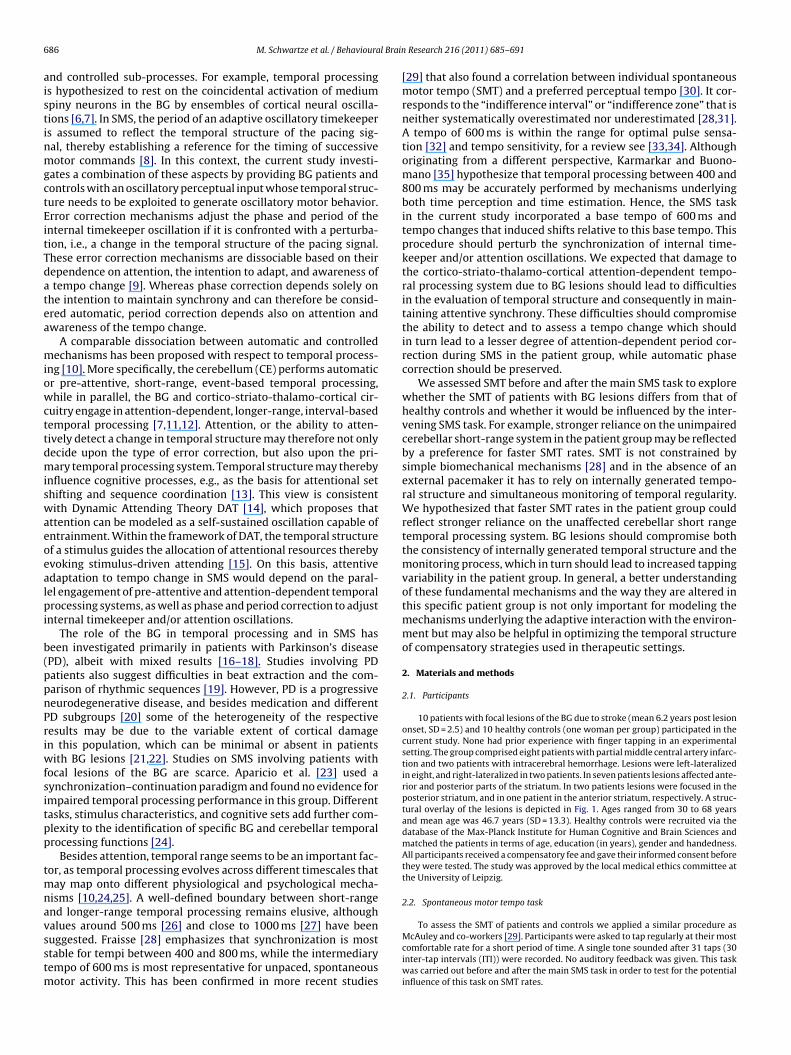
10 patients with focal lesions of the BG due to stroke (mean 6.2 years post lesiononset, SD = 2.5) and 10 healthy controls (one woman per group) participated in thecurrent study. None had prior experience with finger tapping in an experimentalsetting. The group comprised eight patients with partial middle central artery infarc-tion and two patients with intracerebral hemorrhage. Lesions were left-lateralizedin eight, and right-lateralized in two patients. In seven patients lesions affected ante-rior and posterior parts of the striatum. In two patients lesions were focused in theposterior striatum, and in one patient in the anterior striatum, respectively. A struc-tural overlay of the lesions is depicted in Fig. 1. Ages ranged from 30 to 68 yearsand mean age was 46.7 years (SD = 13.3). Healthy controls were recruited via thedatabase of the Max-Planck Institute for Human Cognitive and Brain Sciences andmatched the patients in terms of age, education (in years), gender and handedness.All participants received a compensatory fee and gave their informed consent beforethey were tested. The study was approved by the local medical ethics committee atthe University of Leipzig.
2.2. Spontaneous motor tempo task
To assess the SMT of patients and controls we applied a similar procedure asMcAuley and co-workers [29]. Participants were asked to tap regularly at their mostcomfortable rate for a short period of time. A single tone sounded after 31 taps (30inter-tap intervals (ITI)) were recorded. No auditory feedback was given. This taskwas carried out before and after the main SMS task in order to test for the potentialinfluence of this task on SMT rates.

M. Schwartze et al. / Behavioural Brain Research 216 (2011) 685–691 687
Fig. 1. Structural overlay of basal ganglia lesions. Green shades are associated withmea
2
(WtTsttotortCtt
Mhrtsifociitsbowta
1ratcopbttF
300
350
400
450
500
550
600
650
700
700650600550500450400350300
Mea
n IT
I Aft
er (
ms)
Mean ITI Before (ms)
Spontaneous Motor Tempo
Patients Controls
in patients than in controls (Fig. 3). Furthermore, variability wasgenerally lower after the adaptive timing task than before it. Thiswas the case both in patients and in controls. In this case, Levene’stest of equality of error variances was not significant. A 2 × 2 ANOVA
0
0.01
0.02
0.03
0.04
0.05
0.06
0.07
0.08
0.09
0.1
retfAerofeB
CV
ITI (
ms)
Spontaneous Motor Tempo Variability
Patients Controls
aximum lesion overlap, whereas purple shades are (For interpretation of the ref-rences to color in this figure legend, the reader is referred to the web version of therticle.) associated with minimal lesion overlap.
.3. Sensorimotor synchronization task
All participants used the index finger to tap on an electronic percussion padRoland SPD-6) placed on a table in front of them. The pad was connected to a
indows PC via a MIDI interface. A quiet “thud” sound with intensity proportionalo tapping force was produced upon impact with the rubber surface of the pad.his sound was further attenuated by the use of headphones. However, no digitalound output was provided. The manual mode of the pad was selected (as opposedo drumstick mode). Prior to the SMT and SMS tasks, all participants were allowedo familiarize themselves with the setting, and to test different styles of tapping inrder to find the most comfortable tapping position. Some participants decided toap while holding their forearm above the pad, but most preferred to rest their handn the pad. All patients tapped with their less affected ipsilesional hand in order toeduce possible confounds of impaired motor control. Some patients reported thathey had used this hand more frequently in the average 6.2 years since lesion onset.ontrol participants tapped with their dominant right hand. The adaptive timingask applied in this study was the same as the one used by Repp and Keller [9] withhe exception that the current base tempo was 600 ms instead of 500 ms.
Stimulus presentation and tap recording was controlled by a program written inAX (http://www.cycling74) running on a Windows PC. Stimuli were presented via
eadphones (Sennheiser HD 202) at a comfortable intensity. A total of 100 pseudo-andomized synchronization–continuation trials were processed in 10 blocks of 10rials each. 10 identical high-pitched piano tones (C8; 4176 Hz) were used as pacingtimuli during the synchronization phase of each trial. In eight trials per block thenitial inter-onset interval (IOI) of 600 ms was presented six times and was thenollowed by tempo accelerations or decelerations with a magnitude of 30, 45, 60r 75 ms for the three remaining IOIs. Two trials per block did not contain a tempohange and served as control sequences. Thus, if the trial contained a tempo change,t occurred between the 7th and 8th tone of the pacing sequence. Participants werenstructed to start tapping with the third tone of the pacing sequence. An addi-ional single tone of lower pitch (E7) marked the end of the continuation phase anderved as a signal to stop tapping. Awareness of the tempo change was assessedy means of a perceptual judgment. At the end of each trial, participants reportedrally whether they had perceived a deceleration, acceleration or no tempo changeithin the pacing sequence. Presentation of the next trial started two seconds after
he experimenter recorded the decision via key press. All data were acquired duringsingle session of approximately 60 min.
Missing taps and taps that either followed the target position by more than30 ms or preceded it by more than 150 ms were excluded from the analysis and areeferred to as errors. For the control sequences, mean asynchrony (MA) between tapsnd pacing sequence tones, variability of asynchronies, mean ITI during synchroniza-ion and continuation, and variability for ITIs produced during synchronization andontinuation were calculated. Adaptation to tempo changes was assessed in termsf mean ITIs, error correction and perceptual sensitivity to tempo changes on fiveositions of interest (s0, s1, s2, s3, c). Position s0 corresponds to the ITI terminated
y the tap that coincided approximately with the first tone at the new sequenceempo. This is followed by positions s1, s2 and s3. Position s3 thus corresponds tohe ITI initiated by the tap coinciding approximately with the last sequence tone.inally, c represents the average ITI during the whole continuation phase [9].Fig. 2. Distribution of spontaneous motor tempo for patients with basal ganglialesions and healthy controls before and after the adaptive timing task. ITI = intertapinterval.
3. Results
3.1. Spontaneous motor tempo
Mean SMT in the patient group was 551 ms (SD 105 ms) before,and 541 ms (SD 58 ms) after the SMS task. For the control group thecorresponding values were 536 ms (SD 30 ms) before, and 552 ms(SD 26 ms) after the SMS task (Fig. 2). Distribution of SMT ratesdiffered between the two groups. Levene’s test of equality of errorvariances revealed that the patient group was more heterogeneousthan the control group before, F(1.18) = 5.94, p < .03, and after theSMS task, F(1.18) = 7.64, p < .02. However, contrary to our predictionthere was no unitary trend towards either shorter or longer ITIs.Instead, patients demonstrated both fast and slow SMT rates, whilecontrol’s SMT rates clustered around 550 ms.
SMT variability was assessed by the coefficient of variation (CV)that was computed by dividing the standard deviation of individualITIs within a trial by the mean ITI. Tapping variability was higher
Test Time
Fig. 3. Coefficients of variation (CV) for the spontaneous motor tempo task forpatients with basal ganglia lesions and healthy controls before and after the adaptivetiming task.

688 M. Schwartze et al. / Behavioural Brain Research 216 (2011) 685–691
Table 1Mean Asynchrony, mean intertap interval (ITI), and coefficient of variation (CV) of ITIs for patients and controls during synchronization and continuation.
tion
wagnTi
oattg
3
4efpas(s(pfwuw
ppwovdpIbtddm
Ifc(
apvpsfi
g(
ducted for phase and period correction. The results indicatedthat period correction was generally more effective at fastertempi, Tempo F(1.16) = 8.14, p < .02. However, the effects of tempoon period correction were different for patients and controls,
450
500
550
600
650
700
750
525 540 555 570 600 630 645 660 675
Mea
n IT
I
Final Sequence IOI
Patients
s0
s1
s2
s3
c
450
500
550
600
650
700
750
Mea
n IT
I
Controls
s0
s1
s2
s3
c
Mean asynchrony CV asynchrony Mean ITI synchroniza
Patients −31 0.048 596Controls −26 0.034 600
ith factors group (patients vs. controls) and test time (before vs.fter the adaptive timing task) yielded significant differences forroup F(1.18) = 6.74, p < .02 and test time F(1.18) = 6.45, p < .03; buto significant interaction, group × test time F(1.18) = 3.44, p = .08.hese results imply more variable generation of temporal structuren the patient group.
Due to erratic performance during the subsequent SMS taskne patient and the respective control had to be excluded fromll further analyses except for the detection task. Exclusion ofhese participants changed the results of the preceding ANOVAo group F(1.16) = 7.78, p < .02; test time F(1.16) = 4.84, p < .05; androup × test time F(1.16) = 3.84, p = .07.
.2. Adaptive timing task
For the full sample of participants, the percentage of errors was.5% for patients and 1.4% for controls, t(18) = 1.90, p = .07. How-ver, one patient and the respective control were excluded from allurther analyses. This patient tapped at a tempo independent of theacing sequence tempo. Mean ITI was 497 ms for synchronizationnd 496 ms for continuation. The tapping was quite stable duringynchronization (CV = .053) and highly stable during continuationCV = .036). It is tempting to speculate that the tempo of the pacingequence distracted the patient from tapping at a preferred rateSMT before = 437 ms and SMT after = 482 ms). For the remainingarticipants, percentage of errors was 3.4% for patients and 1.5%or controls, t(16) = 1.34, p = .16. Results for the control sequencesithout a tempo change are provided in Table 1. The negative val-es for MA indicate that the taps preceded the pacing stimulus,hich is a typical finding for SMS in inexperienced tappers [36].
While the groups did not differ with respect to MA, t(16) = .34,= .74, the CV for asynchronies was higher for patients t(16) = 2.36,< .04. Mean ITIs, and CVs for synchronization and continuationere analyzed in separate 2 × 2 ANOVAs to test for the effects
f phase (synchronization vs. continuation) and group (patientss. controls). The ANOVA on mean ITIs revealed no significantifferences, phase F(1.16) = .75, p = .40; group F(1.16) = .17, p = .69;hase × group F(1.16) = .02, p = .90. However, the ANOVA on CV of
TIs yielded a significant effect for phase F(1.16) = 19.25, p < .01,ut not for group F(1.16) = .70, p = .42, and no significant interac-ion phase x group F(1.16) = .055, p = .82, indicating less variabilityuring the paced synchronization phase. Together, these resultsemonstrate that the patients could principally synchronize theirotor behavior with the auditory pacing sequences.To examine the adaptive response to the tempo changes, mean
TIs were plotted as a function of final sequence tempo separatelyor all sequences, including the control sequences, for patients andontrols for each sequence position of interest (s0, s1, s2, s3, c)Fig. 4).
Regression lines were fitted to the slopes of these ITI functionsnd were used as adaptation indices (Fig. 5). A value of 1 representserfect adaptation, values less than 1 indicate undercorrection andalues greater than 1 overcorrection. Adaptation indices were com-uted separately for tempo increases (i.e., faster tempi with final
equence IOIs < 600 ms) and tempo decreases (slower tempi withnal sequence IOIs > 600 ms).A 2 × 2 × 4 ANOVA was conducted to examine the effects ofroup (patients vs. controls), tempo (faster vs. slower), and positions1, s2, s3, c) on adaptation indices. The observed adaptation indices
Mean ITI continuation CV ITI synchronization CV ITI continuation
595 0.057 0.061598 0.041 0.044
differed between the groups F(1.16) = 5.43, p < .04 and betweenpositions F(3.48) = 9.32, p < .01. Adaptation indices were gener-ally higher in controls than in patients, and decreased acrosssequence positions in both groups. However, there were no sig-nificant interactions between group x tempo F(1.16) = 2.97, p = .10or position × group F(1.16) = .33, p = .57. Error correction was par-tioned into phase correction and period correction according tothe two-process model of error correction [8]. These types of errorcorrection were estimated by determining the values of the param-eters that led to the best fit between predictions based on thetwo-process model of error correction (implemented in MATLAB)and the observed adaptation indices [9]. Average phase and periodcorrection estimates for tempo increases and decreases are shownseparately in Fig. 6 for patients and controls. The fact that thesevalues are higher than those observed by Repp and Keller [9] maybe attributed to the participant’s relative inexperience with fin-ger tapping (Repp and Keller tested highly trained tappers) and/orthe slower base tempo employed in the current study (600 ms vs.500 ms).
Separate 2 × 2 ANOVAs, with independent variables group(patients vs. controls) and tempo (faster vs. slower), were con-
525 540 555 570 600 630 645 660 675
Final Sequence IOI
Fig. 4. Mean intertap intervals (ITI) for basal ganglia patients and healthy controlsas a function of the final sequence inter-onset interval (IOI).

M. Schwartze et al. / Behavioural Brain Research 216 (2011) 685–691 689
0
0.2
0.4
0.6
0.8
1
1.2
1.4
1.6
1.8
2
cs3s2s1
Ad
apta
tio
n In
dex
Position
Patients
Faster Slower
0
0.2
0.4
0.6
0.8
1
1.2
1.4
1.6
1.8
2
cs3s2s1
Ad
apta
tio
n In
dex
Position
Controls
Faster Slower
Fig. 5. Adaptation indices for basal ganglia patients and healthy controls for thesequence positions following the tempo change (s1, s2, s3) and during continuationtapping (c).
0
0.2
0.4
0.6
0.8
1
1.2
Phase Period
Co
rrec
tio
n E
stim
ate
Correction Parameter
Patients
Slower Faster
0
0.2
0.4
0.6
0.8
1
1.2
Phase Period
Co
rrec
tio
n E
stim
ate
Correction Parameter
Controls
Slower Faster
Fig. 6. Phase and period correction estimates for basal ganglia patients and healthycontrols for tempo accelerations (faster) and decelerations (slower).
0.00
0.10
0.20
0.30
0.40
0.50
0.60
0.70
0.80
0.90
1.00
675660645630600570555540525
Res
po
nse
Pro
po
rtio
n
Final Sequence IOI
Patients
Slower
Faster
No change
0.00
0.10
0.20
0.30
0.40
0.50
0.60
0.70
0.80
0.90
1.00
675660645630600570555540525
Res
po
nse
Pro
po
rtio
n
Final Sequence IOI
Controls
Slower
Faster
No change
Fig. 7. Proportion of responses to tempo changes for basal ganglia patients andhealthy controls. IOI = interonset interval.
group × tempo F(1.16) = 9.625, p < .01. Patients engaged in lesseffective period correction at slower tempi than at faster tempi,F(1.8) = 14.52, p < .01, whereas such effects of tempo on period cor-rection were absent in controls, F(1.8) = .40, p = .85.
3.3. Detection task
At the conclusion of each synchronization-continuation trial,participants were required to indicate orally whether the pacingsequence tempo had become faster, slower, or had remained con-stant. Average responses are shown for both groups in Fig. 7.
The responses of all participants were converted into d′ scores inorder to take potential response biases into account (Fig. 8). Thesescores were computed by subtracting z-transformed false alarmrates (i.e., the proportion of “slower” or “no change” responses fortempo increases, and “faster” or “no change” responses for tempodecreases) from hit rates (“faster” responses for tempo increases,and “slower” responses for tempo decreases). In accordance withour hypotheses, patients seemed to be more accurate for tempoaccelerations while their performance for decelerations reached aplateau at +45 ms. A 2 × 2 × 4 ANOVA on these scores tested for theeffects of group (patients vs. controls), tempo (faster vs. slower),and magnitude (±30, 45, 60, 75 ms). This ANOVA was computedfor the whole sample of participants as the perceptual judgment
did not involve any motor component.A main effect of group was significant, F(1.18) = 5.01, p < .04,which confirms that patients were generally less sensitive to thetempo changes than controls. The effect of magnitude was sig-

690 M. Schwartze et al. / Behavioural Brain
0
1
2
3
4
5
525 540 555 570 630 645 660 675
d p
rim
e
Detection Accuracy
Patients Controls
Fh
nfwhci
4
smcactthtmtInformapirniiphpt
hcpeiat
Final IOI
ig. 8. Accuracy in the detection of tempo change for basal ganglia patients andealthy controls.
ificant, F(3.48) = 8.02, p < .01 and greater for accelerations thanor decelerations, tempo × magnitude F(3.48) = 21.64, p < .01. Thereas no significant three-way interaction. Only an additional post-oc 2 × 2 ANOVA restricted to the perceptually most salienthanges (±75 ms) that should easily draw attention yielded annteraction for group × tempo, F(1.18) = 5.45, p < .03.
. Discussion
The current study explored spontaneous motor tempo and sen-orimotor synchronization in patients with focal BG lesions byeans of two finger tapping tasks. Damage to the BG was asso-
iated with a more heterogeneous distribution of individual ratess well as more variable tapping during the SMT task. These resultsonfirm that BG lesions have an effect on SMT in that they affecthe ability to execute a steady sequence of periodic actions. Givenhe role of the BG in attention-dependent temporal processing,igher variability may be caused by imprecise representations ofemporal structure. Alternatively, it could be simply due to noisy
otor implementation. However, it seems unlikely that this is alsohe reason for the more heterogeneous distribution of SMT rates.n general, patients performed well in both tapping tasks and didot report difficulties with tapping per se. This suggests, that the
reely chosen SMT rates reflect a different process. In the absencef external cues, SMT has to rely on internally generated, tempo-ally regular pacing information. Such internal pacemaker functionost likely engages the pre-supplementary motor area (pre-SMA)
nd its connections to the BG. The pre-SMA contributes to thelanning and initiation of simple and complex action sequences,
ncluding those required during speech production [37]. Pre-SMAecruitment is strongest when actions are freely chosen and areot guided by external signals [38,39], with increased activation
n early PD patients [40]. However, the pre-SMA is also involvedn perceptual temporal processing [41,42], indicating a function inroduction and perception. Hence, heterogeneous SMT rates andigher variability can be explained on a structural level by impairedrocessing of temporal structure in connections from the pre-SMAo the anterior striatum [43,44], the site affected in most patients.
Patients demonstrated good overall performance during SMS,owever, they tapped with relatively high variability and their errororrection was affected. More specifically, attention-dependent
eriod correction was less efficient in response to tempo decel-rations. Again, higher variability may be due to noisy motormplementation, whereas the difference in error correction hintst another process. While any specific value such as the 600 msempo used in the current study is certainly too precise to disso-Research 216 (2011) 685–691
ciate short-range from longer-range temporal processing, tempochanges relative to this base tempo were sufficient to induce adistinct impairment in the patient group. The fact that periodcorrection was affected supports the notion of specific attention-dependent mechanisms underlying SMS and temporal processing.This process may be modeled as entrainment of the timekeeperand/or attention oscillation by a pulse train [4]. Whereas phasecorrection would be sufficient to compensate for subliminal pertur-bations encoded by the pre-attentive temporal processing system,additional period correction would be needed to adapt if the periodof the internal timekeeper has to be adjusted. In the absence ofsubdivisions, that is, in the context of 1:1 tapping [45], phase cor-rection is assumed to reflect a lower-level process and to rest ontimes of occurrence or reference points, whereas period correctionis assumed to rest on intervals and to involve some form of mem-ory for at least one preceding event. Based on EEG data, Praamstraet al. [46] localized period correction in the supplementary motorarea (SMA). The finding of impaired period correction is thus inline with the proposed role of the BG in interval-based, longerrange temporal processing and an ongoing evaluation of temporalstructure in cortico-striato-thalamo-cortical circuits involving thepre-SMA.
The periodically spaced events of the pacing sequence promotestimulus-driven synchronization. However, if attention-dependenttemporal processing is necessary to recognize temporal regularity,BG lesions could be responsible for an erratic evaluation of temporalstructure and inaccurate predictions about upcoming events. In linewith DAT, this should affect the ability to focus attention in timeand to detect a tempo change. In other words, while the tempo-ral structure of successive pacing events conveys regular temporalstructure that is precisely encoded by the pre-attentive temporalprocessing system, its potentially facilitatory effect on synchro-nization is weakened by inefficient attention-dependent temporalprocessing. This relates to the difficulties of PD patients in process-ing rhythms with a beat structure [19] and evidence for difficultiesin temporal preparation in contrast to intact encoding of temporalintervals [47]. Damage to the BG may affect the ability to evaluatethe temporal relations between successive events, thereby com-promising the use of this information to compare rhythms, to alignmotor behavior, or to allocate attention. The detection of a subse-quent event may then be affected by both imprecise representationand evaluation of temporal structure on the one hand and ineffi-cient allocation of attention in time on the other hand. This in turnmay explain the difficulties that patients displayed in detecting thetempo changes embedded at a predictable position in the pacingsequences.
The results of the present study speak in favor of a function of theBG in SMS that is not restricted to motor control, but that extendsto attention-dependent temporal processing. Temporal processingand the recognition of temporal regularity are crucial for anticipa-tion, which in turn is necessary to temporally align actions to eventsin the environment. The additional finding of reduced perceptualsensitivity to tempo changes in BG patients points to a temporalprocessing network that is engaged in both production and per-ception. Together, attention-dependent temporal processing andresulting difficulties to exploit temporal structure in stimulus-driven attending and adaptive motor control offer an explanationfor the observed differences between patients with BG lesions andhealthy controls.
Acknowledgements
The authors would like to thank Bruno H. Repp for helpful com-ments during the early stages of this work and Anika Stockert forsupport in the preparation of the clinical data.

l Brain
R
[
[
[
[
[
[
[
[
[
[
[
[
[
[
[
[
[
[
[
[
[
[[
[
[
[
[
[
[
[
[
[
[
[
[
[
M. Schwartze et al. / Behavioura
eferences
[1] Fuster JM. The prefrontal cortex—an update: time is of the essence. Neuron2001;30:319–33.
[2] Repp BH. Sensorimotor synchronization: a review of the tapping literature.Psychon B Rev 2005;12:969–92.
[3] Witt ST, Laird AR, Meyerand ME. Functional neuroimaging correlates of finger-tapping task variations: an ALE meta-analysis. Neuroimage 2008;42:343–56.
[4] Pikovsky A, Rosenblum M, Kurths J. Synchronization: A Universal Concept inNonlinear Sciences. Cambridge: Cambridge Universal Press; 2003, 8 pp, 62 pp.
[5] Glass L. Synchronization and rhythmic processes in physiology. Nature2001;410:277–84.
[6] Matell MS, Meck WH. Cortico-striatal circuits and interval timing: coincidencedetection of oscillatory processes. Cognitive Brain Res 2004;21:139–70.
[7] Buhusi CV, Meck WH. What makes us tick? Functional and neural mechanismsof interval timing. Nat Rev Neurosci 2005;6:755–65.
[8] Mates J. A model of synchronization of motor acts to a stimulus sequence I.Timing and error corrections. Biol Cybern 1994;70:463–73.
[9] Repp BH, Keller PE. Adaptation to tempo changes in sensorimotor synchro-nization: effects of intention, attention, and awareness. Q J Exp Psychol2004;57A:499–521.
10] Lewis PA, Miall RC. Distinct systems for automatic and cognitively con-trolled time measurement: evidence from neuroimaging. Curr Opin Neurobiol2003;13:250–5.
11] Spencer RMC, Zelaznik HN, Diedrichsen J, Ivry RB. Disrupted timing of dis-continuous but not continuous movements by cerebellar lesions. Science2003;300:1437–9.
12] Ivry RB. The representation of temporal information in perception and motorcontrol. Curr Opin Neurobiol 1996;6:851–7.
13] Meck WH, Benson AM. Dissecting the brain’s internal clock: how frontal-striatalcircuitry keeps time and shifts attention. Brain Cognition 2002;48:195–211.
14] Large EW, Jones MR. The dynamics of attending: how we track time-varyingevents. Psychol Rev 1999;106:119–59.
15] Barnes R, Jones MR. Expectancy, attention, and time. Cognitive Psychol2000;41:254–311.
16] O’Boyle DJ, Freeman JS, Cody FWJ. The accuracy and precision of timing ofself-paced, repetitive movements in subjects with Parkinson’s disease. Brain1996;119:51–70.
17] Harrington DL, Haaland KY, Hermanowicz N. Temporal processing in the basalganglia. Neuropsychology 1998;12:3–12.
18] Pope PA, Praamstra P, Wing AM. Force and time control in the produc-tion of rhythmic movement sequences in Parkinson’s disease. Eur J Neurosci2006;23:1643–50.
19] Grahn JA, Brett M. Impairment of beat-based rhythm discrimination in Parkin-son’s disease. Cortex 2009;45:54–61.
20] Merchant H, Luciana M, Hooper C, Majestic S, Tuite P. Interval timing andParkinson’s disease: heterogeneity in temporal performance. Exp Brain Res2008;184:233–48.
21] Diedrichsen J, Ivry RB, Pressing J. Cerebellar and basal ganglia contributionsto interval timing. In: Meck WH, editor. Functional and neural mechanisms ofinterval timing. Boca Raton: CRC Press; 2003. p. 457–84.
22] Shin JC, Aparicio P, Ivry RB. Multidimensional sequence learning in patientswith focal basal ganglia lesions. Brain Cognition 2005;58:75–83.
23] Aparicio P, Diedrichsen J, Ivry RB. Effects of focal basal ganglia lesions on timingand force control. Brain Cognition 2005;58:62–74.
24] Koch G, Oliveri M, Caltagirone C. Neural networks engaged in millisecondsand seconds time processing: evidence from transcranial magnetic stimula-
[
[
Research 216 (2011) 685–691 691
tion and patients with cortical or subcortical dysfunction. Phil Trans Soc B2009;364:1907–18.
25] Buonomano DV. The biology of time across different scales. Nat Chem Biol2007;3:594–7.
26] Rammsayer TH. Neuropharmacological evidence for different timing systemsin humans. Q J Exp Psychol B 1999;52:273–86.
27] Lewis PA, Miall RC. Remembering the time: a continuous clock. Trends CognSci 2006;10:401–6.
28] Fraisse P. Rhythm tempo. In: Deutsch D, editor. The psychology of music. NewYork: Academic Press; 1982. p. 149–80.
29] Drake C, Jones MR, Baruch C. The development of rhythmic attending inauditory sequences: attunement, referent period, focal attending. Cognition2000;77:251–88.
30] McAuley JD, Jones MR, Holub S, Johnston HM, Miller NS. The time of ourlives: life span development of timing and event tracking. J Exp Psychol2006;135:348–67.
31] Fraisse P. Psychology of Time. New York: Harper; 1963. pp. 118.32] Parncutt R. A perceptual model of pulse salience and metrical accent in musical
rhythms. Music Percept 1994;11:409–64.33] Drake C, Botte MC. Tempo sensitivity in auditory sequences: evidence for a
multiple-look model. Percept Psychophys 1993;54:277–86.34] London J. Cognitive constraints on metric systems: some observations and
hypotheses. Music Percept 2002;19:529–50.35] Karmarkar UR, Buonomano DV. Timing in the absence of clocks: encoding time
in neural network states. Neuron 2007;53:427–38.36] Aschersleben G. Temporal control of movements in sensorimotor synchroniza-
tion. Brain Cognition 2002;48:66–79.37] Alario F, Chainay H, Lehericy S, Cohen L. The role of the supplementary motor
area (SMA) in word production. Brain Res 2006;1076:129–43.38] Halsband U, Ito N, Tanji J, Freund H-J. The role of the premotor cortex and the
supplementary motor area in the temporal control of movement in man. Brain1993;116:243–66.
39] Nachev P, Kennard C, Husain M. Functional role of the supplementary and pre-supplementary motor areas. Nat Rev Neurosci 2008;9:856–69.
40] Eckert T, Peschel T, Heinze H-J, Rotte M. Increased pre-SMA activation inearly PD patients during simple self-initiated hand movements. J Neurol2006;253:199–207.
41] Coull JT, Vidal F, Nazarian B, Macar F. Functional anatomy of the attentionalmodulation of time estimation. Science 2004;303:1506–8.
42] Macar F, Coull J, Vidal F. The supplementary motor area in motor and perceptualtime processing: fMRI studies. Cogn Process 2006;7:89–94.
43] Lehéricy S, Ducros M, Krainik A, Francois C, Van de Moortele P, Ugur-bil K, Kim D. 3-D diffusion tensor axonal tracking shows distinct SMAand Pre-SMA projections to the human striatum. Cereb Cortex 2004;14:1302–9.
44] Johansen-Berg H, Behrens TEJ, Robson MD, Drobnjak I, Rushworth MFS, BradyJM, Mith SM, Higham DJ, Matthews PM. Changes in connectivity profiles definefunctionally distinct regions in human medial frontal cortex. P Natl Acad SciUSA 2004;101:13335–40.
45] Repp BH. Multiple temporal references in sensorimotor synchronization withmetrical auditory sequences. Psychol Res 2008;72:79–98.
46] Praamstra P, Turgeon M, Hesse CW, Wing AM, Perryer L. Neurophysiologicalcorrelates of error correction in sensorimotor-synchronization. Neuroimage2003;20:1283–97.
47] Praamstra P, Pope P. Slow brain potential and oscillatory EEG manifesta-tions of impaired temporal preparation in Parkinson’s disease. J Neurophysiol2007;98:2848–57.
Copyright © 2022 FDOKUMEN