The Heart - TheFetus.net
70
©1987-2002 Romero-Pilu-Jeanty-Ghidini-Hobbins The Heart Approach to the Examination of the Fetal Heart/ 125 Atrial Septal Defects/ 139 Ventricular Septal Defects/ 141 Atrioventricular Septal Defects/ 144 Univentricular Heart/ 147 Ebstein's Anomaly/ 149 Hvpoplastic Left Heart Syndrome/ 151 Hypoplastic Right Ventricle/ 155 Tetralogy of Fallot/ 157 Complete Transposition of the Great Arteries/ 160 Corrected Transposition of the Great Arteries/ 164 Double Outlet Right Ventricle/ 166 Truncus Arteriosus/ 168 Coarctation and Tubular Hypoplasia of the Aortic Arch/ 171 Pulmonic Stenosis/ 173 Aortic Stenosis/ 175 Cardiomyopathies/ 178 Total Anomalous Pulmonary Venous Return/ 180 Tumors of the Heart/ 182 Cardiosplenic Syndromes/ 184 Ectopia Cordis/ 186 Premature Atrial and Ventricular Contractions/ 187 Supraventricular Tachyarrhythmias/ 188 Atrioventricular Block/ 192 Approach to the Examination of the Fetal Heart The first technique used for the monographic evaluation of the fetal heart was M-mode ultrasound. Using this method, Winsberg reported quantitative evaluation of the fetal cardiac chambers in 1972. 26 However, the use of M-mode echocardiography in the fetus was limited because of the difficulties inherent in examining a moving fetus with a single, pencil-like sound beam. The next step was the use of real-time-directed M- mode that allowed the orientation of the single beam on the bidimensional image.Using this technique, Ianniruberto et al., 10 DeLuca et al., 6 and Wladimiroff and McGhie 27 described the quantitative and qualitative anatomy of the fetal heart. The feasibility of the prenatal diagnosis of congenital heart disease was first established by Kleinet al. 13 A major breakthrough toward the reproducibility of fetal echocardiography was the introduction of high- resolution real-time equipment. This equipment allowed detailed investigation of the anatomy of the fetal heart beginning in early pregnancy. In 1980, Allan et al. 3 described a systematic approach to the bidimensional examination of the fetal heart. In recent years, the experience collected in several laboratories has demonstrated the reliability of prenatal diagnosis of cardiac structural and functional abnormalities 1,4,11-14,17,21-23 ULTRASOUND BIDIMENSIONAL INVESTIGATION OF THE FETAL HEART: A SEQUENTIAL APPROACH The main objective of fetal echocardiography is the prenatal diagnosis of congenital heart disease. Cardiac abnormalities encompass a broad spectrum of structural disorders, ranging from a simple communication between two cardiac chambers to an almost complete rearrangement of the connections between the different cardiac segments. This demands a systematic approach to the investigation of the fetal heart. In our laboratory, we use a "sequential approach" that depends on the recognition of the morphology and connections of the three segments of the fetal heart: atria, ventricles, and great vessels. Sequential analysis for the diagnosis of congenital 125
-
Upload
khangminh22 -
Category
Documents
-
view
2 -
download
0
Transcript of The Heart - TheFetus.net

©1987-2002 Romero-Pilu-Jeanty-Ghidini-Hobbins
The Heart Approach to the Examination of the
Fetal Heart/ 125 Atrial Septal Defects/ 139 Ventricular Septal Defects/ 141 Atrioventricular Septal Defects/ 144 Univentricular Heart/ 147 Ebstein's Anomaly/ 149 Hvpoplastic Left Heart Syndrome/ 151 Hypoplastic Right Ventricle/ 155 Tetralogy of Fallot/ 157 Complete Transposition of the Great
Arteries/ 160 Corrected Transposition of the Great
Arteries/ 164 Double Outlet Right Ventricle/ 166 Truncus Arteriosus/ 168
Coarctation and Tubular Hypoplasia of the Aortic Arch/ 171
Pulmonic Stenosis/ 173 Aortic Stenosis/ 175 Cardiomyopathies/ 178 Total Anomalous Pulmonary Venous Return/ 180 Tumors of the Heart/ 182 Cardiosplenic Syndromes/ 184 Ectopia Cordis/ 186 Premature Atrial and Ventricular
Contractions/ 187 Supraventricular
Tachyarrhythmias/ 188 Atrioventricular Block/ 192
Approach to the Examination of the Fetal Heart The first technique used for the monographic evaluation of the fetal heart was M-mode ultrasound. Using this method, Winsberg reported quantitative evaluation of the fetal cardiac chambers in 1972.26 However, the use of M-mode echocardiography in the fetus was limited because of the difficulties inherent in examining a moving fetus with a single, pencil-like sound beam. The next step was the use of real-time-directed M-mode that allowed the orientation of the single beam on the bidimensional image.Using this technique, Ianniruberto et al.,10 DeLuca et al.,6 and Wladimiroff and McGhie27 described the quantitative and qualitative anatomy of the fetal heart. The feasibility of the prenatal diagnosis of congenital heart disease was first established by Kleinet al.13 A major breakthrough toward the reproducibility of fetal echocardiography was the introduction of high-resolution real-time equipment. This equipment allowed detailed investigation of the anatomy of the fetal heart beginning in early pregnancy. In 1980, Allan et al.3 described a systematic approach to the bidimensional examination of the fetal heart. In
recent years, the experience collected in several laboratories has demonstrated the reliability of prenatal diagnosis of cardiac structural and functional abnormalities 1,4,11-14,17,21-23 ULTRASOUND BIDIMENSIONAL INVESTIGATION OF THE FETAL HEART: A SEQUENTIAL APPROACH The main objective of fetal echocardiography is the prenatal diagnosis of congenital heart disease. Cardiac abnormalities encompass a broad spectrum of structural disorders, ranging from a simple communication between two cardiac chambers to an almost complete rearrangement of the connections between the different cardiac segments. This demands a systematic approach to the investigation of the fetal heart. In our laboratory, we use a "sequential approach" that depends on the recognition of the morphology and connections of the three segments of the fetal heart: atria, ventricles, and great vessels. Sequential analysis for the diagnosis of congenital
125
©1987-2002 Romero-Pilu-Jeanty-Ghidini-Hobbins
126 THE HEART Figure 4-1. Transverse cross-section of the upper fetal abdomen. Study of the intraab-dominal organs Per-mits defi-nition of the visceral situs. With situs solitus, the stomach (St) and the spleen (Spl) are on the left. The position of the hilum of the liver, normally on the right, can be inferred by following the umbilical vein (UV) into the left portal vein (LPV), which bends into the portal sinus (PS). The abdominal aorta (Ao) and inferior vena cava (IVC) can be seen on both sides of the spine (Sp). Ant, anterior; Post, posterior; L, left, R, right.
heart disease was first introduced by Van Praagh24 and subsequently modified by Shinebourne et al.19 In recent years, this type of approach, conceived for the pathologic and angiographic examination of the heart, has been applied to echocardiography in the postnatal period. 9 Such methodology appears extremely suitable for fetal cardiac studies. In this section, we adhere to the elegant approach to diagnosis and classification of congenital heart disease advocated by Becker and Anderson.5
The main steps of sequential analysis are: 1. Position of the heart within the body 2. Identification of the cardiac chambers 3. Study of the atrioventricular connections 4. Study of the ventriculoarterial connections An ideal echocardiographic examination should begin with determination of the position of the head and the spine, establishing the right and left sides of the fetus. The next step, identification of the visceral situs, is important for two reasons: (1) the arrangement of the abdominal organs predicts the relative position of the right and left atria with a high degree of accuracy (this information is extremely valuable because, in many cases, fetal echocardiography does not distinguish the morphologic left from the right atrium) and (2) anomalies of the visceral situs are very frequently associated with cardiac abnormalities (e.g., cardiosplenic syndromes). Three conditions are pos-sible: situs solitus (normal),situs inversus (mirror image of situs solitus), and situs ambiguous, also known as isomeric situs. This term refers to a condition in which there is an abnormal arrangement of the thoracic and abdominal organs (see sections on asplenia and polysplenia syndromes).
The visceral situs can be easily identified in the fetus by using ultrasound in a transverse crosssection of the upper abdomen. In this view, the stomach and spleen are normally positioned on the left. The portal sinus, which topographically corresponds to the hilum of the liver, can be seen to the right. Anterior to the spine, the abdominal aorta and inferior vena cava are seen on both sides of the spine. The abdominal aorta is to the left and appears as a round structure, and the inferior vena cava is to the right and is flattened and more anterior (Fig. 4-1). Recognition of the relative positions of the aorta and inferior vena cava is of special importance in identifying the atrial chambers, since the morphologic right atrium is almost invariably on the same side of the inferior vena cava.5 Since the fetal heart is almost horizontal, a transverse cross-section of the thorax above the level of the diaphragm will demonstrate a four chamber view.3
In this plane of section, it is easy to recognize the apex and base of the heart and to assess the position of the heart inside the chest. Normally, the heart is in the left side of the chest with the apex pointing toward the left, the right ventricle and atrium being anterior to the left ventricle and atrium (levocardia) (Fig. 4-2). In dextrocardia, the heart is in the right side of the chest, and the apex points toward the right. In mesocardia, the heart occupies a central position inside the chest and the apex points anteriorly. Dextrocardia and mesocardia should be differentiated from those conditions in which the position of the heart is altered due to external compression (e.g., diaphragmatic hernia, lung tumors). In these cases, the term "dextroposition of the heart" is more appropriate.
APPROACH TO THE EXAMINATION OF THE FETAL HEART 127
©1987-2002 Romero-Pilu-Jeanty-Ghidini-Hobbins
Figure 4-2. Apical four chamber view of the fetal heart. Note the moderator band (MB) and the more apical insertion of the leaflets of the tricuspid valve (unlabeled) on the ventricular septum, distinguishing the morphologic right ventricle from the left. The interatrial septum is interrupted in its central portion by the foramen ovale. The pulmonary veins (pv) can be seen entering the left atrium (LA). RA, right atrium; LV, left ventricle; RV, right ventricle; Sp, spine; DAo, descending aorta; R, right; L, left; Ant, anterior.
The four chamber view of the fetal heart provides important anatomic information. The interatrial septum separating the two atrial chambers can be seen. Normally, the pulmonary veins are connected to the left atrium, and the two atrial chambers communicate through the foramen ovale, an orifice in the center of the interatrial septum that separates the superior septum secundum and the inferior septum primum
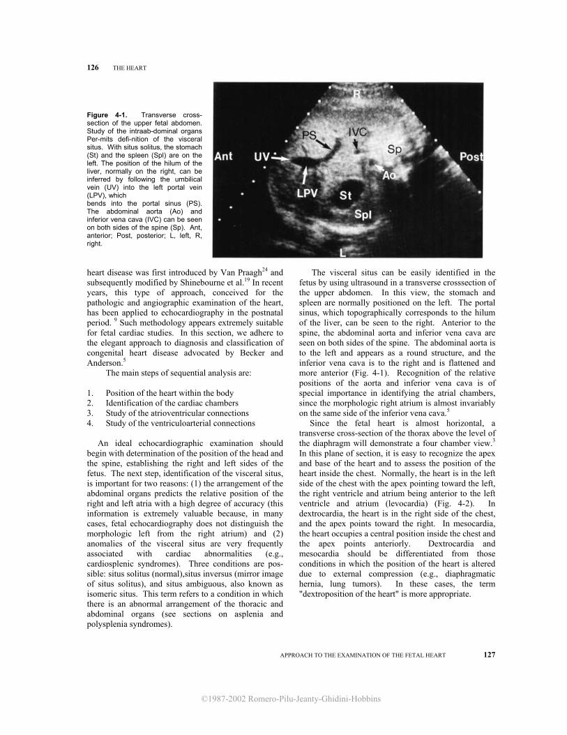
Figure 4-3. A subcostal four chamber view allows better definition of the integrity of the interventricular and interatrial septa. LV, left ventriclecle; RV, right ventricle; LA, left atrium; RA, right atrium; pv, pulmonary veins; Ant, anterior; Post, posterior; L, left.
(Fig. 4-3). The foramen ovale is guarded by a valve that opens toward the left atrium.
The two atrial chambers are connected to the ventricular chambers. The atrioventricular junction is characterized by the more apical insertion of the tricuspid valve than the mitral valve on the interventricular septum. This finding is useful in differentiating the morphologic right and left ventricle and in recognizing anomalies of the atrioventricular junction, such as the atrioventricular canal 11(Fig. 4-2). The other important anatomic details in differenti-
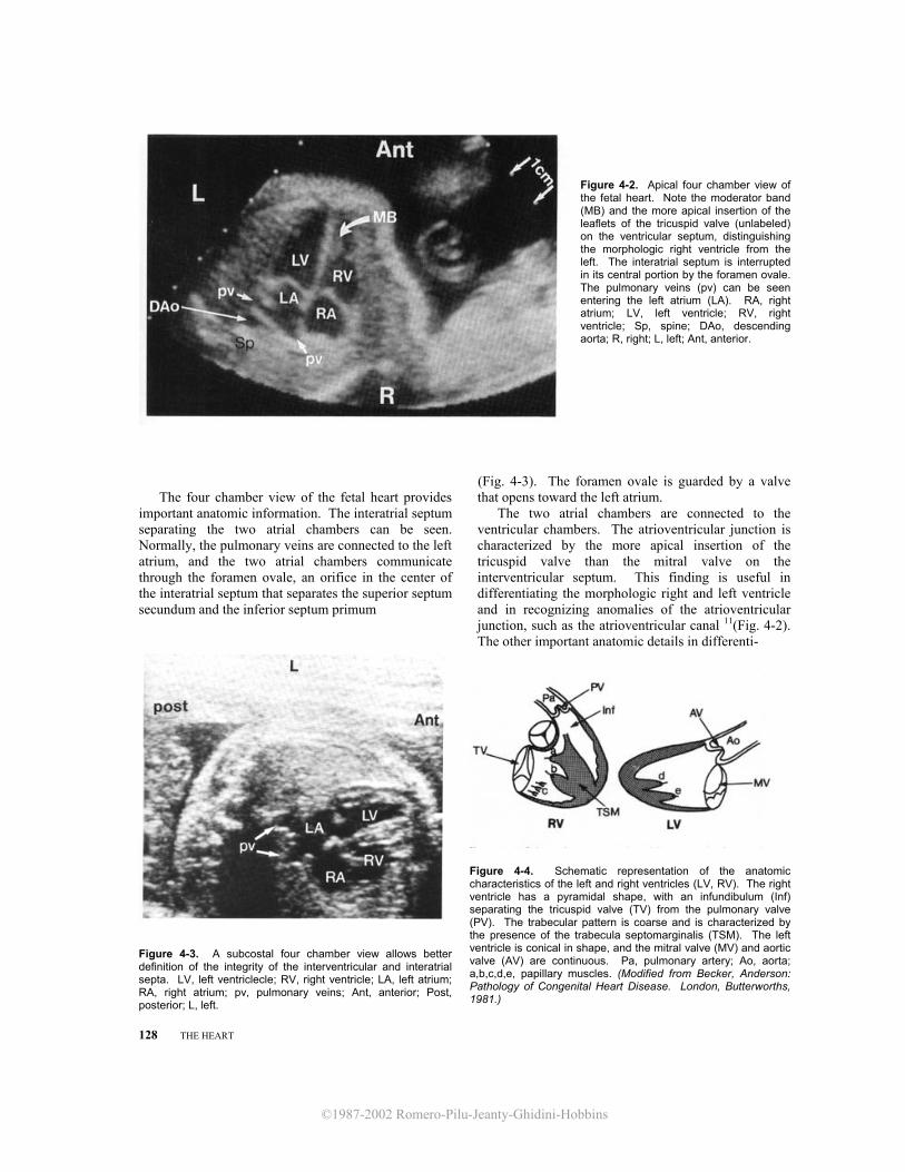
Figure 4-4. Schematic representation of the anatomic characteristics of the left and right ventricles (LV, RV). The right ventricle has a pyramidal shape, with an infundibulum (Inf) separating the tricuspid valve (TV) from the pulmonary valve (PV). The trabecular pattern is coarse and is characterized by the presence of the trabecula septomarginalis (TSM). The left ventricle is conical in shape, and the mitral valve (MV) and aortic valve (AV) are continuous. Pa, pulmonary artery; Ao, aorta; a,b,c,d,e, papillary muscles. (Modified from Becker, Anderson: Pathology of Congenital Heart Disease. London, Butterworths, 1981.)
128 THE HEART
©1987-2002 Romero-Pilu-Jeanty-Ghidini-Hobbins
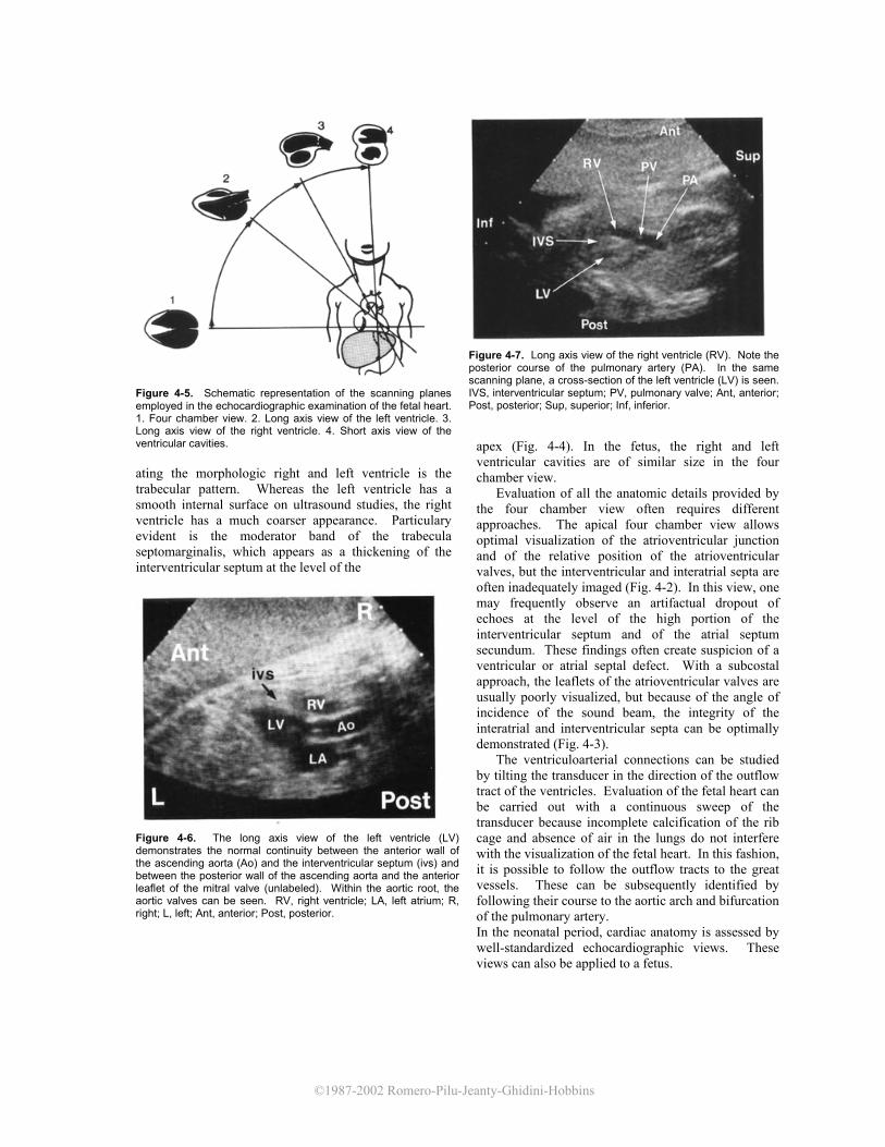
Figure 4-5. Schematic representation of the scanning planes employed in the echocardiographic examination of the fetal heart. 1. Four chamber view. 2. Long axis view of the left ventricle. 3. Long axis view of the right ventricle. 4. Short axis view of the ventricular cavities. ating the morphologic right and left ventricle is the trabecular pattern. Whereas the left ventricle has a smooth internal surface on ultrasound studies, the right ventricle has a much coarser appearance. Particulary evident is the moderator band of the trabecula septomarginalis, which appears as a thickening of the interventricular septum at the level of the
Figure 4-6. The long axis view of the left ventricle (LV) demonstrates the normal continuity between the anterior wall of the ascending aorta (Ao) and the interventricular septum (ivs) and between the posterior wall of the ascending aorta and the anterior leaflet of the mitral valve (unlabeled). Within the aortic root, the aortic valves can be seen. RV, right ventricle; LA, left atrium; R, right; L, left; Ant, anterior; Post, posterior.
Figure 4-7. Long axis view of the right ventricle (RV). Note the posterior course of the pulmonary artery (PA). In the same scanning plane, a cross-section of the left ventricle (LV) is seen. IVS, interventricular septum; PV, pulmonary valve; Ant, anterior; Post, posterior; Sup, superior; Inf, inferior.
apex (Fig. 4-4). In the fetus, the right and left ventricular cavities are of similar size in the four chamber view.
Evaluation of all the anatomic details provided by the four chamber view often requires different approaches. The apical four chamber view allows optimal visualization of the atrioventricular junction and of the relative position of the atrioventricular valves, but the interventricular and interatrial septa are often inadequately imaged (Fig. 4-2). In this view, one may frequently observe an artifactual dropout of echoes at the level of the high portion of the interventricular septum and of the atrial septum secundum. These findings often create suspicion of a ventricular or atrial septal defect. With a subcostal approach, the leaflets of the atrioventricular valves are usually poorly visualized, but because of the angle of incidence of the sound beam, the integrity of the interatrial and interventricular septa can be optimally demonstrated (Fig. 4-3).
The ventriculoarterial connections can be studied by tilting the transducer in the direction of the outflow tract of the ventricles. Evaluation of the fetal heart can be carried out with a continuous sweep of the transducer because incomplete calcification of the rib cage and absence of air in the lungs do not interfere with the visualization of the fetal heart. In this fashion, it is possible to follow the outflow tracts to the great vessels. These can be subsequently identified by following their course to the aortic arch and bifurcation of the pulmonary artery. In the neonatal period, cardiac anatomy is assessed by well-standardized echocardiographic views. These views can also be applied to a fetus.
©1987-2002 Romero-Pilu-Jeanty-Ghidini-Hobbins
APPROACH TO THE EXAMINATION OF THE FETAL HEART 129
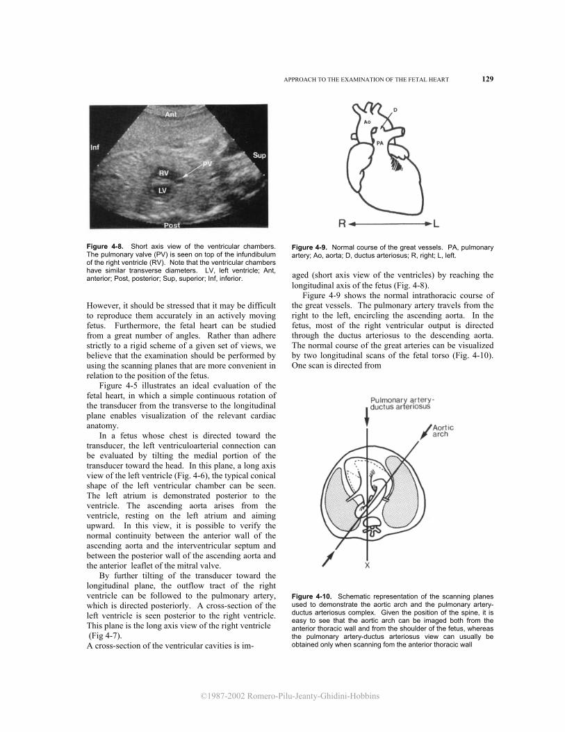
Figure 4-8. Short axis view of the ventricular chambers. The pulmonary valve (PV) is seen on top of the infundibulum of the right ventricle (RV). Note that the ventricular chambers have similar transverse diameters. LV, left ventricle; Ant, anterior; Post, posterior; Sup, superior; Inf, inferior. However, it should be stressed that it may be difficult to reproduce them accurately in an actively moving fetus. Furthermore, the fetal heart can be studied from a great number of angles. Rather than adhere strictly to a rigid scheme of a given set of views, we believe that the examination should be performed by using the scanning planes that are more convenient in relation to the position of the fetus.
Figure 4-5 illustrates an ideal evaluation of the fetal heart, in which a simple continuous rotation of the transducer from the transverse to the longitudinal plane enables visualization of the relevant cardiac anatomy.
In a fetus whose chest is directed toward the transducer, the left ventriculoarterial connection can be evaluated by tilting the medial portion of the transducer toward the head. In this plane, a long axis view of the left ventricle (Fig. 4-6), the typical conical shape of the left ventricular chamber can be seen. The left atrium is demonstrated posterior to the ventricle. The ascending aorta arises from the ventricle, resting on the left atrium and aiming upward. In this view, it is possible to verify the normal continuity between the anterior wall of the ascending aorta and the interventricular septum and between the posterior wall of the ascending aorta and the anterior leaflet of the mitral valve.
By further tilting of the transducer toward the longitudinal plane, the outflow tract of the right ventricle can be followed to the pulmonary artery, which is directed posteriorly. A cross-section of the left ventricle is seen posterior to the right ventricle. This plane is the long axis view of the right ventricle (Fig 4-7). A cross-section of the ventricular cavities is im-
Figure 4-9. Normal course of the great vessels. PA, pulmonary artery; Ao, aorta; D, ductus arteriosus; R, right; L, left.
aged (short axis view of the ventricles) by reaching the longitudinal axis of the fetus (Fig. 4-8). Figure 4-9 shows the normal intrathoracic course of the great vessels. The pulmonary artery travels from the right to the left, encircling the ascending aorta. In the fetus, most of the right ventricular output is directed through the ductus arteriosus to the descending aorta. The normal course of the great arteries can be visualized by two longitudinal scans of the fetal torso (Fig. 4-10). One scan is directed from
Figure 4-10. Schematic representation of the scanning planes used to demonstrate the aortic arch and the pulmonary artery-ductus arteriosus complex. Given the position of the spine, it is easy to see that the aortic arch can be imaged both from the anterior thoracic wall and from the shoulder of the fetus, whereas the pulmonary artery-ductus arteriosus view can usually be obtained only when scanning fom the anterior thoracic wall
©1987-2002 Romero-Pilu-Jeanty-Ghidini-Hobbins
130 THE HEART
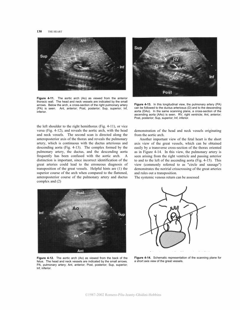
Figure 4-11. The aortic arch (Ao) as viewed from the anterior thoracic wall. The head and neck vessels are indicated by the small arrows. Below the arch, a cross-section of the right pulmonary artery (PA) is seen. Ant, anterior; Post, posterior; Sup, superior; lnf, inferior. the left shoulder to the right hemithorax (Fig. 4-11), or vice versa (Fig. 4-12), and reveals the aortic arch, with the head and neck vessels. The second scan is directed along the anteroposterior axis of the thorax and reveals the pulmonary artery, which is continuous with the ductus arteriosus and descending aorta (Fig. 4-13). The complex formed by the pulmonary artery, the ductus, and the descending aorta frequently has been confused with the aortic arch. A distinction is important, since incorrect identification of the great arteries could lead to the erroneous diagnosis of transposition of the great vessels. Helpful hints are (1) the superior course of the arch when compared to the flattened, anteroposterior course of the pulmonary artery and ductus complex and (2)
Figure 4-12. The aortic arch (Ao) as viewed from the back of the fetus. The head and neck vessels are indicated by the small arrows. PA, pulmonary artery; Ant, anterior; Post, posterior; Sup, superior; lnf, inferior.
Figure 4-13. In this longitudinal view, the pulmonary artery (PA) can be followed to the ductus arteriosus (D) and to the descending aorta (DAo). In the same scanning plane, a cross-section of the ascending aorta (AAo) is seen. RV, right ventricle; Ant, anterior; Post, posterior; Sup, superior; Inf, inferior.
demonstration of the head and neck vessels originating from the aortic arch.
Another important view of the fetal heart is the short axis view of the great vessels, which can be obtained easily by a transverse cross-section of the thorax oriented as in Figure 4-14. In this view, the pulmonary artery is seen arising from the right ventricle and passing anterior to and to the left of the ascending aorta (Fig. 4-15). This view (commonly referred to as "circle and sausage") demonstrates the noririal crisscrossing of the great arteries and rules out a transposition. The systemic venous return can be assessed
Figure 4-14. Schematic representation of the scanning plane for a short axis view of the great vessels.

©1987-2002 Romero-Pilu-Jeanty-Ghidini-Hobbins
APPROACH T'O THE EXAMINATION OF' I'HE FETAL HEART 131
Figure 4-15. In the short axis view of the great vessels, the pulmonary artery (PA) is seen arising from the right ventricle (RV), passing anterior to and to the left of the ascending aorta (Ao) and bifurcating into the ductus arteriosus (D) and the right pulmonary artery (unlabeled). The tricuspid valve separates the right atrium (RA) from the right ventricle. Both aortic (unlabeled) and pulmonary valves (PV) can be seen within the roots of the great arteries. Ant, anterior; Post, posterior; L, left; R, right. easily by a right parasagittal scan demonstrating the inferior and superior vena cava entering the right atrium (Fig. 4-16). M-MODE ECHOCARDIOGRAPHY M-mode is a modality of ultrasound in which the information derived from a single sound beam is displayed against time. This technique allows the movement of structures to be evaluated both quantitatively and qualitatively. M-mode was the first ultrasound modality employed in the study of the fetal heart20,26 Its major shortcoming was difficulty in blindly directing the sound beam toward the cardiac structures of a moving fetus. Recently, advances in ultrasound technology have resulted in the introduction of equipment with which the direction of the single beam can be selected on a bidimensional real-time image (Fig. 4-17). Fetal M-mode echocardiography is useful for measurement of cardiac chambers and great vessels and for assessment of cardiac arrhythmias.
Figure 4-16. Right parasagittal scan of the fetal trunk demonstrating the inferior and superior venae cavae (IVC, SVC) entering the right atrium (RA). The tricuspid valve (tv) divides the right atrium from the right ventricle (RV). Ao, aorta; PA, pulmonary artery; Ant, anterior; Post, posterior; Sup, superior; Inf, inferior.
M-mode tracings are provided with markers that indicate distance in the sound field on the vertical axis and time on the horizontal axis. With most equipment, the vertical distance between markers corresponds to 1 cm, and the horizontal distance corresponds to 0.5 second. This allows calculation of the fetal heart rate and biometry. Until a few years ago, these calculations were made off line, using calipers on a hard copy. Newer equipment is provided with software capable of on-screen measurements with electronic calipers.
The most relevant cardiac structures for M-mode examination are the atrial and ventricular chambers, atrioventricular valves, roots of the great vessels, and semilunar valves.
Examination of the ventricular chambers should be performed by directing the M-mode beam across the ventricles at a right angle to the interventricular septum and at the level of the atrioventricular valves. In Figure 4-17, the myocardium is externally lined by a bright linear echo that represents the pericardium. Inside the ventricular chambers, it is possible to observe the movement of the atrioventricular valves. The movement of the ventricular walls toward the interventricular septum indicates ventricular systole. In Figure 4-18, the typical movement pattern of the mitral valve is shown. The anterior and posterior leaflets of the mitral valve are seen apposed during ventricular systole. At the beginning of ventricular filling, the valve opens, and the anterior leaflet moves toward the interventricular septum (point D). The point of maximal excursion of the leaflet is called

©1987-2002 Romero-Pilu-Jeanty-Ghidini-Hobbins
132 THE HEART
Figure 4-17. With ultrasound equip-ment that has the option of real-time-directed M-mode, the position of the cursor (M-line) can be easily selected during the real-time examination. An M-mode echo-cardiogram of the ventricular cavities (RV, LV) at the level of the atrioventricular valves (tv, mv) is shown. The undulations of free ventricular walls and of the interven- tricular septum (ivs) reflect systole and diastole. P, pericardium.
point E. After this, the leaflet moves away from the interventricular septum until it reaches the F point. The valve opens again with atrial systole (point A), and the leaflet moves away from the interventricular septum and presents a small undulation corresponding to the onset of ventricular systole (point B). At
point C, the leaflets are apposed to each other. The movement of the posterior leaflet of the mitral valve is a mirror image of the movement of the anterior leaflet. The movement of the Bicuspid valve closely resembles that of the mitral valve. Therefore, it is clear that by directing the M-mode beam across the
Figure 4-18. M-mode echocardiogram of the mitral valve in a secondtrimester fetus. See text for explanation of points A through F.
©1987-2002 Romero-Pilu-Jeanty-Ghidini-Hobbins
APPROACH TO THE EXAMINATION OF THE FETAL HEART 133
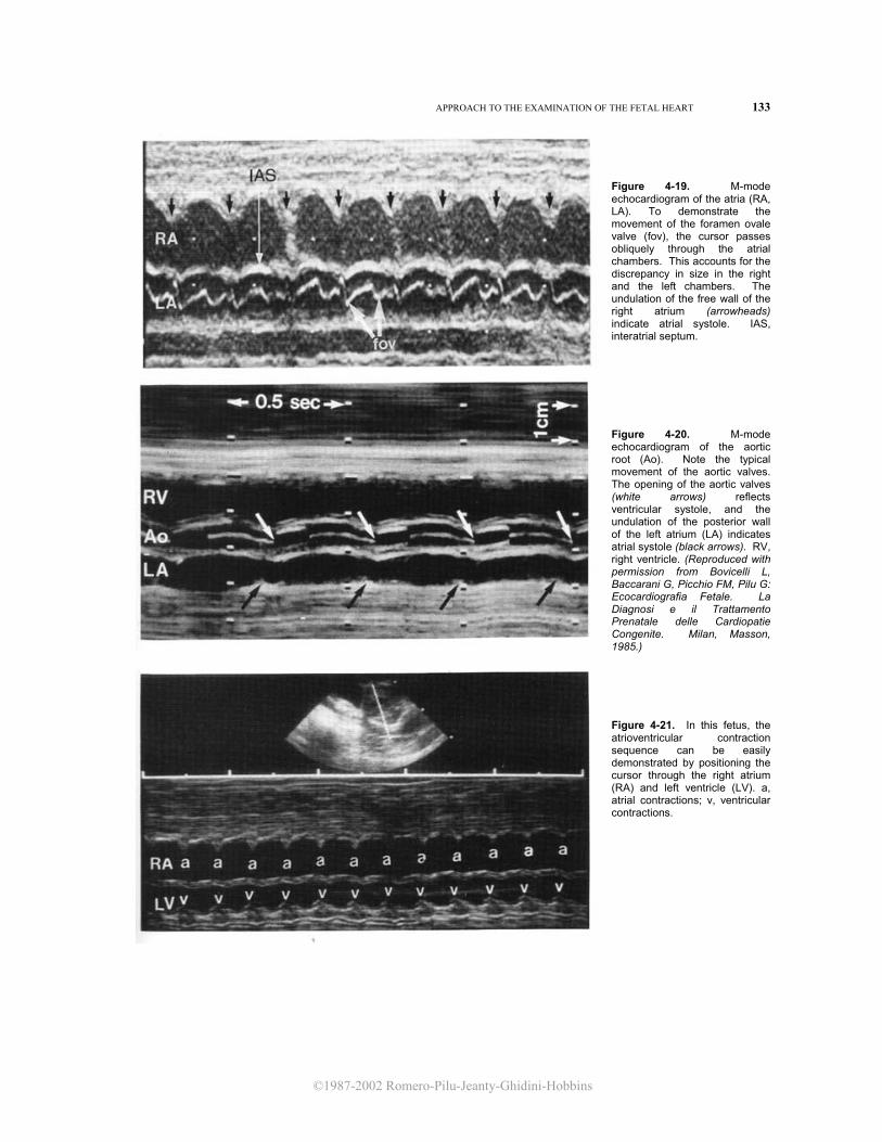
Figure 4-19. M-mode echocardiogram of the atria (RA, LA). To demonstrate the movement of the foramen ovale valve (fov), the cursor passes obliquely through the atrial chambers. This accounts for the discrepancy in size in the right and the left chambers. The undulation of the free wall of the right atrium (arrowheads) indicate atrial systole. IAS, interatrial septum.
Figure 4-20. M-mode echocardiogram of the aortic root (Ao). Note the typical movement of the aortic valves. The opening of the aortic valves (white arrows) reflects ventricular systole, and the undulation of the posterior wall of the left atrium (LA) indicates atrial systole (black arrows). RV, right ventricle. (Reproduced with permission from Bovicelli L, Baccarani G, Picchio FM, Pilu G: Ecocardiografia Fetale. La Diagnosi e il Trattamento Prenatale delle Cardiopatie Congenite. Milan, Masson, 1985.) Figure 4-21. In this fetus, the atrioventricular contraction sequence can be easily demonstrated by positioning the cursor through the right atrium (RA) and left ventricle (LV). a, atrial contractions; v, ventricular contractions.

©1987-2002 Romero-Pilu-Jeanty-Ghidini-Hobbins
134 'I'HE HEAR'I'
Figure 4-22. Normal dimen-sions of the left ventricle throughout gestation. (Repro-duced with permission from Allan et al.: Br Heart J 47:573, 1982.) Figure 4-23. Normal dimen-ions of the right ventricle throughout gestation. (Re-produced with permission from Allan et weeks al.: Br Heart J 47.-573, 1982.) Figure 4-24. Normal dimensions of the inter-entricular septum throughout gestation (Re-produced with permission from Allan et weeks al.: Br Heart J 47.-573, 1982.)
©1987-2002 Romero-Pilu-Jeanty-Ghidini-Hobbins
APPROACH TO THE EXAMINATION OF THE FETAL HEART 135
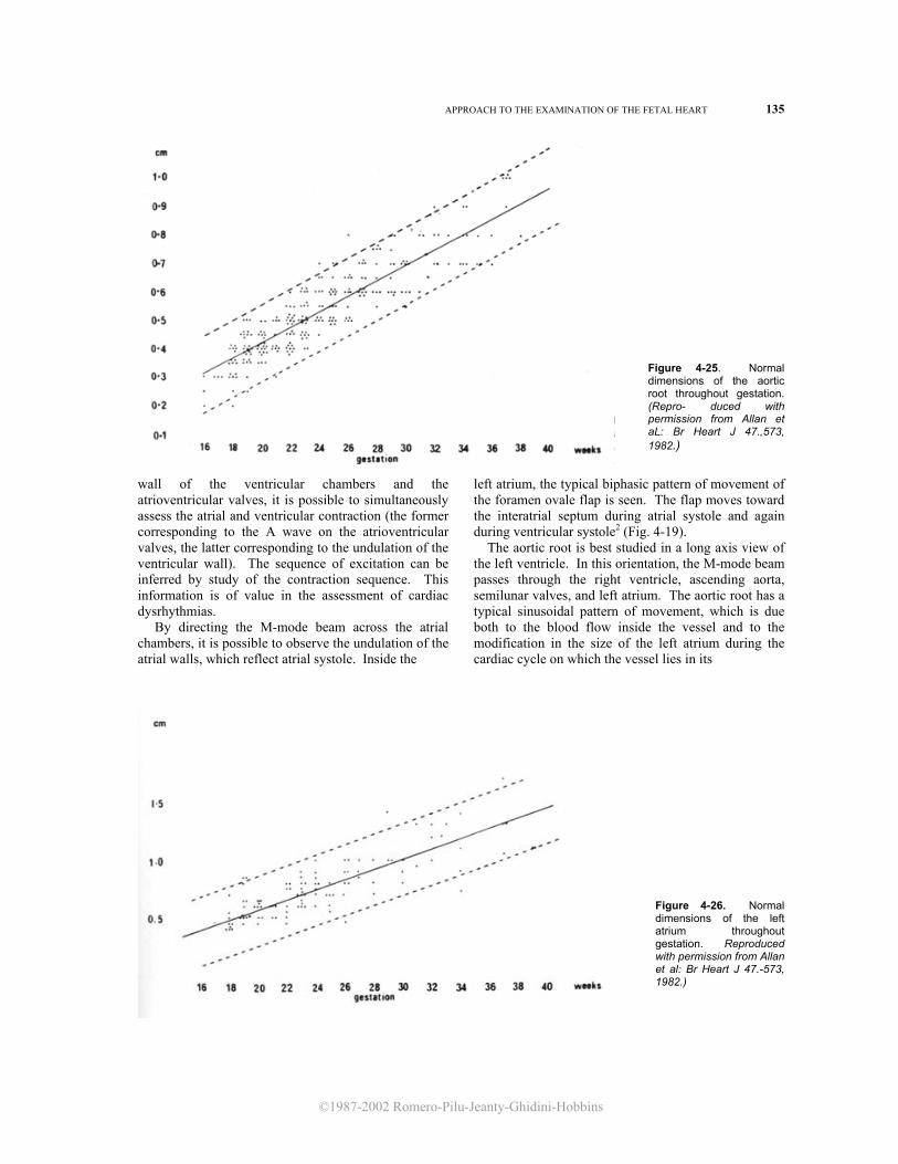
Figure 4-25. Normal dimensions of the aortic root throughout gestation. (Repro- duced with permission from Allan et aL: Br Heart J 47.,573, 1982.)
wall of the ventricular chambers and the atrioventricular valves, it is possible to simultaneously assess the atrial and ventricular contraction (the former corresponding to the A wave on the atrioventricular valves, the latter corresponding to the undulation of the ventricular wall). The sequence of excitation can be inferred by study of the contraction sequence. This information is of value in the assessment of cardiac dysrhythmias. By directing the M-mode beam across the atrial chambers, it is possible to observe the undulation of the atrial walls, which reflect atrial systole. Inside the
left atrium, the typical biphasic pattern of movement of the foramen ovale flap is seen. The flap moves toward the interatrial septum during atrial systole and again during ventricular systole2 (Fig. 4-19). The aortic root is best studied in a long axis view of the left ventricle. In this orientation, the M-mode beam passes through the right ventricle, ascending aorta, semilunar valves, and left atrium. The aortic root has a typical sinusoidal pattern of movement, which is due both to the blood flow inside the vessel and to the modification in the size of the left atrium during the cardiac cycle on which the vessel lies in its
Figure 4-26. Normal dimensions of the left atrium throughout gestation. Reproduced with permission from Allan et al: Br Heart J 47.-573, 1982.)

©1987-2002 Romero-Pilu-Jeanty-Ghidini-Hobbins
136 THE HEART
proximal tract. Inside the aortic root, the semilunar valves are seen. These are apposed during diastole and open briskly at the beginning of the ejection period, displaying a boxlike appearance. The undulation of the wall of the left atrium reflects atrial systole. Therefore, it is possible to correlate the atrial contraction with the ventricular contraction (opening of the aortic valve) and to infer the sequence of excitation (Fig. 4-20). The movement of the pulmonary valve closely resembles that of the aortic valve.
While evaluating a fetus with dysrhythmia, it is not always possible to obtain the views that have been described. It should be remembered that the sequence of excitation (which is the key to the differential diagnosis of arrhythmias) can be inferred from any orientation of the sound beam that allows the simultaneous demonstration of an atrial and a ventricular structure. Figure 4-21 shows how the atrioventricular contraction sequence can be easily demonstrated in a fetus by simply directing the sound beam across the right atrium and left ventricle. In this view, the atrial systole is reflected by the undulations of the atrial wall, and the ventricular systole is reflected by the movement of the ventricular wall.
M-mode echocardiography has been used to quantitate fetal cardiac structures.2,18 In the nomograms reported by Allan et al.,2 a short axis view of the ventricles below the level of the atrioventricular valve was used to measure the inner dimensions of the ventricular chambers at the end of diastole (Figs. 4-22, 4-23) and the thickness of the interventricular septum (Fig. 4-24). The ascending aorta was measured with an
orientation that allowed a visualization of the boxlike pattern of the semilunar valves (Fig. 4-25). The left atrium was measured by using a long axis view of the left ventricle as the point of reference (Fig. 4-26).
It should be stressed that the variability range of many reported nomograms of cardiac dimensions is quite wide and the usefulness of nomograms in the assessment of enlargement or hypoplasia of cardiac chambers and vessels is limited. We have found that a subjective evaluation by an experienced operator has great value.
©1987-2002 Romero-Pilu-Jeanty-Ghidini-Hobbins
APPROACH TO THE EXAMINATION OF THE FETAL HEART 137
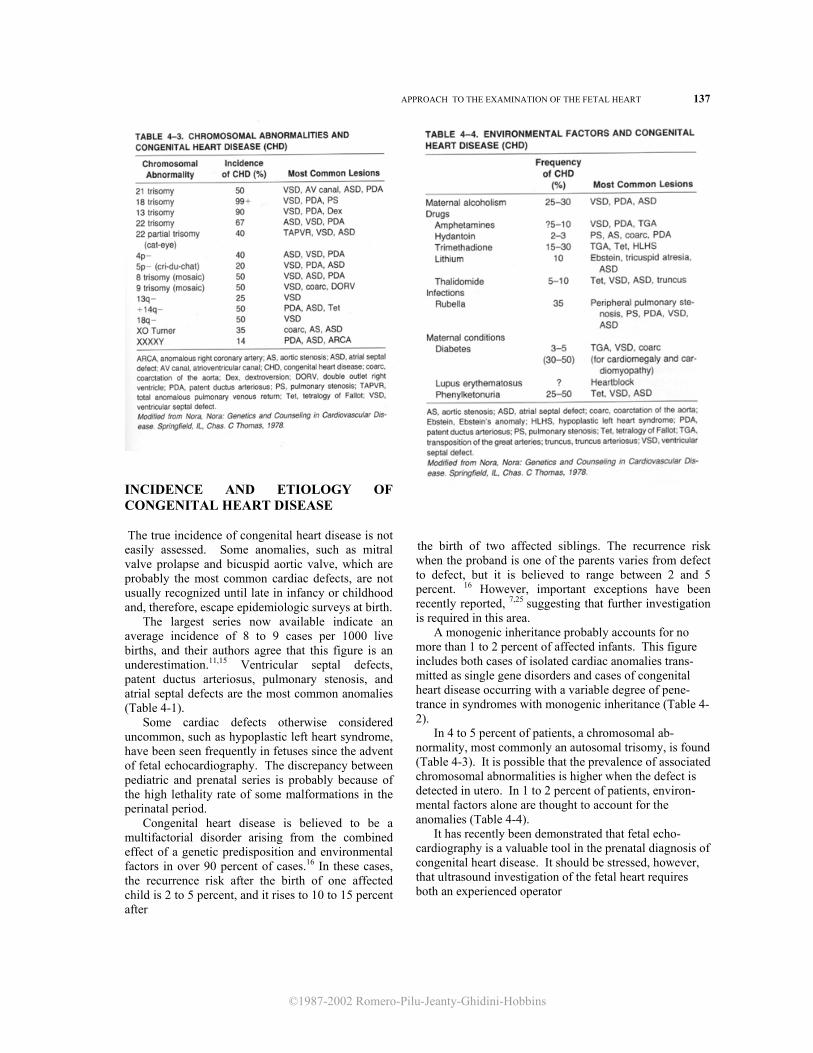
INCIDENCE AND ETIOLOGY OF CONGENITAL HEART DISEASE The true incidence of congenital heart disease is not easily assessed. Some anomalies, such as mitral valve prolapse and bicuspid aortic valve, which are probably the most common cardiac defects, are not usually recognized until late in infancy or childhood and, therefore, escape epidemiologic surveys at birth.
The largest series now available indicate an average incidence of 8 to 9 cases per 1000 live births, and their authors agree that this figure is an underestimation.11,15 Ventricular septal defects, patent ductus arteriosus, pulmonary stenosis, and atrial septal defects are the most common anomalies (Table 4-1).
Some cardiac defects otherwise considered uncommon, such as hypoplastic left heart syndrome, have been seen frequently in fetuses since the advent of fetal echocardiography. The discrepancy between pediatric and prenatal series is probably because of the high lethality rate of some malformations in the perinatal period.
Congenital heart disease is believed to be a multifactorial disorder arising from the combined effect of a genetic predisposition and environmental factors in over 90 percent of cases.16 In these cases, the recurrence risk after the birth of one affected child is 2 to 5 percent, and it rises to 10 to 15 percent after
the birth of two affected siblings. The recurrence risk when the proband is one of the parents varies from defect to defect, but it is believed to range between 2 and 5 percent. 16 However, important exceptions have been recently reported, 7,25 suggesting that further investigation is required in this area.
A monogenic inheritance probably accounts for no more than 1 to 2 percent of affected infants. This figure includes both cases of isolated cardiac anomalies trans-mitted as single gene disorders and cases of congenital heart disease occurring with a variable degree of pene-trance in syndromes with monogenic inheritance (Table 4-2).
In 4 to 5 percent of patients, a chromosomal ab-normality, most commonly an autosomal trisomy, is found (Table 4-3). It is possible that the prevalence of associated chromosomal abnormalities is higher when the defect is detected in utero. In 1 to 2 percent of patients, environ-mental factors alone are thought to account for the anomalies (Table 4-4).
It has recently been demonstrated that fetal echo-cardiography is a valuable tool in the prenatal diagnosis of congenital heart disease. It should be stressed, however, that ultrasound investigation of the fetal heart requires both an experienced operator

©1987-2002 Romero-Pilu-Jeanty-Ghidini-Hobbins
138 THE HEART
and meticulous scanning. Currently accepted indications for fetal echocardiographic evaluation are shown in Table 4-5. REFERENCES l. Allan LD, Crawford DC, Anderson RH, et al.:
Echocardiographic and anatomical correlates in fetal congenital heart disease. Br Heart J 52:542, 1984.
2. Allan LD, Joseph MC, Boyd EGCA, et al.: M-mode echocardiography in the developing human fetus, Br Heart J 47:573, 1982.
3. Allan LD, Tynan M, Campbell S, et al.: Echocardiographic and anatomical correlates in the fetus. Br Heart J 44:444, 1980.
4. Allan LD, Tynan M, Campbell S, et al.: Identification of congenital cardiac malformations by echocardiography in midtrimester fetus. Br Heart j 46:358, 1981.
5. Becker AE, Anderson RH: Pathology of Congenital Heart Disease. London, Butterworths, 1981.
6. DeLuca I, Ianniruberto A, Colonna L: Aspetti ecografici del cuore fetale. G Ital Cardiol 8:778, 1978.
7. Emanuel R, Somerville J, Inns A, et al.: Evidence of congenital heart disease in the offspring of parents with atrioventricular defects. Br Heart J 49:144, 1983.
8. Hoffman JI, Christianson R: Congenital heart disease in a cohort of 19,502 births with long-term follow-up. Am J Cardiol 42:6,41, 1978.
9. Huhta JC, Smallhorn JF, Macartney FJ: Two-dimensional echocardiographic diagnosis of situs. Br Heart J 48:97, 1982.
10. Ianniruberto A, Iaccarino M, DeLuca I, et al.: Analisi delle strutture cardiache fetali mediante ecografia. Nota tecnica. In: Colagrande C, Ianniruberto A, Talia B (eds):
Proceedings of the 3rd National Congress of the SISUM. Terlizzi, September 24-25, 1977, pp 285-290.
11. Kleinman CS, Santulli TV: Ultrasonic evaluation of the fetal human heart. Semin Perinatol 7:90, 1983.
12. Kleinman CS, Donnerstein RL, DeVore GR, et al.: Fetal echocardiography for evaluation of in utero congestive heart failure: A technique for study of nonimmune fetal hydrops. N Engl J Med 306:568, 1982.
13. YIeinman CS, Hobbins JC, Jaffe CC, et al.: Echocardiographic studies of the human fetus: prenatal diagnosis of congenital heart disease and cardiac dysrhythmias. Pediatrics 65:1059, 1980.
14. Lange, LW, Sahn DJ, Allen HD, et al.: Qualitative real-time cross-sectional echocardiographic imaging of the human fetus during the second half of pregnancy. Circulation 62:799, 1980.
15. Mitchell SC, Korones SB, Berendes HW: Congenital heart disease in 56,109 births. Incidence and natural history. Circulation 43:323, 1971.
16. Nora JJ, Nora AH: Genetics and Counseling in Cardiovascular Diseases. Springfield, IL, Chas. C Thomas, 1978.
17. Pilu G, Rizzo N, Orsini LF, et al.: La diagnosi delle anomalie cardiache strutturali nel feto mediante ultrasonografia bidimensionale. Ultr Ost Gin 1:257, 1983.
18. Sahn DJ, Lange LW, Allen HD, et al.: Quantitative real-time cross-sectional echocardiography in the developing normal human fetus and newborn. Circulation 62:588, 1980.
19. Shinebourne EA, Macartney FJ, Anderson RH: Sequential chamber localization: Logical approach to diagnosis in congenital heart disease. Br Heart J 38:327, 1976.
20. Schulz Roczen R: Fetal echocardiography: Present and future applications. J Clin Ultrasound 9:223, 1981.
21. Silverman NH, Golbus MS: Echocardiographic techniques for assessing normal and abnormal fetal cardiac anatomy. J Am Coll Cardiol 5:20S, 1985.
22. Stewart PA, Tonge HM, Wladimiroff JW: Arrhythmia and structural abnormalities of the fetal heart. Br Heart J 50:550, 1983.
23. Stewart PA, Wladimiroff JW, Essed CE: Prenatal ultrasound diagnosis of congenital heart disease associated with intrauterine growth retardation. A report of 2 cases. Prenat Diagn 3:279, 1983.
24. Van Fraagh R: The segmental approach to diagnosis in congenital heart disease. Birth Defects 8:4, 1972.
25. Whittemore R, Hobbins JC, Engle MA: Pregnancy and its outcome in women with and without surgical treatment of congenital heart disease. Am J Cardiol 50:641, 1982.
26. Winsberg F: Echocardiography of the fetal and newborn heart. Invest Radiol 7:152, 1972.
27. Wladimiroff JW, McGhie JS: M-mode ultrasonic assessment of fetal cardiovascular dynamics. Br J Obstet Gynaecol 88:1241, 1981.
©1987-2002 Romero-Pilu-Jeanty-Ghidini-Hobbins
ATRIAL SEPTAL DEFECTS 139
Atrial Septal Defects Embryology of the Atrial Septum
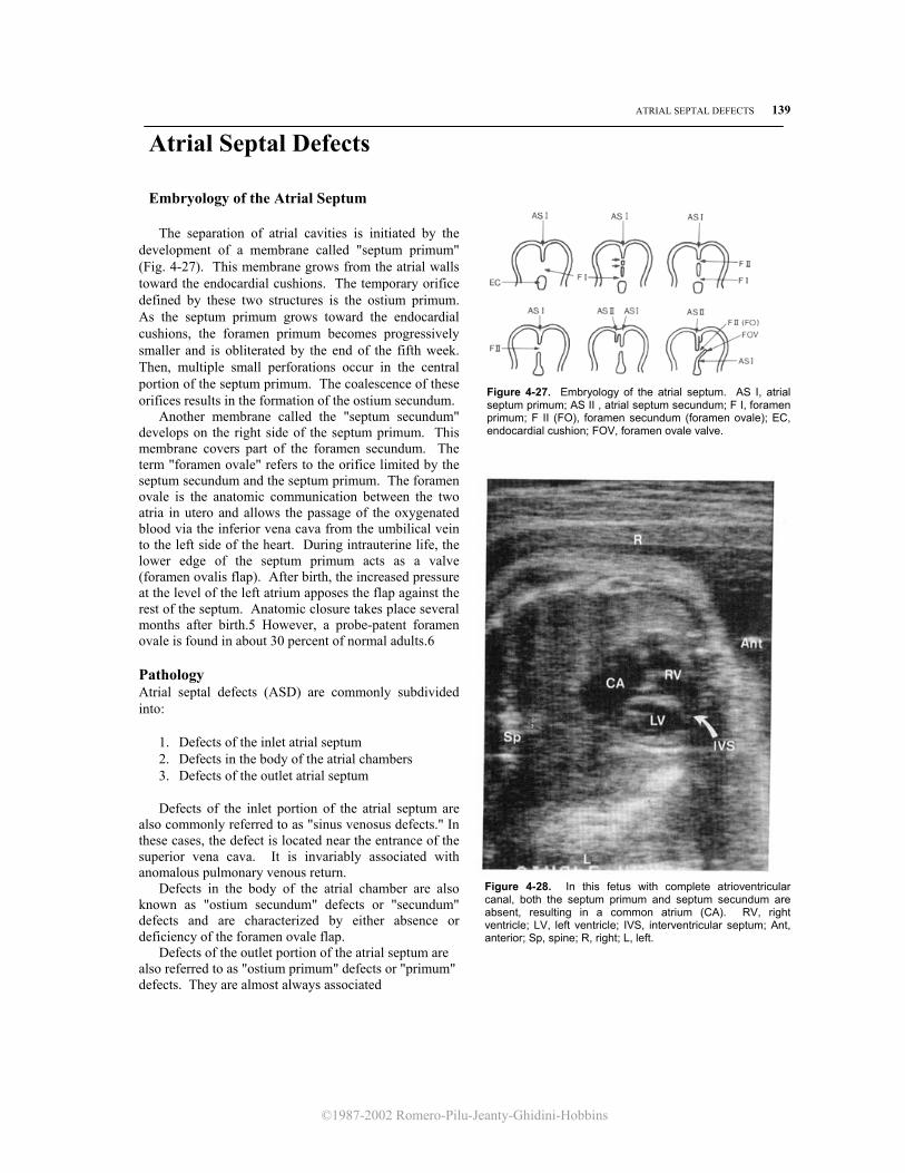
The separation of atrial cavities is initiated by the development of a membrane called "septum primum" (Fig. 4-27). This membrane grows from the atrial walls toward the endocardial cushions. The temporary orifice defined by these two structures is the ostium primum. As the septum primum grows toward the endocardial cushions, the foramen primum becomes progressively smaller and is obliterated by the end of the fifth week. Then, multiple small perforations occur in the central portion of the septum primum. The coalescence of these orifices results in the formation of the ostium secundum.
Another membrane called the "septum secundum" develops on the right side of the septum primum. This membrane covers part of the foramen secundum. The term "foramen ovale" refers to the orifice limited by the septum secundum and the septum primum. The foramen ovale is the anatomic communication between the two atria in utero and allows the passage of the oxygenated blood via the inferior vena cava from the umbilical vein to the left side of the heart. During intrauterine life, the lower edge of the septum primum acts as a valve (foramen ovalis flap). After birth, the increased pressure at the level of the left atrium apposes the flap against the rest of the septum. Anatomic closure takes place several months after birth.5 However, a probe-patent foramen ovale is found in about 30 percent of normal adults.6
Pathology Atrial septal defects (ASD) are commonly subdivided into:
1. Defects of the inlet atrial septum 2. Defects in the body of the atrial chambers 3. Defects of the outlet atrial septum Defects of the inlet portion of the atrial septum are
also commonly referred to as "sinus venosus defects." In these cases, the defect is located near the entrance of the superior vena cava. It is invariably associated with anomalous pulmonary venous return.
Defects in the body of the atrial chamber are also known as "ostium secundum" defects or "secundum" defects and are characterized by either absence or deficiency of the foramen ovale flap.
Defects of the outlet portion of the atrial septum are also referred to as "ostium primum" defects or "primum" defects. They are almost always associated
Figure 4-27. Embryology of the atrial septum. AS I, atrial septum primum; AS II , atrial septum secundum; F I, foramen primum; F II (FO), foramen secundum (foramen ovale); EC, endocardial cushion; FOV, foramen ovale valve.
Figure 4-28. In this fetus with complete atrioventricular canal, both the septum primum and septum secundum are absent, resulting in a common atrium (CA). RV, right ventricle; LV, left ventricle; IVS, interventricular septum; Ant, anterior; Sp, spine; R, right; L, left.

©1987-2002 Romero-Pilu-Jeanty-Ghidini-Hobbins
140 THE HEART
Figure 4-29. A. In this apical four chamber view, the lack of lateral resolution of the ultrasound equipment resulted in a dropout of echoes at the level of the septum secundum (?). LV, left ventricle; RV, right ventricle; LA, left atrium; RA, right atrium; Ant, an- terior; L, left; R, right. B. Subcostal four chamber view of the patient, demonstrating normal septum secundum (I I ias). I ias, septum primum; RV, right ventricle; LV, left ventricle; LA, left atrium; RA, right atrium; Sp, spine; Ant, anterior; Post, posterior; L, left; R, right; ivs, intact ventricular septum.
with anomalies of the atrioventricular junction and therefore, are discussed in the section on atrioventricular canal malformations (see p. 144)2
Hemodynamic Considerations Since a large right-to-left shunt is physiologic during intrauterine life, neither defects of the inlet atrial septum nor defects at the level of the foramen ovale
flap (secundum defects) are a cause of hemodynamic perturbance in the fetus. After birth, there is a physiologic increase in the pressure at the level of the left atrium, creating conditions for a left-to-right shunt. In time, the overload of the right ventricle may lead to dilatation and, in rare instances, to congestive heart failure. Pulmonary vascular bed damage can lead to pulmonary hypertension.

©1987-2002 Romero-Pilu-Jeanty-Ghidini-Hobbins
VENTRICULAR SEPTAL DEFECTS 141
Symptomatic infants may suffer from repeated respiratory infections, feeding difficulties, arrhythmias, thromboembolism, and failure to thrive.1 Diagnosis Diagnosis of an ASD relies on the demonstration of a dropout of echoes at the level of the atrial septum. Because of the presence of the foramen ovale and the rapidly flapping valve, it is unlikely that a small ostium secundum defect can be recognized in the fetus. The prenatal diagnosis of a defect of the inlet portion has not been reported, and it seems extremely difficult to recognize because of its location and size. Larger defects involving both the septum secundum and septum primum are easily recognizable (Fig. 4-28). It should be stressed that the thin interatrial septum may be difficult to image properly with an apical four chamber view of the heart. For an adequate evaluation, the subcostal approach should be used (Fig. 4-29).
Prognosis Campbell reported in 1970 his observations on the natural history of ASD.3 He found that the mortality rates for the first two decades of life were 0.6 percent and 0.7 percent per year, respectively. The figures rose to 2.7 percent, 4.5 percent, 5.4 percent, and 7.5 percent in successive decades. The median age of death was 37 years. Cockerham et al.4 have subsequently reported on the rate of spontaneous closure of ASDS. They studied 264 patients with ostium secundum and found that infants younger than 1 year of age with clinical symptoms had a rate of closure of 22 percent. The rate of closure in patients between the ages of 1 and 2 years was 33 percent. Patients older than 4 years of age had a spontaneous
closure rate of 3 percent. In view of these figures, the authors suggested that infants symptomatic before 2 years old should be initially treated medically. After 4 years of age, elective surgery was recommended because of the unlikelihood of spontaneous closure. The mortality rate with surgery has been estimated to be about 1 percent.4 As with other cardiac defects, it should be stressed that these data have been generated from infants, children, and adults with ASD. They may not apply to the larger defects susceptible to antenatal diagnosis. Obstetrical Management ASDs are often associated with both cardiac and extracardiac anomalies. Therefore, a careful evaluation of the entire fetal anatomy and an amniocentesis for chromosomal analysis are recommended. In the presence of an isolated secundum ASD, standard obstetrical management is not altered. REFERENCES
1. Adams CW: A reappraisal of life expectancy with atrial
shunts of the secundum type. Dis Chest 48:357, 1965. 2. Becker AE, Anderson RH: Atrial septal defects. In:
Pathology of Congenital Heart Disease. London, Butterworths, 1981, pp 67-75.
3. Campbell M: Natural history of atrial septal defect. Br Heart j 32:820, 1970.
4. Cockerham JT, Martin TC, Gutierrez FR, et al.: Spontaneous closure of secundum atrial septal defect in infants and young children. Am j Cardiol 52:1267, 1983.
5. Moore KL: The Developing Human: Clinically Oriented Embryology, 2d ed. Philadelphia, Saunders, 1977.
6. Patten BM: The closure of the foramen ovale. Am J Anat 48:19, 1931.
Ventricular Septal Defects
Pathology and Embryology The ventricular septum originates from the fusion of the endocardial cushions with the muscular part of the septum and the conus ridges at the 7th week. Ventricular septal defects (VSD) can be classified according to the position of the defect. The septum is commonly divided into a membranous and a muscular portion. The muscular portion is subdivided into three components: inlet, trabecular, and outlet or infundibular (Fig. 4-30). The most common location for the VSD is the membranous portion of the septum. Since most of these defects involve the muscular por-
tion as well, the term "perimembranous" has been suggested.14 VSDs are by far the most common cardiac lesion, accounting for 30 percent of all structural heart defects. Furthermore, they are found in many complex abnormalities, such as tetralogy of Fallot and transposition of the great arteries.5 Hemodynamic Considerations
The presence of an isolated VSD is not regarded as a cause of hemodynamic disturbances in utero. Since the pressure in both ventricular cavities is believed to

©1987-2002 Romero-Pilu-Jeanty-Ghidini-Hobbins
142 THE HEART
Figure 4-30. Diagram of the components of the interventricular septum viewed from the right ventricle (RV) and left ventricle (LV). (Modified from Becker, Anderson: Pathology of Congenital Heart Disease. London, Butterworths, 1981.) be equal, it is possible that even large VSDs are only responsible for small bidirectional shunts. 12 This view seems to be supported by the observations that most infants are asymptomatic at birth.7,8
After birth, there is a decrease in the arterial pressure in the pulmonary vascular bed and an increase in the systemic arterial pressure. In the presence of a VSD, a left-to-right shunt occurs. Very small VSDs have little or no hemodynamic consequences because of the negligible magnitude of the shunt. With larger VSDs, some or all of the systemic pressure is transmitted to the pulmonary arteries. In time, this may lead to pulmonary vascular disease and pulmonary hypertension. Increased pressure in the right ventricle may eventually result in a reversal of the shunt, with cyanosis and congestive heart failure. 7,8 An exception to this course of events is an infant with a very large VSD, in whom a large portion of the left ventricular output is diverted into the right ventricle, with ventricular overload, thus possibly creating congestive heart failure soon after birth.11
Several studies have documented spontaneous closure of VSDs. Factors influencing this phenomenon include the size and location of the defect. Smaller defects and those located in the muscular septum have a higher tendency to close than do large and membranous defects. Hoffman and Rudolph 8,9 reported that 40 percent of VSDs are closed within 2 years of life and that 60 percent will close by 5 years. The incidence of closure for membranous defects is 25 percent by 5 years and that of muscular defects is 65 percent.3
The mechanisms of closure are different for perimembranous and muscular defects. The latter are closed by fibrous tissue originating from the septum,8 whereas the former are closed either completely or partially with a variety of anatomic derivatives, in-
cluding reduplication of the tricuspid valve tissue, adhesion of tricuspid valve leaflets, and prolapse of an aortic valve leaflet.4 Diagnosis The diagnosis depends on the demonstration of a dropout of echoes at the level of the interventricular septuml,2,13 (Fig. 4-31). It should be stressed that a careful examination of the interventricular septum is necessary. Since a four chamber view of the heart will reveal only a small portion of the inlet and trabecular septum, it is obvious that a VSD can be missed easily by relying on this view. This is especially true if the defect is located in the outlet or membranous portion of the septum (Fig. 4-32). In addition to the four chamber view, the examination of the septum should include a long axis view of the left ventricle, a long axis view of the right ventricle, and an apex to base sweep along the short axis of the heart.6
The sonographer should be alerted to a potential pitfall. When an apical four chamber view of the heart is obtained, the limitations of lateral resolution of the sound beam could result in the creation of an artifactual hypoechogenic image in the higher portion of the inlet septum (Fig. 4-33). This pitfall is easily recognized by failure to demonstrate the defect in other views. Optimal examination is achieved when
Figure 4-31. In this 25-week-old fetus, a four chamber view reveals a perimembranous ventricular septal defect (*). LV, left ventricle; RV, right ventricle; LA, left atrium; RA, right atrium; Sp, spine; Ant, anterior; L, left; R, right. (Reproduced with permission from Bovicelli L, Baccarani G, Picchio FM, Pilu G: Ecocardiografia Fetale. La Diagnosi ed il Trattamento Prenatale delle Cardiopatie Congenite. Milan, Masson, 1985.)

©1987-2002 Romero-Pilu-Jeanty-Ghidini-Hobbins
VENTRICULAR SEPTAL DEFECTS 143
the sound beam is perpendicular to the septum. An artifactual "defect" often demonstrates a "fading-out" of the septum. A true defect is usually seen as a sharply terminating bright spot or area, Since the resolution of current ultrasound equipment is limited to 1 to 2 mm, it is not surprising that some VSDs will escape detection prenatally. Since VSDs are frequent components of more complex cardiac abnormalities, a careful examination of the entire cardiac morphology is mandatory.
Figure 4-32. A. A four-chamber view in a 30-week fetus reveals a seemingly intact ventricular septum (IVS). LV, left ventricle; RV, right ventricle; LA, left atrium; RA, right atrium. B. In the same patient, a subaortic VSD (*) is clearly demonstrated by a slight cephalic angulation of the transducer. LV, left ventricle; RV, right ventricle; LA, left atrium; RA, right atrium.
Figure 4-33. In this apical four chamber view, lack of lateral resolution and low gain settings result in a dropout of echoes (?) at the level of the perimembranous ventricular septum. LV, left ventricle; RV, right ventricle; LA, left atrium; RA, right atrium; IVS, interventricular septum.
Prognosis The prognosis of infants with VSD is good. It is difficult to provide precise figures because of ascertainment bias in old studies. However, most infants born with VSDs are asymptomatic, 40 percent of defects spontaneously close within 2 years, and 60 percent close within 5 years.7-9 In a series of 428 symptomatic infants, 130 (30 percent) required surgery because of intractable congestive heart failure and failure to thrive.10 In a group of 50 infants less than 18 months of age treated with primary closure of the VSD, there was a 6 percent postoperative mortality rate. In addition, 14 percent had seizures attributable to low cardiac output and hypoxic episodes, and 49 percent had rhythm disturbances (right bundle branch block isolated or associated with left hemiblocks).10 Postoperative studies showed normal pressure in the pulmonary artery.10
Obstetrical Management A careful search for other cardiac and extracardiac abnormalities is indicated if the prenatal diagnosis of VSD is made. An amniocentesis for chromosomal analysis is recommended. In the presence of an isolated VSD, standard obstetrical management is not altered. Infants should be delivered in a tertiary care center where a pediatric cardiologist is immediately available.

©1987-2002 Romero-Pilu-Jeanty-Ghidini-Hobbins
144 THE HEART
REFERENCES 1. Allan LD, Crawford DC, Anderson RH, et al.:
Echocardiographic and anatomical correlations in fetal congenital heart disease. Br Heart J 52:542, 198,4
2. Allan LD, Tynan M, Campbell S, et al.: Identification of congenital cardiac malformations by echocardiography in mid-trimester fetus. Br Heart J 46:358, 1981
3. Alpert BS, Mellits ED, Rowe RD: Spontaneous closure of small ventricular septal defects. Probability rates in the first five years of life. Am j Dis Child 125:194, 1973
4. Anderson RH, Lenox CC, Zuberbuhler JR: Mechanisms of closure of perimembranous ventricular septat defect. Am j Cardiol 52:341, 1983
5. Becker AE, Anderson RH: Pathology of Congenital Heart Disease. London, Butterworths, 1981.
6. Capelli H, Andrade JL, Somerville J: Classification of the site of ventricular septat defect by 2-dimensional echocardiography. Am j Cardiol 51:1474, 1983
7. Hoffman JIE: Natural history of congenital heart disease. Problems in its assessment, with special reference
to ventricular septal defects. Circulation 37:97, 1968.
8. Hoffman JIE, Rudolph AM: The natural history of isolated ventricular septal defect. With special reference to selection of patients for surgery. Adv Pediatr 17:57, 1970.
9. Hoffman JIE, Rudolph AM: The natural history of ventricular septal defects in infancy. Am J Cardiol 16:634, 1965.
10. Rein JG, Freed MD, Norwood WI, et al.: Early and late results of closure of ventricular septal defect in infancv. Ann Thorac Surg 24:19, 1977.
11. Rowe RD, Freedom RM, Mehrizi A, et al.: The Neonate with Congenital Heart Disease, 2d ed. Philadelphia, Saunders, 1981.
12. Rudolph AM: Congenital Diseases of the Heart: Clim-cal-Physiologic Considerations in Diagnosis and Management. Chicago, Year Book, 1974.
13. Silverman NH, Golbus MS: Echocardiographic techniques for assessing normal and abnormal fetal cardiac anatomy. J Am Coll Cardiol 5:20S, 1985.
14. Soto B, Becker AE, Moulaert Aj, et al.: Classification of ventricular septal defects. Br Heart j 43:332, 1980.
Atrioventricular Septal Defects
Synonyms Ostium primum atrial septal defect, atrioventricular canal malformation, endocardial cushion defects, and persistent ostium atrioventriculare commune. Definition Atrioventricular septal defects (AVSD) include a spectrum of cardiac anomalies involving to a different extent the atrial and ventricular septa and the atrioventricular valves. Embryology In the primitive heart, the atrium and common ventricle communicate through a single opening, the atrioventricular canal. The growth of the endocardial cushions divides this opening into two distinct orifices at the 6th week (Fig. 4-34). Subsequently, the fusion of the atrial and ventricular septum partitions the heart into four chambers. AVSDs result from the persistence of the primitive atrioventricular canal. Since the endocardial cushions participate in the development of the atrioventricular valves, anomalies at the level of the tricuspid and mitral valves are the rule in these defects. Pathology AVSDs are subdivided into complete and incomplete or partial forms (Fig. 4-35).3Incomplete AVSDs (also known as "ostium
primum atrial septal defects") are characterized by separate atrioventricular orifices. There is usually an interatrial communication or a communication between the left ventricle and the right atrium. Less frequently, an interventricular communication occurs. The right atrioventricular valve is often normal. The left atrioventricular valve has usually three leaflets, and there is a cleft between the anterior and posterior ones (Fig. 4-35).3
Figure 4-34. Embryology of the atrioventricular junction. The primitive atrioventricular canal is partitioned by the growth of the endocardial cushions (EC), interatrial septum (IAS), and interventricular septum (IVS).
©1987-2002 Romero-Pilu-Jeanty-Ghidini-Hobbins
ATRIOVENTRICULAR SEPTAL DEFECTS 145
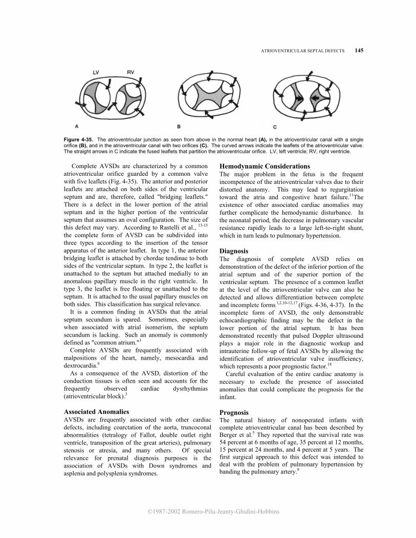
Figure 4-35. The atrioventricular junction as seen from above in the normal heart (A), in the atrioventricular canal with a single orifice (B), and in the atrioventricular canal with two orifices (C). The curved arrows indicate the leaflets of the atrioventricular valve. The straight arrows in C indicate the fused leaflets that partition the atrioventricular orifice. LV, left ventricle; RV, right ventricle. Complete AVSDs are characterized by a common atrioventricular orifice guarded by a common valve with five leaflets (Fig. 4-35). The anterior and posterior leaflets are attached on both sides of the ventricular septum and are, therefore, called "bridging leaflets." There is a defect in the lower portion of the atrial septum and in the higher portion of the ventricular septum that assumes an oval configuration. The size of this defect may vary. According to Rastelli et al., 13-15 the complete form of AVSD can be subdivided into three types according to the insertion of the tensor apparatus of the anterior leaflet. In type 1, the anterior bridging leaflet is attached by chordae tendinae to both sides of the ventricular septum. In type 2, the leaflet is unattached to the septum but attached medially to an anomalous papillary muscle in the right ventricle. In type 3, the leaflet is free floating or unattached to the septum. It is attached to the usual papillary muscles on both sides. This classification has surgical relevance. It is a common finding in AVSDs that the atrial septum secundum is spared. Sometimes, especially when associated with atrial isomerism, the septum secundum is lacking. Such an anomaly is commonly defined as "common atrium."3 Complete AVSDs are frequently associated with malpositions of the heart, namely, mesocardia and dextrocardia.6 As a consequence of the AVSD, distortion of the conduction tissues is often seen and accounts for the frequently observed cardiac dysrhythmias (atrioventricular block).3 Associated Anomalies AVSDs are frequently associated with other cardiac defects, including coarctation of the aorta, truncoconal abnormalities (tetralogy of Fallot, double outlet right ventricle, transposition of the great arteries), pulmonary stenosis or atresia, and many others. Of special relevance for prenatal diagnosis purposes is the association of AVSDs with Down syndromes and asplenia and polysplenia syndromes.
Hemodynamic Considerations The major problem in the fetus is the frequent incompetence of the atrioventricular valves due to their distorted anatomy. This may lead to regurgitation toward the atria and congestive heart failure.11The existence of other associated cardiac anomalies may further complicate the hemodynamic disturbance. In the neonatal period, the decrease in pulmonary vascular resistance rapidly leads to a large left-to-right shunt, which in turn leads to pulmonary hypertension. Diagnosis The diagnosis of complete AVSD relies on demonstration of the defect of the inferior portion of the atrial septum and of the superior portion of the ventricular septum. The presence of a common leaflet at the level of the atrioventricular valve can also be detected and allows differentiation between complete and incomplete forms l,2,10-12,17 (Figs. 4-36, 4-37). In the incomplete form of AVSD, the only demonstrable echocardiographic finding may be the defect in the lower portion of the atrial septum. It has been demonstrated recently that pulsed Doppler ultrasound plays a major role in the diagnostic workup and intrauterine follow-up of fetal AVSDs by allowing the identification of atrioventricular valve insufficiency, which represents a poor prognostic factor.18 Careful evaluation of the entire cardiac anatomy is necessary to exclude the presence of associated anomalies that could complicate the prognosis for the infant.
Prognosis The natural history of nonoperated infants with complete atrioventricular canal has been described by Berger et al.5 They reported that the survival rate was 54 percent at 6 months of age, 35 percent at 12 months, 15 percent at 24 months, and 4 percent at 5 years. The first surgical approach to this defect was intended to deal with the problem of pulmonary hypertension by banding the pulmonary artery.8
©1987-2002 Romero-Pilu-Jeanty-Ghidini-Hobbins
146 THE HEART More recently, intracardiac repair of the defect has been carried out in several centers. Berger et al.5 reported a 91 percent long-term survival with primary intracardiac repair. Bender et al.4 reported 2 operative and 1 postoperative deaths in 24 operated infants, for a mortality rate of 8 percent and 4 percent, respectively. Chin et al.7 have described two consecutive groups of patients. The first group included 13 infants operated on between 1975 and 1977, with an operative mortality of 62 percent and late mortality of 7 percent. In the second group of 30 infants operated on between 1978 and 1980, the operative mortality decreased to 17 percent and late mortality was 6 percent. Prognostic factors related to the outcome of operative procedures include (1) deficiency of atrioventricular tissue, (2) presence of ventricular hypoplasia, (3) malalignment of the common atrioventricular valve, (4) the presence of double orifice mitral valve, (5) the presence of solitary left ventricular papillary muscle group, and (6) the presence of additional muscular septal defects. Obstetrical Management An amniocentesis is strongly recommended because of the association of this defect with Down syndrome. A careful ultrasound evaluation of the entire fetal anatomy is also mandatory. Diagnosis before
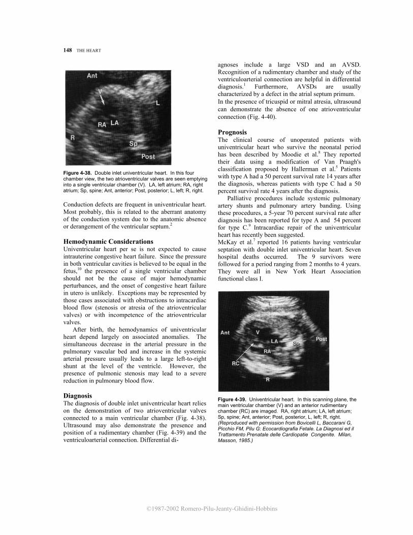
Figure 4-36. Four chamber view in a fetus with compete atrioventricular canal. The diastolic frame reveals the complete absence of the interatrial septum and the presence of a common atrioventricular valve (arrows). CA, common atrium; LV, left ventricle; RV, right ventricle; ivs, interventricular septum; Sp, spine; R, right; L, left; Ant, anterior.
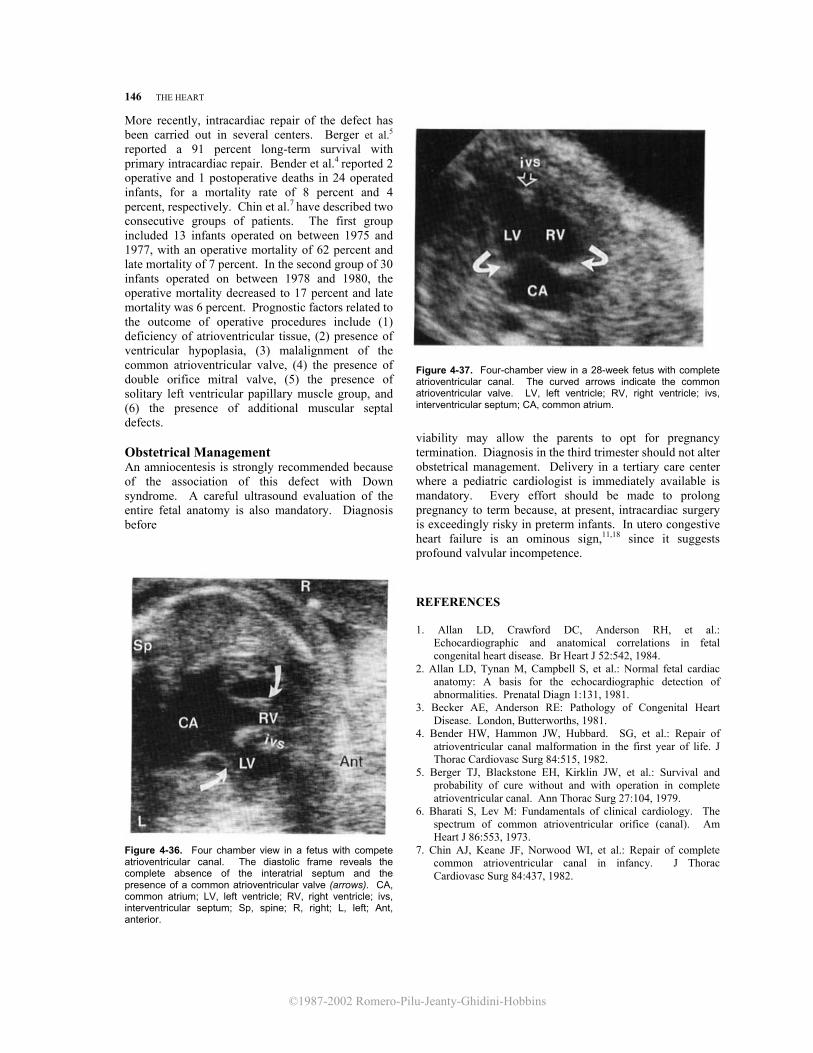
Figure 4-37. Four-chamber view in a 28-week fetus with complete atrioventricular canal. The curved arrows indicate the common atrioventricular valve. LV, left ventricle; RV, right ventricle; ivs, interventricular septum; CA, common atrium.
viability may allow the parents to opt for pregnancy termination. Diagnosis in the third trimester should not alter obstetrical management. Delivery in a tertiary care center where a pediatric cardiologist is immediately available is mandatory. Every effort should be made to prolong pregnancy to term because, at present, intracardiac surgery is exceedingly risky in preterm infants. In utero congestive heart failure is an ominous sign,11,18 since it suggests profound valvular incompetence.
REFERENCES
1. Allan LD, Crawford DC, Anderson RH, et al.: Echocardiographic and anatomical correlations in fetal congenital heart disease. Br Heart J 52:542, 1984.
2. Allan LD, Tynan M, Campbell S, et al.: Normal fetal cardiac anatomy: A basis for the echocardiographic detection of abnormalities. Prenatal Diagn 1:131, 1981.
3. Becker AE, Anderson RE: Pathology of Congenital Heart Disease. London, Butterworths, 1981.
4. Bender HW, Hammon JW, Hubbard. SG, et al.: Repair of atrioventricular canal malformation in the first year of life. J Thorac Cardiovasc Surg 84:515, 1982.
5. Berger TJ, Blackstone EH, Kirklin JW, et al.: Survival and probability of cure without and with operation in complete atrioventricular canal. Ann Thorac Surg 27:104, 1979.
6. Bharati S, Lev M: Fundamentals of clinical cardiology. The spectrum of common atrioventricular orifice (canal). Am Heart J 86:553, 1973.
7. Chin AJ, Keane JF, Norwood WI, et al.: Repair of complete common atrioventricular canal in infancy. J Thorac Cardiovasc Surg 84:437, 1982.

©1987-2002 Romero-Pilu-Jeanty-Ghidini-Hobbins
UNIVENTRICULAR HEART 147
8. Epstein ML, Moller JH, Amplatz K, et al.: Pulmonary
artery banding in infants with complete atrioventricular canal. J Thorac Cardiovasc Surg 78:28, 1979.
9. Fisher EA, Doshi M, DuBrow IW, et al.: Effect of palliative and corrective surgery on ventricular volumes in complete atrioventricular canal. Pediatr Cardiol 5:159, 1984.
10. Kleinman CS, Santulli TV: Ultrasonic evaluation of the fetal human heart. Semin Perinatol 7:90, 1983.
11. Kleinman CS, Donnerstein RL, De Vore GR, et al.: Fetal echocardiography for evaluation of in utero congestive heart failure: A technique for study of nonimmune fetal hydrops. N Engl J Med 306:568, 1982.
12. Pilu G, Rizzo N, Orsini LF, et al.: La diagnosi delle anomalie cardiache strutturali net feto mediante ultrasonografia bidimensionale. Ultr Ost Gin 1:257, 1983.
13. Rastelli GC, Kirklin JW, Kincaid OW: Angiocardiography of persistent common atrioventricular canal. Mayo Clin Proc 42:200, 1967.
14. Rastelli GC, Kirklin JW, Titus JL: Anatomic observations on complete form of persistent common atrioventricular canal with special reference to atrioventricular valves. Mayo Clin Proc 41:296, 1966.
15. Rastelli GC, Ongley PA, Kirklin JW, et al.: Surgical repair of the complete form of persistent common atrioventricular canal. J Thorac Cardiovasc Surg 55:299, 1968.
16. Rowe RD, Uchida IA: Cardiac malformation in mongolism. A prospective study of 184 mongoloid children. Am J Med 31:726, 1961.
17. Silverman NH, Golbus MS: Echocardiographic techniques for assessing normal and abnormal fetal cardiac anatomy. J Am Coll Cardiol 5:20S, 1985.
18. Silverman NH, Kleinman CS, Rudolph AM, et al.: Fetal atrioventricular valve insufficiency associated with nonimmune hydrops: A two-dimensional echocardiographic and pulsed Doppler ultrasound study. Circulation 72:825, 1985.
Univentricular Heart Synonym Single ventricle. Definition
The definition of univentricular heart is controversial. According to some authors,11 this term refers to a condition in which there are two atrioventricular valves or a common atrioventricular valve and a single ventricle (classic double inlet single ventricle). According to Becker and Anderson,2 univentricular heart indicates a group of anomalies in which the entire atrioventricular junction is connected to only one chamber in the ventricular mass. This includes, by definition, the classic double inlet single ventricle and the absence of one atrioventricular connection.
Embryology The double inlet univentricular heart seems to be related to failure of development of the interventricular septum. Absence of one atrioventricular connection results from mitral or tricuspid atresia. Pathology According to Van Praagh et al.,11 the univentricular heart is classified as type A or C according to the presence or absence of outflow tract. Depending on the relationship between the aorta and pulmonary artery, three subtypes are defined: (1) normal relationship (the aorta is posterior and to the left of the pulmonary artery), (2) the aorta is anterior and to the
right, and (3) the aorta is anterior and to the left. Six different varieties of univentricular heart are possible.
According to the elegant definition of Becker and Anderson,2 the univentricular heart is "a generic term for a group of anomalies unified by their ventricular morphology. The unifying criterion is that the entire atrioventricular junction is connected to only one chamber in the ventricular mass." The chamber may be either of left ventricular, right ventricular, or undetermined type depending on the trabecular pattern. In 85 percent of patients, the chamber has a left ventricular morphology.
A second rudimentary ventricular chamber may be present. In these cases, a rudimentary ventricular septum that does not extend to the crux can be seen. The atrioventricular valves may straddle the septum. In a univentricular heart of left ventricular type, the rudimentary chamber is usually anterior. In a right univentricular heart, the rudimentary chamber is usually posterior, and a rudimentary ventricular septum extends to the crux.
The rudimentary chamber may or may not be connected to the great arteries. Aortic and pulmonic stenosis are frequently seen.
In the case of tricuspid atresia with absence of the right ventricular connection, the right atrium communicates with the main ventricular chamber through an atrial septal defect. An interatrial communication is equally necessary in cases of mitral atresia.
©1987-2002 Romero-Pilu-Jeanty-Ghidini-Hobbins
148 THE HEART
Figure 4-38. Double inlet univentricular heart. In this four chamber view, the two atrioventricular valves are seen emptying into a single ventricular chamber (V). LA, left atrium; RA, right atrium; Sp, spine; Ant, anterior; Post, posterior; L, left; R, right. Conduction defects are frequent in univentricular heart. Most probably, this is related to the aberrant anatomy of the conduction system due to the anatomic absence or derangement of the ventricular septum.2 Hemodynamic Considerations Univentricular heart per se is not expected to cause intrauterine congestive heart failure. Since the pressure in both ventricular cavities is believed to be equal in the fetus,10 the presence of a single ventricular chamber should not be the cause of major hemodynamic perturbances, and the onset of congestive heart failure in utero is unlikely. Exceptions may be represented by those cases associated with obstructions to intracardiac blood flow (stenosis or atresia of the atrioventricular valves) or with incompetence of the atrioventricular valves. After birth, the hemodynamics of univentricular heart depend largely on associated anomalies. The simultaneous decrease in the arterial pressure in the pulmonary vascular bed and increase in the systemic arterial pressure usually leads to a large left-to-right shunt at the level of the ventricle. However, the presence of pulmonic stenosis may lead to a severe reduction in pulmonary blood flow. Diagnosis The diagnosis of double inlet univentricular heart relies on the demonstration of two atrioventricular valves connected to a main ventricular chamber (Fig. 4-38). Ultrasound may also demonstrate the presence and position of a rudimentary chamber (Fig. 4-39) and the ventriculoarterial connection. Differential di-
agnoses include a large VSD and an AVSD. Recognition of a rudimentary chamber and study of the ventriculoarterial connection are helpful in differential diagnosis.1 Furthermore, AVSDs are usually characterized by a defect in the atrial septum primum. In the presence of tricuspid or mitral atresia, ultrasound can demonstrate the absence of one atrioventricular connection (Fig. 4-40). Prognosis The clinical course of unoperated patients with univentricular heart who survive the neonatal period has been described by Moodie et al.8 They reported their data using a modification of Van Praagh's classification proposed by Hallerman et al.5 Patients with type A had a 50 percent survival rate 14 years after the diagnosis, whereas patients with type C had a 50 percent survival rate 4 years after the diagnosis. Palliative procedures include systemic pulmonary artery shunts and pulmonary artery banding. Using these procedures, a 5-year 70 percent survival rate after diagnosis has been reported for type A and 54 percent for type C.9 Intracardiac repair of the univentricular heart has recently been suggested. McKay et al.7 reported 16 patients having ventricular septation with double inlet univentricular heart. Seven hospital deaths occurred. The 9 survivors were followed for a period ranging from 2 months to 4 years. They were all in New York Heart Association functional class I.
Figure 4-39. Univentricular heart. In this scanning plane, the main ventricular chamber (V) and an anterior rudimentary chamber (RC) are imaged. RA, right atrium; LA, left atrium; Sp, spine; Ant, anterior; Post, posterior, L, left; R, right. (Reproduced with permission from Bovicelli L, Baccarani G, Picchio FM, Pilu G: Ecocardiografia Fetale. La Diagnosi ed il Trattamento Prenatale delle Cardiopatie Congenite. Milan, Masson, 1985.)

©1987-2002 Romero-Pilu-Jeanty-Ghidini-Hobbins
EBSTEIN'S ANOMALY 149
Figure 4-40. Tricuspid atresia. The left atrium (LA) is connected to a single ventricular chamber (V) with hypertrophic walls (double arrow). The right atrium (RA) lacks a connection with the ventricular mass. A bright linear echo arises from the atretic tricuspid plane (curved arrom. LA, left atrium. Sp, spine.
Obstetrical Management When the diagnosis is made before viability, the option of pregnancy termination should be offered. In all pregnancies, a careful search for associated cardiac and extracardiac anomalies, including karyotype evaluation, is recommended. Serial ultrasound examinations should be performed to search for signs of congestive heart failure. The association of hydrops with a structural cardiac defect is an ominous combination. Optimal management of these patients has not been established. The option of early delivery may be considered, but the parents should be aware that the mortality rate in these patients is extremely high.6 In the absence of
congestive heart failure, there is no indication to alter standard obstetrical management, but delivery in a tertiary care center where a pediatric cardiologist is immediately available is mandatory.
REFERENCES
1. Allan LD, Crawford DC, Anderson RH, et al.: Echo-cardiographic and anatomical correlations in fetal congenital heart disease. Br Heart J 52:542, 198,4.
2. Becker AE, Anderson RH: Pathology of Congenital Heart Disease. London, Butterworths, 1981.
3. Bisset GS, Hirschfeld SS: The univentricular heart: Combined two-dimensional-pulsed Doppler (duplex) echocardiographic evaluation. Am J Cardiol 51:1149, 1983.
4. Goldberg HL, Sniderman K, Devereux RB, et al.: Prolonged survival (62 years) with single ventricle. Am J Cardiol 52:214, 1983.
5. Hallerman FJ, Davis GD, Ritter DG, et al.: Roentgenographic features of common ventricle. Radiology 87: 409, 1966.
6. Kleinman CS, Donnerstein RL, DeVore GR, et al.: Fetal echocardiography for evaluation of in utero congestive heart failure: A technique for study of nonimmune fetal hydrops. N Engl J Med 306:568, 1982.
7. McKay R, Pacifico AD, Blackstone EH, et al.: Septation of the univentricular heart with left anterior subaortic outlet chamber. J Thorac Cardiovasc Surg 84:77, 1982.
8. Moodie DS, Ritter DG, Tajik Aj, et al.: Long-term follow-up in the unoperated univentricular heart. Am J Cardiol 53:1124, 1984.
9. Moodie DS, Ritter DG, Tajik AH, et al.: Long-term follow-up after palliative operation for univentricular heart. Am J Cardiol 53:1648, 1984.
10. Rudolph AM: Congenital Diseases of the Heart-Clinical-Physiological Considerations in Diagnosis and Management. Chicago, Year Book, 1974.
11. Van Praagh R, Ongley PA, Swan HJC: Anatomic types of single or common ventricle in man: Morphologic and geometric aspects of sixty necropsied cases. Am J Cardiol 13:367, 1964.
Ebstein's Anomaly Definition Ebstein's anomaly is a congenital defect usually characterized by downward displacement of the septal and posterior leaflets of the tricuspid valve, with dysplasia of this valve. Etiology Ebstein's anomaly of the tricuspid valve has been reported to occur in 10 percent of cases of chronic maternal lithium intake during pregnancy.8
Pathology Displacement of the tricuspid valve leaflets leads to division of the right ventricle into two components: a superior or atrialized portion and an inferior, functional chamber. The walls of the right ventricle are generally thin. 4 The tricuspid valve is most frequently insufficient, and this results in right atrial enlargement Cardiomegaly is almost the rule in these patients.

©1987-2002 Romero-Pilu-Jeanty-Ghidini-Hobbins
150 THE HEART
Although the posterior and septal leaflets of the valve are displaced downward, the anterior leaflet may be normal.4 Associated Anomalies ASDS (secundum type or patent foramen ovale), pulmonary atresia or stenosis, patent ductus arteriosus, tetralogy of Fallot, coarctation of the aorta, atrioventricular canal, and transposition of the great vessels 4 are possible associated anomalies. Hemodynamic Considerations Dysplasia and displacement of the tricuspid valve leads to tricuspid insufficiency, with blood regurgitation into the right atrium during systole. In turn, this may lead to congestive heart failure in utero. Congestive heart failure is found in 50 percent of affected newborns.10 Diagnosis The main criterion for diagnosis is demonstration of downward displacement of the tricuspid valve into the right ventricle. The right atrium is generally extremely enlarged (Fig. 4-41).2,13 Doppler studies may be helpful in assessing tricuspid valve regurgitation. An enlarged right atrium without valve displacement and regurgitation may be caused by "idiopathic giant right atrium." Prognosis In the absence of tricuspid regurgitation, this condition may be completely asymptomatic. Such patients do not develop symptoms until adolescence or adult life. On the other hand, symptomatic newborns often develop congestive heart failure. In a series of 23 patients, 12 died during the first month of life or later in infancy.10 Advances in cardiovascular surgery have improved the prognosis for these patients. In a total of 147 cases reported in nine series,1,3,5-7,9,11,12,14 there were 19 operative deaths (12.9 percent) and 11 late deaths (7.4 percent). In the largest available series,5 cardiac surgery resulted in important improvements in the clinical condition of the patients. The majority of the 22 patients with long-term follow-up improved from New York Heart Association class III or IV to class I or II. Obstetrical Management When the diagnosis is made before viability, the option of pregnancy termination should be offered. A careful search for associated cardiac and extracardiac anomalies, including karyotype, is recommended for all cases. Serial ultrasound examinations should be performed to search for signs of congestive heart failure. The association of hydrops with a structural cardiac defect is an ominous combination. The optimal man-
Figure 4-41. Four chamber view in a fetus with Ebstein's anomaly of the tricuspid valve. The right atrium (RA) is disproportionately enlarged. The sail-like tricuspid valve (TV) is displaced apically far away from the atrioventricular junction (AVJ). RV, right ventricle; LV, left ventricle; LA, left atrium; Sp, spine.
agement of these patients has yet to be established. The option of early delivery may be considered, but the parents should be aware that the mortality rate in these patients is extremely high. In the absence of congestive heart failure, there is no indication to alter standard obstetrical management, but delivery in a tertiary care center where a pediatric cardiologist is immediately available is mandatory.
REFERENCES
1. Abe T, Komatsu S: Valve replacement for Ebstein's
anomaly of the tricuspid valve. Early and long-term results of eight cases. Chest 84:414, 1983.
2. Allan LD, Crawford DC, Anderson RH, et al.: Echocardiographic and anatomical correlations in fetal congenital heart disease. Br Heart J 52:542, 1984.
3. Barbero-Marcial M, Verginelli G, Awad M, et al.: Surgical treatment of Ebstein's anomaly. Early and late results in twenty patients subjected to valve replacement. J Thorac Cardiovasc Surg 78:416, 1979.
4. Becker AE, Anderson RH: Pathology of Congenital Heart Disease. London, Butterworths, 1981.
5. Danielson GK: Ebstein's anomaly: Editorial comments

©1987-2002 Romero-Pilu-Jeanty-Ghidini-Hobbins
HYPOPLASTIC LEFT HEART SYNDROME 151
and personal observations. Ann Thorac Surg 34:396, 1982. 6. Danielson GK, Maloney JD, Devloo RA: Surgical repair of
Ebstein's anomaly. Mayo Clin Proc 54:185, 1979. 7. Mattila S, Harjula A, Kyllonen KEJ, et al.: Surgical
intervention in cases of Ebstein's anomaly. Abnormal origin and structure of the tricuspid valve. Scand J Thorac Cardiovasc Surg 16:223, 1982.
8. Nora JJ, Nora AH: Genetics and Counseling in Cardio- vascular Diseases. Springfield, IL, Chas. C Thomas, 1978.
9. Raj-Behl P, Blesovsky A: Ebstein's anomaly: Sixteen years' experience with valve replacement without plication of the right ventricle. Thorax 39:8, 1984.
10. Rowe RD, Freedom RM, Mehrizi A, Bloom KR: The
Neonate with Congenital Heart Disease, 2d ed. Philadelphia, Saunders, 1981.
11. Schmidt-Habelmann P, Meisner H, Struck E, et al.: Results of valvuloplasty for Ebstein's anomaly. Thorac Cardiovasc Surg 29:155, 1981.
12. Shigenobu M, Mendez MA, Zubiate P, et al.: Thirteen years' experience with the Kay-Shiley disc valve for tricuspid replacement in Ebstein's anomaly. Ann Thorac Surg 29:423, 1980.
13. Silverman NH, Golbus MS: Echocardiographic techniques 14. Westaby S, Karp RB, Kirklin JW, et al.: Surgical treat-
ment in Ebstein's malformation. Ann Thorac Surg 34:388, 1982.
Hypoplastic Left Heart Syndrome Synonym Aortic atresia. Definition Hypoplastic left heart syndrome (HLHS) is a condition characterized by the association of a diminutive left ventricle with aortic atresia and mitral hypoplasia or atresia. Etiology Shokeir22 reported five families in which HLHS was
Figure 4-42 : Schematic representation of the circulation in hypoplastic left heart syndrome. RV: right ventricle;LV: left ventricle; RA right atrium, LA: left atrium; PA: pulmonary artery; D: ductus arteriosus
transmitted as an autosomal recessive condition. Subsequently, Nora and Nora 15 disputed this view, although they suggested a rather high recurrence risk for this anomaly (4 percent after the birth of one affected infant and 25 percent after the birth of two).
Figure 4-43. Hypoplastic left heart syndrome in a 21 -week-old fetus. Note the diminutive left ventricle (LV) and the hyperechogenic atretic mitral valve (MV). RV, right ventricle; LA, left atrium; RA, right atrium; IVS, interventricular septum; Ant, anterior; Post, posterior; L, left; R, right. (Reproduced with permission from Bovicelli L, Baccarani G, Picchio FM, Pilu G: Ecocardiografia Fetale. La Diagnosi ed il Trattamento Prenatale delle Cardiopatie Congenite. Milan, Masson, 1985.)

©1987-2002 Romero-Pilu-Jeanty-Ghidini-Hobbins
152 THE HEART Figure 4-44. A. Note the hypoplastic ascending aorta (Ao) in the same patient as in Figure 4-43. RV, right ventricle. B. M-mode echocardiogram comparing the size of the roots of the aorta (Ao) and pulmonary artery (PA). Note the normal opening movement of the pulmonary valve (PV). (Figure B reproduced with permission from Bovicelli L, Baccarani G, Picchio FM, Pilu G: Ecocardiografia Fetale. La Diagnosi ed il Trattamento Prenatale delle Cardiopatie Congenite. Milan, Masson, 1985.)
Pathogenesis The pathogenesis of HLHS is unknown. Development of the cardiac chambers is thought to be related to blood flow rather than to intracardiac blood pressure. HLHS is probably the consequence of decreased perfusion of the left ventricle and atrium. It has been postulated that this could result from premature closure of the foramen ovale. However, this hypo-thesis seems unlikely, since in the majority of cases, a large interatrial communication is present. A primary role may be played by atresia of the aortic valve, causing diminished right-to-left shunt at the level of the atria. Pathology The left atrium is either small or normal in size. An interatrial communication is almost always the rule in newborns and provides a path for oxygenated blood coming through the pulmonary veins into the right heart. Left-to-right shunting at the level of the atrium more frequently occurs through a communication
created by herniation and prolapse of the valve of the foramen ovale into the right atrium. In the majority of cases, the mitral valve is hypoplastic and stenotic. In rare instances, an imperforate membrane is found (mitral atresia). The left ventricle is usually severely underdeveloped, although there is a broad spectrum of hypoplasia. The aortic valve is an imperforate membrane in the majority of cases. The ascending aorta and the aortic arch are hypoplastic.2,18,21 Eighty percent of patients have an associated coarctation of the aorta.7 Hemodynamic Considerations The right ventricle supplies both the pulmonary and systemic circulations. Pulmonary venous return is diverted from the left atrium to the right atrium through the interatrial communication. Through the pulmonary artery and the ductus arteriosus, the right ventricle supplies the descending aorta and, in a

©1987-2002 Romero-Pilu-Jeanty-Ghidini-Hobbins
HYPOPLASTIC LEFT HEART SYNDROME 153
Figure 4-45. A. Hypoplastic left heart syndrome in a 21-week-old fetus. The left ventricular cavity cannot be visualized (curved arrow). RV, right ventricle; RA, right atrium; Sp, spine; Ant, anterior; L, left; R, right. retrograde manner, the aortic arch, ascending aorta, and coronary circulation (Fig. 4-42).8 Work overload to the right ventricle may lead to intrauterine congestive heart failure.12,20,23 Untreated infants with HLHS usually die within 6 weeks of life4 from a combination of three problems: (1) cyanosis in patients with an inadequate left-to-right shunt at the level of the atria, (2) decreased perfusion of the aorta and coronary arteries, resulting in generalized tissue hypoxia and compromise of myocardial function, and
(3)congestive heart failure due to right ventricular volume and pressure overload. Diagnosis In the fetus, both ventricles should be of equal size. Logically, recognition of HLHS depends on demonstration of a small left ventricular cavity.12,17,20,23.Nomograms of the inner dimensions of the ventricular chambers obtained from real-time19 and M-model are available (see Fig 4-22). However, the reader should be aware of the limitations of biometry for prenatal diagnosis of congenital heart disease: (1) normal dimensions encompass a very wide range, and (2) the anatomic landmarks commonly used for standardization of measurements may be altered in malformed hearts. In some patients, the condition is obvious, and measurements are unnecessary. A useful "rule" to remember is that the apex of both ventricles should be at the same level. Even severe dilatation.of one ventricle will not alter this because the ventricle will increase in diameter, but very little in length. Associated findings, such as atresia of the aortic valve and hypoplasia of the proximal portion of the ascending aorta, 10,17,23 should be looked for when suspicion arises (Figs. 4-43 to 4-45). Prognosis HLHS is responsible for 25 percent of cardiac deaths in the first week of life. Almost all of the affected infants die within 6 weeks if they are not treated.4 An exceptional infant has survived 3.5 years without surgery.14 Several palliative procedures, including atrial septectomy,5 banding of the pulmonary ar-
Figure 4-45. B. In the same patient as in Figure A, the M-mode echocardiogram directed through the atrioventricular junction demonstrates a large right ventricular (RV) cavity within which the tricuspid valve (tv) can be seen. The left ventricle cannot be demonstrated.

©1987-2002 Romero-Pilu-Jeanty-Ghidini-Hobbins
154 THE HEART tery,11 and creation of an aortopulmonary shunt3,6,13
have been used. Patients undergoing these procedures either died at some time after the operation or have been followed for a very limited period of time. Norwood et al.16 reported a 6-month survivor who had been treated with an initial palliative procedure and a modified Fontan operation that connected the left atrium to the tricuspid valve and the right atrium to the pulmonary artery. Subsequently, the same group reported 10 infants with HLHS who survived the palliative procedure. Three patients subsequently underwent a modified Fontan procedure, and two were alive 9 and 12 months after surgery. Three patients were awaiting repair at the time of the report.9 Notwithstanding this valuable experience, HLHS still has an extremely poor prognosis. The policy of cardiovascular surgeons varies, and reparative procedures for this condition are not available in every center. This consideration is relevant to obstetrical management. Transplant of a baboon heart to a newborn human infant has recently been attempted without success. Obstetrical Management When the diagnosis is made before viability, the option of pregnancy termination should be offered. After viability, a frank discussion with the parents is recommended. A careful search for associated anomalies, including fetal karyotyping, is recommended. The association of HLHS with in utero congestive heart failure is a sign of an extremely poor prognosis, and we believe that in these cases a conservative obstetrical approach should be followed. We would consider the option of not performing a cesarean section for fetal distress if the parents so desired under these circumstances. If the parents want every effort to be made to improve survival, delivery should occur in a tertiary care center where a cardiovascular team is prepared to undertake surgical care of the infant.
REFERENCES 1 .Allan LD, Joseph MC, Boyd EGCA, et al.: M-mode
echocardiography in the developing human fetus. Br Heart J 47:573, 1982.
2. Becker AE, Anderson RH: Pathology of Congenital Heart Disease. London, Butterworths, 1981.
3. Behrendt DM, Rocchini A: An operation for the hypoplastic left heart syndrome: Preliminary report. Ann Thorac Surg 32:284, 1981.
4. Doty DB: Aortic atresia. J Thorac Cardiovasc Surg 79:462, 1980.
5. Doty DB, Knott HW: Hypoplastic left heart syndrome.
Experience with an operation to establish functionally normal circulation. J Thorac Cardiovasc Surg 74:624, 1977.
6. Doty DB, Marvin WJ, Schieken RM, et al.: Hypoplastic left heart syndrome. Successful palliation with a new operation. J Thorac Cardiovasc Surg 80:148, 1980.
7. Hawkins JA, Doty DB: Aortic atresia: Morphologic characteristics affecting survival and operative palliation. J Thorac Cardiovasc Surg 88:620, 1984
8. Kanjuh VI, Eliot RS, Edwards JE: Coexistent mitral and aortic valvular atresia. A pathologic study of 14 cases. Am J Cardiol 15:611, 1965.
9. Lang P, Norwood WI: Hemodynamic assessment after palliative surgery for hypoplastic left heart syndrome. Circulation 68:104, 1983.
10. Lange LW, Sahn DJ, Allen HD, et al.: Cross-sectional echocardiography in hypoplastic left ventricle: Echocardiographic-angiographic-anatomic correlations. Pediatr Cardiol 1:287, 1980.
11. Levitsky S, van der Horst RL, Hastreiter AR, et al.: Surgical palliation in aortic atresia. J Thorac Cardiovasc Surg 79:456, 1980.
12. Mandorla S, Narducci PL, Migliozzi L, et al.: Ecocardiografia fetale. Diagnosi prenatale di sindrome di cuore sinistro ipoplasico. G Ital Cardiol 14:517, 1984.
13. Mohri H, Horiuchi T, Haneda K, et al.: Surgical treatment for hypoplastic left heart syndrome. Case reports. J Thorac Cardiovasc Surg 78:223, 1979.
14. Moodie DS, Gallen WJ, Friedberg DZ: Congenital aortic atresia. Report of long survival and some speculations about surgical approaches. J Thorac Cardiovasc Surg 63:726, 1972.
15. Nora JJ, Nora AH: Genetics and Genetic Counseling in Cardiovascular Diseases. Springfield, IL, Chas. C Thomas, 1978.
16. Norwood WI, Lang P, Hansen DD: Physiologic repair of aortic atresia-Hypoplastic left heart syndrome. N Engl J Med 308:23, 1983.
17. Pilu G, Rizzo N, Orsini LF, et al.: La diagnosi prenatale delle anomalie cardiache strutturali nel feto mediante ultrasonografia bidimensionale. Ultr Ost Gin 1:257, 1983.
18. Roberts WC, Perry LW, Chandra RS, et al.: Aortic valve atresia:A new classification based on necropsy study of
73 cases. Am J Cardiol 37:753, 1976. 19. Sahn Di, Lange LW, Allen HD, et al.: Quantitafive real-
time cross-sectional echocardiography in the developing normal human fetus and newborn. Circulation 62:588, 1980.
20. Sahn DJ, Shenker L, Reed KL, et al.: Prenatal ultrasound diagnosis of hypoplastic left heart syndrome in utero associated with hydrops fetalis. Am Heart J 104:1368, 1982.
21. Saied A, Folger GM: Hypoplastic left heart syndrome. Clinicopathologic and hemodynamic correlation. Am J Cardiol 29:190, 1972.
22. Shokeir MHK: Hypoplastic left heart syndrome: An autosomal recessive disorder. Clin Genet 2:7, 1971.
23. Silverman NH, Golbus MS: Echocardiographic techniques for assessing normal and abnormal fetal cardiac anatomy. J Am Coll Cardiol 5:20S, 1985.

©1987-2002 Romero-Pilu-Jeanty-Ghidini-Hobbins
HYPOPLASTIC RIGHT VENTRICLE 155
Hypoplastic Right Ventricle Synonyms Pulmonary atresia with intact ventricular septum, pulmonary valve fusion with intact ventricular septum, and pulmonary atresia with normal aortic root. Definition The term "hypoplastic ventricle" refers to an underdeveloped ventricular chamber that is normally formed. Hypoplastic right ventricle (HRV) occurs in the majority of cases due to pulmonary atresia in the presence of an intact ventricular septum (PA:IVS). However, PA:IVS can occur with a normal or enlarged right ventricle. Pathology All the components of a normal ventricular chamber (inlet, trabecular, and outlet) are present but hypoplastic in HRV. The underdevelopment of the right ventricle is a consequence of the obstruction of the pulmonary outflow. The tricuspid valve is frequently small and the pulmonary infundibulum may be either atretic or patent. The proximal pulmonary artery is hypoplastic.2 In cases of pulmonary atresia, a communication occurs between the dilated myocardial sinusoids and the coronary circulation. These
Figure 4-46. Four chamber view in a third trimester fetus with @lastic right ventricle (RV). LV, left ventricle; LA, left atrium; RAI atrium; Sp, spine; R, right; L, left; Ant, anterior.
sinusoids are the anatomic basis for a circular shunt that allows blood to flow from the right ventricle to the coronary circulation, to the right atrium, and back to the blind right ventricles PA:IVS may exist with a small ventricular chamber (type I) or with a normal or large right ventricular chamber (type II).3,8 The size of the ventricle seems to be related to the competence (type 1) or incompetence (type II) of the tricuspid valve.6 Type I is the most common variety. A secundum atrial septal defect is a frequent finding. Hemodynamic Considerations During intrauterine life, blood flow is diverted from the right atrium into the left atrium through the foramen ovale. The pulmonary vascular bed is supplied by retrograde flow through the ductus. At birth, closure of the ductus usually results in cyanosis and acidosis, frequently leading to neonatal death. Even if congestive heart failure is not usually seen before the postnatal circulatory changes, it is conceivable that this can occur during fetal life, especially in those cases associated with tricuspid insufficiency. Diagnosis In the fetus, both ventricles should be of equal size. Logically, recognition of HRV depends on the demonstration of a small right ventricular cavity. Nomograms of the inner dimensions of the ventricular chambers obtained with real-timel2 and M-model echocardiography are available (see Fig. 4-23). However, the reader should be aware of the limitations of biometry for prenatal diagnosis of congenital heart disease: (1) normal dimensions encompass a very wide range, and (2) the anatomic landmarks commonly used for standardization of measurements may be altered in malformed hearts. In some cases, the condition is obvious, and measurements are unnecessary (Fig. 4-46). A useful "rule" to remember is that the apex of both ventricles should be at the same level. Even severe dilatation of one ventricle will not alter this because the ventricle will increase in diameter, but very little in length. In other cases, associated findings, such as atresia of the pulmonary valve and hypoplasia of the proximal portion of the pulmonary artery, should be looked for when the suspicion arises (Fig. 4-47). Prenatal identification of PA:IVS with a normal right ventricular cavity is a diagnostic challenge. In these cases, the diagnosis relies entirely on the demonstration of an atretic pulmonary valve.

©1987-2002 Romero-Pilu-Jeanty-Ghidini-Hobbins
156 THE HEART
Figure 4-47. M-mode echocardiogram at the level of the root of the pulmonary artery (PA) shows failure of opening of the pulmonary valve (PV) during systole. LV, left ventricle. Prognosis HRV is a severe congenital anomaly that frequently occurs as a neonatal emergency when ductal closure stops pulmonary flow. Several series have been pub- lished about the prognosis of infants with HRV. 4,5,9-11
Moulton et al.11 reported on the outcome of 30 infants who underwent palliative procedures. Six who underwent only pulmonary valvotomy died, as did 3 of 6 who had only a systemic pulmonary artery shunt. Of the 17 who had both valvotomy and shunt, 14 survived the operation. Among these patients, there were 9 long-term survivors, 5 of whom underwent corrective open heart surgery. De Leval et al.4 reported their experience with 60 patients observed between 1970 and 1980. The overall 5-year survival rate was 36 percent. However, an important decrease in early mortality occurred after 1977, when they introduced preoperative prostaglandin El (PGE1) infusions with valvotomy, and systemic pulmonary artery shunt as a palliative procedure in the neonatal period. Among 15 patients treated after this period, only 1 death occurred. Lewis et al.10 reported 18 long-term survivors of 27 treated infants. It is of note that growth of the right ventricle has been documented after surgical correction. Obstetrical Management When the diagnosis is made before viability, the option of pregnancy termination should be offered. After viability, a frank discussion with the parents is
recommended. A careful search for associated anomalies, including fetal karyotyping, is recommended. In the absence of intrauterine congestive heart failure (hydrops), there is no indication to alter standard obstetrical management, but delivery in a tertiary care center where a pediatric cardiologist is immediately available is mandatory. Optimal management of patients whose fetuses have congestive heart failure has not yet been established. A reasonable approach is to deliver the patient after fetal lung maturity is documented, because the mortality rate of respiratory distress syndrome associated with congestive heart failure is extremely high. For a further discussion of management issues concerning fetal well-being assessment and mode of delivery, see Nonimmune Hydrops Fetalis in Chapter 12. REFERENCES
1. Allan LD, Joseph MC, Boyd EGCA, et al.: M-Mode echocardiography in the developing human fetus. Br Heart J 47:573, 1982.
2. Becker AE, Anderson RH: Pathology of Congenital Heart Disease. London, Butterworths, 1981.
3. Celermajer JM, Bowdler JD, Gengos DC,-et al.: Pulmonary valve fusion with intact ventricular septum. Am Heart J 76:452, 1968.
4. De Leval M, Bull C, Stark J, et al.: Pulmonary atresia and intact ventricular septum: Surgical management based on a revised classification. Circulation 66:272, 1982.
5. Dobell ARC, Grignon A: Early and late results in pulmonary atresia. Ann Thorac Surg 24:264, 1977.
6. Elliott LP, Adams P, Edwards JE: Pulmonary atresia with intact ventricular septum. Br Heart J 25:489, 1963.
7. Freedom RM, Harrington DP: Contributions of intramyocardial sinusoids in pulmonary atresia and intact ventricular septum to a right-sided circular shunt. Br Heart J 36:1061, 1974.
8. Greenwold WE, DuShane JW, Burchell HB, et al.: Congenital pulmonary atresia with intact ventricular septum: Two anatomic types. Circulation 14:945, 1956.
9. Laks H, Milliken JC, Perloff JK, et al.: Experience with the Fontan procedure. J Thorac Cardiovasc Surg 88:939, 1984.
10. Lewis AB, Wells W, Lindesmith GG: Evaluation and surgical treatment of pulmonary atresia and intact ventricular septum in infancy. Circulation 67:1318, 1983.
11. Moulton AL, Bowman FO, Edie RN, et al.: Pulmonary atresia with intact ventricular septum. Sixteen-year experience. J Thorac Cardiovasc Surg 78:527, 1979.
12. Sahn DJ, Lange LW, Allen HD, et al.: Quantitative real-time cross-sectional echocardiography in the developing normal human fetus and newborn. Circulation 62:588, 1980.
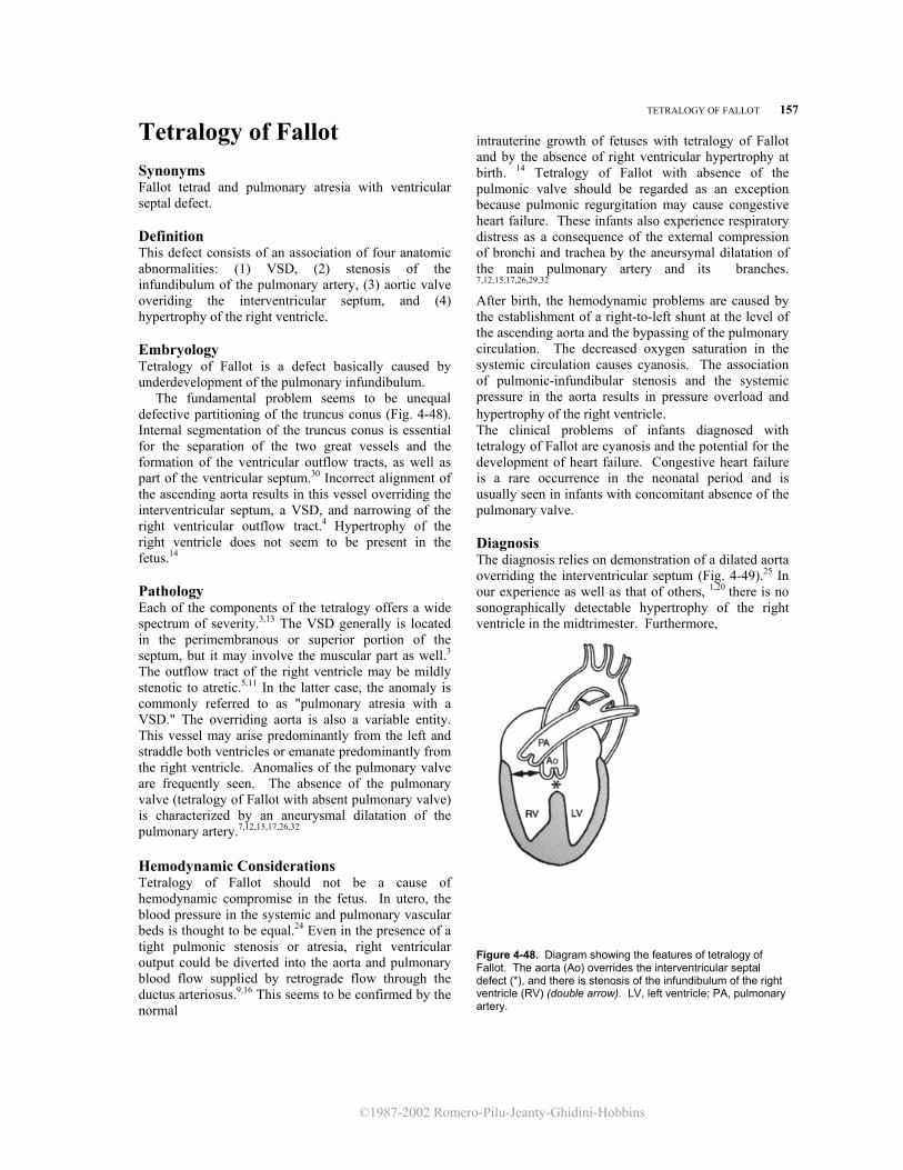
©1987-2002 Romero-Pilu-Jeanty-Ghidini-Hobbins
TETRALOGY OF FALLOT 157
Tetralogy of Fallot Synonyms Fallot tetrad and pulmonary atresia with ventricular septal defect. Definition This defect consists of an association of four anatomic abnormalities: (1) VSD, (2) stenosis of the infundibulum of the pulmonary artery, (3) aortic valve overiding the interventricular septum, and (4) hypertrophy of the right ventricle. Embryology Tetralogy of Fallot is a defect basically caused by underdevelopment of the pulmonary infundibulum. The fundamental problem seems to be unequal defective partitioning of the truncus conus (Fig. 4-48). Internal segmentation of the truncus conus is essential for the separation of the two great vessels and the formation of the ventricular outflow tracts, as well as part of the ventricular septum.30 Incorrect alignment of the ascending aorta results in this vessel overriding the interventricular septum, a VSD, and narrowing of the right ventricular outflow tract.4 Hypertrophy of the right ventricle does not seem to be present in the fetus.14 Pathology Each of the components of the tetralogy offers a wide spectrum of severity.3,13 The VSD generally is located in the perimembranous or superior portion of the septum, but it may involve the muscular part as well.3 The outflow tract of the right ventricle may be mildly stenotic to atretic.5,11 In the latter case, the anomaly is commonly referred to as "pulmonary atresia with a VSD." The overriding aorta is also a variable entity. This vessel may arise predominantly from the left and straddle both ventricles or emanate predominantly from the right ventricle. Anomalies of the pulmonary valve are frequently seen. The absence of the pulmonary valve (tetralogy of Fallot with absent pulmonary valve) is characterized by an aneurysmal dilatation of the pulmonary artery.7,12,15,17,26,32 Hemodynamic Considerations Tetralogy of Fallot should not be a cause of hemodynamic compromise in the fetus. In utero, the blood pressure in the systemic and pulmonary vascular beds is thought to be equal.24 Even in the presence of a tight pulmonic stenosis or atresia, right ventricular output could be diverted into the aorta and pulmonary blood flow supplied by retrograde flow through the ductus arteriosus.9,16 This seems to be confirmed by the normal
intrauterine growth of fetuses with tetralogy of Fallot and by the absence of right ventricular hypertrophy at birth. 14 Tetralogy of Fallot with absence of the pulmonic valve should be regarded as an exception because pulmonic regurgitation may cause congestive heart failure. These infants also experience respiratory distress as a consequence of the external compression of bronchi and trachea by the aneursymal dilatation of the main pulmonary artery and its branches. 7,12,15,17,26,29,32 After birth, the hemodynamic problems are caused by the establishment of a right-to-left shunt at the level of the ascending aorta and the bypassing of the pulmonary circulation. The decreased oxygen saturation in the systemic circulation causes cyanosis. The association of pulmonic-infundibular stenosis and the systemic pressure in the aorta results in pressure overload and hypertrophy of the right ventricle. The clinical problems of infants diagnosed with tetralogy of Fallot are cyanosis and the potential for the development of heart failure. Congestive heart failure is a rare occurrence in the neonatal period and is usually seen in infants with concomitant absence of the pulmonary valve. Diagnosis The diagnosis relies on demonstration of a dilated aorta overriding the interventricular septum (Fig. 4-49).25 In our experience as well as that of others, 1,20 there is no sonographically detectable hypertrophy of the right ventricle in the midtrimester. Furthermore,
Figure 4-48. Diagram showing the features of tetralogy of Fallot. The aorta (Ao) overrides the interventricular septal defect (*), and there is stenosis of the infundibulum of the right ventricle (RV) (double arrow). LV, left ventricle; PA, pulmonary artery.
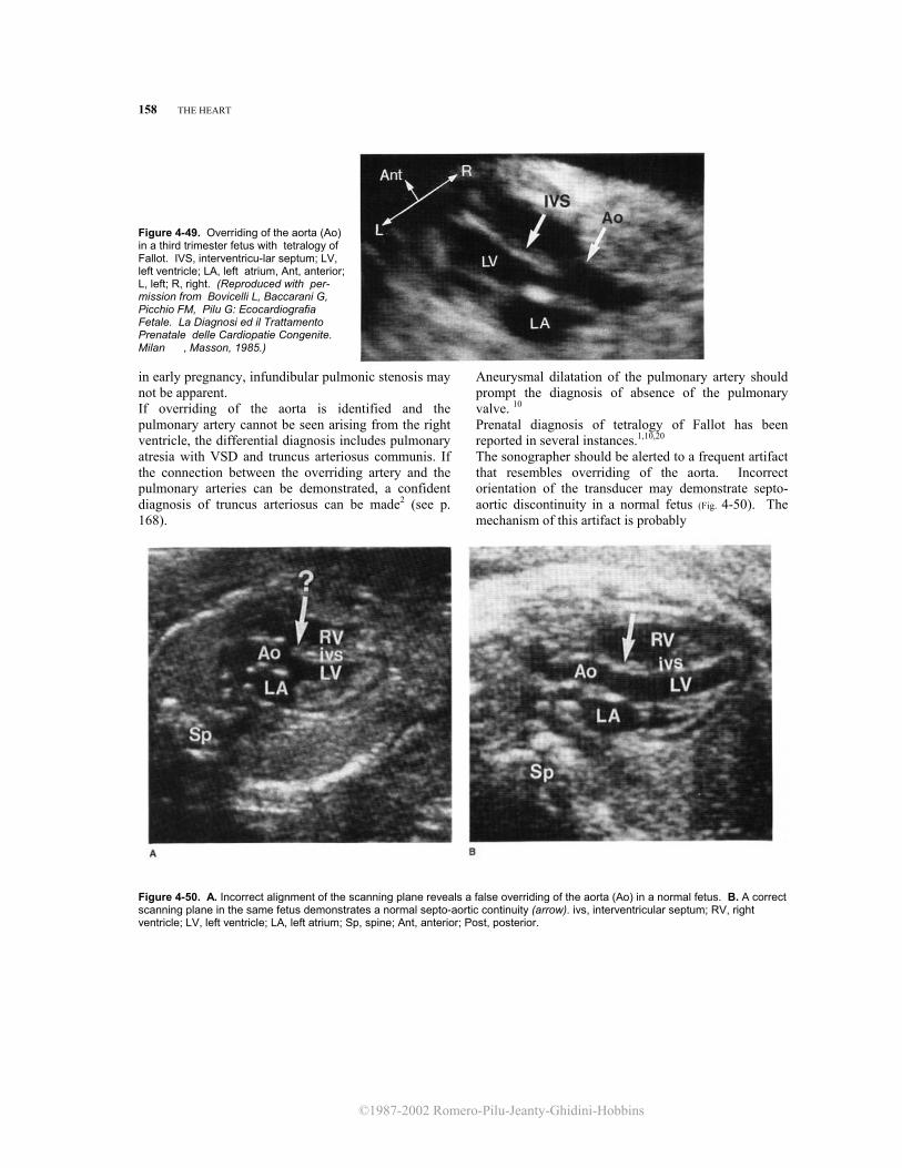
©1987-2002 Romero-Pilu-Jeanty-Ghidini-Hobbins
158 THE HEART
Figure 4-49. Overriding of the aorta (Ao) in a third trimester fetus with tetralogy of Fallot. IVS, interventricu-lar septum; LV, left ventricle; LA, left atrium, Ant, anterior; L, left; R, right. (Reproduced with per-mission from Bovicelli L, Baccarani G, Picchio FM, Pilu G: Ecocardiografia Fetale. La Diagnosi ed il Trattamento Prenatale delle Cardiopatie Congenite. Milan , Masson, 1985.) in early pregnancy, infundibular pulmonic stenosis may not be apparent. If overriding of the aorta is identified and the pulmonary artery cannot be seen arising from the right ventricle, the differential diagnosis includes pulmonary atresia with VSD and truncus arteriosus communis. If the connection between the overriding artery and the pulmonary arteries can be demonstrated, a confident diagnosis of truncus arteriosus can be made2 (see p. 168).
Aneurysmal dilatation of the pulmonary artery should prompt the diagnosis of absence of the pulmonary valve. 10 Prenatal diagnosis of tetralogy of Fallot has been reported in several instances.1,10,20 The sonographer should be alerted to a frequent artifact that resembles overriding of the aorta. Incorrect orientation of the transducer may demonstrate septo-aortic discontinuity in a normal fetus (Fig. 4-50). The mechanism of this artifact is probably
Figure 4-50. A. Incorrect alignment of the scanning plane reveals a false overriding of the aorta (Ao) in a normal fetus. B. A correct scanning plane in the same fetus demonstrates a normal septo-aortic continuity (arrow). ivs, interventricular septum; RV, right ventricle; LV, left ventricle; LA, left atrium; Sp, spine; Ant, anterior; Post, posterior.

©1987-2002 Romero-Pilu-Jeanty-Ghidini-Hobbins
TETRALOGY OF FALLOT 159 related to the angle of incidence of the sound beam. Meticulous scanning is always required to evaluate the relationship between the interventricular septum and the ascending aorta. Prognosis The prognosis for infants with tetralogy of Faflot has changed significantly over the last 3 decades. The development of pediatric cardiothoracic surgery is responsible for the improved outlook for these infants.22 The first surgical approach to tetralogy of Fallot was intended to obviate the underperfusion of the lungs. The Blalock-Taussig shunt consisted of anastomosing the subclavian artery to the pulmonary artery. This procedure was used for many years. Results indicated that the survival rate with a successful anastomosis was 64 percent (441/685 patients) at 15 years and 55 percent (376/679) at 20 years.27,28
The development of extracorporeal circulation and rapid advances in cardiothoracic surgery permitted correction of the primary anatomic defects namely, closure of the VSD and reconstruction of the right outflow tract. 23 Follow-up for 5 to 11 years in 311 patients indicated a survival rate of 85 percent.21 Eighty-seven percent of the survivors had neither symptoms nor restriction of activity. Another study reported a survival rate of 82 percent for a follow-up period ranging from 1 month to 15 years. The author estimated that half of the treated patients would survive to the 4th decade of life. The major contributor to decreased survival was an early postoperative mortality rate (30 days after operation) of 11 percent.8 These results refer to operations performed during adolescence or adulthood. In the last few vears, intracardiac correction in infants of very early age has gained popularity. The first series indicated an early mortality rate ranging from 031 to 5 percent.18 Operations in younger infants seem to significantly improve the mortality rate, as well as the hemodynamic response.8 The prognosis of tetralogy of Fallot with pulmonary atresia and tetralogy of Fallot with absence of the pulmonary valve is different. In a group of 38 patients with pulmonary atresia treated by right ventricular outflow construction, 3 deaths were recorded (mortality of 8 percent). The survivors had both clinical and hemodynamic improvement. 19 Tetralogy of Fallot with an absent pulmonary valve may cause congestive heart failure in the fetus or newborn.10,12 Aneurysmal dilatation of the pulmonary artery and its branches may be a cause of pulmonary distress. These findings should be considered as major prognostic factors.7 In a review of the cases published before 1974, Lakier et al. 12 reported that infants with severe respiratory complications who received medical treatment had a mortality rate
of 76 percent compared to a mortality rate of 41 percent in the group of infants treated surgically. Infants with mild or no pulmonary problems who underwent surgery had a mortality rate of 31 percent. Stafford et al.26 reported 3 deaths (17 percent) in 18 surgically treated patients. Survivors were in good functional condition. Finally, in a series of 15 infants, 10 survived with relief of symptoms.7
Obstetrical Management A careful search for other cardiac and extracardiac abnormalities is indicated whenever the prenatal diagnosis of tetralogy of Fallot is made. An amniocentesis for chromosomal analysis is recommended. The option of pregnancy termination should be offered before viability. In continuing pregnancies, no alteration of standard obstetrical management is required. However, infants should be delivered in a tertiary care center where a pediatric cardiologist is immediately available. Tetralogy of Fallot with an absent pulmonary valve requires careful monitoring to rule out the development of in utero congestive heart failure. In the absence of intrauterine congestive heart failure (hydrops), there is no indication to alter standard obstetrical management, but delivery in a tertiary care center where a pediatric cardiologist is immediately available is mandatory. Optimal management of patients whose fetuses have congestive heart failure has not yet been established. A reasonable approach is to deliver the patient after fetal lung maturity is documented, because the mortality rate of respiratory distress syndrome associated with congestive heart failure is extremely high. For a further discussion of management issues concerning fetal well-being assessment and mode of delivery, see Nonimmune Hydrops Fetalis in Chapter 12.
REFERENCES
1. Allan LD, Crawford CC, Anderson RH, et al.: Echocar-
diographic and anatomical correlations in fetal congenital heart disease. Br Heart J 52:542, 1984.
2. Barron JV, Sahn DJ, Attie F: Two-dimensional echocar-diographic study of right ventricular outflow and great artery anatomy in pulmonary atresia with ventricular septal defects and in truncus arteriosus. Am Heart J 105:281, 1983.
3. Becker AE, Anderson RH: Pathology of Congenital Heart Disease. London, Butterworths, 1981.
4. De La Cruz MV, Da Rocha JP: An ontogenetic theory for the explanation of congenital malformations involving the truncus and conus. Am Heart J 51:782, 1956.
5. Edwards JE, McGoon DC: Absence of anatomic origin from heart of pulmonary arterial supply. Circulation 47:393, 1973.
6. Finnegan P, Haider R, Patel RG, et al.: Results of total correction of the tetralogy of Fallot. Long-term
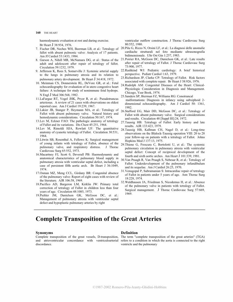
©1987-2002 Romero-Pilu-Jeanty-Ghidini-Hobbins
160 THE HEART
haemodynamic evaluation at rest and during exercise. Br Heart J 38:934, 1976. 7. Fischer DR, Neches WH, Beerman LB, et al.: Tetralogy of
fallot with absent pulmonic valve: Analysis of 17 patients. Am Jf Cardiol 53:1433, 1984.
8. Garson A, Nihill MR, McNamara DG, et al.: Status of the adult and adolescent after repair of tetralogy of fallot. Circulation 59:1232, 1979.
9. Jefferson K, Rees S, Somerville J: Systemic arterial supply to the lungs in pulmonary atresia and its relation to pulmonary artery development. Br Heart J 34:418, 1972.
10. Meinman CS, Donnerstein RL, DeVore GR, et al.: Fetal echocardiography for evaluation of in utero congestive heart failure: A technique for study of nonimmune fetal hydrops. N Engl J Med 306:568, 1982.
11. LaFargue RT, Vogel JHK, Pryor R, et al.: Pseudotruncus arteriosus. A review of 21 cases with observations on oldest reported case. Am J Cardiol 19:239, 1967.
12. Lakier JB, Stanger P, Heymann MA, et al.: Tetralogy of Fallot with absent pulmonary valve. Natural history and hemodynamic considerations. Circulation 50:167, 1974.
13. Lev M, Eckner FAO: The pathologic anatomy of tetralogy of Fallot and its variations. Dis Chest 45:251, 1964.
14. Lev M, Rimoldi HJA, Rowlatt UF: The quantitative anatomy of cyanotic tetralogy of Fallot. Circulation 30:531, 1964.
15. Litwin SB, Rosenthal A, Fellows K: Surgical management of young infants with tetralogy of Fallot, absence of the pulmonary valve, and respiratory distress. J Thorac Cardiovasc Surg 65:552, 1973.
16. Macartney FJ, Scott 0, Deverall PB: Haemodynamic and anatomical characteristics of pulmonary blood supply in pulmonary atresia with ventricular septal defect, including a case of persistent fifth aortic arch. Br Heart J 36:1049, 1974.
17. Osman MZ, Meng CCL, Girdany BR: Congenital absence of the pulmonary valve: Report of eight cases with review of the literature. AJR 106:58, 1969.
18. Pacifico AD, Bargeron LM, Kirklin JW: Primary total correction of tetralogy of Fallot in children less than four years of age. Circulation 48:1085, 1973.
19. Piehler JM, Danielson GK, McGoon DC, et al.: Management of pulmonary atresia with ventricular septal defect and hypoplastic pulmonary arteries by right
ventricular outflow construction. J Thorac Cardiovasc Surg 80:552, 1980.
20. Pilu G, Rizzo N, Orsini LF, et al.: La diagnosi delle anomalie cardiache strutturali nel feto mediante ultrasonografia bidimensionale. Ultr Ost Gin 1:257, 1983.
21. Poirier RA, McGoon DC, Danielson GK, et al.: Late results after repair of tetralogy of Fallot. J Thorac Cardiovasc Surg 73:900, 1977.
22. Rashkind WJ: Pediatric cardiology: A brief historical perspective. Pediatr Cardiol 1:63, 1979.
23. Richardson JP, Clarke CP: Tetralogy of Fallot. Risk factors associated with complete repair. Br Heart J 38:926, 1976.
24. Rudolph AM: Congenital Diseases of the Heart: Clinical-Physiologic Consideration in Diagnosis and Management. Chicago, Year Book, 1974.
25. Sanders SP, Bierman FZ, Williams RG: Conotruncal malformations: Diagnosis in infancy using subxiphoid 2-
dimensional echocardiography. Am J Cardiol 50: 1361, 1982.
26. Stafford EG, Mair DD, McGoon DC, et al.: Tetralogy of Fallot with absent pulmonary valve. Surgical considerations and results. Circulation 48 [Suppl III]:24, 1972.
27. Taussig HB: Tetralogy of Fallot: Early history and late results. AJR 133:423, 1979.
28. Taussig HB, Kallman CH, Nagel D, et al.: Long-time observations on the Blalock-Taussig operation VIII: 20 to 28 year follow-up on patients with a tetralogy of Fallot. Johns Hopkins Med J 137:13, 1975.
29. Thiene G, Frescura C, Bortolotti U, et al.: The systemic pulmonary circulation in pulmonary atresia with ventricular septal defect: Concept of reciprocal development of the fourth and sixth aortic arches. Am Heart J 101:339, 1981.
30. Van Praagh R, Van Praagh S, Nebesar R, et al.: Tetralogy of Fallot: Underdevelopment of the pulmonary infundibulum and its sequelae. Am J Cardiol 26:25, 1970.
31. Venugopal P, Subramanian S: Intracardiac repair of tetralogy of Fallot in patients under 5 years of age. Ann Thorac Surg 18:228, 1974.
32. Waldhausen JA, Friedman S, Nicodemus H, et al.: Absence of the pulmonary valve in patients with tetralogy of Fallot. Surgical management. J Thorac Cardiovasc Surg 57:669, 1969.
Complete Transposition of the Great Arteries Synonyms Complete transposition of the great vessels, D-transposition, and atrioventricular concordance with ventriculoarterial discordance.
Definition The term "complete transposition of the great arteries" (TGA) refers to a condition in which the aorta is connected to the right ventricle and the pulmonary
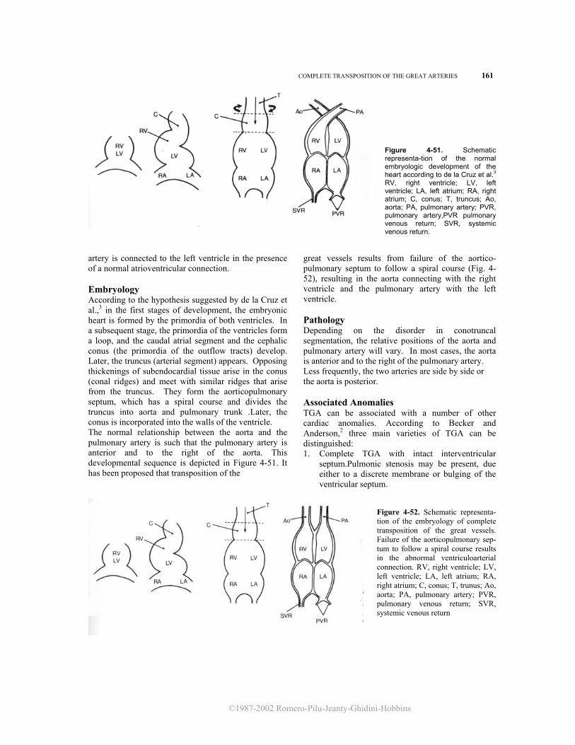
©1987-2002 Romero-Pilu-Jeanty-Ghidini-Hobbins
COMPLETE TRANSPOSITION OF THE GREAT ARTERIES 161
Figure 4-51. Schematic representa-tion of the normal embryologic development of the heart according to de la Cruz et al.3 RV, right ventricle; LV, left ventricle; LA, left atrium; RA, right atrium; C, conus; T, truncus; Ao, aorta; PA, pulmonary artery; PVR, pulmonary artery,PVR pulmonary venous return; SVR, systemic venous return.
artery is connected to the left ventricle in the presence of a normal atrioventricular connection. Embryology According to the hypothesis suggested by de la Cruz et al.,3 in the first stages of development, the embryonic heart is formed by the primordia of both ventricles. In a subsequent stage, the primordia of the ventricles form a loop, and the caudal atrial segment and the cephalic conus (the primordia of the outflow tracts) develop. Later, the truncus (arterial segment) appears. Opposing thickenings of subendocardial tissue arise in the conus (conal ridges) and meet with similar ridges that arise from the truncus. They form the aorticopulmonary septum, which has a spiral course and divides the truncus into aorta and pulmonary trunk .Later, the conus is incorporated into the walls of the ventricle. The normal relationship between the aorta and the pulmonary artery is such that the pulmonary artery is anterior and to the right of the aorta. This developmental sequence is depicted in Figure 4-51. It has been proposed that transposition of the
great vessels results from failure of the aortico-pulmonary septum to follow a spiral course (Fig. 4-52), resulting in the aorta connecting with the right ventricle and the pulmonary artery with the left ventricle. Pathology Depending on the disorder in conotruncal segmentation, the relative positions of the aorta and pulmonary artery will vary. In most cases, the aorta is anterior and to the right of the pulmonary artery. Less frequently, the two arteries are side by side or the aorta is posterior. Associated Anomalies TGA can be associated with a number of other cardiac anomalies. According to Becker and Anderson,2 three main varieties of TGA can be distinguished: 1. Complete TGA with intact interventricular
septum.Pulmonic stenosis may be present, due either to a discrete membrane or bulging of the ventricular septum.
Figure 4-52. Schematic representa-tion of the embryology of complete transposition of the great vessels. Failure of the aorticopulmonary sep- tum to follow a spiral course results in the abnormal ventriculoarterial connection. RV, right ventricle; LV, left ventricle; LA, left atrium; RA, right atrium; C, conus; T, trunus; Ao, aorta; PA, pulmonary artery; PVR, pulmonary venous return; SVR, systemic venous return
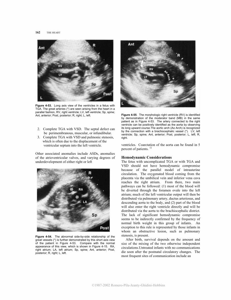
©1987-2002 Romero-Pilu-Jeanty-Ghidini-Hobbins
162 THE HEART
Figure 4-53. Long axis view of the ventricles in a fetus with TGA. The great arteries (*) are seen arising from the heart in a parallel fashion, RV, right ventricle; LV, left ventricle; Sp, spine; Ant, anterior; Post, posterior; R, right; L, left.
2. Complete TGA with VSD. The septal defect can be perimembranous, muscular, or infundibular.
3. Complete TGA with VSD and pulmonic stenosis, which is often due to the displacement of the ventricular septum into the left ventricle.
Other associated anomalies include ASDs, anomalies of the atrioventricular valves, and varying degrees of underdevelopment of either right or left
Figure 4-54. The abnormal side-by-side relationship of the great vessels (*) is further demonstrated by this short axis view of the patient in Figure 4-53. Compare with the normal appearance of this view, which is shown in Figure 4-15. RA, right atrium; LA, left atrium; Sp, spine; Ant, anterior; Post, posterior; R, right; L, left.
Figure 4-55. The morphologic right ventricle (RV) is identified by demonstration of the moderator band (MB) in the same patient as in Figure 4-53. The artery connected to the right ventricle can be positively identified as the aorta by observing its long upward course.The aortic arch (Ao Arch) is recognized by the connection with a brachiocephalic vessel (*). LV, left ventricle; Sp, spine; Ant, anterior; Post, posterior; L, left; R, right.
ventricles. Coarctation of the aorta can be found in 5 percent of patients. 12 Hemodynamic Considerations The fetus with uncomplicated TGA or with TGA and VSD should not have hemodynamic compromise because of the parallel model of intrauterine circulation. The oxygenated blood coming from the placenta via the umbilical vein and inferior vena cava reaches the right atrium. From there, two main pathways can be followed: (1) most of the blood will be diverted through the foramen ovale into the left atrium; much of the left ventricular output will then be distributed via pulmonary artery, ductus arteriosus, and descending aorta to the body, and (2) part of the blood will also enter the right ventricle directly and will be distributed via the aorta to the brachiocephalic district. The lack of significant hemodynamic compromise seems to be indirectly confirmed by the frequency of normal birth weight in this group of infants. An exception to this rule is represented by those infants in whom an obstructive lesion, such as pulmonary stenosis, is present. After birth, survival depends on the amount and size of the mixing of the two otherwise independent circulations.Untreated infants with no communications die soon after the postnatal circulatory changes. The most frequent sites of communication include an
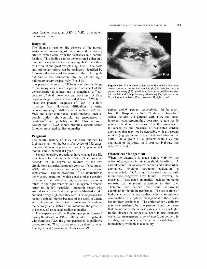
©1987-2002 Romero-Pilu-Jeanty-Ghidini-Hobbins
COMPLETE TRANSPOSITION OF THE GREAT ARTERIES 163
open foramen ovale, an ASD, a VSD, or a patent ductus arteriosus. Diagnosis The diagnosis rests on the absence of the normal anatomic criss-crossing of the aorta and pulmonary arteries, which arise from the ventricles in a parallel fashion. This finding can be demonstrated either in a long axis view of the ventricles (Fig. 4-53) or a short axis view of the great vessels (Fig. 4-54). The aorta and pulmonary artery can be positively identified by following the course of the vessels to the arch (Fig. 4-55) and to the bifurcation into the left and right pulmonary artery, respectively (Fig. 4-56). A prenatal diagnosis of TGA is a serious challenge to the sonographer, since a proper assessment of the ventriculoarterial connections is sometimes difficult because of fetal movement and position. A false negative diagnosis has been reported twice.1,6 We have made the prenatal diagnosis of TGA in a third trimester fetus. However, difficulties in using echocardiography to differentiate complete TGA with VSD and other conotruncal malformations, such as double outlet right ventricle, are encountered in newborns11 and probably in the fetus as well. Recognition of TGA should prompt a careful search for other associated cardiac anomalies. Prognosis The natural history of TGA has been outlined by Liebman et al. 7 on the basis of a review of 742 cases. Survival rate was 70 percent at 1 week, 50 percent at 1 month,' and 11 percent at 1 year. Several operative procedures have changed the life expectancy for infants with TGA. Since survival depends on the degree of mixture of the two circulations, a surgical approach consists of creating an ASD, either by intracardiac surgery or by balloon septostomy (Rashkind procedure). 10 An alternative is the Mustard operation,8 which consists of the creation of an intraatrial baffle diverting the pulmonary venous return to the right ventricle and the systemic venous return to the left ventricle. Anatomic repair with arterial switch was first attempted by Mustard et al..9 and had a very high mortality rate. This approach has recently gained interest because of the work of Jatene et al.5 At present, the choice of procedure depends on the hemodynamic status of the infant and the presence or absence of associated cardiac anomalies. The experience of the Baylor group in Houston4 during the decade of 1968-1978 includes 112 patients with complete TGA; the group performed 46 palliative procedures and 71 corrective repairs on these patients. The 1-year and 5-year survival rates were 79
Figure 4-56. In the same patient as in Figure 4-53, the great artery connected to the left ventricle (LV) is identified as the pulmonary artery (PA) by following its course until it bifurcates into the left and right pulmonary arteries (-) RV, right ventricle; Sp, spine; Ant, anterior; Post, posterior; R, right; L, left.
percent and 64 percent, respectively. In the report from the Hospital for Sick Children of Toronto,12 which includes 394 patients with TGA and intact interventricular septum, the 5-year survival rate was 89 percent. It should be stressed that the prognosis is influenced by the presence of associated cardiac anomalies that may not be detectable with ultrasound in utero (e.g., pulmonic stenosis and coarctation of the aorta). In a group of 32 patients with TGA and coartation of the aorta, the 5-year survival rate was only 57 percent.12 Obstetrical Management When the diagnosis is made before viability, the option of pregnancy termination should be offered. A careful search for associated cardiac and extracardiac anomalies, including karyotype evaluation, is recommended. TGA is not associated per se with intrauterine congestive heart failure. However, the presence of associated anomalies, such as pulmonic stenosis, can represent exceptions to this rule. Therefore, we believe that serial ultrasound examinations should be performed. The association of hydrops with a structural cardiac defect is an ominous combination. The optimal management in these cases has not been established. The option of early delivery may be considered, but the parents should be aware that the mortality rate in these cases is extremely high.6 In the absence of congestive heart failure, standard obstetrical management is not changed, but delivery in a tertiary care center where a pediatric cardiologist is immediately available is mandatory.

©1987-2002 Romero-Pilu-Jeanty-Ghidini-Hobbins
164 THE HEART
REFERENCES 1. Allan LD, Crawford DC, Anderson RH, et al.: Echocar-
diographic and anatomical correlations in fetal congenital heart disease. Br Heart J 52:542, 1984.
2. Becker AE, Anderson RH: Pathology of Congenital Heart Disease. London, Butterworths, 1981. 3. de la Cruz MV, Arteaga M, Espino-Vela J, et al.: Complete
transposition of the great arteries: Types and morphogenesis of ventriculoarterial discordance. Am Heart J 102:271, 1981. 4. Gutgesell HP, Garson A, Park IS, et al.: Prognosis for the
neonate and young infant with congenital heart disease (Abstr). Pediatr Cardiol 2:168, 1982.
5. Jatene AD, Fontes VF, Paulista PP, et al.: Anatomic correction of transposition of the great vessels. J Thorac
Cardiovasc Surg 72:364, 1976. 6. Kleinman CS, Donnerstein RL, DeVore GR, et al.: Fetal
echocardiography for evaluation of in utero congestive
heart failure: A technique for study of nonimmune fetal hydrops. N Engl J Med 306:568, 1982.
7. Liebman J, Cullum L, Belloc NB: Natural history of transposition of the great vessels. Anatomy and birth and death characteristics. Circulation 40:237, 1969
8. Mustard WT: Successful two-stage correction of transposition of the great vessels. Surgery 55:469, 1964.
9. Mustard WT, Chute AL, Keith JD, et al.: A surgical approach to transposition of the great vessels with extracorporeal circuit. Surgery 36:39, 1954.
10. Rashkind WJ, Miller WW: Creation of an atrial septal defect without thoracotomy: A palliative approach to complete transposition of the great arteries. JAMA 196:991, 1966.
11. Sanders SP, Bierman FZ, Williams RG: Conotruncal malformations: Diagnosis in infancy using subxiphoid 2-dimensional echocardiography. Am J Cardiol 50:1361, 1982.
12. Vogel M, Freedom RM, Smallhorn JF, et al.: Complete transposition of the great arteries and coarctation of the aorta. Am J Cardiol 53:1627, 1984.
Corrected Transposition of the Great Arteries
Synonyms L-Transposition and atrioventricular discordance with ventriculoarterial discordance. Definition Corrected transposition of the great arteries (TGA) refers to a condition in which the right atrium and left atrium are connected to the morphologic left and right ventricle, respectively, and the great arteries are transposed. These two defects cancel each other, and ideally there should not be any hemodynamic consequences. Corrected TGA may indeed be an occasional finding at autopsy. However, in the majority of cases, important associated anomalies alter the prognosis. Embryology According to the hypothesis suggested by de la Cruz et al.,3 the embryonic heart in the first stages of development is formed by the primordia of both ventricles. In a subsequent stage, while the primordia of the ventricles form a loop, both the caudal atrial segment and the cephalic conus (the primordia to the outflow tracts) develop. Later, the truncus (arterial segment) appears. Opposing thickenings of subendocardial tissue arise in the conus (conal ridges) and meet with similar ridges arising from the truncus. They form the aorticopulmonary septum, which has a spiral course and divides the truncus into aorta and pulmonary trunk. Later, the conus is incorporated into the walls of the ventricle. The normal relation-
ship between the aorta and the pulmonary artery is such that the pulmonary artery is anterior and to the right of the aorta. This developmental sequence is depicted in Figure 4-57. It has been suggested that corrected transposition results from anomalous looping of the primordia of the ventricle. This is associated with the lack of spiral rotation of the truncoconal septum. In this fashion, the aorta is connected to the morphologic right ventricle and the pulmonary artery is connected to the morphologic left ventricle. Such an abnormality is functionally corrected, since the ventricles are attached to the anatomically anchored, and thus normally positioned, atria (Fig. 4-58).3 Pathology Corrected TGA is frequently associated with malpositions of the heart and occasionally with situs inversus.7 Because of the ventriculoarterial discordance, the aortic valve is separated from the tricuspid valve by a complete infundibulum, whereas there is fibrous continuity between the pulmonic and the mitral valve. In more than 50 percent of patients, there is a VSD, generally of the perimembranous type. The pulmonary artery may override the VSD, and pulmonic stenosis is seen in about 50 percent of cases studied.1,2,7 Anomalies of the atrioventricular valves are frequently found and include Ebsteinlike malformation and straddling of the tricuspid valve.1,2,7
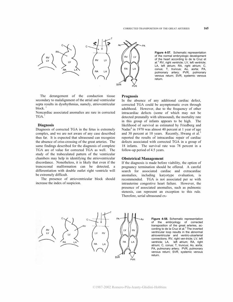
©1987-2002 Romero-Pilu-Jeanty-Ghidini-Hobbins
CORRECTED TRANSPOSITION OF THE GREAT ARTERIES 165
Figure 4-57. Schematic representation of the normal embryologic development of the heart according to de la Cruz et al.3 RV, right ventricle; LV, left ventricle; LA, left atrium; RA, right atrium; C, conus; T, truncus; Ao, aorta; PA, pulmonary artery; PVR, pulmonary venous return; SVR, systemic venous return.
The derangement of the conduction tissue secondary to malalignment of the atrial and ventricular septa results in dysrhythmias, namely, atrioventricular block. 1 Noncardiac associated anomalies are rare in corrected TGA.7
Diagnosis Diagnosis of corrected TGA in the fetus is extremely complex, and we are not aware of any case described thus far. It is expected that ultrasound can recognize the absence of criss-crossing of the great arteries. The same findings described for the diagnosis of complete TGA are of value for corrected TGA as well. The study of the trabeculated pattern of the ventricular chambers may help in identifying the atrioventricular discordance. Nonetheless, it is likely that even if the truncoconal malformation can be detected, a differentiation with double outlet right ventricle will be extremely difficult. The presence of atrioventricular block should increase the index of suspicion.
Prognosis In the absence of any additional cardiac defect, corrected TGA could be asymptomatic even through adulthood. However, due to the frequency of other intracardiac defects (some of which may not be detected prenatally with ultrasound), the mortality rate in this group of infants appears to be high. The likelihood of survival as estimated by Friedberg and Nadas4 in 1970 was almost 40 percent at 1 year of age and 30 percent at 10 years. Recently, Hwang et al.5 reported the results of intracardiac repair of cardiac defects associated with corrected TGA in a group of 18 infants. The survival rate was 78 percent in a follow-up period of 4.5 years. Obstetrical Management If the diagnosis is made before viability, the option of pregnancy termination should be offered. A careful search for associated cardiac and extracardiac anomalies, including karyotype evaluation, is recommended. TGA is not associated per se with intrauterine congestive heart failure. However, the presence of associated anomalies, such as pulmonic stenosis, can represent an exception to this rule. Therefore, serial ultrasound ex-
Figure 4-58. Schematic representation of the embryology of corrected transposition of the great arteries, ac-cording to de la Cruz et al.3 The inverted ventricular loop results in the abnormal atrioventricular and ventric-uloarterial connections. RV, right ven-tricle; LV, left ventricle; LA, left atrium; RA, right atrium; C, conus; T, truncus; Ao, aorta; PA, pulmonary artery; PVR, pulmonary venous return; SVR, systemic venous return.
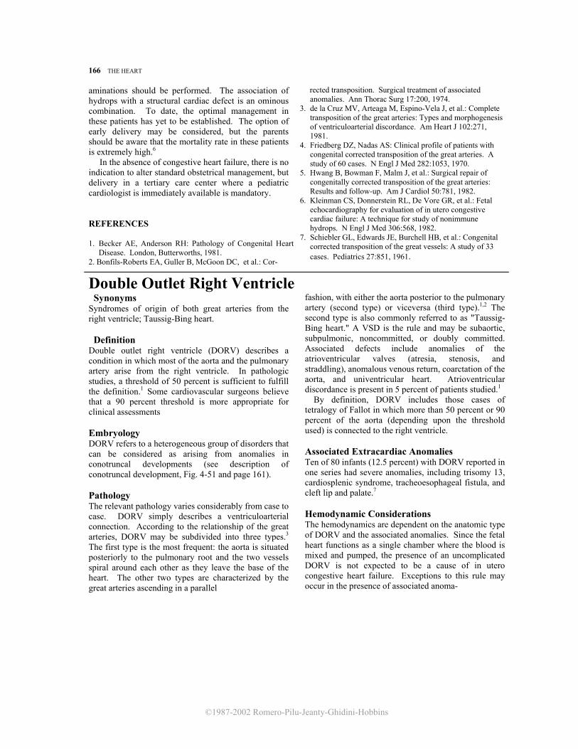
©1987-2002 Romero-Pilu-Jeanty-Ghidini-Hobbins
166 THE HEART
aminations should be performed. The association of hydrops with a structural cardiac defect is an ominous combination. To date, the optimal management in these patients has yet to be established. The option of early delivery may be considered, but the parents should be aware that the mortality rate in these patients is extremely high.6 In the absence of congestive heart failure, there is no indication to alter standard obstetrical management, but delivery in a tertiary care center where a pediatric cardiologist is immediately available is mandatory. REFERENCES 1. Becker AE, Anderson RH: Pathology of Congenital Heart
Disease. London, Butterworths, 1981. 2. Bonfils-Roberts EA, Guller B, McGoon DC, et al.: Cor-
rected transposition. Surgical treatment of associated anomalies. Ann Thorac Surg 17:200, 1974.
3. de la Cruz MV, Arteaga M, Espino-Vela J, et al.: Complete transposition of the great arteries: Types and morphogenesis of ventriculoarterial discordance. Am Heart J 102:271, 1981.
4. Friedberg DZ, Nadas AS: Clinical profile of patients with congenital corrected transposition of the great arteries. A study of 60 cases. N Engl J Med 282:1053, 1970.
5. Hwang B, Bowman F, Malm J, et al.: Surgical repair of congenitally corrected transposition of the great arteries: Results and follow-up. Am J Cardiol 50:781, 1982.
6. Kleinman CS, Donnerstein RL, De Vore GR, et al.: Fetal echocardiography for evaluation of in utero congestive cardiac failure: A technique for study of nonimmune hydrops. N Engl J Med 306:568, 1982.
7. Schiebler GL, Edwards JE, Burchell HB, et al.: Congenital corrected transposition of the great vessels: A study of 33 cases. Pediatrics 27:851, 1961.
Double Outlet Right Ventricle Synonyms
Syndromes of origin of both great arteries from the right ventricle; Taussig-Bing heart.
Definition Double outlet right ventricle (DORV) describes a condition in which most of the aorta and the pulmonary artery arise from the right ventricle. In pathologic studies, a threshold of 50 percent is sufficient to fulfill the definition.1 Some cardiovascular surgeons believe that a 90 percent threshold is more appropriate for clinical assessments Embryology DORV refers to a heterogeneous group of disorders that can be considered as arising from anomalies in conotruncal developments (see description of conotruncal development, Fig. 4-51 and page 161). Pathology The relevant pathology varies considerably from case to case. DORV simply describes a ventriculoarterial connection. According to the relationship of the great arteries, DORV may be subdivided into three types.3 The first type is the most frequent: the aorta is situated posteriorly to the pulmonary root and the two vessels spiral around each other as they leave the base of the heart. The other two types are characterized by the great arteries ascending in a parallel
fashion, with either the aorta posterior to the pulmonary artery (second type) or viceversa (third type).1,2 The second type is also commonly referred to as "Taussig-Bing heart." A VSD is the rule and may be subaortic, subpulmonic, noncommitted, or doubly committed. Associated defects include anomalies of the atrioventricular valves (atresia, stenosis, and straddling), anomalous venous return, coarctation of the aorta, and univentricular heart. Atrioventricular discordance is present in 5 percent of patients studied.1 By definition, DORV includes those cases of tetralogy of Fallot in which more than 50 percent or 90 percent of the aorta (depending upon the threshold used) is connected to the right ventricle. Associated Extracardiac Anomalies Ten of 80 infants (12.5 percent) with DORV reported in one series had severe anomalies, including trisomy 13, cardiosplenic syndrome, tracheoesophageal fistula, and cleft lip and palate.7 Hemodynamic Considerations The hemodynamics are dependent on the anatomic type of DORV and the associated anomalies. Since the fetal heart functions as a single chamber where the blood is mixed and pumped, the presence of an uncomplicated DORV is not expected to be a cause of in utero congestive heart failure. Exceptions to this rule may occur in the presence of associated anoma-
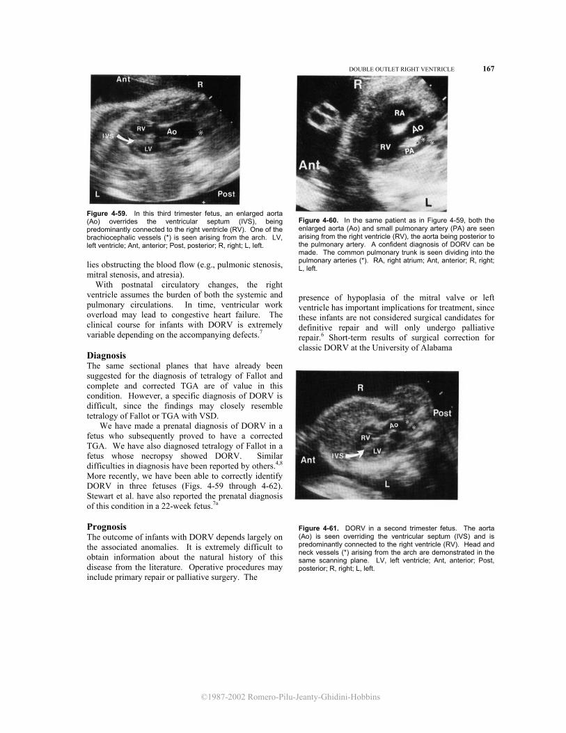
©1987-2002 Romero-Pilu-Jeanty-Ghidini-Hobbins
DOUBLE OUTLET RIGHT VENTRICLE 167
Figure 4-59. In this third trimester fetus, an enlarged aorta (Ao) overrides the ventricular septum (IVS), being predominantly connected to the right ventricle (RV). One of the brachiocephalic vessels (*) is seen arising from the arch. LV, left ventricle; Ant, anterior; Post, posterior; R, right; L, left. lies obstructing the blood flow (e.g., pulmonic stenosis, mitral stenosis, and atresia). With postnatal circulatory changes, the right ventricle assumes the burden of both the systemic and pulmonary circulations. In time, ventricular work overload may lead to congestive heart failure. The clinical course for infants with DORV is extremely variable depending on the accompanying defects.7 Diagnosis The same sectional planes that have already been suggested for the diagnosis of tetralogy of Fallot and complete and corrected TGA are of value in this condition. However, a specific diagnosis of DORV is difficult, since the findings may closely resemble tetralogy of Fallot or TGA with VSD. We have made a prenatal diagnosis of DORV in a fetus who subsequently proved to have a corrected TGA. We have also diagnosed tetralogy of Fallot in a fetus whose necropsy showed DORV. Similar difficulties in diagnosis have been reported by others.4,8 More recently, we have been able to correctly identify DORV in three fetuses (Figs. 4-59 through 4-62). Stewart et al. have also reported the prenatal diagnosis of this condition in a 22-week fetus.7a Prognosis The outcome of infants with DORV depends largely on the associated anomalies. It is extremely difficult to obtain information about the natural history of this disease from the literature. Operative procedures may include primary repair or palliative surgery. The
Figure 4-60. In the same patient as in Figure 4-59, both the enlarged aorta (Ao) and small pulmonary artery (PA) are seen arising from the right ventricle (RV), the aorta being posterior to the pulmonary artery. A confident diagnosis of DORV can be made. The common pulmonary trunk is seen dividing into the pulmonary arteries (*). RA, right atrium; Ant, anterior; R, right; L, left.
presence of hypoplasia of the mitral valve or left ventricle has important implications for treatment, since these infants are not considered surgical candidates for definitive repair and will only undergo palliative repair.6 Short-term results of surgical correction for classic DORV at the University of Alabama
Figure 4-61. DORV in a second trimester fetus. The aorta (Ao) is seen overriding the ventricular septum (IVS) and is predominantly connected to the right ventricle (RV). Head and neck vessels (*) arising from the arch are demonstrated in the same scanning plane. LV, left ventricle; Ant, anterior; Post, posterior; R, right; L, left.
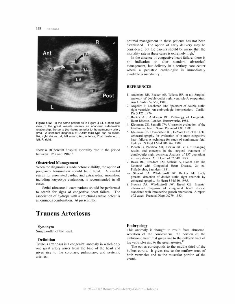
©1987-2002 Romero-Pilu-Jeanty-Ghidini-Hobbins
168 THE HEART
Figure 4-62. In the same patient as in Figure 4-61, a short axis view of the great vessels reveals an abnormal side-to-side relationship, the aorta (Ao) being anterior to the pulmonary artery (PA). A confident diagnosis of DORV third type can be made. RA, right atrium; LA, left atrium; Ant, anterior; Post, posterior; L, left; R, right. show a 10 percent hospital mortality rate in the period between 1967 and 1982.6 Obstetrical Management When the diagnosis is made before viability, the option of pregnancy termination should be offered. A careful search for associated cardiac and extracardiac anomalies, including karyotype evaluation, is recommended in all cases. Serial ultrasound examinations should be performed to search for signs of congestive heart failure. The association of hydrops with a structural cardiac defect is an ominous combination. At present, the
optimal management in these patients has not been established. The option of early delivery may be considered, but the parents should be aware that the mortality rate in these cases is extremely high.5 In the absence of congestive heart failure, there is no indication to alter standard obstetrical management, but delivery in a tertiary care center where a pediatric cardiologist is immediately available is mandatory.
REFERENCES
1. Anderson RH, Becker AE, Wilcox BR, et al.: Surgical
anatomy of double-outlet right ventricle-A reappraisal. Am J Cardiol 52:555, 1983.
2. Angelini P, Leachman RD: Spectrum of double outlet right ventricle: An embryologic interpretation. Cardiol Dis 3:127, 1976.
3. Becker AE, Anderson RH: Pathology of Congenital Heart Disease. London, Butterworths, 1981.
4. Kleinman CS, Santulli TV: Ultrasonic evaluation of the fetal human heart. Semin Perinatol 7:90, 1983.
5. Kleinman CS, Donnerstein RL, DeVore GR, et al.: Fetal echocardiography for evaluation of in utero congestive heart failure: A technique for study of nonimmune fetal hydrops. N Engl J Med 306:568, 1982.
6. Piccoli G, Pacifico AD, Kirklin JW, et al.: Changing results and concepts in the surgical treatment of doubleoutlet right ventricle: Analysis of 137 operations in 126 patients. Am J Cardiol 52:549, 1983.
7. Rowe RD, Freedom RM, Mehrizi A, Bloom KR: The Neonate with Congenital Heart Disease, 2d ed. Philadelphia, Saunders, 1981.
7a. Stewart PA, Wladimiroff JW, Becker AE: Early prenatal detection of double outlet right ventricle by echocardiography. Br Heart J 54:340, 1985.
8. Stewart PA, Wladimiroff JW, Essed CE: Prenatal ultrasound diagnosis of congenital heart disease associated with intrauterine growth retardation. A report of 2 cases. Prenatal Diagn 3:279, 1983.
Truncus Arteriosus
Synonym Single outlet of the heart.
Definition Truncus arteriosus is a congenital anomaly in which only one great artery arises from the base of the heart and gives rise to the coronary, pulmonary, and systemic arteries.
Embryology This anomaly is thought to result from abnormal septation of the conotruncus, the portion of the embryonic heart that gives rise to the outflow tract of the ventricles and to the great arteries.17 The conus corresponds to the middle third of the bulbus cordis. It gives rise to the outflow tract of both ventricles and to the muscular portion of the ventri-

©1987-2002 Romero-Pilu-Jeanty-Ghidini-Hobbins
TRUNCUS ARTERIOSUS 169
cles located between the atrioventricular valves and the semilunar valves. The part of the conus giving rise to the ventricular free walls is referred to as the “parietal conus,” and the portion responsible for the development of the septum is called the “conal septum.” The truncus is the distal part of the bulbus cordis. Through a process of rotation and internal septation, it gives origin to the semilunar valves and the two great arteries. Separation between the aorta and pulmonary arteries occurs by fusion of the truncal ridges (Fig. 4-51). Septation of the conotruncus occurs between the 6th, and 7th weeks of embryonic life and begins at the level of the 4th and 6th aortic arch, progressing toward the heart. The close temporal relationship of these two phenomena explains the frequent association between truncus arteriosus and aortic arch abnormalities, which has been reported to occur in 20 percent of cases studied.2 Pathology The anomaly consists of a single arterial vessel with one semilunar valve arising from both ventricles.3 A wide variety of intracardiac abnormalities is associated with this disorder. Most frequently, there is an infundibular VSD because of a failure in development of the proximal portion of the conotruncal septum. According to Van Praagh and Van Praagh,18 truncus arteriosus may be classified as type A or B depending on the presence or absence, respectively, of a VSD. Furthermore, type A can be subdivided into four types: Al, the aorticopulmonary septum is imcompletely formed, resulting in a partially separated main pulmonary artery; A2, there is complete failure of development of the aorticopulmonary septum, resulting in pulmonary arteries arising directly from the truncus; A3, one lung is supplied by a pulmonary artery arising from the truncus, and the other lung is supplied by arteries derived from the descending aorta; A4, there is underdevelopment of the aortic arch, resulting in hypoplasia, coarctation, atresia, or interruption.4,18 In the classification suggested by Collett and Edwards, truncus arteriosus is divided into four types: type I is characterized by the presence of a pulmonary trunk that bifurcates into right and left pulmonary arteries; type II is characterized by the presence of two pulmonary arteries arising from the back of the truncus; in type III, the two pulmonary arteries arise from the sides of the truncus; in type IV, the pulmonary arteries are absent, and the lungs are supplied by systemic pulmonic collateral arteries derived from the descending aorta (Fig. 4-63). By definition, there is a single arterial valve (truncal valve). In the presence of a VSD, the truncal
Figure 4-63. Schematic representations of the classification of truncus arteriosus according to Van Praagh and Van Praagh'8 (Al-A4) and to Collett and EdwardS5 (I-IV).
valve overrides the septum and is connected to both ventricular cavities in almost equal proportions. In most cases, the truncal valve has three leaflets (tricuspid), but it may have two to six leaflets. The truncal valve is frequently dysplastic and incompetent.2 Associated Anomalies Cardiac defects include mitral atresia, ASD, univentricular heart, and aortic arch abnormalities.2 Hemodynamic Considerations A crucial issue concerns the competence of the truncal valve. Since the fetal heart functions as a common chamber where the blood is mixed and pumped, truncus arteriosus with a competent valve is not expected to be a cause of significant hemodynamic perturbance. Conversely, truncal incompetence may lead to massive regurgitation from the truncus to the ventricles, which may result in congestive heart failure. With postnatal circulatory changes, the decreased resistance of the pulmonary vascular bed leads to massive diversion of flow to the pulmonary district. This left-to-right shunt leads either to congestive heart failure due to ventricular overload or, in time, to pulmonary vascular damage and pulmonary hypertension. Diagnosis Truncus arteriosus is characterized by the presence of a single arterial vessel overriding the ventricular septum (Fig. 4-64). However, an identical finding is

©1987-2002 Romero-Pilu-Jeanty-Ghidini-Hobbins
170 THE HEART
Figure 4-64. Truncus arteriosus in a third trimester fetus with multiple congenital anomalies. A single arterial trunk (T) with a long upward course is seen arising from the base of the heart. The pulmonary artery could not be demonstrated. This finding may be compatible with truncus arteriosus or tetralogy of Fallot with atresia of the pulmonary artery. Due to the presence of multiple intrathoracic cysts, the heart is malpositioned and the apex points inferiorly. LV, left ventricle; RV, right ventricle; Ant, anterior; Post, posterior; Sup, superior; lnf, inferior. present in tetralogy of Fallot with pulmonary atresia. Differential diagnosis between these two conditions in the newborn can only be made by visualizing either a truncal valve with more than three leaflets or the origin of the pulmonary arteries from the truncus.8,13 The latter finding could be demonstrated in the fetus.1 Prognosis Most infants with truncus arteriosus develop heart failure within the first days or months of life. It has been estimated that the survival rate is less than 40 percent at 6 months and less than 20 percent at 1 year.5,9,11,18 Survivors are affected by rapidly progressive pulmonary vascular disease, and at 4 years of age, 30 percent are no longer operable. 10 The surgical treatment of truncus arteriosus consists of either palliation or physiologic correction. Attempts to palliate patients by reducing pulmonary blood flow with pulmonary artery banding, have resulted in uniformly poor outcomes with operative mortality rates of 60 percent. 12,14 On the other hand, physiologic correction by a valved or valveless conduit connecting the right ventricle to the pulmonary artery showed better results, with a lower mortality rate.6,10,15,16 The goal in these studies was to operate on the patients only when maximal medical therapy failed to control the congestive heart failure. Therefore, patients were operated on either in critical conditions or months after birth.
However, some adverse effects develop after birth in patients with truncus arteriosus, namely elevation of pulmonary vascular resistance, truncal valve insufficiency, and progressive ventricular dysfunction, which can compromise the outcome of the operation. A recent study reviewed the results of the infants who underwent physiologic correction prior to 6 months of age.7 Out of 100 patients, there were 11 operative deaths. A 2-year follow-up showed 3 late deaths, 1 with bacterial endocarditis and 2 unrelated to their cardiac conditions. Fifty-five infants, as they grew older, required conduit change, which provided excellent results. The experience of these and other authors supports the need for early repair of truncus arteriosus.6,7,15
Obstetrical Management When the diagnosis is made before viability, the option of pregnancy termination should be offered. A careful search for associated cardiac and extracardiac anomalies, including karyotype evaluation, is recommended. Serial ultrasound examinations should be performed to search for signs of congestive heart failure, which is frequently seen in patients with truncal valve incompetence. The association of hydrops with a structural cardiac defect is an ominous combination. The optimal management in these patients has not been established. The option of early delivery may be considered, but the parents should be aware that the mortality rate in these patients is extremely high. In the absence of congestive heart failure, there is no indication to alter standard obstetrical management, but delivery in a tertiary care center where a pediatric cardiologist is immediately available is mandatory. REFERENCES
1 . Allan LD: Fetal cardiac ultrasound. In: Sanders RC,
James AE (eds): The Principles and Practice of Ultrasonography in Obstetrics and Gynecology, 3d ed. E. Norwalk, CT, Appleton-Century-Crofts, 1985, pp 211-217.
2. Becker AE, Anderson RH: Pathology of Congenital Heart Disease. London, Butterworths, 1981.
3. Bharati S, McAllister HA, Rosenquist GC, et al.: The surgical anatomy of truncus arteriosus communism J Thorac Cardiovasc Surg 67:501, 1974.
4. Calder L, Van Praagh R, Van Praagh S, et al.: Truncus arteriosus communism Clinical, angiocardiographic, and pathologic findings in 100 patients. Am Heart J 92:23, 1976.
5. Collett RW, Edwards JE: Persistent truncus arteriosus: A classification according to anatomic types. Surg Clin North Am 29:1245, 1949.
6. Di Donato RM, Fyfe DA, Puga Fj, et al.: Fifteen-year
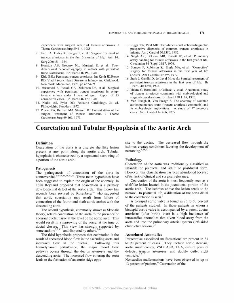
©1987-2002 Romero-Pilu-Jeanty-Ghidini-Hobbins
COARCTATION AND TUBULAR HYPOPLASIA OF THE AORTIC ARCH 171
experience with surgical repair of truncus arteriosus. J
Thorac Cardiovasc Surg 89:414, 1985. 7. Ebert PA, Turley K, Stanger P, et al.: Surgical treatment of
truncus arteriosus in the first 6 months of life. Ann 14. Surg 200:451, 1984.
8. Houston AB, Gregory NL, Murtagh E, et al.: Two-dimensional echocardiography in infants with persistent truncus arteriosus. Br Heart J 46:492, 1981.
9. Kidd BSL: Persistent truncus arteriosus. In: Keith JD,Rowe RD, Vlad P (eds): Heart Disease in Infancy and Childhood. New York, Macmillan, 1978, pp 457-469.
10. Musumeci F, Piccoli GP, Dickinson DF, et al.: Surgical experience with persistent truncus arteriosus in symp- tomatic infants under 1 year of age. Report of 13 consecutive cases. Br Heart J 46:179, 1981.
11. Nadas AS, Fyler DC: Pediatric Cardiology, 3d ed. Philadelphia, Saunders, 1972.
12. Poirier RA, Berman MA, Stansel HC: Current status of the surgical treatment of truncus arteriosus. J Thorac Cardiovasc Surg 69:169, 1975.
13. Riggs TW, Paul MH: Two-dimensional echocardiographic prospective diagnosis of common truncus arteriosus in infants. Am J Cardiol 50:1380, 1982.
14. Singh AK, DeLeval MR, Pincott JR, et al.: Pulmonary artery banding for truncus arteriosus in the first year of life. Circulation 54 [Suppl 3]:17, 1976.
15. Stanger P, Robinson SJ, Engle MA, et al.: "Corrective" surgery for truncus arteriosus in the first year of life (Abstr). Am J Cardiol 39:293, 1977.
16. Stark J, Gandhi D, de Leval M, et al.: Surgical treatment of persistent truncus arteriosus in the first year of life. Br Heart J 40:1280, 1978.
17. Thiene G, Bortolotti U, Gallucci V, et al.: Anatomical study of truncus arteriosus communis with embryological and surgical considerations. Br Heart J 38:1109, 1976.
18. Van Praagh R, Van Praagh S: The anatomy of common aorticopulmonary trunk (truncus arteriosus communis) and its embryologic implications. A study of 57 necropsy cases. Am J Cardiol 16:406, 1965.
Coarctation and Tubular Hypoplasia of the Aortic Arch Definition Coarctation of the aorta is a discrete shelflike lesion present at any point along the aortic arch. Tubular hypoplasia is characterized by a segmental narrowing of a portion of the aortic arch. Patogenesis The pathogenesis of coarctation of the aorta is controversial.2-5,8,9,16,18,20,22 .Three main hypotheses have been suggested to explain the origin of the anomaly. In 1828 Reynaud proposed that coarctation is a primary developmental defect of the aortic arch. This theory has recently been revived by Rosenberg,16 who suggested that aortic coarctation may result from failure of connection of the fourth and sixth aortic arches with the descending aorta. The second hypothesis, commonly known as Skodaic theory, relates coarctation of the aorta to the presence of aberrant ductal tissue at the level of the aortic arch. This would result in a narrowing of the vessel at the time of ductal closure. This view has strongly supported by some authors3,5,8,22 and disputed by others.2,4,9 The third hypothesis proposes that coarctation is the result of decreased blood flow in the ascending aorta and increased flow in the ductus. Following this hemodynamic perturbance, the major blood flow pathway occurs through the ductus arteriosus and the descending aorta. The increased flow entering the aorta leads to the formation of an aortic ridge oppo-
site to the ductus. The decreased flow through the isthmus creates conditions favoring the development of narrowing.9,18,20
Pathology Coarctation of the aorta was traditionally classified as infantile or preductal and adult or postductal form. However, this classification has been abandoned because of its lack of clinical and surgical relevance. Coarctation of the aorta is most frequently seen as a shelflike lesion located in the juxtaductal portion of the aortic arch. The isthmus above the lesion tends to be narrow. In postnatal life, a dilatation of the aorta distal to the coarctation is seen.3 A bicuspid aortic valve is found in 25 to 50 percent of the patients studied. In those patients in whom a bicuspid aortic valve is accompanied by a patent ductus arteriosus (after birth), there is a high incidence of intracardiac anomalies that divert blood away from the aorta and into the pulmonary arterial system (left-sided obstructive lesions).3 Associated Anomalies Intracardiac associated malformations are present in 87 to 90 percent of cases. They include aortic stenosis, aortic insufficiency, VSD, ASD, TGA, ostium primum defects, truncus arteriosus, and double outlet right ventricle.6,7,12 Noncardiac malformations have been observed in up to 13 percent of patients.6 Coarctation of the

©1987-2002 Romero-Pilu-Jeanty-Ghidini-Hobbins
172 THE HEART
Figure 4-65. Coarctation of the aorta in a third trimester fetus with congestive heart failure. Isthmal narrowing (curved arrow) and enlargement of the ascending and descending aorta (AAo, DAo) are demonstrated. RV, right ventricle; RA, right atrium; Sup, superior; Inf, inferior. (Courtesy of Professor A. lanniruberto.) aorta and VSD are the most common cardiac defects in Turner's syndrome. Hemodynamic Considerations Since the blood flowing through the aortic isthmus represents only 10 percent of the total fetal cardiac output,17 it seems unlikely that coarctation of the aorta can cause significant hemodynamic perturbance in utero. However, in a recent case of coarctation of the aorta diagnosed in a fetus, enlargement and hypertrophy of the right ventricle were found. This suggests that obstruction to isthmal flow may alter hemodynamics. 1 After birth, hemodynamics are determined by how rapidly the ductus closes and by the presence or absence of associated cardiovascular anomalies. A wide variety of clinical manifestations may result. Approximately half of the patients have congestive heart failure in the neonatal period or shortly thereafter. In these patients, the preductal coarctation complex (isthmus- hypoplasia and patent ductus) is generally present. Associated intracardiac anomalies are frequently found. In the remaining patients (juxtaductal and postductal types), the anomaly is an incidental finding later on in life. In patients with an intact ventricular septum, left ventricular overload may lead to congestive heart failure in early life. Diagnosis The diagnosis of coarctation of the aorta relies on demonstration of a narrowing of the vessel in the isthmal region, which may be associated with proximal or distal dilatation (Fig. 4-65). However, prenatal
recognition of this condition appears extremely difficult. In fact, in some cases coarctation of the aorta is a postnatal event related to ductal closure. Furthermore, the ultrasound detection of a shelflike lesion in the aortic lumen even during the neonatal period requires a meticulous scanning techniques This quality of examination is hard to achieve in utero. Notwithstanding these difficulties, coarctation of the aorta has been identified in the fetus,1,10 thus demonstrating that the prenatal diagnosis of this anomaly is feasible in some cases. Prognosis Eleven percent of symptomatic infants presenting before 6 months of age die before surgery.7 Perioperative mortality for infants operated on within the first 3 to 6 months of life ranges from 3.6 to 11.4 percent. 7,12 Five-year follow-up studies suggest excellent long-term function.7,12 However, a 32 percent rate of residual coarctation has been reported.7 An important prognostic factor is the presence of associated intracardiac anomalies. In a series of 97 symptomatic infants, no deaths occurred over a mean follow-up period of 6 years in those with isolated coarctation. The mortality rate for those with associated anomalies was 39 percent.7 When comparing the infants operated on before and after 1 year of age, a 100-fold increase in operative mortality was found (43 versus 0.4 percent).13 In recent years, an important decrease in operative mortality in the symptomatic infant has been reported, with figures ranging from 3.612 to 14 percent.15
Obstetrical Management At the time of echocardiographic examination, parents should be informed that coarctation of the aorta may be impossible to diagnose in the fetus for the previously discussed considerations. As a rule, isolated coarctation of the aorta has a better prognosis than coarctation associated with intracardiac anomalies. In the former case, there does not appear to be a need to modify standard obstetrical management. In the presence of associated anomalies, the obstetrical management should be changed according to the severity and nature of the intracardiac defect. Fetal karyotype and serial ultrasound examinations for the detection of signs of congestive heart failure are recommended. The association of hydrops with a structural cardiac defect is an ominous combination. The optimal management of these patients has yet to be established. The option of early delivery may be considered, but the parents should be aware that the mortality rate in these patients is extremely high.11 In the absence of congestive heart failure, there is no need to modify standard obstetrical management, but delivery in a tertiary care center where a pediatric cardiologist is immediately available is recommended.
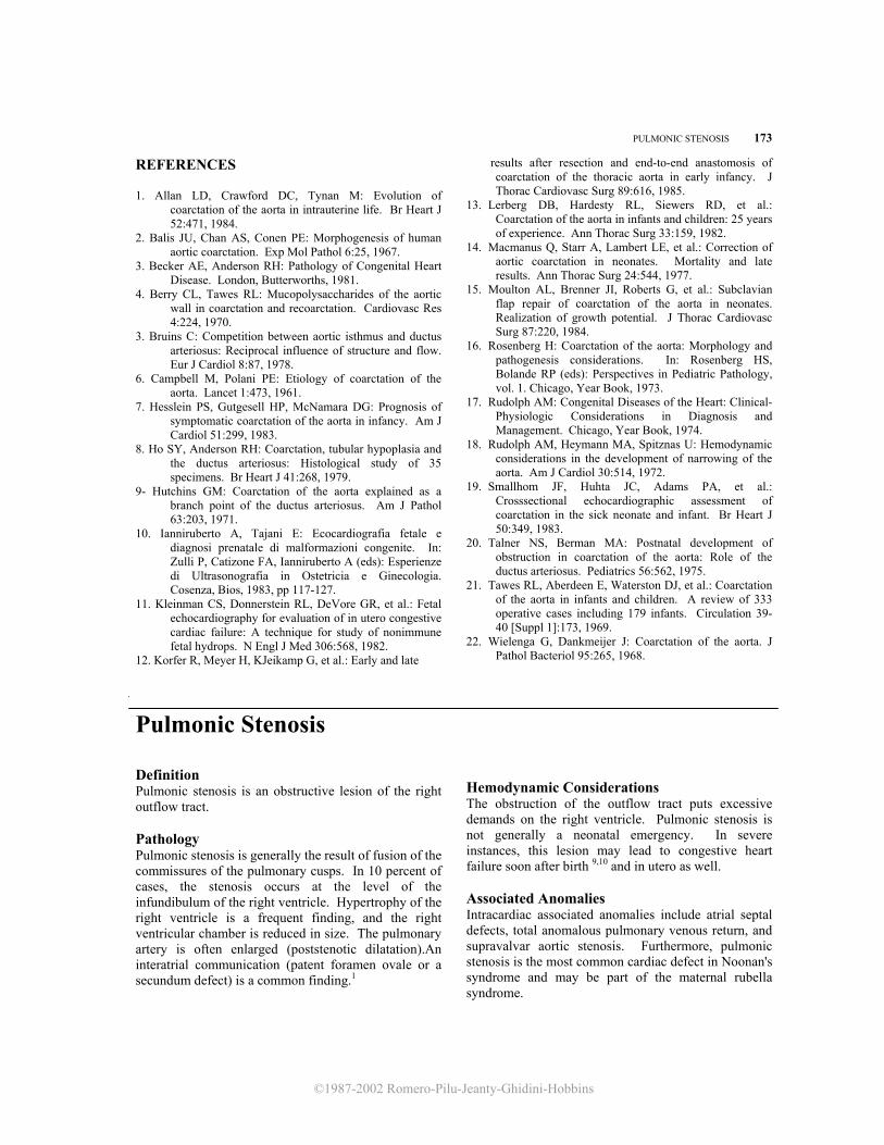
©1987-2002 Romero-Pilu-Jeanty-Ghidini-Hobbins
PULMONIC STENOSIS 173
REFERENCES 1. Allan LD, Crawford DC, Tynan M: Evolution of
coarctation of the aorta in intrauterine life. Br Heart J 52:471, 1984.
2. Balis JU, Chan AS, Conen PE: Morphogenesis of human aortic coarctation. Exp Mol Pathol 6:25, 1967.
3. Becker AE, Anderson RH: Pathology of Congenital Heart Disease. London, Butterworths, 1981.
4. Berry CL, Tawes RL: Mucopolysaccharides of the aortic wall in coarctation and recoarctation. Cardiovasc Res 4:224, 1970.
3. Bruins C: Competition between aortic isthmus and ductus arteriosus: Reciprocal influence of structure and flow. Eur J Cardiol 8:87, 1978.
6. Campbell M, Polani PE: Etiology of coarctation of the aorta. Lancet 1:473, 1961.
7. Hesslein PS, Gutgesell HP, McNamara DG: Prognosis of symptomatic coarctation of the aorta in infancy. Am J Cardiol 51:299, 1983.
8. Ho SY, Anderson RH: Coarctation, tubular hypoplasia and the ductus arteriosus: Histological study of 35 specimens. Br Heart J 41:268, 1979.
9- Hutchins GM: Coarctation of the aorta explained as a branch point of the ductus arteriosus. Am J Pathol 63:203, 1971.
10. Ianniruberto A, Tajani E: Ecocardiografia fetale e diagnosi prenatale di malformazioni congenite. In: Zulli P, Catizone FA, Ianniruberto A (eds): Esperienze di Ultrasonografia in Ostetricia e Ginecologia. Cosenza, Bios, 1983, pp 117-127.
11. Kleinman CS, Donnerstein RL, DeVore GR, et al.: Fetal echocardiography for evaluation of in utero congestive cardiac failure: A technique for study of nonimmune fetal hydrops. N Engl J Med 306:568, 1982.
12. Korfer R, Meyer H, KJeikamp G, et al.: Early and late
results after resection and end-to-end anastomosis of coarctation of the thoracic aorta in early infancy. J Thorac Cardiovasc Surg 89:616, 1985.
13. Lerberg DB, Hardesty RL, Siewers RD, et al.: Coarctation of the aorta in infants and children: 25 years of experience. Ann Thorac Surg 33:159, 1982.
14. Macmanus Q, Starr A, Lambert LE, et al.: Correction of aortic coarctation in neonates. Mortality and late results. Ann Thorac Surg 24:544, 1977.
15. Moulton AL, Brenner JI, Roberts G, et al.: Subclavian flap repair of coarctation of the aorta in neonates. Realization of growth potential. J Thorac Cardiovasc Surg 87:220, 1984.
16. Rosenberg H: Coarctation of the aorta: Morphology and pathogenesis considerations. In: Rosenberg HS, Bolande RP (eds): Perspectives in Pediatric Pathology, vol. 1. Chicago, Year Book, 1973.
17. Rudolph AM: Congenital Diseases of the Heart: Clinical-Physiologic Considerations in Diagnosis and Management. Chicago, Year Book, 1974.
18. Rudolph AM, Heymann MA, Spitznas U: Hemodynamic considerations in the development of narrowing of the aorta. Am J Cardiol 30:514, 1972.
19. Smallhom JF, Huhta JC, Adams PA, et al.: Crosssectional echocardiographic assessment of coarctation in the sick neonate and infant. Br Heart J 50:349, 1983.
20. Talner NS, Berman MA: Postnatal development of obstruction in coarctation of the aorta: Role of the ductus arteriosus. Pediatrics 56:562, 1975.
21. Tawes RL, Aberdeen E, Waterston DJ, et al.: Coarctation of the aorta in infants and children. A review of 333 operative cases including 179 infants. Circulation 39-40 [Suppl 1]:173, 1969.
22. Wielenga G, Dankmeijer J: Coarctation of the aorta. J Pathol Bacteriol 95:265, 1968.
Pulmonic Stenosis Definition Pulmonic stenosis is an obstructive lesion of the right outflow tract. Pathology Pulmonic stenosis is generally the result of fusion of the commissures of the pulmonary cusps. In 10 percent of cases, the stenosis occurs at the level of the infundibulum of the right ventricle. Hypertrophy of the right ventricle is a frequent finding, and the right ventricular chamber is reduced in size. The pulmonary artery is often enlarged (poststenotic dilatation).An interatrial communication (patent foramen ovale or a secundum defect) is a common finding.1
Hemodynamic Considerations The obstruction of the outflow tract puts excessive demands on the right ventricle. Pulmonic stenosis is not generally a neonatal emergency. In severe instances, this lesion may lead to congestive heart failure soon after birth 9,10 and in utero as well. Associated Anomalies Intracardiac associated anomalies include atrial septal defects, total anomalous pulmonary venous return, and supravalvar aortic stenosis. Furthermore, pulmonic stenosis is the most common cardiac defect in Noonan's syndrome and may be part of the maternal rubella syndrome.

©1987-2002 Romero-Pilu-Jeanty-Ghidini-Hobbins
174 THE HEART
Figure 4-66. Enlargement of the pulmonary artery (PA) in a third trimester fetus. The pulmonary valve opening appeared normal on both real-time and M-mode examination. Nevertheless, the infant was found at birth to have moderate to severe valvular pulmonic stenosis. RV, right ventricle; RA, right atrium; Sup, superior; Inf, inferior; Ant, anterior; Post, posterior. (Reproduced with permission from Bovicelli L, Baccarani G, Picchio FM, Pilu G: Ecocardiografia Fetale. La Diagnosi e il Trattamento Prenatale delle Cardiopatie Congenite. Milan, Masson, 1985.)
Diagnosis A prenatal diagnosis is extremely difficult. The condition should be suspected when there is either enlargement of the pulmonary artery or reduction in size of the right ventricle (Figs. 4-23, 4-66). In the newborn, the valvular form of pulmonic stenosis is diagnosed by demonstrating the systolic doming (incomplete opening) of the pulmonary valve. The identification of this finding in a fetus is extremely difficult because of the small size of the pulmonary valve and its distance from the transducer. It should be stressed that M-mode echocardiography is not a reliable tool in the recognition of semilunar valve stenosis.4 Indeed, demonstration of the apparently normal opening of the pulmonic valve with this technique does not rule out stenosis. Doppler echocardiography is a useful technique in the newborn. Its diagnostic value depends on the detection of poststenotic turbulent flow in the pulmonary trunk. However, because of the small size of the fetal pulmonary artery, it is doubtful that a turbulence in the flow at this level can be reliably detected. Doppler echocardiography may play a major role by demonstrating the tricuspid regurgitation that is seen in the most severe cases. Prognosis Pulmonic stenosis encompasses a wide spectrum of severity. An autopsy study has reported that the mean age of death in untreated patients is 21 years. 6 In a large series of 221 patients operated on between 1 day of life and 61 years of age, there were 9 operative deaths (4 percent) and 2 late deaths (1 percent).2
However, severe pulmonic stenosis may be a neonatal emergency. Luke reported a 17 percent preoperative mortality rate in a group of critically ill infants diagnosed between birth and 2 years of age.9 Operative mortality for these patients ranged between 12.5 percent.3 and 16 percent.3 However, Litwin et al.8 reported no perioperative deaths on 29 patients. The most relevant data available for prenatal counseling is that of Freed et al.5 who reported no deaths in a group of 13 critically ill neonates diagnosed within 2 days of birth and who had a diminutive right ventricle. Obstetrical Management For every congenital cardiac lesion, careful scanning of the entire fetal anatomy and karyotyping are recommended. The option of pregnancy termination should be offered before viability. Serial ultrasound examinations to rule out early signs of congestive heart failure are indicated. The association of hydrops with a structural cardiac defect is an ominous combination. The optimal management in these patients has yet to be established. The option of early delivery may be considered, but the parents should be aware that the mortality rate in these patients is extremely high.7 In the absence of congestive heart failure, there is no indication to alter standard obstetrical management, but delivery in a tertiary care center where a pediatric cardiologist is immediately available is mandatory.
REFERENCES
1 . Becker AE, Anderson RH: Pathology of Congenital Heart Disease. London, Butterworths, 1981.
2. Danielson GK, Exarhos ND, Weidman WH, et al.: Pulmonic stenosis with intact ventricular septum. Surgical considerations and results of operation. J Thorac Cardiovasc Surg 61:228, 1971.
3. Daskalopoulos DA, Pieroni DR, Gingell RL, et al.: Closed transventricular pulmonary valvotomy in infants. Long-term results and the effect of the size of the right ventricle. J Thorac Cardiovasc Surg 84:187, 1982.
4. Feigenbaum H: Echocardiography, 3d ed. Philadelphia, Lea & Febiger, 1981.
5. Freed MD, Rosenthal A, Bernhard WF, et al.: Critical pulmonary stenosis with a diminutive right ventricle in neonates. Circulation 48:875, 1973.
6. Keith JD, Rowe RD, Vlad P: Heart Disease in Infancy and Childhood, 3d ed. New York, Macmillan, 1978.
7. Kleinman CS, Donnerstein RL, De Vore GR, et al.: Fetal echocardiography for evaluation of in utero congestive heart failure: A technique for study of nonimmune fetal hydrops. N Engl J Med 306:568, 1982.
8. Litwin SB, Williams WH, Freed MD, et al.: Critical pulmonary stenosis in infants: A surgical emergency. Surgery 74:880, 1973.

©1987-2002 Romero-Pilu-Jeanty-Ghidini-Hobbins
AORTIC STENOSIS 175
9. Luke Mj: Valvular pulmonic stenosis in infancy. J Pediatr 68:90, 1966.
10. Mustard WT, Jain SC, Trusler GA: Pulmonary stenosis in the first year of life. Br Heart J 30:255, 1968.
11. Srinivasan V, Konyer A, Broda JJ, et al.: Critical pulmonary stenosis in infants less than three months of age: A reappraisal of closed transventricular pulmonary valvotomy. Ann Thorac Surg 34:46, 1982.
Aortic Stenosis Definition Aortic stenosis is an obstructive lesion of the left outflow tract. Depending on the site of the lesion, this entity is classified as supravalvar, valvar, or subvalvar. The term includes aortic valvar stenosis, aortic supravalvar stenosis, aortic subvalvar stenosis, subaortic stenosis, asymmetric septal hypertrophy (ASH), and idiopathic hypertrophic subaortic stenosis (IHSS). Etiology and Pathology Supravalvar aortic stenosis can be due to one of three anatomic defects: a membrane (usually placed above the sinuses of Valsalva), a localized narrowing of the ascending aorta (hourglass deformity), or a diffuse narrowing involving the aortic arch and branching arteries (tubular variety).2 Isolated supravalvar aortic stenosis can be inherited with an autosomal recessive pattern.12 The Williams syndrome is a sporadic disease that is characterized by the association of supravalvar aortic or pulmonic stenosis, elfin facies, and idiopathic hypercalcemia. Maternal hypervitaminosis D has been implicated as a cause of this conditions The valvar form of aortic stenosis can be due to dysplastic, thickened aortic cusps or fusion of the commissures between the cusps.2 The subvalvar form of aortic stenosis is commonly divided into two subgroups: fixed and dynamic. The fixed form can be due to either a membrane (discrete subaortic stenosis) or a fibromuscular tunnel (diffuse subaortic stenosis). The dynamic form is due to a muscular thickening of the septal surface.2 Many of these cases are inherited in an autosomal dominant fashion,5 and they are commonly referred to as asymmetric septal hypertrophy (ASH), idiopathic hypertrophic subaortic stenosis (IHSS), or hypertrophic obstructive cardiomyopathy (HOCM). A transient form of dynamic obstruction of the left outflow tract is seen in infants of diabetic mothers. This condition is commonly attributed to the association of fetal hyperglycemia and hyperinsulinemia.8 The left ventricle is usually of normal size or enlarged. In some instances, the ventricular cavity
may be small. In severe forms of aortic stenosis, the endocardium may be thickened (secondary endocardial fibroelastosis). In these patients, mitral insufficiency is a frequent finding.2 Associated Anomalies Supravalvar aortic stenosis may be related to the Williams syndrome. Subaortic stenosis has been described in patients with Tumer's syndrome, Noonan's syndrome, and congenital rubella.14 Hemodynamic Considerations Aortic stenosis causes obstruction of the left ventricular outflow tract. Depending on the severity of the stenosis, the pressure in the left ventricle is increased. Although subvalvar and supravalvar forms are not generally manifested in the neonatal period, the valvar type can be a cause of congestive heart failure in the newborn and the fetus as well.10 Of all congenital cardiac abnormalities, aortic stenosis is the one most frequently found in association with intrauterine growth retardations. Diagnosis A prenatal diagnosis is extremely difficult. The condition should be suspected when there is either enlargement or hypoplasia of the left ventricle or ascending aorta. 1 The prenatal diagnosis of supravalvar aortic stenosis has not been reported. Even if the ascending aorta can be well visualized in utero, it is doubtful that a discrete membrane or an hourglass deformity can be identified. It should be stressed that this condition is not usually manifested in the neonatal period. In the newborn, the valvar form of aortic stenosis is diagnosed by demonstrating the systolic doming (incomplete opening) of the aortic valve. Identification of this finding in a fetus is extremely difficult due to the small size of the aortic valve and its distance from the transducer. It should be stressed that M-mode echocardiography is not a reliable tool in the recognition of semilunar valvar stenosis.6 Indeed, demonstration of the apparently normal opening of the aortic valve with this technique does not rule out
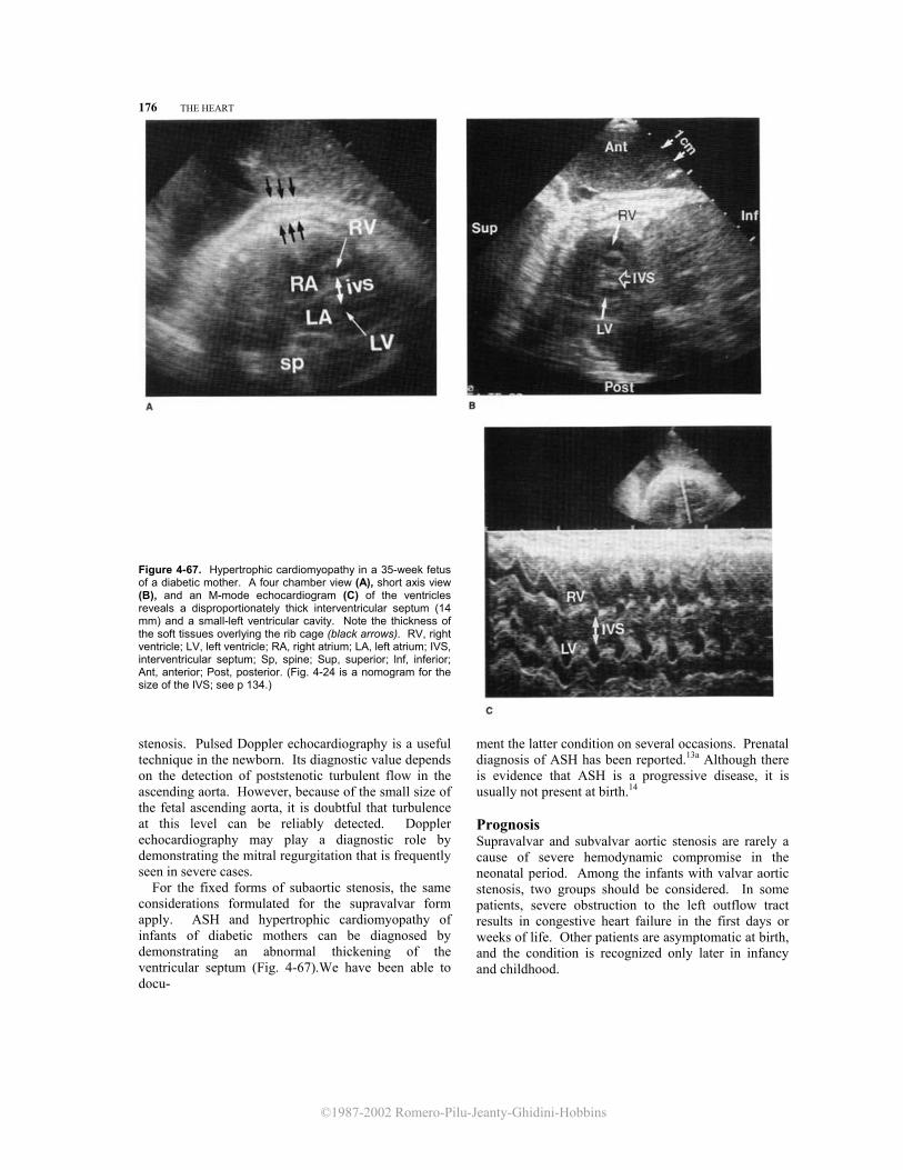
©1987-2002 Romero-Pilu-Jeanty-Ghidini-Hobbins
176 THE HEART
Figure 4-67. Hypertrophic cardiomyopathy in a 35-week fetus of a diabetic mother. A four chamber view (A), short axis view (B), and an M-mode echocardiogram (C) of the ventricles reveals a disproportionately thick interventricular septum (14 mm) and a small-left ventricular cavity. Note the thickness of the soft tissues overlying the rib cage (black arrows). RV, right ventricle; LV, left ventricle; RA, right atrium; LA, left atrium; IVS, interventricular septum; Sp, spine; Sup, superior; lnf, inferior; Ant, anterior; Post, posterior. (Fig. 4-24 is a nomogram for the size of the IVS; see p 134.)
stenosis. Pulsed Doppler echocardiography is a useful technique in the newborn. Its diagnostic value depends on the detection of poststenotic turbulent flow in the ascending aorta. However, because of the small size of the fetal ascending aorta, it is doubtful that turbulence at this level can be reliably detected. Doppler echocardiography may play a diagnostic role by demonstrating the mitral regurgitation that is frequently seen in severe cases. For the fixed forms of subaortic stenosis, the same considerations formulated for the supravalvar form apply. ASH and hypertrophic cardiomyopathy of infants of diabetic mothers can be diagnosed by demonstrating an abnormal thickening of the ventricular septum (Fig. 4-67).We have been able to docu-
ment the latter condition on several occasions. Prenatal diagnosis of ASH has been reported.13a Although there is evidence that ASH is a progressive disease, it is usually not present at birth.14 Prognosis Supravalvar and subvalvar aortic stenosis are rarely a cause of severe hemodynamic compromise in the neonatal period. Among the infants with valvar aortic stenosis, two groups should be considered. In some patients, severe obstruction to the left outflow tract results in congestive heart failure in the first days or weeks of life. Other patients are asymptomatic at birth, and the condition is recognized only later in infancy and childhood.
©1987-2002 Romero-Pilu-Jeanty-Ghidini-Hobbins
AORTIC STENOSIS 177
optimal management of these patients has not been established. The option of early delivery may be considered, but the parents should be aware that the mortality rate in these patients is extremely high.10 In the absence of congestive heart failure, there is no indication to alter standard obstetrical management, but delivery in a tertiary care center where a pediatric cardiologist is immediately available is mandatory. The diagnosis of septal hypertrophy in a fetus of a diabetic mother is an indication for a careful examination of the metabolic control of the patient.
Delineating the natural history of congenital aortic stenosis, Campbell4 estimated that the mortality rate in the first year of life was 23 percent and fell to 1.2 percent for the rest of the first two decades. It then rose again to about 3 percent, 3.5 percent, 6 percent, and 8.5 percent in the third to sixth decades, respectively. According to this estimation, 60 percent of the patients were living at 30 years of age and 40 percent were living at 40 years. Sudden death accounts for a significant proportion of losses in this condition. Jones et al.9 reported the results of corrective surgery in infants with either valvar or subvalvar aorticstenosis.The operative mortality rate was 1.9 percent in the patients with valvar aortic stenosis,6 percent in those with fixed subaortic stenosis, and 5.5 percent in those with ASH. Late survival rates with an average follow-up duration of 5 years were 90 percent, 86 percent, and 82 percent, respectively. It was estimated that 54 percent, 54 percent, and 70 percent, respectively, of patients in the three groups had satisfactory late results 5 to 14 years after operation. These data refer mainly to patients who were not critically ill at birth. The newborn with symptomatic aortic stenosis usually has severe dyspnea and intractable congestive heart failure. In these patients, an operative mortality of 29 to 71 percent has been reported. Recently, Messina et al.11 described a much lower mortality rate (9 percent). The only death in their series occurred in an infant with a small left ventricle. The authors suggested that the association between aortic stenosis and an underdeveloped left ventricular cavity is a poor prognostic indicator. A wide range of clinical and morphologic expression is expected from patients with supravalvar aortic stenosis. Flaker et al. 7 reported a series of 16 patients who underwent patch aortoplasty. Three surgical deaths occurred, and 2 of these patients had a diffuse narrowing of the aorta. Ten patients were asymptomatic 1 to 12 years after operation, one had angina, and one died from cancer. The hypertrophic cardiomyopathy of infants of diabetic mothers is a transient condition that is, in most cases, asymptomatic. Less frequently, cyanosis and congestive heart failure may occur.8 Obstetrical Management ff the diagnosis is made, careful scanning of the entire fetal anatomy and karyotyping are recommended. The option of pregnancy termination should be offered before viability. Serial ultrasound examinations to rule out early signs of congestive heart failure are indicated. The association of hydrops with a structural cardiac defect is an ominous combination. The
REFERENCES 1. Allan L, Little D, Campbell S, et al.: Fetal ascites associated
with congenital heart disease: Case report. Br J Obstet Gynaecol 88:453, 1981.
2. Becker AE, Anderson RH: Pathology of Congenital Heart Disease. London, Butterworths, 1981.
3. Beuren AJ: Supravalvular aortic stenosis: A complex syndrome with and without mental retardation. Birth Defects 8:45, 1972.
4. Campbell M: The natural history of congenital aortic stenosis. Br Heart f 30:514, 1968.
5. Clark CE, Henry WL, Epstein SE: Familial prevalence and genetic transmission of idiopathic hypertrophic subaortic stenosis. N Engl J Med 289:709, 1973.
6. Feigenbaum H: Echocardiography, 3d ed. Philadelphia, Lea & Febiger, 1981.
7. Flaker G, Teske D, Kilman J, et al.: Supravalvular aortic stenosis. A 20-year clinical perspective and experience with patch aortoplasty. Am J Cardiol 51:256, 1983.
8. Gutgesell HP, Mullins CE, Gillette PC, et al.: Transient hypertrophic subaortic stenosis in infants of diabetic mothers. J Pediatr 89:120, 1976.
9. Jones M, Barnhart GR, Morrow AG: Late results after operations for left ventricular outflow tract obstruction. Am J Cardiol 50:569, 1982.
10. Kleinman CS, Donnerstein RL, De Vore GR, et al.: Fetal echocardiography for evaluation of in utero congestive heart failure: A technique for study of nonimmune fetal hydrops. N Engl J Med 306:568, 1982.
11. Messina LM, Turley K, Stanger P, et al.: Successful aortic valvotomy for severe congenital valvular aortic stenosis in the newborn infant. J Thorac Cardiovasc Surg 88:92, 1984.
12. Nora JJ, Nora AH: Genetics and Counseling in Cardiovascular Diseases. Springfield, IL, Chas. C Thomas, 1978.
13. Reynolds JL: Intrauterine growth retardation in children with congenital heart disease Its relation to aortic stenosis. Birth Defects 8:143, 1972.
13a. Stewart PA, Buis-Liem T, Verwey RA, Wladimiroff JW: Prenatal ultrasonic diagnosis of familial asymmetric septal hypertrophy. Prenat Diagn 6:249, 1986.
14. Wright GB, Keane JF, Nadas AS, et al.: Fixed subaortic stenosis in the young: Medical and surgical course in 83 patients. Am J Cardiol 52:830, 1983.
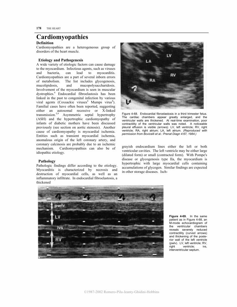
©1987-2002 Romero-Pilu-Jeanty-Ghidini-Hobbins
178 THE HEART
Cardiomyopathies
Figure 4-68. Endocardial fibroelastosis in a third trimester fetus. The cardiac chambers appear greatly enlarged, and the ventricular walls are thickened. At real-time examination, poor contractility of the ventricular walls was noted. A noticeable pleural effusion is visible (arrows). LV, left ventricle; RV, right ventricle; RA, right atrium; LA, left atrium. (Reproduced with permission from Bovicelli et al.: Prenat Diagn 4:67, 1984.)
Definition Cardiomyopathies are a heterogeneous group of disorders of the heart muscle.
Etiology and Pathogenesis A wide variety of etiologic factors can cause damage to the myocardium. Infectious agents, such as viruses and bacteria, can lead to myocarditis. Cardiomyopathies are a part of several inborn errors of metabolism. The list includes glycogenosis, mucolipidosis, and mucopolysaccharidosis. Involvement of the myocardium is seen in muscular dystrophies.6 Endocardial fibroelastosis has been linked in the past to congenital infection by various viral agents (Coxsackie viruses4 Mumps virus8). Familial cases have often been reported, suggesting either an autosomal recessive or X-linked transmission.5,9 Asymmetric septal hypertrophy (ASH) and the hypertrophic cardiomyopathy of infants of diabetic mothers have been discussed previously (see section on aortic stenosis). Another cause of cardiomyopathy is myocardial ischemia. Entities such as transient myocardial ischemia, anomalous origin of the left coronary artery, and coronary calcinosis are probably due to an ischemic mechanism. Cardiomyopathies can also be of idiopathic etiology.
Pathology Pathologic findings differ according to the etiology. Myocarditis is characterized by necrosis and destruction of myocardial cells, as well as an inflammatory infiltrate. In endocardial fibroelastosis, a thickened
grayish endocardium lines either the left or both ventricular cavities. The left ventricle may be either large (dilated form) or small (contracted form). With Pompe's disease or glycogenosis type IIa, the myocardium is hypertrophic with large myocardial cells containing accumulations of glycogen. Similar findings are expected in other storage diseases. Isch-
Figure 4-69. In the same patient as in Figure 4-68, an M-mode echocardiogram of the ventricular chambers reveals severely reduced contractility (curved arrows) and thickening of the poste- rior wall of the left ventricle (pwlv). LV, left ventricle; RV, right ventricle; ivs, interventricular septum.
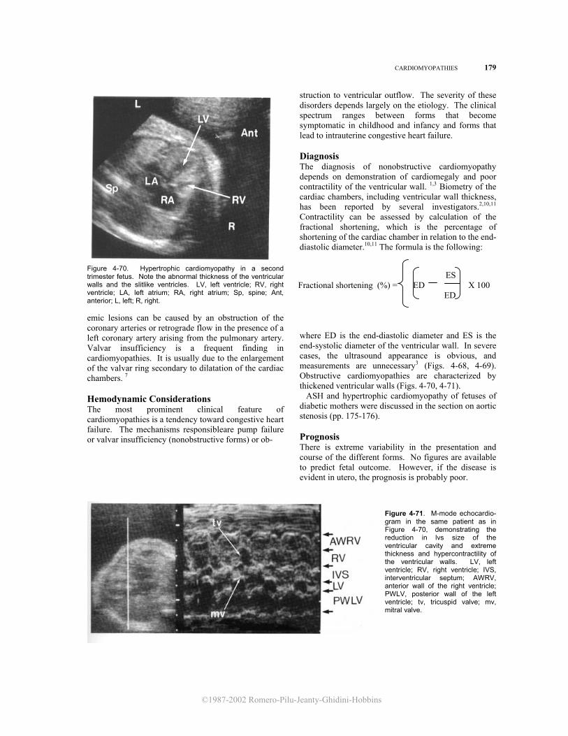
©1987-2002 Romero-Pilu-Jeanty-Ghidini-Hobbins
CARDIOMYOPATHIES 179
Figure 4-70. Hypertrophic cardiomyopathy in a second trimester fetus. Note the abnormal thickness of the ventricular walls and the slitlike ventricles. LV, left ventricle; RV, right ventricle; LA, left atrium; RA, right atrium; Sp, spine; Ant, anterior; L, left; R, right. emic lesions can be caused by an obstruction of the coronary arteries or retrograde flow in the presence of a left coronary artery arising from the pulmonary artery. Valvar insufficiency is a frequent finding in cardiomyopathies. It is usually due to the enlargement of the valvar ring secondary to dilatation of the cardiac chambers. 7 Hemodynamic Considerations The most prominent clinical feature of cardiomyopathies is a tendency toward congestive heart failure. The mechanisms responsibleare pump failure or valvar insufficiency (nonobstructive forms) or ob-
struction to ventricular outflow. The severity of these disorders depends largely on the etiology. The clinical spectrum ranges between forms that become symptomatic in childhood and infancy and forms that lead to intrauterine congestive heart failure. Diagnosis The diagnosis of nonobstructive cardiomyopathy depends on demonstration of cardiomegaly and poor contractility of the ventricular wall. 1,3 Biometry of the cardiac chambers, including ventricular wall thickness, has been reported by several investigators.2,10,11 Contractility can be assessed by calculation of the fractional shortening, which is the percentage of shortening of the cardiac chamber in relation to the end-diastolic diameter.10,11 The formula is the following:
ES Fractional shortening (%) = ED X 100
ED
where ED is the end-diastolic diameter and ES is the end-systolic diameter of the ventricular wall. In severe cases, the ultrasound appearance is obvious, and measurements are unnecessary3 (Figs. 4-68, 4-69). Obstructive cardiomyopathies are characterized by thickened ventricular walls (Figs. 4-70, 4-71). ASH and hypertrophic cardiomyopathy of fetuses of diabetic mothers were discussed in the section on aortic stenosis (pp. 175-176). Prognosis There is extreme variability in the presentation and course of the different forms. No figures are available to predict fetal outcome. However, if the disease is evident in utero, the prognosis is probably poor.
Figure 4-71. M-mode echocardio-gram in the same patient as in Figure 4-70, demonstrating the reduction in lvs size of the ventricular cavity and extreme thickness and hypercontractility of the ventricular walls. LV, left ventricle; RV, right ventricle; IVS, interventricular septum; AWRV, anterior wall of the right ventricle; PWLV, posterior wall of the left ventricle; tv, tricuspid valve; mv, mitral valve.

©1987-2002 Romero-Pilu-Jeanty-Ghidini-Hobbins
180 THE HEART
Obstetrical Management When the diagnosis is made before viability, the option of pregnancy termination should be offered. Serial ultrasound examinations should be performed to search for signs of congestive heart failure. The association of hydrops with a cardiomyopathy is an ominous combination. The optimal management of these patients has not been established. The option of early delivery may be considered, but the parents should be aware that the mortality rate in these patients is extremely high. Delivery in a tertiary care center is mandatory.
REFERENCES 1. Allan LD, Crawford DC, Anderson RH, et al.:
Echocardiographic and anatomical correlations in fetal congenital heart disease. Br Heart J 52:542, 1984.
2. Allan LD, Joseph MC, Boyd EGCA, et al.: M-mode echocardiography in the developing human fetus. Br Heart J: 47:573, 1982.
3. Bovicelli L, Picchio FM, Pilu G, et al.: Prenatal diagnosis of endocardial fibroelastosis. Prenat Diagn 4:67, 1984.
4. Brown GC, Evans TN: Serologic evidence of Coxsackie virus etiology of congenital heart disease. JAMA 199:183, 1967.
5. Chen S, Thompson MW, Rose V: Endocardial fibroelas- tosis: Family studies with special reference to
counselling. J Pediatr 79:385, 1971. 6. Nora JJ, Nora AH: Genetics and Counseling in
Cardiovascular Diseases. Springfield, IL, Chas. C Thomas, 1978.
7. Rowe RD, Freedom RM, Mehrizi A, Bloom KR: The Neonate with Congenital Heart Disease, 2d ed Philadelphia, Saunders, 1981.
8. St. Geme JW, Noren GR, Adams P Jr.: Proposed embryopathic relation between mumps virus and primary endocardial fibroelastosis. N Engl J Med 275:339, 1966.
9. Westwood M, Harris R, Burn JL, et al.: Heredity in primary endocardial fibroelastosis. Br Heart J 37:1077, 1975
10. Wladimiroff JW, Mc Ghie JS: M-mode ultrasonic assessment of fetal cardiovascular dynamics. Br J Obstet Gynaecol 88:1241, 1981.
11. Wladimiroff JW, Vosters R, Mc Ghie JS: Normal cardiac ventricular geometry and function during the last trimester of pregnancy and early neonatal period. Br J Obstet Gynaecol 89:839, 1982.
Total Anomalous Pulmonary Venous Return Definition Total anomalous pulmonary venous return (TAPVR) is characterized by drainage of the pulmonary veins into the right atrium. Embryology During early stages of development, the intraparenchymal or primary pulmonary veins are connected to the veins of the systemic circulation. In a later stage, an anastomosis between the intraparenchymal pulmonary veins and the primary pulmonary vein (derived from the left atrium) is established. At the same time, the communication with the systemic circulation is lost (Fig. 4-72). Failure of reabsorption of the communication between the intraparenchymal pulmonary veins and the systemic circulation results in anomalous pulmonary venous return.1 Pathology The pulmonary veins normally drain into the left atrium. TAPVR occurs when part or all of the blood flow from these vessels returns to the right atrium. Anomalous pulmonary venous return may be
classified as total or partial according to whether all the blood coming from one lung (unilateral) or both lungs (bilateral) drains inside the right atrium. We discuss here only the total bilateral variety. Depending on the site of the drainage, TAPVR is classified as supradiaphragmatic or infradiaphragmatic. The former can be subclassified into supracardiac or cardiac (Fig. 4-73). The supracardiac drainage follows the course of the left innominate vein, right and left persistent superior vena cava, hemiazygos and azygos veins. The cardiac drainage may end directly into the right atrium or into the coronary sinus. The infradiaphragmatic drainage is characterized by the presence of a venous channel that is normally connected either to the inferior vena cava or to the portal veins.1 In some instances, the pulmonary venous return is obstructed. This has important prognostic implications.9 Hemodynamic Considerations TAPVR is not thought to be a cause of significant hemodynamic perturbances in the fetus, because there is normally a right-to-left shunt at the level o the atrial chambers. Exceptions may exist in those
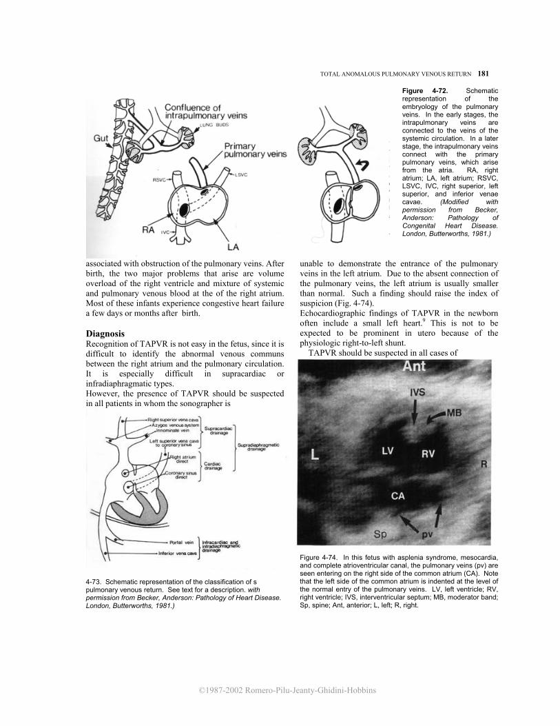
©1987-2002 Romero-Pilu-Jeanty-Ghidini-Hobbins
TOTAL ANOMALOUS PULMONARY VENOUS RETURN 181
Figure 4-72. Schematic representation of the embryology of the pulmonary veins. In the early stages, the intrapulmonary veins are connected to the veins of the systemic circulation. In a later stage, the intrapulmonary veins connect with the primary pulmonary veins, which arise from the atria. RA, right atrium; LA, left atrium; RSVC, LSVC, IVC, right superior, left superior, and inferior venae cavae. (Modified with permission from Becker, Anderson: Pathology of Congenital Heart Disease. London, Butterworths, 1981.)
associated with obstruction of the pulmonary veins. After birth, the two major problems that arise are volume overload of the right ventricle and mixture of systemic and pulmonary venous blood at the of the right atrium. Most of these infants experience congestive heart failure a few days or months after birth. Diagnosis Recognition of TAPVR is not easy in the fetus, since it is difficult to identify the abnormal venous communs between the right atrium and the pulmonary circulation. It is especially difficult in supracardiac or infradiaphragmatic types. However, the presence of TAPVR should be suspected in all patients in whom the sonographer is
4-73. Schematic representation of the classification of s pulmonary venous return. See text for a description. with permission from Becker, Anderson: Pathology of Heart Disease. London, Butterworths, 1981.)
unable to demonstrate the entrance of the pulmonary veins in the left atrium. Due to the absent connection of the pulmonary veins, the left atrium is usually smaller than normal. Such a finding should raise the index of suspicion (Fig. 4-74). Echocardiographic findings of TAPVR in the newborn often include a small left heart.9 This is not to be expected to be prominent in utero because of the physiologic right-to-left shunt. TAPVR should be suspected in all cases of
Figure 4-74. In this fetus with asplenia syndrome, mesocardia, and complete atrioventricular canal, the pulmonary veins (pv) are seen entering on the right side of the common atrium (CA). Note that the left side of the common atrium is indented at the level of the normal entry of the pulmonary veins. LV, left ventricle; RV, right ventricle; IVS, interventricular septum; MB, moderator band; Sp, spine; Ant, anterior; L, left; R, right.

©1987-2002 Romero-Pilu-Jeanty-Ghidini-Hobbins
182 THE HEART atrioventricular septal defects and asplenia and polysplenia syndromes.
Prognosis Seventy-five percent of untreated infants with TAPVR die within 1 year of birth, and the others usually die before reaching adulthood.2-4,5-8 Operative procedures are intended to restore the anatomy of the pulmonary vein drainage.8 In a series of 25 infants, the operative mortality was 20 percent, and all deaths occurred in critically ill infants with pulmonary venous obstruction. No late death was observed, and the hemodynamic condition of the survivors was good.8 The most important prognostic factor was the presence of pulmonary venous obstruction.10 Obstetrical Management TAPVR can be suspected in certain cases, but it is not clear yet if a specific prenatal diagnosis can be made with confidence. However, it should be stressed that if associated anomalies are absent and the infant is promptly assisted in the neonatal period, the prognosis after survival with surgical repair is excellent. Obstetrical management would not be altered by the prenatal diagnosis of TAPVR. It is unclear if TAPVR associated with pulmonary venous obstruction can result in congestive heart failure in utero. Serial ultrasound monitoring of these fetuses is, therefore, recommended. Delivery in a tertiary care center where a pediatric cardiologist is immediately available is suggested.
REFERENCES
1. Becker AE, Anderson RH: Pathology of Congenital Heart Disease. London, Butterworths, 1981.
2. Behrendt DM, Aberdeen E, Waterson DJ, et at.: Total anomalous pulmonary venous drainage in infants. I. Clinical and hemodynamic findings, methods, and results of operation in 37 cases. Circulation 46:347, 1972.
3. Bonham Carter RE, Capriles M, Noe Y: Total anomalous pulmonary venous drainage: A clinical and anatomical study of 75 children. Br Heart J 31:45, 1969.
4. Brody H: Drainage of the pulmonary veins into the right side of the heart. Arch Pathol 33:221, 1942.
5. El-Said G, Mullins CE, McNamara, DG: Management of total anomalous pulmonary venous return. Circulation 45:1240, 1972.
6. Gathman GE, Nadas AS: Total anomalous pulmonary venous connection. Clinical and physiologic observations of 75 pediatric patients. Circulation 42:143, 1970.
7. Gomes MMR, Feldt RH, McGoon DC, et al.: Total anomalous pulmonary venous connection. Surgical considerations and results of operation. J Thorac Cardiovasc Surg 60:116, 1970.
8. Hammon JW, Bender HW, Graham TP, et al.: Total anomalous pulmonary venous connection in infancy. Ten years' experience including studies of postoperative ventricular function. J Thorac Cardiovasc Surg 80:544, 1980.
9. Hawker RE, Celermajer JM, Gengos DC, et al.: Common pulmonary vein atresia. Premortem diagnosis in two infants. Circulation 46:368, 1972.
10. Lima CO, Valdes-Cruz LM, Allen HD, et al.: Prognostic value of left ventricular size measured by echocardiog-raphy in infants with total anomalous pulmonary venous drainage. Am J Cardiol 51:1155, 1983.
Tumors of the Heart
Pathology
Congenital cardiac tumors are extremely rare lesions. It has been estimated that their overall frequency is 1 in 10,000 autopsies in individuals of all ages. Most of these tumors are benign. In infants, the most common tumors are rhabdomyomas (58 percent) and teratomas (20 percent). Less common lesions include fibroma, myxoma (which predominates in adults), hemangioma, and mesothelioma.14 Malignancy occurs in less than 10 percent of all cases.10 Size and location of the tumors vary considerably. Rhabdomyomas tend to be multiple and to involve the septum. Teratomas may be both intrapericardic and extracardiac.2 Fibromas account for 12 percent of the tumors in the neonatal period and until 1 year of age. Fibromas may be pedunculated and may calcify.
Associated Anomalies Cardiac tumors are generally isolated anomalies. An exception to this is the association between tuberous sclerosis (TS) and rhabdomyomas: TS has been re-ported in 50 to 86 percent of patients with cardiac rhabdomyomas.3,4 TS is generally a familial disease inherited as an autosomal dominant trait with a high degree of penetrance and variable expressivity. Rarely, it may be a sporadic event. However, the diagnosis of TS in the first child in the absence of a positive family tree does not allow distinguishing whether the event is sporadic or results from transmission from a mutation-bearing parent. TS is diagnosed when at least one of the following lesions is present: cortical tubers, subependymal hamartomas, multiple retinal hamartomas, and skin lesions

©1987-2002 Romero-Pilu-Jeanty-Ghidini-Hobbins
TUMORS OF THE HEART 183
Figure 4-75. Four-chamber view of the heart in a third trimester fetus with a cardiac rhabdomyoma (@) of the free wall of the left ventricle (LV). RV, right ventricle; RA, right atrium; LA, left atrium; Sp, spine; Ant, anterior, Post, posterior, R, right, L, left. (Courtesy of Dr. Tullia Todros, University of Turin.) (adenoma sebaceum or periungual fibroma). Secondary or presumptive diagnostic criteria include hypomelanotic macules in the skin, subependymal or cortical calcifications, multiple renal tumors, and cardiac rhabdomyomas. Rhabdomyomas are also frequently associated with supraventricular tachycardia. Accessory conductive pathways within the tumor are thought to be responsible for this association.6,13,15
Hemodynamic Considerations The mechanisms by which cardiac tumors become symptomatic include obstruction of the inflow or outflow of the cardiac chambers and cardiac dysrhythmias, both possibly leading to congestive heart failure.11,13,14
Diagnosis Recognition of an intracardiac tumor depends on visuafization of a mass-occupying lesion impinging upon the cardiac cavities. 1,5,7-9 The demonstration of multiple cardiac tumors suggests a diagnosis of rhabdomyomas (Fig 4-75).4 Given the strong association between rhabdomyomas and TS, visualization of multiple cardiac tumors in a fetus should prompt a careful search to detect the other stigmata of TS (concentrating in particular on the CNS and kidneys). Review of the family history, focusing on the presence of mental retardation or seizures in the relatives, is also indicated. First degree relatives should have an eye examination and inspecfion of the skin with Wood's light.8a
Prognosis The prognosis depends on the number, size, location, and histologic type of the tumors. The clinical spectrum varies from completely asymptomatic to
severely ill. In a review of the surgical literature, rhabdomyomas operated on within the first year of life were associated with a 29 percent mortality rate. About one fourth of the patients with TS present by the age of 2 years with seizures or mental retardation. However, presentation may be delayed until adult-hood or the disease may remain clinically absent. Since surgical excision is possible, the prenatal iden-tification of congenital tumors is important for earlier referral and treatment.3,12 Obstetrical Management When the diagnosis of a cardiac tumor is made before viability, the option of pregnancy termination should be offered. Serial ultrasound examinations should be performed to search for signs of congestive heart fail-lure. The association of hydrops with a structural cardiac defect is an ominous combination. The optimal management of these patients has not been established. The option of early delivery may be considered,but the parents should be aware that the mortality rate in these patients is extremely high.9 In the abscense of congestive heart failure, there is no need to modify standard obstetrical management, but delivery in a tertiary care center where a pediatric cardiologist is immediately available is mandatory. REFERENCES
1.Allan LD, Crawford DC, Anderson RH, et al.: Echocar-diographic and anatomical correlations in fetal congenital heart disease. Br Heart J 52:542, 1984.
2.Arciniegas E, Hakimi M, Farooki ZQ, et al.: Primary cardiac tumors in children. J Thorac Cardiovasc Surg. 79:582, 1980.
3.Corno A, de Simone G, Catena G, et al.: Cardiac rhabdomyoma: Surgical treatment in the neonate. J Thorac Cardiovasc Surg. 87:725, 1984.
4. Dennis MA, Appareti K, Manco-johnson ML, et al.: The echocardiographic diagnosis of multiple fetal cardiac tumors. j Ultrasound Med 4:327, 1985.
5. DeVore GR, Hakim S, Kleinman C, et al.: The in utero diagnosis of an interventricular septal cardiac rhab-domyoma by means of real-time directed M-mode echocardiography. Am j Obstet Gynecol 143:967, 1982.
6. Engle MA, Ito T, Ehlers K.H, et al.: Rhabdomyomatosis of heart. Diagnosis during life with clinical and pathologic findings. Circulation 26:712, 1962.
7. Fischer DR, Beerman LB, Park SC, et al.: Diagnosis of intracardiac rhabdomyoma by two-dimensional echo-cardiography. Am J Cardiol 53:978, 1984.
8. Gladden JR, Dreiling Rj, Gollub SB, et al.: Two-dime-nsional echocardiographic features of multiple right atrial myxomas. Am j Cardiol 52:1364, 1983.
8a. Journel H, Roussey M, Plais MH, et al.: Prenatal diagnosis of familial tuberous sclerosis following detec-tion of cardiac rhabdomyoma by ultrasound. Prenat Diagn 6:283, 1986.
9.Kleinman CS, Donnerstein RL, DeVore GR, et al.: Fetal
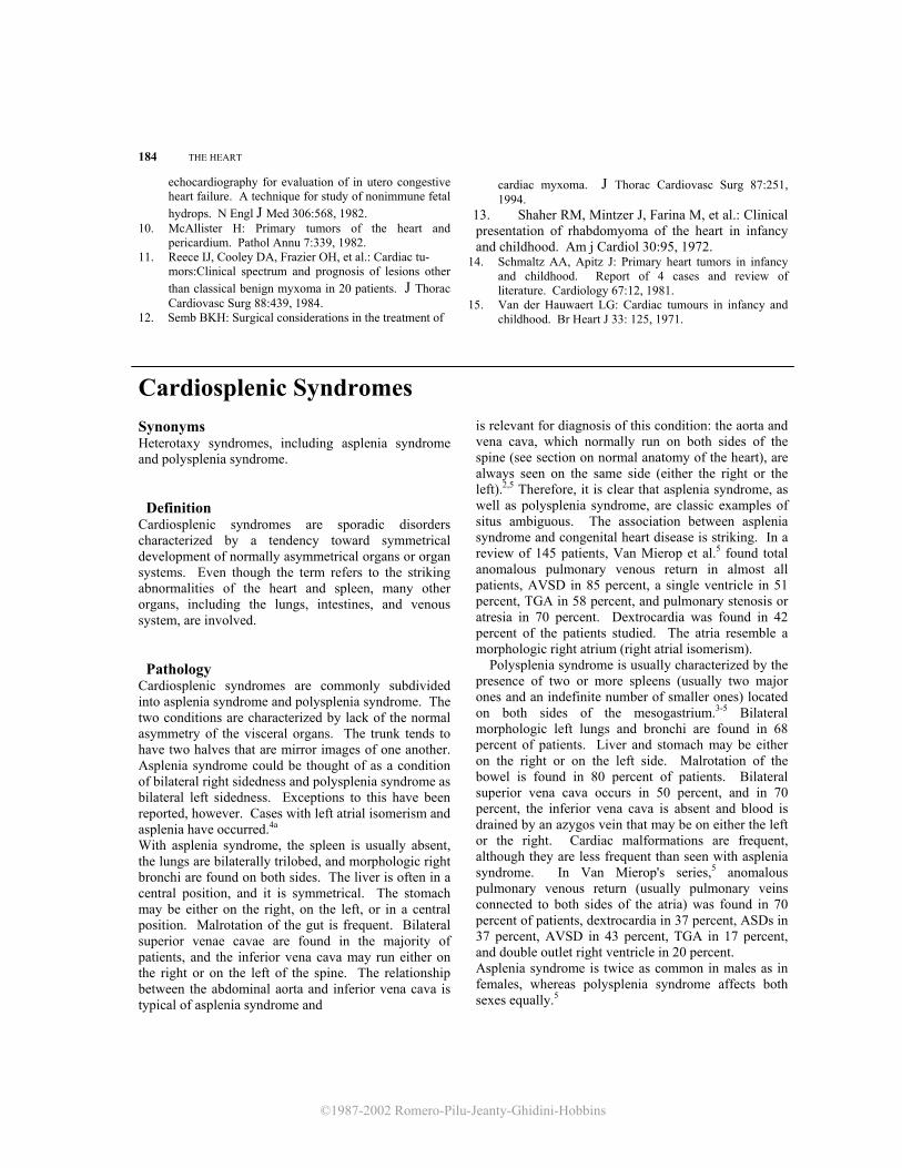
©1987-2002 Romero-Pilu-Jeanty-Ghidini-Hobbins
184 THE HEART
echocardiography for evaluation of in utero congestive heart failure. A technique for study of nonimmune fetal hydrops. N Engl J Med 306:568, 1982.
10. McAllister H: Primary tumors of the heart and pericardium. Pathol Annu 7:339, 1982.
11. Reece IJ, Cooley DA, Frazier OH, et al.: Cardiac tu- mors:Clinical spectrum and prognosis of lesions other than classical benign myxoma in 20 patients. J Thorac Cardiovasc Surg 88:439, 1984.
12. Semb BKH: Surgical considerations in the treatment of
cardiac myxoma. J Thorac Cardiovasc Surg 87:251, 1994.
13. Shaher RM, Mintzer J, Farina M, et al.: Clinical presentation of rhabdomyoma of the heart in infancy and childhood. Am j Cardiol 30:95, 1972.
14. Schmaltz AA, Apitz J: Primary heart tumors in infancy and childhood. Report of 4 cases and review of literature. Cardiology 67:12, 1981.
15. Van der Hauwaert LG: Cardiac tumours in infancy and childhood. Br Heart J 33: 125, 1971.
Cardiosplenic Syndromes
Synonyms Heterotaxy syndromes, including asplenia syndrome and polysplenia syndrome.
Definition Cardiosplenic syndromes are sporadic disorders characterized by a tendency toward symmetrical development of normally asymmetrical organs or organ systems. Even though the term refers to the striking abnormalities of the heart and spleen, many other organs, including the lungs, intestines, and venous system, are involved.
Pathology Cardiosplenic syndromes are commonly subdivided into asplenia syndrome and polysplenia syndrome. The two conditions are characterized by lack of the normal asymmetry of the visceral organs. The trunk tends to have two halves that are mirror images of one another. Asplenia syndrome could be thought of as a condition of bilateral right sidedness and polysplenia syndrome as bilateral left sidedness. Exceptions to this have been reported, however. Cases with left atrial isomerism and asplenia have occurred.4a With asplenia syndrome, the spleen is usually absent, the lungs are bilaterally trilobed, and morphologic right bronchi are found on both sides. The liver is often in a central position, and it is symmetrical. The stomach may be either on the right, on the left, or in a central position. Malrotation of the gut is frequent. Bilateral superior venae cavae are found in the majority of patients, and the inferior vena cava may run either on the right or on the left of the spine. The relationship between the abdominal aorta and inferior vena cava is typical of asplenia syndrome and
is relevant for diagnosis of this condition: the aorta and vena cava, which normally run on both sides of the spine (see section on normal anatomy of the heart), are always seen on the same side (either the right or the left).2,5 Therefore, it is clear that asplenia syndrome, as well as polysplenia syndrome, are classic examples of situs ambiguous. The association between asplenia syndrome and congenital heart disease is striking. In a review of 145 patients, Van Mierop et al.5 found total anomalous pulmonary venous return in almost all patients, AVSD in 85 percent, a single ventricle in 51 percent, TGA in 58 percent, and pulmonary stenosis or atresia in 70 percent. Dextrocardia was found in 42 percent of the patients studied. The atria resemble a morphologic right atrium (right atrial isomerism). Polysplenia syndrome is usually characterized by the presence of two or more spleens (usually two major ones and an indefinite number of smaller ones) located on both sides of the mesogastrium.3-5 Bilateral morphologic left lungs and bronchi are found in 68 percent of patients. Liver and stomach may be either on the right or on the left side. Malrotation of the bowel is found in 80 percent of patients. Bilateral superior vena cava occurs in 50 percent, and in 70 percent, the inferior vena cava is absent and blood is drained by an azygos vein that may be on either the left or the right. Cardiac malformations are frequent, although they are less frequent than seen with asplenia syndrome. In Van Mierop's series,5 anomalous pulmonary venous return (usually pulmonary veins connected to both sides of the atria) was found in 70 percent of patients, dextrocardia in 37 percent, ASDs in 37 percent, AVSD in 43 percent, TGA in 17 percent, and double outlet right ventricle in 20 percent. Asplenia syndrome is twice as common in males as in females, whereas polysplenia syndrome affects both sexes equally.5
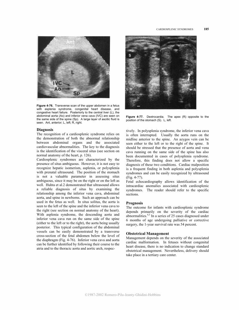
©1987-2002 Romero-Pilu-Jeanty-Ghidini-Hobbins
CARDIOSPLENIC SYNDROMES 185
Figure 4-76. Transverse scan of the upper abdomen in a fetus with asplenia syndrome, congenital heart disease, and congestive heart failure. Posteriorly to the central liver (L), the abdominal aorta (Ao) and inferior vena cava (IVC) are seen on the same side of the spine (Sp). A large layer of ascitic fluid is seen. Ant, anterior; L, left; R, right.
Diagnosis The recognition of a cardiosplenic syndrome relies on the demonstration of both the abnormal relationship between abdominal organs and the associated cardiovascular abnormalities. The key to the diagnosis is the identification of the visceral situs (see section on normal anatomy of the heart, p. 126). Cardiosplenic syndromes are characterized by the presence of situs ambiguous. However, it is not easy to recognize hepatic isomerism, asplenia, or polysplenia with prenatal ultrasound. The position of the stomach is not a valuable parameter in assessing situs ambiguous, since it may be on the right or on the left as well. Huhta et al.2 demonstrated that ultrasound allows a reliable diagnosis of situs by examining the relationship among the inferior vena cava, abdominal aorta, and spine in newborns. Such an approach can be used in the fetus as well. In situs solitus, the aorta is seen to the left of the spine and the inferior vena cava to the right (see section on normal anatomy of the heart). With asplenia syndrome, the descending aorta and inferior vena cava run on the same side of the spine (either to the left or to the right), the aorta being usually posterior. This typical configuration of the abdominal vessels can be easily demonstrated by a transverse cross-section of the fetal abdomen below the level of the diaphragm (Fig. 4-76). Inferior vena cava and aorta can be further identified by following their course to the atria and to the thoracic aorta and aortic arch, respec-
Figure 4-77. Dextrocardia. The apex (R) opposite to the position of the stomach (S). L, left.
tively. In polysplenia syndrome, the inferior vena cava is often interrupted. Usually the aorta runs on the midline anterior to the spine. An azygos vein can be seen either to the left or to the right of the spine. It should be stressed that the presence of aorta and vena cava running on the same side of the spine has also been documented in cases of polysplenia syndrome. Therefore, this finding does not allow a specific diagnosis of these two conditions. Cardiac malposition is a frequent finding in both asplenia and polysplenia syndromes and can be easily recognized by ultrasound (Fig. 4-77). Fetal echocardiography allows identification of the intracardiac anomalies associated with cardiosplenic syndromes. The reader should refer to the specific sections. Prognosis The outcome for infants with cardiosplenic syndrome depends primarily on the severity of the cardiac abnormalities.4,5 In a series of 25 cases diagnosed under 6 months of age undergoing palliative or corrective surgery, the 1-year survival rate was 54 percent. Obstetrical Management Management depends on the severity of the associated cardiac malformation. In fetuses without congenital heart disease, there is no indication to change standard obstetrical management. Nevertheless, delivery should take place in a tertiary care center.
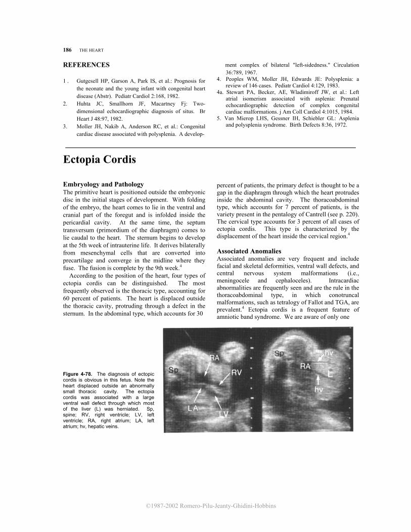
©1987-2002 Romero-Pilu-Jeanty-Ghidini-Hobbins
186 THE HEART
REFERENCES 1 . Gutgesell HP, Garson A, Park IS, et al.: Prognosis for
the neonate and the young infant with congenital heart disease (Abstr). Pediatr Cardiol 2:168, 1982.
2. Huhta JC, Smallhorn JF, Macartney Fj: Two-dimensional echocardiographic diagnosis of situs. Br Heart J 48:97, 1982.
3. Moller JH, Nakib A, Anderson RC, et al.: Congenital cardiac disease associated with polysplenia. A develop-
ment complex of bilateral "left-sidedness." Circulation 36:789, 1967.
4. Peoples WM, Moller JH, Edwards JE: Polysplenia: a review of 146 cases. Pediatr Cardiol 4:129, 1983.
4a. Stewart PA, Becker, AE, Wladimiroff JW, et al.: Left atrial isomerism associated with asplenia: Prenatal echocardiographic detection of complex congenital cardiac malformations. j Am Coll Cardiol 4:1015, 1984.
5. Van Mierop LHS, Gessner IH, Schiebler GL: Asplenia and polysplenia syndrome. Birth Defects 8:36, 1972.
Ectopia Cordis Embryology and Pathology The primitive heart is positioned outside the embryonic disc in the initial stages of development. With folding of the embryo, the heart comes to lie in the ventral and cranial part of the foregut and is infolded inside the pericardial cavity. At the same time, the septum transversum (primordium of the diaphragm) comes to lie caudal to the heart. The sternum begins to develop at the 5th week of intrauterine life. It derives bilaterally from mesenchymal cells that are converted into precartilage and converge in the midline where they fuse. The fusion is complete by the 9th week.4 According to the position of the heart, four types of ectopia cordis can be distinguished. The most frequently observed is the thoracic type, accounting for 60 percent of patients. The heart is displaced outside the thoracic cavity, protruding through a defect in the sternum. In the abdominal type, which accounts for 30
percent of patients, the primary defect is thought to be a gap in the diaphragm through which the heart protrudes inside the abdominal cavity. The thoracoabdominal type, which accounts for 7 percent of patients, is the variety present in the pentalogy of Cantrell (see p. 220). The cervical type accounts for 3 percent of all cases of ectopia cordis. This type is characterized by the displacement of the heart inside the cervical region.4 Associated Anomalies Associated anomalies are very frequent and include facial and skeletal deformities, ventral wall defects, and central nervous system malformations (i.e., meningocele and cephaloceles). Intracardiac abnormalities are frequently seen and are the rule in the thoracoabdominal type, in which conotruncal malformations, such as tetralogy of Fallot and TGA, are prevalent.4 Ectopia cordis is a frequent feature of amniotic band syndrome. We are aware of only one
Figure 4-78. The diagnosis of ectopic cordis is obvious in this fetus. Note the heart displaced outside an abnormally small thoracic cavity. The ectopia cordis was associated with a large ventral wall defect through which most of the liver (L) was herniated. Sp, spine; RV, right ventricle; LV, left ventricle; RA, right atrium; LA, left atrium; hv, hepatic veins.

©1987-2002 Romero-Pilu-Jeanty-Ghidini-Hobbins
PREMATuJRE ATRIAL AND VEN'I'RICULAR CONTRACTIONS 187
patient in whom a chromosomal aberration (45X/ 46XX) was found.2
Diagnosis The diagnosis of ectopia cordis is easy and relies on demonstration of a displaced heart (Fig. 4-78). Several cases of prenatal diagnosis of either thoracic or thoracoabdominal types have been reported.3,5-7 Identification of either the abdominal or the cervical type has not been reported yet, but attention to the anatomic relationships of the heart within the body would make this diagnosis possible.
Prognosis The prognosis is generally poor, and infants are either stillborn or die in the first hours or days of life. Replacement of the heart inside the thoracic cavity has been successful in only a few cases.' Long-term survivors have only been reported for the abdominal type.
Obstetrical Management A careful search for associated anomalies, including a detailed evaluation of the entire fetal anatomy and fetal echocardiography, is recommended. Fetal karyo-
type should be considered. Given the extremely high mortality rate, the option of pregnancy termination should be offered before viability. In the third trimester, nonaggressive management should be considered and discussed with the parents.
REFERENCES
1. Asp K, Sulamaa M: Ectopia cordis. Acta Chir Scand
Supp 283:52, 1961. 2. Garson A, Hawkins EP, Mullins CE, et al.:
Thoracoabdominal ectopia cordis with mosaic Turner's syndrome: Report of a case. Pediatrics 62:218, 1978.
3. Haynor DR, Shuman WP, Brewer DK, et al.: Imaging of fetal ectopia cordis. Roles of sonography and computed tomography. J Ultrasound Med 3:25, 1984.
4. Kanagasuntheram R, Verzin JA: Ectopia cordis in man. Thorax 17:159, 1962.
5. Mercer Lj, Petres RE, Smeltzer JS: Ultrasonic diagnosis of ectopia cordis. Obstet Gynecol 61:523, 1983.
6. Todros T, Presbitero P, Montemurro D, et al.: Prenatal diagnosis of ectopia cordis. j Ultrasound Med 3:429, 1984.
7. Wicks JD, Levine MD, Mettler FA: Intrauterine sonography of thoracic ectopia cordis. AJR 137:619, 1981.
Premature Atrial and Ventricular Contractions
Synonyms Atrial extrasystoles and ectopic atrial beats, ventricular extrasystoles and ectopic ventricular beats.
Definition Premature atrial and ventricular contractions (PAC, PVC) arise from electrical impulses generated outside the cardiac pacemaker (sinus node).
Etiopathogenesis Unknown. In the adult, PACs and PVCs may be stimulated by the ingestion of caffeine, alcohol, or smoking. Electrolyte imbalance and hyperthyroid states have also been implicated. The relevance of these factors to fetal PACs and PVCs is purely speculative. Immaturity or instability of the conducting tissue may be an etiologic factor.
Diagnosis A disturbance in fetal heart rate can be detected either by direct auscultation or Doppler examination. These techniques provide information only about the ven-
tricular rate, whereas accurate diagnosis of a fetal arrhythmia requires assessment of both atrial and ventricular activity. The fetal electrocardiogram is capable of determining both P and QRS waves corresponding to atrial and ventricular depolarization. However, a satisfactory fetal electrocardiogram can be obtained in a small number of patients. At present, the best available technique for the assessment of the fetal dysrhythmias is M-mode echocardiography. We have previously described how the simultaneous visualization of the atrial and ventricular contractions allows us to infer the atrioventricular activation sequence. By using this technique, the origin of an ectopic beat can be easily established (see pp. 131-133).PACs and PVCs may give rise to complex rhythm patterns. PACs may either be conducted to the ventricles or blocked, depending on the moment of the cardiac cycle in which they occur. Thus, repeated PACs may lead to either an increased or a decreased ventricular rate. Blocked PACs must be differentiated from atrioventricular block. The dis-

©1987-2002 Romero-Pilu-Jeanty-Ghidini-Hobbins
188 THE HEART
Figure 4-79. M-mode echocardiogram passing through the left ventricle and right atrium in a term fetus. Undulations of the atrial (a) and ventricular (v) walls reflect atrial and ventricular activity, respec- tively. Repeated premature atrial contractions (pac) conducted to the ventricles are seen. tinction relies on demonstration of an atrial contrac- tion that appears prematurely (Fig. 4-79). PVCs are characterized by a PVC that is not preceded by an atrial contraction. Prognosis and Obstetrical Management Neither PACs nor PVCs are associated with an in- creased incidence of congenital heart disease. There
is unanimity in considering these rhythmic disturbances benign conditions that, in the vast majority of patients, disappear either in utero or shortly after birth.1-4 However, PACs were detected in two fetuses that subsequently developed supraventricular tachycardia.3 It is suggested that the heart rate of fetuses with PACs be serially monitored through pregnancy.There is no harm in asking patients to restrain from caffeine, nicotine, and alcohol and to verify the electrolyte balance of patients ingesting diuretics. There is no need to deliver these fetuses in a tertiary center.
REFERENCES
1. Allan LD, Anderson RH, Sullivan ID, et al.: Evaluation of fetal arrhythmias by echocardiography. Br Heart j 50:240, 1983.
2. Gleicher N, Elkayam U: Intrauterine dysrhythmias. In: Gleicher N, Elkayam U (eds): Cardiac Problems in Preg-nancy: Diagnosis and Management of Maternal and Fetal Disease. New York, Alan R. Liss, 1982, pp 535-564.
3. Kleinman CS, Copel JA, Weinstein EM, et al.: In utero diagnosis and treatment of fetal supraventricular tachycardia. Semin Perinatol 9:113, 1985.
4. Kleinman CS, Donnerstein RL, Jaffe CC, et al.: Fetal echocardiography: A tool for evaluation of in utero cardiac arrhythmias and monitoring in utero therapy. Analysis of 71 patients. Am j Cardiol 51:237, 1983.
Supraventricular Tachyarrhythmias
Definition Supraventricular tachyarrhythmias (SVT) include paroxysmal supraventricular tachycardia, paroxysmal atrial tachycardia, atrial flutter, and atrial fibrillation. SVT is characterized by an atrial frequency of 180 to 300 beats per minute (bpm) and a conduction rate of 1:1. In atrial flutter, the atrial rate ranges from 300 to 460 bpm. Due to variable degrees of atrioventricular block, the ventricular rate usually ranges between 60 and 200 bpm. Atrial fibrillation is characterized by an atrial rate of more than 400 bpm and a ventricular rate ranging from 120 to 200 bpm.25
Pathogenesis SVT occurs by one of two mechanisms: automaticity and reentry.7,21 In cases of automatic induced tachyarrhythmias, an irritable ectopic focus discharges at high
frequency. The reentry mechanism consists of an electrical impulse reentering the atria, giving rise to repeated electrical activity. Reentry may occur at the level of the sinoatrial node, inside the atrium, the atrioventricular node, and the His Purkinje system. Reentry may also occur along an anomalous atrioventricular connection such as the Kent Bundle in the Wolff-Parkinson-White (WPW) syndrome. Atrial flutter and fibrillation often alternate and are thought to result from a similar mechanism. Four theories have been postulated to explain such conditions: (1) circus movement of the electrical impulse, (2) ectopic formation of electrical impulses, (3) multiple reentry, and (4) multifocal impulse formations SVT is by far the most frequent tachyarrhythmia in children, with an incidence of about 1:25,000.21 Within SVT, the most commonly observed form is the
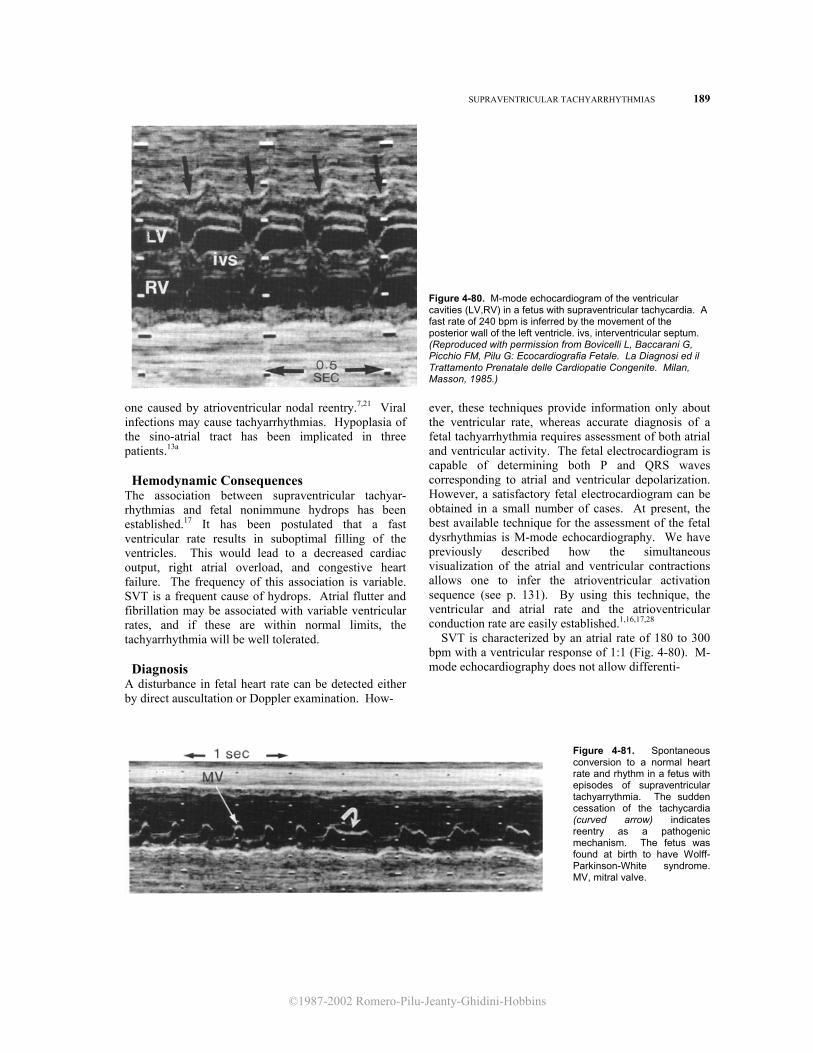
©1987-2002 Romero-Pilu-Jeanty-Ghidini-Hobbins
SUPRAVENTRICULAR TACHYARRHYTHMIAS 189
Figure 4-80. M-mode echocardiogram of the ventricular cavities (LV,RV) in a fetus with supraventricular tachycardia. A fast rate of 240 bpm is inferred by the movement of the posterior wall of the left ventricle. ivs, interventricular septum. (Reproduced with permission from Bovicelli L, Baccarani G, Picchio FM, Pilu G: Ecocardiografia Fetale. La Diagnosi ed il Trattamento Prenatale delle Cardiopatie Congenite. Milan, Masson, 1985.)
one caused by atrioventricular nodal reentry.7,21 Viral infections may cause tachyarrhythmias. Hypoplasia of the sino-atrial tract has been implicated in three patients.13a
Hemodynamic Consequences The association between supraventricular tachyar-rhythmias and fetal nonimmune hydrops has been established.17 It has been postulated that a fast ventricular rate results in suboptimal filling of the ventricles. This would lead to a decreased cardiac output, right atrial overload, and congestive heart failure. The frequency of this association is variable. SVT is a frequent cause of hydrops. Atrial flutter and fibrillation may be associated with variable ventricular rates, and if these are within normal limits, the tachyarrhythmia will be well tolerated.
Diagnosis A disturbance in fetal heart rate can be detected either by direct auscultation or Doppler examination. How-
ever, these techniques provide information only about the ventricular rate, whereas accurate diagnosis of a fetal tachyarrhythmia requires assessment of both atrial and ventricular activity. The fetal electrocardiogram is capable of determining both P and QRS waves corresponding to atrial and ventricular depolarization. However, a satisfactory fetal electrocardiogram can be obtained in a small number of cases. At present, the best available technique for the assessment of the fetal dysrhythmias is M-mode echocardiography. We have previously described how the simultaneous visualization of the atrial and ventricular contractions allows one to infer the atrioventricular activation sequence (see p. 131). By using this technique, the ventricular and atrial rate and the atrioventricular conduction rate are easily established.1,16,17,28
SVT is characterized by an atrial rate of 180 to 300 bpm with a ventricular response of 1:1 (Fig. 4-80). M-mode echocardiography does not allow differenti-
Figure 4-81. Spontaneous conversion to a normal heart rate and rhythm in a fetus with episodes of supraventricular tachyarrythmia. The sudden cessation of the tachycardia (curved arrow) indicates reentry as a pathogenic mechanism. The fetus was found at birth to have Wolff-Parkinson-White syndrome. MV, mitral valve.
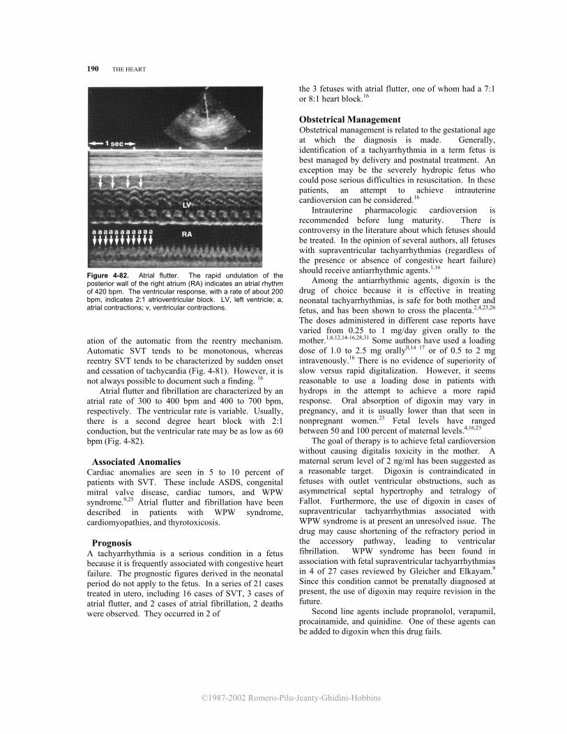
©1987-2002 Romero-Pilu-Jeanty-Ghidini-Hobbins
190 THE HEART
Figure 4-82. Atrial flutter. The rapid undulation of the posterior wall of the right atrium (RA) indicates an atrial rhythm of 420 bpm. The ventricular response, with a rate of about 200 bpm, indicates 2:1 atrioventricular block. LV, left ventricle; a, atrial contractions; v, ventricular contractions. ation of the automatic from the reentry mechanism. Automatic SVT tends to be monotonous, whereas reentry SVT tends to be characterized by sudden onset and cessation of tachycardia (Fig. 4-81). However, it is not always possible to document such a finding. 16
Atrial flutter and fibrillation are characterized by an atrial rate of 300 to 400 bpm and 400 to 700 bpm, respectively. The ventricular rate is variable. Usually, there is a second degree heart block with 2:1 conduction, but the ventricular rate may be as low as 60 bpm (Fig. 4-82).
Associated Anomalies Cardiac anomalies are seen in 5 to 10 percent of patients with SVT. These include ASDS, congenital mitral valve disease, cardiac tumors, and WPW syndrome.9,25 Atrial flutter and fibrillation have been described in patients with WPW syndrome, cardiomyopathies, and thyrotoxicosis.
Prognosis A tachyarrhythmia is a serious condition in a fetus because it is frequently associated with congestive heart failure. The prognostic figures derived in the neonatal period do not apply to the fetus. In a series of 21 cases treated in utero, including 16 cases of SVT, 3 cases of atrial flutter, and 2 cases of atrial fibrillation, 2 deaths were observed. They occurred in 2 of
the 3 fetuses with atrial flutter, one of whom had a 7:1 or 8:1 heart block.16 Obstetrical Management Obstetrical management is related to the gestational age at which the diagnosis is made. Generally, identification of a tachyarrhythmia in a term fetus is best managed by delivery and postnatal treatment. An exception may be the severely hydropic fetus who could pose serious difficulties in resuscitation. In these patients, an attempt to achieve intrauterine cardioversion can be considered.16
Intrauterine pharmacologic cardioversion is recommended before lung maturity. There is controversy in the literature about which fetuses should be treated. In the opinion of several authors, all fetuses with supraventricular tachyarrhythmias (regardless of the presence or absence of congestive heart failure) should receive antiarrhythmic agents.1,16
Among the antiarrhythmic agents, digoxin is the drug of choice because it is effective in treating neonatal tachyarrhythmias, is safe for both mother and fetus, and has been shown to cross the placenta.2,4,23,26 The doses administered in different case reports have varied from 0.25 to 1 mg/day given orally to the mother.1,6,12,14-16,28,31 Some authors have used a loading dose of 1.0 to 2.5 mg orallyll,14 17 or of 0.5 to 2 mg intravenously.16 There is no evidence of superiority of slow versus rapid digitalization. However, it seems reasonable to use a loading dose in patients with hydrops in the attempt to achieve a more rapid response. Oral absorption of digoxin may vary in pregnancy, and it is usually lower than that seen in nonpregnant women.23 Fetal levels have ranged between 50 and 100 percent of maternal levels.4,16,23
The goal of therapy is to achieve fetal cardioversion without causing digitalis toxicity in the mother. A maternal serum level of 2 ng/ml has been suggested as a reasonable target. Digoxin is contraindicated in fetuses with outlet ventricular obstructions, such as asymmetrical septal hypertrophy and tetralogy of Fallot. Furthermore, the use of digoxin in cases of supraventricular tachyarrhythmias associated with WPW syndrome is at present an unresolved issue. The drug may cause shortening of the refractory period in the accessory pathway, leading to ventricular fibrillation. WPW syndrome has been found in association with fetal supraventricular tachyarrhythmias in 4 of 27 cases reviewed by Gleicher and Elkayam.9 Since this condition cannot be prenatally diagnosed at present, the use of digoxin may require revision in the future.
Second line agents include propranolol, verapamil, procainamide, and quinidine. One of these agents can be added to digoxin when this drug fails.

©1987-2002 Romero-Pilu-Jeanty-Ghidini-Hobbins
SIJPRAVENTRICULAR TACHYARRHYTHMIAS 191
rhythmia, digoxin alone is the first step. If this drug is ineffective, combination therapy with either verapamil or propranolol should be considered. Quinidine and procainamide should be the last alternatives.6
Transplacental crossage of propranolol has been reported to vary between 20 and 127 percent. 6,24,30 Fetal side effects have been described. In a review of 153 mothers receiving propranolol for different indications, 15 percent of infants had growth retardation, 9 percent had hypoglycemia, 8 percent hadbradycardia, and 4 percent had respiratory distress at birth.3 The drug has been given orally in doses of 160mg/day.15,30 A loading dose of 0.5 mg intravenously can be considered.
Transplacental crossage of verapamil has been documented.29,31 Umbilical cord levels at birth have been reported to be 30 to 40 percent of maternal levels.16 Fetal side effects have not been clearly established. Doses as high as 300 mg/day have been used for the treatment of pregnancy-related hypertension without demonstrable fetal toxicity.19 A loading dose of 5 to 10 mg intravenously and a maintenance dose of 80 to 120 mg orally every 6 to 8 hours have been suggested. Since verapamil induces a decrease in digoxin clearance, a reduction of 33 to 50 percent of the dose of digoxin may be required when these two medications are used simultaneously.16
Transplacental crossage of procainamide has been reported to vary, with cord to maternal levels ranging from 25 percent to 130 percent.6,8,16 This medication has been administered in combination with other antiarrhythmic agents, such as digoxin, verapamil, and propranolol, in daily dosages of 4 grams.6,28
Very limited experience is available with the administration of quinidine to control fetal supraventricular tachyarrythmias. Placental crossage has been demonstrated with a cord to maternal level ranging from 25 to 100 percent.27 Neonatal thrombocytopenia has been reported in association with maternal administration of this drug.5 Quinidine has been succesfully used in association with digoxin in three cases of fetal supraventricular tachyarrhythmias in dosages of 300 mg every 6 hours orally.27
In a review of 45 cases of supraventricular tachyarrhythmias treated in utero,1,6,8,10-15,16,18,22,27,28,31 digoxin alone was successful in 16 cases (37 percent) and digoxin in association with another antiarrhythmic agents was successful in 18 cases (41 percent). An equivocal result was obtained in one case (2.6 percent), and failure of intrauterine treatment occurred in 10 cases (23 percent). One case of fetal supraventricular tachyarrhythmia has been successfully treated with propranolol alone.30 It has been suggested that fetuses with severe congestive heart failure are less responsive to treatment.1,16
A nonpharmacologic approach to cardioversion has been attempted, with success, by intrauterine compression of the umbilical cord.20
When treating a fetal supraventricular tachyar-
REFERENCES
1. Allan LD, Anderson RH, Sullivan ID, et al.: Evaluation of fetal arrhythmias by echocardiography. Br Heart J 50:240, 1983.
2. Allonen H, Kanto J, lisalo E: The foeto-maternal distribution of digoxin in early human pregnancy. Acta Pharmacol Toxicol 39:477, 1976.
3. Briggs GG, Bodendorfer TW, Freeman RK, Yaffe SJ: Propranolol. In: Drugs in Pregnancy and Lactation: A Reference Guide to Fetal and Neonatal Risk. Baltimore, Williams & Wilkins, 1983, pp 310-313.
4. Chan V, Tse TF, Wong V: Transfer of digoxin across the placenta and into breast milk. Br J Obstet Gynaecol 85:605, 1978.
5. Domula VM, Weissbach G, Lenk H, et al.: Uber die auswirkung medikamentoser behandlung in der schwangerschaft auf das gerinnungspotential des neugeborenen. Zentralbl Gynaekol 99:473, 1977.
6. Dumesic DA, Silverman NH, Tobias S, et al.: Transplacental cardioversion of fetal supraventricular tachycardia with procainamide. N Engl J Med 307:1128, 1982.
7. Garson A, Gillette PC: Electrophysiologic studies of supraventricular tachycardia in children. 1. Clinical-electrophysiologic correlations. Am Heart J 102:233, 1981.
8. Given BD, Phillippe M, Sanders SP, et al.: Procainamide cardioversion of fetal supraventricular tachyarrhythmia. Am J Cardiol 53:1460, 1984.
9. Gleicher N, Elkayam U: Intrauterine dysrhythmias. In: Elkayam U, Gleicher N (eds): Cardiac Problems in Pregnancy: Diagnosis and Management of Maternal
and Fetal Disease. New York, Alan R. Liss, 1982, pp 535-564.
10. Guntheroth WG, Cyr DR, Mack LA, et al.: Hydrops from reciprocating atrioventricular tachycardia in a 27-week fetus requiring quinidine for conversion. Obstet Gynecol 66:29S, 1985.
11. Harrigan JT, Kangos JJ, Sikka A, et al.: Successful treatment of fetal congestive heart failure secondary to tachycardia. N Engl J Med 304:1527, 1981.
12. Heaton FC, Vaughan R: Intrauterine supraventricular tachycardia: Cardioversion with maternal digoxin. Obstet Gynecol 60:749, 1982.
13. Hill LM, Breckle R, Driscoll DJ: Sonographic evaluation of prenatal therapy for fetal supraventricular tachycardia and congestive heart failure: A case report. J Reprod Med 28:671, 1983.
13a. Ho SY, Mortimer G, Anderson RH, et al.: Conduction system defects in three perinatal patients with arrhythmia. Br Heart J 53:158, 1985.
14. Kerenyi TD, Gleicher N, Meller J, et al.: Transplacental
©1987-2002 Romero-Pilu-Jeanty-Ghidini-Hobbins
192 THE HEART cardioversion of intrauterine supraventricular tachycardia
with digitalis. Lancet 2:393, 1980. 15. Klein A, Holzman IR, Austin EM: Fetal tachycardia prior
to the development of hydrops. Attempted pharmacologic cardioversion: Case report. Am J Obstet Gynecol 134:347, 1979.
16. Kleinman CS, Copel JA, Weinstein EM, et al.: In utero diagnosis and treatment of fetal supraventricular tachycardia. Sem Perinatol 9:113, 1985.
17. Kleinman CS, Donnerstein RL, DeVore GR, et al.: Fetal echocardiography for evaluation of in utero congestive heart failure: A technique for study of nonimmune fetal hydrops. N Engl J Med 306:568, 1982.
18. Lingman G, Ohrlander S, Ohlin P: Intrauterine digoxin treatment of fetal paroxysmal tachycardia. Case report. Br J Obstet Gynaecol 87:340, 1980.
19. Marlettini MG, Labriola M, Trabatti MR, et al.: Il verapamil nel trattamento del l'ipertensione in gravidanza. Proceedings of the 62nd National Congress of the Italian Society of Gynecology and Obstetrics. Bologna, Monduzzi, 1984, pp 307-310.
20. Martin CB, Nijhuis JG, Weijer AA: Correction of fetal supraventicular tachycardia by compression of the umbilical cord. Report of a case. Am J Obstet Gynecol 150:324, 1984.
21. Mehta AV, Casta A, Wolff GS: Supraventricular tachycardia. In: Roberts NK, Gelband H (eds): Cardiac Arrhythmias in the Neonate, Infant, and Child, 2d ed. E. Norwalk, CT, Appleton-Century-Crofts,1983, pp 105- 137.
22. Newburger JW, Keane JF: Intrauterine supraventricular tachycardia. J Pediatr 95:780, 1979.
23. Rogers MC, Willerson JT, Goldblatt A, et al.: Serum digoxin concentrations in the human fetus, neonate and infant. N Engl J Med 287:1010, 1972.
24. Schroeder JS, Harrison DC: Repeated cardioversion during pregnancy. Treatment of refractory paroxysmal atrial tachycardia during three successive pregnancies. Am J Cardiol 27:445, 1971.
25. Shenker L: Fetal cardiac arrhythmias. Obstet Gynec Surv 34:561, 1979.
26. Soyka LF: Digoxin: Placental transfer, effects on the fetus and therapeutic use in the newborn. Clin Perinatol 2:23, 1975.
27. Spinnato JA, Shaver DC, Flinn GS, et al.: Fetal supraventricular tachycardia. In utero therapy with digoxin and quinidine. Obstet Gynecol 64:730, 1984.
28. Stewart PA, Tonge HM, Wladimiroff JW: Arrhythmia and structural abnormalities of the fetal heart. Br Heart J 50:550, 1983.
29. Strigl R, Gastroph G, Hege HG, et al.: Nachweis von verapamil im mutterlichen und fetalen blut des menschen. Geburtshilfe Frauenheilkd 40:496, 1980.
30. Teuscher A, Bossi E, Imhof P, et al.: Effect of propranolol on fetal tachycardia in diabetic pregnancy. Am J Cardiol 42:304, 1978.
31. Wolff F, Breuker KH, Schlensker KH, et al.: Prenatal diagnosis and therapy of fetal heart rate anomalies: With a contribution on the placental transfer of verapamil. J Perinat Med 8:203, 1980.
Atrioventricular Block Definition Atrioventricular block (AV block) is a condition in which transmission of the electrical impulse from the atria to the ventricles is impaired. Incidence Congenital heart block affects 1:20,000 live births and is present in 4 to 9 percent of all infants with congenital heart disease.8
Etiology The disorder could result from immaturity of the conduction system, absence of connection to the AV node, or abnormal anatomic position of the AV node.6,8,9 It has been estimated that 50 percent of infants with congenital third degree heart block have associated structural anomalies, including corrected transposition, univentricular heart, cardiac tumors, and cardiomyopathies.3 In the remaining 50 percent the cause remains obscure. Growing evidence suggests an association between maternal antinuclear
antibodies against SSA and SSB antigens and congenital heart block. The SSA and SSB antibodies (also known as Ro and La, respectively) are directed against two saline-soluble ribonucleoprotein antigens from cell nuclei. Transplacental passage of the antibodies is thought to lead to inflammation in the heart conduction system and heart block. Anti-SSA antibodies have been reported in 83 percent of mothers who delivered infants with heart block even though only 30 percent had clinical evidence of connective tissue disease. The most common connective tissue disease associated with congenital heart block is lupus erythematosus.2,7,11,13 Pathophysiology The normal propagation of the electrical impulse occurs from the sinoatrial node to the atrioventricular node and, from there, to the Purkinje system and the ventricles. Atrioventricular block is commonly classified into three types. First degree heart block corresponds

©1987-2002 Romero-Pilu-Jeanty-Ghidini-Hobbins
ATRIOVENTRICULAR BLOCK 193
Figure 4-83. A. M-mode echocardiogram passing through the ventricles (LV,RV) of a third trimester fetus with complete AV block. The a waves seen on the tricuspid valve indicate a normal atrial rate of about 120 bpm. Undulation of the ventricular walls (v) indicate a ventricular rate of less than 50 bpm. B. This M-mode echocardiogram passing through the left ventricle and right atrium clearly demonstrates both the discrepancy and the dissociation between atrial (a) and ventricular (v) activity.
to a simple conduction delay, which on the electrocardiogram is manifested as a prolongation of the PR interval. Second degree block is subdivided into Mobitz types I and II. Mobitz type I consists of a progressive prolongation of the PR interval that finally leads to the block of one atrial impulse (Luciani-Wenckebach phenomenon). In Mobitz type II, there is intermittent conduction with a ventricular rate that is a submultiple of the atrial rate (e.g., 2:1, 3:1). Third degree heart block or complete heart block is characterized by a complete dissociation of atria and ventricles, usually with independent and slow activation of the ventricles.10,12 Hemodynamic Consequences First and second degree AV block are not usually associated with any significant hemodynamic perturbance. Complete AV block may result in important bradycardia, leading to decreased cardiac output and congestive heart failure during fetal life.5 Diagnosis A disturbance in fetal heart rate can be detected either by direct auscultation or Doppler examination. However, these techniques provide information only about the ventricular rate, whereas accurate diagnosis of a fetal arrhythmia requires assessment of both atrial and ventricular activity. Fetal electrocardiogram is capable of determining both P and QRS waves,
corresponding to atrial and ventricular depolarization. However, a satisfactory fetal electrocardiogram can be obtained in a small number of cases. At present, the best available technique for the assessment of the fetal dysrhythmias is M-mode echocardiography. We have previously described how the simultaneous visualization of the atrial and ventricular contractions allows one to infer the atrioventricular activation sequence (see p. 131). By using this technique, the ventricular and atrial rate and the atrioventricular conduction rate are easily established. Since first degree heart block results in an entirely normal heart rate and rhythm, it is not surprising that this diagnosis has not been reported in the human fetus. Blockage of the normal atrial impulse can be diagnosed by demonstration of a normally timed atrial contraction that is not followed by a ventricular contraction. Mobitz type I, type II, and third degree heart block may be differentiated by observing the relationship between atrial and ventricular rate. In Mobitz type I block, only a few atrial impulses are not conducted. In Mobitz type II, a submultiple of atrial impulses are transmitted. In third degree or complete heart block, atrial and ventricular rates are independent of each other and the atrial rate is generally slow (Fig. 4-83). Given the strong correlation between fetal heart block, maternal antinuclear antibodies, and connective tissue disease, appropriate workup is indicated.

©1987-2002 Romero-Pilu-Jeanty-Ghidini-Hobbins
194 THE HEART
Prognosis The two most important prognostic factors are the presence or absence of congenital heart disease and the onset of congestive heart failure. These two factors are interrelated as congestive heart failure is rarely seen in infants without underlying heart disease.4 Obstetrical Management The diagnosis of an atrioventricular block demands a careful search for associated intracardiac anomalies. Isolated second degree heart block does not require any change in clinical management. In the presence of Mobitz type II, serial ultrasound monitoring is suggested, since a transition to complete heart block may occur. Complete heart block may lead to congestive heart failure. Only very few cases of this condition with intrauterine congestive heart failure have been reported in the literaturel,3,5,7 and prognostic figures cannot be provided at present. In these cases, premature delivery may be considered to permit postnatal treatment and prevent intrauterine fetal demise. Steroid administration should be considered before the delivery of a preterm infant with an immature lung profile. Mothers of infants with congenital heart block should be followed prospectively because they are at high risk of developing a connective tissue disorder. REFERENCES 1. Allan LD, Anderson RH, Sullivan ID, et al.: Evaluation of
fetal arrhythmias by echocardiography. Br Heart J 50:240, 1983.
2. Chameides L, Truex RC, Vetter V, et al.: Association of
maternal systemic lupus erythematosus with congenital complete heart block. N Engi J Med 297:1204, 1977.
3. Gleicher N, Elkayam U: Intrauterine dysrhythmias. In: Elkayam U, Gleicher N (eds): Cardiac Problems in
Pregnancy: Diagnosis and Management of Maternal and Fetal Disease. New York, Alan R. Liss, 1982, pp 535-564.
4. Griffiths SP: Congenital complete heart block. Circulation 43:615, 1971.
5. Kleinman CS, Donnerstein RL, Jaffe CC, et al.: Fetal echocardiography: A tool for evaluation of in utero cardiac arrhythmias and monitoring of in utero therapy. Analysis of 71 patients. Am J Cardiol 51:237, 1983.
6. Lev M, Cuadros H, Paul MH: Interruption of the atrioventricular bundle with congenital atrioventricular block. Circulation 43:703, 1971.
7. McCue CM, Mantakas ME, Tingelstad JB, et al.: Congenital heart block in newborns of mothers with connective tissue disease. Circulation 56:82, 1977.
8. McHenry MM, Cayler GG: Congenital complete heart block in newborns, infants, children and adults. Med Times 97 (10):113, 1969.
9. Reemtsma K, Copenhaver WM: Anatomic studies of the cardiac conduction system in congenital malformations of the heart. Circulation 17:271, 1958.
10. Roberts NK: Atrioventricular conduction: Disorders of atrioventricular conduction and intraventricular conduction. In: Roberts NK, Gelband H: (eds): Cardiac Arrhythmias in the Neonate, Infant and Child, 2d ed. E. Norwalk, CT, Appleton-Century-Crofts, 1983, pp 233-252.
11. Scott JS, Maddison PJ, Taylor PV, et al.: Connectivetissue disease, antibodies to ribonucleoprotein, and congenital heart block. N Engl J Med 309:209, 1983.
12. Shenker L: Fetal cardiac arrhythmias. Obstet Gynecol Surv 34:561, 1979.
13. Singsen BH, Akhter JE, Weinstein MM, et al.: Congenital complete heart block and SSA antibodies: Obstetric implications. Am J Obstet Gynecol 152:655, 1985.