Dialogues of The Doubt: Dostoevsky's The Eternal Husband and Machado de Assis's Dom Casmurro
Upload
khangminh22Category
view
0download
0
82 50 YEARS OF DNA
The eternal moleeule As aprelude to the many celebrations around the world saluting the 50th anniversary of the discovery of the DNA double helix, Nature presents a collection of overviews that celebrate the historical, scientific and cultural impacts of arevelatory molecular structure.
F ew molecules captivate like DNA. It enthrals scientists, inspires artists, and challenges society. It is, in every sense, a modern icon. A
defining moment for DNA research was the discovery of its structure half a century ago. On 25 April 1953, in an article in Nature, James Watson and Francis Crick described the entwined embrace of two strands of deoxyribonucleic acid. In doing so, they provided the foundation for understanding molecular damage and repair, replication and inheritance of genetic material, and the diversity and evolution of species.
The broad influence of the double helix is reflected in this collection of articles. Experts from a diverse range of disciplines discuss the impact of the discovery on biology, culture, and applications ranging from medicine to nanotechnology. To help the reader fully appreciate how far the double helix has travelled, we also include the original land mark paper by Watson and Crick and the two accompanying papers by Maurice Wilkins, who shared the Nobel Prize with Watson and Crick in 1962, and by co-discoverer Rosalind Franklin, and their co-authors (pages 83-87).
Transforming science Given the immense significance of the double helix, it is difficult to imagine a world that wasn't transfixed by its discovery. Yet, as Robert Olby recalls on page 88, the proposed structure initially received a lukewarm reception. Maclyn McCarty, who, together with Oswald Avery and Colin MacLeod, had previously showed DNA to be the substance ofinheritance, shares his personal perspective (page 92).
In science, where a lifetime's work can often be encapsulated in a few shining moments, the greatest controversies are sometimes over the sharing of credit. The discovery of the double helix is no exception. The premature death and posthumous treatment of Rosalind Franklin, whose X-ray images of DNA fibres revealed teIltale clues of a double helical structure, propelled her portrayal as a feminist icon. But, as discussed here by her biograph er Brenda Maddox (page 93), Franklin is better remembered as a committed and exacting scientist who saw no boundaries between everyday life and science.
Most of our readers will have grown up with the double helix, and yet it is still startling to consider how quickly DNA biology has progressed in just a lifetime. Bruce Alberts reviews how the elegant pairing of the two strands of the double helix revealed the mechanism for replicating the essential units of inheritance (page 117). Errol Friedberg considers the vulnerability of the DNA molecule to damage and the multitude of ways in which cells repair the damage (page 122). And Gary Felsenfeld and Mark Groudine describe how the gargantuan DNA molecule is packaged inside the
minuscule cells of the body, and how an additional layer of information is encrypted within the proteins intimately associated with DNA (page 134). It is perhaps salutary also to recognize what is still to be learnt about the physiological states in which DNA exists, as discussed by Philip Ball (page 107).
As reviewed by Leroy Hood and David Galas (page 130), DNA science generated the tools that spawned the biotechnologyrevolution. Itenabled the cloning of individual genes, the sequencing of whole genomes and, with the application of computer science, transformed the nature and interactions of molecules into an information science. Carlos Bustamante and co-authors consider how we are stililearning much about the distinct structural and physical properties of the molecule (page 109). And according to Nadrian Seeman, DNA may develop new applications as a material for nano sc ale engineering (page 113).
Influencing society Beyond scientific and technological forums, the double helix has imprinted on society's views of history, medicine and art. As discussed by Svante Pääbo (page 95), the records of evolution have been recalibrated with information traced through DNA sequence. On page 98, Aravinda Chakravarti and Peter Little revisit the 'nature versus nurture' debate and our developing view of the interplay between genetic and environmental factors in human disease. And DNA science will transform clinical medicine according to John Bell (page 100), providing a new taxonomy for human disease and triggering a change to health care practice. On page 126, Gustav Nossal reviews how an understanding of DNA processes, such as recombination, have transformed the Heredity field of immunology.
As a visual icon, and as a profound influence on our nature, the DNA molecule has permeated the imagery and art of our time, and is described by Martin Kemp (page 102) as the Mona Lisa ofthis scientific age. Given that broad impact, and revolutions that are yet to come, it is perhaps appropriate to leave the last word to an artist. Written in 1917, the poem Heredity by Thomas Hardy (see inset) seems to foreshadow both the essence and the fascination of the molecule that we celebrate here. D
Carina Dennis Philip Campbell
doi: lO.1038/nature01396
Commissioning Editor Editor, Nature
Original reference: N ature421, 396 (2003).
I am the family face; Flesh perishes, I live on, Projecting trait and trace Through time to times anon, And leapingfrom place to place Over oblivion.
The years-heired feature that can In curve and voice and eye Despise the human span Of durance - that is I; The eternal thing in man, That heeds no call to die.
Thomas Hardy (First published in Moments of Vision and Miscellaneous Verses, Macmillan, 1917)
J. Clayton et al. (eds.), 50 Years of DNA© Nature Publishing Group 2003

50 YEARS 0' ONA I 83
No. 4356 April 25, 1953 NATURE 737
equipment, and to Dr. G. E. R. Deacon and the eaptain and offieers of R .R . . Di8covery 11 for their pa.rt in making the observations. ' Young, F. B., Gerrar<.l, 11., snd Jevons, W., PMl. Mao., 40, 149
(1920). . I Longuel'Hlgglns, M. ., Jl}'»i. 11'01.. ROll . .d8lro. SO<: .. CeoP"IIB. Stipp.,
Ii, 285 (1949). I I'on All<, W. ., WOO<!s Hole Papers in Phys. Oceuo!<. Meteor., 11
(3) (1950). .
' EkmAn, \', W .. Arkiv.lJfa!. Af/ron. FI/,i.·. (Stocklwlll'). 2 (11) (l1lO1i).
MOLECULAR STRUCTU RE OF NUCLEIC ACIDS
A Structure (or Deoxyribose Nucleic Acid
WE wish to sugg t a strueture for the sa.lt of deoxyt'ibos nueleie acid (D. .A.). This
strueture has novel features whieh are of eonsiderable biological interest .
A strueture for nueleie acid has alrea.dy been propos d by Pauling and Corey'. They kindly mad their manuseript availabl to us in advanee of publieation. Their model consists of three intertwined ehains, with the phosphates near the fibre axi , and the bases on the outside . In our opinion, this structure is unsatisfactory for two reasons: (1) We believe tha the material whieh gives the X-ray diagral1l8 is the salt, not the fr acid. Without the acidic hydrogen atoms it is not elear what forees would hold the structure together, espeeia.l1y as the nega ively charged phosphates near the axis will repel aach other. (2) ome of the van der ' iVaals distanees app ar to be too smalI .
Ano her three -ehain structure hag also b en suggested by Fraser (in the press). In his model the phosphate are on th outside and the ba es on the inside, linked together by hydrogen bonds. 'fhis structur as deserib d is rather il\-d fined, and for
Thls ftgllrc. ia purely dlagmmlllAtlC. The two ribbons symbollze the two phosphate- sugar ehalns, and thc horl· zontal rods tha pairs of basea holding lhe ehaln. togcther. '1' hc vertlcal 11 ne mark. the IIbre axls
this rea on we shaH not comm nt on it .
We wish to pu forward a radically diff rent sI ructUl'e for the salt of deoxyribose nucleie acid . Thi structure has two helical chains ach coiled round the same axi (ee diagra.m). We have made t.h u uat chomical a! umptions, namely, that ea.ch chain consists of phosphate di-stel' groups joining ß-D-d oxy
ribofuranose l'esidues with 3',5' linkages . Thc two chains (but not th ir bases) are retated by a dyad perpendieutar to th fibre axis . Both chain follow righthanded helices, but owing 0
the dyad h sequ nces of th a toms in the two chain run in opposite dir ctions. Each chain 100 ely resembles Ful'b rg's' model No. 1; that is, t he bases are on the inside of th helix and the phosphates on the outsid . Th configuration of h ugar and he a oms near it i close to Furberg's ' standal'd eonfiguration', the sugar boing roughly perpendicular to the attached ba . There
is a re idu on ea.ch chain very 3'4 A. in th z-dire('tion. We have assunl d an angle of 36° between adjacent residue in the same chain, so that th structure repeats after 10 residues on ea.ch ehain, that is, after 34 A. The di tanee of a phosphorus atom from the fibre axis is 10 A. As the phosphates are on the outside, cation have easy access to them.
The structure is an open one, and its water eontent is rat her high. At tower water contents we would expect the bases to tilt so that the structure could become more eompact.
The novel feature of the structure is the manner in wh ich the two chains are held together by the purine and pyrimidine ba.ses. The planes of the ba.ses are perpendieular to the fibre axis. They are joined together in pairs, a single base from one chain being hydrogen-bonded to a single base froro the other chain, so that the two He side by side with identieal z-co-ordinates. One of the pair must be a purine and the other 60 pyrimidine for bonding to occur. The hydrogen bonds are made as folIows: purine position 1 to pyrimidine position 1; purine position 6 to pyrimidine po ition 6.
If it is assumed that the bases on.ly occur in the structure in the most plausible tautomeric forms (that is, with the keto rather than the enol configurations) it is found thM only speeific pairs of bases can bond together. Th se pairs are: adenin (purine) with thymine (pyrimidine) and guanine (purine) with cytosine (pyrimidine).
In other words, if an adenine forms one member of a pair, on either chain, th n on these assumptions the other member must b thymine; similarly for guanine and eytosine. The sequenee of bases on a. single chain does not appeal' to be restricted in any way. However, if only pecific pairs of bases can be formed, it follows that if the sequence of bases on one chain i given, then the sequence on the other ehain is automatically determined.
It hag been found experimentally30t that the ratio of the amounts of adenine to thymine, and the ratio of guanine to eytosine, are always very elose to unity for deoxyribose nueleie acid.
It is probably impossible to build this structure with 60 ribo e sugar in place of the deoxyribose, as the extra oxygen atom would roake too close a van der Waal eontact.
The previously published X-ray data6 ,e on deoxyribose nucl ie acid are insufficient for a rigorous test of our structure. 0 far as we can teIl, it is roughly coropatible with the experimental data, but it roust be regarded as unprov d until it has been checked against more exact results. ome of these are given in the following.eommunications. We were not awar of the details of the results presented there when w devised our strueture, which rests mainly though not entirely on publi hed experimental data and stereochemical arguments.
It ha not e caped our notiee that the specific pairing we have po tulated immediately suggests 60
possible copying mechanism for the genetic material. Full details of the structure, including the con
ditions assum d in building it, together with a. set of co-ordinates for the atoms, will be published elsewhere.
We are much indebted to Dr. Jerry Donohue for eonstant advice and critieism, especially on interatomie distances. We have also been stimulated by a knowledge of the general nature of the unpublished experimental results and ideas of Dr. M. H. F. Wilkins, Dr. R. E. Franklin and their eo-workers a.

84 50 YEARS OF DNA
73 N ATUR E April 25, 1953 VOL. 171
King's College, London. One of us (J. D. W.) has becn e.ided by a fellowship from the National Foundation for Infantile Paralysis.
J. D. WATSO F. H. RI K
Medical Research Council Unit fOl' the Study of the Molecular tructure of
Biological y tcms, 'avendish Laboratory, Cambridge.
April 2.
'Paullng, J •. , "nd torey, R. B .. NeU",., 171, 3·16 (1953); Proc. U.S. NeU. Acaä. ci., 39, 84 (1953).
• l'urberg, S., Ar.!a ehern. OOllll., 6, G34 (J 952). I hlU"qarr, E., for refer~nce8 see Zamenhof, ., Rrawermao, G .. and
hargaff, E .. RWdl irn. et BiOphll" AcIIl, 9, 402 (952) . • "'ynt~. O. R., J. Gen. Phy,io/ .• 36, 201 (J952) . • Astbury. W. T ., ymp. $oe. Exp. Bio!. I, Xuclelc Acid, 66 «'amb.
Univ. PrelJll, 19H). • \\' lIklns, M. JI. F., and 1\andaU, :r. T., Biochim. tt BiOphYI. A rta.
10, 192 (953).
Molecular Structure of Deoxypentose Nucleic Acids
" ' HlLE thc biological propert ies of dcoxypento e nllclcic acid suggest 0. molecular structure containing great compl xity, X-ray c:li1Iraction studi s descl'ibed here (cf. Astburyl) show the basic molecular configuration has great simplicity. The purpose of this communication is to describe, in 0. preliminary way, some of the experimental evidence for he polynucleotide chain configurat ion being helical, and exi, ting in this form when in the natural statc. A fuller account of the work will be published ShOl·tly.
Thc tructurc of deoxypentosc nucleic acid is thc same in 0.11 species (although thc nitrogen base ratios alter consid rably) in nucleoprotein, extracted or in cells, and in purified nuclcate. The same linear group of polynuclcotide chains may pa.ck together parallel in different ways to give crystalline l -', semi-orystalline or paracrystalline material. In 8011 cas s the X-ray diffra.ction photograph consists of two regions, one determined largely by thc regular spa.cing of nucleot ides along the chain, and the other by thc longer spa.cings of the chain configuration. The sequence of differ n t nitrogen bases along the chain i. not ma.de visible.
Ori ntcd para.crystalline dooxypento e nue) ic acid ('structure B ' in the following communication by FrankIin and Gosling) gives a fibre diagram as shown in Fig. 1 (cf. r f. 4). AstblU'Y sugge ted t hat t.he strong 3 ·4-A. reflexion corr ponded to the internucleo ide repeat along the fibre axi. The,....., 34 A. layer lines, however, 8.1·e not dne to 0. repeat of a polynueleotide compo ition, but to th chain configuration repeat, which causes strong diffra.ct ion as tho nucleotide ehains have higher density than the intersti t ial water. The absence of reflexions on or near th meridian immediately ugge ts a he)ical structure with a.xis parall I to fibre length.
Diffraction by Helices
It may be shown$ (al 0 • tokes, unpublished) that th int nsity di tribution in the diffl'action pattern of 80 sories of points equally spa.ced along 80 helix is given by the qllares of Bessei funetions. uniform continuous helix gives a eries of lay r lines of spa.cing eorr ponding to the helix pitch, the intensity distribll ion a.long th nth layer line boing proportional to the quare of J n , the nth order Bessel flmction . A stl'aight line may be drawn approximately t hrough
Fig. I. Flbre dlagmm of d~X(1)entooe nuclelc acid from D. co/i. jo' lbre axls verlieal
the innermost maxime. of ea.ch Be cl function ari.d the origin. The anglo this!in makes with the equa.tol' i roughly equal to the angle between a n I ment of thc helix and the helix axis. If a unit repeats n times along th helix th re will be 80 meridi nal reflexion (J 0 1) on t,h nth layer line. The helical configuration produc s ide-bands on this fundamental frequcncy, the effi ct~ b ing to reproduce the intensity di ribution about the ol'igin Mound thc new origin, on the nth lay r lin , corre ponding to C in Fig. 2.
We will now briefly a.nalyse in phy ical term ome of the effec t of the ha.pe and s ize of the repeat unit or nucl otide on th diffract ion patt rn. First, if the nucleotidc oon ist of 80 uni t having circular ymm try ab out an axi pa.rallel to th hel ix axis, the whole diffra.ct ion pattern is modified by the form factor of the nueleotide. econd, if he nucleotide con ists of aseries of POilltS on 80 ra.diu at right-a.ngles to the helix axi , the phase of radiat ion scattered by Ihe helices of different diameter passing through eaeh poin a.re the same . • ummation of th corre ponding Bes I functions giv r inforeem n for th inner-
./ c .......... ~ ~ , - . -, ---- -
I>. A ,
, A I>. ,
~ ~ A A ....:... .:.,...... --: ......:--- A A ~ 8 :..-
--:. ~ ~ 8 8 :... ~ / "- 8 ~
0
1'lg. 2. Dlffmcllon pattern of sY8lem oe helices ~orrespondlng to Rl rncture oe deoxypentose nuclelc add. The squares of BelSSel functlons are plotted about 0 On the eqllator and on the Orst. second, thlrd and flfth layer IIn s for hnlf of lhe nllcleo~lde mass at 20 A. diameter and remainder dlelribuled along a radius, the mß'!,~ at a ~Iven radius belD/! proportional 10 the radlu.. About C on 1M tenth layer IIne slmilar functloDll are plotted for an ouler
dll\mel~r oe 12 A.
50 YEARS OF DNA I 85
NO. 4356 April 25, 1953 NATURE 739
most maxima and, in general, owing to phase diffi renee, cancellation of all other m5oxim5o. uch a system of helices (corre ponding to a spiral staircase with the core removed) diffracts mainly over a limited angular range, behaving. in fact, like 50 periodic arrangement of flat plates inclined at a. fixed angle to the axis. Third, if the nucleotide is extended as an arc of a circle in a plane at right-angles to the helix axis, and with centre at the axis, the intensity of the system of Bessel function layer-line strea.ks emana.ting from th origin is modified owing to the phase differences of radiation from the helices drawn through ea.ch point on the nucleotide. Tbe form factor is th50t of the series of points in which the .helices intersect a plane drawn through the helix axis. This part of the diffraction pattern is then repeated ss a whole with origin at G (Fig. 2). Hence this sspect of nucleotide sh50pe affects the central and peripheral regions of each layer line differently.
Interpretation of the X- Ray Photograph It must first be decided whether the structure
consists of essentially one helix giving an intensity distribu ion along the layer lines corresponding to J .. J .. J • . . . , or two similar co-axial helices oftwice the above atze and relatively displaced along the axis a distance equal to half the pitch giving J .. J ,. J, . .. , or three helices, etc. Examination of the width of the layer-line streaks suggestl:! the intensities correspond more closely to J.", J,', J,' than to J,', J;, J, • . .. Hence the dominant helix ha.s a pitch of ,..., 34 A., and, from the angle of the helix, its diameter is found to be ,....., 20 A. The strong equatorial reflexion at ,....., 17 A. suggests tha.ll the helices ha.ve e. ma.ximum diameter of ......, 20 A. and are hexagonally packed with little interpenetration. Apart from the width of the Bessel function streaks, the possibility of the helices having twice the above dimensions is also made unlikely by the absence of an equatorial reflexion at ......, 34 A. To obtain a reasonable number of nucleotides per unit volume in the fibre, two or three intertwined coaxial helices are required, there being ten nucleotides on one turn of each helix.
The absence of reflexions on or near the meridian (an empty region AAA on Fig. 2) is a direct consequence of the helical structure. On the photograph there is also s relstively empty region on and near the equator, corresponding to region BBB on Fig. 2. AB discussed above, this absence of secondsry Bessel function maxima can be produced by a radisl distribution of the nucleotide shape. To make the layer-Iine streaks sufficiently narrow, it is necessary to place a large fraction of the nucleotide msss at ......, 20 A. diameter. In Fig. 2 the squares of Bessel functions are plotted for half the msss at 20 A. diameter, and the rest distributed along a radius, the mass at a given radius being proportional to the radius.
On the zero layer line there appears to be a marked J 10" and on the 6rst, second and third layer lines, J.' + J 11" J 8' + J 11" etc., respectively. This means that, in projection on a plane at right-angles to the fibre axis, the outer part of the nucleotide is relatively concentrated, giving rise to high-density regions spaced c. 6 A . apart around the circwnference of a circle of 20 A. diameter. On the fifth lay r lin~ two J. fWlctions overla.p a.nd produce a strong reflexion. On the sixth, seventh snd eighth layer lines the maxima correspond to a heli" of dia.meter ......, 12 A. Apparently it is only the central region of the heli" structure which is well divided by the 3 ·4-A. spacing, the outer
parts.of the nucleotide overla.pping to form a continuous helix. This suggests the presence of nitrogen bases arranged like a pile of penniesI in the central regions of the h Lical sy tem.
There is a mark d absence of reflexions on layer lines beyond the tenth. Disorientation in the specimen will causa more extension along the layer lines of the Bessel function strea.ks on the eleventh, twelfth ' and thirteenth layer lines than on the ninth, eighth and seventh. For this reason the reflexions on the higherorder layer lines will be less rea.dily visible. The form factar of the nucleotide is also proba.bly causing diminution of intensity in this region. Tilting of the nitrogen ba.ses could have such an effect.
Reflexions on the equator are rather inadequate for determination of the radial distribution of density in the helical system. There are, however, indications that a high-density shell, a.s suggested ahove, occurs at diameter ,...." 20 A.
The material ia apparently not completely paracrystalline, ss sharp spots appear in the central region of the second layer line, indica.ting a partial degree of order of the helica.l units relative to one another in the direction of the helix axis. PhotographB similar to Fig. 1 have been obtained from sodium nucleate from ca.lf and pig thymus, wheat germ, herring sperm, human tissue and TI bacteriophage. The most marked correspondence with Fig. 2
'is shown by the exceptional photograph ohtained by our colleagues, R. E. Franklin and R. G. Gosling, from calf thymus deoxypentose nucleate (see following communica.tion).
It must be atressed that some of the above discussion is not without ambiguit.y, but in general thara appea.rs to be ressonsble agreement between the experimental data snd the kind of model described by Watson and Crick (see also preceding communica.tion) .
It is interesting to note that if there are ten phosphate group arranged on each helix of diameter 20 A. and pitch 34 A., the phosphate ester hackbone chain is in an slmost fully extended state. Hence, when sodium nucleate fibres are stretched", the 'helix is evidently extended in length like a spiral spring in tension.
St r ucture in vivo The biological significance of a two-chain nucleic
acid Wlit hss been noted (see preceding commWlication). The evidence that tht' helical structure discussed above does, in fact, exiBt in intact biological syst.ems is briefly as follows :
Sperm heada. It may be shown that the intensity of the X -ray spectra from crystalline sparm hea.ds is determined by the helical form-~ction in Fig. 2. Centrifuged trout semen give the same pattern as the dried and rehydrated or wa.shed sperm hea.ds used previously'. The sperm hea.d fibre diagram is also given by extracted. or synthetic1 nucleoprotamine or extracted ca.lf thymus nucleohistone.
Bacteriophage. Centrifuged wet pellets of T, phage photographed with X-rays while sea.led in a cell with mica windows give a diffra.ction pattern containing the main features of paracrystallina sodium nucleate a.s distinct from that of crystalline nucleoprotein. This confirms current idea.s of phage structure.
TranBforming principle (in collaboration with H . Ephrussi-Taylor). Active deoxypentose nucleate allowed to dry at ,..., 60 per cent humidity ha.s the same crystalline st.ructure ss certain samplesl of sodium thymonucleate.
86 50 YEARS OF DNA
740 NATU R E April 25, 1953 VOL. 171
We wish to thank Prof. J. T. Randall for ancouragement; Profs. E . Chargaff, R. igner, J. A. V. Butler and Drs. J . D . Watson, J. D. Smith, L. Hamilton, J . C. White and G. R. Wyatt for supplying material without whieh this work would have been impossible; also Drs. J. D . Watson and Mr. F. H. Crick for stimulation, and our eolleagues R. E. Franklin, R. G. Gosling, G. L. Brown and W. E . Seeds for discuaaion. One of us (H. R. W.) wishes to aoknowledge the award of a University of Wales Fellowship.
M. H. F. WILKINS Medical R.esearch Council Biophysics
Research Unit, A. R. TOKES H. R. WILSO
Wheatatone Physios Laboratory, King's College, London.
April 2.
I Aalbul'Y, W. T .• Symp. 800. Exp. BIoi., I, Nudele Aeld (Cambrldge Unlv. Press, 1947).
• Rltey, D. P., and Oller, G .• Biochim. el BitrphVI. Acta, 7, 526 (l951). • WllklllllJ M. H. F., Oosl1ng, R. G., and Seeda, W. E .. Nalure. 187,
759 \1951). • A,ltbury, W. T., and Bell, F. 0., Cold Spring Hub. Symp. Quant.
BIo!., 6, 109 (I 938). • Cochran, W., Crlck, F. H. C., and Vand, V., .Act/J CF1lIl., Ii, 581 (1952). ' Wllkln8, M. H. F., and RandeIl, J. T., BiocMm. d Biophll' . .Acta,
10, 192 (l953).
Molecular Conflguration in Sodium Thymonucleate
SODIUM thymonucleate fibres give two distinct types of X -ray dia.gra.m. The firat corresponds to a crystalline form, structure A obtained at about 75 per cent relative humidity; a study of this is deseribed in detail elsewhere l • At higher humiditie a different structure, structure B, showing a lower degree of order, appears and persists over a wide range of a.mbient humidity. The change from A to B is reversible. The water content of structure B fibres which undergo this reversible change may vary from 40-50 per cent to several hundred per cent of the dry weight. Moreover, some fibres never show structure A, and in these structure B ean be obtained with an even lower water content.
The X-ray diagram of structure B (see photograph) shows in striking manner the features characteristic of helical struetures, firat worked out in this laboratory by Stokes (unpublished) and by. Criek, Coehran and Vand'. Stokes and Wilkins were the firat to propose such struetures for nucleic acid as a result of direet studies of nueleic acid fibres, although a helieal structure had been previously suggested by Furberg (thesis, London, 1949) on the basis of X -ray studie of nueleosides and nueleotides.
While the X-ray evidenee eannot, at present, be taken as direet proof that the structure is helieal, other considerations discuaaed below make the existence of a helica! strueture highly probable.
Structure B is derived from the crystalline structure A when the sodium thymonucleate fibres take up quantities of water in excesa of about 40 per eent of their weight. The change is accompanied by an inerea.se of about 30 per cent in the length of the fibre, and by a substantial re4rrangement of the molecule. It therefore seems reasonable to suppo e that in structure B the structural units of sodium thymonucleate (moleeules on groups of moleeules) are relatively free from the inßuence of neighbouring
Sodlum deo:wrlbo8e nucleate from calf thymus.. truclure B
molecuIes, ea.oh unit being shielded by a heath of water. Each unit is then free to take up its leastenergy configuration independently of ita neighbours and, in view of the nature of the long-chain molecules involved, it is highly Iikely that the general form will be helieal". If we adopt the hypothesis of a helieal structure, it is immediately possible, from the X-ray diagram of structure B, 0 make certain deductions as to the nature and dimensions of the helix.
The innermost maxima on the firat, seeond, third and fifth layer line )je appl'oximately on straight lines radiating from the origin. For a smooth singletrand helix the structure factor on the nth layer line
is given by :
F n = J .. (2rrrR) exp i n(1jI + in),
wh re J .. (u) ia the nth-order Be seI function of u, r is the radius of the helix, and Rand ljI are the radial and azimuthai co-ordinates in reciprocal spacet ; this expression leads to an approximately linear array of intensity ma.xima of the type observed, eorresponding to the firat maxima in the funetions J I, J., J s, ete.
If, instead of a smooth helix, we consider aseries of re idues equally spaced along the helix, the transform in the general ease trea.ted by Criek, Cochran and Vand is more eomplieated. But if there is a whole number, 111., of residues per turn, the form of the transform is as for a smooth helix with the addition, only, of the same pattern repeated with its origin at heights mc*, 2mc* . . . etc. (c is the fibreaxis period).
In the present ca.se the fibre-axis period ia 34 A. and the very strong reflexion at 3·4 A. lies on the tenth layer line. Moreover,!ine of ms.xima radiating from the 3 ·4-A. reflexion as from the origin are visible on the fifth and lower layer lines, having a J, maximum coincident with that of the origin series on the fifth layer !ine. (The strong outer streaks
.which apparently radiate from the 3 ·4-A. maximum are not, however, so ea.sily explained.) This suggests strongly that there are exactly 10 residues per turn of the heHx. If this is so, then from a measurement of ~ the position of the firat maximum on the nth layer line (for n 5~), the radius of the helix, can be obtained. In the present instance, measurements of R I , R I , R. and R. aU lead to values of r of about 10 A.

50 YEARS OF DNA 87
NO. 4356 April 25, 1953 NATU R E 741
'ince this linear array of maxima is one of the strongest features of the X-ray diagram, we must conclude t.hat a crystallographically important part of the molecule lies on a helix of trus diameter. This can only be the phosphate group 01' phosphorus atoms.
If t n phosphorus atoms lie on one tmn of a helix of radius 10 A., the distance between neighboming pho phorus atoms in a molecule is 7·1 A. This corre pon Is to th P ... P di tance in a fully extended molecule, and ther fore pl'ovides a furthel' indication tha the pho phates lie on the outside of the structmal unit.
Thus, Otu· conchl ions differ from those of Pauling and ol·ey·. who PI'OPO d for the nucleic acids a helical structure in which t11 pho phate g1'oups fonn a. dense corc .
W mu. t no\\" con. id I' briefly the equatol'ial r flexions. 01" a single h lix the se ries of equatorial maxima , hould con spond to the maxima in J .(2nrR). Thc maxima on our photograph do not, howevcr, fit this function for the valu of r deduced above . There is a very strong reflexion at about 24 A. and then only a faint sharp reflexion at 9·0 A. and t wo diffus ba.nds around 5·5 A. and 4·0 A. Thi lack of agr ment i , however, to be expected, ror w know tha th helix so far considered can only be the mos important memb l' of a serie of coaxial helices of different radii; the non-phosphate parts of the molecllie will lie on innel' co-axial helices, and it can be shown that, whereas the e will not appl" ciably influenc th innermost maxima on the laycr Iines th y may have the effect of d tl'Oying 01' hifting both the quatOl'ial maxima and the outer maxima on othcl" laycr lin .
Thu , jf th structur is helical, w find that the phosphat gl'OUp 01' phosphorus atoms lie on a helix of diam t I" ab ut 20 A ., and the sugar and base group must accordingly be tW'ned inwards towards the helical axi .
Considel'ations of den ity how, however, that a (·ylindrical J' peat unit of height 34 A . and dia.meter 20 A. mu t eontain ma.ny more than ten nucleotides .
inee . truetur B oft n exists in fibres with low wat r content. it s m that the den ity of the helical unit ca.nnot diffcl' gl'eatly from that of dry sodium thyrnonuclea.t , 1·63 gm.!em,> 1,$, the wa.ter in fibl'e of high water-content being situated outside the structul'al unit. On thi basi we find that a. cylinder of radius 10 A. and h ight 34 A. would contain thirty-two nucleotides. Howevel" ther might po Ilibly be omc slight inter-penetl'a ion of the ('ylindrical llnits in the dry tat making their cffceth' I'acliu I'athcl' less. It i ther fore difficult to de id , on the basis of density mea urements alone, whethcl' one r p ating unit eontains ten l111cleotides on each of two or on each of thr e co-axial molecule . (If the e~ ctive radius wer 8 A. the cylinder wo,lId contain twenty nucleotides.) Two other a.rgument " howevcl', make i highly probable t.hat t hel'e ar only t wo co-axial molecules.
Fir. t. a ludy of the Patterson funetion of structure A, USiJlg up rposition method • ha jndicate?' ~~at t here are only !,wo cha.ins passing through a pruUltlVf' lmit c 11 in this stltlcture. ince the A -= B transfOImat ion is readily )" versibl , it sems v I'y unlikely that the mol cules would b grouped in thr ·s in structUI'C B. condly, from mea. ur ments on the
-ray diagram of. t I'uct ure Bi can readi ly be shown that, whether thc number of chains per unit js two 01' thre , the ehain.<; ar not equally spac d along the
fibre axis. For example, three equally paced chains would mea.n that the nth layer line depended on J .n, and would lead to a h Iix of diameter ab out 60 A. This i many times larger than the primitive uni!' cell in structure A, and ab urdly large in relation to the dimension of nueleotides. Three unequally spa.ced ehains, on he other ha.nd, would be crystal -10graphically non-equivalent, and this, again, seems unJikely. It therefore seems probable that ther are only two co-axial molecules and that the e are wlequally spa.ced along the fibr axis.
Thus, while we do not attempt 1,0 offer a complete interpretation of the fib1'e-diagram of struct\U'e B, we may state the followmg eonclusions . The structure is probably helical. The phosphate groups lie on the outside of the structural unit, on a helix of diameter about 20 A . The structural unit probably consist s of two co-axial moleeules which are not equally spaeed along the fibl'e axis, their mutual displa.cemen b ing such as to a.ccount for the variation of observ cl intensities of the inn rmost maxima on the lay I'
!ines; if one molecule i displa.ced from the other by about three-eighths of the fibre-axis period, this would a.ccount fol' the ab nee of the fou1'th layer Jine maxima and the weakness of the sixth. Thus ur general id as are not ineonsistent with the model
propo d by Wat on and Crick in the preceding communication.
The conclusion that the phosphat group lie on he outsid of the struetural unit has been rea.ched
pl'eviously by quit other reasoning'. Two principal lin of argument were invok d. The first derives from the work of ulland and his collaborators', who howed that even in aqueous solution the - 0 anc!
- H I group. of th bases are inaecessible and cannot be titrated, wherea.s the phosphate groupe ar fully a.ccessible. .The second is based on our own observations' on the way in which the structural units in structures A and Bare progr ssively separat d by an exce s of water, the proeess being a continuous one which leads to the formation first of a gel and ultimately to a solution. The hygroseopic part of the moleeule may be presumed to lie in the phosphat groups (( ,H,OhPOtNa and (C.H,OlrPOt a ar rughly hygro eopie'), and the simplest explana ion of the above process is that these groups lie on th outside of the struetural units . Moreover, the r ady availability of the phosphate group for intel'a.ction with pro teins ca.n mo t easily be explained in this way.
Ware grateful to Prof. J . T. Randall for his int I' sand to rs. F . H. . rick, A. R . tokes and M. H. F. Wilkins for di cussion. One of us (R. E. F.) a.cknowledges the award of a Tumer and Newall Fellowship.
ROSALlND E . FRA KLlli* R. G. Gosu:-;
Wheatstone Physics Laboratory, King' Coll ge, London.
Ap-ril~.
• '011' nt 1lltkbeck College H03el\n:h Labotato.tl~. 21 Torringloll Square, l.ondoD. \\".('.1. 1 Frankiln , R . E., and Gasling, n. (1. (10 the PI"el;Il). • Cochrao, W., Crlck, F. H. ( '., lind VaDd, \ ' .• Ac/a erlli!., 5,501 (1952). 'PaDUng. L., Corey, R. B., and Br80801II . H. R .. Proc. U.S. Xat.
Aead. Sei .. 31. 205 (1951). • PauUng. L .• and Corey. R . B., PrO(". U . Nat. Acad. Sci., 39, 4
(1953) . • Aatbury. W. T .• Cold Spring Barbor )'Illp. on Quant. Blol.. 12.
56 (J 947). ' Franklln. R.. E., and Gasllng, R. G. (to be publlshcd). 'Gulland. J. M .. and Jordan, D. 0 ., Cold prlng Harbor rmp. on
Quant . .Blol.. 12. " (19.t7). 'Dru.hel, W. A., "nd Felt~·. A. R., Chem. Z,nl., 89, 1016 (191 ).
88 50 YEARS OF DNA
Quiet debut for the double helix Robert OIby
Department of the History and Philosophy of Science, 1017 Cathedral of Learning, University ofPittsburgh, Pittsburgh, Pennsylvania 15260, USA (e-mail: [email protected])
Past discoveries usually become aggrandized in retrospect, especially at jubilee celebrations, and the double helix is no exception. The historical record reveals a muted response by the scientific community to the proposal of this structure in 1953. Indeed, it was only when the outlines appeared of a mechanism for DNA's involvement in protein synthesis that the biochemical community began to take a serious interest in the structure.
': .. wemayexpect genetic chemistry to become in time an integrating core Jor cellular biochemistry." Robert Sinsheimer, in alecture delivered at the California Institute ofTechnology, 1956 (publishedin ref. 1, p. 1128).
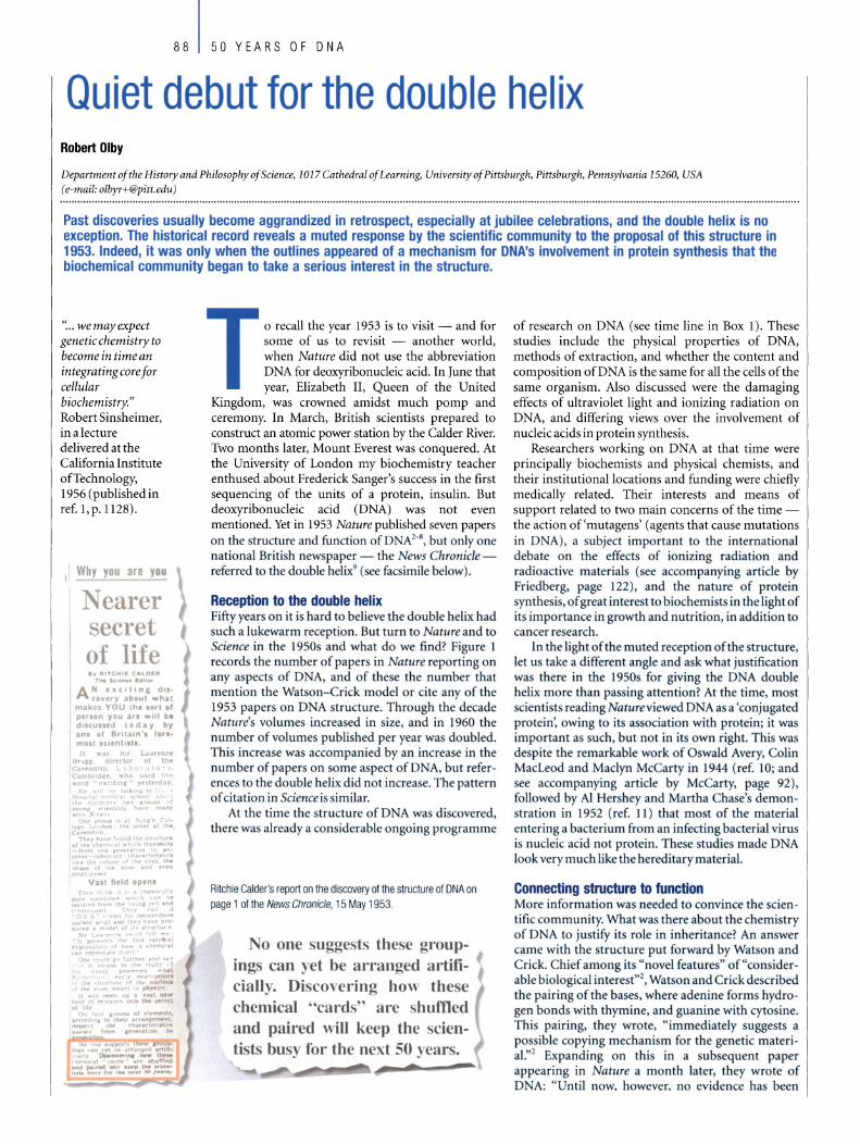
Why you are YIIU
Nearer secret of life ., IU'CtHI C4LO'"
T"" ko' ...... "I~
A N tee,tlnE d.lcover)' abO~1 what
mOikts YOU ,hl Ur' Ir per.on yau iIIrt WII I be dluun.d ta d iI)I' by on, 01 B"t.aln 'I '.re .. mOli lel_"llill.
o recall the year 1953 is to visit - and for so me of us to revisit - another world, when Nature did not use the abbreviation DNA far deoxyribonucleic acid. In June that year, Elizabeth II, Queen of the United
Kingdom, was crowned amidst much pomp and ceremony. In March, British scientists prepared to construct an atomic power station by the Calder River. Two months later, Mount Everest was conquered. At the University of London my biochemistry teacher enthused about Frederick Sanger's success in the first sequencing of the units of a protein, insulin. But deoxyribonucleic acid (DNA) was not even mentioned. Yet in 1953 Nature published seven papers on the structure and function of D Al-iJ, but only one national British newspaper - the News Chroniclereferred to the double helix9 ( ee facsimile below).
Reception to the double helix Fifty years on it is hard to believe the double helix had such a lukewarm reception. But turn to Nature and to Science in the 1950s and wh at do we find? Figure 1 records the number of papers in Nature reporting on any aspects of D A, and of these the number that mention the Watson-Crick model or eite any of the 1953 papers on D A strueture. Through the deeade Nature's volumes increased in size, and in 1960 the number ofvolumes published per year was doubled. This inerease was accompanied by an inerease in the number of papers on some aspeet of DNA, but references to the double helix did not inerease. The pattern ofeitation in Science issimilar.
At the time the strueture of D A was discovered, there was al ready a eonsiderable ongoing programme
Ailchie Galder's report on the discovery 01 lhe slruclure 01 DNA on page 1 of the News Chronicle. 15 May 1953.
o one sugge ts these gmupings can yet be arranged artificially. Disco\'ering how these chemical "cards' al-e shuffied and paired wiII keep the scientists busy tor the next 50 . ears.
of research on DNA (see time line in Box 1). These studies include the physical properties of DNA, methods of extraction, and whether the content and composition ofDNA is the same for a11 the ce11s of the same organism. Also discussed were the damaging effects of ultraviolet light and ionizing radiation on DNA, and differing views over the involvement of nucleic acids in protein synthesis.
Researchers working on DNA at that time were principally biochemists and physical chemists, and their institutionallocations and funding were chiefly medically related. Their interests and me ans of support related to two main concerns of the time -the action of'mutagens' (agents that cause mutations in DNA), a subjecr important to the international debate on the effects of ionizing radiation and radioactive materials (see accompanying article by Friedberg, page (22 ), and the nature of protein synthesis, ofgreat interest to biochemists in the lightof its importance in growth and nutrition, in addition to cancer research.
In the light ofthe muted reception ofthe structure, let us take a different angle and a k wh at justif1cation was there in the 1950s for giving the DNA double helix more than passing attention? At the time, most scientists reading Natureviewed D Aas a 'conjugated protein', owing to its association with protein; it was important a such, but not in its own right. This was despite the remarkable work of Oswald Avery, Colin MacLeod and Maclyn McCarty in 1944 (ref. 10; and see accompanying article by McCarty, page 92), foUowed by Al Hershey and Martha Chase's demon stration in 1952 (ref. 11 ) that most of the material entering a bacterium from an infecting bacterial virus is nucleic acid not protein. These studies made D A look very much like the hereditary material.
Connecting structure to function More information was needed to convinee the seien tific eommunity. What was there about the chemistry of DNA to justify its role in inheritance? An answer eame with the tructure put forward by Watson and Crick. Chief among it "novel features" of"considerable biologieaI interest"l, Watson and Criek de cribed the pairing of the bases, where adenine forms hydrogen bonds with thymine, and guanine with cytosine. Thi pairing, they wrote, " immediately suggests a possible copying mechani m for the genetie materi al.,,2 Expanding on this in a subsequent paper appearing in Nature a month later, they wrote of D A: "Until now, however, no evidence has been

presented to show how it might carry out the essential operation required of a genetic material, that of exact self-duplication."s
With these words Watson and Crick claimed their priority on a mechanism for DNA replication, but admitted there were problems with their scheme: how do the chains unwind and separate "without everything getting tangled"s? What is the exact mechanism by which gene duplication occurs? How does the genetic material "exert a highly specific influence on the cell"12 when the sequence of bases assumed to encode the specificity is on the inside of the helical molecule?
The 'unwinding problem' domina ted much of the early discussions that followed the discovery of the DNA structure. In 1953, Watson and Crick admitted it was "formidable"12, but support for their structure came in 1958, when Matthew Meselson and Franklin Stahl proved the semi-conservative nature of DNA replication 13: each ofthe two new daughter DNA molecules formed during DNA replication consists of one strand from the original parent molecule and a new strand synthesized from the parent strand, which served as a template. This confirmed Watson and Crick's theoretical prediction from the structure that replication would proceed in a semi-conservative manner. Later that same year, Arthur Kornberg announced the partial purification of an enzyme that catalyses DNA synthesis later called DNA polymerase l4• This first linked enzymology to the double helix, for not long thereafter Kornberg provided biochemical evidence that DNA polymerase synthesizes new strands from opposite directions ofthe two chains ofthe moleculeis.
In 1957, Crick defined biological 'information' as the sequence of the bases in the nucleic acids and of the amino acids in proteins, and proposed the now
50 YEARS OF DNA 89
famous 'central dogma' according to which information so defined flows between the nucleic acids and proteins only in one direction - from the former to the latter '6• Just four years later, Marshall Nirenberg and Heinrich Matthaei successfully synthesized a polypeptide constituted of only one kind of amino acid (phenylalanine ) using an RNA composed only of one kind ofbase (uracil). They concluded that "one or more [of these RNA bases 1 appear to be the code for phenylalanine."17 Meanwhile, Crick, Sydney Brenner and Leslie Barnett had been using genetic analysis to investigate mutagenesis. This led them to the important concept of a form of mutation in which there is a 'frame shift' in the sequence of the bases in DNA, from which they went on to infer that the genetic message is composed of single or multiple triplets ofbases, and that the message is read starting at a flXed point and proceeds always in the same
60
50
40 VJ
Ci> a. co 30 a. ci z
20
-
1950
DNA papers
Mentlon double helix
1952 1954 1956 Year
Six of the Nobel winners of 1 962 display their diplomas after formal ceremonies in Stockholm's Concer! Hall. From left to right: Maurice Wilkins (Medicine), Max Perulz (Chemistry), Francis Crick (Medicine), John Steinbeck (Literature), James Watson (Medicine) and John Kendrew (Chemistry).
Figure 1 Papers published in Nature referring to DNA and the extent of their reference to the double helix 1 950-1 960.
1958 1960

90 50 YEARS OF DNA
Box 1 Time IIne 0' the dlscovery 0' the structure 0' DNA
1869 Fritz Mieseher discovers that the nuclei of pus cells contain an acidic substance to which he gave the name 'nuclein' . Later he finds that nuclein is composed of a protein and a compound to which the name nucleic acid, and subsequently DNA, will be given.
1919 Phoebus Aaron Levene proposes the 'tetranucleotide ' structure of DNA, whereby the four bases of DNA were arranged one after another in a set of four.
1928 Frederick Griffith finds that a substance in heat-killed bacteria can cause heritable changes in the live bacteria alongside them. He calls the phenomenon 'transformation'.
1938 Rudoll Signer, Torbjorn Caspersson and Einer Hammarsten find molecular weights for DNA between 500,000 and 1,000,000 daltons. Levene's tetranucleotide must be a polytetranucleotide.
1944 Oswald Avery, Colin MacLeod and Maclyn McCarty establish the chemical identity 01 Griffith's transforming principle as DNA, and they suggest that it may lunction as the genetic material.
1949 Erwin Chargaff reports that DNA base composition varies from one species to another, yet the ratio between the quantities 01 the two purine bases, adenine and guanine, and that between the quantities 01 the two pyrimidine bases, thymine and cytosine, remains about the same, namely onetoone.
1949 Roger and Colette Vendrely, together with Andre Boivin find hall as much DNA in the nuclei of sex cells as they find in the body cells, thus paralleling the reduction in the number of chromosomes, making DNA look like the genetic material.
1951 Rosalind FrankIin distinguishes two forms 01 DNA, the paracryslalline B form and the crystalline A form.
1952 AI Hershey and Martha Chase find that DNA but scarcely any protein from an infecting bacterial virus enters the bacterial cell and can be recovered Irom the progeny virus particles.
1952 Rosalind Franklin and Raymond Gosling produce a magnificent X-ray diffraction pattern 01 the B form 01 DNA.
1953 James Watson and Francis Crick, RosaJind FrankIin and Raymond Gosling, Maurice Wilkins, W. E. Seeds, A1ec Stokes and Herbert Wilson, and Bertil Jacobson all publish on the structure 01 DNA2-8.
1954 George Gamow suggests a DNA code lor the synthesis of proteins. 1955 Seymour Benzer analyses the fine structure 01 the genetic material 01 a
bacterial virus at a level close to the distances that separate the individual bases along the DNA chain .
1957 Francis Crick proposes 'the sequence hypothesis' and 'the central dogma'. 1958 Matthew Meselson and Franklin Stahl demonstrate the semi-conservative
replication 01 DNA. 1959 Arthur Kornberg and colleagues isolate the enzyme DNA polymerase. 1961 Marshall Nirenberg and Johann Heinrich Matthaei show that a sequence 01
nucleotide can encode a particular amino acid, laying the foundations lor deciphering the genetic code.
1962 The Nobel prize in medicine is awarded to James Watson, Francis Crick and Maurice Wilkins.
direction l8• Thus was the stage set for the subsequent unravelling of the entire genetic code.
From a muted reception in 1953 to accelerating momentum towards the end of the decade, one is tempted to infer that the DNA double helix was not taken seriously until a mechanism for its involvement in protein synthesis began to take shape. There was, to be sure, a small band of scientists who from the start either built their careers upon the implications of the structure (such as Meselson and Alexander Rich) or redirected their research to follow it up (including Seymour Benzer and Sydney Brenner). However, many scientists, notably Erwin Chargaff and
Alexander Dounce, did not refer to the structure in their scientific papers in the mid-fifties, even though it was clearly relevant and presumably known to them. Such omissions suggest that some biochemists had their own agendas, and the double helix was not at first seen as an aid to their work.
Biochemists debate protein synthesis Biochemists' reservations about the double helix stemmed in part from the fact that evidential support for it in 1953 was far from strong. Watson and Crick themselves admitted that it "could in no sense be considered proved", although it was "most promising"19. In part the biochemists' coolness owed much to the debates among them over the mechanism of protein synthesis. The paper by Peter Camp bell and Thomas Work, published in Nature on 6 June 1953, portrayed this debate vividly. They identified two contrasting theories under discussion on how proteins are made: first, the peptide theory (also known as the multienzyme theory), where proteins are made by "stepwise coupling of many small peptide units"; and second, the template theory, involving "synthesis on templates, each template being specific for a single protein structure and probably identifiable as a
"20 gene. The peptide model was, for a very long time,
supported by many prominent biochemists, including Joseph Fruton. The conviction behind it was the power of enzymes to both synthesize and break down their substrates, with a high degree of specificity attributed to both actions. Synthesis was proposed to involve the formation of a succession ofpeptides, ultimatelyyielding the protein molecule, and enzymes synthesize only those peptide bonds that they also hydrolyse. But the problem with this theory was that, except for a very few special cases, the alleged peptides constituting the intermediaries in protein synthesis could neither be detected in the cell nor incorporated into the protein being synthesized. Amino acids, however, could be incorporated, indicating they were the building blocks of proteins.
The second model of protein synthesis, which assumed synthesis on a template, had been advocated by Dounce in 1952. He pictured polypeptide chains being laid down on RNA molecules, and the RNA sequence determining the sequence of amino acids incorporated (on a one-to-one basis). Thus, DNA in the nucleus would control the order ofbases in the RNA21 •
After weighing up the merits and difficulties of Dounce's scheme, Campbell and Work voiced their distaste for the genetic control of protein synthesis, remarking in 1953 that: " ... the gene is essentially an abstract idea and it may be amistake to try to clothe this idea in a coat of nucleic acid or protein ... ifwe must have a gene it should have a negative rather than a positive function so far as protein synthesis is concerned:'20 Only three years later, however, Robert Sinsheimer concluded a lecture at the California Institute of Technology with the following words: "The gene, once a formal abstraction, has begun to condense, to assurne form and structure and defined activity:' I
But those three years were a scene of pronounced change. By January 1957, when Fruton revised the second edition of his widely used textbook General Biochemistry, his remarks on the peptide theory were

cautious and were followed by a discussion of the role ofRNA on which, he noted, there have been "stimulating speculations about the role of nucleic acids as 'templates' in protein synthesis:m Earlier in the book he devoted a paragraph to the double helix, describing it as an 'ingenious speculation'. The only diagram was of the base pair adenine-thymine, rather than the helical model of the structure.
Kornberg had shown in 1957 that DNA replication follows the rules of base pairing, whereby DNA polymerase adds a base to the newly synthesized strand that is complementary to the opposing base in the template strand (A is always opposite T, and C always opposite G). But his interest in the subject had not been stimulated by Watson and Crick's discovery. Rather, in 1953 he was preoccupied with how coenzymes (non-protein compounds needed for enzyme activity) are synthesized from nucleotides. He was led to wonder how DNA and RNA might be made from thousands of nucleotides. "The significance of the double helix:' he recalled, "did not intrude" into his work unti11956, after he had shown that a "moderately purified fraction" of what he was later to call DNA polymerase "appeared to increase the size of a DNAchain:m,24
Conclusion The two once enigmatic processes - DNA replication and pro tein synthesis - intersected ongoing research programmes in the physical, organic and biological chemistry of the early 1950s. After the discovery of the double helix, those grappling with the problem of replication found its molecular foundation in the structure of DNA, although it took more than two decades to deduce the intricate mechanism of its operation in the cell (see accompanying article by Alberts, page 117). Those working on pro tein synthesis found the source of its specificity lay in the base sequence ofDNA.
But why celebrate this one discovery? Why not celebrate the golden jubilee ofMax Perutz's solution to the 'phase problem' for pro teins in 1953, without which the subsequent discovery of the structure of myoglobin and haemoglobin would not have been possible? What about the year 2005 for celebrating the golden jubilee of Sanger's determination of the complete amino-acid sequence of a protein? Undoubtedly, the double helix has remarkable iconic value that has contributed significantly to its public visibility, something that has not been achieved by any of the pro tein structures (see accompanying article by Kemp, page 102). There is, too, a degree of notoriety attaching to the manner of its discovery and the characters involved that has given spice to the story, as widely publicized by James Watson's account of the discovery in The Double Helix, published in 1968 (ref. 25), and Brenda Maddox's recent illuminating biography ofRosalind Franklin26•
But there is a centrality about DNA that relates to the centrality ofheredity in general biology.
50 YEARS OF DNA 91
The silver and golden jubilees ofthe Queen's accession to the throne have come and gone, nuclear power stations are no longer being built in the United Kingdom, and mountaineer after mountaineer has ascended Mount Everest without a fanfare of press reports. But DNA is very much in the news - whether it be as a tool for studying evolution, a forensic test for rape, a source of gelletic information or a path to designer drugs. And what better emblem or mascot is there for molecular biology than the double helix, and its spartan yet elegant representation in the original paper2 from the pen of Odile Crick, Francis's wife, fifty yearsago? 0 doi: lO.1038/nature01397
1. Sinsheimer, R. L. First steps toward a genetic chemistry. Science 125,
1123-1128 (1957).
2. Watson, J. D. &Crick, F. H. C. A structure for deoxyribose nucleicacid. Nature
171,737-738 (1953).
3. Wilkins, M. H. E, Stokes, A. R. & Wilson, H. R. Molecular structure of
deoxypentose nucleic acids. Nature 171, 738-740 (1953).
4. Franklin, R. E. & Gosling, R. G. Molecular configuration in sodium
thymonucleate. Nature 171, 740-741 (1953).
5. Watson, J. D. & Crick, F. H. C. Genetical implications ofthe structure of
deoxyribonucleic acid. Nature 17l, 964-967 (1953).
6. Franklin, R. E. & Gosling, R. G. Evidence for 2-chain helix in crystalline
structure of sodium deoxyribonucleate. Nature 172, 156-157 (1953).
7. Jacobson, B. Hydration structure of deoxyribonucleic acid and its physico
chemical properties. Nature 172, 666-667 (1953).
8. Wilkins, M. H. E, Seeds, W. E., Stokes, A. R. & Wilson, H. R. Helical structure of
crystalline deoxypentose nucleic acid. Nature 172,759-762 (1953).
9. Calder, R. Whyyou areyou: nearerthe secret oflife. News Chraniclep. 1 (15
May 1953).
10. Avery, O. T. MacLeod, C. M. & McCarty, M. Studies ofthe chemical nature of
the substance inclucing transformation of pneumococcal types. Induction of
transformation by a desoxyribonudeic acid fraction isolated from
Pneumococcus Type 111. f. Exp. Med. 79,137-158 (1944).
11. Hershey, A. D. & Chase, M. Independent functions of viral proteins and nudeic
acid in growth ofbacteriophage. J. Gen. Physiol. 36, 39-56 (1952).
12. Watson, j. D. & Crick, F. H. C. The structure of DNA. Cold Spring Harb. Symp. Quant. Bio1.18, 123-131 (1953).
13. Meselson, M. & Stahl, E W. The replication ofDNA in Escherichia coli. Prac.
NatlAcad. Sei. USA 44, 671-682 (1958).
14. Lehman, L R., Bessmanm, M. J., Simms, E. S. & Kornberg, A. Enzymatic synthesis
of deoxyribonudeic acid. I. Preparation of substrates and partial purification of
an enzymefrom Escherichia coli.]. Bio!. Chem. 233,163-170 (1958).
15. Kornberg, A. Biological synthesis of deoxyribonudeic acid: an isolated enzyme
catalyzes synthesis ofthis nudeic acid in response to directions from pre
existing DNA. Seience 131, 1503-1508 (1960).
16.Crick, F. H. C. On protein synthesis. Symp. Soc. Exp. Bio1.12, 138-163 (1958).
17. Nirenberg, M. W. & Matthaei, J. H. Thedependence of ceIl-free protein
synthesis in E. CoIi upon naturally occurring or synthetic polynudeotides. Prac. NatlAcad. Sei. USA 47, 1558-1602 (1961).
18. Crick, F. H. c., Barnett, L., Brenner, S. & Watts-Tobin, R. J. General nature of
the genetic code for proteins. Nature 192, 1227-1232 (1961).
19. Crick, F. H. C. & Watson, J. D. The complementary structure of
deoxyribonucleic acid. Prac. R. SOC. Land. A 223. 80-96 (1954).
20. Campbell, P. N. & Work, T. S. Biosynthesis of proteins. Nature 171, 997-1001 (1953).
21.Dounce, A. Duplicating mechanisms far peptide chain and nucleic acid
synthesis. Enzymologia 15, 251-258 (1952).
22. Fruton, J. S. General Biochemistry 2nd edn (Wiley, New York 1958).
23. Kornberg, A. For the Love ofEnzymes. The Odyssey of a Biochemist (Harvard
Univ. Press, Cambridge, MA, 1989).
24. Kornberg, A. in A Symposium on the Chemical Basis ofHeredity (eds McElroy,
W. D. & Glass, 8.) 605 (Johns Hopkins Press, Baltimore, 1957).
25. Watson, J. D. The Double Helix: A Personal Account of the Discovery of the
Structure ofDNA (Atheneum, New York, 1968). [Narton Critical Edition (ed.
Stent, G. 5.) published byNorton, NewYark & London, 1980.]
26. Maddox, 8. Rosalind Franklin. The Dark Lady ofDNA (Harper Collins, London
2002).
Original reference: Nature421, 402-405 (2003).

92 50 YEARS OF DNA
Discovering genes are made of DNA Maclyn McCarty
The RockefeIler University, 1230 YorkAvenue, New York 10021, USA (e-mail: [email protected])
Maclyn McCarty is the sole surviving member of the team that made the remarkable discovery that DNA is the material of inheritance. This preceded by a decade the discovery of the structure of DNA itself. Here he shares his personal perspective of those times and the impact of the double helix.
Editor's note - For a long time, biologists thought that 'genes: the units of inheritance, were made up of pratein. In 1944, in what was arguably the defining moment for nucleic acid research, Oswald Avery, Mac/yn McCarty and Colin MacLeod, at RockefeIler Institute (now University) Hospital, New York, proved that DNA was the material ofinheritance, the so-called stuffoflife. They showed thatthe heritable property ofvirulence fram one infectious strain of pneumococcus (the bacterial agent of pneumonia) could be transferred to a noninfectious bacterium with pure DNA1• They further supported their conc/usions by showing that this 'transforming' activity could be destroyed by the DNA-digesting enzyme DNAase2.3.
This work first linked genetic information with DNA and pravided the historical platform of modern genetics. Their discovery was greeted initially with scepticism, however, in part because many scientists believed that DNA was too simple a molecule to be the genetic material. And the fact that McCarty, Avery and MacLeod were not awarded the Nobel prize is an oversight that, to this day, still puzzles.
"The pivotal discovery of 20th-century biology." Joshua Lederberg, RockefeIler University, 1994, referring to the discovery by McCarty, Avery and MacLeod.
Maclyn McCarty at The Rockefeiler University.
At the time of our discovery and publication in 1944 (ref. 1) of the research showing that DNA is heritable, my personal view, which I shared with MacLeod, was that there was little doubt that genes are made
of DNA, and that this would ultimately be accepted. I was not sure of the best approach to use in pursuing research on the subject, but suspected that darification ofthe structure ofDNA was necessary.
But this was not an area of research in which I had received any training. AdditionaHy, I had planned to make my career in disease-oriented research, and knowledge of the gene did not seem likely to become applicable in this area for so me years. Thus, when invited to lead my own laboratory in the RockefeHer Hospital, investigating streptococcal infection and the pathogenesis of rheumatic fever, I decided to leave Avery's laboratoryfor this newposition in July 1946.
Rollin Hotchkiss joined Avery at this point, and together with Harriett Taylor (a recent PhD graduate in genetics who had joined the laboratory in 1945), carried out studies increasing the purity of the transforming DNA mixture by further reducing any contaminating traces ofprotein. Togetherwith other investigators, they also showed that properties of the pneumococcus other than just specific polysaccharide components of its ceH wall could be transferred by the DNA preparations,
CiJ"'~I!I!::iiiii_il~ indicating that the purified DNA also con-III!' tained other genes of the bacterium.
Our findings continued to receive little acceptance for a varietyofreasons, the most significant being that the work on the composition of DNA, dating back to its first identification 75 years earlier, had conduded that DNA was too limited in diversity to carry genetic information. Even those biologists who had considered the possibility had dropped the idea, and the prevailing dogma was that if genes are composed of a known substance, it must be protein.
There were a few biologists who took a differentview, the mostnotable being Erwin Chargaff, who changed his area of research to DNA after reading our 1944 paper'. His work revealed the great diversity in DNA isolated from various sources, and that
despite this diversity the amount of adenine always equalled that of thymine, and the amount of guanine that of cytosine. The latter findingwas an important factor in the next significant advance in the field - the Watson-Crick determination of the double helical structure ofDNA.
After the change in my research activity, I continued to give talks on our work on pneumococcal transformation and found the acceptance of the probable genetic role ofDNA still to be minimal. However, I was convinced that it was only a matter of time before our results would become established.
Even though I was no longer involved in research on the subject, I continued to foHow the developments as they appeared in the literature. Thus, when the papers of Watson and Crick describing the double helical structure of DNA were published in Nature in 1953, I certainly grasped the significance of their findings and was pleased to see such iHuminating results come from a structural approach. I was not so pleased, however, that they failed to cite our work as one reason for pursuing the structure ofDNA.
The concept of the double helix also hastened the silencing of those who had dung to the idea of genes as proteins. As a progressively larger body of investigators joined the study ofthe genetic role ofDNA, there was an expanding amount of new information, starting with the resolution of the genetic code. By the end of the twentieth century, subsequentwork on the mechanisms bywhich DNA is replicated with each ceH division, is reshuffled with each generation, and is repaired when mistakes arise - the importance of which can in each case be traced back to the finding that D NA is the hereditary material-has transformed research in all areas of biology, technology and medicine. 0 doi: IO.1038/nature01398
1. Avery, O. T., MacLeod, C. M. & McCarty, M. Studies ofthe chemical nature of
the substance inducing transformation of pneumococcal types. Induction of
transformation by a desoxyribonucleie acid fraction isolated from
Pneumococcus Type 111.]. Exp. Med. 79,137-158 (1944).
2. McCarty, M. Purification and properties of desoxyribonuclease isolated from
beefpancreas.]. Gen. Physiol. 29,123-139 (1946).
3. McCarty, M. & Avery, O. T. Studies of the chemical nature of the substance
inducing transformation of pneumococcal types Il. Effect of
desoxyribonuclease on the biological activity ofthe transforming substance.
J. Exp. Med. 83, 89-96 (1946).
Originalreference: Nature421, 406 (2003).

50 YEARS OF DNA 93
The double helix and the 'wronged heroine' Brenda Maddox
9 Pitt Street, London WS 4NX, UK (e-mail: [email protected])
In 1962, James Watson, Francis Crick and Maurice Wilkins received lhe Nobel prize tor lhe discovery ot the structure of DNA. Notably absent from the pOdium was Rosalind Franklin, whose X-ray photographs of DNA contributed directJy to the discovery ot lhe double helix. Franklin's premature death, combined with misogynist treatment by the male scientific establishment, cast her as a feminist icon. This myth overshadowed her intellectual strength and independence both as a scientist and as an individual.
In late February 1953, Rosalind Franklin, a 33-year-old physical chemist working in the biophysics unit of King's College in London, wrote in her notebooks that the structure of DNA had two chains. She had already worked
out that the molecule had its phosphate groups on the outside and that DNA existed in two forms.
Two weeks later James Watson and Francis Crick, at the Cavendish Laboratory at Cambridge, built their now celebrated model ofDNA as a double helix. They did it not only through brilliant intuition and a meeting of compatible minds, but also on the basis of Franklin's unpublished experimental evidence, which had reached them through irregular routes. She did not know that they had seen either her X-ray photograph (Fig. 1), showing unmistakable evidence of a helical structure, or her precise measurements of the unit cell (the smallest repeating unit), and the crystalline symmetry, of the DNA fibres.
As Watson was to write candidly, "Rosy, of course, did notdirectlygive us herdata. For that matter, no one at King's realized they were in our hands." When this admission appeared in Watson's best-selling, much-acclaimed book of the discovery, The Double Helix, published in 1968 (ref. 1), he was a Harvard professor and Nobel laureate (he had shared the prize for medicine and physiology in 1962, with Crick and Maurice Wilkins of King's College.) By then Franklin had died - in 1958, at the age of 37, from ovarian cancer.
Other comments dismissive of"Rosy" in Watson's book caught the attention of the emerging women's movement in the late 1960s. "Clearly Rosy had to go or be put in her place [ ... ] Unfortunately Maurice could not see any decent way to give Rosy the boot': And, "Certainly a bad way to go out into the foulness of a [ . . . ] November night was to be told by a woman to refrain from venturing an opinion about a subject for which you were not trained."
A feminist icon Such flamboyantly chauvinist phrases were sufficient to launch the legend ofFranklin, the wronged heroine. So too was Watson's insistence on judging Franklin by her appearance rather than by her performance as a scientist. (She was, when she came to King's from the French government laboratory where she had worked from 1947 to the end of 1950, a recognized expert on
"Seien ce and everyday life eannot and should not be separated. Seien ce, for me, gives a partial explanation oflife. In so far as it go es, it is based on fact, experience and experiment." Rosalind Franklin, in a letter to her father, summer 1940.
the structure of coals, carbons and disordered crystals, with manypublications to her credit.)
The Franklin myth has continued to grow, abetted by the fact of her tragically early death. Franklin has become a feminist icon - the Sylvia Plath of molecular biology - seen as a genius whose gifts were sacrificed to the greater glory ofthe male. Her failure to win the Nobel prize has been given as a prime example

94 50 YEARS OF DNA
Figure 1 "Her photographs are among the most beautiful X-ray photographs of any substance every taken." - J. D. Bernal, 1958. Franklin's X-ray diagram of the B form of sodium thymonucleate (DNA) fibres, published in Nature on 25 April 1953, shows "in striking mann er the features characteristic of helical structures"5.
of the entrenched misogyny of the science establishment, rather than the consequence of the Nobel statute against posthumous awards.
Watson's caricature of the bad-tempered "Rosy" drew a counter-blast from her good friend, the American writer Anne Sayre, in Rosalind Franklin and DNA, published in 1975 (ref. 2). Sayre's book provided a much-needed corrective portrait, but was marred by a feminist bias. For example, it grossly underestimated the number of women scientists at King's in the early 1950s. Sayre maintained there was only one other than Franklin, whereas there were at least eight on the senior staff. She insisted, moreover, that women's exdusion from the King's senior common room deprived Franklin of the intellectual companionship of her colleagues. In fact, most of the scientific staff preferred to eat in the joint dining room, men and women together, and the women, in general, felt well treated at King's.
Reassessing the facts As a biographer writing nearly three decades later and given access to Franklin's personal correspondence, I found a more attractive, capable woman than Watson had suggested, and a King's College more congenial and welcoming to women scientists than Sayre had allowed. I also found that Franklin felt singularly unhappy at King's, not so much because ofher gender, but because ofher dass and religion: a wealthy Anglo-Jew felt out of place in a Church of England setting dominated by swirling cassocks and students studying for the priesthood. ''At King's:' she wrote to Sayre (albeit inaccurately), "there are neither Jews nor foreigners".
She was, in fact, so unhappy at King's that, in early 1953, getting out as fast as possible was far more important to her than finishing her work on DNA. How far she had advanced was reported in two articles in Naturc" by Sir Aaron Klug, Franklin's dosest collaborator at Birkbeck College, London, where she moved to from King's. He conduded that she had come very dose to discovering the structure ofDNAherself.
An irony of the story is that her own manuscript (coauthored by her student, R. G. Gosling and dated 17 March 1953) summarizing her results was already prepared by the time news reached King's that Watson and Crick had cracked the DNA secret. Thus she inserted a hand-written amendment to her manuscript - which was published in Nature on 25 April 1953 (ref. 5), along with the now-celebrated Watson and Crick paper and another by Wilkins, Herbert Wilson and Alec Stokes ofKing's - to say "Thus our general ideas are not inconsistent with the model proposed by Watson and Crick in the preceding communication". And so they should have been, for the Watson -Crick findings were based on her data.
There is no evidence that she knew that in late January 1953 Wilkins had innocently shown her Photograph 51, with its stark cross ofblack retlections (Fig. 1), to Watson, who was visiting King's. Nor did she know that in February 1953 MaxPerutz, then atthe Cavendish
Belated credit
Laboratory, had let Watson and Crick see his copy of the Medical Research Council's report summarizing the work of an principal researchers, induding Franklin's.
At the same time there is no evidence that Franklin felt bitter about their achievement or had any sense ofhaving been outrun in a race that nobody but Watson and Crick knew was arace. Indeed, shecouldacceptthe Watson-Crick model as a hypo thesis only. She wrote in Acta Crystallographica in September 1953 that "discrepancies prevent us from accepting it in detail"6.
Watson and Crick seem never to have told Franklin directly what they subsequently have said from public platforms long after her death - that they could not have discovered the double helix of DNA in the early months of 1953 without her work. This is all the more surprising in view ofthe dose friendship that developed among the three of them - Watson, Crick and Franklin - during the remaining years of her life. During this time, she was far happier at non -sectarian Birkbeck than she ever was at King's, and led a spirited team of researchers studying tobacco mosaic virus (TMV).
From 1954 until months before her death in April 1958, she, Watson and Crick corresponded, exchanged comments on each other's work on TMV, and had much friendly contact. At Wood's Hole, Massachusetts, in the summer of 1954 Watson offered Franklin a lift across the United States as he was drivingto her destination, the California Institute ofTechnology. In the spring of 1956 she toured in Spain with Crick and his wife Odile and subsequently stayed with them in Cambridge when recuperating from her treatments for ovarian cancer. Characteristically, she was reticent about the nature ofher illness. Cricktold a friend who asked that he thought it was "something female".
In the years after leaving King's, Franklin published 17 papers, mainly on the structure ofTMV (induding four in Nature). She died proud ofherworld reputation in the research of coals, carbons and viruses. Given her determination to avoid fanciful speculation, she would never have imagined that she would be remembered as the unsung heroine ofDNA. Nor could she have envisaged that King's College London, where she spent the unhappiest two years ofher professional career, would dedicate a building - the Franklin-Wilkins building - in honour ofher and the colleague with whom she had been barelyon speaking terms. 0 doi: 1 O.1038/natureO 1399
1. Watson, J. The Double Helix: A Personal Account of the Discovery of the Structure
ofDNA (Atheneum, NewYork, 1968).
2. Sayre, A. Rosalind Franklin and DNA (W. W. Norton & Co., New York, 1975).
3. Klug, A. Rosalind Franklin and the discovery of the structure ofDNA. Nature
219,808-810,843-844 (1968).
4. Klug, A. Rosalind Franklin and the double helix. Nature 248) 787-788 (1974).
5. FrankIin, R. E. & Gosling, R. G. Molecular configuration in sodium
thymonuc1eate. Nature 171, 740-741 (1953).
6. Franklin, R. E. & Gosling, R. G. The structure of sodium thymonucleate fibres:
I. The influence of water content. H. The cylindrically symmetrical Patterson
function. Acta Crystallogr. 6, 673-{'77, 678-685 (1953).
Originalreference: Nature421, 407-408 (2003).

50 YEARS OF DNA 95
The mosaic that is our genome Svante Pääbo
Max Planck Institute for Evolutionary Anthropology, Deutscher Platz 6, D-04103 Leipzig, Germany (e-mail: [email protected] )
The discovery of the basis of genetic variation has opened inroads to understanding our history as a species. It has revealed the remarkable genetic similarity we share with other individuals as weil as with our closest primate relatives. To understand what make us unique, both as individuals and as a species, we need to consider the genome as a mosaic of discrete segments, each with its own unique history and relatedness to different contemporary and ancestral individuals.
he discovery of the structure of DNA I, and the realization that the chemical basis of mutations is changes in the
nudeotide sequence of the DNA, meant that the history of a piece of DNA could be traced by studying variation in its nudeotide sequence found in different individuals and in different species. But it was not until rapid and inexpensive methods became available for probing DNA sequence varIation in many individuals that the efficient study of molecular evolution in general - and of human evolution in particular - became feasible. Thus, the development in the 1980s of techniques for efficiently scoring polymorphisms with restriction enzymes and amplifying DNA 2,3 enabled the study of molecular evolution to become a truly booming enterprise.
What follows is a personal and, by necessity, selective attempt to consider what the accelerating pace of exploration of human genetic variation over the past two decades has taught us about ourselves as a species, as weil as so me suggestions for what may be fruitful areas for future studies.
Primate relations The first insight of fundamental importance for our understanding of our origins came from comparisons of DNA sequences between humans and the great apes. These analyses showed that the African apes, especially the chimpanzees and the bonobos, but also the gorillas, are more dosely related to humans than are the orangutans in Asia4•
Thus, from a genetic standpoint, humans are essentially African apes (Fig. 1). Although there had been hints of this from molecular comparisons of proteinsS,6, it was a marked shift from the earlier common belief that humans represented their own branch separate from the great apes.
Our sense of uniqueness as a species was further rocked by the revelation that human DNA sequences differ by, on average, only 1.2 per cent from those ofthe chimpanzees?, as a consequence ofhumans and apes sharing a recent common ancestry. It should be noted
that the dating of molecular divergences has uncertamtles of unknown magnitude attached, not least because of calibration based on palaeontological data. Nevertheless, it seems dear that the human evolutionary lineage diverged from that of chimpanzees about 4-6 million years ago, from that of gorillas about 6-8 million years ago, and from that of the orangutans about 12-16 million years ago? Before the advent of molecular data, the human-chimpanzee divergence was widely believed to be about 30 million years old.
In fact, we have recently come to realize that the relationship between humans and the African apes is so dose as to be entangled. Although the majority of regions in our genome are most dosely related to chimpanzees and bonobos, a non-trivial fraction is more doselyrelated to gorillas? In yet other regions, the apes are more dosely related to each other than to us (Fig. 2). This is because the speciation events that separated these lineages occurred so dosely in time that genetic variation in the first ancestral species, from which the gorilla lineage diverged, survived
Figure 1 Tree showing the divergence 01 human and ape species. Approximate dates 01 divergences are given lor, lrom left to right,orangutan, gorilla, human, bonobo and chimpanzee.
until the second speciation event between the human and chimpanzee lineages8• Thus, there is not one history with which we can describe the relationship of our genome to the genomes of the African apes, but instead different histories for different segments of our genome. In this respect, our genome is a mosaic, where each segment has its own relationship to that ofthe African apes.
Modern humans The mosaic nature of our genome is even more striking when we consider differences in DNA sequence between currently living humans. Our genome sequences are about 99.9 per cent identical to each other. The variation found along a chromosome is structured in 'blocks' where the nudeotide substitutions are associated in so-called haplotypes (Figs 2b and 3). These 'haplotype blocks' are likely to result from the fact that recombination, that is, the re-shuffling of chromosome segments that occurs during formation of sex cells (meiosis), tends to occur in certain areas of the chromosomes more often than in others9- 11 • In addition, the chance occurrence of recombination events at certain spots and not at others in the genealogy of human chromosomes will influence the structure of these blocks. Thus, any single human chromosome is a mosaic of different haplotype blocks, where each block has its own pattern of variation. Although the delineation of such blocks depends on the methods used to define them, they are typically 5,000-200,000 base pairs in length, and as few as fourto five common haplotypes account for most of the variation in each block (Fig. 3).
Of 928 such haplotype blocks recently studied in humans from Africa, Asia and Europel2, 51 per cent were found on all three

96 50 YEARS OF DNA
continents, 72 per cent in two continents and only 28 per cent on one continent. Of those haplotypes that were on one continent only, 90 per cent were found in Africa, and African DNA sequences differ on average more among themselves than they differ from Asian or European DNA sequences!3. Therefore, within the human gene pool, most variation is found in Africa and what is seen outside Africa is a subset ofthevariation found withinAfrica.
Two parts of the human genome can be regarded as haplotype blocks where the history is particularly straightforward to reconstruct, as no recombination occurs at all. The first of these is the genome of the mitochondrion (the cellular organelle that produces energy and has its own genetic material), which is passed on to the next generation from the mother's side; the second is the Y chromosome, which is passed on from the father's side. Variation in DNA sequences from both the mitochondrial genome14-16 and the Y chromosome17, as weil as many sections of the nuclear genome13,18-20, have their geographical origin in Africa. Because other evidence suggest that humans expanded so me 50,000 to 200,000 years ago21 from a population of about 10,000 individuals, this suggests that we expanded from a rather small African population. Thus, from a genomic perspective, we are all Africans, either living in Africa or in quite recent exile outside Africa.
Ancient humans What happened to the other hominids that existed in the Old World from about 2 million years ago until about 30,000 years ago? For instance, the Neanderthals are abundant in the fossil record and persisted in western Europe until less than 30,000 years ago.
Analysis ofNeanderthal mitochondrial DNA has shown that, at least with respect to the mitochondrial genome, there is no evidence that Neanderthals contributed to the gene pool of current humans22-2S. It is possible, however, that some as yet undetected interbreeding took place between modern humans and archaic hominids, such as Homo erectus in Asia or Neanderthals in Europe22,26,27.
But any interbreeding would not have significantly changed our genome, as we know that the variation found in many haplotype blocks in the nuclear genome of contemporary humans is older than the divergence between Neanderthals and humans. Thus, the divergence of modern humans and Neanderthals was so recent that Neanderthal nuclear DNA sequences were probably more closely related to so me current human DNA sequences than to other Neanderthals. In other words, the overlapping genetic variation that is likely to have existed between different ancient hominid forms makes it difficult to resolve the extent to which any interbreeding occurred.
Nevertheless, the limited vanatlon among humans outside Africa, as weil palaeontological evidence28 , suggest that any contribution cannot have been particularly extensive. Thus, it seems most likely that modern humans replaced archaic humans without extensive interbreeding and that the past 30,000 years of human history are unique in that we lack the company of the closely related yet distinct hominids with which we used to share the planet.
Human variation and 'race' Comparisons of the within-species variation among humans and among the great apes have shown that humans have less genetic
Figure 2 Within- and between-species variation along a single chromosome. a
HU~ a, The interspecies relationships of five chromosome regions to corresponding DNA sequences in a chimpanzee and a gorilla. Most regions show humans to be most closely related to chimpanzees (red) whereas a few regions show other relationships (green and blue). b, The among-human relationships of the same regions are illustrated schematically for five individual chromosomes. Most DNA variants are found in people from all three continents, namely Africa (A~, Asia (As) and Europe (Eu). But a few variants are found on only one continent, most of which are in Africa. Note that each human chromosome is a mosaic of different relationships. For example, a chromosome carried bya person of European descent may be most closely related to a chromosome from Asia in one of its regions, to a chromosome from Africa in another region, and to a chromosome from Europe in a third region. For one region (red), the extent of sequence variation within humans is low relative to what is observed between species. The relationship of this sequence among humans is illustrated as star-shaped owing to a high frequency of nucleotide variations that are unique to single chromosomes. Such regions may contain genes that contribute to traits that set humans apart from the apes.
Ch~ Go
HU~
Ch~ Go
HU>-Go
Ch
HU~
Ch /
Go
HU~
Go y--Ch
variation than the great apes29,30. Furthermore, earlydata thatonlyabout 10 per cent of the genetic variation in humans exist between so-called 'races'31 is borne out by DNA sequences which showthat races are not characterized by fixed genetic differences. Rather, for any given haplotype block in the genome, a person from, for example, Europe is often more closely related to a person from Africa or from Asia than to another person from Europe that shares his or her complexion (forexample, see ref. 32; Fig. 2).
Claims about fixed genetic differences between races (see ref. 33 for example) have proved to be due to insufficient sampling34•
Furthermore, because the main pattern of genetic variation across the globe is one of gene-frequency gradients35, the contention that significant differences between races can be seen in frequencies of various genetic markers36 is very likely due to sampling of populations separated by vast geographical distances. In this context it is worth noting that the colonization history of the United States has resulted in a sampling of the human population made up largely of people from western Europe, western Africa and southeast Asia. Thus, the fact that 'racial groups' in the United States differ in gene frequencies cannot be taken as evidence that such differences represent any true subdivision of the human gene pool on a worldwide scale.
Rather than thinking about 'populations', 'ethnicities' or 'races', a more constructive way to think about human genetic variation is to consider the genome of any particular individual as a mosaic ofhaplotype blocks. A rough calculation (Fig. 3) reveals that each individual carries in the order of 30 per cent of the entire haplotype variation of the human gene pool. Although not all of our
b
----« -~ C - T - ~:As:~ - C - T - AIAsEu - C - AIAsEu
:--C-------<1-;;::---.- - A -
AI As Eu AI As Eu AI As Eu ~
-G
----T - AIAsEu - C - AIAs
~ C _ AIAsEu
- TC - As - TC - AIAsEu _ A _ AIAs - A AI
G T AI Eu ---< .----G T = AIAsEu ---G- AI Eu - A - AfAsEu - A - AI ----< - C - AIAsEu - C - AI ----T - AIAsEu ---A T - AIAsEu - AT - AfAs

50 YEARS OF DNA 97
a
~~== Halpotype block
b Individual 1 _ ....... """"'" -----= --------
- ---===== Individual 2 -------------- - ---- --------
Figure 3 The mosaic structure 01 human genetic variation, a, Each human chromosome is made up 01 regions, called 'haplotype blocks', wh ich are stretches 01 DNA sequence where three to seven variants (at Irequencies above 5 per cent in the human population) account lor most 01 the variation lound among humans, Each such haplotype lound in a block is illustrated here as a bar 01 different colour, The catalogue 01 haplotypes lor every block makes up the 'haplotype map' 01 the human genome, b, The chromosomes 01 !wo hypothetical individuals are shown, Each individual carries!wo copies 01 each block (as humans carry!wo sets 01 chromosomes), As the chance that the!wo haplotypes carried at a block are identical is about 20 per cent, each 01 us carries an average 01 about 1 ,8 different haplotypes per block, Since there is on average 5,5 haplotypes lor every block, each individual carries about 30 per cent 01 the total haplotype diversity 01 the entire human species, Haplotype blocks tend to be shorter in Alrica than elsewhere; as a result, Alrican variation will probably have to be used to deline the species-wide block lengths, wh ich may be an average 01 around 10,000 base pairs, Note that not all 01 the human genome may have a clearly definable haplotype-block structure,
genome may show a typical haplotype-block structure and more research is needed to fully understand the haplotype landscape of our genome, this perspective clearly indicates that each of us contain a vast proportion of the genetic variation found in our species. In the future, we therefore need to focus on individuals rather than populations when exploring genetic variation in our species.
Tracking human traits What are the frontiers ahead of us in human evolutionary studies? One of them, to my mind, is to identify gene variants that have been selected and fIxed in all humans during the past few hundred thousand years. These will include genes involved in phenotypic traits that set humans apart from the apes and at least so me archaic human forms (far example, genes involved in complex cognitive abi!ities, language and longevity). However, an important obstacle in this respect is that there is !ittle detailed knowledge ofmanyofthe relevanttraits in the great apes. For example, only recently has the extent to which apes possess the capability for language37 and culture38 begun to be comprehensively described. As a consequence, we have come to realize that almost all features that set humans apart from apes mayturn out to be differences in grade rather than absolute differences.
Many such differences are likely to be quantitative traits rather than single-gene traits. To have a chance to unravel the genetic basis of such traits, we will need to rigorously defIne the differences between apes and humans - for instance, how we learn, how we communicate and howwe age. In the next
few years, geneticists will therefore need to consider insights from primatology and psychology, and more studies will be required that directly compare humans to apes.
There are, however, ways in which we can contribute towards the future unravelling of functionally impartant genetic differences between humans and apes. Far example, we can identify regions of the human genome where the patterns of variation suggest the recent occurrence of a mutation that was positively selected and swept through the entire human population. The sequencing of the chimpanzee genome, as weil as the haplotype-map project, will greatly help in this. Further prerequisites include the capability to determine the DNA sequence of many human genomes and the development of tools and methods to analyse the resulting data; in particular, a mare realistic model of human demographic history is required.
Collectively these studies will allow us to identify regions in the human genome that have recently been acted upon by selection and thus are likely to contain genes contributing to human-specifIc traits (Fig. 2). Other interesting candidate genes for human-specifIc traits are genes duplicated ar deleted in humans39, genes that have changed their expression in humans40, and genes responsible for disarders affecting traits unique to humans, such as language41
and a large brain size42•
A problem inherent in studying genes that are involved in traits unique to humans, such as language, is that functiona! experiments cannot be performed, as no anima! model exists, and transgenic humans or chimpanzees cannot be constructed. A further difficulty is that many genes that enable
humans to perform tasks ofinterest may exert their effects during early development where our ability to study their expression both in apes and humans is extremely limited.
Achallenge for the future is therefore to design ways around these difficulties. This will involve in vitra as weil as in silico approaches that study how genes interact with each other to influence developmental and physiological systems. As these goals are achieved, we will be able to determine the order and approximate tim es of genetic changes during the emergence of modern humans that led to the traits that set us apart among animals. D doi: 1 0.1 038/natureO 1400
1. Watson, J. D. &Crick, F. H. C. A structure for deoxyribose
nucleic acid. Nature 171, 737-738 (1953).
2. Botstein, D., VVhite, R. L., Skolnick, M. & Davis, R. W.
Construction of a genetic linkage map in man using restriction
fragment length polymorphisms. Am. ]. Hum. Genet. 32,
314-331 (1980),
3. Saiki, R. K. et al. Enzymatic amplification ofß-globin genomic
sequences and restriction site analysis for diagnosis of sickle ceH
anemia. Seienee230, 1350-1354 (1985).
4. Miyamoto, M. M., Slightom, J. L. & Goodman, M. Phylogenetic
relations ofhumans and African apes from DNA sequences in
the tVll-globin region. Seienee 238,369-373 (1987).
5. Mayr, E. Animal Speeies and Evolution (Harvard Univ. Press,
Cambridge, MA, 1963),
6. Wilson, A. C. & Sarich, V. M. A molecular time scale for human
evolution. Proc. NatlAead. Sei. USA 63, 1088-1093 (1969).
7, Chen, F. c., Vallender, E, j" Wang, H" Tzeng, C. S, & Li, W, H,
Genomic divergence between human and chimpanzee estimated
from large-scale alignments of genomic sequences.]. Hered. 92,
481-489 (2001),
8. Nei, M. Molecular Evolutionary Geneties (Columbia Univ. Press,
New York, 1987),
9, Daly, M, j" Rioux, j, D" Schaffner, S, F., Hudson, T, j, & Lander,
E. S. High-resolution haplotype structure in the human genome.
Nature Genet. 29, 229-232 (2001).
10. Jeffreys, A. J., Kauppi, L. & Neumann, R. Intensely punctated
meiotic recombination in the dass n region of the major
histocompatibility complex. Nature Genet. 29, 217-222 (2001).
11. Patil, N. et al. Blocks oflimited haplotype diversity revealed by
high-resolution scanning ofhuman chromosome 21. Seienee
294,1719-1723 (2001),
12. Gabriel, S. B. et al. The structure ofhaplotype blocks in the
human genome. Seienee296, 2225-2229 (2002).
13. Yu, N. et al. Larger genetic differences within Africans than
between Africans and Eurasians. Geneties 161, 269-274 (2002).
14. Cann, R. L., Stoneking, M. & Wilson, A. C. Mitochondrial DNA
and human evolution. Nature 325,31-36 (1987).
15. Vigilant, 1., Stoneking, M., Harpending, H., Hawkes, K. &
Wilson, A. C. African populations and the evolution ofhuman
mitochondrial DNA. Seienee253, 1503-1507 (1991).
16.1ngman, M., Kaessmann, H., Pääbo, S. & Gyllensten, U.
Mitochondrial genome variation and the origin of modern
humans. Nature408, 708-713 (2000).
17. Underhill, P. A. et al. Y chromosome sequence variation and the
historyofhuman populations. Nature Genet. 26, 358-361 (2000).
18. Stoneking, M. et al. Alu insertion polymorphisms and human
evolution: evidence for a larger population size in Africa.
Genome Res. 7, 1061-1071 (1997),
19. Tishkoff, S. A. ef al. Global patterns oflinkage disequilibrium at
the CD410cus and modern human origins. Seienee 271,
1380-1387 (1996),
20. Takahata, N., Lee, S. H. & Satta, Y. Testing multiregionality of
modern human origins. Mol. Biol. Eva/. 18, 172-183 (2001).
21. Harpending, H. & Rogers, A. Genetic perspectives on human
origins and differentiation. Annu. Rev. Genamies Hum. Genet. 1,
361-385 (2000),
22. Krings, M. et al. Neandertal DNA sequences and the origin of
modern human" Ce1l90, 19-30 (1997),
23. Ovchinnikov, L V. et al. Molecular analysis ofNeanderthal DNA
from the northern Caucasus. Nature404, 490-493 (2000).
24. Krings, M., Geisert, H., Schmitz, R. W., Krainitzki. H. & Pääbo,
S. DNA sequence of the mitochondrial hypervariable region 1I
from the Neandertal type specimen. Proc. Natl Acad. Sei. USA
96,5581-5585 (1999),

98 50 YEARS OF DNA
25. Krings, M. et al. A view ofNeandertal genetic diversity. Nature
Genet. 26, 144-146 (2000).
26. Nordborg, M. On the probability ofNeanderthal ancestry. Am. f. Hum. Genet. 63,1237-1240 (1998).
27. Pääbo, S. Human evolution. Trends Ce/I Biol. 9, MI3~MI6
(1999).
28. Stringer, C. Modern human origins: progress and prospects.
Phil. Trans. R. 50[. Lond. B 357,563-579 (2002).
29. Deinard,A. & Kidd, K. Evolution of a HOXB6 intergenie region
within the great apes and humans. f. Hum. Evol. 36, 687-703
(1999).
30. Kaessmann, H., Wiebe, V., Weiss, G. & Pääbo, S. Great ape DNA
sequences reveal a reduced diversity and an expansion in
humans. Nature Genet. 27,155-156 (2001).
31. Lewontin, R. C. The problem of genetic diversity. Evol. Biol. 6,
381-398 (1972).
32. Kaessmann, H., Heissig, E, von Haesler, A. & Pääho, S. DNA
sequence variation in a non-coding region oflow
recombination on the human X chromosome. Nature Genet.
22,78-81 (1999).
33. Harris, E. E. & Hey, J. X chromosome evidence for ancient
human histories. Proc. Natl Acad. Sei. USA 96. 3320-3324
(1999).
34. Yua, N. & Li, W.-H. No fixed nucleotide difference between
Africans and non-Africans at the pyruvate dehydrogenase
EI a-subunit 10cus. Geneties 155. 1481-1483 (2000).
35. Cavalli-Sforza, L. L., Menozzi, P. & Piazza, A. The History and
GeographyofHuman Genes (Princeton Univ. Press, Princeton,
Nj, 1993).
36. Risch, N., Burchard, E., Ziv, E. & Tang, H. Categorization of
humans in biological research: genes, race and disease. Genome
Biol. 3, 2007.1-2007.12 (2002).
37. Tomasello, M. & Call, J. Primate Cognition (Oxford Univ. Press,
NewYork,1997).
38. "Whiten, A. et al. Cultures in chimpanzees. Nature 399,682-685
(1999).
39. Eichler, E. E. Recent duplication, domain accretion and the
dynamic mutation of the human genome. Trends Genet. 17,
661-669 (2001).
40. Enard, W. et al. Intra- and interspecific variation in primate gene
expression patterns. Seienee 296,340-343 (2002).
41. Enard, W. et al. Molecular evolution of FOXP2, a gene involved
in speech and language. Nature418, 869-872 (2002).
42. Jackson, A. P. et al. Identification of microcephalin, a protein
implicated in determining the size of the human brain. Am. J. Hum. Genet. 71,136-142 (2002).
Acknowledgements Mywork is funded by the Max Planck Society, the Bundesministerium für Bildung und Forschung and the Deutsche Forschungsgemeinschaft. I thank B. Cohen, H. Kaessmann, D. Serre, M. Stoneking, C. Stringer, L. Vigilant and especially D. Altshuler for helpful comments on the manuscript.
Original reference: Nature421, 409-412 (2003).
Nature, nurture and human disease Aravinda Chakravarti* & Peter Littlet
* McKusick-Nathans Institute ofGenetic Medicine, lohns Hopkins University School ofMedicine,
600 North Wolfe Street, lefferson Street Building, 2-109, Baltimore, Maryland 21287, USA
(e-mail: [email protected])
t School ofBiotechnology and Biomolecular Sciences, University ofNew South Wales, Sydney, New South
Wales 2052, Australia (e-mail: [email protected])
What has been leamt about individual human biology and common diseases 50 years on trom the discovery of the structure of DNA? Unfortunately the double helix has not, so tar, revealed as much as one would have hoped. The primary reason is an inability to determine how nurture fits into the DNA paradigm. We argue here that the environment exerts its influence at the DNA level and so will need to be understood before the underlying causal factors of common human diseases can be fully recognized.
"We used to think our Ja te was in our stars. Now we know, in Zarge measure, our Ja te is in our genes."J. D. Watson, quoted in Time magazine, 20 March 1989 (ref.I).
The double helix, in its simplicity and beauty, is the ultimate modern icon of contemporary biology and society. Its
discovery provided the bridge between the classical breeding definition and the modern functional definition of genetics, and permanently uni ted genetics with biochemistry, cell biology and physiology. The DNA structure provided an immediate explanation for mutation and variation, change, species diversity, evolution and inheritance. It did not, however, automatically provide a mechanism for understanding how the environment interacts at the genetic level.
One gene, one disease Recognition that genes have a role in human disease dates back to the rediscovery of the rules that govern the inheritance of genes by Gregor Mendel - the so-called Mendelian
laws of inheritance. So far, human geneticists have been most successful at understanding single-gene disorders, as their biological basis, and thus presumed action, could be predicted from inheritance patterns. Mendelian diseases are typically caused by mutation of a single gene that results in an identifiable disease state, the inheritance of which can readily be traced through generations.
The landmark sequencing of the human genome provided some important lessons about the role of genes in human disease. Notably, mutations in specific genes lead to specific biological changes, and rarely do mutations in multiple genes lead to an identical set of characteristics that obey 'Mendelian inheritance'. Additionally, sequence diversity of mutations is large and, consequently, individual mutations are almost always rare, showing relatively uniform global distributions.
But a few exceptions do exist. Some recessive mutations (mutations that influence a person only if both copies of the gene are altered) are surprisingly common in specific populations. This defiance of general mutation patterns arises either from chance increases in frequency in isolated populations, such as in the Old Order Amish\ or from the protective effect of a deleterious mutation in a single copy, such as the genetic mutation that on the one hand causes sicklecell anaemia, but on the other hand offers protection against malaria3• These examples show that human history, geography and ecology of a particular people are relevant to understanding their present-day molecular disease burden4•
For over 90 years, the association between DNA mutations and a vast variety of single-gene dis orders has repeatedly emphasized the notion that human disease results from faults in the DNA double helix (see, for example, the Online Mendelian Inheritance in Man database at www.ncbi.nlm.nih.gov/omim/, which provides a catalogue ofhuman genes and genetic disorders ). Is it then too extrapolative to suggest that all diseases and traits, each of which has some familial and imputed inherited component, will be caused by a corrupted piece of double helix?
15 our fate encoded in our DNA? Is Watson's genetic aphorism ofhuman disease really true? The excitement of genetics, and the perceived medical importance of the human genome sequence, is pegged to the promise of an understanding of common chronic disease and not rare Mendelian diseases. In theory, one might hope that approaches used successfully to identify single-gene diseases could simply be applied to the common causes of world-wide morbidity and mortality, such as cancer, heart disease, psychiatric illness and the like. This would enable a boon for diagnosis, understanding and the eventual treatment of these common maladiess.
The reality is that progress towards identifying common disease mutations has been slow, and only recently have there been some successes6 • It is now appreciated that although genes are one contributor to the origin of common diseases, the mutations they contain must have properties that are different from the more familiar, deterministic features of single-gene mutations. Indeed, the underlying genes are likely to be numerous, with no single gene having a major role, and mutations within these genes being common and imparting small genetic effects (none ofwhich are either necessary or sufficiene) .
Moreover, there is a suspicion that these mutations both interact with one another and with the environment and lifestyle, although the molecular specificity of inter-
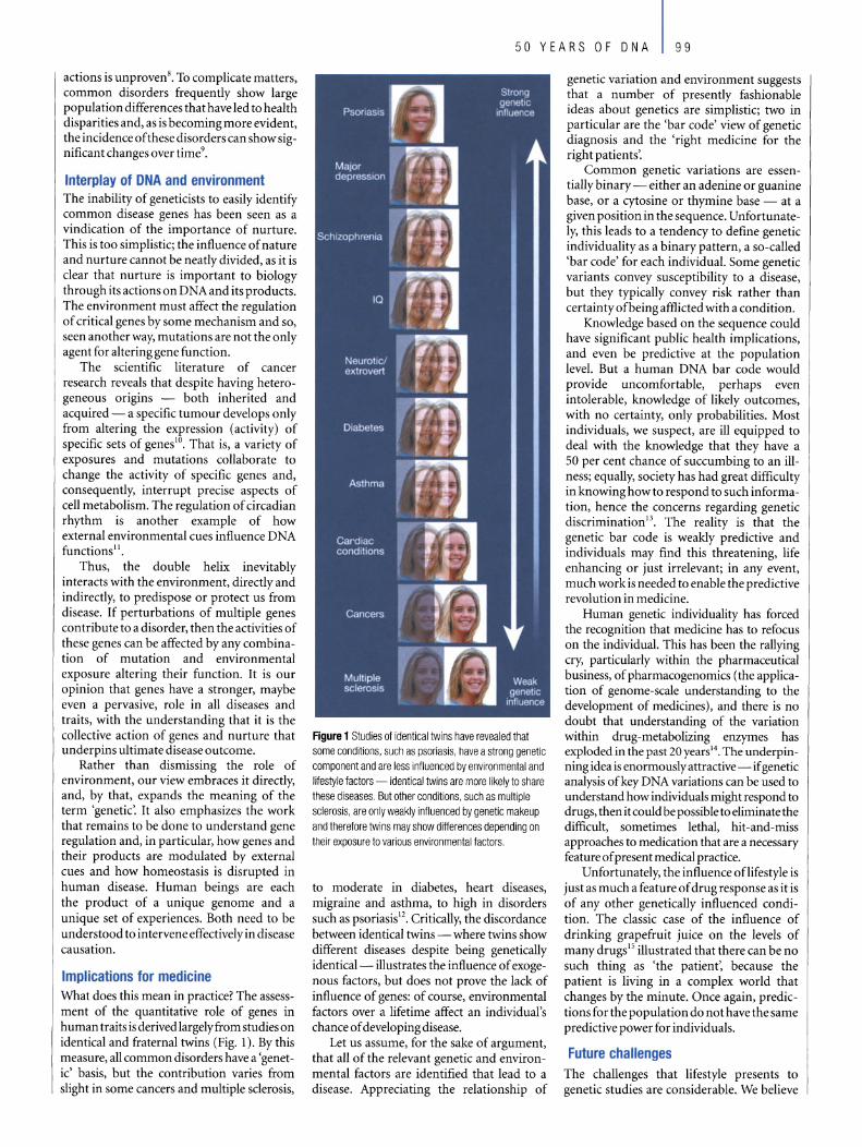
actions is unproven8• To complicate matters, common disorders frequently show large population differences thathave led to health disparities and, as is becoming more evident, the incidence ofthese disorders can show significant changes over time9•
Interplay 01 DNA and environment The inability of geneticists to easily identify common disease genes has been seen as a vindication of the importance of nurture. This is too simplistic; the influence of nature and nurture cannot be neatly divided, as it is clear that nurture is important to biology through its actions on DNA and its products. The environment must affect the regulation of critical genes by some mechanism and so, seen another way, mutations are not the only agent for altering gene function.
The scientific literature of cancer research reveals that despite having heterogeneous origins - both inherited and acquired - a specific tumour develops only from altering the expression (activity) of specific sets of genes!O. That is, a variety of exposures and mutations collaborate to change the activity of specific genes and, consequently, interrupt precise aspects of cell metabolism. The regulation of circadian rhythm is another example of how external environmental cues influence DNA functions ll .
Thus, the double helix inevitably interacts with the environment, directly and indirectly, to predispose or protect us from disease. If perturbations of multiple genes contribute to a disorder, then the activities of these genes can be affected by any combination of mutation and environmental exposure altering their function. It is our opinion that genes have a stronger, maybe even a pervasive, role in all diseases and traits, with the understanding that it is the collective action of genes and nurture that underpins ultimate disease outcome.
Rather than dismissing the role of environment, our view embraces it directly, and, by that, expands the meaning of the term 'genetic'. It also emphasizes the work that remains to be done to understand gene regulation and, in particular, how genes and their products are modulated by external cues and how homeostasis is disrupted in human disease. Human beings are each the product of a unique genome and a unique set of experiences. Both need to be understood to intervene effectively in disease causation.
Implications for medicine What does this mean in practice? The assessment of the quantitative role of genes in human traits is derived largelyfrom studies on identical and fraternal twins (Fig. 1). By this measure, all common disorders have a 'genetic' basis, but the contribution varies from slight in some cancers and multiple sclerosis,
50 YEARS OF DNA 99
Figure 1 Studies of identical twins have revealed that some conditions, such as psoriasis, have astrang genetic component and are less influenced by enviranmental and lifestyle factors - identical twins are more likely to share these diseases. But other conditions, such as multiple sclerosis, are only weakly influenced by genetic makeup and therefore twins may show differences depending on their exposure to various environmental factors.
to moderate in diabetes, heart diseases, migraine and asthma, to high in disorders such as psoriasis 12. Critically, the discordance between identical twins - where twins show different diseases despite being genetically identical- illustrates the influence of exogenous factors, but does not prove the lack of influence of genes: of course, environmental factors over a lifetime affect an individual's chance of developing disease.
Let us assurne, for the sake of argument, that all of the relevant genetic and environmental factors are identified that lead to a disease. Appreciating the relationship of
genetic variation and environment suggests that a number of presently fashionable ideas about genetics are simplistic; two in particular are the 'bar code' view of genetic diagnosis and the 'right medicine for the right patients'.
Common genetic variations are essentially binary - either an adenine or guanine base, or a cytosine or thymine base - at a given position in the sequence. Unfortunately, this leads to a tendency to define genetic individuality as a binary pattern, a so-called 'bar code' for each individual. So me genetic variants convey susceptibility to a disease, but they typically convey risk rather than certainty ofbeing afflicted with a condition.
Knowledge based on the sequence could have significant public health implications, and even be predictive at the population level. But a human DNA bar code would provide uncomfortable, perhaps even intolerable, knowledge of Iikely outcomes, with no certainty, only probabilities. Most individuals, we suspect, are ill equipped to deal with the knowledge that they have a 50 per cent chance of succumbing to an illness; equally, society has had great difficulty in knowing how to res pond to such information, hence the concerns regarding genetic discrimination 13. The reality is that the genetic bar code is weakly predictive and individuals may find this threatening, life enhancing or just irrelevant; in any event, much work is needed to enable the predictive revolution in medicine.
Human genetic individuality has forced the recognition that medicine has to refocus on the individual. This has been the rallying cry, particularly within the pharmaceutical business, of pharmacogenomics (the application of genome-scale understanding to the development of medicines), and there is no doubt that understanding of the variation within drug-metabolizing enzymes has exploded in the past 20 years14• The underpinning idea is enormously attractive - if genetic analysis ofkey DNA variations can be used to understand how individuals might respond to drugs, then it could be possible to eliminate the difficult, sometimes lethal, hit-and-miss approaches to medication that are a necessary feature of present medical practice.
Unfortunately, the influence oflifestyle is just as much a feature of drug response as it is of any other genetically influenced condition. The classic case of the influence of drinking grapefruit juice on the levels of many drugs15 illustrated that there can be no such thing as 'the patient', because the patient is living in a complex world that changes by the minute. Once again, predictions for the population do not have the same predictive power for individuals.
Future challenges The challenges that lifestyle presents to genetic studies are considerable. We believe

1 00 150 YEARS OF ONA
that the next 50 years will bring a genuine revolution of far greater individual significance than that delivered by genetics over the past 50 years. This is because lifestyle can conceivably be analysed, and in so doing, it should be possible to develop a genuinely personalized medicine.
Researchers can now think seriously about how to identify lifestyle influences: such studies will have to be on an unpreceden ted scale and one of the first of these, proposed to comprise 500,000 individuals in the United Kingdom, has already startedl6 .
These kinds of studies are a bold venture into relatively uncharted territory and face substantial technical, biological and scienceculture challenges.
Scientifically, it is necessary to understand a deceptively simple equation: genes + environment = outcome. The difficulty here is the uncertainty surrounding both terms in the equation; ideally, one set of genetic factors will interact with one set of environmental influences to produce identical outcomes, but it is unknown whether this is always going to be the case. A far more difficult relationship would exist if multiple genetic factors interacted with multiple environments to achieve the same outcome. The example of glutathione S-transferase mutations, smoking and incidence oflung cancer l7 shows it is possible to detect some interactions, but it is unclear how, or even if, statistical methods might be developed for addressing the more complex possibilities.
Perhaps the greatest unknown in undertaking these projects is human psychology; the consequences of smoking have been known for many decades, but people still smoke. Advice does not imply acceptance. How to turn knowledge into practical outcomes must be an increasing focus of attention for both researchers and funding agencies.
Psychology is also in play in the initial decision to undertake this research; for researchers, funding agencies and politicians there is great risk implicit in undertaking a hugely expensive project with complex outcome. People would like to live in a simpler world, with simpler decisions, but the vision of such a project is enormous: once complete, as much will be known about the origins ofhuman disorders as can be discovered by using such epidemiological and genetic studies. Perhaps more important, the beginnings of a new medicine will emerge, one focused uniquely and completely upon the individual, upon the combination of genetic uniqueness and personal choices that are the very essence ofindividuallives.
If we are collectively bold in our present decisions and accept the risk of action, a world can be created where medicine is a guide, not a place of last resort. If the past 50 years has seen the revolution ofDNA, then the revolution cannot be completed without
an appreClatlOn of both genetic and environmental individuality; only then will individuals understand the meaning of their inheritance. D doi:lO.l038/natureOl40l
1. Jaroff, L. The gene hunt. Time20 March, 62-67 (1989)
2. Arcos-Burgos, M. & Muenke, M. Geneties of population isolates.
Clin. Genet 61, 233-247 (2002).
3. Aidoo, M. et al. Protective effects of thc sickle ceIl gene against
malaria morbidity and mortality. Lancet359, 1311-1312 (2002).
4. Tishkoff, S. A. & Williams, S. M. Genetie analysis of African
populations: human evolution and complex disease. Nature Rev.
Gene/. 3, 6ll--{;2l (2002).
5. Chakravarti, A. Single nucleotide polymorphisms: ... to a future
of genetie medicine. Nature409, 822-823 (2001).
6. Carrasquillo, M. M. et al. Genome-wide association study and mouse
model identify interaction bmveen RET and EDNRB pathways in
Hirschsprung disease.l'lature Gen.et. 32, 237-244 (2002).
7. Cox, N. J. Challenges in identifying geneticvariation affecting
susceptibility to type 2 diabetes: examples from studies ofthe
calpain-lO gene. Hum. Mol. Genet. 10,2301-2305 (2001).
8. Sullivan, P. F. et al. Analysis of epistasis in linked regions in the
Irish study ofhigh-density schizophrenia families. Am. ]. Med.
Genet lOS, 266-27() (2001).
9. Boyle, J. P. et al. Projection of diabetes burden through 2050:
impact of ehanging demography and disease prevalence in the
Uni ted States. Diabetes eare 24, 1936-1940 (2001).
10. Münger, K. Disruption of oncogene/tumor suppressor networks
duringhuman carcinogenesis. Cancer Invest. 20, 71-81 (2002).
11. Panda, S" Hogenesch, J, B. & Kay, S, A. Circadian rhythms from
flies to human. Nature417, 329-335 (2002).
12. MacGregor, A. J. etal. Twins: novel uses to study complex traits
and genetie diseases. Trends Genet. 16, 131-134 (2000).
13. vVertz, D. C. Ethics watch. Nature Rev. Genet. 3,496 (2002).
14. Evans, W, E, & Johnson, J. A, Pharmacogenomics: the inherited
basis for interindividual differences in drug response, Annu. Rev.
Genomics Hum. Genet. 2, 9-39 (2001).
15. Lown, K. S. et al. Grapefruit juice increases felodipine oral
availability in humans by decreasing intestinal CYP3A pro tein
expression.]. Clin. Invest, 99, 2545-2553 (1997).
16. Wright, A. F., Carothers, A. D. & Campbell, H. Gene
environment interactions-the BioBank UK study.
Pharmacogenomics J. 2. 75-82 (2002).
17. Stucker, I. et al. Genetic polymorphisms of glutathione S
transferases as modulators oflung cancer susceptibility,
Carcinogenesis23, 1475-1481 (2002).
Originalreference: Nature421, 412-414 (2003).
The double helix in clinical practice John I. Bell
The Office of the Regius Professor ofMedicine, University ofOxford, Oxford OX3 9DU, UK
(e-mail: [email protected])
The discovery of the double helix half a century ago has so tar been slow to affect medical practice, but significant transformations are likely over the next 50 years. Changes to the way medicine is practised and new doctors are trained will be required before potential benefits are realized.
"lt is much more important to know what kind 01 patient has a disease than to know what kind 01 disease a patient has." Caleb Parry, 18th century physician, Bath.
he structure of DNA established the basic framework that would develop into the field of molecular genetics. The
information gleaned from this scientific endeavour continues to have a profound influence on our understanding ofbiological systems l . As most human diseases have a significant heritable component, it was so on recognized that the characterization of the genetic determinants of disease would provide remarkable opportunities for clinical medicine, potentially altering the way disease was understood, diagnosed and treated.
But despite the obvious potential applications to medicine, the development of significant genetic advances relevant to clinical practice could take generations. This is in marked contrast to many other medically related discoveries that occurred around the same time and which were translated rapidly into clinical practice. For instance, the development of penicillin by Ernst Chain and Howard Florey in 1941 was saving thousands of lives within months of their discovery of how to efficiently produce the antibiotic2• Discoveries relating to disease aetiology, such as the recognition in 1950 of a relationship between smoking and lung cancer, have had a profound effect on mortality3.
This was despite the convictions of at least one distinguished statistical geneticist who argued against the causality of this observation, implying that a common genetic factor caused both lung cancer and a predilection to smoking cigarettes4!
Although other important discoveries have had demonstrably more impact on health care at the time of their fiftieth anniversaries than has the double helix, its slower transition from discovery to clinical implementation will be balanced by its potentially profound impact across all medical disciplines. Progress has been slow, but mounting evidence suggests that, while public health and antibiotics produced important healthcare outcomes in the past 50 years, the next 50 are likely to belong to genetics and molecular medicine.
The potential impact of genetics on clinical practice has been questioned by some observers5 who believe that the positive predictive value of genetic testing for most common disease genes will be insufficient to provide the beneficial effects seen with single-gene disorders, which affect only a tiny proportion of the population. Many advocates of genetics argue, on the other hand, that our understanding of disease is

50 YEARS OF DNA 101
undcrgoing a major change. They contend that genetie research is playing a fundamental role in improving our understanding of the pathophy iology that underlies di -ea e and that, inevitably, as this i applied, il will alter both the theory and practice of medicine in the future~ .
of distinct di ea e and may resolve que ti n
variable
en wilhin
relating disease
proand
current diagnostic categories. ltimately, this may provide the
greate t contribution genetic will make to clinical practi e: a new taxonomy for human di ease.
A medical revolution Knowing that a di ea e can ari e fr m a
di tinct mechani m will alter a physi-A new taxonomy for human disease cian' approach t a patient with that
linical practiee has always been limited di rd r, all wing a more accurate prog-by its inability to differentiate clinical, no i and choiee of the most appropriate bio hemical and pathological abnormal- ___ therapy. The gene 'mutations' re pon ible itie that accompany a di ea e from lhose for many single-gene di rder ar n w evcnt a tua lly re ponsible for mediating a commonly u cd in diagn tic practi e, disease process. Clinicians may have moved whereas those associated with common on from calling 'fever' a disease7, but they still complex diseases are just being characterized. relyonphenotypic criteriato define most dis- Although their predictive value will be less eases, and yet these may obscure the underly- than with single-gene disorders, their contriing mechanisms and often mask significant bution as risk factors will be similar to other heterogeneity. As Thomas Lewis pointed out risk factors such as blood pressure, cholesin 1944, diagnosis of most human disease terol levels and environmental exposures. provides only "inseeure and temporary con- Because much of clinical practice involves ceptions"s. Of the main common diseases, evaluating and acting on risk probabilities, only the infectious diseases have a truly the addition of genetic risk factors to this mechanism-basednomenclature. process will be an important extension of
An understanding of the genetic basis of existing practice. The overall effect of genetic maladies is providing a new taxonomy of risk factors is likely to be significant. For disease, free from the risk that the diagnostic example, recent estimates in breast cancer criteria related to events are secondary to the suggest that the attributable genetic risks are disease process, rather than to its cause. likely to exceed the predictive value of a range Genetic information has allowed us to ofexistingnon-geneticriskfactors13 •
identify mechanistically distinct forms of Other potential applications of genetics diabetes, defining an auto immune form in health care may be realized in a shorter of the disease associated with human leuko- timeframe. Individual variation in response cyte antigens (a highly diverse complex of to drugs and in drug toxicity is a significant immune-system genes), and recently has problem, both in clinical practice and in the implicated dysfunction of factors that affect development of new therapeutic agents. both expression and modification of gene Clear examples now exist of genetic variants products in mediating the adult form of the that alter metabolism, drug response or risk disorder9 • Similarly, we are now aware of a oftoxicityI4,IS, Such information provides an range of molecules and pathways previously opportunity to direct therapy at individuals not recognized in the pathogenesis of most likely to benefit from an intervention, asthma lO- 12• thereby reducing cost and toxicity, and
A clearer understanding of the mecha- improvingmethodsfordrugdevelopment. nisms and pathways that mediate disease will The discovery of the structure ofDNA not
Table 1 Molecular genetica in clinlcal practlce
.~ ... ~~~~~!~.~ .. ?I~.~t.~ .. 7.~~ ........................... _ : ... ~~.~~.!.~.~ .. ~ .. ~~.~~~ ... , .... " ... ,., ..... , ............. . .~ ... ~.~~.~.d.~~.~~.!~.~,.~.~f.~.~~~ ............. . .~ ... ~~.~~ .. ................................................................ . .~ ... !~!~?,l.~'o.".~.r.~. ?":'9..tfl'.~~~ ......................................... . • Tools for moIecular medicine (for example, recombinant
..... ~~,~,~~~ .................... ............. ................................. .
.: ... ~~.Il<.~.~.~~~~.?'.!.~.a.~tl(;.~?t.~~ ............. . • Gene thefapy
only led to an ability to characterize genetic determinants in disease, but also provided the tools necessary for the revolution in molecular medicine that has occurred in the past 25 years. The description of the double helix was the first important step in the development of techniques to cut, ligate and amplify DNA. The application of these molecular biology and DNA-cloning techniques has already had a profound impact on our understanding of the basic cellular and molecular processes that underlie disease.
Molecular biology has improved our ability to study proteins and pathways involved in disease and has provided the technology necessaryto generate new sets oftargets for smallmoleeule drug design. It has also enabled the creation and production of a new range of biological therapeutics - recombinant proteins such as interferon, erythropoietin and insulin, as well as therapeutic antibodies, which are one of the fastest growing classes of new treatments. Further extensions of this methodologywill see the inevitable introduction of DNA-based therapies that will produce proteins ofinterest in the appropriate cellular setting. DNA-based vaccines represent the first wave of such novel gene-therapy approaches to disease and many more are expected to follow.
We are undergoing a revolution in clinical practice that depends upon a better understanding of disease mechanisms and pathways at a molecular level. Much has already been achieved: an enhanced understanding of disease-related pathways, new therapies, novel approaches to diagnostics and new tools for identifying those at risk. But more remains to be done before the full impact of genetics on medicine is realized. Complex disease, with multiple susceptibility determinants (both environmental and genetic), will take time to dissect. This information must then be moved into the clinic and evaluated for its benefits.
As the practice of medicine moves to one more scientifically founded in disease mechanisms, many aspects of clinical practice will need to be transformed. Individual genetic variation is likely to explain a significant part of the heterogeneity seen clinically in the natural history of disease and in response to therapy. Tools to tailor medicine to an individual' s needs rather than directing it at a population will inevitably become available. Similarly, as predictions of risk improve, early or preventative therapy of high-risk populations will become a reality, with screening programmes targeted to those at particularly high risk.
Transforming clinical practice For fundamental changes to take place in clinical practice, sweeping transformation will be needed to healthcare provision, economic management and training. It is currently difficult to predict the cost-benefit ratio for such changes- certainlythe present impact of molecular medicine has not made medicine less expensive. Few medical schools adequately train their students to think mechanistically about disease; indeed, the trend towards pattern-recognition medicine, away from basic science training, means that we are still far from educating the next generation of clinicians to applythe knowledge and tools bequeathed to us by the double helix . The evolution in health care that will incorporate these new principles of early diagnosis and individualized therapywill be a daunting

102 50 YEARS OF DNA
challenge in an era of uncertainty for healthcare systems worldwide.
The influence of genetic and molecular medicine on the health of patients is already sufficiently ubiquitous that it will have an impact on most common diseases. Its influence will grow over the next few decades (Table 1). It will not, however, answer all of the questions about human health, nor will it provide all the answers for optimizing clinical practice. The reductionism that accompanies molecular genetics will identify the pieces in the jigsaw, but assembling these to understand how complex systems malfunction will require a substantially more integrated approach than is available at present.
The crucial role played by environmental determinants of disease will perhaps become more tractable when combined with an understanding of genetic susceptibility. Sceptics, rightly, will wish to see more data before they acknowledge that molecular medicine will be truly transformed over the next 50 years, despite the fact that its influence on diagnostics and new therapeutics is already clearly apparent. A transition is underway, the direction of travel is clear, but managing the change in clinical practice may
prove at least as challenging as resolving the original structure of the helix. D doi: I 0.1 038/natureO 1402
1. Watson, J. D. & Crick, F. C. H . A structure for deoxyribose
nucleic acid. Naturel71, 737- 738 (1953).
2. Abraham, E. P. cl al. Further observations on penicillin. Lancetii,
177-188 (1941).
3. Doll, R. & Hill, A. B. Smoking and carcinoma ofthe lung. Br. Med.f. 2, 739- 748 (1 950) .
4. Fisher, R. A. Cancer and smoking. Nature 182, 596 (1958).
5. Holtzman, N . A. & Marteau, T. M. Will genetics revolutionise
medicine? N. Engl.f. Med. 343, 141-144 (2000).
6. Bell, J.1. The new genetics in c1inical practice. Br. Med. ,. 316,
618-620 (1998).
7. Osler, W. The Principles and Practice ofMedicitle (Appleton, New
York,1892).
8. Lewis, T. Reflections uponmedical education. Lanceti,619-621 (1944).
9. Cardoll, L. R. & Bell, J.l. Associatioll studydesigns [er complex
diseases. Nature Rev. Genet. 2, 91-99 (200 1).
10. Van Eerdewegh, P. et a1. Assoeiation ofthe ADAM 33 gene with
asthma and bronchial hyperresponsiveness. Na ture418,
426-430 (2002).
11. Cookson, W. o. C. M. , Sharp, P. A., Faux, J. A. & Hopkin, J. M.
Linkage between immunoglobulin E responses underlying asthma
and rhinitis and chromosome l1q. Laneet 1, 1292- 1295 (1989).
12. Shirakawa, I. et al. Atopy and asthma: genetie variants oflL-4
and IL-13 signalling. lmmunol. Today 21, 61-64 (2000).
13. Pharoah, P. D. P. el a1. Polygenie sllseeptibility to breast cancer
and implieations for prevention. Nature Genet. 31, 33-36 (2002).
14. Splawski, I. et a1. Variant ofSCN5A sodium channel implicated
in riskofeardiae arrhythmia. Science297, 1333- 1336 (2002).
15. Weber, W. W. Pharmacogenetics (Oxford Univ. Press, NewYork,
1997).
Original reference: Nature 421) 414-416 (2003).
The Mona Lisa of modern science Martin Kemp
Department of the History of Art, University of Oxford, Littlegate House, St Ebbes, Oxford OXI 1 PT, and WallaceKemp, Artakt, StudioD, 413-419, Harrow Road, London W93Q!, UK (e-mail: [email protected])
No moleeule in the history of science has reached the iconic status of the double helix of DNA. Its image has been imprinted on all aspects of society, from science, art, music, cinema, architecture and advertising. This review of the Mona Usa of science examines the evolution of its fonn at the hands of both science and art.
'/\ monkey is a machine that preserves genes up trees, a fish is a machine that preserves genes in water; there is even a small worm thatpreserves genes in German beer mats. DNA works in mys terious ways."Richard Dawkins in The Selfish Gene (Oxford University Press, 1976).
istory has thrown up a few superimages, which have so insinuated themselves into our visual
consciousness that they have utterly transcended their original context. This is epitomized by the Mona Lisa, painted by Leonardo da Vinci around 1503. The double helix of DNA is unchallenged as the image epitomizing the biological sciences. Both images speak to audiences far beyond their respective specialist worlds, and both carrya vast baggage of associations.
In the worlds of popular image diffusion, particularly on the Internet, the double helix is beginning to riyal the Mona Lisa as a playground for eccentrics and obsessives (Fig. 1). There is an apparent difference, of course. Leonardo's panel painting is the
product ofhuman artifice, whereas DNA is a naturally occurring, large organic molecule. But Leonardo claimed that his art represented a systematic remaking of nature on the basis of a rational understanding of causes and effects. His painting is the result of a complex, nonlinear interaction between concept, subject, plan of action, acquired knowledge, skill, medium and the evolving image itself. In The Art oI Genesi, Enrico Coen argues that "biological development and human creativity are highly interactive processes in which events unfold rather than being necessarily pre-plannedor anticipated. In otherwords, in both cases there is no easy separation between plan ( or programme) and execution."
Looking at the investigation and representations ofthe double helix, we can saythat
they are cultural activities no less than any painting. Behind the discovery lies the vast infrastructure of a scientific culture that led to the development of the knowledge, theories, institutions, techniques and equipment that made the quest both possible and desirable. The very natures of scientific models and representations, using whatever technique, are integral to the vehicles of science communication. Their visuallook is compounded from a complex set of factors, ranging from technical to aesthetic. But, in case anyone should be getting the wrong impression, I acknowledge that the cUltural vehicles are designed to deliver nonarbitraryinformation that is open to rational scrutiny as a way of working towards real knowledge ofthe physical constitution ofthe world.
Looked at from a popular perspective (and even from the standpoint of reputation within science), James Watson and Francis Crick are identified with DNA no less than Leonardo is identified with the Mona Lisa. The researchers were in a very real sense the 'authors' or 'artists' of the acts of visualization that generated their models of the molecule. But their brilliant achievement was not necessarily of a higher order than that of the other pioneers of molecular modelling, such as the Braggs, John Kendrew, Max Perutz, Maurice Wilkins and Linus Pauling. Rather, theywere uniquely fortunate that their molecule was both visually compelling, as a supreme example of nature's 'sculpture', and

50 YEARS OF DNA 103
Figure 2 Structure of DNA, drawn by Francis Crick's wife Odile Crick, which was published as the sole figure in Watson and Crick's seminal paper in Nature, 25 April 1 953 (ref, 2),
Figure 1 LEGO model of the DNA double helix (in reverse!) by Eric Harshbarger (2001), who also used his mastery of the coloured units of LEGO to compose a 'pixelated' LEGO version of the Mona Usa, (Images courtesy of E. Harshbarger,)
lay at the heart of the twentieth-century version of the quest to unravel the ultimate secret oflife.
The 50-year journey of the DNA molecule from the reticent line diagram in Watson and Crick's seminal article2 (Fig. 2) to its position in today's world of global imagery is extraordinary. It is therefore timely to look at so me of the representational issues involved in science communication, and then at a few selected instances of the various guises in which the molecule has replicated itselfwithin varied visual habitats.
A model 01 communication Looking back on the laconic article in Nature that announced the structure ofDNA, which we tend to ass urne in retrospect provided the definitive solution, it is remarkable how !ittle was actually given away. This is true of the artide' s sole diagram, drawn by Odile Crick, Francis's wife, which represented the sugar chains as directional ribbons, while the bases were rudimentary rods represented flat on (Fig. 2). Along the vertical axis runs the central pole, depicted as a thick line that is broken where the bases lie in front. This axis is a visually useful point of reference, but its early ubiquity seems to depend on the structural necessities of physical models. The developed model, composed from standard brass components with tailor-made metal bases, provided a more detailed and explicit entity for debate and large-scale publicity, although the famous photographs by Anthony Barrington Brown (Fig. 3), taken for an article in Timemagazine, were actually staged a few months later.
The model of the double helix - like those of other molecules, such as the model of haemoglobin by Perutz - played an important role in scientific understanding, being both based upon and in turn affecting the acts of scientific conceptualization. Overtaken by more refined models made at King's College London, induding the widely illustrated space-filling model with Van der Waals surfaces by Wilkins (Fig. 4), the ramshackle masterpiece of Watson and Crick
passed the way of so many obsolete bits of scientific paraphernalia. When, 23 years after its making, some of the specially cut plates (Fig. 5) resurfaced in Bristol, they were incorporated into a pious reconstruction by Farooq Hussain of King's College. Like an ancient Greek vase reassembled from chards,
the semi-original model is now a treasured cultural icon, displayed in the Science Museum in London.
Communicating the complex structure and, in due course, the awesomely intricate behaviour of the modular molecule, has provided an unparalleled challenge for
Figure 3 Anthony Barrington Brown's photograph of Watson and Crick with their model of DNA at the Cavendish Laboratory in Cambridge, 21 May 1953,

Agun! 4 Main Image sI'ONS Maurice Wllkins' space-filling modeI 01 DNA. Inset a series 01 ludd and Inventive graphics by Keith Roberts appeared in Watson's MoIecular 8ioIogy of fhe Gene. Here a flat chemical diagrarn is enhanced through
two-coIour printing to prOYide schemalic ernphasis upon the sugar phosphate chalns or 'ribbons'. Qmages reproduced fram rel. 3, with permission.)
_r-v -.,. __
biologieal iIIu trator and model -maker .
Thi is vividly shown by Keith Robert ' ill u tration in ueee ive edition of Wat on's Molecular Biology of tlre Gene, beginning in 1965, wh ich ehart
the omplex interplay between developing seiene ,graphie ingenuity and technologie of reproduetionJ ( ee Fig. 4, in et).
As the eomplex funetioning of A beeame inerea ingly elueidated, 0 methods and eonvention of illu tration that privileged behaviour over strueture played an ever more conspieuou role. A with et of iIlu -tration in any ciene ,th vi uaJ eonvention not only refleet what eienti t want to show, but also provide an important framework for thinking and vi ualization in the pro e of re eareh it elf. ub equently, the re ouree of computer de ign, tereo eopy and, in partieular, animation have provided a vivid sen eof patial and temporal proeesses, only partly
po ible in eonventional text and illu tration.
Three-dimen ional eontrivanee have had a erueial role from the out et, un urpri ingJy given a trueture that ta.xe our power of spatial vi ualization. yen in Ihe age of computer graphie, Ihere is still a pedagogie and popular market for kir u ing a variety f spaee-filling unit .
A number of notable model f 0 A and other large molecules have beeome revered item , typieally di played in proteetive ca e in the foyers oflaboratorie , where they form part ofthe vi ual furniture that speak ofthe enterpri e ofbiological seienee in general and that of the in titution in particular. For the ub-speeiesofbiologi t known a 'moleeular:
the edu tive geometry of A helps to underline the fundamental ' hardne ' of their eienee, eompared to natural hi torian and eeologi t from whom they have become in litutionally di tingui hed. [t i in thi pirit, le of didaetie in truetion than of emblematie ignalling, that the double helix ha beeome the ieon for the eommunieation of a generalized me age. Few have any trou ble in reeognizing the gho tly twi t that emerge from the mo aieoffaee on thecover of the all/re i ue devoted to the human genome on 15 February 2001 (Fig. 6).
Figure 6 Cover of Nature human genome issue, published on 15 February 2001 . The image, by Eric Lander. was created by Runaway Technology lne. using PhotoMosaic by Robert Silvers trom original artwork by Darryl Leja (courtesy 01 the Whitehead Institute ror BIomedicai Research). Gregor Mendel, James Watson and Francis Crick are amongst the crowd.
imilarly, any hint ofthe double twi tin any logo of a laboratory or biote h eompany i immediately idenrifiable.
Aesthetics and meaning Given the role of aesthetie intuition in the proee e thai led to it di eovery, and it reeognition a 'right', it i under tandable that thedoubleheli ha it elfa umedthegui eor a work of art, not least in three-dimen ional form. For artists, the attraction of a ~ rm that i both beautiful and fuH fall kind of eientifie and eial ignifieanee i eon iderable.
ome grand trueture have been eommi ioned by aeademic in titution ,whereby the artist has basically been given abrief to make a ulpture repre enting 0 A , mueh like a eulptor might be commissioned ro produee an anatomieally accurate image of a naked figure. For example, in 1998 Roger Berry produeed a huge eulpture hanging down the eentral weil of a multi- tory stairea e at the niver ity of alifornia at Davi (Fig. 7). Another rendering of the helieal strueture, pimls Tillle - Time pimls, re ide on a hilloek in the ground ofthe old pring Harbor Laboratory (Fig. 8). Designed
by the artist, arehiteet and theorist of po t-

modernism, Charles Jencks, it stands at the heart of a programme of commissioning and collecting artwork that expresses thevision of Watson - who became director of Cold Spring Harbor Laboratory in 1968 and president in 1994 - of an environment in which the visual stimulation of the surroundings is integral to the conduct ofhigh-Ievel mental activity.
In pursuit of structural aesthetics, the British sculptor, Mark Curtis, proposed a reformed molecular structure for DNA. As an artist concerned with geometricallogic and symmetries, Curtis was worried about the 'ugly' engineering of the Watson-Crick version. Rather than using the sugar phosphate backbones to control the helices, he proposed that stacked base pairs, coupled in an opposite orientation from the accepted bonding, comprises a helix of pentagonal plates around a central void of decagonal cross-section. The geometrical and structural probity of Curtis's models, which eschew a central pole, made it on to a British millennium stamp (Fig. 9), if not into the world of scientific orthodoxy. In a real sense, the molecular biologists' rejection of Curtis's effort to re-design DNA on the basis of apriori principles represents an
50 YEARS OF DNA 105
Figure 7 (Ieft) Portrait of a DNA Sequence by Roger Berry (1998) at the Lile Sciences Addition building, University 01 Calilornia, Davis. Figure 8 Spirals Time - Time Spirals by Charles Jencks (2000) at Cold Spring Harbor Laboratory.
extreme exampleofthe ten ion within science itselfbetween the polar instinct of the modellersand theempiricists.
Along ide such sculptural exploitations ofthe inheren t b autyofthe double helix has run as trand of arti tic iconography that has been more overtly concerned with meaning. The tone for the more fan tastical exploitations was set by the flamboyant surreali t, alvador Dali, as ever concerned wirh rhe metaphysical potential implicit in scientific imagery. Ouring the late 1950s and 1960s, the DNA molecule fea tures as a symbolic vision, lurking in a surreal hin terland between galaetie mystery and spiritual sign ificanee (as a kind of Jacob's Ladder). His Butterfly Landscape, The Great Masturbator in Surrealist Lal1dscape with DNA (1957-8; Fig. 10) locates a prettified evoca tion ofa space-fi ll ing model in one of Dali's typically barren land-scapes inhabited by ub-Freud ian enigmas, designed to eonjure up a dreall1world of obscure sexual fantasy4 . Sub quent artists, particularly those who have engaged with the oeial ill1pl ieation of 1l10lecular biology and genetie engineering, have located
images of ONA in eontexts of Illeaning tha t are less obseure and more polemie.
This savagely seleetive glanee at 0 A art - omitting sueh contemporary luminarie of genetie art as uzan ne Anker5 (Fig. 11;
www.geneeu ltu re.org), David Kremers
..-
". ;;. r I , i -, .. ;
L'"- , ~ ~
I , .... :,.
,si ; j:J-"
.~ '". , .. ~ ..... -1 , ..... ,
"""'-- -• ,~ \- •• e! ... ~, 19 Ag ure 9 Painti ngs 01 DNA models on a 'Millennium Colleclion' slamp, designed by Mark Curtis (1999-2000), Irom Ihe UK Royal Mail 's Scientists' Tale collection.

106 50 YEARS OF DNA
Figure 10 Butterfly Landscape, The Great Masturbator in Surrealist Landscape with DNA by Salvador Dali, 1957-8. Private collection.
(http://davidkremers.caltech.edul), Ellen Levy (http://www.geneart.org/genomelevy.htm), Sonya Rapoport (http:// users.lmi.net/sonyarap/transgenicbagel!) and Gary Schneider (http://www.icp.org/ exhibitions/schneiderl) - can barely claim to be representative even ofthe main range of possibilities. In particular, exploitation of
Box 1
the replicating potential ofDNA to generate self-organizing images - exemplified by Marc Quinn's genetic portrait of Sir John Sulston from Sulston's own DNA, fragmented and replicated in bacterial ealonies on plates of agar jelly6 - shows that some artists' engagement with DNA is maturing beyond iconographical opportunism.
2003 exhibition. celebratlng art In the age 01 the double:::::h::e::II::X=====:.
2003 is to throw up aseries of exhibitions, ineluding:
• Representations of the Double Helix, at the Whippie Museum of the Hislory of Seienee, Department of History and Philosophy of Seienee. University of Cambridge, UK. The exhibition is euraled by Soraya de Chadarevian and Harmke Kamminga, with the assistanee of Corrina 8ower, and will run from January to December 2003.
• Genetie Expressions: Art After DNA, al Ihe Heckseher Museum of Art , Huntington, New York. Curaled by Elizabeth Meryman and Lynn Gamwell. the exhibition will run trom 28 June 10 7 September 2003.
• From Code 10 Commodity: Geneties and Visual Art . at the New York Academy of Seienees. The exhibition runs from 13 February 10 11 April 2003 and is eurated by Dorothy Nelkin and Suzanne Anker. Cold Spring Harbor Laboratory Press are publishing a book by Nelkin and Anker entitled The Mo/ecular Gaze: M in the Genetie Age.
• PhotoGENEsis: Opus 2 - Artists' Response To the Genetie Information Age, at the Santa Barbara Museum of Art. 9 November 2002-9 February 2003.
Figure 11 Zoosemiotics: Primates, Frag, Gazelle, Fish (detail) by Suzanne Anker (1993).
But as with the Mona Lisa, opportunism will always be the name of a prominent public game. Typical of this tendency is the introduction by the perfume eampany Bijan in 1993 of a fragrance named DNA. Ironically, we learn from the maker's blurb that "DNA is recommended for casual use". Such is the destiny of one of the greatest popular ieans. D doi: 10.1 038/natureO 1403
1. Coen, E. TheArt ofGenes. How Organisms Grow Themselves (Oxford Univ. Press, Oxford, 1999).
2. Watson, J. D. & Crick, F. H. C. A structure for deoxyribose
nudeic acid. Nature 171, 737-738 (1953).
3. Watson, J. D. Molecular Biology of the Gene (W. A. Benjamin,
Ine.. NewYork, 1965).
4. Descharnes, H. & Neret, G. Salvador Dali 1904-1989 (Taschen,
NewYork,1998).
5. Nelkin, D. & Anker, S. The influence of genetics on
contemporaryart. Nature Rev. Genet. 3. 967-971 (2002).
6. Kemp, M. Reliquary and replication. A Genomic Portrait: Sir lohn
Sulston by Marc Quinn. Nature413, 778 (2001).
Acknowledgements R. Hodgson provided research assistanee. Important adviee has been gratefully received frorn S. de Chadarevian and H. Karnminga of the Departrnent of the History and Philosophy ofSeienee at Cambridge University. E. Levy, E. Coen, and K. Roberts ofthe lohn Innes Centre at Norwieh.
Original reference: Nature 421, 416--420 (2003).
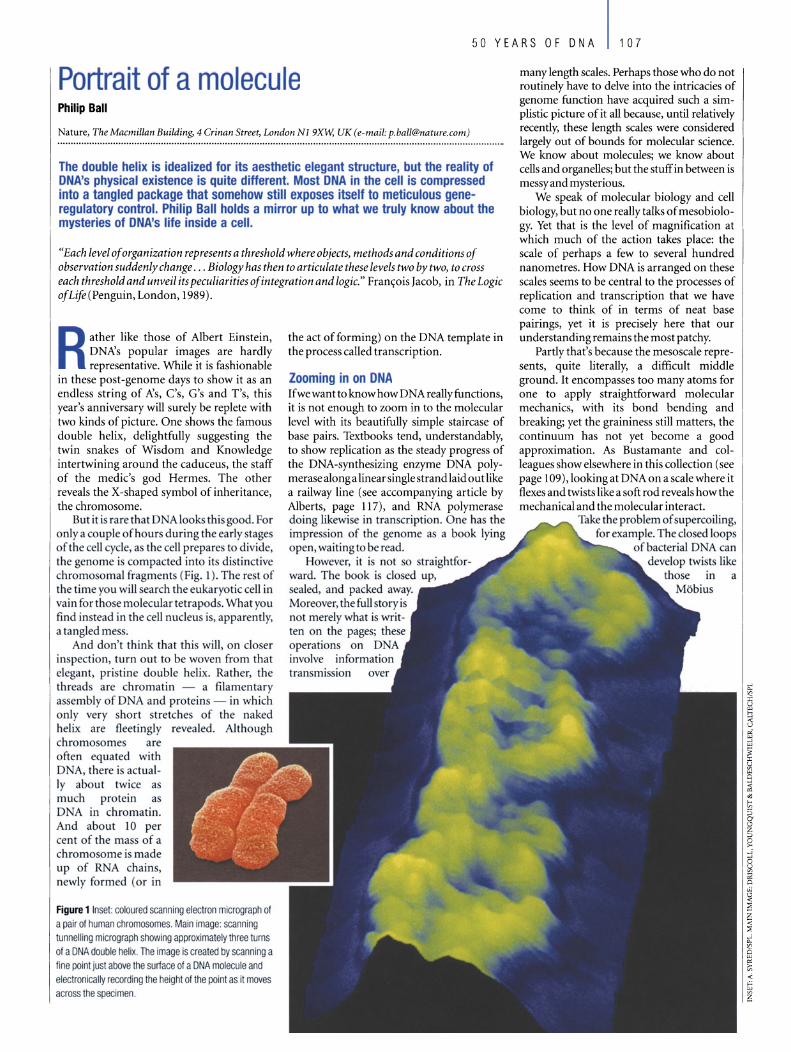
50 YEARS OF DNA 107
Portrait of a molecu le Philip Ball
Nature, The Macmillan Building, 4 Crinan Street, London Ni 9XW; UK (e-mail: [email protected])
The double helix is idealized for its aesthetic elegant structure, but the reality of DNA's physical existence is quite different. Most DNA in the cell is compressed into a tangled package that somehow still exposes itself to meticulous generegulatory contro!. Philip Ball holds a mirror up to what we truly know about the mysteries of DNA's life inside a cell.
"Each level of organization represents a threshold where objects, methods and conditions of observation suddenly change . .. Biology has then to articulate these levels two by two, to cross each threshold and unveil its peculiarities ofintegration and logic." Fran<;:ois Jacob, in The Logic ofLife (Penguin, London, 1989).
Rather like those of Albert Einstein, DNA's popular images are hardly representative. While it is fashionable
in these post-genome days to show it as an endless string of A's, C's, G's and T's, this year's anniversary will surely be replete with two kinds of picture. One shows the famous double helix, delightfully suggesting the twin snakes of Wisdom and Knowledge intertwining around the caduceus, the staff of the medic's god Hermes. The other reveals the X-shaped symbol of inheritance, the chromosome.
But it i rarethat DNA looks thisgood. For onlya couple ofhours during the early stages of the cell cyde, as the cell prepares to divide, the genome is compacted into its distinctive chromosomal fragments (Fig. 1). The rest of the time you will search the eukaryotic cell in vain for those molecular tetrapods. What you find instead in the cell nudeus is, apparently, a tangled mess.
And don't think that this will, on doser in pection, turn out to be woven from that elegant, pristine double helix. Rather, the threads are chromatin - a filamentary assembly of DNA and proteins - in which only very short stretches of the naked helix are fleet ingly revealed. Although chromosome are often equated with DNA, there is actual Iy about twice as much protein a DNA in chromatin. And about 10 per cent of the mass of a chromosome is made up of R A chain , newly formed (or in
Figure 1 Insel: coloured scanning eleclron micrograph 01 a pair of human ch romosomes. Main image: scanning tunnelling micrograph showing approximately three tums of a DNA double helix. The image is c reated by scanning a fi ne point just above the surtace 01 a DNA molecule and eleclronically recording the height of the point as it moves across Ihe specimen.
the act of forming) on the DNA template in the process called transcription.
looming in on DNA If we want to know how DNA really functions, it is not enough to zoom in to the molecular level with its beautifully simple staircase of base pairs. Textbooks tend, understandably, to show replication as the steady progress of the DNA-synthesizing enzyme DNA polymerase along a linear single strandlaid out like a railway line (see accompanying article by Alberts, page 117), and RNA polymerase doing likewi e in transcription. One has the impression of the genome as a book lying open, waiting to be read.
However, it is not so straightforward. The book is c10sed up, sealed, and packed away. Moreover, the fuU storyis not merely what is written on the pages; these operations on DNA involve information tran mission
many length scales. Perhaps those who do not routinely have to delve into the intricacies of genome function have acquired such a simplistic picture of it all because, until relatively recently, these length scales were considered largely out of bounds for molecular science. We know about molecules; we know about cells and organelles; but the stuffin between is messy and mysterious.
We speak of molecular biology and cell biology, but no one really talks of mesobiology. Yet that is the level of magnification at which much of the action takes place: the scale of perhaps a few to several hund red nanometres. How DNA is arranged on these scales seems to be central to the processes of replication and transcription that we have come to think of in terms of neat base pairings, yet it is precisely here that our understanding remains the most patchy.
Partly tha1's because the mesoscale represents, quite literally, a difficult middle ground. It encompasses too many atoms for one to apply straightforward molecular mechanics, with its bond bending and breaking; yet the graininess still matters, the continuum has not yet become a good approximation. As Bustamante and colleagues show elsewhere in this collection (see page 109), lookingat DNAon a scalewhere it flexes and twists like a soft rod reveals howthe mechanical and the molecular interacr.
Take the problem of upercoiling, for example. The closed loops
ofbacterial DNA can
a
108 50 YEARS OF DNA
strip, which either 'overwind' or 'underwind' the helix. Generally there is some degree of underwinding (negative supercoiling) such that there is one negative supercoil for every 200 base pairs (bp) or so. This has an energy cost of around -9 kcal mot!, which manifests itself in physiological effects. In bacteria, too much supercoiling can inhibit growth, which is why enzymes called topoisomerases exist to release it. On the other hand, negative supercoiling tends to unwind the double helix, which is needed to initiate strand separation for DNA replication.
Although the chromosomal DNA of eukaryotes has free ends, it too is prone to supercoiling, as it seems typically to be attached in large loops to a filamentous structure called the nuclear matrix that coats the inside of the nuclear membrane. The attachment may in fact be necessary for both replication and transcription to take place.
Packaging problem Stretched into a linear double helix, the three billion or so base pairs ofhuman DNA would measure 1.8 m. This strand, snipped into 46 chromosomes, has to be packed into a nucleus just 6 f-lm or so across. As a result, the DNA chains are far from the idealized picture of molecules floating in an infinite solvent. They have a density of around 100 mg per ml, comparable to that of a highlyviscous polymer gel.
The packing ratio for the chains is therefore enormous. In the smallest human chromosome, a length of DNA 14 mm long is compressed into a chromosome about 2 f-lm long: a packing ratio of7,000. The first stage in solving this packaging problem is to wind the DNA around protein disks to form a bead-like nucleosome (see accompanying article by Felsenfeld and Groudine, page 134). Each disk is an octamer offour types of histone protein; a fifth histone, called Hl, seals the DNA to the disk at the point where the winding starts and ends. Each nucleosome, 6 nm high by 11 nm in diameter, binds around 200 bp ofDNA in two coils, and there is very little 'free' DNA between adjacent nucleosomes: sometimes as little as 8 bp.
The string of nucleosomes forms a fibre about 10 nm thick, which is then packaged into a filament three times as wide. This 30-nm fibre is the basic element of chromatin - yet we still don't know its structure. It is widely held to be composed of nucleosomes arranged in a solenoid, but hard evidence for this is scanty. How many celebrations of the double helix will admit that, 50 years on, we don't really know what DNA at large in the celllooks like?
The 3D-nm fibre is further folded and condensed to give a packing ratio of around 1,000 in chromosomes during interphase (the time between cell divisions), and around ten times that in the X-shaped chromosomes of mitosis ( ceil division). Howthis happens is even more of a mystery. For mitotic chromosomes it
was thought until onlyrecentlythat there might be a contiguous protein scaffold holding the whole affair together; but now it seems that the structural integritymust come from chromatin crosslinking1• All the histones seem to have higher-order structural functions. Multi-subunit protein complexes in yeast called SWI/SNF and RSC (both of which seem to have human homologues) are chromatin-remodeiling machines, whichdistorthistone-DNAcontacts or transfer histones between DNA molecules, exposing the DNA to attack by DNA-cleaving enzymes called nucleases. How they work remains hazf. According to one recent study, DNA engaged by such complexes 'behaves as if it were free and bound at the same time: Or in other words, as if 'free' and 'bound' were notions too simplistic to have much meaning here. Whatis clear is that these chromatin-shaping machines are important in transcription: ceils lacking RSC are no longerviable.
There are in fact two types of chromatin in the nucleus of an interphase eukaryotic ceil. Euchromatin is the most abundant: it is relatively dispersed, with the tangled-net appearance of a polymer gel. Heterochromatin is much denser (virtually solid-like), comparable to the density of mitotic chromosomes, and is confined to a few small patches. The invitation is to regard euchromatin as 'active' DNA, unpacked enough to letthe transcription apparatus get to work on it, while heterochromatin is compressed, like a big data file, until needed. But like just about any other generalization about DNA's structure and behaviour, this one quickly breaks down. Clearly only a fraction of a cell's euchromatin is made up oftranscribable DNA in the first place (so why not pack the rest away?); and even chromosomes containing a large amount ofheterochromatin can be transcriptionally active. Some researchers think that 'euchromatin' and 'heterochromatin' are just blanket terms for many things we don't understand: further hierarchies of DNA organization yet to be revealed.
Structured chaos Certainly, there seems to be more to the nucleus than a disorderly mass ofD NA. It is a constantly changing structure, but not randomly: there is method in there somewhere. Specific chromosomes occupy discrete nuclear positions during interphase, and these positions can change in a deterministic wayin response to changes in the cell's physiological state.
And the euchromatin itself has an internallogic, albeit one only partly decoded. It has been proposed that DNA has sequences called scaffold/matrix-attached regions (S/MARs), recurring typically every 10-100 kbp, that bind to the nuclear matrix to divide up the chromosome into 100ps2. Yet the existence of not only S/MARs, but also the nuclear scaffold itself, has been questioned. There is no sign of the scaffold during mitosis, and the material it is thought to be
composed of may be nothing more than a mess of denatured proteins.
Be that as it may, the organization of the loops seems to be important for compaction ofDNA and for the regulation of gene expression, and each loop may act as an independent unit of gene activity. In other words, there is at least one level of superstructural organization in the chromosomes that makes its influence felt at the scale of molecular information transfer. Topoisomerase II is one of several proteins that bind specificallyto the putative S/MARs, suggesting that these points are important for controlling supercoiling in the strands.
With all this high-level structure, transcription ofDNA is not so much a matter of slotting the parts in place as tugging on the rope. DNA is highly curved around the nucleosomes, the inward -facing groove compressed and the outer one widened. RNA polymerase, at 13 by 14 nm, is about the same size as the nucleosome, yet it binds to a region of DNA around 50 bp long: about a quarter of the entire histone-bound length. So clearly some DNA must leave the surface of the histone core for transcription to proceed. But this core need not be displaced completely. The histone disk actuaily has a considerable amount of mobility, sometimes described as a corkscrew motion through the DNA coil. The reality is undoubtedly more complex, involving a kind of diffusion of localized defects in the DNA-histone contact.
If all of this destroys the pretty illusion created by the iconic model of Watson and Crick, it surely also opens up a much richer panorama. The fundamental mechanism of information transfer in nucleic acids- complementary base pairing - is so elegant that it risks blinding us to the awesome sophistication of the total process. These molecules do not simply wander up to one another and start talking. They must first be designated for that task, and must then file applications at various higher levels before permission is granted, forming a complex regulatory network (see accompanying article by Hood and Galas, page 130). For those who would like to control these processes, and those who seek to mimic them in artificial systems, the message is that the biological mesoscale, far from being a regime where order and simplicity descend into unpredictable chaos, has its own structures, logic, rules and regulatory mechanisms. This is the next frontier at which we will unfold the continuing story of how DNA works. D doi: lO.1038/nature01404
1. Poirier, M. G. & Marko,]. F. Mitotic chromosomes are
chromatin networks without a mechanically contiguous protein
scaffold. Proc. NatlAcad. Sci. USA 99,15393-15397 (2002).
2. Van Driel, R. & ütte, A. P. (eds) Nuclear Organization, Chromatin
Structure, and Gene Expression (Oxford Univ. Press, 1997).
3. Asturias, F. J" Chung, W.-H., Kornberg, R. D. & Lorch, Y.
Structural analysis of the RSC chromatin-remodeling complex.
Proc. NatlAcad. Sei. USA 99,13477-13480 (2002).
Original reference: Nature 421. 421-422 (2003).
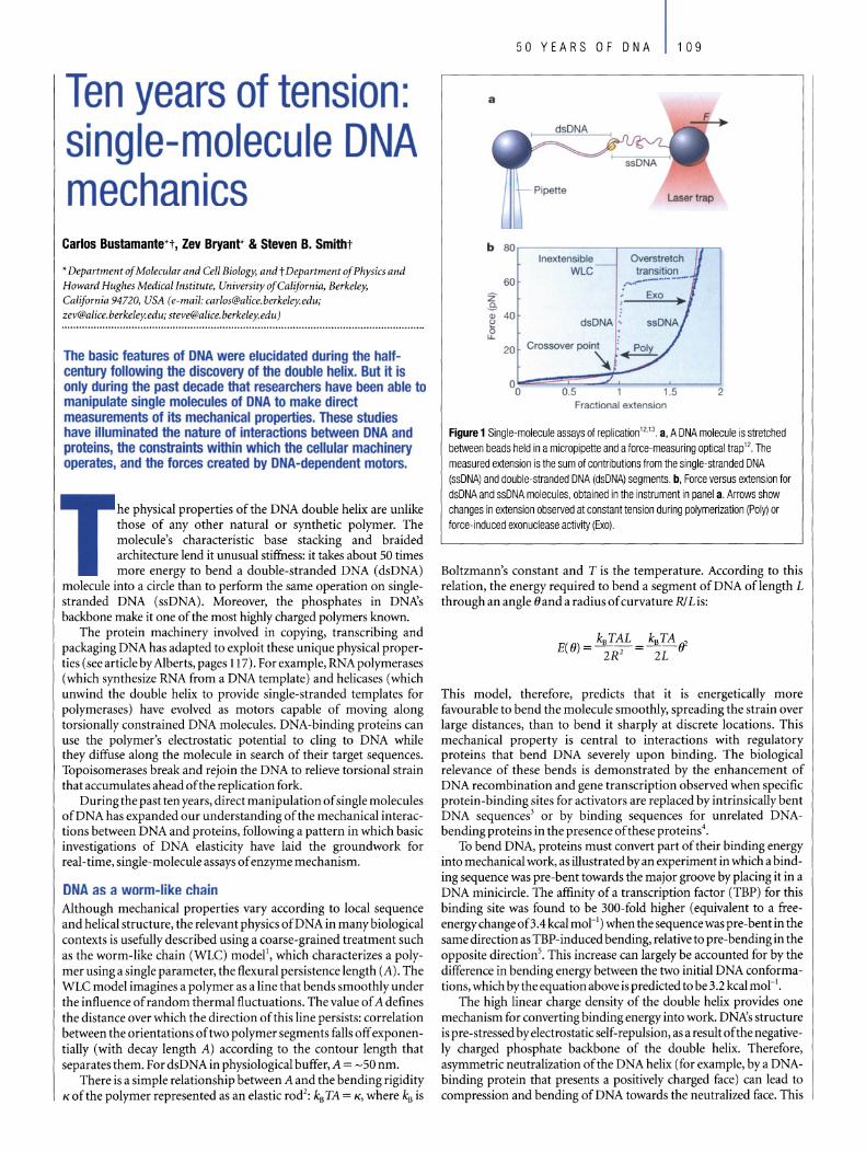
Ten years of tension: single-molecule DNA mechanics
Carlos Bustamante*t, Zev Bryant* & Steven B. Smitht
* Department of Molecular and Cell Biology, and t Department of Physics and Howard Hughes Medical Institute, University ofCalifornia, Berkeley, California 94720, USA (e-mail: [email protected]; [email protected]; [email protected])
The basic features of DNA were elucidated during the halfcentury following the discovery of the double helix. But it is only during the past decade that researchers have been able to manipulate single molecules of DNA to make direct measurements of its mechanical properties. These studies have iIIuminated the nature of interactions between DNA and proteins, the constraints within which the cellular machinery operates, and the forces created by DNA-dependent motors.
he physical properties of the DNA double helix are unlike those of any other natural or synthetic polymer. The molecule's characteristic base stacking and braided architecture lend it unusual stiffness: it takes about 50 times more energy to bend a double-stranded DNA (dsDNA)
molecule into a cirde than to perform the same operation on singlestranded DNA (ssDNA). Moreover, the phosphates in DNA's backbone make it one of the most highly charged polymers known.
The protein machinery involved in copying, transcribing and packaging DNA has adapted to exploit these unique physical properties (seeartide by Alberts, pages 117). Forexample, RNA polymerases (which synthesize RNA from a DNA template) and helicases (which unwind the double helix to provide single-stranded templates for polymerases) have evolved as motors capable of moving along torsionally constrained DNA molecules. DNA-binding proteins can use the polymer's electrostatic potential to ding to DNA while they diffuse along the molecule in search of their target sequences. Topoisomerases break and rejoin the DNA to relieve torsional strain that accumulates ahead ofthe replication fork.
During the past ten years, direct manipulation of single molecules ofDNA has expanded our understanding of the mechanical interactions between DNA and proteins, following a pattern in which basic investigations of DNA elasticity have laid the groundwork for real- time, single-molecule assays of enzyme mechanism.
DNA as a worm-like chain Although mechanical properties vary according to local sequence and helical structure, the relevant physics ofDNA in many biological contexts is usefully described using a coarse-grained treatment such as the worm-like chain (WLC) modell, which characterizes a polymer using a single parameter, the flexural persistence length (A). The WLC model imagines a polymer as a line that bends smoothly under the influence of random thermal fluctuations. The value of Adefines the distance over which the direction of this line persists: correlation between the orientations of two polymer segments falls off exponentially (with decay length A) according to the contour length that separates them. For dsDNA in physiological buffer, A = -50 nm.
There is a simple relationship between A and the ben ding rigidity K of the polymer represented as an elastic rod2: kB TA = K, where kB is
50 YEARS OF DNA 109
b 80 I nexlensi ble Overstretch
WLC transition 60 ,......~_ ... ~_ .. --
Z .s G> 40 e dsDNA : 0
LL
20
00 0.5 1.5 2 Fractional extension
Figure 1 Single-molecule assays 01 replication '2,13, a, A DNA molecule is stretched between beads held in a micropipette and a lorce-measuring optical trap '2. The measured extension is the sum 01 contributions lrom the single-stranded DNA (ssDNA) and double-stranded DNA (dsDNA) segments. b, Force versus extension lor dsDNA and ssDNA molecules, obtained in the instrument in panel a, Arrows show changes in extension observed at constant tension during polymerization (Poly) or lorce-induced exonuclease activity (Exo).
Boltzmann's constant and T is the temperature. According to this relation, the energy required to bend a segment of DNA oflength L through an angle 0 and a radius of curvature R/ L is:
This model, therefore, predicts that it is energetically more favourable to bend the molecule smoothly, spreading the strain over large distances, than to bend it sharply at discrete locations. This mechanical property is central to interactions with regulatory proteins that bend DNA severely upon binding. The biological relevance of these bends is demonstrated by the enhancement of DNA recombination and gene transcription observed when specific protein-binding sites for activators are replaced by intrinsically bent DNA sequences3 or by bin ding sequences for unrelated DNAbending proteins in the presence of these proteins4•
To bend DNA, proteins must convert part of their binding energy into mechanical work, as illustrated by an experiment in which a binding sequence was pre-bent towards the major groove by placing it in a DNA minicirde. The affinity of a transcription factor (TBP) for this binding site was found to be 300-fold higher (equivalent to a freeenergychange oB.4 kcal mor l ) when the sequencewas pre-bent in the same direction as TBP-induced bending, relative to pre-bending in the opposite directions. This increase can largely be accounted for by the difference in bending energy between the two initial DNA conformations, which bythe equation above is predicted to be 3.2 kcal mOr'.
The high linear charge density of the double helix provides one mechanism for converting binding energy into work. DNA's structure is pre-stressed by electrostatic self-repulsion, as a result of the negativeIy charged phosphate backbone of the double helix. Therefore, asymmetric neutralization of the DNA helix (for example, bya DNAbinding protein that presents a positively charged face) can lead to compression and bending ofDNA towards the neutralized face. This
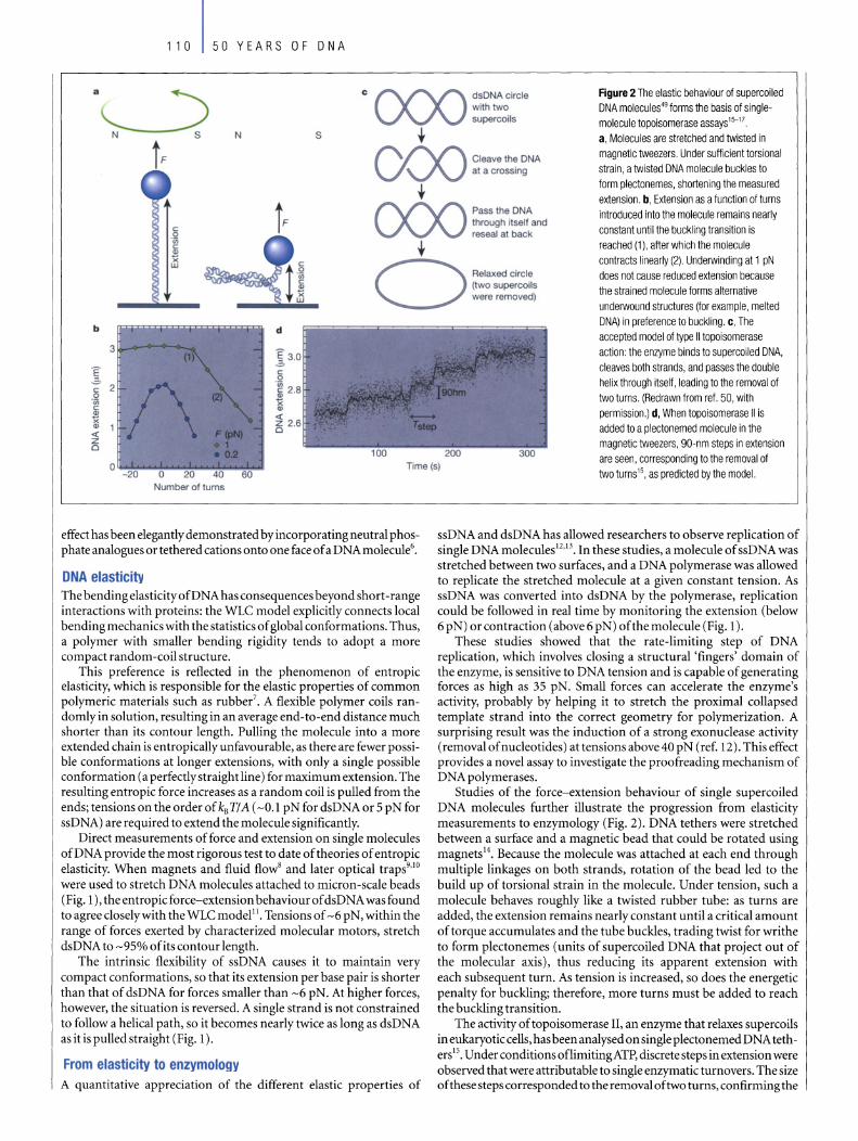
110 50 YEARS OF DNA
.~ C CXX) dsDNAcircle Figure 2 The elastic behaviour of supercoiled with two DNA molecules49 forms the basis of single-supercoils molecule topoisomerase assays'5-H
N s t a, Molecules are stretched and twisted in
t F CXX) Cleave the DNA magnetic tweezers. Under sufficient torsional
at a crossing strain, a twisted DNA molecule buckles to form plectonemes, shorlening the measured t extension. b, Extension as a function of turns
jF CXX) Pass the DNA introduced into the molecule remains nearly c
through itself and constant until the buckling transition is 0 reseal at back
-0;
t reached (1), after which the molecule c ., Ji contracts linearly (2). Underwinding at 1 pN 0-- does not cause reduced extension because
(two supercoils the strained molecule forms alternative were removed)
underwound structures (for example, melted
b d DNA) in preference to buckling. c, The accepted model of type II topoisomerase
3 E action: the enzyme binds to supercoiled DNA, .,2. cleaves both strands, and passes the double E c
.a, 0 helix through itself, leading to the removal of -0; c c 0 ., two turns. (Redrawn from ref. 50, with -0; x ~
., permission.) d, When topoisomerase 11 is x « ., , z added to a plectonemed molecule in the « CI
z magnetic tweezers, gO-nm steps in extension 0
0 Time(s) are seen, corresponding to the removal of
-20 two turns15, as predicted by the model. Number 01 turns
effect has been elegantly demonstrated by incorporating neutral phosphate analogues or tethered cations onto one face of a DNA molecule6•
DNA elasticity The bending elasticity ofDNA has consequences beyond short -range interactions with proteins: the WLC model explicitly connects local bending mechanics with the statistics of global conformations. Thus, a polymer with sm aller bending rigidity tends to adopt a more compact random-coil structure.
This preference is reflected in the phenomenon of entropie elasticity, which is responsible for the elastic properties of common polymerie materials such as rubber? A flexible polymer coils randomly in solution, resulting in an average end-to-end distance much shorter than its contour length. Pulling the molecule into a more extended chain is entropically unfavourable, as there are fewer possible conformations at Ion ger extensions, with only a single possible conformation (a perfectly straight line) for maximum extension. The resulting entropie force increases as a random coil is pulled from the ends; tensions on the order of kB TI A (-0.1 pN for dsDNA or 5 pN for ssDNA) are required to extend the moleeule significantly.
Direct measurements of force and extension on single moleeules ofDNA provide the most rigorous test to date oftheories of entropie elasticity. When magnets and fluid flowB and later optical traps9.10 were used to stretch DNA molecules attached to micron-scale beads (Fig. 1), the entropie force-extension behaviour of dsDNA was found to agree closelywith the WLC modellI. Tensions of -6 pN, within the range of forces exerted by characterized molecular motors, stretch dsDNA to -95% of its contour length.
The intrinsic flexibility of ssDNA causes it to maintain very compact conformations, so that its extension per base pair is shorter than that of dsDNA for forces smaller than -6 pN. At higher forces, however, the situation is reversed. A single strand is not constrained to follow a helical path, so it becomes nearly twice as long as dsDNA as it is pulled straight (Fig. 1).
From elasticity to enzymology A quantitative appreciation of the different elastic properties of
ssDNA and dsDNA has allowed researchers to observe replication of single DNA molecules I2,13. In these studies, a molecule of ssDNA was stretched between two surfaces, and a DNA polymerase was allowed to replicate the stretched moleeule at a given constant tension. As ssDNA was converted into dsDNA by the polymerase, replication could be followed in real time by monitoring the extension (below 6 pN) orcontraction (above6 pN) ofthe molecule (Fig. 1).
These studies showed that the rate-limiting step of DNA replication, which involves closing a structural 'fingers' domain of the enzyme, is sensitive to DNA tension and is capable of generating forces as high as 35 pN. Small forces can accelerate the enzyme's activity, probably by helping it to stretch the proximal collapsed template strand into the correct geometry for polymerization. A surprising result was the induction of a strong exonuclease activity (removal of nucleotides) attensions above 40 p N (ref. 12). This effect provides a novel assay to investigate the proofreading mechanism of DNA polymerases.
Studies of the force-extension behaviour of single supercoiled DNA moleeules further illustrate the progression from elasticity measurements to enzymology (Fig. 2). DNA tethers were stretched between a surface and a magnetic bead that could be rota ted using magnets l4. Because the moleeule was attached at each end through multiple linkages on both strands, rotation of the bead led to the build up of torsional strain in the moleeule. Under tension, such a moleeule behaves roughly like a twisted rubber tube: as turns are added, the extension remains nearly constant until a critical amount of torque accumulates and the tube buckles, trading twist for writhe to form plectonemes (units of supercoiled DNA that project out of the molecular axis), thus reducing its apparent extension with each subsequent turn. As tension is increased, so does the energetic penalty for buckling; therefore, more turns must be added to reach the bucklingtransition.
The activity of topoisomerase II, an enzyme that relaxes supercoils in eukaryotic cells, has been analysed on single plectonemed D NA tetherslS. Underconditions oflimitingATP, discrete steps in extension were observed that were attributable to single enzymatic turnovers. The size ofthese steps corresponded to the removal oftwo turns, confirmingthe
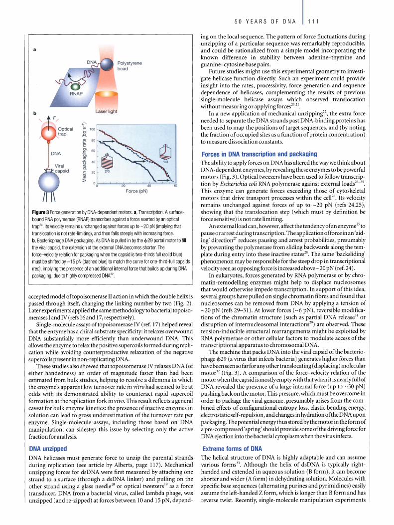
a
b Laser light
1" 1()() a. a ., 80 t!i 0> 60 e: 'e;, co
.>< 40 u co a. e: co cu ::l:
60
Figure 3 Force g eneration by DNA-dependent motors. a, Transcription. A surfacebound RNA polymerase (RNAP) transcribes against a force exerted by an optical trap24 Its velocity remains unchanged against forces up to -20 pN (implying that translocation is not rate-Iimiting), and then falls steeply with increasing force. b, Bacteriophage DNA packaging. As DNA is pulled in by the <1>29 portal motor to fill the viral capsid, the extension of the external DNA becomes shorter. The force-velocity relation for packaging when the capsid is two-thirds full (solid blue) must be shifted by -15 pN (dashed blue) to match the curve for one-third-full capsids (red), implying the presence of an additional internal force that builds up during DNA packaging, due to highly compressed DNA32.
accepted model oftopoisomerase II action in which the double helix is passed through itself, changing the linking number by two (Fig. 2). Later experiments applied the same methodologyto bacterial topoisomerases I and IV (refs 16 and 17, respectively).
Single-molecule assays of topoisomerase IV (ref. 17) helped reveal that the enzyme has a chiral substrate specificity: it relaxes overwound DNA substantially more efficiently than underwound DNA. This allows the enzyme to relax the positive supercoils formed during replication while avoiding counterproductive relaxation of the negative supercoils present in non-replicating DNA.
These studies also showed that topoisomerase IV relaxes DNA (of either handedness) an order of magnitude faster than had been estimated from bulk studies, helping to resolve a dilemma in wh ich the enzyme's apparent low turnover rate in vitra had seemed to be at odds with its demonstrated ability to counteract rapid supercoil formation at the replication fork in viva. This result reflects a general caveat for bulk enzyme kinetics: the presence of inactive enzymes in solution can lead to gross underestimation of the turnover rate per enzyme. Single-molecule assays, including those based on DNA manipulation, can sidestep this issue by selecting only the active fraction for analysis.
DNA unzipped DNA helicases must generate force to unzip the parental strands during replication (see article by Alberts, page 117). Mechanical unzipping forces for dsDNA were first measured by attaching one strand to a surface (through a dsDNA linker) and pulling on the other strand using a glass needle18 or optical tweezers19 as a force transducer. DNA from a bacterial virus, called lambda phage, was unzipped (and re-zipped) at forces between 10 and 15 pN, depend-
50 YEARS OF DNA 111
ing on the local sequence. The pattern of force fluctuations during unzipping of a particular sequence was remarkably reproducible, and could be rationalized from a simple model incorporating the known difference in stability between adenine-thymine and guanine-cytosine base pairs.
Future studies might use this experimental geometry to investigate helicase function directly. Such an experiment could provide insight into the rates, processivity, force generation and sequence dependence of helicases, complementing the results of previous single-molecule helicase assays which observed translocation without measuring or applying forces20,2 1.
In a new application of mechanical unzipping22, the extra force needed to separate the DNA strands past DNA-binding proteins has been used to map the positions of target sequences, and (by noting the fraction of occupied sites as a function of protein concentration) to measure dissociation constants.
Forces in DNA transcription and packaging The abilityto applyforces on DNA has altered the waywe think about DNA-dependentenzymes, byrevealingthese enzymes to be powerful motors (Fig, 3). Optical tweezers have been used to follow tran scripti on by Eseheriehia eoli RNA polymerase against externalloads23- 25,
This enzyme can generate forees exceeding those of cytoskeletal motors that drive transport processes within the ceW6• Its velocity remains unchanged against forces of up to -20 pN (refs 24,25), showing that the translocation step (wh ich must by definition be force sensitive) is not rate limiting.
An externaIload can, however, affect the tendency of an enzyme25 to pause or arrest duringtranscription. The application offorce in an 'aiding' direction27 reduces pausing and arrest probabilities, presumably by preventing the polymerase from sliding backwards a10ng the template during entry into these inactive states28• The same 'backsliding' phenomenon may be responsible for the steep drop in transcriptional velocity seen as opposing force is increased above - 20 P N (ref. 24).
In eukaryotes, forces generated by RNA polymerase or by chromatin-remodelling enzymes might help to displace nucleosomes that would otherwise impede transcription. In support of this idea, several groups have pulled on single chromatin fibres and found that nucleosomes can be removed from DNA by applying a tension of - 20 pN (refs 29-31). At lower forces (-6 pN), reversible modifications of the chromatin structure (such as partial DNA release3l or disruption of internucleosomal interactions29 ) are observed. These tension-inducible structural rearrangements might be exploited by RNA polymerase or other eellular faetors to modulate access of the transcriptional apparatus to chromosomal DNA,
The machine that packs DNA into the viral capsid of the bacteriophage <1>29 (a virus that infects bacteria) generates higher forces than have been seen so far for any other transloca ting ( displacing) molecular motor32 (Fig. 3). A comparison of the force-velocity relation of the motorwhen the capsid is mostlyemptywith thatwhen it is nearlyfull of DNA revealed the presence of a large internal force (up to -50 pN) pushing back on the motor. This press ure, which must be overcome in order to package the viral genome, presumably arises from the combined effects of configurational entropy loss, elastic bending energy, electrostatic self-repulsion, and changes in hydration of the D NA upon packaging. The potential energythus stored bythe motor in the form of a pre-compressed 'spring' should provide some ofthe driving force for DNA ejection into the bacterial cytoplasm when thevirus infects.
Extreme torms ot DNA The helical structure of DNA is highly adaptable and can assurne various forms33 , Although the helix of dsDNA is typically righthanded and extended in aqueous solution (B form), it can become shorter and wider (A form) in dehydrating solution, Molecules with specific base sequences (alternating purines and pyrimidines) easily assume the left -handed Z form, which is longer than B form and has reverse twist. Recently, single-molecule manipulation experiments
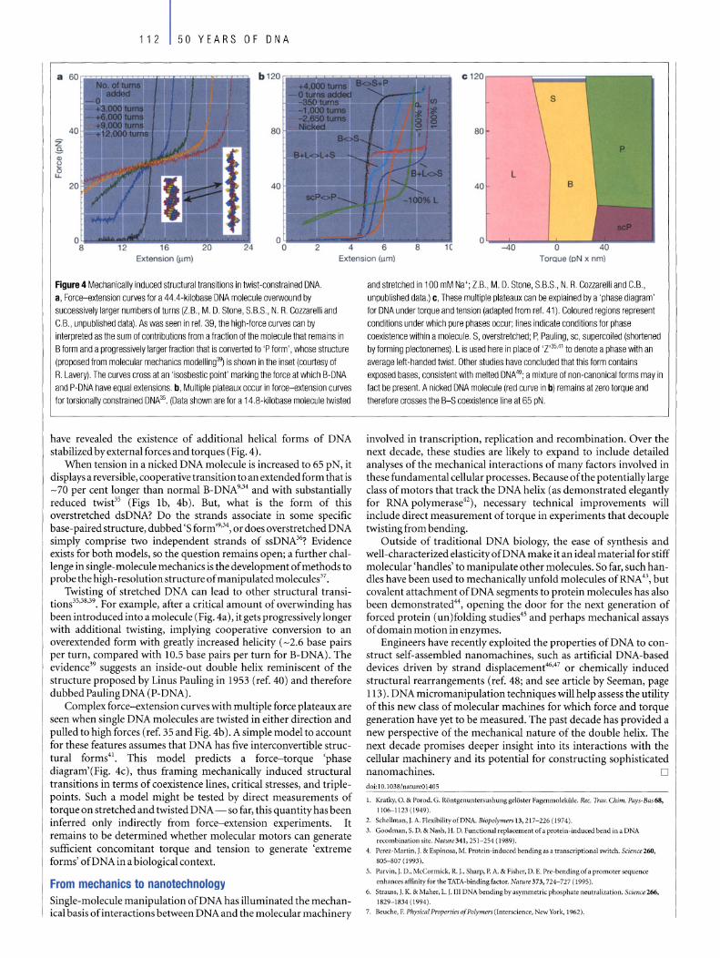
112 50 YEARS OF DNA
80 Z
P S Q)
g L u.
40 B
O~~ __ "",*,_--1
Extension (j.Im) Extension (j.Im) TorQue (pN x nm)
Figure 4 Mechanically induced structural transitions in twist-constrained DNA. a, Force-extension curves for a 44.4-kilobase DNA molecule overwound by successively larger numbers ofturns (l.B., M. D. Stone, S.B.S., N. R. Cozzarelli and C.B., unpublished data). As was seen in ref. 39, the high-force curves can by interpreted as the sum of contributions from a fraction of the molecule that remains in B form and a progressively larger fraction that is converted to 'P form' , whose structure (proposed from molecular mechanics modelling3~ is shown in the inset (courtesy of R. Lavery). The curves cross at an 'isosbestie point' marking the force at which B-DNA and P-DNA have equal extensions. b, Multiple plateaux occur in force-extension curves for torsionally constrained DNA35. (Data shown are for a 14.8-kilobase molecule twisted
have revealed the existence of additional helical forms of DNA stabilized byexternal forces and torques (Fig. 4).
When tension in a nicked DNA molecule is increased to 65 pN, it displays a reversible, cooperative transition to an extended form that is -70 per cent longer than normal B-DNA9,34 and with substantially reduced twise5 (Figs 1b, 4b). But, what is the form of this overstretched dsDNA? Do the strands associate in some specific base-paired structure, dubbed 's form,g,3\ or does overstretched DNA simply comprise two independent strands of ssDNA36? Evidence exists for both models, so the question remains open; a further challenge in single-molecule mechanics is the development of methods to probe the high-resolution structure of manipulated molecules37•
Twisting of stretched DNA can lead to other structural transitions35,38,39. For example, after a critical amount of overwinding has been introduced into a molecule (Fig. 4a), itgets progressively longer with additional twisting, implying cooperative conversion to an overextended form with greatly increased helicity ( - 2.6 base pairs per turn, compared with 10.5 base pairs per turn for B-DNA). The evidence39 suggests an inside-out double helix reminiscent of the structure proposed by Linus Pauling in 1953 (ref. 40) and therefore dubbed Pauling DNA (P-DNA).
Complex force-extension curves with multiple force plateaux are seen when single DNA molecules are twisted in either direction and pulled to high forces (ref. 35 and Fig. 4b). A simple model to account for these features assurnes that DNA has five interconvertible structural forms4!. This model predicts a force-torque 'phase diagram'(Fig. 4c), thus framing mechanically induced structural transitions in terms of coexistence lines, critical stresses, and triplepoints. Such a model might be tested by direct measurements of torque on stretched and twisted DNA - so far, this quantity has been inferred only indirectly from force-extension experiments. It remains to be determined whether molecular motors can generate sufficient concomitant torque and tension to generate 'extreme forms' ofDNA in a biological context.
From mechanics to nanotechnology Single-molecule manipulation ofDNA has illuminated the mechanical basis of interactions between DNA and the molecular machinery
and stretched in 100 mM Na+; l.B., M. D. Stone, S.B.S., N. R. Cozzarelli and C.B, unpublished data.) c, These multiple plateaux can be explained by a 'phase diagram' for DNA undertorque and tension (adapted from ref. 41). Coloured regions represent conditions under which pure phases occur; lines indicate conditions for phase coexistence within a moleeule. S, overstretched; P, Pauling, sc, supercoiled (shortened by forming plectonemes). L is used here in place of 'l'3541 to denote a phase with an average left-handed twist. Other studies have concluded that this form contains exposed bases, consistentwith melted DNA49; a mixture of non-canonical forms may in fact be present. A nicked DNA molecule (red curve in b) remains at zero torque and therefore crosses the B-S coexistence line at 65 pN.
involved in transcription, replication and recombination. Over the next decade, these studies are likely to expand to indude detailed analyses of the mechanical interactions of many factors involved in these fundamental cellular processes. Because of the potentially large dass of motors that track the DNA helix (as demonstrated elegantly for RNA polymerase42), necessary technical improvements will indude direct measurement of torque in experiments that decouple twistingfrom bending.
Outside of tradition al DNA biology, the ease of synthesis and well-characterized elasticity ofDNA make it an ideal material for stiff molecular 'handles' to manipulate other molecules. So far, such handies have been used to mechanically unfold molecules of RNA 43, but covalent attachment ofDNA segments to protein molecules has also been demonstrated44, opening the door for the next generation of forced protein (un)folding studies45 and perhaps mechanical assays of domain motion in enzymes.
Engineers have recently exploited the properties ofDNA to construct self-assembled nanomachines, such as artificial DNA-based devices driven by strand displacement46,47 or chemically induced structural rearrangements (ref. 48; and see artide by Seeman, page 113). DNA micromanipulation techniques will help assess the utility of this new dass of molecular machines for which force and torque generation have yet to be measured. The past decade has provided a new perspective of the mechanical nature of the double helix. The next decade promises deeper insight into its interactions with the cellular machinery and its potential for constructing sophisticated nanomachines. D dai: 1 0.1 038/natureO 1405
1. Kratky, O. & Porad, G. Räntgenuntersushung gelöster Fagenmoleküle. Rec. Trav. Chim. Pays-Bas68,
1106-1123 (1949).
2. Schellman, j. A. Flexibility afDNA. Biopolymers 13, 217-226 (1974).
3. Goodman, S. D. & Nash, H. D. Functional replacement of a protein-induced bend in a DNA
recombination site. Nature341, 251-254 (1989).
4. Perez-Martin, J. & Espinosa, M. Protein-indueed bending as a transcriptional switch. Seienee 260,
805-807 (1993).
5. Parvin, J. D., McCormiek, R. J., Sharp, P. A. & Fisher, D. E. Pre-bending of a promoter sequence
enhances affinity for the TATA-binding faetor. Nature 373,724-727 (1995).
6. Strauss, J. K. & Maher, L. J. 111 DNA bending by asymmetrie phosphate neutralization. Seienee 266, 1829-1834 (1994).
7. Beuche, F. PhysiealProperties ofPolymers (Interscience, NewYork, 1962).

8. Smith, S. 8., Finzi, L. & Bustamante, C. Direct mechanical measurements ofthe elasticity of single
DNA molecules by usingmagnetic beads. Seience258, 1122-1126 (1992) .
9. Smith, S. 8., Cui, Y. & Bustamante, C. Overstretching B-DNA: the elastic response ofindividual
double-stranded and single-stranded DNA molecules. Seienee271, 795-799 (1996).
10. Wang, M. D .• Yin, H., Landick, R, Geiles, J. & Block, S. M. Stretching DNA with optical tweezers.
Biophys. I. 72, 1335-1346 (I997).
11. Bustamante, c., Marko, J. E, Siggia, E. D. & Smith, S. Entropic elasticity oflambda-phage DNA.
Seienee 265, 1599-1600 (I 994).
12. Wuite, G. J., Smith, S. B., Young, M., Keller. D. & Bustamante, C. Single-molecule studies oftheeffect
oftemplate tension on T7 DNA polymerase activity. Nature404,103- 106 (2000).
13. Maier, 8., Bensimon, D. & Croquette. V. Replication bya single DNA polymerase of a stretched
single-stranded DNA. Proc. NatlAcad. Sei. USA 97,12002-12007 (2000).
14. Strick, 1. R., Allemand, J. E, Bensimon, D., Bensimon, A. & Croquette, V. The elasticity of a single
supercoiled DNA molecule. Seience271 , 1835-1837 (1996).
15. Strick, 1. R., Croquette, V. & Bensimon, D. Single-molecule analysis of DNA uncoiling by a type II
topoisomerase. Nature 404. 901-904 (2000) .
16. Dekker, N. H. et al. The mechanism of type IA topoisomerases. Proe. Na tl Acad. Sei. USA 99,
12126-12131 (2002).
17.Crisona, N. J., Strick, 1. R. , Bensimon, D., Croquette, V. & Cozzarelli, N. R. Preferential relaxation of
positiveJy supercoiled DNA by E. coU topoisomerase IV in single-molecule and ensemble
measurements. Genes Dev. 14.2881-2892 (2000).
18. Essevaz-Roulet, 8., Bockelmann, U. & Heslot, F. Mechanical separation ofthe complementary strands
ofDNA. Proc. Natl Acad. Sei. USA 94,11 935-11940 (1997).
19. Bockelmann, 0., Thomen, P., Essevaz-Roulet, 8., Viasnoff, V. & Heslot, F. Unzipping DNA wirh
optical tweezers: high sequence sensitivity and force flips. Biophys. J. 82,1537-1553 (2002).
20. Bianco, P. R. et a/. Processive translocation and DNA unwinding by individual RecBCD enzyme
rnolecules. Nature 409, 374-378 (200 1).
21. Dohoney, K. M. & Geiles, J. x-Sequence recognition and DNA translocation by single RecBCD
helicaselnuclease rnolecules. Nature409. 370-374 (200 1).
22. Koch, S. J., Shundrovsky, A., Jantzen, B. C. & Wang, M. D. Probing protein-DNA interactions by
unzipping a single DNA double helix. Biophys./. 83, 1098-11 05 (2002).
23. Yin, H. er al. Transcription against an applied force. Science 270. 1653- 1657 (1995).
24. Wang, M. D. et al. Force and velocity measured for single molecules ofRNA polymerase. Science 282,
902-907 (1998).
25. Davenport, R. ,. , Wuite, G. J., Landick, R. & Bustamante, C. Single-molecule study oftranscriptio nal
pausing and arrest by E. coli RNA polymerase. Säence 287,2497-2500 (2000).
26. Vale, R. D. & Milligan, R. A. The way things rnove: looking under the hood of molecular motor
proteins. Scienee 288. 88-95 (2000).
27. Forde, N. R., lzhaky, 0., Woodcock, G. R., Wuite, G. J. & Bustamante, C. Usingmechanical force to
probe the mechanism of pausing and arrest during continuous elongation by Escherichia coli RNA
polymerase. Proc. Natl Acad. Sei. USA 99, 11 682-11687 (2002 ).
28. Landick, R. RNA polymerase slides horne: pause and termination site recognition. Ce1l88, 74 1-744
(1997).
29. Cui, Y. & Bustamante, C. Pulling a single chromatin fiber reveals the fo rces that rnaintain its higher
order structure. Froe. NatlAcad. Sei. USA 97,127- 132 (2000).
30. Bennink, M. L. et al. Unfolding individual nucleosomes by stretching single chromatin fibers with
optical tweezers. Nature Struct. Biol. 8. 606--610 (2001).
31 . Brower-Toland, 8. D. er al. Mechanical disruption of individual nucleosomes reveals a reversible
multistage release ofDNA. Proc. Natl Acad. Sei. USA 99, 196()-1965 (2002).
32. Smith, D. E. el al. The bacteriophage <f.29 portal motor can package DNA against a large internal
force. Nnture413, 748-752 (2001).
33.Calladine, C. R. & Drew, H. Understanding DNA (Academic, London, 1997).
34. Cluzel, P. el al. DNA: an extensible molecule. Science 27l, 792-794 (1996).
35. Leger, J. F. el al. Structural transitions of a twisted and stretched DNA molecule. Phys. Rev. Lett. 83,
1066-1069 (1999).
36. Williams, M. c., Rouzina, I. & Bloomfield, V. A. Thermodynamics of DNA interactions from single
moleeule stretchingexperimcms. Ace. e hern. Res. 35,1 59-166 (2002).
37. Wilkins, M. H. F., Gosling, R. G. & Seeds, W. E. Nucleic acid: an extensible molecule? Nature 167,
759-760 (1951).
38. Strick, 1. R., Allemand,]. E, Bensimon, D. & Croquette, V. Behavior of supercoiled DNA. Biophys.].
74,2016-2028 (1998).
39. Allemand, J. E, Bensimon, D., Lavery, R. & Croquette, V. Stretched and overwound DNA forms a
Pauling-like structure with exposed bases. Proc. Natl Acad. Sei. USA 95, 14152-14157 (1998) .
40. Pauling, L & Corey, R B. A proposed structure for the nudeic acids. Proc. Natl Aead. Sei. USA 39.
84-97 (1953).
41. Sarkar, A., Lege r, J. E, Chatenay, D. & Marko,]. F. Structural transitions in DNA driven by external
force and torque. Phys. Rev. E63, 051903-1-051903-10 (2001).
42. Harada, Y. et al. Direct observation ofDNA rotation during transcription by Escherichia coli RNA
polymerase. Nature 409, 113-115 (2001).
43. Liphardt, J., Onoa, 8., Smith, S. 8. , Tinoco, I. J. & Bustamante. C. Reversible unfolding of single RNA
molecules by mechanical force. Scienee292, 733-737 (200] ).
44. Hegner, M. DNA Handles for single molecule experiments. Single Mol. 1, 139--144 (2000).
45. Carrion-Vazquez, M. el al. Mechanical design of proteins studied by single-molecule forcc
spectroscopy and protein engineering. Prog. Biophys. Mol. Biol. 74, 63-91 (2000).
46. Yurke, 8., Turberfield, A. J., Mills, A. P., Simrnel, F. C. & Neumann, J. L. A DNA-fuelled molecular
machine made ofDNA. Nature406, 605-608 (2000).
47. Yan, H., Zhang, X., Shen, Z. & Seernan, N. C A robust DNA rnechanical device controlled by
hybridization topology. Nature415, 62-65 (2002).
48. Mao, C, Sun, W., Shen, Z. & Seernan, N. C A nanomechanical device based on the B-Z transition of
DNA. Nnture397, 144-146 (1999).
49. Strick, 1., Allemand, ]., Croquette, V. & Bensimon, D. Twisting and stretch ing single DNA molecules.
Prog. Biophys. Mol. Biol. 74,115-140 (2000).
50. Alberts, B. e' al. MoleCIIlnr Biology 01 the Cell (Garland, New York, 2002 ).
Original reference: Natu re 421. 423-427 (2003) .
50 YEARS OF DNA 113
DNA in a material world Nadrian C. Seeman
Department ofChemistry, New York University, New York 10003, USA (e-mail: [email protected])
The specific bonding 01 DNA base pairs provides the chemical foundation for genetics. This powerful molecular recognition system can be used in nanotechnology to direct the assembly of highly structured materials with specific nanoscale features, as weil as in DNA computation to process complex information. The exploitation of DNA for material purposes presents a new chapter in the history of the molecule.
"The nucleic-acid 'system' that operates in terrestriallife is optimized (through evolution) chemistry incarnate. Why not use it .. . to allow human beings to sculpt something new, perhaps beautiful, perhaps useful, certainly unnatural." Roald Hoffmann, writing in American Scientist, 1994 (ref. 1).
he DNA molecule has appealing features for use in nanotechnology: its minuscule size, with a diameter of about 2 nanometres, its short structural repeat (helical pitch) of about 3.4-3.6 nm, and its 'stiffness', with a persistence length (a measure of stiffness) of around
50 nm. There are two basic types of nanotechnological construction: 'top-down' systems are where microscopic manipulations of small numbers of atoms or molecules fashion elegant patterns (for example, see ref. 2), while in 'bottom-up' constructions, many molecules self-assemble in parallel steps, as a function of their molecular recognition properties. As a chemically based assembly system, DNA will be a key player in bottom-up nanotechnology.
The origins of this approach date to the early 1970s, when in vitro genetic manipulation was first performed by tacking together molecules with 'sticky ends'. A sticky end is a short single-stranded overhang protruding from the end of a double-stranded helical DNA molecule. Like flaps of Velcro, two molecules with complementary sticky ends - that is, their sticky ends have complementary arrangements of the nucleotide bases adenine, cytosine, guanine and thymine - will cohere to form a molecular complex.
Sticky-ended cohesion is arguably the best example of programmable molecular recognition: there is significant diversity to possible sticky ends (4N for N-base sticky ends), and the product formed at the site of this cohesion is the classic DNA double helix. Likewise, the convenience of solid support-based DNA synthesis3 makes it is easy to pro gram diverse sequences of sticky ends. Thus, sticky ends offer both predictable control of intermolecular associations and predictable geometry at the point of cohesion. Perhaps one could get similar affinity properties from antibodies and antigens, but, in contrast to DNA sticky ends, the relative three-dimensional orientation of the antibody and the antigen would need to be determined for every new pair. The nucleic acids seem to be unique in this regard, providing a tractable, diverse and programmable system with remarkable control over intermolecular interactions, coupled with known structures for their complexes.
Branched DNA There is, however, a catch; the axes of DNA double helices are unbranched lines. Joining DNA molecules by sticky ends can yield Ion ger lines, perhaps with specific components in a particular linear

114 50 YEARS OF DNA
Figure 1 Assembly 01 branched DNA molecules. a, Sell-assembly 01 branched DNA moleeules into a two-dimensional crystal. A DNA branched junction lorms lrom lour DNA strands; those strands coloured green and blue have complementary sticky-end overhangs labelied Hand H', respectively, whereas those coloured pink and red have complementary overhangs V and V', respectively. A number 01 DNA branched junctions cohere based on the orientation 01 their complementary sticky ends, lorming a square-like unit with unpaired sticky ends on the outside, so more units could be added to produce a
a
two-dimensional crystal. b, Ligated DNA moleeules lorm interconnected rings to create a cube-like structure. The structure consists 01 six cyclic interlocked single strands, each linked twice to its lour neighbours, because each edge contains two turns 01 the DNA
or cyclic order in one dimension. Indeed, the chromosomes packed inside cells exist as just such one-dimensional arrays. But to produce interesting materials from DNA, synthesis is required in multiple dimensions and, for this purpose, branched DNA is required.
Branched DNA occurs naturally in living systems, as ephemeral inter mediates formed when chromosomes exchange information during meiosis, the type of cell division that generates the sex cells (eggs and sperm). Prior to cell division, homologous chromosomes pair, and the aligned strands ofDNA break and literallycross over one another, forming structures called Hollidayjunctions. This exchange of adjacent sequences by homologous chromosomes - a process called recombination - during the formation of sex cells passes genetic diversity onto the next generation.
The Holliday junction contains four DNAstrands (each member of a pair of aligned homologous chromosomes is composed of two DNA strands) bound together to form four double-helical arms flanking a branch point (Fig. la). The branch point can relocate throughout the molecule, byvirtue ofthe homologous sequences. In contrast, synthetic DNA complexes can be designed to have fixed branch points containing between three and at least eight arms4•5• Thus, the prescription for using DNA as the basis for complex materials with nanoscale features is simple: take synthetic branched DNA molecules with programmed sticky
Figure 2 Two-dimensional DNA arrays. a, Schematic drawings 01 DNA double crossover (DX) units. In the meiotic DX recombination intermediate, labelied MDX, a pair 01 homologous chromosomes, each consisting 01 two DNA strands, align and cross over in order to swap equivalent portions 01 genetic inlormation; 'HJ' indicates the Holliday junctions. The structure 01 an analogue unit (ADX), used as a tiling unit in the construction 01 DNA two-dimensional arrays, comprises two red strands, two blue crossover strands and a central green crossover strand. b, The strand structure and base pairing 01 the analogue ADX molecule, labelled A, and a variant, labe lied B*. B* contains an extra DNA domain extending lrom the central green strand that, in practice, protrudes roughly perpendicular to the plane 01 the rest 01 the DX molecule. c, Schematic representations 01 A and B* where the perpendicular domain 01 B* is represented as a blue circle. The complementary ends 01 the ADX molecules are represented as geometrical shapes to illustrate how they lit together when they sell-assemble. The dimensions 01 the resulting tiles are about 4 x 16 nm and are joined
b
double helix. For example, the lront red strand is linked to the green strand on the right, the light blue strand on the top, the magenta strand on the left, and the dark blue strand on the bottom. It is linked only indirectly to the yellow strand at the rear.
ends, and get them to self-assemble into the desired structure, which maybe aclosedobjectoracrystalline array (Fig. la).
Other modes of nucleic acid interaction aside from sticky ends are available. For example, Tecto-RNA molecules6, held together by loop-loop interactions, or paranemic crossover (PX) DNA, where cohesion derives from pairing of alternate halfturns in inter-wrapped double helices? These new binding modes represent programmable cohesive interactions between cyclic single-stranded molecules that do not require cleavage to expose bases to pair molecules together. Nevertheless, cohesion using sticky ends remains the most prominent intermolecular interaction in structural DNA nanotechnology.
DNA constructions It is over a decade since the construction of the first artificial DNA structure, a stick-cube, whose edges are double helices8 (Fig. Ib). More complex polyhedra and topological constructs9 , such as knots and Borromean rings (consisting of three intricately interlinked circles), followed. But the apparent floppiness of individual branched junctions led to a hiatus before the next logical step: selfassembly into two-dimensional arrays.
This step required a stiffer motif, as it was difficult to build a periodic well-structured array with marshmallow-like components,
HJ HJ b
~ a + + • • ~ ::=x::::x:= ~ CJ C;-.. •
MDX ADX A B"
c A B"
+
together so that the B* protrusions lie about 32 nm apart. d, The B* protrusions are d
visible as 'stripes' in tiled DNA arrays under an atomic lorce microscope.
1 _32 nm
o 492 nm
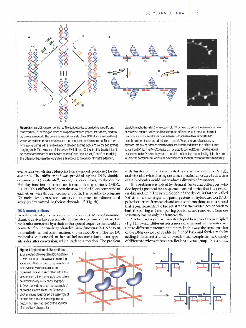
D C
Figure 3 A rotary DNA nanomachine. a, The device works by producing two different conformations, depending on which of two pairs 01 strands (calied 'set' strands) binds to the device framework. The device framework consists of two DNA strands (red and blue) whose top and bottom double helices are each connected by single strands. Thus, they form two rigid arms with a flexible hinge in between and the loose ends of the two strands dangling freely. The two states of the device, PX (Ieft) and JX, (right), differ by a half turn in the relative orientations of their bottom helices (C and 0 on the left, 0 and C on the right). The difference between the two states is analogous to two adjacent fingers extended,
even with a well-defined blueprint (sticky-ended specificity) for their assembly. The stiffer motif was provided by the DNA doublecrossover (DX) molecule lO , analogous, once again, to the double Holliday-junction intermediate formed during meiosis (MDX, Pig. 2a). This stiff molecule contains two double helices connected to each other twice through crossover points. It is possible to pro gram DX molecules to produce a variety of patterned two-dimensional arrays just bycontrollingtheirstickyends ll - 13 (Pig. 2b).
DNA constructions In addition to objects and arrays, a number ofDNA-based nanomechanical devices have been made. The first device consisted oftwo DX molecules connected by a shaft with a special sequence that could be converted from normal right-handed DNA (known as B-DNA) to an unusualleft -handed conformation, known as Z-DNA 14. The two DX molecules lie on one side of the shaft before conversion and on opposite sides after conversion, which leads to a rotation. The problem
Figure 4 Applications of DNA scaffolds. a, Scaffolding of biological macromolecules. a A DNA box (red) is shown with protruding sticky ends that are used to organize boxes into crystals. Macromolecules are organized parallel to each other within the box, rendering them arnenable to structure determination by X-ray crystallography. b, DNA scaffolds to direct the assembly of nanoscale electrical circuits. Branched DNA junctions (blue) direct the assembly of attached nanoelectronic components (red), which are stabilized by the addition of a positively charged ion.
parallel to each other (right), or crossed (Ieft). The states are set by the presence of green or yellow set strands, wh ich bind to the frame in different ways to produce different conformations. The set strands have extensions that enable their removal when complementary strands are added (steps land 111). When one type of set strand is removed, the device is free to bind the other set strands and switch to a different state (steps 11 and IV). b, The PX-JX, device can be used to connect 20-nm DNA trapezoid constructs. In the PX state, they are in a parallel conformation, but in the JX, state, they are in a zig-zag conformation, which can be visualized on the right by atomic force microscopy.
with this device is that it is activated by a sma11 moleeule, Co(NH3)~~ and with a11 devices sharing the same stimulus, an ordered collection ofDX molecules would not produce a diversity of responses.
This problem was solved by Bernard Yurke and co11eagues, who developed a protocol for a sequence-control device that has a tweezers-like motion 15. The principle behind the device is that a so-ca11ed 'set' strand containing a non-pairing extension hybridizes to a DNApaired structural frameworkand sets a conformation; another strand that is complementaryto the 'set' strand is then added, which binds to both the pairing and non-pairing portions, and removes it from the structure, leaving only the framework.
A robust rotary device was developed based on this principle l6
(Pig. 3), in which different set strands can enterand set the conformation to different structural end-states. In this way, the conformation of the DNA device can readily be tlipped back and forth simply by addingdifferent set strands followed bytheir complements. A variety of different devices can be controlled by a diverse group of set strands.
b
+

116 50 YEARS OF DNA
ONA nanomachines What is the purpose of constructing DNA arrays and nanodevices? One prominent goal is to use DNA as scaffolding to organize other molecules. For example, it may be possible to use self-assembled DNA lattices (crystals) as platforms to position biological macromoleeules so as to study their structure by X-ray crystallography4
(Fig. 4a). Towards this goal, programming ofDNA has been used to bring protein moleeules in proximitywith each other to fuse multiple enzymatic activities17 • However, the potential of this approach awaits the successful self-assembly of three-dimensional crystals.
Another goal is to use DNA crystals to assemble nanoelectronic components in two- or three-dimensional arrays 18 (Fig. 4b). DNA has been shown to organize metallic nanopartides as aprecursor to nanoelectronic assembl/9- 22 , but so far it has not been possible to produce multidimensional arrays containing nanoelectronic components with the high -structural order of the naked DNA arrays described earlier.
There has been some controversy over whether DNA can be used as an electrical conductor (for example, ref. 23), although the resolution of this debate is unlikely have any impact on the use ofDNA as a scaffold. Recently, the effects ofDNA conformational changes on conduction in the presence of an analyte were shown to have potential as a biosensor24 •
Replicating ONA components A natural question to ask of any assembly system based on DNA is whether the components can be replicated. To produce branched DNA molecules whose branch points do not move, they must have different sequences in opposite branches but, as a consequence, these structures are not readily reproduced by DNA polymerase; the polymerase would produce complements to all strands present, leading only to double helical molecules. One option is to use topological tricks to convert structureslike the DNA eube into a long single strand byadding extrastretches ofDNA bases. The single strand could then be replicated by DNA polymerase and the final replicated product induced to fold into the original shape, with any extraneous segments deaved using restriction enzymes. Although this would produce a molecule with sticky ends ready to participate in self-assembly, it would be a cumbersome process25•
Günter von Kiedrowski and colleagues have recently developed a way of replicating short, simple DNA branches in a mixed organic-DNA species. Their branched molecule consists ofthree DNA single strands bonded to an organic triangle-shaped linker. To replicate the branched moleeule, the single-stranded complement of each of these strands is bound to the moleeule, so that one end of each complement molecule is dose to the same end ofthe other complement moleeule. In the final step, the juxtaposed complements are connected together by bonding their neighbouring ends to another molecule ofthe organiclinker26• Extension ofthis system to the next level, such as objects like the eube, will need to solve topological problems involved in the separation of the two components, or it will be limited to unligated systems.
Future prospects Many separate capabilities of DNA nanotechnology have been prototyped - it is now time to extend and integrate them into useful systems. Combining sequence-dependent devices with nanoscale arrays will provide a system with a vast number of distinct, programmable structural states, the sine qua nonofnanorobotics. Akey step in realizing these goals is to achieve highly ordered three-dimensional arrays, both periodic and, ultimately, algorithmic.
Interfacing with top-down nanotechnology will extend markedly the capabilities ofthe field. Halso will be necessaryto integrate biological macromoleeules or other macromoleeular complexes into DNA arrays in order to make practical systems with nanoscale components. Likewise, the indusion of electronic components in highly ordered arrays will enable the organization of nanoelectronic cireuits. Chemical function could be added to DNA arrays by adding nudeic acid species evolved in vitro to have specific bin ding properties ('aptamers') or enzymatic activities ('ribozymes' or 'DNAzymes'). A further area that has yet to have an impact on DNA nanotechnology is
Box 1 DNAcomputers
An assembly of DNA strands can process data in a similar way as an electronic computer, and has the potentialto solve far more complex problems and store a greater amount of information. for substantially less energy costs than do electronic microprocessors. DNA-based computation dates from Leonard Adleman's landmark report in 1994 (rel. 27), where he used DNA to solve the 'Hamiltonian path' problem. a variant of the 'travelling salesman' problem. The idea is to establish whether there is a path between two cities, given an incomplete set of available roads. Adleman used strands of DNA to represent cities and roads, and encoded the sequences so that a strand representing a road would connect (according 10 the rules of base pairing) to any two strands representing a city. Sy mixing together the strands, joining the cities connected by roads. and weeding out any 'wrong answers·. he showed that Ihe strands could self-assemble to solve the problem.
It is impossible to separate DNA nanotechnology from DNAbased computation: many researehers work in both leids and the two communities have a symbiotic relationship. The first link between DNA computation and DNA nanotechnology was established by Erik Winfree, who suggested that short branched DNA molecules could be 'programmed' to undergo algorilhmic selfassemblyand thus serve as the basis of computation18.
Periodic building blocks of matter. such as the DNA molecules shown in Fig. 1 a. represent the simplest algorithm for assembly. All components are parallel, so what is on one side of a component is also on the other side, and in every direction. Given this parallelism, if the right side complements the lett. the top complements the bollom and the front complements the back. a crystaJ should resul\. Even more complex algorithms are possible if one uses components 01 the same shape, but with different sticky ends, For example. Winfree has shown that , in principle. DNA tiles can be used to 'count' (see figure below) by creating borders with programmable sizes forone-, two- and possibly three-dimensionaJ assemblies29. 1f this scheme can be reaJized, self-assembly of precisely sized nanoscaJe arrays will be possible. A computation using selfassembly has been prototyped in one dimension, thereby lending some credence 10 the viability 01 algorilhmic assembl ,
Box 1 Figure Counling wilh self-assembled ONA tiles. ONA tiles are represented by squares with coloured edges that are protruded or indented. Seven componenl Illes are shown on the lett : Ihree border tiles on Ihe bottom and four tiles wtth the vatues 0 or 1. The array iIIustrates binary cou~ting 'rom 1 (bollorn row) to 12 (top row). Assembly is assumed to proceed by forming the reverse L -shaped border first. followed by binding the tlles that fit into the sites containing \wo (but not one) edges. Thus. the border determines the 1 tile in its bend. then thai 1 tUe and the horizontat-border tile on its lelt deterrnlne the 0 tile that fits . while the 1 tile and the vertical-border liIe above it deterrnine the (different) 0 tile that fils. (Adapted from rel. 29.)
12
11
10
9
7
6
5
4
3
2
combinatorial synthesis, which may weil lead to greater diversity of integrated components. DNA-based computation and algorithmic assembly is another active area of research, and one that is impossible to separate from DNA nanotechnology (see Box 1).
The field of DNA nanotechnology has attracted an influx of researchers over the past fewyears. Allofthose involved in this area have benefited from the biotechnology enterprise that produces DNAmodifying enzymes and unusual components for synthetic DNA molecules. It is likelythat applications in structuralDNAnanotechnology ultimately will use variants on the theme of DNA (for example, peptide nucleic acids, containing an unconventional synthetic peptide backbone and nucleic acid bases for side chains), whose properties may be better suited to particular types of applications.
For the past half-century, DNA has been alm ost exclusively the province of biologists and biologically oriented physical scientists, who have studied its biological impact and molecular properties. During the next 50 years, it is likely they will be joined by materials scientists, nanotechnologists and computer engineers, who will exploit DNA's chemical properties in a non-biological context. 0 doi: I 0.1 03B/natureO 1406
I. Hoffmann, R. DNA as day. Am. Sei. 82, 308-311 (1994).
2. Ctiberes, M. T.) Schlittier, R. R. & Gimzewski, J. K. Room-temperature repositioning ofindividual C-60
moleculesat Cu steps: operation ofa molecularcountingdevice. Appl. Phys. Lett. 69, 3016-3018 (1996).
3. Caruthers, M. H. Gene synthesis machines: DNA chemistry aod its uses. Science230, 281-285 (1985).
4. Seeman, N. C. Nudeic acid junctions aod lattkes. /. Theor. Biol. 99, 237-247 (1982).
S. Seeman, N. C. Molecular craftwork with DNA. ehern. Intell. 1,38-47 (1995).
6. Jaeger, L., Westhof, E. & Leontis, N. B. Tecto-RNA: modular assemblyunits for the construction of
RNA nano-objects. NudeicAcids Res. 29, 455-463 (2001).
7. Zhang, X., Yan, H., Shen, Z. & Seeman, N. C. Paranemic cohesion oftopologically-c1osed DNA
molecules. /. Am. Ch"n. Soc.I24, 12940-12941 (2002).
8. Chen, J. & Seeman, N. C. The synthesis from DNA of a moleeule with the connectivity of a cube.
Nature 350, 631-633 (1991).
9. Seeman, N. C. Nucleic acid nanostructures and topology. Angew. Chem. Int. Edn Eng!. 37,
3220-3238 (1998).
10. Li, X., Yang, X., Qi, J. & Seeman, N. C. AntiparalieJ DNA double crossover molecules as components
for nanoconstruction. J. Am. Chem. 50c.118, 6131-6140 (1996).
11. Winfree, E., Liu, F., Wenzier, L.A. & Seeman, N.C. Design and self-assembly of two-dimensional DNA
crystaIs. Nature 394, 539-544 (1998).
12.Mao, c., Sun, W. & Seeman, N. C. Designed two-dimensional DNA Holliday junction arrays
visualized by atomic force microscopy. /. Am. Chem. 50c. 121,5437-5443 (1999).
13. LaBean, T. et a!. The cOllstructioll, analysis, ligation and seJf-assembly ofDNA tripie crossover
complexes. J. Am. Chem. $Oc. 122, 1848-1860 (2000).
14. Mao, c., SUll, W., Shen, Z. & See mall, N. C. A DNA nanomechanical device based on the B-Z
transition. Nature 397,144---146 (1999).
15. Yurke, B., TurberfieJd, A. J., Mills, A. P. Jr, SimmeJ, F. C. & Newmann, J. L. A DNA-fuelled molecular
machine made ofDNA. Nature 406, 605-608 (2000).
16. Yan, H., Zhang, X., Shen, Z. & Seeman, N. C. A robust DNA mechanical device controlled by
hybridization topology. Nature4lS, 62--65 (2002).
17. Niemeyer, C. M., Koehler, J. & Wuerdemann, C. DNA-directed assembly ofbi-enzymic complexes from
in vivo biotinylated NADP(H):FMN oxidoreductase and luciferase. ChemBioChem 3, 242-245 (2002).
18. Robinson, B. H. & Seeman, N. C. The design ofa biochip: a self-assembling molecular-scale memory
device. Protein Eng. 1,295-300 (1987).
19. Keren, K. et al. Sequence-specific molecular lithography on single DNA molecules. Science 297,
72-75 (2002).
20. Alivisatos, A. P. et al. Organization of'nanocrystal molecules' using DNA. Nature 382,609-611 (1996).
21. Taton, T. A., Mucic, R. c., Mirkin, C. A. & Letsinger, R. L. The DNA-mediated formation of supramolecular
O1ono- aod multilayered nanoparticle structures. /. Am. Chem. 50c. 122,6305--6306 (2000).
22. Pena, S. R. N., Raioa, 5., Goodrich, G. P., Fedoroff, N. V. & Keating, C. D. Hybridization and enzymatic
extension of Au nanopartide-bound oligonucleotides. /. Am. Chern. 50c.124, 7314---7323 (2002).
23. Dekker, C. & Ratner, M. A. Electronic properties ofDNA. Phys. World 14, 29-33 (2001).
24. Fahlman, R. P. & Sen, D. DNA conforrnational switches as sensitive electronic sensors of analytes. J. Am. Chem. Soc. 124,4610-4616 (2002).
25. Seeman, N. C. The construction of3-D stickfigures !Tom branched DNA. DNA Cell Biol. 10,475-486 (1991).
26. Eckardt, L. H. ef al. Chemical copying of connectivity. Nature 420, 286 (2002).
27. Adlernan, L. Molecular computation of solutions to combinatorial problems. Seienee 266,
1021-1024 (1994).
28. Winfree, E. in DNA Based Computers. Proeeedings 0/ a DIA1ACS Workshop, April 4, 1995, Prineeton
University(eds Lipton, R. J & Baum, E. B.) 199-219 (American MathematicalSociety, Providence, 1996).
29. Winfree, E. AIgorithmic self-assemblyofDNA: theoretical motivations and 2D assembly
experiments.]. Bio1. Mol. Struet. Dynarnies Conversat. 112,263-270 (2000).
30. Mao, c., LaBean, T., Reif, J. H. & Seeman, N. C. Logical computation using algorithmic self-assembly
ofDNA tripie crossover molecules. Nature407, 493-496 (2000).
Acknowledgements This work has been supported by grants from the National Institute of General Medical Sciences, the Office ofNaval Research, the National Science Foundation, and the Defense Advanced Research Projects Agency/ Air Force Office ofScientific Research.
Original reference: Nature 421, 427-431 (2003).
50YEARSOFDNA 117
DNA replication and recombination
Bruce Alberts
National Academy ofSciences, 2101 Constitution Avenue, Washington DC 20418, USA
Knowledge of the structure of DNA enabled scientists to undertake the difficult task of deciphering the detailed molecular mechanisms of two dynamic processes that are central to life: the copying of the genetic information by DNA replication, and its reassortment and repair by DNA recombination. Despite dramatic advances towards this goal over the past five decades, many challenges remain for the next generation of molecular biologists.
"Though facts are inherently less satisfying than the intellectual conclusions drawn from them, their importance should never be questioned." James D. Watson, 2002.
NA carries all of the genetic information for life. One enormously long DNA molecule forms each of the chromosomes of an organism, 23 of them in a human. The fundamental living unit is the single cell. A cell gives rise to many more cells through serial repetitions
of a process known as cell division. Before each division, new copies must be made of each of the many molecules that form the cell, induding the duplication of all DNA molecules. DNA replication is the name given to this duplication process, which enables an organism's genetic information - its genes - to be passed to the two daughter cells created when a cell divides. Only slightly less central to life is a process that requires dynamic DNA acrobatics, called homologous DNA recombination, which reshuffles the genes on chromosomes. In reactions closely linked to DNA replication, the recombination machinery also repairs damage that inevitably occurs to the long, fragile DNA molecules inside cells (see accompanying article by Friedberg, page 122).
The model for the DNA double helixl proposed by James Watson and Francis Crick is based on two paired DNA strands that are complementary in their nucleotide sequence. The model had striking implications for the processes ofDNA replication and DNA recombination. Before 1953, there had been no meaningful way of even speculating about the molecular mechanisms of these two central genetic processes. But the proposal that each nudeotide in one strand ofDNA was tightly base-paired with its complementary nucleotide on the opposite strand -either adenine (A) with thymine (T), or guanine (G) with cytosine (C) - meant that any part of the nucleotide sequence could act as a direct template for the corresponding portion ofthe other strand. As aresult, anypart ofthe sequence can be used eitherto createor to recognize its partner nudeotide sequence - the two functions that are central for DNAreplication and DNA recombination, respectively.
In this review, I discuss how the discovery ofthe structure ofDNA half a century ago opened new avenues for understanding the processes ofDNA replication and recombination. I shall also emphasize how, as our understanding of complex biological molecules and their interactions increased over the years, there have been profound changes in the way that biologists view the chemistry oflife.
Structural features of DNA The research that immediately followed the discovery of the double helix focused primarily on understanding the structural properties

118 50 YEARS OF DNA
Figure 1 The DNA replication lork. a, Nucleoside triphosphates serve as a substrate lor DNA polymerase, according to the mechanism shown on the top strand. Each nucleoside tri phosphate is made up 01 three phosphates (represented he re by yellow spheres), a deoxyribose sugar (beige rectangle) and one 01 lour bases (differently coloured cylinders). The three phosphates are joined to each other by high-energy bonds, and the cleavage of these bonds during the polymerization reaction releases the Iree energy needed to drive the incorporation 01 eaeh nueleotide into the growing DNA ehain. The reaetion shown on the bottom strand, whieh would eause DNA ehain growth in the 3' to 5' ehemical direction, does not oeeur in nature. b, DNA
a
5'
Base 5' tri phosphate
b
Lagging strand with Okazaki fragments
~=;;;:::lI-5'
~' 3, 5' 3' 3'
3' 1 5' 3'
5'
Most recently synlheslzed DNA
polymerases catalyse chain growth only in the 5' to 3' ehemieal direetion, but both new daughter strands grow at the lork, so a dilemma 01 the 1960s was how the bottom strand in this diagram was synthesized. The asymmetrie nature of the replieation lork was recognized by the early 1970s: the 'Ieading strand' grows eontinuously, whereas the 'lagging strand' is synthesized bya DNA polymerase through the backstitehing mechanism illustrated. Thus, both strands are produced by DNA synthesis in the 5' to 3' direetion. (Redrawn lrom rel. 27, with permission.)
of the molecule. DNA specifies RNA through the process of gene transcription, and the RNA molecules in turn specify a11 of the proteins of a ce11. This is the 'central dogma' of genetic information transfer2• Any read-out of genetic information - whether it be during DNA replication or gene transcription - requires access to the sequence of the bases buried in the interior of the double helix. DNA strand separation is therefore critical to DNA function. Thus, the Watson-Crick model drove scientists to a search for conditions that would disrupt the hydrogen bonds joining the complementary base pairs, so as to separate the two strands ofthe DNA double helix.
Physical chemists found that heating a solution of DNA to temperatures near boiling (100°C), or subjecting it to extremes of pH, would cause the strands to separate - a change termed 'DNA denaturation'. The so-ca11ed 'melting temperature' (or Tm) of a stretch of DNA sequence depends on its nucleotide composition: those DNAs with a larger proportion of G-C base pairs exhibit a higher Tm because of the three hydrogen bonds that Watson and Crick had predicted to hold a G-C base pair together, compared with only two for the A-T base pair. At physiological salt concentrations, the Tm of mammalian DNA is nearly 90°C, owing to the particular mixofits base pairs (47% G-C and 53%A-T)3.
Initia11y it seemed inconceivable that, once separated from its complementary partner, a DNA strand could reform a double helix again. In a complex mixture of DNA molecules, such a feat would require finding the one sequence match amongst millions during random co11isions with other sequences, and then rapidly rewinding with a newpartner strand. The dramatic discovery ofthis unexpected phenomenon\ ca11ed 'DNA renaturation', shed light on how sequences could be rearranged by DNA recombination. And it also provided a critical means bywhich DNA could be manipulated in the laboratory. The annealing of complementarynucleotide sequences, a process ca11ed hybridization, forms the basis of several DNA technologies that helped launch the biotechnology industry and modern genomics. These include gene cloning, genomic sequencing, and DNA copying by the polymerase chain reaction (see article by Hood and Galas on page 130).
The arrangement ofDNA molecules in chromosomes presented another mystery for scientists: a long, thin molecule would be highly sensitive to shear-induced breakage, and it was hard to imagine that a mammalian chromosome might contain only a single DNA molecule. This would require that a typical chromosome be formed from a continuous DNA helix more than 100 million nucleotide pairs long
- a massive molecule weighing more than 100 billion daltons, with an end-to-end distance of more than 3 cm. How could such a giant molecule be protected from accidental fragmentation in a ce11 only microns in diameter, while keeping it organized for efficient gene readout and other genetic functions?
There was no precedent for such giant molecules outside the world of biology. But in the early 1960s, autoradiographic studies revealed that the chromosome of the bacterium Escherichia coli was in fact a single DNA molecule, more than 3 million nucleotide pairs in lengths. And when - more than a decade later - innovative physical techniques demonstrated that a single huge DNA molecule formed the basis for each mammalian chromosome6, the result was welcomed by scientists with litde surprise.
DNA replication forks How is the enormously long double-stranded DNA molecule that forms a chromosome accurately copied to produce a second identical chromosome each time a ce11 divides? The template model for DNA replication, proposed by Watson and Crick in 1953 (ref. 7), gained universal acceptance after two discoveries in the late 1950s. One was an elegant experiment using density-Iabe11ed bacterial DNAs that confirmed the predicted template-anti-template scheme8• The other was the discovery of an enzyme ca11ed DNA polymerase, which uses one strand ofDNA as a template to synthesize a new complementary strand9 • Four deoxyribonucleoside triphosphate nucleotides -dATP, dTTP, dGTP and dCTP - are the precursors to a new daughter DNA strand, each nucleotide selected by pairing with its complementary nucleotide (T, A, C or G, respectively) on the parental template strand. The DNA polymer ase was shown to use these triphosphates to add nucleotides one at a time to the 3' end of the newly synthesized DNA molecule, thereby catalysing DNA chain growth in the 5' to 3' chemical direction.
Although the synthesis of short stretches of DNA sequence on a single-stranded template could be demonstrated in a test tube, how an enormous, twisted double-stranded DNA molecule is replicated was a puzzle. Inside the ce11, DNA replication was observed to occur at a Y-shaped structure, ca11ed a 'replication fork', which moves steadily along a parental DNA helix, spinning out two daughter DNA helices behind it (the two arms of the 'y')5. As predicted by Watson and Crick, the two strands of the double helix run in opposite chemical directions. Therefore, as a replication fork moves, DNA polymerase can move continuously along only one arm of the Y - the arm on
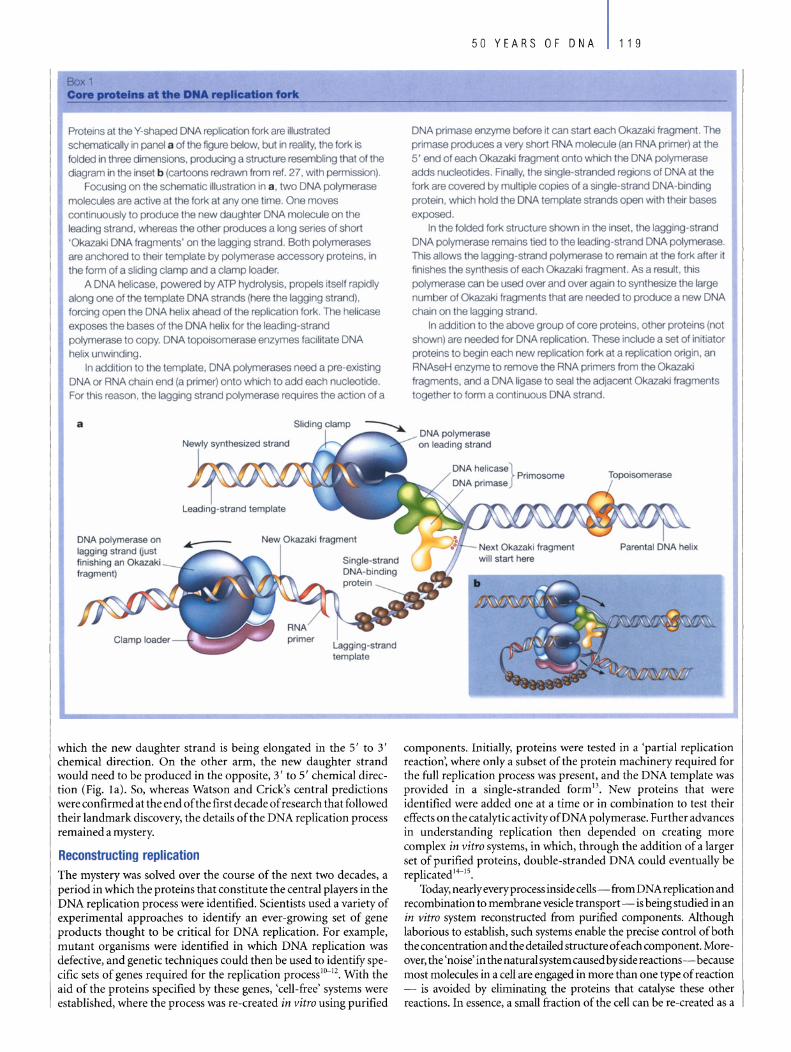
Box 1 Core protein. at the DU repllcatlon fork
Proteins at the Y-shaped DNA replication lork are iIIustrated schematically in panel a of the figure betow, but in reality, the lork is Iolded in three dimensions, producing a structure resembling that of the diagram in the inset b (cartoons redrawn from rel. 27, with permission).
Focusing on the schemalic illustration in a , two DNA polymerase molecules are active at the fork at any one time. One moves continuously to produce the new daughter DNA molecule on the leading strand, whereas the other produces a long series 01 short 'Okazaki DNA fragments ' on the lagging strand. 80th polymerases are anchored to their template by polymerase accessory proteins. in the form of a sliding elamp and a elamp loader.
A DNA helicase. powered by ATP hydrolysis, propeis itself rapidly along one 01 the template DNA strands (here the lagging strand), forcing open the DNA helix ahead of the replication lork. The heiicase exposes the bases of the DNA helix for the leading-strand polymerase to copy. DNA topoisomerase enzymes lacilitate DNA helix unwinding.
In addition 10 the template, DNA polymerases need a pre-existing DNA or RNA chain end (a primer) onto which to add each nucleotide. For this reason, the lagging strand polymerase requires the action of a
a
DNA polymerase on lagging strand fjusl finishing an Okazaki fragment)
Clamp In".1 .. , ._~ Lagging-strand template
which the new daughter strand is being elongated in the 5' to 3' chemical direction. On the other arm, the new daughter strand would need to be produced in the opposite, 3' to 5' chemical direction (Fig. la). So, whereas Watson and Crick's central predictions were confirmed at the end of the first decade of research that followed their landmark discovery, the details of the DNA replication process remained a mystery.
Reconstructing replication The mystery was solved over the course of the next two decades, a period in which the proteins that constitute the central players in the DNA replication process were identified. Scientists used a variety of experimental approaches to identify an ever-growing set of gene products thought to be critical for DNA replication. For example, mutant organisms were identified in which DNA replication was defective, and genetic techniques could then be used to identify specific sets of genes required for the replication processIO- 12• With the aid of the proteins specified by these genes, 'cell-free' systems were established, where the process was re-created in vitro using purified
50 YEARS OF DNA 119
DNA primase enzyme before it can start each Okazaki fragment. The primase produces a very short RNA molecule (an RNA primer) at the 5' end of each Okazaki fragment onto which the DNA polymerase adds nucleotides. Finally, the single-slranded regions of DNA atthe fork are covered by multiple copies of a single-strand DNA-binding protein, which hold the DNA template strands open with their bases exposed.
In the folded fork structure shown in the insel. the lagging-strand DNA polymerase remains tied to the leading-strand DNA polymerase. This allows the lagging-strand potymerase to remain at the fork after it finishes the synthesis 01 each Okazaki fragment. As a resull, this polymerase can be used over and over again to synthesize the large number of Okazaki fragments that are needed to produce a new DNA chain on the lagging strand.
In addition to the above group of core proteins, other proteins (not shown) are needed for DNA replication. These include a set of initiator proteins to begin each new replication fork at a replication origin, an RNAseH enzyme to remove the RNA primers trom the Okazaki fragments, and a DNA ligase to seal the adjacent Okazaki fragments together to form a continuous DNA strand.
DNA polymerase on leading strand
...-- o-u-- Next Okazaki fragment will start here
components. Initially, proteins were tested in a 'partial replication reaction', where only a subset of the protein machinery required for the full replication process was present, and the DNA template was provided in a single-stranded form 13. New proteins that were identified were added one at a time or in combination to test their effects on the catalytic activity ofDNA polymerase. Further advances in understanding replication then depended on creating more complex in vitro systems, in which, through the addition of a larger set of purified proteins, double-stranded DNA could eventually be replicated 14-15.
Today, nearlyeveryprocess insidecells- from DNA replication and recombination to membrane vesicle transport - is being studied in an in vitro system reconstructed from purified components. Although laborious to establish, such systems enable the precise control ofboth the concentration and the detailed structure of each component. Moreover, the 'noise' in the natural system caused by side reactions-because most molecules in a cell are engaged in more than one type of reaction - is avoided by eliminating the proteins that catalyse these other reactions. In essence, a small fraction of the cell can be re-created as a

120 50 YEARS OF DNA
Homologous DNA recombination involves an exchange between two DNA double helices that causes a section of each helix to be exchanged with a section of the other, as illustrated schematically in panel a in the figure below (redrawn from rel. 27, with permission). Criticalto the reaction is Ihe formation of a heteroduplex joint at the point where the two double helices have been broken and then joined together. To form this joint, whlch glues two previously separate molecules together, astrand from one helix musl form base pairs with a complementary strand from the second helix. This requires that the two DNA helices that recombine have a very similar sequence 01 nucleotides, that is, they must be homologous.
The DNA double helix poses a major problem for the DNA recombination process, because Ihe bases that need 10 pair to lorm a heteroduplex Joint are buried in the interior of the helix. How can two DNA helices recognize that Ihey are homologous, in order to begin a recombination event, if their bases are not exposed?
The breakthrough came from Ihe isolation and characterization 01 the RecA protein " lrom the bacterium Escherichia coll, which would turn out to be the prototype for a family 01 strand-exchange proteins that is present in all organisms, Irom bacleria 10 humans. The human equrvalent 01 the RecA protein is the RadS1 protein. These proteins catalyse the central synapsis step of homologous DNA recombination - the process that brings two matching DNA helices logether and causes them to exchange parts, resulting In either the reassortment or the repair of genetic information (panel b below). Powered by the energy generated Irom ATP hydrolysis, the RecA protein assembles into long filaments on a single-strand DNA molecule (brown strand). Because the RecA protein has a second DNA-binding site that recognizes a DNA double helix, a RecAcoated strand has the remarkable ability to scan for a complementary strand in any double helix (blue strand) that it encounters. Once lound, the complementary strand is pulled from the helix to form a new 'hybrid helix' with the RecA-coated single strand, thereby initiating the formation of the heteroduplex joint needed for recombination, as illustrated schematically in panel b (RecA protein not shown).
DNA recombination makes il possible for a damaged chromosome 10 repair itsell by using a second copy 01 the same genetlc informaIIon as a guide. It also causes the extensive breakage and reunion 01 chromosomes that occurs during the developmenl 01 eggs and sperm, which greatly Increases the genetic variation produced by sexual reproducllon. Many 01 the atomic details of the RecA protein reaction are still uncertain, remaining as a future challenge for scientists.
a Two homologous DNA Two DNA molecules double helices that have recombined
b a a b b c c c c
-+- d d -+- d d c e e e e d f f f f e 9 9 9 9 f 9
bounded set of chemical reactions, making it fully amenable to precise studyusing all ofthe tools ofphysics and chemistry.
By 1980, multiprotein in vitra systems had enabled a detailed characterization of the replication machinery and solved the problem ofhow DNA is synthesized on both sides of the replication fork (Fig. 1 b). One daughter DNA strand is synthesized continuously by a DNA polymerase molecule moving along the 'leading strand', while a second DNA polymerase molecule on the 'lagging strand' produces a long series of fragments (calIed Okazaki fragments)16 which are joined together by the enzyme DNA ligase to produce a continuous DNA strand. As might be expected, there is a difference in the proteins required for leading- and lagging-strand DNA synthesis (see Box 1). Remarkably, the replication forks formed in these artificial systems could be shown to move at the same rapid rates as the forks inside cells (500 to 1,000 nucleotides per second), and the DNA template was copied with incredibly high fidelit/ s.
As more and more proteins were found to function at the replication fork, comparisons could be made between the replication machinery of different organisms. Studies of the replication machinery in viruses, bacteria and eukaryotes revealed that a common set of protein activities drives the replication forks in each organism (Box 1). Each system consists of: a leading- and a lagging-strand DNA polymerase molecule; a DNA primase to produce the RNA primers that start each Okazaki fragment; single-strand DNA binding proteins that coat the template DNA and hold it in position; a DNA helicase that unwinds the double helix; and additional polymerase accessory proteins the tie the polymerases to each other and to the DNA template. As one progresses from a simple virus to more complex organisms, such as yeasts or mammals, the number of subunits that make up each type of protein activity tends to increase. For example, the total number of polypeptide subunits that form the core of the replication apparatus increases from four and seven in bacteriophages T7 and T4, respectively, to 13 in the bacterium E. co/i. And it expands to atleast 27 in the yeast Saccharamyces cerevisiae and in mammals. Thus, as organisms with larger genomes evolved, the replication machinery added new protein subunits, without any change in the basic mechanismsI5,IS-20.
While the work I have described on DNA replication was advancing, other groups of researchers were establishing in vitro systems in which homologous DNA recombination could be reconstructed. The central player in these reactions was the RecA type ofprotein l7,
named after the bacterial mutant defective in recombination that led to its discovery(Box2).
Protein machines As for all other aspects of cell biochemistry, the DNA replication apparatus has evolved over billions of years through 'trial and error'- that is, by random variation followed by natural selection. With time, one protein after another could be added to the mix of proteins active at the replication fork, presumably because the new proteinincreased the speed, control or accuracyofthe overall replication process. In addition, the structure of each protein was fine-tuned by mutations that alte red its amino acid sequence so as to increase its effectiveness. The end results of this unusual engineering process are the replication systems that we observe today in different organisms. The mechanism ofDNA replication might therefore be expected to be highly dependent on random past events. But did evolution select for whatever works, with no need for elegance?
For the first 30 years after Watson and Crick's discovery, most researchers seemed to hold the view that cell processes could be sloppy. This view was encouraged by knowledge of the tremendous speed of movements at the molecular level (for example, it was known that a typical protein collides with a second molecule present at a concentration of 1 mM about 106 times per second). The rapid rates of molecular movementwere thought initiallyto allow a process like DNA replication to occur without any organization of the proteins involved in three-dimensional space.

Quite to the contrary, molecular biologists now recognize that evolution has selected for highly ordered systems. Thus, for example, not only are the parts of the replication machinery held together in precise alignments to optimize their mutual interactions, but energydriven changes in protein conformations are used to generate coordinated movements. This ensures that each of the successive steps in a complex process like DNA replication is closely coordinated with the next one. The result is an assembly that can be viewed as a 'protein machine'. For example, the DNA polymerase molecule on the lagging side of the replication fork remains bound to the leading-strand DNA polymerase molecule to ensure that the same lagging-strand polymerase is used over and over again for efficient synthesis of Okazaki fragments I8,20,21 (Box 1). And DNA replication is byno means unique. We now believe that nearlyevery biological process is catalysed by a set of ten or more spatially positioned, interacting proteins that undergo highly ordered movements in a machine-like assemblY2.
Pro tein machines generally form at specific sites in response to particular signals, and this is particularly true for protein machines that act on DNA. The replication, repair and recombination of the DNA double helix are often considered as separate, isolated processes. But inside the cell, the same DNA molecule is able to undergo any one of these reactions. Moreover, specific combinations of the three types of reactions occur. For instance, DNA recombination is often linked directly to either DNA replication or DNA repair23 • For the integrity of a chromosome to be properly maintained, each specific reaction must be carefully directed and controlled. This requires that sets of proteins be assembled on the DNA and activated only where and when they are needed. Although much remains to be learned about how these choices are made, it seems that different types of DNA structures are recognized explicitly by specialized proteins that serve as 'assembly factors'. Each assembly factor then serves to nucleate a cooperative assembly of the set of pro teins that forms a particular protein machine, as needed for catalysing areaction appropriate to that time and place in the cell.
A yjew pt 1M tlllUJ1l It has become customary, both in textbooks and in the regular scientific literature, to explain molecular mechanisms through simple two-dimensional drawings or 'cartoons'. Such drawings are useful for consolidating large amounts of data into a simple scheme, as illustrated in this review. But a whole generation ofbiologists may have become lulled into believing that the essence of a biological mechanism has been captured, and the entire problem therefore solved, once a research er has deciphered enough of the puzzle to be able to draw a meaningful cartoon of this type.
In the past few years, it has become abundantly clear that much more will be demanded of scientists before we can claim to fully understand a process such as DNA replication or DNA recombination. Recent genome sequencing projects, protein-interaction mapping efforts and studies in cell signalling have revealed many more components and molecular interactions than were previously realized. For example, according to one recent analysis, S. cerevisiae, a single-celled 'simple' eukaryotic organism (which has about 6,000 genes compared with 30,000 in humans), uses 88 genes for its DNA replication and49 genes for its DNA recombination24•
To focus on DNA replication, fully understanding the mechanism will require returning to where the studies ofDNA first began - in the realms of chemistry and physics. Detailed atomic structures of all relevant proteins and nucleic acids will be needed, and spectacular progress is being made by structural biologists, owing to increasingly powerful X-ray crystallography and nuclear magnetic resonance techniques. But the ability to reconstruct biological processes in a test tubewith molecules whose precise structures are known is not enough. The replication process is both very rapid and incredibly accurate, achieving a final error rate of about one nucleotide in a billion. Understanding how the reactions between the many different proteins and other molecules are coordinated to create this result will require
50 YEARS OF DNA 121
that experimentalists determine all ofthe rate constants forthe interactions between the various components, something that is rarely done by molecular biologists today. They can then use genetic engineering techniques to alter selected sets of these parameters, carefully monitoring the effect of these changes on the replication process.
Scientists will be able to claim that they truly understand a complex process such as DNA replication only when they can precisely predict the effect of changes in each of the various rate constants on the overall reaction. Because the range of experimental manipulations is enormous, we will need more powerful ways of deciding which such alterations are the most likely to increase our understanding. New approaches from the rapidly developing field of computational biology must therefore be developed - both to guide experimentation and to interpret the results.
The Watson-Crick model ofDNA catalysed dramatic advances in our molecular understanding ofbiology. At the same time, its enormous success gave rise to the misleading view that many other complex aspects of biology might be similarly reduced to elegant simplicity through insightful theoretical analysis and model building. This view has been supplanted over subsequent decades, because most biological subsystems have turned out to be far too complex for their details to be predicted. We now know that nothing can substitute for rigorous experimental analyses. But traditional molecular and cell biology alone cannot bring a problem like DNA replication to closure. New types of approaches will be required, involving not onlynew computational tools, but also a greater integration of chemistry and physics20,25.
For this reason, we urgently need to rethink the education that we are providing to the next generation ofbiological scientists22,26. D doi:1O.1038/natureO 1407
1. Watson, J. D. & Crick, F. H. C. A structure fordeoxyribose nucleic acid. Nature 171, 737-738. (1953).
2. Crick, F. H. C. The biological replication of macromolecules. Symp. Soc. Exp. Bio/. 12, 138-163 (1958).
3. Doty, P. Inside Nucleic Acids (Harvey Lecture, 1960) (Academic, New York, 1961).
4. Marmur, J. & Doty, P. Thermal renaturation of deoxyribonucleic acids. J. Mol. Bio/. 3, 585-594 (1961).
5. Cairns, J. The bacterial chromosome and its manner of replication as seen by autoradiography. J. Mol.
Biol. 6, 208-213 (1963).
6. Kavenoff, R., Klotz, L. C. & Zimm, B. H. On the nature of chromosome-sized DNA molecules. Cold
Spring Harb. Symp. Quant. Biol. 38,1-8 (1974).
7. Watson, J. D. & Crick, F. H. C. Genetical implications of the structure of deoxyribonucleic acid.
Nature 171, 964-967 (1953).
8. Meselson, M. & Stahl, F. W The replication ofDNA in E eoli. Proe. Natl Acad. Sei. USA 44, 671--682 (1958).
9. Kornberg, A. Biological synthesis ofDNA. Scienee 131,1503-1508 (1960).
10. Epstein, R. H. et al. Physiological studies of conditionallethal mutants ofbacteriophage T4D. Cold
Spring Harb. Symp. Quant. Biol. 28, 375 (1963).
11. Bonhoeffer, F. & Schaller, H. A method for selective enrichment of mutants based on the high UV
sensitivity ofDNA containing 5-bromouracil. Biochem. Biophys. Res. Commun. 20, 93 (1965).
12. Kohiyama, M., Cousin, D., Ryter, A. & Jacob, F. Mutants thermosensible d'Escherichia coli K/12. I.
Isolement et caracterisation rapide. Ann. Inst. Pasteur 110, 465 (1966).
13. Huberman, J. A., Kornberg, A. & Alberts, B. M. Stimulation ofT4 bacteriophage DNA polymerase by
the protein product ofT4 gene 32.]. Mol. Biol. 62, 39-52 (1971).
14.Morris, C. E, Sinha, N. K. & Alberts, B. M. Reconstruction ofbacteriophage T4 DNA replication
apparatus from purified components: rolling circle replication following de novo chain initiation on a
single-stranded circular DNA template. Proc. NatlAead. Sei. USA 72. 4800-4804 (1975).
15. Kornberg, A. & Baker, T. A. DNA Replieation2nd edn (Freeman, New York, 1992).
16. Okazaki R. et al. Mechanism ofDNA chain growth: possible discontinuity and unusual secondary
structure of newly synthesized chains. Proc. Natl Acad. Sei. USA 59, 598-605 (1968).
17. Radding, C. M. Recombination activities of E. eoliRecA protein. CeIl25, 3-4 (1981).
18. Davey, M. J. & O'Donnell, M. Mechanisms ofDNA replication. Curr. Opin. Chem. Biol. 4, 581-586
(2000).
19. Waga, S. & Stillman, B. The DNA replication fork in eukaryotic cells. Annu. Rev. Bioehem. 67,
721-751 (1998).
20. Benkovic, S. J., Valentine, A. M. & Salinas E Replisome-mediated DNA replication. Annu. Rev.
Bioehern. 70,181-208 (2001).
21.Alherts, B. M. The DNA enzymology of protein machines. Cold Spring Harb. Symp. Quant. Biol. 49,
1-12 (1984).
22.Alberts, B. The ceIl as a collection of protein machines: preparing the next generation of molecular
biologists. Ce1l92, 291-294 (1998).
23. Radding, C. Colloquium introduction. Links between recombination and replication: vital roles of
recombination. Proc. NatlAcad. Sei. USA 98, 8172 (2001).
24. Dwight, S. S. et al. Saecharomyees Genome Database (SGD) provides secondary gene annotation
using the Gene Ontology (GO). NucieicAcids Res. 30, 69-72 (2002).
25. Trakselis, M. A. & Benkovic, S. J. Intricacies in ATP-dependent clamp loading: variations across
replication systems. Strueture 9, 999-1004 (2001).
26. National Research Council. Bio201O: Undergraduate Education to Prepare BiomedieaI Research
Seicntists (The National Academies Press, Washington DC, 2002).
27. Alberts, B. et al. Molecular Biology 0/ the Cell4th edn (Gariand, New York, 2002).
Originalreference: Nature421, 431-435 (2003).
122 50 YEARS OF DNA
DNAdamage and repair Errol C. Friedberg
Laboratory ofMolecular Pathology, Department ofPathology, University of Texas Southwestern Medical Center, Dallas, Texas 75390-9072, USA (e-mail: [email protected])
The aesthetic appeal of the DNA double helix initially hindered notions of DNA mutation and repair, which would necessarily interfere with its pristine state. But it has since been recognized that DNA is sUbject to continuous damage and the cell has an arsenal of ways of responding to such injury. Although mutations or deficiencies in repair can have catastrophic consequences, causing a range of human diseases, mutations are nonetheless fundamental to life and evolution.
"We totally missed the possible role of. .. [DNA} repair although ... I later came to realise that DNA is so precious that probably many distinct repair mechanisms would exist." Francis Crick, writing in Nature, 26 April 1974 (ref.!).
his retrospective reflection by Francis Crick, penned two decades after he and James Watson reported the structure of DNA, hints at the early perception of DNA as a highly stable macromolecular entity. This prevailing view at the time significantly delayed serious
consideration of biochemical processes such as mutation and repair. It was once suggested by Frank Stahl that "the possibility that ... genes were ... subject to the hurly-burly of both insult and clumsy efforts to reverse the insults, was unthinkable:'2
But subsequent work on three 'R's' ofDNA metabolism - replication (copying ofDNA prior to each cell division), recombination (exchanges between different DNA molecules in a cell) and repair (restoration of alte red DNA to its normal state) - revealed the dynamic state of DNA. It became apparent that DNA in aHliving organisms continuaHy incurs a myriad of types of damage, and that cells have devised ingenious mechanisms for tolerating and repairing the damage. Failure of these mechanisms can lead to serious disease consequences, as well illustrated in the human hereditary diseases xeroderma pigmentosum (XP), hereditary nonpolyposis colon cancer (HNPCC) and some forms ofbreast cancer. XP is characterized by about a 1O,000-fold increased risk of skin cancer associated with sunlight exposure; individuals with HNPCC manifest an increased hereditary predisposition to colon (and other) cancer.
The rools of repair Theearlyworkon DNAdamageandrepairin the 1930swasstimulated by a small but prominent group of physicists3• As recounted by the geneticist Guido Pontecorvo, "in the years immediately preceding World War II something quite new happened: the introduction of ideas (not techniques) from the realm of physics into the realm of genetics, particularly as applied to the problems of size, mutability, and self-replication of genes"4. Seminal to this coalition between physics and biology in pre-war Germany was the collaboration between German physicists Karl Zimmer and Max Delbrück and the Russian geneticist Nikolai Timofeeff-RessovskY. Their partnership was stimulated by the work ofHermann MuHer, a geneticist working
on the fruitfly Drosophila who first demonstrated that external agents, such as ionizing radiation, can cause mutations in living organisms6•
Timofeeff-Ressovsky and Zimmer were interested primarily in how such small amounts of energy in the form of ionizing radiation (formally equivalent to no more than the amount of energy absorbed as heat by drin king a cup ofhottea) could have such profound biological effects3. Delbrück and Muller, on the other hand, were intrigued by whether such mutations could reveal insight into the physical nature ofthe gene.
In retrospect, it was inevitable that the deployment of physical (and later chemical) tools, such as ionizing and ultraviolet (UV) radiation, to study genes would in due course lead to questions as to how these agents damaged DNA3• And, once it was recognized that these interactions promoted deleterious effects on the structure and function of genes, to questions concerning how cells cope with damaged DNA. Zimmerwrote, "one cannot use radiations for elucidating the normal state of affairs without considering the mechanisms of their actions, nor can one find out much about radiation induced changes without being interested in the normal state of the material under investigation."7
Hints of the ability ofliving cells to recover from the lethai effect of UV radiation emerged as early as the mid -1930s8• But the discovery of a DNA-repair mechanism had to wait until the end of the 1940s, through the independent, serendipitous observations of Albert Kelner9 working in Milislav Demerec's group at the Cold Spring Harbor Laboratory, and Renato Dulbecco lO in Salvador Luria's laboratory at The University of Indiana. Neither Keiner nor Dulbecco set out to study damage to DNA or its repair. They were both using UV radiation as an experimental tool, but observed anomalous survival rates when cells or bacteriophage (bacteriainfecting viruses) were inadvertently exposed to long-wavelength light, either as sunlight or fluorescent light in their respective laboratories9,1O. Their efforts to explain these confounding observations led to the discoveryofthe phenomenon now known as photoreacti-
Pyrimidine dimer in UV-exposed DNA
Complex 01 DNA with photoreactivating enzyme
Absorption 01 light (>300 nm)
Release 01 enzyme to restore native DNA
Figure 1 Photoreactivation reverses DNA damage. DNA exposed to ultraviolet (UV) radiation results in covalent dimerization 01 adjacent pyrimidines, typically thymine residues (thymine dimers), illustrated here as a purpie triangle. These lesions are recognized by a photoreactivating enzyme, which absorbs light at wavelengths >300nm (such as fluorescent light or sunlight) and facilitates a series 01 photochemical reactions that monomerize the dimerized pyrimidines, restoring them to their native conlormation.
t
b
Mononucleotides Oligonucleotide fragment
~ '.
~ :...:. ... 6' ••
[ unimodallnCiSiOn]
mechanism
Free base
1-" T- - • 11
. . ·i . , ~~ ~ '. "
.. " . r-~ • ,-'-,,:- ~ ..... ~
vation, whereby the DNA damage incurred by exposure to UV light is repaired by a light -dependent enzyme reaction 11 (Fig. 1).
Curiously, even with the elucidation of the structure ofDNA only four years away, neither Dulbecco nor Watson - who was a graduate student in Luria's laboratory when Dulbecco stumbled on photoreactivation, and had himself examined the effects ofionizing radiation for his doctoral thesis2 - thought about DNA repair. However, shortly after Watson and Crick reported on the DNA double helical structure, they noted the implications of the base-pairing rules for mutagenesis, stating "spontaneous mutation may be due to a base occasionally occurring in one of its less likely tautomeric forms"12.
Tautomerism is the property of a compound that allows it to exist in two interconvertible chemical states; in the case ofDNA bases, as either keto or enol forms. Watson and Crick had initially overlooked the complications of tautomerism and were trying unsuccessfully to construct their DNA model with the rare enol form ofbases. It was only after Jerry Donohue, a former graduate student of Linus Pauling, po in ted out to Watson that he should be using the more common keto form that the problem ofhow bases could stably pair was solved 13.
But no consideration was then given to the fact that the chemical lability ofDNA implicit in tautomerism might have wider implications for the stability of genes. Indeed, the field gave little thought to the precise nature of DNA damage and its possible biological consequences. One must recall, however, that even at the time the DNA double helix was unveiled, its 'pathology' and the biological consequences thereof were far less compelling problems than deciphering the genetic code or understanding the essential features ofDNA replication. Even mutagenesis- putto extensive use as a tool for determining the function of genes and their polypeptide products, and for defining the genetic code - was not widely considered in mechanistic terms until much laterll. This is despite the fact that
,.
50 YEARS OF DNA 123
Signal transductlon
~ ~ ~
Arrested cell-cycie
progression
Figure 2 Responses to DNA damage, DNA damage (illustrated as a black triangle) results in either repair or tolerance. a, During damage tolerance, damaged sites are recognized by the replication machinery before they can be repaired, resulting in an arrest that can be relieved by replicative bypass (transiesion DNA synthesis) (see Fig, 3). b. DNA repair involves the excision of bases and DNA synthesis (red wavy lines). which requires double-stranded DNA Mispaired bases, usually generated by mistakes during DNA replication, are excised as single nucleotides during mismatch repair. A damaged base is excised as a single free base (base excision repair) or as an oligonucleotide fragment (nucleotide excision repair). Such fragments are generated by incisions flan king either side of the damaged base, Nucleotide excision repair can also transpire in some organisms bya distinct biochemical mechanism involving only a single incision next to a site of damage (unimodal incision). c, The cell has a network of complex signalling pathways that arrest the cell cycle and may ultimately lead to programmed cell death,
the repair phenomenon of photoreactivation was known before the discovery of the structure ofDNA.
A DNA duplex tor redundancy Watson and Crick noted, with infamous prophetie understatement, "it has not escaped our notice that the specific [base 1 pairing we have postulated immediately suggests a possible copying mechanism for the genetic material,,14. However, it was not intuitively obvious that a double-stranded molecule should be required for DNA replication. In principle, a single-stranded chain could just as easily do. But the significance ofthe duplex DNA structure soon became apparent. It was shown that DNA replicates in a semi-conservative fashion, whereby each strand of the double helix pairs with a new strand generated by replication. This enables errors introduced during DNA replication to be corrected by a mechanism known as excision repair, which relies on the redundancy inherent in having two complementary strands of the genetic code. If the nucleotides on one strand are damaged, they can be excised and the intact opposite strand used as a templateto directrepair synthesis ofDNA1S (Fig. 2).
Many paths to mutation and repair The elucidation of the DNA structure provided the essential foundation for defining the different types of mutations arising from both spontaneous and environmental DNA damage that affect allliving cells12• Once again, the insights of physicists featured prominentll, including among others, Richard Setlow who identified thymine dimers as stable and naturally occurring DNA lesions arising in cells exposed to sunlight (UV radiation). Such lesions comprise a covalent joining of two adjacent thymine residues in the same DNA chain. They generate considerable distortion of the normal structure of DNA and seriously impede DNA transactions such as replication and transcription. The repair of these lesions could be monitored
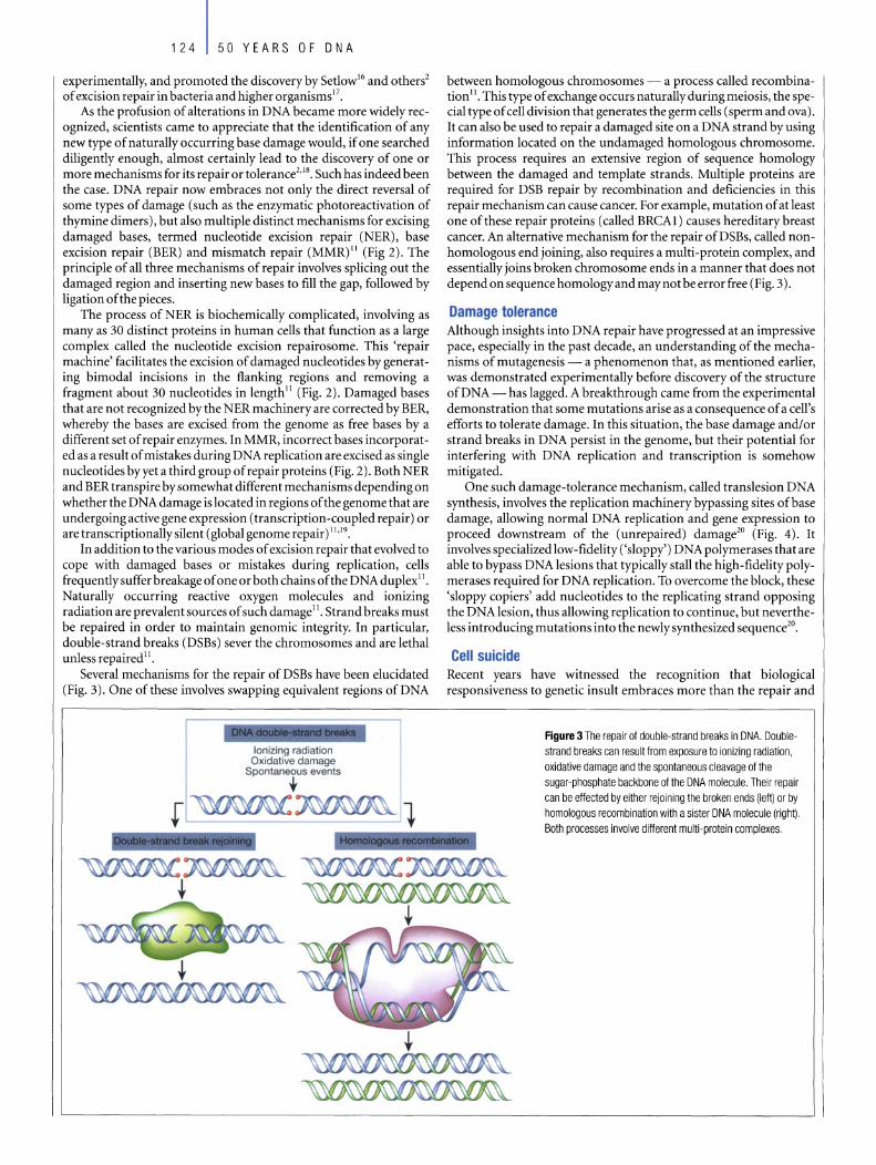
124 50 YEARS OF DNA
experimentally, and promoted the discovery by SetlowI6 and others2
of excision repair in bacteria and higher organisms 17.
As the profusion of alterations in DNA became more widely recognized, scientists came to appreciate that the identification of any new type of naturally occurring base damage would, if one searched diligently enough, almost certainly lead to the discovery of one or more mechanisms for its repair or tolerance2,I8. Such has indeed been the case. DNA repair now embraces not only the direct reversal of some types of damage (such as the enzymatic photoreactivation of thymine dimers), but also multiple distinct mechanisms for excising damaged bases, termed nucleotide excision repair (NER), base excision repair (BER) and mismatch repair (MMR)" (Fig 2). The principle of all three mechanisms of repair involves splicing out the damaged region and inserting new bases to fill the gap, followed by ligation of the pieces.
The process of NER is biochemically complicated, involving as many as 30 distinct proteins in human cells that function as a large complex called the nucleotide excision repairosome. This 'repair machine' facilitates the excision of damaged nucleotides by generat -ing bimodal incisions in the flanking regions and removing a fragment about 30 nucleotides in length" (Fig. 2). Damaged bases that are not recognized by the NER machinery are corrected by BER, whereby the bases are excised from the genome as free bases by a different set of repair enzymes. In MMR, incorrect bases incorporated as a result of mistakes during DNA replication are excised as single nucleotides byyet a thirdgroup ofrepair proteins (Fig. 2). Both NER and BER transpire by somewhat different mechanisms depending on whether the DNA damage is located in regions of the genome that are undergoing active gene expression (transcription-coupled repair) or are transcriptionally silent (global genome repair) 11.19.
In addition to the various mo des of excision repair that evolved to cope with damaged bases or mistakes during replication, cells frequently suffer breakage of one or both chains of the DNA duplexli. Naturally occurring reactive oxygen molecules and ionizing radiation are prevalent sources of such damageII • Strand breaks must be repaired in order to maintain genomic integrity. In particular, double-strand breaks (DSBs) sever the chromosomes and are lethai unless repaired 11.
Several mechanisms for the repair of DSBs have been elucidated (Fig. 3). One of these involves swapping equivalent regions ofDNA
lonizing radiation Oxidative damage
Spontaneous events , ~,. -
between homologous chromosomes - a process called recombination 11. This type of exchange occurs naturally during meiosis, the special type of cell division that generates the germ cells (sperm and ova). It can also be used to repair a damaged site on a DNA strand by using information located on the undamaged homologous chromosome. This process requires an extensive region of sequence homology between the damaged and template strands. Multiple proteins are required for DSB repair by recombination and deficiencies in this repair mechanism can cause cancer. For example, mutation of at least one of these repair proteins (called BRCAl) causes hereditary breast cancer. An alternative mechanism for the repair ofDSBs, called nonhomologous end joining, also requires a multi-protein complex, and essentially joins broken chromosome ends in a manner that does not depend on sequence homology and may not be error free (Fig. 3).
Damage tolerance Although insights into DNA repair have progressed at an impressive pace, especially in the past decade, an understanding of the mechanisms of mutagenesis - a phenomenon that, as mentioned earlier, was demonstrated experimentally before discovery of the structure ofDNA - has lagged. A breakthrough came from the experimental demonstration that some mutations arise as a consequence of a cell's efforts to tolerate damage. In this situation, the base damage and/or strand breaks in DNA persist in the genome, but their potential for interfering with DNA replication and transcription is somehow mitigated.
One such damage-tolerance mechanism, called transiesion DNA synthesis, involves the replication machinery bypassing sites ofbase damage, allowing normal DNA replication and gene expression to proceed downstream of the (unrepaired) damage20 (Fig. 4). It involves specialized low-fidelity ('sloppy') DNA polymerases that are able to bypass DNA lesions that typically stall the high-fidelity polymerases required for DNA replication. To overcome the block, these 'sloppy copiers' add nucleotides to the replicating strand opposing the DNA lesion, thus allowing replication to continue, but nevertheless introducing mutations into the newly synthesized sequence20 •
Cell suicide Recent years have witnessed the recogmtlOn that biological responsiveness to genetic insult embraces more than the repair and
Figure 3 The repair 01 double-strand breaks in DNA. Doublestrand breaks can result lrom exposure to ionizing radiation, oxidative damage and the spontaneous cleavage 01 the sugar-phosphate backbone 01 the DNA moleeule. Their repair can be effected by either rejoining the broken ends (Ieft) or by homologous recombination with a sister DNA moleeule (right). Both processes involve different multi-protein complexes.
' • ..J..Tj __ • -- _ _

b
b
c
poil
Figure 4 'Sloppy copiers' overcome blocks in replication caused bya DNA lesion (a process called transiesion synthesis). a, The DNA replicative machinery (blue) stalls immediately behind a site 01 base damage (black triangle). Two specialized 'sloppy copier' pOlymerases (pol" and poIL) bind to the arrested replication complex. b, This interaction promotes a conlormational change in the arrested replication machinery, placing pol" in direct proximity to the site 01 base damage where it synthesizes ac ross the lesion. C, Pol" may then dissociate and allow POiL to complete the process 01 replicative bypass by incorporating several more nucleotides (red crosses). Once the lesion has been completely bypassed, the replication machinery resumes DNA replication. As a result 01 this process, mutations to the DNA sequence are now incorporated into one strand.
tolerance of DNA damage. The exposure of cells to many DNAdamaging agents results in the transcriptional upregulation of a large number of genes, the precise function( s) of many ofwhich remains to be established. Additionally, cells have evolved complex signalling pathways to arrest the progression ofthe cell cyde in the presence of DNA damage, thereby providing increased time for repair and tolerance mechanisms to operate21 (Fig. 2c). Finally, when the burden of genomic insult is simply too large to be effectively met by the various responses discussed, cells are able to initiate programmed cell death (apoptosis), thereby eliminating themselves from a population that otherwise might suffer serious pathological consequences22 •
ONA damage and cancer The 'somatic mutation hypothesis' of cancer embraces the notion that neoplastic transformation arises from mutations that alter the function of specific genes (now called oncogenes and tu mo ursuppressor genes) that are critical for cell division. This theory has its roots in correlations between chromosomal abnormalities and cancer first observed by the developmental biologist Theodore Bover?l, who at the beginning of the twentieth century reported abnormal numbers of chromosomes (aneuploidy) in cancerous somatic cells.
The discovery of the structure of DNA progressed our understanding of tumorigenesis at severallevels. Watson and Crick predicted from their DNA model that complementary base pairing had implications for recombination (the exchange of genetic
50 YEARS OF DNA 125
material between chromosome pairs): "the pairing between homologous chromosomes at meiosis may depend on pairing between specific bases". The genetic basis of many cancers is now known to arise from abnormal recombination events, such as chromosomal translocations, where a region of one chromosome is juxtaposed to another chromosome. Watson hirnself developed an early and ardent interest in cancer biologywhen he recognized that the experimentalIy tractable genomes of oncogenic viruses could provide important insights into the pathogenesis of cancer. Mutagenesis is now documented as a fundamental cornerstone of the molecular basis of all forms of cancer24.
Arguably the most definitive validation of the somatic mutation hypo thesis derives from the discovery that defective responses to DNA damage and the accumulation of mutations underlies two distinct types ofhereditary cancer; skin cancer associated with defective NER and colorectal cancer associated with defective MMR 11. In both instances, credit belongs to scholars ofDNA repair.
In the la te 1960s, James Cleaver providentially noted an artide in the San Francisco Chronicle that reported the extreme proneness to skin cancer in individuals with XP, a rare sun-sensitive hereditary disease2. Cleaver was then searching for mammalian cell lines that were defective in excision repair, and his intuitive notion that XP individuals might be sunlight-sensitive and prone to cancer because theywere genetically defective in excision repair proved to be correces.
The , subsequent elucidation of the genes defective in XP patients26, and their role in NER of damaged bases in human ceIls 11 ,27,28, represents a triumph of modern genetics and its application to molecular biology. The additional discovery that the process of NER in eukaryotes requires elements of the basic transcription apparatus11 has yielded insights into the complex relationships between deficient DNA repair, defective transcription and hereditary human diseases 11 .
A fascinating denouement to the skin-cancer predisposition in XP patients derives from the recent solution of the 'XP variant problem'. A significant fraction ofXP individuals who are dinicaIly indistinguishable from those defective in NER were found to be proficient in this repair process 11 . It was shown that DNA polymerase-T) (polT)), one of the specialized DNA polymerases capable of overcoming replication blocks at DNA lesions, is mutated in aIl XP-variant patients so far examined29. Not only does polT) replicate past thymine dimers in DNA, but - unlike the other specialized DNA polymerases - it also correctly inserts adenine residues29, thereby preventing mutations at sites of thymine dimers. Therefore, even in XP patients with functional NER, in the absence of polT) one or more other bypass polymerases attempts to cope with arrested replication at thymine dimers, but does so inaccuratelj9. Thus, cancer predisposition in XP essentially derives from an excessive mutation al burden in skin ceIls associated with exposure to sunlight. These mutations accumulate either because thymine dimers are not excised (owing to defective NER) or because in the absence of polT), dimers are inaccurately bypassed by other DNA polymerases29.
The association between HNPCC and defective MMR was determined more-or-less simultaneously by a number of investigators. Paul Modrich2 surmised that the instability of repeated sequences in DNA associated with defective MMR in bacteria30
might be causaIly related to the DNA sequence instability observed in patients with HNPCC31 . This led to the formal demonstration of defective MMR in this human hereditary disease and formed another persuasive validation of the somatic mutation theory of cancer32-34,
A look to the future The study of biological responsiveness to DNA damage embraces DNA repair, mutagenesis, damage tolerance, ceIl-cyde checkpoint controI, programmed ceIl death, and other cellular responses to
126 50 YEARS OF DNA
genomic insult. This integrated field is now deciphering the complex regulatory pathways transduced by signalling mechanisms that detect DNA damage and/or arrested DNA replication. As these pathways become better understood, parallel technological gains in gene therapy and therapeutic intervention by rational drug design will offer new strategies for blocking the unwanted consequences ofDNA damage, especially cancer.
We must remember, however, that while evolution could not have transpired without robust cellular mechanisms to ameliorate the most serious consequences of spontaneous and environmental DNA damage, the process of evolution mandates that the genetic diversification on which Darwinian selection operates be maintained constantly. Thus, life is necessarily a delicate balance between genomic stability and instability - and of mutation and repair. D doi: 1O.1038/nature01408
1. Crick, F. The double helix: a personal view. Nature248, 766-769 (1974).
2. Friedberg, E. C. Correcting the BlueprintofLife: An HistoricalAccountofthe DiscoveryofDNA Repair
Mechanisms(Cold Spring Harbor Laboratory Press, Cold Spring Harbor, NY, 1997).
3. Friedberg, E. C. The interseetion between the birth of molecular biology and the DNA repair and
mutagenesis field. DNA Repairl, 855-867 (2002).
4. Pontecorvo, G. Trends in Genetic Analysis (Columbia Univ. Press, New York, 1958).
5. Timofeeff-Ressovsky, N. W., Zimmer, K. G. & De1brück, M. Über die Natur der Genmutation und der
Genkostruktur. Nachr. Ces. Wiss. Gottingen FG VI Biol. N.F. I, 189-245 (1935).
6. MuHer, H. J. Artificial transmutation ofthe gene. Science66, 84-87 (1927).
7. Zimmer, K. G. in Phageand the Origins ofMoleculur BiologyExpanded edn (eds Cairns, J., Stent, G. S.
& Watson, J. D.) (Cold Spring Harbor Laboratory Press, Cold Spring Harbor, NY, 1992).
8. HoHaender, A. & Curtis, J. T. Effect of sublethai doses of monochromatic ultraviolet radiation on
bacteria in liquid suspension. Proc. Soc. Exp. Biol. Med. 33, 61-62 (1935-36).
9. Kelner, A. Effect of visible light on the recovery of Streptomyces griseus conidia from ultraviolet
irradiation injury. Proc. NatlAcad. Sei. USA 35, 73-79 (1949).
10. Dulbecco, R. Reactivation of ultra-violet~inactivated bacteriophage by visible light. Nature 162,
949-950 (1949).
11. Friedberg, E. C, Walker, G. C. & Siede, W. DNA Repair and Mutagenesis (American Society of
Microbiology Press, Washington De, 1995).
12. Watson, J. D. & Crick, F. H. C Genetical implications ofthe structure of deoxyribonueleic acid.
Nature 171, 964-967 (1953).
13. Watson, J. D. The Double Helix. A Personal Account of the Diseovery of the Structure ofDNA
(Atheneum,NewYork,1968).
14. Watson, J. D. &Crick, F. H. C A structure for deoxyribose nudeic acid. Nature171, 737-738 (1953).
15. Hanawalt, P. C. & Haynes, R. H. The repairofDNA. Sei. Am. 216, 36-43 (1967).
16.Setlow, R. B. & Carrier, W. 1. The disappearance ofthymine dimers from DNA: an error~correcting
mechanism. Proc. NatlAcad. Sei. USA 51, 226-231 (1964).
17. Rasmussen, D. E. & Painter, R. B. Evidence for repair of ultra-violet damaged deoxyribonucleic acid
in cultured mammalian cells. Nature 203, 1360-1362 (1964).
18. Lindahl, T. Instability and decay ofthe primary structure ofDNA. Nature 362,709-715 (1993).
19. Le Page, F. et al. Transcription-coupled repair of 8-oxoguanine: requirement for XPG, TFIIH, and CSB and implications for Cockayne syndronlC. Cd/lOl, 159-171 (2000).
20. Goodman, M. F. Error-prone repair DNA polymerases in prokaryotes and eukaryotes. Annu. Rev.
Biochem. 71, 17-50 (2002).
21. Zhou, B.-B. S. & Elledge, S. J. The DNA damage response: putting checkpoints in perspective. Nature
408,433-439 (2000).
22. Cory, S. & Adams J. M. The Bel2 family: regulators of the cellular life-or-death switch. Nature Rev.
Cancer 2, 647-656 (2002).
23. Boveri, T. The Origin ofMalignant Tumors (Williams and Wilkins, Baltimore, MD, 1929).
24. Vogelstein, B. & Kinzier, K. W. (eds) The Genetie Basis ofHuman Cancer (McGraw-Hill, New York,
1998).
25. CIeaver, J. E. Defective repair replication ofDNA in xeroderma pigmentosum. Nature 218,652-656
(1968).
26. De Weerd-Kastelein, E. A., Keijzer, W. & Bootsma, D. Genetic heterogeneity of xeroderma
pigmentosum demonstrated by somatic ceH hybridization. NatureNew Biol. 238, 80-83 (1972).
27. Wood, R D. DNA repair in eukaryotes. Annu. Rev. Bioehem. 65,135-167 (1996).
28. Sancar, A. DNA excision repair. Annu. Rev. Biochem. 65, 43-81 (1996).
29. Masutani, C. et al. Xeroderma pigmentosum variant: from a human genetic disorder to a novel DNA
polymerase. Cold Spring Harbor Symp. Quant. Biol. 65, 71-80 (2000).
30. Levinson, G., & Gutman, G. A. High frequencies of short frameshifts in poly-CA/TG tandem
repeats borne by bacteriophage M13 in Escheriehia eoli K-12. Nucleie Acids Res. 15, 5323-5338
(1987).
31. Aaltonen, L A. et al. Clues to the pathogenesis of familial colorectal cancer. Scienee260, 812-816 (I993).
32. Fishel, R. et al. The human mutator gene homolog MSH2 and its association with hereditary
nonpolyposis colon cancer. Cell75, 1027-1038 (1993).
33. Leach, F. S. et al. Mutations of a mutS homolog in hereditary nonpolyposis colorectal cancer. CeIl75,
1215-1225 (1993).
34. Parsons, R. et al. Hypermutability and mismatch repair deficiency in RER+ tumor ceHs, Ce1l75,
1227-1236 (1993).
Acknowledgements I thank numerous individuals for their constructive comments. This artide is dedicated to James Watson with thanks for his inspiration as a writer and historian.
Original reference: Nature421, 436-440 (2003).
The double helix and immunology Gustav J. V. Nossal
Department afPathalagy, The University af Me/baurne, Victaria 3010, Australia
The immune system can recognize and produce antibodies to virtually any moleeule in the Universe. This enormous diversity arises from the ingenious reshuffling of DNA sequences encoding components of the immune system. Immunology is an example of a field complelely transformed during the pasl 50 years by the discovery of the structure of DNA and the emergence of DNA technologies thai followed.
"This short history of research in one area, lymphocyte receptors, is yet anotherwitness to the power ofDNA technology, and to the ability ofthis approach not only to explain known biological phenomena, but also to contribute to the discovery ofnew biological systems." Susumu Tonegawa, Nobellecture, 8 December 1987.
he double helix is all about biological information: how it is encoded, stored, replicated and used when required. Immunology, too, is about information. What genetic processes control the vast array of synthetic potential within an immune system capable of reacting specifically
to virtually any microbe or foreign molecule? The secret lies in unique DNA processing that occurs during the development of lymphocyte cells, which are responsible for the specific immune response to a foreign agent (antigen). B lymphocytes produce antibodies (on their surface as well as secreted) and T lymphocytes mount cellular attacks on pathogenic infiltrators.
As lymphocytes develop, an array of short genes are rearranged and assembled together at the DNA level to form genes whose products recognize distinct antigens. As the process is mostly randorn, each lymphocyte makes different choices and thus the result is a vast repertoire oflymphocytes reactive to different antigens. This process has implications for antibody formation, cell-mediated immunity and malignancies ofthe immune system.
One B cell pro duces one antibody At the beginning of the last century, Paul Ehrlich I recognized that the specificity of antibodies lay in the complementarity of their shapes to the antigen ( s) on the microbe being recognized. He saw antibodies as cellular 'side chains', which budded out from the cell surface as what todaywe would term receptors. Karl Landsteiner2 then demonstrated the exquisite specificity of antibodies, showing that animals could make antibodies to almost anything, induding small synthetic organic molecules that had never previously existed in nature. Moreover, tiny structural changes in the antigen could lead to the production of a different antibody. It beggared belief that there could be so many different side chains. When antibodies were shown to be proteins, it seemed natural to condude that a specific antibody molecule was shaped in dose proximity to an antigen molecule much as plastic or sheet metal is moulded against a template. This 'direct template' hypothesis3 held sway for several decades.
In 1955, Niels Jerne4 published his natural selection theory of antibody formation, which postulated the random synthesis of a million or more different sorts of antibodies. When an antigen enters the body, it unites with an antibody that just happens to fit it, the antigen-antibody complex is taken up by a cell and the antibody
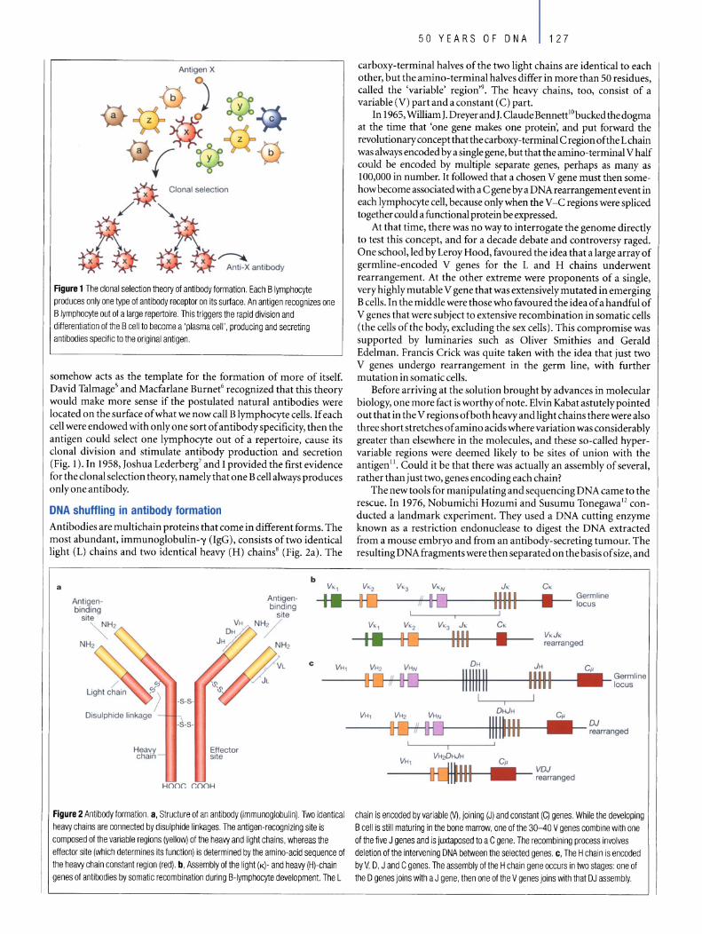
Antigen X
x Clonal selection
x x ~ Anti-X antibody
Figure 1 The clonal selection theory 01 antibody lormation. Each B Iymphocyte produces only one type 01 antibody receptor on its surface. An antigen recognizes one B Iymphocyte out 01 a large repertoire. This triggers the rapid division and differentiation 01 the B cell to become a 'plasma cell', producing and secreting antibodies specilic to the original antigen.
somehow acts as the template for the formation of more of itself. David TalmageS and Macfarlane Burnet6 recognized that this theory would make more sense if the postulated natural antibodies were located on the surface of what we now call B lymphocyte cells. If each cell were endowed with only one sort of antibody specificity, then the antigen could select one lymphocyte out of a repertoire, cause its donal division and stimulate antibody production and secretion (Fig. 1). In 1958, Joshua Lederberg7 and I provided the first evidence for the donal selection theory, namely that one B cell always produces only one antibody.
DNA shuffling in antibody formation Antibodies are multichain proteins that come in different forms. The most abundant, immunoglobulin-'Y (IgG), consists of two identical light (L) chains and two identical heavy (H) chains8 (Fig. 2a) . The
a
AntIgenbinding
site
Heavy chain
Effector si te
Hooe eOOH
b
Antigen-binding
site NH2
c VH,
Figure 2 Antibody lormation. a , Structure 01 an antibody (immunoglobulin). Two identical heavy chains are connected by disulphide linkages. The antigen-recognizing site is composed 01 the variable regions (yellow) 01 the heavy and light chains, whereas the effector site (which determines its lunction) is determined by the amino-acid sequence 01 the heavy chain constant region (red). b, Assembly 01 the light (K)- and heavy (H)-chain genes 01 antibodies by somatic recombination during B-Iymphocyte development. The L
50 YEARS OF DNA 127
carboxy-terminal halves of the two light chains are identical to each other, but the amino-terminal halves differ in more than 50 residues, called the 'variable' region'9. The heavy chains, too, consist of a variable (V) part and a constant (C) part.
In 1965, William J. Dreyer and J. Claude BennettlObucked the dogma at the time that 'one gene makes one protein', and put forward the revolutionary concept that the carboxy-terminal C region ofthe Lchain was always encoded by a single gene, but that the amino-terminal V half could be encoded by multiple separate genes, perhaps as many as 100,000 in number. It followed that a chosen V gene must then somehow become associated with a C gene by a DNA rearrangement event in each lymphocyte cell, because only when the V -C regions were spliced together could a functional protein be expressed.
At that time, there was no way to inter rogate the genome directly to test this concept, and for a decade debate and controversy raged. One school, led by Leroy Hood, favoured the idea that a large array of germline-encoded V genes for the Land H chains underwent rearrangement. At the other extreme were proponents of a single, very highly mutable V gene that was extensively mutated in emerging B cells. In the middle were those who favoured the idea of a handful of V genes that were subject to extensive recombination in somatic cells (the cells ofthe body, exduding the sex cells). This compromise was supported by lumina ries such as Oliver Smithies and Gerald Edelman. Francis Crick was quite taken with the idea that just two V genes undergo rearrangement in the germ line, with further mutation in somatic cells.
Before arriving at the solution brought by advances in molecular biology, one more fact is worthy of note. Elvin Kabat astutely pointed out that in the V regions ofboth heavy and light chains there were also three short stretches of amino acids where variation was considerably greater than elsewhere in the molecules, and these so-called hypervariable regions were deemed likely to be sites of union with the antigen 11. Could it be that there was actually an assembly of several, rather than just two, genes encoding each chain?
The new tools for manipulating and sequencing DNA came to the rescue. In 1976, Nobumichi Hozumi and Susumu Tonegawal2 conducted alandmark experiment. They used a DNA cutting enzyme known as a restriction endonudease to digest the DNA extracted from a mouse embryo and from an antibody-secreting tumour. The resulting DNA fragments were then separated on the basis of size, and
v" V'2
I. HJ
Germline }-----Hi+t-- _.. ..... - locus
V'3 J,
1111 OH
C,
• V.J • rearranged
VH2 VHN
D-Q--{}{] 11111111
JH Cp
1------1H-HH+I----IlfHlIH-II---I __ ~:Iine
VH,
VH ,
I VH20HJH
VOJ 1tftt1t---i __ t-- rearranged
DJ rearranged
chain is encoded by variable M, joining (J) and constant (C) genes. While the developing B cell is still maturing in the bone marrow, one 01 the 30-40 V genes combine with one 01 the live J genes and is juxtaposed to a C gene. The recombining process involves deletion 01 the intervening DNA between the selected genes. c, The H chain is encoded by V, 0, J and C genes. The assembly 01 the H chain gene occurs in two stages: one 01 the 0 genes joins with a J gene, then one 01 the V genes joins with that DJ assembly.

128 50 YEARS OF DNA
were reacted with radioactive probes, one corresponding to the whole L chain, the other to just the C portion. In the tumour, both probes lit up the identical fragment, whereas in the embryonic extract, two different fragments hybridized to the full-Iength probe, but only one of them to the C-region probe. Both fragments from the embryo sampie were different in size from the single hybridizing fragment in the tumour sampie. The experiment strongly argued for the V and C genes being so me distance from each other in the embryo, but having been rearranged and assembled during development of antibody-forming cells in adults to form a continuous DNA sequence constituting the full L-chain gene.
Generating more diversity Definitive elucidation of immunoglobulin gene structure depended on molecular cloning and subsequent sequencing of the genes themselvesJ3 • Here came another surprise, and one that could really not have been anticipated. V-region genes in the germline were found to be significantly shorter than is required to code for the V region ofthe L chain. It turned out that there is aseries of'minigenes' known as 'joining' (J) genes, which code for about 13 amino acids of the L chain. Thus, the full L chain is actually encoded by V, J and C genes (Fig. 2b). For the H chain, it is still more complicated, as there exists a series of'diversity' (D) genes that encode up to eight amino acids that lie between the V and J regions. Thus, the H chain is encoded by V, D, J and C genes (Fig. 2c). The assembly of a complete H-chain V region occurs in two separate steps: first, one ofthe D regions joins with one of the J regions, then one of many V regions joins with that DJ assembly (Fig.2c). The joining process is followed by deletion ofthe intervening DNA between the chosen minigenes. This is the first example of a somatic cell possessing a different genome from its fellow cells.
This minigene assembly process has important implications for antibody diversity. In humans, here are two types ofL chains, K and A, each with its own sets ofV and J genes. For the K light chain, there are 40 functional V genes and 5 functional J genes; for the A chain, there are 31 and 4, respectively. There is only one kind ofvariable region for the H chain, encoded by 51 V genes, 25 D genes and 6 J genes. To a first approximation, therefore, there are (40 x 5) + (31 x 4) = 324 different possible assemblies ofL chains, and 51 X 25 X 6 = 7,650 combinations for H chains. Thus, together, there are potentially 2,478,600 different types of germline-encoded antibodies.
But this is a considerable underestimate for two reasons. Recombination junctions can occur at different positions and this junctional diversity increases variability. Furthermore, a few extra nucleotides, called N regions, can be inserted between D-J junctions and V-D junctions in many H chains, and in a smaller percentage of L-chain V-J junctions. These nucleotides are not present in the germline and add to antibody diversity.
Yet further diversity can be generated by DNA mutations in dividing B cells. B cells expressing newly assembled immunoglobulin genes, each with its own unique specificity, constitute the 'primary repertoire'. When an antigen stimulates a chosen B cell to divide (Fig. 1), a proportion ofthe progeny migrate into the vicinity of antigen-capturing follicular dendritic cells (FDCs) and gradually form a 'germinal centre'. FDCs retain antigen on their surface for longperiods and stimulate further rounds of division. Within the germinal centre the B cells display an extraordinarily high rate of somatic mutation in V genes, estimated at 10-3 per nucleotide per division14• As antibody production accumulates, only those B cells with heightened affinity for the antigen gain access to FDC-bound antigen and thus are further stimulated to divide. As a result, B-cell clones secreting higher affinity antibody are selected in an iterative manner.
The 'memory' B cells that emerge from the germinal centre constitute the 'secondary repertoire', which is even more diverse than the primary one. Twenty mutations per chain are not uncommon; nor are thousand-fold increases in affinity. Thus, as it turned out, two early theories of antibody diversification proved to be correct: rearrangement of germline genes gives the naive B-cell
Box 1 Toleratlng Hit
How does the immune system distinguish foreign molecules trom the cells of ils own body? The failure 10 form antibodies againsl seil components confounded scientists until Maclariane Bumel proposed that an antigen inlroduced during embryonic development would 'trick' the immune system into regarding it as seil, so Ihat even in tater life it could nOI react against lhal anligen22•
Peter Medawar's group proved the prediction correcl by inducing immunological tolerance 10 transplantation antigens. Injecting foreign cells from a donor into laie-stage embryo mice within Ihe uterus led to a situation where, when these mice had grown up, they
could nOI rejecl skin gratts from the donor strain23.
What happens if the anlibody generaled by a newly crealed B I cell is reactive wilh a self componenl? The cell either dies immediately, il the stimulus is one capable 01 strongly cross-linking Ihe cell-surface immunoglobulin receptors, or else is given a
negalive signal which impedes ils full lunClion·" . This negalive I1 selection contribules 10 immunological tolerance.
For a T cell, this is more 01 a juggling act. Allhough il musl recognize the self-molecule major hislocompatibility complex (MHC), it musl avoid mounling an abnormal immune response to the
host's own cells. For Ihe T-cell repertoire to be lashioned correctly wilhin Ihe Ihymus, two sorts 01 seleclion are required. II the T cell, having assembled its receplor genes, has Ihe capacity to recognize seit MHC, it is allowed 10 mature. This step is known as positive selection. But the T cell must not leave the thymus il il has strong I potenlial 10 cause autoimmunity. This would be the case il Ihere were too high an affinity 01 binding wilh self MHC, or wilh a peptide from anormal cellular component bound to Ihe MHC groove. Such cells undergo programmed cell death within the Ihymus, the process 01 negative selection.
Delects in the immune system's ability to recognize seil are the basis 01 many auloimmune diseases, such as juvenile diabetes and multiple sclerosis.
repertoire, and somatic mutation ensures further diversification during memory B-cell development.
Switching function There are several different classes of antibodies, all of which have distinct roles that are also produced by rearrangements at the DNA level. There are eight different genes for the C region of the H chain, which specify different antibody functions. Each B cell first links the chosen VDJ assembly to a C gene known as /-L, creating an antibody class called IgM. If that cell is propelled into a pathway favouring the production of an antibodyprominent in mucus secretions (such as in the gastrointestinal tract), the VDJ section is switched over to a C region encoded byC gene er, and the cell produces IgA. If, on the other hand, the antigen is of parasite origin, or an allergen such as a pollen grain, the cell may be stimulated to produce IgE, in which case the VDJ region associates with the product of the C gene E. All of this occurs without any change in the specificity of the antibody being secreted. Although the detailed molecular mechanisms are still being investigated, the class switching again involves sequential excision of portions of the genetic material. Cytidine deaminase induced by B-cell-specific activation may be significant in both class switching and somatic hypermutation.
Assembly of T-cell receptors Whereas B cells make antibodies against antigens, the thymus-derived or T cells also respond to foreign agents, specializing in a more localized form of combat. Cytotoxic T cells are capable of killing virus-infected cells or cells displaying cancer-specific antigens. Other

myc
Normal chromosomes
8 14
Translocation
Burkitt's lymphoma
8 14
t
eH myc
Figure 3 Reciprocal chromosomal translocations in Burkitt's lymphoma, asolid tumour 01 B Iymphocytes. The genes lor making the heavy chains 01 antibodies (eH) are located on chromosomes 14, whereas those lor making the light chains are on chromosomes 2 and 22. These genes are expressed exclusively in B Iymphocytes, because only these cells have the necessary transcription lactors to switch on their expression. In most (over 90%) 01 Burkitt's lymphoma cases, a reciprocal translocation moves the proto-oncogene c-myc lrom its normal position on chromosome 8 to a location close to the antibody heavy-chain genes on chromosome 14 (rel. 18). In other cases, c-myc is translocated close to the antibody genes on chromosome 2 or 22. In every case, c-myc now linds itsell in a region 01 active gene transcription, and it may simply be the overproduction 01 the c-myc product (a transcription lactor essential lor cell division) that propels the Iymphocyte down the pathway towards cancer.
T cells secrete powerful stimulatory and inflammatory molecules, most of which act in a strictly localized context. T cells can also help guide B cells down appropriate pathways of differentiation.
In common with B cells, T cells also have one-receptor specificity and, for simplicity, I shall mention onlythe aß T-cell receptor (TCR), a heterodimer consisting oftwo subunits, the a- and ß-chains, joined by disulphide bonds 15.16. The strategy for generating T cells with different receptors is strikingly similar to that used by B cells to produce different antibodies and, in fact, the TCR binding surface looks much like that of an antibody. The ß-chain of the TCR is assembled in somatic cells from V, D, J and C genes; the a-chain from V, J and C genes. There are additions ofN-region nucleotides between Vand D, as well as between D and J on the ß-chain; and between V and J on the a-chain. A similar rearrangement also takes place for the "10 TCR.
But something peculiar about T-cell recognition was no ted by Rolf Zinkernagel and Peter Dohertyl7, who demonstrated that cytotoxic T cells could recognize viral antigens only if a specific 'self' molecule were also present on the target cell (see Box 1). The key part of the T -cell recognition puzzle fell into place when it was discovered that the TCR recognized short antigenic peptides bound to the groove of a self molecule known as the major histocompatibility complex (MHC), as well as surrounding portions ofthe MHC molecule itself. Cells have special mechanisms for fragmenting pro teins into peptides of8-24 amino acids in length, attaching these to MHC molecules and transporting the entire complex to the cell surface. TCRs then 'see' these short linear portions of antigens, be these of viral, bacterial or parasitic origin, or even portions of normal intracellular components. Such a system can help to control infections where the pathogen goes 'underground' inside a cell, and can also eliminate cells with mutated self antigens, such as cancer cells.
Lymphocytes and cancer Lymphocytes have been a favourite tool in cancer research. A notable example of DNA science applied in this way relates to the B-cell tumour of humans known as Burkitt's lymphoma. Occasionally DNA strands break and are incorrectly repaired. Thus, a piece of a chromosome becomes attached to the broken end of another one, and vice versa, in a process known as reciprocal translocation (Fig. 3).
50 YEARS OF DNA 129
In the case of Burkitt's lymphoma, a tumour-promoting gene or oncogene called myc is translocated from its normal position on chromosome 8 right into the middle of the IgH chain loeus on chromosome 14 (ref. 18). In this highly active transcriptional environment, myc expression is switched on, and eventually cancer develops.
It has been possible to create lymphoma-prone transgenic mice, which express myc in aberrantly high amounts. Because cancer is typically a multistage process, if further oncogenes are expressed simultaneously in transgenic mice, the onset of cancer can be dramatically accelerated. One such example involves the gene bcl-2. When this gene is expressed, it stops cells from undergoing natural programmed death (apoptosis)19. Mice expressing myc and bcl-2 showed very rapid development of tumours. An enormous amount ofliterature has accumulated related to the expanding familyof bcl-2-related genes and their roles in the regulation of programmed cell death. Models derived from lymphocytes and their malignancies have led to insights with implications well beyond immunology.
DNA vaccines DNA research has been of immense value to vaccine research. Through gene cloning and expression, candidate antigens can be identified and tested. In an era of rapid nucleotide sequencing, the whole genome of a pathogen can be determined, and computer programs can search for sequences likely to encode outer membrane proteins, which can be assessed as candidate vaccine molecules (see, for example, the series of papers published recently in Nature (419, 489-542,2002) on the genomics of the malaria parasite).
Amazingly, DNA itself can serve as a vaccine. DNA vaccines work on the principle that the gene sequence for one or more candidate antigens is introduced into an animal or person via a delivery vehicle known as a vector, together with a strong promoter that can switch on its expression in mammalian cells. Cells that take up the injected DNA transcribe and translate the gene and release the relevant antigen protein, which the body can in turn manufacture antibodies against. Thus, the body itselfbecomes a vaccine factorYo. Unfortunately, so far this approach has worked better in mice than in humans, but manyavenues are being pursued to improve this situation.
To strengthen the immune response to a vaccine, it may be necessary to use an adjuvant substance. Here, DNA mayaiso be of potential use. Scavenger cells, which capture antigens, have evolutionarily conserved receptors, known as Toll-like receptors (TLRs), wh ich recognize antigens common to many pathogens. One such receptor is TLR-9, which recognizes unmethylated CpG motifs commonly found in bacterial but not mammalian DNA. AccordingIy, unmethylated CpG-rich DNA sequences represent a promising new category of adjuvane l .
Future directions The solutions to the puzzle of antibody diversity and mystery ofT -cell recognition of antigenic peptides are among the brightest chapters of biology in the last quarter of the twentieth century. The future of immunologywill be all about how the system is regulated and how it makes decisions: whether to respond or not; whether to direct efforts towards antibodyformation or cell-media ted immunity; and, if the latter, whether more towards cytokine-secreting T cells or cytotoxic T cells.
As in the past, the future will be about information and thus about DNA science. All the complex signalling pathways, the feedback loops, and the intricate rules governing cell division on the one hand or programmed cell death on the other, will be progressively revealed. As this happens, the possibilities for applied research and development will be immense. In particular, new therapeutic targets will be identified. The 'miracle' drug for chronic myelogenous leukaemia, Glivec, was made possible after the characterization of the extraordinary cancerous potential ofthe chimaeric oncogene bcr-abl. This will surely be only the first of a plethora of more intelligently designed

130 50 YEARS OF DNA
anti-cancer drugs. Potent cytokines and monoclonal antibodies directed against cell surface-associated structures are alreadyprominent within a radically revised pharmaceutical armamentarium in areas including cancer, autoimmunity, allergy and transplantation. DNA research is therefore crucial to a new generation of immunologists, from those striving towards the development of novel vaccines to those seeking to understand and control auto immune diseases, allergy and transplant tolerance. D
doi: 10.1 038/natureO 1409
1. Ehrlich, P. The Croonian Lecture. On immunity with special reference to celllife. Proc. R. 50c. Lond. B
66,424-448 (1900).
2. Landsteiner, K. The Specificity ofSeroLogicaL Reactions (Harvard Univ. Press, Boston, 1945).
3. STeinl, F. & Haurowitz, F. Untersuchungen des Präzipitates aus Hämoglobin und anti-Hämoglobin
Serum und Bemerkungen über die Natur der Antikörper. Hoppe-Seyler's Z. Physiol. ehern. 192,45-57
(1930).
4. lerne, N. K. The natural selection theory of antibody formation. Proe. Natl Acad. Sei. USA 41, 849-857 (1955).
5. Talmage, D. W. Allergy and immullology. Annu. Rev. Med. 8, 239-256 (1957).
6. Burnet, F. M. A modification ofJerne's theoryof antibody production using the concept of donal
seleetion. Aust. j. Sci. 20, 67-69 (1957).
7. Nossal, G. J. v. & Lederberg, J. Antibodyproduction by single eells. Nature 181,1419-1420 (1958).
8. Edelman, G. M. & GaU, W. E. The antibody problem. Annu. Rev. Biochem. 38, 415-466 (1969).
9. Hilschmann. N. & Craig, L. C. Amino acid sequenee studies with Benee-Jones proteins. Proc. Na tl
Acad.Sei. USA 53, 1403- 1409 (1965).
10. Dreyer, W. J. & Bennett, J. C. The moleeular basis of antibody formation: a paradox. Proc. Natl Acad.
Sei. USA 54, 864-869 (1965).
11. WUt T. T. & Kabat, E. A. An analysis ofthe sequenees ofthevariable regions ofBence Jones proteins
and myeloma light ehains and their implieations for antibody eomplementarity.]. Exp. Med. 132,
211-250(1970).
12. Hozumi, N. & Tonegawa. S. Evidence for somatie rearrangement of immunoglobulin genes coding
for variable and constant regions. Proc. Nall Acad. Sei. USA 73. 3628-3632 (1976).
13. Bernard, 0. , Hozumi, N. & Tonegawa, S. Sequences of mouse immunoglobulin light chain genes
before and after somatie ehanges. CdliS. 11 33- 1144 (1978).
14. Kocks, C. & Rajewsky, K. Stable expression and somatie hypermutation of antibodyV regions in B
eeil developmental pathways. Annu. Rev. Immunol. 7, 537-559 (1989).
15. Hedrick, S. M., Cohen, D.I., Nielsen, E. A. & Davis, M. M. Isolation of eDNA clones eneodingT cell
specifie membrane-associated proteins. Nature 308, 149-153 (1984).
16. Yanagi, Y. el al. A human T eell-specifie eDNA clone encodes a protein having extensive homology to
immunoglobulin chains. Nature 308,145-149 (1984).
17. Zinkernagel, R. M. & Doherty, P. C. Restrietion of in vitra T eell-mediated eytotoxicity in Iymphocytic
ehoriomeningitis within a syngeneic or semiallogeneic system. Nalllre248, 701-702 (1974).
18. Bernard, 0., Cory. S., Gerondakis. S., Webb, E. & Adams, J. M. Sequence ofthe murine and human
cellular mye oneogenes and two modes of mye transcription resulting from chromosome
transloeation in B lymphoid tumours. EMBO]. 2, 2375-2383 (1983).
19. Vaux, D. L., Cory, S. & Adams, J. M. Bcl-2 gene promotes haemopoietie eeH survival and cooperates
with e-mycto immortalize pre-B cells. Nature 335, 440-442 (1988).
20. Donnelly, J. J. , Ulmer, J. 8., Shiver, J. W. & Liu, M. A. DNA vaecines. Annu. Rev. [mmuflOl. IS, 617-648
(1997).
21. Krieg, A. M. epG motifs in bacterial DNA and their immune effeets. Annu. Rev. Immunol. 20.
709-760 (2002).
22. Burnet, F. M. & Fenner, F. J. The Production of Antibodies (Maemillan, Melbourne, 1949).
23. Billingham, R. F., Brent, L. & Medawar, P. B. Aetively acquired toleranee of foreign cells. Nature 172,
603-{;06(1953).
24. Nossal, G. ,. V. & Pike, B. L. Clonal anergy: persistenee in tolerant mice of antigen-binding B
Iymphoeytes ineapable of responding to antigen or mitogen. Proc. Natl Acad. Sei. USA 77, 1602- 1606
(1980).
Original reference: Nature421, 440-444 (2003).
The digital code otDNA
Leroy Hood* & David Galast
* Institute for Systems Biology, 4225 Roosevelt Way NE, Seattle,
Washington 98105, USA (e-mail: [email protected])
t Keck Graduate Institute of Applied Sciences, 535 Watson Drive, Claremont,
California 91711, USA (e-mail: [email protected])
The discovery of the structure of DNA transformed biology profoundly, catalysing the sequencing of the human genome and engendering a new view of biology as an information science. Two features of DNA structure account for much of its remarkable impact on science: its digital nature and its complementarity, whereby one strand of the helix binds perfectly with its partner. DNA has two types of digital information - the genes that encode proteins, which are the molecular machines of life, and the gene regulatory networks that specify the behaviour of the genes.
'f1ny living cell carries with it the experiences of a billion years of experimentation by its ancestors." Max Delbruck, 1949.
discovery of the double helix in 1953 immediately raised questions about how biological information is encoded in DNA1• A remarkable feature ofthe structure is that DNA can accommodate almost any sequence of base pairs - any combination of the bases adenine (A),
cytosine (C), guanine (G) and thymine (T) - and, hence any digital message or information. During the following decade it was discovered that each gene encodes a complementary RNA transcript, called messenger RNA (mRNA)2, made up of A, C, G and uracil (U), instead ofT. The four bases of the DNA and RNA alphabets are related to the 20 amino acids of the protein alphabet by a triplet code - each three letters (or 'codons') in a gene encodes one amino acid3• For example, AGT encodes the amino acid serine. The dictionary of DNA letters that make up the amino acids is called the genetic code4 • There are 64 different triplets or codons, 61 ofwhich encode an amino acid (different trip lets can encode the same amino acid), and three of which are used for 'punctuation' in that they signal the termination of the growing protein chain.
The molecular complementary of the double helix - whereby each base on one strand ofDNA pairs with its complementary base on the partner strand (A with T, and C with G) - has profound implications for biology. As implied by James Watson and Francis Crick in their landmark paper l , base pairing suggests a templatecopying mechanism that accounts for the fidelity in copying of genetic material during DNA replication (see accompanying article by Alberts, page 117). It also underpins the synthesis of mRNA from the DNA template, as well as processes of repairing damaged DNA ( discussed by Friedberg, page 122).
Tools to modify DNA The enzymes that function in cells to copy, cut and join DNA molecules were also exploited as keytools for revolutionary newtechniques in molecular biology, including the cloning of genes and expression of their proteins, and mapping the location of genes on chromosomes. The ability to recreate the process ofDNA replication artificiallyin the laboratory led to the development oftwo techniques

a
TAA AA AAA
b GGG C T AA C AAG C AAA
T GGG C T AA C AAG C AAA T GGG C T AA C AAG C AAA
GGG C T AA C AAG C AAA
GGG C T AA C AAG C AAA
GGG C T AA C AAG C AAA
GGG C ~ AA C AAG C AAA
GGG C AA C AAG C AAA
GGG C AA C AAG C AAA
d
_goto , I 11;20 , I i2pO I I 1580 I I 13;60 I
GG G AA 90
A GG " A A G G 100 "0
DNA polymerase
AT TG
GA C T G AG
GA T C T G AG GA r C G A
GA r C G
GA T C G
GA T C
GA r C GA
GA
I 14,40 I • 1520 I
G G GAAA G 120
---------A
130
that transformed biology: a manual DNA sequencing method in 1975 and, in 1985, the discovery of the polymerase chain reaction (PCR), whereby DNA sequences could be amplified a millionfold or mores.
Although sequencing and PCR transformed the science of biology, they also had wide applications for medicine and forensics. The detection of variation in DNA sequence from one individual to the next - so-called 'polymorphisms' - forms the basis of DNA 'finger-printing' of individuals. Forensics uses these fingerprints to deal with paternity disputes, as well as criminal cases such as rape. The finding that many specific DNA polymorphisms are associated with disease or disease susceptibility has brought DNA diagnostics to medicine and opened the pathway to truly predictive medicine, where the risks of disease can be identified in advance of symptoms (see accompanying article by Bell, page 100).
Automated DNA sequencing The first efforts to sequence DNA, pioneered by Walter Gilbert6 and Fred Sanger7 in the 1970s, decoded stretches ofDNA a few hundred bases long. When the first complete genome was sequenced over a period of about one year in 1977-78 - that of a viral genome of about 5,000 bases8 - it became clear that DNA sequence data could provide unique insights into the structure and function of genes, as weH as genome organization. It was this potential to generate vast amounts of information about an organism from its genetic code that inspired efforts towards the automation of DNA sequeneing (Fig.1).
The eombination of teehnieal wizardry and intensive automation in the decade that followed launched the 'genomie era'. Aseries of new instruments enabled novel approaehes to biologieal analysis9- 11 • The first sequeneing maehine - invented by Leroy Hood, Lloyd Smith and Mike Hunkapiller in 1986 (ref. 12) - was automated in data aequisition, but still required substantial manual attention and the sequeneing rate was low, roughly 250 bases per day. Over the next ten years, the development of automated DNA sequeneing aecelerated, rapidly passing through three distinet stages: the prototype sequeneing maehine (1986); a robust instrumentthat could be used routinely in a standard laboratory (1989); and finaHy, a maehine that formed part of an integrated faetory-like production line where DNA sampie preparation and sequeneing were aH fuHy automated (1998). The
50 YEARS OF DNA 131
Figure 1 How to sequence DNA. a, DNA pOlymerase copies astrand of DNA. b, The insertion of a terminator base into the growing strand halts the copying process. This is a random event that results in aseries of fragments of different lengths, depending on the base at which the copying stopped. The fragments are separated by size by running them through a gel matrix, with the shortest fragments at the bottom and largest at the top. e, The terminators are labelied with different fluorescent dyes, so each fragment will fluoresce a particular colour depending on whether it ends with an A, C, G, or T base. d, The sequence is 'read' bya computer. It generates a 'sequence trace', as shown here, with the coloured peaks corresponding to fluorescent bands read from the bottom to the top of one lane of the gel. The computer translates these fluorescent signals to DNA sequence, as illustrated across the top of the plot. Image adapted from ref. 20.
advanees in sequencing capacity have been striking - the !atest sequeneing machines are able to decode approximately 1.5 million bases over 24 hours - 6,000 times the throughput of the prototype.
The goals ofhigh-throughput biologieal instrumentation are to inerease throughput, enhanee the quality of the data, and greatly reduce the cost of per unit information aequired. To reaeh these goals in the future, the miniaturization, automation, parallelization and integration of sueeessive proeedures will propel DNA sequeneing teehnologyinto the realm of mierofluidies and mieroelectronies, and eventually into the area of nanoteehnology. With single-DNAmoleeule sequeneing, we foresee a time when the entire genome of an individual could be sequenced in a single day at a cost of less than $US 1 0,000 (compared with the US$50 million or more it would cost today). This will readily enable the decoding of the genomie seq uence of virtually any organism on the planet and provide unparalleled access to the foundations ofbiology and the study ofhuman genetic variability.
The Human Genome Project The breathtaking speed at which automated DNA sequencing developed was largely stimulated by the throughput demands of the Human Genome Project (HGP), which offieiaHy started in 1990 following discussions and studies on feasibility and teehnology that began inearnestin 1985. Theobjeetivesofthe HGPwereto generatea finished sequenee in 15 years13, but a draft of the human genome sequenee was available in 2001. Two vers ions of the draft were genera ted and published in 2001, one by the publicly funded International Human Genome Sequencing Consortium1\ and another by the bioteehnology company Celera1s (Box 1). In the proeess of developing the tools and methodology to be able to sequenee and assemble the 3 billion bases of the human genome, a range of plant, animal and microbial genomes was sequenced and many more are currently being deeoded. As genome sequenees become available, different areas ofbiology are being transformed - for example, the diseipline of microbiology has ehanged signifieantly with the completion of more than 100 baeterial genome sequenees over the past deeade.
The HGP profoundly influeneed biology in two respeets. First, it illustrated the concept of 'discovery seien ce' - the idea that all the elements of the system (that is, the complete genome sequence and

132 50 YEARS OF DNA
the entire RNA and protein output encoded by the genome) can be defined, archived in a database, and made available to facilitate hypothesis-driven science and global analyses. Second, to succeed, the HGP pushed the development of efficient large-scale DNA sequencingand, simultaneously, drove thecreation ofhigh-throughput tools (for example, DNA arrays and mass spectrometry) for the analysis of other types of related biological information, such as mRNAs, proteins and molecular interactions.
The digital nature of biological information The value ofhaving an entire genome sequence is that one can initiate the study of a biological system with a precisely definable digital co re of information for that organism - a fully delineated genetic source code. The challenge, then, is in deciphering what information is encoded within the digital code. The genome encodes two main types of digital information - the genes that encode the protein and RNA molecular machines oflife, and the regulatory networks that specify how these genes are expressed in time, space and amplitude.
It is the evolution of the regulatory networks and not the genes themselves that play the critical role in making organisms different from one another. The digital information in genomes operates across three diverse time spans: evolution (tens to millions of years), development (hours to tens ofyears), and physiology (milliseconds to weeks). Development is the elaboration of an organism from a single cell (thefertilized egg) to an adult (for humans this is 1014 cells of thousands of different types). Physiology is the triggering of specific functional programmes (for example, the immune response) by environmental cues. Regulatory networks are crucial in each of these aspects ofbiology.
Regulatory networks are composed of two main types of components: transcription factors and the DNA sites to which they bind in the control regions of genes, such as promoters, enhancers and silen cers. The control regions of individual genes serve as information processors to integrate the information inherent in the concentrations of different transcription factors into signals that mediate gene expression. The collection of the transcription factors and their cognate DNA-binding sites in the control regions of genes that carry out a particular developmental or physiological function constitute these regulatory networks (Fig. 2).
Because most 'higher' organisms or eukaryotes (organisms that contain their DNA in a cellular compartment called the nucleus), such as yeast, flies and humans, have predominantly the same families of genes, it is the reorganization ofDNA-binding sites in the control regions of genes that mediate the changes in the developmental programmes that distinguish one species from another. Thus, the regulatory networks are uniquely specified by their DNA-binding sites and, accordingly, are basically digital in nature.
One thing that is striking about digital regulatory networks is that they can change significantly in short periods of evolutionary time. This is reflected, for example, in the huge diversity of the body plans, controlled by gene regulatory networks, that emerged over perhaps 10-30 million years during the Cambrian explosion of metazoan organisms (about 550 million years ago). Likewise, remarkable changes occurred to the regulatory networks driving the development of the human brain during its divergence from its common ancestor with chimpanzees about 6 million years ago.
Biology has evolved several different types of informational hierarchies. First, a regulatory hierarchy is a gene network that defines the relationships of a set oftranscription factors, their DNA-binding sites and the downstream peripheral genes that collectively control a particular aspect of development. A model of development in the sea urchin represents a striking examplel6 (Fig. 2) . Second, an evolutionary hierarchy defines an order set of relationships, arising from DNA duplication. For example, a single gene may be duplicated to generate a multi-gene family, and a multi-gene family may be duplicated to create a supergene family. Third, molecular machines may be assembled into structural hierarchies by an ordered assembly process. One
Be1 lequenclng the hu .... n ........
The first complete dralls of the human genome sequence were published in 2001 by the International Human Genome Sequencing Consortium (IHGSC), a publicly funded effor!, and Celera, a biotechnology company, using different approaches. 80th efforts used a random or shotgun approach where the original DNA to be
sequenced was randomly broken into overlapping fragments that were then cloned, and 500 base pairs (bp) were 'read' from one or both ends of the clones.
For the drall genome sequences, each base was read six to ten times to optimize the accuracy of the sequence. The stretches of DNA sequence were read by a computer and assembled into a complete sequence. The IHGSC effor! randomly cleaved DNA inlo -200,OOO-bp fragments and generated a map of these fragments across the 24 different human chromosomes: it then used the shotgun approach to sequence the pre-ordered fragments clone by clone. In contrast , Celera randomly fragmented the entire genome into three sizes of fragments (approximately 2,000, 10,000 and 200,000 bp), sequenced both ends of the clones and then used the end sequences to assemble the entire genome sequence, without the aid of a map.
Celera's 1998 announcement that it would sequence the human genome within three years was greeted with considerable scepticism, but it succeeded in producing a drall sequence and considerably accelerating the public effort. The efforts 01 both groups benefited science by producing drall genome sequences considerably earlier than expected.
A1though minor differences were noted between the two dralls,
the overall conclusions concerning gene numbers, repeated sequences and chromosomal organization were remarkably similar. For example, both groups identified 30,000-35,000 genes, far fewer
.1 than the 100,000 expected from an earlier (admittedly 'back of the Il envelope') calculation.
example of this is the basic transcription apparatus that involves the step-by-step recruitment of factors and enzymes that will ultimately drive the specific expression of a given gene. A second example is provided by the ribosome, the complex that translates RNA into protein, which is assembled from more than 50 different proteins and a few RNA molecules. Finally, an informational hierarchy depicts the flow of information from a gene to environment: gene ~ RNA ~ protein ~ protein interactions ~ protein complexes ~ networks of protein complexes in a cell ~ tissues or organs ~ individual organisms ~ populations ~ ecosystems. At each successively higher level in the informational hierarchy, information can be added or altered for any given element (for example, by alternative RNA splicing or protein modification).
Systems approaches to biology Humans start life as a single cell- the fertilized egg - and develop into an adult with trillions of cells and thousands of cell types. This process uses two types ofbiological information: the digital information of the genome, and environmental information, such as metabolite concentrations, secreted or cell-surface signals from other cells or chemical gradients. Environmental information is of two distinct types: deterministic information where the consequences of the signals are essentially predetermined, and stochastic information where chance dictates the outcome.
Random, or stochastic, signals can generate significant noise in biological systems, but it is onlyin special cases that noise is converted into signals. For example, stochastic events govern many ofthe genetic mechanisms responsible for generating antibody diversity. In the immune response, those B cells that produce antibodies that bind

50 YEARS OF DNA 1 33
a cf3 Figure 2 A gene regulatory
+- network involved in sea urchin development'6 a, Part 01 the network 01 transcription lactors and their interactions with the contra I regions 01 other transcription lactors. Genes are indicated by horizontallines; arrawheads indicate activation; '1.'
symbols indicate gene repression. b, An enlargement 01 the pramoter region 01 a gene, called endo 16, that helps modulate the development 01 the endoderm. It contains 34 binding sites (rectangles) for 13 different transcription factors and cofactors (illustrated as rectangles or 10llipops, respectively). Six modules (A-G) of transcription lactors and binding sites carry out discrete lunctions to developmentally regulate endo 16. C, Diagram depicting the logical
b structures of the A and B contra I (-2.300) G F E DC circuits during sea urchin
development.
MOduieA SP
CYCS1 UI R CS2 CG1 P OTXZ CG2 SPGCF1CG3 CG4
• r I! o ';.'
IIIIIJD Cl 0 0
.. .. i1
L l'-I----~i 1 (
+ I H i3
,- .. - ... + i5 --.-- --··-1 ------- --->-
i9 .......... F.E. DC
..L _~==::t( i6
i2- i~ i 1 ~Q============t=========~,
tightlyto the antigen (that is, those having high affinities) undergo an expansion in number that is proportional to the strength of the antibody affinity (see accompanying article by Nossal, page 126). Hence, the signal (high affinity) is distinguished from the noise (low affinity). Moreover, high levels of mutation in the B cells causes specific diversification of antibody genes in the presence of antigen and permits the affinity to increase even more. The cells carrying the higher-affinity antibody genes are then preferentially selected for survival and proliferation.
The key question is what and how much signal emerges from the noise. Analysis of stochastic events and the differentiation between signal and noise will be a future challenge for contemporary biology. The immune response has been studied for more than 100 years, yet we still have only a partial understanding of its systems properties, such as the immune response and tolerance (the umesponsiveness to one's own cells). This is because until recently immunologists have been able to study this complex system only one gene or one protein at a time.
The systems approach permits the study of all elements in a system in response to genetic (digital) or environmental perturbations. Global quantitative analyses ofbiological information from different levels each provide new insights into the operation of the system; hence, information at as many levels as possible must be captured, integrated, and ultimately, modelled mathematically. The model should explain the properties of the system and establish a framework that allows us to redesign the system in a rational way to generate new emergent properties.
Several systems have been explored successfully. The utilization of the sugar galactose in yeast has been analysed using genetic perturbations (inactivation of genes) and four levels of information were gathered ~ RNA and protein concentrations as weIl as proteinprotein and protein-DNA interactions l7• Using an iterative and integrative systems approach, new insights into the regulation of galactose use were gained. Moreover, the relationships of the galactose regulatory network to other modules in the yeast cell were also delineated. Likewise, systems approaches to early embryonic
134 50 YEARS OF DNA
development in the sea urchin have delineated a regulatory network that has significant predictive power16 (Fig. 2). Finally, systems approaches to metabolism in an archaeal halobacterium (an organism thriving in up to five-molar salt solutions, such as the Dead Seal have revealed new insights into the inter-relationships among several modules controlling energy production in the celll8 •
The study of cellular and organismal biology using the systems approach is at its very beginning. It will require integrated teams of scientists from across disciplines - biologists, chemists, computer scientists, engineers, mathematicians and physicists. New methods for acquiring and analysing high-throughput biological data are needed. A powerful computational infrastructure must be leveraged to generate more effective approaches to the capture, storage, analysis, integration, graphical display and mathematic formulation of biological complexity. New technologies must be integrated with each other. Finally, hypothesis-driven and discovery science must be integrated. In short, both new science and technology must emerge for the systems biology approach to realize its pro mise. A cultural shift in the biological sciences is needed, and the education and training of the next generation of biologists will require significant reform.
Gordon Moore, the founder ofIntel, predicted that the number of transistors that could be placed on a computer chip would double every 18 months. It has for more than 30 years. This exponential growth has been a driver for the explosive growth of information technology. Likewise, the amount of DNA sequence information available to the scientific community is following a similar, perhaps even steeper, exponential increase. The critical issue is how sequence information can be converted into knowledge of the organism and how biology will change as a result. We believe that a systems approach to biology is the key. It is clear, however, that this approach poses significant challenges, both scientific and cultural19• The discovery of DNA structure started us on this journey, the end of which will be the grand unification of the biological sciences in the emerging, information -based view ofbiology. D doi:l0.1038/nature01410
1. Watson, J. D. & Crick, F. H. C. A structure for deoxyribose nucleic acid. Naturel?1, 737-738 (1953).
2. Brenner, S., Jacob, F. & Meselsoll, M. An unstable intermediate carrying information from genes to
ribosomes for protein synthesis. Nature 190,576--581 (1961).
3. Crick, F. H. c., Barnett, 1., Brenner, S. & Watts-Tobin, R. J. General nature ofthe genetic code for proteins. Nature 192,1227-1232 (1961).
4. Nirenberg, M. W. & Matthaei, J. H. The dependence of ceIl-free protein synthesis in E. coli upon
naturally occurring or synthetic polynudeotides. Proc. Natl Acad. Sei. USA 47, 1588-1602
(1961).
5. Saiki, R. K. et al. Enzymatic amplification of ß-globin sequences and restriction site analysis for
diagnosis of sickle cell anemia. Seienee 230, 1350-1354 (1985).
6. Maxam, A. M. & Gilbert, W. A new method of sequencing DNA. Proc. Natl Aead. Sei. USA 74,
56{}-S64 (1977).
7. Sanger, F. & Coulson, A. R. A rapid method for determining sequences in DNA by primed synthesis
with DNApolymerase.]. Mol. Biol. 94, 444-448 (1975).
8. Sanger, F. eta/. Nudeotide sequence ofbacteriophage <flXI74. Nature 265, 678-695 (1977).
9. Hunkapiller, M. W. & Hood, L. Newprotein sequenatorwith increased sensitivity. Scienee207,
523-525 (1980).
10. Horvath, S. J., Firca, J. R., Hunkapiller, T., Hunkapiller M. W. & Hood. L. An automated DNA
synthesizer employing deoxynudeoside 3' phosphoramidites. Methods Enzymol. 154, 314-326
(1987).
11. Kent, S. B. H., Hood, L. E., Beilarr, H., Meister S. & Geiser, T. High yield chemical synthesis of
biologically active peptides on an automated peptide synthesizer of novel design. Peptides 5, 185-188
(1984).
12.Smith, L. M. et al. Fluorescence detection in automated DNA sequence analysis. Nature321, 674-679
(1986).
13.Collins, F. & Galas, D. J. A new five-yearplan for the US Human Genome Project. Science262, 43-46
(1993).
14. Venter, J. C. etal. The sequence ofthe human genome. Science291, 1304-1351 (2001).
15. International Human Genome Sequencing Consortium. Initial sequeneing and analysis of the human
genome. Nature409, 86{}-921 (2001).
16. Davidson, E. H. et al. A genomic regulatory network for development. Science295, 1669-1678 (2002).
17. Ideker, T. et al. Integrated genomic and proteomic analyses of a systematically perturbed metabolie
network. Science292, 929-933 (2001).
18. Baliga, N. S. et al. Coordinate regulators of energy transduction modules in the Halobacterium sp.
analyzed by a global systems approach. Proc. Natl Acad. Sei. USA 99, 14913-14918 (2002).
19.Aderem A. & Hood, L. Immunology in the post-genomic era. Nature lmmunol. 2, 1-3 (2001).
20. Dennis, C. & Gallagher, R. (eds) The Human Genome (Palgrave, Basingstoke, 2001).
Original reference: Nature421, 444-448 (2003).
Controlling the double helix Gary Felsenfeld* & Mark Groudinet
* Laboratory ofMolecular Biology, National Institute ofDiabetes and Digestive
and Kidney Diseases, National Institutes of Health, Building 5, Room 212,
Bethesda, Maryland 20892-0540, USA (e-mail: [email protected])
t Division ofBasic Seiences, Fred Hutchinson Cancer Research Center, 1100
Fairview Avenue North, Seattle, Washington 98109, and Department of
Radiation Oneology, University ofWashington Sehool ofMedieine, Seattle, Washington 98195, USA (e-mail: [email protected])
Chromatin is the complex of DNA and proteins in which the genetic material is packaged inside the cells of organisms with nuclei. Chromatin structure is dynamic and exerts profound control over gene expression and other fundamental cellular processes. Changes in its structure can be inherited by the next generation, independent of the DNA sequence itself.
enes were first shown to be made of DNA only nine before the structure of DNA was discovered
1; and see accompanying article by McCarty, 92). Although revolutionary, the idea that
information was protein-free ultimately proved too simple. DNA in organisms with nuclei is in fact coated with at least an equal mass of protein, forming a complex called chromatin, which controls gene activity and the inheritance of traits.
'Higher' organisms, such as yeast and humans, are eukaryotes; that is, they package their DNA inside cells in aseparate compartment called the nucleus. In dividing cells, the chromatin complex of DNA and protein can be seen as individual compact chromosomes; in non-dividing cells, chromatin appears to be distributed throughout the nucleus and organized into 'condensed' regions (heterochromatin) and more open 'euchromatin' (see accompanying article by Ball, page 107). In contrast, prokaryotes, such as bacteria, lack nuclei.
The evolution of Chromatin The principal protein components of chromatin are proteins called histones (Fig. 1). Core histones are among the most highly conserved eukaryotic proteins known, suggesting that the fundamental structure of chromatin evolved in a common ancestor of eukaryotes. Moreover, histone equivalents and a simplified chromatin structure have also been found in single-cell organisms from the kingdom Archaeabacteria2,3.
Because there is more DNA in a eukaryote than in a prokaryote, it was naturally first assumed that the purpose of histones was to compress the DNA to fitwithin the nucleus. But subsequent research has dramatically revised the view that histones emerged as an afterthought, forced on eukaryotic DNA as a consequence of large genome size and the constraints of the nucleus.
It was known that different genes are active in different tissues, and the distinction ofheterochromatin and euchromatin suggested that differences in chromatin structure were associated with differences in gene expression. This led to the early supposition that the histones were also repressor pro teins designed to shut off unwanted expression. The available evidence, although rudimentary, does indeed suggest that archaeal histones are not merely packaging factors, but function to regulate gene expression2- S• They
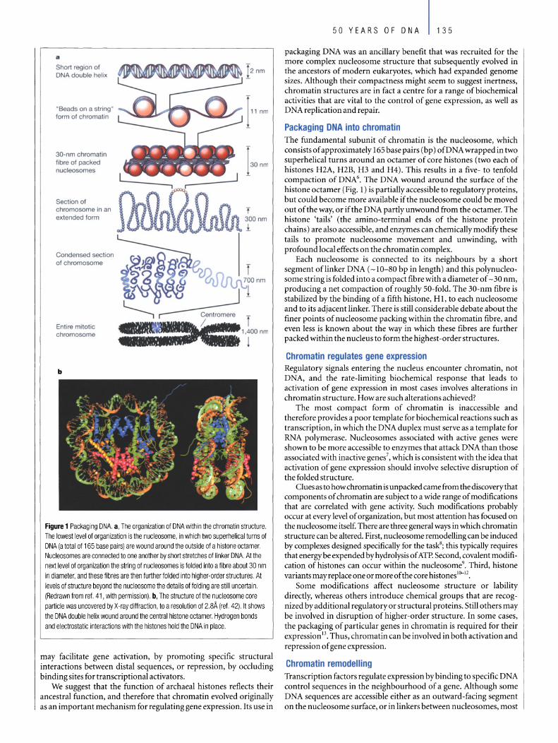
a
Short region of DNA double helix
"Beads on astring" form of chromatin
30-nm chromatin fibre of packed nucleosomes
Section of chromosome in an extended form
Condensed section of chromosome
Entire mitotic chromosome
b
Figure 1 Packaging DNA. a, The organization 01 DNA within the chromatin structure. The lowest level 01 organization is the nucleosome, in which two superhelical turns 01 DNA (a total 01 165 base pairs) are wound around the outside 01 a histone octamer. Nucleosomes are connected to one another by short stretches 01 linker DNA. At the next level 01 organization the string 01 nucleosomes is lolded into a libre about 30 nm in diameter, and these tibres are then lurther lolded into higher-order structures. At levels 01 structure beyond the nucleosome the details ot lolding are still uncertain. (Redrawn trom rel. 41, with permission). b, The structure otthe nucleosome core particle was uncovered by X-ray diffraction, to aresolution 01 2.8Ä (rel. 42). It shows the DNA double helix wound around the central histone octamer. Hydrogen bonds and electrostatic interactions with the histones hold the DNA in place.
may facilitate gene activation, by promoting specific structural interactions between distal sequences, or repression, by occluding bin ding sites for transcriptional activators.
We suggest that the function of archaeal histones retlects their ancestral function, and therefore that chromatin evolved originally as an important mechanism for regulating gene expression. Its use in
50 YEARS OF DNA 135
packaging DNA was an ancillary benefit that was recruited for the more complex nucleosome structure that subsequently evolved in the ancestors of modern eukaryotes, which had expanded genome sizes. Although their compactness might seem to suggest inertness, chromatin structures are in fact a cent re for a range ofbiochemical activities that are vital to the control of gene expression, as weil as DNA replication and repair.
Packaging DNA into chromatin The fundamental subunit of chromatin is the nucleosome, which consists of approximately 165 base pairs (bp) ofDNA wrapped in two superhelical turns around an octamer of core histones (two each of his tones H2A, H2B, H3 and H4). This results in a five- to tenfold compaction of DNA6• The DNA wound around the surface of the histone octamer (Fig. 1) is partially accessible to regulatory proteins, but could become more available if the nucleosome could be moved out of the way, or if the DNA partly unwound from the octamer. The histone 'tails' (the amino-terminal ends of the histone protein chains) are also accessible, and enzymes can chemically modify these tails to promote nucleosome movement and unwinding, with profound local effects on the chromatin complex.
Each nucleosome is connected to its neighbours by a short segment oflinker DNA (~1O-80 bp in length) and this polynucleoso me string is folded into a compact fibre with a diameter of ~ 30 nm, producing a net compaction of roughly 50-fold. The 30-nm fibre is stabilized by the bin ding of a fifth histone, H 1, to each nucleosome and to its adjacent linker. There is still considerable debate about the finer points of nucleosome packing within the chromatin fibre, and even less is known about the way in which these fibres are further packed within the nucleus to form the highest -order structures.
Chromatin regulates gene expression Regulatory signals entering the nucleus encounter chromatin, not DNA, and the rate-limiting biochemical response that leads to activation of gene expression in most cases involves alterations in chromatin structure. How are such alterations achieved?
The most compact form of chromatin is inaccessible and therefore provides a poor template for biochemical reactions such as transcription, in which the DNA duplex must serve as a template for RNA polymerase. Nucleosomes associated with active genes were shown to be more accessible to enzymes that attack DNA than those associated with inactive genes?, which is consistent with the idea that activation of gene expression should involve selective disruption of the folded structure.
Clues as to how chromatin is unpacked came from the discoverythat components of chromatin are subject to a wide range of modifications that are correlated with gene activity. Such modifications probably occur at every level of organization, but most attention has focused on the nucleosome itself. There are three general ways in which chromatin structure can be altered. First, nucleosome remodelling can be induced by complexes designed specifically for the task8; this typically requires that energy be expended by hydrolysis of ATP. Second, covalent modification of histones can occur within the nucleosome9• Third, histone variants mayreplace one or more ofthe core histones10--12•
Some modifications affect nucleosome structure or lability directly, whereas others introduce chemical groups that are recognized by additional regulatory or structural proteins. Still others may be involved in disruption of higher-order structure. In some cases, the packaging of particular genes in chromatin is required for their expression13• Thus, chromatin can be involved in both activation and repression of gene expression.
Chromatin remodelling Transcription factors regulate expression by binding to specific DNA control sequences in the neighbourhood of a gene. Although some DNA sequences are accessible either as an outward-facing segment on the nucleosome surface, or in linkers between nucleosomes, most
136 50 YEARS OF DNA
Box 1 Histone modlflcatlons
Many amino acids of histones, particularly those in the 'tails', are chemically modified41 . These include lysine residues that may be acetylated, methytated or coupled to ubiquitin (a large polypeptide chain): arginine residues that may be methytated: and serine residues that are phosphorylated. All modifications can affeet one another, and many are positively or negatively correlated with each other. Colleetively, they constitute a set of markers 01 the local state of the genetic material, which has been called the 'histone code'48.
Histone modification is a dynamic process. Chromatin in the neighbourhood of transcriptionally active genes is enriched in acetylated histones, and the enzymes responsible for both acetylation and deacetylation are often reeruited to sites where gene expression is 10 be aclivaled or repressed, respeclively. Within the nucieus, local states of both acetylation and phosphorylation can change rapidly. Methytation at certain histone amino-acid residues mayaiso be important for activation, whereas al other sites it is a signal lor inactivation.
Many (perhaps all) of the histone modifications interact with each other in ways that are still not completely understood. For example, in mammals, histone H2B can be modified by ubiquitin at Lys 120 (123 in yeast), and this modification is neeessary for methytation at Lys 4 and Lys 79 of histone H3, reactions that are controlled by two different methytating enzymes. Inftuences between nearby modification sites have also been observed, such that phosphorylation at one site can lacilitale acetylation at another, methytation and phosphorylation at adjacent sites may interfere with one another, and methytation and acetylation cannot occur simultaneously on the same lysine residue.
are buried inside the nucleosome. Regulatory factors must therefore seek out their specific DNA-binding sites and gain access to them. Theyare aided bychromatin-remodelling complexes that continually shuftle the positions of individual nucleosomes so that sites are randomly exposed for a fraction of time8,14.
A number of chromatin-remodelling complexes mobilize nucleosomes, causing the histone octamers to move short distances along the DNA8• Each complex carries a protein with ATPase activity, which provides the necessary energy. Many ofthese complexes are members of the so-called SWI/SNF family, which includes SWI/SNF in budding yeast and human, RSC in yeast, and Brahma in Drosophila. They have similar helicase-motif subunits, but varying co-factors within the complex. Another SWI/SNF subfamily is based on the helicase-domain protein ISWI, which combines with other proteins to form the complexes NURF, CHRAC and ACF in Drosophila, and RSF in humans. A third subfamily is based on the helicase motif protein Mi-2.
Remodelling complexes differ in the mechanisms by which they disrupt nucleosome structure, and they are associated with cofactors that allow them to interact selectively with other regulatory proteins that bind to specific DNA sequences. For example, only certain classes of transcription factors interact with the mammalian SWI/SNF remodelling complex. Thus remodelling complexes can be selective in the genes they modify, and transcription factors recruit these complexes as tools to gain access to chromatin.
Histone modification Nucleosomes are not passive participants in this recognition process. They can accommodate chemical modifications - either on histone
_ Acetyl Ubiquityl
_ Methyl _ Phosphoryl
p
Box 1 Agure Histone modilicatioos. Each modification is colour coded as indicated and the position 01 the modified amino acid Iabelled'I.49-5'.
'tails' that extend from the nucleosome surface, or within the body of the octamer - that serve as signals for the binding of specific proteins. A large number of modifications are already known, such as acetylation of amino acids in the histone tails, and new ones are being identified at a bewildering rate (Box 1). Many modifications are associated with distinct patterns of gene expression, DNA repair or replication, and it is likely that most or all modifications will ultimately be found to have distinct phenotypes.
In addition to histone modifications, nucleosomes can have core histones substituted by a variant, with functional consequences. Histone H2AZ, which is associated with reduced nucleosome stability, replaces H2A non-randomly at specific sites in the genome. Histone H2AX, which is distributed throughout the genome, is a target of phosphorylation accompanying repair of DNA breakage", and also seems to be involved in the V(D)J recombination events that lead to the assembly of immunoglobulin and T-cell-receptor genes. A histone H3 variant, H3.3, can be incorporated into chromatin in non-dividing cells, and seems to be associated with transcriptionally active genes 10. Each ofthese histone substitutions is likelyto be targeted by, and associated with, the binding of other proteins involved in gene activation; thus these proteins can be considered central to the formation oflocalized chromatin structures that are specific for gene activation or accessibility.
Interdependence of histone mOdifications An interplay exists between histone modification and chromatin remodelling. For example, expression of a gene may require disruption of nucleosomes positioned at the promoter by a chromatin-remodelling complex before an enzyme required for histone
Inactive chromatin Boundary element
b
Active chromatin
50 YEARS OF DNA 137
Figure 2 Propagation of inactive ('condensed') and active chromatin states (adapted from ref. 43).
Acetylase
~
a, Nucleosomes methylated at H3 Lys 9 are a mark of inactive chromatin and are bound by the heterochromatin protein HP1 . HP1 in turn recruits a histone methyltransferase enzyme, Suv39h, that specifically methylates H3 Lys9, allowing methylation and HP1 binding to extend to successive nucleosomes in a self -propagating fashion'3-45. Some DNA sequence elements (purpie rectangle) and their associated proteins may serve as barriers between different chromatin regions, perhaps by blocking the propagation of histone modifications and/or the binding of heterochromatin proteins, thus helping to establish well-defined domains'6. b, A similar propagation mechanism may be constructed for activation by histone acetylation (right). Here, acetylated lysines are recognized by an acetyl ase enzyme, resulting in acetylation of the adjacent nucleosome. c, A proposed model for epigenetic inheritance of methylation. During replication, parental nucleosomes carrying H3 with Lys 9 methylation (blue) are distributed randomly to both sides of the replication fork. Nucleosomes containing newly synthesized histones (pink) are deposited between the old ones, and are methylated bya mechanism similar to that described above. The daughter-cell chromatin then carries the same modification as the paren!.
Inactive chromatin Boundary element Active chromatin
c
Me
acetylation can be recruited l 5• In contrast, expression of a different gene may require that histone-acetylating enzymes and even RNA polymerase bind to the promoter prior to recruitment of the chromatin-remodelling complexl6• There is no common series of steps that underlies all or even most processes of gene activation. For any given gene, however, the order of recruitment of chromatinmodifying factors may be crucial for the appropriate timing of expression.
Aside from activating gene expression, histone modifications and chromatin remodelling can also silence genes. Specific histone modifications and chromatin-remodelling complexes, such as the NuRD complex, have been implicated in silencing at some Iod. Even SWI/SNF complexes, which are strongly correlated with gene activation, also seem to silen ce a number of genes.
Specialized chromatin structures So me regions of the genome are packaged in chromatin with distinct structural features. Three of the most studied such regions are centromeres (important for chromosomal organization during mitosis), telomeres (at the ends of chromosomes) and the inactive X chromosome in mammals. In each case, specific chromosomal structures are defined both by histones modified or substituted in specific patterns, and by the association of additional non-histone pro teins or even by regulatory RNA molecules, which increasingly are implicated in chromatin organization 17- 19.
Inactive X chromosomes in mammals are enriched for the histone variant macroH2A20, which is almost three times as large as H2A itself. At vertebrate centromeres, one of the core histones, H3, is replaced by a variant, CENP-A; a similar replacement occurs in centromeres of the fruitfly Drosophila, indicating that this is an ancient evolutionary adaptation at centromeres. CENP-A in turn forms a complex with the centromere proteins CENP-B and -C, which mediates the formation of phased arrays of CENP-A-containing nucleosomes. In turn, additional proteins are recruited during cell division to enable the orderly separation of the two chromatids that make up each chromosome. After DNA replication, the sister chro-
Propagation of methylated state
Parental nucleosomes
matids are held together initially by a multisubunit complex called cohesin, while a second complex, condensin, helps to compact the chromosomes21 • These complexes recognize distinct centromere structures, and a specialized nucleosome-remodelling complex associates with cohesin to help it gain access to the chromosomes22•
In the budding yeast Saccharomyces cerevisiae, gene silencing at the ends of chromosomes is mediated by a complex that assembles at telomeres. The complex is stabilized by the binding of the protein RAPl to the telomere repeat sequences. Additional components, including the silent information regulator (SIR) proteins, then bind inward from the telomere ends, partly through interactions with local nucleosomes23 • One of the SIR proteins is a histone deacetylase and is thought to repress gene expression at this site. So me components ofthese unique complexes are evolutionarily conserved, suggesting that these unusual chromatin structures may be found in organisms other than yeast.
The silencing of genes in the vicinity of centromeres in the fission yeast Schizosaccharomyces pombe has been shown recentlyl7-19 to depend on a set ofRNA-processing enzymes involved in RNA interference, a process by which double-stranded RNA directs sequencespecific degradation of messen ger RNA. One ofthese enzymes, Dicer, gene rates RNA fragments about 23 nucleotides long from transcripts of centromeric regions, which then seem in some way to be targeted back to the centromere to initiate the histone-dependent silencing mechanism. Moreover, non-codingRNA transcripts have been identified on the inactive X chromosome and elsewhere in the genome, and may have related roles at those Iod' .
Epigenetic inheritance An epigenetic trait is one that is transmitted independently of the DNA sequence itself. This can occur at the level of cell division - for example, daughter cells mayinherit a pattern of gene expression from parental cells (so-called cellular memory) - or at the generation al level, when an offspring inherits a trait from its parents.
The classic example of epigenetic inheritance is the phenomenon of imprinting, in which the expression status of a gene depends upon
138 50 YEARS OF DNA
the parent from which it is derived. In mammals, for example, the Igf2 gene (encoding insulin-like growth factor-2) is expressed only from the paternal copy of the gene, whereas the H 19 gene is expressed solelyfrom the maternal allele. The mechanism bywhich this pattern of inheritance is accomplished involves (in part) DNA methylation on the paternal allele. This causes dissociation of a chromatin protein known as CTCF, which normally blocks a downstream enhancer; consequently, the enhancer is then free to activate Igf2 expression25,26.
The methylation state of an allele is linked inextricably with patterns ofhistone modification27 • Methylated CpG (guanine-cytosine) dinucleotide sites near a gene recruit specific DNA-binding proteins, which in turn recruit histone deacetylases, resulting in loss ofhistone acetylation and silencing of gene expression. But ifhistone deacetylation occurs first, it is possible to replace the acetyl group at histone H3 lysine 9 (Lys 9) with one to three methyl groups. It has been shown in turn in the fungus Neurospora that the abilityto methylate histone Lys 9 is essential for DNA methylation28, suggesting that local methylation at Lys 9 may provide a signal for methylation of the underlying DNA. Furthermore, in a different reaction pathway, maintenance of histone acetylation at promoters can lead to inhibition of DNA methylation29.
Epigenetic inheritance involves the maintenance of patterns of histone modification and/or of association of chromosomal proteins correlated with specific expression states. The same mechanisms for propagating permissive or repressive chromatin structure could preserve the pattern of histone modification during replication, when old nucleosomes are distributed randomly on both sides of the fork, with the newly synthesized histones interspersed (Fig. 2).
The maintenance of repressed or activated transcription states represents an efficient mechanism for progressive ceHular differentiation30 • In such a model, fundamental decisions regarding the turning on or off of genes or groups of genes need to be made only once. This principle is perhaps most clearly illustrated by the example of Polycomb-group (PcG) -mediated gene repression in Drosophila3 !. At a specific time during development, a complex of proteins, encoded by a collective ofPcG genes, binds to sequences within so me genes, but only in cells where the genes are silent. At subsequent stages of development, the repressed state is maintained bythe PcG complex in the absence of the original negative signals. Activated expression states can be similarly maintained, again in the absence of the original transcriptional activators, bya complex of proteins encoded by genes collectively termed the trithorax group3!. In both cases, the maintenance of gene-expression patterns is associated with specific histone modification and chromatin -remodelling activities32- 34•
Chromatin and nuclear self-organization Although bacteria lack a true nucleus, a specific region of the ceH, called the nucleoid, contains the chromosome, which in turn is organized into supercoiled domains or loops emanating from central nodes. The organization of the Escherichia coli genome into such domains is necessary to allow it to fit within the confines of the celf. Extensions of the chromosome into the cytoplasm correlate with regions that are transcriptionaHy active. Upon inhibition of transcription, these extensions recede to the nucleoid to give it a more even, spherical shape. The localization of genomic sequences within a bacterial cell is thus determined bytheir association with the transcriptionalltranslational apparatus.
The organization of the genome in eukaryotic nuclei, while necessarily more complex than in bacteria, seems to followthe same model as E. co/i. Individual chromosomes largely occupy distinct 'territories' within the nucleus. Within these territories, actively transcribed genes are on surfaces of channels within sub chromosomal domains35
where soluble transcription factors are presumably more likely to gain access to them.
There is, however, more to the story. The eukaryotic nucleus has distinct subcompartments within which specific nuclear proteins are enriched. For example, the nucleolus, where high-level transcription
of ribosomal genes occurs, and splicing-factor compartments accumulate high local concentrations of certain proteins. In some cases there are attachment sites within the nucleus for the proteins. As a rudimentary example, one or more of the proteins associated with yeast telomeres is able to tether the telomeres in clusters to the nuclear peripherY6. This clustering creates a high local concentration of bin ding sites for the SIR silencing proteins, which in turn results in a high local concentration of these proteins, and a high occupancy of even relatively weak binding sites. The effect is to increase the extent of telomeric silencing - SIR-dependent gene silencing can be accomplished just by artificially tethering a gene to the nuclear peripherY7.
What organizes the formation of nuclear subdomains? Although there is evidence for a proteinaceous nuclear matrix38, the example provided by yeast telomeres suggests that the chromatin fibre itself may be the organizer. Many, and probably most, chromatin-binding proteins are in continuous flux between association with chromatin and the nucleoplasm39,4o. Even such fundamental chromatin pro teins as histone Hl have been found to bind for periods of only a few seconds, interspersed with periods of free diffusion. The notable exceptions to this rule are the core histones, the binding of which is much more stable - on the order of minutes for H2A/H2B, and hours for H3/H4. The on-off rates of proteins binding different regions of the genome may depend on the pattern of histone modifications, which in turn determines their relative enrichment in different regions of the nucleus. Thus, the genome as packaged with histones could determine the nature of nuclear subcompartments.
Future challenges Chromatin proteins and DNA are partners in the control of the activities of the genetic material within cells. The rate-limiting step in activating gene expression typicallyinvolves alterations of chromatin structure. The chromosome is an intricately folded nucleoprotein complex with many domains, in which local chromatin structure is devoted to maintaining genes in an active or silenced configuration, to accommodating DNA replication, chromosome pairing and segregation, and to maintaining telomeric integrity. Recent results suggest strongly that in all of these cases the primary indicators of such specialization are carried on the histones. Thus, the regulatory signals that determine local properties, as well as epigenetic transmission ofthose properties, are likelyto be on histones.
The already large catalogue ofhistone modifications continues to grow rapidly. Although in most cases the loss of the modification (for example, by mutating the responsible enzyme) has a detectable effect on phenotype, the function of many modifications has not yet been determined. While this will be the focus of future research, it presents significant problems because a given modification will occur at many sites in the genome, and mutations could have widespread effects, both direct and indirect. A second significant challenge arises from the potential redundancy of the 'histone code': it is possible that either of two distinct modifications could specify a single structural and functional state, or that the two modifications are always linked to one another. Significant effort will be necessary to determine the complexity of this code, that is, the number of distinct states that can be specified.
The most important immediate problem is to identifythe initiating step in establishing a local chromatin state, which mayaiso correspond to an epigenetic state. Silencing at centromeres and perhaps elsewhere seems to be initiated by smaH RNA transcripts from within the region to be silenced, but formation of other kinds of structures might be triggered directly by a specific histone modification. In the longer term it will be necessary to relate the reactions at individual nucleosomes to higher-order chromatin structures; this will depend in part on the development of higher-resolution methods for determining those structures, and their organization within the nucleus.
At its simplest level, chromatin should be viewed as a single entity, carrying within it the combined genetic and epigenetic codes.

Ultimately OUf understanding of the dynamic states of chromatin throughout the genome will be integrated with a detailed knowledge of patterns of regulation of all genes. 0 doi:l0.1038/natureOI411
1. Avery,o. T., MacLeod, C. M. & McCarty, M. Studies on the chemical nature ofthe substance inducing
transformation of pneumococcal types. Induction of transformation by a desoxyribonucleic acid
ffaction isolated from PneuillococcUS Type In. f. Exp. Med. 79,137-158 (1944).
2. Sandman, K., Pereira, S. 1. & Reeve, ,. N. Diversity of prokaryotic chromosomal proteins aod the
origin ofthe nucleosome. CeLlMol. LifeSci. 54,1350-1364 (1998).
3. Ouzo unis, C. A. & Kyrpides, N. C. Parallel origins ofthe nucleosorne core and eukaryotic
transcription from Archaea. J. Mol. Eva/. 42, 234-239 (1996).
4. Bailey, K. A. & Reeve, J. N. DNA repeats and archaeal nucleosome positioning. Res. Microbiol. 150,
701-709 (1999).
5. Dinger, M. E., Baillie, G. J. & Musgrave, D. R. Growth phase-dependent expression and degradation
ofhistones in the thermophilie archaeon ThermoeoeeusziIligii. Mol. Microbiol. 36, 876-885 (2000).
6. Kornberg, R. 0. Chromatin structure: a repeating unit ofhistones and DNA. Seienee 184, 868-871 (1974).
7. Weintraub, H. & Groudine, M. Chromosomal subunits in active genes have an altered conformation.
Science193, 848-856 (1976).
8. Becker, P. B. & Horz, W. ATP-dependent nucleosome remodeling. Annu. Rev. Bioehern. 71, 247-273
(2002).
9. Zhang, Y. & Reinberg, 0. Transcription regulation by histone methylation: interplay between
different covalent modifications ofthe core histone tails. Genes DeY.15, 2343-2360 (2001).
10. Ahmad, K. & Henikoff, S. Histone H3 variants specify modes of chromatin assembly. Proc. Natl Aead.
Sei. USA 1O.1073/pnas.I72403699 (2002).
11. Redon, C. eta!. Histone H2A variants H2AXand H2AZ. Curr. Opin. Genet. DeY.12, 162-169 (2002).
12. Smith, M. M. Centromeres and variant histones: what, where, when and why? Curr. Opin. Cell Biol.
14,279-285 (2002).
13. Wolffe, A. P. Nucleosome positioning and modification: chromatin structures that potentiate
transcription. Trends Bioehern. Sei. 19,240-244 (1994).
14. Narlikar, G. J., Fan, H. Y. & Kingston, R. E. Cooperation between complexes that regulate chromatin
structure and transcription. Cel1108, 475-487 (2002).
15.Cosma, M. P., Tanaka, T. & Nasmyth, K. Ordered recruitment oftranscription and chromatin
remodeling factors to a ceIl cycle- and developmentaIly regulated promoter. Ce1l97, 299-311 (1999).
16. Agalioti, T. et al. Ordered recruitment ofchromatin modifyingand general transcription factors to
the IFN-ß promoter. Cell \03, 667-678 (2000).
17. Hall, I. M. et al. Establishment and rnaintenance of a heterochromatin domain. Seienee 297,
2232-2237 (2002).
18. Volpe, T. A. et al. Regulation ofheterochromatic silencing and histone H3lysine-9 methylation by
RNAi. Seienee 297, 1833-1837 (2002).
19. AlJshire, R. RNAi and heterochromatin-a hushed- up affair. Seienee 297, 1818-1819 (2002).
20. Chadwick, B. P. & Willard, H. F. Cell cycle-dependent localization of macroH2A in chromatin ofthe
inactive X chromosome. f. Cdl Bio!. 157, 1113-1123 (2002).
21. Nasmyth, K. Segregating sister genomes: the molecular biology of chromosome separation. Seienee
297,559-565 (2002).
22. Hakimi, M. A. et al. Achromatin remodelling complex that loads cohesin onto human chromosomes.
Nature418, 994-998 (2002).
23. Grunstein, M. Molecular model for telomeric heterochromatin in yeast. Curr. Opin. Cell Biol. 9,
383-387 (1997).
24. Kelley, R. L. & Kuroda, M. I. Noncoding RNA genes in dosage compensation and imprinting. Cell
103,9-12 (2000).
25. Bell, A. C. & Felsenfeld, G. Methylation of a CTCF-dependent boundary controls imprinted
expression ofthe Igf2gene. Nature 405, 482-485 (2000).
26. Hark, A. T. et al. CTCF mediates methylation-sensitive enhancer-blocking activity at the H19/ Igf2
locus. Nature405, 486-489 (2000).
27. Richards, E. J. & Elgin, S. C. Epigenetic codes for heterochromatin formation and silencing: rounding
up the usual suspects. Ce/I 108, 489-500 (2002).
28. Jackson, J. P., Lindroth, A. M., Cao, X. & Jacobsen, S. E. Control ofCpNpG DNA methylation bythe
50 YEARS OF DNA 139
KRYPTONITE histone H3 methyltransferase. Nature416, 556-560 (2002).
29. Mutskov, V. J., Farrell, C. M., Wade, P. A., Wolffe, A. P. & Felsenfeld, G. The barrier function of an
insulator couples high histone acetylation levels wirh specific protection of promoter DNA frorn
methylation. Genes Dey. 16, 154(}-1554 (2002).
30. Weintraub, H., Flint, S. J., Leffak, L M., Groudine, M. & Grainger, R. M. The generation and propagation
of variegated chromosome structures. Cold Spring Harb. Symp. Quant. Biol. 42, 401-407 (1978).
31. Francis, N. J. & Kingston, R. E. Mechanisms oftranscriptional memory. Nature Rev. Mol. Cell Bio!. 2,
409-421 (2001).
32. Cavalli G. & Paro, R. Epigenetic inheritance of active chromatin after removal ofthe main
transactivator. Science286, 955-958 (1999).
33. Sewalt, R. G. et al. Selective interactions between vertebrate polycomb homologs and the SUV39Hl
histone lysine methyltransferase suggest that histone H3-K9 methylation contributes to
chromosomal targetingofPolycomb group proteins. Mol. Cdl Biol. 22, 5539-5553 (2002).
34. Cao, R. et al. Role ofhistone H31ysine 27 methylation in Polycomb-group silencing. Seienee 298,
1039-1043 (2002).
35. Cremer, T. & Cremer, C. Chromosome territories, nudear architecture and gene regulation in
mammalian cells. Nature Rey. Genet. 2, 292-301 (2001).
36. Laroche, T. ef al. Mutation of yeast Ku genes disrupts the subnudear organization of telomeres. Curr.
Biol. 8, 653--{;56 (1998).
37.Andrulis, E. 0., Neiman,A. M., Zappulla, 0. C. & Sternglanz, R. Perinuclear localization of
chromatin facilitates transcriptional silencing. Nature 394,592-595 (1998).
38. Hart, C. M. & Laemmli, U. K. Facilitation of chromatin dynamics by SARs. Curr. Opin. Gene!. Dev. 8,
519-525 (1998).
39. Misteli, T. Protein dynamics: implications for nuclear architecture and gene expression. Seienee 291,
843-847 (2001).
40. Hager, G. L., Elbi, C. & Becker, M. Protein dynamics in the nudear compartment. Curr. Opin. Genet.
Dev.12, 137-141 (2002).
41. Alberts, B. et al. Essential Cdl Biology: An Introduction to the Molecular Biology of the Cell (Garland,
NewYork,1998).
42. Luger, K., Mader, A. W., Richmond, R. K., Sargent, D. F. & Richmond, T. J. Crystal structure ofthe
nucleosome core partide at 2.8 A resolution. Nature 389,251-260 (1997).
43. Bannister, A. J. et al. Selective recognition of methylated lysine 9 on histone H3 by the HPI chromo
domain. Nature410,120-124 (2001).
44. Lachner, M., O'Carroll, 0., Rea, 5., MechtIer, K. & Jenuwein, T. Methylation ofhistone H3lysine 9
creates a binding site for HPI proteins. Nature410, 116-120 (2001).
45.Aagaard, L. etal. Functional mammalian homologues ofthe Drosophila PEV-modifier Su(yar)3-9
encode centromere-associated proteins which complex with the heterochromatin cornponent M31.
EMBOf. 18, 1923-1938 (1999).
46. West, A. G., Gaszner, M. & Felsenfeld, G. Insulators: many functions, many mechanisms. Genes Dev.
16,271-288 (2002).
47. Goll, M. G. & Bestor, T. H. Histone modification and replacement in chromatin activation. Genes
Dev.16, 1739-1742 (2002).
48. Strahl, B. 0. & ABis, C. 0. The language ofcovalent histone modifications. Nature403, 4l-45 (2000).
49. Wolffe, A. P. & Hayes, J. J. Chromatin disruption and modification. Nucleic Aeids Res. 27, 711-720
(1999).
50. van Leeuwen, E, Gafken, P. R. & Gottschling, 0. E. Dotlp modulates silencing in yeast by methylation
ofthe nucleosome core. Celll09, 745-756 (2002).
51. Ng, H. H. etal. Lysine methylation within the globular domain ofhistone H3 by Dotl is important
fortelomeric silencing and Sir protein association. Genes Dev. 16, 1518-l527 (2002).
Acknowledgements We thank the members of OUf laboratories, L. HartweU and W. Biekmore far their adviee and helpful eomments. In partieular, we are indebted to M. Bulger far his help in clarifying many of the issues addressed. We also aeknowledge the seminal contributions to this field made by OUf late coUeague and friend H. Weintraub, whose ideas continue to serve as a guide to our thinking.
Original reference: Nature421, 448-453 (2003).
Copyright © 2022 FDOKUMEN