The environmental pollutant and carcinogen 3-nitrobenzanthrone induces cytochrome P450 1A1 and...
12
Available online at www.sciencedirect.com Toxicology 247 (2008) 11–22 The environmental pollutant and carcinogen 3-nitrobenzanthrone induces cytochrome P450 1A1 and NAD(P)H:quinone oxidoreductase in rat lung and kidney, thereby enhancing its own genotoxicity Marie Stiborov´ a a,∗ , Helena Draˇ c´ ınsk´ a a , Jana Mizerovsk´ a a , Eva Frei b , Heinz H. Schmeiser b , Jiˇ r´ ı Hudeˇ cek a , Petr Hodek a , David H. Phillips c , Volker M. Arlt c a Department of Biochemistry, Faculty of Science, Charles University, Albertov 2030, 128 40 Prague 2, Czech Republic b Division of Molecular Toxicology, German Cancer Research Center, Im Neuenheimer Feld 280, 69120 Heidelberg, Germany c Section of Molecular Carcinogenesis, Institute of Cancer Research, Brookes Lawley Building, Sutton, Surrey SM2 5NG, United Kingdom Received 14 December 2007; received in revised form 23 January 2008; accepted 25 January 2008 Available online 6 February 2008 Abstract 3-Nitrobenzanthrone (3-NBA) is a carcinogen occurring in diesel exhaust and air pollution. Using the 32 P-postlabelling method, we found that 3-NBA and its human metabolite, 3-aminobenzanthrone (3-ABA), are activated to species forming DNA adducts by cytosols and/or microsomes isolated from rat lung, the target organ for 3-NBA carcinogenicity, and kidney. Each compound generated identical five DNA adducts. We have demonstrated the importance of pulmonary and renal NAD(P)H:quinone oxidoreductase (NQO1) to reduce 3-NBA to species that are further activated by N,O-acetyltransferases and sulfotransferases. Cytochrome P450 (CYP) 1A1 is the essential enzyme for oxidative activation of 3- ABA in microsomes of both organs, while cyclooxygenase plays a minor role. 3-NBA was also investigated for its ability to induce NQO1 and CYP1A1 in lungs and kidneys, and for the influence of such induction on DNA adduct formation by 3-NBA and 3-ABA. When cytosols from rats treated i.p. with 40mg/kg bw of 3-NBA were incubated with 3-NBA, DNA adduct formation was up to 2.1-fold higher than in incubations with cytosols from control animals. This increase corresponded to an increase in protein level and enzymatic activity of NQO1. Incubations of 3-ABA with microsomes of 3-NBA-treated rats led to up to a fivefold increase in DNA adduct formation relative to controls. The stimulation of DNA adduct formation correlated with the potential of 3-NBA to induce protein expression and activity of CYP1A1. These results demon- strate that 3-NBA is capable to induce NQO1 and CYP1A1 in lungs and kidney of rats thereby enhancing its own genotoxic and carcinogenic potential. © 2008 Elsevier Ireland Ltd. All rights reserved. Keywords: 3-Nitrobenzanthrone; 3-Aminobenzanthrone; DNA adducts; NAD(P)H:quinone oxidoreductase; Cytochrome P450 1A1; Induction Abbreviations: 3-NBA, 3-nitrobenzanthrone; 3-ABA, 3-aminobenzanthrone; COX, cyclooxygenase; c T , cycle threshold; dA-N 6 -ABA, 2-(2 -deoxyadenosin- N 6 -yl)-3-aminobenzanthrone; dG-N 2 -ABA, N-(2 -deoxyguanosin-N 2 -yl)-3-aminobenzanthrone; dG-C8-N-ABA, N-(2 -deoxyguanosin-8-yl)-3-aminobenzanthrone; EROD, 7-ethoxyresorufin O-deethylation; HX, hypoxanthine; LPO, lactoperoxidase; MPO, myeloperoxidase; NQO1, NAD(P)H:quinone oxidoreductase; NAT, N,O- acetyltransferase; SDS, sodium dodecyl sulphate; SULT, sulfotransferase; CYP, cytochrome P450; dA, deoxyadenosine; dG, deoxyguanosine; acetyl-CoA, acetyl coenzyme A; PAPS, 3 -phosphoadenosine-5 -phosphosulfate; TLC, thin-layer chromatography; RAL, relative adduct labelling; RT, real-time; PCR, polymerase chain reaction; XO, xanthine oxidase. This work was supported in part by Grant Agency of the Czech Republic, grant 303/05/2195, Ministry of Education of the Czech Republic, grant MSM0021620808 and by Cancer Research UK. V.M. Arlt and D.H. Phillips are partners of ECNIS (Environmental Cancer Risk, Nutrition and Individual Susceptibility), a network of excellence operating within the European Union 6th Framework Program, Priority 5: “Food Quality and Safety” (Contract No. 513943). ∗ Corresponding author. Tel.: +420 221951285; fax: +420 221951283. E-mail address: [email protected] (M. Stiborov´ a). 0300-483X/$ – see front matter © 2008 Elsevier Ireland Ltd. All rights reserved. doi:10.1016/j.tox.2008.01.018
Transcript of The environmental pollutant and carcinogen 3-nitrobenzanthrone induces cytochrome P450 1A1 and...
A
3idaACrw3osp©
K
NEacc
S
0d
Available online at www.sciencedirect.com
Toxicology 247 (2008) 11–22
The environmental pollutant and carcinogen 3-nitrobenzanthrone inducescytochrome P450 1A1 and NAD(P)H:quinone oxidoreductase in rat
lung and kidney, thereby enhancing its own genotoxicity�
Marie Stiborova a,∗, Helena Dracınska a, Jana Mizerovska a, Eva Frei b, Heinz H. Schmeiser b,Jirı Hudecek a, Petr Hodek a, David H. Phillips c, Volker M. Arlt c
a Department of Biochemistry, Faculty of Science, Charles University, Albertov 2030,128 40 Prague 2, Czech Republic
b Division of Molecular Toxicology, German Cancer Research Center, Im Neuenheimer Feld 280,69120 Heidelberg, Germany
c Section of Molecular Carcinogenesis, Institute of Cancer Research, Brookes Lawley Building, Sutton,Surrey SM2 5NG, United Kingdom
Received 14 December 2007; received in revised form 23 January 2008; accepted 25 January 2008Available online 6 February 2008
bstract
3-Nitrobenzanthrone (3-NBA) is a carcinogen occurring in diesel exhaust and air pollution. Using the 32P-postlabelling method, we found that-NBA and its human metabolite, 3-aminobenzanthrone (3-ABA), are activated to species forming DNA adducts by cytosols and/or microsomessolated from rat lung, the target organ for 3-NBA carcinogenicity, and kidney. Each compound generated identical five DNA adducts. We haveemonstrated the importance of pulmonary and renal NAD(P)H:quinone oxidoreductase (NQO1) to reduce 3-NBA to species that are furtherctivated by N,O-acetyltransferases and sulfotransferases. Cytochrome P450 (CYP) 1A1 is the essential enzyme for oxidative activation of 3-BA in microsomes of both organs, while cyclooxygenase plays a minor role. 3-NBA was also investigated for its ability to induce NQO1 andYP1A1 in lungs and kidneys, and for the influence of such induction on DNA adduct formation by 3-NBA and 3-ABA. When cytosols from
ats treated i.p. with 40 mg/kg bw of 3-NBA were incubated with 3-NBA, DNA adduct formation was up to 2.1-fold higher than in incubationsith cytosols from control animals. This increase corresponded to an increase in protein level and enzymatic activity of NQO1. Incubations of
-ABA with microsomes of 3-NBA-treated rats led to up to a fivefold increase in DNA adduct formation relative to controls. The stimulationf DNA adduct formation correlated with the potential of 3-NBA to induce protein expression and activity of CYP1A1. These results demon-trate that 3-NBA is capable to induce NQO1 and CYP1A1 in lungs and kidney of rats thereby enhancing its own genotoxic and carcinogenic otential.2008 Elsevier Ireland Ltd. All rights reserved.
eywords: 3-Nitrobenzanthrone; 3-Aminobenzanthrone; DNA adducts; NAD(P)H:q
Abbreviations: 3-NBA, 3-nitrobenzanthrone; 3-ABA, 3-aminobenzanthrone; C6-yl)-3-aminobenzanthrone; dG-N2-ABA, N-(2′-deoxyguanosin-N2-yl)-3-aminobenROD, 7-ethoxyresorufin O-deethylation; HX, hypoxanthine; LPO, lactoperoxidase; Mcetyltransferase; SDS, sodium dodecyl sulphate; SULT, sulfotransferase; CYP, cytooenzyme A; PAPS, 3′-phosphoadenosine-5′-phosphosulfate; TLC, thin-layer chromhain reaction; XO, xanthine oxidase.� This work was supported in part by Grant Agency of the Czech Republic, grant 3MSM0021620808 and by Cancer Research UK. V.M. Arlt and D.H. Phillips are partusceptibility), a network of excellence operating within the European Union 6th Fra∗ Corresponding author. Tel.: +420 221951285; fax: +420 221951283.
E-mail address: [email protected] (M. Stiborova).
300-483X/$ – see front matter © 2008 Elsevier Ireland Ltd. All rights reserved.oi:10.1016/j.tox.2008.01.018
uinone oxidoreductase; Cytochrome P450 1A1; Induction
OX, cyclooxygenase; cT, cycle threshold; dA-N6-ABA, 2-(2′-deoxyadenosin-zanthrone; dG-C8-N-ABA, N-(2′-deoxyguanosin-8-yl)-3-aminobenzanthrone;
PO, myeloperoxidase; NQO1, NAD(P)H:quinone oxidoreductase; NAT, N,O-chrome P450; dA, deoxyadenosine; dG, deoxyguanosine; acetyl-CoA, acetylatography; RAL, relative adduct labelling; RT, real-time; PCR, polymerase
03/05/2195, Ministry of Education of the Czech Republic, grantners of ECNIS (Environmental Cancer Risk, Nutrition and Individualmework Program, Priority 5: “Food Quality and Safety” (Contract No. 513943).
1 xicolo
1
wtva2wfeenhce
beSm(itel2Srb1o(dia1
Fnc–d
io(Sv2raN(oe
aDecoPiteg(eatrtaitrat livers (Stiborova et al., 2006), its potential to induce such
2 M. Stiborova et al. / To
. Introduction
Lung cancer is the most common malignant disease world-ide and is the major cause of death from cancer. Although
obacco smoking is the overwhelming cause of lung cancer,ehicular exhaust and ambient air pollution are also implicateds causative factors (IARC, 1989; EPA, 2002; Vineis et al.,004; Grashick et al., 2004). Nitro-aromatic compounds areidely distributed environmental pollutants found in exhaust
rom diesel and gasoline engines and on the surface of ambi-nt air particulate matter. The increased lung cancer risk afterxposure to these environmental sources and the detection ofitro-aromatics in the lungs of non-smokers with lung canceras led to considerable interest in assessing their potential can-er risk (IARC, 1989; EPA, 2002; Vineis et al., 2004; Grashickt al., 2004).
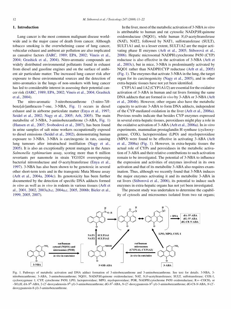
The nitro-aromatic 3-nitrobenzanthrone (3-nitro-7H-enz[de]anthracen-7-one, 3-NBA, Fig. 1) occurs in dieselxhaust and in airborne particulate matter (Enya et al., 1997;eidel et al., 2002; Nagy et al., 2005; Arlt, 2005). The mainetabolite of 3-NBA, 3-aminobenzanthrone (3-ABA, Fig. 1)
Hansen et al., 2007; Svobodova et al., 2007), has been foundn urine samples of salt mine workers occupationally exposedo diesel emissions (Seidel et al., 2002), demonstrating humanxposure to 3-NBA. 3-NBA is carcinogenic in rats, causingung tumours after intratracheal instillation (Nagy et al.,005). It is also an exceptionally potent mutagen in the Amesalmonella typhimurium assay, scoring more than 6 millionevertants per nanomole in strain YG1024 overexpressingacterial nitroreductase and O-acetyltransferase (Enya et al.,997). 3-NBA has also been shown to be genotoxic in severalther short-term tests and in the transgenic Muta Mouse assayArlt et al., 2004a, 2004c). Its genotoxicity has been furtherocumented by the detection of specific DNA adducts formed
n vitro as well as in vivo in rodents in various tissues (Arlt etl., 2001, 2002, 2003a,b,c, 2004a,c, 2005, 2006b; Bieler et al.,999, 2005, 2007).ig. 1. Pathways of metabolic activation and DNA adduct formation of 3-nitroitrobenzanthrone; 3-ABA, 3-aminobenzanthrone; NQO1, NAD(P)H:quinone oxiyclooxygenase 1; CYP, cytochrome P450; LPO, lactoperoxidase; MPO, myelopeSO3H; dA-N6-ABA, 2-(2′-deoxyadenosin-N6-yl)-3-aminobenzanthrone; dG-N2-ABeoxyguanosin-8-yl)-3-aminobenzanthrone.
e
i
gy 247 (2008) 11–22
In the liver, most of the metabolic activation of 3-NBA in vitros attributable to human and rat cytosolic NAD(P)H:quinonexidoreductase (NQO1), while human N,O-acetyltransferaseNAT), NAT2, followed by NAT1, sulfotransferase (SULT),ULT1A1 and, to a lesser extent, SULT1A2 are the major acti-ating phase II enzymes (Arlt et al., 2005; Stiborova et al.,006). Hepatic microsomal NADPH:cytochrome P450 (CYP)eductase is also effective in the activation of 3-NBA (Arlt etl., 2003c), but in mice, 3-NBA is predominantly activated byQO1 rather than NADPH:CYP reductase (Arlt et al., 2005)
Fig. 1). The enzymes that activate 3-NBA in the lung, the targetrgan for its carcinogenicity (Nagy et al., 2005), and in otherxtra-hepatic tissues have not yet been identified.
CYP1A1 and 1A2 (CYP1A1/2) are essential for the oxidativectivation of 3-ABA in human and rat livers forming the sameNA adducts that are formed in vivo by 3-ABA or 3-NBA (Arlt
t al., 2004b). However, other organs also have the metabolicapacity to activate 3-ABA to form DNA adducts, independentf the CYP-mediated oxidation in the liver (Arlt et al., 2006a).revious results indicate that besides CYP enzymes expressed
n several extra-hepatic tissues, peroxidases might play a role inhe oxidative activation of 3-ABA (Arlt et al., 2006a). In in vitroxperiments, mammalian prostaglandin H synthase (cyclooxy-enase, COX), lactoperoxidase (LPO) and myeloperoxidaseMPO) were found to be effective in activating 3-ABA (Arltt al., 2006a) (Fig. 1). However, in extra-hepatic tissues thectual role of CYPs and peroxidases in the metabolic activa-ion of 3-ABA and their relative contributions to such activationemain to be investigated. The potential of 3-NBA to influencehe expression and activities of enzymes involved in its ownctivation and that of its metabolite 3-ABA also requires exam-nation. Thus, although we recently found that 3-NBA induceshe major enzymes activating it and its metabolite 3-ABA in
benzanthrone and 3-aminobenzanthrone. See text for details. 3-NBA, 3-doreductase; NAT, N,O-acetyltransferases; SULT, sulfotransferase; COX-1,roxidase; POR, NADPH:cytochrome P450 oxidoreductase; R = –COCH3 orA, N-(2′-deoxyguanosin-N2-yl)-3-aminobenzanthrone; dG-C8-N-ABA, N-(2′-
nzymes in extra-hepatic organs has not yet been investigated.The present study was undertaken to determine the capabil-
ty of cytosols and microsomes isolated from two rat organs;

xicolo
tkvili
2
2
scmS7c(b
2
ta
2
Ul(bovcana
2
o(pcEwNh
2
72Ctc03cu
awwm
p5(H
2
(afipc
2
spo3tcD2tobwa
2
Dibmti3a
23
mEaesd(Xp
M. Stiborova et al. / To
he lung, the target organ for 3-NBA carcinogenicity, and theidney, an important organ for excretion of xenobiotics, to acti-ate 3-NBA and 3-ABA. A further aim was to identify enzymesnvolved in DNA adduct formation by 3-NBA and 3-ABA in ratung and kidney and to evaluate whether treatment by 3-NBAnduces these enzymes.
. Material and methods
.1. Chemicals
NADPH, hypoxanthine (HX), dicoumarol, allopurinol, deoxyadeno-ine (dA) 3′-monophospate, deoxyguanosine (dG) 3′-monophosphate, acetyloenzyme A (acetyl-CoA) 3′-phosphoadenosine-5′-phosphosulfate (PAPS),enadione (2-methyl-1,4-naphthoquinone) and calf thymus DNA were fromigma Chemical Co. (St. Louis, MO, USA); Sudan I from BDH (Poole, UK);-ethoxyresorufin from Fluka Chemie AG (Buchs, Switzerland). Enzymes andhemicals for the 32P-postlabelling assay were obtained from sources describedPhillips and Arlt, 2007). All these and other chemicals were reagent grade oretter.
.2. Synthesis of 3-NBA and 3-ABA
3-NBA and 3-ABA were synthesized as described (Arlt et al., 2003a) andheir authenticity was confirmed by UV spectroscopy, electrospray mass spectrand high field proton NMR spectroscopy.
.3. Animal experiments
The study was conducted in accordance with the Regulations for the Care andse of Laboratory Animals (311/1997, Ministry of Agriculture, Czech Repub-
ic), which is in compliance with Declaration of Helsinki. Male Wistar rats100–125 g, n = 3 per group) were treated with a single dose of 0.4, 4 or 40 mg/kgw of 3-NBA by intraperitoneal injection. 3-NBA was dissolved in sunfloweril at a concentration of 0.4 or 4 mg/ml. Three control animals received an equalolume of oil only. Rats were placed in cages in temperature and humidityontrolled rooms. Standardised diet and water were provided ad libitum. Thenimals were killed 24 h after treatment by cervical dislocation. Lungs and kid-eys were removed immediately after death and used for isolation of mRNAnd for preparation of microsomal and cytosolic fractions.
.4. Preparation of microsomal and cytosolic fractions
Microsomal and cytosolic fractions were isolated from the lungs and kidneysf rats, either uninduced or pretreated with 3-NBA (see above) as describedStiborova et al., 2003). Both subcellular preparations were analysed for theresence of 3-NBA using the HPLC on a CC 250/4 Nucleosil, 100-5 C18 HDolumn (Macherey-Nagel, 4 mm × 250 mm) preceded by a C-18 guard column.luent was 70% methanol in water, at a flow rate of 0.6 ml min−1, and detectionas at 254 nm. The standard of 3-NBA eluted with a retention time of 8.2 min.o 3-NBA was detectable in microsomal and cytosolic fractions from rats thatad been pretreated with this compound.
.5. Cytosolic incubations
The deaerated and argon-purged incubation mixtures, in a final volume of50 �l, consisted of 50 mM Tris–HCl buffer (pH 7.4), containing 0.2% Tween0, cofactors for cytosolic enzymes (1 mM NADPH, NADH or HX; 2 mM acetyl-oA; 100 �M PAPS), pooled lung and kidney cytosolic sample from three rats,
reated either with vehicle (control) or with 40 mg/kg bw of 3-NBA (1 mg of
ytosolic protein), 30 �M 3-NBA (dissolved in 7.5 �l dimethylsulfoxide) and.5 mg of calf thymus DNA (2 mM dNp). The reaction was initiated by adding-NBA. Incubations with rat cytosols were carried out at 37 ◦C for 3 h; theytosol-mediated 3-NBA-derived DNA adduct formation was found to be linearp to 4 h (Arlt et al., 2005). Control incubations were carried out either (i) withoutdNA(t
gy 247 (2008) 11–22 13
ctivating system (cytosol), (ii) without cofactors (NADPH, NADH, HX), (iii)ithout DNA or (iv) without 3-NBA. After extraction with ethyl acetate, DNAas isolated from the residual water phase by the phenol/chloroform extractionethod as described (Arlt et al., 2005).
Incubations used to evaluate the activation of 3-ABA by peroxidases in ratulmonary and renal cytosolic samples contained, in a final volume of 750 �l,0 mM Tris–HCl buffer (pH 7.4), containing 0.2% Tween 20, 100 �M 3-ABAdissolved in 7.5 �l dimethylsulfoxide), 0.5 mg of calf thymus DNA and 200 �M
2O2.
.6. Cytosolic inhibition studies
Dicoumarol and allopurinol were used to inhibit NQO1 and xanthine oxidaseXO), respectively, in rat pulmonary and renal cytosolic fractions (Watanabe etl., 1997; Ritter et al., 2000). Inhibitors dissolved in 7.5 �l of methanol, to yieldnal concentrations of 10 �M, were added to the incubation mixtures as reportedreviously (Arlt et al., 2005). An equal volume of methanol was added to theontrol incubations.
.7. Microsomal incubations
Incubation mixtures, in a final volume of 750 �l, consisted of 50 mM potas-ium phosphate buffer (pH 7.4), 1 mM NADPH or 0.1 mM arachidonic acid,ooled pulmonary and renal microsomal sample from three rats, either controlr treated with 40 mg/kg bw of 3-NBA (0.5 mg of microsomal protein), 100 �M-ABA or 3-NBA (dissolved in 7.5 �l dimethylsulfoxide) and 0.5 mg of calfhymus DNA. The reaction was initiated by adding 3-ABA or 3-NBA and werearried out at 37 ◦C for 2 h; the microsomal-mediated 3-NBA (3-ABA)-derivedNA adduct formation was found to be linear up to 3 h (Arlt et al., 2003c,004b). Control incubations were carried out either (i) without activating sys-em (microsomes), (ii) with activating system and 3-ABA, but without DNAr (iii) with activating system and DNA but without 3-ABA. After the incu-ation and extraction with ethyl acetate, DNA was isolated from the residualater phase by the phenol/chloroform extraction method as described (Arlt et
l., 2004b).
.8. Microsomal inhibition studies
�-Naphthoflavone (�-NF) (Sigma), which inhibits CYP1A (Rendic andiCarlo, 1997; Stiborova et al., 2005b), and indomethacin (Sigma), which
nhibits COX (Eling et al., 1990), were used to inhibit the activation of 3-ABAy rat pulmonary and renal microsomes. Inhibitors were dissolved in 7.5 �l ofethanol and were added to the incubation mixtures to yield final concentra-
ions of 100 �M. The incubation mixtures containing the inhibitors were thenncubated at 37 ◦C for 10 min with NADPH or arachidonic acid prior to adding-ABA and then for a further 120 min at 37 ◦C. An equal volume of methanollone was added to the control incubations.
.9. 32P-postlabelling analysis and HPLC analysis of 32P-labelled′,5′-deoxyribonucleoside bisphosphate adducts
32P-postlabelling analysis using butanol extraction, and thin layer chro-atography (TLC) and HPLC were performed as described (Arlt et al., 2006b).nrichment by butanol extraction has been shown to yield more adduct spotsnd a better recovery of 3-NBA (3-ABA)-derived DNA adducts than usingnrichment by nuclease P1 digestion (Arlt et al., 2001, 2004b). DNA adductpots were numbered as reported (Arlt et al., 2006b). As reference compoundseoxyadenosine (dAp) and deoxyguanosine (dGp) 3′-monophosphates4 �mol/ml) (Sigma) were incubated with 3-NBA (300 �M) activated byO (1 U/ml) (Sigma) in the presence of HX and analysed as describedreviously (Arlt et al., 2001). DNA adduct standard samples of 3-NBA, 2-(2′-
eoxyadenosin-N6-yl)-3-aminobenzanthrone-3′-phosphate (dA3′p-N6-ABA),-(2′-deoxyguanosin-N2-yl)-3-aminobenzanthrone-3′-phosphate (dG3′p-N2-BA) and N-(2′-deoxyguanosin-8-yl)-3-aminobenzanthrone-3′-phosphatedG3′p-C8-N-ABA), were prepared by reacting N-acetoxy-3-aminobenzan-hrone with dAp or dGp and analysed as described recently (Arlt et al., 2006b).
1 xicolo
2
beSR
2
r(afi(
2m
cgweppehaCCopIa
2
eS7casTN
2
rudhsUtFt(Rtcw
ruU1Pgaatbena�
�
aap
3
3c
bmmaf3Fmerafwiascnds(a
bbatwDb
4 M. Stiborova et al. / To
.10. Isolation of CYP1A1
Recombinant rat CYP1A1 protein was purified to homogeneity from mem-ranes of Escherichia coli transfected with a modified CYP1A1 cDNA (Stiborovat al., 2002), in the laboratory of H.W. Strobel (University of Texas, Medicalchool of Houston, TX, USA) by P. Hodek (Charles University, Prague, Czechepublic).
.11. Preparation of antibodies
Leghorn chicken were immunised subcutaneously three times a week withat recombinant CYP1A1 and human recombinant NQO1 (Sigma) antigens0.1 mg/animal) emulsified in complete Freund’s adjuvant for the first injectionnd in incomplete adjuvant for boosters. Immunoglobulin fraction was puri-ed from pooled egg yolks using fractionation by polyethylene glycol 6000Stiborova et al., 2002).
.12. Estimation of CYP1A1 and NQO1 protein content inicrosomes and cytosols of rat lung and kidney
Immunoquantitation of rat pulmonary and renal microsomal CYP1A1 and ofytosolic NQO1 was done by sodium dodecyl sulphate (SDS)-polyacrylamideel electrophoresis. Samples containing 75 �g microsomal or cytosolic proteinsere subjected to electrophoresis on SDS/10% polyacrylamide gels (Stiborova
t al., 2002, 2005b, 2006). After migration, proteins were transferred ontoolyvinylidene difluoride membranes. Rat CYP1A1 and NQO1 proteins wererobed with the chicken polyclonal antibodies as reported elsewhere (Stiborovat al., 2002, 2005b, 2006). The antibodies against rat recombinant CYP1A1 anduman NQO1 recognise these enzymes in rat pulmonary and renal microsomess one protein band. Rat recombinant CYP1A1 (in SupersomesTM, Gentestorp., Woburn, MA, USA) and human recombinant NQO1 (Sigma Chemicalo., St. Louis, MO, USA) were used as positive controls to identify the bandsf CYP1A1 in microsomes and NQO1 in cytosols. The antigen–antibody com-lex was visualised with an alkaline phosphatase-conjugated rabbit anti-chickengG antibody and 5-bromo-4-chloro-3-indolylphosphate/nitrobluetetrazoliums chromogenic substrate (Stiborova et al., 2002, 2005b, 2006).
.13. CYP1A1 and NQO1 enzyme activity assays
The microsomal samples were characterised for CYP1A1 activity using 7-thoxyresorufin O-deethylation (EROD) activity and the oxidation of Sudan I.udan I was used as a substrate because it is more specific for CYP1A1 than-ethoxyresorufin (Stiborova et al., 2002, 2005b). The cytosolic samples wereharacterised for NQO1 activity, using menadione as a substrate (Stiborova etl., 2003). NQO1 activity was determined by following the oxidation of NADPHpectrophotometrically at 340 nm. The standard assay system contained 25 mMris–HCl (pH 7.4), 0.2% Tween 20, 0.07% bovine serum albumin, 400 mMADPH and 100 mM menadione dissolved in methanol.
.14. CYP1A1 and NQO1 mRNA content in rat lungs and kidneys
Total RNA was isolated from frozen lungs and kidneys of three untreatedats and three rats pretreated with 40 mg/kg body weight of 3-NBA or 3-ABAsing Trizol Reagent (Invitrogen, Carlsbad, CA, USA) according to the proce-ure supplied by the manufacturer. The quality of isolated RNA was verified byorizontal agarose gel electrophoresis, RNA quantity was assessed by UV–vispectrophotometry on a Carry 300 spectrophotometer (Varian, Palo Alto, CA,SA). RNA samples (1 �g) were reversely transcribed using 200 U of reverse
ranscriptase per sample with random hexamer primers utilising RevertAidTM
irst Strand cDNA Synthesis Kit (MBI Fermentas, Vilnius, Lithuania) accordingo the manufacturer’s instructions. The prepared cDNA was used for real-time
RT) polymerase chain reaction (PCR) performed in RotorGene 2000 (Corbettesearch, Sydney, Australia) under the following cycling conditions: incuba-ion at 50 ◦C for 2 min and initial denaturation at 95 ◦C for 10 min, then 50ycles of denaturation at 95 ◦C for 15 s and annealing at 60 ◦C for 1 min. Gainas set to 7 and fluorescence was acquired after elongation step. The PCR
((DN
gy 247 (2008) 11–22
eaction mixtures (20 �l) contained 9 �l cDNA diluted 10 times in Milli-Qltrapure water (Biocel A10, Millipore, Billerica, MA, USA), 10 �l TaqManniversal PCR Master Mix (Applied Biosystems, Foster City, CA, USA) and�l TaqMan Gene Expression Assay Mix (commercially available unlabelledCR primers and FAMTM dye-labelled probe for rat CYP1A1 or NQO1 as tar-et genes and β-actin as reference internal standard gene). Each sample wasnalysed in two parallel aliquots. Negative controls had the same compositionss samples but cDNA was omitted from the mixture. Data were analysed byhe program RotorGene v6 (Corbett Research, Sydney, Australia) and evaluatedy comparative cycle threshold (cT) method for relative quantitation of genexpression. Cycle thresholds, at which a significant increase in fluorescence sig-al was detected, were measured for each sample. Then ��cT was evaluatedccording to following equations: �cT = cT (target) − cT (internal standard),�cT = �cT treated − �cT control, where �cT treated is �cT for treated rats andcT control is �cT for untreated rats. �cT is positive if the target is expressed
t a lower level than the internal standard (β-actin), and negative if expressedt a higher level. The induction of mRNA expression of studied target genes inretreated animals was evaluated as 2−(��cT).
. Results
.1. Activation of 3-NBA by rat pulmonary and renalytosols and microsomes
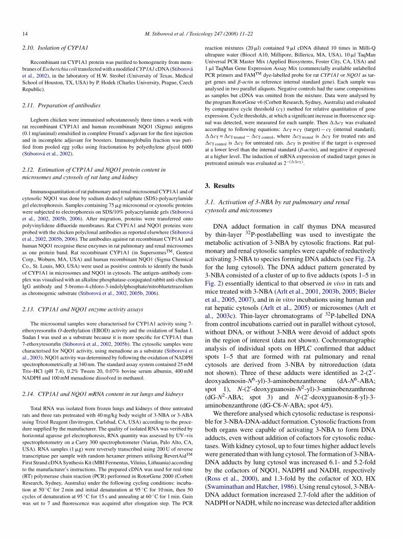
DNA adduct formation in calf thymus DNA measuredy thin-layer 32P-postlabelling was used to investigate theetabolic activation of 3-NBA by cytosolic fractions. Rat pul-onary and renal cytosolic samples were capable of reductively
ctivating 3-NBA to species forming DNA adducts (see Fig. 2Aor the lung cytosol). The DNA adduct pattern generated by-NBA consisted of a cluster of up to five adducts (spots 1–5 inig. 2) essentially identical to that observed in vivo in rats andice treated with 3-NBA (Arlt et al., 2001, 2003b, 2005; Bieler
t al., 2005, 2007), and in in vitro incubations using human andat hepatic cytosols (Arlt et al., 2005) or microsomes (Arlt etl., 2003c). Thin-layer chromatograms of 32P-labelled DNArom control incubations carried out in parallel without cytosol,ithout DNA, or without 3-NBA were devoid of adduct spots
n the region of interest (data not shown). Cochromatographicnalysis of individual spots on HPLC confirmed that adductpots 1–5 that are formed with rat pulmonary and renalytosols are derived from 3-NBA by nitroreduction (dataot shown). Three of these adducts were identified as 2-(2′-eoxyadenosin-N6-yl)-3-aminobenzanthrone (dA-N6-ABA;pot 1), N-(2′-deoxyguanosin-N2-yl)-3-aminobenzanthronedG-N2-ABA; spot 3) and N-(2′-deoxyguanosin-8-yl)-3-minobenzanthrone (dG-C8-N-ABA; spot 4/5).
We therefore analysed which cytosolic reductase is responsi-le for 3-NBA-DNA-adduct formation. Cytosolic fractions fromoth organs were capable of activating 3-NBA to form DNAdducts, even without addition of cofactors for cytosolic reduc-ases. With kidney cytosol, up to four times higher adduct levelsere generated than with lung cytosol. The formation of 3-NBA-NA adducts by lung cytosol was increased 6.1- and 5.2-foldy the cofactors of NQO1, NADPH and NADH, respectively
Ross et al., 2000), and 1.3-fold by the cofactor of XO, HXSwaminathan and Hatcher, 1986). Using renal cytosol, 3-NBA-NA adduct formation increased 2.7-fold after the addition ofADPH or NADH, while no increase was detected after addition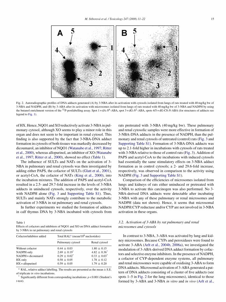
M. Stiborova et al. / Toxicology 247 (2008) 11–22 15
Fig. 2. Autoradiographic profiles of DNA adducts generated (A) by 3-NBA after its activation with cytosols isolated from lungs of rats treated with 40 mg/kg bw of3 olatedt BA,l
omofifdee
NaotrawSa
i
TEb
C
WNNHH
o
t
ra3mSuwPhfrN
l3N
-NBA and NADPH, and (B) by 3-ABA after its activation with microsomes ishe butanol enrichment version of the 32P-postlabelling assay. Spot 1 = dA-N6-Aegend to Fig. 1).
f HX. Hence, NQO1 and XO reductively activate 3-NBA in pul-onary cytosol, although XO seems to play a minor role in this
rgan and does not seem to be important in renal cytosol. Thisnding is also supported by the fact that 3-NBA-DNA adductormation in cytosols of both tissues was markedly decreased byicoumarol, an inhibitor of NQO1 (Watanabe et al., 1997; Rittert al., 2000), whereas allopurinol, an inhibitor of XO (Watanabet al., 1997; Ritter et al., 2000), showed no effect (Table 1).
The influence of SULTs and NATs on the activation of 3-BA in pulmonary and renal cytosols was then investigated by
dding either PAPS, the cofactor of SULTs (Glatt et al., 2001),r acetyl-CoA, the cofactor of NATs (King et al., 2000), intohe incubation mixtures. The addition of PAPS and acetyl-CoAesulted in a 2.5- and 29.7-fold increase in the levels of 3-NBAdducts in uninduced cytosols, respectively, over the activityith NADPH alone (Fig. 3 and Supporting Table S1). Thus,
ULTs and mainly NATs strongly contribute to the metabolicctivation of 3-NBA in rat pulmonary and renal cytosols.In further experiments we studied the formation of adductsn calf thymus DNA by 3-NBA incubated with cytosols from
able 1ffects of cofactors and inhibitors of NQO1 and XO on DNA adduct formationy 3-NBA in rat pulmonary and renal cytosols
ofactor/inhibitor added Total RALa (mean/108 nucleotides)
Pulmonary cytosol Renal cytosol
ithout cofactor 0.44 ± 0.03 1.80 ± 0.15ADPH only 2.69 ± 0.19* 4.82 ± 0.36*
ADPH + dicoumarol 0.29 ± 0.02* 0.33 ± 0.03*
X only 0.58 ± 0.05 1.78 ± 0.12X + allopurinol 0.59 ± 0.05 1.79 ± 0.20
a RAL, relative adduct labelling. The results are presented as the mean ± S.E.f triplicate in vitro incubations.* Significantly different from corresponding incubations: p < 0.001 (Student’s
-test).
3NNa
3m
namtaaDtsf
from lungs of rats treated with 40 mg/kg bw of 3-NBA and NADPH by usingspot 3 = dG-N2-ABA, spots 4/5 = dG-C8-N-ABA (for structures of adducts see
ats pretreated with 3-NBA (40 mg/kg bw). These pulmonarynd renal cytosolic samples were more effective in formation of-NBA-DNA adducts in the presence of NADPH, than the pul-onary and renal cytosols of untreated (control) rats (Fig. 3 andupporting Table S1). Formation of 3-NBA-DNA adducts wasp to 2.1-fold higher in incubations with cytosols of rats treatedith 3-NBA relative to those of control rats (Fig. 3). Addition of
APS and acetyl-CoA to the incubations with induced cytosolsad essentially the same stimulatory effects on 3-NBA adductormation as in control cytosols; a 2- and 29.6-fold increase,espectively, was observed in comparison to the activity usingADPH (Fig. 3 and Supporting Table S1).
Comparison of the efficiencies of microsomes isolated fromungs and kidneys of rats either uninduced or pretreated with-NBA to activate this carcinogen was also performed. No 3-BA-derived DNA adducts were detectable after incubating-NBA with any of these pulmonary or renal microsomes andADPH (data not shown). Hence, it seems that microsomalADPH:CYP reductase and/or CYP are not involved in 3-NBActivation in these organs.
.2. Activation of 3-ABA by rat pulmonary and renalicrosomes and cytosols
In contrast to 3-NBA, 3-ABA was activated by lung and kid-ey microsomes. Because CYPs and peroxidases were found toctivate 3-ABA (Arlt et al., 2004b, 2006a), we investigated theodulation of 3-ABA-derived DNA adduct formation by cofac-
ors and selective enzyme inhibitors. In the presence of NADPH,cofactor of CYP-dependent enzyme systems, all pulmonary
nd renal microsomes were capable of oxidising 3-ABA to form
NA adducts. Microsomal activation of 3-ABA generated a pat-ern of DNA adducts consisting of a cluster of five adducts (seepots 1–5 in Fig. 2 for the lung microsomes), identical to thoseormed by 3-ABA and 3-NBA in vitro and in vivo (Arlt et al.,
16 M. Stiborova et al. / Toxicology 247 (2008) 11–22
F lungb ofactfi three
22t2eabAa3po
Scwas
Da
F4i�
ig. 3. DNA adduct formation by 3-NBA activated with cytosols isolated fromw of 3-NBA and the DNA-adduct levels obtained by adding either PAPS as a cgure represent total levels of DNA adducts (RAL, relative adduct labelling) of
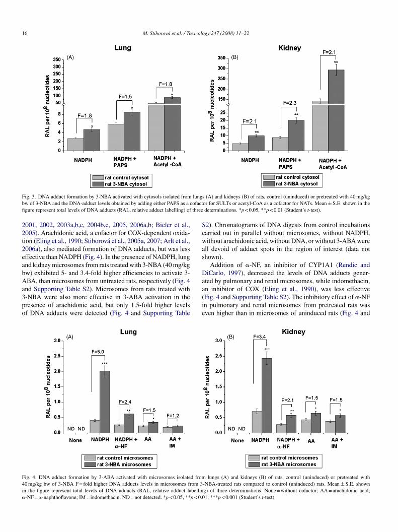
001, 2002, 2003a,b,c, 2004b,c, 2005, 2006a,b; Bieler et al.,005). Arachidonic acid, a cofactor for COX-dependent oxida-ion (Eling et al., 1990; Stiborova et al., 2005a, 2007; Arlt et al.,006a), also mediated formation of DNA adducts, but was lessffective than NADPH (Fig. 4). In the presence of NADPH, lungnd kidney microsomes from rats treated with 3-NBA (40 mg/kgw) exhibited 5- and 3.4-fold higher efficiencies to activate 3-BA, than microsomes from untreated rats, respectively (Fig. 4
nd Supporting Table S2). Microsomes from rats treated with-NBA were also more effective in 3-ABA activation in theresence of arachidonic acid, but only 1.5-fold higher levelsf DNA adducts were detected (Fig. 4 and Supporting Table
a(ie
ig. 4. DNA adduct formation by 3-ABA activated with microsomes isolated from0 mg/kg bw of 3-NBA F = fold higher DNA adducts levels in microsomes from 3n the figure represent total levels of DNA adducts (RAL, relative adduct labellin-NF = �-naphthoflavone; IM = indomethacin. ND = not detected. *p < 0.05, **p < 0.
s (A) and kidneys (B) of rats, control (uninduced) or pretreated with 40 mg/kgor for SULTs or acetyl-CoA as a cofactor for NATs. Mean ± S.E. shown in thedeterminations. *p < 0.05, **p < 0.01 (Student’s t-test).
2). Chromatograms of DNA digests from control incubationsarried out in parallel without microsomes, without NADPH,ithout arachidonic acid, without DNA, or without 3-ABA were
ll devoid of adduct spots in the region of interest (data nothown).
Addition of �-NF, an inhibitor of CYP1A1 (Rendic andiCarlo, 1997), decreased the levels of DNA adducts gener-
ted by pulmonary and renal microsomes, while indomethacin,
n inhibitor of COX (Eling et al., 1990), was less effectiveFig. 4 and Supporting Table S2). The inhibitory effect of �-NFn pulmonary and renal microsomes from pretreated rats wasven higher than in microsomes of uninduced rats (Fig. 4 andlungs (A) and kidneys (B) of rats, control (uninduced) or pretreated with-NBA-treated rats compared to control (uninduced) rats. Mean ± S.E. showng) of three determinations. None = without cofactor; AA = arachidonic acid;01, ***p < 0.001 (Student’s t-test).

M. Stiborova et al. / Toxicology 247 (2008) 11–22 17
F lungb dduct
SaaNa
oricwfTf(io
3e
tfrw
TS
C
ES
i
ef2ctli(i(rwl
e2araa(
ig. 5. DNA adduct formation by 3-ABA activated with cytosols isolated fromw of 3-NBA. Mean ± S.E. shown in the figure represent total levels of DNA a
upporting Table S2). These results point to CYP1A1 havingmajor role in DNA adduct formation by 3-ABA in rat lungs
nd kidneys. Additionally, they show that treating rats with 3-BA increases the relative contribution of this enzyme to 3-ABA
ctivation in both organs.In the presence of hydrogen peroxide, a cofactor for per-
xidases, the pulmonary and renal cytosols from uninducedats and animals treated with 3-NBA were capable of activat-ng 3-ABA to form DNA adducts. In contrast to pulmonaryytosol, by which five 3-ABA-DNA adducts (spots 1–4 in Fig. 2)ere generated, in the renal cytosolic activation system, only
our 3-ABA-DNA adducts (spots 1–4 in Fig. 2) were detected.reatment of rats with 3-NBA slightly stimulated (1.3-fold) theormation of 3-ABA-DNA adducts in cytosols of both organsFig. 5 and Supporting Table S3). No adducts were detectablen controls without cytosols, without 3-ABA (data not shown),r without hydrogen peroxide (Fig. 5 and Supporting Table S3).
.3. The effect of 3-NBA pretreatment on expression ofnzymes activating 3-NBA and 3-ABA
Because NQO1 and CYP1A1 were found to be the essen-
ial enzymes activating 3-NBA and 3-ABA with subcellularractions from rat lungs and kidneys (present paper) and fromat and human livers (Arlt et al., 2004b, 2005), we evaluatedhether treating rats with 3-NBA influences expression of theseable 2pecific CYP1A1 activitiesa in kidney and lung microsomes of control and 3-NBA-tr
YP activity Control rat
Kidney Lung
ROD 9.4 ± 0.4 4.6 ± 0udan I oxidation 9.3 ± 1.0 4.7 ± 0
a Each value (pmol of reaction product per min per mg protein) represents the meanncrease over the control activity caused by the pre-treatment with 3-NBA (40 mg/kg
* Significantly different from controls: p < 0.001 (Student’s t-test).
eogm
s (A) and kidneys (B) of rats, control (uninduced) or pretreated with 40 mg/kgs (RAL, relative adduct labelling) of three determinations.
nzymes. The induction of these enzymes has already beenound in the liver of rats treated with 3-NBA (Stiborova et al.,006). In the present study, Western blots with chicken poly-lonal antibodies raised against NQO1 and CYP1A1 showedhat the expression of both enzymes was also induced in ratung and kidney by 3-NBA (Fig. 6). The efficiency of 3-NBA tonduce CYP1A1 expression was higher in lung than in kidney7.4-fold versus 3.4-fold at 40 mg/kg) (Fig. 6). EROD activ-ty and oxidation of Sudan I, as markers of CYP1A1 activityStiborova et al., 2002, 2005b), were increased in both organs ofats treated with 3-NBA (Table 2). Again, the induction in lungas higher than in kidney, although control enzyme activity in
ung was only half of that in kidney.The levels of renal and pulmonary NQO1 protein were
nhanced by pretreating rats with 3-NBA (40 mg/kg bw), by.3- and 1.4-fold, respectively (Fig. 6). The increase in NQO1ctivities in cytosols depended on the administered dose and cor-elated with the protein expression (Table 3 and Fig. 6). Up to2.5-fold increase in NQO1 activity measured with menadiones a substrate was found in tissues of rats treated with 3-NBATable 3).
Besides the evaluation of the effects of 3-NBA on protein lev-
eated rats
3-NBA-treated rat
Kidney Lung
.5 50.2 ± 1.8 (5.3)* 29.9 ± 2.1 (6.5)*
.5 30.3 ± 2.3 (3.2)* 29.8 ± 2.8 (6.3)*
± S.E. of triplicate measurements. Numbers in parentheses represents the foldbw).
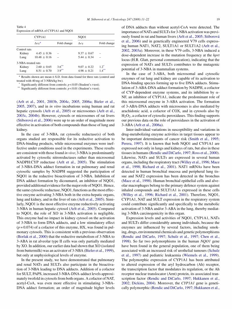
ls and enzyme activities of CYP1A1 and NQO1, modulationf their mRNA expression by the compound was also investi-ated. The relative amounts of CYP1A1 and NQO1 mRNA wereeasured by RT-PCR analysis. As shown in Table 4, treatment

18 M. Stiborova et al. / Toxicology 247 (2008) 11–22
Fig. 6. Induction of CYP1A1 (A and B) and NQO1 (C and D) in lungs (A and C) and kidney (B and D) of rats treated with 0.4, 4 or 40 mg/kg bw of 3-NBAdetermined by Western blots as described in Section 2. Values represent mean ± S.D. obtained from lungs and kidneys of three rats (n = 3). Inset: immunoblots ofmicrosomal CYP1A1 and cytosolic NQO1 from untreated and 3-NBA-treated rats stained with antibody against rat CYP1A1 and human NQO1. Values significantlydifferent from control: *p < 0.05, **p < 0.01, ***p < 0.001 (Student’s t-test).
Table 3NQO1 specific activity in rat renal and pulmonary cytosol
Control rat 3-NBA-treated rat
0.4 mg/kg 4 mg/kg 40 mg/kg
Kidney 0.09 ± 0.01 0.15 ± 0.01 (1.7)* 0.15 ± 0.01 (1.7)* 0.23 ± 0.02 (2.5)**
Lung 0.05 ± 0.01 0.06 ± 0.01 (1.2) 0.06 ± 0.01 (1.2) 0.09 ± 0.01 (1.8)*
The results (units per mg) are averages and S.E.s of five parallel measurements. Enzyme activities with menadione as a substrate was assayed as described in Section2 in in t ◦
oCt
4
tk
pev2t
. One unit of NQO1 is defined to reduce 1 �mol of NADPH per min/mg prote* Significantly different from controls: p < 0.05 (Student’s t-test).
** Significantly different from controls: p < 0.01 (Student’s t-test).
f rats with 40 mg/kg bw of 3-NBA induced mRNA levels ofYP1A1 and NQO1 up to 3.9- and 1.4-fold, respectively, in the
issues studied.
. Discussion
The present study has increased our knowledge on the poten-ial of cytosolic and microsomal fractions of rat lungs andidneys to activate 3-NBA and 3-ABA, and on the enzymes
i
bf
he presence of menadione as substrate at 37 C.
articipating in their bioactivation. The rat was used as anxperimental model on the basis that the same enzymes acti-ate 3-NBA and 3-ABA in livers of both species (Arlt et al.,003a,c, 2004b, 2005, 2006a; Stiborova et al., 2006). Therefore,he results should provide some indication of what might occur
n extra-hepatic tissues of humans exposed to this pollutant.DNA adducts formed during 3-NBA and 3-ABA activationy the cytosolic and microsomal fractions are identical to thoseormed in vivo in rats and mice treated with 3-NBA or 3-ABA
M. Stiborova et al. / Toxicolo
Table 4Expression of mRNA of CYP1A1 and NQO1
CYP1A1 NQO1
�cTa Fold change �cT Fold change
Control ratsKidney 4.45 ± 0.36 – 9.37 ± 0.67 –Lung 10.48 ± 0.16 – 5.44 ± 0.34 –
3-NBA-treated ratsKidney 2.60 ± 0.65 3.6** 9.07 ± 0.22 1.2*
Lung 8.51 ± 0.70 3.9** 4.98 ± 0.21 1.4**
a Results shown are mean ± S.D. from data found for three rats (control andtreated with 40 mg of 3-NBA/kg bw).
(2h2(ek
oDfaaNocNDpttll3tTo(m(3bfb
atfmaD
oioei2dlep
eDloNtoaHo3
dbPetLoeds(oi(Ccai
aei(1haeTttreceptor nuclear translocator (Arnt) protein, its associated tran-
* Significantly different from controls: p < 0.05 (Student’s t-test).** Significantly different from controls: p < 0.01 (Student’s t-test).
Arlt et al., 2001, 2003b, 2004c, 2005, 2006a; Bieler et al.,005, 2007), and in in vitro incubations using human and ratepatic cytosols (Arlt et al., 2005) or microsomes (Arlt et al.,003c, 2004b). However, cytosols or microsomes of rat liversStiborova et al., 2006) were up to an order of magnitude moreffective in activation of both compounds than those of lung andidney.
In the case of 3-NBA, rat cytosolic reductase(s) of bothrgans studied are responsible for its reductive activation toNA-binding products, while microsomal enzymes were inef-
ective under conditions used in the experiments. These resultsre consistent with those found in vivo; 3-NBA is predominantlyctivated by cytosolic nitroreductases rather than microsomalADPH:CYP reductase (Arlt et al., 2005). The stimulationf 3-NBA-DNA adduct formation in rat pulmonary and renalytosolic samples by NADPH suggested the participation ofQO1 in the reductive bioactivation of 3-NBA. Inhibition ofNA adduct formation by dicoumarol, an inhibitor of NQO1,rovided additional evidence for the major role of NQO1. Hence,he same cytosolic reductase, NQO1, functions as the most effec-ive enzyme activating 3-NBA both in the extra-hepatic organs,ung and kidney, and in the liver of rats (Arlt et al., 2005). Simi-arly, NQO1 is the most effective enzyme reductively activating-NBA in human hepatic cytosol (Arlt et al., 2005). Comparedo NQO1, the role of XO in 3-NBA activation is negligible.his enzyme had no impact in kidney cytosol on the activationf 3-NBA to form DNA adducts, and a low stimulatory effectp = 0.074) of a cofactor of this enzyme, HX, was found in pul-onary cytosols. This is consistent with a previous observation
Borlak et al., 2000) that the reductive metabolism of 3-NBA to-ABA in rat alveolar type II cells was only partially mediatedy XO. In addition, our earlier data had shown that XO (isolatedrom buttermilk) was an activator of 3-NBA (Bieler et al., 1999),ut only at unphysiological levels of enzyme.
In the present study, we have demonstrated that pulmonarynd renal NATs and SULTs also participate in the bioactiva-ion of 3-NBA leading to DNA adducts. Addition of a cofactoror SULT, PAPS, increased 3-NBA-DNA-adduct levels approxi-
ately twofold in cytosols from both organs. A cofactor of NAT,cetyl-CoA, was even more effective in stimulating 3-NBA-NA adduct formation; an order of magnitude higher levels
s2c
gy 247 (2008) 11–22 19
f DNA adducts than without acetyl-CoA were detected. Themportance of NATs and SULTs for 3-NBA activation was previ-usly found in rat and human livers (Arlt et al., 2005; Stiborovat al., 2006) and in genetically engineered V79 cells express-ng human NAT1, NAT2, SULT1A1 or SULT1A2 (Arlt et al.,002, 2003a). Moreover, in these V79 cells, 3-NBA induced aose-dependent increase in the mutation frequency at the hprtocus (H.R. Glatt, personal communication), indicating that thexpression of NATs and SULTs contributes to the mutagenicotential of 3-NBA in mammalian systems.
In the case of 3-ABA, both microsomal and cytosolicnzymes of rat lung and kidney are capable of its activation toNA-binding species forming up to five DNA adducts. Stimu-
ation of 3-ABA-DNA adduct formation by NADPH, a cofactorf CYP-dependent enzyme systems, and its inhibition by �-F, an inhibitor of CYP1A1, indicate the predominant role of
his microsomal enzyme in 3-ABA activation. The formationf 3-ABA-DNA adducts with microsomes is also mediated byrachidonic acid, a cofactor of COX, and in cytosols also by2O2, a cofactor of cytosolic peroxidases. This finding supportsur previous data on the role of peroxidases in the activation of-ABA (Arlt et al., 2006a).
Inter-individual variations in susceptibility and variations inrug-metabolizing enzyme activities in target tissues appear toe important determinants of cancer risk (Smith et al., 1995;erera, 1997). It is known that both NQO1 and CYP1A1 arexpressed not only in lungs and kidneys of rats, but also in theseissues in humans (Rendic and DiCarlo, 1997; Ross et al., 2000).ikewise, NATs and SULTs are expressed in several humanrgans, including the respiratory tract (Willey et al., 1996; Macet al., 1998; Richard et al., 2001). NAT1 mRNAs have beenetected in human bronchial mucosa and peripheral lung tis-ue and NAT2 expression has been detected in the bronchusMace et al., 1998). Human bronchial epithelial cells and alve-lar macrophages belong to the primary defence system againstnhaled compounds and SULT1A1 is expressed in these cellsWilley et al., 1996; Richard et al., 2001). Therefore, NQO1,YP1A1, NAT and SULT expression in the respiratory systemould contribute significantly and specifically to the metabolicctivation of 3-NBA and/or 3-ABA in the lung, thereby mediat-ng 3-NBA carcinogenicity in this organ.
Expression levels and activities of NQO1, CYP1A1, NATsnd SULTs differ considerably among individuals, because thenzymes are influenced by several factors, including smok-ng, drugs, environmental chemicals and genetic polymorphismsRendic and DiCarlo, 1997; Schulz et al., 1997; Chen et al.,998). So far two polymorphisms in the human NQO1 geneave been found in the general population, one of them beingssociated with an increased risk of urothelial tumours (Schulzt al., 1997) and pediatric leukaemia (Wiemels et al., 1999).he polymorphic expression of CYP1A1 has been attributed
o altered expression of the aryl hydrocarbon (Ah) receptor,he transcription factor that modulates its regulation, or the Ah
cription factor (Rendic and DiCarlo, 1997; Hukkanen et al.,002; Dickins, 2004). Moreover, the CYP1A1 gene is geneti-ally polymorphic (Rendic and DiCarlo, 1997; Hukkanen et al.,
2 xicolo
2mo(Tmtnp2lid
b2asNaNhJ
tdlgbiHeiDh3Dopaiiochoa
aaeiattiD
t3ttc
lcdNut3tvto
ebmltttc
fiida(esmip
A
i
R
A
A
A
0 M. Stiborova et al. / To
002). So far, CYP1A1*2A, CYP1A1*2B and CYP1A1*4 poly-orphisms have been found that might be associated with lung,
esophageal or breast cancer and with acute myeloid leukaemiaD’Alo et al., 2004; Yang et al., 2005; Li et al., 2004, 2005).he human NAT1 and NAT2 genes are also genetically poly-orphic, resulting in different activities of the gene product
hat segregate individuals into slow and rapid acetylator phe-otypes (Hein et al., 2000). SULT1A1 and SULT1A2 are alsoolymorphic in humans (Raftogianis et al., 1999; Engelke et al.,000) and are associated with increased cancer risk includingung cancer (Wang et al., 2002). Thus, genetic polymorphismsn CYP1A1, NQO1, NAT and SULT genes could be importanteterminants of a possible lung cancer risk from 3-NBA.
Human and rat CYP1A1 is induced by many compounds, e.g.y planar aromatic compounds binding to the Ah receptor, e.g.,3,7,8-tetrachlorodibenzo[1,4]dioxine (TCDD) (Drahushuk etl., 1998), and by polycyclic hydrocarbons present in cigarettemoke (Rendic and DiCarlo, 1997; Hukkanen et al., 2002).QO1 is inducible by a variety of agents with different mech-
nisms of action (see Ross et al., 2000 for a review). AmongQO1 inducers also tumour promoters and hydrogen peroxideave been reported (Prestera et al., 1993; Li and Jaiswal, 1994;aiswal, 1994).
Human exposure to 3-NBA is thought to occur primarily viahe respiratory tract, and inhaled particles, e.g. derived fromiesel emissions, are able to generate reactive oxygen speciesike hydrogen peroxide (Knaapen et al., 2004). Reactive oxy-en species including hydrogen peroxide were even found toe generated by either 3-NBA itself or its metabolite 3-ABAn human A549 lung epithelial cells (Hansen et al., 2007).ence, induction of NQO1 by hydrogen peroxide formed after
xposure to particulate matter and 3-NBA might be a contribut-ng risk factor, increasing 3-NBA activation and its binding toNA, thereby enhancing its genotoxic potential. In addition,ydrogen peroxide produced by 3-NBA metabolite, N-hydroxy--aminobenzanthrone, was found to cause oxidative damage toNA in human leukemia cells (Murata et al., 2006). Generationf hydrogen peroxide in the respiratory tract is also essential foreroxidases expressed in the lung (i.e. MPO and/or COX), whichctivate 3-ABA (Arlt et al., 2006a; present paper). In this context,t is noteworthy that 3-ABA is the major metabolite of 3-NBAn human fetal bronchial, human A549 lung epithelial, rat alve-lar type II, rat epithelial bronchial, and rat mesenchymal lungells (Borlak et al., 2000; Hansen et al., 2007). Furthermore,ydrogen peroxide required for peroxidase-mediated 3-ABAxidation can also be supplied by XO, which as we show here,lso contributes to the bioactivation of 3-NBA in lung cells.
In the present study, we have shown that 3-NBA acts asn inducer of the enzymes responsible for its own metabolicctivation (NQO1) and its metabolite 3-ABA (CYP1A1). Thexpression of NQO1 and CYP1A1 mRNAs and proteins wasnduced by 3-NBA in lungs and kidneys of rats treated i.p. withsingle dose of 0.4, 4 or 40 mg/kg bw of 3-NBA. This induc-
ion leads to an increase in the activities of these enzymes and inheir potential to activate 3-NBA in cytosols (NQO1) and 3-ABAn microsomes (CYP1A1) of both organs to species formingNA adducts, thereby enhancing the first step of their activa-
A
gy 247 (2008) 11–22
ion. Nevertheless, it should be mentioned that self-induction of-NBA metabolism is questionable to be important under realis-ic exposure of humans to much lower concentrations of 3-NBAhan used in our experiments with rats. Other inducers such asigarette smoke among others may be more important.
3-NBA delivered i.p. is absorbed via the mesenteric veins andymphatic systems, and passes through the liver. Thus, its con-entration and effect in this tissue should be higher than in theistal tissues such as lung and kidney. Indeed, the induction ofQO1 and CYP1A1 in rat liver after identical i.p. treatment wasp to five- and twofold higher than in lungs and kidneys, respec-ively (Stiborova et al., 2006, present paper). Since exposure to-NBA occurs primarily via the respiratory tract and inhaled par-icles, a study evaluating the potential of 3-NBA administeredia intratracheal instillation, and using lower 3-NBA concentra-ions (0.2 mg/kg bw), on enzymes capable of reducing 3-NBAr oxidising 3-ABA is under way in our laboratory.
Comparing the efficiencies of cytosolic and microsomalnzymes to participate in genotoxic effects of 3-NBA, it shoulde mentioned that cytosolic enzymes are far more important thaticrosomal enzymes. Total levels of DNA adducts generated in
ung microsomes with NADPH from 3-ABA are not higher thanhose from 3-NBA in lung cytosol without cofactor. In addi-ion, after induction of CYP1A1 by 3-NBA pre-treatment, theurnover by lung microsomes is still lower than in induced lungytosols.
In conclusion, the results of the present study show for therst time that 3-NBA is capable of inducing NQO1 and CYP1A1
n rat lung and kidney. These enzymes we found to be the pre-ominant biotransformating enzymes involved in the metabolicctivation of 3-NBA and 3-ABA not only in these organs in ratspresent study), but also in the liver of both rats and humans (Arltt al., 2004b, 2005; Stiborova et al., 2006). By 3-NBA expo-ure, the metabolic activation of 3-NBA itself and its reductiveetabolite 3-ABA to reactive species forming DNA adducts is
ncreased, thereby enhancing their genotoxic and carcinogenicotential.
ppendix A. Supplementary data
Supplementary data associated with this article can be found,n the online version, at doi:10.1016/j.tox.2008.01.018.
eferences
rlt, V.M., 2005. 3-Nitrobenzanthrone, a potential human cancer hazard in dieselexhaust and urban air pollution: a review of the evidence. Mutagenesis 20,399–410.
rlt, V.M., Bieler, C.A., Mier, W., Wiessler, M., Schmeiser, H.H., 2001.DNA adduct formation by the ubiquitous environmental contaminant 3-nitrobenzanthrone in rats determined by 32P-postlabeling. Int. J. Cancer 93,450–454.
rlt, V.M., Cole, K.J., Phillips, D.H., 2004a. Activation of 3-nitrobenzanthroneand its metabolites to DNA-damaging species in human B-lymphoblastoid
MCL-5 cells. Mutagenesis 19, 149–156.rlt, V.M., Glatt, H.R., Muckel, E., Pabel, U., Sorg, B.L., Schmeiser, H.H.,Phillips, D.H., 2002. Metabolic activation of the environmental contami-nant 3-nitrobenzanthrone by human acetyltransferases and sulfotransferase.Carcinogenesis 23, 1937–1945.
xicolo
A
A
A
A
A
A
A
A
B
B
B
B
C
D
D
D
E
E
E
E
G
G
H
H
H
I
J
K
K
L
L
L
M
M
N
P
M. Stiborova et al. / To
rlt, V.M., Glatt, H., Muckel, E., Pabel, U., Sorg, B.L., Seidel, A., Frank, H.,Schmeiser, H.H., Phillips, D.H., 2003a. Activation of 3-nitrobenzanthroneand its metabolites by human acetyltransferases, sulfotransferases andcytochrome P450 expressed in Chinese hamster V79 cells. Int. J. Cancer105, 583–592.
rlt, V.M., Henderson, C.J., Wolf, C.R., Schmeiser, H.H., Phillips, D.H.,Stiborova, M., 2006a. Bioactivation of 3-aminobenzanthrone, a humanmetabolite of the environmental pollutant 3-nitrobenzanthrone: evidencefor DNA adduct formation mediated by cytochrome P450 enzymes andperoxidases. Cancer Lett. 234, 220–231.
rlt, V.M., Hewer, A., Sorg, B.L., Schmeiser, H.H., Phillips, D.H., Stiborova, M.,2004b. 3-Aminobenzanthrone, a human metabolite of the environmental pol-lutant 3-nitrobenzanthrone, forms DNA adducts after metabolic activationby human and rat liver microsomes: evidence for activation by cytochromeP450 1A1 and P450 1A2. Chem. Res. Toxicol. 17, 1092–1101.
rlt, V.M., Schmeiser, H.H., Osborne, M.R., Kawanishi, M., Kanno, T., Yagi,T., Phillips, D.H., Takamura-Enya, T., 2006b. Identification of three majorDNA adducts formed by the carcinogenic air pollutant 3-nitrobenzanthronein rat lung at the C8 and N2 position of guanine and at the N6 position ofadenine. Int. J. Cancer 118, 2139–2146.
rlt, V.M., Sorg, B.L., Osborne, M., Hewer, A., Seidel, A., Schmeiser, H.H.,Phillips, D.H., 2003b. DNA adduct formation by the ubiquitous environ-mental pollutant 3-nitrobenzanthrone and its metabolites in rats. Biochem.Biophys. Res. Commun. 300, 107–114.
rlt, V.M., Stiborova, M., Henderson, C.J., Osborne, M.R., Bieler, C.A., Frei,E., Martınek, V., Sopko, B., Wolf, C.R., Schmeiser, H.H., Phillips, D.H.,2005. The environmental pollutant and potent mutagen 3-nitrobenzanthroneforms DNA adducts after reduction by NAD(P)H:quinone oxidoreductaseand conjugation by acetyltransferases and sulfotransferases in human hepaticcytosols. Cancer Res. 65, 2644–2652.
rlt, V.M., Stiborova, M., Hewer, A., Schmeiser, H.H., Phillips, D.H., 2003c.Human enzymes involved in the metabolic activation of the environ-mental contaminant 3-nitrobenzanthrone: evidence for reductive activationby human NADPH:cytochrome P450 reductase. Cancer Res. 63, 2752–2761.
rlt, V.M., Zhan, L., Schmeiser, H.H., Honma, M., Hayashi, M., Phillips, D.H.,Suzuki, T., 2004c. DNA adducts and mutagenic specificity of the ubiquitousenvironmental pollutant 3-nitrobenzanthrone in Muta Mouse. Environ. Mol.Mutagen. 43, 186–195.
ieler, C.A., Cornelius, M., Klein, R., Arlt, V.M., Wiessler, M., Phillips, D.H.,Schmeiser, H.H., 2005. DNA adduct formation by the environmental con-taminant 3-nitrobenzanthrone after intratracheal instillation in rats. Int. J.Cancer 118, 833–838.
ieler, C.A., Cornelius, M.G., Stiborova, M., Arlt, V.M., Wiessler, M., Phillips,D.H., Schmeiser, H.H., 2007. Formation and persistence of DNA adductsformed by the carcinogenic air pollutant 3-nitrobenzanthrone in target andnon-target organs after intratracheal instillation in rats. Carcinogenesis 28,1117–1121.
ieler, C.A., Wiessler, M., Erdinger, L., Suzuki, H., Enya, T., Schmeiser,H.H., 1999. DNA adduct formation from the mutagenic air pollutant 3-nitrobenzanthrone. Mutat. Res. 439, 307–311.
orlak, J., Hansen, T., Yuan, Z., Sikka, H.C., Kumar, S., Schmidbauer, S.,Frank, H., Jacob, J., Seidel, A., 2000. Metabolism and DNA-binding of3-nitrobenzanthrone in primary rat alveolar type II cells, in human fetalbronchial, rat epithelial and mesenchymal cell lines. Polycyclic Aromat.Compd. 21, 73–86.
hen, R.M., Chou, M.W., Ueng, T.H., 1998. Induction of cytochrome P450 1Ain hamster liver and lung by 6-nitrochrysene. Arch. Toxicol. 72, 395–401.
’Alo, A., Voso, M., Guidy, F., Kaseiny, G., Sica, S., Pagano, L., Hohaus, S.,Leone, G., 2004. Polymorphisms of CYP1A1 and glutathione S-transferaseand susceptibility to adult acute myeloid leukemia. Haematologica 89,664–670.
ickins, M., 2004. Induction of cytochromes P450. Curr. Top. Med. Chem. 4,
1745–1766.rahushuk, A.T., McGarrigle, B.P., Larsen, K.E., Stegeman, J.J., Olson, J.R.,1998. Detection of CYP1A1 protein in human liver and induction by TCDDin precision-cut liver slices incubated in dynamic organ culture. Carcino-genesis 19, 1361–1368.
P
P
gy 247 (2008) 11–22 21
ling, T.E., Thompson, D.C., Foureman, G.L., Curtis, J.F., Hughes, M.F., 1990.Prostaglandin H synthase and xenobiotic oxidation. Annu. Rev. Pharmacol.Toxicol. 30, 1–45.
ngelke, C.E., Meinl, W., Boeing, H., Glatt, H., 2000. Association betweenfunctional genetic polymorphisms of human sulfotransferases 1A1 and 1A2.Pharmacogenetics 10, 163–169.
nvironmental Protection Agency (EPA), 2002. Health Assessment Documentfor Diesel Engine Exhaust, EPA/600/8-90/057F.
nya, T., Suzuki, H., Watanabe, T., Hirayama, T., Hisamatsu, Y., 1997. 3-Nitrobenzanthrone, a powerful bacterial mutagen and suspected humancarcinogen found in diesel exhausts and airborne particulates. Environ. Sci.Technol. 31, 2772–2776.
latt, H., Boeing, H., Engelke, C.E.H., Ma, L., Kuhlow, A., Pabel, U., Pom-plun, D., Teubner, W., Meinl, W., 2001. Human cytosolic sulphotransferases:genetics, characteristics, toxicological aspects. Mutat. Res. 482, 27–40.
rashick, E., Laden, F., Hart, J.E., Rosner, B., Smith, T.J., Dockery, D.W.,Speitzer, F.E., 2004. Lung cancer in railroad workers exposed to dieselexhaust. Environ. Health Perspect. 112, 1539–1543.
ansen, T., Seidel, A., Borlak, J., 2007. The environmental carcinogen 3-nitrobenzanthrone and its main metabolite 3-aminobenzanthrone enhanceformation of reactive oxygen intermediates in human A549 lung epithelialcells. Toxicol. Appl. Pharmacol. 221, 222–234.
ein, D.W., Doll, M.A., Fretland, A.J., Leff, M.A., Webb, S.J., Xiao, G.H.,Devanaboyina, U.S., Nangju, N.A., Feng, Y., 2000. Molecular genetics andepidemiology of the NAT1 and NAT2 acetylation polymorphisms. CancerEpidemiol. Biomarkers Prev. 9, 29–42.
ukkanen, J., Pelkonen, O., Hakkola, J., Raunio, H., 2002. Expression andregulation of xenobiotic-metabolizing cytochrome P450 (CYP) enzymes inhuman lung. Crit. Rev. Toxicol. 32, 391–411.
nternational Agency for Research on Cancer (IARC), 1989. Diesel and gasolineengine exhausts and some nitroarenes. IARC Monogr. Eval. Carcinog. RisksHum. 46.
aiswal, A.K., 1994. Antioxidant response element. Biochem. Pharmacol. 48,439–444.
ing, R.S., Teitel, C.H., Kadlubar, F.F., 2000. In vivo bioactivation of N-hydroxy-2-amino-�-carboline. Carcinogenesis 21, 1347–1354.
naapen, A.M., Borm, P.J.A., Albrecht, C., Schins, R.P.F., 2004. Inhaled parti-cles and lung cancer. Part A. Mechanism. Int. J. Cancer 109, 799–809.
i, Y., Jaiswal, A.K., 1994. Human antioxidant-response-element-mediated reg-ulation of type 1 NAD(P)H:quinone oxidoreductase gene expression. Effectof sulfhydryl modifying agents. Eur. J. Biochem. 226, 31–39.
i, Y., Millikan, R., Bell, D., Cui, L., Tse, C., Newman, B., Conway, K., 2004.Cigarette smoking, cytochrome P450 1A1 polymorphisms, and breast can-cer among African-American and white women. Breast Cancer Res. 6,R460–R473.
i, Y., Millikan, R., Bell, D., Cui, L., Tse, C., Newman, B., Conway, K.,2005. Polychlorinated biphenyls, cytochrome P450 1A1 (CYP1A1) poly-morphisms, and breast cancer risk among African American women andwhite women in North Carolina: a population-based case-control study.Breast Cancer Res. 7, R12–R18.
ace, K., Bowman, E.D., Vautravers, P., Shields, P.G., Harri, C.C., Pfeifer,A.M.A., 1998. Characterisation of xenobiotic-metabolising enzyme expres-sion in human bronchial mucosa and peripheral lung tissue. Eur. J. Cancer34, 914–920.
urata, M., Tezuka, T., Ohnishi, S., Takamura-Enya, T., Hisamatsu, Y., Kawan-ishi, S., 2006. Carcinogenic 3-nitrobenzanthrone induces oxidative damageto isolated and cellular DNA. Free Radic. Biol. Med. 40, 1242–1249.
agy, E., Zeisig, M., Kawamura, K., Hisumatsu, Y., Sugeta, A., Adachi, S.,Moller, L., 2005. DNA-adduct and tumor formations in rats after intra-tracheal administration of the urban air pollutant 3-nitrobenzanthrone.Carcinogenesis 26, 1821–1828.
erera, F.P., 1997. Environment and cancer: who are susceptible? Science 278,1068–1073.
32
hillips, D.H., Arlt, V.M., 2007. The P-postlabelling assay for DNA adducts.Nat. Protocol 2, 2772–2781.restera, T., Holtzclaw, W.D., Zhang, Y., Talalay, P., 1993. Chemical and molec-ular regulation of enzymes that detoxify carcinogens. Proc. Natl. Acad. Sci.U.S.A. 90, 2965–2969.
2 xicolo
R
R
R
R
R
S
S
S
S
S
S
S
S
S
S
S
V
W
W
W
W
2 M. Stiborova et al. / To
aftogianis, R.B., Wood, T.C., Weinshilboum, R.M., 1999. Human phenol sul-fotransferases SULT1A2 and SULT1A1: genetic polymorphisms, allozymeproperties, and human liver genotype-phenotype correlations. Biochem.Pharmacol. 58, 605–616.
endic, S., DiCarlo, F.J., 1997. Human cytochrome P450 enzymes: a statusreport summarizing their reactions, substrates, inducers, and inhibitors. DrugMetab. Rev. 29, 413–480.
ichard, K., Hume, R., Kaptein, E., Stanley, E.L., Visser, T.J., Coughtrie,M.W.H., 2001. Sulfation of thyroid hormone and dopamine during humandevelopment: ontogeny of phenol sulfotransferases and arylsulfatase in liver,lung, and brain. J. Clin. Endocrinol. Metab. 86, 2734–2742.
itter, C.L., Decker, R.W., Malejka-Giganti, D., 2000. Reduction of nitro and 9-oxo groups of environmental nitrofluorenes by the mammary gland in vitro.Chem. Res. Toxicol. 13, 793–800.
oss, D., Kepa, J.K., Winski, S.L., Beall, H.D., Anwar, A., Siegel, D., 2000.NAD(P)H:quinone oxidoreductase (NQO1): chemoprotection, bioactiva-tion, gene regulation and genetic polymorphisms. Chem. Biol. Interact. 129,77–97.
chulz, W.A., Krummeck, A., Rosinger, I., Eickelman, P., Neuhaus, C., Ebert, T.,Schmitz-Drager, B.J., Sies, H., 1997. Increased frequency of a null-allele forNAD(P)H-quinone oxidoreductase in patients with urological malignancies.Pharmacogenetics 7, 235–239.
eidel, A., Dahmann, D., Krekeler, H., Jacob, J., 2002. Biomonitoring ofpolycyclic aromatic compounds in the urine of mining workers occupa-tionally exposed to diesel exhaust. Int. J. Hyg. Environ. Health 204, 333–338.
mith, G., Stanley, L.A., Sim, E., Strange, R.C., Wolf, C.R., 1995. Metabolicpolymorphism and cancer susceptibility. Cancer Surv. 25, 27–65.
tiborova, M., Dracınska, H., Hajkova, J., Kaderabkova, P., Frei, E., Schmeiser,H.H., Soucek, P., Phillips, D.H., Arlt, V.M., 2006. The environmentalpollutant and carcinogen 3-nitrobenzanthrone and its human metabolite 3-aminobenzanthrone are potent inducers of rat hepatic cytochromes P4501A1 and -1A2 and NAD(P)H:quinone oxidoreductase. Drug Metab. Dispos.34, 1398–1405.
tiborova, M., Frei, E., Hodek, P., Wiessler, M., Schmeiser, H.H., 2005a.Human hepatic and renal microsomes, cytochromes P450 1A1/2,NADPH:cytochrome P450 reductase and prostaglandin H synthase medi-
ate the formation of aristolochic acid-DNA adducts found in patients withurothelial cancer. Int. J. Cancer 113, 189–197.tiborova, M., Frei, E., Sopko, B., Sopkova, K., Markova, V., Lankova, M.,Kumstyrova, T., Wiessler, M., Schmeiser, H.H., 2003. Human cytosolicenzymes involved in the metabolic activation of carcinogenic aristolochic
Y
gy 247 (2008) 11–22
acid: evidence for reductive activation by human NAD(P)H:quinone oxi-doreductase. Carcinogenesis 24, 1695–1703.
tiborova, M., Martınek, V., Rydlova, H., Hodek, P., Frei, E., 2002. Sudan Iis a potential carcinogen for humans: evidence for its metabolic activationand detoxication by human recombinant cytochrome P450 1A1 and livermicrosomes. Cancer Res. 62, 5678–5684.
tiborova, M., Martınek, V., Rydlova, H., Koblas, T., Hodek, P., 2005b. Expres-sion of cytochrome P450 1A1 and its contribution to oxidation of a potentialhuman carcinogen 1-phenylazo-2-naphthol (Sudan I) in human livers. Can-cer Lett. 220, 145–154.
tiborova, M., Poljakova, J., Ryslava, H., Dracınsky, M., Eckschlager, T.,Frei, E., 2007. Mammalian peroxidases activate anticancer drug ellip-ticine to intermediates forming deoxyguanosine adducts in DNA identicalto those found in vivo and generated from 12-hydroxyellipticine and 13-hydroxyellipticine. Int. J. Cancer 120, 243–251.
vobodova, M., Sıstkova, J., Dracınska, H., Hudecek, J., Hodek, P., Schmeiser,H.H., Arlt, V.M., Frei, E., Stiborova, M., 2007. Reductive activation of envi-ronmental pollutants 3-nitrobenzanthrone and 2-nitrobenzanthrone. Chem.Listy 120, s277–s279.
waminathan, S., Hatcher, J.F., 1986. Xanthine oxidase-mediated mutagenicityof the bladder carcinogen 4-nitrobiphenyl. Mutat. Res. 172, 37–45.
ineis, P., Forastiere, F., Hoek, G., Lipsett, M., 2004. Outdoor air pollution andlung cancer: recent epidemiologic evidence. Int. J. Cancer 111, 647–652.
ang, Y., Spitz, M.R., Tsou, A.M., Zhang, K., Makan, N., Wu, X., 2002. Sul-fotransferase (SULT) 1A1 polymorphism as predisposional factor for lungcancer: a case-control analysis. Lung Cancer 35, 137–142.
atanabe, T., Kaji, H., Takashima, M., Kasai, T., Lewtas, J., Hirayama, T., 1997.Metabolic activation of 2- and 3-nitrodibenzopyranone isomers and relatedcompounds by rat liver S9 and the effect of S9 on the mutational specificityof nitrobenzopyranones. Mutat. Res. 388, 67–78.
iemels, J., Pagnamenta, A., Taylor, G.M., Eden, O.B., Alexander, F.E.,Greaves, M.F., 1999. A lack of functional NAD(P)H:quinone oxidoreduc-tase allele is selectively associated with pediatric leukemias that have MLLfusions. United Kingdom Childhood Cancer Study Investigators. CancerRes. 59, 4095–4099.
illey, J.C., Coy, E., Brolly, C., Utell, M.J., Frampton, M.W., Hammersley, J.,Thilly, W.G., Olson, D., Cairns, K., 1996. Xenobiotic metabolism enzyme
gene expression in human bronchial epithelial and alveolar macrophagecells. Am. J. Respir. Cell. Mol. Biol. 14, 262–271.ang, C.-X., Matsuo, K., Wang, Z., Tajima, K., 2005. Phase I/II enzyme genepolymorphisms and esophageal cancer risk: a meta-analysis of the literature.World J. Gastroenterol. 11, 2534–2538.


![Genotoxicity assessment and detoxification induction in Dreissena polymorpha exposed to benzo[a]pyrene](https://static.fdokumen.com/doc/165x107/6344d92703a48733920b14f7/genotoxicity-assessment-and-detoxification-induction-in-dreissena-polymorpha-exposed.jpg)