The anatomy of the walrus head (Odobenus rosmarus). Part 4
32
Aquatic Mammals 1996, 22.2, 95-125 The anatomy of the walrus head (Odobenus rosmarus). Part 4: The ears and their function in aerial and underwater hearing R. A. Kastelein, J. L. Dubbeldam*, M. A. G. de Bakker* and N. M. Gerrits** Harderwijk Marine Mammal Park, Strandboulevard-oost 1, 3841 AB Harderwijk, The Netherlands *Neurobehavioral Morphology group, Department of Biology, Leiden University, P.O. Box 9516, 2300 RA Leiden, The Netherlands **Anatomy Department, Erasmus University, P.O. Box 1738, 3000 DR Rotterdam, The Netherlands Summary Walrus ears have special features which are not found in the ears of most terrestrial carnivores. These are: the lack of pinnae, the long, tubular outer ear of which the lateral side is covered with fat and skin, the ability to open and close the external meatal orifice by auricular muscles, the lining of the cartilaginous and bony parts of the outer ear canal by vascularized tissue, the copious amount of ear- wax, the large middle ear cavity, the lining of the middle ear cavity by vascularized tissue, the elastic fibres, collagen tissue and cartilaginous rods in the wall of the Eustachian tube, and the dense bones surrounding the base of the outer ear and the entire middle and inner ears. When the ambient pressure increases during div- ing, the pressure increases in the entire body includ- ing the organs and the blood vessels. The pressure in non-collapsible spaces with strong casings can be regulated in two ways: (1) by being in contact with collapsible spaces or (2) by being lined with vascu- larized mucosa which can contain a varying amount of blood. In the walrus, pressure equilibration be- tween the outer and middle ear likely occurs in both ways. The middle ear cavity is in contact with the respiratory tract via the Eustachian tube and the bony part of the outer ear canal is in contact with the cartilaginous part of the outer ear canal. The middle ear cavity and the outer ear canal are lined with vascularized mucosa. In air, sound waves probably reach the walrus’ tympanic membrane in the same way as they do in terrestrial mammals. Under water, due to large impedance differences between water and the cra- nial bones, sounds may be reflected off the bones which surround the middle ear. The impedance difference between water and the soft tissues is less, so sound travels most easily through these tissues (cartilage, skin, fat and muscles) to the outer ear canal. The cartilaginous tubular outer ear with its vascular lining is probably the main pathway by which sound is conducted to the tympanic membrane under water. The vascular- ized mucosa lining the middle ear may alter the resonance of the middle ear cavity, and when inflated during diving, rigidify the ossicle chain and improve the conduction of high frequency sounds. Introduction Walruses (Odobenus rosmarus) are vocal pinnipeds. A variety of walrus vocalizations have been de- scribed (Schevill et al., 1966; Ray & Watkins, 1975; Miller & Boness, 1983; Miller, 1985; Stirling et al., 1987; Verboom & Kastelein, 1995; Kastelein et al., 1995), but their function in walrus ecology is not yet fully understood. The level of man-made noise in the distribution areas of walruses is steadily increasing. To deter- mine the impact of such noise on walruses, it is necessary to determine whether they can hear it. Two studies have been done on the aerial hearing sensitivity of walruses. The first was done on wild walruses with a broadband ambient noise level of 45 3 dB(A) re 20 Pa (Kastelein et al., 1993b). The second study was done with a trained walrus in an environment with a background noise level of 52 4 dB(A) re 20 Pa (Kastelein et al., 1996). The data suggest that the thresholds derived in both studies were masked thresholds. How walruses can hear both in air and under water is not fully understood. The morphology and anatomy of the hearing organs of walruses have been studied previously. Laet (1633) and Elliot (1875) describe the outer ears as simple openings in the skin. Schmidt (1885) notes that the outer ear ends as a small closable opening surrounded by a circular fold. Murie (1870 and 1871) describes a cartilaginous tube and three auricular muscles, which he considered to be large for an animal without pinnae. He thought the 1996 EAAM
-
Upload
khangminh22 -
Category
Documents
-
view
1 -
download
0
Transcript of The anatomy of the walrus head (Odobenus rosmarus). Part 4

Aquatic Mammals 1996, 22.2, 95-125
The anatomy of the walrus head (Odobenus rosmarus).Part 4: The ears and their function in aerial and underwater hearing
R. A. Kastelein, J. L. Dubbeldam*, M. A. G. de Bakker* and N. M. Gerrits**
Harderwijk Marine Mammal Park, Strandboulevard-oost 1, 3841 AB Harderwijk, The Netherlands*Neurobehavioral Morphology group, Department of Biology, Leiden University, P.O. Box 9516,
2300 RA Leiden, The Netherlands**Anatomy Department, Erasmus University, P.O. Box 1738, 3000 DR Rotterdam, The Netherlands
SummaryWalrus ears have special features which are notfound in the ears of most terrestrial carnivores.These are: the lack of pinnae, the long, tubularouter ear of which the lateral side is covered with fatand skin, the ability to open and close the externalmeatal orifice by auricular muscles, the lining of thecartilaginous and bony parts of the outer ear canalby vascularized tissue, the copious amount of ear-wax, the large middle ear cavity, the lining of themiddle ear cavity by vascularized tissue, the elasticfibres, collagen tissue and cartilaginous rods in thewall of the Eustachian tube, and the dense bonessurrounding the base of the outer ear and the entiremiddle and inner ears.
When the ambient pressure increases during div-ing, the pressure increases in the entire body includ-ing the organs and the blood vessels. The pressurein non-collapsible spaces with strong casings can beregulated in two ways: (1) by being in contact withcollapsible spaces or (2) by being lined with vascu-larized mucosa which can contain a varying amountof blood. In the walrus, pressure equilibration be-tween the outer and middle ear likely occurs in bothways. The middle ear cavity is in contact with therespiratory tract via the Eustachian tube and thebony part of the outer ear canal is in contact withthe cartilaginous part of the outer ear canal. Themiddle ear cavity and the outer ear canal are linedwith vascularized mucosa.
In air, sound waves probably reach the walrus’tympanic membrane in the same way as they do interrestrial mammals. Under water, due to largeimpedance differences between water and the cra-nial bones, sounds may be reflected off the boneswhich surround the middle ear. The impedancedifference between water and the soft tissues isless, so sound travels most easily through thesetissues (cartilage, skin, fat and muscles) to theouter ear canal. The cartilaginous tubular outerear with its vascular lining is probably the main
pathway by which sound is conducted to thetympanic membrane under water. The vascular-ized mucosa lining the middle ear may alter theresonance of the middle ear cavity, and wheninflated during diving, rigidify the ossicle chainand improve the conduction of high frequencysounds.
Introduction
Walruses (Odobenus rosmarus) are vocal pinnipeds.A variety of walrus vocalizations have been de-scribed (Schevill et al., 1966; Ray & Watkins, 1975;Miller & Boness, 1983; Miller, 1985; Stirling et al.,1987; Verboom & Kastelein, 1995; Kastelein et al.,1995), but their function in walrus ecology is not yetfully understood.
The level of man-made noise in the distributionareas of walruses is steadily increasing. To deter-mine the impact of such noise on walruses, it isnecessary to determine whether they can hear it.Two studies have been done on the aerial hearingsensitivity of walruses. The first was done on wildwalruses with a broadband ambient noise level of45�3 dB(A) re 20 �Pa (Kastelein et al., 1993b).The second study was done with a trained walrus inan environment with a background noise level of52�4 dB(A) re 20 �Pa (Kastelein et al., 1996). Thedata suggest that the thresholds derived in bothstudies were masked thresholds.
How walruses can hear both in air and underwater is not fully understood. The morphologyand anatomy of the hearing organs of walruseshave been studied previously. Laet (1633) andElliot (1875) describe the outer ears as simpleopenings in the skin. Schmidt (1885) notes thatthe outer ear ends as a small closable openingsurrounded by a circular fold. Murie (1870 and1871) describes a cartilaginous tube and threeauricular muscles, which he considered to be largefor an animal without pinnae. He thought the
� 1996 EAAM

auricular muscles moved the distal part of themeatus. Allen (1880) describes the outer ear canalof a 4 cm long Atlantic walrus embryo as amembranous fold on the head. Doran (1878) de-scribes the ossicles. Repenning (1972) describes themiddle ear in detail, and Wyss (1987) describes theauditory region, with emphasis on the middle earand the shape of the ossicles.
The aim of the present study was to describe theanatomy of the walrus’ outer and middle ears anddiscuss their function in aerial and underwaterhearing. The study is Part 4 of a larger investigationof the anatomy of the walrus head. Part 1 describesthe cranial bones in relation to feeding and haulingout (Kastelein & Gerrits, 1990), Part 2 describes thehead muscles and their function in feeding andother behaviour (Kastelein et al, 1991) and Part 3describes the eyes and their function in walrusecology (Kastelein et al., 1993a).
Materials and methods
Study animalsThe ears of seven walruses were studied.
W1. To study gross morphology, the head of anapproximately 8-year-old female Atlantic walrus(Odobenus rosmarus rosmarus, KFHB#19) wasused. The head was obtained from Inuit subsist-ence hunters in the Hudson Bay area, Canada, inJune 1988, frozen immediately after death, andkept frozen during shipment. To investigate thelocation of the outer and middle ears in relationto other head tissues, the frozen head wasmounted upside-down on a wooden board bymeans of straps. The tusks were removed fromjust below the gums and, while frozen, the headwas cut in 28 approximately 1 cm thick transversesections with a band saw. Before each slice wasremoved it was labelled. Each slice was washed,photographed from both sides against a 2 cm gridbackground and stored in fixative (2 phenoxyethanol, 1% solution).
W2. To study details of the outer and middle ears,the head of an approximately 8-year-old femaleAtlantic walrus (KFHB#20) was used. The headwas obtained from Inuit subsistence hunters in theHudson Bay area, Canada, in June 1988, frozenimmediately after death, and kept frozen duringshipment. The frozen head was cut mid-sagitally,and put into fixative (2 phenoxy ethanol, 1% solu-tion). After fixation, tissue layers were removed sothat the outer ear canal and the auricular musclescould be photographed and drawn. The bulla tym-panica were removed with a dentist’s drill, to exposethe middle ear. Photographs and notes were takenduring the tissue removal process.
W3. To investigate the gross anatomy of the outerand middle ears, the ears of a 15-year-old malePacific walrus (Odobenus rosmarus divergens,OrZH001) which lived for 14.5 years at the Hard-erwijk Marine Mammal Park, were used. Thesewere put into fixative (2 phenoxy ethanol, 1%solution) 30 h after death.
W4. To investigate the gross anatomy of the outerand middle ear, the ears of a 2-year-old male Pacificwalrus (OrZH010) which lived for 1.5 years at theHarderwijk Marine Mammal Park were also used.These were put in fixative (10% formalin) 5 h afterdeath.
W5. Photographs were taken from the lateral,dorsal and ventral sides of the skull of a 5-year-old male Pacific walrus (OrZH005), which hadlived for 5 years at the Harderwijk MarineMammal Park.
W6 and W7. For histological investigation, theouter ear canals from 2 male Pacific walruses(A1758, estimated age 15–20 years and A1759, ageover 25 years) were used. The walruses died ofnatural causes near Cape Pierce, Alaska, betweenAugust and September 1990. The outer ear tubeswere dissected several hours after death and storedin 10% formalin. The quality of the material wasless than optimal due to the late preservation ofthe tissues. The outer ear canals were cut on aReichert slicing sledge microtome into 40 �mfrozen sections. These cross sections were stainedby either the Van Giesson technique (Bradbury &Gordon, 1990) for connective tissue, Weigert’sresorcin fuchsin method for elastic fibres (withVan Giesson as counter-stain; Bradbury &Gordon, 1990) or the Azan stain for connectivetissue (Burck, 1982).
Results
Auditory meatal orificeWalruses lack pinnae; the external ear opening(Porus acusticus externus) is a small hole a fewmillimetres in diameter. The orifice of the auditorymeatus is located a few centimetres below the planethrough the nostrils and the eyes (Fig. 1).
Outer ear canalThe following description is based on an 8-year-oldfemale Atlantic walrus (W2) and of an 15-year-oldmale Pacific walrus (W3); animals of different gen-der, age and subspecies. Further on, the measure-ments of W3 are given in square brackets after themeasurements of W2. No major differences inanatomy were found between the animals, otherthan size.
96 R. A. Kastelein et al.

The outer ear tube (meatus acusticus externus)consists of a 15 cm long canal with two perpen-dicular flexures that make it roughly S-shaped(Fig. 2). The proximal 3 cm consists of the funnel-shaped bony auditory canal (meatus acusticus ex-ternus osseus) formed by the squamosal bone andthe bulla tympanica. The rest of the outer ear tubeis cartilaginous (meatus acusticus externus cartilag-ineus). The cartilaginous tube enters the funnel-shaped bony canal between the squamosal boneand the bulla and ends 10 mm [28 mm] in front ofthe tympanic membrane (Fig. 7A). In a recentlydeceased (still warm) animal the proximal end ofthe cartilaginous canal was only loosely connectedto the bone. The first flexure occurs where itleaves the bone. From there it runs in the rostro-dorsal direction at an approximately 60 degreeangle to the length axis of the skull (Figs. 2 and3B). The distal part, which is about 2 cm long,curves laterally towards the external orifice (Figs.2 and 3B). The cartilaginous part of the outer eartube is covered with a 15–30 mm thick adiposelayer and a 17–20 mm thick cutaneous layer (Fig.3B and C).
The outer ear tube has an outer diameter of11 mm [14 mm] and is composed of a layer of toughconnective tissue surrounding auricular cartilage el-ements which are incompletely circular in cross sec-tion; its lumen can vary from a circle to an oval
(Figs. 4 and 5). The non-cartilaginous part of theperimeter of the outer ear tube contains a great dealof connective tissue with small muscle bundles. Inthe distal 2 cm of the tube, the cartilage is a semicircle (with the non-cartilaginous part on the ventralside). In the dorsal flexure the cartilage is irregular.In the part of the tube that lies against the squa-mosal bone and the thin part of the m. temporalis(Fig. 3B; for walrus head muscles see Kastelein etal., 1991) the cartilage is horseshoe-shaped (with thenon-cartilaginous part towards the skull). In theventral flexure and the part that enters the bony partof the outer ear it is almost a complete circle (withthe non-cartilaginous side towards the skull; Fig. 4).
The inside of the tubular cartilage is lined withrichly vascularized tissue (i.e. the part of the tubeventral to the most dorsal flexure; Fig. 6). Bloodvessels are absent in the non-cartilaginous side ofthe canal, which is closed by connective tissue withmany elastic fibres. The rather large lumina of thesevessels and their distinct walls with few musclefibres indicate a venous character. The blood vesselsrun predominantly parallel to the canal. The vascu-larized tissue of the outer ear is much denser thanthe tissue lining the middle ear. Between the proxi-mal end of the tubular cartilage and the tympanicmembrane, the vascularized tissue is thinner(�1 mm) [�0.5 mm] than in the distal part ofthe outer ear canal, and it encloses the lumen
Figure 1. Lateral view of a 15-year-old female Pacific walrus (OrZH002). When the animal swims atthe surface, the external meatal orifice (indicated by the arrow), the eyes, the nares and most of themystacial vibrissae are above the water (Photo: Henk Merjenburgh).
97Walrus ears

Figure 2. The position of the external auditory meatus in relation to the skull of a 5-year-old malePacific walrus (no. W5). (A) Lateral view, and (B) dorsal view. The arrows indicate the auditorymeatal orifice. Ma=processus mastoideus, Mea=meatus acusticus cartilagenous, Mx=maxilla, Fr=osfrontale, Pa=os parietale, Sq=temporal bone, pars squamosa, Ju=os jugale. The * indicates the cleftin the zygomatic arch which, in life, is filled with cartilage (Photos: Ron Kastelein).
98 R. A. Kastelein et al.
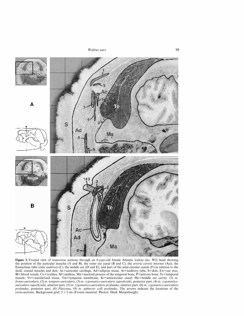
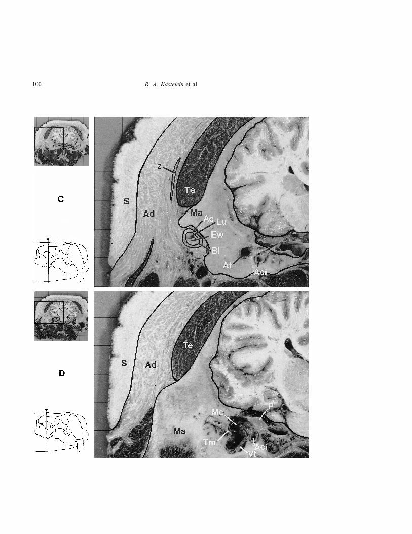
Figure 3. Frontal view of transverse sections through an 8-year-old female Atlantic walrus (no. W2) head showingthe position of the auricular muscles (A and B), the outer ear canal (B and C), the arteria carotis internus (Aci), theEustachian tube (tuba auditiva) (C), the middle ear (D and E), and part of the semi-circular canals (F) in relation to theskull, cranial muscles and skin. Ac=auricular cartilage, Ad=adipose tissue, At=auditory tube, S=skin, Ew=ear wax,Bl=blood vessels, Co=cochlea, M=malleus, Ma=mastoid process of the temporal bone, P=petrous bone, Te=temporalmuscle, Vt=vascularized tissue, Tm=tympanic membrane, Sc=semicircular canal; Mc=middle ear cavity. (1) m.fronto-auricularis, (2) m. temporo-auricularis, (3) m. zygomatico-auricularis superficialis, posterior part, (4) m. zygomatico-auricularis superficialis, anterior part, (5) m. zygomatico-auricularis profundus, anterior part, (6) m. zygomatico-auricularisprofundus, posterior part, (8) Platysma, (9) m. sphincter colli profundus. The arrows indicate the locations of thecross-sections. Background grid: 2�2 cm (Frozen material. Photos: Henk Merjenburgh).
99Walrus ears
100 R. A. Kastelein et al.

101Walrus ears

completely (Fig. 7A). The vascularized tissue isattached in a ring to the perimeter of the oval(approx. 7�9 mm) tympanic membrane whichitself is attached to bone (Fig. 7B).
The inner-most layer lining the approximately5 mm diameter lumen of the outer ear canal con-tains many glandulae ceruminosae and sebaceousglands (Fig. 8). In only one of the 15 sectionsinvestigated, a Pacinian corpuscle was found justbeneath the epithelium (Fig. 9), suggesting thatthese corpuscles occur in low numbers. Hair
follicles occur in the epithelium and the hairs aredirected into the lumen. The hairs are only presentin the distal end of the outer ear tube (Fig. 10).
The outer ear canal is lined with a 0.5–1 mm layerof earwax (cerumen), except for the proximal 2 cm(the bony part of the outer ear canal), of which 90%is filled with wax (Fig. 7A). A very thin layer of waxcovers the tympanic membrane. In live animalsearwax often runs out of the auditory orifice and isvisible on the skin around and above the orifice, likecoagulated wax on a candle.
Figure 4. Transverse sections of the left outer ear canal from a 15-year-old male Pacific walrus (no.W3). (A) proximal end, (B) distal end. Au=auricular cartilage, Bl=blood vessels, Gl=glandulartissue, Co=connective tissue. Lu=lumen, L=lateral, M=medial, P=proximal end. The white barsindicate the locations of the cross sections. HE stained (Photos: Merijn de Bakker).
102 R. A. Kastelein et al.

Figure 5. Detail of a transverse section of the outer ear canal of walrus no. W3, showing theauricular cartilage (Au) and the surrounding connective tissue (Co). HE stained (Photo: Merijn deBakker).
Figure 6. Detail of a transverse section of the outer ear canal of walrus no. W3, showing the bloodsinuses (Bl) and the auricular cartilage (Au). HE stained (Photo: Merijn de Bakker).
103Walrus ears

Figure 7. (A) Ventro-medial view of the right middle ear of W2 (view in the direction of thearrow in Figure 13B). The bone of the bony part of the outer ear canal and the ventral side ofthe cartilagenous part have been removed. The mucosa lining the middle ear and covering theossicles has also been removed. (B) Lateral view of the right tympanic membrane (outer earremoved) of walrus no. W3 showing that the vascularized tissue which lines the outer ear canalconnects in a ring to the perimeter of the ear drum. It also shows the manubrium of the malleus onthe tympanic membrane. Bl=vascularized tissue lining the canal, C=cerumen which has been piledup with a probe, Tm=tympanic membrane, M=Malleus, S=Stapes, I=Incus, Ac=cartilagenouspart of the outer ear canal (Photos: Ron Kastelein and Henk Merjenburgh).
104 R. A. Kastelein et al.

Figure 8. Detail of a transverse section of the outer ear canal of walrus no. W3, showing theglandular tissue (Gl) lining the lumen (Lu) of the tube. Bl=blood vessels, Co=connective tissue,Ep=epithelium. HE stained (Photo: Merijn de Bakker).
Figure 9. Detail of a transverse section of the outer ear canal of walrus no. W3, showinga Pacinian corpuscle (Pa) between the glandular tissue (Gl) and the connective tissue (Co).Lu=lumen, Ep=epithelium. HE stained (Photo: Merijn de Bakker).
105Walrus ears

Auricular musclesThe distal, outwardly projecting part of the auricu-lar tube is surrounded by a number of auricularmuscles. The cartilage and the auricular muscles areprobably homologous with similar elements of theears of terrestrial carnivores. However, the differ-ences in shape and position are so great that theindividual pieces of cartilage have not been named.Similarly, the names of the muscles listed below asnumbers 2–7 (the same designation as in Figs. 3, 11and 12) were inspired by topography alone.
(1) m. fronto-auricularisThis muscle originates on the fascia of the m.frontalis and runs through the subcutaneous fat toinsert on the dorso-rostral side of the tube.
(2) m. temporo-auricularisThe origin is in the superficial layers of the fascia ofthe m. temporalis. The muscle runs deep to them. zygomatico-auricularis superficialis and its fibresfollow the tube in the distal direction.
(3) m. zygomatico-auricularis superficialis,posterior partThe posterior and anterior parts of this muscle havea common origin in the superficial fascia over thezygomatic arc on which the platysma also inserts,but are discontinuous with the latter. The fibres of
the posterior part spiral upwards to insert on theposterior and dorsal aspect of the tube.
(4) m. zygomatico-auricularis superficialis,anterior partThe anterior part of the muscle inserts on theanterior surface of the tube, and is almost in contactwith fibres from the posterior part.
(5) m. zygomatico-auricularis profundus,anterior partThis thin muscle originates on the zygomatic archand runs directly superficial to the parotid fascia. Itinserts at the base of the distal part of the auriculartube.
(6) m. zygomatico-auricularis profundus,posterior partThe posterior part of this L-shaped muscle alsooriginates on the zygomatic arch, but is firmlyembedded in the parotid fascia. It has a finaltendinous insertion slightly deeper than the anteriorpart of this muscle.
(7) m. auricularis internusThis muscle, which is somewhat triangular in cross-section, has its origin on the cartilage forming thebasis of the distal tube, deep to the lumen. It runsupwards, rostral to the lumen almost to the skin
Figure 10. Enlargement of the tissues around the perimeter of the outer ear canal lumen of walrus no.W3 showing hair shafts (Photo: Merijn de Bakker).
106 R. A. Kastelein et al.

covering the orifice of the tube and inserts on theentire free margin of the most dorso-rostral carti-lage element.
Role of the auricular musclesConsidering the arrangement of the different inser-tions, it seems likely that the distal part of thelumen of the tube and the orifice can be opened bycontraction of the fronto-auricular muscle, prob-ably supported by the posterior part of the superfi-cial zygomatico-auricularis muscle. This wouldcreate a backward rotation of the distal tube.Simultaneous contraction of the auricularis internusand both parts of the superficial zygomatico-auricularis muscles will result in a powerful con-striction of the distal tube. A larger volume of
muscle is dedicated to closing the tube than toopening it.
Middle earThe walrus’ middle ear is in the bulla tympanicawhich is surrounded by several openings in the skull(Figs. 3C, D, E and 13A): on the lateral side theforamen stylomastoideum for the nervus facialis andthe meatus acousticus externus osseus; on the rostralside the foramen ovale for the nervus trigeminus andthe canalis musculotubarius the opening of the os-seus part of the auditory (Eustachian) tube (tubaauditiva); on the medial side the canalis caroticusand the foramen lacerum for the arteria carotisinternus, and on the caudal side the foramen jugu-lare for the vena jugularis interna and Nerves 9, 10and 11.
The endotympanic (pars endotympanica) consti-tutes about 1/4 of the bulla tympanica, forming thecanalis caroticus for the arteria carotis internus. Thisartery has a thick wall (3 mm in contracted state)and an outer diameter of 8 mm; the lumen has adiameter of 2 mm, but this is probably larger in aliving animal (Fig. 13B). The rest of the bulla isformed by the ectotympanic. The bulla tympanica isfused to the processus mastoideus of the temporalbone. On the ventro-caudal side, the bulla tym-panica is very dense and about 1 mm thick. Thetympanic bone between the n. facialis and thearticulation joint between the incus and stapes isalso about 1 mm thick. The thickness of the bullatympanica increases to around 20 mm on the rostralside, where the bone is somewhat softer and pen-etrated by many small blood vessels (Fig. 13C).
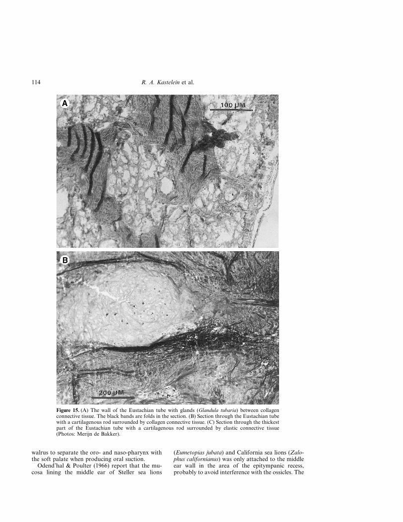
The walrus middle ear cavity is large (about9 cm3 in an adult animal), and consists of thetympanic cavity, the epitympanic recess, a sidepocket and the tympanic orifice of the auditory tube(Fig. 13B and C; For a view of a latex mould of thewalrus middle ear cavity see Fig. 6 in Repenning,1972). The Eustachian tube is flattened, so thathardly any lumen is left; it is difficult to push a1 mm diameter probe through it. Low numbers ofsmall arteries are found in the wall of the Eus-tachian tube (Fig. 14). In the 15-year-old malePacific walrus (W3), the part of the Eustachian tubethat runs through the bulla is 26 mm long. There thewalls of the tube are thickened and the lumen isreduced (the isthmus; Fig. 13B). This thickened partconsists of dense collagen tissue with some cartilagi-nous rods and elastic fibres (Fig. 15). Within thecollagen tissue, glands (glandulae tubariae) occur,which open into the lumen of the Eustachian tube(Fig. 16). The part of the Eustachian tube betweenthe bulla and the opening in the nasopharynx(ostium pharyngeum tubae auditivae) is 19 mm longin the 15-year-old male Pacific walrus (W3), and theopening of the auditory tube in the nasopharynx
Figure 11. The arrangement of the external auricularmuscles of the right auditory canal of an 8-year-old femaleAtlantic walrus (no. W2). The nose of the animal is to theright. *auricular orifice and lumen. (1) m. fronto-auricularis, (2) m. temporo-auricularis, (3) m. zygomatico-auricularis superficialis, posterior part, (4) m. zygomatico-auricularis superficialis, anterior part, (5) m. zygomatico-auricularis profundus, anterior part, (6) m. zygomatico-auricularis profundus, posterior part, (8) Platysma, (9) m.sphincter colli profundus. Mea=Meatus acousticus cartila-genous, Par=parotid gland (Photo: Nico Gerrits).
107Walrus ears
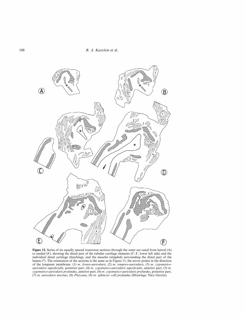
Figure 12. Series of six equally spaced transverse sections through the outer ear canal from lateral (A)to medial (F), showing the distal part of the tubular cartilage elements (C–F, lower left side) and theindividual distal cartilage (hatching), and the muscles (stippled) surrounding the distal part of thelumen (*). The orientation of the sections is the same as in Figure 11; the arrow points in the directionof the tympanic membrane. (1) m. fronto-auricularis, (2) m. temporo-auricularis, (3) m. zygomatico-auricularis superficialis, posterior part, (4) m. zygomatico-auricularis superficialis, anterior part, (5) m.zygomatico-auricularis profundus, anterior part, (6) m. zygomatico-auricularis profundus, posterior part,(7) m. auricularis internus, (8) Platysma, (9) m. sphincter colli profundus (Drawings: Nico Gerrits).
108 R. A. Kastelein et al.

lies 3.5 cm immediately dorsal of the distal end ofthe hamulus pterygoideus, and is 8 mm wide andshaped like an arch (Fig. 17).
The tympanic cavity contains the large auditoryossicles (Figs. 3E, 7, 13B, 18 and 19). For otherviews of walrus ossicles, see Fig. 5 in Repenning(1972) and Wyss (1987). Two muscles are associatedwith the ossicles. The m. tensor tympani has its basein the fossa tensor tympani (Fig. 18). The tendon ofinsertion is attached to the muscular process of themalleus (Fig. 19). The m. stapedius has its origin inthe fossa musculi stapedii and attaches to the mus-cular process of the stapes (Fig. 19B).
The middle ear cavity is lined with a mucousmembrane with loose connective tissue, which insome areas is richly vascularized. On the floor thevascularized tissue is 4 mm [only 1 mm in animalW3, which was raised and housed in a pool] thick,and here the blood vessels are relatively large (Figs.3C, D, E, 13C, 20 and 21). The tissue is about 1 mm[0.5 mm] thick on the sides of the cavity; here thevessels are smaller. On the roof of the middle earcavity the mucosa is not vascularized, except in thepocket medial to the beginning of the Eustachiantube (3/4 of this bony pocket is filled with vascular-ized tissue when this tissue is not engorged withblood), and the dorsal side of the middle earbetween the tympanic orifice of the Eustachian tubeand the tympanic membrane, where the vascular-ized tissue is approximately 0.5 mm thick. Thevascularized tissue seems to be loosely connected tothe floor and walls of the middle ear cavity, andmore tightly connected to the roof. This was thecase in all investigated walruses (W2, W3 and W4).The mucous membrane lining the middle ear is notvascularized near the tympanic membrane. To-wards the epitympanic recess the vascularized tissueis folded and covers a ridge in the bone, the m.tensor tympani, the ossicles and the tympanic nerve(chorda tympani). After leaving the facial nerve, thetympanic nerve passes through the tympanic cavitymedial to the malleus to join the lingual nerve.
Inner earThe foot of the stapes (Fig. 19C) connects to theoval window (Figs. 7A, 13B and 18). The inner earis located between the oval window and the tym-panic cavity (Figs. 3E and 13B). The petrous boneanterior to the cochlea is large (Fig. 3E and F). Fora dissection of the petrosum and cochlea, see Figure2 in Repenning (1972). The semi-circular canals liein the petrosum (Fig. 3E).
Discussion and Conclusions
Requirements the walrus ear has to fulfilThe walrus ear has to fulfil a double function andallow hearing in air (Kastelein et al., 1993b; 1996)
and hearing under water (Stirling et al., 1987).Probably, both functions require different mecha-nisms to transmit sounds from the environment tothe sense organ proper; an impedance switchingmechanism has been suggested for phocids (Møhl,1968a, 1975; Terhune, 1989). Functioning of thehearing mechanism is further complicated by thefact that it also has to cope with pressure changesduring diving. Walruses dive to a maximum depthof 100 m (Fay, 1982; Fay & Burns, 1988). Theyhave to cope with consequences of underwatereating: excavation and food processing requireslarge pressure changes in the oral and pharyngealcavity (Kastelein et al., 1994). The structural fea-tures of the hearing system must be considered inthe light of these requirements.
Special features of the walrus earWalrus outer and middle ears have special featureswhich are not usually found in terrestrial carni-vores. These are: the lack of pinnae, the long,tubular outer ear which is completely embedded inblubber and covered by thick skin, the ability toopen and close the external orifice by auricularmuscles, the lining of the cartilaginous and bonyparts of the outer ear canal with vascularizedtissue, the copious amount of earwax, the largemiddle ear cavity, the lining of the middle earcavity with vascularized tissue, the elastic fibres,collagen tissue and cartilaginous rods in the wallof the Eustachian tube, and the dense bones sur-rounding the base of the outer ear and the entiremiddle and inner ears.
Hypothesis of the function of walrus hearing
(A)Hearing in air
Pressure regulationWhile above the water, the walrus hearing systemprobably functions like that of most terrestrialmammals. In the case of a pressure differencebetween the environment and the middle ear cavity,the animal can swallow or yawn. This will probablyopen the Eustachian tube, so the air pressures in theouter and middle ears can be equalized (Fig. 22A).
HearingThe long outer ear canal in walruses ensures thatthe meatal orifice (porus acousticus) is above waterduring surface swimming (Fig. 1). Walruses oftensleep in a vertical position at the water surface withtheir pharyngeal pouches inflated (Fay, 1960). Inthis posture the whole head and part of the neck areout of the water, which means that the entire outerear is above the surface (Francis H. Fay, pers.comm.) and the walrus can hear air-borne sounds.When swimming at the water surface, the meatus is
109Walrus ears

probably kept open, so that sounds can enter theouter ear canal. In air, the rigidity of the tubularcartilage of the outer ear canal probably keeps thecanal open. Where the cartilage is absent in theexternal meatus, its supporting role is taken over bythe bony canal composed of the squamosal boneand bulla tympanica. The walrus’ outer ear canal islong with two perpendicular flexures, but becausethe orifice is funnel-shaped, the length and flexuresare probably of minor influence on hearing sensi-tivity. The walruses’ lack of pinnae probably limitstheir aerial hearing sensitivity to a small extent andshould almost certainly limit their ability to noticewhether single sounds come from in front or frombehind them unless they move their head (Kietz,1953).
Walruses are gregarious animals which usuallylie against or on top of each other. They are alsovery vociferous and sometimes produce very loudsounds (Loughrey, 1959; Miller, 1985; Verboom &
Kastelein, 1995). They haul out in areas with highlevels of background noise caused by waves andbreaking ice. However, they only seem to haul outto sleep (Krusinskaja & Lisicyna, 1983). Becauseof these loud sounds in their environment, onecould speculate that walruses may close off (if theyare capable of voluntarily doing so) their meatuswhen hauled out to rest. Experiments with a Harpseal (Pagophilus groenlandica) indicate that, in air,sound enters the ear via the meatus. Closing themeatal orifice with a finger reduced the soundlevels received at the cochlea, especially forfrequencies of around 5 kHz (Møhl & Ronald,1975).
(B) Hearing under waterMany of the special features of the walrus outer andmiddle ears probably have both acoustic andpressure-regulating functions. Under water, thewalrus ear probably functions in a similar way to
Figure 13. (A) Ventral view of the right bulla of of a 5-year-old male Pacific walrus (no. W5). (B)Ventral view of the bulla, indicating the location of the middle ear cavity as observed in an8-year-old female Atlantic walrus (no. W2), (C) Medial view of the bulla with the ventral side up foreasy comparison with Figures A and B. Ac=arteria carotis internus, At=auditory tube,Au=auricular cartilage, Bo=basioccipital bone, Bu=bulla (note the large thickness of the bulla onthe rostral side), Cc=canalis caroticus, Co=cochlea, Er=epitympanic recess, Fj=foramen jugale,Fl=foramen lacerum, Fo=foramen ovale, Fm=fossa mandibularis, Fs=foramen stylomastoideum,I=incus, Is=isthmus, M=malleus, Ma=mastoid process of the temporal bone, Mc=middle earcavity, Mea=meatus acousticus externus, Oat=osseus auditory tube, S=stapes, Sp=side pocket,Tm=tympanic membrane, Vt=vascular tissue. The white arrow in (A) indicates the location of thehole shown in Figure 20. The black arrow in (B) indicates the view direction in Figures 7A and 18(Photo and drawings: Ron Kastelein).
110 R. A. Kastelein et al.

111Walrus ears

that of other pinnipeds. The following hypothesison the function of walrus hearing under water ishighly speculative because it is based only on ana-tomical findings. Under water acoustic and pressureregulation experiments on walruses have not yetbeen conducted.
Pressure regulationDiving requires mechanisms that serve to equalizethe pressure on both sides of the tympanic mem-brane. If, during descent, the increase in pressure inthe outer ear canal is not counteracted by a similarincrease in pressure in the middle ear, pain is felt,and, in extreme cases, the tympanic membrane mayrupture.
When a walrus submerges, its meatus is closed bythe auricular muscles, so that water cannot pen-etrate the outer ear canal (Fig. 22B). The tube isalso narrow, long, waxy and lined with hairs andthus difficult to fill with water (and it would be evenmore difficult to empty). A few drops of waterwould reduce aerial hearing sensitivity. It is notclear which stimulus is responsible for activating theauricular muscles. When the skin around the meatalorifice of a walrus is touched, no movements of theunderlying auricular muscles can be seen as are inHarbour seals, Phoca vitulina (Møhl, 1968a). How-ever, this could be because the walrus skin and theunderlying blubber layer are very thick (Fig. 3Aand B). Kastelein et al. (1996) report that the use of
open headphones did not result in a reduction inhearing sensitivity in walruses. Even open head-phones with a grid did not induce the closure of themeatal orifice although the grid touched the skinvery close to the meatal orifice. A reflex triggered bysubmerging is possible, although voluntary controlover the auricular muscles cannot be ruled out.Møhl & Ronald (1975) thought harp seals activelycontrolled the closure of the meatal orifice.
If closure is instigated just before or immediatelyafter submerging, an air-filled cylinder will beformed which is closed at both ends. As the animaldives, the ambient pressure increases. If duringinitial descent, the cartilaginous part of the outerear tube resists deformation due to its own rigidity(which is needed to keep the lumen open for in-airhearing during head movements, which may changethe pressure of skin and blubber on the outer ear)and that of the surrounding tissues, the pressure inthe lumen might temporarily become less than theambient pressure. If this occurs, the sinuses inthe outer ear tube may engorge with blood, sincethe cardiovascular system will be at ambient pres-sure. The filling of the blood sinuses is probablypassive, and drainage is facilitated by the elasticfibres in the walls of the blood vessels.
The outer ear is probably not rigid enough towithstand a great increase in ambient pressurewithout deformation. Light pressure was enough tocause collapse of the outer ear tube of a recently
Figure 14. Collapsed arteries surrounded by elastic fibres in the wall of the Eustachian tube (Photo:Merijn de Bakker).
112 R. A. Kastelein et al.

deceased walrus, but because it was very resilient, itresumed its original shape immediately after thepressure was released.
The outer ear deformation hypothesis is sup-ported by the absence of vascularized tissue in thenon-cartilaginous side of the outer ear canal. Ifblood vessels were present there, inflation wouldcounteract the deformation of the auricular carti-lage. The bony part of the outer ear tube cannotcollapse and here the vascularized tissue enclosesthe entire canal and is attached to the perimeter ofthe tympanic membrane. This was also observed inthe Harp seal (Ramprashad et al., 1972). Møhl(1967) observed a similar lining in the outer ear of aGrey seal (Halichoerus grypus). He suggests that thelining may have a pressure regulating function, butthat other possible functions need to be investi-gated.
The lungs and trachea of seals are severelycompressed by increased pressure during diving(Kooyman et al., 1970), so that the air in therespiratory tract is expected to be at ambient pres-sure. The nasopharynx is linked to the middle earby the Eustachian tube, which can be opened byswallowing and, in air, by yawning (assuming thatthis system in pinnipeds acts in the general mam-malian way). By opening the Eustachian tube, thepressure of the middle ear may come close to theambient pressure. The Eustachian tube is closed bythe elasticity of its wall. The robust wall of theauditory tube in pinnipeds suggests that it mayprovide a strong structural base, which enablesmuscular opening of the tube under increased pres-sure in the nasopharynx during diving.
Because of the high density of water compared toair, pressure changes occur more rapidly in waterthan in air when moving vertically. A positivepressure in the middle ear is easier to overcomethan a negative pressure. When small negativechanges occur in humans in air, they swallow.However, under water human divers have to use theValsalva manoeuvre (forced exhalation against aclosed mouth and nostrils) to overcome negativepressure in the middle ear (Odend’hal & Poulter,1966). Such negative pressure in the middle ear ofthe walrus may be prevented from occurring bycavernous tissue (corpus cavernosum) in the middleear. During a dive, as negative pressure develops inthe middle ear, the mucosa is probably filled withblood. The filling of the cavernous tissue with bloodcould be a passive process as was suggested byTandler (1899) after examining phocid ears. How-ever, if it was a completely passive process, thetissue might be expected to fill with blood when thepinniped was in air, as blood pressure normallyexceeds the ambient air pressure. Graham (1967)proposes that it may be a cardiovascular adjust-ment as a part of the overall diving reflex in
pinnipeds which includes bradycardia. The venousdrainage may be blocked, and thus the bloodpressure may increase, expanding the mucosa.The expanding mucosa compresses the air until thepressure becomes equal to that on the outside of thebody. When descending, the pressure on the bodyand on the circulatory system increases. Underwater, the walrus would have to swallow almostcontinuously while descending or ascending, if itdid not have special pressure regulating features inits ears. If a pressure difference occurs between theear cavities and the vascular system, the cavernoustissues become engorged with blood. The degree ofinflation depends on the pressure difference. Theuneven distribution of the mucous membrane in themiddle ear cavity may be caused by entrances ofthe vessels which supply the blood to this area.
In support of the pressure equilibration functionhypothesis of the cavernous tissue of the walrusmiddle ear is the fact that this tissue was hardlypresent in the 15-year-old male which had spent itsentire life in a pool which was only 4 m deep.Perhaps not being exposed to high ambient pressurelimited his need to develop or maintain this tissue.Less cavernous tissue was found in the middle earof a walrus pup than in that of phocids or otariids(Repenning, 1972), so the walrus seems less able toreduce the volume of air in the middle ear thanother pinnipeds. This is confirmed in the presentstudy: all ears contained a small amount of cavern-ous tissue, regardless of the method of fixation.Therefore it seems to be a property of the species.Repenning (1972) suggests that the walrus middleear has a small amount of cavernous tissue, but notenough to substantially reduce the air volume in themiddle ear cavity, because walruses do not dive asdeep as most other pinnipeds (up to 100 m, Fay,1982). They feed mainly on bivalve molluscs whichusually live up to a depth of 100 m (Fay & Burns,1988).
Once a walrus has found a foraging area in theform of a bivalve mollusc bed, it stays at a relativelyconstant depth and pressure until it has to go to thesurface to breathe. The Eustachian tube probablyopens frequently when walruses forage, becausethey swallow benthic prey under water. In thewalrus, the Eustachian tube seems more rigid thanin terrestrial mammals, as its wall contains a greatdeal of collagen. This probably means that it ismore difficult to open the tube. This might benecessary when large pressure fluctuations occur inthe pharyngeal area during water jetting (Kasteleinet al., 1991) and oral suction (Kastelein & Mosterd,1989; Kastelein et al., 1991; Kastelein et al., 1994).The strong development of the m. tensor veli pal-atini and the m. levator veli palatini which areattached to the well-developed hamuli (Kastelein &Gerrits, 1990; Kastelein et al., 1991) may allow the
113Walrus ears
walrus to separate the oro- and naso-pharynx withthe soft palate when producing oral suction.
Odend’hal & Poulter (1966) report that the mu-cosa lining the middle ear of Steller sea lions
(Eumetopias jubata) and California sea lions (Zalo-phus californianus) was only attached to the middleear wall in the area of the epitympanic recess,probably to avoid interference with the ossicles. The
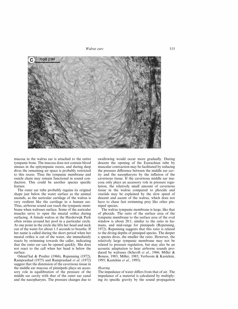
Figure 15. (A) The wall of the Eustachian tube with glands (Glandula tubaria) between collagenconnective tissue. The black bands are folds in the section. (B) Section through the Eustachian tubewith a cartilagenous rod surrounded by collagen connective tissue. (C) Section through the thickestpart of the Eustachian tube with a cartilagenous rod surrounded by elastic connective tissue(Photos: Merijn de Bakker).
114 R. A. Kastelein et al.
mucosa in the walrus ear is attached to the entiretympanic bone. The mucosa does not contain bloodsinuses in the epitympanic recess, and during deepdives the remaining air space is probably restrictedto this recess. Thus the tympanic membrane andossicle chain may remain functional in sound con-duction. This could be another species specificfeature.
The outer ear tube probably regains its originalshape just below the water surface as the animalascends, as the auricular cartilage of the walrus isvery resilient like the cartilage in a human ear.Thus, airborne sound can reach the tympanic mem-brane when walruses surface. Some of the auricularmuscles serve to open the meatal orifice duringsurfacing. A female walrus at the Harderwijk Parkoften swims around her pool in a particular circle.At one point in the circle she lifts her head and neckout of the water for about 1.5 seconds to breathe. Ifher name is called during the short period when hermeatal orifice is out of the water, she immediatelyreacts by swimming towards the caller, indicatingthat the outer ear can be opened quickly. She doesnot react to the call when her head is below thesurface.
Odend’hal & Poulter (1966), Repenning (1972),Ramprashad (1975) and Ramprashad et al. (1972)suggest that the distension of the cavernous tissue inthe middle ear mucosa of pinnipeds plays an acces-sory role in equilibration of the pressure of themiddle ear cavity with that of the outer ear canaland the nasopharynx. The pressure changes due to
swallowing would occur more gradually. Duringdescent the opening of the Eustachian tube bymuscular contraction may be facilitated by reducingthe pressure difference between the middle ear cav-ity and the nasopharynx by the inflation of thecavernous tissue. If the cavernous middle ear mu-cosa only plays an accessory role in pressure regu-lation, the relatively small amount of cavernoustissue in the walrus compared to phocids andotariids may be explained by the slow speed ofdescent and ascent of the walrus, which does nothave to chase fast swimming prey like other pin-niped species.
The walrus tympanic membrane is large, like thatof phocids. The ratio of the surface area of thetympanic membrane to the surface area of the ovalwindow is about 20:1, similar to the ratio in hu-mans, and mid-range for pinnipeds (Repenning,1972). Repenning suggests that this ratio is relatedto the diving depths of pinniped species. The deepera species dives, the smaller the ratio. However, therelatively large tympanic membrane may not berelated to pressure regulation, but may also be anacoustic adaptation to hear airborne sounds pro-duced by walruses (Schevill et al., 1966; Miller &Boness, 1983; Miller, 1985; Verboom & Kastelein,1995; Kastelein et al., 1995).
HearingThe impedance of water differs from that of air. Theimpedance of a material is calculated by multiply-ing its specific gravity by the sound propagation
115Walrus ears

Figure 16. Section of the Eustachian tube with glands which exit in the tube’s lumen. The dark fibresare elastic connective tissue (Photo: Merijn de Bakker).
Figure 17. The bow-shaped entrance of the Eustachian tube of the right middle ear of a 15-year-oldmale Pacific walrus (W3) in the nasopharynx. R=rostral, C=caudal, D=dorsal, which is in thedirection of the distal end of the hamulus pterigoideus. The arrows point out the perimeter of theentrance (Photo: Ron Kastelein).
116 R. A. Kastelein et al.

velocity in it. An impedance difference exists be-tween body tissues and sea water. However, largeimpedance differences exist between different bodytissues such as skin, fat, muscles and bone. Under-water sounds probably travel with little attenuationto the walrus middle ear via tissues which have asmall impedance difference with water. This wouldbe the softer tissues such as the outer ear canal. Thewalrus outer ear canal is surrounded by large, hardbones (Kastelein & Gerrits, 1990); caudally theextremely large mastoid process of the temporalbone and rostrally the zygomatic process of thesquamosal bone (Fig. 2). The middle ear is laterallyshielded by the mastoid process, and ventrally androstrally by the thick, hard bulla (Figs 3 and 4).These bones have dense surfaces, and may act asselective sound reflectors. Sounds, especially thoseof high frequency, may be refracted away from thedenser margins of these bones, and thus ‘piped’along the bones (Repenning, 1972). Sound reachingthe walrus head seems to be focused towards theouter ear canal, which may function as an acousticantenna and conduct sound to the middle ear
(Tonndorf 1966, 1968; Ramprashad, 1975). Evi-dence that non-bony tissues are used for soundconduction comes from a study on directional hear-ing of a Harbour seal by Terhune (1974). In thisspecies the directionality of hearing is similar in airand in water, indicating that the auditory pathwaysoriginate at or near the meatal orifice, and are thusa similar distance apart, in both air and water.Experiments with a harp seal which was forcedunder water indicate that in this species the meatusis closed under water. Closing the meatal orificeunder water with a finger had no effect on under-water hearing (Møhl & Ronald, 1975). The sameexperiments also showed that in a Harp seal inshallow water, the area most sensitive to under-water sounds is just below the meatal orifice (Møhl& Ronald, 1975). This is the location of the outerear canal. Møhl & Ronald (1975) suggested that amodel of the pinniped auditory system should em-ploy both sound transparent and sound opaquetissues.
If in walruses the outer ear canal is the mainpathway by which underwater sounds are received,
Figure 18. View of the middle ear cavity of the right ear of a 15-year-old male Pacific walrus (W3)towards the tympanic recess (the direction of view is indicated by a black arrow in Figure 13B),showing the tympanic membrane (Tm), the malleus (M), incus (I), stapes (S), tympanic nerve (Tn),tensor tympani (Tt), a ridge in the bone (R), and a fold in the mucosa caused by a ventral ridge onthe long crus of the incus (F). The ruler has a mm division (Photo: Henk Merjenburgh).
117Walrus ears

then how are they conducted to the inner ear? Alltissues of the outer ear tube may be involved inunderwater sound conduction towards the middleear. The longitudinal blood vessels themselves,which extend to the tympanic membrane, may serve
as a wave guide for under water sound (Møhl,1968b; Ramprashad, 1975).
The walrus has a large head and a long interauraldistance which leads to a long interaural time delay,favouring directional hearing. Directional hearing
Figure 19. The ear bones of the left ear of a 2-year-old male Pacific walrus. (A) view of individualossicles from one side, (B) view of the same ossicles from the other side, and (C) view of the ossiclesin their natural arrangement. (M) malleus, (I) incus, (S) stapes. (1) manubrium, (2) anterior process,(3) lateral process, (4) head, (5) short crus, (6) long crus, (7) footplate, (8) incudomalleolararticulation, (9) incudostapedia articulation, (10) articulation surface, (11) muscular process. Theruler has a mm division (Photo: Henk Merjenburgh).
118 R. A. Kastelein et al.

under water may be important to walruses, sinceduring the breeding season reproductive males deterother males and attract females in oestrus by meansof underwater calls (Miller & Boness, 1983; Stirlinget al., 1987). If underwater sound reached the innerear by bone conduction, one would have expected avery loose connection between the left and rightmiddle and inner ears in order to maintain direc-tional hearing. Such a construction is seen in dol-phins (McCormick et al., 1970), but this separationis not found in walruses. Walruses might also usedirectional hearing to decode the acoustic signatureof a water volume; a sort of passive sonar forgeneral orientation.
Conduction of vibrations from the walrus’ visce-rocranium to the neurocranium and ears may belimited by the cartilaginous cushion in the zygo-matic arch (Fig. 2A and Kastelein & Gerrits, 1990).Vibrations can be caused by the walrus hitting thesubstrate to haul out or to keep a breathing hole inthe ice open (Fay, 1982). In NE Greenland, wal-ruses have been observed to create breathing holesin 10 cm thick ice by hitting it with their heads frombelow the surface (Møhl, pers. comm.). The zygo-matic cartilage may prevent damage to the ears anddistortion of the vestibular system. It may alsoreduce noise reaching the ears by bone conductionand caused by the tusks scraping along the sub-strate when a walrus is pushing its nose through thesubstrate during foraging (see fig. 102 in Fay, 1982,and Fig. 2 in Kastelein & Mosterd, 1989). Suchnoise, which may continue for a long time, as
walruses dig long furrows in the ocean floor (Oliveret al., 1983; Nelson & Johnson, 1987), may other-wise mask more relevant sounds.
Other than to equalize pressure, the vascularizedtissue in the walrus’ middle ear may also have anacoustic function. Only the non-vascularized partof the mucosa touches the ossicle chain. If thevascularized part of the mucosa touched the ossiclechain, this would result in friction and attenuation.Inflation of the mucosa probably increases thetension on the ossicle chain, and the stiffer thechain, the better it conducts high frequency sounds(Ramprashad et al., 1972). Under water it may beimportant for walruses to hear the high frequencysounds produced by killer whales (Orcinus orca)which predate on walrus pups (Fay, 1982).
Repenning (1972) suggests that in pinnipeds themiddle ear cavernous tissue may transmit soundsacross the tympanic cavity to the cochlear shell.Terhune (1989) proposes that the cavernous tissueswhich line the walls of the outer and middle earsalways engorge with blood when phocids are sub-merging, and that this has a mainly acoustic func-tion: ‘When the tissues are filled with blood, theacoustical impedance would be high, thus facilitat-ing greater sensitivity underwater and producing a30 dB loss of sensitivity in air. When the tissues aredrained, the acoustical impedance would be lower,thus facilitating greater sensitivity in air’. However,this model does not fit the walrus in which thevascularized tissue of the middle ear does not touchthe tympanic membrane.
119Walrus ears

Møhl (1968b) suggests that the vascularized tis-sue lining the middle ear can change the volume ofthe middle ear cavity, thus changing the resonancefrequency.
Lipatov (1992) conducted experiments on theouter ears of seals and sea lions. He concluded thatduring diving, the outer ear remains air-filled andfunctions as ‘a distributed-exitation closed tube’.He concluded that air in the outer ear is essential tounderwater hearing, and that this air in the carti-laginous tube is the basic path of sound conductionfrom water to the middle ear. He suggests thatunderwater hearing involves the excitation of thetympanic membrane and the auditory ossicles.However, some of his methods may have interferedwith the mechanism that occurs during diving. Theinsertion of a glass or metal tube in the outer earprobably interfered with the muscles involved withthe closure of the outer ear opening, and in additionprobably prevented deformation of the tube underpressure. Some of Lipatov’s observations may alsohave been influenced by the shallow depth at whichhis underwater experiments were conducted. Theouter ear probably only deforms after the externalpressure surpasses a certain level, which may not bereached in a shallow experimental tank. Pushingout the air in the external ear with a finger wasanother method used by Lipatov (1992). This re-
sulted in a reduced underwater hearing sensitivity,but the presence of the finger may have caused thedecrease in hearing sensitivity. Filling the outer earwith water also reduced the underwater hearingsensitivity. This is an unnatural situation becausepinnipeds have adaptations to prevent water fromentering the outer ear canal. Therefore filling theouter ear with water will most likely have reducedthe animals’ hearing capabilities, because the waterreached the tympanic membrane. However, al-though Lipatov’s methods may have altered certainmechanisms of the outer ear, that does not meanthat air in the outer ear does not play a role inunderwater hearing.
Ear wax (cerumen)The greater the distance between the meatus andtympanic membrane, the smaller the chance of anywater, which accidently passes the zone with auricu-lar muscles, reaching the tympanic membrane. Sucha long tube, which is probably poorly ventilatedand kept at around core body temperature due tothe thick blubber and skin layers, would be an idealplace for the growth of micro organisms and para-sites. However, the copious amount of ear waxlining the lumen of the outer ear canal probablyinhibits their proliferation. In humans, the cerumenreduces the growth of bacteria and fungi, which are
Figure 20. A hole in the ventro-caudal part of the bulla (Bu) of an 8-year-old female Atlanticwalrus, the location of the thickest vascularized tissue (Vt) lining the middle ear cavity (Mc).R=rostral, C=caudal. For the location of the hole, see the arrow in Figure 13A (Photo: RonKastelein).
120 R. A. Kastelein et al.

often found in the outer ear. This is mainly becausethe ear wax contains heavy metals such as copperand has a low pH of between 5 and 5.7 (Bongers etal., 1990). In terrestrial mammals the sticky ceru-men may also function to prevent insects from
reaching the tympanic membrane. The anopluranlouse (Antarctophthirus trichechi), which resides inskin folds in wild walruses, has also been foundin the walrus’ outer ear canal (Fay, 1982). Possiblythe ear wax in walruses prevents these lice from
Figure 21. (A) The strongly vascularized lining of the middle ear. Some vessels contain remnants ofblood. (B) A larger magnification of Figure A. The wall of the vessels consists of collagen connectivetissue (Photos: Merijn de Bakker).
121Walrus ears

Figure 22. The schematic anatomy of the respiratory tract, vascular system, Eustachian tubeand external and middle ears of a walrus when in air (A) and while diving (B) (Drawings:Ron Kastelein and Rob Triesscheijn).
122 R. A. Kastelein et al.
reaching the tympanic membrane. In walruses earwax production may be stimulated by smallamounts of water which enter the outer ear canal,as is the case in humans.
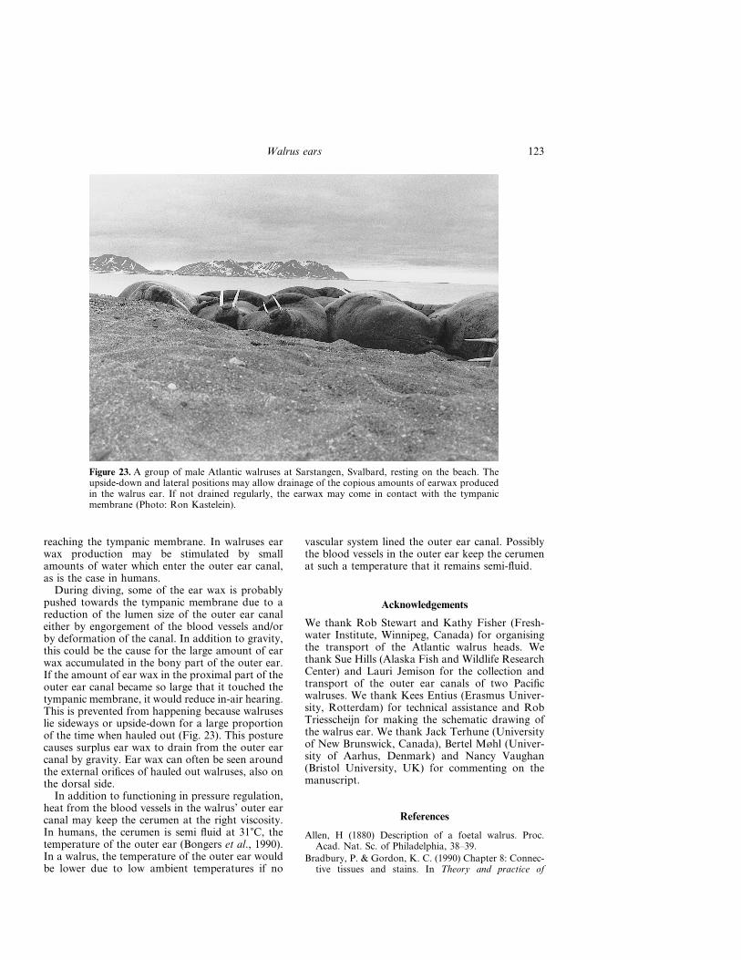
During diving, some of the ear wax is probablypushed towards the tympanic membrane due to areduction of the lumen size of the outer ear canaleither by engorgement of the blood vessels and/orby deformation of the canal. In addition to gravity,this could be the cause for the large amount of earwax accumulated in the bony part of the outer ear.If the amount of ear wax in the proximal part of theouter ear canal became so large that it touched thetympanic membrane, it would reduce in-air hearing.This is prevented from happening because walruseslie sideways or upside-down for a large proportionof the time when hauled out (Fig. 23). This posturecauses surplus ear wax to drain from the outer earcanal by gravity. Ear wax can often be seen aroundthe external orifices of hauled out walruses, also onthe dorsal side.
In addition to functioning in pressure regulation,heat from the blood vessels in the walrus’ outer earcanal may keep the cerumen at the right viscosity.In humans, the cerumen is semi fluid at 31�C, thetemperature of the outer ear (Bongers et al., 1990).In a walrus, the temperature of the outer ear wouldbe lower due to low ambient temperatures if no
vascular system lined the outer ear canal. Possiblythe blood vessels in the outer ear keep the cerumenat such a temperature that it remains semi-fluid.
Acknowledgements
We thank Rob Stewart and Kathy Fisher (Fresh-water Institute, Winnipeg, Canada) for organisingthe transport of the Atlantic walrus heads. Wethank Sue Hills (Alaska Fish and Wildlife ResearchCenter) and Lauri Jemison for the collection andtransport of the outer ear canals of two Pacificwalruses. We thank Kees Entius (Erasmus Univer-sity, Rotterdam) for technical assistance and RobTriesscheijn for making the schematic drawing ofthe walrus ear. We thank Jack Terhune (Universityof New Brunswick, Canada), Bertel Møhl (Univer-sity of Aarhus, Denmark) and Nancy Vaughan(Bristol University, UK) for commenting on themanuscript.
References
Allen, H (1880) Description of a foetal walrus. Proc.Acad. Nat. Sc. of Philadelphia, 38–39.
Bradbury, P. & Gordon, K. C. (1990) Chapter 8: Connec-tive tissues and stains. In Theory and practice of
Figure 23. A group of male Atlantic walruses at Sarstangen, Svalbard, resting on the beach. Theupside-down and lateral positions may allow drainage of the copious amounts of earwax producedin the walrus ear. If not drained regularly, the earwax may come in contact with the tympanicmembrane (Photo: Ron Kastelein).
123Walrus ears
histological techniques. Third edition (eds Bancroft,J. D. & Stevens, A.) pp. 132–137. Churchill Livingstone.
Burck, H. Ch. (1982) Histologische technik 5, unverän-derte Auflage, Gearge Thieme Verlag, Stuttgart,115.
Doran, A. H. G. (1878) The mammalian ossicula auditus.Trans. Linn. Soc. London, 2nd ser. Zool. (1879) 1(7),371–497.
Elliot, H. W. (1875) A report upon the condition of affairsin the Territory of Alaska. pp. 277. US GovernmentPrinting Office, Washington, D.C.
Fay, F. H. (1960) Structure and function of thepharyncheal pouches of the walrus (Odobenus rosmarusL.). Mammalia 24(3), 363–371.
Fay, F. H. (1982) Ecology and biology of the Pacificwalrus, Odobenus rosmarus divergens, Illiger. NorthAmerican fauna no. 74. pp. 279. United States Dept. ofthe Anterior, Fish and Wildlife Service, Washington.
Fay, F. H. & Burns, J. J. (1988) Maximal feeding depth ofwalruses. Arctic 41(3), 239–240.
Graham, S. F. (1967) Sea ears. Science 154, 489.Kastelein, R. A. & Mosterd, P. (1989). The excavation
technique for molluscs of Pacific walruses (Odobenusrosmarus divergens) under controlled conditions.Aquatic Mammals 15(1), 3–5.
Kastelein, R. A., Stevens, S. & Mosterd, P. (1989) Thetactile sensivity of the mystacial vibrissae of a Pacificwalrus (Odobenus rosmarus). Part 2: Masking. AquaticMammals 16(2), 78–87.
Kastelein, R. A. & Gerrits, N. M. (1990) The anatomy ofthe walrus head (Odobenus rosmarus) Part 1: The skull.Aquatic Mammals 16(3), 101–119.
Kastelein, R. A., Gerrits, N. M. & Dubbeldam, J. L.(1991) The anatomy of the walrus head (Odobenusrosmarus) Part 2: Description of the muscles and oftheir role in feeding and haul-out behaviour. AquaticMammals 17(4), 156–180.
Kastelein, R. A., Zweypfenning, R. C. V. J., Spekreijse,H., Dubbeldam, J. L. & Born, E. W. (1993a) Theanatomy of the walrus head (Odobenus rosmarus) Part3: The eyes and their function in walrus ecology.Aquatic Mammals 19(2), 61–92.
Kastelein, R. A., van Ligtenberg, C. L., Gjertz, I. &Verboom, W. C. (1993b) Free field hearing tests on wildAtlantic walruses (Odobenus rosmarus rosmarus) in air.Aquatic Mammals 19(3), 143–148.
Kastelein, R. A., Muller, M. & Terlouw, A. (1994) Oralsuction of a Pacific walrus (Odobenus rosmarus diver-gens) in air and under water. Z. für Säugetierkunde 59,105–115.
Kastelein, R. A., Postma, J. & Verboom, W. C. (1995)Airborne vocalizations of Pacific walrus pups (Odobe-nus rosmarus divergens). In Sensory Systems of AquaticMammals (eds R. A. Kastelein, J. A. Thomas & P. E.Nachtigall) pp. 265–286. De Spil Publishers: Woerden,Netherlands.
Kastelein, R. A., Mosterd, P., van Ligtenberg, C. L. &Verboom, W. C. (1996) Aerial hearing sensitivity testswith a male Pacific walrus (Odobenus rosmarus diver-gens), in the free-field and with headphones. AquaticMammals 22, 85–115.
Kietz, H. (1953) Das Räumliche Hören. Acustica, 3,73–86.
Kooyman, G. L., Hammond, D. D. & Schroeder, J. P.(1970) Bronchograms and tracheograms of seals underpressure. Science 169, 82–84.
Krusinskaja, N. L. & Lisicyna, T. Yu. (1983) The struc-ture of the population of Odobenidae, walruses (inRussian). In Behaviour of Marine Mammals. Nauka:Moscow.
Laet, J. de (1633) Novus Orbis s. Descriptio Indiaeoccidentalis. Luyd. Bat. 1633, 38–39.
Lipatov, N. V. (1992) Underwater hearing in seals: Therole of the outer ear. In Sensory Systems of AquaticMammals (eds J. A. Thomas, R. A. Kastelein & A.Supin) pp. 249–256. Plenum Press: New York.
Loughrey, A. G. (1959) Preliminary investigations of theAtlantic walrus (Odobenus rosmarus rosmarus) (Lin-naeus). Wildl. Man. Bull. Ottowa 1(4).
McCormick, J. G., Wever, E. G., Palin, J. & Ridgway,S. H. (1970) Sound conduction in the dolphin ear.J. Acoust. Soc. Am. 48, 1418–1428.
Miller, E. H. (1985) Airborne acoustic communication inthe walrus Odobenus rosmarus. NGR, winter, 124–145.
Miller, E. H. & Boness, D. J. (1983) Summer behavior ofAtlantic walruses Odobenus rosmarus rosmarus (L.) atCoats Island, N. W. T. (Canada). Z. Säugetierkunde 48,298–313.
Møhl, B. (1967) Seal Ears. Science 157(3784), 99.Møhl, B. (1968a) Auditory sensitivity of the common seal
in air and water. J. Aud. Res. 8, 27–38.Møhl, B. (1968b) Hearing in seals. In The behavior and
physiology of pinnipeds (eds Harrison, R. J., Hubbard,R. C., Peterson, R. S., Rice, C. E. & Schusterman,R. J.) pp. 172–195. Apple-Century-Crofts: New York.
Møhl, B. & Ronald, K. (1975) The peripheral audi-tory system of the harp seal, Pagophilus groenlandicus,(Erxleben, 1777). Rapp. P.-v. Réun. Cons. int. Explor.Mer 169, 516–523.
Murie, J (1870) The anatomy of the walrus. Proc. Zool.Soc. Lond., 544–545.
Murie, J. (1871) Researches upon the anatomy of thePinnipedia. Part 1. On the walrus (Trichechus rosmarus,Linn.) Trans. of Zool. Soc. of London 7(6), 411–464.
Nelson, C. H. & Johnson, K. R. (1987). Whales andwalruses as tillers of the sea floor. Scientific American,February, 74–81.
Odend’hal, S. & Poulter, T. C. (1966) Pressure regulationin the middle ear cavity of sea lions: a possible mech-anism. Science 153, 768–769.
Oliver, J. S., Slattery, P. N., O’Connor, E. F. & Lowry,L. F. (1983) Walrus, Odobenus rosmarus, feeding in theBering Sea: a bentic perspective. Fishery Bulletin 81(3),501–512.
Ramprashad, F. (1975) Aquatic adaptations in the ear ofthe Harp seal Pagophilus groenlandicus (Erxleben,1777). Rapp. P.-v. Réun. Cons.int. Explor. Mer 169,102–111.
Ramprashad, F., Corey, S. & Ronald, K. (1972) Anatomyof the seal’s ear (Pagophilus groenlandicus) (Erxleben,1777) In Functional anatomy of marine mammals. Vol. 1(ed. R. J. Harrison) pp. 263–305. Academic Press:London.
Ray, G. C. & Watkins, W. A. (1975) Social function ofunderwater sounds in the walrus, (Odobenus rosmarus).Rapp. P.-v. Réun. Cons. int. Explor. Mer 169, 524–526.
124 R. A. Kastelein et al.

Repenning, Ch. A. (1972) Underwater hearing in seals:Functional morphology. In Functional anatomy of ma-rine mammals. Vol. 1 (ed. Harrison, R. J.) pp. 307–331.Academic Press: London.
Schevill, W. E., Watkins, W. A. & Ray, C. (1966) Analysisof underwater Odobenus calls with remarks on thedevelopment of the pharyngeal pouches. Zoologica(NY) 51(3), 103–106.
Schmidt, M. (1885) Das Walross (Trichechus rosmarus).Der Zool. Garten, Frankfurt 1, 1–16.
Stirling, I., Calvert, W. & Spencer, C. (1987) Evidence ofstereotyped underwater vocalizations of male Atlanticwalruses (Odobenus rosmarus rosmarus). Can. J. Zool.65, 2311–2321.
Tandler, J. (1899) Ueber ein Corpus cavernosum tympani-cum beim Seehund. Monatsschr. Ohrenheilk. 33, 437–440.
Terhune, J. M. (1974) Directional hearing of a Harborseal in air and water. J. Acoust. Soc. Am. 56(6),1862–1865.
Terhune, J. M. (1989) Can seals alter the acoustic imped-ance of the outer and middle ears? Proc. of ann.meeting of CAA, Halifax, Nova Scotia.
Tonndorf, J. (1966) Bone conduction studies in exper-imental animals. Acta Otolaryngol., Suppl., 213.
Tonndorf, J. (1968) A new concept in bone conduction.Arch. Otolaryngol. 87, 595–600.
Verboom, W. C. & Kastelein, R. A. (1995) Ruttingwhistles of a male Pacific walrus (Odobenus rosmarusdivergens). In Sensory Systems of Aquatic Mammals(eds Kastelein, R. A., Thomas, J. A. & Nachtigall,P. E.) pp. 287–298. De Spil Publishers, Woerden: TheNetherlands.
Wyss, A. R. (1987) The walrus auditory region and themonophyly of the pinnipeds. American MuseumNovitates 2871, 1–31.
125Walrus ears