
The actin regulator coronin 1A is mutant in a thymic egress–deficient mouse strain and in a...
9
The actin regulator coronin 1A is mutant in a thymic egress–deficient mouse strain and in a patient with severe combined immunodeficiency Lawrence R Shiow 1–3 , David W Roadcap 5 , Kenneth Paris 7 , Susan R Watson 2 , Irina L Grigorova 1,2 , Tonya Lebet 2,4 , Jinping An 1,2 , Ying Xu 1,2 , Craig N Jenne 1,2 , Niko Fo ¨ger 8 , Ricardo U Sorensen 7 , Christopher C Goodnow 6 , James E Bear 5 , Jennifer M Puck 3,4 & Jason G Cyster 1–3 Mice carrying the recessive locus for peripheral T cell deficiency (Ptcd ) have a block in thymic egress, but the mechanism responsible is undefined. Here we found that Ptcd T cells had an intrinsic migration defect, impaired lymphoid tissue trafficking and irregularly shaped protrusions. Characterization of the Ptcd locus showed a point substitution of lysine for glutamic acid at position 26 in the actin regulator coronin 1A that enhanced its inhibition of the actin regulator Arp2/3 and resulted in its mislocalization from the leading edge of migrating T cells. The discovery of another coronin 1A mutant during an N-ethyl-N- nitrosourea-mutagenesis screen for T cell–lymphopenic mice prompted us to evaluate a T cell–deficient, B cell–sufficient and natural killer cell–sufficient patient with severe combined immunodeficiency, whom we found had mutations in both CORO1A alleles. Our findings establish a function for coronin 1A in T cell egress, identify a surface of coronin involved in Arp2/3 regulation and demonstrate that actin regulation is a biological process defective in human and mouse severe combined immunodeficiency. After developing in the thymus, mature thymocytes upregulate sphin- gosine 1-phosphate receptor type 1 (S1P 1 ) and exit into the circulation to populate the naive T cell compartment. S1P 1 and its ligand, sphingosine 1-phosphate (S1P), are required for the egress of mature thymocytes 1 . FTY720, a small-molecule immunosuppressant being tested in clinical trials for the treatment of autoimmune disease, inhibits egress by modulating S1P 1 function 1 . Beyond the requirement for S1P 1 , little is understood about how T cells migrate from the thymus. The cataract Shionogi (CTS) strain was initially isolated in the 1960s from a closed colony of ICR mice on the basis of its cataracts and microphthalmia 2 . The CTS strain was later established to have a defect in thymic egress 3 after it failed to reject major histocompat- ibility complex–disparate skin grafts 4 . The nature of this recessive defect, called ‘peripheral T cell deficiency’ (Ptcd), and its function in thymic egress have remained unclear. Coronins are actin regulators found in all eukaryotes 5 . In addition to binding F-actin, coronin proteins associate with and inhibit the nucleation-promoting Arp2/3 complex. Seven coronin family mem- bers exist in mammals, including coronin 1A (Coro1A), which is expressed mainly in hematopoietic cells. Coro1A-deficient mice have fewer peripheral T cells due to more apoptosis 6,7 , and one study has attributed this to an excessive accumulation of F-actin 6 . T cell migration is also reported to be defective 6,7 , but this has been called into question 8 . Those last authors have also questioned whether Coro1A deficiency alters F-actin dynamics and instead link the greater apoptosis to a T cell antigen receptor (TCR) signaling defect 8 . These conflicting reports of Coro1A-deficient mice have complicated the understanding of the function of this actin regulator in T cell biology. Here we report that Ptcd T cells had an intrinsic migration defect that impaired thymic egress and trafficking through lymph nodes. We ‘narrowed’ the Ptcd locus and identified a point mutation in the gene encoding Coro1A (Coro1a) that resulted in mislocalization of the protein in T cells and greater inhibition of Arp2/3 by Coro1A in biochemical assays. In a parallel effort to identify additional traffic- king mutants by screening mice carrying N-ethyl-N-nitrosourea (ENU)-induced mutations for altered peripheral T cell numbers, we identified a strain with 90% less Coro1A and a phenotype similar to that of Coro1A-knockout mice. Comparison of Ptcd and Coro1A- deficient T cells allowed us to separate the defect in TCR-induced Received 9 July; accepted 4 September; published online 5 October 2008; doi:10.1038/ni.1662 1 Howard Hughes Medical Institute, 2 Department of Microbiology and Immunology, 3 Biomedical Sciences Graduate Program and 4 Department of Pediatrics and Institute for Human Genetics, University of California San Francisco, San Francisco, California 94143, USA. 5 Lineberger Comprehensive Cancer Center and Department of Cell and Developmental Biology, School of Medicine, University of North Carolina at Chapel Hill, Chapel Hill, North Carolina 27599, USA. 6 John Curtin School of Medical Research, The Australian National University, Canberra ACT 2601, Australia. 7 Department of Pediatrics, Louisiana State University Health Sciences Center and Children’s Hospital, New Orleans, Louisiana 70118, USA. 8 Department of Immunology and Cell Biology, Leibniz Center for Medicine and Biosciences, Borstel 23845, Germany. Correspondence should be addressed to J.G.C. ([email protected]). NATURE IMMUNOLOGY VOLUME 9 NUMBER 11 NOVEMBER 2008 1307 ARTICLES © 2008 Nature Publishing Group http://www.nature.com/natureimmunology
Transcript of The actin regulator coronin 1A is mutant in a thymic egress–deficient mouse strain and in a...

The actin regulator coronin 1A is mutant in a thymicegress–deficient mouse strain and in a patient withsevere combined immunodeficiency
Lawrence R Shiow1–3, David W Roadcap5, Kenneth Paris7, Susan R Watson2, Irina L Grigorova1,2,Tonya Lebet2,4, Jinping An1,2, Ying Xu1,2, Craig N Jenne1,2, Niko Foger8, Ricardo U Sorensen7,Christopher C Goodnow6, James E Bear5, Jennifer M Puck3,4 & Jason G Cyster1–3
Mice carrying the recessive locus for peripheral T cell deficiency (Ptcd ) have a block in thymic egress, but the mechanism
responsible is undefined. Here we found that Ptcd T cells had an intrinsic migration defect, impaired lymphoid tissue trafficking
and irregularly shaped protrusions. Characterization of the Ptcd locus showed a point substitution of lysine for glutamic acid
at position 26 in the actin regulator coronin 1A that enhanced its inhibition of the actin regulator Arp2/3 and resulted in its
mislocalization from the leading edge of migrating T cells. The discovery of another coronin 1A mutant during an N-ethyl-N-
nitrosourea-mutagenesis screen for T cell–lymphopenic mice prompted us to evaluate a T cell–deficient, B cell–sufficient
and natural killer cell–sufficient patient with severe combined immunodeficiency, whom we found had mutations in both
CORO1A alleles. Our findings establish a function for coronin 1A in T cell egress, identify a surface of coronin involved in
Arp2/3 regulation and demonstrate that actin regulation is a biological process defective in human and mouse severe
combined immunodeficiency.
After developing in the thymus, mature thymocytes upregulate sphin-gosine 1-phosphate receptor type 1 (S1P1) and exit into the circulationto populate the naive T cell compartment. S1P1 and its ligand,sphingosine 1-phosphate (S1P), are required for the egress of maturethymocytes1. FTY720, a small-molecule immunosuppressant beingtested in clinical trials for the treatment of autoimmune disease,inhibits egress by modulating S1P1 function1. Beyond the requirementfor S1P1, little is understood about how T cells migrate fromthe thymus.
The cataract Shionogi (CTS) strain was initially isolated in the1960s from a closed colony of ICR mice on the basis of its cataractsand microphthalmia2. The CTS strain was later established to have adefect in thymic egress3 after it failed to reject major histocompat-ibility complex–disparate skin grafts4. The nature of this recessivedefect, called ‘peripheral T cell deficiency’ (Ptcd), and its function inthymic egress have remained unclear.
Coronins are actin regulators found in all eukaryotes5. In additionto binding F-actin, coronin proteins associate with and inhibit thenucleation-promoting Arp2/3 complex. Seven coronin family mem-bers exist in mammals, including coronin 1A (Coro1A), which is
expressed mainly in hematopoietic cells. Coro1A-deficient mice havefewer peripheral T cells due to more apoptosis6,7, and one study hasattributed this to an excessive accumulation of F-actin6. T cellmigration is also reported to be defective6,7, but this has been calledinto question8. Those last authors have also questioned whetherCoro1A deficiency alters F-actin dynamics and instead link the greaterapoptosis to a T cell antigen receptor (TCR) signaling defect8. Theseconflicting reports of Coro1A-deficient mice have complicated theunderstanding of the function of this actin regulator in T cell biology.
Here we report that Ptcd T cells had an intrinsic migration defectthat impaired thymic egress and trafficking through lymph nodes. We‘narrowed’ the Ptcd locus and identified a point mutation in the geneencoding Coro1A (Coro1a) that resulted in mislocalization of theprotein in T cells and greater inhibition of Arp2/3 by Coro1A inbiochemical assays. In a parallel effort to identify additional traffic-king mutants by screening mice carrying N-ethyl-N-nitrosourea(ENU)-induced mutations for altered peripheral T cell numbers, weidentified a strain with 90% less Coro1A and a phenotype similar tothat of Coro1A-knockout mice. Comparison of Ptcd and Coro1A-deficient T cells allowed us to separate the defect in TCR-induced
Received 9 July; accepted 4 September; published online 5 October 2008; doi:10.1038/ni.1662
1Howard Hughes Medical Institute, 2Department of Microbiology and Immunology, 3Biomedical Sciences Graduate Program and 4Department of Pediatrics and Institutefor Human Genetics, University of California San Francisco, San Francisco, California 94143, USA. 5Lineberger Comprehensive Cancer Center and Department of Celland Developmental Biology, School of Medicine, University of North Carolina at Chapel Hill, Chapel Hill, North Carolina 27599, USA. 6John Curtin School of MedicalResearch, The Australian National University, Canberra ACT 2601, Australia. 7Department of Pediatrics, Louisiana State University Health Sciences Center andChildren’s Hospital, New Orleans, Louisiana 70118, USA. 8Department of Immunology and Cell Biology, Leibniz Center for Medicine and Biosciences, Borstel 23845,Germany. Correspondence should be addressed to J.G.C. ([email protected]).
NATURE IMMUNOLOGY VOLUME 9 NUMBER 11 NOVEMBER 2008 1307
A R T I C L E S©
2008
Nat
ure
Pub
lishi
ng G
roup
ht
tp://
ww
w.n
atur
e.co
m/n
atur
eim
mun
olog
y

calcium signaling from the diminished thymocyte survival. In addi-tion to yielding new alleles of Coro1a, our mouse forward-geneticsapproach led us to identify a T cell–deficient, B cell–sufficient andnatural killer cell–sufficient (T–B+NK+) patient with SCID who hasCoro1A deficiency.
RESULTS
Ptcd is an intrinsic T cell–migration defect
To characterize the cellular basis of the Ptcd defect, we first backcrossedthe Ptcd locus onto the C57BL/6 (B6) strain and confirmed the accu-mulation of mature single-positive (SP) thymocytes (CD69loCD62Lhi)and associated lower number of peripheral T cells (Fig. 1a and Supple-mentary Fig. 1 online). Irradiated wild-type mice that had beenreconstituted with Ptcd bone marrow cells also had an accumulationof mature thymocytes and few circulating T cells (Fig. 1b), whereasreciprocal bone marrow chimeras did not have such defects (Supple-mentary Fig. 2 online). These results localized the Ptcd defect to ahematopoietic lineage–derived cell and indicated that impaired egressof mature thymocytes from the thymus is a pathogenic mechanism.
Although normal amounts of S1P1 were expressed on Ptcd matureSP thymocytes (Fig. 1c), S1P1 function was impaired. In Transwell
migration assays, Ptcd mature CD4 SP thymocytes were less efficientin migrating toward S1P than were cells from control heterozygouslittermates (Fig. 1d). The cells also migrated less efficiently toward thechemokines CCL21 and CXCL12. The response of Ptcd CD4+CD8+
double-positive thymocytes to CXCL12 was similarly lower (Fig. 1e),and the migration of naive Ptcd splenic T cells was impaired, whereasB cells migrated normally (Fig. 1f). The expression of CCR7 andCXCR4, the respective chemokine receptors that recognize CCL21 andCXCL12, was similar in Ptcd and control cells (Supplementary Fig. 3online). These results indicate that T cells in Ptcd mice have a general,cell-intrinsic migration defect that impairs S1P1 responsiveness andblocks thymic egress.
Ptcd T cells have entry, egress and motility defects
To test for peripheral trafficking defects, we transferred Ptcd andcontrol T cells together into wild-type recipients. At 1 h after transfer,the ratio of Ptcd T cells to control T cells was lower in peripheral and
Thy
mus
cel
ls (
106 )
Thy
mus
cel
ls (
106 )
150
0
100
250
200
50
Double-positive
*
*
* *
Het
Ptcd
0
10
20
3035
25
15
5
MatureCD4 SP
MatureCD8 SP
Blo
od c
ells
(pe
r m
l) 107
CD19 CD4 CD8
106
105
104
107
106
105
104
a c
d e f
150
0
100
200
50
Double-positive
*
*
**
0
12
8
MatureCD4 SP
MatureCD8 SP
4
CD19 CD4 CD8
b Het
Ptcd
WT
WT
Blo
od c
ells
(pe
r m
l)
0
1
2
3
4
Mig
ratio
n (%
of i
nput
)
S1P (nM)0 1 10 102 103
HetPtcd
*M
igra
tion
(% o
f inp
ut)
Mig
ratio
n (%
of i
nput
)
Mig
ratio
n (%
of i
nput
)
Mig
ratio
n (%
of i
nput
)
0
20
40
60
80
100Ptcd
Het
Isotype
Per
cent
of m
axim
um
S1P1
0
20
40
60
Nil NilNil Nil
CXCL12
(0.3
µg/m
l)
CXCL12
(0.3
µg/m
l)
CXCL12
(0.3
µg/m
l)
CXCL13
(0.5
µg/m
l)
CXCL12
(0.3
µg/m
l)
CCL21
(1 µg
/ml)
CCL21
(1 µg
/ml)
CCL21
(1 µg
/ml)
CCL21
(1 µg
/ml)
HetPtcd
*
* 8
6
4
2
0
*
*
0
20
40
60
80HetPtcd *
0
5
10
15
20
25
Mature CD4 SP thymocytes Mature CD4 SP thymocytes Double-positive thymocytes Naive CD4+ T cells Naive CD19+ B cells
Figure 1 Ptcd T cells have an intrinsic migration defect. (a,b) Thymocyte and peripheral blood lymphocyte subsets from B6 Ptcd and control (heterozygous
Ptcd/+ (Het)) mice (a), or wild-type mice reconstituted with Ptcd bone marrow (Ptcd-WT) or control bone marrow (Het-WT; b). (c) Flow cytometry of S1P1
on mature CD4 SP thymocytes from Ptcd and control mice. Filled histogram (Isotype), isotype-matched control antibody. (d–f) Transwell migration of Ptcd
and control mature CD4 SP thymocytes to S1P (d) and Transwell migration of thymocytes (e) or naive CD4+ splenic T cells or CD19+ B cells (f) to thechemokines CXCL12, CCL21 and/or CXCL13 (e,f), presented as percentage of input cells that migrated. Nil, no chemokine. In a,b,d–f, bars indicate means;
circles indicate individual mice. *, P o 0.05. Data are representative of at least two experiments (a,b) or three experiments (c) or are pooled from three or
four experiments (d–f).
a b c
d e f
Ptcd/
het c
ontr
ol
Med
ian
velo
city
(µm
/min
)
0
4
8
12
Het Ptcd
* *
0
30
60
90
Med
ian
turn
ing
angl
e
Het Ptcd
0
0.5
1
1.5
2
2.5
Blood Spleen MLN PLN MLN PLN Lymph0
0.5
1
1.5
2
2.5
0
1.0
0.8
T c
ells
ret
aine
d (%
)
0.6
0.4
0.2
MLN PLN
HetPtcd
* *
Time (min)
<R
2 > (
103
µm2 )
0
2
3
4
1
0 2 4 6 8 10
HetPtcd
Ptcd
/het
1 h 24 h
Figure 2 Ptcd T cells are defective in lymph node trafficking. (a,b) Homing
to lymph nodes at 1 h (a) and egress into lymph at 24 h (b) after adoptive
cotransfer of Ptcd thymocytes (Ptcd/Ptcd) and heterozygous control
thymocytes (het; Ptcd/+), presented as the ratio of Ptcd to control cells,
normalized to the injected ratio of Ptcd to control cells. To achieve lymph
node ratio of about 1:1 at 24 h, initial injections used a ratio of 3:1. MLN,
mesenteric lymph node; PLN, peripheral lymph node. (c) Retention of Ptcd
and control cells in the mesenteric or peripheral lymph nodes 20 h after
treatment with integrin-neutralizing antibody. (d–f) Time-lapse two-photonmicroscopy of the mean velocities (d), turning angles (e) and displacement
(f) of Ptcd cells and control cells (Het; Ptcd/+) in explanted lymph nodes.
(f) Average of displacement squared (R2) over time (symbols, means; bars,
s.d.). In a–e, bars indicate means; circles indicate individual mice. *,
P o 0.05. Data are pooled from three experiments (a,b), are from three in-
dependent experiments with four mice each (c) or are representative of three
independent experiments with at least 40 tracks analyzed in each (d–f).
1308 VOLUME 9 NUMBER 11 NOVEMBER 2008 NATURE IMMUNOLOGY
A R T I C L E S©
2008
Nat
ure
Pub
lishi
ng G
roup
ht
tp://
ww
w.n
atur
e.co
m/n
atur
eim
mun
olog
y

mesenteric lymph nodes and higher in blood (Fig. 2a), whichindicated that Ptcd cells were less efficient in entering lymph nodes.Next we tested lymph node egress with two approaches. First, wetransferred Ptcd and control T cells together at a ratio of 3:1 to achieveequal proportions in the lymph nodes. At 24 h after transfer, we founda much lower proportion of Ptcd cells in lymph than in lymph nodes(Fig. 2b), which was consistent with a diminished ability to exitinto lymph. As an additional approach, we assessed the retention ofPtcd T cells in lymph nodes. After transferring Ptcd and control T cells,we blocked entry into lymph nodes for 20 h with integrin-neutralizingantibodies. Roughly 60% of Ptcd T cells were retained in thelymph nodes, compared with just 20% of control T cells (Fig. 2c).These results collectively demonstrate that Ptcd T cells are defectivein exiting lymph nodes.
Two-photon microscopy of explanted lymph nodes from mice thathad received fluorescence-labeled cells showed that whereas controlcells moved at a median velocity of 9.5 mm/min, Ptcd cells moved at alower median velocity of 6.6 mm/min (Fig. 2d and SupplementaryMovie 1 online). In addition, Ptcd cells had larger turning angles(Fig. 2e), indicative of less-directed paths, and the cells failed toefficiently displace over time (Fig. 2f). These results demonstrate thatin addition to blocking thymic egress, the Ptcd defect impairstrafficking through lymph nodes.
Coro1a is mutated at the Ptcd locus
The Ptcd locus has been mapped to a 10.9-megabase region ofchromosome 7 containing over 300 open reading frames9. To refinethe locus, we did a mapping cross between CTS and B6 mice, usingthymocyte accumulation to phenotype mice (Fig. 3a). We noted thatthe cataracts and microphthalmia were present in F1 hybridsand failed to segregate together with the recessive Ptcd trait(data not shown), consistent with an early report that these eye traitsare autosomal dominant10. After analyzing over 900 meiosis events,we further mapped the Ptcd locus to a critical interval of 950 kilobases(Fig. 3b). This gene-rich interval contained 37 open reading frames,
including Coro1a (Supplementary Fig. 4 online). Coro1a was a likelycandidate gene for Ptcd because of its abundance in T cells11 and theassociation of coronin family molecules with actin-based motility5.Sequencing of Ptcd DNA showed a G-to-A mutation in exon 2 ofCoro1a (Fig. 3c), which resulting in a nonconservative glutamicacid–to lysine substitution at residue 26 in the b-propeller domainof the protein (Coro1AE26K; Fig. 3d). This residue was conserved in allannotated Coro1A sequences and the charge of the residue was alsoconserved in a large number of orthologs (Supplementary Fig. 5online) and was in a surface-exposed loop12 adjacent to a region thatin the close relative Coro1B has been indicated to be an actin-bindingsite13. Immunoblot analysis showed that the abundance of Coro1A intotal thymocytes did not vary between Ptcd and controls (Fig. 3e).
To exclude the possibility that substitutions other than Coro1AE26K
were responsible for the Ptcd phenotype, we tested for geneticcomplementation by crossing Ptcd mice with Coro1a+/– mice. Asexpected, the Coro1a wild-type allele complemented Ptcd defectsin T cell lymphopenia and F-actin accumulation, whereas theCoro1a-null allele failed to complement (Supplementary Fig. 6a,bonline). In addition, mature thymocyte numbers were slightly higherin Ptcd/– mice than in Ptcd/+ mice (Supplementary Fig. 6c).Although these studies do not exclude the possibility that thePtcd interval contains additional genetic modifiers, the inability ofthe Coro1a-null allele to ‘rescue’ several of the Ptcd defects providesstrong evidence that the Coro1AE26K substitution results in thePtcd phenotype.
ENU-mutant Koyaanisqatsi is a Coro1A hypomorph
In parallel to the studies reported above, we did an ENU-mutagenesisscreen to identify regulators of lymphocyte trafficking and egress. Weinitially identified ‘Koyaanisqatsi’ (Koy) as a recessive mutant strain ofmice with a circulating T cell deficiency (Fig. 4a). Further character-ization of Koy mice showed that they had fewer mature thymocytesand peripheral T cells (Fig. 4b and Supplementary Fig. 7 online). InTranswell migration assays, Koy thymocytes and naive T cells migrated
54.723.3
100
101
102
103
104
100
101
100
101
102
103
104
100
101
102
103
104
100
101
0
10
20
CD
8
CD
69
CD62LCD62LCD4
Cel
ls
ActinCoro1A
NC
PtcdHetdβ-propeller domain Coiled
coil
1 352 429 461
*E26K(Ptcd )
e
Critical interval
1227
a b
35.73.54
66.3
25.2100
101
102
103
104
100
101
102
103
104
102
103
104
102
103
104
7.82
0.89
02468
10
30
40
50
Affected
Unaffected
Totalthymocytes CD4 SP CD8 SP
c
WT
Glu26
Ptcd
Lys26
Het
SNP markers
Unaffectedrecombinant
mice
903
B6 allele
CTS allele
rs30
2421
2
rs13
4795
13
rs32
3115
99
rs82
5962
5
rs33
0859
08
rs13
4795
11
rs62
7557
9
714
1258
1077
Figure 3 Coro1a is mutated at the Ptcd locus.
(a) Flow cytometry of thymocytes from affected and
unaffected mice generated in the mapping cross
between CTS and B6 mice. Mice were considered
‘affected’ on the basis of a high frequency of SP
cells, a high fraction of CD62LhiCD69lo CD4 SP cells
and enrichment for CD62Lhi staining of CD8 SP cells. Numbers in or adjacent to outlined areas indicate percent cells in each. (b) Critical interval of
Ptcd defined by SNP markers polymorphic between the B6 and CTS strains in various recombinant mice (mouse identifiers, left margin). (c) Dideoxy
sequence tracings of DNA from Ptcd and heterozygous mice identifying a G-to-A mutation at position 76 that resulted in the E26K substitution of Coro1A.
WT, wild-type DNA from the related nonobese diabetic mouse strain (additional control). (d) Coro1A structure, including the Ptcd point substitution (*).(e) Immunoblot analysis of Coro1A and actin in total thymocytes from Ptcd and control mice, for samples loaded in threefold dilutions (wedges). NC,
negative control (Coro1a–/–). Data are representative of three experiments.
NATURE IMMUNOLOGY VOLUME 9 NUMBER 11 NOVEMBER 2008 1309
A R T I C L E S©
2008
Nat
ure
Pub
lishi
ng G
roup
ht
tp://
ww
w.n
atur
e.co
m/n
atur
eim
mun
olog
y

much less efficiently than did cells from littermate control mice(Fig. 4c). These findings in Koy mice, along with more phalloidinstaining of Koy mature thymocytes (Fig. 4d), resembled thosereported for Coro1a–/– mice generated by gene targeting6. Aftersequencing Koy DNA, we identified an A-to-T substitution in exon7 of Coro1a (Fig. 4e) that resulted in an aspartic acid–to–valine
substitution at residue 278. This residue is located at a contact surfacepredicted to be critical for the proper assembly and stability ofCoro1A12, and immunoblot analysis of total thymocytes showedthat Koy cells had roughly one tenth as much Coro1A as wild-typecells had (Fig. 4f). These data indicate that Koy is a hypomorphicmutant of Coro1A.
Low
∆ψ
(%
)
Ice 37 °C Ice 37 °C
PtcdWT KO0
5
10
15
20
25
Low
∆ψ
(%)
Mature CD4 SP thymocytes
*P = 0.6
a b c d
0
5
10
15
20
25
PtcdWT KO
**
Naive CD4+ T cells
0
5
10
15
20
PtcdWT KO
Mature CD4 SP thymocytes
*P = 0.2
Ann
V+
(%)
Naive CD4+ T cells
0
5
10
15
20
PtcdWT KO
*P = 0.1
Ann
V+
(%)
0
20
40
60
80
100
Ptcd
WTWT
KO
Phalloidin
Nai
ve C
D4+
T c
ells
(%
max
)
Ptcd
WTWT
0
20
40
60
80
100
KO
Mat
ure
CD
4 S
Pth
ymoc
ytes
(%
max
)
50 100 150 200 50 100 150 200
50 100 150 200 50 100 150 200
Time (s)
Time (s) Time (s)
Time (s)
0
200
400
600
800
PtcdWTWT
KO
XlinkCD3
Ptcd
WTWT
KO
XlinkCD3
+CD4
0
200
400
600
800
1,000
Ptcd
WTWT
KOXlinkCD3
Ptcd
WTWT
KO
XlinkCD3
+CD4
Mature CD4 SP thymocytes
[Ca2+
] i[C
a2+] i
Naive CD4+ T cells
0
5 × 10
4
10 × 10
4
15 × 10
4
Figure 5 Allelic comparison of cell survival, F-actin accumulation and Ca2+ flux. (a,b) Apoptosis of wild-type (WT), Coro1a-knockout (KO) and Ptcd mature
CD4 SP thymocytes and naive CD4+ T cells after 4 h of incubation on ice or at 37 1C, assessed as loss of mitochondrial membrane potential (DC; a) or
positive annexin V staining (AnnV+; b). *, P o 0.05. Data are pooled from three independent experiments. (c) Flow cytometry of phalloidin binding in wild-
type, Coro1a-knockout and Ptcd mature CD4 SP thymocytes and naive CD4+ T cells. Data are representative of at least three experiments. (d) Intracellular
Ca2+ concentration ([Ca2+]i) in wild-type, Coro1a-knockout and Ptcd mature CD4 SP thymocytes and naive CD4+ T cells. Xlink, crosslinking of prebound
anti-CD3 or anti-CD3 plus anti-CD4 by the addition of streptavidin (time, downward arrow). Dead and dying cells were excluded by propidium iodide
staining. Data are representative of three experiments.
Littermatecontrol Koy
89.5
3.590
102
103
104
105
72.3
22.6
TCRβ
B22
0
CD19+ CD4+ CD8+
107
106
105
Blo
od c
ells
(m
l)
HetKoy*
*P = 0.05 P = 0.07
Per
cent
of m
axim
um
Phalloidin
00
1 × 10
5
2 × 10
5
3 × 10
5
20
40
60
80
100
Het
Koy
ActinCoro1A
WT Het Koy
0
300
Thy
mus
cel
ls (
× 10
6 )
100
200
Double-positive
HetKoy3.5
Mat
ure
CD4 SP
Mat
ure
CD8 SP
3.02.52.01.51.00.5
0
*P = 0.5
a b
d
e
f
WT
Het
Koy
Asp278
Val278
c
0
2
4
6
8
Nil
CXCL12
(0.3
µg/m
l)CCL2
1
(1 µg
/ml)
HetKoy
*
P = 0.6 P = 0.4
Mig
ratio
n (%
of i
nput
)
Double-positive thymocytes
**
*
Mig
ratio
n (%
of i
nput
)
0
10
20
30
40
50
Nil
CXCL12
(0.3
µg/m
l)
CCL21
(1 µg
/ml)
Mature CD4 SP thymocytes
*
*
*
Mig
ratio
n (%
of i
nput
)
0
30
60
90
Nil
CXCL12
(0.3
µg/m
l)
CCL21
(1 µg
/ml)
Naive CD4+ T cells
Figure 4 The ENU mutant Koy is a Coro1A hypomorph. (a) Flow cytometry of B220 and TCRab in T lymphocytes (left) and analysis of lymphocyte subsets
(right) in the peripheral blood of Koy and littermate control mice. Numbers adjacent to outlined areas (left) indicate percent B220+TCRab– cells (top left) or
B220–TCRab+ cells (bottom right). (b) Flow cytometry of thymocyte subsets in Koy and control mice. (c) Transwell migration of Koy or control double-positive
thymocytes, mature CD4 SP thymocytes or splenic naive CD4+ T cell toward CXCL12 or CCL21, presented as the percentage of input cells that migrated.
Bars indicate means; circles indicate individual mice (a–c). (d) Flow cytometry of phalloidin staining in mature CD4 SP thymocytes from Koy and control
mice. Each line represents an individual mouse. (e) Dideoxy sequence tracings of genomic DNA from tail tissue of Koy and control mice. (f) Immunoblot
analysis of Coro1A and actin in cells from Koy and control mice, as described in Figure 3f. Het, Ptcd/+ (control); WT, wild-type B6. *, P o 0.05. Data are
from at least three experiments (a–c) or are representative of at least three experiments (d–f).
1310 VOLUME 9 NUMBER 11 NOVEMBER 2008 NATURE IMMUNOLOGY
A R T I C L E S©
2008
Nat
ure
Pub
lishi
ng G
roup
ht
tp://
ww
w.n
atur
e.co
m/n
atur
eim
mun
olog
y
Survival and Ca2+ flux of Coro1A mutants
Direct comparison of Ptcd, Koy and Coro1a–/– mice confirmed that theE26K substitution was not a phenocopy of the Coro1A-deficient state.Although Ptcd mutants had an accumulation of mature thymocytes(Fig. 1a), Coro1A-deficient mice had fewer mature thymocytes(Fig. 4b) due to reduced cell survival6. Analysis of cell viability bymeasurement of loss of mitochondrial membrane potential (Fig. 5a)and annexin V staining (Fig. 5b) showed that mature thymocytesfrom Ptcd mice were similar to those from wild-type control mice,whereas mature thymocytes from Coro1a–/– mice were less viable.However, Ptcd splenic T cells had a survival defect similar to that ofCoro1a–/– cells (Fig. 5a,b).
A published study has attributed the survival defect of Coro1a–/–
T cells to ‘downstream’ effects of excessive accumulation of F-actin6.Consistent with that interpretation, mature Ptcd thymocytes had onlyslightly more F-actin, as assessed by phalloidin staining (Fig. 5c),whereas mature Coro1a–/– thymocytes and the similarly apoptosis-prone Ptcd and Coro1a–/– splenic T cells all had approximately twofoldmore phalloidin staining (Fig. 5c). However, the correlation betweenF-actin accumulation and less viability did not seem to hold true forPtcd/– T cells, as they had more phalloidin staining similar to that ofCoro1a–/– T cells and yet were not fewer in number in the thymus(Supplementary Fig. 6). A study has suggested that the lower viabilityof Coro1a–/– cells reflects a requirement for Coro1A in TCR-induced
Ca2+ flux rather than in F-actin regulation8. However, Ptcd andCoro1A-deficient mature CD4 SP thymocytes showed similar defectsin Ca2+ flux after CD3 crosslinking (Fig. 5d). Furthermore, both Ptcdand Coro1A-deficient cells showed minimal defects compared withwild-type cells after combined crosslinking of CD3 and CD4 (Fig. 5d),a treatment that may more closely mimic major histocompatibilitycomplex class II–peptide engagement14. These findings dissociate thefunction of Coro1A in TCR-induced Ca2+ signaling from its func-tion in thymocyte viability but leave unclear whether Coro1A actsthrough F-actin regulation or by some other mechanism to promoteT cell survival.
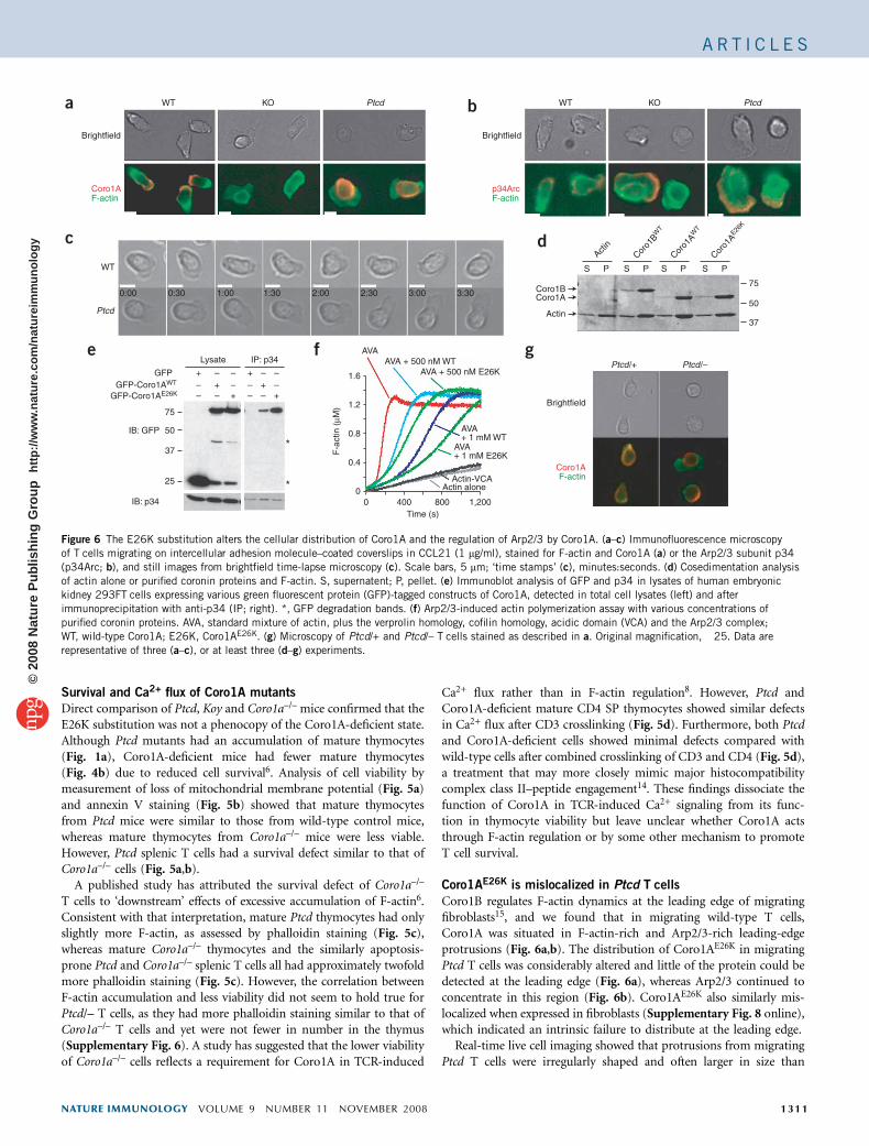
Coro1AE26K is mislocalized in Ptcd T cells
Coro1B regulates F-actin dynamics at the leading edge of migratingfibroblasts15, and we found that in migrating wild-type T cells,Coro1A was situated in F-actin-rich and Arp2/3-rich leading-edgeprotrusions (Fig. 6a,b). The distribution of Coro1AE26K in migratingPtcd T cells was considerably altered and little of the protein could bedetected at the leading edge (Fig. 6a), whereas Arp2/3 continued toconcentrate in this region (Fig. 6b). Coro1AE26K also similarly mis-localized when expressed in fibroblasts (Supplementary Fig. 8 online),which indicated an intrinsic failure to distribute at the leading edge.
Real-time live cell imaging showed that protrusions from migratingPtcd T cells were irregularly shaped and often larger in size than
WT
Brightfield
Coro1AF-actin
Brightfield
p34ArcF-actin
KO Ptcd
WT
Ptcd
WT KO Ptcd
0:00 0:30 1:00 1:30 2:00 2:30 3:00 3:30
S
Actin
Coro1
BW
T
Coro1
AW
T
Coro1
AE26
K
P S P S P S P
Coro1BCoro1A
75
50
37Actin
75
GFP-Coro1AE26KGFP-Coro1AWT
GFP
––+
Lysate IP: p34
–+–
+––
––+
–+–
+––
50
*
*
IB: GFP
IB: p34
37
25
0 400Time (s)
800 1,2000
0.4
0.8
1.2
1.6
F-a
ctin
(µM
)
Actin-VCAActin alone
AVA+ 1 mM E26K
AVA+ 1 mM WT
AVA + 500 nM E26KAVA + 500 nM WT
AVA
Ptcd/+ Ptcd/–
Brightfield
Coro1AF-actin
a
c
e f g
b
d
Figure 6 The E26K substitution alters the cellular distribution of Coro1A and the regulation of Arp2/3 by Coro1A. (a–c) Immunofluorescence microscopy
of T cells migrating on intercellular adhesion molecule–coated coverslips in CCL21 (1 mg/ml), stained for F-actin and Coro1A (a) or the Arp2/3 subunit p34
(p34Arc; b), and still images from brightfield time-lapse microscopy (c). Scale bars, 5 mm; ‘time stamps’ (c), minutes:seconds. (d) Cosedimentation analysis
of actin alone or purified coronin proteins and F-actin. S, supernatent; P, pellet. (e) Immunoblot analysis of GFP and p34 in lysates of human embryonic
kidney 293FT cells expressing various green fluorescent protein (GFP)-tagged constructs of Coro1A, detected in total cell lysates (left) and after
immunoprecipitation with anti-p34 (IP; right). *, GFP degradation bands. (f) Arp2/3-induced actin polymerization assay with various concentrations of
purified coronin proteins. AVA, standard mixture of actin, plus the verprolin homology, cofilin homology, acidic domain (VCA) and the Arp2/3 complex;
WT, wild-type Coro1A; E26K, Coro1AE26K. (g) Microscopy of Ptcd/+ and Ptcd/– T cells stained as described in a. Original magnification, �25. Data are
representative of three (a–c), or at least three (d–g) experiments.
NATURE IMMUNOLOGY VOLUME 9 NUMBER 11 NOVEMBER 2008 1311
A R T I C L E S©
2008
Nat
ure
Pub
lishi
ng G
roup
ht
tp://
ww
w.n
atur
e.co
m/n
atur
eim
mun
olog
y

protrusions from wild-type cells, consistent with a failure toproperly regulate the actin cytoskeleton (Fig. 6c and SupplementaryMovies 2–4 online). We noted similarly dysmorphic protrusionsfrom Ptcd/– compound mutant T cells (Supplementary Fig. 6d).The short in vitro survival of Coro1A-deficient T cells madeassessments difficult, but these cells seemed more severely compro-mised in their migration than Ptcd T cells were (SupplementaryMovie 5 online). These results collectively establish that Coro1AE26K
fails to distribute correctly in migrating T cells and that the cells haveaberrant shape regulation.
Coro1AE26K enhances Arp2/3 inhibition
To understand the biochemical effect of the E26K substitution, we firstassessed the ability of Coro1A to bind F-actin. In cosedimentationassays, Coro1AE26K associated normally with F-actin (Fig. 6d). Incontrast, Arp2/3 coimmunoprecipitated more Coro1AE26K than wild-type Coro1A from cell lysates (Fig. 6e). The corresponding substitu-tion in Coro1B also resulted in more association with Arp2/3(Supplementary Fig. 9 online). Consistent with more interactionwith Arp2/3, Coro1AE26K was more efficient than wild-type Coro1A ininhibiting Arp2/3 activity in actin-polymerization assays (Fig. 6f).
To examine how this biochemical gain-of-function substitutionmanifested as a recessive Ptcd allele, we analyzed T cells from Ptcd/+and Ptcd/– mice. As expected, the Coro1A distribution in Ptcd/–T cells (Fig. 6g) was similar to that in Ptcd/Ptcd T cells (Fig. 6a). InPtcd/+ T cells, Coro1A was both present at and mislocalized awayfrom the leading edge (Fig. 6g). These results suggest that in Ptcd/+cells, wild-type Coro1A can access the leading edge to regulate actindynamics. In summary, these biochemical studies indicate that thehighly conserved Glu26 residue in Coro1A is important for theregulation of Arp2/3.
Coro1A is deficient in a T–B+NK+ patient with SCID
After finding that point mutations in Coro1a resulted in severeT lymphopenia in both Ptcd and Koy mice, we sought to determinewhether CORO1A mutations could also be responsible for humancases of primary T cell immunodeficiency with undefined etiologies.Thus, we sequenced CORO1A in DNA from 16 SCID patients lackingmutations in previously recognized genes associated with T–B+NK+
SCID. Although we failed to detect any CORO1A mutations in 15samples, we did identify a single patient with a two-nucleotidedeletion in exon 3 (deletion of CT between nucleotides 248 and 249
in the coding DNA sequence; Fig. 7a) that resulted in a frame shift andpremature stop codon in the area encoding the Coro1A b-propellerdomain (with the first amino acid affected by the frame shift being asubstitution of arginine for proline at position 83, and the creation ofa new reading frame that ended at the first stop codon at position 10(with arginine now amino acid 1)). Consistent with a destabilizingtruncation, immunoblot analysis of Epstein Barr virus (EBV)-immor-talized B lymphoid cell lines derived from the patient’s peripheralblood showed an absence of Coro1A protein (Fig. 7b). When thispatient was 13 months old, she was hospitalized with a severe varicellainfection after vaccination; she was later diagnosed as an atypicalT–B+NK+ patient with SCID and was treated by allogeneic bonemarrow transplantation. We found that her father was heterozygousfor the two–base pair deletion in CORO1A but her mother and sisterwere not (Fig. 7a).
To assess the gene-copy number of CORO1A in the patient, we didquantitative PCR of genomic DNA with nonoverlapping primersspanning intron 1 through exon 11 of CORO1A and noted 50% lessproduct relative to that of a healthy control (Fig. 7c). In contrast,signals for the human genes encoding S1P1, on chromosome 1, andcoronin 7, located telomeric to CORO1A on chromosome 16p, didnot differ from the quantitative PCR signal in control cells. Along withreports of copy-number variation in chromosome 16p11.2, whereCORO1A is located16–18, these data suggest that our patient, whoinherited a two–base pair paternal deletion in one allele of CORO1A,also has deletion of the other CORO1A allele.
DISCUSSION
Using a forward-genetics approach, we have identified a uniqueCoro1a allele that allowed us to establish a requirement for thisactin-regulatory protein in the egress of T cells from the thymusand lymph nodes. Two-photon microscopy showed that Coro1A wasneeded for normal T cell motility in the lymph node, and in vitromotility studies indicated its involvement in regulating cell shape.Moreover, this mutation identified a site on Coro1A critical for Arp2/3regulation and for controlling Coro1A distribution in the cell. Finally,we demonstrated that Coro1A deficiency in a human was associatedwith a peripheral T cell deficiency of sufficient severity to causeT–B+NK+ SCID.
The identification of a critical function for Coro1A in egress fromthe thymus and lymph nodes provides evidence that migration fromthese tissues depends on proper regulation of Arp2/3-mediated actinbranching, most likely in response to S1P. The protein mDia1, amember of the formin family of actin-nucleating proteins thatpromote the formation of unbranched actin filaments, is also neededfor thymic egress19. Thus, the formation of both correctly branched aswell as unbranched actin networks seems to be required for exit from
b
c
aFatherMother
SCID patientSibling
Mother
C A G C C C C T G T G C
Sibling
C A G C C C C T G T G C
SCIDpatient
C A G C C C G T G C T A
FatherC A G C C C C T G T G CC A G C C C G T G C T A
0
1
2
S1PR1
CORO7
CORO1A primer sets
Normal controlSCID patient
Intro
n 1–
2
Intro
n 2–
3
Intro
n 6–
7
Exon
11
P = 0.11P = 0.07
P < 0.01P < 0.01
P < 0.03
P < 0.03
Gen
omic
sig
nal
(nor
mal
ized
to G
AP
DH
)
Actin
Coro1A
Norm
al co
ntro
l
SCID p
atien
t
Contro
l SCID
198
0
Contro
l SCID
878
5 Figure 7 Deficiency of Coro1A in a T–B+NK+ patient with SCID. (a) Family
pedigree (top) and dideoxy sequence tracings (below) of a T–B+NK+ patient
with SCID and family members. Red arrow indicates the location of the
deletion of CT between nucleotides 248 and 249 in the coding DNA
sequence in the patient that led to a premature truncation after ten mis-
sense codons; the father is heterozygous for this (that tracing includes both
the mutated (top) and normal (bottom) copies). Red outlines enclose the CT
nucleotides missing in the patient and father. (b) Immunoblot analysis ofCoro1A and actin in EBV-transformed B cell lines derived from various
subjects. Data are representative of three experiments. (c) Quantitative PCR
signals from primer sets of human genes encoding S1P1 (S1PR1), coronin 7
(CORO7) and Coro1a (CORO1A) from buccal swabs of genomic DNA from
the patient with SCID and a normal control. Bars indicate means; circles
indicate separate PCR reactions. Data are from three experiments.
1312 VOLUME 9 NUMBER 11 NOVEMBER 2008 NATURE IMMUNOLOGY
A R T I C L E S©
2008
Nat
ure
Pub
lishi
ng G
roup
ht
tp://
ww
w.n
atur
e.co
m/n
atur
eim
mun
olog
y

this organ. The function of mDia1 in lymph node egress has not beenassessed. The small G proteins Rac and Cdc42 induce activation of theWiskott-Aldrich syndrome protein–Arp2/3 system and, consistentwith our results here, the Rac activator Dock2 is needed for lymphnode egress20. Although the function of Dock2 in thymic egress hasnot been determined, given the need for Coro1A, we anticipate thatDock2 is also involved. Lymphocyte protrusions initiate diapedesis21,and our findings suggest that Coro1A-dependent control of actin maybe critical in the formation of protrusions during T cell egress.
The requirement for Arp2/3-mediated actin branching for T cell–migration responses to chemoattractants has been supported byfindings obtained with cells deficient in Dock2, Wiskott-Aldrichsyndrome protein and Wiskott-Aldrich syndrome protein–interactingprotein20,22,23 as well as Coro1A-deficient cells6,7. Our results obtainedwith Ptcd T cells add to those findings and suggest an importantfunction for Coro1A-mediated Arp2/3 regulation in the control ofT cell shape. A published study has argued that F-actin dynamics areunaltered in Coro1A-deficient T cells, although cell migration inresponse to chemoattractants was not examined8. The basis forthese discrepant findings is unclear, but it seems possible that thevery poor survival of Coro1A-deficient naive T cells and theirconsequent depletion from T cell preparations relative to effectorand/or memory T cells confounded some of the comparisons. In ourstudies here, we were careful to exclude both dead cells and effector ormemory cells from our analysis of naive T cells. Although themigration of Ptcd T cells was much lower in vitro and in the lymphnode, it was not absent, and this might explain the continued ability ofthe Ptcd mouse thymus to organize into cortex and medulla3. It seemslikely that the lack of notable defects in Ptcd and Coro1A-deficientB cells is due to redundancy with other coronin family members, andthis adds to other data establishing differences in requirements forB cell and T cell motility19,24,25. Whether substitutions in Coro1A orother actin-regulatory molecules cause defects in common lymphoidprogenitors remains to be determined. Although bone marrow chi-mera–reconstitution efficiencies were similar for Ptcd and controldonors, our studies did not formally address whether the entry of earlythymic precursors is impaired. Future studies should include com-petition assays with mixed–bone marrow chimeras to evaluate theproportion of Ptcd-derived cells in bone marrow and thymus.Leukocyte migration in lymphoid tissue has been described as phasesof force-generating actin protrusions followed by myosin-dependentcontractions26 that aid the squeezing of the cell nucleus through three-dimensional matrices. In this context, it is notable that Ptcd T cells notonly had large and dysmorphic protrusions but also failed to elongatetheir nuclei. Thus, remodeling of the actin cytoskeleton by Coro1Amay contribute to both protrusion and contraction during themigration of T cells through lymphoid tissues.
The mechanism by which coronin proteins inhibit Arp2/3-mediated actin branching is complex and incompletely understood.Although several studies have shown that coronin proteins are able todirectly bind Arp2/3 and possibly inhibit function before F-actinbinds27,28, work with Coro1B in fibroblasts suggests that it acts bydisplacing Arp2/3 from actin branch points29. We have demonstratedthat a Coro1A surface proximal to the predicted F-actin-bindingregion13 was involved in Arp2/3 regulation. Our findings emphasizethe importance of characterizing the highly conserved Glu26-containing surface of the type I coronin subfamily in future struc-ture-function studies focusing on how an oligomeric coronin complexdislodges Arp2/3 from F-actin. The failure of Coro1AE26K to localizenormally to the leading edge of migrating cells has identified what maybe another level of coronin regulation. Further studies are needed to
determine whether interactions with the cofilin phosphatase slingshot5
or other still-unknown coronin-binding partners are altered bythe substitution.
Coro1A deficiency caused a substantial defect in T cell survival, butthe basis for this effect is not yet resolved. Although the Ptcd is animportant allelic comparison that ‘disconnected’ the survival andCa2+-flux defects, the greater phalloidin staining of Ptcd/– maturethymocytes also failed to support the idea of a simple correlationbetween survival and F-actin accumulation. The possibility remainsthat Coro1A directly affects cell survival independently of effects onF-actin, and analysis of additional Coro1A substitutions may yieldnew allelic insights. The continued ability of Ptcd thymocytes tomaintain nearly normal total cellular F-actin despite mislocalizationof Coro1A might be explained by an ongoing association of low butsufficient amounts of Coro1AE26K to achieve partial regulation ofArp2/3 at the leading edge, or it might reflect correspondingly greateractin-debranching and depolymerization in the cell body. Alterna-tively, the in vitro assay for examining colocalization of Arp2/3 andCoro1A may not accurately reflect how these proteins distributein vivo in migrating thymocytes. The similar partial deficiencies inTCR-induced Ca2+ flux in Coro1AE26K and Coro1a–/– T cells are inagreement with emerging evidence that an intact actin cytoskeleton isimportant for Ca2+ flux30. However, our finding that CD3-CD4 co-crosslinking led to equivalent Ca2+ flux in wild-type, Coro1AE26K andCoro1a–/– thymocytes, along with a published report of normalproliferation of Coro1a–/– T cells stimulated by antigen-presentingcells6, suggests that Coro1A may have a limited function in TCRsignaling during encounters with cognate antigen. We anticipate thatsingle-cell analysis of the release of Ca2+ stores and the activity of Ca2+
release–activated channels in Ptcd and Coro1A-deficient mature thy-mocytes and T cells, along with similar studies of T cells with otherdefects in actin-regulatory proteins, will add to the understanding ofhow the actin cytoskeleton contributes to calcium signaling.
An important challenge of studies of mouse models is ‘translation’of the findings to humans. The identification of a patient with aperipheral T cell deficiency who lacks expression of Coro1A suggeststhat Coro1A has similar functions in humans and mice. Although thehuman X-linked Wiskott-Aldrich syndrome has long associated actinbiology with hematopoietic cell dysfunction, the Coro1A-deficientpatient we studied here allowed us to link regulation of the actincytoskeleton with SCID31. T–B+NK+ SCID has been defined before bymutations resulting in impaired TCR or interleukin 7 receptorsignaling32. Other forms of SCID involve defects in purine metabo-lism, DNA recombination or cytokine signaling33. Mutations in genesinvolved in these pathways result in apoptosis through purine toxicity,deficient cytokine signaling or failure to rearrange a signaling-compe-tent antigen receptor. Given the results of our study of Coro1Adeficiency, we can also include apoptosis via actin dysregulation as acause of SCID. A report classifying a Coro1a-null allele as an auto-immune-suppression locus in mice7 is consistent with the idea thatCoro1A defects cause immunodeficiency. We predict that substitu-tions in other hematopoietic cell–specific actin-associated moleculeswill be found to cause SCID. Because the pericentromeric segment ofhuman chromosome 16 that contains CORO1A is prone to copy-number variation16–18, we also anticipate that additional cases ofpatients with lower T cell function will involve CORO1A mutations.
METHODSMice, ENU mutagenesis and bone marrow chimeras. B6 and B6-CD45.1 mice
were from the Jackson Laboratories, the National Cancer Institute or a colony
maintained at the University of California, San Francisco. UBC-GFP mice were
NATURE IMMUNOLOGY VOLUME 9 NUMBER 11 NOVEMBER 2008 1313
A R T I C L E S©
2008
Nat
ure
Pub
lishi
ng G
roup
ht
tp://
ww
w.n
atur
e.co
m/n
atur
eim
mun
olog
y

from Jackson Laboratories. Cataract-Shionogi (CTS/Shi) mice were from TGC.
Ptcd was backcrossed six to ten generations onto the B6 strain with single-
nucleotide polymorphism (SNP) markers as described below. Coro1a–/– mice,
which were backcrossed ten generations with B6 mice, were generated as
described6. Koy mice were identified by an ENU-mutagenesis screen done as
described34 with B6 mice in a colony maintained at the Australian National
University. Male B6 mice were treated with three doses, 1 week apart, of 100 mg
ENU (Sigma) per kg body weight in 10% (vol/vol) ethanol in citrate buffer,
pH 5.0, to produce G0 mice. After a refractory period of 8 weeks, these G0
males were mated with B6 females to generate G1 offspring. Two unrelated
G1 mice were crossed, producing G2 mice, which were then intercrossed to yield
G3 mice. Screening of these G3 mice by flow cytometry identified the Koy
phenotype. All mice were housed in specific pathogen–free conditions, and all
protocols were approved by the Institutional Animal Care and Use Committee
of the University of California, San Francisco, or the Animal Experimentation
Ethics Committee of Australian National University. Bone marrow chimeras
were generated with 2 � 106 to 3 � 106 bone marrow cells from donor mice
transplanted intravenously into lethally irradiated congenic host mice.
Chimeras were given prophylactic water containing antibiotics and were
analyzed 8–12 weeks after transplantation.
Flow cytometry, intracellular staining and measurement of Ca2+ flux.
Polyclonal antibody to S1P1 (anti-S1P1) has been described35. Phalloidin–
fluorescein isothiocyanate was from Sigma. Indo-1 was from Invitrogen. Kits
for intracellular staining and annexin V staining were from BD Biosciences. The
JC-1 mitochondrial membrane potential assay kit was from Cayman. All other
antibodies used for flow cytometry were from BD Biosciences, Invitrogen,
eBiosciences or BioLegend. Ca2+-flux assays were adapted from published
methods for thymocytes14. Cells were loaded for 30 min at 30 1C with Indo-1 in
the presence of biotinylated anti-CD3 (2C11; BD), fluorescein isothiocyanate–
anti-CD8 (53-6.7; BD) and Alexa Fluor 647–anti-CD4 (M4-5; BD). Thymo-
cytes were additionally stained with phycoerythrin–anti-CD62L (Mel14; BD)
and splenocytes were additionally stained with phycoerythrin–anti-CD44 (IM7;
BD). Cells were washed and kept on ice until analysis. For crosslinking of CD3
and CD4, cells were additionally pretreated for 5 min with biotinylated anti-
CD4 (RM4-4; BD). Cells were mixed with prewarmed media containing
propidium iodide for exclusion of dead cells and were warmed to 37 1C.
Baseline Ca2+ concentrations were assessed for 30 s before crosslinking with
streptavidin (50 mg/ml; Pierce). All flow cytometry was done on a FACSCalibur
or an LSRII (BD).
Purification of recombinant protein. Coronin recombinant protein was
expressed and purified with a mammalian expression system as described
for Coro1B15. Purified protein was quantified by absorbance at 280 nm and
the predicted extinction coefficients of 62,910 M–1cm–1 (Coro1A) and
65,890 M–1cm–1 (Coro1B).
Immunoprecipitation. Cells were washed twice with PBS and were lysed with a
KCl buffer (20 mM HEPES, pH 7.0, 100 mM KCl, 0.5% (vol/vol) Nonidet P-40,
1 mM EDTA, 1 mM phenylmethylsulfonyl fluoride, 10 mg/ml of 1,10-phenan-
throline, 10 mg/ml of aprotinin, 10 mg/ml of leupeptin, 10 mM sodium fluoride
and 2 mM sodium orthovanadate). Lysates were cleared for 5 min at 13,000g
and were incubated with 0.5 mg primary antibody for 16 h at 4 1C, followed by
the addition of 20 ml of a slurry of ImmunoPure-immobilized protein A/G
beads (Pierce) and further incubation for an additional hour at 4 1C. Immune
complexes were collected, were washed three times with KCl buffer, were
separated by SDS-PAGE and were transferred to a polyvinylidene difluoride
membranes (Bio-Rad) for immunoblot analysis.
Actin-assembly assay. Arp2/3-nucleation reactions induced by the verprolin
homology, cofilin homology, acidic domain were done as described15: recom-
binant coronin and Arp2/3 (20 nM) were mixed in MKEI-50 buffer (20 mM
imidazole-KOH, pH 7.0, 50 mM KCl, 2 mM MgCl2 and 1 mM EGTA) and were
incubated for 5 min at 25 1C; reactions were initiated by the simultaneous
addition of 1.5 mM actin (5% labeled with pyrene, primed for 90 s with 1 mM
EGTA and 0.1 mM MgCl2) and 1 nM glutathione S-transferase–verprolin
homology, cofilin homology, acidic domain. The delay between mixing of
reactants and recording of fluorescence was 15 s. Fluorescence was converted to
the molar concentration of F-actin according to the fluorescence of completely
polymerized actin (24 h after reaction) and unpolymerized actin, with the
assumption a critical concentration of 0.1 mM.
Immunoblot analysis. Lysates from mouse thymocytes and human EBV-
immortalized cell lines were prepared in radioimmunoprecipitation buffer
(1% (vol/vol) Nonidet P-40, 0.5% (wt/vol) sodium deoxycholate, 0.1% (vol/
vol) SDS, 50 mM Tris, pH 8.0, 150 mM NaCl and 0.02 NaN3) and were resolved
by PAGE (NuPage; Invitrogen), then were transferred (XCell; Invitrogen) to
Immobilon-FL membranes (Millipore), were blocked and probed with LICOR
buffer and were analyzed on an Odyssey infrared imager (LICOR). Anti-Coro1A
(07-493) was from Upstate Biotechnology, anti-actin (A2066) was from Sigma,
and secondary IRDye antibody (611-730-127) was from Rockland.
Transwell migration. Thymocytes and splenocytes collected in migration
medium (RPMI medium (Cellgro) supplemented with 10 mM HEPES,
pH 7.2 (Cellgro), 100 U/ml of penicillin-streptomycin (Cellgro) and 0.5%
(vol/vol) fatty acid–free BSA (Calbiochem)) were used for assays of migration
through Transwells (5 mm; Corning). For analysis of migration to S1P,
thymocytes were assayed immediately after isolation. For analysis of migration
to chemokines, thymocytes or splenocyte samples in which erythrocytes were
lysed were resensitized in migration media for 30 min at 37 1C. S1P (Sigma),
the human chemokine SDF-1a (Peprotech) and mouse CCL21 and CXCL13
(R&D Systems) were used at various concentrations.
Adoptive cell transfer and treatment. Thymocytes were labeled with the
cytosolic dye CFSE (carboxyfluorescein diacetate succinimidyl diester) or
CellTracker orange CMTMR ((5-(and-6)-(((4-chloromethyl)benzoyl)amino)-
tetramethylrhodamine); Molecular Probes) as described36. For adoptive transfer,
mice were injected intravenously with about 1 � 107 to 3 � 107 cells in about
0.3 ml media. For homing assays, mice were analyzed 1 h after transfer. For
assays of peripheral egress, mice were analyzed 24 h after transfer or were treated
intravenously for 20 h with entry-blocking antibodies and were analyzed after
44 h. Antibodies used for in vivo entry blockade have been described37.
Two-photon microscopy. Thymocytes from donor mice were isolated and were
labeled with CellTracker orange CMTMR (Invitrogen-Molecular Probes) as
described38, then were injected intravenously into B6 recipient mice along with
thymocytes from UBC-GFP mice. After 12–24 h, inguinal lymph nodes were
isolated for two-photon microscopy, and axillary and brachial lymph nodes
were isolated for flow cytometry. Flow cytometry confirmed that over 90% of
CMTMR+ cells in lymph nodes were mature SP thymocytes (CD4+CD8–
CD69loCD62Lhi or CD4–CD8+CD69loCD62Lhi). Explants were prepared as
described39. Inguinal lymph nodes were perfused with RPMI medium (Cellgro)
at 36–37 1C, were aerated with 95% O2 and 5% CO2 and were imaged from the
cortical side of the lymph node through the capsule. A ‘custom’ two-photon
microscope system was used for imaging, with the 5-W MaiTai TiSapphire laser
(Spectra-Physics) tuned to an excitation wavelength of 890 nm. Images were
captured with Video Savant software (IO Industries). For time-lapse imagery,
30–36 planes 3 mm apart were collected every 20 s. Each xy plane spanned
240 mm � 288 mm at a resolution 0.6 mm per pixel. Data were further processed
with MetaMorph software (Molecular Devices). Imaris software (Bitplane AG)
was used for three-dimensional volume rendering and cell tracking. Matlab
software was further used to calculate cell velocities, turning angles and
displacement. Statistical significance was evaluated with a paired two-tailed
Student’s t-test.
Genetic mapping, SNP analysis, human samples and sequencing. CTS/Shi
mice were crossed with B6 mice to generate an F1 generation. The Ptcd locus
was mapped with mutant F2 progeny of F1 intercrosses, F1 mice backcrossed to
nonparental CTS mice, or mutant F2 mice crossed with nonparental F1 mice.
Mice were phenotyped by analysis of thymocyte subset frequencies by flow
cytometry. Genomic DNA was isolated from tail tissue (Wizard Genomic kit;
Promega) and the Amplifluor HT kit (Chemicon, Millipore) and an ABI7300
(Applied Biosystems) were used according to the manufacturers’ instructions
for SNP reactions. Mapping SNPs were selected on the basis of polymorphisms
between B6 mice and BUB/J, a strain closely related to CTS/Shi. SNP primers
were designed on the AssayArchitect website. Patients with SCID and their
1314 VOLUME 9 NUMBER 11 NOVEMBER 2008 NATURE IMMUNOLOGY
A R T I C L E S©
2008
Nat
ure
Pub
lishi
ng G
roup
ht
tp://
ww
w.n
atur
e.co
m/n
atur
eim
mun
olog
y

parents, where available, were enrolled with informed content in studies with
Institutional Review Board–approved protocols for determination of genotype
and genotype-phenotype correlations. Blood samples were immortalized with
EBV to create B cell lines. Human genomic DNA was isolated from buccal
swabs or EBV-immortalized lines with a Puregene kit (Gentra Systems). An
ABI7300 was used for quantitative PCR of genomic DNA. Human CORO1A
and mouse Coro1a exons and surrounding splice regions were amplified by
PCR from genomic DNA, were resequenced and were analyzed on 4Peaks
software (mekentosj) relative to the reference human genomic and cDNA
sequences NM_007074 and NM_007074.2, respectively (primers, Supplemen-
tary Table 1 online).
Brightfield and immunofluorescence microscopy. For fixed immunofluores-
cence imaging, 1 � 106 to 2 � 106 lymphocytes from peripheral and mesenteric
lymph nodes were seeded onto chambered coverslips (Lab-tek, Nunc) pre-
coated with recombinant mouse intercellular adhesion molecule 1–Fc fragment
(10 mg/ml; R&D Systems) and were allowed to settle and migrate for 1 h at
37 1C in the presence of CCL21 (1 mg/ml; R&D Systems) in RPMI medium
(Cellgro) supplemented with 1% (vol/vol) FCS (Invitrogen), 10 mM HEPES,
pH 7.2 (Cellgro) and 100 U/ml of penicillin-streptomycin. Cells were then fixed
with 4% (wt/vol) paraformaldehyde (Sigma) and were made permeable with
0.5% (vol/vol) Triton-X100 (Sigma) in PBS. Coverslips were then stained with
a combination of the following: anti-Coro1A (rabbit polyclonal antibody;
07-493; Millipore, Upstate) or anti-p34Arc (rabbit polyclonal antibody to the
Arp2/3 subunit p34; 07-227; Millipore; Upstate), fluorescein isothiocyanate–
conjugated phalloidin (Sigma), Alexa Fluor 647–conjugated anti-CD4 (RM4-5;
BD), Alexa Fluor 647–conjugated anti-CD8 (53-6.7; BD), biotinylated
goat anti-rabbit (BD), streptavidin-indocarbocyanine (Jackson Immuno) or
4,6-diamidino-2-phenylindole (Molecular Probes). The z-stacks were captured
on an Axiovert Z1 (Zeiss) and were deconvoluted with Axiovision Software
(Zeiss). For real-time imaging, 5 � 105 lymphocytes from peripheral and
mesenteric lymph nodes were stained with phycoerythrin-conjugated anti-
CD19 (1D3; BD) and Alexa Fluor 647–conjugated anti-CD4 and anti-CD8 and
were seeded on intercellular adhesion molecule–coated chambered coverslips
with CCL21 as described above with the addition of 0.1% (wt/vol) NuSieve
GTG Low Melt agarose (FMC BioProducts) and 4,6-diamidino-2-phenylindole.
Cells were maintained at 37 1C and 5% CO2 during the 3–4 h of imaging in an
environmentally controlled stage on an Axiovert Z1. Each 5-minute movie was
generated at time-lapse intervals of 10 s, with assembly of a z-stack at each time
point from six brightfield xy views with a z-step of 0.5 mm. After the time lapse,
a fluorescence image was captured to identify live T cells. Movies and images
were processed with Axiovision (Zeiss) and were exported for annotation and
splicing with AfterEffects (Adobe).
Note: Supplementary information is available on the Nature Immunology website.
ACKNOWLEDGMENTSWe thank the patient with SCID and her family; M. Anderson, P. Beemiller,S. Cheung, G. Cinamon, M. Krummel, T. Phan, H. Phee and A. Weiss fordiscussions; and D. Schafer (University of Virginia) for advice and reagentsrelated to the pyrene actin assay. Supported by the University of California, SanFrancisco Medical Scientist Training Program (L.R.S.), Genentech SandlerFamily Foundation (L.R.S.), the US Immunodeficiency Network (J.M.P.), theJeffrey Modell Foundation (J.M.P.), the Howard Hughes Medical Institute(J.G.C.) and the National Institutes of Health (C.C.G., J.E.B. and J.G.C.).
Published online at http://www.nature.com/natureimmunology/
Reprints and permissions information is available online at http://npg.nature.com/
reprintsandpermissions/
1. Schwab, S.R. & Cyster, J.G. Finding a way out: lymphocyte egress from lymphoidorgans. Nat. Immunol. 8, 1295–1301 (2007).
2. Ohtori, H., Yoshida, T. & Inuta, T. ‘‘Small eye and cataract,’’ a new dominant mutation.Exp. Anim. 17, 91–96 (1968).
3. Yagi, H. et al. Defect of thymocyte emigration in a T cell deficiency strain (CTS) of themouse. J. Immunol. 157, 3412–3419 (1996).
4. Ikegami, H., Makino, S., Harada, M., Eisenbarth, G.S. & Hattori, M. The cataractShionogi mouse, a sister strain of the non-obese diabetic mouse: similar class II butdifferent class I gene products. Diabetologia 31, 254–258 (1988).
5. Uetrecht, A.C. & Bear, J.E. Coronins: the return of the crown. Trends Cell Biol. 16,421–426 (2006).
6. Foger, N., Rangell, L., Danilenko, D.M. & Chan, A.C. Requirement for coronin1 in T lymphocyte trafficking and cellular homeostasis. Science 313, 839–842(2006).
7. Haraldsson, M.K. et al. The lupus-related Lmb3 locus contains a disease-suppressingCoronin-1A gene mutation. Immunity 28, 40–51 (2008).
8. Mueller, P. et al. Regulation of T cell survival through coronin-1-mediated generation ofinositol-1,4,5-trisphosphate and calcium mobilization after T cell receptor triggering.Nat. Immunol. 9, 424–431 (2008).
9. Kimura, S. et al. Genetic control of peripheral T-cell deficiency in the cataractShionogi (CTS) mouse linked to chromosome 7. Immunogenetics 47, 278–280(1998).
10. Ohotori, H., Yoshida, T. & Inuta, T. Small eye and cataract, a new dominant mutation inthe mouse. Jikken Dobutsu 17, 91–96 (1968).
11. Nal, B. et al. Coronin-1 expression in T lymphocytes: insights into proteinfunction during T cell development and activation. Int. Immunol. 16, 231–240(2004).
12. Appleton, B.A., Wu, P. & Wiesmann, C. The crystal structure of murine coronin-1: aregulator of actin cytoskeletal dynamics in lymphocytes. Structure 14, 87–96 (2006).
13. Cai, L., Makhov, A.M. & Bear, J.E. F-actin binding is essential for coronin 1B functionin vivo. J. Cell Sci. 120, 1779–1790 (2007).
14. Maltzman, J.S., Kovoor, L., Clements, J.L. & Koretzky, G.A. Conditional deletion revealsa cell-autonomous requirement of SLP-76 for thymocyte selection. J. Exp. Med. 202,893–900 (2005).
15. Cai, L., Marshall, T.W., Uetrecht, A.C., Schafer, D.A. & Bear, J.E. Coronin 1Bcoordinates Arp2/3 complex and cofilin activities at the leading edge. Cell 128,915–929 (2007).
16. Ghebranious, N., Giampietro, P.F., Wesbrook, F.P. & Rezkalla, S.H. A novel microdele-tion at 16p11.2 harbors candidate genes for aortic valve development, seizure disorder,and mild mental retardation. Am. J. Med. Genet. A. 143, 1462–1471 (2007).
17. Weiss, L.A. et al. Association between microdeletion and microduplication at 16p11.2and autism. N. Engl. J. Med. 358, 667–675 (2008).
18. Kumar, R.A. et al. Recurrent 16p11.2 microdeletions in autism. Hum. Mol. Genet. 17,628–638 (2008).
19. Sakata, D. et al. Impaired T lymphocyte trafficking in mice deficient in an actin-nucleating protein, mDia1. J. Exp. Med. 204, 2031–2038 (2007).
20. Nombela-Arrieta, C. et al. A central role for DOCK2 during interstitial lymphocytemotility and sphingosine-1-phosphate-mediated egress. J. Exp. Med. 204, 497–510(2007).
21. Carman, C.V. et al. Transcellular diapedesis is initiated by invasive podosomes.Immunity 26, 784–797 (2007).
22. Gallego, M.D. et al. WIP and WASP play complementary roles in T cell homing andchemotaxis to SDF-1a. Int. Immunol. 18, 221–232 (2006).
23. Snapper, S.B. et al. WASP deficiency leads to global defects of directed leukocytemigration in vitro and in vivo. J. Leukoc. Biol. 77, 993–998 (2005).
24. Reif, K. et al. Cutting edge: differential roles for phosphoinositide 3-kinases, p110gand p110d, in lymphocyte chemotaxis and homing. J. Immunol. 173, 2236–2240(2004).
25. Nombela-Arrieta, C. et al. Differential requirements for DOCK2 and phosphoinositide-3-kinase g during T and B lymphocyte homing. Immunity 21, 429–441 (2004).
26. Lammermann, T. et al. Rapid leukocyte migration by integrin-independent flowing andsqueezing. Nature 453, 51–55 (2008).
27. Humphries, C.L. et al. Direct regulation of Arp2/3 complex activity and function by theactin binding protein coronin. J. Cell Biol. 159, 993–1004 (2002).
28. Rodal, A.A. et al. Conformational changes in the Arp2/3 complex leading to actinnucleation. Nat. Struct. Mol. Biol. 12, 26–31 (2005).
29. Cai, L., Makhov, A.M., Schafer, D.A. & Bear, J.E. Coronin 1B antagonizes cortactin andremodels Arp2/3-containing actin branches in lamellipodia. Cell (in the press)(2008).
30. Gallo, E.M., Cante-Barrett, K. & Crabtree, G.R. Lymphocyte calcium signaling frommembrane to nucleus. Nat. Immunol. 7, 25–32 (2006).
31. Puck, J.M. & Candotti, F. Lessons from the Wiskott-Aldrich syndrome. N. Engl. J. Med.355, 1759–1761 (2006).
32. Fischer, A. Human primary immunodeficiency diseases. Immunity 27, 835–845(2007).
33. Buckley, R.H. The multiple causes of human SCID. J. Clin. Invest. 114, 1409–1411(2004).
34. Nelms, K.A. & Goodnow, C.C. Genome-wide ENU mutagenesis to reveal immuneregulators. Immunity 15, 409–418 (2001).
35. Lo, C.G., Xu, Y., Proia, R.L. & Cyster, J.G. Cyclical modulation of sphingosine-1-phosphate receptor 1 surface expression during lymphocyte recirculation and relation-ship to lymphoid organ transit. J. Exp. Med. 201, 291–301 (2005).
36. Shiow, L.R. et al. CD69 acts downstream of interferon-a/b to inhibit S1P1 andlymphocyte egress from lymphoid organs. Nature 440, 540–544 (2006).
37. Pham, T.H., Okada, T., Matloubian, M., Lo, C.G. & Cyster, J.G. S1P1 receptor signalingoverrides retention mediated by Gai–coupled receptors to promote T cell egress.Immunity 28, 122–133 (2008).
38. Allen, C.D., Okada, T., Tang, H.L. & Cyster, J.G. Imaging of germinal center selectionevents during affinity maturation. Science 315, 528–531 (2007).
39. Okada, T. et al. Antigen-engaged B cells undergo chemotaxis toward the T zone andform motile conjugates with helper T cells. PLoS Biol. 3, e150 (2005).
NATURE IMMUNOLOGY VOLUME 9 NUMBER 11 NOVEMBER 2008 1315
A R T I C L E S©
2008
Nat
ure
Pub
lishi
ng G
roup
ht
tp://
ww
w.n
atur
e.co
m/n
atur
eim
mun
olog
y