Structural validity and item functioning of the LoTi digital-age ...
ORIGINAL RESEARCH ARTICLEpublished: 30 January 2015
doi: 10.3389/fpls.2015.00012
The absence of protein Y4yS affects negatively theabundance of T3SS Mesorhizobium loti secretin, RhcC2, inbacterial membranesVirginia Mercante1, Cecilia M. Duarte1, Cintia M. Sánchez1, Andrés Zalguizuri1,
Gustavo Caetano-Anollés2 and Viviana C. Lepek1*
1 Instituto de Investigaciones Biotecnológicas “Dr. Rodolfo A. Ugalde,” Universidad Nacional de San Martín, Buenos Aires, Argentina2 Evolutionary Bioinformatics Laboratory, Department of Crop Sciences, University of Illinois, Urbana-Champaign, USA
Edited by:
Carmen R. Beuzón, University ofMálaga, Spain
Reviewed by:
Anastasia P. Tampakaki, AgriculturalUniversity of Athens, GreeceAnastasia D. Gazi, Laboratoired’Enzymologie et BiochimieStructurales UPR 3082 - CNRS,France
*Correspondence:
Viviana C. Lepek, Instituto deInvestigaciones Biotecnológicas“Dr. Rodolfo A. Ugalde,”Universidad Nacional de San Martín,Av. 25 de Mayo y Francia, Gral SanMartín, Provincia de Buenos Aires,Buenos Aires B1650HMP, Argentinae-mail: [email protected]
Mesorhizobium loti MAFF303099 has a functional type III secretion system (T3SS) that isinvolved in the determination of nodulation competitiveness on Lotus. The M. loti T3SScluster contains gene y4yS (mlr8765) that codes for a protein of unknown function (Y4yS).A mutation in the y4yS gene favors the M. loti symbiotic competitive ability on Lotustenuis cv. Esmeralda and affects negatively the secretion of proteins through T3SS. Herewe localize Y4yS in the bacterial membrane using a translational reporter peptide fusion. Insilico analysis indicated that this protein presents a tetratricopeptide repeat (TPR) domain,a signal peptide and a canonical lipobox LGCC in the N-terminal sequence. These featuresthat are shared with proteins required for the formation of the secretin complex in type IVsecretion systems and in the Tad system, together with its localization, suggest that they4yS-encoded protein is required for the formation of the M. loti T3SS secretin (RhcC2)complex. Remarkably, analysis of RhcC2 in the wild-type and M. loti y4yS mutant strainsindicated that the absence of Y4yS affects negatively the accumulation of normal levels ofRhcC2 in the membrane.
Keywords: symbiosis, rhizobium, rhizobia, secretion system, secretin, pilotin
INTRODUCTIONType III secretion systems (T3SSs) are present in severalpathogenic bacteria (Viprey et al., 1998; Cornelis, 2002). TheT3SS apparatus is a multiprotein complex that delivers effectorproteins into the host cell and participates in virulence deter-mination (Galán, 2001; Cornelis, 2002; Alfano and Collmer,2004). Several of the core protein constituents of the complexare secreted into the bacterial envelope via the universal sec-dependent secretion pathway (Francis, 2010). Type III secretionsystems also present T3SS-dependent extracellular appendagesthat link bacteria to their hosts (Saad et al., 2008). In animalpathogens these appendages are called needle structures. Whenthe needle comes into contact with a host cell, synthesis ofa translocation pore composed of different bacterial proteins(termed translocators) occurs in the host plasma membrane(Saad et al., 2008). T3SSs are also present in some rhizobiaspecies (Krause et al., 2002; Marie et al., 2004; Sánchez et al.,2009). Flavonoids and NodD induce the expression of rhizobialT3SS components and effectors since the gene encoding the tran-scriptional factor TtsI contains a nod box consensus sequencein its promoter region (Krause et al., 2002; Marie et al., 2004).TtsI binds to tts boxes (TB motifs) in the promoter regions ofgenes encoding T3SS components, inducing their transcription(Wassem et al., 2008). Mesorhizobium loti MAFF303099 has afunctional T3SS (Sánchez et al., 2009; Okazaki et al., 2010).The T3SS gene cluster is part of the symbiotic island (Kaneko
et al., 2000a,b). Regulation of the M. loti MAFF303099 T3SSis similar to that of other rhizobia; a nod box precedes its ttsIgene homolog (Figure 1) (Sánchez et al., 2009). The cluster ofT3SS genes of MAFF303099 also contains conserved TB motifsupstream of the orthologs of nopC (mlr8768), nopX (mll6337),and nopB (mlr8763) (Krause et al., 2002) (Figure 1). Several pro-teins secreted through the rhizobial T3SS have been shown toaffect the nodulation process (Bartsev et al., 2004; Skorpil et al.,2005; Rodrigues et al., 2007; Dai et al., 2008; Kambara et al.,2009; Sánchez et al., 2012). Evidence for T3SS effector translo-cation to the plant host cell cytoplasm has been observed in thecase of several proteins (Schechter et al., 2010; Wenzel et al.,2010; Kimbrel et al., 2013). However, translocation during rhizo-bial nodulation has been observed only for Sinorhizobium frediiUSDA257 NopP and Bradyrhizobium japonicum NopE1/NopE2(Schechter et al., 2010; Wenzel et al., 2010). Depending on thenodulated legume, a mutation affecting M. loti T3SS functional-ity can alter its nodulation competitiveness (Sánchez et al., 2012).Genes that code for proteins secreted by this system in M. lotiand with functionality in nodulation competitiveness (mlr6316,mlr6331, mlr6361, and mlr6358) were localized in the symbioticisland, outside of the T3SS cluster (Hubber et al., 2004; Sánchezet al., 2009, 2012).
The M. loti MAFF303099 T3SS cluster, which contains all theconserved genes required for the formation of the T3SS appa-ratus, also harbors an additional three genes, mlr8762, mlr6343,
www.frontiersin.org January 2015 | Volume 6 | Article 12 | 1

Mercante et al. Mesorhizobium loti T3SS
FIGURE 1 | The T3SS locus of MAFF303099. Predicted cis-elements areshown. Three open reading frames (ORFs) corresponding to genes coding forunknown proteins are shown as hatched bars. Characteristic features of the
protein coded by the y4yS gene are shown. The lipobox and the regioncontaining the TPR domain are underlined by a thin and a wide linerespectively.
and mlr8765, to which no function has yet been demonstrated(Figure 1). mlr8762 codes for a putative lipoprotein with homol-ogy to a protein of Caulobacter crescentus involved in the assemblyof the extracellular filament (CpaD) (Skerker and Shapiro, 2000;Tampakaki, 2014; Rhizobase data bank). mlr6343 codes for a pro-tein similar to members of the T3SS SctO protein family withunknown function. mlr8765 is a homolog to the y4yS gene ofRhizobium sp. NGR234, B. japonicum USDA110, and S. fredii(Marie et al., 2001; Gazi et al., 2012). The M. loti y4yS (mlr8765)gene belongs to a cluster of open reading frames (ORFs) thatpresent a tts box upstream the nopC gene (Figure 1). The genecodes for a small unknown protein (165 aa) with a tetratricopep-tide repeat (TPR) domain. TPR domains are imperfect 34-aminoacid repeats often arranged in tandem arrays (Edqvist et al.,2006) that are involved in protein-protein interactions and theassembly of multiprotein complexes (D’Andrea and Regan, 2003).TPR domains were described in several T3SS proteins such aschaperones, regulators and exceptionally in one T3SS effector.TPR domains are found in class II and class V T3SS chaper-ones. Class II T3SS chaperones are translocator-chaperones andclass V T3SS chaperones are required for T3SS needle formationin pathogens (Sun et al., 2008; Francis, 2010). T3SS of rhizo-bia have pili instead of a needle (Saad et al., 2008; Abby andRocha, 2012). NopX, NopA, and NopB have been described ascomponents of rhizobial T3SS pili where NopX has been sug-gested to be the translocator protein in the system (Marie et al.,2001; Saad et al., 2008). No chaperone for T3SS effectors (namedclass I chaperones) or for pili components has been describedfor M. loti T3SS until now. The existence of tetratricopeptide-like repeats has also been reported in transcriptional regulatorsof T3SS such as HilA from Salmonella enterica and HrpB fromRalstonia solanacearum (Pallen et al., 2003). Also a T3SS effec-tor of Xanthomonas (PthA) was found to have a TPR domain
(Murakami et al., 2010). It has also been reported that TPR pro-teins are involved in the functionality of other secretion systems,including pilotins and some accessory proteins of type IV secre-tion systems (T4SS) (Korotkov et al., 2011; Koo et al., 2012).Pilotins are small membrane lipoproteins required for the local-ization and/or stability of the secretin complex formed at theouter membrane (OM) in T2SS, T3SS, and T4SS (Koo et al.,2012). The secretin complex is a homo-multimeric complex thatforms a gated channel in the OM, which opens to allow passage ofproteins (Koo et al., 2012). Very much as every known OM pro-tein, secretins are synthesized in the cytoplasm as precursors withN-terminal signal sequence, which is essential for translocationacross inner membrane by the Sec system (Bos and Tommassen,2004). Several integral OM proteins are targeted to and insert intothis membrane through a cascade of interactions with periplas-mic chaperones, with peripheral lipoproteins and with an integralOM lipoprotein complex called the BamA complex (Collin et al.,2011). However, the targeting to the OM of some secretins isindependent of the BamA complex and only requires the bind-ing to a specific pilotin (Collin et al., 2011). Pilotins have a typeII N-terminal signal sequence followed by a conserved cysteine,which allows the protein to be lipidated and transferred from theinner membrane to the inner leaflet of the OM by the Lol sys-tem (Okuda and Tokuda, 2011). Then, some secretins transit tothe OM together with pilotin and the corresponding Lol protein(LolA) (Collin et al., 2011). Some secretins are indeed lipopro-teins that are directly transported by the Lol system withoutthe requirement of pilotins (Viarre et al., 2009). As was men-tioned earlier, pilotins and some accessory proteins were alsodescribed as required for secretin monomer and/or secretin com-plex stability (Koo et al., 2012). Putative TPR domains were alsodescribed in an inner membrane accessory protein for type IV pilisecretin complex formation FimV (Wehbi et al., 2011). A TPR
Frontiers in Plant Science | Plant-Microbe Interaction January 2015 | Volume 6 | Article 12 | 2
Mercante et al. Mesorhizobium loti T3SS
protein (TadD) appeared to be also required for the formationof the secretin complex in the Tad system of Aggregatibacter acti-nomycetemcomitans (Clock et al., 2008). T3SSs have a secretincomplex at the OM and require pilotins for their formation.However, no T3SS pilotin or accessory protein has been describedto have TPR domains (Koo et al., 2012).
The aim of the present work was to determine the function ofthe protein encoded by the M. loti y4yS gene.
MATERIALS AND METHODSBACTERIAL STRAINS AND GROWTH MEDIAThe bacterial strains and plasmids used in this study are listed inSupplemental Table 1. Escherichia coli strains were grown at 37◦Cin Luria-Bertani media. MAFF303099 strains were grown at 28◦Cin AB minimal medium (Douglas et al., 1985) supplemented withsucrose (0.5% wt/vol). When necessary, antibiotics were added tothe following final concentrations: gentamicin (Gm) at 30 μg/ml,ampicillin (Amp) at 100 μg/ml, neomycin (Nm) at 100 μg/ml,and tetracycline (Tc) at 10 μg/ml for E. coli or 1 μg/ml for M. loti.For T3SS induction, naringenin was added to cultures at an OD600 nm of 0.1, to a final concentration of 1 μM.
GENERATION OF M. LOTI y4yS MUTANT AND COMPLEMENTATIONThe oligonucleotide primers mlr8765UpF, mlr8765UpR,mlr8765DwF, and mlr8765DwR (Supplemental Table 1) weredesigned to amplify the flanking regions of mlr8765. The HindIIIand BamH1 restriction endonuclease sites for the upstreamflanking region and BamH1 and XbaI sites for the downstreamflanking region were incorporated into the respective forwardand reverse primers. The PCR products were ligated to thepGEMTeasy vector and the appropriate orientation for eachwas selected, resulting in the generation of pGEMUp8765 andpGEMDw8765. pGEMDw8765 was digested with BamHI plusSpeI and the insert was ligated into pGEMUp8765 digested withthe same enzymes. Clones containing pGEMTeasy with thetwo inserts were selected (pGEMUpDw8765). A Gm cassettedevoid of the transcriptional terminator sequence (Ugaldeet al., 2000) was introduced using the created BamH1 siteinto plasmid pGEMUpDw8765, resulting in the generation ofpGEMUpDw8765::Gm. Gm cassette orientation was selected inthe mlr8765 gene orientation. The gene fragment containing theGm cassette was cut out of the plasmid with HindIII and XbaIand ligated with pK18mobTc (Sánchez et al., 2009). The resultingplasmid (pK18mobTc-UpDwy4yS::Gm) was used to transformthe E. coli S17 λpir strain and then introduced by biparentalconjugation into M. loti MAFF303099. Gm-resistant cloneswere isolated and double recombination events were selected bytesting sensitivity to Nm and Tc. On this basis, the mutant namedy4yS strain was selected. By means of PCR, we also confirmedthat the mutant generated was the result of a double crossoverevent.
For mutant complementation, oligonucleotide primersmlr8765UpComp and mlr8765DwComp (Supplemental Table 1)were designed to amplify the entire mlr8765 gene. HindIII andBamH1 restriction endonuclease sites were incorporated into theforward and reverse primers respectively. The amplified fragmentwas cloned into plasmid pBBR1MCS-4 under the lac promoter
activity (constitutive in rhizobium), and then introduced bytriparental conjugation into the y4yS strain.
Plasmid pMP2112 was introduced by triparental conjugationinto the MAFF303099 and y4yS strains.
CONSTRUCTION OF 3xFLAG TRANSLATIONAL FUSIONSThe mlr8765 gene was amplified with oligonucleotide primersmlr8765FlagUp and mlr8765-FlagDw. The BamH1 and NcoIrestriction endonuclease sites were incorporated into the for-ward and reverse primers respectively. pBAD-y4yS-1 was con-structed by cloning the amplified gene sequence into vectorpBAD 3x FLAG (Supplemental Table 1). The amplified fragmentwas sequenced to eliminate any possible alteration. The frag-ment containing the fusion to the 3x FLAG sequence was cutwith restriction enzymes BamH1 and HindIII, and then clonedinto pBBR1MCS-4 in the orientation of the lac promoter. Theresulting plasmid (Supplemental Table 1) was transferred to y4ySpMP2112 by triparental mating.
Oligonucleotide primers mlr8765Up and mlr8765-FlagDwwere used to integrate the y4yS-FLAG fusion into the chromo-some. The amplified fragment in this case did not contain theN-terminal gene sequence. This eliminates the expression of y4ySthat has not been fused to Flag. pBAD-y4yS-2 was constructedby cloning the amplified gene sequence into the vector pBAD3x FLAG (Supplemental Table 1). The amplified fragment wassequenced to eliminate any possible alteration. The fragmentcontaining the fusion to the 3x FLAG sequence was cut withrestriction enzymes BamH1 and HindIII, and then cloned intopK18mobTc (Sánchez et al., 2009). The plasmid was introducedby biparental mating into the MAFF303099 pMP2112 strain. Thesingle homologous recombination event was selected by searchingTc resistant strains and confirmed by PCR using oligonucleotidescomplementary to the vector sequence.
For chromosomal integration of the mlr6335-FLAG fusion, theC-terminal fragment of the gene was amplified with oligonu-cleotide primers mlr6335-FlagUp and mlr6335-FlagDw. The sameprocedure described above for integration of the mlr8765 fusionwas carried out. The only difference was that the fragmentcontaining the fusion to the 3xFlag sequence was cloned intopK18mob (Schäfer et al., 1994) and then a Tc cassette was intro-duced into the HindIII restriction site in the new plasmid. Thisallowed the expression of the gene downstream mlr6335 (mlr8762gene) under the lac promoter activity of pK18mob. The result-ing plasmid was introduced by biparental mating both into theMAFF303099 pMP2112 and y4yS pMP2112 strains. The sin-gle homologous recombination event was selected as describedabove.
CELL FRACTIONATIONBacterial protein extraction involved centrifuging 1 ml of thebacterial cultures and resuspending the resulting pellets in SDS-PAGE sample buffer (50 mM Tris-HCl pH 6.8, 2% SDS, 0.1%Bromophenol Blue, and 10% glycerol) with the addition of 2%β-MSH and 0.8 M Urea. Bacterial membranes were isolated bycellular lysis using osmotic shock. Briefly, cells were harvestedby centrifugation. Bacterial pellet was resuspended in lysis buffer(50 mM Tris-HCl pH 8.0, 5 mM EDTA, 12% Sucrose, 2 mM of
www.frontiersin.org January 2015 | Volume 6 | Article 12 | 3
Mercante et al. Mesorhizobium loti T3SS
phenylmethylsulfonyl fluoride, 0.02 mg/ml of lisozyme, and pro-tease inhibitor cocktail from Sigma). After overnight incubationat 4◦C, eight volumes of cold water were vigorously added to thesuspension. After the addition of 10 μg/ml of DNAse and MgCl2(final concentration 10 mM), the suspension was incubated 1 hat 4◦C and then centrifugated at 3000 × g. Supernatant wassubjected to ultracentrifugation by 4 h at 164,000 × g. The solu-ble fraction containing cytoplasmic and, presumably, periplasmicproteins, was precipitated with 10% of TCA. The pellet com-prising bacterial membranes was resuspended in 3% ZW-3-14with 250 mM NaCl to increase membrane proteins solubilization(Guilvout et al., 2006) and incubated at room temperature for 1 h.Then phenol treatment was made to dissociate possible multi-mers as was previously described (Guilvout et al., 2006). Outerand inner membranes were fractionated by differential deter-gent solubilization of total membranes as previously described(Koster et al., 1997) using 2% Sarkosyl. The Sarkosyl-solublefraction contained the inner membrane proteins, whereas thepellet contained the OM proteins. Pellets were resuspended inSDS-PAGE sample buffer and heated at 65◦C or at 100◦C for5 min. In the latter, β-MSH and Urea were added to the crackingbuffer.
Inner and outer membrane protein-containing fractions wereseparated also by equilibrium density gradient centrifugationaccording Osborn et al. (1972). Fraction aliquots were ana-lyzed to determine protein content (Bio-Rad protein assay) andNADH oxidase activity (as described by Osborn et al., 1972).For immunoblot analysis, equivalent volumes of each fractionwere precipitated with 10% TCA and heated in SDS-PAGE samplebuffer at 100◦C.
ISOLATION OF EXTRACELLULAR PROTEINSSupernatant protein extractions were carried out by directtrichloroacetic acid precipitation as previously described(Sánchez et al., 2009).
ANALYSIS OF PROTEINS BY GEL ELECTROPHORESISProteins were separated using sodium dodecyl sulfate poly-acrylamide gel electrophoresis (SDS-PAGE) and then stainedusing silver nitrate. For immunoblotting, the anti-NGR234strain NopA, the anti-NGR234 strain NopX (Marie et al.,2004), the anti-Brucella Omp19 or a commercially avail-able anti-FLAG M2 monoclonal antibody (Sigma) were used.SuperSignal West Femto reagent (Thermo Scientific) was usedas a substrate for horseradish peroxidase to detect the proteinsencoded by the chromosome-integrated translational fusions(Y4yS-3xFLAG and RhcC2-3xFLAG). When indicated, detec-tion of mouse anti-FLAG and rabbit anti-Omp19 antibodieswas made with fluorescent antibodies anti-mouse and anti-rabbit and subsequent revealing analysis in the Li-Cor, Odysseyequipment.
COMPETITIVE ANALYSISFor competitive analysis, the indicated strains were mixedtogether in equal amount and used to inoculate Lotus plants aspreviously described (D’Antuono et al., 2005). The proportionof nodules occupied by each strain was determined as previously
described (Sánchez et al., 2009). The strain that occupies thehigher proportion of nodules is the strain that presents highercompetitiveness. Statistical analyses were carried out by ANOVAand the Chi-square test.
BIOINFORMATIC ANALYSISThe amino acid sequences of TPR proteins were aligned usingMUSCLE v(3.8.31) (Edgar, 2004). Phylogenetic trees were recov-ered using the maximum likelihood optimality criterion andthe JTT matrix-based model (Jones et al., 1992). A bootstrapconsensus tree inferred from 1000 replicates was taken to rep-resent the evolutionary history of the taxa analyzed (Felsenstein,1985). Branches corresponding to partitions reproduced in lessthan 45% bootstrap replicates were collapsed. The percentagesof replicate trees in which the associated taxa clustered togetherin the bootstrap test (1000 replicates) was shown next to thebranches (Felsenstein, 1985). Initial trees for the heuristic searchwere obtained automatically by applying the Neighbor-Joiningand BioNJ algorithms to a matrix of pairwise distances estimatedusing a JTT model, and then selecting the topology with superiorlog likelihood value. All positions containing gaps and missingdata were eliminated. The final dataset had a total of 106 posi-tions. Evolutionary analyses were conducted in MEGA6 (Tamuraet al., 2013).
The mirror tree online server (Ochoa and Pazos, 2010) wasused to assess the co-evolutionary relationship between M.lotiMlr8765 and Mlr6335. Two homologous groups were createdfor each reference protein, the first one containing high scoringBLAST hits retrieved from NCBI NR database with a coverage>60% (see Supplementary text 1 and Supplementary text 2),and the second one, containing the first group plus seven dis-tant elements of TPR secretins/pilotins pairs that are known tointeract as a part of the secretion system (see Supplementary text3 and Supplementary text 4). The four groups were aligned usingClustalX v2.1, with the standard settings and submitted to the webserver where phylogenetic trees are obtained from these align-ments with the neighbor-joining (NJ) algorithm implementedin ClustalW (Chenna et al., 2003) using bootstrap (100 repeats)and excluding gaps for the calculation. The distance matrices areobtained by summing the branch lengths that separate each pairof proteins in the tree. Instead of calculating the complete matri-ces the tree similarity between the two families is calculated asthe correlation between their distance matrices according to thestandard equation (Pazos and Valencia, 2001):
r =
n∑i = 1
(Ri − R̄)(Si − S̄)√n∑
i = 1(Ri − R̄)2
√n∑
i = 1(Si − S̄)2
Where n is the number of elements of the matrices, that is, n =(N2-N)/2 being N the number of common organisms, Ri are theelements of the first distance matrix (the distance among all theproteins in the first multiple sequence alignment), Si is the cor-responding value for the second matrix and R̄ and S̄, are theaverages of Ri and Si respectively.
Frontiers in Plant Science | Plant-Microbe Interaction January 2015 | Volume 6 | Article 12 | 4

Mercante et al. Mesorhizobium loti T3SS
RESULTS AND DISCUSSIONTHE M. LOTI y4yS MUTANT STRAIN EXHIBITS THE SAME NODULATIONPHENOTYPE AS THE T3SS MUTANT STRAIN rhcNA MAFF303099 y4yS mutant was generated by the integration ofa non-polar Gm-resistance cassette into the gene. Previously, wedescribed that the M. loti rhcN mutant strain is more competi-tive than the wild-type strain in co-inoculation assays on Lotustenuis cv. Esmeralda (Sánchez et al., 2012). M. loti RhcN proteinshows homology to T3SS ATPases and is required for M. loti T3SSfunctionality (Sánchez et al., 2009). On the other hand, mutantsin one, two or three of the genes coding for the putative effectorssecreted by this system showed decreased competitiveness thanthe wild-type strain (Sánchez et al., 2009). Taking into accountthat this assay allows us to infer the effect of T3SS mutation onnodulation phenotype, we compared the competitiveness of they4yS mutant strain with that of the wild-type strain on L. tenuiscv. Esmeralda. At 45 days post inoculation (dpi) with a rhizobialmixture (1:1) of the wild-type and the y4yS mutant strains, 95.5%of the nodules were occupied only by the mutant strain and theremaining 4.5% only by the wild-type strain. No mixed noduleswere observed (Figure 2). The wild-type strain inoculated aloneshowed a normal nodulation phenotype (data not shown). Thefact that the nodulation phenotype of the y4yS mutant on Lotustenuis cv. Esmeralda resembles the phenotype observed when themutation affects system functionality suggests that the proteincodified by y4yS (hereafter Y4yS) is also required for M. loti T3SSfunctionality. Analysis of the in vitro mutant growth rate showedno differences with the wild-type strain (data not shown).
TYPE III SECRETION IS ABOLISHED IN THE y4yS MUTANT STRAINWe have previously demonstrated M. loti T3SS functionality byanalyzing protein secretion to the culture supernatant (Sánchezet al., 2009). Among the proteins secreted by rhizobial T3SS arepili components such as NopX and NopA (Saad et al., 2008;Sánchez et al., 2009). It has been described that while a muta-tion in NopX does not affect NopA secretion in Rhizobium sp.strain NGR234, a mutation in NopA abolishes NopX secretion(Deakin et al., 2005). To discriminate between Y4yS being the
FIGURE 2 | Competition assay on Lotus tenuis cv. Esmeralda. Plantswere co-inoculated with an equal mixture of the wild-type and y4yS mutantstrains. The percentage of nodules occupied by the y4yS strain at 45 dayspost-inoculation (dpi) is shown.
NopX chaperone (putative class II chaperone) or the NopA chap-erone (putative class V chaperone case), we decided to determinethe effect of a mutation in y4yS on the secretion of the proteins.As was previously described (Sánchez et al., 2009, 2012), all thestrains used (here and thereafter) contain the plasmid pMP2112(López-Lara et al., 1995), which constitutively expresses nodD ofRhizobium leguminosarum and allows the in vitro induction ofM. loti T3SS with naringenin.
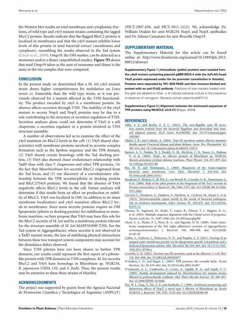
The silver stained gel showed that protein secretion to theculture supernatant in the T3SS inducing conditions was nega-tively affected in the y4yS mutant strain (Figure 3A). A Westernblot analysis using anti-NopX and anti-NopA confirmed that thesecretion of both NopX and NopA, proteins normally secretedby T3SS, was inhibited in the mutant strain (Figures 3B,C). Thesecretion defect was reversed by mutant complementation with agene copy into a plasmid of moderate copy number (Figure 3).Therefore, we conclude from this experiment that Y4yS is nota NopX chaperone because not only NopX secretion was inhib-ited but also NopA secretion was negatively affected. We cannotexclude that Y4yS is the NopA chaperone.
Very much as in the wild-type, NopA was detected in themutant pellet. This indicates that the defect in protein secretionwas not a consequence of a defect in NopA expression and dis-cards a negative T3SS transcriptional regulator role for the proteincoded by mlr8765. NopX could not be detected in wild-type ormutant pellets (data not shown). T3SS secreted proteins of somerhizobia could be detected in the culture supernatant but not inthe bacterial pellet even in the case of mutants that are affectedin secretion (Deakin et al., 2005; Krishnan et al., 2007). It wasspeculated that the accumulation of Nops inside the cell couldbe deleterious to the rhizobial cells and thus subjected to rapiddegradation. This could be also true for NopX in M. loti.
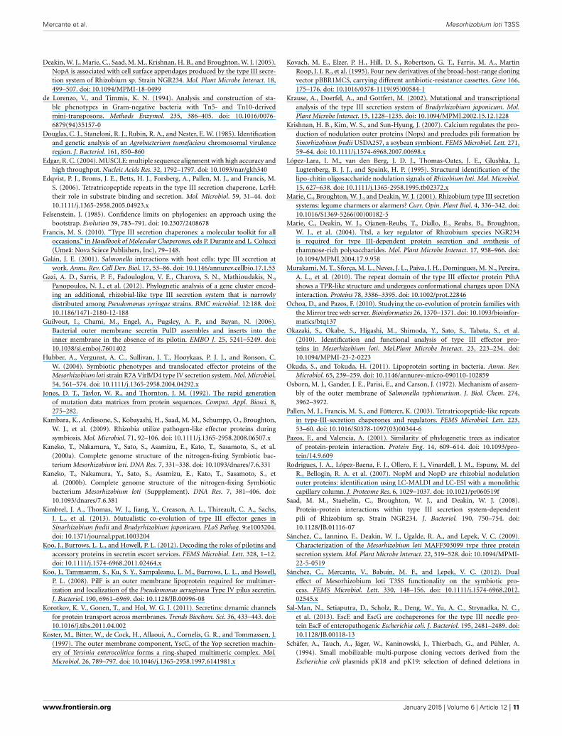
Y4yS IS LOCALIZED IN BACTERIAL MEMBRANESWe determined the cellular localization of Y4yS. Previously, theprotein fused to the triple (3x) copy of the FLAG peptide wasintroduced into the M. loti y4yS mutant strain, cloned into aplasmid of moderate copy number under a constitutively activepromoter in rhizobia. A Western blot analysis of total bacterialextract showed the presence of a band between 15 and 25 kDa,in agreement to the theoretical molecular weight of the pro-tein (16 kDa) (Supplementary Figure 1). Localization analysisdetected the fused protein both in the membrane and cytoplasmfractions although higher levels were detected in membranes(data not shown). The possibility of over expression artifacts ledus to integrate the tagged protein into the M. loti genome viaa single recombination event in order to have only one copy ofthe fused construct in the cell. Once chromosome integrated,the fused proteins were expressed from the corresponding chro-mosomal promoter. Western blot analysis of the various cellularfractions showed that Y4yS was localized exclusively in bacterialmembranes (Figure 4A).
Western blot results also indicate that Y4yS expressionoccurred in naringenin-induced culture, which is the conditionof induction of M. loti T3SS expression (Sánchez et al., 2009)(Figure 4A). This confirms that the y4yS gene forms part ofan operon of co-transcribed ORFs nopC-nopA-rhcD-rhcV-y4yS
www.frontiersin.org January 2015 | Volume 6 | Article 12 | 5

Mercante et al. Mesorhizobium loti T3SS
FIGURE 3 | Analysis of T3SS secreted proteins in the wild-type, y4yS
mutant, and y4yS mutant complemented strains. Supernatant (Sn) andintracellular proteins (pellet) were isolated from MAFF303099 (wt), its y4ySmutant (y4yS), and the mutant complemented with a plasmid of moderatecopy number containing the full-length y4yS gene under the lac promoter(mc). Bacteria were grown in T3SS inducing conditions. All bacteria containplasmid pMP2112. Proteins were separated by 15% SDS-PAGE, stainedwith silver nitrate (A) or transferred to membranes and probed withanti-NopA antibody (B) or anti-NopX antibody (C).
under a promoter region with a tts box localized upstream nopC(Tampakaki, 2014).
We then determined the inner or outer membrane localizationof the Y4yS-FLAG protein. Attempts to detect the chromosome-encoded fusion protein were unsuccessful probably because ofthe low protein levels in the cell. We decided to make this deter-mination with bacteria expressing the fused protein from thepBBR1MCS-4 plasmid. Inner and outer membranes were sepa-rated by density gradient centrifugation. Results showed that theY4yS protein is localized both in outer and inner membranes(Figure 4B). Attempts to detect the Omp19 protein by Westernblot (an OM porin used as OM marker) were unsuccessful prob-ably because of sample dilution.
Y4yS PRESENTS SEQUENCE FEATURES OF T4SS PILOTINS AND TadDPROTEINSince T3SS chaperones are generally cytoplasmic proteins, Y4ySmembrane localization argues against a chaperone role for thisprotein (Francis, 2010). However, this role cannot be completelyexcluded. Two class V chaperones for needle components in
FIGURE 4 | Expression and localization of the 3x FLAG Y4yS fused
protein. (A) Total membranes (TM) and fractions corresponding to thecytoplasm and periplasm (S) of the MAFF303099 strain with sequenceencoding the 3xFLAG Y4yS fused protein integrated into the bacterialchromosome. The two bacteria contain plasmid pMP2112. Proteins wereseparated by 10 % SDS-PAGE and then immuno-blotted and probed with ananti-FLAG antibody. Positions of size markers loaded onto the gels arelabeled (in kDa). ±N indicate bacterial culture in the presence or absence ofnaringenin. (B) Subcellular localization of Y4yS-FLAG expressed from apBBR1MCS-4 plasmid into an y4yS mutant strain was determined bysucrose density gradient centrifugation. Inner and outer membranes werefractionated as described in Materials and Methods. Fractions werecollected as 1-ml aliquots from the top of a discontinuous sucrose gradient.Fractions enriched in the inner membranes were identified by monitoringNADH oxidase activity. Enzyme activity was expressed as percentage ofmaximal activity. Y4yS-FLAG was detected by immunobloting withanti-FLAG (from mouse) and fluorescent anti-mouse antibodies. Bacteriacontain plasmid pMP2112.
Escherichia coli, EscG and EscE, were described to be in theinner membrane (Sal-Man et al., 2013). Nevertheless, these pro-teins do not present TPR domains. TPR proteins have beendescribed in T4SS of Pseudomonas (PilF), Yersinia (PilF) andNeisseria (PilW) and in the Tad system of A. actinomycetemcomi-tans (TadD), where they function as pilotins and docking proteinsrequired for the formation of the secretin complex at the OM(Clock et al., 2008; Koo et al., 2012). These four TPR proteinsare localized in bacterial membranes and, in addition to the TPRdomain, they present a lipidation site at their N-terminus char-acterized by a specific consensus motif, the lipobox (V/L)XXC.
Frontiers in Plant Science | Plant-Microbe Interaction January 2015 | Volume 6 | Article 12 | 6
Mercante et al. Mesorhizobium loti T3SS
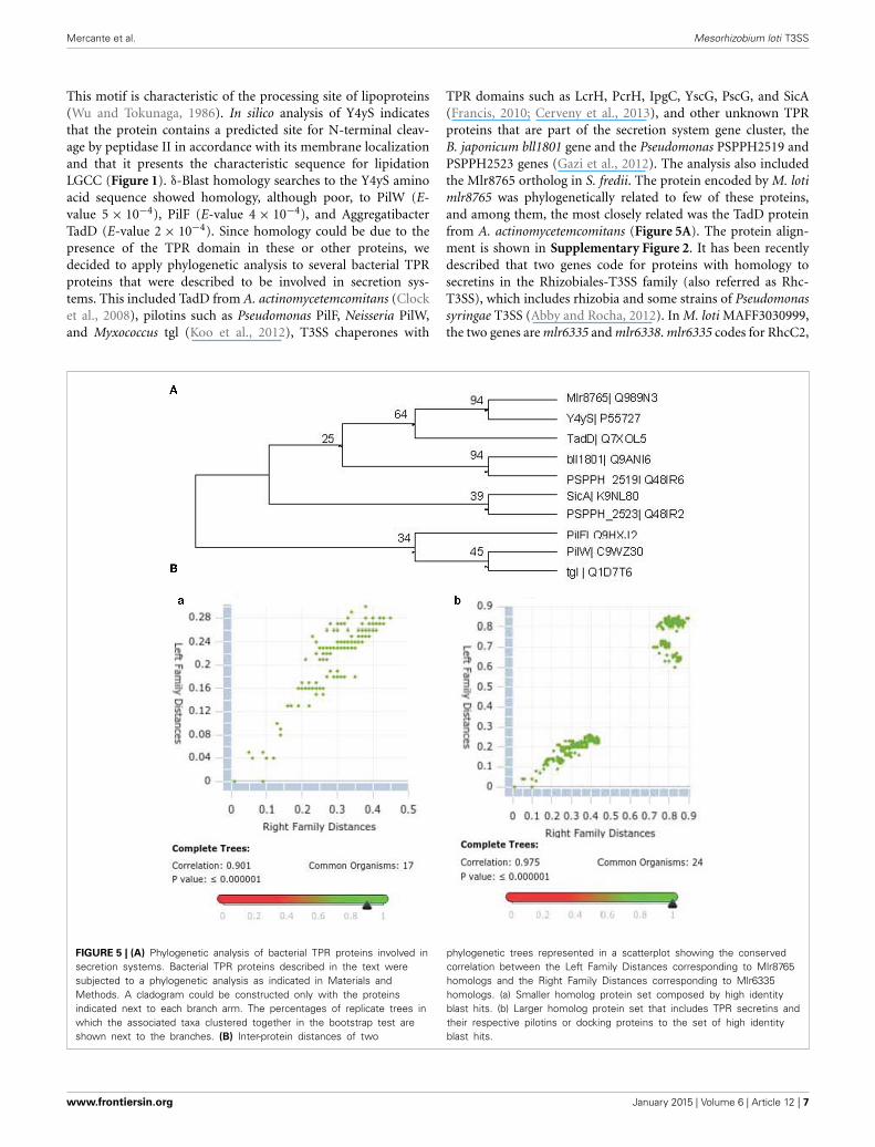
This motif is characteristic of the processing site of lipoproteins(Wu and Tokunaga, 1986). In silico analysis of Y4yS indicatesthat the protein contains a predicted site for N-terminal cleav-age by peptidase II in accordance with its membrane localizationand that it presents the characteristic sequence for lipidationLGCC (Figure 1). δ-Blast homology searches to the Y4yS aminoacid sequence showed homology, although poor, to PilW (E-value 5 × 10⁻⁴), PilF (E-value 4 × 10⁻⁴), and AggregatibacterTadD (E-value 2 × 10⁻⁴). Since homology could be due to thepresence of the TPR domain in these or other proteins, wedecided to apply phylogenetic analysis to several bacterial TPRproteins that were described to be involved in secretion sys-tems. This included TadD from A. actinomycetemcomitans (Clocket al., 2008), pilotins such as Pseudomonas PilF, Neisseria PilW,and Myxococcus tgl (Koo et al., 2012), T3SS chaperones with
TPR domains such as LcrH, PcrH, IpgC, YscG, PscG, and SicA(Francis, 2010; Cerveny et al., 2013), and other unknown TPRproteins that are part of the secretion system gene cluster, theB. japonicum bll1801 gene and the Pseudomonas PSPPH2519 andPSPPH2523 genes (Gazi et al., 2012). The analysis also includedthe Mlr8765 ortholog in S. fredii. The protein encoded by M. lotimlr8765 was phylogenetically related to few of these proteins,and among them, the most closely related was the TadD proteinfrom A. actinomycetemcomitans (Figure 5A). The protein align-ment is shown in Supplementary Figure 2. It has been recentlydescribed that two genes code for proteins with homology tosecretins in the Rhizobiales-T3SS family (also referred as Rhc-T3SS), which includes rhizobia and some strains of Pseudomonassyringae T3SS (Abby and Rocha, 2012). In M. loti MAFF3030999,the two genes are mlr6335 and mlr6338. mlr6335 codes for RhcC2,
FIGURE 5 | (A) Phylogenetic analysis of bacterial TPR proteins involved insecretion systems. Bacterial TPR proteins described in the text weresubjected to a phylogenetic analysis as indicated in Materials andMethods. A cladogram could be constructed only with the proteinsindicated next to each branch arm. The percentages of replicate trees inwhich the associated taxa clustered together in the bootstrap test areshown next to the branches. (B) Inter-protein distances of two
phylogenetic trees represented in a scatterplot showing the conservedcorrelation between the Left Family Distances corresponding to Mlr8765homologs and the Right Family Distances corresponding to Mlr6335homologs. (a) Smaller homolog protein set composed by high identityblast hits. (b) Larger homolog protein set that includes TPR secretins andtheir respective pilotins or docking proteins to the set of high identityblast hits.
www.frontiersin.org January 2015 | Volume 6 | Article 12 | 7

Mercante et al. Mesorhizobium loti T3SS
which shows homology with the secretins of the Tad (tight adher-ence) macromolecular transport system present in bacteria suchas Caulobacter and Aggregatibacter (CpaC and RcpA respectively)(Abby and Rocha, 2012). mlr6338 codes for a protein that presentshomology with the N-terminal part of T3SS secretins (Clocket al., 2008; Abby and Rocha, 2012). A phylogenetic analysisof the T3SS secretins together with secretins from T2SS, typeIV pilus, Tad system and filamentous phages showed that rhi-zobial secretin RhcC2 groups together with secretins from theTad loci (RcpA) (Abby and Rocha, 2012; Clock et al., 2008).The same study concludes that rhizobia originally had a non-flagellar-T3SS-like secretin, RhcC1, and secondarily acquired thesecretin RhcC2 from a Tad locus through a partial homologousgene replacement (Abby and Rocha, 2012). In Aggregatibacter thetadD gene is downstream the tadC gene. M. loti has a cluster ofTad gene homologs (mlr5593 to mlr5604). M. loti Tad secretincoded by mlr5597 gene presents a 32% homology with M. lotiT3SS secretin RhcC2. A gene localized downstream the M. lotitadC gene (mlr5604) and in opposite direction to the Tad clus-ter (mll5605) encodes an unknown protein with 32% homologywith M. loti Y4yS. The above-described data raised the possibil-ity that Y4yS, which shares sequence features and is evolutionarilyrelated to TadD, might be a protein required for the complex for-mation of RhcC2 (evolutionarily related to the Tad secretin) inM. loti MAFF303099 T3SS. The Rhc-T3SS family is subdividedinto three subgroups according to the organization of the T3SScore genes (Gazi et al., 2012). T3SS core genes of subgroup I,represented by Rhizobium sp. NGR234, B. japonicum USDA 110,S. fredii, and M. loti MAFF303099, are organized in three seg-ments (Gazi et al., 2012; Tampakaki, 2014). The second fragmentin members of this subgroup harbors the genes rhcD, rhcV andy4yS. Since members of Subgroup I have both RhcC2 and Y4yS,our hypothesis could be extended to the four above-mentionedstrains. To analyze the existence of an evolutionary relationshipbetween Y4yS to RhcC2, despite being coded in separate segmentsof the Rhc-T3SS cluster, a comparison between the phyloge-netic trees of Y4yS homologs and the respective secretin RhcC2homologs was made using the Mirror tree online server (Ochoaand Pazos, 2010). Two homologous groups were created for eachreference protein as was described in Materials and Methods.Mlr8765 and Mlr6335 showed a high correlation coefficient inboth pairs of homologous protein groups with a P-value <1e-6 (Figure 5B). Interestingly, the larger group, which includeddistant homologs, presented a higher correlation score that thesmaller and more closed related group. This result suggests thatthe TPR secretin/pilotin or docking protein and the rhizobialT3SS secretin/Y4yS homolog proteins are coevolving and arguesin favor of the existence of a physical interaction between RhcC2and Y4yS proteins.
THE M. LOTI y4yS MUTANT STRAIN PRESENTS LOWER RhcC2 PROTEINLEVELS IN THE BACTERIAL MEMBRANESTo analyze the involvement of Y4yS in the formation of theM. loti RhcC2 complex, we integrated the mlr6335 gene thatcodes for RhcC2 fused to the triple (3x) copy of the FLAG pep-tide into the chromosome of the M. loti MAFF303099 and y4ySmutant strains. Sequences coding for Flag-tagged proteins were
integrated into the chromosome by a single homologous recom-bination event. Following chromosomal integration, the fusedproteins were expressed from the corresponding chromosomalpromoter. NopA secretion in inducing conditions was analyzedfor the wild-type strain with the integrated fused protein torule out the possibility of abolishing type III secretion due tothe C-terminal modification of RhcC2 or by defects in the geneexpressed downstream, mlr8762. Figure 6A shows that the iso-lated strain, although in lower levels than the wild-type strain,still secretes NopA. Western blot analysis using anti-FLAG anti-body indicates that a protein of about 35–40 kDa (40 kDa wasthe expected molecular weight) was detected only in the totalmembrane fraction of the wild-type strain with the tagged RhcC2induced with naringenin but not in the total membrane frac-tion of the wild type strain without tagging nor in the wild typestrain with the tagged protein but without induction with narin-genin (Figure 6B). The detection of RhcC2 in the total membrane
FIGURE 6 | Detection of the 3xFLAG RhcC2 fused protein in total
membrane fraction. (A) Silver staining of supernatant proteins ofMAFF303099 (wt) and MAFF303099 with sequence encoding the 3xFLAGRhcC2 fused protein integrated into the bacterial chromosome (wt6335-TF). Total membranes of wt and wt 6335-TF strains were separatedby 12.5% SDS-PAGE and immune-bloted and probed with anti-FLAGantibody (from mouse) (B), and with anti-Omp19 antibody (from rabbit) (C).For detection fluorescent anti-mouse and anti-rabbit antibodies were used.All bacteria contain plasmid pMP2112. ± N: with or without induction withnaringenin, ± Ph: with or without phenol treatment.
Frontiers in Plant Science | Plant-Microbe Interaction January 2015 | Volume 6 | Article 12 | 8

Mercante et al. Mesorhizobium loti T3SS
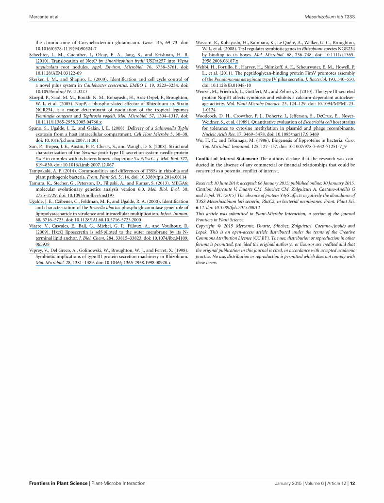
fraction required phenol treatment. Results confirm that RhcC2protein is expressed under naringenin induction. Consequently,its expression depends on the activity of the upstream promoterwith the tts box. Detection using anti-Omp19 antibodies showssimilar protein levels in the three preparations (Figure 6C). Asthe secretin complex has been described to have OM localiza-tion, we isolated the OM of the wild-type and mutant strain withthe tagged RhcC2 protein. Since the secretin complex of the Tadsystem is resistant to detergent at 65◦C but sensitive to boiling(Clock et al., 2008), we analyzed the presence of the RhcC2-3x-FLAG protein in OM of the wild-type and y4yS mutant strainsby SDS-PAGE electrophoresis after resuspending and heating thesamples at 65◦C and 100◦C in the SDS-PAGE sample buffer. Theanti-FLAG antibody detected a monomer of about 40 kDa only inthe wild-type OM (Figure 7A). Under silver staining the samplesrevealed a similar pattern and amount of proteins (Figure 7B).A slight increase in 40 kDa protein levels was observed when
the sample was boiled in the SDS-PAGE sample buffer at 100◦Cinstead of at 65◦C (Figure 7A). Unfortunately, all attempts todetect the high molecular polymers corresponding to secretinoligomers were unsuccessful. In some systems, it is difficult toobserve the secretin complex with Western blots due to problemsof complex solubility and efficient transfer of high-molecular-weight species to nitrocellulose (Burghout et al., 2004; Clock et al.,2008). The absence of RhcC2 in mutant OM indicates that themutation in Y4yS affects the production, stability or localizationof RhcC2. The T3SS protein expression is not affected in the y4ySmutant so a deficiency in transcription is quite unlikely. In someT3SS systems, secretin is localized in the inner membrane in theabsence of pilotin (Koster et al., 1997; Koo et al., 2008), whereasin the Tad system, no endogenous secretin is localized in thewhole cell extract in the absence of pilotin and in physiologicalconditions (Clock et al., 2008). To address this problem we deter-mined total RhcC2 protein levels in the cell. Figures 7C,D show
FIGURE 7 | Detection of the 3x FLAG RhcC2 fused protein. (A) OMsof wt 6335-TF and y4yS mutant strain with sequence encoding the3xFLAG RhcC2 fused protein integrated into the bacterial chromosome(y4yS 6335-TF) heated at 65◦C or 100◦C were separated by 15%SDS-PAGE and then immuno-bloted and probed with an anti-FLAGantibody, (B) Silver staining of samples described in A separated by7.5% SDS-PAGE. Total membranes (TM) and cytoplasmic fractions of wt6335-TF and y4yS 6335-TF strains heated at 100◦C, separated by 12.5%
SDS-PAGE and then immuno-bloted and probed with anti-FLAG (C) andanti-Omp19 (D) antibodies and revealed with fluorescent antibodies. Allbacteria contain plasmid pMP2112 and all bacterial cultures were madein the presence of naringenin. Positions of RhcC2, and of monomer anddimer of Omp19 are indicated. Positions of size markers loaded onto thegels are labeled (in kDa). Anti-Omp19 antibodies nonspecifically probe agreat band both in wt and mutant cytoplasmic fractions betweenmarkers of 25 and 35 kDa.
www.frontiersin.org January 2015 | Volume 6 | Article 12 | 9
Mercante et al. Mesorhizobium loti T3SS
the Western blot results on total membrane and cytoplasmic frac-tions, of wild type and y4yS mutant strains containing the taggedRhcC2 protein. Results indicate that the flagged RhcC2 protein islocalized in membranes and that the y4yS mutant exhibits lowerlevels of this protein in total bacterial extract (membranes andcytoplasm), resembling the results observed in the Tad system(Clock et al., 2008). Omp19, the OM marker, can be detected as amonomer and/or a dimer (unpublished results). Figure 7D showsthat total Omp19 taken as the sum of monomer and dimer is thesame in the two samples that were compared.
CONCLUSIONIn the present study we determined that a M. loti y4yS mutantstrain shows higher competitiveness for nodulation on Lotustenuis cv. Esmeralda than the wild type strain, as it was pre-viously observed for a mutant affected in the T3SS functional-ity. The product encoded by y4yS is a membrane protein. Itsabsence affects secretion through T3SS. The inability of the y4ySmutant to secrete NopA and NopX proteins may be due to arole contributing to the structure or secretion regulation of T3SS.Secretion analyses alone could not determine if Y4yS is a pilichaperone, a secretion regulator or a protein involved in T3SSstructure assembly.
A number of observations led us to examine the effect of they4yS mutation on RhcC2 levels in the cell: (1) Y4yS shared char-acteristics with membrane proteins involved in secretin complexformation such as the lipobox sequence and the TPR domain,(2) Y4yS shared certain homology with the Tad docking pro-tein, (3) Y4yS also showed closer evolutionary relationship withTadD than with class V chaperones and other TPR proteins, (4)the fact that Mesorhizobium loti secretin RhcC2 originated fromthe Tad locus, and (5) our discovery of a coevolutionary rela-tionship between the TPR secretin/pilotin or docking proteinand RhcC2/Y4yS proteins. We found that the absence of Y4ySnegatively affects RhcC2 levels in the cell. Future analyses willdetermine if this results from an effect on production or stabil-ity of RhcC2. Y4yS was localized in OM (in addition to its innermembrane localization) and y4yS mutation affects RhcC2 lev-els in membranes. Since some secretin proteins require an OMlipoprotein (pilotin or docking protein) for stabilization or mem-brane insertion, we here propose that Y4yS may have this role forthe RhcC2 secretin of M. loti and be a membrane protein relevantfor the structure assembly of M. loti MAFF303099 T3SS. For theTad system in Aggregatibacter, where secretin is not observed ina TadD mutant strain, the loss of stabilizing physical interactionsbetween these two transport system components may account forthe abundance defect observed.
Since T3SS pilotins have not been shown to harbor TPRdomains, our results could represent the first report of a pilotin-like protein with TPR domains in T3SS complexes. M. loti secretinRhcC2 and Y4yS have homologs in Rhizobium sp. NGR234,B. japonicum USDA 110, and S. fredii. Thus, the present resultsmay be extensive to these three strains of rhizobia.
ACKNOWLEDGMENTSThe project was supported by grants from the Agencia Nacionalde Promoción Científica y Tecnológica of Argentina (ANPCyT)
(PICT-2007-650, and PICT-2011-1212). We acknowledge Dr.William Deakin for anti-NGR234 NopA and NopX antibodiesand Dr. Juliana Cassattaro for anti-Brucella Omp19.
SUPPLEMENTARY MATERIALThe Supplementary Material for this article can be foundonline at: http://www.frontiersin.org/journal/10.3389/fpls.2015.00012/abstract
Supplementary Figure 1 | Intracellular (pellet) proteins were isolated from
the y4yS mutant containing plasmid pBBR1MCS-4 with the 3xFLAG fused
Y4yS protein expressed under the lac promoter (constitutive in rhizobia),
Proteins were separated by 10% SDS-PAGE and then immuno-blotted and
probed with an anti-FLAG antibody. Positions of size markers loaded onto
the gels are labeled (in kDa). ± N indicate bacterial culture in the presence
or absence of naringenin. Bacteria contain plasmid pMP2112.
Supplementary Figure 2 | Alignment between the aminoacid sequences of
TPR proteins using MUSCLE v(3.8.31) (Edgar, 2004).
REFERENCESAbby, S. S., and Rocha, E. P. C. (2012). The non-flagellar type III secre-
tion system evolved from the bacterial flagellum and diversified into host-cell adapted systems. PLoS Genet. 8:e1002983. doi: 10.1371/journal.pgen.1002983
Alfano, J. R., and Collmer, A. (2004). Type III secretion system effector proteins:double agents I bacterial disease and plant defense. Annu. Rev. Phytopathol. 42,385–414. doi: 10.1146/annurev.phyto.42.040103.110731
Bartsev, A. V., Deakin, W. J., Boukli, N. M., McAlvin, C. B., Stacey, G., Malnoe,P., et al. (2004). NopL, an effector protein of Rhizobium sp. NGR234,thwarts activation of plant defense reactions. Plant Physiol. 134, 871–879. doi:10.1104/pp.103.031740
Bos, M. P., and Tommassen, J. (2004). Biogenesis of the Gram-negativebacterial outer membrane. Curr. Opin. Microbiol. 7, 610–616. doi:10.1016/j.mib.2004.10.011
Burghout, P., Beckers, F., de Wit, E., van Boxtel, R., Cornelis, G. R., Tommassen, J.,et al. (2004). Role of the pilot protein in the biogenesis of the YscC secretin inYersinia enterocolitica. J. Bacteriol. 186, 5366–5375. doi: 10.1128/JB.186.16.5366-5375.2004
Cerveny, L., Straskova, A., Dankova, V., Hartlova, A., Ceckova, M., Staud, F., et al.(2013). Tetratricopeptide repeat motifs in the world of bacterial pathogens:role in virulence mechanisms. Infect. Immun. 81, 629–635. doi: 10.1128/IAI.01035-12
Chenna, R., Sugawara, H., Koike, T., Lopez, R., Gibson, T. J., Higgins, D. G.,et al. (2003). Multiple sequence alignment with the Clustal series of programs.Nucleic Acids Res. 31, 3497–3500. doi: 10.1093/nar/gkg500
Clock, S. A., Planet, P. J., Perez, B. A., and Figurski, D. H. (2008). Outer mem-brane components of the Tad (tight adherence) secreton of Aggregatibacteractinomycetemcomitans. J. Bacteriol. 190, 980–990. doi: 10.1128/JB.01347-07
Collin, S., Guilvout, I., Nickerson, N. N., and Pugsley, A. P. (2011). Sorting of anintegral outer membrane protein via the lipoprotein-specific Lol pathway and adedicated lipoprotein pilotin. Mol. Microbiol. 80, 655–665. doi: 10.1111/j.1365-2958.2011.07596.x
Cornelis, G. R. (2002). Yersinia type III secretion: send in the effectors. J. Cell. Biol.158, 401–408. doi: 10.1083/jcb.200205077
D’Andrea, L. D., and Regan, L. (2003). TPR proteins: the versatile helix. TrendsBiochem. Sci. 28, 655–662. doi: 10.1016/j.tibs.2003.10.007
D’Antuono, A. L., Casabuono, A., Couto, A., Ugalde, R. A., and Lepek, V. C.(2005). Nodule development induced by Mesorhizobium loti mutant strainsaffected in polysaccharide synthesis. Mol. Plant Microbe Interact. 18, 446–457.doi: 10.1094/MPMI-18-0446
Dai, W. J., Zeng, Y., Xie, Z. P., and Staehelin, C. (2008). Symbiosis-promoting anddeleterious effects of NopT, a novel type 3 effector of Rhizobium sp. StrainNGR234. J. Bacteriol. 190, 5101–5110. doi: 10.1128/JB.00306-08
Frontiers in Plant Science | Plant-Microbe Interaction January 2015 | Volume 6 | Article 12 | 10
Mercante et al. Mesorhizobium loti T3SS
Deakin, W. J., Marie, C., Saad, M. M., Krishnan, H. B., and Broughton, W. J. (2005).NopA is associated with cell surface appendages produced by the type III secre-tion system of Rhizobium sp. Strain NGR234. Mol. Plant Microbe Interact. 18,499–507. doi: 10.1094/MPMI-18-0499
de Lorenzo, V., and Timmis, K. N. (1994). Analysis and construction of sta-ble phenotypes in Gram-negative bacteria with Tn5- and Tn10-derivedmini-transposons. Methods Enzymol. 235, 386–405. doi: 10.1016/0076-6879(94)35157-0
Douglas, C. J., Staneloni, R. J., Rubin, R. A., and Nester, E. W. (1985). Identificationand genetic analysis of an Agrobacterium tumefaciens chromosomal virulenceregion. J. Bacteriol. 161, 850–860
Edgar, R. C. (2004). MUSCLE: multiple sequence alignment with high accuracy andhigh throughput. Nucleic Acids Res. 32, 1792–1797. doi: 10.1093/nar/gkh340
Edqvist, P. J., Broms, J. E., Betts, H. J., Forsberg, A., Pallen, M. J., and Francis, M.S. (2006). Tetratricopeptide repeats in the type III secretion chaperone, LcrH:their role in substrate binding and secretion. Mol. Microbiol. 59, 31–44. doi:10.1111/j.1365-2958.2005.04923.x
Felsenstein, J. (1985). Confidence limits on phylogenies: an approach using thebootstrap. Evolution 39, 783–791. doi: 10.2307/2408678
Francis, M. S. (2010). “Type III secretion chaperones: a molecular toolkit for alloccasions,” in Handbook of Molecular Chaperones, eds P. Durante and L. Colucci(Umeå: Nova Sciece Publishers, Inc), 79–148.
Galán, J. E. (2001). Salmonella interactions with host cells: type III secretion atwork. Annu. Rev. Cell Dev. Biol. 17, 53–86. doi: 10.1146/annurev.cellbio.17.1.53
Gazi, A. D., Sarris, P. F., Fadouloglou, V. E., Charova, S. N., Mathioudakis, N.,Panopoulos, N. J., et al. (2012). Phylognetic analysis of a gene cluster encod-ing an additional, rhizobial-like type III secretion system that is narrowlydistributed among Pseudomonas syringae strains. BMC microbiol. 12:188. doi:10.1186/1471-2180-12-188
Guilvout, I., Chami, M., Engel, A., Pugsley, A. P., and Bayan, N. (2006).Bacterial outer membrane secretin PulD assembles and inserts into theinner membrane in the absence of its pilotin. EMBO J. 25, 5241–5249. doi:10.1038/sj.emboj.7601402
Hubber, A., Vergunst, A. C., Sullivan, J. T., Hooykaas, P. J. J., and Ronson, C.W. (2004). Symbiotic phenotypes and translocated effector proteins of theMesorhizobium loti strain R7A VirB/D4 type IV secretion system. Mol. Microbiol.54, 561–574. doi: 10.1111/j.1365-2958.2004.04292.x
Jones, D. T., Taylor, W. R., and Thornton, J. M. (1992). The rapid generationof mutation data matrices from protein sequences. Comput. Appl. Biosci. 8,275–282.
Kambara, K., Ardissone, S., Kobayashi, H., Saad, M. M., Schumpp, O., Broughton,W. J., et al. (2009). Rhizobia utilize pathogen-like effector proteins duringsymbiosis. Mol. Microbiol. 71, 92–106. doi: 10.1111/j.1365-2958.2008.06507.x
Kaneko, T., Nakamura, Y., Sato, S., Asamizu, E., Kato, T., Sasamoto, S., et al.(2000a). Complete genome structure of the nitrogen-fixing Symbiotic bac-terium Mesorhizobium loti. DNA Res. 7, 331–338. doi: 10.1093/dnares/7.6.331
Kaneko, T., Nakamura, Y., Sato, S., Asamizu, E., Kato, T., Sasamoto, S., etal. (2000b). Complete genome structure of the nitrogen-fixing Symbioticbacterium Mesorhizobium loti (Suppplement). DNA Res. 7, 381–406. doi:10.1093/dnares/7.6.381
Kimbrel, J. A., Thomas, W. J., Jiang, Y., Creason, A. L., Thireault, C. A., Sachs,J. L., et al. (2013). Mutualistic co-evolution of type III effector genes inSinorhizobium fredii and Bradyrhizobium japonicum. PLoS Pathog. 9:e1003204.doi: 10.1371/journal.ppat.1003204
Koo, J., Burrows, L. L., and Howell, P. L. (2012). Decoding the roles of pilotins andaccessory proteins in secretin escort services. FEMS Microbiol. Lett. 328, 1–12.doi: 10.1111/j.1574-6968.2011.02464.x
Koo, J., Tammamm, S., Ku, S. Y., Sampaleanu, L. M., Burrows, L. L., and Howell,P. L. (2008). PilF is an outer membrane lipoprotein required for multimer-ization and localization of the Pseudomonas aeruginosa Type IV pilus secretin.J. Bacteriol. 190, 6961–6969. doi: 10.1128/JB.00996-08
Korotkov, K. V., Gonen, T., and Hol, W. G. J. (2011). Secretins: dynamic channelsfor protein transport across membranes. Trends Biochem. Sci. 36, 433–443. doi:10.1016/j.tibs.2011.04.002
Koster, M., Bitter, W., de Cock, H., Allaoui, A., Cornelis, G. R., and Tommassen, J.(1997). The outer membrane component, YscC, of the Yop secretion machin-ery of Yersinia enterocolitica forms a ring-shaped multimeric complex. Mol.Microbiol. 26, 789–797. doi: 10.1046/j.1365-2958.1997.6141981.x
Kovach, M. E., Elzer, P. H., Hill, D. S., Robertson, G. T., Farris, M. A., MartinRoop, I. I. R., et al. (1995). Four new derivatives of the broad-host-range cloningvector pBBR1MCS, carrying different antibiotic-resistance cassettes. Gene 166,175–176. doi: 10.1016/0378-1119(95)00584-1
Krause, A., Doerfel, A., and Gottfert, M. (2002). Mutational and transcriptionalanalysis of the type III secretion system of Bradyrhizobium japonicum. Mol.Plant Microbe Interact. 15, 1228–1235. doi: 10.1094/MPMI.2002.15.12.1228
Krishnan, H. B., Kim, W. S., and Sun-Hyung, J. (2007). Calcium regulates the pro-duction of nodulation outer proteins (Nops) and precludes pili formation bySinorhizobium fredii USDA257, a soybean symbiont. FEMS Microbiol. Lett. 271,59–64. doi: 10.1111/j.1574-6968.2007.00698.x
López-Lara, I. M., van den Berg, J. D. J., Thomas-Oates, J. E., Glushka, J.,Lugtenberg, B. J. J., and Spaink, H. P. (1995). Structural identification of thelipo-chitin oligosaccharide nodulation signals of Rhizobium loti. Mol. Microbiol.15, 627–638. doi: 10.1111/j.1365-2958.1995.tb02372.x
Marie, C., Broughton, W. J., and Deakin, W. J. (2001). Rhizobium type III secretionsystems: legume charmers or alarmers? Curr. Opin. Plant Biol. 4, 336–342. doi:10.1016/S1369-5266(00)00182-5
Marie, C., Deakin, W. J., Ojanen-Reuhs, T., Diallo, E., Reuhs, B., Broughton,W. J., et al. (2004). TtsI, a key regulator of Rhizobium species NGR234is required for type III-dependent protein secretion and synthesis ofrhamnose-rich polysaccharides. Mol. Plant Microbe Interact. 17, 958–966. doi:10.1094/MPMI.2004.17.9.958
Murakami, M. T., Sforça, M. L., Neves, J. L., Paiva, J. H., Domingues, M. N., Pereira,A. L., et al. (2010). The repeat domain of the type III effector protein PthAshows a TPR-like structure and undergoes conformational changes upon DNAinteraction. Proteins 78, 3386–3395. doi: 10.1002/prot.22846
Ochoa, D., and Pazos, F. (2010). Studying the co-evolution of protein families withthe Mirror tree web server. Bioinformatics 26, 1370–1371. doi: 10.1093/bioinfor-matics/btq137
Okazaki, S., Okabe, S., Higashi, M., Shimoda, Y., Sato, S., Tabata, S., et al.(2010). Identification and functional analysis of type III effector pro-teins in Mesorhizobium loti. Mol.Plant Microbe Interact. 23, 223–234. doi:10.1094/MPMI-23-2-0223
Okuda, S., and Tokuda, H. (2011). Lipoprotein sorting in bacteria. Annu. Rev.Microbiol. 65, 239–259. doi: 10.1146/annurev-micro-090110-102859
Osborn, M. J., Gander, J. E., Parisi, E., and Carson, J. (1972). Mechanism of assem-bly of the outer membrane of Salmonella typhimurium. J. Biol. Chem. 274,3962–3972.
Pallen, M. J., Francis, M. S., and Fütterer, K. (2003). Tetratricopeptide-like repeatsin type-III-secretion chaperones and regulators. FEMS Microbiol. Lett. 223,53–60. doi: 10.1016/S0378-1097(03)00344-6
Pazos, F., and Valencia, A. (2001). Similarity of phylogenetic trees as indicatorof protein-protein interaction. Protein Eng. 14, 609–614. doi: 10.1093/pro-tein/14.9.609
Rodrigues, J. A., López-Baena, F. J., Ollero, F. J., Vinardell, J. M., Espuny, M. delR., Bellogin, R. A. et al. (2007). NopM and NopD are rhizobial nodulationouter proteins: identification using LC-MALDI and LC-ESI with a monolithiccapillary column. J. Proteome Res. 6, 1029–1037. doi: 10.1021/pr060519f
Saad, M. M., Staehelin, C., Broughton, W. J., and Deakin, W. J. (2008).Protein-protein interactions within type III secretion system-dependentpili of Rhizobium sp. Strain NGR234. J. Bacteriol. 190, 750–754. doi:10.1128/JB.01116-07
Sánchez, C., Iannino, F., Deakin, W. J., Ugalde, R. A., and Lepek, V. C. (2009).Characterization of the Mesorhizobium loti MAFF303099 type three proteinsecretion system. Mol. Plant Microbe Interact. 22, 519–528. doi: 10.1094/MPMI-22-5-0519
Sánchez, C., Mercante, V., Babuin, M. F., and Lepek, V. C. (2012). Dualeffect of Mesorhizobium loti T3SS functionality on the symbiotic pro-cess. FEMS Microbiol. Lett. 330, 148–156. doi: 10.1111/j.1574-6968.2012.02545.x
Sal-Man, N., Setiaputra, D., Scholz, R., Deng, W., Yu, A. C., Strynadka, N. C.,et al. (2013). EscE and EscG are cochaperones for the type III needle pro-tein EscF of enteropathogenic Escherichia coli. J. Bacteriol. 195, 2481–2489. doi:10.1128/JB.00118-13
Schäfer, A., Tauch, A., Jäger, W., Kaninowski, J., Thierbach, G., and Pühler, A.(1994). Small mobilizable multi-purpose cloning vectors derived from theEscherichia coli plasmids pK18 and pK19: selection of defined deletions in
www.frontiersin.org January 2015 | Volume 6 | Article 12 | 11
Mercante et al. Mesorhizobium loti T3SS
the chromosome of Corynebacterium glutamicum. Gene 145, 69–73. doi:10.1016/0378-1119(94)90324-7
Schechter, L. M., Guenther, J., Olcay, E. A., Jang, S., and Krishnan, H. B.(2010). Translocation of NopP by Sinorhizobium fredii USDA257 into Vignaunguiculata root nodules. Appl. Environ. Microbiol. 76, 3758–3761. doi:10.1128/AEM.03122-09
Skerker, J. M., and Shapiro, L. (2000). Identification and cell cycle control ofa novel pilus system in Caulobacter crescentus. EMBO J. 19, 3223–3234. doi:10.1093/emboj/19.13.3223
Skorpil, P., Saad, M. M., Boukli, N. M., Kobayashi, H., Ares-Orpel, F., Broughton,W. J., et al. (2005). NopP, a phosphorylated effector of Rhizobium sp. StrainNGR234, is a major determinant of nodulation of the tropical legumesFlemingia congesta and Tephrosia vogelii. Mol. Microbiol. 57, 1304–1317. doi:10.1111/j.1365-2958.2005.04768.x
Spano, S., Ugalde, J. E., and Galán, J. E. (2008). Delivery of a Salmonella Typhiexotoxin from a host intracellular compartment. Cell Host Microbe 3, 30–38.doi: 10.1016/j.chom.2007.11.001
Sun, P., Tropea, J. E., Austin, B. P., Cherry, S., and Waugh, D. S. (2008). Structuralcharacterization of the Yersinia pestis type III secretion system needle proteinYscF in complex with its heterodimeric chaperone YscE/YscG. J. Mol. Biol. 377,819–830. doi: 10.1016/j.jmb.2007.12.067
Tampakaki, A. P. (2014). Commonalities and differences of T3SSs in rhizobia andplant pathogenic bacteria. Front. Plant Sci. 5:114. doi: 10.3389/fpls.2014.00114
Tamura, K., Stecher, G., Peterson, D., Filipski, A., and Kumar, S. (2013). MEGA6:molecular evolutionary genetics analysis version 6.0. Mol. Biol. Evol. 30,2725–2729. doi: 10.1093/molbev/mst197
Ugalde, J. E., Czibener, C., Feldman, M. F., and Ugalde, R. A. (2000). Identificationand characterization of the Brucella abortus phosphoglucomutase gene: role oflipopolysaccharide in virulence and intracellular multiplication. Infect. Immun.68, 5716–5723. doi: 10.1128/IAI.68.10.5716-5723.2000
Viarre, V., Cascales, E., Ball, G., Michel, G. P., Filloux, A., and Voulhoux, R.(2009). HxcQ liposecretin is self-piloted to the outer membrane by its N-terminal lipid anchor. J. Biol. Chem. 284, 33815–33823. doi: 10.1074/jbc.M109.065938
Viprey, V., Del Greco, A., Golinowski, W., Broughton, W. J., and Perret, X. (1998).Symbiotic implications of type III protein secretion machinery in Rhizobium.Mol. Microbiol. 28, 1381–1389. doi: 10.1046/j.1365-2958.1998.00920.x
Wassem, R., Kobayashi, H., Kambara, K., Le Quéré, A., Walker, G. C., Broughton,W. J., et al. (2008). TtsI regulates symbiotic genes in Rhizobium species NGR234by binding to tts boxes. Mol. Microbiol. 68, 736–748. doi: 10.1111/j.1365-2958.2008.06187.x
Wehbi, H., Portillo, E., Harvey, H., Shimkoff, A. E., Scheurwater, E. M., Howell, P.L., et al. (2011). The peptidoglycan-binding protein FimV promotes assemblyof the Pseudomonas aeruginosa type IV pilus secretin. J. Bacteriol. 193, 540–550.doi: 10.1128/JB.01048-10
Wenzel, M., Friedrich, L., Gottfert, M., and Zehner, S. (2010). The type III-secretedprotein NopE1 affects symbiosis and exhibits a calcium-dependent autocleav-age activity. Mol. Plant Microbe Interact. 23, 124–129. doi: 10.1094/MPMI-23-1-0124
Woodcock, D. H., Crowther, P. J., Doherty, J., Jefferson, S., DeCruz, E., Noyer-Weidner, S., et al. (1989). Quantitative evaluation of Escherichia coli host strainsfor tolerance to cytosine methylation in plasmid and phage recombinants.Nucleic Acids Res. 17, 3469–3478. doi: 10.1093/nar/17.9.3469
Wu, H. C., and Tokunaga, M. (1986). Biogenesis of lipproteins in bacteria. Curr.Top. Microbiol. Immunol. 125, 127–157. doi: 10.1007/978-3-642-71251-7_9
Conflict of Interest Statement: The authors declare that the research was con-ducted in the absence of any commercial or financial relationships that could beconstrued as a potential conflict of interest.
Received: 10 June 2014; accepted: 06 January 2015; published online:30 January 2015.Citation: Mercante V, Duarte CM, Sánchez CM, Zalguizuri A, Caetano-Anollés Gand Lepek VC (2015) The absence of protein Y4yS affects negatively the abundance ofT3SS Mesorhizobium loti secretin, RhcC2, in bacterial membranes. Front. Plant Sci.6:12. doi: 10.3389/fpls.2015.00012This article was submitted to Plant-Microbe Interaction, a section of the journalFrontiers in Plant Science.Copyright © 2015 Mercante, Duarte, Sánchez, Zalguizuri, Caetano-Anollés andLepek. This is an open-access article distributed under the terms of the CreativeCommons Attribution License (CC BY). The use, distribution or reproduction in otherforums is permitted, provided the original author(s) or licensor are credited and thatthe original publication in this journal is cited, in accordance with accepted academicpractice. No use, distribution or reproduction is permitted which does not comply withthese terms.
Frontiers in Plant Science | Plant-Microbe Interaction January 2015 | Volume 6 | Article 12 | 12
Copyright © 2022 FDOKUMEN