TESE Patricia Bozzetto Ambrosi.pdf - RI UFPE
224
UNIVERSIDADE FEDERAL DE PERNAMBUCO CENTRO DE BIOCIÊNCIAS PROGRAMA DE PÓS-GRADUAÇÃO EM CIÊNCIAS BIOLÓGICAS MORFOLOGIA DESCRITIVA E HEMODINÂMICA DOS ANEURISMAS DO SIFÃO CAROTÍDEO PATRICIA BOZZETTO AMBROSI Recife 2016
-
Upload
khangminh22 -
Category
Documents
-
view
0 -
download
0
Transcript of TESE Patricia Bozzetto Ambrosi.pdf - RI UFPE

UNIVERSIDADE FEDERAL DE PERNAMBUCO CENTRO DE BIOCIÊNCIAS
PROGRAMA DE PÓS-GRADUAÇÃO EM CIÊNCIAS BIOLÓGICAS
MORFOLOGIA DESCRITIVA E HEMODINÂMICA DOS ANEURISMAS DO SIFÃO CAROTÍDEO
PATRICIA BOZZETTO AMBROSI
Recife 2016

PATRICIA BOZZETTO AMBROSI
MORFOLOGIA DESCRITIVA E HEMODINÂMICA DOS ANEURISMAS DO SIFÃO CAROTÍDEO
Tese de doutorado apresentada à Coordenação do Programa de Pós-Graduação em Ciências Biológicas da Universidade Federal de Pernambuco, como parte dos requisitos à obtenção do grau de Doutor em Ciências Biológicas, área de concentração: Biologia Química para a Saúde
Orientação: Prof. Dr. Marcelo Moraes Valença
Co-orientação: Prof. Dr. Laurent Spelle Prof. Dr. Jacques Moret
Recife 2016

Catalogação na fonte
Elaine Barroso CRB 1728
Ambrosi, Patrícia Bozzetto
Morfologia descritiva e hemodinâmica dos aneurismas do sifão carotídeo/ Patrícia Bozzetto Ambrosi – Recife: O Autor, 2016.
224 folhas: il., fig., tab.
Orientador: Marcelo Moraes Valença Coorientadores: Laurent Spelle e Jacques Moret Tese (doutorado) – Universidade Federal de Pernambuco. Centro
de Biociências. Ciências Biológicas, 2016.
Inclui referências e anexos
1. Aneurismas 2. Artérias 3. Biotecnologia I. Valença, Marcelo Moraes (orientador) II. Spelle, Laurent (coorientador) III. Moret, Jacques (coorientador) IV. Título
616.133 CDD (22.ed.) UFPE/CCB-2016-129

PATRICIA BOZZETTO AMBROSI
MORFOLOGIA DESCRITIVA E HEMODINÂMICA DOS ANEURISMAS DO SIFÃO CAROTÍDEO
Tese de doutorado apresentado à Coordenação do Programa de Pós-Graduação em Ciências Biológicas da Universidade Federal de Pernambuco, como parte dos requisitos à obtenção do grau de Doutor de Ciências Biológicas, área de Concentração: Biologia Química para Saúde Aprovada em:
BANCA EXAMINADORA ----------------------------------------------------------------------------------- Prof. Dr. Marcelo Moraes Valença Programa de Pós-Graduação em Ciências Biológicas ------------------------------------------------------------------------------------ Profa. Dra. Maria Teresa dos Santos Correia Depto. Bioquímica- UFPE (Titular- Membro interno) -------------------------------------------------------------------------------------
Profa. Dra. Maria Tereza Jansem de Almeida Catanho Depto. Bioquímica- UFPE (Titular- Membro interno)
------------------------------------------------------------------------------------- Prof. Dr. Marcos Antonio Barbosa
Neurorradiologista do Hospital Esperança (Titular- Membro externo) -------------------------------------------------------------------------------------
Prof. Dr. Carlos Augusto Carvalho de Vasconcelos Depto. Nutrição (Titular- Membro interno)
25/02/2016

DEDICATÓRIA
Dedico essa tese a minha família em especial
Aos meus pais, à minha irmã Caroline,
À tia Odila, ao tio Henri (In memoriam)
Não me esquecendo também do tio Dr. Ervalino
Plácido Bozzetto (In memoriam) qual me ajudou
em meus os primeiros passos e mostrou um
caminho a seguir...
Não há palavra no mundo capaz de expressar o
amor e a gratidão que sinto por todos vocês.

AGRADECIMENTOS Essa tese não é apenas resultado de meus sete anos de estudos e pesquisas sobre os
aneurismas cerebrais, mas contém material original e de imagem proveniente de um dos
departamentos mais ilustres do mundo no tratamento endovascular e no manejo clínico de
pacientes com todos os tipos de patologias relacionadas com a Neurorradiologia
Intervencionista-Neurocirurgia Endovascular. Além disso, as atividades de pesquisa
realizadas me fizeram evoluir em todos os sentidos e teria sido impossível sem o suporte de
todos envolvidos na realização deste sonho;
Agradeço em primeiro lugar aos hospitais e as universidades aonde trabalhei e que me
deram a oportunidade de realizar essa pesquisa, em especial ao Hospital Beaujon, à
Universidade Federal de Pernambuco, e à CAPES pela bolsa concedida durante a parte de
desenvolvimento dessa Tese;
A todos os pacientes que fizeram parte dessa pesquisa; Ao meu orientador, Prof. Dr. Marcelo Valença o grande incentivador desse projeto, que
não mediu esforços, com valiosos ensinamentos tanto do ponto de vista humano como
científico. Sempre vou lembrar de nossos debates muitas vezes a distância, mas que foram
extremamente úteis e estimulantes sobre a metodologia e a literatura;
Aos co-orientadores estrangeiros: o Prof. Dr. Jacques Moret pelo importante papel de
supervisor na minha formação profissional, e também pela apreciação das minhas atividades
acadêmicas e de pesquisas, o meu reconhecido agradecimento pelas orientações e
ensinamentos em Neurorradiologia Intervencionista, além de um profundo agradecimento
pela co-orientação dessa tese e pelo rico e sofisticado material que me foi cedido. Ao Prof.
Dr. Laurent Spelle pela estimada atenção, ajuda no vencimento dos desafios e pelo
compartilhamento dos conhecimentos em Neurorradiologia Intervencionista;
A todos os funcionários do Hospital Beaujon e do Departamento de Neurorradiologia
Intervencionista, e em especial aos amigos da Salle de Garde que me apoiaram em todos os
momentos e me ajudaram a superar muitos momentos delicados;
A todos os professores, colegas e funcionários do Programa de Pós-Graduação em
Ciências Biológicas da Universidade Federal de Pernambuco, em especial ao Prof. Ranilson
Bezerra, sempre bastante atencioso e compreensivo, à Profa Maria Teresa Correia, pela sua
paciência e buscando a melhor solução em todos momentos, à Profa Maria Teresa Jansem pelo
seu entusiasmo constante e ajuda disponibilizada, à Sra Adenilda Eugênia de Lima por todo

apoio e carinho, e à bibliotecária Sra Teresa Lucena pela ajuda na revisão das normas ABNT
dessa tese;
Um especial agradecimento ao Prof. Dr. Carlos Augusto Carvalho de Vasconcelos,
amigo de longa data, pelo tempo concedido dentro de sua agitada rotina, ajuda, suporte e por
compartilhar os bons e os maus momentos nessa caminhada;
Um grande agradecimento também ao Prof. Dr. Marcos Antonio Barbosa, brilhante e
dedicado neurorradiologista, além de mentor também um super colega e amigo, nossos
caminhos se cruzaram várias vezes apesar das voltas que o mundo dá;
A todos, manifesto os meus mais profundos e sinceros remerciamentos.

“Le succès ne se mesure pas là où vous êtes arrivé,
mais le nombre des difficultés que vous avez surmonté ”.
“O exito da vida não se mede pelo caminho que você
conquistou, mas sim pelas dificuldades que superou no
caminho”.
Abraham Lincoln
(1809-1865)

RESUMO
Bozzetto-Ambrosi P. Morfologia Descritiva e Hemodinâmica dos Aneurismas do Sifão Carotídeo [Tese de Doutorado] Recife, 2016. Os novos stents modificadores de fluxo apresentam elevadas taxas de sucesso obtidas no tratamento dos aneurismas que se desenvolvem na circunferência do sifão carotídeo. Esses aneurismas são bastante diversificados e apesar de dinâmicos muitas vezes são silenciosos. Além disso, sua história natural e seu comportamento biológico ainda são bastante desafiadores e incertos. Estudos experimentais tem recentemente demonstrado que a morfologia (remodelamento vascular) e as variações na hemodinâmica cerebral estariam envolvidos tanto na gênese bem como no desenvolvimento e na eventual ruptura dos aneurismas intracranianos. Outrossim, estudos populacionais tem evidenciado uma correlação entre a assimetria das artérias do polígono de Willis e o aparecimento de aneurismas em várias localizações dentro do polígono de Willis. Contudo, estudos específicos voltados para aos aneurismas localizados na circunferência do sifão carotídeo ainda não foram abordados. A presente tese teve como principal objetivo caracterizar o papel das variantes arteriais do círculo de Willis particularmente implicadas na gênese dos aneurismas da circunferência do sifão da carótida. Ao mesmo tempo, foi realizada uma análise de regressão para superar o desequilíbrio significativo entre homens e mulheres em relação a ambos: local do aneurisma e a presença de variantes arteriais do polígono. As duas variantes anatômicas do polígono de Willis consideradas no presente estudo foram a presença do segmento A1 dominante (Hipoplasia A1 contralateral) e a presença de artéria cerebral posterior (PCA) fetal. Realizamos um estudo caso-controle em um dos centros franceses de referência, e que também é um centro de classe mundial contendo cinco principais amostras: (1) Grupo 1, agrupando os aneurismas que se originam em algum ponto da circunferência do sifão carotídeo unilateralmente (n = 178); (2) Grupo 2, contendo aneurismas em algum ponto bilateralmente nos dois sifões carotídeos (n = 99); (3) Grupo 3, composto de sifões sem aneurismas contralaterais do grupo A1 (n = 178); (4) Grupo 4, contendo sifões de pacientes sem aneurismas (n = 210) e Grupo 5, controles combinados contendo os Grupos 3 e 4 (n=388). Em grupos equilibrados, a configuração tipo A1 do polígono de Willis parece ter um efeito protetor na gênese dos aneurismas. A configuração do tipo A1 foi significativamente mais encontrada no Grupo 3 (55/178) versus Grupo 1 (12/178) denotando provalvemente um efeito protetor no aparecimento dos aneurismas na região do sifão carotídeo (p<0.0001, OR 0.1617). A presença da A1 dominante foi significantemente maior no grupo 3 do que no grupo 1, tanto no grupo de homens (1/24 versus 11/24, p=0.0070, OR 0.0514) quanto de mulheres (44/154 versus 1/154, p<0.001, OR 0.1923). A associação incluindo ambas A1 dominante/ Fetal PCA foi encontrada apenas em 3/178 (1.6%) pacientes do grupo 1 (3/178) e um total de 13/178 dos sifões do grupo 3 (p = 0.0189, OR 0.2176). O novo conceito poderá ajudar na melhoria do screening e abrindo as portas para novas estratégias no tratamento dos aneurismas do sifão carotídeo. Nesta análise, também evidenciou-se que um grupo controle equilibrado pode reduzir o viés multivariado e, assim, melhorar as estimativas. Por conseguinte, são necessárias mais investigações com dados multicêntricos com abordagens analíticas melhoradas. Além disso, nessa tese discutimos sobre as biotecnologias aplicadas aos aneurismas intracranianos, apresentamos uma abordagem clínica e uma revisão histórica sobre as técnicas de tratamento dos aneurismas do sifão carotídeo, apresentamos as novas perspectivas sobre as variantes anatômicas aplicadas aos aneurismas intracranianos e propusemos uma nova classificação morfológica baseada nas áreas de susceptibilidade dos aneurismas do sifão carotídeo. Concluindo, o efeito protetor da presença das variantes anatômicas arteriais com hiperfluxo suporta a hipótese do uso da hemodinâmica cerebral como fator preditivo no desenvolvimento dos aneurismas intracranianos. Estudos adicionais são necessários com a inclusão dessas novas ferramentas tecnológicas que proporcionem acurácia nas medições de fluxo da circulação cerebral e dos aneurismas intracranianos permitindo a utilização na prática clínica. Palavras-chave: Aneurismas. Artérias. Biotecnologias.

ABSTRACT
Bozzetto-Ambrosi P. Carotid Siphon Aneurysm: Its Hemodynamic and Descriptive Morphology. [PhD Thesis] Recife, 2016. Despite the high success rates obtained with the new modifier flow stents in the treatment of intracranial aneurysms that develop on the circumference of the carotid siphon. These aneurysms are very diversified and despite being dynamic, often silent. Therefore, their natural history and biological behavior remain controversial and little understood. Recent experimental studies have widely shown the morphology (vascular remodeling) and variation in the cerebral hemodynamics would be involved both in the genesis as well as in the development and eventual rupture of intracranial aneurysms. In addition, population studies have shown a correlation between the asymmetry of arteries of the circle of Willis and the genesis of aneurysms in several locations within the circle of Willis. However, specific studies applied to the carotid siphon location has not yet been addressed. This thesis aimed to examine the association of arterial variants of Circle of Willis particularly implicated in the genesis of aneurysms within the circumference of the carotid siphon. At the same time, a regression analysis was performed to overcome the significant imbalance between men and women as compared to both the aneurysm site and the presence of arterial polygon variants. The two anatomical variants of the circle of Willis studied were the presence of dominant A1 segment (contralateral hypoplasia A1) and the presence of fetal-PCA. A case-control study was conducted derived from World-Class and French University referral Center with five different samples: (1) Group 1, composed by aneurysms that originate somewhere along the circumference of the carotid siphon unilaterally (n = 178); (2) Group 2, containing aneurysms at some point bilaterally in the two carotid siphons (n= 99); (3) Group 3, composed by contralateral healthy siphons of the group 1 (n = 178); (4) Group 4, containing siphons from healthy patients (n = 210) and Group 5, containing the combined control groups 3 and 4 (n = 388). In balanced groups, Circle of Willis with A1 dominance appears to have a protective effect in the genesis of carotid siphon aneurysms. The Willis polygon configuration with A1 dominance had a significantly higher frequency in Group 3 (55/178) vs. Group 1 (12/178) denoting a protective effect on the genesis of aneurysms in the carotid siphon region (p <0.0001, OR 0.1617). The presence of the dominant A1 had a significantly increased protection of an aneurysm development compared with the group of men (1/24 versus 11/24, p=0.0070, OR 0.0514) versus women (44/154 versus 1/154, p<0.001, OR 0.1923). Both A1/Fetal PCA dominance were founded in 3/178 (1.6%) patients of the group with a unilateral aneurysm (3/178) and 13/178 of the contralateral healthy siphons (p = 0.0189, OR 0.2176). The Willis polygon configuration with A1 dominance and PCA fetal variant may prevent the carotid siphon aneurysm development. The new concept might help in improvement f the screening and open the door for new strategies in the treatment of carotid siphon aneurysms. This analysis also showed that a balanced control group multivariate can reduce the bias, and thus improving the estimates. Therefore, more investigations are needed multicenter data with improved analytical approaches. In addition, this thesis contains chapters about the biotechnology applied to the intracranial aneurysms, the current approach and a historical review of endovascular techniques for the carotid siphon aneurysms, the new perspectives of use of cerebral hemodynamic applied to intracranial aneurysms and the proposal of a new morphological classification based on the areas susceptibility to carotid siphon aneurysms. Briefly, the protective effect of the presence of arterial anatomical variants supports the hypothesis of the use of cerebral hemodynamics as a predicting factor in the development of intracranial aneurysms. Additional experimental studies with the further accurate flow measurements of cerebral circulation with and without intracranial aneurysms then allowing the application of this new concept used in the clinical practice. Keywords: Aneurysms. Arteries. Biotechnology.

LISTA DE FIGURAS FIGURA 1 – Imagem 3D da artéria carótida direita. FIGURA 2 –Imagem 3D do sifão carotídeo e um aneurisma sacular. FIGURA 3 – Representação esquemática da circulação encefálica e do Polígono de Willis.
FIGURA 4 – Variabilidade do Polígono de Willis. FIGURA 5 - Interação entre os fatores biológicos e funcionais entre si. FIGURA 6 – Estudos de fluxo óptico de um aneurisma do sifão carotídeo. FIGURA 7 – Diferentes aneurismas da artéria carótida interna e no sifão carotídeo. FIGURA 8 – (A) Imagem radiográfica do sifão carotídeo. (B) Duplo sifão.
FIGURA 9 – Representação das classificações mais conhecidas da artéria carótida interna. FIGURA 10 – Segmentação (A) artéria carótida. (B) sifão carotídeo. (C) Imagem sifão
FIGURA 11 – Morfologia do sifão: (A) Imagem 2D. (B) Imagem 2D.
FIGURA 12 – Classificação de Yasargil e Krayenbuel.
FIGURA 13 – Imagens angiográficas de A até J demonstrando diversos subtipos de sifão.
FIGURA 14 – Imagens angiográficas das artérias carótidas de A até E. FIGURA 15 – Imagem 3D do após colocação de stent flow diverters (Ae B). FIGURA 16 – Classificação de Lin et al. da tortuosidade da artéria carótida cavernosa.
FIGURA 17 – Imagem angiográfica do sifão de A até I. FIGURA 18 – Imagem demonstrando análise através da técnica “Laser Doppler Velocity”. FIGURA 19 – Resultados hemodinâmicos a partir de estudos de “CFD”. FIGURA 20 – Imagem de Moniz (A) Estudo de fluxo (B) Imagem extraída da internet (C). FIGURA 21 – 3D da artéria carótida interna e diferentes configurações do polígono de Willis. FIGURA 22 – 2D da carótid direita (A) 2D de aneurisma oftálmica esquerda (B).

LISTA DE ABREVIATURAS E SIGLAS
A1= Segmento A1
AAA= Aneurismas da Aorta Abdominal
ACA= Artéria Cerebral Anterior
ACha= Artéria Choroidal Anterior
AcomA= Artéria Comunicante Anterior
AAT = Aneurismas da Aorta Torácica
BA=Artéria Basilar
CFD= Computational Dynamics Flow
DOPHA= Artéria Dorsal Oftálmica
ICA Cd= Artéria Carótida Interna
MCA= Artéria Cerebral Média
PCA= Artéria Cerebral Posterior
P1= Segmento P1
P2= Segmento P2
TOF= Time of flight
VA= Artéria Vertebral
VOPHA= Artéria Ventral Oftálmica
WWS=Wall Shear Stress

SUMÁRIO CAPÍTULO I
1 INTRODUÇÃO ...............................................................................................................17
CAPÍTULO II
2 REFERENCIAL TEÓRICO...........................................................................................20
2.1 INTRODUÇÃO.............................................................................................................21
2.2. HEMODINÂMICA CEREBRAL ............................................................................23
2.1.1 Artérias Cerebrais, Polígono de Willis e Artérias Relacionadas...........................23
2.1.2 Variantes Anatômicas ...............................................................................................25
2.3 MECANISMOS DA BIOGÊNESE DOS ANEURISMAS INTRACRANIANOS..28
2.3.1 Fatores Biológicos......................................................................................................30
2.3.1.1 Fatores Sistêmicos e Hormonais...............................................................................30
2.3.1.2 Fatores Genéticos e Familiares.................................................................................31
2.3.1.3 Fatores Modificáveis, Nutricionais e Outros ...........................................................33
2.3.2 Fatores Funcionais....................................................................................................34
2.3.2.1 Fatores Morfológicos...............................................................................................34
2.3.2.2 Fatores Hemodinâmicos...........................................................................................36
2.4 O SIFÃO CAROTÍDEO E ANEURISMAS RELACIONADOS..........................38
2.4.1 Estudos Anatômicos..................................................................................................42
2.4.2 Estudos Embriológicos..............................................................................................46
2.4.3 Estudos Morfológicos e Hemodinâmicos.................................................................48
REFERÊNCIAS..................................................................................................................61
CAPÍTULO III
3 OBJETIVOS: ...................................................................................................................75
3.1 OBJETIVO GERAL....................................................................................................76
3.2 OBJETIVOS ESPECÍFICOS.....................................................................................76
CAPÍTULO IV
Artigo publicado nos International Archives of Medicine...................................................79
CAPÍTULO V
Artigo publicado na Revista Neurobiologia..........................................................................85
CAPÍTULO VI
Artigo publicado na Revista Neurobiologia………………..................................................91
CAPÍTULO VII
Artigo submetido a Revista Anatomical Record.................................................................103

CAPÍTULO VIII
Artigo submetido a Revista Anatomical Record ...................................................................125
CAPÍTULO IX
Artigo submetido a Revista Perspectivas Médicas................................................................130
CAPÍTULO X
Artigo submetido a Revista Clinical Nutrition & Dietetics...................................................141
CAPÍTULO XI
Artigo submetido Circulation................................................................................................149
CAPÍTULO XII
4 DISCUSSÃO ...................................................................................................................166
CAPÍTULO XIII
5 CONCLUSÃO E CONSIDERAÇÕES FINAIS .......................................................172
6 LIMITAÇÕES DA PESQUISA................................................................................177
7 PERSPECTIVAS ATUAIS e FUTURAS DIREÇÕES..............................................178
REFERÊNCIAS ..................................................................................................................179
ANEXO I
Template para formatação para Revista International Archives of Medicine........................181
ANEXO II
Template para formatação do artigo publicado na Revista Neurobiologia............................191
ANEXO III
Template para formatação do artigo a ser submetido a Revista Anatomical Record............194
ANEXO IV
Template para formatação do artigo a ser submetido a Revista Perspectivas Médicas........199
ANEXO V
Template para formatação do artigo a ser submetido a Revista Clinical Nutrition ..............202
ANEXO VI
Template para formatação do artigo a ser submetido a Revista Circulation .........................207
ANEXO VII
Carta do Doutorado Sanduíche...............................................................................................222
ANEXO VIII
Carta de Utilização de Dados .................................................................................................224

Ce travail a été réalisé dans le Service de Neuroradiologie Interventionnelle – Neuri Beaujon, Brain Vascular Center – Université Paris Diderot, en coopération avec l'Université Fédérale de Pernambouc- UFPE/Brésil
Este trabalho foi realizado no Serviço de Neuroradiologia Intervencionista – Neuri Beaujon, Brain Vascular Center da Universidade Paris Diderot em cooperação com a Universidade Federal de Pernambuco – UFPE/ Brasil

CAPÍTULO I
Introdução

17
1 INTRODUÇÃO
A presente tese de doutorado é o resultado das atividades de pesquisa que foram
desenvolvidas a partir de uma parceria científica entre o Centro de Biociências da
Universidade Federal de Pernambuco, Brasil, e o Centro Neuri Beaujon, Departamento de
Neurorradiologia Intervencionista, Universidade Paris Diderot, França. Discorre sobre a
morfologia descritiva e a hemodinâmica dos aneurismas do sifão carotídeo enfatizando a
aplicação da hemodinâmica cerebral na gênese e desenvolvimento dos aneurismas
intracranianos. Também, inclui capítulos em forma de artigos relacionados as novas
biotecnologias aplicadas aos aneurismas intracranianos, as técnicas de tratamento
endovascular dos aneurismas do sifão e os seus aspectos desafiantes, uma nova classificação
morfológica dos aneurismas do sifão carotídeo é apresentada, além de dois capítulos em
especial dedicados à aplicabilidade das variantes anatômicas das artérias na gênese dos
aneurismas intracranianos e do sifão carotídeo.
Os trabalhos de pesquisa tiveram início em janeiro de 2011 sob a co-orientação dos
professores Jacques Moret e Laurent Spelle, no Departamento de Neurorradiologia
Intervencionista do Hospital Beaujon em Paris, o qual é um dos pioneiros e referenciados
serviços da Neurointervenção Endovascular tendo uma parceria com o Centro de Pesquisas
Medicen, localizada em Suresnes, na região metropolitana de Paris.
O tema proposto deve-se ao fato de que os aneurismas do sifão carotídeo
correspondem aos mais frequentemente tratados por via neuroendovascular pela
acessibilidade e pelos excelentes resultados obtidos com os novos stents modificadores de
fluxo. Por outro lado, a idéia de rever essa temática trata-se de um “renascimento” na
Neurorradiologia sob a forma de idéias evolutivas e agora fazendo referência à hemodinâmica
e à morfologia vascular.
O objetivo foi analisar se existem fatores relacionados com a morfologia vascular
como certos subtipos de configurações do polígono de Willis que estariam relacionadas com
os aneurismas do sifão carotídeo e possivelmente envolvidas na gênese desses aneurismas.
Em 1927, Egaz Moniz (Moniz, 1933; Moniz 1934) descreveu o sifão carotídeo que
ainda permanece pouco desvendado (Sanders-Taylor et al., 2014). É considerado um marco
anatômico e radiológico importante durante a realização de exames angiográficos, além de ser
um local comumente acometido pelos processos patolológicos degenerativos, ateroscleróticos
e de dissecção vascular (Platzer et al ., 1956; Routsonis et al., 1973; Goldsmith et al., 1991;
Algra et al., 1998; Nixon et al., 2010). Também são frequentemente identificados aneurismas

18
ao longo de sua circunferência arterial (Day, 1990; Larson et al., 1995; Barami et al., 2003;
Jin et al., 2009; Colby et al., 2012; Wang et al., 2013; Ahn et al., 2014; D'Urso et al., 2014).
Estudos recentes têm indicado que a tortuosidade do sifão, a sua complexa geometria
de curvas e a sua variabilidade são características que o fazem uma estrutura relevante para o
estudo da gênese dos aneurismas intracranianos (Jou et al., 2008; Valen-Sendstad et al.,
2014). Principalmente o formato sinuoso pode gerar instabilidades no fluxo dinâmico com
significante correlação nos processos de iniciação e ruptura aneurismática (Cebral et al.,
2005; Piccinelli et al., 2011; Valen-Sendstad et al., 2014; Lauric et al., 2014).
Poucos estudos foram encontrados na literatura investigando o sifão carotídeo e seus
aneurismas com essa proposta de entender a fisiopatogênese. Os estudos existentes são
essencialmente pequenas amostras baseadas em estudos de fluxo aneurismático in vitro
dedicados à análise da hemodinâmica vascular e outros analisando a angulação do sifão
propriamente dito (Piccinelli et al., 2011, Valen-Sendstad et al., 2014; Lauric et al., 2014).
Sobretudo, quanto a estes estudos in vitro, na maioria eles utilizam modelos experimentais
hemodinâmicos tridimensionais com custos bem elevados, além de serem pacientes-
específicos e difíceis de se obter.
Estudos morfológicos e de fluxo aplicados aos aneurismas intracranianos mais viáveis
são esperados, porque os poucos estudos existentes além de serem in vitro são específicos
para cada paciente (Cebral et al., 2005; Sun et al., 2012; Valen-Sendstad et al., 2014).
Análises de fluxo dinâmico através de softwares do tipo “computational flow dynamics”
ainda com pouca acurácia clínica, laboriosos e de alto custo. Esses estudos permanecem a
nível de pesquisa, e ainda pouco disponíveis no cenário clínico. Portanto, são necessários
mais estudos de baixo custo, embasados na aplicabilidade clínica que possam ser utilizados
dentro do cenário operatório.
De uma forma geral, o estudo da morfologia e da hemodinâmica vem se destacando
particularmente com os vários trabalhos correlacionando os novos tratamentos endovasculares
e as novas tecnologias de estudo dos aneurismas (Chong et al., 1994; Aenis et al, 1997;
Lieber et al., 1997; Liou et al., 1997; Liou et al., 2004; Rhee et al., 2002; Cebral et al., 2005;
Bor et al., 2008; Ausburger et al., 2009; Tateshima et al., 2010; Levit et al., 2014; Sorkin et
al., 2014; Xiang et al., 2015) dentre outros.
As novas opções de tratamento não-invasivas tem sido cada vez mais utilizadas no
tratamento dos aneurismas intracranianos sendo feita a partir de stents chamados de
modificadores de fluxo com propriedades específicas (Wong et al., 2011; Alderazi et al.,
2014). Esses novos stents também podem ser utilizados como marcadores biológicos

19
durante os estudos de fluxo por permitirem a análise antes e depois de sua colocação (Lieber
et al., 1997; Barath et al., 2005).
Para um bom desempenho como stents propriamente ditos e também como
marcadores, a indústria tem aperfeiçoado suas propriedades e buscado oferecer mais
segurança em primeiro lugar para o paciente e aos médicos neurointervencionistas durante
os procedimentos (Girard, 2014). No entanto, por ser uma técnica relativamente recente
pode ser considerada um desafio, e fatores preditivos quanto a morfologia e a hemodinâmica
vascular ainda são esperados.
Nosso assunto, os aneurismas do sifão carotídeo, além de desafiador e sobretudo seu
comportamento é algo não previsível, pois os aneurismas intracranianos são dinâmicos e
agem em silêncio (Brainin, 2006; Vernooij et al., 2007; Valenca, 2012; Turjman et al.,
2014). Altas taxas de sucesso tem se mostrado com os novos tratamentos, porém antes disso
sua verdadeira prevalência não é conhecida e o consenso de tratamento dos aneurismas é
baseado na sua primeira manifestação que pode ser catastrófica. Logo como ainda temos
poucos dados sobre o comportamento dos aneurismas, normalmente a escolha do tratamento
é baseada em balançar os riscos tentando evitar a probabilidade da hemorragia subaracnóide.
Mas, é imperativo compreender e encontrar explicações para a origem e a história natural
dos aneurismas intracranianos, em particular a interação das forças mecânicas da parede
com o fluxo e a circulação cerebral.
Assim sendo, neste trabalho, estudamos a morfologia do sifão carotídeo e seus
aneurismas e fizemos uma correlação com as variantes anatômicas arteriais utilizando
estudos angiográficos convencionais e estudos morfológicos tridimensionais.
Este presente trabalho se concentra nos seguintes tópicos já descritos: os aneurismas da
circunferência do sifão carotídeo e sua morfologia e hemodinâmica fazendo parte de uma
nova abordagem que poderá proporcionar nova visão para o diagnóstico e tratamento desses
aneurismas.

20
CAPÍTULO II
Referencial Teórico

21
2 REFERENCIAL TEÓRICO
2.1 INTRODUÇÃO
O estudo dos fatores morfológicos e hemodinâmicos que contribuem para a formação
e desenvolvimento dos aneurismas intracranianos é uma nova estratégia destinada a melhorar
a sua investigação e trazendo um melhor entendimento da sua fisiopatogênese conforme
ilustramos na Figura 1.
Ao mesmo tempo, o estudo morfológico e hemodinâmico vem abrindo novas
perpectivas em melhorias no seu manejo clínico e tratamento. Paralelamente, com o crescente
aprimoramento das técnicas terapêuticas em Neurorradiologia intervencionista/Neurocirurgia
endovascular, cada dia mais existe a necessidade de ter um estudo morfológico e
angioarquitetônico mais bem detalhado que possa ser rapidamente acessado dentro da sala
operatória.
(A) (B) (C)
(D) (E) Figura 1 – Imagem tridimensional da artéria carótida direita ilustrando um aneurisma na bifurcação da carótida interna (A) Padrões de fluxo intra-aneurismático ilustrado em forma de vetores (B) Aneurismas com áreas de afluxo e regiões com aumento da força de cisalhamento na artéria parente (C) Áreas de estudo selecionadas do estudo tridimensional (D) Áreas com aumento da tensão de cisalhamento aumentada na artéria parente e demontrada em porções do colo do aneurisma (E). Fonte: Extraída do arquivo do Serviço de Neurorradiologia, Neuri-Beaujon.

22
Na Figura 2, ilustramos um aneurisma complexo que se origina dentro do sifão
carotídeo que foi estudado através de imagens morfológicas tridimensionais. Particularmente
com relação aos aneurismas que se desenvolvem dentro do sifão carotídeo, a patogênese dos
aneurismas do sifão carotídeo parece muito mais complexa e multivariada comparada aos
aneurismas intracranianos em geral. O sifão carotídeo tem uma estrutura arquitetônica com
uma série de curvas e voltas, sendo um padrão peculiar, ímpar e relevante dentro da circulação
cerebral.
Figura 2 –Imagem tridimensional do sifão carotídeo e um aneurisma sacular. Fonte: Extraída do arquivo do Serviço de Neurorradiologia, Neuri-Beaujon.
Apresentamos uma pequena revisão da literatura em forma tradicional com reiteração.
Correlacionamos as opiniões da literatura com as pessoais incluindo algumas ilustrações e
imagens radiológicas.

23
2.2 HEMODINÂMICA CEREBRAL
2.2.1 Artérias Cerebrais, o Polígono de Willis e suas Artérias Relacionadas
Nos seres humanos, a estrutura das artérias intracranianas difere consideravelmente
das artérias sistêmicas, pois têm ausência da lâmina elástica externa; uma túnica média com
menos tecido fibroelástico, porém tem uma desenvolvida lâmina elástica interna. A sua
camada de músculo liso é geralmente reduzida ou desorganizada nas áreas de bifurcações
vasculares (Lee, 1995; Ku, 1997). Além disso, existem significantes diferenças na
configuração da estrutura vascular cerebral dos humanos (Morris, 1997). Provavelmente,
resultado de um complexo processo com influência genética. Inicia com o embrião com a
vasculogênese e continua com a angiogênese. A formação e persistência do segmento vascular
implica na manutenção biológica que inclue ajustes e remodelamento de seus componentes
(Berenstein et al, 2004).
Na Figura 3, apresentamos uma representação esquemática da vascularização
encefálica e do polígono de Willis. Genericamente, o encefálo é irrigado por dois pares de
grandes artérias: as duas artérias carótidas internas que se originam a partir das artérias
carótidas comuns (Lee, 1995) que surgem do arco aórtico, do lado esquerdo e do tronco
braquiocefálico no lado direito constituindo a circulação anterior. E as duas artérias vertebrais
que surgem das artérias subclávias e constituem a circulação posterior (Makowicz et al.,
2013). Ambas, circulação anterior e posterior estão conectadas entre si através das artérias
comunicantes posteriores formando um anel vascular conhecido como círculo arterial de
Willis (circulus arteriosus cerebri) ou simplesmente círculo/polígono de Willis (Blood
Vessels of the Brain, 2013).
A circulação anterior se origina a partir das duas carótidas internas, as quais tem as
duas curvas em forma de “S” ou também conhecida como "sifão carotídeo". Logo após o sifão
carotídeo emergem as duas artérias cerebrais anteriores ligadas por uma única artéria
comunicante anterior que se estende anteriormente. Lateralmente, temos as artérias cerebrais
médias que consistem de extensões morfológicas das artérias carótidas internas (Makowicz et
al., 2013). A circulação posterior é constituída a partir das artérias vertebrais que se fusionam
formando a artéria basilar e originam dois ramos distais, as chamadas artérias cerebrais
posteriores.

24
Thomas Willis, um médico inglês foi o primeiro a descrever a capacidade da
circulação cerebral de garantir fluxo sanguíneo de uma área para outra através de uma rede
colateral em sua famosa publicação chamada de Cerebri Anatome em 1664. O círculo de
Willis é oficialmente nomeado como o cerebri circulus arterial (Symonds, 1955). Sua função
depende da continuidade da sua configuração anelar (referido como integridade morfológica e
funcional) que é conhecida por variar e apresentar constantes assimetrias (Krabbe-Hartkamp,
et al., 1998).
Figura 3 – Representação esquemática da circulação encefálica e do Polígono de Willis. Legenda: AcomA= artéria comunicante anterior, ACA= artéria cerebral anterior, MCA= artéria cerebral média, PCA= artéria cerebral posterior, P1= segmento P1, P2= segmento P2, BA=artéria basilar, VA= artéria vertebral.
O círculo de Willis circunda a haste pituitária e serve com importante via no sistema
de colaterabilidade da circulação cerebral, proporcionando trocas entre o suprimento de
sangue do cérebro anterior e o rombencéfalo, ou seja, entre a carótida interna e os sistemas
vertebrobasilares, após a obliteração de conexões embrionárias primitivas (Lie et al, 1968;
Saki et al., 1977; Baptista, 1964; Krabbe-Hartkamp et al., 1998, Kapoor et al., 2008;
Liebeskind, 2013).

25
2.2.2 Variantes Anatômicas
O estudo da filogênese da vascularização encefálica arterial tem sido intensamente
debatido e grande quantidade de informação é vista na literatura. Os princípios do
funcionamento da hemodinâmica cerebral foram descritos por Guyot, em seguida por Duret e
serviram de base para a maioria dos clássicos estudos descritos na literatura. A maioria desses
estudos citam as variantes anatômicas do polígono de Willis dentro da circulação cerebral e
são baseadas em dissecções anatômicas ou estudos de autópsias (Adachi, 1928; Padget, 1945;
Daniel et al., 1953; Alpers et al., 1959; Batista et al., 1964; Fisher, 1965; Lazorthes et al.,
1977; Efethkar et al., 2006; Ashwini et al., 2008; Ansari, 2011; De Silva et al., 2011). No
entanto, um dos problemas que se observa na maioria dos estudos seria a existência de vieses
operador-dependentes levando muitas vezes a resultados com uma certa perda de acurácia.
Um dos primeiros anatomistas a estudar e ilustrar a circulação cerebral foi o
antropologista japonês Buntaro Adachi no final do século 19. Naquela época, no Japão um
dos principais focos da antropologia teria sido a comparação entre as raças. Adachi acreditava
que existiam diferenças morfológicas além dos ossos que eram comumente estudados em sua
época e, portanto, investigou os outros tecidos moles do organismo humano. Ele estudou
intensamente as variações do sistema arterial e venoso dos japoneses, e seus livros além de
terem sido bastante prestigiados na época são de valor inestimável como referência para a
variação anatômica humana (Watanabe et al., 2012).
Ilustramos o clássico trabalho de Adachi na Figura 4 mostrando as diferentes
variações no polígono de Willis com 83 dissecções anatômicas cerebrais (59 homens e 24
mulheres). Ele descreveu separadamente vaso a vaso, as possíveis variações hipoplásicas e as
combinações entre elas.
Outro autor bastante citado é uma celebre ilustradora, artista e neuroembriologista do
Hospital Johns Hopkins que trabalhou com Walter Dandy, a Dorcas Hager Padget. Ela
publicou um dos primeiros trabalhos sobre anatomia vascular, embriologia e
desenvolvimento das artérias e das veias e ainda sobre a variabilidade das artérias
intracranianas (Komiyama, 2009).
Vários trabalhos que se destacam na literatura foram realizados por Riggs e Rupp,
1963, Fisher, 1965; Lazorthes et al., 1979, Milenkovic et al., 1985; El Khamlichi et al., 1985;
Kapoor et al., 2008.
Relativo a Lazorthes et al. realizaram um dos mais extensos e minuciosos trabalhos
sobre variantes anatômicas e identificaram mais de 22 subtipos de variantes do polígono de

26
Willis. Efekhar et al. compararam 102 pacientes apenas do sexo masculino detectaram apenas
11 subtipos de variações anatômicas do polígono de Willis similares a Lazorthes (Efekhar et
al., 2006).
Figura 4 – Variabilidade do Polígono de Willis. Fonte: Extraída de Adachi,1928.
Um polígono considerado completo é visto apenas em 20-25% dos pacientes
(Harnsberger et al., 2006; Osborn, 1999, Norton et al. 2012). Porém, funcionalmente o
polígono de Willis parece estar presente na maioria dos indivíduos. Em torno de 50% das
pessoas, pode-se ligar uma carótida sem repercussão (Matas, 1914) denotando uma boa
funcionabilidade do polígono de Willis e uma comunicação bem desenvolvida entre a

27
circulação anterior e posterior. Estudos têm mostrado que existem diferenças entre homens e
mulheres no que tange a configuração do polígono de Willis e quanto a presença de variantes
anatômicas (krabbe-Hartkamp et al., 1998; Horikoshi et al., 2002; Silva Neto et al., 2011).
Outras evidências tem mostrado diferenças raciais e étnicas relacionadas com a circulação
cerebral. Os resultados com populações ainda são conflitantes, mais estudos são necessários
(Efekhar et al., 2006). Horikoshi et al., 2002; Bor et al., 2008; Silva Neto et al., 2012
estudando os aneurismas intracranianos e as variantes anatômicas detectaram uma possível
correlação entre a localização dos aneurismas e a variante anatômica correspondente.
Diferenças quanto a lateralidade também tem sido vistas, aparentemente as variações
anatômicas associadas com aneurismas intracranianos seriam mais comumente encontradas no
lado direito comparado com o lado esquerdo (Stojanovic et al., 2009).
Com relação a localização, apesar de que na circulação anterior tem se observado um
certo grau de assimetria entre as artérias cerebrais anteriores presentes em cerca de 80% dos
pacientes. As variantes anatômicas parecem ser mais frequentemente observadas na parte
posterior do círculo de Willis (80%) do que na parte anterior (50%) (Pedroza et al., 1987;
Hoksbergen et al., 2003).
Na circulação anterior, as anomalias mais comumente observadas são a hipoplasia
(10%) e /ou a ausência completa ou aplasia (1-2%) do segmento A1 (10%). Variantes menos
frequentes são a artéria mediana do corpo caloso (2-13%), a artéria cerebral anterior ázigos
(0,3-2%) e as fenestrações. Quanto as fenestrações, elas parecem ser cada vez mais
observadas, onde estudos tem mostrado uma prevalência de até 40% dos pacientes (De Gast,
2008). Outras variantes mais raras seriam a artéria cerebral anterior infra-óptica e as variantes
da artéria oftálmica como a persistência da artéria oftálmica dorsal e a artéria oftálmica
cavernosa (Dilenge, 1980; Komiyama, 2009).
Quanto as variantes da circulação posterior, elas correspondem a uma frequência entre
10% a 46% dos indíviduos estudados por Merkkola et al., 2006 e Efekhar et al., 2006 sendo a
presença de hipoplasia dos segmentos P1 (isto é, o diâmetro externo menor do que 1mm)
observada em 12% a 60% dos casos estudados por Efekhar et al., 2006 e Stopford, 1916.
Outra variante bastante comum é a presença de artéria cerebral posterior do tipo fetal,
a qual pode ser completa ou incompleta (Shaban et al., 2013). A variante completa é definida
como a artéria cerebral posterior que se origina completamente da artéria carótida interna sem
conexão com a artéria basilar. Por outro lado, a variante parcial é definida como artéria
cerebral posterior proveniente de artéria carótida interna com uma pequena ou estreita ligação
com o sistema vertebrobasilar. Ambas tem sido demonstradas em indivíduos saudáveis (15-
28
40%) (Alpers et al., 1959; Padget, 1945; Shaban et al., 2013). A variante parcial parece mais
frequente vista unilateralmente (11-29%) comparada com bilateralmente (1-9%). Outras
variações observadas são a duplicação da segmento P1, a fusão da artéria cerebelar superior e
as fenestrações (Shaban et al., 2013).
2.3 MECANISMOS DA BIOGÊNESE DOS ANEURISMAS INTRACRANIANOS
Com base em evidências atuais, a fisiogênese dos aneurismas intracranianos apesar do
constante surgimento de novas teorias, ainda não foi completamente compreendido (Lasheras,
2007). O processo de gênese dos aneurismas intracranianos é considerado como uma
interação entre forças hemodinâmicas levando a uma deformação plástica estrutural com um
abaulamento permanente, e subsequente ruptura pelo efeito de uma pressão interna
oscilatória e fadiga estrutural (Lasheras, 2007).
O consenso atual parece envolver um complexo processo evolutivo onde vários fatores
biológicos estariam envolvidos, fundamentalmente levando ao remodelamento destrutivo da
parede arterial. Além de fatores biológicos potencialmente envolvidos e fatores funcionais
dentre eles, a própria morfologia e a geometria vascular, forças hemodinâmicas e/ou estado de
hipervelocidade em conjunto com os fatores endógenos inerentes à própria parede vascular
estariam envolvidos. A fragilidade e a vulnerabilidade são possíveis co-fatores desse processo
(Goldsmith et al., 1991; Krex et al., 2001; Gasparotti e Liserre, 2005; Brisman et al., 2006;
Lasheras 2007; Bor et al.,2008; Rinkel et al., 2008; Meng et al., 2011; Cebral, 2010; Frosen et
al., 2012).
As áreas de bifurcações das artérias aparentam ser mais vulneráveis e mais
suscetíveis a danos com mudanças na pressão, força de cisalhamento e outras forças
hemodinâmicas. Portanto, sofreriam um processo de remodelamento vascular como um
resultado de processos inflamatórios, infecções, processos degenerativos, e mesmo devido
ao envelhecimento. Por isso acredita-se que o estresse hemodinâmico ocorreria mais a nível
das bifurcações, curvas e próximo ao ângulo dos vasos (Bruno et al, 1998; Wermer et al.,
2007; Lasheras, 2007; Krex et al, 2001; Bor et al., 2008).
No entanto, um dos grandes desafios desta doença é desventar os mecanismos
biológicos envolvidos em todo o processo da história natural dos aneurismas que
aparentemente se interligam. Na Figura 5 resumimos a interação desses fatores entre si.
Desde sua iniciação, seu crescimento, sua ruptura, sobre os processos inflamatórios e
degenerativos envolvidos e durante a recanalização que dependendo do caso pode ter altas

29
taxas. Além disso, saber porque os aneurismas na maioria dos casos permanecem silenciosos
até que incidentalmente sejam detectados ou no rastreio de doenças neurológicas ou por
ocasião de uma expansão em seu tamanho ou ainda decorrente de uma ruptura (Pokrovskii et
al., 2003, Szilagyi, 1982).
Uma vez que o aneurisma se forma, as mesmas forças hemodinâmicas causariam
muitas vezes a expansão do saco aneurismático, bem como a geometria vascular e a
continuidade do processo degenerativo na parede vascular poderiam levar a ruptura
aneurismática que poderia ser a primeira manifestação da existência do aneurisma que é
muitas vezes catastrófica. No entanto, o mecanismo exato mantém muitas incógnitas
(Stehbens, 1989; Schiewink, 1997; Krex et al., 2001; Krishchek e Inoue, 2006; Weir, 2002;
Zhang et al., 2003; Nahed et al., 2007; Ruigrok et al., 2005; Brisman et al., 2006; Sadasivan et
al., 2013).
Figura 5 - Interação entre os fatores biológicos e funcionais entre si. Fonte: Elaborada pela autora.
A interação de todos esses fatores gerariam forças hemodinâmicas persistentes que em
conjunto com a remodelação da parede vascular envolvendo pelo menos quatro processos
celulares (crescimento celular, apoptose e migração, com a produção ou degradação da matriz
extracelular) levariam a interações dinâmicas entre os fatores de crescimento gerados
localmente, substâncias vasoativas, e esses fenômenos hemodinâmicos.

30
Entre estas mudanças, podemos citar:
1) Aumento do nível de elastase plasmática (Baker et al., 1995) e expressão alterada ou
atividade das metaloproteinases e seus inibidores teciduais (Bruno et al., 1998);
2) Alteração de moléculas nucleares envolvidas na reparação da remodelação vascular
dos tecidos;
3) Modificação da manutenção da matriz extracelular (devido a um desequilíbrio entre a
degradação e a síntese de colágeno) (Krebs et al., 2000);
4) A depleção de oxigênio levando à liberação de fatores de crescimento;
5) Alterações nas moléculas de adesão celular, a seguir da ativação de células na zona de
penumbra (por exemplo, por estímulos oxidativos);
6) A presença de aberrações genéticas (genes/ou regiões genéticas que codificam
colágenos, elastina, óxido nítrico, alfa-1 antitripsina, metaloproteinase da matriz);
7) Outros fatores nutricionais, como a fibronectina (uma proteína ácida e rica em
cisteína), policistina, endoglina (um fator de crescimento transformador dos beta-receptores),
versicano, perlecano, serpina e fibrilina envolvidas na formação de trombo intraluminal e não
nutricionais como a fibrina que está relacionada com a turbulência do fluxo de sangue.
2.3.1 Fatores Biológicos
2.3.1.1 Fatores Sistêmicos e Hormonais
O sexo feminino e a idade são um dos mais conhecidos fatores de risco dos aneurismas
intracranianos. Na mulher, é bem frequente o aparecimento de aneurismas intracranianos, e
particularmente aneurismas múltiplos e em espelho. As estatísticas variam entre 54 a 61% dos
aneurismas intracranianos em geral. O risco aumenta significantemente com aneurismas
múltiplos (Sarti et al., 1991) e em certas localizações como o sifão carotídeo podem atingir até
90% dos casos conforme nossa series descrita nos capitulos a seguir. Outro achado foi de que
no sexo feminino existe tanto um risco maior de desenvolver aneurismas, como também um
aumento do risco de ruptura sendo 3 vezes maior em homens e cinco vezes maior em
mulheres (Rinkel et al.,1998).
Estudos mostram que as mulheres tem um efeito protetor na pré-menopausa. Acredita-
se que a deficiência de estrogênio na menopausa provoca uma redução no teor de colágeno de
tecidos (Neil-Dwyer et al.,1983; Jamous et al., 2005). Esta perda de massa de colágeno pode
contribuir para o desenvolvimento do aneurisma em mulheres pós-menopáusicas, análoga à

31
situação em pacientes com doenças do tecido conjuntivo onde a frequência de aneurismas
intracranianos é mais elevada do que na população normal. Essa influência hormonal também
tem se observado nos acidentes vasculares isquêmicos e na doença coronariana (Samai et al.,
2015). Longstret et al., realizou um estudo caso-controle e verificaram que mulheres pré-
menopausa sem história de tabagismo ou hipertensão estavam em risco reduzido de
hemorragia subaracnóide em comparação com as mulheres da mesma idade pós-menopausa
(Odds Ratio 0.24) (Longstret et al., 1994).
Os hormônios sexuais agem na reatividade da vasculatura cerebral. In vivo, testosterona
e estrogênio parecem ter efeitos opostos na reatividade vascular sendo que o estrogênio tende
a causar efeito de vasodilatação contrapondo o efeito de vasoconstritores (Miller e Duckles,
2008) Outros estudos mostraram que as artérias femininas são menos constrictas devido ao
efeito que o estrogênio tem sobre o óxido nítrico no endotélio (Li et al., 2004). Outras
hipóteses seriam que os diametros arteriais seriam menor em mulheres com excecao do
diametro da arteria comunicante posterior (Valenca, 2012).
Quanto a presença de outros aneurismas em outras localizações. Recentemente se c-
onstatou que aneurismas da aorta torácica (AAT) e aneurismas intracranianos podem
coexistir. Um estudo estudando 212 pacientes com AAT detectou aneurismas intracranianos
em 9% dos pacientes, sendo mais frequente naqueles com aneurismas na aorta descendente
versus ascendente. Inclusive pacientes com válvula aórtica bicúspide tem fator de risco
conhecido para ATT com maior risco de desenvolver aneurismas intracranianos (Kuzmik et
al., 2010). Um estudo analisando a prevalência de ATT em pacientes com aneurisma
abdominal e comparando os sexos mostrou que 2% dos homens tinham aneurisma da aorta
ascendente comparado com 25.8% das mulheres (p < 0.0001), and 6.6% dos homens tinham
aneurisma do arco aórtico comparado com 10.5% das mulheres (p < 0.4) (Agricola et al.,
2013).
2.3.1.2 Fatores Genéticos e Familiares
Alg et al. recentemente realizaram uma meta-análise abrangente envolvendo sessenta e
um estudos, incluindo 32.887 casos de aneurismas e 83.683 pacientes saudáveis e
identificaram 19 polimorfismos de nucleotídeo único associados aos aneurismas
intracranianos. Os polimorfismos de nucleotídeo único foram encontrados dentro do
cromossomo 9 e dentro do inibidor da quinase do gene inibidor 2B antisense dependente da
ciclina (OR 1.29; 95 Cl 1.21-1.38) ; (OR 1.24; Cl 95% 1.20-1.29) no cromossomo 8, perto do

32
gene regulador de transcrição (OR 1.21; CI 95% 1.15-1.27;: OR 1.19; IC 95% 1.13-1.26), e
no cromossomo 4, perto da endotelina receptor (OR 1.22; IC 95% 1.14-1.31) confirmando
uma contribuição genética substancial para os aneurismas intracranianos esporádicos,
implicando múltiplas vias fisiopatológicas, principalmente em matéria de manutenção
vascular endotelial. No entanto, outras doenças complexas requerem estudos de replicação em
larga escala em um espectro completo de populações, com investigação de como variantes
genéticas relacionadas com fenótipo e.g o tamanho do aneurisma, a localização e o status de
ruptura (Alg et al., 2013).
Existe também uma maior predisposição aos aneurismas intracranianos entre os
familiares de pacientes com aneurisma (Ter Berg et al., 1987). Ronkainen et al., mostrou que
a prevalência de aneurismas em pacientes com familiares de primeiro grau tende a ser 9%
maior que a população em geral (Ronkainen et al., 2000).
Outros estudos mostraram que a história familiar de hemorragia subaracnóide e de
aneurismas aumentou em 3.6 o risco de aneurismas. Contudo, embora pareça que exista
alguma síndrome genética envolvida, ainda não tem sido associado com nenhuma síndrome
hereditária em particular. O modo de herança é variável, com transmissão autossômica
dominante, recessiva e multifatorial evidente em famílias diferentes (Foroud, 2012).
Aneurismas familiares têm sido associados a vários loci cromossômicos e tendem a romper-se
com tamanho menor e em pacientes mais jovens do que os aneurismas esporádicos. Irmãos
muitas vezes experimentam ruptura na mesma década de vida (Broderick, 2009).
A síndrome de Moya-Moya também está associada com um aumento da frequência de
aneurismas intracranianos. Embora a maioria dos casos de Moya-moya é esporádica,
provavelmente há uma suscetibilidade genética subjacente à doença e a ocorrência familiar é
conhecida. Existem evidências demonstrando uma ligação com outras doenças de ordem
hereditária como a doença renal policística, síndrome de Ehlers-Danlos, síndrome de Marfan,
deficiência de alfa-1-antitripsina, neurofibromatose, lúpus eritematoso, artrite de Takayasu,
arterite de células gigantes e aneurismas intracranianos (Weir et al., 1998; Lietchfield et al.,
1998; Conway, 1999; Chaulhod et al., 2013). Certamente mais estudos são necessários a esse
nível. Ainda está incerto, se seria ligado a uma influência genética ou populacional. Existem
poucos estudos populacionais a esse respeito. Os estudos populacionais sobre aneurismas
intracranianos estudando os fatores genéticos e familiares descritos na literatura foram
realizados basicamente na Escandinávia e parecem que os aneurismas não tem claramente
uma influência genética ainda bem conhecida.

33
2.3.1.3 Fatores Modificáveis, Nutricionais e Outros
Os fatores modificáveis ligados a formação de aneurismas intracranianos que se
destacam são: a hipertensão arterial, o tabagismo, o uso abusivo de substâncias, os fatores
nutricionais e emocionais (St Jean et al, 1986; Rinkel et al., 1998). Maioria desses fatores
modificáveis se sobrepõem com os fatores associados com a ruptura e a presença de
hemorragia subaracnóide.
Pacientes tabagistas têm um risco maior não somente de desenvolver aneurismas, mas
também de ruptura, que também aumenta consideravelmente com o sexo sendo três vezes
maior em homens e cinco vezes maior em mulheres (Rinkel et al., 1998). Vlak et al., 2011
identificaram que os fumantes de cigarro apresentaram um risco significativamente
aumentado de hemorragia subaracnóide em comparação com uma população controle; o risco
relativo para os homens e mulheres foi de 3.0 e 4.7, respectivamente, e o risco aumenta com o
número de cigarros fumados. Além disso, existiria um efeito aditivo entre hipertensão e
tabagismo, onde aqueles hipertensos fumantes tiveram um risco de quase quinze vezes maior
em comparação com não-fumantes normotensos (Bonita et al., 1986; Vlak et al., 2011). O
mecanismo pelo qual o tabagismo predispõe à formação de aneurisma pode estar relacionado
com a baixa da antitripsina alfa-1, um inibidor de proteases importantes, tais como a elastase.
O suporte para esta hipótese deriva de estudos que sugerem que os pacientes com deficiência
de alfa-1-antitripsina estão em maior risco de formação de aneurisma (St Jean, 1996).
A ingestão de moderada a elevada de álcool aumenta o risco de hipertensão arterial e
consequemente aumenta o risco de formação de aneurismas intracranianos. Em uma revisão
sistemática, o consumo elevado de álcool foi um fator de risco significativo para hipertensao
arterial, tanto na longitudinal (RR 2.1, IC 95% 1.5-2.8) e estudos de caso-controle (OR 1.5, IC
95% 1.3-1.8) (Feigin et al., 2005). Por outro lado, quanto ao uso de drogas ilícitas e álcool,
existem uma prevalência maior entre homens jovens (menor que 45 anos). Embora o uso de
drogas e álcool é mais prevalente entre os homens, estudos recentes demonstram que uso de
álcool entre as mulheres tem se aproximado dos homens (Samai et al., 2015).
Dentre os fatores nutricionais, a obesidade paradoxal ainda está em estudos
(apresentamos um capítulo exclusivo sobre esse novo achado). A hipercolesterolemia e o
exercício físico regular parecem diminuir o risco de formação de aneurisma; há alguma
especulação de que esse efeito é mediado através da terapia com estatinas. Os fatores
emocionais embora estejam ligados com a ruptura ainda não está claro se estão relacionados

34
como fatores de risco que atuariam cronicamente na formação dos aneurismas (Schuie et al.,
2010).
2.3.2 Fatores Funcionais
Segundo Adair baseado principalmente em artigos das "Research Publications of the
Association for Research in Nervous and Mental Diseases", vol. 18, 1938 os três principais
conceitos funcionais da circulação arterial são consistência, economia de distribuição e
conveniência de suprimento. Em relação a circulação cerebral, a mesma é estabelecida e
garantida por uma organização anatômica e mecanismos fisiológicos particulares. Dentre as
suas características anatômicas especiais situam-se: (1) proteção oferecida pela caixa craniana,
(2) sinuosidades, angulações e ramificações de grandes artérias, (3) diversos sistemas
anastomóticos das artérias cerebrais (Adair, 1947).
As artérias cerebrais são um constante processo de adaptação com mudanças na forma,
tamanho conforme as necessidades nutricionais. As forças hemodinâmicas seriam
responsáveis pelas modificações na morfologia vascular e consequentemente estimulariam
mudanças na angioarquitetura local e regional e.g formação de aneurismas relacionados com o
fluxo e o desenvolvimento de circulação colateral cerebral (Lasjaunias, 2002).
2.3.2.1 Fatores Morfológicos
Ainda não foram elucidados a anatomia funcional de formação dos aneurismas ou
também chamado de processo de remodelamento vascular (Loschley et al, 1966; Lasheras,
2007; Sforza et al. 2009). Até o momento, acredita-se que o conhecido estresse hemodinâmico
parece incitar mudanças murais e resultaria na angiogênese focal, regional e mudanças
hipertróficas que resultariam em constrição do lumen arterial ou através de ajustes na
morfologia arterial levando ao surgimento do remodelamento vascular. Embora acredita-se
que o aneurisma seja uma doença da parede vascular (Tulamo et al., 2010; Krings, 2011)
Dependendo do subtipo morfológico, a literatura hipotetiza que exista um processo patológico
diferente.
Quanto a sua morfologia, normalmente os aneurismas intracranianos são divididos em
oito principais grupos: saculares, dissecantes, fusiformes, ectasias arteriais, relacionados com
fluxo e micoticos. Subtipos mais raros são os tipos: blood blister like e os pseudoaneurismas.
(Krings et al, 2011).

35
Quanto aos aneurismas saculares tendem a crescer perto de bifurcações, onde a artéria
faz um ângulo reto entre a artéria principal e suas ramificações, foram bastante estudados por
Rothon através de estudos microcirúrgicos (Wang et al., 2009) e são entre os mais frequentes,
estimativas variam entre 66-98% dos aneurismas intracranianos. Os mecanismos ainda sao
esclarecidos. O consenso atual é de que a morfologia vascular principalmente nas bifurcações
vasculares geraria o estresse hemodinâmico com significante alterações nos parâmetros
hemodinâmicos como a força de cisalhamento. Não existem evidências suficientes mas
acredita-se que um aumento na força de fricção sobre o endotélio da parede do sangue parece
relacionado com o aparecimento desses aneurismas (Gao et al., 2008). Outros fatores
envolvidos seria o ângulo do vasos (Perktold et al., 1989; Meng et al., 2007; Bor et al 2008), a
morfologia multilobulada dos aneurimas, certas localizações dentro do polígono de Willis, o
ambiente perianeurismático como e.g o contato ósseo com o vaso (Sforza et al., 2009).
Os aneurismas não-saculares parecem ser exceções a estes princípios. Eles são
geralmente dissecantes e alguns são chamados aneurismas fusiformes. Ao contrário dos
saculares são mais frequentes no sistema vertebrobasilar, e mais comumente encontrados nos
homens que na mulheres (Yamaura et al., 2000). Os aneurismas fusiformes são normalmente
dilatados, tortuosos e dolicoectásicos. Normalmente não possuem um colo definido, com
envolvimento circunferencial de toda artéria e frequentemente trombosados. Podem ocorrer
em qualquer ponto da circulação cerebral, são comumente vistos na região do sifão carotídeo
na porção cavernosa (Mawad e Klucznik, 1995).
Os aneurismas blood blister like ou da parede, as ectasias arteriais, os
pseudoaneurismas e os aneurismas micoticos parecem ter similar patogênese que bastante
difere dos aneurismas saculares, esses aneurismas são derivados de lesão na parede como
dissecção, infecção ou origem neoplásica (Hara et al., 2000).
Quanto aos aneurismas relacionados com o fluxo são frequentemente associados com
malformações arteriovenosas ou nos casos de aneurisma de novo apos a oclusao de vasos
parentes. São causados pelo estado de hiperfluxo com consequente dilatação e mudanças
patológicas nas artérias nutridoras (Redekop et al.,1998; Arambepola et al., 2010; Tutino et
al., 2014).
Outros estudos também sobre a possibilidade de mudanças na morfologia em grandes
como o tronco principal e artérias carótidas internas também poderiam ser responsável pela
formação de aneurismas cerebrais (Sekhar et al., 1981). Estudos experimentais têm
encontrado, no interior do um aneurisma onde o estresse hemodinâmico poderia ser causado
por um fluxo turbulento sequencial e repetitivo. Este tipo de anormalidade é foi evidenciado
36
na cavidade de um aneurisma durante a sístole. Este fluxo torna-se anormal invertido durante
a diástole, de modo que essas mudanças rápidas na direcção do fluxo continuam a causar
fricção na parede interna do vaso e contribuem para a formação e progressão de um aneurisma
(Gonzalez et al., 1992).
2.3.2.2 Fatores Hemodinâmicos
Os fatores hemodinâmicos tem sido implicado não somente na iniciação, mas em todo
o processo de desenvolvimento, ruptura, inflamação, degeneração e recurrência ou
recanalização dos aneurismas (Mantha et al. 2006; Jeong et al., 2012) Dentre os fatores
hemodinâmicos implicados estariam os padrões de fluxo anormais (Castro et al., 2009;
Chatziprodromou et al., 2010; Raschi et al, 2012) e as configurações do Poligono de Willis
(Kayembe et al., 1984; Milenkovic et al., 1981, Merkkola et al, 2006; Eldawody et al., 2009).
Anormalidades do fluxo aneurismático e padrões anormais de fluxo vascular tem sido
associados com a presença de aneurismas não só intracranianos, mas em outras localizações
como coronária ou aorta ascendente (Koller et al, 1993; Stepp et al., 1999). Anormalidades na
força de cisalhamento ou no gradiente da força de cisalhamento parecem estar envolvidos na
história natural dos aneurismas intracranianos sendo um processo dinâmico em conjunto com
a degeneração e inflamação da parede vascular (Bluestein et al., 1996) e tem sido abordados
com os novos stents flow diverters (Fiorella et al, 2011).
A força de cisalhamento seria responsável pela regulação do fluxo sanguíneo e do
calibre e também a estrutura histológica das paredes arteriais (Stepp, 1999, Meng et al. 2014;
Dolan et al., 2013). Um aumento na velocidade do fluxo microvascular produz aumento da
tensão de cisalhamento quando a viscosidade e diâmetro são constantes. O aumento da tensão
de cisalhamento estimula a liberação de fatores que causem dilatação endotélio-dependente,
aumentando assim diâmetro e diminuindo a tensão de cisalhamento. Inibição de dilatadores
dependentes do endotélio, portanto, iria prejudicar a regulação da tensão de cisalhamento e
levar a níveis mais elevados de cisalhamento intravascular durante aumentos no fluxo
sanguíneo, ou seja, tensão de cisalhamento não seria regulada pelo seu valor de linha de base.
Esta variação morfológica das camadas de endotélio vascular resulta em diferentes níveis de
produção de substâncias vasoactivas, como o óxido nítrico (Guzman et al., 1997, Kamiya et
al., 1988, Luscher & Tanner 1993, Tanweer et al., 2010; Dolan et al., 2011).

37
Em resumo, o calibre e também a estrutura histológica das paredes arteriais são
reguladas pelo fluxo de sangue, particularmente pela força de cisalhamento. Na presença de
células endoteliais e células do músculo liso, um aumento crônico da forca de cisalhamento
devido ao aumento do fluxo sanguíneo arterial provoca uma resposta adaptativa da histologia
da parede arterial, conduzindo a um alargamento e uma redução da forca de cisalhamento para
valores da linha de base fisiológicas. No entanto, se a forca de cisalhamento é aumentada
focalmente, que pode potencialmente causar um aumento focal de danos e para a parede
arterial, denominado remodelação destrutivo, que é induzida pela produção excessiva de
moléculas, tais como oxido nítrico.
As forças hemodinâmicas são responsáveis pelas modificações na morfologia vascular
e consequentemente estimulariam mudanças na angioarquitetura local e regional e.g formação
de aneurismas relacionados com o fluxo e o desenvolvimento de circulação colateral cerebral.
As artérias cerebrais são um constante processo de adaptação com mudanças na forma,
tamanho conforme as necessidades nutricionais. Contudo somente essas anormalidades
isoladas não são suficientes para explicar a história natural dos aneurismas intracranianos
Ao analisar a hemodinâmica cerebral, verificou-se que o círculo arterial de Willis tem
grande variabilidade, com assimetrias frequentes conforme descrevemos no capitulo dedicado
a hemodinâmica cerebral. Além disso, existem grupos anastomóticos podem entrar em ação
para reestabelecer uma circulação interrompida em casos especiais. Literalmente, os
territórios destes dois sistemas superpõem-se parcialmente pois como se sabe, a circulação da
região craniana depende do sistema carótida externa, desde a superficie até o nível da dura-
máter e nesses territórios distribuem-se também vasos do sistema da carótida interna, como
artéria meníngea anterior, a supra-orbitária e a frontal, ramos da artéria oftálmica (Adair,
1947).
Há evidências de que as variações anatômicas observadas no círculo arterial cerebral e
os vasos de Willis relacionados possam desempenhar um papel na gênese de aneurismas
intracranianos (Kayembe et al., 1984). Padget foi um dos primeiros a comparar o número de
casos de anormalidades embriológicas com aneurismas e sem aneurismas (Padget, 1945).
Acredita-se que um círculo assimétrico de Willis, seja congênita ou adquirida, é um fator de
risco para o desenvolvimento de aneurismas, onde o estresse hemodinâmico produz alterações
degenerativas que levam ao fluxo hiperdinâmico (Milenkovic, 1981).
Por outro lado, estudos recentes têm proposto que uma ausência congênita da
capacidade anastomose do círculo de Willis é correlacionada com outras doenças
cerebrovasculares como doenca microangiopática, referindo-se assim para o mecanismo

38
hipoperfusão no desenvolvimento de patologia isquêmica crênica ou doença cerebrovascular
pequeno vaso (Ryan et al., 2015). Outra doença conhecida relacionada com o fluxo tem sido
demonstrada com após a ligação da artéria carótida conforme já comentamos acima. Este
procedimento foi realizado longo tempo para o tratamento de aneurismas gigantes do sifão
carótideo como a técnica de escolha antes do desenvolvimento de terapia neuroendovascular.
No entanto, este tipo de procedimento tem sido associada com a formação de aneurismas
intracranianos de novo e em remodelação vascular induzida pelo fluxo (Gao et al., 1981).
Estudos experimentais sobre a ligadura da carótida demonstraram que existiria um fluxo
sanguíneo compensatório após a oclusão da carótida com remodelamento patológico
secundário. O desenvolvimento adaptativo de fluxo ao longo do círculo de Willis resultaria na
formação de um aneurisma da artéria carótida contralateral (Tutino et al., 2014).
É provável que o mesmo princípio pode ser aplicado aos aneurismas do sifão
carotídeo e aos aneurismas da bifurcação da artéria carótida e os seus ramos, que pode ser
explicado devido a uma alteração nos parâmetros hemodinâmicos a este nível. Estudos de
fluxo têm evidenciado que o ponto de origem de um aneurisma intracraniana é distal em
relação à bifurcação onde os gradientes seriam mais elevados (Alnaes et al., 2007). Portanto, é
provável que o estresse hemodinâmico e o fluxo sanguíneo turbulento associado com os
padrões de fluxo hiperdinâmicas poderiam causar um desgaste excessivo e vibrações,
resultando em fadiga estrutural e a ruptura da lâmina elástica interna e, por conseguinte, a
formação de aneurisma cerebral. Pacientes com padrões de fluxo hiperdinâmicas como
resultado de condições de alto fluxo anormais ou outras vias colaterais são, portanto,
predispostos a alterações degenerativas acelerados na parede do vaso e conseqüente
crescimento de um aneurisma (Wiebers et al., 2003).
2.4 O SIFÃO CAROTÍDEO E ANEURISMAS RELACIONADOS
Os aneurismas do sifão carotídeo sempre foram um desafio para os neurocirurgiões e
neuroradiologistas intervencionistas devido à complexidade anatômica da área onde eles estão
localizados (Amacher et al., 1979; Sundt & Gelber, 1980; Dolenc, 1985; Hidashida, 1990;
Drake et al., 1994; Roy et al.,1997; Hoh et al., 2001, Xu et al., 2011).
Uma nova abordagem dos aneurismas intracranianos, parte do tratamento
endovascular tem sido idealizada para corrigir os distúrbios decorrentes dos distúrbios
hemodinâmicos e morfológicos envolvidos na biogênese dos aneurismas intracranianos.
Algumas ferramentas que têm sido incorporadas na sala de intervenção para ajudar o operador

39
na estratégica dos tratamentos como podemos visualizar na Figura 6 abaixo, onde as
alterações hemodinâmicas decorrentes da colocação do stent flow diverters são analisadas
antes e depois do procedimento. Observa-se nítida alteração na disposição dos vetores que
mudam de orientação, além disso também a cartografia de fluxo demonstra uma redistribuição
de fluxo (áreas escuras) após a colocação do stent dentro da circulação cerebral.
(A)
(B)
Figura 6 – Estudos de fluxo óptico realizado antes (A) e depois (B) da colocação do stent em um aneurisma do sifão carotídeo. Fonte: Extraída do arquivo do Serviço de Neurorradiologia, Neuri-Beaujon/ Suresnes.
Em se tratando dos aneurismas do sifão carotídeo, a problemática dos distúrbios
hemodinâmicos e morfológicos parece ainda mais complexa e peculiar. De fato, os
aneurismas que se desenvolvem na circunferência do sifão da carótida são bastante
diversificados e por vezes lesões quase que aberrantes (Day, 1990; Larson, 1995; Roy, 1997;
Hoh et al., 2001; Barami, 2003; Jin 2009; Ogilvy et al., 2011; Colby et al., 2012; Wang et al.,
2013; kim et al., 2014; Ahn, 2014; D'Urso 2014; Alba et al., 2014).
Até o momento, não existem estatísticas sobre sua real frequência, porque esses
aneurismas são na maioria clinicamente silenciosos e assintomáticos. Detectamos através de

40
estudo realizado em Paris que a maioria dos pacientes com aneurismas do sifão carotídeo
eram encontrados de maneira incidental durante investigação por neuroimagem de
sintomatologia banal. Apenas 20% desses pacientes tinham apresentado algum evento
hemorrágico recente, enquanto mais de 90% do restante dos aneurismas intracranianos são
descobertos na ocasião de ruptura, descrevemos melhor sobre isso no capítulo relativo a
sintomatologia clínica dos aneurismas do sifão carotídeo ou artigo Editorial.
Outro fato curioso, revisando na literatura é que não existe uma terminologia precisa
para os aneurismas do sifão carotídeo (kim et al., 2000; Sherif et al., 2009). Logo,
possivelmente muitos dos aneurismas do sifão carotídeo tendem a ser subestimados
estatisticamente, talvez devido a falta de marcos anatômicos e radiológicos precisos de
delimitação. Esses aneurismas tem uma significante complexidade anatômica devido a área
onde se localizam, que é relativamente de mais dífícil acesso contida por um envoltório ósseo
e uma série de estruturas anatomicamente complexas situadas próximo a base do crânio,
sendo assim foram considerados inoperáveis por muito tempo. Até que técnicas
endovasculares foram desenvolvidas e vem revolucionando a sua terapêutica (Debrun et al.,
1981; Fox et al, 1987; Moret et al, 1991; Van del Schaaf et al., 2002; Jin et al., 2009; Know et
al, 2010; Nelson et al., 2011; Colby et al., 2012; Lanzino et al., 2012; Sato et al., 2012, Wang
et al., 2013; Tanweer et al, 2014). Dedicamos um capítulo exclusivo discutindo sobre as
técnicas de tratamento endovasculares existentes até o momento. Além disso, apresentamos
outro capítulo apresentando uma nova classificação proposta aos aneurismas do sifão
carotídeo. Na Figura 7, ilustramos os diferentes tipos morfológicos de aneurismas do sifão
que detectamos durante estudo de nossa séries.

41
Figura 7 – Diferentes aneurismas localizados na artéria carótida interna e no sifão carotídeo. Imagem angiográfica ilustrando aneurisma cavernoso (A). Reconstrução tridimensional ilustrando um aneurisma saindo da artéria oftálmica (B), Imagem angiográfica de um aneurisma do segmento cavernoso com uma porção supra-clinóidea (C), Reconstrução tridimensional ilustrando aneurisma da porção cavernosa do sifão carotídeo (D), Imagem angiográfica ilustrando duplo sifão com aneurisma no segmento horizontal do sifão carotídeo (E), Imagem angiográfica ilustrando artéria carótida displásica com aneurisma gigante do sifão carotídeo (F), Imagem angiográfica ilustrando aneurisma do segmento comunicante posterior do sifão carotídeo (G), Imagem tridimensional ilustrando aneurisma do segmento oftálmico (H), Imagem tridimensional ilustrando aneurisma fusiforme do sifão carotídeo (I), Imagem tridimensional ilustrando aneurisma do segmento oftálmico tratado com stent Pipeline. Fonte: Extraída do arquivo do Serviço de Neurorradiologia, Neuri-Beaujon. Fonte: Imagens originais elaboradas pela autora.

42
2.4.1 Estudos Anatômicos
A angiografia cerebral foi descoberta pelo neurologista Português Antonio Caetano de
Abreu Freire de Resende, ou também conhecido como Egaz Moniz, através da realização
das primeiras radiografias contrastadas para estudar a angioarquitetura dos vasos cerebrais
em 1927 (Almeida, 1950; Lima, 1951; Schierhorn, 1981; Dagi et al., 2001, Harrigan, 2009).
(A) (B)
Figura 8 – (A) Primeira imagem radiográfica do sifão carotídeo e da artéria carótida interna com os seus ramos intracranianos, (B) Imagem radiográfica mostrando o duplo sifão. Fonte: Extraído de Moniz, 1933.
Cinco anos mais tarde, Moniz demonstrou um aneurisma no ramo da artéria carótida
interna na base do crânio descrevendo o sifão carotídeo (Moniz, 1933) conforme ilustramos
na Figura 8, baseada em imagens originais da publicação de Moniz no Lancet.
Essa porção da carótida ficou conhecida como sifão carotídeo ou, também, como
sifão de Egaz Moniz. Essa última denominação foi dada em homenagem ao seu descobridor
que introduziu essa nova terminologia anatômica que a partir de então passou a ser
amplamente utilizada (Moniz, 1934; Pimenta, 1954).

43
Conforme descrição do próprio Egaz Moniz seria complexo de aplicar o termo “sifão
carotídeo” e relacionar com a anatomia. Quanto aos limites anatômicos e radiológicos do
sifão carotídeo como estrutura anatômica ainda continua confuso na literatura. Do ponto de
vista imaginológico, o sifão carotídeo refere-se a aparência radiográfica da artéria carótida
interna como uma série de voltas e curvas que caracterizam esta região anatômica (Moniz,
1933).
Anatomicamente não existem estudos específicos sobre o sifão carotídeo. Os estudos
anatômicos descritos até então parecem ser mais atrelados as certas regiões e estruturas
anatômicas vizinhas ao sifão carotídeo, e.g seio cavernoso, artéria oftálmica, etc.., Muitos
desses estudos foram desenvolvidos por neuroanatomistas e por neurocirurgiões da base do
crânio. Parkinson, Dolenc e kawase descreveram vários importantes marcos anatômicos em
forma de triângulos nessa região (Parkinson, 1965; Parkinson, 1967; Dolenc, 1989, Watanabe
et al., 2003).
Tentando situar melhor a anatomia da artéria carótida interna foram criadas
classificações subdivididas em segmentos. Ilustramos as classificações mais conhecidas na
Figura 9.
A primeira classificação foi desenvolvida por Fischer em 1938, onde ele dividiu
elegantemente a artéria carótida interna em 5 segmentos: (C1) o cervical, (C2) o petroso, (C3)
o cavernoso, (C4) segmento cerebral e (C5) que são classificadas de maneira retrógrada
oposta à direção do fluxo de sangue. Essa classificação ainda é bastante utilizada em vários
centros de neurointervenção endovascular e bastante citada na literatura.
Posteriormente, outras classificações foram sendo descritas na literatura através da
análise angiográfica. Gibo et al. Desenvolveram uma classificacao dando mais atenção a
artéria carótida supraclinóidea descreveu uma classificação mais direcionada especificamente
aos aneurismas intracranianos e introduziu um sistema alfanumérico anterógrado, de C1 até
C4: C1 - cervical, C2 - petroso, C3 - cavernoso, C4 - supraclinóide, sendo o segmento
subdividido em oftálmico, comunicante posterior e coroidal (Gibo et al., 1981).
Bouthilier et al. baseou-se na classificação de Fischer, embora estruturamente
diferente. Utilizou a designação alfanumérica de C1 a C6 sendo C1- cervical, C2- petroso, C3-
lacerado, C4-cavernoso, C5-clinóide, C6-oftálmico, C7-comunicante posterior que foi
basicamente dirigida para as técnicas neurocirúrgicas permitindo acessar os aneurismas da
região paraoftálmica (Bouthilier et al, 1996).
Antes disso, Ziyal et al. descreveram uma classificação simplificada removendo o
segmento lacerado de Bouthilier, e, ao invés do segmento oftálmico, delimitação do segmento

44
cisternal e em 5 subtipos (C1- cervical, C2- petroso, C3- cavernoso, C4- clinóideo, C5-
cisternal) (Ziyal et al., 1995).
Lasjaunias e Santoyo-Vasquez, 1984 desenvolveram uma classificação baseada nos
fatores morfológicos e embriológicos e dividira em oito partes: 1. cervical (até o forame
lacerado) 2. petroso ascendente 3. petroso horizontal 4. cavernoso ascendente 5. cavernoso
horizontal 6. clinóide 7. oftálmico 8. terminal.
Shapiro et al. sugerem que a maior parte desses sistemas de classificação baseados em
sistemas de numeração parece confusa, e portanto desenvolveram uma classificação segundo
eles baseada em fatores endovasculares, sem correspondência alfanumérica, dividindo desde a
porção cervical da artéria carótida interna até a bifurcação intracraniana em nove segmentos
respectivamente: A-Cervical, B-Petroso, C-Cavernoso, D-Transicional, E-Oftálmico, F-
Hipofisiária, G-Comunicante Posterior e T-Terminal (Shapiro et al., 2013).
Revisando rapidamente a anatomia dessa região do sifão carotídeo, situada logo após a
bifurcação das artérias carótidas comuns, geralmente ao nível da quarta vértebra cervical,
onde a artéria carótida interna aparece ser mais alargada (Bendorf, 2010).
O primeiro segmento de Fisher se estende quase verticalmente à base do crânio para
chegar a abertura externa do canal carotídeo e entrar no osso petroso. Situa-se medialmente à
veia jugular interna e o nervo vago geralmente entra em ambos os vasos (Bendorf, 2010).
Essa porção corre verticalmente e se transforma em medial e anterior para formar um
joelho da carótida e torna-se a parte horizontal que emerge anteriormente e acima do forame
lacerado para, eventualmente, deixar o canal ósseo próximo ao ápice petroso. O segmento
petroso pode dar origem a dois pequenos ramos arteriais em 38% dos casos. A artéria vidiana
usualmente emerge a partir da artéria maxilar interna que pode também surgir a partir da
carótida interna petrosa (30%). A seguir, o segmento cavernoso começa na margem superior
do ligamento petrolingual na porção posterior do seio cavernoso e termina no processo
clinóide. Este segmento está dentro do seio cavernoso cercado de seus espaços venosos e por
algum tecido conjuntivo trabecular (Rothon, 2002; Bendorf, 2010).

45
(A) (B)(C)
(D) (E) (F) Figura 9 –Representação esquemática das classificações mais conhecidas da artéria carótida interna: 1-Fisher em incidência AP (A) e Lateral (B), 2- Gibo em incidência AP (A) e Lateral (B), 3- Boutillier em incidência AP (1) e lateral (2), 4 - Ziyal em incidência AP (A) e Lateral (B), 5- Lasjaunias, 6- Shapiro em AP (A) e Lateral (B). Fonte: Imagem de base foi extraída do arquivo do Serviço de Neurorradiologia, Neuri-Beaujon, ilustrações originais criadas pela autora.

46
2.4.2 Estudos Embriológicos
A embriogênese das artérias carótidas internas começa a partir de 5 semanas de
gestação. Normalmente, há também anastomoses embrionárias entre estes dois troncos
principais: as artérias trigeminais; artérias óticas; artérias hipoglossos; e artérias proatlantais.
A artéria comunicante anterior se origina a partir da artéria carótida interna. Durante o mesmo
período de tempo, a artéria carótida interna se bifurca nas duas divisões craniais e caudais que
posteriormente dará origem a artéria comunicante posterior. A divisão craniana dará origem
ao complexo anterior, a artéria coróide anterior e a artéria cerebral média (Lasjaunias, 2002).
De acordo com Padget, a embriogênese é o resultado de duas fases importantes: em
primeiro lugar, o desenvolvimento, a partir da divisão cranial da artéria carótida interna, de
numerosas artérias que irrigam a parte anterior do cérebro; e, em seguida, a regressão de
determinados segmentos arteriais no útero e, em alguns casos, durante o desenvolvimento do
útero na idade adulta.
Na Figura 10, ilustramos a idéia do célebre neuradiologista Pierre Lasjaunias sobre o
processo de segmentação ligado ao desenvolvimento da carótida interna e do sifão carotídeo,
onde acreditava que a artéria carótida interna deriva de uma série de sucessivos segmentos
distintos embriologicamente de cada uma das artérias embrionárias.
Figura 10 – (A) Segmentação embrionária da artéria carótida (B) Segmentação embrionária da artéria embrionária interna do sifão carotídeo e da artéria carótida interna com os seus ramos intracranianos, (C) Imagem centrada no sifão carotídeo Fonte: Extraído de Laujasnias, 2002. (Legenda: 1, 2,3,4, 5,6,7 DOPHA= artéria dorsal oftálmica, VOPHA= artéria ventral oftálmica, ACA= artéria cerebral anterior, MCA= artéria cerebral média, ACha= artéria choroidal anterior, ICA Cd= artéria carótida interna). Extraído de Lasjaunais, 2002.

47
Em 1984, Lasjaunias tentando aprimorar seu conceito, lançou o desafio de que a
artéria carótida interna termina com a bifurcação originando um ramo rostral e um ramo
caudal. Essa nova perspectiva se baseou na observação da agenesia segmentar da carótida
interna distal ao segmento comunicante posterior. A partir desta perspectiva, um novo
segmento (o oitavo segmento) distal a artéria carótida e a artéria comunicante posterior foi
descrito que terminaria com a bifurcação em artéria cerebral média e artéria cerebral
anterior(Lasjaunias e Santoyo-Vasques, 1984).
De acordo com Egas Moniz, o sifão carotídeo em 70% dos casos mostrava outra
curvatura para a região frontal dando a impressão de duplo sifão (Moniz 1933; Moniz, 1934)
conforme ilustramos na Figura 11. Posteriormente, os estudos anatômicos sobre o sifão
carotídeo mostraram que 84% dos adultos têm artéria carótida interna com forma de duplo
sifão característica (Platzer, 1957), mas curiosamente em recém-nascidos aparentemente a
artéria toma um rumo muito mais retificado.
Figura 11 – Morfologia do sifão (A) Imagem angiográfica bidimensional da artéria carótida interna esquerda de uma paciente jovem (B) Imagem angiográfica bidimensional da artéria carótida interna direita. Fonte: Imagens elaboradoras pela autora a partir do arquivo do próprio Serviço de Neurorradiologia- Neuri-Beaujon.
Estudos particularmente da investigação de Weninger e Muller demonstraram que a
anatomia da porção paraselar nas crianças difere distintamente do adulto, e particularmente a

48
porção paraselar da carótida dos recém-nascidos não forma um sifão (Weninger e Muller,
1999) indicando que a forma de artéria carótida interna é fortemente transformada durante a
primeira infância. Além disso, em crianças, também são observados que em mais da metade
dos casos, a bifurcação da artéria carótida primitiva ou origem da artéria carótida interna está
normalmente localizada mais superiormente, quando comparada à origem dessa artéria no
adulto, na altura da segunda e terceira vértebras cervicais (Dilenge, 1962).
2.4.3 Estudos Morfológicos e Hemodinâmicos
A importância clínica do estudo da morfologia e da hemodinâmica da vasculatura
cerebral tem cada dia se salientado mais (Menshawi et al., 2015). Consequentemente,
observa-se um significante aumento na quantidade de estudos e publicações sobretudo nos
últimos 10 anos (Algra et al., 1998; Cebral et al., 2005; Chatziprodromou et al., 2007; Alnaes
et al., 2007; Ansari et al., 2011).
Essa tendência não é apenas evidenciada na investigação e no tratamento dos
aneurismas intracranianos (Gao et al., 2008; Jeong et al., 2012, Silva Neto et al, 2012,
Cucchiara et al., 2013), mas nas patologias neurovasculares em geral: no acidente vascular
encefálico isquêmico (Hoksbergen et al., 2013, Shaban et al., 2013; Ryan et al., 2015) e
hemorrágico e nas cefaléias migranosas (Cucchiara et al., 2013).
Ambos tanto o aumento no número de publicações como a importância clínica desses
estudos estão diretamente implicados como a impressiva evolução das ciências das imagens e
dos materiais de cirurgia endovascular. A revolução tecnológica experimentada nos últimos
tempos está diretamente correlacionada e cada dia envolve a necessidade de uma equipe
multidisciplinar composta de engenheiros, técnicos especializados não apenas o cirurgião ou
neurorradiologista (Djindjian et al., 1978; Wang et al., 2005; Bey et al, 2011; Sorkin et al.,
2014; Girard, 2014).
Por outro lado, hoje é possível, graças a novas sequências de imagens e softwares
avançados acessar a circulação cerebral mesmo por meio de estudos de imagens não invasivos
como a angiotomografia, a angioressonância magnética ou o doppler transcraniano
(Zwiebel et al., 1985Algra et al 1998; krabbe et al., 1998; Van Gelder et al., 2003; Hendrikse
et al., 2005; Alnaes et al., 2007; Ansari et al., 2011; Makowicz et al., 2013). O screening
vascular cerebral até recentemente só seria possível através de métodos invasivos como
angiografia convencional, ou a céu aberto durante avaliação intra-operatória ou por meio de

49
autópsias (Fischer, 1965; Alpers et al., 1959; Lazorthes et al., 1979, Ter Berg et al., 1987;
Hacen-Bey et al., 2011).
Consequentemente, esses recentes e fascinantes avanços na precisão do estudo da
circulação cerebral tem permitido detectar cada vez com mais freqüência diferentes
configurações, variantes da normalidade, anomalias e irregularidades na vasculatura cerebral.
Após a introdução dos métodos digitais e da técnica de Seldinger, a angiografia
cerebral passou a ser considerada o padrão-ouro no diagnóstico por imagem para a doença
neurovascular (Dagi et al, 2001). As novas técnicas de pós-processamento modernas tem
transformado a angiografia cada vez mais informativa para os neurocirurgiões ou aos
neurorradiologistas intervencionistas. O estudo aprofundado da anatomia da patologia
vascular antes e após a cirurgia, muitas vezes através de angiografia cerebral seletiva, é um
componente crítico do planejamento cirúrgico (Li et al., 2009).
A imagem tridimensional é crucial para a compreensão da anatomia vascular e
ajudado na tomada de decisão cirúrgica. Esta é uma nova estratégia de combinar a angiografia
cerebral seletiva adquirida de forma independente para criar uma representação mais precisa
da anatomia vascular. Dada a crescente disponibilidade das tecnologias de aquisição e
processamento de imagem relevantes, tem se tornado uma estratégia preciosa e auxiliar em
procedimentos vasculares cerebrais (Hope et al., 2010).
Importantes avanços tem sido registrados nas técnicas de imagem morfológicas in vivo
permitindo a visualização com alta precisão do sistema vascular cerebral e também
consequentemente do sifão carotídeo.
No que concerne as técnicas não invasivas iniciou com o desenvolvimento da
angiografia computadorizada com contraste ou ressonância magnética sem administração de
contraste mediante a técnica “time of flight” ou “TOF” com reconstrução tridimensional, essa
última considerada uma técnica de eleição por ser simples, com boa resolução temporal e
permitir uma boa identificação das anormalidades e variantes normais das artérias cerebrais
incluindo as artérias carótidas intra e extracranianas e seus ramos (Stock et al., 1996; Takano
et al., 1990).
No caso do sifão carotídeo tanto a imagem bidimensional convencional angiográfica
quanto as técnicas não-invasivas supracitadas fornecem uma imagem anatômica incompleta
(Tamakloe et al., 2011). Graças aos novos estudos tridimensionais hemodinâmicos uma
melhor avaliação para a morfologia do sifão carotídeo, particularmente devido à sua forma
sinuosa (Kim et al., 2015) e do envoltório ósseo tem sido possível. Outra modalidade de
imagem que permite estudo dinâmico e que tem ganhado espaço o doppler intracraniano,

50
porém devido ao pobre impacto na visualização tem sido mais utilizado a nível das pesquisas.
Ainda não existem estudos refinados para a artéria carótida interna e o sifão carotídeo
permitindo o estudo anatômico e funcional ao mesmo tempo. Até então sobretudo a superficie
óssea do sifão dificultava os estudos não invasivos. Acredita-se que num futuro bem próximo
com a fusão das técnicas de imagem isso será possível.
Ainda na década de 70 com poucos recursos, e apenas mediante os estudos
angiográficos bidimensionais, Krayenbuel e Yasargil perceberam que existiam diferentes
tipos morfológicos de sifão carótideo. Eles subdiviram em sete subtipos segundo a sua
tortuosidade baseando-se na incidência angiográfica lateral e particularmente na porção
cavernosa do sifão carotídeo: uma forma de U, uma forma em V, forma de Arco e forma de
Omega, forma de duplo sifão, megasifão ou dolicosifão. Os diversos subtipos estão ilustrados
na Figura 12 esquematicamente, e na Figura 13 baseada em exemplos de casos com
aneurismas do sifão carotídeo.
Os três primeiros tipos são vistos com mais freqüência, enquanto que em pacientes
mais velhos (51-74 anos) do tipo omega ocorre com mais frequência (Yasargil e Krayenbuel
et al., 1968).
Figura 12 – Representação esquemática da classificação de Yasargil e Krayenbuel Tipo 1: formato de C, Tipo 2: formato de V, Tipo 3: formato de arco, Tipo 4: Tipo de Ômega, Tipo 5: Tipo Duplo, Tipo 6: Tipo Dólico, Tipo 7: Megasifão. Fonte: Adaptado de Yasargil e Krayenbuel, et al. 1968.

51
(A) (B) (C) (D) (E)
(F) (G) (H) (I) (J)
Figura 13 – Imagem angiográfica demonstrando sifão tipo C (A), Imagem angiográfica demonstrando sifão tipo V (B), Imagem angiográfica demonstrando sifão tipo U (C), Imagem angiográfica demonstrando sifão tipo Omega (D), Imagem do sifão tipo Omega (E), Imagem angiográfica do sifão carotídeo demonstrando subtipo dólico (F), Imagem angiográfica demonstrando duplo sifão (G), Imagem angiográfico demonstrando duplo sifão (H), Imagem angiográfica demonstrando sifão sinuoso (I), Imagem I em outra incidência (J). Fonte: Imagens elaboradoras pela autora a partir do arquivo do próprio Serviço Neuri-Beaujon.

52
Novas técnicas de telenavegação e telereconstrução associadas a arteriografia
convencional (com sequências tridimensionais) que tem se desenvolvido nos últimos anos
para uso na sala operatória para estudo do sifão carotídeo e estruturas vizinhas durante
colocação de stents. Ilustramos na Figura 15. Elas tem permitido a obtenção de um estudo
mais preciso e ao mesmo tempo diminuindo o risco de radiação ionizante. Uma dessas
técnicas é chamada VasoCT® e compreende novas técnicas fusionadas que começaram a se
desenvolver a partir da tomografia computadorizada, mais atualmente já conta com programas
informatizados bem mais performantes que tem permitido inclusive acessar a medida de fluxo
vascular e tentando conquistar o espaço do Computational Flow Dynamic (ilustrado na
Figura 5).
Os sifões carótideos são locais comuns onde encontramos aterosclerose. Provavelmente
devido à sua natureza, um segmento de vaso tortuoso com curvas acentuadas e grandes
variações da área e que também tem relevância para o estudo de iniciação dos aneurismas e da
ruptura (Sendstad, 2014) conforme observamos na Figura 14 abaixo.
(A) (B) (C) (D) (E)
Figura 14 – Imagem angiográfica das artérias carótidas comum, interna e externa esquerda em projeção de perfil (A) Imagem angiográfica da artéria carótida comum e seus ramos em uma projeção anteroposterior (B) Imagem angiográfica da artéria carótida direita (C) Imagem angiográfica da bifurcação carotideana direita (D) Imagem angiográfica mostrando a sinuosidade da carótida interna (E). Fonte: Imagens elaboradoras pela autora a partir do arquivo do próprio Serviço de Neurorradiologia- Neuri-Beaujon.

53
A tortuosidade do sifão da carótida em pacientes idosos está particularmente relacionada
à aterosclerose. Estas curvas podem ser vistas até mesmo em crianças, e descrita como tipo de
variante anatômica. Em crianças, no entanto, observa-se que, em mais da metade dos casos a
bifurcação da artéria carótida comum, a origem da artéria carótida interna está tipicamente
localizada mais superior em comparação com a origem da artéria no adulto, no momento da
segunda e terceira vértebras cervicais (Dilenge, 1962; Al-Rafiah et al., 2011).
(A) (B) Figura 15 – Imagem tridimensional mostrando o sifão nas proximidades do seio esfenóide, controle após colocação de stent flow diverters (A), Imagem tridimensional mostrando o sifão nas proximidades do seio esfenóide, controle após colocação de stent flow diverters (B). Fonte: Imagens elaboradoras pela autora a partir do arquivo do próprio Serviço de Neurorradiologia- Neuri-Beaujon.
Mais atualmente a tortuosidade do sifão tem sido importante como estratégia durante
os procedimentos endovasculares pode ajudar a prever as complexidades processuais e levar
ao sucesso técnico.
Lin et al, 2014 propuseram uma classificação de quatro níveis para a tortuosidade
baseada na geometria do joelho anterior e posterior (Figura 16). O grau de tortuosidade em
seguida, foi categorizado em mínima (tipo I, n = 28), moderada (tipo II-III, n = 29), e severos
ou 'Simmons-tipo' (tipo IV, n = 26). Os três grupos foram comparáveis para a idade do
paciente (média ± anos, Tipo I: 55.6 ± 10.4, II-III: 56.4 ± 14.4, IV: 55 ± 12.8), o tamanho do
aneurisma (média mm, Tipo I: 6.25 ± 3,5 mm, Tipo II-III: 7.6 ± 4.9 mm, Tipo IV: 9.11 ± 4.9
mm), o arco aórtico (p = 0.635), e a presença de tortuosidade cervical (p = 0,578). Os grupos
foram significativamente diferentes na sua grau de tortuosidade (D/ AP) com o tipo IV tendo a

54
maior valor de 0,482 ± 0,365 em comparação com 0,141 ± 0,07 e a média 0,008 ± 0,0008 para
o tipo II-III e tipo I, respectivamente (p <0,0001).
Figura 16 – Classificação da tortuosidade da artéria carótida interna cavernosa. Tipo I tem configurações aberta / ângulo do gênero com subcategoria determinada pelo ângulo do joelho posterior sendo IA é maior do que 90 e IB é igual a 90 °. Tipo II é caracterizado pela configuração fechada do joelho anterior com um ângulo mais agudo dos joelhos em comparação com o tipo I. Tipo III é definido pela deformação posterior do joelho posterior dando-lhe aparência afivelada. Tipo IV é mais tortuoso com característica em forma de cateter Simmons, onde o joelho posterior é afivelado superiormenteem comparação com o joelho anterior.
Fonte: Extraído de Li et al. 2014.

55
(F) (G) (H) (I) (J)
Figura 17– Imagem angiográfica mostrando o sifão normal (A), Imagem angiográfica mostrando o sifão com aneurisma medialmente situado (B), Imagem angiográfica em incidência lateral (C), Imagem tridimensional do sifão carotídeo (D), Imagem angiográfica de controle após colocação do stent (E e J), Imagem tridimensional mostrando o aneurisma nas proximidades do seio esfenóide (F), controle após colocação de stent flow diverters (G), Imagem tridimensional mostrando o sifão nas proximidades do seio esfenóide, controle após colocação de stent flow diverters (H e I). Fonte: Imagens elaboradoras pela autora a partir do arquivo do próprio Serviço de Neurorradiologia- Neuri-Beaujon.
(A) (B) (C) (D) (E)

56
Com o intuito de estudar melhor a hemodinâmica, modelos experimentais e
computacionais dos aneurismas intracranianos tem sido obtidos (Rhee et al, 2002; Steinman
et al., 2003; Rayz et al., 2008; Jiang et al., 2011). O estudo do fluxo dinâmico a partir das
imagens dos aneurismas intracranianos com base, teoricamente poderia fornecer informações
sobre os eventos hemodinâmicos e auxiliar na estrategia do tratamento (Wong et al, 2011;
Damiano et al., 2015, Xiang et al, 2015). A mesma tecnologia já é amplamente utilizada em
outras áreas, como engenharia mecânica e da indústria automotiva.
Um dos métodos para estudar mais objetivamente o fluxo é medindo as alterações no
fluxo sanguíneo induzidos pelos stents modificadores de fluxo em aneurismas cerebrais in
vitro, o que pode ser produzido por um stent implantado na artéria principal. A avaliação do
efeito sobre o fluxo in vitro é feita para estimar os efeitos do fluxo in vivo. Estes testes podem
permitir a avaliação dos efeitos hemodinâmicos sobre o fluxo na artéria principal e seus
ramos, tais como valores de velocidade, pressão e seus parâmetros, que inclui vorticidade
(circulação), helicidade (rotação), força de cisalhamento e oscilação no cisalhamento e outros
índices (Ausburger et al., 2009).
Com a melhoria do poder de computação da dinâmica dos fluidos do computador,
simulações numéricas tem ganhado em velocidade e precisão. A combinação de técnicas de
processamento de imagem avançadas e modelagem geométrica permitem a avaliação
hemodinâmica detalhada dos aneurismas em geometrias específicas do paciente.
Hoje, as aquisições através do CFD tem poder suficiente para calcular o fluxo em uma
geometria específica do aneurisma de um paciente em poucas horas. A qualidade das imagens
médicas para a reprodução da geometria vascular ganharam com muita precisão, e também
simulam o fluxo do aneurisma com eficácia (Cebral et al., 2007; Steinman et al., 2003).
Cebral et al. desenvolveu CFD usando stent Pipeline® que permite o cálculo do fluxo da
artéria e aneurisma com base na angiotomografia e imagens tridimensionais a partir da
angiografia rotacional. Os limites morfológicos do lúmen do vaso são obtidos através da
segmentação e da reconstrução 3D. Os limites do fluxo fisiológico foram estabelecidas com
base em medições de ultra-som (Cebral et al., 2005; Cebral et al., 2007; Cebral et al., 2010).
Antes disso, os primeiros estudos iniciaram com técnicas angiográficas com injeção de
produtos de contraste permitindo a visualização direta do fluxo. Kim et al. foi um dos
pioneiros a visualizar os padrões de fluxo das bifurcações carótidas cervicais humanas. Kim et
al. concluíram que as forças hemodinâmicas causariam alterações degenerativas na camada

57 endotelial e iniciar a formação dos aneurismas (Ausburger, 2009).
Um ano mais tarde, Natakani et al. analisando artérias de ratos realizou a injecção de
partículas fluorescentes na corrente sanguínea e o mesmo conseguiu visualizar o sangue
fluindo em forma de espiral in vivo, a nível das bifurcações arteriais proximais e distais na
base do cérebro do rato (Nakatani et al.,1999). Chong comparou os padrões de fluxo em um
modelo de artéria vertebrobasilar usando medições de fluxo com base na ressonância
magnética (Chong et al., 1994).
Lieber et al. 1997 realizaram estudos através de medições de velocidade utilizando a
técnica "Particle Immage Velocity” que permite a reconstrução dos vetores de velocidade
dentro de um campo de fluídos (Lieber et al., 1997) conforme ilustramos na Figura 18.
Validação e comparação com simulações numéricas e dados clínicos sem stent foram feitas
por Liou et al. em relação aos campos de fluxo tridimensionais com as simulações de
dinâmicas de fluidos computacionais nos modelos de aneurismas. Verificaram-se que o
ângulo de entrada para dentro da parede lateral do aneurisma e as forças de cisalhamento
máxima que atuam sobre o lado da extremidade distal do aneurisma e no interior do
aneurisma. aumentavam com a diminuição do tamanho do aneurisma, com destaque para a
geometria do aneurisma (Liou et al., 1997).
Figura 18 – Imagem demonstrando o processo de análise através da técnica “Laser Doppler Velocity”. Extraída da Royal Society, 2014.
Anos mais tarde utilizando um novo metodo baseado na fotografia de partícula de

58 luz, Barath et al. estudou 20 stents diferentes, com diferentes faixas de permeabilidade e
porosidade. Visualizaram os diferentes padrões de fluxo utilizando partículas de vidro e de
laser e o efeito do posicionamento dos stents. Em seguida, investigaram as mudanças nos
fluxos intrassaculares induzidos pelos stents dentro de um modelo experimental de artéria
carótida que foi extraída a partir da vasculatura humana (Ausburger et al., 2009; Barath et al.,
2004; Barath et al.,2005).
Outro metodo utilizado foi a velocimetria por imagem de particulas que foi utilizado
por Canton et al.. Neuroform stents em modelos de aneurismas intracranianos de bifurcações
foram utilizados. Eles demonstraram que uma redução na amplitude do escoamento giratório
ou vorticidades nas bifurcações logo após seguida a introdução de dois a três stents (Canton et
al., 2005).
Yu et al. também utilizou modelos experimentais de aneurisma em condições de fluxo
constante, onde o número de Reynolds variou de 200 a 1600. Eles descobriram que, para um
número de Reynolds superior a 700, uma grande turbulência seria formada na inserção de
stents com duas diferentes zonas de amortecimento do fluxo e de velocidade média, o que
consequentemente reduz a força de cisalhamento (Yu et al., 1999).
Ohta estudou padrões de fluxo descrito nos modelos hemodinâmicos de aneurismas da
bifurcação antes e depois do stent num ciclo cardíaco. Como o sangue foi modelado para ser
um fluido não-newtoniano, valores de velocidade e a força de cisalhamento, devido à
colocação de stent foram menores com o aumento da viscosidade dinâmica. Simulações
numéricas também foram realizadas para avaliar a forma como os stents podem ser
otimizados para melhorar a sua capacidade para desviar o fluxo e, por conseguinte, reduzir o
risco de ruptura. Efeitos do stent endovascular no fluxo do aneurisma, a força de atrito sobre a
pressão e o valor do cisalhamento oscilatório foram quantificados.
Aenis et al. estudaram os padrões de fluxo dentro de um aneurisma em condições de
fluxo fisiológico e pressão. O stent mudou a hemodinâmica local e diferenças significativas
nos padrões de valores do fluxo e da pressão foram observados e quantificados (Otha et al.,
2005). Stunhe tentou criar ferramentas para gerenciar as dificuldades com a colocação do
stent e a geometria vascular do aneurisma.
Outros métodos consistem do uso da ressonância magnética (Isoda et al,. 2010).
Hollnagel et al. usaram a angiorressonância magnética para analisar um modelo de aneurisma
específico em condições de fluxo constante. Descobriram que pequenas diferenças na
distribuição do campo de velocidade e dos valores médios de velocidade entre as duas
técnicas, a precisão, dependia do tamanho da artéria e do posicionamento do plano de

59 medição (Hollnagel et al., 2007). A vantagem da ressonância magnética avaliando as
características hemodinâmicas é por ser de alta resolução. Na simulação numérica do fluxo
dinâmico, um perfil de velocidade espacial e temporal é medido em três direções através da
sequências tridimensionais (Van Ooij et al., 2012). A resolução da imagem é limitada a
determinados vasos de muito pequeno calibre como a artéria oftálmica ou artéria comunicante
posterior, que não podem ser bem visualizadas na ressonância magnética. No entanto, a
resolução espacial e temporal das imagens da ressonância magnética podem ser melhoradas
aumentando o tempo de aquisição.
Com relação aos aneurismas do sifão carotídeo foram identificados apenas poucos
estudos detectados na literatura estudando a sua hemodinâmica através de modelos
experimentais ou computacionais (Jou et al., 2008; Wang et al., 2009; Zhu et al., 2010;
Takeuchi et al., 2010, Xiang et al 2014). Takeuchi et al., estudaram cinco sifões analisando as
várias curvas agudas e sua hemodinâmica. Como resultado, notaram que a velocidade foi
menor na bifurcação terminal da artéria carótida interna, mas foi maior na primeira bifurcação
da MCA. Assim, neste última bifurcação, existiria uma colisão maior com a parede do vaso
com uma velocidade muito maior do que na bifurcação do terminal anterior da artéria carótida
interna. Ao longo da artéria carótida interna, as lesões ateroscleróticas foram encontrados
quase exclusivamente em regiões de baixa tensão de cisalhamento (Takeuchi et al, 2010).
Lauric et al. estudaram 35 sifões carotídeos sendo 10 aneurismas e 25 controles
utilizando o CFD. Sugerem que a curvatura focal da porção horizontal do sifão carotídeo está
correlacionada com a presença do aneurisma. Os aneurismas nestas regiões parecem se formar
distalmente do local da curvatura máxima, em áreas de fluxo estagnado e apresentam elevados
picos de estresse hemodinâmico que foram significantemente comparados com os controles
conforme ilustrado na Figura 19. Além disso, áreas de aumentada força de cisalhamento
foram encontradas na região do colo desses aneurismas (Lauric et al., 2014).

60
Figura 19 – Resultados hemodinâmicos a partir de estudos de CFD. As setas pretas apontam para o influxo aneurismátco. Na primeira coluna, observa-se as medidas de força de atrito ou Wall Shear Stress (WSS). Na segunda coluna, observa-se a magnitudade do WSS. Na terceira coluna, a velocidade através de corte no modelo na linha média.
Fonte: Extraído de Lauric et al., 2014.

61
REFERÊNCIAS
ABLA, A; LAWTON, M. Current Treatment Strategies for Cavernous Internal Carotid Artery Aneurysms. World Neurosurg, v.82, n.6, p.994-995, 2014.
ADACHI, B. Anatomie der Japaner. Das Arteriensystem der Japaner. Verlag der Kaiserlich-Japanischen Universität zu Kyoto. In Kommission bei 'Mararuzen Co, 1928.
AENIS, M; STANCAMPIANO, A.P. et al. Modeling of flow in a straight stented and nonstented side wall aneurysm model. Journal of Biomechanical Engineering-Transactions of the ASME, v.119, n.2, p.206-212, 1997.
ALDERAZI, Y.J; SHASTRUI, D. et al. Flow diverters for intracranial aneurysms. Stroke Research and Treatment, 2014.
BARÁTH, K; CASSOT, F. et al. Influence of stent properties on the alteration of cerebral intra-aneurysmal haemodynamics: flow quantification in elastic sidewall aneurysm models. Neurol Rev, v.27, n.1, p.120-128, 2005.
AGRICOLA, E; SLAVICH, M. et al. Prevalence of thoracic ascending aortic aneurysm in adult patients with known abdominal aortic aneurysm: An echocardiographic study. International Journal of Cardiology, v.168, p.3147-3148.
AHN, J.H; CHO, Y.D.et al. Endovascular Treatment of Ophthalmic Artery Aneurysms: Assessing Balloon Test Occlusion and Preservation of Vision in Coil Embolization. Am J Neurorad, 2014.
AIDAR, O. Sobre a circulação cerebral. Revista de Medicina, 1947. ALG, V.S; SOFAT, R. et al. Genetic risk factors for intracranial aneurysms: a meta-
analysis in more than 116,000 individuals. Neurology, v.80, n.23, p.2154-2165, 2013. ALGRA, A; HILLEN, B. et al. Circle of Willis: morphologic variation on three-
dimensional time-of-flight MR angiograms. Radiology, v.207, n.1, p.103-111, 1998. ALMEIDA, L.P. Cerebral Angiography. Oxford University Press, 1950. ALNAES, M.S; ISAKSEN, J. et al. Computation of hemodynamics in the circle of
willis. Stroke, v.38, p. 2500-2505, 2007. ALPERS, B.J; BERRY, R.G. et al. Anatomical studies of the circle of Willis in the
normal brain. Archives of Neurology and Psychiatry, v.81, p.409-418, 1959. AL-RAFIAH, A; EL- HAGGAGY, A.A. et al. Anatomical study of the carotid
bifurcation and origin variations. Folia Morphol, v.70, n.1, p.47-55, 2011. AMACHER, A; DRAKE, C.G. Results of operating upon cerebral aneurysm and
angiomas in children and adolescents. Childs Brain, v.5, n.3, p.151-165, 1979. AMBROSI, P.B. Aneurysms of ophthalmic segment of carotid internal artery: clinical
and angiographics caracteristics and results of endovascular treatment. MSc Dissertation, Federal University of Pernambuco, Recife, Brazil, 2010.
ANDALUZ, N; BERETTA, T. et al. Aneurysms of the ophthalmic (C6) segment of the internal carotid artery - Clinical experience, treatment options, and strategies. Neurosurgery Q, v.15, n.2, p.91-102, 2005.
ANSARI, S; DADMEHR, M. et al. A simple technique for morphological measurement of cerebral arterial circle variations using public domain software. Anat Cell Biol, v.44, n.4, p.324-330, 2011.
ARAMBEPOLA, P.K; MCEVOY, S.D. et al. De novo aneurysm formation after carotid

62 artery occlusion for cerebral aneurysms. Skull Base, v.20, n.6, p.405-408, 2010.
ASHWINI, C.A; SHUBHA, R. et al. Comparative anatomy of the circle of Willis in man, cow, sheep, goat, and pig. Neuroanatomy, v.7, p.54-85, 2008.
AUSGBURGER, L; REYMOND, P. et al. Methodologies to assess blood flow in cerebral aneurisms: current state of state and perspectives. J Neuroradiol, v.36, n.5, p.270-277, 2009.
BAKER, C.J; FIORE, A. et al. Serum elastase and alpha-1-antitrypsin levels in patients with ruptured and unruptured cerebral aneurysms. Neurosurgery, v.37, n.1, p.56-62, 1995.
BAPTISTA, A.G. Studies on the Arteries of the Brain. Circle of Willis: Morphologic Features. Acta Neurol Scand, v.40, p.398-414, 1964.
BARAMI, K; HERNANDEZ, V.S. et al. Paraclinoid carotid aneurysms: Surgical management, complications, and outcome based on a new classification scheme. Skull Base-an Interdisciplinary Approach, v.13, n.1, p.31-41, 2003.
BARÁTH, K; CASSOT, F. et al. Anatomically shaped internal carotid artery aneurysm in vitro model for flow analysis to evaluate stent effect. Am J Neuroradiol, v.25, N.10, p.1750-1759, 2004.
BENNDORF, G. Dural Cavernous Sinus Fistulas. Diagnostic and Endovascular Therapy. Classification of Cavernous Sinus Fistulas (CSFs) and Dural Arteriovenous Fistulas (DAVFs). Medical Radiology, 2010.
BERENSTEIN, A; LASJAUNIAS, P. et al. Surgical Neuroangiography. Clinical and Endovascular Treatment Aspects in Adults, v.2, 2004.
BERG, J.W; OVERTOOM, T.M. et al. Detection of unruptured familial intracranial aneurysms by intravenous digital subtraction angiography. Screening of two affected families. Neuroradiology, v.29, n.3, p.272-276, 1987.
BEY, H.L; PROVENZALE, J.M. Current Imaging Assessment and Treatment of Intracranial Aneurysms. American Journal of Roentgenology, v.196, n.1, p.32-44, 2011.
Blood Vessels of the Brain. Disponível em: http://www.strokecenter.org/professionals/brain-anatomy/blood-vessels-of-the-brain/. Acesso em: 30 Dez. 2015.
BLUESTEIN, D; NIU, L. et al. Steady flow in an aneurysm model: correlation between fluid dynamics and blood platelet deposition. J Biomech, Eng, v.118, n.3, p.280-286, 1996.
BONITA, R. Cigarette smoking, hypertension and the risk of subarachnoid hemorrhage: a population-based case-control study. Stroke, v.17, n.5, p.831-835, 1986.
BOR, A.S.E; VELTHUIS, B.K. et al. Configuration of intracranial arteries and development of aneurysms. Neurol, v.70, p.700-705, 2008.
BOUTHILLIER, A; LOVEREN, H. et al. Segments of the internal carotid artery: a new classification. Neurosurgery, v.38, n.3, p.425-432, 1996.
BRAININ, M. Finding silent cerebral aneurysms: the importance of doing nothing. J Neurol Neurosurg Psychiatry, v.77, n.6, p.713, 2006.
BRISMAN, J.L; SONG, J.K. et al. Cerebral aneurysms. N Engl J Med, v.355, n.9, p.928-939, 2006.
BRODERICK, J.P; BROWN, R.D. Jr. et al. Greater rupture risk for familial as compared to sporadic unruptured intracranial aneurysms. Stroke, v.40, n.6, p.1952-1957, 2009.
BRUNO, G; TODOR, R. et al. Vascular extracellular matrix remodeling in cerebral aneurysms. J Neurosurg, v.89, p.431-440, 1998.

63 CASTRO, M.A; PUTMAN, C.M. et al. Hemodynamic patterns of anterior
communicating artery aneurysms: a possible association with rupture. Am J Neuroradiol, v.30, p. 297-302, 2009.
CEBRAL, J.R; CASTRO, M.A. et al. Efficient pipeline for image-based patient specific analysis of cerebral aneurysm hemodynamics: technique and sensitivity. IEEE Trans Med Imaging, v.24, n.4, p.457-467, 2005.
CEBRAL, J.R; CASTRO, M.A. et al. Characterization of cerebral aneurysms for assessing risk of rupture by using patient-specific computational hemodynamics models. Am J Neuroradiol, v.26, n.10, p.2550-2559, 2005.
CEBRAL, J.R; SHERIDAN, M. et al. Hemodynamics and bleb formation in intracranial aneurysms. Am J Neuroradiol, v.31, p.304-310, 2010.
CHALHOUB, V; ABI-RAFEH, L. et al. Intracranial aneurysm and recessive polycystic kidney disease: the third reported case. JAMA Neurol, v.70, n.1, p.114-116, 2013.
CHATZIPRODROMOU, I; TRICOLI, A. et al. Haemodynamics and wall remodelling of a growing cerebral aneurysm: a computational model. J Biomech, v.40, p.412-426, 2007.
CHONG, B.W; KERBER, C.W. et al. Blood flow dynamics in the vertebrobasilar system: correlation of a transparent elastic model and MR angiography. Am J Neuroradiol, v.15, n.4, p.733-745, 1994.
COLBY, G.P; PAUL, A.R. et al. A single center comparison of coiling versus stent assisted coiling in 90 consecutive paraophthalmic region aneurysms. J Neurointerv Surg, v.4, n.2, p.116-120, 2012.
CONWAY, J.E; HUTCHINS, G.M. et al. Marfan syndrome is not associated with intracranial aneurysms. Stroke, v.30, n.8, p.1632, 1999.
CUCCHIARA, B; WOLF, R.L. et al. Migraine with aura is associated with an incomplete circle of Willis: Results of a prospective observational study. PLoS ONE, v.8, n.7, 2013.
DAGI, T.F. Neurosurgery and the introduction of cerebral angiography. Neurosurg Clin N Am, v.12, p.145-153, 2001.
DAMIANO, R; MA, D. et al. Finite Element Modeling of Endovascular Intervention Enables Hemodynamic Prediction of Complex Treatment Strategies of Coiling and Flow Diversion. J Biomechanics, 2015.
DANIEL, P.M; DAWES, J.D.K. et al. Studies of the carotid rete and its associated arteries. Philos Trans R Soc Lond Biol, v.237, p.173-208, 1953.
DAY, A.L. Aneurysms of the ophthalmic segment: a clinical and anatomic analysis. J Neurosurg, v.72, p.677-691,1999.
DE GAST, A.N; VAN ROOIJ, W.J. et al. Fenestrations of the anterior communicating artery: incidence on 3D angiography and relationship to aneurysms. Am J of Neuroradiology, v.29, p. 296-298, 2008.
DE SILVA, K.R; SILVA, R. et al. Types of the cerebral arterial circle in a Sri Lanka population. BMC Neurology, p.11-15, 2011. DEBRUN, G; FOX, A. et al. Giant unclippable aneurysms Treatment with Detachable Balloons. Am J Neuroradiol, v.2, p.167-173, 1981.
DILENGE, D. L'Angiographie de l'Artere Carotide Interne chez le Sujet Normal. Masson et Cie, Paris, 1962.
DILENGE, D; ASHERL, G. F. Jr. Variations of the ophthalmic and middle meningeal arteries : relation to the embryonic stapedial artery. Am J Neuroradiol, p.45-54, 1980.

64 DILENGE, D; HEON, M. The internal carotid artery. Radiology of the skull and
brain, v. 2, p.1202-1245, 1974. DJINDJIAN, R; PICARD, L. et al. Development de la neuroradiologie therapeutique.
Neuroradiology, v.16, p.381-384, 1978.� DOLAN, J.M; KOLEGA, J. et al. High Wall Shear Stress and Spatial Gradients in
Vascular Pathology: A Review. Annals of Biomedical Eng, v.41, n.7, p.1411-1427, 2013. DOLAN, J.M; MENG, H. et al. High Fluid Shear Stress and Spatial Shear Stress
Gradients Affect Endothelial Proliferation, Survival, and Alignment. Annals of Biomedical Eng, v.39, p.1620, 2011.
DOLENC, V.V. Microsurgical Anatomy and Surgery of the Central Skull Base. Springer-Verlag, 2003.
DOLENC, V.V. A combined epidural and subdural direct approach to carotid-ophthalmic artery aneurysms. J Neurosurg, v.62, n.5, p.667-672, 1985.
DRAKE, C.G; PEERLESS, S.J; FERGUSON, G.G. Hunterian proximal arterial occlusion for giant aneurysms of the carotid circulation. J. Neurosurg, v.81, n.5, p.656-665, 1994.
D'URSO, P.I; KARADELI, H.H. et al. Coiling for paraclinoid aneurysms: time to make way for flow diverters? Am J Neuroradiol, v.33, n.8, p.1470-1474, 2012.
EFTEKHAR, B; DADMEHR, M. et al. Are the distributions of variations of circle of Willis different in different populations? - Results of an anatomical study and review of literature. BMC Neurol, v.24, n.6, p.22-38, 2006.
EL KHAMLICHI, A; AZOUZI, M. et al. Anatomic configuration of the circle of Willis in the adult studied by injection technics. Apropos of 100 brains. Neurochirurgie, v.31, p.287-293, 1985.
ELDAWOODY, H; SHIMIZU, H. et al. Simplified experimental cerebral aneurysm model in rats: comprehensive evaluation of induced aneurysms and arterial changes in the circle of Willis. Brain Res, v.1300, p.159-168, 2009.
FEIGIN, V.L; RINKEL, G.J. et al. Risk factors for subarachnoid hemorrhage: an updated systematic review of epidemiological studies. Stroke, v.36, p.2773, 2005.
FIORELLA, D; SADASIVAN, C. et al. Regarding Aneurysm rupture following treatment with flow-diverting stents: computational hemodynamics analysis of treatment. Am J Neurorad, v.32, n.5, p.95-97, 2011.
FISHER, C.M. The circle of Willis: anatomical variations. Vasc Dis, v.2, p.99-105, 1965.
FOROUD, T; KOLLER, D.L. et al. Genome-wide association study of intracranial aneurysms confirms role of Anril and SOX17 in disease risk. Stroke, v.43, n.11, p.2846-2852, 2012.
FOX, A.J; VIÑUELA, F. et al. Use of detachable balloons for proximal artery-occlusion in the treatment of unclippable cerebral aneurysms. J Neurosurg, v.66, n.1, p.40-46, 1987.
FROSEN, J; TULAMO, R.et al. Saccular intracranial aneurysm: pathology and mechanisms. Acta Neuropathol, v.123, p.773-786, 2012.
GAO, L; HOI, Y.et al. Nascent aneurysm formation at the basilar terminus induced by hemodynamics. Stroke, v.39, n.7, p.2085-2090, 2008.
GASPAROTTI, R; LISERRE, R. Intracranial aneurysms. Eur Radiol, v.15, p. 441-447, 2005.

65 GHODS, A.J; LOPES, D. et al. Gender differences in cerebral aneurysm location.
Front. Neurol, v.3, n.78, 2012. GIBO, H; LENKEY, C.et al. Microsurgical anatomy of the supraclinoid portion of the
internal carotid artery. J Neurosurg, v.55, p.560-574, 1981. GIRARD, N.J. Not Your Mother’s OR: The Evolution of Endovascular Surgery. AORN
Jornal, v.100, n.3, p.238-240, 2014. GOLDSMITH, H.L; KARINO, T. Role of fluid dynamic factors in the development and
progression of atherosclerosis, thrombosis and aneurysms. Vasa Suppl, v.32, p.14-17, 1991. GONZALEZ, C.F; CHO, Y.I. et al. Intracranial aneurysms: flow analysis of their origin
and progression. Am J Neuroradiol, v.13, p.181-188, 1992. HACEIN-BEY, L; PROVENZALE, J.M. Current imaging assessment and treatment of
intracranial aneurysms. Am J Roentgenol, v.196, n.1, p.32-44, 2011. HARA, M; YAMAMOTO, I. Introduction to symposium: intracranial dissecting
aneurysms. Neuropathology, v.20, p.83-84, 2000. HARNSBERGER, H.R; OSBORN, A.G. et al. Diagnostic and Surgical Imaging
Anatomy brain, head & neck, spine. Lippincott Williams & Wilkins, 2006. HARRIGAN, M.R; DEVEIKIS, J.P. Handbook of Cerebrovascular Disease and
Neurointerventional Technique, 2009. HIGASHIDA, R.T; HALBACH, V.V. et al. Endovascular detachable balloon
embolization therapy of cavernous carotid artery aneurysms: results in 87 cases. J Neurosurg, v.72, p.857-863, 1990.
HOH, B.L; CARTER, B.S. et al. Results after surgical and endovascular treatment of paraclinoid aneurysms by a combined neurovascular team. Neurosurgery, v.48, n.1, p.78-90, 2001.
HOH, B.L; PUTMAN, C.M. et al. Combined surgical and endovascular techniques of flow alteration to treat fusiform and complex wide-necked intracranial aneurysms that are unsuitable for clipping or coil embolization. J Neurosurgery, v.95, n.1, p.24-35, 2001.
HOKSBERGEN, A.W; LEGEMATE, D.A. et al. Absent collateral function of the circle of Willis as risk factor for ischemic stroke. Cerebrovasc Dis, v.16, n.3, p.191-198, 2003.
HOLLNAGEL,D.J; SUMMERS, P.E. et al. Laser Doppler velocimetry (LDV) and 3D phase-contrast magnetic resonance angiography (PC-MRA) velocity measurements: validation in an anatomically accurate cerebral artery aneurysm model with steady flow. J Magn Reson Imaging, v.26, n.6, p.1493-1505, 2007.
HOPE, T.A; HOPE, M. D. et al. Evaluation of intracranial stenoses and aneurysms with accelerated 4D flow. Magn Reson Imaging, v.28, n.1, p.41-46, 2010.
HORIKOSHI, T; AKIYAMA, I.et al. Magnetic resonance angiographic evidence of sex-linked variations in the circle of willis and the occurrence of cerebral aneurysms. J Neurosurg, v.96, p.697-703, 2002.
ISODA, H; OHKURA,Y. et al. In vivo hemodynamic analysis of intracranial aneurysms obtained by magnetic resonance fluid dynamics (MRFD) based on time-resolved three-dimensional phase-contrast MRI. Neuroradiology, v.52, n.10, p.921-928, 2010.
JAMOUS, M.A; NAGAHIRO, S. et al. Role of estrogen deficiency in the formation and progression of cerebral aneurysms. Part I: experimental study of the effect of oophorectomy in rats. J Neurosurg, v.103, n.6, p.1046-1058, 2005.

66 JAMOUS, M.A; NAGAHIRO, S. et al. Role of estrogen deficiency in the formation and
progression of cerebral aneurysms. Part II: experimental study of the effects of hormone replacement therapy in rats. J Neurosurg, v.103, n.6, p.1052-1057, 2005.
JEONG, W; RHEE, K. Hemodynamics of cerebral aneurysms: computational analyses of aneurysm progress and treatment. Comput Math Methods Med, 2012.
JIANG, J; JOHNSON, K. et al. Flow characteristics in a canine aneurysm model: a comparison of 4D accelerated phase-contrast MR measurements and computational fluid dynamics simulations. Med Phys, v.38, n.11, p.6300-6312, 2011.
JIN, S.C; KWON, D.H. et al. Clinical and radiogical outcomes of endovascular detachable coil embolization in paraclinoid aneurysms: a 10-year experience. J Korean Neurosurg Soc, v.45, n.1, p.5-10, 2009.
JOU, L.D; LEE, D.H. et al. Wall Shear Stress on Ruptured and Unruptured Intracranial Aneurysms at the Internal Carotid Artery. Am Journal Neurorad, v.29, p.1761-1767, 2008.
KAPOOR, K; SINGH, B. et al. Variations in the configuration of the circle of Willis. Anat Sci Int, v.83, p.96-106, 2008.
KAYEMBE, K.N; SASAHARA, M. et al. Cerebral aneurysms and variations in the circle of Willis. Stroke, v.15, n.5, p.846-850, 1984.
KIM, B.C; KNOW, O.K. et al. Endovascular internal carotid artery trapping for ruptured blister-like aneurysm: long term results from a single centre. Neuroradiology, v.56, n.3, p.211-217, 2014.
KIM, J.M; ROMANO, A. et al. Microsurgical anatomic features and nomenclature of the paraclinoid region. Neurosurgery, v.46, n.3, p.670-680, 2000.
KOLLER, A; KALEY, G. Role of shear stress and endothelial prostaglandins in flow- and viscosity-induced dilation of arterioles in vitro. Circ Res, v.72, p.1276-1284, 1993.
KOMIYAMA, M. Embryology of the Ophthalmic Artery: a Revived Concept. Interv Neuroradiol, v.15, n.3, p.363-368, 2009.
KRABBE-HARTKAMP, M.J; VAN DER GROND, J. et al. Circle of Willis: morphologic variation on three-dimensional time-of-flight MR angiograms. Radiology, v.207, n.1, p.103-111, 1998.
KRAYENBUEL, H.A; YASARGIL, M.G. Cerebral Angiography. 2.ed. J.B. Lippincott, 1968.
KREX, D; SCHACKERT, H. et al. Genesis of cerebral aneuryms-an update. Acta Neurochir, v.143, p.429-449, 2001.
KRINGS, T; MANDELL, D.M. et al. Intracranial aneurysm: from vessel wall pathology to therapeutic approach. Nature Reviews Neurology, v.7, p.547-559.
KRISCHEK, B; INOUE, I. The genetics of intracranial aneurysms. Journal of Human Genetics, v.51, n.7, p.587-594, 2006.
KU, D.N. Blood flow in arteries. Annual Review of Fluid Mechanics, v.29, p.399-434, 1997.
KUZMIK, G.A; SANG, A.X. et al. Natural history of thoracic aortic aneurysms. J Vasc Surg, v.56, p.565, 2012.
KWON, B.J; IM, S.H. et al. Shaping and navigating methods of microcatheters for endovascular treatment of paraclinoid aneurysms. Neurosurgery, v.67, n.1, p.34-40, 2010.
LANZINO, G; CROBEDDU, E. et al. Efficacy and Safety of Flow Diversion for Paraclinoid Aneurysms: A Matched-Pair Analysis Compared with Standard Endovascular Approaches. Am J Neuroradiol, v.33, n.11, p.2158-2161, 2012.

67 LARSON, J.J; TEW, J.M. Jr. et al. Treatment of aneurysms of the internal carotid artery
by intravascular balloon occlusion: long-term follow-up of 58 patients. Neurosurgery, v.36, p.26-30, 1995.
LASHERAS, J.C. The Biomechanics of Arterial Aneurysms. Annual Review of Fluid Mechancis, v.39, p.293-319, 2007.
LASJAUNIAS, P; SANTOYO-VAZQUEZ, A. Segmental agenesis of the internal carotid artery: angiographic aspects with embryological discussion. Anat Clin, v.6, p.133-141, 1984.
LAURIC, A; SAFAIN, M.G. et al. High curvature of the internal carotid artery is associated with the presence of intracranial aneurysms. J Neurointerv, v.6, n.10, p.733-739, 2014.
LAZORTHES, G; GOUAZÉ, A. Modeling of the diameter of segments of Willis' polygon (circulus arteriosus cerebri). Role of the movements of the head. C R Acad Sci Hebd Seances Acad Sci D, v.271, n.19, p.1682-1685, 1970.
LEE, R.M. Morphology of cerebral arteries. Pharmacol Ther, v.66. p.149-173, 1995. LEVIT, M; MCGAH, P.M. et al. Cerebral aneurysms treated with flow-diverting stents:
computational models using intravascular blood flow measurements. Am J Neuroradiol, v.35, n.1, p.143-148, 2014.
LI, X; GEARY, G.G. et al. Effect of estrogen on cerebrovascular prostaglandins is amplified in mice with dysfunctional NOS. Am J Physiol Heart Circ Physiol, v.287, p.588-594, 2004.
LI, M.H; CHENG, Y.S. et al. Large-cohort comparison between three-dimensional time-of-flight magnetic resonance and rotational digital subtraction angiographies in intracranial aneurysm detection. Stroke, v.40, p.3127, 2009.
LIE, T.A. Congenital anomalies of the carotid arteries including the carotid-basilar and carotid-vertebral anastomoses. An angiographic study and a review of the literature. Excerpta Medica Foundation, 1968.
LIEBER, B.B; STANCAMPIANO, A.P. et al. Alteration of hemodynamics in aneurysm models by stenting: Influence of stent porosity. Annals of Biomedical Engineering, v.25, n.3, p.460-469, 1997.
LIEBESKIND, D. Collateral Circulation. Journal of American Heart Association, 2013.
LIN, L.M; COLBY, G.P. et al. Classification of cavernous internal carotid artery tortuosity: a predictor of procedural complexity in Pipeline embolization. J Neurointerv Surg, 2014.
LIOU, T.M; CHANG, W.C. et al. Experimental study of steady and pulsatile flows in cerebral aneurysm model of various sizes at branching site. Journal of Biomechanical Engineering-Transactions of the Asme, v.119, n.3, p.325-332, 1997.
LIOU, T.M; LI, Y.C. Effects of stent porosity on hemodynamics in a sidewall aneurysm model. Journal of Biomechanics, v.41, n.6, p.1174-1183, 2008.
LIOU, T.M; LIOU, S.N. Pulsatile flows in a lateral aneurysm anchored on a stented and curved parent vessel. Experimental Mechanics, v.44, n.3, p.253-260, 2004.
LITCHFIELD, W.R; ANDERSON, B.F. et al. Intracranial aneurysm and hemorrhagic stroke in glucocorticoid-remediable aldosteronism. Hypertension, v.31, p.445, 1998.
LIU, J; XIANG, J. et al. Morphologic and Hemodynamic analysis of paraclinoid aneurysms: ruptured vs unruptured. J Neurointerv Surg, v.6, n.9, p.658-663, 2014.

68 LOCKSLEY, H.B. Report on the Cooperative Study of Intracranial Aneurysms and
Subarachnoid Hemorrhage. Section V. Part I. Natural history of subarachnoid hemorrhage, intracranial aneurysms and arteriovenous malformations. J Neurosurg, v.25, p.219-239, 1966.
LONGSTRETH, W.T; NELSON, L.M. et al. Subarachnoid hemorrhage and hormonal factors in women. A population-based case-control study. Ann Intern Med, v.121, n.3, p.168, 1994.
MA, D.; DARGUSH, G.F. et al. Computer modeling of deployment and mechanical expansion of neurovascular flow diverter in patient-specific intracranial aneurysms. J Biomech, v.45, p.2256-2263, 2012.
MAKOWICZ, G; PONIATOWSKA, R. et al. Variants of cerebral arteries -anterior circulation. Pol J Radiol, v.78, n.3, p.42-47, 2013.
MANTHA, A; KARMONIK, C. et al. Hemodynamics in a cerebral artery before and after the formation of an aneurysm. Am J Neuroradiol, v.27, n.5, p.1113-1118, 2006.
MATAS, R. Testing the efficiency of the collateral circulation as preliminary to the occlusion of the great surgical arteries. Ann Surg, v.53, p.1-43, 1911.
MAWAD, M.E; KLUCZNIK, R.P. Giant serpentine aneurysms: radiographic features and endovascular treatment. Am J Neuroradiol, v.16, p.1053-1060, 1995.
MENG, H; METAXA, E. et al. Progressive aneurysm development following hemodynamic insult. J Neurosurg, v.114, n.4, p.1095-1103, 2011.
MENG, H; TUTINO, V.M. et al. High WSS or Low WSS? Complex Interactions of Hemodynamics with Intracranial Aneurysm Initiation, Growth, and Rupture: Toward a Unifying Hypothesis. Am J Neuroradiol, v.35, n.7, p.1254-1262, 2014.
MENG, H; WANG, Z. et al. Complex hemodynamics at the apex of an arterial bifurcation induces vascular remodeling resembling cerebral aneurysm initiation. Stroke, v. 38, p.1924-1931, 2007.
MENSHAWI, K; MOHR, J.P. et al. A Functional Perspective on the Embryology and Anatomy of the Cerebral Blood Supply. J Stroke, v.17, n.2, p.144-158, 2015.
MERKKOLA, P; TULLA, H. et al. Incomplete Circle of Willis and right axillary artery perfusion. Annals of Thoracic Surgery, v.82, p.74-80, 2006.
MILENKOVIÉ, Z; VUCETIÉ, R. et al. Asymmetry and anomalies of the circle of Willis in fetal brain. Microsurgical study and functional remarks. Surg Neurol, v.24, n.5, p.563-570, 1985.
MILLES, V; DUCKLES, S. Vascular Actions of Estrogens: Functional Implications. Pharmacological Reviews, v.60, p.210-241.
MONIZ, E. Anevrysme intra-cranien de la carotid interne droite rendu visible par l’arteriographie cerebrale. Rev-Oto-Neuro-Ophtal, v.11, p.746-748, 1933.
MONIZ, E. Cerebral angiography - Its application in clinical practice and physiology. Lancet, v.2, p.1144-1147, 1933.
MONIZ, E. L’angiographie cérébrale. Masson & Cia, 1934. MORET, J; BOULIN, A. et al. Endovascular treatment of berry aneurysms by
endosaccular balloon occlusion. Neuroradiology, v.33, p.135-136, 1991. MORRIS, P.P. Cerebral vascular anatomy. Neuroimaging Clinics of North America,
v.6, n.3, p.541-550, 1997. NAHED, B.V; BYDON, M.et al. Genetics of intracranial aneurysms. Neurosurgery,
v.60, n.2, p.213-225, 2007.

69 NAKATANI, H; HASHIMOTO, N. et al. In vivo flow visualization of induced saccular
cerebral aneurysms in rats. Acta Neurochir, v.122, p.244-249, 1993. NELSON, P.K; LYLYK, P. et al.The pipeline embolization device for the intracranial
treatment of aneurysms trial. Am J Neurorad, v.32, n.1, p.34-40, 2011. NIXON, A.M; GUNEL, M. et al.The critical role of hemodynamics in the development
of cerebral vascular disease. J Neurosurgery, v.112, n.6, p.1240-1253, 2010. NORDON, D.G; RODRIGUES, O.F.Jr. Variations in the brain circulation – the circle of
Willis. J. Morphol. Sci, v.29, n.4, p.243-247, 2012. OGILVY, C.S; NATARAJAN, S.K. et al. Stent-assisted coiling of paraclinoid aneurysms: risks and effectiveness. J Neurointerv Surg, v.3, n.1, p.14-20, 2011.
OHTA, M. et al. Rheological changes after stenting of a cerebral aneurysm: a finite element modeling approach. Cardiovasc Intervent Radiol, v.28, p.768-772, 2005.
OSBORN, A. Diagnostic Cerebral Angiography. Williams & Wilkins, 1999. PADGET, D.H. The circle of Willis. Its embriyology and anatomy. Intracranial
arterial aneurysms. Comstock publishing, p.67-90, 1947. PADGET, D.H. The circle of Willis: its embryology and anatomy. Dandy WE, editor.
Intracranial arterial aneurysm. Comstrock publishing, p.74-85, 1945. PARKINSON, D. A surgical approach to the cavernous portion of the carotid artery:
anatomical studies and case report. J Neurosurg, v.23, p.474-483, 1965. PARKINSON, D. Transcavernous repair of carotid cavernous fistula. Case report. J
Neurosurg, v.26, p.420-424, 1967. PEDROZA, A.; DUJOVNY, M. et al. Microanatomy of the posterior communicating
artery. Neurosurgery, v.20, n.2, p.228-235, 1987. PERKTOLD, K.; PETER, R. et al. Pulsatile non-newtonian blood-flow simulation
through a bifurcation with an aneurysm. Biorheology, v.26, n.6, p.1011-1030, 1989. PICCINELLI, M.; BACIGALUPPI, S. et al. Geometry of the internal carotid artery and
recurrent patterns in location, orientation, and rupture status of lateral aneurysms: an image-based computational study. Neurosurgery, v.68, n.5, p.1270-1285, 2011.
PIMENTA, A.M; ALBERNAZ, P.M.F. Variations of the form of the carotid siphon in cerebral arteriography: study of 120 cases without tumors. Arq Neuropsiquiatr, v.12, n.2, p.185-194, 1954.
PLATZER, W. Zur Anatomie der ‘sellabrücke’ und ihrer Beziehung zur a. carotis interna. Fortschr. Röntgenstr, v.87, p.613-616, 1956.
POKROVSKI, A.V.; DAN, V.N. et al. The impact of cardiac status and arterial hypertension on the results of surgical treatment of patients over 70 years with abdominal aortic aneurysms. Angiol. Sosud. Khir, v.9, p.71-76, 2003.
RASCHI, M.; MUT, F. et al. Analysis of Hemodynamics in a Growing Intracranial Aneurysm. Int J Numer Method Biomed Eng, v.28, n.2, p.214-228, 2012.
RAYZ, V.L.; BOUSSEL, L. et al. Numerical simulations of flow in cerebral aneurysms: comparison of CFD results and in vivo MRI measurements. J Biomech Eng, v.130, n.5, 2008.
REDEKOP, G.; TERBRUGGE, K. et al. Arterial aneurysms associated with cerebral arteriovenous malformations: classification, incidence, and risk of hemorrhage. J Neurosurg, v.89, n.4, p.539-546, 1998.
RHEE, K.; HAN, M.H. et al. Changes of flow characteristics by stenting in aneurysm models: influence of aneurysm geometry and stent porosity. Ann Biomed Eng, v.30, p.894-904, 2002.

70 RHOTON, A.L.Jr; SAEKI, N. et al. Microsurgical anatomy of common aneurysm sites.
Clin Neurosurg, v.26, p.248-306, 1979. RIGGS, H.E.; GRIFFITH, J. Anomalies of the circle of Willis in persons with nervous
and mental disorders. Archives of Neurology and Psychiatry, v.39, p.1353-1356, 1938. RINKEL, G.J. Natural history, epidemiology and screening of unruptured intracranial
aneurysms. Rev Neurol, v.164, n.10, p.781-786, 2008. RINKEL, G.J.; DJIBUTI, M. et al. Prevalence and risk of rupture of intracranial
aneurysms: a systematic review. Stroke, v.29, n.1, p.251-256, 1998. RONKAINEN, A.; PURANEN, M.I. et al. Intracranial aneurysms : MR angiographic
screening in 400 asymptomatic individuals with increased familiar risk. Radiology, v.195, n.35, 1995.
ROTHON, A.L. Jr. Aneurysms. Neurosurgery, v.51, p.121-158, 2002. ROY, D.; RAYMOND, J. et al. Endovascular treatment of ophthalmic segment
aneurysms with Guglielmi detachable coils. Am J Neuroradiol, v.18, n.7, p.1207-1215, 1997.
ROUTSONIS, K.G.; STAMBOULIS, E.L. et al. Anomalies of the circle of Willis and atherosclerosis. Vascular and Endovascular Surgery, v.7, n.3, p.141-145, 1973.
RUIGROK, Y.M.; RINKEL, G.J. et al. Genetics of intracranial aneurysms. Lancet Neurol, v.4, p.179-189, 2005.
RYAN, D.J.; BYRNE, S. et al. White matter disease and an incomplete circle of Willis. Int J Stroke, v.10, n.4, p.547-552, 2015.
SADASIVAN, C.; FIORELLA, D.J. et al. Physical factors effecting cerebral aneurysms pathophysiology. Annals of Biomedical Engineering, v.41, p.1347-1365, 2013.
SAEKI, N.; RHOTON, A.L. Jr. Microsurgical anatomy of the upper basilar artery and the posterior circle of Willis. J Neurosurg, v.46, p.563-578, 1977.
SAMAI, A.A.; MARTIN-SCHILD, S. Sex differences in predictors of ischemic stroke : current perspectives. Vasc Health Risk Manag, v.11, p.427-436, 2015.
SANDERS-TAYLOR, C.; KURBANOV, A. et al. The Carotid Siphon. A Historic Radiographic Sign, Not an Anatomic Classification. World Neurosurg, v.82, n.3-4, p.423-427, 2014.
SARTI, C.; TUOMILEHTO, J. et al. Epidemiology of subarachnoid hemorrhage in Finland from 1983 to 1985. Stroke, v.22, p.848-853,1991.
SATO, K.; YAMADA, M. et al. Tailored flow alteration treatment for intracranial internal carotid artery aneurysms: strategy beyond parent artery occlusion with bypass. Neurol Med Chir, v.52, n.4, p. 213-216, 2012. SCHIERHORN, H. Egas Moniz, Wilhelm Löhr and cerebral angiography. An expedition into the discovery of the cerebral vessels. J Hirnforsch, v.22, n.5, p.491-515, 1981.
SCHIEVINK, W.I. Intracranial aneurysms. New England Journal of Medicine, p.336-1267, 1997.
SEKHAR, L.N.; HEROS, R.C. Origin, growth and rupture of saccular aneurysms: A review. Neurosurgery, v.8, p.248-260, 1981.
SFORZA, D.M.;PUTMAN, C.M; CEBRAL, J.R. Hemodynamics of cerebral aneurysms. Annu Rev Fluid Mech, v.1; 41: p. 91–107, 2009.
SHABAN, A.; ALBRIGHT, K.C.et al. Circle of Willis Variants: Fetal PCA. Stroke Res Treat, 2013.

71 SHAPIRO, M.; BECSKE, T. et al. Toward an Endovascular Internal Carotid Artery
Classification System. Am J of Neuroradiol, v.35, n.2, p.230-236, 2014. SHERIF, C.; GRUBER, A. et al. Ruptured carotid artery aneurysms of the ophthalmic
(C6) segment: clinical and angiographic long term follow-up of a multidisciplinary management strategy. J Neurol Neurosurg Psychiatry, v.80, n.11, p.1261-1267, 2009.
SHIUE, I.; ARIMA, H. et.al. Life events and risk of subarachnoid hemorrhage: The Australasian cooperative research on subarachnoid hemorrhage study. Stroke, v.41, n.6, p.1304, 2010.
SILVA, A.R.N. Influence of carotid siphon geometry and circle of Willis variants on the origin o carotid-posterior communicanting aneurysms. MSc Dissertation, Federal University of Pernambuco, Recife, Brazil, 2009.
SILVA, A.R.N.; CÂMARA, R.L. et al. Carotid siphon geometry and variants of the circle of Willis in the origin of carotid aneurysms. Arq Neuropsiquiatr, v.12, p.917-921, 2012.
SONGSAENG, D.; SRIVATANAKUL, K. et al. Repair process in spontaneous intradural dissecting aneurysms in children: report of eight patients and review of the literature. Childs Nerv Syst, v.25, n.1, p.55-62, 2009.
SORKIN, G.C.; DUMONT, T.M. et al. Cerebrovascular neurosurgery in evolution: the endovascular paradigm. Neurosurgery, v.74, n.1, p.191-197, 2014.
ST JEAN, P.; HART, B. et al. Cigarette smoking, hypertension and the risk of subarachnoid hemorrhage: a population-based case-control study. Stroke, v.17, n.5, p.831-835, 1986.
ST JEAN, P.; HART, B. et al. Alpha-1-antitrypsin deficiency in aneurysmal disease. Hum Hered, v.46, n.2, p.92-97, 1996.
STEHBENS, W.E. Etiology of intracranial berry aneurysms. J Neurosurg, v.70, p.823-831, 1989.
STEINMAN, D.A; MILNER, J.S. et al. Image-based computational simulation of flow dynamics in a giant intracranial aneurysm. Am J Neuroradiol, v.24, p.559-566, 2003.
STEPP, D.W. Regulation of Shear Stress in the Canine Coronary Microcirculation. Circulation, v. 100, p. 1555-1561, 1999.
STEPP, D.W.; NISHIKAWA, Y. et al. Regulation of Shear Stress in the Canine Coronary Microcirculation. Circulation, v.100, p.1555-1561, 1999.
STOCK, K.W.; WETZEL, S. et al. Anatomic evaluation of the circle of Willis: MR angiography versus intra-arterial digital subtraction angiography. Am J Neuroradiol, v.17, p.1495-1499, 1996.
STOPFORD, J.S.B. The arteries of the pons and medulla oblongata. Journal of Anatomy, v.50, p.131-164, 1916.
SUN, Q.; GROTH, A.et al. Comprehensive validation of computational fluid dynamics simlations of in-vivo blood flow in patient-specific cerebral aneurysms. Med Phys, v.39, n.2, p.742-754, 2012.
SUNDT, T.M.Jr; GELBER, B.R. Treatment of intracavernous and giant carotid aneurysms by combined internal carotid ligation and extra- to intracranial bypass. J Neurosurg, v.52, n.1, p.1-10, 1980.
SYMONDS, C. The Circle of Willis. Br Med J, v.1, p.119-124, 1955. SZILAGY, D.E. Clinical diagnosis of intact and ruptured abdominal aortic aneurysms.
Aneurysms, Diagnosis and Treatment, p.205-216, 1982.

72 TAKANO, K.; UTSUNOMIYA, H.et al. Dynamic contrast-enhanced subtraction MR
angiography in intracranial vascular abnormalities. European Radiology, v.9, n.9 p.1909-1912, 1999.
TAKEUCHI, S.; KARINO, T. Flow patterns and distributions of fluid velocity and wall shear stress in the human internal carotid and middle cerebral arteries. World Neurosurg, v.73, n.3, p.174-185, 2010.
TAMAKLOE, T.; LE, T.L. et al. Paraclinoid region: descriptive anatomy and radiological correlations with MR imaging. Morphologie, v.95, n.308, p.10-19, 2011.
TANWEER, O.; METAXA, E. et al. Inhibition of Stretch-activated Ion Channels on Endothelial Cells Disrupts Nitric Oxide-mediated Arterial Outward Remodeling. J Biorheology, v.24, p.77-83, 2010.
TANWEER, O.; RAZ, E. et al. Cavernous Carotid Aneurysms in the Era of Flow Diversion: A Need to Revisit Treatment Paradigms. Am J Neuroradiology, v.35, n.12, p.2334-2340, 2014.
TATESHIMA, S.; CHIEN, A. et al. The effect of aneurysm geometry on the intra-aneurysmal flow condition. Neuroradiology, v.52, n.12, p.1135-1141, 2010.
CANTÓN, G.; LEVY D.I. et al. Flow changes caused by the sequential placement of stents across the neck of sidewall cerebral aneurysms. J Neurosurg, v.103, p.891-902, 2005.
TER BERG, H.W.; BIJLSMA, J.B. et al. Familial occurrence of intracranial aneurysms in childhood: a case report and review of the literature. Neuropediatrics, v.18, p.227-230, 1987.
TREMMEL, M.; XIANG, J. et al. Alteration of Intra-aneurysmal Hemodynamics for Flow Diversion Using Enterprise and Vision Stents. World Neurosurgery, v.74, p.306-315, 2010.
TULAMO, R.; FROSEN, J. et al. Inflammatory changes in the aneurysm wall: a review. J Neurointerv, 2010.
TURJMAN, A.S.; TURJMAN, F.et al. Role of fluid dynamics and inflammation in intracranial aneurysm formation. Circulation, v.129, n.3, p.373-382, 2014.
TUTINO, V.M.; MANDELBAUM, M. Aneurysmal remodeling in the circle of Willis after carotid occlusion in an experimental model. J Cereb Blood Flow Metab, v.34, p.415-424, 2014.
VALENÇA, M.M. Sit back, observe and wait or is there a pharmacologic preventive treatment for cerebral aneurysm. Neurosurgical Review, 2012.
VALEN-SENDSTAD, K.; STEINMAN, D.A. Mind the gap: impact of computational fluid dynamics solution strategy on prediction on intracranial aneurysm haemodynamics and rupture status indicators. Am J Neuroradiol, v.35, n.3, p.536-543, 2014.
VAN DER SCHAAF, E.H.; BUSKENS, E. et al. Endovascular treatment of aneurysms in the cavernous sinus: a systematic review on balloon occlusion of the parent vessel and embolization with coils. Stroke, v.33, p.313-318, 2002.
VAN GELDER, J.M. Computed tomographic angiography for detecting cerebral aneurysms: implications of aneurysm size distribution for the sensitivity, specificity, and likelihood ratios. Neurosurgery, v.53, 2003.
VAN OOIJ, P.; ZWANENBURG, J.J.et al. Quantification and visualization of flow in the Circle of Willis: Time-resolved three-dimensional phase contrast MRI at 7 T compared with 3 T. Magn Reson Med, 2012.
VERNOOIJ, M.W.; IKRAM, M.A.et al. Incidental findings on brain MRI in the general population. N Engl J Med, v.357, p.1821-1828, 2007.

73 VLAK, M.H.; ALGRA, A.et al. Prevalence of unruptured intracranial aneurysms, with
emphasis on sex, age, comorbidity, country, and time period: a systematic review and meta-analysis. Lancet Neurol, v.10, p.626-636, 2011.
VON POISEL, S.; GOLTH, D.Z. Variabilität der großen Arterien im Trigonum caroticum. Wien Med Wochen- schr, v.15, p.229-232, 1974.
WANG, H.; FRASER, K.et al. The evolution of endovascular therapy for neurosurgical disease. Neurosurgery Clinics, v.16, n.2, p.223-229, 2005.
WANG, W.; GRAZIANO, F.et al. Giant intracranial aneurysm embolization with a yield stress fluid material: insights from CFD analysis. Biorheology, v.50, n.3-4, p.99-114, 2013.
WANG, Y.; LI, Y.et al. Endovascular treatment of paraclinoid aneurysms: 142 aneurysms in one centre. J Neurointerv Surg, v.5, n.6, p.552-556, 2013.
WANG, Y.; LI, Y.et al. Could the types of paraclinoid aneurysm be used as a criterion in choosing endovascular treatment? Neuro-radiologists' view. Acta Neuroch, v.155, n.11, p.2019-2027, 2013.
WANG, Z.; KOLEGA, J.et al. Molecular alterations associated with aneurysmal remodeling are localized in the high hemodynamic stress region of a created carotid bifurcation. Neurosurgery, v.65, p.169-177, 2009.
WATANABE, A.; NAGASEKI, Y.et al. Anatomical variations of the ten triangles around the cavernous sinus. Clin Anato, v. 16, p.9-14, 2003.
WATANABE, K.; SHOJA, M.M.et al. Japanese master of human anatomic variation. Clin Anat, v.25, n.8, p.957-960, 2012.
WATTON, P.N; RABERGER, N.B.et al. Coupling the hemodynamic environment to the evolution of cerebral aneurysms: computational framework and numerical examples. J Biomech Eng, v.131, n.10, 2009.
WEICHERT, F.; WALCZAK, L. et al. Simulation of intra-aneurysmal blood flow by different numerical methods. Comput Math Methods Med, p.1-10, 2013.
WEIR, B. Unruptured intracranial aneurysms: a review. J Neurosurg, v.96, p.3-42, 2002. WEIR, B.; AMIDEI, C.et al. The aspect ratio (dome/neck) of ruptured and unruptured aneurysms. J Neurosurgery, v.99, n.3, p.447-451, 2003.
WERMER, M.J.; VAN DER SCHAAF, I.C.et al. Risk of Rupture of Unruptured Intracranial Aneurysms in Relation to Patient and Aneurysm Characteristics: An Updated Meta-Analysis. Stroke, v.38, p.1404-1410, 2007.
WILLIAMS, D.M.; LEE, D.Y.et al. The dissected aorta 3. Anatomy and radiologic diagnosis of branch-vessel compromise. Radiology, v.203, n.1, p.37-44, 1997.
WONG, G.K.; POON, W.S. Current status of computational fluid dynamics for cerebral aneurysms: the clinician's perspective. J Clin Neurosci, v.18, n.10, p.1285-1288, 2011.
XIANG, J.; ANTIGA, L. et al. A View: An Image-based Clinical Computational Tool for Intracranial Aneurysm Flow Visualization and Clinical Management. Ann Biomed Eng, 2015.
XIANG, J.; DAMIANO, R.et al. High-fidelity virtual stenting: modeling of flow diverter deployment for hemodynamic characterization of complex intracranial aneurysms. Journal of Neurosurgery, v.19, p.1-19, 2015.

74 XIANG, J.; TREMMEL, M.et al. Newtonian viscosity model could overestimate wall
shear stress in intracranial aneurysm domes and underestimate rupture risk. J Neurointerv Surg, v.4, p.351-357, 2012.
XU, B.N.; SUN, Z.H. et al. Microsurgical management of large and giant paraclinoid aneurysms. World Neurosurg, v.73, n.3, p.117-146, 2010.
YAMAURA, A.; ONO, J. et al. Clinical picture of intracranial non-traumatic dissecting aneurysm . Neuropathology, v. 20 p. 85-90, 2000.
YU, S.C.; ZHAO, J.B. A steady flow analysis on the stented and non-stented sidewall aneurysm models. Med Eng Phys, v.21, n.3, p.133-41, 1999.
ZHANG, B.; FUGLEHOLM, K.et al. Molecular pathogenesis of subarachnoid haemorrhage. Int. J. Biochem, p.1341-1360, 2003.
ZHANG, H.; SUN, P. et al. L-Cysteine capped CdTe-CdS core-shell quantum dots: preparation, characterization and immuno-labeling of HeLa cells. Luminescence : the journal of biological and chemical luminescence, v.26, n.2, p. 86-92, 2011.
ZHU, Y.Q.; LI, M.H.et al. Treatment of carotid siphon aneurysms by use of the Willis stent graft: an angiographic and histopathological study. Eur Radiol, v.20, n.8, p.1974-1984, 2010.
ZIYAL, I.M.; OZGEN, T. et al. Proposed classification of segments of the internal carotid artery: anatomical study with angiographical interpretation. Neurol Med Chir, v.45, n.4, p.184-191, 2005.
ZWIEBEL, W.J.; STROTHER, C.M. et al. Comparison of ultrasound and IV-DSA for carotid evaluation. Stroke, v.16, n.4, p.633-643, 1985.

75
CAPÍTULO III Objetivos

76
3 OBJETIVOS
Dentro do contexto apresentado, a presente tese tem os seguintes objetivos:
3.1 OBJETIVO GERAL
ü Estudar a correlação entre o segmento A1 dominante (Hipoplasia A1 contralateral) e a
presença de PCA Fetal com o desenvolvimento de aneurismas na circunferência do sifão
carotídeo. Ao mesmo tempo, realizar uma análise de regressão para superar o desequilíbrio
significativo entre homens e mulheres, tanto no que tange aos aneurismas quanto a
presença de variantes anatômicas do polígono de Willis; 3.2 OBJETIVOS ESPECÍFICOS
ü Revisar sistematicamente a literatura e apresentar as recentes aplicações das
biotecnologias utilizadas para o tratamento de aneurismas intracranianos;
ü Apresentar uma curta discussão sobre os aneurismas do sifão carotídeo, uma
patologia silenciosa e desafiante;ü Apresentar um histórico do resultado dos mais de 10 anos de experiência do
Departamento de Neuroradiologia Intervencionista, Hospital Beaujon, Paris, França com
as descrições das técnicas mais usadas, os desafios e as evoluções das últimas décadas que
têm feito da terapia endovascular (através de coiling, técnica de remodelamento do colo,
stents modificadores de fluxo e mais recentemente stents disruptores de fluxo) e o
tratamento de escolha relacionado aos aneurismas do sifão carotídeo; ü Estudar a morfologia tridimensional de uma série de aneurismas do sifão carotídeo
tratadas no Departamento de Neuroradiologia Intervencionista, Hospital Beaujon, Paris,
França e identificar os subtipos mais prevalentes com base em áreas de susceptibilidade
usando marcos microcirúrgicos e radiológicos e correlacionar com a morfologia do sifão.ü Apresentar uma breve discussão sobre as nossas futuras direções na patogênese,
hemodinâmica e o desenvolvimento de aneurismas intracranianos;

77 ü Revisar sistematicamente a literatura dos últimos 35 anos e apresentar as
perspectivas atuais sobre as variantes arteriais do polígono de Willis e suas ramificações
envolvidas na patogênese dos aneurismas intracranianos;
ü Apresentar uma mini review sobre os aneurismas intracranianos e a obesidade
paradoxal;

78
CAPÍTULO IV
Artigo Publicado nos International Archives
of Medicine

79
INTERNATIONAL ARCHIVES OF MEDICINESECTION: MEDICAL IMAGING
ISSN: 1755-7682
1
2015Vol. 8 No. 184
doi: 10.3823/1783
iMedPub Journalshttp://journals.imed.pub
© Under License of Creative Commons Attribution 3.0 License This article is available at: www.intarchmed.com and www.medbrary.com
Abstract
Background: Despite several ongoing studies about new biotech-nologies applied to intracranial aneurysms, particularly its genesis, its haemodynamic and its endovascular management; there are few stu-dies with clinical validation until now.
Objective: To systematically review studies about biotechnologies used for the treatment of intracranial aneurysms.
Method: The authors searched the following databases: MEDLINE via PubMed (May 2014), EMBASE (May 2014), Science Citation In-dex Expanded (May 2014), AMED Allied Medicine (May 2014). It was considered for inclusion all studies when the role of biotechnologies and intracranial aneurysms were analysed using correlational or expe-rimental researches.
Results: Three hundred fifty seven articles met the inclusion criteria. Most of the studies were about devices, flow studies, focused on bioinformatics, or computer science. The number of such investiga-tions have proliferated considerably during the past five years.
Conclusion: Use of biotechnologies has raised expectations for both understanding of genesis and development of intracranial aneurysms and their strategical management. Based on current evidences, mostly small pilot trials, there are too few validated data. Refinements have dramatically improved flow studies in clinical settings.
Biotechnologies Applied to Intracranial Aneurysms REVIEW
Patricia Bozzetto Ambrosi1, 3, Maria Tereza dos Santos Correia3, Laurent Spelle1, Jacques Moret1, Marcelo Moraes Valença2, 3
1 Neuri- Beaujon, Interventional Neuroradiology Department, Clichy, France.
2 Neurosurgery Unit, Department of Neuropsychiatry, Federal University of Pernambuco, Recife, Brazil.
3 Postgraduate Program in Biological Sciences, Federal University of Pernambuco, Recife, Brazil.
Contact information:
Patricia Bozzetto Ambrosi.Neuri; Hôpital Beaujon.
Address: 100 Boulevard du Général Leclerc; Clichy; 92110- France.
Keywords
Intracranial Aneurysms; Flow;
Bioinformatics; Biotechnology;
Biotechnologies;
Medical Devices.
IntroductionIn the last years, biotechnologies –particularly intravascular flow mo-difiers– have been incorporated into the clinical management of in-tracranial aneurysm as a viable alternative to classical neurosurgery.

80

81

82
83

84

85
CAPÍTULO V
Artigo Publicado na Revista Neurobiologia

86
Artigo Publicado na Revista Neurobiologia

87
xx

88

89
CAPÍTULO VI
Artigo Publicado na Revista Neurobiologia

90
Artigo Publicado na Revista Neurobiologia

91

92

93

94
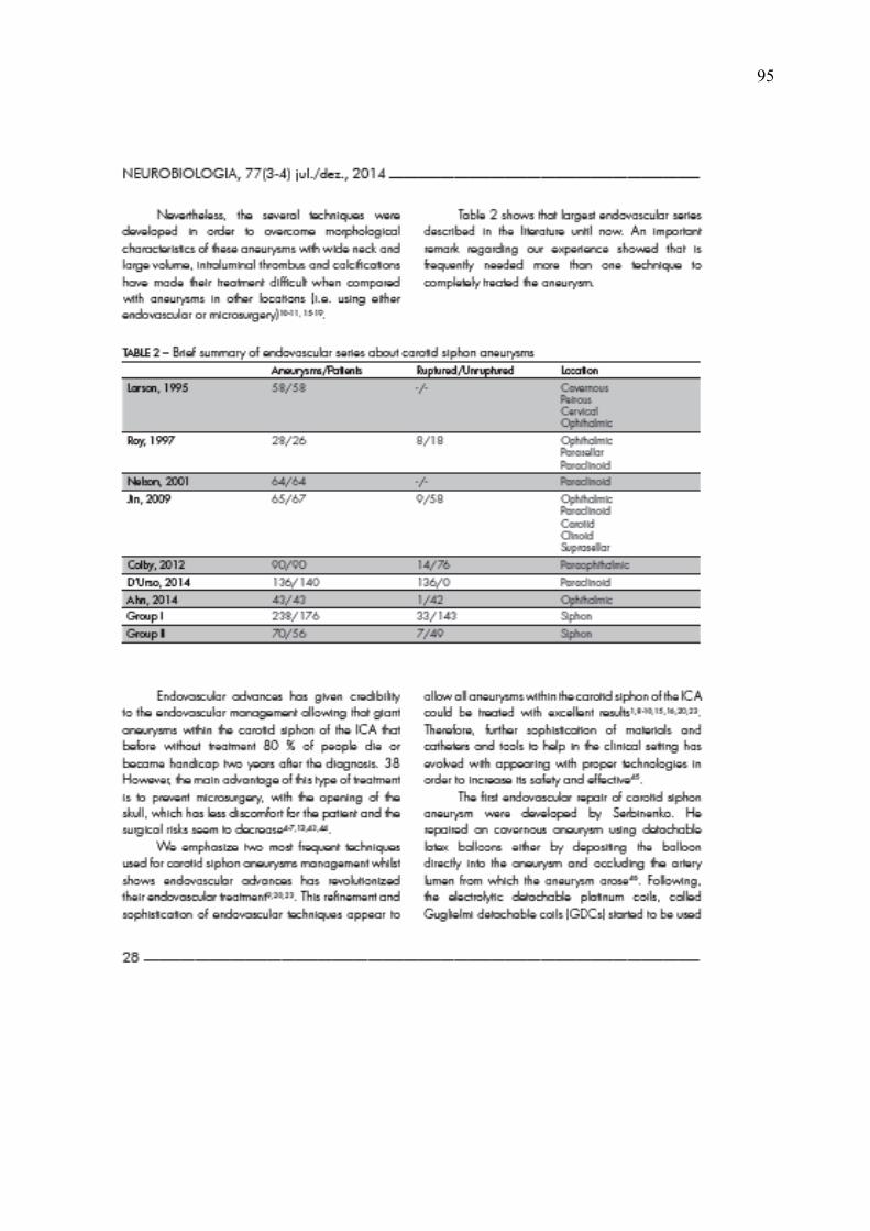
95

96

97

98

99

100

101

102

103

104
CAPÍTULO VII
Artigo Submetido para Publicação na Revista Anatomical Record

105
Artigo Submetido para Revista “The Anatomical Record”
Submitted to THE ANATOMICAL RECORD
Aneurysms of Egas Moniz Siphon: A Three-Dimensional Geometric Morphometric Classification Proposed in the Areas of
Susceptibility PATRICIA B. AMBROSI 1,2, CARLOS A.C. DE VASCONCELOS 3, LAURENT SPELLE1, JACQUES MORET1, MARCELO M. VALENÇA2,4, VALÉRIA P.S. FAZAN5, JEFFREY T. LAITMAN6
1Neuri- Beaujon, Interventional Neuroradiology, Clichy, Paris, 2 Postgraduate Program in Biological Sciences, Federal University of Pernambuco, Recife, Brazil
3 Department of Nutrition/LNED, Federal University of Pernambuco, Recife, Brazil
4 Department of Neuropsychiatry, Federal University of Pernambuco, Recife, Brazil
5 Department of Anatomy and Surgery, University of São Paulo, Ribeirao Preto, Brazil
6 Department of Anatomy, Mount Sinai School of Medicine, New York, New York
ABSTRACT
The aneurysms that develop on the circumference of the carotid siphon are a diverse group of challenging lesions, due to the anatomical complexity of the region in which they are located (i.e. using either endovascular or microsurgery). A new approach of endovascular treatment has been addressed to correct the disturbances involving hemodynamic and vascular morphology. Furthermore, it has been hypothesized that siphon carotid morphology variability might be related to aneurysms development. However, until recently, there has been no consensus yet about morphologic subtypes of carotid siphon aneurysms. Two hundred ninety human aneurysms were analyzed and nine different morphologic subtypes most frequently found were identified. We attempt to identify the most prevalent subtypes based in areas of susceptibility using previous microsurgical and radiological landmarks held and correlating with siphon morphology. Results indicate that Egaz Moniz siphon aneurysms despite the diversity seem to be more frequent in the upward curve of a carotid siphon. More future studies are desired to understanding the morphological and functional patterns of the carotid siphon.
Key words: vascular geometry; morphology; intracranial aneurysms; carotid siphon; bends; genesis

106
Abbreviations: CTA= computed tomography angiography; DSA= digital substration angiography; ICA= internal carotid artery; ; PSR= parasellar; WSS= wall shear stress; WSSG= wall shear stress gradient
*Correspondence to: Patricia B Ambrosi, MD, 100 boulevard du General Leclerc, Paris Diderot University, Paris, 92110. Fone: +33643505184. E-mail: [email protected]
Received ? December 2015; Accepted ? ? ?
INTRODUCTION
In 1927, the Portuguese neurologist Egas Moniz introduced the contrasted X-Ray cerebral angiography to study the angioarchitecture of cerebral vasculature (Bozzetto, 2014; Lima, 1951; Schierhorn, 1981). Five years later, Moniz described the carotid siphon and demonstrated an aneurysm arising from the internal carotid artery (ICA) at the skull base (Bozzetto, 2014; Moniz, 1933). The aneurysms that develop in the circumference of the carotid siphon are quite diversified (Ahn, 2014; Barami, 2003; Colby, 2012; Day, 1990; D’Urso 2014; Jin 2009; Larson, 1995; Wang 2013; Roy, 1997). Beyond anatomical, microsurgical and complex neuroendovascular repair, the natural history of intracranial aneurysms of remains poorly understood. However, several studies showed a correlation between geometric variability of the carotid siphon and the possible aneurysm formation (Bozzetto, 2010; Lee, 2008; Silva Neto, 2012). The areas of turbulent flow are susceptible to weakness and morphological changes of the vascular wall, which may be associated with aneurysm development (Bozzetto, 2010; Valen-Stendstat K et al., 2014). On reviewing the literature, two main points in the natural history of carotid siphon aneurysms require further investigation. Firstly, there is a need of accurate statistics because an underestimation is observed, as most of the current statistics are based on ruptured aneurysms and less about unruptured aneurysms. Secondly, most of series highlight ruptured aneurysms particularly that appear above clinoidal (described in the literature as carotid-ophthalmic, para ophthalmic, para clinoid among others (Ahn, 2014; Barami, 2003; Colby, 2012; Day, 1990; Larson, 1995; Roy, 1997). In fact, it was observed that there is still no agreement on the morphological subtypes of the different types of carotid siphon aneurysms. As far the other subgroups as the cavernous aneurysms, cave, transitional, dorsal wall carotid, blister-like and others remain largely unexplored. It is stated that carotid siphon aneurysms should represent about 5-10% of intracranial aneurysms. However, it is estimated that more than 30 % of all aneurysms are located in the intracranial segments of ICA usually at or distal to the carotid siphon (Bogunovic et al, 2012). New biotechnologies have been incorporated into the diagnosis and management of intracranial aneurysms (Bozzetto et al.,2015). Some of these new technologies consist of tools to better understanding the pattern of aneurysmal flow and detect possible morphological or hemodynamic aneurysm-specific features which could be responsible for the formation of cerebral aneurysms (Bozzetto et al., 2015; Sekhar et al., 1981). Through experimental studies and flow inside the dome of an aneurysm seems to be the hemodynamic stress is caused by a sequential and repetitive turbulent flow. This type of abnormality was evidenced in the cavity of an intracranial aneurysm during systole. This abnormal flow becomes reversed during diastole so that these rapid changes in the direction of flow continues to cause friction on the inner wall of the vessel and contributing to the formation and progression of an aneurysm (Gonzalez et al., 1992). Following to refinement of endovascular treatment which has targeted to correct the disturbances involved in hemodynamics/geometry and vascular morphology, a new morphological classification would be particularly useful to give a new approach linking pathogenesis and treatment of carotid siphon aneurysms. In this paper, we analyze the carotid siphon morphology and discuss the relevance

107
of classifying in subtypes morphological for the better understanding of pathogenesis and treatment of this peculiar aneurysms.
FUNCTIONAL SHAPE OF CAROTID SIPHON
The carotid siphon or also known as Egas Moniz’s siphon refers to the radiographic appearance of the internal carotid artery as a series of twists and curves that characterize this anatomical landmark (Moniz, 1933). The term carotid siphon gained popularity after Egaz Moniz publication based on brain contrasted radiographic studies of ICA (Sander-Taylor et al., 2013), when he also described an aneurysm of internal carotid at the base of the brain and described the siphon carotid using a two-dimensional projection with anatomical documentation and being the most important landmark in the angiogram (Moniz, 1933). According to Egas Moniz’s original description, the carotid siphon in 70 per cent of cases showed another curvature towards the frontal region, giving the impression of the double siphon (Moniz, 1933; Moniz,1934). Posteriorly, anatomical studies about carotid siphon showed 84% of adults have ICA has the characteristic double-bent shape (Platzer, 1957) but curiously in neonates apparently ICA takes a much straighter course. Studies particularly the investigation of Weninger and Muller demonstrated that the anatomy of the infant para sellar portion (PSR) differs distinctly from that of the adult and the PRS of the ICA of the neonates does not form a siphon (Weninger and Muller, 1999) which indicates that the shape of ICA is strongly transformed during early childhood.
Inside the carotid siphon and along the ICA is frequent to find tortuous curves and angles as Moniz described and illustrated in Figure 1 below. Carotid siphons are common sites of intracranial atherosclerosis. Probably due to its nature a tortuous vessel segment with sharp bends and large area variations and which also has relevance to the study of aneurysms initiation and rupture (Sendstad, 2014).Generally carotid siphon tortuosity in elderly patients is particularly related to atherosclerosis. These curves can be seen even in children, and described as anatomical variant type. In children, however, it is observed that in over half the cases, the bifurcation of the common carotid artery, the origin of ACI, is typically located more superior compared to the origin of this artery in the adult at the time of the second and third cervical vertebrae (Dilenge, 1962). More recently three-dimensional and hemodynamic studies have been helpful providing the better assessment to the carotid siphon morphology, particularly due to its sinuous shape which sometimes difficult to exact positionally and analysis (Kim et al., 2015). Further studies have showed that high peak focal curvature of the carotid siphon is correlated with aneurysm presence. In synthetic and patient-derived models, carotid loops are characterized by high WSS and WSSG, which correspond to the site of peak focal curvature. Aneurysms in these regions seem to form distal from the site of peak curvature, in areas of stagnant flow flanked by dynamically fluctuating high WSS and WSSG peaks. The distance between WSS peaks correlates with the size of the aneurysm neck (Lauric et al., 2014). The new strategy of endovascular treatment also has been addressed to explore carotid shaping. In Figure 1, we also illustrated the flow studies, which are taking part of the endovascular strategic particularly during the treatment of carotid siphon aneurysms with flow diverters. However, this research focused on areas of susceptibility and morphology but did not consider the influence of flow instabilities and hemodynamic measures.

108
Fig. 1. Three-Dimensional geometric morphometric reconstruction (a) and curve reformatted view (b) of an female adult and its left carotid siphon The distal tip of the blue linel in this geometric configuration is within the carotid siphon (c) blue line in the lumen of siphon (d) optic flow study before (e) and after flow diverters treatment (f) linear reconstruction of same siphon (g)
GEOMETRIC MORPHOLOGICS OF CAROTID SIPHON
Although the understanding that local geometrical and hemodynamics factors are still contradictory.

109
Recent studies have showed that an association between the presence of aneurysms and intracranial ICA area expansions (Schimansky et al., 2013), curvatures (Lauric et al., 2013), angle (Silva Neto et al., 2012) geometric features know to affect flow stability ICA area expansions. There is no yet specific pattern described of carotid siphon associated with aneurysms or subtypes of aneurysms.
In 1965, Weibel and Field performed anatomical studies and demonstrated that the initial portion tortuous of carotid siphon shape is more frequently observed in individuals older than 50 years old (Weibel and Field, 1965). Three year later, Yasargil and krakenbuel using 2D angiographical projections demonstrated that carotid siphon may differ remarkably due to its tortuosity and described seven important variations noted in the carotid siphon: (1)U-shaped observed in 40,1 % of patients between 0-20 years, 35.0% between 21-50 years and 15,2 % between 51-74 years, which indicates that it type of young siphon. (2) C-shaped also observed in 45,2% between 0-21, 14,6% between 21-50 years and 15,2% between 51-74 years, also denotates a type of young siphon. (3) V-shapes has equilibrated distribution respectively (Yasargil and krakenbuel, 1968). Dilenge and Heon, 1974 simplified the Krayenbuhl and Yasargil’s classification and reclassified in four types: U, C, arch and double siphon (Dilenge and Heon, 1974). Recently Lin et al., reintroduced Yasargil & Kraenbuel’s classification based on the tortuosity of the cavernous segment of the internal carotid artery as a predictive factor in the complexity of embolization with Pipeline® (Lin et al., 2014). Lin et al., classified into Class IA and IB, when the siphon showing a configuration with open angle determined by the angle of the posterior knee, where IA is greater than 90 and IB is equal to 90°. The Type II was characterized by the closed configuration of the previous angle with a more acute angle compared to the Type I and Type III when the carotid siphon had the further deflection of the knee. Type IV represents the most tortuous of all with the characteristic feature of Simmons® catheter.
CAROTID SIPHON AND ITS ANEURYSMS IN ANIMALS
Comparing the cerebral vasculature between humans and other animal species, particularly the rat (Rattus norvegicus) one of the most common animal used in scientific experimentation. The cerebral circulation differs between men and animals. Most animals have the rete mirabile (Ashwini, 2008). We found that carotid siphon has a similar confirmation in its proximal trajectory, but the anatomical topography is unique in humans compared with other vertebrates and mammals, as well as other primate species (Ashwini, 2008; Lee, 1995). Although aneurysms in rats are not common, German and Black, 1954 were the first researchers to produce experimental aneurysms using the surgical construction of saccular aneurysms on the common carotid artery of dogs (German and Black, 1954). Carotid aneurysms experimental models mimicked the ones occurring on the side wall and were frequently used in hemodynamic studies. However, they produced fibrosis at the suture site, which was a disadvantage. Since then, surgical models have evolved with the culmination of the swine model consisting of a graft of the venous pouch onto the common carotid artery of pigs. This method produces lateral wall aneurysms but includes disadvantages such as venous histology, induction of intense fibrosis at the suture site, hemodynamic and low tension.
HYPOTHESES
An attempt to estimate the most prevalent morphological subtypes based in areas of susceptibility. A review of microsurgical landmarks and previous classifications of carotid siphon aneurysm were held. It has been hypothesized that there are specific morphological and shapes configurations related to aneurysms development. 1. What are main susceptible areas of carotid aneurysms development?

110
2. What are the subtypes of siphon most frequently related to siphon carotid aneurysms, is there differences between males and females siphon? 3. Is there a difference between human age and their carotid siphon and subtype of carotid siphon aneurysm? 4. Can we predict bleeding by carotid siphon morphologic subtype? 5. Perhaps certain aneurysms would justify screening and preventive treatment?
MATERIALS AND METHODS
STUDY DESIGN AND ANEURYSM POPULATIONS
After reviewing most known classifications of ICA and carotid siphon aneurysms in the literature which are described in Table 1. Some authors have classified as carotid-ophthalmic, para clinoid, superior hypophyseal and cavernous aneurysms. Most of the classifications were addressed to the horizontal and supraclinoid portion of ICA. Several classifications of carotid siphon on basis of surgical considerations had been addressed in the literature and a established classification system based on the exact point from the carotid artery into ophthalmic, superior hypophyseal, clinoidal, cave, transitional, intracavernous (Al-Rodhan et al., 1993, Barami, 2003; Day, 1990; Roy, 1997; Ogilvy, 1995;Wang et al., 2013).
Aneurysms are considered within the carotid siphon when they were located in segments by C4-6 Bouthilier 5 (C2 -C4 classification of the Fischer) (Fischer, 1938; Bouthilier,1996) or based on the original description of Egas Moniz (Moniz, 1933). Data were collected with institutional and university ethics committee approval (CAAE-0196.0.172-000-10) regarding the group I and (CAAE- 4707.5.215.1.0000.5208) regarding group II. The aneurysms population analyzed were divided into two groups: Group I that included 1288 patients (432 men and 856 women) with 1552 intracranial aneurysms (IAs) which were treated between 1st of February 2002 and 25th of May 2010 at Foundation Adolphe Rothschild, Paris, France. We identified a total of 135 patients (with 178 carotid siphon aneurysms). When compared with those patients with aneurysms in another location than siphon segment (414 men and 739 women; 1: 1.8) there was a significantly higher chance of finding females in siphon segment aneurysms of the ICA group (OR 3.641, 2.185 to 6.068 95% CI p <0.0001). Each type of carotid siphon aneurysm were considered based on its specific clinical and angiographical features according to the Figure 2. There were five most prevalent groups of siphon carotid aneurysms: (1) ophthalmic, (2) superior hypophyseal (3) clinoidal or cave, (4) suprasellar, (5) others. Four aneurysms were purely located in the intracavernous segment and were not included in the first analysis. Fig. 2 - Three-Dimensional siphon reconstruction illustration

111
Group II included 532 patients (295 IAs) treated between 1st November 2010 and 31th of October 2014 at the Beaujon University Hospital, Clichy, France. This group contained 79 patients (5 men and 75 women) with 112 aneurysms within the ICA carotid siphon, which were analyzed prospectively. After our redefinition about carotid siphon aneurysms, combined analysis of both groups were performed and nine most prevalent groups were considered: (1) ophthalmic linked, (2) ophthalmic related (3)superior hypophyseal, (4) clinoidal or cave, (5) suprasellar, (6) dorsal wall, (7) anterior wall (8) posterior communicant artery related, (9) choroidal anterior or terminal as illustrated in Figure 3. There are possible subtypes not covered, for example, some of the aneurysms can be difficult to classify. Fig. 3- Three-Dimensional geometric siphon schema and neighbour structures (a), siphon shape subtypes (b), magnification of carotid siphon with morphological subtypes most prevalent encountered

112
RESULTS
DEMOGRAPHICS AND CLINICAL FEATURES
Characteristics of patients with siphon carotid aneurysms analyzed shown in Table 2. Patients were 90.6% female (194/214), with a mean admission age of years 49.24 ± 11.8 (range 16-84) in the group I and 50,23 ± 13,18 in the group II. Thirty-nine patients (18,2 %) had a history of subarachnoid bleeding.
In the group I, when comparing ruptured and un-ruptured aneurysms, the median age of patients was 48.90 ± 14.62 years (range 18-81) and 50.22 ± 13.4 years (range 23-84) respectively. The most common age group was between 41-60 years (59.3% of the total sample), followed by 21-40 years (22.2%). Multiple aneurysms were present in 45.9% (62/135) of patients. Mirror aneurysms were present in 17% (23/178) of patients.
Clinical features were tenaciously studied in the group I, the most common clinical presentation that was founded in the referral was the presence of a headache (observed in 51.1 % (69/135) of patients), only 22.9% of patients had ruptured aneurysms. A group of 24 patients complained of visual disturbances, ie 19.2 % of patients had visual deficits. The incidental finding of an aneurysm detected through studies by MRI or CTA during an investigation in the context of a chronic headache or other neurological symptoms occurred in 61/135 (45 %) patients. Remaining symptoms were other
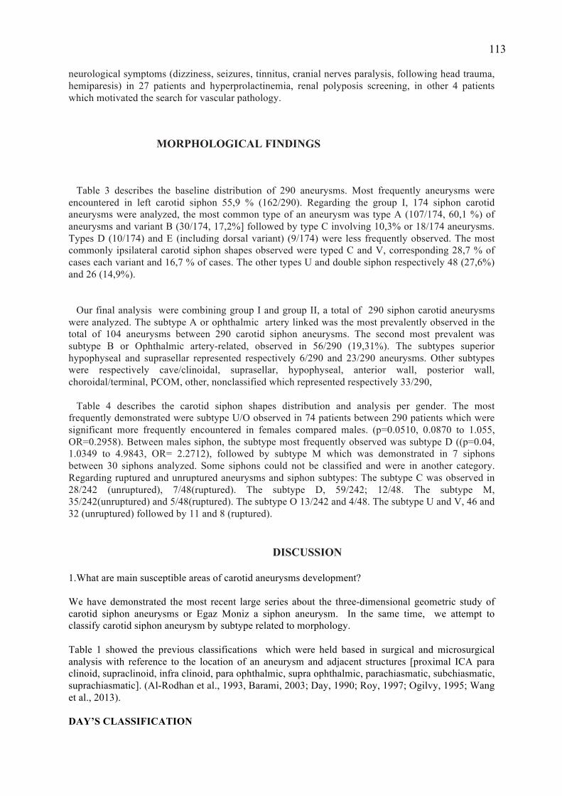
113
neurological symptoms (dizziness, seizures, tinnitus, cranial nerves paralysis, following head trauma, hemiparesis) in 27 patients and hyperprolactinemia, renal polyposis screening, in other 4 patients which motivated the search for vascular pathology.
MORPHOLOGICAL FINDINGS Table 3 describes the baseline distribution of 290 aneurysms. Most frequently aneurysms were encountered in left carotid siphon 55,9 % (162/290). Regarding the group I, 174 siphon carotid aneurysms were analyzed, the most common type of an aneurysm was type A (107/174, 60,1 %) of aneurysms and variant B (30/174, 17,2%] followed by type C involving 10,3% or 18/174 aneurysms. Types D (10/174) and E (including dorsal variant) (9/174) were less frequently observed. The most commonly ipsilateral carotid siphon shapes observed were typed C and V, corresponding 28,7 % of cases each variant and 16,7 % of cases. The other types U and double siphon respectively 48 (27,6%) and 26 (14,9%). Our final analysis were combining group I and group II, a total of 290 siphon carotid aneurysms were analyzed. The subtype A or ophthalmic artery linked was the most prevalently observed in the total of 104 aneurysms between 290 carotid siphon aneurysms. The second most prevalent was subtype B or Ophthalmic artery-related, observed in 56/290 (19,31%). The subtypes superior hypophyseal and suprasellar represented respectively 6/290 and 23/290 aneurysms. Other subtypes were respectively cave/clinoidal, suprasellar, hypophyseal, anterior wall, posterior wall, choroidal/terminal, PCOM, other, nonclassified which represented respectively 33/290, Table 4 describes the carotid siphon shapes distribution and analysis per gender. The most frequently demonstrated were subtype U/O observed in 74 patients between 290 patients which were significant more frequently encountered in females compared males. (p=0.0510, 0.0870 to 1.055, OR=0.2958). Between males siphon, the subtype most frequently observed was subtype D ((p=0.04, 1.0349 to 4.9843, OR= 2.2712), followed by subtype M which was demonstrated in 7 siphons between 30 siphons analyzed. Some siphons could not be classified and were in another category. Regarding ruptured and unruptured aneurysms and siphon subtypes: The subtype C was observed in 28/242 (unruptured), 7/48(ruptured). The subtype D, 59/242; 12/48. The subtype M, 35/242(unruptured) and 5/48(ruptured). The subtype O 13/242 and 4/48. The subtype U and V, 46 and 32 (unruptured) followed by 11 and 8 (ruptured).
DISCUSSION 1.What are main susceptible areas of carotid aneurysms development? We have demonstrated the most recent large series about the three-dimensional geometric study of carotid siphon aneurysms or Egaz Moniz a siphon aneurysm. In the same time, we attempt to classify carotid siphon aneurysm by subtype related to morphology. Table 1 showed the previous classifications which were held based in surgical and microsurgical analysis with reference to the location of an aneurysm and adjacent structures [proximal ICA para clinoid, supraclinoid, infra clinoid, para ophthalmic, supra ophthalmic, parachiasmatic, subchiasmatic, suprachiasmatic]. (Al-Rodhan et al., 1993, Barami, 2003; Day, 1990; Roy, 1997; Ogilvy, 1995; Wang et al., 2013). DAY’S CLASSIFICATION

114
Kothandaram et al., 1971 were the first authors to describe differents subtypes of para clinoid aneurysms. Therefore, Day 1990 draw attention to differents subtypes of carotid siphon aneurysm and established the first classification which was used as a baseline in the future classifications. Day, 1990 analyzed clinical, radiographic and anatomical features in 80 patients with the ophthalmic aneurysms and categorized into (1) ophthalmic and (2) superior hypophyseal aneurysms, according to a presumed aneurysmatic origin related. The main interest was in the fact to display a difference in the technical challenge and morbidity associated with the treatment of these two groups of aneurysms. The first group represented the true carotid-ophthalmic aneurysms, that is those arising at the junction between the ICA and the origin of the ophthalmic artery, pointed superiorly or superomedially and (when large) deflected the carotid artery posteriorly and inferiorly. The second group, superior hypophyseal artery aneurysms arose just above the dural ring from a medial bend of ICA, at the site of superior hypophyseal artery origin to the superior aspect of the hypophysis, and had no direct association with the ophthalmic artery. These lesions could extend medially beneath the chiasm (suprasellar variant), producing a clinical and computerized tomography picture similar to a pituitary adenoma, or they could extend ventrally to burrow beneath the anterior clinoid process (para clinoid variant) (Day, 1990). AL-RODHAN & OGILVY’S CLASSIFICATION Al-Rodhan et al., 1993 described five subgroups: Group I – when the neck was intradural located and ICA segment that is supra-ophthalmic and infra posterior the posterior communicating. This group is divided into two subgroups: Ia- refers to aneurysms known as hypophyseal superior projecting superiorly Ib- are aneurysms that Yasargil & Fox has called ventral paraclinoidal protruding posteriorly and inferiorly intradural or extradural into the cavernous sinus. Group II - true aneurysms whose arises at the junction between the ophthalmic artery and ACI, which projects anterior-superior and remain infra-durally. Group III - are aneurysms that Kobayashi called cave carotid. Are medial, infra-ophthalmic and cavernous (Kobayashi, 1989; Hitotsumatsu, 1997). Group IV - are so-called transitional, whose lap surge in the cavernous space, but its dome protrudes intradural. Group V - purely intracavernous. Posteriorly, Ogilvy et al., applied this classification and reviewed their experience using a combined surgical and endovascular treatment to approach to para clinoid segment aneurysms between 1991 and 1999. They reviewed 238 aneurysms in 216 patients and subdivided into five subgroups: 1) true ophthalmic aneurysm, arise from the of the ophthalmic artery which is equivalent of subgroup 1 of Day. 2) Superior hypophyseal aneurysm arises distal to the ophthalmic artery and project medially, often incorporating perforating branches of the hypophysis also equivalent of subgroup 2 of Day 3) Carotid a cave aneurysm arises at or bellow the dural ring around the ICA as it enters the subarachnoid space which was described by Kobayashi, 1989 4) Posterior carotid wall aneurysms arise just distal to the ophthalmic artery on the posterior wall of the ICA and project posteriorly which were described originally by Dandy. 5)Transitional aneurysms originate within CS and extend superiorly into the subarachnoid space. ROY & RAYMOND’S CLASSIFICATION In 1997, Roy and Raymond reviewed their endovascular experience and twenty-eight aneurysms encountered in 26 patients were analyzed and the second group described by Day was subdivided into two variants: para clinoid and suprasellar. The para clinoid variant when an aneurysm had an intracavernous proximal neck and fundus with only the distal neck lying in the subarachnoid space. It was included a para clinoid variant what Kobayashi called carotid cave aneurysms when both involve the proximal and medial parts of an ophthalmic segment (Roy et al, 1997, Beretta et al, 2004). On the

115
other hand, the aneurysms arising in association with aberrant origin of the ophthalmic artery and superior hypophyseal artery, occurring in 10% of cases. An aneurysm was initially expanding toward the optic strut and may project laterally or medially or superiorly leading to optic nerve compression or rash toward hard dural ring adjacent to the subarachnoid space. Subsequently, this same classification was adapted by Roy describe the endovascular treatment of aneurysms of the ophthalmic segment 28 in 26 patients treated by coiling as the first indication as to the treatment (Roy et al., 1997.
2. What are the subtypes of siphon most frequently related to siphon carotid aneurysms, is there differences between males and females siphon?
We demonstrated that carotid siphon may differ remarkably from normal patients as Yasargil description where
(1)U-shaped observed in 40,1 % of females;
(2) C-shaped which could indicate the denotates a subtype of "young" siphon;
(3) V-shapes has equilibrate distribution respectively 14,6 (0-21y),24,5% (21-50 y), 22,3% (51-74y)
(4) Omega- shaped siphon appear to be normal variant from U-shaped, which respectively 23,7% (21-50 y) and 50,7% (51-74y).
(5) Double siphon, Megasiphon, and Dolicosiphon were the most frequently observed which might denotate a vascular remodeling occurring in the carotid siphon.
Female and males vasculatures appear to have a different configuration. At the same time, our research corroborates the recent speculations where the authors have been investigating the relationship between the circle of Willis (circulation variations) and analyzing the risk of aneurysm formation detected.
The known correlation between the men and women to the anatomical presence variants also appear to be correlated with the difference the distribution of aneurysms. Females also have a predominance of PCOM fetal comparing to males. Evidence shows that the local aneurysmatic distribution could be linked with sex. At the same time, the Willis Circle anatomy has considerable variability with frequent asymmetries So probably the vessel geometry variations can play a role in the genesis of intracranial aneurysms, particularly in this siphon region that is subjected to significant variability. At this point, Padget was among the first to say this for a number of cases compared to embryological abnormalities with aneurysms and without aneurysms (Padget, 1968).
FUTURE DIRECTIONS To summarize, the results of this present research confirm there are several morphological patterns of siphon related to carotid siphon aneurysms. It appears that the siphon carotid aneurysms development is related to aging process of the vascular wall and accelerated by atherosclerosis. The siphon which is straight in the children appears to suffer remodeling which could be particularly accelerated in patient with hyperdynamic status or asymmetric will polygon. This study suggests that there is certainly a particular shape morphology modifying that happen and which be related with aneurysms genesis. Besides, there is probably standard pattern with the geometry siphon associated with certain subtypes of aneurysms.This is one the first recent studies related to siphon morphology which are more focused in treatment operative techniques. There is a lack of detailed studies in this matter. The lack of information is also related to a hidden position of carotid siphon which prevents the use of some techniques as doppler for example. We attempt to incorporate new methods to our routine to better understand the morphology and hemodynamic of carotid siphon aneurysms. However, the understanding of vascular morphology appear to be promising for the understanding of the genesis of aneurysms, and also the response to treatment with new technologies as flow diverters stents.

116
These new evidence should contribute to the increased of studies in this matter and helping us to better understand the pathophysiology and mainly pointing to a more effective and efficient direction in the detection and prevention of aneurysms.
ACKNOWLEDGEMENT
LITERATURE CITED
Abla A, Lawton M. Current Treatment Strategies for Cavernous Internal Carotid Artery. Aneurysms World Neurosurg in
press.
Ahn JH, Cho YD, Kang HS, Kim JE, Cho WS, Jung SC, Kim CH, Han MH. 2014. Endovascular Treatment of Ophthalmic
Artery Aneurysms: Assessing Balloon Test Occlusion and Preservation of Vision in Coil Embolization. Am J Neurorad.
Ambrosi PB. 2010. Aneurysms of carotid-ophthalmic segment: clinical and angiographical findings. Federal University of
Pernambuco, Recife.
Ambrosi PB, Valenca M. 2014. Carotid Siphon Aneuysms, a silent disease. Editorial. Neurobiologia in press.
Ambrosi PB, Correia MTS, Spelle L, Moret J, Valença MM. 2015. Biotechnologies applied to intracranial aneurysms.
International Archives of Medicine.
Andaluz N, Beretta, T, Keller, JT, Zuccarello, M. Aneurysms of the ophthalmic (C6) segment of the internal carotid artery -
Clinical experience.
Ashwini CA, Shubha R, Jayanthi KS. 2008. Comparative anatomy of the circle of Willis in man, cow, sheep, goat, and pig-
Neuroanatomy 7: 54-85.
Barami, K, Hernandez, VS, Diaz, FG, Guthikonda, M. 2003. Paraclinoid carotid aneurysms: Surgical management,
complications, and outcome based on a new classification scheme. Skull Base-an Interdisciplinary Approach. 13:31-41.
Benderson JB, Connolly ES Jr, Batjer HH, Dacey RG, Dion JE, Diringer MN, Duldner JE Jr, Harbaugh RE, Patel AB,
Rosenwasser RH. 2009. American Heart Association SO. Guidelines for the management of aneurysmal subarachnoid
hemorrhage: a statement for healthcare professionals from a special writing group of the Stroke Council, American Heart
Association. AU Stroke. 40:994.
Beretta F. 2004. The paraclinoid aneurysms and the distal dural ring: a new classification. J Neurosurg Sci 48:161-75.
Bogunovic H1, Pozo JM, Cárdenes R, Villa-Uriol MC, Blanc R, Piotin M, Frangi AF. 2012. Automated landmarking and
geometric characterization of the carotid siphon. Med Image Anal 16:889-903.
Bouthillier A, Loveren H, Keller J. 1996. Segments of the internal carotid artery: a new classification. Neurosurgery 38:425–
432. Colby,GP, Paul, AR, Radvany, M.G. et al. 2012. A single center comparison of coiling versus stent assisted coiling in
90 consecutive paraophthalmic region aneurysms. J Neurointerv Surg,4:116-120.
Day AL. 1990. Aneurysms of the ophthalmic segment: a clinical and anatomic analysis. J Neurosurg 72:677-91.
Dilenge A, Heon M. 1974.The internal carotid artery. In: Newton TPD, ed. Radiology of the skull and brain. St. Louis, MO:
Mosby-Year Book.1238–1245.
Dilenge D. 1962. L’arteriographie de l’artere carotide interne. Masson Paris 1-230.
Dolenc VV. 1985. A combined epidural and subdural direct approach to carotid-ophthalmic artery aneurysms. J Neurosurg
62:667-672.
D'Urso PI, Karadeli HH, Kallmes DF, Cloft HJ, Lanzino G. 2012. Coiling for paraclinoid aneurysms: time to make way for

117
flow diverters? Am J Neuroradiol 33:1470-1474.
Fischer, E. Die. 1938. Lageabweichungen der vorderen Hirnarterie im Gefissbild.Zentralbl Neurochir 3:300-312.
Fox JL. 1983. The incidence of intracranial aneurysms. In: Fox, J.L. Ed.). Intracranial aneurysms. New York Springer
Verlag 1:15-17.
Fox AJ, Vinuela F, Pelz DM, et al. 1987. Use of detachable balloons for proximal artery-occlusion in the treatment of
unclippable cerebral aneurysms. J Neurosurg 66:40-46.
German WJ. 1954. Black SPW: Intra-aneurysmal hemodynamics: Turbulence. Trans Am Neurol Assoc 163-165
Jamn KK. 1967. Headache in relation to intracranial aneurysm. Headache 7: 122-126.
Indo M, Oya S, Tanaka M, Matsui T. 2014. High incidence of ICA anterior wall aneurysms in patients with an anomalous
origin of the ophthalmic artery: possible relevance to the pathogenesis of aneurysm formation. J Neurosurg 120:93-98.
Jin SC, Kwon DH, Ahn JS, Kwun BD, Song Y, Choi CG. 2009. Clinical and radiogical outcomes of endovascular
detachable coil embolization in paraclinoid aneurysms: a 10-year experience. J Korean Neurosurg Soc 45:5-10.
Jou LD, Lee DH, Morsi H, Mawad ME. 2008. Wall shear stress on ruptured and unruptured intracranial aneurysms at the
internal carotid artery. Am J Neuroradiol 29:1761-1767. Krayenbuel HA, Yasargil MG. 1968.Cerebral Angiography ed 2.
Philadelphia: JB Lippincott 24-38.
Kato Y, Sano H, Hayakawa M, Imai F, Kawase T, Nonomura K, Kanno T.1996 Surgical treatment of internal carotid
siphon aneurysms. Neurol Res 18:409- 15.
Kim BC, Know OK, Oh CW, Bang Js, Hwang G, Jin SC, Park H. 2014. Endovascular internal carotid artery trapping for
ruptured blister-like aneurysm: long term results from a single centre. Neuroradiology 56:211-7.
Kobayashi S1, Kyoshima K, Gibo H, Hegde SA, Takemae T, Sugita K. 1989. J .Carotid cave aneurysms of the internal
carotid artery. J Neurosurg 70:216-21.
Kothandaram P, Dawson BH, Kruyt RC.1971. Carotid- ophthalmic aneurysms. A study of 19 patients. J Neurosurg 34:544–
548.
Kwon BJ, Im SH, Park JC, et al. 2010. Shaping and navigating methods of microcatheters for endovascular treatment of
paraclinoid aneurysms. Neurosurgery 67:34-40.
Hoh BL, Carter BS, Budzik RF, Putman CM, Ogilvy CS. 2001. Results after surgical and endovascular treatment of
paraclinoid aneurysms by a combined neurovascular team. Neurosurgery 48:78-89.
Lanzino G, Crobeddu E, Cloft HJ, Hanel R, Kallmes DF. 2012. Efficacy and Safety of Flow Diversion for Paraclinoid
Aneurysms: A Matched-Pair Analysis Compared with Standard Endovascular Approaches. Am J Neuroradiol 33:2158-61.
Larson JJ, Tew JMJr, Tomsick TA,Van Loveren HR. 1995. Treatment of aneurysms of the internal carotid artery by
intravascular balloon occlusion: long-term follow-up of 58 patients. Neurosurgery 36:26–30.
Lauric A, Hippelheuser J, Safain MG, Malek AM. 2014. Curvature effect on hemodynamic conditions at the inner bend of
the carotid siphon and its relation to aneurysm formation. 2014. J Biomech 47:3018-27.
Lee RM.1995. Morphology of cerebral arteries. Pharmacol Ther 66:149-173.
Lee SW, Antiga L, Spence JD, Steinman DA. 2008. Geometry of the carotid bifurcation predicts its exposure to disturbed
flow. Stroke 39:2341–2347.
Lin ML, Colby GP, Bowen J, Chiedozie U, Huang J, Tamargo RJ, Coon AL. 2014. Classification of cavernous internal
carotid artery tortuosity: a predictor of procedural complexity in Pipeline embolization. NeuroIntervent Surg.
Lima A. 1951. L’angiographie cerebrale et la leucotomie pre-frontale:deux contributions de la neurologie portugaise.
Semaine des Hopitaux de Paris, 27:3-8.
Moniz, E.1933. Cerebral angiography. Its application in clinical practice and physiology. Lancet 2:1144-7.
Moniz E.1933. Anevrysme intra-cranien de la carotid interne droite rendu visible par l’arteriographie cerebrale. Rev-Oto-
Neuro-Ophtal 11-746-748.
Moniz E. 1934. L’arteriographie cerebrale. Masson et Cie 230-246.
Nelson PK, Levy DI. 2001. Balloon-assisted coil embolization of wide-necked aneurysms of the internal carotid artery:

118
medium-term angiographic and clinical follow-up in 22 patients. Am J Neuroradiol 22:19–26.
Ogilvy CS, Natarajan SK, Jahshan S, et al. 2011. Stent-assisted coiling of paraclinoid aneurysms: risks and effectiveness. J
Neurointerv Surg 3:14-20.
Omodaka S, Sugiyama S, Inoue T, Funamoto K, Fujimura M, Shimizu H, Hayase T, Takahashi A, Tominaga T. 2012.Local
hemodynamics at the rupture point of cerebral aneurysms determined by computational fluid dynamics analysis.Cerebrovasc
Dis. 34:121-9.
Padget DH .1945. The circle of Willis: its embryology and anatomy. In Dandy, WE. Intracranial arterial aneurysm. New
York:Comstock, 74-85.
Pia HW. Cerebral Aneurysms. Advances in Diagnosis and Therapy History of Aneurysm Surgery 1.
Platzer W.1957.Die Variabilität der Arteria carotis interna im Sinus cavernosus in Beziehung zur Variabilität der
Schädelbasis. Gegenbauers Morphol Jahrb. 98:227–244.
Puffer RC, Kallmes DF, Cloft HJ, Lanzino G. 2012. Patency of the ophthalmic artery after flow diversion treatment of
paraclinoid aneurysms. J Neurosurg 116:892-89.
Roy D, Raymond J, Bouthillier A, Bojanowski MW, Moumdjian R, L'Espérance G. 1997.Endovascular treatment of
ophthalmic segment aneurysms with Guglielmi detachable coils. Am J Neuroradiol 18:1207-1215.
Sanders-Taylor C1, Kurbanov A1, Cebula H1, Leach JL2, Zuccarello M3, Keller JT4. 2014. The carotid siphon: a historic
radiographic sign, not an anatomic classification. World Neurosurg 82:423-7.
Schierhorn H. 1981. Egas Moniz, Wilhelm Löhr and cerebral angiography. An expedition into the discovery of the cerebral
vessels. J Hirnforsch 22:491-515.
Schimansky S, Patel S, Rahal J, Lauric A, Malek AM. 2013. Extradural internal carotid artery caliber dysregulation is
associated with cerebral aneurysms. Stroke 44, 3561–3564.
Sherif C, Gruber A, Dorfer C, Bavinzski G, Standhardt H, Knosp E. 2009. Ruptured carotid artery aneurysms of the
ophthalmic (C6) segment: clinical and angiographic long term follow-up of a multidisciplinary management strategy. J
Neurol Neurosurg Psychiatry 80:1261-1267.
Silva Neto ÂR, Câmara RL, Valença MM. 2012.Carotid siphon geometry and variants of the circle of Willis in the origin of
carotid aneurysms.Arq Neuropsiquiatr 12:917-21.
Stehbens WE. 1983.The pathology of intracranial arterial aneurysms and their complications.ln: Fox, J.L. ed, Intracranial
Aneurysms. Springer-Verlag 272-342.
Valen-Stendstat K, Piccinelli M, Steinman DA. 2014. High-resolution computational fluid dynamics detects flow
instabilities in the carotid siphon: implications for aneurysm initiation and rupture? J Biomech 12:3210-6.
Vlak MH, Algra A, Brandenburg R, Rinkel GJ. 2011. Prevalence of unruptured intracranial aneurysms, with emphasis on
sex, age, comorbidity, country, and time period: a systematic review and meta-analysis. Lancet Neurol 10:626-36.
Wang Y, Li Y, Jiang C, et al. Endovascular treatment of paraclinoid aneurysms: 142 aneurysms in one centre. J Neurointerv
Surg 5:552-556.
Weninger W, Muller GB.1999.The parasellar region of human infants: cavernous sinus topography and surgical approaches.
Journal of neurosurgery, 90: 484-490.
Xu BN, Sun ZH, Romani R, et al. 2010. Microsurgical management of large and giant paraclinoid aneurysms. World
Neurosurg 73:137-146.
Zhu YQ, Li MH, Xie J, Tan HQ, Cheng YS, Wang JB. 2010. Treatment of carotid siphon aneurysms by use of the Willis
stent graft: an angiographic and histopathological study. Eur Radiol 20:1974-19

119
TABLE 1. Overview of morphological classifications, types of aneurysms, sample studied, type of treatment performed, carotid segment, type of analysis done, carotid surface analysed. Types of aneurysms Patient, SAH and aneurysms Surgical or Carotid Type of Surface (n, n, n) Endovascular Segments Analysis Treatment (Fisher, Bouthillier) Kothandaram, Subchiasmal, 19 Surgery C2, C3 intraoperative Superior 1971 Suprachiasmal, Paraclinoid Day, 1990 True carotid-ophthalmic 80, 28, 41 carotid-ophthalmic 67% (54/80) C2, C3 intraoperative Superior Superior hypophyseal 39 hypophyscal clipping remaining-not Al-Rodhan, 1993 Group I a- superior hypophyseal 118, 13, 23 transitional 73% (18/23), 17 C2, C3, C4 Angiographic/ Superior Group I b- ventral paraclinoid direct clipping, 1, Intraoperative Group II- true ophthalmic bypass findigns Group III- Kobayashi Group IV- transitional Group V- intracavernous Ogilvy, 1995 Transitional 216, 59(25%), 238 180/216 (%) clipping, C2, C3 Angiographic/ Superior Carotid Cave 57/216 endovascular, Intraoperativve Ophthalmic 1 bypass Findings Superior Hypophyseal Posterior Carotid Wall Kato , 1996 Ophthalmic 48, -, 45 All surgical patients C2- C4 - Superior Clinoid Horizontal Roy, 1997 1-ophthalmic 26, 8, 28 26 endovascular C2, C3 Angiographic Superior

120
2A upper-paraclinoid 100% treated findings variant of superior hypophyseal 2A lower-kobayashi cave 2B suprasellar variant of superior hypophyseal Barami, 2003 Ophthalmic artery (Ia) 61,-,61 61 surgical C2, C3 Angiographic Superior, Dorsal ICA (Ib) findings Ventral Ventral ICA (II) Medial Supradiaphragmatic (IIIa) Infradiaphragmatic (IIIb) Sherif, 2008 Ophthalmic artery 64, -, 64 23 surgery, C2 Superior Hypophyseal Artery 38 embolization Posterior Wall Carotid 3 bypass Carotid Cave Transitional Wang, 2013 Type I- distal ophthalmic 56, -, 64 12 coiling and C2, C3 Angiographic Superior Type II- ventral, medial or lateral 52 stenting findings segment Subtype a- proximal Subtype b- distal New 9 types Until 2010, 90% classification coiling, 81% remodeling balloon After 2010, 90% flow diverters
A- linked to the ophthalmic 214,-, 290 Group I- 93% C3-C5 Group I-2D All artery ; Superior face of endovascular, 81% carotid artery remodeling balloon B- Superior face of ICA, not linked 7% no treatment and 3D
to ophthalmic artery C- Cave carotid Group II- 100% Group II-3D endovascular, 90%
flow diverters

121
TABLE 2. Demographical features of patients analysed. Total of patients Gender Total of aneurysms Ruptured
214 194/214 Females (90,6%) 20/214 Males(10,2%) 290 39/214 patients (18,22%)
Group I n/n(%)
Group II n/n (%)
Age (yr) 49,24 ± 11,8 50,23 ± 13,18 Gender Male Female
18/135 (13,3) 117/135 (86,7)
5/79 (6,3) 74/79 (93,7)
Multiple aneurysms 62/135 (45,9) 33/79 (41,7) Mirror 23/135 (17,0) 10/79 (17,9) SAH 31/178 (22,9) 8/79 (10,1) Total of aneurysms 178/135 (1,3) 112/79(1,4) Total of treated aneurysms
163/178 (91,5)
112/112(100)
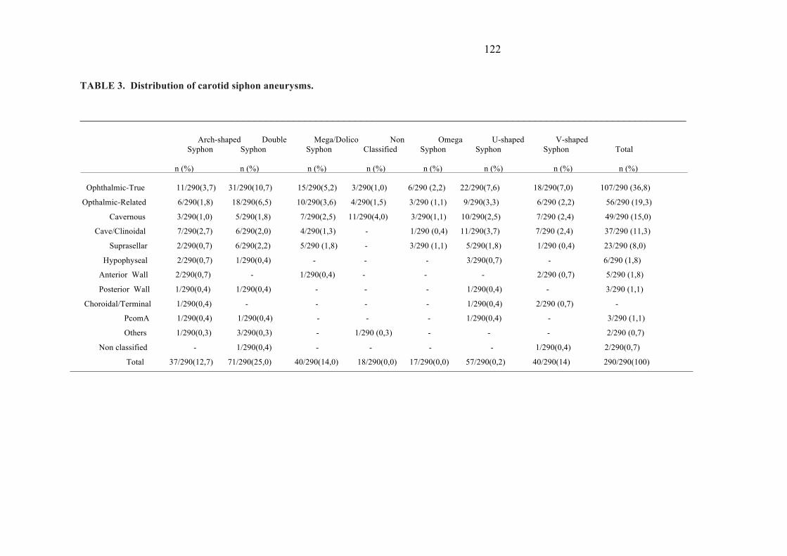
122
TABLE 3. Distribution of carotid siphon aneurysms. ____________________________________________________________________________________________________________ Arch-shaped Double Mega/Dolico Non Omega U-shaped V-shaped Syphon Syphon Syphon Classified Syphon Syphon Syphon Total n (%) n (%) n (%) n (%) n (%) n (%) n (%) n (%) Ophthalmic-True 11/290(3,7) 31/290(10,7) 15/290(5,2) 3/290(1,0) 6/290 (2,2) 22/290(7,6) 18/290(7,0) 107/290 (36,8)
Opthalmic-Related 6/290(1,8) 18/290(6,5) 10/290(3,6) 4/290(1,5) 3/290 (1,1) 9/290(3,3) 6/290 (2,2) 56/290 (19,3)
Cavernous 3/290(1,0) 5/290(1,8) 7/290(2,5) 11/290(4,0) 3/290(1,1) 10/290(2,5) 7/290 (2,4) 49/290 (15,0)
Cave/Clinoidal 7/290(2,7) 6/290(2,0) 4/290(1,3) - 1/290 (0,4) 11/290(3,7) 7/290 (2,4) 37/290 (11,3)
Suprasellar 2/290(0,7) 6/290(2,2) 5/290 (1,8) - 3/290 (1,1) 5/290(1,8) 1/290 (0,4) 23/290 (8,0)
Hypophyseal 2/290(0,7) 1/290(0,4) - - - 3/290(0,7) - 6/290 (1,8)
Anterior Wall 2/290(0,7) - 1/290(0,4) - - - 2/290 (0,7) 5/290 (1,8)
Posterior Wall 1/290(0,4) 1/290(0,4) - - - 1/290(0,4) - 3/290 (1,1)
Choroidal/Terminal 1/290(0,4) - - - - 1/290(0,4) 2/290 (0,7) -
PcomA 1/290(0,4) 1/290(0,4) - - - 1/290(0,4) - 3/290 (1,1)
Others 1/290(0,3) 3/290(0,3) - 1/290 (0,3) - - - 2/290 (0,7)
Non classified - 1/290(0,4) - - - - 1/290(0,4) 2/290(0,7)
Total 37/290(12,7) 71/290(25,0) 40/290(14,0) 18/290(0,0) 17/290(0,0) 57/290(0,2) 40/290(14) 290/290(100)

123
TABLE 4. Siphons and aneurysms Gender
Type C=IA
Type U/O= IB
Type V=II
Type D=III
Type M=IV
Type R
Total
Female
34/250(13.6)
71/250(28.4)
35/250(14.0)
59/250(23.6)
33/250(13.2)
18/250(7.2)
250/279(89.6)
Male 1/29(3.4) 3/29(10.3) 5/29(17.2) 12/29(41.3)
7/29(24.1) 1/29(3.4) 29/279(10.3)
Total 35/279(12.5) 74/279(26.5) 39/279(13.9) 62/279(22.2) 40/279(14.3) 19/279(6.8) 279/279(100)

124
CAPÍTULO VIII
Artigo Submetido para Publicação na Revista Anatomical Record

125
Artigo Submetido para publicação na Revista “ The Anatomical Record” Editorial
Pathogenesis, haemodynamic and growth of intracranial
aneurysms: our future directions
Patricia Bozzetto Ambrosi1,2,Carlos Augusto Carvalho de Vasconcelos3, Laurent Spelle1, Jacques Moret1, Marcelo Moraes Valença2
1. Neuri- Beaujon, Interventional Neuroradiology Department, Clichy, Paris, FRANCE 2. Postgraduate Program in Biological Sciences Federal University of Pernambuco, Recife,
BRAZIL 3. Department of Nutrition/LNED, Federal University of Pernambuco, Recife, BRAZIL
Adresse for correspondence: Patricia Bozzetto Ambrosi – Department of Interventional Neuroradiology, Hôpital Beaujon 100 Boulevard du Général Leclerc Clichy – 92110, France. Tel +33 643505184 Fax: +33 140875067 e-mail: [email protected] Received in 23-11-2015 1st review sent 08-01-2016 Waiting final review

126
Treatments using flow diversion or disrupting stents have increasingly gained attention due to being less invasive, and because they have shown increased levels of safety, as well as being feasible and having high success rates (Tse et al., 2013; Chalouhi et al., 2015). This is especially true of large, dysplastic, and wide neck aneurysms (Brinjikji et al., 2009; Huang et al., 2009; Byrne et al., 2012). In addition, intravascular flow modifier stents are persuasive tools that act directly on the pathogenesis of intracranial aneurysms (McAuliffe and Wenderoth, 2012; Huang et al., 2013). An intracranial aneurysm is a vascular ectasia arising from the wall of intracranial arteries predominantly in the cerebral arterial circle of Willis, in most cases of saccular form (Keedy, 2006). However, irrespective of its shape and location, an intracranial aneurysm is a disease within the vascular wall particularly at the level of the endothelium of blood vessels, which respond physiologically to blood flow patterns Therefore, flow diverters stents were designed to act by promoting a temporary scaffold for the endothelial proliferation with healing to the formation of a new vessel vascular wall and stimulating aneurysmal thrombosis (Tremmel et al., 2010). One of our challenges is to understand the interplay of the biological and hemodynamic processes involved in the formation and progression of intracranial aneurysms: its initiation; its growth; the inflammatory and degenerative processes related to the aneurysm’s rupture; and the eventual recanalization (Sforza et al., 2009). Another interesting element regards the behaviour of intracranial aneurysms, which is unpredictable; although they are dynamic, they often remain unnoticed until they are detected incidentally. Also, the true prevalence of intracranial aneurysms is not known, and the current consensus in clinical decisions is often to avoid subarachnoid hemorrhage (Turjman et al., 2014). Populational studies have shown that 85% of intracranial saccular aneurysms develop in the anterior circulation; more specifically, 35% appear on the complex anterior communicating artery, followed by carotid artery (30%) and middle cerebral artery (22%). They are less common in the posterior circulation at the top of the basilar artery and along its branches. Multiple aneurysms are found in about 30% of patients who already have at least one (Keedy, 2006). There is a preponderance of intracranial aneurysms in women, which increases with the number of aneurysms (Defillo et al., 2014). In samples of people aged 50 and over, this preponderance can approach a ratio of 1:2, or even greater; this trend seems to be associated with low levels of estrogen (Longstreth et al., 1994). Regarding the formation of an intracranial aneurysm, the current consensus is that the origins are multifactorial, and there is no completely satisfactory theory. Evidence demonstrates a complex evolutionary process in which many factors may be involved. The possible co-factors involved are: anatomy, vascular geometry, and abnormal flow patterns, with endogenous factors related to the vascular wall (weakness and vulnerability). However, the interplay of biological and hemodynamic factors is not well understood and remains unclear. Recent experimental studies have shown that arterial vessels’ caliber and their histological structure are regulated by blood flow variations (Sforza et al., 2009). Also, variations in the vessels and changes in bifurcation angles are involved in the genesis, development, and eventual rupture of aneurysms. As a result, there is increasing importance placed on cerebral hemodynamic assessment as a predictor of the initiation and development of intracranial aneurysms. When analysing the cerebral hemodynamic, it was found that the arterial circle of Willis has considerable variability, with frequent asymmetries. A complete circle of Willis was observed only in 20–25% of patients (Puchade-Orts et al., 1975). There is evidence that the anatomical variations observed in the cerebral arterial circle of Willis and related vessels may play a role in the genesis of intracranial aneurysms (Kayembe et al., 1984). Padget was among the first to compare the number of cases of embryological abnormalities with aneurysms and without aneurysms (Padget, 1945). It is believed that an asymmetrical circle of Willis, whether congenital or acquired, is a risk factor for the development of aneurysms, where hemodynamic stress produces degenerative changes leading to hyperdynamic flow (Milenkovic, 1981).
Recent studies have proposed that a congenital absence of the anastomosis capacity of the circle of Willis is correlated with other cerebrovascular diseases thus alluding to the hypoperfusion mechanism in the development of chronic ischemic pathology or small vessel cerebrovascular disease (Ryan et al., 2015). Another known flow-related disease well known is the ligation of the carotid artery. This procedure was performed for the treatment of giant carotid siphon aneurysms as the technique of choice for a long period of time before the development of neuroendovascular therapy. However, this kind of procedure has been associated with de novo intracranial aneurysm formation and flow-induced vascular remodelling (Gao et al., 1981).

127
Carotid ligation experimental models have demonstrated that compensatory cerebral blood flow increases after carotid occlusion with secondary pathologic remodeling. The flow adaptive development throughout the circle of Willis will result in the formation of an aneurysm in the contralateral carotid artery (Tutino et al., 2014). It is probable that the same principle can be applied to the bifurcation of the carotid artery and its branches, which can be explained due to a change in hemodynamic parameters at this level. Flow studies have shown that the source point of an intracranial aneurysm is distal to the bifurcation or the gradient fields, which are higher (Alnaes et al., 2007). Therefore, it is likely that the hemodynamic stress and the turbulent blood flow associated with hyperdynamic flow patterns could cause excessive wear and vibrations, resulting in structural fatigue and the rupture of the internal elastic lamina and, therefore, the formation of cerebral aneurysms. Patients with hyperdynamic flow patterns as a result of abnormal high flow conditions or other collateral pathways are therefore predisposed to accelerated degenerative changes in the vessel wall and the subsequent growth of an aneurysm (Wiebers, 2004). In this case, environmental factors such as hypertension, smoking, and connective tissue diseases probably play a contributory role, rather than being the cause. Studies have also looked into the possibility of changes in morphology in large vessels, as the main trunk and internal carotid arteries could be responsible for the formation of cerebral aneurysms (Sekhar et al., 1981). Experimental studies have found that within the dome of an aneurysm, hemodynamic stress seems to be caused by a sequential and repetitive turbulent flow. This type of abnormality is evidenced in the cavity of an aneurysm during systole. This abnormal flow becomes inverted during diastole, so these quick changes in the direction of flow continue to cause friction in the inner wall of the vessel and contribute to the formation and progression of an aneurysm (Gonzalez et al., 1992). Given all the facts and ideas that arise when trying to better figure out the local vascular and Willis polygon environment for the understanding of an aneurysm, some key points can help us: is there any correlation between the different pattern of geometries in the circle of Willis and the vessels related to certain subtypes of intracranial aneurysms? Perhaps certain anomalies or circle of Willis patterns justify screening and preventative treatment? Or is it that an aneurysm will not only be a local disease but a disease trigger point in all other hemodynamic spots that develop in the cerebral circulation or are acquired congenitally? Examining the use of cerebral hemodynamics is not easy because there are some confounding elements as constitutional problems or disease characteristics and blood structure contribution, hemodynamic, mechanical vascular by gender and the formation of aneurysms. As has previously been shown, there is a sex-linked difference in anatomical variations and this is linked to the anatomical distribution of aneurysms (Ghods et al., 2012); we still do not know if these two elements are connected. More controlled studies are needed. Finally, our current and future directions further elucidate the genesis and natural history of intracranial aneurysms and perhaps predict the response to treatment with stents flow diverters and to have a better understanding of the cerebral hemodynamics. This should also contribute to the understanding of the pathophysiology, in particular, to a more effective direction in the detection and prevention of intracranial aneurysms. LITERATURE CITED Alnaes, MS, Isaksen J, Mardal, K.A.; Romner, B.; Morgan, M.K.; Ingebrigtsen, T. 2007. Computation of hemodynamics in the circle of willis. Stroke 38:2500–2505.
Brinjikji W, Cloft HJ, Kallmes DF. 2009. Difficult aneurysms for endovascular treatment: overwide or undertall? Am J Neuroradiol 30:1513–17
Byrne JV, Szikora I. 2012. Flow diverters in the management of intracranial aneurysms: a review. EJMINT. 1225000057.
Cebral JR, Sheridan M, Putman CM. 2010. Hemodynamics and bleb formation in intracranial aneurysms. Am J Neuroradiol, 31, 304–10.
Chalouhi N, Zanaty M, Whiting A, Yang S, Tjoumakaris S, Hasan D, Starke RM, Hann S, Hammer C,
128
Kung D, Rosenwasser R, Jabbour P. 2015. Safety and efficacy of the Pipeline Embolization Device in 100 small intracranial aneurysm J Neurosurg. 122(6):1498-502. doi: 10.3171/2014.12.
Defillo A, Qureshi MH, Nussbaum ES. 2014. Are Multiple Intracranial Aneurysms, ore Than 5 At One Time, Almost Exclusively A Female Disease? A Clinical Series and Literature Review. J Neurol Stroke 1(4): 00024. DOI: 10.15406/jnsk.2014.01.00024
Ghods AJ, Lopes D, Chen M. 2012. Gender differences in cerebral aneurysm location. Front. Neurol, (3) 78.
Gonzalez CF, Cho YI, Ortega HV, Moret, J.1992. Intracranial aneurysms: flow analysis of their origin and progression. AJNR Am J Neuroradiol, 13:181–188.
Horikoshi T, Akiyama I, Yamagata Z, Sugita M and Nukui H. 2002. Magnetic resonance angiographic evidence of sex-linked variations in the circle of willis and the occurrence of cerebral aneurysms. J. Neurosurg. 96(4): 697-703.
Huang Q, Xu J, Cheng J, et al. 2013.Hemodynamic changes by flow diverters in rabbit aneurysm models: a computational fluid dynamic study based on micro-computed tomography reconstruction. Stroke 44:1936–41.
Huang Q, Xu Y, Hong B, et al. 2009. Stent-assisted embolization of wide-neck anterior communicating artery aneurysms: review of 21 consecutive cases. Am J Neuroradiol 30:1502–6.
Kayembe KN, Sasahara M. et al. 1984. Cerebral aneurysms and variations in the circle of Willis. Stroke, (15)5: 846-850, 1984.
Keedy A. 2006. An overview of intracranial aneurysms. Mcgill J Med 9(2): 141–146.
Longstreth WT, Nelson LM, Koepsell TD, Van Belle G. 1994.Subarachnoid hemorrhage and hormonal factors in women. A population-based case-control study. Ann Intern Med;121:168.
McAuliffe W, Wenderoth JD. 2012. Immediate and Midterm Results following Treatment of Recently Ruptured Intracranial Aneurysms with the Pipeline Embolization Device. Am J Neuroradiol 33:487–93.
Milenkovic Z .1981. Anastomosis between internal carotid artery and anterior cerebral artery with other anomalies of the circle of Willis in a fetal brain. J Neurosurg 55:701-03.
Puchades-Orts A, Nombela-Gomez M, Ortuno-Pachedo G. 1975.Variation in form of circle of Willis: some anatomical and embryological considerations. Anatomical Record.185:119-24.
Padget DH.1945. The circle of Willis: its embryology and anatomy. In Dandy, WE. Intracranial arterial aneurysm. New York:Comstock, 74-85.
Ryan DJ ,Byrne S, Dunne R, Harmon M, Harbison, J. 2015. White matter disease and an incomplete circle of Willis. International journal of stroke : official journal of the International Stroke Society, 10(4): 547-552.
Sforza DM, Putman CM, Cebral JR. 2009. Hemodynamics of Cerebral Aneurysms. 2009. Annu Rev Fluid Mech. 1(41): 91–107.doi: 10.1146/annurev.fluid.40.111406.102126
Tremmel M, Xiang J, Natarajan S, Hopkins N, Siddiqui A, Levy EI, Meng H. 2010. Alteration of intra-aneurysmal hemodynamics for flow diversion using Enterprise and Vision Stens. World Neurosurgery 74(2-3) 306-315.doi: 10.1016/j.wneu.2010.05.008
Tse MM, Yan B, Dowling RJ, Mitchell PJ. 2012. Current status of pipeline embolization device in the treatment of intracranial aneurysms: a review. World Neurosurg. 80(6):829-35. doi:

129
10.1016/j.wneu.2012.09.023. Oct 3.
Turjman AS1, Turjman F, Edelman ER. 2014.Role of fluid dynamics and inflammation in intracranial aneurysm formation. Circulation 21;129(3):373-82. doi: 10.1161/CIRCULATIONAHA.113.001444.
Tutino VM, Mandelbaum M, Choi H, Pope LC, Siddiqui A, Kolega J, Meng H. 2014. Aneurysmal remodeling in the circle of Willis after carotid occlusion in an experimental model. J Cereb Blood Flow Metab. 34(3): 415–424.
Wiebers DO, Piepgras DG, Meyer FB, Kallmes DF, Meissner IF, et al. 2004 Pathogenesis, natural history, and treatment of unruptured intracranial aneurysms. Mayo Clin Proc.79:1572–83.

130
CAPÍTULO IX
Artigo Submetido para Publicação na Revista Perspectivas Médicas

131
Artigo Submetido para Publicação na Revista Perspectivas Médicas
The arterial variants of Willis’circle and its associated branches involved in the pathogenesis of intracranial aneurysms: Current perspectives
As variantes arteriais do polígono de Willis e suas ramificações
envolvidas na patogênese dos aneurismas intracranianos: perspectivas atuais
Patricia Bozzetto Ambrosi 1,5 Carlos Augusto Vasconcelos 2 Valéria Fazan 3
Marcos Antonio Barbosa 4 Laurent Spelle 1 Jacques Moret 1 Marcelo Moraes Valença 5
1 Departamento de Neuroradiologia Intervencionista, Neuri Beaujon, Paris, França. 2 Professor Adjunto do Programa de Ciências da Nutrição e Pesquisador do LIKA, Universidade Federal de
Pernambuco, Recife, Brasil. 3 Professor Titular do Departamento de Cirurgia e Anatomia, Universidade de São Paulo, Ribeirão Preto, Brasil. 4 Médico Neuroradiologista Intervencionista, Hospital Esperança, Recife, Brasil. 5 Programa de Pós-Graduação em Ciências Biológicas, Universidade Federal de Pernambuco, Recife, Brasil
Endereço para Correspondência: Patricia Bozzzetto Ambrosi- 100 boulevard du General Leclerc- Paris France
Não existem conflitos de interesse
Artigo recebido em 25/12/2015 Artigo sendo revisado

132
RESUMO INTRODUÇÃO: Os aneurismas intracranianos são uma das principais causas de morte súbita e incapacidade permanente nos indivíduos adultos, principalmente entre mulheres acima de 50 anos. Dentro de um quadro multifactorial, as conhecidas alterações hemodinâmicas como estresse oxidativo e o fluxo sanguíneo turbulento associados com o polígono de Willis hiperdinâmico e assimétrico levariam a formação do aneurisma intracraniano. Modernos tratamentos utilizando stents modificadores de fluxo tem cada vez ganhado mais espaço, pois além de menos invasivos também atuariam diretamente bloqueando o círculo vicioso da fisiopatogênese dos aneurismas intracranianos. Devido a isso, observa-se um crescimento na importância de avaliar as variantes arteriais embriológicas como fator de risco/preditor dos aneurismas intracranianos. OBJETIVO: Investigar a aplicabilidade das variantes arteriais embriológicas implicadas na gênese e no tratamento dos aneurismas intracranianos. METODOLOGIA: Uma revisão sistemática via PubMed e EMBASE foi realizada a partir da combinação de palavras-chaves relacionadas e seus correspondentes. A pesquisa seguiu um protocolo adaptado dos critérios estabelecidos para estudos de medicina baseados em evidências e teve como limite temporal de 1977 a 2015. RESULTADOS: Foram localizados 2026 artigos, aos quais foram adicionados mais 55 estudos longitudinais publicados nos principais artigos internacionais. A análise dos artigos permitiu dividir em três principais grupos de variantes anatômicas relacionadas com a embriologia arterial com estudos relevantes: Hipoplasia/Dominância de A1, mais evidenciado com 14 estudos, as variantes em geral com 7 estudos, seguido de PCA fetal com dois estudos relevantes apenas. Um total de 12 estudos longitudinais preencheram os critérios de relevância. CONCLUSÃO: Apesar da escassez de estudos, as variações na própria anatomia vascular do polígono de Willis associadas com a geometria vascular parecem desencadear um processo de desgaste excessivo e vibrações que resultariam em fadiga estrutural e ruptura da lâmina elástica interna das arterias intracranianas levando a formação dos aneurismas intracranianos. A análise das variantes arteriais embriológicas do polígono de Willis e seus ramos poderá se tornar uma técnica potencialmente útil para o entendimento da gênese e na avaliação dos resultados do tratamento dos aneurismas intracranianos. Palavras-Chave: aneurismas intracranianos, fluxo, gênese, vascularização cerebral

133
ABSTRACT INTRODUCTION: Intracranial aneurysms are a major cause of sudden death and permanent disability in adults, especially women over 50 years. In a multifactorial framework, hemodynamic alterations well known as oxidative stress and turbulent blood flow associated with asymmetrical hyperdynamic Willis circle flow might lead to the formation of an intracranial aneurysm. Latest options of treatment using flow modifiers stents have increasingly gained more space, as well as less invasive, would also act directly blocking the vicious cycle of the pathophysiology of intracranial aneurysms. Because of this, there is a needed of assessing the embryological arterial variants as risk factors/ predictor of intracranial aneurysms. AIM: To investigate the applicability of embryological arterial variants implicated in the genesis and treatment of intracranial aneurysms. METHODS: A systematic review via PubMed and EMBASE was performed from the combination of related keywords and their corresponding. The research followed a protocol adapted the criteria established for medical studies based on evidence and limited time between 1977-2015. RESULTS: A total of 2026 articles were identified, of which were added more 55 longitudinal studies published in leading international papers. Analysis of the articles allowed to divide into three main groups relevant studies about anatomical variants related to blood embryology: hypoplasia / Dominance A1, more prominent with 14 studies, variants usually with 7 studies, followed by fetal PCA to two relevant studies only. Twelve studies filled the relevance criteria. CONCLUSION: Despite the scarcity of studies, variations in the vascular anatomy of the Willis polygon associated with remodeled vascular geometry seem the factor that trigger excessive wear process and vibrations that result in structural fatigue and rupture of the internal elastic lamina of intracranial arteries leading to the formation of aneurysms. The analysis of embryological arterial variants of the circle of Willis and its branches could become a potentially useful tool for understanding the genesis and evaluation of results of treatment of intracranial aneurysms. Keywords: intracranial aneurysm, flow, genesis, cerebral vasculature. Abbreviations: P.B.A= Patricia Bozzetto Ambrosi, S.S =Sarah Sutton INTRODUCTION The intracranial aneurysms are generally diagnosed, or when rupture occurs, or during the screening of neurological diseases (e.g. a headache)1,2 and hereditary syndromes associated with high frequency of aneurysms rupture.3 The Willis of circle (CoW) or circulus arteriosus is a polygonal anastomotic channel vessels surrounding the skull base of the brain which unites both internal carotid arteries and vertebrobasilar system. It maintains the steady and constant supply to the brain. The cerebral vasculature has considerable variability and anatomical variations of CoW are seen often. The CoW appears to be complete only 20-25% of individuals. 4-7

134
Since anatomical variations associated with intracranial aneurysms were pictured by Dorcas Padget while she illustrated the neurodevelopment and aneurysm formation 8-9 Few pieces of evidence has been described in the literature correlating congenital variability of Cow and its vessels related and hemodynamic factors. This relationship may be attributable to an increased blood flow and therefore, a significant overload hemodynamic and development of arterial disease which although unsatisfactory proved until now mostly due to methodology difficults, it have possible role in the genesis of intracranial aneurysms. 10-11 Recent studies have shown the correlation between specific subtypes of anatomical variants e.g A1 dominant hypoplasia or fenestrations and the presence of aneurysms in the anterior communicating artery. Arterial variants of CoW can be congenitally absent or sometimes acquired secondary to clamped vessels. The results of this process induce flow mediated-patterns and could implicate oxidative stress in aneurysmal disease progression. 5-7 The aim of this study is to briefly to present a systematic review which followed criteria established for medical studies based on evidences of the last 35 years about the applicability of anatomical and embryological variants involved in the genesis of intracranial aneurysms. METHODS Eligibility criteria and Search Strategy A extensive systematic review was performed in the two main databases Medline and EMBASE. The publications before December 21th 2015 were retrieved. The research was performed by author (P.B.A) and a Clinical Librarian (S.S.). After the first selection of articles, the author (P.B.A) reviewed the literature independently. The established limitation criteria were articles published in the last 10 years. The research was firstly performed on 3th September 2015 and updated automatically until 22 th December 2015. New terms (Table - Flow 1) were added in the second time to better tailored the research. The objective relevance criteria were establisedh based on: 1) contends of study longitudinal study or not 2) experience and background: if the study was from reference center or not 3 )accuracy/validity: presence of elements of significance statistically or not 3) source of documents: international study or not 4)context: answer to our main question or not Data Analysis The following data were extracted from the relevant studies: name of first author, year of publication subtype of anatomical variant, characteristics of arterial variant, distribution of sex, the location of the intracranial aneurysm. Final data analysis were performed by two authors (P.B.A and C.A.V). RESULTS AND DISCUSSION Our first systematic review identified 912 publications in EMBASE and 1011 publications in Medline on 3th October, 2015. Following 10 years limitation were applied to Medline final resulting in 508 articles founded between 2004 and 2015. The abstracts were screened and duplications were removed. Other irrelevant papers also were excluded. A total of 1543 were selected for further scrutining. A total of 1517 studies were removed because didn’t meet the eligibility criteria. Further update performed on 22 th December 2015 showed 987 publications in the EMBASE and 1039 in the Medline. Fifty-five articles (44 from EMBASE and 11 from Medline) which meet the relevance criteria were added. Twenty-six studies

135
evaluated the association between intracranial aneurysms and anatomical variations. The table 1summarize our search for eligible studies in our systematic review. From twelve studies eligible, five of them showed a statistically significant association between anatomical variants and intracranial aneurysms. We separated into three main anomalous collateral pathways: (1) hypoplasia / Dominance A1 demonstrated in 14 studies, (2) All variants in 7 studies, followed by (3) persistence of fetal PCA described in two studies only. In our knowledge, this is the first systematic review to address this matter. It is believed that hyperactive hemodynamic status with endogenous factors related to the vascular wall (weakness and vulnerability) are possible co-factors involved in the intracranial aneurysms genesis. Furthermore, the presence of an asymmetric circle of Willis, whether congenital or acquired, is a risk factor for the development of aneurysms, where the hemodynamic stress produces degenerative changes leading to the hyperdynamic flow. Studies correlating A1 dominant/ hypoplasia with presence of intracranial aneurysm were performed by Flores et al who studied and correlated the presence of A1 dominance between ruptured and unruptured Acom A aneurysms. In their series comparing 52 patientes with AcomA versus 104 healthy patients. There were no significant demonstrated AComAA rupture (P = .15). However, the A1-2 ratio correlated positively with the presence of ruptured AComAAs (P = .04) . They suggested that this finding may facilitate treatment decision in cases of small, unruptured AComAAs. Another series described by Charbel et al who analysed four significant associations: 1) A1 dominant (filling both A2's) was found in 57% of ACAA patients versus 14% of controls (p less than 0.001). 2) Unilateral hypoplasia of the opposite A1 was present in 24% of ACAA patients versus 6% of controls (p = 0.01). 3) Exclusive filling of the ACAA from one A1 occurred in 78%.. However, no statistically significant relationship was found between the anatomic flow patterns studied and the patients clinical presentation including age, sex, or grade. Their findings conclude that anterior communicating artery aneurysms are significantly related in a majority of patients with the presence of A1 dominant, probably as the result of enhanced haemodynamic stress caused by this anatomic abnormality in the circulation. However, this association is not constant, and a dominant pattern of flow did not correlate with the clinical course. This is probably a reflection of the differences between factors initiating aneurysm formation and those influencing its growth, as well as of the relative limitations of angiography when pathophysiological extrapolations are attempted. Other studies were described by Chen et al which studied between January 2009 and June 2012, 8,013 patients that underwent MRA examination at 3.0 T. MR using 3D-TOF with volume rendering technique. The presence and location of A1 segment hypoplasia and AcomA aneurysm was reviewed.Among the 8,013 patients examined, 138 patients were identified with AcomA aneurysms. 425 patients were defined with A1 segment hypoplasia, among whom 303 right-sided A1 hypoplasia and 122 left-sided A1 hypoplasia. 60 of these 425 patients were confirmed with AcomA aneurysms, among them were 49 right-sided A1 hypoplasia. The prevalence of AcomA aneurysm with A1 segment hypoplasia was 14.1 %, which was much higher compared with that (1.0 %) of AcomA aneurysm without A1 segment hypoplasia (P\0.001).The incidence of right-sided A1 segment hypoplasia either accompanied with AcomA aneurysm or not was much greater than that of left-sided. The study suggested that intracranial AcomA aneurysm development appeared to be associated with A1 segment hypoplasia. Another study, Tarulli & Fox showed that A1 dominance configuration is strongly associated with the presence of AcomA aneurysms for patients with intracranial aneurysms (odds ratio, 17.8). This association was also present compared with the incidence of A1 dominance in the large sequential control series of patients without aneurysms undergoing CTA for other reasons (odds ratio, 7.5). Outflow dilution of selective angiographic images augments anatomic information. A flow-based assessment of contrast

136
flowing from the A1 to the A2 segments after injection pressure is superior to an A1 diameter based categorization when A1 vessel diameters are not strikingly different. They concluded that anatomic variant of asymmetric A1 configurations likely facilitates the development of AcomA aneurysms by flow stresses, providing further evidence to support the role of biophysical factors in intracranial aneurysm development. Xu et al also demonstrated that A1 dysplasia/hypoplasia is a potential risk factor in the formation of ACoA aneurysms. They performed a study using hemodynamic flow measures. The wall shear increase appear to contribute to aneurysm formation. They conclude that wall shear stress decreased and turbulent flow may be responsible for the growth and rupture of ACoA aneurysms. Studies regarding fetal PCA are more rare. In 2008, Zada et al published a long term series of 15-year period where 271 patients were treated for 273 ICA-PComA aneurysms. Aneurysms occurring at the origin of fetal PCAs were identified in 30 patients (11%). There were 23 women (77%) and seven men (23%) (sex difference, P = 0.0035). The mean aneurysm size was 7 mm. PComA aneurysms originating from fetal PCA vessels posed a more substantial risk for infarction and subsequent neurological sequelae with surgical or endovascular obliteration. Fetal variant circulations were identified at the PComA origin in 11% of ICA-PComA aneurysm patients and were more commonly encountered in women. No group control were included. Songsaeng et al 2010 studied several anatomical variations including A1 aplais, fetal PCA and assymetrical fusion. Following their analysis, the AcoA, PcoA, and BA tip aneurysms tend to occur more often in anatomically variant parent artery dispositions, some of which are related to aneurysm recurrence following coil embolization. The findigns suggested that a more fragile vessel disposition as it is not fully matured or to altered haemodynamics secondary to the anatomical variations could be responsible for aneurysms formations. Stojanovic et al published studies about asymmetrical Willis which accounts 64% of in the whole group of patients studied. Within the group of patients suffering from multiple aneurysms, the presence of asymmetrical Willis' circle was 75.7%. The highest incidence of the asymmetrical Circle of Willis was found among patients with aneurysmal rupture detected at the anterior comunicative artery (ACoA) site (72.7% among cases with solitary and 100% among those with multiple aneurysms). Morphological changes on the A1 segment of ACoA were observed in 50 (44%) cases, with higher incidence found on the right side (60%). When comparing location of ruptured aneurysms between genders, a statistically significant prevalence of the ruptured aneurisms on ACoA was present in men, whereas women showed higher incidence of ruptured aneurysms on interior cartid artery (ICA) site (p < 0.01). The linkage between aneurysms with hypoplasia of the A1 segment of ACA and decreasing of the angle at which segments A1 and A2 join suggests the relationship between their onset, corresponding configuration type of Willis and subsequent hemodynamic changes. High incidence of asymmetry of Willis circle in the group of patients with ruptured aneurysms imply association of asymmetrical configuration and disorder in haemodynamic relations with forming and rupture of intracranial aneurysms. Most recently, Van Rooj studied 140 patients where 210 aneurysms were present. In 33 of 140 patients (24%; 95% confidence interval, 17.2%-31.3%), 45 fenestrations were detected with the following locations: anterior communicating artery in 31 (69%), A1 segment of the anterior cerebral artery in 4 (9%), middle cerebral artery in 4 (9%), basilar artery in 4 (9%), vertebral artery in 1 (2%), and anterior inferior cerebellar artery in 1 (2%). Of 56 patients with anterior communicating artery aneurysms, 14 had a fenestration on the anterior communicating artery complex. The remaining 31 fenestrations had no anatomic relation to aneurysms. In 140 patients with 210 aneurysms, 14 aneurysms (7%) were located

137
on a fenestration and 196 were not. In patients with a suspected ruptured aneurysm, fenestrations of intracranial arteries were detected in 24% (33 of 140). Most fenestrations were located on the anterior communicating artery. Of 45 fenestrations, 14 (31%) were related to an aneurysm. Other rare associations were described as case reports. No longitudinal studies were published until now. In 2014, Ho Allen studied short MCA. In 2006, Krishnamoorthy descibed callosomarginal variations associatd with intracranial aneurysms Although anatomic variations are well known in the A1 segment of the anterior cerebral artery (ACA), anomalous origin of a cortical artery from the A1 segment is extremely rare. The only reported cortical branch to arise from the A1 segment is the fronto-orbital artery. We report a case of anomalous origin of the callosomarginal artery (CMA) in association with a saccular aneurysm from the A1 segment of the left ACA in a 35-year-old man who presented with intracerebral hemorrhage. Freitas described a high flow association. Most of neurosurgeons and radiologists that work with patients with intracranial aneurysms are familiar with such anatomic variations. Currently, due to the technical improvements, anatomical variations of the small arteries, those were previously reported on cadaveric series, can also be evaluated on CT angiography. (Pekecvik). The same principle can be applied also to the bifurcation aneurysms of the carotid artery and its branches explained as due to a change in hemodynamic parameters at this level. This could explain the flow changes as a predictor of response to treatment with stents flow diverters well as the induction flow changes in collateral circulation. CONCLUSION Although variations of Willis polygon are frequent, the real impact is still unknow. More clinical and hemodynamic detailed studies are expected. Also the development of a predictive model estimating the probability. The analysis of anomalies of the intracranial arteries could become a potentially useful technique for understanding the genesis and treatment of intracranial aneurysms Probably in the near future the understanding of the cerebrovascular anatomy and embryology will became important point for appropriate management of the aneurysm REFERENCES Jamn, K.K. Headache in relation to intracranial aneurysm. Headache, 1967;7: 122-126. Stehbens, W.E, The pathology of intracranial arterial aneurysms and their complications.ln: Fox, J.L. ed, Intracranial Aneurysms. Vol. 1, New York, Springer-Verlag, 1983, Pp. 272-342 Benderson JB, Connolly ES Jr, Batjer HH, Dacey RG, Dion JE, Diringer MN, Duldner JE Jr, Harbaugh RE, Patel AB, Rosenwasser RH. 2009. American Heart Association SOGuidelines for the management of aneurysmal subarachnoid hemorrhage: a statement for healthcare professionals from a special writing group of the Stroke Council, American Heart Association. AU Stroke. 40(3):994 Harnsberger, H.R.; Osborn, A.G.; Ross, J. et al. Diagnostic and Surgical Imaging Anatomy. Lippincott Williams & Wilkins, 2006 Gunnal S.A., Farooqui M.S., Wabale R.N.Anatomical variations of the circulus arteriosus in cadaveric human brain. Neurology Research International, 2014, vol./is. 2014/, 2090-1852;2090-1860 (2014) Fahy P., McCarthy P., Sultan S., Hynes N., Delassus P., Morris L. An experimental investigation of the hemodynamic variations due to aplastic vessels within three-dimensional phantom models of the circle of willis Annals of Biomedical Engineering, January 2014, vol./is. 42/1(123-138), 0090-6964;1573-9686

138
Efekhar B, Dadmehr M, Ansari S, Ghodi M, Nazparvar B, Ketabchi E. Are the distributions of variations of circle of Willis different in different populations?-Recults of anatomical study and review. BMC Neurol 2006;6(22):1-9. Harnsberger, H.R.; Osborn, A.G.; Ross, J. et al. Diagnostic and Surgical Imaging Anatomy. Lippincott Williams & Wilkins, 2006. Padget, D.H. The circle of Willis: its embryology and anatomy. In Dandy, WE. Intracranial arterial aneurysm. New York:Comstock, p. 74-85, 1945. Ryan M. Kretzer, B.A., Ranice W. Crosby, M.L.A., David A. Rini, M.F.A., and Rafael J. Tamargo, M.D Dorcas Hager Padget: neuroembryologist and neurosurgical illustrator trained at John Hopkins. J Neurosurgery 100 7190730, 204. Kayembe, K.N.; Sasahara, M. et al. Cerebral aneurysms and variations in the circle of Willis. Stroke, v.15, n.5, p.846-850, 1984 Horikoshi T., Akiyama I., Yamagata Z., Sugita M., Nukui H..Magnetic resonance angiographic evidence of sex-linked variations in the circle of Willis and the occurrence of cerebral aneurysms. Journal of Neurosurgery, 2002, vol./is. 96/4(697-703), 0022-3085 (2002) Moher D, Liberati A, Tetzlaff J, Altman DG, The PRISMA Group (2009). Preferred Reporting Items for Systematic Reviews and Meta-Analyses: The PRISMA Statement. PLoS Med 6(6): e1000097. doi:10.1371/journal.pmed1000097 Menshawi K1, Mohr JP1, Gutierrez J1. A Functional Perspective on the Embryology and Anatomy of the Cerebral Blood Supply. J Stroke. 2015 May;17(2):144-58. doi: 10.5853/jos.2015.17.2.144. Epub 2015 May 29. Flores, Bruno C,Scott, William W,Eddleman, Christopher S,Batjer, H Hunt,Rickert, Kim L The A1-A2 diameter ratio may influence formation and rupture potential of anterior communicating artery aneurysms. Neurosurgery, vol. 73, no. 5, p. 845 (November 201 Kiarash Golshani, Andrew Ferrell,1 Ali Zomorodi, Tony P. Smith, and Gavin W. Britz1 A review of the management of posterior communicating artery aneurysms in the modern era Surg Neurol Int. 2010; 1: 88. Published online 2010 Dec 22. doi: 10.4103/2152-7806.74147 D. Songsaeng Impact of anatomical variations of the circle of Willis on the incidence of aneurysms and their recurrence rate following endovascular treatment November 2010Volume 65, Issue 11, Pages 895–901 Stojanović N1, Stefanović I, Randjelović S, Mitić R, Bosnjaković P, Stojanov D. Presence of anatomical variations of the circle of Willis in patients undergoing surgical treatment for ruptured intracranial aneurysms. Vojnosanit Pregl. 2009 Sep;66(9):711-7 van Kooij BJM, Hendrikse J, Benders MJNL, de Vries LS, Groenendaal F. Anatomy of the circle of Willis and blood flow in the brain-feeding vasculature in prematurely born infants. Neonatology 2010;97(3):235-41. PubMed PMID: 19887852. doi: 10.1159/000253754 Milenković Z, Vučetić R, Puzić M. Asymmetry and anomalies of the circle of Willis in fetal brain. Microsurgical study and functional remarks. Surg Neurol 1985;24(5):563-70. PubMed PMID: 3840282. doi: 10.1016/0090-3019(85)90275-7 Umansky F, Dujovny M, Ausman JI, Diaz FG and Mirchandani HG. Anomalies and Variations of the Middle cerebral artery:a microanatomical study. Neurosurgery. 1988; 22(6): 1023-27. Matsumura M, Nojiri KSurg Neurol Ruptured anterior communicating artery aneurysms associated with fenestration of the anterior cerebral artery. 1984 Oct;22(4):371-6. Bozzetto Ambrosi Neuronutri Table 1. Search Strategy 1. EMBASE; exp INTRACRANIAL ANEURYSM/; 27481 results. 2. EMBASE; (brain* adj2 aneurysm*).ti,ab; 480 results. 3. EMBASE; (intracranial* adj2 aneurysm*).ti,ab; 10459 results. 4. EMBASE; ("intra cranial*" adj2 aneurysm*).ti,ab; 54 results.

139
5. EMBASE; (cerebral* adj2 aneurysm*).ti,ab; 7489 results. 6. EMBASE; 1 OR 2 OR 3 OR 4 OR 5; 29775 results. 7. EMBASE; variance.ti; 4070 results. 8. EMBASE; 6 AND 7; 0 results. 9. EMBASE; "A1 hypoplas*".ti,ab; 13 results. 10. EMBASE; (A1 adj2 hypoplas*).ti,ab; 53 results. 11. EMBASE; a1.ti,ab; 44474 results. 12. EMBASE; 9 OR 10 OR 11; 44474 results. 13. EMBASE; 6 AND 12; 291 results. 14. EMBASE; pcom.ti,ab; 224 results. 15. EMBASE; "posterior* communicating".ti,ab; 1987 results. 16. EMBASE; 14 OR 15; 2142 results. 17. EMBASE; 12 OR 16; 46517 results. 18. EMBASE; 6 AND 17; 1141 results. 19. EMBASE; 18 [Limit to: (Year Published Last 10 Years)]; 695 results. 20. EMBASE; exp POSTERIOR COMMUNICATING ARTERY/ [Limit to: (Year Published Last 10 Years)]; 1341 results. 21. EMBASE; 17 OR 20 [Limit to: (Year Published Last 10 Years)]; 25735 results. 22. EMBASE; 6 AND 21 [Limit to: (Year Published Last 10 Years)]; 987 results. 23. Medline; (brain* adj2 aneurysm*).ti,ab; 594 results. 24. Medline; (intracranial* adj2 aneurysm*).ti,ab; 8802 results. 25. Medline; ("intra cranial*" adj2 aneurysm*).ti,ab; 38 results. 26. Medline; (cerebral* adj2 aneurysm*).ti,ab; 6968 results. 27. Medline; "A1 hypoplas*".ti,ab; 10 results. 28. Medline; (A1 adj2 hypoplas*).ti,ab; 54 results. 29. Medline; a1.ti,ab; 33758 results. 30. Medline; pcom.ti,ab; 131 results. 31. Medline; exp INTRACRANIAL ANEURYSM/; 22737 results. 32. Medline; 23 OR 24 OR 25 OR 26 OR 31; 26345 results. 33. Medline; "posterior* communicating".ti,ab; 1572 results. 34. Medline; 27 OR 28 OR 29 OR 33; 35268 results. 35. Medline; 32 AND 34; 1039 results. 36. EMBASE; NUCLEAR MAGNETIC RESONANCE IMAGING/ OR BRAIN CIRCULUS ARTERIOSUS/ OR THREE DIMENSIONAL IMAGING/ OR RADIOGRAPHY/ OR ANTHROPOMETRY/; 890701 results. 37. EMBASE; (loop AND of AND Willis).ti,ab; 10 results. 38. EMBASE; (cerebral AND arterial AND circle).ti,ab; 569 results. 39. EMBASE; BRAIN CIRCULUS ARTERIOSUS/; 2553 results. 40. EMBASE; 36 OR 37 OR 38 OR 39; 890907 results. 41. EMBASE; ANATOMICAL VARIATION/; 14910 results. 42. EMBASE; (arterial AND variant).ti,ab; 1724 results. 43. EMBASE; 40 AND 41; 2793 results. 44. EMBASE; 41 OR 42; 16540 results. 45. EMBASE; 40 AND 44; 2942 results. 46. EMBASE; 6 AND 45; 44 results. 47. Medline; Willis'circle.ti,ab; 97 results. 48. Medline; (loop AND of AND willis).ti,ab; 7 results. 49. Medline; CIRCLE OF WILLIS/; 1618 results. 50. Medline; (Willis AND polygon).ti,ab; 35 results. 51. Medline; (arterial AND variant).ti,ab; 1140 results.

140
52. Medline; (anatomical AND variant).ti,ab; 1038 results. 53. Medline; 47 OR 48 OR 49 OR 50; 1707 results. 54. Medline; 51 OR 52; 2115 results. 55. Medline; 53 AND 54; 11 results.
Table 2. Flow diagram
Recordsidentifiedthroughdatabase
searching(n=2026)
3.Screen
ing
4.Eligibility
1.Iden
tifica
ton
Recordsafterduplicatesremovedand10yearslimitation(n=483)
Recordsscreened(n=1543)
Recordsexcluded(n=1517)
Full-textarticlesassessedforeligibility(n=26)
Full-textarticlesexcluded,withreasons
(n=5)
Studiesincludedinqualitativesynthesis
(n=12)
Additionalrecordsidentifiedthroughother
resources(n=55)(n=2026)

141
Table 3. Main eligible studies published in the current literature Author(Year)
Subtype
Location Aneurysm
Sex Patient
Control Group
Van Rooij et. al, (2015)
Fenestrations
Several locations
140 patients/210 aneurysms
No controle group
Chen et. al,(2014)
A1 dominant
AcomaA
138 patients
7875 patients
Flores et. al, (2013)
A1/A2 diameters
AcomA
52 AComA
104 healthy
Xu et al (2012)
A1 dysplasia/
hypoplasia
AcomA
?
?
Songsaeng (2010)
A1 aplasia, foetal PcoA, asymmetrical fusion
AcoA
PcoA
BA
96 AcoA
67PcoA
29 BA
No control group
Tarulli 2010 A1 dominance AcomA 105 AcomA
123 other
159 healthy
Stojanović et al (2009)
AcoA
Mutliple aneurysms
114 patients multiple aneuryms
No control group
Zada (2008)
PCA
ICA
271 patients
No control group
Kitami, K (1985)
Dominant A1
Charbel F (1991)
Dominant A1
AcomA
51 patients
51 healthy and matched controls
Matsumura & Nojiri 1984 Fenestrations AcomA 12 patients No control
Kayembe et al (1984) Multiple Multiple 44 aneurysms 148 healthy CoW

142
CAPÍTULO X
Artigo Submetido para Publicação na
Revista Clinical Nutrition

143
Artigo Submetido para Publicação para a Revista” Clinical Nutrition”
Paradoxical obesity and intracranial aneurysms: a mini review
Patricia Bozzetto Ambrosi 1,2, Marcelo Moraes Valença2, Carlos Augusto Carvalho de Vasconcelos3
1- Interventional Neuroradiology, NEURI-Beaujon, Paris - France 2- Pos-graduation Program in Biological Science, Federal University of Pernambuco, Recife,
Brazil 3- Department of Nutrition-LNED and LIKA – Electron Microscopy Laboratory, Federal
University of Pernambuco, Recife, Brazil
Submitted: 21/12/2015 Revised: em revisão; Accepted: ? Correspondence: Dr Patricia Bozzetto Ambrosi- 100 boulevard du General Leclerc- Clichy 92100 France
[email protected] Phone: +33643505184 The authors have no conflicts of interest to disclose. Abstract Body mass index above 30 is an independent risk factor for cardiovascular disease and linked
to poor prognosis, especially in patients admitted to intensive therapy departments. However,
recent reports have indicated that individuals with mild to moderate obesity have favorable
outcomes in the survival in coronary heart disease and either in hemorrhagic or ischaemic
stroke. The same association has been also encountered in patients who had ruptured
intracranial aneurysms. These patients are although obese, they stay metabolically healthy
with apparent protection from the disease. More studies are needed to determine its consistent
correlation. Obesity, the body mass index or Quetelet index and its correlation intracranial
aneurysms were briefly examined in this article.

144
Key words intracranial aneurysms, subarachnoid haemorrhage, body mass index, paradoxical
obsesity, body weight
Introduction
More than 2500 years, Hippocrates described the association between cerebrovascular or
cardiovascular diseases in individuals with obesity and overweight causing sudden death.1,2
Both obesity and overweight are rapidly advancing in worldwide and has become a socio-
political problem due to the fact to be an independent risk factor for cardiovascular disease
particularly in developing countries.3-5
World obese populations have doubled since 1980.3,4According to National US studies, it
has been estimated that more than one-third (about 35% or more than 78 million individuals)
are obese and more than 70% of American adults 6are affected by obesity and/or
overweight.6,7 Further studies carried out by World Health Organization (WHO) have found
that both obesity and overweight also affects over 50% of the South American populations.4
For immediate primary care of the population estimates by the WHO already showed
increased mortality from nutritional chronic diseases in Brazil of up to 22% by 2015. 4,5
Although not yet have accurate data in the northeast of Brazil, is known the number of cases
of type 2 diabetes is increasing every day as well as childhood obesity, among other
associated factors such as physical inactivity and poor diet, excessive consumption of the
simple carbohydrates and excess saturated and trans fats. The association of obesity with
abdominal aortic aneurysm is also a great indicator and risk factor to worsen the picture of
morbidity, because reducing body weight, reduces associated risk as anthropometry measures
and other diagnostics. 7
Obesity, Overweight and Quetelet index
According to National Institutes of Health in Bethesda, overweight is defined as having
excess body weight for a particular height from fat, muscle, bone, water, or a combination of
these factors.8 And the obesity is defined as having excess body fat. 8 The term body mass
index (BMI) was based in Quetelet’s theory of human trait measurement based in normal
curve.9 This term is universally adopted as a screening population measurement.10,11Adults
with the BMI greater than or equal to 25 are considered overweight and those with BMI
greater or equal to 30 are considered obese. 8-10
Related Risk and Protective Factors

145
Obesity and overweight are independent risk factors for cardiovascular diseases, cancer,
diabetes, hypertension, musculoskeletal disease and stroke. However, since 1999 when the
first description about obesity-survival was performed based on survival advantage for
overweight and obese patients undergoing hemodialysis, a large studies supporting the
paradoxical obesity in several conditions has published.
Several epidemiological studies have showed that obese individuals with coronary heart
disease demonstrated a clear survival advantage compared with their nonobsese cohorts. 12,13
Recent studies described the same outcome in patients with mild and moderate obesity who
had chronic obstructive pulmonar disease, ischemic and haemorrhagic stroke and
subarachnoid haemorrhage (SAH).13-16
Cigarette smoking, hypertension, alcohol consumption, estrogen deficiency, collagen
diseases, atherosclerosis, ageing and female sex also appear to be involved in the formation,
growth and rupture of intracranial aneurysms.15,17-18 Obesity and other modifiable risk
factors, such as hypercholesterolemia, ischemic heart disease, diabetes mellitus are limited
and conflicting and controversial. Hypercholesterolemia and regular physical exercise appear
to decrease the risk of aneurysm formation; there is some speculation that the former effect is
mediated through statin therapy.
We recently reviewed main sources of researches as Embase, Pubmed and Cochrane
(CENTRAL), Allied, Amed, BNI, CINAHL, Health Business Elite using key-terms
correlated changes in weight, obesity paradox with intracranial aneurysms, hemorrhagic
stroke and SAH over the last 30 years. 19 A total of 690 studies were found, 45 were
duplicates, 147 showed stroke and “paradox obesity” association.
After reading all relevant articles, only one study from Vlak et al 17, studying 206 patients
with unruptured intracranial who never had a SAH and 574 controls who were randomly
retrieved from general pratices of area of catchment of Utretch. Unruptured aneurysms and
obesity were consistently correlated unruptured, however no significant statistical correlation
was founded. 17
Two main studies correlated obesity and ruptured aneurysms. 17,20 Both studies indicated that
obesity reduces mortality to short and long term, although remain controversial with regard to
functional recovery. First one was performed by Platz et al. 14 analyzing 741 patients with
the history of SAH secondary to intracranial aneurysms: 36.2% or 267 patients were

146
overweight and 15.2% or 113 patients were obese. Although many physicians anticipated a
worse outcome for the obese patient the BMI was not an independent predictor of outcome. 15
In the Platz’study the multivariate analysis, only age (OR:1.051, 95% Cl: 1.04-1.07, p <.001),
WFNS grade (OR:2.095, 95%, Cl:1.87-2.35, P<.001), occurrence of vasospasm (OR:2.90,
95% Cl:1-94-4.34, P< .001) and aneurysm size larger than 12 mm (OR: 2.215,95% Cl:1.20-
4.10, P=0.11) were independent predictors of outcome after 6 months. Of the 321 poor grade
patients (WFNS score >3), 171 (53,3%) were overweight. Of these, 21.6% attained a
favorable outcome compared with 35.3% of normal-weight patients (P=.006). Based on the
BMI, obesity seems to be negligible for outcome after SAH compared with the impact of
SAH, the patient's age, the occurrence of vasospasm, or aneurysm size. 15
Another recent study of Tawk et al. 20 investigated 274 consecutive patients admitted with
SAH between 2008 and 2012. After performing a multivariate analysis adjusting for an age,
the older age was associated with worsened severity and that BMI was not noticeably
associated with severity bleeding or functional outcome in patients with SAH.
Although the obesity paradox correlation has limited evidence patients with intracranial
aneurysms appear to overlap other vascular conditions as heart ischemic disease and stroke.
The reason for the paradox has yet to be defined and the related mechanisms are still
unknown. It appears to be the same underlying mechanism observed in hypercholesterolemia
and regular physical exercise and weight of loss. 21
A better nutrition and increased metabolic reserve appear to confers a kind of protection
against endotoxin inflammatory cytokines and survival advantages. The exercise associated
with good nutrition not only preserves muscle strength and its vascular dynamics, reducing
circulating glucose, thereby increasing the effectiveness of insulin released as improving
appetite and mood. Futures and more consistent studies are needed.
References
1. Katz A.M., Katz P.B.; Diseases of the heart in the works of Hippocrates. Br Heart J 24 1962:257-264.
2. Lavie C.J., Milani R.V.; Obesity and cardiovascular disease. the Hippocrates paradox?. J Am Coll Cardiol 42 2003:677-679.

147
3. World Health Organization . Obesity and overweight. Geneva: World Health Organization, 2011. (http://www.who.int/mediacentre/factsheets/fs311/en, ( accessed on 19 Dec 2015).
4. Arbex A K, Rocha DRTW, Aizenberg M, Ciruzzi MS. Obesity Epidemic in Brazil and Argentina: A Public Health Concern. J Health Popul Nutr 2014 32(2):327-334.
5. World Health Organization. Region and country specific information sheets: the impact of chronic diseases in Brazil [Internet]. [accessed on 21 Dec 2015]. Available from: http://www.who.int/chp/chronic_disease_report/ media/impact/en/index.html
6. Krebs NF, Himes JH, Jacobson D, Nicklas TA, Guilday P, Styne D. Assessment of child and adolescent overweight and obesity. Pediatrics 2007;120:S193–S228
7. Cronin O, Walker PJ, Golledge J. The association of obesity with abdominal aortic aneurysm presence and growth. Atherosclerosis. 2013 Feb;226(2):321-7.
8. National Institutes of Health, National Heart, Lung, and Blood Institute. Disease and Conditions Index: What Are Overweight and Obesity? Bethesda, MD: National Institutes of Health; 2010.
9. Garabed, E. "Adolphe Quetelet (1796–1874) The average man and indices of obesity". In: Nephrol. Dial. Transplant. 2008: 23 (1): 47–51.
10. Macdonald FC. Quetelet index as indicator of obesity. Lancet 1986;1:104311. http://www.cdc.gov/healthyweight/assessing/bmi/,( accessed on 20 Dec 2015). 12. Fleischmann E, Teal N, Dudley J, May W, Bower JD, Salahudeen AK. Influence of
excess weight on mortality and hospital stay in 1346 hemodialysis patients. Kidney Int 1999;55:1560–7
13. Reeves BC, Ascione R, Chamberlain MH, Angelini GD. Effect of body mass index on early outcomes in patients undergoing coronary artery bypass surgery. J Am Coll Cardiol 2003;42:668 –76.
14. Romero-Corral A, Montori VM, Somers VK, et al. Association of bodyweight with total mortality and with cardiovascular events in coronary artery disease: a systematic review of cohort studies. Lancet. 2006;368:666-678.
15. Platz, Johannes; Güresir, Erdem; Schuss, Patrick; Konczalla, Jürgen; Seifert, Volker; Vatter, Hartmut.The Impact of the Body Mass Index on Outcome After Subarachnoid Hemorrhage: Is There an Obesity Paradox in SAH? A Retrospective Analysis. Neurosurgery 2013 73 (2):201–208.
16. Park JH, Yoon SJ, Lee H, Jo HS, Lee SI, Kim Y, Kim I, Shin Y. Burden of disease attributable to obesity and overweight in Korea. International Journal of Obesity 2006 30 1661-1669.
17. Vlak MH, Rinkel GJ, Greebe P, Algra A. Independent risk factors for intracranial aneurysms and their joint effect: a case-control study Stroke. 2013 44(4):984-7. Epub 2013 Feb 19.
18. Sadavisan C, Fiorella DJ, Woo HH, Lieber BB. Physical factors effecing cerebral aneurysm pathophysiology.Ann Biomed Eng. 2013 Jul;41(7):1347-65. doi: 10.1007/s10439-013-0800-z. Epub 2013 Apr 3.
19. Bozzetto Ambrosi P, Valenca M, Vasconcelos C. O paradoxo da obesidade e os aneurismas intracranianos. Annais do Neutronutri.[ in portuguese]
20. Tawk, Rabih G., Grewal, Sanjeet S.; Heckman, Michael G. MS; Navarro, Ramon; Ferguson, Jennifer L. MA; Starke, Emily L.; Rawal, Bhupendra MS; Hanel, Ricardo; Miller, David MD; Wharen, Robert E.; Freeman, William D. Influence of Body Mass Index and Age on Functional Outcomes in Patients With Subarachnoid Hemorrhage. Neurosurgery. 76 (2):136-141, February 2015.
21. Vasconcelos, CAC, Rodrigues, BFS. Nutrição e Envelhecimento Humano- As Necessidades Nutricionais do Idoso: Um Panorama atual. In: Spencer Júnior;

148
Leopoldo Barbosa. (Org.). SAÚDE DO IDOSO - UMA ABORDAGEM MULTIDISCIPLINAR. 1ed. Recife: EDUPE, 2015, v. 1, p. 157-168.
CAPÍTULO XI
Artigo a ser Submetido para Publicação na Revista Circulation

149
Artigo a ser Submetido para Publicação na Revista Circulation
Submission to Circulation
The arterial variants of Willis circle and vessels
related and its effect on carotid siphon
aneurysms genesis
Patricia Bozzetto Ambrosi, 1 Laurent Spelle, 1 Jacques Moret, 1 Marcelo
Moraes Valença 1
1 Department of Interventional Neuroradiology, Neuri Beaujon, Paris, France
2 Postgraduation in Biological Sciences, Recife, Brazil
Corresponding author: Patricia Bozzetto Ambrosi E-mail: [email protected] Telephone: +33643505184 Fax: Short title: {Bozzetto Ambrosi} : Variants of Wills circle and carotid siphon aneurysms
genesis
Word count: The total word count of the manuscript, including the title page, abstract, text,
references ?

150
Subject Codes: Please select from here www.ahajournals.org/site/misc/subject_codes.xhtml
Abstract
BACKGROUND: Populated-based, crossing-matched and flow computational studies have suggested a correlation between arterial variations of Willis circle and the development of an intracranial aneurysm. Asymmetric A1 segment, posterior cerebral artery (PCA) dominance and other minor variants e.g sharper angle bifurcations, vessel duplications and fenestrations has been demonstrated to leading to accelerated degenerative changes in the vessel wall at several locations either in the anterior or posterior circulation. Specific studies about hemodynamic changes correlated to arterial variants of the circle of Willis and carotid siphon aneurysms has been not addressed yet. OBJECTIVE: To characterize the role of arterial variants of the circle of Willis as particularly implicated in the genesis of aneurysms within the circumference of the carotid siphon. At the same time, a regression analysis to overcome significant imbalance between men and women in both with respect to the aneurysm location and the presence of arterial variants of Willis polygon. METHODS: A case-control study was conducted evaluating individuals with unilateral carotid siphon aneurysms treated between 2002 and 2014 (n = 178 patients/aneurysms, 154 females, 24 males) at the Department of Interventional Neuroradiology, Hospital Beaujon, France and compared with a control healthy group (n = 210 patients, 142 females, 68 males) and a combined cohort with contralateral healthy siphon and control healthy patients (n=468 patients, 372 females, 96 males). The presence of A1 or Fetal PCA dominance or both associated were assessed. RESULTS: Contrary to expectations, there was an inverse relationship between the presence of aneurysms somewhere in the circumference of the siphon and arterial variants of Willis circle. Both A1/Fetal PCA dominance were founded in 3/178(1.6%) patients of the group with a unilateral aneurysm (3/178) and in 13 of 178 of the contralateral healthy siphons (OR=0.2176, p = 0.0189). A1 dominance was demonstrated in 11 of 24 healthy male siphons and 1 of 24 males with unilateral carotid siphon aneurysm (OR 0.514, p <0.0070) and in 44 females with 154 healthy siphons vs. 11 females with 154 unilateral aneurysms (OR 0.192, p <0.0001). DISCUSSION: The high association of A1/Fetal PCA in healthy siphons denotate a probable protective effect of this association. The lower frequency of carotid siphon aneurysms in certain locations might relate with a greater resistance and increased capacity of adaptation against the wall hemodynamic disorder. CONCLUSION: Anatomical variants high flow-related appear to be relevant protection factor of carotid siphon aneurysms. This new concept can help in screening and treatment strategy of the intracranial aneurysms. Therefore, more studies involving flow measurements might be considered for better assessment of the blood circulation. KEYWORDS: Willis circle, circulus arteriosus cerebri, intracranial aneurysms, carotid siphon, genesis
Introduction
The intracranial aneurysms are abnormal local vascular dilatations occurring more
frequently within the arteries of Willis polygon and its vessels related. 1-3 Aneurysms that
151
develop on the circumference of the carotid siphon are quite diversified and complex
surgical techniques are needed for either its microsurgical or endovascular repair. 4-13
Although the natural history of intracranial aneurysms remains not elucidated, current
evidence have showed that hemodynamic and geometric factors seem to be possible
cofactors of the evolutionary process involving endogenous and local factors (weakness
and vulnerability) in the destructive remodeling of the arterial wall and aneurysms
formation. 14-17
Populations and crossing matched studies performed in several locations as
ACA(Anterior cerebral artery) and PCoA(Posterior communicating artery) indicate that
asymmetry in the arteries of the circle of Willis and other geometric changes as sharper
bifurcation angles has been demonstrated in the flow studies as playing an important role
in the aneurysm development.14-20At the same time, authors investigating the relationship
between variations of the Willis polygon and analyzing the risk of aneurysm formation,
have detected a correlation between the known difference between men and women for
anatomic variants which appear to be correlated with the difference in the distribution of
aneurysms. 21,22
We conduct a study detecting A1 or P1 dominance between siphons with and without
aneurysms in a series of patients were referred for carotid siphon aneurysm
neuroendovascular management and comparing with a matched sampling of contralateral
healthy siphon and a series of healthy regression-adjusted sample from a high-performing
French university center, and reference center for intra-cranial aneurysms.
Methods
An observational, descriptive and quantitative, case-control survey was conducted from
January 2011 until October 30, 2014, within the Department of Interventional
Neuroradiology at Beaujon University Hospital, France. The research was developed in
accordance with the ethical standards for research involving human subjects (CAAE
47075215.1.0000.5208).
From a daily updated database of patients with intracranial aneurysms treated by the
department between February 2002 until October 2014, only patients with unilateral carotid
siphon aneurysms were included in this study. An aneurysm was considered within the
carotid siphon when located in any point of siphon circumference or corresponding to

152
Bouthilier24 C4-6 segment of each ICA analysed (C2-C4 segment in Fischer classification25)
as shown in Figure 1.
Following healthy patients were recruited between January 2011 and October 2014 whose
angiographic studies of four vessels have no showed evidence of brain aneurysms, "blebs" or
other related vascular malformations and were analyzed prospectively. Patients with
incomplete arteriographical examinations or not allowing proper analysis were excluded.
Also patients with bilateral carotid siphon aneurysms were considered in a separated group
called “mirror” for further analysis.
Data collection
Patient demographical data, such as age and gender, and history of subarachnoid
haemorrhage were reviewed. Data from pre-treatment angiograms, including 2- and 3-
dimensional rotations used to locate aneurysms in the siphon (and harbored in the nine most
frequent locations as Figure 1) 26, as well as the type of syphon (based on a lateral view of
Huber’s modified description27, in seven subtypes) were analyzed and recorded, as well as
any anatomical variations and collateral circulation.
Figure 1. A. B.
Procedure
All carotid siphons studied were categorized in five group as either: i) Group 1, carotid
siphon with "unique" aneurysm, ii) Group 2, both carotid siphon with aneurysms labeled as
"mirror"; iii) Group 3 , healthy contralateral siphon; iv) Group 4, both healthy carotid
siphons; v) Group 5, healthy combined group.

153
All configuration of circle of Willis were assessed by reviewing of several images of
Digital Subtraction Angiography (DSA) of the cervical or intracranial vasculature, 3D-
reconstruction, intra-arterial vasoCT, arterial perfusion, 2D and 3D flow acquisitions which
were performed in each patient during surgical pre and post-assessment. The presence of
anterior communicating artery (ACoA), both A1, P1 segments and posterior communicating
arteries (PCoA) were recorded. We classified each Willis circle for the presence of A1 or
Fetal posterior cerebral artery (PCA) dominance or both. A1 dominance was defined as the
contralateral branch was not visible or a diameter < 1 mm. PCA fetal was defined when the
PCA originates from the ICA, completely (without connection with basilar artery) as well
partially (with small connection from basilar artery).
Figure 2. Different Willis Circle configurations.
Posteriorly, data will be stored in an Excel ® spreadsheet and then analyzed using the SPSS
® 23 version:
Baseline characteristics of analyzed groups
A total of 262 patients (230 female and 32 male) with a mean admission age of 50.47 ±
12.9 years (range 16-84) were diagnosed and treated of carotid siphon aneurysms between

154
2002 and 2014 at the Department of Interventional Neuroradiology, Hospital Beaujon,
France. Of this number, 40 patients (18.5%) had a history of subarachnoid bleeding. The
control healthy group were composed of 210 patients (142 females and 68 males) with mean
admission age of 47.99 ± 16.774 years (range 16-84) and resulting in a combined cohort with
contralateral healthy siphon (n=472 patients, 372 females, 96 males).
The table 1 illustrates the results regarding gender analysis and summarizes the main
demographic features of five group studied. The aneurysm group showed a female
preponderance of 154/178(86.5%). However, the mirror group had the greater predominance
with 76/84(90.5%) females. The healthy control after reformatting had a group
142/210(67,6%) of females and the final sample reached in the combined group was
composed of 296/388(76.2%) females.
Statistical Analysis
Statistical analysis was performed using Statistical Analysis Software (SAS) version 23
(SAS Institute Inc., Cary, NC 27513-2414, USA). For continuous variables, demographic and
clinical variables are reported as mean and median, showing the standard error and 95%
confidence interval. For categorical variables, data were presented in terms of numbers and
percentages. Statistical analyses were performed using the Pearson chi-square test, and the
Fisher exact test used to determine the statistical inference for categorical variables, based on
the outcome variable. Data were deemed statistically significant at p<0.05.
Results
Of the 1038 carotid siphons analysed, the presence of Fetal PCA was demonstrated in
282/1038 (27.16%) of total of siphons studied and respectively in 44/178(24.7%) of both
groups with unilateral carotid siphon aneurysm and its contralateral healthy siphon. The
mirror m healthy and combined groups showed respectively 12/84(14.3%),
69/210(32.8%) and 113/388(29.12%) of Fetal PCA dominance.
Regarding A1 dominance, it was demonstrated in 217/1038 (20.9%) of total of carotid
siphons and successively in 12/178(6.7%) of siphons with a unilateral aneurysm,
11/84(13.0%) of mirror siphon group, 55/178(30.8%) of control healthy siphons,
42/210(20.0%) of healthy controls, 97/388(25.0%) of combined healthy groups.
Our analysis showed A1 dominance significantly greater in siphon without aneurysms
demonstrated in 42/210(20%) versus unilateral aneurysms 12/178 (6.7%), OR= 0.2892, p
<0.0003 as illustrated in Table 2. The same preponderance of A1 dominance is noted
comparing unilateral aneurysm (12/178=6,7%) versus healthy siphon (55/178=30.8%),

155
OR=0.1617, p<0.0001 (Table 3). The A1 dominance was significantly more frequent in
circle of Willis of females with normal siphons.

156
Table 1 – Characteristics of five groups studied
Group 1 (One Siphon)
Group 2(Mirror Siphon)
Group 3( Healthy Siphon)
Group Group 4 (Healthy Patient Patient)
Group 5 (Combined rho Group)
Total
Fetal PCA 44/178(24.7%) 12/84(14.3%) 44/178(24.7%) 69/210(32.8%) 113/388(29.12%) 282/1038(27.16%)
A1 dominance 12/178(6.7%) 11/84(13.0%) 55/178(30.8%) 42/210(20.0%) 97/388(25.0%) 217/1038(20%)
Both 3/178(1.6%) 1/84(1.2%) 8/210(3.8%) 13/178(7.3%) 21/388(5.4%) 46/1038(0.4%) Age 49.84±12.299 51.10±13.846 49.84±12.299 47.99±16.774 49.95±14.886 49.74±14.020
Females Total
154/178(86.5%) 178/178(100%)
76/84(90.5%) 84/84(100%)
154/178(86.5%) 178/178(100%)
142/210(67.6%) 210/210(100%)
296/388(76.2%) 388/388(100%)
822/1038(79.19%) 1038/1038(100%)

157
Table 2 – Comparison between siphon with unilateral siphon aneurysm and healthy patients. Sample 1 Sample 4 p-value OR(95%Cl) Total
Fetal PCA 44/178(24.7%) 69/210(32.8%) 0.0795 0.6710 (0.4296-1.0481) 113/388 (29.12%)
Females 40/154(25.9%) 48/142(33.8%) 0.1418 0.6871 (0.4165-1.1336) 88/296 (29.72%)
Males 4/24(16.6%) 21/68 (30.8%) 0.1857 0.4476 (0.1361-1.4720) 24/92 (26.08%)
A1 Dominance 12/178(6.7%) 42/210(20.0%) 0.0003* 0.2892 (0.1470-0.5687) 54/388(13.91%)
Females 11/154(7.1%) 32/142(22.5%) 0.0003* 0.2644 (0.1276-0.5480) 43/296(14.52%)
Males 1/24 (4.1%) 10/68(14.7%) 0.2575 0.2957 (0.0359-2.4370) 11/92(11.95%)
Both 3/178(1.6%) 8/210(3.8%) 0.2214 0.4329 (0.1131-1.6569) 11/388(2.8%)
Females 2/154(1.3%) 7/142(4.9%) 0.0906 0.2538(0.518-1.2425) 9/296(3.0%)
Males 1/24(4.1%) 1/68(1.5%) 0.4499 2.9565(0.1777-49.2004) 2/92(2.1%)

158
Table 3 – Characteristics of five samples studied. Group 1 Group 3 p-value OR(95%Cl) Total
Fetal PCA 44/178(24.71%) 44/178(24.71%) 1 1 (0.6178 -1.6187 ) 88/356(24.71%)
Females 40/154(25.97%) 40/154(25.97%) 1 1 (0.6009- 1.6643) 80/308(20.61%)
Males 4/24(16.60%) 4/24(16.60%) 1 1 (0.2191-4.5640) 8/48(16.60%)
A1 Dominante 12/178(6.70%) 55/178(30.80%) <0.0001* 0.1617(0.0830- 0.3149) 67/356(18.82%)
Females 1/154(7.10%) 44/154(28.60%) <0.0001* 0.1923(0.0949 -0.3896) 44/308(14.28%)
Males 1/24 (4.20%) 11/24 (45.80%) 0.0070 0.0514(0.0059-0.4443) 12/48(25.00%)
Both 3/178(1.60%) 13/178(7.30%) 0.0189* 0.2176(0.0609-0.7773) 16/356(4.50%)
Females 2/154(1.30%) 11/154(7.20%) 0.0221* 0.1687(0.0367-0.7742) 13/308(4.20%)
Males 1/24(4.10%) 2/24(83.30%) 0.5584 0.4783(0.0404- 5.6578) 3/48(6.25%)

159 Comparing the healthy patient group with unilateral aneurysm group showed
8/210(3.8%) versus 3/178(1.6%), OR 0.4329 (0.1131-1.6569), p=0.2214. While mirror
group, there was no statistical significant regarding A1 dominance respectively have a
frequency of 12/84 (14.28%), and 11/84 (13.0% ) of Fetal PCA dominance.
Table 3 illustrates regarding both dominances associated comparing unilateral aneurysms
group (3/178=1.60%) versus healthy control siphon (13/178=7.30%), p = 0.0189, OR =
0.2176 which showed a statistically significant difference. Regarding A1 dominance also was
statistically significant. When compared separately only males, the healthy siphon group
(11/24) versus group aneurysm (1/24) showed significance p <0.0070, OR 0.0514. The same
comparing only females, siphon healthy (44/154) vs. siphon with aneurysm group (11/154), p
<0.0001, OR = 0.192.
Discussion
In this study we present our experience that represent one the biggest series of carotid
siphon aneurysms described in the literature. 1-3 Many advances and progresses has been
made the techniques, materials and biotechnologies related to aneurysms management. 1-3
However, we emphatize the importance should be given to understand the genesis and
pathogenic process with a better clinical utility. Therefore, we addressed a study with the
use of cerebral hemodynamics in prediction of genesis of carotid siphon aneurysms. To
our knowledge, this is the first time a balanced case control study of female
predominance is used to study the Willis circle asymmetry.
We substantially tried to remove bias from our analysis. Horishowi and colleagues when
analysing retrospectively 131 patients with intracranial aneurysms and comparing with 440
healty patients found that ACoA aneurysms were significantly related to the A1 hypoplasia
and ICA aneurysms to PCA dominance. They found that male patients who did not harbor
aneurysms tended to do not have A1 hypoplasia, whereas women had a significantly greater
incidence of PCA fetal dominance. Their results demonstrated a sex-linked difference in
anatomical variations may be correlated to the well-known sex-linked difference in aneurysm
distribution. Neto and colleagues alsoevaluated the anatomical variants in the carotid siphon
and of the circle of Willis in patients with aneurysms and found higher incidency of PCoA
aneurysms in females (p<0.05), and ACoA in males (p<0.1). Also they demonstrated higher
incidence of Fetal PCO in patients with PCoA aneurysms (24 versus 8%, p<0.05) and
patients with ACoA aneurysm with higher incidence of A1 hypoplasia (p<0.0001, OR=32.13,
160 95%CI 12.95–79.71) and lower frequency of fetal-type PcomA compared to their control
counterparts (p=0.0125).
Our study showed interesting results. There was an inverse relationship between the
presence of aneurysm somewhere in the circumference of the carotid siphon and association
with both types of circle of Willis with A1 and Fetal PCA dominance. Our results are in line
with previous studies and suggested a possible protective effect of these associations.
Regarding the possible relationship between anatomical variations and intracranial
aneurysms development, Padget was among the first to highlight by perfoming series
studies comparing embryological abnormalities with aneurysms and without aneurysms.
Kayembe et al comparing and studied macroscopically 44 complete circle of Willis and
detected higher incidence of anatomical variations in the aneurysm series than in the
control circles without aneurysm and founded a correlation between asymmetrical
proximal segment of ACA and aneurysms of ACoA and the similar correlation in
asymmetric PCoA and aneurysms of ICA-PCoA junction. Willis polygon hemodynamic
appear to play important role to intracranial aneurysms genesis. However, they founded a
limitating factor as most of such studies were unsatisfactory because of their methodology,
that is poor handling of statistical data, lack of control series.
More recent studies with populations and crossing matched studies in several
locations as ACA and PCoA has showed evidences that a circle of Willis asymmetric
either congenital or acquired is a risk factor for the development, growth and recurrence
of aneurysms there hemodynamic stress producing degenerative lesions leading to
hyperdynamic flow.
In our adjusted case control analysis, we have a paired control sample to decrease
effectthe sex differences in arterial anatomy for a long time. Moreover, variations in the
anatomy of the circle of Willis were divided into too many subtypes, and such complicated
classifications obscured or even overlooked the sex difference. In only one MR angiography
study of the circle of Willis, a sex difference was observed in the diameters of the ACoA,
ICA, BA, and MCA, which were greater in men than in women, whereas the mean diameter
of the PCoA was greater in women.
The differences between men and women may be related to the constitutional factors and
with the proper nutrition. Fetal circulation aneurysms had a lower frequency of these polygons
that there will be greater resistance to the likely cause a hemodynamic disorder that excessive
wear and vibrations result in structural fatigue and rupture of the internal elastic lamina and,
therefore, lead to the formation brain aneurysm.

161 On the other hand, patients with hypoplastic A1 appear to have less capacity to adapt to
hyperdynamic flow patterns as a result of abnormal high flow states or other collateral
pathways which could accelerate degenerative changes in the vessel wall and subsequent
development of aneurysms.
Previous studies describe that following a circle of Willis asymmetric either congenital or
acquired would be a risk factor for the development of aneurysms there hemodynamic stress
producing degenerative lesions leading to hyperdynamic flow. There some questions still
without response. Does the lack of colateralidade due to lack of dominant A1/fetal PCOM
would generate a stream of disorder that could lead to the formation of aneurysms.? The same
phenomenon that occurs when ligation of one of the carotid arteries (considered an effective
treatment for carotid artery aneurysm), has been associated with the induction of
hemodynamic changes, which can be potentially responsible for the formation of new
contralateral aneurysms by retrograde flow?
In this series case studies detected a similar case of a 62 year old patient with left carotid
siphon aneurysm treatment history handled by the parent vessel occlusion and presented
dysplastic aneurysms in the contralateral siphon. Subsequent research with DSA with
opacification of the right internal carotid artery showed multiple aneurysms in the
contralateral siphon.
Ryan et al described recently that asymmetric white matter disease, referring as well to
hypoperfusion mechanism in the development of chronic ischemic pathology or small vessel
disease or incomplete. Although there is a standard anatomy and hemodynamic, there are a
range of individual differences between arteries, these differences occur depending on the age,
genetic influence morphology and likely not yet understood.
Comparing the cerebral aneurysms with thoracic aortic aneurysms that are frequent in
men. There is a clear difference between aortic diameter and the female and male arterial
stiffness. In any age, men have aortas larger diameter than women, 39 though the absolute
difference in diameter of the infrarenal aorta is only ≈1.4 mm.40 There is also a more
pronounced dependent increase in diameter observed after age 45 to 54 years in men than in
women. Thus, if a diameter of 3 cm is used as an AAA definition, the prevalence of age
groups 45 to 54 and 75 to 84 years has increased from 2.6% to 19.8%, compared with men
0.5% to 5.2% in women. This increase, age dependent on the diameter of the aorta is
accompanied by a compensatory increase in wall thickness to minimize the increase in tension
in the circumferential wall, but compensatory response is higher in women than in men. Other
arteries, including the common femoral artery, are smaller in women, which may affect the

162 suitability for endovascular treatment in the presence of AAA. In any age, healthy men are
also more rigid aortas than women, but this may not be independent of the lower aorta
diameters in men However, it has been suggested that smoking causes much greater rigidity of
the aorta in women than in men .com stiffer aortas of larger diameter are more prone to age-
dependent increases in diameter without sufficient compensatory thickening of the aorta, it is
not surprising that AAA is more common in men than in women. However, partly due to its
lower height, the morphology of AAA in women may be different from that in men; for
example, women have shorter and angled AAA than men.
Seeking to understand the pathophysiology of carotid siphon aneurysms that related to
anatomical, morphological, embryological factors.
Our study also showed a significant female preponderance regarding intracranial
aneurysms which increase with the increasing number of aneurysms and is correlated among
them certain locations, the carotid siphon. (Sarti et. al, 1991, Silva Neto et al., 2012) In
populations more than 50 years, the increased prevalence in women can approach a ratio of 2:
1 or greater has been associated with low estrogen. (Longstreth et al 1994;. Vlak et al, 2011;.
Defillo et al, 2014) and carotid siphon in particular aneurysms occurring in the two siphons
make up more than 90% of the cases as evidenced in this study. This also corrobates with sex-
effect aneurysm development.
However, there are several points from this complex to elucidate the evolutionary process
involving own anatomy (which conditions provide the exact point source of aneurysms most
often associated bifurcations). What aspects of vascular geometry (curves and angles) would
provide that the hemodynamic status with endogenous factors related to the vascular wall
(weakness and vulnerability) could be co-factors in this process.
It is likely that changes in morphology in large pots as the main trunk and adaptability of
internal carotid arteries would be responsible for development of carotid siphon aneurysms.
Given all the facts and ideas trying to find out in which environment the aneurysm would
form, some key points could help us: is there any correlation between the pattern of the
geometry of the circle of Willis and certain subtypes of intracranial aneurysms. Apparently
not, our analysis did not find arguments justifying the tortuosity of the siphon with aneurysms
subtypes.
On the other hand certain hemodynamic patterns of the polygon of Willis seem to justify a
higher screening and perhaps a preventive treatment?
Finally, this paradigm to our current and future directions must turn to the understanding
of the genesis and natural history of intracranial aneurysms and then be able to predict

163 treatment response modifiers stents flow at the same time having a better understanding of
changes in flow circle of Willis. This should also contribute to the understanding of the
pathophysiology and point primarily to a more effective direction in the detection and
prevention of intracranial aneurysms.
Like any research, the limitations are evident to the few. The main difficulty was to
control the biases and have a control group as identical as possible to the affected group.
However it may be an area to be explored in the future. Another issue will also examine the
banal variants as the fenestration, the size differences between the segments of the arteries etc
We had a bias with respect to frequency of siphon aneurysms subtypes, because our
center and reference deals with more complex cases and had few cases of aneurysms of the
posterior communicating artery that are theoretically the most frequently found.
Conclusions
This little research on the morphology and hemodynamics of carotid siphon aneurysms
basically comparing groups of siphon with unilateral aneurysms versus siphon without
aneurysms and healthy patients showed relevant findings. Our research involved a quite
representative population (greater series of carotid siphon aneurysms described in the
literature) and might open the door for more controlled and multicenter studies to develop in
the near future. Also, it contributed to a resumption of studies regarding the carotid siphon and
the pathophysiology of intracranial aneurysms.
Other relevant contributions are that: So far as it detected anatomical variations, there are
no specific screening protocols, most cases are treated as normal variant of the abnormality.
More studies are required to establish future protocol of screening and strategic of treatment
and including new tools of hemodynamic studies.
Acknowledgments
Funding Sources
Disclosures

164 PBA received CAPES for PhD sanduich between University of Paris Diderot and Federal
University of Pernambuco
References
1. Gasparotti R, Liserre R. Performing your original search, intracranial aneurysm gasparotti. Eur Radiol. 2005; 15(3):441-7. 2. Keedy A. An overview of intracranial aneurysms. Mcgill J Med. 2006;9(2):141–146. 3. Hacker RJ, Krall JM, Fox JL. The incidence of intracranial aneurysms. Intracranial Aneurysms. 1983;15-62. 4. Ahn JH, Cho YD, Kang HS, Kim JE, Cho WS, Jung SC, Kim CH, Han MH. Endovascular Treatment of Ophthalmic Artery Aneurysms: Assessing Balloon Test Occlusion and Preservation of Vision in Coil Embolization. Am J Neurorad. 2014;35(11):2146-52. doi: 10.3174/ajnr.A3999. 5. Barami K, Hernandez, VS, Diaz, FG, Guthikonda, M. Paraclinoid carotid aneurysms: Surgical management, complications, and outcome based on a new classification scheme. Skull Base-an Interdisciplinary Approach. 2003;13(1):31-41. 6. Colby GP, Paul AR, Radvany MG, Gandhi D, Gailloud P, Huang J, Tamargo RJ, Coon AL. A single center comparison of coiling versus stent assisted coiling in 90 consecutive paraophthalmic region aneurysms. J Neurointerv Surg. 2012;4(2):116-120. 7. Day, AL. Aneurysms of the ophthalmic segment: a clinical and anatomic analysis. J Neurosurg. 1999;72:677-691. 8. D'Urso PI, Karadeli HH, Kallmes DF, Cloft HJ, Lanzino G. Coiling for paraclinoid aneurysms: time to make way for flow diverters? Am J Neuroradiol. 2012;33(8):1470-1474. 9. Jin SC, Do Hoon K, Ahn JS, Byung-Duk K, Young S, Choong-Gon C. Clinical and Radiological outcomes of endovascular coil embolization in paraclinoid aneurysms: a year experience. J Korean Neurosurg. 2009;45(1):5-10. 10. Larson JJ, Tew JMJr, Tomsick TA, Van Loveren HR. Treatment of aneurysms of the internal carotid artery by intravascular balloon occlusion: long-term follow-up of 58 patients. Neurosurgery. 1995;36:26–30. 11. Wang Y, Li Y, Jiang C, Jiang F, Meng H, Siddiqui AH, Yang X. Molecular alterations associated with aneurysmal remodeling are localized in the high hemodynamic stress region of a created carotid bifurcation. Neurosurgery. 2009;65:169–177. 12. Roy D, Raymond J, Bouthillier A, Bojanowski MW, Moumdjian R, L'Espérance G. Endovascular treatment of ophthalmic segment aneurysms with Guglielmi detachable coils. Am J Neuroradiol. 1997;18(7):1207-1215. 13. Ambrosi PB, Spelle L, Moret J, Valença M. Ballon remodeling technique and flow diverters devices: Historical review and present perspectives for management of carotid siphon aneurysms. Revista Neurobiologia. 2014; 3-4. in press. 14. De Rooij NK, Velthuis BK, Algra A, Rinkel GJ. Configuration of the circle of Willis, direction of flow, and shape of the aneurysm as risk factors for rupture of intracranial aneurysms. J Neurol. 2009;56(1):45-50. doi: 10.1007/s00415-009-0028-x. 15. Bor AS, Velthuis BK, Majoie CB, Rinkel GJ. Configuration of intracranial arteries and development of aneurysms: a follow-up study. Neurology. 2008;70(9):700-5. doi: 10.1212/01.wnl.0000302176.03551.35. 16. Milenkovic Z. Anastomosis between internal carotid artery and anterior cerebral artery with other anomalies of the circle of Willis in a fetal brain. J Neurosurg. 1981;(55):701-703. 17. Cebral JR, Sheridan M, Putman CM. Hemodynamics and bleb formation in intracranial aneurysms. Am J Neuroradiol. 2010;31:304–310 18. Erguson GG. Physical factors in the initiation, growth, and rupture of human intracranial saccular aneurysms. J Neurosurg. 1972;37:666–677. 19. Glynn LE. Medial defects in the circle of Willis and their relation to aneurysm formation. The Journal of Pathology and Bacteriology. 1940;51:213–222. 20. Ingebrigtsen T, Morgan MK, Faulder K, Ingebrigtsen L, Sparr T, Schirmer H. Bifurcation geometry and the presence of cerebral artery aneurysms. J Neurosurg. 2004;101:108–113. 21. Silva Neto ÂR, Câmara RL, Valença MM. Carotid siphon geometry and variants of the circle of Willis in the origin of carotid aneurysms. Arq Neuropsiquiatr. 2012;(12):917-21. 22. Lauric A, Hippelheuser J, Safain MG, Malek AM. 2014. Curvature effect on hemodynamic conditions at the inner bend of the carotid siphon and its relation to aneurysm formation. J Biomech. 2014;47(12):3018-27. doi: 10.1016/j.jbiomech.2014.06.042.

165 23. Kasuya H, Shimizu T, Nakaya K et al. Angeles between A1 and A2 segments of the anterior cerebral artery visualized by three-dimensional computed tomographic angiography and association of anterior communicating artery aneurysms. Neurosurgery.1999; 45: 89-93. 24. Bouthillier A, Loveren H, Keller J. Segments of the internal carotid artery: a new classification. Neurosurgery .1996; 38(3):425–432. 25. Fischer, E. Die Lageabweichungen der vorderen Hirnarterie im Gefissbild.Zentralbl Neurochir. 1938; 3:300-312. 26. Ambrosi PB, De Vasconcelos CAC, Valença MM, Fazan VPS, Laitman JT. Aneurysms of Egas Moniz Siphon: A Three-Dimensional Geometric Morphometric Classification Proposed in the Areas of Susceptibility. Anatomic Record. 2016. in press. 27. Krayenbuel HA, Yasargil MG. Cerebral Angiography. 2.ed. Philadelphia, 1968. 28. Ambrosi PB, Correia MTS, Spelle L, Moret J, Valença MM. Biotechnologies applied to intracranial aneurysms. International Archives of Medicine. 2015. DOI: http://dx.doi.org/10.3823/178 29. Kasuya H, Shimizu T, Nakaya K et al. Angeles between A1 and A2 segments of the anterior cerebral artery visualized by three-dimensional computed tomographic angiography and association of anterior communicating artery aneurysms. Neurosurgery 1999; 45: 89-93 30. Harnsberger, H.R.; Osborn, A.G.; Ross, J. et al. Diagnostic and Surgical Imaging Anatomy. Lippincott Williams & Wilkins, 2006 31. Efekhar B, Dadmehr M, Ansari S, Ghodi M, Nazparvar B, Ketabchi E. Are the distributions of variations of circle of Willis different in different populations?-Recults of anatomical study and review. BMC Neurol 2006;6(22):1-9. 32. Padget, D.H. The circle of Willis: its embryology and anatomy. In Dandy, WE. Intracranial arterial aneurysm. New York:Comstock, p. 74-85, 1945. 33. Ryan M. Kretzer, B.A., Ranice W. Crosby, M.L.A., David A. Rini, M.F.A., and Rafael J. Tamargo, M.D Dorcas Hager Padget: neuroembryologist and neurosurgical illustrator trained at John Hopkins. J Neurosurgery 100 7190730, 204. 34. Kayembe, K.N.; Sasahara, M. et al. Cerebral aneurysms and variations in the circle of Willis. Stroke, v.15, n.5, p.846-850, 1984 35. Horikoshi T., Akiyama I., Yamagata Z., Sugita M., Nukui H..Magnetic resonance angiographic evidence of sex-linked variations in the circle of Willis and the occurrence of cerebral aneurysms. Journal of Neurosurgery, 2002, vol./is. 96/4(697-703), 0022-3085 (2002) 36. Menshawi K1, Mohr JP1, Gutierrez J1. A Functional Perspective on the Embryology and Anatomy of the Cerebral Blood Supply. J Stroke. 2015 May;17(2):144-58. doi: 10.5853/jos.2015.17.2.144. Epub 2015 May 29. 37. Flores, Bruno C,Scott, William W,Eddleman, Christopher S,Batjer, H Hunt,Rickert, Kim L The A1-A2 diameter ratio may influence formation and rupture potential of anterior communicating artery aneurysms. Neurosurgery, vol. 73, no. 5, p. 845 (November 201 38. D. Songsaeng Impact of anatomical variations of the circle of Willis on the incidence of aneurysms and their recurrence rate following endovascular treatment November 2010Volume 65, Issue 11, Pages 895–901 39. Stojanović N1, Stefanović I, Randjelović S, Mitić R, Bosnjaković P, Stojanov D. Presence of anatomical variations of the circle of Willis in patients undergoing surgical treatment for ruptured intracranial aneurysms. Vojnosanit Pregl. 2009 Sep;66(9):711-7 40. van Kooij BJM, Hendrikse J, Benders MJNL, de Vries LS, Groenendaal F. Anatomy of the circle of Willis and blood flow in the brain-feeding vasculature in prematurely born infants. Neonatology 2010;97(3):235-41. PubMed PMID: 19887852. doi: 10.1159/000253754 41. Milenković Z, Vučetić R, Puzić M. Asymmetry and anomalies of the circle of Willis in fetal brain. Microsurgical study and functional remarks. Surg Neurol 1985;24(5):563-70. PubMed PMID: 3840282. doi: 10.1016/0090-3019(85)90275-7 42. Umansky F, Dujovny M, Ausman JI, Diaz FG, Mirchandani HG. Anomalies and Variations of the Middle cerebral artery:a microanatomical study. Neurosurgery. 1988; 22(6): 1023-27. 43. Matsumura M, Nojiri KSurg Neurol Ruptured anterior communicating artery aneurysms associated with fenestration of the anterior cerebral artery. 1984 Oct;22(4):371-6. 44. Tarulli E, Fox AJ Potent Risk Factor for Aneurysm Formation: Termination aneurysm of the AnterioCommunicating Artery and Detection of A1 Vessel Asymmetry by Flow Dilution. AJNR.2010 31: 1186-1191.

166
CAPÍTULO XII
Discussão

167
4 DISCUSSÃO
A morfologia e a hemodinâmica vascular tem tido recentemente uma ampla
utilização e expansão na investigação e terapêutica aneurismática, particularmente depois da
introdução dos stents modificadores de fluxo para uso clínico. Temos presenciado uma
evolução da hemodinâmica cerebral como instrumento clínico na decisão do tratamento.
Além disso, cada dia se tem mais interesse em entender fisiopatogenicamente os aneurismas
intracranianos. Conforme ilustramos na Figura 20, evoluímos desde visão de raio X
proporcionada pelos primeiros exames contrastados realizados por Moniz, passando pelo uso
das ferramentas hemodinâmicas modernas no presente e caminhamos para uma visão futurista
que nos permitirá conhecer em detalhes o interior das moléculas.
Figura 20 – Imagem radiográfica de Moniz (A) Imagem angiográfica com estudo de fluxo (B)
Imagem extraída da internet (C).
Com o aprimoramento das técnicas de diagnóstico e tratamento percebeu-se que a
cura obtida após a colocação do clip cirúrgico, quando os neurocirurgiões davam por
terminado nem sempre acontece. Acredita-se cada vez mais o aneurisma intracraniano é uma
doença que acomente a parede vascular e talvez tenha repercussões em toda circulação e
hemodinâmica cerebral.
Nosso estudo utilizou-se a própria circulação cerebral (exames angiográficos) para
analisar de uma forma mais simples e prática a hemodinâmica cerebral. Realizou-se uma
análise de sifões de pacientes com aneurismas versus sifões de pacientes controles sem
aneurismas. Enquanto os poucos outros estudos da literatura utilizaram acompanhando a

168
tendência os modelos de silicone para avaliar as geometrias vasculares obtidas a partir de
seres humanos.
Nossa análise de distribuição de fluxo no polígono de Willis foi feita através da
análise da presença de assimetrias de hipo ou hiperfluxo, a presença da hipoplasia de A1 e
PCA fetal. Nesta experiência em particular, observou-se que os controles tem mais frequência
de PCA fetal e A1 dominante denotando que a presença de uma circulação fetal pode ser
sinômimo de hiperfluxo e consequente proteção na formação de aneurismas intracranianos.
Nos sifões com aneurismas houve uma maior incidência de assimetrias de hipofluxo
denotando um estado de degeneração e adaptação avançada da circulação cerebral.
Particularmente, sugerindo que a configuração do sifão/polígono de Willis é diferente com
aneurisma comparado com pacientes sem aneurisma. Outra investigação realizada foi com
relação a posição dos aneurismas e o círculo de Willis (analisando o risco de formação do
aneurisma dentro do sifão) correlacionaram a diferença conhecida entre os homens e as
mulheres para a presença de aneurismas e de variantes anatômicas. Isso parece também estar
correlacionados com a diferença a distribuição dos aneurismas como um todo.
Nosso cuidado analítico foi em relação à obtenção de um grupo controle que se
aproximasse do grupo mais afetado pelos aneurismas do sifão unilaterais, no caso mulheres
com polígonos assimétricos.
Observou-se uma relação inversa entre a presença de aneurisma em algum ponto da
circunferência do sifão e o polígono de Willis tipo A1 e P, provavelmente devido ao efeito
protetor dessas duas associações. O que denota que a fisiopatologia dos aneurismas do sifão
carotídeo que se relaciona com fatores anatômicos-morfólogicos-embriológicos. Não foi
surpresa ter encontrado essa frequência maior em mulheres nos aneurismas em espelho, onde
a questão anatômica embriológica parece bem mais evidente. Provavelmente exista um
processo de crescimento e desenvolvimento dos aneurismas do sifão carotídeo diferente
comparando homens e mulheres.
Acredita-se que os fatores morfológicas e hemodinâmicos possam estar
correlacionados entre si. As diferenças entre homens e mulheres podem estar relacionadas
com os fatores constitucionais e com a própria nutrição. Os polígonos com circulação fetal
tiveram menor frequência de aneurismas. Será que nesses polígonos existe uma resistência
maior contra o provável distúrbio hemodinâmico que causaria um desgaste excessivo e
vibrações resultam em fadiga estrutural e ruptura da lâmina elástica interna e, por
conseguinte, levam à formação do aneurisma cerebral. Está bem estabelecido que existe uma
preponderância feminina para aneurismas intracranianos que aumentam com o aumento do

169
número dos aneurismas e está correlacionado com certas localizações dentre elas, o sifão
carotídeo (Sarti et. Al, 1991).
Em populações com mais de 50 anos, o aumento da prevalência em mulheres pode
aproximar-se de uma proporção de 2 para 1, ou maior do que tem sido associado com baixo
estrogênio. (Longstreth et. al 1994; Vlak et al, 2011; Defillo et al., 2014) e em aneurismas do
sifão carotídeo particularmente ocorrendo nos dois sifões perfazem mais de 90% dos casos
conforme evidenciamos nesse estudo. Isso corrobora com as evidências que mostram que a
distribuição aneurismática é correlacionada com o sexo.
Por outro lado, será que pacientes com hipoplasia de A1 parecem ter menor
capacidade de adaptar-se a padrões de fluxo hiperdinâmicos como um resultado de estados de
alto fluxo anormais ou outras vias colaterais acelerarando as alterações degenerativas na
parede dos vasos e subsequente desenvolvimento dos aneurismas. Isso tinha sido observado
previamente nos seguintes estudos Milenkovic descreveu que um polígono de Willis
assimétrico tanto congênito como adquirido seria um fator de risco para o desenvolvimento de
aneurismas se existe estresse hemodinâmico produzindo lesões degenerativas levando ao
fluxo hiperdinâmico (Milenkovic et al, 1995).
Será que a falta de colateralidade decorrente da falta de A1 dominante/ PCOM fetal
geraria um distúrbio de fluxo que poderia levar a formação com aneurismas. O mesmo
fenômeno que certamente ocorre quando ligamos uma das artérias carótidas (considerado um
tratamento eficaz para aneurismas da artéria carótida), tem sido associado com a indução de
alterações hemodinâmicas, que podem ser potencialmente responsáveis pela formação de
novos aneurismas contralaterais por fluxo retrógrado (Arampeola, 2010).
Na população geral, a incidência e distribuição de configurações assimétricas do
polígono de Willis varia entre 15 e 22%. A presença de assimetria indica uma relação clara
entre as alterações morfológicas que ocorrem como um resultado da configuração assimétrica,
e desordem de relações hemodinâmicas para poder fornecer suprimento adequado através da
colaterais. A incidência significativamente maior de assimetria em relação às mudanças
aneurismáticas sobre os vasos sanguíneos cerebrais, sugerem que o fluxo de sangue no
cérebro é adaptável às necessidades de perfusão mais elevadas. Desordens hemodinâmicas e
anormalidades de fluxo devido a mudanças ultra-estruturais nas paredes dos vasos sanguíneos
possivelmente desencadeiam uma cascata de mecanismos de adaptação patológica da parede e
levando a formação de aneurismas.

170
Os estudos recentes sugerem também que polígonos assimétricos teriam maior doença
da substância branca, referindo-se assim para o mecanismo hipoperfusão no desenvolvimento
de patologia isquêmica crônica ou a doença dos pequenos vasos ou lacunar.
Apesar de haver um padrão na anatomia e hemodinâmica, existem uma gama de
diferenças individuais entre as artérias, essas diferenças ocorrem dependendo da idade,
morfologia e provável influência genética que ainda não entendemos.
Comparando os aneurismas cerebrais com os aneurismas da aorta torácica que são
frequentes em homens. Existe uma clara diferença entre o diâmetro aórtico e a rigidez arterial
femininas e masculinas. Em qualquer idade, os homens têm aortas de maior diâmetro do que
as mulheres, embora a diferença absoluta no diâmetro da aorta infra só é ≈1.4mm. Há também
um aumento dependente da idade mais acentuada de diâmetro observado após 45 a 54 anos
nos homens do que em mulheres. Assim, se um diâmetro de 3cm é utilizado como uma
definição de AAA, a prevalência entre os grupos de idade 45 a 54 anos e 75 a 84 anos
aumentou de 2,6% para 19,8%, em comparação com os homens de 0,5% a 5,2 % nas
mulheres. Este aumento, dependente da idade no diâmetro da aorta é acompanhada por um
aumento compensatório na espessura da parede para minimizar o aumento da tensão na
parede circunferencial, mas a resposta compensatória é maior em mulheres do que nos
homens. Outras artérias, incluindo a artéria femoral comum, são menores nas mulheres, que
podem afetar a adequação para o tratamento endovascular na presença de um AAA. Em
qualquer idade, homens saudáveis também têm aortas mais rígidas do que as mulheres, mas
isto pode não ser independente dos diâmetros da aorta inferiores em homens. No entanto, tem
sido sugerido que o tabagismo provoca muito maior rigidez da aorta em mulheres do que em
homens.Com aortas mais rígidas, de maior diâmetro que são mais propensas a aumentos
dependentes da idade de diâmetro sem espessamento da aorta compensatória suficiente, não é
de estranhar que AAA é mais comum em homens do que em mulheres. No entanto, em parte
devido à sua menor estatura, a morfologia da AAA em mulheres pode ser diferente da que nos
homens; por exemplo, as mulheres têm mais curtos e mais angulados AAA do que homens.
Recentes estudos tem mostrado uma correlação entre a variabilidade morfológica e
hemodinâmica do sifão carotídeo e a formação de aneurismas. (Lee, 2008; Silva Neto, 2012)
As áreas de fluxo turbulento são susceptíveis a fraqueza e causariam alterações morfológicas
na parede vascular do sifão carotídeo. Assim como, alterações hemodinâmicos causados pela
variabilidade do polígono de Willis e do sifão carotídeo podem estar associados ao
desenvolvimento de aneurismas.

171
CAPÍTULO XIII
Conclusão e Considerações Finais, Limitações da Pesquisa, Perpectivas Atuais e Futuras Direções

172
5 CONCLUSÃO E CONSIDERAÇÕES FINAIS
Cada vez fica mais evidente que existe uma forte correlação entre a morfologia
vascular e a hemodinâmica cerebral com os seus aneurismas relacionados. Essa tese de
doutorado incluiu o minucioso estudo da morfologia vascular do sifão carotídeo e sua relação
com as mudanças de fluxo do polígono de Willis e suas ramificações. Os resultados indicaram
que a hemodinâmica cerebral e os processos envolvidos devem as nossas direções atuais e
perspectivas futuras para o melhor entendimento da gênese e história natural dos aneurismas
intracranianos, onde será possível também prever uma melhor resposta ao tratamento com os
stents modificadores de fluxo e ao mesmo tempo da evolução da própria lesão aneurismática.
Isso também deve contribuir para a compreensão da fisiopatologia como um todo e apontar
principalmente para uma direção mais eficaz dentro da problemática atual.
As principais conclusões e considerações dos estudos dessa tese relacionam-se com o
entendimento da morfologia e hemodinâmica dos aneurismas intracranianos, particularmente
dos aneurismas que surgem na circunferência do sifão carotídeo e são resumidas a seguir.
No estudo 1, que foi demonstrado no capítulo IV, o resultado final obtido foi de que o
uso das biotecnologias tem gerado expectativas no entendimento da gênese e do
desenvolvimento dos aneurimas intracranianos e consequente no seu manejo estratégico. No
entanto, baseado nas evidências encontradas revisando sistematicamente a literatura, poucos
estudos validados foram identificados e apenas pequenas amostras quando estudamos
especificamente o sifão carotídeo foram validadas até então. Esses estudos existentes são
essencialmente baseadas em estudos de fluxo aneurismático in vitro dedicados à análise da
hemodinâmica vascular e outros analisando a angulação do sifão propriamente dito (Piccinelli
et al., 2011, Valen-Sendstad et al., 2014; Lauric et al., 2014). Finalmente, os refinamentos
tecnológicos tem drasticamente por outro lado melhorado o estudo de fluxo aneurismático
visando a sua incorporação dentro do contexto clínico.
No estudo 2, que foi demonstrado no capítulo V, conclui-se que apesar das evoluções
e refinamentos tecnológicos obtidos dentro do cenário diagnóstico e terapêutico, os
aneurismas que surgem dentro da circunferência do sifão carotídeo, que são os mais
frequentemente tratados por via endovascular permanecem um desafio. O puro e simples

173
diagnóstico clínico-radiológico desses aneurismas detentores de uma morfologia e geometria
peculiar ainda persiste um desafio. Os sintomas de apresentação muitas vezes se confundem e
são quase impossível de diferenciá-los dos achados incidentais. Mais estudos são indicados,
que visem subdividir os aneurismas do sifão carotídeo em diferentes subgrupos
correlacionados através de rigoroso e minucioso estudo.
No estudo 3, que foi demonstrado no capítulo VI, conclui-se que historicamente as
técnicas endovasculares foram introduzidas para tratar os aneurimas do sifão carotídeo
inoperáveis e progressivamente viram a se tornar-se a técnica de escolha de tratamento. O
tratamento endovascular obviou a microcirurgia devido sua comprovada eficácia e segurança
demonstrada face a cirurgia convencional aberta. A técnica de remodelamento com balão
revolucionou o tratamento dos aneurismas intracranianos com colo largo. No entanto, as
técnicas de tratamento endovascular em meados de 2010 sofreram uma grande mudança com
a introdução dos novos stents modificadores de fluxo visando corrigir e compensar os
distúrbios hemodinâmicos e morfológicos vasculares.
No estudo 4, que foi demonstrado no capítulo VII, conclui-se que através de nova
classificação tridimensional geométrica proposta, identificamos e agrupamos os subtipos mais
prevalentes com base em áreas de susceptibilidade usando marcos de microcirurgia e
radiológicos e correlacionando com morfologia do sifão carotideo. Os resultados indicaram
que esses aneurimas apesar da diversidade a ser mais frequente na curva ascendente de um
sifão carotídeo. Estudos são necessários correlacionados os diferentes subtipos de aneurismas
do sifão carotídeo e do próprio sifão com padrões morfológicos e funcionais.
No estudo 5, que foi demonstrado no capítulo VIII, as considerações feitas que
examinar o uso de hemodinâmica cerebral não é fácil porque há elementos de confusão como
fatores constitucionais ou características da doença e da estrutura arterial como hemodinâmica
vascular, efeito do gênero na formação de aneurismas intracranianos. Como anteriormente
tinha-se mostrado, há uma diferença ligada ao sexo em variações anatômicas e isso está
ligado à distribuição anatômica de aneurismas (Ghods et al, 2012.); porém não sabe-se se
esses dois elementos estão correlacionados. Certamente estudos controlados devem ajudar no
melhor entendimento dessa problemática. Outras questões-chave seriam: existe alguma
correlação entre o padrão diferente de geometrias no círculo de Willis e os vasos relacionados
com alguns subtipos de aneurismas intracranianos? Talvez algumas anomalias do círculo
arterial de Willis justificariam seu rastreamento e tratamento preventivo? Seria o próprio
aneurisma o ponto de gatilho dentro da hemodinâmica cerebral ou vice-versa, uma doença
congenitamente adquirida?

174
No estudo 6, que foi demonstrado no capítulo IX, as considerações feitas foram que
apesar da escassez de estudos, as variações na própria anatomia vascular do polígono de
Willis associadas com a geometria vascular parecem desencadear um processo de desgaste
excessivo e vibrações que resultariam em fadiga estrutural e ruptura da lâmina elástica interna
das arterias intracranianas levando a formação dos aneurismas intracranianos. A análise das
variantes arteriais embriológicas do polígono de Willis e seus ramos poderá se tornar uma
técnica potencialmente útil para o entendimento da gênese e na avaliação dos resultados do
tratamento dos aneurismas intracranianos.
No estudo 7, que foi demonstrado no capítulo X, considerou-se que a existe uma forte
correlação entre a obesidade paradoxal com aneurismas intracranianos e que isso parecem se
sobrepor com outras condições vasculares, como a doença cardíaca e o acidente vascular
cerebral isquêmico. A razão para o paradoxo ainda não foi elucidado e os mecanismos
relacionados ainda são desconhecidos. Parece que o mecanismo subjacente seria o mesmo que
observado na hipercolesterolemia e o uso do exercício físico regular para a perda de peso.
Certamente, uma melhor nutrição e uma maior reserva metabólica confeririam uma espécie de
protecção contra as citocinas inflamatórias ou endotoxinas e consequentemente aumentado as
chances de sobrevivência. O exercício associado com uma boa alimentação, não só preserva a
força muscular e sua dinâmica vasculares, mas leva a redução de glicose circulante,
aumentando assim a eficácia da insulina liberada como melhorando o apetite e humor.
Futuros estudos correlacionados são esperados.
No estudo 8, que foi demonstrado no capítulo XI, realizou-se um estudo caso controle
envolvendo grupos de sifões com aneurismas versus pacientes saudáveis sem aneurismas ou
sifões sem aneurismas. Conclui-se que ao mesmo tempo que existem fatores que facilitam a
gênese dos aneurismas intracranianos (hipoplasia de A1, duplicações, fenestrações...) foram
encontrados fatores protetores como certas configurações do polígono de Willis com a
presença da A1 dominante e da PCA fetal dentro da circulação cerebral conforme ilustrado na
Figura B abaixo.

175
(A) (B)
Figura 21 – Estudo tridimensional arteriográfico da artéria carótida interna
direita de uma paciente com aneurisma do sifão carotídeo em posição
paraclinóide (A) Figura esquemática das diferentes configurações do polígono
de Willis e enfatizando o subtipo que parece ser protetor (B).
Por outro lado, isso corrobora com estudos recentes os quais têm proposto que a ausência
congênita da capacidade anastomóstica do círculo de Willis é correlacionada com outras
doenças cerebrovasculares como doenca microangiopática, referindo-se assim para o
mecanismo hipoperfusão no desenvolvimento de patologia isquêmica crênica ou doença
cerebrovascular pequeno vaso (Ryan et al., 2015). Outra doença conhecida relacionada com o
fluxo tem sido demonstrada após a ligação da artéria carótida conforme já comentamos
anteriormente. Este procedimento foi realizado durante longo tempo para o tratamento de
aneurismas gigantes do sifão carótideo como a técnica de escolha antes do desenvolvimento
de terapia neuroendovascular. No entanto, este tipo de procedimento tem sido associado com
a formação de aneurismas intracranianos de novo e em remodelação vascular induzida pelo
fluxo (Gao et al., 1981). Estudos experimentais sobre a ligadura da carótida demonstraram
que existiria um fluxo sanguíneo compensatório após a oclusão da carótida com
remodelamento patológico secundário. O desenvolvimento adaptativo de fluxo ao longo do
círculo de Willis resultaria na formação de um aneurisma da artéria carótida contralateral
(Tutino et al., 2014). Nossa pesquisa envolveu população bastante representativa (maior série
de aneurismas do sifão carotídeo descrita na literature até o presente) e poderá abrir a porta
para mais estudos controlados e multicêntricos se desenvolvam num futuro próximo. Além
disso, contribuiu para a retomada dos estudos sobre o sifão carótideo e da sua fisiopatologia,
bem como dos aneurismas intracranianos.

176
Outras contribuições relevantes são foram as seguintes:
1) Na medida em que detectam-se variações anatômicas, não há protocolos de rastreio
específicos, a maioria dos pacientes diagnosticados são tratados como variante normal da
anormalidade. Mais estudos são necessários para o desenvolvimento de protocolos de
rastreio e de estratégia do tratamento, incluindo de estudos hemodinâmicos.
2) Evidências mostram que as variações anatômicas observadas no círculo de Willis
poderiam desempenhar um papel na gênese dos aneurismas intracranianos e ao mesmo
tempo podem ter um fator protetor. Isso suporta o uso da hemodinâmica como preditor
clínico e instrumento de decisão no diagnóstico e manejo dos aneurismas do sifão
carotídeo.
3) Ao mesmo tempo, os autores investigaram a relação entre o círculo de Willis e o risco de
formação de aneurisma detectando que também há uma correlação entre a diferença
conhecida entre os homens e as mulheres para a presença anatômica das variantes que
também parecem estar correlacionados com a diferença da distribuição dos aneurismas. É
bem provável que mudanças na morfologia em grandes vasos como o tronco principal e
da capacidade de adaptação das artérias carótidas internas seria responsável pela
formação dos aneurismas do sifão.
4) Aneurismas em geral tem se mostrado mais como da uma doença da parede vascular. Os
stents flow diverters foram desenvolvidos para atuar no endotélio do vaso e criando uma
nova parede vascular. Além disso, acreditamos que não seja apenas uma doença local,
mas os aneurismas podem ser apenas um ponto de gatilho da doença em toda a
hemodinâmica cerebral que se desenvolve na circulação cerebral congenitamente ou
adquirida.
5) Dado a todos esses fatos e idéias na tentantiva de desvendar em que ambiente o
aneurisma se formaria, alguns pontos-chaves poderiam ajudar-nos: existe alguma
correlação entre o padrão da geometria do círculo de Willis e certos subtipos de
aneurismas intracranianos. Aparentemente não, nossa análise não encontrou argumentos
justificando a tortuosidade do sifão com subtipos de aneurismas. Ainda não sabemos se
existiria talvez uma terapia especifica ou condições especificas que desencaderiam
diferentes aneurismas na mesma localização no mesmo paciente como podemos observar
na Figura 22 abaixo. Por outro lado certos padrões hemodinâmicos do polígono de Willis
parecem justificar uma maior triagem e talvez um tratamento preventivo?

177
(A) (B)
Figura 22 – Estudo arteriográfico da artéria carótida interna direita de uma
paciente jovem com acufenos e aneurisma do sifão carotídeo em posição
oftálmica direita (A) Estudo arteriográfico da artéria carótida contralateral de
uma paciente com aneurisma do sifão carotídeo em posição oftálmica
esquerda (B).
6 LIMITAÇÕES DA PESQUISA
A principal dificuldade foi de controlar os viéses e ter um grupo controle o mais
idêntico possível do grupo afetado. Porém pode ser uma área a se explorar no futuro. Outra
questão será analisar também as banais variantes como as fenestrações, as diferenças de
tamanho entre os segmentos das artérias etc... que não foram detalhadamente estudados.
Tivemos um viés com relação a frequência dos subtipos de aneurismas do sifão carotídeo,
porque nosso centro sendo de referência trata os casos mais complexos e poucos casos de
aneurismas da artéria comunicante posterior foram relatados que são teoricamente os mais
frequentemente encontrados.

178
7 PERSPECTIVAS ATUAIS E FUTURAS DIREÇÕES
Nos últimos anos, tem se visto significantes avanços biotecnológicos no diagnóstico e
no tratamento dos aneurismas intracranianos em geral, incluindo os aneurismas do sifão
carotídeo. Têm sido visto muitas pesquisas se centrando nas técnicas, e pouca importância
parece ser dada a entender a gênese e processo patogênico. Muitos avanços foram feitos com
as novas biotecnologias tanto no diagnóstico como no tratamento, mas uma melhor correlação
clinica é esperada.
Mudanças na morfologia vascular do sifão carotídeo correlacionado com aneurismas
intracranianos influenciariam não apenas na escolha da técnica endovascular, mas nos
resultados, por exemplo quando nos deparamos com um caso de recanalização.
Esse conceitos e resultados obtidos certamente também poderão ser aplicadso a outras
localizações de aneurismas principalmente da circulação anterior se consideramos a origem
embriológica vascular.
Esses aneurismas do sifão carotídeo são muito provavelmente um processo dinâmico,
onde se deve levar em conta todos esses fatores anatômicos-morfológicos-hemodinâmicos
que caracterizam a geometria do aneurisma e da circulação arterial.

179
REFERÊNCIAS
HENDRIKSE, J.; VAN RAT, A.F.; VAN DER GRAAF, Y.; MALI, W.P.; VAN DER GROND, J. Distribution of cerebral blood flow in the circle of Willis. Radiology, v.235, n.1, p.184-189, 2005.
AMBROSI, P.B. Aneurysms of ophthalmic segment of carotid internal artery: clinical and angiographics caracteristics and results of endovascular treatment. MSc Dissertation, Federal University of Pernambuco, Recife, Brazil, 2010.
ARAMBEPOLA, P.K.; MCEVOY, S.D.; BULSARA, K.R. De novo aneurysm formation after carotid artery occlusion for cerebral aneurysms. Skull Base, v.20, n.6, p.405-408, 2010.
HOKSBERGEN, A.W.; LEGEMATE, D.A.; CSIBA, L.; CSÁTI, G.; SÍRO, P.; FULESDI, B. Absent collateral function of the circle of Willis as risk factor for ischemic stroke. Cerebrovasc Dis, v.16, n.3, p.191-198, 2003.
HORIKOSHI, T.; AKIYAMA, I.; YAMAGATA, Z.; SUGITA, M.; NUKUI, H. Magnetic resonance angiographic evidence of sex-linked variations in the circle of willis and the occurrence of cerebral aneurysms. J Neurosurg, v.96, p.697–703, 2002.
KAYEMBE, K.N.; SASAHARA, M.; HAZAMA, F. Cerebral aneurysms and variations in the circle of Willis. Stroke, v.15, n.5, p.846-850, 1984.
LONGSTRETH, W.T.; NELSON, L.M.; KOEPSELL, T.D.; VAN BELLE, G. Subarachnoid hemorrhage and hormonal factors in women. A population-based case-control study. Ann Intern Med, v.121, n.3, p.168, 1994.
MENSHAWI, K.; MOHR, J.P; GUTIERREZ, J. A Functional Perspective on the Embryology and Anatomy of the Cerebral Blood Supply. J Stroke, v.17, n.2, p.144–158, 2015.
SILVA, A.R.N. Influence of carotid siphon geometry and circle of Willis variants on the origin o carotid-posterior communicanting aneurysms. MSc Dissertation, Federal University of Pernambuco, Recife, Brazil, 2009.
SILVA, A.R.N.; CÂMARA, R.L.; VALENÇA, M.M. Carotid siphon geometry and variants of the circle of Willis in the origin of carotid aneurysms. Arq Neuropsiquiatr, v.12, p.917-921, 2012.
SONGSAENG, D.; SRIVATANAKUL, K.; TOULGOAT, F.; SALIOU, G.; OZANNE, A.; LASJAUNIAS, P. Repair process in spontaneous intradural dissecting aneurysms in children: report of eight patients and review of the literature. Childs Nerv Syst, v.25, n.1, p.55-62, 2009.
TANWEER, O.; RAZ, E.; BRUNSWICK, A.; ZUMOFEN, D.; SHAPIRO, M.; RIINA, H.A.; FOULADVAND, M .; BECSKE, T.; NELSON, P.K. Cavernous Carotid Aneurysms in the Era of Flow Diversion: A Need to Revisit Treatment Paradigms. Am J Neuroradiology, v.35, n.12, p.2334-2340, 2014.
TATESHIMA, S.; CHIEN, A.; SAYRE, J.; CEBRAL, J.; VIÑUELA, F. The effect of aneurysm geometry on the intra-aneurysmal flow condition. Neuroradiology, v.52, n.12, p.1135-1141, 2010.
TURJMAN, A.S.; TURJMAN, F.; EDELMAN, E.R. Role of fluid dynamics and inflammation in intracranial aneurysm formation. Circulation, v.129, n.3, p.373-382, 2014.
TUTINO VM, MANDELBAUM M, CHOI H, POPLE LC, SIDDIQUI A, KOLEGAJ, MENG H. Aneurysmal remodeling in the circle of Willis after carotid occlusion in an experimental model. J Cereb Blood Flow Metab. Vol. 34 pg. 415-24, 2014.

180
VALENÇA, M.M. Sit back, observe and wait or is there a pharmacologic preventive treatment for cerebral aneurysm. Neurosurgical Review, 2012.
VALEN-SENDSTAD, K.; STEINMAN, D.A. Mind the gap: impact of computational fluid dynamics solution strategy on prediction on intracranial aneurysm haemodynamics and rupture status indicators. Am J Neuroradiol, v.35, n.3, p.536-543, 2014.

181
ANEXO I
Normas para Submissão ao International Archives of
Medicine

182
Template para formatação do artigo submetido ao International Archives of Medicine
Author guidelines Contents Any manuscript submitted to a iMedPub journal and any book published with iMedPub must be original. The manuscript/book, or substantial parts of it, must not be under consideration by any other journal/publisher. In general, the manuscript/book should not have already been formally published. However, we allow preprint publication and postacceptance peer-review. In any case where there is the potential for overlap or duplication we require that authors are transparent. Any potentially overlapping publications should be declared on submission and, where possible, uploaded as additional files with the manuscript. Any overlapping publications should be cited. The Editors of iMedPub journals reserve the right to judge potentially overlapping or redundant publications on a case-by-case basis. Format Most articles published in iMedPub Journals will be organized into the following sections: title, authors, affiliations, abstract, introduction, methods, results, discussion, references, acknowledgments, and figure legends. Uniformity in format will help readers and users of the journal. We recognize, however, that this format is not ideal for all types of studies. If you have a manuscript that would benefit from a different format, please contact the editors to discuss this further. Although we have no firm length restrictions for the entire manuscript or individual sections, we urge authors to present and discuss their findings concisely. Title The title should be specific to the study yet concise, and should allow sensitive and specific electronic retrieval of the article. It should be comprehensible to readers outside your field. Avoid specialist abbreviations if possible. Titles should be presented in title case, meaning that all words except for prepositions, articles, and conjunctions should be capitalized. If the paper is a randomized controlled trial or a meta-analysis, this description should be in the title. Examples: Climate Change and Increased Spread of Malaria in Sub-Saharan Africa A Cluster-Randomized Controlled Trial of a Nurse-Led Intervention after Stroke Please also provide a brief "running head" of approximately 40 characters. Authors and Affiliations Provide the first names or initials (if used), middle names or initials (if used), surnames, and affiliations—department, university or organization, city, state/province (if applicable), and country—for all authors. One of the authors should be designated as the corresponding author. It is the corresponding author’s responsibility to ensure that the author list, and the summary of the author contributions to the study are accurate and complete. If the article has been submitted on behalf of a consortium, all consortium members and affiliations should be listed after the Acknowledgments. Abstract The abstract is divided into the following four sections with these headings: Title, Background, Methods and Findings, and Conclusions. It should contain the all following elements, except for items in square brackets, which are only needed for some study types. Please use the same format for abstracts submitted as presubmission inquiries. Background This section should describe clearly the rationale for the study being done. It should end with a statement of the specific study hypotheses and/or study objectives. Methods and Findings Describe the participants or what was studied (eg cell lines, patient group; be as specific as possible, including numbers studied). Describe the study design/intervention/main methods used/What was primarily being assessed eg primary outcome measure and, if appropriate, over what period. [If appropriate, include how many participants were assessed out of those enrolled eg what was the response rate for a survey.] [If critical to the understanding of the paper, describe how results were analysed, ie which specific statistical tests were used.] For the main outcomes provide a numerical result if appropriate (it nearly always is) and a measure of its precision (e.g. 95% confidence interval). Describe any adverse events or side effects.Describe the main limitations of the study. Conclusions Provide a general interpretation of the results with any important recomendations for future research. [For a clinical trial provide any trial identification numbers and names (e.g. trial registration number, protocol number or acronym).]
183
Introduction The introduction should discuss the purpose of the study in the broader context. As you compose the introduction, think of readers who are not experts in this field. Include a brief review of the key literature. If there are relevant controversies or disagreements in the field, they should be mentioned so that a non-expert reader can delve into these issues further. The introduction should conclude with a brief statement of the overall aim of the experiments and a comment about whether that aim was achieved. Methods This section should provide enough detail for reproduction of the findings. Protocols for new methods should be included, but well-established protocols may simply be referenced. Detailed methodology or supporting information relevant to the methodology can be published on our Web site. This section should also include a section with descriptions of any statistical methods employed. These should conform to the criteria outlined by the Uniform Requirements, as follows: "Describe statistical methods with enough detail to enable a knowledgeable reader with access to the original data to verify the reported results. When possible, quantify findings and present them with appropriate indicators of measurement error or uncertainty (such as confidence intervals). Avoid relying solely on statistical hypothesis testing, such as the use of P values, which fails to convey important quantitative information. Discuss the eligibility of research participants. Give details about randomization. Describe the methods for and success of any blinding of observations. Report complications of treatment. Give numbers of observations. Report losses to observation (such as dropouts from a clinical trial). References for the design of the study and statistical methods should be to standard works when possible (with pages stated) rather than to papers in which the designs or methods were originally reported. Specify any general-use computer programs used." Results The results section should include all relevant positive and negative findings. The section may be divided into subsections, each with a concise subheading. Large datasets, including raw data, should be submitted as supporting files; these are published online alongside the accepted article. The results section should be written in past tense. As outlined in the Uniform requirements, authors that present statistical data in the Results section, should "...specify the statistical methods used to analyze them. Restrict tables and figures to those needed to explain the argument of the paper and to assess its support. Use graphs as an alternative to tables with many entries; do not duplicate data in graphs and tables. Avoid nontechnical uses of technical terms in statistics, such as "random" (which implies a randomizing device), "normal," "significant," "correlations," and "sample." Define statistical terms, abbreviations, and most symbols." Discussion The discussion should be concise and tightly argued. It should start with a brief summary of the main findings. It should include paragraphs on the generalisability, clinical relevance, strengths, and, most importantly, the limitations of your study. You may wish to discuss the following points also. How do the conclusions affect the existing knowledge in the field? How can future research build on these observations? What are the key experiments that must be done? References The International Committee of Medical Journal Editors offers guidance to authors in its Uniform Requirements for Manuscripts Submitted to Biomedical Journals publication. The recommended style for references is based on the National Information Standards Organization NISO Z39.29-2005 (R2010) Bibliographic References as adapted by the National Library of Medicine for its databases. Details are inCiting Medicine. (Note Appendix F which covers how citations in MEDLINE/PubMed differ from the advice in Citing Medicine.) Sample references typically used by authors of journal articles are provided below. Articles in Journals
1. Standard journal article List the first six authors followed by et al. (Note: NLM now lists all authors.) Halpern SD, Ubel PA, Caplan AL. Solid-organ transplantation in HIV-infected patients. N Engl J Med. 2002 Jul 25;347(4):284-7. As an option, if a journal carries continuous pagination throughout a volume (as many medical journals do) the month and issue number may be omitted. Halpern SD, Ubel PA, Caplan AL. Solid-organ transplantation in HIV-infected patients. N Engl J Med. 2002;347:284-7.
2. More than six authors: Rose ME, Huerbin MB, Melick J, Marion DW, Palmer AM, Schiding JK, et al. Regulation of interstitial excitatory amino acid concentrations after cortical contusion injury. Brain Res. 2002;935(1-2):40-6.
3. Optional addition of a database's unique identifier for the citation: [Edited 12 May 2009] Halpern SD, Ubel PA, Caplan AL. Solid-organ transplantation in HIV-infected patients. N Engl J Med. 2002 Jul 25;347(4):284-7. PubMed PMID: 12140307. Forooghian F, Yeh S, Faia LJ, Nussenblatt RB. Uveitic foveal atrophy: clinical features and associations. Arch Ophthalmol. 2009 Feb;127(2):179-86. PubMed PMID: 19204236; PubMed Central PMCID: PMC2653214.
4. Optional addition of a clinical trial registration number: [Added 12 May 2009] Trachtenberg F, Maserejian NN, Soncini JA, Hayes C, Tavares M. Does fluoride in compomers prevent future caries in children? J Dent Res. 2009 Mar;88(3):276-9. PubMed PMID: 19329464. ClinicalTrials.gov registration number: NCT00065988.

184
5. Organization as author Diabetes Prevention Program Research Group. Hypertension, insulin, and proinsulin in participants with impaired glucose tolerance. Hypertension. 2002;40(5):679-86.
6. Both personal authors and organization as author (List all as they appear in the byline.) [Edited 12 May 2009] Vallancien G, Emberton M, Harving N, van Moorselaar RJ; Alf-One Study Group. Sexual dysfunction in 1,274 European men suffering from lower urinary tract symptoms. J Urol. 2003;169(6):2257-61. Margulies EH, Blanchette M; NISC Comparative Sequencing Program, Haussler D, Green ED. Identification and characterization of multi-species conserved sequences. Genome Res. 2003 Dec;13(12):2507-18.
7. No author given 21st century heart solution may have a sting in the tail. BMJ. 2002;325(7357):184.
8. Article not in English [Edited 12 May 2009] Ellingsen AE, Wilhelmsen I. Sykdomsangst blant medisin- og jusstudenter. Tidsskr Nor Laegeforen. 2002;122(8):785-7. Norwegian.
9. Optional translation of article title (MEDLINE/PubMed practice): Ellingsen AE, Wilhelmsen I. [Disease anxiety among medical students and law students]. Tidsskr Nor Laegeforen. 2002 Mar 20;122(8):785-7. Norwegian.
10. Volume with supplement Geraud G, Spierings EL, Keywood C. Tolerability and safety of frovatriptan with short- and long-term use for treatment of migraine and in comparison with sumatriptan. Headache. 2002;42 Suppl 2:S93-9.
11. Issue with supplement Glauser TA. Integrating clinical trial data into clinical practice. Neurology. 2002;58(12 Suppl 7):S6-12.
12. Volume with part Abend SM, Kulish N. The psychoanalytic method from an epistemological viewpoint. Int J Psychoanal. 2002;83(Pt 2):491-5.
13. Issue with part Ahrar K, Madoff DC, Gupta S, Wallace MJ, Price RE, Wright KC. Development of a large animal model for lung tumors. J Vasc Interv Radiol. 2002;13(9 Pt 1):923-8.
14. Issue with no volume Banit DM, Kaufer H, Hartford JM. Intraoperative frozen section analysis in revision total joint arthroplasty. Clin Orthop. 2002;(401):230-8.
15. No volume or issue Outreach: bringing HIV-positive individuals into care. HRSA Careaction. 2002 Jun:1-6.
16. Pagination in roman numerals Chadwick R, Schuklenk U. The politics of ethical consensus finding. Bioethics. 2002;16(2):iii-v.
17. Type of article indicated as needed Tor M, Turker H. International approaches to the prescription of long-term oxygen therapy [letter]. Eur Respir J. 2002;20(1):242. Lofwall MR, Strain EC, Brooner RK, Kindbom KA, Bigelow GE. Characteristics of older methadone maintenance (MM) patients [abstract]. Drug Alcohol Depend. 2002;66 Suppl 1:S105.
18. Article containing retraction Feifel D, Moutier CY, Perry W. Safety and tolerability of a rapidly escalating dose-loading regimen for risperidone. J Clin Psychiatry. 2002;63(2):169. Retraction of: Feifel D, Moutier CY, Perry W. J Clin Psychiatry. 2000;61(12):909-11.
19. Article containing a partial retraction: [Added 12 May 2009] Starkman JS, Wolder CE, Gomelsky A, Scarpero HM, Dmochowski RR. Voiding dysfunction after removal of eroded slings. J Urol. 2006 Dec;176(6 Pt 1):2749. Partial retraction of: Starkman JS, Wolter C, Gomelsky A, Scarpero HM, Dmochowski RR. J Urol. 2006 Sep;176(3):1040-4.
20. Article retracted Feifel D, Moutier CY, Perry W. Safety and tolerability of a rapidly escalating dose-loading regimen for risperidone. J Clin Psychiatry. 2000;61(12):909-11. Retraction in: Feifel D, Moutier CY, Perry W. J Clin Psychiatry. 2002;63(2):169.
21. Article partially retracted: [Added 12 May 2009] Starkman JS, Wolter C, Gomelsky A, Scarpero HM, Dmochowski RR. Voiding dysfunction following removal of eroded synthetic mid urethral slings. J Urol. 2006 Sep;176(3):1040-4. Partial retraction in: Starkman JS, Wolder CE, Gomelsky A, Scarpero HM, Dmochowski RR. J Urol. 2006 Dec;176(6 Pt 1):2749.
22. Article republished with corrections Mansharamani M, Chilton BS. The reproductive importance of P-type ATPases. Mol Cell Endocrinol. 2002;188(1-2):22-5. Corrected and republished from: Mol Cell Endocrinol. 2001;183(1-2):123-6.
23. Article with published erratum Malinowski JM, Bolesta S. Rosiglitazone in the treatment of type 2 diabetes mellitus: a critical review. Clin Ther. 2000;22(10):1151-68; discussion 1149-50. Erratum in: Clin Ther. 2001;23(2):309.
24. Article published electronically ahead of the print version Yu WM, Hawley TS, Hawley RG, Qu CK. Immortalization of yolk sac-derived precursor cells. Blood. 2002 Nov 15;100(10):3828-31. Epub 2002 Jul 5.
Books and Other Monographs
1. Personalauthor(s)MurrayPR,RosenthalKS,KobayashiGS,PfallerMA.Medicalmicrobiology.4thed.St.Louis:Mosby;2002.
2. Editor(s), compiler(s) as author Gilstrap LC 3rd, Cunningham FG, VanDorsten JP, editors. Operative obstetrics. 2nd ed. New York: McGraw-Hill; 2002.
3. Author(s) and editor(s) Breedlove GK, Schorfheide AM. Adolescent pregnancy. 2nd ed. Wieczorek RR, editor. White Plains (NY): March of Dimes Education Services; 2001.
4. Organization(s) as author [Edited 12 May 2009] Advanced Life Support Group. Acute medical emergencies: the practical approach. London: BMJ Books; 2001.

185
454 p. American Occupational Therapy Association, Ad Hoc Committee on Occupational Therapy Manpower. Occupational therapy manpower: a plan for progress. Rockville (MD): The Association; 1985 Apr. 84 p. National Lawyer's Guild AIDs Network (US); National Gay Rights Advocates (US). AIDS practice manual: a legal and educational guide. 2nd ed. San Francisco: The Network; 1988.
5. Chapter in a book Meltzer PS, Kallioniemi A, Trent JM. Chromosome alterations in human solid tumors. In: Vogelstein B, Kinzler KW, editors. The genetic basis of human cancer. New York: McGraw-Hill; 2002. p. 93-113.
6. Conference proceedings Harnden P, Joffe JK, Jones WG, editors. Germ cell tumours V. Proceedings of the 5th Germ Cell Tumour Conference; 2001 Sep 13-15; Leeds, UK. New York: Springer; 2002.
7. Conference paper Christensen S, Oppacher F. An analysis of Koza's computational effort statistic for genetic programming. In: Foster JA, Lutton E, Miller J, Ryan C, Tettamanzi AG, editors. Genetic programming. EuroGP 2002: Proceedings of the 5th European Conference on Genetic Programming; 2002 Apr 3-5; Kinsdale, Ireland. Berlin: Springer; 2002. p. 182-91.
8. Scientific or technical report Issued by funding/sponsoring agency: Yen GG (Oklahoma State University, School of Electrical and Computer Engineering, Stillwater, OK). Health monitoring on vibration signatures. Final report. Arlington (VA): Air Force Office of Scientific Research (US), Air Force Research Laboratory; 2002 Feb. Report No.: AFRLSRBLTR020123. Contract No.: F496209810049. Issued by performing agency: Russell ML, Goth-Goldstein R, Apte MG, Fisk WJ. Method for measuring the size distribution of airborne Rhinovirus. Berkeley (CA): Lawrence Berkeley National Laboratory, Environmental Energy Technologies Division; 2002 Jan. Report No.: LBNL49574. Contract No.: DEAC0376SF00098. Sponsored by the Department of Energy.
9. Dissertation Borkowski MM. Infant sleep and feeding: a telephone survey of Hispanic Americans [dissertation]. Mount Pleasant (MI): Central Michigan University; 2002.
10. Patent Pagedas AC, inventor; Ancel Surgical R&D Inc., assignee. Flexible endoscopic grasping and cutting device and positioning tool assembly. United States patent US 20020103498. 2002 Aug 1.
Other Published Material
1. Newspaper article Tynan T. Medical improvements lower homicide rate: study sees drop in assault rate. The Washington Post. 2002 Aug 12;Sect. A:2 (col. 4).
2. Audiovisual material Chason KW, Sallustio S. Hospital preparedness for bioterrorism [videocassette]. Secaucus (NJ): Network for Continuing Medical Education; 2002.
3. Legal Material Public law: Veterans Hearing Loss Compensation Act of 2002, Pub. L. No. 107-9, 115 Stat. 11 (May 24, 2001). Unenacted bill: Healthy Children Learn Act, S. 1012, 107th Cong., 1st Sess. (2001).
4. Code of Federal Regulations: Cardiopulmonary Bypass Intracardiac Suction Control, 21 C.F.R. Sect. 870.4430 (2002).
5. Hearing: Arsenic in Drinking Water: An Update on the Science, Benefits and Cost: Hearing Before the Subcomm. on Environment, Technology and Standards of the House Comm. on Science, 107th Cong., 1st Sess. (Oct. 4, 2001).
6. Map Pratt B, Flick P, Vynne C, cartographers. Biodiversity hotspots [map]. Washington: Conservation International; 2000.
7. Dictionary and similar references Dorland's illustrated medical dictionary. 29th ed. Philadelphia: W.B. Saunders; 2000. Filamin; p. 675
Unpublished Material
In press or Forthcoming [Edited 12 May 2009] (Note: NLM prefers "Forthcoming" rather than "In press" because not all items will be printed.) Tian D, Araki H, Stahl E, Bergelson J, Kreitman M. Signature of balancing selection in Arabidopsis. Proc Natl Acad Sci U S A. Forthcoming 2002.

186
Electronic Material
1. CD-ROM- Anderson SC, Poulsen KB. Anderson's electronic atlas of hematology [CD-ROM]. Philadelphia: Lippincott Williams & Wilkins; 2002.
2. Journal article on the Internet [Edited 12 May 2009] Abood S. Quality improvement initiative in nursing homes: the ANA acts in an advisory role. Am J Nurs [Internet]. 2002 Jun [cited 2002 Aug 12];102(6):[about 1 p.]. Available from: http://www.nursingworld.org/AJN/2002/june/Wawatch.htmArticle
3. Optional presentation (omits bracketed phrase that qualifies the journal title abbreviation): Abood S. Quality improvement initiative in nursing homes: the ANA acts in an advisory role. Am J Nurs. 2002 Jun [cited 2002 Aug 12];102(6):[about 1 p.]. Available from: http://www.nursingworld.org/AJN/2002/june/Wawatch.htmArticle
4. Article with document number in place of traditional pagination: Williams JS, Brown SM, Conlin PR. Videos in clinical medicine. Blood-pressure measurement. N Engl J Med. 2009 Jan 29;360(5):e6. PubMed PMID: 19179309.
5. Article with a Digital Object Identifier (DOI): Zhang M, Holman CD, Price SD, Sanfilippo FM, Preen DB, Bulsara MK. Comorbidity and repeat admission to hospital for adverse drug reactions in older adults: retrospective cohort study. BMJ. 2009 Jan 7;338:a2752. doi: 10.1136/bmj.a2752. PubMed PMID: 19129307; PubMed Central PMCID: PMC2615549.
6. Article with unique publisher item identifier (pii) in place of traditional pagination or DOI: Tegnell A, Dillner J, Andrae B. Introduction of human papillomavirus (HPV) vaccination in Sweden. Euro Surveill. 2009 Feb 12;14(6). pii: 19119. PubMed PMID: 19215721.
7. Monograph on the Internet [Edited 12 May 2009] Foley KM, Gelband H, editors. Improving palliative care for cancer [Internet]. Washington: National Academy Press; 2001 [cited 2002 Jul 9]. Available from:http://www.nap.edu/books/0309074029/html/.
8. Homepage/Web site [Edited 12 May 2009] Cancer-Pain.org [Internet]. New York: Association of Cancer Online Resources, Inc.; c2000-01 [updated 2002 May 16; cited 2002 Jul 9]. Available from: http://www.cancer-pain.org/.
9. Part of a homepage/Web site [Edited 12 May 2009] American Medical Association [Internet]. Chicago: The Association; c1995-2002 [updated 2001 Aug 23; cited 2002 Aug 12]. AMA Office of Group Practice Liaison; [about 2 screens]. Available from: http://www.ama-assn.org/ama/pub/category/1736.html
10. Database on the Internet [Edited 12 May 2009] Open database: Who's Certified [Internet]. Evanston (IL): The American Board of Medical Specialists. c2000 - [cited 2001 Mar 8]. Available from: http://www.abms.org/newsearch.asp Closed database: Jablonski S. Online Multiple Congenital Anomaly/Mental Retardation (MCA/MR) Syndromes [Internet]. Bethesda (MD): National Library of Medicine (US); c1999 [updated 2001 Nov 20; cited 2002 Aug 12]. Available from:http://www.nlm.nih.gov/archive//20061212/mesh/jablonski/syndrome_title.html
11. Part of a database on the Internet [Edited 12 May 2009] MeSH Browser [Internet]. Bethesda (MD): National Library of Medicine (US); 2002 - . Meta-analysis [cited 2008 Jul 24]; [about 2 p.]. Available from: http://www.nlm.nih.gov/cgi/mesh/2008/MB_cgi?mode=&index=16408&view=concept MeSH Unique ID: D017418.
12. Blogs [Added 12 May 2009] Holt M. The Health Care Blog [Internet]. San Francisco: Matthew Holt. 2003 Oct - [cited 2009 Feb 13]. Available from: http://www.thehealthcareblog.com/the_health_care_blog/. KidneyNotes.com [Internet]. New York: KidneyNotes. c2006 - [cited 2009 Feb 13]. Available from:http://www.kidneynotes.com/. Wall Street Journal. HEALTH BLOG: WSJ's blog on health and the business of health [Internet]. Hensley S, editor. New York: Dow Jones & Company, Inc. c2007 - [cited 2009 Feb 13]. Available from: http://blogs.wsj.com/health/.
13. Contribution to a blog: Mantone J. Head trauma haunts many, researchers say. 2008 Jan 29 [cited 2009 Feb 13]. In: Wall Street Journal. HEALTH BLOG [Internet]. New York: Dow Jones & Company, Inc. c2008 - . [about 1 screen]. Available from: http://blogs.wsj.com/health/2008/01/29/head-trauma-haunts-many-researchers-say/. Campbell A. Diabetes and alcohol: do the two mix? (Part 2). 2008 Jan 28 [cited 2009 Feb 13]. In: Diabetes Self-Management Blog [Internet]. New York: Diabetes Self-Management. [2006 Aug 14] - . 2 p. Available from:http://www.diabetesselfmanagement.com/blog/Amy_Campbell/Diabetes_and_Alcohol_Do_the_Two_Mix_Part_2 Reider J. Docnotes: Health, Technology, Family Medicine and other observations [Internet]. [place unknown]: Jacob Reider. 1999 - . CRP again ...; 2004 Apr 2 [cited 2009 Feb 13]; [about 1 screen]. Available from:http://www.docnotes.com/2004/04/crp-again.html

187
Acknowledgments People who contributed to the work, but do not fit the criteria for authors should be listed in the Acknowledgments, along with their contributions. You must also ensure that anyone named in the acknowledgments agrees to being so named. Details of the funding sources that have supported the work should be confined to the funding statement. Do not include them in the Acknowledgments. Funding This section should describe sources of funding that have supported the work. Please also describe the role of the study sponsor(s), if any, in study design; collection, analysis, and interpretation of data; writing of the paper; and decision to submit it for publication. Competing and conflicting Interests It is important to consider this carefully. If you don’t declare a conflict of interest and you are subsequently found to have one your paper will lose credibility. Conversely, full disclosure of conflict of interest does not prevent a paper being published but does allow you to be open with your readers. This section should list specific competing interests associated with any of the authors. If authors declare that no competing interests exist, we will print a statement to this effect. For guidelines on what is and what is not conflict of interest have a look at http://grants.nih.gov/grants/policy/coi/ Abbreviations Please keep abbreviations to a minimum. List all non-standard abbreviations in alphabetical order, along with their expanded form. Define them as well upon first use in the text. Non-standard abbreviations should not be used unless they appear at least three times in the text. Nomenclature The use of standardized nomenclature in all fields of science and medicine is an essential step toward the integration and linking of scientific information reported in published literature. We will enforce the use of correct and established nomenclature wherever possible: We strongly encourage the use of SI units. If you do not use these exclusively, please provide the SI value in parentheses after each value. Species names should be italicized (e.g., Homo sapiens) and the full genus and species must be written out in full, both in the title of the manuscript and at the first mention of an organism in a paper; after that, the first letter of the genus name, followed by the full species name may be used. Genes, mutations, genotypes, and alleles should be indicated in italics. Use the recommended name by consulting the appropriate genetic nomenclature database, e.g., HUGO for human genes. It is sometimes advisable to indicate the synonyms for the gene the first time it appears in the text. Gene prefixes such as those used for oncogenes or cellular localization should be shown in roman: v-fes, c-MYC, etc. The Recommended International Non-Proprietary Name (rINN) of drugs should be provided. Accession Numbers All appropriate datasets, images, and information should be deposited in public resources. Please provide the relevant accession numbers (and version numbers, if appropriate). Accession numbers should be provided in parentheses after the entity on first use. Suggested databases include, but are not limited to: ArrayExpress BioModels Database Database of Interacting Proteins DNA Data Bank of Japan [DDBJ] EMBL Nucleotide Sequence Database GenBank Gene Expression Omnibus [GEO] Protein Data Bank UniProtKB/Swiss-Prot ClinicalTrials.gov In addition, as much as possible, please provide accession numbers or identifiers for all entities such as genes, proteins, mutants, diseases, etc., for which there is an entry in a public database, for example: Ensembl

188
Entrez Gene FlyBase InterPro Mouse Genome Database (MGD) Online Mendelian Inheritance in Man (OMIM) Providing accession numbers allows linking to and from established databases and integrates your article with a broader collection of scientific information. Figures If the article is accepted for publication, the author will be asked to supply high-resolution, print-ready versions of the figures. Please ensure that the files conform to our Guidelines for Figure and Table Preparation when preparing your figures for production. After acceptance, authors will also be asked to provide an attractive image to highlight their paper online. Figures may be published under a Creative Commons Attribution License, which allows them to be freely used, distributed, and built upon as long as proper attribution is given. Please do not submit any figures that have been previously copyrighted unless you have express written permission from the copyright holder to publish under the CCAL license. Figure Legends The aim of the figure legend should be to describe the key messages of the figure, but the figure should also be discussed in the text. An enlarged version of the figure and its full legend will often be viewed in a separate window online, and it should be possible for a reader to understand the figure without switching back and forth between this window and the relevant parts of the text. Each legend should have a concise title of no more than 15 words. The legend itself should be succinct, while still explaining all symbols and abbreviations. Avoid lengthy descriptions of methods. Virtual Slides Authors must include virtual slides into their manuscripts when including microscopic preparations. These will be administered by DiagnomX, scanned and hosted by Huron Technologies International, Inc, Canada, Leica Microsystems, Germany, or Motic Medical Diagnostic Systems, China. The slides will be displayed with an easy to use viewing tool and an individual image data bank for your own use. If you wish to include virtual slides in your manuscript please indicate this in the "Comments to Editor" field on submission form. Tables All tables should have a concise title. Footnotes can be used to explain abbreviations. Citations should be indicated using the same style as outlined above. Tables occupying more than one printed page should be avoided, if possible. Larger tables can be published as online supporting information. Tables must be cell-based; do not use picture elements, text boxes, tabs, or returns in tables. Please ensure that the files conform to our Guidelines for Figure and Table Preparation when preparing your tables for production. Requirements for figures and tables 1) When you submit an article; tables and figures must be submitted as separate files 2) Tables must be in Word.doc format 3) Line Graphs should be in or tif or eps formats, and resolution of 900-1200 dpi. If you are unsure about this, please send us the graph in Microsoft excel format and we will convert it into eps or tif formats. 4) Photographs containing no text must be in jpg or tif formats with resolution of 500+ dpi. If you do not have tif or eps, please submit as jpg. 5) Images which contain a combination of text and picture elements must be jpg or tif or eps formats with resolution of 500-1200 dpi. If you do not have tif or eps, please submit as jpg. **** Generally, we will NOT accept any images with resolution below 300 dpi. You must submit at least in jpg format, that way we can change it into any other format accordingly. **** Please note that all images must be big (greater than the intended size) and of high resolution. For more information regarding the image quality requirements, please visit:http://www.ncbi.nlm.nih.gov/pmc/about/PMC_Filespec.html#Image_File_Requirements

189
Please note that we will be strictly enforcing these conditions and files which fail to conform to these requirements will not be considered for publication. Multimedia Files and Supporting Information We encourage authors to submit essential supporting files and multimedia files along with their manuscripts. All supporting material will be subject to peer review, and should be smaller than 10 MB in size because of the difficulties that some users will experience in loading or downloading files of a greater size. If your material weights more than 10 MB, please provide it by email: [email protected] Supporting files should fall into one of the following categories: Dataset, Figure, Table, Text, Protocol, Audio, or Video. All supporting information should be referred to in the manuscript with a leading capital S (e.g., Figure S4 for the fourth supporting information figure). Titles (and, if desired, legends) for all supporting information files should be listed in the manuscript under the heading "Supporting Information." Supporting files may be submitted in a variety of formats, but should be publication-ready, as these files are not copyedited. All video files should be submitted as AVI or Quicktime files. Assistance Any other feature not stated in this guidelines should follow the Uniform Requirement for Manuscriptsof the ICMJE. You can also read more at info for authors in iMedPub Journals or contact us at [email protected]

190
ANEXO II
Normas para publicação na Revista Neurobiologia

191
Normas para publicação na Revista Neurobiologia
Forma dos artigos
A revista NEUROBIOLOGIA possui sua própria formatação, baseada no modelo padrão da área de saúde, que é o VANCOUVER, cuja característica principal é que as referências são organizadas por ordem de citação e não por ordem alfabética. Assim, em vez de aparecer o (s) sobrenome (s) do (s) autor (es) no texto, os autores devem colocar a numeração de forma sobrescrita, correspondendo a ordem que foi citada no texto. Os autores devem submeter o original em processador de Word, fonte 12 (Arial). O texto deve conter, nesta ordem: 1) Apresentação (página de rosto):
a) Título sintético e preciso, com até 100 caracteres. Deve ser centralizado, em negrito e em espaço simples. O título deve ser sugestivo, chamando a atenção para o conteúdo e não se restringindo a um aspecto estritamente descritivo. A não ser em manuscritos referentes a aspectos particulares de uma região não passíveis de extrapolação para a população geral, deve ser evitada, no título, a descrição da região de procedência do estudo. b) Autor: nome e sobrenome, este como desejado para indexação; c) Informações complementares: nome da instituição em que foi feito o estudo, cidade e país; grau e cargo do autor; declaração de conflito de interesses; financiadora; endereço postal e eletrônico para correspondência.
2) Resumo: a) Artigos Originais, Artigos de Revisão e Notas Históricas: até 150 palavras em letra Arial no tamanho 10 e com espaçamento simples, contendo uma pequena introdução, objetivo, metodologia (sujeitos avaliados, instrumentos de avaliação, tratamento estatístico), resultados e conclusão; b) Cartas e Opiniões não têm Resumo.
3) Resumo e Palavras-Chave: devem ser colocados, nessa ordem, depois do nome dos autores, antes do texto. Os nomes dos autores em Arial, tamanho 10 e em itálico. 4) Texto:
a) Artigos Originais: até 3000 palavras, excluindo-se as referências, contendo: introdução e objetivo; método (sujeitos e procedimentos, referência explícita quanto ao cumprimento das normas éticas aplicáveis, incluindo o nome da Comissão de Ética que aprovou o estudo e o Consentimento no caso de trabalhos com seres humanos); resultados; discussão; agradecimentos; referências; b) Artigos de Revisão: até 5000 palavras, sem contar as referências, incluindo análise de dados de outros autores ou metanálise, avaliação crítica dos dados da literatura e considerações baseadas em sua experiência pessoal; c) Notas Históricas: até 1000 palavras, excluindo-se as referências; d) Cartas: até 500 palavras, excluindo-se as referências; e) Opiniões: até 400 palavras; f) Resumos de Teses: até 200 palavras.
5) Tabelas: a) Artigos e Artigos de Revisão: até 5, apresentadas em páginas separadas, constando: número de ordem, título e legenda. Não usar barras para separar linhas ou colunas; b) Cartas e notas históricas: até 2, com formato semelhante ao descrito para os artigos.
6) Ilustrações: a) Artigos e Artigos de Revisão: até 3, gráficos ou fotos, de boa qualidade, com legendas em páginas separadas; reproduções de ilustrações publicadas: anexar autorização da publicadora e do autor; b) Cartas e Notas Históricas: até 2, com formato semelhante ao descrito para os artigos;
7) Referências: a) Artigos Originais: até 30, restritas àquelas essenciais ao conteúdo do artigo; b) Artigos de Revisão: até 60; c) notas históricas: até 10; d) Cartas e Opiniões: até 5.
As referências devem: 1) Ser numeradas (VANCOUVER) na ordem consecutiva de sua citação ao longo do texto;

192
2) Incluir todos os autores quando até 6; quando 7 ou mais, listar os 3 primeiros, seguidos de "et al.". Sempre que aparecer qualquer termo em latim ou em outro idioma que não seja o português, deve aparecer em itálico.
Modo de fazer a citação: a) Artigos: Autor (es). Título. Periódico ano; volume: páginas inicial-final (com todos
os dígitos); b) Livros: Autor (es) ou editor (es). Título. Edição, se não for a primeira. Tradutor (es),
se for o caso. Cidade em que foi publicado: publicadora, ano: páginas inicial-final; c) Capítulos de livros: Autor (es). Título. Editor (es) do livro e demais dados sobre este,
conforme o item anterior; d) Resumos: Autor (es). Título, seguido de (Abstract). Periódico ano; volume
(Suplemento e seu número, se for o caso): página(s). Quando não publicado em periódico: Título da publicação. Cidade em que foi publicada: publicadora, ano, página(s);
e) Livro ou texto on-line: autor (es). Título. Disponível em: www ... (nome do site). Acessado em (mês, dia e ano);
f) Comunicações pessoais só devem ser mencionadas no texto, entre parênteses. As referências que constam dos artigos publicados neste número servem para orientação.

193
ANEXO III
Normas para publicação na Revista
Anatomical Record

194
Normas para Publicação na Revista Anatomical Record
FORM
The manuscript should have uniform style according to The Anatomical Record, as detailed below. It should be written in English and be as concise as possible, without omitting relevant results. Literature surveys, overly detailed methods, or extensive bibliographies will not be published. Supplemental material will be published electronically by The Anatomical Record. However, supplemental material is limited to video clips, 3-D files, sound files, and/or description of detailed methods. Not permitted as supplementary material is primary data (results), such as digital images, schemas, or data tables. Primary data are to be included as part of the manuscript files so that reviewers can evaluate the data. The editorial office will look at uploaded files and if digital image files, schema files, or data table files are uploaded as supplemental material, the editorial office will contact the corresponding author to correct the submission. Review will be delayed until the correction is made to the uploaded submission. Text should be supplied in a format compatible with Microsoft Word for Windows. Mac users are asked to save their files with their appropriate file extension (i.e., .doc, .xls, .tif, .eps, etc.). For example, when saving a Word document on a Mac, please add a suffix of ".doc". ScholarOne Manuscripts (formerly known as Manuscript Central) does NOT accept .pdf files. Abbreviations and style of references are contained in the current edition of the CBE style manual (sixth edition, 1994, Council of Biology Editors, Inc., Suite 230 N. Michigan Ave., Chicago, IL 60601). Spelling reference is to the current edition of Webster's International Dictionary. In items of nomenclature, this journal adheres to the principles specified in Nomina Anatomica, Nomina Embryologica, Nomina Anatomica Veterinaria, and Nomina Anatomica Avium, where appropriate.
Manuscripts should be subdivided into the following sequence:
Title Page Abstract Text Acknowledgments Literature Cited Footnotes Tables Figure Legends
Each subdivision should start on a new page.
Title page: The first page of the manuscript should include: Title of paper Full name of author(s) Institutional affiliation and complete address Telephone and facsimile numbers and e-mail address of the corresponding author Running title not to exceed 45 letters and spaces Individual and address to whom correspondence concerning manuscript should be sent All grant information in the following format: Grant sponsor(s): ________; Grant number(s): _________.
Abstract: Submit an abstract of 250 words or less that will serve in lieu of a concluding summary. The abstract must be written in complete sentences. It should concisely state the significant findings without reference to the rest of the paper. Append three to eight key words at the end of the abstract for the purposes of citing your work by the secondary services. Text: This is divided into an Introduction, Materials and Methods, Results, and Discussion sections. Literature Cited: Text references to literature should be arranged chronologically by author's name followed by year of publication:
. . . studies by Gheerbrandt et al., (2005) reveal
. . . studies by Geisler and Uhen (2005) shown

195
. . . a recent report (Buchholtz, 2007)
. . . (Uhen, 2007)
When references are made to more than one paper by the same author, published in the same year, they are to be designated in the text as (Kelley, 1970a,b) and in the literature list as follows:
Kelley RO. 1970a. An electron microscopic study of mesenchyme during development of interdigital spaces in man. Anat Rec 168:43-54. Kelley RO. 1970b. Fine structure of apical, digital and interdigital cells during limb morphogenesis in man. In: Proceedings of the VIIth International Congress of Electron Microscopy. Vol. III: p 381-382.
Literature Cited is to be arranged alphabetically in the following style: Author's name (or names), year of publication, complete title, volume, and inclusive pages.
Journal: Bartsiokas A. 2002. Hominid cranial bone structure: a histological study of Omo 1 specimens from Ethiopia using different microscopic techniques. Anat Rec 267:52-59. Bräuer G, Collard M, Stringer C. 2004. On the reliability of recent tests of the Out of Africa hypothesis for modern human origins. Anat Rec 279:701-707. Broadfield DC, Holloway RL, Mowbray K, Silvers A, Yuan MS, Márquez M. 2001. Endocast of Sambungmacan 3 (Sm 3): a new Homo erectus from Indonesia. Anat Rec 262:369-379. Bruner E, Manzi G. 2005. CT-based description and phyletic evaluation of the archaic human calvarium from Ceprano, Ital. Anat Rec 285:643-657. Bush EC, Simons EL, Allman JM. 2004. High-resolution computed tomography study of the cranium of a fossil anthropoid primate, Paraphithecus grangeri: new insights into the evolutionary history of primate sensory systems. Anat Rec 281:1083-1087. Martin RD, MacLarnon AM, Phillips JL, Dobyns WB. 2006. The Flores hominid: new species or microcephalic dwarf? Anat Rec 288:1123-1145. Book Chapter: Depew MJ, Tucker AS, Sharpe PT. 2002. Craniofacial development. In: Rossant J, Tam PPL, editors. Mouse Development: Patterning, Morphogenesis and Organogenesis. San Diego: Academic Press. p 421-498. Book: Sternberger LA. 1986. Immunocytochemistry. 3rd ed. New York: John Wiley & Sons. Footnotes: Number footnotes to the text consecutively. The corresponding reference numbers must be clearly indicated in the text. Additional references to the identical footnote must be numbered with the next consecutive number, for example: 1 Material used for this experiment was . . . 2 provided by . . . 3 See footnote 2, page . . . Type table footnotes directly beneath the table and number them 1, 2, 3 etc. They must not be numbered in sequence with text footnotes.
Tables: All tables must be cited in the text and have titles. Table titles should be complete but brief. Information other than that defining the data should be presented as footnotes. Since tabular matter is expensive to reproduce, it should be simple and uncomplicated with as few vertical and horizontal rules as possible. Figure legends: All figures must be cited in the text and must have legends. Number figures, including charts and graphs, consecutively throughout the text. Give text references to figures only in terms of the figure number. Whenever possible, integrate figures into the text. Group figures to fit a single page with their appropriate legend. References to relevant text passages can often reduce the length of legends and avoid redundancy. Abbreviations: Spell out all nonstandard abbreviations the first time used. Abbreviations for all figures should be listed alphabetically and placed before the first figure in which they are mentioned, e.g., AchE Acetylcholinesterase
CP Cortical Plate
Smc Primary somatosensory cortex
196
V Ventral
Digital Illustrations: The Anatomical Record is known for its free-of-charge publication of high-quality figures. To maintain this level of quality, it is necessary to require images to be submitted and processed with this goal in mind. Therefore, when preparing digital art, please submit figures in separte .tif or .eps file formats, with the following attributes: • CMYK format for color figures (color figures are FREE to all authors) • 300 DPI/PPI for picture-only (without text of any kind) figure files • 600 DPI/PPI for figures containing pictures and lie elements (i.e., text labels, thin lines, arrows, etc.) • 1200 DPI/PPI for black and white images such as line drawings, graphs, or charts
Scaling, cropping, and rotating should be performed in the originating application. To ensure that your figures will not be too large to upload, be sure to adjust the height and width to approximately 2500 pixels. If the figure(s) does not meet the specifications shown above (including not exceeding the maximum size of 2500 pixels), please use graphics software (e.g., Adobe Photoshop or Illustrator) to modify the figure(s).
ATTENTION AUTHORS: Please verify that figure files meet the printer's specifications for format and resolution, at the time that you submit the original version of your manuscript. Note that .tif (or .eps) file formats for figures (black and white, color, and grayscale) is recommended. We recommend creating your graphics, with all fonts and scale bars included, using Photoshop, Illustrator, or Freehand and then uploading the figure files into ScholarOne Manuscripts (formerly known as Manuscript Central). File formats that are NOT acceptable are JPG/JPEG, GIF, ONG, PCX, PNG, XBM, Word, and Excel. For further guidance on preparing digital figure files, authors are encouraged to visit http://cjs.cadmus.com/da/applications.asp . Figure files that do not conform to the required format and resolution will delay review of your manuscript, because the editorial office will return the files to you for correction before sending your manuscript to reviewers. Cover Illustrations: Authors are encouraged to submit CMYK color figures for consideration as cover illustrations. These figures must be submitted with the manuscript, preferably sized to 21 x 26 cm, with 600 DPI/PPI resolution.
Care and Use of Experimental Animals: The Anatomical Record and the American Association of Anatomists require that all studies involving experimental animals be conducted in a humane manner and in accordance with all local, state and federal guidelines for the care and utilization of laboratory animals. Husbandry of the animals must meet the NIH Guidelines for the Care and Use of Laboratory Animals. Each manuscript must include details of the a) food and water regimen, b) light cycles, c) appropriate tranquilizers, analgesics, anesthetics, and care performed in association with all surgical procedures, d) manner by which the animals were euthanized, including drugs and their dosages, and e) written assurance that an Institutional Animal Care and Use Committee (or equivalent) approved the protocol.
Protection of Human Subjects: The Anatomical Record and the American Association of Anatomists also require that all studies involving human subject adhere to the principles of the Declaration of Helsinki or research in Human Beings. Each manuscript must include details of the a) number of subjects, b) age, c) gender, d) women, children and other minorities, e) inclusion and exclusion criteria, and f) written assurance that an Institutional Review Board (or equivalent) approved the protocol. AAA Guidelines for Research Involving Human Subjects and Vertebrate Animals: The authors signify by submission of their manuscript that research involving use of human subjects—including research for educational studies— has been conducted according to the principles of the Declaration of Helsinki and Title 45, U.S. Code of Federal Regulations, Part 46, Protection of Human Subjects, Revised November 13, 2001, unless regulated by more restrictive state or local laws. Research involving vertebrate animals must adhere to AAA’s Guiding Principles in the Care and Use of Animals. For investigations involving human subjects or vertebrate animals, a statement of protocol approval from an Institutional Review Board (IRB) or Institutional Animal Care and Use Committee (IACUC) or its equivalent, respectively, must be included in the Methods section of the paper. Editors/Associate Editors are expected to refuse papers in which evidence of adherence to these principles is not stated explicitly.
MISCELLANEOUS
• Do not hyphenate words at the end of the lines. • Corrections to the manuscript should be typed or printed legibly in ink. • Do not begin sentences with abbreviations. • Spell out the word Figure in the text except when it appears in parentheses: Figure 2, (Figs. 4-6).

197
• Always spell out numbers when they stand as the first word in a sentence, abbreviations cannot follow such numbers. Numbers indicating time, weight and measurements are to be in Arabic numerals when followed by abbreviations (e.g., 2mm; 1sec; 3ml). In general, write out the numbers one to ten in the text. All higher numbers should be given as numerals.
• The metric system should be used for all measurements, weights, etc. • The author will be asked to sign a Copyright Transfer Agreement transferring rights to the publisher, who
reserves copyright. • It is the current policy to underwrite all normal black-and-white tabular and illustration costs.
Proofs: A single set of page proofs will be sent via E-mail to the corresponding author. All corrections should be marked clearly directly on page proofs. Reprints: Reprints may be purchased at https://caesar.sheridan.com/reprints/redir.php?pub=10089&acro=ar. It is important to order initially a sufficient quantity of reprints, since the price is substantially higher is they are ordered after the paper has been published. COPYRIGHT TRANSFER AGREEMENT POLICY If your paper is accepted, the author identified as the formal corresponding author for the paper will receive an email prompting them to login into Author Services; where via the Wiley Author Licensing Service (WALS) they will be able to complete the license agreement on behalf of all authors on the paper. For authors signing the non-standard CTA If the OnlineOpen option is not selected the corresponding author will be presented with The Anatomical Record non-standard CTA to sign. The terms and conditions of the non-standard CTA can be previewed below: Terms and Conditions. Please do not complete this PDF until you are prompted to login into Author Services as described above. Note to Contributors on Deposit of Accepted Version Funder arrangements Certain funders, including the NIH, members of the Research Councils UK (RCUK) and Wellcome Trust require deposit of the Accepted Version in a repository after an embargo period. Details of funding arrangements are set out at the following website:http://www.wiley.com/go/funderstatement. Please contact the Journal production editor if you have additional funding requirements. Institutions Wiley has arrangements with certain academic institutions to permit the deposit of the Accepted Version in the institutional repository after an embargo period. Details of such arrangements are set out at the following website: http://www.wiley.com/go/funderstatement For authors choosing OnlineOpen If the OnlineOpen option is selected the corresponding author will have a choice of the following Creative Commons License Open Access Agreements (OAA):
Creative Commons Attribution License OAA
Creative Commons Attribution Non-Commercial License OAA
Creative Commons Attribution Non-Commercial -NoDerivs License OAA
To preview the terms and conditions of these open access agreements please visit the Copyright FAQs hosted on Wiley Author Services http://authorservices.wiley.com/bauthor/faqs_copyright.aspand visit http://www.wileyopenaccess.com/details/content/12f25db4c87/Copyright--License.html. If you select the OnlineOpen option and your research is funded by The Wellcome Trust and members of the Research Councils UK (RCUK) you will be given the opportunity to publish your article under a CC-BY license supporting you in complying with Wellcome Trust and Research Councils UK requirements. For more information on this policy and the Journal’s compliant self-archiving policy please visit: http://www.wiley.com/go/funderstatement.

198
ANEXO IV
Normas para publicação na Revista Perspectivas Médicas

199
Normas para Publicação na Revista Perspectivas Médicas Periódico científico oficial da Faculdade de Medicina de Jundiaí, a Revista Perspectivas Médicas tem como objetivo a apresentação de trabalhos de qualidade na área experimental básica e das áreas de saúde.
Os trabalhos poderão ser originais, revisões, comunicações curtas, relatos de caso, cartas ao editor e outras. The Perpectivas Médicas (Perspect. Med.) is the official Journal of the Jundiaí Medical School, Jundiaí, São Paulo, Brazil. Its main objective is to contribute for development of basic sciences and clinical sciences by publishing high-quality scientific articles produced by researchers all over the world. The works will be able to be original papers, revisions, short communications, accounts of case, letters to the editor and other. Os trabalhos devem ser encaminhados ao Corpo Editorial preferencialmente na forma eletrônica no item submissão, após conferir todas as normas editoriais através do endereço: http://www.fmj.br/revista_online.asp ou para o endereço abaixo com cópia em CD: Corpo Editorial da Revista Perspectivas Médicas - Rua Francisco Telles, 250, Bairro Vila Arens, Jundiaí, São Paulo, CEP: 13202-550. Tel.: (11) 4587-1095. ou pelo e-mail: [email protected] Princípios Gerais: As normas deverão ser obedecidas com rigor para que o trabalho seja encaminhado para avaliação com vistas à sua publicação. Os trabalhos devem ser inéditos, escritos preferencialmente em português, podendo ser aceitos também em espanhol ou inglês (se interessar os autores podem enviar além da submissão on-line do artigo em português, uma versão completa do mesmo em idioma estrangeiro pelo email da revista), todos com aprovação em Comitê de Ética em Pesquisa oficial da Instituição e ainda não publicado de forma completa em outro veículo de divulgação científica. Deve enquadrar-se em uma das diferentes seções, a saber: Artigos Originais: investigação experimental básica ou clínica, contendo título (100 caracteres ou 20 palavras no máximo) e palavras-chave (ambos em português e inglês), autor(es) - máximo de seis, principal titulação e Departamento, resumo (até 250 NORMAS DE PUBLICAÇÃO Página | 2 palavras), abstract, introdução e objetivos, material e métodos, resultados, discussão e conclusões, agradecimentos e referências bibliográficas. Revisões (máximo de 3000 palavras): artigo que sumariza o conhecimento atual em determinado campo e período, devendo incluir título e palavras-chave (ambos em português e inglês), autor(es) e titulação, resumo, abstract, introdução e objetivos, materiais e métodos, resultado da pesquisa, discussão e conclusões, agradecimentos e referências bibliográficas. Comunicações Curtas (máximo de 2000 palavras): artigos práticos, curtos e objetivos incluindo aspectos pessoais, devendo seguir a estrutura de um artigo original. Relatos de Caso (máximo de 2000 palavras): relato de um caso ou de uma série de casos, com justificada razão para publicação como raridade, aspectos inusitados, evolução atípica e/ou nova terapêutica, devendo seguir a estrutura de um artigo original. Cartas ao Editor (máximo de uma lauda): perguntas, respostas, comentários e opiniões a respeito de outros artigos publicados. Outras: destinada a ideias e inovações, terapêutica, normas e rotinas e atualizações, devendo seguir a estrutura de uma comunicação curta. Forma de apresentação dos artigos: Os artigos devem ter a seguinte formatação: folhas de tamanho A4 (210 x 297 mm), em uma coluna, com margens 2,0 cm, em fonte arial 12 e espaço duplo. Todas as páginas devem ser numeradas, em caso da forma impressa, na borda superior direita a partir da identificação. Deve-se observar: -Título em português e inglês com no máximo 100 caracteres, contando os espaços e símbolos. -Nome do autor(es) no máximo de 6 e excepcionalmente até 10 para trabalhos originais de grande monta. -Data, nome dos autores e respectivas assinaturas, quando da forma impressa. - Seção para a qual o trabalho deverá ser avaliado e área do conhecimento. -Palavras-chave e Key words, deverão ser no máximo de 6 e mínimo de 3, em português, espanhol e inglês, em letras minúsculas e usando os descritores em Ciências da Saúde (DeCS) e o Medical Subject Headings ( M e S H ) acessáveis pelo endereço: http://www.bireme.org.br ou http://decs.bvs.br ou www.nlm.nih.gov. -Nome do autor (es) identificado(s) com asterisco e logo abaixo a(s) titulação(ões) (Exemplo: Professor Doutor entre outros) e local(is) de realização do trabalho (Exemplo: Laboratório, Departamento, Instituto entre outros), bem como cidade, estado e país de origem. -Artigo ainda não publicado. -Relatar conflitos de interesse através do texto seguinte: Conflito de interesse; sim ou não e enviar carta ao e-mail: [email protected] declarando: Os autores do artigo, assinados abaixo (assinatura eletrônica), intitulado (título completo), declaram não ter nenhum potencial conflito de interesse em relação ao Página | 3 presente artigo, submetido à revista Perspectivas Médicas. Além disso, todos concordam com o conteúdo em questão. - Endereço completo para correspondência, incluindo telefone e e-mail. -Fonte de Financiamento. A partir da primeira página, numerá-las em seqüência no canto superior direito (em caso de submissão na forma impressa) seguindo a estrutura, a saber: -Resumo em português e Abstract em inglês contendo no máximo 250 palavras, fazendo referência à essência do assunto. -Introdução e objetivos, material e métodos, resultados, discussão e conclusões, seguidos de agradecimentos e suporte financeiro e referências bibliográficas. Todas as referências citadas no corpo do texto deverão ser numeradas, entre parênteses e sobrescritas, por ordem de aparecimento no texto. -Tabelas, Figuras e Quadros As tabelas, figuras e quadros devem montadas em slides do Power Point e salvas em formato “apresentação”, versão compatível ao Office 2003 em arquivo único e ser anexado durante a submissão on-line em seu devido lugar e com a respectiva legenda no corpo do texto no final dos resultados, sendo que as mesmas devem ser planejadas para serem apresentadas com tamanho aproximado de 13 cm de largura por 13 cm de altura podendo variar de acordo com o formato do plate. O título ou legenda

200
em caso de figuras (salvas em jpg) deverão ser colocados sob as figuras e juntos das mesmas, no mesmo arquivo do Power Point, ou como já citado, no corpo do texto no final dos resultados, lembrando de colocar a mesma identificação com a figura (numero arábico). Também deverá constar na legenda da figura o valor da sua ampliação direta (Exemplo: 100x) ou na forma de barra micrométrica. Os títulos das tabelas, gráficos e quadros devem ficar sobre os mesmos, devendo seguir processo semelhante ao das figuras, exemplificado abaixo. Tabela 1: Comparação das médias e variâncias do consumo de proteínas e gorduras em gestantes diabéticas. -Referências Bibliográficas As referências devem ser numeradas e apresentadas seguindo a ordem de inclusão no texto, segundo o estilo Vancouver (http://www.icmje.org). É imprescindível ao enviar seu artigo conferir estas normas, sendo sujeito ao não aceite de artigos sem a devida revisão. As abreviações das revistas devem estar em conformidade com o index medicus/medline (list of journals indexed in índex medicus) ou pelo site http://www.nlm.nih.gov/. É sugerido utilizar revistas indexadas. Todas as referências devem ser digitadas, separadas por vírgula, sem espaço e sobreescritas no corpo do texto e se forem citadas mais de duas referências em seqüência, apenas a primeira e a última devem ser digitadas, sendo separadas por um traço (exemplo: 5-8). É sugerido que as citações de livros, resumos e home page, devam ser evitadas, e juntas não devem ultrapassar a 10% do total das referências. Os editores sugerem também a citação de artigos publicados na revista Perspectivas Médicas. Exemplos Periódicos: Página | 4 - Até seis autores 1.Halpern SD, Ubel PA, Caplan AL. Solid-organ transplantation in HIV-infected patients. N Engl J Med. 2002 Jul 25;347(4):284-7. - Mais de seis 1.Rose ME, Huerbin MB, Melick J, Marion DW, Palmer AM, Schiding JK, et al. Regulation of interstitial excitatory amino acid concentrations after cortical contusion injury. Brain Res. 2002;935(1-2):40-6. Programas e Órgãos: 1.Diabetes Prevention Program Research Group. Hypertension, insulin, and proinsulin in participants with impaired glucose tolerance . Hypertension. 2002;40(5):679-86. Livros: 1.Murray PR, Rosenthal KS, Kobayashi GS, Pfaller MA. Medical microbiology. 4ª ed. St. Louis: Mosby; 2002. Capítulo de Livro: 1.Meltzer PS, Kallioniemi A, Trent JM. Chromosome alterations in human solid tumors. In: Vogelstein B, Kinzler KW, editores. The genetic basis of human cancer. New York: McGraw-Hill; 2002. p. 93-113. Tese 1.Tannouri AJR, Silveira, P G. Campanha de prevenção do AVC: doença carotídea extracerebral na população da grande Florianópolis. [categoria]. Florianópolis: Universidade Federal de Santa Catarina, Curso de Medicina, Departamento de Clínica Médica; 2005. Internet: 1.Abood S. Quality improvement initiative in nursing homes: the ANA acts in an advisory role. Am J Nurs [periódico na Internet]. 2002 Jun [acesso em 2002 Aug 12];102(6):[aproximadamente 3 p.]. Disponível em: http://www.nursingworld.org/AJN/2002/june/Wawat ch.htm Julgamento dos Artigos: A avaliação pelos pares (peer review) será realizada em todos os trabalhos submetidos à revista Perspectivas Médicas (Perspect. Med.), tendo que atender às normas para publicação, assim como ao escopo e política editorial. Os trabalhos serão avaliados sob anonimato durante todo o processo de julgamento, cada trabalho será avaliado por dois árbitros da área para análise do mérito científico e da contribuição do estudo, em caso de dúvidas, o editor poderá solicitar a colaboração de um terceiro profissional, expert na área, que conste ou não conste do corpo de revisores. Somente serão encaminhados aos revisores os artigos que estejam rigorosamente de acordo com as normas e elementos básicos à publicação. O aceite será feito na originalidade e contribuição científica para a área. Os assessores farão sugestões gerais sobre o trabalho e decidirão se o mesmo deve ser: aprovado, aprovado com correções menores, aprovado com correções maiores ou recusado. O artigo com correções passará por novo processo de avaliação. Os pareceres serão encaminhados ao editor-chefe ou co- Página | 5 editor, o qual encaminhará resposta aos autores, via eletrônica com o aceite ou correções sugeridas para reformulação e prazo de entrega. Em caso de trabalhos recusados, os autores receberão os pareceres e o referido julgamento. Qualquer caso omisso será resolvido pelos editores, sempre norteados pelos comentários dos assessores. Após a aprovação do trabalho os autores receberão uma carta de aceite do artigo para publicação, sendo este em formato padrão sem custo nenhum aos autores. Direitos Autorais A revista terá todos os direitos, inclusive de tradução em todos os países signatários da Convenção Internacional sobre Direitos Autorais. A reprodução total ou parcial em outros periódicos deverá mencionar a fonte e dependerá da autorização prévia da revista, sendo proibida para fins comerciais. DECLARAÇÃO Enviar ao e-mail: [email protected] a declaração prévia atrelada à possível publicação: “Declaramos para fins de publicação na revista Perspectivas Médicas, que o presente artigo (Título) não foi e não será publicado em outro veículo de divulgação científica até o final da apreciação pelo Corpo Editorial. Igualmente, declaramos que todas as informações apresentadas no presente artigo estão de acordo com a opinião de todos os autores”. Todos os autores deverão assinar e datar.

201
ANEXO V
Normas para publicação na Revista Clinical Nutrition & Dietetics (IJCND)

202
Normas para Publicação na Revista Clinical Nutrition & Dietetics (IJCND)
Manuscript Preparation Guidelines
Manuscript Title The title should be limited to 30 words or less and should not contain abbreviations. The title should be a brief phrase describing the contents of the paper. Author's information Complete names and affiliation of all authors, including contact details of corresponding author (Telephone, Fax and E-mail address*). Note: The corresponding author should be marked with (*). Abstract The abstract should be clearly written, well informative and briefly state the scope of the research. It should be citation free. The Abstract of the manuscript approximately 300 words, must be structured into separate sections and a short description of the study. Abbreviations should be avoided while writing the abstract. Background: The purpose of the study. Methods: How the study was performed and statistical tests used. Results: The main findings. Conclusion: Brief summary and potential implications. Note: This necessary to keep a focus on title & abstract while writing the manuscript. The title & abstract are the most visible part of a manuscript while inviting a reviewer to evaluate the manuscript and database search. So the title & abstract must be as concise, accurate, informative and readable as possible. Keywords A list in alphabetical order not exceeding ten words or short phrases, excluding words used in the title. All the keywords must be included in the content of the manuscript. (E.g. keyword 1, keyword 2, keyword 3...) Units, symbols and abbreviations Authors are requested to use the International System of Units for all measurements. The mathematical expressions should contain symbols, no abbreviations are allowed. If the paper contains many symbols, it is recommended that they should be defined as early in the text as possible. Scientific names should be given the Latin names of each species in full, together with the authority for its name, at first mention in the main text. Subsequently, the genus name may be abbreviated, except at the beginning of a sentence. If there are many species, cite a Flora or checklist which may be consulted for authorities instead of listing them in the text. Do not give authorities for species cited from published references. Give priority to scientific names in the text (with colloquial names in parentheses, if desired). Background/Introduction This section should be written in a way that is accessible to researchers without specialist knowledge in that area and must clearly state and should include a summary of a search of the literature to indicate why this study was necessary and what it aimed to contribute to the field. Materials & Method The materials & method section should include the design of the study, the setting, the type of participants or materials involved, a clear description of all interventions and comparisons, and the type of analysis used, including a power calculation if appropriate. The references should be properly cited for the given published procedures. This section may each be divided by subheadings or may be combined. All the material should be shortlisted with required the quantity. If the materials are obtained from any laboratories, it should acknowledge properly in the manuscript. Generic drug names should generally be used, If the materials are obtained from any laboratories, it should acknowledge properly in the manuscript. The instruments or lab used for the experiment and their application details clearly explained. Results and Discussion This section may each be divided by subheadings or may be combined. The results section should provide complete details of the experiment that are required to support the conclusion of the study. This section should present clearly but precisely the experimental findings. Only results essential to establish the actual point of the work should be included. Numerical data should be analyzed using appropriate statistical tests. State the results and draw attention in the text to important details shown in tables and figures.

203
When preparing the discussion section we strongly advise that you pay particular attention to principal findings, a discussion of the validity of the observations, a discussion of the findings in light of other published work dealing with the same or closely related subjects, and a statement of the possible significance of the work. Conclusions This should clearly explain the main conclusions of the work highlighting its importance and relevance. Competing interests A competing interest exists when your interpretation of data or presentation of information may be influenced by your personal or financial relationship with other people or organizations. Authors must disclose any financial competing interests; they should also reveal any non-financial competing interests that may cause them embarrassment were they to become public after the publication of the manuscript. Authors are required to complete a declaration of competing interests. All competing interests that are declared will be listed at the end of published articles. Where an author gives no competing interests, the listing will read 'The author(s) declare that they have no competing interests'. Author's contributions In order to give appropriate credit to each author of a paper, the individual contributions of authors to the manuscript should be specified in this section. An 'author' is generally considered to be someone who has made substantive intellectual contributions to a published study. To qualify as an author one should:
1. The substantial contributions to conception and design, or acquisition of data, or analysis and interpretation of data.
2. Involvement in drafting the manuscript or revising it critically for important intellectual content.
3. Final approval of the version to be published. Each author should have participated sufficiently in the work to take public responsibility for appropriate portions of the content. Acquisition of funding, collection of data, or general supervision of the research group, alone, does not justify authorship.
Acknowledgements If any acknowledgments are there, it should be included at the very end of the paper before the references. This section includes acknowledgment of people, grant details, funds, etc. Authors should obtain permission to acknowledge from all those mentioned in the Acknowledgements section. Funding/Financial Disclosure The author should describe the sources of funding that have supported their work. Please include relevant grant numbers and the URL of any funder's Web site. References All references must be numbered consecutively, in square brackets E.g.: [1] or [1,5-7,28], in the order in which they are cited in the text, followed by any in tables or legends. Each reference must have an individual reference number. Authors are requested to provide at least one link for each reference. Note: Only published or accepted manuscripts, datasets, clinical trial registration records and abstracts should be included in the reference list. Papers that have been submitted but not yet accepted should not be cited. Limited citation of unpublished work should be included in the body of the text only as "unpublished data". All "personal communications" citations should be supported by a letter from the relevant authors. Example of the reference style Published Papers 1. Layman LC (2013) Clinical genetic testing for Kallmann syndrome. J Clin Endocrinol Metab 98: 1860-1862. (DOI Number) 2. Dai J, Liu B, Ngoi SM, Sun S, Vella AT, et al. (2007) TLR4 hyperresponsiveness via cell surface expression of heat shock protein gp96 potentiates suppressive function of regulatory T cells. J Immunol 178: 3219-3225. (DOI Number) Accepted/unpublished papers Format will be same as published papers, instead of the page numbers "In press". Books Dormandy T (1999) The White Death: A History of Tuberculosis. (1st edition), New York: New York University Press, USA, 433 p. Book Chapters

204
Hu Z (2010) Advanced Visualization, Analysis, and Inference of biological Networks using VisANT. In: Wang E (Ed) Cancer Systems Biology, CRC Press, USA, pp. 323-350. Conference proceedings Minuesa G, Erkizia I, Arimany-Nardi C, Pastor-Anglada M, Clotet B, et al. (2012) The Intracellular Disposition of Raltegravir Is Dependent on P-gp (ABCB1) Activity and Is Significantly Reduced in Primary CD4+ P-gphigh T Cells, Proceedings of the 19th Conference on Retroviruses and Opportunistic Infections. Workshop Siccardi M, D'Avolio A, Bonora S, Baietto L, Gatti D, et al. (2009) Combined effect of SLCO1B1 521T>C, PXR 63396C>T and ABCB1 3435C>T on the achievement of therapeutic concentrations of unboosted atazanavir, Proceedings of the 10th International Workshop on Clinical Pharmacology of HIV Therapy. Clinical trial registration Withes JA (2010) A study of intravesical TMX-101 in subjects with Non-Muscle-Invasive Bladder Cancer, Current Controlled Trials. (DOI Number) Figures Upon submission of an article, authors are supposed to include all figures in the manuscript .doc, .docx, TIFF and JPEG format. All figures should be cited in the manuscript in a proper sequence (Figure 1, Figure 2). The legends should be included in the main manuscript text file at the end of the document, rather than being a part of the figure file. For each figure there should be legends and the figure also be discussed in the text of the manuscript. Note: The responsibility of the author(s) to obtain permission from the copyright holder to reproduce figures or tables that have previously been published elsewhere. Tables and captions Tables submitted for publication should be included at the very end of the manuscript file (.doc, .rtf, .tex). Each table should be numbered and cited in sequence using Arabic numerals (i.e. Table 1, 2, 3 etc.). Tables should also have a title (above the table) that summarizes the whole table; it should be no longer than 15 words. Detailed legends may then follow, but they should be concise. Tables should always be cited in text in consecutive numerical order. Supplementary Files We provide unlimited storage space for the author work, so that the author will not left out with any information to share with the scientific community (No page limit). We encourage authors to provide datasets, tables, Audio, video, or other information as supplementary files to support the research work. The supplementary file size should not more than 10 MB. Supplementary files can be in any format, and will be downloadable from the final published article as supplied by the author. All supporting material will be subject to peer review. Supplementary files should be named "Supplementary file 1" and so on and should be referenced explicitly by file name within the body of the article. Supported Supplementary file formats Additional documentation : PDF (Portable Document Format), PPT (Power Point Presentation) Animations: SWF (Shockwave Flash) Video : MP4 (MPEG 4) Tabular data : XLS, XLSX (Excel Spreadsheet), CSV (Comma separated values) Production and Reprint After the final acceptance from the correspond manuscript handling editor and Editor-In-Chief, manuscripts are copyedited by our professional copyeditor. Once the article done with typeset, PDF proofs are generated and sent to author for final acceptance. Authors will have free access to the full text (HTML, PDF and XML) of the article. Authors can freely download the PDF file from which they can print unlimited copies of their articles. Note: Once the manuscript is accepted for publication, no major correction will allowed expect few minor correction. Corrections will be allowed only: Errors in author names or affiliations, Figure & Table position in the final PDF, Errors to scientific fact,Typographical or minor clerical errors Article Processing Charges All journals published by Graphy Publications are fully open access. This allows the scientific community to view, download, distribution of an article in any medium, provided that the original work is properly cited, under the term of "Creative Commons Attribution License".

205
Graphy Publications does not charge users to print or view online versions of its journal content as the traditional subscription-based publishing model, for which article-processing charge (APC) is levied at the beginning of the process. The article-processing charges is paid by author, author's institutes or research funding bodies, which cover editorial service and production of an article (editing, publishing, maintaining and archiving). Article Processing Charges will be paid after acceptance of the article towards publication.Click here to know the exact value of article processing charges (APCs) for each journal.

206
ANEXO VI
Normas para publicação na Revista Circulation

207
Normas para publicação na Revista Circulation
Circulation considers all types of original research articles, including experiments conducted in human subjects, laboratory animals, and in vitro. Specific content areas of interest are as follows: arrhythmia, cardiovascular surgery, congenital heart disease, coronary heart disease, epidemiology, exercise physiology, genetics, health services and outcomes research, heart failure, hypertension, imaging, interventional cardiology, molecular cardiology, pediatric cardiology, pericardial disease, preventive cardiology, stroke, transplantation, valvular heart disease, and vascular medicine. General Preparation Instructions 1 Maximum Word Length: 7000 words 2 Word count includes title page, abstract, text, references, tables, and figure legends. 3 Maximum Number of References: 50 4 Maximum Number of Figures and Figure Legends: 8 5 Manuscript should be typed double-spaced, including title page, abstract, text, references, figure
legends, and tables. Text should only appear on one side of the page. Acceptable formats are Word or WordPerfect.
6 Leave a 1-inch margin on all sides. Do not use justified margins. 7 Cite references, figures, and tables in numeric order. For review, it is preferred if figures are
embedded within the main manuscript file, rather than uploaded individually. For publication, see acceptable figure requirements under "Accepted Manuscripts" below.
8 Use SI units of measure. A more conventionally used measurement may follow in parentheses. Make all conversions before manuscript submission.
9 Please provide sex-specific and/or racial/ethnic-specific data when appropriate, in describing the outcomes of epidemiologic analyses or clinical trials, or specifically state that no sex-based or racial/ethnic-based differences were present.
10 Consult the American Medical Association Manual of Style, 10th ed, New York, Oxford University Press, 2007, for style.
11 Manuscripts must conform to the “Uniform Requirements for Manuscripts Submitted to Biomedical Journals” (http://www.icmje.org/).
12 Assemble the manuscript in this order: Title Page, Abstract, Text, Acknowledgments, Funding Sources, Disclosures, References, Figure Legends, Tables, and Figures.
Title Page The title page (page 1, do not number) should contain these elements: 1. Full title 2. First author’s surname and short title (not to exceed 50 characters, including spaces) 3. Authors’ names, academic degrees, and affiliations 4. Name and complete address for correspondence (include street name and address as well as post
office box, and address for reprints if different from correspondence) 5. Fax number, telephone number, and email address 6. The total word count of the manuscript, including the title page, abstract, text, references, tables,
and figures legends 7. The Journal Subject Codes pertaining to the article. Please refer to the Subject Code List. Abstract and Key Words • Do not cite references in the abstract • Limit use of acronyms and abbreviations. Define at first use with acronym or abbreviation in
parentheses. • Be concise (250 words maximum) • Use the following headings: 1 Background—rationale for study 2 Methods and Results—brief presentation of methods and presentation of significant results;
please include sample size

208
3 Conclusions—succinct statement of data interpretation 4 When applicable, include a fourth heading, "Clinical Trial Registration." Please list the URL, as
well as the Unique Identifier, for the publicly accessible website on which the trial is registered.
• Insert 3 to 5 Key Words after abstract. Please refer to the Key Word List . Text • Typical main headings include Methods, Results, and Discussion. • Number pages. • Abbreviations must be defined at first mention. • MethodsPlease note that the print version of the Methods and Results should be able to stand alone
and should provide sufficient information for the reader to understand the basic methods of the study and to review the fundamental findings in a mechanistic way.
• Experimental animals: State the species, strain, number used, and pertinent descriptive characteristics. When describing surgical procedures, identify the preanesthetic and anesthetic agents used and the amounts, concentrations, routes, and frequency of administration of each. Paralytic agents are not considered acceptable substitutes for anesthetics. For other invasive procedures on animals, report the analgesic or tranquilizing drug used. If none were used, provide justification for exclusion.
• Human studies: Indicate that the study was approved by an institutional review committee and that the subjects gave informed consent.
• Drugs and Devices: In the Methods, the complete name and location of the manufacturer must be supplied for all reagents, equipment, and devices used. In all other instances, the generic rather than trademark names of all drugs and devices.
• Independent Data Access and Analysis: The Editors consider it preferable for investigators to have direct access to the primary data in a clinical trial (raw and derived datasets) when reporting results of the trial. Alternatively, an independent party with an academic affiliation who has access to the primary data may serve as the analyst for the investigators. It is recognized that for logistical reasons these options may not be possible in all instances. At a minimum, the authors should have the ability to query any aspect of the data either directly or through an independent analysis. However, the Editors reserve the right to ask for additional information from the corresponding author regarding measures that were taken to minimize bias and verify the integrity of the primary data and any analyses performed.
• Guidelines for Clinical Trials: In accordance with the Clinical Trial Registration Statement from the International Committee of Medical Journal Editors (Circulation. 2005;111:1337 and http://content.nejm.org/cgi/content/full/NEJMe078110 ), all clinical trials in Circulation must be registered in a public trials registry at or before the onset of participant enrollment. This requirement applies to all clinical trials that begin enrollment after July 1, 2005 and applies to all clinical trials, including Phase 1 studies. Any research study that prospectively assigns human participants or groups of humans to one or more health-related intervention(s) to evaluate the effects on health outcomes is considered a clinical trial. The special report, The Proposed Rule for U.S. Clinical Trial Registration and Results Submission published in The New England Journal of Medicine, can be consulted for the guidance. Those who are uncertain whether their trial meets the ICMJE definition of a clinical trial should err on the side of registration if they wish to seek publication. The registry must be accessible to the public at no charge, searchable, open to all prospective registrants, and managed by a not-for-profit organization. The registry must include the following information: a unique identifying number, a statement of the intervention(s), study hypothesis, definition of primary and secondary outcome measurements, eligibility criteria, target

209
number of subjects, funding source, contact information for the principal investigator, and key dates (registration date, start date, and completion date). The registry sponsored by the United States National Library of Medicine (http://www.clinicaltrials.gov ) meets these requirements and is recommended by the editors. Other registries are acceptable if they meet these requirements. In addition to http://www.clinicaltrials.gov, the following registries are recommended by the ICMJE:
A http://isrctn.org B http://www.umin.ac.jp/ctr/index/htm/ C http://www.anzctr.org.au/Default.aspx D http://www.trialregister.nl/trialreg/index.asp • In accordance with the ICMJE’s recommendation, we will also accept registration of clinical
trials in any of the primary registers that participate in the World Health Organization’s International Clinical Trial Registry Platform. Primary registers are WHO-selected registers managed by not-for-profit entities that will accept registrations for any interventional trials, delete duplicate entries from their own register, and provide data directly to the WHO. Please note that registration in any WHO partner registers is insufficient. The authors will be requested to provide the exact URL and unique identification number for the trial registration at the time of submission. Since this information will be published, we ask that you include a fourth heading in your abstract: "Clinical Trial Registration Information". Please list the URL, as well as the unique identifier, for the publicly accessible web site on which the trial is registered in this section. Clinical trial reports should also comply with the Consolidated Standards of Reporting Trials (CONSORT) and include a flow diagram presenting the enrollment, intervention allocation, follow-up, and data analysis with number of subjects for each (http://www.consort-statement.org/?o=1011). Please also refer specifically to the CONSORT Checklist of items to include when reporting a randomized clinical trial. Results posted in the same clinical trials registry in which the primary registration resides will not be considered prior publication if they are presented in the form of a brief abstract (≤500 words) or a table.
• Guidelines for Meta-Analyses: See “Meta-analysis of Observational Studies in Epidemiology: A Proposal for Reporting,” JAMA. 2000;283:2008-2012.
• Guidelines for Studies on Diagnostic Tests: See “The STARD Statement for Reporting Studies of Diagnostic Accuracy: Explanation and Elaboration,” Ann Intern Med. 2003;138:40-44.
• Guidelines for Human Phenotype–Genotype Association or Linkage Studies: A Reporting issues: 1 Report process for selecting genes and SNPs. 2 Report Hardy-Weinberg statistics or p-values and method of calculating
same. 3 Refer to existing public domain websites for the Human Gene Ontology
name and the rs number for SNPs. ▪ http://www.ncbi.nlm.nih.gov/projects/SNP/ ▪ http://www.ncbi.nlm.nih.gov/entrez/query.fcgi?db=Snp 4 Describe genotyping methods. If numerous primers have been used, please
include them in an online supplement. B False positive and false negative concerns. Given well-described problems with both
false positive and false negative associations, phenotype-genotype association studies should meet some or all of the criteria below:
1 Phenotype is clearly defined, is heritable, and if a quantitative phenotype is reported, reproducibility data are provided.

210
2 The sample size is adequate to detect a SNP or haplotype with a modest effect. For genotype-trait associations, provide an estimate of the effect size that could be detected with power 0.80 or higher with the allele frequency and sample size reported.
3 Since multiple statistical testing methods are frequently used in genotyping-phenotyping studies, please include specifics of the primary model(s) tested. Nonessential secondary models may be published as electronic data supplements. Clinically relevant confounders should be included in multivariable models or residuals.
C Review criteria for human linkage studies. Manuscripts should include the following: 1 Identifying plausible candidate genes under the linkage peak. 2 Follow-up fine mapping to narrow the region of linkage, and/or genotyping
some of the candidate genes under the linkage peak. 3 Replication data from another sample. • Guidelines for Genomic and Proteomic Studies: A Preparation of Data Submitted: Data should follow the MIAME checklist (for more
information see http://www.mged.org/Workgroups/MIAME/miame_checklist.html ).
B Accessibility of Data: Authors of papers that include genomic, proteomic, or other high-throughput data are required to make their data easily accessible for the reviewers and the editors during the review process.
1 You may submit your data to the NCBI gene expression and hybridization array data repository (GEO, http://www.ncbi.nlm.nih.gov/geo/) and provide the GEO accession number; or
2 You may provide a link to a secure or publicly accessible website which hosts the data. Prior to publication, the data must be submitted and an accession number obtained. Access to the information in the database must be available at the time of publication. GEO has a web-based submission route, suitable for a small number of samples, or a batch submission tool (called SOFT). GEO is accessible from http://www.ncbi.nlm.nih.gov/geo/. The submission FAQ is available at http://www.ncbi.nlm.nih.gov/projects/geo/info/faq.html.
• Guidelines for Proteins and Nucleic Acid Sequences: • Newly reported nucleotide or protein sequences must be deposited in GenBank or EMBL
databases, and an accession number must be obtained. Access to the information in the database must be available at the time of publication. Authors are responsible for arranging release of data at the time of publication. The authors must also provide a statement in the manuscript that this sequence has been scanned against the database and all sequences with significant relatedness to the new sequence identified (and their accession numbers included in the text of the manuscript).
A GenBank B GenBank Submissions C National Center for Biotechnology Information D 8600 Rockville Pike, Building 38A E Room 8N-805 F Bethesda, MD 20894 G Tel: (301) 496-2475 H On the web at: http://www.ncbi.nlm.nih.gov/Genbank/index.html I EMBL Nucleotide Sequence Submissions J European Bioinformatics Institute

211
K Hinxton Hall L Hinxton, Cambridge CB10 1SD, UK M Tel.: 44-1223-494401; Fax: 44-1223-494472 N e-mail: [email protected] O On the web at: http://www.ebi.ac.uk P DNA Data Bank of Japan Q Center for Information Biology R National Institute of Genetics S Mishima, Shizuoka, 411, Japan T Tel.: 81-559-81-6853; Fax: 81-559-81-6849 U On the web at: http://www.ddbj.nig.ac.jp • Submission to any data bank is sufficient to ensure entry in all. Acknowledgments Authors should obtain written permission from all individuals who are listed in the “Acknowledgments” section of the manuscript, because readers may infer their endorsement of data and conclusions. The corresponding author must sign the “Acknowledgment Section” of the Copyright Transfer Agreement, certifying that (1) all persons who have made substantial contributions in the manuscript (eg, data collection, analysis, or writing or editing assistance), but who do not fulfill authorship criteria, are named with their specific contributions in the Acknowledgments section of the manuscript; (2) all persons named in the Acknowledgments section have provided the corresponding author with written permission to be named in the manuscript; and (3) if an Acknowledgments section is not included, no other persons have made substantial contributions to this manuscript. Funding Sources • All sources of support for the research should be listed under this heading. • All grant funding agency abbreviations should be completely spelled out, with the exception of the
NIH. Disclosures All potential conflicts of interest must be stated within the text of the manuscript, under this heading. This pertains to relationships with pharmaceutical companies, biomedical device manufacturers, or other corporations whose products or services are related to the subject matter of the article. Such relationships include, but are not limited to, employment by an industrial concern, ownership of stock,membership on a standing advisory council or committee, being on the board of directors, or being publicly associated with the company or its products. Other areas of real or perceived conflict of interest could include receiving honoraria or consulting fees or receiving grants or funds from such corporations or individuals representing such corporations. Please state "None" if no conflicts exist. References • Accuracy of reference data is the responsibility of the author. • Verify all references against original sources. • List all authors for each reference; do not use “et al.” • Example of a good reference: 1 Smith HJ, Allen S, Yu W, Fard S. This is the title. Circulation. 2004;104:276-308. 2 Please note that if you use reference software tools (e.g., EndNote or Reference Manager),
they do not always match our style and you may need to manually correct your references.
• Cite references in numeric order according to first mention in the text. In the text, ensure accuracy of spelling and details of publication, i.e., the text citation should match the reference information.
• Personal communications, unpublished observations, and submitted manuscripts are not legitimate references. They must be cited in the text only (not in the reference list) as follows: author name, degree(s) held, unpublished data, year.

212
• Abstracts may be cited only if they are the sole source and must be identified in the reference as “Abstract.”
• References must be from a full-length publication in a peer-reviewed journal. • “In-press” citations must have been accepted for publication and the name of the journal or book
publisher must be included. Figures 8) Figure parts should be clearly labeled. Letters and locants must be uniform in size and style within
each figure, and when possible, between figures. (The font size must be 10 point or higher.) 9) Avoid headings on the figure. Heading information should appear in the figure legend. 10) Line art should not contain hair lines, which are hard to reproduce. 11) Supply a scale bar with photomicrographs. 12) Provide double-spaced copy for figure legends on a separate page. 13) Symbols and abbreviations must be defined in the figure or its legend. 14) Limit white space between the panel and panel label. 15) Figures should be sized as close as possible to their final print size. Please note that very few
figures qualify for a 2-column format. Tables 6) Begin each table on a separate page, double-spaced. Please remember that tables prepared with
Excel are not accepted unless embedded within your text document. 7) The table number should be Arabic, followed by a period and brief title. 8) Use same size type as in text. 9) Supply a brief heading for each column. 10) Indicate footnotes in this order: *, †, ‡, §, ||, #, **. 11) Do not use vertical lines between columns. Use horizontal lines above and below the column
headings and at the bottom of the table only. Use extra space to delineate sections within the table.
12) Abbreviations used in the table must be defined in a footnote to the table. Online Data Supplements Online Data Supplements are encouraged as an enhancement to the print Methods section. This optional section provides an opportunity to present supporting materials to the manuscript. Please note that all supplements undergo peer review and must be submitted with the original submission of the manuscript. Online Data Supplements can consist of the following: 5) Expanded Methods and Results 6) Additional Figures 7) Additional Tables 8) Video Files If citations are made in an Online Data Supplement, the supplement must contain its own Reference Section, with references numbered sequentially beginning with the number 1. Please try to keep the individual file size to 10 MB or less to facilitate easier downloading for readers. A combined PDF of your supplemental data must be provided. The first page of this PDF should include the heading, "SUPPLEMENTAL MATERIAL." Please note that this single PDF would include all of the supplemental material related to your manuscript, except for the Video or Movie files. The supplemental material to be included in this PDF is as follows: Supplemental Methods, Supplemental Tables, Supplemental Figures and Figure Legends, and Supplemental References. Lastly, the legends for the Video files should also be included in this PDF. Please upload this PDF to your author area. Supplemental Materials Required for Review 4) A copy of all submitted manuscripts mentioned in the article must be submitted as part of the review
process. 5) A copy of all manuscripts, either in preparation or submitted, that potentially overlap with your
Circulation submission. Please note that failure to include such material is a violation of the

213
journal's Ethical Policy, below. 6) A copy of all in-press articles cited in the Reference section must be supplied for review by the
editors and the reviewers. Correspondence 3) Letters to the Editor, which pertain directly to an article published in the journal within the preceding
8 weeks, will be considered for publication online. A letter must not exceed 500 words in length and must be limited to 3 authors and 5 references. Letters should not have tables or figures. Unpublished observations are not considered legitimate references, and letters citing unpublished data will not be accepted. Authors of the original article cited in the letter will be invited to reply. Letters to the Editor should be submitted via the online manuscript submission process as described below. Letters to the Editor on American Heart Association statements and guidelines are not considered for publication in Circulation and are handled by the AHA. They can be found on the AHA Web site.
How to Submit a Manuscript: General Online Submission Information - All manuscripts must be submitted electronically. Before proceeding to the online submission site, please prepare your manuscript according to the instructions listed above under the heading, "How to Prepare a Manuscript." When your manuscript is ready for submission, please go to http://circ-submit.aha-journals.org/. The instructions below are meant to help guide you through the submission process. 2) Submission Files: 1. Manuscript text file 2. Tables – either embedded within the manuscript or submitted separately. Please note that,
for publication purposes, all tables must be provided in an editable text format. Excel or PDF files will not be accepted.
3. Figures – either embedded or submitted separately, although embedded is preferred. Please note that only EPS and TIFF files will be allowed for publication. If a figure is created using PowerPoint, we prefer that you create a PDF using the save as PDF menu option, and submit that PDF file.
4. Supplemental Files: When submitting supplemental files, please note whether or not they are intended for review purposes only or if they are intended for publication as an online data supplement.
5. For all revisions, invited de novo submissions, and uninvited resubmissions, please note that three additional files are required:
5.1. Response to Reviewers 5.2. Tracked Changes version of the manuscript 5.3. Clinical Perspective Summary 3) All Author Information, including 1. First and Last Names 2. Affiliations 3. E-mail Addresses Submission Process The four steps of the submission process are: Files, Manuscript Information, Validate, and Submit. The four steps each contain sub-steps that can be accessed by clicking on their respective tabs. Navigating through this "Tab View" will save any entered information each time a new tab is clicked (or the boxes "Save and Continue" and "Next" are clicked). Each step and sub-step is listed below: • Title and Running Title (you can cut and paste this from your manuscript).Please note that you will
not be allowed to exceed a 50 character limit for the short title. • Abstract (you can cut and paste this from your manuscript) – For Original Research articles only. • Subject Codes, Key Words, and Categories (Original Articles only) – A minimum of 1 subject code and
key word is required. Only 1 category may be selected; please note that, if accepted, your

214
manuscript will be published under the category selected here. • Clinical Trial Registration – All clinical trials should have been registered prior to patient enrollment.
If your manuscript is considered a clinical trial but has not been registered, it will not be considered for publication in the journal. You will be asked to provide the URL and identification number for the trial registration.
• Files ◦ Upload Files: A screen asking for the actual file locations (via an open file dialog) will appear.
After completing this screen, your files will be sent to be converted to PDF for the peer review process.
◦ Remove Files: Allows the user to remove previously uploaded files. ◦ Replace Files: Allows the user to replace any previously submitted files with another file. ◦ File Type: This tab prompts the user to choose the "file type" that corresponds to the upload
document. The five basic types of files are Author Cover Letter, Manuscript File, Figure, Table, Supplemental Material.
◦ File Description: When uploading a file type labeled "Figure," "Table," or "Supplemental Material," it is required to give a brief description of the content that is included in the file.
◦ File Order: This tab allows the user to rearrange files to be displayed at the author's discretion. This tab also gives the option to merge PDF files into a single PDF file to display to the Editor and Reviewers. Upon completion, the user must check the checkbox indicating completion of the ordering and selection process.
◦ Manuscript Information ▪ Title, Cover Title, Abstract: It is required for the user to provide a Title for the
manuscript as well as a ShortTitle and an Abstract. The Short Title and Abstract have word or character limits. Invited content does not have Abstracts.
▪ Authors: This tab prompts the user to submit General Information about the author. The fields marked with an asterisk (*) are required, and need to be completed to continue the submission process.
▪ Keywords & Subject Areas: A screen where the author provides the subject areas and key words for the manuscript.
▪ Detailed Information: This screen asks for more detailed information regarding the manuscript, including "Clinical Trial Registration."
▪ Additional Information: This screen asks the author to verify that the study complies with the AHA's ethical policies.
▪ Author Reviewer Suggestions: This screen allows the user to provide "suggested reviewers" to include for the review process. The author can also provide reviewers to exclude from the review process.
◦ Review ▪ Approve Files: This screen allows the user to verify that the manuscript has been
uploaded and converted to the PDF format correctly. ▪ Approve Manuscript: This screen asks the author to correct, review and approve the
information entered as part of the submission process. ◦ Submit ▪ This screen is the final step of the submission process. The system will check to
make sure everything is completed before the manuscript is submitted. If the manuscript is ready for submission, then there will be text that reads: "Your manuscript is ready to be submitted. Click the link below to finalize your submission." Otherwise, it will ask that you modify your submission to fulfill all of the submission requirements.

215
◦ Author Service: The journal’s publisher, Wolters Kluwer, in partnership with Editage, offers a unique range of editorial services to help you prepare a submission-ready manuscript. For more information, please visit wkauthorservices.editage.com.
[Top]
Revised Manuscripts: • The journal is in the process of transitioning to a new peer review system. Please refer to your
decision letter for instructions regarding the submission of your revision. • Please remember that your manuscript text, including title page, abstract, references, figure legends,
and tables, should not exceed 7,000 words. Manuscripts exceeding this limit will be returned to authors for further revision, prior to review. Word count is calculated by the Editorial Office using the Microsoft Word tool.
• Please label each page of your revision using your manuscript number followed by /R1, /R2, /R3, etc.
• In your written response to the reviewers' comments, give the exact page number(s), paragraphs(s) and line number(s) where each revision was made.
• Enter Conflict of Interest Disclosures. You will need to describe conflicts regarding this manuscript for all authors.
• Original manuscript files should be submitted electronically to the Editorial Office, so as not to delay publication if and when the paper is accepted.
Compliance With NIH and Other Research Funding Agency Accessibility Requirements • Several research funding agencies now require or request authors to submit the post-print (the
article after peer review and acceptance but not the final published article) to a repository that is accessible online by all without charge. Within medical research, 3 funding agencies in particular have announced such policies:
1 The US National Institutes of Health (NIH) requires authors to deposit post-prints of articles, which have received NIH funding, in its repository PubMed Central (PMC). This deposit should be done within the 12 months after publication of the final article in the journal.
2 The Howard Hughes Medical Institute (HHMI) requires, as a condition of research grants, deposit in PMC, but within 6 months after publication of the final article.
3 The Wellcome Trust requires, as a condition of research grants, deposit in UK PMC within 6 months after publication of the final article.
• As a service to authors, the Publisher (Wolters Kluwer Health/Lippincott Williams & Wilkins) of the AHA journals will identify to PMC articles that require depositing. The Copyright Transfer Agreement provides the primary mechanism for identifying such articles. The AHA also requests that, during the submission process in Bench>Press, funding is indicated in the first page of the submission process.
• WKH/LWW will transmit the post-print of an article, which is based on research funded in whole or in part by 1 or more of these 3 agencies, to PMC.
• On NIH request, it remains the legal responsibility of the author(s) to confirm with the NIH the provenance of their manuscript for purposes of deposit.
1 Author(s) will not deposit their articles themselves. 2 Author(s) will not alter the post-print already transmitted to NIH. 3 Author(s) will not authorize the display of the post-print prior to 12 months after
publication of the final article, in the case of NIH, or 6 months after publication of the final article, in the case of HHMI and the Wellcome Trust.
• For more information on PMC, please visit http://nihms.nih.gov. • A Permissions and Rights Question and Answer is also available; please visit
http://www.ahajournals.org/site/rights/� .

216
[Top]
Accepted Manuscripts: • The following are required for publication: 1 Original source files for the manuscript text, tables and figures. 2 For detailed information about submitting print publication-quality digital art, please visit
http://art.cadmus.com/da/index.jsp. 3 Only TIFF (tagged image file format) and EPS (encapsulated postscript) files are acceptable
for publication. If a figure is created using PowerPoint, we prefer that you create a PDF using the save as PDF menu option, and submit that PDF file.
4 Color files must be saved as RGB (Red-Green-Blue not CMYK (Cyan-Magenta-Yellow-Black). 5 Line art must be saved at resolution of at least 1200 dpi; photographs, CT scans,
radiographs, etc, should be saved at a resolution of at least 300 dpi. Images saved at 72 dpi are not acceptable for printed publications.
6 Each panel of the figure should be saved in a separate file. 7 Prior to publication, the Copyright Transfer Agreement form must be completed by each
author. 8 Written permissions from all persons acknowledged by name should be collected and
retained by the corresponding author. 9 Online Supplemental Data: A combined PDF of your supplemental data must be provided.
The first page of this PDF should include the heading, "SUPPLEMENTAL MATERIAL." Please note that this single PDF would include all of the supplemental material related to your manuscript, except for the Video or Movie files. The supplemental material to be included in this PDF is as follows: Supplemental Methods, Supplemental Tables, Supplemental Figures and Figure Legends, and Supplemental References. Lastly, the legends for the Video files should also be included in this PDF. Please upload this PDF to your author area.
Timely publication of a manuscript will depend upon all of the above. Open Access Information Circulation now offers an open access option for full-length, Original Contributions. The corresponding author may select an open access option during the first submission of the manuscript. Please review the options here: http://www.ahajournals.org/site/openaccess. All authors will be required to complete the same agreement if the manuscript is accepted. All agreements are completed at the revision stage. Cost to Authors • Fee per printed black and white page: $70; • Fee per printed color page: $723; • Supplemental fee for exceeding the word limit: • Authors of papers exceeding our 7,000 word limit will be charged for the overage. Minimum fee for
papers exceeding this maximum will be $425. In addition, authors will be charged an additional $425 for each additional 1,000 words over 7,000. The usual $70 page charge will also apply. Word count will be calculated by the editorial office, using the Microsoft Word tool. Title page, abstract, references, tables and figures legends are included in the total word count.
1 Reprint expenses: Price lists are sent with author's proofs. 2 Excessive author alterations to typeset page: $50 per page. 3 Correction (erratum): $100 per page if error results from an author's oversight. Wolters Kluwer Author Services If you need help preparing a manuscript for submission, our publisher, Wolters Kluwer, in partnership with Editage, offers a unique range of editorial services for a fee, including: • Premium Editing: Intensive language and structural editing of academic papers to improve the clarity
and impact of your manuscript.

217
• Advanced Editing: A complete language, grammar, and terminology check to give you a publication-ready manuscript.
• Translation with Editing: Write your paper in your native language and Wolters Kluwer Author Services will translate it into English, as well as edit it to ensure that it meets international publication standards.
• Plagiarism Check: Helps ensure that your manuscript contains no instances of unintentional plagiarism.
• Artwork Preparation: Save precious time and effort by ensuring that your artwork is viewed favorably by the journal without you having to incur the additional cost of purchasing special graphics software.
For more information regarding Wolters Kluwer Author Services and to receive a quote for your manuscript, please visit wkauthorservices.editage.com. Please note that use of Wolters Kluwer Author Services does not in any way imply a guarantee, or even a likelihood, of acceptance of your manuscript in Circulation. Permissions • Requests for permission to reproduce figures, tables, or portions of articles originally published in
Circulationcan be obtained via Rightslink (a service of the Copyright Clearance Center), not the Editorial Office. All permission requests are now processed via the Rightslink online system. Steps for obtaining permission include:
1 On the Circulation home page, either search for the article using the Search feature or locate a copy of the article in the online archives for which you are requesting permission.
2 Next, select the Full Text or PDF version of the article. 3 Then, locate the "Request Permissions" link in the menu on the right side of the Web page
(under "Services"). A new Web browser will open, which is Rightslink. 4 Follow the step-by-step instructions in Rightslink for requesting permission by: a selecting the way the content will be used. b creating an account, if one does not exist already. c accepting the terms and conditions for reuse. d determining method of payment. • Further information can be found in the Permissions and Rights Instructions. • Note: For AHA Scientific Statements and Guidelines, permission to reprint, modify, alter, enhance,
copy, or distribute this content must be obtained from the American Heart Association. Instructions are located at http://www.heart.org/HEARTORG/General/Copyright-Permission-Guidelines_UCM_300404_Article.jsp. A link to the "Permission Request Form" appears on the right side of the Web page, in the box.
Journal Policies Duplicate Submission / Publication Manuscripts are considered on the understanding that they contain original material, that the manuscript and material within the manuscript have not been published and are not being considered for publication elsewhere in whole or in part in any language, including publicly accessible web sites or e-print servers, except as an abstract. The authors also certify that any and all other work in preparation, submitted, in press, or published that is potentially overlapping either in the actual data presented or in the conceptual approach is enclosed along with the original submission. Any material within the manuscript that has appeared elsewhere must be cross-referenced and permission to use or adapt the material must be received, in writing from the copyright holder. Abstracts and Webcasts If some or all of the work in the manuscript has been published or submitted in abstract form, and/or overlapping data exists, the following rules apply: • The published or submitted abstract must accompany the submitted manuscript. • The abstract cannot itself have been referenced in MEDLINE or PubMed. • The potentially overlapping work and a separate explanation of the nature of any possible overlap

218
with the submitted manuscript must accompany the submitted manuscript. These restrictions generally do not apply to presentations or press reports published in connection with scientific meetings, or to poster presentations at scientific meetings that are videotaped, provided that the material has not been widely circulated, copyrighted or sold. Posting an audio recording, video recording, or short summary of a presentation made at a professional meeting on the Internet would be considered as a meeting presentation by the American Heart Association and would not compromise consideration of a submission. Direct release of information through press releases or media briefings may preclude publication. Embargo Policy • Please review our complete, revised embargo policy at
http://newsroom.heart.org/newsmedia/embargo-policy. The statements below include only part of our policy. Companies/corporate sponsors of studies, news media, and AHA/ASA Scientific Statements and Guidelines Writing Committee members should refer to the complete policy. Those with questions about Sessions embargoes should also refer to the complete policy.
Embargo Policies for Researchers/Scientists/Spokespersons: • Abstracts or manuscripts accepted for presentation at AHA/ASA scientific meetings or to be
published in an AHA journal may not be presented at other meetings or published in other journals prior to the embargo time set by the AHA/ASA.
• Any researcher associated with an abstract being presented at an AHA/ASA scientific meeting or with a manuscript being published in an AHA journal must comply with the embargo policies of the AHA/ASA. Researchers may not distribute embargoed written or visual information (slides, manuscript, etc.) to the news media prior to expiration of the embargo period; however, they are allowed to conduct embargoed media interviews as long as the embargo date and time are emphasized to the news media. The researcher’s institution may issue an embargoed release on non-late-breaking studies but they are not allowed to issue any embargoed information on late-breaking trials being presented at AHA/ASA scientific meetings (see Embargo Policies for Medical Institutions/Universities).
• If the news media breaks an embargo as a result of a researcher conducting an embargoed interview or as the result of their public relations department /agency or institution providing them with written or visual information, the researcher risks having his study pulled from being published or presented at an AHA/ASA scientific meeting. No individual, institution, agency or organization, other than the AHA/ASA, may distribute written or visual media materials relating to the scientific information prior to the AHA/ASA embargo time.
• Failure to honor embargoes may also jeopardize future acceptance of studies and/or exhibition at AHA/ASA scientific meetings or acceptance of manuscripts for publication in AHA scientific journals.
• Researchers who are co-authors of a study to be presented at an AHA/ASA news conference may attend a news conference as guests as long as their name appears on the abstract being presented at the news conference.
Embargo Policies for Medical Institutions/Universities/Health Organizations: • Studies published in AHA/ASA journals: Universities, medical institutions and health organizations
who are involved in a study may distribute their own embargoed news releases 2 business days prior to the embargo and authors of studies and representatives from the institution may conduct embargoed media interviews during the embargo period. However, no one other than AHA is allowed to issue embargoed media materials pertaining to scientific statements, guidelines or late-breaking clinical trials.
• Scientific statements and guidelines: Universities, medical institutions or health organizations are not allowed to issue/distribute embargoed news releases or other information about AHA/ASA scientific statements or guidelines prior to embargo.

219
• Late-breaking clinical trials presented at AHA/ASA scientific meetings: Universities, medical institutions or health organizations may not issue any embargoed news releases or other information prior to the embargo. The embargo time for late-breaking trials is at the beginning of the plenary session at an AHA/ASA scientific meeting. However, since AHA provides the news media with embargoed information on late-breaking clinical trials, representatives from universities, medical institutions or health organizations may conduct embargoed interview upon receiving media requests.
• No individual, institution, agency or organization, other than the AHA/ASA, may distribute the study’s manuscript, abstract or presentation materials, such as slides, that include specific information about the study prior to the AHA/ASA embargo time. Media must contact the AHA/ASA News Media relations office at 214-706-1173 or [email protected] to request manuscripts or presentation materials.
• Universities and medical institutions must alert the AHA/ASA News Media Relations Dept about their release by providing the name of the study, the lead/first author’s name and the journal or meeting where the study is being published or presented. AHA asks that the universities/medical institutions include the name of the AHA journal where the study is being published or the name of the AHA’s scientific meeting where the study is being presented in their news release. The news release along with the author information should be sent to AHA/ASA News Media Relations Dept. at [email protected] or call (214) 706-1173.
• Upon request, AHA/ASA may make the institution/university/organization’s embargoed news release available in the news conference/media briefing room as well as the working newsroom for studies presented at an AHA/ASA scientific meeting news conference or embargoed media briefing. For AHA/ASA scientific meetings, the embargo time for a late-breaking study is the beginning of the late-breaking plenary session. For a non-late-breaking studies, the embargo time is time of presentation or news conference, whichever comes first.
• Universities, medical institutions and health organizations who need to confirm embargo dates and times should contact AHA/ASA News Media Relations Dept (214) 706-1173 or (SMR EMAIL).
• Universities, medical institutions and health organizations must obtain specific study information from the researcher. AHA/ASA will only provide the embargo date and time and will not provide anyone with study information unless the researcher contacts the AHA/ASA asking them to provide the information.
• Universities, medical institutions and health organizations whose information is being presented at an AHA/ASA scientific meeting may not conduct any embargoed promotional media activities other than sending out an embargoed news release and conducting individual embargoed media interviews. Promotional media activities refer to any activity that may involve multiple news media outlets, including media briefings/news conferences, satellite media tours, corporate receptions, investigator meetings, etc.
• At AHA Scientific Sessions, universities and medical institutions may register to have their media materials displayed in the Industry Newsroom for a fee (see Industry Newsroom section for more information).
• If media break an embargo as a result of a university, medical institution or health organization issuing information or conducting an interview, the AHA/ASA may enforce sanctions against the reporter/media outlet, the researcher/scientist and university/institution/company as appropriate.
• Questions about media embargoes should be directed to Bridgette McNeill, Senior Communications Manager, Corporate & Media Communications, AHA National Center, 7272 Greenville Avenue, Dallas, TX 75231-4596; Tel: 214-706-1135; Email: [email protected].
• Although the Editorial Office will endeavor to notify authors of the anticipated publication date/time, neither the Editorial Office nor the AHA/ASA will be responsible for any consequences of early

220
online posting with regard to the intellectual property rights. To safeguard their intellectual property, authors should ensure that appropriate reports of invention and patent applications have been filed before the manuscript is accepted.
Conflict of Interest Policy for Editors Original manuscripts authored or coauthored by the Editor-in-Chief and/or any of the Associate Editors are handled by a Guest Editor, who makes all decisions about the manuscript (including choice of referees and ultimate acceptance or rejection). The entire process is handled confidentially. All manuscripts submitted from the Editor-in-Chief's home institution are also handled entirely by a Guest Editor. Manuscripts from an Associate Editor's institution can be handled by the Editor or another Associate Editor as long as those individuals are not from the same institution. Additionally, the Editor-in-Chief and/or Associate Editors may, from time to time, refer a manuscript to a Guest Editor to avoid a real or reasonably perceived conflict of interest. The AHA Conflict of Interest Policy contains the full conflict of interest policy. Ethical Conduct Policy All American Heart Association journals adhere to the American Heart Association ethical conduct policy as outlined in Miano J. What is Truth? Standards of Scientific Integrity in American Heart Association Journals. Arterioscler Thromb Vasc Biol. 30:1-4 (http://atvb.ahajournals.org/cgi/content/full/30/1/1. By submitting a manuscript for review by [the journal,] authors certify that they abide by the ethical policies and standard of scientific integrity required by the American Heart Association. The AHA Scientific Publishing Ethical Conduct Policy contains the full ethical conduct policy. Disclaimer Statements, opinions, and results of studies published in Circulation are those of the authors and do not reflect the policy or position of the American Heart Association, and the American Heart Association provides no warranty as to their accuracy or reliability. Circulation Portfolio Submission and Peer Review Sites
View more information or submit a manuscript to one of the other Circulation journals.

221
ANEXO VII
Carta do Doutorado Sanduíche

222 Carta do Doutorado Sanduíche
DECLARAÇÃO DE EX-BOLSISTA
Brasília, 19 de Maio de 2015,
Declaramos, para os devidos fins, que o(a) interessado(a) abaixo, foi bolsista da Capes e realizou Doutorado Sanduíche/PDSE, no exterior, conforme os dados abaixo:
BOLSISTA: PATRICIA BOZZETTO AMBROSI Nº PROCESSO: 1873/13-0 PERÍODO DA BOLSA: 05/2013 a 04/2014 INSTITUIÇÃO: UNIVERSITÉ PARIS DIDEROT - PARIS VII PAÍS: FRANÇA ÁREA: BIOTECNOLOGIA.
Atenciosamente,
ADI BALBINOT JÚNIOR
Coordenador-Geral de Monitoramento e Acompanhamento de Resultados _________________________________________________________________________________________
CAPES / DRI / CGMR / DAE - Divisão de Acompanhamento de Egressos. E-mail: [email protected] SBN Quadra 2, Bloco L, Lote 06, 8ª andar - CEP.: 70.040-020, Brasília-DF
DOCUMENTO DE ENVIO DIGITAL

223
ANEXO VIII
Carta de Utilização de Dados

224
LETTRE DU CONSENTEMENT AVEC AUTORISATION POUR L'UTILISATION DES DONNÉES
Je soussigné, le Professeur Jacques MORET, chef du service de Neuroradiologie Interventionnelle, certifie autoriser le chercheur Docteur PATRICIA BOZZETTO AMBROSI à effectuer son projet de recherche: “Morphologie et hémodynamique des anévrysmes du siphon carotidien” sur la base de données de mon Service dans le cadre du stage doctorale de sa thèse de Sciences à la Université Fédérale de Pernambuco au Brésil, qui est sous la supervision du Pr. MARCELO VALENÇA. Ces données pourront être utilisées par le Docteur PATRICIA BOZZETTO AMBROSI pour des publications scientifiques.
Ce projet vise à caractériser les aspects anatomo-radiologiques, morphologiques et hémodynamiques des anévrysmes du siphon carotidien ainsi que les facteurs anatomiques liés à l'apparition de ces anévrysmes.
Cette autorisation est subordonnée au respect des exigences du
décret 466/12 et ses annexes, le chercheur s'engage à utiliser les données personnelles des sujets de recherche, exclusivement à des fins scientifiques, tout en assurant la confidentialité et la non- utilisation des informations au détriment des individus et / ou des communautés.
Avant de commencer la collecte de données, le chercheur présente à l'institution l'avis, dûment approuvé, émis par le comité d'éthique de la recherche avec des sujets humains, accrédités au CEP de système/ CONEP.
Paris, le 7 juillet 2015
WWW.NEUR I . F R
HÔPITALBEAUJON100BOULEVARDDUGÉNÉRALLECLERC92110CLICHYCEDEX FRANCESTANDARD:0140875000
FROMFOREIGNCOUNTRIES,DIAL+331INSTEADOF01
________________________________
CHEFDESERVICE PrJacquesMORETPROFESSEURDESUNIVERSITÉS PRATICIENHOSPITALIER
PrLaurentSPELLEPROFESSEURDESUNIVERSITÉS PRATICIENHOSPITALIER
PRATICIENHOSPITALIER
ANCIENCHEFDECLINIQUEDESHÔPITAUXDEPARIS
CHEFDECLINIQUE
MÉDECINASSISTANT
MAIL:[email protected]
CADREDESANTÉ
HOSPITALISATION6ÈMEETAGE
ASSISTANTESOCIALE