TENOSCOPIC EXAMINATION OF THE DIGITAL FLEXOR ...
83
UNIVERSIDADE DE LISBOA Faculdade de Medicina Veterinária TENOSCOPIC EXAMINATION OF THE DIGITAL FLEXOR TENDON SHEATH: A RETROSPECTIVE ANALYSIS OF 86 HORSES (2016) SARA LOURENÇO DE MATOS SÊRRO FIDALGO CONSTITUÍÇÃO DO JÚRI Doutor Luís Ressano Garcia Pardon Lamas Doutora Maria Rita Martins Garcia da Fonseca Capitão Med. Vet. Gonçalo José Carmona Santana Paixão ORIENTADOR Capitão Med. Vet. Gonçalo José Carmona Santana Paixão CO-ORIENTADOR Doutor José Paulo Pacheco Sales Luís 2017 LISBOA
-
Upload
khangminh22 -
Category
Documents
-
view
2 -
download
0
Transcript of TENOSCOPIC EXAMINATION OF THE DIGITAL FLEXOR ...

UNIVERSIDADE DE LISBOA
Faculdade de Medicina Veterinária
TENOSCOPIC EXAMINATION OF THE DIGITAL FLEXOR TENDON SHEATH:
A RETROSPECTIVE ANALYSIS OF 86 HORSES (2016)
SARA LOURENÇO DE MATOS SÊRRO FIDALGO
CONSTITUÍÇÃO DO JÚRI
Doutor Luís Ressano Garcia Pardon
Lamas
Doutora Maria Rita Martins Garcia da
Fonseca
Capitão Med. Vet. Gonçalo José Carmona
Santana Paixão
ORIENTADOR
Capitão Med. Vet. Gonçalo José Carmona
Santana Paixão
CO-ORIENTADOR
Doutor José Paulo Pacheco Sales Luís
2017
LISBOA

UNIVERSIDADE DE LISBOA
Faculdade de Medicina Veterinária
TENOSCOPIC EXAMINATION OF THE DIGITAL FLEXOR TENDON SHEATH:
A RETROSPECTIVE ANALYSIS OF 86 HORSES (2016)
SARA LOURENÇO DE MATOS SÊRRO FIDALGO
DISSERTAÇÃO DE MESTRADO INTEGRADO EM MEDICINA VETERINÁRIA
CONSTITUÍÇÃO DO JÚRI
Doutor Luís Ressano Garcia Pardon
Lamas
Doutora Maria Rita Martins Garcia da
Fonseca
Capitão Med. Vet. Gonçalo José Carmona
Santana Paixão
ORIENTADOR
Capitão Med. Vet. Gonçalo José Carmona
Santana Paixão
CO-ORIENTADOR
Doutor José Paulo Pacheco Sales Luís
2017
LISBOA

i
To my family and friends, for all the love, dedication and support
during these years, and especially to my beautiful mother, who
raised me to be strong, passionate and to always follow my dreams.
After six years of adventures, hard work and determination, this one
is for you Mum.

ii

iii
Acknowledgments
First, I would like to thank the whole team at the Equine Military Veterinarian Hospital for
teaching and assisting me in the development of my practical skills, and especially my
supervisor Capitão Med. Vet. Gonçalo Paixão for helping with this study and encouraging me
to seize opportunities abroad.
Thank you to my co-supervisor, Prof. Dr. José Sales Luís for always being available and
interested in my work, and inspiring me by being a reference of knowledge and experience.
I could not be more grateful to Matthew Smith, Will Barker, Ian Wright, Bruce Bladon, Jessica
Kidd, Hattie Lawrence, Timothy Mair and David Sinclair of NEH, DGVG, VEH and BEVC for
giving me the opportunity of working with your fantastic teams, being taught by some of the
best Veterinary Surgeons in the country and generously providing the clinical cases for this
study. Without you this wouldn’t be possible, thank you!
Thanks to Richard Payne and the Rossdales Equine Hospital team for introducing me to the
equine practice and teaching me, with you I discovered the beauty in surgery and now I am
sure I wish to pursue a surgical career.
A special thanks to Prof. Dr. Luís Lamas for all the advice and teaching during these last two
years, and support in the redaction of this dissertation.
Thank you to Dr. Telmo Nunes, for the time spent helping me with the statistical design and
analysis of this study. You made it all look easy!
I would like to thank Prof. Dr. Luís Madeira de Carvalho, Prof. Drª. Conceição Peleteiro and
Prof. Drª. Graça Alexandre-Pires for all the shared experiences, knowledge and effort in
supporting my quest of becoming an Equine Veterinarian.
Thanks to Lauro Marinho and Bruno Dias, my unconditional friends that always believed in
me and helped me following my dreams. To Artur, Unida de Fôja, Coral, C-Lady, Cordi M,
Bairradino de Fôja and Dourada das Arribas, for carrying me on their back, making me feel
loved and respected, throwing me on the floor a few times, and making me forget my worries
and enjoy the moment, because life is so much brighter when seen from the back of a horse.
I would like to thank my mother, my inspiration of strength and love, that knows me better
than anyone else and raised me to be the person I am now, always believing in me. Thanks
to my dogs Bolota, Mel and Kika, for receiving me at the door with a waging tail after endless
days, making me company, filling my life with joy and loving me as I am.
At last and most important, to all my friends and family, I could name all of you but you know
who you are and how important you were during these six years. Thank you for being the
home I always come back to.

iv

v
Abstract
TENOSCOPIC EXAMINATION OF THE DIGITAL FLEXOR TENDON SHEATH:
A RETROSPECTIVE ANALYSIS OF 86 HORSES (2016)
The digital flexor tendon sheath (DFTS) enfolds the digital flexor tendons and their
associated structures, protecting and facilitating its movements. Tenoscopic examination of
the DFTS has become a routine procedure in equine surgical practice since it is a minimal
invasive technique that allows the exploration of the sheath structures with direct observation
of lesions and therefore, confirmation of a diagnosis and, in some cases, early treatment.
In this work, medical records of horses that underwent tenoscopic examination of the DFTS
in 2016 at four different equine hospitals in the United Kingdom had their medical records
reviewed. Eighty-six cases (93 DFTSs) were included in this study. There were 31%
Thoroughbreds, 28% Warmbloods, 20% Coldbloods, 12% Ponies and 9% unknown. Ninety-
eight percent of horses were lame at the time of clinical examination, effusion was present in
94% of the DFTSs and the hindlimbs were more frequently intervened (61%). Palmar/plantar
annular ligament (PAL) constriction (43%) was the most common pathology, followed by
sheath penetration (27%), and tears of the flexor tendons (24%) and manica flexoria (MF)
(19%). Other diagnosis included tendonitis, infectious tenosynovitis, fibrosis, diseases of
sesamoidean ligaments and ganglion cysts.
Results show that PAL constriction, flexor tendon and MF tears were diagnosed more
frequently in the hindlimbs (72.5%, 64% and 90%, respectively), while traumatic injuries
affected more forelimbs (60%). Coldbloods and Ponies were predisposed to MF tears and
PAL constriction, Warmbloods to PAL constriction and flexor tendon tears, and
Thoroughbreds to traumatic injuries. Young horses (<10 years) had a higher incidence of
traumatic injuries, whereas older horses (>10 years) were commonly diagnosed with MF
tears and PAL constriction.
Tenoscopy was of extreme importance as a diagnostic method, considering that MF tears
were only identified by other diagnostic methods in 61% of cases, flexor tendon tears in 67%
and PAL constriction in 53%. In conclusion, tenoscopy was proven to be a simple and useful
diagnose and treatment method of DFTS pathology, even though the long term follow-up of
the analysed horses was not studied.
Keywords: horse; tendon; digital flexor tendon sheath; tenoscopy.

vi
vii
Resumo
TENOSCOPIA DA BAÍNHA DIGITAL DOS TENDÕES:
ANÁLISE RETROSPETIVA DE 86 CAVALOS (2016)
A bainha digital dos tendões (BDT) envolve os tendões flexores digitais e as estruturas
associadas aos mesmos, protegendo-os e facilitando os seus movimentos. A examinação
tenoscópica da BDT tornou-se um procedimento de rotina em cirurgia de equinos, visto ser
uma técnica cirúrgica minimamente invasiva que permite a observação direta de lesões,
confirmação de diagnósticos e, em alguns casos, tratamento precoce.
Neste trabalho foram revistos os registos médicos dos cavalos que foram submetidos a
tenoscopia da BDT em 2016 em quatro hospitais de equinos no Reino Unido. Oitenta e seis
casos (93 BDTs) foram incluídos no presente estudo sendo que 31% eram de raça puro-
sangue inglês, 28% de sangue quente, 20% de sangue frio, 12% póneis e 9% de raça
desconhecida. Noventa e oito porcento dos cavalos exibiram claudicação e 94% dos membros
tinham distensão da BDT. Os membros posteriores foram os mais intervencionados (61%).
Constrição pelo ligamento anular palmar/plantar (LAP) (46%) foi a doença mais comum,
seguida de lesão percutânea da bainha (27%), e lesões marginais dos tendões flexores (24%)
e roturas da manica flexoria (MF) (19%). Outros diagnósticos incluíram tendinite, tenosinovite
infeciosa, fibrose, doenças de ligamentos sesamoideus e quistos.
Os resultados indicam que a constrição pelo LAP, lesões marginais dos tendões flexores e
roturas da MF são mais frequentes nos membros posteriores (72.5%, 64% e 90%,
respetivamente), enquanto lesões percutâneas afectam mais os membros anteriores (60%).
Cavalos de sangue frio e póneis foram mais propensos a roturas da MF e constrição pelo
LAP, cavalos de sangue quente a constrição pelo LAP e lesões marginais dos tendões
flexores, e puro-sangue inglês a lesões traumáticas da BDT. Cavalos jovens (<10 anos)
tiveram maior incidência de lesões traumáticas mas cavalos mais velhos (>10 anos) foram
mais afetados por roturas da MF e constrição pelo LAP.
A tenoscopia é de extrema importância como método de diagnóstico, considerando que
roturas da MF apenas foram identificadas por outros métodos em 61% dos casos, lesões
marginais dos tendões flexores em 67% e constrição pelo PAL em 53%. Em conclusão, foi
provado que a tenoscopia é uma técnica simples e útil de diagnóstico e tratamento de
doenças da BDT, embora o acompanhamento da recuperação do cavalo a longo prazo não
tenha sido estudado.
Palavras-chave: cavalo, tendão, bainha digital dos tendões, tenoscopia.

viii

ix
Table of contents
Acknowledgements iii Abstract v Resumo vii Table of Contents ix List of Figures xi List of Tables xii List of Graphics xii List of Annexes xii List of Abbreviations and Symbols xiii Externship Reports xv
LITERATURE REVIEW 1 1. THE TENDON 1
1.1. Structure 1 1.2. Functional characteristics 2
2. ANATOMY OF THE DISTAL LIMB 3 2.1. Bones and joints 3 2.2. Arteries, veins and nerves 4 2.3. Tendons 6 2.4. Ligaments 8 2.5. Synovial sheaths 10
3. TENDON PATHOLOGY 11 3.1. Factors affecting tendon pathophysiology 11 3.2. Tendon injury and healing 12
3.3. Tendonitis 13 3.4. Tendon tears 14 3.5. Percutaneous tendon injury 15
4. LIGAMENT PATHOLOGY 16 4.1. Palmar/plantar annular syndrome and desmitis 16 4.2. Diseases of the intersesamoidean ligament 17 4.3. Straight and oblique sesamoidean ligaments desmitis 18
5. DIGITAL FLEXOR TENDON SHEATH PATHOLOGY 19 5.1. Ganglion cyst 19 5.2. Non-infectious tenosynovitis 19 5.3. Infectious tenosynovitis 20
6. DIAGNOSTIC METHODS 21 6.1. Synoviocentesis 21 6.2. Diagnostic analgesia 22 6.3. Radiography 23 6.4. Ultrasonography 23 6.5. Surgical procedure 27
6.5.1. General technique and equipment 27 6.5.2. Tears of the manica flexoria 30
6.5.3. Longitudinal tears of the digital flexor tendons 31 6.5.4. Complex tenosynovitis 32 6.5.5. Palmar/plantar annular ligament desmotomy 32 6.5.6. Contaminated and infected tendon sheath 33 6.5.7. Post-operative management 34
MATERIALS & METHODS 35 1. Inclusion criteria 35 2. Data classification and analysis 35

x
RESULTS 37 1. Case details and clinical features 37 2. Diagnostic analgesia 38 3. Synoviocentesis 39 4. Radiography 40 5. Ultrasonography 42 6. Surgical treatment 44
DISCUSSION 53
CONCLUSION 58
REFERENCES 59 ANNEXES 63

xi
List of Figures
Figure 1. Structural hierarchy of the tendon 2 Figure 2. (A) Medial and (B) lateral views of the distal thoracic limb arteries, veins and nerves 6 Figure 3. Sagittal section of equine fetlock and digit 7 Figure 4. Lateral view of equine fetlock and digit 9 Figure 5. (A and B) Cross section of a forelimb 4 cm proximal to the apices of the proximal sesamoid bones, showing the digital flexor tendon sheath and related structures 11 Figure 6. Ultrasonographic image of an acute case of a longitudinal tear in the lateral border of the deep digital flexor tendon (DDFT) 15 Figure 7. Lateral view of a limb with plantar annular ligament (PAL) syndrome 16 Figure 8. Lateral view of a hindlimb with primary desmitis of the plantar annular ligament 16 Figure 9. (A) Transverse and (B) longitudinal ultrasonographic images of the fetlock region of a horse with primary desmitis of the palmar annular ligament (PAL) 17 Figure 10. (A) Representation of the digital flexor tendon sheath (DFTS) pouches. (B) Access to the DFTS by the distal pouch 22 Figure 11. Ultrasonographic imaging protocol of the metacarpus 25 Figure 12. Ultrasonographic imaging protocol of the proximal and middle phalanxes 25 Figure 13. Standard tenoscopic approach to the digital flexor tendon sheath 28 Figure 14. Tenoscopic views looking proximally, showing multiple regions from dorsal to the flexor tendons to the palmar surface of the sheath 29 Figure 15. Tenoscopic views with the arthroscope reversed to visualize distal regions of the digital sheath 30 Figure 16. Completely removed manica flexoria 31 Figure 17. Lateromedial radiography of Horse 85 40 Figure 18. Lateromedial radiography of Horse 51 40 Figure 19. (A) Lateromedial and (B) oblique dorsomedial-palmarolateral projections of Horse 28 fetlock 41 Figure 20. Contrast tenogram of Horse 35 41 Figure 21. Contrast tenogram of Horse 44 41 Figure 22. (A) Transverse and (B) longitudinal ultrasonographic images of the right hindlimb of Horse 56 42 Figure 23. Ultrasonographic images of Horse 11 43 Figure 24. Ultrasonographic images of Horse 66 left hindlimb 43 Figure 25. Contrast tenogram and tenoscopic images of Horse 29 left hindlimb 46 Figure 26. Ultrasonographic images, tenoscopic views and manica flexoria of the right hindlimb of Horse 47 48 Figure 27. Tenoscopic views of Horse 34 right hindlimb digital flexor tendon sheath 49 Figure 28. Tenoscopic examination of the digital flexor tendon sheath of Horse 18 49 Figure 29. Tenoscopic view of the deep digital flexor tendon (DDFT) of Horse 68 49 Figure 30. Ultrasonographic images and tenoscopic views of the three tenoscopies performed to the left hindlimb of Horse 41 50
xii
List of Tables
Table 1. Summary of age, gender, breed and affected limb of 86 horses that underwent tenoscopic examination of the digital flexor tendon sheath 37 Table 2. Results of diagnostic analgesia performed in 23 horses 38 Table 3. Results of synovial fluid analysis of 21 horses 39 Table 4. Radiographic findings of 47 horses 40 Table 5. Ultrasonographic findings of 72 horses 42 Table 6. Tenoscopic diagnoses in 93 digital flexor tendon sheaths 44 Table 7. Distribution of manica flexoria tears 45 Table 8. Distribution of deep digital flexor tendon tears 47 Table 9. Summary of 43 limbs submitted to a PAL desmotomy 51 Table 10. Distribution of lacerations to the superficial and deep digital flexor tendons 52 Table 11. Summary of the most frequent tenoscopic findings and diagnostic methods performed 52
List of Graphics
Graphic 1. (A) Simplified stress-strain curve for tendon. (B) Hysteresis loop for tendon 3 Graphic 2. Age distribution according to breed 37 Graphic 3. Tenoscopic diagnoses in 93 digital flexor tendon sheaths 45 Graphic 4. Age distribution of 18 horses diagnosed with manica flexoria (MF) tear, 18 with deep digital flexor tendon (DDFT) tear, 4 with superficial digital flexor tendon (SDFT) tear, 37 with palmar/plantar annular ligament (PAL) constriction and 25 with sheath wall (SW) penetration 47 Graphic 5. Breed distribution of 18 horses diagnosed with manica flexoria (MF) tear, 18 with deep digital flexor tendon (DDFT) tear, 4 with superficial digital flexor tendon (SDFT) tear, 37 with palmar/plantar annular ligament (PAL) constriction and 25 with sheath wall (SW) penetration 47 Graphic 6. Gender distribution of 18 horses diagnosed with manica flexoria (MF) tear, 18 with deep digital flexor tendon (DDFT) tear, 4 with superficial digital flexor tendon (SDFT) tear, 37 with palmar/plantar annular ligament (PAL) constriction and 25 with sheath wall (SW) penetration 48
List of Annexes
Annex 1. Horse identification (ID) and respective age, breed, gender, affected limb(s) and diagnosis 63

xiii
List of Abbreviations and Symbols
% - Percent ºC - Degree Celsius BEVC - Bell Equine Veterinary Clinic CDET - Common digital extensor tendon CI - Confidence interval cm - Centimetres CSA - Cross-sectional area CSL - Cruciate sesamoidean ligament DDAL - Distal digital annular ligament DDFT - Deep digital flexor tendon DFTS - Digital flexor tendon sheath DGVG - Donnington Grove Veterinary Group dL - Decilitres DM - Digital manica EDTA - Ethylenediamine tetraacetic acid g - Grams hr - Hour IPJ - Interphalangeal joint ISL - Intersesamoidean ligament kN - Kilonewton L - Litres LDET - Lateral digital extensor tendon LF - Left forelimb LH - Left hindlimb LT - Longitudinal tear Mc2 - Metacarpal bone 2 Mc3 - Metacarpal bone 3 Mc4 - Metacarpal bone 4 MCP joint - Metacarpophalangeal joint MF - Manica flexoria MHz - Megahertz min - Minute mL - Millilitres mm - Millimetres mm2 - Square millimetres MPa - Megapascal Mt2 - Metatarsal bone 2 Mt3 - Metatarsal bone 3 Mt4 - Metatarsal bone 4 MTP joint - Metatarsophalangeal joint N - Newton NEH - Newmarket Equine Hospital No. / N / n - Number OSL - Oblique sesamoidean ligament P1 - Proximal phalanx P2 - Middle phalanx P3 - Distal phalanx PAL - Palmar/plantar annular ligament PDAL - Proximal digital annular ligament P - P-value p - Proportion of positives pp. - Pages PSB - Proximal sesamoid bone

xiv
RF - Right forelimb RH - Right hindlimb SDFT- Superficial digital flexor tendon SL - Suspensory ligament SSL - Straight sesamoidean ligament SW - Sheath wall “T” ligament - Transverse lamina ligament TNCC - Total nucleated cell count TP - Total protein USG - Ultrasonography VEH - Valley Equine Hospital

xv
Externship Reports
As part of the Integrated Masters Degree in Veterinary Medicine from the Faculty of
Veterinary Medicine - University of Lisbon, seven months of clinical training in equine
medicine were completed. During these, five hospital externships in England, one in
Belgium, one in France and one in Portugal were undertaken.
In England, the extra-mural studies started in Rossdales Equine Hospital from 18th of
September 2016 to 9th October (three weeks) and continued in Donnington Grove Veterinary
Group (DGVG), from 16th October to 12th November (four weeks), Valley Equine Hospital
(VEH), from 13th to 26th November (two weeks), Bell Equine Veterinary Clinic (BEVC), from
27th November to 11th December (two weeks) and Newmarket Equine Hospital (NEH), from
12th December to 7th January (four weeks). In all five clinics, interns and senior clinicians
supervised the student’s involvement in the admission, treatment and management of both
elective and emergency, medical and surgical cases. It was given the opportunity of being
present in several surgeries and scrubbing in in various surgical procedures such as
arthroscopy, tenoscopy, fracture repair, fetlock arthrodesis, standing dorsal spine process
removal, fasciotomy and neurectomy of the deep branch of the lateral plantar nerve,
neurectomy of the palmar digital nerve, colic surgery, castration, tie-forward, tie-back,
resection of the aryepiglottic folds and resection of tumours (keratomas, sarcoids and
melanomas). Alongside this, it was allowed to assist the interns during induction, preparing
the horse (placing urinary catheter, clipping and scrubbing), anaesthesia and recovery.
Hospital duties were assigned, including out of hours shifts, monitoring and feeding horses,
preparing drugs and intravenous perfusions, walking and cold hosing horses, cleaning the
facilities and re-stocking rooms. Aside the hospital duties, it was possible to assist and follow
the cases, from the horse’s arrival to the diagnostic procedure (sample collection,
radiographs, ultrasound, scintigraphy, CT, MRI and diagnostic analgesia) and the treatment,
allowing the development of clinical and communication skills.
In Portugal, the externship started the 30th January and finished the 29th of March (two
months), during which there was a two week break for attending the externships in Belgium
and France. The two months were spent in the Equine Military Veterinarian Hospital - Escola
das Armas, Mafra, under the supervision of the Captain Med. Vet. Gonçalo Paixão. A more
practical training was possible, as the students were given the opportunity to collect samples,
administer drugs (oral and parenteral), manage wounds, change bandages, and walk and
lunge horses. As part of the teaching, it was allowed to perform lameness evaluations
(flexion tests, diagnostic analgesia, radiographs and ultrasounds), colic examination (colic
check, rectal palpation and nasogastric tubing) and reproduction assessment (rectal
ultrasonography, palpation and standing castration).

xvi
In Belgium, the externship was attended at Dierenkliniek De Morette, between 12th and 18th
of March (one week) and in France at Clinique Equine de Meslay, from 20th to 27th of March
(one week). The extern duties in both clinics included morning checks and treatment of
inpatients, drug administration, setting of intravenous perfusions, wound management,
assisting in consultations, lameness workups and emergencies, and helping in induction and
recovery from surgery. As in the other externships, it was possible to assist and follow the
cases, from the horse’s arrival to the diagnostic procedure and the treatment.

1
LITERATURE REVIEW
1. THE TENDON
1.1. Structure
Tendons and ligaments are connective tissues of the skeletal system that play a critical role
in the transmission of forces between muscles and bones and between bones, respectively.
A tendon is a though band of fibrous tissue that connects muscle to bone being able to
withstand tension and allow the movement of joints. Certain tendons developed a specialized
role acting as springs to store energy for locomotion (Birch, Sinclair, Goodship & Smith,
2014; Thorpe, Udeze, Birch, Clegg & Screen, 2013; Thorpe et al., 2016). This role is
particularly important in the horse, where the superficial digital flexor tendon (SDFT), the
deep digital flexor tendon (DDFT) and the suspensory ligament (SL) on the palmar/plantar
aspect of the metacarpal/metatarsal region act to support the hyper-extended metacarpo/
metatarsophalangeal joint during weight bearing (Birch et al., 2014).
All tendons are composed by highly parallel aligned type I collagen fibers grouped to form
collagen bundles of increasing diameter: fibril, fiber and fascicle. The fascicles are held
together by the loose connective tissue, the endotenon, which is confluent with the epitenon,
the outside of the tendon. When the tendon is not surrounded by a tendon sheath, a thick
fibrous layer, the paratenon, surrounds it (Smith, 2003; Thorpe et al., 2013). In gross
inspection tendons have a white homogenous appearance although they are composed by a
complex arrangement of fibroblast-like cells (tenocytes), extracellular matrix proteins, blood
vessels, lymphatic vessels and nerves. The tenocytes reside between the parallel aligned
fibers and are responsible for synthetizing and degrading the matrix components of the
tendon (Birch et al., 2014; Thorpe et al., 2013).
The functional properties of the tendons rely on the extracellular matrix, which is composed
by two-thirds of water and one-third of collagen type I and other proteins (dry weight). About
80% of the dry weight refers to collagen while the remaining 20% is comprised of non-
collagenous glycoproteins such as proteoglycans, cartilage oligomeric matrix protein, elastin,
fibronectin and lubricin. These proteins are vital for the structure and function of the tissue as
they provide resistance, regulate the collagen fibril diameters and organization, and facilitate
the sliding between fascicles. The blood vessels along with the synovial fluid provide the
nutrients for the tendon cells. The tendon blood supply arises from the musculo-tendinous
junction, the osseous insertion and via mesotendon attachments within tendon sheaths. It
has been shown that the blood flow to the tendons is higher in foals and increases during
exercise (about 200%) and when injuries are present (more than 300%) (Birch et al., 2014;
Smith, 2003; Thorpe et al., 2013).
2
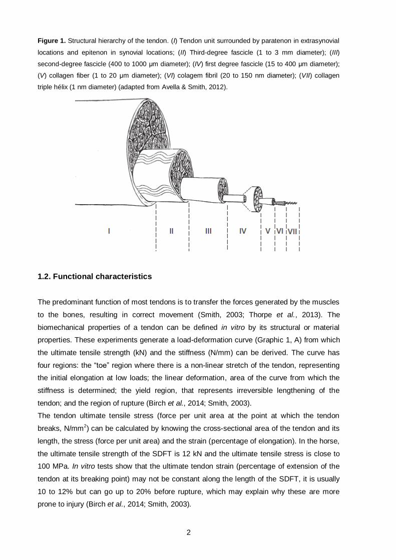
Figure 1. Structural hierarchy of the tendon. (I) Tendon unit surrounded by paratenon in extrasynovial
locations and epitenon in synovial locations; (II) Third-degree fascicle (1 to 3 mm diameter); (III)
second-degree fascicle (400 to 1000 μm diameter); (IV) first degree fascicle (15 to 400 μm diameter);
(V) collagen fiber (1 to 20 μm diameter); (VI) colagem fibril (20 to 150 nm diameter); (VII) collagen
triple hélix (1 nm diameter) (adapted from Avella & Smith, 2012).
1.2. Functional characteristics
The predominant function of most tendons is to transfer the forces generated by the muscles
to the bones, resulting in correct movement (Smith, 2003; Thorpe et al., 2013). The
biomechanical properties of a tendon can be defined in vitro by its structural or material
properties. These experiments generate a load-deformation curve (Graphic 1, A) from which
the ultimate tensile strength (kN) and the stiffness (N/mm) can be derived. The curve has
four regions: the “toe” region where there is a non-linear stretch of the tendon, representing
the initial elongation at low loads; the linear deformation, area of the curve from which the
stiffness is determined; the yield region, that represents irreversible lengthening of the
tendon; and the region of rupture (Birch et al., 2014; Smith, 2003).
The tendon ultimate tensile stress (force per unit area at the point at which the tendon
breaks, N/mm2) can be calculated by knowing the cross-sectional area of the tendon and its
length, the stress (force per unit area) and the strain (percentage of elongation). In the horse,
the ultimate tensile strength of the SDFT is 12 kN and the ultimate tensile stress is close to
100 MPa. In vitro tests show that the ultimate tendon strain (percentage of extension of the
tendon at its breaking point) may not be constant along the length of the SDFT, it is usually
10 to 12% but can go up to 20% before rupture, which may explain why these are more
prone to injury (Birch et al., 2014; Smith, 2003).

3
Graphic 1. (A) Simplified stress-strain curve for tendon. Zone 1 refers to the toe region, initial
elongation at low loads; zone 2 to the linear deformation, area of the curve from which the stiffness is
determined; zone 3 to the yield region after which irreversible damage occurs; and zone 4 to where
tendon fibers rupture. (B) Hysteresis loop for tendon (adapted from Smith, 2003).
When a tendon is loaded the curve obtained is different than when unloaded, demonstrating
the hysteresis property (Graphic 1, B). The area between both curves represents the energy
lost during the loading cycle, which is usually 5%. The energy is mostly lost as heat and can
rise the tendon temperature up to 46ºC. The high temperatures could be harmful to the
tenocytes but these cells remain viable when subjected to temperatures of this magnitude
(Birch et al., 2014).
During tendon loading the energy is stored in its extension, resulting in a reduction in the
energetic cost of locomotion. In order to allow this energy to return to its usable form, the rate
of unloading needs to be tuned to stride frequency since there is little tolerance in the system
and the tendon is prone to overstrain injury. When the limb impacts the ground the loads rise
the quickest in the SDFT, being considered a weight-bearing and energy storing tendon.
During galloping exercise the strain of SDFT is 16%, while the strain of the common digital
extensor tendon (CDET) is 3%, indicating that it is a positional tendon. In energy storing
tendons the properties of the interfascicular matrix influence directly the mechanical
properties of the whole structure, implying that interfascicular sliding is critical for these
tendons (Birch et al., 2014; Smith, 2003; Thorpe et al., 2013).
2. ANATOMY OF THE DISTAL LIMB
2.1. Bones and joints
The equine digit is comprised by the foot and pastern, including the navicular bone, proximal
phalanx (P1), middle phalanx (P2), distal phalanx or coffin bone (P3) and all their joints and
associated structures. Proximally to the digit, the metacarpophalangeal (MCP)/
metatarsophalangeal (MTP) joint, the proximal sesamoid bones (PSBs) and the components
A B

4
surrounding them form the fetlock in both thoracic and pelvic limbs (Kainer & Fails, 2011;
Budras, Sack, Röck, Horowitz & Berg, 2009).
In the horse, only the metacarpal/metatarsal bones two (Mc2/Mt2), three (Mc3/Mt3) and four
(Mc4/Mt4) are present. Depending if localized on thoracic or pelvic limbs, they will be
referred as metacarpal or metatarsal bones, respectively. The Mc2 and Mc4 or splint bones
are rudimental and the Mc3, also known as cannon bone, carries the entire weight assigned
to the limb. In the thoracic limbs (forelimbs) the proximal bases of the Mc3 articulate with the
carpal bones (carpometacarpal joint) while in the pelvic limbs (hindlimbs) the metatarsal 3
(Mt3) articulates with the distal row of tarsal bones (tarsometatarsal joint). The distal end of
the Mc3/Mt3 presents a sagittal ridge that engages a groove in P1 originating the MCP/MTP
joint. The distal surface of P1 and the proximal surface of P2 form the proximal
interphalangeal joint (IPJ) or pastern joint (Kainer & Fails, 2011; Budras et al., 2009).
The proximal and distal sesamoid bones are of extreme importance in the horse. The PSBs
articulate with the Mc3/Mt3, while the distal sesamoid bone, also known as navicular bone,
lies within the hoof and articulates with both P2 and P3. The PSBs are part of the fetlock and
the digital flexor tendons slide between them, through the fetlock canal. The navicular bone
has two articular surfaces that articulate with P2 and P3 and a flexor surface, where the
DDFT glides before inserting in P3. The articular facets of the navicular bone, the distal
articular surface of P2 and the articular surface of P3 form the distal IPJ or coffin joint (Kainer
& Fails, 2011; Budras et al., 2009). Associated to P3, two ungular cartilages provide support
to the palmar/plantar aspect of the foot, dissipate forces of the foot’s impact with the ground
and are involved in venous return from the digit (Dyson, Brown, Collins & Murray, 2010).
These cartilages are large rhomboid curved plates that lie under the corium of the hoof and
extend from each palmar process of the bone proximal to the coronary border of the hoof
(Kainer & Fails, 2011; Mair & Sherlock, 2008).
2.2. Arteries, veins and nerves
The blood supply of the distal limb relies on the medial and lateral palmar/plantar arteries. In
the thoracic limb, the medial palmar vein, artery and nerve lie next to each other in this
dorsopalmar sequence (Figure 2). The artery and nerve pass through the carpal canal to the
metacarpus, where they run medial to the SL and the DDFT. The vein crosses the carpus
superficial to the flexor retinaculum, joining the artery and the nerve in the metacarpus. The
lateral palmar vein, artery and nerve pass the carpus near the extremity of the accessory
carpal bone. The vein and the nerve lie next to each other and deep to them is a small artery
(Budras et al., 2009).
In the fetlock region, the medial and lateral palmar vessels and nerves turn into the medial
and lateral digital vessels and nerves (Figure 2) (Budras et al., 2009). The digital vessels and
5
nerves become superficial on the proximal part of the fetlock, covered by superficial fascia.
As each digital artery crosses over the fetlock it gives off branches to the fetlock joint, digital
extensor and flexor tendons, digital synovial sheath, ligaments, fascia and skin (Kainer &
Fails, 2011).
Around the middle of P1, a short artery of P1 arises from the medial and lateral digital
arteries, originating an anastomotic circle that divides into dorsal and palmar branches. The
palmar branch extends between P1 and the DDFT, while the dorsal branch lies deep to the
CDET. At the level of the proximal IPJ the artery of the digital cushion (bulbar artery) arises
from each digital artery, supplying the hoof with its branches (Kainer & Fails, 2011; Budras et
al., 2009).
The venous drainage of the foot is assured by two parallel veins in the solar canal that come
together at the level of the navicular bone to form the medial and lateral terminal veins.
These veins join with branches of an inner plexus becoming the digital veins that carry the
blood from the digit (Kainer & Fails, 2011).
At the carpal level, the median nerve splits into medial and lateral palmar nerves. The ulnar
nerve also divides in two branches: a dorsal branch that supplies the skin over the
dorsolateral aspect of carpus and metacarpus, and a palmar branch that joins the lateral
palmar nerve. After joining with the ulnar nerve, the lateral palmar nerve gives off a deep
branch that innervates the proximal attachment of the SL and is continued by the medial and
lateral palmar metacarpal nerves (Budras et al., 2009). These run along the axial surface of
the splint bones and emerge immediately on their distal extremity, ramifying in the superficial
fascia of the pastern (Kainer & Fails, 2011).
In the middle of the metacarpus, the lateral and medial palmar nerves form a communicating
branch, before descending to the fetlock. In the fetlock, these nerves continue as lateral and
medial palmar digital nerves and each one gives off a dorsal branch that crosses between
the digital vein and artery (Figure 2). In one third of the cases, an intermediate branch
originates from the dorsal branch and together they supply sensory and vasomotor
innervation to the fetlock and to the dorsal structures of the digit. The palmar continuations of
the nerves supply the fetlock and the palmar structures of the digit. The palmar digital nerves
terminate in a fine branch accompanied by a small artery, establishing a neurovascular
bundle that descends adjacent to the synovial membrane of the distal IPJ to enter P3 (Kainer
& Fails, 2011).
In the pelvic limb, the lateral and medial plantar arteries, veins and nerves continue the
caudal branches of the saphenous artery, medial saphenous vein and tibial nerve,
respectively. The medial and lateral plantar metatarsal nerves are analogous to the thoracic
limb and the medial and lateral dorsal metatarsal nerves are terminal branches of the deep
peroneal nerve (Budras et al., 2009). Anatomically, the vessels and nerves of the distal
pelvic limb are arranged in a similar way to the distal thoracic limb, with minor differences.
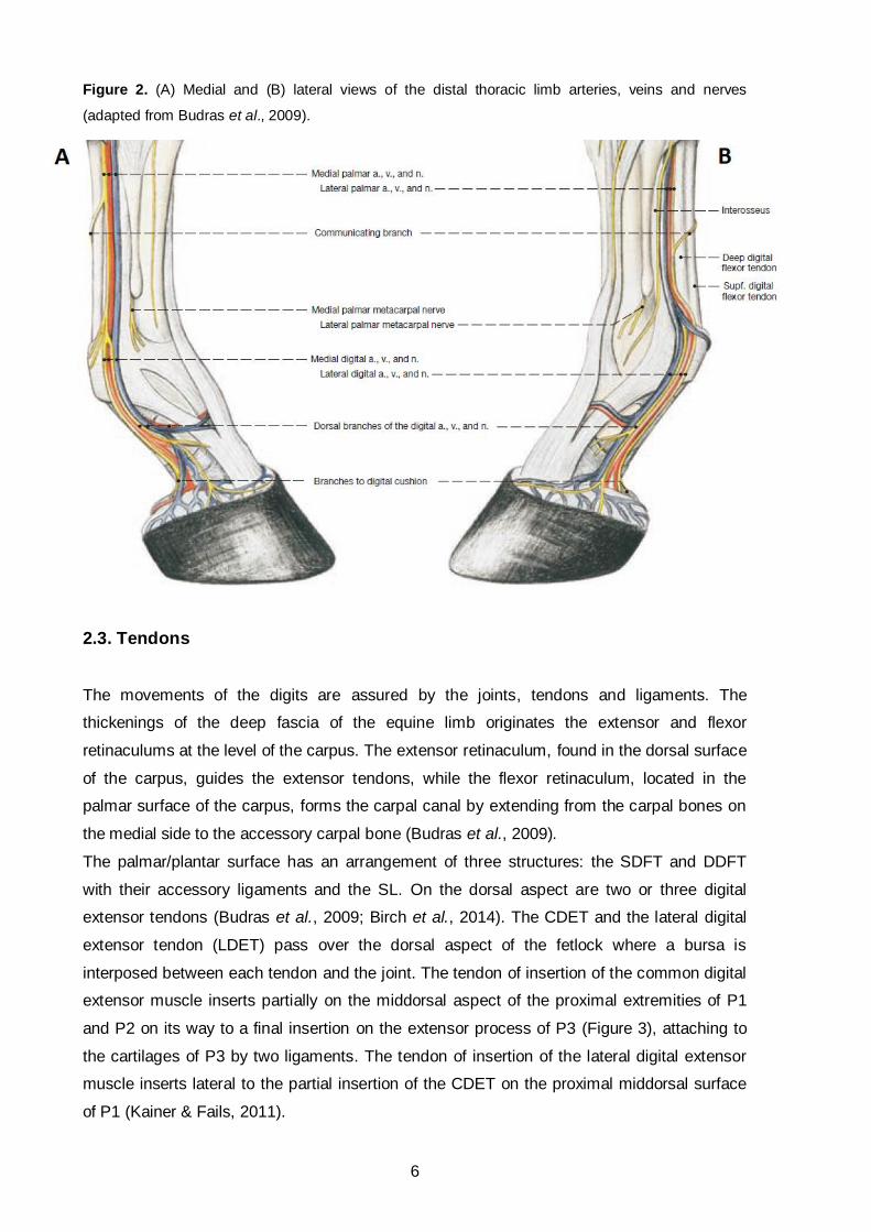
6
Figure 2. (A) Medial and (B) lateral views of the distal thoracic limb arteries, veins and nerves
(adapted from Budras et al., 2009).
2.3. Tendons
The movements of the digits are assured by the joints, tendons and ligaments. The
thickenings of the deep fascia of the equine limb originates the extensor and flexor
retinaculums at the level of the carpus. The extensor retinaculum, found in the dorsal surface
of the carpus, guides the extensor tendons, while the flexor retinaculum, located in the
palmar surface of the carpus, forms the carpal canal by extending from the carpal bones on
the medial side to the accessory carpal bone (Budras et al., 2009).
The palmar/plantar surface has an arrangement of three structures: the SDFT and DDFT
with their accessory ligaments and the SL. On the dorsal aspect are two or three digital
extensor tendons (Budras et al., 2009; Birch et al., 2014). The CDET and the lateral digital
extensor tendon (LDET) pass over the dorsal aspect of the fetlock where a bursa is
interposed between each tendon and the joint. The tendon of insertion of the common digital
extensor muscle inserts partially on the middorsal aspect of the proximal extremities of P1
and P2 on its way to a final insertion on the extensor process of P3 (Figure 3), attaching to
the cartilages of P3 by two ligaments. The tendon of insertion of the lateral digital extensor
muscle inserts lateral to the partial insertion of the CDET on the proximal middorsal surface
of P1 (Kainer & Fails, 2011).

7
In the equine limb, the tendon of insertion of the superficial digital flexor muscle is located in
the palmar surface of the cannon bone, fetlock and pastern (Figure 3). The SDFT terminates
by bifurcating into two branches that insert on the proximal extremity of P2, palmar to the
collateral ligaments of the proximal IPJ. The tendon of insertion of the deep digital flexor
muscle is positioned dorsally to the SDFT, passing between its two branches and inserting in
the flexor surface of P3 (Figure 3) (Kainer & Fails, 2011; Budras et al., 2009). Proximal and
distal to the MCP/MTP joint the SDFT encircles the DDFT forming the manica flexoria (MF)
and the digital manica (DM), respectively (Fiske-Jackson, Barker, Eliashar, Foy & Smith,
2013). The MF extends over 30 mm from the most distal part of the metacarpal/metatarsal
region to the apex of the PSBs, being thicker proximally and becoming thinner distally
(Seignour, Coudry, Norris & Denoix, 2011). This structure has a free distal margin, the
proximal margin is continuous with the tendon sheath and abaxially blends with the SDFT
(McIlwraith, Nixon & Wright, 2015). Its function is to maintain the flexor tendons in a central
position within the digital flexor tendon sheath (DFTS) (Redding, 1993).
Both flexor tendons have accessory ligaments to attach them directly to bone providing a
direct bone to bone connection. Proximally to the carpus the accessory ligament of the SDFT
(superior check ligament) makes this connection, while distally it is achieved by the
accessory ligament of the DDFT (inferior check ligament) localized in the proximal third of the
metacarpus (Birch et al., 2014).
Figure 3. Sagittal section of equine fetlock and digit (adapted from Kainer & Fails, 2011).

8
2.4. Ligaments
The ligaments stabilize the joints by transmitting forces between bones. The fetlock joint
needs to be stabilized to prevent it from overextending when the weight of the horse is
supported by the limb. This is achieved by the suspensory apparatus and the digital flexor
tendons. The suspensory apparatus is composed by the PSBs, the SL and the sesamoidean
ligaments (Figure 4). The musculus interosseous medius tendon (suspensory ligament; SL)
arises from the carpus and proximal end of the cannon bone and inserts on the PSBs, where
it sends two extensor branches around P1 to the CDET. There are two collateral ligaments in
the fetlock that extend distally from the cannon bone and attach to the edge of the articular
surface of P1 and to the abaxial surface of the PSBs, maintaining them in position. Between
the PSBs, the intersesamoidean ligament (ISL) covers the flexor surfaces of these bones
and forms a smooth depression through which the digital flexor tendons pass. The tension in
the SL is continued distal to the joint by the short, cruciate, oblique and straight sesamoidean
ligaments. The short sesamoidean ligaments extend from the base of both PSBs to the
palmar edge of the articular surface of P1, they are the deepest ligaments. The cruciate
sesamoidean ligaments (CSLs) cross each other inserting distally to the contralateral
eminence on the proximal extremity of P1. The oblique sesamoidean ligaments (OSLs)
attach distally to the palmar surface of P1, while the straight sesamoidean ligament (SSL)
attaches to the proximal extremity of the palmar surface of P2. The digital flexor tendons
assist the suspensory apparatus providing a tendinous support via their accessory ligaments
(Kainer & Fails, 2011; Budras et al., 2009).
The thickenings of the deep fascia originate three annular ligaments (Budras et al., 2009).
These are the most superficial ligaments and collectively function as a retinaculum in order to
hold the flexor tendons in the DFTS at the level of the MCP joint (Cohen, Schneider, Zubrod,
Sampson & Tucker, 2008; McGhee, White & Goodrich, 2005). At the level of the fetlock joint,
the fascia forms the palmar annular ligament (PAL) (Figure 4), that has transverse fiber
orientation and lays immediately beneath the skin and subcutaneous tissue (Seignour et al.,
2011). The PAL arises from the abaxial aspect of the PSBs and is most prominent at the
palmar aspect, constituting the palmar border of the fetlock canal. The fetlock canal is an
inelastic canal formed by the PSBs, the ISL and the PAL, maintaining the flexor tendons in
place (Budras et al., 2009; Cohen et al., 2008, Fiske-Jackson et al., 2013; Wilderjans,
Boussauw, Madder & Simon, 2003). Distally, the proximal digital annular ligament (PDAL)
(Figure 4) is located on the palmar surface of P1 and has an X shape, adhering to the SDFT
and extending to the medial and lateral borders of the distal aspect of P1 (Budras et al.,
2009; Cohen et al., 2008). This ligament covers the SDFT as it bifurcates and aids in holding
the DDFT (Kainer & Fails, 2011). The distal digital annular ligament (DDAL) originates
between the medial and lateral borders of the distal aspect of P1 and inserts on the palmar
9
aspect of P3, between the DDFT and the digital cushion. This ligament is crossed medial and
laterally by ligaments of the ergot, which diverge from beneath the horny ergot on the palmar
surface of the fetlock and reach the DDAL, connecting it to the hoof cartilage (Kainer & Fails,
2011; Budras et al., 2009; Cohen et al., 2008).
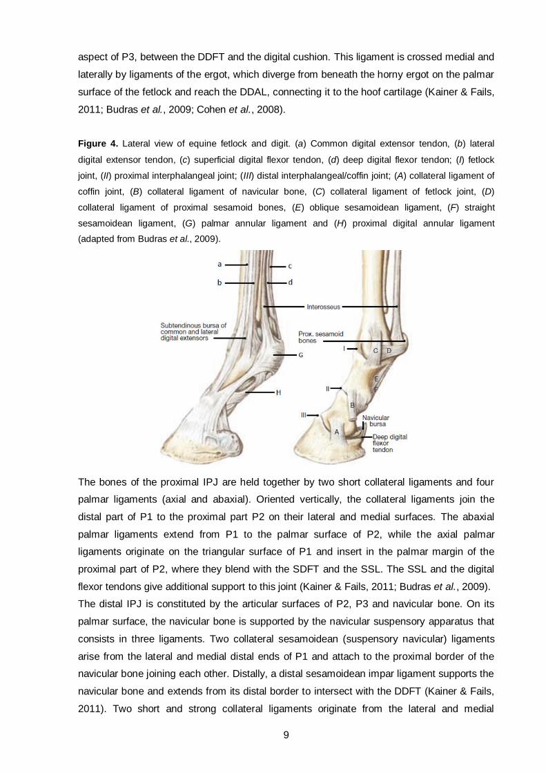
Figure 4. Lateral view of equine fetlock and digit. (a) Common digital extensor tendon, (b) lateral
digital extensor tendon, (c) superficial digital flexor tendon, (d) deep digital flexor tendon; (I) fetlock
joint, (II) proximal interphalangeal joint; (III) distal interphalangeal/coffin joint; (A) collateral ligament of
coffin joint, (B) collateral ligament of navicular bone, (C) collateral ligament of fetlock joint, (D)
collateral ligament of proximal sesamoid bones, (E) oblique sesamoidean ligament, (F) straight
sesamoidean ligament, (G) palmar annular ligament and (H) proximal digital annular ligament
(adapted from Budras et al., 2009).
The bones of the proximal IPJ are held together by two short collateral ligaments and four
palmar ligaments (axial and abaxial). Oriented vertically, the collateral ligaments join the
distal part of P1 to the proximal part P2 on their lateral and medial surfaces. The abaxial
palmar ligaments extend from P1 to the palmar surface of P2, while the axial palmar
ligaments originate on the triangular surface of P1 and insert in the palmar margin of the
proximal part of P2, where they blend with the SDFT and the SSL. The SSL and the digital
flexor tendons give additional support to this joint (Kainer & Fails, 2011; Budras et al., 2009).
The distal IPJ is constituted by the articular surfaces of P2, P3 and navicular bone. On its
palmar surface, the navicular bone is supported by the navicular suspensory apparatus that
consists in three ligaments. Two collateral sesamoidean (suspensory navicular) ligaments
arise from the lateral and medial distal ends of P1 and attach to the proximal border of the
navicular bone joining each other. Distally, a distal sesamoidean impar ligament supports the
navicular bone and extends from its distal border to intersect with the DDFT (Kainer & Fails,
2011). Two short and strong collateral ligaments originate from the lateral and medial

10
collateral fossae of P2 and are placed on the dorsomedial and dorsolateral aspects of the
joint. These ligaments are orientated obliquely, inserting on the collateral fossae of P3 and
the dorsal border of the ungular cartilages (Kainer & Fails, 2011; Denoix, Bertoni, Heitzmann,
Werpy & Audigié, 2011).
In the pelvic limbs, the fetlock and interphalangeal joints are supported as in the thoracic
limbs by the SDFT, DDFT, SL, sesamoidean ligaments and collateral ligaments. There are
two differences from the arrangement in the distal part of the thoracic limbs: the inferior
check ligament is thinner and may be missing, and the superior check ligament is absent,
being compensated by the SDFT firm attachment on the calcanean tuber (Kainer & Fails,
2011).
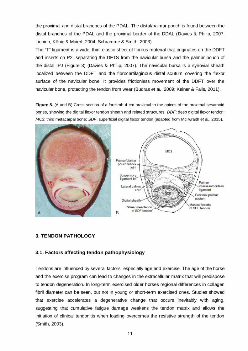
2.5. Synovial sheaths
As a protective mechanism, the digital flexor tendons are contained within synovial sheaths
in regions where they pass over high motion joints: the carpal sheath proximally and the
digital sheath distally. The DFTS surrounds the flexor tendons protecting them from shear
damage and facilitating their movements. The anatomy of the sheath has been well
described and is similar in thoracic and pelvic limbs. It extends from the distal third of the
metacarpal region (4 to 7 cm proximal to the PSBs) to the palmar pouch of the distal IPJ as
far as the transverse lamina ligament (“T” ligament) containing a very small amount of fluid
(Kainer & Fails, 2011; Fiske-Jackson et al., 2013; Schramme & Smith, 2003; Seignour et al.,
2012; Wilderjans, Boussauw, Madder & Simon, 2003).
The DFTS is composed of two layers, an outer fibrous layer and an inner synovial layer. The
dorsal wall of the sheath is formed by the proximal and middle scutum and distal
sesamoidean ligaments, while the palmar wall incorporates the three annular ligaments
(Schramme & Smith, 2003). The DFTS is surrounded by the PAL at the level of the fetlock
canal (Figure 5), the PDAL along the palmar border of P1 and the DDAL that adheres to the
palmar surface of the distal part of the sheath (Wilderjans et al., 2003). Proximal to the MF,
the DDFT is attached to the DFTS medially and laterally by the mesotenon or synovial plica.
Distal to the MF further mesotenons (vinculae) attach the DDFT to the dorsal sheath wall (SW)
(Wright & McMahon, 1999). The SDFT is also attached to the sheath by a thin mesotenon on
the palmar aspect of the fetlock (Figure 5) (Wilderjans et al., 2003). The mesotenon between
the SDFT and the PAL is the so-called vinculum of the SDFT (Schramme & Smith, 2003).
In case of inflammation the sheath can accumulate fluid and pouch out in any of the places
where it is not attached to the annular ligaments: three paired proximal pouches (proximal,
proximal collateral and distal collateral pouches) and one distal/palmar pouch. The proximal
pouch is proximal to the PAL and palmar to the branches of the SL, the proximal collateral
pouch lies between the PAL and the PDAL, and the distal collateral pouch occurs between
11
the proximal and distal branches of the PDAL. The distal/palmar pouch is found between the
distal branches of the PDAL and the proximal border of the DDAL (Davies & Philip, 2007;
Liebich, König & Maierl, 2004; Schramme & Smith, 2003).
The “T” ligament is a wide, thin, elastic sheet of fibrous material that originates on the DDFT
and inserts on P2, separating the DFTS from the navicular bursa and the palmar pouch of
the distal IPJ (Figure 3) (Davies & Philip, 2007). The navicular bursa is a synovial sheath
localized between the DDFT and the fibrocartilaginous distal scutum covering the flexor
surface of the navicular bone. It provides frictionless movement of the DDFT over the
navicular bone, protecting the tendon from wear (Budras et al., 2009; Kainer & Fails, 2011).
Figure 5. (A and B) Cross section of a forelimb 4 cm proximal to the apices of the proximal sesamoid
bones, showing the digital flexor tendon sheath and related structures. DDF: deep digital flexor tendon;
MC3: third metacarpal bone; SDF: superficial digital flexor tendon (adapted from McIlwraith et al., 2015).
3. TENDON PATHOLOGY
3.1. Factors affecting tendon pathophysiology
Tendons are influenced by several factors, especially age and exercise. The age of the horse
and the exercise program can lead to changes in the extracellular matrix that will predispose
to tendon degeneration. In long-term exercised older horses regional differences in collagen
fibril diameter can be seen, but not in young or short-term exercised ones. Studies showed
that exercise accelerates a degenerative change that occurs inevitably with aging,
suggesting that cumulative fatigue damage weakens the tendon matrix and allows the
initiation of clinical tendonitis when loading overcomes the resistive strength of the tendon
(Smith, 2003).
12
Being submitted to stress during aging leads to a gradual decrease in the mechanical
integrity of the tendons. Various stimuli could result in the synthesis, release or activation of
proteolytic enzymes that originate an imbalance between the synthesis and degradation of
the extracellular matrix and result in accumulation of partially cleaved collagen. The actual
mechanism of degeneration of the tendon is currently unknown but the decline of collagen
turnover, the energy transmitted to the tendon under weight-bearing load and the rise of the
temperature within the tendon can produce direct damage to the matrix by disrupting the
tendon ultrastructure (Birch et al., 2014; Smith, 2003; Thorpe et al., 2013).
Thorpe et al. (2013) suggested that alongside aging a significant stiffening of the SDFT
interfascicular matrix occurs, leading to a decreased capacity of sliding between fascicles.
The area occupied by the matrix decreases causing tendons to be more tightly packed in
aged individuals. In addition, the improper repair of any tendon damage may result in the
formation of adhesions.
Besides the exercise and the age of the horse, there are other risk factors that predispose for
tendon injury such as the weight the horse is carrying, its body weight, fatigue, shoeing,
working surface (especially hard surfaces) and speed of the horse (the faster the horse is
going, the greater the risk of tendinitis) (Birch et al., 2014).
3.2. Tendon injury and healing
Tendons and ligaments can be injured by overstrain or percutaneous penetration/laceration.
Overstrain injuries can result from a sudden overloading or continuous degeneration. Clinical
injuries occur when the stress encountered by the tendon overwhelms its structural integrity
leading to irreversible damage. These strain-induced tendinopathies are usually bilateral with
one limb more severely affected than the other (Birch et al., 2014).
Once the tendon suffers clinical injury with disruption of its matrix, there is an acute
inflammatory reaction with increased blood-flow, oedema, infiltration of cells (neutrophils,
monocytes and macrophages) and release of proteolytic enzymes. This first phase of repair
allows the removal of the damaged tendon tissue, lasting one to two weeks. After this, the
reparative phase begins, with strong angiogenic response and accumulation of fibroblasts to
synthetize scar tissue. The scar tissue is mainly composed by collagen type III, being weaker
than the normal tendon tissue. Several months after the injury, the remodeling phase starts
and the collagen type III is gradually replaced by collagen type I. This matured scar tissue is
stronger than the normal tendon tissue but has poor elasticity, resulting in increased strain in
adjacent undamaged regions of the tendon. Consequently, re-injure in the same tendon can
occur, but frequently at adjacent or remote sites of the original injury (Birch et al., 2014;
Smith, 2003).

13
3.3. Tendonitis
Clinical overstrain injuries, known as tendonitis, are about 10% of lameness cases in the
athletic horses (Kalisiak, 2012). Thoroughbreds and upper level event horses have an
increased risk of SDFT injury due to the high speed associated with jumping. Most SDFT
injuries caused by athletic use occur in the forelimb mid-metacarpal region (zones 2B to 3B)
and appear as a convex “bow” on the side view (Avella & Smith, 2012; Bertone, 2011; Gillis,
2014; Jorgensen & Genovese, 2003). Lesions in the distal cannon bone can be associated
with digital sheath tenosynovitis or constriction of the PAL (Bertone, 2011). Regions of the
SDFT enclosed within tendon sheaths are less affected, in contrast with the DDFT, which is
more frequently injured within the DFTS (Avella & Smith, 2012; Gillis, 2014).
The clinical signs of SDFT injury vary considerably depending on the location, type, severity
and timing of the injury. Clinical signs of SDF tendonitis include lameness, swelling,
thickening, heat and sensitivity to direct digital palpation. A high palmar nerve block usually
resolves the lameness but ultrasonography (USG) is the recommended diagnostic method
for tendonitis. It is important to evaluate the echogenicity of the tendons, perform area
measurements and compare it with the contralateral limb. One of the greatest manifestations
of SDF tendonitis is a central injury (“core” lesion) seen ultrasonographically. The cross-
section area (CSA) of the hypoechogenic lesions is the most used parameter to describe
tendonitis severity, along with an increase in the maximal tendon CSA and total tendon CSA
(Avella & Smith, 2012; Kalisiak, 2012; Jorgensen & Genovese, 2003).
Tendonitis of the DDFT is generally associated to unilateral DFTS effusion and mild to
moderately severe lameness. During clinical evaluation, pain can be elicited by palpation and
passive flexion of the lower limb. Intrathecal analgesia of the DFTS usually results in
significant improvement but low palmar/plantar perineural analgesia shows better results.
The definitive diagnosis requires ultrasonographic evaluation that can be difficult to perform
with DFTS effusion. DDF tendonitis can display four types of lesions: enlargement and
change in tendon shape, focal hypoechogenic lesions within the tendon or on its border,
mineralization within the tendon and marginal tears. In some cases, surgical exploration
(tenoscopy) may be required for a definitive diagnosis (Dyson, 2003).
The treatment of strain-induced tendinopathies can be nonsurgical and/or surgical. The
decision of whether to manage a horse conservatively or surgically should take in account the
history, clinical evaluation, response to previous treatments, use of the horse and some other
factors. As a nonsurgical approach, pharmacological treatment (systemic and intralesional) and
physical therapy such as cold therapy, compression, controlled exercise, extracorporeal shock
wave therapy and therapeutic ultrasound are of extreme importance for a successful return to
work. The surgical approach allows the exploration of the DFTS with direct observation of the
lesions and possibility of debriding adhesions and tears (Avella & Smith, 2012).

14
3.4. Tendon tears
Tendon tears within the DFTS usually occur in the DDFT (predominantly in the forelimbs)
and the MF of the SDFT (mainly in the hindlimbs) (Fiske-Jackson et al., 2013; Smith &
Wright, 2006). DDFT tears occur when higher pressures within the compressed portion of the
tendon passes over the MCP joint leading to its “bursting”, more commonly of its lateral
border. Manica tears may occur when the MF gets trapped in the fetlock canal, and be
exacerbated by synovial hypertrophy, adhesions and/or PAL constriction within the sheath
(Avella & Smith, 2012).
MF tears are more common in the hindlimbs and associated to DFTS effusion (Findley,
Oliveira & Bladon, 2012; Smith & Wright, 2006). Ultrasonographic diagnosis has a low
sensitivity, showing more than one abnormality, such as sheath effusion, PAL thickening and
synovial proliferation (Findley et al., 2012; Smith & Wright, 2006; Wilderjans et al., 2003).
According to the study undertaken by Fiske-Jackson et al. (2013) MF tears are particularly
hard to identify ultrasonographically hence contrast radiography performed at the same time
as intrathecal analgesia of the DFTS can provide valuable information to an accurate
diagnose. Tears of the MF are more frequent on the medial attachment to the SDFT and can
be classified as acute or chronic, complete or partial and longitudinal or transverse. This
classification is based on appearance during tenoscopic observation (Findley et al., 2012;
Smith & Wright, 2006).
Longitudinal tears (LTs) of the digital flexor tendons were first described by Wright and
McMahon (1999) as an underlying cause of tenosynovitis of the DFTS. These are seen
predominantly in the forelimbs. The most common site of LTs is the lateral margin of the
DDFT, followed by the medial margin and the dorsal and palmar/plantar margins in a minority
of cases (Arensburg, Wilderjans, Simon, Dewulf & Boussauw, 2011; Smith & Wright, 2006;
Wilderjands et al., 2003; Wright & McMahon, 1999). LTs of the SDFT seem to be much less
common than the ones in the DDFT (Arensburg et al., 2011; Smith & Wright, 2006).
Horses with DDFT tears are more easily diagnosed with USG and intrathecal analgesia,
since they are more likely to show a positive improvement in lameness after diagnostic
analgesia than horses with a MF tear (Fiske-Jackson et al., 2013). Typical ultrasonographic
changes indicative of LTs were defined as irregular borders of the tendon, hypoechogenic
foci and echogenic masses continuous with the tendon’s border (Figure 6) (Arensburg et al.,
2011; Smith & Wright, 2006; Wilderjands et al., 2003; Wright & McMahon, 1999). Tenoscopy
is the most reliable technique used to confirm the presence of LTs and is recommended in all
cases (Arensburg et al., 2011; Wilderjands et al., 2003). Since disrupted tendon fibrils cannot
be removed from the tendon sheath by intrinsic mechanisms (Wright & McMahon, 1999),
tenoscopic debridement and removal of torn fibrils is the most effective treatment, although
the long-term prognosis is guarded (Arensburg et al., 2011).

15
Figure 6. Ultrasonographic image of an acute case of a longitudinal tear (LT) in the lateral border of
the deep digital flexor tendon (DDFT). Typical ultrasonographic changes indicative of LTs are visible:
irregular lateral border of the DDFT (arrow), hypoechogenic foci and an echogenic mass (x)
continuous with the DDFT border. LAT = lateral (adapted from Arensburg et al., 2011).
3.5. Percutaneous tendon injury
The equine distal limb has minimal soft tissue cover, being prone to tendon injuries by
trauma. The extensor tendons and the SDFT are positioned directly under the skin therefore
small wounds can transect these tendons. Traumatic injuries such as overreaching, wires,
landing on sharp objects, jumping and kicking injuries are the most common causes to
percutaneous tendon damage (Avella & Smith, 2012; Dyson & Bertone, 2003).
It is of extreme importance to palpate, shave, clean, debride and evaluate the wound,
especially its position relative to the synovial structures, since concurrent synovial
contamination or sepsis requires specific emergency treatment. If suspected of DFTS
contamination synovial fluid can be obtained by synoviocentesis and submitted for
cytological and biochemical evaluation. Ultrasonographic examination of the wound and
tendons can help to determine the presence of foreign material, the degree of tendon
damage and if there is involvement of synovial structures (Dyson & Bertone, 2003).
As medical management, all horses with tendon lacerations need wound cleansing
debridement and broad-spectrum antibiotics (Dyson & Bertone, 2003). If the DFTS is
involved, surgery should be performed in order to further evaluate the lesions, remove
debris, adhesions or foreign bodies, lavage, repair and perform intrathecal administration of
antimicrobial drugs. Close monitoring of sheath fluid cytological condition, longer use of
systemic and intrathecal antibiotics and immobilization of the limb should be continued post-
surgery (Fraser & Bladon, 2004; Dyson & Bertone, 2003).
X

16
4. LIGAMENT PATHOLOGY
4.1. Palmar/plantar annular ligament syndrome and desmitis
The PAL is composed of transverse fibers that attach to the PSBs, forming the fetlock canal
together with the ISL. PAL syndrome refers to the different diseases that have characteristic
clinical manifestation that include PAL thickening and distension of the DFTS. Enlargement
of the DFTS or the flexor tendons can lead to constriction within the fetlock canal with a
relatively normal PAL. The clinical signs of PAL syndrome consist in persistent lameness
accompanying a concave notch at the level of the PAL with the DFTS protruding proximally
(Figure 7). In these cases, tenoscopic evaluation for further examination and treatment is
advised (McGhee et al., 2005; Schramme & Smith, 2003).
Primary desmitis of the PAL can be caused by traumatic injury, hyperextension of the fetlock
with excessive tension on the PAL (Owen et al., 2008; Schramme & Smith, 2003), pressure
caused by swelling of the flexor tendons and chronic tenosynovitis with adhesion and
fibrosis. Thickening of the PAL with no pathological conditions of the structures within the
DFTS can cause lameness in horses since it leads to stenosis of the fetlock canal and
constriction of the flexor tendons. This constriction and continuous pressure originate further
inflammation, fibrosis, thickening and persistent pain (Schramme & Smith, 2003).
Figure 7. Lateral view of a limb with plantar
annular ligament (PAL) syndrome. Note the
digital sheath distension with characteristic
concave notch of the tendons proximal to the
PAL (arrow) (adapted from Nixon, 2011).
Figure 8. Lateral view of a hindlimb with
primary desmitis of the plantar annular ligament.
There is a convex contour of the plantar aspect
of the fetlock (arrow) due to ligament
thickening (adapted from Owen et al., 2008).
17
PAL desmitis and thickening is more common in hindlimbs and results in a convex contour of
the palmar/plantar aspect of the fetlock with distension of the DFTS, especially proximal to
the PAL (Figure 8). Horses usually have a history of mild to moderate persistent lameness
and pain can be elicited by palpation and manipulation of the fetlock. Intrathecal and
perineural analgesia of the palmar/plantar nerves can improve lameness associated with this
condition (Owen et al., 2008; Schramme & Smith, 2003).
USG is widely used as a diagnostic method for PAL desmitis and should be performed in the
lame and the contralateral nonlame limb as comparison. An unaffected PAL can be hard to
identify because it is only 1 to 2 mm thick, while a thickened PAL has more than 2 mm
(Figure 9) and can have a diffuse or focal decrease in echogenicity, loss of fiber pattern and
subcutaneous fibrosis associated. In acute cases, controlled exercise, anti-inflammatory
medication and correct farriery can be an effective treatment. In chronic, unresponsive cases
and/or with lesions identified within the DFTS, surgery is recommended (Owen et al., 2008;
Schramme & Smith, 2003). A minimally invasive approach through a small skin incision and
subcutaneous transection of the PAL blindly is quick and simple but tenoscopically guided
PAL desmotomy allows a complete examination of the DFTS and the treatment of
concomitant pathologies (Schramme & Smith, 2003).
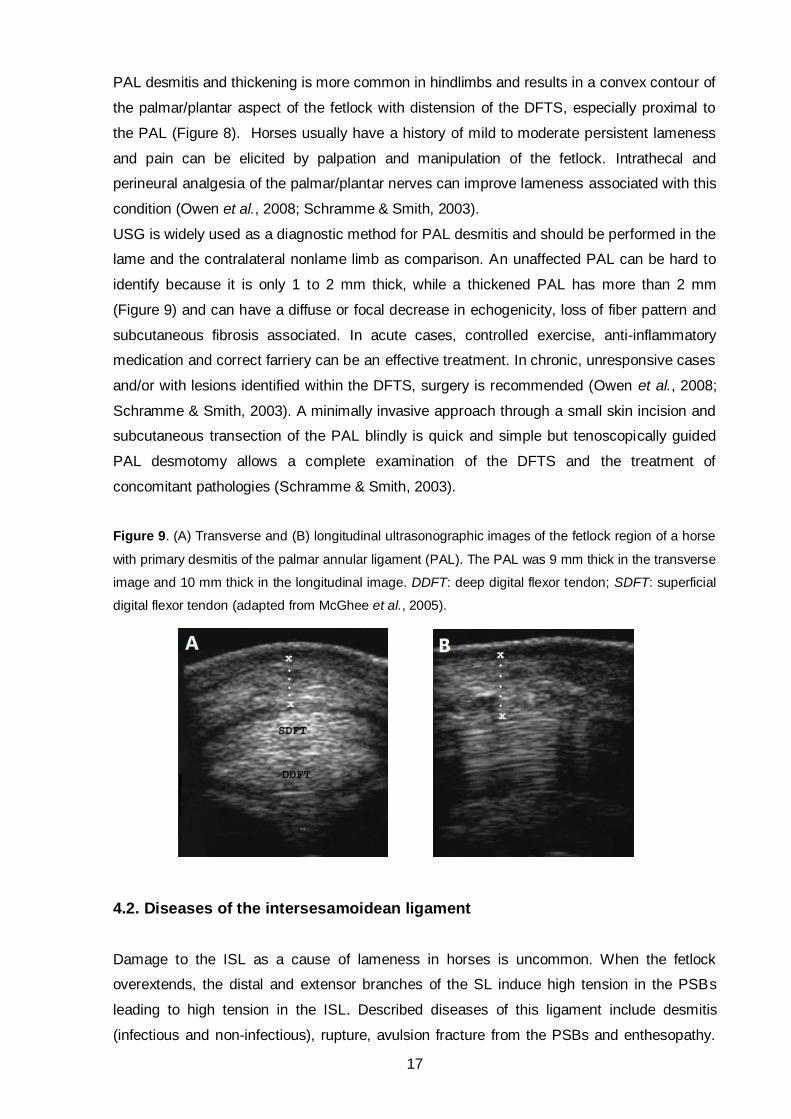
Figure 9. (A) Transverse and (B) longitudinal ultrasonographic images of the fetlock region of a horse
with primary desmitis of the palmar annular ligament (PAL). The PAL was 9 mm thick in the transverse
image and 10 mm thick in the longitudinal image. DDFT: deep digital flexor tendon; SDFT: superficial
digital flexor tendon (adapted from McGhee et al., 2005).
4.2. Diseases of the intersesamoidean ligament
Damage to the ISL as a cause of lameness in horses is uncommon. When the fetlock
overextends, the distal and extensor branches of the SL induce high tension in the PSBs
leading to high tension in the ISL. Described diseases of this ligament include desmitis
(infectious and non-infectious), rupture, avulsion fracture from the PSBs and enthesopathy.

18
Injuries in the ISL usually cause acute moderate to severe lameness that can be improved
with intrasynovial analgesia of the fetlock joint or DFTS and eliminated by a low four-point
nerve block. Ultrasonographic examination can display abnormalities such as enlargement of
the space between the PSBs in case of rupture, and enlargement and alteration of
echogenicity within the ligament in case of desmitis and tears (Dyson & Genovese, 2003).
Desmitis of the ISL is usually accompanied by axial osteitis of the PSBs, characterized by
focal areas of bone lysis and wearing of the ISL in its insertions. This pathology can be
associated with infectious tenosynovitis and/or arthritis (Brommer et al., 2014).
Tears in this ligament are rare and particularly difficult to diagnose with USG. A definitive
diagnose and treatment can only be achieved by arthroscopy of the palmar/plantar pouch of
the MCP/MTP joint and/or tenoscopy of the DFTS. Horses with desmitis and/or focal tears of
the ISL have poor response to the surgical treatment even when associated with
conservative therapy (Dyson & Genovese, 2003).
4.3. Straight and oblique sesamoidean ligaments desmitis
The SSL and the OSLs are part of the suspensory apparatus, holding the PSBs to P1 and P2
(Sampson et al., 2007; Smith, Dyson & Murray, 2008). Desmitis of the SSL as cause of
lameness in horses is rare and can occur together with desmitis of one or both OSLs (Dyson
& Genovese, 2003).
Clinical signs include acute onset of lameness with no palpable abnormalities in the region
and usually without swelling, although swelling and DFTS effusion can develop after a few
days. Lameness can be eliminated with an abaxial sesamoid nerve block (Gillis, 2014; Dyson
& Genovese, 2003; Sampson et al., 2007; Smith et al., 2008).
The diagnosis is based on USG even though the overlying of the digital flexor tendons
impairs the ultrasound image (Sampson et al., 2007; Smith et al., 2008). Desmitis of these
ligaments can be recognised in USG as enlargement of the ligament, diffuse reduction in
echogenicity, focal hypoechogenic areas, poor delineation of the margins of the ligament,
reduction of the distance between the ligament and the DDFT/SDFT and sheath effusion
(Dyson & Genovese, 2003). Radiography might reveal bone changes in the insertions of the
ligaments (Gillis, 2014). For an accurate diagnose, magnetic resonance imaging seems to be
the best diagnostic method. Treatment for desmitis of the SSL and/or the OSL consists of
box rest and corrective farriery followed by a rehabilitation program with controlled exercise
(Dyson & Genovese, 2003; Sampson et al., 2007). Occasionally, lesions of the SSL can
disrupt the palmar/plantar surface of the ligament and covering synovium, leading to
tenosynovitis. In these cases, tenoscopic examination is indicated for debridement and
treatment (McIlwraith et al., 2015).
19
5. DIGITAL FLEXOR TENDON SHEATH PATHOLOGY
5.1. Ganglion cyst
Ganglion cysts are small cysts (1.5 to 3 cm diameter) composed of fibrous or fibromyxoid
tissue that does not hold synovium. Originated from degenerated connective tissue, these
cysts are connected to a joint or tendon sheath. Their common localization is proximally in
the DFTS just proximal to the PAL. Lameness and DFTS effusion caused by ganglion cysts
in horses is an uncommon condition. To establish the clinical significance of the cyst,
ultrasound, perineural anaesthesia and intrathecal analgesia with contrast radiographs are
required. Tenoscopy and surgical removal of the cyst has a good prognosis for soundness if
there is no concurrent pathology of the tendons (Crawford, O’Donnell, Crowe, Eliashar &
Smith, 2010; Schramme & Smith, 2003).
5.2. Non-infectious tenosynovitis
DFTS effusion is common in working horses, frequently idiopathic in origin and affecting both
limbs without causing lameness. In some cases this effusion is seen in only one limb along
with lameness. The term tenosynovitis refers to inflammation of the synovial membrane of a
tendon sheath and may be a single clinical entity or be associated to a tendon injury
(Schramme & Smith, 2003; Wright & McMahon, 1999).
Non-infectious tenosynovitis of the DFTS has been attributed to many causes, for example
tendonitis of the SDFT and DDFT, desmitis of the PAL and PDAL, LTs of the SDFT and
DDFT, MF tears and chronic synovitis of the DFTS of unknown cause (Smith & Wright, 2006;
Wilderjans et al., 2003). These conditions are likely to result in chronic tenosynovitis due to
continuous irritation to the sheath by the exposed torn collagenous tissue (Wright &
McMahon, 1999). Complex tenosynovitis has been described as tenosynovitis with thickening
of the PAL, synovial distension and adhesions/synovial masses (Schramme & Smith, 2003).
Diagnostic techniques that localize disease to the sheath include synoviocentesis and
synovial fluid analysis, intrathecal analgesia and positive contrast radiography (Schramme &
Smith, 2003). USG is the most commonly used technique for evaluating the DFTS, although
it has poor accuracy for lesion prediction, especially for the presence of marginal tears of the
DDFT, SDFT and MF (Smith & Wright, 2006). Tenosynovitis has been described in three
ultrasonographic stages of progression: stage 1, with symmetrical distention of the DFTS and
without evidence of synovial proliferation; stage 2, often asymmetrical distension of the proximal
pouch with synovial proliferation; and stage 3, extensive synovial proliferation with adhesions and
synovial masses in the sheath (Schramme & Smith, 2003).

20
The treatment of horses with acute tenosynovitis consists of systemic anti-inflammatory
medication, bandage immobilization, cold therapy and rest. In the chronic cases, aspiration
of the synovial fluid and intra-thecal administration of hyaluronan and corticosteroids might
be necessary. In unresponsive cases and in the ones of complex tenosynovitis tenoscopic
exploration is indicated (Schramme & Smith, 2003).Tenoscopy is necessary to identify
accurately the presence of lesions and their morphologic features, allowing an appropriate
treatment and a better prognosis (Smith & Wright, 2006).
5.3. Infectious tenosynovitis
Infectious tenosynovitis is a critical condition in the horse that can lead to death or
euthanasia. Horses with DFTS infection usually present with severe, non-weight-bearing
lameness with heat, pain and diffuse swelling. Contamination results from the introduction of
microorganisms usually by penetrating wounds, although it may also develop following intra-
thecal injections or as result of extension from adjacent tissues. Rarely haematogenous
spread to the digital sheath may result from bacteremia (Bertone, 2011; Schramme & Smith,
2003; Wereszka, White & Furr, 2007). Infection follows when the microorganisms proliferate
and colonize the sheath, leading to an acute inflammatory response with rapid influx of
neutrophils and other inflammatory cells (McIlwraith et al., 2015).
An early recognition and prompt treatment are crucial, since it is difficult to eliminate infection
from the DFTS and there is a high risk of long-term sequela that contributes to permanent
lameness. Clinical examination, radiography and USG are useful to recognize complicating
factors such as foreign bodies, concurrent tendon and ligament injuries, osteomyelitis and
infectious tendinitis. Synoviocentesis should be performed as early as possible since it allows
collection of a synovial fluid sample for culture and sensitivity testing, and cytological analysis
(Bertone, 2011; Schramme & Smith, 2003; Wereszka et al., 2007). Negative bacteria culture
results do not necessarily rule out infection because bacteria can be sequestered in
neutrophils, fibrin or synovium, and previous treatments can inhibit their growth (Schramme
& Smith, 2003; Wereszka et al., 2007).
Treatment of infectious tenosynovitis must consist of aggressive systemic and intrathecal
broad-spectrum antibiotics with lavage of the sheath. Tenoscopy should be performed in
order to lavage the sheath (either with plain fluids or diluted antimicrobials), debride fibrin and
adhesions, remove foreign bodies and sometimes transect the PAL to provide pain relieve in
cases where there is excessive fluid distension and thickening of the SW. The portals are
usually closed but in specific cases can be left open for drainage. Bandaging and
immobilization of the limb promote wound healing and reduce inflammation; on the other
hand, it limits drainage and promotes the formation of scar tissue and adhesions (Schramme
& Smith, 2003; Wereszka et al., 2007).

21
In horses with extensive contaminated wounds, these can be cleaned and kept under sterile
bandages to allow healing by second intension. It is of extreme importance to maintain the
intrathecal antimicrobial concentration within therapeutic concentrations over an extended
period of time. That can be achieved with intrathecal administration, intravenous regional
perfusion of antibiotics, antibiotic infusion pumps and slow release antibiotic-depot systems
in collagen or polymethylmetacrylate (Schramme & Smith, 2003). According to Fraser and
Bladon (2004), treatment within 36 hr of initial injury leads to a better prognosis for the
horse’s return to athletic function.
6. DIAGNOSTIC METHODS
6.1. Synoviocentesis
Synoviocentesis or tenovaginocentesis is usually performed for intrathecal administrations
and to determine if a wound has penetrated the sheath, especially soon after injury or in
small self-closing wounds. This technique can be done in one of the DFTS pouches (Figure 10,
A), being easier if the sheath is distended (Dykgraaf et al., 2007; Schramme & Smith, 2003).
The easiest access to the sheath is through its distal/palmar pouch (Figure 10, B), localized
between the distal branches of the PDAL and the proximal border of the DDAL, along the
palmar surface of the DDFT. When the sheath is distended, access to the proximal pouch is
possible by introducing a needle along the dorsal aspect of the DDFT, between the DDFT
and the lateral branch of the SL, proximal to the lateral PSB and PAL. Synoviocentesis of the
DFTS can also be done from its proximal or distal collateral pouches (Schumacher,
Schumacher, Schramme, Degraves & Smith, 2007; Moyer, Schumacher & Schumacher,
2007; Schramme & Smith, 2003). Recent studies show that this technique is more successful
when performed in an axial (axial to the PSB) or a distal (at the pastern) approaches
(Jordana, Oosterlinck, Pille, Valère & Martens, 2012).
After a sterile preparation of the sheath (clipping and surgical scrub), a 19 or 20 gauge, 2.5
cm (1 inch) needle is introduced, the fluid is collected with a syringe and placed into a
sodium EDTA tube for cytological analysis (Mahaffey, 2002; Dykgraaf et al., 2007;
Schumacher et al., 2007). The fluid can also be placed into a plain tube for culture and
sensitivity testing. The normal DFTS synovial fluid is clear yellow with total protein (TP)
concentration values less than 10 g/L and total nucleated cell count (TNCC) inferior than 2 x
109cells/L (Mahaffey, 2002; Schramme & Smith, 2003). A TNCC higher than 30 x 109 cells/L
with more than 80 to 90% neutrophils and TP greater or equal to 40 g/L is considered
pathognomonic of infection (Dykgraaf et al., 2007; Schramme & Smith, 2003; Wereszka et
al., 2007).

22
Figure 10. (A) Representation of the digital flexor tendon sheath (DFTS) pouches. In this image, the
distal and the lateral proximal pouches are distended. (B) Access to the DFTS through the distal
pouch. The needle should be aimed between the deep digital flexor tendon and the ipsilateral distal
branch of the superficial digital flexor tendon (adapted from Schumacher et al., 2007).
6.2. Diagnostic analgesia
Diagnostic analgesia is one of the most valuable tools to localize lameness. The technique
can be performed on site with minimal equipment and expense, allowing a fast assessment
of the outcome (Bassage II & Ross, 2003).
To accurately localize lameness to the DFTS, perineural (nerve blocks) and intrathecal
analgesia are of extreme importance. Because of the DFTS extension, regional analgesia of
the medial and lateral palmar/plantar digital nerves proximal to the DFTS (abaxial sesamoid
nerve block) can be non-specific, resulting in partial responses (Baxter & Stashak, 2011;
Fiske-Jackson et al., 2013). Presently, mepivacaine hydrochloride 2% is the most frequently
used anaesthetic for regional and intrasynovial anaesthesia due to its long lasting activity
and low tissue irritation (Jordana et al., 2016).
Analgesia of the sheath is considered more specific and efficient since it desensitizes the
structures within, localizing the source of pain to the DFTS. This can be achieved by
intrathecal administration of 10 to 15 mL of mepivacaine hydrochloride 2% in one of the sites
where synoviocentesis can be performed (Baxter & Stashak, 2011; Moyer et al., 2007;
Schramme & Smith, 2003). Even when the pathology is localized within the sheath, partial
responses to intrathecal analgesia are common, especially in horses with MF tears as shown
by Findley et al. (2012) and Fiske-Jackson et al. (2013). Furthermore, it has been shown that
diffusion of mepivacaine from the DFTS to adjacent synovial structures occurs but the
concentrations are insufficient for synovial analgesia (Jordana et al., 2016), suggesting that
the improvement of lameness in horses that undergo intrathecal analgesia of the DFTS is
due to analgesia of the structures within the sheath and not adjacent to it (Harper,
Schumacher, Degraves, Schramme & Schumacher, 2007).
A B

23
6.3. Radiography
Radiography of the DFTS is an important diagnostic method for tendon sheath pathology. It
is usually performed to make evident the existence of intrathecal air when a penetrating
wound is present, to visualize foreign materials, soft-tissue swelling and metaplastic
mineralization of structures usually associated with sheath pathology, and to study
concomitant bone injury (Farrow, 2006; McIlwraith et al., 2015; Schramme & Smith, 2003).
Contrast radiography of the DFTS (negative and positive-contrast tenography) is an easy
technique to perform and by outlining the interior of the sheath, it provides information
regarding tendon sheath perforation, thickening, stenosis, adhesion, intra and extrasynovial
masses, mass effects and foreign bodies (Farrow, 2006). Positive-contrast tenography can be
achieved by injecting 5 to 10 mL of sterile iodine solution in the DFTS while performing
intrathecal diagnostic analgesia, followed by a lateromedial radiograph of the distal limb
including the whole length of the DFTS (Fiske-Jackson et al., 2013; Schramme & Smith, 2003).
The contrast is uptaken by the proximal pouch dorsal to the DDFT, and distal to it, the MF,
DDFT and SDFT are visible as filling defects outlined by contrast. Distal to the PSBs the
contrast accumulates in the collateral pouches and extends from the distal pouch to the “T”
ligament (Fiske-Jackson et al., 2013).
Contrast radiography was a widely used diagnostic method before the advances and
widespread of USG (Valdéz-Martinéz & Park, 2011). With this diagnostic technique it is
possible to identify pathologies such as MF and DDFT tears (Fiske-Jackson et al., 2013),
synovial herniations, intrasynovial communications (Valdéz-Martinéz & Park, 2011), ganglion
cysts (Crawford et al., 2010) and perforation of the sheath (Schramme & Smith, 2003).
According to Fiske-Jackson et al. (2013) positive contrast radiography delineates the border
of the MF accurately allowing the diagnosis of MF tears with a sensitivity of 96%, exceeding
that of USG. Currently, the use of contrast radiography has declined but it still remains a
useful method for the preoperative diagnosis of DFTS pathology, especially if combined with
USG and the results of diagnostic analgesia (Fiske-Jackson et al., 2013).
6.4. Ultrasonography
Diagnostic USG was introduced in the early 1980s to equine veterinarians as a practical
diagnostic technique to evaluate soft tissue injuries. Over the last decades ultrasound
technology has improved, becoming the imaging modality of choice for soft tissue evaluation,
especially tendons and ligaments. A correct knowledge of the normal anatomy, adequate
equipment, limb preparation and scanning skills of the ultrasonographer are critical for a
reliable diagnose (Rantanen, Jorgensen & Genovese, 2003; Redding, 2011).
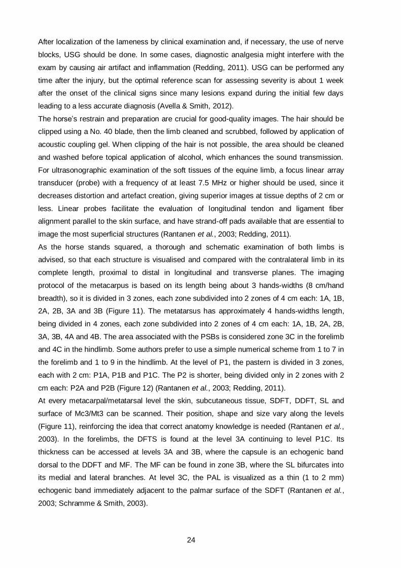
24
After localization of the lameness by clinical examination and, if necessary, the use of nerve
blocks, USG should be done. In some cases, diagnostic analgesia might interfere with the
exam by causing air artifact and inflammation (Redding, 2011). USG can be performed any
time after the injury, but the optimal reference scan for assessing severity is about 1 week
after the onset of the clinical signs since many lesions expand during the initial few days
leading to a less accurate diagnosis (Avella & Smith, 2012).
The horse’s restrain and preparation are crucial for good-quality images. The hair should be
clipped using a No. 40 blade, then the limb cleaned and scrubbed, followed by application of
acoustic coupling gel. When clipping of the hair is not possible, the area should be cleaned
and washed before topical application of alcohol, which enhances the sound transmission.
For ultrasonographic examination of the soft tissues of the equine limb, a focus linear array
transducer (probe) with a frequency of at least 7.5 MHz or higher should be used, since it
decreases distortion and artefact creation, giving superior images at tissue depths of 2 cm or
less. Linear probes facilitate the evaluation of longitudinal tendon and ligament fiber
alignment parallel to the skin surface, and have strand-off pads available that are essential to
image the most superficial structures (Rantanen et al., 2003; Redding, 2011).
As the horse stands squared, a thorough and schematic examination of both limbs is
advised, so that each structure is visualised and compared with the contralateral limb in its
complete length, proximal to distal in longitudinal and transverse planes. The imaging
protocol of the metacarpus is based on its length being about 3 hands-widths (8 cm/hand
breadth), so it is divided in 3 zones, each zone subdivided into 2 zones of 4 cm each: 1A, 1B,
2A, 2B, 3A and 3B (Figure 11). The metatarsus has approximately 4 hands-widths length,
being divided in 4 zones, each zone subdivided into 2 zones of 4 cm each: 1A, 1B, 2A, 2B,
3A, 3B, 4A and 4B. The area associated with the PSBs is considered zone 3C in the forelimb
and 4C in the hindlimb. Some authors prefer to use a simple numerical scheme from 1 to 7 in
the forelimb and 1 to 9 in the hindlimb. At the level of P1, the pastern is divided in 3 zones,
each with 2 cm: P1A, P1B and P1C. The P2 is shorter, being divided only in 2 zones with 2
cm each: P2A and P2B (Figure 12) (Rantanen et al., 2003; Redding, 2011).
At every metacarpal/metatarsal level the skin, subcutaneous tissue, SDFT, DDFT, SL and
surface of Mc3/Mt3 can be scanned. Their position, shape and size vary along the levels
(Figure 11), reinforcing the idea that correct anatomy knowledge is needed (Rantanen et al.,
2003). In the forelimbs, the DFTS is found at the level 3A continuing to level P1C. Its
thickness can be accessed at levels 3A and 3B, where the capsule is an echogenic band
dorsal to the DDFT and MF. The MF can be found in zone 3B, where the SL bifurcates into
its medial and lateral branches. At level 3C, the PAL is visualized as a thin (1 to 2 mm)
echogenic band immediately adjacent to the palmar surface of the SDFT (Rantanen et al.,
2003; Schramme & Smith, 2003).

25
Figure 11. Ultrasonographic imaging protocol of the metacarpus. Schematic representation of its 7
levels (1 to 7): 1A, 1B, 2A, 2B, 3A and 3B. The area associated with the proximal sesamoid bones is
considered zone 3C. The structures identified from the probe to the palmar metacarpus are the
superficial digital flexor tendon (SDFT), deep digital flexor tendon (DDFT), suspensory ligament (SL)
and accessory ligament of the DDFT (ALDDFT) (adapted from Redding, 2011).
Figure 12. Ultrasonographic imaging protocol of the proximal and middle phalanxes. Schematic
representation of the levels P1A, P1B, P1C, P2A and P2B. The structures identified from the probe are
the superficial digital flexor tendon (SDFT), deep digital flexor tendon (DDFT), cruciate sesamoidean
ligaments (CSL), straight sesamoidean ligament (SSL) and oblique sesamoidean ligaments (OSL)
(adapted from Redding, 2011).
26
In the pastern zones (Figure 12), the PDAL can be seen in the palmar/plantar surface of the
DDFT in zone P1A, where the SSL and a small portion of the CSLs may be identified. The
OSLs are visualized in close proximity to the palmar/plantar surface of P1, dorsal to the SSL in
zone P1B. The SSL continues to zone P1C, being found on the dorsal surface of the DDFT
and having its fibrocartilaginous insertion in zone P2A. In zone P2A a thin DDAL is seen in the
palmar/plantar border of the DDFT (Rantanen et al., 2003).
The prognosis of tendinopathies is most dependent on the severity of the initial damage,
being prudent to evaluate, describe and classify the injuries during the lameness assessment
(Avella & Smith, 2012). A systematic approach to the defined zones including both qualitative
and quantitative analysis should be used to define the injuries and to compare with follow-up
examinations (Rantanen et al., 2003). The use of dynamic USG is beneficial in improving the
diagnostic certainty, being achieved by maximally flexing and extending the MCP and the
IPJs while scanning (DiGiovanni, Rademacher, Riggs, Baumruck & Gaschen, 2016).
Tendon and ligament injury can be recognised ultrasonographically by changes in size,
shape, position, echogenicity, fiber pattern (echotexture) and surrounding inflammatory
reaction. The lesions should be classified based on their location (core, peripheral, radial and
diffuse), length, alteration in echogenicity, pattern of altered echogenicity (focal, diffuse,
homo and heterogenous), alteration of fiber pattern (in longitudinal section), percent CSA of
tendon injury and changes of the lesion over time. These findings should be analysed in
conjunction with the clinical signs, comparing with images of the contralateral limb (Farrow,
2006; Rantanen et al., 2003). The CSA measurements are considered a very sensitive
indicator of inflammation and can be assessed from a frozen image on the screen (Redding,
2011). The CSA of the hypoechogenic lesions, maximal tendon and total tendon CSAs are the
parameters used to describe injury severity (Avella & Smith, 2012; Kalisiak, 2012; Jorgensen &
Genovese, 2003). Acute tendinopathy can exhibit enlargement, hypoechogenicity (focal or
generalized), reduced fiber pattern, changes in position, margins and/or shape, and
associated edema and effusion in the DFTS. Furthermore, chronic tendon pathology is
associated with irregular striated pattern (indicative of fibrosis) and variable changes in
echogenicity and size of the tendon (Avella & Smith, 2012).
Even though USG is currently the widest used diagnostic tool for the evaluation of equine
tendons and ligaments, it is not without limitations. The quality of the image is directly related
to the anatomical area being examined, the equipment settings and the operator skills in
positioning, defining the appropriate settings and recognizing structures and artifacts
(Redding, 2011). It has been determined that tenoscopy reveals tendon lesions that are not
detected on ultrasonographic evaluation (Smith & Wright, 2006; Wilderjans et al., 2003).
Presently, USG remains a practical, inexpensive and accessible imaging technique for soft
tissue injuries in the horse but magnetic resonance imaging is now considered the gold
standard diagnostic technique for lameness with origin in the distal limbs (Redding, 2011).

27
6.5. Surgical procedure
Endoscopy of a synovial tendon sheath, the so-called tenoscopy, is a minimal invasive
surgical technique that offers comprehensive evaluation of the DFTS. This technique has
many advantages by permitting minimally invasive surgical approaches leading to decreased
morbidity, simplified aftercare, rapid return to normal function of the sheath and reduced risks
of complications such as infection, fibrosis, ankylosis and wound breakdown (Cauvin, 2003;
McIlwraith et al., 2015). Recently, this surgical approach has become a routine technique in
equine surgical practice, making a major contribution to the understanding and diagnosis of
tendon sheath pathology (McIlwraith et al., 2015).
The DFTS is the most common site of tenosynovitis (Cauvin, 2003); hence horses presenting
it with pain on palpation and associated lameness should be considered candidates for
tenoscopic examination (Avella & Smith, 2011). A tenoscopic approach to the DFTS allows
the exploration of the structures within the sheath with direct observation of previously
unreported lesions, confirmation of lesions diagnosed by other diagnostic methods and early
treatment of the previous (McIlwraith et al., 2015).
6.5.1. General technique and equipment
Tenoscopy requires general anaesthesia and can be performed with the horse in lateral or
dorsal recumbency. Lateral recumbency is preferred in most cases, with the affected limb
uppermost for a lateral approach and the distal limb free to permit variation of joint angles.
The use of an Esmarch bandage and tourniquet applied to the proximal metacarpal/
metatarsal level and moderate flexion of the distal joints facilitates the entry into the sheath
(McIlwraith et al., 2015).
A standard arthroscopic equipment is used, including a rigid 4 mm diameter with a 25º to 30º
lens videoarthroscope or direct-view arthroscope, video camera, digital image capture and
storage device, fiberoptic light cable and light source, fluid irrigation system (usually a
motorized pump), 5 to 6 mm diameter cannula and conical obturator, 2 to 3 mm diameter
egress cannula and locking trocar (sharp stylet or conical obturator), probes, Dyonics 5.2 mm
suction punch rongeurs, Ferris-Smith arthroscopic rongeurs, biopsy punch rongeurs,
arthroscopic scissors, hook knives and motorized synovial resectors (McIlwraith et al., 2015).
Nixon’s (1990) tenoscopic approach using a single portal for evaluating the DFTS has been
adopted as the standard. The term palmar is used in the following description for either
palmar or plantar, since the approach is similar in both fore and hindlimbs. When the tendon
sheath is not distended enough, 10 to 20 mL of sterile saline solution should be administered
through a needle inserted in the palmar aspect of the mid-pastern region in order to safely
entering the sheath (Cauvin, 2003; McIlwraith et al., 2015). The standard tenoscopic portal is

28
created immediately distal to the PSB and about 6 mm palmar to the neurovascular bundle,
between the PAL and the PDAL (Figure 13). First, a small skin incision is made with a
scalpel, continued by a stab incision using a No. 11 or 15 blade to penetrate only the SW.
Following this, the arthroscopic cannula is introduced through the portal using a conical
obturator, and directed proximally through the fetlock canal between the DDFT and the SW,
avoiding iatrogenic trauma to the tendons. The obturator is then replaced by the arthroscope,
the light source and fluid lines are connected and systematic examination is carried out
(Cauvin, 2003; McIlwraith et al., 2015). As the fluids are running, the sheath is lavaged
through an 18 gauge needle inserted in the proximal pouch (Cauvin, 2003). The instrument
portals are created where appropriate by passing a spinal needle for accurate location before
the incision, avoiding the annular ligaments (McIlwraith et al., 2015).
Figure 13. Standard tenoscopic approach to the digital flexor tendon sheath. Left hindlimb with a fluid
line inserted in the plantar aspect of the mid-pastern region and standard lateral arthroscopic portal
with a cannula placed, before inserting the arthroscope (Original illustration).
A detailed knowledge of the normal tenoscopic anatomy of the DFTS and its structures is
vital, since normal anatomical structures such as vinculae, endotenon and synovial folds
should not be damaged or confused with pathological changes (Cauvin, 2003).
A systematic “layer-by-layer” approach is recommended by McIlwraith, Nixon & Wright
(2015), to completely evaluate the sheath. After creating the tenoscopic portal, the
arthroscope should be positioned in the proximal recess of the sheath and by rotation of the
lens and gradual withdrawal, the dorsal SW, dorsal surface of the MF and its distal free
margin can be visualized (Figure 14). Further withdraw of the arthroscope maintaining it in
the dorsal region will show the dorsal surface of the DDFT when viewing palmarly, and the
PSBs and ISL when viewing dorsally (Figure 14). Then, the scope is repositioned to place its
tip distally and the DDFT, DM and paired vinculae come to image (Figure 15).
The arthroscope should be reoriented proximally for a second visualization of the sheath, but
this time the tip of the scope should be passed between the free margin of the MF and the
DDFT, showing the medial and lateral mesotenon of the DDFT. After that, the arthroscope
can be rotated over the abaxial margin of the DDFT leading its tip to the space between the
DDFT and the SDFT. In this position, by rotating the lens and moving slightly the scope it is

29
possible to assess both medial and lateral reflections of the MF from the SDFT and the
surfaces of both tendons. The tip of the scope is then pointed distally to evaluate the dorsal
and the palmar surfaces of the SDFT and the DDFT, respectively (Figure 15). In this
pathway, close to the midpoint of P1 it is possible to visualize the free margin of the SDFT
isthmus and the dorsal and axial surfaces of the insertion branches of the SDFT. The DDFT
is further inspected as far as the reflection of the SW onto the tendon’s surface, lining the
proximal margin of the DDAL. Rotating the scope over the abaxial margin of the DDFT allows
the visualization of the distal cul-de-sac of the sheath, which is typically covered by synovium
superimposing the “T” ligament. While in this position, withdrawal and angulation of the lens
images the synovium overlying the proximal IP scutum, the SSL and the branches of
insertion of the SDFT (Figure 15). To complete the sheath examination, the scope should be
positioned again proximal to the fetlock to run outside of the MF on the abaxial surface of the
SDFT to visualize it as far as its vinculum. For visualization of specific lesions the
arthroscope may need to be introduced through a proximal instrument portal, leaving the
tenoscopic portal free for instrument entry (McIlwraith et al., 2015).
Figure 14. Tenoscopic views looking proximally, showing multiple regions from dorsal to the flexor
tendons to the palmar surface of the sheath. S: superficial digital flexor tendon; D: deep digital flexor
tendon; MF: manica flexoria; AL: palmar annular ligament; Ses: lateral proximal sesamoid bone; M:
midline attachment of the mesotenon (adapted from McIlwraith et al., 2015).
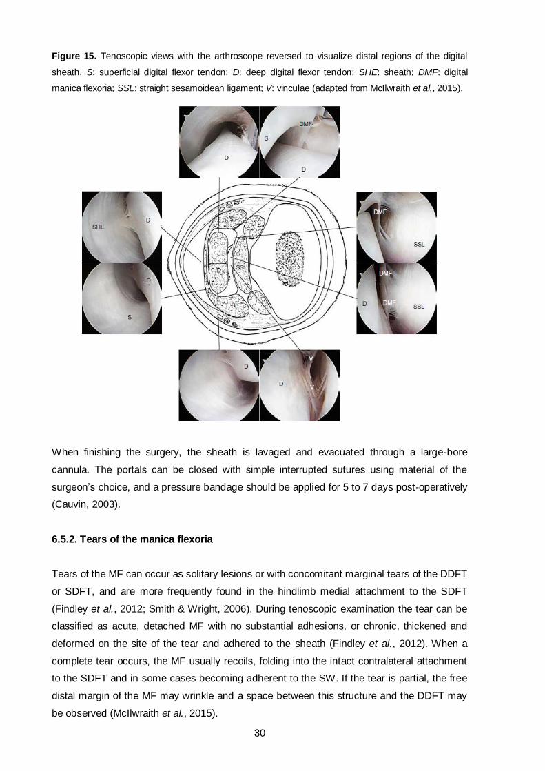
30
Figure 15. Tenoscopic views with the arthroscope reversed to visualize distal regions of the digital
sheath. S: superficial digital flexor tendon; D: deep digital flexor tendon; SHE: sheath; DMF: digital
manica flexoria; SSL: straight sesamoidean ligament; V: vinculae (adapted from McIlwraith et al., 2015).
When finishing the surgery, the sheath is lavaged and evacuated through a large-bore
cannula. The portals can be closed with simple interrupted sutures using material of the
surgeon’s choice, and a pressure bandage should be applied for 5 to 7 days post-operatively
(Cauvin, 2003).
6.5.2. Tears of the manica flexoria
Tears of the MF can occur as solitary lesions or with concomitant marginal tears of the DDFT
or SDFT, and are more frequently found in the hindlimb medial attachment to the SDFT
(Findley et al., 2012; Smith & Wright, 2006). During tenoscopic examination the tear can be
classified as acute, detached MF with no substantial adhesions, or chronic, thickened and
deformed on the site of the tear and adhered to the sheath (Findley et al., 2012). When a
complete tear occurs, the MF usually recoils, folding into the intact contralateral attachment
to the SDFT and in some cases becoming adherent to the SW. If the tear is partial, the free
distal margin of the MF may wrinkle and a space between this structure and the DDFT may
be observed (McIlwraith et al., 2015).

31
When a partial tear is present, debridement of the torn fibrils should be done with suction
punch biopsy rongeurs, Ferris-Smith arthroscopic rongeurs and/or a motorized synovial
sector in oscillating mode with suction applied. If the tear is complete, total resection of the
MF is advised by separating its intact border from the SDFT using arthroscopic scissors or
meniscectomy knives, before section of its proximal synovial attachment. Most of the times
another portal is created so that the MF can be grabbed with Ferris-Smith arthroscopic
rongeurs, stabilized and pulled (Findley et al., 2012; McIlwraith et al., 2015).
The prognosis for the majority of horses that undergo tenoscopic surgery due to MF tears is
considered reasonable (Smith & Wright, 2006) and a complete removal has been associated
with a better outcome (Avella & Smith, 2011).
Figure 16. Completely removed manica flexoria. The lateral border of the manica flexoria was acutely
torn and partially separated from the superficial digital flexor tendon (Original illustration).
6.5.3. Longitudinal tears of the digital flexor tendons
As described in the tendon pathology section, LTs occur more frequently in the lateral margin
of the DDFT (Arensburg et al., 2011; Smith & Wright, 2006; Wilderjans et al., 2003; Wright &
McMahon, 1999), being more easily diagnosed by USG than MF tears (Fiske-Jackson et al.,
2013). Tenoscopic examination of the DFTS is recommended to confirm the presence of LTs
and to treat them. LTs can be classified as superficial (< 5 mm) or deep (> 5 mm) (Arensburg
et al., 2011; Wilderjans et al., 2003) and localized proximal, distal or in the fetlock canal, being
considered long if extending for two regions. Long tears are more common in the fetlock canal
and proximal to it, while short tears are more frequent distally (Smith & Wright, 2006).
The goal of the surgical treatment is the removal of the torn fibrils, which reduces the surface
area of exposed disrupted collagenous tissue to the synovial sheath, decreasing the
inflammatory response and endorsing the development of a viable scar. The torn fibers are
managed by the same principles and techniques as the partial MF tears. The margin of the
tears can be covered by granulomata (granulation tissue with varying degrees of
organization) and in those cases, the large masses should be dissected free with
arthroscopic scissors before removal with Ferris-Smith arthroscopic rongeurs. The

32
instrument portals are created according to the lesion location, for proximal lesions the
access has to be from a portal abaxial to the MF (McIlwraith et al., 2015).
The prognosis for LTs after tenoscopical intervention appears to be reasonably fair, but
considered worse than for other lesions. Negative prognostic features include obvious pre-
operative distension of the DFTS, long duration of clinical signs, presence of a long tear and
persistence of post-operative distension, indicating incomplete healing (Smith & Wright, 2006).
6.5.4. Complex tenosynovitis
Complex tenosynovitis has been described as lesions involving two or more structures within
or adjacent to the DFTS with usually the PAL being one of them (McIlwraith et al., 2015). This
pathology may result from chronic tenosynovitis with low grade inflammation, synovial
distension, thickening and fibrosis of the sheath, PAL syndrome and tendon deformation.
Adhesions and synovial masses secondary to SW or tendon disruption are a common feature
(McIlwraith et al., 2015; Schramme & Smith, 2003).
The treatment of complex tenosynovitis is achieved by removing tenoscopically the synovial
masses and adhesions, and performing a PAL desmotomy to facilitate the movement through
the fetlock canal. Synovial masses can be resected using arthroscopic scissors, biopsy punch
rongeurs, straight and curved motorized synovial resectors and some other instruments. A
second instrument portal might be needed if the mass is large, to provide tension on the mass
while it is separated from the synovium. Adhesions are commonly synoviosynovial and their
removal is achieved using biopsy punch rongeurs and/or motorized resectors (McIlwraith et al.,
2015). After performing a PAL desmotomy and removing the synovial masses and adhesions,
the prognosis for horses with complex tenosynovitis is favourable, unless there are
associated tendon injuries, which are a negative feature (Schramme & Smith, 2003).
6.5.5. Palmar/Plantar annular ligament desmotomy
PAL syndrome and primary desmitis have been associated with tenosynovitis as primary or
secondary to it (McIlwraith et al., 2015). When the PAL is causing sheath constriction and
impeding the normal gliding between the tendons a desmotomy is advised (Avella & Smith,
2011). It is believed that desmotomy of the PAL releases the sheath constriction and
decreases inflammation, allowing a better tenoscopic access and examination of the
structures within the DFTS (McIlwraith et al., 2015).
Tenoscopically, desmotomy of the PAL can be performed by a “free-hand” division or using a
slotted cannula, being done abaxially between the PSB and the palmar/plantar reflection of
the sheath (McIlwraith et al., 2015). Extrathecal techniques such as an approach through a
small skin incision and blind subcutaneous transection of the PAL have been described as

33
quick and simple, but the tenoscopically guided technique is considered better since it allows
direct visualization of the sheath and treatment of concurrent pathologies (Schramme &
Smith, 2003). The decision between “free-hand” or slotted cannula techniques can be done
during the initial examination of the sheath, considering that the use of slotted cannula is
advantageous for the most chronic and constricted fetlock canals. The “free-hand” technique
can be achieved by creating a standard arthroscopic portal and an ipsilateral proximal
instrument portal abaxial to the MF (the portals can be interchanged for a better
visualization). The desmotomy should be done using a curved meniscectomy knife or a right-
angled blade, in a distoproximal direction and between the PSB and the reflection of the
sheath to the SDFT (McIlwraith et al., 2015).
Tenoscopic desmotomy of the PAL, with the aim of relieving intrathecal restriction, is
considered a nonharmful procedure and has been encouraged in the treatment of complex
tenosynovitis (Nixon, 2003), MF tears and LTs of the digital flexor tendons, exposing the
sheath contents and improving its tenoscopic evaluation (Wilderjans et al., 2003; Wright &
McMahon, 1999). Constriction of the PAL is often secondary to chronic inflammation and
distension of the sheath; hence since 2003 desmotomy should be reserved for chronic cases
or cases with obvious thickening of the ligament (Arensburg et al., 2011; Smith & Wright,
2006).
6.5.6. Contaminated and infected tendon sheath
Infectious tenosynovitis is a critical condition in the horse that has high risk of long-term
sequela and permanent lameness (Bertone, 2011; Schramme & Smith, 2003). The treatment
of a contaminated and infected sheath consists in the removal of foreign material,
debridement of torn and infected tissue, elimination of microorganisms, effective lavage and
restoration of a normal intrathecal environment (Wright, Smith, Humphrey, Eaton-Evans &
Hillyer, 2003).
In most cases, initial high pressure lavage (500 mL/min) of the sheath is needed (McIlwraith
et al., 2015) in order to wash the inflammatory exudates, cellular debris and fibrin (Wereszka,
et al., 2007). This can be achieved by creating multiple ingress and egress portals, and
moving the scope around and between the tendons. The fluid of choice is sterile buffered
polyionic solution, but antimicrobial drugs, antiseptics, dimethyl sulfoxide and fibrinolytics can
be added to it. After the lavage, a thorough and prompt examination of the sheath is
important since foreign material can migrate and deposit at any site. Standard tenoscopic
portals can be used, and when wounds or punctures are present, they can work as
instrument and egress portals (McIlwraith et al., 2015).
Removal of foreign material is of extreme importance as it acts as a substrate for bacterial
growth and causes physical and biochemical irritation to the sheath, perpetuating the
34
inflammation. Ferris-Smith rongeurs for large pieces and motorized synovial resectors for
small pieces are used for the removal of foreign material and detached contaminated tissue,
and debridement of torn fibers, pannus and wounds (Wright et al., 2003). A final lavage
should be performed and traumatic wounds must be debrided or excised to a clean state so
that they can be closed, minimizing the risk of further contamination and/or infection. If
needed, the limb can be immobilized by a cast to improve wound healing (McIlwraith et al.,
2015).
6.5.7. Post-operative management
After a tenoscopic surgery, a pressure bandage from foot to distal carpus/tarsus should be
applied to reduce motion and prevent sheath swelling and contamination (Cauvin, 2003). It is
advised that the bandaging continues between 2 to 4 weeks after surgery. Perioperative
administration of antibiotics and nonsteroidal anti-inflammatory drugs is usually performed to
the surgeon’s preference. In the case of infectious tenosynovitis and presence of wounds the
antibiotics sometimes have to be continued for more days or changed according to sensitivity
testing. Preferably, microbiological samples should be taken before antimicrobial therapy and
empirical treatment implemented until the results are available. Monitoring the synovial fluid
and clinical signs, such as improvement of lameness, reduced swelling and surface
temperature, helps to determine the appropriate duration of the antimicrobial treatment
(McIlwraith et al., 2015).
The horse should be restricted to box rest in a clean environment until the sutures are
removed around 10 to 14 days postoperatively (Avella & Smith, 2011). Hand-walking can be
started and gradually increased according to the surgeon’s discretion. The duration of the
rehabilitation protocol depends on the severity of the injury and clinical response (Avella &
Smith, 2011; McIlwraith et al., 2015).

35
TENOSCOPIC EXAMINATION OF THE DIGITAL FLEXOR TENDON
SHEATH: A RETROSPECTIVE ANALYSIS OF 86 HORSES (2016)
MATERIALS AND METHODS
Tenoscopy has become a routine procedure in equine surgical practice since it is a minimal
invasive surgical technique that offers comprehensive evaluation of the DFTS, carpal and
tarsal sheaths, and carpal and tarsal extensor tendon sheaths. Injuries to the digital flexor
tendons are among the leading causes of clinical disability in horses, therefore tenoscopic
examination of the DFTS is of great relevance. In spite of all this, there are relatively few
publications regarding this subject.
In this study, medical records regarding tenoscopic examination of the DFTS were analysed
for non-infectious and infectious tenosynovitis, and traumatic injuries to the sheath.
1. Inclusion criteria
The medical records from four British equine hospitals (BEVC, DGVG, NEH and VEH) were
reviewed for horses that underwent tenoscopic examination of the DFTS in 2016.
Ninety-three tenoscopies were performed in 86 horses and the author was present in 12 of
them. Two horses had two surgeries and one horse had three, nevertheless, for statistical
significance only the first surgery of each horse was included in this study. In most cases
data was collected regarding the horse’s age, gender, breed, history, clinical signs and
clinical findings: affected limb, diagnostic analgesia (perineural and intrathecal), cytological
analysis of synovial fluid, radiographic and USG abnormalities, tenoscopic findings, surgical
treatment and peri-operative treatments.
Since some of the cases were referred to the Hospitals for surgery, all information was
obtained from the medical records. Therefore, clinical follow-up information was not available
and for most cases it was not possible to collect the data regarding the current use of the
horse, diagnostic methods and long term outcome.
2. Data classification and analysis
Diagnostic analgesia and synovial fluid analysis results were reviewed. Radiologic,
ultrasonographic and tenoscopic findings were studied and summarized.
All horses were evaluated tenoscopically under general anaesthesia. Standard arthroscopic
material and surgical techniques as previously described in the chapter “Surgical procedure”
(pp. 27-34) were used, with small variations according to the surgeon performing the surgery.
36
MF tears were classified in acute (detached with no substantial adhesions) or chronic
(thickened, deformed and adhered to the sheath wall), partial or complete and regarding the
affected border (medial, lateral and distal). Concerning DDFT LTs, these were categorized
according to their location (proximal, distal or within the fetlock canal), length (long or short),
depth (superficial or deep) and affected border (medial, lateral, dorsal or palmar/plantar).
PAL constriction was studied as caused by primary PAL disease or secondary to DFTS
and/or tendon pathology. Additionally, traumatic injuries were characterized by affecting only
the SW or also the ligaments and/or tendons. In the latest, the location of the injuries was
recorded (medial, lateral, palmar or dorsal).
In the cases where synoviocentesis was performed, samples were considered positive if at
least two of the analysed parameters were pathognomonic of contamination.
For analysis, 5 age groups were defined: 0-5, 6-10, 11-15, 16-20 and >20. Gender definition
included: male (colts and stallions), gelding and female. Horse breeds were grouped into
Thoroughbreds, Warmbloods, Coldbloods, Ponies and unknown. Specific information
regarding each horse can be conferred in Annex 1. Comparisons were made concerning
different pathologies and between the affected limb (forelimb versus hindlimb),
characteristics of specific lesions, age, gender and breed of the horses for each diagnosis.
To determine whether various factors were associated with the diagnosed pathologies,
analyses were made in R version 3.3.2 (R Development Core Team, 2010). Pearson’s Chi-
squared tests were performed to identify significant associations between the pathologies
diagnosed and the dependant variables: affected limb (forelimb versus hindlimb), age class,
gender and breed group of the horse. A significance level of P < 0.05 was assumed for all
tests.
The sensitivity of some the diagnostic tests was determined and the 95% confidence interval
(CI) for the proportion of positives (p) obtained from all the horses submitted to the test (n)
was calculated using the normal approximation with the following formula:
𝑝 ± 1,96√𝑝(1−𝑝)
𝑛

37
RESULTS
1. Case details and clinical features
Eighty-six horses were included in this study. The age range was 0.5-21 years, with a mean
of 9.8 years and median of 10 years. Nine percent were males (n = 8), 55% geldings (n = 47)
and 36% females (n = 31). Thirty-one percent were Thoroughbreds (n = 27), 28%
Warmbloods (n = 24), 20% Coldbloods (n = 17), 12% Ponies (n = 10) and 9% of unknown
breed (n = 8). The age, gender, breed and affected limb are summarised in Table 1. Age
distribution according to breed is represented in Graphic 2.
Seventy-nine (92%) horses had surgery in only 1 limb, 2 (2%) had it in both forelimbs, 4 (5%)
in both hindlimbs and 1 (1%) in the right forelimb and hindlimb. Thus, 93 DFTS were
affected: 24% right forelimbs (RFs) (n = 22), 15% left forelimbs (LFs) (n = 14), 30% right
hindlimbs (RHs) (n = 28) and 31% left hindlimbs (LHs) (n = 29).
Table 1. Summary of age, gender, breed and affected limb of 86 horses that underwent tenoscopic
examination of the digital flexor tendon sheath.
Age (years)
No. (%) Gender No. (%) Breed No. (%) Limb No. (%)
0-5 16 (19%) Male 8 (9%) Thoroughbred 27 (31%) RF 22 (24%)
6-10 31 (36%) Gelding 47 (55%) Warmblood 24 (28%) LF 14 (15%)
11-15 26 (30%) Female 31 (36%) Coldblood 17 (20%) RH 28 (30%)
16-20 12 (14%) Pony 10 (12%) LH 29 (31%)
> 20 1 (1%) Unknown 8 (9%)
Total 86 (100%) 86 (100%) 86 (100%) 93 (100%)
Graphic 2. Age distribution according to breed.
5
10
15
20
Coldblood Pony Unknown Warmblood
Breed
Ag
e
Thoroughbred

38
Eighty-four (98%) horses were lame at the time of clinical examination and 87 (94%) limbs
had a visible distended DFTS. Flexion tests were recorded for 24 limbs and considered
positive in 21 (88%).
Three horses (4%) were euthanized, 2 while still under anaesthesia due to extensive tendon
damage, and 1 after its third surgery given the lack of response to aggressive surgical and
medical therapy.
2. Diagnostic analgesia
Diagnostic analgesia for the assessment of the lameness source was recorded in 23 horses.
The results are represented in Table 2.
Perineural analgesia of the palmar/plantar nerves (abaxial sesamoid) resulted in
improvement in lameness in 4 out of 8 (50%) horses. This nerve block was performed in 1
forelimb and 7 hindlimbs, being positive in 4 hindlimbs. The 4 horses that had no significant
improvement of lameness after the abaxial sesamoid nerve block all improved after a low 4
point nerve block. Positive response to perineural analgesia was seen in horses with PAL
thickening (n = 6), SDFT tears (n = 1) and ganglion cysts (n = 1).
Intrathecal analgesia of the DFTS was performed in 22 horses and 19 (86%) were thought to
have a positive response. Three forelimbs and 19 hindlimbs were submitted to intrathecal
analgesia, from which the 3 forelimbs and 16 hindlimbs had a positive response. These
included horses diagnosed with MF tears (n = 10), PAL thickening (n = 5), SDFT tears (n = 2)
and DDFT tears (n = 2).
Seven horses had the abaxial sesamoid and the intrathecal blocks, 3 (43%) were positive to
both, 2 (29%) were only positive to the intrathecal analgesia, 1 (14%) was only positive to the
abaxial sesamoid block and 1 (14%) was negative to both.
Table 2. Results of diagnostic analgesia performed in 23 horses.
Response to Diagnostic Analgesia
Abaxial Sesamoid Nerve Block
Low 4 Point Nerve Block
Intrathecal Analgesia
Forelimb Hindlimb Forelimb Hindlimb Forelimb Hindlimb
Positive - 4 1 3 3 16
Negative 1 3 - - - 3
Total 8 4 22
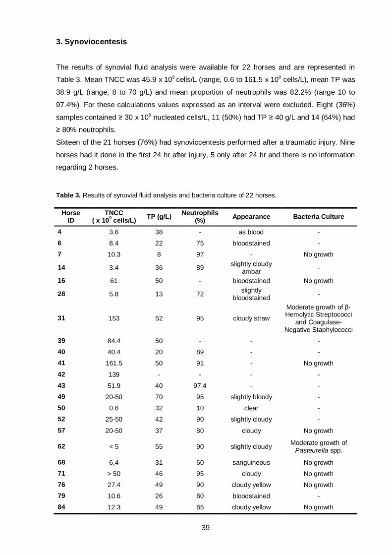
39
3. Synoviocentesis
The results of synovial fluid analysis were available for 22 horses and are represented in
Table 3. Mean TNCC was 45.9 x 10⁹ cells/L (range, 0.6 to 161.5 x 10⁹ cells/L), mean TP was
38.9 g/L (range, 8 to 70 g/L) and mean proportion of neutrophils was 82.2% (range 10 to
97.4%). For these calculations values expressed as an interval were excluded. Eight (36%)
samples contained ≥ 30 x 109 nucleated cells/L, 11 (50%) had TP ≥ 40 g/L and 14 (64%) had
≥ 80% neutrophils.
Sixteen of the 21 horses (76%) had synoviocentesis performed after a traumatic injury. Nine
horses had it done in the first 24 hr after injury, 5 only after 24 hr and there is no information
regarding 2 horses.
Table 3. Results of synovial fluid analysis and bacteria culture of 22 horses.
Horse ID
TNCC ( x 10
9 cells/L)
TP (g/L) Neutrophils
(%) Appearance Bacteria Culture
4 3.6 38 - as blood -
6 8.4 22 75 bloodstained -
7 10.3 8 97 - No growth
14 3.4 36 89 slightly cloudy
ambar -
16 61 50 - bloodstained No growth
28 5.8 13 72 slightly
bloodstained -
31 153 52 95 cloudy straw
Moderate growth of β-Hemolytic Streptococci
and Coagulase-Negative Staphylococci
39 84.4 50 - - -
40 40.4 20 89 - -
41 161.5 50 91 - No growth
42 139 - - - -
43 51.9 40 97.4 - -
49 20-50 70 95 slightly bloody -
50 0.6 32 10 clear -
52 25-50 42 90 slightly cloudy -
57 20-50 37 80 cloudy No growth
62 < 5 55 90 slightly cloudy Moderate growth of
Pasteurella spp.
68 6,4 31 60 sanguineous No growth
71 > 50 46 95 cloudy No growth
76 27.4 49 90 cloudy yellow No growth
79 10.6 26 80 bloodstained -
84 12.3 49 85 cloudy yellow No growth
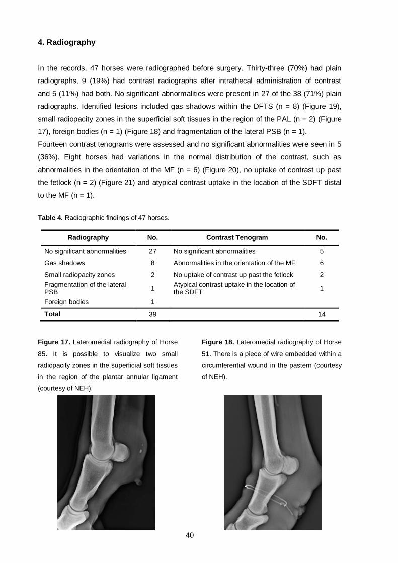
40
4. Radiography
In the records, 47 horses were radiographed before surgery. Thirty-three (70%) had plain
radiographs, 9 (19%) had contrast radiographs after intrathecal administration of contrast
and 5 (11%) had both. No significant abnormalities were present in 27 of the 38 (71%) plain
radiographs. Identified lesions included gas shadows within the DFTS (n = 8) (Figure 19),
small radiopacity zones in the superficial soft tissues in the region of the PAL (n = 2) (Figure
17), foreign bodies (n = 1) (Figure 18) and fragmentation of the lateral PSB (n = 1).
Fourteen contrast tenograms were assessed and no significant abnormalities were seen in 5
(36%). Eight horses had variations in the normal distribution of the contrast, such as
abnormalities in the orientation of the MF (n = 6) (Figure 20), no uptake of contrast up past
the fetlock (n = 2) (Figure 21) and atypical contrast uptake in the location of the SDFT distal
to the MF (n = 1).
Table 4. Radiographic findings of 47 horses.
Radiography No. Contrast Tenogram No.
No significant abnormalities 27 No significant abnormalities 5
Gas shadows 8 Abnormalities in the orientation of the MF 6
Small radiopacity zones 2 No uptake of contrast up past the fetlock 2
Fragmentation of the lateral PSB
1 Atypical contrast uptake in the location of the SDFT
1
Foreign bodies 1
Total 39 14
Figure 17. Lateromedial radiography of Horse
85. It is possible to visualize two small
radiopacity zones in the superficial soft tissues
in the region of the plantar annular ligament
(courtesy of NEH).
Figure 18. Lateromedial radiography of Horse
51. There is a piece of wire embedded within a
circumferential wound in the pastern (courtesy
of NEH).

41
Figure 19. (A) Lateromedial and (B) oblique dorsomedial-palmarolateral projections of Horse 28
fetlock. The horse presented with a laceration of the tendon sheath that was confirmed
radiographically by the presence of gas within the sheath (courtesy of DGVG).
Figure 20. Contrast tenogram of Horse 35.
The manica flexoria is thickened, shortened
and in the proximal digital flexor tendon sheath
only (arrow) (courtesy of DGVG).
Figure 21. Contrast tenogram of Horse 44.
There is no uptake of contrast up past the
fetlock suggesting plantar annular ligament
constriction (courtesy of BEVC).
A B

42
5. Ultrasonography
Ultrasonographic images were available for retrospective evaluation in 72 horses. USG was
performed in 28 (39%) forelimbs and 44 (61%) hindlimbs.
Eleven (15%) limbs had limited ultrasonographic abnormalities. Findings included PAL
thickening (n = 16), PAL disruption (n = 4) (Figure 22), hypoechogenic lesion within the PAL
(n = 3) (Figure 23, A), SDFT disruption (n = 8), hypoechogenic lesion within the SDFT (n = 3)
(Figure 24), DDFT thickening (n = 3), DDFT disruption (n = 6), hypoechogenic lesion within
the DDFT (n = 7), MF thickening (n = 4), MF disruption (n = 8) (Figure 24), SSL thickening
with an hypoechogenic lesion within (n = 1), intrathecal echogenic material (n = 12), air
within the DFTS (n = 4), adhesion formation (n = 3), fluid filled structure (n = 2) (Figure 23,
B), tenosynovitis (n = 2) and foreign material (n = 1) (Table 5).
The ultrasonographic findings were consistent with primary PAL disease (n = 20), SDFT tears
(n = 4), DDFT tears (n = 14), MF tears (n = 9), SW perforation (n = 4), SDFT laceration (n = 4),
DDFT laceration (n = 2), ganglion cysts (n = 2), DDFT fibrosis (n = 1) and SSL desmitis (n = 1).
Table 5. Ultrasonographic findings of 72 horses.
Findings No. Findings No.
Limited findings 11 MF thickening 4
PAL thickening 16 MF disruption 8
PAL disruption 4 SSL thickening + hypoechogenic lesion 1
Hypoechogenic lesion within the PAL 3 Intrathecal echogenic material 12
SDFT disruption 8 Air within the DFTS 4
Hypoechogenic lesion within the SDFT 3 Adhesion formation 3
DDFT thickening 3 Fluid filled structure 2
DDFT disruption 6 Foreign material 1
Hypoechogenic lesion within the DDFT 7
Figure 22. (A) Transverse and (B) longitudinal ultrasonographic images of the right hindlimb of Horse
56. The plantar annular ligament is markedly thickened and disrupted (x) (courtesy of NEH).
A B
x x

43
Figure 23. Ultrasonographic images of Horse 11. (A) Transverse image of the right forelimb palmar
annular ligament demonstrating thickening of the ligament (0.6 cm) associated with a focal
hypoechogenic area within the medial aspect of the ligament itself. (B) A transverse image of the right
hindlimb fetlock revealed a multilocular hypoechogenic subcutaneous area within the medial plantar
aspect of the fetlock, consistent with a ganglion (courtesy of DGVG).
Figure 24. Ultrasonographic images of Horse 66 left hindlimb. (A) Transverse and (B) longitudinal
ultrasonographs of the proximal portion of the digital flexor tendon sheath (zone 3A). There is a
hypoechogenic zone (x) in the plantar lateral aspect of the superficial digital flexor tendon proximal to
the sheath. (C and D) Transverse ultrasonographic images of the proximal fetlock region (zone 3B). It
is possible to visualize a thickened and medially disrupted manica flexoria adherent to the sheath wall
(arrows) (courtesy of NEH).
C
A B
x
x
D C
B A

44
6. Surgical treatment
Eighty-six horses were submitted to tenoscopic examination. Two were euthanized while still
under anaesthesia due to extensive tendon damage.
Ninety-three DFTS were examined, 36 (39%) forelimbs and 57 (61%) hindlimbs. The
tenoscopic diagnoses are summarized in Table 6 and Graphic 3.
Table 6. Tenoscopic diagnoses in 93 digital flexor tendon sheaths.
Diagnosis Forelimb Hindlimb Total
Torn MF 18
Torn MF only - 2 2
Torn MF + other* 2 14 16
SDFT marginal tear 4
Marginal tear only - - 0
Marginal tear + other* - 4 4
DDFT marginal tear 18
Marginal tear only 7 5 12
Marginal tear + other* 1 5 6
PAL constriction 40
PAL constriction only 8 11 19
PAL constriction + other* 4 17 21
SW penetration 25
SW penetration only 4 5 9
SW penetration + other* 11 5 16
SDFT laceration + other* 8 4 12
DDFT laceration + other* 4 3 7
Ganglion cyst - 2 2
Limited findings - 2 2
DDFT tendonitis and tenosynovitis - 2 2
DDFT tendonitis 1 - 1
DDFT fibrosis 1 - 1
SDFT tenotomy 1 - 1
SSL desmitis - 1 1
ISL tear - 1 1
*The concomitant pathologies are described further in the text.

45
0
5
10
15
20
25
30
35
40
45N
o. in
th
is s
tud
y
Graphic 3. Tenoscopic diagnoses in 93 digital flexor tendon sheaths.
Eighteen torn MF were found, from which 15 had concomitant pathologies. From the 15
horses, 14 had a torn MF associated with PAL constriction and 1 had a medial tear in the MF
with concomitant medial tears to the SDFT and the DDFT, and PAL constriction (Figure 26).
There were 21 tears in 18 MF, since 2 had a medial and a lateral tear and 1 had a medial
and a horizontal tear. Ten (47%) tears occurred in the medial attachment to the SDFT, Nine
9 (43%) in the lateral attachment (Figure 25), 1 (5%) was seen in the distal margin and 1
(5%) in the horizontal plane (Table 7). The hindlimbs (n = 16, 89%) were significantly (P <
0.01) more commonly affected. Chronic (n = 10, 47%) and complete (n = 12, 57%) tears
were more frequent than acute (n = 6, 29%) and partial (n = 4, 19%), and there was no
information regarding 5 (24%) tears. All MF were completely removed except for 1.
In the 18 horses diagnosed with MF tears the majority (n = 10, 56%) were aged 11-15 years
(Graphic 4). This pathology occurred predominantly in Coldbloods (n = 10, 56%), followed by
Ponies (n = 4, 22%), Warmbloods (n = 3, 17%) and a horse of unknown breed (n = 1, 5%)
(Graphic 5). There were significant associations between the occurrence of a MF tear and
the horse’s age class (P < 0.01) and breed group (P < 0.001), and no significant association
with the gender (P = 0.8).
Table 7. Distribution of manica flexoria tears.
Medial Lateral Distal Horizontal Total
Forelimb 1 - 1 - 2 (10%)
Hindlimb 9 9 - 1 19 (90%)
Total 10 (47%) 9 (43%) 1 (5%) 1 (5%) 21 (100%)

46
Figure 25. Contrast tenogram and tenoscopic images of Horse 29 left hindlimb. (A) Contrast tenogram
suggestive of disruption to the manica flexoria (arrow). (B) Tenoscopic view of the torn manica flexoria.
There is a complete separation from its lateral attachment to the superficial digital flexor tendon. (C) A
plantar annular ligament desmotomy using a No. 12 blade was performed because the ligament was
grossly thickened. DDFT: deep digital flexor tendon; MF: manica flexoria; PAL: plantar annular
ligament; SDFT: superficial digital flexor tendon; SW: sheath wall (courtesy of DGVG).
There were 4 SDFT marginal tears, all in hindlimbs: 1 medial, 1 lateral, 1 plantar and 1 to the
mesotenon. All 4 horses with SDFT marginal tears had concomitant PAL constriction and 1
of them had simultaneous medial tears to the MF and the DDFT (Figure 26). The horses
affected were 2 Warmbloods and 2 Coldbloods (Graphic 5) with ages between 7-16 years
(Graphic 4). A tenoscopic guided tenotomy of the SDFT was performed in the RF of a 15 years
old Warmblood gelding with no other pathologies associated.
Marginal tears of the DDFT were identified in 8 (44%) forelimbs and 10 (56%) hindlimbs of 18
horses. Tears to the lateral border were the most common finding (n = 9, 50%) whereas medial
(n = 3, 17.3%), dorsal (n = 3, 17.3%) and palmar/plantar (n = 3, 17.3%) tears were equally
frequent (Table 8). The length of the tear was described in 16 of the 18 cases: 8 (44%) long
and 8 (44%) short tears. Four long tears extended from beneath the MF to halfway the pastern,
2 from the proximal sheath to the level of the DM and 2 from the proximal sheath to the fetlock.
Short tears were found mainly distal to the fetlock canal (n = 6), but also within (n = 1) and
proximal to it (n = 1). Proximal tears were predominantly lateral (n = 7) with the remaining
being medial (n = 2). Tears further distal affected mainly the palmar/plantar (n = 3) surface of
the DDFT and less commonly the lateral (n = 1), medial (n = 1) and dorsal (n = 1) surfaces.
The defects were classified as superficial in 3 cases and deep in 2 cases.
In all DDFT defects torn herniated fibrils were visualized. Other tenoscopic findings included
granulomatous masses (n = 6) (Figure 27), hemosiderin deposits (n = 6), adhesions to the SW
(n = 2), pannus deposits (n = 2) and fibrin deposits (n = 2).
B C A
DDFT
MF SW
SDFT
PAL

47
0
2
4
6
8
10
12
14
16
0-5 6-10 11-15 16-20 > 20
Série1
Série2
Série3
Série4
Série5
0
2
4
6
8
10
12
14
16
18
20
Coldblood Pony Thoroughbred Unknown Warmblood
Série1
Série2
Série3
Série4
Série5
Table 8. Distribution of deep digital flexor tendon tears.
Medial Lateral Dorsal Palmar/Plantar Total
Forelimb - 6 - 2 8 (44%)
Hindlimb 3 3 3 1 10 (56%)
Total 3 (17.3%) 9 (50%) 3 (17.3%) 3 (17.3%) 18 (100%)
From the 18 horses, 6 had PAL constriction associated to the DDFT tear, and 1 had additional
medial tears to the MF and SDFT. DDFT tears occurred more frequently in geldings (n = 12,
67%) (Graphic 6), Warmbloods (n = 7, 39%) (Graphic 5) and horses aged between 11-15
years (n = 7, 39%) (Graphic 4). No significant associations were seen between the occurrence
of SDFT or DDFT tears and the affected limb (SDFT: P = 0.098; DDFT: P = 0.632), age class
(SDFT: P = 0.834; DDFT: P = 0.794), gender (SDFT: P = 0.729; DDFT: P = 0.502) and breed
group (SDFT: P = 0.301; DDFT: P = 0.123).
Graphic 4. Age distribution of 18 horses diagnosed with manica flexoria (MF) tear, 18 with deep digital
flexor tendon (DDFT) tear, 4 with superficial digital flexor tendon (SDFT) tear, 34 with palmar/plantar
annular ligament (PAL) constriction and 25 with sheath wall (SW) penetration.
Graphic 5. Breed distribution of 18 horse diagnosed with manica flexoria (MF) tear, 18 with deep digital
flexor tendon (DDFT) tear, 4 with superficial digital flexor tendon (SDFT) tear, 34 with palmar/plantar
annular ligament (PAL) constriction and 25 with sheath wall (SW) penetration.
N o
f h
ors
es
Age Class
DDFT tear
SDFT tear
MF tear
PAL constriction
SW penetration
N o
f h
ors
es
Breed Group
DDFT tear
SDFT tear
MF tear
PAL constriction
SW penetration

48
0
5
10
15
20
25
Female Gelding Male
Série1
Série2
Série3
Série4
Série5
Graphic 6. Gender distribution of 18 horses diagnosed with manica flexoria (MF) tear, 18 with deep
digital flexor tendon (DDFT) tear, 4 with superficial digital flexor tendon (SDFT) tear, 34 with
palmar/plantar annular ligament (PAL) constriction and 25 with sheath wall (SW) penetration.
Figure 26. Ultrasonographic, tenoscopic and manica flexoria images of the right hindlimb of Horse 47.
(A and B) Transverse ultrasonographs confirming the fluid distension of the digital flexor tendon
sheath. The manica flexoria is grossly thickened and completely torn medially from its attachments to
the superficial digital flexor tendon (arrow). There was irregular margination of the medial border of the
deep digital flexor tendon at the base of the fetlock canal consistent with a marginal tear (x). (C and D)
Tenoscopic view of the complete tear of the manica flexoria from its medial attachment to the
superficial digital flexor tendon. It is possible to visualize a mass of disrupted fibrils. (E) Long
longitudinal tears on the medial margin of both superficial and deep digital flexor tendons. (F) Plantar
and (G) dorsal surfaces of the completely removed manica flexoria. DDFT: deep digital flexor tendon;
MF: manica flexoria; SDFT: superficial digital flexor tendon; SW: sheath wall (courtesy of NEH).
A
C D E
F G
DDFT
MF
B
x
DDFT
SW
SDFT
DDFT
SW
N o
f h
ors
es
Gender
DDFT tear
SDFT tear
MF tear
PAL constriction
SW penetration

49
Figure 27. Tenoscopic views of Horse 34 right hindlimb digital flexor tendon sheath. (A) Plantar
annular ligament desmotomy using a No. 12 blade. (B) A granulomatous mass (arrow) was present on
the plantar border of the deep digital flexor tendon just medial to the midline. (C) View of the DDFT
following debridement of the mass. DDFT: deep digital flexor tendon; PAL: plantar annular ligament;
SW: sheath wall (courtesy of DGVG).
Besides the presence of marginal tears, other single pathologies regarding the DDFT were
diagnosed. In the RF of a 7 years old Warmblood gelding the presence of a scar (fibrosis) in
the palmar surface of the DDFT was visualized (Figure 28). There was 1 case of tendinitis
(Figure 29) of the RF of a 3 years old Thoroughbred mare and 2 cases of infectious
tenosynovitis: the RH of an 8 year old female and the LH of a 6 years old gelding, both
Thoroughbreds. Regarding the last case, tenosynovitis occurred consequently to an abscess
in the RH. This horse was submitted to 3 tenoscopies (Figure 30) before euthanasia was
performed due to the absence of response to aggressive surgical and medical therapy.
Figure 28. Tenoscopic examination of the
digital flexor tendon sheath of Horse 18. It is
possible to visualize the presence of fibrosis in
the palmar surface of the deep digital flexor
tendon close to the distal reflection of the
sheath (courtesy of DGVG).
Figure 29. Tenoscopic view of the deep digital
flexor tendon (DDFT) of Horse 68. There is
marked inflammation in the proximal lateral
epitenon of the DDFT. DDFT: deep digital
flexor tendon (courtesy of NEH).
A B C
PAL DDFT
SW
DDFT
SW
DDFT

50
Figure 30. Ultrasonographic images and tenoscopic views of the three tenoscopies performed to the
left hindlimb of Horse 41. (A) Transverse and (B) longitudinal ultrasonographic images revealing
irregular fiber pattern and hypoechogenicity (arrows) of the deep digital flexor tendon (DDFT). (C)
Tenoscopic view of the digital flexor tendon sheath (DFTS) in the first surgery. There was mild sheath
wall (SW) inflammation and superficial haemorrhage under the intact epitenon of the DDFT. (D)
Transverse and (E) longitudinal ultrasonographic images showing more severe irregularity regions of
hypoechogenicity with loss of fiber pattern (arrows) in the DDFT. (F) Tenoscopic view of the DFTS in
the second surgery. Severe DDFT tenosynovitis with marked pannus and sub-epitenon haemorrhage
was seen. (G and H) Tenoscopic views of the DFTS during the third surgery. The DDFT appeared
similar in gross appearance after removal of pannus and fibrinous debris. DDF Tendon: deep digital
flexor tendon (courtesy of VEH).
A B C
DDF Tendon
D E F
DDF Tendon
G H

51
A “free-hand” PAL desmotomy (Figures 25C and 27A) was the most frequent surgical
technique performed in this study. From the 86 horses, 38 (44%) had a PAL desmotomy
done in 44 PALs. Thirty two horses had this technique done in a single limb, 4 had it in both
hindlimbs and 2 in both forelimbs. The hindlimbs (n = 33, 75%) were significantly (P < 0.001)
more commonly intervened. PAL desmotomy was performed alone in 19 limbs. In the other
limbs it was associated to other pathologies such as MF tears (n = 17) and marginal tears of
the SDFT (n = 4) and the DDFT (n = 6).
PAL constriction caused by primary PAL disease was diagnosed in 25 (57%) limbs. In 15
(34%) limbs the constriction was secondary to DFTS and/or tendon pathology. The
remaining 4 (9%) had a PAL desmotomy performed as part of treatment even though there
was no evident constriction to the fetlock canal (Table 9). Thirty-four horses had PAL
constriction, from which 20 (59%) were found to have primary PAL disease (25 limbs). This
was more frequently found in the hindlimbs (n = 16, 64%) and thickening of the ligament was
seen in all 25 limbs, sometimes associated with tissue calcifications (n = 2) (Figure 17), fiber
disruption (n = 2) (Figure 22) and presence of a cyst (n = 1) (Figure 23).
Geldings (n = 20, 59%) (Graphic 6), Coldbloods (n = 14, 41%) (Graphic 5) and horses aged
11-15 years (n = 15, 44%) (Graphic 4) were more frequently diagnosed with PAL
constriction. There were significant associations between this pathology and the horse’s age
class (P = 0.001) and breed group (P < 0.001).
Table 9. Summary of 43 limbs submitted to PAL desmotomy.
Primary PAL
disease Secondary PAL
constriction No evident
constriction Total
Forelimb 9 2 - 11 (25%)
Hindlimb 16 13 4 33 (75%)
Total 25 (57%) 15 (34%) 4 (9%) 44 (100%)
Twenty-five horses were presented after a traumatic injury, from which horses with less than
10 years (n = 20, 80%) (Graphic 4), Thoroughbreds (n = 19, 76 %) (Graphic 5) and geldings
(n = 11, 44%) (Graphic 6) were significantly more affected (P < 0.001, P < 0.001 and P =
0.01, respectively). Forelimbs (n = 15, 60%) were significantly (P < 0.05) more injured. A SW
penetration as single pathology was seen in 9 (36%) cases and in association with tendon
and ligament lesions in the 16 (64%) remaining cases. The last included lacerations to the
SDFT (n = 7), DDFT (n = 2), SDFT and DDFT simultaneously (n = 5), and to the PAL (n = 2).
Regarding the SDFT lacerations, these were identified in the medial (n = 4), lateral (n = 4),
palmar (n = 3) and dorsal (n = 1) borders. DDFT lacerations were seen in the medial (n = 3),
lateral (n = 3) and palmar (n = 1) surfaces of the tendon (Table 10).

52
Table 10. Distribution of lacerations to the superficial and deep digital flexor tendons.
Medial Lateral Dorsal Palmar Total
SDFT 4 4 1 3 12 (63%)
DDFT 3 3 - 1 7 (37%)
Total 7 (37%) 7 (37%) 1 (5%) 4 (21%) 19 (100%)
The presence of a ganglion cyst was visualized in 2 horses. A 15 years old Pony mare was
diagnosed with PAL desmitis and a cyst in the RF, and a ganglion cyst in the RH (Figure 23).
In the other horse, a 7 year old Warmblood gelding, the ganglion cyst was identified in the LH.
Pathology of the sesamoidean ligaments was recognised in the RH of 2 horses: a 13 years
old Thoroughbred with SSL desmitis and a 10 years old Warmblood with a tear to the ISL.
The most frequent tenoscopic findings, diagnostic methods performed, and sensitivity and
95% CI for radiography, contrast tenography and USG can be consulted in Table11.
Table 11. Summary of the most frequent tenoscopic findings and diagnostic methods performed. The
sensitivity and 95% CI were calculated for radiography, contrast tenography and ultrasonography.
Primary PAL
Disease MF tear SDFT tear DDFT tear
SW Perforation
Perineural Analgesia (Abaxial Sesamoid)
NA 21 18 3 17 25
+ 1 - 1 0 -
- 3 - 0 1 -
Intrathecal Analgesia
NA 14 8 2 16 25
+ 6 10 2 2 -
- 3 0 0 0 -
Synoviocentesis
NA 24 17 4 17 10
+ 0 - - 1 10
- 1 1 - 0 5
Radiography
NA 18 15 2 13 12
+ 2 0 0 0 9
- 5 3 2 5 4
Sens 30% 0% 0% 0% 70%
95%CI ± 34% - - - ± 25%
Contrast Tenography
NA 20 10 2 16 25
+ 2 6 0 0 -
- 3 2 2 2 -
Sens 40% 75% 0% 0% -
95%CI ± 43% ±30% - - -
Ultrasonography
NA 4 2 - 2 5
+ 20 9 1 13 18
- 1 7 3 3 2
Sens 95% 56% 25% 81% 90%
95%CI ± 9% ± 24% ± 42% ± 19% ± 13%
Tenoscopy No. 25 18 4 18 25
NA: not accessed; +: positive; -: negative; Sens: sensitivity; CI: confidence interval; No.: number.

53
DISCUSSION
In this series, 27 Thoroughbreds, 24 Warmbloods, 17 Coldbloods, 10 Ponies and 8 horses of
unknown breed were examined tenoscopically. The over-representation of Warmbloods
(including Thoroughbreds) and young horses reflects the case load of the 3 Hospitals located
in important places of the racehorse industry: Newmarket, Newbury and Lambourn.
The typical clinical findings of horses presented with chronic tenosynovitis were lameness,
distension of the DFTS and positive distal limb flexion test, which is in accordance with ones
presented by Wright and McMahon (1999), Wilderjans et al. (2003), Smith and Wright
(2006), Owen et al. (2008) and Arensburg et al. (2011).
The most common finding in horses with noninfected tenosynovitis was PAL constriction.
This was caused by primary PAL disease in 57% of the cases and in the remaining the
constriction was secondary to DFTS and/or tendon pathology. Regarding tendon pathology,
MF tears and DDFT marginal tears were the most common findings. Other diagnosis
included SDFT marginal tears, DDFT tendonitis, DDFT infectious tenosynovitis, DDFT
fibrosis, desmitis/tears of sesamoidean ligaments and ganglion cysts.
Diagnostic analgesia is an important tool to determine the source of lameness (Bassage II &
Ross, 2003). There were only records of 23 horses regarding this, but it was probably
performed in most horses presented with chronic lameness. Regional analgesia can be non-
specific because of the DFTS extension, resulting in partial responses (Baxter & Stashak,
2011; Fiske-Jackson et al., 2013). This was confirmed in this study as 50% of the abaxial
sesamoid nerve blocks performed had a negative response, but when a low 4 point block
followed the response was positive. Intrathecal analgesia was shown to be more specific,
since a positive response was seen in 86% of cases. Even though it is more efficient, partial
responses can still be seen, especially in horses with MF tears (Findley et al., 2012; Fiske-
Jackson et al., 2013). In this series, the horses that did not show improvement in lameness
following intrathecal analgesia all had primary PAL constriction.
USG is currently the most commonly used non-invasive diagnostic method for identification
of tendon and ligament lesions (Redding, 2011). In some cases, it confirms the typical
changes seen in a chronic inflamed sheath but it fails to reveal the primary cause behind the
lameness. In this study, ultrasonographic examination was the most used diagnostic method.
When performed, predicted the lesions identified by tenoscopy with a sensitivity of 81% (95%
CI: 62‒100) for DDFT tears, 56% (95% CI: 32‒80) for MF tears, 95% (95% CI: 86‒100) for
primary PAL disease and 90% (95% CI: 77‒100) for SW perforation. There was a poor
sensitivity in the diagnostic accuracy of DDFT and MF tears using USG, being in keeping
with previous observations (Wilderjans et al., 2003; Smith & Wright, 2006; Arensburg et al.,
2011; Findley et al., 2012; Fiske-Jackson et al., 2013). PAL thickening, intrathecal echogenic
material, SDFT and DDFT disruption, hypoechogenic lesions within the DDFT, and MF

54
disruption and thickening were all common findings in this series. When ultrasonographic
changes indicate the presence of LTs of the flexor tendons or MF tears, tenoscopy of the
DFTS should be recommended to remove tears as the underlying cause for non-infectious
chronic tenosynovitis. In refractory cases or extensive tears, an open approach with direct
suturing of the tendon, as described by Wright and McMahon (1999), may be adopted.
Radiography and contrast tenography associated with intrathecal analgesia were performed
in a smaller number of cases. According to Fiske-Jackson et al. (2013), MF tears are
predicted with a higher sensitivity by contrast tenography than by USG and the opposite
occurs for DDFT tears. Contrast tenography was performed in 44% of cases diagnosed with
MF tears tenoscopically, being indicative of this pathology in 75% (95% CI: 45‒100) of
contrast studies, which is greater compared to the sensitivity of 56% (95% CI: 32‒80) with
USG. The low sensitivity of USG in predicting MF tears can be related to the thick hairy skin
and prominent folds of the most affected breed group (Coldbloods) associated with the
convex swelling of the DFTS, which reduces the contact area of the probe. In this series,
DDFT tears were not identified by contrast tenography but the sensitivity of USG was 81%
(95% CI: 62‒100), comparable with the 71% (95% CI: 60‒82) reported by Smith and Wright
(2006) and 63% (95% CI: 53‒72) by Arensburg et al. (2011).
Smith and Wright (2006), Findley et al. (2012) and Fiske-Jackson et al. (2013) reported a
greater prevalence of MF tears in the hindlimbs. A breed association was only evidenced by
Findley et al. (2012), considering Cobs and Ponies predisposed to MF tears. The results of
this study are in concordance with the previous reported associations, adding the finding that
this pathology affects more frequently horses aged 11-15 years. MF tears were
predominantly medial, chronic and complete, as showed in Findley et al. (2012) study.
In series of 17, 46 and 91 LTs of the DDFT, Wilderjans et al. (2003), Smith and Wright (2006)
and Arensburg et al. (2011), respectively, noted a greater prevalence of DDFT marginal tears
in forelimbs, but in this study the hindlimbs were affected in 56% of the cases. It is believed
that this distribution was random and the number of cases is too small to draw conclusions.
In a retrospective analysis of 76 cases of noninfected tenosynovitis, Smith & Wright (2006)
found tears of the DDFT to be more commonly proximal. Regarding their length, long tears
involved the proximal portion of the DFTS and short ones were found more frequently distal
to the fetlock canal. Proximal tears were predominantly in the lateral border of the tendon
and distal tears were palmar/plantar. This study verified the exact same results.
The most consistent tenoscopic findings were disrupted herniated fibrils, considered the most
probable cause of chronic tenosynovitis (Wright & McMahon, 1999). The presence of
granulomatous masses (granulomata) frequently containing areas of hemosiderin staining
was often seen and these were debrided and removed to allow tendon healing. Comparing
with Wilderjans et al. (2003) series, adhesions were uncommon.

55
Marginal tears to the SDFT were less identified than ones in the DDFT. The same was true
for series reported previously (Arensburg et al., 2011; Smith and Wright, 2006). It is known
that SDF tendonitis is common in the mid-carpal region, appearing ultrasonographically as a
“core” lesion (Avella & Smith, 2012). Thus, one explanation for this might be that the
components of the extracellular matrix of the SDFT are more stable inside the sheath,
conferring resistance to degeneration, which might justify the low incidence of marginal tears
seen in this tendon. The DDFT might be prone to LTs inside the sheath since it is
compressed dorsally by the SL and intersesamoidean ligament, and palmarly by the SDFT,
leading to marginal ruptures when passing through the fetlock canal (R. Smith and A. Fiske-
Jackson, personal communication).
The PAL is an inelastic ligament that attaches to the PSBs forming the fetlock canal. Primary
PAL disease, ligament/tendon pathology and chronic tenosynovitis can lead to stenosis of
the fetlock canal (McGhee et al., 2005; Schramme & Smith, 2003). Previously, when
tenosynovitis occurred, PAL desmotomy was advised in order to relieve the pressure within
the sheath and consequently decrease the pain. It was also beneficial to the tenoscopic
examination as it decompresses the sheath and a better access to the structures is achieved
(Wilderjans et al., 2003). The same authors do not longer consider PAL desmotomy to help
the case management, although it enhances the tenoscopic examination. Consequently, it
was advocated that PAL transection should only be performed when PAL thickening is
causing the constriction. In this study, PAL desmotomy was the most frequent surgical
technique. It was performed in 44 limbs, even though thickening of this ligament was only
evident in 25 cases. In the remaining cases, desmotomy was done when necessary as part
of the management of LTs of the flexor tendons and MF, since the constriction within the
fetlock canal was not allowing access to the lesions.
Traumatic injuries are quite common in horses, especially in the distal limb. The flexor
tendons are enfolded by the DFTS and positioned under the skin, being prone to injuries
after a trauma (Avella & Smith, 2012; Dyson & Bertone, 2003). The determination of whether
the SW was penetrated and contaminated relies on radiography, ultrasonography and
synoviocentesis (Bertone, 2011; Schramme & Smith, 2003; Wereszka et al., 2007). From
horses that were presented with a traumatic injury radiography was performed in 50%,
ultrasonography in 80% and synoviocentesis in 60%. The presence of gas within the soft
tissues and the sheath was found in 70% (95% CI: 45‒95) of the radiographs, indicating SW
perforation. Ultrasonography predicted penetration to the sheath in 90% (95% CI: 77‒100) of
cases, regularly identifying injuries to the SDFT. In a few cases no complementary diagnostic
method was performed after clinical examination due to obvious perforation and indication for
surgery. Cytological analysis revealed contamination and possible infection in 10 out of 15
samples. Samples were considered positive if at least two of the analysed parameters were
indicative of contamination. The horses that presented with traumatic injuries were more

56
frequently young Thoroughbreds and injuries were seen especially in forelimbs. This is a
result of the case load and location of the Hospitals, since some horses presented after
racing, especially with overreaching injuries. Traumatic injuries sustained while turned out
were also frequent.
Infectious tenosynovitis can be consequence of an injury or caused by intrathecal injections,
extension from adjacent tissues and haematogenous spread (Bertone, 2011; Schramme &
Smith, 2003; Wereszka et al., 2007). Two cases of infectious tenosynovitis with no evidence
of a penetrating wound to the DFTS were diagnosed by cytological analysis and
ultrasonography. Both presented extremely lame with pain and distension of the DFTS, the
typical signs of infectious tenosynovitis (Bertone, 2011; Schramme & Smith, 2003; Wereszka
et al., 2007). The samples were submitted for bacterial culture and sensitivity, yielding a
Pasteurella spp. with broad sensitivity to antimicrobials in one case, and no growth in the
second case. The first horse progressively improved after tenoscopic lavage, debridement
and instillation of antimicrobials associated with intravenous antibiotics. Concerning the
second horse, several surgeries were performed and multiple samples were analysed but no
bacterial or fungal growth was obtained. As the horse was progressively deteriorating, the
intravenous antimicrobials were changed and three surgeries were done until euthanasia
was decided. Necropsy confirmed chronic DDFT tendonitis and tenosynovitis in the affected
limb and presence of a granulating wound secondary to a suspected foreign body
penetration in the contralateral limb. Since there were no signs of a wound in the affected
limb, haematogenous spread from one limb to the other was the most probable cause, even
though it is rare.
Ganglion cysts and diseases of the ISL and SSL as cause of lameness in horses are
uncommon (Dyson & Genovese, 2003; Schramme & Smith, 2003). Regarding the case of
SSL desmitis, the conservative treatment was not successful so a tenoscopic approach was
advocated as advised by McIlwraith et al. (2015). The debridement and removal of torn fibrils
resulted in significant clinical progress. There is no previous information about the horse that
was presented with an ISL tear, so it can only be reported that the tear was debrided.
Ganglion cysts were identified by clinical examination and confirmed by USG. In both cases
was proven difficult to isolate the cyst from the surrounding neuro-vascular structures.
Careful sharp dissection was made and the defect debrided and sutured. DFTS examination
revealed no abnormalities other than a focal defect in the fibrocartilage wall of the fetlock
canal which was debrided and repaired with a single suture. Since there was no other
pathology, the prognosis was considered very good in both cases.
As limitations to this study, the absence of long term follow-up was considered the most
important. All surgeries were performed in order to promote the horse’s return to the previous
athletic function. Since there was no access to that information, it was not possible to
determine the success rate of the tenoscopies. Other important factor to have in mind is the

57
subjectivity of reports interpretation. In spite of a thorough and precise medical records
revision, misinterpretation is a possibility that should be taken in account.
In this series, age and breed predispositions were identified for MF tears, PAL constriction
and traumatic injuries to the sheath. Gender predisposition was only true for traumatic
injuries. Additionally, hindlimbs were significantly more affected by MF tears and PAL
constriction, while forelimbs by traumatic injuries. It would be important to correlate the
horse’s current use to the diagnosis, as it is a determinant factor in tendon pathophysiology.

58
CONCLUSION
In this study, a retrospective analysis of 86 horses submitted to tenoscopic examination of
the DFTS in 2016 was made. As expected, PAL constriction and tears of the flexor tendons
and MF were the most frequent diagnosis. Tenoscopy was of extreme importance as a
therapeutic and diagnostic technique, considering that MF tears were only identified by other
methods in 61% of the cases, flexor tendon tears in 67% and PAL constriction in 53%.
The correlation study of various factors with the diagnosed pathologies determined important
associations and predispositions. This analysis was undertaken for the horse’s age, gender,
breed and affected limb. The strongest relation was seen between traumatic injuries and
forelimbs of young Thoroughbreds. Furthermore, hindlimbs of middle aged Coldbloods were
significantly more diagnosed with MF tears.
In conclusion, tenoscopy was proven to be a simple and useful surgical technique, even
though the long term follow-up of analysed horses was not studied.

59
REFERENCES
Arensburg, L., Wilderjans, H., Simon, O., Dewulf, J. & Boussauw, B. (2011). Nonseptic
tenosynovitis of the digital flexor tendon sheath caused by longitudinal tears in the digital flexor tendons: A retrospective study of 135 tenoscopic procedures. Equine Veterinary Journal, 43 (6), 660-668.
Avella, C.S. & Smith, R.K.W. (2012). Diagnosis and Management of Tendon and Ligament
Disorders. In J.A. Auer & J.A. Stick (Eds.), Equine Surgery. (4th ed.). (pp. 1157-1178). Missouri: Saunders Elsevier.
Bassage II, L.H. & Ross, M.W. (2003). Diagnostic Analgesia. In M.W. Ross & S.J. Dyson
(Eds.), Diagnosis and Management of Lameness in the Horse. (pp.93). Missouri: Saunders Elsevier.
Baxter, G.M. & Stashak, T.S. (2011). Perineural and Intrasynovial Anesthesia. In G.M. Baxter
(Ed.), Adams & Stashak’s Lameness in Horses. (6th ed.). (pp. 173-191). Oxford: Willey-Blackwell.
Bertone, A.L. (2011). Lameness in the Extremities - The Metacarpus and Metatarsus. In
G.M. Baxter (Ed.), Adams & Stashak’s Lameness in Horses. (6th ed.). (pp. 649-656). Oxford: Willey-Blackwell.
Birch, H.L., Sinclair, C., Goodship, A.E. & Smith, R.K.W. (2014). Tendon and Ligament
Physiology. In K.W. Hinchcliff, A.J. Kaneps & R.J. Geor (Eds). Equine Sports Medicine & Surgery. (2nd ed.). (pp. 167-181). Missouri: Saunders Elsevier.
Brommer, H., Voermans, M., Veraa, S., Vand den Belt, A.J.M., Van der Toorn, A., Ploeg, M.,
Gröne, A. & Black, W. (2014). Axial osteitis of the proximal sesamoid bones and desmitis of the intersesamoidean ligament in the hindlimb of Friesian horses: review of 12 cases (2002-2012) and post-mortem analysis of the bone-ligament interface. BMC Veterinary Research, 10, 1-11.
Budras, K., Sack, W.O., Röck, S., Horowitz, A. & Berg, R. (2009). Anatomy of the Horse. (5th
ed.). (pp.4-14). Hannover: Schlütersche Verlagsgesellschaft mbH & Co. KG. Cauvin, E. (2003). Tenoscopy and Bursoscopy. In M.W. Ross & S.J. Dyson (Eds.), Diagnosis
and Management of Lameness in the Horse. (pp.230-232). Missouri: Saunders Elsevier.
Cohen, J.M., Schneider, R.K., Zubrod, C.J., Sampson, S.N. & Tucker, E.L. (2008). Desmitis
of the distal digital annular ligament in seven horses: MRI diagnosis and surgical treatment. Veterinary Surgery, 37, 336-344.
Crawford, A., O’Donnell, M., Crowe, O., Eliashar, E. & Smith, R.K. (2010). Digital sheath
synovial ganglion cyst in horses. Veterinary Surgery, 40, 66-72. Davies, H.M.S. & Philip, C. (2007). Anatomy and Physiology of the Equine Digit. In A.E.
Floyd & R.A. Mansmann (Eds.) Equine Podiatry. (pp. 20-24). Missouri: Saunders Elsevier.
Denoix, J.M., Bertoni, L., Heitzmann, A.G., Werpy, N. & Audigié, F. (2011). Ultrasonographic
examination of the collateral ligaments of the distal interphalangeal joint in horses: Part A: Technique and normal images. Equine Veterinary Education, 23 (11), 574-580.

60
DiGiovanni, D.L., Rademacher, N., Riggs, L.M., Baumruck, R.A. & Gaschen, L. (2016). Dynamic sonography of the equine metacarpo(tarso)phalangeal digital flexor tendon sheath. Veterinary Radiology & Ultrasound, 57 (6), 621-629.
Dykgraaf, S., Dechant, J.E., Johns, J.L., Christopher, M.M., Bolt, D.M. & Snyder, J.R. (2007).
Effect of intrathecal amikacin administration and repeated centesis on digital flexor tendon sheath synovial fluid in horses. Veterinary Surgery, 36, 57-63.
Dyson, S.J. (2003). The Deep Digital Flexor Tendon. In M.W. Ross & S.J. Dyson (Eds.),
Diagnosis and Management of Lameness in the Horse. (pp.644-649). Missouri: Saunders Elsevier.
Dyson, S.J. & Bertone, A.L. (2003). Tendon Lacerations. In M.W. Ross & S.J. Dyson (Eds.),
Diagnosis and Management of Lameness in the Horse. (pp.712-715). Missouri: Saunders Elsevier.
Dyson, S., Brown, V., Collins, S. & Murray, R. (2010). Is there an association between
ossification of the cartilages of the foot and collateral desmopathy of the distal interphalangeal joint or distal phalanx injury? Equine Veterinary Journal, 42 (6), 504-511.
Dyson, S.J. & Genovese, R.L. (2003). The Suspensory Apparatus. In M.W. Ross & S.J.
Dyson (Eds.), Diagnosis and Management of Lameness in the Horse. (pp.666-672). Missouri: Saunders Elsevier.
Farrow, C.S. (2006). Ligaments and Tendons of the Metacarpal and Metatarsal Regions. In
Veterinary Diagnostic Imaging: The Horse. (pp.305-318). Missouri: Mosby Elsevier. Findley, J.A., Oliveira, F., Bladon, B. (2012). Tenoscopic surgical treatment of tears of the
manica flexoria in 53 horses. Veterinary Surgery, 41, 924-930. Fiske-Jackson, A.R., Barker, W.H., Eliashar, E., Foy, K. & Smith, R.K.W. (2013). The use of
intrathecal analgesia and contrast radiography as preoperative diagnostic methods for digital flexor tendon sheath pathology. Equine Veterinary Journal, 45, 36-40.
Fraser, B.S.L. & Bladon, B.M. (2004). Tenoscopic surgery for treatment of lacerations of the
digital flexor tendon sheath. Equine Veterinary Journal, 36 (6), 528-531. Gillis, C. (2014). Soft Tissue Injuries: Tendinitis and Desmitis. In K.W. Hinchcliff, A.J. Kaneps
& R.J. Geor (Eds). Equine Sports Medicine & Surgery. (2nd ed.). (pp. 399-411). Missouri: Saunders Elsevier.
Harper, J., Schumacher, J., Degraves, F., Schramme, M. & Schumacher, J. (2007). Effects
of analgesia of the digital flexor tendon sheath on pain originating in the sole, distal interphalangeal joint or navicular bursa of horses. Equine Veterinary Journal, 39 (6), 535-539.
Jordana, M., Martens, A., Duchateau, L., Haspeslagh, M., Vanderperren, K., Oosterlinck, M.
& Pille, F. (2016). Diffusion of mepivacaine to adjacent synovial structures after intrasynovial analgesia of the digital flexor tendon sheath. Equine Veterinary Journal, 48 (3), 326-330.
Jordana, M., Oosterlinck, M., Pille, F., Valère, A. & Martens, A. (2012). Comparison of four
techniques for synoviocentesis of the equine digital flexor tendon sheath: a cadaveric study. Veterinary and Comparative Orthopaedics and Traumatology, 25 (3), 178-183.
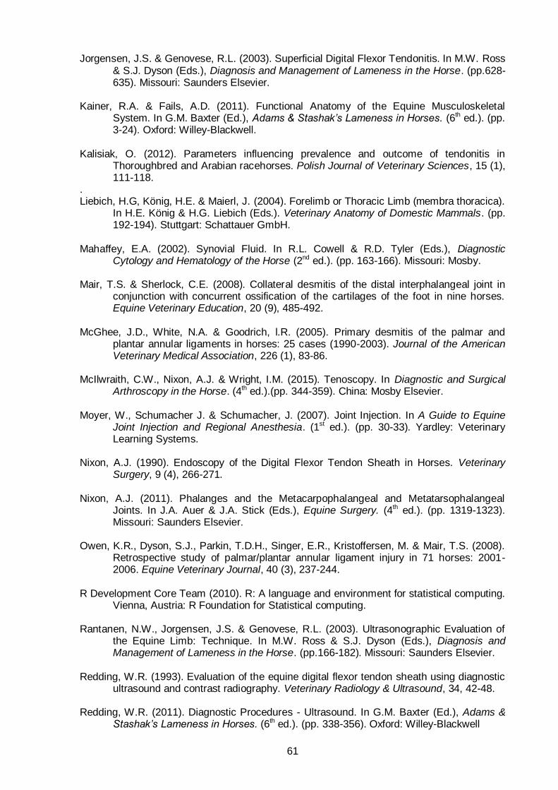
61
Jorgensen, J.S. & Genovese, R.L. (2003). Superficial Digital Flexor Tendonitis. In M.W. Ross & S.J. Dyson (Eds.), Diagnosis and Management of Lameness in the Horse. (pp.628-635). Missouri: Saunders Elsevier.
Kainer, R.A. & Fails, A.D. (2011). Functional Anatomy of the Equine Musculoskeletal
System. In G.M. Baxter (Ed.), Adams & Stashak’s Lameness in Horses. (6th ed.). (pp. 3-24). Oxford: Willey-Blackwell.
Kalisiak, O. (2012). Parameters influencing prevalence and outcome of tendonitis in
Thoroughbred and Arabian racehorses. Polish Journal of Veterinary Sciences, 15 (1), 111-118.
. Liebich, H.G, König, H.E. & Maierl, J. (2004). Forelimb or Thoracic Limb (membra thoracica).
In H.E. König & H.G. Liebich (Eds.). Veterinary Anatomy of Domestic Mammals. (pp. 192-194). Stuttgart: Schattauer GmbH.
Mahaffey, E.A. (2002). Synovial Fluid. In R.L. Cowell & R.D. Tyler (Eds.), Diagnostic
Cytology and Hematology of the Horse (2nd ed.). (pp. 163-166). Missouri: Mosby. Mair, T.S. & Sherlock, C.E. (2008). Collateral desmitis of the distal interphalangeal joint in
conjunction with concurrent ossification of the cartilages of the foot in nine horses. Equine Veterinary Education, 20 (9), 485-492.
McGhee, J.D., White, N.A. & Goodrich, l.R. (2005). Primary desmitis of the palmar and
plantar annular ligaments in horses: 25 cases (1990-2003). Journal of the American Veterinary Medical Association, 226 (1), 83-86.
McIlwraith, C.W., Nixon, A.J. & Wright, I.M. (2015). Tenoscopy. In Diagnostic and Surgical
Arthroscopy in the Horse. (4th ed.).(pp. 344-359). China: Mosby Elsevier. Moyer, W., Schumacher J. & Schumacher, J. (2007). Joint Injection. In A Guide to Equine
Joint Injection and Regional Anesthesia. (1st ed.). (pp. 30-33). Yardley: Veterinary Learning Systems.
Nixon, A.J. (1990). Endoscopy of the Digital Flexor Tendon Sheath in Horses. Veterinary
Surgery, 9 (4), 266-271. Nixon, A.J. (2011). Phalanges and the Metacarpophalangeal and Metatarsophalangeal
Joints. In J.A. Auer & J.A. Stick (Eds.), Equine Surgery. (4th ed.). (pp. 1319-1323). Missouri: Saunders Elsevier.
Owen, K.R., Dyson, S.J., Parkin, T.D.H., Singer, E.R., Kristoffersen, M. & Mair, T.S. (2008).
Retrospective study of palmar/plantar annular ligament injury in 71 horses: 2001-2006. Equine Veterinary Journal, 40 (3), 237-244.
R Development Core Team (2010). R: A language and environment for statistical computing.
Vienna, Austria: R Foundation for Statistical computing. Rantanen, N.W., Jorgensen, J.S. & Genovese, R.L. (2003). Ultrasonographic Evaluation of
the Equine Limb: Technique. In M.W. Ross & S.J. Dyson (Eds.), Diagnosis and Management of Lameness in the Horse. (pp.166-182). Missouri: Saunders Elsevier.
Redding, W.R. (1993). Evaluation of the equine digital flexor tendon sheath using diagnostic
ultrasound and contrast radiography. Veterinary Radiology & Ultrasound, 34, 42-48. Redding, W.R. (2011). Diagnostic Procedures - Ultrasound. In G.M. Baxter (Ed.), Adams &
Stashak’s Lameness in Horses. (6th ed.). (pp. 338-356). Oxford: Willey-Blackwell

62
Sampson, S.N., Schneider, R.K., Tucker, R.L., Gavin, P.R., Zubrod, C.J. & Ho, C.P. (2007). Magnetic resonance imaging features of oblique and straight distal sesamoidean desmitis in 27 horses. Veterinary Radiology & Ultrasound, 48 (4), 303-311.
Schramme, M.C. & Smith, R.K.W. (2003). Diseases of the Digital Synovial Sheath, Palmar
Annular Ligament and Digital Annular Ligaments. In M.W. Ross & S.J. Dyson (Eds.), Diagnosis and Management of Lameness in the Horse. (pp.674-683). Missouri: Saunders Elsevier.
Schumacher, J., Schumacher, J., Schramme, M.C., DeGraves, F. & Smith, R.K.W. (2007).
Diagnostic Analgesia of the Equine Foot. In A.E. Floyd & R.A. Mansmann (Eds.) Equine Podiatry. (pp. 136-138). Missouri: Saunders Elsevier.
Seignour, M., Coudry, V., Norris, R. & Denoix, J.M. (2011). Ultrasonographic examination of
the palmar/plantar aspect of the fetlock in the horse: technique and normal images. Equine Veterinary Education, 24 (1), 19-29.
Smith, M.R.W. (2003). Pathophysiology of Tendon Injury. In M.W. Ross & S.J. Dyson (Eds.),
Diagnosis and Management of Lameness in the Horse. (pp.616-628). Missouri: Saunders Elsevier.
Smith, M.R.W. & Wright, I.M. (2006). Noninfected tenosynovitis of the digital flexor tendon
sheath: a retrospective analysis of 76 cases. Equine Veterinary Journal, 38 (2), 134-141.
Smith, S., Dyson, S.J. & Murray, R.C. (2008). Magnetic resonance imaging of distal
sesamoidean ligament injury. Veterinary Radiology & Ultrasound, 49 (6), 516-528. Thorpe, C.T., Karunaseelan, K.J., Chieng Hin, J.N., Riley, G.P., Birch, H.L., Clegg, P.D. &
Screen, H.R.C. (2016). Distribution of proteins within different compartments of tendon varies according to tendon type. Journal of Anatomy, 229, 450-458.
Thorpe, C.T., Udeze, C.P., Birch, H.L., Clegg, P.D. & Screen, H.R.C. (2013). Capacity for
sliding between tendon fascicles decreases with ageing in injury prone equine tendons: a possible mechanism for age-related tendinopathy? European Cells and Materials, 25, 48-60.
Valdéz- Martínez, A. & Park, R.R. (2011). Diagnostic Procedures - Radiology. In G.M. Baxter
(Ed.), Adams & Stashak’s Lameness in Horses. (6th ed.). (pp. 225-227). Oxford: Willey-Blackwell
Wereszka, M.M., White, N.A. & Furr, M.O. (2007). Factors associated with outcome following
treatment of horses with septic tenosynovitis: 51 cases (1986-2003). Journal of the American Veterinary Medical Association, 230, 1195-1200.
Wilderjans, H., Boussauw, B., Madder, K. & Simon, O. (2003). Tenosynovitis of the digital
flexor tendon sheath and annular ligament constriction syndrome caused by longitudinal tears in the deep digital flexor tendon: a clinical and surgical report of 17 cases in Warmblood horses. Equine Veterinary Journal, 35 (3), 270-275.
Wright, I.M. & McMahon, P.J. (1999). Tenosynovitis associated with longitudinal tears of the
digital flexor tendons in horses: a report of 20 cases. Equine Veterinary Journal, 31 (1), 12-18.
Wright, I.M., Smith, M.R.W., Humphrey, D.J., Eaton-Evans, T.C.J. & Hillyer, M.H. (2003).
Tenoscopic surgery in the treatment of contaminated and infected synovial cavities. Equine Veterinary Journal, 35 (6), 613-619.

63
ANNEXES
Annex 1. Horse identification (ID) and respective age, breed, gender, affected limb(s) and diagnosis.
Horse ID
Age Breed Gender Affected Limb(s)
Diagnosis
PAL
Constriction
MF
Tear
SDFT
Tear
DDFT
Tear
Traumatic
injury Other
1 17 Cob x
Thoroughbred Female LF X
2 11 Connemara Gelding RH + LH X X
3 15 Welsh Cob
Section C Gelding LH X X
4 9 Unknown Female LF X
5 17 Andalusian Male LH X
6 3 Thoroughbred Gelding RH X
7 6 Thoroughbred Gelding LF X
8 7 Irish Sport
Horse Gelding LH X
9 14 Unknown Gelding RF X X
10 3 Thoroughbred Male RF X
11 15 Polo Pony Female RF + RH X X
12 13 Cob Gelding RF + LF X X
13 12 New Zealand Thoroughbred
Gelding LH X
14 1 Thoroughbred Female RH X
15 8 Connemara x
Thoroughbred Gelding RH X
16 16 Hungarian
Warmblood Gelding RH X
17 14 New Forest
Pony Female LH X X
18 7 Irish Sports
Horse Gelding RF X
19 11 Welsh Cob Section D
Gelding LH X X
20 16 KWPN Gelding RH X X
21 7 KWPN Female LH X
22 10 Sports Horse Female LH X
23 12 New Forest
Pony Gelding RF X X
24 9 KWPN Gelding RH + LH X X
25 9 Irish Draught x
Thoroughbred Female RH + LH X X X
26 8 Skewbald Gelding LH X
27 9 Welsh Cob
Section D Female RH + LH X X X
28 3 Thoroughbred Gelding RF X
29 14 Unknown Female LH X X
30 16 Welsh Cob
Section C Female LH X
31 15 Polish Riding
Horse Female RH X
32 19 Skewbald Gelding LH X X
33 13 Unknown Gelding RH X X
34 11 Unknown Gelding RH X
35 12 Lusitano Gelding LH X X
36 5 Welsh Cob Section C
Gelding RF X X
37 6 Thoroughbred Gelding LF X
38 12 Thoroughbred Gelding LH X
39 16 Thoroughbred Gelding RF X

64
40 5 Thoroughbred Female RF X
41 6 Thoroughbred Gelding LH X
42 1 Thoroughbred Male RH X
43 10 Cob Female RH X
44 20 Cob Female LH X
45 12 Welsh Cob Gelding RH X X
46 13 Connemara Gelding RH X X
47 16 Cob (Dale x
Fell) Gelding RH X X X X
48 6 Thoroughbred Female LF X
49 1 Thoroughbred Female LF X
50 14 Fell Pony Female RH X X
51 6 Shetland Pony Gelding RH X
52 10 Irish Draught x
Thoroughbred Gelding LF X
53 9 Unknown Female RF X
54 1 Thoroughbred Male RF X
55 16 Polish Cob Female LH X X
56 14 Belgian
Warmblood Female RH X X
57 1 Thoroughbred Male LF X
58 21 Irish Draught x Female RH X X
59 8 KWPN Gelding RH X
60 11 Welsh Cob Sec
D Female RF X
61 5 Hannoverian x
Thoroughbred Gelding RF X
62 8 Thoroughbred Female RH X
63 9 Unknown Gelding RF + LF X X
64 9 Thoroughbred Gelding LH X
65 14 Unknown Gelding LF X
66 13 Welsh Cob Sec
C Gelding LH X
67 10 Danish
Warmblood Gelding LF X
68 3 Thoroughbred Female RF X
69 13 Thoroughbred Gelding RH X
70 7 Thoroughbred Gelding RF X
71 2 Thoroughbred Female LH X
72 10 KWPN x
Thoroughbred Gelding RH X
73 6 Pony Gelding RF X
74 7 Polo Pony Female RF X
75 6 Thoroughbred Female RF X
76 3 Thoroughbred Male LH X
77 15 Irish Sports
Horse Gelding RF X
78 8 Cob Gelding RH X X
79 1 Thoroughbred Male LH X
80 6 Warmblood Female LF X
81 12 Warmblood Female RF X
82 6 Irish Sports
Horse Female RH X
83 12 Hanoverian Gelding LH X
84 3 Thoroughbred Male LF X
85 20 Arabian Gelding LH X
86 16 Thoroughbred Gelding LH X