Synthesis, characterization, ab initio calculations, thermal behaviour and thermodynamics of some...
10
J. Chem. Sci., Vol. 122, No. 4, July 2010, pp. 539–548. © Indian Academy of Sciences. 539 *For correspondence Synthesis, characterization, ab initio calculations, thermal behaviour and thermodynamics of some oxovanadium(IV) complexes involving O,O- and N,N-donor moieties MOZAFFAR ASADI a, *, MOHAMMAD HADI GHATEE a , SUSAN TORABI a , KHOSRO MOHAMMADI b and FATEMEH MOOSAVI a a Chemistry Department, College of Sciences, Shiraz University, Shiraz 71454, I.R. Iran b Chemistry Department, Faculty of Sciences, Persian Gulf University, Bushehr 75169, I.R. Iran e-mail: [email protected]; [email protected] MS received 4 November 2009; revised 13 March 2010; accepted 30 March 2010 Abstract. Some oxovanadium(IV) complexes, namely bis(1,1,1-trifluro-2,4-pentanedionato-O,O′) oxovanadium (IV), [VO(tfac) 2 (H 2 O)], bis(1-phenyl-2,4-pentanedionato-O,O′)oxovanadium(IV), [VO (phac) 2 (H 2 O)], bis(1,3-diphenyl-2,4-pentanedionato-O,O′)oxovanadium(IV), [VO(dphac) 2 (H 2 O)], of the type [VO(O 4 )] and bis(pyrolidineaniline)oxovanadium(IV), [VO(pyran) 2 (H 2 O)], bis(p-hydroxy- pyrolidineaniline)oxovanadium(IV), [VO(p-hydroxypyran) 2 (H 2 O)], bis(p-methoxypyrolidineaniline) oxovanadium(IV), [VO(p-MeOpyran) 2 (H 2 O)], bis(p-chloropyrolidineaniline)oxovanadium(IV), [VO(p- chloropyran) 2 (H 2 O)], bis(p-bromopyrolidineaniline)oxovanadium(IV), [VO(p-bromopyran) 2 (H 2 O)], bis (p-cyano pyrolidineaniline)oxovanadium(IV), [VO(p-cyanopyran) 2 (H 2 O)], and bis(pyrolidinebenzyl- amine)oxovanadium(IV), [VO(pyrbz) 2 (H 2 O)], of the type [VO(N 4 )] were synthesized and characterized by IR, UV–Vis, mass spectrometry, elemental analysis, magnetic moment and thermogravimetry in order to evaluate their thermal stability and thermal decomposition pathways. The number of steps and, in par- ticular, the starting temperature of decomposition of these complexes depends on the equatorial ligand. Also, formation constants of the complexes have been determined by UV-Vis absorption spectroscopy through titration of the ligands with the metal ions at constant ionic strength (0⋅1 M NaClO 4 ) and at 25°C. According to the thermodynamic studies, as the steric character of the ligand increases, the complexation tendency to VO(IV) center decreases. Also, the ab initio calculations were carried out to determine the structural and the geometrical properties of the complexes. Keywords. Oxovanadium(IV); formation constants; thermodynamic; ab initio calculations. 1. Introduction In recent years, vanadium chemistry has attracted at- tention due to its interesting structural features and biological relevance. 1–7 Vanadium oxide (vanadyl) complexes, in particular, have come under close scrutiny because of their utility as insulin mimetics. 2 Peroxovanadium complexes 8–12 and bis-(maltolato) oxovanadium(IV) 13,14 (VO(malto) 2 ) have been found to be more effective than the simple vanadium(IV) and -(V) salts, both in cell cultures and in animal studies. Furthermore, a bis(maltolato)oxovanadium (IV) derivative (KP-102) has now entered phase I clinical trials in humans. A related compound, bis(2,4-pentanedionato-O,O′)oxovanadium(IV) (VO (acac) 2 ) was also found to have insulin-mimetic properties superior to those of VOSO 4 in cell culture studies. 15 Since the first published account of the synthesis of VO(acac) 2 in 1914, 16 the complex has been used extensively as a reagent in organic synthesis. 17–19 The physical properties of VO(acac) 2 have been examined by numerous workers, 20–22 and the crystal structure was published in 1962. 23 In the solid state, VO(acac) 2 -type complexes are five-coordinate; however, upon dissolution in organic solvents, the vanadium coordinates a donor ligand in the vacant site, generating products expressed as [VO(acac) 2 L] (where L = coordinated ligand and acac is 2,4- pentanedionato or corresponding acac- derivatives). In view of importance of vanadium compounds, and also extending the search for more efficacious compounds, this study is undertaken of the synthesis and coordination chemistry of some oxovana-
Transcript of Synthesis, characterization, ab initio calculations, thermal behaviour and thermodynamics of some...
J. Chem. Sci., Vol. 122, No. 4, July 2010, pp. 539–548. © Indian Academy of Sciences.
539
*For correspondence
Synthesis, characterization, ab initio calculations, thermal behaviour and thermodynamics of some oxovanadium(IV) complexes involving O,O- and N,N-donor moieties
MOZAFFAR ASADIa,*, MOHAMMAD HADI GHATEE
a, SUSAN TORABI
a,
KHOSRO MOHAMMADIb and FATEMEH MOOSAVI
a
aChemistry Department, College of Sciences, Shiraz University, Shiraz 71454, I.R. Iran bChemistry Department, Faculty of Sciences, Persian Gulf University, Bushehr 75169, I.R. Iran
e-mail: [email protected]; [email protected]
MS received 4 November 2009; revised 13 March 2010; accepted 30 March 2010
Abstract. Some oxovanadium(IV) complexes, namely bis(1,1,1-trifluro-2,4-pentanedionato-O,O′) oxovanadium (IV), [VO(tfac)2(H2O)], bis(1-phenyl-2,4-pentanedionato-O,O′)oxovanadium(IV), [VO (phac)2(H2O)], bis(1,3-diphenyl-2,4-pentanedionato-O,O′)oxovanadium(IV), [VO(dphac)2 (H2O)], of the type [VO(O4)] and bis(pyrolidineaniline)oxovanadium(IV), [VO(pyran)2(H2O)], bis(p-hydroxy-pyrolidineaniline)oxovanadium(IV), [VO(p-hydroxypyran)2(H2O)], bis(p-methoxypyrolidineaniline) oxovanadium(IV), [VO(p-MeOpyran)2 (H2O)], bis(p-chloropyrolidineaniline)oxovanadium(IV), [VO(p-chloropyran)2(H2O)], bis(p-bromopyrolidineaniline)oxovanadium(IV), [VO(p-bromopyran)2(H2O)], bis (p-cyano pyrolidineaniline)oxovanadium(IV), [VO(p-cyanopyran)2(H2O)], and bis(pyrolidinebenzyl-amine)oxovanadium(IV), [VO(pyrbz)2(H2O)], of the type [VO(N4)] were synthesized and characterized by IR, UV–Vis, mass spectrometry, elemental analysis, magnetic moment and thermogravimetry in order to evaluate their thermal stability and thermal decomposition pathways. The number of steps and, in par-ticular, the starting temperature of decomposition of these complexes depends on the equatorial ligand. Also, formation constants of the complexes have been determined by UV-Vis absorption spectroscopy through titration of the ligands with the metal ions at constant ionic strength (0⋅1 M NaClO4) and at 25°C. According to the thermodynamic studies, as the steric character of the ligand increases, the complexation tendency to VO(IV) center decreases. Also, the ab initio calculations were carried out to determine the structural and the geometrical properties of the complexes. Keywords. Oxovanadium(IV); formation constants; thermodynamic; ab initio calculations.
1. Introduction
In recent years, vanadium chemistry has attracted at-
tention due to its interesting structural features and
biological relevance.1–7 Vanadium oxide (vanadyl)
complexes, in particular, have come under close
scrutiny because of their utility as insulin mimetics.2
Peroxovanadium complexes8–12
and bis-(maltolato)
oxovanadium(IV)13,14
(VO(malto)2) have been found
to be more effective than the simple vanadium(IV)
and -(V) salts, both in cell cultures and in animal
studies. Furthermore, a bis(maltolato)oxovanadium
(IV) derivative (KP-102) has now entered phase I
clinical trials in humans. A related compound,
bis(2,4-pentanedionato-O,O′)oxovanadium(IV) (VO
(acac)2) was also found to have insulin-mimetic
properties superior to those of VOSO4 in cell culture
studies.15
Since the first published account of the synthesis
of VO(acac)2 in 1914,16
the complex has been used
extensively as a reagent in organic synthesis.17–19
The physical properties of VO(acac)2 have been
examined by numerous workers,20–22
and the crystal
structure was published in 1962.23
In the solid state,
VO(acac)2-type complexes are five-coordinate;
however, upon dissolution in organic solvents, the
vanadium coordinates a donor ligand in the vacant
site, generating products expressed as [VO(acac)2L]
(where L = coordinated ligand and acac is 2,4-
pentanedionato or corresponding acac- derivatives).
In view of importance of vanadium compounds,
and also extending the search for more efficacious
compounds, this study is undertaken of the synthesis
and coordination chemistry of some oxovana-
Mozaffar Asadi et al
540
dium(IV) complexes of the type [VO(O4)] and
[VO(N4)] involving some diketone and some
pyrrolecarbaldehyde derivatives.
The resulting complexes have been characterized
on the basis of elemental analysis (C, H, N), infra-
red, mass and electronic spectral techniques. Thermal
stability is an important feature of the complexes in
their potential applications as catalysts, so the dif-
ferences observed in the thermal behaviour of
[VO(O4)]-type complexes is described by using
thermogravimetry (TG). Also the formation con-
stants of the complexes were determined spectro-
photometrically at 25°C in methanol.
In addition to the importance of the experimental
studies, it is crucial to investigate the theoretical beha-
viour of target complexes to find their structural
characteristics and the way that these structures
affect the other properties of the complexes. The
structure of a complex can be studied by the ab ini-
tio methods to find the stable structure electronically
by solving the wave function of the molecule. The
theoretical goal of this study focuses on these fea-
tures of the chemistry.
2. Experimental
2.1 Reagents
Diketones, aniline and its derivatives were pur-
chased from Aldrich and were used as received. All
other solvents and reagents with analytical grade
(PA) were obtained commercially and used without
further purification.
2.2 Physical measurements
All of the scanning UV-Vis spectra were recorded
on Perkin-Elmer Lambda 2 spectrophotometer. FT–
IR spectra were run on a Shimadzu FTIR-8300 spec-
trophotometer. Mass spectra were obtained with
Shimadzu LCMS–2010EV. The Elemental analysis
was carried out by Thermo Finnigan-Flash-1200.
The NMR spectra were recorded by a Bruker
Avance DPX 250 MHz spectrometer.
2.3 Preparation of the vanadyl complexes
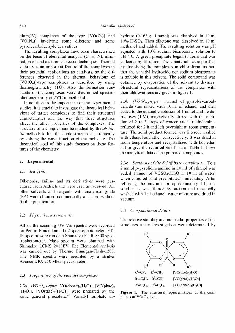
2.3a [VO(O4)]-type: [VO(dphac)2(H2O)], [VO(phac)2
(H2O)], [VO(tfac)2(H2O)], were prepared by the
same general procedure.24
Vanadyl sulphate tri-
hydrate (0⋅163 g, 1 mmol) was dissolved in 10 ml
10% H2SO4. Then diketone was dissolved in 10 ml
methanol and added. The resulting solution was pH
adjusted with 10% sodium bicarbonate solution to
pH 4⋅0. A green precipitate began to form and was
collected by filtration. These materials were purified
by dissolving the complexes in chloroform, as nei-
ther the vanadyl hydroxide nor sodium bicarbonate
is soluble in this solvent. The solid compound was
obtained by evaporation of the solvent to dryness.
Structural representations of the complexes with
their abbreviations are given in figure 1.
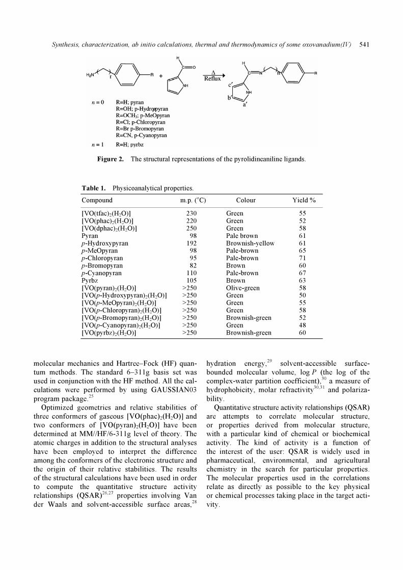
2.3b [VO(N4)]-type: 1 mmol of pyrrol-2-carbal-
dehyde was mixed with 10 ml of ethanol and then
added to the ethanolic solution of 1 mmol aniline de-
rivatives (1 M), magnetically stirred with the addi-
tion of 2 to 3 drops of concentrated triethylamine,
refluxed for 2 h and left overnight at room tempera-
ture. The solid product formed was filtered, washed
with ethanol and ether consecutively. It was dried at
room temperature and recrystallized with hot etha-
nol to give the required Schiff base. Table 1 shows
the analytical data of the prepared compounds.
2.3c Synthesis of the Schiff base complexes: To a
2 mmol p-pyrolidineaniline in 10 ml of ethanol was
added 1 mmol of VOSO4⋅5H2O in 10 ml of water,
when coloured solid precipitated immediately. After
refluxing the mixture for approximately 1 h, the
solid mass was filtered by suction and repeatedly
washed with 1 : 1 ethanol–water mixture and dried in
vacuum.
2.4 Computational details
The relative stability and molecular properties of the
structures under investigation were determined by
Figure 1. The structural representations of the com-plexes of VO(O4) type.
Synthesis, characterization, ab initio calculations, thermal and thermodynamics of some oxovanadium(IV)
541
Figure 2. The structural representations of the pyrolidineaniline ligands.
Table 1. Physicoanalytical properties.
Compound m.p. (oC) Colour Yield %
[VO(tfac)2(H2O)] 230 Green 55 [VO(phac)2(H2O)] 220 Green 52 [VO(dphac)2(H2O)] 250 Green 58 Pyran 98 Pale brown 61 p-Hydroxypyran 192 Brownish-yellow 61 p-MeOpyran 98 Pale-brown 65 p-Chloropyran 95 Pale-brown 71 p-Bromopyran 82 Brown 60 p-Cyanopyran 110 Pale-brown 67 Pyrbz 105 Brown 63 [VO(pyran)2(H2O)] >250 Olive-green 58 [VO(p-Hydroxypyran)2(H2O)] >250 Green 50 [VO(p-MeOpyran)2(H2O)] >250 Green 55 [VO(p-Chloropyran)2(H2O)] >250 Green 58 [VO(p-Bromopyran)2(H2O)] >250 Brownish-green 52 [VO(p-Cyanopyran)2(H2O)] >250 Green 48 [VO(pyrbz)2(H2O)] >250 Brownish-green 60
molecular mechanics and Hartree–Fock (HF) quan-
tum methods. The standard 6–311g basis set was
used in conjunction with the HF method. All the cal-
culations were performed by using GAUSSIAN03
program package.25
Optimized geometries and relative stabilities of
three conformers of gaseous [VO(phac)2(H2O)] and
two conformers of [VO(pyran)2(H2O)] have been
determined at MM//HF/6-311g level of theory. The
atomic charges in addition to the structural analyses
have been employed to interpret the difference
among the conformers of the electronic structure and
the origin of their relative stabilities. The results
of the structural calculations have been used in order
to compute the quantitative structure activity
relationships (QSAR)26,27 properties involving Van
der Waals and solvent-accessible surface areas,28
hydration energy,29
solvent-accessible surface-
bounded molecular volume, log P (the log of the
complex-water partition coefficient),30 a measure of
hydrophobicity, molar refractivity30,31
and polariza-
bility.
Quantitative structure activity relationships (QSAR)
are attempts to correlate molecular structure,
or properties derived from molecular structure,
with a particular kind of chemical or biochemical
activity. The kind of activity is a function of
the interest of the user: QSAR is widely used in
pharmaceutical, environmental, and agricultural
chemistry in the search for particular properties.
The molecular properties used in the correlations
relate as directly as possible to the key physical
or chemical processes taking place in the target acti-
vity.
Mozaffar Asadi et al
542
Table 2. UV-Visa, mass spectral, elemental analysis and magnetic moments data of the compounds.
Elemental analysis (%, Found)
Mass spectra Μeff B.M. Compounds λmax (nm) (m/z) C H N (298 K)
[VO(tfac)2(H2O)]a 296 373, 218, 150 30⋅71 (30⋅60) 2⋅58 (2⋅21) 1⋅65 [VO(phac)2(H2O)]a 326 389, 228, 161 58⋅98 (59⋅36) 4⋅95 (4⋅57) 1⋅68 [VO(dphac)2(H2O)]a 356 513, 289, 223 67⋅80 (67⋅88) 4⋅55 (4⋅39) 1⋅66 pyranb 320 170 71⋅91 (72⋅10) 6⋅31 (6⋅02) 15⋅25 (15⋅65) p-Hydroxypyranb 295 (sh), 335 187, 170 70⋅27 (70⋅39) 5⋅47 (5⋅50) 14⋅90 (14⋅70) p-MeOpyranb
297 (sh), 332 200, 170 71⋅98 (71⋅82) 6⋅04 (6⋅04) 13⋅99 (13⋅97) p-Chloropyranb 326 204, 170 62⋅36 (62⋅10) 4⋅66 (4⋅32) 13⋅22 (12⋅88) p-Bromopyranb 271, 343 249, 170 73⋅15 (72⋅81) 4⋅71 (5⋅01) 21⋅33 (21⋅10) p-Cyanopyranb 326 195, 170 70⋅57 (70⋅68) 4⋅94 (5⋅08) 20⋅57 (21⋅11) pyrbzb 280 184 74⋅58 (74⋅65) 6⋅78 (7⋅01) 14⋅50 (14⋅10) [VO(pyran)2(H2O)].0.25H2O
b 237, 364 405,236,169 61⋅76 (61⋅44) 4⋅83 (4⋅52) 13⋅09 (13⋅33) 1⋅65 [VO(p-Hydroxypyran)2(H2O)]b 300, 380 437 ,252, 186 58⋅03 (58⋅10) 4⋅43 (4⋅60) 12⋅30 (12⋅26) 1⋅67 [VO(p-MeOpyran)2(H2O)]b 381 465, 265, 200 59⋅63 (59⋅95) 5⋅00 (5⋅04) 11⋅59 (11⋅31) 1⋅63 [VO(p-Chloropyran)2(H2O)]b 243, 370 473, 268, 204 53⋅68 (53⋅85) 3⋅69 (4⋅10) 11⋅38 (11⋅46) 1⋅70 [VO(p-Bromopyran)2(H2O)]b 211, 271 560, 314, 247 45⋅47 (45⋅12) 3⋅12 (3⋅12) 9⋅64 (9⋅46) 1⋅69 [VO(p-Cyanopyran)2(H2O)]b 242, 371 455, 261, 194 60⋅89 (60⋅82) 3⋅83 (3⋅98) 17⋅75 (17⋅88) 1⋅66 [VO(pyrbz)2(H2O)]0⋅5H2O
b 240, 326 433, 250, 183 62⋅61 (62⋅22) 5⋅47 (5⋅80) 12⋅17 (12⋅48) 1⋅64
aSolvent is CH3OH. bSolvent is CH3CN
3. Results and discussion
3.1 Characterization of the compounds
The compounds were synthesized and characterized
by 1HNMR, IR, magnetic moment, mass, UV-Vis
spectroscopy and elemental analysis.
3.1a Magnetic moments and electronic spectra:
The effective magnetic moment was measured using
a Gouy balance. Data were corrected for diamag-
netic contributions using the Pascal’s constants.32
Oxovanadium(1V) complexes show magnetic
moment values in the range of 1⋅64–1⋅70 BM, which
are in accordance with the values reported for
oxovanadium(IV) complexes with one unpaired
electron33
(table 2) indicating that the complexes are
monomeric. This is in agreement with the ν (V=O)
obtained.
The ligands show two absorption bands at
UV-Visible region. A n – π* transition band at 326–
410 nm and a π–π* transition band at 240–297 nm
are shown in the ligands. These absorption bands
show a slight shift to higher energy in the complexes
that is evident for unalteration structure of ligands in
complexation (table 2).
All the vanadyl(IV) complexes have a band at
340–470 nm in solvent corresponding to a d–d tran-
sition band. This band is not always observed, being
often buried beneath a high intensity charge transfer
band (or more accurately the low energy tail of that
band), and when it is observed it is generally a
shoulder (table 2). UV–Vis spectra of pyrbz and its
oxovanadium(IV) are shown in figure S1.
3.1b Mass spectra: The mass spectra of the
ligands show intense molecular ion peaks m/z
M+/(M + H)
+ (table 2). The mass spectra of the
vanadium(IV) chelates show intense peaks including
[VOL2] and [VOL2 + H]+ that confirmed a structure
of stoichiometry of 2 : 1 for ligands to vanadium(IV).
The spectra of the vanadyl(IV) complexes, also
show some peaks corresponding to [VOL]+ and L
+
fragments (table 2).
3.1c Infrared spectra: The solid state properties
of the ligands and the vanadyl complexes were
examined by infrared spectroscopy. For each of
these compounds, the characteristic ν (V=O) stretch-
ing at 920–995 cm–1
was observed, as reported for
other oxovanadium(IV) derivatives34
and is not pre-
sent in the spectrum of the free ligands (table S1).
IR spectra of the ligand (table S1) showed the
absence of bands at ~ 1735 and 3315 cm
–1 due to the
carbonyl ν (C=O) and ν
(NH2) stretching vibrations
and a strong new band appeared at 1612–1633 cm–1
assigned to azomethine ν (HC=N) linkage, showing
that amino and aldehyde moieties of the starting
Synthesis, characterization, ab initio calculations, thermal and thermodynamics of some oxovanadium(IV)
543
Table 3. 1HNMR spectroscopic data of the compounds (δ in ppm).
Compounds Pyrrol-H H–C=N Ar–H –CH2– OH CH3
Pyrana 6⋅3b′ 6⋅6a′ 8⋅59 6⋅93–7⋅93 6⋅8c′
p-Hydroxypyranb 6⋅1b′ 11⋅7 6⋅5a′ 8⋅26 6⋅69–7⋅05 6⋅7c′
p-MeOpyrana 6⋅3b′ 3⋅8 6⋅7a′ 8⋅3 6⋅9–7⋅5 6⋅9c′
p-Chloropyrana 6⋅3b′ 6⋅7a′ 8⋅2 6⋅9–7⋅3 6⋅9c′
p-Bromopyrana 6⋅4b′ 6⋅8a′ 8⋅2 7⋅2–7⋅6 7⋅1c′
p-Cyanopyranb 6⋅1b′ 6⋅2a′ 9⋅4 6⋅5–7⋅3 6⋅7c′
pyrbza 6⋅1b′ 6⋅4a′ 8⋅1 7⋅0–7⋅2 4⋅64 6⋅7c′
aSolvent is CDCl3. bSolvent is DMSO-d6
material are absent and have been converted into the
ligand, i.e. p-pyrrolidineaniline.
The comparison of IR spectra of the ligand and its
vanadyl complexes indicated that the ligand is prin-
cipally coordinated to the metal ion in 2 ways, thus
acting as a bidentate ligand. The band appearing at
~ 1630 cm
–1 due to azomethine was shifted to a lower
frequency by ~ 1–15 cm
–1 in both complexes, indi-
cating participation of azomethine nitrogen in the
interaction with the metal ion. The band appearing
at 3153–3300 cm–1
assigned to the ν (NH) in
the ligand was no longer found in the spectra of the
metal complexes indicating deprotonation and coor-
dination of the nitrogen with the metal ion.
3.1d 1HNMR spectra:
1H NMR spectral data of
pyrrolidineaniline ligands show a singlet (1H) signal
at δ = 10⋅5–12 ppm which can be assigned to the N–
H of the pyrrol rings. The pyrrol ring proton signals
resolved into three groups in the range δ = 6⋅2–
7⋅4 ppm as a triplet and doublet that are related to
the protons in position b and (a, c). The signals
of the hydrogen of the phenyl group are appeared at
δ = 6⋅6–8 ppm. A singlet signal at δ = 3⋅8 ppm is
assigned to those compounds that have OMe group.
The 1HNMR spectra of the ligands provide compel-
ling evidence of the presence of one azomethine
proton group at ~ 8⋅0 ppm.
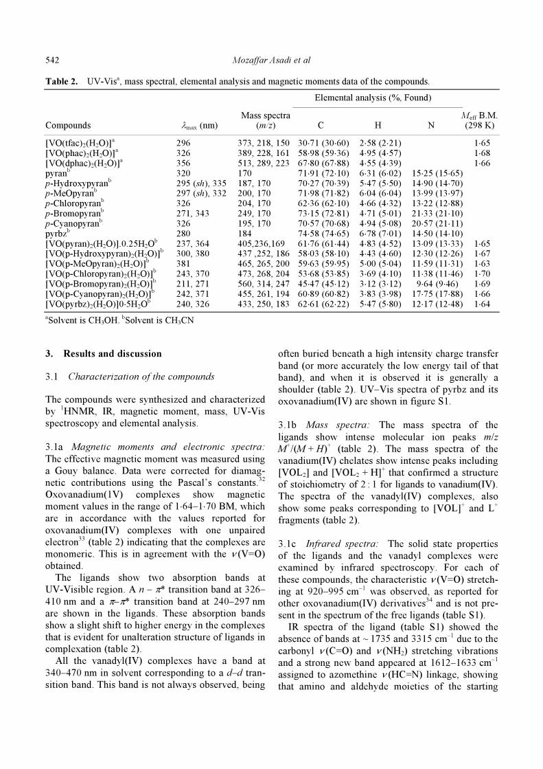
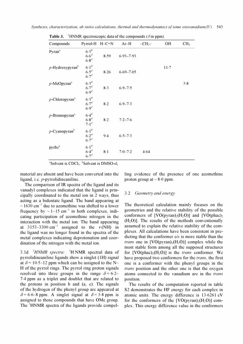
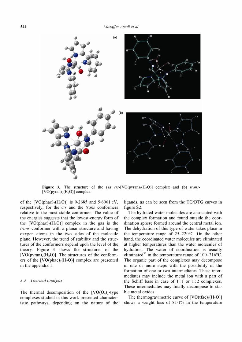
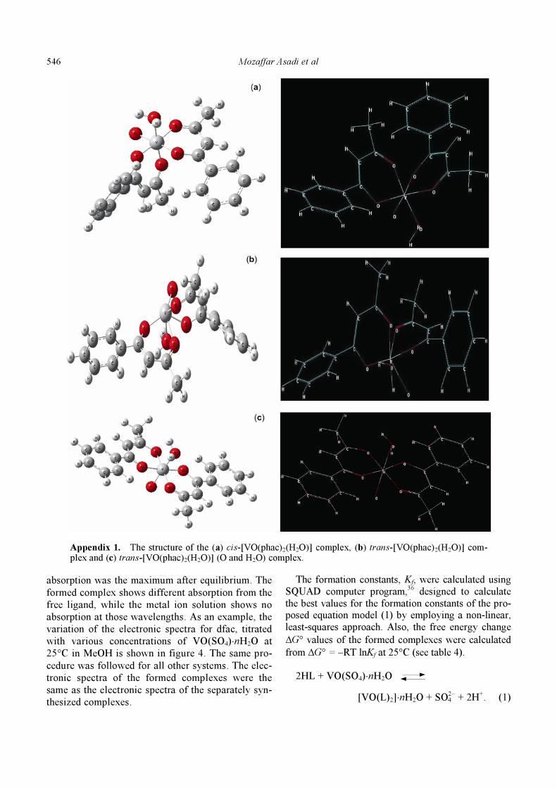
3.2 Geometry and energy
The theoretical calculation mainly focuses on the
geometries and the relative stability of the possible
conformers of [VO(pyran)2(H2O)] and [VO(phac)2
(H2O)]. The results of the methods conventionally
assumed to explain the relative stability of the com-
plexes. All calculations have been consistent in pre-
dicting that the conformer cis is more stable than the
trans one in [VO(pyran)2(H2O)] complex while the
most stable form among all the supposed structures
for [VO(phac)2(H2O)] is the trans conformer. We
have proposed two conformers for the trans, the first
one is a conformer with the phenyl groups in the
trans position and the other one is that the oxygen
atoms connected to the vanadium are in the trans
position.
The results of the computation reported in table
S2 demonstrates the HF energy for each complex in
atomic units. The energy difference is 13⋅6261 eV
for the conformers of the [VO(pyran)2(H2O)] com-
plex. This energy difference value in the conformers
Mozaffar Asadi et al
544
Figure 3. The structure of the (a) cis-[VO(pyran)2(H2O)] complex and (b) trans-[VO(pyran)2(H2O)] complex.
of the [VO(phac)2(H2O)] is 0⋅2685 and 5⋅6061 eV,
respectively, for the cis and the trans conformers
relative to the most stable conformer. The value of
the energies suggests that the lowest-energy form of
the [VO(phac)2(H2O)] complex in the gas is the
trans conformer with a planar structure and having
oxygen atoms in the two sides of the molecule
plane. However, the trend of stability and the struc-
tures of the conformers depend upon the level of the
theory. Figure 3 shows the structures of the
[VO(pyran)2(H2O)]. The structures of the conform-
ers of the [VO(phac)2(H2O)] complex are presented
in the appendix 1.
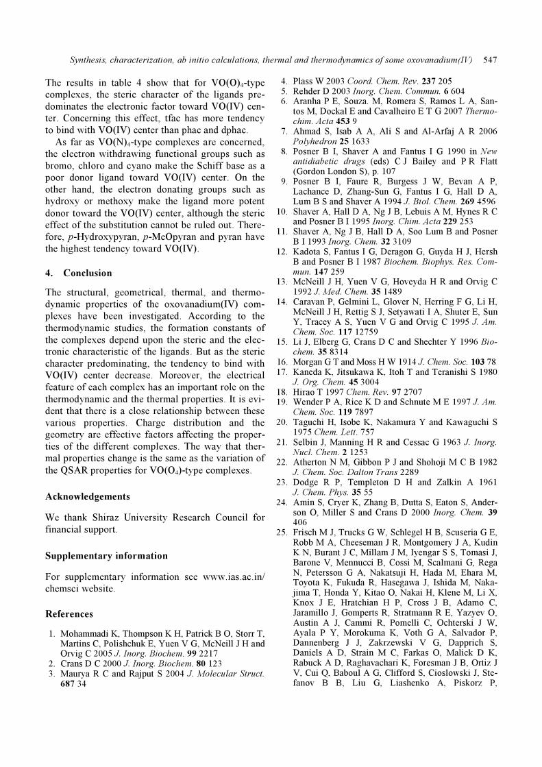
3.3 Thermal analysis
The thermal decomposition of the [VO(O4)]-type
complexes studied in this work presented character-
istic pathways, depending on the nature of the
ligands, as can be seen from the TG/DTG curves in
figure S2.
The hydrated water molecules are associated with
the complex formation and found outside the coor-
dination sphere formed around the central metal ion.
The dehydration of this type of water takes place in
the temperature range of 25–220°C. On the other
hand, the coordinated water molecules are eliminated
at higher temperatures than the water molecules of
hydration. The water of coordination is usually
eliminated35 in the temperature range of 100–316°C.
The organic part of the complexes may decompose
in one or more steps with the possibility of the
formation of one or two intermediates. These inter-
mediates may include the metal ion with a part of
the Schiff base in case of 1 : 1 or 1
: 2 complexes.
These intermediates may finally decompose to sta-
ble metal oxides.
The thermogravimetric curve of [VO(tfac)2(H2O)]
shows a weight loss of 81⋅1% in the temperature
Synthesis, characterization, ab initio calculations, thermal and thermodynamics of some oxovanadium(IV)
545
range of 150–230°C with a residue of 19⋅8% (calcd.
weight loss for 1 mol of H2O and 2 mol of tfac:
82⋅1%) corresponding to the elimination of one
molecule of the coordinated water and 2 tfac groups.
According to the DTG curve, the decomposition
involves one step (figure S2, curve a).
The thermal decomposition of [VO(phac)2(H2O)]
occurs in two steps. The first mass loss (observed
36⋅6%), can be seen between 190 and 307°C (calcd.
weight loss for 1mol of H2O and 1 mol of phac:
42⋅1%) corresponding to the elimination of one
molecule of coordinated water and 1 phac group.
The decomposition continues with the gradual
weight loss, and it ceases at about 480°C. This
weight loss in the temperature range of 370–480°C
(observed 32⋅6%) (calcd. 38⋅1%), assuming weight
removal of the other coordinated phac group. The
final residue with attaining a constant weight
(observed 37%) roughly corresponds to V2O5 (calcd.
42%) (figure S2, curve b).
The [VO(dphac)2(H2O)] decomposes in two steps.
The first step occurs between 188 and 310°C is
probably a partial decomposition of the ligand and
elimination of one molecule of the coordinated
water (observed 42⋅6%) (calcd. weight loss for
1 mol of H2O and 1 mol of phac: 45⋅0%). The sec-
ond step of the thermal decomposition, which occurs
in the range of 360–458°C, was assigned to the loss
of the second ligand (observed 35⋅3%) (calcd.
40⋅7%) with the formation of V2O5 (observed 30%)
(calcd. 35.0%) (figure S2, curve c).
The thermal studies performed suggest that
[VO(tfac)2(H2O)] is less stable than the other com-
plexes because of the lower degradation temperature
(approximately 150°C). The presence of the phenyl
group in the coordination sphere of the two other
complexes may enhance their stability.
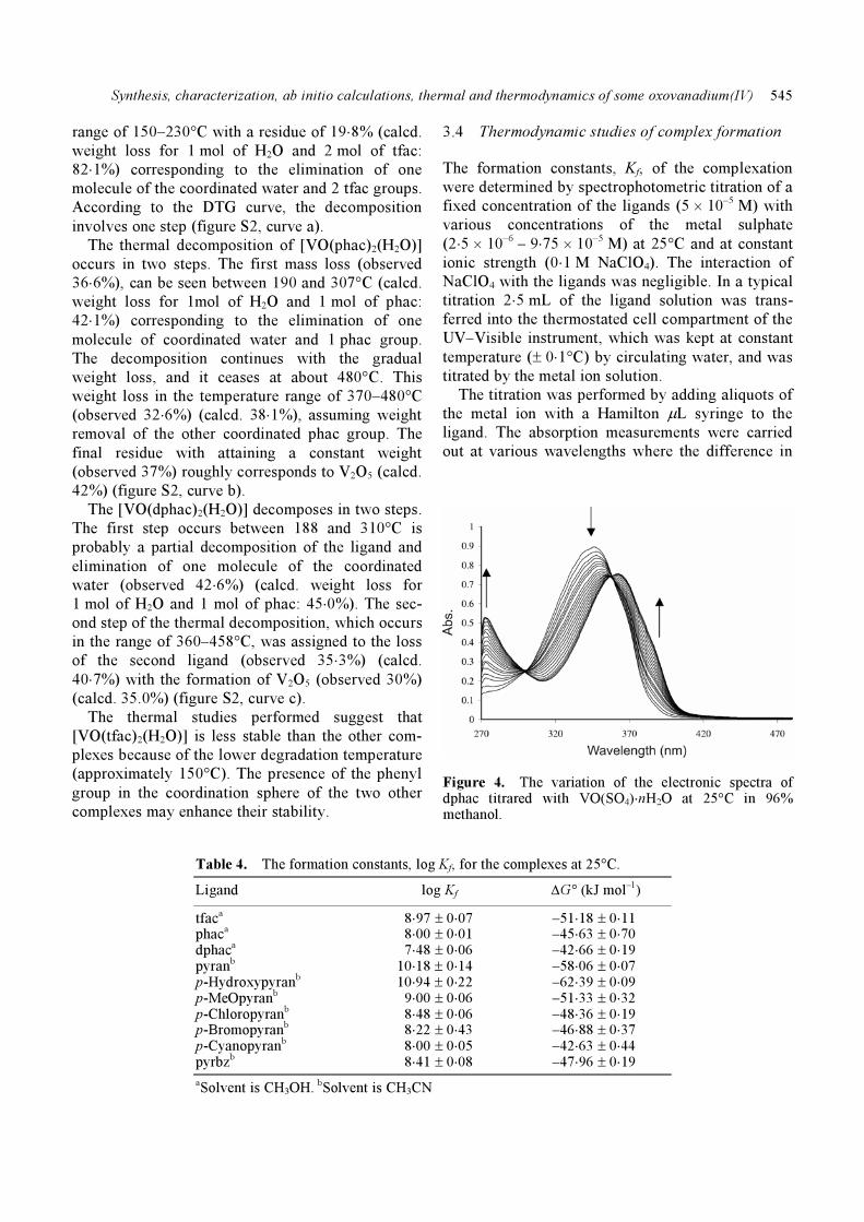
3.4 Thermodynamic studies of complex formation
The formation constants, Kf, of the complexation
were determined by spectrophotometric titration of a
fixed concentration of the ligands (5 × 10⁻⁵ M) with
various concentrations of the metal sulphate
(2⋅5 × 10–6
– 9⋅75 × 10–5
M) at 25°C and at constant
ionic strength (0⋅1 M NaClO4). The interaction of
NaClO4 with the ligands was negligible. In a typical
titration 2⋅5 mL of the ligand solution was trans-
ferred into the thermostated cell compartment of the
UV–Visible instrument, which was kept at constant
temperature (± 0⋅1°C) by circulating water, and was
titrated by the metal ion solution.
The titration was performed by adding aliquots of
the metal ion with a Hamilton μL syringe to the
ligand. The absorption measurements were carried
out at various wavelengths where the difference in
Figure 4. The variation of the electronic spectra of dphac titrared with VO(SO4)⋅nH2O at 25°C in 96% methanol.
Table 4. The formation constants, log Kf, for the complexes at 25°C.
Ligand log Kf ΔG° (kJ mol–1)
tfaca 8⋅97 ± 0⋅07 –51⋅18 ± 0⋅11 phaca 8⋅00 ± 0⋅01 –45⋅63 ± 0⋅70 dphaca
7⋅48 ± 0⋅06 –42⋅66 ± 0⋅19 pyranb 10⋅18 ± 0⋅14 –58⋅06 ± 0⋅07 p-Hydroxypyranb
10⋅94 ± 0⋅22 –62⋅39 ± 0⋅09 p-MeOpyranb 9⋅00 ± 0⋅06 –51⋅33 ± 0⋅32 p-Chloropyranb 8⋅48 ± 0⋅06 –48⋅36 ± 0⋅19 p-Bromopyranb 8⋅22 ± 0⋅43 –46⋅88 ± 0⋅37 p-Cyanopyranb 8⋅00 ± 0⋅05 –42⋅63 ± 0⋅44 pyrbzb 8⋅41 ± 0⋅08 –47⋅96 ± 0⋅19
aSolvent is CH3OH. bSolvent is CH3CN
Mozaffar Asadi et al
546
Appendix 1. The structure of the (a) cis-[VO(phac)2(H2O)] complex, (b) trans-[VO(phac)2(H2O)] com-plex and (c) trans-[VO(phac)2(H2O)] (O and H2O) complex.
absorption was the maximum after equilibrium. The
formed complex shows different absorption from the
free ligand, while the metal ion solution shows no
absorption at those wavelengths. As an example, the
variation of the electronic spectra for dfac, titrated
with various concentrations of VO(SO4)⋅nH2O at
25°C in MeOH is shown in figure 4. The same pro-
cedure was followed for all other systems. The elec-
tronic spectra of the formed complexes were the
same as the electronic spectra of the separately syn-
thesized complexes.
The formation constants, Kf, were calculated using
SQUAD computer program,36
designed to calculate
the best values for the formation constants of the pro-posed equation model (1) by employing a non-linear,
least-squares approach. Also, the free energy change
ΔG° values of the formed complexes were calculated
from ΔG° = –RT lnKf at 25°C (see table 4).
2HL + VO(SO4)⋅nH2O
[VO(L)2]⋅nH2O + SO24
– + 2H
+. (1)
Synthesis, characterization, ab initio calculations, thermal and thermodynamics of some oxovanadium(IV)
547
The results in table 4 show that for VO(O)4-type
complexes, the steric character of the ligands pre-
dominates the electronic factor toward VO(IV) cen-
ter. Concerning this effect, tfac has more tendency
to bind with VO(IV) center than phac and dphac.
As far as VO(N)4-type complexes are concerned,
the electron withdrawing functional groups such as
bromo, chloro and cyano make the Schiff base as a
poor donor ligand toward VO(IV) center. On the
other hand, the electron donating groups such as
hydroxy or methoxy make the ligand more potent
donor toward the VO(IV) center, although the steric
effect of the substitution cannot be ruled out. There-
fore, p-Hydroxypyran, p-MeOpyran and pyran have
the highest tendency toward VO(IV).
4. Conclusion
The structural, geometrical, thermal, and thermo-
dynamic properties of the oxovanadium(IV) com-
plexes have been investigated. According to the
thermodynamic studies, the formation constants of
the complexes depend upon the steric and the elec-
tronic characteristic of the ligands. But as the steric
character predominating, the tendency to bind with
VO(IV) center decrease. Moreover, the electrical
feature of each complex has an important role on the
thermodynamic and the thermal properties. It is evi-
dent that there is a close relationship between these
various properties. Charge distribution and the
geometry are effective factors affecting the proper-
ties of the different complexes. The way that ther-
mal properties change is the same as the variation of
the QSAR properties for VO(O4)-type complexes.
Acknowledgements
We thank Shiraz University Research Council for
financial support.
Supplementary information
For supplementary information see www.ias.ac.in/
chemsci website.
References
1. Mohammadi K, Thompson K H, Patrick B O, Storr T, Martins C, Polishchuk E, Yuen V G, McNeill J H and Orvig C 2005 J. Inorg. Biochem. 99 2217
2. Crans D C 2000 J. Inorg. Biochem. 80 123 3. Maurya R C and Rajput S 2004 J. Molecular Struct.
687 34
4. Plass W 2003 Coord. Chem. Rev. 237 205 5. Rehder D 2003 Inorg. Chem. Commun. 6 604 6. Aranha P E, Souza. M, Romera S, Ramos L A, San-
tos M, Dockal E and Cavalheiro E T G 2007 Thermo-chim. Acta 453 9
7. Ahmad S, Isab A A, Ali S and Al-Arfaj A R 2006 Polyhedron 25 1633
8. Posner B I, Shaver A and Fantus I G 1990 in New antidiabetic drugs (eds) C J Bailey and P R Flatt (Gordon London S), p. 107
9. Posner B I, Faure R, Burgess J W, Bevan A P, Lachance D, Zhang-Sun G, Fantus I G, Hall D A, Lum B S and Shaver A 1994 J. Biol. Chem. 269 4596
10. Shaver A, Hall D A, Ng J B, Lebuis A M, Hynes R C and Posner B I 1995 Inorg. Chim. Acta 229 253
11. Shaver A, Ng J B, Hall D A, Soo Lum B and Posner B I 1993 Inorg. Chem. 32 3109
12. Kadota S, Fantus I G, Deragon G, Guyda H J, Hersh B and Posner B I 1987 Biochem. Biophys. Res. Com-mun. 147 259
13. McNeill J H, Yuen V G, Hoveyda H R and Orvig C 1992 J. Med. Chem. 35 1489
14. Caravan P, Gelmini L, Glover N, Herring F G, Li H, McNeill J H, Rettig S J, Setyawati I A, Shuter E, Sun Y, Tracey A S, Yuen V G and Orvig C 1995 J. Am. Chem. Soc. 117 12759
15. Li J, Elberg G, Crans D C and Shechter Y 1996 Bio-chem. 35 8314
16. Morgan G T and Moss H W 1914 J. Chem. Soc. 103 78 17. Kaneda K, Jitsukawa K, Itoh T and Teranishi S 1980
J. Org. Chem. 45 3004 18. Hirao T 1997 Chem. Rev. 97 2707 19. Wender P A, Rice K D and Schnute M E 1997 J. Am.
Chem. Soc. 119 7897 20. Taguchi H, Isobe K, Nakamura Y and Kawaguchi S
1975 Chem. Lett. 757 21. Selbin J, Manning H R and Cessac G 1963 J. Inorg.
Nucl. Chem. 2 1253 22. Atherton N M, Gibbon P J and Shohoji M C B 1982
J. Chem. Soc. Dalton Trans 2289 23. Dodge R P, Templeton D H and Zalkin A 1961
J. Chem. Phys. 35 55 24. Amin S, Cryer K, Zhang B, Dutta S, Eaton S, Ander-
son O, Miller S and Crans D 2000 Inorg. Chem. 39 406
25. Frisch M J, Trucks G W, Schlegel H B, Scuseria G E, Robb M A, Cheeseman J R, Montgomery J A, Kudin K N, Burant J C, Millam J M, Iyengar S S, Tomasi J, Barone V, Mennucci B, Cossi M, Scalmani G, Rega N, Petersson G A, Nakatsuji H, Hada M, Ehara M, Toyota K, Fukuda R, Hasegawa J, Ishida M, Naka-jima T, Honda Y, Kitao O, Nakai H, Klene M, Li X, Knox J E, Hratchian H P, Cross J B, Adamo C, Jaramillo J, Gomperts R, Stratmann R E, Yazyev O, Austin A J, Cammi R, Pomelli C, Ochterski J W, Ayala P Y, Morokuma K, Voth G A, Salvador P, Dannenberg J J, Zakrzewski V G, Dapprich S, Daniels A D, Strain M C, Farkas O, Malick D K, Rabuck A D, Raghavachari K, Foresman J B, Ortiz J V, Cui Q, Baboul A G, Clifford S, Cioslowski J, Ste-fanov B B, Liu G, Liashenko A, Piskorz P,

Mozaffar Asadi et al
548
Komaromi I, Martin R L, Fox D J, Keith T, Al-Laham M A, Peng C Y, Nanayakkara A, Challa-combe M, Gill P M W, Johnson B, Chen W, Wong M W, Gonzalez C and Pople J A 2003 (Pittsburgh PA: Gaussian, Inc)
26. Ghose A K, Pritchett A and Crippen G M 1994 J. Comput. Chem. 9 80
27. Rogers D and Hopfinger A J 1994 J. Chem. Inf. Comput. Sci. 34 854
28. Bodor N, Gabanyi Z and Wong C 1989 J. Am. Chem. Soc. 111 3783
29. Ooi T, Oobatake M, Nemethy G and Scheraga H A 1987 Proc. Natl. Acad. Sci. USA 84 3086
30. Viswanadhan V N, Ghose A K, Revankar G N and Robins R K 1989 J. Chem. Inf. Comput. Sci. 29 163
31. Ghose A K and Crippen GM 1987 J. Chem. Inf. Comput. Sci. 27 21
32. Selwood P W 1956 Magnetochemistry (New York: Interscience)
33. Symal A 1975 Coord. Chem. Rev. 21 309 34. Selbin J 1966 Coord. Chem. Rev. 1 293 35. Abdel-Ghani N T and Sherif O E 1989 Thermochim.
Acta 156 69 36. Leggett D L 1985 Computational methods for the
determination of formation constant (New York: Plenum Press)