Bioleaching of copper- and zinc-bearing ore using consortia of ...
Upload
independentCategory
view
5download
0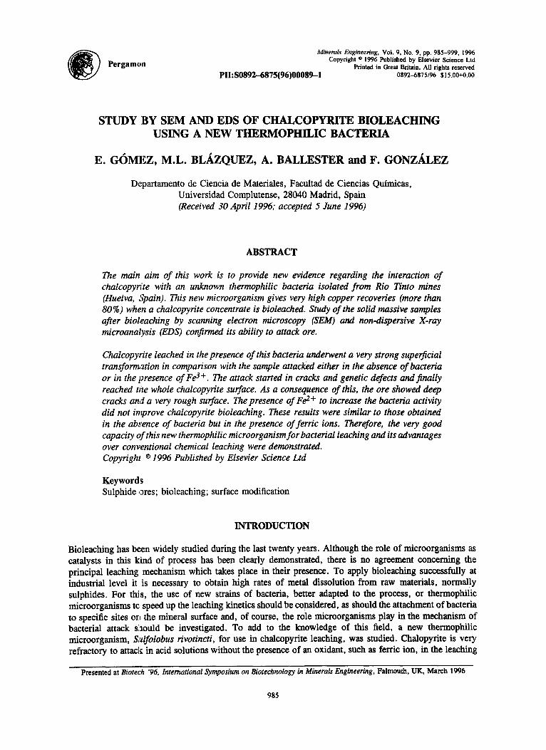
Pergamon Minerals Engineering, Vol. 9, No. 9, pp. 985-999, 1996
Copyright © 1996 Published by Elsevier Science Ltd Printed in Great Britain. All rights reserved
Plh S0892-6875(96)00089--1 0892-6875/96 $15.00+0.00
STUDY :BY SEM AND EDS OF CHALCOPYRITE BIOLEACHING USING A NEW TItERMOPHILIC BACTERIA
E. GOMEZ, M.L. BL/LZQUEZ, A. BALLESTER and F. GONZALEZ
Departamento de Ciencia de Materiales, Facultad de Ciencias Quimicas, Universidad Complutense, 28040 Madrid, Spain (Received 30 April 1996; accepted 5 June 1996)
ABSTRACT
The main aim of this work is to provide new evidence regarding the interaction of chalcopy~!te with an unknown thermophilic bacteria isolated from Rio Tinto mines (Huelva, Spain). This new microorganism gives very high copper recoveries (more than 80%) when a chalcopyrite concentrate is bioleached. Study of the solid massive samples after bioleaching by scanning electron microscopy (SEM) and non-dispersive X-ray microanalysis (EDS) confirmed its ability to attack ore.
Chalcopyrite leached in the presence of this bacteria underwent a very strong superficial transformation in comparison with the sample attacked either in the absence of bacteria or in the presence of Fe 3+. The attack started in cracks and genetic defects and finally reached the whole chalcopyrite surface. As a consequence of this, the ore showed deep cracks and a very rough surface. The presence of Fe z + to increase the bacteria activity did not improve chalcopyrite bioleaching. These results were similar to those obtained in the absence of bacteria but in the presence of ferric ions. Therefore, the very good capacity ~f this new thermophilic microorganism for bacterial leaching and its advantages over conventional chemical leaching were demonstrated. Copyright © 1996 Published by Elsevier Science Ltd
Keywords Sulphide ores; bioleaching; surface modification
INTRODUCTION
Bioleaching has be:en widely studied during the last twenty years. Although the role of microorganisms as catalysts in this kind of process has been clearly demonstrated, there is no agreement concerning the principal leaching mechanism which takes place in their presence. To apply bioleaching successfully at industrial level it is necessary to obtain high rates of metal dissolution from raw materials, normally sulphides. For this, the use of new strains of bacteria, better adapted to the process, or thermophilic microorganisms to, speed up the leaching kinetics should be considered, as should the attachment of bacteria to specific sites orL the mineral surface and, of course, the role microorganisms play in the mechanism of bacterial attack slaould be investigated. To add to the knowledge of this field, a new thermophilic microorganism, S~,~lfolobus rivotincti, for use in chalcopyrite leaching, was studied. Chalopyrite is very refractory to attac]~ in acid solutions without the presence of an oxidant, such as ferric ion, in the leaching
Presented at Biotech '96, International Symposium on Biotechnology in Minerals Engineering, Falmouth, UK, March 1996
985

986 E. G6mez et al.
medium. However, bacterial leaching can resolve this problem by using mesophilic or thermophilic microorganisms, the latter being more efficient than the former. Sulfolobus rivotincti is a new archaebacteria which is of great technological interest due to its high leaching capacity on metallic sulphide minerals. It was isolated from a mine site and is perfectly adapted to very acidic environments with high concentrations of metallic cations. The metabolism of the new isolate is strictly aerobic and chemolithotrophic. In the presence of minerals, a large percentage of the cell population is attached to the mineral surface. All the above characteristics make the new isolate very interesting for leaching processes.
MATERIALS AND METHODS
Bacterial culture. A strain of Sulfolobus rivotincti, a thermoacidophilic archaebacteria was used. This strain, which was isolated from Rio Tinto mines (Huelva, Spain), can obtain energy from the oxidation of both Fe 2+ to Fe 3 + and several mineral sulphides, including chalcopyrite. The bacterial culture, from which all the tests were inoculated, was kept growing on copper concentrates as energy source, in iron free 9K medium [1] at pH 1.8 and 68°C. These conditions were optimum for culture growth, and therefore were employed during all the experiments. Inoculum for the experiments was previously harvested by centrifugation of a culture grown in those conditions. Cells were re-suspended in 1 mi of 9K medium and divided in equal proportions in all flasks.
Mineral samples. For the tests with solid massive samples, chalcopyrite from South Africa was used. This was characterised by X-ray diffraction, which proved the major presence of chalcopyrite and small amounts of silica, aluminium, iron and magnesium silicates, and pyrite. Chemical analysis of the samples showed the following composition: Cu: 21.60%; Fe: 20.93%; Zn: 0.03%; S: 26.24%. For bioleaching tests, a chalcopyrite concentrate from Rio Tinto mines, with a particle size of below 74/zm, was used. Chemical analysis showed the main metallic elements to be: Cu: 27.63%; Fe: 29.70%; Zn: 0.15%; Pb: 0.03%.
Once the mineral was characterised, massive samples of chalcopyrite were prepared by inclusion of small pieces of mineral (between 0.5 and 0.7 cm diameter) in a Estractil AL-100 resin. Thereafter, the samples were ground on special papers (numbers 100, 400,600 and 1200), and polished on alumina and diamond paste. The samples, prior to study by SEM, were metalized. Once the samples had been observed by SEM, and before bacterial attack, this metal layer was eliminated.
Electron microscopy. SEM and microanalysis by EDS allowed observation of the morphological evolution of the samples during attack and analysis of the surface to compare the chalcopyrite structures submitted to different attack conditions. The observations by SEM were carried out with two different microscopes: Jeol JSM-35C and Jeol JSM-6400, the latter equipped with an analytical detector from Link.
Experimental methods. 100 ml of 9K medium without iron (pH 1.8) were .introduced in 250 mi shake flasks to which the corresponding inoculum was added. The temperature was 68°C and stirring speed, in all cases, 150 rpm. A chalcopyrite sample was introduced into each flask, attached to the stopper by a plastic bridle (Figure 1) to maintain it in a vertical position (to avoid reaction precipitates on sample surfaces). The length of time needed for bacteria to attack powdered mineral in this kind of bioleaching test is usually about 10-15 days, before a plateau in the dissolution kinetic curve is reached. However, as the mineral was included in an inert resin, its free surface and reactivity decrease and so the reaction time must be longer. Thus a time of 32 days was fixed for the experiments. Culture conditions were adjusted as previously described for the optimal growth of microorganisms. Other experiments were prepared under the same conditions, also with massive chalcopyrite samples, although they were withdrawn from the shake flasks at 6 and 13 days to verify the evolution of the mineral at intermediate times. All the inoculated experiments had their corresponding sterile control tests prepared under the same conditions.
As a complement to these experiments, a further two were prepared (one with bacteria and Fe 2+, and another without bacteria but with Fe 3+) to evaluate the chemical attack produced by the ferric iron. In both cases the iron was added as sulphate and the concentration was 3 % (w/v). Comparison of these two

Study by SEM and EDS of chalcopyrite bioleaching 987
experiments allowed study of the prevailing attack mechanism in the presence of the Sulfolobus rivotincti culture.
PLASTIC BRIDLg
MIDIUM WITHOtff IRON
CHAI~OPYRn~
Fig. 1 Experimental device for the massive sample tests
During the experinaentation period, liquid samples of each test were taken at different times, and the bacterial population was counted with a Thoma chamber, pH and Eh were measured with a glass electrode and with an Ag/AgCI reference electrode (+207mV vs SHE at 25°C), respectively. Fe 2+ was measured according to the method of [2]. Atomic absorption spectrophotometry was used to measure other metals in solution (total Fe and Cu2+). In tests where iron was added, the precipitates appearing on the solid were analysed by X-ray diffraction. Bioleaching tests on powdered mineral were carried out at the same temperature, pH and stirring conditions. A chalcopyrite concentrate (1% pulp density), rather than massive samples, was used as energy source.
RESULTS AND DISCUSSION
Bioleaehing of the chalcopyrite concentrate
Sulfolobus rivotinciri is an archaebacteria isolated from Rio Tinto mines. This microorgansim showed great efficiency in copper leaching in the tests carried out with chalcopyrite during 11 days (Figure 2). When sterile and inoculated tests were compared, important differences regarding metallic extraction were evident, confirming the potential of this microorganism for application to commercial processes. These differences would also be considerable if massive samples were leached.
Massive sample bioleaching
These tests were carried out to determine the characteristics of the attack and the interactions between Sulfolobus rivotincti and chalcopyrite. In the first experiments, a study was performed removing a massive sample both from ~the inoculated test and from the sterile test at different times (0, 6, 13 and 32 days), to follow the evoluti(m of the attack on the mineral surface.
Massive samples before attacking. Initially, the massive samples did not show many defects on their surface, which was quite uniform, with few cracks and pits, although they did contain some zones with triangular defects characteristic of chalcopyrite. EDS analysis (Figure 3) of massive sample surfaces revealed inclusions of silica, silico-aluminates and magnesium silicates.

988 E. G6mez e t al.
% C a
I --'4,--R~.ellce
0 50 100 150 200 250 300
100
80
60
40
20
0
Fig.2 Copper efficiency (%) from bioleaching of a chalcopyrite concemrate by Sulfolobus rivotincti
X-RAY: 0 - 20 keV Live: lOOs Preset : lOOs Remaining: Rear: 117s 157. Dead.
Os
<s" 4K HEMI :
C R R
10.363 keU oh 528= 51 e t
Fig.3 Analysis by EDS of chalcopyrite surface. Analysis showed peaks corresponding to the main elements of chalcopyrite as well as very intense peaks of silicon, aluminium and magnesium. Gold peaks
were due to the previous metallization of the sample
Massive samples attacked for 6 days. The first samples were studied after 6 days of attack. In the inoculated test, several pits appeared on the surface and rounded particles, possibly of elemental sulphur and jarosites, began to appear. Elemental sulphur is a product of the chemical attack of chalcopyrite:
CuFeS 2 + 4Fe 3+ -,. Cu 2+ + 5Fe 2+ + 2S (1)
and can therefore appear in slightly attacked zones of the massive samples. Sulphur can be detached from

Study by SEM and EDS of chaleopyrite bioleaehing 989
these areas and be deposited in other areas by agitation. Preferential attack of chaleopyrite was observed in some areas where silica remained close to the surface with respect to the chalcopyrite phase (Figure 4). EDS analysis showexl the presence of iron phosphate precipitates on the mineral surface (Figure 5) produced by the reaction of the iron dissolved from the mineral with the 9K medium, which is very rich in salts. This surface was not extensively attacked, possibly because the bacteria found it difficult to grow in the culture conditions used. No microorganisms were observed attached to the mineral, despite the references of some authors, who indicate that bacteria can appear attached to the samples prepared in similar conditions to those used [3,4].
Fig.4 Chalcopyrite surface showing different planes of leaching by preferential attack of mineral
Fig.5 Cracks covered by solid particles of iron phosphate
Attack in the sterile., test was less intense than in the inoeulatedl test even in the zones of the sample surface that originally contained defects. Also sulphur particles were ngted on the clean surface (Figure 6). In some areas, an attack along longitudinal cracks (Figure 7) crossed by other smaller ones was produced. This type of surface deterioration corresponds to chemical attack, as opposed to bacterial attack which produces more irregular cracks [511. i
i
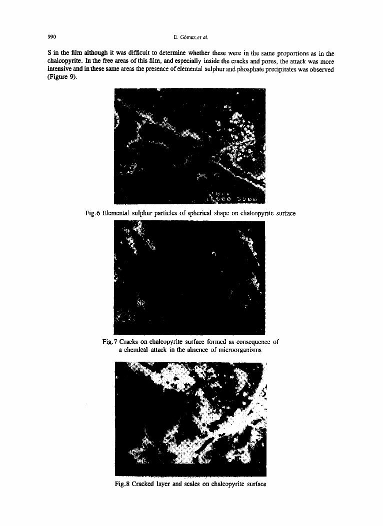
Massive samples attacked for 13 days. After 13 days of attack, two more massive samples were removed from the flasks and studied, The specimen corresponding to the inoculated test had a very cracked surface and a greater presence of precipitates than the previous sample attacked during 6 days. Zones in which preferential attack was produced compared with neighbouriag areas could be observed. On part of the sample surface a p~mially raised and cracked thin film was detected (Figure 8) which could correspond to a passive layer hindering the mineral attack. Analysis by EDS only revealed the presence of Fe, Cu and
990 E. G6mezet al.
S in the film although it was difficult to determine whether these were in the same proportions as in the chaleopydte. In the free areas of this film, and especially inside the cracks and pores, the attack was more intensive and in these same areas the presence of elemental sulphur and phosphate precipitates was observed (Figure 9).
Fig.6 Elemental sulphur particles of spherical shape on chalcopyrite surface
Fig.7 Cracks on chalcopyrite surface formed as consequence of a chemical attack in the absence of microorganisms
Fig.8 Cracked layer and scales on chalcopyrite surface

Study by SEM and EDS of chaleopyrite bioleaching 991
Fig.9 Iron phosphate and elemental sulphur precipitates inside superficial chalcopyrite defects
In the sterile test, the attack on the sample surface was milder and there was a great amount of precipitates. The same partially cracked film observed in the inoculated tes t was detected in some zones of the massive sample attacked in the absence of bacteria. This indicates that its formation probably ocurred through a chemical phenomenon independent of the presence of bacteria. A similar film has been observed on chalcopyrite surfaces inoculated with the same microorganism after voltametry tests [6]. These authors concluded that this layer was a diffusion barrier for the chalCopyrite leaching, which had a tendency to crack, forming solitd products in the form of scales on the ~ e r a l surface.
Massive samples attacked for 32 days. Finally, the last two massive samples attacked over a period of 32 days were studi.ed. There were fewer cells in the culturei which suggested that the microorganisms experienced growth difficulties in the experimental conditions. The massive samples were attacked very strongly in compariLson with previous samples. In addition, the attack took place according to the different crystaUisation planes of the chalcopyrite. This was in accordance with the observations of several authors, who noted that bac:terial leaching progresses in the regions Where the mineral has surface defects, or in zones of crystallographic heterogeneity [7,8]. Cracks appearedibordering and delimiting the different grains forming the mineral (Figure 10).
Fig.10 Massive chalcopyrite sample after 32 days attack in the presence of bacteria. The grain structure of sample is revealed by the strong attack
Examination of the cracked zone in more detail showed an in-depth attack not only at the interior of the fissures (as in the sample after 13 days of attack) but also on the outside edges where many pits with rounded forms were detected. These pits were similar to the shape of Sulfolobus rivotincti, although the

992 E. G6mez et al.
bacteria were not observed inside them. This is in accordance with the observations of Vargas et al. [9], who argued that the mechanism of pit formation is related to bacterial attachment to the mineral surface. In this case, it seems that the pits on the pyrite surface had the same shape as Thiobacillus ferrooxidans bacteria. According to their observations, the bacteria did not remain in the pits, which increased in size despite the absence of bacteria. Other authors that have also worked with mesophilic bacteria [ 10] indicate that the microorganisms sometimes remain close to the mineral surface and sometimes in the solution, and that they can leave this surface to reproduce in the fluid medium after a period of corrosive activity on the surface.
In our case, because of their ferrooxidising capacity, the bacteria could be in the liquid medium, obtaining their energy from the oxidation of the Fe 2+ , which might have been produced as a consequence of the chalcopyrite oxidation (reaction 1). In addition, the resulting Fe 3 + could contribute to an increase in attack on the mineral surface.
The massive sample of the sterile test had a surface which was much cleaner and less attacked than the above, the cracks and surface defects hardly being touched (Figure 11). No trace of the layer, which was observed on the sample attacked for 13 days, was observed at this attack time.
Fig. 11 Test carried out in the absence of microorganisms. Surface appears slightly attacked after 32 days
Bioleaehing of massive ehalcopyrite samples with added iron
Besides studying the effect of the microorganisms, bacterial leaching was compared with the effect that the presence of an external oxidising agent (Fe 3+ in our case) could have on chalcopyrite. For this purpose two other tests with massive samples were prepared to leach the same type of chalcopyrite. In the first, Fe 3+ but no bacteria was added, while in the second, Fe 2+ and the same amount of inoculum as used in the control test were added. The comparison between these tests and the two previous tests were intended to increase our knowledge of the leaching mechanism using Sulfolobus rivotincti, and show whether the attack was mainly chemical or biological.
If the mechanism were a purely chemical phenomenon, the attack of the samples in the presence of Fe 3 + should be greater than in the other cases, since from the very first moment the oxidising agent is present in the medium. However, if microorganisms act as catalysts of the oxidation of Fe 2 + to Fe 3 +, the indirect mechanism could be the most important, and so massive samples leached with either Fe 2 + or Fe 3 + would show similar signs of attack, which could be more pronounced than when only bacteria are used. Finally, if a direct mechanism is the most important, the sample attacked with bacteria, but in the absence of iron, could show greater deterioration, since in the other inoculated test the bacteria had a more easily available energy source(Fe2+); in this case the microorganisms could obtain their energy from the oxidation of iron and not from oxidation of the chalcopyrite, which is attacked with difficulty.

Study by SEM and EDS of chalcopyrite bioleaching 993
In the experiments using iron, jarosite precipitation was evident from the outset. The 9K medium used contains great amounts of ammonium and potassium salts. When iron was added as ferric ion, these salts reacted to form jarosites. At the pH used in these experiments (1.8), ferric-hydroxisalts precipitated quickly. The main reason for using this medium and this pH was that these conditions are optimal for S. rivotincti growth. So other, more diluted media, at lower pH to decrease the possibility of jarosite formation, were rejected. When iron, as ferrous salt, was added in tests inoculated with S. Rivotincti, Fe 2+ was quickly oxidised to Fe 3+ by bacterial action and then precipitated. The high testing temperatute also contributed appreciably to jarosite formation. Moreover, j arosite precipitation on the surface of the massive samples hindered the mineral leaching in both cases. In accordance with these results, experiments carded out by other authors [11] in the presence of thermophilic bacteria of genus Acidianus and Sulfolobus showed that nutrient media rich in potassium and ammonium Salts decrease the rates of iron solubilization from pyrite and increase jarosite formation.
Attack of massive samples with added Fe 3+. The massive samples leached with Fe 3+ in a sterile medium, after 32 clays testing, remained totally covered by a thick layer ofjarosite. In the bottom of the shake flask used for the test the presence of an abundant and very fine orange precipitate was detected, which was mainly composed of potassium and ammonium jarosites (Figure 12).
Int~asity
1~0 f
A3
AJ
I n I
10 20
r J . ~
AJ
I , I I n n
:30 40 $0 60 70 2e
Fig.12 X-ray diffraction pattern of precipitate detected on chalcopyrite sample attacked with Fe3+: (KJ) potassium jarosites; (AJ) ammonium jarosites
To study the surface of the chalcopyrite after attack, the sample was partially cleaned ultrasonically which easily eliminated much of the jarosite layer on the chalcopyrite surface.
Once the surface was cleaned, it could be seen that the sample had been attacked to a lesser extent than the sample leachedL without iron but with bacteria. The precipitates were very abundant, and EDS analysis revealed that they were formed mainly of elemental sulphur and jarosites. Leaching of chalcopyrite with ferric sulphate in -'~id solutions produces elemental sulphur according to reaction (1). This sulphur covers the surface, and acts as a barrier to electron transport and to the diffusion of reactants and products between the solid phase and the solution [12,13]. Observation of the sample by SEM showed some pits, particularly in areas with cracks and defects, as well as parallel cracks, that, as mentioned previously, are typical of a chemical attack which leaches the mineral across the defects of the crystalline lattice of the chalcopyrite (Figure 13).
HE 9:94;

994 E. G6mez et al.
Fig.13 Cracks formed by chemical attack. Test with Fe 3+ as oxidising agent (32 days of leaching)
Attack of massive samples in the presence of Fe 2+ . Massive samples attacked with Fe 2+ and bacteria also produced a great amount ofjarosites precipitated on the surface. As in the previous case, the sample surface had to be partially ultrasonically cleaned before SEM observation.
In this massive sample, jarosite formation was very regular. Once the surface was clean, the attack on chalcopyrite was easily observed. The pits were very regular and several planes of attack were observed (Figure 14). The attack was similar or slightly greater than that obtained with Fe 3+, but without bacteria. Part of the sample surface was unattacked and the remaining surface was damaged through defects of the mineral crystals. The role of the bacteria was limited to the rapid oxidation of iron in solution, the resultant cation (Fe 3+) attacking the mineral. Furthermore, it was observed that the precipitates were mainly jarosites, unlike in the tests with ferric ion where a high amount of elemental sulphur was detected on massive samples, which is in agreement with the results obtained by Keller and Murr [5].
Fig. 14 Regular pits produced on massive sample surface attacked with Fe 2+ and microorganisms. Great abundance of jarosite precipitates on surface are observed. A preferential attack
through different planes is noted after 32 days of bioleaching
The high concentration of Fe 3+ in the solution of these two tests, which favours jarosite formation and whose presence was confirmed by X-ray diffraction, had another effect. Protons were generated during the jarosites precipitation, in accordance with the following reaction:
3Fe 3+ + X + + 2HSO 4 + 6H20 ,,,* XFe3(SO4)2(OH)6 + 8H + X = K+,Na+,NH4+,H + (2)
and this could have some influence on the pH of the leaching medium, which can be described using Figure

Study by SEM and EDS of chalcopyrite bioleaehing 995
15. It can be seen how, in the two tests in which there was iron in solution, the pH decreased more than in the experiments carried out in the absence of iron. At the same time, the decrease was more pronounced in the test with Fe 3 + than in the other test.
2
1.9
1.8
1,7
1,6
X Sterile • Inoculated • Fe (3+) @ Fe (2+) inoculated
"
1,2
1,1
1 1 I I 0 10 20 30
t ime (days)
I
4O
Fig. 15 pH evolution in the different tests with massive samples
In the tests withc,ut iron (the sterile as well as the inoculated), pH remained practically stable. In the inoculated test, the bacteria did not form sulphuric acid in sufficient amounts to decrease the pH, as occurred in the leaching tests with powdered mineral, due to the small amount of mineral from the massive samples that were attacked.
The initial decrease in pH in the tests carried out in the presence of iron could favour chemical attack of the chalcopyrite, but this mineral is difficult to attack in acid solutions and the precipitation ofjarosites on its surface hinders the oxidation process.
When the potential of the solution in the different tests was analysed (Figure 16), it was observed that the media with iron had noticeably higher oxidising potentials. The sterile test maintained a constant potential of around 0.4 V. At this potential, in cyclic voltametry assays carried out by other authors [14], it was observed that the surface of the chalcopyrite was not attacked. These same authors found that at potentials between 0.45 and 0.5 V, the attack was strong both with bacteria and without bacteria. In addition, when bacteria were used, a certain roughness and an incipient deterioration appeared on the surface, as happened in our inoculated test. Finally, at high potentials, such as those detected in the test with Fe 3+ (around 0.6V) but without bacteria, these authors affirm that an elemental sulphur or iron phosphate layer can appear. In our case this was not observed, due to the abundant jarosite precipitation on the mineral surface. EDS analysis indicated the presence of elemental sulphur and, of course, iron compounds as solid reaction products.
In the case of the test with Fe 2 +, where the potential appreciably increased from the beginning because of the almost total oxidation of ferrous iron to ferric iron, the situation was very similar to that previously mentioned. Natarajan [15] observed that for an oxidising potential higher than 0.55 V the growth of the bacteria is poor if there is not enough Fe 2+ in the system. In our case, this is what probably occurred from the eighth day onwards, since most of the iron appeared as Fe 3+.

996 E. G6mez et al.
700
J ~
600
500
400
300
200
100
• Fe (3+)
• Fe (2+) inoculated
• Inoculated
• Sterile
0 I I I
0 10 20 30 t ime (days)
40
Fig. 16 Redox potential evolution in all the tests
A cell recount showed that the growth of the bacterial population (Figure 17), in the test with Fe 2+, decreased, while in the inoculated test without iron addition, the decrease was less marked although the microorganisms had no additional source of energy appart from the chalcopyrite immobilized in the resin.
1,E+09
1,E+O8 ~.o. ........................ ,.~, ........... , .................. A. .......... A . . . . . . . . •
1,E+0~
I,E+05
• S. rfvotincfl" wi thout i ron
• S. rivotinea +Fe (11) I I t ' I I i
0 5 10 15 20 25 30 35 time (days)
Fig. 17 Bacterial population growth in inoculated tests (with and without Fe 2+
Initially, the number of cells decreased in the two cultures, probably due to the attachment of the bacteria to the chalcopyrite surface. For other iron oxidising bacteria such as ThiobaciUus ferrooxidans, cell attachment to the mineral can comprise 90% of the initial population [16]. The decrease was initially smaller in the test with ferrous iron, because the bacteria found an energy source in the liquid medium, which was more accessible than the chalcopyrite from the massive sample. However, due to the high temperature and the rapid oxidation produced by the bacteria themselves, the amount of Fe 2+ in the
Study by SEM and EDS of chalcopyrite bioleaching 997
medium began to fall (Figure 18), almost disappearing after 8 days, the concentration of Fe 3+ thus increasing. The source of available energy in the medium decreased, while concentration of a cati6n such as Fe 3+, which c~m be toxic at high concentration, increased. Additionally, strong jarosite precipitation took place, covering the whole surface of the massive sample and hindering the access of the bacteria to the other energy source present in the test. Because of all these facts, the number of bacteria appreciablly decreased in this test. In the experiment with Fe 3+ , there was an almost imperceptible increase of Fe 2+ concentration as a result of reaction (1).
+
30
25
20
3,5
3,0
2,5
2,0
1,5 ...........
1,0
0,5
0,0
-- Fe (3+)
A Fe(2+) inoculated
m _ i ~ - m
20 40
time (days)
Fig.18 Concentration of Fe 2+ in experiments with external addition of iron
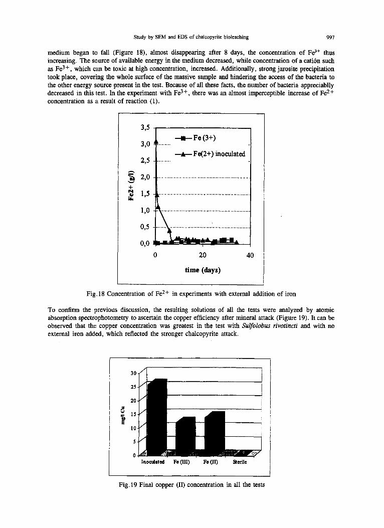
To confirm the previous discussion, the resulting solutions of all the tests were analyzed by atomic absorption spectrophotometry to ascertain the copper efficiency after mineral attack (Figure 19). It can be observed that the copper concentration was greatest in the test with Sulfolobus rivotincti and with no external iron added, which reflected the stronger chalcopyrite attack.
I0
5
0 Inoculated Fe (III) Fe (II) Sterile
Fig. 19 Final copper (II) concentration in all the tests

998 E. G6mez et al.
CONCLUSIONS
Comparison of the different experiments showed that the greatest attack took place in the inoculated test without added iron, which indicates that Sulfolobus rivotincti acted mainly through a direct bioleaching mechanism in the tested work conditions. As a consequence of the attack on the chalcopyrite, Fe 2 + was produced. This was later oxidised to Fe 3 +, thus increasing the attack through an indirect mechanism. The simultaneous actuation of both mechanisms resulted in higher copper dissolution rates in this test than in the other.
The external addition of Fe 2+, to evaluate the attack by the indirect mechanism, resulted in little deterioration due to the rapid oxidation of ferrous ion and the subsequent precipitation of ferric ion as jarosites which covered the chalcopyrite surface. The bacteria, in this case, limited themselves to rapidly oxidizing the iron in solution, ignoring the chalcopyrite as energy source. For this reason, any leaching in this experiment was basically chemical. This indicates the importance of not providing alternative and more accesible sources of energy for the microorganisms other than the substrate that is oxidised, in our case, chalcopyrite.
The use of experimental conditions similar to the optimum growth conditions for the microorganism (9K medium very rich in salts at pH 1.8) produced strong jarosite precipitation in the tests carried out in the presence of iron. The thickish layer ofjarosites hindered the bacterial attack of the samples in the test with Fe 2+, and the chemical leaching in the test with Fe 3+ . Therefore, for subsequent research purposes, it is probably necessary to use more diluted media with lower pH, to avoid the precipitation of reaction products.
As a final conclusion, we hope to have confirmed the capacity of this new microorganism for application to commercial bioleaching processes because of the high efficiencies reached in the bioleaching of chalcopyrite in the presence of bacteria alone.
ACKNOWLEDGEMENT
The work was supported by a grant from CICYT Spain (BIO94-0733-C03-03).
.
.
.
.
5.
6.
.
REFERENCES
Silverman, M.P. & Lundgren, D.G., Studies on the chemoautotrophic iron bacterium Ferrobacillusferrrooxidans I. An improved medium and a harvesting procedure for securing high cell yields. J. Bacteriol., 77, 642 (1959). Herrera, L., Ruiz, P., Aguillon, J.C. & Fehrmann, A., A new spectrophotometric method for the determination of ferrous iron in the presence of ferric iron. J. Chem. Tech. Biotechnol., 44, 171 (1988). Ostrowsky, M. & Sklodowska, A., The use of scanning electron microscopy and an electron microprobe in studies on oxidation of sulfur compounds by bacteria and bioleaching of heavy metals. FEMS Microbiol. Letts., 82, 27 (1991). Mustin, C., Berthelin, J., Marion, P. & De Donato, P., Corrosion and electrochemical oxidation of a pyrite by Thiobacillusferrooxidans. Appl. Environ. Microbiol., 58, 1175 (1992). Keller, L. & Murr, L.E., Acid-Bacterial and ferric sulfate leaching of pyrite single crystals. Biotechnology and Bioengineering. 24, 83 (1982). Mufioz, J.A., Ballester, A., Bl~tzqaez, M.L., Gonz,~ez, F. & G6mez, C., Studies on the anodic dissolution of chalcopyrite at a constant potential: Effect of a new thermophilic microorganism. In Copper'95. Vol 4:"Electrorefining and Hydrometallugy of Copper". Eds.: W.C. Cooper, D.B. Dresinger, J.E. Dutrizac, G.Ugarte. TMS, Warrendale, PA (USA). 409 (1995). Rodriguez-Leiva, M. & Tributsch, H., Morphology of bacterial leacdaing patterns by ThiobaciUus ferrooxidans on synthetic pyrite. Arch Microbiol., 149, 401 (1988).

Study by SEM and EDS of chalcopyrite bioleaching 999
.
9.
10.
11.
12.
13.
14.
15.
16.
Claasen, R., Mineralogical controls on the bacterial oxidation of refractory Barberton gold ores. FEMS Microbiology Reviews. l l , 197 (1992). Vargas, T., Wiertz, J.V., Escobar, B. & Badilla-Ohlbaum, R., The catalytic role of ThiobaciUus ferrooxidans in the leaching of pure natural chalcopyrite. EPD Congress. Eds: Gaskell, D. R. TMS, Warrendale, PA (USA). 229 (1990). Wakao, N., Mishena, M., Sakurai, Y. & Shiota, H., Bacterial pyrite oxidation Ill--Adsorption of 1". ferrooxidans cells on solid surface and its effecy on iron release from pyrite. J. Gen. Appl. Microbiol., 30, 63 (1984). Larsson, L., Olsson, G., Hoist, O. & Karlsson, H.T., Pyrite oxidation by thermophilic archaebao~ria. Appl. Environ. Microbiol., 56, 697 (1992). Tuovinen, O.H., Biological fundamentals of mineral leaching processes. In "Microbial Mineral Recovery". Eds.: H.L. Ehrlich & C.L. Briefley. McGraw-Hill. 55 (1990). Ahonen, L. & Touvinen, O.H., Alterations in surface and textures of minerals during the bacterial leaching of a complex sulphide ore. Geomicrobiology Journal. 10, 207 (1993). Muiioz, J.A., G6mez, C., Figueroa, M., Ballester, A., Gonz~ez, F. & Bl~tzquez, M., Effect of thermophilic microorganism on the electrochemical behavior of the chalcopyrite. In "BiohydrometaUurgical Processing". Vol. I. Eds.: T. Vargas, C.A. Jerez, J.V. Wiertz, H. Toledo. [I. de Chile. Chile. 67 (1995). Natarajan, K.A., Bioleaching of Sulphides under Applied Potentials. Hydrometallurgy. 29, 161 (1992). Natarajan, K.A., Electrochemical aspects of bioleaching of base-metal sulfides. In "Microbial Mineral Recovery". Eds.: H.L. Ehrlich & C.L. Brierley. McGraw-Hill. 79 (1990).
Copyright © 2022 FDOKUMEN