Structure–mutagenicity relationship of kaurenoic acid from Xylopia sericeae (Annonaceae
11
Mutation Research 701 (2010) 153–163 Contents lists available at ScienceDirect Mutation Research/Genetic Toxicology and Environmental Mutagenesis journal homepage: www.elsevier.com/locate/gentox Community address: www.elsevier.com/locate/mutres Structure–mutagenicity relationship of kaurenoic acid from Xylopia sericeae (Annonaceae) B.C. Cavalcanti a , J.R.O. Ferreira a , D.J. Moura b , R.M. Rosa c , G.V. Furtado b , R.R. Burbano d , E.R. Silveira e , M.A.S. Lima e , C.A.G. Camara f , J. Saffi b,c , J.A.P. Henriques b,c , V.S.N. Rao a , L.V. Costa-Lotufo a , M.O. Moraes a , C. Pessoa a,∗ a Departamento de Fisiologia e Farmacologia, Universidade Federal do Ceará (UFC), CEP: 60430-270, Fortaleza, CE, Brazil b Departamento de Biofísica, Centro de Biotecnologia, Universidade Federal do Rio Grande do Sul (UFRGS), CEP: 91501-970, Porto Alegre, RS, Brazil c Laboratório de Genética Toxicológica, Universidade Luterana do Brasil (ULBRA), CEP: 92420-280, Canoas, RS, Brazil d Departamento de Biologia, Universidade Federal do Pará (UFPA), CEP: 66075-900, Belém, PA, Brazil e Departamento de Química Orgânica e Inorgânica, Universidade Federal do Ceará (UFC), CEP: 60455-760, Fortaleza, CE, Brazil f Departamento de Química, Universidade Federal Rural de Pernambuco (UFRPE), CEP: 52171-030, Recife, PE, Brazil article info Article history: Received 15 September 2009 Received in revised form 17 May 2010 Accepted 14 June 2010 Available online 25 June 2010 Keywords: Kaurenoic acid DNA damage Apoptosis Genotoxicity Mutagenicity abstract Kaurane diterpenes are considered important compounds in the development of new highly effective anticancer chemotherapeutic agents. Genotoxic effects of anticancer drugs in non-tumour cells are of special significance due to the possibility that they induce secondary tumours in cancer patients. In this context, we evaluated the genotoxic and mutagenic potential of the natural diterpenoid kaurenoic acid (KA), i.e. (−)-kaur-16-en-19-oic acid, isolated from Xylopia sericeae St. Hill, using several standard in vitro and in vivo protocols (comet, chromosomal aberration, micronucleus and Saccharomyces cerevisiae assays). Also, an analysis of structure–activity relationships was performed with two natural diter- penoid compounds, 14-hydroxy-kaurane (1) and xylopic acid (2), isolated from X. sericeae, and three semi-synthetic derivatives of KA (3–5). In addition, considering the importance of the exocyclic double bond (C16) moiety as an active pharmacophore of KA cytotoxicity, we also evaluated the hydrogenated derivative of KA, (−)-kauran-19-oic acid (KAH), to determine the role of the exocyclic bond (C16) in the genotoxic activity of KA. In summary, the present study shows that KA is genotoxic and mutagenic in human peripheral blood leukocytes (PBLs), yeast (S. cerevisiae) and mice (bone marrow, liver and kidney) probably due to the generation of DNA double-strand breaks (DSB) and/or inhibition of topoisomerase I. Unlike KA, compounds 1–5 and KAH are completely devoid of genotoxic and mutagenic effects under the experimental conditions used in this study, suggesting that the exocyclic double bond (C16) moiety may be the active pharmacophore of the genetic toxicity of KA. © 2010 Elsevier B.V. All rights reserved. 1. Introduction Kaurene diterpenes have shown a number of biological proper- ties such as antibacterial activity [1], cytotoxicity against tumour cell lines [2,3], and inhibition of nuclear factor-B (NF-B) [4]. Given the anti-apoptotic properties of the NF-B pathway, kaurene diterpenes are recognized as useful candidates for new antitumour agents [5]. Many of these compounds have been shown to induce apoptosis in different cell lines; however, their molecular targets differ significantly [5–8] and show unequal levels of drug sensitivity [9]. ∗ Corresponding author at: Rua Cel Nunes de Melo, 1127, CEP: 60430-270, P.O. Box 3157, Fortaleza, Ceará, Brazil. Tel.: +55 85 3366 8255; fax: +55 85 3366 8333. E-mail address: [email protected] (C. Pessoa). ent-Kaurenic acid and many natural derivatives of this diter- pene are known to have interesting biological properties [10]. (−)-Kaur-16-en-19-oic acid (KA) is regarded as an intermediate in the biogenesis of gibberellin and other phytohormones involved in the regulation of growth and development of higher plants and some fungal metabolites [11]. A literature survey revealed a wide variety of interesting biological activities of KA, including antipar- asitic and antimicrobial effects [12–14], anti-inflammatory action [15], and cytotoxicity against human cancer cells and hemolytic effects against mouse erythrocytes [16]. The use of medicinal plant extracts has increased during the last decades in Brazil. Since there is little information available on their potential health risk, genotoxicity studies can help to determine the safety and effectiveness of herbal health products [17]. Copaiba oil of Copaifera langsdorffii is a reputed folk remedy (rich in KA) used in its natural form for the treatment of sore throat and urinary and pulmonary infections, to alleviate ulcers and to hasten wound 1383-5718/$ – see front matter © 2010 Elsevier B.V. All rights reserved. doi:10.1016/j.mrgentox.2010.06.010
-
Upload
independent -
Category
Documents
-
view
6 -
download
0
Transcript of Structure–mutagenicity relationship of kaurenoic acid from Xylopia sericeae (Annonaceae
S(
BELa
b
c
d
e
f
a
ARRAA
KKDAGM
1
tcGdaad[
B
1d
Mutation Research 701 (2010) 153–163
Contents lists available at ScienceDirect
Mutation Research/Genetic Toxicology andEnvironmental Mutagenesis
journa l homepage: www.e lsev ier .com/ locate /gentoxCommuni ty address : www.e lsev ier .com/ locate /mutres
tructure–mutagenicity relationship of kaurenoic acid from Xylopia sericeaeAnnonaceae)
.C. Cavalcanti a, J.R.O. Ferreiraa, D.J. Mourab, R.M. Rosac, G.V. Furtadob, R.R. Burbanod,
.R. Silveirae, M.A.S. Limae, C.A.G. Camaraf, J. Saffib,c, J.A.P. Henriquesb,c, V.S.N. Raoa,.V. Costa-Lotufoa, M.O. Moraesa, C. Pessoaa,∗
Departamento de Fisiologia e Farmacologia, Universidade Federal do Ceará (UFC), CEP: 60430-270, Fortaleza, CE, BrazilDepartamento de Biofísica, Centro de Biotecnologia, Universidade Federal do Rio Grande do Sul (UFRGS), CEP: 91501-970, Porto Alegre, RS, BrazilLaboratório de Genética Toxicológica, Universidade Luterana do Brasil (ULBRA), CEP: 92420-280, Canoas, RS, BrazilDepartamento de Biologia, Universidade Federal do Pará (UFPA), CEP: 66075-900, Belém, PA, BrazilDepartamento de Química Orgânica e Inorgânica, Universidade Federal do Ceará (UFC), CEP: 60455-760, Fortaleza, CE, BrazilDepartamento de Química, Universidade Federal Rural de Pernambuco (UFRPE), CEP: 52171-030, Recife, PE, Brazil
r t i c l e i n f o
rticle history:eceived 15 September 2009eceived in revised form 17 May 2010ccepted 14 June 2010vailable online 25 June 2010
eywords:aurenoic acidNA damagepoptosisenotoxicity
a b s t r a c t
Kaurane diterpenes are considered important compounds in the development of new highly effectiveanticancer chemotherapeutic agents. Genotoxic effects of anticancer drugs in non-tumour cells are ofspecial significance due to the possibility that they induce secondary tumours in cancer patients. In thiscontext, we evaluated the genotoxic and mutagenic potential of the natural diterpenoid kaurenoic acid(KA), i.e. (−)-kaur-16-en-19-oic acid, isolated from Xylopia sericeae St. Hill, using several standard invitro and in vivo protocols (comet, chromosomal aberration, micronucleus and Saccharomyces cerevisiaeassays). Also, an analysis of structure–activity relationships was performed with two natural diter-penoid compounds, 14-hydroxy-kaurane (1) and xylopic acid (2), isolated from X. sericeae, and threesemi-synthetic derivatives of KA (3–5). In addition, considering the importance of the exocyclic doublebond (C16) moiety as an active pharmacophore of KA cytotoxicity, we also evaluated the hydrogenated
utagenicity derivative of KA, (−)-kauran-19-oic acid (KAH), to determine the role of the exocyclic bond (C16) in thegenotoxic activity of KA. In summary, the present study shows that KA is genotoxic and mutagenic inhuman peripheral blood leukocytes (PBLs), yeast (S. cerevisiae) and mice (bone marrow, liver and kidney)probably due to the generation of DNA double-strand breaks (DSB) and/or inhibition of topoisomeraseI. Unlike KA, compounds 1–5 and KAH are completely devoid of genotoxic and mutagenic effects under
ons uacoph
the experimental conditimay be the active pharm
. Introduction
Kaurene diterpenes have shown a number of biological proper-ies such as antibacterial activity [1], cytotoxicity against tumourell lines [2,3], and inhibition of nuclear factor-�B (NF-�B) [4].iven the anti-apoptotic properties of the NF-�B pathway, kaureneiterpenes are recognized as useful candidates for new antitumourgents [5]. Many of these compounds have been shown to induce
poptosis in different cell lines; however, their molecular targetsiffer significantly [5–8] and show unequal levels of drug sensitivity9].∗ Corresponding author at: Rua Cel Nunes de Melo, 1127, CEP: 60430-270, P.O.ox 3157, Fortaleza, Ceará, Brazil. Tel.: +55 85 3366 8255; fax: +55 85 3366 8333.
E-mail address: [email protected] (C. Pessoa).
383-5718/$ – see front matter © 2010 Elsevier B.V. All rights reserved.oi:10.1016/j.mrgentox.2010.06.010
sed in this study, suggesting that the exocyclic double bond (C16) moietyore of the genetic toxicity of KA.
© 2010 Elsevier B.V. All rights reserved.
ent-Kaurenic acid and many natural derivatives of this diter-pene are known to have interesting biological properties [10].(−)-Kaur-16-en-19-oic acid (KA) is regarded as an intermediate inthe biogenesis of gibberellin and other phytohormones involvedin the regulation of growth and development of higher plants andsome fungal metabolites [11]. A literature survey revealed a widevariety of interesting biological activities of KA, including antipar-asitic and antimicrobial effects [12–14], anti-inflammatory action[15], and cytotoxicity against human cancer cells and hemolyticeffects against mouse erythrocytes [16].
The use of medicinal plant extracts has increased during the lastdecades in Brazil. Since there is little information available on their
potential health risk, genotoxicity studies can help to determinethe safety and effectiveness of herbal health products [17]. Copaibaoil of Copaifera langsdorffii is a reputed folk remedy (rich in KA)used in its natural form for the treatment of sore throat and urinaryand pulmonary infections, to alleviate ulcers and to hasten wound
1 ion Research 701 (2010) 153–163
hsgil[
omoespiKbatta
2
2
ataefiKMrtm
2
(1bmfafNl(Yf
2
MCmp
mlgmptt
2
rbwl
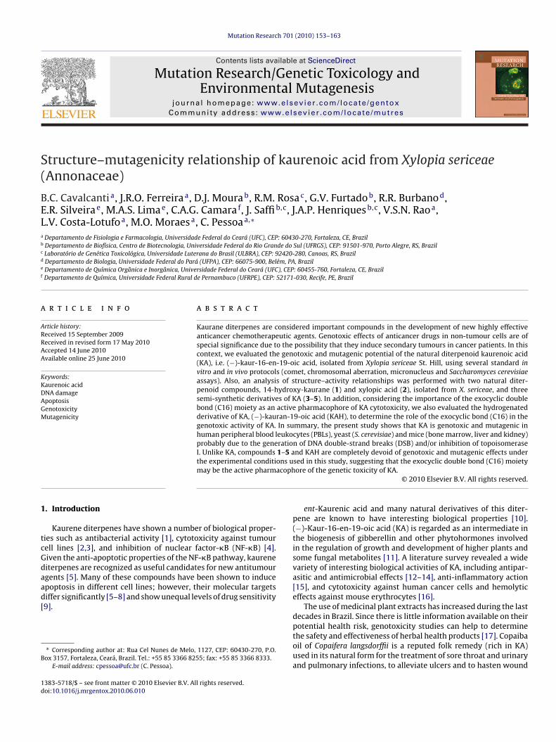
Fig. 1. Chemical structures of kauren-19-oic acid (KA), 14-hydroxy-kaurane
54 B.C. Cavalcanti et al. / Mutat
ealing [18,19], and to treat cancer [20–22]. Recently, we demon-trated that high concentrations of KA isolated from copaiba oilenerates DNA strand-breaks, as observed by the comet assay, andncreases the frequency of micronucleated cells in Chinese hamsterung fibroblasts (V79 cells), as analyzed by the micronucleus assay23].
Considering the association of DNA damage and cancer devel-pment, the aim of this study was to investigate the genotoxic andutagenic activity of KA, the main diterpene found in the roots
f Xylopia sericeae St. Hill, a tree that grows abundantly in North-ast Brazil. Also, in order to correlate genotoxicity with chemicaltructure, we analyzed the genotoxic effects of two natural diter-enoid compounds, 14-hydroxy-kaurane (1) and xylopic acid (2),
solated from X. sericeae, and three semi-synthetic derivatives ofA (3–5). Furthermore, we demonstrated that the exocyclic doubleond (C16) moiety is the active pharmacophore of KA cytotoxicitygainst cancer and non-cancer cells [24]. Therefore, we evaluatedhe hydrogenated derivative of KA, (−)-kauran-19-oic acid (KAH),o determine the role of the exocyclic bond (C16) in the genotoxicctivity of KA.
. Materials and methods
.1. Extraction of kaurenoic (KA) and hydrogenated kaurenoic (KAH) acids
Dried roots of X. sericeae St Hill (1.6 kg) were pulverized and extracted with hex-ne at room temperature. The solvent was removed under reduced pressure to givehe corresponding extract (28.5 g) as a green oil. The residue obtained after hex-ne extraction was extracted with EtOH to yield a dark brown residue (97.0 g) aftervaporation under vacuum. KA (Fig. 1) (7.2 g) was isolated by recrystallization afterltration of the precipitate from the hexane extract. A total amount of 50.0 mg ofA was dissolved in MeOH (10.0 mL) and added to a suspension containing Pd/Rh ineOH (20.0 mL), which was previously saturated with H2. The mixture was stirred at
oom temperature for 30 min. The usual work-up yielded the hydrogenated deriva-ive KAH (Fig. 1) (42.0 mg). The purity of KA and KAH was >95%, as analyzed by TLC,
elting point and spectroscopic data, including IR, MS and NMR.
.2. Chemicals
The experimental procedures for obtaining compounds 1–2, 16�-methoxy-−)-kauran-19-oic acid (3), 16�-methoxy-(−)-kauran-19-oic methyl ester (4) and6�-hydroxy-(−)-kauran-19-oic acid (5) (Fig. 1) were recently described in detaily Cavalcanti et al. [24]. Fetal bovine serum, phytohaemagglutinin, RPMI-1640edium, trypsin-EDTA, glutamine, penicillin and streptomycin were purchased
rom GIBCO® (Invitrogen, Carlsbad, CA, USA). Low-melting point agarose andgarose were obtained from Invitrogen (Carlsbad, CA, USA). Cyclophosphamide wasrom ASTA MEDICA (Brazil). Colchicine, cytochalasin-B (Cyt-B), 4-nitroquinoline--oxide (4-NQO), methylmethane sulfonate (MMS), amino acids (l-histidine,
-threonine, l-methionine, l-tryptophan, l-leucine, and l-lysine) and nitrogen basesadenine and uracil) were purchased from Sigma Aldrich Co. (St. Louis, MO, USA).east extract, yeast nitrogen base, Bacto-peptone, and Bacto-agar were obtained
rom Difco Laboratories (Detroit, MI, USA).
.3. Cell lines and cell cultures
The human cancer cell-lines used in this work were HL60 and K562 (leukemias),DA-MB435 (melanoma) and SF295 (glioblastoma), all obtained from the National
ancer Institute (Bethesda, MD, USA). The cells were maintained in RPMI-1640edium supplemented with 10% fetal bovine serum, 2 mM glutamine, 100 U/mL
enicillin and 100 �g/mL streptomycin at 37 ◦C in 5% CO2.Heparinized blood (from healthy, non-smoker donors who had not taken any
edication at least 15 days prior to sampling) was collected, and peripheral bloodeukocytes (PBLs) were isolated by a standard method of density-gradient centrifu-ation over Histopaque-1077. PBLs were washed and resuspended in RPMI 1640edium supplemented with 20% fetal bovine serum, 2 mM glutamine, 100 U/mL
enicillin and 100 �g/mL streptomycin, at 37 ◦C under 5% CO2. Phytohaemagglu-inin (2%) was added at the beginning of culture. After 24 h of culture, cells werereated with the test compounds.
.4. MTT assay
The growth of tumour cells and PBLs was quantified by the ability of living cells toeduce the yellow dye 3-(4,5-dimethyl-2-thiozolyl)-2,5-diphenyl-2H-tetrazoliumromide (MTT) to a purple formazan product [25]. Before the experiments, cellsere plated in 96-well plates (0.7 × 10⁵ cells/well for MDA-MB435 and SF295 cell
ines, 0.3 × 10⁶ cells/well for leukemia cells, and 4 × 10⁵ cells/mL for PBMC). After
(1), xylopic acid (2), and semi-synthetic derivatives of KA [16�-methoxy-(−)-kauran-19-oic acid, (3), 16�-methoxy-(−)-kauran-19-oic methyl ester (4) and16�-hydroxy-(−)-kauran-19-oic acid (5)], and the hydrogenated derivative of KA,(−)-kauran-19-oic acid (KAH).
24 h, KAH (0.39–25 �g/mL in 0.1% DMSO) was added to each well and the cells wereincubated for 72 h. DMSO (0.1%) and doxorubicin were used as negative and positivecontrols, respectively. Thereafter, the plates were centrifuged and the medium wasreplaced with fresh medium (150 �L) containing 0.5 mg/mL MTT. Three hours later,the MTT-formazan product was dissolved in 150 �L DMSO and the absorbance wasmeasured using a multiplate reader (Spectra Count, Packard, Ontario, Canada). Drugeffect was quantified as the percentage of control absorbance of the reduced dye at595 nm.
2.5. DNA relaxation assay
Since KA is able to interfere with topoisomerase I activity (topo I) [24], wedecided to determine the effect of its hydrogenated derivative (KAH) using TopoI Drug Screening Kit (TopoGEN, Inc.). Supercoiled (Form I) plasmid DNA (250 ng)was incubated with human Topo I (4 units) at 37 ◦C for 30 min in relaxation buffer(10 mM Tris buffer pH 7.9, 1 mM EDTA, 0.15 M NaCl, 0.1% BSA, 0.1 mM spermidineand 5% glycerol) in the absence or presence of KAH (10 or 30 �g/mL, final volumeof 20 �L). The reaction was terminated by the addition of 10% SDS (2 �L) and pro-teinase K (50 �g/mL) followed by incubation at 37 ◦C for 30 min. DNA samples wereadded to the loading dye (2 �L) and subjected to electrophoresis on a 1% agarosegel for 90 min at room temperature, and the bands were visualized with ethidiumbromide.
2.6. PBL treatments
5
Treatment of PBLs (0.5 × 10 cells/mL) was performed with different KA andKAH concentrations (2.5, 5.0, 10, 30 and 60 �g/mL) or MMS (4 × 10⁻⁵ M) for 24 hat 37 ◦C in a humidified atmosphere containing 5% CO2. For the comet assay andmicronucleus test, besides treatment with KA and KAH, PBLs were also exposed toincreasing concentrations (10, 30 and 60 �g/mL) of compounds 1–5. All experimentswere performed in triplicate in three independent experiments.
ion Re
2
bc1[
2
wwp0sat1“tEeatvs6(
7cdcstwp[a
2
cbdTwacpfiic
2
([Kwadmbw
2
as(csnb(
B.C. Cavalcanti et al. / Mutat
.7. Morphological characterization of apoptotic and necrotic PBLs
Apoptotic and necrotic PBLs were determined at the end of each treatmenty use of the acridine orange (AO)/ethidium bromide (EB) assay: 25 �L of theell suspension were mixed with 1 �L of the staining solution (AO 100 �g/mL + EB00 �g/mL in PBS) and spread on a slide, and 300 cells were counted per data point24]. The percentage of apoptotic and necrotic cells was then calculated.
.8. In vitro comet assay (alkaline and neutral conditions)
The alkaline comet assay was performed as described by Singh et al. [26]ith minor modifications [27]. At the end of the treatment, cells were washedith ice-cold PBS, detached with 100 �L trypsin (0.15%) and resuspended in com-lete RPMI medium. Next, 20 �L cell suspension (∼106 cells/mL) were mixed with.75% low melting-point agarose and immediately spread onto a glass microscopelide precoated with a layer of 1% normal melting-point agarose. The agarose wasllowed to set at 4 ◦C for 5 min. The slides were incubated in ice-cold lysis solu-ion (2.5 M NaCl, 10 mM Tris, 100 mM EDTA, 1% Triton X-100 and 10% DMSO, pH0.0) at 4 ◦C for a minimum of 1 h to remove cellular proteins, leaving the DNA asnucleoids.” After the lysis procedure, the slides were placed on a horizontal elec-rophoresis unit. The unit was filled with fresh buffer (300 mM NaOH and 1 mMDTA, pH > 13.0) to cover the slides for 20 min at 4 ◦C to allow DNA unwinding andxpression of alkali-labile sites. Electrophoresis was conducted for 20 min at 25 Vnd 300 mA (0.86 V/cm). The neutral protocol was carried out following essentiallyhe same procedure as the alkaline version except for the pH value. In the neutralersion, electrophoresis was run in buffer consisting of 100 mM Tris and 300 mModium acetate at pH 8.5 [28]. Electrophoresis was conducted for 60 min, after a0-min equilibration period in neutral electrophoresis buffer, at 12 mA and 14 V0.5 V/cm).
In both versions of the comet assay, the slides were neutralized (0.4 M Tris, pH.5), stained with ethidium bromide (20 �g/mL) and analyzed by use of a fluores-ence microscope. All the above steps were conducted under yellow light or in theark to prevent additional DNA damage. Images of 100 randomly selected cells (50ells from each of two replicate slides) were analyzed for each concentration of testubstance. Cells were scored visually and assigned to one of five classes, according toail size (from undamaged: 0, to maximally damaged: 4) and a damage index valueas calculated for each sample of cells. Damage index thus ranged from 0 (com-letely undamaged: 100 cells × 0) to 400 (with maximum damage: 100 cells × 4)29]. The vehicle was used as the negative control and MMS (4 × 10⁻⁵ M) was useds the positive control.
.9. Chromosomal aberration (CA) test
After the end of the treatment, cells were washed with ice-cold PBS and re-ultivated in complete RPMI medium for 48 h. Colchicine (0.0016%) was added 2 hefore fixation (72 h). Chromosomes were prepared according to standard proce-ures [30]. Hypotonic treatment with KCl (0.75 M, 37 ◦C) was applied for 15 min.he cells were fixed with methanol and acetic acid (3:1), and the fixative solutionas changed twice. Air-dried slides were stained with Giemsa (5%, pH 6.8) for 7 min
nd scored for CAs according to Savage [31]. Gap cells were also recorded, but notonsidered for the evaluation of mutagenicity. MMS (4 × 10⁻⁵ M) was used as theositive control. Only well-spread metaphases were examined. One hundred andfty metaphases per culture were analyzed for the presence of CAs. The mitotic
ndex was determined for 2000 cells and given as the number of mitoses per 100ells (%) [32,33].
.10. Cytokinesis-block micronucleus assay
After treatment, the cultures were washed twice with medium and Cyt-B3 �g/mL) was added to the cultures at 44 h post-initiation, as described by Fenech34]. Cells were harvested 72 h after the start of treatment, resuspended in a 75 mMCl solution, maintained at 4 ◦C for 3 min (mild hypotonic treatment), and fixedith cold methanol/acetic acid (3:1) solution. This fixation step was repeated twice,
nd finally, cells were resuspended in a small volume of methanol/acetic acid andropped onto clean slides. Slides were stained with 10% Giemsa (pH 6.8) for 3–4 min,ounted and coded prior to microscopic analysis. Micronuclei were counted in 2000
inucleated cells with well-preserved cytoplasm. The identification of micronucleias carried out according to Fenech [34].
.11. Saccharomyces cerevisiae strain, media and growth conditions
Mutagenesis was determined in S. cerevisiae strain XV185-14c (MAT� ade2-2rg4-17 his1-7 lys1-1 trp5-48 hom3-10, R.C. Von Borstel, Edmonton, Canada). Media,olutions and buffers were prepared according to Burke et al. [35]. Complete medium
YPD) was used for routine growth of yeast cells. The minimal medium (MM)ontained yeast nitrogen base without amino acids, glucose and Bacto-agar. Theynthetic complete medium (SC) was MM supplemented with amino acids (argi-ine, lysine, histidine, leucine, methionine, tryptophan and threonine) and nitrogenases (adenine and uracil). For mutagenesis, the omission media, lacking lysineSC-lys), histidine (SC-his), or homoserine (SC-hom) were used. Stationary phasesearch 701 (2010) 153–163 155
cultures were obtained by transferring an isolated colony to liquid YPD medium.After 48 h incubation at 30 ◦C with aeration by shaking, the cultures contained1–2 × 10⁸ cells/mL. Exponential phase cultures (1–2 × 10⁷ cells/mL) were obtainedby transferring 5 × 10⁶ cells/mL of a YPD stationary phase culture to fresh YPDmedium and incubation for 3–4 h at 30 ◦C. Cells were harvested and washed twicewith saline solution before treatment.
2.11.1. Detection of KA- and KAH-induced reverse and frameshift mutation inyeast
A suspension of 2 × 10⁸ cells/mL, either stationary or in the exponential phase,was incubated for 20 h at 30 ◦C with different concentrations of KA and KAH (0.1,0.25, 0.5, 1 and 2 mg/mL). Survival was determined on SC (3–5 days, 30 ◦C) andmutation induction (LYS, HIS or HOM revertants) on appropriate omission media(5–7 days, 30 ◦C). Whereas his-1-7 is a non-suppressible missense allele and whilereversions result from mutations at the locus itself [36], lys1-1 is a suppressible ochrenonsense mutant allele [37] that can be reverted either by locus-specific mutationor by a forward mutation in a suppressor gene [38]. The two mutation types atthe lys1-1 locus were differentiated according to Schuller and von Borstel [39]. Thehom3-10 mutant allele was used for assessing putative frameshift mutagenesis. It isbelieved that hom3-10 contains a frameshift mutation due to its response to a rangeof diagnostic mutagens [38].
2.12. Animals and experimental design
Male Swiss mice, 7 weeks old, weighing approximately 25 g, were obtained fromthe Federal University of Ceará animal house and submitted to 1 week of acclima-tization. The animals were maintained in an experimental room under controlledconditions of temperature (22 ± 2 ◦C), humidity (∼60 ◦C) and a 12-h light/dark cycle,with ad libitum access to food and water. The assays were performed with eightanimals/group. The doses of KA and KAH were 25, 50 and 100 mg/kg given by theintraperitoneal (i.p.) route. The negative and positive control groups received an i.p.injection of DMSO (0.1%) and cyclophosphamide (20 mg/kg), respectively. All treat-ments were performed by single i.p. injection. The injection volume was 0.1 mL/10 gbody weight. After treatment, mouse bone marrow and organs were sampled 24 and48 h after administration of the test compounds. All procedures were carried out inaccordance with the NIH Guide for the Care and Use of Laboratory Animals andthe recommendations for animal care of the Brazilian Society for Neuroscience andBehavior (SBNeC).
2.12.1. Mouse bone-marrow micronucleus testBefore animal sacrifice, both femora were dissected and the marrow cells were
flushed out with 1 mL fetal bovine serum and pipetted several times. The cell sus-pension was centrifuged (200 × g for 5 min) and the supernatant discarded. The cellpellet was then resuspended and placed on a clean glass slide. The preparations weredried overnight, refreshed with concentrated Leishman dye for 3 min and counter-stained with diluted Leishman dye (1:6) for 15 min. The slides were scored undera light microscope at 40× magnification. The percentage of micronucleated cellswas determined relative to a blinded differential count of 1000 polychromatic ery-throcytes (PCEs) per animal. To evaluate the cytotoxic effect due to treatment, theratio between PCEs and normochromatic erythrocytes (NCEs) was determined inthe same samples, where a minimum of 1000 erythrocytes per animal were counted[40,41].
2.12.2. In vivo alkaline comet assayThe experiments were carried out according to the protocol of Hartmann et
al. [42]. Five animals were used per experimental group, where three slides wereprepared from each organ (liver, spleen and kidney) and 100 nuclei/animal wereevaluated per slide. Cells were scored visually and assigned to one of five classes,according to tail size (from undamaged: 0, to maximally damaged: 4), and a dam-age index value was calculated for each sample of cells. Damage index thus rangedfrom 0 (completely undamaged: 100 cells × 0) to 400 (with maximum damage: 100cells × 4) [29].
2.13. Statistical analysis
All experiments were performed independently three times. All statistical anal-yses were carried out using the GRAPHPAD program (Intuitive Software for Science,San Diego, CA). For the comet and micronucleus assays, data are presented asmeans ± S.E.M. and compared by analysis of variance (ANOVA) followed by Tukey’s
test. Data from the in vivo micronucleus test are presented as means ± S.E.M. andwere compared by ANOVA followed by Dunnett’s test. For induction of chromoso-mal aberrations, data are presented as means ± S.E.M. and were compared by ANOVAfollowed by Student’s t-test. Data from mutagenesis in S. cerevisiae were expressedas means ± S.E.M. and statistically analyzed by use of ANOVA followed by Tukey’stest.
1 ion Research 701 (2010) 153–163
3
3
eadlaia
3
Kmtdatc2tasl(
3
P
Fo(DrTp
Fig. 3. (A) Effect of kaurenoic acid (KA) isolated from Xylopia sericeae and (B) itshydrogenated derivative (KAH) on the damage index in human leukocytes, tested inthe alkaline comet assay after 24 h of treatment. Bars represent the mean ± S.E.M. ofthree independent experiments. *p < 0.001 vs control (ANOVA, Tukey’s test). DMSO(0.1%) and MMS (4 × 10⁻⁵ M) were used as the negative and positive controls, respec-tively.
56 B.C. Cavalcanti et al. / Mutat
. Results
.1. KAH is not cytotoxic and does not affect topo I activity
Our previous study has demonstrated that KA inhibits the prolif-ration of cancer cells and PBLs, and interferes with human topo-Ictivity [24]. However, its hydrogenated derivative is completelyevoid of cytotoxic properties (IC50 > 25 �g/mL for all cancer cell
ines and PBLs). In view of the chemical similarity between KAnd KAH, we investigated if the hydrogenated derivative had annhibitory effect on human topo I. KAH (10 or 30 �g/mL) was notble to reduce or inhibit topo I activity (data not shown).
.2. Induction of apoptosis and necrosis in PBLs
The viability of untreated PBLs was greater than 90%. AfterA treatment, PBLs exhibited reduced volume, condensed chro-atin, fragmented nuclei and emergence of apoptotic bodies. At
he lower KA concentration tested, the percentage of apoptotic cellsid not differ from that of the control (6.55 ± 0.84%): 7.66 ± 2.08%nd 11.66 ± 2.88% for 2.5 and 5 �g/mL, respectively. For culturesreated with 10, 30 and 60 �g/mL KA, the percentage of apoptoticells showed a clear concentration-effect relationship (p < 0.001):5.11 ± 4.76; 39.55 ± 2.16 and 46.11 ± 1.01%, respectively, whilehe increase in the population of necrotic cells was smaller, peakingt 18.99 ± 1.85% in cells treated with 60 �g/mL KA (Fig. 2A). Fig. 2Bhows that KAH at higher concentrations induced apoptosis to aesser extent than did KA: 10.33 ± 0.57% (p < 0.05) and 12.33 ± 0.57%p < 0.001), for 30 and 60 �g/mL, respectively.
.3. In vitro comet assay
Figs. 3 and 4 show the effects of KA and KAH on DNA damage inBLs in both the alkaline and neutral versions of the comet assay,
ig. 2. (A) Effect of kaurenoic acid (KA) and (B) its hydrogenated derivative (KAH)n human leukocyte viability using acridine-orange and ethidium-bromide stainingAO/EB), and determined by fluorescence microscopy count after 24 h of incubation.MSO (0.1%) and MMS (4 × 10⁻⁵ M) were used as the negative and positive controls,
espectively. *p < 0.001 and **p < 0.05 compared with control by ANOVA followed byukey’s test. Data are presented as means ± S.E.M. of three independent experimentserformed in triplicate.
Fig. 4. (A) Effect of kaurenoic acid (KA) isolated from Xylopia sericeae and (B) itshydrogenated derivative (KAH) on the damage index in human leukocytes, tested inthe neutral comet assay after 24 h of treatment. Bars represent the mean ± S.E.M. ofthree independent experiments. *p < 0.001 vs control (ANOVA, Tukey’s test). DMSO(0.1%) and MMS (4 × 10⁻⁵ M) were used as the negative and positive controls, respec-tively.

B.C. Cavalcanti et al. / Mutation Re
Table 1Effects of compounds 1–5 on DNA damage index (DI) for 24 h using alkaline versionof comet assay.
Compounds Treatment Damage index ± S.E.M.
DMSOa – 8.15 ± 1.48
MMSb 4 × 10⁻⁵ M 293.75 ± 3.68*
1 10 �g/mL 5.41 ± 0.5630 �g/mL 5.17 ± 2.2160 �g/mL 11.03 ± 3.14
2 10 �g/mL 7.18 ± 1.1330 �g/mL 9.64 ± 2.1560 �g/mL 6.45 ± 0.81
3 10 �g/mL 10.23 ± 1.7530 �g/mL 5.68 ± 2.8160 �g/mL 9.67 ± 0.25
4 10 �g/mL 6.27 ± 1.0830 �g/mL 9.41 ± 2.1560 �g/mL 6.11 ± 1.05
5 10 �g/mL 10.23 ± 3.8130 �g/mL 8.15 ± 1.0760 �g/mL 9.56 ± 2.17
* p < 0.001 compared to control by ANOVA followed by Tukey’s test. Data arep
t
ri6vto(psn
3
mawthaincsw
3
stiescni
resented as means ± S.E.M. for three independent experiments in triplicate.a Negative control was treated with the vehicle (DMSO, 0.1%) used for diluting
he test substances.b MMS (positive control).
espectively. KA-treated cells clearly show a significant increasen the mean DNA-damage index at higher concentrations (30 and0 �g/mL) compared with the negative control (p < 0.001) in bothersions of the assay. At lower concentrations (2.5–10 �g/mL), nei-her KA nor KAH affected DNA integrity. As expected, the exposuref PBLs to MMS resulted in a significant increase in DNA damage15-fold) when compared with the negative control (vehicle). Com-ounds 1–5 were also examined for their ability to induce DNAtrand-breaks in PBLs. The results show that these compounds doot cause DNA damage (Table 1).
.4. Chromosomal aberration test
Table 2 shows the effects of KA and KAH on the induction of chro-osomal aberrations (CAs) in PBLs. KA mainly induced chromatid
nd chromosome breaks. Polyploid cells and endo-reduplicationere not observed after KA treatment. A significant increase in
he number of CAs was obtained for cultures treated with KA atigher concentrations (30 and 60 �g/mL) compared with the neg-tive control (p < 0.001). KA also significantly reduced the mitoticndex at 30 and 60 �g/mL (p < 0.001). For cultures treated with KAH,o increase in the appearance of CAs or decrease in mitotic indexould be seen with any of the concentrations tested. MMS caused aignificant increase (13.47-fold) in the percentage of aberrant cellshen compared with the negative control (vehicle).
.5. In vitro micronucleus assay
The evaluation of KA and KAH in the micronucleus assay ishown in Fig. 5A and B, respectively. Exposure of PBLs to KA concen-rations of up to 10 �g/mL did not induce any significant increasen the number of micronucleated binucleated cells (MNBC). How-
ver, at higher concentrations (30 and 60 �g/mL), KA induced aignificant increase in the population of MNBC cells (p < 0.001). Inontrast, KAH did not induce any significant enhancement of theumber of MNBC cells at any of the concentrations tested. MMSnduced a significant increase (7-fold) in the number of micronucle-
search 701 (2010) 153–163 157
ated cells compared with the negative control (vehicle). In addition,none of compounds 1–5 was able to increase the frequency ofmicronucleated cells (Table 3).
3.6. Cytotoxic and mutagenic effects in S. cerevisiae
The results of the cytotoxicity and mutagenicity tests are shownin Tables 4 and 5. KA induced dose-dependent cytotoxic effects onXV185-14c S. cerevisiae in all conditions tested. Cultures treatedwith KA during growth showed a more pronounced cytotoxiceffect in comparison with those treated under non-growth con-ditions (Table 5). KA increased the frequencies of point (HIS1+)and frameshift (HOM3+) mutations during the stationary phaseof growth at concentrations ranging from 0.5 to 2 mg/mL duringstationary phase (Table 4). Also, KA was able to increase the fre-quencies of point (HIS1+, LYS1+) and frameshift (HOM3+) mutationsduring the exponential phase (growth and non-growth conditions).In addition, mutant frequencies of KA for the lis1 loci were signifi-cant at all concentrations tested in exponential cultures treated inPBS and SC (Table 5). On the other hand, KAH induced a moder-ate dose-dependent cytotoxicity in XV18514c only when used at ahigher concentration and during the stationary phase of growth inPBS (Table 4). Moreover, KAH did not induce mutations in this strainat any concentration, in either stationary or exponential phase ofgrowth (Tables 4 and 5).
3.7. In vivo micronucleus assay
Table 6 shows the incidence of micronucleated polychromaticerythrocytes (MNPCE) and the PCE/NCE ratio in bone marrowof mice treated with KA and its hydrogenated derivative KAH.Groups sacrificed 24 and 48 h after KA administration showeda significant increase in the number of MNPCE, compared withthe cyclophosphamide group, and exhibited significant cytotoxicactivity, determined as the PCE/NCE ratio, at higher doses (50 and100 mg/kg) for both exposure times (p < 0.001). On the other hand,KAH did not show cytotoxic or mutagenic effects in bone-marrowcells after 24 or 48 h of exposure.
3.8. In vivo alkaline comet assay
Fig. 6 shows the different extent of DNA migration in differentorgans (liver, kidney and spleen) of mice after KA or KAH treat-ment for 24 and 48 h. DNA migration in liver cells was considerablewith all KA doses studied (p < 0.001), as well as at higher doses(50 and 100 mg/kg) in kidney cells (p < 0.001). No DNA strand-breaks in spleen cells were observed after KA treatment. None ofthe organs from the KAH-treated mice showed an altered patternof DNA migration.
4. Discussion
Recently, our group determined that the cytotoxicity of KA inhuman cancer cells may be due in part to its partial inhibitory effecton human topo-isomerase (topo) I activity. In addition, we showedthat the exocyclic bond (C16) is an important pharmacophoricunit for KA cytotoxic activity [24]. Considering that topo I and IIinhibitors are mutagenic to bacterial or mammalian cells [43–45]and that KA interferes with topo I activity, thus leading to DNAstrand-breaks, we decided to analyze the relationship between thepresence of the exocyclic bond (C16) in KA (absent in KAH) and the
genotoxic and mutagenic profile.In the present study, KA was able to induce DNA strand-breaksand cytogenetic abnormalities in PBLs in vitro, as evaluated bycomet, cytokinesis-block micronucleus and chromosomal aber-ration assays, only at higher concentrations (30 and 60 �g/mL).

158 B.C. Cavalcanti et al. / Mutation Research 701 (2010) 153–163
Table 2Mitotic index, frequency of chromosomal aberrations, and numeric changes in human lymphocytes in culture after kaurenoic acid (KA) and their hydrogenated derivative(KAH) treatments.
Mitotic indexc Number of aberrationsd Aberrant cellse
Substance Treatment Exp. % Mean ± S.E.M. G R P E % Mean ± S.D.
MMSa 4 × 10⁻⁵ M 1 2.8 2.46 ± 0.3* 5 9 0 0 5.3 6.2 ± 1.8*
2 2.1 6 11 1 0 8.33 2.5 3 7 0 0 5.0
DMSOb 0.1% 1 4.1 3.86 ± 0.3 1 1 0 0 0.7 0.46 ± 0.42 4.0 0 0 0 0 0.03 3.5 1 1 0 0 0.7
KA 2.5 �g/mL 1 4.3 4.23 ± 0.1 0 0 0 0 0.0 0.02 4.1 0 0 0 0 0.03 4.3 0 0 0 0 0.0
5.0 �g/mL 1 4.0 4.0 ± 0.2 0 0 0 0 0.0 0.23 ± 0.42 3.8 1 0 0 0 0.03 4.2 0 0 0 0 0.0
10.0 �g/mL 1 3.3 3.13 ± 0.1 3 1 0 1 0.7 0.46 ± 0.42 3.0 1 0 0 0 0.03 3.1 1 1 0 0 0.7
30.0 �g/mL 1 2.3 2.23 ± 0.4* 1 6 0 0 5.3 5.1 ± 0.3*
2 1.8 0 3 0 0 4.73 2.6 1 9 0 0 5.3
60.0 �g/mL 1 1.3 1.36 ± 0.3* 5 5 0 0 4.3 5.3 ± 1.2*
2 1.7 2 8 0 0 5.03 1.1 7 12 1 0 6.7
KAH 2.5 �g/mL 1 4.4 4.13 ± 0.2 0 0 0 0 0.0 0.02 4.0 0 0 0 0 0.03 4.0 0 0 0 0 0.0
5.0 �g/mL 1 4.2 4.0 ± 0.2 0 0 0 0 0.0 0.23 ± 0.42 4.1 0 0 0 0 0.03 3.7 0 1 0 0 0.7
10.0 �g/mL 1 3.5 3.5 ± 0.4 2 0 0 0 0.0 0.02 3.9 0 0 0 0 0.03 3.1 3 0 0 0 0.0
30.0 �g/mL 1 3.2 3.43 ± 0.2 0 0 0 0 0.0 0.23 ± 0.42 3.7 0 1 0 0 0.73 3.4 1 0 0 0 0.0
60.0 �g/mL 1 3.0 3.23 ± 0.3 3 0 0 0 0.0 0.43 ± 0.42 3.1 1 1 0 1 0.73 3.6 0 1 0 0 0.7
*Data significant in relation to control group (vehicle) at p < 0.001/ANOVA followed by Student’s t-test.a Positive control.b Vehicle.c Frequency per experiment, mean and standard deviation, in 2000 cells.
xcludc
Taaircdeftib
ampSsbmr
d Number of aberrations per 150 metaphases analysed.e Frequency per experiment, mean and standard deviation of aberrant cells e
hromatid); P: polyploid cells; E: endo-reduplication.
hese results are consistent with the effects of KA on DNA dam-ge assessed by the comet and micronucleus tests using V79 cellss a model, in which KA caused significant increases in DNA damagendex and frequency [23]. Interestingly, considering the structuralequirements, KA-related compounds (1–5 and KAH) are devoid ofytotoxicity [24] and genetic toxicity under the experimental con-itions of this study. The present findings further highlight that thexocyclic double bond (C16) is an important pharmacophoric unitor this series of compounds. Although xylopic acid (2) possesseshe same chemical feature at C16 as KA, it was devoid of genotoxic-ty. This suggests that the allylic C15 position cannot be substitutedy an acetyl group in this case.
In addition, KAH did not show any effect in the DNA-relaxationssay indicating that the presence of the exocyclic bond (C16)ay be crucial for interaction with the DNA molecule, since we
reviously showed that KA interferes with topo I activity [24].
ince topo I inhibitors cause DNA single-strand breaks (SSBs), theyhould also cause chromatid damage, while the DNA double-strandreaks (DSBs) associated with topo II inhibitors should lead to chro-osome breaks [46]. Whereas chromosome-type aberrations canesult from incompletely repaired or unrepaired DSBs generated
ing gaps; G: gaps (chromosome and chromatid); R: ruptures (chromosome and
in vivo in G0/G1 lymphocytes mostly by S-phase-independent clas-togens, chromatid-type breaks arise predominantly in vitro duringthe S-phase of the cultured lymphocytes in response to base modi-fications and SSBs induced in vivo by S-phase-dependent clastogens[47,48]. In the present study, both types of CA (chromatid or chro-mosome types) were observed in PBL cultures treated with KA athigher concentrations (30 and 60 �g/mL), suggesting that the clas-togenic effects of KA were not dependent on a specific phase of thecell cycle (Table 2). These observations confirm the results obtainedin the comet assay, which showed that KA induces DSB (Fig. 4). Thiscould explain at least the occurrence of chromosome-type aberra-tions. The mitotic index represents the proportion of cells duringthe M phase of the cell cycle. Unlike KAH, KA caused a significantdecrease in lymphocyte proliferation, as evidenced by a decrease ofthe mitotic index of PBLs at the highest KA concentration (Table 2).
Polyploid cells are induced as a consequence of the total inhi-
bition of sister chromatid separation before anaphase [49], but ourcytogenetic analyses showed highly condensed metaphases withseparated chromatids. In addition, no significant increase in thenumber of endo-reduplicated cells was noted (Table 2). Mosesso etal. [50] recently reported that the induction of endo-reduplication
B.C. Cavalcanti et al. / Mutation Research 701 (2010) 153–163 159
Fig. 5. (A) Effect of kaurenoic acid (KA) isolated from Xylopia sericeae and (B) itshydrogenated derivative (KAH) in the in vitro micronucleus assay after a 24-h treat-ment of human leukocytes. Bars represent the mean ± S.E.M. of three independente(cc
arefttfad
m
Table 3Effects compounds 1–5 on PBLs micronucleated cell frequency in the micronucleus(MN) test after 24 h exposure.
Compounds Treatment MN per 2000 BNCc
DMSOa – 12.50 ± 1.25
MMSb 4 × 10⁻⁵ M 87.00 ± 1.68*
1 10 �g/mL 9.21 ± 1.0230 �g/mL 7.20 ± 0.7560 �g/mL 8.44 ± 0.33
2 10 �g/mL 11.08 ± 1.1130 �g/mL 12.71 ± 2.7560 �g/mL 10.18 ± 1.45
3 10 �g/mL 6.14 ± 0.1530 �g/mL 8.16 ± 2.0860 �g/mL 9.61 ± 1.15
4 10 �g/mL 13.42 ± 4.1630 �g/mL 10.21 ± 0.8660 �g/mL 11.27 ± 2.05
5 10 �g/mL 7.46 ± 0.5630 �g/mL 11.04 ± 1.2560 �g/mL 8.42 ± 0.18
* p < 0.001 compared to control by ANOVA followed by Tukey’s test. Data arepresented as means ± S.E.M. for three independent experiments in triplicate.
a
TIt
*
xperiments. *p < 0.001 vs control (ANOVA, Tukey’s test). DMSO (0.1%) and MMS4 × 10−5 M) were used as the negative and positive controls, respectively. Micronu-lei (MN) were counted in 2000 binucleated cells (BNC) scored with well-preservedytoplasm.
nd chromatid separation provides strong evidence for a non-DNA-eactive mechanism of carcinogenicity. However, the absence ofndo-reduplication after KA treatment strengthens the evidenceor a DNA-reactive mechanism of KA mutagenicity. This observa-ion is in agreement with our previous report, where we proposedhat KA primarily intercalates in DNA and does not directly inter-
ere with topo I activity [24]. The absence of endo-reduplicationnd polyploid cells suggests that KA does not affect microtubuleynamics.In general, the induction of genotoxic damage such as chro-osomal and DNA lesions are strongly correlated with mutagenic
able 4nduction of the mutation point (his 1-7), ochre allele (lys 1-1) and frameshift (hom 3-10) mheir hydrogenated derivative (KAH) treatments in the stationary phase of growth in PBS
Substance Treatment Survival (%) His1/107
DMSO 0.1% 100 3.1 ± 1
4NQOc 0.5 �g/mL 62.0 118.0 ± 2
KA 0 100 3.0 ± 00.1 mg/mL 91.5 5.1 ± 20.25 mg/mL 88.7 5.6 ± 10.5 mg/mL 60.6 9.2 ± 11 mg/mL 45.9 14.0 ± 32 mg/mL 30.8 22.0 ± 1
KAH 0 100 3.0 ± 00.1 mg/mL 99.7 2.4 ± 00.25 mg/mL 95.3 1.6 ± 00.5 mg/mL 88.0 5.0 ± 01 mg/mL 76.1 3.5 ± 02 mg/mL 36.5 3.4 ± 0
Data significance in relation to negative control group (solvent) at p < 0.05; **p < 0.01 anda Locus-specific revertants.b Locus non-specific revertants (forward mutation).c Positive control.
Negative control was treated with the vehicle (DMSO, 0.1%) used for dilutingthe test substances.
b MMS (positive control).c MN frequency is expressed per 2000 binucleated cells (BNC).
activity [51]. Since KA induces micronuclei (MN), we investigatedits action in a model using yeast cells. KA was cytotoxic and muta-genic in the XV185-14c strain, as it increased the frequencies ofpoint, frameshift, and forward mutations in the stationary phase athigher concentration (0.5–2 mg/mL) (Table 4). However, the cyto-toxic and mutagenic effects were more pronounced when cellswere treated in the exponential phase, either in growth and non-growth conditions (Table 5). Once again, KA toxicity may be linked,at least in part, to its ability to directly interact with DNA andtopo-isomerase, because these cytotoxic and mutagenic effects aredemonstrated mainly during cell growth, when DNA is more vul-
nerable. On the other hand, KAH did not show any mutageniceffect, but it was cytotoxic at higher concentrations when cells weretreated in the stationary phase.Apoptosis induced by many chemical genotoxins is a conse-quence of the blockage of DNA replication, which leads to collapse
utations in haploid XV 185-14c strain of S. cerevisiae after kaurenoic acid (KA) and.
survivorsa Lys1/107 survivorsb Hom3/107 survivorsa
.0 8.3 ± 1.9 2.0 ± 0.4
.6*** 48.9 ± 2.2*** 24.0 ± 6.1***
.1 7.0 ± 0.5 1.5 ± 0.4
.4 7.4 ± 0.1 2.2 ± 1.0
.9 6.8 ± 0.5 2.7 ± 0.8
.7* 10.4 ± 2.3 4.2 ± 0.1**
.9*** 5.3 ± 0.6 6.6 ± 0.1**
.8*** 6.8 ± 2.4 9.6 ± 0.3***
.1 7.0 ± 0.5 1.5 ± 0.4
.8 6.6 ± 0.3 1.8 ± 1.1
.1 5.8 ± 0.2 1.8 ± 0.2
.4 5.0 ± 0.3 2.3 ± 0.3
.1 5.0 ± 0.8 2.9 ± 0.1
.7 9.0 ± 1.5 2.8 ± 0.6
***p < 0.001/ANOVA followed by Tukey’s Multiple Comparison test.

160 B.C. Cavalcanti et al. / Mutation Research 701 (2010) 153–163
Table 5Induction of point mutation (his 1-7), ochre allele (lys 1-1) and frameshift (hom 3-10) mutations in haploid XV 185-14c strain of S. cerevisiae after kaurenoic acid (KA) andtheir hydrogenated derivative (KAH) treatments in exponential phase in PBS and growth conditions.
Agent Treatment Survival (%) His1/107 survivorsa Lys1/107 survivorsb Hom3/107 survivorsa
DMSO 0.1% 100 3.3 ± 1.9 8.1 ± 0.5 3.6 ± 0.44NQOc 0.5 �g/mL 57.8 142.2 ± 12.0* 57.4 ± 4.3*** 28.9 ± 0.3***
Exponential cells treated in PBSKA 0 100 5.0 ± 0.4 9.8 ± 1.2 3.6 ± 0.6
0.1 mg/mL 81.9 9.7 ± 2.7* 7.3 ± 1.9 3.9 ± 0.50.25 mg/mL 63.2 8.9 ± 0.2* 8.8 ± 0.1 4.2 ± 2.70.5 mg/mL 45.4 9.0 ± 0.4* 9.1 ± 0.7 6.2 ± 0.6*1 mg/mL 29.5 24.7 ± 0.5*** 10.5 ± 0.3 7.8 ± 0.4*2 mg/mL 7.1 35.7 ± 6.5*** 14.5 ± 2.3* 8.8 ± 0.6**
KAH 0 100 5.01 ± 1.6 1.7 ± 0.4 2.1 ± 0.10.1 mg/mL 98.1 2.0 ± 3.5 1.9 + 0.1 1.7 ± 0.10.25 mg/mL 97.0 1.0 ± 0.3 1.5 ± 0.1 1.6 ± 0.10.5 mg/mL 96.1 3.9 ± 0.6 2.1 ± 0.5 2.1 ± 0.51 mg/mL 84.0 4.9 ± 1.4 2.9 ± 0.9 1.9 ± 1.02 mg/mL 75.2 6.0 ± 1.7 3.5 ± 1.1 3.5 ± 0.7
Cells treated during growth in SCKA 0 100 9.1 ± 0.7 5.1 ± 1.9 4.4 ± 1.0
0.1 mg/mL 64.2 18.3 ± 0.4* 7.0 ± 0.2 9.7 ± 0.70.25 mg/mL 41.1 11.2 ± 0.1 4.0 ± 0.1 12.8 ± 2.5*0.5 mg/mL 19.6 22.4 ± 0.6** 15.8 ± 2.4** 18.1 ± 0.7***1 mg/mL 6.7 45.7 ± 4.6*** 18.5 ± 4.3*** 21.7 ± 3.1***2 mg/mL 0.5 74.1 ± 1.9*** 19.5 ± 0.7*** 34.6 ± 0.4***
KAH 0 100 9.8 ± 1.2 7.3 ± 0.1 3.1 ± 0.30.1 mg/mL 94.0 9.3 ± 1.1 6.5 ± 1.0 4.0 ± 1.10.25 mg/mL 89.1 12.0 ± 2.9 9.6 ± 3.3 4.0 ± 1.50.5 mg/mL 84.1 12.0 ± 1.9 8.5 ± 0.3 4.1 ± 0.21 mg/mL 78.0 16.0 ± 3.3 8.1 ± 1.6 3.7 ± 2.02 mg/mL 72.2 10.2 ± 8.0 10.1 ± 4.0 5.0 ± 2.8
* and *
otfit
TE
P*
Data significant in relation to negative control group (solvent) at p < 0.05; **p < 0.01a Locus-specific revertants.b Locus non-specific revertants (forward mutation).c Positive control.
f replication forks and DSB formation. The latter event is thoughto be crucial downstream for apoptosis-triggering lesions [52]. Thendings reported by Costa-Lotufo et al. [16] suggest that KA cyto-oxicity may be related to the inhibition of DNA replication and/or
able 6ffects of kaurenoic acid (KA) and their hydrogenated derivative (KAH) on micronucleus
MNPCEd
Substance Treatment Time (h)c Individual data
CPa 20 mg/kg 24 23 16 19 1248 15 26 29 15
DMSOb 0.1% 24 1 0 0 548 1 0 1 1
KA 25 mg/kg 24 11 8 11 548 8 1 1 12
50 mg/kg 24 12 14 16 748 11 7 7 9
100 mg/kg 24 15 21 16 1048 9 9 12 8
KAH 25 mg/kg 24 1 1 0 048 1 0 3 0
50 mg/kg 24 2 4 0 048 0 0 0 2
100 mg/kg 24 0 6 3 148 3 0 1 1
CE: polychromatic erythrocytes; NCE: normochromatic erythrocytes.data significant in relation to control (vehicle) group at p < 0.05; **p < 0.001; ***p < 0.01/A
a CP (cyclophosphamide) as positive control.b Vehicle.c Exposure time.d Number of micronucleated PCE (MNPCE) in 2000 PCE/animal and mean and standarde Ratio PCE/NCE calculated by counting a total of 1000 erythrocytes.
**p < 0.001/ANOVA followed by Tukey’s Multiple Comparison test.
protein synthesis. Here, apoptosis was demonstrated by the use offluorescent DNA-binding dyes (AO/EB) to visualize cells with nor-mal or aberrant chromatin organization. Our results showed thatuntreated cells contained nuclei with organized chromatin struc-
assay in bone marrow of mice.
Mean ± S.E.M. PCE/NCEe
20 15 18 17 17.50 ± 3.33** 0.62 ± 0.01**21 18 23 17 20.50 ± 5.18 0.58 ± 0.01**
0 3 6 0 1.87 ± 2.45 0.86 ± 0.050 2 3 0 1.00 ± 1.06 0.91 ± 0.01
0 4 9 7 6.87 ± 3.75* 0.82 ± 0.114 8 5 13 7.75 ± 5.12*** 0.75 ± 0.0710 5 8 10 10.25 ± 3.65** 0.68 ± 0.05**13 17 11 4 9.87 ± 4.05** 0.54 ± 0.1**18 6 10 13 13.62 ± 4.86** 0.60 ± 0.08**15 11 14 7 10.62 ± 2.87** 0.51 ± 0.1**
4 1 3 0 1.25 ± 1.4 0.94 ± 0.011 3 0 5 1.62 ± 1.8 0.87 ± 0.012 1 1 0 1.25 ± 1.38 0.82 ± 0.050 2 1 1 0.7 ± 0.8 0.88 ± 0.011 1 0 4 2.0 ± 2.1 0.91 ± 0.054 0 3 1 1.6 ± 1.5 0.85 ± 0.05
NOVA followed by Dunett’s test.
deviation.

B.C. Cavalcanti et al. / Mutation Research 701 (2010) 153–163 161
F n (C)h r 48 h* wed b
tictttTwawtcAbiia
ig. 6. DNA damage index in different organs, i.e. liver (A), kidney (B) and spleeydrogenated derivative (KAH) as assessed by the alkaline comet assay after 24 h op < 0.001; vs control (DMSO, 0.1%). Statistical analysis was done with ANOVA, follo
ure and an intact cytoplasm. In contrast, morphological changesn the nucleus and cytoplasm were observed in KA-treated cells atoncentrations of up to 10 �g/mL. Morphological features of apop-osis increased with KA concentration, which correlated well withhe presence of comets with nearly the entire DNA content in theail leaving only a small head (the so-called “hedgehog” comets).hese comets represent apoptotic cells, observed in PBLs treatedith the highest concentrations of KA, as visualized in the comet
ssay. The baseline values for apoptosis found here (6.55 ± 0.84%)ere similar to those obtained by Tompa et al. [53], who reported
hat the mean spontaneous apoptotic fraction measured by flowytometry was 6.6 ± 0.8% in lymphocytes from healthy subjects.
dditionally, necrotic features were observed in KA-treated cells,ut the increase in the frequency of necrotic cells was smaller, peak-ng at 18.99 ± 1.85% at 60 �g/mL. The percentage of apoptotic cellsnduced by KA was compared with abnormal cell frequency (% CAs)nd mitotic index induced by the treatments. In KA-treated cul-
, of mice treated with kaurenoic acid (KA) isolated from Xylopia sericeae and itsof exposure. Bars represent the mean ± S.E.M. of three independent experiments.
y Tukey’s test.
tures, the increased induction of abnormal cells correlated withthe frequency of apoptosis (r = 0.908, p < 0.001) and mitotic index(r = 0.923, p < 0.001). KA induced apoptosis in human promyelo-cytic leukemia HL60 cells [24] which are p53-null as well in PBLs,showing that KA-induced apoptosis is p53-independent.
No single test is capable of detecting all relevant genotoxicagents in in vivo conditions. Therefore, we performed the comet andmicronucleus tests after treatments with KA and KAH. Evaluationof in vivo micronucleus frequencies (Table 6) is consistent with ourfindings in in vitro experiments for both compounds (Fig. 5). In KA-treated mice, the mean number of MNPCE increased with all dosestested at 24 and 48 h (Table 6). In the in vivo micronucleus assay,
the PCE/NCE ratio between test agent-treated animals and vehicle-control animals provides a cytotoxicity index [54]. The increasednumber of micronucleated cells was associated with cytotoxicityinvolving erythropoiesis, as the PCE/NCE ratio was significantlydecreased in mice treated with KA at the starting dose of 50 mg/kg.
1 ion Re
Ldc
mondpsudeetthi
iapa
C
ci
A
CoclpL
R
[
[
[
[
[
[
[
[
[
[
[
[
[
[
[
[
[
[
[
[
[
[
[
[
[[
62 B.C. Cavalcanti et al. / Mutat
ikewise, as in the genotoxicity/mutagenicity experiments con-ucted in vitro, KAH did not display the slightest evidence of beingytotoxic or mutagenic.
The genotoxic effects of KA and its hydrogenated derivative inultiple organs (liver, kidney and spleen) of mice, assessed by use
f the in vivo alkaline comet assay showed that the liver and kid-ey are the main organs affected by KA treatment. No statisticalifferences were found between damage indexes in the cell sam-les after the 24-h and 48-h treatments (Fig. 6). In contrast, noignificant increase in DNA migration was observed in organs eval-ated in mice from the KAH-treated groups. The differences in DNAamage observed among the organs in the present study could bexplained by several factors. For example, there is a differentialxpression of basal levels of DNA-repair genes in various mouseissues using DNA microarrays [55]. Moreover, a variation in consti-utive expression of cytochrome P450 across various organs in miceas been demonstrated, and thus, tissues may differ considerably
n their capacity to biotransform xenobiotics [56].In summary, our results show that KA is genotoxic in vitro and
n vivo and mutagenic in yeast, probably due to generation of DSBnd/or inhibition of topo-isomerase I. In addition, as described inrevious work, we confirm that the exocyclic double bond (C16) isn important element for the genotoxicity and mutagenicity of KA.
onflict of interest statement
The authors declare that there are no conflicts of interest andertify that this paper consists of original, unpublished work, whichs not under consideration for publication elsewhere.
cknowledgments
We wish to thank CNPq, Instituto Claude Bernard, PRONEX, FUN-AP, Banco do Nordeste and FINEP for financial support in the formf grants and fellowship awards. We also thank the National Can-er Institute (Bethesda, MD, USA) for donation of the tumour cellines used in this study. We are grateful for the technical assistancerovided by Silvana Franca dos Santos and Luciana Franca. Dr. A.eyva helped with the English editing of the manuscript.
eferences
[1] I. Kubo, Y. Xu, K. Shimizu, Antibacterial activity of ent-kaurene diterpenoidsfrom Rabdosia rosthornii, Phytother. Res. 18 (2004) 180–183.
[2] M.Y. Gui, Y. Aoyagi, Y.R. Jin, X.W. Li, T. Hasuda, K. Takeya, H. Excisanin, A novelcytotoxic 14,20-epoxy-ent-kaurene diterpenoid, and three new ent-kaurenediterpenoids from Rabdosia, J. Nat. Prod. 67 (2004) 373–376.
[3] G.E. Henry, L.S. Adams, J.C. Rosales, H. Jacobs, D. Heber, N.P. Seeram, Kaurenediterpenes from Laetia thamnia inhibit the growth of human cancer cells invitro, Cancer Lett. 244 (2006) 190–194.
[4] J.H. Lee, T.H. Koo, B.Y. Hwang, J.J. Lee, Kaurane diterpene, kamebakaurin, inhibitsNF-kappa B by directly targeting the DNA-binding activity of p50 and blocks theexpression of antiapoptotic NF-kappa B target genes, J. Biol. Chem. 277 (2002)18411–18420.
[5] M. Kondoh, I. Suzuki, M. Sato, F. Nagashima, S. Simizu, M. Harada, M. Fujii,H. Osada, Y. Asakawa, Y. Watanabe, Kaurene diterpene induces apoptosis inhuman leukemia cells partly through a capase-8-dependent pathway, J. Phar-macol. Exp. Ther. 311 (2004) 115–122.
[6] A. Castrillo, B. de las Heras, S. Hortelano, B. Rodríguez, A. Villars, L. Boscá, Inhibi-tion of the nuclear factor �B (NF�B) pathway by tetracyclic kaurene diterpenein macrophages, J. Biol. Chem. 276 (2001) 15854–15860.
[7] I. Suzuki, M. Kondoh, M. Horada, N. Koizumi, M. Fujii, Y. Watanabe, An ent-kaurene diterpene enhances apoptosis induced by tumor necrosis factor inhuman leukemia cells, Planta Med. 70 (2004) 723–727.
[8] A. Morales, P. Pérez, R. Mendoza, R. Compagnone, A. Suárez, F. Arvelo, J.Ramirez, I. Galindo-Castro, Cytotoxic and proapoptotic activity of ent-16b-17a-dihydroxykaurane on human mammary carcinoma cell line MCF-7, Cancer Lett.
218 (2005) 109–116.[9] S. Kuar, M. Hushem, S. Arora, P. Harkonen, S. Kumar, The in vitro cytotoxicand apoptotic activity of Triphala, an Indian herbal drug, J. Ethnopharmacol. 97(2005) 15–20.
10] Y. Ruiz, J. Rodríguez, F. Arvelo, A. Usubillaga, M. Monsalve, B. Diez, I.Galindo-Castro, Cytotoxic and apoptosis-inducing effect of ent-15-oxo-kaur-
[
[
[
search 701 (2010) 153–163
16-en-19-oic acid, a derivative of grandiflorolic acid from Espeletia schultzii,Phytochemistry 69 (2008) 432–438.
11] D Barton, K. Nakanishi, O. Meth-Cohn, Comprehensive Natural Products Chem-istry, vol. 2, Elsevier, Oxford, 1999, pp. 231–234.
12] S.C. Davino, A.M. Giesbrecht, N.F. Roque, Antimicrobial activity of kaurenoicacid derivate substituted on carbon-15, Braz. J. Med. Biol. Res. 22 (1989)1127–1129.
13] R. Batista, E. Chiari, A.B. de Oliveira, Trypanosomicidal kaurenes diterpenesfrom Widelia paludosa, Planta Med. 65 (1999) 283–284.
14] M. Wilkens, C. Alarcon, A. Urzua, L. Mendoza, Characterization of the bacteri-cidal activity of the natural diterpene kaurenoic acid, Planta Med. 68 (2002)452–454.
15] L.A.F. Paiva, L.A. Gurgel, R.M. Silva, A.R. Tomé, N.V. Gramosa, E.R. Silveira, F.A.Santos, V.S.N. Rao, Anti-inflammatory effect of kaurenoic acid, a diterpene fromCopaifera langsdorffii on acetic acid-induced colitis rats, Vasc. Pharmacol. 39(2003) 303–307.
16] L.V. Costa-Lotufo, G.M.A. Cunha, P.A.M. Farias, G.S.B. Viana, K.M.A. Cunha, C.Pessoa, M.O. Moraes, E.R. Silveira, N.V. Gramosa, V.S.N. Rao, The cytotoxic andembryotoxic effects of kaurenoic acid, a diterpene isolated from Copaifera langs-dorffii oleo-resin, Toxicon 40 (2002) 1231–1234.
17] A Bast, R.F. Chandler, P.C. Choy, L.M. Delmulle, J. Gruenwald, S.B.A.Halkes, K. Keller, J.H. Koeman, P. Peters, H. Przyrembel, E.M. de Ree, A.G.Renwick, I.T.M. Vermeer, Botanical health products, positioning and require-ments for effective and safe use, Environ. Toxicol. Pharmacol. 12 (2002)195–211.
18] L.A.F. Paiva, L.A. Gurgel, A.R. Campos, E.R. Silveira, V.S.N. Rao, Attenuation ofischemia/reperfusion-induced intestinal injury by oleo-resin from Copaiferalangsdorfii in rats, Life Sci. 75 (2004) 1979–1987.
19] L.A.F. Paiva, L.A. Gurgel, E.T. De Souza, E.R. Silveira, R.M. Silva, F.A. Santos,V.S.N. Rao, Protective effect of Copaifera langsdorfii oleo-resin against aceticacid-induced colitis in rats, J. Ethnopharmacol. 93 (2004) 53–56.
20] A. Ohsaki, L.T. Yan, S. Ito, H. Edatsugi, D. Iwata, Y. Komoda, The isolation and invivo potent antitumor activity of clerodane diterpenoid from the oleoresin ofthe Brazilian medicinal plant, Copaifera langsdorfi desfon, Bioorg. Med. Chem.Lett. 24 (1994) 2889–2892.
21] S.R.M. Lima, V.F. Veiga-Júnior, H.B. Christo, A.C. Pinto, P.D. Fernandes, In vivoand in vitro studies on the anticancer activity of Copaifera multijuga Hayne andits fractions, Phytother. Res. 17 (2003) 1048–1053.
22] N.M. Gomes, C.M. Rezende, S.P. Fontes, A.M.C. Hovell, R.G. Landgraf, M.E.Matheusa, A.C. Pinto, P.D. Fernandes, Antineoplasic activity of Copaifera multi-juga oil and fractions against ascitic and solid Ehrlich tumor, J. Ethnopharmacol.119 (2008) 179–184.
23] B.C. Cavalcanti, L.V. Costa-Lotufo, M.O. Moraes, R.R. Burbano, E.R. Silveira, K.M.A.Cunha, V.S.N. Rao, D.J. Moura, R.M. Rosa, J.A.P. Henriques, C. Pessoa, Genotox-icity evaluation of kaurenoic acid, a bioactive diterpenoid present in Copaibaoil, Food Chem. Toxicol. 44 (2006) 388–392.
24] B.C. Cavalcanti, D.P. Bezerra, H.I.F. Magalhães, M.O. Moraes, A.S. Lima, E.R. Sil-veira, C.A.G. Câmara, V.S. Rao, C. Pessoa, L.V. Costa-Lotufo, Kauren-19-oic acidinduces DNA damage followed by apoptosis in human leukemia cells, J. Appl.Toxicol. 29 (2009) 560–568.
25] T. Mosmann, Rapid colorimetric assay for cellular growth and survival: appli-cation to proliferation and cytotoxicity assays, J. Immunol. Methods 16 (1983)55–63.
26] N.P. Singh, M.T. McCoy, R.R. Tice, E.L. Scheider, A simple technique for quantifi-cation of low levels of DNA damage in individual cells, Exp. Cell Res. 175 (1988)184–191.
27] A. Hartmann, G. Speit, The contribution of Cytotoxicity to DNA-effects in thesingle cell gel test (comet assay), Toxicol. Lett. 90 (1997) 183–188.
28] M. Wojewodzka, I. Buraczewska, M. Kruszewski, A modified neutral cometassay: elimination of lysis at high temperature and validation of the assay withanti-single-stranded DNA antibody, Mutat. Res. 518 (2002) 9–20.
29] A.R. Collins, M. Dusinska, M. Franklin, M. Somorovska, H. Petrovska, S. Duthie, L.Fillion, M. Panayiotidis, K. Raslova, N. Vaughan, Comet assay in human biomon-itoring studies: reliability, validation, and applications, Environ. Mol. Mutagen.30 (1997) 139–146.
30] P.S. Moorhead, P.C. Nowell, W.J. Mellman, D.M. Battips, D.A. Hungerford, Chro-mosome preparations of leukocytes cultured from human peripheral blood,Exp. Cell Res. 20 (1960) 613–616.
31] J.R.K. Savage, Classification and relationship of induced chromosome structuralchanges, J. Med. Genet. 13 (1976) 103–122.
32] P. Arni, T. Hertner, Chromosomal aberrations in vitro induced by aneugens,Mutat. Res. 379 (1997) 83–93.
33] B.C. Cavalcanti, C.M.L. Sombra, J.H.H.L. De Oliveira, R.G.S. Berlinck, M.O. DeMorais, C. Pessoa, Cytotoxicity and genotoxicity of ingenamine G isolated fromthe Brazilian marine sponge Pachychalina alcaloidifera, Comp. Biochem. Physiol.C 147 (2008) 409–415.
34] M. Fenech, The in vitro micronucleus technique, Mutat. Res. 455 (2000) 81–95.35] D. Burke, D. Dawson, T. Stearns, Methods in yeast genetics, in: Cold Spring
Harbor Laboratory Course Manual, CSH Laboratory Press, Cold Spring Harbor,2000.
36] S. Snow, Absence of suppressible alleles at the his 1 locus of yeast, Mol. Gen.Genet. 164 (1978) 341–342.
37] D.C. Hawthorne, Identification of nonsense codons in yeast, J. Mol. Biol. 43(1969) 71–75.
38] R.C. Von Borstel, K.T. Cain, C.M. Steinberg, Inheritance of spontaneous mutabil-ity in yeast, Genetics 69 (1971) 17–27.
ion Re
[
[
[
[
[
[
[
[
[
[
[
[
[
[
[
[
[Kegelmeyer, J. Nath, A.J. Wyrobek, Differential basal expression of genes asso-ciated with stress response, damage control, and DNA repair among mousetissues, Mutat. Res. 561 (2004) 1–14.
B.C. Cavalcanti et al. / Mutat
39] R.C. Schuller, R.C. Von Borstel, Spontaneous mutability in yeast. I. Stability oflysine reversion rates to variation of adenine concentration, Mutat. Res. 24(1974) 17–23.
40] M. Hayashi, R.R. Tice, J.T. MacGregor, D. Anderson, D. Blakey, M. Kirsh-Volders,F.B. Oleson, F. Pachierotiti, H. Shimada, S. Sutuo, B. Vannier, In vivo rodenterythrocyte micronucleus assay, Mutat. Res. 312 (1994) 293–296.
41] M.E.S.P. Ramos, B.C. Cavalcanti, L.V. Costa-Lotufo, M.O. Moraes, E.M.M.Cerqueira, C. Pessoa, Evaluation of mutagenic effects of formocresol:detection of DNA–protein cross-links and micronucleus in mouse bone mar-row, Oral Surg. Oral Med. Oral Pathol. Oral Radiol. Endod. 105 (2008)398–404.
42] A. Hartmann, E. Agurell, C. Beevers, S. Brendler-Schwaab, B. Burlinson, P. Clay,A. Collins, A. Smith, G. Speit, V. Thybaud, R.R. Tice, Recommendations for con-ducting the in vivo alkaline Comet assay, Mutagenesis 18 (2003) 45–51.
43] L.R. Ferguson, B.C. Baguley, Mutagenicity of anticancer drugs that inhibit topoi-somerase enzymes, Mutat. Res. 355 (1996) 91–101.
44] L. Bassi, F. Palitti, Anti-topoisomerase drugs as potent inducers of chromosomalaberrations, Gen. Mol. Biol. 23 (2000) 1065–1069.
45] D.J. Smart, Genotoxicity of topoisomerase II inhibitors: an anti-infective per-spective, Toxicology 3 (2008) 192–198.
46] L.C. Backer, J.W. Allen, K. Harrington-Brock, J.A. Campbell, D.M. DeMarine, C.L.Doerr, D.R. Howard, A.D. Kligerman, M.M. Moore, Genotoxicity of inhibitors
of DNA topoisomerases-I (camptothecin) and topoisomerases-II (m-AMSA) invivo and in vitro, Mutagenesis 5 (1990) 541–547.47] R.J. Albertini, D. Anderson, G.R. Douglas, L. Hagmar, K. Hemminki, F. Merlo, A.T.Natarajan, H. Norppa, D.E. Shuker, R. Tice, M.D. Waters, A. Aitio, IPCS guidelinesfor the monitoring of genotoxic effects of carcinogens in humans, InternationalProgramme on Chemical Safety, Mutat. Res. 463 (2000) 111–172.
[
search 701 (2010) 153–163 163
48] L. Hagmar, U.S. Bonassi, S. Hansteen, L.E. Knudsen, C. Lindholm, H. Norppa,Impact of types of lymphocyte chromosomal aberrations on human cancer risk:results from Nordic and Italian cohorts, Cancer Res. 64 (2004) 2258–2263.
49] F. Degrassi, M. Fiore, F. Palitti, Chromosomal aberrations and genomic insta-bility induced by topoisomerase-targeted antitumour drugs, Curr. Med. Chem.Anticancer Agents 4 (2004) 317–325.
50] P. Mosesso, S. Cinelli, J. Pinero, R. Bellacima, G. Pepe, In vitro cytogenetic resultssupporting a DNA nonreactive mechanism for ochratoxin A, potentially rele-vant for its carcinogenicity, Chem. Res. Toxicol. 21 (2008) 1235–1243.
51] J.S. Bertram, The molecular biology of cancer, Mol. Aspects Med. 21 (2001)167–223.
52] W.P. Roos, B. Kaina, DNA damage-induced cell death by apoptosis, Trends Mol.Med. 12 (2006) 440–450.
53] A. Tompa, M.G. Jakab, J. Major, M. Idei, J. Bocsi, R. Mihalik, B. Szende, G. Keri,The somatostin analogue peptide TT-232 induces apoptosis and chromosomebreakage in cultured human lymphocytes, Mutat. Res. 464 (2000) 61–68.
54] G. Krishna, M. Hayashi, In vivo rodent micronucleus assay: protocol, conductand data interpretation, Mutat. Res. 455 (2000) 155–156.
55] L.M. Tomascik-Cheeseman, M.A. Coleman, F. Marchetti, D.O. Nelson, L.M.
56] D. Choudhary, I. Jansson, J.B. Schenkman, M. Sarfarazi, I. Stoilov, Comparativeexpression profiling of 40 mouse cytochrome P450 genes in embryonic andadult tissues, Arch. Biochem. Biophys. 414 (2003) 91–100.