state of rhode island workers' compensation court medical ...
206
1 STATE OF RHODE ISLAND WORKERS' COMPENSATION COURT MEDICAL ADVISORY BOARD P R O T O C O L S Approved by the Medical Advisory Board Rhode Island Workers' Compensation Court Vincent Yakavonis, MD George E. Healy, Jr. Chair Chief Judge Workers’ Compensation Court John F. McBurney, IV MAB Administrator Passed: 3/22/2011 Effective: 4/19/2011
-
Upload
khangminh22 -
Category
Documents
-
view
0 -
download
0
Transcript of state of rhode island workers' compensation court medical ...

1
STATE OF RHODE ISLAND
WORKERS' COMPENSATION COURT
MEDICAL ADVISORY BOARD
P R O T O C O L S
Approved by the Medical Advisory Board
Rhode Island Workers' Compensation Court
Vincent Yakavonis, MD George E. Healy, Jr.
Chair Chief Judge
Workers’ Compensation Court
John F. McBurney, IV
MAB Administrator
Passed: 3/22/2011
Effective: 4/19/2011

2
TABLE OF CONTENTS
YEAR PROTCOL WAS LAST REVIEWED . . . . . . . . . . . . . . . . . . 3
PREFACE. . . . . . . . . . . . . . . . . . . . . . . . . . . . . . 4
CARPAL TUNNEL SYNDROME . . . . . . . . . . . . . . . . . . . . . . 6
CERVICAL MUSCULOLIGAMENTOUS INJURY (Sprain/Strain) . . . . . . . . 10
HERNIATED CERVICAL DISK. . . . . . . . . . . . . . . . . . . . . . 14
PROTOCOLS FOR INJURIES TO THE EYE. . . . . . . . . . . . . . . . . 18
PROTOCOL FOR THE EVALUATION AND MANAGEMENT OF
ACUTE SHOULDER INJURIES . . . . . . . . . . . . . . . . . . 34
PROTOCOL FOR THE MANAGEMENT OF ACUTE INJURIES TO
THE KNEE . . . . . . . . . . . . . . . . . . . . . . . . . 38
LOW BACK MUSCULOLIGAMENTOUS INJURY (SPRAIN/STRAIN) . . . . . . . . 42
HERNIATED LUMBAR DISK. . . . . . . . . . . . . . . . . . . . . . . 46
LUMBAR FUSION. . . . . . . . . . . . . . . . . . . . . . . . . . . 51
POST TRAUMATIC HEADACHE. . . . . . . . . . . . . . . . . . . . . . 52
CHRONIC REGIONAL PAIN SYNDROME (formerly Symp. Dyst.). . . . . . . 54
THORACIC OUTLET SYNDROME . . . . . . . . . . . . . . . . . . . . . 58
PROTOCOLS FOR INJURIES TO THE FOOT AND ANKLE . . . . . . . . . . . 61
WORKERS COMP. PROTOCOLS WHEN PRIMARY INJURY IS
PSYCHIATRIC/PSYCHOLOGICAL. . . . . . . . . . . . . . . . . . 81
OUTPATIENT PHYSICAL AND OCCUPATIONAL THERAPY
PROTOCOL GUIDELINES. . . . . . . . . . . . . . . . . . . . . 101
ACOUSTIC TRAUMA. . . . . . . . . . . . . . . . . . . . . . . . . . 109
EPIDURAL STEROID INJECTIONS IN THE MANAGEMENT OF
LOWER EXTREMITY PAIN.. . . . . . . . . . . . . . . . . . . . 114
WORK HARDENING PROTOCOLS . . . . . . . . . . . . . . . . . . . . . 119
PROTOCOL FOR THE MANAGEMENT OF GROIN HERNIAS . . . . . . . . . . . 124
ACUPUNCTURE. . . . . . . . . . . . . . . . . . . . . . . . . . . . 128
DIAGNOSTIC TESTING PROTOCOLS . . . . . . . . . . . . . . . . . . . 132
TEMPOROMANDIBULAR JOINT DISORDERS. . . . . . . . . . . . . . . . . 138
ACUTE HAND INJURY PROTOCOLS. . . . . . . . . . . . . . . . . . . . 146
PHARMACEUTICAL PROTOCOLS . . . . . . . . . . . . . . . . . . . . . 163
CONTACT DERMATITIS PROTOCOL. . . . . . . . . . . . . . . . . . . . 164
PROTOCOL CONCERNS REGARDING PERFORMANCE OF
RADIOGRAPHIC EVALUATION IN
WORKERS’ COMPENSATION CASES. . . . . . . . . . . . . . . . . 166
CUBITAL TUNNEL SYNDROME. . . . . . . . . . . . . . . . . . . . . . 170
RADIAL TUNNEL SYNDROME . . . . . . . . . . . . . . . . . . . . . . 174
SPINAL COLUMN STIMULATORS . . . . . . . . . . . . . . . . . . . . 177
ANTERIOR CRUCIATE RUPTURES . . . . . . . . . . . . . . . . . . . . 180
HEARING LOSS PROTOCOL . . . . . . . . . . . . . . . . . . . . . . 182
INITIAL MEDICAL CASE MANAGEMENT ASSESSMENT PROTOCOL
GUIDELINES.. . . . . . . . . . . . . . . . . . . . . . . . . 185
INITIAL VOCATIONAL ASSESSMENT PROTOCOL GUIDELINES. . . . . . . . . 187
HIERARCHY OF VOCATIONAL REHABILITATION . . . . . . . . . . . . . . 190
OCCUPATIONAL HEARING IMPAIRMENT TREATMENT PROTOCOL . . . . . . . . 191
DIAGNOSIS AND INITIAL TREATMENT OF OCCUPATIONAL ASTHMA . . . . . . 196

3
YEAR PROTOCOL WAS LAST REVIEWED
PREFACE
CARPAL TUNNEL SYNDROME . . . . . . . . . . . . . . . . . . . . . 2006
CERVICAL MUSCULOLIGAMENTOUS INJURY (Sprain/Strain) . . . . . . . .2007
HERNIATED CERVICAL DISK. . . . . . . . . . . . . . . . . . . . . .2007
PROTOCOLS FOR INJURIES TO THE EYE. . . . . . . . . . . . . . . . .2005
PROTOCOL FOR THE EVALUATION AND MANAGEMENT OF
ACUTE SHOULDER INJURIES . . . . . . . . . . . . . . . . . .2006
PROTOCOL FOR THE MANAGEMENT OF ACUTE INJURIES TO
THE KNEE . . . . . . . . . . . . . . . . . . . . . . . . .2007
LOW BACK MUSCULOLIGAMENTOUS INJURY (SPRAIN/STRAIN) . . . . . . . .2009
HERNIATED LUMBAR DISK. . . . . . . . . . . . . . . . . . . . . . .2009
LUMBAR FUSION. . . . . . . . . . . . . . . . . . . . . . . . . . .2007
POST TRAUMATIC HEADACHE. . . . . . . . . . . . . . . . . . . . . .2009
CHRONIC REGIONAL PAIN SYNDROME (formerly Symp. Dyst.). . . . . . .2010
THORACIC OUTLET SYNDROME . . . . . . . . . . . . . . . . . . . . .2009
PROTOCOLS FOR INJURIES TO THE FOOT AND ANKLE . . . . . . . . . . .2009
WORKERS COMP. PROTOCOLS WHEN PRIMARY INJURY IS
PSYCHIATRIC/PSYCHOLOGICAL. . . . . . . . . . . . . . . . . .2007
OUTPATIENT PHYSICAL AND OCCUPATIONAL THERAPY
PROTOCOL GUIDELINES. . . . . . . . . . . . . . . . . . . . .2009
ACOUSTIC TRAUMA. . . . . . . . . . . . . . . . . . . . . . . . . .2008
EPIDURAL STEROID INJECTIONS IN THE MANAGEMENT OF
LOWER EXTREMITY PAIN.. . . . . . . . . . . . . . . . . . . .2010
WORK HARDENING PROTOCOLS . . . . . . . . . . . . . . . . . . . . .2009
PROTOCOL FOR THE MANAGEMENT OF GROIN HERNIAS . . . . . . . . . . .2002
ACUPUNCTURE. . . . . . . . . . . . . . . . . . . . . . . . . . . .2009
DIAGNOSTIC TESTING PROTOCOLS . . . . . . . . . . . . . . . . . . .2002
TEMPOROMANDIBULAR JOINT DISORDERS. . . . . . . . . . . . . . . . .2002
ACUTE HAND INJURY PROTOCOLS. . . . . . . . . . . . . . . . . . . .2010
PHARMACEUTICAL PROTOCOLS . . . . . . . . . . . . . . . . . . . . .2002
CONTACT DERMATITIS PROTOCOL. . . . . . . . . . . . . . . . . . . .2002
PROTOCOL CONCERNS REGARDING PERFORMANCE OF
RADIOGRAPHIC EVALUATION IN
WORKERS’ COMPENSATION CASES. . . . . . . . . . . . . . . . .2002
CUBITAL TUNNEL SYNDROME. . . . . . . . . . . . . . . . . . . . . .2011
RADIAL TUNNEL SYNDROME . . . . . . . . . . . . . . . . . . . . . .2002
SPINAL COLUMN STIMULATORS . . . . . . . . . . . . . . . . . . . .2010
ANTERIOR CRUCIATE RUPTURES . . . . . . . . . . . . . . . . . . . .2011
HEARING LOSS PROTOCOL . . . . . . . . . . . . . . . . . . . . . .2002
INITIAL MEDICAL CASE MANAGEMENT ASSESSMENT PROTOCOL
GUIDELINES.. . . . . . . . . . . . . . . . . . . . . . . . .2001
INITIAL VOCATIONAL ASSESSMENT PROTOCOL GUIDELINES. . . . . . . . .2001
HIERARCHY OF VOCATIONAL REHABILITATION . . . . . . . . . . . . . .2001
OCCUPATIONAL HEARING IMPAIRMENT TREATMENT PROTOCOL . . . . . . . .2001
DIAGNOSIS AND INITIAL TREATMENT OF OCCUPATIONAL ASTHMA . . . . . .2004

4
PREFACE
The Medical Advisory Board of the Workers’
Compensation Court has developed treatment protocols for
some of the most frequent work-related injuries seen in
Rhode Island. It is important that the medical community
understand the purpose of establishing these protocols,
that is, to ensure the provision of quality medical care
for all injured workers, while limiting costly,
inappropriate intervention and unnecessary delay in
returning workers to gainful employment.
The medical protocols were not designed as ―cookbooks‖
of care, rather, they outline options of appropriate
methods and types of intervention from which physicians and
other providers are to choose. Limitation by practice or
procedure is not, however, intended to reflect the opinion
of the Medical Advisory Board that a particular area of
practice or individual physician within an area of practice
is not competent to perform a procedure, conduct a
diagnostic test, or perform other services. Rather, any
such limitations set forth in these protocols have been
developed, and will be reviewed, to address issues within
the Workers’ Compensation system. Although primarily
geared toward the entry-level physician, i.e, the first
treating physician, these protocols offer important
information for all physicians and health care providers.
These multidisciplinary protocols note anticipated
time for the resolution of the injury and the time-frame
for further medical interventions. The Medical Advisory
Board is well aware that resolution of the injury may be
affected by many factors, such as patient age, co-
morbidity, etc. All treating medical providers are
expected to follow the spirit of these guidelines. All
cases which exceed the anticipated time frames will be
reviewed by the Board.
In particular, rehabilitation intervention is geared
toward the same time-frames for treatment. However, these
time guidelines are based on the early referral of
appropriate patients into therapy. The time guidelines may
need to be extended when the onset of rehabilitation is
delayed. Still important, though, is the health care
provider’s understanding that intervention should be as
time-limited as is safe and feasible and that all

5
treatments are geared toward improving objectively measured
physical and work skill deficits.
A particular treatment option, not specifically
mentioned in most of the protocols, is that of early
referral for psychiatric or psychological evaluation. If
the treating physician is concerned that psychosocial
issues, such as marital problems, alcohol, or drug abuse,
etc., are delaying the worker’s return to work, a referral
to treatment resources is an appropriate action. Referral
may also be indicated for individuals with history of prior
psychiatric treatment or those reporting anxiety or
depression as a major symptom of the work injury.
Lastly, the effort to establish these protocols has
been shared by many dedicated professionals. The Medical
Advisory Board welcomes and appreciates feedback from all
of the medical community of Rhode Island.

6
CARPAL TUNNEL SYNDROME
D) BACKGROUND
Carpal tunnel syndrome, also known as tardy median nerve
palsy, is believed to be caused by local impairment of the
median nerve at the carpal canal in the wrist secondary to
narrowing or crowding of the nerve in the carpal tunnel.
The condition may have multiple causes including 1) space-
occupying lesions such as the residual of a wrist fracture,
infections, local edema, tumors, flexor tenosynovitis (non-
specific as well as that associated with rheumatoid
arthritis), foreign bodies, or aberrant muscles; 2)
systemic conditions such as pregnancy, obesity, diabetes
mellitus, thyroid dysfunction, arthritis, or amyloidosis;
3) overuse of hand and wrist, work-related trauma and
repetitive movements, constricting bandages around the
wrist, or improper postural habits regarding the wrist
joint; or 4) it may have a spontaneous or idiopathic onset.
The condition can occur at any age but is most often
encountered in patients over 30 years in age. It occurs
three to five times more frequently in women than men.
II. DIAGNOSTIC CRITERIA
D) Pertinent Historical and Physical Findings
Patients complain of paresthesias and numbness in all
or part of the sensory distribution pattern of the median
nerve in the hand, which often worsen at night when lying
in bed. These sensations are occasionally associated with
pain that may radiate proximally to the shoulder area. The
most characteristic history involves nocturnal
paresthesias, described frequently as sensations of burning
or numbness that may be relieved by shaking or holding the
affected arm in the dependent position. Weakness of grip,
hypohydrosis, clumsiness and proximal pain migration may be
accompanying complaints. Wrist palmar flexion may
aggravate the symptoms, and the patient may note difficulty
manipulating small objects. Occasionally, patients may
complain of circulatory disturbances in the fingers.

7
Symptoms may be reproduced by hand and wrist motions,
such as forced flexion and extension of the wrist, that
constrict the carpal canal. This tendency forms the
physiologic basis for the Phalen Test, which may be
positive in the presence of median nerve compression at the
wrist. The patient may exhibit dryness of the skin on the
hand and fingers, thenar muscle atrophy or fasciculations,
and decreased pinch or grip strength. There may be
increased median nerve two-point discrimination. Tinel’s
sign may be positive. These tests are strongly
corroborative, but their absence does not exclude this
diagnosis.
B. Appropriate Diagnostic Tests and Examinations
1. Radiographs of wrist
2. Electromyogram and nerve conduction studies
3. Hematologic, serologic, and endocrinologic
studies if symptoms suggest an underlying systemic disease
4. Response to steroid injection into carpal canal
5. Anteroposterior and lateral oblique radiographs
of cervical spine if symptoms suggest origin in the
cervical spine
6. Chest radiograph, if there is concern about
brachial plexus or apex of lung
D) Supporting Evidence
The electromyograph and nerve conduction tests are
helpful when positive but can be negative in some patients
with this disorder. They are useful in atypical patients
or in patients in whom secondary gain may be a motive. The
most difficult differentiation involves patients with
diabetes mellitus and suspected carpal tunnel syndrome.
Some patients with neuropathies may be difficult to assess.
Electrodiagnostic studies may facilitate the assessment of
patients with both neuropathy and suspected carpal tunnel
syndrome. In patients with suspected double-crush

8
syndrome, electrodiagnostic tests may be helpful in
determining the relative contributions of each site of
compression.
III. TREATMENT
A. Outpatient Treatment
1. Nonoperative treatment – Treatment time limited
to 3 to 6 weeks, provided all appropriate conservative
measures have been assessed.
a. Indications
1) Mild symptoms
2) Pregnancy
3) If constricting bindings or positional
abnormalities are causative
b. Treatment Options
1) Neutral position wrist splint, especially
at night
2) Steroid injections
3) Nonsteroidal anti-inflammatory drugs
4) Activity modification
5) Treatment of underlying systemic disease
6) Removal of constricting bindings or
bandages
c. Rehabilitation
1) Hand and wrist exercises
2) Grip strengthening exercises
3) Modification of activities of daily living
and/or job tasks
d. Supporting evidence consists of favorable
response to steroid injections and to the use of a wrist
splint in the absence of objective evidence of denervation.

9
2. Ambulatory Surgery
a. Indications
1) Failure to respond to nonoperative
treatment
2) Presence of thenar atrophy or weakness or
significant hyperesthesia/dysesthesia (especially with
objective impairment of sensibility as determined by two-
point discrimination or by light touch)
3) Progressive symptoms
4) Presence of space-occupying lesion in
carpal canal
b. Treatment Options
1) Release of transverse carpal ligament,
either under local or regional block
c. Rehabilitation
1) Elevation of hand and exercise of fingers
and shoulder
2) Wrist splint in position of slight
extension for two to three weeks postoperatively
B. Estimated Duration of Care
1. Nonoperative treatment – maximal medical
improvement
2. Operative treatment – three to six weeks
following surgery.
PROTOCOL HISTORY:
Passed: 9/01/1992
Amended: 6/6/2006

10
CERVICAL MUSCULOLIGAMENTOUS INJURY
(Sprain/Strain)
D) BACKGROUND
These injuries may occur on the job, including
operation of a motor vehicle as it relates to the patient’s
employment. Symptoms are believed to be related to a
partial stretching or tearing of the soft tissues (muscles,
fascia, ligaments, facet joint capsule, etc.). This may be
associated, in addition to the neck pain, with vague upper
extremity complaints. The recovery period is of variable
duration, but generally is less than three or four weeks.
II. DIAGNOSTIC CRITERIA
A. Pertinent Historical and Physical Findings
The onset of neck pain and paraspinal muscle spasm
begins either suddenly after the injury occurs or develops
gradually over the next 24 hours. This pain is usually
aggravated by motion of the neck and/or shoulder and
frequently relieved by rest. The pain usually does not
radiate below the shoulder. It can be accompanied by
paresthesia or a sense of weakness in the upper extremities
related to the muscle spasm in the neck. Physical findings
include tenderness to palpation, spasm of the paravertebral
muscles and aggravation of the pain with motion.
Neurological examination and nerve root stretch tests are
usually negative.
B. Appropriate Diagnostic Tests and Examinations
In general, anteroposterior, lateral, oblique,
flexion and extension x-rays of the cervical spine and open
mouth view to visualize the odontoid process are
appropriate. Other x-rays may be added to the
roentgenographic series as indicated. Straightening of the
cervical spine is frequently observed on the lateral x-ray.
C. Inappropriate Diagnostic Tests and Examinations
during the acute phase of the first 4 weeks.

11
1. CT Scan
2. MRI
3. Bone Scan
4. Myelography
5. EMG in the absence of abnormal neurologic
findings
6. Thermogram *
7. Evoked Potentials
III. TREATMENT
A. Outpatient Treatment
1. Non-operative Treatment
a. Indications: Almost all patients with
cervical musculoligamentous (sprain/strain) can be treated
satisfactorily. No indications exist for the use of
surgery in the treatment of cervical musculoligamentous
injury.
b. Treatment Options
1) Pain medication, non-narcotic
2) Muscle relaxants
3) Anti-inflammatory drugs, non-steroidal
4) Physical therapy and/or rehabilitative
services*
5) Occasional trigger point injections
may be helpful
6) Spinal manipulative therapy
c. Rehabilitation Procedures
1) Therapy may be initiated as early as the
day of injury; indications for and focus of
(early) intervention include:
* Never appropriate
a. acute management of pain/spasms;
b. limited use of passive modalities,
except unlimited ice;
c. instruction in ROM/stretching
exercises for neck/shoulder muscles;
d. assessment of return to work
readiness and identifying necessary work
modifications;
e. patient education in healing process
and body mechanics;

12
Time Frame: May range from one visit only to ½ to 2
hours per day.
2. Inappropriate Treatments: Exclusive use
of passive (palliative) modalities; TENS is not
indicated.
3. For the (smaller) portion of workers,
some may have unique job requirements
necessitating a change in work duties or work
skills retraining.
B. Inappropriate Treatment
1. Operative treatment is inappropriate for a
cervical strain
2. Narcotic medication for a prolonged period of
time
3. Inpatient treatment
C. Estimated Duration of Care: 1 to 4 weeks
D. Anticipated Outcome
1) Resumption of normal activity without
residual symptoms in most cases
D) Modifiers (age, sex, and co-morbidity)
Co-morbidity (e.g. degenerative disk disease,
spondylolisthesis, segmental instability, osteoporosis,
spine deformity) may be associated with a higher incidence
of persistent symptoms.
D) If the patient has not responded to the above-
outlined treatments in four weeks time, the patient
must be referred to a Neurologist, Neurosurgeon,
Orthopedic Surgeon, or Physiatrist. The specialist
referred to above may order further diagnostic
procedures, since the failure to respond to

13
conservative treatment brings with it the
distinct possibility of a different diagnosis
such as a cervical disk.
NOTE: Cervical Musculoligamentous Injury (Sprain/Strain)
will also include BACK SPASM, BACK SPRAIN, SUBLUXATIONS,
FACET ARTHROPATHY, SPONDYLOLISTHESIS WITH NO NEUROLOGICAL
INVOLVEMENT, HERNIATED INTERVERTEBRAL DISK WITH NO
NEUROLOGICAL INVOLVEMENT, ANNULAR TEARS, MYOFASCIAL PAIN,
SPINAL STENOSIS.
PROTOCOL HISTORY
Passed: 9/1/1992
Amended: 5/17/1993
Amended: 11/19/2002
Amended: 6/12/2007

14
HERNIATED CERVICAL DISC
I. Background
A herniated cervical disc is a condition in which
there is a protrusion of the intervertebral disc past the
posterior longitudinal ligament. Herniations occur most
commonly through a posterolateral defect, but may also
occur in the midline. The resulting compression of a
spinal nerve root may result in cervical radiculopathy, a
condition with an annual incidence of approximately 8 per
1000 persons and a prevalence of 3.5 per thousand persons,
with a peak incidence between 50-54 years of age. Cervical
disc herniations cause radiculopathy most frequently at the
C6 and C7 levels; multiple etiologies including mechanical
compression, nerve root hypoxia and/or release of
inflammatory mediators in the vicinity of the nerve root
have been implicated. Patients will often experience pain,
paresthesias, numbness and/or upper extremity weakness.
Infrequently, a disc herniation may cause compression of
the cervical spinal cord with associated myelopathy
manifested as motor dysfunction in the lower extremities
and bowel and/or bladder symptoms.
II. Diagnostic Criteria
D) Historical and Physical Examination Findings
Neck pain is often the first symptom of cervical disc
herniation with radiculopathy, and may be associated with
interscapular or upper extremity pain. Paresthesias and/or
numbness may also develop. Pain is often described as
sharp, shooting, or burning with radiation along the
anatomic course of the nerve from proximal to distal. The
onset may be sudden or insidious. Cervical range of motion
is often limited, and neck motion may cause an exacerbation
of pain.
The neurological examination may be normal if the
compressed nerve is functional, or there may be objective
evidence of nerve dysfunction including atrophy, weakness,
sensory dysfunction and/or altered reflex depending upon
the anatomic nerve root affected.

15
D) Diagnostic Testing and Examination
If the symptoms and/or signs of a cervical disc
herniation noted above manifest themselves and/or persist
beyond four weeks, referral to a specialist physician
(neurologist, neurological surgeon, orthopedic surgeon,
physiatrist) is indicated.
Diagnostic Tests:
Laboratory Studies
Imaging Studies
Electrodiagnostic Testing
Laboratory Studies
Laboratory studies include white blood cell count,
ESR, and C-reactive protein can be increased with spinal
infection or cancer, but do not have sufficient sensitivity
or specificity to direct further testing.
Imaging Studies
Magnetic Resonance Imaging is a non-invasive means of
evaluating the status of the cervical spine and its
components. MRI is appropriate in the presence of
objectives and/or progressive neurologic deficits.
Indications include:
1. Symptoms or signs of myelopathy
2. Diagnostic suspicion of tumor or infection
3. Presence of progressive neurologic deficit
For most of patients, it is appropriate to limit the
use of MRI to those individuals who remain symptomatic
after 30 days of non-surgical management. Gadolinium
contrast may be used in cases where previous surgery was
performed in order to differentiate between epidural
fibrosis and a recurrent disc herniation.
Conventional radiographs of the cervical spine are
often obtained but are of limited value in detecting a
cervical disc herniation, infection, or neoplasm.
Computer tomography (CT) can be useful in assessing
the extent of bone spurs, canal encroachment, and/or
ossification of the posterior longitudinal ligament.

16
Myelography has largely been supplanted by MRI, but
in combination with CT (i.e., CT-myelography) may be useful
in selected cases.
Electrodiagnostic studies
Needle electromyography and nerve conduction studies
can help distinguish between cervical radiculopathy and
other causes of neck pain. Involvement of muscles within
the affected myotome can occur as soon as three weeks post-
injury.
C. Inappropriate diagnostic tests and examinations
1. Myeloscopy
2. Thermography
3. Spinoscopy
III. Management
D) Non-surgical treatment
The main objectives of treatment are to relieve pain,
improve neurologic function and prevent recurrence. None
of the commonly recommended non-surgical therapies have
been tested in a randomized, controlled trial, and
recommendations derive largely from case series and/or
anecdotal experience. Patient preference should be taken
into account in the decision-making process.
Treatment options include:
1. Physical rehabilitation procedures including
modalities, traction and exercise.
2. Cervical collar or pillow
3. Home cervical traction preceded by the
application of moist heat
4. Medications
Analgesics (narcotic and/or non-
narcotic)
muscle relaxants
NSAIDS
Steroids
5. Limited period of bed rest
6. Epidural steroid injections in selected
cases

17
B. Surgical Management
Surgical intervention may be recommended when all
of the following are present:
1. Definite cervical root compression on
diagnostic imaging studies
2. Concordance symptoms and signs of cervical
root-related dysfunction, pain, or both
3. Persistence of pain despite non-surgical
treatment for a minimum of six weeks, or
4. The presence of a progressive, functionally
important motor deficit, or
5. Cervical cord compression with clinical
evidence of moderate to severe myelopathy
Discharge from the hospital should be obtained within
72 hours after most cervical spine procedures, unless
complicated by wound infection, thrombophlebitis, spinal
fluid leak or other significant morbidity. Post-
operatively, rehabilitation procedures will be initiated in
many cases and can be completed within 12 weeks of
initiation of therapy.
The estimated duration of care for non-surgical
patients is up to 6 weeks, and for surgical patients is at
a point of maximum improvement, not to exceed 12 months
after surgery.
PROTOCOL HISTORY:
Passed: 9/01/1992
Amended: 5/17/1993
Amended: 11/19/2002
Amended: 6/12/2007

18
PROTOCOLS FOR INJURIES TO THE EYE
CORNEAL ABRASION
I. Background
A corneal abrasion is usually caused by a foreign
body or other object striking the eye. This results
in a disruption of the corneal epithelium.
II. Diagnostic Criteria
A. Pertinent History and Physical Findings
Patients complain of pain and blurred vision.
Photophobia may also be present. Symptoms may
not occur for several hours following an injury.
B. Appropriate Diagnostic Tests and Examinations
Comprehensive examination by an ophthalmologist
to rule out a foreign body under the lids,
embedded in the cornea or sclera, or penetrating
into the eye. The comprehensive examination
should include a determination of visual acuity,
a slit lamp examination and a dilated fundus
examination when indicated.
III. Treatment
A. Outpatient Treatment
Topical antibiotics, cycloplegics, and pressure
patch at the discretion of the physician.
Analgesics may be indicated for severe pain.
B. Duration of Treatment
May require daily visits until cornea
sufficiently healed, usually within twenty-four
to seventy-two hours but may be longer with more
extensive injuries. In uncomplicated cases,
return to work anticipated within one to two
days. The duration of disability may be longer
if significant iritis is present.
C. Anticipated Outcome

19
Full recovery.
CORNEAL FOREIGN BODY
I. Background
A corneal foreign body most often occurs when
striking metal on metal or striking stone. Auto body
workers and machinists are the greatest risk for a
corneal foreign body. Hot metal may perforate the
cornea and enter the eye. Foreign bodies may be
contaminated and pose a risk for corneal ulcers.
II. Diagnostic Criteria
A. Pertinent History and Physical Findings
The onset of pain occurs either immediately after the
injury or within the first twenty-four hours.
Typically there is a sensation of something in the
eye, pain, and photophobia. The pain is aggravated
by blinking or moving the eye. Vision may be
affected if the foreign body is in the visual axis.
B. Appropriate Diagnostic Tests and Examinations
A comprehensive examination by an ophthalmologist is
necessary, including determination of visual acuity,
slit lamp and dilated fundus examination to rule out
intraocular foreign bodies. An orbital x-ray or CT
scan may be indicated if there is a suspicion of
ocular or orbital penetration.

20
III. Treatment
A. Outpatient Treatment
Superficial or embedded corneal foreign bodies are
usually removed at the slit lamp in the emergency
room or ophthalmologist’s office. Topical
antibiotics, cycloplegics, and pressure patch are
used at the discretion of the physician. Analgesics,
including narcotics may be necessary for the first
several days. Daily visits may be necessary until
the cornea is healed.
B. Estimated Duration of Care
Return to work anticipated within one to two days in
uncomplicated cases.
C. Anticipated Outcome
Full recovery unless the foreign body leaves a
significant scar in the visual axis. This may result
in diminished visual acuity or may require
spectacles, a contact lens, or corneal surgery to
improve the vision.
HYPHEMA
I. Background
Hyphema is bleeding within the anterior chamber of
the eye. It is typically caused by severe blunt
trauma to the eye rupturing intraocular blood
vessels. Hyphema may be associated with disruption
of the trabecular meshwork and lead to angle
recession glaucoma. Elevated intraocular pressure
with hyphema may cause blood staining of the cornea.
Hyphema in patients with sickle cell anemia also
poses significant risk to vision. The most
significant risk with hyphema is rebleeding which
will occur in up to 30% of cases within the third to
fifth day. Rebleeding may cause marked elevation of
intraocular pressure, as well as corneal blood

21
staining and visual loss. Late complications may
include angle-recession glaucoma and cataract.
II. Diagnostic Criteria
A. Pertinent History and Physical Findings
Hyphema generally occurs after severe blunt trauma to
the eye. It can range from red blood cells visible
within the anterior chamber to a layered clot filling
the entire anterior chamber. Intraocular pressure is
often elevated.
B. Appropriate Diagnostic Tests and Examinations
This is an ocular emergency and requires immediate
referral to an ophthalmologist. Appropriate
diagnositic tests include a comprehensive exam by an
ophthalmologist including a slit lamp exam,
determination of the intraocular pressure, and
dilated fundus examination if possible. Orbital x-
rays may be indicated to rule out other orbital
injuries depending on the mechanism of injury. A
platelet count and coagulation studies may be
indicated, and a sickle prep or hemoglobin
electrophoresis should be performed if there is a
question of sickle cell anemia.
III. Treatment
A. Outpatient Treatment
If the individual is reliable and the hyphema is not
severe and there are no other complicating factors,
this condition can be managed as an outpatient. All
patients require strict bed rest for five days except
for daily examinations. Topical cycloplegics,
steroids, and ocular hypotensive agents are indicated
at the discretion of the physician. Oral prednisone
and/or aminocaproic acid may also be used at the
discretion of the physician. A hard shield is
typically worn throughout the day and night. After
several weeks a gonioscopy is indicated to evaluate
the trabecular meshwork.

22
B. Inpatient Treatment
If there is a significant hyphema, marked elevation
of intraocular pressure, other complicating factors
(e.g. sickle cell anemia, hyphema in a monocular
patient, other ocular injuries) or if the individual
does not seem reliable, hospital admission may be
indicated to insure strict bed rest and regular
follow-up. Oral prednisone and/or aminocaproic acid
may also be used at the discretion of the physician.
Hospitalization should last five days. Persistent
elevated intraocular pressure, corneal blood
staining, or persistence of the hyphema in the
setting of sickle cell anemia may require surgical
evacuation of the clot.
C. Estimated Duration of Care
Return to work anticipated in three weeks for
uncomplicated cases. If there is evidence of
disruption of intraocular structures, they will
require lifetime monitoring for glaucoma and
cataracts.
D. Anticipated Outcome
Resolution of the hyphema with return of visual
acuity. These individuals should wear polycarbonate
safety glasses if involved in an occupation where
there is continued risk of ocular injury.
EYELID LACERATION
I. Background
Eyelid lacerations may occur from blunt injuries or
from laceration by a sharp object. The lacerations
may only involve skin but may involve the eyelid
muscles, eyelid margin, the lacrimal drainage system,
and may be associated with an orbital foreign body.

23
II. Diagnostic Criteria
A. Pertinent History and Physical Findings
There is often profuse bleeding. Lacerations through
the eyelid margin, in the medial canthus, or
resulting in exposure of orbital fat indicate severe
injuries and require immediate evaluation. Retained
orbital foreign bodies must also be suspected,
especially if the injury is caused by an explosion or
fragmented object. With severe injuries to the lids,
injury to the eye must be ruled out.
B. Appropriate Diagnostic Tests and Examinations
A comprehensive examination by an ophthalmologist
including determination of visual acuity, slit lamp
and dilated fundus examination is necessary to rule
out ocular or orbital injury or foreign body.
III. Treatment
A. Outpatient Treatment
Superficial lacerations or lacerations not involving
the lacrimal system or entering the orbit may be
repaired in the emergency room or office. Sutures
are removed over one to two weeks. Topical and oral
antibiotics are usually prescribed. Analgesics may
be necessary for pain.
B. Inpatient Treatment
Injuries involving the lacrimal drainage system or
penetrating the orbit should be repaired in the
operating room. These repairs may require general
anesthesia. Intravenous antibiotics are often
indicated. Depending on the severity of the injury
and overall condition of the patient, these
individuals may be discharged from the recovery room
or may require a one or two day hospital stay.

24
C. Estimated Duration of Care
Return to work anticipated within two weeks in
uncomplicated cases. Medical follow-up four weeks if
uncomplicated. Damage to the eyelid muscles
resulting in traumatic ptosis may require six to
twelve months to resolve, or may ultimately require
surgical repair.
D. Anticipated Outcome
Resumption of normal eyelid function.
CANALICULAR LACERATION
I. Background
Laceration in the medial eyelid may injure the upper
or lower canaliculus or lacrimal sac. Disruption of
the lacrimal drainage system may result in constant
tearing or the development of an abscess within the
lacrimal sac (dacryocystitis). Constant tearing may
be no more than a nuisance, but it may also obstruct
vision and the presence of an infection within the
lacrimal system usually requires surgical repair.
II. Diagnostic Criteria
A. Pertinent History and Physical Findings
There is usually a laceration in the medial eyelid.
The laceration may at first glance seem trivial, but
any laceration medial to the punctum should raise the
suspicion of a canalicular laceration. There may be
tearing or bloody tears. The punctum may be
displaced laterally.

25
B. Appropriate Diagnostic Tests and Examinations
A comprehensive examination by an ophthalmologist
including determination of visual acuity, slit lamp
and dilated fundus examination to rule out other
orbital or ocular injuries is necessary. Probing of
the canaliculus is indicated to determine if the
canaliculus is lacerated and the extent of the
injuries. Orbital x-rays or CT scan may be indicated
if a fracture or foreign body is suspected.
III. Treatment
A. Outpatient Treatment
Repair of canalicular lacerations requires the
operating room, frequently using the operating
microscope. The lacerated canaliculi are intubated
either with a silicone tube or other stent and the
cut ends reapproximated. Depending on the severity
of the injury, other complicating factors, and
general condition of the patient, these individuals
can be discharged from the recovery room. Topical
drops and oral antibiotics may be indicated.
B. Inpatient Treatment
If the individual has eaten recently, it may be
necessary to delay the surgery for twenty-four to
forty-eight hours. Hospital admission may be
required if the wound is contaminated and intravenous
antibiotics are needed. Admission is also indicated
in the presence of other complicating injuries.
Complex reconstruction requiring prolonged general
anesthesia would also require admission.
C. Estimated Duration of Care
Return to work anticipated in two weeks in
uncomplicated cases. Medical follow-up three to six
months. Occasionally the repair is unsuccessful, and
lacrimal bypass surgery is indicated.

26
D. Anticipated Outcome
Return of normal eyelid function and elimination of
tearing.
ORBITAL CONTUSION
I. Background
An orbital contusion is usually a result of blunt
trauma causing swelling and ecchymosis of the orbit.
A pure orbital contusion is not associated with any
fractures or significant lacerations. There may be
significant swelling and initial double vision, but
visual acuity is not usually affected, and ocular
motility and diplopia return towards normal within
several days.
II. Diagnostic Criteria
A. Pertinent History and Physical Findings
If there is a history of blunt trauma to the ocular
area, there may be progressive swelling of the lids
with ptosis, proptosis of the eye, and diplopia. The
swelling and diplopia should improve over several
days. Visual acuity is usually normal.
B. Appropriate Diagnostic Tests and Examinations
Orbital x-rays are indicated to rule out a fracture.
A CT scan is indicated if the diplopia persists or if
there is suspicion of an orbital fracture in spite of
normal plain films. A comprehensive examination by
an ophthalmologist, including assessment of visual
acuity, slit lamp examination, and dilated fundus
examination are necessary to rule out concomitant
intraocular injury.

27
III. Treatment
A. Outpatient Treatment
If there are no complicating injuries, an orbital
contusion is treated as an outpatient. Analgesics,
ice packs, and systemic antibiotics may be indicated.
B. Inpatient Treatment
Diminished visual acuity or severe pain may indicate
more extensive injury and may warrant hospital
admission for further evaluation and treatment.
C. Estimated Duration of Care
Return to work in one to two days in uncomplicated
cases. Disability may be longer if diplopia or
ptosis persist.
D. Anticipated Outcome
Resolution of the swelling and diplopia with return
of normal ocular motility.
ORBITAL FRACTURE
I. Background
Fractures of the orbit may be indirect, resulting in
―blowout‖ of the orbital floor or medial wall, or
direct involving fractures of the orbital rims.
Fractures of the orbit open communication between the
orbit and the sinuses. Significant fractures may
cause ocular motility disturbance from entrapment of
orbital content, enophthalmos due to prolapse of
orbital contents into the sinus, and dystopia of the
eye.

28
II. Diagnostic Criteria
A. Pertinent History and Physical Findings
There is a history of blunt trauma to the eye,
usually by an object larger than the bony orbital
opening. The eye may appear proptotic or
enophthalmic. Ocular motility is usually diminished.
The intraocular pressure may elevate when the eye is
turned away from an entrapped muscle. There is
usually numbness over the cheek due to injury to the
infraorbital nerve. There may be a palpable fracture
of the orbital rim. There may also be a fracture of
the zygomatic arch. This causes flattening of the
cheek and may interfere with opening the mouth.
B. Appropriate Diagnostic Tests and Examinations
A comprehensive examination by an ophthalmologist is
necessary, including a determination of visual
acuity, slit lamp examination, and dilated fundus
examination to rule out intraocular injury. X-ray of
the orbits may miss up to 20% of orbital fractures.
A coronal CT scan is indicated, especially if surgery
is contemplated.
III. Treatment
A. Outpatient Treatment
Not all orbital fractures require repair. If there
is no enophthalmos or diplopia, repair may not be
necessary. It is appropriate to follow the patient
on an outpatient basis for the first one to two weeks
to determine if the diplopia is resolving. Oral
antibiotics are usually given prophylactically.
Analgesics may be required.
B. Inpatient Treatment
Severe facial fractures require hospital admission.
Other complicating injuries may also make hospital
admission necessary. Surgical repair of the

29
fractures is usually undertaken within the first
three weeks. This usually requires a one to three
day hospital stay postoperatively.
C. Estimated Duration of Care
Disability from orbital fracture is usually due to
diplopia. Double vision while looking straight ahead
or down makes driving, operating machinery, reading,
typing, and close work difficult. Double vision
within the central 20 degrees of the visual field is
considered a 100% loss of ocular motility according
to the American Medical Association’s Guide to
Evaluation of Permanent Impairment.
Diplopia may resolve spontaneously within one to two
weeks with small fractures not requiring repair.
More severe fractures may have more persistent
diplopia. Generally, double vision resolves within
two to three weeks after surgical repair unless there
is intrinsic damage to the extraocular muscles. It
is rarely necessary that eye muscle surgery or
further orbital surgery is necessary.
Light work may be done when diplopia is resolved.
Heavy work can generally be resumed three weeks after
injury if surgery is not required, or three weeks
after surgical repair.
Individuals with diplopia in primary gaze, down gaze,
or within the central 20 degrees should not drive,
operate machinery, or work in a dangerous environment
where good peripheral vision is necessary.
D. Anticipated Outcome.
Resolution of diplopia and normal functioning of the
eye. Numbness over the cheek may persist for one
year or longer and is not affected by surgical
repair.

30
CORNEOSCLERAL LACERATIONS
I. Background
Corneoscleral lacerations are potentially severe
injuries resulting from sharp objects making forceful
contact with the globe. The severity of such
injuries is quite variable and is dependent on the
sharpness of the object and its velocity at the time
of impact.
II. Diagnostic Criteria
A. detailed examination by an ophthalmologist,
including visual acuity, slit lamp exam, intraocular
pressure, and dilated fundus exam is necessary to
determine the extent of injury. If retained foreign
body is anticipated, localizing radiologic studies
(e.g., CAT scan of orbits) may be required.
III. Treatment
Small partial thickness lacerations may require only
follow-up and/or patching. More severe ones may
respond to bandage contact lens application and
follow-up.
Virtually all full-thickness corneal lacerations
require very careful follow-up. Very small ones may
respond to bandage lens application with or without
cyanoacrylate tissue adhesive and protective shield.
Larger ones require surgical repair under general
anesthesia and hospitalization.
31
The goal of management is to restore the eye to its
normal anatomic configuration and create a water-
tight closure. If the lens is involved in the
injury, it often must be removed at the time of
surgery. Prolapsing uveal tissue must be replaced.
Vitreous must be meticulously removed from the
anterior chamber if it is present. Involvement of
retinal tissue in the injury can make the prognosis
much more guarded, and a vetreoretinal surgeon would
then be required at the time of initial repair.
Postoperative management usually consists of forms of
cycloplegic, steroid, and antibiotic drops.
IV. Estimated Duration of Care and Anticipated Outcome
Partial thickness laceration patients may be managed
as outpatients. The patient should wear a protective
shield for three to six weeks. Light work may be
done after several days. Usually recovery is quite
good with normal visual function after six weeks.
Full thickness simple corneal lacerations require two
to four months to heal and remove sutures.
Protective shield should be worn for six weeks.
Light work could be done after two weeks. Return to
full work after suture removal in three to four
months if vision is adequate for tasks. Sometimes,
corneal scar is extensive, and corneal transplant for
visual recovery would be necessary at a later date.
Lacerations involving lens, uveal tissue, and retina
may require a week’s hospitalization and perhaps six
months to achieve stability. At that time, contact
lens correction of the aphakic condition may allow
good visual recovery. Many patients with these
severe injuries may never recover full vision, either
with later cornea transplant and intraocular lens
placement.
32
CHEMICAL OCULAR INJURIES
I. Background
Chemical injuries may result from an almost infinite
variety of agents contacting the ocular surface. The
extent of the injury is largely a function of the
nature of the substance involved, how much of the
ocular surface is involved, and the duration of
exposure. In general, alkali injuries (e.g.,
ammonia, lye, potassium hydroxide, calcium hydroxide
[lime] are the most serious because these agents
readily penetrate into the ocular tissue. Acid burns
(e.g., sulfuric acid, hydrofluoric acid, nitric acid,
acetic acid) may be serious but have less penetration
than alkalis.
II. Diagnostic Criteria
A detailed examination by an ophthalmologist is
performed after copious irrigation (see Treatment).
It is vitally important to know the chemical causing
the injury, its concentration, and amount of
exposure.
In alkali burns, the Hughes classification (grading
of corneal haziness and loss of blood vessels at
limbus) is helpful in assessing long-term prognosis.
III. Treatment
Acute phase (0 to 7 days). Immediate copious
irrigation using any nontoxic irrigating solution is
the most important treatment of any chemical injury.
It should be continued for at least 30 minutes.
Checking the pH until it returns to normal is a good
way to determine if enough irrigation is done.
After the irrigation, management by the
ophthalmologist may include topical steroids and the
use of prophylactic antibiotic drops. Other agents,
such as topical ascorbate, cycloplegic agents, etc.,
may be warranted.

33
Severe chemical injuries should be hospitalized for
treatment for several days. For milder cases,
outpatient care with frequent follow-up (every
several days for first three weeks) is appropriate.
IV. Estimated Duration of Care and Anticipated Outcome
Quite dependent on extent of initial injury. Milder
injuries may return to work after several days.
Moderate chemical injuries (if bilateral) may need
several weeks to recover. Severe burns (if
bilateral) may be blinding. In many cases, corneal
transplants, performed months after the initial
injury, may be able to restore vision.
PROTOCOL HISTORY:
Passed: 12/15/1992
34
PROTOCOL FOR THE EVALUATION AND MANAGEMENT
OF
ACUTE SHOULDER INJURIES
INTRODUCTION:
This protocol is designed to aid the practitioner in the
appropriate evaluation and management of acute shoulder
girdle injuries. The goal of early evaluation is to
establish a precise diagnosis in order to initiate
effective management.
The vast majority of shoulder injuries result from soft
tissue rather than bony injury. Injuries can result from
direct or indirect trauma, or overuse. The affected soft
tissues include muscles, ligaments, and tendons. These
problems fall into major categories; instability and
dislocations (acromioclavicular and glenohumeral), rotator
cuff tendon and subacromial disorders, and periscapular
muscle injuries.
Shoulder pain as a result of cervical spine pathology
should always be considered and excluded before
definitively determining a diagnosis for shoulder pain.
Overuse injuries can present with acute or chronic
symptoms and may be the result of acute tendonitis and
bursitis or chronic degenerative conditions. Overuse
injuries of the shoulder include scapular muscle strain,
rotator cuff tendonitis (impingement) and tearing, and
arthritic conditions of the glenohumeral joint and
acromioclavicular joint.
In general, patients with shoulder injuries should be
referred for orthopaedic, physiatric, neurologic, or
rheumatologic consultation or treatment under the following
circumstances:
1. History of radiographic evidence of joint instability
such as acromioclavicular, sternoclavicular, or
glenohumeral joint subluxation or dislocation.
2. Significant lack of active motion and/or weakness.
3. Evidence of neurologic injury.
4. Shoulder fracture.

35
5. Significant obvious soft tissue swelling or
echymosis.
6. Failure of shoulder sprain or strain to demonstrate
progressive resolution of symptoms and respond to
appropriate conservative management within 4 weeks.
EVALUATION:
Evaluation of shoulder injuries includes detailed
history, physical examination, and plain radiographs.
Details of prior related conditions, co-morbid medical
conditions, work history, mechanism of injury, and current
symptoms should be obtained. A careful physical
examination includes observation, palpation, and assessment
of active and passive motion, strength and stability.
Significant acute shoulder injuries should be evaluated
with x-rays to assess acute injury and signs of chronic
pathology. Specific attempts should be made to diagnose
injuries such as extensive acute rotator cuff tearing that
may be best treated with early surgery.
INITIAL TREATMENT:
Initial management of most shoulder injuries includes a
combination of the following:
1. Non-narcotic analgesics and non-steroidal anti-
inflammatory drugs, and ice for symptomatic relief.
2. Short-term sling immobilization.
3. Physical therapy for range of motion, progressive
resistive exercises, and symptom control.
Appropriate modalities include, but are not limited
to, ice, ultrasound, phonophoresis, heat.
Customary and usual therapy documentation
requirements prevail. Therapy treatments may be
indicated beyond the initial 9 visits, as the
expected healing time is 4 to 6 weeks.
Reauthorization for continued treatments should
follow the normal requested procedures and be based
on improvement in objective measures. Prolonged
therapy is not indicated if a patient’s status is not
improving.

36
4. Corticosteroid injection for overuse injuries.
5. Activity modification.
Initial management should continue for 4 to 6 weeks.
Resolution of symptoms and resumption of normal activities
is anticipated.
FURTHER EVALUATION:
If symptoms persist despite a trial of initial
treatment, further evaluation can be pursued in order to
determine a diagnosis. Additional testing includes:
1. CT scan or radionuclide bone scan to evaluate bone
and joint pathology.
2. Arthrogram to evaluate for rotator cuff tearing.
3. MRI to evaluate periarticular soft tissues, including
the rotator cuff, capsule, and labrum.
4. Electrodiagnostic studies (EMG/NCV) to evaluate for
neurologic pathology.
FURTHER TREATMENT:
Further treatment should be based upon the results of
additional evaluation. Surgically treatable pathology can
be addressed with arthroscopy and/or open surgery.
Arthroscopy permits minimally invasive surgery both to
confirm a diagnosis and perform debridement, excision, or
repair. The outcome of arthroscopic and open surgical
treatment of specific diagnostic entities should be the
same.
Postoperative rehabilitation duration will vary with
arthroscopic and open surgeries. In general, arthroscopic
debridement/acromioplasty should resolve within 6 weeks of
therapy. Open repairs require more prolonged therapy, but
should be completed within 12 weeks of rehabilitation.

37
Therapy following arthroscopic repairs should focus on
regaining full range of motion, with progression to
strength and endurance exercises as soon as tolerated. Use
of strength and isokinetic equipment is appropriate; use of
modalities other than ice is not generally indicated.
Therapy following open repairs requires a number of
weeks with passive range of motion only (per individual
orthopedist protocol). A slower progression to regain
active range of motion and strength is then followed. Use
of equipment and job simulated tasks are appropriate in the
later phase of treatment. Short-term modalities may be
indicated when initially regaining range of motion.
Customary and usual therapy documentation requirements
would still prevail.
PROTOCOL HISTORY
Passed: 9/1/1992
Amended: 6/9/1998

38
PROTOCOL FOR THE MANAGEMENTOF
ACUTE INJURIES TO THE KNEE
INTRODUCTION
The vast majority of knee injuries result from direct
trauma to the joint or are causes by torsional or
angulatory forces. These injuries vary in severity from
simple ligamentous strains to complex injuries involving
ligamentous disruption with meniscal damage and associated
fractures.
The Protocol is designed to guide the practitioner in
the appropriate management of these injuries and to
establish a logical sequence for the diagnostic evaluation
and treatment of the more complex injuries.
In general, knee injuries should be referred for
orthopedic consultation and/or treatment under the
following circumstances:
1. Failure of a presumed knee sprain to show
progressive resolution and respond to appropriate
conservative treatment in a period of three (3) weeks.
2. Radiographic evidence of an associated fracture.
3. The initial presence of a tense hemarthrosis or
the development of a recurrent hemarthrosis.
4. An acutely locked, or an acutely dislocated knee.
5. Clinical evidence of gross ligamentous
instability.
6. A presumed diagnosis of a meniscal injury.
ACUTE KNEE SPRAINS – MILD VS. MAJOR
I. MILD KNEE SPRAINS
These are common injuries usually resulting from the
application of a torsional or angulatory force to the knee
and are characterized by pain, swelling, localized

39
tenderness, increased discomfort on weight bearing,
negative x-rays, and no clinical evidence of instability.
A. APPROPRIATE DIAGNOSTIC TESTS
1) Plain x-rays
2) MRI of knee by Orthopedic Specialist,
Rheumatologist, or Physiatrist
3) Bone Scan
4) CT Scan of knee
5) Arthrogram of knee (if MRI contraindicated)
B. OUTPATIENT/OPERATIVE TREATMENT
a) Medications to include analgesics and non-
steroidal anti-inflammatory drugs
b) Application of ice, compression dressings, and
temporary partial restriction of weight bearing
c) Physical modalities and/or rehabilitative
procedures (up to 6 weeks)
d) Surgical treatment and inpatient treatment are
generally not indicated for this level of injury.
C. DURATION OF TREATMENT
Should not exceed three (3) weeks.
D. ANTICIPATED RESULTS
Resolution of symptoms and resumption of normal
activities
II. MAJOR KNEE SPRAINS
Cases with positive clinical evidence of
instability
A. APPROPRIATE DIAGNOSTIC TESTS
1) Plain x-rays
2) MRI of knee by Orthopaedic Specialist,
Rheumatologist, or Physiatrist
3) Bone Scan
4) CT Scan of knee
5) Arthrogram of knee (if MRI is contraindicated)

40
B. OUTPATIENT/NON-OPERATIVE TREATMENT
Includes bracing and Physical therapy up to 6
weeks
C. ANTICIPATED RESULTS
a) Variable permanent limitation of activities
b) Surgical treatment is frequently indicated and
may require inpatient hospital stay.
III. MENISCAL INJURIES
The mechanism of injury is similar to that for knee
sprains, but symptoms of pain and swelling fail to resolve
in the anticipated period of time, and the symptoms
frequently include a sensation of ―catching or giving away‖
of the joint, and a history of locking of the joint may be
elicited.
Clinical findings may include joint space tenderness,
a mild effusion restricted range of motion, or a positive
McMurry’s sign.
A. DIAGNOSTIC STUDIES
1) Plain x-rays
2) Arthrocentesis
3) MRI
4) Arthrogram, especially when an MRI is
contraindicated
5) Bone Scan
6) Diagnostic Arthroscopy
B. TREATMENT
1) OUTPATIENT/NON-OPERATIVE TREATMENT
a) Short-term use of non-steroidal anti-
inflammatory drugs in conjunction with an
Arthrocentesis and short-term immobilization with a
period of limited weight bearing.
b) Physical modalities and/or rehabilitative
procedures.

41
2) OUTPATIENT/OPERATIVE TREATMENT
a) Options include arthroscopic menisectomy
and/or arthroscopic meniscal repair.
b) Physical Therapy/Rehabilitation
3) INPATIENT/NON-OPERATIVE TREATMENT
Admission for non-operative treatment is not
indicated.
4) INPATIENT/OPERATIVE TREATMENT
The reason for admission for surgical
treatment may include the presence of associated
medical conditions, a concomitant knee injury such as
a fracture of the tibial plateau or a major
ligamentous disruption, or the presence of other
injuries which require inpatient treatment.
a) Treatment options include:
1) Arthroscopic menisectomy or meniscal
repair
2) Open arthrotomy for menisectomy or
meniscal repair
b) Physical modalities and/or rehabilitative
procedures.
C) DURATION OF TREATMENT
Duration of treatment generally may vary up to
three (3) months or to a point of maximum medical
improvement. The patient’s age and pre-existence of
arthritic changes within the joint influence the duration
of treatment.
D) ANTICIPATED RESULTS
1) Improved knee function with minimal residual
symptoms
2) Possible predisposition to the development of
traumatic arthritis of the knee.
PROTOCOL HISTORY:
Passed: 9/01/1992
Amended: 11/19/2002
Amended: 6/12/2007

42
Low back
Musculoligamentous Injury (sprain/strain)
I. Introduction
Low back injuries including muscular strains and/or
ligament sprains are exceedingly common in the general
population. These injuries may be the result of mechanical
stresses and/or functional demands placed on the low back
by everyday activities, or may be related to an acute
injury. Symptoms are believed to be caused by a partial
stretching or tearing of the soft tissues (muscles, fascia,
ligaments, facet joint capsule, etc.) For the vast majority
of patients, these conditions are of short duration with a
complete recovery as the general rule. Most individuals
with a musculoligamentous injury of the lower back recover
rapidly, with 50 to 60% of patients recovering within one
week and 90% of patients recovering within six weeks.
II. Diagnostic criteria
A. Historical and physical examination findings
Low back pain, with or without paraspinal muscle spasm, may
begin suddenly or develop gradually over the first 24 hours
following an injury. Pain is usually relieved by rest and
exacerbated by motion. Pain due to a musculoligamentous
injury does not radiate below the knee, and a lumbar strain
is not accompanied by paresthesias or weakness in the legs
or feet. Physical findings may include tenderness to
palpation in the lower back, loss of a normal lumbar
lordosis, and/or spasm of the paraspinal muscles. Straight
leg raising and other tests that cause spinal motion may
increase low back pain. The subject may stand in a flexed
position or tilted to one side. Neurologic examination and
nerve root stress tests are commonly negative.
III. Diagnostic testing and examination
A. Laboratory studies
Laboratory studies including white blood cell count, ESR
and C-reactive protein can be increased with spinal
infection or cancer, but do not have sufficient sensitivity
or specificity to direct further testing in most cases.
Serologic testing including rheumatoid factor, antinuclear
antibody and/or Lyme titer are rarely necessary or
appropriate in the case of a work-related injury.

43
B. Imaging studies
Conventional radiographs of the lumbar spine are often
obtained, but are of limited value in detecting a lumbar
disc herniation, infection or neoplasm. The diagnosis of a
musculoligamentous injury is not based on radiographic
criteria, but x-rays may be indicated in certain cases.
Criteria developed by the Agency for Healthcare Research
and Quality (AHRQ) suggests that lumbar spine x-rays may be
appropriate in a patient with any of the following risk
factors:
* age over 50 years
* high velocity trauma
* history of cancer
* history of osteoporosis or fracture
Magnetic resonance imaging is a non-invasive means of
evaluating the status of the lumbar spine and its
components. MRI is appropriate in the presence of
objective and/or progressive neurologic deficits.
Indications for early MRI examination include:
1. Symptoms or signs of acute neurologic
bowel or bladder dysfunction or saddle
anesthesia
2. Diagnostic suspicion of tumor, hemorrhage
or infection
3. Presence of progressive weakness
(neurologic motor deficit)
For most of patients, it is appropriate to limit the use of
MRI to those individuals who remain symptomatic after 30
days of non-surgical management. Gadolinium contrast may
be used in cases where previous surgery was performed in
order to differentiate between epidural fibrosis and a
recurrent disc herniation.
Computed tomography (CT) can be useful in assessing the
extent of bone spurs, canal encroachment and/or
ossification of the posterior longitudinal ligament.
Myelography has largely been supplanted by MRI, but in
combination with CT (i.e. CT- myelography) may be useful in
selected cases.

44
C. Electrodiagnostic studies
Needle electromyography and nerve conduction studies can
help distinguish between lumbar radiculopathy and other
causes of back pain. Involvement of muscles within the
affected myotome may occur as soon as three weeks post-
injury, but EMG testing is of limited value in the absence
of neurologic findings and is generally reserved until
after 30 days post-injury.
D. Inappropriate diagnostic tests and examinations
1. Myeloscopy
2. Thermomography
3. Spinoscopy
IV. Management
A. Appropriate treatment strategies
Almost all patients with low back musculoligamentous
injuries can be treated satisfactorily. No indications
exist for the use of surgery in the treatment of low back
musculoligamentous injuries. The main objectives of
treatment are to relieve pain, improve function and prevent
recurrence. Few of the commonly recommended non-surgical
therapies have been tested via a randomized, controlled
trial, and treatment recommendations derive largely from
case series and/or anecdotal experience. The AHRQ has
established guidelines for treatment based upon the
recommendations of a consensus panel formed from
specialists in many disciplines including orthopedics,
neurology, neurosurgery, physiatry, rheumatology,
chiropractic, physical therapy, etc.
Appropriate treatment recommendations include:
1. Limited period of bed rest, generally not to exceed 48
hours after injury
2. Physical modalities and procedures including therapeutic
cold or heat, instruction in proper body mechanics, and
exercise. A physical therapy program may be initiated as
early as the day of injury, but can often be reserved until
>4 days post-injury.
3. Spinal manipulation therapy.
4. Medicationsmuscle relaxants NSAIDs analgesics (narcotic
and/or non-narcotic)steroids.

45
5. Epidural steroid injections in selected cases
6. Psychological evaluation and treatment, functional
capacity evaluation and/or work conditioning or work
hardening programs may be indicated for individuals with
prolonged symptoms and/or disability status.
Consultation with an appropriate specialist (neurologist,
orthopedic surgeon, physiatrist or neurosurgeon) should be
obtained if conservative treatment has not led to
significant clinical improvement within four weeks of the
reported injury.
B. Inappropriate treatment strategies
1. Operative treatment is inappropriate for a lumbar
musculoligamentous injury.
2. Prolonged bed rest > 5 days.
3. Narcotic medications for a prolonged period.
4. Prolonged home traction.
PROTOCOL HISTORY:
Passed: 9/01/1992
Amended: 5/17/1993
Amended: 6/09/1998
Amended: 11/19/2002
Amended: 5/5/2009

46
HERNIATED LUMBAR DISK
Patients with sciatic nerve pain under treatment by
their own physician who fail to improve after four weeks –
refer to a Neurologist, Orthopedic Surgeon, Physiatrist, or
Neurosurgeon for consultation and/or treatment.
I. BACKGROUND
A herniated lumbar disk is a condition in which there
is protrusion of the intervertebral disk. Herniations
occur most commonly through a posterolateral defect, but
midline herniations may occur. Resulting compression of
the spinal nerve root causes inflammation and pain, often
along the anatomic course of the nerve. In the lumbar
spine, this most often occurs at the L4-L5 and L5-S1 disk
levels, causing involvement of the corresponding L5 and S1
nerve roots. As a result of both mechanical and
biochemical changes around the nerve root, the patient will
experience pain, paresthesia, and possibly weakness in one
or both lower extremities, usually below the knee. The
rare herniations at the L1, L2 and L3 levels are usually
associated with vague pain, paresthesia, and weakness above
the knee. Back pain may or may not be a presenting
complaint with any herniated lumbar disk.
Most acute lumbar disk herniations occur in patients
between 35 and 55 years of age, whereas spinal stenosis
usually occurs in patients over 50 years of age. Spinal
stenosis may mimic a herniated disk. Patients with spinal
stenosis in addition to low back pain will give a history
suggestive of neurogenic claudication (pain on walking) and
will present radicular signs and symptoms caused by
degenerative changes involving the intervertebral disks and
the facet joints.
II. DIAGNOSTIC CRITERIA
A. Pertinent Historical and Physical Findings
Back pain is usually the first symptom and may or
may not abate as the pain and paresthesia begins to radiate
down the lower extremity. Motion of the spine is

47
limited due to pain and muscle spasm. The neurological
examination may be normal if the compressed nerve is still
functional, or it may yield objective evidence of impaired
nerve conduction (e.g. atrophy, weakness, sensory
alteration or diminished reflex) depending upon the
anatomic nerve root affected. Signs of nerve root tension
(e.g. positive straight leg raising, bow-string test,
Lasgue’s test) may also be present.
When the L4 disk herniates, it usually causes
pressure on the L5 nerve root resulting in weakness of the
great toe extensor or other dorsiflexor muscles of the foot
and sensory loss along the medial aspect of the foot to the
great toe, but it may be associated with a knee reflex
abnormality. When the L5 disk herniates, it usually causes
pressure on the S1 nerve root, resulting in weakness of the
plantar flexors of the foot and a sensory deficit in the
posterior calf area and lateral aspect of the foot in
addition to a diminished Achilles’ reflex.
B. Appropriate Diagnostic Tests and Examinations
1. Clinical examination by Neurologist,
Neurosurgeon, Physiatrist, Orthopedic Surgeon.
2. Plain radiographs of the lumbosacral spine
may be indicated.
3. MRI Imaging is the prime diagnostic test in
evaluating a herniated disk suspect, which in addition to
the disk would evaluate tumor, infection, fracture and
congenital abnormalities.
4. CT Scan may be ordered if there is a
specific bone problem that may be better delineated by that
test, or when MRI imaging is contraindicated (e.g., metal
imbedment or severe claustrophobia.
5. Electrodiagnostic studies may be done three
or four weeks following the onset of symptoms to diagnose
and assess the extent of nerve dysfunction and may be
necessary to correlate the affected level by the findings
on the above testing. This should include both needle EMG
and nerve conduction studies.
6. Myelography is rarely indicated and is done
as an outpatient procedure. It may be performed with a CT
Scan in an instance where the above studies leave some
question.

48
C. Inappropriate Diagnostic Tests and
Examinations
1. Myeloscopy
2. Thermography
3. Spinoscopy
4. Dermatomal Somatosensory Evoked
Potential
III. TREATMENT
A. OUTPATIENT TREATMENT
1. Non-operative Treatment
a. Short period of bed rest, up to 2 days,
with analgesics, mild relaxants, and nonsteroidal anti-
inflammatory drugs. Complete bed rest for long periods may
be deleterious to the body and should be closely monitored.
b. Physical therapy and/or rehabilitation.
c. Injection of trigger points, spinal
nerve blocks.
Outpatient Procedure
d. Finite course of chiropractic spinal
manipulation.
e. Epidural steroid injections.
Outpatient Procedure.
f. Pain clinic – chronic phase.
g. Orthotics.
B. INPATIENT TREATMENT
1. Non-operative Treatment
Rarely is there indication for admission but
in some cases inability to control pain may require a short
period of hospitalization.
2. Operative Treatment
a. Indications.
1. Failure of nonoperative treatment to
improve function.
2. Quality of patient’s life
significantly impaired.
49
3. Presence of significant or
progressive neurological deficit.
b. Procedure Options
1. Laminectomy with diskectomy.
2. Laminotomy with diskectomy.
3. Microdiskectomy.
4. Percutaneous diskectomy (in
developmental phase).
5. Interbody fusion.
6. Posterior or lateral bony fusion.
7. Transpedicular fixation.
c. Indication for Discharge
1. Uncomplicated
a. One day following
microdiskectomy or percutaneous diskectomy.
b. One to two days after open
diskectomy.
2. Complicated – after wound
infection, thrombophlebitis, spinal fluid leak, or other
significant complications have been controlled.
d. Home health care may be required for a
short period.
e. Physical modalities and/or
rehabilitative procedures.
1. Some monitoring of the patient’s
activities may be necessary.
2. Patient should be instructed in
walking program with a gradual increase in their physical
activities.
3. Strengthening exercises or work
simulation activities may be indicated for some patients.
C. ESTIMATED DURATION OF CARE
In both non-operative and operative treatment, it
would depend on the degree of improvement and the length of
time his physical impairment will enable him to return to
his pre-operative occupation or the availability of a
transfer to a less demanding physical position.

50
D. MODIFIERS (age, sex, and co-morbidity)
Patients with symptoms suggestive of cauda quine
syndrome will require a different approach to treatment.
Cauda quine syndrome is usually caused by a central
herniated disk. Symptoms include low back pain, unilateral
or bilateral leg pain and weakness, saddle anesthesia, and
paralysis with loss of bladder and bowel control. Once
this diagnosis is suspected, the patient should undergo
prompt neurodiagnostic evaluation. Early surgery is
recommended; however, there is no evidence that neurologic
recovery will be effected.
PROTOCOL HISTORY:
Passed: 9/01/1992
Amended: 5/17/1993
Amended: 11/19/2002
Amended: 5/5/2009

51
LUMBAR FUSION
A. Indications for Lumbar Fusion
1. Unstable vertebral fracture
2. Fusion may be indicated after second or third
surgery with documented MRI, CT Scan, or myelogram showing
re-extrusion of previously unsuccessfully operated disc at
the same level, with or without intractable back pain and
clear clinical evidence of new lumbar radiculopathy with
EMG evidence, if felt needed.
3. Traumatic (acquired or congenital) spinal
deformity, history of compression wedge fractures with
demonstrated acquired kyphosis-scoliosis.
4. Intractable low back pain for longer than three
months and six-week trial with a rigid back brace or body
cast producing significant pain relief associated with one
of the following conditions involving the lower lumbar
segments below L3.
a. For first surgery only, degenerative disc
disease with
pre-operative documentation of instability
(motion on flexion/extension or fixed spondylolisthesis.)
b. Pseudoarthrosis
c. For second or third time disc surgery
B. Contraindications for Lumbar Fusion
1. Primary surgery for a new, acute disc herniation
with unilateral radiation leg pain
C. Surgical Procedures
1. Posterior or lateral bony fusion
2. Transpedicular fixation
3. Interbody fusion
PROTOCOL HISTORY:
Passed: 9/01/1992
Amended: 6/12/2007

52
POST-TRAUMATIC HEADACHE
I. Background
Headache is a frequent consequence of head and neck
injury and may be experienced soon after injury by 30 – 80%
of persons. Post-traumatic headaches may be mild or
severe, but frequently resolve within 6 – 12 months after
injury.
The most frequent cause of post-traumatic headache is
related to muscular contraction within the neck and scalp
and may account for up to 85% of cases, with migraine-like
vascular headaches accounting for nearly 15% of cases.
II. Classification
A. Acute post-traumatic headaches:
Headaches develop within 14 days of injury and
resolve within 8 weeks after injury.
B. Chronic post-traumatic headaches:
Headaches develop within 14 days of injury and last
longer than 8 weeks after injury.
III. Diagnostic Criteria
A. History of a direct or indirect head or neck
injury.
B. Persistent pain and/or impaired sensation or
cognition.
III. Diagnostic Studies
A. History and physical examination including
pertinent neurological examination.
B. X-rays of the cervical spine in the presence of
neck pain.
C. Skull films are not usually indicated but may be
obtained in the presence of penetrating injury to the skull
or scalp, otorrhea or peri-orbital ecchymosis.

53
D. MRI and CT scans may be essential in the presence
of objective neurologic abnormalities; in the absence of
localizing neurologic findings, MRI and/or CT scan are
rarely indicated within 30 days of injury.
E.EEG is not indicated within 30 days of injury unless
the patient has signs or symptoms of a post-traumatic
seizure disorder.
F. Neuropsychological testing may be helpful for
objective evaluation of cognitive and/or behavioral
function.
IV. Treatment
The vast majority of individuals with post-traumatic
headache may be treated as outpatients, and hospital
admission for observation is rarely necessary. Symptomatic
treatment may include non-steroidal anti-inflammatory
medications, mild analgesics and/or muscle relaxants.
Superficial heat, postural support, and exercise may be
useful for cervicogenic headache. Individuals with
migraine-type post-traumatic
headaches may require tricyclic antidepressants or abortive
medications (butalbital compounds such as fiorinal,
Fioricet, Esgic and phrenilin, ergots, sumatriptin,
valproate, or intravenous dihydroergotamine).
Individuals with chronic post-traumatic headaches may
develop symptoms and signs including dizziness, vertigo,
candidates, hearing loss, irritability, anxiety,
depression, personality change, fatigue, sleep disturbance,
decreased libido, and/or decreased appetite. These
conditions and symptoms may require treatment by an
appropriate specialist such as a psychiatrist or
otolaryngologist.
PROTOCOL HISTORY:
Passed: 9/01/1992 (as POST-CONCUSSION SYNDROME)
Amended: 11/19/2002
Amended: 5/5/2009 (as POST-TRAUMATIC HEADACHE)

54
Chronic Regional Pain Syndrome (CRPS)
(also referred to as Sympathetic Dystrophy or Causalgia)
I. Background
Complex regional pain syndrome is a descriptive term
encompassing a variety of painful conditions following
injury, which appear regionally and have a distal
predominance of abnormal physical examination findings.
This painful condition typically follows a traumatic injury
or noxious event to an extremity, with a disproportionate
response respective to the original insult. Medical
conditions including stroke and myocardial infarction may
also be precipitating factors. The pain pattern is not
limited to the distribution of a single peripheral nerve,
and physical findings include edema, alterations in skin
blood flow, abnormal sudomotor activity in the region of
pain, allodynia or hyperalgesia.
CRPS Type I (Reflex Sympathetic Dystrophy)
1. Type 1 CRPS is a syndrome that develops after an
initiating noxious event.
2. Spontaneous pain or allodynia/hyperalgesia occurs,
is not limited to the territory of a single peripheral
nerve and is disproportionate to the inciting event.
3. There is or has been evidence of edema, skin blood
flow abnormality, or abnormal sudomotor activity in the
region of the pain since the inciting event.
4. The diagnosis is excluded by the existence of
conditions that would otherwise account for the degree of
pain and dysfunction.
CRPS Type II (Causalgia)
1. Type II CRPS is a syndrome that develops after a
nerve injury. Spontaneous pain or allodynia/hyperalgesia
occurs and is not necessarily limited to the territory of
the injured nerve.
2. There is or has been evidence of edema, skin blood
flow abnormality or abnormal sudomotor activity in the
region of the pain since the inciting event.

55
3. The diagnosis is excluded by the existence of
conditions that would otherwise account for the degree of
pain and dysfunction.
II. Diagnostic Criteria
1. History of noxious event or cause of
immobilization.
2. Continued pain, allodynia or hyperalgesia out of
proportion to the injury.
3. Physical evidence of edema, trophic skin changes,
hair loss, alterations in skin blood flow or abnormal
sudomotor activity in the region of pain.
4. The diagnosis is excluded by the existence of
conditions that otherwise account for the degree of pain
and dysfunction.
III. Diagnostic Studies
1. Surface temperature measurements indicating at
least 1 degree Celsius asymmetry between the normal and
injured sides. The existence of a skin temperature
differential may vary, and repeated measurements are
helpful. The injured side may be warmer or cooler.
2. A three-phase radionuclide bone scan may assist in
diagnosis. A normal study does not exclude this diagnosis,
however.
3. Radiographic studies of the injured extremity may
show patchy demineralization in chronic or severe
cases.
IV. Treatment
Treatment for complex regional pain syndrome type 1
(reflex sympathetic dystrophy) should be directed at
providing pain control, in conjunction with an effort to
promote participation in a directed physical and/or
occupational therapy program to restore use and function of
the injured extremity. Treatment options include:
1. Pharmacologic Agents:
a. Nonsteroidal anti-inflammatory drugs
b. Tricyclic antidepressants
c. Membrane stabilizers (anticonvulsants)

56
d. Oral opioids
e. Oral corticosteroids
f. Capsaicin
2. Physical Modalities
a. Desensitization (contrast baths or fluidotherapy)
b. Range of motion exercises (passive, active
assisted, active)
c. Edema control garments (stocking or glove)
d. Stress-loading via weight-bearing exercises
e. Functional training/Work conditioning/Work
hardening
3. Injection Techniques
Somatic and sympathetic nerve blocks may be effective for
patients displaying allodynia who are unable to tolerate
manipulation of the injured extremity. Occasionally,
continuous nerve blocks employing brachial plexus or
epidural catheter is/may be necessary for patients with
severe pain and stiffness from prolonged immobility.
General guidelines for the use of neural blockade are
as follows:
a. Evidence of a successful block, either an increase
in skin temperature by 4 degrees Fahrenheit with
sympathetic blocks, or evidence of motor block in the
appropriate nerve distribution should be documented.
b. Unless a continuous catheter is used, nerve blocks
should be utilized at most two or three times per week
in conjunction with therapy.
c. Repeated neural blockade should only be considered
if a clear benefit is evident following each block, as
indicated by substantial improvement in pain
persisting for prolonged time periods following the
block, or marked improvement in range of motion and
swelling can be documented.
d. Nerve blocks performed in a series should be
conducted based on a positive benefit from the initial
blocks and should not exceed three blocks in a series.
The response to the block series should then be
reassessed following a period of continued physical
therapy, not to exceed 6 weeks of treatment between
physician reassessments. Failure to continue to

57
improve, or diminished function, should be considered
an indication for additional nerve blocks, assuming a
positive response was documented with the first
series.
e. If a substantial improvement cannot be
demonstrated, excluding the transient pain relief that
accompanies any somatic nerve block, further use of
neural blockade is unwarranted.
4. Surgical Sympathectomy and Neuromodulation
Surgical sympathectomy is rarely considered effective
in resolution of complex regional pain syndromes. These
syndromes, including causalgia and reflex sympathetic
dystrophy, are related to receptor supersensitivity and are
not caused by over-activity of the sympathetic nervous
system. Most patients undergoing a surgical sympathectomy
obtain only transient improvement in pain levels and may
suffer serious or disabling complications from the surgery.
Neuromodulation techniques such as spinal spinal column
Stimulator implantation use is governed by the Spinal
Column Stimulators Protocol.
5. The assistance of a pain management psychologist or
psychiatrist may be helpful in cases where symptoms persist
for 2 months or more. Psychology and or psychiatry
intervention can provide motivational support, assess and
treat co-existing conditions such as depression, and may
aid in the establishment of realistic treatment goals and
objectives. This condition may be appropriate for
treatment in a multidisciplinary program if pain persists
for 2 months or more.
Protocol History:
Passed: 9/1/1992 As ―Sympathetic Dystrophy‖
Amended: 11/19/2002 As ―Chronic Regional Pain Syndrome‖
Amended: 6/3/2008
Amended: 5/5/2009
Amended: 4/27/2010

58
Thoracic Outlet Syndrome
I. Background
The thoracic outlet syndrome (TOS) is a potential
cause of neck, arm, and/or hand pain. TOS is more common
among women than men, and occurs most frequently in the 2nd
and 4th decades. The thoracic outlet is located at the
superior aspect of the thorax; neural and/or vascular
compression attributed to the thoracic outlet syndrome has
been described as occurring at up to 9 anatomic locations,
with the three most common being (a) the interscalene
triangle, (b) between the first rib and the clavicle, and
(c) between the pectoralis minor and thoracic cage. Risk
factors include anatomic anomalies (cervical rib, long
transverse process at the cervical spine, clavicle fracture
or anomaly, bifid first rib or fusion of the 1st and 2
nd
ribs, tumor, subclavian artery aneurysm, etc.), trauma, or
occupations requiring prolonged, static shoulder
protraction postures and/or frequent shoulder abduction
activity such as reaching or lifting over shoulder height.
This diagnosis often requires consultation by a
specialist (neurologist, neurosurgeon, orthopedist,
physiatrist, or vascular surgeon). Treatment is non-
surgical in the majority of cases, but surgical
decompression of the brachial plexus and/or vascular
structures may be required in some instances.
II. Diagnostic Criteria
A. History and Physical Examination
Patients most commonly complain of supraclavicular
shoulder pain with radiation to the medial arm and forearm,
often with numbness and/or coolness in the 4th and 5
th digits
of the hand. Hand weakness or difficulty with fine
manipulation may be reported, as well as cold intolerance.
Cervical motion may increase symptoms, and headaches may
develop. A cool, pale hand or swollen upper extremity may
be reported. Symptom duration ranges from weeks to years,
with an average of 18 months. Ten percent of patients have
bilateral hand symptoms. Differential diagnosis includes
carpal tunnel symdrome, ulnar neuropathy, cervical
radiculopathy, medial epicondylitis, fibromyalgia, CRPS-1,

59
axillary vein thrombosis, subclavian steal syndrome and/or
apical lung tumor.
Physical examination should include a complete
orthopedic and neurovascular examination, with attention
to: sensation, reflexes, strength, range of motion, muscle
atrophy and pulses. Specific diagnostic tests include:
1) Adson's maneuver, in which the shoulder is
abducted and externally rotated with the neck extended, and
the radial pulse is palpated. A positive test includes a
decrease in the radial pulse pressure.
2) Wright's maneuver, in which the shoulders
are abducted and externally rotated as the patient inhales
deeply and holds his/her breath. A positive test includes
a reproduction of paresthesias in the symptomatic
distribution.
3) Roos test, in which the shoulders are fully
flexed, and repetitive, rapid finger flexion and extension
are performed. A positive test includes a reproduction of
paresthesias in the symptomatic distribution.
B. Diagnostic Test Procedures Include:
1) X-rays of the cervical spine, to rule out
cervical rib and/or apical tumor.
2) Electrodiagnostic studies including nerve
conduction testing and electromyography. A prolongation of
the ulnar F wave and/or a decrease in the ulnar sensory
nerve action potential (SNAP) may be seen.
3) MRI of the cervical spine and/or brachial
plexus may rule out cervical disc herniation or a space-
occupying lesion (tumor, cyst, abscess, etc.).
4) MRA or arteriography with the arm in
abduction may demonstrate compression of the
supraclavicular vasculature.

60
III. Treatment
A. Non-operative
1) Application of a specific exercise protocol,
as may be provided under the direction of a physical
therapist or occupational therapist. Scapular retraction
(passive and active) and cervical active range of motion
exercises are generally included.
2) Avoidance of carrying heavy objects;
avoidance of persistent and/or repetitive activities with
the shoulders flexed and/or abducted; avoidance of carrying
a heavy backpack.
3) Medication: Analgesics, NSAIDS, tricyclics,
SSRIs, muscle relaxants and/or anticonvulsants.
B. Operative
1) Surgical resection of a segment of the first
rib.
2) Scalenectomy or removal of cervical rib or
rudimentary rib via a supraclavicular approach.
IV. Estimated Duration of Care
Non-operative care options are indicated prior to
consideration of operative treatment. Non-operative care
may be provided for at least 8 weeks prior to considering
surgery, and may be continued until the point of maximum
medical improvement.
Operative treatment, followed by a post-operative
treatment phase of up to six months' duration, should lead
to a point of maximum medical improvement in most cases.
Protocol History:
Passed: 9/1/1992
Amended: 9/16/2003
Amended: 5/5/2009

61
Protocols for Injuries to the Foot and Ankle
I. Digital Fractures
A. Background
Digital fractures commonly occur in the workplace
and are usually the result of a crush injury from a falling
object, or from striking one’s foot against an immobile
object (stubbing one’s toe).
There is a wide range of digital fractures, from
simple non-displaced fractures requiring stiff soled shoe
wear, to comminuted compound intra-articular fractures
requiring emergent surgical debridement and stabilization.
Minimizing digital fracture occurrence should be
the primary goal in the workplace, and the steel toe
―safety shoes‖ have significantly reduced the incidence of
these injuries.
B. Diagnostic Criteria
1. History and Physical Examination:
i. Typically the patient presents with a
painful, swollen toe. The patient often complains of
difficulty with shoe wear and ambulation.
ii. Physical exam reveals swelling, erythema
and ecchymosis at the injured digit, which can often extend
into the forefoot. Palpating the injured digit reproduces
pain.
2. Diagnostic imaging:
i. Plain Radiography: Standard antero-
posterior (AP), obligue, and lateral radiographs of the
entire foot should be obtained to not only include the toe,
but the entire foot as injuries more proximal are common.
ii. Bone Scan: Not indicated.
iii.CT Scan: Not indicated.
iv. MRI: Not indicated.
C. Treatment based on Fracture Type
1. Lesser Digit Fractures – 2nd and 5
th toe
fractures
i. Extra-articular fractures
1. Non-displaced: Buddy splint with
post op shoe or short CAM walker depending on patient
comfort level for 2-4 weeks.

62
2. Displaced: Closed reduction under
digital anesthetic block followed by buddy splint, post-op
shoe, or short CAM walker boot for 4-6 weeks.
ii. Intra-articular fractures
1. Non-displaced: Buddy splint with
post op shoe or short CAM walker depending on patient
comfort level for 2-4 weeks.
2. Displaced: Closed reduction under
digital anesthetic block followed by buddy splint, post-op
shoe, or short CAM walker boot for 4-6 weeks.
iii. Open Fractures
1. Tetanus prophylaxis should be
administered as soon as possible, with appropriate
antibiotics.
2. Simple wounds can be irrigated and
closed in the Emergency Department.
3. More complex wounds and crush
injuries should be evaluated by an available Orthopaedic
Surgeon or Podiatrist, and often require operative
intervention.
iv. Return to Work
1. With all types, patient may return
to modified duty when comfortable, with appropriate foot
orthosis (buddy splint, post-op shoe, or CAM walker).
2. Physical therapy can be used to
expedite return to function when appropriate.
2. Great Toe Fractures
i. Extra-articular fractures
1. Non-displaced proximal or distal
phalanx fracture
a. Subungal hematoma should be
decompressed if present via nail puncture or nail avulsion.
b. Post-op shoe or Short CAM walker
for 2-4 weeks
2. Comminuted distal tuft fracture
a. Subungal hematoma should be
decompressed if present via nail puncture or nail avulsion.
b. Post-op shoe or Short CAM walker
for 2-4 weeks
ii. Intra-articular fractures

63
1. Distal phalanx dorsal avulsion
fracture (Great toe mallet)
a. Displaced: Open reduction and
internal fixation followed by immobilization with post-op
shoe, short leg cast, or Short CAM walker for 4-6 weeks.
b. Non-displaced: Fracture shoe, or
Short CAM walker for 4-6 weeks.
2. Intra-articular distal or proximal
phalanx fractures
a. Non-displaced: Fracture shoe or
Short CAM walker for 4-6 weeks.
b. Displaced: Attempt closed
reduction, but often unsuccessful, under digital anesthetic
block. If necessary open reduction and internal fixation
(ORIF) followed by short leg cast immobilization or short
CAM walker for 4-6 weeks
iii. Open Fractures
1. Tetanus prophylaxis should be
administered as soon as possible, with appropriate
antibiotics.
2. Simple wounds can be irrigated and
closed in the Emergency Department
3. More complex wounds and crush
injuries should be evaluated by an available Orthopaedic
surgeon or Podiatrist and often require operative
intervention.
iv. Return to Work
1. With non-operative fractures,
patient may return to modified duty when comfortable, with
appropriate foot orthosis (buddy splint, post-op shoe, or
CAM walker).
2. Operative injuries typically require
two weeks of strict elevation at home followed by return to
modified duty when comfortable, with appropriate foot
orthosis (buddy splint, post-op shoe, or CAM walker) for 4-
6 weeks.
3. Physical therapy can be used to
expedite return to function when appropriate.
D. Summary
Digital fractures are common in the workplace and
often result from blunt trauma caused by a falling object,
or stubbing of the toe. Injuries range from simple non-

64
displaced fractures to open intra-articular injuries which
require surgical treatment.
The nature of the worker’s occupation will often
dictate when return to function will occur. It is to be
determined by the treating physician when return to work
will either delay healing or put the worker at risk for re-
injury.
Digital fractures usually do not preclude a worker
returning to modified duty or sedentary desk work when soft
tissue swelling and patient’s comfort level allows.
II. Metatarsal Fractures and Dislocations
A. Background
Metatarsal fractures are typically the result of
blunt trauma or a crush injury to the foot from a falling
object, a fall from height or misstep, or from a worker
striking their foot against an immobile structure.
Metatarsal stress fractures can occur from repetitive
overuse of the foot (such as frequent pedal use, excessive
walking, or jack-hammer use) and are of insidious onset.
Metatarsal fractures are typically separated into
three areas, 1st metatarsal fractures, central metatarsal
fractures (2-4 metatarsal), and 5th metatarsal fractures.
They can be open or closed, intra-articular or extra-
articular, and follow fracture classification patterns of
long bones where fractures can occur at the base, midshaft,
neck, or head of the metatarsal. Metatarsal dislocations
can often occur in work-related injuries and represent
another subcategory of metatarsal injuries.
Occurrence is common and not related to gender or
age. Protective industrial work boots and varied terrain
floor surfaces offer protection from these injuries.
B. Diagnostic Criteria
1. History and Physical Examination:
i. The patient typically presents acutely
after an accident or fall with immediate pain and swelling
at the fracture site, with often the inability to ambulate.
The exception to this is with stress fractures where the
onset is insidious but the patient often points directly to
the level of stress fracture. Obtaining an accurate
history is important, in particular the mechanism of
injury.

65
ii. Physical examination reveals progressive
edema in the forefoot with tenderness to palpation in the
fracture site and surrounding radial tissue. Patients
often will be highly guarded, particularly in comminuted,
displaced, and intra-articular fractures. Careful
neurovascular exam is critical, particularly in crush
injuries where foot compartment syndromes can occur and are
surgical emergencies. In cases where compartment syndrome
is suspicious, frequent neurovascular exams and elevation
at or just above the level of the heart is important. The
orthopaedic surgeon should have a low threshold to perform
foot fasciotomies if excessive swelling and pain out of
proportion to the injury suggests a compartment syndrome.
Open injuries are not common, but the foot should be
carefully examined in its entirety for open wounds, and
these should be addressed in the operating room.
2. Diagnostic Imaging:
i. Plain Radiography: Three view x-rays
(AP, Oblique, and lateral) should be obtained of the entire
foot to evaluate for injury. Contralateral films are not
necessary, but stress views and weight bearing views can
often be helpful to evaluate for ligamentous injuries
(Lisfranc Injuries). Repeat x-rays at 2-3 weeks after
onset of pain can be helpful in identifying stress
fractures.
ii. Bone Scan: Not routine. Can be helpful
in cases of suspected stress fractures, but often require
7-10 days after stress fracture occurrence to be positive.
iii. CT Scan: Not routine. Can be helpful
and necessary when intra-articular extension exists and is
used to aid operative decision making and planning. Also
important tool in diagnosing metatarsal base dislocations
(Lisfranc injuries).
iv. MRI: Not routine. Can be helpful when
history suggests a stress fracture and can detect a stress
fracture within days of their occurrence. Can also be
helpful tools when patient fails to improve post 4-6 weeks
of conservative management.
C. Treatment Based on Fracture Type
1. 1st Metatarsal Fractures.
i. Non-Displaced: These can be treated in a
non-weight bearing cast or CAM walker for 4-6 weeks,
followed by progressive weight bearing for another 4-6

66
weeks. Close radiographic follow-up is required at 1-2
week intervals in those fractures at risk for displacing.
ii. Displaced: Since the 1st metatarsal bears the
majority of the weight during the gait cycle, reduction of
the displaced 1st metatarsal is important to
minimize the long-term complication of lesser metatarsal
overload secondary to a shortened or elevated 1st metatarsal
head. ORIF is usually required to stabilize the
fracture. The foot is typically immobilized in a short leg
cast or CAM walker for 4-6 weeks, followed by another 4-6
weeks of progressive weight bearing and physical therapy.
Return to sedentary desk work can occur as early as 2-3
weeks after surgery in some patients.
iii. Intra-articular. Anatomic reduction of
intra-articular fractures is essential to prevent long-term
post-traumatic arthritis of the 1st metatarsal phalangeal
joint, or 1st tarsal metatarsal joint. ORIF is usually
required to stabilize the fracture. The foot is typically
immobilized in a short leg cast or CAM walker for 4-6
weeks, followed by another 4-6 weeks of progressive weight
bearing and physical therapy.
2. Central Metatarsal Fractures
i. Non-Displaced: These can be treated in a
non-weight bearing cast or CAM walker for 4-6 weeks,
followed by progressive weight bearing for another 4-6
weeks. Close radiographic follow-up is required at 1-2
week intervals in those fractures at risk for displacing.
ii. Displaced: When evaluating displaced
central metatarsal fractures, it is important to evaluate
the relationship of the metatarsal heads with respect to
the 1st metatarsal. Displaced fractures with a normal
metatarsal head relationship with respect to the
surrounding metatarsal heads can be treated nonoperatively
in a short leg cast or CAM walker for 4-6 weeks. These are
relatively stable injuries, as the distal intermetatarsal
ligaments are usually intact. In displaced fractures where
there is significant shortening of the metatarsal head, or
where neurovascular or skin compromise is present,
reduction is necessary. Those fractures that cannot be
maintained by closed means are treated with ORIF. The foot
is typically immobilized in a short leg cast or CAM walker
for 4-6 weeks, followed by another 4-6 weeks of progressive
weight bearing and physical therapy. Return to sedentary

67
desk work can occur as early as 2-3 weeks after surgery in
some patients.
iii. Intra-articular: Fortunately, these
injuries are rare. When they occur they usually occur
along with associated dislocations of the metatarsal bases,
and represent Lisfranc joint fracture dislocations. These
are usually treated with ORIF. Postoperatively, the
patient is immobilized for 4-6 weeks in a short leg splint
or short leg cast, and then requires another 4-6 weeks of
progressive weight bearing and physical therapy until full
weight bearing with a regular shoe is possible. Return to
sedentary desk work can occur as early as 2-4 weeks after
initial surgery.
3. 5th Metatarsal Fractures
i. Non-displaced: These can be treated in a
post-operative shoe or CAM walker, for 4-6 weeks, and the
patient is allowed to weight bear as tolerated. Typically
symptoms subside by 2-4 weeks and return to modified duty
if possible.
ii. Displaced: Follow the guidelines for
displaced central metatarsal fractures above. (C-2-ii)
iii. Jones Fractures: These represent
fractures at the metaphyseal-diaphyseal junction, and have
the propensity to become non-unions. The blood supply in
this area of the 5th metatarsal is tenuous and represents a
watershed area. As such, nonunions of Jones fractures can
occur. There are some proponents that suggest immediate
ORIF, which is often advocated in high performance
athletes. Jones fractures, however, can be managed by
closed mean with a short leg non-weight bearing cast for 6
weeks followed by another 4-6 weeks of progressive weight
bearing. If during serial radiographic follow-up there are
no visible signs of bony healing by about 6 weeks, AND the
patient has persistent pain in the fractured site, then
ORIF is recommended. Postoperatively, the patient is
immobilized for 4-6 weeks in a short leg splint or short
leg cast, and then requires another 4-6 weeks of
progressive weight bearing and physical therapy until full
weight bearing with a regular shoe is possible. Return to
sedentary desk work can occur as early as 2-4 weeks after
initial surgery.
iv. Base of the 5th Avulsion Fractures:
These represent an avulsion fracture from the lateral

68
tarsal metatarsal ligament pulling on the base of the 5th
metatarsal. Most often these are stable injuries and
can
be treated in a weight bearing short leg cast, CAM walker,
or postoperative shoe for 4-6 weeks with return to modified
duty once the patients comfort allowed. Significantly
displaced and rotated fractures represent significant
intra-articular injuries and should be reduced. If the
reduction is not stable via closed means, then ORIF should
be performed. Postoperatively, the patient is immobilized
for 4-6 weeks in a short leg splint or short leg cast, and
then requires another 4-6 weeks of progressive weight
bearing and physical therapy until full weight bearing with
a regular shoe is possible. Return to sedentary desk work
can occur as early as 2-4 weeks after initial surgery.
4. Stress Fractures
i. Stress fractures are typically non
displaced and treated with a CAM walker, short leg cast, or
post op shoe for a period of 4-6 weeks, with return to
modified duty once the patient is comfortable. Return to
full activity is possible after fracture healing.
5. Metatarsal Dislocations (Lisfranc Joint
Injuries)
i. Metatarsal dislocations often require
surgical treatment, as closed management typically does not
allow for anatomic reduction.
ii. Lisfranc dislocations represent
dislocations at the bases of the metatarsals with respect
to the tarsal bones. Missed Lisfranc injuries go on to
develop often debilitating midfoot arthritis. The standard
of care is to treat them with ORIF.
Postoperatively, the patient is immobilized for 4-6 weeks
in a short leg splint or short leg cast, and then requires
another 4-6 weeks of progressive weight bearing and
physical therapy until full weight bearing with a regular
shoe is possible. Return to sedentary desk work can occur
as early as 2-4 weeks after initial surgery.
6. Open Fractures and Crush Injuries
i. Crush injuries should be monitored in the
hospital with frequent neurovascular checks to rule out
compartment syndrome of the foot.

69
ii. When open fractures are identified,
tetanus prophylaxis should be administered as soon as
possible, with appropriate antibiotics.
iii. Patient is taken emergently to the
operating room by an orthopaedic surgeon for surgical
debridement, open reduction, and internal fixation.
iv. Severe crush injuries with concomitant
compartment syndrome are treated with foot fasciotomies,
and delayed closure, either primarily or with skin grafts.
v. Post-operatively, the foot is immobilized
for approximately 4-6 weeks, and the patient is then
started in a physical therapy program with progressive
weight bearing for another 4-6 weeks until able to return
to modified duty work.
D. Summary
Metatarsal fractures represent a higher level of
injury to the foot and ankle, and as such, proper
identification, treatment, and rehabilitation is paramount
to the successful outcome and expedient return to function
of the injured worker. These injuries can occur as the
result of direct blunt trauma, such as an object falling on
a foot or a worker striking his foot against another
object, via indirect means, such as twisting mechanism or
misstep, or from repetitive microtrauma leading to a stress
fracture.
Most often these injuries can be treated non-
operatively, however, when the mechanics of the foot are
significantly affected because of displaced fractures,
reduction is necessary and this is usually via ORIF.
The nature of the worker’s occupation will often
dictate when return to function will occur. The treating
physician determines when return to work will no longer
interfere with healing or put the worker at risk for re-
injury. Often though the patient is allowed to return to
sedentary desk work when soft tissue swelling and patient’s
comfort level allows. This occurs typically 2-3 weeks
after surgery, or 1-2 weeks with closed management of
minimally displaced fractures.
III. Midfoot and Hindfoot Injuries
A. Background

70
The midfoot is comprised of five tarsal bones
(navicular, cuboid, medial cuneiform, middle cuneiform,
and lateral cuneiform), and the hindfoot is comprised of
two bones (calcaneus and talus). The intricate
relationship of the tarsal bones with the hindfoot make up
the apex of both the longitudinal and traverse arches of
the foot, and their stability is important to the normal
function of the foot.
Variations in normal anatomy of the foot can lead to a
wide variety of foot shapes which range from the high
arched cavo-varus foot shape, to the adult acquired or
flexible flat foot. Having an underlying high arched or
flat foot does not preclude a worker from performing the
normal duties of most jobs as evidenced by the Royal
Canadian Army study of the 1940’s which showed no
demonstrable functional difference between asymptomatic
flat feet and normal feet of army recruits.
Injuries to the mid and hind foot typically represent
a higher level of injury often a result from a fall from
height or a crush injury. Emergent evaluation of the foot
in an emergency room by experienced orthopaedic surgeons is
often necessary to evaluate for serious soft tissue
injuries, compartment syndromes, and fractures which often
need surgical stabilization.
B. Diagnostic Criteria
1. History and Physical Examination
i. The patient typically presents acutely
after an accident or fall with immediate pain and swelling
at the injury site, with often the inability to ambulate.
Obtaining an accurate history is important, in particular
the mechanism of injury. Sprains of the midfoot present
much more innocuously, and the worker is usually able to
ambulate but complains of a pain and a limp.
ii. Physical examination reveals progressive
edema in the mid and hindfoot with tenderness to palpation
at the fracture site and surrounding radial tissue.
Patients often will be highly guarded, particularly in
comminuted, displaced, and intra-articular fractures.
Careful neurovascular exam is critical, particularly in
crush injuries where foot compartment syndromes can occur
and are surgical emergencies. In cases where compartment
syndrome is suspicious, frequent neurovascular exams and
71
elevation at or just above the level of the heart is
important. The orthopaedic surgeon should have a low
threshold to perform foot fasciotomies if excessive
swelling and pain out of proportion to the injury suggests
a compartment syndrome. Open injuries are not common, but
the foot should be carefully examined in its entirety for
open wounds, and these should be addressed in the operating
room.
2. Diagnostic Imaging:
i. Plain Radiography: Three view x-rays
(AP, Oblique, and lateral) should be obtained of the entire
foot to evaluate for injury. Contralateral films are not
necessary, but stress views and weight bearing views can
often be helpful to evaluate for ligamentous injuries
(Lisfranc Injuries). Repeat x-rays 2-3 weeks after injury
can be helpful in identifying stress fractures or unstable
midfoot ligamentous injuries.
ii. Bone Scan: Not routine. Can be helpful
in cases of suspected stress fractures, but often require
7-10 days after stress fracture occurrence to be positive.
iii. CT Scan: Not routine. Can be helpful
and necessary when intra-articular extension exists and is
used to aid operative decision making and planning. Also
important tool in diagnosing tarsal-metatarsal injuries
(Lisfranc injuries) as well as talus and Calcaneus
fractures. Useful tool also to delineate osteochondral
injuries to the talus.
iv. MRI: Not routine. Can be helpful when
history suggests a stress fracture, and can detect a stress
fracture within days of their occurrence. Can also be
helpful tools when patient fails to improve post 4-6 weeks
of conservative management and can pick up avulsion
fractures that are easily missed with plain radiography.
Useful tool also to delineate osteochondral injuries of the
talus.
C. Treatment Based on Injury type
1. Midfoot Sprains
i. Midfoot sprains represent a continuum of
injury to the midfoot and can often be debilitating
injuries. If the plain x-rays are initially normal,
treatment begins with activity modification in a stiff
sneaker, post-op shoe or CAM walker for 1-2 weeks while the

72
swelling and pain resolves, and gradual return to
activity. Referral to orthpaedic surgeons is indicated
when patient fail to improve after 2-3 weeks. This often
suggests a more significant injury, and repeat x-rays
should be obtained to evaluate for unstable midfoot
injuries and/or fractures not recognized at initial work
up; MRI and CT
scan may be indicated at this point of the history,
physical exam, and plain films warrant. Physical therapy
is often initialized to maximize functional recovery.
2. Tarsal Fractures
i. Non-displaced: Treatment often consists
of short leg cast or CAM walker for 4-6 weeks with a
progressive weight bearing program with physical therapy.
Return to sedentary work can occur as early as 1-2 weeks,
and return to normal function by 6-8 weeks is typical.
ii. Displaced: Treatment often consists of
ORIF, short leg cast or CAM walker for 4-6 weeks, followed
by a progressive weight bearing with physical therapy for
another 4-6 weeks. Return to sedentary desk work is
dictated by the soft tissues but can occur as early as 2-4
weeks after surgery. Return to normal function can occur
as early as 3-4 months, but in significant injuries can be
upwards of 12 months for full recovery.
3. Talus and Calcaneus Fractures
i. Non-displaced: Treatment often consists
of short leg cast or CAM walker for 4-6 weeks with a
progressive weight bearing program with physical therapy.
Return to sedentary work can occur as early as 1-2 weeks,
and return to normal function by 6-8 weeks is typical.
ii. Displaced: Treatment often consists of
ORIF, short leg cast or CAM walker for 4-6 weeks, followed
by a non weight bearing physical therapy program for
another 4-6 weeks. Return to sedentary desk work is
dictated by the soft tissues but can occur as early as 2-4
weeks after surgery. Return to normal function can occur
as early as 3-4 months, but in significant injuries can be
upwards of 12-24 months for full recovery.
4. Open Fractures and Crush Injuries
i. Crush injuries should be monitored in the
hospital with frequent neurovascular checks to rule out
compartment syndrome of the foot.
73
ii. When open fractures are identified,
tetanus prophylaxis should be administered as soon as
possible, with appropriate antibiotics.
iii. Patient is taken emergently to the
operating room by an orthopaedic surgeon for surgical
debridement, open or closed reduction, and internal or
external fixation.
iv. Severe crush injuries with concomitant
compartment syndrome are treated with foot fasciotomies,
and delayed closure, either directly or with skin grafts.
v. Post-operatively, the foot is immobilized
for approximately 4-6 weeks, and the patient is then
started in a physical therapy program with progressive
weight bearing for another 4-6 weeks until able to return
to modified duty work.
D. Summary
Mid and hind foot injuries are typically the
result of higher energy mechanisms and fortunately do not
occur frequently. Prompt involvement by orthopaedic
surgeons is essential to both maximize functional outcome
of patients, as well as expedite return to work.
Sprains of the midfoot often resolve within 2-3
weeks, and symptoms persisting beyond this should prompt
referral to orthopaedic surgeons for further work up.
Physical therapy early on can maximize return to function
and expedite return to work. Return to sedentary work
within 1-2 weeks is typical with return to full function by
2-4 weeks.
Severe injuries to the mid and hindfoot can be
significantly debilitating and often are the result of
falls from heights or crush injuries. Open reduction and
internal fixation of indicated fractures can maximize
overall long term function. Despite prompt evaluation,
treatment, and fixation, however, the long-term functional
outcome of these injuries is typically poor and is usually
related to the development of significant post-traumatic
arthritis.
IV. Ankle Injuries
A. Background
Ankle injuries are amongst one of the most common
injuries sustained at work. They represent approximately
30% of all complaints of patients reporting to the
emergency department. More common reasons to sustain an
74
ankle injury include poor shoe wear choice, uneven or
irregular surfaces, missteps, and falls from heights.
There is a wide spectrum of injuries comprising
ankle injuries. They range from the common grade 1 ankle
sprain which typically resolves within 1-3 days, to the
severe open ankle fracture dislocation which can take
upwards of 1-2 years to obtain maximal medical improvement.
Preventing ankle injuries is the primary goal in
protecting workers. Appropriate shoe wear, textured
surfaces to prevent slippage, and awareness of surroundings
when operating machinery and when working at heights are
important measures which workers and employees should be
aware of in order to minimize the risk of ankle injuries.
B. Diagnostic Criteria
1. History and Physical Examination
i. The patient typically presents acutely
after an accident or fall with immediate pain and swelling
at the injury site, with often the inability to ambulate.
Obtaining an accurate history is important, in particular
the mechanism of injury. Ankle
sprains typically occur after an inversion of the foot
(―rolling in‖, ―rolled over‖). The patient complains of
pain and inability to ambulate.
ii. Physical examination reveals progressive
edema at the level of the ankle with tenderness to
palpation usually over the lateral side of the ankle (area
of anterior talo-fibular ligament rupture and fibular
fractures). Patients often will be highly guarded,
particularly in comminuted, displaced, and intra-articular
fractures. Careful neurovascular exam is critical,
particularly in crush injuries where leg and foot
compartment syndromes can occur and are surgical
emergencies. In cases where compartment syndrome is
suspicious, frequent neurovascular exams and elevation at
or just above the level of the heart is important. The
orthopaedic surgeon should have a low threshold to perform
foot and leg fasciotomies if excessive swelling and pain
out of proportion to the injury suggests a compartment
syndrome. Open injuries are not common, but the foot and
ankle should be carefully examined in its entirety for open
wounds, and these should be addressed emergently in the
operating room.

75
2. Diagnostic Imaging:
i. Plain Radiography: Three view x-rays
(AP, Mortise, and lateral) should be obtained of the ankle
to evaluate for injury. Contralateral films are not
necessary, but stress views and weight bearing views can
often be helpful to evaluate for gross ligamentous
instability. Repeat x-rays 2-3 weeks after injury can be
helpful in identifying stress fractures or unstable
ligamentous injuries in those patients who fail to improve
after a period of activity modification.
ii. Bone Scan: Not routine. Can be helpful
in cases of suspected stress fractures, but often require
7-10 days after stress fracture occurrence to be positive.
iii. CT Scan: Not routine. Can be helpful
and necessary when intra-articular fracture extension
exists or if an osteochondral defect or intra-articular
loose body is suspected.
iv. MRI: Not routine. Can be helpful when
history suggests a stress fracture, and can detect a stress
fracture within days of their occurrence. Can also be a
helpful tool when patient fails to improve post 4-6 weeks
of physical therapy and can pick up a multitude of foot and
ankle injuries masquerading as an ankle sprain (See
―Persistent Pain After an Ankle Sprain‖ below).
C. Treatment Based on Injury Type
1. Anatomy
i. Stability of the ankle is made possible
by both bony congruence (the fit of the talus within the
distal tibia and fibula) as well as by the integrity of the
ligaments, muscles, and tendons which surround the ankle.
The ligaments and bones represent the static stabilizers
(as they are fixed) and the muscles and tendons represent
the dynamic stabilizers (as they move). The lateral side
of the ankle is stabilized by the lateral collateral
ligament (LCL) complex, the fibula and syndesmosis, and the
peroneal tendons. The LCL complex consists of the anterior
talo-fibular ligament, the calcaneo-fibular ligament, and
the posterior talo-fibular ligament. The medial side of
the ankle is stabilized by the deltoid ligament, the medial
maleolus, the posterior tibial tendon, flexor
digitorum longus tendon, and the flexor hallucis longus
tendon. The deltoid ligament consists of superficial and
deep layers which work in concert to stabilize the medial
side of the ankle.

76
2. Ankle Sprains
i. The most common ligament injured in
the typical inversion ankle sprain is the anterior talo-
fibular ligament, followed by the calcaneo-fibular
ligament, the posterior talo-fibular ligament, and finally
the deltoid ligament. Ankle sprains are graded 1-3. Acute
surgical repair is NOT indicated, even with MRI confirmed
complete ligament rupture. Patients with clinical ankle
instability after months of rehabilitation MAY warrant
surgical reconstruction.
1. Grade 1 Sprain: Microtearing of the
collateral ligaments about the ankle, without any
appreciable ankle joint laxity on exam. Treated with RICE
protocol (Rest, Ice, Compressive Dressing (splint),
Elevation). Typically resolves within 1-2 weeks.
2. Grade 2 Sprain: Complete tearing of
some of the collateral ligaments of the ankle, with some
laxity noted on physical exam. Treated with RICE protocol,
immobilization with an ankle brace or CAM walker boot, and
early mobilization with Physical Therapy. Typically
resolves in 2-4 weeks.
3. Grade 3 Sprain: Complete rupture of
the collateral ligaments of the ankle (usually medial or
lateral side), with gross instability on examination.
Acute surgical repair is NOT indicated. Treatment requires
immobilization in a short leg cast or CAM walker boot for
2-3 weeks, followed by 3-6 weeks of Physical Therapy.
Grade 3 sprains can potentially go on to gross instability
that requires long-term bracing, rehabilitation, or
surgical reconstruction.
4. Chronic Ankle instability: Ankles
which are chronically unstable after 2-3 months of
rehabilitation and bracing warrant further workup with
stress x-rays and/or MRI to evaluate for intra-articular
Osteochondral defects. Based on functional complaints,
physical exam, and diagnostic tests, reconstructive surgery
may be required for functional recovery. Post-operatively,
patients are typically immobilized with a cast or CAM
walker for 4-6 weeks, followed by a functional
rehabilitation and Proprioceptive training program for
another 4-6 weeks.
5. Return to work: For all of the
above, return to sedentary work is possible as early as 1-2
weeks after injury or reconstructive surgery. Return to
full function is based on completion of a functional
rehabilitation and Proprioceptive training program.

77
3. Ankle Dislocations
i. Ankle dislocations are the result of a
higher mechanism of injury and represent complete rupture
of the lateral and medial collateral ligaments. They are
usually associated with a fracture, but not always.
Treatment is emergent closed reduction under conscious
sedation or anesthetic ankle block. The patient is
typically immobilized in a short leg cast or splint for 2-3
weeks followed by progressive weight bearing in a CAM
walker or weight bearing short leg cast over 4-6 weeks.
Patient is then initiated in a functional rehabilitation
and Proprioceptive training program for approximately 4-6
weeks, and then allowed to return to full function.
Surgery is rarely indicated, unless chronic instability
develops after several months of rehabilitation.
4. Ankle Fractures
i. Stable fractures: Fractures involving
the tips of the medial or lateral maleolus, and do not
involve the ankle mortise represent stable ankle fractures.
These injuries typically represent an indirect avulsion
fracture from the collateral ligament origins on
the medial and/or lateral maleolus. Oblique fractures
involving the lateral maleolus (typical supination-external
rotation pattern of injury), without any widening of the
medial ankle clear space (space must be less than 4mm), are
also considered stable. Rarely minimally displaced
fractures of the posterior maleolus can occur, and
typically represent extra-articular injuries and have no
evidence of displacement of the tibio-talar joint on AP,
lateral or mortise x-rays. Stable ankle fractures are
treated with an air splint, ankle brace, CAM walker, or
short leg cast for a period of 2-4 weeks, followed by
rehabilitation program for another 4-6 weeks. Return to
sedentary work can occur as early as 1-2 weeks (depending
on the injury), and return to full duty is typical after
completion of a functional rehabilitation program.
Surgical treatment is rarely indicated, unless the fracture
goes on to a painful non-union, in which case surgery is
indicated.
ii. Unstable Fractures: These fractures
indicate the loss of bony stability to the ankle joint and
represent intra-articular fractures. Displacement of the
medial clear space (space between the medial maleolus and
the medial side of the talus) greater than 4 mm indicates

78
an unstable ankle fracture. Initial treatment begins in
the emergency department with a closed reduction under
conscious sedation or ankle block followed by splinting or
short leg casting. Depending on the conditions of the
soft tissues, surgery can be delayed as long as 2-3 weeks
to minimize the risk of wound healing problems. Fractures
involving the weight bearing portion of the distal tibia
(pilon fractures) represent high energy injuries of the
ankle, and usually require 1-3 weeks for soft tissue
swelling to resolve prior to surgery. Pilon fractures are
typically treated in an external fixator initially, as
splint and casts are inadequate, and surgical ORIF is
delayed. Post-operative course for most ankle fractures
requires immobilization for 2-4 weeks in a splint or short
leg cast, followed by 4-8 weeks of progressive weight
bearing in a CAM walker, short leg cast, or ankle brace
with physical therapy. Pilon fractures are typically
immobilized longer and kept non-weight bearing for 3 months
prior to the initialization of weight bearing. Maximal
medical improvement after surgical repair of an unstable
ankle fracture typically occurs 3-6 months after surgery,
but can be upwards of 1-2 years in more severe injuries.
5. Open Fractures and Crush Injuries
i. Crush injuries should be monitored in
the hospital with frequent neurovascular checks to rule out
compartment syndrome of the foot and leg.
ii. When open fractures are identified,
tetanus prophylaxis should be administered as soon as
possible, with appropriate antibiotics.
iii. Patient is taken emergently to the
operating room by an
orthopaedic surgeon for surgical debridement, open or
closed reduction, and internal or external fixation.
iv. Severe crush injuries with concomitant
compartment syndrome are treated with leg fasciotomies, and
delayed closure. Often the application of vacuum assisted
closure devices (VAC dressings) and implanted antibiotic
cement beads are utilized to minimize wound infections. In
severe injuries, involvement with a plastic surgeon and/or
vascular surgeon is necessary to re-establish neurovascular
supply to the foot, as well as closure of the soft tissue
envelope.
v. Post-operatively, the foot is immobilized
for approximately 6-8 weeks. Physical therapy is delayed
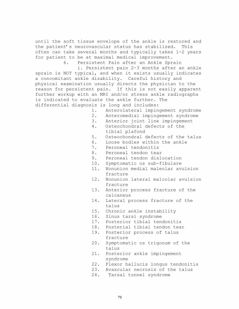
79
until the soft tissue envelope of the ankle is restored and
the patient’s neurovascular status has stabilized. This
often can take several months and typically takes 1-2 years
for patient to be at maximal medical improvement.
6. Persistent Pain after an Ankle Sprain
i. Persistent pain 2-3 months after an ankle
sprain is NOT typical, and when it exists usually indicates
a concomitant ankle disability. Careful history and
physical examination usually directs the physician to the
reason for persistent pain. If this is not easily apparent
further workup with an MRI and/or stress ankle radiographs
is indicated to evaluate the ankle further. The
differential diagnosis is long and includes:
1. Anterolateral impingement syndrome
2. Anteromedial impingement syndrome
3. Anterior joint line impingement
4. Osteochondral defects of the
tibial plafond
5. Osteochondral defects of the talus
6. Loose bodies within the ankle
7. Peroneal tendonitis
8. Peroneal tendon tear
9. Peroneal tendon dislocation
10. Symptomatic os sub-fibulare
11. Nonunion medial maleolar avulsion
fracture
12. Nonunion lateral malcolar avulsion
fracture
13. Anterior process fracture of the
calcaneus
14. Lateral process fracture of the
talus
15. Chronic ankle instability
16. Sinus tarsi syndrome
17. Posterior tibial tendonitis
18. Posterial tibial tendon tear
19. Posterior process of talus
fracture
20. Symptomatic os trigonum of the
talus
21. Posterior ankle impingement
syndrome
22. Flexor hallucis longus tendonitis
23. Avascular necrosis of the talus
24. Tarsal tunnel syndrome

80
25. Peripheral neuropathy
ii. Treatment: Treatment is dictated by the
pathology, but usually begins with a period of rest,
immobilization, physical therapy guided specifically
towards the pathology, and possibly diagnostic and
therapeutic injections of cortisone with a local
anesthetic. Failure to improve after non-surgical
treatment for about 4-6 weeks warrants surgical treatment.
Recovery is dictated by the surgical intervention, but the
patient is typically at maximal medical improvement by 6-12
months after surgical reconstruction.
D. Summary
Ankle injuries are amongst the most commonly
sustained injuries in the workplace. Approximately 25,000
ankle injuries occur every day in the United States. There
is a wide range of ankle injuries, but fortunately, most
only require a short period of disability before return to
full functional.
Acute surgical repair of ankle sprains or
dislocations is not indicated, and only rarely after
completing a functional rehabilitation and proprioceptive
training program is surgery warranted.
Physical therapy is a useful adjunct in treating
patients with ankle injuries as often their proprioception
and static ankle stabilizers are disrupted. Physical
therapy focusing on functional rehabilitation and
proprioceptive training can expedite return to function and
minimize the development of chronic ankle instability.
As with all foot and ankle injuries, prevention
is the key to worker safety. Efforts should be made by
employers to provide employees with education regarding
proper shoe wear and fall prevention, as well as providing
a work environment free of hazards which could cause
serious injury.
PROTOCOL HISTORY:
Passed: 12/15/1992
Amended: 5/5/2009

81
WORKERS COMPENSATION PROTOCOLS
WHEN PRIMARY INJURY IS PSYCHIATRIC/PSYCHOLOGICAL
General Guidelines
Patient must have a diagnosed mental illness on Axis I
as defined by DSM-IV that, by accepted medical standards,
can be expected to improve significantly through medically
necessary and appropriate therapy. The emotional
impairment must be of such a degree to severely interfere
with social, familial, or occupational functioning.
For the purpose of determining medical necessity of
care, medical necessity is defined as "Services and
supplies by a provider to identify or treat an illness that
has been diagnosed." They are:
A. Consistent with the efficient diagnosis and treatment
of a condition, and standards of good medical practice.
B. Required for other than convenience.
C. The most appropriate supply or level of service.
D. Unable to be provided in a more cost effective and
efficient manner; and
E. Unable to be provided elsewhere by a less intensive
level of care.
The evaluation and assignment of mental illness
diagnosis must take place in a face-to-face evaluation of
the patient performed by a psychiatrist or doctoral level
clinical psychologist.
Presence of the illness(es) must be documented through
the assignment of appropriate DSM-IV codes on all axes
(I-V), using published criteria.
Whenever feasible and appropriate, psychiatric care and
treatment should take place in an outpatient setting or the
less intensive treatment setting able to meet the patient's
needs. Structured outpatient programs are considered the
treatment of first choice. Inpatient treatment is
considered medically necessary when all less intensive
levels of treatment have been determined to be unsafe or
have been unsuccessful.

82
The initial evaluation should include not only
documentation of the diagnosis (DSM-IV, axes I-V) but also
an initial treatment plan, individualized goals for
treatment, treatment modalities to be used, and discharge
planning.
A progress note documenting the provider's treatment,
the patient's response to treatment, and the persistence of
the problems that necessitate continued care despite
treatment efforts, with the emergence of additional
problems consistent with the initial diagnosis, must be
written for each session of treatment. Documentation of
disposition planning should be an integral part of each
session note. Response, non-response or severe reactions
to medications given must be recorded.

83
ADULT PSYCHIATRIC HOSPITALIZATION CRITERIA
Medical necessity of psychiatric inpatient admission
must be documented based on conditions defined under either
Section I or Section II.
I. Criteria for Admission Based on Severity of Illness.
A. Patient makes direct threats or a reasonable
inference of serious harm to self or to the body or
property of others.
B. Violent, unpredictable or uncontrolled behavior,
including patients with organic brain impairment and/or
functional illness.
C. Lack of insight, unwillingness or inability to
adequately care for one's physical needs. Acute cases may
include starvation or failure to take essential medications
accurately and safely.
D. Lack of response to previously attempted partial
hospitalization management of medication and/or
psychotherapy.
II. Criteria for Admission Based on Intensity of Service.
A. Need for daily skilled observation by both MD and
RN staff(such as, but not limited to):
(1) To confirm diagnosis;
(2) To initiate medication regime;
(3) To regulate dosage of potent medication; or
(4) To withdraw potent medication.
B. Need for electroconvulsive shock therapy.
III. Criteria for Continued Stay.
The treatment plan should include documentation of
diagnosis, individualized goals of treatment and

84
therapeutic modalities. The medical record must include
daily progress notes by the psychiatrist or
psychologist.
While documentation may justify the need for continued
hospitalization, the Medical Advisory Board expects that
each service rendered by a physician or other provider of
care and reported for payment be documented in the medical
record. Documentation should include:
A. The persistence of the problems that necessitated
the admission, despite therapeutic efforts, or the
emergence of additional problems consistent with the
admission criteria.
B. Severe reaction to the medication or need for
further monitoring and adjustment of dosage.
C. Attempts at therapeutic re-entry into the community
have resulted in exacerbation of the psychiatric illness.
D. Psychiatric evidence or rationale indicating the
need for stabilization of patient's condition to a point
where stress of community re-entry does not substantially
risk an exacerbation of the psychiatric illness.

85
HOSPITALIZATION CRITERIA FOR SUBSTANCE DEPENDENCY
(Applies to Psychiatric Hospitals and General Hospital
Psychiatric Units)
Admission to a psychiatric hospital is appropriate for
alcohol and/or drug dependency of a severity which requires
intensive intervention by a multi-disciplinary health care
team including physicians, nurses, counselors, social
workers, and other therapists. Evidence should be present
that outpatient care or treatment in an intermediate care
facility has been attempted recently, but has been
unsuccessful.
The patient also must have, in addition to substance
dependency of a severity described above, a psychiatric
disorder which inhibits his/her ability to be treated in a
less intensive setting. There must be documented evidence
of a present and acute psychiatric disorder of a severity
which would require hospitalization in and of itself in
accordance with the Adult Psychiatric criteria.
I. SUBSTANCE DEPENDENCY CRITERIA FOR REHABILITATION
SERVICES FOR ADMISSION.
Patient needs to meet the Adult Psychiatric Admission
Criteria and both of the admission criteria given below.
A. Patient has alcohol and/or drug dependency of a
severity which requires intensive intervention, at a
hospital level of care, by a multi-disciplinary health care
team including physicians, nurses, counselors, social
workers, and other therapists. Evidence that the patient
cannot be treated in a residential center for substance
abuse must be documented.
B. Patient has, in addition to substance dependency of a
severity described above, a psychiatric disorder which
inhibits his/her ability to be treated in a less intensive
setting. Evidence of a present and acute psychiatric
disorder of a severity which would require hospitalization
in accordance with the adult psychiatric criteria must be
documented.

86
II. CRITERIA FOR CONTINUED STAY
The patient needs to meet the Adult Psychiatric
Continued Stay Criteria, as well as (all of) A through D
below.
A. The treatment plan should include documentation for
both the substance dependency and psychiatric disorders of
individualized goals of treatment and therapeutic
modalities.
B. The medical record should include daily patient's
progress notes by the psychiatrist, psychologist, or
primary therapist. Evidence should be presented as to
whether or not the problems necessitating admission have
changed in response to specific treatment modalities being
utilized.
C. Documentation of all therapeutic modalities being
provided to the patient on a daily basis should be present
and should specify the plan of treatment and patient's
progress.
D. Post-hospital treatment planning and referral
efforts that have been conducted as soon as the initial
evaluation is complete must be documented in the treatment
plan and progress notes.

87
RESIDENTIAL TREATMENT CRITERIA FOR SUBSTANCE ABUSE
I. CRITERIA FOR ADMISSION.
Medical necessity for admission to a residential
substance abuse treatment facility must be documented by
the presence of all of the criteria below in Section A and
Section B.
In addition, it is noted that structured professional
outpatient treatment is the treatment of first choice.
Residential treatment, when indicated, should (a) be
individualized and not consist of a standard, pre-
established number of days, and (b) should follow recent
outpatient treatment in a structured professional program
of significant duration and intensity during the course of
which the patient has not been able to maintain abstinence
for a significant period of time.
A. Severity of Need.
1. The provider must be able to document that the
individual has a history of alcohol/substance dependence
but is mentally competent and cognitively stable enough to
benefit from admission to the inpatient program at this
point in time. Individual days during any part of the stay
where the patient does not meet this criterion cannot be
certified as medically necessary.
2. Individual exhibits a pattern of severe alcohol
and/or drug abuse as evidenced by continued inability to
maintain abstinence despite recent professional outpatient
intervention.
If the patient has not been in a recent outpatient
program (i.e., the past 3 months), then the following
conditions must be met: 1) patient must be residing in a
severely dysfunctional living environment; or 2) there must
be actual evidence for, or clear and reasonable inference
of serious imminent physical harm to self or others
directly attributable to the continued abuse of substances
which would prohibit treatment in an outpatient setting.
3. For individuals with a history of repeated
relapses and a treatment history involving multiple

88
treatment attempts, there must be documentation of the
restorative potential for the proposed admission.
B. Intensity of Service.
Due to significant impairment in social, familial,
scholastic or occupational functioning, the individual
requires intensive individual, group, and family education
and therapy in an inpatient rehabilitative setting.
II. CRITERIA FOR CONTINUED STAY
In addition to meeting all of the admission criteria on
a daily, continued basis, there must be daily documentation
supporting the need for continued inpatient treatment. All
of A through C below need to be met.
A. Progress Notes - Daily documenting of the
providers' treatment, the patient's response to treatment,
and the persistence of the problems that necessitated the
admission, despite treatment efforts, or the emergence of
additional problems consistent with the admission criteria.
B. The persistence of the problems that caused the
admission to the degree that would necessitate continued
inpatient care, despite therapeutic efforts, or the
emergence of additional problems consistent with the
admission criteria and to the degree that would necessitate
continued inpatient care.
C. Clear and reasonable evidence that re-entry into
the community would result in exacerbation of the illness
to the degree that would require an inpatient level of
care.

89
CRITERIA FOR ADMISSION AND LENGTH OF STAY
FOR ALCOHOL/DRUG DETOXIFICATION AND AN INPATIENT SETTING
Patient must meet both of the criteria under the
appropriate section.
I. CRITERIA FOR ADMISSION
A. Patient has a history of heavy and continuous
alcohol/drug use requiring detoxification services where
(a) there is the potential for serious physical harm from
the side effects of withdrawal and (b) these services
cannot be provided on an outpatient basis. Services that
cannot be provided on an outpatient basis must require
intensive nursing and medical treatment intervention on a
24-hour basis in order to be medically necessary on an
inpatient basis.
B. Patient presents signs and symptoms of impending
withdrawal and/or history of seizures of delirium tremens
and requires intensive nursing and medical treatment
intervention on a 24-hour basis.
II. CRITERIA FOR CONTINUED STAY
A. Documentation of the need for skilled observation
and medical treatment consistent with AEP criteria.
B. Documentation of physical signs and symptoms of
acute withdrawal which require intensive nursing and
medical treatment intervention on a 24-hour basis. This
documentation must be noted three times daily, of which one
such notation must be made by a physician.
III. CONDITIONS LIKELY AND UNLIKELY TO BE RELATED
TO TRAUMA OR WORK
A. The following classes of disorders are frequently
diagnosed post-trauma:
1. Cognitive Mental Disorders.

90
Cognitive mental disorders associated with Axis III
physical disorders - (mainly deliriums, but occasional
dementias).
2. Mood Disorders.
a. Depressive Disorders NOS
b. Major Depression (all types)
3. Anxiety Disorders.
a. Panic Disorder (with or without Agoraphobia)
b. Agoraphobia without Panic
c. Specific Phobia
d. Post-Traumatic Stress Disorder
e. Generalized Anxiety Disorder
f. Anxiety Disorder NOS
g. Acute Stress disorder
h. Anxiety due to a (compensable) general medical
condition
4. Somatoform Disorders
a. Conversion Disorders
b. Pain Disorders (all types, if pain secondary to a
compensable injury)
5. Adjustment Disorders (all types) (note: reaction
lasts no more than six (6) months)
6. Psychotic Disorders NOS
a. Brief Psychotic Disorder
b. Psychotic disorder due to a compensable general
medical condition
B. The following classes of disorders are rarely post-
trauma and in the committee's opinion are not caused or
worsened by industrial injuries, diseases, or stresses.
1. Organic Mental Disorders.
a.Dementias arising in the senium and presenium,
like Alzheimer's.
b.Multi-infarct dementia.
2. Psychoactive substance-induced organic mental
disorders.
a. Alcohol (intoxication, idiosyncratic
intoxication, uncomplicated alcohol withdrawal,

91
withdrawal delirium, hallucinosis, amnestic
disorder, dementia associated with alcoholism)
b. Amphetamine (intoxication, withdrawal, delirium,
delusional disorder)
c. Caffeine (intoxication)
d. Cannabis (intoxication, delusional disorder)
e. Cocaine (intoxication, withdrawal, delirium,
delusional disorder)
+ f. Hallucinogen (hallucinosis; delusional, mood, or
post hallucinogen perception disorders)
+ g. Inhalant (intoxication)
h. Nicotine (withdrawal)
* i. Opioid (intoxication, withdrawal)
j. Phencyclidine (PCP) (intoxication, delirium;
delusional mood or organic mental disorders)
* k. Sedative, hypnotic or anxiolytic (intoxication,
withdrawal, withdrawal delirium, amnestic disorder)
* l. Other or unspecified psychoactive substance
(intoxication, withdrawal, delirium, dementia,
hallucinosis; delusional, mood, anxiety,
personality, or organic mental disorders)
3. Psychoactive Substance Use Disorders.
a. Alcohol (dependence, abuse)
b. Amphetamine (dependence, abuse)
c. Cannabis (dependence, abuse)
d. Cocaine (dependence, abuse)
e. Hallucinogen (dependence, abuse)
f. Inhalant (dependence, abuse)
g. Nicotine (dependence)
* h. Opioid (dependence, abuse)
i. Phencyclidine (PCP) (dependence, abuse)
* j. Sedative, Hypnotic or Anxiolytic (dependence,
abuse)
k. Polysubstance dependence
l. Psychoactive substance dependence or abuse NOS
+ = compensable if an industrial agent exposure occurs at
worksite.
* = compensable if iatrogenic via treatment for compensable
injury.

92
3. Schizophrenia (catatonic, disorganized, paranoid,
undifferentiated, residual).
4. Delusional (Paranoid) Disorder (erotomanic,
grandiose, jealous, persecutory, somatic, unspecified)
5. Psychotic Disorders Not Elsewhere Classified
a. Schizophreniform disorders
b. Schizoaffective disorders
c. Induced (shared) psychotic disorder
6. Mood Disorders
a. Bipolar disorders (Mixed, Manic, Depressed)
b. Dysthymic Disorder (all types)
7. Anxiety Disorders
a. Social Phobia
b. Obsessive Compulsive Disorder
8. Somatoform Disorders
a. Body Dysmorphic Disorder
b. Somatization Disorder
c. Hypochondriasis
9. All Dissociative Disorders
10. Sexual Disorders
All sexual dysfunctions (unless caused by a physical
disorder caused by a work injury, or psychogenic only
secondary to work stress, disease or injury). Not
compensable if lifelong or acquired through other than
compensable means.
Sexual disorder NOS
11. Sleep Disorders, all types, - unless there is an
organic factor related to the compensable injury.
12. Factitious Disorders, (all types)
13. Impulse Control Disorders Not Elsewhere Classified
a. Intermittent Explosive Disorder

93
b. Kleptomania
c. Pathological Gambling
d. Pyromania
e. Trichotillomania
f. Impulse Control Disorder NOS
14. V Codes for Conditions Not Attributable to a Mental
Disorder that are a Focus of Attention or Treatment.
a. Academic Problem
b. Adult Antisocial Behavior
c. Borderline Intellectual Functioning
d. Childhood or Adolescent Antisocial Behavior
e. Malingering
f. Marital Problem
g. Parent-Child Problem
h. Other Interpersonal Problem
i. Other Specified Family Circumstances
j. Phase of Life or Other Life Circumstances Problem
k. Uncomplicated Bereavement
15. Disorders Usually First Evidence in Infancy,
Childhood, or Adolescence as defined in DSM-IV
Classification
16. All Personality Disorders
94
UTILIZATION REVIEW CRITERIA
OUTPATIENT MENTAL HEALTH AND SUBSTANCE ABUSE TREATMENT
PROTOCOL
Mental health and substance abuse outpatient services
are defined as partial hospital, intensive outpatient
programs, outpatient therapy, and all other noninpatient
treatment. The criteria contained in this protocol have
been developed for outpatient mental health and substance
abuse services that are less intensive than partial
hospitalization or intensive specialty outpatient treatment
programs.
Outpatient treatment protocols are based on both
necessity of care and treatment approach. Outpatient
treatment is based on Severity of Illness (SI) and
Intensity of Service (IS) indicators. Patients must have
psychiatric and/or substance abuse disorders with
appropriate Severity of Illness and Intensity of Service
indicators for outpatient treatment to be determined to be
medically necessary.
Medical necessity is defined as follows:
"Services and supplies by a provider to identify or
treat an illness that has been diagnosed. They are:
a. consistent with:
(1) the diagnosis and treatment of a condition;
and
(2) standards of good medical practice;
b. required for other than convenience; and
c. the most appropriate supply or level of
service."
The following criteria are a more detailed elaboration
of the above definition for the purpose of establishing the
medical necessity of outpatient mental health services.
1. The method of treatment specified in terms of
treatment framework or orientation, treatment modality,
treatment frequency, and estimate of treatment duration;

95
2. provision of measurable, target criteria for interim
goals and end of treatment goals to be used to determine
both that 1) treatment is progressing and 2) when treatment
is no longer indicated; and
3. an alternative plan to be implemented if patient
does not make substantial progress toward the given goals
in a specified period of time. Examples of an alternative
plan are a second opinion or introduction or adjunctive or
alternative therapies.
CONTINUED OUTPATIENT TREATMENT CRITERIA
After initial treatment has been completed (GAF=70),
continued psychotherapy treatment is indicated only if
criteria below are met.
I. Severity of Illness Indicators
Continued outpatient psychotherapy treatment requires
the presence of each of the following Severity of Illness
Indicators:
A. A DSM IV diagnosis on Axis I.
B. A description of DSM-IV psychiatric symptoms,
behavioral (occupational) and/or cognitive dysfunction,
consistent with the diagnoses given; and
C. Impairment in occupational functioning due to those
psychiatric symptoms. To address medical necessity in the
context of varying patient needs, this impairment in
functioning is divided into two subcategories.
1. Patients in the middle phases of treatment (six
one-hour sessions over six weeks) who typically have
fluctuating degress of impairments in functioning as
evidenced by a specific clinical description. GAF scores,
fluctuate but may exceed 71 for the six-week period prior
to review. Such scores are frequently considered typical
and appropriate within the context of the progressive
response to treatment and the treatment plan.

96
Among the factors considered in making a determination
on the continued medical necessity of treatment in this
phase are the frequency and severity of previous relapses,
level of stressors, and other relevant clinical indicators.
2. Patients in the final and consolidation phases
of treatment (six one-hour sessions over twelve weeks) who
typically have GAF scores above 71. Such scores are
frequently considered typical and appropriate within the
context of the progressive response to treatment and the
treatment plan.
However, if the level of functioning has
progressed to the point that the patient has sustained a
GAF score above 71, serious consideration should be given
to the medical necessity of continued treatment. Options
to consider are: a) termination of treatment or b)
reduction in the level and/or type of treatment previously
given.
Note: Medication management with a visit every eight weeks
for 15-20 minutes may be necessary indefinitely and should
be reviewed on a case-to-case basis.
As above, the factors considered in making a
determination about the continued medical necessity of
treatment in this phase are the frequency and severity of
previous relapse, level of stressors, and other relevant
clinical indicators. The therapist should be able to
explain whether the therapeutic modality being utilized
will shift (and if not, why) when there has been sustained
improvement as measured in part by a GAF score over 71.
II. Intensity of Services Indicators
Continued outpatient psychotherapy treatment requires
the presence of each of the following indicators.
A. An update of the medically necessary and
appropriate treatment plan specific to the patient's
impairment in functioning and DSM-IV psychiatric symptoms,
behavior or cognitive dysfunctions.

97
B. The treatment plan update must identify:
1. all changes in target specific DSM-IV
psychiatric symptoms, behavior, and cognitive dysfunction
being treated;
2. all modifications, if any, in biologic,
behavioral, psychodynamic or psychosocial framework(s) of
treatment for each psychiatric symptom/cluster and/or
behavior;
3. all changes in the specific and measurable
goals for treatment specified in terms of symptom
alleviation, behavioral change, cognitive alteration,
psychodynamic change, or improvement in occupational
functioning;
4. all modifications in treatment methods in terms
of:
. treatment framework or orientation,
. treatment modality,
. treatment frequency, and
. estimate of treatment duration;
5. progress in measurable, target criteria used to
identify both interim treatment goals and end of treatment
goals to determine 1) treatment is progressing and 2) goals
have been met and treatment is no longer needed;
6. alternative plan to be implemented if patient
does not make substantial progress toward the given goals
in a specified period of time. Examples of an alternative
plan are a second opinion or introduction of adjunctive or
alternative therapies.

98
ADULT PSYCHIATRIC PARTIAL HOSPITALIZATION CRITERIA
Preamble - Medical necessity is defined as "services and
supplies by a provider to identify or treat an illness that
has been diagnosed or suspected. They are:
a. consistent with:
(1) the diagnosis and treatment of a condition;
and
(2) standards of good medical practice;
b. required for other than convenience; and
c. the most appropriate supply or level of
service.
When applied to inpatient care, the term means: "the
needed care cannot be safely given on other than an
inpatient basis."
The purpose of this protocol is to define and clarify
criteria for when partial hospitalization for psychiatric
treatment meets the above definition for medical necessity.
PRINCIPLES FOR CERTIFICATION
When a patient has a psychiatric disorder that requires
professional evaluation and treatment, he/she should be
treated at the least intensive outpatient level appropriate
for the condition prior to partial hospital/day treatment;
unless there is compelling evidence to the contrary.
I. Criteria for Admission
Medical necessity for psychiatric partial
hospitalization treatment must be based on meeting the
conditions defined under Section A, 1 and 2 (both must be
met) and either 3 and 4, as well as meeting all of the
criteria defined under Section B.
A. Severity of Need
1. Patient must have a mental illness. Mental
illness is defined as Axis I psychiatric disorder that, by
accepted medical standards, can be expected to improve

99
significantly through medically necessary treatment and
therapy.
2. There is clinical evidence that documents that a
less intensive outpatient setting is not appropriate at
this time and/or a day treatment program can safely
substitute for or shorten a hospital stay.
3. There is clinical evidence that the patient
would be at risk to self or others if he were not in a
partial hospitalization program. Additionally:
a. Patient can contract for safety in a
structured environment under clinical supervision for part
of the day and has a suitable environment for the rest of
the time; or
b. The patient is believed to be capable of
controlling this behavior and/or seeking professional
assistance or other support when not in the partial
hospital setting.
4. As a result of the patient's mental disorder
there is an inability to adequately care for one's physical
needs, representing potential serious harm to self.
B. Intensity of Service
1. In order for a partial hospital program to be
safe and therapeutic for an individual patient,
professional and/or social supports must be identified and
available to the patient outside of program hours, and the
patient must be capable of seeking them as needed.
2. The patient's condition must require intensive
nursing and medical intervention for more than three (3)
hours per day and for more than two (2) days per week.
3. The individualized plan of treatment for partial
hospitalization requires treatment by a multidisciplinary
team. A specific treatment goal of this team is improving
symptoms and level of functioning enough to return the
patient to a lesser level of care.

100
II. Criteria for Continued Stay
In addition to continuing to meet the criteria given
above for admission, patients must meet A and B.
A. Progress notes for each day that patient is in a
partial hospital/day treatment program documenting the
provider's treatment, the patient's response to treatment,
and the persistence of the problems that necessitated the
admission to the partial hospitalization program, despite
treatment efforts, or the emergence of additional problems
consistent with the admission criteria.
B. Documentation that attempts at therapeutic re-entry
into a less intensive level of care have or would result in
exacerbation of the psychiatric illness to the degree that
would warrant the continued need for partial
hospitalization services.
VNS Therapy for Depression
The Medical Advisory Board concludes that there is
sufficient scientific evidence to conclude that vagus nerve
stimulation is not reasonable and necessary for the
treatment of resistant depression.
PROTOCOL HISTORY:
Passed: 9/01/1992
Amended: 11/19/2002
Amended: 6/12/2007
101
OUTPATIENT PHYSICAL AND OCCUPATIONAL THERAPY PROTOCOL
GUIDELINES
General Therapy Guidelines
1. Therapy evaluations must be provided by licensed
physical and/or occupational therapists. Therapy
evaluations may not be performed by therapy assistants or
other medical providers.
2. For worker's compensation patients, physicians may
not provide or bill for physical therapy services without
employing a licensed therapist to evaluate and supervise
treatments.
3. Therapy treatments may be provided by licensed
therapy assistants ** as directed by the licensed therapist
or by therapy aides under supervision of the licensed
therapist.
A facility may not employ more than two licensed
assistants per therapist. Physical therapists shall
maintain the following documentation regarding the
supervision of physical therapy assistants:
1.ON-SITE SUPERVISION OF THE ASSISTANTS PERFORMANCE
2.A REVIEW OF THE ASSISTANTS DOCUMENTATION
3.A REASSESSMENT AND UPDATE OF THE PATIENTS PROGRAM AND
GOALS.
** Certified Occupational Therapy Assistants are
nationally certified to provide care under the direction of
licensed occupational therapists.
4. A course of physical and/or occupational therapy
treatment will consist of nine (9) treatments or less. In
those few instances where further treatments need to be
given, the following format will be followed:
a. The therapist will provide the rationale for
continuation of treatment to the employer/insurer.
b. The employer/insurer, usually in correlation
with a medical specialist, will make a judgment concerning
the medical necessity for further treatment. The
employer/insurer will inform the therapist within ten (10)
days of receipt of the written or verbal request for

102
continued treatment whether therapy treatment will be
reauthorized.
5. Therapy evaluations must identify patient problems
and objective measurements of physical or work-skills
deficits. These objective measures should be as specific
as is possible for the diagnosis or patient problem.
Example: Patient diagnosis of rotator cuff strain.
Appropriate
ROM flexion 160, abduction 90, int rotation 45, ext
rotation 60.
Unable to reach or lift above shoulder height; able to lift
up to 25 lbs from floor to waist.
Inappropriate
ROM limited in all planes.
Unable to lift secondary to pain.
6. Therapy treatment plans must be problem oriented.
7. Therapy evaluations should identify subjective
complaints of pain or paresthesias, however, therapy
treatments cannot be based solely on pain reduction.
Evaluations must identify specific treatment plans and
relate treatments to improving objective deficits and
patient problems.
8. Frequent reassessment of progress towards
improving objective deficits must be done and documented.
Timing of reassessment is based on frequency of treatment,
but should occur no less than every nine (9) sessions.
Revision of problem lists, goals, and treatment plans must
be documented at this time.
9. Continuation of treatments cannot be based solely
on presence of continued pain symptoms. If objective
measures have failed to improve, or have plateaued, the
rehabilitation professional will confer with the referring
physician to determine if the treatment should be modified
or changed.
10. All treatment sessions and tests must be
documented in writing. Daily treatment notes must:
a) identify type of treatment PROVIDED.
b) note patient response to treatment in
subjective and objective terms.

103
c) identify any change in treatment plan and
reasoning for change; e.g., stopping ultrasound
treatment because of diminished tendonitis symptoms and
increased ROM.
d) all assisting personnel notations must be co-
signed by the supervising therapist.
11. Most of the treatment protocols anticipate healing
and return to work will occur during the first four weeks
after injury. There are some patients whose rehabilitation
will take longer than the anticipated time frame because of
the severity of their injury or the occupational demands of
their job. Continuance of the therapy program will be
according to the guidelines noted above.
THERAPY PROTOCOLS
LOW BACK MUSCULAR INJURY
...as delineated in the Low Back Musculoligamentous
Injury (Sprain/Strain) Medical Advisory Board Protocols.
CERVICAL MUSCULAR NECK INJURY
...as delineated in the Cervical Musculoligamentous
Injury (Sprain/Strain) Medical Advisory Board Protocols.
CARPAL TUNNEL SYNDROME
. . . as delineated in the Carpal Tunnel Syndrome
Medical Advisory Board Protocols.
Non-operative Intervention
1. Appropriate Interventions:
a) ROM and strengthening exercises
b) splint fabrication
c) assessment of job skill levels for RTW
d) instruction in work activities modifications or
simulation of work activities
e) patient education
2. Inappropriate Interventions:
a) exclusive use of passive modalities
3. Extenuating Services:
a) prolonged onset of symptoms prior to referral

104
Post-Operative Intervention
1. Extenuating Circumstances
a) post-operative complications
b) delayed referral into therapy
2. Appropriate Interventions:
a) ROM, simple strengthening exercises
b) Splint fabrication
c) Scar tissue/swelling management
d) Assessment of job skill levels needed for RTW
e) Instruction in work activities modifications or
simulation.
f) Patient education.
CERVICAL HERNIATED DISC
... as delineated in the Herniated Cervical Disk
Medical Advisory Board Protocols.
Non-operative Intervention
1. Appropriate Interventions:
a) ROM exercises for neck and upper extremity
b) strengthening/endurance exercises for upper
extremity
c) trial of cervical traction; if beneficial, a
prescription for a home unit is sought.
d) short-term use of modalities for pain relief.
e) patient education
f) assessment of work skill levels for return-to-
work
g) modification/simulation of work activities.
2. Extenuating Circumstances:
a) profound muscle weakness
b) delayed referral into therapy.
Post-Operative Intervention
1. Extenuating Circumstances:
a) profound muscle weakness
b) delayed referral into therapy

105
2. Appropriate Interventions:
a) ROM exercises for neck and upper extremity
b) strengthening/endurance exercises for upper
extremity
c) patient education
d) modification/simulation of work activities.
3. Inappropriate Interventions:
a) cervical traction
b) exclusive and/or prolonged use of passive
modalities.
LUMBAR HERNIATED DISC
... as delineated in the Herniated Lumbar Disk Medical
Advisory Board Protocols.
Non-operative
1. Appropriate Interventions:
a) ROM exercises for trunk and extremities
b) strengthening/endurance exercises for trunk and
extremities
c) short-term use of modalities for pain relief,
in conjunction with active exercises
d) patient education
e) assessment of work skill levels for return-to-
work
f) work simulation activities (when acute symptoms
have subsided) or work-site modifications.
g) short-term trial TENS for chronic pain; if
found to relieve symptoms, a referral for a home unit
should be sought.
2. Inappropriate Interventions:
a) prolonged and/or exclusive use of modalities
3. Extenuating Circumstances:
a) delayed referral into therapy
b) profound muscle weakness (non-operative and
post-operative)
Post-operative
1. Appropriate Interventions:

106
As above, exceptions noted below.
2. Inappropriate Interventions:
a) Use of passive modalities, including traction
NON-OPERATIVE SOFT TISSUE INJURIES:
SHOULDER SPRAINS, OVERUSE INJURIES, KNEE STRAINS, ANKLE
SPRAINS
(Refer to appropriate Medical Advisory Board
Protocols)
1. Appropriate Interventions:
a) Acute management of muscle spasms, pain,
and/or swelling
b) ROM exercises
c) Gait training w/assistive devices, as needed
d) (as tissue healing progresses) strengthening
and endurance exercises
e) Proprioception and balance activities
f) Assessment of job skill levels; job simulation
activities if significant deficits noted
g) isokinetic tests and rehab if deficits noted.
2. Inappropriate Interventions:
a) exclusive and/or prolonged use of passive
modalities
b) multiple computerized tests in any one week
3. Extenuating Circumstances
a) further medical evaluation that changes
diagnosis
b) surgery
c) delayed referral into therapy
MENISCAL INJURIES
Refer to appropriate Medical Advisory Board Protocols.
Non-Operative
1. Appropriate Interventions:
a) ROM and strengthening exercises
b) acute management of swelling and pain
c) gait training with assistive devices, as needed

107
d) isokinetic testing and rehab.
e) assessment of work skill levels for return-
to-work
f) work skills simulation
2. Inappropriate Interventions
a) prolonged and/or exclusive use of passive
modalities
3. Extenuating Circumstances
a) delayed referral into therapy
b) surgery
Post-Operative
As noted above.
SYMPATHETIC DYSTROPHY
... as delineated in the Chronic Regional Pain
Syndrome (formerly Sympathetic Dystrophy) Medical Advisory
Board Protocols.
1. Appropriate Interventions:
a) ROM exercises (aggressive if done after nerve
block)
b) Strengthening and endurance exercises
c) Short-term use of modalities
d) Patient education
e) short-term trial of TENS; if beneficial, a home
unit should be sought.
f) assessment of work skills levels; simulation of
work activities if deficits are found

108
2. Inappropriate Interventions
a) prolonged or exclusive use of modalities
3. Extenuating Circumstances
a) development of adhesive capsulitis
b) delayed referral into therapy
c) repeated nerve blocks with therapy after each
procedure
THORACIC OUTLET SYNDROME
1. Appropriate Interventions:
a) postural exercises and correction
b) ROM exercises
c) strengthening and endurance exercises
d) patient education
e) assessment of work skills; simulation if
deficits are noted.
2. Inappropriate Intervention:
a) prolonged or excessive use of modalities
b) traction
PROTOCOL HISTORY:
Passed: 3/30/1993
Amended: 5/5/2009

109
ACOUSTIC TRAUMA
TRAUMA TO THE EXTERNAL EAR
I. BACKGROUND
The common types of trauma to the external ear
usually result from thermal, blunt or penetrating trauma
causing damage to the auricle, external auditory canal, or
tympanic membrane.
II. DIAGNOSTIC CRITERIA
A. Pertinent History and Physical Findings
Direct examination of the external ear and
tympanic membrane and evaluation of hearing with an
audiogram.
III. TREATMENT
1. Hematoma of the external ear, usually due to a
direct blow, is treated by drainage of the hematoma which
may be done with an 18 gauge needle and syringe or a small
incision under local anesthesia followed by application of
Vaseline gauze and fluffs between the external ear and
mastoid, and a soft gauze bandage is wrapped around the
head. The patient should be re-examined in 24 hours for
reaccumulation. Time loss from work, 0 to 2 days.
2. Simple lacerations present no difficulty in
management and may be sutured, and a bulky pressure
dressing is applied. They are anticipated to heal. Time
loss from work, none.
3. Exposed cartilage presents a special problem.
Debridement and complete coverage of all cartilage are key
principles, and torn cartilage should be repaired. These
usually heal readily. Maximum time lost from work, only
with the most serious injuries, five days.
4. Large auricular avulsions may need to be
reanastomosed by an otolaryngologist or plastic surgeon.
This will require follow-up visits. Loss of work may be
minimal depending on the type of work, but maximum time
lost from work, two weeks.

110
5. Large circumferential lacerations to the
external auditory canal may lead to stenosis of the canal
and these mandate referral to an otolaryngologist. Loss of
time from work, 1 to 2 days.
6. Burns to the auricle require removal of
devitalized tissue and antibiotic ointments to protect the
underlying cartilage. Time lost from work, none.
7. Chemical burns may follow exposure to acids or
alkali. Primary treatment consists of immediate irrigation
with several liters of water, identification of the toxic
chemical and should be treated primarily as a burn. No
loss of work anticipated. No time lost from work.
8. Simple perforation of the tympanic membrane
generally heals in four to six weeks, some use of
antibiotics if there are definite signs of contamination.
Failure to heal will require an ENT referral. Patient to
be instructed to keep water out of ear until perforation
has healed. No loss of time from work anticipated.
IV. ANTICIPATED OUTCOME
Full Recovery.
INJURY TO THE MIDDLE EAR
I. BACKGROUND
The middle ear cavity is connected with the nasal
pharynx by the eustachian tube and is intimately related to
injury or diseases of both structures.
The primary trauma to the middle ear is barotrauma due
to changes in barometric pressure and blunt trauma. Severe
injury can disrupt the ossicular chain with conductive
hearing loss or cause a perilymphatic fistula resulting in
vertigo and sensorineural hearing loss.
Tympanic membrane perforations secondary to thermal
burns as well as slag-bur injury and perforations from
direct trauma to the ear drum from foreign body.

111
II. DIAGNOSTIC CRITERIA
Examination of the ear looking for retraction, or
perforation of the tympanic membrane as well as evidence of
effusion or hemotympanum. A neurological examination
should be performed looking for evidence of vestibular
disfunction (nystagmus). Patient should have an audiogram
and if clinically indicated (vertigo) a fistula test can be
performed by an audiologist, but only after examination by
otorhinolaryngologist.
III. TREATMENT
1. Antibiotic if URI is present, oral steroids may
reduce eustachian tube edema. No loss of time from work.
2. Patient with vestibular findings requires an
emergency ENT referral. There may be no time lost from
work, but this would depend on the ENT referral, including
the severity of the vertigo and the type of work the
patient is involved with.
IV. ANTICIPATED OUTCOME
This depends on how much damage has occurred.
TRAUMA TO THE INNER EAR
I. BACKGROUND
Trauma may result from blunt injury causing temporal
bone fracture, blast injury, noise exposure or toxic
injury. Vestibular, cochlear or facial nerve function may
be affected.
II. DIAGNOSTIC CRITERIA
Radiologic evaluation with blunt trauma is of limited
value. An MRI or CT Scan may show the fracture. The
physical examination may reveal the discolored tympanic
membrane and may show the fracture through the external
canal. The neurological examination may reveal facial
paralysis, perforation of the tympanic membrane with CSF

112
leak. The patient should be examined for evidence of
hearing loss (Hearing Test) or vestibular dysfunction
(ENG)by an otolaryngologist.
III. TREATMENT
1. CSF Leak. One should watch for a cerebral spinal
fluid leak and if this persists may require a neurosurgical
consultation and repair, usually a combined procedure
performed by an otolaryngologist and neurosurgeon. The use
of antibiotics is controversial, more recently it is felt
that they are not useful in this situation.
2. Hearing Loss.
a. Nerve hearing loss, there is no surgical
treatment although amplification devices may be required.
b. Conductive hearing loss.
1. Repair of tympanic membrane perforation.
Time lost from work with surgery, maximum one week.
2. Repair of disrupted ossicles. Time lost
from work with surgery, maximum two weeks.
3. Facial paralysis may require nerve repair or a
form of re-animation procedures of the facial muscles.
Time lost from work would be variable in this case, but not
more than three days.
4. Vestibular Injury.
a. Vestibular suppression medications such as
Antivert, Valium or Klonopin.
b. If the vertigo becomes disabling and persists
after six months of treatment with the above medications,
then vestibular destructive surgery either with
labyrinthine destruction or vestibular nerve section may be
required. Loss of time from work would be two weeks
following surgery.
IV. ANTICIPATED OUTCOME
This depends on how much damage has occurred.

113
WORK-RELATED HEARING IMPAIRMENT DUE TO NOISE
I. BACKGROUND
Hearing impairment due to noise may occur in the
workplace. An effort has been made by the American Academy
of Otolaryngology Committee on Hearing and Equilibrium and
the American Council of Otolaryngology Committee on the
medical aspects of noise.
II. DIAGNOSTIC CRITERIA
A. Pertinent Historical and Physical Findings.
The history consists of impairment of hearing. The
Hearing Conservation Program requires employers to monitor
noise exposure levels in a manner that will accurately
identify employees who are exposed to noise at or above 85
decibels (dB) averaged over eight working hours. The
exposure measurement must include all noise within an 80 dB
to 130 dB range and must be taken during a typical work
situation. Audiometric testing must be made available to
all employees who have average exposure levels over an
eight-hour period of 85 decibels.
III. TREATMENT
Hearing protectors must adequately reduce the severity
of noise in each employees' work environment.
The percentage loss is to be evaluated by an
Otolaryngologist and Audiologist.
Protocol History:
Passed: 3/30/1993
Amended: 11/19/2002
Amended: 6/3/2008

114
EPIDURAL STEROID INJECTIONS IN THE MANAGEMENT OF SPINAL
PAIN
A. BACKGROUND
Epidural glucocorticoid and local anesthetic injections can be
considered as part of a treatment program for radicular pain
syndromes secondary to a herniated disc, degenerative disc disease,
or spinal stenosis. The goal of such injections is to deliver the
active medication with minimal systemic effects (vs. oral steroids)
as close as possible to the target tissue. There are three most
frequently used approaches: caudal, interlaminar, and
transforaminal. Of these three, the most common is the use of
interlaminar epidural injection, performed by injection immediately
adjacent to the dural sac in the posterior spinal column, with
subsequent diffusion to the herniated disc or other inflamed,
irritated, or impinged neural structures. Fluoroscopic guidance of
needle placement has been shown to improve the accuracy of the
placement of the injection; however, whether the clinical outcome is
improved with this remains somewhat unclear.
Epidural injections may be performed in the cervical, thoracic,
lumbar, as well as sacral (caudal) regions. Caudal epidural steroid
injections may be used for patients with leg pain of sacral origin or
in whom direct access to the lumbar region is difficult. Epidural
injections are invasive, have a low risk of adverse effects, and are
relatively costly. They are most commonly offered as an option in
acute radiculopathy as a second-line treatment after prior treatment
with NSAIDs, possibly a short course of an oral steroid, and a
waiting period of at least 3 weeks with/without other adjunctive
treatment measures (exercise, spinal manipulation, etc.).
Complications of injections can occur, but are not common and are
usually self-limited. However, serious complications can occur,
including (but not limited to) infection, hemorrhage (penetration of
an anatomical variant artery), and hematoma formation with/without
compression of an adjacent nerve or spinal cord. Finally,
suppression of the pituitary-adrenal axis can occur, as does
transient elevation of blood sugar in diabetics.
If the injury being treated is attributable to a single nerve root
irritation/compression, transforaminal injections usually involve the
least volume of agent, provided in the most closely targeted site of
symptomatic pathology. Also known as ―selective nerve root
injections‖, transforaminal epidural injections achieve the same
goal, however, the needle is placed along the nerve root, in closer
proximity to either the impinged nerve or herniated disc. As these

115
are technically more challenging injections, they are frequently
performed with either fluoroscopic or CT guidance. Electrical
stimulation is sometimes also used to facilitate nerve root
identification. The injections involve the injection of a
glucocorticoid as well as anesthetic agent. As such, they can often
provide both diagnostic assistance, as well as therapeutic relief of
targeted symptoms. The efficacy of these injections is significantly
tied to the technical precision of the procedure.
Finally, local anesthetic (as well as glucocorticoid) epidural
injections can be useful in conjunction with aggressive physical
therapy.
B. DIAGNOSTIC CRITERIA
1. Pertinent History and Physical Findings
A pattern of pain in the upper extremity, thoracic region, or lower
extremity with the characteristics as well as specific distribution
of a known nerve root, known as a radiculitis or radiculopathy.
Radiculopathy refers to a sensory and/or motor dysfunction in the
discrete distribution of an affected nerve root. Most cases result
from either compression of, or inflammation to the nerve root as it
exits the spinal canal, most commonly secondary to a disc
protrusion/herniation. Such pain may be seen in the absence of
previous surgery, but can also be seen following failed post-
operative disc surgery. Physical findings strongly suggestive of a
radiculitis/radiculopathy (positive straight leg raise testing,
reflex diminishment in the affected limb, motor weakness and/or
sensory dysesthesias/hypoesthesias, particularly in a pattern
consistent with specific nerve root(s)) may accompany subjective
complaints.
For cervical epidural steroid injections, pain may be distributed in
a specific nerve root pattern (dermatome):
Neck, shoulder and upper arm: C5 nerve root
Neck, shoulder and radial forearm: C6 nerve root
Neck, shoulder and dorsal forearm: C7 nerve root
Neck, shoulder and ulnar forearm: C8 nerve root
For thoracic epidural injections, pain may be distributed in a
―barrel stave‖ fashion, from the mid back, then extending anteriorly
to the side and chest, in accordance with the underlying thoracic
nerve root inflammation.

116
For lumbar epidural steroid injections, pain may be distributed in a
specific nerve root pattern:
Hip, thigh and knee: L3 nerve root
Hip, thigh, knee, and medial leg: L4 nerve root
Hip, lateral thigh and leg: L5 nerve root
Buttock, posterior thigh and calf: S1 nerve root
Anatomic variation can exist in these nerve root distributions.
Additionally, particularly in the early stages, entire nerve root
distribution may not be affected.
The duration of symptoms may play a role in decision making regarding
timing of injections. Subacute radicular pain (pain lasting 3 weeks
or longer) that has not responded to more conservative measures
(particularly when the presumed etiology of this pain is well
identified and potentially reversible with a steroid injection) may
form a historical foundation for proceding with injection. Patients
with chronic back pain with an exacerbation also fall into this
group. The goal of injection(s) in this group is to afford a few
weeks of partial pain relief while spontaneous recovery occurs, or
allowing for the patient to tolerate other treatments, and therefore
facilitate more active and aggressive pursuit of rehabilitative goals
and restoration of function.
C. APPROPRIATE DIAGNOSTIC TESTS AND EXAMINATIONS
The current practice in the U.S. is to obtain either a CT or MRI scan
prior to the performance of an epidural injection. There are,
however, studies of good quality showing effectiveness of injections
based on clinical examination to address the target level for
injection. Benefits of pre-procedural imaging include a greater
safety margin in the determination of the entry level, the ability to
rule out/in other pathology, as well as determining the presence of a
surgical condition (thereby contraindicating the use of the epidural
injection). Studies are ongoing, however, regarding whether imaging
is required or not, as well as the benefit magnitude obtained from
imaging.
Occasionally, the use of an EMG/NCVS can be considered, particularly
in cases where the symptoms/physical findings and, possibly, imaging
studies are ambiguous, or do not provide a clear guide as to the
underlying pathology attributable to the presenting complaints. In

117
some instances (e.g. presence of a medical condition
contraindicating a specific imaging study, etc.) other studies (CT
myelogram, discography) may be considered to confirm the diagnosis
prior to proceding to epidural injection.
D. Treatment
1. Outpatient Treatment
Since the pain relief from epidural steroid injections is usually
brief and by definition chronic non-specific back pain and chronic
radicular pain (with or without prior back surgery) are chronic
problems, injections are not recommended as a transient treatment for
these long-term problems, unless there is a specific exacerbation
that indicates their use. Additionally, there is currently no
information that supports an advantage to the performance of
injections early on in the course of the syndrome. There is
insufficient evidence, at present, to recommend one technique
(caudal, interlaminar, or transforaminal) over another for an initial
approach. Finally, the concurrent use of injections during
participation in a rehabilitation program may be beneficial.
Injections are commonly performed on an outpatient basis. As noted
in section A, fluoroscopic guidance provides the most accurate method
for ensuring injection of the steroid into the desired location. If
local anesthetics are also used, proper vital sign monitoring is
required, including electocardiography, blood pressure monitoring, as
well as pulse oximetry. Concious sedation may be required for some
anxious patients, but is not usually necessary. Emergency equipment,
including but not limited to oxygen, ventilatory tools, laryngoscope,
endotracheal tubes, intravenous access supplies and vasopressors must
be available, as well as appropriately trained individuals, as per
State of R.I. Board of Health requirements, as well as any other
applicable regulatory medical agencies or groups. Commonly, long
acting steroids (betamethasone or methylprednisone) are used, with
the most common anesthetics being lidocaine and/or bupivicaine.
Corticosteroid dosing is most often based on one third (1/3) the dose
associated with adrenal suppression, per injection.

118
2. Treatment Duration
Epidural steroid injections are primarily intended for reducing
inflammation around the nerve root for primarily radicular pain. Due
to the long acting nature of the steroid preparations used, they
should not be performed at less than two (2) week intervals.
Optimally, injections should occur at four (4) week intervals.
Injections should not exceed three (3) in number over a six (6) month
interval, and not more than six (6) in a twelve (12) month interval.
If a lack of response is seen after two epidural steroid injections,
no further injections should be performed at the same level.
Epidural injections should be scheduled separately and effects of
each evaluated, rather than scheduling a series of 3. A second
epidural injection is not recommended if, following the first
injection, there has been significant reduction or resolution of
targeted symptoms, or if a documented increase in physical
activities/function occurs. If no response to a first injection
occurs, there is less indication for a second injection. However, if
the interventionalist opines that the medication was not well placed,
and/or if the underlying condition is believed to be so severe that
one injection of a standard steroid dose would not be anticipated to
adequately reverse the pathology, a second injection may be
indicated. In patients who respond to an injection with 3-6 weeks of
temporary, partial radicular pain relief, but then develop worsening
pain and functional loss, but do not wish to proceed to surgery, a
repeat epidural injection my be an option. Patients requesting a 4th
injection should be counseled regarding surgery, as further
injections are not likely to be of benefit.
Protocol History:
Passed: 4/27/1993 As ―Caudal Epidural Blocks . . . ‖
Amended: 6/9/1998 As ―Epidural Nerve Blocks and Epidural
Steroid Injections . . .‖
Amended: 11/19/2002
Amended: 4/27/2010

119
WORK HARDENING PROTOCOLS
I. INTRODUCTION
Guidelines have been established that define the
nature, character, time duration, and cost of
physical/occupational therapy treatments. In order to
return an injured worker back to work, the therapy provider
can provide one or both of the following therapy programs:
"work hardening and/or work conditioning." The provider
should indicate that their treatment services will be in
the form of work conditioning with job simulated activities
for a true work hardening program.
II. DEFINITION
Work conditioning is an intensive work-related, goal-
oriented conditioning program designed specifically to
restore systemic neuromuscular functions, including joint
integrity, mobility, and muscular performance (including
strength, power, and endurance), motor function (motor
control and motor learning), ROM and
cardiovascular/pulmonary functions. The objective of a
work conditioning program is to restore physical capacity
and function to enable the patient/client to return to
work.
A work hardening program is an interdisciplinary,
individualized and goal oriented, job specific program
designed to return the patient/client to work. Work
hardening programs use real or simulated work activities
and progressively graded conditioning exercises that are
based on the individuals measured tolerances, to restore
physical, behavioral, and vocational functions. Work
hardening programs address the issues of productivity,
safety, physical tolerances, and work behaviors.
III. THE PROTOCOL COMPONENTS
1. Determination of the strength and endurance goals
of the client in relation to the return to work
goal should be established using equipment that
quantifies and measures strength and conditioning
levels; i.e., ergometers, dynamometers, treadmills,
free weights, and circuit training. Goals for each

120
worker are dependent on the demands of their
respective jobs.
2. Simulation of the client’s work demands job
simulations that provide for progressions in
frequency, load, and duration of work are essential.
These should directly relate to the work goal and
offer the client opportunity to practice work
related positions and motions.
3. The program should include education in body
mechanics, work safety and injury prevention. This
should include direct therapist interaction and may
be combined with video presentations that cover
anatomy, back care, posture and the role of exercise
and the worker’s responsibility in self-treatment.
4. Assessment of the worker’s need for job modifications. Documentation of job
modifications are needed, i.e., adaptations in
equipment, work station ergonomics. Adaptations
should be made and practiced to insure success.
5. A written plan that includes measureable goals,
the strategies used to meet these goals and the
projected time necessary to accomplish the goals and
expected outcomes. This plan may be supported by a
functional capacity evaluation to establish a
base-line that can be compared to the demands of the
job. A re-evaluation may be performed to determine
success of the program and the worker’s readiness
to return to work. These evaluations are considered
part of the work hardening program.
6. The work hardening facility should be a safe work
environment that is appropriate for the
vocational goals and the worker. The amount of
space should be determined by the number of
workers, or approximately 100 square feet per
client.
7. A total of nine (9) to twenty (20)
physical/occupational therapy treatments will be
paid for by the insurer for services given to the
injured worker. The total number of treatments
should be dependent upon the severity of the
condition and intervention necessary.

121
IV. DOCUMENTATION
The following represents the general outline for the
evaluation of candidates for work hardening and for
implementation of treatment.
1. A request for work hardening may be made by the
treating physician,insurer/case manager, acute care therapy
provider, physician’s assistant, nurse practitioner,
osteopathic physician, and/or chiropractor.
2. All requests for work hardening will be forwarded
from the generating source of the referral to the physician
and then to the industrial health provider. They must
include prior approval from the physician as well as the
claims manager before the program begins.
3. Work hardening facility will submit a copy of the
evaluation to the referring physician and the insurer
within three (3) business days of the evaluation. The
evaluation should include the initial plan as well as the
following:
a. the medical status
b. the musculoskeletal exam
c. the current functional work capacity testing
d. the projected work capacity to return to
work
e. the cognitive/perceptual status
f. the behavioral/attitudinal status
g. the vocational status.
The evaluation should document a benchmark from
which to establish the initial treatment plan and/or the
physical/functional/vocational disposition.
Information will include:
a. the name of the case manager
b. the estimated time frame for treatment.
c. the worker must demonstrate physical
recovery sufficient to allow
for progressive participation for a minimum
of 4 hours a day for three to five days per
week. Some exceptions may be made for hand
injuries as well as other specialized

122
diagnoses that may begin at 2-3 hours per
day.
4. It is anticipated that work hardening programs
will include:
a. the practice, modification, and instruction
of component work
tasks through real or simulated work.
b. the development of strength and endurance of
the person
related to the performance of work tasks.
c. the education to teach safe job performance
to prevent re-injury.
d. the assessment of specific job requirements
in relation to program
goals through work site evaluation and/or
job analysis.
e. the provision of ergonomic recommendations
to the employer
which would facilitate and optimize the
successful and safe
return to employability.
f. communication with the employer as to the
person's present
functional level.
g. the development of behaviors and attitudes
that will improve
the person's ability to return to work or to
benefit from other
rehabilitation efforts.
5. A brief, weekly report should identify
progress or lack of progress to date towards
goals of treatment. Any changes in objective
measures should be noted, e.g., amount of weight
that can not be lifted. This report should be
sent to the employer/insurer.
6 Work hardening programs may be conducted three to
five days per week for a period of up to four weeks
or less. Prior authorization will be required to
continue treatment beyond four weeks with a maximum of
2 additional weeks allowed. These exceptions must be
justified by diagnosis, and must be accompanied by

123
documentation of good participation and the necessity
to reach the vocational goal.
7. A full reassessment of all objective measures
must be completed at the end of the program or at the
end of four weeks. If approval for continued
treatment beyond the initial four weeks is requested,
this reassessment must be forwarded to the
insurer/employer. The rationale for continued
treatment, proposed treatment extensions, and cost of
services must also be identified.
8. An exit/discharge summary shall be submitted to
the referring physician and insurer/employer within
seven working days of the exit/discharge date. This
summary shall include:
a. The reason for program termination.
1. The client has reached initially stated
goals.
2. The client has not participated
according to program plan and
absences exceed that allowed by
program.
3. The worker is not adhering to schedule.
4. The worker has not reached interim
goals (2, 3, and 4 must be reported to
the claims staff to determine future
planning).
b. clinical and functional status.
c. recommendations for return to work.
d. recommendations for follow-up services.
The final assessment may be used in lieu of a
separate summary if all of the information above is
contained therein.
PROTOCOL HISTORY
Passed: 7/27/1993
Amended: 6/20/1995
Amended: 5/5/2009

124
PROTOCOL FOR THE MANAGEMENT OF GROIN HERNIAS
I. Background
Hernia is defined as a weakness in the supporting
structures through which a contained organ may protrude. A
hernia may be described in terms of a weakness or actual
opening or defect in an enclosing layer. However, the
organ need not be present within the weakness for the
hernia to exist.
Groin hernias can be sub-classified into:
1. Inguinal
2. Femoral
Hernias may further be classified into:
1. Reducible
2. Non-reducible - incarcerated
3. Strangulated - where there is compromise
to the blood supply to the protruding organ
Other abdominal wall or ventral hernias include:
1. Incisional/Ventral - through a prior
surgical incision in the abdominal wall
2. Umbilical - through a defect at the
umbilicus or belly button
3. Epigastric - defect through the linea
alba above the umbilicus
4. Spigelian - through a defect at the
lateral border of the rectus muscle
5. Lumbar - defect through the lateral
abdominal wall
Hernias may be congenital or secondary, that is, they
develop later in life. The etiology of a hernia that
develops secondarily in later life is usually trauma.
However the traumatic explanation may not be entirely
clear. In some instance, the patient may be able to
pinpoint the precise event, such as lifting a heavy object,
and suddenly feeling a tear or severe pain in the groin. In
other cases, the patient may only recognize a gradual bulge
over years of hard work.

125
II. Symptoms of Hernias
1. Asymptomatic
a. Many hernias are discovered only on routine
physical examination, and patients have no
symptoms referable to them.
2. Symptomatic
a. Noticeable, painless bulge in the groin
which may or may not be intermittent.
b. Noticeable, painful bulge in the groin which
may or may not be intermittent.
1. Pain may be quite severe initially,
but usually subsides to a dull ache
unless incarceration or strangulation
occurs.
c. Severe generalized abdominal pain often
associated with nausea and vomiting, abdominal
distention, and a non-reducible bulge in the
groin – which suggests incarceration and/or
strangulation, causing bowel obstruction.
d. In the obese patient, actual bulge can be
missed on examination, but the patient may
present with symptoms and signs of bowel
obstruction with no other etiology.
III. Physical Signs
1. Hernia may not be detectable on physical
examination. This is frequently the case with baby hernias,
or in obese patients.
2. The defect and/or bulge can be felt in the
inguinal canal. For a reducible hernia, often the patient
must be in the upright position and strain, to increase the
intra-abdominal pressure for the hernia to be detected. A
dilated external ring does not, in and of itself,
constitute the diagnosis of a hernia.

126
3. Signs of bowel obstruction, such as
abdominal distention and tenderness, suggests incarcerated
and/or strangulated hernia, in the absence of another cause.
IV. Differential Diagnosis of Groin Masses
1. Testicular torsion
2. Acute femoral lymphadenitis
3. Soft tissue mass, such as lipoma
V. Treatment
1. Non-operative
a. External device or truss to maintain
reduction of the hernia to prevent incarceration
and/or strangulation. This is most helpful for
large ventral hernias or incisional hernias and
of little help in groin hernias. It does not
treat the hernia, it only helps to prevent
complications resulting from the hernia.
2. Operative Repair
a. This should be scheduled in a timely fashion
after diagnosis.
b. If there are signs or symptoms of
incarceration and/or strangulation, surgery
should be scheduled more urgently or emergently
(usually within 24 hours).
c. Out-patient
1. Conventional surgical treatment is
performed under local, neuroleptic(IV)
sedation and localnesthesia),
general anesthesia, spinal or epidural
anesthesia. 2. Laparoscopic repair usually requires
general anesthesia.
3. If strangulation has occurred, the
patient may require conversion to a
general anesthetic with full
laparotomy with resection of the
involved organ. The patient may need
admission to the hospital following
this procedure.

127
Most surgeons performing hernia repairs today use a
tension free technique which reduces pain, reduces the risk
of recurrence, and enables the patient to return to work
much quicker. A tension free repair can be performed either
using an open technique or a laparoscopic technique. The
type of repair is usually based on the patient’s anatomy,
as well as the surgeons preference and expertise.
Most groin hernias can be repaired on an outpatient
basis. If incarceration and/or strangulation occurs, and
conversation to a laparotomy is required or a bowel
resection is required, admission to the hospital is usually
required, and recovery is usually longer.
VI. Complications Resulting from Repair of the Hernia
1. Infection—rare
2. Wound Hematoma/Seroma
3. Nerve entrapment with hypesthesias or numbness
4. Recurrence — early or late
5. Testicular ischemia/Infarction — rare
VII. Follow-Up
1. Patients are usually treated as outpatients with
initial postoperative visit one to two weeks following the
surgery. Patients may return to work at 2 weeks. For
individuals who routinely lift greater than 100 lbs., 3
weeks recovery is generally required. Follow up visits
beyond 2-3 weeks are generally needed if complications have
occurred. Patients who undergo bilateral hernia repair, in
general, should not require longer recuperative time.
VIII. Precautions to Prevent Recurrence from Work-Related
Hernias
1. Cessation of smoking
2. Weight reduction
3. Muscle strengthening exercises, which usually do
not require physical therapy
4. Learning proper techniques in lifting and bending
Protocol History:
Passed: 7/27/1993
Amended: 11/19/2002

128
ACUPUNCTURE
INTRODUCTION:
The indications and uses of acupuncture in injury/illness treatment
continue to be defined and refined over time. Acupuncture is used
as an option when pain medication is reduced or not tolerated, or
it may be used as an adjunct to physical rehabilitation and/or
surgical intervention to hasten functional recovery. As noted in
the American College of Occupational and Environmental Medicine’s
―Occupational Medicine Practice Guidelines‖ (2nd Edition, with
revisions; 2008), acupuncture is based largely on the theory that
many diseases are manifestations of a yin/yang imbalance, reflected
in disruption of ―Qi‖ (normal vital energy flow) in specific
locations referred to as ―meridians‖. Restoring balance occurs via
placement of needles in one or several classical acupuncture points
on these meridians. Typically thin, solid, metallic needles are
used, either manually manipulated, or stimulated electrically
(electroacupuncture). Needles may be inserted, manipulated, and
retained for a period of time. Physiological effects (depending on
location and settings) may include endorphin release for pain
relief, reduction of inflammation, increased blood circulation,
analgesia through interruption of pain stimulus, and muscle
relaxation. Additionally, other techniques such as moxibustion and
cupping are occasionally used as part of the treatment.
In addition to Chinese acupuncture, many other types of acupuncture
have developed, with use on non-traditional acupuncture points.
Different techniques are also used, including more standard
acupuncture, superficial dry needling, and deep dry needling.
Acupuncture is minimally invasive, carries minimal risk for adverse
effects, and is moderately costly.
Acupuncture has been utilized to treat many musculoskeletal
disorders, as well as non-musculoskeletal conditions (chronic pain,
headaches, etc.). Acupuncture has been claimed to reduce pain,
reduce inflammation, increase blood flow, increase range of motion,
decrease the side effect of medication-induced nausea, promote
relaxation in an anxious patient, and reduce muscle spasm. A major
challenge in assessing the effectiveness and efficacy of this
methodology in the treatment of various disorders has been the
quality of study design and exclusion of study bias. There remain

129
significant reservations regarding acupuncture’s true mechanism(s)
of action. Several states, however, have incorporated limited and
defined clinical situations in which
acupuncture has been possibly shown to be of benefit. The ACOEM’s
Guidelines provides the most comprehensive, evidence-based
assessment and recommendations regarding the use of acupuncture to
date and, therefore, forms the foundation of this protocol.
RECOMMENDATIONS:
A. Current studies do not differentiate between the different acupuncture methodologies and effectiveness of
treatment.
B. Referral to an acupuncturist will only be made to an individual who has successfully met all qualifications
and licensure requirements as set forth by the State of
Rhode Island Department of Health.
C. Acupuncture should be considered only after failure of prior treatment (NSAIDs, exercise, physical therapy,
chiropractic, and weight loss (in the case of knee/hip
arthrosis) to effectively limit or resolve symptoms.
D. Acupuncture may be recommended for select use for treatment of chronic moderate to severe low back pain,
neck pain, chronic trigger points/myofascial pain, and
osteoarthrosis of the knee and hip, as an adjunct to more
efficacious treatments.
1. Chronic pain, for purposes of acupuncture, is defined as pain that persists for at least 30 days beyond the
usual course of an acute disease, or a reasonable time
for an injury to heal, or that is associated with a
chronic pathological process that causes continuous
pain.
2. The role of acupuncture in these conditions is to assist in increasing functional activity levels and,
therefore, should be incorporated only in those cases
where a conditioning program is in progress.
3. In cases where an injured worker is not involved in a conditioning program, or where evidence exists of
noncompliance with a conditioning program (consisting

130
of graded increases in activity levels is
documented), such intervention is not appropriate.
4. Based on current studies, the use of acupuncture in the treatment of other entities, such as acute
tender/trigger points, chronic lateral epicondylitis,
adhesive capsulitis of the shoulder, chronic regional
pain syndrome (CRPS), and migraine headaches can be
considered in select cases as a secondary or tertiary
treatment where other, more standard therapies (see
appropriate protocol) have failed, or to assist in
increasing functional activity levels more rapidly.
5. Referral to an acupuncturist will be made by the treating/referring health care professional, in
writing, after well documented lack of acceptable
response/return of acceptable function, disability or
incapacity, despite use of more standard medical care
(as outlined in the appropriate protocol for that
condition) over a period of time usually and
reasonably associated with functional recovery from
that condition.
6. Initial treatment will be limited to six (6) acupuncture sessions, as an adjunct to a conditioning
program (with both graded aerobic exercise and
strengthening exercises).
a. During this time, clear objective and functional goals are to be documented, with
achievement of the goals documented as well.
b. The conditioning program is not required to be provided by the acupuncturist, but can be
provided by an appropriate rehabilitation
facility equipped and capable of the
performance of a well-defined, systematic
conditioning program.
7. Resolution of symptoms and functional limitations, treatment intolerance, non-compliance (with either
acupuncture and/or conditioning program), or failure
to improve are indications for discontinuance of
treatment.

131
8. Ongoing acupuncture treatment extending beyond the initial 6 visits should be based on objectifiable
measures of improvement, with an initial extension of
6 additional visits, if justified, for a total of 12
visits/sessions.
a. Ongoing authorization for continuing acupuncture treatment may require independent, objective
evidence of the efficacy of treatment(s) and may,
at the direction of the Workers’ Compensation
Court, require a supportive opinion rendered by
an impartial medical examiner.
b. At the completion of the initial 6 sessions, the treating acupuncturist should submit a written
report with clinical assessment, response to
treatment, as well as recommendations to either
terminate or extend treatment. Objective
parameters, in addition to the patient’s
subjective reports of pain/limitations will be
provided as part of this report. These objective
parameters will also be provided by the provider
of the conditioning program, in accordance with
an acceptable reporting methodology, in
accordance with the appropriate treatment
protocol providing guidance in that regard.
c. If ongoing treatment is recommended and supported by objectifiable parameters, similar reports will
be submitted at the completion of each subsequent
6 session interval, until the patient has
recovered, realized maximal functional benefit,
or displayed noncompliance with treatment
recommendations, at which point treatment will be
terminated.
PROTOCOL HISTORY:
Passed: 7/27/1993
Amended: 11/19/2002
Amended: 5/5/2009

132
DIAGNOSTIC TESTING PROTOCOLS
Guidelines for the Ordering of CT Scans, MRI Scans,
EMG, Bone Scans, Myelograms & Angiograms
I. CT Scans
A. A CT scan is appropriate for an acute head injury
when there is need to rule out an associated acute cerebral
condition.
B. A CT scan is appropriate for low back injuries with
appropriate neurologic deficits which have not responded to
conservative treatment after a period of 4 to 6 weeks.
C. In the event of an eye injury, orbital CT scans may
be ordered by an ophthalmologist in the presence of foreign
body or orbital injury.
D. Shoulder injuries may require a CT scan, but this
should be ordered by an Orthopedic Surgeon.
E. A CT scan may be ordered by an Orthopedic or
Neurosurgeon in a case where a patient has undergone a 2nd
or 3rd surgical procedure and in which a lumbar fusion is
being considered.
F. A repeat CT scan may be ordered if there has been a
marked progression of signs and symptoms but should not be
ordered just for routine follow-up purposes.
G. CT scans may not be ordered for routine follow-up
purposes. In addition, any follow-up CT scan may only be
done with the permission of the employer/insurer.

133
II. MRI Scans With or Without Contrast
Indications
A. Cervical injuries in which a cervical disc is
suspected, generally performed without contrast.
(to be ordered generally by Orthopedic Surgeon,
Neurologist, Neurosurgeon, Physiatrist, or
Rheumatologist).
B. Acute knee injuries with suspected (1) meniscal
injuries or (2) collateral ligament injuries (to be ordered
only by an Orthopedic Surgeon, Physiatrist or
Rheumatologist).
C. In lumbar disc injuries, a CT scan may be a
reasonable alternative. Generally both studies should not
be performed.
(to be ordered generally by Orthopedic Surgeon,
Neurologist, Neurosurgeon, Physiatrist, or Rheumatologist).
D. In metatarsal fractures, an MRI is rarely
indicated (can be ordered only by an Orthopedic
Surgeon/Hand Surgeon, Physiatrist or Rheumatologist).
E. Thoracic spine injuries with any indication of
damage within the canal (to be ordered generally by
Orthopedic Surgeon, Neurologist, Neurosurgeon, Physiatrist,
or Rheumatologist).
A repeat MRI study is indicated only if:
1) There are clear clinical or radiographic
signs of significant progression.
2) A repeat study may be useful after surgery if
a patient's condition fails to improve. In this
situation, contrast material should be used to
differentiate between further disc material and
scar tissue.
F. Waters view is frequently done to determine if there
is a suspicion of a metallic foreign body of the orbit. In

134
the infrequent occasion in which there is a high level of
suspicion of metallic foreign body in the orbit, a CT scan
of the orbit can be done.
G. MRI can be utilized for shoulder injuries (to be
ordered only by an Orthopedic Surgeon, Physiatrist or
Rheumatologist).
H. An MRI of a peripheral nerve disorder may only be
ordered by a specialist (Orthopedic Surgeon, Neurologist,
Neurosurgeon, Physiatrist or Rheumatologist) and only with
the express consent of the insurer.
MRI scans may not be ordered for routine follow-up
purposes. In addition, any follow-up MRI Study may only be
done with the permission of the employer/insurer.
III. Bone Scans
A Bone Scan may be ordered for the following reasons:
A. Suspected tumor involvement of the bony part
injured.
B. Suspected infection of the bony part injured.
C. Occasionally, where x-rays have failed to show a
fracture.
D. In some cases of acute knee injuries. (should be
ordered by an Orthopedic Surgeon)
IV. Myelograms
A Myelogram may be ordered for the following reasons:
A. When there are true signs of cervical disc and
one has been demonstrated by MRI Scan and the patient is a
surgical candidate.
B. In a low back injury where a disc has previously
been demonstrated by CT Scan or MRI Scan and who has not

135
responded to conservative treatment and the patient is a
surgical candidate.
C. Thoracic injury would follow the same as above.
D. Any spinal fracture or subluxation in which there
is suspected cord compression.
V. Angiograms
A. In traumatic cervical injuries in which there is a
suspicion of damage to the vertebral or carotid arteries.
B. In thoracic outlet syndrome, if vascular
compression is suspected.
VI. Electromyogram and Nerve Conduction Studies
Neurophysiological studies (EMG and CV studies) are
frequently utilized diagnostic techniques for the
identification and assessment of disorders affecting the
nerve roots (radiculopathy), peripheral nerves,
neuromuscular junction and for the diagnoses of diseases of
the muscles. These techniques are generally not useful for
the diagnosis of disorders of the central nervous system.
The aforementioned electrophysiological techniques can
be utilized for the diagnosis or evaluation of several
conditions that are associated with an injury at work.
These include (I) radiculopathy in association with disc
disease, with spondylitic disease, or with other nerve root
conditions, (II) peripheral nerve injury.
A. Radiculopathy - EMG studies are employed to detect
the presence of nerve root injury. This study is most
useful after a period of four weeks and is generally not
indicated prior to that time.
1. If the initial study is negative for nerve root
irritation and/or damage, a repeat study may be indicated
after a six month time interval. However, a repeat study
can be performed prior to six months if surgery is under
consideration or if requested by an attending physician.

136
Follow-up EMG studies may be required (on not less than
a yearly basis), for the purpose of re-evaluation of an
active problem requiring ongoing treatment (prior to MMI).
2. If the study is abnormal, a repeat study may be
indicated (after six months) if:
a. There is a significant change in the
patient's clinical status.
b. If surgical treatment has been performed and
the desired clinical result has not been achieved.
c. If repeat surgical treatment is being
contemplated or if the study is requested by the
attending physician (radiculopathy).
3. Conduction velocity studies can be useful in
evaluating for the presence of radiculopathy as well.
a. In testing for radiculopathy, study of a
motor nerve, a sensory nerve and study of a "late response"
(usually F wave in the upper extremity and the H response
in the lower extremity) may be of significant value in the
diagnosis of a radiculopathic disorder. H response may be
performed in the opposite extremity as well.
b. In addition, studies may need to be
performed to rule out an associated peripheral nerve lesion,
and the appropriate format for study is described below (see
"II. Peripheral Nerve Injury").
B. Peripheral Nerve Injury
1. Studies can include EMG and CV studies to
evaluate for the presence of a peripheral nerve injury.
a. Acute injury - EMG and nerve conduction
studies are the most useful after four weeks
(approximately) and are generally not indicated prior to
that time. However, EMG and nerve conduction studies can
be performed prior to that time if
(1) surgical treatment is under consideration
or
(2) if requested by an attending physician.

137
b. Chronic dysfunction - In general, a
single study (EMG and CV) is sufficient to evaluate for a
chronic nerve disorder (carpal tunnel, ulnar nerve disorder
or other nerve entrapment condition). A repeat study may
be performed after three or four months if specific
treatment (for example, surgical release procedure) is
contemplated or if requested by the attending physician.
Follow-up studies may be performed after this time but not
more frequently than yearly for purpose of re-evaluation of
an active problem requiring ongoing treatment (prior to
MMI).
2. Concerning the issue of nerve conduction studies
and the appropriate nerve(s).
a. Conduction velocity studies are useful for
the study of one or more nerves that are clinically suspect
in the affected extremity.
b. Testing of an uninvolved nerve in the same
limb such as the ulnar nerve in a patient with, for
example, a median nerve disorder (carpal tunnel) is useful.
Studies of the contralateral and presumably normal nerve
may also be of diagnostic importance.
c. On occasion, the testing of a motor nerve, a
sensory nerve, and a "late response" study may be performed
in a non-affected extremity to evaluate for the presence of
a coexistent systemic peripheral nerve disorder (e.g.
Diabetic Peripheral Neuropathy).
VII. Evoked potential studies are not useful for diagnosis
and management of peripheral nerve disorders.
PROTOCOL HISTORY:
Passed: 5/24/1994
Amended: 6/29/2000
Amended: 11/19/2002

138
TEMPOROMANDIBULAR JOINT DISORDERS
I. BACKGROUND
Temporomandibular Joint Disorders (TMD) has been defined
as a collective term embracing a number of clinical
problems that involve the musculature and/or the
temporomandibular joint itself. Temporomandibular Joint
Disorder (TMD) has been used to refer to a group of
conditions that are often called TMJ by the public.
Unfortunately, this imprecise term, TMJ, has been used by
physicians and dentists as well to describe all of the
myriad of pain problems that patients experience in
association with the head, neck, jaws and muscles in this
anatomical region of the body. This imprecision in the use
of terms has led to a great deal of confusion. In an
attempt to clarify this situation, the following
definitions are presented:
There are two distinct categories of TMD:
1. Masticatory and cervical muscle fatigue/spasm/pain
and dysfunction.
This is a specific term used to describe painful and
debilitating extra-articular maladies of the head, neck,
and jaws. These problems result from the abuse of the
masticatory and cervical musculature secondary to abnormal
parafunctional habits such as bruxism and clenching of the
teeth in response to stress and/or myofascial pain.
However, if not controlled or eliminated, these problems
could, in some cases, cause intra-articular pathology.
2. Intra-articular biomechanical dysfunction.
This is a specific term used to describe the
consequences of the pathologic entities that occur to the
intra-articular structures of the TMJ.
The important distinction is that masticatory and
cervical muscle pain and dysfunction is not primarily
centered in the joint itself, whereas biomechanical
dysfunction of the TMJ is directly related to the anatomy
and associated pathology of the joint.

139
The health consequences of TMD can be devastating.
Dependence on pain medications, decreased productivity, and
disability are common. Most patients who have extra-
articular TMD, fortunately, can be successfully treated and
rehabilitated with a combination of rest, medication,
change in habits, and an orthotic appliance. However,
those patients whose cause of TMD is intra-articular
pathology often cannot be treated successfully without
surgical intervention.
II. DIAGNOSTIC CRITERIA -
Masticatory and Cervical Muscle Pain And Dysfunction
A. Pertinent Historical and Physical Findings
Intermittent, generalized unilateral or bilateral
dull, aching preauricular or auricular pain is usually the
first symptom. Often this leads the patient to their
physician or an otolaryngologist. This pain will
frequently migrate to the temporal, cervical, and occipital
regions.
Masticatory and cervical muscle origin pain
(extra-articular) differs from the pain associated with
intra-articular biomechanical dysfunction in that with
intra-articular pain the pain is directly localized to the
affected joint, rather than generalized to an area as is
the pain associated with the extra-articular conditions.
Also, with the intra-articular conditions, the pain is
constant each time the patient functions the mandible.
The extra-articular patient will complain of
decreased range of motion of the mandible. Often, this is
worse in the morning upon awaking, particularly if the
patient clenches and/or grinds (bruxism) their teeth while
sleeping. Many times the patient will describe a sensation
of their jaw feeling locked. This sensation usually goes
away as they go about their daily activities.
These patients will also complain that their jaw
feels tired and/or tight after functional motions
associated with eating, chewing, or prolonged talking.
Often, joint noises such as clicking with function are
described. Patients describe a feeling in their

140
ipsilateral ear of a stuffiness as when going up in an
airplane.
All of these symptoms in the extra-articular
patient are intermittent daily, weekly, or monthly.
Physical examination is remarkable for tenderness
to palpation over the muscles of mastication, particularly
the deep masseter, anterior temporalis and its tendon and
the cervical and occipital muscles to which the pain
migrates.
There is usually no intrameatal tenderness to
palpation, and there may or may not be evidence of joint
noise on palpation or auscultation of the affected
joint(s).
The patients will have a decreased range of
mandibular function as demonstrated by measuring the
opening pattern between the maxillary and mandibular
incisor teeth on maximum opening. The patients will
describe a tight sensation as they attempt this maneuver.
Lateral excursion are decreased to the contralateral side,
and protrusive excursion deviates the mandible to the
affected side in unilateral cases.
B. Appropriate Diagnostic Tests and Examinations
Suggested Sequence
1. Clinical Diagnosis is supported by these
studies:
a. Imaging - plain or panoramic radiograph to
determine that there is no gross articular bony pathology.
b. Differential diagnostic local analgesia
blocks to determine extra- vs. intra-articular etiology of
pain.
c. Trial dosage of medication such as NSAID or muscle
relaxant.
C. Inappropriate Diagnostic Tests and Examinations
1. Masticatory or cervical muscle evoked
potentials.
2. Trial doses of narcotic analgesics.
D. Supporting Evidence

141
Imaging is essential to the initial work-up of
these patients to rule out the presence of incipient intra-
articular biomechanical dysfunction pathology.
Differential diagnostic blocks are helpful in complex cases
in determining the primary site of the etiology of the
problem as extra-articular or intra-articular so the
treatment can be appropriately directed. Trial dosages of
NSAIDS and/or muscle relaxants can be useful in determining
etiology and thus dictate treatment.
III. Treatment
All treatment directly associated with masticatory and
cervical muscle pain and dysfunction is done on an
outpatient basis. There are occasions when the patient has
such a tremendous psychological overlay that inpatient
behavioral modification therapy is needed.
A. Appropriate Forms of Therapy
1. Medications
a. NSAIDS
b. Muscle relaxants
c. Sedatives
d. Antidepressants
e. Local analgesic trigger point injections
2. Orthotics
3. Physical therapy
a. Exercises
b. Ultrasound
c. Galvanic stimulation
d. Heat and cold packs
e. TENS
f. Iontophoresis
4. Diet modification
5. Psychological counselling
6. Relaxation therapy
7. Family therapy

142
B. Supporting Evidence
With the proper early diagnosis of masticatory and
cervical muscle pain and dysfunction with identification of
the etiology and its removal or treatment, the vast
majority of these patients can be taught to manage this
problem. Progression of this problem untreated can lead to
biomechanical dysfunction in a small percentage of cases
(5%).
C. Estimated Duration of Care
Extra-articular TMD is a management problem
because there is no anatomical or pathological entity that
can be repaired or removed. The basis of the problem is
stress relieving patterns that lead to abnormal
parafunctional oral habits that result in fatigue, spasm,
and muscle pain.
D. Modifiers
Modifying factors are defined as factors that
precipitate, aggravate, or alleviate the individual
episodes of pain and dysfunction. Frequent precipitating
factors include stressful situations, weather changes, and
trauma. Frequent aggravating factors include tooth
clenching and grinding and tension. Frequent alleviating
factors include heat or ice, rest, medications, massage,
stretching exercises and relaxation.
IV. DIAGNOSTIC CRITERIA - Intra-articular Biomechanical
Dysfunction
Biomechanical dysfunction of the TMJ can occur as the
result of the following pathologic conditions:
1. Trauma
A. Pertinent Historical and Physical Findings
1. History of trauma
2. Physical evidence of fracture
3. Malocclusion
4. Mandibular dysfunction
143
5. Abnormal relationship of the jaw
6. Presence of a foreign body
7. Hemorrhage in external auditory canal
8. Laceration of external auditory canal
9. CSF in external auditory canal
B. Appropriate Diagnostic Tests and Examinations
Suggested Sequence
1. Clinical Diagnosis is supported by these
studies:
a. Imaging - Plain or panoramic radiograph to
determine the nature and extent of the
fracture and any displacement.
- CT Scan
- Tomogram
C. Inappropriate Diagnostic Tests and
Examinations
a. Arthrogram
b. MRI
c. Arthroscopy
D. Treatment
Outpatient or Inpatient
1. Closed reduction in cases of:
a. Nondisplaced fracture of the
mandibular condyle
b. Displaced fracture of the mandibular
condyle
c. Medical contraindication for open
reduction
2. Open reduction in cases of:
a. Fracture dislocation of the mandibular
condyle
b. Mechanical interference with
function by a condyle
c. Condyle fracture with loss of
anterior posterior and vertical dimension which
cannot be managed by closed reduction
d. Compound fracture
e. Displacement of a mandibular condyle
into the middle cranial fossa
E. Supportive Evidence

144
It has been well documented that with proper
treatment, fractures of the mandibular condyle heal
well.
F. Estimated Duration of Care
Early mobilization (2 - 3 weeks) is important
to prevent ankylosis.
H. Estimated Return to Work
6 - 8 weeks
2. Internal Derangement
A. Pertinent Historical and Physical Findings
1. Earaches, headaches, masticatory or
cervical myalgias
2. Clicking or popping of the joint
3. Locking of the joint
4. Restricted masticatory function
5. Restricted range of jaw motion
6. Imaging evidence of disc displacement
and/or perforation
7. Arthroscopic evidence of internal
derangement
2. Appropriate Diagnostic Tests and Examinations
Suggested Sequence
1. Clinical Diagnosis is supported by these
studies:
a. Imaging – MRI, Arthrogram
b. Arthroscopy
C. Inappropriate Diagnostic Tests and Examinations
1. Imaging - any imaging that professes to
show disc displacement by condylar position
- CT scan
D. Treatment: Outpatient or Inpatient
a. Arthrocentesis and/or manipulation of
mandible
b. Arthroscopic surgery
c. Arthroplasty
1. Discoplasty with or without
arthroplasty or discorrhaphy
2. Discectomy

145
3. Discectomy with insertion of
autogenous graft
4. Discectomy with recontouring of the
articular surface and placement of
autogenous graft
5. Repair of perforated posterior
attachment
d. Mandibular condylotomy
e. Orthognathic surgery
f. Orthotics
g. Physical therapy
E. Supporting Evidence
It has been well documented that with proper
treatment, internal derangements of the TMJ do well.
F. Estimated Duration of Care:
With surgery and post-operative physical
therapy, 4 - 6 months.
F. Estimated Return to Work:
6 - 8 weeks
PROTOCOL HISTORY:
Passed: 5/24/94
146
ACUTE HAND INJURY PROTOCOLS
I. FRACTURES OF THE HAND AND DIGITS
A. Background
Digital and hand fractures are seen in workers
who use their hands, due to the exposed nature of the hand
(in most functions) at work. Most fractures are due to
local trauma caused by an applied force. The energy of
applied force determined the severity of the fracture.
Digital fractures are much more common than hand fractures,
and may present as open fractures with soft tissue loss.
B. Medical History
1. Pain, swelling, and discomfort to the injured
digit, thumb, or hand.
2. Age, occupation, activities, hand dominance,
history of previous hand injury/impairment important
to document
3. Date of injury, as well as time interval between
injury and treatment
4. Conditions surrounding injury (physical
environment)
Assists in determination of dirty vs. clean
wound
5. Mechanism of injury
C. Physical Examination
1. Swelling and tenderness of the affected part
2. Digital range of motion
3. Vascular changes (ischemic, congestion, or
cyanosis)
4. Neurologic changes (including two-point
discrimination)
5. In digital fractures, notation of the soft tissue
―envelope‖ and the presence of any skin interruption,
consistent with an open fracture, should be sought
D. Appropriate Diagnostic Tests and Examinations
1. X-rays, including true lateral views of the
involved digit/metacarpal bone
2. Occasionally, noninvasive/invasive vascular
studies may be useful and appropriate, when there is

147
suspicion of circulatory compromise. Such studies
include:
a. Doppler
b. Ultrasound
c. Angiogram
d. MRA
E. Outpatient Treatment: Uncomplicated Fractures
1. Uncomplicated digital fractures are expected to
heal within four to six weeks.
2. Indications for Treatment
a. Pain
b. Limited motion
c. Swelling
3. Treatment Options: Closed Reduction With/Without
Anesthesia
a. Digital finger splints
b. Intrinsic plus splints
c. Buddy taping
d. Intrinsic plus casting
e. Casting
4. Rehabilitation
a. After initial healing (confirmed by exam/x-
ray), active and passive range of motion
exercises of the digits, hand and wrist
b. Grip strengthening exercises, when indicated
c. Activities of daily living modification,
with job task limitation may be necessary, based
on the nature of the injury.
5. Duration of Care
a. Generally extends over 6-12 weeks
b. Duration depends on severity of wound,
complications, and complexity of care required
for healing and optimization of functional
restoration.
6. Return to Work Status
a. Based on extent and severity of wound
b. Two to four weeks of no use of injured hand
c. Usual return to full hand use within 8 weeks

148
F. Closed Reduction, Internal Fixation/OpenReduction,
Internal Fixation/Surgically Treated Injuries
1. Indications
a. Failure to respond to conservative measures
b. Failure to correct digital
deformity/displacement (seen in AP, lateral, or
rotatory x-ray views)
c. Intra-articular joint fracture that cannot
be adequately treated by closed measures
d. Open fractures requiring irrigation and
debridement
e. Amputations
2. Treatment Options
a. Closed reduction with/without internal
fixation
b. Open reduction with/without internal
fixation
c. Irrigation and debridement
d. Closed reduction or external fixation
3. Rehabilitation
a. Following initial healing, active and
passive range of motion exercises of the digits,
hand and wrist
b. When indicated, grip strengthening
exercises.
c. Activities of daily living modification
d. Job task limitations
e. Range of motion exercises (after fracture
healing)
f. Splinting/casting
4. Duration of Care
a. Operative treatment: 3-6 months
b. Follows surgery
5. Return to Work Status
a. Generally, no use of injured hand for 3-6
weeks

149
b. Full use of injured hand generally within 6-12
weeks
II. DIGIT AND HAND DISLOCATIONS
A. Background
Dislocations require tearing of some of the
structures surrounding the joints of the digits, hand,
and/or wrist. All injuries of this sort must be reduced to
allow for adequate post-injury function. Unduly lengthy
immobilization following these injuries can lead to
stiffness in the affected part. Often accompanying these
injuries is cartilaginous disruption, resulting in eventual
joint (traumatic) arthritis.
B. Diagnostic Criteria
1. Precipitating Injury History/Mechanism of Injury
a. Usually involves a hyperextension type
injury (digits)
b. Metacarpal dislocations often involve a
direct blow to the ―knuckles‖
c. Usually presents with severe pain, swelling,
and deformity of the hand or digit
2. Physical Findings
a. Swelling
b. Pain
c. Limited motion
C. Appropriate Diagnostic Tests and Examinations
1. Digital X-rays: true lateral views of the digits,
including AP, lateral, and oblique pre and post-
reduction views
2. Hand X-rays: true lateral radiographs, including
metacarpals, with AP, lateral, and oblique pre and
post-reduction views
D. Outpatient Treatment
1. Nonoperative Treatment
a. Varies according to injury severity
b. Ranges from 6-24 weeks
150
c. Can include closed reduction of digital joints
under local anesthesia
d. Immobilization after reduction, including
digital splints, intrinsic-plus splints of the
hand or wrist, and casting
2. Rehabilitation
a. Can include active/passive range of motion
exercises, beginning 2-6 weeks after injury
b. Grip strength exercises, when indicated
c. Activities of daily living modification
d. Job task modification
3. Return to Work Status
a. Simple digital dislocation: no hand use for
1-2 weeks
b. Full use of injured hand: 2-6 weeks
4. Surgery
a. Indications
Inability to reduce a dislocation under
closed conservative treatment
Open digital dislocation
Irreducible joint dislocations with
extensor and/or flexor tendon
involvement
Fractures associated with dislocations
b. Surgical options
Closed reduction under anesthesia
Closed reduction, internal fixation
Open reduction
Open reduction, internal fixation, with
ligament or tendon repair
c. Post-operative rehabilitation
Although this group may require
extended periods of rehabilitation,
generally required rehabilitation
components approximates that of the
nonoperative group

151
5. Estimated Duration of Care
a. Usually requires 10-24 weeks after surgery
b. Varies depending on severity of tissue
damage, complication occurrence, etc.
6. Return to Work Status
a. No use of injured hand for 3-6 weeks
b. Full use of injured hand within 6-12 weeks
III. WRIST FRACTURES AND DISLOCATIONS
A. Background
Fractures and dislocations of the wrist are
frequently missed emergent musculoskeletal injuries. The
intricate anatomy of the carpal bones, along with multiple
overlapping shadows on x-rays, make this type of injury
difficult to diagnose. Many injuries, therefore, are
missed on initial examination. Careful evaluation,
therefore, is paramount in recognition of these injuries.
B. Diagnostic Criteria
1. Medical History
a. Mechanism of injury
Direct blow to wrist or hand
Fall onto wrist or hand
Hypertension or hyperflexion injury
2. Physical Examination
a. Swelling, as well as tenderness, are
localized to the location of the injury
b. Tenderness to the anatomic snuff box,
consistent with scaphoid fracture
c. Swelling, with restricted range of motion,
suggestive of serious ligamentous disruption
d. Potential scaphoid or carpal fracture, as
well as ligament injury, should be ruled out
prior to assigning diagnosis of wrist pain.
e. Difficulty with performance of wrist flexion
and extension

152
f. Occasional numbness and/or dysesthesias,
consistent with median and/or ulnar nerve
involvement
When present, further nerve testing (see below) is critical
3. Diagnostic Tests and Examination
a. X-rays: true, AP, lateral, and oblique
views, in addition to scaphoid views (when
clinically indicated)
b. CT or MRI scan indicated for detection of
suspected nonunion
c. Arthrogram, fluoroscopic (CT and MRI
arthrogram) may be indicated when physical
examination indicates wrist instability
d. EMG/NCVS may be indicated to verify presence
and of nerve involvement, if clinically suspected
C. Nonsurgical Treatment
1. Outpatient/nonoperative treatment
a. Treatment is specified and fracture-based
b. Variable, diagnosis-specific healing times
Triquetral fractures: 4-6 weeks
Scaphoid fractures: 3-6 months
2. Treatment Options
a. Neutral position wrist splint
b. Thumb spica splint/short arm cast
c. Thumb spica long arm cast
d. Wrist neutral cast
3. Estimated Duration of Care/Return to Work Status
a. Care duration usually from 6 weeks to 6 months
b. Use of injured hand: 6-12 weeks
D. Neurosurgical Rehabilitation
1. Begins after fracture/injury healed
2. Digital, hand and wrist exercises
3. Active and passive range of motion exercises
4. Grip strengthening exercises, as indicated
5. Activities of daily living and job task
modifications
E. Surgical Treatment

153
1. Indicated for failure to heal with nonoperative
measures
2. Treatment options
a. Open reduction, internal fixation of fracture
b. Open reduction and operative repair of
ligamentous injury
c. Intercarpal fusion
d. Radiocarpal fusion
e. Wrist arthroscopy
f. Wrist arthroplasty
F. Surgical Rehabilitation
1. Digital, hand and/or wrist active and passive
range of motion exercises
2. Grip strengthening exercises
3. Wrist splinting in extension
G. Estimated Care Duration/Return to Work
1. Usually from 3-6 months following surgery
2. No use of injured hand: 12-24 weeks
IV. TENDON INJURIES
A. Background
The flexor and extensor tendons of the digits lie
superficially under the skin and, therefore, are commonly
injured. Appropriate care at the point of initial
treatment is imperative for a positive outcome. However,
due largely to the complexity of the extensor and flexor
tendon systems in the upper extremity, accurate diagnosis
of injury is often problematic. For example, every hand
laceration (regardless of the size) carries with it the
potential for tendon tear(s). Anticipating a tendon tear,
based on the location of the laceration, therefore, is
paramount in the provision of appropriate care of these
injuries.
B. Medical History
1. Open Tendon Injuries
a. Most are secondary to sharp objects that cause
wounds to skin and soft tissue(s)

154
b. Hand position at time of injury determines
location of tendon injury
c. Usually, patients cannot fully bend or extend
the affected finger or hand, as well as noted
alteration in function
d. Pain in affected digit
e. Numbness/dysesthesias suggestive of
accompanying nerve injury
2. Closed Tendon Injuries
a. Complete extensor/flexor tendon rupture can
occur without a visible wound
b. Spontaneous ruptures can occur secondary to
other medical conditions
C. Physical Examination
1. Includes subtle evaluation of normal stance of
the digits in both flexion and extension
2. Active motion tests indicate lack of motion in
affected digit
3. Partial lacerations can be present with pain with
resisted motion
4. Sensibility should be assessed via light touch,
two-point discrimination, etc.
D. Diagnostic Tests and Examinations
1. Radiograph of digit
2. Sensibility tests
E. Outpatient, Nonsurgical Treatment (Closed Extensor
Tendon Injuries)
1. Neutral position using intrinsic plus splint
2. Digital splint
3. Buddy taping
F. Nonsurgical Rehabilitation
1. Begins after tendon heals
2. Active and passive range of motion of digits,
hand, and wrist
3. Grip strengthening exercises as appropriate

155
G. Surgical Treatment
1. Indications
a. All open flexor or extensor tendon injuries
with open wounds and limited motion
b. Open injuries with pain with motion
c. All expectant tendon injuries
(flexor/extensor)
d. Closed flexor tendon injuries
e. Failure to respond to nonoperative treatment
and rehabilitation after appropriate time to heal
(including active/passive range of motion digital
exercises)
H. Estimated Duration of Care
1. Nonoperative treatment: 8-12 weeks after injury
2. Operative treatment: 3-6 months after injury
I. Return to Work Status
1. Nonoperative treatment: 6 weeks
2. Operative treatment: no use of hand for 3-6
weeks
3. Operative treatment: full use of injured hand in
6-12 weeks
V. DIGITAL NERVE INJURIES
A. Background
Most significant digital nerge injuries result in
sensation loss distal to the injury level. Most are the
result of lacerations that frequently also involve the
flexor tendons. Contusions or crush injuries may disrupt
nerve function without an actual physical disruption of the
nerve.
B. Diagnostic Criteria
1. Medical History and Physical Examination
a. History of trauma
b. Laceration over the volar digital surface
(palm for the common digital nerves)

156
c. Absent sensibility in the distribution of the
affected nerve
C. Diagnostic Tests
1. Light touch: diagnostic if deficit is in
anatomic distribution consistent with the location of
laceration
2. Two-point discrimination (Semmes-Weinstein)
3. Monofilament testing
4. Digital vibration
5. Sensory nerve conduction studies
D. Surgical Treatment
1. Laceration with probable nerve division:
operative exploration and repair with magnification
Healthy nerve: end-to-end repair
Other: interposition nerve graft
a. Immediate repair if suitable operative
candidate
b. Urgent repair if skin wound closed and repair
delayed up to 7 days, then repaired primarily
c. Delayed repair after 7 days if patient is
unstable or graft needed
d. After 7 days, neuroma at divided nerve ends
just be resected, with additional nerve length
required for closure without tension
2. Laceration With Equivocal Nerve Division
a. Exploratory surgery
If patient at surgery for other
injuries
If wound does not need enlargement
b. Observation
With closure of wound and reassessment
in 1-3 days
3. No Laceration
a. Observe for functional return (Tinel’s sign)
or increase of sensibility
b. Explore if progression of Tinel’s sign is not
seen
157
E. Rehabilitation
1. Splint three weeks to maintain tension on the nerve
repair, with elevation to minimize swelling
2. Range of motion exercise after 3 weeks, avoiding
stretching or trauma to the nerve repair for
additional 3 weeks
F. Duration of Care/Return to Work
1. Work/activities not requiring stretch or trauma to
nerve repair, or sensibility to affected nerve
distribution: 6-12 weeks
2. Work not requiring use of the injured digit: 6
weeks
3. Work requiring sensibility in the affected nerve
distribution:
Gross sensibility (1mm. / day, or 1
inch/month)
Nerve regeneration beyond injury level
as indicated by advancing Tinel’s sign
and return of sensibility
Maximum sensibility return occurs at an
approximately rate of time equal to
twice that required for gross
sensibility to return
o Never returns to 100%
o Range is zero to near 100% return
o Maximal medical improvement at 6
months
o If function is unsatisfactory,
neuroma resection and nerve
grafting may be appropriate
VI. DISTAL PHALANX/FINGER TIP INJURIES
A. Background
Injuries to the tips of digits are very common in
industry, especially in the manufacturing and construction
sectors. Injuries of this type include full thickness soft
tissue injuries with soft tissue loss, compound fractures
of the distal phalanx of an upper extremity, as well as
nail bed injuries requiring repair. Injuries extending

158
proximally to the distal interphalangeal (DIP) joint are
considered elsewhere.
B. Medical History
1. Usually result from crush type injury
C. Physical Examination and Diagnostic Testing
1. X-rays of affected digit are usually sufficient
D. Outpatient Treatment: nonoperative
1. Most often provided in an emergency room setting
2. Debridement and lateration(s) repair
3. Fracture reduction
4. Skin grafting (full/partial thickness)
5. Local Flap
6. Amputation
E. Outpatient Operative Treatment
1. May require overnight stay
2. Fixation of complex or intra-articular fractures
3. Pedicle flaps
F. Inpatient Operative Treatment
1. Sensory neurovascular island flap (rare)
2. Replantation
G. Rehabilitation and Return to Work
1. Elevation and protection of fracture(s)
2. Gradual mobilization and desensitization
3. Estimated duration of care:
Return to light, non-forceful, non-
dexterous, nondiscriminating use of
injured digit: 3-6 weeks
Return to forceful use of injured
digit(s): 6-12 weeks
Grafts/flaps with decreased sensibility
4. No use of injured hand at work: 2-6 weeks
5. MMI at 3-6 months (longer in older patients)

159
VII. ULNAR COLLATERAL LIGAMENT INJURY (THUMB):
SPRAIN/TEAR
A. Background
Injuries to the ulnar collateral ligament (UCL) of the
thumb occur in a variety of ways, including a fall from a
height, resulting in a radial deviation force to the
metacarpophalangeal (MCP) joint, placing the ligament under
tension. Partial or complete tear may occur, as well as
avulsion of the ligament from its bony attachment (with or
without fracture). Skiing and contact sports are
frequently associated with this type of injury.
B. Medical History and Physical Examination
1. Pain, swelling and weakness are frequent
complaints
2. History of a blow or fall involving the thumb
(MCP joint).
3. Palpable lump at site of avulsed ligament
4. Ulnar stress instability should be documented
C. Diagnostic Tests and Examination
1. X-rays of the injured thumb are sufficient
2. MRI may be appropriate if exam equivocal
D. Outpatient Treatment
1. Nonoperative
a. Indications
Incomplete ligamentous injury; not
disrupted either within its substance,
nor at its attachments
Nondisplaced fracture at the attachment
of the ulnar collateral ligament
b. Treatment options
Immobilization for 4-6 weeks
Elevation and range of motion of all
uninvolved joints
Home health care unnecessary
c. Rehabilitation
Active range of motion after
cast/splint removal

160
Begin rehabilitation after exam
documents healing
2. Ambulatory (outpatient) Surgery
a. Indications
Significantly displaced or avulsed
fracture with ligament attachment
Complete ligamentous disruption
Stenner’s lesion (displacement of the
UCL superficial to the adductor tendon)
Joint instability or subluxation
b. Treatment options
Exploration with ligament
reapproximation, or fracture reduction
and/or fixation, with attached
ligament, followed by immobilization
for 4-8 weeks
Primary or secondary reconstruction,
including joint subluxation
Postoperative elevation and range of
motion of all uninvolved joints
c. Rehabilitation/Return to Work
Operative treatment: no use of injured
hand for 4-6 weeks
Operative treatment: full use of
injured hand within 8-12 weeks
Nonoperative treatment: no use for 4-6
weeks
Nonoperative treatment: full use
within 8-12 weeks
VIII. DIGITAL STENOSING TENOSYNOVITIS (TRIGGER THUMB AND
TRIGGER FINGER)
A. Background
Arising from irritation and inflammation of the flexor
tenosynovium at the A-1 pulley of the digital flexor tendon
sheath, this injury can be due to trauma during a single
event, or secondary to repetitive ―microtrauma‖ (repetitive
motion), or an inflammatory process. It is frequently seen
in conjunction with other upper extremity tendonopathies or
inflammatory conditions, such as carpal tunnel syndrome or
DeQuervain’s tenosynovitis.

161
B. Medical History and Physical Examination
1. Most often caused by repetitive and/or forceful
gripping, or use of vibrational tools
2. Gradual onset of pain and limitation of full
digital flexion, with ―triggering‖ or clicking of the
digit
3. Can follow a single episode of pain accompanying
forceful gripping or digit hyperextension
4. Exam shows point-specific pain/tenderness at the
A-1 pulley (distal palmar crease) with/without
crepitance with active motion
5. Passive arc of motion exceeds active arc
6. Palpable, sometimes audible click with
flexion/extension
7. Finger swelling; morning stiffness/triggering,
often diminishing during the day
8. Retinacular (ganglion) cysts may be present
C. Diagnostic Tests and Examinations
1. Hand x-rays, primarily to rule out associated
arthritis or bony lesions
2. Laboratory studies to rule out/in connective
tissue disease, if clinically suspected
3. MRI only if cyst or mass is clinically suspected
D. Nonoperative Treatment
1. Indications
Pain
Triggering
Functional limitations/disability
2. Treatment options
Nonsteroidal anti-inflammatory medications
(NSAIDS)
Intermittent splinting
Tendon sheath steroid injections
Activity modification
E. Operative Treatment
1. Outpatient surgery Indications

162
Lack of response to nonoperative measures
after 4-10 weeks, dependent on symptom
complex
2. Options
Release of the A-1 pulley, partial excision
and partial release of A-2 pulley (proximal
margin) under local, regional, or general
anesthesia
Limited tenosynovectomy and tenolysis of
flexor tendon(s)
3. Rehabilitation
a. Progressive active range of motion,
strengthening
b. Splinting
c. Hand therapy may be useful for scar
tenderness and/or post surgical stiffness
d. In the case of long term symptoms,
postoperative splinting may be indicated to
regain full extension
4. Duration of Care
a. Nonoperative treatment: 2-4 weeks,
depending on symptom complex
b. Operative treatment: 4-8 weeks, may need
postoperative splinting
5. Return to Work
a. Nonoperative: 2-4 weeks
b. Operative
No use of injured finger: 2-4
weeks
Use of injured finger: 4-8 weeks
PROTOCOL HISTORY:
Passed: 5/24/94
Amended: 4/27/2010

163
PHARMACEUTICAL PROTOCOLS
The Medical Advisory Board establishes this protocol with
the intent to:
1. Reduce the numbers of GI bleeds and other
complications caused by prescriptions;
2. Reduce the numbers of injured workers being
addicted to pain medications;
3. Reduce the disposal of drugs that are ineffective
or not tolerated by the worker.
The protocol is established as follows:
1. Generics should be used as the first choice;
2. If a generic equivalent exists, but the
attending physician feels that the brand name is needed,
the physician must seek preauthorization from the insurer
before using that drug;
3. No over-the-counter medications will be paid
for unless prescribed by the attending physician;
4. Workers not declared permanently injured may
not receive more than a thirty (30) day prescription at any
one time. No more than one (1) refill will be allowed
without a new prescription form;
5. At the end of three months time, if
additional medication is needed, the attending physician
must make a clear statement to the insurer substantiating
the need for additional medication being prescribed;
6. Any new prescription (a drug not previously
shown effective and/or tolerated by an injured worker) must
include a 10 day trial period on the initial prescription;
7. Permanently injured workers requiring
ongoing medication should use mail away pharmacy designated
by their workers' compensation insurance company for a
ninety (90) day prescription, if this service is more cost
effective.
PROTOCOL HISTORY
Passed: 3/21/1995
Amended: 1/9/2001

164
CONTACT DERMATITIS PROTOCOL
Contact dermatitis is a broad term used to describe
various abnormal reactions of the skin to the external
environment. Contact dermatitis is of two types - allergic
and irritant. Allergic contact dermatitis represents an
immunologic response of the skin to an external allergen.
Irritant refers to a reaction to a chemical substance seen
in certain susceptible individuals at lower concentration
than would be expected in "normal" people. Either
condition can be induced by or aggravated by photic
exposure.
I. DIAGNOSIS: Appropriate evaluation and diagnostic
measures include the following:
a. Extensive and comprehensive history and complete
examination of the skin is necessary to diagnose
the nature and cause of the patient's condition.
b. Skin biopsy may be necessary if the diagnosis is
unclear or if there is a question of an
underlying (coincident) skin disease.
c. Bacterial and fungal cultures and limited blood
evaluation may also be required.
d. Patch testing is frequently necessary to identify
the offending agent.
e. On rare occasion, intradermal scratch tests to
the suspected allergens may be necessary,
particularly in dealing with an urticarial form
of dermatitis.
II. THERAPY:
a. Removal of the patient from contact with the
suspected allergen is necessary. The acute
process generally persists for a period of two to
four weeks.
b. Local therapy to include wet dressings, steroids,
and/or emollient creams, tars, etc., are usually
required.
c. Systemic therapy may be required as well
(antibiotics, antifungals, steroids, etc.). A
chronic disorder may require use of tar, tar
baths, or local PUVA.

165
d. If the process persists, referral for dermatologic
specialist care should be made after one month of
therapeutic treatment.
III. PROGNOSIS:
a. Assuming that the patient is removed from the
offending agent, the acute contact reaction
usually resolves with appropriate treatment over
a two to four week period, depending upon the
severity and location of the condition. A
chronic dermatitis may require treatment over a
three to six month interval, particularly if an
underlying skin disease is contributing to the
problem. On rare occasion, the condition is
persistent and non-responsive to the usual
treatments.
IV. DISPENSATION:
a. With contact allergen - If the patient is found
to be allergic to a specific material (at work)
he-she cannot return to work requiring further
contact with a specific agent. However, the
previous difficulty does not preclude work in a
similar field where the specific allergen is not
present.
b. With contact irritant - The patient may be able
to return to his-her present job with exposure to
a more dilute concentration of the offending
substance or with a more protected situation
(gloves, creams, hardening, etc.).
PROTOCOL HISTORY:
Passed: 3/21/1995

166
PROTOCOL CONCERNS
REGARDING
PERFORMANCE OF RADIOGRAPHIC EVALUATION
IN WORKERS' COMPENSATION CASES
1. Repetition of X-rays:
A repeat examination for fracture would be considered
reasonable in 7-10 days of initial radiographic
examination, assuming that initial films fail to
demonstrate fracture and that symptoms persisted,
which suggested the possibility of occult fracture.
Alternatively, bone scan evaluation, magnetic
resonance imaging, or CT imaging of the symptomatic
bone could be done, which would preclude the necessity
of repeating x-ray examination.
Repeat examination of a known fracture might be
considered in order to assess fracture healing,
angulation, or displacement which might have occurred
since the initial fracture.
Repetition of radiographic examinations would not be
considered within reason if done for convenience
(either patient or physician convenience) or because
of failure to obtain adequate history revealing that
radiographs had been obtained.
2. Comparison X-Rays:
Comparison x-rays would be considered reasonable if
there is, on initial radiographic examination of the
affected area, a finding which may or may not
represent a variation of normal.
The observed finding for which comparison views are
deemed necessary must be well described in the initial
report and given as a reason for obtaining comparison
x-rays.

167
3. Contiguous Parts:
Radiographic examination for workers' compensation
injury should be preceded by examination of physician,
chiropractor or nurse practitioner and the examination
specified by that examiner, and that examination should
be limited to only those areas which are symptomatic or
felt to be significant in the evaluation of patient
injury.
For example: If injury has occurred to the metacarpal
region of the hand, only a right hand radiographic
evaluation would be considered as necessary, and right
hand radiographs would be requested by the medical
personnel. Interpretation and billing of right hand and
right wrist radiographs, in this instance, would be
considered unnecessary, as the site of suspected injury
is the hand and not the wrist, and considering that the
wrist is usually included in hand radiographs.
An additional example would consist of injury to the
right thigh. X-rays requested for evaluation of the
right femur should include both the knee and hip, but
billing for right hip, right femur and right knee would
be considered improper, as only the right femur x-ray
examination was requested. Continuing this example, if
there was concern of right femur fracture and
abnormality of the right hip, then both right femur and
right hip radiographs should be obtained, and these
examinations would be considered medically necessary.
4. Regarding Health Care Professionals or Extenders
Examination of Patient Prior to X-ray:
It is felt that a physical examination and a history
would be necessary before a proper radiographic
evaluation could be requested and performed.
With regard to protocols for specific injuries:
a. Low back musculoligamentous injury -
Appropriate diagnostic tests -

168
If the acute injury involves trauma, radiographic
examination following the traumatic event would be
considered appropriate.
If the injury is not precipitated by a single
traumatic event but of chronic origin, x-ray
examination should be considered if pain
persists for more than four weeks. If pain
persists for a period of greater than four
weeks, with negative plain radiograph
examination, magnetic resonance imaging
should be considered for further evaluation,
as this imaging modality will evaluate both
disc and bone.
Alternatively, CT examination will provide
evaluation of disc and, to some degree, bone
with nuclear medicine bone scan imaging being
limited to the evaluation of metabolically
active bone lesions.
b. Neck, muscular injury -
If the injury is not precipitated by a single
traumatic event but of chronic origin, x-ray
examination should be considered if pain
persists for more than four weeks. If pain
persists for a period of greater than four
weeks, with negative plain radiograph
examination, magnetic resonance imaging
should be considered for further evaluation,
as this imaging modality will evaluate both
disc and bone.
Alternatively, CT examination will provide
evaluation of disc and, to some degree, bone
with nuclear medicine bone scan imaging being
limited to the evaluation of metabolically
active bone lesions.

169
c. Acute hand injuries -
Radiographic evaluation immediately following
hand injury. Follow-up radiographic
evaluation in 7-10 days, if pain persists,
suggesting fracture with initial plain
radiographs failing to demonstrate fracture.
With penetrating injuries that might result
in tendon or ligament damage, magnetic
resonance imaging might be helpful in the
assessment of fracture extent.
d. Injuries to the foot -
Radiographic evaluation immediately following
andfoot injury. Follow-up radiographic
evaluation in 7-10 days, if pain persists,
suggesting fracture with initial plain
radiographs failing to demonstrate fracture.
With penetrating injuries that might result
in tendon or ligament damage, magnetic
resonance imaging might be helpful in the
assessment of fracture extent.
e. Herniated lumbar disk -
See State of Rhode Island Workers'
Compensation Court Medical Advisory Board
Protocols for Herniated Lumbar Disk.
f. Herniated cervical disk -
See State of Rhode Island Workers'
Compensation Court Medical Advisory Board
Protocols for Herniated Cervical Disk.
g. Acute injuries to the shoulder -
See State of Rhode Island Workers'
Compensation Court Medical Advisory Board
Protocols for Acute Injuries to the Shoulder.
h. Acute injuries to the knee -
See State of Rhode Island Workers'
Compensation Court Medical Advisory Board
Protocols for Acute Injuries to the Knee.
PROTOCOL HISTORY
Passed: 6/18/96

170
Cubital Tunnel Syndrome
I. Background
The ulnar nerve originates from the C8 and T1 spinal
nerve roots and is the terminal branch of the medial cord
of the brachial plexus. The ulnar nerve travels posterior
to the medial epicondyle of the humerus at the elbow and
enters the cubital tunnel. After exiting the cubital
tunnel, the ulnar nerve passes between the humeral and
ulnar heads of the flexor carpi ulnaris muscle and
continues distally to innervate the intrinsic hand
musculature.
Ulnar nerve compression occurs most commonly at the
elbow. At the elbow, ulnar nerve compression has been
reported at five sites: the arcade of Struthers, medial
intermuscular septum, medial epicondyle/post-condylar
groove, cubital tunnel and deep flexor pronator
aponeurosis. The most common site of entrapment is at the
cubital tunnel.
Ulnar nerve compression at the elbow may have multiple
causes, including:
A. chronic compression B. local edema or inflammation C. space-occupying lesion such as a tumor or bone spur D. repetitive elbow flexion and extension E. prolonged flexion of the elbow, as and habitual
sleeping in the fetal position
F. in association with a metabolic disorder including diabetes mellitus.
Ulnar neuropathy at the elbow can occur at any demographic
but is generally seen between 25 and 45 years of age and
occurs slightly more often in women than in men.
II. Diagnostic Criteria
A. Pertinent History and Physical Findings
Patients often present with intermittent paresthesias,
numbness and/or tingling in the small finger and ulnar half
of the ring finger (i.e. ulnar nerve distribution). These
symptoms may be more prominent after prolonged periods of
elbow flexion, such as sleeping in the ―fetal position,‖
sleeping with the arm tucked under the pillow or head, or

171
with repetitive elbow flexion-extension activities.
Subjects may progress to develop atrophy or weakness of the
intrinsic hand musculature manifested as hand weakness or
impaired dexterity.
Several provocative exam techniques have been
validated to aid in the diagnosis of these patients. The
elbow flexion test, in which the elbow is held in maximal
flexion for one minute, may reproduce symptoms. Tinel’s
test, in which the post-condylar groove is tapped by the
examiner, may also reproduce symptoms. Patients may develop
weak finger abduction secondary to interosseus muscle
atrophy; weak small finger adduction may be noted
(Wartenberg sign) and some patients may note that the small
finger gets caught when placing the hand inside of a
pocket. Patients may also be unable to grasp with a lateral
pinch grip and instead compensate with a fingertip grip
(Froment sign). Severe clawing of the ring and small
fingers (i.e. flexion of the interphalangeal joints with
extension of the metacarpophalangeal joints) may be noted
secondary to interosseus and lumbrical muscle atrophy.
Other potential causes of medial hand numbness or
weakness include nerve root compression at the cervical
spine, brachial plexopathy, thoracic outlet syndrome and/or
ulnar nerve compression at the wrist (Shea neuropathy,
including entrapment of the ulnar nerve at Guyon’s canal).
B. Appropriate Diagnostic Tests and Examination
1. Electromyographic and nerve conduction studies 2. Radiographs of the elbow 3. Magnetic resonance imaging of the elbow 4. Clinical laboratory tests to assess for potential
causes of peripheral neuropathy
C. Supporting Evidence
1. Electromyographic and nerve conduction studies are particularly helpful in localizing the site
of nerve compression, quantifying the degree of
demyelination, evaluating patients with atypical
symptoms, and/or assessing for alternative
diagnoses. These studies may also aid in
determining the prognosis for nerve and muscle
recovery. Performing these studies with the elbow
in flexion may increase sensitivity. Elbow

172
radiographs may be helpful to identify osteophytes
or bone fragments in patients with arthritis or
prior trauma. MRI may be helpful if a space-
occupying lesion is suspected, but otherwise is not
routinely used. Clinical laboratory tests may help
assess for potential causes of peripheral neuropathy
including such as diabetes, pernicious anemia,
chronic alcoholism or hypothyroidism.
III. Treatment
A. Outpatient Treatment
1. Conservative Management i. Indications
1. In the absence of intrinsic muscle atrophy, four to eight weeks of
conservative treatment should be
attempted.
ii. Treatment options 1. Activity modification to avoid elbow
flexion and/or reduce cubital tunnel
compression, such as use of an elbow
extension splint, adjusting posture at
work to reduce elbow flexion, using a
hands-free headset for the phone and/or
padding the elbow.
2. Non-steroidal anti-inflammatory drugs may be used for analgesia.
iii. Rehabilitation Exercise therapy may be utilized to
improve strength, dexterity and hand function.
iv. Supporting Evidence Most cases of mild or moderate cubital
tunnel syndrome will improve and/or resolve
with conservative management.
2. Ambulatory Surgery i. Indications
1. Failure to respond to conservative treatment
2. Intrinsic muscle atrophy or weakness 3. Severe, persistent symptoms
ii. Treatment options – requiring referral to an orthopedic surgeon, neurosurgeon or hand
surgeon

173
1. Ulnar nerve release at the cubital
tunnel (i.e. in situ decompression)
2. Ulnar nerve release at the cubital
tunnel with subcutaneous, intramuscular or
submuscular transposition of the ulnar
nerve
3. Ulnar nerve release at the cubital
tunnel with medial epicondylectomy
4. Endoscopic ulnar nerve release at the
cubital tunnel
iii. Rehabilitation Post-operative rehabilitation is often
directed by the surgeon.
iv. Supporting Evidence Given the similarity in outcomes
reported between the surgical treatments for
cubital tunnel syndrome, the choice of
procedure is based largely on surgeon
experience, as well as underlying etiology.
Two recent meta-analyses have demonstrated
similar outcomes between in situ
decompression and anterior transposition
(subcutaneous, intramuscular or submuscular)
of the ulnar nerve with a 65% to 96% patient
satisfaction rate. Patients with recurrent
disease following in situ decompression may
benefit from subsequent anterior
transposition of the ulnar nerve. However,
revision surgery outcomes are often
disappointing.
B. Estimated Duration of Care 1. Non-operative treatment -maximum medical
improvement should be achieved by eight weeks
after diagnosis.
2. Operative treatment - eight to twelve weeks
post-operatively.
PROTOCOL HISTORY
Passed: 6/18/96
Amended: 3/22/11

174
RADIAL TUNNEL SYNDROME
I. BACKGROUND
Radial Tunnel Syndrome involves compression of the
radial nerve in the proximal forearm. In this region, the
radial nerve splits into the posterior interosseous nerve
branch (the main trunk) and the sensory branch of the
radial nerve (the minor trunk) in the proximal forearm.
Compression can occur either before or after this split off
of the sensory branch of the radial nerve has occurred.
The condition has multiple causes including space-occupying
lesions such as tumors, local edema or inflammation,
overuse of the hand and wrist through repetitive movements,
blunt trauma to the proximal forearm with secondary
bleeding, and idiopathic onset. The condition can occur at
any age but is generally seen in younger individuals.
This is a rare condition, infrequently encountered
by most practitioners, and in the case of failure to
respond to non-operative treatment, the patient should be
referred to a surgeon who has had experience in the
treatment of Radial Tunnel Syndrome.
II. DIAGNOSTIC CRITERIA
A. Pertinent Historical and Physical Findings.
Patients generally complain of a deep-seated aching
or tightness in the proximal forearm over the mobile wad of
Henry muscle mass. Patients can occasionally have
sensation of paresthesias and numbness and tingling in the
distribution of the sensory branch of the radial nerve (the
dorsal first web space of the hand including the back of
the thumb and back of index finger).
Patients frequently have symptoms after significant
repetitive or power grip use of the upper extremity
involved. Burning or pain can also be associated with the
condition and should be related to the proximal forearm,
specifically over the mobile wad of Henry muscle mass.
Strength in the hand is generally not reduced. Patients
can have pain with resisted wrist extension or

175
resisted extension of the middle finger with pain being
noted in the proximal forearm during these maneuvers. A
Tinel's sign is rarely seen over the nerve itself.
Patients most commonly have a positive radial
tunnel compression test which involves the examiner rolling
the fingers over the radial nerve region in the proximal
forearm eliciting pain and tenderness in the local region.
Occasionally, distal radiation of symptoms along the
sensory branch of the radial nerve distribution will occur
during this test.
B. Appropriate Diagnostic Tests and Examinations.
1. Radiographs of the forearm.
2. Electromyogram and nerve conduction studies.
3. Trial injection of Xylocaine around the radial
nerve to see if symptoms resolve.
C. Supporting Evidence.
EMG/nerve conduction tests can be helpful if
positive but are most frequently negative in this
particular condition. The nerve conduction velocity
component is rarely positive, and diagnosis is generally
made on the electromyographic component showing changes in
the muscle innervated by the posterior interosseous nerve.
III. TREATMENT
A. Outpatient Treatment.
1. Nonoperative treatment - treatment time
generally limited to three to six weeks, provided all
appropriate conservative measures have been assessed.
a. Indications.
1) Mild to moderate symptoms.
2) Symptoms after significant use activities
of the affected upper extremity.
b. Treatment Options.
1) Neutral position wrist splint.
2) Steroid injection.
3) Nonsteroidal anti-inflammatory medications.
4) Activity modification
c. Rehabilitation.

176
1) Modification of activities of daily
living and/or job tasks.
2) Ultrasound over the mobile wad of Henry.
d. Supportive evidence consisting of favorable
response to Xylocaine injection at the radial nerve region.
2. Ambulatory Surgery.
a. Indications.
1) Failure to respond to nonoperative
treatment.
2) Loss of wrist or finger extensors or
significant weakness in this distribution.
3) Progressive or unchanged symptoms.
b. Treatment Options.
1) Neurolysis of the radial and posterior
interosseous nerves under regional or general anesthesia.
c. Rehabilitation.
1) Range of motion and strengthening exercises
of the fingers, wrist, and elbow.
B. Estimated Duration of Care.
1. Nonoperative treatment - maximum medical
improvement.
2. Operative treatment - six to eight weeks
following surgery.
PROTOCOL HISTORY
Passed: 6/18/96

177
SPINAL COLUMN STIMULATORS
I. BACKGROUND
The Spinal Column Stimulator (SCS) is a device that
allows for electrical stimulation of the dorsal aspect of
the spinal cord in an effort to relieve pain. Stimulation
in this area interferes with the conduction of pain
impulses through adjacent sensory pathways. The technique
does not alter the underlying pathological process.
However, in carefully selective patients with persistent,
intractable pain of nerve origin, roughly half realize pain
relief, thereby decreasing the need for analgesic
medication and, at times, obviating the need for further
procedures.
II. PROCEDURE
The SCS system consists of stimulation lead(s) (which
deliver(s) electrical stimulation to the spinal cord); an
extension wire (which conducts electrical pulses from the
power source to the lead); and a power source (which
generates electrical pulses). One or more epidural
electrodes are inserted into the spinal canal over the
dorsal aspect of the spinal cord. The locus of the
electrode placement – cervical, thoracic or lumbar – is
dependent on the location of the patient’s pain. The
electrode is usually placed by a percutaneous technique
but, on occasion (usually in a post-surgical patient),
surgical placement (laminotomy) is required.
SCS is a reversible therapy that can be tested for
pain relief effectiveness before the patient receives a
permanent implant. The procedure is performed in two
stages. First, during the trial stage, the electrode is
implanted, with a wire located outside of the body. The
trial usually lasts from three to five days and, if
successful in relieving pain, permanent placement of the
SCS is performed. The procedures are generally safe but,
on occasion, local or epidural infection occurs.
III. APPROPRIATE CONDITIONS FOR SCS PLACEMENT
A. In approximately 75% of cases, the ―failed back
syndrome‖, with persistent, intractable disabling pain of
neural origin (perineural fibrosis, arachnoiditis, etc.)
despite medical, surgical, or other appropriate therapies.

178
B. In five to ten percent of cases, chronic and
intractable pain following spinal cord surgery.
C. The remainder of cases consists of nerve
disorders/injuries, such as chronic regional pain syndrome
(CRPS, formerly known as reflex sympathetic dystrophy),
post-amputation (phantom limb) pain, and post-herpetic
neuralgia; wherein there has been a failure to respond to
generally acceptable alternative therapeutic modalities.
IV. SCS PATIENT SELECTION CRITERIA
A. SCS stimulation shall be provided after an
assessment by a multidisciplinary team experienced in
chronic pain assessment and management of people with
spinal cord stimulation devices, including experience in
the provision of ongoing monitoring and support of the
assessed patient. At a minimum,implantation treatment is
limited to physicians with training and experience in the
field of pain management, as well as in SCS use.
B. SCS implantation is restricted to those patients
with an organic basis for neurogenic pain, for whom
conventional medical, surgical or other therapeutic as well
as behavioral modalities and therapies have been
unsuccessful in providing adequate pain relief. The
patient’s condition must have been previously evaluated by
two prior consultants (neurosurgeon, neurologist,
physiatrist, or orthopedic surgeon).
C. Patients must have been evaluated by a
psychiatrist/psychologist with specific experience in the
evaluation of chronic pain problems.
D. A satisfactory response to a trial of SCS, with
the temporary insertion of an electrode, is required prior
to permanent SCS placement.
V. SCS IMPLANTATION CONTRAINDICATIONS
A. Patients with significant drug-seeking behavior,
including substantial drug and alcohol abuse.
179
B. Patients with substantial psychological
instability, psychosis, etc., need to be carefully
evaluated and, if appropriate, excluded.
C. Patients in whom the possibility of secondary
gain (compensation, litigation, etc.) plays a significant
role, need to be carefully evaluated and, if appropriate,
excluded.
D. Patients requiring chronic anticoagulant
treatment.
PROTOCOL HISTORY
Passed: 6/9/1998
Amended: 4/27/2010

180
ANTERIOR CRUCIATE RUPTURES
I. Acute Ruptures of the Anterior Cruciate Ligament
There is a history of direct trauma to the knee of the
patient or of an injury involving torsional or angular
forces.
The Protocol for the management of acute injuries to
the knee notes two separate sets of circumstances which
require orthopaedic referral and, namely, these are
―clinical evidence of gross ligamentous instability‖ and
―the initial presence of a tense hemarthrosis or the
development of a recurrent hemarthrosis.‖ These are
diagnostic features of acute ruptures of the anterior
cruciate ligament.
A. Diagnostic Tests
1. Plain x-rays to rule out associated
fractures.
2. MRI – to confirm the diagnosis and/or to
determine associated meniscal or ligamentous
pathology.
3. Diagnostic/Therapeutic arthroscopy – to confirm
the diagnosis and/or to provide initial or
definitive treatment.
B. Outpatient Nonoperative Treatment
1. Aspirate knee.
2. Analgesics
3. Compression dressing, ice application,
immobilizer splint.
4. Partial to full weight-bearing as tolerated.
5. Physical therapy – initially a period of range of
motion exercises followed by a progressive
resistive exercise program.
6. Question long-term bracing.
Duration of this treatment program is 4 to 6 months.
Probable outcome – clinical recovery with residual
permanent partial impairment of function which may be of
mild (3%, 7%), moderate (7%, 17%), or severe (10%, 25%).
C. Outpatient Operative Treatment

181
1. One to four as above with an operative
arthroscopy and debridement followed by five and
six.
Duration of treatment - 6 month minimum.
Probable outcome – probable clinical recovery
with residual impairment which may be mild (3%, 7%),
moderate (7%, 17%) or severe (10%, 25%).
D. Inpatient Operative Treatment
1. Treatment measures as above followed by an open
arthrotomy or arthroscopy with reconstruction of
the anterior cruciate ligament.
2. Surgical procedure followed by a period of total
and then partial immobilization followed by a
rehabilitative physical therapy program.
3. Duration of treatment –6 month minimum from the
date of the surgical procedure.
Anticipated outcome – clinical recovery with residual
permanent partial impairment which may be mild (3%, 7%), or
moderate (7%, 17%), or severe (10%, 25%).
II. Chronic Rupture of the Anterior Cruciate Ligament
Clinical features include a history of remote injury
from which full recovery never occurred for which surgical
treatment was either not done or was not successful.
History of recurrent effusions and/or demonstrable
instability with likelihood of secondary traumatic
arthritic changes.
Nonoperative and operative options similar to those
outlined for acute ruptures.
PROTOCOL HISTORY:
Passed: 6/9/1998
Amended: 3/22/2011

182
HEARING LOSS PROTOCOL
I. INTRODUCTION
Hearing loss related to injury sustained in the
workplace is of two general types:
1) Acuity hearing loss related to a single event -
usually trauma (ex: in association with a basal skull
fracture) or by other mechanism.
2) Occupational hearing disorder, generally related
to chronic exposure to excessive noise in the workplace,
resulting in nerve(s) injury. This condition is usually
bilateral and is almost always less than total.
Occupational hearing loss is generally a loss in the 4,000-
6,000 Hz range; however, it can, at times, affect the lower
frequencies.
II. DETERMINATION OF THE EXTENT OF AND THE CAUSE(S) OF
HEARING LOSS FOR THE PURPOSE OF COMPENSATION FOR THE
INJURY(IES) SUSTAINED
1) The patient will be examined by a Board Certified
Otolaryngologist to determine the cause(s) of the hearing
loss and the extent of that loss. The physician will
determine if hearing loss has occurred as well as the
extent of the loss in each ear. The physician will
determine the relationship of the hearing loss to the
workplace injury and will determine, if possible, the
coexistence of other processes that may have antedated the
injury(ies) in the workplace.
2) An Audiometric Study will be performed after
maximum rehabilitation has been achieved and when the
impairment is judged to be stable (neither improvement nor
progression). Audiometric Testing for the purpose of
determining the degree of hearing impairment will not be
performed before 4 to 6 weeks following acoustic injury.
3) Testing will be performed without the use of
prosthetic devices (Hearing Aids).
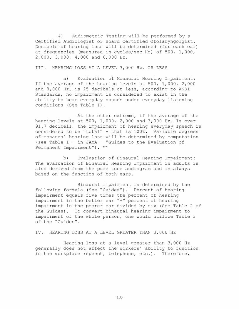
183
4) Audiometric Testing will be performed by a
Certified Audiologist or Board Certified Otolaryngolgist.
Decibels of hearing loss will be determined (for each ear)
at frequencies (measured in cycles/sec-Hz) of 500, 1,000,
2,000, 3,000, 4,000 and 6,000 Hz.
III. HEARING LOSS AT A LEVEL 3,000 Hz. OR LESS
a) Evaluation of Monaural Hearing Impairment:
If the average of the hearing levels at 500, 1,000, 2,000
and 3,000 Hz. is 25 decibels or less, according to ANSI
Standards, no impairment is considered to exist in the
ability to hear everyday sounds under everyday listening
conditions (See Table I).
At the other extreme, if the average of the
hearing levels at 500, 1,000, 2,000 and 3,000 Hz. Is over
91.7 decibels, the impairment of hearing everyday speech is
considered to be ―total‖ - that is 100%. Variable degrees
of monaural hearing loss will be determined by computation
(see Table I - in JAMA - ―Guides to the Evaluation of
Permanent Impairment‖). **
b) Evaluation of Binaural Hearing Impairment:
The evaluation of Binaural Hearing Impairment in adults is
also derived from the pure tone audiogram and is always
based on the function of both ears.
Binaural impairment is determined by the
following formula (See ―Guides‖). Percent of hearing
impairment equals five times the percent of hearing
impairment in the better ear ―+‖ percent of hearing
impairment in the poorer ear divided by six (See Table 2 of
the Guides). To convert binaural hearing impairment to
impairment of the whole person, one would utilize Table 3
of the ―Guides‖.
IV. HEARING LOSS AT A LEVEL GREATER THAN 3,000 HZ
Hearing loss at a level greater than 3,000 Hz
generally does not affect the workers' ability to function
in the workplace (speech, telephone, etc.). Therefore,

184
hearing loss at this level is not addressed in the AMA
Guides to the Evaluation of Permanent Impairment These
losses should be classified by a Board Certified
Otolaryngologist or Certified Audiologist as mild,
moderate, severe or profound.
** Information concerning the mechanism of
determination of extent of hearing loss in relationship to
workplace injury has been derived from information provided
by the JAMA Guides to the Evaluation of Permanent
Impairment of Hearing (Pages 922-925 in section labeled
Ear, Nose, and Throat and related structures).
PROTOCOL HISTORY:
Passed: 6/29/2000

185
INITIAL MEDICAL CASE MANAGEMENT ASSESSMENT
PROTOCOL GUIDELINES
The purpose of medical case management is to provide a
systematic approach for identifying and coordinating
quality medical care. While advocating for the injured
worker, the medical case manager will conduct an assessment
and will work as a liaison in planning, implementing and
evaluating on-going medical care as recommended by the
treatment team. The ultimate goal of medical case
management is to facilitate maximum medical recovery.
1. An Initial Medical Case Management
Assessment must be provided by a Qualified Rehabilitation
Counselor (QRC) or Qualified Rehabilitation Counselor
Intern (QRCI) as certified by the RI Department of Labor
and Training.
2. Prior to the assessment, the medical case
manager should review all available medical records and
clarify the purpose of the referral with the referral
source.
3. The initial interview will be conducted at a
mutually agreeable location.
4. The medical case management assessment
should include, but not be limited to, the following areas:
A. Statement of purpose for the
assessment.
B. Diagnosis and reference to the average
length of disability per the Presley Reed Disability
Advisor or another nationally recognized disability guide.
C. Summary of medical providers and
medical treatment to date.
D. Client’s present medical status
including history of current illness or injury, relevant
past medical history, description of functional limitations
and abilities and current treatment plan as outlined by the
treating physician.
E. Client’s social, educational and
vocational history.

186
F. Review of client’s job description and
potential availability of transitional duty through contact
with the employer.
G. Identify assets and/or limitations for
return to work.
H. Recommendations for medical management
goals to facilitate the treatment plan and timely return to
work.
5. The Initial Medical Case Management
Assessment will be submitted to the referral source within
two (2) weeks of the initial interview.
Protocol History:
Passed: 5/29/2000

187
INITIAL VOCATIONAL ASSESSMENT PROTOCOL GUIDELINES
The purpose of the Initial Vocational Assessment
protocol is to establish standard practices for a
vocational assessment through the application of consistent
procedures including the hierarchy of vocational
rehabilitation as defined in Appendix A. The goal of a
vocational assessment is to objectively measure an injured
worker’s employability to identify realistic return to work
opportunities and to develop appropriate vocational
recommendations based on the individual’s functional
status, education, and vocational background and
transferable skills. Progression in the hierarchy of
vocational rehabilitation is a sequential process based on
the injured worker’s functional status, transferable
skills, and established average weekly wage. It is
presumed that each level of the hierarchy will be addressed
when establishing vocational recommendations.
1. An Initial Vocational Assessment must be
provided by a Qualified Rehabilitation Counselor (QRC) or
Qualified Rehabilitation Counselor Intern (QRCI) as
certified by the RI Department of Labor and Training per
Section 28-33-41(h) of the Rhode Island Workers’
Compensation Act.
2. The initial interview may be conducted at a
mutually agreeable meeting place.
3. The referral source will provide claimant-
identifying data, medical records, including functional
capacities, if available, as they pertain to the work-
related injury, purpose of referral and special
instructions, if any.
4. During the initial interview, the
rehabilitation counselor should gather all relevant
information to include, but not be limited to; current
medical status, educational history, specialized training,
military experience, vocational history, including job
duties and wages, interests, and hobbies. The hierarchy of
vocational rehabilitation will be explained to the injured
worker at the time of the initial interview. One meeting

188
with the claimant will be allowed to complete the Initial
Vocational Assessment.
5. A Transferable Skills Analysis should be
completed provided that defined functional capacities are
identified in the medical records and a return to work with
the employer, to the original job (with or without
modifications) has been ruled out. The Transferable Skills
Analysis will be based on the following U.S. Department of
Labor publications: D.O.T. (Dictionary of Occupational
Titles), C.O.J. (Classification of Jobs), GOE (Guide for
Occupational Exploration), SOC (Selected Characteristics of
Occupations defined in the Dictionary of Occupational
Titles) and the O*NET. Software programs based on these
publications/references will be considered acceptable
resources for completing the analysis.
6. Testing is not considered part of the
Initial Vocational Assessment, but may be included as a
recommendation.
7. The initial Vocational Assessment Report
will address the following:
a. Purpose of the referral.
b. Brief summary of claimant’s medical
history and current status, description of functional
limitations and abilities, and any pending medical
treatment.
c. Claimant’s education, specialized
training and military experience.
d. Claimant’s vocational history,
including wages and length of employment. DOT (Dictionary
of Occupational Titles) numbers should accompany job titles
held.
e. Results of the Transferable Skills
Analysis, if completed.
f. Identification of assets and barriers
as they relate to continued vocational rehabilitation
services.
g. The hierarchy of vocational
rehabilitation will be considered in establishing
recommendations.

189
h. Recommendations.
8. The Initial Vocational Assessment report
will be submitted within two (2) weeks of the initial
interview.
Protocol History:
Passed: 5/29/2001
APPENDIX A

190
HIERARCHY OF VOCATIONAL REHABILITATION
1. Return to work, same employer, same job -
vocational services may include a job analysis and
coordination to return to work with the employer, but
usually no vocational services provided.
2. Return to work, same employer, different job -
work with the employer to identify a new position that
would fit the restrictions or modifications needed by the
injured worker.
3. Return to work, different employer, same job -
vocational services would assist in job development and
placement.
4. Return to work, different employer, different job
- vocational services may consist of performing a
transferable skills analysis, interest testing, job
development and job placement.
5. On-the-job training - identify a new employer
that can train the injured worker on the job. This program
can last between 3 months and 6 months.
6. Skills enhancement - vocational services may
identify a course to develop a skill prior to a job search.
This does not consist of a full retraining program.
7. Retraining - vocational assessment identifies
that the above options are not feasible and then identifies
a retraining program usually less than two (2) years in
length. The training program can range from a short-term
certificate program to a two (2) year associates degree
program. Vocational services would probably include
interest testing, transferable skills analysis, aptitude
testing, labor market research and vocational exploration
to support a training program.
Protocol History:
Passed: 5/29/2001

191
OCCUPATIONAL HEARING IMPAIRMENT TREATMENT PROTOCOL
This Protocol addresses the treatment of hearing
impairment that has been established as ―work related‖ by a
Board Certified Otorhinolaryngologist. Hearing impairment
may be related to a single event, such as trauma or a basal
skull fracture, or it may be related to exposure to
excessive noise in the work place.
DEGREES OF HEARING LOSS
0 to 25 dB Normal
25 to 45 dB Mild
45 to 60 dB Moderate
60 to 75 dB Moderately Severe
75 to 90 dB Severe
Over 90 dB Profound
Reference should be made to the OSHA table for age-
related hearing loss, data from which is attached hereto
and made a part of this Protocol.
I. TREATMENT OPTIONS
A. A trial of aural rehabilitation, if indicated,
usually in cases of mild loss if recommended by the
otorhinolaryngologist.
B. A hearing aid may be prescribed for occupational
hearing impairment related to exposure to excessive noise
in the workplace as determined by an otorhinolaryngologist.
The need for such will be determined by an
otorhinolaryngologist, who has provided the testing and
indicated that the loss is work-related and sufficient to
require the use of a hearing aid. This hearing aid may be
provided by an otolaryngologist.
C. A hearing aid may be prescribed for a monaural
hearing loss, if recommended by an otorhinolaryngologist.

192
II. TYPES OF HEARING AIDS TO BE CONSIDERED
A. BTE (Behind the ear)
B. CIC (Completely in ear canal) This is only
helpful in mild to moderate hearing loss and not in smaller
angular canals.
C. ITC (In the canal) This is stronger than the
CIC.
D. ITE (Inside the ear) This device is easier to
adjust the volume.
III. HEARING AID CIRCUITRY
A. Analog, is basic and the oldest type.
B. Programmable.
C. Digital, which is state of the art.
D. Disposable hearing aids are not acceptable
treatment.
E. Average life expectancy of a hearing aid is five
(5) years.
IV. SURGERY
A. Cochlear implants; used in patients with hearing
loss so extreme that the best hearing aid would have no
effect.
B. Reconstructive surgery, for either traumatic
abnormalities to the external ear canal, tympanic membrane,
or middle ear.
C. A second opinion is required before surgical
intervention may be performed.
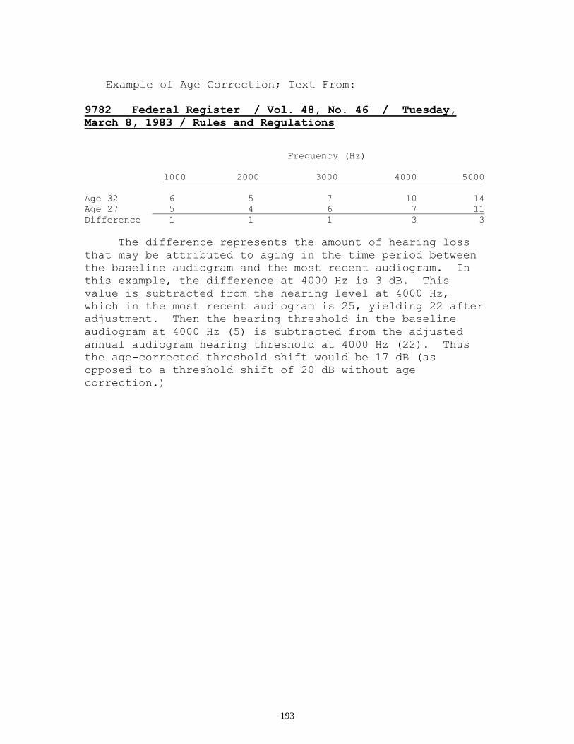
193
Example of Age Correction; Text From:
9782 Federal Register / Vol. 48, No. 46 / Tuesday,
March 8, 1983 / Rules and Regulations
Frequency (Hz)
1000 2000 3000 4000 5000
Age 32 6 5 7 10 14
Age 27 5 4 6 7 11
Difference 1 1 1 3 3
The difference represents the amount of hearing loss
that may be attributed to aging in the time period between
the baseline audiogram and the most recent audiogram. In
this example, the difference at 4000 Hz is 3 dB. This
value is subtracted from the hearing level at 4000 Hz,
which in the most recent audiogram is 25, yielding 22 after
adjustment. Then the hearing threshold in the baseline
audiogram at 4000 Hz (5) is subtracted from the adjusted
annual audiogram hearing threshold at 4000 Hz (22). Thus
the age-corrected threshold shift would be 17 dB (as
opposed to a threshold shift of 20 dB without age
correction.)

194
Table F-1 - Age Correction Values In Decibels for Males
Audiometric Test Frequencies (Hz)
Years 1000 2000 3000 4000 6000
20 or younger 5 3 4 5 8
21 5 3 4 5 8
22 5 3 4 5 8
23 5 3 4 6 9
24 5 3 5 6 9
25 5 3 5 7 10
26 5 4 5 7 10
27 5 4 6 7 11
28 6 4 6 8 11
29 6 4 6 8 12
30 6 4 6 9 12
31 6 4 7 9 13
32 6 5 7 10 14
33 6 5 7 10 14
34 6 5 8 11 15
35 7 5 8 11 15
36 7 5 9 12 16
37 7 6 9 12 17
38 7 6 9 13 17
39 7 6 10 14 18
40 7 6 10 14 19
41 7 6 10 14 20
42 8 7 11 16 20
43 8 7 12 16 21
44 8 7 12 17 22
45 8 7 13 18 23
46 8 8 13 19 24
47 8 8 14 19 24
48 9 8 14 20 25
49 9 9 15 21 26
50 9 9 16 22 27
51 9 9 16 23 28
52 9 10 17 24 29
53 9 10 18 25 30
54 10 10 18 26 31
55 10 11 19 27 32
56 10 11 20 28 34
57 10 11 21 29 35
58 10 12 22 31 36
59 11 12 22 32 37
60 or older 11 13 23 33 38
9782 Federal Register / Vol. 48, No. 46 / Tuesday, March 8, 1983 / Rules and
Regulations

195
Table F-2 - Age Correction Values in Decibels for Females
Audiometric Test Frequencies (Hz)
Years 1000 2000 3000 4000 6000
20 or younger 7 4 3 3 6
21 7 4 4 3 6
22 7 4 4 4 6
23 7 5 4 4 7
24 7 5 4 4 7
25 8 5 4 4 7
26 8 5 5 4 8
27 8 5 5 5 8
28 8 5 5 5 8
29 8 5 5 5 9
30 8 6 5 5 9
31 8 6 6 5 9
32 9 6 6 6 10
33 9 6 6 6 10
34 9 6 6 6 10
35 9 6 7 7 11
36 9 7 7 7 11
37 9 7 7 7 12
38 10 7 7 7 12
39 10 7 8 8 12
40 10 7 8 8 13
41 10 8 8 8 13
42 10 8 9 9 13
43 11 8 9 9 14
44 11 8 9 9 14
45 11 8 10 10 15
46 11 9 10 10 15
47 11 9 10 11 16
48 12 9 11 11 16
49 12 9 11 11 16
50 12 10 11 12 17
51 12 10 12 12 17
52 12 10 12 13 18
53 13 10 13 13 18
54 13 11 13 14 19
55 13 11 14 14 19
56 13 11 14 15 20
57 13 11 15 15 20
58 14 12 15 16 21
59 14 12 16 16 21
60 or older 14 12 16 17 22
9782 Federal Register / Vol. 48, No. 46 / Tuesday, March 8, 1983 / Rules and
Regulations
Protocol History:
Passed: 5/29/01

196
Diagnosis and Initial Treatment
Of
Occupational Asthma
I. Background:
A. Asthma is an airways disease of the lungs
characterized by the following:
1. Airway inflammation
2. Increased airway responsiveness to a variety
of stimuli; and
3. Airway obstruction that is partially or
completely reversible, either
spontaneously or with treatment.
The two essential clinical elements for the
diagnosis of asthma are airways obstruction which is
partially or totally reversible with treatment, and/or
airways hyperreactivity. Occupational asthma is asthma
that has its onset in association with workplace
exposure(s). Occupationally – aggravated asthma is asthma
that is aggravated by workplace exposure(s).
B. Causative agents are classified as sensitizers
(including but not limited to the appended list) or
irritants. Sensitizers cause inflammation through one or
more immunologic mechanisms, whereas irritants directly
inflame the airway. Occupational environments are often
complex, and it may be difficult to identify a single
specific causal agent.
C. A delay in diagnosis resulting in continued
exposure of the worker to even minute amounts of
sensitizers can lead to permanent and irreversible airways
disease or death.
D. An acute high level inhalation exposure to an
irritant may result in a permanent asthmatic condition
known as Reactive Airways Dysfunction Syndrome (RADS).
E. This guideline is meant to cover the majority of
tests and treatments that may be used to diagnose and
initially stabilize occupational and occupationally-

197
aggravated asthma. This guideline does not include
parameters of care for long term management of either
occupational or occupationally-aggravated asthma. It is
expected that approximately 10% of cases will fall outside
this guideline and require review on a case-by-case basis.
II. Criteria for Diagnosis:
A. Diagnosis of Occupational Asthma
1. Diagnosis of asthma within these guidelines
by a medical doctor,
using the appended algorithm.
2. Historical association between the onset of
asthma and work,
AND
3. At least one of the following criteria:
a. Documentation (see Occupational
History, Section III.B)
of workplace exposure to a category of
agents or processes
associated with asthma;
b. Work-related change in FEV1 or in peak
expiratory flow
(PEF);
c. Onset of respiratory signs and/or
symptoms within hours
after an acute high level occupational
inhalation exposure
to an irritant (RADS)
B. Diagnosis of Occupationally-Aggravated Asthma:
There must be a history of asthma prior to the occupational
exposure in question. Other diagnostic criteria are the
same as for new onset occupational asthma.
III. Medical Diagnosis and Initial Stabilization:
Physician Visits Allowed. The number of physician
visits needed to diagnose and stabilize cases of
occupational and occupationally-aggravated asthma is likely
to vary from patient to patient. Physicians must use their
judgment to determine the number of physician visits
necessary for diagnosis and initial stabilization.

198
IV. Establishing the Diagnosis:
A. Medical History:
1. Characteristic symptoms: wheeze, cough,
chest tightness,
shortness of breath.
2. Past respiratory history: prior diagnosis
of asthma, allergies,
eczema, rhinitis, bronchitis, sinusitis,
hayfever, chest colds,
and respiratory symptoms upon exertion,
exposure to minor
irritants, or exposure to cold air.
3. Review of systems: history of other
diseases with symptoms
that could mimic or precipitate asthma;
e.g., cardiovascular
disease with left ventricular dysfunction;
gastroesophageal
reflux.
4. Family history: asthma, atopy.
5. Smoking history: average # packs of
cigarettes per day x #
years smoked (pack years of smoking).
6. List of current medications.
7. Home, hobby, and environmental exposure
history to exclude other
causal or contributing factors.
B. Occupational History:
1. Description of the patient’s work tasks,
exposures and related
processes, both past and present.
2. Effect(s) of workplace exposures on
respiratory symptoms, with
emphasis on temporal associations. Note
whether symptoms
change on weekends and/or vacation.
3. Documentation of workplace exposures where
possible:

199
e.g., Material Safety Data Sheets (MSDS);
employer records; industrial hygiene monitoring data
from government agencies or private consultants.
4. Where data for characterizing exposures is
inadequate, worksite evaluation by an appropriate health
care provider or industrial hygienist may be necessary and
is encouraged.
C. Physical Examination:
1. Examination of head for rhinitis, nasal
polyps, conjunctivitis, and
sinusitis.
2. Chest percussion and auscultation.
3. Cardiovascular exam to rule out cardiogenic
explanation for
respiratory symptoms.
4. Skin exam for atopic dermatitis.
D. Diagnostic Tests Allowed:
1. A total of 11 spirometry studies is allowed.
For purposes of this
guideline, each study shall consist of a
minimum of 3 and a
maximum of 8 maneuvers, with at least the
initial study pre- and
post-inhaled bronchodilator.
a. Up to 2 follow-up spirometry studies
will be allowed to
establish a diagnosis of asthma.
b. Up to 8 pre- and post-shift spirometry
studies will be
allowed at the beginning and end of
each work week for
2 weeks.
c. When PEF diary and spirometric
monitoring are equivocal,
a longer absence from work may be
needed to establish or
rule out the diagnosis, with
(i) 1 repeat spirometry study allowed
at the beginning of the absence from work and 1 repeat

200
spirometry study allowed at the end of the absence from
work, and
(ii) the PEF diary monitoring repeated.
2. One Non-Specific Inhalation Challenge Test
Allowed:
If there is no significant improvement in
FEV1 in response to inhaled
bronchodilator, and if the existence of airways
hyperreactivity remains in question (see appended
algorithm), but only when:
a. Performed in a Hospital-based
Outpatient Setting,
b. Consistent with this guideline’s
Appended Algorithm, and
c. Under Supervision of a medical doctor
experienced in this type of
procedure.
3. Ten Specific Skin Tests with relevant
antigens allowed, but only when:
a. Performed by a Medical Doctor
Experienced in this type of
Procedure and,
b. In a hospital-based outpatient setting.
WARNING: SKIN TESTS ARE NON-EMERGENT PROCEDURES, WITH
SIGNIFICANT RISK OF SEVERE REACTION, INCLUDING DEATH.
4. Chest radiograph – 1 postero-anterior and 1
lateral view allowed.
5. Latex and laboratory animal dander RAST
test(s) for specific
work-related exposure – 1 allowed for each
antigen.
V. Initial Treatment Program:
A. Prevention of further exposure to causal or
precipitating agent(s):
1. When caused by a sensitizing agent, all
further exposure to the causal agent must be eliminated

201
because of the increased risk for irreversible airways
obstruction, severe bronchospasm and/or death. A statement
of the physician’s discussion of these and other risks with
the patient must be documented in the medical record.
2. When caused by an irritant, elimination of
exposure is desirable but significant reduction of exposure
may be sufficient. When elimination of exposure is not
possible, alternative approaches may include, in order of
preference:
a. Engineering controls such as local
exhaust ventilation
b. Appropriate use of respiratory
protection provided by the employer
B. Where these approaches fail and the clinical
condition warrants, removal of the workers from the
workplace may be necessary.
C. Medications:
1. Medications should only be used in
conjunction with prevention of further exposure as outlined
in Section V. A. above.
2. Spirometric testing is allowed as needed to
monitor effectiveness of therapy, not to exceed a maximum
of 11 spirometry studies allowed in Section IV. D. above.
Due to its unique nature, Occupational Asthma often
requires a more aggressive therapeutic approach than Non-
Occupational Asthma.
The recommended therapeutic approach is as
follows:
a. Step 1: Rapid-onset B-agonist as
needed for control of symptoms of asthma occurring less
than three times per week. If this fails, then:
b. Step 2: Inhaled low-to-medium dose
corticosteroids to treat underlying inflammation, combined
with a rapid-onset inhaled B-agonist as needed to control
symptoms of asthma. If this fails, then:
c. Step 3: Increase inhaled
corticosteroids to high dose, plus long-acting inhaled B-
agonist, and/or oral B-agonist and/or theophylline with
202
continued use of rapid-onset inhaled B-agonist as needed
to control symptoms of asthma. If this fails, then:
d. Step 4: Add an oral corticosteroid.
D. Patient Education (The following shall be
discussed with the patient at the initial physician visit
and repeated thereafter as necessary):
1. Key points about signs and symptoms of
asthma and characteristic
airway changes in asthma.
2. Asthma triggers and how to avoid them.
3. How medications work and their potential
adverse effects; instruction and
demonstration in the correct use of all medications
(e.g., proper use of MDIs).
4. Techniques of monitoring status of asthma,
such as PEF readings.
5. Indications for emergency care.
VI. Discharge Plan:
A. Future medical care will depend upon the outcome
of initial medical management. This guideline is meant to
address only the diagnosis and initial stabilization of
occupational and occupationally-aggravated asthma.
B. If causal or aggravating exposure is eliminated
or reduced and asthma symptoms resolve without medication,
no further medical management is needed. If symptoms have
resolved with medication, a period of medical follow-up
will be needed to determine the necessity for continued
medication and to establish an effective maintenance
regimen. Practitioners should consult other guidelines,
practice parameters and/or standards of care for guidance
in the long-term management of persistent symptoms of
asthma.
Protocol History:
Passed: 4/20/2004

203

204

205

206
IN RE:
Administrative Filing of the Medical Treatment Protocols of the
Medical Advisory Board of the
Rhode Island Workers’ Compensation Court
In accordance with the applicable statutes, in the State of Rhode Island, relating to
Administrative Procedures, the attached Protocols and Standards for Treatment for Compensable
Injuries (hereinafter referred to as “Protocols and Standards”), as promulgated by the Medical
Advisory Board for the Workers’ Compensation Court, in accordance with R.I.G.L. §28-30-22
and formally approved and adopted by the Chief Judge of the Workers’ Compensation Court, are
heretofore presented and filed with the office of the Rhode Island Secretary of State.
It is of the utmost importance, however, that it be noted that these Protocols and
Standards are filed out of an abundance of caution and in strict adherence to the Rhode Island
Administrative Procedures Act, so-called, as set forth in R.I.G.L. §42-35-1 et seq. The Protocols
and Standards are in no way intended to be, nor are they to be used as a binding rule or regulation.
These Protocols and Standards are intended to outline options of appropriate methods and types of
intervention to be utilized by physicians and other healthcare providers for what is believed to be
some of the most frequent work-related injuries seen in Rhode Island.
It is in this spirit that these Protocols and Standards are to be used, and no other purpose or reason
is intended.