SPRITE and ASSAM: web servers for side chain 3D-motif ...
7
SPRITE and ASSAM: web servers for side chain 3D-motif searching in protein structures Nurul Nadzirin 1 , Eleanor J. Gardiner 2 , Peter Willett 2 , Peter J. Artymiuk 3, * and Mohd Firdaus-Raih 1, * 1 School of Biosciences and Biotechnology, Faculty of Science and Technology, Universiti Kebangsaan Malaysia, 43600 UKM Bangi, Malaysia, 2 Information School and 3 Department of Molecular Biology and Biotechnology, Krebs Institute, University of Sheffield, Western Bank, Sheffield S10 2TN, UK Received February 12, 2012; Revised April 11, 2012; Accepted April 18, 2012 ABSTRACT Similarities in the 3D patterns of amino acid side chains can provide insights into their function despite the absence of any detectable sequence or fold similarities. Search for protein sites (SPRITE) and amino acid pattern search for substructures and motifs (ASSAM) are graph theoretical programs that can search for 3D amino side chain matches in protein structures, by representing the amino acid side chains as pseudo-atoms. The geometric rela- tionship of the pseudo-atoms to each other as a pattern can be represented as a labeled graph where the pseudo-atoms are the graph’s nodes while the edges are the inter-pseudo-atomic dis- tances. Both programs require the input file to be in the PDB format. The objective of using SPRITE is to identify matches of side chains in a query struc- ture to patterns with characterized function. In contrast, a 3D pattern of interest can be searched for existing occurrences in available PDB structures using ASSAM. Both programs are freely accessible without any login requirement. SPRITE is available at http://mfrlab.org/grafss/sprite/ while ASSAM can be accessed at http://mfrlab.org/grafss/assam/. INTRODUCTION In biological macromolecules, the 3-dimensional (3D) structure determines the functionality of the molecule. Therefore, it has long been recognized that similarities in structure can be a valuable guide to similarities in function, even if there is no detectable sequence similarity (1). For this reason, tools and services that are able to detect similarities in folding between different protein structures, such as DALI (2), have been available since the 1990s. However, it has also been clear for decades that similar constellations of amino acid residues in unre- lated proteins can give rise to similar chemical activity, even where there is no fold similarity, sequence similarity or common evolutionary precursor. The classic example of this convergent evolution at the atomic level is the ‘catalytic triad’ of an aspartate, a histidine and a serine which was found to occur in both chymotrypsin and subtilisin (3). The result of this association in these unre- lated enzymes is that the serine becomes very nucleophilic and is able to catalyze peptide bond cleavage. Such 3D amino acid constellations are therefore of interest because they may be involved in key functions that can include structure stabilization, binding and catalysis. The recogni- tion of such similarities may hence be a valuable guide to function. The ability to search for specific 3D arrangements of amino acids can be especially useful for structural biolo- gists especially for identifying residues of interest in newly solved structures. This can be of value in assigning function to proteins of unknown function, or in identify- ing ligands that bind to similar sites in different proteins. One important use of such searches lies in the area of structural genomics where a large number of structures of proteins with unknown functions have been solved. Several existing services that allow for 3D motif searching include ProFunc/JESS (4), GIRAF (5), PINTS (6), SPASM (7), RIGOR (7), SuMo (8), RASMOT-3D PRO (9) and SA-Mot (10). We have previously described a program, amino acid pattern search for substructures and motifs (ASSAM) that successfully uses a graph theoretical approach to search for and identify 3D motifs in protein structures (11,12). Here, we present a web service that deploys two graph theoretical computer programs. The first is a new program called search for protein sites (SPRITE), which allows the 3D structure of a protein to be searched against a database of curated sites, and the second is ASSAM Correspondence may also be addressed to Peter J. Artymiuk. Tel: +44 114 222 4190; Fax: +44 114 222 2800; Email: p.artymiuk@sheffield.ac.uk *To whom correspondence should be addressed. Tel: +603 89215961; Fax: +603 89252698; Email: fi[email protected] W380–W386 Nucleic Acids Research, 2012, Vol. 40, Web Server issue Published online 9 May 2012 doi:10.1093/nar/gks401 ß The Author(s) 2012. Published by Oxford University Press. This is an Open Access article distributed under the terms of the Creative Commons Attribution Non-Commercial License (http://creativecommons.org/licenses/ by-nc/3.0), which permits unrestricted non-commercial use, distribution, and reproduction in any medium, provided the original work is properly cited. Downloaded from https://academic.oup.com/nar/article/40/W1/W380/1073692 by guest on 04 February 2022
-
Upload
khangminh22 -
Category
Documents
-
view
0 -
download
0
Transcript of SPRITE and ASSAM: web servers for side chain 3D-motif ...
SPRITE and ASSAM: web servers for side chain3D-motif searching in protein structuresNurul Nadzirin1, Eleanor J. Gardiner2, Peter Willett2, Peter J. Artymiuk3,* and
Mohd Firdaus-Raih1,*
1School of Biosciences and Biotechnology, Faculty of Science and Technology, Universiti KebangsaanMalaysia, 43600 UKM Bangi, Malaysia, 2Information School and 3Department of Molecular Biology andBiotechnology, Krebs Institute, University of Sheffield, Western Bank, Sheffield S10 2TN, UK
Received February 12, 2012; Revised April 11, 2012; Accepted April 18, 2012
ABSTRACT
Similarities in the 3D patterns of amino acid sidechains can provide insights into their functiondespite the absence of any detectable sequence orfold similarities. Search for protein sites (SPRITE)and amino acid pattern search for substructuresand motifs (ASSAM) are graph theoretical programsthat can search for 3D amino side chain matches inprotein structures, by representing the amino acidside chains as pseudo-atoms. The geometric rela-tionship of the pseudo-atoms to each other as apattern can be represented as a labeled graphwhere the pseudo-atoms are the graph’s nodeswhile the edges are the inter-pseudo-atomic dis-tances. Both programs require the input file to bein the PDB format. The objective of using SPRITEis to identify matches of side chains in a query struc-ture to patterns with characterized function. Incontrast, a 3D pattern of interest can be searchedfor existing occurrences in available PDB structuresusing ASSAM. Both programs are freely accessiblewithout any login requirement. SPRITE is availableat http://mfrlab.org/grafss/sprite/ while ASSAM canbe accessed at http://mfrlab.org/grafss/assam/.
INTRODUCTION
In biological macromolecules, the 3-dimensional (3D)structure determines the functionality of the molecule.Therefore, it has long been recognized that similarities instructure can be a valuable guide to similarities infunction, even if there is no detectable sequence similarity(1). For this reason, tools and services that are able todetect similarities in folding between different proteinstructures, such as DALI (2), have been available since
the 1990s. However, it has also been clear for decadesthat similar constellations of amino acid residues in unre-lated proteins can give rise to similar chemical activity,even where there is no fold similarity, sequence similarityor common evolutionary precursor. The classic exampleof this convergent evolution at the atomic level is the‘catalytic triad’ of an aspartate, a histidine and a serinewhich was found to occur in both chymotrypsin andsubtilisin (3). The result of this association in these unre-lated enzymes is that the serine becomes very nucleophilicand is able to catalyze peptide bond cleavage. Such 3Damino acid constellations are therefore of interest becausethey may be involved in key functions that can includestructure stabilization, binding and catalysis. The recogni-tion of such similarities may hence be a valuable guide tofunction.
The ability to search for specific 3D arrangements ofamino acids can be especially useful for structural biolo-gists especially for identifying residues of interest in newlysolved structures. This can be of value in assigningfunction to proteins of unknown function, or in identify-ing ligands that bind to similar sites in different proteins.One important use of such searches lies in the area ofstructural genomics where a large number of structuresof proteins with unknown functions have been solved.Several existing services that allow for 3D motif searchinginclude ProFunc/JESS (4), GIRAF (5), PINTS (6),SPASM (7), RIGOR (7), SuMo (8), RASMOT-3D PRO(9) and SA-Mot (10).
We have previously described a program, amino acidpattern search for substructures and motifs (ASSAM)that successfully uses a graph theoretical approach tosearch for and identify 3D motifs in protein structures(11,12). Here, we present a web service that deploys twograph theoretical computer programs. The first is a newprogram called search for protein sites (SPRITE), whichallows the 3D structure of a protein to be searched againsta database of curated sites, and the second is ASSAM
Correspondence may also be addressed to Peter J. Artymiuk. Tel: +44 114 222 4190; Fax: +44 114 222 2800; Email: [email protected]*To whom correspondence should be addressed. Tel: +603 89215961; Fax: +603 89252698; Email: [email protected]
W380–W386 Nucleic Acids Research, 2012, Vol. 40, Web Server issue Published online 9 May 2012doi:10.1093/nar/gks401
� The Author(s) 2012. Published by Oxford University Press.This is an Open Access article distributed under the terms of the Creative Commons Attribution Non-Commercial License (http://creativecommons.org/licenses/by-nc/3.0), which permits unrestricted non-commercial use, distribution, and reproduction in any medium, provided the original work is properly cited.
Dow
nloaded from https://academ
ic.oup.com/nar/article/40/W
1/W380/1073692 by guest on 04 February 2022

itself, which accepts a 3D amino acid pattern as a queryfor searching against a database of protein structures.
PROGRAMS AND METHODS
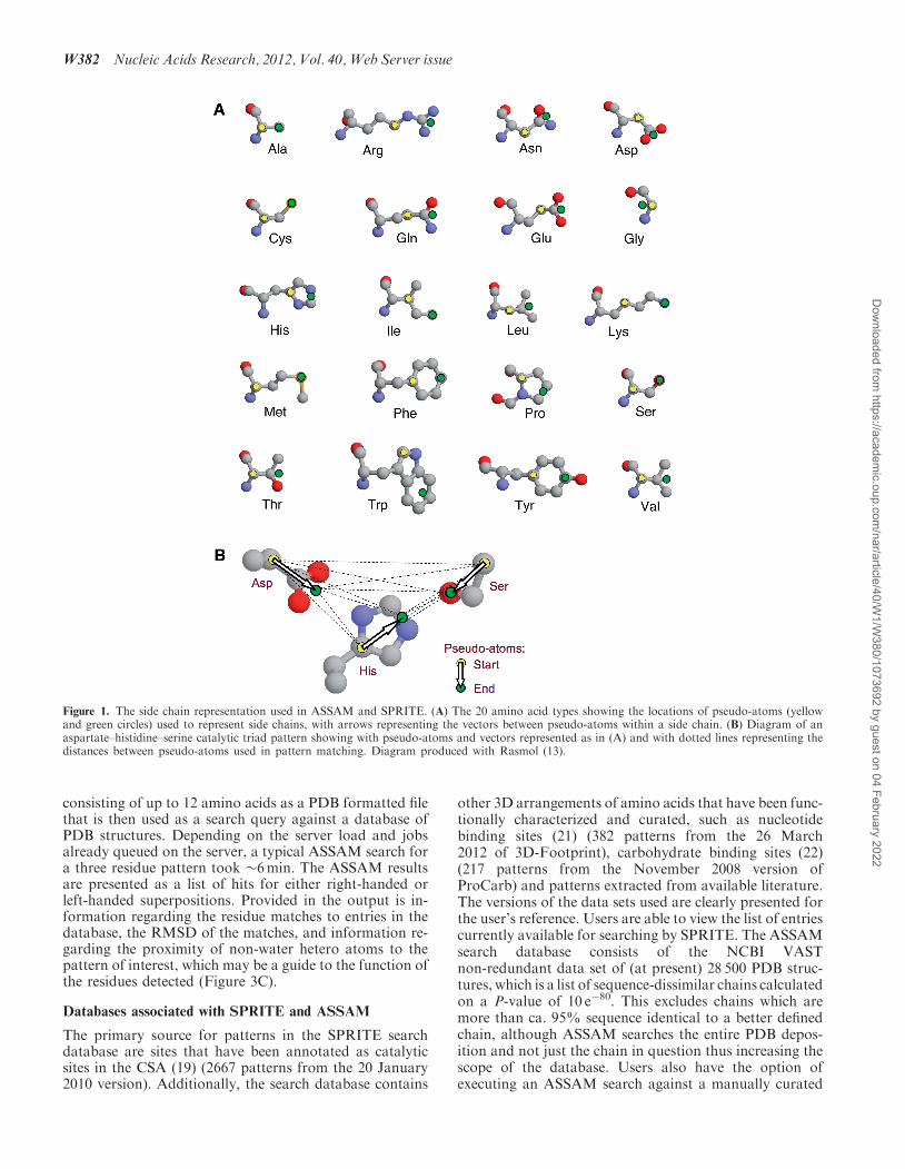
The basic concept behind the search methodology for bothSPRITE and ASSAM has been described previously(11,12). Briefly, the protein structure is represented as agraph with the nodes representing individual amino acidside chains and the inter-node geometric relationships arethe graphs. Each node consists of two pseudo-atomswhich are used to generate a vector, and each suchvector corresponds to one of the nodes in a graph(Figure 1A). The positions of the pseudo-atoms arechosen to emphasize the functional part of the sidechain corresponding to that node. The geometric relation-ships between pairs of residues are defined in terms ofdistances calculated between the corresponding vectors,and these relationships correspond to the edges of agraph (Figure 1B). Specifically, if we let S, M and Edenote the start, middle and end, respectively, of avector, then the graph edges contain five parts, thesebeing the SS, SE, ES, EE and MM distances (althoughonly a subset of these five distances is normally used tospecify a query pattern) (11).
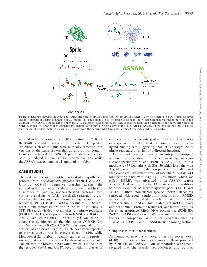
The current version of ASSAM, we report here, usesa maximal common subgraph (MCS) approach. Thisinvolves a fast initial screen using the Carraghan andPardalos (1990) (14) clique detection algorithm to rapidlydetermine if any structural correspondences actually exist,followed, if appropriate, by the use of the Bron andKerbosch (1973) (15) MCS algorithm to enumerate allthe possible correspondences. SPRITE continues to usethe Ullmann algorithm (16) of the original ASSAM butin a reversed approach in which a database of queries iscompared with a single structure (Figure 2). The SPRITEand ASSAM programs provide separate outputs for bothleft-handed and right-handed superpositions, which, toour knowledge, is unique to these servers, and which canyield valuable chemical information (17) as discussedbelow.
The SPRITE program enables the user to examine asingle complete protein structure in order to identify orannotate functional sites that have been documented inother structures. Such a utility can assist in providinginsights into the potential function of proteins that yieldno detectable sequence or fold similarity to existingexamples in the databases and thus help direct functiondetermination experiments. The ASSAM program, incontrast, enables the user to search the entire ProteinData Bank (18) for occurrences of a specific small aminoacid motif. This can provide insights into the conservationand/or evolution of specific 3D arrangements such as cata-lytic sites. If required, the SPRITE and ASSAM programscan be used in series. As an example, a user can submit aquery structure to identify which known motifs arepresent. Motifs of interest can immediately be submittedfor an ASSAM search via the web interface to identifyother structures where such motifs occur.
SPRITE: searching for sites in a protein structure query
The 3D SPRITE program accepts a PDB formatted file asinput and this structure is searched against databases ofsites annotated from X-ray crystallographic structuresarchived in the PDB. Perhaps the well-known example ofsuch a database is the Catalytic Site Atlas (CSA) in whichPorter et al. (19) used literature searches, hand annotationand homology searches to identify amino acids unequivo-cally involved in the catalytic activity of enzymes whosestructures were stored in the PDB. In order to carry out aSPRITE search, the 3D arrangement of amino acids fromthe input file is converted into a graph representation thatis then compared against the database of graph represen-tations for patterns of sites in protein structures. MostSPRITE searches tested take <2min to complete inclusiveof upload times under the server’s normal daily load.Results are presented in a main menu (Figure 3) that
provides a number of visualization options to the user,and hyperlinks to results of structural superpositions.When superposing two protein folds, there is a differencebetween, for example, a left-handed a-helical bundle and aright-handed one and they cannot be considered asequivalent. However, at the level of side chains, twonon-evolutionarily related groupings of amino acids donot necessarily have to be of the same handedness inorder to have the same chemical activity (17), theymerely need to agree in terms of inter-residue distances.An example of this is the Asp–His–Ser catalytic triad inprolyl oligopeptidase, which is on the opposite hand tothat in chymotrypsin (20), although it carries out thesame peptidase function, albeit on a different substrate.Therefore, the program considers both right-handed andleft-handed superpositions to be equally valid in principleand both are given in two separate results lists.The three output visualization options for both the
right- and left-handed superpositions are: (i) a list of thePDB structures which contain sites that match to sites inthe query structure; (ii) a full list of matches that includeRMSD values for the superpositions based on pseudo-atom positions, mapping of the query residues (number,chain and amino acid) to their database hits and a matrixfor input as a TRANSFORM command in the CCP4macromolecular crystallography software suite (http://www.ccp4.ac.uk; Figure 3B); and (iii) a list arranged bynon-redundant matching sites in the query structure. Thesecond option, which presents the full details of the hits,also allows the user to execute an ASSAM search usingthe residues in the query structure with hits to the SPRITEpattern database (Figure 2). All of the output browsingoptions listed also allow for visualization of the hits in aJmol (http://www.jmol.org/) molecular viewer window.Users have the options of viewing superpositions of thequery to the database matches (Figure 3D) or of viewingthe residues in the query structure that have matches in thedatabase (Figure 3E).
ASSAM: searching for a pattern in a structure database
The ASSAM program enables users to search for aminoacid constellations of interest in a database of PDB struc-tures (11). Users can input the coordinates of a 3D motif
Nucleic Acids Research, 2012, Vol. 40, Web Server issue W381
Dow
nloaded from https://academ
ic.oup.com/nar/article/40/W
1/W380/1073692 by guest on 04 February 2022
consisting of up to 12 amino acids as a PDB formatted filethat is then used as a search query against a database ofPDB structures. Depending on the server load and jobsalready queued on the server, a typical ASSAM search fora three residue pattern took �6min. The ASSAM resultsare presented as a list of hits for either right-handed orleft-handed superpositions. Provided in the output is in-formation regarding the residue matches to entries in thedatabase, the RMSD of the matches, and information re-garding the proximity of non-water hetero atoms to thepattern of interest, which may be a guide to the function ofthe residues detected (Figure 3C).
Databases associated with SPRITE and ASSAM
The primary source for patterns in the SPRITE searchdatabase are sites that have been annotated as catalyticsites in the CSA (19) (2667 patterns from the 20 January2010 version). Additionally, the search database contains
other 3D arrangements of amino acids that have been func-tionally characterized and curated, such as nucleotidebinding sites (21) (382 patterns from the 26 March2012 of 3D-Footprint), carbohydrate binding sites (22)(217 patterns from the November 2008 version ofProCarb) and patterns extracted from available literature.The versions of the data sets used are clearly presented forthe user’s reference. Users are able to view the list of entriescurrently available for searching by SPRITE. The ASSAMsearch database consists of the NCBI VASTnon-redundant data set of (at present) 28 500 PDB struc-tures, which is a list of sequence-dissimilar chains calculatedon a P-value of 10 e�80. This excludes chains which aremore than ca. 95% sequence identical to a better definedchain, although ASSAM searches the entire PDB depos-ition and not just the chain in question thus increasing thescope of the database. Users also have the option ofexecuting an ASSAM search against a manually curated
Figure 1. The side chain representation used in ASSAM and SPRITE. (A) The 20 amino acid types showing the locations of pseudo-atoms (yellowand green circles) used to represent side chains, with arrows representing the vectors between pseudo-atoms within a side chain. (B) Diagram of anaspartate–histidine–serine catalytic triad pattern showing with pseudo-atoms and vectors represented as in (A) and with dotted lines representing thedistances between pseudo-atoms used in pattern matching. Diagram produced with Rasmol (13).
W382 Nucleic Acids Research, 2012, Vol. 40, Web Server issue
Dow
nloaded from https://academ
ic.oup.com/nar/article/40/W
1/W380/1073692 by guest on 04 February 2022
non-redundant version of the PDB consisting of 57 500 ofthe 80 400 available structures. For this data set, repeatedstructures such as mutants were manually removed, butversions of the same protein that do and do not containligands are retained. The SPRITE pattern database is peri-odically updated as new patterns become available whilethe ASSAM search database is updated monthly.
CASE STUDIES
The first example we present here is that of a hypotheticalprotein from Archaeoglobus fulgidus (PDB ID: 2OO2;UniProt: O28492). Sequence searches against thenon-redundant sequence databases only identified hits toa number of putative uncharacterized proteins fromvarious organisms. A DALI search (23) returned severalmatches, the most significant being an eight-heme nitritereductase (PDB ID: 3F29) with a Z-value of 9.1. Severalother nitrite reductases are also in the list of matches. ASPRITE search yielded two matches to a nitrite reductase(PDB ID: 1NID), with pseudo-atom RMSDs of 0.44 and0.55 A over two residues. Further analysis was done togauge the significance of the hits, using CCRXP (24)and Metapocket 2.0 (25). CCRXP was designed to findclusters of conserved residues, which have been reportedto play a crucial role in protein function (26), whileMetapocket 2.0 is able to identify cavities on the proteinsurface that could indicate the location of an active site.The hit with the lower RMSD value, which is made up ofthe residues Phe43 and Gly47, occurs within a cluster of
conserved residues consisting of six residues. This regionoverlaps with a cleft that potentially constitutes aligand-binding site suggesting that 2OO2 might be anitrite reductase or a similarly directed function.The second example involves an intriguing ion-pair
network from the structure of a Salmonella typhimuriumsucrose specific porin ScrY (PDB ID: 1A0S) (27). In thismotif, Arg 437 ion pairs with Glu 439 which ion pairs withArg 441, which, in turn, also ion pairs with Glu 480, andthen completes the square array of side chains by Glu 480ions pairing back with Arg 437. This motif, which wecalled ‘RERE’, was submitted to an ASSAM searchwhich yielded as expected the 1A0S structure in additionto other examples of sucrose specific porin (1A0T and1OH2). Other non-sucrose-specific porin structuresretrieved with good pseudo-atom superposition RMSDvalues include hits that also involve an Arg and a Glufrom one subunit and a 2-fold related Arg and Glu fromanother subunit. From the results list, one interesting hit isfor a bacteriophage RB69 DNA polymerase (PDB ID:2ATQ, RMSD=0.92 A). We discuss this examplefurther in comparison with other programs such asRASMOT-3D PRO and SPASM in the following section.
Comparisons with other methods
As mentioned previously above, other web servers exist(4–10) that allow analogous enquiries to those providedby SPRITE or ASSAM. Our comparative assessmentrevealed that the search methodologies and outputs
Figure 2. Diagram showing the input and output structure of SPRITE and ASSAM. (i) SPRITE accepts a whole structure in PDB format as inputand (ii) compares it against a database of 3D motifs. (iii) The output is a list of amino acids in the query structure that matches to patterns in thedatabase. (iv) ASSAM’s inputs can be either any 2–12 pattern residues given by the user, or selected from the hit residues in the query structure of aSPRITE search. (v) ASSAM then compares this pattern to representative structures in the PDB. (vi) The ASSAM output is a list of PDB structuresthat contain the query motif. An example is shown with the superposed hit residues identified and magnified as red circles.
Nucleic Acids Research, 2012, Vol. 40, Web Server issue W383
Dow
nloaded from https://academ
ic.oup.com/nar/article/40/W
1/W380/1073692 by guest on 04 February 2022

presented by these programs differ in significant waysfrom the searches that SPRITE and ASSAM are able tocarry out. In many respects, the currently availableprograms and SPRITE and ASSAM are complementary,and an integration of the results can provide usefulinsights or enable further investigations to be carried out.The SPASM program (7) and RASMOT-3D PRO (9)
service are similar to the ASSAM server in being able tosearch for 3D motifs in a database of structures. However,the SPASM and RASMOT-3D PRO structural represen-tations are different compared to ASSAM. SPASM usesthe Ca and the centre of gravity of the side chain whileRASMOT-3D PRO uses the Ca and Cb positions of eachresidue. In contrast, ASSAM and SPRITE use atoms from
the functional part of the side chain itself (Figure 1) andare therefore more sensitive to the positions of the ends ofside chains and less dependent on the main chain position.The use of the RERE motif from 1A0S, which we havedescribed above, serves to demonstrate this effect. Whilethe RASMOT-3D PRO was also able to retrieve the 1A0Tstructure, a sucrose-containing version of 1A0S, with anRMSD of 0.13, other matches in the results were not ofthe same RERE motif. For example, the next hits in thelist provided by a RASMOT-3D PRO search were aGAAT motif (PDB ID: 1K42; RMSD=0.28) and anALER motif (PDB ID: 1X51; RMSD=0.4).
The ASSAM search was able to retrieve a hit fora bacteriophage RB69 DNA polymerase (2ATQ)
Figure 3. Snapshots of various pages from the web server showing the output of a SPRITE search using a serine proteinase structure (PDB ID:4CHA). (A) List of motifs in the database that match to the query protein. (B) A detailed description of the results, showing the matched residuesand the TRANSFORM command for CCP4; users can also opt to execute an ASSAM search on any hit of interest. (C) An example of an ASSAMsearch output, using the SPRITE output as a query pattern. (D) Jmol view of the superposition of a site of interest with the motif in the databasethat it matches to, which can be selected from page (A). (E) Jmol view showing the position of a site of interest relative to the query structure, whichcan be chosen from the page depicted in (B).
W384 Nucleic Acids Research, 2012, Vol. 40, Web Server issue
Dow
nloaded from https://academ
ic.oup.com/nar/article/40/W
1/W380/1073692 by guest on 04 February 2022
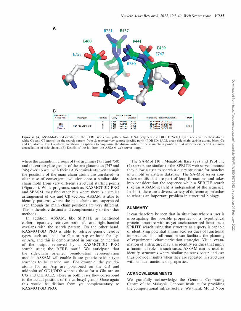
where the guanidium groups of two arginines (751 and 750)and the carboxylate groups of the two glutamates (747 and745) overlap well with their 1A0S equivalents even thoughthe positions of the main chain atoms are unrelated—aclear case of convergent evolution onto a similar side-chain motif from very different structural starting points(Figure 4). While programs, such as RASMOT-3D PROand SPASM, may find other hits where there is a similararrangement of Ca and Cb vectors, ASSAM is able toidentify patterns where the side chains are superposedeven though the main chain positions are very different.This is therefore distinct and complementary to the othermethods.
In addition, ASSAM, like SPRITE as mentionedearlier, separately retrieves both left- and right-handedoverlaps with the search pattern. On the other hand,RASMOT-3D PRO is able to retrieve generic residuetypes, such as acidic for Glu or Asp or basic for Lysor Arg, and this is demonstrated in our earlier mentionof the output retrieved by a RASMOT-3D PROsearch using the RERE motif. We anticipate thatthe side-chain oriented pseudo-atom representationused in ASSAM will enable future generic residue typesearches to be carried out. For example, the pseudo-atoms for an Asp are positioned on the CB andmidpoint of OD1/OD2 whereas those for a Glu are onCG and OE1/OE2, where in both cases they correspondto the actual position of the carboxyl group. Once againthis would be distinct from yet complementary toRASMOT-3D PRO.
The SA-Mot (10), MegaMotifBase (28) and ProFunc(4) servers are similar to the SPRITE web server becausethey allow a user to search a query structure for matchesin a motif or pattern database. The SA-Mot server con-siders motifs that are part of loop formations and takesinto consideration the sequence while a SPRITE search(like an ASSAM search) is independent of the sequence.In short, there are a diverse variety of different approachesto what is an important problem in structural biology.
SUMMARY
It can therefore be seen that in situations where a user isinvestigating the possible properties of a hypotheticalprotein structure with as yet uncharacterized function, aSPRITE search using that structure as a query is capableof identifying potential amino acid residues of functionalimportance. This information can facilitate the planningof experimental characterization strategies. Visual exam-ination of a structure may also identify residues that implya functional role. In such cases, ASSAM can be used toidentify structures where similar patterns occur and canthus provide insights when they are repeated in structureswith similar functions or properties.
ACKNOWLEDGEMENTS
We gratefully acknowledge the Genome ComputingCentre of the Malaysia Genome Institute for providingthe computational infrastructure. We thank Mohd Noor
Figure 4. (A) ASSAM-derived overlap of the RERE side chain pattern from DNA polymerase (PDB ID: 2ATQ, cyan side chain carbon atoms,white Ca and Cb atoms) on the search pattern from S. typhimurium sucrose specific porin (PDB ID: 1A0S, green side chain carbon atoms, black Caand Cb atoms). The Ca atoms are shown as spheres to emphasize the dissimilarities in the main chain positions that nevertheless permit a similarconstellation of side chains. (B) Details of the hit from the ASSAM web server output.
Nucleic Acids Research, 2012, Vol. 40, Web Server issue W385
Dow
nloaded from https://academ
ic.oup.com/nar/article/40/W
1/W380/1073692 by guest on 04 February 2022

Mat Isa and Hafiza Aida Ahmad for technical assistancewith server operations.
FUNDING
Universiti Kebangsaan Malaysia [UKM-GGPM-KPB-101-2010 to M.F.-R.]; Ministry of Higher Education,Malaysia, National Science Fellowship (to N.N.).Funding for open access charge: University KebangsaanMalaysia grant [UKM-DLP-2012-018].
Conflict of interest statement. None declared.
REFERENCES
1. Artymiuk,P.J., Poirrette,A.R., Rice,D.W. and Willett,P. (1997) Apolymerase I palm in adenylyl cyclase? Nature, 388, 33–34.
2. Holm,L. and Sander,C. (1995) Dali: a network tool for proteinstructure comparison. Trends Biochem. Sci., 20, 478–480.
3. Warshel,A., Naray-Szabo,G., Sussman,F. and Hwang,J.K. (1989)How do serine proteases really work? Biochemistry, 28, 3629–3637.
4. Laskowski,R.A., Watson,J.D. and Thornton,J.M. (2005)ProFunc: a server for predicting protein function from 3Dstructure. Nucleic Acids Res., 33, W89–W93.
5. Kinjo,A.R. and Nakamura,H. (2009) Comprehensive structuralclassification of ligand-binding motifs in proteins. Structure, 17,234–246.
6. Stark,A., Sunyaev,S. and Russell,R.B. (2003) A model forstatistical significance of local similarities in structure.J. Mol. Biol., 326, 1307–1316.
7. Kleywegt,G.J. (1999) Recognition of spatial motifs in proteinstructures. J. Mol. Biol., 285, 1887–1897.
8. Jambon,M., Andrieu,O., Combet,C., Deleage,G., Delfaud,F. andGeourjon,C. (2005) The SuMo server: 3D search for proteinfunctional sites. Bioinformatics, 21, 3929–3930.
9. Debret,G., Martel,A. and Cuniasse,P. (2009) RASMOT-3D PRO: a3D motif search webserver. Nucleic Acids Res., 37, W459–W464.
10. Nuel,G., Regad,L., Martin,J. and Camproux,A.C. (2010)Exact distribution of a pattern in a set of random sequencesgenerated by a Markov source: applications to biological data.Algorithms Mol. Biol., 5, 15.
11. Spriggs,R.V., Artymiuk,P.J. and Willett,P. (2003) Searching forpatterns of amino acids in 3D protein structures. J. Chem. Inf.Comput. Sci., 43, 412–421.
12. Poirrette,A.R., Artymiuk,P.J., Grindley,H.M., Rice,D.W. andWillett,P. (1994) Structural similarity between binding sites in
influenza sialidase and isocitrate dehydrogenase: implications foran alternative approach to rational drug design. Protein Sci., 3,1128–1130.
13. Sayle,R.A. and Milner-White,E.J. (1995) RASMOL: biomoleculargraphics for all. Trends Biochem. Sci., 20, 374.
14. Carraghan,R. and Pardalos,P.M. (1990) An exact algorithm forthe maximum clique problem. Oper. Res. Lett., 9, 375–382.
15. Bron,C. and Kerbosch,J. (1973) Algorithm 457: finding all cliquesof an undirected graph. Commun. ACM, 16, 575–577.
16. Ullmann,J.R. (1976) An algorithm for subgraph isomorphism.J. ACM, 23, 31–42.
17. Garavito,R.M., Rossmann,M.G., Argos,P. and Eventoff,W.(1977) Convergence of active center geometries. Biochemistry,16, 5065–5071.
18. Berman,H.M., Westbrook,J., Feng,Z., Gilliland,G., Bhat,T.N.,Weissig,H., Shindyalov,I.N. and Bourne,P.E. (2000) The ProteinData Bank. Nucleic Acids Res., 28, 235–242.
19. Porter,C.T., Bartlett,G.J. and Thornton,J.M. (2004) The CatalyticSite Atlas: a resource of catalytic sites and residues identified inenzymes using structural data. Nucleic Acids Res., 32, D129–D133.
20. Fulop,V., Bocskei,Z. and Polgar,L. (1998) Prolyl oligopeptidase:an unusual beta-propeller domain regulates proteolysis. Cell, 94,161–170.
21. Contreras-Moreira,B. (2010) 3D-footprint: a database for thestructural analysis of protein-DNA complexes. Nucleic Acids Res.,38, D91–D97.
22. Malik,A., Firoz,A., Jha,V. and Ahmad,S. (2010) PROCARB: adatabase of known and modelled carbohydrate-binding proteinstructures with sequence-based prediction tools. Adv.Bioinformatics, 436036.
23. Holm,L. and Rosenstrom,P. (2010) Dali server: conservationmapping in 3D. Nucleic Acids Res., 38, W545–W549.
24. Ahmad,S., Keskin,O., Mizuguchi,K., Sarai,A. and Nussinov,R.(2010) CCRXP: exploring clusters of conserved residues in proteinstructures. Nucleic Acids Res., 38, W398–W401.
25. Zhang,Z., Li,Y., Lin,B., Schroeder,M. and Huang,B. (2011)Identification of cavities on protein surface using multiplecomputational approaches for drug binding site prediction.Bioinformatics, 27, 2083–2088.
26. DeLano,W.L. (2002) Unraveling hot spots in bindinginterfaces: progress and challenges. Curr. Opin. Struct. Biol.,12, 14–20.
27. Forst,D., Welte,W., Wacker,T. and Diederichs,K. (1998) Structureof the sucrose-specific porin ScrY from Salmonella typhimuriumand its complex with sucrose. Nat. Struct. Biol., 5, 37–46.
28. Pugalenthi,G., Suganthan,P.N., Sowdhamini,R. andChakrabarti,S. (2008) MegaMotifBase: a database of structuralmotifs in protein families and superfamilies. Nucleic Acids Res.,36, D218–D221.
W386 Nucleic Acids Research, 2012, Vol. 40, Web Server issue
Dow
nloaded from https://academ
ic.oup.com/nar/article/40/W
1/W380/1073692 by guest on 04 February 2022