Hard, Dry Eyes and Eyes that Weep: Vision and Ethics in Levinas and Derrida
1
Spontaneous and visible light-induced ultraweak photon emission from rat
eyes Chao Wang1, István Bókkon2∗, Jiapei Dai1, István Antal3
B R A I N R E S E A R C H 1 3 6 9 ( 2 0 1 1 ) 1 – 9 1Wuhan Institute for Neuroscience and Neuroengineering, South-Central University for
Nationalities, Wuhan, China 2Doctoral School of Pharmaceutical and Pharmacological Sciences, Semmelweis University,
Budapest, Hungary 3Department of Pharmaceutics, Semmelweis University, Budapest, Hungary
∗Corresponding author email: [email protected]
Co-Corresponding author email: [email protected]
Abstract
Here, we present the first experimental in vitro evidence of the existence of spontaneous and visible light induced ultraweak photon emission from freshly isolated whole eye, lens, vitreous humor and retina samples from rats. These results suggest that the photochemical source of retinal discrete noise, as well as retinal phosphenes, may originate from natural bioluminescent photons within the eyes. During normal vision, the eyes are continuously exposed to ambient powerful photons that pass through various parts of the eyes, which can produce ultraweak delayed bioluminescent photons that arise from diverse parts of the eyes. Although the importance and possible role of ambient light induced permanent delayed photons (within different parts of the eyes) during vision requires further investigation, our study may provide evidence of an origin of discrete dark noise and retinal phosphenes. Key words: bioluminescent photons, lipid peroxidation, discrete dark noise, retinal phosphenes
PDF created with pdfFactory trial version www.pdffactory.com
2
1. Introduction
During natural metabolic processes, continuous and spontaneous ultraweak photon emissions
(UPEs) have been observed without any excitation from any kind of living organisms or cells
(Quickenden and Que Hee, 1974; Tilbury and Cluickenden, 1988; Scott et al., 1991; Devaraj
et al., 1991; Takeda et al., 1998; Kobayashi et al., 1999a; Kobayashi and Inaba, 2000; Niggli
et al., 2001; Takeda et al., 2004; Nakano, 2005; Mansfield, 2005; Yoon et al., 2005; Van
Wijk et al., 2006; Laager et al., 2008; Tafur et al., 2010). UPEs of biological systems are very
weak electromagnetic waves in the optical range of the spectrum. These spontaneous photon
emissions are known by many names including biophotons, low-intensity
chemiluminescence, dark luminescence, ultraweak electromagnetic light, ultraweak
bioluminescence, ultraweak autoluminescence and ultraweak photons. UPEs cannot be seen
by the naked eye but can be measured by very sensitive instruments, such as a
photomultiplier tube (PMT) or an electron multiplier CCD (EM-CCD) camera, as well as by
in situ biophoton autography (Sun et al., 2010).
UPEs are attributable to the endogenous production of diverse biochemical reactions,
especially bioluminescent radical reactions of reactive oxygen species (ROS) and reactive
nitrogen species (RNS) and their derivates, as well as the simple cessation of excited states
during natural oxidative processes. There are several possible sources of UPEs within cells
including mitochondrial respiration chains, lipid peroxidation, peroxisomal reactions,
oxidation of catecholamines, and oxidation of tyrosine and tryptophan residues in proteins
(Kruk et al., 1989; Watts et al., 1995; Nakamo, 2005; Catalá, 2006). These ultraweak photon
emissions are comprised of various ranges of wavelengths including infrared, visible, and
ultraviolet ranges. UPEs originate mainly from mitochondrial oxidative metabolism and lipid
peroxidation that produce light-emitting molecules such as triplet carbonyls and singlet
oxygen (Nakamo, 2005; Thar and Kühl, 2004).
Neural cells also generate constantly ultraweak photons during their natural
metabolism (Isojima et al., 1995; Kobayashi et al., 1999a; Kobayashi et al., 1999b). In vivo
UPEs from rat brains are associated with electroencephalogram (EEG) activity, cerebral
energy metabolism, and oxidative processes (Kobayashi et al., 1999b). UPEs from neural
tissue depend on the membrane depolarization and Ca2+ entry into cells (Kataoka et al.,
2001). Neural activity-dependent UPEs have been demonstrated in the rat hippocampus
(Kataoka et al., 2001). Recently, additional evidence by Sun et al. (2010) revealed that
ultraweak bioluminescent photons can conduct along the neural fibers and can be considered
PDF created with pdfFactory trial version www.pdffactory.com
3
a means of neural communication. They have also been suggested that biophotonic and
bioelectronic activities are not independent biological events in the nervous system, and their
synergic action may play an important role in neural signal transductions.
Recently, Bókkon (2008) proposed a novel biophysical (redox molecular)
interpretation of phosphene lights. He argued that phosphenes could arise from unregulated
overproduction of free radicals and excited biomolecules in various parts of the visual system
(Bókkon, 2008). Unregulated overproduction of free radicals and excited species can
generate a brief increase of UPEs in different regions of the visual system, and if this excess
of UPEs exceeds a threshold, they can appear as phosphene lights in our mind. Among those
phosphenes which share this common feature are electrical, magnetic, ionizing radiation, and
mechanical-induced phosphenes, as well as phosphenes arising from exposure to various
drugs, stress, and optic nerve diseases (Bókkon, 2008). In addition, Bókkon and Vimal
(2009) suggested that the discrete dark noise of rods (in dark-adapted retinal cells) may
result from the bioluminescent photons generated continuously by retinal lipid peroxidation
and oxidative metabolism.
According to electrical recordings, rods have two components of the dark noise: a
constant, low amplitude component (amplitude ≈ 0.2 pA) and a discrete component
(amplitude ≈ 1 pA) (Schwartz, 1977; Baylor et al., 1980). The continuous component of a
rod’s noise originates from the spontaneous activation of cGMP phosphodiesterase molecules
(Rieke and Baylor, 1996). Spontaneous activation of rhodopsin produces discrete dark noises
indistinguishable from single-photon responses (Baylor et al., 1980). In a retinal
photoreceptor cell, a visual pigment molecule can be activated not only by absorption of a
photon but also by thermal energy. Current estimates of the activation energies for these two
processes in vertebrate rod and cone pigments are on the order of 40 – 50 kcal / mol for
activation by light and 20 – 25 kcal / mol for activation by heat (Ala-Laurila et al., 2004).
To resolve this discrepancy, it was suggested that photon and thermal activation of
rhodopsin follow different biochemical processes. The most persuasive argument for a
separate low-energy thermal pathway stems from the observation that the discrete noises in
rods occur in a small subpopulation of rhodopsins, where the Schiff base linking the
chromophore to the protein part has been deprotonated (Barlow et al., 1993). According to
this hypothesis, the dark event rate should be strongly dependent on pH. However,
observations indicating that thermal pigment activation depends on prior deprotonation of the
Schiff base contradict this hypothesis (Firsov et al., 2002). Thus, new hypotheses have been
developed to account for this contradiction. Ala-Laurila et al. (2004) suggested that the low-
PDF created with pdfFactory trial version www.pdffactory.com
4
energy thermal activation pathway may not exist and is simply an analytical artifact.
According to this hypothesis, activation by heat and by light may follow the same molecular
route. Lorenz-Fonfria et al. (2010) proposed that thermodynamic and kinetic structural
fluctuations of protein may facilitate retinal thermal isomerization of rhodopsin. However, a
molecular theory that can present an adequate explanation for the discrete retinal dark events
of vertebrate rhodopsin remains undeveloped.
Since natural lipid peroxidation is one of the main sources of UPEs (Nakano, 2005;
Adam et al., 2005; Thar and Kühl, 2004; Catalá, 2006), and photoreceptors have the highest
oxygen consumption and polyunsaturated fatty acid concentration in the body (Fliesler and
Anderson, 1983), permanent UPEs should occur in the retina without any external photonic
stimulation. Thus, Bókkon and Vimal’s (2009) photochemical (biophysical) interpretation
about the discrete dark noise of rods may be a possible explanation for the discrete retinal
dark. However, to date, experimental evidence of spontaneous or visible light induced
ultraweak photon emissions in human or animal eyes has not been reported, but it is
necessary to support the hypotheses set forth by Bókkon (2008) and colleagues (Bókkon and
Vimal, 2009). Here, we present the first in vitro evidence of spontaneous and visible light
induced UPEs from freshly isolated whole eye, lens, vitreous humor and retina samples from
rats.
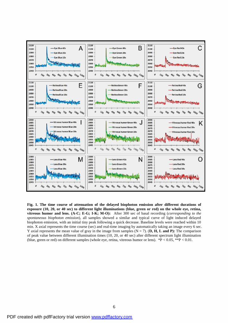
2. Results
All isolated samples (whole eye, lens, vitreous humor and retina) presented continuous,
spontaneous and basal photon emission without any excitation. In addition, after 300 sec of
basal recording followed by 30 min of adaptation to the dark, isolated rat whole eye, lens,
vitreous humor and retina were illuminated by monochromatic red, green or blue light with
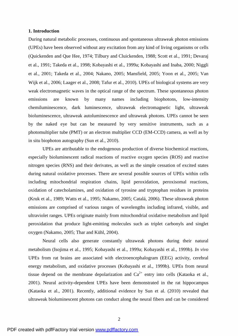
10, 20 or 40 sec duration, respectively. As shown in Figure 1, all samples presented
continuous spontaneous biophoton emission and obvious delayed biophoton emission (Fig. 1
A-C; E-G; I-K; M-O). All figures (Fig. 1 A-C; E-G; I-K; M-O) show a similar and typical
curve with an initial tiny peak following a quick decrease, and delayed biophoton emission
reaches a baseline level within 10 min. The effects of delayed biophoton emission were
affected by different spectrum light and illumination time and also depended on the different
parts of the eye (Fig. 1 A-C; E-G; I-K; M-O). We compared the peak values in different
spectrum light illumination using three different illumination times (10, 20 or 40 sec) for the
whole eye and retina. The results of this analysis showed a significantly larger peak value in
20 or 40 sec blue light illumination than in 10 sec blue light illumination (Fig. 1D and H).
PDF created with pdfFactory trial version www.pdffactory.com
5
For green light illumination, differences were observed between 40 and 10 sec, but not 20
and 10 sec for all samples (see Fig. 1D, H, L and P). No significant differences were found
between 40, 20 and 10 sec after red light illumination for the retina, lens and vitreous humor
with the exception of whole eye (Fig. 1D, H, L and P). The blue light illumination appeared
to show some differences for the lens and vitreous humor as compared to the whole eye and
retina (Fig. 1D, H, L and P), though this may be due to the difference of tissue
characteristics between lens, vitreous humor, retina and whole eye. In summary, the current
results showed that 40 sec exposure to red, green or blue light illumination caused delayed
biophoton emission to reach peak maximum values. In addition, the effects of red light
illumination on biophoton emission were less than those of the green and blue light
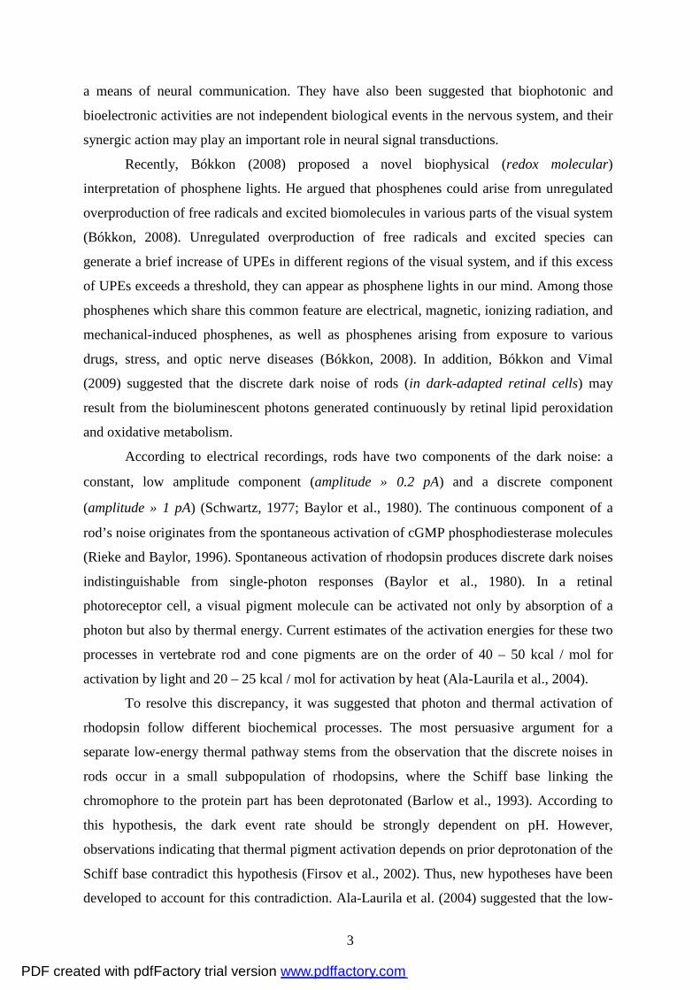
illuminations (see Fig. 2A-D).
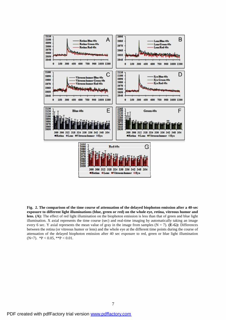
Then we analyzed the differences during the time course of attenuation of the delayed
biophoton emission after 40 sec red, green or blue light illumination by comparing the lens,
vitreous humor or retina with the whole eye. We found that there existed obvious differences
between lens or vitreous humor and whole eye at the most of time points during the course of
attenuation of the delayed biophoton emission, but no significant difference between retina
and whole eye (Fig. 2E-G).
PDF created with pdfFactory trial version www.pdffactory.com
6
Fig. 1. The time course of attenuation of the delayed biophoton emission after different durations of exposure (10, 20, or 40 sec) to different light illuminations (blue, green or red) on the whole eye, retina, vitreous humor and lens. (A-C; E-G; I-K; M-O): After 300 sec of basal recording (corresponding to the spontaneous biophoton emission), all samples showed a similar and typical curve of light induced delayed biophoton emission, with an initial tiny peak following a quick decrease. Baseline levels were reached within 10 min. X axial represents the time course (sec) and real-time imaging by automatically taking an image every 6 sec. Y axial represents the mean value of gray in the image from samples (N = 7). (D, H, L and P): The comparison of peak value between different illumination times (10, 20, or 40 sec) after different spectrum light illumination (blue, green or red) on different samples (whole eye, retina, vitreous humor or lens). *P < 0.05, **P < 0.01.
PDF created with pdfFactory trial version www.pdffactory.com
7
Fig. 2. The comparison of the time course of attenuation of the delayed biophoton emission after a 40-sec exposure to different light illuminations (blue, green or red) on the whole eye, retina, vitreous humor and lens. (A): The effect of red light illumination on the biophoton emission is less than that of green and blue light illumination. X axial represents the time course (sec) and real-time imaging by automatically taking an image every 6 sec. Y axial represents the mean value of gray in the image from samples (N = 7). (E-G): Differences between the retina (or vitreous humor or lens) and the whole eye at the different time points during the course of attenuation of the delayed biophoton emission after 40 sec exposure to red, green or blue light illumination (N=7). *P < 0.05, **P < 0.01.
PDF created with pdfFactory trial version www.pdffactory.com
8
3. Discussion
Dark retinal noise by bioluminescent photons
The present results unambiguously demonstrated that various parts of in vitro, freshly
isolated rat eye, including the lens, vitreous humor and retina as well as the whole eye, can
display continuous ultraweak photon emission. As shown in Figs. 1 and 2, the lens, vitreous
humor, retina and the whole eye displayed basic intensities of continuous UPEs. Together,
these results indicate that there are several natural sources of UPEs within the eye. During
photopic or scotopic vision, continuous UPEs can be negligible. In contrast, in dark-adapted
retina, such UPEs, are significant.
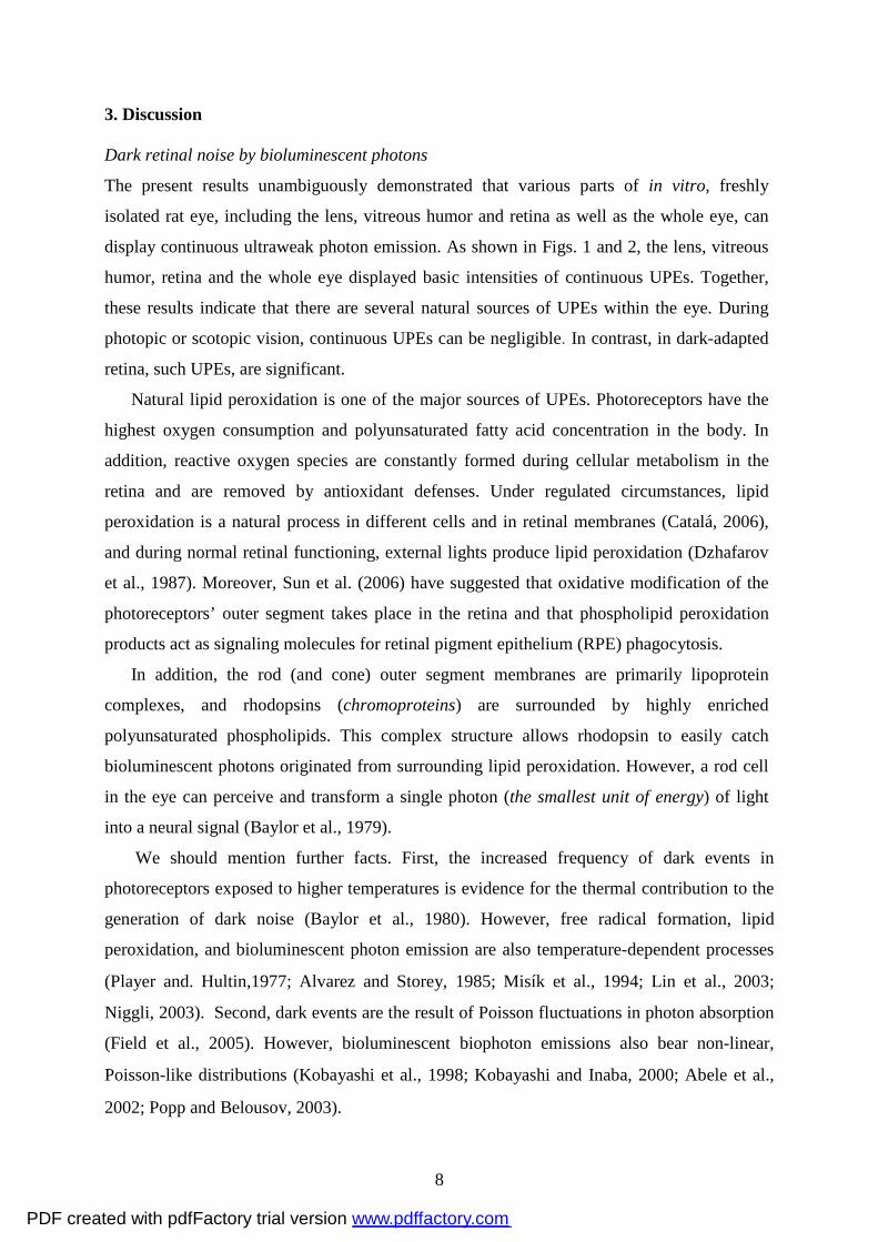
Natural lipid peroxidation is one of the major sources of UPEs. Photoreceptors have the
highest oxygen consumption and polyunsaturated fatty acid concentration in the body. In
addition, reactive oxygen species are constantly formed during cellular metabolism in the
retina and are removed by antioxidant defenses. Under regulated circumstances, lipid
peroxidation is a natural process in different cells and in retinal membranes (Catalá, 2006),
and during normal retinal functioning, external lights produce lipid peroxidation (Dzhafarov
et al., 1987). Moreover, Sun et al. (2006) have suggested that oxidative modification of the
photoreceptors’ outer segment takes place in the retina and that phospholipid peroxidation
products act as signaling molecules for retinal pigment epithelium (RPE) phagocytosis.
In addition, the rod (and cone) outer segment membranes are primarily lipoprotein
complexes, and rhodopsins (chromoproteins) are surrounded by highly enriched
polyunsaturated phospholipids. This complex structure allows rhodopsin to easily catch
bioluminescent photons originated from surrounding lipid peroxidation. However, a rod cell
in the eye can perceive and transform a single photon (the smallest unit of energy) of light
into a neural signal (Baylor et al., 1979).
We should mention further facts. First, the increased frequency of dark events in
photoreceptors exposed to higher temperatures is evidence for the thermal contribution to the
generation of dark noise (Baylor et al., 1980). However, free radical formation, lipid
peroxidation, and bioluminescent photon emission are also temperature-dependent processes
(Player and. Hultin,1977; Alvarez and Storey, 1985; Misík et al., 1994; Lin et al., 2003;
Niggli, 2003). Second, dark events are the result of Poisson fluctuations in photon absorption
(Field et al., 2005). However, bioluminescent biophoton emissions also bear non-linear,
Poisson-like distributions (Kobayashi et al., 1998; Kobayashi and Inaba, 2000; Abele et al.,
2002; Popp and Belousov, 2003).
PDF created with pdfFactory trial version www.pdffactory.com
9
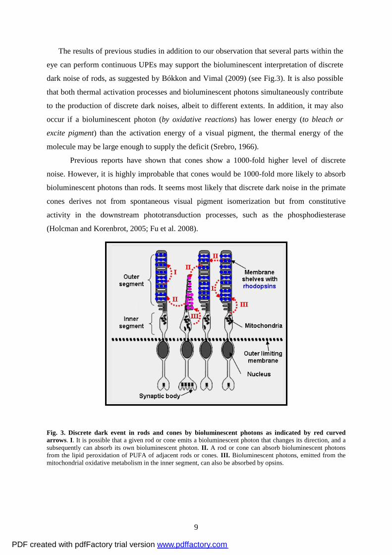
The results of previous studies in addition to our observation that several parts within the
eye can perform continuous UPEs may support the bioluminescent interpretation of discrete
dark noise of rods, as suggested by Bókkon and Vimal (2009) (see Fig.3). It is also possible
that both thermal activation processes and bioluminescent photons simultaneously contribute
to the production of discrete dark noises, albeit to different extents. In addition, it may also
occur if a bioluminescent photon (by oxidative reactions) has lower energy (to bleach or
excite pigment) than the activation energy of a visual pigment, the thermal energy of the
molecule may be large enough to supply the deficit (Srebro, 1966).
Previous reports have shown that cones show a 1000-fold higher level of discrete
noise. However, it is highly improbable that cones would be 1000-fold more likely to absorb
bioluminescent photons than rods. It seems most likely that discrete dark noise in the primate
cones derives not from spontaneous visual pigment isomerization but from constitutive
activity in the downstream phototransduction processes, such as the phosphodiesterase
(Holcman and Korenbrot, 2005; Fu et al. 2008).
Fig. 3. Discrete dark event in rods and cones by bioluminescent photons as indicated by red curved arrows. I. It is possible that a given rod or cone emits a bioluminescent photon that changes its direction, and a subsequently can absorb its own bioluminescent photon. II. A rod or cone can absorb bioluminescent photons from the lipid peroxidation of PUFA of adjacent rods or cones. III. Bioluminescent photons, emitted from the mitochondrial oxidative metabolism in the inner segment, can also be absorbed by opsins.
PDF created with pdfFactory trial version www.pdffactory.com
10
Retinal phosphenes by bioluminescent photons
Phosphene perception is an entoptic phenomenon characterized by the experience of seeing
light without light actually entering into the eye (i.e., without external photons). Phosphenes
can be points, spots, bars or chaotic structures of colored or colorless light. Phosphenes can be
elicited by a variety of stimuli (electrical, magnetic, mechanical, etc.) that excite cells in the
visual pathway, as well as by random firing of cells in the visual system (Reznikov, 1981;
Lindenblatt and Silny, 2002; Merabet et al., 2003). Phosphenes can appear in a variety of
diseases of the retina or of the visual pathways, though healthy persons can perceive them as
well (Onofrj et al., 1998; Brigatti and Maguluri, 2005). Phosphene generation by electrical
stimulation of the visual pathway with implanted electrodes is regarded as a hopeful method
for reversing blindness (Dobelle et al., 1974; Zrenner, 2002). Different types of visual
implants (also called visual prostheses) are named according to their locations: subretinal,
epiretinal, optic nerve, lateral geniculate nucleus and cortical (Cohen, 2007).
Vision researchers think that phosphenes result from the electrical activity in various parts
of the visual system. A historic and discredited assumption is that phosphene lights are
generated within the eye (Grüsser and Hagner, 1990). Recently, however, Bókkon (2008) put
forward a new notion about the origin of phosphenes by suggesting that phosphene flashes
result from induced or spontaneous increased overproduction of free radicals and excited
biomolecules. Such overproduction is hypothesized to produce a transient excess of UPEs in
cells of various parts of the visual system. When excess UPEs goes above a distinct threshold,
they may emerge as phosphene flash in our mind. In other words, the brain interprets these
retinal bioluminescent photons as if they originate from the external world. Bókkon’s
prediction about one kind of retinal phosphenes was experimentally supported by Narici et al.
(2009). According to this study, free radicals induced by ionizing radiation (cosmic particles)
can produce chemiluminescent photons via lipid peroxidation. Chemiluminescent photons are
then absorbed by the photoreceptors and start the photo-transduction cascade, which results in
the perception of phosphenes.
In complete darkness, a rod cell can convert a single photon into a neural signal,
though cones require the coincident absorption of some photons to generate a detectable
signal (Baylor et al., 1979, Vimal et al., 1989). Namely, approximately 10-30 photons are
required for seeing a flash in complete darkness (Hecht et al., 1942; Field et al., 2005).
Several experiments have demonstrated that retinal phosphenes are commonly elicited
by electrical, magnetic and mechanical stimuli. Here, we demonstrated that visible lights (red,
blue, and green) can also induce ultraweak photon emission (also called delayed
PDF created with pdfFactory trial version www.pdffactory.com
11
luminescence) from isolated parts of the rat eye. Our results may suggest that retinal
phosphenes result from excess bioluminescent photons, as we observed that photon emission
does exist within the eye. For example, during electrical stimulation of retina, various
biomolecules can undergo electron transfer reactions to form excited states and free radicals
(lipid peroxidation) at electrodes. Then, these excited biomolecules and free radical processes
can directly or indirectly produce photon emission when they return to basic states. Namely,
retinal electrical induction performs a brief electrochemiluminescence process (also known as
electrogenerated chemiluminescence) at electrodes (Chen et al., 1997).
Together, previous reports (Isojima et al., 1995; Kobayashi et al., 1999a; Kobayashi
et al., 1999b; Kataoka et al., 2001; Artem'ez et al 1967; Zhang et al., 1997; Dzhafarov et al.,
1987) and the current findings provide support for the hypothesis that retinal phosphenes are
due to the free radical related excess UPEs in the eyes, and the brain interprets these retinal
bioluminescent photons as if they are derived from our external world.
Normal vision and ambient induced delayed photons
During normal vision, the eyes are continuously exposed to powerful ambient photons that
pass through various parts of the eyes. However, our experiments provide evidence for the
existence of spontaneous and visible light induced delayed photon emission from various
parts of in vitro, freshly isolated rat eyes. Therefore, ambient photons could continuously
produce ultraweak delayed photon emission simultaneously from diverse parts of vertebrate
eyes during normal vision. It is important to consider that under natural circumstances,
ambient photons can perform much stronger delayed photon emission (within the eyes) than in
the isolated and dark-adapted experiments described here. The significance of ambient light
induced bioluminescent delayed photons within different parts of the eyes during vision is
currently unknown and should be considered in future research endeavors.
In brief
In the present study, we demonstrated the existence of spontaneous and visible photon
induced (delayed luminescence) ultraweak photon emission from in vitro, freshly isolated
whole eye, lens, vitreous humor and retina samples from rats. These experiments may have
the following implications:
i. Our results may support the bioluminescent photon concept of dark retinal noise.
Specifically, the discrete dark noise of rods may be due to the ultraweak bioluminescent
photons generated within the retina from natural lipid peroxidation and other free radical
PDF created with pdfFactory trial version www.pdffactory.com
12
processes. It is also possible that both thermal activation processes and bioluminescent
photons are simultaneously involved in the production of discrete dark noises to differing
extents. It may also occur if a bioluminescent photon (by oxidative reactions) has lower
energy (to bleach or excite pigment) than the activation energy of a visual pigment, the
thermal energy of the molecule may be large enough to supply the deficit
ii. Here we demonstrated visible lights induced ultraweak photon emission (also
called delayed luminescence) from isolated parts of rat eyes. Nevertheless, our results may
also suggest that electrical, magnetic or other types of stimuli can induce retinal phosphenes
that also result from an excess of bioluminescent photons. Thus, our experiments may
indicate that induced photon emission can exist within the eyes.
iii. Retinal bioluminescent photons (retinal phosphenes) can be interpreted by the
brain as if they are derived from our external world.
iv. The design of high-resolution retinal prostheses for the restoration of sight
presents several unique engineering and biological challenges. Our interpretations regarding
retinal discrete noise and phosphenes may lead to new notions and designs related to retinal
prostheses.
v. During normal photopic (or scotopic) vision, vertebrate eyes are continuously
exposed to powerful ambient photons that pass through various parts of the eyes. According
to our results, during vision, ambient light radiation can induce permanent bioluminescent
photon emission within various parts of the eyes. However, we do not know the significance
or role of ambient light induced permanent bioluminescent photons during visual
mechanisms.
Finally, in the future, several further studies should be conducted not only to replicate
the current findings but also to extend the novel concepts of ultraweak biophotonic research.
We should also consider that the ultraweak biophotonic and bioelectronic activities may be
not independent biological events during neural signal processes.
4. Experimental procedures
4.1. The preparation and pre-processing of samples
Sprague–Dawley (SD) rats weighing 200 ± 15 g were purchased from the Experimental
Animals Center of Tongji Medical College of Huazhong University of Science and
Technology. 12 rats were employed and 7 samples (N=7) from each tissue (retina, lens,
vitreous humor and whole eye) were used for experiments. All animal experiments were
approved by the Animal Care Committee of South-Central University for Nationalities. Each
PDF created with pdfFactory trial version www.pdffactory.com
13
rat was decapitated, and both eyes were isolated quickly from fossa orbitalis. All remaining
surrounding connective tissue was carefully cut away with microscissors. Under the
operating microscope, the cornea from one eye was cut away with microscissors along the
limbus and the lens. The vitreous body and optic cup (retina) were separated carefully. The
other eye was kept intact.
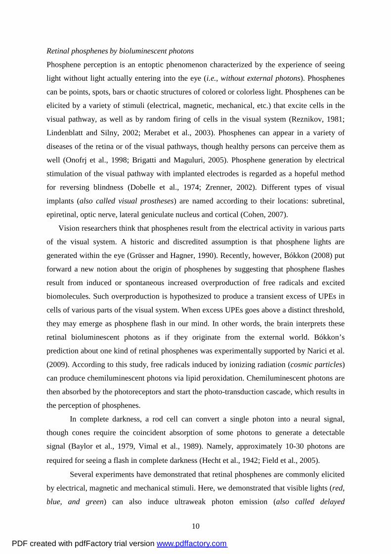
4.2. Biophoton detection with a biophoton imaging system
The biophoton imaging system has been previously described (Sun et al., 2010). The system
consists of a stereomicroscope (AZ100, Nikon, Japan) and an electron multiplier CCD (EM-
CCD) camera (C9100-13; Hamamatsu Photonics K. K., Hamamatsu, Japan), which were set
in a complete dark box (90.5 cm in length, 75.5 cm in width, 110.5 cm in height) in a dark
room. The EM-CCD camera was mounted on top of the stereomicroscope and controlled by
a computer and image analysis software (SimplePCI 6.0), as shown in Figure 4.
Fig. 4. Schematic drawing of the biophoton imaging system. A stereomicroscope, EM-CCD camera and LED light are set in a complete dark box. A CCD controller, computer and cooling water circulating pump are set outside of dark box (also see text).
The lens, vitreous body, optic cup (retina) and complete eye were placed on a glass slide,
which was put on the specimen stage of the stereomicroscope with an objective lens (AZ-
Plan Apo 1x) (see Fig. 5A). Light illumination was applied to the preparations by an LED
lamp which can be replaced in different color LED lamp (red, 621 - 623.5 nm, 4,500 - 5,000
mcd; green, 515 - 520 nm, 12,000 - 14,000 mcd; blue, 460 - 465 nm, 4,000 - 5,000 mcd)
supplied by a 3 V direct current (DC). The distance from the LED lamp to the samples was
approximately 6 cm. A ground glass was set at the front of the LED lamp to obtain uniform
illumination.
PDF created with pdfFactory trial version www.pdffactory.com
14
Biophotons were detected and imaged using the EM-CCD camera in water-cool mode
(in this situation, the working temperature at the CCD can be maintained as low as -90 °C).
The other setup parameters for EM-CCD camera during imaging are 1200 x gain and 2 x 2
binning. The specific steps for biophoton detection were as follows: (1) the samples were
kept in complete darkness for at least 20 min before biophoton imaging to exclude the effects
of ambient light; (2) real-time images were taken automatically every 6 sec; (3) after 50
images were acquired (i.e. 300 sec basal recording), imaging was paused and the was applied
to the samples by a LED lamp (first red light) for 10 sec before an additional 150 images
were taken (lasting 20 min); (4) steps 2-3 were repeated, with the illumination time extended
to 20 and 40 seconds, respectively; (5) steps 2-3 were repeated, with green or blue LED in
place of red light illumination.
4.3. Image processing and data analysis
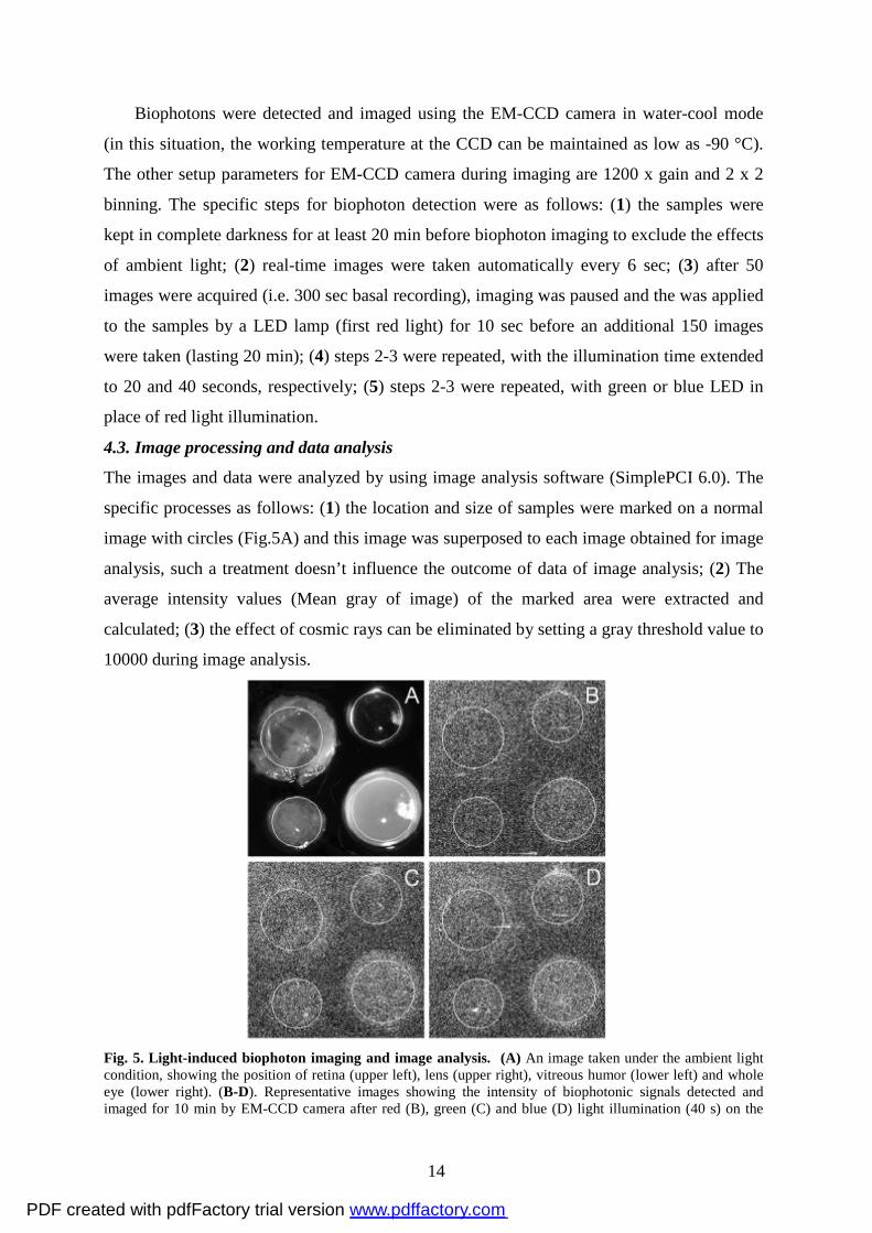
The images and data were analyzed by using image analysis software (SimplePCI 6.0). The
specific processes as follows: (1) the location and size of samples were marked on a normal
image with circles (Fig.5A) and this image was superposed to each image obtained for image
analysis, such a treatment doesn’t influence the outcome of data of image analysis; (2) The
average intensity values (Mean gray of image) of the marked area were extracted and
calculated; (3) the effect of cosmic rays can be eliminated by setting a gray threshold value to
10000 during image analysis.
Fig. 5. Light-induced biophoton imaging and image analysis. (A) An image taken under the ambient light condition, showing the position of retina (upper left), lens (upper right), vitreous humor (lower left) and whole eye (lower right). (B-D). Representative images showing the intensity of biophotonic signals detected and imaged for 10 min by EM-CCD camera after red (B), green (C) and blue (D) light illumination (40 s) on the
PDF created with pdfFactory trial version www.pdffactory.com
15
retina, lens, vitreous humor and whole eye. The areas marked with circles in each sample were processed for image analysis.
4.4. Statistical analysis
All 12 rats were employed and 7 samples (N=7) from each tissue (retina, lens, vitreous
humor and whole eye) were used for data analysis. Two-tailed Student’s t-tests (calculated
using Microsoft Excel) or one-way analysis of variance (ANOVA) were used. All summary
bar graphs are presented as mean ± standard deviation, with the significance denoted as
follows: *P < 0.05, **P < 0.01.
∗Footnote Delayed luminescence (DL) is the long-term ultraweak re-emission of optical photons from diverse cells, organisms, and other material if they were illuminated with monochromatic or white light (Ho et al., 2002; Popp and Yan, 2002; Kim et al., 2005). DL intensity is radically lower than the well-known fluorescence or phosphorescence. The decay time of DL is dependent on the physiological conditions of the samples and the kinds of tissues they were extracted from, as well as the intensity, duration, and spectral distribution of illumination (Kim et al., 2005). Abbreviations: UPE, ultraweak photon emissions; PMT, photomultiplier tube; EM-CCD, electron multiplier CCD camera; ROS, reactive oxygen species; RNS, reactive nitrogen species; EEG, electroencephalogram; cGMP, cyclic guanosine monophosphate; DC, direct current; V1, primary visual cortex; DL, delayed luminescence. Acknowledgements Bókkon I. gratefully acknowledges support of this work by the BioLabor (Hungary), www.biolabor.org; Bókkon’s URL: http://bokkon-brain-imagery.5mp.eu. This work was also supported by the research foundation for “key laboratory of Neuroscience and Neuroengineering” from South-Central University for Nationalities (XJS09001, Dai J), and partly by National natural Science Foundation of China (NSFC: 31070961, Dai J). Declaration of interest
The authors report no conflicts of interest. The authors alone are responsible for the content.
REFERENCES
Abele, D., Heise, K., Pörtner, H.O., Puntarulo, S., 2002. Temperature-dependence of mitochondrial function and production of reactive oxygen species in the intertidal mud clam Mya arenaria. J. Exp. Biol. 205, 1831–1841.
Adam, W., Kazakov, D.V., Kazakov, V.P., 2005. Singlet-oxygen chemiluminescence in peroxide reactions. Chem. Rev. 105, 3371−3387.
Ala-Laurila, P., Donner, K., Koskelainen, A., 2004. Thermal activation and photoactivation of visual pigments. Biophys. J. 86, 3653−3662.
PDF created with pdfFactory trial version www.pdffactory.com
16
Alvarez, J.G., Storey, B.T., 1985. Spontaneous lipid peroxidation in rabbit and mouse epididymal spermatozoa: dependence of rate on temperature and oxygen concentration. Biol. Reprod. 32, 342-351.
Artem'ey, V.V., Goldobin, A.S., Gus'kov, L.N., 1967. Recording the optical emission of a nerve. Biophysics 12, 1278−1280.
Barlow, R.B.Jr., Birge, R.R., Kaplan, E., Tallent, J.R., 1993. On the molecular origin of photoreceptor noise. Nature 366, 64−66.
Baylor, D.A., Lamb, T.D., Yau, K.W., 1979. Responses of retinal rods to single photons. J. Physiol. 288, 613−634.
Baylor, D.A., Matthews, G., Yau, K-W., 1980. Two components of electrical dark noise in toad retinal rod outer segments. J. Physiol. (Lond.) 309, 591−621.
Bókkon, I., 2008. Phosphene phenomenon: A new concept. BioSystems 92, 168–174. Bókkon, I., Vimal, R.L.P., 2009. Retinal phosphenes and discrete dark noises in rods: a new
biophysical framework. J. Photochem. Photobiol. B. 96, 255–259. Brigatti, L., Maguluri, S., 2005. Reproducibility of self-measured intraocular pressure with
the phosphene tonometer in patients with ocular hypertension and early to advanced glaucoma. J. Glaucoma 14, 36–39.
Catalá, A., 2006. An overview of lipid peroxidation with emphasis in outer segments of photoreceptors and the chemiluminescence assay. Int. J. Biochem. Cell. Biol. 38, 1482−1495.
Chen, G.N., Lin, R.E., Zhao, Z.F., Duan, J.P., Zhang, L., 1997. Electrogenerated chemiluminescence for determination of indole and tryptophan. Analytica Chimica Acta 341, 251−256.
Cohen, E.D., 2007. Prosthetic interfaces with the visual system: biological issues. J. Neural Eng, 4, R14–R31.
Devaraj, B., Scott, R.Q., Roschger, P., Inaba, H., 1991. Ultraweak light emission from rat liver nuclei. Photochem. Photobiol. 54, 289−293.
Dobelle, W.H., Mladejovsky, M.G., Girvin, J.P., 1974. Artificial vision for the blind: electrical stimulation of visual cortex offers hope for a functional prosthesis. Science 183, 440–444.
Dzhafarov, A.I., Gadzhieva, N.A., Mamedkhanly, T.A., Kul''gavin, L.É., Dagkesamanskaya, D.N., Alieva, N.I., 1987. Effect of visual cortical stimulation on lipid peroxidation in the rabbit retina. Bull. Exp. Biol. Med. 103, 318−320.
Field, G.D., Sampath, A.P., Rieke, F., 2005. Retinal processing near absolute threshold: from behavior to mechanism. Annu. Rev. Physiol. 67, 491−514.
Firsov, M.L., Donner, K., Govardovskii, V.I., 2002. pH and rate of "dark" events in toad retinal rods: test of a hypothesis on the molecular origin of photoreceptor noise. J. Physiol. 539, 837−846.
Fliesler, S.J., 1983. Anderson, R.E. Chemistry and metabolism of lipids in the vertebrate retina, Prog. Lipid Res. 22, 79−131.
Fu, Y., Kefalov, V., Luo, D.G., Xue, T., Yau, K.W., 2008. Quantal noise from human red cone pigment. Nat. Neurosci. 11, 565-571.
Grüsser, O.J., Hagner, M., 1990. On the history of deformation phosphenes and the idea of internal light generated in the eye for the purpose of vision. Doc. Ophthalmol. 74, 57–85.
Hecht, S., Shlaer, S., Pirenne, M.H., 1942. Energy, quanta, and vision. J. Gen. Physiol. 25, 819−840.
Ho, M.W., Musumeci, F., Scordino, A., Triglia, A., Privitera, G., 2002. Delayed luminescence from bovine Achilles' tendon and its dependence on collagen structure. J. Photochem. Photobiol. B. 66, 165−170.
PDF created with pdfFactory trial version www.pdffactory.com
17
Holcman, D., Korenbrot, J.I., 2005. The limit of photoreceptor sensitivity: molecular mechanisms of dark noise in retinal cones. J. Gen. Physiol. 125, 641-660.
Isojima, Y., Isoshima, T., Nagai, K., Kikuchi, K., Nakagawa, H., 1995. Ultraweak biochemiluminescence detected from rat hippocampal slices. NeuroReport 6, 658–660.
Kataoka, Y., Cui, Y., Yamagata, A., Niigaki, M., Hirohata, T., Oishi, N., Watanabe, Y., 2001. Activity-Dependent Neural Tissue Oxidation Emits Intrinsic Ultraweak Photons. Biochem. Biophys. Res. Commun. 285, 1007–1011.
Kim. H.W., Sim, S.B., Kim, C.K., Kim, J., Choi, C., You, H., Soh, K.S., 2005. Spontaneous photon emission and delayed luminescence of two types of human lung cancer tissues: adenocarcinoma and squamous cell carcinoma. Cancer Lett. 229, 283−289.
Kobayashi, M., Devaraj, B., Inaba, H., 1998. Observation of super-Poisson statistics from bacterial (Photobacterium phosphoreum) bioluminescence. Phys. Rev. E 57, 2129.
Kobayashi, M., Inaba, H., 2000. Photon Statistics and Correlation Analysis of Ultraweak Light Originating from Living Organisms for Extraction of Biological Information. Appl. Opt. 39, 183−192.
Kobayashi, M., Takeda, M., Ito, K.I., Kato, H., Inaba, H., 1999a. Two-dimensional photon counting imaging and spatiotemporal characterization of ultraweak photon emission from a rat’s brain in vivo. J. Neurosci. Methods 93, 163–168.
Kobayashi, M., Takeda, M., Sato, T., Yamazaki, Y., Kaneko, K., Ito, K., Kato, H., Inaba, H., 1999b. In vivo imaging of spontaneous ultraweak photon emission from a rat’s brain correlated with cerebral energy metabolism and oxidative stress. Neurosci. Res. 34, 103–113.
Kruk, I., Lichszteld, K., Michalska, T., Wronska, J., Bounias, M., 1989. The formation of singlet oxygen during oxidation of catechol amines as detected by infrared chemiluminescence and spectrophotometric method. Z. Naturforsch. [C] 44, 895–900.
Laager, F., Park, S.H., Yang, J.M., Song, W., Soh, K.S., 2008. Effects of exercises on biophoton emission of the wrist. Eur. J. Appl. Physiol. 102, 463–469.
Lin, Y.R., Huang, S.L., Huang, C.H., 2003. Characteristics of NADH-dependent lipid peroxidation in sarcoplasmic reticulum of white shrimp, Litopenaeus vannamei, and freshwater prawn, Macrobrachium rosenbergii. Comp. Biochem. Physiol. B Biochem. Mol. Biol. 135, 683-687.
Lindenblatt, G., Silny, J., 2002. Electrical phosphenes: on the influence of conductivity inhomogeneities and small-scale structures of the orbita on the current density threshold of excitation. Med. Biol. Eng. Comput. 40, 354–359.
Lórenz-Fonfría, V.A., Furutani, Y., Ota, T., Ido, K., Kandori, H., 2010. Protein fluctuations as the possible origin of the thermal activation of rod photoreceptors in the dark. J. Am. Chem. Soc. 132, 5693–5703.
Mansfield, J.W., 2005. Biophoton distress flares signal the onset of the hypersensitive reaction. Trends. Plant. Sci. 10, 307-309.
Merabet, L.B., Theoret, H., Pascual-Leone, A., 2003. Transcranial magnetic stimulation as an investigative tool in the study of visual function. Optom. Vis. Sci. 80, 356–368.
Misík, V., Gergel', D., Alov, P., Ondrias, K., 1994. An unusual temperature dependence of malondialdehyde formation in Fe2+/H2O2-initiated lipid peroxidation of phosphatidylcholine liposomes. Physiol. Res. 43, 163-167.
Nakano, M., 2005. Low-level chemiluminescence during lipid peroxidations and enzymatic reactions. J. Biolumin. Chemilum. 4, 231–240.
Narici, L., De Martino, A., Brunetti, V., Rinaldi, A., Sannita, W.G., Paci, M., 2009. Radicals excess in the retina: A model for light flashes in space. Rad. Meas. 44, 203−205.
PDF created with pdfFactory trial version www.pdffactory.com
18
Niggli, H.J., Scalet, C., Popp, F.A., Yu, Y., Applegate, L.A., 2001. Ultraweak photon emission in assessing growth factor efficiency using fibroblastitic differentiation. J. Photochem. Photobiol. B 64, 62–68.
Niggli, H.J., 2003. Temperature dependence of ultraweak photon emission in fibroblastic differentiation after irradiation with artificial sunlight. Indian J. Exp. Bio. 41, 419-423.
Onofrj, M., Thomas, A., Paci, C., Rotilio, D., 1998. Optic neuritis with residual tunnel vision in perchloroethylene toxicity. J. Toxicol. Clin. Toxicol. 36, 603–607.
Player, T.J., Hultin, H.O., 1977. Some characteristics of the NAD(P)H-dependent lipid peroxidation system in the microsomal fraction of chicken breast muscle. J. Food Biochem. 1, 153-171.
Popp, F.A., Belousov, L.V., Integrative biophysics: biophotonics. Springer, 2003. Popp. F.A., Yan, Y., 2002. Delayed luminescence of biological systems in terms of coherent
states. Physics Lett. A 293, 93−97. Quickenden, T.I., Que Hee, S.S., 1974. Weak luminescence from the yeast Sachharomyces-
Cervisiae. Biochem. Biophys. Res. Commun. 60, 764–770. Reznikov, IuE., 1981. Mechanophosphene in optic nerve changes. Oftalmol. Zh. 36,
218−220. Rieke, F., Baylor, D.A., 1996. Molecular origin of continuous dark noise in rod
photoreceptors. Biophys. J. 71, 2553−2572. Schwartz, E.A., 1977.Voltage noise observed in rods of the turtle retina. J. Physiol. 272,
217–246. Scott, R.Q., Roschger, P., Devaraj, B., Inaba, H., 1991. Monitoring a mammalian nuclear
membrane phase transition by intrinsic ultraweak light emission. FEBS Lett, 285. 97–98. Srebro. R., 1966. A thermal component of excitation in the lateral eye of Limulus. J. Physiol.
187, 417-425. Sun, M., Finnemann, S.C., Febbraio, M., Shan, L., Annangudi, S.P., Podrez, E.A., Hoppe,
G., Darrow, R., Organisciak, D.T., Salomon, R.G., Silverstein, R.L., Hazen, S.L., 2006. Light-induced oxidation of photoreceptor outer segment phospholipids generates ligands for CD36-mediated phagocytosis by retinal pigment epithelium: a potential mechanism for modulating outer segment phagocytosis under oxidant stress conditions. J. Biol. Chem. 281, 4222−4230.
Sun, Y,. Wang, Ch., Dai, J., 2010. Biophotons as neural communication signals demonstrated by in situ biophoton autography. Photochem. Photobiol. Sci. 9, 315–322.
Tafur, J., Van Wijk, E.P., Van Wijk, R., Mills, P.J., 2010. Biophoton detection and low-intensity light therapy: a potential clinical partnership. Photomed. Laser Surg. 28, 23–30.
Takeda, M., Kobayashi, M., Takayama, M., Suzuki, S., Ishida, T., Ohnuki, K., Moriya, T., Ohuchi, N., 2004. Biophoton detection as a novel technique for cancer imaging. Cancer Science 95, 656−661.
Takeda, M., Tanno, Y., Kobayashi, M., Usa, M., Ohuchi, N., Satomi, S., Inaba, H., 1998. A novel method of assessing carcinoma cell proliferation by biophoton emission. Cancer Lett. 127, 155–160.
Thar, R., Kühl, M., 2004. Propagation of electromagnetic radiation in mitochondria? J. Theor. Biol. 230, 261–70.
Tilbury, R.N., Cluickenden, T.I., 1988. Spectral and time dependence studies of the ultraweak bioluminescence emitted by the bacterium Escherichia coli. Photobiochem. Photobiophys. 47, 145–150.
Van Wijk. R., Van Wijk, E.P., Bajpai, R.P., 2006. Photocount distribution of photons emitted from three sites of a human body. J. Photochem. Photobiol. B 84, 46–55.
Vimal, R.L., Pokorny, J., Smith, V.C., Shevell, S.K., 1989. Foveal cone thresholds. Vision Res. 29, 61−78.
PDF created with pdfFactory trial version www.pdffactory.com
19
Watts, B.P., Barnard, M., Turrens, J.F., 1995. Peroxynitrite-Dependent Chemiluminescence of Amino Acids, Proteins, and Intact Cells. Arch. Biochem. Biophys. 317, 324–330.
Yoon, Y.Z., Kim, J., Lee, B.C., Kim, Y.U., Lee, S.K., Soh, K.S., 2005. Changes in ultraweak photon emission and heart rate variability of epinephrine-injected rats. Gen. Physiol. Biophys. 24, 147–159.
Zhang, J., Yu, W,, Sun, T., Popp, F.A., 1997. Spontaneous and light-induced photon emission from intact brains of chick embryos. Sci. China. C Life. Sci. 40, 43−51.
Zrenner, E., 2002. Will retinal implants restore vision? Science 295, 1022–1025.
PDF created with pdfFactory trial version www.pdffactory.com
Copyright © 2022 FDOKUMEN