Complete Genome Sequence and Comparative Genomics of Shigella flexneri Serotype 2a Strain 2457T
Upload
independentCategory
view
1download
0
Downloaded from www.microbiologyresearch.org by
IP: 54.160.90.203
On: Wed, 29 Jun 2016 09:03:09
Spa13 of Shigella flexneri has a dual role:chaperone escort and export gate-activator switchof the type III secretion system
Youness Cherradi, Abderrahman Hachani3 and Abdelmounaaım Allaoui
Correspondence
Abdelmounaaım Allaoui
Received 31 July 2013
Accepted 11 October 2013
Laboratoire de Bacteriologie Moleculaire, Faculte de Medecine, Universite Libre de Bruxelles,Route de Lennik, 808, 1070 Bruxelles, Belgium
The type III secretion apparatus (T3SA) is used by numerous Gram-negative pathogens to inject
virulence factors into eukaryotic cells. The Shigella flexneri T3SA spans the bacterial envelope
and its assembly requires the products of ~20 mxi and spa genes. Despite progress made in
understanding how the T3SA is assembled, the role of several predicted soluble components,
such as Spa13, remains elusive. Here, we show that the secretion defect of the spa13 mutant is
associated with lack of T3SA assembly which is partly due to the instability of the needle
component MxiH. In contrast to its Yersinia counterpart, Spa13 is not a secreted protein. We
identified a network of interactions between Spa13 and the ATPase Spa47, the C-ring protein
Spa33, and the inner-membrane protein Spa40. Moreover, we revealed a Spa13 interaction with
the inner-membrane MxiA and showed that overexpression of the large cytoplasmic domain of
MxiA in the WT background shuts off secretion. Lastly, we demonstrated that Spa13 interacts
with the cleaved form of Spa40 and with the translocator chaperone IpgC, suggesting that Spa13
intervenes during the secretion hierarchy switch process. Collectively, our results support a dual
role of Spa13 as a chaperone escort and as an export gate-activator switch.
INTRODUCTION
Shigella is the causative agent of shigellosis in humans,causing 1.1 million deaths each year, particularly amongchildren under 5 years old in developing countries (Kotloffet al., 1999). During infection, Shigella and many otherGram-negative pathogenic bacteria use a type III secretion(T3S) system to promote invasion of animal, plant orsymbiont cells by injecting virulence effectors into theircytoplasm (Galan & Wolf-Watz, 2006; Hueck, 1998;Schroeder & Hilbi, 2008). Shigella uses the T3S apparatus(T3SA) to penetrate enterocytes and to disseminate intothe colonic epithelium, which leads to a partial destructionof the mucosal lining and shigellosis symptoms. Most ofthe virulence factors of Shigella are encoded by a largevirulence plasmid harbouring a 31 kb region that issufficient to promote bacterial entry into host cells(Buchrieser et al., 2000). The ‘entry’ region is organizedin two loci, one encoding Mxi (membrane expression ofIpa antigen)–Spa (surface presentation of Ipa antigen)
proteins that form the T3SA, whilst the other encodes itsearly substrates and their cognate chaperones. At 37 uC, theneedle subunit MxiH and the predicted inner-rod com-ponent MxiI are secreted through the T3SA base. Upon itsmultimerization, MxiH forms a needle with a lengthregulated at 50 nm by the Spa32 protein (Magdalena et al.,2002). Approximately 50 proteins contribute to the T3SS ofShigella, including four chaperones, three transcriptionalactivators, three translocators, ~25 effectors and compo-nents of the Mxi–Spa T3SA (Parsot, 2009). The T3SAconsists of a cytoplasmic region called the ‘C-ring’, a basalbody composed of a pair of rings spanning the bacterialenvelope and a needle-like structure, protruding outsidethe bacterium, capped by a tip complex (Espina et al., 2006;Sani et al., 2007).
The T3SA is evolutionarily and structurally related to theexport machinery of the bacterial flagella (Cornelis, 2006;Kubori et al., 1998; Macnab, 1999; Minamino et al., 2008).Indeed, the proximal portion of the Shigella T3SA assumesrotational symmetry and its constituents share significanthomology with the flagellar basal body subunits, such asthe inner-membrane proteins MxiA (FlhA), Spa9 (FliP),Spa24 (FliR), Spa29 (FliQ) and Spa40 (FlhB), and theirassociated cytoplasmic proteins Spa47 (FliI), MxiN (FliH)and Spa33 (FliN, FliM and FliG) (Abrusci et al., 2013;Aizawa, 2001; Blocker et al., 2003; Diepold et al., 2012;Macnab, 2004).
3Present address: MRC Centre for Molecular Bacteriology and Infection,Department of Life Sciences, Imperial College, London, UK.
Abbreviations: T3S, type III secretion; T3SA, type III secretion apparatus;CR, Congo red; Spa, surface presentation of Ipa antigen; Mxi, membraneexpression of Ipa antigen; GST, glutathione S-transferase.
Two supplementary tables and one supplementary figure are availablewith the online version of this paper.
Microbiology (2014), 160, 130–141 DOI 10.1099/mic.0.071712-0
130 071712 G 2014 SGM Printed in Great Britain

Downloaded from www.microbiologyresearch.org by
IP: 54.160.90.203
On: Wed, 29 Jun 2016 09:03:09
Although significant progress towards the understanding ofthe T3SA assembly has been made in the last decade, thefunction of several Mxi and Spa proteins that are predictedto be cytoplasmic remains unknown. Even though theT3SA components present in different bacteria share stronghomologies, it is not the case for their respective chap-erone proteins (Abrusci et al., 2013; Bennett & Hughes,2000). Virulence clusters in Shigella, Salmonella, Yersinia,Chlamydia, Pseudomonas aeruginosa and Escherichia coli(enteropathogenic or enterohaemorragic) share a poorlycharacterized gene situated between the genes encoding theexport ATPase and the proteins controlling the hook orneedle length (Collazo et al., 1995; Journet et al., 2003;Payne & Straley, 1998; Shibata et al., 2007). Although thesegenes do not share significant sequence similarity withspa13, these members of the yscO–invI–fliJ family encode aprotein of a similar size, with abundant charged residuesand high predicted a-helical content (Minamino et al.,2000). In a previous study, Penno et al. (2006) described aseries of 10 adenosines within the spa13 sequence that areresponsible for a transcriptional slippage mechanism. Thisslippage reduces the proportion of spa13 mRNA, which inturn regulates the production of the full-length Spa13.Interestingly, this slippage mechanism was also reported toregulate MxiE, MxiA and Spa33 production (Penno &Parsot, 2006; Penno et al., 2006).
We have described previously several interactions amongthe Mxi and Spa proteins. We showed, for example, thatthe ATPase Spa47 interacts with MxiK, MxiN and Spa33(Jouihri et al., 2003), and more recently with the gatekeeperMxiC and the translocators chaperone IpgC (Cherradiet al., 2013). In addition, we reported numerous interac-tions involving the large C-terminal cytoplasmic domain ofSpa40 (Spa40C) with the needle length regulator Spa32,and with several Mxi and Spa proteins (Botteaux et al.,2008, 2010). Despite the identification of key interactionswithin the T3SA, and although Spa13 is essential to itsassembly, little is known about the role of Spa13 in thisapparatus (Collazo et al., 1995; Payne & Straley, 1998;Penno et al., 2006; Sasakawa et al., 1993). The identifica-tion of Spa13-interacting partners will contribute signific-antly to a better understanding of the events supporting theT3S pathway. Our results show that Spa13 interacts withthe cytosolic components Spa47 and Spa33, and thetranslocon chaperone IpgC and the cytoplasmic C-terminiof two inner-membrane proteins, MxiA (MxiAC) andSpa40 (Spa40C). The significance of these interactions isdiscussed in light of the T3SA.
METHODS
Strains and bacterial growth conditions. The list of E. coli andShigella flexneri strains used in this study is shown in Table S1(available in Microbiology Online). E. coli strain DH5lpir was used forderivatives of the suicide vector pGP704 (Miller & Mekalanos, 1988)and SM10lpir was used to transfer derivatives of pGP704 to S.flexneri. Shigella were phenotypically selected on Congo red (CR) agarplates (Meitert et al., 1991) and grown in trypticase soy broth (VWR)
with the appropriate antibiotics at the following concentrations:
zeocin 50 mg ml21, kanamycin 50 mg ml21, streptomycin 100 mg
ml21 and ampicillin 100 mg ml21.
Construction of the spa13 null strain. For the construction of a
non-polar mutant of the spa13 gene, a ble cassette was used (Botteaux
et al., 2009). The non-polar mutant was constructed by replacing the
spa13 gene by the EcoRI-digested ble cassette which confers resistance
to zeocin. The pHA22 plasmid, bearing the inactivated spa13 gene and
its flanking regions, is a derivative of the suicide vector pGP704. The
latter was transferred to S. flexneri M90T-Sm by conjugal mating.
Transconjugants were selected on plates containing streptomycin and
zeocin and clones in which a double recombinational event had
exchanged the WT spa13 gene for its inactivated copy were identified
by screening for sensitivity to ampicillin. The structure of pWR100
derivatives carrying the spa13 mutation was confirmed by PCR and
the resulting strains were designated SBH43 (spa13).
Construction of plasmids. Plasmids and primers used in this study
are listed in Tables S1 and S2, respectively. Plasmids pHA1 and
pC76 (pQE60-lacI-spa13-His) (Penno et al., 2006) were used to
complement the spa13 mutant. Recombinant [Glutathione S-transfer-
ase (GST) or His] proteins, used in the interaction assays, were
produced from plasmids listed in Table S1. Plasmids expressing GST
fused to Spa13, Spa13L51A, Spa13L55A and Spa32 were constructed by
inserting PCR-digested DNA fragments into the pGEX4T1 expression
vector. Plasmids expressing N-terminal His6 fused to Spa13, Spa32
and MxiAC were constructed by inserting PCR-digested DNA
fragments into the pET30 vector expressing His6 recombinant
proteins, whilst plasmids encoding His6 fused to His-IpgC, Spa40C
or MxiAC and Spa33 were constructed by ligation of PCR-digested
DNA fragments into, respectively, pQE30, pBAD/HisA and pBAD/
Myc-HisA vectors.
Directed mutagenesis was carried out according to the procedure of
the Quick Change Mutagenesis kit (Stratagene). The use of each
primer pair in PCR creates a restriction site to confirm the introduced
mutation (Table S2). Mutations L51A and L55A of Spa13 were
carried out on plasmids pC76 (pQE60-lacI-spa13-His) and pKC82
(pGEX4T1-spa13). The resulting plasmids were named pYC182
(pQE60-lacI-spa13L51A-His), pYC183 (pQE60-lacI-spa13L55A-His),
pYC184 (pGEX4T1-spa13L51A) and pYC185 (pGEX4T1-spa13L55A).
Production of recombinant proteins and GST pull-down assay.E. coli BL21 (DE3 Rosetta) were transformed with pGEX4T1 plasmid
encoding GST alone or its derivatives encoding GST fusion proteins and
grown in 100 ml LB medium to OD600 ~0.7 at 37 uC. Protein
expression was induced with 0.1 mM IPTG for 3 h at 37 uC. Cells were
harvested, resuspended in PBS lysis buffer and then lysed by sonication.
A final concentration of 1 % Triton X-100 was added to the lysate
followed by incubation on ice for 20 min. The lysates were then clarified
by centrifugation and the supernatants mixed with Glutathione-
Sepharose 4B matrix beads (GE Healthcare) equilibrated previously
with PBS buffer. The mixtures were incubated overnight at 4 uC in a
rotor shaker with bacterial lysates producing His or recombinant
proteins. Beads were washed eight times and proteins eluted by
incubating beads for 10 min with elution buffer [40 mM Tris (pH 8.0),
500 mM NaCl and 50 mM reduced glutathione]. The eluted proteins
were resolved by SDS-PAGE and analysed by Western blotting.
Protein analysis. Crude extracts and culture supernatants of S.
flexneri strains were prepared and analysed as described previously
(Allaoui et al., 1992; Magdalena et al., 2002). For the induction with
CR, bacteria were grown until OD600 reached 2 units, harvested by
centrifugation, resuspended in PBS containing 200 mg CR ml21 , and
incubated for 30 min at 37 uC. Bacteria were centrifuged at 13 000 gfor 15 min at 4 uC and proteins present in the supernatant were
Role of Spa13 in the T3SS of Shigella
http://mic.sgmjournals.org 131

Downloaded from www.microbiologyresearch.org by
IP: 54.160.90.203
On: Wed, 29 Jun 2016 09:03:09
analysed by SDS-PAGE. Western blotting experiments were per-
formed using PVDF membranes (Roche) and developed using
chemiluminescence (Perkin Elmer). Immunodetection was carried
out as described by Botteaux et al. (2009) using a mAb directed
against the His6 motif, IpaB and IpaC, and a polyclonal antibody
against the GST protein.
RESULTS
Spa13 is required for T3S and is not secreted
The spa13 gene encodes a 149-amino-acid hydrophilicprotein required for protein secretion via the T3SA by amechanism that remains unknown (Penno et al., 2006).Spa13 and its homologues FliJ and InvI from Salmonella,and YcsO from Yersinia, share common features. Theseinclude a molecular mass of ~17 kDa, a mean of 35 %charged residues (Asp, Glu, Arg, Lys) over their sequence, abasic pI (9.88) and a predicted a-helical domain. Indeed, aregion encompassing residues 89–117 presents a highpropensity (78 %) to form a coiled-coil structure (Fig. S1a)(Kim et al., 1997). This coiled-coil prediction is supportedby another program (McDonnell et al., 2006) within aregion encompassing residues 82–111 (Fig. S1b).
The non-polar deletion of spa13 impaired severely thesecretion of substrates, including the translocon componentsIpaB and IpaC, and the effector proteins (IpaA and IpgD).The lack of IpaB and IpaA secretion was not associated withthe absence of their production (Fig. 1b). The WT secretionlevel was recovered upon complementation with plasmid
pC76 producing Spa13-His, even in the absence of IPTGinduction, suggesting that a small number of molecules issufficient to restore the secretion (Fig. 1a, c). This result is inagreement with the phenotypes observed with the fliJmutant, which is unable to export rod/hook-type andfilament substrates (Minamino et al., 2000; Minamino &Macnab, 2000), the Salmonella invI mutant, which isimpaired for host cell invasion (Collazo et al., 1995), andthe Yersinia yscO mutant, which is deficient in Yop secretion(Payne & Straley, 1998).
As YscO was reported to be a T3S substrate of Yersinia(Payne & Straley, 1998, 1999), we assessed whether Spa13-His could be secreted under either CR-induced or constitu-tive secretion conditions. Western blot analysis revealed thatSpa13-His, although produced, was undetectable in bothsupernatant fractions (Fig. 1d, e). This result is in agreementwith the behaviours of Salmonella FliJ, Chlamydia tracho-matis homologue CT670 (renamed CdsO) and CPn706from Chlamydophila pneumoniae (Herrmann et al., 2006;Samudrala et al., 2009). Thus, our result suggests that Spa13exerts its function from inside the bacterium.
Spa13 interacts with the translocon chaperoneIpgC and the ATPase Spa47
Previous work reported that YscO and InvI proteins bindto the translocator chaperones SycD and SicA, respectively,and act as chaperone escorts (Evans & Hughes, 2009). Toestablish if Spa13 could have a similar role, we investigatedwhether it could directly interact with the translocon
MW
(kDa)
WT
Spa13
(+IP
TG)
Spa13
(–IP
TG)
–
spa13(a)
55
40
Sup
ern
ata
nt
+ C
R
72 IpaA
IpaC
Coom
assie
WT
Spa13
Spa13
spa13
SN (–CR) WCE
Spa13-His
(e)
Blo
t
WT
Spa13
Spa13
spa13
SN (+CR) WCE
Spa13-His
(d)
Blo
t
IpaD
IpaB/IpgD
Spa13-His
(b)
(c) WC
E
IpaA
Blo
tIpaB
35
–
–
Fig. 1. Spa13 is required for T3S and is not a secreted protein. Proteins of the supernatant (SN) of the WT (M90T-Sm), spa13
mutant (spa13) and spa13/Spa13-His (spa13+), induced (+IPTG) or not (–IPTG), prepared under two secretion conditions,CR induced (+CR: a, d) or not (–CR: e), were separated by SDS-PAGE and analysed by Coomassie blue staining (a) or byimmunoblotting using anti-His antibody. Whole-cell extract (WCE) was analysed using polyclonal antibodies directed againstIpaB, IpaA (b) and anti-His mAb (c–e). All experiments were performed at least three times. MW, Molecular mass.
Y. Cherradi, A. Hachani and A. Allaoui
132 Microbiology 160

Downloaded from www.microbiologyresearch.org by
IP: 54.160.90.203
On: Wed, 29 Jun 2016 09:03:09
chaperone IpgC, which is the Shigella counterpart of SycDand SicA. We constructed two plasmids encoding GST-Spa13 and His-IpgC, and performed a GST pull-downassay (see Methods). Soluble bacterial lysates containingHis-IpgC were incubated with Glutathione-Sepharosebeads pre-coated with either GST-Spa13 (45 kDa) orGST alone (28 kDa). The eluted fractions analysis bySDS-PAGE showed an interaction between IpgC and GST-Spa13, but not with the GST alone (Fig. 2a).
The strong structural similarities of FliJ with the c-subunitof F0F1-ATP synthase and of FliI with the a/b-subunits ofthe same synthase suggest that FliJ and FliI might form acomplex similar to the F1-ATP synthase. FliJ has also beenshown to promote the formation of FliI ATPase hexamerrings by binding to the centre of the ring (Ibuki et al.,2011). Moreover, other Spa13 homologues have beenreported to interact with T3S ATPases (Riordan et al.,2008; Stone et al., 2008). We therefore investigated whetherSpa13 could also bind the ATPase Spa47. Followingimmobilization on glutathione-Sepharose beads, GST-Spa13 was incubated with a lysate of E. coli producingHis-Spa47. We found that Spa47 interacts with GST-Spa13,but not with the GST alone (Fig. 2b).
Spa13 interacts with the cleaved form of Spa40,but not with the needle length control proteinSpa32
In flagella as well as in the T3SA, the switch in substratespecificity leading to the growth arrest of the hook/needlealso determines its length. This mechanism is controlled bytwo families of protein: FliK/YscP and FlhB/YscU (Erhardtet al., 2011; Wagner et al., 2010). Spa13 counterparts, YscO(Yersinia), and CdsO and Cpn0607 (Chlamydia), werereported to interact with the molecular rulers YscP and
CdsP, respectively, and have been considered to constitutea chaperone–effector pair (Lorenzini et al., 2010; Riordanet al., 2008). These observations would suggest that Spa13,by interacting with Spa32 (YscP), contributes also to itssecretion during needle complex assembly. Additionally,homologues of Spa13 and Spa40 proteins in the flagellarsystem (FliJ and FlhB) and in Yersinia (YscO and YscU)were reported to be interacting partners, suggesting apotential role of Spa13 in the secretion switch from needlecomponents to translocators (Minamino et al., 2000;Riordan et al., 2008). Subsequently, we performed a GSTpull-down assay using GST-Spa13, His-Spa32 and His-Spa40C recombinant proteins, and observed that GST-Spa13 interacts with His-Spa40C, whereas no interactionwas detected with His-Spa32 (Fig. 3a, b). These results wereconfirmed by using a reversed setup, using GST-Spa32,GST-Spa40C and His-Spa13 constructs (Fig. 3c).
As for FlhB, the cleavage of Spa40 at the NPTH motif isrequired for the T3SA assembly (Botteaux et al., 2010). Toverify whether the cleavage of Spa40 is required for itsinteraction with Spa13, we performed a GST pull-downassay using GST-Spa40C and the uncleaved form GST-Spa40CDNPTH. In contrast to GST-Spa40C, the unpro-cessed form (although well produced) failed to interactwith His-Spa13, which supports a role for Spa13 in theT3SA assembly (Fig. 3d).
Truncated MxiA overproduction blocks thesecretion in a WT background by non-productiveinteraction with T3S partners including Spa13
The protein complex formed by FliH2–FliI–FliJ is thoughtto bring substrates to the export gate upon binding to thecytoplasmic region of the FlhA–FlhB platform (Fraser et al.,2003; Minamino & Macnab, 2000). Indeed, in the presence
(a)
MW
(kDa)
GST
GST-S
pa13
55
40
72
25
Blo
t
His-IpgC
SCE EF
(b)
MW
(kDa)
GST
GST-S
pa13
55
40
72
25
Blo
t
His-Spa47
SCE EF
Fig. 2. Spa13 interacts with the chaperoneIpgC and the ATPase Spa47. GST-Spa13 orGST alone bound to glutathione-Sepharosebeads was incubated with soluble cell extracts(SCE) of E. coli producing (a) His-IpgC or (b)His-Spa47, and eluted fractions (EF) wereseparated on SDS-PAGE and further analysedby immunoblotting using the anti-GST (toppanels) and anti-His (bottom panels) antibod-ies. These experiments were repeated at leastthree times. MW, Molecular mass.
Role of Spa13 in the T3SS of Shigella
http://mic.sgmjournals.org 133
Downloaded from www.microbiologyresearch.org by
IP: 54.160.90.203
On: Wed, 29 Jun 2016 09:03:09
of FliH and FliI, FliJ binds specifically to FlhA to fullyactivate the protein export process (Minamino et al., 2011).
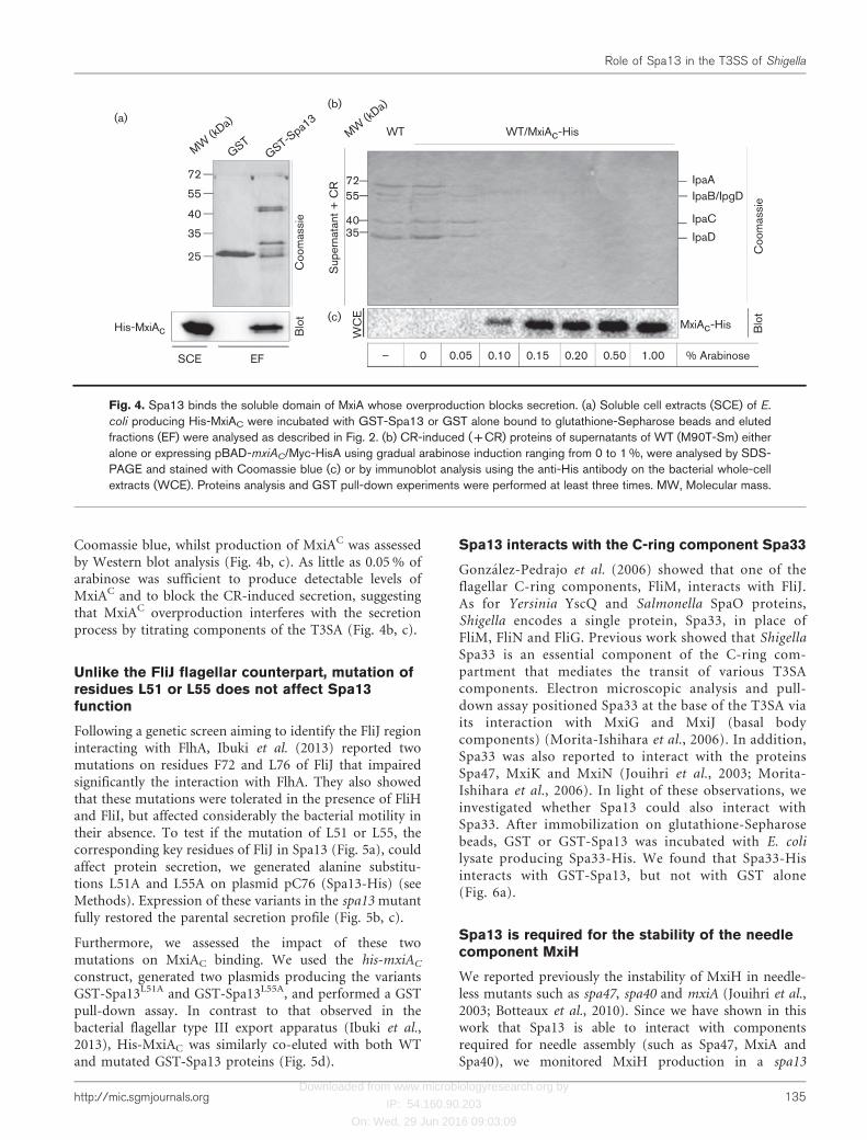
We addressed the role of Spa13 in Shigella in this context.
We performed a GST pull-down assay following the
immobilization of GST-Spa13 on glutathione-Sepharose
beads and incubation with E. coli lysate producing His-
MxiAC. We found that MxiAC interacts specifically with
GST-Spa13, but not with GST alone (Fig. 4a).
To gain further insights into the role played by MxiAC in
secretion, we performed MxiAC co-purification experi-
ments in the Shigella background to confirm the inter-
action with Spa13 and also to identify additional MxiAbinding partners. To do this, we constructed plasmid
pHA42, allowing the expression of mxiAC under thecontrol of a pBAD promoter. Recombinant MxiAC-Hiswas produced in the WT Shigella strain using a concen-tration gradient of arabinose up to 1 %. The ability of theWT strain to fix CR dye, and the subsequent red staining ofcolonies growing on a CR agar plate, is a clear indicationof the T3SA functionality and particularly of the secretionof Ipa proteins. Interestingly, upon arabinose induction,the production of recombinant MxiAC-His impaired CRfixation by the WT strain, which remained white on CRagar plates, suggesting that overproduction of MxiAC mayaffect protein secretion via the T3SA (data not shown). Toconfirm this hypothesis, proteins secreted upon CRinduction were separated by SDS-PAGE and stained with
(b)
MW
(kDa)
GST
GST-S
pa13
55
40
72
25
Blo
tC
oom
assie
His-Spa40c
SCE EF
(a)
MW
(kDa)
GST
GST-S
pa13
55
40
72
25
Blo
tC
oom
assie
His-Spa32
SCE EF
(c)
MW
(kDa)
GST
GST-S
pa32
GST-S
pa40 c
55
40
72
25
Blo
tC
oom
assie
His-Spa13
SCE EF
(d)
MW
(kDa)
GST
GST-S
pa40 c
GST-S
pa40 cDN
PTH
55
40
72
25
Blo
tC
oom
assie
His-Spa13
SCE EF
Fig. 3. Spa13 interacts with the cleaved forms of Spa40C, but not with Spa32. GST-Spa13 or GST alone bound toglutathione-Sepharose beads was incubated with E. coli soluble cell extracts (SCE) producing (a) His-Spa32 or (b) His-Spa40C, and eluted fractions (EF) were separated by SDS-PAGE and stained with Coomassie blue or analysed byimmunoblotting using an anti-His antibody. (c) Soluble cell extracts of E. coli producing His-Spa13 were incubated with GST-Spa32, GST-Spa40C or GST alone bound to glutathione-Sepharose beads. (d) Soluble cell extracts of E. coli producing His-Spa13 were incubated with GST-Spa40C, GST-Spa40CDNPTH or GST bound to glutathione-Sepharose beads. In both (c)and (d), eluted fractions were analysed as described in (a). The binding assays were repeated at least three times. MW,Molecular mass.
Y. Cherradi, A. Hachani and A. Allaoui
134 Microbiology 160
Downloaded from www.microbiologyresearch.org by
IP: 54.160.90.203
On: Wed, 29 Jun 2016 09:03:09
Coomassie blue, whilst production of MxiAC was assessedby Western blot analysis (Fig. 4b, c). As little as 0.05 % ofarabinose was sufficient to produce detectable levels ofMxiAC and to block the CR-induced secretion, suggestingthat MxiAC overproduction interferes with the secretionprocess by titrating components of the T3SA (Fig. 4b, c).
Unlike the FliJ flagellar counterpart, mutation ofresidues L51 or L55 does not affect Spa13function
Following a genetic screen aiming to identify the FliJ regioninteracting with FlhA, Ibuki et al. (2013) reported twomutations on residues F72 and L76 of FliJ that impairedsignificantly the interaction with FlhA. They also showedthat these mutations were tolerated in the presence of FliHand FliI, but affected considerably the bacterial motility intheir absence. To test if the mutation of L51 or L55, thecorresponding key residues of FliJ in Spa13 (Fig. 5a), couldaffect protein secretion, we generated alanine substitu-tions L51A and L55A on plasmid pC76 (Spa13-His) (seeMethods). Expression of these variants in the spa13 mutantfully restored the parental secretion profile (Fig. 5b, c).
Furthermore, we assessed the impact of these twomutations on MxiAC binding. We used the his-mxiAC
construct, generated two plasmids producing the variantsGST-Spa13L51A and GST-Spa13L55A, and performed a GSTpull-down assay. In contrast to that observed in thebacterial flagellar type III export apparatus (Ibuki et al.,2013), His-MxiAC was similarly co-eluted with both WTand mutated GST-Spa13 proteins (Fig. 5d).
Spa13 interacts with the C-ring component Spa33
Gonzalez-Pedrajo et al. (2006) showed that one of theflagellar C-ring components, FliM, interacts with FliJ.As for Yersinia YscQ and Salmonella SpaO proteins,Shigella encodes a single protein, Spa33, in place ofFliM, FliN and FliG. Previous work showed that ShigellaSpa33 is an essential component of the C-ring com-partment that mediates the transit of various T3SAcomponents. Electron microscopic analysis and pull-down assay positioned Spa33 at the base of the T3SA viaits interaction with MxiG and MxiJ (basal bodycomponents) (Morita-Ishihara et al., 2006). In addition,Spa33 was also reported to interact with the proteinsSpa47, MxiK and MxiN (Jouihri et al., 2003; Morita-Ishihara et al., 2006). In light of these observations, weinvestigated whether Spa13 could also interact withSpa33. After immobilization on glutathione-Sepharosebeads, GST or GST-Spa13 was incubated with E. colilysate producing Spa33-His. We found that Spa33-Hisinteracts with GST-Spa13, but not with GST alone(Fig. 6a).
Spa13 is required for the stability of the needlecomponent MxiH
We reported previously the instability of MxiH in needle-less mutants such as spa47, spa40 and mxiA (Jouihri et al.,2003; Botteaux et al., 2010). Since we have shown in thiswork that Spa13 is able to interact with componentsrequired for needle assembly (such as Spa47, MxiA andSpa40), we monitored MxiH production in a spa13
(b)
WT WT/MxiAc-His
MxiAc-His
(a)
(c)
1.000.500.200.150.100.050– % Arabinose
MW
(kDa)
MW
(kDa)
GST
GST-S
pa13
55
40
72
25
35
55
40
72
35
Coom
assie
Sup
ern
ata
nt
+ C
R
WC
E
Blo
t
Coom
assie
Blo
t
His-MxiAc
SCE EF
IpaA
IpaC
IpaD
IpaB/IpgD
Fig. 4. Spa13 binds the soluble domain of MxiA whose overproduction blocks secretion. (a) Soluble cell extracts (SCE) of E.
coli producing His-MxiAC were incubated with GST-Spa13 or GST alone bound to glutathione-Sepharose beads and elutedfractions (EF) were analysed as described in Fig. 2. (b) CR-induced (+CR) proteins of supernatants of WT (M90T-Sm) eitheralone or expressing pBAD-mxiAC/Myc-HisA using gradual arabinose induction ranging from 0 to 1 %, were analysed by SDS-PAGE and stained with Coomassie blue (c) or by immunoblot analysis using the anti-His antibody on the bacterial whole-cellextracts (WCE). Proteins analysis and GST pull-down experiments were performed at least three times. MW, Molecular mass.
Role of Spa13 in the T3SS of Shigella
http://mic.sgmjournals.org 135

Downloaded from www.microbiologyresearch.org by
IP: 54.160.90.203
On: Wed, 29 Jun 2016 09:03:09
background. As shown in Fig. 6(b), and compared to theWT and complemented spa13-/Spa13 strains, MxiH wasnot detected in cell extracts of spa13. The instability ofMxiH is similar to that observed in the mxiD secretionmutant (Blocker et al., 2001). The absence of Spa13 did
not impact the stability of the translocators IpaB and IpaC(Fig. 6b). Collectively, our results confirm the existence ofa negative feedback inhibition mechanism that controlsMxiH stability in the absence of needle complexformation.
(a)
Spa13
20 30 40 50 60 70 80
FliJ
(c)
Blo
t
Spa13-His
(b)
MW
(kDa)
WT
Spa13
Spa13L5
1A
Spa13L5
5A
–
Spa13
55
40
72
35
25
IpaA
IpaCIpaD
OspD1
IpaB/IpgD
Coom
assie
Sup
ern
ata
nt
+ C
R
WC
E
(d)
MW
(kDa)
GST
GST-S
pa13
GST-S
pa13L5
1A
GST-S
pa13L5
5A
55
40
72
35
25
Coom
assie
Blo
t
His-MxiAc
SCE EFSpa13-His
Fig. 5. Mutations Spa13L51A and Spa13L55A do not affect Spa13 function. (a) Sequence alignment of Spa13 and FliJ using theCLUSTAL Omega and Jalview programs. Identical residues are boxed in black, whilst similar ones are shown in grey. Arrows pointto residues L51 and L55 of Spa13 (F72 and L76 in FliJ) mutated in this study. (b) CR-induced proteins of supernatants (+CR)of WT (M90T-Sm), spa13 mutant (spa13) and spa13 mutant complemented with IPTG-induced plasmid expressing nativeSpa13 (pC76) or its derivatives expressing Spa13L51A or Spa13L55A. (c) Control analysis of Spa13 and its derivativesproduction, in the bacterial whole-cell extracts (WCE), by immunoblot using the anti-His antibody. (d) Soluble cell extracts(SCE) of E. coli producing His-MxiAC were incubated with GST-Spa13, GST-Spa13L51A, GST-Spa13L55A or GST alonebound to glutathione-Sepharose beads and eluted fractions (EF) were analysed as in Fig. 2. These experiments were repeatedthree times. MW, Molecular mass.
MW
(kDa)
GST
GST-S
pa13
55
40
72
25
Coom
assie
Blo
t
Blo
t
SCE EF
WCE
Spa33-His
(a)
(b)
WT
mxiD
spa13
spa13/S
pa13
IpaCIpaB
MxiH
Fig. 6. Spa13 interacts with the C-ringcomponent Spa33 and is required for MxiHstability. (a) Soluble cell extracts (SCE) ofE. coli producing Spa33-His were incubatedwith GST-Spa13 or GST alone bound toglutathione-Sepharose and eluted fractions(EF) were analysed as described in Fig. 2.The binding assays were repeated at leastthree times. (b) MxiH is not stable in the spa13
mutant. Cultures of strains M90T-Sm (WT),mxiD (T3S-deficient mutant), spa13 andspa13/Spa13 (pHA1) were harvested in theexponential phase of growth, and proteins ofthe whole-cell extracts (WCE) were analysedby SDS-PAGE and immunoblotting using anti-MxiH polyclonal antibodies and mAbs againstIpaB and IpaC. MW, Molecular mass.
Y. Cherradi, A. Hachani and A. Allaoui
136 Microbiology 160

Downloaded from www.microbiologyresearch.org by
IP: 54.160.90.203
On: Wed, 29 Jun 2016 09:03:09
DISCUSSION
Although it has been described and often replaced
hypothetically within T3S models, little is known about
the role of Spa13 in the T3SA of Shigella. The interaction
network presented in this work offers more insight about
the role of Spa13 in the T3S process. The molecular
characteristics of Spa13 are quite similar to those of its
homologues, including the high probability of an a-helical
coiled-coil structure. This suggests that Spa13 is likely to
engage an amphipathic helical interaction, either with itself
to form a homodimer or to serve as a binding platform for
other T3S partners. Analysis of protein–protein interaction
provides valuable information regarding the potential
functions of interacting partners. Here, in an attempt tounderstand the role of Spa13 in the secretion process, weidentified some of its interacting partners among majorT3S components, including Spa47, Spa33, Spa40 and MxiAas well as the translocator chaperone IpgC. In addition, wereport that the spa13 mutant is unable to produce a stableneedle component MxiH, indicating that Spa13 acts at anearly stage of needle complex assembly. Comparatively, thisphenotype is similar to previously reported observationsfor several needle-less mutants such as spa40, spa47, mxiA,mxiN, mxiK, mxiI, mxiD, mxiJ and mxiG (Jouihri et al.,2003; Botteaux et al., 2010; our unpublished data).Consequently, through its various interactions, Spa13 playsa crucial role in the needle assembly process.
(1)
MxiDOM
P
IM
MxiG
MxiJ
MxiH
Mxil
MxiA
?
Spa47
Spa33
Activation of late-effectorgene expression
Spa13
Spa40
IpgC
IpaB/C
MxiN
MxiE
MxiD
MxiG
MxiJ
MxiD
MxiG
MxiJ
MxiD
MxiG
MxiJ
MxiD
MxiG
MxiJ
MxiD
MxiG
MxiJ
(2) (3)
Fig. 7. Model of the roles of Spa13 during the export and secretion processes. (1) Spa13 interacts with the ATPase Spa47 andthe C-terminal domain of both inner-membrane proteins MxiA and Spa40. Spa33, which forms the C-ring, interacts with Spa13,the ATPase and MxiN. (2) After needle assembly, a free pool of Spa13 interacts with chaperone IpgC coupled to translocatorsIpaB or IpaC and recruits them to the ATPase. (3) Spa47 promotes chaperone–translocator dissociation, allowing entry of thepartially folded translocator inside the cage formed by nonameric MxiAC where Spa13 may act as an export gate-activatorswitch. Meanwhile, free IpgC is recycled by Spa13 to ensure new translocator income into the export platform. Free IpgC willfurther bind MxiE to induce late-effector gene expression and secretion. OM, Outer membrane; P, periplasmic space; IM, innermembrane.
Role of Spa13 in the T3SS of Shigella
http://mic.sgmjournals.org 137

Downloaded from www.microbiologyresearch.org by
IP: 54.160.90.203
On: Wed, 29 Jun 2016 09:03:09
In Yersinia, YscO has been shown previously to interactwith the molecular ruler protein YscP, the ATPase YscNand the inner-membrane protein YscU, indicating thatthese proteins function together to facilitate Yop secretion(Payne & Straley, 1998, 1999; Riordan et al., 2008). Here,we demonstrate that Spa13 interacts with both Spa40C
(YscUC) and the ATPase Spa47 (YscN), but not with Spa32(YscP). Very recently, it was shown that YscO interactionwith YscP involves the T3S substrate specificity switch(T3S4) domain of YscP, which is also conserved in Spa32(Mukerjea & Ghosh, 2013; Botteaux et al., 2008). Whilstmost of the components of the T3SA share a high level ofhomology, Spa13 and Spa32 present a low level of sequencesimilarity with YscO and YscP, suggesting a Yersinia-specific function for the YscO–YscP complex (Bergmanet al., 1994; Payne & Straley, 1999). As YscO has been co-purified with blocked export machinery complexes con-taining the ATPase YscN (Riordan & Schneewind, 2008)and FliJ increases ATP hydrolysis of the membrane-associated export ATPase FliI, the identified Spa13–Spa47interaction suggests a similar function for this complex.Whether Spa13 positively affects or not Spa47 oligomer-ization and/or activity remains to be addressed. In parallel,Spa13 localization should also be determined, as both FliJand FliI have an intrinsic membrane affinity (Auvray et al.,2002; Evans et al., 2006).
As has been reported for FliJ, Spa13 is not a secretedprotein, which contrasts with YscO secretion by the YscT3SA (Payne & Straley, 1998, 1999). Therefore, this resultrather supports a role of Spa13 as a cytoplasmic T3S-interacting module. Indeed, FliJ was reported to act as achaperone escort protein in the flagellar system (Evanset al., 2006), a role that was also assigned to YscO (Yersinia)and InvI (Salmonella) which bind the translocon chaper-ones SycD and SicA, respectively (Evans & Hughes, 2009).The identified interaction between Spa13 and IpgC suggeststhat Spa13 also acts as an escort for IpgC both in its free stateand probably when associated with its cognate substrate IpaBand/or IpaC (Fig. 7). Given that the gatekeeper MxiCinteracts with IpgC to promote translocator secretion(Cherradi et al., 2013), we propose that Spa13 takes part inthis control mechanism, provided that the T3SA is assembledcorrectly (Fig. 7). Indeed, in the latter process, Spa32 wasshown to interact with the cleaved form of Spa40, which alsointeracts with MxiA, MxiN, Spa47, Spa33 and MxiK. Here,we demonstrated that Spa13 does not bind the unprocessedform of Spa40, as reported previously for Spa47 and Spa33(Botteaux et al., 2010), suggesting an active role of Spa13during the T3SA assembly process.
In the Salmonella flagellar system, FliJ interacts with theFliH2–FliI complex, and with the large cytoplasmic domainof the export gate proteins FlhAC (MxiAC homologue) andFlhBC (Spa40C homologue). The FliH–FliI–FliJ complex isthought to bring substrates to the export gate upon bindingto the cytoplasmic region of the FlhA–FlhB dockingplatform (Fraser et al., 2003; Minamino & Macnab,2000). FliJ interacts with one of the C-ring components,
FliM (Spa33), to empty subunit chaperones such as FliT
and FlgN (Evans et al., 2006; Imada et al., 2010). Finally,
the interaction described between chaperone–substrate
complexes and FlhAC is potentiated by FliJ. This demon-
strates that FliJ promotes the export of substrates upon
their delivery to the T3SA entry gate (Saijo-Hamano et al.,
2010). In light of these data, FliJ is implicated markedly in
the export process through dynamic interactions with its
binding partners. Whether such a mechanism also occurs
in related T3SA-secreting virulence effectors is not obvious.
Our data presented in this work show that several of the
described flagellar interactions also take place in the
Shigella context, such as those involving MxiAC, Spa40C,
Spa33, Spa47 and IpgC. In addition, interactions detected
previously between MxiN (FliH) and either Spa33 or
Spa47 support fully our current observations (Jouihri et al.,
2003). The kinetics of these interactions during the
secretion process remain to be investigated.
The recently solved structure of MxiA offered new leads
about its function in the T3SA, but also offered new
perspectives regarding the interaction network supporting
the dynamics of the apparatus (Abrusci et al., 2013). We
demonstrated in this work that overexpression of the
soluble domain of MxiA (MxiAC) impairs T3SA activity,
which is likely due to an unproductive titration of T3S
soluble components, including Spa13. Comparatively,
earlier data reported that individual overexpression of
Spa32 or Spa33 in WT Shigella blocks Ipa protein secretion;
however, their simultaneous co-overexpression restores the
parental secretion pattern (Schuch & Maurelli, 2001).
Likewise, overproduction of YscP in the WT Yersinia
background blocked secretion, whilst co-overexpression of
both YscP and YscO had no effect on secretion (Payne &
Straley, 1998, 1999). Thus, it appears attractive to identify
point mutations within spa13 that could reduce Spa13
interaction with MxiAC. In this context, we showed here that
in contrast to mutations F72A and L76A of FliJ, which
impair its binding to FlhA and Salmonella motility (Ibuki
et al., 2013), substitution of the corresponding residues of
Spa13, L51 and L55, by alanine does not impair secretion or
MxiA binding. This result indicates that although similar,
FliJ and Spa13 exhibit specific and distinct interactions
within their corresponding secretory apparatus.
Accordingly, the amount of some soluble T3S componentseems critical for a functional T3SA. In line with thishypothesis, we showed that WT secretion is restored in thespa13 mutant even in the absence of a detectable level ofSpa13-His in the bacterial lysate, suggesting that a limitednumber of Spa13 molecules are sufficient to ensure itsfunction in T3S. Given that overexpression of MxiAC blockedCR-induced secretion, and that Spa13, Spa33 and MxiA levelsare controlled by the same slippage regulatory mechanism,one could speculate that a functional T3SA is orchestrated bya subset of molecules present in a relatively low abundancecompared with other components of the machinery.
Y. Cherradi, A. Hachani and A. Allaoui
138 Microbiology 160

Downloaded from www.microbiologyresearch.org by
IP: 54.160.90.203
On: Wed, 29 Jun 2016 09:03:09
In light of our results, and in perspective of the literature, wepropose a new T3SA model focused on the role of Spa13 as achaperone escort to IpgC and also as an export gate activator(Fig. 7). Spa13 interacts with the following components ofthe T3SA: the ATPase Spa47, the C-terminal domains ofinner-membrane proteins MxiA and Spa40, and Spa33which forms the C-ring. Upon assembly of the needle, a freepool of Spa13 is available to interact with the chaperoneIpgC coupled to its cognate partners, the translocators IpaBand IpaC. Spa13 would then direct them towards the ATPaseSpa47. At this stage, Spa47 would promote the dissociationof the chaperone–translocator complex, thus facilitating theengagement of partially folded translocators inside the entrygate cage composed by nonameric MxiAC where Spa13 mayact as an export gate-activator switch. Once the machinery iscompleted, free IpgC could be recycled by Spa13 to ensurethe provision of new translocator molecules at the exportplatform, but is also available to engage with the transcrip-tional activator MxiE, and will ultimately lead to theexpression of late-effector genes.
In conclusion, we report here an interaction networkaround the poorly studied T3S component Spa13 andprovide new insights into its function. In light of itspredicted a-helical coiled-coil structure, further studiesshould investigate the potential oligomeric state of Spa13.Resolving the structure of Spa13 should help understandhow it operates as an escort chaperone and as an exportgate-activator switch. Finally, the interaction identifiedbetween Spa13 and Spa47 should be taken into account toresolve the crystal structure of the Spa13–Spa47 complex,which will help us to understand the roles of Spa13 in thedynamics of the secretion process.
ACKNOWLEDGEMENTS
The present work was supported by grants from the Fonds National dela Recherche Scientifique – Fonds National Belge de la RechercheScientifique (Convention F.3.4556.11) and from the EuropeanCommunity’s Seventh Framework Programme FP7/2011-2015 undergrant agreement 261742. Y. C. is a recipient of a PhD fellowshipfrom the Belgian Fonds National de Recherches Industrielles etAgronomiques. A. H. was a recipient of a fellowship from the FondsNational de Recherches Industrielles et Agronomiques. A part of thiswork was also supported by the Fonds Defay and by the Alice andDavid Van Buuren foundation. We would like to thank Luke Allsoppfor critical reading of the manuscript, A. Botteaux and C.A. Kayath forhelpful discussions, and V. Delforge for technical assistance.
REFERENCES
Abrusci, P., Vergara-Irigaray, M., Johnson, S., Beeby, M. D.,Hendrixson, D. R., Roversi, P., Friede, M. E., Deane, J. E., Jensen,G. J. & other authors (2013). Architecture of the major component ofthe type III secretion system export apparatus. Nat Struct Mol Biol 20,99–104.
Aizawa, S. I. (2001). Bacterial flagella and type III secretion systems.FEMS Microbiol Lett 202, 157–164.
Allaoui, A., Mounier, J., Prevost, M. C., Sansonetti, P. J. & Parsot, C.(1992). icsB: a Shigella flexneri virulence gene necessary for the lysis ofprotrusions during intercellular spread. Mol Microbiol 6, 1605–1616.
Auvray, F., Ozin, A. J., Claret, L. & Hughes, C. (2002). Intrinsic
membrane targeting of the flagellar export ATPase FliI: interaction
with acidic phospholipids and FliH. J Mol Biol 318, 941–950.
Bennett, J. C. & Hughes, C. (2000). From flagellum assembly to
virulence: the extended family of type III export chaperones. Trends
Microbiol 8, 202–204.
Bergman, T., Erickson, K., Galyov, E., Persson, C. & Wolf-Watz, H.(1994). The lcrB (yscN/U) gene cluster of Yersinia pseudotuberculosis is
involved in Yop secretion and shows high homology to the spa gene
clusters of Shigella flexneri and Salmonella typhimurium. J Bacteriol
176, 2619–2626.
Blocker, A., Jouihri, N., Larquet, E., Gounon, P., Ebel, F., Parsot, C.,
Sansonetti, P. & Allaoui, A. (2001). Structure and composition of the
Shigella flexneri ‘‘needle complex’’, a part of its type III secreton. Mol
Microbiol 39, 652–663.
Blocker, A., Komoriya, K. & Aizawa, S. (2003). Type III secretion
systems and bacterial flagella: insights into their function from
structural similarities. Proc Natl Acad Sci U S A 100, 3027–3030.
Botteaux, A., Sani, M., Kayath, C. A., Boekema, E. J. & Allaoui, A.(2008). Spa32 interaction with the inner-membrane Spa40 compon-
ent of the type III secretion system of Shigella flexneri is required for
the control of the needle length by a molecular tape measure
mechanism. Mol Microbiol 70, 1515–1528.
Botteaux, A., Sory, M. P., Biskri, L., Parsot, C. & Allaoui, A. (2009).MxiC is secreted by and controls the substrate specificity of the
Shigella flexneri type III secretion apparatus. Mol Microbiol 71, 449–
460.
Botteaux, A., Kayath, C. A., Page, A. L., Jouihri, N., Sani, M.,Boekema, E., Biskri, L., Parsot, C. & Allaoui, A. (2010). The 33
carboxyl-terminal residues of Spa40 orchestrate the multi-step
assembly process of the type III secretion needle complex in
Shigella flexneri. Microbiology 156, 2807–2817.
Buchrieser, C., Glaser, P., Rusniok, C., Nedjari, H., D’Hauteville, H.,Kunst, F., Sansonetti, P. & Parsot, C. (2000). The virulence plasmid
pWR100 and the repertoire of proteins secreted by the type III
secretion apparatus of Shigella flexneri. Mol Microbiol 38, 760–
771.
Cherradi, Y., Schiavolin, L., Moussa, S., Meghraoui, A., Meksem, A.,Biskri, L., Azarkan, M., Allaoui, A. & Botteaux, A. (2013). Interplay
between predicted inner-rod and gatekeeper in controlling substrate
specificity of the type III secretion system. Mol Microbiol 87, 1183–
1199.
Collazo, C. M., Zierler, M. K. & Gatan, J. E. (1995). Functional analysis
of the Salmonella typhimurium invasion genes invl and invJ and
identification of a target of the protein secretion apparatus encoded in
the inv locus. Mol Microbiol 15, 25–38.
Cornelis, G. R. (2006). The type III secretion injectisome. Nat Rev
Microbiol 4, 811–825.
Diepold, A., Wiesand, U., Amstutz, M. & Cornelis, G. R. (2012).
Assembly of the Yersinia injectisome: the missing pieces. Mol
Microbiol 85, 878–892.
Erhardt, M., Singer, H. M., Wee, D. H., Keener, J. P. & Hughes, K. T.
(2011). An infrequent molecular ruler controls flagellar hook length
in Salmonella enterica. EMBO J 30, 2948–2961.
Espina, M., Olive, A. J., Kenjale, R., Moore, D. S., Ausar, S. F.,
Kaminski, R. W., Oaks, E. V., Middaugh, C. R., Picking, W. D. &Picking, W. L. (2006). IpaD localizes to the tip of the type III secretion
system needle of Shigella flexneri. Infect Immun 74, 4391–4400.
Evans, L. D. & Hughes, C. (2009). Selective binding of virulence type
III export chaperones by FliJ escort orthologues InvI and YscO. FEMS
Microbiol Lett 293, 292–297.
Role of Spa13 in the T3SS of Shigella
http://mic.sgmjournals.org 139

Downloaded from www.microbiologyresearch.org by
IP: 54.160.90.203
On: Wed, 29 Jun 2016 09:03:09
Evans, L. D., Stafford, G. P., Ahmed, S., Fraser, G. M. & Hughes, C.(2006). An escort mechanism for cycling of export chaperones duringflagellum assembly. Proc Natl Acad Sci U S A 103, 17474–
17479.
Fraser, G. M., Gonzalez-Pedrajo, B., Tame, J. R. & Macnab, R. M.(2003). Interactions of FliJ with the Salmonella type III flagellar export
apparatus. J Bacteriol 185, 5546–5554.
Galan, J. E. & Wolf-Watz, H. (2006). Protein delivery into eukaryotic
cells by type III secretion machines. Nature 444, 567–573.
Gonzalez-Pedrajo, B., Minamino, T., Kihara, M. & Namba, K. (2006).Interactions between C ring proteins and export apparatus compo-
nents: a possible mechanism for facilitating type III protein export.Mol Microbiol 60, 984–998.
Herrmann, M., Schuhmacher, A., Muhldorfer, I., Melchers, K.,Prothmann, C. & Dammeier, S. (2006). Identification and character-ization of secreted effector proteins of Chlamydophila pneumoniae
TW183. Res Microbiol 157, 513–524.
Hueck, C. J. (1998). Type III protein secretion systems in bacterial
pathogens of animals and plants. Microbiol Mol Biol Rev 62, 379–433.
Ibuki, T., Imada, K., Minamino, T., Kato, T., Miyata, T. & Namba, K.(2011). Common architecture of the flagellar type III protein export
apparatus and F- and V-type ATPases. Nat Struct Mol Biol 18, 277–282.
Ibuki, T., Uchida, Y., Hironaka, Y., Namba, K., Imada, K. & Minamino,T. (2013). Interaction between FliJ and FlhA, components of the
bacterial flagellar type III export apparatus. J Bacteriol 195, 466–473.
Imada, K., Minamino, T., Kinoshita, M., Furukawa, Y. & Namba, K.(2010). Structural insight into the regulatory mechanisms of
interactions of the flagellar type III chaperone FliT with its bindingpartners. Proc Natl Acad Sci U S A 107, 8812–8817.
Jouihri, N., Sory, M. P., Page, A. L., Gounon, P., Parsot, C. & Allaoui,A. (2003). MxiK and MxiN interact with the Spa47 ATPase and arerequired for transit of the needle components MxiH and MxiI, but
not of Ipa proteins, through the type III secretion apparatus of
Shigella flexneri. Mol Microbiol 49, 755–767.
Journet, L., Agrain, C., Broz, P. & Cornelis, G. R. (2003). The needle
length of bacterial injectisomes is determined by a molecular ruler.Science 302, 1757–1760.
Kim, P. S., Berger, B. & Wolf, E. (1997). MultiCoil: a program for
predicting two- and three-stranded coiled coils. Protein Sci 6, 1179–1189.
Kotloff, K. L., Winickoff, J. P., Ivanoff, B., Clemens, J. D., Swerdlow,D. L., Sansonetti, P. J., Adak, G. K. & Levine, M. M. (1999). Global
burden of Shigella infections: implications for vaccine development
and implementation of control strategies. Bull World Health Organ77, 651–666.
Kubori, T., Matsushima, Y., Nakamura, D., Uralil, J., Lara-Tejero, M.,Sukhan, A., Galan, J. E. & Aizawa, S. I. (1998). Supramolecularstructure of the Salmonella typhimurium type III protein secretion
system. Science 280, 602–605.
Lorenzini, E., Singer, A., Singh, B., Lam, R., Skarina, T., Chirgadze,N. Y., Savchenko, A. & Gupta, R. S. (2010). Structure and protein–
protein interaction studies on Chlamydia trachomatis protein CT670(YscO homolog). J Bacteriol 192, 2746–2756.
Macnab, R. M. (1999). The bacterial flagellum: reversible rotary
propellor and type III export apparatus. J Bacteriol 181, 7149–7153.
Macnab, R. M. (2004). Type III flagellar protein export and flagellar
assembly. Biochim Biophys Acta 1694, 207–217.
Magdalena, J., Hachani, A., Chamekh, M., Jouihri, N., Gounon, P.,Blocker, A. & Allaoui, A. (2002). Spa32 regulates a switch in substrate
specificity of the type III secreton of Shigella flexneri from needlecomponents to Ipa proteins. J Bacteriol 184, 3433–3441.
McDonnell, A. V., Jiang, T., Keating, A. E. & Berger, B. (2006).Paircoil2: improved prediction of coiled coils from sequence.
Bioinformatics 22, 356–358.
Meitert, T., Pencu, E., Ciudin, L., Tonciu, M., Mihai, I. & Nicolescu, S.(1991). Correlation between Congo red binding as virulence marker
in Shigella species and Sereny test. Roum Arch Microbiol Immunol 50,
45–52.
Miller, V. L. & Mekalanos, J. J. (1988). A novel suicide vector and its
use in construction of insertion mutations: osmoregulation of outer
membrane proteins and virulence determinants in Vibrio cholerae
requires toxR. J Bacteriol 170, 2575–2583.
Minamino, T. & Macnab, R. M. (2000). Interactions among
components of the Salmonella flagellar export apparatus and its
substrates. Mol Microbiol 35, 1052–1064.
Minamino, T., Chu, R., Yamaguchi, S. & Macnab, R. M. (2000). Role
of FliJ in flagellar protein export in Salmonella. J Bacteriol 182, 4207–
4215.
Minamino, T., Imada, K. & Namba, K. (2008). Mechanisms of type III
protein export for bacterial flagellar assembly. Mol Biosyst 4, 1105–
1115.
Minamino, T., Morimoto, Y. V., Hara, N. & Namba, K. (2011). An
energy transduction mechanism used in bacterial flagellar type III
protein export. Nat Commun 2, 475.
Morita-Ishihara, T., Ogawa, M., Sagara, H., Yoshida, M., Katayama,E. & Sasakawa, C. (2006). Shigella Spa33 is an essential C-ring
component of type III secretion machinery. J Biol Chem 281, 599–607.
Mukerjea, R. & Ghosh, P. (2013). Functionally essential interaction
between Yersinia YscO and the T3S4 domain of YscP. J Bacteriol 195,
4631–4638.
Parsot, C. (2009). Shigella type III secretion effectors: how, where,
when, for what purposes? Curr Opin Microbiol 12, 110–116.
Payne, P. L. & Straley, S. C. (1998). YscO of Yersinia pestis is a mobile
core component of the Yop secretion system. J Bacteriol 180, 3882–
3890.
Payne, P. L. & Straley, S. C. (1999). YscP of Yersinia pestis is a secreted
component of the Yop secretion system. J Bacteriol 181, 2852–2862.
Penno, C. & Parsot, C. (2006). Transcriptional slippage in mxiE
controls transcription and translation of the downstream mxiD gene,
which encodes a component of the Shigella flexneri type III secretion
apparatus. J Bacteriol 188, 1191–1198.
Penno, C., Hachani, A., Biskri, L., Sansonetti, P., Allaoui, A. & Parsot,C. (2006). Transcriptional slippage controls production of type III
secretion apparatus components in Shigella flexneri. Mol Microbiol 62,
1460–1468.
Riordan, K. E. & Schneewind, O. (2008). YscU cleavage and the
assembly of Yersinia type III secretion machine complexes. Mol
Microbiol 68, 1485–1501.
Riordan, K. E., Sorg, J. A., Berube, B. J. & Schneewind, O. (2008).Impassable YscP substrates and their impact on the Yersinia
enterocolitica type III secretion pathway. J Bacteriol 190, 6204–6216.
Saijo-Hamano, Y., Imada, K., Minamino, T., Kihara, M., Shimada, M.,Kitao, A. & Namba, K. (2010). Structure of the cytoplasmic domain of
FlhA and implication for flagellar type III protein export. Mol
Microbiol 76, 260–268.
Samudrala, R., Heffron, F. & McDermott, J. E. (2009). Accurate prediction
of secreted substrates and identification of a conserved putative secretion
signal for type III secretion systems. PLoS Pathog 5, e1000375.
Sani, M., Botteaux, A., Parsot, C., Sansonetti, P., Boekema, E. J. &Allaoui, A. (2007). IpaD is localized at the tip of the Shigella flexneri
type III secretion apparatus. Biochim Biophys Acta 1770, 307–311.
Y. Cherradi, A. Hachani and A. Allaoui
140 Microbiology 160

Downloaded from www.microbiologyresearch.org by
IP: 54.160.90.203
On: Wed, 29 Jun 2016 09:03:09
Sasakawa, C., Komatsu, K., Tobe, T., Suzuki, T. & Yoshikawa, M. (1993).Eight genes in region 5 that form an operon are essential for invasion ofepithelial cells by Shigella flexneri 2a. J Bacteriol 175, 2334–2346.
Schroeder, G. N. & Hilbi, H. (2008). Molecular pathogenesis ofShigella spp.: controlling host cell signaling, invasion, and death bytype III secretion. Clin Microbiol Rev 21, 134–156.
Schuch, R. & Maurelli, A. T. (2001). Spa33, a cell surface-associatedsubunit of the Mxi-Spa type III secretory pathway of Shigellaflexneri, regulates Ipa protein traffic. Infect Immun 69, 2180–2189.
Shibata, S., Takahashi, N., Chevance, F. F., Karlinsey, J. E., Hughes,K. T. & Aizawa, S. (2007). FliK regulates flagellar hook length as aninternal ruler. Mol Microbiol 64, 1404–1415.
Stone, C. B., Johnson, D. L., Bulir, D. C., Gilchrist, J. D. & Mahony, J. B.(2008). Characterization of the putative type III secretion ATPase CdsN(Cpn0707) of Chlamydophila pneumoniae. J Bacteriol 190, 6580–6588.
Wagner, S., Stenta, M., Metzger, L. C., Dal Peraro, M. & Cornelis, G. R.(2010). Length control of the injectisome needle requires only onemolecule of Yop secretion protein P (YscP). Proc Natl Acad Sci U S A107, 13860–13865.
Role of Spa13 in the T3SS of Shigella
http://mic.sgmjournals.org 141
Copyright © 2022 FDOKUMEN