Paper G-2 333 - Visualization of cavitation and investigation of ...

This article appeared in a journal published by Elsevier. The attachedcopy is furnished to the author for internal non-commercial researchand education use, including for instruction at the authors institution
and sharing with colleagues.
Other uses, including reproduction and distribution, or selling orlicensing copies, or posting to personal, institutional or third party
websites are prohibited.
In most cases authors are permitted to post their version of thearticle (e.g. in Word or Tex form) to their personal website orinstitutional repository. Authors requiring further information
regarding Elsevier’s archiving and manuscript policies areencouraged to visit:
http://www.elsevier.com/copyright

Author's personal copy
Review
Sp185/333: A novel family of genes and proteins involved in the purple seaurchin immune response
Julie Ghosh a, Katherine M. Buckley b, Sham V. Nair c, David A. Raftos c, Chase Miller a,Audrey J. Majeske a, Taku Hibino b,1, Jonathan P. Rast b, Mattias Roth c, L. Courtney Smith a,*a Department of Biological Sciences, George Washington University, Washington, DC, United Statesb Department of Immunology, Sunnybrook Health Sciences Centre, University of Toronto, Toronto, Canadac Department of Biological Sciences, Macquarie University, Sydney, Australia
Contents
1. Introduction . . . . . . . . . . . . . . . . . . . . . . . . . . . . . . . . . . . . . . . . . . . . . . . . . . . . . . . . . . . . . . . . . . . . . . . . . . . . . . . . . . . . . . . . . . . . . . . . . . . . . 236
2. The Sp185/333 transcripts . . . . . . . . . . . . . . . . . . . . . . . . . . . . . . . . . . . . . . . . . . . . . . . . . . . . . . . . . . . . . . . . . . . . . . . . . . . . . . . . . . . . . . . . . . 236
3. The Sp185/333 cDNAs . . . . . . . . . . . . . . . . . . . . . . . . . . . . . . . . . . . . . . . . . . . . . . . . . . . . . . . . . . . . . . . . . . . . . . . . . . . . . . . . . . . . . . . . . . . . . 236
4. The deduced Sp185/333 proteins . . . . . . . . . . . . . . . . . . . . . . . . . . . . . . . . . . . . . . . . . . . . . . . . . . . . . . . . . . . . . . . . . . . . . . . . . . . . . . . . . . . . 236
5. Localization of Sp185/333 proteins in coelomocytes . . . . . . . . . . . . . . . . . . . . . . . . . . . . . . . . . . . . . . . . . . . . . . . . . . . . . . . . . . . . . . . . . . . . . 237
6. Tissue-specific localization of Sp185/333 proteins. . . . . . . . . . . . . . . . . . . . . . . . . . . . . . . . . . . . . . . . . . . . . . . . . . . . . . . . . . . . . . . . . . . . . . . 239
7. Sp185/333 is expressed in sea urchin larvae. . . . . . . . . . . . . . . . . . . . . . . . . . . . . . . . . . . . . . . . . . . . . . . . . . . . . . . . . . . . . . . . . . . . . . . . . . . . 239
8. Sp185/333 gene expression in response to various PAMPs . . . . . . . . . . . . . . . . . . . . . . . . . . . . . . . . . . . . . . . . . . . . . . . . . . . . . . . . . . . . . . . . 240
9. Changes in coelomocyte populations in response to immune challenge . . . . . . . . . . . . . . . . . . . . . . . . . . . . . . . . . . . . . . . . . . . . . . . . . . . . . 240
10. Diversity of Sp185/333 proteins . . . . . . . . . . . . . . . . . . . . . . . . . . . . . . . . . . . . . . . . . . . . . . . . . . . . . . . . . . . . . . . . . . . . . . . . . . . . . . . . . . . . . 240
11. The Sp185/333 gene family . . . . . . . . . . . . . . . . . . . . . . . . . . . . . . . . . . . . . . . . . . . . . . . . . . . . . . . . . . . . . . . . . . . . . . . . . . . . . . . . . . . . . . . . . 241
12. The Sp185/333 gene family is the result of recent divergence. . . . . . . . . . . . . . . . . . . . . . . . . . . . . . . . . . . . . . . . . . . . . . . . . . . . . . . . . . . . . . 242
13. Sources of Sp185/333 diversity . . . . . . . . . . . . . . . . . . . . . . . . . . . . . . . . . . . . . . . . . . . . . . . . . . . . . . . . . . . . . . . . . . . . . . . . . . . . . . . . . . . . . . 243
13.1. Sp185/333 gene recombination and sequence diversification . . . . . . . . . . . . . . . . . . . . . . . . . . . . . . . . . . . . . . . . . . . . . . . . . . . . . . . . . 243
13.2. mRNA editing . . . . . . . . . . . . . . . . . . . . . . . . . . . . . . . . . . . . . . . . . . . . . . . . . . . . . . . . . . . . . . . . . . . . . . . . . . . . . . . . . . . . . . . . . . . . . . 243
Developmental and Comparative Immunology 34 (2010) 235–245
A R T I C L E I N F O
Article history:
Received 22 September 2009
Received in revised form 23 October 2009
Accepted 24 October 2009
Available online 12 November 2009
Keywords:
Echinoderm
Immune diversity
Sea urchin
Coelomocyte
Gene family
Sequence variability
Strongylocentrotus purpuratus
A B S T R A C T
The Sp185/333 system of genes, messages and proteins are expressed in the coelomocytes of the purple
sea urchin, Strongylocentrotus purpuratus, and is an extraordinary example of diversification of a putative
innate immune response system in an invertebrate. Reviewed here, is the current understanding of this
complex system as illustrated by sequence comparisons of the genes, messages and deduced proteins
with descriptions of diversity, including preliminary results on genomic organization and descriptions of
185/333 in other echinoids. Sp185/333 gene expression in adults and embryos occurs in response to
immune challenge and includes changes in the frequencies of Sp185/333-positive coelomocytes in the
adults. The diversity of the Sp185/333 protein repertoire in coelomocytes is far greater than the sequence
diversity encoded in the genes, which may be the result of rapid gene recombination, RNA editing and/or
low-fidelity transcription, plus post-translational modifications. This review concludes with preliminary
results and speculations on protein function.
� 2009 Elsevier Ltd. All rights reserved.
Abbreviations: RAG, recombination activating genes; TLRs, Toll-like receptors; NLRs, NOD/NALP-like receptors; SNPs, single nucleotide polymorphisms; LPS,
lipopolysaccharide; ORF, open reading frame; AID, activation induced deaminase; VLRs, variable leukocyte receptors; LRRs, leucine rich repeats; FREP, fibrinogen related
protein; DSCAM, Down’s syndrome cell adhesion molecule; PAMPs, pathogen-associated molecular patterns; 1D, one-dimensional; 2DE, two-dimensional electrophoresis;
My, million years; Mya, million years ago.
* Corresponding author at: Department of Biological Sciences, George Washington University, 340 Lisner Hall, 2023 G St. NW, Washington, DC 20052, United States.
Tel.: +1 202 994 9211; fax: +1 202 994 6100.
E-mail address: [email protected] (L.C. Smith).1 Current address: Saitama University, Japan.
Contents lists available at ScienceDirect
Developmental and Comparative Immunology
journa l homepage: www.e lsev ier .com/ locate /dc i
0145-305X/$ – see front matter � 2009 Elsevier Ltd. All rights reserved.
doi:10.1016/j.dci.2009.10.008

Author's personal copy
14. Conclusion . . . . . . . . . . . . . . . . . . . . . . . . . . . . . . . . . . . . . . . . . . . . . . . . . . . . . . . . . . . . . . . . . . . . . . . . . . . . . . . . . . . . . . . . . . . . . . . . . . . . . . 243
Acknowledgements . . . . . . . . . . . . . . . . . . . . . . . . . . . . . . . . . . . . . . . . . . . . . . . . . . . . . . . . . . . . . . . . . . . . . . . . . . . . . . . . . . . . . . . . . . . . . . . 244
References . . . . . . . . . . . . . . . . . . . . . . . . . . . . . . . . . . . . . . . . . . . . . . . . . . . . . . . . . . . . . . . . . . . . . . . . . . . . . . . . . . . . . . . . . . . . . . . . . . . . . . 244
1. Introduction
Sea urchins are members of the echinoderm phylum, which isgrouped within the deuterostome lineage and is evolutionarily theclosest invertebrate phylum to the chordates. Consequently, seaurchins provide important comparative data on the evolution ofdeuterostome biology including the immune system. The genomeof the California purple sea urchin, Strongylocentrotus purpuratus
(Stimpson, 1857) [1] reveals homologues of a number of immune-related genes previously identified in both vertebrates andinvertebrates including Toll-like receptors (TLRs), NOD-likereceptors (NLRs) [2] and genes with striking similarity tovertebrate adaptive immune response genes, RAG 1 and 2 [3].Other genes, such as C-type lectins [4], components of thecomplement cascade [5,6], and scavenger receptors [7] are criticalto the anti-pathogen defenses of S. purpuratus.
In this article, we review our current knowledge of a novel geneand protein family from S. purpuratus that was designatedpreviously as 185/333 [8]. However, because the 185/333 genefamily is present in other echinoids (see below), the family of genesin S. purpuratus is renamed here as Sp185/333. They represent anovel set of genes and proteins without recognizable homologuesin other classes of animals, and constitute one of the major familiesof genes that is upregulated in S. purpuratus upon immuneactivation [8,9]. Sp185/333 genes are expressed in the phagocyteclass of coelomocytes in sea urchins [10]. Furthermore, they are ahighly diversified family of genes and proteins both within andamong individuals, which is a characteristic of immune responsefactors in many organisms. This review is divided into two majorparts, the first of which will cover the initial discovery of Sp185/333,the characteristics of the genes, messages and proteins, and evidencefor an immune-related function. The second part will describe thediversity and diversification of this novel system.
2. The Sp185/333 transcripts
Sp185/333 transcripts were initially discovered using differ-ential display [9] and subtracted probes followed by EST analysis[8]. They were repeatedly identified from two coelomocyte cDNAlibraries screened with a forward subtracted probe from lipopo-lysaccharide (LPS) activated coelomocytes. About 6000 clones(4.5%) in the bacterially activated library screened positive, and ofthose randomly selected for sequencing, 73% showed significantsequence similarity to two previously uncharacterized cDNAs;DD185 [9] and EST333 [4]. The transcripts were thereforedesignated as 185/333 (now changed to Sp185/333). Given thelarge number of Sp185/333 sequences represented among the ESTs,generic Sp185/333 messages could be reconstructed from align-ments. The open reading frames (ORFs) of Sp185/333 mRNAs aregenerally similar, consisting of a 50 signal sequence or leader,followed by recognizable blocks of sequence, called elements, thatare based on the locations of artificial gaps required for optimalalignment of the sequences (Fig. 1A and B). Sp185/333 sequencesexhibit considerable diversity due to both the variable presenceand absence of elements in addition to extensive single nucleotidepolymorphisms (SNPs) within the elements. Initial speculationsproposed the source of Sp185/333 sequence diversity to bealternative splicing from one or a few large genes [8], as isobserved for diversity generation in Down’s Syndrome Cell
Adhesion Molecules (DSCAMs) in arthropods [11]. However, recentsearches for cryptic splice sites in Sp185/333 gene sequencesstrongly suggests that alternative or transgene splicing does notoccur [12]. The observations that Sp185/333 transcription is highlyupregulated in response to LPS challenge and that the transcriptsdisplay considerable diversity suggest a fundamental role for theSp185/333 proteins in the S. purpuratus immune response.
3. The Sp185/333 cDNAs
Full-length Sp185/333 cDNA sequences show considerablygreater complexity compared to the ESTs [13]. From 81 cDNAs,67 distinct nucleotide sequences encode 64 unique proteins with amaximum of 25 different elements. Each of the cDNAs has between6 and 22 of the 25 elements that are present in variouscombinations, defining 22 element patterns (Fig. 1A). Element 25is subdivided into three sub-elements (a, b and c; Fig. 1A), based onthe variable presence or absence of stop codons positioned at theend of each of the three sub-elements, with 25a present in all full-length transcripts. Element 15 is particularly variable in bothnucleotide sequence and length, such that the cDNAs can becategorized into one of seven groups depending on the presence orabsence and the length of element 15. Element 11 has twodivergent sub-elements, 11a and 11b (Fig. 1A), which encode verydifferent amino acid sequences, and transcripts have eitherelement 11a or 11b, but not both. In addition to the varyingelement patterns, cDNA sequence diversity is also the result ofsmall indels and SNPs.
185/333 sequences are also present in a second sea urchinspecies, Heliocidaris erythrogramma (Roth et al., unpublished). Todate, 16 genes and 80 full-length transcripts have been sequenced.Similar to the S. purpuratus homologues, He185/333 sequencesshow extreme sequence diversity, with similar element patterns,SNPs and sequence repeats. Despite the overall similarity betweenthe He185/333 and Sp185/333 sequences, they cluster separately inphylogenetic analysis, reflecting evolutionary differences andlikely differences in pathogen pressures related to habitatdifferences for the two sea urchin species.
4. The deduced Sp185/333 proteins
Despite displaying diversity both in element patterns andsequence differences within elements, the predicted Sp185/333proteins share a conserved overall organizational structure,including a relatively invariant hydrophobic N-terminal signalpeptide, a glycine-rich (gly-rich) region, followed by a histidine-rich (his-rich) region, and a C-terminal region (Fig. 1C) [13]. Noneof the full-length deduced protein sequences contain cysteines, ornoteworthy secondary structures. A conserved RGD (integrin-binding) motif is located in element 7 near the end of the gly-richregion. There are numerous N-linked glycosylation motifs mostlypresent in the his-rich region and O-linked glycosylation motifs arelocalized to element 25a. Both the gly-rich and his-rich regions,and the C-terminal region contain imperfect amino acid repeatsthat fall into five types: tandem type 1 repeats are found only in thegly-rich region, whereas repeat types 2–5 are interspersedthroughout the his-rich region and the C-terminal region (Fig.1C). Based on these observations, the deduced proteins represent afamily that exhibits considerable diversity within the context of an
J. Ghosh et al. / Developmental and Comparative Immunology 34 (2010) 235–245236

Author's personal copy
overall conserved structure, including variability in sequence,overall length, element pattern, and potential sites for post-translational modification. The lack of homology with knownproteins precludes tertiary structure prediction for now, but theconserved signal peptide and an RGD motif may provide clues tothe functions of these proteins.
5. Localization of Sp185/333 proteins in coelomocytes
Purple sea urchins have four morphologically different types ofcoelomocytes: phagocytes, colorless spherule cells, red spherulecells, and vibratile cells [14,15]. The phagocyte class can be furthercategorized into at least three subtypes. Large phagocytes displaytwo morphotypes based on differing cytoskeletal architecture ofadherent cells: discoidal cells (disc-shaped) and polygonal cells(polygon shaped) [10,16–18], whereas, small phagocytes [5] aresignificantly smaller and are always observed with filopodialmorphology. Although the Sp185/333 proteins do not have atransmembrane region, they are associated with the coelomocytesrather than the fluid fraction of coelomic fluid [10]. Sp185/333
proteins are specifically found associated with subpopulations ofsmall phagocytes and polygonal cells (Fig. 2; Table 1) [10]. In someof the small phagocytes, Sp185/333 proteins are localized toperinuclear vesicles and are also present on the extracellularsurface of the cell membrane in a punctate pattern, appearing asslightly enlarged knobs on the filopodia (Fig. 2A). Localization ofthe Sp185/333 proteins in polygonal cells is limited primarily toperinuclear vesicles (Fig. 2B), but in some cells the Sp185/333proteins also appear distributed throughout the cytoplasm invarying amounts (Fig. 2C). In rarely observed cells that are anundescribed type, the Sp185/333 proteins are present in thecytoplasm in much higher levels compared to polygonal cells (Fig.2D). Given that the Sp185/333 proteins are present in perinuclearvesicles, this suggests that they may be produced and secreted byboth polygonal cells and small phagocytes, and subsequentlymay associate with the plasma membrane of small phagocytes.Preliminary results from transmission electron microscopy of H.
erythrogramma coelomocytes show that He185/333 proteins arealso associated with intracellular membranes. It is likely that theHe185/333 proteins displayed on the surfaces of coelomocytes are
Fig. 1. Two different Sp185/333 alignments and a generic Sp185/333 protein. (A) cDNA alignment modified from Terwilliger et al. [13]. (B) Repeat-based alignment modified
from Buckley and Smith [29]. Optimal alignments of Sp185/333 sequences require the insertion of artificial gaps (black horizontal lines) that delineate blocks of sequence or
individual elements (shown as colored blocks that are numbered across the top; L, leader; Ex, elements in A; Er, elements in B; see [29]). Different element patterns are based on
the variable presence or absence of elements within a given transcript. The same set of Sp185/333 sequences can be used to generate different, but equally good alignments,
which are enabled by the repeats within the sequences. For additional descriptions of the two types of alignments and their analyses, see [29]. Designations of element
patterns are listed to the left in A and B. Element 25 (in A) has three types, a, b, and c, which are defined by the location of stop codons at the end of each subtype. Several
different sequence repeats, which correspond to some elements in B, are indicated at the bottom of A and B. Repeats are denoted by different colors and occur as tandem
repeats (type 1, red) or interspersed tandem repeats (type 2, blue; type 3, green; type 4, yellow; type 5, purple). In the Sp185/333 genes, the intron is positioned between the
leader and the first element in the alignments shown in A and B. (C) A deduced Sp185/333 protein showing the leader (black box) at the N terminus, the glycine-rich region
(blue line), histidine-rich region (yellow line), an RGD motif (orange star), and the several types of repeats (circles, colors as in A and B) that correspond to the repeats shown in
A and B. (For interpretation of the references to color in this figure legend, the reader is referred to the web version of the article.)
J. Ghosh et al. / Developmental and Comparative Immunology 34 (2010) 235–245 237

Author's personal copy
delivered to that location when intracellular membranes, probablytransport vesicles, fuse with the plasma membrane (Dheilly et al.,unpublished).
It is unlikely that Sp185/333 proteins are integral membraneproteins due to the lack of a transmembrane region. Consequently,they may localize on the surface of coelomocytes throughinteractions between the RGD motif and cell surface integrins,
which are known to be expressed by coelomocytes [19]. However,other mechanisms of cell surface association are possible because185/333 gene sequences have been identified that do not encode anRGD motif including some from Strongylocentrotus franciscanus andAlocentrotus fragilus (Buckley and Smith, unpublished), and allHe185/333 transcripts from H. erythrogramma (Roth, Raftos, Nair,unpublished).
Fig. 2. Sp185/333 proteins are localized to polygonal cells and small phagocytes. (A) Two small phagocytes are shown. The upper cell has Sp185/333 proteins (red) on the
filopodia as well as in the cytoplasm surrounding the nucleus (blue). The lower cell is negative for Sp185/333, showing only the actin cytoskeleton (green) and nucleus. (B and
C) Polygonal cells (P) have varying amounts of Sp185/333 protein in perinuclear vesicles. (D) A rarely observed cell of unidentified type that express more Sp185/333 proteins.
All scale bars are 10 mm. See Table 1 for additional information.
Table 1Characteristics of the coelomocytes shown in Fig. 2a.
Fig. 2 image Cell type Characteristics of the cells Sp185/333 localization
A Small phagocytes
(type 3 cells [10])
3 mm cell body, 20 mm including filopodia.
These cells are always filopodial
Sp185/333 is on the cell surface associated with the
plasma membrane and in perinuclear vesicles.
3.02�5.37%b to 13.56�19.03%c of the small phagocytes
are Sp185/333-positive [10]
B and C Polygonal phagocytes
(type 2 cells [16–18])
25–50 mm. These cells are shown as
lamelipodial, but can change to filopodial
Sp185/333 is in perinuclear vesicles. 2.10�3.49%b to
18.10�26.99%c of the polygonal cells are
Sp185/333-positive [10].
D Unknown type, rarely observed 60–90 mm. These cells are lamelipodial with
short filopodial spikes
Large amounts of Sp185/333 are associated with
these cells of which some may be cytoplasmic.
Cell surface expression has not been evaluated.
a See also [15].b Cells from immunoquiescent animals.c Cells from immune activated animals.
J. Ghosh et al. / Developmental and Comparative Immunology 34 (2010) 235–245238

Author's personal copy
One clue to the function of Sp185/333 proteins comes fromobservations of coelomocytes in vitro. These cells aggregate andeventually form syncytia, a behaviour that is believed to beequivalent to an in vivo encapsulation response [20,21]. Coelo-mocyte aggregates and larger syncytia appear to be particularlyenriched for Sp185/333-positive small phagocytes (Fig. 3) [10].This raises the possibility that Sp185/333 proteins play a role inmediating cell–cell interactions among the small phagocytes
leading to syncytium formation. However, this notion is pre-liminary and requires additional testing.
6. Tissue-specific localization of Sp185/333 proteins
Sp185/333 proteins are present in the major tissues of the seaurchin: coelomic fluid, axial organ, gut, esophagus, gonad, andpharynx ([10]; Majeske, Oleksyk, Smith, unpublished). It is notcurrently clear whether this distribution is due to the presence ofimmune cells in these tissues or whether the various organ-specific cells themselves express Sp185/333. Preliminary resultsindicate that Sp185/333 protein levels are elevated in the axialorgan relative to other organs, and that these levels increase inresponse to immune challenge (Majeske, Oleksyk, Smith, unpub-lished), similar to that found for coelomocytes [22]. The axial organhas been proposed to harbor phagocytic cells [23] and perhapshave a role in the immune response. If Sp185/333 expression iselevated in the axial organ due to coelomocytes, investigations ofthe functions of the axial organ are warranted because it may be asource of new coelomocytes and/or a site for removal of senescentcells.
7. Sp185/333 is expressed in sea urchin larvae
Strongylocentrotus purpuratus is an indirectly developingspecies, which means that the embryos develop into free-swimming larvae that feed in the plankton for about 6 weeksbefore undergoing metamorphosis into benthic juveniles. Thelarvae are in constant contact with potential pathogens both in thesurrounding water and from ingested food, and presumablyrequire a functional immune system. The process of phagocytosisof foreign bodies was first demonstrated by Metchnikoff in thelarva of another echinoderm, the sea star Astropecten pentacanthus,and he later demonstrated similar activity in the larvae of seaurchins [24]. More recently, larval responses to foreign bodies havebeen described in the sea urchins Lytechinus variagatus [25] and S.
purpuratus [2,15], and in the sea star Asterina pectinifera [26]. Larvalexpression of Sp185/333 is inducible and is observed after exposureof feeding larvae to microbes in unfiltered seawater (Fig. 4) (Hibinoand Rast, unpublished). Some of the larvae grown under theseconditions show no evidence of expression, but in about 60% of theanimals, Sp185/333 expression is localized to the blastocoelar cellsthat are known to also express other immune factors. Variation inexpression among a cohort of larvae suggests that some areimmunologically activated while others are not, a response that
Fig. 3. Sp185/333 proteins are present in large quantities in coelomocyte syncytia.
(A) Sp185/333 proteins (green) are present in cytoplasmic syncytia (arrows) formed
by coelomocytes after 2 h of incubation on a slide. (B) Higher magnification
illustrates the Sp185/333 proteins present throughout the syncytium. Dark areas
are where nuclei have been lost during processing (arrows). Nuclei are stained with
propidium iodide (red).
Fig. 4. Expression of Sp185/333 in 10-day pluteus stage larvae on exposure to marine microbes. (A) In situ hybridization of Sp185/333 using a full-length cDNA probe (1128 nt).
Based on the similarity of the sequences (>88%) probes cross react with all Sp185/333 transcripts (see [8]). Approximately 60% of larvae in this treatment express Sp185/333 in
blastocoelar cells throughout the larva as shown in this image. (B) A close up of the end of one arm in the larva shown in A, illustrating that expression is likely limited to cells
within the blastocoel. The ectodermal cells are negative for Sp185/333, and in situ hybridization with pigment cell markers does not overlap with Sp185/333 expression (Rast,
unpublished). (C) Approximately 40% of the larvae from the same experiment show no expression suggesting that larval Sp185/333 is either activated at relatively high levels
throughout the larva or is in an inactivated state. Scale bars are 100 mm in A and 10 mm in B. The embryo in C is the same size as that in A.
J. Ghosh et al. / Developmental and Comparative Immunology 34 (2010) 235–245 239

Author's personal copy
may be based on microbial contact resulting from the concentra-tion of microbes in the water. In addition to differentiatedblastocoelar cells, Sp185/333 expression is sometimes observedin very early blastocoelar cells (secondary mesenchyme cells)shortly after they delaminate from the tip of the invaginating gut orarchenteron. The observation that the Sp185/333 expression inembryos and larvae is inducible through contact with marinemicrobes indicates that the function of the encoded proteins islikely to be immunological and not developmental.
8. Sp185/333 gene expression in response to various PAMPs
When individual sea urchins are challenged with pathogen-associated molecular patterns (PAMPs), there is an increase inSp185/333 gene expression and a correlated change in the type ofelement patterns observed among the transcripts [27]. Challengewith LPS, b-1,3-glucan and wounding result in a shift in thedominant element patterns and an increase in the numbers ofminor element patterns that are present in the Sp185/333
messages. Although challenge with dsRNA does not significantlyaffect the dominant element patterns, it does increase the numberof minor patterns observed. Surprisingly, 50% of the cDNAs containpremature stop codons, which is the result of either a SNP thatchanges a translated codon to a stop codon, or a small indel thatcauses a frame shift giving rise to missense sequence and an earlystop codon [27]. There is a subset of Sp185/333 messages with aSNP at a specific nucleotide in element 13 (Fig. 1A) that alters atranslated codon to a stop. Prior to immune challenge, 81% of thesemessages have the stop codon, whereas after challenge, thepercentage of messages with the SNP decreases to 58%. This wouldmean that immune challenge would result in a decrease in thefrequency of truncated proteins. The introduction of this SNPoccurs post-transcriptionally, as the corresponding position in thegenes does not encode this early stop. The process is clearly non-random and may be the result of directed editing of the transcripts[28] (see below).
The truncated proteins encoded by the Sp185/333 messageswith the SNP in element 13 are missing most of the his-rich region(see E2 pattern in Fig. 1A; element 8 in rE2/3/7 pattern in Fig. 1B).These proteins are composed of just the gly-rich region and giventhat at least some of the truncated and missense proteins arepresent in the coelomic fluid (see below, [22]), a number offunctions can be postulated for these proteins. Both native andrecombinant Sp185/333 proteins dimerize and oligomerize [10],and truncated gly-rich proteins might block this if oligomerizationresults from interactions of the N-terminal gly-rich region. If full-length proteins normally function in cell–cell interactions leadingto encapsulation and syncytia formation, the truncated proteinsmay block this activity in the absence of immune challenge. Afterchallenge, the decrease in truncated gly-rich proteins might bequickly accomplished by blocking or inactivating the deaminasethat has been speculated to introduce the SNP into the full-lengthmessage. This might result in a swift alteration in the proteinrepertoire and change the function of the Sp185/333 proteins topromote cell–cell interactions and encapsulation. Modifying theexpression or function of just the deaminase gene or the enzymeitself, rather than changing the expression of the array of Sp185/333
genes, might be a simple and efficient means for altering a complexresponse.
9. Changes in coelomocyte populations in response to immunechallenge
Initial EST studies showed that levels of Sp185/333 transcriptsincrease�75-fold in coelomocytes activated with LPS and bacteriacompared to the non-activated control [8]. In addition to an
increase in gene expression, this result also reflects a two-foldincrease in total coelomocytes 12 h after challenge with LPS [10].Some of this change in coelomocyte numbers is due to an increasein the frequency of Sp185/333-positive coelomocytes in thecoelomic fluid. The percentage of small phagocytes increasesin response to immune challenge including those that areSp185/333-positive. Furthermore, there is an increase in thepercentage of Sp185/333-positive polygonal cells even thoughtheir overall numbers do not change. Therefore, the increase inSp185/333 protein expression in coelomocytes appears to be acombination of increased gene expression, increased proteinproduction, and an increase in Sp185/333-positive cells. Theincrease in positive cells may arise not only from the induction ofexpression in polygonal cells, but also from the appearance ofadditional small phagocytes in the coelomic fluid.
10. Diversity of Sp185/333 proteins
It was predicted that the diversity inherent in Sp185/333 genesand transcripts would be represented in Sp185/333 proteins.Indeed, while studies of Sp185/333 proteins did reveal consider-able diversity in their physicochemical properties, which were inconcordance with the molecular data described above, proteomicanalyses also revealed unexpectedly greater levels of structuralcomplexity than were previously envisaged [10,22]. One-dimen-sional (1D) Western blots show that the coelomic fluid ofindividual sea urchins contain a number of distinct Sp185/333bands that range in size from 20 kDa to more than 200 kDa, withthe most prominent molecular weight forms in the higher end ofthis range (70 to >200 kDa) (Fig. 5) [10,22]. Only a few of themolecular weight forms detected are common to more than oneindividual. Both the numbers of Sp185/333 proteins bands, as well
Fig. 5. Expression of a wide range of sizes of Sp185/333 proteins. Whole coelomic
fluid from five animals was separated on gradient gels (4–20%) and filters were
analyzed with equal amounts of the three anti-Sp185/333 sera (see text). Standards
are indicated to the left.
J. Ghosh et al. / Developmental and Comparative Immunology 34 (2010) 235–245240
Author's personal copy
as their apparent molecular weights also vary within individualanimals upon immunological insult. These molecular weights arefar larger than expected based on predictions from cDNAsequences (4–55.3 kDa) [13,27]. The discrepancy between thepredicted and observed sizes suggests that the expressed proteinsundergo a substantial level of post-translational modificationssuch as glycosylation, in addition to non-reducing oligomerizationor aggregation to give rise to the higher molecular weight proteinsobserved on Western blots. However, it is possible that other, andas yet undefined features contribute to their molecular weightdisparity, which may be more than 100 kDa. The absence ofcysteine residues in the Sp185/333 proteins, together with theobservation that the apparent molecular weights are not alteredwith reducing agents such as dithiothreitol, point to complexmolecular associations (e.g. oligomerization) among the proteins.Although the nature of the potential interactions is unknown, it isalso observed with isolated recombinant Sp185/333 proteins [10].
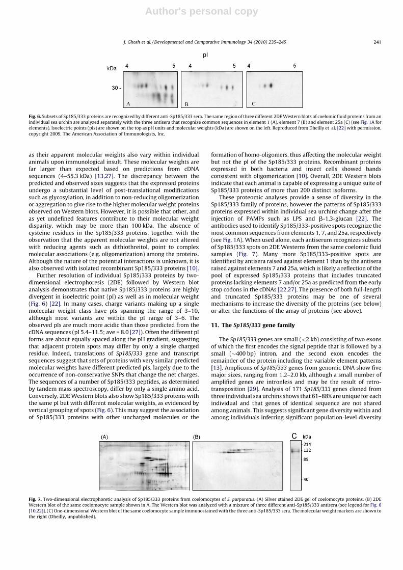
Further resolution of individual Sp185/333 proteins by two-dimensional electrophoresis (2DE) followed by Western blotanalysis demonstrates that native Sp185/333 proteins are highlydivergent in isoelectric point (pI) as well as in molecular weight(Fig. 6) [22]. In many cases, charge variants making up a singlemolecular weight class have pIs spanning the range of 3–10,although most variants are within the pI range of 3–6. Theobserved pIs are much more acidic than those predicted from thecDNA sequences (pI 5.4–11.5; ave = 8.0 [27]). Often the different pIforms are about equally spaced along the pH gradient, suggestingthat adjacent protein spots may differ by only a single chargedresidue. Indeed, translations of Sp185/333 gene and transcriptsequences suggest that sets of proteins with very similar predictedmolecular weights have different predicted pIs, largely due to theoccurrence of non-conservative SNPs that change the net charges.The sequences of a number of Sp185/333 peptides, as determinedby tandem mass spectroscopy, differ by only a single amino acid.Conversely, 2DE Western blots also show Sp185/333 proteins withthe same pI but with different molecular weights, as evidenced byvertical grouping of spots (Fig. 6). This may suggest the associationof Sp185/333 proteins with other uncharged molecules or the
formation of homo-oligomers, thus affecting the molecular weightbut not the pI of the Sp185/333 proteins. Recombinant proteinsexpressed in both bacteria and insect cells showed bandsconsistent with oligomerization [10]. Overall, 2DE Western blotsindicate that each animal is capable of expressing a unique suite ofSp185/333 proteins of more than 200 distinct isoforms.
These proteomic analyses provide a sense of diversity in theSp185/333 family of proteins, however the patterns of Sp185/333proteins expressed within individual sea urchins change after theinjection of PAMPs such as LPS and b-1,3-glucan [22]. Theantibodies used to identify Sp185/333-positive spots recognize themost common sequences from elements 1, 7, and 25a, respectively(see Fig. 1A). When used alone, each antiserum recognizes subsetsof Sp185/333 spots on 2DE Westerns from the same coelomic fluidsamples (Fig. 7). Many more Sp185/333-positive spots areidentified by antisera raised against element 1 than by the antiseraraised against elements 7 and 25a, which is likely a reflection of thepool of expressed Sp185/333 proteins that includes truncatedproteins lacking elements 7 and/or 25a as predicted from the earlystop codons in the cDNAs [22,27]. The presence of both full-lengthand truncated Sp185/333 proteins may be one of severalmechanisms to increase the diversity of the proteins (see below)or alter the functions of the array of proteins (see above).
11. The Sp185/333 gene family
The Sp185/333 genes are small (<2 kb) consisting of two exonsof which the first encodes the signal peptide that is followed by asmall (�400 bp) intron, and the second exon encodes theremainder of the protein including the variable element patterns[13]. Amplicons of Sp185/333 genes from genomic DNA show fivemajor sizes, ranging from 1.2–2.0 kb, although a small number ofamplified genes are intronless and may be the result of retro-transposition [29]. Analysis of 171 Sp185/333 genes cloned fromthree individual sea urchins shows that 61–88% are unique for eachindividual and that genes of identical sequence are not sharedamong animals. This suggests significant gene diversity within andamong individuals inferring significant population-level diversity
Fig. 6. Subsets of Sp185/333 proteins are recognized by different anti-Sp185/333 sera. The same region of three different 2DE Western blots of coelomic fluid proteins from an
individual sea urchin are analyzed separately with the three antisera that recognize common sequences in element 1 (A), element 7 (B) and element 25a (C) (see Fig. 1A for
elements). Isoelectric points (pIs) are shown on the top as pH units and molecular weights (kDa) are shown on the left. Reproduced from Dheilly et al. [22] with permission,
copyright 2009, The American Association of Immunologists, Inc.
Fig. 7. Two-dimensional electrophoretic analysis of Sp185/333 proteins from coelomocytes of S. purpuratus. (A) Silver stained 2DE gel of coelomocyte proteins. (B) 2DE
Western blot of the same coelomocyte sample shown in A. The Western blot was analyzed with a mixture of three different anti-Sp185/333 antisera (see legend for Fig. 6
[10,22]). (C) One-dimensional Western blot of the same coelomocyte sample immunostained with the three anti-Sp185/333 sera. The molecular weight markers are shown to
the right (Dheilly, unpublished).
J. Ghosh et al. / Developmental and Comparative Immunology 34 (2010) 235–245 241

Author's personal copy
well beyond the 4–5% polymorphism estimated for the S.
purpuratus species [1,30]. Paradoxically, despite the high numbersof unique Sp185/333 genes (121 of 171; 71%), the genes themselvesare actually quite similar. Pairwise analysis of the genes (ignoringgaps resulting from different element patterns) show that allsequenced genes are �88% identical [29]. Although assemblies ofthe S. purpuratus genome (v0.5 and v2.1 [1]) contain six Sp185/333
genes, this copy number is probably an underestimate due, in part,to artificial collapsing of the very similar Sp185/333 genes.Quantitative PCR analysis of genomic DNA provides an estimateof 80–120 alleles in a diploid genome [13]. A computationallikelihood method [31] that compares the number of unique genesidentified to the total number of genes cloned and sequenced peranimal predicted between 91 and 142 (most likely 118) Sp185/333
alleles per individual assuming all alleles are unique [32]. Theseanalyses suggest a genomic- and population-level contribution tothe observed sequence diversity of the Sp185/333 proteins and ledto further analysis of the Sp185/333 genomic organization.
PCR analysis of the Sp185/333 genes from eight BAC clones showat least 1, and up to 10 sizes of amplicons per BAC ([29]; Miller,Buckley, Easley, Smith, unpublished). The intergenic regions rangein size from 1.6–8 kb with the major intergenic size of �3 kb. Thisis consistent with a genomic organization of clusters of tightlylinked genes with adjacent genes being oriented head-to-tail,head-to-head and tail-to-tail. Given the numbers of Sp185/333-positive BAC clones, the average insert size for the two BAClibraries and the genomic coverage for each library (48 clonesisolated from a small-insert library (�50–80 kB) of 6.25X genomecoverage, 73 clones isolated from a large-insert (�140 kB) libraryof 25X coverage), the estimated size of the haploid genomic regionin which the Sp185/333 genes are located may be 200–250 kB.Assuming that all genes are closely linked and most genes plustheir flanking intergenic regions span �5 kB, this also leads to anestimate of about 40–50 genes per genome. Although this is a veryrough approximation, it is in agreement with results from qPCR[13] and the computational likelihood method [32].
Finishing-level sequence of one BAC insert shows six clusteredSp185/333 genes within 34 kB (Miller, Buckley, Easley, Smith,unpublished). The cluster is composed of genes with a variety ofelement patterns; A2, B8, E2 and D1 (see Fig. 1A). The genes thatflank that cluster are oriented in the opposite direction withrespect to the genes within the cluster. Three genes within thecluster are positioned within tandem segmental duplications. Eachduplication is bounded by GAT microsatellites and each gene isflanked by GA microsatellites. Based on sequence similarity in theflanking regions in addition to the similarity among the genes, themicrosatellites may be involved with gene duplication anddeletion. Clusters of duplicated genes are characteristic of genesinvolved in the immune response (e.g., R gene families in higher
plants [33]) or immune avoidance (e.g., the variant surfaceglycoprotein genes in trypanosomes [34]). Future work willdetermine whether the majority of the Sp185/333 genes arearranged in tightly linked clusters, and if there are multiple, smallclusters, one or a few large clusters, and if any isolated genes existin the genome.
12. The Sp185/333 gene family is the result of recent divergence
Given the diversity in element patterns and the striking numberof unique sequences, the Sp185/333 genes are surprisingly similar.The full-length genes are >88% identical, yet different versions ofindividual elements are even more similar, with a range of 93.5–97.2% identical [32]. Molecular clock analysis using rates ofevolution ranging from 0.65–1% per million years (My) [35–37]estimates that the last common Sp185/333 ancestor for the extantgenes existed between 2.7 and 10 million years ago (Mya) [32]. It isnoteworthy that this time frame is within that of 4.6 and 12 Myaduring which S. purpuratus is though to have diverged from itsclosest relatives, Strongylocentrotus intermedius, S. droebachiensis,and S. pallidus [37]. In light of the relatively recent speciationwithin the Strongylocentrotid family, we searched genomicsequences (http://www.hgsc.bcm.tmc.edu/projects/seaurchin/) for185/333 sequences from two other sea urchin species, the red seaurchin S. franciscanus, and the fragile red sea urchin Allocentrotus
fragilis. Blast searches identified 100 sequences from S. franciscanus
and 59 from A. fragilis that were homologous to the Sp185/333 genes.The sequence traces (200–250 nt) included four elements present inall genes and were subjected to pairwise difference analysis to assessthe intra- and interspecific diversity.
Among the three species, the 185/333 sequences were quitesimilar with an average intraspecific diversity of 5.63%. The leaderwas the most conserved element (2.88–5.03% different; Table 2)for all three species, which may reflect functional constraints onthe encoded signal sequence. Interspecific comparisons indicatethat element Er6 (see Fig. 1B) was the most conserved amongspecies (average difference of 7.94%; Table 2), and Er1 was the leastconserved (12.17% different among species; Table 2). The elementsfrom S. purpuratus were, on average, 6.2 and 11.9% different fromthe A. fragilis and S. franciscanus sequences, respectively. Sequencesfrom A. fragilis and S. franciscanus were 12.6% different (Table 2),supporting the previously reported echinoderm phylogeny thatplaces S. purpuratus and A. fragilis as more closely related thaneither species is to S. franciscanus [35,37–39].
A molecular clock analysis was also performed on the tracesobtained from S. franciscanus and A. fragilis. As with the S.
purpuratus elements, the 185/333 elements from the other specieswere estimated to be between 5.96 and 10.81 My old, which isyounger than the estimated divergence times for either of these
Table 2The 185/333 elements from three urchin species are distinct.
Leader Er1a Er6 Er27 Avg.
# % # % # % # % %
Strongylocentrotus purpuratus 121 2.88 121 5.47 121 3.65 121 3.67 3.92
Allocentrotus fragilis 10 4.93 6 6.96 3 4.66 3 7.29 5.96
S. franciscanus 12 5.03 10 12.56 4 5.71 5 4.80 7.02
Average 4.28 8.33 4.67 5.25 5.63
% differences between species
S. purpuratus�A. fragilis 6.13 8.52 4.03 6.18 6.21
S. purpuratus� S. franciscanus 12.13 13.52 9.82 12.27 11.93
A. fragilis� S. franciscanus 13.11 14.49 9.97 12.93 12.63
Average 10.46 12.17 7.94 10.46 10.26
#, number of sequences, %, percent difference.a Elements refer to those shown in Fig. 1B.
J. Ghosh et al. / Developmental and Comparative Immunology 34 (2010) 235–245242

Author's personal copy
species. However, the estimated divergence times calculated usinginterspecific comparisons of the Sp185/333 sequences werenotably more recent (6.2–9.6 My for the divergence between S.
purpuratus and A. fragilis, 11.9–18.4 My for S. purpuratus and S.
franciscanus divergence, and 12.6–19.4 My for the S. franciscanus
and A. fragilis divergence). These seemingly contrasting results, i.e.,that the 185/333 gene families appear older than the speciesthemselves, although in each of the three species the 185/333
elements are rapidly evolving, may suggest that selective pressuresare playing a role in the rapid evolution of these gene families. Thethree species of sea urchins live in the East Pacific at varying depths[38] and may therefore be subject to somewhat different pathogenthreats, which may shape the organisms’ immune systemsincluding the 185/333 gene families.
13. Sources of Sp185/333 diversity
High sequence diversity is a general property of many immune-related protein families. The range of strategies employed togenerate diversity in different protein families include somaticrecombination and AID-mediated somatic hypermutation of thegenes encoding vertebrate antigen receptors on B and T cells [40],assembly of variable leukocyte receptors (VLRs) from cassettes ofleucine rich repeats (LRRs) in agnathans [41,42], gene duplication,meiotic mispairing and recombination and somatic diversificationof snail fibrinogen related protein (FREP) genes [43], andalternative splicing of the arthropod Dscam mRNA [11,44–46],to cite a few examples. Similarly, the Sp185/333 proteins exhibithigh levels of diversity, both within and among individuals. Thisdiversity appears to be generated by a number of differentprocesses acting at different levels.
13.1. Sp185/333 gene recombination and sequence diversification
It is generally agreed that mRNAs that are not alternativelyspliced will have identical 50 and 30 ends. Surprisingly, theSp185/333 EST sequences show an inconsistent associationbetween the leader sequence and that of the adjacent 50UTR [8].There is also little or no correlation between the element patternsat the 50 end of the genes and the element patterns at the 30 endeven though all are encoded by a single exon [32]. Furthermore,when two elements are found together in multiple gene sequences,the specific sequence version of one element is not alwaysassociated with a specific sequence version of the next. In essence,the elements and different regions in the various genes appear tobe ‘‘mixed and matched’’ with respect to each other [32].
A more detailed analysis of this phenomenon employed the type1 repeats located at the 50 end of the genes. To enable this analysis,the gene sequences were re-aligned in the repeat-based alignmentto optimize the correspondence between the repeats and elements(Fig. 1B) [29]. Type 1 repeats have a relatively simple organization oftwo to four tandem repeats that encode part of the gly-rich region ofthe protein. A detailed phylogenetic analysis of 52 unique type 1repeats (of 292 repeats obtained from 121 unique genes) results inthree major clades and seven categories of type 1 repeats [32]. Theprimary factors affecting clustering are the number of type 1 repeatsin a gene sequence and the position of a given repeat with respect tothe other type 1 repeats in a gene. The consensus sequences for theseven categories suggest a complex evolutionary history of the50 end of the Sp185/333 genes. The original level of diversity mayhave been based on three putative ancestral sequences thatgenerated the diversity of the extant genes through frequent repeatduplication, deletion, recombination and point mutations. Furtherinvestigations of all the elements in the gene sequences indicaterecombination events are likely to occur both between and withinelements at any point along the Sp185/333 gene sequence.
13.2. mRNA editing
Although the control of Sp185/333 gene expression in S.
purpuratus is not yet understood, these genes give rise to a diverseset of transcripts, which in turn, are translated into a diverse set ofSp185/333 proteins. However, evidence suggests that there is anadditional layer of complexity involved in the generation of a diverseSp185/333 protein repertoire: modification of Sp185/333 mRNAs. Itis notable that all but one of 171 sequenced genes have perfect, full-length ORFs [29] while 50% of the mRNAs encode truncated proteinsresulting from SNPs and short indels [27]. Very few message andgene sequences collected from individual sea urchins are identical[28]. Furthermore, pairwise comparisons among the genes andmessages indicate that one gene may be the likely source of themajority of the messages. Analysis of the frequency and type ofsubstitutions between a candidate gene and its corresponding (mostlikely) transcripts indicate that 73% of the changes are transitions(purine to purine or pyrimidine to pyrimidine). Specifically, cytosineto uridine transitions are significantly more frequent than would bepredicted under typical RNA polymerase activity [47]. This result issuggestive of cytidine deaminase activity [48], and several genemodels encoding cytidine deaminase-like enzymes have beenannotated in the S. purpuratus genome [2,28]. Although no cleardiversity hotspots are present in the sequences, the nucleotidesubstitutions between genes and messages are not random and aremore frequent at certain positions. This activity may be restricted topost-transcriptional editing of RNAs, however, low-fidelity tran-scription of Sp185/333 genes may employ RNA polymerase m [49].Comparison of Sp185/333 genes cloned from sperm vs. those fromcoelomocyte genomic DNA suggests that somatic diversification ofSp185/333 in coelomocytes is unlikely. These findings infer a role forRNA editing in the generation of diversity of Sp185/333 transcriptsthat results in the expression of truncated and missense peptides inaddition to full-length proteins. Tandem mass spectrometry analysisof coelomic fluid samples reveal the presence of truncated Sp185/333 proteins, indicating that transcripts with missense sequenceand premature stop codons are translated [22]. It is not known whatfunctional roles these truncated and missense Sp185/333 proteinsmay play in the sea urchin immune responses (see speculationsabove), but it is clear that the primary amino acid sequence diversityof the protein repertoire is greater than that encoded by the genes.
14. Conclusion
Sequence diversification through a variety of mechanisms hasbeen observed in a large set of immune-related proteins ininvertebrates. The Sp185/333 system in the purple sea urchin is anexample of this diversification that appears to involve multiplelayers including gene recombination, RNA editing and/or low-fidelity transcription, and putative post-translational proteinmodifications. There are an estimated 80–120 Sp185/333 allelesin the sea urchin genome, and it is very likely that the suite of genesvaries significantly among individuals, such that there is a verylarge total pool of Sp185/333 genes in the population. The apparentwinnowing of diversity at the level of transcription, wherein only asmall subset of these diverse genes may be transcribed andprocessed into mature message is curious [32], however, this mayonly reflect the limited number of immune challenges that havebeen analyzed. It is possible that the untranscribed genes may be asource of intergenerational novelty for functional Sp185/333 genesthrough recombination events, assisted by the presence of repeatswithin the genes, microsatellites surrounding the genes, and a highlevel of overall sequence conservation among the genes. Non-expressed genes may also simply be the product of intergenera-tional duplication events, perhaps with non-functional regulatoryelements, as is likely the case for the putatively retrotransposed
J. Ghosh et al. / Developmental and Comparative Immunology 34 (2010) 235–245 243

Author's personal copy
intronless genes. However, the identification of very few pseudo-genes is unusual for a family of similar genes.
Following transcription of active genes, further diversificationoccurs, in the form of RNA editing and possibly low-fidelitypolymerase activity [28]. The function of this editing is not yetunderstood, but there is a hint that it may be directed to specificnucleotides [27]. It is possible that one of the several deaminasesthat are encoded in the genome [2] may act as a constitutive downregulator of Sp185/333 protein function by generating truncatedproteins under conditions with little or no immune challenge.Upon immune challenge, deaminase expression/function may bedown regulated allowing the translation of full-length Sp185/333proteins. Alternatively, the truncated proteins may have functionsthat are different from or complementary to the full-lengthproteins in immunoquiescent animals. Very preliminary evidenceof Sp185/333 function indicates that the proteins may bind to awide range of bacteria (Schrankel et al., unpublished), suggesting apossible immune effector function. The Sp185/333 proteins mayalso have a role in syncytium formation by phagocytes, a processbelieved to be an in vitro equivalent of an in vivo encapsulationresponse (Fig. 3) [10]. Future functional studies should shed lighton the role of Sp185/333 in the immune response in particular andthe evolutionary strategies for mounting an effective immuneresponse in general.
Acknowledgements
The following funding agencies supported research on seaurchin immunity; the US National Science Foundation to LCS, theAustralian Research Council to DAR and SVN, the CanadianInstitutes of Health Research and the Natural Sciences andEngineering Research Council to JPR. Unpublished data was kindlyprovided by Nolwenn Dheilly, Catherine Schrankel and Youn-OkKim. The authors are grateful to Drs. John Henson and SandroSacchi for critically reading the manuscript.
References
[1] Sodergren E, Weinstock GM, Davidson EH, Cameron RA, Gibbs RA, Angerer RC,et al. The genome of the sea urchin Strongylocentrotus purpuratus. Science2006;314(5801):941–52.
[2] Hibino T, Loza-Coll M, Messier C, Majeske AJ, Cohen AH, Terwilliger DP, et al.The immune gene repertoire encoded in the purple sea urchin genome. DevBiol 2006;300:349–65.
[3] Fugmann SD, Messier C, Novack LA, Cameron RA, Rast JP. An ancient evolu-tionary origin of the Rag1/2 gene locus. Proc Natl Acad Sci USA2006;103(10):3728–33.
[4] Smith LC, Chang L, Britten RJ, Davidson EH. Sea urchin genes expressed inactivated coelomocytes are identified by expressed sequence tags. Comple-ment homologues and other putative immune response genes suggestimmune system homology within the deuterostomes. J Immunol1996;156(2):593–602.
[5] Gross PS, Clow LA, Smith LC. SpC3, the complement homologue from thepurple sea urchin, Strongylocentrotus purpuratus, is expressed in two subpo-pulations of the phagocytic coelomocytes. Immunogenetics 2000;51(12):1034–44.
[6] Al-Sharif WZ, Sunyer JO, Lambris JD, Smith LC. Sea urchin coelomocytesspecifically express a homologue of the complement component C3. J Immu-nol 1998;160(6):2983–97.
[7] Pancer Z. Dynamic expression of multiple scavenger receptor cysteine-richgenes in coelomocytes of the purple sea urchin. Proc Natl Acad Sci USA2000;97(24):13156–61.
[8] Nair SV, Del Valle H, Gross PS, Terwilliger DP, Smith LC. Macroarray analysis ofcoelomocyte gene expression in response to LPS in the sea urchin. Identifica-tion of unexpected immune diversity in an invertebrate. Physiol Genom2005;22(1):33–47.
[9] Rast JP, Pancer Z, Davidson EH. New approaches towards an understanding ofdeuterostome immunity. Curr Top Microbiol Immunol 2000;248:3–16.
[10] Brockton V, Henson JH, Raftos DA, Majeske AJ, Kim YO, Smith LC. Localizationand diversity of 185/333 proteins from the purple sea urchin—unexpectedprotein-size range and protein expression in a new coelomocyte type. J Cell Sci2008;121(3):339–48.
[11] Schmucker D, Chen B. Dscam and DSCAM: complex genes in simple animals,complex animals yet simple genes. Genes Dev 2009;23:147–56.
[12] Buckley KM, Florea LD, Smith LC. A method of identifying alternative or crypticsplice sites within gene and mRNA sequences. Comparisons among sequencesfrom vertebrates, echinoderms and other groups. BMC Genom 2009;10(318).
[13] Terwilliger DP, Buckley KM, Mehta D, Moorjani PG, Smith LC. Unexpecteddiversity displayed in cDNAs expressed by the immune cells of the purple seaurchin, Strongylocentrotus purpuratus. Physiol Genom 2006;26:134–44.
[14] Johnson PT. The coelomic elements of sea urchins (Strongylocentrotus). I. Thenormal coelomocytes; their morphology and dynamics in hanging drops. JInvert Pathol 1969;13:25–41.
[15] Smith LC, Rast JP, Brockton V, Terwilliger DP, Nair SV, Buckley KM, et al. The seaurchin immune system. Invert Survival J 2006;3:25–39.
[16] Henson JH, Nesbitt D, Wright BD, Scholey JM. Immunolocalization of kinesin insea urchin coelomocytes. Association of kinesin with intracellular organelles. JCell Sci 1992;103(Pt 2):309–20.
[17] Edds KT. Cell biology of echinoid coelomocytes. J Invert Biol 1993;61:173–8.[18] Henson JH, Svitkina TM, Burns AR, Hughes HE, MacPartland KJ, Nazarian R,
et al. Two components of actin-based retrograde flow in sea urchin coelomo-cytes. Mol Biol Cell 1999;10(12):4075–90.
[19] Whittaker CA, Bergeron KF, Whittle J, Brandhorst BP, Burke RD, Hynes RO. Theechinoderm adhesome. Dev Biol 2006;300(1):252–66.
[20] Dybas L, Fankboner PV. Holothurian survival strategies: mechanisms for themaintenance of a bacteriostatic environment in the coelomic cavity of the seacucumber, Parastichopus californicus. Dev Comp Immunol 1986;10(3):311–30.
[21] Johnson PT. The coelomic elements of sea urchins (Strongylocentrotus). His-tochem Cell Biol 1969;17(3):213–31.
[22] Dheilly NM, Nair SV, Smith LC, Raftos DA. Highly variable immune responseproteins from the sea urchin, Strongylocentrotus purpuratus: proteomic ana-lysis of diversity within and between individuals. J Immunol 2009;182:2203–12.
[23] Bachmann S, Pohla H, Goldschmid A. Phagocytes in the axial complex of thesea urchin, Sphaerenchinus granularis (Lam.). Cell Tissue Res 1980;213:109–20.
[24] Metchnikoff E. Lectures on the comparative pathology of inflammation:delivered at the Pasteur Institute in 1891 [Starling FA, Starling EH, Trans.].Kegan Paul, Trench, Trubner & Co. Ltd.; 1893.
[25] Silva JR. The onset of phagocytosis and identity in the embryo of Lytechinusvariegatus. Dev Comp Immunol 2000;24(8):733–9.
[26] Furukawa R, Takahashi Y, Kanajima Y, Dan-Sohkawa M, Kaneko H. Defensesystem by mesenchyme cells in bipinnaria larvae of the starfish, Asterinapectinifera. Dev Comp Immunol 2009;33(2):205–15.
[27] Terwilliger DP, Buckley KM, Brockton V, Ritter NJ, Smith LC. Distinctiveexpression patterns of 185/333 genes in the purple sea urchin, Strongylocen-trotus purpuratus: an unexpectedly diverse family of transcripts in response toLPS, beta-1,3-glucan, and dsRNA. BMC Mol Biol 2007;8:16.
[28] Buckley KM, Terwilliger DP, Smith LC. Sequence variations in 185/333 mes-sages from the purple sea urchin suggest posttranscriptional modifications toincrease immune diversity. J Immunol 2008;181(12):2203–12.
[29] Buckley KM, Smith LC. Extraordinary diversity among members of the largegene family, 185/333, from the purple sea urchin, Strongylocentrotus purpur-atus. BMC Mol Biol 2007;8(1):68.
[30] Britten RJ, Cetta A, Davidson EH. The single-copy DNA sequence polymorphismof the sea urchin Strongylocentrotus purpuratus. Cell 1978;15(4):1175–86.
[31] Barth RK, Kim BS, Lan NC, Hunkapiller T, Sobieck N, Winoto A, et al. The murineT-cell receptor uses a limited repertoire of expressed V gene segments. Nature1985;316:517–23.
[32] Buckley KM, Munshaw S, Kepler TB, Smith LC. The 185/333 gene family is arapidly diversifying host-defense gene cluster in the purple sea urchin Stron-gylocentrotus purpuratus. J Mol Biol 2008;379:912–28.
[33] Lucht JM, Mauch-Mani B, Steiner HY, Metraux JP, Ryals J, Hohn B. Pathogenstress increases somatic recombination frequency in Arabidopsis. Nat Genet2002;30(3):311–4.
[34] Taylor JE, Rudenki G. Switching trypanosome coats: what’s in the wardrobe?Trends Genet 2006;22(11):614–20.
[35] Biermann CH. The molecular evolution of sperm binding in six species of seaurchins (Echinoidea: Strongylocentrotidae). Mol Biol Evol 1998;15(12):1761–71.
[36] Grula JW, Hall TJ, Hunt JA, Guiugni DF, Graham GJ, Davidson EH, et al. Seaurchin DNA sequence variation and reduced interspecies differences of theless variable DNA sequences. Evolution 1982;36:655–76.
[37] Lee YH. Molecular phylogenies and divergence times of sea urchin species ofStrongylocentrotidae, Echinoidea. Mol Biol Evol 2003;20(8):1211–21.
[38] Biermann CH, Kessing BD, Palumbi SR. Phylogeny and development ofmarine model species: Strongylocentrotid sea urchins. Evol Dev 2003;5(4):360–71.
[39] Smith AB. Phylogenetic relationship, divergence times, and rates of molecularevolution for camarodont sea urchins. Mol Biol Evol 1988;5:345–65.
[40] Longerich S, Basu U, Alt F, Storb U. AID in somatic hypermutation and classswitch recombination. Curr Opin Immunol 2006;18(2):164–74.
[41] Rogozin IB, Iyer LM, Liang L, Glazko GV, Liston VG, Pavlov YI, et al. Evolutionand diversification of lamprey antigen receptors: evidence for involvementof an AID-APOBEC family cytosine deaminase. Nat Immunol 2007;8(6):647–56.
[42] Nagawa F, Kishishita N, Shimizu K, Hirose S, Miyoshi M, Nezu J, et al. Antigen-receptor genes of the agnathan lamprey are assembled by a process involvingcopy choice. Nat Immunol 2007;8(2):206–13.
[43] Zhang S-M, Adema CM, Kepler TB, Loker ES. Diversification of Ig superfamilygenes in an invertebrate. Science 2004;305(5681):251–4.
J. Ghosh et al. / Developmental and Comparative Immunology 34 (2010) 235–245244

Author's personal copy
[44] Watson FL, Puttmann-Holgado R, Thomas F, Lamar DL, Hughes M, Kondo M,et al. Extensive diversity of Ig-superfamily proteins in the immune system ofinsects. Science 2005;309(5742):1874–8.
[45] Dong Y, Taylor HE, Dimopoulos G. AgDscam, a hypervariable immunoglobulindomain-containing receptor of the Anopheles gambiae innate immune system.PLoS Biol 2006;4(7):e229.
[46] Brites D, McTaggart S, Morris K, Anderson J, Thomas K, Colson I, et al. TheDscam homologue of the crustacean Daphnia is diversified by alternativesplicing like in insects. Mol Biol Evol 2008;25(7):1429–39.
[47] Chester A, Scott J, Anant S, Navaratnam N. RNA editing: cytidine to uridineconversion in apolipoprotein B mRNA. Biochim Biophys Acta (BBA)-GeneStruct Expr 2000;1494(1–2):1–13.
[48] Gott JM, Emeson RB. Functions and mechanisms of RNA editing. Annu RevGenet 2000;34:499–531.
[49] Ruiz JF, Dominguez O, Lain de Lera T, Garcia-Diaz M, Bernad A, Blanco L. DNApolymerase mu, a candidate hypermutase? Phil Trans R Soc Lond B Biol Sci2001;356(1405):99–109.
J. Ghosh et al. / Developmental and Comparative Immunology 34 (2010) 235–245 245
Copyright © 2022 FDOKUMEN


![100019.ppt [\254\333\256e\274\322\246\241]](https://static.fdokumen.com/doc/165x107/631ce499b8a98572c10d1be1/100019ppt-254333256e274322246241.jpg)


![100040.ppt [\254\333\256e\274\322\246\241]](https://static.fdokumen.com/doc/165x107/633baafca215b3a22b0d61d3/100040ppt-254333256e274322246241.jpg)