Gut microflora associated characteristics in children with celiac disease
Upload
independentCategory
view
0download
0
MucosalImmunology | VOLUME 5 NUMBER 5 | SEPTEMBER 2012 513
nature publishing group ARTICLES
See COMMENTARY page XX
INTRODUCTION The development and functional shaping of innate and adap-
tive components of gut-associated lymphoid tissue is completed
postnatally, and depends on the close relationship with micro-
flora and environmental agents; 1 – 5 among its components,
several phenotypically and functionally distinct T lymphocyte
subsets abundantly home either in the lamina propria (LP),
and in close proximity with the epithelial layer (intraepithelial
lymphocytes (IEL)).
Human gut intraepithelial T lymphocytes (IEL-T) are mostly
� / � TCR + (T-cell receptor), although a significant fraction of
� / � TCR + T cells is represented; in marked contrast with LP-T
cells, the vast majority of IEL-T is CD8 + , a proportion of which
also expresses natural killer receptors. 6 – 9 Most IEL-T cells have
an effector / memory phenotype (CD45R0 + ), and are capable
of immediately participating in immune defenses. They con-
tribute to mucosal barrier function, to epithelial homeostasis,
and to the regulation of mucosal immune responses, through
cytotoxic activity and cytokine production, as well as by cell – cell
communication. 9 – 16
Cytokines of the interleukin (IL)-17 family have recently
emerged as important players of mucosal immune responses
and in the maintenance of epithelial barrier integrity; 17,18 they
orchestrate the crosstalk between immune and tissue cells,
induce strengthening of tight junctions, and production of
antimicrobial proteins and chemokines by both epithelium
and immune cells. 19 IL-17A, the best-characterized member of
the family, is produced by a specialized population of CD4 +
Size and dynamics of mucosal and peripheral IL-17A + T-cell pools in pediatric age, and their disturbance in celiac disease R La Scaleia 1 , 2 , M Barba 1 , G Di Nardo 3 , M Bonamico 4 , S Oliva 3 , R Nenna 4 , F Valitutti 3 , M Mennini 4 ,
M Barbato 3 , M Montuori 4 , A Porzia 1 , L Petrarca 4 , S Battella 1 , S Cucchiara 3 , M Piccoli 1 , A Santoni 2 , 5 ,
F Mainiero 1 , 7 and G Palmieri 1 , 6 , 7
Mucosal interleukin (IL)-17A – producing T cells contribute to protective antimicrobial responses and to epithelial barrier integrity; their role in celiac disease (CD) is debated. We analyzed the frequency and developmental dynamics of mucosal (intraepithelial lymphocytes (IEL)) and circulating (peripheral blood (PB)) IL-17A (T17) and / or interferon (IFN)- � – producing (T1, T1 / T17) T-cell populations in 86 pediatric controls and 116 age-matched CD patients upon phorbol myristate acetate / ionomycin or CD3 / CD28 stimulation. T17 and T1 / 17 are physiologically present among IEL and PB populations, and their frequency is selectively and significantly reduced in CD IEL. The physiological age-dependent increase of Th17 IEL is also absent in CD, while IFN- � – producing PB-T cells significantly accumulate with patient ’ s age. Finally, the amplitude of IL-17A + and IFN- � + T-cell pools are significantly correlated in different individuals; this relationship only applies to CD4 + T cells in controls, while it involves also the CD4 − counterpart in CD patients. In conclusion, both size and dynamics of mucosa-associated and circulating IL-17A + T-cell pools are finely regulated in human pediatric subjects, and severely disturbed in CD. The impaired IL-17A + IEL-T pool may negatively impact on epithelial barrier efficiency, and contribute to CD mucosa damage; the disturbed dynamics of circulating IL-17A + and IFN- � + T-cell pools may be involved in the extraintestinal autoimmune manifestations associated with CD.
1 Department of Experimental Medicine, Sapienza University , Rome , Italy . 2 Istituto Pasteur-Fondazione Cenci Bolognetti, Sapienza, University of Rome , Rome , Italy . 3 Gastroenterology and Liver Unit, Sapienza University , Rome , Italy . 4 Celiac Disease and Malabsorptive Diseases Unit, Department of Pediatrics, Sapienza University , Rome , Italy . 5 Department of Molecular Medicine, Sapienza University , Rome , Italy . 6 Research Centre for Social Diseases, Sapienza University , Rome , Italy . 7 The last two authors contributed equally to this work . Correspondence: G Palmieri ( [email protected] )
Received 25 August 2011; accepted 26 February 2012; published online 9 May 2012. doi: 10.1038/mi.2012.26
514 VOLUME 5 NUMBER 5 | SEPTEMBER 2012 | www.nature.com/mi
ARTICLES
T-helper cells (Th17), and by CD8 + (Tc17) and � / � TCR T cells,
as well as by several populations of innate cells. 20 – 25 The existence
of IL-17A / IFN- � (interferon- � ) double-producer T-cell popu-
lations, as well as the occurrence of Th17-to-Th1 conversion
during immune responses, testify that the genetic program of
IL-17 – producing T cells is tightly regulated, and bears a sub-
stantial plasticity at the same time. 18,20,26,27
In mouse, the development of mucosal Th17-cell popula-
tion depends on specific commensal flora components. 2,3,17 – 19
Although IL-17A – producing T cells are physiologically present
in human gut LP 23,24 and peripheral blood, 25,26 no information
is available regarding either the ability of IEL-T populations to
produce IL-17A, or the dynamics of IL-17A – producing mucosal
and circulating T-cell pools during human development.
IL-17 dysregulation has been associated with different auto-
immune and chronic inflammatory diseases. 17 – 20 Celiac disease
(CD) is triggered, in genetically predisposed individuals, by an
improper immune response to gluten components. 28,29 Besides
specific HLA class II alleles, several genes involved in immune
regulation and epithelial barrier function constitute predispos-
ing factors for CD; 29,30 moreover, environmental agents con-
tribute to the triggering of immune-mediated tissue damage
by modulating immune response, inflammation, and epithelial
permeability.
Several phenotypic and functional alterations in the IEL-T
compartment, ranging from dysregulated cytokine production,
aberrant expression and function of natural killer receptors on IEL
CD8 + T cells, and enhanced cytotoxic activity, have all been impli-
cated in CD tissue damage. 6 – 8,11,12,14,28,29,31 – 39 In addition, the
association between CD and other immune-mediated disorders
supports the existence of a systemic immune dysregulation. 28,29
A possible pathogenic role for IL-17A – producing T cells
in CD has been proposed by recent evidence reporting the
augmentation and the aberrant phenotype of LP Th17 in CD
patients; 40,41 intriguingly, gliadin-specific CD4 + T-cell lines
exhibit an impaired ability to produce IL-17A, 42 leaving the
involvement of IL-17A in CD still open to investigation.
We evaluated duodenal IEL and peripheral blood IL-17A –
producing T-cell populations in a cohort of CD children and
adolescents and age-matched controls, and analyzed the dynam-
ics of IL-17A + T cells with respect to both subjects ’ age and
IFN- � – producing T-cell populations. The results provide novel
evidence that these factors regulate the pool size of IL-17A – pro-
ducing T cells during human development, and disclose multiple
alterations occurring in CD.
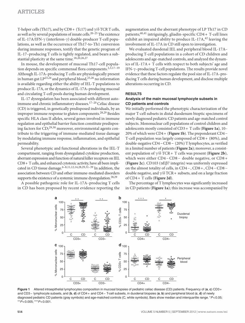
RESULTS Analysis of the main mucosal lymphocyte subsets in CD patients and controls We initially performed the phenotypic characterization of the
major T-cell subsets in distal duodenum bioptic specimens of
newly diagnosed pediatric CD patients and age-matched control
subjects. Mononuclear cell populations of control children and
adolescents mostly consisted of CD3 + T cells ( Figure 1a ), 10 –
20 % of which were CD4 + ( Figure 1b ). The preponderant CD4 −
T-cell population was largely composed of CD8 + (80 % ), and
double-negative CD4 − CD8 − (20 % ) T lymphocytes, as verified
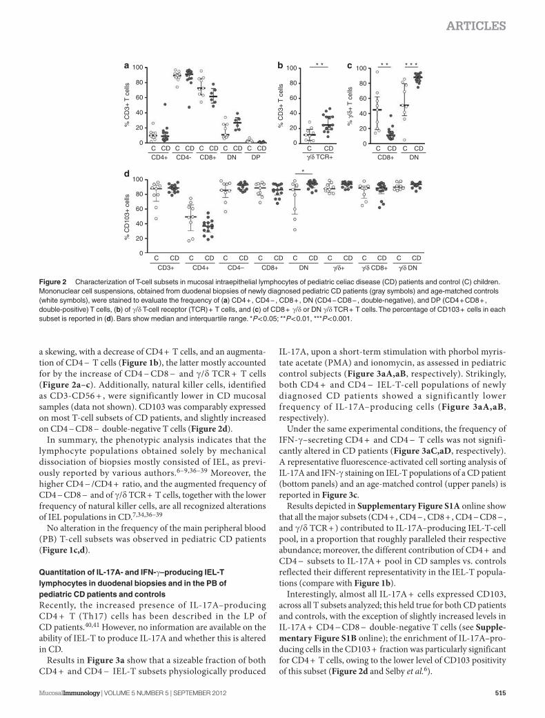
in a limited number of patients ( Figure 2a ); moreover, a consist-
ent population of � / � TCR + T cells was present ( Figure 2b ),
which were either CD4 − CD8 − double negative, or CD8 +
( Figure 2c ). CD103 ( � E � 7 integrin) was uniformly expressed
on the almost totality of cells, in CD4 − , CD8 + , CD4 − CD8 −
double negative, and � / � TCR + subsets, and on a large fraction
of CD4 + T cells ( Figure 2d ).
The percentage of T lymphocytes was significantly increased
in CD patients ( Figure 1a ); this increase was accompanied by
100
75
50
% M
onon
ucle
ar c
ells
% C
D3+
T c
ells
25
C C
CD3–CD3+
CD CD
Peripheralblood
Duodenalbiopsy
* * ** ** * *
0
100
75
50
% M
onon
ucle
ar c
ells
25
0
100
75
50
25
0
% C
D3+
T c
ells
100
75
50
25
0
C C
CD4–CD4+
CD CD
C C
CD3–CD3+
CD CD C C
CD4–CD4+
CD CD
Figure 1 Altered intraepithelial lymphocytes composition in mucosal biopsies of pediatric celiac disease (CD) patients. Frequency of ( a , c ) CD3 + and CD3 − lymphocyte subsets, and ( b , d ) of CD4 + and CD4 − T-cell subsets, in duodenal biopsies ( a , b ) and peripheral blood ( c , d ) of newly diagnosed pediatric CD patients (gray symbols) and age-matched controls (C, white symbols). Bars show median and interquartile range. * P < 0.05; * * P < 0.005; * * * P < 0.001.
MucosalImmunology | VOLUME 5 NUMBER 5 | SEPTEMBER 2012 515
ARTICLES
a skewing, with a decrease of CD4 + T cells, and an augmenta-
tion of CD4 − T cells ( Figure 1b ), the latter mostly accounted
for by the increase of CD4 − CD8 − and � / � TCR + T cells
( Figure 2a – c ). Additionally, natural killer cells, identified
as CD3-CD56 + , were significantly lower in CD mucosal
samples (data not shown). CD103 was comparably expressed
on most T-cell subsets of CD patients, and slightly increased
on CD4 − CD8 − double-negative T cells ( Figure 2d ).
In summary, the phenotypic analysis indicates that the
lymphocyte populations obtained solely by mechanical
dissociation of biopsies mostly consisted of IEL, as previ-
ously reported by various authors. 6 – 9,36 – 39 Moreover, the
higher CD4 − / CD4 + ratio, and the augmented frequency of
CD4 − CD8 − and of � / � TCR + T cells, together with the lower
frequency of natural killer cells, are all recognized alterations
of IEL populations in CD. 7,34,36 – 39
No alteration in the frequency of the main peripheral blood
(PB) T-cell subsets was observed in pediatric CD patients
( Figure 1c,d ).
Quantitation of IL-17A- and IFN- � – producing IEL-T lymphocytes in duodenal biopsies and in the PB of pediatric CD patients and controls Recently, the increased presence of IL-17A – producing
CD4 + T (Th17) cells has been described in the LP of
CD patients. 40,41 However, no information are available on the
ability of IEL-T to produce IL-17A and whether this is altered
in CD.
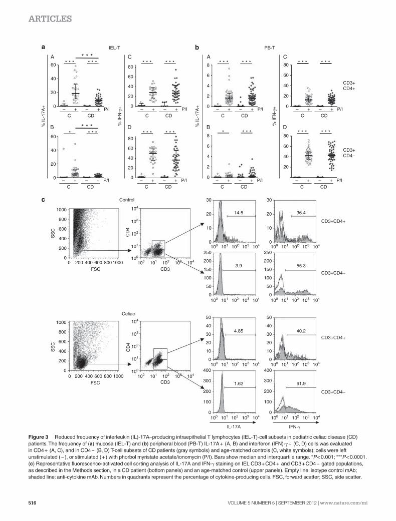
Results in Figure 3a show that a sizeable fraction of both
CD4 + and CD4 − IEL-T subsets physiologically produced
IL-17A, upon a short-term stimulation with phorbol myris-
tate acetate (PMA) and ionomycin, as assessed in pediatric
control subjects ( Figure 3aA,aB , respectively). Strikingly,
both CD4 + and CD4 − IEL-T-cell populations of newly
diagnosed CD patients showed a significantly lower
frequency of IL-17A – producing cells ( Figure 3aA,aB ,
respectively).
Under the same experimental conditions, the frequency of
IFN- � – secreting CD4 + and CD4 − T cells was not signifi-
cantly altered in CD patients ( Figure 3aC,aD , respectively).
A representative fluorescence-activated cell sorting analysis of
IL-17A and IFN- � staining on IEL-T populations of a CD patient
(bottom panels) and an age-matched control (upper panels) is
reported in Figure 3c .
Results depicted in Supplementary Figure S1A online show
that all the major subsets (CD4 + , CD4 − , CD8 + , CD4 − CD8 − ,
and � / � TCR + ) contributed to IL-17A – producing IEL-T-cell
pool, in a proportion that roughly paralleled their respective
abundance; moreover, the different contribution of CD4 + and
CD4 − subsets to IL-17A + pool in CD samples vs. controls
reflected their different representativity in the IEL-T popula-
tions (compare with Figure 1b ).
Interestingly, almost all IL-17A + cells expressed CD103,
across all T subsets analyzed; this held true for both CD patients
and controls, with the exception of slightly increased levels in
IL-17A + CD4 − CD8 − double-negative T cells (see Supple-
mentary Figure S1B online); the enrichment of IL-17A – pro-
ducing cells in the CD103 + fraction was particularly significant
for CD4 + T cells, owing to the lower level of CD103 positivity
of this subset ( Figure 2d and Selby et al. 6 ).
100
80
60
% C
D3+
T c
ells
% C
D10
3+ c
ells
% C
D3+
T c
ells
% γ
/δ+
T c
ells
40
20
0
100
80
60
40
20
0
100 * *
*
* * * * *
80
60
40
20
0
100
80
60
40
20
0C CDCD4+
C CD
CD3+
C CD
CD4+
C CD
CD4–
C CD
CD8+
C CD
DN
C CD
γ/δ+
C CD
γ/δ CD8+
C CD
γ/δ DN
C CDγ/δ TCR+
C CDCD4-
C CDCD8+
C CDDN
C CDDP
C CDCD8+ DN
C CD
Figure 2 Characterization of T-cell subsets in mucosal intraepithelial lymphocytes of pediatric celiac disease (CD) patients and control (C) children. Mononuclear cell suspensions, obtained from duodenal biopsies of newly diagnosed pediatric CD patients (gray symbols) and age-matched controls (white symbols), were stained to evaluate the frequency of ( a ) CD4 + , CD4 − , CD8 + , DN (CD4 − CD8 − , double-negative), and DP (CD4 + CD8 + , double-positive) T cells, ( b ) of � / � T-cell receptor (TCR) + T cells, and ( c ) of CD8 + � / � or DN � / � TCR + T cells. The percentage of CD103 + cells in each subset is reported in ( d ). Bars show median and interquartile range. * P < 0.05; * * P < 0.01, * * * P < 0.001.
516 VOLUME 5 NUMBER 5 | SEPTEMBER 2012 | www.nature.com/mi
ARTICLES
60 * * * * * * * * * * * * * * * * * * * * *
CD3+CD4+
CD3+CD4+
Control
Celiac
FSC
1000
1000
800
800
600
600
400
400
200
2000
0
10008006004002000
1000
800
600
400
200
0
0
10
20
30
0
0
0
0
100
200
300
400
0
100
200
300
400
10
20
30
40
50
0
10
20
30
40
50
50
100
150
200
250
0
50
100
150
200
250
10
20
30
FSC
CD3
101
101100
100
102
102
103
103
104
104
101
101100
100
102
102
103
103
104
104
101100 102 103 104 101100 102 103 104
101100 102 103 104 101100 102 103 104
101100 102 103 104 101100 102 103 104
101100 102 103 104 101100 102 103 104
CD3
3.9
14.5 36.4
55.3
40.24.85
1.62 61.9
IL-17A IFN-γ
CD
4C
D4
SS
CS
SC
CD3+CD4+
CD3+CD4–
CD3+CD4–
CD3+CD4–
* * ** * *
* * ** * * * * * * * * * * * * * ** * ** *
40
20
0
60
80
IEL-T PB-T
40
20
0
60
40
20
0
60
80
40
20
0
60
80
40
20
0
60
80
40
20
6
8
4
2
0
6
8
4
2
0
– +C
% IL
-17A
+
% IL
-17A
+
% IF
N-γ
+
% IF
N-γ
+
CDP/I– + – +
C CDP/I– + – +
C CDP/I– + – +
C CDP/I– +
– +C CD
P/I– + – +C CD
P/I– + – +C CD
P/I– + – +C CD
P/I– +
Figure 3 Reduced frequency of interleukin (IL)-17A – producing intraepithelial T lymphocytes (IEL-T)-cell subsets in pediatric celiac disease (CD) patients. The frequency of ( a ) mucosa (IEL-T) and ( b ) peripheral blood (PB-T) IL-17A + (A, B) and interferon (IFN)- � + (C, D) cells was evaluated in CD4 + (A, C), and in CD4 − (B, D) T-cell subsets of CD patients (gray symbols) and age-matched controls (C, white symbols); cells were left unstimulated ( − ), or stimulated ( + ) with phorbol myristate acetate / ionomycin (P / I). Bars show median and interquartile range. * P < 0.001; * * * P < 0.0001. ( c ) Representative fluorescence-activated cell sorting analysis of IL-17A and IFN- � staining on IEL CD3 + CD4 + and CD3 + CD4 − gated populations, as described in the Methods section, in a CD patient (bottom panels) and an age-matched control (upper panels). Empty line: isotype control mAb; shaded line: anti-cytokine mAb. Numbers in quadrants represent the percentage of cytokine-producing cells. FSC, forward scatter; SSC, side scatter.
MucosalImmunology | VOLUME 5 NUMBER 5 | SEPTEMBER 2012 517
ARTICLES
The frequency of T cells spontaneously producing
IL-17A or IFN- � in ex vivo – analyzed mucosal samples
was negligible in both CD patients and controls (data not
shown).
Interestingly, IL-17A – producing CD4 + and CD4 − T cells
( Figure 3bA,bB , respectively) were also present, albeit at a
lower level, in the PB of pediatric control subjects. The per-
centage of IL-17A + ( Figure 3bA,bB ) and IFN- � + ( Figure
3bC,bD ) CD4 + and CD4 − T cells was comparable in CD
patients and control children. IL-17A – producing cells of both
CD patients and controls markedly expressed more CD161
receptor (see Supplementary Figure S2B online), as compared
with the frequency of this marker in CD4 + and CD4 − PB
T-cell populations (see Supplementary Figure S2A online),
in accordance with previous evidence in the literature; 22,24 a
representative analysis of a CD patient and a control is reported
in Supplementary Figure S2C online.
Collectively taken, these results show that Th17 and IL-17A –
producing CD3 + CD4 − T cells are physiologically present
in human duodenal mucosa and, with a lower frequency, in PB;
our data indicate a selectively defective abundance of IL-17A –
secreting cells in intraepithelial but not circulating T popula-
tions of CD patients.
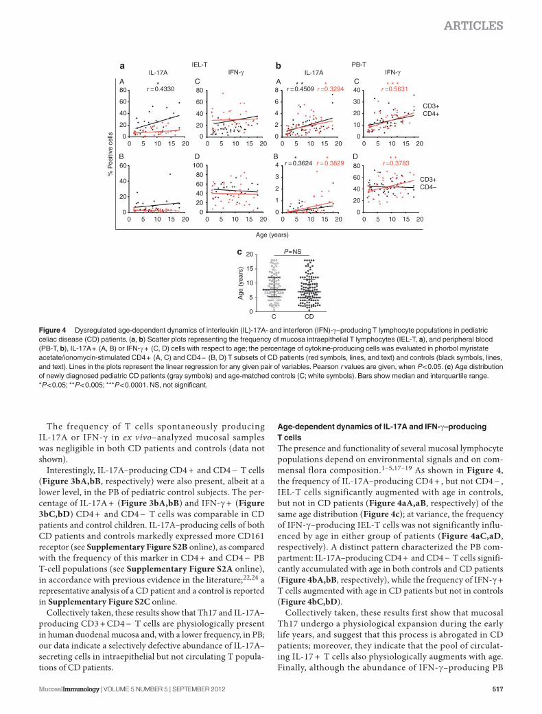
Age-dependent dynamics of IL-17A and IFN- � – producing T cells The presence and functionality of several mucosal lymphocyte
populations depend on environmental signals and on com-
mensal flora composition. 1 – 5,17 – 19 As shown in Figure 4 ,
the frequency of IL-17A – producing CD4 + , but not CD4 − ,
IEL-T cells significantly augmented with age in controls,
but not in CD patients ( Figure 4aA,aB , respectively) of the
same age distribution ( Figure 4c ); at variance, the frequency
of IFN- � – producing IEL-T cells was not significantly influ-
enced by age in either group of patients ( Figure 4aC,aD ,
respectively). A distinct pattern characterized the PB com-
partment: IL-17A – producing CD4 + and CD4 − T cells signifi-
cantly accumulated with age in both controls and CD patients
( Figure 4bA,bB , respectively), while the frequency of IFN- � +
T cells augmented with age in CD patients but not in controls
( Figure 4bC,bD ).
Collectively taken, these results first show that mucosal
Th17 undergo a physiological expansion during the early
life years, and suggest that this process is abrogated in CD
patients; more over, they indicate that the pool of circulat-
ing IL-17 + T cells also physiologically augments with age.
Finally, although the abundance of IFN- � – producing PB
80
60
40
20
0
80
60
100
40
20
0
80
60
40
20
0
60
40
20
0
80
60
40
20
20
15
10
P=NS
5
0C CD
Age
(ye
ars)
200
8
6
4
40
30
20
10
0
2
0
4
2
3
1
0
0 5 10 15 5 10 150 205 10
* * * *
* * *
r =0.3624 r = 0.3829 r =0.3783
r =0.5631** *
r =0.3294r =0.4509r =0.4330
IL-17AIEL-T PB-T
IFN-γ
Age (years)
% P
ositi
ve c
ells
IFN-γIL-17A
*
CD3+CD4+
CD3+CD4–
150 205 10 150
20200 5 10 15 5 10 150 205 10 150 205 10 150
20
Figure 4 Dysregulated age-dependent dynamics of interleukin (IL)-17A- and interferon (IFN)- � – producing T lymphocyte populations in pediatric celiac disease (CD) patients. ( a , b ) Scatter plots representing the frequency of mucosa intraepithelial T lymphocytes (IEL-T, a ), and peripheral blood (PB-T, b ), IL-17A + (A, B) or IFN- � + (C, D) cells with respect to age; the percentage of cytokine-producing cells was evaluated in phorbol myristate acetate / ionomycin-stimulated CD4 + (A, C) and CD4 − (B, D) T subsets of CD patients (red symbols, lines, and text) and controls (black symbols, lines, and text). Lines in the plots represent the linear regression for any given pair of variables. Pearson r values are given, when P < 0.05. ( c ) Age distribution of newly diagnosed pediatric CD patients (gray symbols) and age-matched controls (C; white symbols). Bars show median and interquartile range. * P < 0.05; * * P < 0.005; * * * P < 0.0001. NS, not significant.
518 VOLUME 5 NUMBER 5 | SEPTEMBER 2012 | www.nature.com/mi
ARTICLES
T cells was not altered in CD patients ( Figure 3bD ), this
population pathologically accumulated with patient ’ s age,
thus underlying a deeper and systemic T-cell dysregulation
in CD.
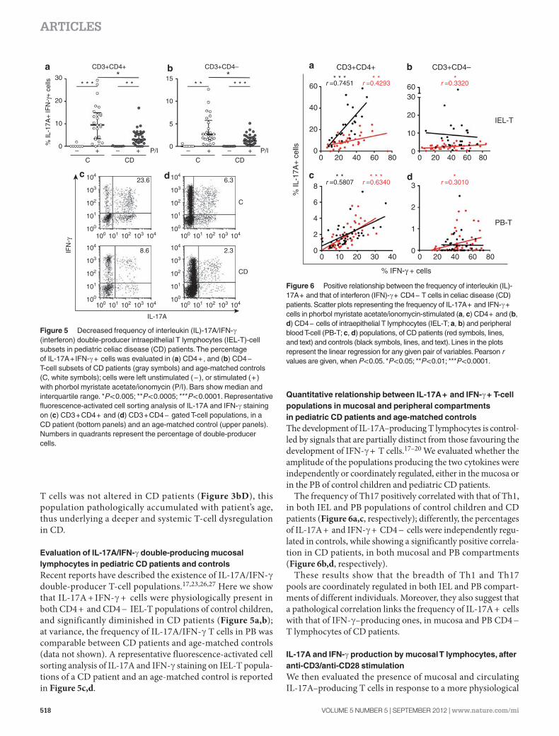
Evaluation of IL-17A / IFN- � double-producing mucosal lymphocytes in pediatric CD patients and controls Recent reports have described the existence of IL-17A / IFN- �
double-producer T-cell populations. 17,23,26,27 Here we show
that IL-17A + IFN- � + cells were physiologically present in
both CD4 + and CD4 − IEL-T populations of control children,
and significantly diminished in CD patients ( Figure 5a,b );
at variance, the frequency of IL-17A / IFN- � T cells in PB was
comparable between CD patients and age-matched controls
(data not shown). A representative fluorescence-activated cell
sorting analysis of IL-17A and IFN- � staining on IEL-T popula-
tions of a CD patient and an age-matched control is reported
in Figure 5c,d .
Quantitative relationship between IL-17A + and IFN- � + T-cell populations in mucosal and peripheral compartments in pediatric CD patients and age-matched controls The development of IL-17A – producing T lymphocytes is control-
led by signals that are partially distinct from those favouring the
development of IFN- � + T cells. 17 – 20 We evaluated whether the
amplitude of the populations producing the two cytokines were
independently or coordinately regulated, either in the mucosa or
in the PB of control children and pediatric CD patients.
The frequency of Th17 positively correlated with that of Th1,
in both IEL and PB populations of control children and CD
patients ( Figure 6a,c , respectively); differently, the percentages
of IL-17A + and IFN- � + CD4 − cells were independently regu-
lated in controls, while showing a significantly positive correla-
tion in CD patients, in both mucosal and PB compartments
( Figure 6b,d , respectively).
These results show that the breadth of Th1 and Th17
pools are coordinately regulated in both IEL and PB compart-
ments of different individuals. Moreover, they also suggest that
a pathological correlation links the frequency of IL-17A + cells
with that of IFN- � – producing ones, in mucosa and PB CD4 −
T lymphocytes of CD patients.
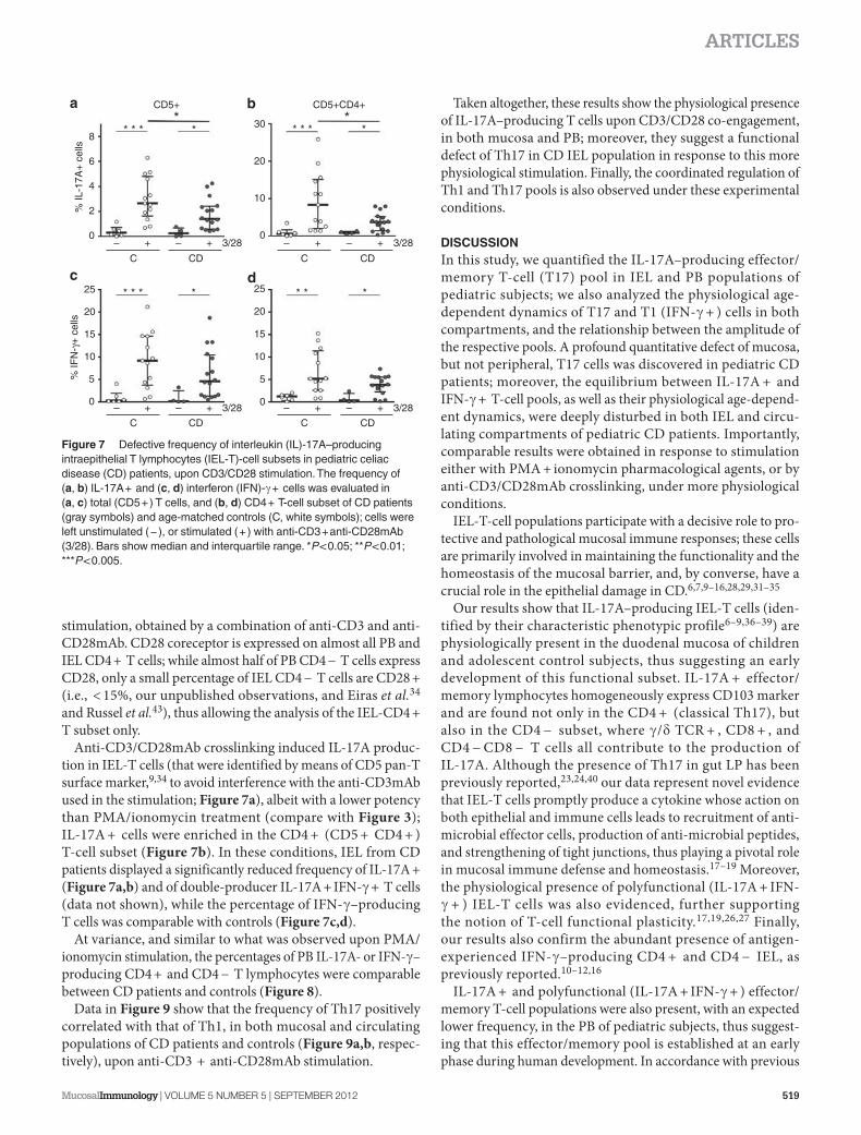
IL-17A and IFN- � production by mucosal T lymphocytes, after anti-CD3 / anti-CD28 stimulation We then evaluated the presence of mucosal and circulating
IL-17A – producing T cells in response to a more physiological
30
20
% IL
-17A
+ IF
N-γ
+ c
ells
10
10
15
0 0
5
– –+ + P/I
23.6104
104
103
103
102
102
101
101100
100
104
104
103
103
102
102
101
101100
100
104
104
103
103
102
102
101
101100
100
104
104
103
103
102
102
101
101100
100
IFN
-γ
8.6
6.3
2.3
CD
IL-17A
CD3+CD4+ CD3+CD4–
* * * * ** * * * ** *
C CD
CD
C
C
P/I– –+ +
Figure 5 Decreased frequency of interleukin (IL)-17A / IFN- � (interferon) double-producer intraepithelial T lymphocytes (IEL-T)-cell subsets in pediatric celiac disease (CD) patients. The percentage of IL-17A + IFN- � + cells was evaluated in ( a ) CD4 + , and ( b ) CD4 − T-cell subsets of CD patients (gray symbols) and age-matched controls (C, white symbols); cells were left unstimulated ( − ), or stimulated ( + ) with phorbol myristate acetate / ionomycin (P / I). Bars show median and interquartile range. * P < 0.005; * * P < 0.0005; * * * P < 0.0001. Representative fluorescence-activated cell sorting analysis of IL-17A and IFN- � staining on ( c ) CD3 + CD4 + and ( d ) CD3 + CD4 − gated T-cell populations, in a CD patient (bottom panels) and an age-matched control (upper panels). Numbers in quadrants represent the percentage of double-producer cells.
CD3+CD4+ CD3+CD4–
606030
30
20
20
20 806040
40
10
10
00
20 8060400
20
% IL
-17A
+ c
ells
20
806040
40
0
00 0
1
2
3
2
4
6
8 r =0.5807* *
r =0.7451 r =0.4293 r =0.3320** * * * *
r =0.6340* * *
r =0.3010*
0
IEL-T
PB-T
% IFN-γ + cells
Figure 6 Positive relationship between the frequency of interleukin (IL)-17A + and that of interferon (IFN)- � + CD4 − T cells in celiac disease (CD) patients. Scatter plots representing the frequency of IL-17A + and IFN- � + cells in phorbol myristate acetate / ionomycin-stimulated ( a , c ) CD4 + and ( b , d ) CD4 − cells of intraepithelial T lymphocytes (IEL-T; a , b ) and peripheral blood T-cell (PB-T; c , d ) populations, of CD patients (red symbols, lines, and text) and controls (black symbols, lines, and text). Lines in the plots represent the linear regression for any given pair of variables. Pearson r values are given, when P < 0.05. * P < 0.05; * * P < 0.01; * * * P < 0.0001.
MucosalImmunology | VOLUME 5 NUMBER 5 | SEPTEMBER 2012 519
ARTICLES
stimulation, obtained by a combination of anti-CD3 and anti-
CD28mAb. CD28 coreceptor is expressed on almost all PB and
IEL CD4 + T cells; while almost half of PB CD4 − T cells express
CD28, only a small percentage of IEL CD4 − T cells are CD28 +
(i.e., < 15 % , our unpublished observations, and Eiras et al. 34
and Russel et al. 43 ), thus allowing the analysis of the IEL-CD4 +
T subset only.
Anti-CD3 / CD28mAb crosslinking induced IL-17A produc-
tion in IEL-T cells (that were identified by means of CD5 pan-T
surface marker, 9,34 to avoid interference with the anti-CD3mAb
used in the stimulation; Figure 7a ), albeit with a lower potency
than PMA / ionomycin treatment (compare with Figure 3 );
IL-17A + cells were enriched in the CD4 + (CD5 + CD4 + )
T-cell subset ( Figure 7b ). In these conditions, IEL from CD
patients displayed a significantly reduced frequency of IL-17A +
( Figure 7a,b ) and of double-producer IL-17A + IFN- � + T cells
(data not shown), while the percentage of IFN- � – producing
T cells was comparable with controls ( Figure 7c,d ).
At variance, and similar to what was observed upon PMA /
ionomycin stimulation, the percentages of PB IL-17A- or IFN- � –
producing CD4 + and CD4 − T lymphocytes were comparable
between CD patients and controls ( Figure 8 ).
Data in Figure 9 show that the frequency of Th17 positively
correlated with that of Th1, in both mucosal and circulating
populations of CD patients and controls ( Figure 9a,b , respec-
tively), upon anti-CD3 + anti-CD28mAb stimulation.
Taken altogether, these results show the physiological presence
of IL-17A – producing T cells upon CD3 / CD28 co-engagement,
in both mucosa and PB; moreover, they suggest a functional
defect of Th17 in CD IEL population in response to this more
physiological stimulation. Finally, the coordinated regulation of
Th1 and Th17 pools is also observed under these experimental
conditions.
DISCUSSION In this study, we quantified the IL-17A – producing effector /
memory T-cell (T17) pool in IEL and PB populations of
pediatric subjects; we also analyzed the physiological age-
dependent dynamics of T17 and T1 (IFN- � + ) cells in both
compartments, and the relationship between the amplitude of
the respective pools. A profound quantitative defect of mucosa,
but not peripheral, T17 cells was discovered in pediatric CD
patients; moreover, the equilibrium between IL-17A + and
IFN- � + T-cell pools, as well as their physiological age-depend-
ent dynamics, were deeply disturbed in both IEL and circu-
lating compartments of pediatric CD patients. Importantly,
comparable results were obtained in response to stimulation
either with PMA + ionomycin pharmacological agents, or by
anti-CD3 / CD28mAb crosslinking, under more physiological
conditions.
IEL-T-cell populations participate with a decisive role to pro-
tective and pathological mucosal immune responses; these cells
are primarily involved in maintaining the functionality and the
homeostasis of the mucosal barrier, and, by converse, have a
crucial role in the epithelial damage in CD. 6,7,9 – 16,28,29,31 – 35
Our results show that IL-17A – producing IEL-T cells (iden-
tified by their characteristic phenotypic profile 6 – 9,36 – 39 ) are
physiologically present in the duodenal mucosa of children
and adolescent control subjects, thus suggesting an early
development of this functional subset. IL-17A + effector /
memory lymphocytes homogeneously express CD103 marker
and are found not only in the CD4 + (classical Th17), but
also in the CD4 − subset, where � / � TCR + , CD8 + , and
CD4 − CD8 − T cells all contribute to the production of
IL-17A. Although the presence of Th17 in gut LP has been
previously reported, 23,24,40 our data represent novel evidence
that IEL-T cells promptly produce a cytokine whose action on
both epithelial and immune cells leads to recruitment of anti-
microbial effector cells, production of anti-microbial peptides,
and strengthening of tight junctions, thus playing a pivotal role
in mucosal immune defense and homeostasis. 17 – 19 Moreover,
the physiological presence of polyfunctional (IL-17A + IFN-
� + ) IEL-T cells was also evidenced, further supporting
the notion of T-cell functional plasticity. 17,19,26,27 Finally,
our results also confirm the abundant presence of antigen-
experienced IFN- � – producing CD4 + and CD4 − IEL, as
previously reported. 10 – 12,16
IL-17A + and polyfunctional (IL-17A + IFN- � + ) effector /
memory T-cell populations were also present, with an expected
lower frequency, in the PB of pediatric subjects, thus suggest-
ing that this effector / memory pool is established at an early
phase during human development. In accordance with previous
30
20
8
6
4
% IL
-17A
+ c
ells
% IF
N-γ
+ c
ells
2
0
10
0–– ++ – + 3/28
CC CDCD
3/28– +
CD5+ CD5+CD4+*
** * ** * *
* * * * *
*
* *
*
0
5
15
20
25
10
0
5
15
20
25
10
–– ++ – + 3/28
CC CDCD
3/28– +
Figure 7 Defective frequency of interleukin (IL)-17A – producing intraepithelial T lymphocytes (IEL-T)-cell subsets in pediatric celiac disease (CD) patients, upon CD3 / CD28 stimulation. The frequency of ( a , b ) IL-17A + and ( c , d ) interferon (IFN)- � + cells was evaluated in ( a , c ) total (CD5 + ) T cells, and ( b , d ) CD4 + T-cell subset of CD patients (gray symbols) and age-matched controls (C, white symbols); cells were left unstimulated ( − ), or stimulated ( + ) with anti-CD3 + anti-CD28mAb (3 / 28). Bars show median and interquartile range. * P < 0.05; * * P < 0.01; * * * P < 0.005.

520 VOLUME 5 NUMBER 5 | SEPTEMBER 2012 | www.nature.com/mi
ARTICLES
evidence in the literature, 22,24 PB IL-17A – producing T cells were
enriched in the CD161 + fraction, comparably in CD patients
and controls.
The frequency of IL-17A + , but not IFN- � + , effector / memory
mucosal and circulating T cells was found to increase according
to children ’ s age, in the control population. These data suggest
that the IL-17A – producing pool of effector / memory T cells
is exquisitely modulated during infancy and adolescence in
humans, and are in accordance with the notion that the matura-
tion of some components of the immune system is completed in
the postnatal period. 1 – 5 Our results also show that the frequency
of Th17 and that of Th1 were significantly correlated, in both
IEL and peripheral compartments of pediatric control subjects;
at variance, the sizes of IL-17A + and IFN- � + CD4 − T-cell
pools were independently regulated in different individuals. The
coordinated regulation of distinct functional T-cell subsets may
be the result of environmental stimuli and / or may underlie a
positive crosstalk of immune regulatory circuits. 17 – 19 Moreover,
these observations suggest that the acquisition of the functional
ability to produce IL-17A is differently regulated in CD4 + and
CD4 − T-cell subsets, within the same compartment.
In summary, our analysis on children and adolescent
controls has elucidated some aspects of the physiological status
of T1 and T17 major functional T-cell subsets during human
development.
A two-tiered defect was found to selectively characterize
IL-17A – producing IEL-T lymphocytes of pediatric CD patients:
IL-17A + as well as IL17A + IFN- � + polyfunctional T-cell pools
were strongly reduced among both CD4 + and CD4 − subsets,
and the age-dependent accumulation of Th17 was also severely
compromised. The frequency of IFN- � – producing T cells
did not significantly differ between CD patients and controls,
arguing against the possibility that a generalized unresponsive-
ness may be at the basis of this phenomenon. The dysregulated
functionality of the IEL compartment in CD patients was also
3* * * * * * * * *
* * * * * ** * ** * ** * ** * *
* ** * *
2
2
4
6
1.0
0.5
0.0
% IL
-17A
+ c
ells
% IF
N-γ
+ c
ells
1
0 0
0
10 5
0
20
30 15
20
10
40
0
10
20
30
40
– +
C CD
3/28– +
– +
C CD
3/28– + – +
C CD
3/28– + – +
C CD
3/28– +
– +
C
CD5+ CD5+CD4+ CD5+CD4–
CD
3/28– + – +
C CD
3/28– +
Figure 8 Frequency of circulating interleukin (IL)-17A – producing T-cell subsets in pediatric celiac disease (CD) patients and controls (C), upon CD3 / CD28 stimulation. The frequency of peripheral blood IL-17A + and interferon (IFN)- � + cells was evaluated ( a , d ) in total (CD5 + ) T cells, and ( b , e ) in CD4 + and ( c , f ) CD4 − T-cell subsets of CD patients (gray symbols) and age-matched controls (white symbols); cells were left unstimulated ( − ), or stimulated ( + ) with anti-CD3 + anti-CD28mAb (3 / 28). Bars show median and interquartile range. * P < 0.05; * * * P < 0.0001.
30
20
10
00
0
2
4
6
8
5 10 15 20
0 5
% IFN-γ + cells
10 15 20
IEL-T
PB-T
r =0.6232
r =0.4499 r =0.4692* * *
% IL
-17A
+ c
ells
* *
r =0.7140* **
Figure 9 Positive relationship between the frequency of interleukin (IL)-17A + and that of interferon (IFN)- � + CD4 + T cells in celiac disease (CD) patients and controls. Scatter plots representing the frequency of IL-17A + and IFN- � + cells in anti-CD3 / CD28-stimulated ( a ) intraepithelial lymphocytes (IEL) and ( b ) peripheral blood (PB) CD4 + T-cell populations of CD patients (red symbols, lines, and text) and controls (black symbols, lines, and text). Lines in the plots represent the linear regression for any given pair of variables. Pearson r values are given, when P < 0.05. * P < 0.05; * * P < 0.005; * * * P = 0.0005.
MucosalImmunology | VOLUME 5 NUMBER 5 | SEPTEMBER 2012 521
ARTICLES
underscored by the evidence that the frequency of IL-17A +
and that of IFN- � + CD4 − T cells were positively correlated in
CD, but not in control, individuals.
The role of IL-17A in CD is still unclear. The presence of
higher levels of IL-17A (evaluated as mRNA or protein) was
reported in mucosal biopsies of CD patients; 40,44,45 the contri-
bution of non-T cells to IL-17A production, and the increased
immune infiltrate may explain the difference with our data.
This work represents a first assessment on the ability of intra-
epithelial T cells to produce IL-17A in CD; contrasting evi-
dence have been reported on the ability of LP CD4 + T cells
and gliadin-specific LP T-cell lines to produce IL-17A, 40 – 42 thus
leaving this issue open to further investigation. The impaired
abundance of IL-17A – producing IEL-T populations may affect
the homeo stasis of the mucosal barrier and directly contribute
to the augmented epithelial permeability, which constitutes a
hallmark of CD; 28 – 31 moreover, the reduced frequency of both
IL-17A + and double-producer effector / memory T cells may
negatively impact on the efficiency of antimicrobial immune
responses, and indirectly concur to the worsening of inflam-
mation in CD. 17 – 20
The mechanisms underlying the defective abundance of IL-
17A + IEL-T populations in CD are unknown. The develop-
ment of T17 is controlled by microbial components, cytokines,
and metabolites, and is exquisitely dependent on a network of
interactions with other T-cell subsets (such as T1 and T reg),
and other immune and tissue cells. 1 – 5,17 – 20 All these factors
are deeply dysregulated in the CD mucosa microenviron-
ment; 1,11,14,28 – 32 moreover, alterations in the metabolic status
of the gut microflora have been reported in CD. 46
We could not find any significant relationship between the
frequency of IL-17A + T cells and CD clinical form (typical vs.
atypical vs. silent), or histological grading (data not shown);
interestingly, the frequency of IL-17A + T cells in patients
showing a positive serology and a histological grading of 0 or
1, and possibly representing an early stage of disease, 28,29,47,48
was not significantly different from that of those affected by
the overt disease (data not shown), hinting at the possibility
that the impairment of IL-17A + T-cell pool represents an early
event in CD.
The presence of circulating pathogenic T cells has been con-
vincingly shown in CD. 29 Although neither the percentage nor
the age-dependent increase of PB IL-17A + T lymphocytes
were significantly affected, the pool of IFN- � – producing
CD4 − T cells expanded with age (and possibly, with length
of disease) in pediatric CD patients, but not in controls; more-
over, in this cell population, the frequency of IL-17A – pro-
ducing cells and that of IFN- � + ones were positively linked,
thus mirroring what occurred in the mucosa compartment.
Collectively taken, these observations underline that distinct
and common modes of functional dysregulation character-
ize the IL-17A + CD4 − T-cell subset in mucosal lesions and
in the PB of CD patients; in fact, although this population is
significantly decreased in IEL but not in PB lymphocytes, its
frequency is nevertheless positively linked with that of IFN- � +
cells in both compartments.
In summary, our work provides novel information on
the amplitude and dynamics of IL-17A – producing T cells,
and its relationship with IFN- � – secreting subset, in human
development. These results describe novel aspects of the
alterations of immune system in CD; they may contribute
to a better comprehension of the underlying pathogenetic
mechanisms and be useful in the design of innovative thera-
peutic strategies. The multifaceted dysregulation of cytokine-
producing T-cell populations at the systemic level may also be
involved in the reported association between CD and other
autoimmune and immune-mediated extraintestinal manifes-
tations. 28,29,47
METHODS Patients and controls . One hundred and sixteen newly-diagnosed children and adolescent CD subjects (age range: 0.5 – 18 years) and 86 age-matched controls (age range: 0.5 – 18 years) were consecu-tively enrolled in our study; all patients were referred to either the Celiac Disease and Malabsorptive Diseases Unit or the Gastroenterology and Liver Unit, Department of Pediatrics, Sapienza University, Rome, Italy.
Diagnosis of CD was assessed according to clinical, serological and histological criteria. 47 Duodenal histology was graded according to Marsh classification. 48 Positivity for anti-tTG IgA was assessed in all patients by enzyme-linked immunosorbent assay and / or radioimmunoassay. 49
Control subjects were under investigation for upper gastrointestinal and / or systemic signs and symptoms (i.e., reflux disease, failure to thrive, dyspepsia, dysphagia, persistent vomiting, and abdominal pain) and were serologically and histologically negative. Helicobacter pylori infection was ruled out in all controls, except six, by the combined analysis of rapid urease-based method and histological examination on both antral and fundic stomach biopsies stained with Giemsa. None of the controls dis-closed any inflammatory or abnormal aspect at duodenal biopsies that could be suggestive of other enteropathies (inflammatory bowel disease, autoimmune enteropathy, food allergies, non-steroidal anti-inflammatory drug-related enteropathy, and Giardia or Cryptosporidium infection).
All patients and controls were on a gluten-containing diet at diagnosis.
Demographic, clinical, serological, and histological features of CD patients and controls are reported in Table 1 .
Two distal duodenum biopsies and a heparinized PB sample were taken during upper gastrointestinal endoscopy performed with a pedi-atric endoscope (Olympus GIF-E, or PQ20, Tokyo, Japan) and a video gastroscope (Olympus GIF180, or GIF P140), after conscious sedation with intravenous pethidine (1 – 2 mg kg − 1 ) and midazolam (0.1 mg kg − 1 ), or general anesthesia.
The study protocol was defined in accordance with the Declaration of Helsinki and approved by the institutional ethical committee. Written informed consent was obtained from children ’ s parents; children >12 years of age signed a statement of assent.
Mononuclear cell isolation and stimulation . PB mononuclear cells were purified from heparinized blood samples by Ficoll-Hypaque (Eurobio, Les Ulis Cedex, France) density-gradient centrifugation.
Duodenal mucosa samples were put in physiological solution imme-diately upon biopsy; mononuclear cells were obtained by mechanical dissociation on a 70- � m cell strainer (BD Biosciences, San Jose, CA), and collected in physiological solution.
Mononuclear cells were resuspended in RPMI completed with 10 % fetal calf serum and 1 % glutamine (all from Euroclone, Milan, Italy), and left untreated or stimulated with 50 ng ml − 1 PMA (Sigma-Aldrich, St Louis, MO) and 0.5 � g ml − 1 ionomycin (Sigma-Aldrich). Alternatively, Fc � R + P815 mastocytoma cell line (3 × 10 5 cells) was incubated with anti-CD3 and anti-CD28 antibodies (0.5 � g each; anti-CD3 OKT3mAb clone
522 VOLUME 5 NUMBER 5 | SEPTEMBER 2012 | www.nature.com/mi
ARTICLES
was obtained from ATCC (Rockville, MD) and purified in our labora-tory; anti-CD28mAb was from BD Biosciences) for 20 min, washed, and added to mononuclear cells in a 1:3 ratio.
All stimulations were carried out at 37 ° C for 6 h, in the presence of the intracellular trafficking inhibitor brefeldin A (10 � g ml − 1 ; Sigma-Aldrich), before intracellular staining and cytofluorimetric analysis.
Intracellular staining and multiparameter cytofluorimetric analysis . Mononuclear cells were washed with phosphate-buffered saline, fixed with 2 % paraformaldehyde for 20 min, permeabilized with 0.5 % saponin / 1 % fetal calf serum, and stained with anti-CD3 PerCP (or anti-CD5 PECy7, in samples stimulated with anti-CD3 + anti-CD28 mAb), anti-CD4 FITC, IgG1 PE, and IgG1 APC isotypic controls, or anti-IFN- � APC (all from BD Biosciences) and anti-IL-17A PE (e-Biosciences, San Diego, CA) mAb for 30 min at 4 ° C.
Alternatively, mononuclear cells were stained with anti-CD3 PerCP Cy5.5, anti-CD4 PE Cy7, anti-CD8 APC H7, anti-TCR � / � APC, IgG1 FITC or CD103 FITC or CD161 FITC, for 30 min at 4 ° C. Cells were washed with phosphate-buffered saline, fixed with 2 % paraformaldehyde for 20 min and permeabilized with 0.5 % saponin / 1 % fetal calf serum, before an additional staining with IgG1 PE or anti-IL-17A PE.
Cells were analyzed with a FACScalibur or FACSCANTO II (BD Biosciences), using CellQuest Pro (BD Biosciences) or FlowJo (Treestar, Ashland, OR) software. Lymphocyte region was defined on the basis of forward and side scatter physical parameters; T-cell subsets were identi-fied by anti-CD3 (or anti-CD5), 9,34 and anti-CD4, anti-CD8 or anti-TCR � / � staining. The percentage of cytokine-producing cells in each T-cell subset was obtained by setting the lower limit on the basis of a matched isotype control mAb-stained sample, whose positivity never exceeded 0.5 % of gated events. Cell viability in the populations analyzed was routinely >95 % (not shown).
Statistical analysis . Differences between groups were analyzed with parametric (Student t -test) and non-parametric (Mann – Whitney and Wilcoxon) tests, as appropriate. Correlation analysis was performed using Pearson correlation test. Statistical analysis was performed with
PRISM v.5 (GraphPad Software, San Diego, CA) and SPSS v.19 (Chicago, IL) softwares; P value < 0.05 (two sided) was considered statistically significant.
SUPPLEMENTARY MATERIAL is linked to the online version of the paper at http://www.nature.com/mi
ACKNOWLEDGMENTS Grant support: Sapienza University ( “ Progetti Coordinati di Ateneo Federato delle Scienze delle Politiche Pubbliche e Sanitarie (SPPS) ” ). We thank Dr Rossella Baldini for statistical advice, Dr Claudio Tiberti for anti-tTG RIA, Sofia Guida for anti-tTG ELISA, and all patients and their families who participated in the study.
DISCLOSURE The authors declared no conflict of interest.
© 2012 Society for Mucosal Immunology
REFERENCES 1 . Garrett , W . S . , Gordon , J . I . & Glimcher , L . H . Homeostasis and
infl ammation in the intestine . Cell 140 , 859 – 870 ( 2010 ). 2 . Hooper , L . V . & Macpherson , A . J . Immune adaptations that maintain
homeostasis with the intestinal microbiota . Nature Rev. Immunol. 10 , 159 – 169 ( 2010 ).
3 . Cerf-Bensussan , N . & Gaboriau-Routhiau , V . The immune system and the gut microbiota: friends or foes? Nat. Rev. Immunol. 10 , 735 – 744 ( 2010 ).
4 . Lee , Y . K . & Mazmanian , S . K . Has the microbiota played a critical role in the evolution of the adaptive immune system? Science 330 , 1768 – 1773 ( 2010 ).
5 . Eberl , G . & Lochner , M . The development of intestinal lymphoid tissues at the interface of self and microbiota . Mucosal Immunol. 2 , 478 – 485 ( 2009 ).
6 . Selby , W . S . , Janossy , G . , Bofi ll , M . & Jewell , D . P . Lymphocyte subpopulations in the human small intestine. The fi ndings in normal mucosa and in the mucosa of patients with adult celiac disease . Clin. Exp. Immunol. 52 , 219 – 228 ( 1983 ).
7 . Brandtzaeg , P . et al. Immunobiology and immunopathology of human gut mucosa: humoral immunity and intraepithelial lymphocytes . Gastroenterology 97 , 1562 – 1584 ( 1989 ).
8 . Leon , F . Flow cytometry of intestinal intraepithelial lymphocytes in celiac disease . J. Immunol. Methods 363 , 177 – 186 ( 2011 ).
9 . Jarry , A . , Cerf-Bensussan , N . , Brousse , N . , Selz , F . & Guy-Grand , D . Subsets of CD3+ (T cell receptor alpha/beta or gamma/delta) and CD3 − lymphocytes isolated from normal human gut epithelium display phenotypical features different from their counterparts in peripheral blood . Eur. J. Immunol. 20 , 1097 – 1103 ( 1990 ).
10 . Sheridan , B . S . & Lefran ç ois , L . Intraepithelial lymphocytes: to serve and protect . Curr. Gastroenterol. Rep. 12 , 513 – 521 ( 2010 ).
11 . Jabri , B . & Ebert , E . Human CD8+ intraepithelial lymphocytes: a unique model to study the regulation of effector cytotoxic T lymphocytes in tissue . Immunol. Rev. 215 , 202 – 214 ( 2007 ).
12 . Meresse , B . & Cerf-Bensussan , N . Innate T cell responses in human gut . Semin. Immunol. 21 , 121 – 129 ( 2009 ).
13 . Shibahara , T . et al. Alteration of intestinal epithelial function by intraepi-thelial lymphocyte homing . J. Gastroenterol. 40 , 878 – 886 ( 2005 ).
14 . Bhagat , G . et al. Small intestinal CD8+TCRgammadelta+NKG2A+ intraepithelial lymphocytes have attributes of regulatory cells in patients with celiac disease . J. Clin. Invest. 118 , 281 – 293 ( 2008 ).
15 . Le ó n , F . , S á nchez , L . , Camarero , C . & Roy , G . Cytokine production by intestinal intraepithelial lymphocyte subsets in celiac disease . Dig. Dis. Sci. 50 , 593 – 600 ( 2005 ).
16 . Cheroutre , H . , Lambolez , F . & Mucida , D . The light and dark sides of intestinal intraepithelial lymphocytes . Nat. Rev. Immunol. 11 , 445 – 456 ( 2011 ).
17 . Korn , T . , Bettelli , E . , Oukka , M . & Kuchroo , V . K . IL-17 and Th17 Cells . Annu. Rev. Immunol. 27 , 485 – 517 ( 2009 ).
18 . O ’ Connor , W . Jr , Zenewicz , L . A . & Flavell , R . A . The dual nature of T(H)17 cells: shifting the focus to function . Nat. Immunol. 11 , 471 – 476 ( 2010 ).
19 . Mucida , D . & Salek-Ardakani , S . Regulation of TH17 cells in the mucosal surfaces . J. Allergy Clin. Immunol. 123 , 997 – 1003 ( 2009 ).
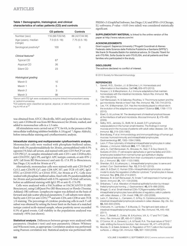
Table 1 Demographic, histological, and clinical characteristics of celiac patients (CD) and controls
CD patients Controls
Number (sex) 116 (66 F / 50 M) 86 (42 F / 44 M)
Age (years), median (range)
7.0 (0.5 – 18) 7.75 (0.5 – 18)
Serological positivity a 100 0
Clinical features b
Typical CD 67
Atypical CD 26
Silent CD 23
Histological grading
Marsh 0 10
Marsh 1 5
Marsh 2 3
Marsh 3 98 a Serum anti-tTG IgA were evaluated by enzyme-linked immunosorbent assay or radioimmunoassay. b CD patients were classifi ed as typical, atypical, or silent clinical form accord-ing to Hill et al. 47

MucosalImmunology | VOLUME 5 NUMBER 5 | SEPTEMBER 2012 523
ARTICLES
20 . Cua , D . J . & Tato , C . M . Innate IL-17-producing cells: the sentinels of the immune system . Nat. Rev. Immunol. 10 , 479 – 489 ( 2010 ).
21 . Shrikant , P . A . et al. Regulating functional cell fates in CD8 T cells . Immunol. Res. 46 , 12 – 22 ( 2010 ).
22 . Maggi , L . et al. CD161 is a marker of all human IL-17-producing T-cell subsets and is induced by RORC . Eur J Immunol. 40 , 2174 – 2181 ( 2010 ).
23 . Annunziato , F . et al. Phenotypic and functional features of human Th17 cells . J. Exp. Med. 204 , 1849 – 1861 ( 2007 ).
24 . Kleinschek , M . A . et al. Circulating and gut-resident human Th17 cells express CD161 and promote intestinal infl ammation . J. Exp. Med. 206 , 525 – 534 ( 2009 ).
25 . Kondo , T . , Takata , H . , Matsuki , F . & Takiguchi , M . Cutting edge: Phenotypic characterization and differentiation of human CD8+ T cells producing IL-17 . J. Immunol. 182 , 1794 – 1798 ( 2009 ).
26 . Chen , Z . & O ’ Shea , J . J . Regulation of IL-17 production in human lymphocytes . Cytokine 41 , 71 – 78 ( 2008 ).
27 . Peck , A . & Mellins , E . D . Plasticity of T-cell phenotype and function: the T helper Type 17 example . Immunology 129 , 147 – 153 ( 2010 ).
28 . Di Sabatino , A . & Corazza , G . R . Coeliac disease . Lancet 373 , 1480 – 1493 ( 2009 ).
29 . Abadie , V . , Sollid , L . M . , Barreiro , L . B . & Jabri , B . Integration of genetic and immunological insights into a model of celiac disease pathogenesis . Annu. Rev. Immunol. 29 , 493 – 525 ( 2011 ).
30 . Fasano , A . Zonulin and its regulation of intestinal barrier function: the biological door to infl ammation, autoimmunity, and cancer . Physiol. Rev. 91 , 151 – 175 ( 2011 ).
31 . Jabri , B . & Sollid , L . M . Tissue-mediated control of immunopathology in coeliac disease . Nat. Rev. Immunol. 9 , 858 – 870 ( 2009 ).
32 . Gianfrani , C . , Auricchio , S . & Troncone , R . Adaptive and innate immune responses in celiac disease . Immunol. Lett. 99 , 141 – 145 ( 2005 ).
33 . Freedman , A . R . , Macartney , J . C . , Nelufer , J . M . & Ciclitira , P . J . Timing of infi ltration of T lymphocytes induced by gluten into the small intestine in coeliac disease . J. Clin. Pathol. 40 , 741 – 745 ( 1987 ).
34 . Eiras , P . , Rold á n , E . , Camarero , C . , Olivares , F . , Bootello , A . & Roy , G . Flow cytometry description of a novel CD3-/CD7+ intraepithelial lymphocyte subset in human duodenal biopsies: potential diagnostic value in coeliac disease . Cytometry 34 , 95 – 102 ( 1998 ).
35 . Meresse , B . et al. Reprogramming of CTLs into natural killer-like cells in celiac disease . J. Exp. Med. 203 , 1343 – 1355 ( 2006 ).
36 . Jenkins , D . , Goodall , A . & Scott , B . B . T-lymphocyte populations in normal and coeliac small intestinal mucosa defi ned by monoclonal antibodies . Gut 27 , 1330 – 1337 ( 1986 ).
37 . Spencer , J . , MacDonald , T . T . , Diss , T . C . , Walker-Smith , J . A . , Ciclitira , P . J . & Isaacson , P . G . Changes in intraepithelial lymphocyte subpopulations in coeliac disease and enteropathy associated T cell lymphoma (malignant histiocytosis of the intestine) . Gut 30 , 339 – 346 ( 1989 ).
38 . Spencer , J . , Isaacson , P . G . , Diss , T . C . & MacDonald , T . T . Expression of disulfi de-linked and non-disulfi de-linked forms of the T cell receptor gamma/delta heterodimer in human intestinal intraepithelial lymphocytes . Eur. J. Immunol. 19 , 1335 – 1338 ( 1989 ).
39 . Halstensen , T . S . , Scott , H . & Brandtzaeg , P . Intraepithelial T cells of the TcR gamma/delta+ CD8 − and V delta 1/J delta 1+ phenotypes are increased in coeliac disease . Scand. J. Immunol. 30 , 665 – 672 ( 1989 ).
40 . Monteleone , I . et al. Characterization of IL-17A producing cells in celiac disease mucosa . J. Immunol. 184 , 2211 – 2218 ( 2010 ).
41 . Fern á ndez , S . et al. Characterization of gliadin-specifi c Th17 cells from the mucosa of celiac disease patients . Am. J. Gastroenterol. 106 , 528 – 538 ( 2011 ).
42 . Bodd , M . et al. HLA-DQ2-restricted gluten-reactive T cells produce IL-21 but not IL-17 or IL-22 . Mucosal Immunol. 3 , 594 – 601 ( 2010 ).
43 . Russell , G . J . et al. p126 (CDw101): a costimulatory molecule preferentially expressed on mucosal T lymphocytes . J. Immunol. 157 , 3366 – 3374 ( 1996 ).
44 . Castellanos-Rubio , A . , Santin , I . , Irastorza , I . , Casta ñ o , L . , Carlos Vitoria , J . & Ramon Bilbao , J . TH17 (and TH1) signatures of intestinal biopsies of CD patients in response to gliadin . Autoimmunity 42 , 69 – 73 ( 2009 ).
45 . Sapone , A . et al. Differential mucosal IL-17 expression in two gliadin-induced disorders: gluten sensitivity and the autoimmune enteropathy celiac disease . Int. Arch. Allergy Immunol. 152 , 75 – 80 ( 2010 ).
46 . Tjellstr ö m , B . et al. Gut microfl ora associated characteristics in children with celiac disease . Am. J. Gastroenterol. 100 , 2784 – 2788 ( 2005 ).
47 . Hill , ID . et al. Guideline for the diagnosis and treatment of celiac disease in children: recommendations of the North American Society for Pediatric Gastroenterology, Hepatology and Nutrition . J. Pediatr. Gastroenterol. Nutr. 40 , 1 – 19 ( 2005 ).
48 . Marsh , M . N . Gluten, major histocompatibility complex, and the small intestine. A molecular and immunobiologic approach to the spectrum of gluten sensitivity ( ‘ celiac sprue ’ ) . Gastroenterology 102 , 330 – 354 ( 1992 ).
49 . Li , M . et al. A report on the International Transglutaminase Autoantibody Workshop for Celiac Disease . Am. J. Gastroenterol. 104 , 154 – 163 ( 2009 ).
Copyright © 2022 FDOKUMEN