Siliceous spicules in marine demosponges (example ...
14
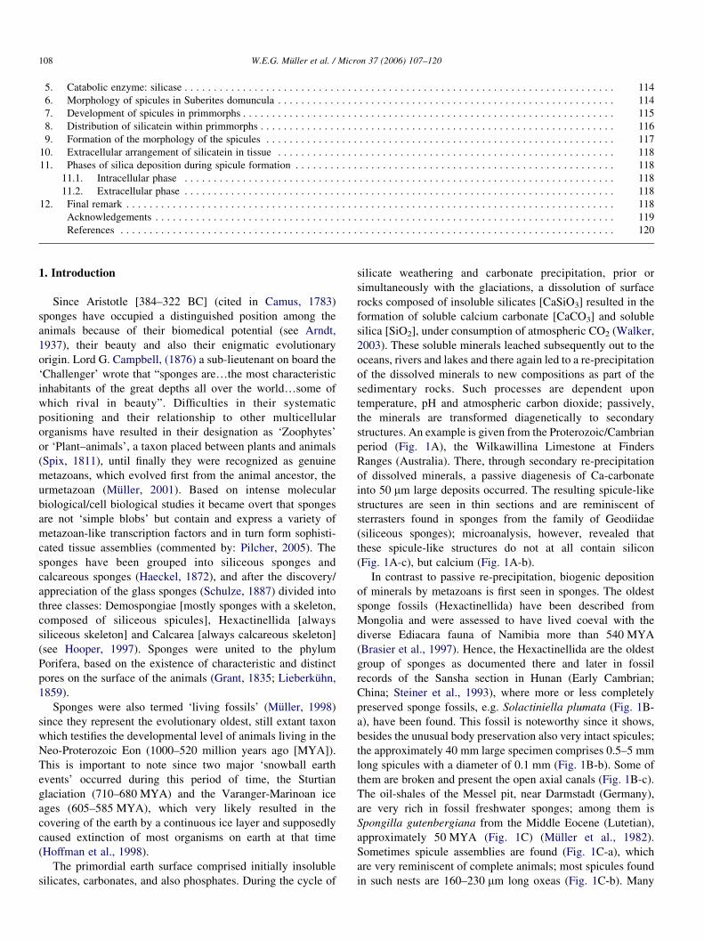
Review Siliceous spicules in marine demosponges (example Suberites domuncula) Werner E.G. Mu ¨ller a, * , Sergey I. Belikov b , Wolfgang Tremel c , Carole C. Perry d , Winfried W.C. Gieskes e , Alexandra Boreiko a , Heinz C. Schro ¨der a a Institut fu ¨r Physiologische Chemie, Abteilung Angewandte Molekularbiologie, Universita ¨t, Duesbergweg 6, D-55099 Mainz, Germany b Limnological Institute of the Siberian Branch of Russian Academy of Sciences, Ulan-Batorskaya 3, RUS-664033 Irkutsk, Russian Federation c Institut fu ¨r Anorganische Chemie, Universita ¨t, Duesbergweg 10-14, D-55099 Mainz, Germany d Department of Chemistry and Physics, Nottingham Trent University, Clifton Lane, Nottingham NG11 8NS, UK e Department of Marine Biology, Biological Center, Center for Ecological and Evolutionary Studies, University of Groningen, P.O. Box 14, 9750 AA Haren, The Netherlands Received 2 August 2005; revised 4 September 2005; accepted 5 September 2005 Abstract All metazoan animals comprise a body plan of different complexity. Since—especially based on molecular and cell biological data—it is well established that all metazoan phyla, including the Porifera (sponges), evolved from a common ancestor the search for common, basic principles of pattern formation (body plan) in all phyla began. Common to all metazoan body plans is the formation of at least one axis that runs from the apical to the basal region; examples for this type of organization are the Porifera and the Cnidaria (diploblastic animals). It seems conceivable that the basis for the formation of the Bauplan in sponges is the construction of their skeleton by spicules. In Demospongiae (we use the model species Suberites domuncula) and Hexactinellida, the spicules consist of silica. The formation of the spicules as the building blocks of the skeleton, starts with the expression of an enzyme which was termed silicatein. Spicule growth begins intracellularly around an axial filament composed of silicatein. When the first layer of silica is made, the spicules are extruded from the cells and completed extracellularly to reach their the final form and size. While the first steps of spicule formation within the cells are becoming increasingly clear, it remains to be studied how the extracellularly present silicatein strings are formed. The understanding of especially this morphogenetic process will allow an insight into the construction of the amazingly diverse skeleton of the siliceous sponges; animals which evolved between two periods of glaciations, the Sturtian glaciation (710– 680 MYA) and the Varanger-Marinoan ice ages (605–585 MYA). Sponges are—as living fossils—witnesses of evolutionary trends which remained unique in the metazoan kingdom. q 2005 Elsevier Ltd. All rights reserved. Keywords: Sponges; Porifera; Suberites domuncula; Spicules; Biosilica; Silica formation; Silicatein; Evolution Contents 1. Introduction .................................................................................... 108 2. Historical aspect ................................................................................. 109 3. Structural features of the sponge Bauplan ............................................................... 110 3.1. Molecules Involved in cell–cell interaction ........................................................ 110 3.2. Molecules involved in cell–substrate interaction ..................................................... 110 3.3. Molecules involved in morphogenesis ............................................................ 110 3.4. Transcription factors: homeodomain molecules ..................................................... 111 3.5. Genes in S. domuncula indicative for Wnt signaling .................................................. 111 3.6. Molecules present in tight junctions .............................................................. 113 4. Anabolic enzyme for the synthesis of silica: silicatein ...................................................... 114 Micron 37 (2006) 107–120 www.elsevier.com/locate/micron 0968-4328/$ - see front matter q 2005 Elsevier Ltd. All rights reserved. doi:10.1016/j.micron.2005.09.003 * Corresponding author. Tel.: C49 61 31 39 25910; fax: C49 61 31 39 25243. E-mail address: [email protected] (W.E.G. Mu ¨ller). URL: http://www.biotecmarin.de/.
-
Upload
khangminh22 -
Category
Documents
-
view
0 -
download
0
Transcript of Siliceous spicules in marine demosponges (example ...
Review
Siliceous spicules in marine demosponges (example Suberites domuncula)
Werner E.G. Muller a,*, Sergey I. Belikov b, Wolfgang Tremel c, Carole C. Perry d, Winfried
W.C. Gieskes e, Alexandra Boreiko a, Heinz C. Schroder a
a Institut fur Physiologische Chemie, Abteilung Angewandte Molekularbiologie, Universitat, Duesbergweg 6, D-55099 Mainz, Germanyb Limnological Institute of the Siberian Branch of Russian Academy of Sciences, Ulan-Batorskaya 3, RUS-664033 Irkutsk, Russian Federation
c Institut fur Anorganische Chemie, Universitat, Duesbergweg 10-14, D-55099 Mainz, Germanyd Department of Chemistry and Physics, Nottingham Trent University, Clifton Lane, Nottingham NG11 8NS, UK
e Department of Marine Biology, Biological Center, Center for Ecological and Evolutionary Studies, University of Groningen,
P.O. Box 14, 9750 AA Haren, The Netherlands
Received 2 August 2005; revised 4 September 2005; accepted 5 September 2005
Abstract
All metazoan animals comprise a body plan of different complexity. Since—especially based on molecular and cell biological data—it is well
established that all metazoan phyla, including the Porifera (sponges), evolved from a common ancestor the search for common, basic principles of
pattern formation (body plan) in all phyla began. Common to all metazoan body plans is the formation of at least one axis that runs from the apical
to the basal region; examples for this type of organization are the Porifera and the Cnidaria (diploblastic animals). It seems conceivable that the
basis for the formation of the Bauplan in sponges is the construction of their skeleton by spicules. In Demospongiae (we use the model species
Suberites domuncula) and Hexactinellida, the spicules consist of silica. The formation of the spicules as the building blocks of the skeleton, starts
with the expression of an enzyme which was termed silicatein. Spicule growth begins intracellularly around an axial filament composed of
silicatein. When the first layer of silica is made, the spicules are extruded from the cells and completed extracellularly to reach their the final form
and size. While the first steps of spicule formation within the cells are becoming increasingly clear, it remains to be studied how the extracellularly
present silicatein strings are formed. The understanding of especially this morphogenetic process will allow an insight into the construction of the
amazingly diverse skeleton of the siliceous sponges; animals which evolved between two periods of glaciations, the Sturtian glaciation (710–
680 MYA) and the Varanger-Marinoan ice ages (605–585 MYA). Sponges are—as living fossils—witnesses of evolutionary trends which
remained unique in the metazoan kingdom.
q 2005 Elsevier Ltd. All rights reserved.
Keywords: Sponges; Porifera; Suberites domuncula; Spicules; Biosilica; Silica formation; Silicatein; Evolution
Contents
1. Introduction . . . . . . . . . . . . . . . . . . . . . . . . . . . . . . . . . . . . . . . . . . . . . . . . . . . . . . . . . . . . . . . . . . . . . . . . . . . . . . . . . . . . 108
2. Historical aspect . . . . . . . . . . . . . . . . . . . . . . . . . . . . . . . . . . . . . . . . . . . . . . . . . . . . . . . . . . . . . . . . . . . . . . . . . . . . . . . . . 109
3. Structural features of the sponge Bauplan . . . . . . . . . . . . . . . . . . . . . . . . . . . . . . . . . . . . . . . . . . . . . . . . . . . . . . . . . . . . . . . 110
3.1. Molecules Involved in cell–cell interaction . . . . . . . . . . . . . . . . . . . . . . . . . . . . . . . . . . . . . . . . . . . . . . . . . . . . . . . . 110
3.2. Molecules involved in cell–substrate interaction . . . . . . . . . . . . . . . . . . . . . . . . . . . . . . . . . . . . . . . . . . . . . . . . . . . . . 110
3.3. Molecules involved in morphogenesis . . . . . . . . . . . . . . . . . . . . . . . . . . . . . . . . . . . . . . . . . . . . . . . . . . . . . . . . . . . . 110
3.4. Transcription factors: homeodomain molecules . . . . . . . . . . . . . . . . . . . . . . . . . . . . . . . . . . . . . . . . . . . . . . . . . . . . . 111
3.5. Genes in S. domuncula indicative for Wnt signaling . . . . . . . . . . . . . . . . . . . . . . . . . . . . . . . . . . . . . . . . . . . . . . . . . . 111
3.6. Molecules present in tight junctions . . . . . . . . . . . . . . . . . . . . . . . . . . . . . . . . . . . . . . . . . . . . . . . . . . . . . . . . . . . . . . 113
4. Anabolic enzyme for the synthesis of silica: silicatein . . . . . . . . . . . . . . . . . . . . . . . . . . . . . . . . . . . . . . . . . . . . . . . . . . . . . . 114
Micron 37 (2006) 107–120
www.elsevier.com/locate/micron
0968-4328/$ - see front matter q 2005 Elsevier Ltd. All rights reserved.
doi:10.1016/j.micron.2005.09.003
* Corresponding author. Tel.: C49 61 31 39 25910; fax: C49 61 31 39 25243.
E-mail address: [email protected] (W.E.G. Muller).
URL: http://www.biotecmarin.de/.
W.E.G. Muller et al. / Micron 37 (2006) 107–120108
5. Catabolic enzyme: silicase . . . . . . . . . . . . . . . . . . . . . . . . . . . . . . . . . . . . . . . . . . . . . . . . . . . . . . . . . . . . . . . . . . . . . . . . . . 114
6. Morphology of spicules in Suberites domuncula . . . . . . . . . . . . . . . . . . . . . . . . . . . . . . . . . . . . . . . . . . . . . . . . . . . . . . . . . . 114
7. Development of spicules in primmorphs . . . . . . . . . . . . . . . . . . . . . . . . . . . . . . . . . . . . . . . . . . . . . . . . . . . . . . . . . . . . . . . . 115
8. Distribution of silicatein within primmorphs . . . . . . . . . . . . . . . . . . . . . . . . . . . . . . . . . . . . . . . . . . . . . . . . . . . . . . . . . . . . . 116
9. Formation of the morphology of the spicules . . . . . . . . . . . . . . . . . . . . . . . . . . . . . . . . . . . . . . . . . . . . . . . . . . . . . . . . . . . . 117
10. Extracellular arrangement of silicatein in tissue . . . . . . . . . . . . . . . . . . . . . . . . . . . . . . . . . . . . . . . . . . . . . . . . . . . . . . . . . . 118
11. Phases of silica deposition during spicule formation . . . . . . . . . . . . . . . . . . . . . . . . . . . . . . . . . . . . . . . . . . . . . . . . . . . . . . . 118
11.1. Intracellular phase . . . . . . . . . . . . . . . . . . . . . . . . . . . . . . . . . . . . . . . . . . . . . . . . . . . . . . . . . . . . . . . . . . . . . . . . . . 118
11.2. Extracellular phase . . . . . . . . . . . . . . . . . . . . . . . . . . . . . . . . . . . . . . . . . . . . . . . . . . . . . . . . . . . . . . . . . . . . . . . . . . 118
12. Final remark . . . . . . . . . . . . . . . . . . . . . . . . . . . . . . . . . . . . . . . . . . . . . . . . . . . . . . . . . . . . . . . . . . . . . . . . . . . . . . . . . . . . 118
Acknowledgements . . . . . . . . . . . . . . . . . . . . . . . . . . . . . . . . . . . . . . . . . . . . . . . . . . . . . . . . . . . . . . . . . . . . . . . . . . . . . . . 119
References . . . . . . . . . . . . . . . . . . . . . . . . . . . . . . . . . . . . . . . . . . . . . . . . . . . . . . . . . . . . . . . . . . . . . . . . . . . . . . . . . . . . . 120
1. Introduction
Since Aristotle [384–322 BC] (cited in Camus, 1783)
sponges have occupied a distinguished position among the
animals because of their biomedical potential (see Arndt,
1937), their beauty and also their enigmatic evolutionary
origin. Lord G. Campbell, (1876) a sub-lieutenant on board the
‘Challenger’ wrote that “sponges are.the most characteristic
inhabitants of the great depths all over the world.some of
which rival in beauty”. Difficulties in their systematic
positioning and their relationship to other multicellular
organisms have resulted in their designation as ‘Zoophytes’
or ‘Plant–animals’, a taxon placed between plants and animals
(Spix, 1811), until finally they were recognized as genuine
metazoans, which evolved first from the animal ancestor, the
urmetazoan (Muller, 2001). Based on intense molecular
biological/cell biological studies it became overt that sponges
are not ‘simple blobs’ but contain and express a variety of
metazoan-like transcription factors and in turn form sophisti-
cated tissue assemblies (commented by: Pilcher, 2005). The
sponges have been grouped into siliceous sponges and
calcareous sponges (Haeckel, 1872), and after the discovery/
appreciation of the glass sponges (Schulze, 1887) divided into
three classes: Demospongiae [mostly sponges with a skeleton,
composed of siliceous spicules], Hexactinellida [always
siliceous skeleton] and Calcarea [always calcareous skeleton]
(see Hooper, 1997). Sponges were united to the phylum
Porifera, based on the existence of characteristic and distinct
pores on the surface of the animals (Grant, 1835; Lieberkuhn,
1859).
Sponges were also termed ‘living fossils’ (Muller, 1998)
since they represent the evolutionary oldest, still extant taxon
which testifies the developmental level of animals living in the
Neo-Proterozoic Eon (1000–520 million years ago [MYA]).
This is important to note since two major ‘snowball earth
events’ occurred during this period of time, the Sturtian
glaciation (710–680 MYA) and the Varanger-Marinoan ice
ages (605–585 MYA), which very likely resulted in the
covering of the earth by a continuous ice layer and supposedly
caused extinction of most organisms on earth at that time
(Hoffman et al., 1998).
The primordial earth surface comprised initially insoluble
silicates, carbonates, and also phosphates. During the cycle of
silicate weathering and carbonate precipitation, prior or
simultaneously with the glaciations, a dissolution of surface
rocks composed of insoluble silicates [CaSiO3] resulted in the
formation of soluble calcium carbonate [CaCO3] and soluble
silica [SiO2], under consumption of atmospheric CO2 (Walker,
2003). These soluble minerals leached subsequently out to the
oceans, rivers and lakes and there again led to a re-precipitation
of the dissolved minerals to new compositions as part of the
sedimentary rocks. Such processes are dependent upon
temperature, pH and atmospheric carbon dioxide; passively,
the minerals are transformed diagenetically to secondary
structures. An example is given from the Proterozoic/Cambrian
period (Fig. 1A), the Wilkawillina Limestone at Finders
Ranges (Australia). There, through secondary re-precipitation
of dissolved minerals, a passive diagenesis of Ca-carbonate
into 50 mm large deposits occurred. The resulting spicule-like
structures are seen in thin sections and are reminiscent of
sterrasters found in sponges from the family of Geodiidae
(siliceous sponges); microanalysis, however, revealed that
these spicule-like structures do not at all contain silicon
(Fig. 1A-c), but calcium (Fig. 1A-b).
In contrast to passive re-precipitation, biogenic deposition
of minerals by metazoans is first seen in sponges. The oldest
sponge fossils (Hexactinellida) have been described from
Mongolia and were assessed to have lived coeval with the
diverse Ediacara fauna of Namibia more than 540 MYA
(Brasier et al., 1997). Hence, the Hexactinellida are the oldest
group of sponges as documented there and later in fossil
records of the Sansha section in Hunan (Early Cambrian;
China; Steiner et al., 1993), where more or less completely
preserved sponge fossils, e.g. Solactiniella plumata (Fig. 1B-
a), have been found. This fossil is noteworthy since it shows,
besides the unusual body preservation also very intact spicules;
the approximately 40 mm large specimen comprises 0.5–5 mm
long spicules with a diameter of 0.1 mm (Fig. 1B-b). Some of
them are broken and present the open axial canals (Fig. 1B-c).
The oil-shales of the Messel pit, near Darmstadt (Germany),
are very rich in fossil freshwater sponges; among them is
Spongilla gutenbergiana from the Middle Eocene (Lutetian),
approximately 50 MYA (Fig. 1C) (Muller et al., 1982).
Sometimes spicule assemblies are found (Fig. 1C-a), which
are very reminiscent of complete animals; most spicules found
in such nests are 160–230 mm long oxeas (Fig. 1C-b). Many
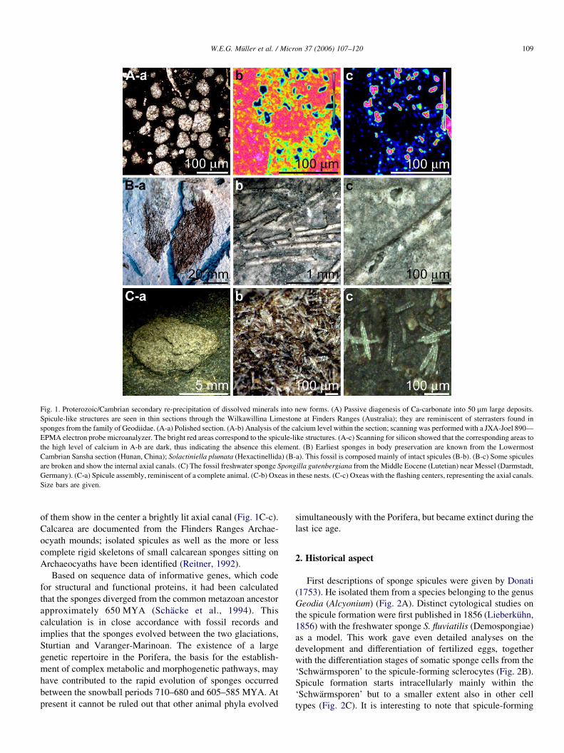
Fig. 1. Proterozoic/Cambrian secondary re-precipitation of dissolved minerals into new forms. (A) Passive diagenesis of Ca-carbonate into 50 mm large deposits.
Spicule-like structures are seen in thin sections through the Wilkawillina Limestone at Finders Ranges (Australia); they are reminiscent of sterrasters found in
sponges from the family of Geodiidae. (A-a) Polished section. (A-b) Analysis of the calcium level within the section; scanning was performed with a JXA-Joel 890—
EPMA electron probe microanalyzer. The bright red areas correspond to the spicule-like structures. (A-c) Scanning for silicon showed that the corresponding areas to
the high level of calcium in A-b are dark, thus indicating the absence this element. (B) Earliest sponges in body preservation are known from the Lowermost
Cambrian Sansha section (Hunan, China); Solactiniella plumata (Hexactinellida) (B-a). This fossil is composed mainly of intact spicules (B-b). (B-c) Some spicules
are broken and show the internal axial canals. (C) The fossil freshwater sponge Spongilla gutenbergiana from the Middle Eocene (Lutetian) near Messel (Darmstadt,
Germany). (C-a) Spicule assembly, reminiscent of a complete animal. (C-b) Oxeas in these nests. (C-c) Oxeas with the flashing centers, representing the axial canals.
Size bars are given.
W.E.G. Muller et al. / Micron 37 (2006) 107–120 109
of them show in the center a brightly lit axial canal (Fig. 1C-c).
Calcarea are documented from the Flinders Ranges Archae-
ocyath mounds; isolated spicules as well as the more or less
complete rigid skeletons of small calcarean sponges sitting on
Archaeocyaths have been identified (Reitner, 1992).
Based on sequence data of informative genes, which code
for structural and functional proteins, it had been calculated
that the sponges diverged from the common metazoan ancestor
approximately 650 MYA (Schacke et al., 1994). This
calculation is in close accordance with fossil records and
implies that the sponges evolved between the two glaciations,
Sturtian and Varanger-Marinoan. The existence of a large
genetic repertoire in the Porifera, the basis for the establish-
ment of complex metabolic and morphogenetic pathways, may
have contributed to the rapid evolution of sponges occurred
between the snowball periods 710–680 and 605–585 MYA. At
present it cannot be ruled out that other animal phyla evolved
simultaneously with the Porifera, but became extinct during the
last ice age.
2. Historical aspect
First descriptions of sponge spicules were given by Donati
(1753). He isolated them from a species belonging to the genus
Geodia (Alcyonium) (Fig. 2A). Distinct cytological studies on
the spicule formation were first published in 1856 (Lieberkuhn,
1856) with the freshwater sponge S. fluviatilis (Demospongiae)
as a model. This work gave even detailed analyses on the
development and differentiation of fertilized eggs, together
with the differentiation stages of somatic sponge cells from the
‘Schwarmsporen’ to the spicule-forming sclerocytes (Fig. 2B).
Spicule formation starts intracellularly mainly within the
‘Schwarmsporen’ but to a smaller extent also in other cell
types (Fig. 2C). It is interesting to note that spicule-forming
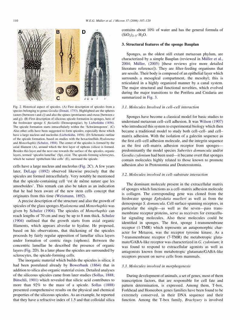
Fig. 2. Historical aspect of spicules. (A) First description of spicules from a
species belonging to genus Geodia (Donati, 1753). Highlighted are the spheres
(asters [between t and c]) and also the spines (protriaenes and oxeas [between c
and g]). (B) First description of siliceous spicule formation in sponges, here of
the freshwater sponge S. fluviatilis (Demospongiae), by Lieberkuhn (1856).
The spicule formation starts intracellularly within the ‘Schwarmsporen’. (C)
Also other cells have been suggested to form spicules; especially those which
have a large nucleus and nucleolus (Lieberkuhn, 1856). (D) Schematic outline
of the spicule formation, based on studies with the hexactinellids Hyalonema
and Monorhaphis (Schulze, 1904). The center of the spicules is formed by the
axial filament (A), around which the first layer of siphons (silica) is formed.
Besides this layer and the next one towards the surface of the spicules, organic
layers, termed ‘spiculin lamellae’ (Sp), exist. The spicule-forming sclerocytes,
which he named ‘epithelium like cells’ (E), surround the spicule.
W.E.G. Muller et al. / Micron 37 (2006) 107–120110
cells have a large nucleus and nucleolus (Fig. 2C). A few years
later, DeLage (1892) observed likewise precisely that the
spicules are formed intracellularly. Very notably he mentioned
that the spicule-containing cell ‘est de meme nature que les
amoeboıdes’. This remark can also be taken as an indication
that he had been aware of the new stem cells concept that
originates from this time (Weismann, 1892).
A precise description of the structure and also the growth of
spicules of the glass sponges Hyalonema and Monorhaphis was
given by Schulze (1904). The spicules of Monorhaphis can
reach lengths of 70 cm and may be up to 8 mm thick. Schulze
(1904) outlined that the growth starts from axial organic
filaments, which appears alveolar to hyaline. He proposed,
based on his observations, that thickening of the spicules
proceeds by fairly regular apposition of lamellar silica layers
under formation of centric rings (siphons). Between the
concentric lamellae he described the presence of organic
layers (Fig. 2D). In a later phase the spicules are surrounded by
sclerocytes, the spicule-forming cells.
The inorganic material which builds the spicules is silica; it
had been postulated already by Bowerbank (1864) that in
addition to silica also organic material exists. Detailed analyses
of the siliceous spicules came from later studies (Sollas, 1888;
Butschli, 1901) which revealed that silicic acid contributes to
more than 92% to the mass of a spicule. Sollas (1888)
presented comprehensive results on the physical and chemical
properties of the siliceous spicules. As an example, he reported
that they have a refractive index of 1.5 and that colloidal silica
contains about 10% of water and has the general formula of
(SiO2)2–5$H2O.
3. Structural features of the sponge Bauplan
Sponges, as the oldest still extant metazoan phylum, are
characterized by a simple Bauplan (reviewed in Muller et al.,
2004; Muller, (2005) [these reviews give more detailed
literature reference]). They are filter-feeding organisms that
are sessile. Their body is composed of an epithelial layer which
surrounds a mesogleal compartment, the mesohyl; this is
reticulated in a highly organized manner by a canal system.
The major structural and functional novelties, which evolved
during the major transitions to the Porifera and Cnidaria are
summarized in Fig. 3.
3.1. Molecules Involved in cell–cell interaction
Sponges have become a classical model for basic studies to
understand metazoan cell–cell adhesion. It was Wilson (1907)
who introduced this system to experimental biology which then
became a traditional model to study both cell–cell- and cell–
matrix adhesion. With the isolation of a galectin sequence as
the first cell–cell adhesion molecule, and the integrin sequence
as the first cell–matrix adhesion receptor from sponges—
predominantly the model species Suberites domuncula and/or
Geodia cydonium had been used—it became overt that sponges
contain molecules highly related to those known to promote
adhesion also in Protostomia and Deuterostomia.
3.2. Molecules involved in cell–substrate interaction
The dominant molecule present in the extracellular matrix
of sponges which functions as a cell–matrix adhesion molecule
is collagen. The corresponding gene was cloned from the
freshwater sponge Ephydatia muelleri as well as from the
demosponge S. domuncula. Cell surface-spanning receptors, in
particular the single—as well as the seven—pass trans-
membrane receptor proteins, serve as receivers for extracellu-
lar signaling molecules. Also these molecules could be
identified in sponges. The first, sponge 1-transmembrane
receptor (1-TMR) which represents an autapomorphic char-
acter for Metazoa, was the receptor tyrosine kinase. As a
7-transmembrane receptor (7-TMR) the metabotropic gluta-
mate/GABA-like receptor was characterized in G. cydonium; it
was found to respond to extracellular agonists as well as
antagonists known from metabotropic glutamate/GABA-like
receptors present on nerve cells from mammals.
3.3. Molecules involved in morphogenesis
During development of animals, a set of genes, most of them
transcription factors, that are responsible for cell fate and
pattern determination, is expressed. Among them, T-box,
Forkhead and Homeobox genes families have been found to be
extremely conserved, in their DNA sequence and their
function. Among the T-box family, Brachyury is involved
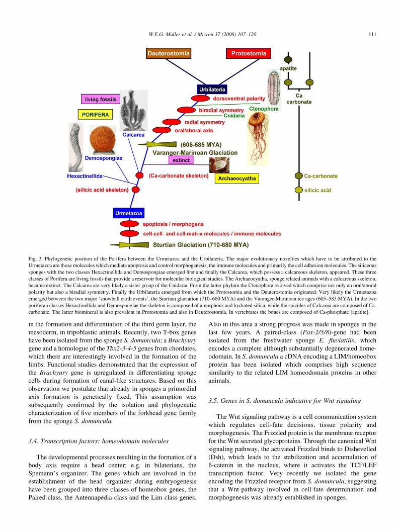
Fig. 3. Phylogenetic position of the Porifera between the Urmetazoa and the Urbilateria. The major evolutionary novelties which have to be attributed to the
Urmetazoa are those molecules which mediate apoptosis and control morphogenesis, the immune molecules and primarily the cell adhesion molecules. The siliceous
sponges with the two classes Hexactinellida and Demospongiae emerged first and finally the Calcarea, which possess a calcareous skeleton, appeared. These three
classes of Porifera are living fossils that provide a reservoir for molecular biological studies. The Archaeocyatha, sponge related animals with a calcareous skeleton,
became extinct. The Calcarea are very likely a sister group of the Cnidaria. From the latter phylum the Ctenophora evolved which comprise not only an oral/aboral
polarity but also a biradial symmetry. Finally the Urbilateria emerged from which the Protostomia and the Deuterostomia originated. Very likely the Urmetazoa
emerged between the two major ‘snowball earth events’, the Sturtian glaciation (710–680 MYA) and the Varanger-Marinoan ice ages (605–585 MYA). In the two
poriferan classes Hexactinellida and Demospongiae the skeleton is composed of amorphous and hydrated silica, while the spicules of Calcarea are composed of Ca-
carbonate. The latter biomineral is also prevalent in Protostomia and also in Deuterostomia. In vertebrates the bones are composed of Ca-phosphate [apatite].
W.E.G. Muller et al. / Micron 37 (2006) 107–120 111
in the formation and differentiation of the third germ layer, the
mesoderm, in tripoblastic animals. Recently, two T-box genes
have been isolated from the sponge S. domuncula; a Brachyury
gene and a homologue of the Tbx2-3-4-5 genes from chordates,
which there are interestingly involved in the formation of the
limbs. Functional studies demonstrated that the expression of
the Brachyury gene is upregulated in differentiating sponge
cells during formation of canal-like structures. Based on this
observation we postulate that already in sponges a primordial
axis formation is genetically fixed. This assumption was
subsequently confirmed by the isolation and phylogenetic
characterization of five members of the forkhead gene family
from the sponge S. domuncula.
3.4. Transcription factors: homeodomain molecules
The developmental processes resulting in the formation of a
body axis require a head center; e.g. in bilaterians, the
Spemann’s organizer. The genes which are involved in the
establishment of the head organizer during embryogenesis
have been grouped into three classes of homeobox genes, the
Paired-class, the Antennapedia-class and the Lim-class genes.
Also in this area a strong progress was made in sponges in the
last few years. A paired-class (Pax-2/5/8)-gene had been
isolated from the freshwater sponge E. fluviatilis, which
encodes a complete although substantially degenerated home-
odomain. In S. domuncula a cDNA encoding a LIM/homeobox
protein has been isolated which comprises high sequence
similarity to the related LIM homeodomain proteins in other
animals.
3.5. Genes in S. domuncula indicative for Wnt signaling
The Wnt signaling pathway is a cell communication system
which regulates cell-fate decisions, tissue polarity and
morphogenesis. The Frizzled protein is the membrane receptor
for the Wnt secreted glycoproteins. Through the canonical Wnt
signaling pathway, the activated Frizzled binds to Dishevelled
(Dsh), which leads to the stabilization and accumulation of
ß-catenin in the nucleus, where it activates the TCF/LEF
transcription factor. Very recently we isolated the gene
encoding the Frizzled receptor from S. domuncula, suggesting
that a Wnt-pathway involved in cell-fate determination and
morphogenesis was already established in sponges.
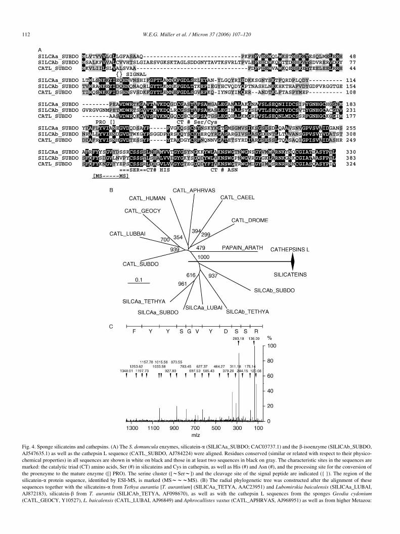
Fig. 4. Sponge silicateins and cathepsins. (A) The S. domuncula enzymes, silicatein-a (SILICAa_SUBDO; CAC03737.1) and the b-isoenzyme (SILICAb_SUBDO,
AJ547635.1) as well as the cathepsin L sequence (CATL_SUBDO, AJ784224) were aligned. Residues conserved (similar or related with respect to their physico-
chemical properties) in all sequences are shown in white on black and those in at least two sequences in black on gray. The characteristic sites in the sequences are
marked: the catalytic triad (CT) amino acids, Ser (#) in silicateins and Cys in cathepsin, as well as His (#) and Asn (#), and the processing site for the conversion of
the proenzyme to the mature enzyme ([] PRO). The serine cluster ([wSerw]) and the cleavage site of the signal peptide are indicated ({ }). The region of the
silicatein-a protein sequence, identified by ESI-MS, is marked (MSwwwMS). (B) The radial phylogenetic tree was constructed after the alignment of these
sequences together with the silicateins-a from Tethya aurantia [T. aurantium] (SILICAa_TETYA, AAC23951) and Lubomirskia baicalensis (SILICAa_LUBAI,
AJ872183), silicatein-b from T. aurantia (SILICAb_TETYA, AF098670), as well as with the cathepsin L sequences from the sponges Geodia cydonium
(CATL_GEOCY, Y10527), L. baicalensis (CATL_LUBAI, AJ96849) and Aphrocallistes vastus (CATL_APHRVAS, AJ968951) as well as from higher Metazoa:
W.E.G. Muller et al. / Micron 37 (2006) 107–120112
3
W.E.G. Muller et al. / Micron 37 (2006) 107–120 113
3.6. Molecules present in tight junctions
Our screening for a gene encoding a tight junction scaffold
protein from a sponge, here S. domuncula, was successful; the
scaffold protein membrane-associated guanylate kinase with
inverted arrangement (MAGI) had been identified. In addition,
the existence of one tetraspan receptor, tetraspanin, in S.
domuncula has been reported. The tetraspanins belong to a
group of hydrophobic proteins, comprising four trans-
membrane domains with a series of conserved aa residues in
the extracellular loops.
Besides these mentioned genomic regulatory systems,
primarily those structural and enzymatic molecules which
govern the skeletal formation especially in the phylum Porifera
are crucial for the establishment of the body plan (Bauplan).
Therefore, an understanding of the spicule formation and the
association of spicules within the sponge body is crucial for an
understanding of the sponge body plan. In the class of
Demospongiae, spicule formation is a dynamic process and
involves both anabolic and the catabolic enzymes.
pHkDa
A B
25
31
76
1345 345 122
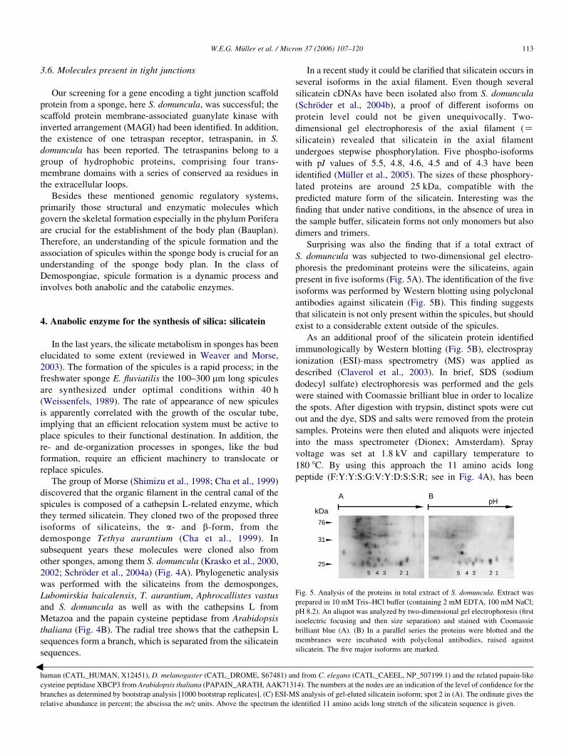
Fig. 5. Analysis of the proteins in total extract of S. domuncula. Extract was
prepared in 10 mM Tris–HCl buffer (containing 2 mM EDTA, 100 mM NaCl;
pH 8.2). An aliquot was analyzed by two-dimensional gel electrophoresis (first
isoelectric focusing and then size separation) and stained with Coomassie
brilliant blue (A). (B) In a parallel series the proteins were blotted and the
membranes were incubated with polyclonal antibodies, raised against
silicatein. The five major isoforms are marked.
4. Anabolic enzyme for the synthesis of silica: silicatein
In the last years, the silicate metabolism in sponges has been
elucidated to some extent (reviewed in Weaver and Morse,
2003). The formation of the spicules is a rapid process; in the
freshwater sponge E. fluviatilis the 100–300 mm long spicules
are synthesized under optimal conditions within 40 h
(Weissenfels, 1989). The rate of appearance of new spicules
is apparently correlated with the growth of the oscular tube,
implying that an efficient relocation system must be active to
place spicules to their functional destination. In addition, the
re- and de-organization processes in sponges, like the bud
formation, require an efficient machinery to translocate or
replace spicules.
The group of Morse (Shimizu et al., 1998; Cha et al., 1999)
discovered that the organic filament in the central canal of the
spicules is composed of a cathepsin L-related enzyme, which
they termed silicatein. They cloned two of the proposed three
isoforms of silicateins, the a- and b-form, from the
demosponge Tethya aurantium (Cha et al., 1999). In
subsequent years these molecules were cloned also from
other sponges, among them S. domuncula (Krasko et al., 2000,
2002; Schroder et al., 2004a) (Fig. 4A). Phylogenetic analysis
was performed with the silicateins from the demosponges,
Lubomirskia baicalensis, T. aurantium, Aphrocallistes vastus
and S. domuncula as well as with the cathepsins L from
Metazoa and the papain cysteine peptidase from Arabidopsis
thaliana (Fig. 4B). The radial tree shows that the cathepsin L
sequences form a branch, which is separated from the silicatein
sequences.
human (CATL_HUMAN, X12451), D. melanogaster (CATL_DROME, S67481) an
cysteine peptidase XBCP3 from Arabidopsis thaliana (PAPAIN_ARATH, AAK713
branches as determined by bootstrap analysis [1000 bootstrap replicates]. (C) ESI-M
relative abundance in percent; the abscissa the m/z units. Above the spectrum the i
In a recent study it could be clarified that silicatein occurs in
several isoforms in the axial filament. Even though several
silicatein cDNAs have been isolated also from S. domuncula
(Schroder et al., 2004b), a proof of different isoforms on
protein level could not be given unequivocally. Two-
dimensional gel electrophoresis of the axial filament (Zsilicatein) revealed that silicatein in the axial filament
undergoes stepwise phosphorylation. Five phospho-isoforms
with pI values of 5.5, 4.8, 4.6, 4.5 and of 4.3 have been
identified (Muller et al., 2005). The sizes of these phosphory-
lated proteins are around 25 kDa, compatible with the
predicted mature form of the silicatein. Interesting was the
finding that under native conditions, in the absence of urea in
the sample buffer, silicatein forms not only monomers but also
dimers and trimers.
Surprising was also the finding that if a total extract of
S. domuncula was subjected to two-dimensional gel electro-
phoresis the predominant proteins were the silicateins, again
present in five isoforms (Fig. 5A). The identification of the five
isoforms was performed by Western blotting using polyclonal
antibodies against silicatein (Fig. 5B). This finding suggests
that silicatein is not only present within the spicules, but should
exist to a considerable extent outside of the spicules.
As an additional proof of the silicatein protein identified
immunologically by Western blotting (Fig. 5B), electrospray
ionization (ESI)-mass spectrometry (MS) was applied as
described (Claverol et al., 2003). In brief, SDS (sodium
dodecyl sulfate) electrophoresis was performed and the gels
were stained with Coomassie brilliant blue in order to localize
the spots. After digestion with trypsin, distinct spots were cut
out and the dye, SDS and salts were removed from the protein
samples. Proteins were then eluted and aliquots were injected
into the mass spectrometer (Dionex; Amsterdam). Spray
voltage was set at 1.8 kV and capillary temperature to
180 8C. By using this approach the 11 amino acids long
peptide (F:Y:Y:S:G:V:Y:D:S:S:R; see in Fig. 4A), has been
d from C. elegans (CATL_CAEEL, NP_507199.1) and the related papain-like
14). The numbers at the nodes are an indication of the level of confidence for the
S analysis of gel-eluted silicatein isoform; spot 2 in (A). The ordinate gives the
dentified 11 amino acids long stretch of the silicatein sequence is given.
W.E.G. Muller et al. / Micron 37 (2006) 107–120114
identified (Fig. 4C). This region in the silicatein protein is
especially interesting, since there the serine-rich region starts.
5. Catabolic enzyme: silicase
In the course to further elucidate the metabolism of siliceous
spicules in Demospongiae another enzyme, silicase, was
identified from the marine sponge S. domuncula; silicase is
able to depolymerize amorphous silica. The cDNA was
isolated and the deduced polypeptide identified as an enzyme
similar to carbonic anhydrases. Recombinant silicase displays
besides a carbonic anhydrase activity the ability to dissolve
amorphous silica under formation of free silicic acid (Schroder
et al., 2003).
6. Morphology of spicules in Suberites domuncula
The marine demosponge S. domuncula was collected in the
Northern Adriatic near Rovinj (Croatia). From this species
tissue samples, primmorphs and spicules were obtained
(summarized in Muller et al., 2005). Primmorphs, a special
type of 3D-cell aggregates, containing proliferating and
differentiating cells, allow during incubation in medium
supplemented with silicic acid to study the differentiation of
archaeocytes to sclerocytes (see Muller et al., 2004). Spicules
and their filaments were prepared from tissue samples that were
treated first with sulfuric acid/nitric acid and then with
n-butanol/water/SDS. For immunofluorescence studies the
spicules were obtained from the tissue by treatment with
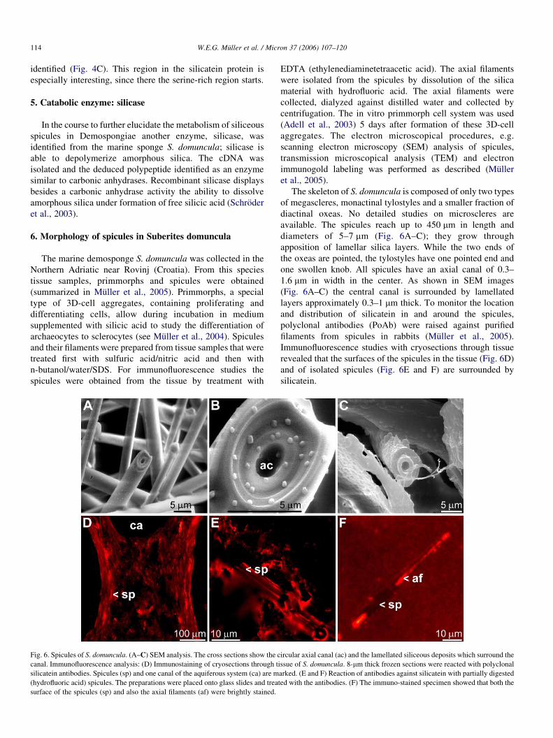
Fig. 6. Spicules of S. domuncula. (A–C) SEM analysis. The cross sections show the c
canal. Immunofluorescence analysis: (D) Immunostaining of cryosections through t
silicatein antibodies. Spicules (sp) and one canal of the aquiferous system (ca) are m
(hydrofluoric acid) spicules. The preparations were placed onto glass slides and trea
surface of the spicules (sp) and also the axial filaments (af) were brightly stained.
EDTA (ethylenediaminetetraacetic acid). The axial filaments
were isolated from the spicules by dissolution of the silica
material with hydrofluoric acid. The axial filaments were
collected, dialyzed against distilled water and collected by
centrifugation. The in vitro primmorph cell system was used
(Adell et al., 2003) 5 days after formation of these 3D-cell
aggregates. The electron microscopical procedures, e.g.
scanning electron microscopy (SEM) analysis of spicules,
transmission microscopical analysis (TEM) and electron
immunogold labeling was performed as described (Muller
et al., 2005).
The skeleton of S. domuncula is composed of only two types
of megascleres, monactinal tylostyles and a smaller fraction of
diactinal oxeas. No detailed studies on microscleres are
available. The spicules reach up to 450 mm in length and
diameters of 5–7 mm (Fig. 6A–C); they grow through
apposition of lamellar silica layers. While the two ends of
the oxeas are pointed, the tylostyles have one pointed end and
one swollen knob. All spicules have an axial canal of 0.3–
1.6 mm in width in the center. As shown in SEM images
(Fig. 6A–C) the central canal is surrounded by lamellated
layers approximately 0.3–1 mm thick. To monitor the location
and distribution of silicatein in and around the spicules,
polyclonal antibodies (PoAb) were raised against purified
filaments from spicules in rabbits (Muller et al., 2005).
Immunofluorescence studies with cryosections through tissue
revealed that the surfaces of the spicules in the tissue (Fig. 6D)
and of isolated spicules (Fig. 6E and F) are surrounded by
silicatein.
ircular axial canal (ac) and the lamellated siliceous deposits which surround the
issue of S. domuncula. 8-mm thick frozen sections were reacted with polyclonal
arked. (E and F) Reaction of antibodies against silicatein with partially digested
ted with the antibodies. (F) The immuno-stained specimen showed that both the
W.E.G. Muller et al. / Micron 37 (2006) 107–120 115
From these studies we conclude that silicatein is not only
present in the axial filament of the spicules, but is also located
on their surface.
7. Development of spicules in primmorphs
In S. domuncula, the formation of spicules is a rapid process
and surely proceeds more frequently in embryos or in
primmorphs, than in adult specimens. Since all studies on the
formation of spicules had hitherto been performed with
sections through tissue samples from adult animals the
conclusion was published that the formation of the megascleres
proceeds extracellularly in the bulky mesohyl of the animals
(Uriz et al., 2000; Weaver and Morse, 2003). We applied the
primmorph system and could establish unequivocally that
the initial steps of spicule formation occur intracellularly in the
sclerocytes (Muller et al., 2005).
Sclerocytes in primmorphs produce after an cultivation
period of 5 days readily spicules, provided the medium
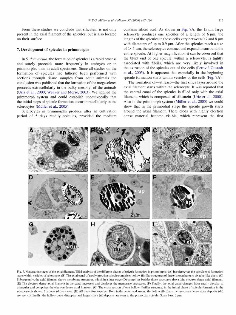
Fig. 7. Maturation stages of the axial filament; TEM analysis of the different phases o
starts within vesicles of sclerocyte. (B) The axial canal of newly growing spicule com
Subsequently, the axial filament shows membrane structures, which in a later stage (
(E) The electron dense axial filament in the canal increases and displaces the mem
triangular and comprises the electron dense axial filament. (G) The cross section o
sclerocyte, is shown. Six ducts (du) are seen. (H) All ducts fuse together. Both in the
are see. (I) Finally, the hollow ducts disappear and larger silica (si) deposits are se
contains silicic acid. As shown in Fig. 7A, the 15 mm large
sclerocyte produces one spicules of a length of 6 mm; the
lengths of the spicules in those cells vary between 0.7 and 8 mm
with diameters of up to 0.9 mm. After the spicules reach a size
of O 5 mm, the sclerocytes contract and expand to surround the
entire spicule. At higher magnification it can be observed that
the blunt end of one spicule, within a sclerocyte, is tightly
associated with fibrils, which are very likely involved in
the extrusion of the spicules out of the cells (Perovic-Ottstadt
et al., 2005). It is apparent that especially in the beginning
spicule formation starts within vesicles of the cells (Fig. 7A).
The formation of—at least—the first silica layer around the
axial filament starts within the sclerocyte. It was reported that
the central canal of the spicules is filled only with the axial
filament, which is composed of silicatein (Uriz et al., 2000).
Also in the primmorph system (Muller et al., 2005) we could
show that in the primordial stage the spicule growth starts
around the axial filament. There clods with highly electron
dense material become visible, which represent the first
f spicule formation in primmorphs. (A) In sclerocytes the spicule (sp) formation
prises hollow fibrillar structures of three (shown here) to six tube-like ducts. (C)
D) comprises besides those structures also a thin, electron dense axial filament.
brane structures. (F) Finally, the axial canal changes from nearly circular to
f one hollow fibrillar structure, in the initial phase of spicule formation in the
center and around the hollow fibrillar structures, very dense silica deposits (de)
en in the primordial spicule. Scale bars: 2 mm.
W.E.G. Muller et al. / Micron 37 (2006) 107–120116
deposits of spicules. During progression of growth in the
extracellular space, the diameter of the axial filament decreases
from 1.5 to 0.8 mm while simultaneously the size of the spicule
increases up to 450 mm and a diameter of 5 mm. In this phase
the spicules are found extracellularly (Fig. 7B). In the next
growth stage, two types of structures can be distinguished
within the axial canal. First, the 1.6 mm thick axial canal is
filled primarily with membrane structures (Fig. 7B) or a
comparably thin axial filament is seen, besides the membrane
structures (Fig. 7C–E). The 0.8–0.2 mm axial filament has the
same electron density as in the initial stage of spicule
formation. Among the cellular structures are many 10–15 nm
round fibrils of not yet defined nature. The axial filament in the
canal increases again in size and becomes more electron dense
(Fig. 7E). In the final stage the axial canal changes from nearly
circular to triangular (Fig. 7F). Interesting are the hollow
fibrillar structures which are seen in sections through spicules
in the transition phase (Fig. 7B) from those which comprise
complete membrane structures in the axial canal (Fig. 7C) and
spicules which show in the canal a more compact axial filament
(Fig. 7F). In the transition phase (Fig. 7B), the hollow fibrillar
structure fills almost the complete space in the canal.
These hollow fibrillar structures, newly described here,
show six tube-like ducts, which are initially regularly arranged
around a central filled canal of spicules inside the cells
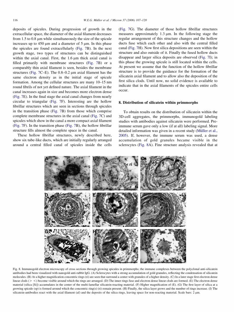
Fig. 8. Immunogold electron microscopy of cross sections through growing spicule
antibodies had been visualized with nanogold anti-rabbit IgG. (A) Sclerocytes with
molecules. (B) At a higher magnification concentric rings (ri) are seen that surround
linear clods (O !) become visible around which the rings are arranged. (D) The inn
material (silica [Si]) accumulates in the center of the multi-lamellar silicatein-reac
growing spicule (sp) is formed around which the concentric ring(s) (ri) remain prese
silicatein-antibodies react with the axial filament (af) and the deposits of the silica
(Fig. 7G). The diameter of those hollow fibrillar structures
measures approximately 1.3 mm. In the following stage the
regular arrangement of this structure changes and the hollow
ducts fuse which each other and also with the central filled
canal (Fig. 7H). Now first silica depositions are seen within the
structure and also outside of it. Finally the fused hollow ducts
disappear and larger silica deposits are observed (Fig. 7I); in
this phase the growing spicule is still located within the cells.
At present we assume that the function of the hollow fibrillar
structure is to provide the guidance for the formation of the
silicatein axial filament and to allow also the deposition of the
first silica clods. Until now, no solid evidence is available to
indicate that in the axial filaments of the spicules entire cells
occur.
8. Distribution of silicatein within primmorphs
To obtain results on the distribution of silicatein within the
3D-cell aggregates, the primmorphs, immunogold labeling
studies with antibodies against silicatein were performed. Pre-
immune serum gave only a low (if at all) labeling signal. More
detailed information was given in a recent study (Muller et al.,
2005). If, however, the immune serum was used, a dense
accumulation of gold granules became visible in the
sclerocytes (Fig. 8A). Fine structure analysis revealed that at
s in primmorphs; the immune complexes between the polyclonal anti-silicatein
a strong accumulation of gold granules, reflecting the condensation of silicatein
a center with granules of a higher density. (C) In a later stage first electron dense
er rings fuse and electron dense linear clods are formed. (E) The electron dense
ting material. (F) Higher magnification of (E). (G) The first layer of silica at a
nt. (H) Finally, the silica layer grows and the number of rings increase. (I) The
rings, leaving space for non-reacting material. Scale bars: 2 mm.
W.E.G. Muller et al. / Micron 37 (2006) 107–120 117
first concentric rings are seen around the forming spicules
which are 0.2–0.5 mm apart (Fig. 8B). Subsequently, the inner
rings fuse and electron dense linear clods become visible
(Fig. 8C). Later, during maturation, the number of the
concentric rings increases from two to three rings with a
diameter of 2–3 mm (Fig. 8B) to 10 rings and a total diameter
4–6 mm (Fig. 8D). At higher magnification the clods appear
round-shaped (Fig. 8E and F). Finally, the first layer of silica is
formed, showing again around these silica structures con-
centric rings of gold particles, reflecting silicatein antibodies
(Fig. 8G). The number of gold/antibody rings around the
primordial spicules, composed of a first silica layer, is larger
than three (Fig. 8H). Under the experimental conditions used
here, also the axial filament (Z silicatein) reacts with the
antibodies (Fig. 8H and I). It should be mentioned that around
the axial filament and the first layer of the silica deposition, a
space filled with non-reacting material is seen (Fig. 8I).
Probably there the substrate for the silicatein is stored.
9. Formation of the morphology of the spicules
As recently highlighted, biomineralization is the process
by which Metazoa form hard minerals for support, defense,
and feeding (see Wilt, 2005). In sponges, the major
structural elements which contribute to morphogenesis are
the spicules. Until now, no experimental data on the
underlying cellular mechanism(s), or even developmental
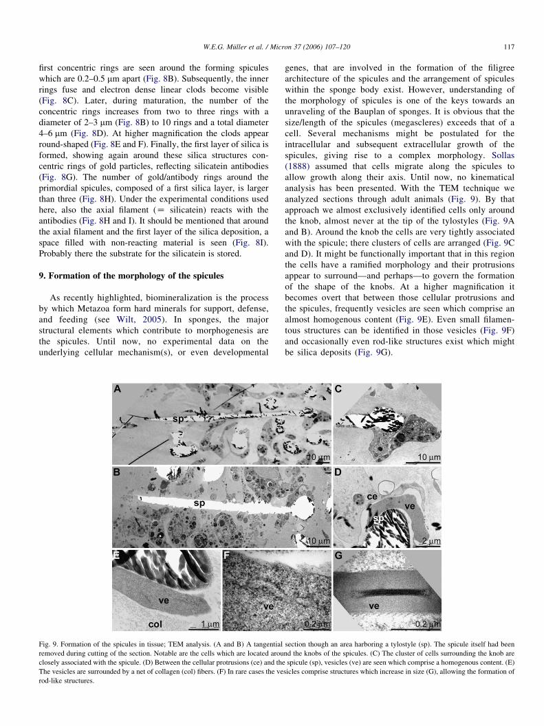
Fig. 9. Formation of the spicules in tissue; TEM analysis. (A and B) A tangential
removed during cutting of the section. Notable are the cells which are located arou
closely associated with the spicule. (D) Between the cellular protrusions (ce) and the
The vesicles are surrounded by a net of collagen (col) fibers. (F) In rare cases the ve
rod-like structures.
genes, that are involved in the formation of the filigree
architecture of the spicules and the arrangement of spicules
within the sponge body exist. However, understanding of
the morphology of spicules is one of the keys towards an
unraveling of the Bauplan of sponges. It is obvious that the
size/length of the spicules (megascleres) exceeds that of a
cell. Several mechanisms might be postulated for the
intracellular and subsequent extracellular growth of the
spicules, giving rise to a complex morphology. Sollas
(1888) assumed that cells migrate along the spicules to
allow growth along their axis. Until now, no kinematical
analysis has been presented. With the TEM technique we
analyzed sections through adult animals (Fig. 9). By that
approach we almost exclusively identified cells only around
the knob, almost never at the tip of the tylostyles (Fig. 9A
and B). Around the knob the cells are very tightly associated
with the spicule; there clusters of cells are arranged (Fig. 9C
and D). It might be functionally important that in this region
the cells have a ramified morphology and their protrusions
appear to surround—and perhaps—to govern the formation
of the shape of the knobs. At a higher magnification it
becomes overt that between those cellular protrusions and
the spicules, frequently vesicles are seen which comprise an
almost homogenous content (Fig. 9E). Even small filamen-
tous structures can be identified in those vesicles (Fig. 9F)
and occasionally even rod-like structures exist which might
be silica deposits (Fig. 9G).
section though an area harboring a tylostyle (sp). The spicule itself had been
nd the knobs of the spicules. (C) The cluster of cells surrounding the knob are
spicule (sp), vesicles (ve) are seen which comprise a homogenous content. (E)
sicles comprise structures which increase in size (G), allowing the formation of
W.E.G. Muller et al. / Micron 37 (2006) 107–120118
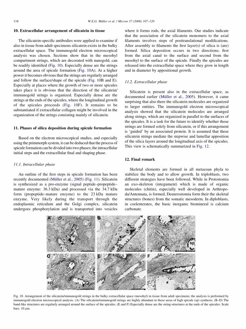
10. Extracellular arrangement of silicatein in tissue
The silicatein-specific antibodies were applied to examine if
also in tissue from adult specimens silicatein exists in the bulky
extracellular space. The immunogold electron microscopical
analysis was chosen. Sections show that in the mesohyl
compartment strings, which are decorated with nanogold, can
be readily identified (Fig. 10). Especially dense are the strings
around the area of spicule formation (Fig. 10A). At a higher
power it becomes obvious that the strings are regularly arranged
and follow the surface/shape of the spicule (Fig. 10B and E).
Especially at places where the growth of two or more spicules
takes place it is obvious that the direction of the silicatein/
immunogold strings is organized. Especially dense are the
strings at the ends of the spicules, where the longitudinal growth
of the spicules proceeds (Fig. 10F). It remains to be
substantiated if extracellular collagen might be involved in the
organization of the strings consisting mainly of silicatein.
11. Phases of silica deposition during spicule formation
Based on the electron microscopical studies, and especially
using the primmorph system, it can be deduced that the process of
spicule formation can be divided into two phases; the intracellular
initial steps and the extracellular final and shaping phase.
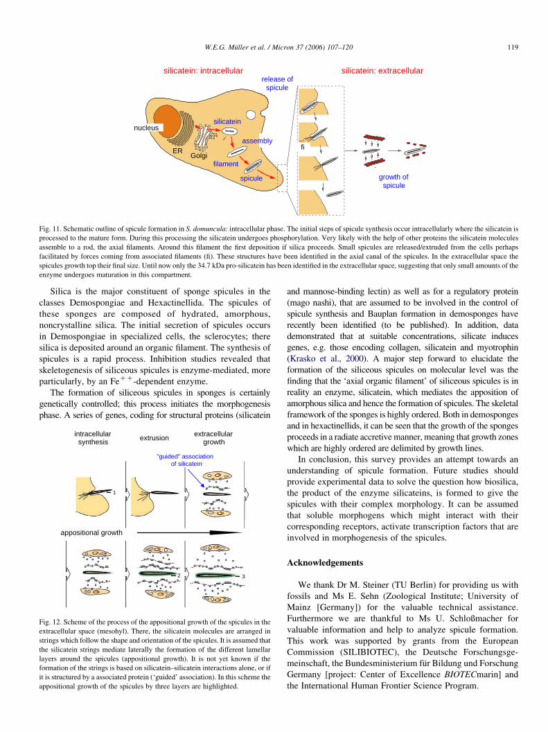
11.1. Intracellular phase
An outline of the first steps in spicule formation has been
recently documented (Muller et al., 2005) (Fig. 11). Silicatein
is synthesized as a pro-enzyme (signal peptide–propeptide–
mature enzyme: 36.3 kDa) and processed via the 34.7 kDa
form (propeptide–mature enzyme) to the 23 kDa mature
enzyme. Very likely during the transport through the
endoplasmic reticulum and the Golgi complex, silicatein
undergoes phosphorylation and is transported into vesicles
Fig. 10. Arrangement of the silicatein/immunogold strings in the bulky extracellula
immunogold electron microscopical analysis. (A) The silicatein/immunogold string
band-like structures are regularly arranged around the surface of the spicules. (E an
bars: 10 mm.
where it forms rods, the axial filaments. Our studies indicate
that the association of the silicatein monomers to the axial
filaments involves steps of posttranslational modifications.
After assembly to filaments the first layer(s) of silica is (are)
formed. Silica deposition occurs in two directions; first
from the axial canal to the surface and second from the
mesohyl to the surface of the spicule. Finally the spicules are
released into the extracellular space where they grow in length
and in diameter by appositional growth.
11.2. Extracellular phase
Silicatein is present also in the extracellular space, as
documented earlier (Muller et al., 2005). However, it came
surprising that also there the silicatein molecules are organized
to larger entities. The immunogold electron microscopical
analysis showed that the silicatein molecules are arranged
along strings, which are organized in parallel to the surfaces of
the spicules. It is a task for the future to identify whether those
strings are formed solely from silicatein, or if this arrangement
is ‘guided’ by an associated protein. It is assumed that these
silicatein strings mediate the stepwise and lamellar apposition
of the silica layers around the longitudinal axis of the spicules.
This view is schematically summarized in Fig. 12.
12. Final remark
Skeletal elements are formed in all metazoan phyla to
stabilize the body and to allow growth. In triploblasts, two
different strategies have been followed. While in Protostomia
an exo-skeleton (integument) which is made of organic
molecules (chitin), especially well developed in Arthropo-
da/Antennata, is formed, Deuterostomia form their the skeletal
structures (bones) from the somatic mesoderm. In diploblasts,
in coelenterates, the basic inorganic biomineral is calcium
carbonate.
r space (mesohyl) in tissue from adult specimens; the analysis is performed by
s are highly abundant in those areas of high spicule (sp) synthesis. (B–D) The
d F) Especially dense are the string-structures at the ends of the spicules. Scale
nucleus
GolgiER
silicatein
filament
spicule
release ofspicule
fi
growth ofspicule
assembly
silicatein: extracellularsilicatein: intracellular
Fig. 11. Schematic outline of spicule formation in S. domuncula: intracellular phase. The initial steps of spicule synthesis occur intracellularly where the silicatein is
processed to the mature form. During this processing the silicatein undergoes phosphorylation. Very likely with the help of other proteins the silicatein molecules
assemble to a rod, the axial filaments. Around this filament the first deposition if silica proceeds. Small spicules are released/extruded from the cells perhaps
facilitated by forces coming from associated filaments (fi). These structures have been identified in the axial canal of the spicules. In the extracellular space the
spicules growth top their final size. Until now only the 34.7 kDa pro-silicatein has been identified in the extracellular space, suggesting that only small amounts of the
enzyme undergoes maturation in this compartment.
W.E.G. Muller et al. / Micron 37 (2006) 107–120 119
Silica is the major constituent of sponge spicules in the
classes Demospongiae and Hexactinellida. The spicules of
these sponges are composed of hydrated, amorphous,
noncrystalline silica. The initial secretion of spicules occurs
in Demospongiae in specialized cells, the sclerocytes; there
silica is deposited around an organic filament. The synthesis of
spicules is a rapid process. Inhibition studies revealed that
skeletogenesis of siliceous spicules is enzyme-mediated, more
particularly, by an FeCC-dependent enzyme.
The formation of siliceous spicules in sponges is certainly
genetically controlled; this process initiates the morphogenesis
phase. A series of genes, coding for structural proteins (silicatein
intracellularsynthesis extrusion extracellular
growth
appositional growth
"guided" associationof silicatein
32
1
Fig. 12. Scheme of the process of the appositional growth of the spicules in the
extracellular space (mesohyl). There, the silicatein molecules are arranged in
strings which follow the shape and orientation of the spicules. It is assumed that
the silicatein strings mediate laterally the formation of the different lamellar
layers around the spicules (appositional growth). It is not yet known if the
formation of the strings is based on silicatein–silicatein interactions alone, or if
it is structured by a associated protein (‘guided’ association). In this scheme the
appositional growth of the spicules by three layers are highlighted.
and mannose-binding lectin) as well as for a regulatory protein
(mago nashi), that are assumed to be involved in the control of
spicule synthesis and Bauplan formation in demosponges have
recently been identified (to be published). In addition, data
demonstrated that at suitable concentrations, silicate induces
genes, e.g. those encoding collagen, silicatein and myotrophin
(Krasko et al., 2000). A major step forward to elucidate the
formation of the siliceous spicules on molecular level was the
finding that the ‘axial organic filament’ of siliceous spicules is in
reality an enzyme, silicatein, which mediates the apposition of
amorphous silica and hence the formation of spicules. The skeletal
framework of the sponges is highly ordered. Both in demosponges
and in hexactinellids, it can be seen that the growth of the sponges
proceeds in a radiate accretive manner, meaning that growth zones
which are highly ordered are delimited by growth lines.
In conclusion, this survey provides an attempt towards an
understanding of spicule formation. Future studies should
provide experimental data to solve the question how biosilica,
the product of the enzyme silicateins, is formed to give the
spicules with their complex morphology. It can be assumed
that soluble morphogens which might interact with their
corresponding receptors, activate transcription factors that are
involved in morphogenesis of the spicules.
Acknowledgements
We thank Dr M. Steiner (TU Berlin) for providing us with
fossils and Ms E. Sehn (Zoological Institute; University of
Mainz [Germany]) for the valuable technical assistance.
Furthermore we are thankful to Ms U. Schloßmacher for
valuable information and help to analyze spicule formation.
This work was supported by grants from the European
Commission (SILIBIOTEC), the Deutsche Forschungsge-
meinschaft, the Bundesministerium fur Bildung und Forschung
Germany [project: Center of Excellence BIOTECmarin] and
the International Human Frontier Science Program.
W.E.G. Muller et al. / Micron 37 (2006) 107–120120
References
Adell, T., Grebenjuk, V.A., Wiens, M., Muller, W.E.G., 2003. Isolation
and characterization of two T-box genes from sponges, the
phylogenetically oldest metazoan taxon. Development, Genes &
Evolution 213, 421–434.
Arndt, W., 1937. Schwamme. In: Pax, F., Arndt, W. (Eds.), Die Rohstoffe des
Tierreiches. Georg Borntraeger, Berlin, pp. 1578–2000.
Bowerbank, J.S., 1864. A Monograph of the British Spongiadae. Ray Society,
London.
Brasier, M., Green, O., Shields, G., 1997. Ediacarian sponge spicule clusters
from southwest Mongolia and the origins of the Cambrian fauna. Geology
25, 303–306.
Butschli, O., 1901. Einige Beobachtungen uber Kiesel- und Kalknadeln von
Spongien. Zeitschrift wissenschaftliche Zoologie 64, 235–286.
Campbell, L.G., 1876. Log Letters from “The Challenger”. MacMillan,
London.
Camus, M., 1783. Histoire des Animaux d’ Aristote. Desaint, Paris.
Cha, J.N., Shimizu, K., Zhou, Y., Christianssen, S.C., Chmelka, B.F.,
Stucky, G.D., Morse, D.E., 1999. Silicatein filaments and subunits from
a marine sponge direct the polymerization of silica and silicones
in vitro. Proceedings of the National Academy of Sciences USA 96,
361–365.
Claverol, S., Burlet-Schiltz, O., Gairin, J.E., Monsarrat, B., 2003. Character-
ization of protein variants and post-translational modifications. ESI-MS
analyses of intact proteins eluted from polyacrylamide gels. Molecular &
Cellular Proteomics 2, 483–493.
DeLage, Y., 1892. Embryogenie des eponge. Archives De Zoologie
Experimental (ser 2) 10, 345–498.
Donati, V., 1753. Auszug einer Natur-Geschichte des Adriatischen Meers. CP
Franckens, Halle.
Grant, R.E., 1835. Porifera. In: Bailliere, H. (Ed.), Outlines of Comparative
Anatomy. London, pp. 1–656.
Haeckel, E., 1872. Die Kalkschwamme. Georg Reimer, Berlin.
Hoffman, P.F., Kaufman, A.J., Halverson, G.P., Schrag, D.P., 1998. A
neoproterozoic snowball earth. Science 281, 1342–1346.
Hooper, J.N.A., 1997. Sponge Guide. Queensland Museum.
Krasko, A., Batel, R., Schroder, H.C., Muller, I.M., Muller, W.E.G., 2000.
Expression of silicatein and collagen genes in the marine sponge Suberites
domuncula is controlled by silicate and myotrophin. European Journal of
Biochemistry 267, 4878–4887.
Krasko, A., Schroder, H.C., Batel, R., Grebenjuk, V.A., Steffen, R., Muller,
I.M., Muller, W.E.G., 2002. Iron induces proliferation and morphogenesis
in primmorphs from the marine sponge Suberites domuncula. DNA & Cell
Biology 21, 67–80.
Lieberkuhn, N., 1856. Zur Entwicklungsgeschichte der Spongillen. Archiv fur
Anatomie und Physiologie, 399–414.
Lieberkuhn, N., 1859. Neue Beitrage zur Anatomie der Spongien. Archiv fur
Anatomie und Physiologie, 515–529.
Muller, W.E.G., 2001. How was metazoan threshold crossed: the hypothetical
Urmetazoa. Comparative Biochemistry Physiology [A] 129, 433–460.
Muller, W.E.G., 2005. Origin of Metazoa: sponges as living fossils.
Naturwissenschaften 85, 11–25.
Muller, W.E.G., in press. Spatial and temporal expression patterns in animals.
In: Meyers RA (ed.) Encyclopedia of Molecular Cell Biology and
Molecular Medicine. Wiley VCH GmbH, Weinheim 13, 269–309.
Muller, W.E.G., Zahn, R.K., Maidhof, A., 1982. Spongilla gutenbergiana n.sp.,
ein Sußwasserschwamm aus dem Mittel-Eozan von Messel. Senckenbergi-
ana lethaea 63, 465–472.
Muller, W.E.G., Wiens, M., Adell, T., Gamulin, V., Schroder, H.C., Muller,
I.M., 2004. The Bauplan of the Urmetazoa: The basis of the genetic
complexity of Metazoa using the siliceous sponges [Porifera] as living
fossils. International Review of Cytology 235, 53–92.
Muller, W.E.G., Rothenberger, M., Boreiko, A., Tremel, W., Reiber, A.,
Schroder, H.C., 2005. Formation of siliceous spicules in the marine
demosponge Suberites domuncula. Cell & Tissue Research 321, 285–297.
Perovic-Ottstadt, S., Schroder, H.C., Batel, R., Giovine, M., Wiens, M.,
Krasko, A., Muller, I.M., Muller, W.E.G., 2005. Arginine kinase in the
demosponge Suberites domuncula: regulation of its expression and
catalytic activity by silicic acid. Journal of Experimental Biology 208,
637–646.
Pilcher, H., 2005. Back to our roots. Nature 435, 1022–1023.
Reitner, J., 1992. Coralline Spongien. Der Versuch einer phylogenetisch-
taxonomischen Analyse. Berliner Geowissenschaftliche Abhandlungen (E)
1, 1–352.
Schacke, H., Muller, I.M., Muller, W.E.G., 1994. Tyrosine kinase from the
marine sponge Geodia cydonium: the oldest member belonging to the
receptor tyrosine kinase class II family.. In: Muller, W.E.G. (Ed.), Use of
Aquatic Invertebrates as Tools for Monitoring of Environmental Hazards.
Gustav Fischer Verlag, Stuttgart, New York, pp. 201–211.
Schroder, H.C., Krasko, A., Le Pennec, G., Adell, T., Hassanein, H., Muller,
I.M., Muller, W.E.G., 2003. Silicase, an enzyme which degrades biogenous
amorphous silica: contribution to the metabolism of silica deposition in the
demosponge Suberites domuncula. Progress in Molecular and Subcellular
Biology 33, 249–268.
Schroder, H.C., Perovic-Ottstadt, S., Rothenberger, M., Wiens, M., Schwertner,
H., Batel, R., Korzhev, M., Muller, I.M., Muller, W.E.G., 2004a. Silica
transport in the demosponge Suberites domuncula: fluorescence emission
analysis using the PDMPO probe and cloning of a potential transporter.
Biochemical Journal 381, 665–673.
Schroder, H.C., Perovic-Ottstadt, S., Wiens, M., Batel, R., Muller, I.M.,
Muller, W.E.G., 2004b. Differentiation capacity of the epithelial cells in the
sponge Suberites domuncula. Cell & Tissue Research 316, 271–280.
Schulze, F.E., 1887. In: Murray, J. (Ed.), Report on the hexactinellida
Report on the Scientific Results of the Voyage of the HMS Challenger
During the years 1873–1876, vol. 21. Majesty’s Stationary Office,
London, p. 513.
Schulze, F.E., 1904. Hexactinellida. In: Chun, C. (Ed.), Wissenschaftliche
Ergebnisse der Deutschen Tiefsee-Expedition auf dem Dampfer “Valdivia”
1898-1899. Gustav Fischer, Jena.
Shimizu, K., Cha, J., Stucky, G.D., Morse, D.E., 1998. Silicatein alpha:
cathepsin L-like protein in sponge biosilica. Proceedings of the National
Academy of Sciences of the USA 95, 6234–6238.
Sollas, W.J., 1888. Report on the Tetractinellida. Schulze FE (1887) Report on
the Hexactinellida. In: Murray, J. (Ed.), Report on the Scientific Results of
the Voyage of the HMS Challenger During the Years 1873–1876, vol. 25.
Majesty’s Stationary Office, London, p. 458.
Spix, J., 1811. Geschichte und Beurtheilung aller Systeme in der Zoologie.
Schrag’sche Buchhandlung, Nurnberg.
Steiner, M., Mehl, D., Reitner, J., Erdtmann, B.D., 1993. Oldest entirely
preserved sponges and other fossils from the Lowermost Cambrian and a
new facies reconstruction of the Yangtze Platform (China). Berliner
Geowissenschaftliche Abhandlungen (E) 9, 293–329.
Uriz, M.J., Turon, X., Becerro, M.A., 2000. Silica deposition in Demospongiae:
spiculogenesis in Crambe crambe. Cell & Tissue Research 301, 299–309.
Walker, G., 2003. Snowball Earth. Bloomsbury, London.
Weaver, J.C., Morse, D.E., 2003. Molecular biology of demosponge axial
filaments and their role in biosilicification. Microscopy Research
Techniques 62, 256–367.
Weismann, A., 1892. Das Keimplasma: Eine Theorie der Vererbung. Fischer,
Jena.
Weissenfels, N., 1989. Biologie und Mikroskopische Anatomie der
Sußwasserschwamme (Spongillidae). Gustav Fischer Verlag, Stuttgart.
Wilson, H.V., 1907. On some phenomena of coalescence and regeneration in
sponges. Journal of Experimental Zoology 5, 245–258.
Wilt, F.H., 2005. Developmental biology meets materials science: morpho-
genesis of biomineralized structures. Developmental Biology 280, 15–25.