Short-term in vitro responses of human peripheral blood monocytes to ferritic stainless steel fiber...
8
Short-term in vitro responses of human peripheral blood monocytes to ferritic stainless steel fiber networks Rose L. Spear, 1 Roger A. Brooks, 2 Athina E. Markaki 1 1 Department of Engineering, University of Cambridge, Cambridge CB2 1PZ, United Kingdom 2 Orthopaedic Research Unit, Addenbrooke’s Hospital, Cambridge CB2 2QQ, United Kingdom Received 8 May 2012; revised 20 August 2012; accepted 11 September 2012 Published online 31 October 2012 in Wiley Online Library (wileyonlinelibrary.com). DOI: 10.1002/jbm.a.34451 Abstract: Beneficial effects on bone–implant bonding may accrue from ferromagnetic fiber networks on implants which can deform in vivo inducing controlled levels of mechanical strain directly in growing bone. This approach requires ferro- magnetic fibers that can be implanted in vivo without stimu- lating undue inflammatory cell responses or cytotoxicity. This study examines the short-term in vitro responses, including attachment, viability, and inflammatory stimulation, of human peripheral blood monocytes to 444 ferritic stainless steel fiber networks. Two types of 444 networks, differing in fiber cross section and thus surface area, were considered alongside aus- tenitic stainless steel fiber networks, made of 316L, a widely established implant material. Similar high percent seeding effi- ciencies were measured by CyQuant V R on all fiber networks after 48 h of cell culture. Extensive cell attachment was con- firmed by fluorescence and scanning electron microscopy, which showed round monocytes attached at various depths into the fiber networks. Medium concentrations of lactate de- hydrogenase (LDH) and tumor necrosis factor alpha (TNF-a) were determined as indicators of viability and inflammatory responses, respectively. Percent LDH concentrations were simi- lar for both 444 fiber networks at all time points, whereas signifi- cantly lower than those of 316L control networks at 24 h. All networks elicited low-level secretions of TNF-a, which were sig- nificantly lower than that of the positive control wells containing zymosan. Collectively, the results indicate that 444 networks produce comparable responses to medical implant grade 316L networks and are able to support human peripheral blood monocytes in short-term in vitro cultures without inducing sig- nificant inflammatory or cytotoxic effects. V C 2012 Wiley Periodicals, Inc. J Biomed Mater Res Part A: 101A: 1456–1463, 2013. Key Words: stainless steel, fiber networks, monocyte, cyto- toxicity, inflammatory response How to cite this article: Spear RL, Brooks RA, Markaki AE. 2013. Short-term in vitro responses of human peripheral blood monocytes to ferritic stainless steel fiber networks. J Biomed Mater Res Part A 2013:101A:1456–1463. INTRODUCTION Total hip replacement using porous metal implant surfaces has been widely implemented surgically for biological fixa- tion of femoral implants. The key feature for such surfaces is the space afforded for osseous integration, improving the mechanical interlocking at the implant-surface/bone inter- face. As clinical practice is shifting from an unloaded healing period to immediate or early implant loading, acceleration of the osseointegration process is becoming increasingly im- portant. It has been proposed 1 that biological fixation can be enhanced by the introduction of a magneto-active porous layer, made by bonding ferromagnetic fibers together, into which bone tissue growth would occur. The advantage of such a design is that controlled levels of mechanical strain could be induced directly in growing bone via the porous layer itself if a magnetic field were imposed in vivo. Such mechanical deformation is known to be highly beneficial in promoting bone growth, provided the associated strain lies in a certain range (0.1% strain). 2,3 This approach requires a biocompatible ferromagnetic material. A strong candidate is ferritic stainless steel. Ferritic stainless steels are ‘‘soft’’ magnetic materials (i.e. have a low coercivity and rema- nence) and can exhibit relatively high magnetic inductions. Our recent in vitro work 4 demonstrated favorable cell responses to fully dense discs of 444, a ferritic stainless steel well known for its good corrosion resistance. 5 In par- ticular, 444 fully dense discs caused no cytotoxic or inflam- matory responses from human monocytes and supported the proliferation, differentiation and mineralization of osteoblasts. Bonded fiber networks constitute an interesting class of material, offering high levels of porosity (space for bone in- growth and mechanical interlocking), fluid permeability and specific surface area, in combination with considerable scope for controlling and tailoring of structure. The struc- ture of these materials can be largely controlled by tailoring the fiber characteristics (material, diameter, sectional shape, etc.) and the network architecture (fiber volume fraction, Correspondence to: A. E. Markaki; e-mail: [email protected] Contract grant sponsor: European Research Council; contract grant number: 240446 Contract grant sponsor: National Institute for Health Research (NIHR) (RAB) 1456 V C 2012 WILEY PERIODICALS, INC.
Transcript of Short-term in vitro responses of human peripheral blood monocytes to ferritic stainless steel fiber...
Short-term in vitro responses of human peripheral bloodmonocytes to ferritic stainless steel fiber networks
Rose L. Spear,1 Roger A. Brooks,2 Athina E. Markaki1
1Department of Engineering, University of Cambridge, Cambridge CB2 1PZ, United Kingdom2Orthopaedic Research Unit, Addenbrooke’s Hospital, Cambridge CB2 2QQ, United Kingdom
Received 8 May 2012; revised 20 August 2012; accepted 11 September 2012
Published online 31 October 2012 in Wiley Online Library (wileyonlinelibrary.com). DOI: 10.1002/jbm.a.34451
Abstract: Beneficial effects on bone–implant bonding may
accrue from ferromagnetic fiber networks on implants which
can deform in vivo inducing controlled levels of mechanical
strain directly in growing bone. This approach requires ferro-
magnetic fibers that can be implanted in vivo without stimu-
lating undue inflammatory cell responses or cytotoxicity. This
study examines the short-term in vitro responses, including
attachment, viability, and inflammatory stimulation, of human
peripheral blood monocytes to 444 ferritic stainless steel fiber
networks. Two types of 444 networks, differing in fiber cross
section and thus surface area, were considered alongside aus-
tenitic stainless steel fiber networks, made of 316L, a widely
established implant material. Similar high percent seeding effi-
ciencies were measured by CyQuantVR
on all fiber networks
after 48 h of cell culture. Extensive cell attachment was con-
firmed by fluorescence and scanning electron microscopy,
which showed round monocytes attached at various depths
into the fiber networks. Medium concentrations of lactate de-
hydrogenase (LDH) and tumor necrosis factor alpha (TNF-a)
were determined as indicators of viability and inflammatory
responses, respectively. Percent LDH concentrations were simi-
lar for both 444 fiber networks at all time points, whereas signifi-
cantly lower than those of 316L control networks at 24 h. All
networks elicited low-level secretions of TNF-a, which were sig-
nificantly lower than that of the positive control wells containing
zymosan. Collectively, the results indicate that 444 networks
produce comparable responses to medical implant grade 316L
networks and are able to support human peripheral blood
monocytes in short-term in vitro cultures without inducing sig-
nificant inflammatory or cytotoxic effects. VC 2012 Wiley Periodicals,
Inc. J Biomed Mater Res Part A: 101A: 1456–1463, 2013.
Key Words: stainless steel, fiber networks, monocyte, cyto-
toxicity, inflammatory response
How to cite this article: Spear RL, Brooks RA, Markaki AE. 2013. Short-term in vitro responses of human peripheral bloodmonocytes to ferritic stainless steel fiber networks. J Biomed Mater Res Part A 2013:101A:1456–1463.
INTRODUCTION
Total hip replacement using porous metal implant surfaceshas been widely implemented surgically for biological fixa-tion of femoral implants. The key feature for such surfacesis the space afforded for osseous integration, improving themechanical interlocking at the implant-surface/bone inter-face. As clinical practice is shifting from an unloaded healingperiod to immediate or early implant loading, accelerationof the osseointegration process is becoming increasingly im-portant. It has been proposed1 that biological fixation canbe enhanced by the introduction of a magneto-active porouslayer, made by bonding ferromagnetic fibers together, intowhich bone tissue growth would occur. The advantage ofsuch a design is that controlled levels of mechanical straincould be induced directly in growing bone via the porouslayer itself if a magnetic field were imposed in vivo. Suchmechanical deformation is known to be highly beneficial inpromoting bone growth, provided the associated strain liesin a certain range (�0.1% strain).2,3 This approach requires
a biocompatible ferromagnetic material. A strong candidateis ferritic stainless steel. Ferritic stainless steels are ‘‘soft’’magnetic materials (i.e. have a low coercivity and rema-nence) and can exhibit relatively high magnetic inductions.Our recent in vitro work4 demonstrated favorable cellresponses to fully dense discs of 444, a ferritic stainlesssteel well known for its good corrosion resistance.5 In par-ticular, 444 fully dense discs caused no cytotoxic or inflam-matory responses from human monocytes and supportedthe proliferation, differentiation and mineralization ofosteoblasts.
Bonded fiber networks constitute an interesting class ofmaterial, offering high levels of porosity (space for bone in-growth and mechanical interlocking), fluid permeability andspecific surface area, in combination with considerablescope for controlling and tailoring of structure. The struc-ture of these materials can be largely controlled by tailoringthe fiber characteristics (material, diameter, sectional shape,etc.) and the network architecture (fiber volume fraction,
Correspondence to: A. E. Markaki; e-mail: [email protected]
Contract grant sponsor: European Research Council; contract grant number: 240446
Contract grant sponsor: National Institute for Health Research (NIHR) (RAB)
1456 VC 2012 WILEY PERIODICALS, INC.

orientation, distribution, and interjoint spacing). A possibleproblem with such highly porous metallic materials is thatthey are often mechanically weak, particularly in tension.However, it has been shown that certain types of fibrousnetworks, do exhibit promising strength and toughnesslevels.6–8 These networks were first proposed as implantmaterials in the late 1960s.9 Although some work has beendone on the inflammatory and cytotoxic responses totitanium10–16 and 316L17 fiber networks, we are the first toreport on 444.
The functionality of implanted materials is dependent onthe foreign body reaction that occurs in the tissue surround-ing the implant. A number of cells contribute to the foreignbody reaction in vivo, including lymphocytes, monocyte-derived macrophages and neutrophils.18,19 After implanta-tion and protein adsorption, one of the first cell respondersin an inflammatory response are human blood mononuclearcells.20 The adhesion and response of these cells dependson the adsorbed protein layer, which in turn depends on dif-ferent properties of the material surface, such as chemistry,topography, and roughness.21–25 In this study, we have usedhuman peripheral blood monocytes, which are a subset ofthe peripheral blood mononuclear cells, as an in vitro modelof early inflammatory responses. We have varied the surfacearea available for protein adsorption and cell attachment by
comparing two fiber networks types made of 444 ferriticstainless steel. The responses are presented alongside 316Laustenitic stainless steel fiber networks, which provide amedical grade control. The aim of this study was to investi-gate whether 444 fiber networks affect the short-termin vitro behavior of human monocytes as measured by cellnumber, lactate dehydrogenase (LDH) release, and tumornecrosis factor alpha (TNF-a) production.
EXPERIMENTAL
Fiber networksSintered fiber networks (Bekaert S.A., Belgium) were madefrom two types of stainless steel fibers: AISI 316L, an aus-tenitic stainless steel and AISI 444, a ferritic stainless steel.Their chemical composition in weight percent (wt %) isgiven in Table I. Fiber volume fractions were estimatedusing a simple volumetric technique: weighing disc-shapedspecimens using a PS-60 FisherbrandV
R
balance and meas-uring dimensions using digital calipers. Typical fiber con-tents, surface morphologies and distributions are illustratedby the scanning electron micrographs shown in Figure 1.316L fiber networks were made from fibers produced by abundle drawing process.26 Because the wires get drawn in abundle, the fibers have a hexagonal cross-sectional shape[Fig. 1(d)]. 444 fiber networks, designated 444-1 and 444-2,
TABLE I. Chemical Composition of AISI 316L and 444 Steels
AISI Type
Composition (wt %)
C Mn Si P S Cr Ni Mo N Ti/Nb Fe
316L �0.030 �2.00 �0.75 �0.045 �0.030 16.00–18.00 10.00–14.00 2.00–3.00 �0.10 – Balance444 �0.025 �1.00 �1.00 �0.040 �0.015 17.00–20.00 – 1.80–2.50 �0.030 �0.45 Balance
FIGURE 1. Low-magnification SEM images showing top views of (a) 316L, (b) 444-1, and (c) 444-2 stainless steel fiber networks. In addition,
shown are higher magnification SEM images of typical surface morphologies and fiber shapes from (d) 316L, (e) 444-1, and (f) 444-2 networks.
The inset in (d) shows a transverse section of a 316L fiber.
ORIGINAL ARTICLE
JOURNAL OF BIOMEDICAL MATERIALS RESEARCH A | MAY 2013 VOL 101A, ISSUE 5 1457
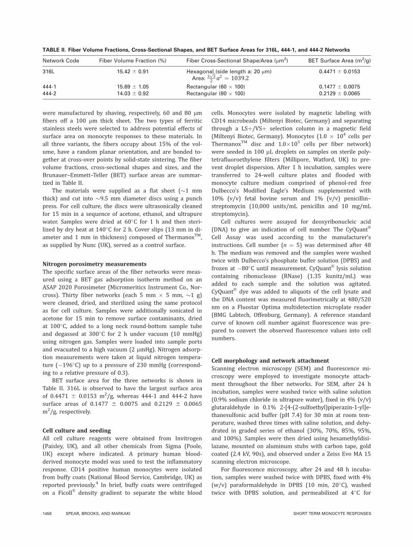
were manufactured by shaving, respectively, 60 and 80 lmfibers off a 100 lm thick sheet. The two types of ferriticstainless steels were selected to address potential effects ofsurface area on monocyte responses to these materials. Inall three variants, the fibers occupy about 15% of the vol-ume, have a random planar orientation, and are bonded to-gether at cross-over points by solid-state sintering. The fibervolume fractions, cross-sectional shapes and sizes, and theBrunauer–Emmett–Teller (BET) surface areas are summar-ized in Table II.
The materials were supplied as a flat sheet (�1 mmthick) and cut into �9.5 mm diameter discs using a punchpress. For cell culture, the discs were ultrasonically cleanedfor 15 min in a sequence of acetone, ethanol, and ultrapurewater. Samples were dried at 60�C for 1 h and then steri-lized by dry heat at 140�C for 2 h. Cover slips (13 mm in di-ameter and 1 mm in thickness) composed of ThermanoxTM,as supplied by Nunc (UK), served as a control surface.
Nitrogen porosimetry measurementsThe specific surface areas of the fiber networks were meas-ured using a BET gas adsorption isotherm method on anASAP 2020 Porosimeter (Micromeritics Instrument Co., Nor-cross). Thirty fiber networks (each 5 mm � 5 mm, �1 g)were cleaned, dried, and sterilized using the same protocolas for cell culture. Samples were additionally sonicated inacetone for 15 min to remove surface contaminants, driedat 100�C, added to a long neck round-bottom sample tubeand degassed at 300�C for 2 h under vacuum (10 mmHg)using nitrogen gas. Samples were loaded into sample portsand evacuated to a high vacuum (2 lmHg). Nitrogen adsorp-tion measurements were taken at liquid nitrogen tempera-ture (�196�C) up to a pressure of 230 mmHg (correspond-ing to a relative pressure of 0.3).
BET surface area for the three networks is shown inTable II. 316L is observed to have the largest surface areaof 0.4471 6 0.0153 m2/g, whereas 444-1 and 444-2 havesurface areas of 0.1477 6 0.0075 and 0.2129 6 0.0065m2/g, respectively.
Cell culture and seedingAll cell culture reagents were obtained from Invitrogen(Paisley, UK), and all other chemicals from Sigma (Poole,UK) except where indicated. A primary human blood-derived monocyte model was used to test the inflammatoryresponse. CD14 positive human monocytes were isolatedfrom buffy coats (National Blood Service, Cambridge, UK) asreported previously.4 In brief, buffy coats were centrifugedon a FicollV
R
density gradient to separate the white blood
cells. Monocytes were isolated by magnetic labeling withCD14 microbeads (Miltenyi Biotec, Germany) and separatingthrough a LSþ/VSþ selection column in a magnetic field(Miltenyi Biotec, Germany). Monocytes (1.0 � 104 cells perThermanoxTM disc and 1.0�105 cells per fiber network)were seeded in 100 lL droplets on samples on sterile poly-tetrafluoroethylene filters (Millipore, Watford, UK) to pre-vent droplet dispersion. After 1 h incubation, samples weretransferred to 24-well culture plates and flooded withmonocyte culture medium comprised of phenol-red freeDulbecco’s Modified Eagle’s Medium supplemented with10% (v/v) fetal bovine serum and 1% (v/v) penicillin–streptomycin (10,000 units/mL penicillin and 10 mg/mLstreptomycin).
Cell cultures were assayed for deoxyribonucleic acid(DNA) to give an indication of cell number. The CyQuantV
R
Cell Assay was used according to the manufacturer’sinstructions. Cell number (n ¼ 5) was determined after 48h. The medium was removed and the samples were washedtwice with Dulbecco’s phosphate buffer solution (DPBS) andfrozen at �80�C until measurement. CyQuantV
R
lysis solutioncontaining ribonuclease (RNase) (1.35 kunitz/mL) wasadded to each sample and the solution was agitated.CyQuantV
R
dye was added to aliquots of the cell lysate andthe DNA content was measured fluorimetrically at 480/520nm on a Fluostar Optima multidetection microplate reader(BMG Labtech, Offenburg, Germany). A reference standardcurve of known cell number against fluorescence was pre-pared to convert the observed fluorescence values into cellnumbers.
Cell morphology and network attachmentScanning electron microscopy (SEM) and fluorescence mi-croscopy were employed to investigate monocyte attach-ment throughout the fiber networks. For SEM, after 24 hincubation, samples were washed twice with saline solution(0.9% sodium chloride in ultrapure water), fixed in 4% (v/v)glutaraldehyde in 0.1% 2-[4-(2-sulfoethyl)piperazin-1-yl]e-thanesulfonic acid buffer (pH 7.4) for 30 min at room tem-perature, washed three times with saline solution, and dehy-drated in graded series of ethanol (30%, 70%, 85%, 95%,and 100%). Samples were then dried using hexamethyldisi-lazane, mounted on aluminum stubs with carbon tape, goldcoated (2.4 kV, 90s), and observed under a Zeiss Evo MA 15scanning electron microscope.
For fluorescence microscopy, after 24 and 48 h incuba-tion, samples were washed twice with DPBS, fixed with 4%(w/v) paraformaldehyde in DPBS (10 min, 20�C), washedtwice with DPBS solution, and permeabilized at 4�C for
TABLE II. Fiber Volume Fractions, Cross-Sectional Shapes, and BET Surface Areas for 316L, 444-1, and 444-2 Networks
Network Code Fiber Volume Fraction (%) Fiber Cross-Sectional Shape/Area (lm2) BET Surface Area (m2/g)
316L 15.42 6 0.91 Hexagonal (side length a: 20 lm)Area: 3
ffiffi
3p
2 a2 ¼ 1039.20.4471 6 0.0153
444-1 15.69 6 1.05 Rectangular (60 � 100) 0.1477 6 0.0075444-2 14.03 6 0.92 Rectangular (80 � 100) 0.2129 6 0.0065
1458 SPEAR, BROOKS, AND MARKAKI SHORT TERM MONOCYTE RESPONSES
10 min in pH 7.2 buffer composed of sucrose (10.3 g), so-dium chloride (0.292 g), magnesium chloride (0.06 g), 4-(2-hydroxyethyl)-1-piperazineethanesulfonic acid buffer (0.476g), and Triton-X (0.5 mL) in distilled water (100 mL). Sam-ples were incubated with 1% (w/v) bovine serum albuminin DPBS at 37�C for 10 min to block nonspecific binding.Samples were washed twice with DPBS, mounted withVectashield antifade mountant containing 40,6-diamidino-2-phenylindole (Vector Laboratories, UK) to stain cell nuclei,and imaged using a Zeiss Axioskop 2 fluorescence microscope.
Cell viability: LDH releaseLDH release into culture medium due to membrane damageof cultured monocytes was used as an indicator of cell via-bility. Duplicate samples from the cell medium were takenafter culture of the monocytes for 4, 24, and 48 h of incuba-tion and stored at 4�C before the assay. LDH-mediatedconversion of pyruvic acid to lactic acid in the presence ofnicotinamide adenine dinucleotide in a reduced form, and2,4-dinitrophenylhydrazine was determined fluorimetricallyby the Cytox-ONETM homogenous membrane integrity assay(Promega) at 544/590 nm (Ex/Em) on a Fluostar Optimamultidetection microplate reader.
Inflammatory response: TNF-a releaseDuring an inflammatory response, monocytes secrete sev-eral cell-signaling molecules, including the cytokine TNF-a.The release of TNF-a from monocytes cultured on stainlesssteel surfaces was determined by an enzyme-linked immu-nosorbent assay (ELISA; Thermo Scientific, Pierce Biotech-nology), as a measure of the inflammatory response. Zymo-san A from Saccharomyces cerevisiae (0.04 wt %) wasadded to monocyte culture medium, forming a suspensionof particles. This suspension of zymosan was added to posi-tive control cells seeded on ThermanoxTM discs to stimulatean inflammatory response.27 Aliquots of medium in contactwith cultured monocytes were collected after 4, 24, and 48h of incubation. Duplicate samples were frozen at �80�Cuntil assaying.
Statistical analysisFive independent experiments were conducted using cellsfrom separate blood donors. Results from a single represen-tative experiment are presented and expressed as mean 6
standard deviation (SD). Data were analyzed for statisticalsignificance by one-way analysis of variance (ANOVA), fol-lowed by Bonferroni’s post hoc test for multiple compari-sons. Differences were considered statistically significant atp values of one symbol <0.05 and three symbols <0.001.4
RESULTS
Fiber surface morphologySEM images of fiber networks obtained before and after cellculture, such as those depicted in Figures 1 and 4, respec-tively, showed no apparent changes in the fiber surface mor-phology after 48 h in monocyte culture medium.
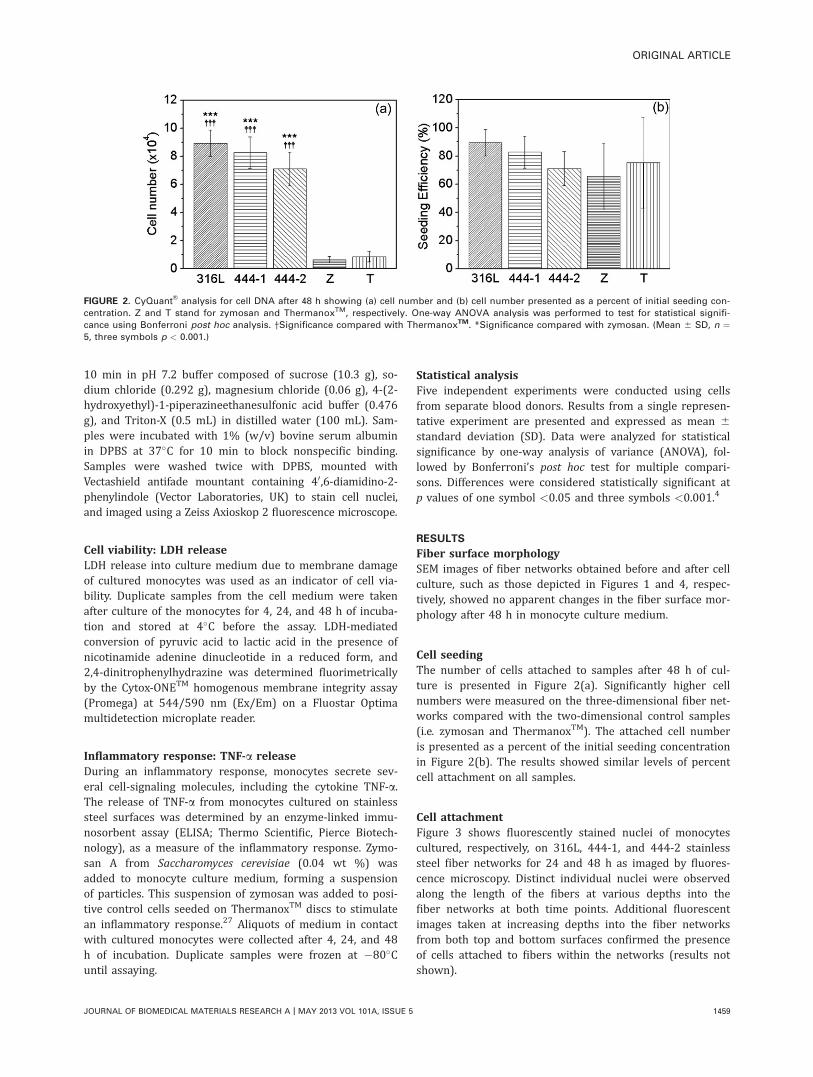
Cell seedingThe number of cells attached to samples after 48 h of cul-ture is presented in Figure 2(a). Significantly higher cellnumbers were measured on the three-dimensional fiber net-works compared with the two-dimensional control samples(i.e. zymosan and ThermanoxTM). The attached cell numberis presented as a percent of the initial seeding concentrationin Figure 2(b). The results showed similar levels of percentcell attachment on all samples.
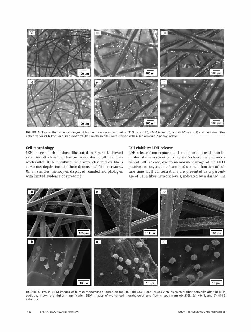
Cell attachmentFigure 3 shows fluorescently stained nuclei of monocytescultured, respectively, on 316L, 444-1, and 444-2 stainlesssteel fiber networks for 24 and 48 h as imaged by fluores-cence microscopy. Distinct individual nuclei were observedalong the length of the fibers at various depths into thefiber networks at both time points. Additional fluorescentimages taken at increasing depths into the fiber networksfrom both top and bottom surfaces confirmed the presenceof cells attached to fibers within the networks (results notshown).
FIGURE 2. CyQuantVR
analysis for cell DNA after 48 h showing (a) cell number and (b) cell number presented as a percent of initial seeding con-
centration. Z and T stand for zymosan and ThermanoxTM, respectively. One-way ANOVA analysis was performed to test for statistical signifi-
cance using Bonferroni post hoc analysis. †Significance compared with ThermanoxTM. *Significance compared with zymosan. (Mean 6 SD, n ¼5, three symbols p < 0.001.)
ORIGINAL ARTICLE
JOURNAL OF BIOMEDICAL MATERIALS RESEARCH A | MAY 2013 VOL 101A, ISSUE 5 1459
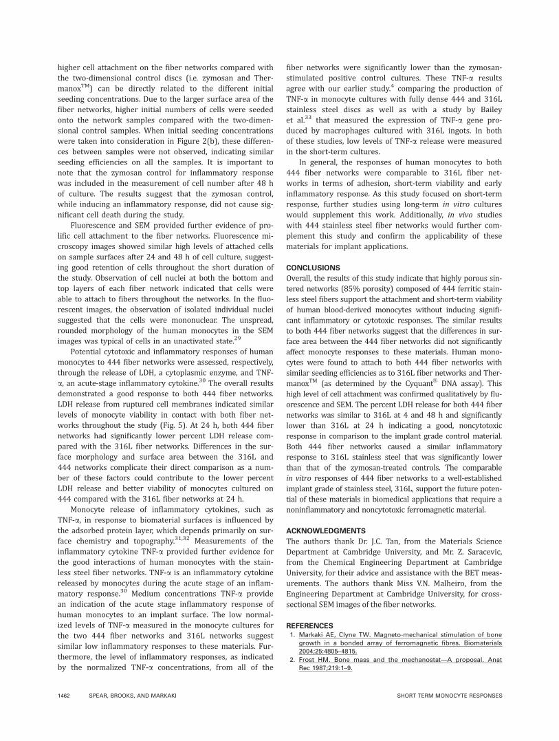
Cell morphologySEM images, such as those illustrated in Figure 4, showedextensive attachment of human monocytes to all fiber net-works after 48 h in culture. Cells were observed on fibersat various depths into the three-dimensional fiber networks.On all samples, monocytes displayed rounded morphologieswith limited evidence of spreading.
Cell viability: LDH releaseLDH release from ruptured cell membranes provided an in-dicator of monocyte viability. Figure 5 shows the concentra-tion of LDH release, due to membrane damage of the CD14positive monocytes, in culture medium as a function of cul-ture time. LDH concentrations are presented as a percent-age of 316L fiber network levels, indicated by a dashed line
FIGURE 3. Typical fluorescence images of human monocytes cultured on 316L (a and b), 444-1 (c and d), and 444-2 (e and f) stainless steel fiber
networks for 24 h (top) and 48 h (bottom). Cell nuclei (white) were stained with 40,6-diamidino-2-phenylindole.
FIGURE 4. Typical SEM images of human monocytes cultured on (a) 316L, (b) 444-1, and (c) 444-2 stainless steel fiber networks after 48 h. In
addition, shown are higher magnification SEM images of typical cell morphologies and fiber shapes from (d) 316L, (e) 444-1, and (f) 444-2
networks.
1460 SPEAR, BROOKS, AND MARKAKI SHORT TERM MONOCYTE RESPONSES

at 100%. At all time points, similar percent LDH concentra-tions were measured in the medium of monocytes culturedon 444-1 and 444-2 fiber networks. Monocytes cultured on444-1 and 444-2 fiber networks released significantly lowerpercent LDH than those on 316L at 24 h. At 4 and 48 h,percent LDH release from monocytes cultured on 444-1 and444-2 was similar to 316L control levels of LDH.
Inflammatory response: TNF-a releaseThe secretion of the inflammatory cytokine TNF-a wasdetermined by ELISA and used as a measure of inflamma-tory response. Figure 6 presents the concentrations ofTNF-a, normalized for cell number at 48 h. Similar low lev-els of TNF-a were secreted by cells cultured on 444-1, 444-2, and 316L fiber networks. The TNF-a concentration of thepositive control, ThermanoxTM discs with inflammatoryzymosan stimulation, was significantly higher than all othersamples.
DISCUSSION
In this work, we have presented the first study concerningthe in vitro responses of human monocytes to two types offiber networks made of 444 ferritic stainless steel; a steelwell known for its good corrosion resistance.5 Theirresponses are presented alongside a 316L austenitic stain-less steel network, a well-characterized implant grade ofstainless steel. They build on our earlier work with thesestainless steel grades in fully dense form.4 All three net-works have similar fiber volume fractions (�15%—seeTable II), and fiber orientation distributions, that is all net-works exhibit random planar isotropy (transverse isotropy)and have fibers lying in-plane. However, direct comparisonsbetween the two different types of stainless steel networks,316L and 444, are not possible due to the different fiber
geometries and surface morphologies. In 444-1 and 444-2networks, the fibers exhibit a rectangular cross section (60lm � 100 lm and 80 lm � 100 lm, respectively), whereas316L networks have hexagonal-shaped cross sections (40lm diagonal length). Furthermore, due to the shaving pro-cess, the 444 coil-shaped fibers are significantly rougherthan the 316L fibers. The differences in fiber geometriesand surface morphologies have a direct effect on the specificsurface area measurements (Table II). 316L has the largestsurface area compared with 444 networks. Due to these dif-ferences, the fiber networks of 316L were included in thestudy as a control, rather than an experimental group. Onthe other hand, the two types of 444 networks differ onlyin surface area, allowing direct comparison between thetwo. The cross-sectional dimensions of the fibers are signifi-cantly larger than the average monocyte diameter (< 20lm) to eliminate any potential effects of geometric con-straints on attached cell morphology. Among the two 444networks, 444-2 has the highest surface area (Table II).While for a given fiber volume fraction and length, 444-2will have less fibers per unit area, the higher cross-sectionalarea of its coarser fibers (80 lm � 100 lm vs. 60 lm �100 lm) results in a higher total surface area.
Human cell cultures have been used extensively for bio-material evaluation. For the current studies, we haveselected human peripheral blood monocytes as a model ofthe acute stage of in vivo response after implantation.18,19
After adhesion of monocytes to a biomaterial surface, mor-phological and cytoplasmic changes occur as a componentof cell activation and inflammatory response.28 Several stud-ies have demonstrated that monocyte adhesion and inflam-matory cytokine production depend on biomaterial proper-ties, such as surface chemistry and roughness.21–25
Considerable cell attachment of human monocytes to allsamples was measured by CyQuantV
R
. The significantly
FIGURE 5. LDH release from monocytes cultured on 316L, 444-1, and
444-2 fiber networks. Results are presented as percent of 316L control
levels (dashed line). One-way ANOVA analysis was performed to test
for statistical significance using Bonferroni post hoc analysis.
†Significance compared with 316L. (Mean 6 SD, n ¼ 5, one symbol p
< 0.05 and three symbols p < 0.001).
FIGURE 6. Concentrations of TNF-a for 316L, 444-1, 444-2, and zymo-
san. Concentrations have been normalized for cell number as meas-
ured by CyQuantVR
at 48 h. One-way ANOVA analysis was performed
to test for statistical significance using Bonferroni post hoc analysis.
*Significance of each sample paired with zymosan. (Mean 6 SD, n ¼5, three symbols p < 0.001.)
ORIGINAL ARTICLE
JOURNAL OF BIOMEDICAL MATERIALS RESEARCH A | MAY 2013 VOL 101A, ISSUE 5 1461
higher cell attachment on the fiber networks compared withthe two-dimensional control discs (i.e. zymosan and Ther-manoxTM) can be directly related to the different initialseeding concentrations. Due to the larger surface area of thefiber networks, higher initial numbers of cells were seededonto the network samples compared with the two-dimen-sional control samples. When initial seeding concentrationswere taken into consideration in Figure 2(b), these differen-ces between samples were not observed, indicating similarseeding efficiencies on all the samples. It is important tonote that the zymosan control for inflammatory responsewas included in the measurement of cell number after 48 hof culture. The results suggest that the zymosan control,while inducing an inflammatory response, did not cause sig-nificant cell death during the study.
Fluorescence and SEM provided further evidence of pro-lific cell attachment to the fiber networks. Fluorescence mi-croscopy images showed similar high levels of attached cellson sample surfaces after 24 and 48 h of cell culture, suggest-ing good retention of cells throughout the short duration ofthe study. Observation of cell nuclei at both the bottom andtop layers of each fiber network indicated that cells wereable to attach to fibers throughout the networks. In the fluo-rescent images, the observation of isolated individual nucleisuggested that the cells were mononuclear. The unspread,rounded morphology of the human monocytes in the SEMimages was typical of cells in an unactivated state.29
Potential cytotoxic and inflammatory responses of humanmonocytes to 444 fiber networks were assessed, respectively,through the release of LDH, a cytoplasmic enzyme, and TNF-a, an acute-stage inflammatory cytokine.30 The overall resultsdemonstrated a good response to both 444 fiber networks.LDH release from ruptured cell membranes indicated similarlevels of monocyte viability in contact with both fiber net-works throughout the study (Fig. 5). At 24 h, both 444 fibernetworks had significantly lower percent LDH release com-pared with the 316L fiber networks. Differences in the sur-face morphology and surface area between the 316L and444 networks complicate their direct comparison as a num-ber of these factors could contribute to the lower percentLDH release and better viability of monocytes cultured on444 compared with the 316L fiber networks at 24 h.
Monocyte release of inflammatory cytokines, such asTNF-a, in response to biomaterial surfaces is influenced bythe adsorbed protein layer, which depends primarily on sur-face chemistry and topography.31,32 Measurements of theinflammatory cytokine TNF-a provided further evidence forthe good interactions of human monocytes with the stain-less steel fiber networks. TNF-a is an inflammatory cytokinereleased by monocytes during the acute stage of an inflam-matory response.30 Medium concentrations TNF-a providean indication of the acute stage inflammatory response ofhuman monocytes to an implant surface. The low normal-ized levels of TNF-a measured in the monocyte cultures forthe two 444 fiber networks and 316L networks suggestsimilar low inflammatory responses to these materials. Fur-thermore, the level of inflammatory responses, as indicatedby the normalized TNF-a concentrations, from all of the
fiber networks were significantly lower than the zymosan-stimulated positive control cultures. These TNF-a resultsagree with our earlier study.4 comparing the production ofTNF-a in monocyte cultures with fully dense 444 and 316Lstainless steel discs as well as with a study by Baileyet al.33 that measured the expression of TNF-a gene pro-duced by macrophages cultured with 316L ingots. In bothof these studies, low levels of TNF-a release were measuredin the short-term cultures.
In general, the responses of human monocytes to both444 fiber networks were comparable to 316L fiber net-works in terms of adhesion, short-term viability and earlyinflammatory response. As this study focused on short-termresponse, further studies using long-term in vitro cultureswould supplement this work. Additionally, in vivo studieswith 444 stainless steel fiber networks would further com-plement this study and confirm the applicability of thesematerials for implant applications.
CONCLUSIONS
Overall, the results of this study indicate that highly porous sin-tered networks (85% porosity) composed of 444 ferritic stain-less steel fibers support the attachment and short-term viabilityof human blood-derived monocytes without inducing signifi-cant inflammatory or cytotoxic responses. The similar resultsto both 444 fiber networks suggest that the differences in sur-face area between the 444 fiber networks did not significantlyaffect monocyte responses to these materials. Human mono-cytes were found to attach to both 444 fiber networks withsimilar seeding efficiencies as to 316L fiber networks and Ther-manoxTM (as determined by the CyquantV
R
DNA assay). Thishigh level of cell attachment was confirmed qualitatively by flu-orescence and SEM. The percent LDH release for both 444 fibernetworks was similar to 316L at 4 and 48 h and significantlylower than 316L at 24 h indicating a good, noncytotoxicresponse in comparison to the implant grade control material.Both 444 fiber networks caused a similar inflammatoryresponse to 316L stainless steel that was significantly lowerthan that of the zymosan-treated controls. The comparablein vitro responses of 444 fiber networks to a well-establishedimplant grade of stainless steel, 316L, support the future poten-tial of these materials in biomedical applications that require anoninflammatory and noncytotoxic ferromagnetic material.
ACKNOWLEDGMENTS
The authors thank Dr. J.C. Tan, from the Materials ScienceDepartment at Cambridge University, and Mr. Z. Saracevic,from the Chemical Engineering Department at CambridgeUniversity, for their advice and assistance with the BET meas-urements. The authors thank Miss V.N. Malheiro, from theEngineering Department at Cambridge University, for cross-sectional SEM images of the fiber networks.
REFERENCES1. Markaki AE, Clyne TW. Magneto-mechanical stimulation of bone
growth in a bonded array of ferromagnetic fibres. Biomaterials
2004;25:4805–4815.
2. Frost HM. Bone mass and the mechanostat—A proposal. Anat
Rec 1987;219:1–9.
1462 SPEAR, BROOKS, AND MARKAKI SHORT TERM MONOCYTE RESPONSES

3. Rubin CT, Lanyon LE. Regulation of bone mass by mechanical
strain magnitude. Calcif Tissue Int 1985;37:411–417.
4. Malheiro VN, Spear RL, Brooks RA, Markaki AE. Osteoblast and
monocyte responses to 444 ferritic stainless steel intended for a
magneto-mechanically actuated fibrous scaffold. Biomaterials
2011;32:6883–6892.
5. Takada Y, Nakamura K, Kimura K, Okuno O. Corrosion behaviour
of the stainless steel composing dental magnetic attachments. Int
Congr Ser 2005;1284:314–315.
6. Ducheyne P, Aernoudt E, De Meester P. The mechanical behav-
iour of porous austenitic stainless steel fibre structures. J Mater
Sci 1978;13:2650–2658.
7. Markaki AE, Clyne TW. Mechanics of thin ultra-light stainless steel
sandwich sheet material: Part II—Resistance to delamination. Acta
Mater 2003;51:1351–1357.
8. Tan JC, Clyne TW. Ferrous fibre network materials for noise
reduction in gas turbine aeroengines. Part II: Thermomechanical
stability. Adv Eng Mater 2008;10:201–209.
9. Lueck RA, Galante J, Rostoker W, Ray RD. Development of an
open pore metallic implant to permit attachment to bone. Surg
Forum 1969;20:456–457.
10. Jansen JA, van der waerden JPCM, Degroot K. Development of a
new percutaneous access device for implantation in soft-tissues.
J Biomed Mater Res 1991;25:1535–1545.
11. Paquay YCGJ, de Ruijter JE, van der Waerden JPCM, Jansen JA. Tis-
sue reaction to Dacron velour and titanium fibre mesh used for an-
chorage of percutaneous devices. Biomaterials 1996;17:1251–1256.
12. Paquay YCGJ, de Ruijter JE, van der Waerden JPCM, Jansen JA.
Wound healing phenomena in titanium fibre mesh: The influence
of the length of implantation. Biomaterials 1997;18:161–166.
13. Gerritsen H, Lutterman JA, Jansen JA. The influence of impaired
wound healing on the tissue reaction to percutaneous devices
using titanium fiber mesh anchorage. J Biomed Mater Res 2000;
52:135–141.
14. ten Hallers EJO, Jansen JA, Marres HAM, Rakhorst G, Verkerke
GJ. Histological assessment of titanium and polypropylene fiber
mesh implantation with and without fibrin tissue glue. J Biomed
Mater Res Part A 2007;80A:372–380.
15. Shalabi MM, Walboomers XF, Jansen JA. The influence of bone
formation on anchoring percutaneous devices with titanium fibre
mesh flanges. J Mater Sci Mater Med 2004;15:809–816.
16. Paquay YC, de Ruijeter AE, van der Waerden JPC, Jansen JA. A
one stage versus two stage surgical technique—Tissue reaction
to a percutaneous device provided with titanium fiber mesh appli-
cable for peritoneal dialysis. ASAIO J 1996;42:961–967.
17. Paquay YCGJ, De Blieck-Hogervorst JMA, Jansen JA. Corrosion
behaviour of metal fibre web structures. J Mat Sci Mater Med
1996;7:585–589.
18. Anderson JM. Biological responses to materials. Annu Rev Mater
Res 2001;31:81–110.
19. Xia Z, Triffitt JT. A review on macrophage responses to biomate-
rials. Biomed Mater 2006;1:R1–R9.
20. Anderson JM, Rodriguez A, Chang DT. Foreign body reaction to
biomaterials. Semin Immunol 2008;20:86–100.
21. Bonfield TL, Anderson JM. Functional versus quantitative com-
parison of IL-1-beta from monocytes/macrophages on biomedical
polymers. J Biomed Mater Res 1993;27:1195–1199.
22. Bonfield TL, Colton E, Anderson JM. Protein adsorption of bio-
medical polymers influences activated monocytes to produce
fibroblast stimulating factors. J Biomed Mater Res 1992;26:
457–465.
23. Bonfield TL, Colton E, Marchant RE, Anderson JM. Cytokine and
growth factor production by monocytes/macrophages on protein
preadsorbed polymers. J Biomed Mater Res 1992;26:837–850.
24. Defife KM, Yun JK, Azeez A, Stack S, Ishihara K, Nakabayashi N,
Colton E, Anderson JM. Adhesion and cytokine production by
monocytes on poly(2-methacryloloxyethyl phosphorylcholine-co-
alkyl methacrylate)-coated polymers. J Biomed Mater Res 1995;
29:431–439.
25. Yun JK, Defife K, Colton E, Stack S, Azeez A, Cahalan L, Verho-
even M, Cahalan P, Anderson JM. Human monocyte-macrophage
adhesion and cytokine production on surface-modified poly(tetra-
fluoroethylene hexafluoropropylene) polymers with and without
protein preadsorption. J Biomed Mater Res 1995;29:257–268.
26. Everett SJ. Bundle drawing to produce metallic fibers;1936.
27. Wimhurst JA, Brooks RA, Rushton N. Inflammatory responses of
human primary macrophages to particulate bone cements in
vitro. J Bone Joint Surg Br 2001;83B:278–282.
28. Salthouse TN. Some aspects of macrophage behavior at the
implant interface. J Biomed Mater Res 1984;18:395–401.
29. Jakubiec B, Marois Y, Zhang Z, Roy R, Sigot-Luizard MF, Dugre
FJ, King MW, Dao L, Laroche G, Guidoin R. In vitro cellular
response to polypyrrole-coated woven polyester fabrics: Potential
benefits of electrical conductivity. J Biomed Mater Res 1998;41:
519–526.
30. Tracey KJ, Cerami A. Tumor-necrosis-factor-alpha pleiotropic
cytokine and therapeutic target. Annu Rev Med 1994;45:491–503.
31. Soskolne WA, Cohen S, Sennerby L, Wennerberg A, Shapira L.
The effect of titanium surface roughness on the adhesion of
monocytes and their secretion of TNF-alpha and PGE(2). Clin Oral
Implants Res 2002;13:86–93.
32. Zhao G, Schwartz Z, Wieland M, Rupp F, Geis-Gerstorfer J, Coch-
ran DL, Boyan BD. High surface energy enhances cell response to
titanium substrate microstructure. J Biomed Mater Res Part A
2005;74A:49–58.
33. Bailey LO, Lipplatt S, Biancanello FS, Ridder SD, Washburn NR.
The quantification of cellular viability and inflammatory response
to stainless steel alloys. Biomaterials 2005;26:5296–5302.
ORIGINAL ARTICLE
JOURNAL OF BIOMEDICAL MATERIALS RESEARCH A | MAY 2013 VOL 101A, ISSUE 5 1463