Setor 05. Dor e Nocicepção/Pain and Nociception - SBFTE
71
Setor 05. Dor e Nocicepção/Pain and Nociception 05.001 Envolvimento de peptídeos opióides endógenos no efeito antinociceptivo periférico do cafestol. Guzzo LS, Perez AC, Duarte ID UFMG - Farmacologia Introdução: O cafestol é um diterpeno encontrado somente na fração lipídica não saponificada da semente do café, que é liberado durante a fervura, mas fica retido durante a filtração. Possui propriedades anticarcinogênicas, além de causar aumento das concentrações plasmáticas de triglicérides e colesterol. Embora já tenham sido feitos muitos estudos que comprovem a atividade antinociceptiva e antiinflamatória da cafeína, nenhum estudo foi feito com intuito de avaliar a atividade antinociceptiva e poucos foram feitos sobre a atividade antiinflamatória do cafestol. Diante disso, o presente trabalho objetivou examinar uma possível ação antinociceptiva periférica do cafestol sendo avaliado o envolvimento de peptídeos opióides endógenos nesse efeito. Métodos: A hiperalgesia foi induzida por injeção intraplantar (ipl.) de prostaglandina E 2 (PGE 2 , 2 μg) e foi medida através do método de retirada da pata posterior direita do rato submetida à compressão. A PGE 2 e o cafestol foram injetados na pata direita do animal, com exceção do protocolo utilizado para excluir a possibilidade de um efeito não local do cafestol, em que a PGE 2 foi injetada em ambas as patas e o cafestol na pata esquerda. O cafestol foi sempre injetado 175 min após a administração de PGE 2 . A naloxona e a bestatina foram administradas na pata direita do animal 30 min antes do cafestol. Em todos os testes foram utilizados ratos Wistar machos (180-220 g). Resultados e Discussão: O cafestol (20, 40 e 80 μg) foi administrado na face plantar posterior direita dos animais e as doses 40 e 80 μg/pata induziram efeito antinociceptivo significativo de forma dose-dependente. Esse efeito foi considerado local, uma vez que mesmo na maior dose utilizada não foi observado qualquer efeito na pata contralateral. O bloqueador de receptores opióides, naloxona (50 e 100 μg/pata) antagonizou de forma dose-dependente a ação antinociceptiva periférica do cafestol (80 μg/pata) e o inibidor de encefalinase, bestatina (400 μg/pata), potencializou e efeito antinociceptivo desse diterpeno (40 μg/pata). Os resultados mostraram pela primeira vez que o cafestol apresenta efeito antinociceptivo periférico e sugerem que esse efeito é resultante indiretamente da liberação de peptídeos opióides com posterior ação sobre seus receptores. Apoio Financeiro: EMBRAPA, FAPEMIG e CNPq Comitê de Ética em Experimentação Animal (CETEA/UFMG) protocolo nº 41/2007.
-
Upload
khangminh22 -
Category
Documents
-
view
1 -
download
0
Transcript of Setor 05. Dor e Nocicepção/Pain and Nociception - SBFTE
Setor 05. Dor e Nocicepção/Pain and Nociception 05.001 Envolvimento de peptídeos opióides endógenos no efeito antinociceptivo periférico do cafestol. Guzzo LS, Perez AC, Duarte ID UFMG - Farmacologia
Introdução: O cafestol é um diterpeno encontrado somente na fração lipídica não saponificada da semente do café, que é liberado durante a fervura, mas fica retido durante a filtração. Possui propriedades anticarcinogênicas, além de causar aumento das concentrações plasmáticas de triglicérides e colesterol. Embora já tenham sido feitos muitos estudos que comprovem a atividade antinociceptiva e antiinflamatória da cafeína, nenhum estudo foi feito com intuito de avaliar a atividade antinociceptiva e poucos foram feitos sobre a atividade antiinflamatória do cafestol. Diante disso, o presente trabalho objetivou examinar uma possível ação antinociceptiva periférica do cafestol sendo avaliado o envolvimento de peptídeos opióides endógenos nesse efeito. Métodos: A hiperalgesia foi induzida por injeção intraplantar (ipl.) de prostaglandina E2 (PGE2, 2 µg) e foi medida através do método de retirada da pata posterior direita do rato submetida à compressão. A PGE2 e o cafestol foram injetados na pata direita do animal, com exceção do protocolo utilizado para excluir a possibilidade de um efeito não local do cafestol, em que a PGE2 foi injetada em ambas as patas e o cafestol na pata esquerda. O cafestol foi sempre injetado 175 min após a administração de PGE2. A naloxona e a bestatina foram administradas na pata direita do animal 30 min antes do cafestol. Em todos os testes foram utilizados ratos Wistar machos (180-220 g). Resultados e Discussão: O cafestol (20, 40 e 80 µg) foi administrado na face plantar posterior direita dos animais e as doses 40 e 80 µg/pata induziram efeito antinociceptivo significativo de forma dose-dependente. Esse efeito foi considerado local, uma vez que mesmo na maior dose utilizada não foi observado qualquer efeito na pata contralateral. O bloqueador de receptores opióides, naloxona (50 e 100 μg/pata) antagonizou de forma dose-dependente a ação antinociceptiva periférica do cafestol (80 µg/pata) e o inibidor de encefalinase, bestatina (400 μg/pata), potencializou e efeito antinociceptivo desse diterpeno (40 µg/pata). Os resultados mostraram pela primeira vez que o cafestol apresenta efeito antinociceptivo periférico e sugerem que esse efeito é resultante indiretamente da liberação de peptídeos opióides com posterior ação sobre seus receptores. Apoio Financeiro: EMBRAPA, FAPEMIG e CNPq Comitê de Ética em Experimentação Animal (CETEA/UFMG) protocolo nº 41/2007.
05.002 Efeito antinociceptivo induzido pela hemopressina em modelo experimental de dor crônica. Maique ET1, Ferro ES2, Heimann AS3, Dale CS1 1Hospital Sírio Libanês - Ensino e Pesquisa, 2ICB-USP, 3Proteimax Biotecnologia Ltda
Introdução: Dados recentes demonstram que a hemopressina, um peptídeo derivado da cadeia a1 da hemoglobina, inibe a dor em diferentes modelos experimentais de hiperalgesia aguda, sendo este efeito independente da ativação de receptores opióides (Dale et al., Peptides.26:431, 2005). Ainda, foi também demonstrado que a hemopressina atua como um agonista inverso para receptores canabinóides do tipo 1 (CB1) bloqueando a sinalização direta por este receptor (Heimann et al., PNAS.104:20588, 2007). Este estudo avalia a atividade antinociceptiva da hemopressina em modelo experimental de dor crônica. Métodos: Ratos Wistar, machos, (180-200 g) foram submetidos ao modelo de constrição crônica do nervo ciático (Bennet e Xie., Pain, 33:87, 1988) e foram avaliados no modelo de pressão de pata (Randall e Selitto., Arch. Intern. Pharmacodyn., 111:209,1957) antes (medida inicial), 3, 7 e 14 dias após o procedimento cirúrgico para estabelecimento do quadro de dor neuropática. No 14º dia, na vigência de dor neuropática, receberam hemopressina nas doses de 0,05, 0,125, 0,25 ou 0,5 mg/kg administradas por via intraplantar (i.pl.) ou oral (v.o.) e foram novamente avaliados no modelo de pressão de pata 1ª, 3ª, 6ª, 12ª e 24ª horas após os tratamentos. Animais operados e tratados com salina ou animais falso operados foram avaliados como grupo controle. Resultados: A injeção i.pl. de 0,05 mg/kg inibiu a nocicepção da 1ª a 6ª hora após o tratamento, sendo o efeito perdido na 12ª hora após a administração. Resultados semelhantes foram observados com a dose de 0,125 mg/kg, sugerindo que o efeito observado não sofre influências da variação de dosagem. A administração oral de hemopressina também reverteu a nocicepção da 1ª a 6ª hora após a administração das doses de 0,25 ou 0,5 mg/kg. Discussão: Os dados obtidos demonstram que a hemopressina inibe a nocicepção em modelo experimental de dor crônica. Esses dados poderão auxiliar no esclarecimento do papel antinociceptivo da hemopressina, um potente candidato para fins terapêuticos. Apoio financeiro: Instituto de Ensino e Pesquisa Hospital Sírio-Libanês e Proteimax Biotecnologia Ltda. Número de Licença da Comissão de Ética: CEUA22/2008
05.003 Characterization of the antinociceptive and anti-inflammatory activities of nicotinic acid and its isomers in different experimental models. Godin AM, Ferreira, WC, Rocha LTS, Vieira, RP, Nascimento Jr EB, Seniuk, JGT, Coelho MM FaFar-UFMG Produtos Farmacêuticos
Introduction: Nicotinic acid, a carboxylic acid derivative, is one of the two principal forms of the B3 vitamin (SAUVE, A.A. et al.; J. Pharmacol. Exp. Ther., v. 3, p. 883, 2007). In addition to being a nutrient, it is a clinically applied pharmacological agent. High doses of nicotinic acid have been used for decades as a lipid-lowering agent (Bodor, E.T. et al.; Br. J. Pharmacol., v. 153, p. 568, 2008). Several studies have demonstrated that nicotinamide, the amide derivative of vitamin B3, exert a number of anti-inflammatory properties unrelated to its vitamin activity (Godin, A. M. et al.; 39° Congresso Brasileiro de Farmacologia e Terapêutica Experimental. SBFTE: p. 61, 2007; Cuzzocrea, S. et al.; Life Sci., v. 65, p. 1297, 1999; PERO, R.W. et al.; Mol. Cell. Biochem., v. 193, p. 119, 1999). Thus, we investigated the effects induced by nicotinic acid and its isomers, picolinic and isonicotinic acid, in models of nociceptive and inflammatory pain and also edema, as these effects have not been investigated. Methods: Nicotinic (250 or 500 mg/kg), picolinic (62.5 or 125 mg/kg) or isonicotinic (250 or 500 mg/kg) acid were administered per os in female Swiss mice (25-30 g) 1 h before the s.c. injection of formaldehyde (0.92%, 20 µl) into the dorsum of the right hindpaw or the evaluation of motor activity in the rota-rod (14 rpm, 2 min). In the model of paw edema induced by carrageenan (600 µg, 30 µl, i.pl.) in mice, nicotinic acid or its isomers were administered 1 h before and 2 h after the inflammatory stimulus. The paw volume was measured 2, 4 and 6 h after carrageenan injection. Results were analyzed by one-way analysis of variance followed by Newman-Keuls post-hoc test. The study was approved by the Ethics Committee on Animal Experimentation of the Federal University of Minas Gerais (CETEA/UFMG n° 146/2007). Results: Nicotinic acid (250 or 500 mg/kg) inhibited the first (41 and 58%, respectively) and the second (62 and 88%, respectively) phases of the formaldehyde-induced nociceptive response. Picolinic acid (125 mg/kg) also inhibited the first (38%) and the second (44%) phases of the nociceptive response. However, isonicotinic acid (250 or 500 mg/kg) did not present activity in this experimental pain model. Nicotinic acid (250 or 500 mg/kg) inhibited (36 and 64%, respectively) carrageenan-induced paw edema. Such inhibition was not observed after treatment with picolinic (62.5 or 125 mg/kg) or isonicotinic (250 or 500 mg/kg) acids. Nicotinic or picolinic acids did not impair the motor activity of mice in the rota-rod test. Discussion: The results show the antinociceptive activity of nicotinic and picolinic acids and also the anti-inflammatory activity of nicotinic acid. It is unlikely that antinociception resulted from an impairment of motor activity or a muscle relaxing effect. The study clearly shows the structure-activity relationship of the molecules. Despite the lack of precise information on its mode of action, nicotinic acid is a safe drug, is approved for clinical use and represents a potentially valuable analgesic and anti-inflammatory agent. More research is needed to elucidate its mechanisms of action. Acknowledgements: FAPEMIG, CNPq and CAPES.
05.004 Effects of an isolated lectin from the red marine alga Hypnea cervicornis J. Agardh in the mechanical hypernociception. Bitencourt FS1, Figueiredo JG2, Cunha TM3, Luz PB1, Mota MRL1, Nascimento KS2, Cavada BS2, Sampaio AH2, Cunha FQ3, Alencar NMN de1 1UFC - Fisiologia e Farmacologia, 2UFC- Bioquímica e Biologia Molecular, 3FMRP-USP - Farmacologia
Introduction: Lectins are (glyco)proteins that can recognize and reversibly bind to carbohydrates or other substances derived from sugars and are encountered throughout animal and plant kingdoms. Several biological activities of plant lectins have been described, including pro-inflammatory and anti-inflammatory effects. Hypnea cervicornis is a species of red algae found in the coast of Northeast of Brazil. The isolated lectin (H. cervicornis agglutinin – HCA) is a polypeptide containing a mixture of 90 amino acids residues (9193±3 Da) which binds specifically to glycoproteins of mucin type. The aim of the present work was to study the anti-inflammatory effect, not exploited yet, of a lectin isolated from the red marine alga HCA in mechanical hypernociception. Methods: Animal handling and experimental protocols were registered on the Institutional Ethics Committee under number 60/09. Wistar rats (180-200g) were evaluated in mechanical hypernociception by electronic pressure-meter paw test (∆ reaction g). The animals (n=5) were pretreated 15 min before with saline (i.v.) or HCA (1 mg/kg; i.v.). Hypernociception was measured at 0, 1, 3, 5, 7, 12 and 24 h after injection of carrageenan (Cg; 100μg/paw). Three hours after Cg injection, neutrophil influx (hind paw tissue) was evaluated by myeloperoxidase levels (MPO/activity) in animals treated by HCA (0.1 and 1 mg/kg; i.v.). In another set of experiment, animals were treated intravenously with saline or HCA (1 mg/kg) or non-selective, N-nitro-L-arginine (nitro; 50 mg/kg; s.c.), or selective inducible NO synthase (iNOS), aminoguanidine (amino; 50 mg/kg; s.c.) inhibitors and after 15 minutes hypernociception was induced by intraplantar injection of Cg, as described above (p<0.05; ANOVA-Bonferroni´s test). Results and Discussion: The hypernociceptive response was inhibited by HCA at time 1 (15.7±0.5g), 3 (20.3±1.8g), 5 (19.5±1.0g) and 7 (16.5±1.3g) hours after Cg (31.1±0.5g; 39.5±1.5g; 32.7±0.9g; 31.2±1.0g; respectively). The MPO activity was reduced by HCA at dose 0.1 (21.4±2.4) and 1 (14.0±2.4) mg/kg, respectively when compared with Cg group (33.8±3.7). Only amino, but not nitro, inhibited (32.9±2.4g) the effect of HCA when compared with HCA alone group (13.9±3.2g). In conclusion, we demonstrated that the lectin HCA inhibits the mechanical inflammatory hypernociception and that NO might be involved in this inhibition. Further research would be of interest to explain the exact mechanism of this anti-inflammatory effect. Acknowledgements: Capes and CNPq
05.005 The sulphonamide group present in the coxibs molecule is necessary for hypoalgesia development in a model of inflammatory pain in rats. Role of endogenous opioids. Gassani BCA, Rezende RM, Francischi JN UFMG - Fisiologia e Farmacologia
Introduction: In a model of peripherally induced inflammatory pain in rats, we first demonstrated that a particular class of sulphonamides, the selective cyclooxygenase (COX)-2 inhibitors, was associated with a raise in nociceptive threshold which was well above basal values, thereby named “hypoalgesia” (Francischi, JN et al. Br. J. Pharmacol. 137:837(2002)). In addition, such an effect was associated with the endogenous opioid system (França, DS et al. Neuropharmacol. 51(1):37(2006); Rezende RM et al. Pain. 142(1-2):94(2009)). Here, we have assessed whether the sulphonamide group would be important for the coxibs hypoalgesia development using the same model of inflammatory pain. Methods: Ethics Committee for Animal Experimentation: 45/08. Mechanical hyperalgesia model: injection of carrageenan lambda (250 μg/paw) into 150-180 g male rats of Holtzman´s lineage hind paw pads at time zero constituted the painful stimulus and the nociceptive thresholds (in grams by Randall-Selitto’s method) hourly followed the subsequent 6 h. Three different sulphonamides, celecoxib (CX-12 mg/kg; a selective COX-2 inhibitor), furosemide (FUR-1, 10 and 40 mg/kg; a loop diuretic), acetazolamide (ACTZ–100, 200 and 400 mg/kg; a carbonic anhydrase inhibitor), and two coxibs lacking the sulphonamide group (etoricoxib, ETX-3, 12 and 24 mg/kg, and lumiracoxib, LX–6, 9 and 12 mg/kg) were given by subcutaneous (sc) injection in the rat’s dorsum (0.1 ml/100 g of weight), 30 minutes before inflammatory stimulus. Naltrexone (NTX; 3 mg/kg), a non-selective opioid antagonist was also administered sc, 30 minutes before test drugs. Control group was injected with the respective vehicle for each drug [saline (CX, ETX, LX, NTX) or DMSO 30% in saline (FUR, ACTZ)]. Results: Mean of nociceptive threshold values of the groups at time zero (C= -6.7+3.3; CX= 2 +3.7/C= -6.7 +3.3; FUR= 1.7+3.1)/(C= 5+2.2; ACTZ= -2+3.7). ACTZ 200 mg/kg (C= -51.7+4.8; ACTZ= 22+2*) and FUR 40 mg/kg (C= -86.7+8.8; FUR= 35+4.3*) induced hypoalgesia, peaking at 1 h and 3 h, respectively, being the later effect more similar to CX (C= -86.7 + 8.8; CX= 32 +3.7*). However, ETX and LX only produced anti-hyperalgesia. NTX completely abolished the hypoalgesia induction by sulphonamides (CX at 3 h: C= -68+9.2; CX= 32+3.7*; NTX+CX= -80+5.8#; ACTZ at 1 h: C= -42+8; ACTZ= 32+7.5*; NTX+ACTZ= -32+3.1#; FUR at 3 h: C= -73+14.5; FUR= 35+4.3*; NTX+FUR= -70+5.8#) but only partially prevented the anti-hyperalgesic effect of LX and did not affect the effect seen with ETX. Discussion and Conclusions: our results suggest that sulphonamide radical in the coxibs molecule is determinant for hypoalgesia development and they also show that such an effect is mediated by the endogenous opioid system. However, the variability of hypoalgesia induced by sulphonamide drugs seems to be not involved with the sulphonamide group. Support: CNPq, CAPES, FAPEMIG. *Difference between test drugs treated rats and control group. #Difference between test drugs and NTX-treated rats.
05.006 Involvement of endogenous opioids in ketamine-induced peripheral antinociception in rat. Romero TRL, Resende, LC, Mendes, R, Duarte IDG UFMG - Fisiologia e Farmacologia
.Introduction: Ketamine was initially introduced into clinical practice as a dissociative anesthetic in 1964, from these had been extensively used in burns, cancer and neuropathic pain. Ketamine is classically administered systemically, in which case, probably induced analgesia by recruitment of spinal and supraspinal mechanisms. This effect induced by ketamine in rats can be blocked by the opioid receptor antagonist naloxone. Recently, it was demonstrated that ketamine produces peripheral antinociceptive action in the formalin, thermal hyperalgesia and electrical stimulation test in rats. In the last test it was also inhibited by naloxone. Although ketamine shows a peripheral analgesic component, the base of this mechanism is not completely elucidated. Thus the aim of this study was to extend the idea of opioids receptor activation to obtain pharmacological evidences for the involvement of endogenous opioids peptides through an enkephalinase inhibitor in the peripheral antinociceptive effect induced by ketamine. Methods: The rat paw pressure test was used and hyperalgesia was induced by intraplantar injection of prostaglandin E2 (2 µg/paw). All drugs were administered locally into the right hind paw of Wistar male rats. Results: Ketamine (10, 20, 40 and 80 µg/paw) elicited a local inhibition of hyperalgesia (25%, 50%, 70% and 90%, respectively). The enkephalinase inhibitor bestatin (400 µg/paw) increased the peripheral antinociceptive effect of low dose of ketamine 10 µg/paw from 25% to 75%. Additionally naloxone (12.5, 25 and 50 µg/paw), opioid antagonist, antagonized the antinociception effect induced by ketamine higher dose (25%, 50% and 100%, respectively). Discussion: The results provide evidences that ketamine probably induces peripheral antinociceptive effect by release of endogenous opioid peptides, leading to the activation of opioid receptors in nociceptors. Financial support: Fapemig, CNPq (473758/2007-5) fellowships by CNPq. Ethics Committee on Animal Experimentation (CETEA/UFMG) protocol No. 41/2007.
05.007 Antinocicpetive effect of Luetzelburgia auriculata seed lectin. Pinheiro RSP1, Oliveira RSB2, Figueiredo JG2, Cavalcante IJM1, Luz PB1, Portela TCL2, Ramos MV2, Alencar NMN de1 1UFC- Fisiologia e Farmacologia, 2UFC - Bioquímica e Biologia Molecular
Introduction: Lectins are (glyco) proteins of non-immune origin that interact reversibly and specifically with carbohydrates. The interest by lectins study has been increased due the ability of these proteins to bind carbohydrates and mediates a variety of biological events on cell recognition and whole systems. The purified lectin from Luetzelburgia auriculata seeds (LAL) binds galactose and some of its derivatives. Studies have shown that LAL is a potent hemagglutinin and exhibits anti-inflammatory effects. In this work we evaluated the antinociceptive effect of LAL on different experimental models of nociception in mice. Methods: Animal handling and experimental protocols were registered on the Institutional Ethics Committee under number 58/09. Male Swiss mice (20-25 g) were treated with the LAL (0.1; 1 or 10 mg/kg; i.v.) 30 min before each experiment. Controls were injected with saline, morphine (5 mg/kg) or diazepam (2 mg/kg; s.c.). Abdominal writhing induced by acetic acid (AC): AC was injected (0.6%; 10 mL/kg; i.p.) and, after 10 min, the abdominal writhing was counted by 20 min. Formalin test: Formalin was injected (1.2%; 20 μL/paw; s.c.) and time the animals spent licking the injected paw was annotated in the first 5 min (1st phase - neurogenic) and within 20-25 min (2nd phase - inflammatory). Hot-plate test: The time of the animals reaction was monitored at 55 ºC at 0, 30, 60, 90, 120 and 150 min. Open-field test: The animals were exposed to a open-field (30 cm x 30 cm) divided in 9 small squares of 10 cm2. After 1 min, the number of squares visited was counted during 4 min. Rota rod test: mice were placed on the bar rotating at a speed of 4 rpm (5cm/ diameter). It was tested for its permanency during a 2 min period. The results were expressed as mean ± SEM. The statistical significance between groups was analyzed by analysis of variance (ANOVA) followed by Bonferroni’s test (p<0.05). Results and Discussion: The LAL significantly reduced the abdominal writhing induced by AC in doses of 1 and 10 mg/kg when compared with vehicle (8.0±2.8; 5.3±3.7; 24.9±4.9, respectively); in formalin test, LAL (1 and 10 mg/kg) reduced significantly the response when compared with vehicle in 1st phase (8.0±2.8; 5.3±3.7; 24.9±4.9, respectively) and 2nd phase (15.2±9.8; 18.0±7.5; 45.7±11.5, respectively). In hot-plate test, LAL (1 and 10 mg/kg) increased significantly the reaction’s time in all observations, with a maximum value of 26.6±9.4 (218%) at 120 min, when compared with vehicle (12.2±5.4). In compartmental assays didn’t have statistical differences between control animals and mice treated whit lectin. The results indicate that this lectin is a potent antinociceptive molecule and did not affect the locomotors activity (open-field test) neither the motor coordination (Rota-rod test). Supported by CNPq, CAPES, RENORBIO and IFS.
05.008 Role of TRPA1 receptors in cold hyperalgesia induced by infraorbital nerve constriction. Martini AC, Chichorro JG, Fiuza CR, Rae GA UFSC - Pharmacology
Introduction: Perception of cold has been proposed to be mediated by two members of the transient receptor potential (TRP) family, TRPA1 and TRPM8 (McKemy et al., Nature, 416:52, 2002; Story et al., Cell,112:819, 2003). Whereas TRPA1 has a threshold near 17 °C and is activated by compounds such as mustard oil and cinnamaldehyde, TRPM8 is activated by moderate cooling (22-27ºC) and by cooling substances such as menthol and eucalyptol. Consequently, TRPA1 has been considered the mediator of noxious cold sensations, while TRPM8 had been proposed to generate both non-painful and painful reactions to cold, as well as cold-induced pain relief (for review see Foulkes and Wood, Channels, 1-3:154, 2007). However, the cooling compound icilin is a common activator of both TRPM8 and TRPA1. Additionally, TRPA1 receptors seem to be expressed by a subset of small-diameter sensory neurons that co-express TRPV1, while TRPM8 receptors are expressed by a different subset of TRPV1-negative small diameter neurons. In light of these considerations, the present study aimed to investigate the role of TRPA1 receptors in cold hyperalgesia in a rat model of trigeminal neuropathic pain. Methods: Cold stimulation consisted in the application of a tetrafluoroethane spray to the center of the vibrissal pad and the duration of facial grooming behavior was recorded over the first 2 min as an index of cold-induced nociception. The responsiveness to the cold stimulus was assessed after injection of icilin (10 and 30 mg/50 µl, s.c. into the upper lip), or on day 4 after infraorbital nerve (ION) constriction, which was induced by placing two loose silk 4.0 ligatures around the right ION of anesthetized male Wistar rats (200-250 g). The effects of the selective TRPA1 antagonists HC-030031 (100 mg/kg, i.p., Mcnamara et al., PNAS, 104:13525, 2007) and AP-18 ( 10 μg/50 μl e 100 μg/50 μl, s.c., Petrus et al., Molecular Pain, 3:40, 2007) in rats submitted to ION constriction were also investigated. All protocols were previously approved by UFSC’s Committee on the Ethical Use of Animals (authorization number PP00625). Results and Discussion: Injected into the upper lip, icilin caused dose-dependent increases in duration of facial grooming behavior of naive rats at 30 min post injection (10.5 ± 1.7 s for vehicle; 25.4 ± 3.3 and 33.1 ± 3.6 s for 10 and 30 mg/50 µl of icilin, respectively). The cold hyperalgesia induced by ION was potentiated by 117 and 132%, at 30 and 60 min after icilin injection, respectively. Cold hyperalgesia induced by icilin or infraorbital nerve constriction was not observed in rats treated with capsaicin (50 mg/kg, s.c.) on post-natal day 2. Systemic administration of HC-030031 (100 mg/kg) or local administration of AP-18 (10 μg/50 μl e 100 μg/50 μl) each reduced cold hyperalgesia on day 4 after ION constriction. Thus, capsaicin-sensitive C fibers mediate the cold hyperalgesia triggered by icilin, as well as ION injury, which is also significantly reduced by TRPA1 selective antagonists. Taken together, these data suggest that specific blockers of TRPA1 receptors might provide effective therapeutic tools for the management of orofacial cold hyperalgesia. Financial Support: CNPq, Fapesc, PRONEX.
05.009 The protective effect of testosterone on the development of the TMJ pain is mediated by the activation of the opioid system. Fanton LE1, Tambeli CH1, Fischer L23 1FOP-UNICAMP - Ciências Fisiológicas, 2UFPR - Fisiologia
Introduction: TMJ pain can be up to twice fold more prevalent in women than in men, which suggests that sex hormones modulate TMJ pain. We have previously suggested that testosterone presents a protective role diminishing the risk of TMJ pain development (Fischer et al., J Pain, 8(5): 437, 2007), since a concentration of formalin (0.5%) that does not induce nociceptive behavior in naive male rats, induces in gonadectomized males and naive females. The aim of this study was to test the hypothesis that the protective role of testosterone in the development of TMJ pain is mediated by a central endogenous opioid mechanism. Methods: The TMJ formalin test (Roveroni et al., Pain; 94(2): 185, 2001) was used and the intensity of the nociceptive behavior response was quantified in male rats, after the injection of Naloxone (opioid receptor antagonist) or its vehicle (0.9% NaCl) in the medullary subarachnoid region and the TMJ injection of 0.5% formalin. These experiments were approved by the committee on animal research of UNICAMP (protocol number 1431-1). Data are expressed as mean + epm and were analyzed by ANOVA and the post hoc Tukey test (p<0.05). Results: The nociceptive behavior response of naive male rats that received a subarachnoid injection of naloxone (15 mg) (185,8 + 15,5) was significantly higher than that of naive male rats that received a subarachnoid injection of 0.9% NaCl (75,6 + 12,2) and was similar to that of gonadectomized male rats that received a subaracnoid injection of 0.9% NaCl (169,3 + 15,1) or of naloxone (162,6 + 18,9). Discussion: The blockade of opioid receptors by the administration of naloxone in the subaracnoid space of the trigeminal sensory complex blocked the protective effect of testosterone on the development of TMJ nociception in rats. This finding confirms our hypothesis that this effect is mediated by a central neural mechanism that depends on the activation of the endogenous opioid system. Testosterone could exert its protective effect by activating the opioid system through distinct pathways: by increasing the expression of opioid receptors and/or the release of opioid peptides. Financial support: FAPESP 07/57517-4.
05.010 The CB1 cannabinoid receptor mediates the central analgesic action of celecoxib through endogenous opioid release. Rezende RM1, Dos Reis, WGP2, Paiva-Lima P2, Camêlo VM2, Bakhle YS3, Francischi JN3 1UFMG - Fisiologia e Farmacologia, 2UFMG - Farmacologia, 3Imperial College of London - Leukocyte Biology
Introduction: Previously we have shown that central celecoxib (CX) administration raised nociceptive thresholds above the normal level in rat paws inflamed by carrageenan (CG), characterizing the development of hypoalgesia and thus reproducing the effects of its systemic injection (Francischi, JN et al. Br J Pharmacol. 137: 837 (2002); Rezende RM et al. Pain. 142 (1-2): 94 (2009)). This CX-induced hypoalgesia was shown to involve endogenous opioid release (França, DS et al. Neuropharmacol. 51(1): 37 (2006); Rezende RM et al. Pain. 142 (1-2): 94 (2009)). Here we have assessed which opioid receptors are involved in hypoalgesia induced by centrally injected CX and the possible participation of endocannabinoid system in such an effect. Methods: Ethics Committee for Animal Experimentation: 163/07. Anesthetized rats were cannulated for intracerebroventricular (icv) injections, according to coordinates from Paxinos and Watson Atlas (Paxinos, G; Watson, C. 2nd edn. Academic Press, Sydney, Australia (1986)). Seven days later, the animals were injected icv (in a maximum volume of 5 μl; N=3-5/group) with either sterile physiological saline or CX. Injection of CG lambda (250 μg/paw) into the hind paw of 180-200 g male Holtzman lineage at time zero constituted the painful stimulus. The nociceptive thresholds in rat paws were assessed by Randall-Selitto’s method, every hour for the next 6 h. The effect of β-funaltrexamine (FNT), naltrindole (NTD) and nor-binaltorphimine (BNI), selective antagonists for μ-, δ-, κ- opioid receptors, respectively; bestatin (BE), an aminopeptidase inhibitor; AM251 (AM) and SR144528 (SR), selective antagonists for CB1 and CB2 cannabinoid receptors, respectively or vehicle (saline for CX, FNT, NTD, BNI, BE, and DMSO 30% in saline for AM, SR) injected icv 0.5 h before CX, on the nociceptive threshold, was also evaluated. Results: CX (22 μg) induced hypoalgesia in the animals, confirming previous work. FNT and NTD (both at 5 μg), but not BNI (5 μg) reversed the CX-induced hypoalgesia. Moreover, AM (10 μg), but not SR (10 μg) abolished the hypoalgesia produced by CX. BE (40 μg) together with an ineffective dose of CX (5.5 μg) induced hypoalgesia which was completely reversed by the pre-treatment, but not by the pos-treatment of rats with subcutaneously AM injection (2 mg/kg; 0.1 ml/100 g of weight). Discussion and Conclusions: our data suggest that CX activates CB1 receptors, leading to release of endogenous opioids which act on μ- and δ-opioid receptors to provide hypoalgesia. As only μ- and δ-opioid receptors were activated, β-endorphin, an endogenous opiod that binds with the same affinity at both receptors (Smith, PA et al. Drugs News Perspect. 14(6): 335 (2001)) is proposed as a final mediator of CX-induced hypoalgesia. Support: CNPq, CAPES, FAPEMIG.
05.011 Peripheral mu- and kappa-opioid receptors participate in the hypoalgesic effect of celecoxib as shown by the rat thermal model of hyperalgesia. Correa JD1, Paiva-Lima P1, Rezende RM2, Dos Reis, WGP1, Ferreira-Alves DL1, Francischi JN1, Bakhle YS3 1UFMG - Farmacologia, 2UFMG - Fisiologia e Farmacologia, 3Imperial College of London - Leukocyte Biology
Introduction: Previous studies have shown an association between hypoalgesia to coxibs and the endogenous opioid system (Francischi el al, 2002; França et al, 2006; Rezende et al, 2009). Thus, the aim of the present study was to verify whether the hypoalgesia due to celecoxib (CX) could be also detected using a model of thermal hyperalgesia and to determine the type of endogenous opioid receptors involved peripherally in such effect. Methods: Ethics Committee for Animal Experimentation: 179/08. Male Holtzman rats, weighing 180-200g received intraplantar injections of carrageenan (CG; 250 µg/paw) or saline in the right rat paw. Drugs were administered either subcutaneously [CX, SC236, naltrexone (NAL)] or peripherally in the inflamed [β-funaltrexamine (FNT), nor-binaltorphimine (BNI)] or the contralateral paws (FNT). The opioid antagonists (NAL, FNT and BNI) were administered always ½ h before CX and the later ½ h before CG, which was considered the time zero. Assessment of hyperalgesia consisted of measurement of the threshold stimulus for nociceptive reaction (paw withdraw) using a radiant heat stimulus applied to the pads of hindpaws (Hargreaves et al, 1988). Results: CX induced a dose-dependent reversal of hyperalgesia, reaching values well above basal levels, characterizing development of hypoalgesia. The other selective COX-2 inhibitor (SC236) reproduced CX findings, whereas indomethacin only induced an anti-hyperalgesic effect. NAL reversed the hypoalgesic effect induced by CX to the basal level, as well as FNT and BNI. The anti-hyperalgesic component to CX remained unaffected. Reversal of hypoalgesia was not detected when the µ-opioid antagonist treatment was given in the contralateral, non-inflamed paw. Discussion: Our data confirmed the hypoalgesic effect of CX also in the thermal model of hyperalgesia in rats. They further indicated the peripheral involvement of µ and κ opioid receptors in such effect. Differences herein detected reinforce data from the literature and provide tools to dissecate the differences found between the mechanical and thermal models of hyperalgesia. References: Francischi, JN et al. Br J Pharmacol. 137: 837 (2002). França DS et al. Neuropharmacol. 51:37 (2006). Hargreaves K et al. Pain. 32(1): 77 (1988). Rezende RM et al. Pain. 142 (1-2): 94 (2009). Financial support: Fapemig, CNPq, CAPES.
05.012 Pharmacological profile of the analgesic activity of compounds present in the skin of Phyllomedusa rohdei (Amphibia, Anura). Rodrigues L1, Malpezzi-Marinho ELA1, Paula MAV1, Silva CI2, Zaharenko AJ3, Muscará MN2, Costa SKP2, Marinho EAV4 - 1UBC - Ciências da Saúde, 2ICB-USP - Farmacologia, 3IB-USP - Fisiologia, 4UBC/UNIFESP - Ciências da Saúde/Farmacologia
Introduction: over the past century, natural products bioprospecting has yielded a considerable number of drug candidates. Some of the natural products (eg. opioid peptides) isolated from Amphibians skin (eg. genus Phyllomedusa) are also used in rituals by indigenes and can cause long lasting analgesia and catalepsy1. The possible antinociceptive role of compounds present in the crude extract (and fractions) isolated from Phyllomedusa rohdei skin (EPr) was investigated in murine models of nociception and hipernociception. Methods: female C57Bl/6 mice (25-30g) were used, and experiments were conducted under a protocol approved by our Institutional Ethics Committee (n. 055, book 02, pg 44;). Briefly, the frog was killed by anaesthesia and the skin removed, blended and placed in methanol for 30 days. Following filtration, the protein contents (80µg/mL) was assessed by BCA assay. The skin extract separation (700 µg/3 mL; ammonium acetate 0.1 M; pH 7.0) was performed by gel filtration and reversed-phase high-pressure liquid chromatography (RP-HPLC), yielding 6 fractions. The effect of fractions 1-6 (500 µg/kg, i.p., - 30 min)-induced analgesia was assessed in the acetic acid (0.8%, 100 μL)-induced writhing test or carrageenan (CGN; 100 µg/paw)-induced hyperalgesia by the electronic Von Frey device. Results: fraction 3 (F3) remarkably inhibited by 87% the acetic acid-induced writhing response, and this effect was reversed by pretreatment of mice with the non-selective opioid antagonist, naltrexone (1 mg/kg; i.p.). Likewise, F3 significantly decreased by 66.6% the CGN-induced hyperalgesia. Fractions P4,93 and T5-15 isolated from F3 by RP-HPLC, significantly attenuated by 81,5% and 89,5%, respectively, the acetic acid-induced nociception, and again this effect was prevented by naltrexone. Discussion: Phyllomedusa rohdei skin fractions produced antinociceptive effect that was prevented by naltrexone, a long acting opiate antagonist, suggesting a mechanism mediated by activation of opioid receptors. References: 1. Amiche et al. EXS. 1998;85:57-71. Acknowledgments: CNPq, CAPES and Fapesp for financial support. Mrs. Barreto MA for technical support.
05.013 Antinociceptive effect of benzopyrans from Hypericum polyanthemum in mice. Haas JS1, Heckler APM.1, Viana AF1, Stolz, ED2, von Poser GL1, Rates SMK1 1UFRGS - Ciências Farmacêuticas, 2UFRGS - Psicofarmacologia
Introduction: In the last years, several studies over the biological properties of Brazilian Hypericum species have been published. Among the native species, the most investigated in our research group is H. polyanthemum. The crude cyclohexane extract of this specie displayed an antinociceptive effect in the hot plate and writhing tests that seems to be at least in part mediated by the opioid system (Viana, Braz J Med Biol Res, 36, 631, 2003). Phloroglucinol derivatives and benzopyrans (6-isobutyryl-5,7-dimethoxy-2,2-dimethyl-benzopyran -HP1; 7- hydroxy-6-isobutyryl-5-methoxy-2,2-dimethyl-benzopyran - HP2; and 5-hydroxy-6-isobutyryl-7-methoxy-2,2-dimethyl-benzopyran - HP3) were obtained from H. polyanthemum being HP1 the most abundant (Ferraz, Phytochemistry, 57, 1227, 2001; Bernardi, Plant Physiol Biochem., 46, 694, 2007). The aim of the present study was to carry out an evaluation of the antinociceptive activity of the benzopyrans HP1, HP2 and HP3. Methods: Air dried and powdered aerial parts of H. polyanthemum were submitted to maceration with cyclohexane. The benzopyrans HP1, HP2 and HP3 were obtained from this extratc by silica gel column chromatography, purified by preparative-TLC, and identified by 1H RMN and 13C RMN. For the pharmacological study adult male mice from FEEPS breeding colony were used. The antinociceptive activity of the benzopyranes (HP2 and HP3 at 30 mg/kg i.p.; HP1 at 30, 60 and 90 mg/kg i.p.) was evaluated by the hot plate test (53 ± 1 °C). HP1 (60 mg/kg p.o.) was also evaluated in acetic acid-induced writhing and rota-rod tests. The opioid system involvement on the antinoceceptive effect of HP1 (60 mg/kg, i.p.) was investigated by the pre-administration of naloxone (2.5 mg/kg s.c.) in the hot plate test. Appropriated controls (saline, morphine, dypirone, codeine and haloperidol) were used in all experiments. The hot plate data were analyzed by the paired Student t-test, considering the animal as its own control (second measure vs first measure); data from writhing and the rotarod tests were analyzed by ANOVA one factor. All experimental protocols were approved by UFRGS Ethical Committee (Nº2008008). Results and Discussion: The benzopyran HP1 presented a dose dependent antinociceptive effect in the hot plate (Second measure, SAL: 12,24±0,6; MOR: 28,33±1,21; HP1 30: 15,47±0,91; HP1 60: 16,25±0,38; HP1 90: 13,83±2,02), and this effect was prevented by pre-administration of naloxone (SAL: 12,00±0,82; HP1 60: 13,40±0,82; HP1+NAL: 10,02±0,86). HP1 diminished in 52% the writhing induced by acetic acid, and the treatment didn’t affect the rota-rod mice performance. In clonclusion, the benzopyran HP1 seems to be the compound at least in part responsible for the antinociceptive effect previously reported for H. polyanthemum. The HP1 antinociceptive effect is mediated by the opioid neurotransmission, and occurs without motor coordination impairing. Aknowlegments: CNPq for the financial support.

05.014 Antinocieptive effect of uliginosin B, a phloroglucinol isolated from species of Hypericum natives to south Brazil, is mediated by dopaminergic and serotonergic neurotransmission. Stolz, ED1, Haas JS2, Hasse DR2, Grazziotin LR2, von Poser GL3, Rates SMK2 1UFRGS - Neurociências, 2UFRGS - Psicofarmacologia Experimental, 3UFRGS - Produção de Matéria-Prima
Introduction: Phloroglucinols are molecules present in many species, such as Hypericum perforatum and Humulus lupulos. This group of substances shows interesting biological activities, as analgesic and antidepressive activity mediated by dopaminergic system (VIANA, et al. Neuropharmacology. 49:1042, 2005). Uliginosin B is a phloroglucinol present in H. polyanthemum, H. miryanthum, H. carinatum and H. caprifoliatum, species from Rio Grande do Sul (FERRAZ, et al. Biochem Syst Ecol. 30:989, 2002.; NOR, et al. Biochem Syst Ecol. 32:517, 2004; NOR, et al. Natural Product Communication. 3:237, 2008). This phloroglucinol demonstrated antinociceptive activity in the hot plate model that was not impaired by naloxone. Moreover, uliginosin B did not affect the [3H]-naloxone binding in mice brain (HECKLER, FeSBE, 2005). The aim of this study was to investigate the involvement of D1-like (SCH 23390) and D2-like (sulpiride) dopamine receptors and serotonin neurotransmission (pCPA) on uliginosin B antinociceptive effect. Methods: Uliginosin B was isolated from n-hexane extract obtained from aerial parts of H. miryanthum by chromatographic methods, and identified through 1H RMN and 13C RMN. The antinociceptive effect was evaluated in mice on the hot plate test (55±1ºC). Motor coordination was evaluated by rota-rod test. The data from hot plate and rota-rod tests were analyzed by ANOVA one factor and ANOVA two factors RM followed by Student-Newman-Keuls, respectively. All experimental protocols were approved by UFRGS Ethical Committee – 2008008. Results and Discussion: Uliginosin 90 mg/kg i.p. treatment presented antinociceptive effect in hot plate test (22,0±3,7), which was inhibited by sulpiride 10 mg/kg i.p. (13,71±1,34). Saline i.p. (12,1±0,6) and morphine 4 mg/kg i.p. (21,8±1,4) were used as controls. The uliginosin 90 mg/kg i.p. antinociceptive effect (21,3±2,9) was also inhibited by pCPA 300mg/kg i.p. (15,8±1,5). Saline i.p. (11,7±0,6) and morphine 4 mg/kg i.p. (26,3±2,1) were controls groups. Uliginosin 90 mg/kg i.p. antinociceptive effect (18,1±0,8) was not impaired by SCH 23390 15 µg/kg i.p. (22,1±3,1). Saline i.p. (11,7±0,6) and morphine 4 mg/kg i.p. (24,1±1,6) were used as control. Sulpiride 10 mg/kg i.p. (15,4±1,9), pCPA 300 mg/kg i.p. (14,9±2,2) and SCH 23390 15 µg/kg i.p. (11,6±1,2) did not present antinociceptive effect per se. Uliginosin 90 mg/kg p.o. did not affect fall number (FN) neither permanence time (PT) (FN: T0 1,4±0,6; T60 1,1±0,5; PT: T0 213,0±30,8; T60 234,7±27,3). Haloperidol 4 mg/kg p.o. (FN: T0 1,0±0,4; T60 14,1±3,7; PT: T0 259,7±17,4; T60 86,9±22,9) codeine 10 mg/kg p.o. (FN: T0 2,5±0,9; T60 1,6±0,4; PT: T0 191,7±22,9; T60 201,7±16,9) and saline p.o. (FN: T0 1,9±0,7; T60 0,9±0,4; PT: T0 220,4±17,4; T60 236,4±16,1) were used as control. In conclusion, uliginosin B presented antinociceptive effect in the hot plate and this effect was mediated by D2-like dopamine receptors and serotonin neurotransmission. Moreover, uliginosin B did not impair the motor coordination. Acknowledgements: CNPq.
05.015 Antinociceptive and anti-inflammatory evaluation of ethanolic and methanolic extracts from leaves and stems of Costus spiralis (Jacq.) Roscoe (Costaceae). Dias TLMF1, Alexandre-Moreira MS1, Queiroz AC1, Matta CBB1, Cavalcante-Silva, LHA1, Porfírio APR1, Rocha, BAM2, Delatorre, P3, Campesatto-Mella E1 1UFAL - Farmacologia e Imunidade, 2UFAL - Biotecnologia Vegetal e Enzimologia, 3UFPB - Cristalografia de Proteínas
Introduction: Costus spiralis Rosc. (Costaceae) is commonly known in Brazil as “Cana-branca”, “Cana-de-macaco”, “Jacuanga”, and “Cana-do-Brejo”. It is used in Brazilian folk medicine as diuretic, in urinary affections, and for expelling urinary stones. Pharmacological evaluation of the antiurolithic activity of the water extract of this plant in rats confirmed the folk information. In this study, we attempted to identify the possible antinociceptive and anti-inflammatory action of ethanolic and methanolic extracts of the leaves and stems from this specie. Methods: The ethanolic and methanolic extracts of leaves and stems (100 mg/kg) administered by oral route were tested in some models in mice: acetic acid-induced writhing, formalin test and hot-plate test. The Ethical Committee of Federal University of Alagoas (N º 006443/2005-78) approved all experimental protocols described in this study. Results and discussion: The writhing assay revealed inhibitory effects by ethanolic extract of leaves (EEL) 43.30%, ethanolic extract of stem (EES) 74.23%, methanolic extract of leaves (MEL) 82.40%. The standard drug (dypirone) inhibited the abdominal constriction number in 68.22% significantly (P < 0.01). In addition, inhibited licking and shaking behaviors in both early (neurogenic pain) and late inflammatory pain phases caused by formalin to each extracts tested were significant (P < 0.001), respectively: EEL (42.7% and 78.9%), EES (44.2% and 59.2%), EMS (54.2% and 87.7%) and indomethacin (65.5% and 90.3%). Utilizing the hot plate test, none of the extracts was able to increase the latency time significantly, which indicates that the tested extracts do not perform a central antinociceptive action. These results strongly suggest that the extracts tested from C. spiralis possess antinociceptive activity. Subsequent studies will be necessary for the complete clarification of the possible antinociceptive and/or anti-inflammatory mechanism of these extracts. Acknowledgements: FAPEAL, CAPES, CNPq and IM-INOFAR for financial support and fellowships.
05.016 Evaluation of nociceptive behavior of mice submitted to the brachial plexus avulsion. Jorge IP1, Quintão NLM2 1CCS-UNIVALI, 2UNIVALI - Ciências Farmacêuticas
Introduction: Chronic pain is generally caused by tissue injury that surpass the body capability of reverting the sensitization of pain pathways, causing adaptive changes of peripheral and central nervous systems, such as synaptic reorganization of afferent fiber, descending facilitation and inhibition of pain. Brachial plexus avulsion (BPA) induces, in humans, a constant crushing and intermittent shooting pain that remains without a satisfactory treatment. Recent studies demonstrated that BPA in mice and rat induces mechanical and thermal hypernociception, when evaluated in distant site of the nerve injury (QUINTÃO, Neuropharmacol., 50, 614, 2006). Recently, it was demonstrated that formalin-induced nociception in operated mice was significantly different compared with sham-operated mice. These changes have been reverted by pre-treating the operated mice with indomethacin or dexamethasone. This study has the aim of evaluating the involvement of opioid system in the decrease of formalin-induced nociception in operate mice. Furthermore, it was investigated the nociceptive response induced by capsaicin or glutamate in mice submitted to the BPA. Methods: Male Swiss mice were used throughout this study (25-35g, N=6-8). Mice were firstly anesthetized with chloral hydrate (7 %; 8 mg/kg; i.p.) and then they were submitted to the BPA as describe by Quintão et al. (2006). Right brachial plexus was approached through a longitudinal incision parallel to the clavicle and the lower trunk was extorted by traction. A sham-operated group was used as negative control. In the 6th day after the surgery, the animals were submitted to the nociception models induced by capsaicin (0.0016 - 1.6 µg/paw) or glutamate (0.3 – 30 µmol/paw). In other set of experiment, the animals were treated with morphine (0.5 mg/kg, s.c.) 30 min before the formalin (2.5 %, i.pl.) injection. All the procedures used in the present study were approved by the Animal Ethics Committee of UNIVALI (Protocol numbers 363/2007 UNIVALI). Results: The i.pl. injection of capsaicin did not produce significant difference of the nociceptive response in mice submitted to BPA when compared with sham-operated group. However, when operated mice were injected with glutamate (0.3 – 30 µmol/paw), it was observed a decrease in the nociceptive response of 69 ± 15 %, 23 ± 8 % and 23 ± 8 %, respectively. When mice were pre-treated with morphine and then submitted to the formalin test, the reduced response observed in the operated was completely reversed. Discussion: These results demonstrate that BPA produces changes in acute nociception induced by formalin or glutamate, but not by capsaicin paw injection, suggesting that BPA develops alteration in the glutamatergic system and it not interfere with peripheral vanilloid receptor activation. Furthermore, it was firstly demonstrated that the opioid system was involved in the decrease of nociception response observed in operated mice injected with formalin 2.5 %. This study, together with previous data, corroborates with the understanding of mechanisms involved in persistent pain induced by BPA in mice. Financial Support: CNPq; FAPESC-SC; ProPPEC/ ProBIC/UNIVALI
05.017 Endothelins as pronociceptive mediators of the rat trigeminal system: role of ETA and ETB receptors. Fiuza CR, Chichorro JG, Bressan E, Claudino RF, Leite DFP, Rae GA UFSC Pharmacology
Introduction: The trigeminal nerve comprises the ophthalmic, maxillary and mandibular divisions, each providing somatosensory inervation to distinct regions of the head, face and oral cavity. Recent studies have proposed a role for endothelins in nociceptive signaling in the trigeminal system (Chichorro et al., Pain 123:64, 2006; Chichorro et al., 43:133, 2009). Here we sought to better characterize the participation of the endothelin system in trigeminal nociceptive transmission, to identify potential targets to control orofacial pain. Methods: Male Wistar rats (180-250 g) were used in all protocols, which were previously approved by UFSC’s Committee on the Ethical Use of Animals of (authorization number PP00143). For the RT-PCR experiment, total RNA from trigeminal ganglia (TG) and brains was extracted using the Trizol protocol and the assay was carried out as described in the M-MLV Reverse Transcriptase protocol. Retrograde labeling of TG neurons innervating the temporomadibular joint (TMJ), upper lip or eye was performed by applying a fluorescent dye (Fluorogold, FG, 4%) to each of these structures, in halothane-anesthetized rats (Thalakoti et al., Headache, 47:1008, 2007). TG sections of FG-injected and naïve rats were processed for immunohistochemistry to characterize the distribution of ETA and ETB receptors in the different divisions of the TG, and also to quantify their co-localization with TRPV1 receptors, respectively, as previously described (Pomonis et al., J Neurosci, 21:999, 2001; Plant et al., Exp Biol Med, 231:1161, 2006). Other rats were treated with ET-1, ET-3, the ETB receptor agonist IRL-1620 (3 to 30 pmol/site each) or the TRPV1 receptor agonist capsaicin (1 and 10 μg/site) injected into the upper lip (1), TMJ (2) or applied to the cornea (3), and the (1) duration of facial grooming, (2) time spent rubbing the orofacial region, moving the mandible and/or flinching the head, (3) number of eye wipes were recorded as indices of nociception. Finally, BQ-123 or BQ-788 (selective ETA and ETB receptor antagonists, respectively, 10 nmol, each) were injected into the upper lip or the TMJ, 30 min before ET-1 injection. Results and Discussion: ET-1 and ET-3 mRNA were detected in both the TG and brain (which was used as a positive control). ETA and ETB receptors were shown to be distributed along the entire TG, but expression of ETA receptors predominated in all three divisions. TRPV1 receptors were also widely expressed in the entire TG, and a significant proportion of TRPV1 positive neurons (~30 %) co-expressed ETA or ETB receptors. Our behavioral data showed that ET-1, ET-3 and IRL-1620 induced nociceptive responses after injection into the upper lip or TMJ. BQ-123, but not BQ-788, abolished ET-1-induced facial grooming, but both antagonists reduced the nociceptive responses induced by ET-1 in the TMJ. In contrast, when applied to the eye, ET-1, ET-3 and IRL-1620 all failed to elicit eye wipes, but ET-1 induced hyperalgesia to nociception triggered by capsaicin. Altogether, the findings suggest that endothelins, acting through ETA and/or ETB receptors, may play important roles in mediating/exacerbating pain elicited by noxious stimuli in the various branches of the trigeminal nerve. Financial Support: CNPq, FAPESC and PRONEX.
05.018 Role of kinin B1 and B2 receptor mechanisms in nociception in a mouse model of orofacial neuropathy. Schroeder SD, Luiz AP, Chichorro JG, Calixto JB, Rae GA UFSC - Farmacologia
Introduction: Despite its low prevalence, trigeminal neuralgia is among the most severe types of pain known, and is the most common neuropathic craniofacial pain. Rats submitted to infraorbital nerve constriction (CION) are hypersensitive to orofacial application of mechanical, thermal and chemical stimuli (Anderson et al., Arch. Oral Biol., 48: 161, 2003; Imamura et al., Exp. Brain Res., 116: 97, 1997; Vos et al., J. Neurosci., 14: 2708, 1994). Kinin B1 and B2 receptor-operated mechanisms contribute to neuropathic pain induced by sciatic or spinal nerve ligation (Ferreira et al., J. Neurosci., 25: 2405, 2005; Werner et al., Neuropharmacology, 51: 48, 2007), but the involvement of kinins in CION-induced orofacial neuropathy is unknown. The current study was conducted to assess if changes in mechanical and thermal responsiveness induced by CION in mice are mediated by kinins. Methods: Male Swiss mice (~35 g, 6-10 per group) underwent CION or sham surgery (Vos et al., J. Neurosci., 14: 2708, 1994) and submitted to repeated application of a mechanical stimulus (von Frey filaments) to the forehead (region innervated by the trigeminal nerve; 0.04 g filament) or plantar surface of the right hind paw (0.6 g filament). Each filament was applied consecutively 10 times, at ~30 s intervals, and the percentage of attack/escape reactions or head/paw withdrawal was taken as reflecting mechanical nociception magnitude. Development of CION-induced heat and cold hyperalgesia was also estimated, as decreases in the latency to display head withdrawal/vigorous snout flicking, or increases in duration of bilateral facial grooming behavior, in response to application of radiant heat or cold spray to the snout, respectively. At 5 days after surgery, mice were treated with (Des-Arg9,Leu8)-Bradykinin (DALBK, B1 receptor antagonist), HOE-140 (B2 receptor antagonist; each at 0.01, 0.1, 1 micromol/kg, i.p.) or vehicle and submitted to sensory stimuli at 1 h intervals for up to 4 h. All protocols were previously approved by UFSC’s Ethics Committee on Animal Use (protocol no. PP00194). Results: CION induced mechanical hyperalgesia of the forehead (from Day 5 until Day 36), or snout hyperalgesia to heat (Days 2 to 17) or cold (Days 5 to 32). CION failed to modify responsiveness to mechanical or heat stimulation of the hind paw. DALBK or HOE-140 treatment on Day 5 transiently reduced mechanical hyperalgesia. At 1 h after DALBK (0.1 micromol/kg), responses induced by mechanical stimulation were: sham-vehicle 11.2 + 3.0 %, CION-vehicle 70.0 + 9.3 %, CION-DALBK 21.7 + 8.7 %. At 2 h after HOE-140 (0.1 micromol/kg), responses to mechanical stimulation were: sham-vehicle 20.0 + 5.8 %, CION-vehicle 75.0 + 3.4 %, CION-HOE-140 26.0 + 6.8 %. Both DALBK and HOE-140 treatments were also effective in transiently reversing heat and cold hyperalgesia on Day 5 after CION. Discussion: These results suggest that mechanisms operated by both B1 and B2 receptors are implicated in maintenance of orofacial (but not hind paw) mechanical and thermal hyperalgesia induced by CION in mice. Experiments involving biochemical tests and B1/B2 receptor knockout mice are underway to further characterize the role of kinins in the sensory changes inflicted by CION. Financial Support: CNPq, CAPES, PRONEX, UFSC.
05.019 Peripheral component of estradiol mediated temporomandibular joint antinociception in physiological conditions. Fávaro-Moreira NC1, Torres-Chavez KE1, Fischer L2, Tambeli CH1 1FOP-UNICAMP - Physiology, 2UFPR - Physiology
Introduction: We have previously demonstrated that during high physiological estradiol level of the rat estrous cycle formalin-induced temporomandibular joint (TMJ) nociception is attenuated. In this study we asked if peripheral mechanisms contribute to this antinociceptive effect of estradiol. Methods: Female Wistar rats (200-300g) were used and their estrous cycle phases were citologically determined. The proestrus and initial diestrus phases were chosen because their represent the phases with the highest and lowest estradiol serum level, respectively. Formalin (1.5%) was co-administered with estradiol (0.4, 1.2 or 3.6µg) or its vehicle, with the estrogen receptor antagonist ICI 182780 (1µg or 6µg) or its vehicle, or with both, estradiol and ICI 182780. The nociceptive behaviour responses characterized by flinching the head and rubbing the periarticular region were quantified for 45 minutes (value expressed in seconds) and taken together as a quantitative nociceptive behaviour measure. These experiments were approved by the committee on animal research of UNICAMP (969-1). Data were analyzed by ANOVA and the post hoc Tukey test (p<0.05). Results: Formalin-induced TMJ nociception was significantly higher in diestrus (373.0 + 31.6) than in proestrus (236.8 + 16.6) female rats. The antinociceptive effect observed in proestrus females was reversed by blocking estrogen receptors located in the TMJ through the administration of ICI 182780 in the ipsilateral (354.8 + 18.5) but not in the contralateral (170.0 + 11.6) TMJ. This finding suggests that peripheral mechanisms mediate the antinociceptive effect induced by high physiological estradiol level in proestrus females. We showed that the TMJ administration of estradiol in the ipsilateral but not in the contralateral formalin injected TMJ significantly decreased formalin-induced TMJ nociception in diestrus (168.8 + 24.6) but not in proestrus (225.2 + 9.1) females. This antinociceptive effect of peripheral estradiol in diestrus females was blocked by the administration of ICI 182780 in the ipsilateral (347.8 + 31.2) but not in the contralateral (170.0 + 11.6) TMJ. Discussion: Taken together, these findings demonstrated that estradiol decreases formalin-induced TMJ nociception by a peripheral mechanism and support the idea that the antinociceptive effect of physiological estradiol on proestrus females is mediated by a peripheral mechanism. Since estrogen receptor ligands devoid of classic estrogenic activity have been successfully used at experimental conditions the present data suggest that peripheral estrogen receptors may be valuables molecular targets for the development of future drugs of this class.
05.020 Antinociceptive and anti-inflammatory effect from Aiphanes aculeata Milld. (Palmae) leaves fractions. Pinheiro MMG1, Rezende CM2, Matheus ME1, Fernandes PD1 1UFRJ - Farmacologia Básica e Clínica, 2UFRJ - Química
Introduction: Aiphanes aculeta Milld., popularly known as “pupunha” and “cariota-de-espinho”, belongs to the family Palmae and is widely distributed all over amazonian region (Colombia, Venezuela, Peru, Bolivia, and Brazil). Recent studies have shown that poliphenolic compounds isolated from seeds of A. aculeta Milld. inhibited COX-1 and -2 in in vitro experiments (Lee et al., 2001). The objectives of this work were to investigate the anti-inflammatory and antinociceptive activities from leaves fractions of A. aculeta Milld. Methods: the use of animals in this work was approved by the ethical committee of animal experimentation from Centro de Ciências da Saúde (UFRJ), and received the number DFBC015. Male Swiss mice (20-25g, n=6-8) were used in the acetic acid (AA, 2%, intraperitoneal) induced abdominal contortions, in the licking response induced by formalin (2.5%, intraplantar), tail flick, and hot plate models. Leaves of A. aculeata Milld. were collected at the Botanical Garden (Rio de Janeiro), in July/2008. The leaves were submitted to successive extractions with solvents with crescent polarities, leading to crude ethanolic extract (EE) and fractions in hexane (H) and dichloromethane (D). Animals received oral administration of A. aculeata Milld. EE or fractions at the doses of 10, 30, or 100 mg/kg 1h before experiments. Statistical analyses were done by ANOVA followed by Bonferroni (* p<0.05). Results: AA induced 56±8.7 contortions. The doses of 10, 30, and 100 mg/kg of EE significantly and dose dependent reduced the AA-induced abdominal contortions (39.8±3.8*; 28.2±7.5*; 28.8±6.9*, respectively). Similar results were observed with H (40.8±6.6*; 31.4±6.7*; 18.0±2.2*, respectively) and D (32.7±8.8*; 31.6±4.4*; 23.2±2.8*, respectively). In the 1st and 2nd phase of formalin model EE, H, and D (100 mg/kg) reduced the time that the animal spent licking the injected paw (1st phase: vehicle-treated group=45.9±6.3 sec; E=45.2±9.2 sec; H= 22.9±7.8*sec; D=28.2±9*sec; 2nd phase: vehicle-treated group=155.3±10.0 sec; E= 102.7±5.2* sec; H=70.0±7.7* sec; D=76.7±9.3* sec. To evaluate an antinociceptive activity EE, H, and D were tested in the tail flick and hot plate models. EE and fractions (at 100 mg/kg and 60 min after oral administration) significantly increased the latency time of mice in the tail flick model (vehicle-treated group=2.3±0.4 sec; EE=3.5±0.7* sec; H=5.1±1.3* sec; D=3.6±0.5* sec). In the hot plate model (at 100 mg/kg and 60 min after oral administration) EE and fractions significantly increased the latency time of mice (vehicle-treated group=3.7±1.2 sec; EE=8.8±3.6* sec; H=10.5±1.9* sec; D=11.7±2.7* sec). Discussion: Results indicate that hexanic and dichloromethane fractions from the leaves of A. aculeata Milld. have peripheral and central antinociceptive activities. Also, EE and fractions seems to have anti-inflammatory effect, probably via inhibiting the liberation of some of the inflammatory mediators involved in the models. Financial support: CAPES, CNPq and FAPERJ. Lee et al., Org. Lett. (3)14: 2169-71, 2001.
05.021 Antinociceptive effect of dichlopromethane and methanolic extracts obtained from Piper variabile C. DC. (Piperaceae) in mice. Alves DR1, Silva S1, Cechinel Filho V2, Cruz SM3, Alvarez LE3, Caceres A3, Quintão NLM4 1UNIVALI - Ciências da Saúde, 2CCS-NIQFAR-UNIVALI, 3Universidad de San Carlos - Ciencias Quimicas y Farmacia, 4UNIVALI - Ciências Farmacêuticas
Introduction: Piper variabile C. DC. (Piperaceae) grows in Guatemala being widely used to treat several ailments. We selected this plant as part of an Iberoamerican program (Ribiofar/CYTED) to search for bioactive natural products from plants, with the aim of evaluating the possible antinociceptive effects in mice. Methods: The plant was collected in Chisec, A.V., Guatemala and extracted sequentially with dichloromethane and methanol by percolation and concentrated by rotavapor. Male Swiss mice were used throughout this study (25-35g, N=6-8). The animals were pre-treated intraperitoneally (i.p.) with methanolic and dichloromethane extract from P. variabile leaves (0.1-30 mg/kg) or saline, and after 30 min, they were submitted to the writhing test induced by acetic acid (0.6 %) or spontaneous nociception tests induced by formalin (2.5 %). Mice were individually observed and the licking behavior in the injected paw was timed and considered as nociceptive index. Results: The pre-treatment with dichloromethane extract of the P. variabile (1-30 mg/kg, i.p.) was able to significantly reduce, in a dose dependent manner, the abdominal writhing induced by acetic acid (63 ± 10 %, 77 ± 6 %, 95 ± 0,8 % and 95 ± 3 % ,), with ID50 of 6.6 (5.9-7.3) mg/kg. Pre-treatment with methanolic extract (1-30 mg/kg, i.p.) was able to significantly reduce, in a dose dependent manner, the abdominal writhing induced by acetic acid (71 ± 8 % and 89 ± 3 %), with ID50 of 10.9 (9.1 – 13.1) mg/kg. In the formalin-induced nociception, dichloromethane extract slightly but significantly inhibited the first and second phases of the test, with maximal inhibition of 31 ± 6 % and 50 ± 7 %, respectively, and ID50% value for the second phase of 1.29 (0.59-2.79) mg/kg. Furthermore, methanolic extract significantly inhibited both phases of the formalin test, with maximal inhibition of 41 ± 7 % and 52 ± 9 %, respectively and ID50% value of 3.0 (1.6-5.6) mg/kg. Discussion: These results show for the first time the antinociceptive effect of the methanolic and dichloromethane extract obtained from P. variabile, as demonstrated by the reduction of nociceptive behavior in mice submitted to acetic acid- or formalin-induced nociception tests. However, additional studies are necessary to better delineate the antinociceptive effects of this plant, as well as to identify its active principles and respective mechanism of action. Financial Support: CNPq; FAPESC-SC; ProPPEC/UNIVALI; Programa Iberoamericano de Ciência y Tecnologia para el Desarrollo (CYTED) – Red 0284 RIBIOFAR; Dirección General de Investigación (DIGI), USAC.
05.022 HNO/NO- donor inhibits overt pain-like behavior in mice. Zarpelon AC1, Marchesi M2, Ferreira SH3, Cunha FQ3, Miranda K4, Verri WA, Jr1 1UEL - Ciências Patológicas, 2USP - Ciências Farmacêuticas, 3FMRP-USP - Farmacologia, 4Universidade do Arizona - Química
Introduction: In this study we used a drug capable of suffering simple deprotonization, which gives HNO the ability of donating nitroxyl anion (NO¯), a highly reactive molecule. There is evidence that nitric oxide presents both nociceptive and antinociceptive effects depending on the models of nociception, tissues and doses of donors/inhibitors of nitric oxide synthesis used. However, there is no evidence on the effect of HNO/NO- donor in nociception. Therefore, we addressed the antinociceptive effect of a HNO/NO¯ donor in the overt pain-like behaviors induced by formalin, acetic acid and phenyl-p-benzoquinone (PBQ). Material and methods: The tests were performed in male Swiss mice weighing between 20 and 25g from the Londrina State University. The mice were treated via subcutaneous route with the HNO/NO¯ donor at the doses of 0.3, 1.0 and 3.0 mg/kg 40 minutes before the intraplantar injection 30µl of formalin 2.5%, or intraperitoneal injection of PBQ (1890 µg/kg) or acetic acid (0.6%). The nociceptive responses of paw flinching (formalin) or abdominal contortions (PBQ, acetic acid) were cumulatively quantified during 30 or 20 min, respectively. This study was performed accordingly to the International Association for the Study of Pain (IASP) and was approved by the Ethics Committee on Animal Studies of the Londrina State University (2652/209). Results and discussion: The treatment with the HNO/NO¯ donor inhibited the flinch behavior in both phases of formalin test in a dose-dependent manner. The dose of 3 mg/kg inhibited by 49% and 86% the number of flinches in the peak of the first (5 min) and second (25 min) phases of the formalin test, respectively. This same dose also inhibited the writhing response induced by PBQ and acetic acid by 59% and 63%, respectively. Thus, HNO/NO¯ donor presented significant antinociceptive effect in both neurogenic and inflammatory phases of formalin test and also the inflammatory overt pain-like behavior in the acetic acid and PBQ models. Therefore, suggesting HNO/NO¯ donor as an antinociceptive drug that merits further investigation concerning its mechanisms of action. Financial support: FAPESP, CNPq and CAPES.
05.023 Abdominal hyperalgesia secondary to secretory PLA2-induced pancreatitis does not correlate with direct activation of sensory fibers. Camargo E1, Silva CI2, Muscará MN2, Docherty RJ3, Costa SKP2 1CCBS-UFS - Fisiologia, 2USP - Farmacologia, 3King´s College London - Age-related Diseases
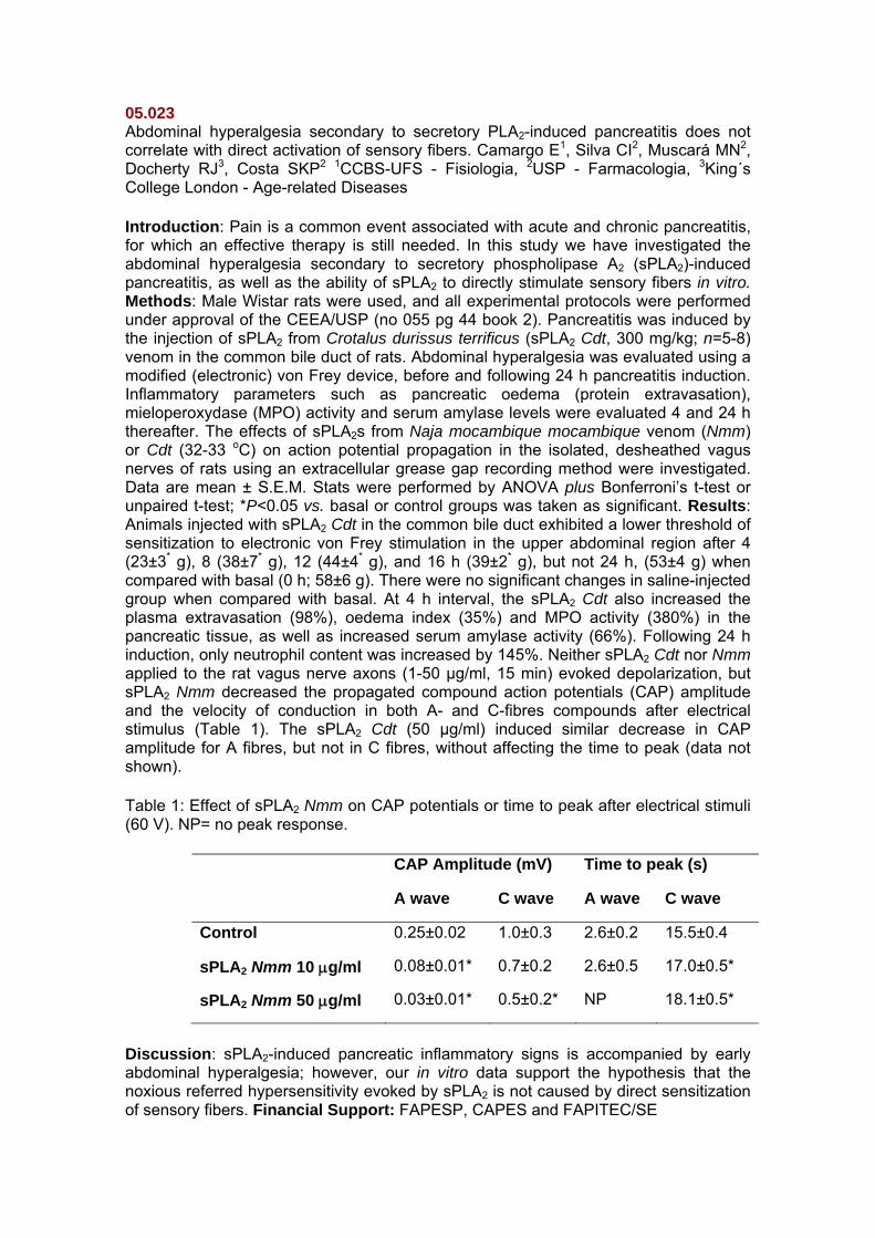
Introduction: Pain is a common event associated with acute and chronic pancreatitis, for which an effective therapy is still needed. In this study we have investigated the abdominal hyperalgesia secondary to secretory phospholipase A2 (sPLA2)-induced pancreatitis, as well as the ability of sPLA2 to directly stimulate sensory fibers in vitro. Methods: Male Wistar rats were used, and all experimental protocols were performed under approval of the CEEA/USP (no 055 pg 44 book 2). Pancreatitis was induced by the injection of sPLA2 from Crotalus durissus terrificus (sPLA2 Cdt, 300 mg/kg; n=5-8) venom in the common bile duct of rats. Abdominal hyperalgesia was evaluated using a modified (electronic) von Frey device, before and following 24 h pancreatitis induction. Inflammatory parameters such as pancreatic oedema (protein extravasation), mieloperoxydase (MPO) activity and serum amylase levels were evaluated 4 and 24 h thereafter. The effects of sPLA2s from Naja mocambique mocambique venom (Nmm) or Cdt (32-33 oC) on action potential propagation in the isolated, desheathed vagus nerves of rats using an extracellular grease gap recording method were investigated. Data are mean ± S.E.M. Stats were performed by ANOVA plus Bonferroni’s t-test or unpaired t-test; *P<0.05 vs. basal or control groups was taken as significant. Results: Animals injected with sPLA2 Cdt in the common bile duct exhibited a lower threshold of sensitization to electronic von Frey stimulation in the upper abdominal region after 4 (23±3* g), 8 (38±7* g), 12 (44±4* g), and 16 h (39±2* g), but not 24 h, (53±4 g) when compared with basal (0 h; 58±6 g). There were no significant changes in saline-injected group when compared with basal. At 4 h interval, the sPLA2 Cdt also increased the plasma extravasation (98%), oedema index (35%) and MPO activity (380%) in the pancreatic tissue, as well as increased serum amylase activity (66%). Following 24 h induction, only neutrophil content was increased by 145%. Neither sPLA2 Cdt nor Nmm applied to the rat vagus nerve axons (1-50 µg/ml, 15 min) evoked depolarization, but sPLA2 Nmm decreased the propagated compound action potentials (CAP) amplitude and the velocity of conduction in both A- and C-fibres compounds after electrical stimulus (Table 1). The sPLA2 Cdt (50 µg/ml) induced similar decrease in CAP amplitude for A fibres, but not in C fibres, without affecting the time to peak (data not shown).
Table 1: Effect of sPLA2 Nmm on CAP potentials or time to peak after electrical stimuli (60 V). NP= no peak response.
CAP Amplitude (mV) Time to peak (s)
A wave C wave A wave C wave
Control 0.25±0.02 1.0±0.3 2.6±0.2 15.5±0.4
sPLA2 Nmm 10 μg/ml 0.08±0.01* 0.7±0.2 2.6±0.5 17.0±0.5*
sPLA2 Nmm 50 μg/ml 0.03±0.01* 0.5±0.2* NP 18.1±0.5*
Discussion: sPLA2-induced pancreatic inflammatory signs is accompanied by early abdominal hyperalgesia; however, our in vitro data support the hypothesis that the noxious referred hypersensitivity evoked by sPLA2 is not caused by direct sensitization of sensory fibers. Financial Support: FAPESP, CAPES and FAPITEC/SE
05.024 Low frequency transcutaneous electric nerve stimulation interferes with the peripheral hyperalgesic response due to serotonin in rat paws. Santos CMF1, Francischi JN2, Sluka, KA3, Silva DS1, Antonio de Resende M1 1UFMG - Fisioterapia, 2UFMG - Farmacologia, 3University of Iowa - Medicine
Introduction: Transcutaneous electric nerve stimulation (TENS) is a noninvasive method to promote analgesia in acute inflammatory pain conditions (Sabino, GS et al, 2008). Although the use of TENS is very common, its analgesic mechanism is not fully understood. Since serotonin (5-HT) in the periphery is a proinflammatory and pronociceptive agent (Sufka et al, 1991), the purpose of this study was to investigate the mechanism of action of TENS at low frequency (LF: 10 Hz) in hyperalgesia induced by peripherally injected 5-HT. Methods: Experimental Animals Ethics Committee: 237/08. Male Holtzman rats (280-310g) were used in the present study (n= 5 to 8 rats/group). Intradermal injection of serotonin (10 μg/100μl) into the rat right hind paw at time zero constituted the painful stimulus and the nociceptive thresholds (in seconds by Hargreave’s method) were tested at times 0, 5, 15, 30 and 60 min. Serotonin receptor antagonist, methysergide (5-HT1 and 5-HT2 receptor antagonist) and pizotifen (5-HT1 and 5-HT2A receptor antagonist) were administered by subcutaneous injection into the rat’s dorsum (2 mg/kg) 30 minutes before 5-HT administration. LF TENS or switched-off TENS was applied on the right hind paw for 20 minutes and immediately thereafter 5-HT was administered. Control group was injected with the vehicle of drugs at the same time (saline). Results: Methysergide, pizotifen and LF TENS reduced at the same extent (about 100%) the hyperalgesic response to 5-HT in rat paws. Discussion and Conclusions: It is proposed, for the first time, that the analgesic mechanism of low TENS in the periphery may involve direct inhibition of serotonin-mediated pathways. References: Sabino, GS et al. J Pain. 9(2):157(2008). Sufka, KJ et al. Pharmacol Biochem Behav. 41:53(1991). Support: CNPq, CAPES, FAPEMIG
05.025 Characterization of a novel TRPV1 antagonist with analgesic activity in a model of neuropathic pain. Santos MLH1, Mendonça Tributino JL2, Mesquita CM2, Barreiro EJ3, Fraga CAM3, Castro NG1, Miranda ALP3, Guimarães MZP2 1CCS-UFRJ - Farmacologia Molecular, 2UFRJ - Farmacologia Básica e Clínica, 3LASSBio-FF-UFRJ - Fármacos
Introduction. The search for new targets for analgesics has revealed that the TRPV1 channel, expressed by nociceptors and activated by capsaicin (CAP), protons and high temperatures, is a valuable alternative to COX inhibitors. The compound LASSBio 881 was developed by molecular hybridization of LASSBio 294 (previously described as analgesic but with weak anti-inflammatory effects) with nimesulide, in an attempt to increase its anti-inflammatory activity. However, it was shown to be active only in the neurogenic phase of pain models and to bind cannabinoid receptors (Duarte et al., Bioorg. Med. Chem. 15:2421, 2007). Considering that TRPV1 can also be stimulated by endocannabinoids, we investigated whether LASSBio 881 was able to modulate this channel. Methods: Xenopus oocytes were removed under anesthesia and were injected with cRNA encoding TRPV1. The oocytes were then used in two-electrode voltage clamp electrophysiology experiments. Results are expressed as fractions of maximal CAP current (10 mM, mean ± SEM). Mice received subplantar co-injections of LASSBio 881 (5 nmol/paw) and CAP (1.6 mg/paw). The time animals spent in nociceptive behavior was recorded after CAP administration. The neuropathic pain model used was as described by Seltzer (Pain 43: 205, 1990) and consisted of tying up the dorsal portion of the sciatic nerve and treating daily with 100 mmol/kg LASSBio 881. The latency in withdrawal responses was determined with a thermal stimulus. For body temperature measurements, mice had their temperature taken before and after LASSBio 881 (300mmol/kg). License number DFBCICB 009. Results. LASSBio 881 was unable to activate TRPV1 but tended to inhibit proton currents (pH 5.5: 0.259 ± 0.054, n=5; LASSBio 881 in pH 5.5: 0.167 ± 0.056, n=5), which prompted the investigation as to whether it could antagonize CAP currents. Indeed, LASSBio 881 (20 mM) inhibited currents elicited by 1 mM CAP at TRPV1 (1 mM CAP 1.19 ± 0.112, n=9; CAP + LASSBio 881 0.589 ± 0.078, n=4, P<0.01), with a IC50 of 14 mM. LASSBio 881 was able to decrease time spent in CAP-elicited nociceptive responses when co-injected in mice’s paws (CAP 57.7s ± 5.5, CAP + LASSBio 881 37.8s ± 6.9, n=9, P<0.01). In addition, LASSBio 881 was able to decrease the delta of latency in withdrawal responses of animals with neuropathy (at day 9 after ligation; Vehicle 5.88 ± 0.36; LASSBio 881 2.77 ± 0.84, n=6, P<0.05). Finally, temperature readings remained unchanged after LASSBio 881 treatment (36.5 ± 0.09 before and 36.5 ± 0.08 after, n=10). Discussion: LASSBio 881 was able to antagonize CAP-elicited currents, suggesting it is an antagonist at TRPV1. This was confirmed in vivo by co-injections of CAP and LASSBio 881 in mice paws, in which the latter inhibited nociceptive responses. In addition, LASSBio 881 was effective in promoting analgesia in a mouse neuropathic pain model. These actions were not hindered by hyperthermia, a common side effect in other TRPV1 antagonists being developed. Together with data from another abstract that shows that LASSBio 881 is not an agonist at CB1, we propose that at least part of its antinociceptive properties are due to TRPV1 antagonism. Financial Support: Faperj and PRONEX.

05.026 Interleukin-33 mediates the increased mechanical sensitivity in the chronic constriction injury model of neuropathy in mice by activating ST2 receptors. Rodrigues, FC1, Souza GR2, Carvalho, TT1, Schivo IRS2, Xu D3, Liew FY3, Ferreira SH2, Cunha FQ2, Verri Jr WA1 1UEL - Ciências Patológicas, 2FMRP-USP - Farmacologia, 3University Glasgow - Immunology Infection, Inflammation,
Introduction: Neuropathic pain is caused by injury of the nervous system in peripheral or central sites. There are evidences of significant activity modulation of neurons, microglia and astrocytes by cytokines (e.g. TNFα) and activation of toll-like receptor-4 (TLR-4) after peripheral nerve injury. The interleukin (IL)-33 is a member of IL-1 family of cytokines, which acts through activation of the receptor ST2 to induce cytokine production. IL-33 mediates cutaneous and articular mechanical hyperalgesia in a model of auto-immune inflammation by inducing cytokine production, including TNFα. On the other hand, TNFα also induces the production of IL-33 in other systems. Although there is IL-33 expression in the spinal cord of mice and TLR-4 agonists such as LPS (lipopolysaccharide) stimulate glial cells to express IL-33 mRNA, there is no evidence on the role of IL-33 in pain mediation in the spinal cord. Therefore, we investigate the participation of the IL-33 in the development of neuropathic pain after chronic constriction injury of the sciatic nerve (CCI) in mice as well as whether TNFα could be inducing increased mechanical sensitivity via IL-33 production in the spinal cord. Materials and Methods: Balb/c (WT, ST2+/+) and ST2 deficient (-/-, Balb/c background) were submitted to CCI or sham operated, or received intrathecal injection of LPS, IL-33 or TNFα. The sensitivity to mechanical stimulus was evaluated by the electronic pressure-meter test (electronic von Frey) at different time points. The Ethics Committee of the University of Londrina approved the study (nº 2652/2009). Results and discussion: A significant increase of mechanical sensitivity was detected in ST2+/+ CCI mice compared to sham control group and ST2-/- CCI group (maximum reduction of 92% at 23rd day) during a 45 days period. A single intrathecal administration of IL-33 mimicked the increased sensitivity to mechanical stimulus detected in CCI ST2+/+ mice, but with a shorter duration. The IL-33 effect was absent in ST2-/- mice. The mechanical sensitivity induced by intrathecal injection of LPS or TNFα was significantly reduced in ST2-/- mice compared with ST2+/+. IL-33 is an important cytokine in the development of neuropathic pain after CCI. Considering that IL-33 has also a role in peripheral inflammatory pain, it is possible that the huge inhibition of CCI-induced pain detected in ST2-/- mice depends on peripheral and spinal IL-33. The neuropathic pain in the CCI model depends on TLR-4 activation and TNFα production, which seems to induce their effects via IL-33 action on ST2 receptors. Thus, the inhibition of IL-33 activity may contribute for the reduction of neuropathic pain after peripheral nerve injury. Financial support: CNPq, CAPES, FAPESP.

05.027 Analysis of the scratching behavior induced by the proteinase activated receptor-4 (PAR-4) agonist AYPGKF-NH2 in mice. Patricio ES, Costa R, Figueiredo CP, Motta EM, Calixto JB UFSC - Farmacologia
Introduction: Itch is a common symptom present in several cutaneous and systemic diseases. Recent works have been suggesting the involvement of both PAR-2 and -4 in the pruritus transmission (Reddy, J Neurosci., 28: 4331, 2008; Tsujii, J Pharmacol Sci., 108: 385, 2008). Nevertheless, studies are still required in order to characterize the mechanisms underlying the pruriceptive actions of PAR-4. Herein, we sought to investigate some of the mechanisms involved in the scratching behavior induced by the selective PAR-4 agonist AYPGKF-NH2 in mice. Methods: Female adult CD-1 mice (25-30 g, n=6) received a dorsal intradermal (i.d.) injection of saline (50 μl) containing the selective PAR-4 activating peptide AYPGKF-NH2 (30-500 nmol/site), the inactive control peptide YAPGKF-NH2 (200 nmol/site) or the compound 48/80 (C48/80; 10 μg/site). After the treatments, the frequency of scratching bouts with the hind-paws toward the injected site was quantified by 30 min (Ethics Committee protocol number: PP00032). Results: The i.d. administration of AYPGKF-NH2 (30-500 nmol/site) elicited a marked and dose-related scratching behavior response. The effective dose ranged from 200 to 500 nmol/site [p<0.05]. This response was maximal as early as 10 min after AYPGKF-NH2 (200 nmol/site) injection and gradually returned at the basal level within 30 min, an effect comparable to that produced by the positive control C48/80 (10 μg/site) [p<0.05]. The inactive sequence YAPGKF-NH2 (200 nmol/site) did not cause any significant alteration. Histological analysis demonstrated that the scratching behavior is not associated with dermis edema nor vascularization (p<0.05), as assessed by Casson´s staining protocol. The pretreatment with the mast cell degranulator C48/80 (1-10 μg/site, 4 days, once a day) or the mast cell stabilizer disodium cromoglycate (8 mg/kg, i.p., 6 days, once a day) partially prevented AYPGKF-NH2 (200 nmol/site)-elicited scratching behaviour in mice (68 ± 11% [p<0,05] and 50 ± 16% [p<0,05] of inhibitions, respectively). In line with these results, histological analysis of mouse skin sections, stained with toluidine blue dye, revealed that dermal mast cells intensely degranulated after AYPGKF-NH2 (200 nmol/site) or C48/80 (10 μg/site) injections when compared to YAPGKF-NH2 (200 nmol/site)- or saline-injected groups [p<0.05], respectively. Discussion: These findings provide evidences that PAR-4 activation, possibly on the mast cell surface, induce mast cell degranulation and consequent release of several degranulation products, which partially contribute to the scratching behavior. Studies are in progress to better clarify the mechanisms involved in this process. Supported by: CAPES/CNPq/ FAPESC/FINEP/PRONEX.
05.028 Antinociceptive activities of crude methanolic extract and the phases from Caulerpa racemosa (Caulerpaceae). Souza ET1, Queiroz AC2, Lorenzo, V. P.3, Aquino AB4, Cupertino-Silva YK4, Silva DJC4, Miranda, GEC5, Santo, B. V. O7, Oliveira CMC3, Alexandre-Moreira MS4 1LaFI-ICBS-UFAL, 2UFAL - Farmácia, 3UFPB - Tecnologia Farmacêutica, 4UFAL - Farmacologia e Imunidade, 5UFPB - Algas Marinhas,
Introduction: Caulerpa racemosa (Forsska°l) J. Agardh, a pan-tropical to temperate-warm water species widely distributed throughout the world, is a green alga (Bryopsidales) that was collected for the first time in1926 in the Mediterranean Sea by Hamel in the Sousse harbour, Tunisia. This seaweed have a number of pharmacological activities described in the literature, among which we mention: antitumor, anti-viral and antioxidant. As the seaweed are a important source biologically active natural products we evaluated the antinocipetive profile. Objective: In this study, we attempted to identify the possible antinociceptive and anti-inflammatory actions of n-butanol phase, chloroform phase, acetate phase and extract methanolic crude obtained of Caulerpa racemosa. Materials and Methods: We used animals of the Swiss line of both sexes (20-35g). Both the crude methanol extract as the phases were administered orally (p.o) at a dose of 100 mg/kg. The dipyrone (100 μmol/kg, p.o), indomethacin (100 μmol/kg, p.o) and morphine (15 μmol/kg, i.p) were used as drug standard. Been made functional models of nociception and inflammation, in vivo, such as: abdominal twitch induced by acetic acid, hot-plate test and nociceptive induced by formalin. The Ethical Committee of Federal University of Alagoas (N º 006443/2005-78) approved all experimental protocols described in this study. Results: In the assay writhing test, the phases n-butanol, chloroform, acetate and extract methanolic crude (100 mg/kg, p.o.), reduced the nociception produced by acetic acid by 47.39%, 70.51%, 76.11% and 72.24%, respectively. In assessing the antinociceptive and anti-inflammatory activity in the test of nociception induced by formalin, activity was observed in the neurogenic phase of crude methanolic extract (51.77%) and its phases, n-butanol (35.12%), chloroform (32.70%) and indomethacin (32.06%), with the exception of acetate phase. In the inflammatory phase only the acetate (75.43%) and indomethacin (47.83%) induced significant inhibition of response in this model. In the model of hot plate, only the chloroform showed statistically significant activity. Discussion: From the obtained results nociceptive models (acetic acid-induced writhings and formalin tests), it was noticed that crude methanolic extract and phases showed considerable antinociceptive activities. From these data it can be suggested that the acetate phase is significant anti-inflammatory profile, whose power has not yet been determined. In vivo inhibition of pain in hot plate the point is that the extract did not increase the latency time of response in animal, with the exception of the chloroform phase, this shows that this extract shows activity of supra-spinal analgesia. Conclusion: In conclusion, this study has shown that all phases and extract from Caulerpa racemosa possess significant antinociceptive and anti-inflammatory effects in laboratory animals at the doses an routes investigated. Acknowledgements: CNPq, FAPEAL, IM-INOFAR.
05.029 Evaluation of antinociceptive and anti-inflammatory activities of new (-)-cassine semi-synthetic derivatives. Silva DJC1, Melo GMA1, Cupertino-Silva YK1, Porfírio, APR1, Fossaluzza PC2, Nicastro PC2, Gomes CP2, Pivatto M3, Santos LA3, Bolzani V3, Viegas Jr C2, Alexandre-Moreira MS1 1UFAL - Farmacologia e Imunidade, 2UNIFAL - Fitoquímica e Química Medicinal, 3NuBBe-UNESP-Araraquara – Química Orgânica
Introduction: Phytochemical studies on the flowers and green fruits of Senna spectabilis (Fabaceae) had furnished numerous bioactive piperidine alkaloids, among these, (-)-cassine as the major constituent. Ethnopharmacological data and Phytochemical studies guided by pharmacological evaluation have pointed to analgesic, anti-inflammatory, cytotoxic, anesthetic and antibiotic activities for this kind of metabolites. These observations prompted us to evaluate the peripheral antinociceptive and anti-inflammatory activities of two semi-synthetic derivative series prepared from (-)-cassine, using classical pain and inflammation models in mice. Methods: Swiss mice (20 to 35 grams) were used in the experiments. All animals used in this study were handled in accordance with standards established by the International Ethics Committee for handling of animals in models of inflammation submitted for approval by the Research Ethics Committee of UFAL (No.: 006443/2005-78).The substances were administered intraperitoneally at the dose of 100 μmol/kg. Dypirone (30 μmol/kg), indomethacin (100 μmol/kg) and morphine (15 μmol/kg) were used as standard drugs. The antinociceptive and anti-inflammatory activities were evaluated by acetic acid-induced abdominal writhing, formalin-induced murine nociception model, hot plate assay and Zymosan-induced peritonitis assay. Results: The results showed that new eight aromatic esters derivatives of (-)-cassine series (100 mmol/kg, i.p.) were able to inhibit contortions in 80% when compared to the standard drug used, dipyrone. In addition, another series of arylhydrazones derivatives of (-)-cassine also showed significative antinociceptive properties as evidenced for LFQM-21 - ID50=8.22 mmol/kg, LFQM-22 - ID50=1.9 mmol/kg, LFQM-26 - ID50=2.11 mmol/kg e LFQM-27 - ID50=6.15 mmol/kg, showing maximum effect of 100%, compared to dipyrone (ID50 = 0.64 mmol/kg, maximum effect of 97%). Preliminary, the results showed statistically significant results for (-)-cassine derivatives at 100 mmol/kg in the second phase of formalin-induced murine nociception model, when compared to indomethacine only induced by formalin, thus showing that they were able to modulate the inflammatory response. In hot plate model, the results weren´t estatistically significant. In another assay, using Zymosan-induced peritonitis, all substances tested had inhibited the cellular migration significantly, and the most active compounds were LFQM-29 (65.5%*) and LFQM-30 (60.6%*). Discussion: These results clearly indicates that the structural modifications on the original prototype, (-)-cassine, led to new derivatives with a very high potency and efficiency, being able to interfere in the antinociceptive response. Antinociceptive properties were also evaluated using the formalin-induced murine nociception model that elicits a neurogenic phase, followed for an inflammatory event. In the hot plate test no significant differences between pre-treatment and treatment latency values were observed. These data allowed us to suggest a greater performance of these derivatives, considering that (-)-cassine only showed peripheral antinociceptive activity. Conclusion: In the view of the above data, our results showed an important increase of antinociceptive and anti-inflammatory activities for the new derivatives of (-)-cassine and additional studies are being conducted in order to elucidate possible mechanisms of activity and to evaluate the acute and sub-acute toxicity of these derivatives. Acknowledgements: CNPq, FAPEMIG, FAPEAL and IM-INOFAR.
05.030 Antinociceptive properties of hexanic extract of Pterodon pubescens Benth. seeds. Motta NAV1, Reis RC1, Novis CS1, Romeiro LAS2, Miranda ALP3, Brito FCF1 1LAFE-UFF - Fisiologia e Farmacologia, 2UCB - Química Bioorgânica e Medicinal, 5FF-LASSBio-UFRJ – Fármacos
Introduction: Pterodon pubescens Benth., known as “sucupira branca”, is a native tree widely distributed over the central region of Brazil (Coelho, L. P. et al., 2005, J. Ethnopharmacol., 98, 109-116) largely employed at folk medicine for their anti-inflammatory properties (Mors et al., 1967, Science.,157, 950–951). Coelho et al. (2005) have shown important anti-inflammatory properties associated to Pterodon hidroalcoholic extract at arthritis model and the oil extract have presented antinociceptive activity at writhing test and at formalin assay in mice. Searching for a better characterization of the pharmacological properties of “sucupira branca”, the hexanic extract (HE) was obtained and this work reports the evaluation of its antinociceptive properties, using the writhing and hot-plate models of nociception. Methods: The analgesic activity was determined in vivo by the adbominal constrictions test induced by acetic acid 0.6% (0.1mL/ 10g) in mice (Whittle, B. A., 1964, Br. J. Pharmacol.; 22: 246-253) and by the hot-plate test performed according to Kuraishi, Y. et al., 1983, Brain Res., 273, 245-252. Swiss mice of both sexes (18 – 25g) (n= 10 for each experiment group) were pre-treated orally (p.o.) with HE (10 – 300 µg/ kg), dissolved in ethanol 10% with 10% Tween 80 (vehicle). At abdominal constriction test acetic acid (0.6%) was administered i.p. one hour after the administration of HE. Ten minutes following i.p. acetic acid injection, the number of constrictions per animal was recorded for 20 minutes. Control animals received an equal volume of vehicle. Analgesic activity was expressed as a percentage of inhibition of constrictions when compared with the vehicle control group. At hot-plate test, the mice were first placed on a hot plate set at 55.0 ± 0.1oC to obtain two baseline response latencies before drug administration. The mice were observed for licking either their fore- or hind limb in response to the heat. The mice were tested again at the appropriate time (30, 60, 90 and 120 min) after being administered HE or vehicle (p.o.). Antinociception was expressed as a difference between latency time pos-treatment and the pre-treatment latency time. We performed the analysis of variance (one-way ANOVA) and tested the statistical significance of differences between groups by Dunnett´s test (p < 0.05). Results: At a dose of 100 µg/kg, HE significantly inhibited at 31.6% the abdominal constrictions acetic acid induced and at hot-plate test it has enhanced the latency time at 31.2% and 45.2% in times 30 and 60 minutes, respectively. We have compared the HE with the ethanolic extract, and at 100 µg/kg the ethanolic extract did not present any activity at these two models. Discussion: The oral treatment of animals with HE, induced antinociception when assessed by the acetic acid-induced constrictions, a useful method to screen both peripherally and/or centrally acting analgesic activities. At hot-plate test, a useful test to evaluate the role of supraspinally mediated responses to noxious heat, HE also presented an important analgesic activity. These results contributes to elucidates the potent analgesic profile of Pterodon pubescens Benth. Financial Support: FAPERJ, PROPPI/UFF.
05.031 Evaluation of exercise and KCl supplementation on blood pressure and nociceptive threshold in hypertensive rats. Galdino GS, Lopes AMC, França VM, Duarte IDG, Perez ACP UFMG - Farmacologia
Introduction: According to the World Health Organization, arterial hypertension is a major cause of death in the world1. Many studies have demonstrated that hypertensive humans and animals presented a lower sensibility to pain2. Physical exercise and potassium supplementation are among the main non-pharmacological treatments for this disorder 3. To the present moment, however, no study has been conducted to associate the effect of exercise with potassium supplementation on the arterial hypertension and nociceptive threshold. Methods: Male normotensive Wistar rats and SHR were used. The animals were divided in eight groups, submitted or not to potassium supplementation and/or exercise. KCl (10%) was administered orally and the exercise was performed in a treadmill, every day for 4 weeks4,5. Nociceptive threshold was measured by the Paw-withdrawal test and the blood pressure by a noninvasive tail-cuff method6,7. Results: KCl diet did not alter blood pressure nor the nociceptive threshold in all eight groups. Exercise produced a significant reduction of the nociceptive threshold (p < 0.05) in SHR. However, this effect was inhibited by KCl. Discussion and conclusion: Physical exercise normalized the nociceptive threshold in SHR. This effect may be very important for early detection of angina and myocardial infarction in hypertensive subjects meanwhile KCl supplementation can avoid this benefit. Animal experimentation ethics committees protocol (CETEA/UFMG):185/2007. Financial Support: FAPEMIG and CNPQ
05.032 Opioid receptor expression is regulated by peripheral injury in both dorsal root ganglia and nerve paw of rats. Zambelli VO1, Gutierrez VP1, Parada CA2, Cury Y1 1Instituto Butantan - Fisiopatologia, 2UNICAMP - Farmacologia
Introduction: The efficacy of opioid drugs is enhanced in the presence of tissue injury and inflammation. Previous data of our group demonstrated that, in rats, prostaglandin E2 (PGE2, intraplantar/i.pl.) and chronic constriction injury (CCI) of the sciatic nerve increase the peripheral antinociceptive efficacy of opioid agonists and of Crotalphine (CRP), a peptide obtained from Crotalus durissus terrificus snake venom. CRP has a local antinociceptive effect mediated by activation of κ- opioid receptor in PGE2-induced hyperalgesia model or κ- and δ- opioid receptor in CCI model. The aim of this study is to characterize some of the mechanisms involved in the increase of the analgesic efficacy of opioids caused by inflammation and tissue injury. For this purpose the effect of PGE2- induced hyperalgesia and CCI on opioid receptor expression in dorsal root ganglia (DRG) and nerve paw (NP) was evaluated. Methods: The expression of μ, κ and δ-opioid receptors was evaluated by immunoblotting, in DRG or NP (ipsilateral and contralateral to injury), of male Wistar rats, 3h after i.pl. injection of PGE2 (100 ng/paw) or 14 days after CCI. This study was approved by the Ethical Committee of Butantan Institute (386/07). Results: PGE2 increases the expression of μ- and κ-opioid receptors in NP (43% and 71%, respectively) and decreases (30%) the expression of δ-opioid receptors, when compared to naïve rats. μ-opioid receptor expression is also increased in the ipsilateral and contralateral DRG (79 and 27%, respectively), while κ-opioid receptor expression is increased only in the ipsilateral DRG (168%), when compared to naïve rats. CCI up-regulates μ-opioid receptors in NP (27%) and DRG (ipsilaterally and contralaterally, 49 and 20%, respectively) and δ-opioid receptors in the ipsilaterally DRG (35%). On the other hand, κ-opioid receptors are down-regulated by CCI in both NP (51%) and DRG (21%), when compared to naïve rats. Discussion: Peripheral opioid receptor expression is distinctly regulated by the presence of acute or chronic tissue injury. The different pattern of κ and δ opioid receptors expression caused by acute and chronic injury may contribute to the comprehension of the mechanisms involved in the activation of opioid receptors by CRF in PGE2-induced hyperalgesia and CCI models. Financial Support: FAPESP (07/03404-4, 07/00135-2)
05.033 Participação dos sistemas opióide e canabinóide no efeito hipoalgésico periférico causado pelo celecoxibe e sua dependência do citoesqueleto. Paiva-Lima P1, Rezende RM2, Francischi JN1 1UFMG - Farmacologia, 2UFMG - Fisiologia e Farmacologia
Introdução: Em estudos anteriores, nosso grupo mostrou a participação de componentes do citoesqueleto (Lima et al, 2006 e Francischi et al., 2007) na elevação do limiar de deflagração da resposta nociceptiva acima dos valores basais (hipoalgesia) após administração sistêmica de coxibes (Francischi et al, 2002). A atuação de opióides endógenos sobre esta resposta também foi demonstrada em pré-tratamentos com celecoxibe (CX) tanto por via sistêmica (s.c.) quanto central (i.c.v.) (França et al., 2006; Rezende et al., 2009). Em adição, trabalhos de Ibrahim et al (2003 e 2005) demonstraram a inter-relação dos sistemas opióide e canabinóide endógenos na resposta antinociceptiva periférica. Os objetivos deste trabalho foram: avaliar a ocorrência de efeito hipoalgésico após administração periférica de diferentes compostos e verificar a participação de microfilamentos e/ou dos sistemas opióide e canabinóide sobre este efeito. Métodos: As patas direitas foram injetadas com: CX (30, 100 e 300 µg), β-endorfina (βEN 0.5; 2 e 10 µg), ACEA (6 e 60 µg) e JWH015 (JWH 3 e 33 µg) e, após ½ h receberam estímulo pró-inflamatório (carragenina CG-250 µg) ou salina (SAL). Na segunda parte dos experimentos os ratos foram pré-tratados com: Citocalasina B (CTB 1µg); Naltrexona (NTX 100 µg); SR141617A (SR1 2 µg), SR144528 (SR2 3 µg) ou SAL. A resposta nociceptiva foi avaliada em algesímetro de pressão (Ugo-Basile - Randall & Sellito, 1957) nos tempos 15, 30, 60, 120, 180 e 240 min após a CG. Protocolo CETEA: 128/07. Resultados: Os compostos CX, βEN, ACEA e JWH mostraram efeito hipoalgésico periférico de maneira dose-dependente. Injeções prévias de CTB, NTX, SR1 e SR2 preveniram a hipoalgesia causada pelo CX. A CTB foi também capaz de prevenir totalmente o efeito hipoalgésico da βEN e do JWH (agonista CB2), mas não do ACEA (agonista CB1). Discussão: A hipoalgesia causada pelo CX se mostrou semelhante a aquela induzida por βEN, ACEA e JWH e foi prevenida pela administração de antagonistas de receptores opióides e canabinóides, demonstrando a participação destes sistemas no efeito hipoalgésico observado. A disfunção periférica de microfilamentos causada pela CTB foi capaz de prevenir totalmente o efeito hipoalgésico causado por CX, βEN e JWH, porém apenas parcialmente o causado pelo ACEA, mostrando o papel crucial do citoesqueleto na resposta antinociceptiva periférica, e a maior participação dos receptores CB2 nesta resposta. Estes resultados mostraram que o citoesqueleto medeia a hipoalgesia causada por CX, opióides e canabinóides sugerindo o sinergismo entre estes sistemas endógenos, perifericamente. References: (1) Lima at al., 38º CBSBFTE, 75, 2006; (2) Francischi et al., Inflam. Res. 56:89, 2007. (3) Francischi et al. Br J Pharmacol. 137:837, 2002; (4) França et al. Neuropharmacol. 51:37, 2006. (5) Rezende et al. Pain 142:94, 2009; (6) Ibrahim et al., PNAS 100:10529, 2003; (7) Ibrahin et al., PNAS 102:3093, 2005; (8) Randall & Sellito, Arch. Intern. Pharmac; 113:233, 1957. Support: CNPq, CAPES, FAPEMIG.
05.034 Fructose-1,6-bisphosphate reduces inflammatory pain-like behaviour in mice: role of adenosine acting on A1 receptors. Valério DA1, Ferreira FI2, Cunha TM3, Alves-Filho JC3, Lima FO3, Rodrigues de Oliveira J4, Ferreira SH3, Cunha FQ3, Queiroz RHC2, Verri WA, Jr5 1FMTM-UFTM, 2FCFRP-USP - Toxicologia, 3FMRP-USP - Farmacologia, 4PUCRS - Biociências, 5UEL - Ciências Patológicas
Introduction: D-Fructose-1,6-bisphosphate (FBP) is a high energy intermediate in the glycolytic pathway, exerting pharmacological actions on inflammation by inhibiting cytokine production1 or interfering with adenosine production2. Then, the possible antinociceptive effect of FBP and its mechanism of action in the carrageenin paw inflammation model in mice were addressed, focusing on the two mechanisms described above. Methods: Mechanical hypernociception was evaluated by the electronic pressure-metre test3; cytokine levels were measured by ELISA as described previously4 and adenosine was determined by high performance liquid chromatography whose experimental procedure was validated for this purpose. The use of male Swiss mice (25g) was approved by the ethics committee (protocol n° 07.1.570.53.0). Results and discussion: Pretreatment of mice with FBP reduced hypernociception induced by intraplantar injection of carrageenin (up to 54%), tumour necrosis factor α (40%), interleukin-1 β (46%), CXCL1 (33%), prostaglandin E2 (41%) or dopamine (55%). However, FBP treatment did not alter carrageenin-induced cytokine (tumour necrosis factor α and interleukin-1 β) or chemokine (CXCL1) production. On the other hand, the antinociceptive effect of FBP was prevented by systemic and intraplantar treatment with an adenosine A1 receptor antagonist (8-cyclopentyl-1,3-dipropylxanthine), suggesting that the FBP effect is mediated by peripheral adenosine acting on A1 receptors. Giving FBP to mice increased adenosine levels in plasma, and adenosine treatment of paw inflammation presented a similar antinociceptive mechanism to that of FBP. In addition to anti-inflammatory action, FBP also presents an antinociceptive effect upon inflammatory hyperalgesia. Its mechanism of action seems dependent on adenosine production but not on modulation of hypernociceptive cytokine/chemokine production. In turn, adenosine acts peripherally on its A1 receptor inhibiting hypernociception. Therefore, FBP may have possible therapeutic applications in reducing inflammatory pain. References: 1. Markov AK. Transplantation, v.74, p.1651, 2002. 2. Sola A. J Leukoc Biol, v.73, p.74, 2003. 3. Cunha TM. Proc Natl Acad Sci USA, v.102, p.1755, 2005. 4. Valério DA. Eur J Pharmacol, v.562, p.155, 2007. Financial Support: CAPES and FAPESP.
05.035 Antiallodynic effects of acupuncture after spinal nerve ligation-induced neuropathic pain in rats. Cidral Filho FJ1, Werner MFP2, Silva MD1, Córdova MM1, More AOO1, Santos ARS1 1UFSC - Ciências Fisiológicas, 2UFSC - Farmacologia
Introduction: Neuropathic pain is of major clinical concern as it affects 7% to 8% of the general population, with treatment often unsatisfactory due to side effects or insufficient analgesia of currently available drugs (Bouhassira D. et al., Pain, 136, 380, 2008). In the past years, alternative therapeutic approaches have been systematically investigated, with emphasis to acupuncture and related techniques (eletroacupuncture and lasertherapy). On light of these data, we investigated the effect of acupuncture on mechanical allodynia in the spinal nerve ligation (SNL) model of neuropathic pain in rats. Methods: Spinal nerve ligation was induced in male Wistar rats (200-250 g, n=5-6) by placing tight 6-0 silk thread sutures unilaterally around L5 and L6 spinal nerves (no ties in sham-operated rats). As shown previously (Werner et al., Neuropharmacology, 53, 48, 2007), SNL reduces paw withdrawal threshold (g) to mechanical stimulation (von Frey hairs, up-down method). Two different groups (with treatments initiating 5 or 14 days after surgery) were chronically treated with acupuncture. Acupuncture was performed in awake rats by inserting a needle at Zusanli (ST36) and Sanyinjiao (SP6) acupoints, for 10 min, three times a week in a total of 10 interventions for each group. The experimental procedures were previously approved by the Committee on the Ethical Use of Animals of the UFSC (protocol number: PP00208). Results: When acupuncture treatment initiated 5 days after SNL, mechanical allodynia was significantly reduced on all treatment days, with inhibition ranging from 62 ± 8% to 96 ± 3%, and effect lasting for up to 4h. With treatment initiating on the 14th day after SNL a less pronounced effect was observed. Even though mechanical allodynia was reduced on all treatment days, inhibition ranged from 54 ± 8% to 84 ± 8%, and effect lasted for up to 2h. In both groups, chronic acupuncture treatment did not induce tolerance. Conclusion: Results indicate that acupuncture might be a viable alternative therapy for the treatment of neuropathic pain, with optimum results obtained in the case of early treatment. Financial Support: CNPq, CAPES.
05.036 Neutrófilos estão envolvidos na gênese da dor pós-incisional. Carreira EU, Cunha FQ, Ferreira SH, Cunha TM FMRP-USP - Farmacologia
Introdução: A dor pós-operatória é um tipo muito comum de dor aguda e pode provocar sofrimento desnecessário aos pacientes. Recentes estudos têm demonstrado um componente inflamatório nesse tipo de dor. No entanto, os mecanismos envolvidos não estão totalmente elucidados. Recentemente, nosso grupo demonstrou que a migração de neutrófilos para o foco inflamatório é fundamental para a gênese da hipernocicepção inflamatória. Nesse contexto, o objetivo do presente estudo foi avaliar o papel da migração de neutrófilos na dor pós-operatória. Métodos: Os animais foram anestesiados e posteriormente submetidos à cirurgia, que consistia em uma incisão longitudinal de 1 cm em ratos (Wistar, 180g-200 g) e 0,5 cm em camundongos (C57BL/6, 20g-30 g) através da pele e da fáscia da pata, iniciada a 0,5 cm da extremidade proximal do calcanhar em direção aos dedos. O músculo plantar foi elevado e incisionado longitudinalmente e a incisão foi suturada. A hiperalgesia foi avaliada utilizando-se o teste de von Frey eletrônico e o estímulo mecânico foi aplicado na região adjacente à incisão. O papel dos neutrófilos foi avaliado em ratos dos quais estas células foram eliminadas com a administração de Vimblastina sulfato i.v. (0,8 mg/kg), 72 horas antes da cirurgia. O teste da atividade da Myeloperoxidase (MPO) foi utilizado a fim de quantificar a migração de neutrófilos para o local da incisão. Resultados: A incisão na pata tanto de ratos quanto de camundongos causou uma diminuição do limiar nociceptivo mecânico, a qual foi estatisticamente significante até 72 horas após a cirurgia. Esta diminuição estava associada a um aumento na atividade de MPO, sugerindo uma intensa migração de neutrófilos para o local da lesão. Além disso, foi observado que animais cujos neutrófilos haviam sido eliminados pelo tratamento com vimblastina, apresentavam uma redução tanto na hiperalgesia quanto na atividade de MPO no local da lesão. Discussão: Estes resultados indicam que a migração de neutrófilos desempenha um papel crucial na gênese da dor pós-incisional. Dessa forma, pode-se sugerir que a inibição da migração de neutrófilos possa ser um alvo no controle da dor pós-operatória.
05.037 Involvement of protein kinase-c on antinociceptive effect of a (1-3),(1-6)-linked B-glucan isolated from Pleurotus pulmonarius (Fr.) Quel. Baggio CH1, Freitas CS1, Marcon R2, Werner MFP3, Rae GA2, Smiderle FR4, Sassaki GL3, Iacomini M4, Marques MCA1, Santos ARS4 1UFPR - Farmacologia, 2UFSC - Farmacologia, 3UFPR - Bioquímica, 4UFSC - Ciências Fisiológicas
Introduction: b-glucan is a polysaccharide isolated from Pleurotus pulmonarius (Fr.) Quel., an edible mushroom known as “usuhiratake” in Japan. This study evaluated the possible mechanisms of action involved on antinociceptive effect of a (1-3),(1-6)-linked b-glucan (GL) in chemical models of nociception in mice. Methods: Swiss mice (30-40 g, n= 8-12/group) both sexes were used (Ethics committee number: 23080.006747/2008-90). The GL was evaluated in capsaicin (1.6 mg/paw), cinnamaldehyde (10 nmol/paw), menthol (1%/paw), acidified saline (pH 1.98/paw) and PMA (500 pmol/paw) model of pain. The activation of protein kinase C was evaluated by Western blot analysis. The participation of opioid system was evaluated with pretreatment of naloxone (1 mg/kg, i.p.), after 20 min was administered the GL (10 mg/kg, i.p.) or morphine (1 mg/kg, s.c.) and after 30 min was injected capsaicin. The involvement of capsaicin-sensitive fibers was evaluated 7 weeks after fibers depletion with capsaicin (50 mg/kg, s.c.), the mice were treated with either GL (3 mg/kg, i.p.) or vehicle, 30 min before submitting them to the acetic acid test. Results: Intraperitoneal administration of GL potently inhibited the nociception induced by intraplantar injection of capsaicin, cinnamaldehyde, menthol, acid saline and PMA, with ID50 values of 8.1 (5.9-11.3), 13.8 (9.6-19.8), 0.19 (0.10-0.37), 29.8 (20.0-44.4) and 17.5 (11.0-28.0) mg/kg, respectively. Western blot analysis revealed that GL treatment also prevented the translocation of protein kinase C (PKC)e following intraplantar injection of PMA in mice. The antinociceptive effect of GL on capsaicin-induced nociception was not affected by the pretreatment of mice with naloxone (a nonselective opioid receptor antagonist). Furthermore, neonatal pretreatment of mice with capsaicin did not modify the antinociception caused by GL in the acetic acid-induced visceral pain. Discussion: Collectively, present results demonstrate that GL isolated from P. pulmonarius produces antinociception when assessed on capsaicin-, cinnamaldehyde-, menthol-, acid saline- and PMA-induced pain in mice, through mechanisms that seem to involve an interaction with transient receptor potential (TRP) channels and protein kinase C pathways. Financial support: CAPES, CNPq, PAPESC, UFSC, UFPR.
05.038 Antinociceptive effect of guttiferone an isolated from Rheedia achachairu Rusby (Clusiaceae) seeds in mice. Alves DR1, Dal Molin MM2, Silva S1, Delle Monache F3, Cechinel Filho V4, Niero R5, Quintão NLM2 - 1UNIVALI - Ciências da Saúde, 2UNIVALI - Ciências Farmacêuticas, 3UIN - Farmacologia, 4CCS-NIQFAR-UNIVALI - Ciências Farmacêuticas, 5UNIVALI - CCS/NIQFAR
Introduction: Rheedia achachairu Rusby (Clusiaceae) belongs to the Garcinia genus (ex-Rheedia). Guttiferone A, one of the compounds isolated from R. achachairu, presents important pharmacological effects, including anti-oxidant and anti-HIV properties. However, few scientific studies are found in the literature evaluating its analgesic properties. The present study has the aim of investigating the possible antinociceptive effects of Guttiferone A isolated from R. achachairu Rusby in mice. Methods: Guttiferone A was isolated from seeds methanolic extract of R. achachairu by column chromatography (CC) over silica gel and identified by spectroscopy data in comparison with literature. Male Swiss mice were used (25-35g, N=6-8) throughout the study. The animals were pre-treated intraperitoneally (i.p.) with Guttiferone A (1-30 mg/kg) or saline and after 30 min they were submitted to the writhing test induced by acetic acid (0.6 %) or spontaneous nociception tests induced by formalin (2.5 %), capsaicin (1.6 µg/paw) or glutamate (30 µmol/paw). Mice were individually observed and the licking behavior in the injected paw was timed and considered as nociceptive index. Dipyrone (60 mg/kg, i.p.) and acetaminophen (30 mg/kg, i.p.) were used as positive controls. All the procedures used in the present study were approved by the Animal Ethics Committee of UNIVALI (Protocol numbers 001/2008 UNIVALI). Results: The pre-treatment with Guttiferone A (1-10 mg/kg, i.p.) was able to significantly reduce, in a dose dependent manner, the abdominal writhing induced by acetic acid (37 ± 6 %, 53 ± 7 % and 73 ± 5 %), with ID50 of 4.7 (2.9 – 7.6) mg/kg. Acetaminophen also significantly diminished the abdominal writhing, with inhibition of 62 ± 10 %. In the formalin-induced nociception, it slightly but significantly inhibited the first and second phases of the test, with maximal inhibition of 24 ± 5 % and 34 ± 5 %, respectively. Acetaminophen was also effective against the both phases (26 ± 6 % and 37 ± 8 %). Guttiferone A (1–10 mg/kg) also inhibited the capsaicin- and glutamate-induced nociception, with inhibitions of 44 ± 7 %, and 39 ± 5 %, respectively. Dipyrone, the positive control used for these tests, was also effective (42 ± 6 %; 65 ± 9 %). Discussion: These results show for the first time the antinociceptive effect of the compound Guttiferone A, isolated from R. achachairu seeds extract, as demonstrated by the reduction of licking behavior in mice submitted to several nociception tests. However, additional studies are necessary to delineate the antinociceptive effects of Guttiferone A, as well as to identify its mechanism of action. Financial Support: CNPq; FAPESC-SC; ProPPEC/UNIVALI
05.039 Neuropatia sensitiva periférica induzida pelo tratamento crônico com o agente antineoplásico oxaliplatina em camundongos. Pontes RB, Lino JA, Paiva MN, Rolim FE, Ribeiro RA, Vale ML UFC - Fisiologia e Farmacologia
Objetivos: Oxaliplatina (OX) é a 3ª geração de agentes platinos com amplo espectro de atividade antitumoral. Exibe potente atividade citotóxica, incluindo câncer colorretal, ovariano e pulmonar. Dentre os efeitos tóxicos estão: laringoespasmo, náuseas, vômitos, fadiga e neuropatia periférica, foco deste trabalho. O presente estudo objetivou desenvolver um modelo experimental para o estudo da neuropatia por OX em camundongos, animais geneticamente mais semelhantes ao humano, econômicos e dado a existência de espécies nocaute para vários fatores. Métodos e resultados: O estudo foi aprovado pelo Comitê de Ética em Pesquisa Animal da UFC (protocolo nº 70/07). Camundongos Swiss machos (20-40g) foram tratados com OX (1-4 mg/kg) por 4 semanas paralelamente aos testes de hipernocicepção e alodínia utilizados para avaliar o desenvolvimento da neuropatia sensitiva e Rota-Rod para verificar algum comprometimento motor. A hipernocicepção e alodínia térmica foram avaliadas pelo teste de imersão da cauda (TIC) em água fria (4 ou 10ºC) e em água aquecida (46 ou 42ºC). O teste de hipernocicepção plantar mecânico (HPM; Von Frey) consistiu na estimulação das patas traseiras com um sensor de força (g) até a sua retirada por um movimento de “flinch”. Foi ainda verificado a ação analgésica da carbamazepina (CZP), oxcarbazepina (OZP), gabapentina (GABA) e indometacina (INDO) no TIC água fria. No TIC 4ºC houve uma diminuição significativa (p<0,05) no limiar nociceptivo a partir do 35º dia atingindo o máximo na dose de 1mg/kg (103%) comparado ao grupo controle. No TIC alodínea pelo frio (10ºC) foi observado uma diminuição significativa (p<0,05) no limiar nociceptivo a partir do 35º dia atingindo o máximo na dose de 1mg/kg (88,8%) comparado ao grupo controle. No TIC 46ºC foi observada uma diminuição significativa (p<0,05) no limiar nociceptivo a partir do 28º dia atingindo o máximo nas doses de 1mg/kg (94,4%) comparado ao grupo controle. No TIC alodínea pelo quente (42ºC) foi observado uma diminuição significativa (p<0,05) no limiar nociceptivo a partir do 28º dia atingindo o máximo na dose de 2mg/kg (92,4%). No HPM houve uma diminuição significativa (p<0,05) no limiar nociceptivo a partir do 21º dia atingindo o máximo na dose de 2mg/kg (236%) comparado ao grupo controle. No teste Rota-Rod nenhuma variação significativa foi observada em nenhum dos grupos, indicando a ausência de comprometimento motor. O tratamento com CZP (0,3-30 mg/kg), OZP (0,3-100mg/kg) e GABA (6-54mg/kg) aumentou o limiar nociceptivo, indicando efeito analgésico, sendo as doses de maior efeito a de 30mg/kg (109 %) para a CZP; a de 100mg/kg (59%) para a OZP e a de 54mg/kg (76,6%) para a GABA. Contudo, INDO (1-4mg/kg) não demonstrou atividade analgésica nesse modelo. Conclusão: Os dados sugerem que o tratamento crônico com OX induz hipernocicepção e alodínea térmica e mecânica no TIC e HPM. A dose que produziu melhor resposta nos testes térmicos foi a de 1mg/kg e no HPM a de 2mg/kg. O efeito analgésico dos anticonvulsivantes CZP, OZP e GABA somado a ausência de efeito do antiinflamatório INDO evidencia a presença de neuropatia. Em adição, a ausência de comprometimento motor sugere uma neuropatia predominantemente sensitiva. Apoio Financeiro: CNPq
05.040 Antinociceptive effects evoked by acupuncture through mechanical and photonics stimulations in acupoint zusanli (ST36) of rats. Erthal V1, Silva MD2, Santos ARS2 1UFSC - Neurociências, 2UFSC - Ciências Fisiológicas
Introduction: The present study examined the antinociceptive effect evoked by acupuncture through mechanical and photonic stimulations in acupoint Zusanli (ST36) in rats. Methods: The experiments were conducted after the approval of the Ethics Committee in Animal Research of the Pontifícia Universidade Católica do Paraná, under the register CEUA/PUCPR number 198. Mechanical stimulation was carried out with 0.25 X 0.7 mm needles, which were retained for 15 min, and photonic stimulation was applied with an AsGaAl laser equipment of 830 nm wavelength, 30mW, for 6 s, both in St36 acupoint. Results: Mechanical and photonic stimuli applied to this acupoint, were able to significantly reduce (p< 0,001) the abdominal constriction caused by acetic acid (p< 0,001), with inhibitions of 42±6%, 36±2% and 39±6% for acupuncture, laser stimulation and when both stimuli were associated, respectively. Furthermore, both types of stimuli caused significant inhibition of the neurogenic (phase I, mechanical: 51±7%, photonic: 48±7%, and both: 54±4%) and inflammatory (phase IIA, mechanical: 49±3%, photonic: 34±4% and both: 45±3% and phase IIB, mechanical: 73±9%, photonic: 83±6% and both: 78±4%) nociception induced by formalin. The antinociception caused by mechanical and photonic stimulation of ST36 acupoint in the acetic acid test was significantly attenuated by i.p. treatment of mice with naloxone, pindolol, ketanserin or ondansetron. Discussion: Together, these results provide experimental evidence indicating that mechanical and photonic stimulation of ST36 acupoint produce important antinociceptive activity in experimental models of nociception in rats through mechanisms that involve activation of opioid and serotonergic systems. Financial support: CNPq, CAPES, FAPESC.
05.041 Synergic effect between acupuncture and lower dose of morphine after plantar incision surgery in mice. Silva MD1, Mazzardo L2, Werner MFP3, Cidral Filho FJ4 1UFSC - Ciências Fisiológicas, 2UFSC - Fisiologia, 3UFSC - Farmacologia, 4UFSC - Neurofisiologia
Introduction: In a previous study, we reported that acupuncture in Spleen 6 (SP6) acupoint promotes analgesia in several animal models of nociception. The present study aims to investigate the possible antinociceptive synergistic effect of acupuncture and lower dose of morphine in a model of post-surgical incisional pain in mice. Methods: Male Swiss mice (25-35 g; n=6-8) were anesthetized with isoflurane, after antiseptic preparation of the right hind paw, a 5-mm longitudinal incision was made with a no. 11 blade through the skin and fascia of the plantar foot. The incision started 2 mm from the proximal edge of the heel and extended toward the toes. The underlying muscle was elevated, leaving the muscle intact and the skin was closed with a single suture of 6-0 nylon. The withdrawal response frequency of the ipsilateral hind paw was measured after 10 applications (1 s each) of von Frey filaments: 0.4g (non noxious stimulus) and 2 g (noxious stimulus) to characterize the hypernociceptive behavior. The baseline response of the animals was assessed before and 30 min after surgery, when the hypernociception was confirmed. Acupuncture was performed by inserting a needle unilaterally into SP6 acupoint (10 min), whereas sham acupuncture group were punctured in a non-acupoint. Animals of control group were operated and no treated, and naïve group was anesthetized but not operated. In addition, mice were treated with a higher dose of morphine (10 mg/kg, s.c.) or with a sub-active dose of morphine (1 mg/kg, s.c.) or with SP6 acupuncture plus sub-active dose of morphine (1mg/kg, s.c.). Mechanical stimulation was performed (in all groups) at 30 min, 1 and 2 hours after treatments, and then once a day during 5 days after surgery. The experimental procedures were previously approved by the Committee on the Ethical Use of Animals of the UFSC (nº115/CEUA/PRPe/2008). The statistical analysis used was Two-way Anova followed by Bonferroni pos hoc test. Results: Morphine (10 mg/kg) abolished the post surgical pain (0.4 and 2 g) at 30 min for up 2 hours after the treatment. This effect was observed for 5 days, when withdrawal response frequency of control group returned to the basal levels. On the other hand, the treatment with morphine (1 mg/kg) failed to modify the mechanical hypernociception, whereas the treatment with acupuncture reduced the mechanical hypernociception (0.4g) for up 1 h after treatment, with inhibition of 45 ± 13%. When SP6 acupuncture was associated with morphine (1 mg/kg), the anti-hypernociceptive effect (0.4 g) lasted until 2 hours (48 ± 10% of inhibition) and was observed during 5 days of treatment. However, the anti-hypernociceptive effect of acupuncture for stimulation with 2 g filament, associated or not with morphine, was observed only on the second day of treatment, with inhibitions of 59 ± 15% and 76 ± 7%, respectively. Discussion: These data show that the SP6 acupuncture promotes analgesia after plantar incision surgery and that occurs a synergistic effect between SP6 acupuncture and lower dose of morphine, increasing the antinociception time caused by acupuncture. Supported by: CAPES, CNPq

05.042 The involvement of kinin receptors in muscle pain model induced by formalin in mice. Campos RS, Paszcuk AF, Silva KABS, Calixto JB UFSC – Farmacologia
Introduction: Kinins are peptides produced by the kallicrein-kinin system that elicit a wide range of physiological and pathological effects by stimulation of two subtypes of seven transmembrane G-protein coupled receptors, denoted B1 and B2 (Calixto et al, Br J Pharmacol; 143, 803, 2004). The effects include the control of blood pressure and vascular permeability, and are implicated in some pathological states, such as pain and inflammation (Calixto JB, Pain; 87, 1, 2000). The muscle pain is normally related with inflammatory process and involves the release of inflammatory mediators, like PGE2, histamine, serotonin and cytokines. These mediators are responsible for the sensitization and activation of nociceptive neurons to cause pain. The purpose of this work was to evaluate the possible involvement of kinin receptors in experimental model of muscle pain. Methods: Experiments were performed using male Swiss mice (25-30 g, N= 5-8). Muscle pain was induced by intramuscular injection of formalin 5% (50μl/site) into the gastrocnemius muscle of right hind limb. The non-peptidic selective B1 (SSR240612) and B2 (FR173657) antagonists were administered intrathecally (i.t.) or intraperitoneally (i.p.) 30 min before or 1 hour after formalin injection, and mechanical hypernociception was measured 0, 3, 6 and 24 h after formalin injection following the model described by Randall and Selitto (Arch Int Pharmacodyn; 4, 409, 1957). All experiments were approved by Ethical Committee on Animal Use/UFSC (053/CEUA/PRPe/2008). Results: The systemic pre-treatment with the selective B1 receptor antagonist SSR240612 (1mg/kg) significantly reduced (47±7%) the mechanical hypernociception after formalin injection, and the B2 receptor antagonist FR173657 (10 mg/kg) had the same response (47±16%). When administered by i.t. route both B1 and B2 antagonists, SSR240612 (20.5 µg/site) and FR173657 (105 ng/site) reduced the mechanical hypernociception with inhibition of 42±20% and 32±17%. The therapeutic treatment (1 h after formalin injection) with the B1 receptor antagonist SSR240612 (1mg/kg and 20.5µg/site) administered by systemic and i.t. route were also able to inhibit the hypernociception induced by formalin injection, with inhibition of 36±12% and 36±10%, respectively. Similarly, the i.t. treatment with FR173657 (105 ng/site) also reduced formalin-induced hypernociception (32±5% of inhibition). Discussion: The results of this study demonstrate that muscle nociception in mice induced by formalin injection seems to involve both kinin B1 and B2 receptors, and their participation occurs in both the beginning and in the maintenance of pain response. Moreover, additional experiments are still necessary to elucidate the mechanisms through which kinin modulate the pain response. Supported by: CNPq, CAPES, FAPESC.
05.043 Anti-hipernociceptive effects of cyclic imide in persistent models of pain in mice. Silva GF1, Buzzi FC2, Correa R2, Cechinel Filho V2, Quintão NLM2 1UNIVALI - Ciências da Saúde, 2NIQFAR-UNIVALI - Ciências Farmacêuticas
Introduction: Preliminary studies demonstrated that cyclic imides have an important antinociceptive activity and that N-antipyrine-3,4-dichloremaleimide (NA-3,4-DCM) stood out as 12 to 15 times more active than the current analgesic drugs. Complimentary studies must be undertaken considering the need for safer and more efficient substances to treat different types of chronic pain. The present study extends the previous findings and deals with the effects of NA-3,4-DCM against persistent pain-like behavioural models in mice. Methodology: Female Swiss mice were used (25-35g, N= 6-8). Animals were treated with NA-3,4-DCM (0.85 – 8.5 mmol/kg, i.p.) or saline, and then received carrageenan (300 mg/paw) 30 min after the treatment. Persistent mechanical hypernociception was evaluated injecting complete Freund adjuvant (CFA, 20 μL/paw). After 24 h, mice received DA-3,4-DCM (0.85 - 8.5 mmol/kg, i.p.; 85.2 mmol/kg, p.o.) or saline, once a day for 4 days, and evaluated during 5 days. For the induction of neuropathic pain mice were anesthetized with 7% chloral hydrate (6 ml/kg; i.p.) and a partial ligation of sciatic nerve (PLSN) was performed by tying 1/3 to 1/2 of the dorsal portion of the sciatic nerve. In sham-operated control groups, the sciatic nerve was exposed without ligation. 4 days after the surgery mice were treated with DA-3,4-DCM (0.85 - 8.5 mmol/kg, i.p.; 85.2 mmol/kg, p.o.), saline or gabapentin (GAB; 0.4 mmol/kg, p.o.) once a day for 4 days. The mechanical hypernociception was measured using von Frey hair 0.6 g. All the procedures were approved by the Animal Ethics Committee of UNIVALI (Protocol number 370/2008 UNIVALI). It was used the two way ANOVA followed by Bonferroni´s post hoc test for the analysis of the results. Results: NA-3,4-DCM produced a significant inhibition of mechanical hypernociception at a dose of 8.52 mmol/kg (%I=61 ± 8 %), and ID50 value of 4.35 (3.16 - 6.00) mmol/kg, and this effect remained for up to 48 h after the injection of carrageenan. The systemic treatment with NA-3,4-DCM (0.85 – 8.52 mmol/kg, i.p. or 85.2 mmol/kg, p.o.) significantly reversed the hypernociceptive response induced by CFA (inhibition of 72 ± 6 % for animals treated i.p. at a dose of 8.52 mmol/kg and 63 ± 5 % for animals treated orally) [ID50 = 1.77 (0.60 – 5.24) mmol/kg, i.p]. Furthermore, GAB significantly inhibited CFA-induced hypernociception (59 ± 3 %). Mice treated with NA-3,4-DCM (0.85 – 8.52 mg/kg, i.p.; or 85.2 mg/kg, p.o.) or GAB (0.4 mmol/kg, p.o.) also presented a significant decrease in the mechanical hypernociception observed in the contralateral hindpaw (inhibition of 54 ± 8 % for animals treated i.p. with dose of 2.84 mmol/kg, and 67 ± 4 % and 55 ± 5 % for the mice treated orally with DA-3,4-DCM or GAB). DA-3,4-DCM (0.85 – 8.52 mmol/kg, i.p.; or 85.2 mg/kg, p.o.) was markedly effective in reducing the mechanical hypernociception induced by PLSN (inhibition of 74 ± 15 % and 65 ± 10 %, respectively). GAB treatment (0.4 mmol/kg, p.o.) also demonstrated a marked decrease in mechanical sensitization (93 ± 2 %). Conclusion: We have demonstrated that the cyclic imide compound NA-3,4-DCM, administered systemically, reduces the mechanical hypernociception induced by carrageenan, CFA or PLSN in mice. These findings might have additional therapeutic implications for the development of a new drug to treat chronic pain. Financial Support: CNPq, FAPESC-SC, ProPPEC/UNIVALI
05.044 PAR2-induced arthritis in the rat temporomandibular joint. Denadai-Souza A1, Cenac N2, Casatti CA3, Câmara PRS4, Yshii LM1, Costa SKP1, Vergnolle N2, Muscará MN1 1ICB-USP - Pharmacology, 2UPS - Immunology, 3FOA-UNESP - Basic Sciences, 4UNICAMP - Clínica Médica
Introduction: The Proteinase-Activated Receptor 2 (PAR2) is considered a putative therapeutic target for arthritis. We hypothesised that early pro-inflammatory effects of its activation in synovial joints are mediated by neurogenic mechanisms. Methods: The experimental protocol was approved by our Institutional Ethics Committee (Protocol number 154, page 24, book 2). Male Wistar rats (200 - 250 g) were anaesthetised with ketamine/xylazine (80 and 20 mg/kg, i.p., respectively). PAR2 agonists were intra-articularly injected in the left TMJ at previously determined ED50 doses (150 ng of trypsin or 100 mg of the PAR2-activating peptide [PAR2-AP] SLIGRL-NH2). Control animals received the same doses of boiled trypsin or the reverse peptide (PAR2-RP). Plasma extravasation (mL/g TMJ) was determined by the accumulation of 125I-albumin 45 min after the agonist injection; 4 h later, neutrophil influx was estimated by measuring myeloperoxidase (MPO) activity in the synovial lavage fluid and the histopathological analysis of TMJ. The head-withdrawal threshold was measured using a digital von Frey from 1 to 24 h after injection of PAR2 agonists. The blockade of NK1 receptors was performed by pre-treating rats with the selective antagonist SR140333 (300 nmol/kg, i.v.) 15 min prior to PAR2 agonists. The expression of PAR2 was investigated in trigeminal ganglion (TG) neurones labelled with the retrograde neurotracer FastBlue and in the joint by immunofluorescence and confocal microscopy. The [Ca2+]i after PAR2 activation in dissociated TG neurones was estimated with fluo 3-AM. Data were analysed by ANOVA, followed by Bonferroni’s post-test or Student’s t test, considering P<0.05 as significant. Results: Trypsin induced an increase in plasma extravasation in comparison to control (117 vs 72 mL/g TMJ; P<0.01), which was abolished by SR140333 (73; P<0.01). The plasma extravasation induced by PAR2-AP (129 vs 74; P<0.001) was also abolished by SR140333 (82; P<0.001). Similarly, the increase in MPO activity induced by PAR2-AP in comparison to control (6.9 vs 1.0 U/MPO; P<0.01) was inhibited by SR140333 (2.2; P<0.05), as confirmed by histopathological analysis. Trypsin or PAR2-AP induced a significant decrease in head-withdrawal threshold (P<0.001) from 1 to 4 h in relation to controls, while SR140333 inhibited the allodynic effect of both PAR2 agonists (P<0.001). PAR2 immunoreactivity was observed in 84% of retrogradely labelled TG neurons and in co-localization with the neuronal marker PGP9.5 and substance P in TMJ nerve fibres. The stimulation of dissociated TG neurones with PAR2-AP (100 mM) induced calcium responses in 56% of cells, with a significant increase in [Ca2+]i when compared to control (64 vs 37 nM of Ca2+; P<0.001). Discussion: PAR2 activation induces TMJ inflammation and allodynia through a neurogenic mechanism involving NK1 receptors. This suggests that PAR2 is a signalling target for serine-proteases in joint nerve fibres, implicating this receptor as an important component of innate neuro-immune response in the TMJ. Financial support: CNPq, Capes, FAPESP Acknowledgements: M.A.A.G. Barreto and I.M. Gouvea (ICB-USP), A.L. Piedade (FOA-UNESP), S. Allart and C. Pouzet (INSERM and INRA, IFR 150) for the technical assistance.
05.045 Participação dos diferentes tipos de canais de potássio na antinocicepção causada pela inosina no teste de formalina em camundongos. Macedo Junior SJ1, Nascimento FP2, Santos ARS1 1UFSC - Ciências Fisiológicas, 2UFSC - Farmacologia
Introdução: A inosina, um nucleosídeo endógeno, é o primeiro metabólito da adenosina, e é sintetizada através da adenosina pela adenosina desaminase. Nosso grupo já demonstrou a atividade antinociceptiva da inosina em vários testes de nocicepção, além do envolvimento dos receptores de adenosina A1 e A2A. O presente estudo investigou o papel dos diferentes tipos de canais de potássio no efeito antinociceptivo provocado pela inosina no teste de formalina. Métodos: Foram utilizados camundongos Swiss machos pesando entre 25 e 35 gramas. Inicialmente os animais foram tratados por via intraperitoneal com inosina nas doses 0.1, 1, 10 e 100mg/kg, após 25 minutos foram submetidos ao teste da formalina. Após a obtenção da curva dose-resposta da inosina, procurou-se investigar o envolvimento dos canais de potássio neste efeito. Para tanto, os camundongos foram pré-tratados por via intratecal (5μl) com os seguintes bloqueadores seletivos de canais de potássio: Tetraetilamônio (bloqueador de canais de potássio do tipo voltagem-dependente, 1μg/site); Apamina (bloqueador de canais de potássio ativados por cálcio de baixa condutância, 50ng/site); glibenclamida (bloqueador de canais de potássio ATP-dependente, 100 μg/site) e charibdotoxina (bloqueador de canais de potássio ativados por cálcio de alta condutância, 250pg/site). Após 15 minutos, os animais receberam por via intraperitoneal diclofenaco 10 mg/kg (controle positivo) ou inosina 10 mg/kg. Finalmente, após 25 minutos os animais receberam injeção intraplantar de formalina (2,5 %). O tempo de lambida e/ou mordida na pata injetada foi considerado indicativo de nocicepção, de 0 a 5 minutos (fase neurogênica) e de 15 a 30 minutos (fase inflamatória). Protocolos aprovados pelo CEUA/UFSC sob número 23080.006747/2008-90. Resultados: a inosina inibiu de forma dose-dependente a nocicepção induzida pela formalina apenas na fase inflamatória do teste, com DI50= 6.4 mg/kg e inibição máxima de 96 ± 2%. O efeito antinociceptivo causado por esse nucleosídeo foi revertido em 100% pelo pré-tratamento com ambos os bloqueadores tetraetilamônio e glibenclamida, além de ser revertido em 62% pelo pré-tratamento com charibdotoxina. No entanto, o pré-tratamento com o bloqueador apamina não foi capaz de reverter a antinocicepção causada pela inosina. Discussão: A inosina apresentou efeito antinociceptivo no teste da formalina, mais precisamente na dor inflamatória (fase 2), e este efeito depende pelo menos em parte da ativação de canais de potássio do tipo voltagem-dependente, ATP-dependente e ativados por cálcio de alta condutância. Todavia, parece não depender da ativação de canais de potássio ativados por cálcio de baixa condutância. Apoio financeiro: CNPq e CAPES.
05.046 Characterization of the antinociceptive and anti-inflammatory activities of nicotinamide and its isomers in different experimental models. Ferreira, WC, Godin AM, Rocha LTS, Vieira RP, Nascimento Jr EB, Seniuk JGT, Coelho MM FaFar-UFMG - Produtos Farmacêuticos
Introduction: Nicotinamide, a member of the vitamin B3 family, presents antinociceptive (GODIN, A.M. et al.; 39° Congresso Brasileiro de Farmacologia e Terapêutica Experimental. SBFTE: Eventus, p. 61, 2007) and anti-inflammatory activities (GODIN, A. M. et al.; 39° Congresso Brasileiro de Farmacologia e Terapêutica Experimental. SBFTE: Eventus, p. 61, 2007; CUZZOCREA, S. et al.; Life Sci., v. 65, p. 1297, 1999; PERO, R.W. et al.; Mol. Cell. Biochem., v. 193, p. 119, 1999). The anti-inflammatory activity may be related to inhibition of enzymes that are involved with ATP depletion, citotoxicity and inflammation (UNGERSTEDT, J.S. et al.; Clin. Exp. Immunol., v. 131, p. 48, 2003). One of the nicotinamide isomers, picolinamide, also seems to inhibit these enzymes (YAMAMOTO, H. et al.; Biochem. Biophys. Res. Comm., v. 95, p. 474, 1980). However, there is not any information about effects induced by isonicotinamide, another isomer of nicotinamide, on the inflammatory response. Thus, we have investigated the effects induced by nicotinamide and its isomers in models of nociceptive and inflammatory pain and also edema, aiming to establish a structure-activity relationship. Methods: Nicotinamide, isonicotinamide (500 or 1000 mg/kg) or picolinamide (62.5 or 125 mg/kg) were administered per os, 1 h before the s.c. injection of formaldehyde (0.92%, 20 µl) into the dorsum of the right hindpaw of female Swiss mice (25-30 g). In the model of edema induced by carrageenan (600 µg, 30 µl, i.pl.) in mice, each isomer was administered twice, 1 h before and 2 h after the inflammatory stimulus. The paw volume was measured 2, 4 and 6 h after carrageenan injection. The motor activity was investigated by using a rota-rod apparatus (14 rpm, 2 min). The results were analyzed by one-way analysis of variance followed by Newman-Keuls post-hoc test. A P<0.05 was considered significant. The experimental models were approved by the Ethics Committee on Animal Experimentation of the Federal University of Minas Gerais (CETEA/UFMG n° 146/2007). Results: Nicotinamide (1000 mg/kg) inhibited the first (53%) and second (92%) phases of the formaldehyde-induced nociceptive response. Only the second phase of this response was inhibited by isonicotinamide (500 or 1000 mg/kg; 42 and 73% respectively) or picolinamide (125 mg/kg; 47%). Isonicotinamide (500 or 1000 mg/kg) inhibited (56 and 71%, respectively) the paw edema induced by carrageenan. Only the highest dose of nicotinamide (1000 mg/kg) or picolinamide (125 mg/kg) inhibited (67 and 40%, respectively) the paw edema. None of the substances impaired the motor activity of mice in the rota-rod test. Conclusions: This study shows that the antinociceptive and anti-inflammatory activities of nicotinamide extend to its isomers isonicotinamide and picolinamide. Moreover, the antinociception is not related to an impairment of motor activity or a muscle relaxing effect. Despite some evidences of potential molecular targets, the mechanisms that contribute to these activities are still uncertain. Nicotinamide and its two isomers may prove to be useful as anti-inflammatory and analgesic drugs. Acknowledgements: FAPEMIG, CNPq, CAPES.
05.047 The novel calcium channel blocker Tx3.3 facilitates morphine antinociception in opioid tolerant mice. Dalmolin GD1, Rigo FK1, Silva CR2, Gomez MV1, Ferreira J2 1UFMG - Farmacologia, 2UFSM - Química
Introduction: Morphine is the drug of choice to treat severe chronic pain, however prolonged administration could cause analgesic tolerance. At the spinal dorsal horn, morphine exerts its analgesic effect through some well known mechanisms, such as inhibition of voltage-dependent calcium channels (VDCCs). However, mechanisms involved in opioid tolerance are not as well understood. Some evidences point to a role of VDCCs both in morphine antinociception and in tolerance induced by repeated morphine administration. Tx3.3, a peptide toxin isolated from the Brazilian armed spider Phoneutria nigriventer, blocks VDCCs in a non-selective manner, inhibiting preferentially P/Q and R-type calcium currents. To test the contribution of these VDCCs to the morphine antinociception and tolerance, we analysed the effect of spinal administration of Tx3.3 on morphine effect in opioid tolerant and non-tolerant mice. Methods: Opioid tolerance was induced by three days repeated morphine administration, according to Marshall and Weinstock (1971). Tolerant and non-tolerant mice were pre-administered with Tx3.3 (10 or 30 pmol/site) by intrathecal (it) route 15 min before intraperithoneal (ip) morphine injection (3 or 10 mg/kg). Antinociception was assessed by tail-flick test. All protocols employed have been approved by the Local Ethics Committee (process number: 23081.008569/2006-60). Results and Discussion: Systemic morphine caused thermal antinociception with estimated ED50 (and 95% confidence limits) of 7.5 (4.6 – 12.4) mg/kg. Spinal administration of Tx3.3 in a dose that not shown antinociceptive effect (10 pmol/site) or in an effective dose (30 pmol/site) potentiated the antinociception response of a morphine sub effective dose (3 mg/kg, ip) in non-tolerant mice (29.7±13.6% and 58.9±10.0% of antinociception increase, for 10 and 30 pmol/site of Tx3.3, respectively). In opioid tolerant group, administration of morphine (10 mg/kg, ip), an usual antinociceptive dose, did not promote thermal antinociception (37.3±8.3% and 0.5±3.5% of antinociception for non-tolerant and tolerant group). Previous spinal administration of Tx3.3 (30 pmol/site) was able to recover morphine (10 mg/kg, ip) effect in opioid tolerant mice (0.5±3.5% and 30.0±11.0% of antinociception for tolerant mice pretreated with PBS and Tx3.3, respectively). An important clinical observation is that sustained morphine administration produces analgesic tolerance. Although the mechanisms involved in this event are until unknown, some evidences show that this phenomenon share common features with neuropathic pain mechanisms (Mayer et al., 1999). We previous demonstrated that spinal administration of Tx3.3 is able to diminish neuropathic pain (Dalmolin et al., 2008). Here, we demonstrated that Tx3.3. is also able to interfere with opioid antinociception seen after sustained morphine exposure. Thus, possible activation of spinal P/Q and R-type VDCC could be involved in mechanisms underlying opioid tolerance. References: Marshall I, Nature 234: 223 (1971); Mayer et al., Proc Natl Acad Sci 96: 7731 (1999); Dalmolin et al., I Neurolatam pg 97 (2008). Fellowship: Instituto do Milenio MCT/CNPq, Capes, Pronex, Fapemig
05.048 Evaluation of antinoceptive spinal activity in rodents of TX3-4, a toxin peptide purified from the spider Phoneutria nigriventer venom. Silva JF1, Gonçalves JM1, de Castro Junior CJ1, De Souza AH1, Ferreira J2 1UFMG - Farmacologia Bioquímica e Molecular, 2UFSM - Bioquímica
Introduction: The voltage-sensitive calcium channels (VSCCs) are located in nerve terminals and play a key role in several cellular and physiological functions like neurotransmitter release from both central and peripheral nerves. Phoneutria nigriventer toxins have shown a high potential to block these channels and one of these toxins, Tx3-4 blocks P/Q-type of VSCCs (Miranda et al, 2001). The present study investigates the Tx3-4 actions on the levels of intracellular calcium [Ca+2]i in spinal cord synaptosomes and its effect on the nociception. Methods: Experimental Animal Ethics Committee: 4201092005-6. Synaptosomes were prepared from male Wistar rat (180-250 g) spinal cord. The P2 pellet was resuspended and aerated (with 95%O2/5%CO2) Krebs-Ringer buffer. Synaptosomes were despolarized with KCL and they were loaded with 5 μM fura2/AM and fluorescence was used to measure the free calcium [Ca+2]. Tests of behavior were performed on healthy Swiss mice (25-35 g) injecting intrathecally (i.t.) 5 μl of Tx3-4 (3-30pmol/site) or omega-conotoxin MVIIC(3-30pmol/site) , that block mainly P/Q-type of VSCCs, 0.5 h, 1h, 2h, and 3 hours before injection of formalin (2,5% formaldehyde in saline, 20 μl volume) into the plantar surface of the right hind paw. The injected hind paw pain behavior’s time was counted in the acute and tonic phase for formalin test. Results: Tx3-4(3x10⁵pM) significantly blockade KCL-induced rises of [Ca+2]i in the rats synaptosome, Imax= 75.73± 4.98% (P < 0.05, Student Newman- Keuls test followed by unparied t-test). At dose course, after 1 hour of intrathecally injection, the toxin (30pmol/site) presented a significantly antinociceptive effect compared with vehicle (P < 0.05, one-way ANOVA followed by Bonferroni test). ID50= 26.3(16.2-42.6) and Imax= 55.6± 8. Furthermore, at time course, Tx3-4(30pmol/site) also performed a significantly antinociceptive effect at 0.5 h and 1 hour. (P < 0.05, two-way ANOVA followed by Bonferroni test). Imax= 60.43±7.89.Discussion and Conclusion: The Tx3-4 toxin is a potent VSCCs P/Q-type blockader (De Castro Junior et al, 2008) and this is consistent with its action reducing the levels of intracellular calcium induced by KCL depolarized spinal cord synaptosomes. Moreover, the effect of Tx3-4 inhibiting the nociceptive process was time dependent on the maximum effect at 1 hour, injecting 30pmol/site. At 1 hour, after intrathecally injection, Tx3-4 induces antinociception action dose dependent on the maximum effect observed with a dose of 30pmol/site. Thus, these data suggest that Tx3-4 might be a useful drug for pain treatment. References: De Miranda, DM et al. Brain Res Bull. 54(5):533(2001); Castro Junior, C J et al. Neurosci Lett. 439(2):170(2008). Supported by: FAPEMIG, CNPq, Instituto Milênio, Grupo Santa Casa
05.049 Antinociceptive effect of carvacrol (5-Isopropyl-2-methylphenol) in mice. Cavalcante GIT1, Félix FHC1, Moura BA1, Rios ERV2, Pequeno MIG1, De Sousa DP2, Sousa FCF1, Fonteles MMF5 1UFC - Fisiologia e Farmacologia, 2UFS - Fisiologia, 3UFC - Farmácia, Fisiologia e Farmacologia
Introduction: Contemporary analgesics, such as opiates and non-steroidal anti-inflammatory drugs (NSAIDS), are often not suitable in all patients and cases, particularly chronic pain on account of their limitations. These medications are associated with numerous side-effects, including propensity to lead to tolerance (opiates). As a result, the continuing search for other alternatives is necessary. Medicinal plants are known to be an important source of new chemical substances with potential therapeutic effects. Carvacrol (5-Isopropyl-2-methylphenol) is a monoterpenic phenol present in the essencial oil of Labitae including Origanum, Satureja, Thymbra, Thymus, and Corydothymus. It is the major component of the essential oil fraction of oregano and thyme. The present work was undertaken to evaluate the antinociceptive effect of carvacrol, using animal models of antinociceptive activity. Methods:This work was approved by Committee on Ethics in Animal Research (CEPA), of the Federal university of Ceara (protocol number 15/09). The effects of carvacrol (cvc) were studied in two behavior animal models in mice: acetic acid-induced abdominal whrithing test, described by Koster et al (1959) and formalin test, described by Dubuisson and Dennis (1977) . Carvacrol (cvc) was administered orally at single doses of 50 and 100 mg/kg while indometacin 10 mg/kg and morphine (7.5 mg/kg) i.p. were used as standard drugs. Results and Discussion: The results are presented as mean ± S.E.M. Data were analyzed by ANOVA followed by Student–Newman–Keuls´s post hoc test. Results were considered significant at P<0.05. In the acetic acid-induced abdominal whrithing test, groups treated with carvacrol (50mg/kg and 100 mg/kg) and indometacin (10 mg/kg) significantly decreased the number of acetic acid-induced abdominal whrithing as compared to control group [control: 39.75±1.989 (8); CVC-50: 23±1.165 (8); CVC-100: 18.50±2.318 (7); IND-10: 12.08±1.323 (10)]. In the formalin test, groups treated with carvacrol 100 mg/kg and morphine 7.5 mg/kg significantly decreased the paw licking time at the early phase [control: 53.53±2.661 (10); CVC-100: 37.11±3.307 (10); MORP-7.5 mg/kg: 2.000± 0.8660 (8)] and late phase [control: 21.88±3.641 (10); CVC-100: 0.7207±0.5415 (10); MORP-7.5 mg/kg: 2.000± 0.8660 (8)] as compared to control. However, animals treated with carvacrol 50 mg/kg significantly decreased the paw licking time only at the late phase [CVC-50: 1.381±0.8284 (10)] as compared to control. In conclusion, acute treatment with carvacrol at doses of 50 and 100 mg/kg seems to possess antinociceptive activity as demonstrated in the acetic acid-induced abdominal writhing test and formalin test in mice. Financial Support: CNPq. References: Vongtau et al. J of Ethnopharmac 92: 317, 2004. Nguelefack et al. J of Ethnopharmac 106: 70, 2006. Koster et al. Fed. Proc. 18: 412, 1959. Dubuisson and Dennis. Pain 4: 161, 1977
05.050 NO/cGMP/KATP pathway’s activated by ketamine to induce peripheral antinociception in rat. Romero TRL, Mendes R, Resende LC, Duarte ID UFMG - Fisiologia e Farmacologia
Introduction: Since the established by our group, of NO/cGMP/KATP pathway in antinociception, it has been implicated that the molecular mechanism of various antinociceptive drugs like μ, κ or δ-opioid receptor agonists, non-steroidal analgesics, cholinergic agonist, α2C adrenoceptor agonist and even in non-pharmacological electroacupuncture. In the current study we investigated if ketamine, dissociative anesthetic NMDA (N-metil-D-aspartato) antagonist, is also capable to activate this pathway eliciting peripheral antinociception. Methods: The rat paw pressure test was used and hyperalgesia induced by intraplantar injection of prostaglandin E2 (2 µg/paw). All drugs were administered locally into the right hind paw of Wistar male rats. Results: Ketamine (10, 20, 40 and 80 µg/paw) elicited a local peripheral antinociceptive effect. This effect was antagonized by NO synthase inhibitor L-NOarg (12, 18 and 24 µg/paw) and by soluble guanylyl cyclase inhibitor ODQ (25, 50 and 100 µg/paw). Additionally, cGMP-phosphodiesterase inhibitor zaprinast (50 µg/paw) potentiated the antinociceptive effect of low dose of ketamine. The ketamine induced antinociception was antagonized by glibenclamide (20, 40 and 80 µg/paw), a specific blocker of ATP-sensitive K+ channels. In another experiment, a voltage-dependent K+ channel blocker tetraethylammonium (30 µg/paw), small and large conductance blockers of Ca2+-activated K+ channels dequalinium (50 µg/paw) and paxilline (20 µg/paw), were ineffective in blocking the effect of local injection of ketamine. Discussion: The results provide evidences that ketamine probably induces peripheral antinociceptive effect by NO/cyclic GMP/ATP-sensitive K+ channel pathway activation. Financial Support: Fapemig, CNPq (473758/2007-5) e bolsas CNPq. Ethics Committee on Animal Experimentation (CETEA/UFMG) protocol No. 41/2007.
05.051 Estudos preliminares do efeito antinociceptivo de α-Terpineol, o constituinte majoritário do óleo essencial da resina de Protium heptaphyllum March. em roedores. Marques RB1, Lopes LS2, Figueiredo KA1, Mendes RMB1, Pereira SS1, Oliveira FA1, Almeida FRC3 1NPPM-CCS-UFPI, 2HU-UFPI, 3UFPI - Bioquímica e Farmacologia
Introdução: O óleo essencial obtido da resina de Protium heptaphyllum March, é composto de vários constituintes, sendo o α-Terpineol, um álcool terpenóide, seu constituinte majoritário. Em estudos anteriores o óleo demonstrou atividade antinociceptiva em vários modelos animais. Dessa forma, este trabalho teve como objetivo avaliar o efeito antinociceptivo do seu principal constituinte, utilizando modelos de nocicepção em roedores. Este trabalho obteve parecer favorável pelo Comitê de Ética em Pesquisa, Nº 10/08. Métodos: A nocicepção foi avaliada através do tempo em que o animal (camundongos machos Swiss, 25-30 g) permanecia lambendo e/ou mordendo a pata que recebeu formalina 2 % na primeira fase 0-5 min e segunda fase 15-30 min (20µL/pata) (N=7-9), 30 min após receber morfina (MOR, 5 mg/kg, s.c.) ou 60 min após receber α-Terpineol v.o., nas doses 25 e 50 mg/kg. No teste da capsaicina foi quantificado o tempo em que o animal permanecia lambendo ou mordendo a pata que recebeu o agente (20 μL, 2 μg/ i.pl.) (N=8) durante 5 min, 30 min após receberem morfina (MOR 2,5 mg/kg, s.c.) ou α-Terpineol v.o., nas doses que variaram de 12,5 a 100 mg/kg ou água destilada como controle. No teste do glutamato foi quantificado o tempo em que o animal permanecia lambendo ou mordendo a pata que recebeu o agente (20 µmol/ i.pl.) (N=8-10), no período de 0-15 min, 30 min após receber MK 801 (0,01µg/pata) ou 60 min após receber α-Terpineol v.o., nas doses 25 e 50 mg/kg ou água destilada como controle. Um outro método avaliou a nocicepção através do número de contorções abdominais induzidas por ácido acético 0,75 % (i.p.) durante 20 min, 30 min após receber morfina (MOR 2,5 mg/kg, s.c.) ou 60 min após receber α-Terpineol v.o., nas doses que variaram de 6,25 a 50 mg/kg ou água destilada como controle. O α-Terpineol foi diluído em Tween 80%. Resultados e discussão: α-Terpineol reduziu a nocicepção induzida por formalina apenas na segunda fase do teste, nas doses de 25 mg/kg (47,9±10,2, p<0,05) e 50 mg/kg (32,1±10,6, p<0,001) com relação aos controles (80,8±8,6) (MOR= 10,4±4,3, p<0,001); diminuiu também a resposta à capsaicina, nas doses de 25 mg/kg (22,7±2,6, p<0,05) e 50 mg/kg (15,2±2,5, p<0,001) quando comparado aos controles (32,1±1,8) (MOR=5,4±0,4, p<0,001). α-Terpineol reduziu a nocicepção induzida por ácido acético nas doses de 12,5 mg/kg (32,8±4,1, p<0,001), 25 mg/kg (30,6±3,3, p<0,001) e 50 mg/kg (27,6±2,1, p<0,001) em relação ao controle (49,3±1,9)(MK801 7,2±1,1, p<0,001), entretanto tal constituinte não apresentou efeito antinociceptivo significativo no modelo do glutamato nas doses utilizadas. Conclusão: O α-Terpineol apresentou efeito antinociceptivo significante, com resultados semelhantes aos do óleo essencial da resina de Protium heptaphyllum March, obtidos em estudos anteriores, sugerindo que o α-Terpineol é um constituinte importante deste, com participação nos resultados obtidos. Estudos posteriores são necessários para elucidar os prováveis mecanismos de ação envolvidos. Apoio financeiro: CNPq, UFPI, Sociedade Brasileira de Biotecnologia/RENORBIO.
05.052 Envolvimento da via óxido nítrico/GMPc/canais para K+ ATP sensíveis no efeito antinociceptivo periférico induzido pelo cafestol. Guzzo LS, Perez AC, Duarte IDG ICB-UFMG- Farmacologia
Introdução: A demonstração de que a antinocicepção periférica induzida pelo nitroprussiato de sódio e DbGMPc ocorre pela ativação de canais para K+ sensíveis ao ATP estabeleceu uma ligação entre a ativação da via L-arginina/NO/GMPc e a ativação desses canais. De fato, a ativação da via NO/GMPc/canais para K+ ATP sensíveis vem sido proposta, por nosso grupo, como mecanismo de antinocicepção periférica de muitos fármacos, incluindo opióides. Dessa forma, sabendo que o cafestol, diterpeno presente no café, induz liberação de peptídeos opióides endógenos, o objetivo do presente trabalho foi avaliar o envolvimento da via L-arginina/óxido nítrico/GMPc e a ativação de canais para K+ no efeito antinociceptivo desse diterpeno. Métodos: A hiperalgesia foi induzida por injeção intraplantar de prostaglandina E2 (PGE2, 2 µg) e foi medida através do método de retirada da pata posterior direita do rato submetida à compressão. A PGE2 e o cafestol foram injetados na pata direita do animal, sendo que o cafestol foi injetado 175 min após a administração de PGE2. Foram utilizadas as seguintes ferramentas farmacológicas: inibidor da NO-sintase, L-NOArg (-30 min); inibidor da guanilato ciclase solúvel, ODQ (-10 min); inibidor da fosfodiasterase de GMPc, zaprinast (-60 min); bloqueador de canais para K+ sensíveis ao ATP, glibenclamida (-5 min); bloqueador de canais para K+ dependentes de voltagem, tetraetilamônio (-30 min) e os bloqueadores de canais para K+ ativados por Ca2+ de baixa condutância, dequalinium (-5 min) e de alta condutância, paxilina (-5 min). Foram utilizados ratos Wistar machos (180-220 g). Resultados e Discussão: O pré-tratamento com L-NOArg (36 e 48 μg/pata) e ODQ (50 e 100 μg/pata) antagonizou de forma dose-dependente o efeito antinociceptivo periférico do cafestol na dose de 80 µg/pata e o zaprinast (50 µg/pata) potencializou a resposta antinociceptiva do cafestol na dose de 40 µg/pata. Além disso, o efeito antinociceptivo periférico do cafestol foi antagonizado de forma dose-dependente pela glibenclamida (40 e 80 μg/pata). Já o tetraetilamônio, o dequalinium e a paxilina não se mostraram efetivos em antagonizar tal efeito. Os resultados sugerem que o efeito antinociceptivo periférico do cafestol é resultante da ativação da via óxido nítrico/GMPc/canais para K+
ATP-sensíveis e que canais para K+ voltagem dependentes e ativados por Ca2+ não estão envolvidos em tal efeito. Apoio Financeiro: EMBRAPA, Fapemig e CNPq (473758/2007-5). Comitê de Ética em Experimentação Animal (CETEA/UFMG) protocolo nº 41/2007.
05.053 Effects of myricitrin on chemical models of overt nociception in mice. Córdova MM1, Werner MFP2, Silva MD1, Santos ARS1 1UFSC - Ciências Fisiológicas, 2UFSC - Farmacologia
Introduction: The detection of noxious stimuli by nociceptors is a process that involves several receptors and ion channels, including members of the transient receptor potential (TRP) family and acid-sensing ion channels (ASICs). Our group has previously demonstrated that the flavonoid myricitrin inhibits the nociceptive response in several models of acute pain. Moreover, myricitrin reduced bradykinin-induced hyperalgesia as well as capsaicin (TRPV1 receptor agonist)- and phorbol myristate acetate (PMA)-induced nociception, and the latter effect is closely related to inhibition of protein kinase C (Meotti et al., J Pharmacol Exp Ther., 316, 789, 2006). In light of these considerations, the aim of the current study was to further explore the antinociceptive effect of myricitrin in chemical models of nociception in mice. Methods: Swiss mice (25-35 g) were pretreated with myricitrin (0.01-100 mg/kg) by intraperitoneal (i.p.) route 30 min beforehand. The nociceptive response was induced by an intraplantar (i.pl.) injection of 20 μL of bradykinin (B2 receptor agonist, 3 nmol), cinnamaldehyde (TRPA1 receptor agonist, 10 nmol); menthol (TRPM8 receptor agonist, 1%) or acid saline (activator of TRPV1 and ASIC channels, acetic acid pH 5). The amount of time the mice spent licking or biting the injected paw was measured and was considered as indicative of nociception. The incidences of pain behavior were counted with a chronometer following local post-injections of bradykinin (10 min), cinnamaldehyde (5 min), menthol (20 min) and acetic acid (15 min). The experimental procedures were previously approved by the Committee on the Ethical Use of Animals of the UFSC (protocol number: 23080.006747/2008-90). Results: The overt nociception induced by bradykinin was reduced by previous treatment with myricitrin, with ID50 value of 18.4 mg/kg (10.6 - 31.8) and inhibition of 59 ± 7 %. Cinnamaldehyde injected into the hindpaw induced overt nociception which was significantly reduced by myricitrin or camphor (non-selective TRPA1 receptor antagonist, 7.6 mg/kg, s.c.) with inhibitions of 51 ± 7 % and 54 ± 9, respectively. Myricitrin was also able to reduce the overt nociception induced by menthol, with ID50 value of 3.14 mg/kg (2.14-4.6) and inhibition of 95 ± 3 %. Similarly, the nociceptive behaviors induced by acid saline was reduced by myricitrin, with ID50 value of 17.42 mg/kg (12.1 - 25) and inhibition of 71 ± 6 %. Conclusion: Collectively, the present findings suggest that myricitrin produces a consistent antinociceptive response that could be related to inhibition of B2 receptors, TRPA1, TRPM8, TRPV1 and ASICs channels, or involve a common step, represented by the inhibition of PKC pathways activated by B2, TRPA1, and TRPV1 receptors. Financial Support: CAPES, CNPq
05.054 Carcinossarcoma 256 de Walker diminui a hipernocicepção mecânica induzida por carragenina (CG) OU PGE2 - participação da via NO/cGMP. Barbosa ALR1, Oliveira GJ2, Araujo CP2, Torres JNL2, Ribeiro RA2, Souza MHLP2, Vale ML2 1UFPI - Fisioterapia, 2UFC - Fisiologia e Farmacologia
Introdução: Dados da literatura indicam que animais portadores de tumor experimental apresentam inibição de parâmetros inflamatórios. O presente trabalho objetiva avaliar a hipernocicepção mecânica induzidos por carragenina (CG) ou PGE2 em ratos inoculados com Carcinossarcoma 256 de Walker. Métodos: O presente estudo foi aprovado pelo comitê de ética em pesquisa animal da UFC com protocolo de nº 63/07. A linhagem do tumor foi mantida em ratos Wistar na idade de 3 meses (peso 180-200g) através de inoculações intramusculares sucessivas de 1 milhão de células na coxa direita a cada 7 dias. Os experimentos foram realizados após o 4º, 7º dias (4D, 7D) da inoculação do tumor de Walker ou de salina (C). A atividade hipernociceptiva foi avaliada 3 e 4 horas após a injeção intraplantar de CG ou PGE2 em animais com ou sem tumor através da medida da força em gramas (g), aplicada por meio de um analgesímetro digital (Insight). A modulação da via do NO/GMPc foi feita com o L-NAME ( ip.90mg/kg), ODQ (8µg/pata) e L-Arg (200 mg/kg) Resultados: Os animais com o tumor de Walker apresentaram um aumento do limiar nociceptivo ( delta da força em gramas) quando a dor foi induzida por CG ou PGE2 tanto no 4D para a 3ªh (C+CG= 29,10g±3,01; CG +Tumor= 15,28g ±2,66 / C+PGE2= 23,79g±3,99; PGE2+Tumor= 9,52g±2,26) e para PGE2 no 4D para a 4ªh (C+PGE2= 32,47±2,61; PGE2+Tumor= 1,13g±0,48) quanto no 7D na 3ªh ( C+CG= 38,69g±3,82; CG +Tumor= 21,46g ±4,71 / C+PGE2= 29,06g±5,09; PGE2+Tumor= 8,62g±4,53) e 4ªh ( C+PGE2= 29,05g±5,08; PGE2+Tumor= 8,62g± 4,54). O tratamento no 4D com L-NAME e ODQ diminuíram o limiar nociceptivo na 3ªh e 4ªh quando comparados com os animais com tumor (3ªh; PGE2+Tumor= 9,52g±2,26; PGE2+Tumor+L-NAME= 26,71±2,59; PGE2+Tumor+ODQ= 25,24g±4,03 / 4ªh; PGE2+Tumor= 1,13g±0,48; PGE2+Tumor+L-NAME= 31,12g±2,55, PGE2+Tumor+ODQ= 34,38±2,60). L-arginina foi capaz de reverter no 4D a diminuição do limiar nociceptivo após o tratamento com L-NAME nos animais inoculados com o tumor de Walker tanto na 3ªh e 4ªh (3ªh; PGE2+Tumor+L-NAME= 26,71±2,59; PGE2+Tumor+L-NAME + L-Arg= 12,97±1,95 / 4ªh; PGE2+Tumor+L-NAME= 31,12g±2,55; PGE2+Tumor+L-NAME + L-Arg= 2,99±1,45). Discussão: O Tumor experimental de Walker aumenta o limiar nociceptivo no 4º e 7º dias, com possível efeito antiinflamatório e analgésico. O mecanismo parece ser via NO/cGMP. Apoio Financeiro: CNPq, FUNCAP.
05.055 NF-КB (nuclear factor КB): a potential therapeutic target for postoperative pain. Nogueira TM, Senra J, Ribeiro-dos-Santos RR1, Soares MBP, Villarreal CF CPqGM-FIOCRUZ - Engenharia Tecidual e Imunofarmacologia
Introduction: Recent studies demonstrated that about 50-70% of patient present moderate to severe pain after surgery. Although there are currently treatments to manage postoperative pain, it represents a world-wide clinical problem. Inflammation and nociceptive sensitization are important features of an unrepaired tissue during incision. Some proinflammatory cytokines that are under NF-kB control have an important role in nociception in different models of pain. It’s possible that NF-kB could contribute to nociceptive sensitization observed in post-incisional pain. Therefore, the aim of this work was to evaluate the role of NF-kB on induction and maintenance of postoperative pain. Methods: We studied Pyrrolidine dithiocarbamate (PDTC, 25, 50 and 100 mg/kg), a well-known NFkB inhibitor, for activity against incision-induced pain behaviors in a mice model of post-incisional pain. The nociceptive threshold of C57Bl/6 male mice (8-12 weeks) was evaluated by using von Frey hair. The motor function was tested using the Rota-Rod test and locomotor activity was measured by Open-Field test. In addition, we evaluated the effect of PDTC on IL1-β levels in paw after incision by ELISA. This study was submitted by the Institutional Animal Care and Use Committee - FIOCRUZ 26/2009-1. Results: We have demonstrated that PDTC (100 mg/kg) was able to reduce significatively the alodinia induced by incision in mice. Of note, we didn’t observe any significative alteration in motor function or locomotor activity in these animals. Importantly, we observed that this antinociceptive effect persists even six days following single dose administration. In addition, IL1-β levels on incisioned paw were reduced by PDTC treatment (50 and 100 mg/kg). Discussion: These data are in line with our hypothesis in which NFkB may accounts to the nociceptive sensitization through transcriptional regulation. The present study would contribute overall to improve the therapeutic strategies for the treatment of postoperative pain. Financial support: FIOCRUZ
05.056 Analysis of the antinociceptive activity of extract fractions of Pterodon polygalaeoflorus and fitochemistry. Nogueira MCO, Pinto FA, Vigliano MV, Fernandes DC, Coelho MGP IBRAG-UERJ - Bioquímica
Introduction: Within peripheral damage tissue (such as skin, muscles, joints, viscera), primary afferent neurons transduce noxious mechanical, chemical or heat stimuli into action potentials. After synaptic transmission and modulation within the primary sensory neuron and spinal cord, nociceptive signals reach the brain, where they are finally perceived as “pain” (Woolf, 288:1765, 2000). The aim of this work was to evaluate the antinociceptive activity of hexanic extract/fractions of P. polygalaeoflorus. Methods: The antinociceptive activities were evaluated by three models. In the acetic acid abdominal constriction test, male SW mice were pre-treated by oral route with extracts/fractions or vehicle. One hour later, the acetic acid 0.6% was intraperitoneally injected and the constrictions were counted after 5 minutes during 10 minutes. In the tail immersion test, one third of the tail was immersed into a water bath set at 55°C, with a maximum cut-off time of 10 s to minimize tail skin tissue damage. As positive control, a group of animals received morphine 10 mg/kg, i.p., 45 min before the test. The treatments with extracts/fractions occurred one hour before the test. In other model, after injecting 20 µl formalin (s.c.) under the plantar surface of the right hind-paw, the number of licks and bites were measured between 0-5 min (first phase) and between 20 and 50 min. As control, a group of mice was treated with dipirone (50 mg/kg) 30 minutes before the test. EHxPpg was fractionated on silica gel column giving six fractions, which were analyzed by GC, TLC on Silica Gel 60 and HPLC-DAD. These fractions were assayed by the acetic acid abdominal constriction test. All animal experiments were approved by the ethics committee of IBRAG-UERJ protocol 05/2009. Results/Discussion: The average of constriction test (± SD; %inhibition) were: vehicle: 41.7±6.5; AAS 100 mg/kg: (45%); EHxPpg 0.01 mg/kg (25.0±3.4; 45%); 0.05 mg/kg (41.8±7.7; 8.3%); 0.1 mg/kg (31.0±2.6; 32%); 0.5 mg/kg (0±0; 100%); 1 mg/kg (0±0; 100%). The doses 0.1 e 1 mg/kg of the extract showed significant differences in relation to control group (p<0.001, Tukey). In the formalin model EhxPpg showed significant inhibition in the two phases: 0.1 mg/kg: 42.4±35.2 (41.1%) in the first phase and 52.5±36.3 (46.7%) in the second phase; and 1 mg/kg: 35.73±7.5 (51.9%) in the first phase and 54.67±8.7 (59%) in the second phase. Fractions obtained from EHxPpg were evaluated by abdominal constriction test given: vehicle (44.64± 2.40); AAS 100 mg/kg: (25.6±1.5;42.7%); Dose 1 mg/kg: FRI (30.5±1.7;33.8%); FRII (34.1±5.5;26.1%); FRIII (30.4±4.1;35.1%);FRIV(19.3±3.7;56.5%);FRV(19.7±5.5;55.5%); FRVI (22.5±2.2;48.4%). Dose 5 mg/kg: FRI (29.8±2.5;29.6%); FRII(18.7±1.9;59.4%); FRIII (30.6±4.7;34.7%); FRIV (10.5±0.4;76.3%); FRV (20.8±2.3;52.9%); FRVI (10.7±0.7; 75.4%). Conclusion: The EHxPpg showed higher inhibition with 0.5 and 1.0 mg/kg doses in the constriction test model and with 0.1 and 1.0 mg/kg in the formalin model (both phases), showing no effect on the tail immersion test. When analyzed by GC and TLC on Silica Gel 60 plates fractions obtained from EHxPpg exhibited different classes of terpenes. Fractions FRIV and FRVI showed higher activity by the constriction test. Financial support: Capes and CNPq
05.057 Prostaglandin E2 receptors (EP) selective agonists induce calcium transients in satellite cells from dorsal root ganglia. Souza GR, Lotufo CMC, Cunha TM, Cunha FQ, Ferreira SH FMRP-USP - Farmacologia
Introduction: Inflammatory hypernociception is a consequence of the sensitization of primary nociceptive neurons by inflammatory mediators, such as prostaglandin E2 (PGE2). Recently, several studies have demonstrated that satellite cells, which envelop primary sensory neurons cell bodies at the dorsal root ganglion, are activated after peripheral inflammation or peripheral nerve lesion and might play a role in the genesis of inflammatory and neuropathic pain. We have recently observed PGE2 induces a calcium transient in satellite cells. In this study we aimed to characterize the EP receptors in satellite cells using selective EP agonists. Methods: We evaluated the effects of sulprostone, an EP1 and EP3 selective agonist (10, 30 and 90μM) and butaprost, a selective EP2 agonist (10 and 30 μM) in intracellular calcium levels in primary cultures of dorsal root ganglia from adult Wistar rats. Intracellular calcium variations were observed using confocal microscopy (Leica, SP5) in cells loaded with the fluorescent calcium indicator FLUO-3 AM. The protocol number of the ethical committee is 041/2008. Results – Calcium transients induced by PGE2 in satellite cells were fast and transitory, during no more than 30 seconds. Both sulprostone and butaprost induced similar calcium transients in satellite cells. Also, the calcium responses in satellite cells were independent of extracellular calcium and were not inhibited by dantrolene, a ryanodine receptor antagonist. The effects of EP agonists on satellite cells were independent of the presence of sensory neurons, because they were observed in isolated satellite cells. Discussion - Results herein described suggest that satellite cells express at least two types of EP receptors (EP2, EP1 and/or EP3).These receptors seem to be distinct from those found at primary sensory neurons. EP receptors in satellite cells are almost certainly coupled to PLC, which induces intracellular calcium transients due to IP3 production and IP3R activation in endoplasmic reticulum. The present study indicates that the product of COX enzyme produced during inflammation affect also satellite cells that are intimately connected to neurons and might be important to the development of inflammatory pain. Acknowledgments: We thank Ieda Regina dos Santos Schivo and Sérgio Roberto Rosa for technical assistance. Supported by: FAPESP, CNPq, FAEPA.
05.058 Hyponociceptive role of articular histamine H1 receptor. Souza-Silva E, De Oliveira DT, Faria T, Barbosa MP, Velho L, Tonussi CR UFSC - Farmacologia
Introduction: Histamine is found in synovial fluid of arthritic patients. It is well known that histamine induces itch in cutaneous tissues, although in deep tissues its sensorial function is not clear. Our aim was to evaluate the role of H1 antihistamines in the articular incapacitation, edema, and plasma leakage after formalin and carrageenan injection into rat knee-joints.Methods: Articular incapacitation was measured by counting the paw elevation time (PET; s) during 1 min period of forced walk either each 5 min throughout 60 min after formalin or hourly throughout 6 hours after carrageenan knee-joint injection. Edema was evaluated by the articular diameter increase (AD; cm), and the plasma leakage was measured by the amount of Evans Blue (25 mg/kg, i.v., 30 min before the test) in synovial fluid (PL; µg/mL), 1 and 6 hour after formalin and carragenin injection, respectively. This work was approved by the local ethical committee for animal use (CEUA 23080.042991/2008-16). Results: Formalin dose-dependently evoked two phases of incapacitation, as well as AD and PL increase. Promethazine (0.5 mg/kg i.p. P<0.05), loratadine (2.5; 10 mg/kg i.p. P<0.05 and P<0.01, respectively) and cetirizine (10 mg/kg i.p. P<0.01), 1 h before formalin caused hypernociception in the 2nd phase. However, promethazine (12 mg/kg, P<0.01) caused hyponociception in the 2nd phase. None of the treatments inhibited the AD and PL increase. Co-injecting loratadine (20 pmol/joint P<0.01) with formalin caused hypernociception in the 2nd phase and changed the PL (P<0.05). However histamine (0.2 and 20 nmol) co-injected with formalin caused hyponociception in the 2nd phase of formalin response (P<0.01). None of these treatments modified DA and PL. The same doses of histamine injected alone into knee-joint did not cause incapacitation, AD or PL increase. Co-injecting formalin with sodium cromoglycate (1.6 mg/joint) prevented the hypernociceptive effect of loratadine (2.5 mg/kg, i.p.) (P<0.05). In the carrageenan-induced arthritis the antihistamines cetirizine (10 mg/kg, i.p) and loratadine (2.5, 5, 10 mg/kg, i.p.) caused hypernociception (P<0.05) and (P<0.01), respectively. In addition, loratadine (2.5 and 5 mg/kg) and cetirizine (2.5 mg/kg) caused an early increase of the AD, while the higher dose of loratadine lately inhibited AD. Discussion: The present data suggest that H1 antihistamines can cause articular hypernociception probably by a peripheral action. Such an effect seems to be due to the blockade of locally-released histamine from mast cells on H1 receptors. In addition, this effect seemed to be unrelated to any vascular effect. Indeed, in the carrageenan-induced acute arthritis hypernociception could be observed either with the inhibition or the increase of edema. Acknowledgments: Capes, Fapesc/Pronex/Cnpq
05.059 Efeito do pré-tratamento com amifostina no modelo de neuropatia periférica induzida pelo tratamento crônico com o antineoplásico oxaliplatina. Lino JA, Pontes RB, Rolim FE, Vale ML, Ribeiro RA UFC - Fisiologia e Farmacologia
Objetivos: A oxaliplatina (OX) é a terceira geração de agentes platinos, exibindo uma potente atividade citotóxica em linhas de células cancerosas humanas, incluindo câncer colorretal, ovariano e pulmonar. Sua toxicidade difere de outros compostos platinos, dentre eles está o laringoespasmo, náuseas, vômitos, diarréia, reações de hipersensibilidade, fadiga, fibrose pulmonar e neuropatia periférica, que é o objeto desse estudo. Dentre os vários fármacos estudados para prevenir neurotoxicidade, a amifostina ganhou destaque por ser um agente citoprotetor de largo espectro, apresentando uma ação antioxidante. Dados da literatura sugerem também que o efeito neurotóxico inicial da OX seria através de estresse oxidativo nos tecidos periféricos. A partir desses dados objetivou-se investigar o efeito do pré-tratamento com amifostina no modelo de neuropatia periférica induzida pelo tratamento crônico com OX em camundongos. Métodos e resultados: O estudo foi aprovado pelo Comitê de Ética em Pesquisa Animal da UFC (protocolo nº 2708). Foram utilizados camundongos Swiss machos (20-40 g). A Neuropatia sensitiva por OX foi induzida através de 2 injeções semanais de OX (1mg/kg) por via endovenosa, durante 4,5 semanas, totalizando 9 injeções ao todo, sendo avaliada através de testes nociceptivos térmicos e mecânicos a cada semana. A Alodínia térmica foi avaliada pelo teste de imersão da cauda (TIC) em água fria (10ºC) e a hipernocicepção mecânica pelo teste de hipernocicepção plantar mecânico (HPM; Von Frey) que consiste na estimulação da pata traseira do animal com um sensor de força (g) até a sua retirada por um movimento de “flinch”. Os animais injetados com OX foram divididos em quatro grupos: um controle e três grupos experimentais, com a administração de amifostina (25, 50 e 100mg/kg). A amifostina foi injetada por via subcutânea, no dorso do animal, 30 minutos antes de cada administração de OX. O grupo controle recebeu salina por via subcutânea. Os testes nociceptivos foram realizados uma vez por semana, durante sete semanas. Nossos resultados mostram que a administração de OX no periodo e doses estabelecidas foi capaz de diminuir significativamente (p<0,05) tanto o limiar nociceptivo térmico como mecânico com pico no dia 42º para o estimulo termico (38%) e dia 42º para o estimulo mecânico (59%) comparado ao dia inicial. O tratamento com amifostina nas doses de 25, 50 e 100 mg/kg preveniu esse efeito (p<0,05) aumentando o limiar em 48% (efeito máximo na dose de 25 mg/kg) para o teste térmico e em 52% (efeito máximo na dose de 25 mg/kg) para o teste mecânico. Conclusão: Embora ainda preliminares, os dados apresentados sugerem que a amifostina possui efeito neuroprotetor, prevenindo o surgimento da neuropatia sensitiva periférica por OX e oferece subsídio para um potencial uso na prevenção desse efeito colateral na quimioterapia do câncer. Apoio Financeiro: CNPq
05.060 Role of transient receptor potential ankyrin, subtype 1 (TRPA1) on the hemorrhagic cystitis visceral nociception model in mice. Pereira LMS1, Medeiros RP1, Callado RB1, Cavalcante-Neto JS1, Marques Neto RD1, Wong DVT1, Vale ML1, Lima-Júnior RCP1, Larsen GR2, Ribeiro RA1 1UFC - Physiology and Pharmacology, 2Hydra Biosciences Biopharmaceutical Introduction: Ifosfamide (IFO) is an alkylating agent with a broad spectrum of antineoplastic activity. Hemorrhagic cystitis is a side-effect that is attributable to the renal excretion of acrolein, an urotoxic metabolite of IFO. Despite the effective use of mesna for preventing hemorrhagic cystitis, a complete protection is not always achieved. This fact raises the importance of studying the mechanisms involved in bladder lesions resulting from alkylating agent therapies. Additionally, patients experience visceral pain whose management is primarily based on pharmacological and interventional techniques not always effective. Tissue damage generates chemical mediators that sensitize afferent nerve fibers. The sensitization contributes to the development of visceral pain. The description of the TRP family of receptors including TRPA1 has provided potential therapeutic targets for treating acute and chronic pain. Objectives: This work then aimed to investigate the role of TRPA1 in the hemorrhagic cystitis animal model of inflammation and visceral nociception. Methods: Swiss male mice (n=6) were given Carboxymethyl cellulose (vehicle CMC 0.5%, 1 mL/kg) or the compound HC-030031, a TRPA1 specific antagonist, (HC 75, 150 or 300 mg/kg, i.p) 1h previously the i.p. injection of saline (1 mL/kg) or IFO (400 mg/kg). Mesna (80 mg/kg, i.p) in the classical protocol (one dose 5min before and two more 2 and 6h after IFO) was adopted as positive control. Visceral nociception were assessed through von Frey test previously and 10h later IFO injection by the stimulation of abdominal with a pressure meter. The results were obtained in grams (T0-T1). The animals were then sacrificed 12h following IFO injection to determine bladder wet weight (BWW, mg) and macroscopic analysis through scores to edema and hemorrhage according to Grey’s criteria (J Urol 133, 497–500, 1986). Statistical analysis was performed with ANOVA/Student Newman Keul or Kruskal Wallis/Dunn as appropriate. p < .05 was accepted. (CEPA: Protocol 06/09). Results: IFO induced significant (p<0.05) increasing on BWW (44.03±3.19), edema (2[1-3]), and hemorrhage (3[1-3]) scores compared with saline treated group (20.33±0.77; 0[0-0]; 0[0-0] respectively). Mesna was effective (p< .05) on preventing such increasing (27.01±2.58; 0[0-2]; 0[0-2], respectively). However, HC pretreatment failed to produce these effects (p>.05). IFO also induced significant nociceptive responses (8.13±1.53g) in comparison to saline treated group (1.18 ± 0.83). Despite the lack of anti-iflammatory effect, the compound HC inhibited in a significant manner (p<.05) and in all doses tested (75, 150 and 300 mg/kg) the nociceptive response (77%; 66%; 70%, respectively) similarly to mesna (97%) when compared to IFO. Discussion: This study shows for the first time the participation of TRPA1 on visceral nociception due to antineoplastic agents and provides perspective for the effective management of visceral pain. These results suggest a pure neuronal blockage of nociceptive pathways by HC which contrasts to the antinociceptive / anti-inflammatory effect of mesna. Financial support: CAPES/CNPq.
05.061 Estudo dos mecanismos envolvidos na ação antinociceptiva de Zanthoxylum rhoifolium Lam. (Rutaceae). Pereira SS1, Lopes LS1, Marques RB1, Mendes RMB1, Chaves MH2, Almeida FRC1 1NPPM-CCS-UFPI, 2UFPI - Química
Introdução: A Zanthoxylum rhoifolium Lam. (Rutaceae) é conhecida como mamica de cadela ou mamica de porca, utilizada popularmente como tônico, febrífuga, indicada nos casos de fraqueza orgânica (raiz), combate a dores de dente e ouvido, antiofídica, antitumoral (casca). Tendo em vista que o extrato hidroalcoólico de suas cascas, frações de partição deste e o lupeol (constituinte triterpênico isolado da fração hexânica-F.HEX) demonstraram atividade antinociceptiva em vários modelos de nocicepção química, o objetivo deste trabalho foi investigar alguns mecanismos envolvidos no efeito antinociceptivo da F.HEX e do lupeol. Métodos: Camundongos Swiss machos (25-35 g; n=6-12 animais/grupo), foram utilizados no teste do glutamato (20 mL, 10 mmol/pata), onde foi quantificado o tempo de lambedura da pata estimulada durante 15 minutos. No estudo da participação opióide na ação antinociceptiva da F.HEX e lupeol neste método, grupos distintos de animais foram pré-tratados com o antagonista opióide não seletivo naloxona-NAL (2 mg/kg, i.p.), 20 min antes da administração oral da F.HEX (125 mg/kg), lupeol (12,5 mg/kg) ou salina, e da morfina-MOR (5 mg/kg, s.c.). Na avaliação do envolvimento serotoninérgico nesse efeito, os animais foram pré-tratados com Centanserina (0,3 mg/kg i.p., antagonista 5HT2A), Pindolol (1 mg/kg i.p., antagonista 5HT1A/1B e antagonista dos β-adrenoreceptores) e Ondansetrona (0,5 mg/kg i.p., antagonista 5HT3), e após 20 min, receberam F.HEX (125 mg/kg), lupeol (12,5 mg/kg) ou salina por via oral, sendo a nocicepção avaliada 60 min depois. Na investigação da participação da via L-arginina/NO no efeito antinociceptivo evidenciado, os animais foram previamente tratados com L-arginina (600 mg/kg, i.p.), após 20 min cada grupo recebeu F.HEX (125 mg/kg, p.o.), lupeol (12,5 mg/kg, p.o.) e Nω–nitro–L-arginina (L-NOARG, 75 mg/kg, i.p.), e a nocicepção foi quantificada após 60 min da administração oral ou 30 min da parenteral. Resultados: A antinocicepção causada pela F.HEX e lupeol foi revertida pela naloxona (C: 92,09 ± 7,73; NAL: 74,11 ± 8,01; MOR: 9,02 ± 2,11; NAL+MOR: 75,15 ± 11,64; F.HEX: 38,43 ± 5,68***; NAL + F.HEX: 61,37 ± 2,52a; lupeol: 41,13 ± 7,09***; NAL + lupeol: 73,02 ± 12,75a). No estudo da participação serotoninérgica, apenas a cetanserina reverteu o efeito da F.HEX e lupeol (C: 81,18 ± 5,97; CET: 66,12 ± 5,30; F.HEX: 41,52 ± 8,79***; CET + F.HEX: 73,63 ± 6,42a; lupeol: 33,05 ± 3,87***; CET + lupeol: 88,07 ± 15,17a). O pré-tratamento com L-arginina reverteu a atividade antinociceptiva da F.HEX (C: 83,42 ± 7,46; L-Arg: 81,16 ± 4,53; L-NOARG: 17,52 ± 3,71***; L-NOARG + L-Arg: 77,06 ± 6,65a; F.HEX: 41,52 ± 8,79***; L-Arg + F.HEX: 86,17 ± 5,91a) e do lupeol (lupeol: 33,05 ± 3,87***; L-Arg+ lupeol: 79,45 ± 9,84a)(***p<0,001 comparado a C; ap<0,05 comparado ao grupo referência). Discussão: O lupeol parece ser um dos principais constituintes da F.HEX responsável pelo efeito antinociceptivo observado, e este parece envolver a participação do sistema opióide e serotoninérgico (via 5-HT2A), além da inibição da via L-arginina - óxido nítrico. Apoio Financeiro: PROCAD-CAPES/FINEP/UFPI
05.062 Evaluation of the effects induced by nicorandil and its immediate precursor in the models of formaldehyde-induced nociceptive response and carrageenan-induced edema in mice. Godin AM1, Oliveira FC2, Ferreira, WC1, Rocha LTS1, Vieira RP1, Nascimento Jr EB1, Seniuk, JGT1, De Fátima A2, Coelho MM1 1FF-UFMG - Produtos Farmacêuticos, 2UFMG - Química
Introduction: Nitric oxide (NO)-releasing compounds have been used as therapeutic agents and also as pharmacological tools to investigate the role of NO in several systems (MILLER, M.R. et al.; Br. J. Pharmacol., v. 151, p. 305, 2007). Nicorandil [N-(2-nitroxyethyl)nicotinamide] has both the ability to release NO and open ATP-dependent K+ channels . The lack of information in the literature about the antinociceptive properties of nicorandil prompted us to synthesize this drug and its immediate precursor and investigate their effects in models of nociceptive and inflammatory pain as well as edema. Methods: Antinociceptive activity. Nicorandil (50, 100 or 150 mg/kg) or its precursor [N-(2-hidroxyethyl) nicotinamide] (50, 100, 150, 250, 500 or 1000 mg/kg) were administered per os 1 h prior the s.c. injection of formaldehyde (0.92%, 20 µl) into the dorsum of the right hindpaw of female Swiss mice (25-30 g). Licking time was then monitored. The motor activity was investigated by using a rota-rod apparatus (14 rpm, 2 min). Anti-inflammatory activity. Nicorandil or its precursor were administered per os 1 h prior the induction of paw edema with carrageenan (600 µg, 30 µl, i.pl.). The paw volume was measured 2, 4 and 6 h after carrageenan injection. All experimental models were approved by the Ethics Committee on Animal Experimentation of the Federal University of Minas Gerais (CETEA/UFMG n° 146/2007). Results: Nicorandil (50, 100 or 150 mg/kg) inhibited the first and the second phases of the formaldehyde-induced nociceptive response. Lower doses of the nicorandil precursor (50, 100 or 150 mg/kg) did not inhibit the nociceptive response. When higher doses of the precursor were used (250, 500 or 1000 mg/kg), only the highest dose inhibited the second phase of the nociceptive response. Neither nicorandil nor its precursor inhibited carrageenan-induced paw edema. The motor activity of mice was not impaired by the compounds. Discussion: Nicorandil, but not its immediate precursor, presented marked antinociceptive activity that was not associated to impairment of motor activity or muscle relaxing effect. Interestingly, none of the compounds exhibited anti-inflammatory activity. The inhibition of the first phase of the formaldehyde-induced nociceptive response and the lack of effect on the inflammatory edema suggest that the antinociceptive activity may result from an action in the central nervous system. These results clearly demonstrate the role of nicotinamide derivatives or NO-releasing compounds as pain modulators. Indeed, the role of NO in the pathophysiology of inflammatory processes is still under debate (FERNANDES, D.; Inflamm. Res., v. 51, p. 377, 2002; FERNANDES, D.; Eur. J. Pharmacol., v. 501, p. 209, 2004; WEI, X.M.; Int. Immunopharmacol., v. 3, p. 1581, 2003). Studies on the mechanism by which nicorandil inhibits the nociceptive response are in progress. Acknowledgements: FAPEMIG, CNPq and CAPES.
05.063 Avaliação do efeito antinociceptivo da fração hidroalcoólica de Combretum leprosum Mart.&Eicher (Combretaceae). Lopes LS1, Pereira SS1, Marques RB1, Ayres MCC2, Chaves MH2, Almeida FRC3 1NPPM-CCS UFPI, 2CCN-UFPI - Química, 3UFPI - Bioquímica e Farmacologia
Objetivo: O Combretum leprosum Mart. & Eicher (Combretaceae) é utilizado popularmente na forma de decoctos ou infusos como hemostático, sudorífico, calmante (folhas e entrecascas do caule), antiofídico (casca do caule), antiasmático (folhas e frutos) e anti-diarréico. É conhecido como mofumbo ou mufumbo sendo uma planta encontrada principalmente nos estados do Piauí e Ceará. Resultados preliminares mostraram efeito antinociceptivo a partir do extrato etanólico (EE) das cascas do caule e de suas frações aquosa (FA) e hidroalcoólica (FH) nos modelos da formalina, capsaicina e contorções abdominais envolvendo o sistema serotoninérgico e adrenérgico, mas não o opióide. O presente trabalho objetivou investigar outros mecanismos envolvidos no efeito antinociceptivo da FH de C. leprosum. Métodos: Camundongos Swiss machos (20-30 g; n=6-10) foram utilizados no método das contorções abdominais (CA) induzidas por ácido acético (0,75 % i.p.), sendo quantificado o número de contorções durante 20 min, após 60 min do tratamento com FH (250 mg/kg p.o.), ou veículo (10 ml/kg v.o) e 30 min após o tratamento com Morfina (2,5 mg/kg s.c.). No estudo dos mecanismos envolvidos no efeito antinociceptivo evidenciado neste método, utilizou-se FH (250 mg/kg v.o) frente aos antagonistas cafeína (3 mg/kg s.c) e atropina (0,1 mg/kg s.c), administrados 20 min antes da FH, buscando a participação de receptores da adenosina e muscarínicos. Para avaliar a participação da via do óxido nítrico no efeito antinociceptivo de FH, foi utilizado o teste do glutamato (GT), onde quantificou-se o tempo de lambedura da pata estimulada com GT (20 mmol/pata) aplicado na superfície ventral, durante 20 min. Os animais foram pré-tratados com L-Arginina (600 mg/kg i.p) ou D-Arginina (600 mg/kg i.p), 20 min depois receberam L-NOARG (75 mg/kg i.p), FH (125 mg/kg v.o) ou veículo (10 ml/kg v.o) e após 30 ou 60 min foram estimulados com GT, sendo comparados os grupos. Os protocolos foram aprovados pelo Comitê de Ética Animal/UFPI sob parecer No. 011/2008 Resultados: A associação da L-Arginina com FH (79,21±6.10) e L-NOARG (79,20±6.10) reverteu completamente o efeito antinociceptivo da FH (35,88 ±7,10) e da L-NOARG (12,82±2.89) no teste do GT, porém esta reversão não foi observada frente à associação com Atropina ou Cafeína no teste de CA. Discussão: O efeito antinociceptivo da FH envolve o sistema do óxido nítrico, porém não envolve a participação de receptores da adenosina ou muscarínicos. Apoio: PROCAD-CAPES/FINEP/RENORBIO/UFPI
05.064 Estudo do efeito antinociceptivo de Zanthoxylum rhoifolium Lam. (Rutaceae). Lopes LS1, Pereira SS1, Marques RB1, Figueiredo KA1, Costa DA2, Chaves MH3, Almeida FRC4 1NPPM-CCS-UFPI, 2UFPB - Ciências Farmacêuticas, 3UFPI - Química, 4UFPI - Bioquímica e Farmacologia
Introdução: A Zanthoxylum rhoifolium Lam.(Rutaceae) é conhecida como mamica de cadela, mamica de porca, utilizada popularmente como tônico, febrífuga, indicada nos casos de fraqueza orgânica (raiz), no combate a dores de dente e ouvido, antiofídica, antitumoral (casca). O objetivo deste trabalho foi investigar o efeito antinociceptivo do extrato etanólico da cascas do caule (EEtOHZr ) e suas frações de partição, assim como do lupeol, um triterpeno obtido da fração hexânica (F.HEX). Métodos: Camundongos Swiss machos (25-35 g; n=6-12 animais/grupo), foram utilizados nos testes de nocicepção. No teste da formalina (2 %; 20 mL/i.pl.), foi quantificado o tempo que o animal lambia a pata que recebeu o estímulo durante 0-5 min (fase A) e 15-30 min (fase B), sendo este tempo comparado entre os grupos que receberam EEtOHZr, F.HEX,F.AcOEt ou salina por via oral 60 min antes, e morfina-MOR (5,0 mg/kg i.p.) 30 min antes da formalina. No teste da capsaicina (20 mL, 2 mg/pata) foi quantificado o tempo de lambida da pata estimulada durante 5 min, e comparado entre os grupos tratados com EEtOHZr, F.HEX, F.AcOEt, F.AQ ou salina por via oral 60 min antes, e MOR (2,5 mg/kg i.p.) 30 min antes do estímulo. Num outro método quantificou-se o número de contorções abdominais induzidas por ácido acético (0,75 % i.p.) durante 20 min, e comparou-se os grupos que receberam EEtOHZr (250 e 500 mg/kg) ou salina por via oral 60 min antes, e MOR (2,5 mg/kg i.p.) 30 min antes do estímulo. No teste do glutamato (20 mL, 10 mmol/pata), foi quantificado o tempo de lambedura da pata estimulada durante 15 min, após 60 min do tratamento oral com EEtOHZr, F.HEX, F.AcOEt, F. AQ, lupeol ou salina, e MK801 (0,003 mg/kg; i.p.) 30 min antes do glutamato. Os protocolos foram aprovados pelo Comitê de Ética Animal/UFPI sob parecer No. 09/2008. Resultados: No teste da formalina, EEtOHZr, F.HEX e F.AcOEt apresentaram efeito antinociceptivo significativo nas fases A (EEtOHZr250: 28,40 ± 3,86**, C: 54,27 ± 3,62)(F.HEX125: 27,83 ± 4,03**) e B (EEtOHZr250: 18,66 ± 6,90***; EEtOHZr125: 37,89 ± 7,42* e C: 63,65 ± 6,74)(F.HEX125: 28,36 ± 3,62***; F.HEX62,5: 20,05 ± 4,92***)(F.AcOEt62,5: 40,84 ± 4,12*; F.AcOEt125: 14,27 ± 1,97***). No teste da capsaicina, EEtOHZr, F.HEX e F.AcOEt apresentaram efeito antinociceptivo significativo (EEtOHZr250: 5,13 ± 0,65***; F.HEX500: 9,86 ± 2,03***; F.AcOEt250: 10,76 ± 1,61**; F.AcOEt500: 9,94 ± 1,25**; C: 18,80 ± 1,85; MOR: 2,44 ± 0,66***), ao contrário da F. AQ que se mostrou inefetiva. No teste do glutamato, EEtOHZr, F.HEX, F.AcOEt e o lupeol diminuíram a resposta nociceptiva (EEtOHZr250: 24,81 ± 4,21***; F.HEX125: 41,52 ± 8,79***; F.HEX250: 8,41 ± 2,94***; F.HEX500: 15,22 ± 2,92***; F.AcOEt125: 37,86 ± 2,38***; F.AcOEt250: 42,45 ± 6,13***, F.AcOEt500: 42,71 ± 5,05***; lupeol12,5: 33,05 ± 3,87***; lupeol25: 36,05 ± 4,71***; C: 83,85 ± 6,12; MK801: 20,63 ± 4,53***)(*p<0,05; **p<0,01; ***p<0,001), enquanto que a F.AQ não apresentou efeito antinociceptivo. O EEtOHZr não reduziu o número de contorções abdominais nas doses utilizadas. Discussão: De acordo com os resultados obtidos, EEtOHZr, F.HEX e F.AcOEt apresentam efeito antinociceptivo significativo em modelos de nocicepção química, e o lupeol parece ser um dos constituintes envolvidos em tal efeito. Apoio: PROCAD-CAPES/FINEP/UFPI.

05.065 Atividade antinociceptiva de vouacapanos isolados de Pterodon pubescens Benth. Servat L1, Spindola HM1, Carvalho JE2, Rodrigues RAF3, Foglio M3 1FOP-UNICAMP - Farmacologia, Anestesiologia e Terapêutica, 2CPQBA-UNICAMP - Farmacologia e Toxicologia, 3CPQBA-UNICAMP - Fotoquímica
Introdução: Pterodon pubescens Benth (Leguminosae) conhecida como sucupira é encontrada no cerrado da região central do Brasil. A infusão das sementes de sucupira é popularmente utilizada para dores na coluna, dor de garganta, reumatismo, fortificante e depurativo (Carvalho et al, 1999). Estudos fitoquímicos do gênero Pterodon tem demonstrado a presença de alcalóides, isoflavonas e diterpenos. Diterpenos furânicos foram isolados por Mahjan e Monteiro (1973), Fascio (1975) e Arriaga (2000). A atividade antiinflamatória e antinociceptiva foi demonstrada por Carvalho (1999), Silva (2004), Duarte (1996) e Coelho (2005). Já Vieira et al (2008) e Spindola et al (2009) demonstraram atividade antiproliferativa em células tumorais humanas in vitro. Neste trabalho foi avaliada a atividade antinociceptiva da mistura de isômeros 6α-acetoxi-7β-hidroxi-vouacapano-17β-oato de metila e 6α-hidroxi-7β-acetoxi-vouacapano-17β-oato de metila. Métodos: O extrato bruto (EB) foi obtido por moagem das sementes seguido de extração com diclorometano. O fracionamento do EB, através de cromatografia em coluna filtrante utilizando sílicagel como fase estacionária e gradientes de hexano/acetato de etila como fase móvel, levou ao isolamento dos isômeros 6α-acetoxi-7β-hidroxi-vouacapano-17β-oato de metila e 6α-hidroxi-7β-acetoxi-vouacapano-17β-oato de metila. O perfil foi analisado por cromatografia gasosa com detector de massas CG/MS (HP-6890/5975). Camundongos swiss machos (25-35g) divididos em grupos (n = 6), tratados via Intraperitoneal, foram submetidos ao teste das contorções abdominais induzidas por ácido acético (Whriting test) para avaliar a atividade antinociceptiva. Experimentos aprovados pelo comitê de ética em pesquisa animal do IB- Unicamp (protocolo 1076-1). Resultados e Discussão: O processo cromatográfico deu origem a uma nova mistura de isômeros (404 u.m.a) com atividade antiproliferativa in vitro, que foi submetido ao ensaio preliminar de atividade antinociceptiva utilizando o teste das contorções abdominais induzidas por ácido cetico. A mistura de isômeros reduziu o número de contorções em: 67,5%, 74,7%, 79,2% e 73,3% nas doses de 10, 30, 100 e 300mg/kg respectivamente (p ≤ 0,001). O controle indometacina 10 mg/kg reduziu em 73,9% (p ≤ 0,001). Esses resultados preliminares demonstram a potencial atividade antinociceptiva desse novo vouacapano. Referências: Mahjan e Monteiro, J Chem Soc. 5:520, 1973; Fascio, Phytochemistry, 15: 201, 1975; Arriaga, Fitoterapia, 71(2):211, 2000; Carvalho, J Ethnopharmacol, 64: 127, 1999; Silva, Pharmacy and Pharmacology, 55: 135, 2004; Duarte, J Ethnopharmacol, 55:13, 1996; Coelho, J Ethnopharmacol, 98: 109, 2005; Vieira, Phytomedicine, 15(6-7):528, 2008; Spindola, J. Braz.Chem. Soc., 20(3), 569,2009. Agradecimentos: FAPESP, CNPQ
05.066 Evidence for the mechanisms underlying interleukin-6-triggered muscle pain in mice. Manjavachi MN, Marotta DM, Leite DFP, Motta EM, Calixto JB UFSC - Farmacologia
Introduction: Muscle pain is a prevalent clinical problem, but its treatment is difficult because little is known about the mechanisms that mediate and modulate it (Kehl, L. J. et al. Exerc Sport Sci Rev. 31:188, 2003). Cytokines, such as interleukin-6 (IL-6) have long been associated with muscle pathology, but mostly in recognition of their catabolic action (Schäfers, M. et al. Pain 104:579, 2003). Recently, evidence suggests that IL-6 can be involved in pain processes through its role in regulating the immune and inflammatory responses (Verri, W.A. Jr. et al. Pharmacol Ther. 12:116, 2006). Nevertheless, so far, the role IL-6 in acute muscle pain remains to be elucidated. The present study aims to investigate some of the mechanisms by which IL-6-induced muscle pain. Methods and Results: Experiments were performed on 30-40 g male Swiss mice and 25-30 g male C57BL/6 WT or TNF-R1-/- mice (number 045/CEUA/PRPe/2008). IL-6 (3, 6 or 10 ng/site, i.m.), or its vehicle was injected into the belly of gastrocnemius muscle (GM) of mice. To quantify muscle hyperalgesia, withdrawal thresholds response to increasing pressure applied to the GM was assessed. Intramuscular injection of IL-6 evokes a time- and dose-dependent (3, 6 or 10 ng/site, i.m.) reduction in nociceptive threshold response (3rd hour; 17 ± 6 %, 61 ± 7, 68 ± 3 %, respectively). IL-6-mediated muscle hyperalgesia was associated with an enhancement of MPO activity, an effect which lasted up to 24 h. Furthermore, the GM levels of TNF-α, IL-1β, and KC were significantly increased and peaked 1 hour after IL-6 injection. IL-6 (6 ng/site, i.m.)-induced mechanical hyperalgesia was significantly inhibited by the selective CXCR2 antagonist SB225002 or by an anti-macrophage antibody treatment. Likewise, IL-6-induced muscle hyperalgesia was inhibited in TNF-R1-/- mice, or following the pretreatment with antibody against TNF-α or KC, or with the IL-1 receptor antagonist (IL-1RA). The treatment of animals with phospholipase A2 or with phosphatidylinositol 3-kinase inhibitors also consistently decreased the IL-6-mediated mechanical hypersensitivity. Similar inhibitions were observed following systemic treatment with phospholipase C, protein kinase C or protein kinase A inhibitors. Finally, the possible involvement of mitogen-activated protein kinases (MAPK) in IL-6-induced hypernociception was also investigated. The selective inhibitors of ERK, p38 MAPK or JNK were found able to significantly reducing the nociceptive response produced by IL-6. Conclusion: Taken together, results of the present study have shown that IL-6 injection into GM of mice results in a time- and dose-dependent reduction of mechanical nociceptive thresholds. These hyperalgesic effects elicited by IL-6 seem to be associated with neutrophils migration, increase of GM levels of the pro-inflammatory cytokines, namely TNF-α, IL-1 and KC and activation of MAPKs. Therefore, IL-6 could constitute an interesting and novel therapeutic target for the management of muscle pain. Financial support: CNPq, FAPESC, CAPES
05.067 Cytoskeleton effect on hypoalgesia caused by celecoxib in various inflammatory pain models in rats. Paiva-Lima P1, Rezende RM2, Francischi JN1 1UFMG - Farmacologia, 2UFMG - Fisiologia e Farmacologia
Introduction: Based on previous works from our group, it was demonstrated the involvement of specific components of the cytoskeleton on the hypoalgesia induced by celecoxib (CX), especially that of the actin filaments (Francischi et al, 2002; Lima et al., 2006; Francischi et al., 2007). The aim of the present study was to verify whether the involvement of actin filaments on celecoxib-induced hypoalgesia was dependent on the kind of nociceptive response analyzed. Methods: CX (12 mg/kg) was subcutaneously injected ½ h before or ½, 1 or 2 h after intraplantar injection of CG (250 µg/site) or PGE2 (200 ng/site). Cytochalasin B (CTB 1 µg) was locally administrated ½ h before CX pretreatment. The latter protocol was repeated in pre-sensitized rats injected with the same pro-inflammatory agents, 24 h before the experiments. Nociceptive response was evaluated using a pressure algesimeter (a modification of Randall & Sellito, 1957 method). CETEA: 128/07. Results: CX pretreated groups injected with CG or PGE2 and CG group treated ½ h later, showed similar hypoalgesic effects, with fast onset and 4 h of duration. Rats injected with CG treated 1 or 2 h later, showed only an anti-hyperalgesic effect of late onset. In contrast, PGE2 post-treated groups showed a hyperalgesic response similar to control groups. CTB pretreatment prevented CX hypoalgesic effect in CG injected rats but not in PGE2 injected ones. In contrast, the CG pre-sensitization turned CTB able to prevent hypoalgesic effect in both models. Discussion: The results indicated that the establishment of the hypoalgesic response to celecoxib depends crucially on the nociceptive model used and on microfilament participation. Moreover, our data also suggest that modulation of actin filaments may be a target for nociceptive control, thus fostering the development of new therapeutic agents to treat peripheral pain. REF: (1) Francischi et al, Br. J. Pharmacol. 137:837, 2002; (2) Lima et al, 38º CBSBFTE, 75, 2006; (3) Francischi et al, Inflam. Res. 56:89, 2007. (4) Randall & Sellito, Arch. Intern. Pharmac; 113:233, 1957. Financial support: CNPq, CAPES, FAPEMIG.
05.068 Potential therapeutic actions of a resolvin D precursor on the inflammatory pain in a model of arthritis in rats. Lima-Garcia JF1, Motta EM1, Campos MM2, Calixto JB2 1UFSC - Farmacologia, 2PUCRS - Cirurgia-Odontologia
Introduction: Polyunsaturated fatty acids (PUFAs)-derived compounds (lipoxins, resolvins and protectins) have recently been described as potent bioactive molecules with anti-inflammatory and proresolving properties (Serhan, Annu. Rev. Immunol., 25: 101, 2007). Among these proresolving mediators, Resolvins dampen inflammation by regulating polymorphonuclear neutrophil infiltration (Serhan et al., J. Exp. Med., 192: 1197, 2000). Moreover, Resolvins from D series are also able to block IL-1b transcription induced by TNF-a in microglia and in inflammatory models (Serhan et al., J. Exp. Med., 196: 1025, 2002). Nonetheless, no study has addressed so far the potential therapeutic involvement of the Resolvins in pain processing related to chronic inflammation (Svensson et al., J. Exp. Med., 204: 245, 2007). Herein, we sought to investigate the therapeutic potential of a precursor from Resolvin D series, namely 17(R)-hydroxy-docosahexaenoic acid [17(R)HDoHE], to modulate inflammatory pain in a model of arthritis in rats. Methods: Male adult Wistar rats (180-250 g, n=6) received an intraplantar (i.pl.) injection of CFA (1 mg.ml-1, 100 μl) suspended in saline (100 μl) into the right paw. The control group received 200 μl of sterile saline into the right hindpaw. Animals were treated intraperitonealy (i.p.) with the precursor 17(R)HDoHE (300 ng/i.p) at different time-points: 30 min before, 72 h and 30 days after CFA injection. Mechanical and thermal hyperalgesia were evaluated as the paw withdrawal response frequency (%, von Frey Hair, 8.0 g) and by the plantar test, respectively. Joint hyperalgesia was evaluated as the number of vocalizations emitted when bending and extending ankle joint (10 times). Inflammatory parameters such as hindpaw edema (plethysmometer), joint diameter (plastic caliper) and joint stiffness (restriction of ankle joint movement after bending and extension) were also evaluated. Nociceptive and inflammatory parameters were evaluated at different time points (hours and days) (Ethics Committee protocol number: 043/CEUA/PRPe/2008). Results and discussion: The i.pl. CFA administration elicited a pronounced mechanical and thermal hyperalgesia in the ipsilateral paw as early as 1 h after CFA injection, an effect which lasted throughout the entire period of behavioral assessment (30 days). The 17(R)HDoHE pretreatment (i.p.) 30 min before CFA injection significantly decreased mechanical hyperalgesia for up to 6 hours (51.3 ± 2.6%; p<0.01). When 17(R)HDoHE was administered 72 h after CFA injection, it produced a more pronounced effect in decreasing mechanical hyperalgesia, an effect that lasted for up to 12 hours (59.1 ± 3.1%; p<0.01). However, when administered 30 days after CFA injection 17(R)HDoHE did not significantly affect the mechanical hyperalgesia in both hindpaws of arthritic rats. Neither thermal nor joint hyperalgesia were influenced by 17(R)HDoHE treatments. Also, the inflammatory parameters evaluated hindpaw edema, joint stiffness and joint diameter were not modified by the treatments with 17(R)HDoHE. These results provide for the first time, evidence indicating that proresolving mediators might regulate, at least in part, the genesis of inflammatory pain states in a model of artrhitis. Furthermore, such class of proresolving mediators might represent potential interesting targets for the management of inflammatory pain. Studies are in progress to determine the mechanisms underlying this process. Supported by: CAPES/CNPq/FAPESC.
05.069 Estudo preliminar do efeito antinociceptivo de Parkia platycephala Benth. (fava-de-bolota). Marques RB1, Figueiredo KA1, Mendes RMB1, Pereira SS1, Lopes LS1, Bezerra RDS2, Chaves MH2, Almeida FRC3 1NPPM-CCS-UFPI, 2CCN-UFPI - Química, 3UFPI - Bioquímica e Farmacologia
Introdução: Parkia platycephala é conhecida popularmente por faveira, faveira-preta, visgueira, ou fava-de-bolota, cujas vagens são muito utilizadas na suplementação alimentar para ruminantes. É usada ainda popularmente na terapia anti-inflamatória e anti-infecciosa. Espécies do gênero Parkia têm demonstrado diversas atividades farmacológicas, tais como: antidiarréica, antidiabética, analgésica, anti-inflamatória, antiplaquetária. Atividade alelopática e toxicidade sobre Artemia salina são algumas atividades relatadas por preparações de sementes da espécie em questão. O objetivo deste trabalho foi avaliar o efeito antinociceptivo de Parkia platycephala em modelos experimentais de nocicepção. Este trabalho obteve parecer favorável pelo Comitê de Ética em Pesquisa, Nº 13/08. Métodos: Camundongos Swiss machos (20-30 g; n=6-12/grupo) foram utilizados nos testes de nocicepção. No teste das contorções abdominais, quantificou-se durante 20 min o número de contorções do abdômen do animal que recebeu ácido acético (0,75 % i.p.). Os animais receberam veículo-C (salina) ou Extrato hidroalcoólico de P. platycephala (folhas)- EEtOHPP (7,8, 15,6, 31,2, 62,5 e 125 mg/kg) via oral 60 min, e morfina-MOR (2,5 mg/kg i.p.), 30 min antes do estímulo. No teste da capsaicina, foi quantificado o tempo de lambedura da pata estimulada com este agente (20 μL, 2 μg/ i.pl.) durante 5 min, sendo comparados os grupos EEtOHPP (7,8, 15,6, 31,2 mg/kg) ou C via oral 60 min antes, e MOR (2,5 mg/kg i.p.) 30 min antes do estímulo. Para o teste da formalina (2 %; 20 mL/i.pl.), foi quantificado o tempo que o animal lambia a pata que recebeu o estímulo durante 0-5 min (fase A) e 15-30 min (fase B), sendo este tempo (s) comparado entre os grupos EEtOHPP (20 e 100 mg/kg) ou C via oral 60 min antes, e MOR (5 mg/kg i.p.), 30 min antes do estímulo. No teste do glutamato, quantificou-se durante 15 min o número de vezes que o animal lambeu a pata que recebeu glutamato (20 µmol/ i.pl.), sendo esse comparado entre os grupos EEtOHPP (10, 20 e 40 mg/kg) ou C via oral 60 min antes e MK801 (0,03 mg/kg; i.p.) 30 min antes do estímulo. Na avaliação da participação opióide no mecanismo de ação do EEtOHPP (20 mg/kg), utilizou-se Naloxona-NAL (2 mg/kg s.c). Resultados: O EEtOHPP apresentou efeito antinociceptivo significativo nas contorções abdominais (C=51,1±3,0; EEtOHPP: 7,8=42,3±4,0; 15,6=30,1±5,2***; 31,25=30,0±2,4***; 62,5=29,7±3,9***; 125=29,1±3,3***; MOR=7,2±1,1***) e no teste da capsaicina (C=24,0±1,7; EEtOHPP: 7,8=18,5±3,7; 15,6=12,1±3,5***; 31,2=13,4±1,6***; MOR=5,8±2,1***). Já nos testes do glutamato e da formalina o EEtOHPP não apresentou efeito antinociceptivo significativo em nenhuma das doses utilizadas. Na avaliação da participação opióide no efeito evidenciado pelo EEtOHPP no teste das contorções, a NAL não reverteu a atividade antinociceptiva deste (C=57,6±3,0; MOR=8,3±2,8***; MOR+NAL=44,8±6,0; NAL=50,2±3,8; EEtOHPP20 + NAL=35,0±6,6***; EEtOHPP20=29,2±4,5***)(***p<0,001). Discussão: Os mecanismos envolvidos no efeito antinociceptivo apresentado pelo EEtOHPP provavelmente envolvem fibras C, sem a participação dos sistemas opióide e glutamatérgico. Apoio Financeiro: CNPq/UFPI.

05.070 The role of TRPA1 receptor in the development and maintenance of the mechanical and cold hyperalgesia in inflammatory persistent model of pain. Costa DSM1, Meotti FC1, Andrade EL1, Leal PC2, Motta EM1, Calixto JB1 1UFSC - Farmacologia, 2QMC-CFM-UFSC
Introduction: Recent studies have shown that the inhibition of TRPA1 function reduced mechanical hyperalgesia produced by inflammation. However, the role of TRPA1 in cold hyperalgesia is still a matter of debate. (Eid, S.R. et. al. Mol Pain. 27: 4, 2008). This study investigated the role of TRPA1 on the development and maintenance of mechanical and cold hyperalgesia in persistent inflammation induced by the Complete Freud’s Adjuvant (CFA). Methods: Male mice CD1 (30-40 g) (PP00032/CEUA/PRPe/2008) received an intraplantar injection of CFA or saline (i.pl., 20 µl/site). After 24h, mice received an intraperitoneal (i.p., 30-300 mg/kg), intraplantar (i.pl. 100 µg/site), intrathecal (i.t.) or intracerebroventricular (i.c.v.) (10 µg/site) injections of the TRPA1 antagonist HC-030031 and were tested on mechanical hyperalgesia using von Frey hair for 28 days. The cold hyperalgesia was evaluated as the time the mice spent licking the paw instilled with tetrafluoroethane. The mice were previously treated with HC-030031 or with saline (i.p.), CFA or saline (i.pl.). Another group of animals received the oligonucleotide antisense i.t. (AS-ODN) against TRPA1 expressing gene, previously to CFA injection. These mice were also tested on mechanical and cold hyperalgesia. Results: CFA injection induced a pronounced hyperalgesia in both mechanical and thermal stimuli in ipsilateral paw. HC-030031 100 or 300 mg/kg i.p. significant inhibited the mechanical hyperalgesia (42 ± 4 and 51 ± 7%). This effect was kept throughout the 28 days of experiment. The i.pl. and i.t. injection of HC-030031 also inhibited the hyperalgesia (67 ± 5 and 62 ± 5 %, respectively). The i.c.v. injection of HC-030031 did not alter the CFA-induced mechanical hyperalgesia. On the other hand, HC-030031 (300 mg/kg, i.p.) completely inhibited the cold hyperalgesia in mice treated with CFA. The pre-treatment with AS-ODN consistently prevented both mechanical (89 ± 5 %) and cold (71 ± 7 %) hyperalgesia. Discussion: Our data showed that TRPA1 is important for the development and maintenance of mechanical hyperalgesia during the inflammatory process. Furthermore, our date show that the anti-hyperalgesic effect HC-030031 occurred by both local and central (spinal) sites and that TRPA1 had a critical role in controlling cold hyperalgesia caused by inflammation. Financial support: CNPq, FAPESC, CAPES.

05.071 IL-17 mediates articular hypernociception in antigen-induced arthritis in mice. Pinto LG1, Cunha TM1, Vieira SM2, Lemos HP1, Verri WA, Jr3, Cunha FQ1, Ferreira SH1
1FMRP-USP - Farmacologia, 2COPE-INPA, 3UEL - Ciências Patológicas
Introduction IL-17 is an important cytokine in the physiopathology of rheumatoid arthritis (RA). One of the most prevalent symptoms of arthritis is pain caused by the activation of sensitized (hypernociception) nociceptive fibers that supply the joint. While the pro-inflammatory activities of IL-17 have been demonstrated, its participation in the genesis of nociception during RA remains undetermined. In this study, we evaluated the role of IL-17 in the genesis of articular nociception in a model of antigen (mBSA)-induced arthritis. Methods Arthritis was induced in mice, by subcutaneous (s.c.) injection of methylated bovine serum albumin (mBSA 500 µg/100 µl of saline) mixed with 100 µl of complete Freud’s adjuvant. The procedure was repeated seven days after. Twenty-one days after the initial injection, arthritis was induced by intra-articular (i.a.) injection of mBSA or IL-17 dissolved in saline. Articular hypernociception was evaluated using an electronic version of the von Frey test. Neutrophil recruitment was assessed directly in knee joint exudate. IL-17, TNF-, IL-1β and KC levels were assessed by ELISA. Concentrations of PGE2 were determined by RIA. The COX-2 mRNA expression was performed by RT-PCR. This study was approved by Animal Ethics Committee of FMRP/USP (nº 173/2008). Results The challenge in the femur-tibial joint of immunized mice with mBSA induced a dose- and time-dependent mechanical hypernociception. The local IL-17 concentration within the mBSA-injected joints increased significantly over time. In addition, co-treatment of mBSA challenged mice with a neutralizing antibody against IL-17 inhibited hypernociception and neutrophil recruitment. In agreement, we found that intra-articular injection of IL-17 induced hypernociception and neutrophil migration, which were reduced by the pre-treatment with fucoidin, a leukocyte adhesion inhibitor. The hypernociceptive effect of IL-17 was also reduced in TNFR1-/- mice by pre-treatment with infliximab (anti-TNF antibody), a CXCR1/2 antagonist, or by an IL-1 receptor antagonist. Consistent with these findings, we found that IL-17 injection into joints increased the production of TNF-α, IL-1β and CXCL1/KC. Treatment with indomethacin (cyclooxygenase inhibitor) or guanethidine (sympathetic blocker) also inhibited IL-17-induced hypernociception. IL-17 injection also increased PGE2 production and COX-2 mRNA expression in the synovial membrane. Discussion These results suggest that IL-17 is a novel pro-nociceptive cytokine in RA whose effect depends on both neutrophil migration and various pro-inflammatory mediators such as TNF-a, IL-1b, CXCR1/2 chemokines ligands, as well as prostaglandins and sympathetic amines. Therefore, it is reasonable to propose IL-17 targeting therapies to control to pain in RA. Financial support: CAPES, CNPq, FAPESP.