Geología aplicada a la ingeniería civil y fotointerpretación
Upload
khangminh22Category
view
2download
0
Seqüenciació massiva aplicada a l'epidemiologia d'aigües residuals
i a la caracterització de viromes
Sandra Martínez Puchol
ADVERTIMENT. La consulta d’aquesta tesi queda condicionada a l’acceptació de les següents condicions d'ús: La difusió d’aquesta tesi per mitjà del servei TDX (www.tdx.cat) i a través del Dipòsit Digital de la UB (diposit.ub.edu) ha estat autoritzada pels titulars dels drets de propietat intel·lectual únicament per a usos privats emmarcats en activitats d’investigació i docència. No s’autoritza la seva reproducció amb finalitats de lucre ni la seva difusió i posada a disposició des d’un lloc aliè al servei TDX ni al Dipòsit Digital de la UB. No s’autoritza la presentació del seu contingut en una finestra o marc aliè a TDX o al Dipòsit Digital de la UB (framing). Aquesta reserva de drets afecta tant al resum de presentació de la tesi com als seus continguts. En la utilització o cita de parts de la tesi és obligat indicar el nom de la persona autora. ADVERTENCIA. La consulta de esta tesis queda condicionada a la aceptación de las siguientes condiciones de uso: La difusión de esta tesis por medio del servicio TDR (www.tdx.cat) y a través del Repositorio Digital de la UB (diposit.ub.edu) ha sido autorizada por los titulares de los derechos de propiedad intelectual únicamente para usos privados enmarcados en actividades de investigación y docencia. No se autoriza su reproducción con finalidades de lucro ni su difusión y puesta a disposición desde un sitio ajeno al servicio TDR o al Repositorio Digital de la UB. No se autoriza la presentación de su contenido en una ventana o marco ajeno a TDR o al Repositorio Digital de la UB (framing). Esta reserva de derechos afecta tanto al resumen de presentación de la tesis como a sus contenidos. En la utilización o cita de partes de la tesis es obligado indicar el nombre de la persona autora. WARNING. On having consulted this thesis you’re accepting the following use conditions: Spreading this thesis by the TDX (www.tdx.cat) service and by the UB Digital Repository (diposit.ub.edu) has been authorized by the titular of the intellectual property rights only for private uses placed in investigation and teaching activities. Reproduction with lucrative aims is not authorized nor its spreading and availability from a site foreign to the TDX service or to the UB Digital Repository. Introducing its content in a window or frame foreign to the TDX service or to the UB Digital Repository is not authorized (framing). Those rights affect to the presentation summary of the thesis as well as to its contents. In the using or citation of parts of the thesis it’s obliged to indicate the name of the author.

Sandra Martínez PuchoSandra Martínez PuchollTesi Doctoral Tesi Doctoral
Desembre 2020Desembre 2020
Seqüenciació massiva aplicada a Seqüenciació massiva aplicada a l’epidemiologia d’aigües residuals l’epidemiologia d’aigües residuals i a la caracterització de viromesi a la caracterització de viromes


Programa de Doctorat de Biotecnologia
Departament de Genètica, Microbiologia i Estadística Secció de Microbiologia, Virologia i Biotecnologia
Facultat de Biologia
Seqüenciació massiva aplicada a l’epidemiologia d’aigües residuals i a la caracterització de viromes
Memòria presentada per Sandra Martínez Puchol per a optar al grau de Doctora per la Universitat de Barcelona.
Directora i tutora Directora
Dra. Sílvia Bofill Mas Prof. Rosina Girones Llop
Doctoranda
Sandra Martínez Puchol
Barcelona, 2020


Als meus avis.
“so what of this release? some life feels good now dont it ?
dont have to have a leaving plan nothings gonna ease your mind
well its all fine and were all fine any way”
Bon Iver – RABi

Aquesta tesi ha estat finançada amb una beca Ayudas para contratos predoctorales para la formación de doctores (FPI 2015) del Ministerio de Economía y Competitividad.
La imatge de la portada és obra de la il·lustradora Lynn Scurfield per a la revista The
Scientist (2016).
Totes les figures de la present tesi han estat creades amb Biorender.com.
AGRAÏMENTS Hola, benvinguda a l’única part de la tesi que tothom llegeix. Agafa un cafè i seu, que després d’aquest temps hi ha molta gent a la que agrair...
Primer de tot, gràcies a les meves directores per donar-me la oportunitat de fer la tesi al grup i així continuar en el món de la recerca quan pensava que no podria. Gràcies Sílvia per la teva proximitat, confiança i implicació i perquè, encara que hem viscut alguns mals moments en aquests darrers 5 anys, hem sabut treure el millor d’ells i seguir endavant amb més fortalesa. Gràcies Rosina per creure en mi i ensenyar-me a veure el reptes científics des d’altres perspectives. He après molt, de virologia i de mi mateixa, al vostre costat en aquests darrers anys.
He d’agrair seguidament a la persona que em va ensenyar el que volia dir “fer recerca”. Allà pel gener del 2012, jo només buscava un lloc on fer les pràctiques de màster i matar ràpid el tràmit, gràcies Quim per acollir-me al grup de les enteros quan ningú veia més enllà de la meva nota, per contagiar-me la teva passió per la ciència, pels bacteris i antibiòtics i per tot el que em vas ensenyar. Espero que en un futur els nostres camins professionals es tornin a creuar.
Continuant amb el que he guanyat aquests darrers anys he de parlar de l’Eva. De tu he après a “ser peor persona para ser mejor persona”, a que la birra post-lab és obligatòria, a relativitzar preocupacions que realment no ho havien de ser... i aixina moltíssimes coses més. Gràcies per tots aquests anys de companyia i amistat de la bona, qui ho havia de dir quan estàvem destinades a ser enemigues (guiño guiño). Ah! i gràcies per guardar el secret fins avui del dia que vam tirar tots els estàndards (dos cops!) d’adenos i MS2 pel terra quan no portàvem ni dues setmanes al lab. Destinades a guanyar palitos des del primer minut, però ja saps el que diuen, sempre “go with the flow...” i arribaràs on et proposis.
Martus! Recordo quan vaig arribar al departament i tu estaves amb la teva boleta Anna a casa. Tothom parlava meravelles de tu i jo en aquell moment no em podia imaginar quanta raó tenien. Gràcies per ensenyar-me i aportar-me taaaaantes coses a nivell professional i personal, ets l’amiga i companya que tothom desitjaria tenir, sobre tot pels rampells de neteja i organització que compartim! :P Recordes aquell dia màgic a les pràctiques de viro? Espero que vinguin molts

més moment iguals o millors (se el que penses, shhhhh encara nooo) i ja saps, sempre juntes en la vida, en la ciència i en la decoració d’interiors jaja.
Bueno Marta (“petita”), qui ho havia de dir que la noia de “lo sud” a la que vam entrevistar per al TFG a la cuina, mentre feia una nested, m’arribaria a aportar tant en aquest temps. Gràcies per fer-me la vida tan fàcil al dirigir-te els treballs de grau i màster amb la teva implicació i rigor. Des de que et vaig conèixer sabia que estaves destinada a la recerca, me’n alegro que el camí que vas seguir et portés a on ets ara i et recuperés com a companya incansable de llibreries de seqüenciació, concentracions baixes d’ADN i de frikades de virus. Gaudeix del que queda d’aventura, tant tu com l’Eva em tindreu, com sempre, per celebrar les pujades i per a empènyer cap amunt en els moments complicats del doctorat.
Xavi, recordo com a la teva tesi em deies “no te librarás de mi tan fácilmente”, i menys mal que ha estat així. Gràcies per tots els momentazos que m’has regalat durant el temps que vam compartir al departament i pels de després. Gràcies per sempre fer-li cas a tu trans consumista favorita, per les llargues converses frikejant sobre la metagenòmica i sus cosas, pels consells i per sempre estar. Espero que seguim compartint moments científics, de riures i vídeos nous de La Leti durant molts més anys.
Gràcies a tots els altres companys que formen o han format part del grup per aquests anys de complicitat. Ayalke, gracias chikito por siempre tener una sonrisa para todos y contagiar positividad allà donde vas. Gràcies David per les llargues hores de converses i berenars que van endolcir el temps que vam compartir. Gràcies Laurus per compartir la teva energia i motivació per a fer-nos arribar al que ens proposem. Obrigada Marcelle por dejarme entrar en el maravilloso mundo de los rotavirus, aderezando siempre nuestras horas de trabajo con conversaciones musicales. Gracias Eloy por las clases de “estadística para dummies” i también por las peleas por el threshold que le daban salsa al asunto. Gràcies Tecles i Pep per ensenyar-me que les pantalles de comandaments no fan tanta por com sembla i per ajudar-me quan no sabia ni el que era un fastaq.
Parlar de l’ànima del departament de Micro és parlar d’ells. Gracias Hulios por ser los mejores bailarines, jugadores de dixit, liantes profesionales y AMIGOS que pueden existir. Haberos conocido ha hecho que todos estos años valgan aún más la pena (sobre todo en época de biofesta jiji). Es imposible enumerar todos los

momentazos vividos dentro y fuera del departamento, y necesitaría demasiadas paginas para agradeceros todo lo que habéis hecho por mi, así que voy a ejemplificarlo en formato abstract. Pedro, gracias por el apoyo. Manu, gracias por las charlas. Genoveva, gracias por la fuerza. Sara, gràcies per la inocència. Ale, gracias por la alegría. Gala, gràcies per la bondat. Eva, tú ya tienes un párrafo entero. Cris, gracias por la vitalidad. Oscar, gracias por la implicación. Sergio, gracias por la calma. Siempre inclusivos, siempre intolerantes, siempre nosotros.
De tots és sabut que la Fase 2 del departament (viva la fase 3!) és la millor de tot l’edifici, gràcies en part a tots el companys dels labs 9-10-12. Gràcies Míriam, Julia, Pablo, Dani, Pedro, Judith, Laura, Anna, Lorena, Maite, Eli (segur em deixo a molta gent) per la companyia, per les grans converses de passadís i per l’ajut durant tots aquest anys. Especialment agrair a l’Aiora i a la Rachel per sempre estar quan se les necessita (i no només parlo de ciència) i per la seva alegria que contagien arreu... sou les millors! També donar les gràcies al senyor Robert per la seva constant positivitat i amor al prójimo (jajaja), per les xerrades transcendentals amb birra a la mà a farmàcia i per sempre intentar lluitar pel bé comú.
Gràcies també Alberto, Yexe, Rosa i Arantxa pels grans moments passats al lab 8, i especialment a la Karmins, per sempre estar disposada a ajudar i a escoltar, i a l’Edu, per les estones de confidències a l’speaking corner i els “shows”. Gràcies a la resta de “microlloros”, amb menció honorífica a l’Aurora i la Montse (se us troba a faltar!), pels bons moments des de fent cafès fins a findes rurals o casaments <3. També a la resta de membres entèrics, Gemma, Adan, Mario i Albert pels congressos, mostrejos i fins i tot partits de pàdel compartits.
Han estat moltes estones de menjador (quan no existia la covid i erem com 20 persones apretujades en una sala, quins records eh?), de pizzes compartides pre-vacances, de sopars de nadal, de picapica de departament/secció, de Arena, de calçotades... gràcies Quim, Elena, Pol, Maite, Sara, Vero i un llarg etcètera per haver format part d’alguns d’aquests grans moments. Gràcies també a tot el equipazo de secretaria per sempre estar al peu del canó, Rosario, Susana, Mónica, Manolo i especialment Bea, per la paciència i l’ajut que sempre m’heu/ens heu donat.
No em puc deixar d’anomenar a algunes de les persones amb les que, abans d’entrar al departament, vaig passar moltíssimes hores de laboratori, de congressos a l’altra punta del món, de mostrejos a 4000m d’alçada i de molta felicitat i aprenentatge. Gràcies Cláudia, Lidia, Noe i Mari, érem un gran equip, chipis! També gràcies a totes les persones de l’antic CRESIB amb les que vaig gaudir d’infinitat de bons moments, especialment a la Núria i a l’Ariel. Gràcies també a tots vosaltres he arribat on estic ara.
Agradecer cómo no a mis snobs favoritas, al team miuec (¿qué queda ya de eso? jaja) que tanto me ha dado estos últimos años. Gracias Maider, Bea, Salo, Estefi, Elena, Rebe, Rosa, Estela, Paula, Ale, Laura y Míriam por tantísimas horas de risas a distancia, apoyo incondicional y conciertos variaditos que espero podamos retomar PRONTO. Un poquito de esta tesis también es vuestra. Maider, a ti gracias especialmente por ser una amiga como pocas, siempre dispuesta a dar todo lo que tienes sin esperar nada a cambio, ya sabes todo así que solo decirte: asko maite zaitut. També agrair a la Mercè els grans moments viscuts, molts d’ells bastant surrealistes i “increíbles”, i la seva generositat.
Estem arribant al final i com no parlar d’un dels pilars més important de la meva vida, els meus biòlegs preferits. Gracias Cris, Clara, Luís y Laura por todos estos años de amistad incansable. Quién diría que esas tardes saltándonos las clases de mates (perdó) en el hall, en nuestro sitio, llegarían tan lejos. Hemos vivido grandes momentos juntos y muchos cambios, pero lo que nos unió ahí sigue. Cómo no agradecer aquí también a Patxi y Carlos, ahora compañeros de departamento, porque sus clases nos unieron y a mi me hicieron interesarme por eso de la Micro.
I arriba el moment de parlar de la família, així que primer li toca a la portuguesa. Obrigada Inês y Francisco per tudo. Vosotros, que sabéis más que nadie lo que significa llegar a este punto, habéis sido un apoyo fundamental para poder lograrlo. Gracias por acompañarme en este viaje y, aunque nos separen algunos kilómetros, en los que quedan por venir. Love u!
Gràcies als meus avis, tiets i cosins (els que estan i els que ja no) pel suport i confiança rebuda. Gracias también a mis suegros, Mari i Dionisio, por toda la ayuda que siempre me habéis brindado y sobre todo por el amor.

Gràcies als meus pares, Pau i Maribel, i a la meva germana, Anabel, per ajudar-me a ser el que sóc, cuidar-me i donar-m’ho tot, sense vosaltres no hagués aconseguit. I encara que sóc una rancia i no us ho dic mai, us estimo moltíssim.
I per acabar parlar de l’Álvaro. Gracias por nunca dejar de confiar en mi cuando ni yo lo hacía, por darme energía, risas, cariño, consejos, clases de historia, datos futbolísticos muy interesantes (ejem), música, mil conciertos, viajes inolvidables… la lista es infinita. En estos 14 años hemos vividos muchas etapas juntos, desde acompañarme en mi primer día de carrera, celebrar tu mudanza a Barna, casarnos y hasta buscar hipotecas… Heavy, eh? Gracias también por todo tu apoyo estos últimos años y por aguantar estoicamente mis ralladas científicas, que no han sido pocas. Te quiero infinito, I wish I was the verb 'to trust' and never let you down…
Desembre 2020
PD?: Per a fer-vos més partícips d’una part molt important de mi, he creat una breu llista de reproducció que defineix prou bé el que ha estat aquesta etapa que ara acaba. Enjoy! :)
https://open.spotify.com/playlist/12H0UiMHvFYmfelUAKUmpf?si=
msFqs6CMQcew4L22BhgI9w


Sinopsi
Next generation sequencing (NGS) techniques have emerged in the last decade as keystone for the thorough study of microorganisms in a wide variety of samples and settings, replacing traditional molecular methods. In the field of virology, the constant evolution of sequencing platforms and applications enabled the improvement of virome studies. The main limitation when analysing the virome from any type of sample is the low proportion of viral sequences identified compared with the total number of sequences, especially critical for human viruses.
In this work we aimed to evaluate the use of different sequencing approaches, target enrichment (TES) and amplicon deep sequencing (ADS), for the characterization of the virome and specific viral pathogens in sewage and as tools for efficient wastewater-based epidemiology in outbreak scenario. The application of TES has proved to be a very successful strategy for the study of vertebrate viruses in sewage samples providing a higher number of detected families, a higher number of members within these families, more reads and larger genome coverage than conventional untargeted viral metagenomics. Additionally, allowed the obtention of SARS-CoV-2 sequences as part of sewage virome in a COVID-19 pandemic context, retrieving also other relevant human and animal coronavirus sequences, shedding light on the co-circulation of different strains in a determined population. In contrast, ADS proved to a very sensitive technique for the description of the diversity within a viral family, enabling the subtyping of sequences belonging to Enterovirus A71 C1 in sewage, while an encephalitis outbreak caused by this strain was happening during sampling period.
NGS, with and without TES panels, was also evaluated for the study of viral etiological agents of acute gastroenteritis in a collection of samples tested negative for the commonly associated pathogens. Its application resulted in the detection of emergent viral variants, like Norovirus GIV, and viruses not traditionally tested, like sapoviruses and astroviruses. These results highlighted the need of the incorporation of these viruses in clinical testing and the potential use of viral metagenomics as a diagnostic tool.

Lastly, to evaluate the use of enrichment panel in animal virology, TES was applied for the virome study of two economically important fish species from the Portuguese Atlantic coast. Pathogens causing viral nervous necrosis and infectious pancreatic necrosis in fishes were detected, demonstrating the utility of NGS techniques for the study of infections that may cause an economic impact in fish industry. Also, the identification of human noroviruses sequences in one of the fish samples suggested that fish virome studies can be used for evaluating potential threats regarding food safety.

CONTINGUTS
1
ABREVIACIONS ................................................................................ 3
GLOSSARI ........................................................................................ 7
INTRODUCCIÓ ................................................................................. 9
1. ELS VIRUS EXCRETATS PELS HUMANS ................................................................ 11 1.1. Epidemiologia basada en aigües residuals .......................................... 12 1.2. Principals famílies de virus excretats pels humans i caracterització de viromes .......................................................................................................... 15
1.2.1. Virus associats a infeccions agudes ............................................................. 17 1.2.2. Virus persistentment excretats ................................................................... 21 1.2.3. Virus excretats sense un paper etiològic clar .............................................. 23
2. TÈCNIQUES DE SEQÜENCIACIÓ MASSIVA PER A L’ESTUDI DEL VIROMA ................ 25 2.1. Eines d’estudi de virus en mostres ambientals i clíniques prèvies a la era de la metagenòmica .............................................................................. 25 2.2. Tècniques de seqüenciació massiva aplicades a la virologia ............. 26
2.2.1. Seqüenciació massiva per metagenòmica ................................................... 29 2.2.2. Seqüenciació amb enriquiment de dianes ................................................. 33 2.2.3. Seqüenciació massiva d’amplicons ............................................................. 36
2.3. Plataformes de seqüenciació massiva ................................................. 38 2.3.1. Seqüenciació de segona generació. Seqüenciació de fragments curts ....... 40 2.3.2. Seqüenciació tercera generació. Seqüenciació de fragments llargs ........... 44
2.4. Eines bioinformàtiques i possibilitats en l’anàlisi de dades ............... 47
OBJECTIUS ..................................................................................... 51
INFORMES DELS ARTICLES .......................................................... 55
1. LLISTA D’ARTICLES INCLOSOS EN LA TESI .......................................................... 57 2. INFORME DE COAUTORIA ................................................................................. 58 3. INFORME SOBRE EL FACTOR D’IMPACTE ............................................................ 60
CAPÍTOL 1: SEQÜENCIACIÓ MASSIVA PER A ESTUDIS D’EPIDEMIOLOGIA BASATS EN AIGÜES RESIDUALS .................... 61
ARTICLE 1: CARACTERITZACIÓ DEL VIROMA DE L’AIGUA RESIDUAL: COMPARATIVA
D’EINES DE SEQÜENCIACIÓ MASSIVA I PRESÈNCIA DE PATÒGENS IMPORTANTS ....... 63 ARTICLE 2: SARS-COV-2 ÉS UN NOU MEMBRE DEL VIROMA DE L’AIGUA RESIDUAL
URBANA .............................................................................................................. 75

CONTINGUTS
2
CAPÍTOL 2: IDENTIFICACIÓ DE POTENCIALS AGENTS ETIOLÒGICS CAUSANTS DE GASTROENTERITIS MITJANÇANT SEQÜENCIACIÓ MASSIVA ......................................................................................... 91
ARTICLE 3: UTILITZACIÓ DE LA METAGENÒMICA PER A L’ESTUDI DELS VIRUS
ASSOCIATS A GASTROENTERITIS D’ETIOLOGIA DESCONEGUDA ............................... 93
CAPÍTOL 3: ESTUDI DEL VIROMA D’ESPÈCIES DE PEIXOS ATLÀNTICS DESTINATS AL CONSUM HUMÀ ............................... 117
ARTICLE 4: EL VIROMA SENSE EXPLORAR DE DOS ESPÈCIES PEIXOS DE LA COSTA
ATLÀNTICA: CONTRIBUCIÓ DE LA SEQÜENCIACIÓ MASSIVA EN LA VIROLOGIA DE
PEIXOS ............................................................................................................... 119
DISCUSSIÓ .................................................................................... 139
CONCLUSIONS .............................................................................. 151
REFERÈNCIES ............................................................................... 155
ANNEXOS ...................................................................................... 187
MATERIAL SUPLEMENTARI DE L’ARTICLE 1 ........................................................ 189 MATERIAL SUPLEMENTARI DE L’ARTICLE 2 ......................................................... 197 MATERIAL SUPLEMENTARI DE L’ARTICLE 3 ........................................................ 203 MATERIAL SUPLEMENTARI DE L’ARTICLE 4 ........................................................ 221 ALTRES PUBLICACIONS ...................................................................................... 236

ABREVIACIONS
3
ABREVIACIONS ADN Àcid desoxiribonucleic
ADNdc ADN bicatenari
ADNmc ADN monocatenari
ADS Seqüenciació massiva d’amplicons (de l’anglès Amplicon Deep Sequencing)
AdV Adenovirus
AiV Aichivirus
ARN Àcid ribonucleic
ARNdc ARN bicatenari
ARNmc ARN monocatenari (de l’anglès single strand RNA)
AstV Astrovirus
BKPyV Poliomavirus BK
bp Parell de bases (de l’anglès base pair)
cDNA ADN de cadena complementària
CRISPR Repeticions palindròmiques curtes agrupades i regularment espaiades (de l’anglès Clustered Regularly Interspaced Short Palindromic Repeats)
EDAR Estació Depuradora d’Aigües Residuals
EV Enterovirus

ABREVIACIONS
4
Gb Giga-bases
GC Còpies genòmiques
HAdV Adenovirus humà
HAV Virus de l’hepatitis A
HCoV Coronavirus humà
HEV Virus de l’hepatitis E
HPV Virus del papil·loma humà
HPyV Poliomavirus humà
HTS Seqüenciació d’alt rendiment (de l’anglès High Troughtput Sequencing)
IPNV Virus de la necrosi hepàtica
JCPyV Poliomavirus JC
Kb Quilo-bases
KIPyV Poliomavirus KI
MAstV Mamastrovirus
MCPyV Poliomavirus de les cèl·lules de Merkel
MDA Amplificació de desplaçament múltiple (de l’anglès Multiple Displacement Amplification)
ml Mil·lilitre
NGS Seqüenciació massiva (de l’anglès Next-Generation Sequencing)
ABREVIACIONS
5
NoV Norovirus
nPCR PCR niuada (o nested PCR)
PCR Reacció en cadena de la polimerasa (de l’anglès Polymerase Chain Reaction)
PeV Parecovirus
qPCR PCR quantitativa
RVA Rotavirus A
SaV Sapovirus
SISPA Amplificació independent de seqüència (de l’anglès Sequence-Independent single-primer amplification)
SNV Polimorfismes d’un únic nucleòtid (de l’anglès Single Nucleotide Variant)
TES Seqüenciació amb enriquiment de dianes (de l’anglès target enrichment sequencing)
TTV Torque teno virus
UNIX Sistema operatiu portàtil, multitasca i multiusuari (de l’anglès Uniplexed Information and Computing Service)
VNNV Virus de la necrosi nerviosa
WBE Epidemiologia d’aigües residuals (de l’anglès Wastewater Based Epidemiology)
WUPyV Poliomavirus WU

6

GLOSSARI
7
GLOSSARI
Basecalling Procés d’assignació de bases posterior a la seqüenciació
Contig Fragment d’ADN obtingut a partir de la superposició d’un grup de reads
Coverage Cobertura de genoma, percentatge del genoma complert recuperat com a resultat de la seqüenciació massiva
Ensamblat Alineament i unió de reads solapats per a la construcció de contigs
Índex Seqüència d’ADN curta que s’incorpora en la producció de les llibreries de seqüenciació per etiquetar (indexar) mostres que es seqüenciaran alhora
Llibreria Fragments d’ADN d’una mostra a seqüenciar, de una determinada mida, amb índex i adaptadors de seqüenciació en els extrems
Mapat Comparació de reads amb un genoma de referència
Read Conjunt de seqüències resultant després del procés de seqüenciació
Scaffold Agrupació consecutiva de contigs en seqüències d’una llargada encara major

8

INTRODUCCIÓ


INTRODUCCIÓ
11
1. Els virus excretats pels humans
Des de inicis del segle XXI, està ben acceptat que els éssers humans som amfitrions d'un ecosistema microscòpic complex format per una àmplia comunitat de virus, bacteris, arqueobacteris, fongs i altres eucariotes, que coneixem de manera conjunta com a microbiota. El component viral que forma part d’aquest microbioma és el viroma (García-López et al., 2019; Lecuit i Eloit, 2013).
La metagenòmica vírica és un camp d'investigació que estudia la col·lecció completa de virus que formen part de la microbiota en qualsevol nínxol donat. La ràpida evolució d’aquest camp de la virologia en els darrers anys ha estat possible gràcies a l’adveniment de tècniques de seqüenciació massiva, eines principals d’estudi de la present tesi doctoral i de les que parlarem més endavant. A la figura 1 es presenten les principals famílies de virus coneguts fins el moment i que formen el viroma humà.
Figura 1. Famílies de virus que formen el viroma humà. Adaptat de Popgeorgiev et al., 2013.

INTRODUCCIÓ
12
Com a membres del viroma de diversos teixits o òrgans del cos humà, trobem principalment virus associats a infeccions persistents que en molts casos no estan associats o es desconeix el seu paper en el desenvolupament de malalties. Per una altra banda, es possible detectar virus que estiguin causant una infecció aguda activa i també retrovirus endògens. L’ús generalitzat de tècniques d’estudi de viromes ha permès també la detecció de virus emergents que, en molts casos, estan relacionats amb malalties d’alta rellevància sanitària. Un dels grups de virus més abundant en tots els ecosistemes son els bacteriòfags (virus que infecten bacteris), és per aquest motiu que són un grup molt nombrós de virus del microbioma humà, especialment a nivell de l’intestí (Popgeorgiev et al., 2013; Wylie et al.,2017).
A part de la detecció de virus en teixits associats a malaltia, moltes de les famílies de virus que colonitzen o infecten el tracte digestiu, urinari o epitelial poden ser excretats. Aquest fet ens permet estudiar de manera unificada, i a nivell de població, els membres d’aquestes famílies que tenen rellevància en el desenvolupament de malalties i els que s’excreten persistentment i seguir la seva presència o concentració gràcies a l’epidemiologia d’aigües residuals, un dels focus d’estudi d’aquesta tesi. L’estudi de virus excretats també genera informació important de potencials fonts de contaminació d’aigües i aliments (Rusiñol i Girones, 2017).
1.1. Epidemiologia basada en aigües residuals
Des de fa dècades, l’anàlisi de les aigües residuals s’ha utilitzat pel monitoreig de compostos químics d’origen humà com ara drogues, medicaments, contaminants exògens o nutrients. El principal focus d’anàlisis ha sigut la determinació de l’eficiència d’eliminació d’aquests compostos per les plantes de tractament d’aigües i la avaluació dels efluents d’aquestes plantes com a possibles fonts de contaminació (Choi et al., 2018). L’epidemiologia basada en aigües residuals també s’ha postulat

INTRODUCCIÓ
13
com a eina de control de la presència de patògens i d’indicadors de salut poblacional, entre d’altres (Fig. 2) (Lorenzo i Picó, 2019; Pina et al., 1998).
Figura 2. Aplicacions de l’epidemiologia basada en aigües residuals. Adaptada de (Choi et al., 2018). L’anàlisi dels virus que circulen en una població i són excretats a la femta i l’orina es pot realitzar amb la seva detecció en aigües residuals urbanes. Gràcies al monitoreig d’aquestes, s’ha obtingut informació rellevant sobre quins virus es troben presents de manera persistent, podent actuar d’indicadors de contaminació fecal (Bofill-Mas et al., 2000; Jebri et al., 2017; Kongprajug et al., 2019; Pina et al., 1998). També ha permès estudiar quins patògens circulen a la població en moments determinats, caracteritzant els diferents genotips, la seva estacionalitat i diferències geogràfiques (Bisseux et al., 2018; Bofill-Mas et al., 2000; Thongprachum et al., 2018).

INTRODUCCIÓ
14
La vigilància ambiental s’ha implementat des d’organismes com la Organització Mundial de la Salut, com per exemple en la Iniciativa per l’Eradicació Mundial de la Poliomielitis, on es realitza un monitoreig de la presència del virus de la poliomielitis en regions en vies d’eradicació de la malaltia (Nakamura et al., 2015; Tiwari i Dhole, 2018). En aquest mateix context, també des del Instituto de Salud Carlos III anualment s’utilitza la detecció dels enterovirus circulants en aigües residuals com a complement del monitoreig dels casos clínics de paràlisi flàccida aguda (Instituto de Salud Carlos III, 2018).
L’epidemiologia d’aigües residuals ha generat un important impacte en la detecció de virus emergents, detectant-se per exemple la circulació del virus de l’hepatitis E en països industrialitzats (Clemente-Casares et al., 2003), o reflectint l’efecte de la vacunació en la circulació de soques de virus com els de l’hepatitis A o els rotavirus (Matthijnssens et al., 2012; Pintó et al., 2007).
Finalment, també ha demostrat ser una eina eficient per al seguiment de brots causats per un ampli ventall de virus (Guerrero-Latorre et al., 2011; Hellmér et al., 2014), aplicació que ha agafat especial rellevància aquest any 2020 arrel de la pandèmia causada pel SARS-CoV-2. Existeixen una gran quantitat de publicacions que recolzen la utilitat de l’epidemiologia basada en aigües residuals per al seguiment de l’evolució de la malaltia com a suport per a la implementació d’accions de control (Medema et al., 2020; Randazzo et al., 2020), el que ha fet que un gran nombre d’administracions inverteixin en aquests estudis i utilitzin els resultats obtinguts com una eina més per a la presa de decisions. A Catalunya, des del mes de juliol de 2020, s’està realitzant el seguiment de 56 plantes de tractament d’aigües residuals repartides territorialment, per a representar el 80% de la població, analitzant setmanalment l’evolució de la concentració del virus (https://sarsaigua.icra.cat) (Corominas et al., 2020). Aquest tipus de seguiment es pot utilitzar per a conèixer l’estat del

INTRODUCCIÓ
15
brot (o pandèmia), actuant com a potencial eina de detecció precoç en casos en els que la malaltia no s’estigui seguint a nivell clínic. També és útil per seguir el funcionament de les estratègies de mitigació implantades.
Per a realitzar un bon seguiment dels virus que circulen per la població és indispensable triar el mètode de detecció adequat a les necessitats de l’estudi. En l’epidemiologia d’aigües residuals per al seguiment de virus, els mètodes mol·leculars han estat aplicats amb èxit i la millora d’aquests ha permès l’elaboració d’estudis cada cop més exhaustius (Corpuz et al., 2020). A continuació es detallen les principals famílies de virus que s’excreten i com la seva detecció i caracterització s’ha vist millorada gràcies a l’aparició de noves eines basades en la seqüenciació massiva.
1.2. Principals famílies de virus excretats pels humans i caracterització de viromes
Es coneixen centenars de virus que infecten els éssers humans i són alliberats en la femta i l'orina fent el seu camí a l'ambient per l'excreció o secreció de fluids corporals o per la descamació epitelial. Els virus que infecten el tracte digestiu són en algunes ocasions excretats en elevades concentracions per persones sanes o malaltes durant llargs períodes de temps després de la resolució de la malaltia. A la taula 1 s’indiquen les principals famílies virals que s’excreten, de quina manera ho fan i les possibles malalties relacionades amb la seva infecció.

INTRODUCCIÓ
16
Taula 1. Principals famílies de virus que s'excreten a través de femta, orina o pell i les seves característiques. Adaptada de Rusiñol i Girones, 2017.
Família (genoma, mida) Gènere
Espècies que infecten humans
Excreció Malalties relacionades
Adenoviridae (ADNdc, 70- 90nm)
Mastadenovirus Adenovirus humans (HAdV) A-G
Persistent Gastroenteritis, infeccions respiratòries, conjuntivitis, cistitis
Anelloviridae (ADNmc, 30-32nm)
Alphatorquevirus Torquetenovirus (TTV) Persistent
Possible associació amb diverses malalties com hepatitis o malalties pulmonars
Astroviridae (ARNmc, 28-30nm)
Mamastrovirus Astrovirus (HAstV) 1-9
Associada a malaltia Gastroenteritis
Caliciviridae (ARNmc, 27- 40nm)
Norovirus Norovirus (NoV) GI, GII, GIV
Associada a malaltia Gastroenteritis
Sapovirus Sapovirus (SaV) GI, GII, GIV, GV
Associada a malaltia Gastroenteritis
Circoviridae (ADNmc, 15- 20nm)
Circovirus Circovirus humà (HuACV) 1
Associada a malaltia / Persistent
Possible associació amb infeccions sistèmiques
Cyclovirus Ciclovirus humà (HuACyV) 1-12
Associada a malaltia / Persistent
Possible associació amb infeccions sistèmiques
Coronaviridae (ARNmc, 80-220nm)
Betacoronavirus SARS-CoV-2 Associada a malaltia
Síndrome respiratòria aguda greu
Hepeviridae (ARNmc, 27- 34nm)
Orthohepevirus Virus de l'hepatitis E G1,2,3,4,7 (HEV)
Associada a malaltia Hepatitis aguda
Picornaviridae (ARNmc, 24-30nm)
Enterovirus Enterovirus A-D (EV-68 i EV-71), Rhinovirus A-C,
Associada a malaltia
Paràlisis, meningitis, malaltia boca-mà-peu, anomalies cardíaques, erupció cutània, poliomielitis.
Hepatovirus Virus de l'hepatitis A GI-III (HAV)
Associada a malaltia Hepatitis aguda
Kobuvirus Aichivirus A-C (AiV)
Associada a malaltia Gastroenteritis
Parechovirus Parechovirus 1-16 (PeV)
Associada a malaltia
Gastroenteritis, infeccions respiratòries, encefalitis, meningitis, hepatitis
Reoviridae (ARNdc, 70- 75nm)
Rotavirus Rotavirus A-G (RoV)
Associada a malaltia Gastroenteritis

INTRODUCCIÓ
17
Parvoviridae (ADNmc, 18-26nm)
Erythroparvovirus Parvovirus B19 (PaV)
Associada a malaltia
Eritema infecciós en nens, artropatia, hepatitis
Bocaparvovirus Human bocavirus 1-4 (HBoV)
Associada a malaltia
Gastroenteritis, relacionats amb infeccions respiratòries
Protoparvovirus Bufavirus (HBuV) Associada a malaltia Gastroenteritis
Dependovirus Virus Adeno-associats (AAV)
Associada a malaltia? Desconegut
Papillomaviridae (ADNdc, 50- 60nm)
Alphapapillomavirus
Papil·loma virus humans (HPV) 16, 18, 6, 11, entre d'altres
Associada a malaltia / Persistent
Càncer de cèrvix, penis, anus, vulva i orofaringe. També associats a Berrugues genitals, infeccions respiratòries.
Betapapillomavirus HPV 20, 80, 110, 120, entre d'altres Persistent
Poden estar associats a berrugues genitals, anals i perianals.
Polyomaviridae (ADNdc, 50 -60nm)
Betapolyomavirus
Poliomavirus JC (JCPyV) Persistent
Leucoencefalopatia multifocal progressiva (LMP)
Poliomavirus BK (BKPyV) Persistent
Infeccions respiratòries lleus, nefropatia, cistitis hemorràgica
Poliomavirus WU (WUPyV) Persistent Possible associació amb
infeccions respiratòries
Poliomavirus KI (KIPyV) Persistent Possible associació amb
infeccions respiratòries
Alphapolyomavirus Poliomavirus de les cèl·lules de Merkel (MCPyV)
Persistent Associats al carcinoma de cèl·lules Merkel
1.2.1. Virus associats a infeccions agudes
Caliciviridae
La família Caliciviridae engloba un gran nombre de virus de mida petita (27–40nm) amb un genoma ARN de cadena senzilla i polaritat positiva. Dins d’aquesta família, els gèneres que estan relacionats amb el desenvolupament de la malaltia en humans son els Norovirus (NoV) i els Sapovirus (SaV) (Vinjé et al., 2019). Es coneix que s’excreten en títols de 107 a 109 còpies genòmiques per gram de femta (Bozkurt et al., 2015; Rimoldi et al., 2011).

INTRODUCCIÓ
18
Els NoV són els causant de la major part de brots de gastroenteritis associats a aigua i aliments, i es troben de forma abundant en matrius ambientals com son aigües residuals, de riu, de mar o aigües de rentat de mol·luscs amb contaminació fecal (Haramoto et al., 2018). Els principals genogrups de NoV que trobem en infeccions humanes son els genogrups 1, 2 i 4 (GI, GII, GIV), els quals presenten una freqüència de detecció diferenciada en l’ambient. Els NoV GI es detecten més freqüentment en aigües que no pas els NoV GII i GIV, suggerint una resistència major a condicions ambientals dels primers (Borchardt et al., 2012; Miura et al., 2013). Mitjançant la seqüenciació massiva s’han realitzat molts estudis sobre la detecció dels tipus circulants, donant informació de la distribució de genogrups i l’emergència de nous de subtipus com el NoV GII.17 (Fumian et al., 2019; Strubbia et al., 2019).
Els SaV també son els responsables de casos greus de gastroenteritis, amb un gran augment de la seva prevalença a nivell mundial, arribant a ser la tercera causa més comú de gastroenteritis (Iritani et al., 2016). La seva prevalença en aigües residuals ha demostrat ser alta en els darrers anys, amb una elevada diversitat de genogrups (Ibrahim et al., 2019).
Astroviridae
La família Astroviridae engloba un gran nombre de virus de mida petita (28-30 nm) amb un genoma ARN de cadena senzilla i polaritat positiva. Dins d’aquesta família, el gènere Mamastrovirus (MAstV) esta relacionat amb un elevat nombre de gastroenteritis en nens i en brots afectant adults. En femtes les seves concentracions es troben al voltant a les 108 a 1013
còpies genòmiques per gram de femta (Bosch et al., 2012; Payne, 2017).
Els MAstV es troben circulant activament en l’ambient, detectant-se en aigües residuals i efluents de depuradores, i aigües de pou i de riu amb contaminació fecal. Mitjançant estudis de metagenòmica, s’ha trobat una

INTRODUCCIÓ
19
alta diversitat de tipus, incloent emergents (com els MAstV-8), poc habituals i recombinants (Hata et al., 2018).
Reoviridae
Dins de la família Reoviridae (virus ARN de cadena doble fragmentat) i del gènere Rotavirus, els Rotavirus A (RVA) són el virus més habitualment associats a gastroenteritis infantil arreu del món, convertint-se en una amenaça per a la salut pública ja que s'excreten en grans quantitats en femta (1010 a 1012 còpies genòmiques per gram de femta) (Rimoldi et al., 2011).
Els RVA són molt estables i resistents als diferents mètodes de tractament d'aigües residuals, persistint en el medi ambient durant períodes prolongats i convertint-se així en potencials fonts d’infecció. Per una altra banda, l’impacte de la vacunació ha fet que variïn els genogrups circulants amb el temps i en funció de les polítiques territorials de vacunació (Matthijnssens et al., 2012; Ruggeri et al., 2015).
Picornaviridae
Un elevat nombre de gèneres, amb ampli ventall de malalties associades, s’agrupen en la família Picornaviridae. Els membres d’aquesta família son virus ARN de cadena senzilla d’una mida al voltant de 24-30nm.
Encara que no estan associats a episodis de diarrea, els gèneres Enterovirus i Hepatovirus (amb el virus de l’hepatitis A com a membre principal), s’excreten en femtes quan estan causant una infecció activa en ordres de 103 a 107 còpies genòmiques per gram de femta. És per aquest fet que s’ha reportat la seva presència en l’ambient (Bisseux et al., 2020; La Rosa et al., 2014; Lizasoain et al., 2018) i el seu potencial risc de transmissió a través d’aigua contaminada fecalment (Glas et al., 2001; Sinha i Dutta, 2019).

INTRODUCCIÓ
20
El gèneres Kobuvirus i Parechovirus, amb Aichivirus (AiV) i Parechovirus (PeV) respectivament com a espècies principals, sí que estan relacionats amb casos de gastroenteritis. Els AiV s’han detectat en coinfeccions amb RVA i també com a agents etiològics causants de brots alimentaris (Rivadulla i Romalde, 2020). Els PeV en canvi, estan relacionats amb casos lleus de gastroenteritis, però el seu rol en brots de la malaltia ha estat qüestionat recentment, suggerint la possibilitat de la simptomatologia sigui deguda a coinfeccions (Olijve et al., 2018). AiV i PeV s’han trobat en mostres ambientals en gairebé tots els mesos de l’any en que els s’han analitzat mostres (Thongprachum et al., 2018).
Hepeviridae
Els virus de la família Hepeviridae es caracteritzen per tenir un ARN de cadena senzilla i polaritat positiva i una mida de 27–34nm (Purdy et al., 2017). Dins del gènere Orthohepevirus trobem el virus de la Hepatitis E (VHE), un virus emergent responsable de brots d’hepatitis aguda relacionats amb el consum de carn poc cuinada (Bouwknegt et al., 2007; Meng i Lindsay, 2009). S’ha descrit la seva presència en el 70% de les femtes d’individus infectats, amb una excreció de 103 a 107 còpies genòmiques per gram de femta (Kim et al., 2014; Takahashi et al., 2007).
La circulació del VHE en l’ambient està àmpliament documentada, trobant-se, per exemple, en més del 30% de les aigües residuals analitzades en un estudi a Espanya (Clemente-Casares et al., 2003). Aquesta contaminació pot considerar-se com una font important de dispersió del virus (Rodriguez-Manzano et al., 2010; Rusiñol et al., 2015).
Coronaviridae
Membres de la família Coronaviridae (ARN de cadena senzilla, 80-220nm de mida) tradicionalment s’han relacionat amb el desenvolupament de refredats comuns (subespècies 229E, NL63, OC43 i

INTRODUCCIÓ
21
HKU1) i, en els darrers anys, de brots de malalties respiratòries greus (subespècies MERS, SARS) (Van der Hoek, 2015).
L’aparició del SARS-CoV-2, membre emergent del gènere Betacoronavirus causant de la pandèmia de la malaltia COVID-19, ha fet de la seva excreció un dels principals eixos d’estudi per al seu monitoreig ambiental. Es coneix que entre el 30% i el 55% dels malalts excreten el virus en femta, el qual pot arribar a concentracions des de 102 a 107 còpies genòmiques per gram de femta (Wölfel et al., 2020; Wu et al., 2020; Zhang et al., 2020). És així, que la seva circulació en aigües residuals està àmpliament demostrada (Farkas et al., 2020; Kitajima et al., 2020).
1.2.2. Virus persistentment excretats
Adenoviridae
En la família Adenoviridae (ADN de doble cadena, 70-90nm), dins del gènere Mastadenovirus, les espècies d’adenovirus humans (HAdV) A a G poden estar implicades en el desenvolupament de malalties de diversa severitat, des de gastroenteritis moderades a infeccions respiratòries o oculars (Lynch i Kajon, 2016).
Els HAdV es poden excretar en concentracions que es troben sobre 107 a 1011 còpies genòmiques per gram de femta i la seva circulació és global (Lion et al., 2010; Rimoldi et al., 2011). Aproximadament en més del 95% de les aigües residuals es pot detectar la presència de HAdV (Farkas et al., 2020; Rusiñol et al., 2015). Aquesta alta prevalença, juntament amb la seva fàcil detecció i capacitat de cultiu, han fet que s’utilitzin exitosament com a indicadors de contaminació fecal humana (Bofill-Mas et al., 2006; Pina et al., 1998; Rames et al., 2016).

INTRODUCCIÓ
22
Polyomaviridae
Els virus de la família Polyomaviridae presenten un ADN circular de doble cadena i una mida de 50-60nm. Les espècies de Betapoliomavirus JC i BK (JCPyV i BKPyV) produeixen infeccions ubiqües i persistents en individus sans que, en situacions d’immunodepressió, poden reactivar-se causant un ampli ventall de malalties (Doerries, 2006). El poliomavirus de les cèl·lules Merkel (MCPyV) ha estat relacionat amb el desenvolupament d’un carcinoma de pell agressiu (Feng et al., 2008; Varga et al., 2009).
El 85% de la població presenta anticossos contra JCPyV, amb concentracions d’excreció en orina de fins a 105 còpies genòmiques per mil·lilitre d’orina (Fumian et al., 2010). Tal i com passa amb els HAdV, l’anàlisi de la seva presència es pot utilitzar com a indicador de contaminació fecal humana (Bofill-Mas et al., 2000).
Papillomaviridae
Els papil·lomavirus humans (HPV, ADN circular de doble cadena, 50-60 nm) pertanyen a la família Papillomaviridae i es distribueixen en cinc gèneres. Els membres del gènere Alphapapillomavirus infecten principalment les superfícies mucoses orals i genitals, així com els genitals externs, i estan relacionats amb el desenvolupament de carcinomes. Els HPV dels gèneres Betapapillomavirus, Gammapapillomavirus i Mupapillomavirus infecten la mucosa no genital i la pell. Les infeccions per Betapapillomavirus són en molts casos persistents i poden donar lloc a una àmplia varietat de manifestacions clíniques en les superfícies epidèrmiques. La seva excreció s’ha relacionat amb la descamació d’epitelis i per orina i la presència d’aquests virus en l’ambient es podria considerar emergent i ha estat reportada en diversos ecosistemes (Di Bonito et al., 2015; Hamza i Hamza, 2018; La Rosa et al., 2013).

INTRODUCCIÓ
23
1.2.3. Virus excretats sense un paper etiològic clar Existeixen famílies de virus de les que es desconeix la seva implicació en el desenvolupament de malalties però que s’excreten, a vegades de manera persistent, i es poden detectar en mostres ambientals, formant part del viroma. L’aplicació de la metagenòmica ha permès saber més d’elles i de les seves possibles implicacions en la salut pública.
En la família Parvoviridae (ADN circular de cadena senzilla, 18-26nm) a part de la presència de tipus que s’associen a malalties respiratòries i gastrointestinals, trobem espècies víriques, com els virus adeno-associats, de les que es desconeix la seva implicació en el desenvolupament de patologies. Virus de la família Anelloviridae (ADN circular de cadena senzilla, 30-32nm) com els torque-tenos (TTV) han estat postulats com possibles a agents etiològics de diverses malalties com hepatitis o infeccions pulmonars, però la seva implicació encara està en estudi (Reshetnyak et al., 2020). Amb tot això, actualment se sap que una gran quantitat de virus d’ADN de cadena senzilla circular, similars a la família Circoviridae, s'han detectat a través de la metagenòmica en aigües residuals, demostrant la necessitat de més estudis enfocats a aquestes famílies i el seu potencial patogènic (Blinkova et al., 2009; Rusiñol i Girones, 2017).
Els bacteriòfags, o fags, que infecten bacteris associats amb l'intestí humà també estan presents en altes concentracions en aigües residuals. Conseqüentment, els colifags somàtics i els bacteriòfags d'ARN F-específics s'utilitzen per avaluar la contaminació fecal humana (Muniesa et al., 2009), encara que, com que no totes les soques s'associen exclusivament a bacteris humans, la seva utilització com a marcadors s’ha d’aplicar amb precaució (Farkas et al., 2020). Un altre tipus de fags amb potencial utilització com a marcadors son els que infecten a Bacteroides spp. Entre aquests, trobem els crAssphage, bacteriòfags que han co-

INTRODUCCIÓ
24
evolucionat amb els humans i recentment han estat descrits com a part integral del viroma del sistema digestiu humà. Els crAssphage es troben globalment distribuïts i la seva concentració en aigües residuals és de 104
a 107 còpies genòmiques per 100 mil·lilitres d’aigua (Edwards et al., 2019; García-Aljaro et al., 2017; Kongprajug et al., 2019). Una família de virus que durant molt temps ha tingut un paper desconegut com a possibles patògens és la família Picobirnaviridae. En el passat, molts autors els hi atribuïen un rol en el desenvolupament de diarrea però sense poder demostrar una associació epidemiològica consistent (Van Leeuwen et al., 2010). Estudis recents sobre els motius d’unió a ribosomes dels picobirnavirus han postulat que aquests virus no infectarien animals i, de fet, podrien classificar-se com bacteriòfags (Krishnamurthy i Wang, 2018).
Molts virus de plantes es troben a la femta humana i conseqüentment en aigües residuals (Zhang et al., 2006). En els darrers anys un d’aquests virus, el pepper mild mottle virus (PMMoV; família Virgaviridae), s’ha trobat també en aigües superficials i subterrànies i en aigua potable. L’excreció del PMMoV es produeix per la ingesta de pebrots, o aliments que en continguin, que molts cops estan infectats pel virus (Rosario et al., 2009). Degut a la seva ubiqüitat, s’utilitzen també com a indicadors de contaminació fecal humana (Farkas et al., 2020).
Amb l’expansió de les eines de seqüenciació massiva, també ha sorgit el terme “matèria obscura viral” (viral dark matter), que es refereix a la gran quantitat de seqüències, possiblement de virus encara desconeguts, que es troben formant part del viroma i que també poden ser persistentment excretats. Segons quin tipus de mostra s’estigui estudiant, aquest gran grup de seqüències que no es poden identificar poden representar del 40 al 90% del total (Krishnamurthy i Wang, 2017). Millores en les aplicacions de seqüenciació han ajudat en el descobriment de virus nous, però encara

INTRODUCCIÓ
25
que queda molt camí per recórrer en l’estudi de la relació del viroma i la salut (Wang, 2020).
2. Tècniques de seqüenciació massiva per a l’estudi del viroma
2.1. Eines d’estudi de virus en mostres ambientals i clíniques prèvies a la era de la metagenòmica
El descobriment de virus durant la major part del segle anterior va seguir un enfoc tradicional d’aïllament de virus i l'amplificació en cultiu cel·lular o models animals, classificant-los en funció de les seves propietats morfològiques i serològiques. Aquests mètodes es basen en l’observació de l’efecte citopàtic, l’efecte que genera la infecció viral en la cèl·lula hoste, i tenen com a principal desavantatge que existeixen espècies virals que no produeixen aquest efecte de manera evident o ho fan en un temps massa elevat. Per una altra banda, existeixen virus que no es poden cultivar o dels que no disposem de línies cel·lulars per a fer-ho (Leland i Ginocchio, 2007).
Els mètodes moleculars són els més àmpliament utilitzats ja que, en comparació a les tècniques de cultiu, són mètodes més específics, sensibles, ràpids i econòmics. Dins d’aquests, els més clàssicament utilitzats són els basats en la tècnica de la PCR, que permeten detectar la presència de genomes vírics mitjançant l’ús d’encebadors específics amb els que s’amplifiquen les regions d’interès dels virus. Existeixen variacions que ens permeten augmentar la sensibilitat per realitzar la detecció si la concentració vírica és baixa (PCR niuada/nested o nPCR) o si volem quantificar els genomes d’interès (PCR quantitativa o qPCR). Aquests mètodes no ens permeten tenir dades sobre la infectivitat dels virus d’estudi, a no ser que els complementin amb tractaments per eliminar genomes virals no encapsulats o amb l’adició de compostos que només

INTRODUCCIÓ
26
s’uniran a partícules víriques íntegres (potencialment infeccioses) (Bofill-Mas et al., 2006; Quijada et al., 2016; Randazzo et al., 2018).
Recentment, s’ha descrit l’ús de la tècnica d’edició genètica CRISPR/Cas per a la detecció dels àcids nucleics virals en mostres clíniques de manera ràpida, fàcilment miniaturitzable i amb baix cost, basada en l’ús de l’activitat de tall d’enzims CRISPR per al reconeixement d’ADN o ARN vírics (Gootenberg et al., 2018; Mirzaei et al., 2020)
La pròpia naturalesa dels mètodes mencionats fa que molts cops no es pugui reflectir la diversitat vírica real que hi ha en una mostra ja que són mètodes molt específics o estan enfocats a un grup determinat de virus, pel que cal conèixer prèviament el virus d’estudi (Kumar et al., 2017). El sorgiment de la seqüenciació massiva aplicada a l’estudi de poblacions víriques cobreix aquest buit de coneixement, permetent un estudi exhaustiu d’aquestes poblacions i la seva presència en mostres clíniques o ambientals. L’aplicació d’aquestes tècniques, les quals estan evolucionant de forma molt ràpida, ha esdevingut clau per a descriure de forma precisa els virus que circulen per la població i l'ambient, constituint-se com a eines de gran importància per a estudis epidemiològics i de salut pública i per al descobriment de virus nous (Cantalupo et al., 2011; Fernandez-Cassi et al., 2018; Wang et al., 2020).
2.2. Tècniques de seqüenciació massiva aplicades a la virologia
L’any 2008 la seqüenciació massiva va ser aplicada per primer cop per a la identificació d’agents virals desconeguts, els arenavirus, en pacients amb una malaltia febril que no va poder ser diagnosticada per altres mètodes (Palacios et al., 2008). Posteriorment, va començar a ser extensament utilitzada per estudis de metagenòmica, transcriptòmica i per al descobriment de virus nous, esdevenint una important eina per a

INTRODUCCIÓ
27
l’estudi de la diversitat vírica en tot tipus de mostres, com aigües o mostres clíniques (Barzon et al., 2013; Kapgate et al., 2015).
Una de les principals limitacions en l’aplicació de la seqüenciació massiva per a l’estudi de virus és que en molts tipus de mostres els genomes virals es troben en menor proporció respecte a genomes bacterians o del propi hoste, en especial en mostres ambientals com poden ser aigües o aliments. Previ a tot el procediment de preparació de la seqüenciació massiva vírica, necessitem assolir una concentració d’ADN mínima que no obtenim al realitzar l’extracció d’àcids nucleics. És per aquest motiu, que s’ha de realitzar un pre-tractament del concentrat viral i de l’extracció d’àcids nucleics. També, i a diferència de la seqüenciació massiva bacteriana, els virus no disposen de regions del genoma compartides que puguin ser usades per fer una amplificació selectiva o un enriquiment previ, fent tot el processament més complex.
Abans d’iniciar l’extracció de l’ADN i ARN víric, els concentrats virals s’han de tractar amb enzims que degradin els àcids nucleics lliures. Un cop feta l’extracció, es poden seguir mètodes d’amplificació de desplaçament múltiple (mètodes MDA) o independents de seqüència (mètodes SISPA) adaptats al processament de material genètic víric (Allander et al., 2001; Cantalupo et al., 2011; Kohl et al., 2015). El darrer, SISPA, és el mètode més extensament utilitzat, i es basa en realitzar una retrotranscripció del ARN i una complementació de les dobles cadenes (mitjançant un enzim sequenasa o klenow) amb un encebador amb nonàmers aleatoris que contenen una seqüència inicial compartida, anomenat Primer A. Aquesta seqüència inicial s’incorpora a tots els àcids nucleics de la mostra, el que permetrà després amplificar-la per PCR mitjançant un encebador específic (Fig. 3).

INTRODUCCIÓ
28
Figura 3. Protocol de pre-amplificació d'àcids nucleics. Mètode SISPA.
El nombre de cicles d’amplificació realitzats pot afectar la diversitat de seqüències obtingudes degut a biaixos inherent de la PCR, per això és necessari ajustar el nombre de cicles (25-30) per a obtenir la quantitat de ADN per a la realització de les llibreries de seqüenciació (Fernández-Cassi et al., 2018).
INTRODUCCIÓ
29
2.2.1. Seqüenciació massiva per metagenòmica
El tipus de seqüenciació massiva més utilitzat fins la data ha estat la metagenòmica no dirigida o d’alt rendiment (High Troughtput Sequencing, HTS). L’objectiu d’aquesta tècnica és la seqüenciació de tots els genomes que es troben en una mostra.
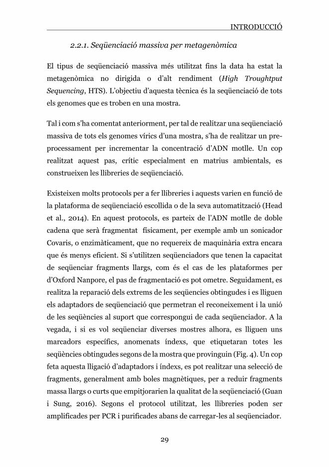
Tal i com s’ha comentat anteriorment, per tal de realitzar una seqüenciació massiva de tots els genomes vírics d’una mostra, s’ha de realitzar un pre-processament per incrementar la concentració d’ADN motlle. Un cop realitzat aquest pas, crític especialment en matrius ambientals, es construeixen les llibreries de seqüenciació.
Existeixen molts protocols per a fer llibreries i aquests varien en funció de la plataforma de seqüenciació escollida o de la seva automatització (Head et al., 2014). En aquest protocols, es parteix de l’ADN motlle de doble cadena que serà fragmentat físicament, per exemple amb un sonicador Covaris, o enzimàticament, que no requereix de maquinària extra encara que és menys eficient. Si s’utilitzen seqüenciadors que tenen la capacitat de seqüenciar fragments llargs, com és el cas de les plataformes per d’Oxford Nanpore, el pas de fragmentació es pot ometre. Seguidament, es realitza la reparació dels extrems de les seqüencies obtingudes i es lliguen els adaptadors de seqüenciació que permetran el reconeixement i la unió de les seqüències al suport que correspongui de cada seqüenciador. A la vegada, i si es vol seqüenciar diverses mostres alhora, es lliguen uns marcadors específics, anomenats índexs, que etiquetaran totes les seqüències obtingudes segons de la mostra que provinguin (Fig. 4). Un cop feta aquesta lligació d’adaptadors i índexs, es pot realitzar una selecció de fragments, generalment amb boles magnètiques, per a reduir fragments massa llargs o curts que empitjorarien la qualitat de la seqüenciació (Guan i Sung, 2016). Segons el protocol utilitzat, les llibreries poden ser amplificades per PCR i purificades abans de carregar-les al seqüenciador.

INTRODUCCIÓ
30
Figura 4. Protocol de seqüenciació massiva per metagenòmica. Producció de llibreries.
Aquest protocols han estat utilitzat per a estudis de viroma en una gran diversitat de matrius. En matrius ambientals la seva aplicació va permetre, a partir de l’any 2008, l’estudi de bacteriòfags i virus animals (Parsley et al., 2010; Rosario et al., 2009), i millores en les metodologies utilitzades van fer possible estudis més complets de viromes d’aigües residuals i

INTRODUCCIÓ
31
biosòlids en les que es van detectar un alt nombre de virus patògens d’interès humà (Bibby et al., 2011; Bibby i Peccia, 2013; Cantalupo et al., 2011; Ng et al., 2012). En l’actualitat, els estudis del viroma amb aquestes metodologies donen informació valuosa per a la epidemiologia basada en aigües residuals i el seu potencial paper com a sistema de detecció precoç de brots (Taula 2) (Haramoto et al., 2018).
Taula 2. Revisió d'estudis de metagenòmica en aigües residuals (Novembre 2020).
Títol Autors i any Matriu Principals virus detectats
Census of the Viral Metagenome within an Activated sludge Microbial Assemblage
Parsley et al., 2010 Fangs actius Bacteriòfags
Raw sewage harbors diverse viral populations
Cantalupo et al., 2011 Aigua residual
Bacteriòfags, Adenoviridae, Astroviridae, Caliciviridae, Papillomaviridae, Parvoviridae, Picobirnaviridae, Picornaviridae, Polyomaviridae
Viral Metagenome Analysis to Guide Human Pathogen Monitoring in Environmental Samples
Bibby et al., 2011 Biosòlids
Bacteriofags, Herpesviridae, Coronaviridae, Picornaviridae, Adenoviridae, Flaviviridae, Circoviridae
High Variety of Known and New RNA and DNA Viruses of Diverse Origins in Untreated Sewage
Ng et al., 2012 Aigua residual
Bacteriòfags, Adenoviridae, Astroviridae, Caliciviridae, Hepeviridae, Parvoviridae, Picornaviridae, Reoviridae
Identification of Viral Pathogen Diversity in Sewage Sludge by Metagenome Analysis
Bibby i Peccia, 2013 Biosòlids
Papillomaviridae, Adenoviridae, Parvoviridae, Circoviridae, Coronaviridae, Parvoviridae, Reoviridae, Caliciviridae, Flaviviridae, Astroviridae, Herpesviridae
Metagenomic approaches for direct and cell culture evaluation of the virological quality of wastewater
Aw et al., 2014 Aigua residual
Bacteriòfags, virus de plantes, Adenoviridae, Polyomaviridae, Picornaviridae, Papillomaviridae, Ascoviridae, Hytrosaviridae, Baculoviridae, Poxviridae, Iridoviridae, Circoviridae, Herpesviridae
Metagenomics for the study of viruses in urban sewage as a tool for public health surveillance
Fernández-Cassi et al., 2018
Aigua residual
Bacteriòfags, virus de plantes, Adenoviridae, Circoviridae, Parvoviridae, Caliciviridae, Astroviridae, Picornaviridae, Polyomaviridae, Hepeviridae

INTRODUCCIÓ
32
Characterization of Norovirus and Other Human Enteric Viruses in Sewage and Stool Samples Through Next-Generation Sequencing
Strubbia et al., 2019
Aigua residual i femtes
Bacteriòfags, Astroviridae, Caliciviridae, Nodaviridae, , Picornaviridae, Reoviridae
Variations among Viruses in Influent Water and Effluent Water at a Wastewater Plant Over One Year as Assessed by Quantitative PCR and Metagenomics
Wang et al., 2020
Efluents de depuradora
Bacteriòfags, virus de plantes, Adenoviridae, Picornaviridae, Caliciviridae, Hepeviridae, Reoviridae, Astroviridae
Metagenomic analysis of viruses, bacteria and protozoa in irrigation water
Rusiñol et al., 2020
Aigua residual i aigües d’irrigació
Bacteriòfags, virus de plantes, Circoviridae, Picornaviridae, Astroviridae, Caliciviridae, Adenoviridae, Hepeviridae, Anelloviridae, Papillomaviridae
Aquests protocols han estat també satisfactòriament aplicats en la detecció de virus en mostres clíniques, demostrant el seu potencial ús com a eines de diagnòstic que permeten alhora l’estudi de diverses mostres i diversos grups vírics (Bodewes et al., 2015; Zhou et al., 2016). També s’han utilitzat exitosament per a descriure l’agent etiològic de malalties en les que es desconeixia (Moore et al., 2015; Palacios et al., 2008).
La seqüenciació massiva per metagenòmica té com a principal avantatge la possibilitat d’estudiar el viroma complet sense un biaix cap a famílies de virus concretes. En contrapartida, en mostres complexes on les seqüències virals poden suposar només un 1% del total generat pel seqüenciador, aquesta aproximació pot suposar una pèrdua d’informació de seqüències de variants o tipus vírics que es troben en una concentració menor. És per aquest motiu que es van desenvolupar aplicacions de seqüenciació dirigida, o amb enriquiment de dianes, cap a famílies o grups vírics d’interès, les quals han estat implementades en aquesta tesi doctoral.

INTRODUCCIÓ
33
2.2.2. Seqüenciació amb enriquiment de dianes
Incrementar la sensibilitat de la seqüenciació massiva cap a famílies o grups de virus d’interès ha estat un dels objectius principals per molts grups de recerca i per a possibles aplicacions en el diagnòstic clínic. Incorporar un biaix en la producció de llibreries de seqüenciació pot ser de gran utilitat per a no perdre rendiment de seqüenciació en reads de virus i altres organismes que no son el focus d’estudi.
Amb aquesta idea, a l’any 2015 va néixer les primera aplicació de seqüenciació amb enriquiment de dianes per a virus. Briese i col·laboradors van idear un protocol de sondes de captura que estava dirigit a la seqüenciació dirigida de virus que infecten vertebrats, el panell VirCapSeq-VERT. Es van dissenyar 2 milions de sondes que cobrien els genomes complerts de 207 virus que infecten vertebrats. Aquest enriquiment va permetre un augment d’entre 100 i 1000 vegades de seqüències virals en mostres d’origen humà (sang i teixit pulmonar). El mètode desenvolupat també va demostrar ser útil per a la descripció de virus nous, ja que s’identificaven genomes de virus que eren fins a un 40% diferents dels utilitzat en el disseny de les sondes (Briese et al., 2015).
Es va dissenyar un panell de captura seguint el protocol de construcció de llibreries i hibridació desenvolupat per Nimblegen-Roche, els quals van comercialitzar posteriorment el kit complet. El fonament del mètode de captura està representat a la figura 5. Un cop les llibreries de seqüenciació estan preparades, aquestes s’hibriden en líquid amb les sondes marcades amb biotina durant un màxim de 20 hores. Posteriorment, les seqüències unides a les sondes es recuperen mitjançant l’adició de boles magnètiques recobertes d’estreptavidina. Es produirà la unió del conjugat biotina-estreptavidina que a la vegada estarà unit a les seqüències de les llibreries que hibridin amb les sondes. Amb successius procediments de rentats s’eliminen aquelles seqüències que no hagin estat reconegudes per les sondes, obtenint així per a seqüenciar únicament els fragments

INTRODUCCIÓ
34
corresponents al virus d’interès. Aquest panell ha estat aplicat amb èxit en diversos estudis, millorant la detecció de virus d’interès que d’altra manera no es detectarien en mostres clíniques (Anderson et al., 2018; Mishra et al., 2020; Tokarz et al., 2019), ambientals (Hjelmsø et al., 2019; Strubbia et al., 2019) o associades a brots sense etiologia descrita (Cummings et al., 2019; Williams et al., 2018).
Un altre panell que segueix el mateix fonament és el de Wylie i col·laboradors, anomenat ViroCap. En aquest cas, la hibridació és produïda amb la mateixa tecnologia però en un microxip i les sondes dissenyades cobreixen virus de 34 famílies diferents que infecten vertebrats (Wylie et al., 2015).
Altres empreses com Agilent Technologies, Illumina o Twist Biosciences, disposen de tecnologies de captura similars que es poden adaptar a dissenys a mida de sondes per a la seqüenciació dirigida a famílies de virus concretes (Chalkias et al., 2018; Li et al., 2017; Miyazato et al., 2016; No et al., 2019; Oba et al., 2018). Recentment, dos kits de seqüenciació amb enriquiment de dianes, VirCapSeq VERT de Roche i el SARS-CoV-2 Research Panel de Twist Biosciences, han estat utilitzats per l’estudi genètic del SARS-CoV-2 en mostres clíniques (Carbo et al., 2020; Klempt et al., 2020), demostrant la seva gran utilitat en aplicacions de seqüenciació massiva.

INTRODUCCIÓ
35
Figura 5. Protocol de seqüenciació amb enriquiment de dianes mitjançant sondes conjugades amb biotina.
INTRODUCCIÓ
36
Aquestes tècniques milloren substancialment els resultats obtinguts per a l’estudi de virus per seqüenciació massiva i la seva aplicació esta en constant evolució i millora. La principal limitació es troba en el biaix inherent de l’enriquiment per a tipus de virus coneguts, però tal i com passa amb kits ja descrits, la hibridació es pot fer menys restrictiva per a obrir la possibilitat del descobriment de nous virus. Una altra limitació que podem tenir en l’aplicació de la hibridació de sondes pot ser la falta de cobertura del genoma en estudis de variants víriques, ja que si alguna regió del virus d’estudi no hibrida amb les sondes dissenyades (noves variants, pèrdua de fragments), no tindríem la totalitat de les regions cobertes. Una metodologia que supera aquest inconvenient és la seqüenciació massiva d’amplicons.
2.2.3. Seqüenciació massiva d’amplicons
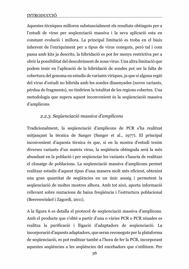
Tradicionalment, la seqüenciació d’amplicons de PCR s’ha realitzat mitjançant la tècnica de Sanger (Sanger et al., 1977). El principal inconvenient d’aquesta tècnica és que, si en la mostra d’estudi tenim diverses variants d’un mateix virus, la seqüència obtinguda serà la més abundant en la població i per seqüenciar les variants s’hauria de realitzar el clonatge de poblacions. La seqüenciació massiva d’amplicons permet realitzar estudis d’aquest tipus d’una manera molt més eficient, obtenint una gran quantitat de seqüències en un únic assaig i permetent la seqüenciació de moltes mostres alhora. Amb tot això, aporta informació rellevant sobre mutacions de baixa freqüència i l’estructura poblacional (Beerenwinkel i Zagordi, 2011).
A la figura 6 es detalla el protocol de seqüenciació massiva d’amplicons. Amb el producte que s’obté a partir d’una o vàries PCR o PCR niuades es realitza la purificació i lligació d’adaptadors de seqüenciació. La incorporació d’aquests adaptadors, que seran reconeguts per la plataforma de seqüenciació, es pot realitzar també a l’hora de fer la PCR, incorporant aquestes seqüències a les seqüències del encebadors que s’utilitzen. Per

INTRODUCCIÓ
37
seqüenciar varis amplicons de diverses mostres al mateix temps, es pot també lligar en els amplicons els índex que identificaran cada mostra amb una seqüència única.
Figura 6. Protocol de seqüenciació massiva d'amplicons.

INTRODUCCIÓ
38
Aquesta aproximació ha obert la possibilitat d’estudis detallats de famílies específiques de virus, generant informació de variants genètiques dins de les pròpies espècies (estudis de quasiespècies) (Del Campo et al., 2018; Kijak et al., 2019), reconstruccions de genomes (Charre et al., 2020; Maurier et al., 2019) i estudis de diversitat viral de famílies específiques en mostres clíniques o ambientals (Fernandez-Cassi et al., 2018; Hata et al., 2018; Prevost et al., 2015).
2.3. Plataformes de seqüenciació massiva
Les tecnologies de seqüenciació d'ADN han existit des de principis de la dècada de 1970, però inicialment el seu cost, la seva complexitat i la utilització de reactius tòxics o radioactius van limitar el seu ús en recerca. Els mètodes de terminació de cadena iniciats per Sanger et al. (Sanger et al., 1977) eren més pràctics, i van formar la base per a la primera generació de seqüenciadors d'ADN automatitzats. El primer genoma complet d'un microorganisme de vida lliure, Haemophilus influenza, publicat el 1995 (Fleischmann et al., 1995), va ser seqüenciat utilitzant el mètode Sanger. No obstant això, la seqüenciació del genoma sencer per aquesta tecnologia va ser molt costosa i lenta.
La necessitat de disposar de tecnologies de seqüenciació d'alt rendiment es va intensificar per l'inici del Projecte Genoma Humà. Es va tardar més de 15 anys, amb una inversió de quasi 3 billons de dòlars, per realitzar la seqüenciació Sanger del genoma humà (Venter et al., 2001; International Human Genome Sequencing Consortium, 2004). Avui en dia, gràcies a l’adveniment dels mètodes de seqüenciació massiva, es pot realitzar en menys d’una setmana i el cost no supera els 700 dòlars (Wetterstrand, 2020).
La seqüenciació massiva ha esdevingut una tecnologia de reemplaçament ja que la informació generada a partir de múltiples fluxos de treball tradicionals es pot combinar en un únic flux de treball eficient i amb un

INTRODUCCIÓ
39
cost cada cop menor. Juntament amb la bioinformàtica i el desenvolupament informàtic està en ràpida evolució, s'espera que es produeixin encara més millores propers anys que afectaran en gran manera l'aplicació de la seqüenciació massiva en tota mena d’àmbits de recerca i clínics (Besser et al., 2018).
L’aparició de la primera plataforma de seqüenciació massiva data del 2005 de la mà de la empresa 454 Life Sciences (posteriorment comprada per Roche) i el seu piroseqüenciador. La seva aplicació en la seqüenciació de genomes es va demostrar amb la exitosa seqüenciació del bacteri Mycoplasma genitalium (Margulies et al., 2005). Aquesta tecnologia d'alt rendiment va permetre la generació i detecció de milers a milions de lectures de seqüenciació en una sola màquina sense necessitat de clonació. Des de llavors, han sorgit moltes altres tecnologies de seqüenciació massiva que generen tant lectures curtes (50-400 bp) com llargues (1-100 kb). A la taula 3 es resumeixen les principals característiques de les plataformes de seqüenciació que es detallaran a continuació.
Taula 3. Principals plataformes de seqüenciació i característiques.
Plataforma Companyia Any sortida
Reacció seqüenciació Amplificació Mètode de
detecció Fragments
454 Roche 2005 Piroseqüenciació SI (PCR en emulsió)
Nucleòtids fluorescents Curts
MiSeq, HiSeq, MiniSeq NextSeq, NovaSeq
Illumina 2007 Síntesi SI (PCR en pont)
Nucleòtids fluorescents Curts
IonTorrent Thermo Fisher 2010 Síntesi SI
(PCR en emulsió) Canvis de pH Curts
PacBio Pacific Biosciences 2011 Síntesi NO Nucleòtids
fluorescents Llargs
MinION, GridION, PromethION
Oxford Nanopore Technologies
2015 Pas per nanopor NO Canvis de corrent Llargs

INTRODUCCIÓ
40
2.3.1. Seqüenciació de segona generació. Seqüenciació de fragments curts
Les tecnologies de seqüenciació de fragments curts son les que es coneixen com a tecnologies de segona generació. Els fragments curts poden ser ensamblats bioinformàticament com es fa amb les seqüències que s’obtenen de la seqüenciació de Sanger. La principal diferència és que la regió compartida entre seqüències és menor en les resultants de la seqüenciació massiva, degut a que els fragments son més curts, i l’ensamblat en alguns casos és limitat. Totes les plataformes tenen associades en el seu procediment l’amplificació de l’ADN de les llibreries de seqüenciació i actualment són les més extensament utilitzades.
A continuació s’expliquen les principals plataformes de seqüenciació de segona generació.
Roche 454
Aquesta plataforma de seqüenciació té com a fonament el mètode de piroseqüenciació. En aquest mètode, les llibreries de seqüenciació s’uneixen, mitjançant adaptadors, a una petita esfera. L’ADN és amplificat mitjançant PCR en emulsió de manera que cada esfera acaba tenint moltes còpies idèntiques de la mateixa molècula d’ADN. Aquestes esferes són immobilitzades en unes plaques de fibra òptica que contenen més d'un milió de pous i en cada pou es col·locarà una esfera per a produir la reacció de seqüenciació. Amb la piroseqüenciació, la incorporació de cada nucleòtid durant la síntesi de l’ADN resulta en l'alliberació d'un pirofosfat inorgànic que proporciona l'energia necessària per activar l'enzim luciferasa, generant una emissió de llum que serà captada. Els quatre nucleòtids flueixen seqüencialment sobre la placa, en un ordre definit, de manera que la producció d’un pols de llum identificarà quina base ha sigut inserida (Fig. 7) (Mardis, 2008).

INTRODUCCIÓ
41
La limitació més important d’aquesta tècnica és el baix nombre de reads, el seu elevat preu, així com biaixos en la seqüenciació de regions amb repeticions de més de tres nucleòtids (homopolímers). Aquesta tecnologia es considera obsoleta i està en desús.
Figura 7. Esquema del funcionament de la piroseqüenciació, base de la seqüenciació en la plataforma 454 Roche.
Solexa-Illumina
El primer seqüenciador Solexa, anomenat Genome Analyzer, va sortir al mercat a l’any 2006. Aquesta plataforma generava 1 Gb de seqüències en un únic run. Al 2007, la companyia Solexa va ser comprada per Illumina i els seus seqüenciadors es van convertir, i ho segueixen sent avui en dia, en referent en plataformes de seqüenciació massiva arreu del món. Les seves plataformes han seguit evolucionant amb els anys per aconseguir un nombre cada cop major de seqüències en cada procés de seqüenciació independent. Les plataformes MiniSeq i MiSeq ofereixen un rendiment de mostra baix a mitjà, preus d'instruments assequibles, un flux de treball fàcil d'utilitzar sense necessitat d'automatització i un cost raonable per mostra, convertint-se en una opció atractiva per als laboratoris de diagnòstic i salut pública. Els instruments NextSeq, HiSeq i NovaSeq estan dissenyats per a un rendiment molt més alt reduint el cost per mostra, però

INTRODUCCIÓ
42
requereixen una automatització addicional per a la preparació de les llibreries de seqüenciació i per tant estan enfocats a grans centres de seqüenciació. De totes les tecnologies de seqüenciació massiva disponibles fins a la data, el seqüenciadors d’Illumina són el que obtenen una assignació de bases (basecalling) amb major qualitat (Uelze et al., 2020).
Aquestes plataformes de seqüenciació tenen com a fonament principal l’amplificació en pont amb marcatge fluorescent (Bentley et al., 2008). La llibreria preparada es carrega i s'immobilitza a la superfície de la cel·la de flux la qual conté seqüències unides covalentment que són complementàries a les seqüències dels adaptadors que s'han utilitzat per fer la llibreria (Mardis, 2013). Aquestes seqüències complementàries capturen l’ADN i serveixen d'encebadors per a l'amplificació en pont de la cadena complementària. Es produeixen successives rondes d'amplificació que generen clústers de molècules amplificades a partir d'una sola molècula original (Fig. 8).
Els clústers d’amplificació s’utilitzaran per a la seqüenciació que es produeix a continuació, la qual es fa mitjançant la síntesi d’ADN amb nucleòtids marcats fluorescentment i que a l’incorporar-se aturen la
Figura 8. Esquema de la seqüenciació mitjançant amplificació en pont en la que es basen els sistemes d'Illumina.

INTRODUCCIÓ
43
síntesi. Després de cada incorporació, els clústers són monitoritzats i es fa la lectura de la base corresponent a cada posició de la seqüència. Abans de produir-se el següent cicle, es reverteix químicament el fluorocrom del nucleòtid que havia aturat la síntesi temporalment, permetent l'addició del següent nucleòtid en la següent ronda de seqüenciació (Radford et al., 2012).
La lectura seqüencial d’un sol nucleòtid per cicle assegura una baixa taxa d’error (al voltant de l’1%) inclòs en regions amb repeticions de bases. Per contra, degut a que l’addició de nucleòtids en el clúster no es fa alhora en totes les seqüències que es sintetitzen, la qualitat del senyal que s’obté va disminuint al llarg dels cicles de seqüenciació. Aquest darrer fet condiciona un dels desavantatges de la tecnologia, ja que no la fa apta per a seqüències més llargues de 300 parells de bases (Tan et al., 2019).
Ion Torrent
La plataforma Ion Torrent, de Thermo Fisher, va sortir al mercat a l’any 2010 per a postular-se com a competidora de les tecnologies del moment. En comptes d’utilitzar nucleòtids amb marcatge fluorescent, el fonament de la seqüenciació es basa en la tecnologia semiconductora, mesurant els ions d’hidrogen que s’alliberen en la síntesi de l’ADN (Myers i Rothberg, 2011).
La llibreria de seqüenciació s’amplifica mitjançant una PCR en emulsió. Aquesta PCR es realitza en els pous d’una cel·la de flux en la que, mitjançant un sensor de conductivitat, es detecten els canvis de pH que es produeixen al alliberar-se ions d’hidrogen. A continuació, s’assignen a cada posició els nucleòtids corresponents segons els canvis de conductivitat produïts (Fig. 9) (Mardis, 2013).

INTRODUCCIÓ
44
Figura 9. Esquema de la seqüenciació mitjançant la plataforma Ion Torrent. Adaptat de Mardis et al. (Mardis, 2013)
El principal inconvenient d’aquesta tecnologia de seqüenciació és l’alta taxa d’errors en regions homopolimèriques llargues, degut a la saturació que es produeix de la sonda de pH. En comparació a les plataformes desenvolupades per Illumina, la preparació de les llibreries de seqüenciació és més laboriosa (encara que es pot automatitzar) però el run de seqüenciació més ràpid (Besser et al., 2018; Mardis, 2013).
2.3.2. Seqüenciació tercera generació. Seqüenciació de fragments llargs
Les tecnologies seqüenciació de tercera generació es caracteritzen per tenir la capacitat de seqüenciar llargues molècules d’ADN sense una amplificació prèvia per PCR. Aquestes tecnologies són de gran utilitat per l’estudi de regions genòmiques grans, complexes o amb moltes

INTRODUCCIÓ
45
repeticions. En contrapartida, la taxa d’error que es produeix en la seqüenciació és major que en les plataformes de segona generació.
PacBio
La primera plataforma en permetre la seqüenciació d’una única molècula d’ADN va ser PacBio, de Pacific Biosciences, a l’any 2011.
El mètode de seqüenciació que s’utilitza és el que es coneix com a SMRT (Single Molecule Real-time), en el que la seqüenciació es produeix a temps real. Les reaccions es produeixen dins de nanocavitats en les que l’ADN polimerasa allarga la cadena creixent afegint nucleòtids marcats cadascun amb un fluorocrom diferent. A la vegada que es van incorporant, es registra l’emissió de fluorescència per un làser i el fluoròfor del nucleòtid és eliminat abans de que s’incorpori el següent nucleòtid. D'aquesta manera, els marcadors no s'acumulen en l’ADN i es permet la detecció de cada nucleòtid que s'incorpora a la cadena de forma individual (Logsdon et al., 2020).
S’ha incrementat la longitud mitjana de les seqüències i la precisió en la seqüenciació en els darrers anys, realitzant millores en les reaccions enzimàtiques i la detecció òptica. Actualment la taxa d’error es troba al voltant del 10% (McCombie et al., 2019).
Seqüenciació per nanopors: MinION
El concepte de seqüenciació per nanopors va aparèixer per primer cop en una publicació de Kasianowicz et al., a l’any 1996 (Kasianowicz et al., 1996), però no va ser fins l’any 2015 que la companyia Oxford Nanopore Technologies va comercialitzar la primera plataforma de seqüenciació utilitzant aquesta tecnologia, anomenada MinION.

INTRODUCCIÓ
46
Es distingeix del totes les plataformes de seqüenciació anteriors per detectar directament els nucleòtids d’una cadena d’ADN sense la seva síntesi activa, motiu per a que alguns autors la consideren com a tecnologia de “quarta generació”. A mesura que una molècula senzilla d’ADN passa a través del nanopor, un sensor detecta els canvis de corrent iònic que genera cada tipus de nucleòtid, determinant així a temps real la seqüència d’ADN que passa pel porus (Feng et al., 2015; Jain et al., 2016).
Es carreguen les llibreries de seqüenciació produïdes a la cel·la de flux que conté 2000 nanopors (en grups de 4) en la seva superfície. L’adaptador que s’incorpora en la producció de les llibreries es lliga amb una proteïna motora, que permet que la doble cadena d’ADN es separi i una de les cadenes senzilles passi a través del porus. Al travessar-lo, cada nucleòtid produirà consecutivament una pertorbació en el corrent del porus que quedarà registrat (Fig. 10).
Figura 10. Esquema de la seqüenciació mitjançant nanopors.

INTRODUCCIÓ
47
El gran avantatge d'aquesta tecnologia és la seva mida (existeixen plataformes portàtils), la seva velocitat i la possibilitat de seqüenciar fragments molt llargs d’ADN. La seva taxa de error es troba al voltant del 5-10%, però la tecnologia es troba en constant actualització i recentment han sortit al mercat noves cel·les de flux i plataformes més avançades que augmenten la fidelitat de la seqüenciació i el volum de dades generades (Van Dijk et al., 2018).
2.4. Eines bioinformàtiques i possibilitats en l’anàlisi de dades
Les plataformes seqüenciació massiva poden generar de milers a milions de lectures de seqüenciació en un sol experiment. No obstant això, la qualitat de les seqüències no és uniforme entre el conjunt de dades. Per tant, el primer pas per a processar-les és l’avaluació la qualitat de les seqüències obtingudes mitjançant diferents procediments bioinformàtics. La raó per la qual les seqüències o reads obtinguts han ha de ser filtrats, és per eliminar aquelles que poden esbiaixar els procediments següents, com son seqüències de baixa qualitat, els adaptadors de seqüenciació o duplicats discordants. La majoria d'eines que s’utilitzen per a fer el control de qualitat només estan disponibles per a sistemes operatius basats en UNIX, que no tenen una interfície gràfica d'usuari fàcil d’utilitzar per a no informàtics, no obstant això, existeixen algunes eines web que ho fan més accessible (Goecks et al., 2010; Vincent et al., 2017).
Després de la eliminació dels reads de baixa qualitat, els restants s’ensamblen per a formar seqüències d’una llargada major, anomenades contigs. Existeixen programes específics per a realitzar ensamblats que utilitzen diversos algoritmes per a la reconstrucció de seqüències, tots es basen en l’alineament de regions solapades entre seqüències amb paràmetres més o menys restrictius segons l’algoritme que s’utilitzi. Alguns d’ells també realitzen ensamblatges utilitzant genomes de referència (Honaas et al., 2016; Nurk et al., 2017). Aquests contigs

INTRODUCCIÓ
48
generats es poden ordenar i enllaçar entre ells en seqüències d’una llargada encara major anomenades scaffolds.
El pas final és l’assignació taxonòmica de les seqüències ja processades. Aquesta assignació es pot realitzar mitjançant l’alineament (o mapat) dels contigs contra bases de dades locals o contra bases de dades disponibles com poden ser GenBank (“National Center for Biotechnology Information (NCBI). Bethesda (MD).,” 1988) o UniProt (Bateman, 2019).
Tot a quest procediment bioinformàtic és laboriós i complex (Fig. 11), i la disponibilitat de bases de dades actualitzades de virus és limitada. Recentment han sorgit eines pel processament de dades de metagenòmica vírica que apropen al investigadors no bioinformàtics el processament i anàlisi de les dades generades per les plataformes de seqüenciació massiva. Entre les que realitzen un processament de dades més complert i gratuït trobem les eines web Genome Detective (Vilsker et al., 2018) (https://www.genomedetective.com/app/typingtool/virus/) i Kaiju (Menzel et al., 2016) (http://kaiju.binf.ku.dk).

INTRODUCCIÓ
49
Figura 11. Esquema del processament bioinformàtic de les dades generades en la seqüenciació massiva.

50

OBJECTIUS


OBJECTIUS
53
L’objectiu principal de la present tesi doctoral ha estat l’optimització i aplicació de procediments basats en la seqüenciació massiva per a la descripció del viroma associat a l’aigua residual i a altres mostres d’interès epidemiològic.
Els objectius específics han estat:
1. Optimitzar i aplicar diferents tècniques de seqüenciació massiva per a l’anàlisi de mostres ambientals i biològiques.
2. Caracteritzar el viroma de l’aigua residual amb focus en els patògens humans.
3. Avaluar l’ús del la seqüenciació massiva, amb i sense enriquiment
de dianes, i de la seqüenciació d’amplicons per a l’estudi i monitoreig de brots infecciosos d’origen viral.
4. Aplicar la seqüenciació massiva a mostres de pacients amb
gastroenteritis d’etiologia desconeguda per conèixer la presència d’espècies virals no detectades pels mètodes de diagnòstic clàssics.
5. Estudiar el viroma de diverses espècies de peixos Atlàntics amb potencials implicacions comercials i de seguretat alimentària.

54

INFORMES DELS ARTICLES


INFORME DELS ARTICLES
57
1. Llista d’articles inclosos en la tesi
La present tesi està basada en les següents publicacions:
Martínez-Puchol S., Rusiñol M., Fernández Cassi X., Timoneda N., Itarte M., Andrés C., Antón A., Abril J.F., Girones R., Bofill-Mas S. Characterisation of the sewage virome: comparison of NGS tools and occurrence of significant pathogens. Science of the Total Environment 713-136604, doi: 10.1016/j.scitotenv.2020.136604
Martínez-Puchol S., Itarte M., Rusiñol M., Forés E., Andrés C., Antón A., Quer J., Girones R., Abril J.F., Bofill-Mas S. SARS-CoV-2 is a new member of urban wastewater virome. Manuscrit en procés de revisió a la revista Genes.
Fernández Cassi X., Martínez-Puchol S., Cornejo T., Bartolomé R., Bofill-Mas S., Girones R. Unveiling viruses associated with gastroenteritis using a metagenomics approach. Viruses, 12(12), 1432; https://doi.org/10.3390/v12121432.
Filipa-Silva A., Parreira R., Martínez-Puchol S., Bofill-Mas S., Barreto Crespo M.T., Nunes M. The Unexplored Virome of Two Atlantic Coast Fish: Contribution of Next-Generation Sequencing to Fish Virology. Foods, 9, 1634, doi:10.3390/foods9111634.

INFORMES DELS ARTICLES
58
2. Informe de coautoria
Martínez-Puchol S., Rusiñol M., Fernández Cassi X., Timoneda N., Itarte M., Andrés C., Antón A., Abril J.F., Girones R., Bofill-Mas S. Characterisation of the sewage virome: comparison of NGS tools and occurrence of significant pathogens. Science of the Total Environment 713-136604, doi: 10.1016/j.scitotenv.2020.136604
L’estudi ha format part del projecte nacional VIRTENGS (AGL2014-55081-R) en el que la doctoranda ha participat activament. La doctoranda ha participat en la gestió del projecte i en el disseny experimental i ha realitzat els experiments. Finalment, ha redactat el manuscrit sota la supervisió de les seves codirectores de tesi.
Martínez-Puchol S., Itarte M., Rusiñol M., Forés E., Andrés C., Antón A., Quer J., Girones R., Abril J.F., Bofill-Mas S. SARS-CoV-2 is a new member of urban wastewater virome. Manuscrit sota revisió en la revista Genes.
L’estudi ha format part del projecte nacional MetaPatFood (AGL2017-86797-C2-1-R) i del monitoreig d’aigua residual per a l’estudi de la incidència de SARS-CoV-2 en els que la doctoranda ha participat activament. La doctoranda ha participat en el disseny experimental i ha realitzat els experiments de seqüenciació massiva. Finalment, ha redactat el manuscrit sota la supervisió de les seves codirectores de tesi.
Fernández Cassi X., Martínez-Puchol S., Cornejo T., Bartolomé R., Bofill-Mas S., Girones R. Unveiling viruses associated with gastroenteritis using a metagenomics approach. Viruses, 12(12), 1432; https://doi.org/10.3390/v12121432.
L’estudi ha format part del projecte nacional VIRTENGS (AGL2014-55081-R) en el que la doctoranda ha participat activament. La doctoranda ha participat en el disseny experimental, ha caracteritzat les mostres, ha

INFORME DELS ARTICLES
59
analitzat les seqüències virals i ha redactat el manuscrit juntament amb el primer signant de l’article, sota la supervisió de les seves codirectores de tesi.
Filipa-Silva A., Parreira R., Martínez-Puchol S., Bofill-Mas S., Barreto Crespo M.T., Nunes M. The Unexplored Virome of Two Atlantic Coast Fish: Contribution of Next-Generation Sequencing to Fish Virology. Foods, 9, 1634, doi:10.3390/foods9111634.
L’estudi ha format part d’una col·laboració amb el Instituto de Biologia Experimental e Tecnológica de Lisboa. La doctoranda ha preparat els SOPs (Standard Operational Procedures) i ha format als participants en les tècniques utilitzades. També ha contribuït en el disseny experimental i, juntament amb la primera signant del treball, ha realitzat la part experimental. Finalment ha col·laborat en la redacció i correcció del manuscrit.
Signat,
Dra. Rosina Girones Llop Dra. Sílvia Bofill Mas Barcelona, Desembre 2020 Barcelona, Desembre 2020

INFORMES DELS ARTICLES
60
3. Informe sobre el factor d’impacte
Els treballs que formen part de la present tesi doctoral s’han publicat o estan en procés de revisió en revistes científiques rellevants per a la línia de recerca en la qual la doctoranda ha participat en el darrers anys.
L’article “Characterisation of the sewage virome: comparison of NGS tools and occurrence of significant pathogens” va ser publicat al mes de gener de 2020 a la revista Science of the Total Environment. L’índex d’impacte de la revista a l’any 2019 era de 6,551 (Q1).
L’article “SARS-CoV-2 is a new member of urban wastewater virome” es troba actualment en procés de revisió (novembre 2020) a la revista Genes. L’índex d’impacte de la revista a l’any 2019 era de 3,750 (Q1).
L’article “Unveiling viruses associated with gastroenteritis using a metagenomics approach” va ser publicat al mes de desembre de 2020 a la revista Viruses. L’índex d’impacte de la revista a l’any 2019 era de 3,816 (Q1).
L’article “The Unexplored Virome of Two Atlantic Coast Fish: Contribution of Next-Generation Sequencing to Fish Virology” va ser publicat al mes de novembre de 2020 a la revista Foods. L’índex d’impacte de la revista a l’any 2019 era de 4,092 (Q1).
Signat,
Dra. Rosina Girones Llop Dra. Sílvia Bofill Mas Barcelona, Desembre 2020 Barcelona, Desembre 2020

CAPÍTOL 1: SEQÜENCIACIÓ MASSIVA PER A ESTUDIS D’EPIDEMIOLOGIA
BASATS EN AIGÜES RESIDUALS


CAPÍTOL 1
63
Article 1: Caracterització del viroma de l’aigua residual: comparativa d’eines de seqüenciació massiva i presència
de patògens importants
Les tècniques de seqüenciació massiva són eines útils pel monitoreig i identificació del patògens vírics que circulen en la població però presenten certes limitacions, sobretot pel que fa a matrius complexes. L’aigua residual conté una elevada concentració d’altres microorganismes que poden interferir a l’hora de seqüenciar virus. Per això, és necessari realitzar optimitzacions prèvies al protocol de seqüenciació.
En el present estudi, s’han comparat diferents metodologies de seqüenciació massiva per a la detecció i caracterització de virus en l’aigua residual, amb especial focus en tres patògens importants: els virus del papil·loma humà, els adenovirus i els enterovirus. Les tècniques aplicades han estat: seqüenciació massiva per metagenòmica, seqüenciació amb enriquiment de dianes i seqüenciació massiva d’amplicons.
Aplicant la seqüenciació massiva per metagenòmica s’ha obtingut el perfil complet dels virus excretats, detectant membres de quatre famílies víriques diferents a més de bacteriòfags, virus de plantes i virus que infecten altres hostes. L’ús de la seqüenciació massiva amb enriquiment de dianes, en canvi, ha permès la detecció de vuit famílies de virus humans, amb una alta varietat de tipus en cada família i una elevada cobertura dels
“Characterisation of the sewage virome: comparison of NGS tools and occurrence of significant pathogens.”
Martínez-Puchol S., Rusiñol M., Fernández Cassi X., Timoneda N., Itarte M., Andrés C., Antón A., Abril J.F., Girones R., Bofill-Mas S. Science of the Total Environment 713-136604, doi: 10.1016/j.scitotenv.2020.136604

CAPÍTOL 1
64
genomes detectats, és a dir s’han pogut ensamblar trossos grans de genomes virals. Per últim, la seqüenciació massiva d’amplicons proporciona moltes dades sobre la diversitat genètica dels tres patògens d’estudi en comparació amb les altres dues tècniques aplicades. A més, ha permès detectar i subtipar seqüències d’enterovirus A71 C1, la soca causant del brot de romboencefalitis en nens que va ocórrer a Catalunya a la primavera de 2016, època de presa de mostra de l’aigua residual analitzada.
Amb els resultats obtinguts es pot concloure que diferents estratègies de seqüenciació massiva proporcionen diferents nivells d’anàlisis. L’aplicació de l’enriquiment de dianes ha esdevingut la millor estratègia per a descriure la presència de virus d’interès humà en mostres complexes, com l’aigua residual. Les altres eines utilitzades són útils per l’estudi del viroma complet d’una mostra (metagenòmica) o quan l’interès de l’estudi és la diversitat viral dins d’una mateixa família (seqüenciació d’amplicons).

Characterisation of the sewage virome: comparison of NGS tools andoccurrence of signi!cant pathogens
Sandra Martínez-Puchol a,b,!, Marta Rusiñol a,b, Xavier Fernández-Cassi a, Natàlia Timoneda a,c, Marta Itarte a,b,Cristina Andrés d, Andrés Antón d, Josep F. Abril c, Rosina Girones a,b, Sílvia Bo!ll-Mas a,b
a Laboratory of Viruses Contaminants of Water and Food, Genetics, Microbiology &Statistics Dept., Universitat de Barcelona, Barcelona, Catalonia, Spainb The Water Research Institute (IdRA); Universitat de Barcelona, Barcelona, Catalonia, Spainc Computational Genomics Lab, Genetics, Microbiology & Statistics Dept., Universitat de Barcelona, Institut de Biomedicina (IBUB), Barcelona, Catalonia, Spaind Respiratory Viruses Unit, Virology Section, Microbiology Department, Hospital Universitari Vall d'Hebron, Vall d'Hebron Research Institute, Universitat Autònoma de Barcelona, Barcelona, Spain
H I G H L I G H T S
• Different NGS methods have been ap-plied to the study of sewage virome.
• Untargeted Viral Metagenomics pro-vides a full picture of the completevirome.
• Target Enrichment Sequencing is thebest option for detecting vertebrate vi-ruses.
• Amplicon Deep Sequencing is useful forstudying viral diversity.
• Enterovirus A71 related to an outbreakwas identi!ed with two of thesemethods.
G R A P H I C A L A B S T R A C T
a b s t r a c ta r t i c l e i n f o
Article history:Received 22 November 2019Received in revised form 7 January 2020Accepted 7 January 2020Available online 08 January 2020
Editor: Damia Barcelo
NGS techniques are excellent tools to monitor and identify viral pathogens circulating among the populationwith some limitations that need to be overcome, especially in complexmatrices. Sewage contains a high amountof other microorganisms that could interfere when trying to sequence viruses for which random PCR ampli!ca-tions are needed beforeNGS. The selection of appropriate NGS tools is important for reliable identi!cation of viraldiversity among the population.We have compared different NGSmethodologies (Untargeted Viral Metagenomics, Target Enrichment Sequenc-ing and Amplicon Deep Sequencing) for the detection and characterisation of viruses in urban sewage, focusingon three important human pathogens: papillomaviruses, adenoviruses and enteroviruses.A full picture of excreted viruses was obtained by applying Untargeted Viral Metagenomics, which detectedmembers of four different vertebrate viral families in addition to bacteriophages, plant viruses and viruses infect-ing other hosts. Target Enrichment Sequencing, using speci!c vertebrate viral probes, allowed the detection of upto eight families containing human viruses,with high variety of typeswithin the families andwith a high genomecoverage.By applying Amplicon Deep Sequencing, the diversity of enteroviruses, adenoviruses and papillomaviruses ob-served was higher than when applying the other two strategies and this technique allowed the subtyping of
Keywords:Sewage viromeTarget Enrichment SequencingAmplicon Deep SequencingPapillomavirusAdenovirusEnterovirus
Science of the Total Environment 713 (2020) 136604
! Corresponding author.E-mail address: [email protected] (S. Martínez-Puchol).
https://doi.org/10.1016/j.scitotenv.2020.1366040048-9697/© 2020 Elsevier B.V. All rights reserved.
Contents lists available at ScienceDirect
Science of the Total Environment
j ourna l homepage: www.e lsev ie r .com/ locate /sc i totenv

an enterovirus A71 C1 strain related to a brainstem encephalitis outbreak occurring at the same time in the sam-pling area.From the data obtained, we concluded that the different strategies studied provided different levels of analysis:TES is the best strategy to obtain a broad picture of human viruses present in complex samples such as sewage.Other NGS strategies are useful for studying the virome of complex sampleswhen also targeting viruses infectingplants, bacteria, invertebrates or fungi (Untargeted Viral Metagenomics) or when observing the variety within asole viral family is the objective of the study (Amplicon Deep Sequencing).
© 2020 Elsevier B.V. All rights reserved.
1. Introduction
Viruses excreted by humans in faeces, urine and skin desquamationtogether with animal viruses and viruses infecting invertebrates, plants,bacteria and fungi constitute the sewage virome, a complex matrix thatcontains a large variety of pathogenic and commensal viruses and couldgive important information on persistent and acute infections affectingthe population.
Currently, NextGeneration Sequencing (NGS)methodologies are in-creasingly viewed as promising tools for the comprehensive study ofmicroorganisms in awide variety of samples and settings, replacing tra-ditional molecular methods. The main reasons for this new trend are:the capacity to process larger number of samples simultaneously, thereduction in sequencing costs, the ease with which samples and librar-ies can be prepared and the development of faster and more ef!cientbioinformatic tools with which to process the huge amount of data.(Huang et al., 2019).
The application of NGS techniques was easily incorporated into thestudy of bacterial communities even for environmental samples, butthe lack of shared regions in viral genomes make them more challeng-ing for viral sequencing. To overcome this problem, the use ofrandom-primer-based sequencing facilitates the development of differ-ent protocols adapted for viral NGS (Cantalupo et al., 2011; Kohl et al.,2015; Ng et al., 2012), making this methodology key for the study ofviral communities and viral discovery while providing enough startingmaterial for library preparation.
The excreted virome of a population provides critical informationabout virus circulation, the introduction of emergent viruses and howthey are transmitted among the population. The accurate study of thevirome with a focus on speci!c target viral groups urges the develop-ment of NGS protocols with higher sensitivity.
The main limitation when analysing the virome from any type ofsample is the low proportion of viral sequences identi!ed comparedwith the total number of sequences ampli!ed when using randomprimers (Krishnamurthy andWang, 2017; Santiago-Rodriguez andHol-lister, 2019; Tamaki et al., 2012). This is especially critical for human vi-ruses which seem to represent a low fraction of the whole virome ofsewage, clearly dominated by phages and plant viruses (Cantalupoet al., 2011; Ng et al., 2012). Few published studies in viral sewagemetagenomics describe the human virome together with protocols toachieve a better recovery of viral sequences by applyingmainly negativeviral selection methods, also called pre-extraction, which generally en-tail the use of, for example, !lters, density gradients and nucleases(Cantalupo et al., 2011; Fernandez-Cassi et al., 2018a, 2018b; Hjelmsøet al., 2017). Viral selection approaches are effective for avoiding thehigh background presence of other non-desired nucleic acids from bac-teria or other hosts. Apart from negative selection methods, positive se-lection methods, also referred to post-extraction methods or TargetEnrichment Methods, are characterised by the use of probes withinthe PCR assays, microarrays or hybridisation (Kumar et al., 2017).Thesemethods increase in viral sequence reads aswell as in the breadthand depth of genome coverage, in some cases extending to the full ge-nome (Paskey et al., 2019; Wylie et al., 2015). Among these, the mostcommonly used are those based on hybridisation probes, with different
custom-made and commercial approaches available (Chalkias et al.,2018; Hjelmsø et al., 2019; Mühlemann et al., 2018), showing the po-tential use in viral discovery (Briese et al., 2015).
Another NGS approach useful for identi!cation and typing of differ-ent viral species for outbreak control or environmental surveillance isAmplicon Deep Sequencing, based on themass sequencing of tradition-ally sanger-sequenced PCR amplicons. This approach opened up newopportunities promoting the detailed study of speci!c families andtheir diversitywithin a sample, also proving useful for a different natureof samples, from studies of quasispecies of Hepatitis C Virus or HumanImmunode!ciency Virus in clinical settings (Del Campo et al., 2018;Kustin et al., 2019) to environmental samples (Fernandez-Cassi et al.,2018a, 2018b; Hata et al., 2018; Prevost et al., 2015).
This study shows the wide diversity of viral pathogens identi!ed inurban sewage over one year of study with the application of differentNGS tools for the determination of the virome with different levels ofanalysis. To our knowledge, this is the !rst work aimed to comparetwo different NGS strategies, Untargeted Viral Metagenomics (UVM)and Target Enrichment Sequencing (TES), for the characterisation ofthe virome excreted in a population in terms of viral diversity, speci!c-ity and genome coverage. Also, Amplicon Deep Sequencing (ADS), wasexamined as an alternative strategy when a deeper study on a concreteviral family is needed. The work is focused on three speci!c viral fami-lies, human adenoviruses (HAdV), human papillomaviruses (HPV) andhuman enteroviruses (EV), since they are important pathogens andsome of them are persistently excreted by the population. In addition,we analysed enterovirus nucleotide sequences obtained from clinicalsamples for evaluating the capacity of the studied NGS strategies tocatch up viruses causing clinical disease in the population.
2. Material and methods
2.1. Sampling, viral particles concentration and nucleic acid extraction
In April 2016, a 24-hour urban raw sewage composite sample wascollected from a wastewater treatment plant (WWTP), located in thenorth of Barcelona city (WWTP A), that treats a population equivalentof up to 2.8 million and receives domestic and industrial waste fromthe sewerage system. This composite sample was analysed to compareTES, UVM and ADS.
Additionally, monthly sampleswere collected over the year, in 2016,from a second WWTP located 30 km from Barcelona (WWTP B). Thesamples from this WWTP were pooled by season (spring, summer, au-tumn and winter) and analysed by ADS in order to evaluate speci!cviral groups, in depth, over one year. This plant serves up to 115,000population equivalents.
Samples were collected in sterile containers and kept at 4 °C untilconcentrated within 24 h. Viral particles from 42 ml of sewage fromeach sample were concentrated by ultracentrifugation, as previouslydescribed (Bo!ll-Mas et al., 2006). DNase treatment (TurboDNAse,Ambion) and extraction of nucleic acids was performed as describedpreviously (Fernandez-Cassi et al., 2018a, 2018b) using QIAmp RNAViral Mini Kit (Qiagen).
2 S. Martínez-Puchol et al. / Science of the Total Environment 713 (2020) 136604

2.2. Untargeted viral metagenomics and target enrichment sequencing
2.2.1. Random tagging of nucleic acids and pre-ampli!cationSamples were prepared prior to the library construction following
the protocol described (Fernandez-Cassi et al., 2018a, 2018b). Brie"y,in order to analyse both RNAandDNAviruses, NAwere retrotranscribedwith SuperScript III enzymes (Life Technologies) and a randomnonamer primer. The second cDNA strand was obtained usingSequenase 2.0 (ThermoFisher Scienti!c) and a PCRof 25 cycleswasper-formed in order to obtain enough dsDNA for the next steps. These PCRproducts were puri!ed and concentrated with the Zymo DNA Clean &Concentrate kit (Zymo Research) and quanti!ed using Quibit 2.0HSdsDNA kit (Life Technologies).
2.2.2. Library constructionFor each sample, libraries were constructed in duplicate using KAPA
HyperPlus Library Preparation Kit (KAPA Biosystems, Roche). An enzy-matic fragmentationwas performed in thepreviously obtained puri!ca-tion with a starting concentration ranging from 1 to 3 ng/!l followingthe manufacturer's instructions. After the fragmentation and the end-repair and A-tailing reaction, the adapter's ligation was performed.Using the KAPA Single-Indexed Adapter Kit (KAPA Biosystems, Roche),each sample was paired with the desired index. A post-ligation clean-up was followed with a double-sized size selection with the magneticAMPure XP Beads (Beckman Coulter) to select those fragments of be-tween 250 and 450 bp. Using an LM-PCR, sample libraries were ampli-!ed (seven cycles of ampli!cation for the adapters used) and puri!ed.The quality of the resulting libraries was assessed by using AgilentBioanalyzer DNA 1000 assay (Agilent Technologies) and the concentra-tion was measured using Qubit 2.0 (Life Technologies). Then, librarieswere mixed together in two replicates, to obtain two pools containing1 !g each. One pool was directly sequenced on an Illumina MiSeq2x300bp platform and the other was captured later by using speci!cviral probes.
2.2.3. Capture of viral sequences by using the VirCapSeq-VERT CapturePanel
One of the pooled libraries was captured using the VirCapSeq-VERT Capture Panel (Roche). This panel consists of approximatelytwo million probes, covering the genomes of 207 viral taxa knownto infect vertebrates, thus enabling the detection of viral sequencesin complex sample types (Briese et al., 2015). Using the HyperCapTarget Enrichment Kit (Roche) and the HyperCap Bead Kit (Roche),the sample was prepared and then hybridised with the VirCapSeq-VERT probes at 47 °C for 20 h. Immediately after the hybridisation,the captured multiplex DNA sample was recovered with the CaptureBeads (HyperCap Bead Kit, Roche), using a magnetic particle collec-tor, and cleaned. The DNA captured, still bead-bounded, was ampli-!ed using an LM-PCR. This post-capture PCR was puri!ed and theconcentration and quality were checked, as mentioned before. Cap-tured DNA was sequenced in the same run as non-captured DNA inan Illumina MiSeq 2x300bp platform.
2.3. Amplicon Deep Sequencing
Speci!c nested PCR, previously proved to be adequate for typingpurposes, for HAdV, HPV and EV ampli!cation were performed as de-scribed (De Roda Husman et al., 1995; Fernandez-Cassi et al., 2018a,2018b; Forslund et al., 2003; WHO Regional Of!ce for Europe, 2015)with incorporation of Illumina adapters in the nested primers.Ampliconswere puri!ed fromagarose gelwith theQIAquick Gel Extrac-tion Kit (Qiagen) and then sequenced with an Illumina MiSeq 2x300bpplatform.
2.4. Clinical enterovirus A71 sampling
Most of 25 EV-A71-studied cases by Vall d'Hebron Hospital Respira-tory Viruses Unit corresponded to rhombencephalitis, while the re-maining patients had hand-foot-mouth disease (2), gastroenteritis (2),aseptic meningitis (1), and acute bronchitis (1). EV-A71 was ampli!edfrom upper and lower respiratory tract samples from suspicious pa-tients by using CE-marked commercial multiplex real-time RT-PCR-based assay (Anyplex II RV16 assay, Seegene, Korea). Total nucleicacids were previously extracted using NucliSens easyMAG (bioMérieux,Marcy líEtoile, France) according to themanufacturer's instructions andkept frozen (!20C) until use. An additional real-time RT-PCR (Seegene,Korea) was carried out to improve the detection of all EV strains(Gimferrer et al., 2015).
The partial coding sequences of the viral protein 1 (VP1) from EVamplicons, obtained according to the protocol recommended by theWorld Health Organisation (WHO Regional Of!ce for Europe, 2015),were used to construct a phylogenetic tree.
2.5. Bioinformatics
2.5.1. UVM and TES bioinformatic processing and taxonomical assignmentPair-end FASTAQ!les generated from the sequencingwere analysed
using Genome Detective web-based software (https://www.genomedetective.com/)(Vilsker et al., 2018a). Brie"y, low-qualityreads and adapters were trimmed using Trimmomatic (Bolger et al.,2014), viral reads were selected using DIAMOND alignment methodand non-viral sequences were discarded. Subsequently, viral readswere assembledwithmetaSPAdes (Nurk et al., 2017) and taxonomicallyclassi!ed with NCBI-BLASTX and NCBI-BLASTN against NCBI RefSeqviral database (Vilsker et al., 2018b; Wheeler et al., 2007), using onlythe contigs with 70% identity cut-off. Richness Chao1 ratio was calcu-lated using the Catchall software, version 4.0 (Allen et al., 2013).
2.5.2. Amplicon Deep SequencingFor the study of the amplicons, the quality of raw and clean read se-
quences was assessed using the FASTX-Toolkit software, version 0.0.14(Hannon Lab, http://www.hannonlab.org). The cleaned reads wereclustered using the software CDHIT with default parameters (Huanget al., 2010); the output was queried for sequence similarity usingNCBI-BLASTN against a reference genome species-speci!c custom data-base for HADV, HPV and EV (Wheeler et al., 2007). Based on the bestBLAST results (95% coverage and 95% identity cut-off), each clusterwas classi!ed into its taxonomic group.
2.5.3. Enterovirus phylogenetic analysesA phylogenetic tree based on enterovirus VP1 nucleotide sequences
of the EV-A71 C1 strains obtained from sewage and clinical sampleswasconstructed by a using neighbour-joining method with Geneious soft-ware version 11.0 (https://www.geneious.com), where branches hav-ing a bootstrap value below 70% were discarded for the Figure.Coxackievirus A16 sequence (GenBank accession number KT327162)was used as the outgroup.
3. Results and discussion
3.1. Study of the sewage virome using Untargeted Viral Metagenomics andTarget Enrichment Sequencing
An urban sewage composite sample obtained after 24 h of collectionin WWTP A was analysed using UVM and TES in parallel. A total of 6.07million viral readswere obtainedwhen applying TES and 727,784whenapplyingUVM. Read counts for eachof the viral groups detected are pre-sented in Table 1. The Chao1 diversity index showed, as expected, ahigher richness when using UVM, increasing from 170 obtained usingthe TES approach, to 311 when using UVM.
3S. Martínez-Puchol et al. / Science of the Total Environment 713 (2020) 136604

Probe enrichment increased the detection of sequences from verte-brate viruses by a factor of 81.04% (299,650 sequences) of the total se-quences obtained, compared with the 2.74% (3549 sequences)obtained without applying enrichment. An exhaustive description ofthe total virome obtained using both methodologies is presented asSupplementary material 1.
A comparison of the viral species obtained via each methodology,based on host distribution, is shown in Fig. 1. Not only was the num-ber of sequences from viruses that infect vertebrates higher after theTES approach, but also the number of species within each viral familyincreased. The Picornaviridae and Parvoviridae families showed ahigher number of taxonomically assigned sequences after the appli-cation of the TES. When applying UVM, a wide variety of bacterio-phage species was observed (49% of the total reads) within thefamilies Siphoviridae, Myoviridae, Microviridae and Podoviridae, aswell as species of the viral plant families (30% of the total reads)
Virgaviridae, Tombusviridae and Solemoviridae. The predominanceof these viral families in environmental settings has been reportedbefore (Fernandez-Cassi et al., 2018a, 2018b) and is of importancebecause of their potential economic impact as plant pathogens.Reads from vertebrate families were identi!ed as JC Polyomavirus,Mamastrovirus 1, Aichivirus A, non-human circoviruses and parvovi-ruses, representing 2.75% of the total sequences. Accordingly, UVMcan be considered a reference tool for the study of a global pictureof the whole virome of environmental and clinical samples, in accor-dance with other studies performed by our research group(Cantalupo et al., 2011; Fernandez-Cassi et al., 2018a).
The TES kit used in this study was developed in 2015 by Briese et al.(Briese et al., 2015) with the aim of capturing viral sequences from onlythose viruses that infect vertebrates, for use in clinical and veterinariansettings. This system usesmore than twomillion probes of 207 differentvertebrate viruses andhas been reported useful for the detection of newvariants. When applied to raw sewage, probes helped to capture verte-brate viruses (Briese et al., 2015), although some sequences from otherviral hosts were still recovered. These kind of enrichment approachesare clearly biased towards speci!c viral families (Parras-Moltó et al.,2018) andwhile theymight be useful for studying speci!c viral families(or groups of families), they do not provide the whole virome picture.
However, when focusing on viral families that infect humans, TESprovided a more sensitive approach, allowing the detection of moreviral families and catching a wide diversity of viral species within agiven family while providing a higher number of reads. Members ofthe Adenoviridae, Hepeviridae, Papillomaviridae, and Reoviridae familiesas well as some species of the Picornaviridae (including an EV71 contig)and Polyomaviridaewere only detected when using TES. Regarding theAstroviridae and Caliciviridae families, a big increase in sequences fromMamastrovirus 1 species (up to 3-log) and Norwalk and Sapporo vi-ruses (up to 2-log) was observed when compared with UVM. Again,when processing these reads as contigs, a considerable increase in thegenome coverage of each virus was observed after TES which is ofhuge interest for viral characterisation and discovery, as reported before(Briese et al., 2015). Contigs corresponding to Aichivirus A, Norwalkvirus, JC Polyomavirus and Mamastrovirus 1 presented more than 90%genome coverage. Indeed, for the latter two, almost the totality of thegenomewas covered, at 93.82% and 99.25% respectively, whereas with-out enrichment the respective coverage of these viruseswas 77.37% and30.45% (Table 1). This fact could be due to the higher number of se-quences obtained with TES and the wider distribution of reads fromacross all viral genomes.
From the data obtained, we conclude that TES is the best strategy toobtain a broad picture of human viruses present in complex samples
Table 1
Read counts and Genome coverage obtained from contigs of viral species of interest ob-tained by Target Enrichment Sequencing (TES) and Untargeted Viral Metagenomics(UVM).
Viral sequence reads
Viral readcount
Breadthgenomecoverage (%)
Viral families Viral species TES UVM Foldincrease(log)
TES UVM
Papillomaviridae Betapapillomavirus 2 24 0 1,38 15,47 0Picornaviridae Aichivirus A 23372 33 2,85 89,86 22,05
Enterovirus A 725 0 2,82 61,12 0Enterovirus B 902 0 2,96 44,00 0Enterovirus C 2098 0 3,32 62,10 0
Adenoviridae Humanmastadenovirus A
343 0 2,54 14,22 0
Humanmastadenovirus F
1333 0 3,12 34,66 0
Polomaviridae BK polyomavirus 829 0 2,92 79,89 0Human polyomavirus6
79 0 1,90 10,88 0
JC polyomavirus 714 237 0,48 93,82 77,37WU polyomavirus 17 0 1,23 10,96 0
Astroviridae Mamastrovirus 1 83857 64 3,12 99,25 30,45Caliciviridae Sapporo virus 421 10 1,62 62,07 57,33
Norwalk virus 1820 8 2,36 88,72 11,73Hepeviridae Orthohepevirus A 781 0 2,89 24,21 0Reoviridae Rotavirus A 423 0 2,63 38,64 0
Fold increase is presented in logs of increase and breadth genome coverage inpercentages.
HumanTES 4 1 2 1 0 1 3 3 3 9 4 1 0 0 0 0 0 0 0 0 0 0 0 0 0 0 0 0 0 0
UVM 0 1 2 1 0 0 0 1 1 1 1 0 0 0 0 0 0 0 0 0 0 0 0 0 0 0 0 0 0 0
Other
Vertebrates
TES 1 4 0 5 1 1 1 10 4 5 0 0 0 0 0 0 0 0 0 0 0 0 0 0 0 0 0 0 0 0
UVM 0 0 0 5 5 0 0 0 1 2 2 0 0 0 0 0 0 0 0 0 0 0 0 0 0 0 0 0 0 0
PlantTES 0 0 0 0 0 0 0 0 0 0 0 0 1 0 1 5 1 2 1 4 0 0 0 0 0 0 0 0 0 0
UVM 0 0 0 0 0 0 0 0 0 0 0 0 1 1 1 2 5 6 3 7 0 0 0 0 0 0 0 0 0 0
InvertebrateTES 0 0 0 1 4 0 0 8 0 0 0 0 0 2 0 0 0 0 0 0 2 0 0 0 0 0 0 0 0 0
UVM 0 0 0 4 2 0 0 5 0 0 0 0 0 1 0 0 0 0 0 0 1 1 0 0 0 0 0 0 0 0
FungiTES 0 0 0 0 0 0 0 0 0 0 0 0 0 0 0 0 0 0 0 0 0 0 1 1 1 0 0 0 0 0
UVM 0 0 0 0 0 0 0 0 0 0 0 0 0 0 0 0 0 0 0 0 0 0 0 3 0 0 0 0 0 0
BacteriaTES 0 0 0 0 0 0 0 0 0 0 0 0 0 0 0 0 0 0 0 0 0 0 0 0 10 2 1 15 5 15
UVM 0 0 0 0 0 0 0 0 0 0 0 0 0 0 0 0 0 0 0 0 0 0 0 0 43 3 2 27 32 88
Fam
ily
Adenoviridae
Astroviridae
Caliciviridae
Circoviridae
Dicistroviridae
Hepeviridae
Papillomaviridae
Parvoviridae
Picobirnaviridae
Picornaviridae
Polyomaviridae
Reoviridae
Alphaflexiviridae
Genomoviridae
Potyviridae
Secoviridae
Solemoviridae
Tombusviridae
Tym
oviridae
Virgaviridae
Nodaviridae
To!viridae
Marnaviridae
Par!viridae
Podoviridae
Inoviridae
Leviviridae
Microviridae
Myoviridae
Siphoviridae
Fig. 1. Host distribution of viral species within families obtained by Target Enrichment Sequencing (TES) and Untargeted Viral Metagenomics (UVM) inWWTP A.
4 S. Martínez-Puchol et al. / Science of the Total Environment 713 (2020) 136604

such as sewage, as well as being a technique thatmight be useful for en-vironmental public health surveillance.
3.2. Application of Amplicon Deep Sequencing to the study of speci!c viralgroups in sewage: human adenoviruses, papillomaviruses, andenteroviruses
3.2.1. Comparison between ADS and TES for the study of the sewage viromeAmplicon Deep Sequencing has demonstrated its usefulness in the
study of speci!c viral groups, giving higher sensitivity and a largeamount of information about the viruses present in a given sample.This methodology has previously been applied to environmental sam-ples for studying diversity within viral groups, such as Adenoviridae(Fernandez-Cassi et al., 2018a, 2018b; Ogorzaly et al., 2015),Papillomaviridae (Hamza and Hamza, 2018; Iaconelli et al., 2015; LaRosa et al., 2015) or enterovirus (Brinkman et al., 2017).
The composite 24 h sewage sample collected from WWTP A wasevaluated by ADSusing speci!c PCR primers for HAdV, HPV and EV fam-ilies. Results obtained after the application of ADS are summarised inFig. 2.
While ADS showed similar adenovirus species to TES, detectingHuman Mastadenovirus F and A species, with HAdV 41 and HAdV 18being the most prevalent, some of the species appeared only with TES(e.g., HAdV5, HAdV 27 and HAdV 56) and others only with ADS(e.g., HAdV 18, HAdV 12 and HAdV 61).
Regarding HPV, ADS provided a higher diversity of species than TES,with HPV6 and HPV66 being the only alphapapillomavirus detected byADS and the most abundant types, being 36.01% and 21.22% of the totalsequences detected respectively. Other members of thebetapapillomavirus genus were also detected, with HPV120 andHPV19 being the most prevalent types. An important difference be-tween these two methodologies was observed: sequences of HPV 122and HPV 49 (betapapillomavirus types) were obtained from both ap-proaches, but HPV 17 (betapapillomavirus type) and HPV127(gammapapillomavirus type) only by TES and a wider variety of typesonly by ADS, including oncogenic ones.
Regarding enterovirus, viral species Enterovirus A, B and C were de-tected byADS as occurredwhen applying TES. E-E30was themost prev-alent enterovirus, followed by EV-C99 and EV-A119. Also, sequencesfrom EV-A71were obtained by ADS and TES, whichwere of interest be-cause a clinical brainstem encephalitis outbreak was occurring in Cata-lonia during the sampling period. By applying TES, only one EV-A71contig (884 bp) was obtained fromWWTP A and, by ADS, 14 amplicons(301 bp) were obtained from both WWTPs.
3.2.2. Amplicon Deep Sequencing for characterisation of speci!c viralgroups in sewage: human adenoviruses, papillomaviruses, andenteroviruses
The distribution of the three viral groups over one year in sewagewas also evaluated by applying ADS in seasonally pooled samplesfrom WWTP B and the results obtained are represented in Fig. 3. Awide variety of HAdV, HPV and EV sequences was obtained.
The in-depth analysis of HAdV showed more than 10 serotypes inthe year of study (Fig. 3a). HAdV fromHumanMastadenovirus A species,related to gastrointestinal, urinary and respiratory infections, andHuman Mastadenovirus F, related to infantile gastroenteritis, were thegroups most commonly detected over the whole year, with beingHAdV 41 (Group F) and HAdDV 31 (Group A) being the most abundanttypes. HAdV 40 (Group F), and HAdV 12 (Group A) found throughoutthe year and HAdV 18 (Group A) in spring. Other HumanMastadenoviruses serotypes (HAdV 51, HAdV 59, HAdV 46, HAdV 19)were detected in lower proportions, as previously described in otherstudies (Fernandez-Cassi et al., 2018a, 2018b; Iaconelli et al., 2017).
Many HAdV produce infections thatmay be subclinical and could beexcreted in faeces by healthy people over a long period. The prevalenceof HAdV in sewage through the year is in accordance with the proposalto use adenovirus as an indicator of human faecal contamination (Pinaet al., 1998).
For papillomaviruses, a large diversity of types was observed by ADSthrough the whole year of sampling (Fig. 3b). Sequences of the poten-tially oncogenic types HPV 6 and HPV 66 (genus alphapapillomavirus)were detected through the year, with higher proportions than otherHPV in the spring and winter months. Members of this genus hadbeen described previously in raw sewage (Iaconelli et al., 2015), butHPV 66, involved in vulvar cancer and classi!ed as a group I carcinogen(Proceedings of the IARC Working Group on the Evaluation of Carcino-genic Risks to Humans. Biological Agents., 2009), has never been re-ported in environmental samples. More than 25 different cutaneousbetapapillomaviruses were detected, with HPV 120, HPV 19, HPV 9,HPV 8, HPV 49 and HPV 80 being the most abundant during the yearof observation.
Enterovirus ADS showed different distribution patterns through thestudied year (Fig. 3c): CV-B5, E-E18 and E30 (EV species B) were the se-rotypes more frequently detected in spring and summer and E-E14 (EVspecies B) and EV-C99 (EV species C) in autumn and winter. These en-terovirus species are traditionally related to aseptic meningitis casesand, with the exception of Enterovirus C99, had been also reported as eti-ological agents involved in Acute Flaccid Paralysis (AFP) cases by theSpanish Ministry of Health in their Annual Epidemiologic Surveillance
Fig. 2. Diversity of speci!c viral groups (human adenoviruses, papillomaviruses and enteroviruses) obtained by Amplicon Deep Sequencing in the composite raw sewage sample A.
5S. Martínez-Puchol et al. / Science of the Total Environment 713 (2020) 136604

6 S. Martínez-Puchol et al. / Science of the Total Environment 713 (2020) 136604

(López-Perea et al., 2017). This emergent member of enterovirus speciesC has been reported to be involved in the development of AFP (Brownet al., 2009) and was recently described as being very prevalent inUruguay and Brazil (Lizasoain et al., 2018; Luchs et al., 2019).
Also, a comparison of EV presence in spring in two differentWWTPs(composite sample from WWTP A, which was collected in spring, andspring pooled samples from WWTP B) showed E-E30 to be the mostprevalent strain detected in both plants while CV-B5 was the secondmost frequent strain in plant B and EV-C99 and EV-119 in plant A.
In fact, the comparison of the results obtained by analysing sewagesamples collected in spring from bothWWTPs by ADP resulted in a sim-ilar distribution of HAdV, HPV and EV, despite the locations being 30 kmapart and treating sewage from different population equivalentamounts.
Further research should be directed towards analysing sewage dur-ing longer periods, in order to establish the most appropriate samplingdesign for use in the characterisation of the sewage virome from a de-termined population (composite, pooled or individual samples), aswell as for elucidating if there exist any seasonality in the excretion ofrelevant pathogenic viruses.
3.3. Effect of the EV-A71 outbreak on the excreted virome in sewage
An outbreak of brainstem encephalitis occurred in the geographicalarea studied during the studied period affectingmore than 100 childrenshowing a peak in spring 2016 (Casas-Alba et al., 2017).
Sequences from EV-A71 were detected using ADS in both of theWWTP studied which belong to geographical areas located within 30Km
Fig. 4. Phylogenetic tree based on enterovirus VP1 nucleotide sequences of EV-A71 C1 strains identi!ed by ADS. Represented in bold are the sequences obtained in bothWWTPs (BCN_Aand BCN_B). The rest of the strains were the onex obtained from clinical cases by Sanger sequencing from Hospital Vall d'Hebrón (VH) and the ones related to the Germany outbreak in2015 (thosewith KU pre!x; Böttcher et al., 2016). Scale bar indicate nucleotide substitution per site. CV-A16 (KT327162) has been used as outgroup,with a distance of 0.511 to BCNA_08.
Fig. 3.Diversity of Adenoviridae (a), Papillomaviridae (b) and Picornaviridae (c) families in the seasonal pooled raw sewage samples fromWWTP B obtained by AmpliconDeep Sequencing.Data presented as percentage of viral species and number of reads.
7S. Martínez-Puchol et al. / Science of the Total Environment 713 (2020) 136604

in the outbreak area. A total of 14 amplicons of 301 bp from VP1 regionwere subtyped as C1 with the Enterovirus Genotyping tool (Kronemanet al., 2011). These sequences showed a pairwise identity of 97% with se-quences related to the German outbreak of 2015 (Böttcher et al., 2016)and with clinical sequences from patients involved in the outbreak, ob-tained at Hospital Vall d'Hebron in Barcelona (Andres et al., 2019).
A phylogenetic tree including all these sequences was constructedand represented in Fig. 4 and, even though the presence of this serotypewas in a minority, these sequences proved to be phylogenetically closeto those that caused the German (2015) and Catalan outbreaks (2016).
By applying TES, only one EV-A71 contig (884 bp) was obtainedfromWWTP A, but subtyping was not possible due to the short VP1 re-gion present in this contig.
Thus, the application of ADS, in addition to TES, has also been useful tomonitor an outbreak, detecting EV A71 sequences only when an enceph-alitis outbreakwas occurring simultaneously with the sampling period intwo different sewage samples from two different locations in Catalonia.
4. Conclusions
• Enterovirus A71 C1 was detected by Amplicon Deep Sequencing andTarget Enrichment Sequencing during an encephalitis outbreak, al-though it was only a small percentage of the enterovirus excreted,being other serotypes muchmore abundant. Only Amplicon Deep Se-quencing was useful for subtyping purposes.
• Sewage from two different WWTPs, collected as a unique compositeand as monthly pooled samples in spring 2016, showed a similar dis-tribution of HAdV, HPV and EV types, despite the locations being30 kmapart and treating sewage fromdifferent population equivalentamounts.
• Untargeted Viral Metagenomics is the only NGS technique that pro-vides a complete picture of the whole virome present in sewage in-cluding vertebrate, invertebrate, bacteria, plant and fungi viruses.
• Target EnrichmentNGS based on probe capture has proved a very suc-cessful strategy for the study of vertebrate viruses in sewage samplesproviding a higher number of detected families, a higher number ofmembers within these families, more reads and larger genome cover-age than conventional Untargeted Viral Metagenomics.
• Amplicon Deep Sequencing proved useful when observing the varietywithin a sole viral family is the objective of the study and, because is avery sensitive technique, it may be useful for the surveillance of spe-ci!c pathogenic viruses (e.g: EV-A71).
Supplementary data to this article can be found online at https://doi.org/10.1016/j.scitotenv.2020.136604.
Acknowledgements
We thank the Genomics Core Facility (UPF) for providing support inthe sequencing experiments. We thank Eva Forés, Ayalkibet Hundesa,David Aguado and Aiora Aregita for their lab assistance and support.Graphical Abstract was created with Biorender.com.
Availability of data and material
The datasets generated during the current study are available inzenodo under the DOI number https://doi.org/10.5281/zenodo.3539112.
Funding
This work was supported through a grants funded by the SpanishMinistry of Economy and Competitiveness (MINECO) AGL2014-55081-R, AGL2017-86797-C2-1-R and JPI Water project METAWATER
(4193-00001B). During the development of this study, Sandra Martí-nez-Puchol was a fellow of the MINECO. Sílvia Bo!ll-Mas is a Serra-Hunter fellow at the University of Barcelona.
Declaration of competing interest
The authors declare that they have no known competing !nancialinterests or personal relationships that could have appeared to in"u-ence the work reported in this paper.
References
Allen, H.K., Bunge, J., Foster, J.A., Bayles, D.O., Stanton, T.B., 2013. Estimation of viral rich-ness from shotgunmetagenomes using a frequency count approach. Microbiome 1, 5.https://doi.org/10.1186/2049-2618-1-5.
Andres, C., Vila, J., Gimferrer, L., Pinana, M., Esperalba, J., Codina, M.G., Barnes, M., Martin,M.C., Fuentes, F., Rubio, S., Alcubilla, P., Rodrigo, C., Pumarola, T., Anton, A., 2019. Sur-veillance of enteroviruses from paediatric patients attended at a tertiary hospital inCatalonia from 2014 to 2017. J. Clin. Virol. 110, 29–35. https://doi.org/10.1016/j.jcv.2018.11.004.
Bo!ll-Mas, S., Albinana-Gimenez, N., Clemente-Casares, P., Hundesa, A., Rodriguez-Manzano, J., Allard, A., Calvo, M., Girones, R., 2006. Quanti!cation and stability ofhuman adenoviruses and polyomavirus JCPyV in wastewater matrices. Appl. Environ.Microbiol. 72, 7894–7896. https://doi.org/10.1128/AEM.00965-06.
Bolger, A.M., Lohse, M., Usadel, B., 2014. Trimmomatic: a "exible trimmer for Illumina se-quence data. Bioinformatics 30, 2114–2120. https://doi.org/10.1093/bioinformatics/btu170.
Böttcher, S., Obermeier, P.E., Neubauer, K., Diedrich, S., Hofmann, J., Heider, H., Reif, U.,Korn, K., Rabenau, H.F., Wille, A., Baillot, A., Pettke, A., Kühn, J., Ackermann, N.,Ramelow, C., Hahn, R., Enders, M., Adam, M., Michel, D., Schubert, A., Weißbrich, B.,Prifert, C., 2016. Recombinant enterovirus A71 subgenogroup C1 strains, Germany,2015. Emerg. Infect. Dis. 22, 1843–1846. https://doi.org/10.3201/eid2210.160357.
Briese, T., Kapoor, A., Mishra, N., Jain, K., Kumar, A., OJ, J., Lipkin, W., 2015. Virome capturesequencing enables sensitive viral diagnosis and comprehensive virome analysis.MBio 6, 1–11. https://doi.org/10.1128/mBio.01491-15.Editor.
Brinkman, N.E., Fout, G.S., Keely, S.P., 2017. Retrospective surveillance of wastewater toexamine seasonal dynamics of enterovirus infections. mSphere 2. https://doi.org/10.1128/msphere.00099-17.
Brown, B.A., Maher, K., Flemister, M.R., Naraghi-Arani, P., Uddin, M., Oberste, M.S.,Pallansch, M.A., 2009. Resolving ambiguities in genetic typing of human enterovirusspecies C clinical isolates and identi!cation of enterovirus 96, 99 and 102. J. Gen.Virol. 90, 1713–1723. https://doi.org/10.1099/vir.0.008540-0.
Cantalupo, P.G., Calgua, B., Zhao, G., Hundesa, A., Wier, A.D., Katz, J.P., Grabe, M., Hendrix,R.W., Girones, R., Wang, D., Pipas, J.M., 2011. Raw sewage harbors diverse viral pop-ulations. MBio 2, e00180-11. https://doi.org/10.1128/mBio.00180-11.
Casas-Alba, D., de Sevilla, M.F., Valero-Rello, A., Fortuny, C., García-García, J.J., Ortez, C.,Muchart, J., Armangué, T., Jordan, I., Luaces, C., Barrabeig, I., González-Sanz, R.,Cabrerizo, M., Muñoz-Almagro, C., Launes, C., 2017. Outbreak of brainstem encepha-litis associated with enterovirus-A71 in Catalonia, Spain (2016): a clinical observa-tional study in a children's reference Centre in Catalonia. Clin. Microbiol. Infect.https://doi.org/10.1016/j.cmi.2017.03.016.
Chalkias, S., Gorham, J.M., Mazaika, E., Parfenov, M., Dang, X., DePalma, S., McKean, D.,Seidman, C.E., Seidman, J.G., Koralnik, I.J., 2018. ViroFind: a novel target-enrichmentdeep-sequencing platform reveals a complex JC virus population in the brain ofPML patients. PLoS One 13, e0186945. https://doi.org/10.1371/journal.pone.0186945.
De Roda Husman, A.M., Walboomers, J.M.M., Van den Brule, A.J.C., Meijer, C.J.L.M.,Snijders, P.J.F., 1995. The use of general primers GP5 and GP6 elongated at their 3"ends with adjacent highly conserved sequences improves human papillomavirus de-tection by PCR. J. Gen. Virol. 76, 1057–1062. https://doi.org/10.1099/0022-1317-76-4-1057.
Del Campo, J.A., Parra-Sánchez, M., Figueruela, B., García-Rey, S., Quer, J., Gregori, J., Bernal,S., Grande, L., Palomares, J.C., Romero-Gómez, M., 2018. Hepatitis C virus deep se-quencing for sub-genotype identi!cation in mixed infections: a real-life experience.Int. J. Infect. Dis. 67, 114–117. https://doi.org/10.1016/j.ijid.2017.12.016.
Fernandez-Cassi, Xavier, Rusiñol, M., Martínez-Puchol, S., 2018a. Viral concentration andampli!cation from human serum samples prior to application of next-generation se-quencing analysis. Methods in Molecular Biology, pp. 173–188. https://doi.org/10.1007/978-1-4939-8682-8_13.
Fernandez-Cassi, X., Timoneda, N., Martínez-Puchol, S., Rusiñol, M., Rodriguez-Manzano,J., Figuerola, N., Bo!ll-Mas, S., Abril, J.F., Girones, R., 2018b. Metagenomics for thestudy of viruses in urban sewage as a tool for public health surveillance. Sci. Total En-viron. 618, 870–880. https://doi.org/10.1016/j.scitotenv.2017.08.249.
Forslund, O., Ly, H., Higgins, G., 2003. Improved detection of cutaneous human papilloma-virus DNA by single tube nested ‘hanging droplet’ PCR. J. Virol. Methods 110,129–136. https://doi.org/10.1016/S0166-0934(03)00109-5.
Gimferrer, L., Campins, M., Codina, M.G., Esperalba, J., Martin, M.D.C., Fuentes, F.,Pumarola, T., Anton, A., 2015. First Enterovirus D68 (EV-D68) cases detected inhospitalised patients in a tertiary care university hospital in Spain, October 2014.Enferm. Infecc. Microbiol. Clin. 33, 585–589. https://doi.org/10.1016/j.eimc.2015.01.008.
Hamza, H., Hamza, I.A., 2018. Oncogenic papillomavirus and polyomavirus in urban sew-age in Egypt. Sci. Total Environ. 610–611, 1413–1420. https://doi.org/10.1016/j.scitotenv.2017.08.218.
8 S. Martínez-Puchol et al. / Science of the Total Environment 713 (2020) 136604

Hata, A., Kitajima, M., Haramoto, E., Lee, S., Ihara, M., Gerba, C.P., Tanaka, H., 2018. Next-generation amplicon sequencing identi!es genetically diverse human astroviruses,including recombinant strains, in environmental waters. Sci. Rep. 8, 11837. https://doi.org/10.1038/s41598-018-30217-y.
Hjelmsø, M.H., Hellmér, M., Fernandez-Cassi, X., Timoneda, N., Lukjancenko, O., Seidel, M.,Elsässer, D., Aarestrup, F.M., Löfström, C., Bo!ll-Mas, S., Abril, J.F., Girones, R., Schultz,A.C., 2017. Evaluation of methods for the concentration and extraction of viruses fromsewage in the context of metagenomic sequencing. PLoS One 12, e0170199. https://doi.org/10.1371/journal.pone.0170199.
Hjelmsø, M.H., Mollerup, S., Jensen, R.H., Pietroni, C., Lukjancenko, O., Schultz, A.C.,Aarestrup, F.M., Hansen, A.J., 2019. Metagenomic analysis of viruses in toilet wastefrom long distance "ights—A new procedure for global infectious disease surveil-lance. PLoS One 14, e0210368. https://doi.org/10.1371/journal.pone.0210368.
Huang, Y., Niu, B., Gao, Y., Fu, L., Li, W., 2010. CD-HIT suite: a web server for clustering andcomparing biological sequences. Bioinformatics 26, 680–682. https://doi.org/10.1093/bioinformatics/btq003.
Huang, B., Jennsion, A., Whiley, D., McMahon, J., Hewitson, G., Graham, R., De Jong, A.,Warrilow, D., 2019. Illumina sequencing of clinical samples for virus detection in apublic health laboratory. Sci. Rep. 9, 5409. https://doi.org/10.1038/s41598-019-41830-w.
Iaconelli, M., Petricca, S., Libera, S. Della, Di Bonito, P., La Rosa, G., 2015. First detection ofhuman papillomaviruses and human polyomaviruses in river waters in Italy. FoodEnviron. Virol. https://doi.org/10.1007/s12560-015-9203-7.
Iaconelli, M., Valdazo-Gonz Alez, B., Equestre, M., Ciccaglione, A.R., Marcantonio, C., Libera,S., Della, Rosa, La, G., 2017. Molecular Characterization of Human Adenoviruses inUrban Wastewaters Using Next Generation and Sanger Sequencing. https://doi.org/10.1016/j.watres.2017.05.039.
Kohl, C., Brinkmann, A., Dabrowski, P.W., Radonic, A., Nitsche, A., Kurth, A., 2015. Protocolfor metagenomic virus detection in clinical specimens. Emerg. Infect. Dis. 21, 48–57.https://doi.org/10.3201/eid2101.140766.
Krishnamurthy, S.R., Wang, D., 2017. Origins and challenges of viral dark matter. VirusRes. 239, 136–142. https://doi.org/10.1016/j.virusres.2017.02.002.
Kroneman, A., Vennema, H., Deforche, K., v d Avoort, H., Penaranda, S., Oberste, M.S.,Vinje, J., Koopmans, M., 2011. An automated genotyping tool for enteroviruses andnoroviruses. J. Clin. Virol. 51, 121–125. https://doi.org/10.1016/j.jcv.2011.03.006.
Kumar, A., Murthy, S., Kapoor, A., 2017. Evolution of selective-sequencing approaches forvirus discovery and virome analysis. Virus Res. 239, 172–179. https://doi.org/10.1016/j.virusres.2017.06.005.
Kustin, T., Ling, G., Sharabi, S., Ram, D., Friedman, N., Zuckerman, N., Bucris, E.D., Glatman-Freedman, A., Stern, A., Mandelboim, M., 2019. A method to identify respiratory virusinfections in clinical samples using next-generation sequencing. Sci. Rep. 9, 2606.https://doi.org/10.1038/s41598-018-37483-w.
La Rosa, G., Della Libera, S., Petricca, S., Iaconelli, M., Briancesco, R., Paradiso, R., Semproni,M., Di Bonito, P., Bonadonna, L., 2015. First detection of papillomaviruses andpolyomaviruses in swimming pool waters: unrecognized recreational water-relatedpathogens? J. Appl. Microbiol., 1–9 https://doi.org/10.1111/jam.12925.
Lizasoain, A., Burlandy, F.M., Victoria, M., Tort, L.F.L., da Silva, E.E., Colina, R., 2018. An en-vironmental surveillance in Uruguay reveals the presence of highly divergent types ofhuman enterovirus species C and a high frequency of species a and B types. Food En-viron. Virol. 10, 343–352. https://doi.org/10.1007/s12560-018-9351-7.
López-Perea, N., Masa-Calles, J., Torres de Mier, M., Fernandez-Martinez, B., Cabrerizo, M.,2017. Vigilancia de la Parálisis Flácida Aguda y Vigilancia de Enterovirus, 2016 Plan deAcción en España para la erradicación de la poliomelitis. https://doi.org/10.13140/RG.2.2.35292.90240.
Luchs, A., Leal, E., Tardy, K., Milagres, F.A. de P., Komninakis, S.V., Brustulin, R., Teles, M. daA.R., Lobato, M.C.A.B.S., das Chagas, R.T., Abrão, M. de F.N.D.S., Soares, C.V. de D.A.,Deng, X., Delwart, E., Sabino, E.C., da Costa, A.C., 2019. The rare enterovirus c99 andechovirus 29 strains in Brazil: potential risks associated to silent circulation. Mem.Inst. Oswaldo Cruz 114, e190160. https://doi.org/10.1590/0074-02760190160.
Mühlemann, B., Jones, T.C., Damgaard, P. de B., Allentoft, M.E., Shevnina, I., Logvin, A.,Usmanova, E., Panyushkina, I.P., Boldgiv, B., Bazartseren, T., Tashbaeva, K., Merz, V.,Lau, N., Smr!ka, V., Voyakin, D., Kitov, E., Epimakhov, A., Pokutta, D., Vicze, M., Price,T.D., Moiseyev, V., Hansen, A.J., Orlando, L., Rasmussen, S., Sikora, M., Vinner, L.,Osterhaus, A.D.M.E., Smith, D.J., Glebe, D., Fouchier, R.A.M., Drosten, C., Sjögren, K.-G., Kristiansen, K., Willerslev, E., 2018. Ancient hepatitis B viruses from the Bronze
Age to the Medieval period. Nature 557, 418–423. https://doi.org/10.1038/s41586-018-0097-z.
Ng, T.F.F., Marine, R., Wang, C., Simmonds, P., Kapusinszky, B., Bodhidatta, L., Oderinde,B.S., Wommack, K.E., Delwart, E., 2012. High variety of known and new RNA andDNA viruses of diverse origins in untreated sewage. J. Virol. 86, 12161–12175.https://doi.org/10.1128/JVI.00869-12.
Nurk, S., Meleshko, D., Korobeynikov, A., Pevzner, P.A., 2017. MetaSPAdes: a new versatilemetagenomic assembler. Genome Res. 27, 824–834. https://doi.org/10.1101/gr.213959.116.
Ogorzaly, L., Walczak, C., Galloux, M., Etienne, S., Gassilloud, B., Cauchie, H.-M., 2015.Human adenovirus diversity in water samples using a next-generation amplicon se-quencing approach. Food Environ. Virol. 7, 112–121. https://doi.org/10.1007/s12560-015-9194-4.
Parras-Moltó, M., Rodríguez-Galet, A., Suárez-Rodríguez, P., López-Bueno, A., 2018. Evalu-ation of bias induced by viral enrichment and random ampli!cation protocols inmetagenomic surveys of saliva DNA viruses. Microbiome 6, 1–18. https://doi.org/10.1186/s40168-018-0507-3.
Paskey, A.C., Frey, K.G., Schroth, G., Gross, S., Hamilton, T., Bishop-Lilly, K.A., 2019. Enrich-ment post-library preparation enhances the sensitivity of high-throughputsequencing-based detection and characterization of viruses from complex samples.BMC Genomics 20, 155. https://doi.org/10.1186/s12864-019-5543-2.
Pina, S., Puig, M., Lucena, F., Jofre, J., Girones, R., 1998. Viral pollution in the environmentand in shell!sh: human adenovirus detection by PCR as an index of human viruses.Appl. Environ. Microbiol. 64, 3376–3382.
Prevost, B., Lucas, F.S., Ambert-Balay, K., Pothier, P., Moulin, L., Wurtzer, S., 2015.Deciphering the diversities of astroviruses and noroviruses in wastewater treatmentplant ef"uents by a high-throughput sequencing method. Appl. Environ. Microbiol.81, 7215–7222. https://doi.org/10.1128/AEM.02076-15.
Proceedings of the IARC Working Group on the Evaluation of Carcino- genic Risks toHumans. Biological Agents, 2009. Proceedings of the IARC working group on the eval-uation of carcinogenic risks to humans. Biological agents. IARC Monogr. Eval.Carcinog. Risks to Humans 100B.
Santiago-Rodriguez, T.M., Hollister, E.B., 2019. Human virome and disease: high-throughput sequencing for virus discovery, identi!cation of phage-bacteria dysbiosisand development of therapeutic approaches with emphasis on the human gut. Vi-ruses https://doi.org/10.3390/v11070656.
Tamaki, H., Zhang, R., Angly, F.E., Nakamura, S., Hong, P.-Y., Yasunaga, T., Kamagata, Y., Liu,W.-T., 2012. Metagenomic analysis of DNA viruses in a wastewater treatment plant intropical climate. Environ. Microbiol. 14, 441–452. https://doi.org/10.1111/j.1462-2920.2011.02630.x.
Vilsker, M., Moosa, Y., Nooij, S., Fonseca, V., Ghysens, Y., Dumon, K., Pauwels, R., Alcantara,L.C., Vanden Eynden, E., Vandamme, A.-M., Deforche, K., de Oliveira, T., 2018a. Ge-nome detective: an automated system for virus identi!cation from high-throughput sequencing data. Bioinformatics https://doi.org/10.1093/bioinformatics/bty695.
Vilsker, M., Moosa, Y., Nooij, S., Fonseca, V., Ghysens, Y., Dumon, K., Pauwels, R., Alcantara,L.C., Vanden Eynden, E., Vandamme, A.-M., Deforche, K., de Oliveira, T., 2018b. Ge-nome detective: an automated system for virus identi!cation from high-throughput sequencing data. Bioinformatics, 1–3 https://doi.org/10.1093/bioinfor-matics/bty695.
Wheeler, D.L., Barrett, T., Benson, D.A., Bryant, S.H., Canese, K., Chetvernin, V., Church,D.M., DiCuccio, M., Edgar, R., Federhen, S., Geer, L.Y., Kapustin, Y., Khovayko, O.,Landsman, D., Lipman, D.J., Madden, T.L., Maglott, D.R., Ostell, J., Miller, V., Pruitt,K.D., Schuler, G.D., Sequeira, E., Sherry, S.T., Sirotkin, K., Souvorov, A., Starchenko, G.,Tatusov, R.L., Tatusova, T.A., Wagner, L., Yaschenko, E., 2007. Database resources ofthe National Center for Biotechnology Information. Nucleic Acids Res. 35, D5–D12.https://doi.org/10.1093/nar/gkl1031.
WHO Regional Of!ce for Europe, 2015. Enterovirus surveillance guidelines. Guidel. en-terovirus Surveill. Support Polio Erad. Initiat.
Wylie, T.N., Wylie, K.M., Herter, B.N., Storch, G.A., 2015. Enhanced virome sequencingusing targeted sequence capture. Genome Res. 25, 1910–1920. https://doi.org/10.1101/gr.191049.115.
9S. Martínez-Puchol et al. / Science of the Total Environment 713 (2020) 136604


CAPÍTOL 1
75
Article 2: SARS-CoV-2 és un nou membre del viroma de l’aigua residual urbana
Arrel de la pandèmia de la COVID-19, les tècniques de seqüenciació massiva han esdevingut eines encara més importants per a la caracterització genètica del aïllats clínics del SARS-CoV-2. A part de la recerca enfocada en pacients, l’epidemiologia basada en aigües residuals ha demostrat ser una útil per a complementar els estudis clínics, servint també com un mecanisme d’alerta a l’aparició d’un brot. La utilització de la seqüenciació massiva en mostres d’aigua residual pot ser clau per un estudi a fons del viroma excretat ja que pot estar dirigit a l’estudi de la circulació i caracterització del SARS-CoV-2 i també a la detecció d’altres virus amb potencial pandèmic.
L’objectiu del present estudi ha estat l’aplicació de dues tècniques de seqüenciació massiva, amb i sense enriquiment de dianes, per a la seqüenciació de dues mostres d’aigua residual en dos moments diferents de la pandèmia, Març i Juliol 2020.
S’ha obtingut el viroma complet de les mostres analitzades, trobant membres de la família Coronaviridae únicament en la mostra del mes de Març i amb l’enriquiment de dianes. Un contig va ser identificat com a HCoV-OC43 i vuit contigs (corresponent a 2,03 kb del genoma) com a SARS-CoV-2. Aquest darrers van ser comparats amb els obtinguts de mostres clíniques aïllades durant el mateix període i seqüenciades amb el
“SARS-CoV-2 is a new member of urban wastewater virome.”
Martínez-Puchol S., Itarte M., Rusiñol M., Forés E., Andrés C., Antón A., Quer J., Girones R., Abril J.F., Bofill-Mas S. Manuscrit en procés de revisió a la revista Genes.

CAPÍTOL 1
76
mateix protocol d’enriquiment, observant 3 canvis nucleotídics en la regió de la nucleocàpsida. A més, tres coronavirus animals van ser detectats al aplicar la seqüenciació amb enriquiment de dianes.
Tot això demostra que existeix una circulació simultània de coronavirus humans i coronavirus que probablement infecten animals domèstics i rosegadors en àrees urbanes. Els protocols de seqüenciació massiva amb enriquiment de dianes són clau per a la descripció de l’ocurrència i de les característiques genètiques de membres de la família Coronaviridae que formen part del viroma excretat en l’aigua residual.

Genes 2020, 11, x; doi: FOR PEER REVIEW www.mdpi.com/journal/genes
Article 1
SARS-CoV-2 is a new member of urban wastewater 2
virome 3
4 Sandra Martínez-Puchol1,2,*, Marta Itarte1,2, Marta Rusiñol3, Eva Forés1,2, Cristina Andrés4, Andrés 5 Antón4, Josep Quer5, Rosina Girones1,2, Josep F Abril6 and Sílvia Bofill-Mas1,2 6
7 1 Laboratory of Viruses Contaminants of Water and Food; Genetics, Microbiology & Statistics Dept.; 8
Universitat de Barcelona. Barcelona, Catalonia, Spain. 9 2 The Water Research Institute (IdRA), Universitat de Barcelona. Barcelona, Catalonia, Spain. 10 3 Institute of Environmental Assessment & Water Research (IDAEA), CSIC. Barcelona, Catalonia, Spain. 11 4 Respiratory Viruses Unit, Virology Section, Microbiology Department, Hospital Universitari Vall 12
d’Hebron, Vall d’Hebron Research Institute, Universitat Autònoma de Barcelona. Barcelona, Catalonia, 13 Spain 14
5 Liver Unit, Liver Diseases - Viral Hepatitis, Vall d’Hebron Institut de Recerca (VHIR), Vall d’Hebron 15 Hospital Universitari, Vall d’Hebron Barcelona Hospital Campus, Passeig Vall d'Hebron 119-129, 08035 16 Barcelona, Spain 17
6 Computational Genomics Lab; Genetics, Microbiology & Statistics Dept.; Universitat de Barcelona; Institut 18 de Biomedicina (IBUB); Barcelona, Catalonia, Spain. 19
20 * Correspondence: [email protected] 21
22
Abstract: In the wake of COVID-19 pandemics, NGS applications have raised as important tools for 23 the genetic characterization of SARS-CoV-2 from clinical isolates and wastewater-based 24 epidemiology for early warning outbreak containment. The use of NGS in wastewater samples 25 could be the key for an in-depth study of the excreted virome, not only focusing in SARS-CoV-2 26 circulation and typing, but also to detect other potentially pandemic viruses within the same family. 27 With this aim, two 24-hours composite wastewater samples from March and July 2020 were 28 sequenced by applying specific viral NGS as well as target enrichment NGS. The full virome of the 29 analysed samples was obtained, with Coronaviridae members (CoV) in one of the analysed samples 30 after applying the enrichment. One contig was identified as HCoV-OC43 and 8 contigs as SARS-31 CoV-2. These contigs were compared with those obtained from clinical specimens by applying the 32 same target enrichment approach. Animal CoVs were also detected when applying this technique. 33 The results showed that there is a co-circulation in urban areas of human and animal coronaviruses 34 infecting domestic animals and rodents. NGS enrichment-based protocols might be crucial to 35 describe the occurrence and genetic characteristics of Coronaviridae family members within the 36 excreted virome present in wastewater. 37
Keywords: next generation sequencing; viral metagenomics; coronavirus; sewage; target 38 enrichment sequencing 39
40
41
42
43

Genes 2020, 11, x FOR PEER REVIEW 2 of 10
1. Introduction 44
SARS-CoV-2 was identified in China at the end of 2019 and has become the first pandemic 45 coronavirus (CoV) [1]. Upon confirmation that COVID-19 patients shed SARS-CoV-2 in faeces, 46 different studies provided significant correlation between the concentration of SARS-CoV-2 in 47 wastewater and the prevalence of COVID-19 in the served population, increasing the evidence that 48 wastewater is a good indicator of the prevalence of the excreted virus in a population (wastewater-49 based epidemiology, WBE). SARS-CoV-2 has been recently reported to be excreted for up to 55% of 50 COVID-19 patients suggesting the possibility of long viral shedding in faeces, for nearly 5 weeks after 51 the patients’ respiratory samples tested negative for SARS-CoV-2 RNA [2]. The concentration of 52 SARS-CoV-2 excreted from infected patients has been estimated to be of 1E+02 to 1E+07 copies per 53 gram of faeces [3]. 54
It is then presumable that, in a pandemic context, SARS-CoV-2 could become a member of sewage 55 virome where viruses commonly present in human excreta have been described to occur [4,5]. 56 Previous studies described useful procedures to describe sewage virome and compare high 57 throughput viral metagenomics to target enrichment and amplicon deep sequencing approaches [6]. 58 In this issue, we applied a probe-capture target enrichment NGS for describing sewage virome from 59 a 2 wastewater 24-hours composite samples collected in March and July 2020 from a wastewater 60 treatment plant located in the city of Barcelona, when COVID-19 incidence was of approximately 258 61 cases and 175 cases/100,000 inhabitants respectively. The results obtained were compared with those 62 obtained from clinical samples by applying the same enrichment approach. 63
2. Materials and Methods 64
Two urban 24-hours composite sewage samples were collected in March 19th and July 14th 2020 from 65 a wastewater treatment plant (WWTP) located in the city of Barcelona that serves up to 2.8 million 66 population equivalents and receives domestic and industrial waste from the sewer system. Samples 67 were collected and kept at 4ºC in a sterile container until viral particles from 70 ml of sewage were 68 concentrated by centrifugal ultrafiltration. After a debris removal (15 min, 4500 g), the sample was 69 ultrafiltered with a Centricon® Plus-70 device (30KDa) following the manufacturer instructions, 70 obtaining a viral concentrate of 200 µl. Additionally, two SARS-CoV-2-positive naso/oropharyngeal 71 swabs were obtained on March 15th from a male and on March 24th from a female patients attended 72 at Hospital Universitari Vall d’Hebron (HUVH) (consensus sequences by an independent Illumina 73 TruSeq high coverage sequencing run are already available at GISAID as EPI_ISL_418860 and 74 EPI_ISL_418861, respectively). 75
Nucleic acid extraction (NA) was performed as described previously [7]. The concentration of SARS-76 CoV-2 RNA in wastewater samples was measured by (RT)-qPCR of two viral targets in nucleocapsid 77 phosphoprotein (N1 and N2 region) and clinical samples in HUVH by commercial real-time 78 multiplex RT-PCR (Allplex™ 2019-nCoV Assay, Seegene, South Korea) [8]. EURM-019 single 79 stranded RNA (ssRNA) fragments of SARS-CoV-2 (Joint Research Centre, EC, 80 https://crm.jrc.ec.europa.eu/p/EURM-019) was used to construct the standard for quantitation. JC 81 polyomavirus (JCPyV) was quantified in the samples as an indicator of human faecal viral 82 contamination as previously described (Bofill-Mas et al., 2006). 83
Previous to library preparation, NA were retrotranscribed to cDNA, tagged and complemented to 84 obtain dsDNA. This viral randomly tagged dsDNA was then amplified (25 cycles) to obtain the 85 sufficient amount of DNA for the preparation of libraries. Libraries were prepared in duplicate 86 following the instructions provided by the manufacturer (Roche-Kappa Biosystems). Briefly, dsDNA 87 was fragmented, indexed, amplified and the quality of the fragments and the concentration measured 88 by Qubit 3.0 Fluorometer (Thermo Fisher Scientific). Then, one replicate of the libraries was 89

Genes 2020, 11, x FOR PEER REVIEW 3 of 10
hybridized with probes designed to capture sequences from vertebrate viral pathogens (VirCapSeq 90 Enrichment Kit, Roche). After the capture, quality and concentration were re-checked and sequencing 91 from the captured and non-captured libraries was performed (Illumina Miseq 2x300bp). The 92 sequencing raw data obtained was analysed with Genome Detective Virus Tool [9] and the contigs 93 obtained with a nucleotide identity ≥70% were further processed with Geneious (v11.1.5; 94 https://www.geneious.com). Simultaneously, raw reads from wastewater and clinical samples were 95 cleaned of technical sequences and trimmed by quality using Trimmomatic (v0.38; [10]) in order to 96 remove low quality segments and Illumina adapters (min Phred score=Q20 on 4 bp window, min 97 read length=30bp, leading/trailing clip=15bp, max mismatch count=2, palindrome clip threshold=30, 98 and simple clip threshold=10). When one of the reads, either R1 or R2, was discarded, the remaining 99 one was collected into a single-ended (SG) reads file to use along with the resulting filtered PE reads 100 later on samtools (v1.9; [11]) and bamtools (v2.5.1; [12]) sets of commands were used to process, sort, 101 and index those alignments made by bowtie2 (v2.3.4.3, 64bit; [13]; with parameters k=5, L=12, and 102 “sensitive-local” switch), mapping the PE/SG reads against the SARS-CoV-2 reference genome 103 (GenBank entry: NC_045512.2) and ensuring that the stored alignments were position sorted on the 104 final bam files. Trinity (r20190503git; [14,15]; min contig length=100, k-mer size 31) produced contigs 105 that later on were mapped over the reference genome obtaining the final scaffolds, then manually 106 curated to finish the sequences. Due to the low number of reads recovered from wastewater sample, 107 the corresponding assembly was filled with 93% of Ns to place the contigs into the assembled scaffold 108 (this sequence is provided as Supplementary File 2). mafft (v7.407; [16]; with localpair switch on) was 109 chosen to calculate the multiple sequence alignment shown on Figure 3, comparing the wastewater 110 scaffold against a randomly sampled set of 600 Spanish and 600 international sequences from GISAID 111 database (available on August, 4th, 2020; [17]; [18]; see also acknowledgements). 112
3. Results and Discussion 113
3.1. SARS-CoV-2 quantification in clinical and wastewater samples 114
The viral N1 and N2 copies were quantified in a composite raw sewage sample collected in Barcelona 115 in March 2020, one week after the state of alarm declaration in the country due to the COVID-19 116 pandemics, and in a composite sewage collected in July 2020 just before the beginning of the second 117 COVID-19 peak. The samples were kept at -80°C until their analysis. The (RT)-qPCR were performed 118 in quadruplicate with the direct NA extractions and in quadruplicate with 1/10 dilution of NA 119 extractions, in order to avoid potential inhibitory effects. Mean concentration values obtained were 120 3.92E3 CG/ml for N1 and 2.71E3 CG/ml for N2 in March 2020 and 3.80E1 CG/ml for N1 and 1.25E1 121 CG/ml for N2 in July 2020. The concentrations for the human faecal indicator JCPyV were similar in 122 both samples, with 1.99E2 CG/ml and 8.73E2 CG/ml in March and July respectively [19]. Enzymatic 123 inhibition was not observed. About the two SARS-CoV-2 laboratory-confirmed clinical samples from 124 HUVH, the E gene cycle-threshold values were 15.5 and 16.4 from EPI_ISL_418860 and 125 EPI_ISL_418861, respectively. 126
3.2. Urban wastewater virome during COVID-19 pandemics 127
The virome obtained from March, by using target enrichment, resulted in almost 1 million reads 128 belonging to 27 viral families. The distribution of the number of reads obtained for each vertebrate 129 viral family is shown in Figure 1. As expected, a wide variety of viral families infecting vertebrates 130 was observed when applying the probed-based capture methodology. This approach was 131 successfully applied in the past to improve the deep sequencing in human-focused virome studies 132 [6,20,21], a key point in the study of viral species that are present in a low concentration in 133 environmental and clinical samples. Traditional virome studies have shown the full picture of the 134 viral sequences present in a sample, being bacteriophages and plant viruses the most abundant ones 135 in sewage [4,7], but depending on the sequencing platform used or the number of samples 136 multiplexed, the total number of reads obtained could be reduced and relevant human and animal 137

Genes 2020, 11, x FOR PEER REVIEW 4 of 10
viral reads could be undetected. These enrichment methods could overcome these limitations 138 enabling the possibility of viral discovery. 139
In this study we used the VirCapSeq Enrichment Kit (Roche) intended to capture vertebrate viruses 140 from complex samples prior to metagenomic sequencing. It employs approximately 2 million 141 biotinylated oligonucleotide probes designed to bind to the coding sequences of all viral taxa known 142 to infect vertebrates at intervals of 50–100 nt. Libraries prepared from random primed cDNA are 143 hybridized with the biotinylated probes and trapped with streptavidin magnetic beads. After 144 magnetic capture and washing, NA are released from the beads and subjected to post-hybridization 145 PCR prior to sequencing. VirCapSeq platform was initially described in 2015 by Briese and co-146 workers (Briese et al., 2015) before SARS-CoV-2 emergence; it is based on 1,309,451 fragments derived 147 from 256,679 viral genomic sequences. In silico studies performed in this study showed that although 148 being designed before SARS-CoV-2 emergence, the kit contained 21,414 fragments from 1,838 149 sequences described for 346 Coronaviridae species. NCBI-BLASTn of the full sequences and the probes 150 was run, with default parameters but e-value=10e-25, against SARS-CoV-2 reference genome (RefSeq 151 ID: NC_045512; [1]). Among the 1,838 full targets, 277 were found on 456 alignment hits, and 29 of 152 those had hits above 90% identity; on the other hand, 104 probe fragments from 28 different 153 sequences returned 104 hits. The most abundant families obtained using target enrichment were 154 Astroviridae, Picornaviridae and Parvoviridae. A complete list of all the identified viral species and 155 families is presented in the Supplementary File 1. 156
Coronaviridae sequences were also retrieved (Table 1). One contig of a Betacoronavirus 1, typed as 157 HCoV-OC43, was mapped with a high identity to the CoV 2'-O-methyltransferase, an enzyme that 158 enables the mRNA cap formation, essential for viral RNA stability [22]. HCoV-OC43 is one of the 159 four seasonal HCoVs that are known to cause common cold in humans with winter seasonality [23], 160 and is known to have rodents as natural hosts and bovines as intermediate ones [24]. 161
Table 1. Coronaviridae family contigs description after performing Probe-capture targeted NGS in 162 one urban wastewater sample (March 2020). 163
Coronaviridae species
Host Contigs Length
(bp) Region
Nt ID (%)
AA ID (%)
SARS-CoV-2 Human 8
203 ORF1ab polyprotein 100 100
217 ORF1ab polyprotein 100 100
126 ORF1ab polyprotein 100 100
126 ORF1ab polyprotein 100 100
178 ORF1ab polyprotein 100 100
266 ORF3 protein 100 100
516 Nucleocapsid protein 99.6 99.1
399 Nucleocapsid protein 99.8 99.3
Betacoronavirus 1
(HCoV OC43) Human 1 235 2'-O-methyltransferase 99.5 98.7
Feline coronavirus Other
vertebrates 1 524 ORF1ab polyprotein 91.8 86.5
Lucheng Rn rat
coronavirus
Other
vertebrates 5
354 ORF1ab polyprotein 97.4 97.1
416 ORF1ab polyprotein 97.0 99.3
542 ORF1ab polyprotein 97.0 99.4
848 ORF1ab polyprotein 96.6 96.6

Genes 2020, 11, x FOR PEER REVIEW 5 of 10
1129 ORF1ab polyprotein 97.3 99.4
Canine coronavirus Other
vertebrates 1 231 ORF1ab polyprotein 96.1 94.8
Eight SARS-CoV-2 contigs were obtained, with a total of 2.03 Kb representing 6.8% of the genome. 164 Five contigs corresponded to the ORF1ab polyprotein region and one contig to the ORF3 protein, all 165 of them with 100% of nucleotide and aminoacidic identity with the genome Reference Sequence 166 NC_045512. Additionaly, two contigs, with sizes 516 bp and 316 bp, mapped against the nucleocapsid 167 protein, separated with a gap between them of 231 bp, and presented a nucleotide identity of 99.6% 168 and 99.8% to the reference sequence. 169
Regarding other members of the Coronaviridae family, one feline CoV contig (524 bp) matching the 170 replicase ORF1ab polyprotein region was obtained in the analysis of sequences from the March 171 wastewater sample. This virus causes asymptomatic persistent enteric infections in a high percentage 172 of household and catteries’ cats [25]. Even though their high prevalence in their hosts, it is known 173 that their survival is low in sewage [26]. Additionally, one contig from the same region (231 bp) was 174 typed as canine CoV, known for being responsible of mild or moderate enteritis on dogs of all breeds 175 and ages and with a high establishment in the environment [27]. Finally, five contigs of Lucheng Rn 176 rat CoV, comprising 3.28 Kb of the ORF1ab polyprotein, were obtained. This Alphacoronavirus was 177 firstly described on 2014 in rat samples from China [28], when rodents were not considered to be a 178 relevant CoV reservoir. It is now clear that this group of animals are important reservoirs for 179 alphacoronaviruses and also betacoronaviruses [29]. 180
Regarding the wastewater sample from July, it retrieved a virome composition similar to sample from 181 March (data not shown) but no members of the Coronaviridae family were detected. The absence of 182 SARS-CoV-2 could be due to the fact that COVID-19 incidence recorded when this sample was 183 collected was lower, 175.2 cases/100,000 inhabitants, with quantifications of SARS-CoV-2 in the 184 sample two logarithms below the values obtained in March. The lack of presence of HCoV-OC43 and 185 animal CoV reads in July sample could be explained by the winter seasonality of the human strain 186 and the low amount of reads of these viruses compared with other members of the virome, suggesting 187 that the presence of these viral RNA in the samples is close to the limit of detection. 188
For instance, the presence of new CoV members in bats has been a focus of study since they could be 189 the origin of important outbreaks. In this field, the use of metagenomics is important for a better 190 understanding of evolution, epidemiology, and host-relationships of zoonotic and human viruses 191 [30]. These approaches made possible the discovery of new alphacoronaviruses [31,32], and recently 192 the description of a betacoronavirus closely related to SARS-CoV-2 in Rhinolophus bats from China 193 [33]. 194
Moreover, the low sensitivity or the lack of sequencing depth in samples with a high viral load has 195 revealed enrichment NGS as a successful strategy for CoV surveillance and discovery in Asia [34,35]. 196 Thus, NGS for CoV discovery should ideally be wide enough to detect unknown viruses but targeting 197 viruses belonging to the family of interest among many other present in the analysed samples. 198
3.3. Comparison of wastewater strains vs. clinical strains 199
SARS-CoV-2 contigs retrieved after applying targeted NGS to March wastewater sample were 200 compared with sequences obtained by applying VirCapSeq enrichment approach to two clinical 201 samples isolated in the same period of time. A short summary of the sequencing results for the three 202 samples is provided on Table 2; see Figure 2 for an overview of the coverage distribution of the reads 203 mapped over reference genome. 204
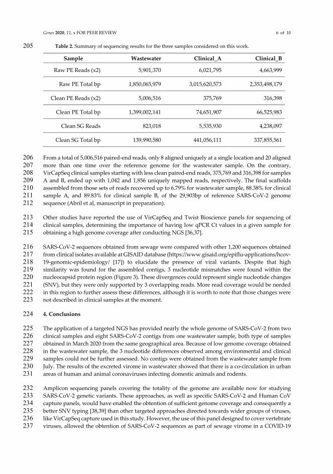
Genes 2020, 11, x FOR PEER REVIEW 6 of 10
Table 2. Summary of sequencing results for the three samples considered on this work. 205
Sample Wastewater Clinical_A Clinical_B
Raw PE Reads (x2) 5,901,370 6,021,795 4,663,999
Raw PE Total bp 1,850,065,979 3,015,620,573 2,353,498,179
Clean PE Reads (x2) 5,006,516 375,769 316,398
Clean PE Total bp 1,399,002,141 74,651,907 66,525,983
Clean SG Reads 823,018 5,535,930 4,238,097
Clean SG Total bp 139,990,580 441,056,111 337,855,561
From a total of 5,006,516 paired-end reads, only 8 aligned uniquely at a single location and 20 aligned 206 more than one time over the reference genome for the wastewater sample. On the contrary, 207 VirCapSeq clinical samples starting with less clean paired-end reads, 375,769 and 316,398 for samples 208 A and B, ended up with 1,042 and 1,856 uniquely mapped reads, respectively. The final scaffolds 209 assembled from those sets of reads recovered up to 6.79% for wastewater sample, 88.38% for clinical 210 sample A, and 89.83% for clinical sample B, of the 29,903bp of reference SARS-CoV-2 genome 211 sequence (Abril et al, manuscript in preparation). 212
Other studies have reported the use of VirCapSeq and Twist Bioscience panels for sequencing of 213 clinical samples, determining the importance of having low qPCR Ct values in a given sample for 214 obtaining a high genome coverage after conducting NGS [36,37]. 215
SARS-CoV-2 sequences obtained from sewage were compared with other 1,200 sequences obtained 216 from clinical isolates available at GISAID database (https://www.gisaid.org/epiflu-applications/hcov-217 19-genomic-epidemiology/ [17]) to elucidate the presence of viral variants. Despite that high 218 similarity was found for the assembled contigs, 3 nucleotide mismatches were found within the 219 nucleocapsid protein region (Figure 3). These divergences could represent single nucleotide changes 220 (SNV), but they were only supported by 3 overlapping reads. More read coverage would be needed 221 in this region to further assess these differences, although it is worth to note that those changes were 222 not described in clinical samples at the moment. 223
4. Conclusions 224
The application of a targeted NGS has provided nearly the whole genome of SARS-CoV-2 from two 225 clinical samples and eight SARS-CoV-2 contigs from one wastewater sample, both type of samples 226 obtained in March 2020 from the same geographical area. Because of low genome coverage obtained 227 in the wastewater sample, the 3 nucleotide differences observed among environmental and clinical 228 samples could not be further assessed. No contigs were obtained from the wastewater sample from 229 July. The results of the excreted virome in wastewater showed that there is a co-circulation in urban 230 areas of human and animal coronaviruses infecting domestic animals and rodents. 231
Amplicon sequencing panels covering the totality of the genome are available now for studying 232 SARS-CoV-2 genetic variants. These approaches, as well as specific SARS-CoV-2 and Human CoV 233 capture panels, would have enabled the obtention of sufficient genome coverage and consequently a 234 better SNV typing [38,39] than other targeted approaches directed towards wider groups of viruses, 235 like VirCapSeq capture used in this study. However, the use of this panel designed to cover vertebrate 236 viruses, allowed the obtention of SARS-CoV-2 sequences as part of sewage virome in a COVID-19 237

Genes 2020, 11, x FOR PEER REVIEW 7 of 10
pandemic context. Also, it retrieved other relevant human and animal CoV sequences which could 238 shed light on the co-circulation of different CoV strains in a determined population. 239
Different targeted NGS approaches have been applied for providing information on SARS-CoV-2 and 240 other CoV circulation and genetic characteristics. Specific SARS-CoV-2/human CoV panels should be 241 used for studying SARS-CoV-2 and other Human CoV genetic diversity while panels targeting a wide 242 variety of viral families would be a better choice for evaluating the co-circulation of known and 243 unknown human and animal CoV, which may be of relevance regarding potentially zoonosis as well 244 as CoV strains co-circulation immune cross-protective phenomena. 245
Supplementary Materials: The following are available online at www.mdpi.com/xxx/s1, Table S1: Complete list 246 of all the identified viral species and families in the sewage March sample by Target Enrichment NGS, File S2: 247 Complete SARS-CoV-2 assembled scaffold from sewage contigs, File S3: Mafft whole alignment from the 248 recovered reads with respect to the alignment with 1200 randomly chosen GISAID SARS-CoV-2 sequences. The 249 NGS datasets generated during the current study are available in zenodo under the DOI number 250 10.5281/zenodo.4252765. 251 Author Contributions: Conceptualization, S.B-M. and S.M-P.; methodology, S.M-P, M.I, M.R, E.F; software, J.A., 252 S.M-P; validation, S.B-M, R.G and J.A.; formal analysis, S.M-P, S.B-M and J.A; investigation, S.M-P and S.B-M.; 253 resources, S.B-M, R.G, J.Q, A.A.; data curation, S.M-P and C.A; writing—original draft preparation, S.M-P and 254 S.B-M.; writing—review and editing, S.M-P, S.B-M., M.I, M.R, E.F, C.A, A.A. J.Q, R.G a J.A; visualization, S.M-P 255 and J.A.; supervision, S.B-M.; project administration, S.B-M, R.G, A.A, J.Q.; funding acquisition, S.B-M, R.G, A.A, 256 J.Q. All authors have read and agreed to the published version of the manuscript. 257 Funding: This study was partially supported by and the Ministry of Science, Innovation and Universities 258 (AGL2017-86797-C2-1-R) through the University of Barcelona and the Direcció General de Recerca i Innovació 259 en Salut (DGRIS) Catalan Health Ministry Generalitat de Catalunya through Vall d’Hebron Research Institute 260 (VHIR). Sílvia Bofill-Mas is a Serra-Hunter fellow at the University of Barcelona. 261 Acknowledgments: We thank the members of ICRA for the coordination of the sewage sampling. We also 262 gratefully acknowledge the authors originating and submitting laboratories of the SARS-Cov-2 sequences from 263 GISAID’s EpiCov Database (https://www.epicov.org/epi3/) used to build the Figure 3 alignment; access to the 264 individual isolates is facilitated through GISAID web site (https://www.gisaid.org/). 265 Conflicts of Interest: The authors declare no conflict of interest 266 267 References 268
1. Wu, F.; Zhao, S.; Yu, B.; Chen, Y.M.; Wang, W.; Song, Z.G.; Hu, Y.; Tao, Z.W.; Tian, J.H.; Pei, Y.Y.; 269 et al. A new coronavirus associated with human respiratory disease in China. Nature 2020, 579, 265–270 269, doi:10.1038/s41586-020-2008-3. 271
2. Wu, Y.; Guo, C.; Tang, L.; Hong, Z.; Zhou, J.; Dong, X.; Yin, H.; Xiao, Q.; Tang, Y.; Qu, X.; et al. 272 Prolonged presence of SARS-CoV-2 viral RNA in faecal samples. Lancet Gastroenterol. Hepatol. 2020, 273 5, 434–435, doi:10.1016/S2468-1253(20)30083-2. 274
3. Wölfel, R.; Corman, V.M.; Guggemos, W.; Seilmaier, M.; Zange, S.; Müller, M.A.; Niemeyer, D.; 275 Jones, T.C.; Vollmar, P.; Rothe, C.; et al. Virological assessment of hospitalized patients with COVID-276 2019. Nature 2020, 581, 465–469, doi:10.1038/s41586-020-2196-x. 277
4. Cantalupo, P.G.; Calgua, B.; Zhao, G.; Hundesa, A.; Wier, A.D.; Katz, J.P.; Grabe, M.; Hendrix, 278 R.W.; Girones, R.; Wang, D.; et al. Raw sewage harbors diverse viral populations. MBio 2011, 2, 279 e00180-11, doi:10.1128/mBio.00180-11. 280
5. Ng, T.F.F.; Marine, R.; Wang, C.; Simmonds, P.; Kapusinszky, B.; Bodhidatta, L.; Oderinde, B.S.; 281 Wommack, K.E.; Delwart, E. High variety of known and new RNA and DNA viruses of diverse 282

Genes 2020, 11, x FOR PEER REVIEW 8 of 10
origins in untreated sewage. J. Virol. 2012, 86, 12161–12175, doi:10.1128/JVI.00869-12. 283
6. Martínez-Puchol, S.; Rusiñol, M.; Fernández-Cassi, X.; Timoneda, N.; Itarte, M.; Andrés, C.; 284 Antón, A.; Abril, J.F.; Girones, R.; Bofill-Mas, S. Characterisation of the sewage virome: comparison 285 of NGS tools and occurrence of significant pathogens. Sci. Total Environ. 2020, 713, 286 doi:10.1016/j.scitotenv.2020.136604. 287
7. Fernandez-Cassi, X.; Timoneda, N.; Martínez-Puchol, S.; Rusiñol, M.; Rodriguez-Manzano, J.; 288 Figuerola, N.; Bofill-Mas, S.; Abril, J.F.; Girones, R. Metagenomics for the study of viruses in urban 289 sewage as a tool for public health surveillance. Sci. Total Environ. 2018, 618, 870–880, 290 doi:10.1016/j.scitotenv.2017.08.249. 291
8. CDC 2019-nCoV CDC EUA qPCR Assay. 2020. 292
9. Vilsker, M.; Moosa, Y.; Nooij, S.; Fonseca, V.; Ghysens, Y.; Dumon, K.; Pauwels, R.; Alcantara, 293 L.C.; Vanden Eynden, E.; Vandamme, A.-M.; et al. Genome Detective: an automated system for virus 294 identification from high-throughput sequencing data. Bioinformatics 2018, 1–3, 295 doi:10.1093/bioinformatics/bty695. 296
10. Bolger, A.M.; Lohse, M.; Usadel, B. Trimmomatic: A flexible trimmer for Illumina sequence data. 297 Bioinformatics 2014, 30, 2114–2120, doi:10.1093/bioinformatics/btu170. 298
11. Li, H.; Handsaker, B.; Wysoker, A.; Fennell, T.; Ruan, J.; Homer, N.; Marth, G.; Abecasis, G.; 299 Durbin, R. The Sequence Alignment/Map format and SAMtools. Bioinformatics 2009, 25, 2078–2079, 300 doi:10.1093/bioinformatics/btp352. 301
12. Barnett, D.W.; Garrison, E.K.; Quinlan, A.R.; Strmberg, M.P.; Marth, G.T. Bamtools: A C++ API 302 and toolkit for analyzing and managing BAM files. Bioinformatics 2011, 27, 1691–1692, 303 doi:10.1093/bioinformatics/btr174. 304
13. Langmead, B.; Salzberg, S.L. Fast gapped-read alignment with Bowtie 2. Nat. Methods 2012, 9, 305 357–359, doi:10.1038/nmeth.1923. 306
14. Grabherr, M.G..; Brian J. Haas, Moran Yassour Joshua Z. Levin, Dawn A. Thompson, Ido Amit, 307 Xian Adiconis, Lin Fan, Raktima Raychowdhury, Qiandong Zeng, Zehua Chen, Evan Mauceli, Nir 308 Hacohen, Andreas Gnirke, Nicholas Rhind, Federica di Palma, Bruce W., N.; Friedman, and A.R. 309 Trinity: reconstructing a full-length transcriptome without a genome from RNA-Seq data. Nat. 310 Biotechnol. 2013, 29, 644–652, doi:10.1038/nbt.1883.Trinity. 311
15. Haas, B.J.; Papanicolaou, A.; Yassour, M.; Grabherr, M.; Philip, D.; Bowden, J.; Couger, M.B.; 312 Eccles, D.; Li, B.; Macmanes, M.D.; et al. De novo transcript sequence recostruction from RNA-Seq: 313 reference generation and analysis with Trinity; 2013; Vol. 8; ISBN 3942667509. 314
16. Katoh, K.; Standley, D.M. MAFFT multiple sequence alignment software version 7: 315 Improvements in performance and usability. Mol. Biol. Evol. 2013, 30, 772–780, 316 doi:10.1093/molbev/mst010. 317
17. Elbe, S.; Buckland-Merrett, G. Data, disease and diplomacy: GISAID’s innovative contribution 318 to global health. Glob. Challenges 2017, 1, 33–46, doi:10.1002/gch2.1018. 319
18. Shu, Y.; McCauley, J. GISAID: Global initiative on sharing all influenza data – from vision to 320 reality. Eurosurveillance 2017, 22, 2–4, doi:10.2807/1560-7917.ES.2017.22.13.30494. 321

Genes 2020, 11, x FOR PEER REVIEW 9 of 10
19. Bofill-Mas, S.; Albinana-Gimenez, N.; Clemente-Casares, P.; Hundesa, A.; Rodriguez-Manzano, 322 J.; Allard, A.; Calvo, M.; Girones, R. Quantification and stability of human adenoviruses and 323 polyomavirus JCPyV in wastewater matrices. Appl. Environ. Microbiol. 2006, 72, 7894–7896, 324 doi:10.1128/AEM.00965-06. 325
20. Briese, T.; Kapoor, A.; Mishra, N.; Jain, K.; Kumar, A.; OJ, J.; Lipkin, W. Virome Capture 326 Sequencing Enables Sensitive Viral Diagnosis and Comprehensive Virome Analysis. MBio 2015, 6, 1–327 11, doi:10.1128/mBio.01491-15.Editor. 328
21. Hjelmsø, M.H.; Mollerup, S.; Jensen, R.H.; Pietroni, C.; Lukjancenko, O.; Schultz, A.C.; 329 Aarestrup, F.M.; Hansen, A.J. Metagenomic analysis of viruses in toilet waste from long distance 330 flights—A new procedure for global infectious disease surveillance. PLoS One 2019, 14, e0210368, 331 doi:10.1371/journal.pone.0210368. 332
22. Krafcikova, P.; Silhan, J.; Nencka, R.; Boura, E. Structural analysis of the SARS-CoV-2 333 methyltransferase complex involved in RNA cap creation bound to sinefungin. Nat. Commun. 2020, 334 11, 1–7, doi:10.1038/s41467-020-17495-9. 335
23. Van der Hoek, L. Human coronaviruses: What do they cause? Antivir. Ther. 2015, 12. 336
24. Ye, Z.W.; Yuan, S.; Yuen, K.S.; Fung, S.Y.; Chan, C.P.; Jin, D.Y. Zoonotic origins of human 337 coronaviruses. Int. J. Biol. Sci. 2020, 16, 1686–1697, doi:10.7150/ijbs.45472. 338
25. Vogel, L.; Van Der Lubben, M.; Te Lintelo, E.G.; Bekker, C.P.J.; Geerts, T.; Schuijff, L.S.; Grinwis, 339 G.C.M.; Egberink, H.F.; Rottier, P.J.M. Pathogenic characteristics of persistent feline enteric 340 coronavirus infection in cats. Vet. Res. 2010, 41, doi:10.1051/vetres/2010043. 341
26. Gundy, P.M.; Gerba, C.P.; Pepper, I.L. Survival of Coronaviruses in Water and Wastewater. Food 342 Environ. Virol. 2009, 1, 10–14, doi:10.1007/s12560-008-9001-6. 343
27. Pratelli, A. Genetic evolution of canine coronavirus and recent advances in prophylaxis. Vet. 344 Res. 2006, 37, 191–200, doi:10.1051/vetres:2005053. 345
28. Wang, W.; Lin, X.; Guo, W.; Zhou, R.; Wang, M.; Wang, C.-Q.; Ge, S.; Mei, S.-H.; Li, M.-H.; Shi, 346 M.; et al. Discovery, diversity and evolution of novel coronaviruses sampled from rodents in China. 347 Virology 2015, 474, 19–27, doi:10.1016/j.virol.2014.10.017. 348
29. Wartecki, A.; Rzymski, P. On the coronaviruses and their associations with the aquatic 349 environment and wastewater. Water (Switzerland) 2020, 12, 1–27, doi:10.3390/W12061598. 350
30. Kivistö, I.; Tidenberg, E.M.; Lilley, T.; Suominen, K.; Forbes, K.M.; Vapalahti, O.; Huovilainen, 351 A.; Sironen, T. First Report of Coronaviruses in Northern European Bats. Vector-Borne Zoonotic Dis. 352 2020, 20, 155–158, doi:10.1089/vbz.2018.2367. 353
31. Hall, R.J.; Wang, J.; Peacey, M.; Moore, N.E.; McInnes, K.; Tompkins, D.M. New 354 alphacoronavirus in Mystacina tuberculata bats, New Zealand. Emerg. Infect. Dis. 2014, 20, 697–700, 355 doi:10.3201/eid2004.131441. 356
32. De Sabato, L.; Lelli, D.; Faccin, F.; Canziani, S.; Di Bartolo, I.; Vaccari, G.; Moreno, A. Full genome 357 characterization of two novel Alpha-coronavirus species from Italian bats. Virus Res. 2019, 260, 60–358 66, doi:10.1016/j.virusres.2018.11.007. 359

Genes 2020, 11, x FOR PEER REVIEW 10 of 10
33. Zhou, H.; Chen, X.; Hu, T.; Li, J.; Song, H.; Liu, Y.; Wang, P.; Liu, D.; Yang, J.; Holmes, E.C.; et al. 360 A Novel Bat Coronavirus Closely Related to SARS-CoV-2 Contains Natural Insertions at the S1/S2 361 Cleavage Site of the Spike Protein. Curr. Biol. 2020, 30, 2196-2203.e3, doi:10.1016/j.cub.2020.05.023. 362
34. Li, B.; Si, H.-R.; Zhu, Y.; Yang, X.-L.; Anderson, D.E.; Shi, Z.-L.; Wang, L.-F.; Zhou, P. Discovery 363 of Bat Coronaviruses through Surveillance and Probe Capture-Based Next-Generation Sequencing. 364 mSphere 2020, 5, 1–10, doi:10.1128/msphere.00807-19. 365
35. Lim, X.F.; Lee, C.B.; Pascoe, S.M.; How, C.B.; Chan, S.; Tan, J.H.; Yang, X.; Zhou, P.; Shi, Z.; 366 Sessions, O.M.; et al. Detection and characterization of a novel bat-borne coronavirus in Singapore 367 using multiple molecular approaches. J. Gen. Virol. 2019, 100, 1363–1374, doi:10.1099/jgv.0.001307. 368
36. Klempt, P.; Brož, P.; Kašný, M.; Novotný, A.; Kvapilová, K.; Kvapil, P. Performance of Targeted 369 Library Preparation Solutions for SARS-CoV-2 Whole Genome Analysis. Diagnostics 2020, 10, 769, 370 doi:10.3390/diagnostics10100769. 371
37. Carbo, E.C.; Sidorov, I.A.; Zevenhoven-Dobbe, J.C.; Snijder, E.J.; Claas, E.C.; Laros, J.F.J.; Kroes, 372 A.C.M.; de Vries, J.J.C. Coronavirus discovery by metagenomic sequencing: a tool for pandemic 373 preparedness. J. Clin. Virol. 2020, 131, 104594, doi:10.1016/j.jcv.2020.104594. 374
38. Xiao, M.; Liu, X.; Ji, J.; Li, M.; Li, J.; Yang, L.; Sun, W.; Ren, P.; Yang, G.; Zhao, J.; et al. Multiple 375 approaches for massively parallel sequencing of SARS-CoV-2 genomes directly from clinical samples. 376 Genome Med. 2020, 12, 1–15, doi:10.1186/s13073-020-00751-4. 377
39. Nasir, J.A.; Kozak, R.A.; Aftanas, P.; Raphenya, A.R.; Smith, K.M.; Maguire, F.; Maan, H.; 378 Alruwaili, M.; Banerjee, A.; Mbareche, H.; et al. A comparison of whole genome sequencing of sars-379 cov-2 using amplicon-based sequencing, random hexamers, and bait capture. Viruses 2020, 12, 380 doi:10.3390/v12080895. 381
Publisher’s Note: MDPI stays neutral with regard to jurisdictional claims in published maps and institutional 382 affiliations. 383
© 2020 by the authors. Submitted for possible open access publication under the terms and conditions of the Creative Commons Attribution (CC BY) license (http://creativecommons.org/licenses/by/4.0/).
384

Figure 1: Diversity and relative abundance of vertebrate viral families reads. The results for non-enrichment NGS are represented in dotted lines, regular lines represent the results after Target enrichment NGS. Relative abundance is represented in log10.ç

Figure 2: Summary of reads coverage along the SARS-CoV-2 reference genome sequence. The number of reads per position for the clean Pair-End reads (PEcovg) and for the Single-End reads (SEcovg) is show here for the three samples discussed on this manuscript. SE-reads were produced when cleaning the raw PE-reads, corresponding to the single member of a pair or reads that passed the cleaning and trimming criteria while the other read was discarded. Top track represents the segments defining the open reading frames encoding for the viral proteins (except the trailing 5’UTR and 3’UTR segments that are not protein-coding and were included for illustrative purposes). Dashed red line is the average coverage.

Figure 3: Nucleotide mismatches found in the nucleocapsid protein region. The red boxes highlight the 3 nucleotides that differ on the sequence reconstructed from the recovered reads with respect to the alignment with 1200 randomly chosen GISAID SARS-CoV-2 sequences (including the reference). Those nucleotide substitutions correspond to 1 synonymous and 2 non-synonymous changes: 28721.caa[Q] <> 28514.cCa[P], 29187.tgc[C] == 28959.tgT[C], 29188.aca[T] <> 28960.Gca[A]. Those three positions do not change on the alignment for all the other provided sequences yet are supported by only three reads which in fact does not allow defining them as variants. Full alignment is available as Supplementary File 3.


CAPÍTOL 2: IDENTIFICACIÓ DE POTENCIALS AGENTS ETIOLÒGICS CAUSANTS
DE GASTROENTERITIS MITJANÇANT SEQÜENCIACIÓ
MASSIVA


CAPÍTOL 2
93
Article 3: Utilització de la metagenòmica per a l’estudi dels virus associats a gastroenteritis d’etiologia desconeguda
La gastroenteritis aguda infecciosa és una malaltia amb elevada importància a nivell mundial, especialment en nens, on aproximadament el 70% dels casos estan associats a infeccions víriques. També un elevat nombre de casos presenten etiologia desconeguda, pel que l’aparició de les tècniques de seqüenciació massiva ha obert noves oportunitats per a la detecció i descobriment de patògens vírics.
Es van analitzar per metagenòmica 124 mostres de femta de pacients amb gastroenteritis, dels que no es va trobar agent causal clàssic, per tal d’identificar possibles infeccions víriques. Les famílies de virus que infecten vertebrats més abundantment trobades han estat Astroviridae i Caliciviridae, amb la detecció de NoV GIV i sapovirus. També s’han detectat seqüències d’enterovirus, adenovirus i rotavirus en algunes de les mostres analitzades. Aquests resultats posen de manifest que caldria optimitzar les tècniques de cribratge rutinari que s’utilitzen actualment, per a fer possible la detecció d’astrovirus, sapovirus i NoV GIV.
L’aplicació de la metagenòmica per a l’estudi d’infeccions víriques té doncs un gran potencial en l’anàlisi clínic rutinari per, principalment, identificar patògens que tradicionalment no s’han testat i escapen els test rutinaris i també per detectar noves variants que poden causar gastroenteritis.
“Unveiling viruses associated with gastroenteritis using a metagenomics approach.”
Fernández Cassi X.*, Martínez-Puchol S.*, Cornejo T., Bartolomé R., Bofill-Mas S., Girones R. Viruses, 12(12), 1432; https://doi.org/10.3390/v12121432.

CAPÍTOL 2
94

Viruses 2020, 12, 1432; doi:10.3390/v12121432 www.mdpi.com/journal/viruses
Article
Unveiling Viruses Associated with Gastroenteritis Using a Metagenomics Approach
Xavier Fernandez-Cassi 1,*,†,‡, Sandra Martínez-Puchol 1,†, Marcelle Silva-Sales 1,2, Thais Cornejo 3, Rosa Bartolome 3, Silvia Bofill-Mas 1 and Rosina Girones 1
1 Laboratory of Virus Contaminants of Water and Food, Department of Genetics, Microbiology and Statistics, University of Barcelona, Barcelona 08028, Spain; [email protected] (S.M.-P.); [email protected] (M.S.-S.); [email protected] (S.B.-M.); [email protected] (R.G.)
2 Laboratory of Virology and Cell Culture, Tropical Pathology and Public Health Institute, Federal University of Goiás, 74605-050 Goiânia, Goiás, Brazil
3 Hospital Universitari Vall d’Hebron, Microbiology Service, 08035 Barcelona, Spain; [email protected] (T.C.); [email protected] (R.B.)
* Correspondence: [email protected] † These authors contributed equally to this work. ‡ Present address: Laboratory of Environmental Chemistry, School of Architecture, Civil and Environmental
Engineering (ENAC), École Polytechnique Fédérale de Lausanne (EPFL), 1015 Lausanne, Switzerland.
Academic Editor: Lennart Svensson
Received: 4 November 2020; Accepted: 8 December 2020; Published: 13 December 2020
Abstract: Acute infectious gastroenteritis is an important illness worldwide, especially on children, with viruses accounting for approximately 70% of the acute cases. A high number of these cases have an unknown etiological agent and the rise of next generation sequencing technologies has opened new opportunities for viral pathogen detection and discovery. Viral metagenomics in routine clinical settings has the potential to identify unexpected or novel variants of viral pathogens that cause gastroenteritis. In this study, 124 samples from acute gastroenteritis patients from 2012–2014 previously tested negative for common gastroenteritis pathogens were pooled by age and analyzed by next generation sequencing (NGS) to elucidate unidentified viral infections. The most abundant sequences detected potentially associated to acute gastroenteritis were from Astroviridae and Caliciviridae families, with the detection of norovirus GIV and sapoviruses. Lower number of contigs associated to rotaviruses were detected. As expected, other viruses that may be associated to gastroenteritis but also produce persistent infections in the gut were identified including several Picornaviridae members (EV, parechoviruses, cardioviruses) and adenoviruses. According to the sequencing data, astroviruses, sapoviruses and NoV GIV should be added to the list of viral pathogens screened in routine clinical analysis�
Keywords: acute gastroenteritis; viral metagenomics; children; next-generation sequencing; norovirus; sapovirus; astrovirus; Picornaviridae
1. Introduction
Gastroenteritis is an important health problem around the globe affecting people at any age, but it is especially severe among children and the elderly. In 2016, diarrhea was the eighth leading cause of death among populations of all ages, causing an estimated 1,655,944 deaths and representing the fifth cause of death in children under 5 years with more than 446,000 estimated deaths [1]. While most of these deaths occur in developing countries, in developed countries acute gastroenteritis is still an important cause of death and an economic problem [2]. Clinical gastroenteritis can be caused

Viruses 2020, 12, 1432 2 of 19
�
by several infectious agents including bacteria, protozoa and viruses, the transmission of which can be foodborne or waterborne, through contaminated fomites or by person-to-person contact. Among all gastroenteritis etiological agents, viruses account for approximately 70% of acute cases in children with rotavirus (RV), norovirus (NoV), and adenovirus (AdV), accounting for the majority of cases [3,4], with an increase in astrovirus (AstV) and sapovirus (SaV) in the same age groups [5,6]. Despite these important viral agents, it has also been estimated that nearly 40% of gastroenteritis cases have an unknown etiological agent, suggesting that most could be caused by undiscovered viruses [7]. In recent years, the advent of next-generation sequencing (NGS) technologies has opened new opportunities for viral pathogen detection and discovery in the clinical setting [8]. Currently, more than 10,261 validated, complete viral genomes are available in databases (e.g., the National Center for Biotechnology Information (NCBI) RefSeq in September 2020, https://www.ncbi.nlm.nih.gov/genome/viruses/).
In the present manuscript, fecal samples from acute gastroenteritis patients who had previously tested negative for common pathogenic bacteria (Salmonella sp., Campylobacter sp., Shigella sp., Yersinia sp. and Verotoxigenic E. coli (VTEC).), parasites (Giardia sp. and Cryptosporidium sp.) and viruses (rotavirus, adenovirus, norovirus) using conventional routine methodologies, were analyzed by NGS to elucidate unidentified viral infections.
2. Materials and Methods
2.1. Sample Collection and Characterization
A total of 124 fecal samples from patients with acute gastroenteritis of unknown origin were collected in collaboration with Hospital Universitari Vall d’Hebron, Barcelona, Catalonia, Spain during 2012 and 2014. Previously, fecal samples were screened for the most common entero-pathogens, including Salmonella sp., Campylobacter sp., Shigella sp., Yersinia sp., and Verotoxigenic E. coli (VTEC), for which they tested negative. Samples were also screened for Giardia sp. and Cryptosporidium sp. if the clinical symptoms were compatible with a parasitic infection. In addition, frequent viral gastroenteritis agents for the specific age groups were tested using immunochromatographic assays for human adenoviruses (ONE STEP Adenovirus CARD TEST, Certest Biotec S.L), human rotaviruses (ONE STEP Rotavirus CARD TEST, Certest Biotec S.L) and human astroviruses (ONE STEP Astrovirus CARD TEST, Certest Biotec S.L), and conventional RT-PCR (norovirus GI and GII) [9]. No etiological agent could be identified to explain the clinical acute gastroenteritis; hence, the samples were screened using metagenomic means by pooling the samples according to age criteria in four different categories: less than 1 year (3 pools: F < 1, S < 1A and S < 1B), from 1 to 3 years (4 pools: F1-3A, S1-3A, S1-3B and S1-3C), from 3 to 10 years (1 pool: S3-10), and older than 10 years (1 pool: F > 10). For pools “F”, all samples were tested by RT-PCR for noroviruses [9]. Samples pooled under “S” criteria were only tested by RT-PCR under specific clinical request. A summary of the sample features composing each pool and the clinical tests performed is represented as Table 1.

Viru
ses 2
020,
12,
143
2 3
of 1
9
�
Tab
le 1
. Sum
mar
y of
the
sam
ples
com
posi
ng e
ach
sequ
ence
d po
ol a
ccor
ding
to a
ge c
rite
ria
and
the
perf
orm
ed c
onve
ntio
nal d
iagn
ostic
ass
ays i
n th
e cl
inic
al c
ente
r. Th
e re
sults
of t
he b
acte
rial
and
vir
al p
aram
eter
s ar
e ex
pres
sed
as th
e “n
umbe
r pos
itive
sam
ples
/num
ber s
ampl
es te
sted
”. F
or h
uman
ade
novi
rus,
hum
an ro
tavi
rus
and
hum
an a
stro
viru
s, a
com
mer
cial
ly a
vaila
ble
imm
unoc
hrom
atog
raph
ic te
st w
as e
mpl
oyed
(ON
E ST
EP A
deno
viru
s C
ARD
TES
T, O
NE
STEP
Rot
avir
us C
ARD
TE
ST a
nd O
NE
STEP
Ast
rovi
rus
CA
RD T
EST,
Cer
test
Bio
tec
S.L)
. For
nor
ovir
us G
I and
GII
, con
vent
iona
l RT-
PCR
was
use
d [9
].
B
acte
rial
Par
amet
ers
Vir
al P
aram
eter
s
Poo
l A
ge
N
Salm
onel
la
sp.
Cam
pylo
bact
er
sp.
Shig
ella
sp
. Ye
rsin
ia
sp.
Aer
omon
as
sp.
Ver
otox
igen
ic
E. c
oli (
VTE
C)
Vib
rio
sp.
Rot
avir
us
Ad
enov
iru
s A
stro
viru
s N
orov
iru
s G
GI
Nor
ovir
us
GG
II
F <
1 <1
10
0/
9 0/
9 0/
9 0/
9 0/
0 0/
0 0/
0 0/
10
0/10
0/
10
0/10
0/
10
S <
1A
9 0/
9 0/
9 0/
9 0/
9 0/
7 0/
0 0/
8 0/
9 0/
9 0/
9 0/
0 0/
0 S
< 1B
9
0/9
0/9
0/9
0/9
0/8
0/0
0/8
0/9
0/9
0/9
0/1
0/1
F1-3
A
1–3
23
0/23
0/
23
0/23
0/
23
0/0
1/1
0/1
0/23
0/
23
0/23
0/
23
0/23
S1
-3A
14
0/
13
0/13
0/
13
0/13
0/
11
0/1
0/11
0/
13
0/13
0/
13
0/1
0/1
S1-3
B 14
0/
14
0/14
0/
14
0/14
0/
7 0/
0 0/
8 0/
14
0/14
0/
14
0/1
0/1
S1-3
C
13
0/14
0/
14
0/14
0/
14
0/8
0/0
0/11
0/
14
0/14
0/
14
0/0
0/0
F3-1
0 3–
10
13
0/13
0/
13
0/13
0/
13
0/0
0/0
0/0
0/12
0/
12
0/13
0/
13
0/13
S3
-10
10
0/9
0/9
0/9
0/9
0/9
0/0
0/9
0/1
0/10
0/
10
0/0
0/0
F >
10
>10
9 0/
9 0/
9 0/
9 0/
9 0/
0 0/
1 0/
1 0/
8 0/
8 0/
9 0/
9 0/
9

Viruses 2020, 12, 1432 4 of 19
Viruses 2020, 12, x; doi: FOR PEER REVIEW www.mdpi.com/journal/viruses
2.2. Viral Concentration, Free DNA Removal, Nucleic Acid Extraction, and Library Preamplification and Preparation Using Untargeted Viral Metagenomics (UVM) and Target Enrichment Sequencing (TES)
Individual fecal samples of 0.5 g were suspended in 4.5 mL PBS 1x solution to obtain a 10% fecal suspension. Suspensions were homogenized and pooled by mixing 1 mL of each sample to construct the pools (Table 1). To enrich the viral fraction, fecal pools were centrifuged for 15 min at 3,000× g to remove cellular debris and bacteria; the supernatant was collected and filtered using a 0.22-µm low-binding protein syringe filter (Millex-GV, 0.22 µm, PVDF, 33 mm, Gamma-Sterilized, Millipore, Massachusetts, USA). Filtered pools (500 µL) were treated with 230 units of Turbo DNAse (Ambion, Lithuania) for 1 h at 37 °C to remove free DNA prior to nucleic acid extraction. The NA (nucleic acids) present in 280 µL of DNAse-treated viral concentrates was extracted using the QIAamp®Viral RNA Mini Kit from QIAGEN (Qiagen, Valencia, CA, USA). NA were eluted in 60 µL and stored at ƺ80 °C until further processing. To enable the detection of both DNA and RNA viruses, the total NAs were reverse-transcribed as previously described by Fernandez-Cassi et al. [10]. In short, SuperScript III (Life Technologies, Carlsbad, CA, USA) was used to retrotranscribe RNA to cDNA with primerA (5ȝ-GTTTCCCAGTCACGATCNNNNNNNNN-3ȝ). Second strand cDNA and DNA were constructed including primer sequences using Sequenase (USB/Affymetrix, Cleveland, OH, USA). PCR amplification with AmpliTaqGold (Life Technologies, Austin, Texas, USA) was performed using primerB (5ȝ-GTTTCCCAGTCACGATC-3ȝ); this step was performed in duplicate. After 10 min at 95 °C to activate DNA polymerase, the following PCR program was used: 25 cycles of 30 s at 94 °C, 30 s at 40 °C, and 30 s at 50 °C, with a final step of 60 s at 72 °C. The PCR products were purified and eluted in 15 µL using a Zymo DNA clean and concentrator (cat no. D4013, Zymo Research, Irvine, CA, USA) to yield sufficient DNA for the library preparation. Amplified DNA samples were quantified using Qubit 2.0 (cat no. Q32854, Life Technologies, OR, USA).
Untargeted viral metagenomics (UVM) viral libraries were constructed using a Nextera XT DNA sample preparation kit following the manufacturer’s instructions (Illumina Inc).
For the Target Enrichment Sequencing (TES), one pool (F2, 1–3 years) was reprocessed and resequenced by hybridizing the libraries with the vertebrate viral capture panel (VirCapSeq Enrichment Kit, Roche, Pleasanton, CA, USA) developed by Briese and coworkers [11] as detailed by Martínez-Puchol et al. [12]. Libraries were sequenced for three different runs using Illumina MiSeq 2 × 300 in base-pair paired-end format.
2.3. Bioinformatics Analysis
Bioinformatics analysis was performed using the Genome Detective platform (https://www.genomedetective.com/) [13]. For a more precise and accurate taxonomic classification, human viral contigs longer than 100 bp were queried for sequence similarity using BLASTN against the NCBI GenBank nucleotide collection database [14] with Geneious Software 11.1.5 (www.geneious.com) [15].
The species nomenclature and classification were performed according to the NCBI Taxonomy standards. For specific typing of human calicivirus, the assembled genomes of human noroviruses and sapoviruses were uploaded into the Noronet web-based service Typing tool (version 2.0) developed by RIVM using the updated classification of norovirus genogroups and genotypes [16,17]. Specific phylogenetic trees for Sapovirus were performed by aligning the complete genomes and the VP1 region against reference strains using MUSCLE [18]. Trees were constructed using tree view [19] software within Geneious (software version 11.0, https://www.geneious.com). For the Enterovirus genus within the Picornaviridae family, the same approach was performed, using the Enterovirus Genotyping Tool (version 0.1) developed by the RIVM [17]. For human adenovirus, retrieved contigs were mapped against their closest adenovirus type, and the hexon, penton, and fiber genes regions were annotated, extracted, and phylogenetically analyzed using the prototype strains suggested by the human Adenovirus Working Group (http://hadvwg.gmu.edu/). For human rotavirus analysis, a routinely binary phylogenetic analysis using the genes VP4 and VP7 was performed with the tool Rotavirus A Genotype Determination (ViPR, www.viprbrc.org) [20]. Contigs belonging to the

Viruses 2020, 12, x FOR PEER REVIEW 5 of 19
�
mamastrovirus and parechovirus genus were aligned to their complete reference genomes according to the ICTV classification. In addition, specific alignments using the ORF2 region and VP1 region for mamastrovirus and parechovirus against selected reference strains were conducted [21,22].
2.4. RT-PCR and RT-qPCR for Specific Viral Pathogens Sequencing
Specific viral RT-qPCR to detect NoVGIV was performed using primers and conditions as previously described [9,23,24] for individual fecal samples. RT-PCR for norovirus genogroup II was used in a specific subset of samples to confirm the obtained NGS results [25]. RT-PCR-positive band products were purified using Zymo DNA clean and concentrator (Zymo Research, cat no. D4013, Zymo Research, Irvine, CA, USA) and submitted to Sanger sequencing at Serveis Científico-Tècnics of the University of Barcelona.
3. Results and Discussion
3.1. Metagenomic Identification of Viral Families in Pooled Fecal Samples
A summary of the MiSeq output obtained from each sequenced pool containing the raw read number, contigs, and viral sequences detected, among other information, is presented as Supplementary material 1. Approximately 44 million raw-paired ends (0.9 to 12 M per sample) were generated. On average, 95% of the reads passed the QC cut-off and were annotated as viral using the Diamond tool [26], with 46% of these reads identified as viral (23% to 88%). When analyzing the samples according to age, a minimum and maximum percentage of viral reads ranging from to 29% to 50%, 23% to 56%, and 41% to 88% were obtained for pools with individuals aged younger than 1 year (F < 1, S < 1A and S < 1B), 1 to 3 years (F1-3A, S1-3A, S1-3B and S1-3C), and 3 to 10 years (F3-10 and S-3-10), respectively. Despite efforts to enhance viral content using filtration and nuclease treatment, 54% of the reads were classified as nonviral (12–77%). In the pool enriched by viral probe capture prior to sequencing (F1-3A), 74% of the reads were classified as viral.
A complete inventory of identified viral species classified according to the genome detective pipeline are presented in Supplementary material 2. Relevant taxonomical assignments for acute gastroenteritis will be discussed further. Additionally, these relevant pathogens which were considered important viral pathogens associated with gastroenteritis were plotted according to age and log10 abundance of reads (Figure 1).
Figure 1. Age distribution of the number of reads (log10) from human viral families with etiologically related members with viral gastroenteritis.

Viruses 2020, 12, x FOR PEER REVIEW 6 of 19
�
Astrovirus, Sapovirus and NoV GIV were detected with a high number of contigs, retrieving almost their complete genomes, and are considered the potential causative agents of gastroenteritis.
3.2. Caliciviridae
The results for this family have been divided into the main two human genera: Norovirus (NoV) and Sapovirus (SaV).
A summary of the viral contigs identified in the pools belonging to Caliciviridae family is presented in Table 2.
Table 2. Characterization of Caliciviridae contigs detected in the gastroenteritis pools with the norovirus typing tool by RIVM (v 2.0) using the dual system assessing the polymerase (ORF1) and the capsid region (ORF2). NT: non-typable by the RIVM Norovirus Genotyping Tool.
Pool Contig Length
Classification Polymerase Genotype Capsid Genotype BLAST Score
F < 1 7,521 Sapovirus GV NT GV.1 98.03 S < 1A 7,440 Norovirus GII GII.P21 (GII.Pb) GII.3 80.95
F1-3A 7,187 Norovirus GIV NT NT 77.26 4,827 Sapovirus GI NT GI.2 73.61
S1-3A
5,978 Norovirus GII GII.P31 (GII.Pe) GII.2 85.10 1,107 Norovirus GII NT NT 88.89 7,292 Sapovirus GII NT GII.1b 74.37 550 Sapovirus GI NT NT 74.27 376 Sapovirus GI NT NT 74.86 698 Sapovirus GI NT NT 76.72
2,109 Sapovirus GI NT NT 72.10 859 Sapovirus GI NT GI.3 78.06
1,321 Sapovirus GI NT GI.3 71.14 S1-3B 6,808 Norovirus GII GII.P17 GII.17 75.56
F3-10 7,384 Norovirus GII
GII.P4 New_Orleans_2009
GII.4 New_Orleans_2009
84.54
7,521 Sapovirus GV NT GV.1 98.08
S3-10
3,533 Norovirus GI NT NT
2,065 Norovirus GI NT GI.2 69.38 798 Norovirus GI GI.P2 GI.2 77.26
1,551 Sapovirus GI NT NT 75.02 1,059 Sapovirus GI NT NT 72.61 345 Sapovirus GI NT GI.3 66.47 349 Sapovirus GI NT GI.3 73.99
F > 10
924 Norovirus GIV NT NT 98.70 576 Norovirus GIV NT NT 98.44
1,249 Norovirus GIV NT NT 99.25 375 Norovirus GIV NT NT 98.67
3.2.1. Norovirus
Norovirus is the leading viral agent related to acute gastroenteritis in all age groups worldwide, with frequent outbreaks related to schools, hospitals, or cruise ships [27]. Its classification into genogroups and P-types (polymerase types) is sustained using a binary system based on the amino acid diversity of the complete VP1 gene and nucleotide diversity of the RNA-dependent RNA polymerase (RdRp) [16,17]. However, due their continuous recombination, NoV taxonomical classification is constantly evolving [16]. Four main genogroups are known to infect humans: GI, GII, GIV, and GIX (formerly GII.15) [16]. Norovirus GII genogroups are responsible for the majority of acute gastroenteritis cases, with GII.4 being the most predominant genotype worldwide with different variants emerging frequently [28].

Viruses 2020, 12, x FOR PEER REVIEW 7 of 19
�
In pools S1-3A, F1-3A, and F1-3B, three NoV GII contigs were identified. Two contigs of 5978 bp and 6808 bp were classified as GII.P31-GII.2 and GII.P17-GII.17, respectively. Infections caused by GII.P17-GII.17 have been increasing over the last decade, being predominant in some Asian regions, where they have displaced the classical GII.P4-GII.4 and its recombinant strains [29]. Its presence has increased and expanded in subsequent years after the first reported case, presenting a worldwide distribution and producing outbreaks elsewhere [30,31]. According to Van Beek et al., NoV GIIP.17 and GII.17 strains were detected in most European Countries in 2015–2016, but not in Spain [29]. The detection in 2014 samples suggests that the virus was already circulating in the population outside China. To verify this finding, conventional RT-PCR was conducted by following the suggested typing protocol described by van Beek and collaborators [25]. One sample from the S1-3B pool tested positive for NoV GII, and the amplicon was submitted to the RIVM Norovirus Genotyping Tool to confirm the finding of GII.17 (Accession number: MW205840).
Finally, one contig from pool F3-10 was subtyped as NoV GII.P4-GII.4 (New Orleans 2009). As stated previously, this is the predominant genotype worldwide and accounts for more than 70% of norovirus outbreaks [32], with GII.4 subvariants emerging, cocirculating, and replacing each other over time [33,34]. This particular strain could have been considered as non-detected by conventional RT-PCR due to a failure of the assay.
In pool S < 1A, one contig of 7,440 bp of NoV GII.P21-GII.3 was obtained. This recombinant strain is one of the most common strains circulating during the period under study [25,35,36]. In the same pool, one NoV GI.2-P1.2 was detected. This genotype has been linked to the consumption of polluted shellfish [37–39]. The non-detection of this particular genotype has been reported previously on some commercially available kits [40,41], and in-silico analysis revealed three mismatches with two of the forward primers used in clinical routine detection.
Several contigs taxonomically classified as NoV GIV were retrieved from the F1-3A and F > 10 pools. To explore the real incidence of norovirus GIV, a specific RT-qPCR assay for NoV GIV was performed using individual patient samples to confirm the finding. Three of 26 samples (12% of incidence) within the affected pools were positive by the RT-qPCR assay, with viral loads ranging from 2.71 × 10⁶ to 1.34 × 10⁷ genomic copies/gr feces. The viral loads are compatible with an active replication of the virus and could explain the acute gastroenteritis of these three patients, despite its lower incidence compared with NoVGII and GI. The detected contigs could not be typed using the Noronet Typing tool. In an attempt to obtain a more precise taxonomic classification, full NoV GIV retrieved was aligned against NoV GIV full complete genomes available at genbank using MUSCLE, and a phylogenetic tree was constructed (Figure 2). Full sequenced NoV GIV shares a homology of 99.2% with the NoV GIV strain from Australia (JQ613567) and could be classified as GIV.1. Norovirus GIV was first reported in 2000 by analyzing sporadic gastroenteritis cases [42], after which two different genotypes were identified, with NoV GIV.1 affecting humans and NoV GIV.2 considered a zoonotic genotype producing gastroenteritis in animals. Norovirus infections caused by GIV.1 are rare, and its prevalence in the population remains unknown. An international molecular surveillance NoV study conducted between 2005 and 2016 showed that less than 0.1% of the sequences submitted to NoroNet belonged to the NoV GIV.1 genotype [29]. Our findings are in line with previously reported results showing an incidence of 16% of NoV GIV in clinical samples [43]. The presence of this genotype has been detected by metagenomics in samples from children affected by gastroenteritis [44], as well as in other clinical and urban wastewater samples [43,45,46], indicating that it is actively circulating in the population but probably underreported. While the introduction of routine molecular detection in clinical settings could be the key for better monitoring of GIV-related cases, the NGS techniques have been shown to be a useful tool to detect a broader spectrum of norovirus genotypes and genogroups. This is especially relevant considering that their detection by common RT-PCR can sometimes fail due to the high variability in the P1 and VP1 regions, once again lending importance to the improvement of primer-independent detection techniques [47].

Viruses 2020, 12, x FOR PEER REVIEW 8 of 19
�
Figure 2. Phylogenetic tree of the almost-complete NoV GIV sequence from the F1-3A pool. Full genome sequences of NoV GIV are available at GenBank and were aligned using MUSCLE (Geneious 11.1). The murine norovirus sequence (accession number: AY228235) was used to root the tree. The tree was constructed with the Tamura-Nei genetic distance model using the neighbor-joining method with 1,000 bootstrap replicates. The nodes labels indicate the consensus support whereas the labels on the branch indicate the number of substitutions per site.
3.2.2. Sapovirus
Sapoviruses are one of the leading viral causes of gastroenteritis in children, with a particular impact in developing countries [48,49]. Their taxonomic classification is based on the capsid (VP1) sequence, with GI, GII, GIV and GV being the genogroups responsible for human gastroenteritis. Additionally, genogroups are further classified into genotypes, a classification system that keeps growing due to their high genetic diversity, with GI.1, GII.1, and GI.2 being the genotypes most commonly associated with clinical cases [48,50,51].
Contigs from the sapovirus genus were found in all sample pools except F > 10 (Table 2). Most of the detected sapoviruses were classified as GI (11/14), the most prevalent genogroup in patients within the studied age range (1 to 10 years) [49]. The typed GI contigs were GI.2 and GI.3, but no contig belonging to GI.1 was identified despite being the most prevalent in gastroenteritis samples and in this patient age group [49,52].
Contigs identified as Sapovirus GII (1/14) and GV (2/14) could not be typed using the Noronet Typing tool even though the complete genome was nearly assembled. GV is commonly linked to food-borne outbreaks [53–55], and some studies have shown a lack of detection of these types by

Viruses 2020, 12, x FOR PEER REVIEW 9 of 19
�
conventional RT-PCR, while they can be detected when applying NGS [54,55]. Alignments of the complete genomes retrieved from the sequenced pools against the complete genomes and the VP1 region from reference strains showed the clustering of GII contigs with SaV GII.1b, whereas sequenced GV contigs clustered with GV.1 (Supplementary material 3).
3.3. Astroviridae
Human astroviruses (HAstV) are important agents causing acute gastroenteritis in children and have also been involved in outbreaks affecting adults [56]. Recently, astroviruses have also been associated with a wider array of diseases, including central nervous system alterations such as meningitis or acute flaccid paralysis [57,58]. The classification is currently based on the amino acid sequence of the capsid region (ORF2), dividing the HAsV in the “classical” genotypes (Mamastrovirus 1 (MAstV-1) species) and the “novel” recombinant ones (MAstV-6, 8, and 9 species) [22,59]. A summary of the astrovirus species detected is presented in Table 3.
Table 3. Characterization of human astrovirus contigs detected in the tested fecal pools.
Pool Contig length Classification ORF2
Genotype % ORF2 Identity
% ORF2 Coverage
% Genome Identity
% Genome Coverage
F < 1 6,569 Mamastrovirus 6 MLB-3 97.8 100 98.1 99.8 S1-3A 6,642 Mamastrovirus 1 HAstV-2 92.7 100 85.7 97.6 F3-10 6,652 Mamastrovirus 1 HAstV-1 95.4 100 94.1 97.6 F > 10 6,651 Mamastrovirus 1 HAstV-1 95.1 100 94.0 97.6
Nearly the full genomes (6,469–6,652 bp) of four different astroviruses were retrieved. Human astrovirus species were found in all analyzed pools (Figure 1). HAstV-1 was detected in two pools and is the most common type associated with infantile gastroenteritis [60]. A HAstV-2 presenting a low homology over the full genome (85.7% identity) was detected in the S1-3A pool. During the development of the study, a commercially available immunochromatographic test was used to detect human astroviruses. In pooled samples “F”, this test was only used under specific circumstances (clinical signs supported an astrovirus infection, while samples “S” were all tested with this immunochromatographic test). Interestingly, among samples tested by the antigenic test, one result was inconclusive, which could be partially explained by the low homology of the detected astrovirus in this pool of samples. Phylogenetic trees with HAstV ORF2 complete sequences (Supplementary material 4a) and complete HAstV genomes (Supplementary material 4b) were constructed.
Nearly the full genome (6,569 bp) of an astrovirus MLB-3 was detected in the F < 1 pool. This novel astrovirus was first described in 2014 in stools from children with acute diarrhea [61]. It has also been reported in asymptomatic patients [62] or involved in coinfections with other microorganisms that cause diarrhea, raising doubts regarding its role as a gastroenteritis causative agent [62,63].
3.4. Adenoviridae
Human adenovirus is a well-known pathogen in children as well as adults, producing a wide array of diseases, from gastroenteritis to conjunctivitis, respiratory infections, cystitis and meningitis. There are currently 104 recognized human adenovirus types according to the Human adenovirus working group (http://hadvwg.gmu.edu/) divided into seven different adenovirus species (A–G) with species D being the most abundant [64]. Sequencing has allowed the identification of several recombinant types, rapidly increasing the number of reported human adenoviruses in the literature [65,66].
Human adenovirus contigs were reported in pools F1-3A, S1-3B and F3-10. No human adenovirus species F (HAdV40 and 41), which are important etiological gastroenteritis agents in children [67], were detected in any of the pooled samples. This suggests a high specificity of immunochromatographic tests for HAdV species F. However, the full genome of a HAdV31 has been retrieved from the pool F3-10. The phylogenetic analysis of the sequenced human adenovirus clusters

Viruses 2020, 12, x FOR PEER REVIEW 10 of 19
�
with HAdV31 reference type (Supplementary material 5). HAdV31 has been identified as a causative agent of acute gastroenteritis in children despite not belonging to HAdV species F [68].
Other HAdV type partial sequences have been identified based on the hexon, penton and fiber analysis. These adenoviruses might belong to species A (HAdV18, HAdV61, HAdV61, HAdV31), B (HAdV3, HAdV66), and C (HAdV89). Human adenoviruses are known to produce permanent infections in symptomatic and asymptomatic individuals. Hence, the detection of fragments of the adenovirus genomes could be indicative of the presence of persistent HAdV infections [69,70]. However, the lower number of contigs associated to other than HAdV31 and HAdV F species make them unlikely to be the causative agents of acute gastroenteritis.
3.5. Reoviridae
Rotavirus (RV) reads were detected in pools F < 1, S < 1A, S < 1B and S3-10. All detected RV belonged to the genotype G2P[4]. This genotype is one of the six most commonly genotypes detected worldwide, together with G1P[8], G3P[8], G4P[8], G9P[8], and G12P[8] [71,72]. RV has been identified as the main leading fatal etiologic agent for diarrhea in children less than five years old, causing 128,515 deaths in 2016 [1].
In the pool of fecal samples F < 1, S < 1A, and S < 1B, reads from 9 out of the 11 RVA genes were as follows: six structural protein (VP) coding genes and three nonstructural (NSP) coding genes. In the genotyping analysis, R2, C2, M2, P[4], I2, and G2, referred to as VP1, VP2, VP3, VP4, VP6, and VP7, respectively, were detected, and N2, T2, E2, referred to as NSP2, NSP3, and NSP4, respectively, were detected for the NSP genotypes [73]. In pool S3-10, reads of the genes coding for VP2, VP3, VP4, VP6, and VP7 with the same genotype pattern observed previously were found: C2, M2, P(4), I2, and G2. RVA detected in the present study showed high similarity (99.5–100%) to the genotype G2P(4) RVA strains circulating in Europe, Asia, and South America in the last decade for both pooled samples (data not shown). In this study, no RVA sequences revealed a close nucleotide similarity with the two commercially anti-RVA vaccines (Rotarix® and Rotateq®) available worldwide. Together, these results suggest that children probably acquired RVA strains circulating in Barcelona from 2012 to 2014, having no correlation with an anti-RVA vaccination. A study conducted between 2015 and 2016 revealed the presence of RVA in high viral loads in sewage samples from Barcelona. The authors also found no evidence of RVA vaccine-related strains in the sewage samples [74]. These results suggest that RVA is widespread in the community, since the population excretes RVA and the children still require healthcare assistance due to gastroenteritis associated with RVA.
Immunochromatographic tests for RVA diagnosis have been a useful tool to provide quick and cost-effective tests for routine clinical analyses at the expense of low sensitivity when compared to molecular methods such as RT-PCR [75,76]. The low number of reads obtained within the pools might be explained by three important facts: (a) the samples could have low levels of virus, since all were negative in the commonly used test; (b) individual patient samples were pooled, diluting the number of viral reads present in positive patients; (c) dsRNA viruses have been shown to be difficult to detect using current NGS techniques [77].
3.6. Other Viral Species Identified in Fecal Samples
Detected picornaviruses have been taxonomically classified according to their genera: Enterovirus, Kobuvirus, Parechovirus and Cardiovirus. Enterovirus and non-enterovirus picornaviruses have been summarized in Supplementary materials 6 and 7.
Enterovirus (EV) causes a wide range of diseases in children, such as hand-foot-mouth disease (HFMD), viral meningitis and encephalitis, acute flaccid paralysis, myocarditis, common cold or neonatal sepsis. It is also reported to be present in asymptomatic infections or coinfections with another microorganism both in children and adults [78].
The EV genus is subdivided by its capsid sequence (VP1) in 13 species, with EV-A, EV-B, EV-C, EV-D, rhinovirus A (RV-A), RV-B and RV-C infecting humans [79,80]. Enterovirus from species EV-A and EV-B with RV-A and RV-B were detected by NGS in the pooled samples. An exhaustive list

Viruses 2020, 12, x FOR PEER REVIEW 11 of 19
�
containing the typed enterovirus and rhinovirus using the RIVM enterovirus typing tool is provided in Supplementary material 6.
Rhinoviruses A (HRV-A) and B (HRV-B) were present in all the pools analyzed. These species are the most important etiological agents of common cold, with a high incidence of infections in early childhood, mainly affecting the upper respiratory airways, with HRV-A being the most prevalent [81]. In the S2 pool, nearly the full genome of HRV-A78 was obtained (6,499 bp). HRV is one of the predominant circulating species of HRV-A in children [82] and has been previously associated with pneumonia [83].
Members of the genus Parechovirus (HPeV) are common causes of aseptic meningitis in children and mild gastrointestinal and respiratory diseases [84]. Currently, more than 18 different types have been recognized to infect humans according to the HPeV ICTV taxonomy group, all of which cluster under HPEV-A species (https://talk.ictvonline.org/ictv-reports/ictv_online_report/positive-sense-rna-viruses/picornavirales/w/picornaviridae/693/genus-parechovirus). Sequences from HPeV A were detected in pools associated with younger patients (<3 years). Phylogenetic analysis allowed their classification as HPeV-1 and HPeV-6 (Supplementary material 8).
The Cardiovirus genus was first associated with rodent infections, until their role in human pathogenicity was described in 2007 [85]. There are currently three recognized species, Cardiovirus A, Cardiovirus B and Cardiovirus C. More than 11 genotypes of Cardio-virus B species are associated with human infection, with Saffold viruses (SAFV) 2 and 3 being the most prevalent.
Almost the whole genome of a Cardiovirus B member, genotyped as SAFV-2, was sequenced in pool F > 10 from affected individuals above 10 years old (Supplementary material 7). SAFV are not routinely tested in clinical samples, but recent studies have reported a common presence in gastroenteritis patients [86,87] and in raw sewage [88,89].
The genus Kobuvirus is composed of six recognized Aichi virus (AiV) viral species (AiVA-AiVF) that are classified according to their amino acid homology in the polyprotein, P1 2C and 3CD regions (https://talk.ictvonline.org/ictv-reports/ictv_online_report/positive-sense-rna-viruses/picornavirales/w/picornaviridae/686/genus-kobuvirus). Members of the AiV A and B species have been recognized as human pathogens causing gastroenteritis outbreaks [90–92]. Members within this genus are considered zoonotic and have the potential to infect dogs, cats or goats [93].
Two different Aichi virus contigs were detected in pool S3-10. The alignments of the retrieved contigs were phylogenetically related to AiV A which has been associated with acute gastroenteritis.
Picobirnavirus-related contigs were detected in most of the pools analyzed except F < 1 and S3-10. Previously, picobirnavirus has been considered a putative pathogenic viral species present in the respiratory and digestive tract of humans and other animals, including pigs, cows, dogs and otters. However, recent research has suggested that picobirnavirus might be a prokaryotic virus because the ribosomal binding site (RBS) motif is conserved within its genome. This motif has been detected in all available picobirnavirus sequences and is commonly enriched in all viral families that infect prokaryotes, but not in eukaryotic viral families [94].
Contigs belonging to primate bocaparvovirus 2, formerly known as Human Bocavirus 2, were found in pool S6. These viral species have been previously found in stool samples from gastroenteritis-related studies in young children with variable prevalence depending on the area of study and year of sample collection [95].
A diverse number of ssDNA circular viruses belonging to the Anelloviridae and Circoviridae families have been detected among the different pools analyzed. The family Anelloviridae includes the Torque teno virus (TTV), Torque teno midi virus (TTMDV) and mini virus Torque teno virus (TTMV). The detection of anellovirus species in clinical samples does not seem unlikely, as it is estimated that 70–90% of the human population carries the virus in loads that range from 103 to 106 genomic copies/mL plasma [96]. None of the different torque teno virus species known to date have been directly associated with disease [97].
However, the leading role in acute gastroenteritis of the discussed viral agents under this subsection is at least controversial, as most of them can replicate actively in the intestinal tract [98],

Viruses 2020, 12, x FOR PEER REVIEW 12 of 19
�
have been co-detected with other well stablished gastrointestinal viral pathogens such as RV, NoV, SaV or HAdV [78,86,92,99] or are commonly detected in non-symptomatic control samples [97,100].
Further studies focusing on these specific families in non-pooled samples are needed to clarify their involvement in acute gastroenteritis either as monoinfection or coinfection pathogens.
3.7. Target Enrichment Sequencing
A target enrichment approach (TES) was performed in one pool (F1-3A) to evaluate its usefulness for the detection of gastroenteritis viral agents compared with untargeted metagenomics (UVM) applied in this study. The application of a target enrichment probe-based capture method, prior to sequencing, has been demonstrated to overcome the low sensitivity or the lack of sequencing depth in complex samples with large viral diversity [12,101].
Comparing both methodologies and focusing on viral families of interest regarding gastroenteritis etiology, TES enabled the detection of a larger number of viral reads (Figure 4a), allowing the detection of Reoviridae members, which were not detected with the UVM approach. Additionally, TES allowed the typing of the hexon, penton and fiber genes of a HAdV-31 that could not be typed by UVM. In contrast, when analyzing these reads as contigs, the differences were reduced to obtain the same number of contigs by applying both methodologies to the detected families (Figure 4b). This observation could be related to the sequence fragmentation and diversity of the sample, the absence of sequences from different regions of each viral assignation, or the absence of regions to which the probes are hybridizing. This phenomenon would result in a large number of reads from the same genome region and a reduction of diversity, but an increase in accuracy of the contig sequence.
(a) (b)
Figure 4. Comparison of the number of viral reads (a) and contigs (b) from families with members related to gastroenteritis with Target Enrichment Sequencing (TES) and Untargeted Viral Metagenomics (UVM) in the F1-3A pool. The number of reads is represented as log10.
The application of TES allowed the detection of 4 RV contigs in the sample pool analyzed that were not previously detected in the UVM pool. The detected rotavirus sequences belonged to genotype G2P[4], the predominant RV circulating in Europe between 2010 and 2019 [71,72,102]. As observed for other detected RV sequences in the study, none of the contigs was related to the two commercially available rotavirus vaccines.
4. General Discussion and Conclusions
Viral metagenomics, in combination with or without a viral enrichment approach, has the potential to uncover any viral species that might be present in a given sample. These methodologies are not primer-dependent methods; hence, they detect viral species that might be undetected by using conventional molecular methodologies such as RT-PCR or RT-qPCR. The use of primer-independent methodologies represents an important advantage in detecting new viral pathogens (i.e., SARS-CoV-2) or recombinant/reassortant types. These methodologies are especially relevant for viral species, as

Viruses 2020, 12, x FOR PEER REVIEW 13 of 19
�
viruses are prone to have high mutation rates and to recombine/reassort their genomes as an evolutionary mechanism. In addition, the gastrointestinal tract is a complex ecosystem, and the evolution and severity of acute gastroenteritis might be conditioned by the presence of coinfections that could be masked by the application of single pathogen detection methods such as immunochromatography or real-time PCR. However, a large number of sequences obtained by metaviromics cannot be taxonomically classified by using reference databases and are considered viral dark matter, requiring a more exhaustive analysis [103,104]. The use of a viral-enriched approach (TES) increased the number of viral reads up to 74%, in contrast to the 23% viral reads detected without enrichment in the same pooled sample. However, despite the enrichment applied, a wider diversity of viral species could be detected without the viral enrichment with the exception of rotavirus A, which was not detected without applying TES.
According to the number of reads detected, the most abundant sequences from vertebrate viral families were from Astroviridae-related members, which were identified in all age groups, suggesting the need to include systematic molecular testing for this particular viral family independently of the patient age. Following AstV, NoV and SaV present an important number of reads. At the time of sample collection, clinical screening did not include any molecular assay to detect sapoviruses; thus, their detection using NGS seems reasonable. Sapoviruses have been identified as an increasing etiological agent for human gastroenteritis, and their screening in the clinical setting should be systematically performed at least for children under 10 years of age. Human noroviruses were detected in all pools analyzed despite most samples having been previously tested by conventional RT-PCR in clinical settings. Some of these NoV might have escaped detection by RT-PCR due to the presence of mismatches in primer annealing zones. Additional in-silico analysis of the NGS-retrieved sequence showed mismatches in the primer binding region, highlighting the utility of primer-independent methodologies to target viruses with a high mutation rate. Interestingly, NoV GIV, a minority NoV species related to gastroenteritis, was detected by NGS and confirmed in three patients by RT-qPCR. The inability to detect this specific NoV genogroup is expected because it is not routinely tested. Samples for which the NoVs test was ruled out according to clinical evaluation showed the presence of both NoV GI and GII. Therefore, we recommend the inclusion of NoVGI, GII and GIV in the systematic testing of all acute gastroenteritis cases.
HAdV not belonging to species F were detected in patients less than 10 years old. The use of immunochromatographic tests targeting adenovirus species F (HAdV 40 and 41) might present a lower sensitivity towards other adenoviruses (i.e., HAdV species A or C), which could be the etiological agents for some acute gastroenteritis such as the detected HAdV31. Nevertheless, HAdV are known to produce a wide array of infections including asymptomatic persistent infections of the gastrointestinal tract, raising doubts about their involvement in acute gastroenteritis. Similarly, human enteroviruses and other related picornaviruses (such as parechovirus and cardiovirus) can produce a wide array of clinical outcomes but are also persistently excreted by healthy asymptomatic individuals. Thus, without any additional clinical information, their detection in clinical samples might result from a coinfection with another gastroenteritis virus or simply as a subclinical infection. In this sense, the application of viral metagenomics has emerged as a promising tool contributing to increased knowledge of excreted viruses and their role in human pathogenesis.
Finally, a small proportion of reads could be taxonomically assigned to the family Reoviridae, which includes HRV. According to the phylogenetic analyses, the identified RV were not related with any of the existent vaccine strains; hence, their presence in the analyzed pools suggests their implication in the clinical gastroenteritis despite the low genome coverage and small number of reads retrieved from this particular viral family in this study. Similarly to HAdV, the small number of obtained reads could be explained by an inefficient tagging of double-stranded genomes during the retro-transcription or sequenase steps resulting in low coverage and the small number of reads assigned to this viral family. Specific protocols to increase the sensitivity towards double-stranded genomes (dsDNA and dsRNA) might need to be applied to explore the viral diversity within these genomic conformations [77]. �

Viruses 2020, 12, x FOR PEER REVIEW 14 of 19
�
Supplementary Materials: Supplementary Materials can be found at www.mdpi.com/1999-4915/12/12/1432/s1�
Author Contributions: Conceptualization, X.F.-C., S.M.-P., S.B.-M. and R.G.; Methodology, X.F.-C. and S.M.-P.; Software, X.F.-C., M.S.-S. and S.M.-P.; Validation, R.G., S.B.-M. and R.B.; Formal Analysis, X.F.-C., M.S.-S. and S.M.-P.; Investigation, X.F.-C. and S.M.-P.; Resources, R.G., S.B.-M. and R.B.; Data Curation, S.M.-P., X.F.-C. and T.C.; Writing–Original Draft Preparation, X.F.-C. and S.M.-P.; Writing–Review & Editing, X.F.-C., M.S.-S., S.M.-P., R.G., S.B.-M., R.B., T.C.; Visualization, X.F.-C. and S.M.-P.; Supervision, R.G.; Project Administration, R.G.; Funding Acquisition, R.G. and S.B.-M. All authors have read and agreed to the published version of the manuscript.
Funding: This study was partially funded by the Programa RecerCaixa 2012 (ACUP-00300) the projects AGL2011-30461-C02-01/ALI from the Spanish Ministry of Science and Innovation and the project AGL2014-55081-R from the Ministry of Economy. This study was partially funded by grants from the Catalan Government to the Consolidated Research Group VirBaP (2014SRG914) with the collaboration of the Institut de Recerca de l’Aigua (IdRA).
Availability of Data and Material: The datasets generated during the current study are available in zenodo under the DOI number https://doi.org/10.5281/zenodo.4161275 (https://zenodo.org/record/4161275#.X6KAjyySlPY).
Acknowledgments: During the development of the study Xavier Fernandez Cassi was a fellow of the Catalan government “AGAUR” (fi-dgr) while Sandra Martínez-Puchol was a fellow of Economy, Industry and Competitiveness Ministry (MINECO). Marcelle Silva-Sales would like to acknowledge the Brazilian National Council for Scientific and Technological Development (CNPq, in Portuguese: Conselho Nacional de Desenvolvimento Científico e Tecnológico) for support via a postdoctoral grant. Sílvia Bofill-Mas is a Serra-Hunter fellow at the University of Barcelona.
Conflicts of Interest: The authors declare no conflict of interest. The funders had no role in the design of the study; in the collection, analyses, or interpretation of data; in the writing of the manuscript, or in the decision to publish the results.
References
1.� Troeger, C.; Blacker, B.F.; Khalil, I.A.; Rao, P.C.; Cao, S.; Zimsen, S.R-M.; Albertson, S.B.; Stanaway, J.D; Deshpande, A.; Deshpande, A.; et al. stimates of the global, regional, and national morbidity, mortality, and aetiologies of diarrhoea in 195 countries: A systematic analysis for the Global Burden of Disease Study 2016. Lancet Infect. Dis. 2018, 18, 1211–1228.
2.� Bányai, K.; Estes, M.K.; Martella, V.; Parashar, U.D. Viral gastroenteritis. Lancet 2018, 392, 175–186. 3.� Fletcher, S.; Van Hal, S.; Andresen, D.; McLaws, M.L.; Stark, D.; Harkness, J.; Ellis, J. Gastrointestinal
pathogen distribution in symptomatic children in Sydney, Australia. J. Epidemiology Glob. Heal. 2013, 3, doi:10.1016/j.jegh.2012.11.004.
4.� Oppong, T.B.; Yang, H.; Amponsem-Boateng, C.; Kyere, E.K.D.; Abdulai, T.; Duan, G.; Opolot, G. Enteric pathogens associated with gastroenteritis among children under 5 years in sub-saharan Africa: A systematic review and meta-Analysis. Epidemiol. Infect. 2020, 148, 1–26.
5.� Tran, A.; Talmud, D.; Lejeune, B.; Jovenin, N.; Renois, F.; Payan, C.; Leveque, N.; Andreoletti, L. Prevalence of rotavirus, adenovirus, norovirus, and astrovirus infections and coinfections among hospitalized children in Northern France. J. Clin. Microbiol. 2010, 48, 1943–1946.
6.� Chhabra, P.; Payne, D.C.; Szilagyi, P.G.; Edwards, K.M.; Staat, M.A.; Shirley, S.H.; Wikswo, M.; Nix, W.A.; Lu, X.; Parashar, U.D.; Vinjé, J. Etiology of viral gastroenteritis in children <5 years of age in the United States, 2008–2009. J. Infect. Dis. 2013, 208, 790–800.
7.� Holtz, L.R.; Finkbeiner, S.R.; Zhao, G.; Kirkwood, C.D.; Girones, R.; Pipas, J.M.; Wang, D. Klassevirus 1, a previously undescribed member of the family Picornaviridae, is globally widespread. Virol. J. 2009, 6, 86.
8.� Fischer, T.K.; Rasmussen, L.D.; Fonager, J. Taking gastro-surveillance into the 21st century. J. Clin. Virol. 2019, 117, 43–48.
9.� Kageyama, T.; Kojima, S.; Shinohara, M.; Uchida, K.; Fukushi, S.; Hoshino, F.B.; Takeda, N.; Katayama, K. Broadly reactive and highly sensitive assay for Norwalk-like viruses based on real-time quantitative reverse transcription-PCR. J. Clin. Microbiol. 2003, 41, 1548–1557.
10.� Fernandez-Cassi, X.; Rusiñol, M.; Martínez-Puchol, S. Viral Concentration and Amplification from Human Serum Samples Prior to Application of Next-Generation Sequencing Analysis; Springer: New York, NY, USA, 2018; pp. 173–188.

Viruses 2020, 12, x FOR PEER REVIEW 15 of 19
�
11.� Briese, T.; Kapoor, A.; Mishra, N.; Jain, K.; Kumar, A.; Jabado, O.J.; Lipkin, W.I. Virome capture sequencing enables sensitive viral diagnosis and comprehensive virome analysis. mBio 2015, 6, e01491-15.
12.� Martínez-Puchol, S.; Rusiñol, M.; Fernández-Cassi, X.; Timoneda, N.; Itarte, M.; Andrés, C.; Antón, A.; Abril, J.F.; Girones, R.; Bofill-Mas, S. Characterisation of the sewage virome: Comparison of NGS tools and occurrence of significant pathogens. Sci. Total. Environ. 2020, 713, 136604.
13.� Vilsker, M.; Moosa, Y.; Nooij, S.; Fonseca, V.; Ghysens, Y.; Dumon, K.; Pauwels, R.; Alacantara, L.C.; Vanden Eynden, E.; Vandamme, A-M.; Deforche, K.; de Oliveira, T. Genome Detective: An automated system for virus identification from high-throughput sequencing data. Bioinformatics 2019, 35, 871–873.
14.� Benson, D.A.; Clark, K.; Karsch-Mizrachi, I.; Lipman, D.J.; Ostell, J.; Sayers, E.W. GenBank. Nucleic Acids Res. 2015, 43, D30–D35.
15.� Kearse, M.; Moir, R.; Wilson, A.; Stones-Havas, S.; Cheung, M.; Sturrock, S.; Buxton, S.; Cooper, A.; Markowitz, S.; Dura, C. Geneious Basic: An integrated and extendable desktop software platform for the organization and analysis of sequence data. Bioinformatics 2012, 28, 1647–1649.
16.� Chhabra, P.; de Graaf, M.; Parra, G.I.; Chan, M.C.W.; Green, K.; Martella, V.; Wang, Q.; White, P.A.; Katayama, K.; Vennema, H.; Koopmans, M.; Vinjé, J. Updated classification of norovirus genogroups and genotypes. J. Gen. Virol. 2019, 100, 1393–1406.
17.� Kroneman, A.; Vennema, H.; Deforche, K.; Avoort, H.; Peñaranda, S.; Oberste, M.S.; Vinjé, J.; Koopmans, M. An automated genotyping tool for enteroviruses and noroviruses. J. Clin. Virol. 2011, 51, 121–125.
18.� Edgar, R.C. MUSCLE: Multiple sequence alignment with high accuracy and high throughput. Nucleic Acids Res. 2004, 32, 1792–1797.
19.� Page, R.D.M. Treeview: An application to display phylogenetic trees on personal computers. Bioinformatics 1996, 12, 157–158.
20.� Maes, P.; Matthijnssens, J.; Rahman, M.; Van Ranst, M. RotaC: A web-based tool for the complete genome classification of group A rotaviruses. BMC Microbiol. 2009, 9, 238.
21.� Böttcher, S.; Obermeier, P.E.; Neubauer, K.; Diedrich, S. Recombinant enterovirus A71 subgenogroup C1 strains, Germany, 2015. Emerg. Infect. Dis. 2016, 22, 1843–1846.
22.� Donato, C.; Vijaykrishna, D. The broad host range and genetic diversity of mammalian and avian astroviruses. Viruses 2017, 9, 102.
23.� Loisy, F.; Atmar, R.L.; Guillon, P.; Le Cann, P.; Pommpepuy, M.; Le Guyader, F.S. Real-time RT-PCR for norovirus screening in shellfish. J. Virol. Methods 2005, 123, 1–7.
24.� Miura, T.; Parnaudeau, S.; Grodzki, M.; Okabe, S.; Atmar, R.L.; Le Guyader, F.S. Environmental detection of genogroup I, II, and IV noroviruses by using a generic real-time reverse transcription-PCR assay. Appl. Environ. Microbiol. 2013, 79, 6585–6592.
25.� Van Beek, J.; van der Eijk, A.A.; Fraaij, P.L.A.; Caliskan, K.; Cransberg, K.; Dalinghaus, M.; Hoek, R.A.S.; Metselaar, H.J.; Roodnat, J.; Vennema, H.; Koopmans, M. Chronic norovirus infection among solid organ recipients in a tertiary care hospital, the Netherlands, 2006–2014. Clin. Microbiol. Infect. 2017, 23, 265.e9–265.e13.
26.� Buchfink, B.; Xie, C.; Huson, D.H. Fast and sensitive protein alignment using DIAMOND. Nat. Methods 2015, 12, 59–60.
27.� Hall, A.J.; Eisenbart, V.G.; Etingüe, A.L.; Gould, L.H.; Lopman, B.A.; Parashar, U.D. Epidemiology of foodborne norovirus outbreaks, United States, 2001–2008. Emerg. Infect. Dis. 2012, 18, 1566–1573.
28.� Tohma, K.; Lepore, C.J.; Gao, Y.; Ford-Siltz, L.A.; Parra, G.I. Population genomics of gii.4 noroviruses reveal complex diversification and new antigenic sites involved in the emergence of pandemic strains. mBio 2019, 10, e02202-19.
29.� Van Beek, J.; de Graaf, M.; Al-Hello, H.; Allen, D.J.; Ambert-Balay, K.; Botteldoorn, N.; Brytting, M.; Buesa, J.; Cabrerizo, M.; Chan, M.; et al. Molecular surveillance of norovirus, 2005–2016: An epidemiological analysis of data collected from the NoroNet network. Lancet Infect. Dis. 2018, 18, 545–553.
30.� De Graaf, M.; Van Beek, J.; Koopmans, M.P.G. Human norovirus transmission and evolution in a changing world. Nat. Rev. Genet. 2016, 14, 421–433.
31.� Chen, L.; Xu, D.; Wu, X.; Liu, G.; Ji, L. An increasing prevalence of non-GII.4 norovirus genotypes in acute gastroenteritis outbreaks in Huzhou, China, 2014–2018. Arch. Virol. 2020, 165, 1121–1128.
32.� Vega, E.; Barclay, L.; Gregoricus, N.; Shirley, S.H.; Lee, D.; Vinjé, J. Genotypic and epidemiologic trends of norovirus outbreaks in the united states, 2009 to 2013. J. Clin. Microbiol. 2013, 52, 147–155.

Viruses 2020, 12, x FOR PEER REVIEW 16 of 19
�
33.� Lun, J.H.; Hewitt, J.; Yan, G.J.H.; Tuipulotu, D.E.; Rawlinson, W.; White, P. Recombinant GII.P16/GII.4 sydney 2012 was the dominant norovirus identified in Australia and New Zealand in 2017. Viruses 2018, 10, 548.
34.� Sabrià, A.; Pintó, R.M.; Bosch, A.; Bartolomé, R.; Cornejo, T.; Torner, N.; Martínez. A.; de Simón, M.; Domínguez, A.; Guix, S. Molecular and clinical epidemiology of norovirus outbreaks in Spain during the emergence of GII.4 2012 variant. J. Clin. Virol. 2014, 60, 96–104.
35.� Hoa Tran, T.N.; Trainor, E.; Nakagomi, T.; Cunliffe, N.A.; Nakagomi, O. Molecular epidemiology of noroviruses associated with acute sporadic gastroenteritis in children: Global distribution of genogroups, genotypes and GII.4 variants. J. Clin. Virol. 2013, 56, 269–277.
36.� Mans, J.; Murray, T.Y.; Taylor, M.B. Novel norovirus recombinants detected in South Africa. Virol. J. 2014, 11, 168.
37.� Le Guyader, F.S.; Atmar, R.L.; Le Pendu, J. Transmission of viruses through shellfish: When specific ligands come into play. Curr. Opin. Virol. 2012, 2, 103–110.
38.� Yu, Y.; Cai, H.; Hu, L.; Lei, R.; Pan, Y.; Yan, S.; Wang, Y. Molecular epidemiology of oyster-related human noroviruses and their global genetic diversity and temporal-geographical distribution from 1983 to 2014. Appl. Environ. Microbiol. 2015, 81, 7615–7624.
39.� Tunyakittaveeward, T.; Rupprom, K.; Pombubpa, K.; Howteerakul, N.; Kittigul, L. Norovirus Monitoring in Oysters Using Two Different Extraction Methods. Food Environ. Virol. 2019, 11, 374–382.
40.� Kanwar, N.; Hassan, F.; Barclay, L.; Langley, C.; Vinjé, J.; Bryant, P.W.; George, K.; Mosher, L.; Matthews-Greer, J.M.; Rocha, M.A. Evaluation of RIDA ® GENE norovirus GI/GII real time RT-PCR using stool specimens collected from children and adults with acute gastroenteritis. J. Clin. Virol. 2018, 104, 1–4.
41.� Dunbar, N.L.; Bruggink, L.D.; Marshall, J.A. Evaluation of the RIDAGENE real-time PCR assay for the detection of GI and GII norovirus. Diagn. Microbiol. Infect. Dis. 2014, 79, 317–321.
42.� Vinjé, J.; Koopmans, M.P.G. Simultaneous detection and genotyping of “Norwalk-like viruses” by oligonucleotide array in a reverse line blot hybridization format. J. Clin. Microbiol. 2000, 38, 2595–2601.
43.� La Rosa, G.; Pourshaban, M.; Iaconelli, M.; Muscillo, M. Detection of genogroup IV noroviruses in environmental and clinical samples and partial sequencing through rapid amplification of cDNA ends. Arch. Virol. 2008, 153, 2077–2083.
44.� Ao, Y. yun, Yu, J. mei, Li, L. li, Jin, M.; Duan, Z. Detection of human norovirus GIV.1 in China: A case report. J. Clin. Virol. 2014, 61, 298–301.
45.� Muscillo, M.; Fratini, M.; Graffeo, R.; Sanguinetti, M.; Martella, V.; Green, K.Y.; Della Libera, S.; La Rosa, G. GIV Noroviruses in Wastewaters and in Stool Specimens from Hospitalized Patients. Food Environ. Virol. 2013, 5, 194–202.
46.� Fioretti, J.M.; Fumian, T.M.; Rocha, M.S.; dos Santos, I. de A.L.; Carvalho-Costa, F.A.; de Assis, M.R.; Rodrigues, J.; Leite, J.P.G, Miagostovich, M.P. Surveillance of Noroviruses in Rio De Janeiro, Brazil: Occurrence of New GIV Genotype in Clinical and Wastewater Samples. Food Environ. Virol. 2017, 10, 1–6.
47.� Das Neves Costa, L.C.P.; Teixeira, D.M.; Portela, A.C.R.; De Lima, I.C.G.; da Silva Bandeira, R.; Sousa Júnior, E.C.; Siqueira, J.A.M.; Resque, H.R.; da Silva, L.D.; Gabbay, Y.B. Molecular and evolutionary characterization of norovirus gii.17 in the northern region of brazil. BMC Infect. Dis. 2019, 19, 1–11.
48.� Xue, L.; Cai, W.; Gao, J.; Jiang, Y.; Wu, H.; Zhang, L.; Zuo, Y.; Dong, R.; Pang, R.; Zeng, H. et al. Genome characteristics and molecular evolution of the human sapovirus variant GII.8. Infect. Genet. Evol. 2019, 73, 362–367.
49.� Varela, M.F.; Rivadulla, E.; Lema, A.; Romalde, J.L. Human sapovirus among outpatients with acute gastroenteritis in Spain: A one-year study. Viruses 2019, 11, 144.
50.� Mann, P.; Pietsch, C.; Liebert, U.G.; Genetic diversity of sapoviruses among inpatients in germany, 2008ƺ2018. Viruses 2019, 11, 726.
51.� Oka, T.; Mori, K.; Iritani, N.; Harada, S.; Ueki, Y.; Iizuka, S.; Mise, K.; Murakami, K.; Wakita, T.; Katayama, K. Human sapovirus classification based on complete capsid nucleotide sequences. Arch. Virol. 2012, 157, 349–352.
52.� Phan, T.G.; Okame, M.; Nguyen, T.A.; Nishio, O.; Okitsu, S.; Ushijima, H. Genetic diversity of sapovirus in fecal specimens from infants and children with acute gastroenteritis in Pakistan. Arch. Virol. 2004, 150, 371–377.
53.� Usuku, S.; Kumazaki, M. A gastroenteritis outbreak attributed to sapovirus genogroup V in Yokohama, Japan. Jpn. J. Infect. Dis. 2014, 67, 411–412.

Viruses 2020, 12, x FOR PEER REVIEW 17 of 19
�
54.� Shibata, S.; Sekizuka, T.; Kodaira, A.; Kuroda, M.; Haga, K.; Doan, Y.H.; Takai-Todaka, R.; Katayama, K.; Wakita, T.; Oka, T. Complete genome sequence of a novel GV.2 sapovirus strain, NGY-1, detected from a suspected foodborne gastroenteritis outbreak. Genome Announc. 2015, 3, e01553-14.
55.� Hergens, M.; Nederby Öhd, J.; Alm, E.; Askling, H.H.; Helgesson, S.; Insulander, M.; Lagerqvist, N.; Svenungsson, B.; Tihane, M.; Tolfvenstam, T.; Follin, P. Investigation of a food-borne outbreak of gastroenteritis in a school canteen revealed a variant of sapovirus genogroup V not detected by standard PCR, Sollentuna, Sweden, 2016. Eurosurveillance 2017, 22, doi:10.2807/1560-7917.es.2017.22.22.30543.
56.� Jeong, H.S.; Jeong, A.; Cheon, D.S. Epidemiology of astrovirus infection in children. Korean J. Pediatr. 2012, 55, 77–82.
57.� Cordey, S.; Vu, D.-L.; Schibler, M.; L’Huillier, A.G.; Brito. F.; Docquier, M.; Posfay-Barbe, K.M.; Petty, T.J.; Turin, L.; Zdobnoy, E.M.; Kaiser, L. Astrovirus MLB2, a New Gastroenteric Virus Associated with Meningitis and Disseminated Infection. Emerg. Infect. Dis. 2016, 22, 846–853.
58.� Kapoor, A.; Li, L.; Victoria, J.; Oderinde, B.; Mason, C.; Pandey, P.; Zaidi, S. Z.; Delwart, E. Multiple novel astrovirus species in human stool J. Gen. Virol. 2009, 90, 2965–2972.
59.� Bosch, A.; Pintó, R.M.; Guix, S. Human Astroviruses. Clin. Microbiol. Rev. 2014, 27, 1048–1074. 60.� Gabbay, Y.B.; Leite, J.P.G.; Oliveira, D.S.; Nakamura, L.S.; Nunes, M.R.T.; Mascarenhas, J.D.A.P.;
Heinemann, M.B.; Linhares, A.C. Molecular epidemiology of astrovirus type 1 in Belém, Brazil, as an agent of infantile gastroenteritis, over a period of 18 years (1982–2000): Identification of two possible new lineages. Virus Res. 2007, 129, 166–174.
61.� Jiang, H.; Holtz, L.R.; Bauer, I.; Franz, C.J.; Zhao, G.; Bodhidatta, L.; Shrestha, S.K.; Kang, G.; Wang, D. Comparison of novel MLB-clade, VA-clade and classic human astroviruses highlights constrained evolution of the classic human astrovirus nonstructural genes. Virology 2013, 436, 8–14.
62.� Meyer, C.T.; Bauer, I.K.; Antonio, M.; Adeyemi, M.; Saha, D.; Oundo, J.O.; Ochieng, J.B.; Omore, R.; Stine, O.C.; Wang, D.; Holtz, L.R. Prevalence of classic, MLB-clade and VA-clade Astroviruses in Kenya and the Gambia Emerging viruses. Virol. J. 2015, 12, doi:10.1186/s12985-015-0299-z.
63.� Vu, D.L.; Cordey, S.; Brito, F.; Kaiser, L. Novel human astroviruses: Novel human diseases? J. Clin. Virol. 2016, 82, 56–63.
64.� Robinson, C.M.; Singh, G.; Lee, J.Y.; Dehghan, S.; Rajaiya, J.; Liu, E.B.; Yousuf, M.A.; Betensky, R.A.; Jones, M.S.; Dyer, D.W. Molecular evolution of human adenoviruses. Scientific Reports 2013, 3, 1812.
65.� Kaján, G.L.; Kajon, A.E.; Pinto, A.C.; Bartha, D.; Arnberg, N. The complete genome sequence of human adenovirus 84, a highly recombinant new Human mastadenovirus D type with a unique fiber gene. Virus Res. 2017, 242, 79–84.
66.� Hage, E.; Dhingra, A.; Liebert, U.G.; Bergs, S.; Ganzenmueller, T.; Heim, A. Three novel, multiple recombinant types of species of human mastadenovirus D (HAdV-D 73, 74 & 75) isolated from diarrhoeal faeces of immunocompromised patients. J. Gen. Virol. 2017, 98, 3037–3045.
67.� Shimizu, H.; Phan, T.G.; Nishimura, S.; Okitsu, S.; Maneekarn, N.; Ushijima, H. An outbreak of adenovirus serotype 41 infection in infants and children with acute gastroenteritis in Maizuru City, Japan. Infect. Genet. Evol. 2007, 7, 279–284.
68.� Leruez-Ville, M.; Chardin-Ouachée, M.; Neven, B.; Picard, C.; Le Guinche, I.; Fisher, A.; Rouzioux, C.; Blanche, S. Description of an adenovirus A31 outbreak in a paediatric haematology unit. Bone Marrow Transplant. 2006, 38, 23–28.
69.� Roy, S.; Calcedo, R.; Medina-Jaszek, A.; Keough, M.; Peng, H.; Wilson, J.M. Adenoviruses in lymphocytes of the human gastro-intestinal tract. PLoS ONE 2011, 6, e24859.
70.� Kosulin, K. Intestinal HAdV Infection: Tissue Specificity, Persistence, and Implications for Antiviral Therapy. Viruses 2019, 11, 804.
71.� Da Silva, M.F.M.; Fumian, T.M.; de Assis, R.M.S.; Fialho, A.M.; Carvalho-Costa, F.A.; da Silva, J.; Leite, J.P.G. VP7 and VP8* genetic characterization of group A rotavirus genotype G12P[8]: Emergence and spreading in the Eastern Brazilian coast in 2014. J. Med Virol. 2016, 89, 64–70.
72.� Bányai, K.; László, B.; Duque, J.; Steele, A.D.; Nelson, E. A. S.; Gentsch, J.R.; Parashar, U.D. Systematic review of regional and temporal trends in global rotavirus strain diversity in the pre rotavirus vaccine era: Insights for understanding the impact of rotavirus vaccination programs. Vaccine 2012, 30, A122–A130.

Viruses 2020, 12, x FOR PEER REVIEW 18 of 19
�
73.� Matthijnssens, J.; Ciarlet, M.; Heiman, E.; Arijs, I.; Delbeke, T.; McDonald, S.M.; Palombo, E.A.; Iturriza-Gómara, M.; Maes, P.; Patton, J.T. Full Genome-Based Classification of Rotaviruses Reveals a Common Origin between Human Wa-Like and Porcine Rotavirus Strains and Human DS-1-Like and Bovine Rotavirus Strains. J. Virol. 2008, 82, 3204–3219.
74.� Silva-Sales, M.; Martínez-Puchol, S.; Gonzales-Gustavson, E.; Hundesa, A.; Gironès, R. High prevalence of rotavirus A in raw sewage samples from northeast Spain. Viruses 2020, 12, 318.
75.� Moutelíková, R.; Dvoìáková Heroldová, M.; Holá, V.; Sauer, P.; Prod»lalová, J. Human rotavirus a detection: Comparison of enzymatic immunoassay and rapid chromatographic test with two quantitative RT-PCR assays. Epidemiol. Mikrobiol. Imunol. 2018, 63, 110–113.
76.� Khamrin, P.; Tran, D.N.; Chan-it, W.; Thongprachum, A.; Okitsu, S.; Maneekarn, N.; Ushijima, H. Comparison of the rapid methods for screening of group a rotavirus in stool samples. J. Trop. Pediatr. 2011, 57, 375–377.
77.� Wilcox, A.H.; Delwart, E.; Díaz-Muñoz, S.L. Next-generation sequencing of dsRNA is greatly improved by treatment with the inexpensive denaturing reagent DMSO. Microb. Genom. 2019, 5, e000315.
78.� Tatte, V.S.; Gopalkrishna, V. Detection of different enteric viruses in children with diarrheal disease: Evidence of the high frequency of mixed infections. Access Microbiol. 2019, 1, e000010.
79.� Adams, M.J.; Lefkowitz, E.J.; King, A.M.Q.; Harrach, B.; Harrison, R.L.; Knowles, N.J.; Kropinski, A.M.; Krupovic, M.; Kuhn, J.H.; Mushegian, A.R.; et al. Ratification vote on taxonomic proposals to the International Committee on Taxonomy of Viruses (2016). Arch. Virol. 2016, 161, 2921–3949.
80.� Nikonov, O.S.; Chernykh, E.S.; Garber, M.B.; Nikonova, E.Y. Enteroviruses: Classification, diseases they cause, and approaches to development of antiviral drugs. Biochemistry 2017, 82, 1615–1631.
81.� Arakawa, M.; Okamoto-Nakagawa, R.; Toda, S.; Tsukagoshi, H.; Kobayashi, M.; Ryo, A.; Mizuta, K.; Hasegawa, S.; Hirano, R.; Wakiguchi, H.; et al. Molecular epidemiological study of human rhinovirus species A, B and C from patients with acute respiratory illnesses in Japan. J. Med Microbiol. 2012, 61, 410–419.
82.� Martin, E.T.; Kuypers, J.; Chu, H.Y.; Foote, S.; Hashikawa, A.; Fairchok, M.P.; Englund, J.A. Heterotypic infection and spread of rhinovirus a, b, and c among childcare attendees. J. Infect. Dis. 2018, 218, 848–855.
83.� Daleno, C.; Piralla, A.; Scala, A.; Senatore, L.; Principi, N.; Esposito, S. Phylogenetic analysis of human rhinovirus isolates collected from otherwise healthy children with community-acquired pneumonia during five successive years. PLoS ONE 2013, 8, e80614.
84.� Olijve, L.; Jennings, L.; Walls, T. Human parechovirus: An increasingly recognized cause of sepsis-like illness in young infants. Clin. Microbiol. Rev. 2017, 31, e00047-17.
85.� Jones, M.S.; Lukashov, V.V.; Ganac, R.D.; Schnurr, D.P. Discovery of a novel human picornavirus in a stool sample from a pediatric patient presenting with fever of unknown origin. J. Clin. Microbiol. 2007, 45, 2144–2150.
86.� Li, L. li, Liu, N.; Yu, J. mei, Ao, Y. yun, Li, S.; Stine, O. C.; Duan, Z. Analysis of Aichi virus and Saffold virus association with pediatric acute gastroenteritis. J. Clin. Virol. 2017, 87, 37–42.
87.� Vandesande, H.; Edman, K.; Rondahl, E.; Falkeborn, T.; Serrander, L.; Lindberg, A. M. Saffold virus infection in elderly people with acute gastroenteritis in Sweden. J. Med Virol. 2020, doi:10.1002/jmv.26452.
88.� Bonanno Ferraro, G.; Mancini, P.; Veneri, C.; Iaconelli, M.; Suffredini, E.; Brandtner, D.; La Rosa, G. Evidence of Saffold virus circulation in Italy provided through environmental surveillance. Lett. Appl. Microbiol. 2019, 70, 102–108.
89.� Fernandez-Cassi, X.; Timoneda, N.; Martínez-Puchol, S.; Rusiñol, M.; Rodriguez-Manzano, J.; Figuerola, N.; Bofill-Mas, S.; Abril, J.F.; Girones, R. Metagenomics for the study of viruses in urban sewage as a tool for public health surveillance. Sci. Total. Environ. 2018, 618, 870–880.
90.� Reuter, G.; Boros, Á.; Pankovics, P. Kobuviruses—A comprehensive review. Rev. Med Virol. 2011, 21, 32–41.
91.� Ambert-Balay, K.; Lorrot, M.; Bon, F.; Giraudon, H.; Kaplon, J.; Wolfer, M.; Lebon, P.; Gendrel, D.; Pothier, P. Prevalence and genetic diversity of Aichi virus strains in stool samples from community and hospitalized patients. J. Clin. Microbiol. 2008, 46, 1252–1258.
92.� Rivadulla, E.; Romalde, J.L. A Comprehensive Review on Human Aichi Virus. Virol. Sin. 2020, 1–16, doi:10.1007/s12250-020-00222-5.
93.� Khamrin, P.; Maneekarn, N.; Okitsu, S.; Ushijima, H. Epidemiology of human and animal kobuviruses. VirusDisease 2014, 25, 195–200.

Viruses 2020, 12, x FOR PEER REVIEW 19 of 19
�
94.� Krishnamurthy, S.R.; Wang, D. Extensive conservation of prokaryotic ribosomal binding sites in known and novel picobirnaviruses. Virology 2018, 516, 108–114.
95.� Leitão, G.A.A.; Olivares, A.I.O.; Pimenta, Y.C.; Delgado, I.F.; Miagostovich, M. P.; Leite, J. P. G.; Moraes, M. T. B. Human Bocavirus genotypes 1 and 2 detected in younger Amazonian children with acute gastroenteritis or respiratory infections, respectively. Int. J. Infect. Dis. 2020, 95, 32–37.
96.� Haloschan, M.; Bettesch, R.; Görzer, I.; Weseslindtner, L.; Kundi, M.; Puchhammer-Stöckl, E. TTV DNA plasma load and its association with age, gender, and HCMV IgG serostatus in healthy adults. Age 2014, 36, 9716.
97.� Focosi, D.; Antonelli, G.; Pistello, M.; Maggi, F. Torquetenovirus: The human virome from bench to bedside. Clin. Microbiol. Infect. 2016, 22, 589–593.
98.� Wildenbeest, J.G.; Benschop, K.S.M.; Bouma-De Jongh, S.; Wolthers, K.C.; Pajkrt, D. Prolonged Shedding of Human Parechovirus in Feces of Young Children after Symptomatic Infection. Pediatr. Infect. Dis. J. 2016, 35, 580–583.
99.� Netshikweta, R.; Chidamba, L.; Nadan, S.; Taylor, M.B.; Page, N.A. Molecular epidemiology of human bocavirus infection in hospitalized children with acute gastroenteritis in South Africa, 2009–2015. J. Med Virol. 2019, 92, 1124–1132.
100.� De, R.; Liu, L.; Qian, Y.; Zhu, R.; Deng, J.; Wang, F.; Sun, Y.; Dong, H.; Jia, L.; Zhao, L. Risk of acute gastroenteritis associated with human bocavirus infection in children: A systematic review and meta-analysis. PLoS ONE 2017, 12, e0184833.
101.� Paskey, A.C.; Frey, K.G.; Schroth, G.; Gross, S.; Hamilton, T.; Bishop-Lilly, K. A. Enrichment post-library preparation enhances the sensitivity of high-throughput sequencing-based detection and characterization of viruses from complex samples. BMC Genom. 2019, 20, 155.
102.� Luchs, A.; Timenetsky, M. do C.S.T. Group A rotavirus gastroenteritis: Post-vaccine era, genotypes and zoonotic transmission. Einstein 2016, 14, 278–287.
103.� Roux, S.; Hallam, S.J.; Woyke, T.; Sullivan, M.B. Viral dark matter and virus–host interactions resolved from publicly available microbial genomes. eLife 2015, 4, e08490.
104.� Clooney, A.G.; Sutton, T.D.S.; Shkoporov, A.N.; Holohan, R.K.; Daly, K.M.; O’Regan, O.; Ryan, F.J.; Draper, L.A.; Plevy, S.E.; Ross, R.P.; et al. Whole-Virome Analysis Sheds Light on Viral Dark Matter in Inflammatory Bowel Disease. Cell Host Microbe 2019, 26, 764–778.e5.
Publisher’s Note: MDPI stays neutral with regard to jurisdictional claims in published maps and institutional affiliations.
© 2020 by the authors. Licensee MDPI, Basel, Switzerland. This article is an open access article distributed under the terms and conditions of the Creative Commons Attribution (CC BY) license (http://creativecommons.org/licenses/by/4.0/).


CAPÍTOL 3: ESTUDI DEL VIROMA
D’ESPÈCIES DE PEIXOS ATLÀNTICS DESTINATS AL
CONSUM HUMÀ


CAPÍTOL 3
119
Article 4: El viroma sense explorar de dos espècies peixos de la costa Atlàntica: Contribució de la seqüenciació
massiva en la virologia de peixos
Molt del coneixement que tenim dels virus està enfocat en aquells causants de malalties en humans o que generen impacte econòmic al infectar alguns animals i plantes. En canvi, aquesta classificació reflecteix només una petita part de la biosfera. En aquest context, s’ha col·laborat amb el Instituto de Biologia Experimental e Tecnológica (Lisboa, Portugal) en l’estudi, mitjançant eines de seqüenciació massiva, del viroma de dues espècies de peixos de pesca i aqüicultura, sorell i orada, de les regions centre i sud de Portugal.
S’han detectat seqüències víriques d’espècies amb potencial patogènic per peixos, crustacis i humans de les famílies Astroviridae, Nodaviridae, Hepadnaviridae, Birnaviridae, Caliciviridae i Picornaviridae. A més, les seqüències trobades amb més abundància en les dues espècies de peixos corresponen a bacteriòfags. Comparant entre aquestes espècies de peixos d’estudi, els sorells han presentat un major nombre de virus en els teixits analitzats.
Els resultats obtinguts mostren que els peixos disponibles per al consum poden contenir una elevada diversitat de virus, molts d’ells desconeguts. Mentre que la transmissió de virus de peixos a humans és improbable, la
“The Unexplored Virome of Two Atlantic Coast Fish: Contribution of Next-Generation Sequencing to Fish Virology.”
Filipa-Silva A., Parreira R., Martínez-Puchol S., Bofill-Mas S., Barreto Crespo M.T., Nunes M. Foods, 9, 1634, doi:10.3390/foods9111634.

CAPÍTOL 3
120
troballa d’alguns virus patògens per humans suggereix un potencial risc en seguretat alimentària.

foods
Article
The Unexplored Virome of Two Atlantic Coast Fish:Contribution of Next-Generation Sequencing toFish Virology
Andreia Filipa-Silva 1,2 , Ricardo Parreira 3, Sandra Martínez-Puchol 4,5 , Sílvia Bofill-Mas 4,5,
Maria Teresa Barreto Crespo 1,2 and Mónica Nunes 1,2,*
1 ITQB NOVA, Instituto de Tecnologia Química e Biológica António Xavier, Universidade Nova de Lisboa,
Av. da República, 2780-157 Oeiras, Portugal; [email protected] (A.F.-S.); [email protected] (M.T.B.C.)2 iBET, Instituto de Biologia Experimental e Tecnológica, Apartado 12, 2781-901 Oeiras, Portugal3 Global Health and Tropical Medicine (GHTM) Research Center, Unidade de Microbiologia Médica,
Instituto de Higiene e Medicina Tropical (IHTM), Universidade Nova de Lisboa (NOVA),
1349-008 Lisboa, Portugal; [email protected] Laboratory of Viruses Contaminants of Water and Food, Genetics, Microbiology & Statistics Department,
Universitat de Barcelona, 08028 Barcelona, Catalonia, Spain; [email protected] (S.M.-P.);
[email protected] (S.B.-M.)5 The Water Research Institute (idRA), Universitat de Barcelona, 08001 Barcelona, Catalonia, Spain
* Correspondence: [email protected]
Received: 20 October 2020; Accepted: 6 November 2020; Published: 9 November 2020!"#!$%&'(!!"#$%&'
Abstract: Much of the knowledge on viruses is focused on those that can be propagated using
cell-cultures or that can cause disease in humans or in economically important animals and plants.
However, this only reflects a small portion of the virosphere. Therefore, in this study, we explore
by targeted next-generation sequencing, how the virome varies between Atlantic horse mackerels
and gilthead seabreams from fisheries and aquaculture from the center and south regions of
Portugal. Viral genomes potentially pathogenic to fish and crustaceans, as well as to humans,
were identified namelyese included Astroviridae, Nodaviridae, Hepadnaviridae, Birnaviridae, Caliciviridae,
and Picornaviridae families. Also bacteriophages sequences were identified corresponding to the
majority of sequencese detected, with Myoviridae, Podoviridae, and Siphoviridae, the most widespread
families in both fish species. However, these findings can also be due to the presence of bacteria in
fish tissues, or even to contamination. Overall, seabreams harbored viruses from a smaller number
of families in comparison with mackerels. Therefore, the obtained data show that fish sold for
consumption can harbor a high diversity of viruses, many of which are unknown, reflecting the
overall uncharacterized virome of fish. While cross-species transmission of bonafide fish viruses to
humans is unlikely, the finding of human pathogenic viruses in fish suggest that fish virome can be a
potential threat regarding food safety.
Keywords: virome diversity; metagenomics; Atlantic horse mackerel; gilthead seabream; health-threat
1. Introduction
As an important source of food and revenue for millions of people globally, it was estimated
that fish production in 2018 reached 179 million tons, 87 percent of which were used directly for
human consumption [1]. While the demand for fish may be rising, viral diseases are a major factor
constraining fish trade and aquaculture production, a!ecting the development of fish industry with
substantial economic losses worldwide [2,3]. Furthermore, the increasing demand for seafood with the
Foods 2020, 9, 1634; doi:10.3390/foods9111634 www.mdpi.com/journal/foods

Foods 2020, 9, 1634 2 of 18
subsequent growth of aquaculture systems, and the impact of climate change, o!er new opportunities
for the transmission of both novel and previously characterized pathogens, namely viruses [3].
Marine environments are known for their viral diversity and richness. Bacteriophages, commonly found
in marine and freshwater systems, play an important role in the aquatic environments balance as predators
of bacteria, representing one of the most abundant organisms in fish [4–6]. In addition to the presence of
bacteriophages, recent studies suggest that fish harbor a greater number of viruses than any other class of
vertebrates, including multiple families of RNA viruses previously thought to infect only mammals [3,7].
For instance, the discovery of hepadnaviruses and filoviruses in fish implies that these viruses have ancient
vertebrate origins, and that their evolution possibly required more host jumping than previously realized,
involving a possible move from aquatic to terrestrial vertebrates [3,7,8].
Valuable insights into virus ecology and evolution are provided when using metagenomics
approaches [3,7], with benefits for aquaculture [3] and food security. For instance, information about
outbreaks associated with human pathogenic viruses in fish is scarce. However, the Centers for Disease
Control and Prevention (CDC) compiles searchable lists of viral outbreaks that can be retrieved from
the National Outbreak Reporting System [9]. In this system, regarding the years between 2008 and
2018, fish alone were responsible for 14 noroviruses (NoV) outbreaks, totaling 177 human infections
and 3 hospitalizations.
Much of our present knowledge of fish viruses is focused either on those that act as agents of disease
for economically relevant species, or those that can be easily isolated in cell culture, representing a small
proportion of the overall viral diversity [10]. To circumvent this problem, metagenomic approaches
provide the possibility for an in-depth characterization of the molecular diversity of viruses present
in a range of environments, potentially revealing the entire virus composition associated with an
individual or taxa—i.e., its virome—even when they are not associated with evident disease [7,11,12].
Metagenomic-based studies and characterization of more viruses can also o!er important new data
about evolutionary processes, as well the factors that may mediate di!erences in virus composition
between di!erent species of fish, and the frequency with which viruses jump over species borders [3].
Moreover, it can provide insightful information on how viral community composition changes,
which depends on regional and local processes, including interactions occurring between fish, as well
as environmental factors such as temperature, salinity, or dissolved oxygen, that determine fish
distribution [13,14]. Virome diversity in fish populations is also a!ected by how viral transmission
between fish occurs: primarily through feces, contaminated water and/or broodstocks, culture of
multiple fish species in close proximity, or the use of unsterilized fish products (e.g., feed) in aquaculture
productions [15].
Therefore, in this study, the diversity of viruses associated with two of the most highly captured,
farmed and consumed fish species in Europe was evaluated using a metagenomic approach. We aimed
to determine: (i) how virus composition varies between species; (ii) how virus composition varies
between fisheries from di!erent regions and aquaculture within the same species; (iii) whether a large
and dense host population is associated with a greater number of viruses when compared to a more
solitary counterpart; (iv) and, if the analyzed fish virome could pose a risk regarding food safety.
To address these goals, targeted next-generation sequencing (NGS) was used to evaluate the virome of
wild gilthead seabreams (Sparus aurata) and of Atlantic horse mackerels (Trachurus trachurus) from
fisheries of Portugal, namely from Peniche and Algarve regions, as well as farmed gilthead seabreams
from Algarve.
2. Materials and Methods
2.1. Fish Sample Collection and Nucleic Acids Extraction
Fish from two distinct regions of Portugal with di!erent population density and ocean temperature,
were analyzed during this study (Figure 1). A total of 15 gilthead seabreams caught along the coast of
Peniche (n = 5, west/central Atlantic coast of Portugal), Algarve (n = 5, Portuguese southern Atlantic

Foods 2020, 9, 1634 3 of 18
coast) and from an aquaculture farm in Algarve (n = 5) were purchased post-mortem from the respective
commercial fisheries (Table 1). Additionally, 20 Atlantic horse mackerels were analyzed post-mortem
from the same fisheries (Peniche, n = 10; Algarve, n = 10) (Table 1).
Population density (people/km2)High
Low
Peniche
Algarve
a) b)
Ύ
Figure 1. Localization of the fish sampling regions studied. (a) Population density on Portuguese coast
(adapted) [16]; * indicates aquaculture sampling site. (b) Sea surface temperature during the summer
of 2017 in Europe. Black arrows indicate the sampling sites on the Portuguese coast (adapted) [17].
Table 1. Fish samples acquired during this study, their source, and fishery type.
Species Source Fishery Type Nº of Specimens
Sparus aurata (gilthead seabream) Peniche fish market Wild fisheries 5Sparus aurata (gilthead seabream) Algarve fish market Wild fisheries 5Sparus aurata (gilthead seabream) Algarve fish market Aquaculture 5
Trachurus trachurus (Atlantic horse mackerel) Peniche fish market Wild fisheries 10Trachurus trachurus (Atlantic horse mackerel) Algarve fish market Wild fisheries 10
Fish have been divided by size and species. Larger fish, namely gilthead seabreams were gathered
in pools of five specimens, while the smaller fish, Atlantic horse mackerel, in pools of 10. One internal
tissue (liver—detoxification tissue and local of viral replication), and two external tissues (skin—contact
tissue exposed to several environmental contaminants, and gills—portal of entry of many viral
pathogens) were selected considering their characteristics. Approximately 2.0 g of tissue were chopped
individually by using a sterilized razor blade to minimize external contamination. The dissection
material was decontaminated with a solution of bleach a 10% and sterilized water between each sample
manipulation. The tissues were homogenized in 10 mL of TNE bu!er (50 mM Tris-HCl, 100 mM
NaCl, 0.1 mM EDTA, pH 7.6) using a Precellys Evolution Homogenizer (Bertin Instruments, France).
The homogenates previously obtained were centrifuged 10 min at 2000 rpm at 4 !C to remove any
potential tissue debris, and the supernatant of each homogenized tissue was divided in triplicates
and used for nucleic acids extraction. From the 45 samples, nucleic acids extraction was carried out
from 250 µL of clarified supernatant, using NZYol (NZYTech, Portugal), following the manufacturer’s
instructions. Nucleic acids were dissolved in 30 µL of DEPC-water, and the concentration and purity,
of the obtained extracts determined using a NanoDrop 1000 spectrophotometer. The extracted nucleic
acids were stored at "80 !C until further use.

Foods 2020, 9, 1634 4 of 18
2.2. Library Preparation and Sequencing
For all samples, libraries were prepared using the protocol described by Fernandez-Cassi
et al. [18]. Briefly, in order to detect both RNA and DNA viruses, total RNA was retrotranscribed
into cDNA using Invitrogen Superscript IV (Life technologies, Austin, TX, USA) and primer
A, which is composed of 17-nucleotide specific sequence followed by a random nonamer for
random priming A-(5#-GTTTCCCAGTCACGATANNNNNNNNN-3#) [19]. A cDNA complementary
strand was synthesized using Sequenase 2.0 (USB/A!ymetrix, Cleveland, OH, USA). To obtain
enough DNA for sequencing library preparation, a pre-amplification PCR was performed using
primer B (5#-GTTTCCCAGTCACGATA-3#) and AmpliTaqGold (Life technologies, Austin, TX, USA).
The temperature profile for this reaction was: 10 min at 95 !C as hot start, followed by 30 cycles
of 30 s at 94 !C for denaturation, 30 s at 50 !C for annealing, and 1 min at 72 !C for extension.
Finally, an extension at 72 !C for 10 min was used. To remove excess primers and dNTPs, PCR products
were cleaned and concentrated using the Zymo DNA Clean and Concentrator kit (Zymo Research, USA).
The concentration of the obtained DNA samples was determined using Qubit 2.0 (Life Technologies,
USA), and for each sample, libraries were constructed using the KAPA HyperPlus Library Preparation
kit (Roche, Switzerland) according to the manufacturer’s instructions.
Libraries were captured using VirCapSeq-VERT capture Panel (Roche, Switzerland). This panel
consists of approximately two million probes, covering the genomes of 207 viral taxa known to infect
vertebrates, thus enabling the detection of viral sequences in complex samples [20]. The samples
prepared with the HyperCap Target Enrichment Kit (Roche, Switzerland) and the HyperCap Bead
Kit (Roche, Switzerland), were hybridized with the VirCapSeq-VERT probes at 47 !C for 20 h.
Immediately after the hybridization, the captured DNA samples were recovered with the Capture
Beads (HyperCap Bead Kit, Roche) using a magnetic particle collector, and cleaned. The captured
DNA, still bead-bounded, was purified using an ligation mediated PCR (LM-PCR). This post-capture
PCR was purified and then sequenced in two runs using an Illumina MiSeq (2 $ 300 bp) device,
producing paired-end reads.
All reactions included non-template negative controls to rule out the possibility of positive
amplification results due to external contamination.
2.3. Bioinformatic Pipeline
Bioinformatic processing and taxonomical assignment of the pair-end reads obtained as described
above were analyzed using Genome Detective web-based software (https://www.genomedetective.
com/) [21]. Briefly, low-quality reads were filtered and adapters trimmed using Trimmomatic [22].
Viral reads were selected using the DIAMOND protein-based alignment and non-viral sequences
discarded. The obtained viral reads from Genome Detective were assembled with metaSPAdes [23]
and taxonomically classified with NCBI-BLASTn and BLASTx tools to search for candidate reference
sequences against the NCBI RefSeq virus database. In addition, assembled unused viral contigs and
discovery viral contigs from Genome Detective software were analyzed using NCBI-BLASTn and
tBLASTx tools against NCBI RefSeq viral databases using Python scripts. Only viral contigs with 85%
identity and an e-value threshold below 10"3 (used as a cut-o!) were used in this study.

Foods 2020, 9, 1634 5 of 18
2.4. Dataset Compilation and Phylogenetic Analysis
Nucleotide (nt) sequences used for the preparation of the di!erent sequence-datasets were selected
among those previously deposited in the GenBank database (Supplementary Materials Tables S1 and S2),
on the proviso that they would be representative of (i) each of the previously described species with (ii) a
significant sequence overlap with the sequences obtained in the course of this study, in order to maximize
the number of unambiguously aligned nt positions in each sequence alignment. For phylogenetic
analysis, multiple alignments of nt sequences were constructed with the iterative G-INS-I method as
implemented in MAFFT vs. 7 [24] followed by their edition using GBlocks [25]. Phylogenetic trees
were constructed using the Maximum Likelihood (ML) optimization criterium and the best fitting
evolutionary model (GTR+!+I; GTR—general time reversal, !—Gamma distribution, I—proportion of
invariant sites), as suggested W-IQ-TREE [26]. Phylogenetic reconstructions were carried out using
either the Mega X software [27] run on a Linux server or W-IQ-TREE (version 2.1.2 MacOSX) [26]
running on a personal computer, and the stability of the obtained ML tree topologies assessed by
bootstrapping with 1000 re-samplings of the original sequence data. The sequence data files were
deposited in GenBank within the BioProject with the SRA accession number PRJDB10531.
3. Results
3.1. Viral Sequences Diversity
3.1.1. Viral Sequences Diversity—Global Results
We performed metagenomics analysis to characterize tissue-specific virome of two species
of ray-finned (Actinopterygii) bony fish: the gilthead seabream and the Atlantic horse mackerel.
This analysis was carried out using wild-caught specimens of gilthead seabreams and Atlantic horse
mackerels, available from fisheries on the west (Peniche) and south (Algarve) Atlantic coast of Portugal,
and farmed specimens of gilthead seabreams from the south (Algarve). To carry out this study,
nucleic acids were extracted from the liver, gills, and skin of these animals, which was then pooled
in libraries for next-generation sequencing. Overall, 1,216,428 viral reads were obtained that were
assembled, de novo, into viral contigs for metagenomic analysis.
In total, the analysis revealed viral sequences that could be assigned to 25 di!erent viral families
(Figure 2). The analytical approach used in this study was able to identify both RNA and DNA viruses
based on sequence similarity (Figure 2). The majority of the viral contigs were assigned to DNA viral
families Ackermannviridae, Alloherpesviridae, Genomoviridae, Hepadnaviridae, Herelleviridae, Herpesviridae,
Marseilleviridae, Microviridae, Mimiviridae, Myoviridae, Nimaviridae, Parvoviridae, Phycodnaviridae,
Podoviridae, Poxviridae, and Siphoviridae. The remaining viral contigs were assigned to families of
RNA viruses, including Astroviridae, Birnaviridae, Caliciviridae, Marnaviridae, Nodaviridae, Picornaviridae,
Peribunyaviridae, Retroviridae, and Totiviridae. This analysis also revealed an unclassified taxonomic
fraction of contigs (around 20%), with 3% of unclassified DNA viruses, 10% of unclassified RNA viruses,
and 7% of viral sequences without any assignment to a known viral taxon after BLAST analysis [28,29].

Foods 2020, 9, 1634 6 of 18
�
(a)
(b)
Figure 2. Blast results concerning viral diversity within each fish species and region. (a) Viral diversity
in each fish species per region (including those contigs assigned as unclassified even within a family).
(b) Viral diversity of unclassified viral contigs of each species per region. Bacteriophages families are
pointed out with *, whereas pathogenic families for fish/other vertebrates and humans/other vertebrates
are marked with ** and ***, respectively.

Foods 2020, 9, 1634 7 of 18
3.1.2. Atlantic Horse Mackerel and Gilthead Seabream Viral Sequence Diversity
Our first analysis focused on parallel assessment of the viral sequence diversity between the two
species selected, without considering their geographic or fishery/aquaculture origins. A comparison of
viral abundance across the di!erent studied organs for both species under analysis, revealed several
di!erences. In total, viral sequences were more abundant in skin samples (44%) than in the liver (38%)
or gills (18%). Bacteriophages from families Myoviridae, Podoviridae, and Siphoviridae were the viruses
most frequently found in both fish species (Figures 2 and 3). Podoviruses, which comprise short
noncontractile-tailed bacteriophages with dsDNA genomes, were the most abundant (31%), totaling 19%
and 8% of the total viral sequences associated with the liver and skin, respectively. The remaining 4%
of podoviruses sequences were found in the gills. In the liver tissue samples prepared from Atlantic
horse mackerel, the total virome associated with the Podoviridae family comprised 48% of the sequences
analyzed, while the equivalent value for the gilthead seabream was only 14% (Figure 3). As for the
gilthead seabream, we observed that almost 26% of the total virome for Podoviridae was associated
with the skin tissues, whereas in the case of the Atlantic horse mackerel, the corresponding value was
only 5%.
�
Figure 3. Viral abundance for each fish species, region, and tissue type normalized for each viral
Figure 3. Viral abundance for each fish species, region, and tissue type normalized for each viral family
(the taxonomic classification used is based on the Taxonomy Database from NCBI).
Additionally, the virome of mackerels from Peniche was apparently characterized by a higher,
and more diverse number of viral sequences from di!erent taxa, when compared with mackerels and

Foods 2020, 9, 1634 8 of 18
seabreams from Algarve region, with viral sequences distributing among 17 viral families across the
di!erent organs (Figure 3). Although gilthead seabreams, in general, seemed to harbor a less diverse
group of viral families regardless of the origin of the tissues under analysis, they displayed a higher
abundance of unclassified viral sequences (Figures 2b and 3).
Our data also revealed the presence of viral sequences from astroviruses, birnaviruses, caliciviruses,
hepadnaviruses, nodaviruses, herpesviruses, and picornaviruses, all of which include representatives
that are known to infect not only marine animals, such as fish and crustaceans, but also humans.
For instance, Birnaviridae, Hepadnaviridae, and Herpesviridae viral sequences were identified in Atlantic
horse mackerels, while others, indicating the presence of other families (Astroviridae, Caliciviridae,
and Nodaviridae) were detected in libraries prepared from the skin and gills of gilthead seabreams.
3.1.3. Viral Sequence Diversity: Fisheries vs. Aquaculture
An uneven distribution of viral diversity between fisheries from Peniche and Algarve, as well as
from an aquaculture environment was observed. From the total of viral sequences obtained, the most
abundant ones were associated with fish specimens from Peniche, with a total of 29% associated with
the skin, followed by the liver (28%) and the gills (11%). The same pattern was observed in specimens
from Algarve region, with 9%, 8%, and 2% of viral sequences being associated with the skin, liver,
and gills, respectively. In aquaculture, 5% of viral sequences were detected in sequencing libraries
prepared from skin samples, 3% from the liver and 5% from the gills.
As expected, and regardless the geographic origin of the specimens under analysis, bacteriophages
were the most commonly found viruses, with Podoviridae being the most widespread family representing
21% and 7% of the total viral sequences detected in samples from Peniche and Algarve, respectively.
In specimens from the Peniche region, we observed that 39% of the total liver virome was composed of
podoviruses, although these could also be detected in libraries prepared from the skin (22%) and gills
(7%). A similar trend was observed in specimens collected from Algarve, but the values associated
with the liver (16%), the skin (4%), or the gills (2%) were lower.
It is notable that the Atlantic horse mackerel from Peniche, a more shoaling fish, had an associated
higher number of distinct viral families (17 families) when compared to the gilthead seabream, a more
solitary fish, with only 9 viral families identified (Figure 3). In contrast, Atlantic horse mackerels
and gilthead seabreams from Algarve region presented a smaller number of viral families (9 and 7,
respectively). Additionally, gilthead seabream from an aquaculture farm displayed the less diverse
virome with our analysis, with nucleotide sequences allocated to only 6 viral families, half of which
included bacteriophages. However, the unclassified taxonomic fraction associated with farmed gilthead
seabream is substantial (56%).
3.2. Phylogenetic Relationships of Relevant Viral Pathogenic Families
For further characterization of some relevant viral sequences from known human and fish
pathogenic viruses from selected viral families, a phylogenetic analysis was carried out using the ML
optimization criterium, and di!erent sequence-datasets. The inferred phylogenetic trees (Figures 4
and 5; Supplementary Materials Figures S1 and S2) revealed the phylogenetic relationships between the
obtained sequences and their closest relatives, identified by sequence similarity searches at GenBank.
All trees have been rooted at mid-point.

Foods 2020, 9, 1634 9 of 18
�
(a)
�
(b)
Figure 4. Cont.

Foods 2020, 9, 1634 10 of 18
�
(c)
ǃ
Figure 4. Phylogenetics trees of common fish pathogenic viruses: (a) Nodaviridae—partial sequence
of Viral Nervous Necrosis Virus amplified from gilthead seabream gills from Algarve,
(b) Birnaviridae—partial sequence of Infectious Pancreatic Necrotic Virus detected in Atlantic horse
mackerel skin from Peniche, (c) Hepadnaviridae—partial sequence of Hepatitis B virus obtained from
Atlantic horse mackerel gills from Algarve. The viral sequences analyzed in this study are colored
and highlighted by a fish. All viral sequences are identified by their accession number and name.
At specific branch nodes, bootstrap branches %0.60 are displayed by *; branch node point out with a
circle referred to human viral sequences.

Foods 2020, 9, 1634 11 of 18
�
(a)
Figure 5. Cont.

Foods 2020, 9, 1634 12 of 18
�
(b)
ǃ
Figure 5. Phylogenetics trees of human pathogenic viruses identified in this study: (a) Picornaviridae—partial
sequence of Rhinovirus A obtained from gilthead seabream skin from Peniche; (b) Caliciviridae—partial
sequence of Norovirus GII amplified from gilthead seabream skin from Algarve. The viral sequences
analyzed in this study are colored and highlighted by a blue fish. All viral sequences are identified by their
accession number and name; all viral sequences used are from human hosts. At specific branch nodes
bootstrap values %0.60 are displayed by *.
Among the viral sequences identified, Viral Nervous Necrosis Virus (VNNV, accession number
LC581281), one of upmost importance pathogenic nodaviruses to fish, which was detected in gilthead
seabream from the Algarve, was placed, as expected, in a cluster with other betanodaviruses but
branched separately from the other sequences previously identified (Figure 4a and Supplementary
Materials Figure S1a). This group also included other viral reference sequences, previously detected
in specimens of striped jack, European seabass, Senegalese sole, and gilthead seabream from Europe
and Asia.
A birnavirus sequence (accession number LC581280), detected in Atlantic horse mackerel
from Peniche (Figure 4b; Supplementary Materials Figure S1b), was placed, as expected from
BLAST search results, with other Infectious Pancreatic Necrosis Viruses (IPNV) amplified from
salmonids. Specifically, the closest homolog to the Atlantic horse mackerel IPNV sequence
identified in this study corresponded to a putative virus detected in an Atlantic salmon (Salmo salar)
caught in Chile. These sequences encoded putative proteins sharing 99.7% of amino acid identity.
Interestingly, hepadnaviruses (which include Hepatitis B viruses (HBV) from various sources) were
also identified from the gills of Atlantic horse mackerel but in this case from Algarve (accession number
LC581279; Figure 4c and Supplementary Materials Figure S1c). The Atlantic horse mackerel HBV
sequence here described branched separately from all the others in the dataset but was clearly

Foods 2020, 9, 1634 13 of 18
closely related with other hepadnaviruses from fish, specifically, blue gill HBV, forming a separated
cluster from the HBV that infect mammals. Additionally, a sequence amplified from the skin of
gilthead seabream (accession number LC581278), clustered within a large monophyletic cluster among
Rhinovirus A viral sequences from diverse sources, including those identified in humans, and clearly
segregated away from the genetic lineages that include Rhinovirus B and Rhinovirus C sequences
(Figure 5a and Supplementary Materials Figure S2a). The sequence detected shared 99% similarity
with Rhinovirus A sequences detected in humans from Uganda (accession numbers MH685691 and
MH685686). Another viral sequence obtained from gilthead seabream from Algarve (accession number
LC581277) clustered in a major cluster grouping strains of NoV GII (Figure 5b and Supplementary
Materials Figure S2b), specially NoV GII.P16-GII.4, phylogenetically close to the recombinant NoV
GII.P16-GII.4 Sydney 2012 variant (Figure 5b), one of the most common NoV strains associated with
outbreaks of gastroenteritis worldwide.
4. Discussion
Viruses can be found in every single environment on Earth, but their importance is more evident
in oceans, where they are known to be the reservoir of most of the genetic variety [5]. To explore the
viral diversity of two important commercialized fish species, we analyzed viral sequences obtained
from liver, gills, and skin tissue samples using a metagenomic approach. As these species included
shoaling—Atlantic horse mackerel, and solitary—gilthead seabream specimens, the obtained data
allowed us to tentatively address how fish population density might a!ect virus composition.
The presence of unknown virus genomes in fish samples was expected since previous studies on
aquatic viral communities have shown that the unknown (orphan) viral fraction (i.e., without homology
to any sequence available in the databases) could be substantial, ranging between 20% to 99% [30,31].
In fact, the analysis of virus open reading frames (ORFs) showed that a large portion were orphans,
with these unclassified ORFs being more frequently identified in viruses than in bacteria (30% and 9%,
respectively) [32].
In addition, nucleotide sequences identifying viral families associated with vertebrate infections
such as Astroviridae, Nodaviridae, Hepadnaviridae, Birnaviridae, Caliciviridae, and Picornaviridae were
detected during this analysis (Figures 2 and 3). Although, astroviruses have only recently been
discovered in fish [7], they are known to persist in aquatic environments and have been detected
in seabirds [33], amphibians [7], and mammals [34], where these viruses are a frequent cause of
intestinal diseases [34]. Nodaviruses can cause neural necrosis, encephalopathy, or retinopathy in fish,
being associated with behavioral abnormalities and high mortality, posing significant problems to
marine aquaculture [35]. The analysis of a VNNV sequence detected in gilthead seabream gills, revealed
that it was closely related to Striped jack nervous necrosis virus, the type species of the betanodaviruses.
While, our sequence forms a separate branch with other betanodaviruses (Figure 4a), also detected
in gilthead seabream, it segregates in a larger group of viral sequences that include others found in
common fish species such as European seabass and Senegalese sole [36]. Interestingly, VNNV is endemic
in the Mediterranean Sea, with several reports in fish samples [37–39], being in direct communication
with the Atlantic Ocean through the Strait of Gibraltar, located at the south of Spain near the Algarve
region, where part of this study was focused. In addition, other nodaviruses were also identified,
namely the Macrobrachium rosenbergii nodavirus or Penaeus vannamei nodavirus, both pathogens of
freshwater crustaceans that pose a threat to food security, and cause significant economic losses in the
aquaculture industries [40].
Until recently, the known host range of hepadnaviruses was limited to mammals and birds [8].
However, previous studies showed that fish carry a remarkable diversity of hepadnaviruses [3,8].
Notably, the Atlantic horse mackerel Hepatitis B virus forms a sister group with other hepadnaviruses
recently discovered in fish [8], segregating away from mammalian hepadnaviruses (Figure 4c and
Supplementary Materials Figure S1c) which are known to cause infections that have the potential to
lead to both severe chronic liver disease and hepatocellular carcinoma in humans [8].
Foods 2020, 9, 1634 14 of 18
IPNV is an important fish pathogen and the protype virus of the Birnaviridae family [41]. It has
a worldwide distribution, and normally is associated with an acute and contagious disease that
causes distended abdomen, aberrant swimming, darkened pigmentation, and necrotic lesions in
internal organs. In this study, an IPNV sequence was detected in Atlantic horse mackerel skin tissue,
corresponding to a distinct branch on a phylogenetic tree, separating it from other aquabirnaviruses
such as the Yellowtail ascites virus (Figure 4b), the causative agent of ascites and responsible for
serious losses in the fish-farming industry in Japan [42]. This sequence forms a group with IPNV
sequences amplified from those clustering with others from Europe and South America specimens of
salmonids [43]. This result is not surprising taking into account a report that supports the association
between fish eggs from Europe and the disease transmission across Chilean farms [44].
Human NoV genomic sequence was also identified in the skin of gilthead seabream from the
Algarve. Although, its presence does not mean that the virus is active or infectious, we cannot
exclude the hypothesis that this virus can also be present in the fish flesh that can be consumed raw
or undercooked in several meals, possessing a potential risk to human health. Noroviruses are the
leading cause of viral gastroenteritis worldwide, and are estimated to cause 677 million cases and &
210,000 human deaths every year [45].
Furthermore, the detection of NoV genomes in these fish samples can indicate that the water was
contaminated with human feces, probably due to sewage pollution, or that fish were contaminated
during handling, processing, or preparation. While the small size of the sequence under study limits
the breath of observations drawn from its analysis, its place in a phylogenetic tree revealed that it
branched with a group containing essentially recombinant strains of NoV GII.4 (Figure 5b) which
caused six food-related outbreaks since 1995 [46]. In particular, the NoV sequence identified in this
study is closely related to the recombinant GII.P16/GII.4 Sydney 2012. Interestingly, in the last decades,
the GII.4 variant dominated NoV infections, being responsible for 60–80% of all NoV outbreaks.
Therefore, in the last years, we observed the emergence of a novel GII.4 recombinant virus responsible
for several outbreaks worldwide [45,46] that retained the Sydney 2012 capsid-coding sequence but
acquired a new structural region (GII.P16/GII.4 Sydney 2012) [45,46].
Picornaviruses such as rhinoviruses, or other small non-enveloped viruses with RNA genomes
such as noroviruses (Caliciviridae), have a compact capsid structure that has been selected to allow them
to withstand a long-stand preservation in harsh environments [3]. A closer analysis of the rhinovirus
sequence amplified from a sample of gilthead seabream skin from Peniche showed that this viral
sequence branched separately from Rhinovirus B and C (Figure 5a). It formed a cluster mainly with
Rhinovirus A serotypes 54 and 98, being phylogenetically close to rhinovirus sequences identified
from stools in African children with diarrhea [47]. Although often ignored, human rhinoviruses are
the most frequent causes of respiratory tract infections with severe disease manifestations in patients
su!ering from bronchiolitis and asthma [48].
Highly abundant viral sequences associated with the fish species studied were assigned to
bacteriophage families, which are known as the most abundant viruses in the biosphere, occurring in
large numbers in freshwater, soil, sewage, and marine environments [4]. Their presence in the
environment suggests that they play an important role in the ecology and evolution of diverse
ecosystems [5]. In particular, several bacteriophages from freshwater and marine ecosystems seem to
play important roles not only in the equilibrium of the ecosystems as predators of bacteria, but also in
a broad range of ecological processes such as carbon cycle [5,6].
The genomes of myoviruses, podoviruses, and shipoviruses were identified as the most widespread
viral sequences found among the samples analyzed during this study. As expected, they were frequently
associated with exterior organs (the skin), or tissues that were exposed to the external environment
(the gills). Surprisingly, our data also suggest their presence in the liver. Considering that internal
and external portions of the livers were used, it is likely that the majority of bacteriophage sequences
detected in our samples would correspond to viruses found on the liver surface, in the abdominal cavity.
Gut microbiota consists of a plethora of microorganisms including bacteria, fungi, and archaea, and a

Foods 2020, 9, 1634 15 of 18
myriad of viruses including bacteriophages and others infecting eukaryotic cells. Bacteriophages have
been found in humans and other animal, inhabiting the oral, gastrointestinal and respiratory tracts,
in the urine and blood, the latter being considered an sterile environment in heathy individuals [49,50].
Moreover, the liver is an important component of the reticuloendothelial system [51], being one
of the organs that filter a variety of foreign elements that are in circulation, including phages [51],
being identified as one of the sites with the highest phage accumulation [30,31,52].
It has previously been shown that species of fish forming large and dense groups are characterized
by high-level contact rates and exhibit higher viral diversity compared to their more solitary
counterparts [3,53–55]. This hypothesis supports classic epidemiological theories that larger populations
with higher contact rates have an increased likelihood of acquiring, and transmitting, viruses [56,57].
Interestingly, in our study, this only seems valid for the specimens caught in the region of Peniche since
Atlantic horse mackerels and gilthead seabreams from Algarve fisheries do not reveal this pattern.
These di!erences in the distribution of the viral families between the two fish populations could be due
to sampling bias, di!erences in water temperature (around to 5 !C to 10 !C lower in Peniche than in the
Algarve), anthropogenic factors, and recycled water management systems [57]. Thus, this hypothesis
requires further studies to be investigated. Taking this into account, our data regarding the virome
associated with liver and gills suggests that the most solitary fishes studied, the gilthead seabream from
Peniche, houses the smallest viral diversity. On the other hand, Atlantic horse mackerel from the same
region, a densely shoaling fish, held the greatest number of viral families across the studied organs.
For this analysis, the skin tissue was not considered since it is an external contact organ more propitious
to environmental and handling contaminations. Additionally, as expected, farmed gilthead seabream
had the lower viral diversity, indicating more uniform rearing conditions [58]. Therefore, these results
support, to some extent, the concept that more frequent intra-host contacts could increase viral
diversification and spread. However, a broader comparison of more fish species is essential to
understand how population density may influence virus diversity, abundance, and evolution [3].
Although this study focused on only two fish species commonly consumed in Europe,
a characterization of their virome in di!erent environments was made for the first time. Since the species
analyzed were bought at fisheries, it is possible that we missed viruses with low abundance and gained
other viruses (including phages) due to sample processing, manipulation, external contamination,
and bacterial growth during fish transportation. Our findings support that fish harbor a very
large number of viruses and that viral metagenomics is a useful tool to exhaustively characterize
their viral-associated diversity. Indeed, metagenomic analysis has become a precious tool for the
characterization of viral composition and diversity in environmental samples. However, the procedures
used for sample collection, sequencing preparation and bioinformatic analyses may induce biases
in estimating viral diversity [14]. Nevertheless, our knowledge, regarding the diversity of
viruses in fish, has increased greatly with the development of metagenomic approaches [14].
Moreover, these approaches would be a valuable asset if used to understand the viral diversity
and putative viral pathogens in aquaculture settings, since such information may directly impact on
the biosafety of aquaculture systems [59]. Finally, the data presented here show that fish commonly
sold in the markets, many of which may be consumed raw or undercooked, harbor a wide range of
viral genomes, much of them, unclassified. This fact does not mean that the identified viruses are
infectious or pathogenic, nor does it indicate their degree of virulence. However, we cannot exclude
that at least some of these viruses can pose a potential risk to human health, being some of them human
viral pathogens of interest regarding food safety.
Supplementary Materials: The following are available online at http://www.mdpi.com/2304-8158/9/11/1634/s1,Figure S1: Maximum Likelihood phylogenetics trees of each fish pathogenic viral family identified in this study,Figure S2: Maximum Likelihood phylogenetics trees of each human pathogenic viral family identified in this study,Table S1: Accession numbers of sequences used for phylogenetic analysis, including the sequences obtained in thisstudy (*) and those downloaded from GenBank, Table S2: Accession numbers of sequences used for phylogeneticanalysis (family analysis), including the sequences obtained in this study (*) and those downloaded from GenBank.

Foods 2020, 9, 1634 16 of 18
Author Contributions: Conceptualization, A.F.-S., R.P., M.T.B.C., and M.N.; methodology, A.F.S., R.P., S.M.-P.,S.B.-M., and M.N.; formal analysis, A.F.-S. and M.N.; investigation, A.F.-S. and M.N.; validation, A.F.-S., S.M.-P.,S.B.-M., and M.N.; writing—original draft preparation, A.F.-S.; writing—review and editing, A.F.-S., R.P.,S.M.-P., S.B.-M., M.T.B.C., M.N.; supervision, M.T.B.C. and M.N.; project administration, M.T.B.C. and M.N.;funding acquisition, M.T.B.C. All authors have read and agreed to the published version of the manuscript.
Funding: Funding from INTERFACE Program, through the Innovation, Technology and Circular Economy Fund(FITEC) (CIT/2018/15), and MultiBiorefinery project (POCI-01–0145-FEDER-016403) is gratefully acknowledged.
Acknowledgments: The authors would like to acknowledge the iNOVA4Health—UID/Multi/04462/2013 program,and Andreia Silva to FCT for the doctoral fellowship (PD/BD/128363/2017).
Conflicts of Interest: The authors declare no conflict of interest.
References
1. FAO. The State of World Fisheries and Aquaculture 2020. State World Fish. Aquac. 2020, 60–61. [CrossRef]
2. Kibenge, F.S.B. Emerging viruses in aquaculture. Curr. Opin. Virol. 2019, 34, 97–103. [CrossRef]
3. Geoghegan, J.L.; Di Giallonardo, F.; Cousins, K.; Shi, M.; Williamson, J.E.; Holmes, E.C. Hidden diversity and
evolution of viruses in market fish. Virus Evol. 2018, 4, 1–11. [CrossRef]
4. Chow, C.-E.T.; Suttle, C.A. Biogeography of Viruses in the Sea. Annu. Rev. Virol. 2015, 2, 41–66. [CrossRef]
5. Suttle, C.A. Marine viruses—Major players in the global ecosystem. Nat. Rev. Microbiol. 2007, 5, 801–812.
[CrossRef]
6. Weitz, J.S.; Wilhelm, S.W. Ocean viruses and their e!ects on microbial communities and biogeochemical
cycles. F1000 Biol. Rep. 2012, 4, 2–9. [CrossRef]
7. Shi, M.; Lin, X.-D.; Chen, X.; Tian, J.-H.; Chen, L.-J.; Li, K.; Wang, W.; Eden, J.-S.; Shen, J.-J.; Liu, L.; et al.
The evolutionary history of vertebrate RNA viruses. Nature 2018, 556, 197–202. [CrossRef]
8. Dill, J.A.; Camus, A.C.; Leary, J.H.; Di Giallonardo, F.; Holmes, E.C.; Ng, T.F.F. Distinct Viral Lineages
from Fish and Amphibians Reveal the Complex Evolutionary History of Hepadnaviruses. J. Virol. 2016,
90, 7920–7933. [CrossRef]
9. National Outbreak Reporting System (NORS). Available online: https://wwwn.cdc.gov/norsdashboard/
(accessed on 20 September 2020).
10. Nkili-Meyong, A.A.; Bigarré, L.; Labouba, I.; Vallaeys, T.; Avarre, J.-C.; Berthet, N. Contribution of
Next-Generation Sequencing to Aquatic and Fish Virology. Intervirology 2017, 59, 285–300. [CrossRef]
11. Geoghegan, J.L.; Pirotta, V.; Harvey, E.; Smith, A.; Buchmann, J.P.; Ostrowski, M.; Eden, J.S.; Harcourt, R.;
Holmes, E.C. Virological sampling of inaccessible wildlife with drones. Viruses 2018, 10, 300. [CrossRef]
[PubMed]
12. Shi, M.; Zhang, Y.Z.; Holmes, E.C. Meta-transcriptomics and the evolutionary biology of RNA viruses.
Virus Res. 2018, 243, 83–90. [CrossRef]
13. Crane, M.; Hyatt, A. Viruses of fish: An overview of significant pathogens. Viruses 2011, 3, 2025–2046.
[CrossRef] [PubMed]
14. Hwang, J.; Park, S.Y.; Park, M.; Lee, S.; Lee, T.K. Seasonal dynamics and metagenomic characterization of
marine viruses in Goseong Bay, Korea. PLoS ONE 2017, 12, e0169841. [CrossRef] [PubMed]
15. Kurath, G.; Winton, J. Complex dynamics at the interface between wild and domestic viruses of finfish.
Curr. Opin. Virol. 2011, 1, 73–80. [CrossRef]
16. República Portuguesa. Available online: https://www.portugal.gov.pt/pt/gc21/governo/programa/
programa-nacional-para-a-coesao-territorial-/ficheiros-coesao-territorial/programa-nacional-para-a-
coesao-territorial-o-interior-em-numeros-territorio-pdf.aspx (accessed on 23 September 2020).
17. Deutscher Wetterdienst (DWD), Sea Surface Temperature. Available online: https://www.dwd.de/EN/
ourservices/rcccm/int/rcccm_int_sst.html (accessed on 23 September 2020).
18. Fernandez-Cassi, X.; Rusiñol, M.; Martínez-Puchol, S. Viral Concentration and Amplification from Human
Serum Samples Prior to Application of Next-Generation Sequencing Analysis. In The Human Virome:
Methods and Protocols; Moya, A., Pérez Brocal, V., Eds.; Springer: New York, NY, USA, 2018; pp. 173–188.
ISBN 978-1-4939-8682-8.
19. Wang, D.; Urisman, A.; Liu, Y.T.; Springer, M.; Ksiazek, T.G.; Erdman, D.D.; Mardis, E.R.; Hickenbotham, M.;
Magrini, V.; Eldred, J.; et al. Viral discovery and sequence recovery using DNA microarrays. PLoS Biol. 2003,
1, e2. [CrossRef]

Foods 2020, 9, 1634 17 of 18
20. Briese, T.; Kapoor, A.; Mishra, N.; Jain, K.; Kumar, A.; Jabado, O.J.; Ian Lipkina, W. Virome capture sequencing
enables sensitive viral diagnosis and comprehensive virome analysis. MBio 2015, 6, e01491-15. [CrossRef]
21. Vilsker, M.; Moosa, Y.; Nooij, S.; Fonseca, V.; Ghysens, Y.; Dumon, K.; Pauwels, R.; Alcantara, L.C.; Vanden
Eynden, E.; Vandamme, A.M.; et al. Genome Detective: An automated system for virus identification from
high-throughput sequencing data. Bioinformatics 2019, 35, 871–873. [CrossRef]
22. Bolger, A.M.; Lohse, M.; Usadel, B. Trimmomatic: A flexible trimmer for Illumina sequence data. Bioinformatics
2014, 30, 2114–2120. [CrossRef]
23. Nurk, S.; Meleshko, D.; Korobeynikov, A.; Pevzner, P.A. MetaSPAdes: A new versatile metagenomic
assembler. Genome Res. 2017, 27, 824–834. [CrossRef] [PubMed]
24. Katoh, K.; Standley, D.M. MAFFT Multiple Sequence Alignment Software Version 7: Improvements in
Performance and Usability. Mol. Biol. Evol. 2013, 30, 772–780. [CrossRef]
25. Castresana, J. Selection of conserved blocks from multiple alignments for their use in phylogenetic analysis.
Mol. Biol. Evol. 2000, 17, 540–552. [CrossRef]
26. Trifinopoulos, J.; Nguyen, L.-T.; von Haeseler, A.; Quang Minh, B. W-IQ-TREE: A fast online phylogenetic
tool for maximum likelihood analysis. Nucleic Acids Res. 2016, 44, W232–W235. [CrossRef]
27. Kumar, S.; Stecher, G.; Li, M.; Knyaz, C.; Tamura, K. MEGA X: Molecular Evolutionary Genetics Analysis
across Computing Platforms. Mol. Biol. Evol. 2018, 35, 1547–1549. [CrossRef]
28. Benson, D.A.; Karsch-Mizrachi, I.; Lipman, D.J.; Ostell, J.; Sayers, E.W. GenBank. Nucleic Acids Res. 2009,
37, 26–31. [CrossRef]
29. Sayers, E.W.; Barrett, T.; Benson, D.A.; Bryant, S.H.; Canese, K.; Chetvernin, V.; Church, D.M.; Dicuccio, M.;
Edgar, R.; Federhen, S.; et al. Database resources of the National Center for Biotechnology Information.
Nucleic Acids Res. 2009, 37, 5–15. [CrossRef] [PubMed]
30. Uhr, J.W.; Weissman, G. Intracellular distribution and degradation of bacteriophage in mammalian tissues.
J. Immunol. 1965, 94, 544–550.
31. Trigo, G.; Martins, T.G.; Fraga, A.G.; Longatto-Filho, A.; Castro, A.G.; Azeredo, J.; Pedrosa, J. Phage Therapy
Is E!ective against Infection by Mycobacterium ulcerans in a Murine Footpad Model. PLoS Negl. Trop. Dis.
2013, 7, e2183. [CrossRef] [PubMed]
32. Yin, Y.; Fischer, D. Identification and investigation of ORFans in the viral world. BMC Genom. 2008, 9, 24.
[CrossRef] [PubMed]
33. Chu, D.K.W.; Leung, C.Y.H.; Perera, H.K.K.; Ng, E.M.; Gilbert, M.; Joyner, P.H.; Grioni, A.; Ades, G.; Guan, Y.;
Peiris, J.S.M.; et al. A Novel Group of Avian Astroviruses in Wild Aquatic Birds. J. Virol. 2012, 86, 13772–13778.
[CrossRef]
34. Rivera, R.; Nollens, H.H.; Venn-Watson, S.; Gulland, F.M.D.; Wellehan, J.F.X. Characterization of
phylogenetically diverse astroviruses of marine mammals. J. Gen. Virol. 2010, 91, 166–173. [CrossRef]
35. Sahul Hameed, A.S.; Ninawe, A.S.; Nakai, T.; Chi, S.C.; Johnson, K.L. ICTV virus taxonomy profile:
Nodaviridae. J. Gen. Virol. 2019, 100, 3–4. [CrossRef]
36. To!an, A.; Pascoli, F.; Pretto, T.; Panzarin, V.; Abbadi, M.; Buratin, A.; Quartesan, R.; Gijon, D.; Padros, F. Viral
nervous necrosis in gilthead sea bream (Sparus aurata) caused by reassortant betanodavirus RGNNV/SJNNV:
An emerging threat for Mediterranean aquaculture. Sci. Rep. 2017, 7, 46755. [CrossRef]
37. Berzak, R.; Scheinin, A.; Davidovich, N.; Regev, Y.; Diga, R.; Tchernov, D.; Morick, D. Prevalence of nervous
necrosis virus (NNV) and Streptococcus species in wild marine fish and crustaceans from the Levantine Basin,
Mediterranean Sea. Dis. Aquat. Organ. 2019, 133, 7–17. [CrossRef]
38. Giacopello, C.; Foti, M.; Bottari, T.; Fisichella, V.; Barbera, G. Detection of viral encephalopathy and retinopathy
virus (VERV) in wild marine fish species of the South Tyrrhenian Sea (Central Mediterranean). J. Fish Dis.
2013, 36, 819–821. [CrossRef]
39. Luna, G.M.; Corinaldesi, C.; Dell’Anno, A.; Pusceddu, A.; Danovaro, R. Impact of aquaculture on benthic
virus-prokaryote interactions in the Mediterranean Sea. Water Res. 2013, 47, 1156–1168. [CrossRef] [PubMed]
40. Ho, K.L.; Gabrielsen, M.; Beh, P.L.; Kueh, C.L.; Thong, Q.X.; Streetley, J.; Tan, W.S.; Bhella, D. Structure of the
Macrobrachium rosenbergii nodavirus: A new genus within the Nodaviridae? PLoS Biol. 2018, 16, e3000038.
[CrossRef] [PubMed]
41. Isshiki, T.; Nagano, T.; Suzuki, S. Infectivity of aquabirnavirus strains to various marine fish species.
Dis. Aquat. Organ. 2001, 46, 109–114. [CrossRef]

Foods 2020, 9, 1634 18 of 18
42. Hirayama, T.; Nagano, I.; Shinmoto, H.; Yagyu, K.I.; Oshima, S.I. Isolation and characterization of virulent
yellowtail ascites virus. Microbiol. Immunol. 2007, 51, 397–406. [CrossRef] [PubMed]
43. Manríquez, R.A.; Vera, T.; Villalba, M.V.; Mancilla, A.; Vakharia, V.N.; Yañez, A.J.; Cárcamo, J.G. Molecular
characterization of infectious pancreatic necrosis virus strains isolated from the three types of salmonids
farmed in Chile. Virol. J. 2017, 14, 1–16. [CrossRef]
44. Mutoloki, S.; Evensen, Ø. Sequence similarities of the capsid gene of Chilean and European isolates of
infectious pancreatic necrosis virus point towards a common origin. J. Gen. Virol. 2011, 92, 1721–1726.
[CrossRef]
45. Lun, J.H.; Hewitt, J.; Yan, G.J.H.; Tuipulotu, D.E.; Rawlinson, W.D.; White, P.A. Recombinant GII.P16/GII.4
sydney 2012 was the dominant norovirus identified in Australia and New Zealand in 2017. Viruses 2018,
10, 548. [CrossRef]
46. Ruis, C.; Roy, S.; Brown, J.R.; Allen, D.J.; Goldstein, R.A.; Breuer, J. The emerging GII.P16-GII.4 Sydney 2012
norovirus lineage is circulating worldwide, arose by late-2014 and contains polymerase changes that may
increase virus transmission. PLoS ONE 2017, 12, e0179572. [CrossRef]
47. Ramesh, A.; Nakielny, S.; Hsu, J.; Kyohere, M.; Byaruhanga, O.; de Bourcy, C.; Egger, R.; Dimitrov, B.;
Juan, Y.-F.; Sheu, J.; et al. Etiology of fever in Ugandan children: Identification of microbial pathogens using
metagenomic next-generation sequencing and IDseq, a platform for unbiased metagenomic analysis. bioRxiv
2018, 385005. [CrossRef]
48. McIntyre, C.L.; Knowles, N.J.; Simmonds, P. Proposals for the classification of human rhinovirus species A, B
and C into genotypically assigned types. J. Gen. Virol. 2013, 94, 1791–1806. [CrossRef]
49. Abeles, S.R.; Pride, D.T. Molecular bases and role of viruses in the human microbiome. J. Mol. Biol. 2014,
426, 3892–3906. [CrossRef]
50. !usiak-Szelachowska, M.; Weber-Dabrowska, B.; Jonczyk-Matysiak, E.; Wojciechowska, R.; Górski, A.
Bacteriophages in the gastrointestinal tract and their implications. Gut Pathog. 2017, 9, 1–5. [CrossRef]
51. Dabrowska, K. Phage therapy: What factors shape phage pharmacokinetics and bioavailability?
Systematic and critical review. Med. Res. Rev. 2019, 39, 2000–2025. [CrossRef]
52. Geier, M.R.; Trigg, M.E.; Merril, C.R. Fate of bacteriophage lambda in Non-immune germ-free mice. Nature
1973, 246, 221–223. [CrossRef] [PubMed]
53. Ezenwa, V.O.; Price, S.A.; Altizer, S.; Vitone, N.D.; Cook, K.C. Host traits and parasite species richness in
even and odd-toed hoofed mammals, Artiodactyla and Perissodactyla. Oikos 2006, 115, 526–536. [CrossRef]
54. Lindenfors, P.; Nunn, C.L.; Jones, K.E.; Cunningham, A.A.; Sechrest, W.; Gittleman, J.L. Parasite species
richness in carnivores: E!ects of host body mass, latitude, geographical range and population density.
Glob. Ecol. Biogeogr. 2007, 16, 496–509. [CrossRef]
55. Webber, Q.M.R.; Fletcher, Q.E.; Willis, C.K.R. Viral Richness is Positively Related to Group Size, but Not
Mating System, in Bats. EcoHealth 2017, 14, 652–661. [CrossRef]
56. May, R.M.; Anderson, R.M. Population biology of infectious diseases: Part II. Nature 1979, 280, 455–461.
[CrossRef]
57. Munang’andu, H.M. Environmental viral metagenomics analyses in aquaculture: Applications in
epidemiology and disease control. Front. Microbiol. 2016, 7, 1986. [CrossRef]
58. Uren Webster, T.M.; Consuegra, S.; Hitchings, M.; Garcia de Leaniz, C. Interpopulation Variation in the
Atlantic Salmon Microbiome Reflects Environmental and Genetic Diversity. Appl. Environ. Microbiol. 2018,
84, e00691-18. [CrossRef]
59. Alavandi, S.V.; Poornima, M. Viral metagenomics: A tool for virus discovery and diversity in aquaculture.
Indian J. Virol. 2012, 23, 88–98. [CrossRef]
Publisher’s Note: MDPI stays neutral with regard to jurisdictional claims in published maps and institutionala"liations.
© 2020 by the authors. Licensee MDPI, Basel, Switzerland. This article is an open access
article distributed under the terms and conditions of the Creative Commons Attribution
(CC BY) license (http://creativecommons.org/licenses/by/4.0/).

DISCUSSIÓ


DISCUSSIÓ
141
Seqüenciació massiva per a estudis d’epidemiologia basats en aigües residuals.
L’epidemiologia basada en aigües residuals és una eina eficient per al monitoreig dels virus, entre d’altres, que circulen en la població. Les eines de seqüenciació massiva han experimentat avenços en els darrers anys, permetent estudiar el viroma circulant en la població i el possible seguiment de brots en un únic assaig.
El primer capítol de la tesi inclou un article on s’avalua l’aplicació de tres tècniques de seqüenciació massiva diferents per a l’estudi del viroma i de la diversitat de patògens presents a l’aigua residual. La principal limitació en els estudis de viroma en aquest tipus de matrius és la baixa proporció de seqüències víriques en comparació amb el nombre total de seqüències amplificades quan s’utilitzen encebadors aleatoris, un fet crític quan l’objectiu d’estudi són virus d’interès humà. Els membres més abundants del viroma són els bacteriòfags i els virus de plantes (Aw et al., 2014; Cantalupo et al., 2011; Fernández-Cassi et al., 2018), motiu pel qual l’aplicació d’eines de seqüenciació massiva d’amplicons i d’enriquiment de dianes poden ser de gran utilitat per a l’estudi detallat de famílies específiques i la seva diversitat en una mostra.
Per avaluar les eines més apropiades per analitzar el viroma en aigües residuals, primerament es va analitzar per seqüenciació massiva per metagenòmica i per enriquiment de dianes una mostra composta d’aigua residual d’abril de 2016 d’una EDAR de la ciutat de Barcelona, que tracta l’aigua residual produïda per 1,5 milions d’habitants. La utilització de l’enriquiment va resultar en un augment de 0,72 a 6,07 milions de seqüències víriques. El kit d’enriquiment aplicat per a la seqüenciació estava dirigit a famílies de virus que infecten vertebrats, pel que la detecció de seqüències obtingudes d’aquestes famílies es va veure augmentada quasi un 80% en comparació a la seqüenciació no dirigida. Aquesta tècnica va resultar ser més sensible per a la detecció de patògens vírics d’interès

DISCUSSIÓ
142
humà, permetent la identificació de membres de les famílies Caliciviridae i Astroviridae, també detectades per metagenòmica no dirigida, a més de seqüències de Adenoviridae, Hepeviridae, Papillomaviridae, Reoviridae, Polyomaviridae i Picornaviridae. A part d’una millora substancial en la detecció d’espècies de virus d’aquestes famílies, l’enriquiment de dianes va permetre una cobertura major dels genomes seqüenciats gràcies al elevat nombre i diversitat de seqüències obtingudes amb aquesta metodologia, arribant a recuperar més del 90% de la seqüència del genoma complet de diferents virus com AiV, NoV, JCPyV o membres del gènere Mamastrovirus.
A la vegada, es va avaluar la utilització de la seqüenciació massiva d’amplicons per a l’estudi de la diversitat de tres grups vírics (adenovirus, papil·lomavirus i enterovirus) i la seva presència en el transcurs d’un any, mostrejant mensualment una EDAR del nord del Barcelonès (115,000 habitants). Es van poder seqüenciar més de 10 serotips diferents d’adenovirus dels grups F i A, relacionats amb gastroenteritis i altres malalties, amb HAdV 41 i HAdV 31 com a membres més abundants, tal i com ha estat descrit prèviament (Ayed i Sabbahi, 2017). Es van detectar en totes les estacions analitzades, el que concorda amb el fet de que els adenovirus són excretats de manera persistent, també per persones sanes, i del seu ús com a indicador de contaminació fecal humana (Bofill-Mas et al., 2006; Pina et al., 1998). L’estudi dels virus del papil·loma humà va constatar l’elevada diversitat de Betapapillomavirus en l’aigua residual, en concordància amb l’estudi realitzat a Itàlia per La Rosa i col·laboradors (La Rosa et al., 2013). Respecte als membres del gènere Alphapapillomavirus, HPV 6 i HPV 66 van ser detectats en major proporció a l’hivern i primavera. Encara que la presència d’aquest gènere ha estat descrita prèviament en aigua residual (Iaconelli et al., 2015), el tipus HPV 66, relacionat amb càncer vulvar, no ha estat descrit amb anterioritat a l’ambient, encara que recentment s’ha reportat la seva

DISCUSSIÓ
143
elevada incidència lesions de cèrvix o en el recte (Juárez-González et al., 2020; Smelov et al., 2018).
L’estudi de la diversitat del gènere Enterovirus va mostrar un elevat nombre de genotips amb distribució diferent en funció de l’estació de l’any analitzada. En la primavera de l’any 2016 es va d’escriure a Catalunya l’aparició d’un brot de romboencefalitis en nens amb l’enterovirus A71 C1 com a agent causal (Casas-Alba et al., 2017). La seqüenciació massiva d’amplicons va permetre la detecció de seqüències d’aquest genotip en les mostres d’abril 2016 analitzades, les quals van ser comparades amb les obtingudes d’aïllats clínics del brot, trobant una elevada proximitat filogenètica entre elles. Amb la seqüenciació amb enriquiment de dianes també es van obtenir seqüències d’aquest genotip, però en analitzar el contig no va ser possible realitzar el subtipatge.
Els resultats d’aquest estudi demostren la utilitat de les diverses aplicacions de seqüenciació massiva en l’epidemiologia d’aigües residuals. Mentre que la seqüenciació per metagenòmica permet estudiar el viroma complet en una mostra determinada, l’enriquiment de dianes facilita la detecció i caracterització dels potencials patògens amb una major sensibilitat i en un únic estudi, el que fa que sigui una potencial eina per la vigilància ambiental en termes de salut pública. Finalment, la seqüenciació massiva d’amplicons ha permès l’anàlisi de grups vírics específics, permetent el caracterització genotípica de tots els tipus presents i la seva distribució estacional.
Arrel de la identificació del nou SARS-CoV-2 i de la declaració de la OMS de la pandèmia de COVID-19 el passat 11 de març del 2011, es van aplicar les tècniques descrites anteriorment per tal de caracteritzar el viroma i monitoritzar la presència del SARS-CoV-2 i altres coronavirus en aigües residuals. En el context d’aquesta pandèmia, s’ha demostrat la correlació entre la concentració del virus en aigües residuals i la prevalença de la malaltia, fent encara més evident la utilització de la epidemiologia basada

DISCUSSIÓ
144
en aigües residuals com a eina d’estudi de patògens rellevants a nivell sanitari (Medema et al., 2020).
Es va estudiar una mostra d’aigua residual composta, proporcional al cabal i durant 24h, del mes de març de 2020, pic de casos de la primera onada de la pandèmia a Catalunya, i una altra mostra del mes de juliol, prèvia a la segona onada, que presentaven concentracions diferents de SARS-CoV-2 (106 i 104 còpies genòmiques per litre d’aigua residual, respectivament) però concentracions similars de l’indicador de contaminació fecal JCPyV. La caracterització del viroma es va realitzar amb i sense enriquiment de dianes, trobant en ambdós mesos analitzats una composició similar del viroma. Seqüències de virus de la família Coronaviridae només es van detectar en la mostra de març i mitjançant l’enriquiment, amb la obtenció de 8 contigs de SARS-CoV-2 que comprenen aproximadament el 7% del genoma complet del virus i un contig del coronavirus humà OC43, relacionat amb el refredat comú estacional (Van der Hoek, 2015). Altres coronavirus no humans van poder ser detectats amb la seqüenciació amb enriquiment de dianes de virus que infecten vertebrats, trobant coronavirus felins, canins i de rata, encara que amb un baix nombre de seqüències. L’ús d’aquestes aplicacions dirigides específicament a l’estudi dels coronavirus humans i animals circulants generaria informació valuosa sobre potencials riscs de zoonosi i fenòmens d’immunitat creuada entre soques (Ma et al., 2020).
Les seqüències de SARS-CoV-2 recuperades de la mostra d’aigua residual de març van ser comparades amb seqüències clíniques provinents de l’Hospital Vall d’Hebron, obtingudes en el mateix mes i amb la mateixa metodologia de seqüenciació massiva amb enriquiment de dianes. El 90% de cobertura de genoma aconseguit en les mostres clíniques va permetre estudiar que existien 3 divergències nucleotídiques respecte a les seqüències obtingudes de l’aigua residual. A la vegada aquestes últimes es van comparar amb un total de 1200 seqüències d’aïllats clínics del virus

DISCUSSIÓ
145
del repositori de dades GISAID (Elbe i Buckland-Merrett, 2017). Els canvis nucleotídics detectats no es van trobar en cap de les seqüències comparades ni estan recollits en estudis preliminars existents de variants del virus trobades en aigües residuals (Izquierdo-Lara et al., 2020). Es així que, per que aquests canvis es puguin considerar polimorfismes, seria necessari disposar de més seqüències de la regió divergent per a constatar que estan presents a l’ambient i la seva presència no és fruit d’errors generats en la preparació de la mostra o la seqüenciació. Aquests resultats tornen demostrar que per a realitzar aquests estudis de tipatge i anàlisi de variants en mostres ambientals és més adequada la utilització de la seqüenciació massiva d’amplicons, els quals han estat utilitzat recentment i de manera exitosa per a la caracterització del SARS-CoV-2 en mostres clíniques (Nasir et al., 2020; Xiao et al., 2020).
La seqüenciació amb enriquiment de dianes en canvi ha permès també la detecció d’altres coronavirus humans i animals, però la seva concentració ha demostrat ser determinant per la recuperació eficient de genomes (Carbo et al., 2020; Klempt et al., 2020). Existeix un ampli espai de millora en aquestes tecnologies i, arrel de l’aparició del SARS-CoV-2, el seu ús s’ha vist potenciat i millorat com per exemple en els panells dirigits a l’amplificació massiva de virus respiratoris d’Illumina (Illumina Inc., 2020) o Twist Biosciences (Twist Bioscience, 2020). La seqüenciació amb enriquiment de dianes també ha permès recentment el descobriment de nous coronavirus en ratpenats asiàtics (Li et al., 2020; Lim et al., 2019), pel que seria interessant l’elaboració de panells optimitzats per a detectar virus desconeguts però dirigits a virus que pertanyen a famílies d'interès en les mostres a analitzar.
Identificació de potencials agents etiològics causants de gastroenteritis mitjançant seqüenciació massiva.
La gastroenteritis és una important malaltia arreu del món, afectant amb més severitat a nens i gent gran i causant un elevat nombre de morts en

DISCUSSIÓ
146
països de mitja i baixa renta i un impacte econòmic important també en països amb bones condicions sanitàries. Dins dels agents etiològics causants de la malaltia, els virus són responsables de més del 70% del casos en nens, amb els rotavirus, norovirus i adenovirus com a patògens principals (Oppong et al., 2020). Encara així, en molts casos no és possible identificar l’agent causal, pel que s’ha postulat que l’estudi mitjançant seqüenciació massiva de la presència de virus nous, o no tradicionalment relacionats amb la malaltia, pot jugar un paper important per al seu diagnòstic clínic (Fischer et al., 2019).
Amb l’objectiu d’identificar patògens vírics en mostres clíniques, es va plantejar l’aplicació de la seqüenciació massiva per a l’estudi dels virus presents en mostres negatives pels patògens tradicionalment associats a gastroenteritis. Així, es van seqüenciar pools de mostres de femtes de nens i adults amb gastroenteritis aguda per metagenòmica i per seqüenciació massiva amb enriquiment de dianes. Les famílies identificades conegudes com a potencials causants de gastroenteritis van ser Caliciviridae, Astroviridae, Adenoviridae i Reoviridae.
La detecció de seqüències de genotips de NoV GII en diversos pools es podria explicar per possibles canvis en les regions d’anellament dels primers utilitzats pel diagnòstic o per estar presents en baixes concentracions, el que faria que escapessin a la detecció clínica. Dins del mateix gènere Norovirus, es van detectar un elevat nombre de seqüències corresponents al genogrups IV en dos pools, resultat que va ser comprovat mitjançant qPCR. La presència d’aquest genogrups va ser descrita a principis de l’any 2000 en casos esporàdics de gastroenteritis (Vinjé i Koopmans, 2000), i des de llavors la seva presència ha estat descrita en diversos estudis de metagenòmica en casos clínics i en monitoreig ambiental (Ao et al., 2014; Fioretti et al., 2018; La Rosa et al., 2008), encara que molts dels kits de detecció a nivell clínic no inclouen la seva detecció. Un cas similar succeeix amb els membres del gènere Sapovirus,

DISCUSSIÓ
147
els quals es van trobar en gairebé tots els pools analitzats. Els SaV són una de les causes principals de gastroenteritis en nens, amb un impacte major en països de baixa renta però, degut a la seva emergència en els darrers anys, la seva detecció no es realitza encara de manera rutinària en molts dels centres de diagnòstic del país (Varela et al., 2019).
Pel que fa a l’elevat nombre de seqüències obtingudes d’AstV humans, es va poder recuperar, quasi al complet, el genoma de 3 genotips diferents presents en tots els pools analitzats. Els astrovirus estan relacionats amb brots de gastroenteritis aguda en adults i nens (Jeong et al., 2012) i la seva presència va ser avaluada mitjançant un test inmunocromatogràfic en les mostres que comprenen l’estudi, suggerint així la necessitat de millora de la sensibilitat dels kits de detecció utilitzats.
A part dels patògens mencionats anteriorment, també es van obtenir una gran quantitat de seqüències d’enterovirus i altres membres de la família Picornaviridae, com els PeV i els cardiovirus, dels que es coneix que poden produir una gran varietat de quadres clínics (Blinkova et al., 2009; De Crom et al., 2016) i alhora s’excreten activament per individus sans, pel que la seva relació amb el desenvolupament de gastroenteritis aguda no es pot afirmar amb les dades clíniques disponibles. També es van trobar seqüències de virus de les famílies Parvoviridae, Anelloviridae i Circoviridae, de les que es desconeix la seva implicació directa o indirecta en el desenvolupament de la malaltia (Reshetnyak et al., 2020; Tatte i Gopalkrishna, 2019). L’aplicació de la metagenòmica va poder permetre detectar membres de totes aquestes famílies en les femtes dels individus analitzats, encara que es desconeix si pot la seva presència estar deguda per una infecció subclínica al moment de la recollida de mostra i/o per coinfeccions amb altres patògens.
Finalment, l’aplicació de la seqüenciació massiva amb enriquiment de dianes en un dels pools analitzats va resultar en la obtenció d’un nombre major de seqüències víriques. Això es va traduir en la detecció de més
DISCUSSIÓ
148
reads de membres de les famílies Caliciviridae, Astroviridae i Adenoviridae encara que al analitzar-los com a contigs no es van trobar diferències significatives amb la seqüenciació sense enriquiment. Aquest fet es podria explicar perquè l’enriquiment de dianes probablement generaria moltes seqüències solapades dels virus detectats, no aconseguint reconstruir en aquest cas una major part del genoma del virus. Pel que fa a la família Reoviridae, l’aplicació d’aquesta tècnica de seqüenciació massiva sí va permetre detectar 4 contigs de rotavirus en aquest pool que no havien estat detectats d’altra manera.
Els resultats obtinguts, a més de recolzar la incorporació i millora de tests de detecció de NoV GIV, SaV i AstV en el diagnòstic rutinari de gastroenteritis, posen en valor l’aplicació de tècniques de seqüenciació massiva en el context del diagnòstic clínic. Encara que són eines que permeten un anàlisi global dels patògens presents en una mostra en un sol assaig, la seqüenciació massiva per a l’estudi de virus presenta un gran potencial en el camp dels assajos automatitzats i amb plataformes avançades de seqüenciació per al seu ús en diagnòstic rutinari, com s’ha fet, per exemple, en el diagnòstic de malalties hereditàries, prenatals o càncer (Cabanillas et al., 2017; Chen et al., 2016; Muzzey, 2018).
Estudi del viroma d’espècies de peixos atlàntics destinats al consum humà.
La seqüenciació amb enriquiment de dianes també permet estudiar el viroma d’altres organismes i en concret d’animals destinats al consum humà. En aquest sentit, s’ha aplicat aquesta metodologia per a l’estudi del viroma i dels patògens presents en dues espècies de peixos d’una important zona de pesca i criança europea, la costa Atlàntica portuguesa. Aquest estudi es va realitzar en col·laboració amb un grup de recerca del Instituto de Biologia Experimental e Tecnológica de la ciutat de Lisboa.

DISCUSSIÓ
149
La producció de peix és una important font d'aliments per al consum humà i d’ingressos per a milions de persones a tot el món. Tot i que la demanda de peix es troba en augment, les malalties víriques són un factor important per restringir el comerç de peix i la producció d'aqüicultura, afectant el desenvolupament de la indústria pesquera amb importants pèrdues econòmiques (Geoghegan et al., 2018). El coneixement actual en la virologia de peixos es centra en aquelles famílies estan relacionades amb el desenvolupament de malalties, i conseqüent pèrdua econòmica, d’espècies rellevants, pel que una gran proporció de la diversitat viral d’aquestes i el seu possible impacte en salut animal i humana es desconeix (Crane i Hyatt, 2011). És en aquest darrer punt que l’aplicació de la seqüenciació massiva pot generar informació rellevant per a la salut animal i a la vegada en termes de seguretat alimentària.
En aquest estudi es va analitzar, mitjançant seqüenciació massiva amb enriquiment de dianes de virus vertebrats, la diversitat vírica de diversos òrgans de dues espècies de peixos de vida lliure, sorell (Trachurus trachurus) i orada (Sparus aurata), i d’aqüicultura. Les espècies de virus trobades més abundantment van ser les corresponents a bacteriòfags, el que concorda amb la seva ubiqüitat i alta presencia també en ecosistemes marins (Chow & Suttle, 2015). Les famílies detectades que poden estar relacionades amb infeccions en vertebrats van ser Astroviridae, Nodaviridae, Hepadnaviridae, Birnaviridae, Caliciviridae i Picornaviridae. Al voltant del 13% de les seqüències víriques obtingudes no es van poder classificar en cap família coneguda.
Respecte a les seqüències víriques trobades de famílies que tenen un impacte en la salut dels propis peixos, trobem virus de la necrosi nerviosa (VNNV) un nodavirus que està relacionat amb necrosis neural, encefalopaties i rentinopaties i que s’associa a una alta mortalitat convertint-se en un problema important en aqüicultura (Hameed et al., 2019). També es van detectar seqüències del virus de la necrosi hepàtica

DISCUSSIÓ
150
(IPNV), comunament associat a lesions necròtiques en espècies de salmons i grans pèrdues econòmiques, que poden estar transmesos entre els ous dels peixos en el moment del seu creixement en granges marines (Mutoloki i Evensen, 2011).
En quant a l’aplicació del mètode de seqüenciació per a la detecció de les famílies que tenen poden tenir un impacte en la salut dels consumidors, la seva utilitat queda demostrada amb la detecció de seqüències de NoV GII.4 en mostres de pell d’orada. Encara que la presència de genomes en aquest teixit no ens dona informació sobra la seva infectivitat, sí que dona informació sobre una possible contaminació d’aigua en origen o un mala manipulació del producte. Si aquesta contaminació arriba a la carn del peix, pot suposar un potencial risc per a la salut si és ingerida crua o poc cuinada.
Els resultats obtinguts en aquest darrer capítol reforcen l’ús de la seqüenciació massiva per la caracterització de la elevada diversitat vírica en peixos i el seu impacte, juntament amb la detecció de patògens, en l’estudi de la bioseguretat dels sistemes d’aqüicultura. A la vegada l’aplicació d’aquestes tècniques permet detectar en un únic assaig independent de seqüència, com s’ha comentat amb anterioritat, la potencial presència de patògens que poden ser de risc per la salut dels consumidors. Aquestes eines són doncs potencialment vàlides per a l’estudi de virus contaminants d’aigua i aliments amb repercussió en salut pública.
Així doncs, la utilització de diferents tècniques de seqüenciació massiva en aigua residual, mostres clíniques i animals ha permès la realització d’estudis exhaustius sobre la circulació i diversitat de virus en la població, analitzant la seva potencial relació amb malalties en humans i el seu ús en aplicacions com l’estudi del viroma d’animals d’interès alimentari, posant èmfasi en la necessitat de tractar tota la informació obtinguda mitjançant aquestes metodologies des d’un context One-Health.

CONCLUSIONS


CONCLUSIONS
153
§ L’optimització i aplicació de diferents tècniques de seqüenciació
massiva en aigües residuals, mostres clíniques i animals ha permès realitzar estudis exhaustius sobre la circulació i diversitat de virus en la població, la seva potencial relació amb malalties en humans i el seu ús en aplicacions com l’estudi del viroma d’animals d’interès alimentari.
§ La seqüenciació massiva per metagenòmica ha permès descriure el viroma humà d’aigües residuals, incloent virus que infecten vertebrats, invertebrats, bacteris, plantes i fongs.
§ La seqüenciació massiva amb enriquiment de dianes ha resultat ser l’eina més útil per a l’estudi d’un grup de virus d’interès, com els virus que infecten vertebrats, proporcionant un elevat nombre de seqüències d’aquestes famílies, amb elevada cobertura genòmica, que són d’interès en estudis d’epidemiologia basada en l’estudi d’aigües residuals.
§ La seqüenciació massiva d’amplicons ha facilitat la caracterització genètica de grups de virus concrets amb una elevada sensibilitat, podent realitzar estudis de subtipatge de patògens específics associats a brots que circulen en la població, com ha estat el cas de l’enterovirus A71 C1 causant del brot d’encefalitis català de l’any 2016.
§ En el context de la pandèmia de COVID-19, l’aplicació de l’enriquiment de dianes en la seqüenciació ha permès la recuperació quasi total del genoma del SARS-CoV-2 en mostres clíniques i de seqüències parcials del virus aïllades de l’aigua residual.

CONCLUSIONS
154
§ El viroma de l’aigua residual estudiat durant el primer pic de la
pandèmia ha evidenciat la co-circulació d’altres coronavirus que infecten humans, animals domèstics i rosegadors en àrees urbanes, demostrant la importància d’aplicar estudis de caracterització de virus circulants potencialment zoonòtics.
§ Mitjançant seqüenciació massiva, amb i sense enriquiment de dianes, s’han detectat patògens causants de gastroenteritis aguda en pacients de diferents edats en els que no s’havia pogut identificar prèviament l’agent etiològic.
§ Els resultats obtinguts en l’anàlisi per metagenòmica de mostres de pacients de gastroenteritis suggereix que el diagnòstic de virus causants de gastroenteritis, a més de rotavirus, adenovirus i norovirus GI i GII, hauria d’incloure la detecció de norovirus GIV, sapovirus i astrovirus.
§ L’estudi del viroma de dues espècies de peixos de la costa Atlàntica destinats a consum humà ha mostrat una elevada diversitat de famílies víriques, detectant importants patògens de peixos amb un impacte econòmic directe i amb implicacions en la gestió de la producció d’aquests tipus d’aliments.

REFERÈNCIES


REFERÈNCIES
157
Allander, T., Emerson, S. U., Engle, R. E., Purcell, R. H., & Bukh, J. (2001). A virus discovery method incorporating DNase treatment and its application to the identification of two bovine parvovirus species. Proceedings of the National Academy of Sciences, 98(20), 11609–11614. https://doi.org/10.1073/pnas.211424698
Anderson, M. E., Nagy-Szakal, D., Jain, K., Patrone, C. C., Frattini, M. G., Lipkin, W. I., & Geskin, L. J. (2018). Highly Sensitive Virome Capture Sequencing Technique VirCapSeq-VERT Identifies Partial Noncoding Sequences but no Active Viral Infection in Cutaneous T-Cell Lymphoma. Journal of Investigative Dermatology, 138(7), 1671–1673. https://doi.org/10.1016/j.jid.2018.01.024
Ao, Y. yun, Yu, J. mei, Li, L. li, Jin, M., & Duan, Z. jun. (2014). Detection of human norovirus GIV.1 in China: A case report. Journal of Clinical Virology, 61(2), 298–301. https://doi.org/10.1016/j.jcv.2014.08.002
Aw, T. G., Howe, A., & Rose, J. B. (2014). Metagenomic approaches for direct and cell culture evaluation of the virological quality of wastewater. Journal of Virological Methods, 210, 15–21. https://doi.org/10.1016/j.jviromet.2014.09.017
Ayed, L., & Sabbahi, S. (2017). Part Three. Specific Excreted Pathogens: Environmental and Epidemiology Aspects: Entamoeba Histolytica. Global Water Pathogens Project, 36. Retrieved from http://www.waterpathogens.org
Barzon, L., Lavezzo, E., Costanzi, G., Franchin, E., Toppo, S., & Palù, G. (2013). Next-generation sequencing technologies in diagnostic virology. Journal of Clinical Virology, 58(2), 346–350. https://doi.org/10.1016/j.jcv.2013.03.003

REFERÈNCIES
158
Bateman, A. (2019). UniProt: A worldwide hub of protein knowledge. Nucleic Acids Research. https://doi.org/10.1093/nar/gky1049
Beerenwinkel, N., & Zagordi, O. (2011). Ultra-deep sequencing for the analysis of viral populations. Current Opinion in Virology, 1(5), 413–418. https://doi.org/10.1016/j.coviro.2011.07.008
Bentley, D. R., Balasubramanian, S., Swerdlow, H. P., Smith, G. P., Milton, J., Brown, C. G., … Smith, A. J. (2008). Accurate whole human genome sequencing using reversible terminator chemistry. Nature, 456(7218), 53–59. https://doi.org/10.1038/nature07517
Besser, J., Carleton, H. A., Gerner-Smidt, P., Lindsey, R. L., & Trees, E. (2018). Next-generation sequencing technologies and their application to the study and control of bacterial infections. Clinical Microbiology and Infection, 24(4), 335–341. https://doi.org/10.1016/j.cmi.2017.10.013
Bibby, K., Viau, E., & Peccia, J. (2011). Viral metagenome analysis to guide human pathogen monitoring in environmental samples. Letters in Applied Microbiology. https://doi.org/10.1111/j.1472-765X.2011.03014.x
Bibby, Kyle, & Peccia, J. (2013). Identification of viral pathogen diversity in sewage sludge by metagenome analysis. Environmental Science and Technology. https://doi.org/10.1021/es305181x
Bisseux, M., Colombet, J., Mirand, A., Roque-Afonso, A. M., Abravanel, F., Izopet, J., … Henquell, C. (2018). Monitoring human enteric viruses in wastewater and relevance to infections encountered in the clinical setting: A one-year experiment in central France, 2014 to 2015. Eurosurveillance. https://doi.org/10.2807/1560-7917.ES.2018.23.7.17-00237

REFERÈNCIES
159
Bisseux, M., Didier, D., Audrey, M., Christine, A., Hélène, P.-L., Jean-Luc, B., & Cécile, H. (2020). Monitoring of enterovirus diversity in wastewater by ultra-deep sequencing: An effective complementary tool for clinical enterovirus surveillance. Water Research, 169, 115246. https://doi.org/10.1016/j.watres.2019.115246
Blinkova, O., Rosario, K., Li, L., Kapoor, A., Slikas, B., Bernardin, F., … Delwart, E. (2009). Frequent detection of highly diverse variants of Cardiovirus, Cosavirus, Bocavirus, and Circovirus in sewage samples collected in the United States. Journal of Clinical Microbiology. https://doi.org/10.1128/JCM.01062-09
Bodewes, R., Run, P. R. W. A. Van, Schürch, A. C., Koopmans, M. P. G., Osterhaus, A. D. M. E., Baumgärtner, W., … Smits, S. L. (2015). Virus characterization and discovery in formalin-fixed paraffin-embedded tissues. Journal of Virological Methods, 214, 54–59. https://doi.org/10.1016/j.jviromet.2015.02.002
Bofill-Mas, Silvia, Albinana-Gimenez, N., Clemente-Casares, P., Hundesa, A., Rodriguez-Manzano, J., Allard, A., … Girones, R. (2006). Quantification and stability of human adenoviruses and polyomavirus JCPyV in wastewater matrices. Applied and Environmental Microbiology, 72(12), 7894–7896. https://doi.org/10.1128/AEM.00965-06
Bofill-Mas, Sílvia, Pina, S., & Girones, R. (2000). Documenting the epidemiologic patterns of polyomaviruses in human populations by studying their presence in urban sewage. Applied and Environmental Microbiology. https://doi.org/10.1128/AEM.66.1.238-245.2000
Borchardt, M. A., Spencer, S. K., Kieke, B. A., Lambertini, E., & Loge, F. J. (2012). Viruses in nondisinfected drinking water from municipal wells and community incidence of acute gastrointestinal illness.
REFERÈNCIES
160
Environmental Health Perspectives. https://doi.org/10.1289/ehp.1104499
Bosch, A., Guix, S., & Pintó, R. M. (2012). Epidemiology of Human Astroviruses. In Astrovirus Research (pp. 1–18). New York, NY: Springer New York. https://doi.org/10.1007/978-1-4614-4735-1_1
Bouwknegt, M., Lodder-Verschoor, F., Van Der Poel, W. H. M., Rutjes, S. A., & Husman, A. M. D. R. (2007). Hepatitis E virus RNA in commercial porcine livers in The Netherlands. Journal of Food Protection. https://doi.org/10.4315/0362-028X-70.12.2889
Bozkurt, D., Selimoğlu, M. A., Otlu, B., & Sandıkkaya, A. (2015). Eight different viral agents in childhood acute gastroenteritis. Turkish Journal of Pediatrics.
Briese, T., Kapoor, A., Mishra, N., Jain, K., Kumar, A., Jabado, O. J., & Ian Lipkina, W. (2015). Virome capture sequencing enables sensitive viral diagnosis and comprehensive virome analysis. MBio, 6(5), 1–12. https://doi.org/10.1128/mBio.01491-15
Cabanillas, R., Diñeiro, M., Castillo, D., Pruneda, P. C., Penas, C., Cifuentes, G. A., … Cadiñanos, J. (2017). A novel molecular diagnostics platform for somatic and germline precision oncology. Molecular Genetics and Genomic Medicine. https://doi.org/10.1002/mgg3.291
Cantalupo, P. G., Calgua, B., Zhao, G., Hundesa, A., Wier, A. D., Katz, J. P., … Pipas, J. M. (2011). Raw sewage harbors diverse viral populations. MBio, 2(5), e00180-11. https://doi.org/10.1128/mBio.00180-11

REFERÈNCIES
161
Carbo, E. C., Sidorov, I. A., Zevenhoven-Dobbe, J. C., Snijder, E. J., Claas, E. C., Laros, J. F. J., … de Vries, J. J. C. (2020). Coronavirus discovery by metagenomic sequencing: a tool for pandemic preparedness. Journal of Clinical Virology, 131(January), 104594. https://doi.org/10.1016/j.jcv.2020.104594
Casas-Alba, D., de Sevilla, M. F., Valero-Rello, A., Fortuny, C., García-García, J. J., Ortez, C., … Launes, C. (2017). Outbreak of brainstem encephalitis associated with enterovirus-A71 in Catalonia, Spain (2016): a clinical observational study in a children’s reference centre in Catalonia. Clinical Microbiology and Infection. https://doi.org/10.1016/j.cmi.2017.03.016
Chalkias, S., Gorham, J. M., Mazaika, E., Parfenov, M., Dang, X., DePalma, S., … Koralnik, I. J. (2018). ViroFind: A novel target-enrichment deep-sequencing platform reveals a complex JC virus population in the brain of PML patients. PloS One, 13(1), e0186945. https://doi.org/10.1371/journal.pone.0186945
Charre, C., Ginevra, C., Sabatier, M., Regue, H., Destras, G., Brun, S., … Josset, L. (2020). Evaluation of NGS-based approaches for SARS-CoV-2 whole genome characterisation. Virus Evolution. https://doi.org/10.1093/ve/veaa075
Chen, S., Dong, C., Wang, Q., Zhong, Z., Qi, Y., Ke, X., & Liu, Y. (2016). Targeted Next-Generation Sequencing Successfully Detects Causative Genes in Chinese Patients with Hereditary Hearing Loss. Genetic Testing and Molecular Biomarkers. https://doi.org/10.1089/gtmb.2016.0051
Choi, P. M., Tscharke, B. J., Donner, E., O’Brien, J. W., Grant, S. C., Kaserzon, S. L., … Mueller, J. F. (2018). Wastewater-based epidemiology biomarkers: Past, present and future. TrAC - Trends in Analytical Chemistry, 105, 453–469.

REFERÈNCIES
162
https://doi.org/10.1016/j.trac.2018.06.004
Chow, C.-E. T., & Suttle, C. A. (2015). Biogeography of Viruses in the Sea. Annual Review of Virology, 2(1), 41–66. https://doi.org/10.1146/annurev-virology-031413-085540
Clemente-Casares, P., Pina, S., Buti, M., Jardi, R., Martín, M., Bofill-Mas, S., & Girones, R. (2003). Hepatitis E virus epidemiology in industrialized countries. Emerging Infectious Diseases. https://doi.org/10.3201/eid0904.020351
Corominas, L., Collado, N., Guerrero-Latorre, Laura Abasolo-Zabalo, Nerea Anfruns-Estrada, Eduard Anzaldi-Varas, G., Bofill-Mas, S., Bosch, A., Bosch-Lladó, L., … Teichenne-Jané, Joan Torrell-Galceran, Helena Vázquez-Portero, Mario Borrego, C. (2020). Catalan Surveillance Network of SARS-CoV-2 in Sewage.
Corpuz, M. V. A., Buonerba, A., Vigliotta, G., Zarra, T., Ballesteros, F., Campiglia, P., … Naddeo, V. (2020). Viruses in wastewater: occurrence, abundance and detection methods. Science of the Total Environment, 745, 140910. https://doi.org/10.1016/j.scitotenv.2020.140910
Craig Venter, J., Adams, M. D., Myers, E. W., Li, P. W., Mural, R. J., Sutton, G. G., … Zhu, X. (2001). The sequence of the human genome. Science, 291(5507), 1304–1351. https://doi.org/10.1126/science.1058040
Crane, M., & Hyatt, A. (2011). Viruses of fish: An overview of significant pathogens. Viruses. https://doi.org/10.3390/v3112025
Cummings, M. J., Tokarz, R., Bakamutumaho, B., Kayiwa, J., Byaruhanga, T., Owor, N., … O’Donnell, M. R. (2019). Precision Surveillance for Viral Respiratory Pathogens: Virome Capture Sequencing for the

REFERÈNCIES
163
Detection and Genomic Characterization of Severe Acute Respiratory Infection in Uganda. Clinical Infectious Diseases. https://doi.org/10.1093/cid/ciy656
de Crom, S. C. M., Rossen, J. W. A., van Furth, A. M., & Obihara, C. C. (2016). Enterovirus and parechovirus infection in children: a brief overview. European Journal of Pediatrics, 175(8), 1023–1029. https://doi.org/10.1007/s00431-016-2725-7
Del Campo, J. A., Parra-Sánchez, M., Figueruela, B., García-Rey, S., Quer, J., Gregori, J., … Romero-Gómez, M. (2018). Hepatitis C virus deep sequencing for sub-genotype identification in mixed infections: A real-life experience. International Journal of Infectious Diseases : IJID : Official Publication of the International Society for Infectious Diseases, 67, 114–117. https://doi.org/10.1016/j.ijid.2017.12.016
Di Bonito, P., Della Libera, S., Petricca, S., Iaconelli, M., Sanguinetti, M., Graffeo, R., … La Rosa, G. (2015). A large spectrum of alpha and beta papillomaviruses are detected in human stool samples. Journal of General Virology, 96(3), 607–613. https://doi.org/10.1099/vir.0.071787-0
Doerries, K. (2006). Human polyomavirus JC and BK persistent infection. Advances in Experimental Medicine and Biology. https://doi.org/10.1007/0-387-32957-9_8
Edwards, R. A., Vega, A. A., Norman, H. M., Ohaeri, M., Levi, K., Dinsdale, E. A., … Dutilh, B. E. (2019). Global phylogeography and ancient evolution of the widespread human gut virus crAssphage. Nature Microbiology, 4(10), 1727–1736. https://doi.org/10.1038/s41564-019-0494-6

REFERÈNCIES
164
Elbe, S., & Buckland-Merrett, G. (2017). Data, disease and diplomacy: GISAID’s innovative contribution to global health. Global Challenges, 1(1), 33–46. https://doi.org/10.1002/gch2.1018
Farkas, K., Hillary, L. S., Malham, S. K., McDonald, J. E., & Jones, D. L. (2020). Wastewater and public health: the potential of wastewater surveillance for monitoring COVID-19. Current Opinion in Environmental Science and Health, 17, 14–20. https://doi.org/10.1016/j.coesh.2020.06.001
Farkas, K., Walker, D. I., Adriaenssens, E. M., McDonald, J. E., Hillary, L. S., Malham, S. K., & Jones, D. L. (2020). Viral indicators for tracking domestic wastewater contamination in the aquatic environment. Water Research, 181, 115926. https://doi.org/10.1016/j.watres.2020.115926
Feng, H., Shuda, M., Chang, Y., & Moore, P. S. (2008). Clonal integration of a polyomavirus in human Merkel cell carcinoma. Science. https://doi.org/10.1126/science.1152586
Feng, Y., Zhang, Y., Ying, C., Wang, D., & Du, C. (2015). Nanopore-based fourth-generation DNA sequencing technology. Genomics, Proteomics and Bioinformatics, 13(1), 4–16. https://doi.org/10.1016/j.gpb.2015.01.009
Fernandez-Cassi, X., Timoneda, N., Martínez-Puchol, S., Rusiñol, M., Rodriguez-Manzano, J., Figuerola, N., … Girones, R. (2018). Metagenomics for the study of viruses in urban sewage as a tool for public health surveillance. Science of The Total Environment, 618(August), 870–880. https://doi.org/10.1016/j.scitotenv.2017.08.249

REFERÈNCIES
165
Fernandez-Cassi, Xavier, Rusiñol, M., & Martínez-Puchol, S. (2018). Viral concentration and amplification from human serum samples prior to application of next-generation sequencing analysis. In Methods in Molecular Biology (Vol. 1838, pp. 173–188). https://doi.org/10.1007/978-1-4939-8682-8_13
Fioretti, J. M., Fumian, T. M., Rocha, M. S., dos Santos, I. de A. L., Carvalho-Costa, F. A., de Assis, M. R., … Miagostovich, M. P. (2018). Surveillance of Noroviruses in Rio De Janeiro, Brazil: Occurrence of New GIV Genotype in Clinical and Wastewater Samples. Food and Environmental Virology, 10(1), 1–6. https://doi.org/10.1007/s12560-017-9308-2
Fischer, T. K., Rasmussen, L. D., & Fonager, J. (2019). Taking gastro-surveillance into the 21st century. Journal of Clinical Virology, 117(March), 43–48. https://doi.org/10.1016/j.jcv.2019.05.013
Fleischmann R. D., Adams M. D., W. O., Clayton R. A., Kirkness E. F., Kerlavage A. R., Bult C. J., … et al. (1995). Whole-genome random sequencing and assembly of Haemophilus influenzae Rd. Science.
Fumian, Tulio M., Fioretti, J. M., Lun, J. H., dos Santos, I. A. L., White, P. A., & Miagostovich, M. P. (2019). Detection of norovirus epidemic genotypes in raw sewage using next generation sequencing. Environment International, 123(October 2018), 282–291. https://doi.org/10.1016/j.envint.2018.11.054
Fumian, Tulio Machado, Guimarães, F. R., Vaz, B. J. P., Da Silva, M. T. T., Muylaert, F. F., Bofill-Mas, S., … Miagostovich, M. P. (2010). Molecular detection, quantification and characterization of human polyomavirus JC from waste water in Rio de Janeiro, Brazil. Journal of Water and Health, 8(3), 438–445. https://doi.org/10.2166/wh.2010.090

REFERÈNCIES
166
García-Aljaro, C., Ballesté, E., Muniesa, M., & Jofre, J. (2017). Determination of crAssphage in water samples and applicability for tracking human faecal pollution. Microbial Biotechnology. https://doi.org/10.1111/1751-7915.12841
García-López, R., Pérez-Brocal, V., & Moya, A. (2019). Beyond cells – the virome in the human holobiont. Microbial Cell, 6(9), 373–396. https://doi.org/10.15698/mic2019.09.689
Geoghegan, J. L., Di Giallonardo, F., Cousins, K., Shi, M., Williamson, J. E., & Holmes, E. C. (2018). Hidden diversity and evolution of viruses in market fish. Virus Evolution. https://doi.org/10.1093/ve/vey031
Glas, C., Hotz, P., & Steffen, R. (2001). Hepatitis A in workers exposed to sewage: A systematic review. Occupational and Environmental Medicine. https://doi.org/10.1136/oem.58.12.762
Goecks, J., Nekrutenko, A., Taylor, J., Afgan, E., Ananda, G., Baker, D., … Vincent, K. (2010). Galaxy: a comprehensive approach for supporting accessible, reproducible, and transparent computational research in the life sciences. Genome Biology. https://doi.org/10.1186/gb-2010-11-8-r86
Gootenberg, J. S., Abudayyeh, O. O., Kellner, M. J., Joung, J., Collins, J. J., & Zhang, F. (2018). Multiplexed and portable nucleic acid detection platform with Cas13, Cas12a and Csm6. Science, 360(6387), 439–444. https://doi.org/10.1126/science.aaq0179
Guan, P., & Sung, W. K. (2016). Structural variation detection using next-generation sequencing data: A comparative technical review. Methods, 102, 36–49. https://doi.org/10.1016/j.ymeth.2016.01.020

REFERÈNCIES
167
Guerrero-Latorre, L., Carratala, A., Rodriguez-Manzano, J., Calgua, B., Hundesa, A., & Girones, R. (2011). Occurrence of water-borne enteric viruses in two settlements based in Eastern Chad: Analysis of hepatitis E virus, hepatitis A virus and human adenovirus in water sources. Journal of Water and Health. https://doi.org/10.2166/wh.2011.126
Hamza, H., & Hamza, I. A. (2018). Oncogenic papillomavirus and polyomavirus in urban sewage in Egypt. Science of the Total Environment, 610–611, 1413–1420. https://doi.org/10.1016/j.scitotenv.2017.08.218
Haramoto, E., Kitajima, M., Hata, A., Torrey, J. R., Masago, Y., Sano, D., & Katayama, H. (2018). A review on recent progress in the detection methods and prevalence of human enteric viruses in water. Water Research, 135, 168–186. https://doi.org/10.1016/j.watres.2018.02.004
Hata, A., Kitajima, M., Haramoto, E., Lee, S., Ihara, M., Gerba, C. P., & Tanaka, H. (2018). Next-generation amplicon sequencing identifies genetically diverse human astroviruses, including recombinant strains, in environmental waters. Scientific Reports, 8(1), 11837. https://doi.org/10.1038/s41598-018-30217-y
Head, S. R., Kiyomi Komori, H., LaMere, S. A., Whisenant, T., Van Nieuwerburgh, F., Salomon, D. R., & Ordoukhanian, P. (2014). Library construction for next-generation sequencing: Overviews and challenges. BioTechniques, 56(2), 61–77. https://doi.org/10.2144/000114133
Hellmér, M., Paxéus, N., Magnius, L., Enache, L., Arnholm, B., Johansson, A., … Norder, H. (2014). Detection of pathogenic viruses in sewage provided early warnings of hepatitis A virus and norovirus outbreaks. Applied and Environmental Microbiology.

REFERÈNCIES
168
https://doi.org/10.1128/AEM.01981-14
Hjelmsø, M. H., Mollerup, S., Jensen, R. H., Pietroni, C., Lukjancenko, O., Schultz, A. C., … Hansen, A. J. (2019). Metagenomic analysis of viruses in toilet waste from long distance flights—A new procedure for global infectious disease surveillance. PLOS ONE, 14(1), e0210368. https://doi.org/10.1371/journal.pone.0210368
Honaas, L. A., Wafula, E. K., Wickett, N. J., Der, J. P., Zhang, Y., Edger, P. P., … DePamphilis, C. W. (2016). Selecting superior de novo transcriptome assemblies: Lessons learned by leveraging the best plant genome. PLoS ONE. https://doi.org/10.1371/journal.pone.0146062
Iaconelli, M., Petricca, S., Libera, S. Della, Di Bonito, P., & La Rosa, G. (2015). First Detection of Human Papillomaviruses and Human Polyomaviruses in River Waters in Italy. Food and Environmental Virology. https://doi.org/10.1007/s12560-015-9203-7
Ibrahim, C., Hammami, S., & Hassen, A. (2019). Noroviruses, sapoviruses, and aichi viruses emergence in wastewater associated with viral pandemic gastroenteritis. Emerging and Reemerging Viral Pathogens: Volume 1: Fundamental and Basic Virology Aspects of Human, Animal and Plant Pathogens. Elsevier Inc. https://doi.org/10.1016/B978-0-12-819400-3.00019-3
Illumina Inc. (2020). Detection and characterization of respiratory viruses, including SARS-CoV-2, using Illumina RNA Prep with Enrichment.
Instituto de Salud Carlos III. (2018). Annual Epidemiological Report - Acute Flaccid Paralysis Surveillance and Enterovirus Surveillance. https://doi.org/10.13140/RG.2.2.35292.90240

REFERÈNCIES
169
International Human Genome Sequencing Consortium. (2004). Finishing the euchromatic sequence of the human genome. Nature, 431(7011), 931–945.
Iritani, N., Yamamoto, S. P., Abe, N., Kubo, H., Oka, T., & Kaida, A. (2016). Epidemics of GI.2 sapovirus in gastroenteritis outbreaks during 2012-2013 in Osaka City, Japan. Journal of Medical Virology. https://doi.org/10.1002/jmv.24451
Izquierdo-Lara, R., Elsinga, G., Heijnen, L., Oude Munnink, B. B., E Schapendonk, C. M., Nieuwenhuijse, D., … de Graaf, M. (2020). Monitoring SARS-CoV-2 circulation and diversity through community wastewater sequencing. MedRxiv.
Jain, M., Olsen, H. E., Paten, B., & Akeson, M. (2016). The Oxford Nanopore MinION: delivery of nanopore sequencing to the genomics community. Genome Biology, 17(1), 1–11. https://doi.org/10.1186/s13059-016-1103-0
Jebri, S., Muniesa, M., & Jofre, J. (2017). General and host-associated bacteriophage indicators of faecal pollution. In Global Water Pathogen Project. Part 2 Indicators and Microbial Source Tracking Markers.
Jeong, H. S., Jeong, A., & Cheon, D. S. (2012). Epidemiology of astrovirus infection in children. Korean Journal of Pediatrics, 55(3), 77–82. https://doi.org/10.3345/kjp.2012.55.3.77
Juárez-González, K., Paredes-Cervantes, V., Gordillo-Rodríguez, S., González-Guzmán, S., Moncayo-Valencia, X., Méndez-Martínez, R., … Vázquez-Vega, S. (2020). High prevalence of human papillomavirus type 66 in low-grade cervical lesions of Mexican women. Archives of Virology, 165(11), 2633–2640. https://doi.org/10.1007/s00705-020-04771-8

REFERÈNCIES
170
Kapgate, S. S., Barbuddhe, S. B., & Kumanan, K. (2015). Next-generation sequencing technologies: Tool to study avian virus diversity. Acta Virologica, 59(1), 3–13. https://doi.org/10.4149/av
Kasianowicz, J. J., Brandin, E., Branton, D., & Deamer, D. W. (1996). Characterization of individual polynucleotide molecules using a membrane channel. In Proceedings of the National Academy of Sciences of the United States of America. https://doi.org/10.1073/pnas.93.24.13770
Khan Mirzaei, M., Xue, J., Costa, R., Ru, J., Schulz, S., Taranu, Z. E., & Deng, L. (2020). Challenges of Studying the Human Virome – Relevant Emerging Technologies. Trends in Microbiology, 1–12. https://doi.org/10.1016/j.tim.2020.05.021
Kijak, G. H., Sanders-Buell, E., Pham, P., Harbolick, E. A., Oropeza, C., O’Sullivan, A. M., … Tovanabutra, S. (2019). Next-generation sequencing of HIV-1 single genome amplicons. Biomolecular Detection and Quantification. https://doi.org/10.1016/j.bdq.2019.01.002
Kim, J. H., Nelson, K. E., Panzner, U., Kasture, Y., Labrique, A. B., & Wierzba, T. F. (2014). A systematic review of the epidemiology of hepatitis E virus in Africa. BMC Infectious Diseases. https://doi.org/10.1186/1471-2334-14-308
Kitajima, M., Ahmed, W., Bibby, K., Carducci, A., Gerba, C. P., Hamilton, K. A., … Rose, J. B. (2020). SARS-CoV-2 in wastewater: State of the knowledge and research needs. Science of the Total Environment. https://doi.org/10.1016/j.scitotenv.2020.139076
Klempt, P., Brož, P., Kašný, M., Novotný, A., Kvapilová, K., & Kvapil, P. (2020). Performance of Targeted Library Preparation Solutions for SARS-CoV-2 Whole Genome Analysis. Diagnostics, 10(10), 769.

REFERÈNCIES
171
https://doi.org/10.3390/diagnostics10100769
Kohl, C., Brinkmann, A., Dabrowski, P. W., Radonic, A., Nitsche, A., & Kurth, A. (2015). Protocol for Metagenomic Virus Detection in Clinical Specimens. Emerging Infectious Diseases, 21(1), 48–57. https://doi.org/10.3201/eid2101.140766
Kongprajug, A., Mongkolsuk, S., & Sirikanchana, K. (2019). CrAssphage as a Potential Human Sewage Marker for Microbial Source Tracking in Southeast Asia. Environmental Science and Technology Letters. https://doi.org/10.1021/acs.estlett.9b00041
Krishnamurthy, S. R., & Wang, D. (2017). Origins and challenges of viral dark matter. Virus Research, 239, 136–142. https://doi.org/10.1016/j.virusres.2017.02.002
Krishnamurthy, S. R., & Wang, D. (2018). Extensive conservation of prokaryotic ribosomal binding sites in known and novel picobirnaviruses. Virology. https://doi.org/10.1016/j.virol.2018.01.006
Kumar, A., Murthy, S., & Kapoor, A. (2017). Evolution of selective-sequencing approaches for virus discovery and virome analysis. Virus Research, 239(June), 172–179. https://doi.org/10.1016/j.virusres.2017.06.005
Kunin, V., He, S., Warnecke, F., Peterson, S. B., Garcia Martin, H., Haynes, M., … Hugenholtz, P. (2008). A bacterial metapopulation adapts locally to phage predation despite global dispersal. Genome Research. https://doi.org/10.1101/gr.6835308
La Rosa, G., Pourshaban, M., Iaconelli, M., & Muscillo, M. (2008). Detection of genogroup IV noroviruses in environmental and clinical samples and partial sequencing through rapid amplification of cDNA

REFERÈNCIES
172
ends. Archives of Virology, 153(11), 2077–2083. https://doi.org/10.1007/s00705-008-0241-4
La Rosa, Giuseppina, Della Libera, S., Iaconelli, M., Ciccaglione, A. R., Bruni, R., Taffon, S., … Muscillo, M. (2014). Surveillance of hepatitis A virus in urban sewages and comparison with cases notified in the course of an outbreak, Italy 2013. BMC Infectious Diseases. https://doi.org/10.1186/1471-2334-14-419
la Rosa, Giuseppina, Fratini, M., Accardi, L., D’Oro, G., Della Libera, S., Muscillo, M., & Di Bonito, P. (2013). Mucosal and Cutaneous Human Papillomaviruses Detected in Raw Sewages. PLoS ONE, 8(1), 75–77. https://doi.org/10.1371/journal.pone.0052391
Lecuit, M., & Eloit, M. (2013). The human virome: New tools and concepts. Trends in Microbiology, 21(10), 510–515. https://doi.org/10.1016/j.tim.2013.07.001
Leland, D. S., & Ginocchio, C. C. (2007). Role of cell culture for virus detection in the age of technology. Clinical Microbiology Reviews. https://doi.org/10.1128/CMR.00002-06
Li, B., Si, H.-R., Zhu, Y., Yang, X.-L., Anderson, D. E., Shi, Z.-L., … Zhou, P. (2020). Discovery of Bat Coronaviruses through Surveillance and Probe Capture-Based Next-Generation Sequencing. MSphere, 5(1), 1–10. https://doi.org/10.1128/msphere.00807-19
Li, T., Unger, E. R., Batra, D., Sheth, M., Steinau, M., Jasinski, J., … Rajeevan, M. S. (2017). Universal human papillomavirus typing assay: Whole-genome sequencing following target enrichment. Journal of Clinical Microbiology, 55(3), 811–823. https://doi.org/10.1128/JCM.02132-16
Lim, X. F., Lee, C. B., Pascoe, S. M., How, C. B., Chan, S., Tan, J. H., … Yap,

REFERÈNCIES
173
G. (2019). Detection and characterization of a novel bat-borne coronavirus in Singapore using multiple molecular approaches. The Journal of General Virology, 100(10), 1363–1374. https://doi.org/10.1099/jgv.0.001307
Lion, T., Kosulin, K., Landlinger, C., Rauch, M., Preuner, S., Jugovic, D., … Matthes-Martin, S. (2010). Monitoring of adenovirus load in stool by real-time PCR permits early detection of impending invasive infection in patients after allogeneic stem cell transplantation. Leukemia. https://doi.org/10.1038/leu.2010.4
Lizasoain, A., Burlandy, F. M., Victoria, M., Tort, L. F. L., da Silva, E. E., & Colina, R. (2018). An Environmental Surveillance in Uruguay Reveals the Presence of Highly Divergent Types of Human Enterovirus Species C and a High Frequency of Species A and B Types. Food and Environmental Virology, 10(4), 343–352. https://doi.org/10.1007/s12560-018-9351-7
Logsdon, G. A., Vollger, M. R., & Eichler, E. E. (2020). Long-read human genome sequencing and its applications. Nature Reviews Genetics, 21(10), 597–614. https://doi.org/10.1038/s41576-020-0236-x
Lorenzo, M., & Picó, Y. (2019). Wastewater-based epidemiology: current status and future prospects. Current Opinion in Environmental Science and Health, 9, 77–84. https://doi.org/10.1016/j.coesh.2019.05.007
Lynch, J. P., & Kajon, A. E. (2016). Adenovirus: Epidemiology, Global Spread of Novel Serotypes, and Advances in Treatment and Prevention. Seminars in Respiratory and Critical Care Medicine. https://doi.org/10.1055/s-0036-1584923
Ma, Z., Li, P., Ji, Y., Ikram, A., & Pan, Q. (2020). Cross-reactivity towards SARS-CoV-2: the potential role of low-pathogenic human

REFERÈNCIES
174
coronaviruses. The Lancet Microbe, 1(4), e151. https://doi.org/10.1016/s2666-5247(20)30098-7
Mardis, E. R. (2008). Next-Generation DNA Sequencing Methods. Annual Review of Genomics and Human Genetics, 9(1), 387–402. https://doi.org/10.1146/annurev.genom.9.081307.164359
Mardis, E. R. (2013). Next-Generation Sequencing Platforms. Annual Review of Analytical Chemistry, 6(1), 287–303. https://doi.org/10.1146/annurev-anchem-062012-092628
Margulies, M., Egholm, M., Altman, W. E., Attiya, S., Bader, J. S., Bemben, L. A., … Rothberg, J. M. (2005). Genome sequencing in microfabricated high-density picolitre reactors. Nature, 437(7057), 376–380. https://doi.org/10.1038/nature03959
Matthijnssens, J., Nakagomi, O., Kirkwood, C. D., Ciarlet, M., Desselberger, U., & Van Ranst, M. (2012). Group A rotavirus universal mass vaccination: how and to what extent will selective pressure influence prevalence of rotavirus genotypes? Expert Review of Vaccines. https://doi.org/10.1586/erv.12.105
Maurier, F., Beury, D., Fléchon, L., Varré, J. S., Touzet, H., Goffard, A., … Caboche, S. (2019). A complete protocol for whole-genome sequencing of virus from clinical samples: Application to coronavirus OC43. Virology. https://doi.org/10.1016/j.virol.2019.03.006
McCombie, W. R., McPherson, J. D., & Mardis, E. R. (2019). Next-generation sequencing technologies. Cold Spring Harbor Perspectives in Medicine, 9(11). https://doi.org/10.1101/cshperspect.a036798
Medema, G., Heijnen, L., Elsinga, G., Italiaander, R., & Brouwer, A. (2020). Presence of SARS-Coronavirus-2 RNA in Sewage and
REFERÈNCIES
175
Correlation with Reported COVID-19 Prevalence in the Early Stage of the Epidemic in the Netherlands. Environmental Science and Technology Letters. https://doi.org/10.1021/acs.estlett.0c00357
Meng, X. J., & Lindsay, D. S. (2009). Wild boars as sources for infectious diseases in livestock and humans. Philosophical Transactions of the Royal Society B: Biological Sciences. https://doi.org/10.1098/rstb.2009.0086
Menzel, P., Ng, K. L., & Krogh, A. (2016). Fast and sensitive taxonomic classification for metagenomics with Kaiju. Nature Communications. https://doi.org/10.1038/ncomms11257
Mishra, N., Reslan, L., El-Husseini, M., Raoof, H., Finianos, M., Guo, C., … Zaraket, H. (2020). Full genome characterization of human G3P[6] and G3P[9] rotavirus strains in Lebanon. Infection, Genetics and Evolution, 78(September 2019), 104133. https://doi.org/10.1016/j.meegid.2019.104133
Miura, T., Parnaudeau, S., Grodzki, M., Okabe, S., Atmar, R. L., & Le Guyader, F. S. (2013). Environmental detection of genogroup I, II, and IV noroviruses by using a generic real-time reverse transcription-PCR assay. Applied and Environmental Microbiology, 79(21), 6585–6592. https://doi.org/10.1128/AEM.02112-13
Miyazato, P., Katsuya, H., Fukuda, A., Uchiyama, Y., Matsuo, M., Tokunaga, M., … Satou, Y. (2016). Application of targeted enrichment to next-generation sequencing of retroviruses integrated into the host human genome. Scientific Reports, 6(June), 1–10. https://doi.org/10.1038/srep28324
Moore, N. E., Wang, J., Hewitt, J., Croucher, D., Williamson, D. A., Paine, S., … Hall, R. J. (2015). Metagenomic analysis of viruses in feces from unsolved outbreaks of gastroenteritis in humans. Journal of Clinical

REFERÈNCIES
176
Microbiology, 53(1), 15–21. https://doi.org/10.1128/JCM.02029-14
Muniesa, M., Payan, A., Moce-Llivina, L., Blanch, A. R., & Jofre, J. (2009). Differential persistence of F-specific RNA phage subgroups hinders their use as single tracers for faecal source tracking in surface water. Water Research. https://doi.org/10.1016/j.watres.2008.12.038
Mutoloki, S., & Evensen, Ø. (2011). Sequence similarities of the capsid gene of Chilean and European isolates of infectious pancreatic necrosis virus point towards a common origin. Journal of General Virology, 92(7), 1721–1726. https://doi.org/10.1099/vir.0.030270-0
Muzzey, D. (2018). Understanding the Basics of NGS in the Context of NIPT. In Noninvasive Prenatal Testing (NIPT): Applied Genomics in Prenatal Screening and Diagnosis. https://doi.org/10.1016/B978-0-12-814189-2.00002-5
Myers, J., & Rothberg, J. (2011). Semiconductor Sequencing for Life. Journal of Biomolecular Techniques : JBT, 22(Suppl), S41–S42.
Nakamura, T., Hamasaki, M., Yoshitomi, H., Ishibashi, T., Yoshiyama, C., Maeda, E., … Yoshida, H. (2015). Environmental surveillance of poliovirus in sewage water around the introduction period for inactivated polio vaccine in Japan. Applied and Environmental Microbiology. https://doi.org/10.1128/AEM.03575-14
Nasir, J. A., Kozak, R. A., Aftanas, P., Raphenya, A. R., Smith, K. M., Maguire, F., … Mubareka, S. (2020). A comparison of whole genome sequencing of sars-cov-2 using amplicon-based sequencing, random hexamers, and bait capture. Viruses, 12(8). https://doi.org/10.3390/v12080895
National Center for Biotechnology Information (NCBI). Bethesda (MD). (1988).

REFERÈNCIES
177
Ng, T. F. F., Marine, R., Wang, C., Simmonds, P., Kapusinszky, B., Bodhidatta, L., … Delwart, E. (2012). High variety of known and new RNA and DNA viruses of diverse origins in untreated sewage. Journal of Virology, 86(22), 12161–12175. https://doi.org/10.1128/JVI.00869-12
No, J. S., Kim, W. K., Cho, S., Lee, S. H., Kim, J. A., Lee, D., … Song, J. W. (2019). Comparison of targeted next-generation sequencing for whole-genome sequencing of Hantaan orthohantavirus in Apodemus agrarius lung tissues. Scientific Reports. https://doi.org/10.1038/s41598-019-53043-2
Nurk, S., Meleshko, D., Korobeynikov, A., & Pevzner, P. A. (2017). MetaSPAdes: A new versatile metagenomic assembler. Genome Research, 27(5), 824–834. https://doi.org/10.1101/gr.213959.116
Oba, M., Tsuchiaka, S., Omatsu, T., Katayama, Y., Otomaru, K., Hirata, T., … Mizutani, T. (2018). A new comprehensive method for detection of livestock-related pathogenic viruses using a target enrichment system. Biochemical and Biophysical Research Communications, 495(2), 1871–1877. https://doi.org/10.1016/j.bbrc.2017.12.017
Olijve, L., Jennings, L., & Walls, T. (2018). Human Parechovirus : an Increasingly Recognized Cause of. Clinical Microbiology Reviews, 31(Nov 2017, 31 (1) e00047-17), 1–17. Retrieved from https://doi.org/10.1128/CMR.00047-17.
Oppong, T. B., Yang, H., Amponsem-Boateng, C., Kyere, E. K. D., Abdulai, T., Duan, G., & Opolot, G. (2020). Enteric pathogens associated with gastroenteritis among children under 5 years in sub-saharan Africa: A systematic review and meta-Analysis. Epidemiology and Infection. https://doi.org/10.1017/S0950268820000618
Palacios, G., Druce, J., Du, L., Tran, T., Birch, C., Briese, T., … Lipkin, W.

REFERÈNCIES
178
I. (2008). A New Arenavirus in a Cluster of Fatal Transplant-Associated Diseases. New England Journal of Medicine. https://doi.org/10.1056/nejmoa073785
Parsley, L. C., Consuegra, E. J., Thomas, S. J., Bhavsar, J., Land, A. M., Bhuiyan, N. N., … Liles, M. R. (2010). Census of the viral metagenome within an activated sludge microbial assemblage. Applied and Environmental Microbiology. https://doi.org/10.1128/AEM.02520-09
Payne, S. (2017). Family Astroviridae. In Viruses. https://doi.org/10.1016/b978-0-12-803109-4.00014-3
Pina, S., Jofre, J., Emerson, S. U., Purcell, R. H., & Girones, R. (1998). Characterization of a strain of infectious hepatitis E virus isolated from sewage in an area where hepatitis E is not endemic. Applied and Environmental Microbiology. https://doi.org/10.1128/aem.64.11.4485-4488.1998
Pina, S., Puig, M., Lucena, F., Jofre, J., & Girones, R. (1998). Viral pollution in the environment and in shellfish: Human adenovirus detection by PCR as an index of human viruses. Applied and Environmental Microbiology. https://doi.org/10.1128/aem.64.9.3376-3382.1998
Pintó, R. M., Alegre, D., Domíngueza, A., El-Senousy, W. M., Sánchez, G., Villena, C., … Bosch, A. (2007). Hepatitis A virus in urban sewage from two Mediterranean countries. Epidemiology and Infection. https://doi.org/10.1017/S0950268806006753
Popgeorgiev, N., Temmam, S., Raoult, D., & Desnues, C. (2013). Describing the silent human virome with an emphasis on giant viruses. Intervirology, 56(6), 395–412. https://doi.org/10.1159/000354561

REFERÈNCIES
179
Prevost, B., Lucas, F. S., Ambert-Balay, K., Pothier, P., Moulin, L., & Wurtzer, S. (2015). Deciphering the Diversities of Astroviruses and Noroviruses in Wastewater Treatment Plant Effluents by a High-Throughput Sequencing Method. Applied and Environmental Microbiology, 81(20), 7215–7222. https://doi.org/10.1128/AEM.02076-15
Purdy, M. A., Harrison, T. J., Jameel, S., Meng, X. J., Okamoto, H., Van Der Poel, W. H. M., & Smith, D. B. (2017). ICTV virus taxonomy profile: Hepeviridae. Journal of General Virology. https://doi.org/10.1099/jgv.0.000940
Quijada, N. M., Fongaro, G., Barardi, C. R. M., Hernández, M., & Rodríguez-Lázaro, D. (2016). Propidium monoazide integrated with qPCR enables the detection and enumeration of infectious enteric RNA and DNA viruses in clam and fermented sausages. Frontiers in Microbiology. https://doi.org/10.3389/fmicb.2016.02008
Radford D., A., Chapman, D., Dixon, L., & Chantrey, J. (2012). Application of Next-Generation Sequencing Technologies in Virology. Journal of General Virology, (May), 401–412. https://doi.org/10.1016/B978-0-12-801498-1.00038-3
Rames, E., Roiko, A., Stratton, H., & Macdonald, J. (2016). Technical aspects of using human adenovirus as a viral water quality indicator. Water Research. https://doi.org/10.1016/j.watres.2016.03.042
Randazzo, W., Khezri, M., Ollivier, J., Le Guyader, F. S., Rodríguez-Díaz, J., Aznar, R., & Sánchez, G. (2018). Optimization of PMAxx pretreatment to distinguish between human norovirus with intact and altered capsids in shellfish and sewage samples. International Journal of Food Microbiology. https://doi.org/10.1016/j.ijfoodmicro.2017.11.011
REFERÈNCIES
180
Randazzo, W., Truchado, P., Cuevas-Ferrando, E., Simón, P., Allende, A., & Sánchez, G. (2020). SARS-CoV-2 RNA in wastewater anticipated COVID-19 occurrence in a low prevalence area. Water Research. https://doi.org/10.1016/j.watres.2020.115942
Reshetnyak, V. I., Maev, I. V., Burmistrov, A. I., Chekmazov, I. A., & Karlovich, T. I. (2020). Torque teno virus in liver diseases: On the way towards unity of view. World Journal of Gastroenterology. https://doi.org/10.3748/WJG.V26.I15.1691
Rimoldi, S. G., Stefani, F., Pagani, C., Chenal, L. L., Zanchetta, N., Bartolo, I. Di, … Gismondo, M. R. (2011). Epidemiological and clinical characteristics of pediatric gastroenteritis associated with new viral agents. Archives of Virology. https://doi.org/10.1007/s00705-011-1037-5
Rivadulla, E., & Romalde, J. L. (2020). A Comprehensive Review on Human Aichi Virus. Virologica Sinica, 12250. https://doi.org/10.1007/s12250-020-00222-5
Rodriguez-Manzano, J., Miagostovich, M., Hundesa, A., Clemente-Casares, P., Carratala, A., Buti, M., … Girones, R. (2010). Analysis of the evolution in the circulation of HAV and HEV in eastern Spain by testing urban sewage samples. Journal of Water and Health, 8(2), 346–354. https://doi.org/10.2166/wh.2009.042
Rosario, K., Nilsson, C., Lim, Y. W., Ruan, Y., & Breitbart, M. (2009). Metagenomic analysis of viruses in reclaimed water. Environmental Microbiology. https://doi.org/10.1111/j.1462-2920.2009.01964.x
Ruggeri, F. M., Bonomo, P., Ianiro, G., Battistone, A., Delogu, R., Germinario, C., … Fiore, L. (2015). Rotavirus genotypes in sewage treatment plants and in children hospitalized with acute diarrhea in Italy in 2010 and 2011. Applied and Environmental Microbiology.

REFERÈNCIES
181
https://doi.org/10.1128/AEM.02695-14
Rusiñol, M., Fernandez-Cassi, X., Timoneda, N., Carratalà, A., Abril, J. F., Silvera, C., … Girones, R. (2015). Evidence of viral dissemination and seasonality in a Mediterranean river catchment: Implications for water pollution management. Journal of Environmental Management, 159, 58–67. https://doi.org/10.1016/j.jenvman.2015.05.019
Rusiñol, M., & Girones, R. (2017). Summary of Excreted and Waterborne Viruses. In J. S. Meschke & R. Girones (Eds.), Global Water Pathogen Project. Part 3 Viruses. Michigan State University. https://doi.org/10.14321/waterpathogens.19
Rusiñol, M., Martínez-Puchol, S., Timoneda, N., Fernández-Cassi, X., Pérez-Cataluña, A., Fernández-Bravo, A., … Girones, R. (2020). Metagenomic analysis of viruses, bacteria and protozoa in irrigation water. International Journal of Hygiene and Environmental Health, 224(December 2019), 113440. https://doi.org/10.1016/j.ijheh.2019.113440
Sahul Hameed, A. S., Ninawe, A. S., Nakai, T., Chi, S. C., & Johnson, K. L. (2019). ICTV virus taxonomy profile: Nodaviridae. Journal of General Virology. https://doi.org/10.1099/jgv.0.001170
Sanger, F., Nicklen, S., & Coulson, A. R. (1977). DNA sequencing with chain-terminating inhibitors. Proceedings of the National Academy of Sciences, 74(12), 5463–5467. https://doi.org/10.1073/pnas.74.12.5463
Sinha, A., & Dutta, S. (2019). Waterborne & foodborne viral hepatitis: A public health perspective. Indian Journal of Medical Research. https://doi.org/10.4103/ijmr.IJMR_1430_18

REFERÈNCIES
182
Smelov, V., Elfström, K. M., Eklund, C., Sokolova, O., & Dillner, J. (2018). Determinants of the presence of human papillomaviruses in the anal canal of Russian men. Journal of Medical Virology. https://doi.org/10.1002/jmv.25234
Strubbia, S., Phan, M. V. T., Schaeffer, J., Koopmans, M., Cotten, M., & Le Guyader, F. S. (2019). Characterization of Norovirus and Other Human Enteric Viruses in Sewage and Stool Samples Through Next-Generation Sequencing. Food and Environmental Virology, 11(4), 400–409. https://doi.org/10.1007/s12560-019-09402-3
Strubbia, S., Schaeffer, J., Oude Munnink, B. B., Besnard, A., Phan, M. V. T., Nieuwenhuijse, D. F., … Le Guyader, F. S. (2019). Metavirome sequencing to evaluate norovirus diversity in sewage and related bioaccumulated oysters. Frontiers in Microbiology, 10(OCT). https://doi.org/10.3389/fmicb.2019.02394
Takahashi, M., Tanaka, T., Azuma, M., Kusano, E., Aikawa, T., Shibayama, T., … Okamoto, H. (2007). Prolonged fecal shedding of hepatitis E virus (HEV) during sporadic acute hepatitis E: Evaluation of infectivity of HEV in fecal specimens in a cell culture system. Journal of Clinical Microbiology. https://doi.org/10.1128/JCM.01086-07
Tan, G., Opitz, L., Schlapbach, R., & Rehrauer, H. (2019). Long fragments achieve lower base quality in Illumina paired-end sequencing. Scientific Reports. https://doi.org/10.1038/s41598-019-39076-7
Tatte, V. S., & Gopalkrishna, V. (2019). Detection of different enteric viruses in children with diarrheal disease: evidence of the high frequency of mixed infections. Access Microbiology, 1(2), 1–7. https://doi.org/10.1099/acmi.0.000010
Thongprachum, A., Fujimoto, T., Takanashi, S., Saito, H., Okitsu, S., Shimizu, H., … Ushijima, H. (2018). Detection of nineteen enteric
REFERÈNCIES
183
viruses in raw sewage in Japan. Infection, Genetics and Evolution. https://doi.org/10.1016/j.meegid.2018.05.006
Tiwari, S., & Dhole, T. N. (2018). Assessment of enteroviruses from sewage water and clinical samples during eradication phase of polio in North India. Virology Journal. https://doi.org/10.1186/s12985-018-1075-7
Tokarz, R., Hyams, J. S., Mack, D. R., Boyle, B., Griffiths, A. M., Leleiko, N. S., … Lipkin, W. I. (2019). Characterization of Stool Virome in Children Newly Diagnosed with Moderate to Severe Ulcerative Colitis. Inflammatory Bowel Diseases. https://doi.org/10.1093/ibd/izz099
Twist Bioscience. (2020). NGS-Based Target Capture for SARS-CoV-2 Detection and Characterization.
Uelze, L., Borowiak, M., Bönn, M., Brinks, E., Deneke, C., Hankeln, T., … Malorny, B. (2020). German-Wide Interlaboratory Study Compares Consistency, Accuracy and Reproducibility of Whole-Genome Short Read Sequencing. Frontiers in Microbiology, 11(September), 1–14. https://doi.org/10.3389/fmicb.2020.573972
Van der Hoek, L. (2015). Human coronaviruses: What do they cause? Antiviral Therapy, 12(March).
van Dijk, E. L., Jaszczyszyn, Y., Naquin, D., & Thermes, C. (2018). The Third Revolution in Sequencing Technology. Trends in Genetics. https://doi.org/10.1016/j.tig.2018.05.008
Van Leeuwen, M., Williams, M. M. W., Koraka, P., Simon, J. H., Smits, S. L., & Osterhaus, A. D. M. E. (2010). Human picobirnaviruses identified by molecular screening of diarrhea samples. Journal of Clinical Microbiology. https://doi.org/10.1128/JCM.02452-09

REFERÈNCIES
184
Varela, M. F., Rivadulla, E., Lema, A., & Romalde, J. L. (2019). Human sapovirus among outpatients with acute gastroenteritis in Spain: A one-year study. Viruses, 11(2). https://doi.org/10.3390/v11020144
Varga, E., Kiss, M., Szabó, K., & Kemény, L. (2009). Detection of merkel cell polyomavirus DNA in merkel cell carcinomas. British Journal of Dermatology, 161(4), 930–932. https://doi.org/10.1111/j.1365-2133.2009.09221.x
Vilsker, M., Moosa, Y., Nooij, S., Fonseca, V., Ghysens, Y., Dumon, K., … de Oliveira, T. (2018). Genome Detective: an automated system for virus identification from high-throughput sequencing data. Bioinformatics, 1–3. https://doi.org/10.1093/bioinformatics/bty695
Vincent, A. T., Derome, N., Boyle, B., Culley, A. I., & Charette, S. J. (2017). Next-generation sequencing (NGS) in the microbiological world: How to make the most of your money. Journal of Microbiological Methods, 138, 60–71. https://doi.org/10.1016/j.mimet.2016.02.016
Vinjé, J., Estes, M. K., Esteves, P., Green, K. Y., Katayama, K., Knowles, N. J., … Ictv Report Consortium. (2019). ICTV Virus Taxonomy Profile: Caliciviridae. The Journal of General Virology, 100(11), 1469–1470. https://doi.org/10.1099/jgv.0.001332
Vinjé, J., & Koopmans, M. P. G. (2000). Simultaneous detection and genotyping of “Norwalk-like viruses” by oligonucleotide array in a reverse line blot hybridization format. Journal of Clinical Microbiology. https://doi.org/10.1128/jcm.38.7.2595-2601.2000
Wang, D. (2020). 5 Challenges in Understanding the Role of the Virome in Health and Disease. PLoS Pathogens, 16(3), 1–6. https://doi.org/10.1371/journal.ppat.1008318

REFERÈNCIES
185
Wang, H., Neyvaldt, J., Enache, L., Sikora, P., Mattsson, A., Johansson, A., … Norder, H. (2020). Variations among Viruses in Influent Water and Effluent Water at a Wastewater Plant over One Year as Assessed by Quantitative PCR and Metagenomics. Applied and Environmental Microbiology, 86(24), 1–14. https://doi.org/10.1128/AEM.02073-20
Wetterstrand, K. (2020). DNA Sequencing Costs: Data from the NHGRI Genome Sequencing Program (GSP). Retrieved from www.genome.gov/sequencingcostsdata
Williams, S. H., Cordey, S., Bhuva, N., Laubscher, F., Hartley, M.-A., Boillat-Blanco, N., … Lipkin, W. I. (2018). Investigation of the Plasma Virome from Cases of Unexplained Febrile Illness in Tanzania from 2013 to 2014: a Comparative Analysis between Unbiased and VirCapSeq-VERT High-Throughput Sequencing Approaches. MSphere. https://doi.org/10.1128/msphere.00311-18
Wölfel, R., Corman, V. M., Guggemos, W., Seilmaier, M., Zange, S., Müller, M. A., … Wendtner, C. (2020). Virological assessment of hospitalized patients with COVID-2019. Nature, 581(7809), 465–469. https://doi.org/10.1038/s41586-020-2196-x
Wu, Y., Guo, C., Tang, L., Hong, Z., Zhou, J., Dong, X., … Huang, X. (2020). Prolonged presence of SARS-CoV-2 viral RNA in faecal samples. The Lancet Gastroenterology and Hepatology, 5(5), 434–435. https://doi.org/10.1016/S2468-1253(20)30083-2
Wylie, K. M., Weinstock, G. M., & Storch, G. A. (2012). Emerging view of the human virome. Translational Research, 160(4), 283–290. https://doi.org/10.1016/j.trsl.2012.03.006
Wylie, T. N., Wylie, K. M., Herter, B. N., Storch, G. A., & Wylie, T. N. (2015). Enhanced Virome Sequencing Using Targeted Sequence

REFERÈNCIES
186
Capture. Genome, (October).
Xiao, M., Liu, X., Ji, J., Li, M., Li, J., Yang, L., … Li, J. (2020). Multiple approaches for massively parallel sequencing of SARS-CoV-2 genomes directly from clinical samples. Genome Medicine, 12(1), 1–15. https://doi.org/10.1186/s13073-020-00751-4
Zárate, S., Taboada, B., Yocupicio-Monroy, M., & Arias, C. F. (2017). Human Virome. Archives of Medical Research, 48(8), 701–716. https://doi.org/10.1016/j.arcmed.2018.01.005
Zhang, T., Breitbart, M., Lee, W. H., Run, J. Q., Wei, C. L., Soh, S. W. L., … Ruan, Y. (2006). RNA viral community in human feces: Prevalence of plant pathogenic viruses. PLoS Biology. https://doi.org/10.1371/journal.pbio.0040003
Zhang, Y., Chen, C., Song, Y., Zhu, S., Wang, D., Zhang, H., … Xu, W. (2020). Excretion of SARS-CoV-2 through faecal specimens. Emerging Microbes & Infections. https://doi.org/10.1080/22221751.2020.1844551
Zhou, Y., Wylie, K. M., Feghaly, R. E. E., Mihindukulasuriya, K. A., Elward, A., Haslam, D. B., … Weinstock, G. M. (2016). Metagenomic approach for identification of the pathogens associated with diarrhea in stool specimens. Journal of Clinical Microbiology, 54(2), 368–375. https://doi.org/10.1128/JCM.01965-15

ANNEXOS


ANNEXOS
189
Material suplementari de l’Article 1

ANNEXOS
190

Target Enrichment Sequencing Untargeted Viral MetagenomicsHuman mastadenovirus A (HAdV 31)Human mastadenovirus C (HAdV 5)Human mastadenovirus D (HAdV 27, HAdV 51, HAdV 56)Human mastadenovirus F (HAdV 40, HAdV 41)
Astroviridae Mamastrovirus 1 Mamastrovirus 1Sapporo virus (SV GI.2, SV GII.1, SV GII.2, SV GII.3) Sapporo virus (SV GI.2)Norwalk virus (NV G II.17) Norwalk virus (NV G II.17)
Circoviridae Human fecal virus Jorvi2 Human fecal virus Jorvi2Hepeviridae Hepatitis E virus
Betapapillomavirus 2 (HPV 17, HPV 122)Betapapillomavirus 3 (HPV 49)Gammapapillomavirus 12 (HPV 127)Adeno-associated virus (AAV2, AAV3)Human bocavirus 3Parvovirus NIH-CQV Parvovirus NIH-CQVHuman picobirnavirusHuman circovirus VS6600022 Human circovirus VS6600022Human associated cyclovirus 10Aichivirus A Aichivirus ACardiovirus (Human TMEV-like cardiovirus, Saffold virus)Cosavirus AEnterovirus A (CV-A16, EV-A71)Enterovirus B (E-E30, CV-B5, CV-A9)Enterovirus C (CV-A22)Hepatovirus A(HAV)Rhinovirus A (RV-A61)Salivirus ABK polyomavirusHuman polyomavirus 6JC polyomavirus JC polyomavirusWU polyomavirus
Reoviridae Rotavirus AOthers Hudisavirus, Gemykibivirus Hudisavirus, Gemykibivirus
Adenoviridae Simian mastadenovirus F (Simian adenovirus 18)Canine astrovirusPorcine astrovirus 4Qinghai Himalayan marmot astrovirus 2Rodent astrovirusBat associated circovirus 2Canary circovirusCanine circovirusPigeon circovirusPorcine stool-associated virus Porcine stool-associated virus
Duck circovirusFinch circovirusGull circovirusFur seal faeces associated virus
Dicistroviridae Goose dicistrovirusHepeviridae Orthohepevirus C
Papillomaviridae Iotapapillomavirus 1Bovine adeno-associated virusCanine bocavirus 1Feline bocaparvovirus 2Feline panleukopenia virusParus major densovirusPorcine bocavirus H18 Porcine bocavirus H18Primate bocaparvovirus 2Rat parvovirus 1Rodent protoparvovirus 1Ungulate protoparvovirus 1Picobirnavirus dog/KNA/2015Picobirnavirus green monkey/KNA/2015Porcine picobirnavirus Porcine picobirnavirusCardiovirus BCardiovirus CRabovirus ARosavirus B Rosavirus B
Viral species obtained by Target Enrichment Sequencing and Untargeted Viral Metagenomics in raw sewage from WWTP A.
Genus/Specie
Human
Adenoviridae
Caliciviridae
Papillomaviridae
Parvoviridae
Picobirnaviridae
Picornaviridae
Polyomaviridae
Picornaviridae
Picobirnaviridae
Host Family
Other Vertebrates
Astroviridae
Circoviridae
Parvoviridae

Hunnivirus A Hunnivirus ARasavirus Rasavirus
Bovine faeces associated virusChimpanzee faeces associated virusDuck faeces associated virusGemycircularvirusGemykibivirus
Potyviridae Bell pepper mottle virus Bell pepper mottle virusSecoviridae Broad bean wilt virus 2
Cowpea mosaic virusRed clover mottle virus Squash mosaic virus Squash mosaic virus Tomato mosaic virus Tomato mosaic virus
Virgaviridae Cucumber green mottle mosaic virus Cucumber green mottle mosaic virusPepper mild mottle virus Pepper mild mottle virusTobacco mild green mosaic virus Tobacco mild green mosaic virusTobacco mosaic virus Tobacco mosaic virus
Paprika mild mottle virusTomato brown rugose fruit virusTurnip vein-clearing virus
Tombusviridae Honeysuckle ringspot virus Honeysuckle ringspot virusTomato bushy stunt virus Tomato bushy stunt virus
Carnation mottle virusJapanese iris necrotic ring virusOlive mild mosaic virusSaguaro cactus virus
Alphaflexiviridae Pepino mosaic virus Pepino mosaic virusTymoviridae Turnip yellow mosaic virus Turnip yellow mosaic virus
Clover yellow mosaic virusScrophularia mottle virus
Solemoviridae Velvet tobacco mottle virusCymbidium chlorotic mosaic virusLucerne transient streak virusRottboellia yellow mottle virusSouthern bean mosaic virusSoybean yellow common mosaic virus
Genomoviridae Soybean leaf-associated gemycircularvirus 1Parvoviridae Acheta domestica densovirus
Blattella germanica densovirus 1Cherax quadricarinatus densovirus
Decapod penstyldensovirus 1 Decapod penstyldensovirus 1Diaphorina citri densovirusDipteran brevidensovirus 1Junonia coenia densovirus
Mythimna loreyi densovirusPenaeus stylirostris penstyldensovirus 1Periplaneta fuliginosa densovirus Periplaneta fuliginosa densovirus
Solenopsis invicta densovirusDicistroviridae Aphid lethal paralysis virus Aphid lethal paralysis virus
Drosophila C virus Drosophila C virusKashmir bee virusTaura syndrome virus
Circoviridae Avon-Heathcote Estuary associated circular virus Avon-Heathcote Estuary associated circular virusCockroach associated cyclovirus 1Fiddler Crab associated circular virusHermit crab associated circular virus
Genomoviridae Dragonfly-associated circular virus Dragonfly-associated circular virus Thrips-associated genomovirus 3
Nodaviridae Le Blanc nodavirus Orsay virus Orsay virus
Totiviridae Armigeres subalbatus virusOther Beihai narna-like virus Beihai narna-like virus
Beihai picorna-like virus Beihai picorna-like virusBeihai tombus-like virusBeihai mantis shrimp virus Beihai zhaovirus-like virus
Changjiang crawfish virus Changjiang crawfish virusChangjiang narna-like virusChangjiang picorna-like virus Changjiang tombus-like virusCtenophore-associated virusDragonfly circularisvirus
Others
Plant
Invertebrates

Dragonfly cyclicusvirusDragonfly larvae associated virus
Hubei arthropod virus 1 Hubei arthropod virus 1Hubei picorna-like virus Hubei picorna-like virus
Hubei hepe-like virus 2Hubei leech virus 4Hubei narna-like virus 11Hubei odonate virus 1Hubei tetragnatha maxillosa virus 1Hubei tombus-like virus 25
Lake Sarah-associated virus Lake Sarah-associated virusSanxia picorna-like virus 9Sanxia water strider virus 17Shahe picorna-like virusWenling crustacean virus 3
Wenzhou bivalvia virus 3 Wenzhou bivalvia virus 3Wenzhou picorna-like virus Wenzhou picorna-like virus
Wenzhou channeled applesnail virus 3Wenzhou narna-like virus 5Wenzhou tombus-like virus
Marnaviridae Aurantiochytrium virusPodoviridae Actinomyces virus Av1Partiviridae Heterobasidion partitivirus 8
Pleurotus ostreatus virus 1 Rosellinia necatrix partitivirus 6 Rosellinia necatrix partitivirus 6
Others Fusarium poae fusarivirus 1Phytophthora parasitica virus Phytophthora parasitica virusSclerophthora macrospora virus A Sclerophthora macrospora virus AAcinetobacter phage AB3
Achromobacter phage JWAlphaAchromobacter phage phiAxp-3Acinetobacter phage PettyAcinetobacter phage Presley
Acinetobacter phage vB_AbaP_Acibel007Actinomyces virus Av1
Alteromonas phage vB_AmaP_AD45-P1 Alteromonas phage vB_AmaP_AD45-P1Bacillus phage MG-B1Bacillus phage StitchBacillus phage VMY22
Bacillus virus B103 Bacillus virus B103Bordetella virus BPP1Burkholderia virus DC1Celeribacter phage P12053LCitrobacter phage CVT22Erwinia phage Ea9-2
Erwinia phage PEp14 Erwinia phage PEp14Erwinia phage vB_EamP-S6 Erwinia phage vB_EamP-S6
Erwinia virus FrozenEscherichia phage Bp4
Escherichia phage CICC 80001Escherichia phage ECBP2Escherichia phage IME11Escherichia phage KBNP1711Escherichia phage PollockEscherichia phage vB_EcoP_PhAPEC5Escherichia phage vB_EcoP_SU10
Escherichia virus K1-5 Escherichia virus K1-5Escherichia virus K1EEscherichia virus N4Escherichia virus phiEco32
Hollowayvirus HollowayvirusKlebsiella phage F19Klebsiella phage K11Lactococcus virus KSY1Puniceispirillum phage HMO-2011Rhodoferax phage P26218Salinivibrio phage CW02Salmonella phage FSL SP-058Silicibacter phage DSS3phi2
Sinorhizobium phage PBC5Streptococcus phage Cp-1Vibrio phage phi 1Vibrio phage VBP32
Fungi
Podoviridae

Xylella phage PradoXylella phage Xfas53
Acinetobacter phage Acj61 Acinetobacter phage Acj61Acinetobacter phage LZ35Acinetobacter phage phiAC-1Acinetobacter phage vB_AbaM_Acibel004Acinetobacter phage vB_AbaM_phiAbaA1Acinetobacter phage YMC13/03/R2096Acinetobacter phage YMC-13-01-C62Acinetobacter virus 133Aeromonas phage Aes508Aeromonas phage CC2Aeromonas phage pAh6-CAeromonas phage phiAS4
Aeromonas phage vB_AsaM-56 Aeromonas phage vB_AsaM-56Bdellovibrio phage phi1402Citrobacter phage MoonClostridium phage c-st
Cronobacter phage CR8Enterobacteria phage phi92
Enterobacteria phage phiEcoM-GJ1Erwinia phage vB_EamM_ChrisDBEscherichia phage phAPEC8Escherichia virus P1Flavobacterium phage FCL-2Klebsiella phage JD001Klebsiella phage KP15Klebsiella phage MatisseKlebsiella phage vB_KpnM_KB57Pseudomonas phage PA11Pseudomonas phage PaBGPseudomonas phage VCMPsychrobacter phage pOW20-AShewanella phage Spp001Shewanella sp. phage 1/41
Shewanella sp. phage 3/49 Shewanella sp. phage 3/49Acinetobacter phage Acj61 Acinetobacter phage AP205
AllolevivirusAcinetobacter phage Bphi-B1251
Acinetobacter phage vB_AbaS_TRS1 Acinetobacter phage vB_AbaS_TRS1Arthrobacter phage DecurroArthrobacter phage MudcatBacillus phage BCJA1cBacillus phage vB_BanS-TsamsaBacteroides phage B124-14Bacteroides phage B40-8Brochothrix phage NF5Burkholderia phage Bcep176Burkholderia phage KL1Cellulophaga phage phi10:1
Cellulophaga phage phi39:1 Cellulophaga phage phi39:1Citrobacter virus StevieClostridium phage phiCT453BCroceibacter phage P2559S
Croceibacter phage P2559Y Croceibacter phage P2559YEnterobacter phage phiEap-2Enterococcus phage EfaCPT1Enterococcus phage EFAP-1
Enterococcus phage EFC-1 Enterococcus phage EFC-1Enterococcus phage IME-EFm5Enterococcus phage phiFL4AEscherichia phage EnvyEscherichia phage K1-depEscherichia phage vB_Eco_ACG-M12Escherichia virus RtpFlavobacterium phage 11bGeobacillus virus E3Gordonia phage BritBratGordonia phage GMA6Gordonia phage GRU3Gordonia phage Terapin
Bacteria
Myoviridae
Leviviridae

Idiomarinaceae phage 1N2-2Idiomarinaceae phage Phi1M2-2Klebsiella phage 1513
Klebsiella phage KP36Klebsiella phage phiKO2Lactobacillus phage Ld3
Lactobacillus virus phiJL1Lactococcus phage 1706Lactococcus phage 949Lactococcus phage D4412
Lactococcus phage M5938Lactococcus phage phi7Lactococcus phage Q54Lactococcus phage WRP3Lactococcus virus bIL170Lactococcus virus c2Lactococcus virus Sl4Leuconostoc phage phiLN12Leuconostoc phage phiLN25Listeria phage vB_LmoS_188Methanobacterium phage psiM2Mycobacterium phage Lolly9Polaribacter phage P12002LPseudoalteromonas phage H103Pseudomonas phage NP1Pseudomonas phage PaMx11Pseudomonas phage phiPSA1
Pseudomonas phage phiPto-bp6g Pseudomonas phage phiPto-bp6gPseudomonas phage PS-1Pseudomonas virus Ab18Pseudomonas virus Yua
Psychrobacter phage Psymv2 Psychrobacter phage Psymv2Rhizobium phage vB_RglS_P106BRhodobacter phage RcTitanRhodococcus phage ReqiPepy6Rhodococcus phage ReqiPoco6Salmonella phage 64795_sal3Salmonella phage 7-11Salmonella phage IME207Salmonella phage Jersey
Salmonella phage vB_SosS_OsloSalmonella virus SPN3US
Serratia phage Eta Serratia phage EtaShigella phage pSf-1Stenotrophomonas phage S1
Streptococcus phage 315.2 Streptococcus phage 315.2Streptococcus phage Str-PAP-1 Streptococcus phage Str-PAP-1
Streptococcus phage YMC-2011Streptococcus virus 858Streptococcus virus ALQ132Streptococcus virus DT1Streptococcus virus O1205Streptococcus virus phiAbc2Streptomyces phage Jay2Jay
Synechococcus phage S-CBS3 Synechococcus phage S-CBS3Vibrio phage pYD21-AVibrio phage pYD38-AXanthomonas phage Xp15
Yersinia phage phiR201 Yersinia phage phiR201Bdellovibrio phage phiMH2K Bdellovibrio phage phiMH2K
Chlamydia phage 4Chlamydia virus Chp1 Chlamydia virus Chp1Chlamydia virus Chp2 Chlamydia virus Chp2
Chlamydia virus CPAR39Enterobacteria phage alpha3Enterobacteria phage ID18 sensu lato Enterobacteria phage ID18 sensu lato
Enterobacteria phage ID2 Moscow/ID/2001Enterobacteria phage St-1
Enterobacteria phage WA13 sensu lato Enterobacteria phage WA13 sensu latoEscherichia virus phiX174 Escherichia virus phiX174Gokushovirinae Bog1183_53 Gokushovirinae Bog1183_53
Siphoviridae

Gokushovirinae Bog5712_52 Gokushovirinae Bog5712_52Gokushovirinae Bog8989_22 Gokushovirinae Bog8989_22Gokushovirinae Fen672_31 Gokushovirinae Fen672_31Gokushovirinae Fen7875_21 Gokushovirinae Fen7875_21
Gokushovirinae GAIR4Gokushovirinae GNX3R Gokushovirinae GNX3R
Microviridae Bog1249_12Microviridae Bog5275_51Microviridae Bog9017_22
Microviridae Fen2266_11 Microviridae Fen2266_11Microviridae Fen418_41Microviridae Fen7918_21Microviridae IME-16Microviridae phi-CA82
Parabacteroides phage YZ-2015a Parabacteroides phage YZ-2015aParabacteroides phage YZ-2015b
Ackermannviridae Salmonella phage Det7Pseudomonas phage Pf3Ralstonia phage 1 NP-2014
Ralstonia phage RSS0uncultured phage WW-nAnB uncultured phage WW-nAnB
Cellulophaga phage phi14:2Cellulophaga phage phi48:2Pseudomonas phage O4Salicola phage CGphi29
uncultured crAssphage uncultured crAssphageYellowstone Lake virophage 5
Other
Microviridae
Inoviridae

ANNEXOS
197
Material suplementari de l’Article 2

ANNEXOS
198

Family Assignment # ContigsHuman mastadenovirus A 1Human mastadenovirus F 13Simian mastadenovirus F 5
Alphaflexiviridae Pepino mosaic virus 1Avian gyrovirus 2 2Chicken anemia virus 1Astrovirus MLB1 2Astrovirus MLB2 2Astrovirus VA3 3Canine astrovirus 1Canine astrovirus 2Canine astrovirus 1Feline astrovirus 2 1Feline astrovirus 2 1Feline astrovirus 2 1Feline astrovirus 2 1Feline astrovirus 2 1Feline astrovirus 2 1HMO Astrovirus A 3Mamastrovirus 1 1Mamastrovirus 2 1Mamastrovirus 9 2Porcine astrovirus 4 2
Betaflexiviridae Garlic common latent virus 6Norovirus GI 3Norovirus GII 2Norway rat hunnivirus 4Sapporo virus 5Sapporo virus 1Artemia melana sponge associated circular genome 1Cyclovirus TN25 2Human associated cyclovirus 10 1Human circovirus VS6600022 2Human circovirus VS6600022 2Human fecal virus Jorvi2 2Porcine circovirus 2 1Porcine stool-associated circular virus 4 2Betacoronavirus 1 (HCoV OC43) 1Feline coronavirus 1Lucheng Rn rat coronavirus 5SARS-CoV-2 8Canine coronavirus 1Aphid lethal paralysis virus 4Drosophila C virus 5Rhopalosiphum padi virus 1Gopherus associated genomovirus 1 2Human associated gemykibivirus 2 1Rhinolophus associated gemykibivirus 1 1
A complete list of all the identified viral species and families in March sewage samblpe after Target Enrichment Sequencing.
Coronaviridae
Astroviridae
Anelloviridae
Adenoviridae
Genomoviridae
Dicistroviridae
Circoviridae
Caliciviridae

Sewage derived gemycircularvirus 2 3Sewage derived gemycircularvirus 3 2Sewage-associated gemycircularvirus-10a 1Hepatitis E virus 4Hepatitis E virus rat/R63/DEU/2009 4Culex Iflavi-like virus 1 2Sacbrood virus 3Varroa destructor virus 1 4Yongsan iflavirus 1 1uncultured phage WW-nAnB 3Uncultured phage WW-nAnB strain 2 4Uncultured phage WW-nAnB strain 3 5
Iridoviridae Invertebrate iridescent virus 31 2Leviviridae Acinetobacter phage AP205 1
Marnaviridae Sanfarnavirus 2 4Chimpanzee faeces associated microphage 1 1Escherichia virus G4 2Escherichia virus ID52 2Escherichia virus Talmos 1Gokushovirinae Bog1183_53 3
Mimiviridae Acanthamoeba polyphaga mimivirus 1Myoviridae Acinetobacter phage Acj61 4
Le Blanc nodavirus (segment RA11) 1Orsay virus (segment RNA 1) 1Orsay virus (segment RNA 2) 1Santeuil nodavirus (segment RNA 2) 1
Paramyxoviridae Human respirovirus 3 1Partitiviridae; Rosellinia necatrix partitivirus 8 (segment RNA 1) 1
Partiviridae Fusarium poae partitivirus 2 (segment 2) 1Adeno-associated virus - 1 2Adeno-associated virus - 2 1Adeno-associated virus - 5 1Aedes albopictus densovirus 1Avian adeno-associated virus strain DA-1 2Blattodean ambidensovirus 1 2Blattodean ambidensovirus 2 3Bovine adeno-associated virus 1Carnivore bocaparvovirus 1 4Carnivore bocaparvovirus 2 3Carnivore protoparvovirus 1 4Decapod penstyldensovirus 1 2Dipteran ambidensovirus 1 1Human bocavirus 3 2Mythimna loreyi densovirus 1Mythimna loreyi densovirus 2Orthopteran ambidensovirus 1 3Parus major densovirus 2Porcine bocavirus 3 1Porcine parvovirus 2Primate bocaparvovirus 1 1Primate bocaparvovirus 2 2
Nodaviridae
Parvoviridae
Microviridae
Inoviridae
Iflaviviridae
Hepeviridae
Genomoviridae

Primate protoparvovirus 1 3Rodent protoparvovirus 1 2Rodent protoparvovirus 2 4Ungulate bocaparvovirus 3 2Ungulate bocaparvovirus 4 3Chicken picobirnavirus (segment RNA 2) 1Human picobirnavirus (segment 1) 1Human picobirnavirus (segment 2) 1Human picobirnavirus (segment 2) 2Otarine picobirnavirus (segment 2) 1Picobirnavirus dog/KNA/2015 2Picobirnavirus green monkey/KNA/2015 3Porcine picobirnavirus (segment S) 1Porcine picobirnavirus (segment S) 2Aichivirus A 2Aichivirus F 2Cardiovirus C 4Chicken megrivirus 1Chicken picornavirus 1 2Chicken picornavirus 5 2Cosavirus A 4Cosavirus D 1Enterovirus A 3Enterovirus B 5Enterovirus C 5Parechovirus A 2Rabovirus 4Rhinovirus A 2Rhinovirus B 2Salivirus A 1Salivirus A 2Sicinivirus A 4Theilovirus 1Theilovirus 3Human polyomavirus 1 2Human polyomavirus 2 1
Reoviridae Mammalian orthoreovirus (segment L3) 2Sewage-associated circular DNA molecule-1 1Sewage-associated circular DNA molecule-3 1Broad bean wilt virus 2 (segment RNA 1) 1Broad bean wilt virus 2 (segment RNA 1) 2Broad bean wilt virus 2 (segment RNA 1) 1Broad bean wilt virus 2 (segment RNA 2) 1Carrot torradovirus 1 (segment RNA 2) 2Squash mosaic virus (segment RNA 1) 4Squash mosaic virus (segment RNA 2) 2
Siphoviridae Streptococcus phage Str-PAP-1 1Smacoviridae Human associated porprismacovirus 2 1Solemoviridae Papaya lethal yellowing virus 2
Grapevine Algerian latent virus 2Moroccan pepper virus 1
Satellites
Polyomaviridae
Picornaviridae
Picobirnaviridae
Tombusviridae
Secoviridae

Pelargonium leaf curl virus 2Poinsettia mosaic virus 1Turnip yellow mosaic virus 3Gyrovirus 4 2Hubei picorna-like virus 15 1Hubei picorna-like virus 61 5Hubei picorna-like virus 62 4Hubei picorna-like virus 63 4Hudisavirus sp. 3Hudisavirus sp. 1Human feces pecovirus 1Odonata-associated circular virus-18 1Picalivirus A 3Picornavirales Tottori-HG1 4Wenzhou picorna-like virus 47 3Wuhan insect virus 23 (segment Seg 2) 1Bell pepper mottle virus 4Cucumber green mottle mosaic virus 3Paprika mild mottle virus 1Pepper mild mottle virus 1Tobacco mild green mosaic virus 5Tobacco mosaic virus 2Tomato brown rugose fruit virus 1Tomato mosaic virus 1Turnip vein-clearing virus 1
Virgaviridae
Unclassified
Tymoviridae
ANNEXOS
203
Material suplementari de l’Article 3

ANNEXOS
204

Supp
lem
enta
ry m
ater
ial 1
. Me
tage
no
mic
se
qu
en
cin
g su
mm
ary
stat
isti
cs o
bta
ine
d w
ith
ge
no
me
de
tect
ive
.
Sam
ple
Initi
al
paire
d en
d re
ads
Rem
oved
read
s afte
r qu
ality
(per
cent
age)
Us
ed re
ads
Non-
vira
l rea
ds
(per
cent
age)
re
ads u
sed
for
asse
mbl
y M
appe
d re
ads
into
cont
igs
F<1
6
09
59
90
4
68
76
6 (
7%
) 5
62
72
24
2
86
77
32
(5
0%
) 2
75
94
92
2
36
43
00
F1-3
A
56
13
53
2
22
05
24
(3
%)
53
93
00
8
41
61
30
6 (
77
%)
12
31
70
2
48
77
08
F3-1
0
92
37
54
6
51
38
22
(5
%)
87
23
72
4
10
49
52
4 (
12
%)
76
74
20
0
74
63
77
8
F>1
0
12
26
51
16
6
55
73
6 (
5%
) 1
16
09
38
0
40
36
56
6 (
34
%)
75
72
81
4
74
14
04
1
S<1
A
19
82
13
0
11
95
64
(6
%)
18
62
56
6
10
49
22
2 (
56
%)
81
33
44
8
73
82
9
S<1
B
12
58
27
8
69
80
4(5
%)
11
88
47
4
85
02
64
(7
1%
) 3
38
21
0
19
96
47
S1-3
A
15
49
15
0
60
66
2 (
3%
) 1
48
84
88
8
19
48
0 (
55
%)
66
90
08
5
14
74
9
S1-3
B
10
37
99
2
41
84
0 (
4%
) 9
96
15
2
72
53
26
(7
2%
) 2
70
82
6
85
04
6
S1-3
C
88
17
40
3
77
30
(4
%)
84
40
10
3
79
78
4 (
44
%)
46
42
26
1
74
25
S3-1
0
24
19
02
6
70
82
2 (
2%
) 2
34
82
04
1
38
77
86
(5
9%
) 9
60
41
8
57
89
19

Supp
lem
enta
ry m
ater
ial 3
. P
hyl
oge
ne
tic
tre
es
of
sap
ovi
rus
con
tigs
. Tre
es
we
re c
on
stru
cte
d b
y u
sin
g tr
ee
vie
w s
oft
war
e w
ith
in G
en
eio
us
(so
ftw
are
ve
rsio
n 1
1.0
(h
ttp
s://
ww
w.g
en
eio
us.
com
) m
axim
um
-lik
elih
oo
d (
Tam
ura
-Ne
i mo
de
l, B
oo
tstr
ap o
f 1
00
0 r
ep
licat
es)
an
alys
is.
Ph
ylo
gen
eti
c tr
ee
s w
ith
th
e f
ull
sap
ovi
rus
typ
es
(a),
fu
ll le
ngt
h V
P1
(b
), c
om
ple
te g
en
om
es
are
pre
sen
ted
. Th
e la
be
ls o
n t
he
bra
nch
ind
icat
e t
he
nu
mb
er
of
sub
stit
uti
on
s p
er
site
.
a.

b.

Supp
lem
enta
ry m
ater
ial 4
. Ph
ylo
gen
eti
c tr
ee
s o
f m
amas
tro
viru
s co
nti
gs. T
ree
s w
ere
co
nst
ruct
ed
by
usi
ng
tre
e v
iew
so
ftw
are
wit
hin
Ge
ne
iou
s
(so
ftw
are
ve
rsio
n 1
1.0
(h
ttp
s://
ww
w.g
en
eio
us.
com
) m
axim
um
-lik
elih
oo
d (
Tam
ura
-Ne
i mo
de
l, B
oo
tstr
ap o
f 1
00
0 r
ep
licat
es)
an
alys
is.
Ph
ylo
gen
eti
c tr
ee
s w
ith
th
e f
ull
mam
astr
ovi
rus
typ
es
(a),
fu
ll le
ngt
h O
RF2
(b
), c
om
ple
te g
en
om
es
are
pre
sen
ted
. Th
e la
be
ls o
n t
he
bra
nch
ind
icat
e t
he
nu
mb
er
of
sub
stit
uti
on
s p
er
site
.
a.

b.

Supplementary material 5
Accession numbers of reference adenovirus types were retrieved from the Human Adenovirus Working Group (http://hadvwg.gmu.edu/). Full adenovirus genomes or the annotated regions corresponding to the hexon, penton and fiber were extracted and aligned with MUSCLE. Phylogenetic tree were constructed by using tree view software within Geneious (software version 11.0 (https://www.geneious.com) maximum-likelihood (Tamura-Nei model, Bootstrap of 1000 replicates) analysis. Phylogenetic trees with the full adenovirus reference types (a), using the hexon (b), penton (c) and fiber (d) are presented. The labels on the branch indicate the number of substitutions per site.
a)

b)

c)

d)

Supplementary material 6. Characterization of enterovirus (a) and rhinovirus (b) contigs detected in the gastroenteritis pools using the enterovirus typing tool by RIVM (v.0.1).
(a)
Sample Contig length
BLAST score Genotype VP1 Serotype, Sub-
Genogroup Classification
F1-3A
7400 80.481865 CV-A10 CV-A10 Picornaviridae Enterovirus A
7193 83.12013 E-6 E-6 Picornaviridae Enterovirus B
7011 95.69187 CV-A6 CV-A6 Picornaviridae Enterovirus A
2696 83.11688 CV-B5 CV-B5 Picornaviridae Enterovirus B
471 83.6518 - - Picornaviridae Enterovirus B
1248 86.3124 - - Picornaviridae Enterovirus B
F3-10
680 91.87592 - - Picornaviridae Enterovirus B
817 83.190186 - - Picornaviridae Enterovirus B
722 84.02778 E-6 E-6 Picornaviridae Enterovirus B
965 81.12033 E-6 E-6 Picornaviridae Enterovirus B
2835 85.472496 - - Picornaviridae Enterovirus B
S<1A 7213 83.90564 CV-B5 CV-B5 Picornaviridae Enterovirus B
S<1B 2737 93.78882 E-13 E-13 Picornaviridae
Enterovirus B 2068 92.30769 1367 91.93899
S1-3A
6865 80.99679 E-9 E-9 Picornaviridae Enterovirus B
7085 85.63747 E-3 E-3 Picornaviridae Enterovirus B
6650 89.35338 CV-A6 CV-A6 Picornaviridae Enterovirus A
6902 80.17743 CV-A10 CV-A10 Picornaviridae Enterovirus A
2608 93.66603 E-13 E-13 Picornaviridae Enterovirus B
365 90.95891 Picornaviridae Enterovirus B

1363 Picornaviridae Enterovirus B
S1-3B
7158 95.96199 CV-A16 CV-A16 , B1a Picornaviridae Enterovirus A
6284 78.27817 CV-B1 CV-B1 Picornaviridae Enterovirus B
436 87.006966 CV-A4 CV-A4 Picornaviridae Enterovirus A
458 86.18421 CV-A4 Picornaviridae Enterovirus A
1105 85.791855 CV-A4 CV-A4 Picornaviridae Enterovirus A
2122 83.577156 CV-A8 Picornaviridae Enterovirus A
564 83.27402 EV-A71
S1-3C
1224 84.72222 E-9 E-9 Picornaviridae Enterovirus B
635 86.456696 Picornaviridae Enterovirus B
614 83.68679 Picornaviridae Enterovirus B
794 86.901764 Picornaviridae Enterovirus B
S1-3B (unassigned)
1454 82.09366 E-16 E-16 Picornaviridae Enterovirus B
741 80.13514 E-16 E-16 Picornaviridae Enterovirus B
358 92.20056 NTP NTP Picornaviridae Enterovirus B

(b)
Sample Contig length
BLAST score Genotype VP1 Serotype, Sub-
Genogroup Classification
F3
597 90.60403 HRV-A60 Picornaviridae Rhinovirus A
483 90.47619 HRV-A60 Picornaviridae Rhinovirus A
592 88.983055 NTP Picornaviridae Rhinovirus A
S1
459 94.77124 HRV-B52 Picornaviridae Rhinovirus B
574 91.78322 HRV-B52 Picornaviridae Rhinovirus B
1155 92.03463 HRV-B52 Picornaviridae Rhinovirus B
766 92.81985 HRV-B52 Picornaviridae Rhinovirus B
626 93.29073 NTP Picornaviridae Rhinovirus B
413 91.26214 NTP Picornaviridae Rhinovirus B
451 93.11111 NTP Picornaviridae Rhinovirus B
409 93.887535 NTP Picornaviridae Rhinovirus B
S2 6499 86.74773 HRV-A78 Picornaviridae Rhinovirus A

Supplementary material 7. Characterization of non-enterovirus Picornaviridae contigs detected on the fecal pools analyzed, including parechovirus and cardiovirus.
Pool Contig length Classification VP1 Genotype % VP1 Identity % VP1 Coverage % Genome
Identity % Genome Coverage
S<1B 7248 Parechovirus A HPeV-1 87.8 100 87.8 98.5
S1-3B
796 Parechovirus A - - - 95.6 10.8
471 Parechovirus A - 92.0 7.1 95.8 6.4
1695 Parechovirus A - - - 95.4 23.1
343 Parechovirus A - - - 95.0 4.7
6949 Parechovirus A HPeV-6 95.4 100 95.9 94.6
S3-10 7942 Cardiovirus B SAFV-2 94.7 100 90.7 98.7

Supp
lem
enta
ry m
ater
ial 8. P
hylo
gene
tic
tree
s of
ret
riev
ed p
arec
hovi
rus
cont
igs.
Tre
es w
ere
cons
truc
ted
by u
sing
tre
e vi
ew s
oftw
are
wit
hin
Gen
eiou
s (s
oftw
are
vers
ion
11.0
(htt
ps:/
/ww
w.g
enei
ous.
com
) max
imum
-like
lihoo
d (T
amur
a-N
ei m
odel
, Boo
tstr
ap o
f 100
0 re
plic
ates
) ana
lysi
s.
Phyl
ogen
etic
tre
es w
ere
cons
truc
ted
usin
g th
e (a
), fu
ll le
ngth
of t
he V
P1 g
enom
ic s
eque
nce
(b),
full
hum
an p
arec
hovi
rus
com
plet
e ge
nom
es. T
he
labe
ls o
n th
e br
anch
indi
cate
the
num
ber
of s
ubst
itut
ions
per
sit
e.
a.

b.


ANNEXOS
221
Material suplementari de l’Article 4

ANNEXOS
222

Table S1. Accession numbers of sequences used for phylogenetic analysis, including the sequences obtained
in this study (*) and those downloaded from GenBank: a) sequences used for phylogenetic analysis of Viral
Nervous Necrosis Virus obtained from a gilthead seabream gills from Algarve, b) sequences used for
phylogenetic analysis of Infectious Pancreatic Necrotic Virus amplified from an Atlantic horse mackerel skin
sample from Peniche, c) sequences used for phylogenetic analysis of Hepatitis B virus obtained from Atlantic
horse mackerel gills from Algarve, d) sequences used for phylogenetic analysis of Rhinovirus A amplified
from gilthead seabream skin from Peniche; e) sequences used for phylogenetic analysis of Norovirus GII
obtained from gilthead seabream skin from Algarve.
a)
Country Origin Access number Japan Striped jack AB056572
Japan Striped jack D30814
Japan European seabass AF175519
Italy European Seabass JN189936
Spain Senegalese sole JN189919
Italy Senegalese sole JN189932
Croatia European seabass JN189915
Portugal Senegalese sole FJ803920
Portugal Seabream JN189916
China Threadfin porgy KP994911
Cyprus Seabream JN189920
Cyprus Seabream KY354697
Italy Seabream KY354703
Japan Tiger puffer D38637
Japan Tiger puffer EU236149
Japan Japanese flounder AB045980
Australia Redspotted grouper GQ402013
Israel European Seabass AY284969
Israel White Grouper AY284963
China Hybrid grouper MK107836
China Tiger Grouper KX027363
China Tiger grouper HQ859940
Malaysia Tiger grouper MN493039
Japan Sevenband grouper AB373029
Spain Seabass FJ829452
India Asian seabass JF412273
India Asian seabass GU953669
India Asian seabass JF412263
India Asian seabass GU826692
India Asian seabass GU826693
India Asian seabass FR669249
India Asian seabass GU563883
China Orange-spotted
grouper
MG874758
Taiwan Giant grouper MF565445
Australia Redspotted grouper GQ402011
Taiwan Giant grouper KM588181
Vietnam Mouse grouper KU705815
South Korea Sevenband grouper KM095959
Malaysia Golden pompano GQ904199
China Sevenband grouper KP455642
Malaysia Asian seabass HQ859948
Malaysia Tiger grouper HQ859938
Malaysia Asian seabass HQ859935
Malaysia Asian seabass GQ120525

China Pearl gentian MG637439
Malaysia Asian seabass EU380202
Malaysia Golden pompano HQ859932
Taiwan Dragon grouper AF245004
Portugal Seabream LC581281*
b)
Country Origin Access number
Norway Atlantic salmon KX355260
Norway Atlantic salmon AY379737
Norway Atlantic salmon KX355241
Norway Atlantic salmon KX355225
Norway Atlantic salmon KX355244
Norway Atlantic salmon MH562010
Norway Atlantic salmon MH562014
Norway Atlantic salmon MH561943
Norway Atlantic salmon MH561975
Norway Atlantic salmon AY374435
Chile Atlantic salmon KX523805
Chile Atlantic salmon KX523802
Norway Atlantic salmon MH561992
Norway Atlantic salmon MH562002
Norway Atlantic salmon MH562000
Norway Atlantic salmon MH562011
Norway Atlantic salmon MH561997
Norway Atlantic salmon MH561948
China Japanese flounder/
Bastard halibut
EU161285
South Korea Japanese flounder AY283784
Japan Yellowtail AB006783
Japan Japanese amberjack/
yellowtail
AY283785
Japan Yellowtail AY283781
Japan Japanese flounder AY283783
Japan Yellowtail AB011440
Japan Yellowtail AB281673
Japan Yellowtail AY283782
Korea Rockfish AY064396
Portugal Atlantic horse mackerel LC581280*
c)
Country Origin Access number
USA Bluegill KX058433
USA Bluegill NC_030445
Australia Eastern sea garfish MH716822
China Greater horseshoe bat KY962692
China Greater horseshoe bat KY962687
China Woolly horseshoe bat KY962696
China Least horseshoe bat KY962705
China Pomona roundleaf bat NC_038503
China Horseshoe bat KX513949
Mexico Human AB625343
Russia Human KY660218
Poland Human GQ477473
Nigeria Human FN547200

France Human GQ486240
Nigeria Human FN547168
Angola Human KF849736
Taiwan Human EU835940
China Human MK171192
Netherlands Human MH521359
China Human KF917508
China Human KR013765
Taiwan Human KC793068
Indonesia Human AB644297
China Human KY670780
China Human KM455832
China Human KM875412
China Human KM875413
Taiwan Human KC792775
China Human MK580119
Japan Human AB029956
China Human MK170959
China Human KY360845
China Human JX948756
China Human JX948755
China Human GQ402152
China Human KY470927
China Human KX777514
China Human FJ622431
China Human KX777461
South Korea Human KX264903
China Human KY361361
China Human KY470990
China Human MG715397
China Human MG826330
China Human MG715386
China Human MK171235
China Human MK170856
China Human MK170936
China Human MK170895
China Human MK171241
China Human MK170941
Bangladesh Human JQ514420
China Human MK170921
India Human JN107877
China Human KY470916
China Human KY470914
Portugal Atlantic horse mackerel LC581279*
d)
Country Origin Access number
USA Human FJ445114
El Salvador Human JX129407
Peru Human JX129435
USA Human FJ445115
Switzerland Human JF285315
Switzerland Human JF285314
South Korea Human KC414928
Singapore Human MH648009

Singapore Human MH648008
USA Human MN749149
USA Human MN749151
Austria Human X02316
USA Human JN541268
Australia Human KF543937
USA Human MN306051
Australia Human KF543911
Australia Human KF543912
USA Human DQ473494
Uganda Human MH685691
Uganda Human MH685686
USA Human FJ445139
USA Human FJ445138
USA Human FJ445173
Australia Human KF543933
Singapore Human MH648059
Singapore Human MH648037
El Salvador Human JX129408
USA Human KY369875
USA Human FJ445163
South Korea Human JX177625
Singapore Human MH648048
USA Human MK167031
USA Human JN798579
USA Human JX074051
USA Human FJ445129
USA Human JN815252
USA Human FJ445125
USA Human DQ473499
USA Human FJ445145
Singapore Human MH648023
USA Human MN306057
USA Human MN306047
USA Human FJ445123
USA Human JQ747747
Australia Human KF543922
Australia Human KF543929
Peru Human JX129450
USA Human JN837693
USA Human FJ445148
USA Human JN112340
USA Human JN621246
USA Human KY369890
Australia Human KF543930
Singapore Human MH648032
Singapore Human MH648046
USA Human JN837692
USA Human FJ445175
USA Human JN798582
USA Human FJ445180
USA Human DQ473495
USA Human JQ994496
USA Human JN541272
USA Human FJ445141

Singapore Human MH648086
Australia Human KF543896
Australia Human KF543895
India Human KM109982
USA Human JN614995
USA Human FJ445135
Australia Human KF543909
Columbia Human JX129423
South Korea Human JX177621
Switzerland Human EU840727
USA Human FJ445140
USA Human KY369886
Australia Human GQ323774
Australia Human EF186077
USA Human JQ994495
USA Human JN798570
Kenya Human MK989744
USA Human MN306034
USA Human K02121
USA Human L05355
Portugal Seabream LC581278*
e)
Country Origin Access number
USA Human JN797508
Hong Kong Human KT346358
Hong Kong Human KT589391
French Guiana Human KC597139
South Korea Human JX439793
South Korea Human JX439794
South Korea Human KU561250
Netherlands Human JN176920
USA Human MK754442
USA Human MK629457
USA Human MK752942
Japan Human LC153121
Russia Human MG892946
Russia Human MG892955
USA Human MK756037
United Kingdom Human KY887601
United Kingdom Human NC_039477
United Kingdom Human KY887600
United Kingdom Human KY887603
United Kingdom Human KY887602
United Kingdom Human KY817505
USA Human MK762570
USA Human MK762569
Japan Human LC331997
Japan Human LC325217
USA Human MT032004
Portugal Human MH930953
Japan Human LC175468
Japan Human LC153122
USA Human MK073891
USA Human MH260484

USA Human MK775032
USA Human KY947549
USA Human MK753033
Japan Human LC215415
Japan Human LC213892
Russia Human MG892929
USA Human KY947550
United Kingdom Human MH218685
United Kingdom Human KY887599
Portugal Seabream LC581277*
Table S2. Accession numbers of sequences used for phylogenetic analysis (family analysis), including the
sequences obtained in this study (*) and those downloaded from GenBank: a) sequences used for
phylogenetic analysis of Viral Nervous Necrosis Virus detected in gilthead seabream gills from Algarve, b)
sequences used for phylogenetic analysis of Infectious Pancreatic Necrotic Virus amplified from Atlantic
horse mackerel skin from Peniche, c) sequences used for phylogenetic analysis of Hepatitis B virus obtained
from Atlantic horse mackerel gills from Algarve, d) sequences used for phylogenetic analysis of Rhinovirus
A detected in a gilthead seabream skin sample from Peniche; e) sequences used for phylogenetic analysis of
Norovirus GII obtained from gilthead seabream skin from Algarve.
a)
Country Origin Access number Japan Barfin flounder NC_013459
Japan Barfin flounder EU826138
Japan Redspotted grouper NC_008041
China Redspotted grouper AY690596
Japan Tiger puffer NC_013461
Japan Tiger puffer EU236149
Portugal Gilthead seabream LC581281*
Japan Striped jack NC_003449
Japan Striped jack D30814
USA Mosquitoes NC_004145
USA Mosquitoes X15960
USA Mosquitoes NC_004144
USA Black beetle X15959
USA Black beetle X00956
USA Mosquitoes NC_002691
USA Mosquitoes AF174534
Peru Southern armyworm NC_003692
Peru Southern armyworm AF171943
b)
Country Origin Access number Germany Chicken X84034
France Chicken AJ310185
Canada Insects NC_004177
China Mosquitoes JX403941
Germany Mosquitoes JQ659254
France Rotifer FM995220
USA Mosquitoes GQ342962
Singapore Asian seabass MK103419
Vietnam Blotched snakehead NC_005982
Vietnam Blotched snakehead AJ459382
Japan Yellowtail NC_004168

Japan Yellowtail AY283781
Canada Fish NC_001915
Canada Fish M18049
Portugal Atlantic horse mackerel LC581280*
USA Bivalve AF342731
United Kingdom Bivalve AJ920336
United Kingdom Bivalve NC_038870
c)
Country Origin Access number Brazil Capuchin monkey KY703886
Brazil Capuchin monkey NC_043528
USA Woolly monkey AF046996
USA Woolly monkey NC_028129
USA Human NC_003977
USA Human X02763
China White-toothed shrew MH484442
China Asian gray shrew MH484438
Myanmar Bat NC_020881
China Pomona roundleaf bat KF939649
China Pomona roundleaf bat NC_038503
Gabon Noack's roundleaf bat NC_024443
Gabon Noack's roundleaf bat KC790376
Ivory Coast Antelope MK620908
USA Ground squirrel NC_001484
USA Ground squirrel K02715
USA Woodchuck M19183
USA Woodchuck NC_004107
Australia Domestic cat MH307930
Australia Domestic cat NC_040719
Panama Tent making bat KC790381
Panama Tent making bat NC_024445
Portugal Atlantic horse mackerel LC581279 *
USA Bluegill KX058433
USA Bluegill NC_030445
USA White sucker NC_027922
USA White sucker KR229754
Germany Heron NC_001486
Germany Heron M22056
Canada Duck AF047045
China Duck NC_001344
Poland Parakeet JX274035
Poland Parakeet NC_016561
China Tibetan frog MH700450
China Tibetan frog NC_030446
d)
Country Origin Access number Finland Human NC_001612
Japan Human NC_001472
Russia Human AY896767
China African green monkey MN427525
China African green monkey MN427527
China African green monkey MN427528
China Rhesus monkey KU587555

China Rhesus monkey NC_029905
Nigeria Wastewater MT542698
Germany Bovine MT019688
Germany Bovine NC_021220
USA Human DQ092795
Dubai Dromedary NC_038310
Dubai Dromedary KP345888
Japan Wild boar LC316832
Japan Wild boar LC316831
Japan Wild boar LC316819
United Kingdom Human NC_001430
Cameroon Human MK032898
Malawi Human MN914205
China Rodent KX156158
China Rodent NC_038989
USA Simian NC_038309
USA Simian AF326759
USA Human JX193795
USA Human JX074053
Malaysia Human KJ675506
Hong Kong Human NC_009996
USA Human JX074055
Portugal Gilthead seabream LC581278*
USA Environment MF043119
e)
Country Origin Access number Japan Wild boar AB863586
Canada Wild boar NC_012699
USA Rhesus monkey NC_043512
USA Rhesus monkey EU391643
France Hare NC_002615
Italy Wolf MF356366
Portugal Rabbit KF442964
China Alpine Musk Deer MN478485
United Kingdom Cattle DQ013304
United Kingdom Cattle NC_007916
United Kingdom Feline M86379
USA Feline NC_001481
USA Pig NC_002551
USA Pig U76874
Sweden Human KX431437
Sweden Human KY040366
Brazil Chicken NC_033081
Germany Chicken HQ010042
Germany Turkey NC_043516
Germany Turkey JQ347522
Germany Human AF093797
Portugal Gilthead seabream LC581277*
Vietnam Human AF504671
USA Fathead minnow KX371097
USA Fathead minnow NC_035675
Canada Atlantic salmon MN995807
Norway Atlantic salmon NC_024031

(a)

(b)

(c)
Figure S1. Maximum Likelihood phylogenetics trees of each fish pathogenic viral family
identified in this study: a) Nodaviridae (including a partial sequence of Viral Nervous Necrosis
Virus detected in gilthead seabream gills from Algarve), b) Birnaviridae (including a partial
sequence of Infectious Pancreatic Necrotic Virus obtained from Atlantic horse mackerel skin from
Peniche), and c) Hepadnaviridae (including a partial sequence of Hepatitis B virus obtained from
Atlantic horse mackerel gills from Algarve). The viral sequences analysed in this study are
coloured and highlighted by fish. All viral sequences are identified by their accession number
���ȱ����ǯȱ��ȱ��������ȱ������ȱ�����ǰȱ���������ȱ������ȱǃŖǯŜŖȱ���ȱ������¢��ȱ�¢ȱȘǯ
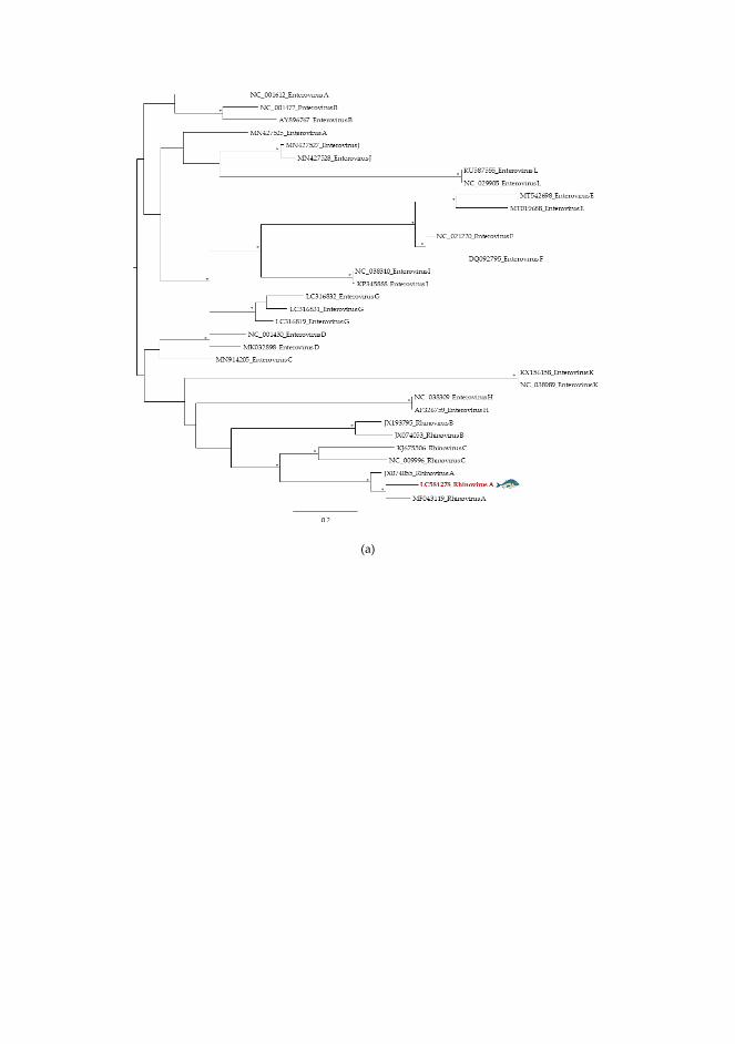
(a)

(b)
Figure S2. Maximum Likelihood phylogenetics trees of each human pathogenic viral family
identified in this study: a) Picornaviridae (partial sequence of Rhinovirus A obtained in gilthead
seabream skin from Peniche); b) Caliciviridae (partial sequence of Norovirus GII obtained in
gilthead seabream skin from Algarve). Due to the hight genetic diversity observed in
Picornaviridae, the phylogenetic tree for this taxon was constructed based on the alignment of the
closest sequences from a phylogenetic point of view. The viral sequences analysed in this study
are coloured and highlighted by a blue fish. All viral sequences are identified by their accession
������ȱ���ȱ����ǯȱ��ȱ��������ȱ������ȱ�����ǰȱ���������ȱ������ȱǃŖ.60 are displayed by *.


ANNEXOS
236
Altres publicacions
A continuació es detallen, per ordre cronològic, altres publicacions no incloses en aquesta tesi però realitzades durant el doctorat:
§ Fernandez-Cassi X., Timoneda N., Martínez-Puchol S., Rusiñol
M., Rodríguez-Manzano J., Figuerola N., Purcell N.H., Bofill-Mas S., Abril J.F., Girones R.. Metagenomics for the study of viruses in urban sewage as a tool for public health surveillance. Science of the Total Environment Science of the Total Environment 618 (2018) 870–880. Factor d’impacte: 6,551; Q1
§ Fernandez-Cassi X., Rusiñol M., Martínez-Puchol S. Viral Concentration and Amplification from Human Serum Samples Prior to Application of Next-Generation Sequencing Analysis. Chapter in "The Human Virome: Methods and Protocols, Methods in Molecular Biology", 1838:173-188, Springer Nature (2018).
§ Aguado D., Fores E., Guerrero-Latorre L., Rusiñol M., Martínez-Puchol S., Codony F., Girones R., Bofill-Mas S. VirWaTest, A Point-of-Use Method for the Detection of Viruses in Water Samples. JoVE - Journal of Visualized Experiments 2019 May 11;(147). Factor d’impacte: 1.163; Q2.
§ Rusiñol M., Martínez-Puchol S., Timoneda N., Fernandez-Cassi X, Perez-Cataluña A., Fernandez-Bravo A., Moreno-Mesonero L., Moreno Y., Alonso JL., Figueras M.J., Abril J.F., Bofill-Mas S., Girones R. Metagenomic analysis of viruses, bacteria and protozoa in irrigation water. International Journal of Hygiene and Environmental Health 224 (2020) 113440. Factor d’impacte: 4.379; Q1

ANNEXOS
237
§ Silva-Sales M., Martínez-Puchol S., Gonzales-Gustavson E., Hundesa A., Gironès R. High Prevalence of Rotavirus A in Raw Sewage Samples from Northeast Spain. Viruses 2020 Mar; 12(3): 318. Factor d’impacte: 3.826; Q1.
§ Rusiñol M., Martínez-Puchol S., Forés E., Itarte M., Girones R., Bofill-Mas S. Concentration methods for the quantification of coronavirus and other potentially pandemic enveloped virus from wastewater. Current Opinion in Environmental Science & Health 2020 Oct;17:21-28.

ANNEXOS
238

ANNEXOS
239

ANNEXOS
240

ANNEXOS
241

ANNEXOS
242

ANNEXOS
243
Copyright © 2022 FDOKUMEN