Electrophysiological effects of guanosine and MK-801 in a quinolinic acid-induced seizure model

DOI: 10.1002/chem.200601461
Self-Assembly of an Alkylated Guanosine Derivative into OrderedSupramolecular Nanoribbons in Solution and on Solid Surfaces
Stefano Lena,[a] Giorgia Brancolini,[c] Giovanni Gottarelli,[a] Paolo Mariani,[b]
Stefano Masiero,[a] Alessandro Venturini,[c] Vincenzo Palermo,[c] Omar Pandoli,[a]
Silvia Pieraccini,[a] Paolo Samor/,*[d] and Gian Piero Spada*[a]
Introduction
Mimicking nature, hierarchical self-assembly[1] provides atool for bottom-up nanoconstruction of sophisticated func-tional architectures[2] as for the unraveling of complex bio-logical arrangements and processes,[3,4] paving the way to-wards their potential application in the realms of nanotech-nology[5] and nanomedicine.[6] Self-assembly is an intrinsicproperty of DNA nucleosides.[3] Learning to precisely con-trol nucleoside self-assembly represents a powerful way ofconstructing a wealth of complex architectures and nano-structured materials, as well as devices with pre-program-med (dynamics) functions.[7] Ultimately, one might foreseetheir use as components for bio-hybrid electronics,[8] such astransistors. Lipophilic guanosine nucleosides can undergodifferent self-assembly pathways, depending on the experi-mental conditions (Figure 1). The presence of monovalentand divalent cations can template the formation of G-quad-ruplex-based octamers or columnar aggregates, dependingon the concentration of the ion and nucleobase, both for or-ganic-soluble[9,10] and water-soluble derivatives.[11] These Gquadruplexes are of great interest because they hold poten-tial in anticancer drug design as they can act as enzyme telo-
Abstract: We report on the synthesisand self-assembly of a guanosine deriv-ative bearing an alkyloxy side groupunder different environmental condi-tions. This derivative was found tospontaneously form ordered supra-molecular nanoribbons in which the in-dividual nucleobases are interactingthrough H-bonds. In toluene andchloroform solutions the formation ofgel-like liquid-crystalline phases wasobserved. Sub-molecularly resolvedscanning tunneling microscopic imag-ing of monolayers physisorbed at the
graphite–solution interface revealedhighly ordered two-dimensional net-works. The recorded intramolecularcontrast can be ascribed to the elec-tronic properties of the different moie-ACHTUNGTRENNUNGties composing the molecule, as provenby quantum-chemical calculations. Thisself-assembly behavior is in excellentagreement with that of 5’-O-acylated
guanosines, which are also character-ized by a self-assembled motif of gua-nosines that resembles parallel ribbons.Therefore, for guanosine derivatives(without sterically demanding groupson the guanine base) the formation ofsupramolecular nanoribbons in solu-tion, in the solid state, and on flat sur-faces is universal. This result is trulyimportant in view of the electronicproperties of these supramolecular an-ACHTUNGTRENNUNGisotropic architectures and thus for po-tential applications in the fields ofnano- and opto-electronics.
Keywords: guanosine · liquid crys-tals · nanoribbon · self-assembly ·supramolecular chemistry
[a] Dr. S. Lena, Prof. G. Gottarelli, Dr. S. Masiero, O. Pandoli,Dr. S. Pieraccini, Prof. G. P. SpadaAlma Mater Studiorum-Universit; di BolognaDipartimento di Chimica Organica “A. Mangini”Via San Giacomo 11, 40126 Bologna (Italy)Fax: (+39)051-209-5688E-mail : [email protected]
[b] Prof. P. MarianiUniversit; Politecnica delle MarcheDipartimento di Scienze Applicate ai Sistemi Complessi and INFMvia Ranieri 65, 60131 Ancona (Italy)
[c] Dr. G. Brancolini, Dr. A. Venturini, Dr. V. PalermoIstituto per la Sintesi Organica e la Fotoreattivit;Consiglio Nazionale delle Ricerche, via Gobetti 10140129 Bologna (Italy)
[d] Dr. P. SamorEInstitut de Science et d’IngFnierie SupramolFculairesUniversitF Louis Pasteur8, allFe Gaspard Monge, 67083 Strasbourg (France)andInstituto per la Sintesi Organica e la Fotoreattivit;Consiglio Nazionale delle Ricerche, via Gobetti 10140129 Bologna (Italy)Fax: (+33)390-245-161E-mail : [email protected]
Supporting information for this article is available on the WWWunder http://www.chemeurj.org/ or from the author.
Chem. Eur. J. 2007, 00, 0 – 0 I 2007 Wiley-VCH Verlag GmbH&Co. KGaA, Weinheim
These are not the final page numbers! ��&1&
FULL PAPER

merase inhibitors.[12] Surprisingly the formation of G quad-ruplexes was also accomplished in the absence of cations bygrafting a sterically demanding group to the C(8) position,which induced a syn stereochemistry.[13]
In the absence of metal templates, guanosines without aC(8) substituent self-assemble, both in solution and in thesolid state, into ribbon-like architectures with an anti orien-tation of the base around the glycosidic bond.[10,14–16] Theseribbon structures are interesting as they are the buildingblocks for new lyotropic mesophases formed in organic sol-vents.[15,17] In the solid state the ribbons, by bridging goldelectrodes, are photoconductive.[18] More interestingly, theseribbons also display rectifying properties.[19] A field-effecttransistor based on this supramolecular structure has recent-ly been described.[20] It is worth noting that the ribbonshown in Figure 1e is dipolar and that in the crystal, owingto the parallel packing of the ribbons, these dipoles are par-allel.[15]
Araki and Yoshikawa recently introduced nonpolar andflexible alkylsilyl groups into 2’-deoxyguanosine to obtainefficient organogelators for alkanes.[21] From an in-depthstructural analysis, they concluded that in these gels thebasic structure is a sheetlike assembly: the supramolecular
structure (see Figure 2) consists of anti-parallel G ribbonslike in Figure 1e linked through two additional inter-tapehydrogen bonds between NH(2) and N(3) of the two gua-nines belonging to adjacent ribbons. Upon heating, a gel-to-liquid-crystal phase transition is observed and has been as-cribed to the selective cleavage of the inter-tape H-bonds.
In our attempts to find general strategies to form guano-sine nanoribbons, we never observed this sheet-like architec-ture for 5’-O acylated guanosines. To verify the universalityof the tendency for guanosine derivatives to form ribbonlikemotifs irrespective of the nature of the 5’-O substitution, weprepared the O-alkylated guanosine 1. Our specific goal wasto find out if the carbonyl group in the 5’-O-acylated deriva-tive, which is known to interact through an intra-ribbon H-bond with NH(2), was essential for the formation of nano-ribbons. It is worth stressing that the strongly anisotropicquasi-1D nanoribbons were found to possess interestingphysicochemical properties,[18–20] while the 2D sheetlike as-semblies can be expected to hold different yet more modestproperties for applications in (opto)electronics. In light ofthis, it is of paramount importance to find universal strat-egies to form functional nanoribbons from different guano-sine derivatives in order to control and improve the proper-ties of the supramolecular arrangements. We report here onthe synthesis, solution characterization, and self-assembly of1; small-angle X-ray diffraction characterization made it
possible to study the structure in the liquid-crystallinephases, scanning tunneling microscopy investigations, corro-borated by quantum chemical calculation, were employed tounveil the structural and electronic properties of the self-as-sembled species on graphite.
Figure 1. a) Donor/acceptor sites in the guanine moiety, b) the G-quartetarrangement, c) the ion-directed columnar self-assembly, d) and e) thestructural motifs of the two ribbon-like supramolecular assemblies ofguanosine derivatives in the absence of cations.
Figure 2. Two-dimensional H-bonded sheet of guanine moieties. Theboxes highlight the individual guanine ribbons (see Figure 1e) connectedby H-bonds between NH(2) and N(3) of two facing guanines belongingto adjacent ribbons.[16]
www.chemeurj.org I 2007 Wiley-VCH Verlag GmbH&Co. KGaA, Weinheim Chem. Eur. J. 0000, 00, 0 – 0
�� These are not the final page numbers!&2&

Results and Discussion
Self-assembly in solution : The supramolecular behavior ofcompound 1 was studied by NMR spectroscopy. Spectrawere recorded at room temperature in CDCl3 and[D6]DMSO/CDCl3 3:1 solutions with concentrations rangingfrom 8O10�3 to 7O10�2m. Signals (Table 1) were assigned onthe basis of 2D COSY and NOESY experiments.
Modest line broadening was observed upon increasing theconcentration in CDCl3. The proton spectrum in[D6]DMSO/CDCl3 shows the NH(1) signal at d=10.64 ppm.This signal shifts to d=12.02 ppm in pure CDCl3 solutionsand is unaffected when the concentration is increased from8O10�3 to 7O10�2m. The NH(2) signal appears as a broadsinglet at d=6.17 and 6.01 ppm in [D6]DMSO/CDCl3 and inthe most diluted CDCl3 solution, respectively, and shiftsslightly downfield with increasing concentration in chloro-form (d=6.28 ppm for the 7O10�2m solution). The NH(1)group therefore always seems to be hydrogen-bonded inchloroform, while the NH(2) is eventually hydrogen-bondedonly at higher concentration. While NOESY spectra record-ed for the most dilute solutions in CDCl3 and in DMSOshow cross peaks with phases opposite to the diagonal, solu-tions above 3O10�2m exhibit cross peaks with the samephase as the diagonal. Therefore, in the lower concentrationrange in chloroform the aggregates are still in the fast-tum-bling regime[22] and no extensive hydrogen bonding seems tooccur. Given that the molecular weight of 1 is 463, and con-sidering the downfield shift observed for the NH(1) protonin CDCl3 relative to the signal in DMSO, we can concludethat the compound exists as a dimer in dilute chloroform so-lution, as observed before[14] for a similar compound. Athigher concentrations the scenario is markedly different:with increasing concentration we observed the formation ofsupramolecular oligomeric/polymeric aggregates with higher“molecular” weight and slower tumbling rates, as evidencedby negative cross peaks in the NOESY spectra.Information on the structure of supramolecular aggre-
gates can be gathered from a closer inspection of NOESYand ROESY spectra. In Figure 3 the NOESY spectrum of a
7O10�2m solution of 1 in CDCl3 (mixing time 100 ms) is re-ported. The spectrum shows cross peaks (boxed) betweenNH(1) and H(8) and between NH(2) and H(8) signals.These signals are characteristic of the ribbon-like supra-molecular arrangement shown in Figure 1e.[14] It is notewor-thy that cross peaks between NH(2) and H(2’) or H(1’) sig-nals are very weak and cross peaks between isopropylideneCH3 and NH(2) signals are absent. These last interactions
would be expected both if thesupramolecular structure wereof the type depicted in Fig-ure 1d or in the case of asheetlike assembly analogousto the one described by Arakiand co-workers (Figure 2). Itshould be pointed out thatproton spectra did not changewith time and that NOESYspectra were recorded on agedsamples in wet CDCl3: underthese same conditions, theanalogous didecanoyl ester de-rivative[14] self-assemblesthrough the hydrogen-bondnetwork shown in Figure 1d.
The CD spectrum of 1 in chloroform shows (see Support-ing Information) weak signals in the 300–220 nm wavelengthregion corresponding to the low-energy transitions of theguanine chromophore. This behavior is in agreement withprevious reports on ribbon-forming guanosines[15] in contrastwith helix-forming guanosines, which give relatively intenseCD, as reported for 8-oxoguanosine derivatives.[23,24]
The liquid-crystalline phase : Compound 1 exhibits lyotropicliquid-crystalline properties in organic solvents. Polarizedoptical microscopy (POM) reveals the presence of a bire-fringent fluid phase at c>2.5% (w/w) in toluene and chloro-form (Figure 4).X-Ray diffraction experiments confirm the existence of a
liquid-crystalline order. Compound 1 was investigated in tol-uene and in chloroform at different concentrations and inthe form of a dry film produced by drop-casting chloroformsolutions. While diffraction spectra in chloroform solutionswere very low in intensity (due to chloroform absorption),one or two intense peaks in the low-angle region and a largeband in the high-angle region were detected in toluene solu-tions.Better-resolved X-ray diffraction profiles were obtained
at concentrations higher than 50% (w/w) or by using thedry film cast from chloroform solution. A typical X-ray dif-fraction pattern is supplied in the Supporting Information.In particular, the low-angle diffraction region is character-ized by a series of broad peaks that can be indexed accord-ing to a 2D rectangular lattice of p2mm symmetry.[25] Fromthe Bragg spacings Qh,k, the unit cell dimensions a and bhave been derived using Equation (1), in which h and k arethe Miller indices of the observed Bragg reflections. The
Table 1. 1H NMR (400 MHz) chemical shifts (ppm) for solutions of 1 at RT. Assignments were made on thebasis of COSY and NOESY spectra.
c (solvent) NH(1) H(8) NH(2) H(1’) H(2’) H(3’) H(4’) H ACHTUNGTRENNUNG(5’/5’’)[a]
OCH2 isopropylideneCH3
[a]
8O10�3m
ACHTUNGTRENNUNG(CDCl3)12.02 7.76 6.01 6.02 5.15 4.92 4.43 3.64–
3.573.43 1.62–1.39
3O10�2m
ACHTUNGTRENNUNG(CDCl3)12.02 7.76 6.25 6.02 5.18 4.92 4.42 3.62–
3.573.43 1.62–1.39
7O10�2m
ACHTUNGTRENNUNG(CDCl3)12.02 7.77 6.28 6.02 5.18 4.92 4.42 3.63–
3.573.43 1.62–1.39
5O10�2m
([D6]DMSO/CDCl3)
10.64 7.73 6.17 5.91 5.03 4.90 4.27 3.55–3.48
3.35 1.49–1.28
[a] Diastereotopic protons have not been assigned.
Chem. Eur. J. 2007, 00, 0 – 0 I 2007 Wiley-VCH Verlag GmbH&Co. KGaA, Weinheim www.chemeurj.org
These are not the final page numbers! ��&3&
FULL PAPERSupramolecular Nanoribbons

unit cell parameters show a de-pendence on concentration(see Table 2), while a rathersmall unit cell has been de-rived for the dry film.
Qh,k ¼ 2pððh=aÞ2 þ ðk=bÞ2Þ0:5
ð1Þ
The high-angle diffractionregion is characterized by twobands, the first rather narrowand centered at about Q =
(5.46 Q)�1, while the second isvery large and centered at Q =
(4.5 Q)�1. Both peak positionsare insensitive to the tolueneconcentration.The X-ray diffraction pro-
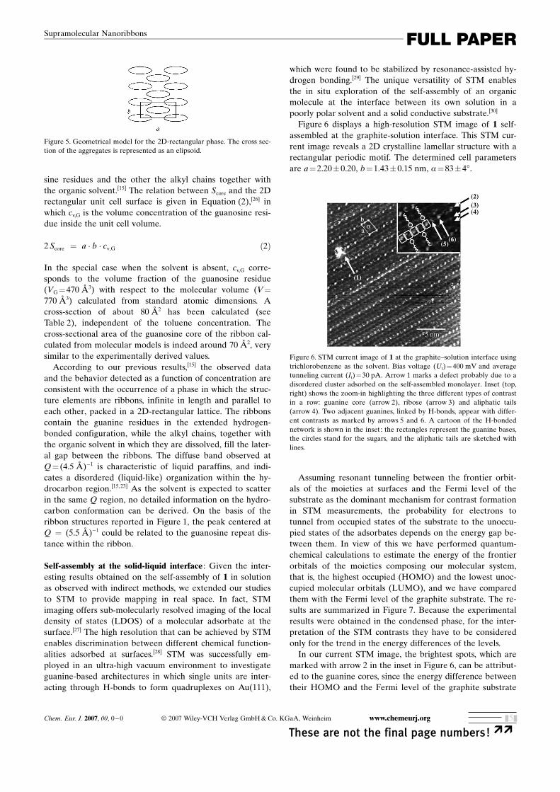
files are thus consistent withthe presence of a liquid-crys-talline phase:[26] the low-anglepeaks suggest a 2D rectangularpacking of aggregates, whosedistance depends on theamount of solvent. Accordingto the symmetry group, two ag-gregates are present in the unitcell (see Figure 5). On theother hand, the high-anglelarge band indicates the disor-dered conformation of the hy-drocarbon chains (eventuallydissolved in the solvent), while
the narrow band provides evidence for an intra-aggregatecharacteristic repeat distance of 5.5 Q.From the unit cell parameters, the cross-sectional area
Score of the central core of the aggregates can be determined,assuming that they are infinite in length and that the unitcell can be divided into two regions, one holding the guano-
Figure 3. NOESY spectrum (mixing time 100 ms) of 7O10�2m 1 in CDCl3 at RT. Relevant intermolecularcross-peaks are boxed.
Figure 4. Polarized optical microscopy images of 7% (w/w) solutions of 1in toluene (top) and chloroform (bottom). Magnification 100O .
Table 2. Low-angle X-ray diffraction results. A and b are the parametersof the 2D rectangular unit cell, c is the weight of 1 over the total weightof the sample, Score is the cross-sectional area of the central core of theribbon (see text).
a [Q, �0.5] b [Q, �0.5] c [w/w, �5%] Score [Q2, �10%]
25.9 9.8 1.0 78.028.7 10.0 0.9 79.432.0 11.5 0.75 84.936.7 11.7 0.6 79.938.0 12.0 0.5 70.140.2 –[a] 0.4 –43.5 –[a] 0.3 –44.6 –[a] 0.2 –46.1 –[a] 0.1 –
[a] Only one peak is detected at low concentration, therefore the b pa-rameter cannot be determined.
www.chemeurj.org I 2007 Wiley-VCH Verlag GmbH&Co. KGaA, Weinheim Chem. Eur. J. 0000, 00, 0 – 0
�� These are not the final page numbers!&4&
G. P. Spada, P. SamorE et al.
sine residues and the other the alkyl chains together withthe organic solvent.[15] The relation between Score and the 2Drectangular unit cell surface is given in Equation (2),[26] inwhich cv,G is the volume concentration of the guanosine resi-due inside the unit cell volume.
2 Score ¼ a � b � cv,G ð2Þ
In the special case when the solvent is absent, cv,G corre-sponds to the volume fraction of the guanosine residue(VG=470 Q3) with respect to the molecular volume (V=
770 Q3) calculated from standard atomic dimensions. Across-section of about 80 Q2 has been calculated (seeTable 2), independent of the toluene concentration. Thecross-sectional area of the guanosine core of the ribbon cal-culated from molecular models is indeed around 70 Q2, verysimilar to the experimentally derived values.According to our previous results,[15] the observed data
and the behavior detected as a function of concentration areconsistent with the occurrence of a phase in which the struc-ture elements are ribbons, infinite in length and parallel toeach other, packed in a 2D-rectangular lattice. The ribbonscontain the guanine residues in the extended hydrogen-bonded configuration, while the alkyl chains, together withthe organic solvent in which they are dissolved, fill the later-al gap between the ribbons. The diffuse band observed atQ= (4.5 Q)�1 is characteristic of liquid paraffins, and indi-cates a disordered (liquid-like) organization within the hy-drocarbon region.[15,23] As the solvent is expected to scatterin the same Q region, no detailed information on the hydro-carbon conformation can be derived. On the basis of theribbon structures reported in Figure 1, the peak centered atQ = (5.5 Q)�1 could be related to the guanosine repeat dis-tance within the ribbon.
Self-assembly at the solid-liquid interface : Given the inter-esting results obtained on the self-assembly of 1 in solutionas observed with indirect methods, we extended our studiesto STM to provide mapping in real space. In fact, STMimaging offers sub-molecularly resolved imaging of the localdensity of states (LDOS) of a molecular adsorbate at thesurface.[27] The high resolution that can be achieved by STMenables discrimination between different chemical function-alities adsorbed at surfaces.[28] STM was successfully em-ployed in an ultra-high vacuum environment to investigateguanine-based architectures in which single units are inter-acting through H-bonds to form quadruplexes on AuACHTUNGTRENNUNG(111),
which were found to be stabilized by resonance-assisted hy-drogen bonding.[29] The unique versatility of STM enablesthe in situ exploration of the self-assembly of an organicmolecule at the interface between its own solution in apoorly polar solvent and a solid conductive substrate.[30]
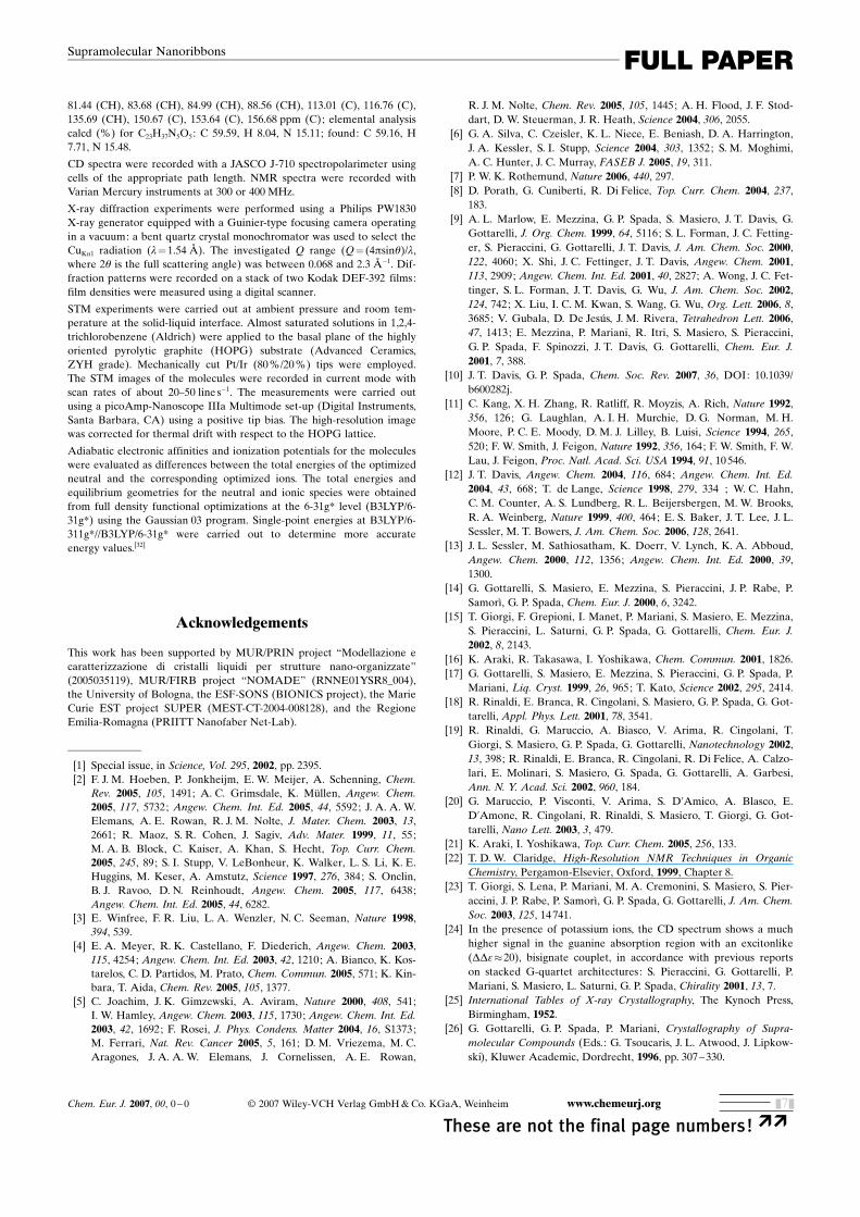
Figure 6 displays a high-resolution STM image of 1 self-assembled at the graphite-solution interface. This STM cur-rent image reveals a 2D crystalline lamellar structure with arectangular periodic motif. The determined cell parametersare a=2.20�0.20, b=1.43�0.15 nm, a=83�48.
Assuming resonant tunneling between the frontier orbit-ACHTUNGTRENNUNGals of the moieties at surfaces and the Fermi level of thesubstrate as the dominant mechanism for contrast formationin STM measurements, the probability for electrons totunnel from occupied states of the substrate to the unoccu-pied states of the adsorbates depends on the energy gap be-tween them. In view of this we have performed quantum-chemical calculations to estimate the energy of the frontierorbitals of the moieties composing our molecular system,that is, the highest occupied (HOMO) and the lowest unoc-cupied molecular orbitals (LUMO), and we have comparedthem with the Fermi level of the graphite substrate. The re-sults are summarized in Figure 7. Because the experimentalresults were obtained in the condensed phase, for the inter-pretation of the STM contrasts they have to be consideredonly for the trend in the energy differences of the levels.In our current STM image, the brightest spots, which are
marked with arrow 2 in the inset in Figure 6, can be attribut-ed to the guanine cores, since the energy difference betweentheir HOMO and the Fermi level of the graphite substrate
Figure 5. Geometrical model for the 2D-rectangular phase. The cross sec-tion of the aggregates is represented as an elipsoid.
Figure 6. STM current image of 1 at the graphite–solution interface usingtrichlorobenzene as the solvent. Bias voltage (Ut)=400 mV and averagetunneling current (It)=30 pA. Arrow 1 marks a defect probably due to adisordered cluster adsorbed on the self-assembled monolayer. Inset (top,right) shows the zoom-in highlighting the three different types of contrastin a row: guanine core (arrow 2), ribose (arrow 3) and aliphatic tails(arrow 4). Two adjacent guanines, linked by H-bonds, appear with differ-ent contrasts as marked by arrows 5 and 6. A cartoon of the H-bondednetwork is shown in the inset: the rectangles represent the guanine bases,the circles stand for the sugars, and the aliphatic tails are sketched withlines.
Chem. Eur. J. 2007, 00, 0 – 0 I 2007 Wiley-VCH Verlag GmbH&Co. KGaA, Weinheim www.chemeurj.org
These are not the final page numbers! ��&5&
FULL PAPERSupramolecular Nanoribbons

is rather small.[31] Spots with a lower brightness, indicatedwith arrow 3, can be ascribed to the ribose, while the darkerpart of the image (arrow 4) can be attributed to the aliphaticside chains, which have not been resolved, probably owingto their high conformational mobility on a time scale fasterthan the STM imaging. Therefore the detailed analysis as-sisted by quantum-chemical calculations made it possible todiscriminate different moieties composing 1.A careful inspection reveals that the contrast of two adja-
cent guanines linked by H-bonding is different (see, for ex-ample, those marked by arrows 5 and 6 in Figure 6); this canbe explained in view of a different packing in the X,Y withrespect to the HOPG lattice underneath. The value of thecell parameter a, which roughly amounts to half of the esti-mated ribbon width, suggests that the alkyl tails in two adja-cent H-bonded ribbons are interdigitated. Given the STMresolution obtained, and in view of the pretty similar size ofthe unit cell that can be expected for the two nanoribbonsdepicted in Figures 1d and e, taking into account the esti-mated unit cell and relative error bar, we are unable to un-ambiguously ascribe the supramolecular motif shown inFigure 6 to either one or the other nanoribbon-type.On a larger scale, a monolayer of 1 is polycrystalline (see
Figure 8). Up to seven domains with a diameter of a fewtens of nanometers are observed. The orientation of most ofthe lamellae is symmetry-equivalent with respect to the crys-talline substrate lattice. The high-resolution imagingACHTUNGTRENNUNGachieved in the polycrystalline structure made it possible torecord two different kinds of defects on the nanometerlength scale. The first kind of defect consists of empty do-mains in which the molecules are not adsorbed at surfaces;an example of missing molecules is indicated by a greyarrow. Such defects get recovered on the time scale of a fewminutes. The second kind of defect is found at the domainboundaries, which have a fuzzy character and surroundsome crystals (marked with white arrows). At these frontiersthe molecules are more loosely packed.
Conclusion
In summary, in our attempt to find general strategies toform functional nanoribbons from guanosine derivatives, wehave prepared O-alkylated guanosine, which is an extensionof the well-known 5’-O-acylated guanosines. Our specificgoal was to find out if the carbonyl group, existing in the 5’-O-acylated derivative, which is known to interact throughan intra-ribbon H-bond with NH(2), was essential for theformation of nanoribbons. We have thus synthesized andstudied the self-assembly of 1 under different environmentalconditions. NMR, X-ray, and STM measurements revealedthat 1 self-assembles into highly ordered nanoribbons inwhich the single nucleosides are held together by H-bonds.This self-assembly behavior appears to be universal, as it isin line with many other guanosine derivatives, and in partic-ular with that of 5’-O-acylated guanosines, revealing that thepresence of the carbonyl unit in the 5’-O-acylated derivativeis not a prerequisite for the formation of the nanoribbon.The self-assembled motif observed for 1 conveys parallelribbons, most probably with parallel dipoles. This result isvery important in view of the well-known physico-chemicalproperties of these quasi-1D nanostructures, and in particu-lar for their use in (opto)electronics.
Experimental Section
2’,3’-O-Isopropylidene-5’-O-decylguanosine : 2’,3’-O-Isopropylidenegua-nosine (Sigma) (0.4 g, 1.2 mmol) was dried in vacuo over P2O5 for 2 h at50 8C and suspended in anhydrous THF (10 mL). NaH (0.058 g,2.4 mmol) and 1-bromodecane (1.24 mL, 6 mmol) were added. The mix-ture was heated at reflux overnight, then cooled to room temperature.The solvent was removed and the residual solid was taken up in dichloro-methane, washed with water, dried over MgSO4, concentrated in vacuo,and applied to a silica gel column using 94:6 dichloromethane/methanolas the eluent. The product was recrystallized from ethanol to afford awhite solid (0.26 g, 48% yield). 1H NMR (400 MHz, CDCl3): d=0.86 (t,3H; CH3), 1.25 (m, 14H; CH2), 1.39 (s, 3H; CH3), 1.53 (m, 2H; O�CH2CH2), 1.61 (s, 3H; CH3), 3.43 (m, 2H; O�CH2), 3.55–3.64 (m, 2H;H5’-H5’’), 4.42 (m, 1H; H4’), 4.92 (m, 1H; H3’), 5.17 (m, 1H; H2’), 6.00(d, 1H; H1’), 6.28 (s, 2H; NH2), 7.76 (s, 1H; H8), 12.02 ppm (s, 1H;NH); 13C NMR (300 MHz, [D6]DMSO): d=13.93 (CH3), 22.07 (CH2),25.21 (CH3), 25.53 (CH2), 26.98 (CH3), 28.68 (CH2), 28.30 (CH2), 28.93(CH2), 28.97 (CH2), 28.99 (CH2), 31.28 (CH2), 70.43 (CH2), 70.61 (CH2),
Figure 7. Scheme of the adiabatic electron affinities (Ea) and ionizationpotentials (Ip) for guanine, sugar, and aliphatic chain (C10H22) in vacuo,as calculated from energy differences between the optimized structuresof neutral and charged systems at the B3LYP/6-311+G*//B3LYP/6-31G*level of theory.
Figure 8. Current STM survey image of self-assembled architecture of 1recorded at the solid–liquid interface on HOPG. Ut=290 mV and aver-age It=200 pA.
www.chemeurj.org I 2007 Wiley-VCH Verlag GmbH&Co. KGaA, Weinheim Chem. Eur. J. 0000, 00, 0 – 0
�� These are not the final page numbers!&6&
G. P. Spada, P. SamorE et al.
81.44 (CH), 83.68 (CH), 84.99 (CH), 88.56 (CH), 113.01 (C), 116.76 (C),135.69 (CH), 150.67 (C), 153.64 (C), 156.68 ppm (C); elemental analysiscalcd (%) for C23H37N5O5: C 59.59, H 8.04, N 15.11; found: C 59.16, H7.71, N 15.48.
CD spectra were recorded with a JASCO J-710 spectropolarimeter usingcells of the appropriate path length. NMR spectra were recorded withVarian Mercury instruments at 300 or 400 MHz.
X-ray diffraction experiments were performed using a Philips PW1830X-ray generator equipped with a Guinier-type focusing camera operatingin a vacuum: a bent quartz crystal monochromator was used to select theCuKa1 radiation (l=1.54 Q). The investigated Q range (Q= (4psinq)/l,where 2q is the full scattering angle) was between 0.068 and 2.3 Q�1. Dif-fraction patterns were recorded on a stack of two Kodak DEF-392 films:film densities were measured using a digital scanner.
STM experiments were carried out at ambient pressure and room tem-perature at the solid-liquid interface. Almost saturated solutions in 1,2,4-trichlorobenzene (Aldrich) were applied to the basal plane of the highlyoriented pyrolytic graphite (HOPG) substrate (Advanced Ceramics,ZYH grade). Mechanically cut Pt/Ir (80%/20%) tips were employed.The STM images of the molecules were recorded in current mode withscan rates of about 20–50 line s�1. The measurements were carried outusing a picoAmp-Nanoscope IIIa Multimode set-up (Digital Instruments,Santa Barbara, CA) using a positive tip bias. The high-resolution imagewas corrected for thermal drift with respect to the HOPG lattice.
Adiabatic electronic affinities and ionization potentials for the moleculeswere evaluated as differences between the total energies of the optimizedneutral and the corresponding optimized ions. The total energies andequilibrium geometries for the neutral and ionic species were obtainedfrom full density functional optimizations at the 6-31g* level (B3LYP/6-31g*) using the Gaussian 03 program. Single-point energies at B3LYP/6-311g*//B3LYP/6-31g* were carried out to determine more accurateenergy values.[32]
Acknowledgements
This work has been supported by MUR/PRIN project “Modellazione ecaratterizzazione di cristalli liquidi per strutture nano-organizzate”(2005035119), MUR/FIRB project “NOMADE” (RNNE01YSR8_004),the University of Bologna, the ESF-SONS (BIONICS project), the MarieCurie EST project SUPER (MEST-CT-2004-008128), and the RegioneEmilia-Romagna (PRIITT Nanofaber Net-Lab).
[1] Special issue, in Science, Vol. 295, 2002, pp. 2395.[2] F. J. M. Hoeben, P. Jonkheijm, E. W. Meijer, A. Schenning, Chem.
Rev. 2005, 105, 1491; A. C. Grimsdale, K. MVllen, Angew. Chem.2005, 117, 5732; Angew. Chem. Int. Ed. 2005, 44, 5592; J. A. A. W.Elemans, A. E. Rowan, R. J. M. Nolte, J. Mater. Chem. 2003, 13,2661; R. Maoz, S. R. Cohen, J. Sagiv, Adv. Mater. 1999, 11, 55;M. A. B. Block, C. Kaiser, A. Khan, S. Hecht, Top. Curr. Chem.2005, 245, 89; S. I. Stupp, V. LeBonheur, K. Walker, L. S. Li, K. E.Huggins, M. Keser, A. Amstutz, Science 1997, 276, 384; S. Onclin,B. J. Ravoo, D. N. Reinhoudt, Angew. Chem. 2005, 117, 6438;Angew. Chem. Int. Ed. 2005, 44, 6282.
[3] E. Winfree, F. R. Liu, L. A. Wenzler, N. C. Seeman, Nature 1998,394, 539.
[4] E. A. Meyer, R. K. Castellano, F. Diederich, Angew. Chem. 2003,115, 4254; Angew. Chem. Int. Ed. 2003, 42, 1210; A. Bianco, K. Kos-tarelos, C. D. Partidos, M. Prato, Chem. Commun. 2005, 571; K. Kin-bara, T. Aida, Chem. Rev. 2005, 105, 1377.
[5] C. Joachim, J. K. Gimzewski, A. Aviram, Nature 2000, 408, 541;I. W. Hamley, Angew. Chem. 2003, 115, 1730; Angew. Chem. Int. Ed.2003, 42, 1692; F. Rosei, J. Phys. Condens. Matter 2004, 16, S1373;M. Ferrari, Nat. Rev. Cancer 2005, 5, 161; D. M. Vriezema, M. C.Aragones, J. A. A. W. Elemans, J. Cornelissen, A. E. Rowan,
R. J. M. Nolte, Chem. Rev. 2005, 105, 1445; A. H. Flood, J. F. Stod-dart, D. W. Steuerman, J. R. Heath, Science 2004, 306, 2055.
[6] G. A. Silva, C. Czeisler, K. L. Niece, E. Beniash, D. A. Harrington,J. A. Kessler, S. I. Stupp, Science 2004, 303, 1352; S. M. Moghimi,A. C. Hunter, J. C. Murray, FASEB J. 2005, 19, 311.
[7] P. W. K. Rothemund, Nature 2006, 440, 297.[8] D. Porath, G. Cuniberti, R. Di Felice, Top. Curr. Chem. 2004, 237,
183.[9] A. L. Marlow, E. Mezzina, G. P. Spada, S. Masiero, J. T. Davis, G.
Gottarelli, J. Org. Chem. 1999, 64, 5116; S. L. Forman, J. C. Fetting-er, S. Pieraccini, G. Gottarelli, J. T. Davis, J. Am. Chem. Soc. 2000,122, 4060; X. Shi, J. C. Fettinger, J. T. Davis, Angew. Chem. 2001,113, 2909; Angew. Chem. Int. Ed. 2001, 40, 2827; A. Wong, J. C. Fet-tinger, S. L. Forman, J. T. Davis, G. Wu, J. Am. Chem. Soc. 2002,124, 742; X. Liu, I. C. M. Kwan, S. Wang, G. Wu, Org. Lett. 2006, 8,3685; V. Gubala, D. De Jesffls, J. M. Rivera, Tetrahedron Lett. 2006,47, 1413; E. Mezzina, P. Mariani, R. Itri, S. Masiero, S. Pieraccini,G. P. Spada, F. Spinozzi, J. T. Davis, G. Gottarelli, Chem. Eur. J.2001, 7, 388.
[10] J. T. Davis, G. P. Spada, Chem. Soc. Rev. 2007, 36, DOI: 10.1039/b600282j.
[11] C. Kang, X. H. Zhang, R. Ratliff, R. Moyzis, A. Rich, Nature 1992,356, 126; G. Laughlan, A. I. H. Murchie, D. G. Norman, M. H.Moore, P. C. E. Moody, D. M. J. Lilley, B. Luisi, Science 1994, 265,520; F. W. Smith, J. Feigon, Nature 1992, 356, 164; F. W. Smith, F. W.Lau, J. Feigon, Proc. Natl. Acad. Sci. USA 1994, 91, 10546.
[12] J. T. Davis, Angew. Chem. 2004, 116, 684; Angew. Chem. Int. Ed.2004, 43, 668; T. de Lange, Science 1998, 279, 334 ; W. C. Hahn,C. M. Counter, A. S. Lundberg, R. L. Beijersbergen, M. W. Brooks,R. A. Weinberg, Nature 1999, 400, 464; E. S. Baker, J. T. Lee, J. L.Sessler, M. T. Bowers, J. Am. Chem. Soc. 2006, 128, 2641.
[13] J. L. Sessler, M. Sathiosatham, K. Doerr, V. Lynch, K. A. Abboud,Angew. Chem. 2000, 112, 1356; Angew. Chem. Int. Ed. 2000, 39,1300.
[14] G. Gottarelli, S. Masiero, E. Mezzina, S. Pieraccini, J. P. Rabe, P.SamorE, G. P. Spada, Chem. Eur. J. 2000, 6, 3242.
[15] T. Giorgi, F. Grepioni, I. Manet, P. Mariani, S. Masiero, E. Mezzina,S. Pieraccini, L. Saturni, G. P. Spada, G. Gottarelli, Chem. Eur. J.2002, 8, 2143.
[16] K. Araki, R. Takasawa, I. Yoshikawa, Chem. Commun. 2001, 1826.[17] G. Gottarelli, S. Masiero, E. Mezzina, S. Pieraccini, G. P. Spada, P.
Mariani, Liq. Cryst. 1999, 26, 965; T. Kato, Science 2002, 295, 2414.[18] R. Rinaldi, E. Branca, R. Cingolani, S. Masiero, G. P. Spada, G. Got-
tarelli, Appl. Phys. Lett. 2001, 78, 3541.[19] R. Rinaldi, G. Maruccio, A. Biasco, V. Arima, R. Cingolani, T.
Giorgi, S. Masiero, G. P. Spada, G. Gottarelli, Nanotechnology 2002,13, 398; R. Rinaldi, E. Branca, R. Cingolani, R. Di Felice, A. Calzo-lari, E. Molinari, S. Masiero, G. Spada, G. Gottarelli, A. Garbesi,Ann. N. Y. Acad. Sci. 2002, 960, 184.
[20] G. Maruccio, P. Visconti, V. Arima, S. D’Amico, A. Blasco, E.D’Amone, R. Cingolani, R. Rinaldi, S. Masiero, T. Giorgi, G. Got-tarelli, Nano Lett. 2003, 3, 479.
[21] K. Araki, I. Yoshikawa, Top. Curr. Chem. 2005, 256, 133.[22] T. D. W. Claridge, High-Resolution NMR Techniques in Organic
Chemistry, Pergamon-Elsevier, Oxford, 1999, Chapter 8.[23] T. Giorgi, S. Lena, P. Mariani, M. A. Cremonini, S. Masiero, S. Pier-
accini, J. P. Rabe, P. SamorE, G. P. Spada, G. Gottarelli, J. Am. Chem.Soc. 2003, 125, 14741.
[24] In the presence of potassium ions, the CD spectrum shows a muchhigher signal in the guanine absorption region with an excitonlike(DDe�20), bisignate couplet, in accordance with previous reportson stacked G-quartet architectures: S. Pieraccini, G. Gottarelli, P.Mariani, S. Masiero, L. Saturni, G. P. Spada, Chirality 2001, 13, 7.
[25] International Tables of X-ray Crystallography, The Kynoch Press,Birmingham, 1952.
[26] G. Gottarelli, G. P. Spada, P. Mariani, Crystallography of Supra-molecular Compounds (Eds.: G. Tsoucaris, J. L. Atwood, J. Lipkow-ski), Kluwer Academic, Dordrecht, 1996, pp. 307–330.
Chem. Eur. J. 2007, 00, 0 – 0 I 2007 Wiley-VCH Verlag GmbH&Co. KGaA, Weinheim www.chemeurj.org
These are not the final page numbers! ��&7&
FULL PAPERSupramolecular Nanoribbons
[27] A. Yazdani, C. M. Lieber, Nature 1999, 401, 227; J. K. Gimzewski, C.Joachim, Polim. Cienc. Tecnol. Science 1999, 283, 1683; P. SamorE, J.Mater. Chem. 2004, 14, 1353.
[28] D. M. Cyr, B. Venkataraman, G. W. Flynn, A. Black, G. M. White-sides, J. Phys. Chem. 1996, 100, 13747.
[29] R. Otero, M. Schock, L. M. Molina, E. Laegsgaard, I. Stensgaard, B.Hammer, F. Besenbacher, Angew. Chem. 2005, 117, 2310; Angew.Chem. Int. Ed. 2005, 44, 2270.
[30] G. C. McGonigal, R. H. Bernhardt, D. J. Thomson, Appl. Phys. Lett.1990, 57, 28; J. P. Rabe, S. Buchholz, Science 1991, 253, 424; X. H.Qiu, C. Wang, Q. D. Zeng, B. Xu, S. X. Yin, H. N. Wang, S. D. Xu,C. L. Bai, J. Am. Chem. Soc. 2000, 122, 5550; D. M. Cyr, B. Venka-
taraman, G. W. Flynn, Chem. Mater. 1996, 8, 1600; S. De Feyter,F. C. De Schryver, J. Phys. Chem. B 2005, 109, 4290; P. SamorE, V.Francke, V. Enkelmann, K. MVllen, J. P. Rabe, Chem. Mater. 2003,15, 1032.
[31] R. Lazzaroni, A. Calderone, J. L. BrFdas, J. P. Rabe, J. Chem. Phys.1997, 107, 99.
[32] L. Horny, N. D. K. Petraco, C. Pak, H. F. SchYfer, J. Am. Chem. Soc.2002, 124, 5861; L. Horny, N. D. K. Petraco, H. F. SchYfer, J. Am.Chem. Soc. 2002, 124, 14716.
Received: October 16, 2006Published online:&& &&, 2006
www.chemeurj.org I 2007 Wiley-VCH Verlag GmbH&Co. KGaA, Weinheim Chem. Eur. J. 0000, 00, 0 – 0
�� These are not the final page numbers!&8&
G. P. Spada, P. SamorE et al.

Supramolecular Chemistry
S. Lena, G. Brancolini, G. Gottarelli,P. Mariani, S. Masiero, A. Venturini,V. Palermo, O. Pandoli, S. Pieraccini,P. Samor;,* G. P. Spada* . . . . . . &&&—&&&
Self-Assembly of an Alkylated Guano-sine Derivative into Ordered Supra-molecular Nanoribbons in Solutionand on Solid Surfaces
Spontaneous formation of supramolec-ular nanoribbons based on alkyl ACHTUNGTRENNUNGatedguanosine is observed in solution, inthe solid state, and on flat surfaces.
This suggests that this self-assemblybehavior is universal in guanosinederivatives without sterically demand-ing substituents on the guanine base.
Chem. Eur. J. 2007, 00, 0 – 0 I 2007 Wiley-VCH Verlag GmbH&Co. KGaA, Weinheim www.chemeurj.org
These are not the final page numbers! ��&9&
FULL PAPERSupramolecular Nanoribbons
Copyright © 2022 FDOKUMEN