Selective laser cleaning of chlorine on ancient coins
9
Selective laser cleaning of chlorine on ancient coins Domenico Aiello a , Alessandro Buccolieri b,e , Giovanni Buccolieri b,e , Alfredo Castellano b , Massimo Di Giulio b , Laura Sandra Leo b,e , Antonella Lorusso *c,e , Gloria Nassisi b , Vincenzo Nassisi c,e , Lorenzo Torrisi d a ENEL Produzione S.p.A., 72020, Tuturano (BR) – Italy b University of Lecce, Department of Material Science, via Monteroni, 73100, Lecce - Italy c University of Lecce, Department of Physics, via Arnesano, 73100, Lecce – Italy d University of Messina, Department of Physics, INFN-LNS, Ctr. Papardo, 31, 98166, S. Agata, Messina - Italy e INFN, sect. of Lecce, C.P. 193, 73100, Lecce - Italy ABSTRACT Results about the efficiency of the laser cleaning on the reduction of corrosion products from the surface of ancient coins are reported. In this work an ancient copper coin datable from 1500 to 1600 A.D. and a UV excimer laser were utilized. The goal of this work consists to study the potentiality of UV laser treatment in the reduction of the chlorine concentration on the coin surface which is the main responsible of the corrosion processes of the ancient coins. We used Energy Dispersive X-Ray Fluorescence (EDXRF) and X-Ray Diffraction (XRD) techniques to estimate the chemical composition of the coin surface, before and after UV excimer laser cleaning. In particular, we measured the chlorine, copper and calcium concentrations. We found that a radiation dose of about 19 J/cm 2 was able to reduce the chlorine concentration from 2.3 % w/w to 0.6 % w/w without damaging the metal bulk. Keywords: ancient coins; patina; copper chlorides; laser cleaning; EDXRF portable apparatus 1. INTRODUCTION The corrosion processes of ancient artefacts represent a very important problem for the cultural heritage conservation. Bronze and copper artefacts are especially involved to serious processes of deterioration owing to their interaction with the environment. So thin layers of alteration products, called commonly patina, grow spontaneously on the object surface and cause long-term problems for chemical stability of object itself 1, 2 . The formation of thin (few µm) layer of cuprite (Cu 2 O) is usually the first step of alteration process. It often happens that this primary film of cuprite preserves important details of the original surface of the artefacts. Through the years, the patina grows and its chemical composition and its physical properties are influenced by a wide range of factors, such as the environment (burial, marine, outdoor, indoor, etc.), the location (urban, rural, industrial, etc.) and the local meteorology (relative humidity, precipitation, pH value, etc.) which can promote or slow down corrosion processes of artefacts. In particular, the hydrochloric acid formation is the main responsible for a particular corrosion of bronzes called “bronze’s cancer” 1 . This corrosion starts with the formation of copper chloride (see equation (1)), which is soluble in water and forms copper oxide (red cuprite) and hydrochloric acid as described by the equation (2): * [email protected]; phone +39 0832 297554; fax +39 0832 297482 XVI International Symposium on Gas Flow, Chemical Lasers, and High-Power Lasers, edited by Dieter Schuöcker, Proceedings of SPIE Vol. 6346, 63463H, (2007) 0277-786X/07/$18 doi: 10.1117/12.739397 Proc. of SPIE Vol. 6346 63463H-1 Downloaded From: http://proceedings.spiedigitallibrary.org/ on 07/23/2013 Terms of Use: http://spiedl.org/terms
-
Upload
tecnologicocomfenalco -
Category
Documents
-
view
1 -
download
0
Transcript of Selective laser cleaning of chlorine on ancient coins
Selective laser cleaning of chlorine on ancient coins
Domenico Aielloa, Alessandro Buccolierib,e, Giovanni Buccolierib,e, Alfredo Castellanob, Massimo Di Giuliob, Laura Sandra Leob,e, Antonella Lorusso*c,e, Gloria Nassisib, Vincenzo Nassisic,e, Lorenzo
Torrisid
aENEL Produzione S.p.A., 72020, Tuturano (BR) – Italy bUniversity of Lecce, Department of Material Science, via Monteroni, 73100, Lecce - Italy
cUniversity of Lecce, Department of Physics, via Arnesano, 73100, Lecce – Italy dUniversity of Messina, Department of Physics, INFN-LNS, Ctr. Papardo, 31, 98166, S. Agata,
Messina - Italy eINFN, sect. of Lecce, C.P. 193, 73100, Lecce - Italy
ABSTRACT
Results about the efficiency of the laser cleaning on the reduction of corrosion products from the surface of ancient coins are reported. In this work an ancient copper coin datable from 1500 to 1600 A.D. and a UV excimer laser were utilized. The goal of this work consists to study the potentiality of UV laser treatment in the reduction of the chlorine concentration on the coin surface which is the main responsible of the corrosion processes of the ancient coins. We used Energy Dispersive X-Ray Fluorescence (EDXRF) and X-Ray Diffraction (XRD) techniques to estimate the chemical composition of the coin surface, before and after UV excimer laser cleaning. In particular, we measured the chlorine, copper and calcium concentrations. We found that a radiation dose of about 19 J/cm2 was able to reduce the chlorine concentration from 2.3 % w/w to 0.6 % w/w without damaging the metal bulk. Keywords: ancient coins; patina; copper chlorides; laser cleaning; EDXRF portable apparatus
1. INTRODUCTION
The corrosion processes of ancient artefacts represent a very important problem for the cultural heritage conservation. Bronze and copper artefacts are especially involved to serious processes of deterioration owing to their interaction with the environment. So thin layers of alteration products, called commonly patina, grow spontaneously on the object surface and cause long-term problems for chemical stability of object itself1, 2. The formation of thin (few µm) layer of cuprite (Cu2O) is usually the first step of alteration process. It often happens that this primary film of cuprite preserves important details of the original surface of the artefacts. Through the years, the patina grows and its chemical composition and its physical properties are influenced by a wide range of factors, such as the environment (burial, marine, outdoor, indoor, etc.), the location (urban, rural, industrial, etc.) and the local meteorology (relative humidity, precipitation, pH value, etc.) which can promote or slow down corrosion processes of artefacts. In particular, the hydrochloric acid formation is the main responsible for a particular corrosion of bronzes called “bronze’s cancer”1. This corrosion starts with the formation of copper chloride (see equation (1)), which is soluble in water and forms copper oxide (red cuprite) and hydrochloric acid as described by the equation (2):
* [email protected]; phone +39 0832 297554; fax +39 0832 297482
XVI International Symposium on Gas Flow, Chemical Lasers, and High-Power Lasers, edited by Dieter Schuöcker,Proceedings of SPIE Vol. 6346, 63463H, (2007) 0277-786X/07/$18 doi: 10.1117/12.739397
Proc. of SPIE Vol. 6346 63463H-1
Downloaded From: http://proceedings.spiedigitallibrary.org/ on 07/23/2013 Terms of Use: http://spiedl.org/terms
4HCl + 4Cu + O2 → 4CuCl + 2H2O (1) 4CuCl + 2H2O → 2Cu2O + 4HCl (2)
Finally, hydrochloric acid reacts again with the copper forming cyclic reactions. As a consequence the corrosion propagates inside the object. Besides, copper chloride hydroxides grow on the surface in two different allotropic forms (atacamite and paratacamite), according to the following equation:
2Cu + HCl + H2O + O2 → Cu2Cl(OH)3 (3) These corrosion products are not very soluble and they are characterised by a blue-green pigmentation. From the above chemical reactions it is possible to understand how the chloride concentration is one of the main responsible of the deterioration of ancient coins. Recently, pulsed lasers such as excimer lasers, Nd-Yag lasers, Er-Yag lasers, frequency doubled Nd-Yag lasers and frequency triple Nd-Yag lasers 3,4, have been employed in the cultural heritage as a very interesting tool for the artefact cleaning. Actually, pulsed laser beams can operate a fast and controlled vaporisation of few layers of the target if laser parameters are chosen in appropriate way. So this straightforward technique can reach meaningful results in the cleaning field. Otherwise the laser cleaning is the result of many processes which depend on the irradiation conditions and artefact components. In this work we studied the potentiality of UV laser cleaning as technique of removing copper corrosion products from ancient coins surface. In particular, we evaluated the reduction of the chlorine concentration as result of ablation processes by a KrF laser. It is well known in literature5,6, in effect, that ultraviolet radiation induced an extensive fragmentation of the molecules due to relatively high energy of UV photons. For this reason, by irradiating the coin zones where chlorine compounds are present, we expected that UV laser ablation was very efficiency on removing the chlorine from the coin surface.
2. MATERIAL AND METHODS
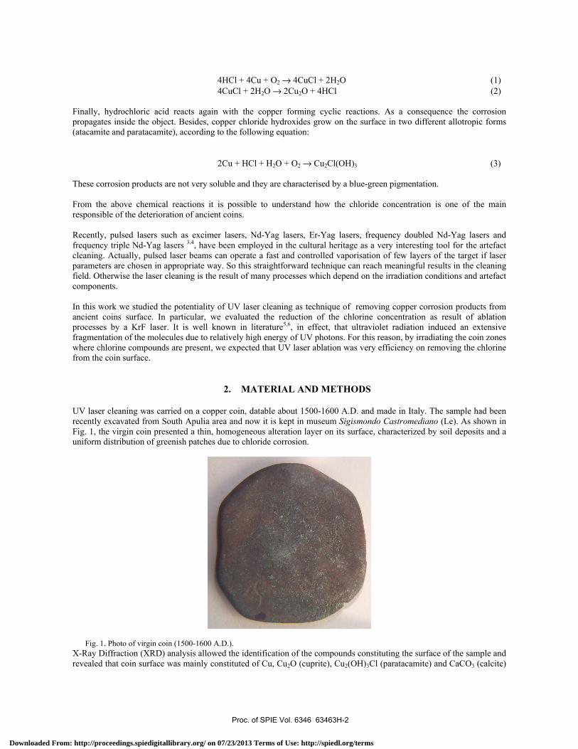
UV laser cleaning was carried on a copper coin, datable about 1500-1600 A.D. and made in Italy. The sample had been recently excavated from South Apulia area and now it is kept in museum Sigismondo Castromediano (Le). As shown in Fig. 1, the virgin coin presented a thin, homogeneous alteration layer on its surface, characterized by soil deposits and a uniform distribution of greenish patches due to chloride corrosion.
Fig. 1. Photo of virgin coin (1500-1600 A.D.). X-Ray Diffraction (XRD) analysis allowed the identification of the compounds constituting the surface of the sample and revealed that coin surface was mainly constituted of Cu, Cu2O (cuprite), Cu2(OH)3Cl (paratacamite) and CaCO3 (calcite)
Proc. of SPIE Vol. 6346 63463H-2
Downloaded From: http://proceedings.spiedigitallibrary.org/ on 07/23/2013 Terms of Use: http://spiedl.org/terms
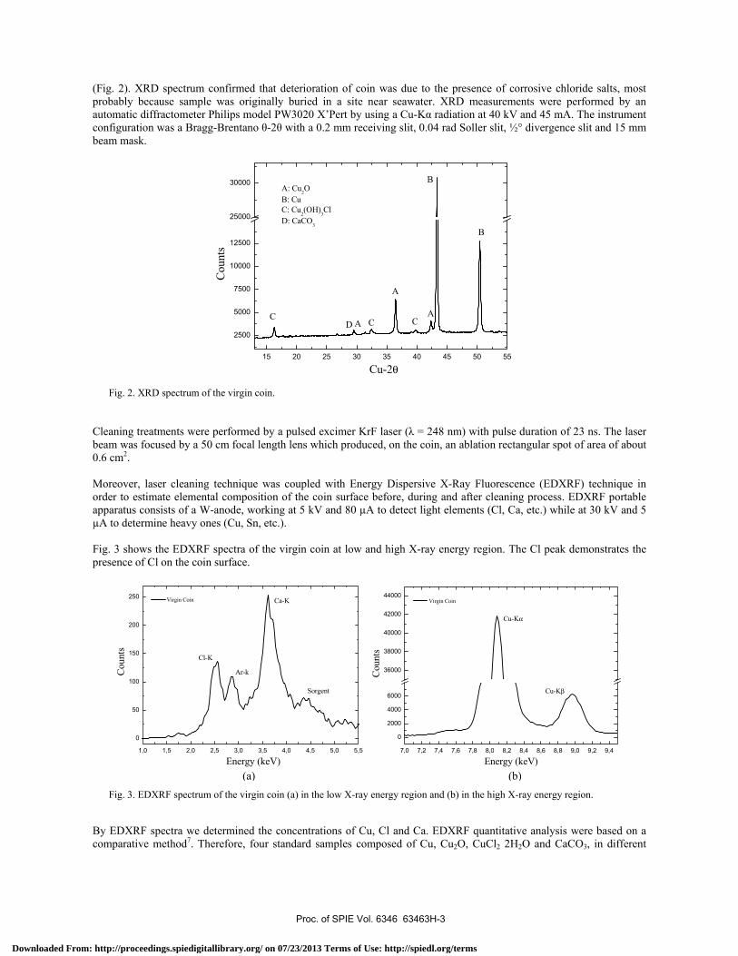
(Fig. 2). XRD spectrum confirmed that deterioration of coin was due to the presence of corrosive chloride salts, most probably because sample was originally buried in a site near seawater. XRD measurements were performed by an automatic diffractometer Philips model PW3020 X’Pert by using a Cu-Kα radiation at 40 kV and 45 mA. The instrument configuration was a Bragg-Brentano θ-2θ with a 0.2 mm receiving slit, 0.04 rad Soller slit, ½° divergence slit and 15 mm beam mask.
15 20 25 30 35 40 45 50 55
2500
5000
7500
10000
12500
25000
30000
D
B
B
A
A
A CCC
A: Cu2OB: CuC: Cu2(OH)3ClD: CaCO3
Cou
nts
Cu-2θ
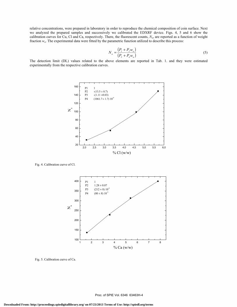
Fig. 2. XRD spectrum of the virgin coin. Cleaning treatments were performed by a pulsed excimer KrF laser (λ = 248 nm) with pulse duration of 23 ns. The laser beam was focused by a 50 cm focal length lens which produced, on the coin, an ablation rectangular spot of area of about 0.6 cm2. Moreover, laser cleaning technique was coupled with Energy Dispersive X-Ray Fluorescence (EDXRF) technique in order to estimate elemental composition of the coin surface before, during and after cleaning process. EDXRF portable apparatus consists of a W-anode, working at 5 kV and 80 µA to detect light elements (Cl, Ca, etc.) while at 30 kV and 5 µA to determine heavy ones (Cu, Sn, etc.). Fig. 3 shows the EDXRF spectra of the virgin coin at low and high X-ray energy region. The Cl peak demonstrates the presence of Cl on the coin surface.
1,0 1,5 2,0 2,5 3,0 3,5 4,0 4,5 5,0 5,5
0
50
100
150
200
250
Sorgent
Ar-k
Ca-K
Cl-K
Cou
nts
Energy (keV)
Virgin Coin
7,0 7,2 7,4 7,6 7,8 8,0 8,2 8,4 8,6 8,8 9,0 9,2 9,4
0
2000
4000
6000
36000
38000
40000
42000
44000
Cu-Kβ
Cu-Kα
Cou
nts
Energy (keV)
Virgin Coin
Fig. 3. EDXRF spectrum of the virgin coin (a) in the low X-ray energy region and (b) in the high X-ray energy region.
By EDXRF spectra we determined the concentrations of Cu, Cl and Ca. EDXRF quantitative analysis were based on a comparative method7. Therefore, four standard samples composed of Cu, Cu2O, CuCl2 2H2O and CaCO3, in different
(a) (b)
Proc. of SPIE Vol. 6346 63463H-3
Downloaded From: http://proceedings.spiedigitallibrary.org/ on 07/23/2013 Terms of Use: http://spiedl.org/terms
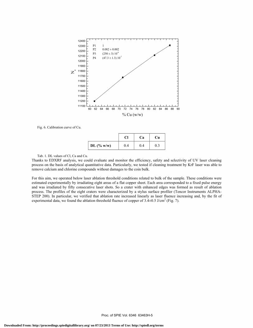
relative concentrations, were prepared in laboratory in order to reproduce the chemical composition of coin surface. Next we analyzed the prepared samples and successively we calibrated the EDXRF device. Figs. 4, 5 and 6 show the calibration curves for Cu, Cl and Ca, respectively. There, the fluorescent counts, Na, are reported as a function of weight fraction wa. The experimental data were fitted by the parametric function utilized to describe this process:
( )( ) wPP
wPPN
a43
a21a +
+= (5)
The detection limit (DL) values related to the above elements are reported in Tab. 1. and they were estimated experimentally from the respective calibration curves.
2,0 2,5 3,0 3,5 4,0 4,5 5,0 5,5 6,020
40
60
80
100
120
140
160
P1 1P2 -(13.5 ± 0.7)P3 -(1.11 ±0.03)P4 (1061.7± 1.7) 10-4
Na
% Cl (w/w)
Fig. 4. Calibration curve of Cl.
1 2 3 4 5 6 7 8100
150
200
250
300
350
400
P1 1P2 1.28 ± 0.07P3 (212 ± 8) 10-4
P4 (80 ± 8) 10-5
Na
% Ca (w/w)
Fig. 5. Calibration curve of Ca.
Proc. of SPIE Vol. 6346 63463H-4
Downloaded From: http://proceedings.spiedigitallibrary.org/ on 07/23/2013 Terms of Use: http://spiedl.org/terms
60 62 64 66 68 70 72 74 76 78 80 82 84 86 88 9011100
11200
11300
11400
11500
11600
11700
11800
11900
12000
12100
12200
12300
12400
P1 1 P2 0.082 ± 0.002P3 (250 ± 3) 10-6
P4 (47.3 ± 1.3) 10-7
Na
% Cu (w/w)
Fig. 6. Calibration curve of Cu.
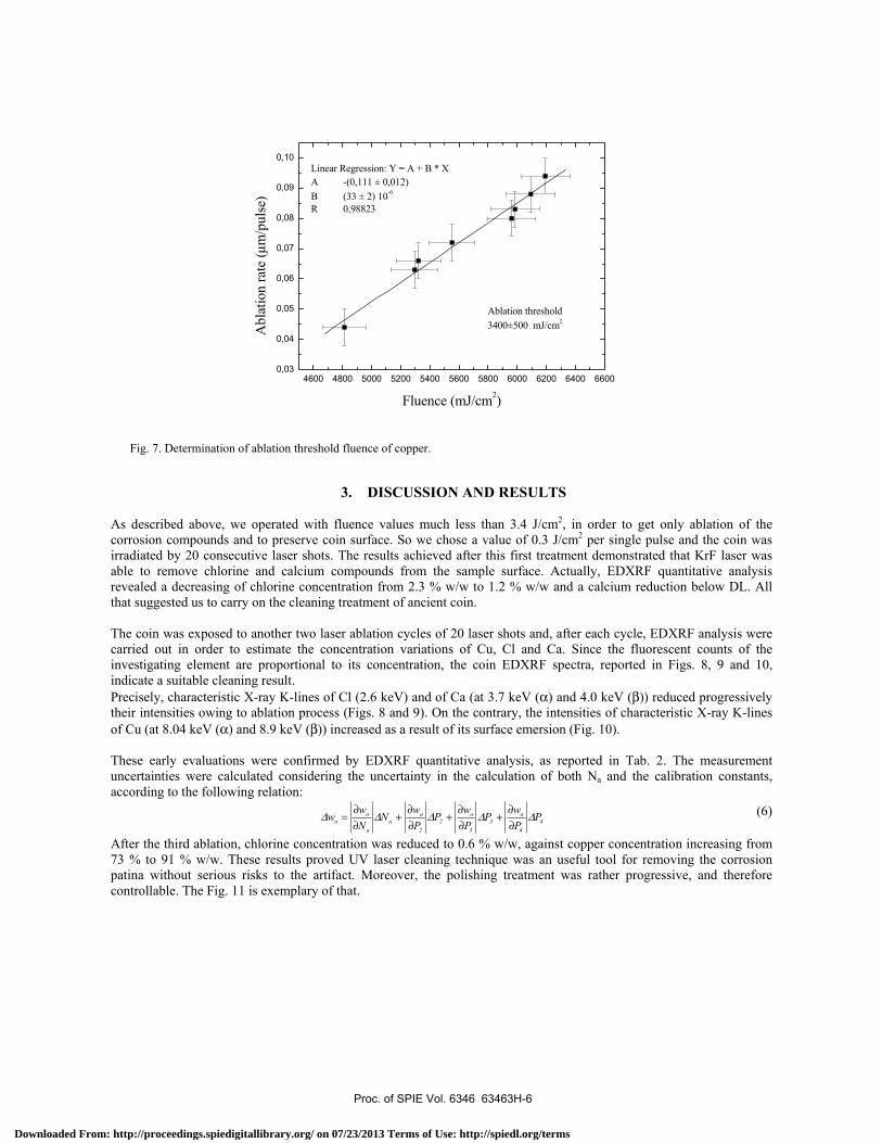
Cl Ca Cu
DL (% w/w) 0.4 0.4 0.3
Tab. 1. DL values of Cl, Ca and Cu. Thanks to EDXRF analysis, we could evaluate and monitor the efficiency, safety and selectivity of UV laser cleaning process on the basis of analytical quantitative data. Particularly, we tested if cleaning treatment by KrF laser was able to remove calcium and chlorine compounds without damages to the coin bulk. For this aim, we operated below laser ablation threshold conditions related to bulk of the sample. These conditions were estimated experimentally by irradiating eight areas of a flat copper sheet. Each area corresponded to a fixed pulse energy and was irradiated by fifty consecutive laser shots. So a crater with enhanced edges was formed as result of ablation process. The profiles of the eight craters were characterized by a stylus surface profiler (Tencor Instruments ALPHA-STEP 200). In particular, we verified that ablation rate increased linearly as laser fluence increasing and, by the fit of experimental data, we found the ablation threshold fluence of copper of 3.4±0.5 J/cm2 (Fig. 7).
Proc. of SPIE Vol. 6346 63463H-5
Downloaded From: http://proceedings.spiedigitallibrary.org/ on 07/23/2013 Terms of Use: http://spiedl.org/terms
4600 4800 5000 5200 5400 5600 5800 6000 6200 6400 66000,03
0,04
0,05
0,06
0,07
0,08
0,09
0,10
Ablation threshold3400±500 mJ/cm2
Linear Regression: Y = A + B * XA -(0,111 ± 0,012)B (33 ± 2) 10-6
R 0,98823
Abl
atio
n ra
te (µ
m/p
ulse
)
Fluence (mJ/cm2)
Fig. 7. Determination of ablation threshold fluence of copper.
3. DISCUSSION AND RESULTS
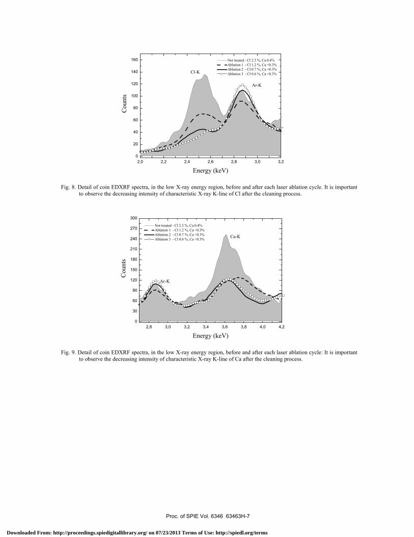
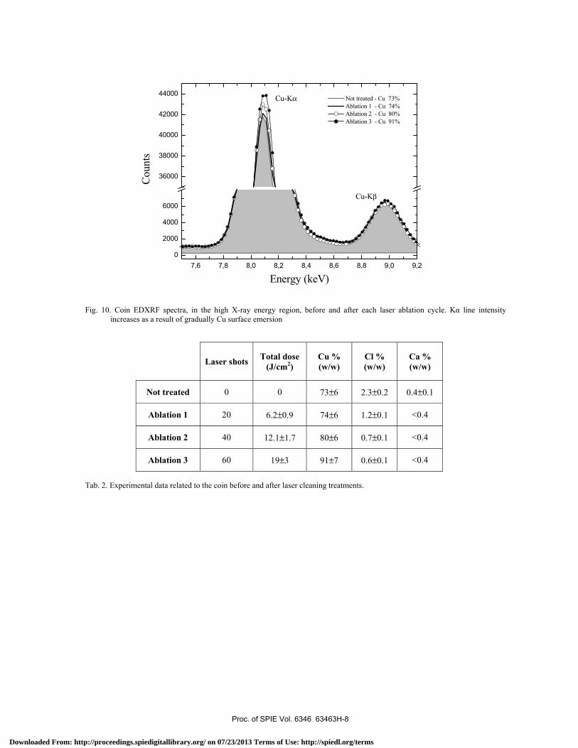
As described above, we operated with fluence values much less than 3.4 J/cm2, in order to get only ablation of the corrosion compounds and to preserve coin surface. So we chose a value of 0.3 J/cm2 per single pulse and the coin was irradiated by 20 consecutive laser shots. The results achieved after this first treatment demonstrated that KrF laser was able to remove chlorine and calcium compounds from the sample surface. Actually, EDXRF quantitative analysis revealed a decreasing of chlorine concentration from 2.3 % w/w to 1.2 % w/w and a calcium reduction below DL. All that suggested us to carry on the cleaning treatment of ancient coin. The coin was exposed to another two laser ablation cycles of 20 laser shots and, after each cycle, EDXRF analysis were carried out in order to estimate the concentration variations of Cu, Cl and Ca. Since the fluorescent counts of the investigating element are proportional to its concentration, the coin EDXRF spectra, reported in Figs. 8, 9 and 10, indicate a suitable cleaning result. Precisely, characteristic X-ray K-lines of Cl (2.6 keV) and of Ca (at 3.7 keV (α) and 4.0 keV (β)) reduced progressively their intensities owing to ablation process (Figs. 8 and 9). On the contrary, the intensities of characteristic X-ray K-lines of Cu (at 8.04 keV (α) and 8.9 keV (β)) increased as a result of its surface emersion (Fig. 10). These early evaluations were confirmed by EDXRF quantitative analysis, as reported in Tab. 2. The measurement uncertainties were calculated considering the uncertainty in the calculation of both Na and the calibration constants, according to the following relation:
PPwP
PwP
PwN
Nww 4
4
a3
3
a2
2
aa
a
aa ∆∆∆∆∆
∂∂
+∂∂
+∂∂
+∂∂
= (6)
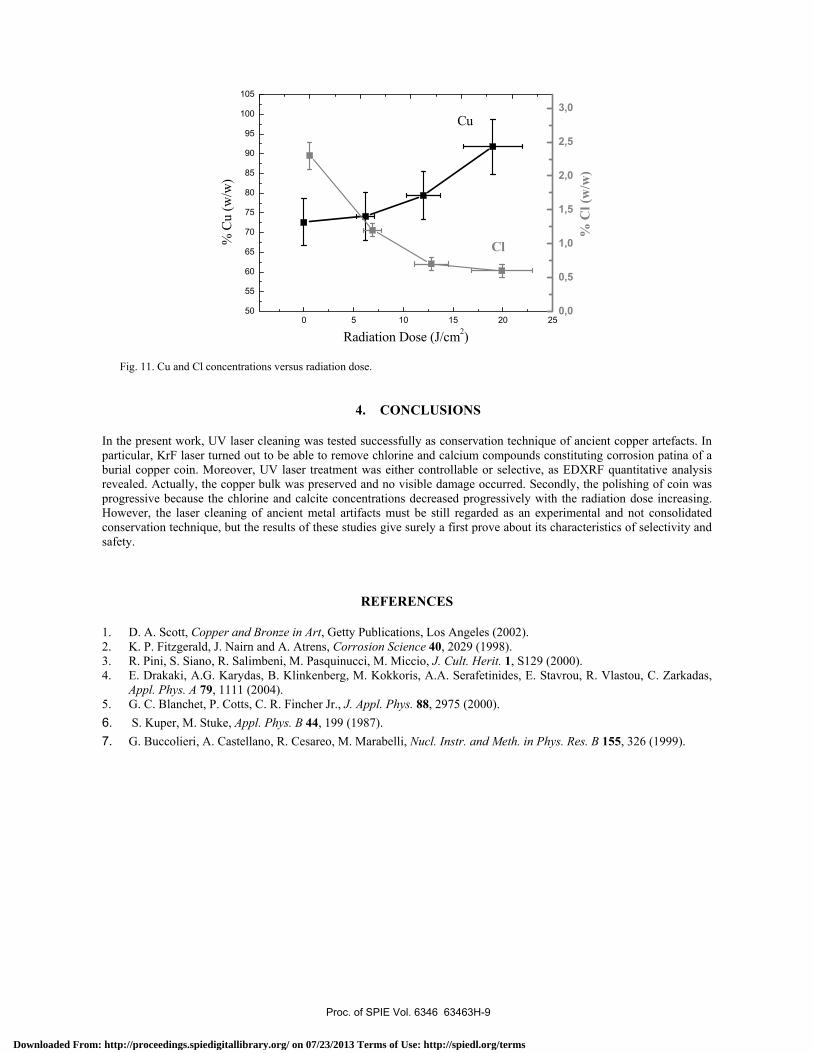
After the third ablation, chlorine concentration was reduced to 0.6 % w/w, against copper concentration increasing from 73 % to 91 % w/w. These results proved UV laser cleaning technique was an useful tool for removing the corrosion patina without serious risks to the artifact. Moreover, the polishing treatment was rather progressive, and therefore controllable. The Fig. 11 is exemplary of that.
Proc. of SPIE Vol. 6346 63463H-6
Downloaded From: http://proceedings.spiedigitallibrary.org/ on 07/23/2013 Terms of Use: http://spiedl.org/terms
2,0 2,2 2,4 2,6 2,8 3,0 3,20
20
40
60
80
100
120
140
160
Ar-K
Cl-K
Cou
nts
Energy (keV)
Not treated - Cl 2.3 %, Ca 0.4% Ablation 1 - Cl 1.2 %, Ca <0.3% Ablation 2 - Cl 0.7 %, Ca <0.3% Ablation 3 - Cl 0.6 %, Ca <0.3%
Fig. 8. Detail of coin EDXRF spectra, in the low X-ray energy region, before and after each laser ablation cycle. It is important to observe the decreasing intensity of characteristic X-ray K-line of Cl after the cleaning process.
2,8 3,0 3,2 3,4 3,6 3,8 4,0 4,20
30
60
90
120
150
180
210
240
270
300
Ar-K
Ca-K
Cou
nts
Energy (keV)
Not treated - Cl 2.3 %, Ca 0.4% Ablation 1 - Cl 1.2 %, Ca <0.3% Ablation 2 - Cl 0.7 %, Ca <0.3% Ablation 3 - Cl 0.6 %, Ca <0.3%
Fig. 9. Detail of coin EDXRF spectra, in the low X-ray energy region, before and after each laser ablation cycle: It is important to observe the decreasing intensity of characteristic X-ray K-line of Ca after the cleaning process.
Proc. of SPIE Vol. 6346 63463H-7
Downloaded From: http://proceedings.spiedigitallibrary.org/ on 07/23/2013 Terms of Use: http://spiedl.org/terms
7,6 7,8 8,0 8,2 8,4 8,6 8,8 9,0 9,20
2000
4000
6000
36000
38000
40000
42000
44000
Cu-Kβ
Cu-Kα
Coun
ts
Energy (keV)
Not treated - Cu 73% Ablation 1 - Cu 74% Ablation 2 - Cu 80% Ablation 3 - Cu 91%
Fig. 10. Coin EDXRF spectra, in the high X-ray energy region, before and after each laser ablation cycle. Kα line intensity increases as a result of gradually Cu surface emersion
Laser shots Total dose (J/cm2)
Cu % (w/w)
Cl % (w/w)
Ca % (w/w)
Not treated 0 0 73±6 2.3±0.2 0.4±0.1
Ablation 1 20 6.2±0.9 74±6 1.2±0.1 <0.4
Ablation 2 40 12.1±1.7 80±6 0.7±0.1 <0.4
Ablation 3 60 19±3 91±7 0.6±0.1 <0.4
Tab. 2. Experimental data related to the coin before and after laser cleaning treatments.
Proc. of SPIE Vol. 6346 63463H-8
Downloaded From: http://proceedings.spiedigitallibrary.org/ on 07/23/2013 Terms of Use: http://spiedl.org/terms
0 5 10 15 20 2550
55
60
65
70
75
80
85
90
95
100
105
Cl
Cu
% C
l (w
/w)
% C
u (w
/w)
Radiation Dose (J/cm2)
0,0
0,5
1,0
1,5
2,0
2,5
3,0
Fig. 11. Cu and Cl concentrations versus radiation dose.
4. CONCLUSIONS
In the present work, UV laser cleaning was tested successfully as conservation technique of ancient copper artefacts. In particular, KrF laser turned out to be able to remove chlorine and calcium compounds constituting corrosion patina of a burial copper coin. Moreover, UV laser treatment was either controllable or selective, as EDXRF quantitative analysis revealed. Actually, the copper bulk was preserved and no visible damage occurred. Secondly, the polishing of coin was progressive because the chlorine and calcite concentrations decreased progressively with the radiation dose increasing. However, the laser cleaning of ancient metal artifacts must be still regarded as an experimental and not consolidated conservation technique, but the results of these studies give surely a first prove about its characteristics of selectivity and safety.
REFERENCES
1. D. A. Scott, Copper and Bronze in Art, Getty Publications, Los Angeles (2002). 2. K. P. Fitzgerald, J. Nairn and A. Atrens, Corrosion Science 40, 2029 (1998). 3. R. Pini, S. Siano, R. Salimbeni, M. Pasquinucci, M. Miccio, J. Cult. Herit. 1, S129 (2000). 4. E. Drakaki, A.G. Karydas, B. Klinkenberg, M. Kokkoris, A.A. Serafetinides, E. Stavrou, R. Vlastou, C. Zarkadas,
Appl. Phys. A 79, 1111 (2004). 5. G. C. Blanchet, P. Cotts, C. R. Fincher Jr., J. Appl. Phys. 88, 2975 (2000). 6. S. Kuper, M. Stuke, Appl. Phys. B 44, 199 (1987). 7. G. Buccolieri, A. Castellano, R. Cesareo, M. Marabelli, Nucl. Instr. and Meth. in Phys. Res. B 155, 326 (1999).
Proc. of SPIE Vol. 6346 63463H-9
Downloaded From: http://proceedings.spiedigitallibrary.org/ on 07/23/2013 Terms of Use: http://spiedl.org/terms