Salt stress induced variation in DNA methylation pattern and its influence on gene expression in...
10
Salt Stress Induced Variation in DNA Methylation Pattern and Its Influence on Gene Expression in Contrasting Rice Genotypes Ratna Karan, Teresa DeLeon, Hanamareddy Biradar, Prasanta K. Subudhi* School of Plant, Environmental, and Soil Sciences, Louisiana State University Agricultural Center, Baton Rouge, Louisiana, United States of America Abstract Background: Salinity is a major environmental factor limiting productivity of crop plants including rice in which wide range of natural variability exists. Although recent evidences implicate epigenetic mechanisms for modulating the gene expression in plants under environmental stresses, epigenetic changes and their functional consequences under salinity stress in rice are underexplored. DNA methylation is one of the epigenetic mechanisms regulating gene expression in plant’s responses to environmental stresses. Better understanding of epigenetic regulation of plant growth and response to environmental stresses may create novel heritable variation for crop improvement. Methodology/Principal Findings: Methylation sensitive amplification polymorphism (MSAP) technique was used to assess the effect of salt stress on extent and patterns of DNA methylation in four genotypes of rice differing in the degree of salinity tolerance. Overall, the amount of DNA methylation was more in shoot compared to root and the contribution of fully methylated loci was always more than hemi-methylated loci. Sequencing of ten randomly selected MSAP fragments indicated gene-body specific DNA methylation of retrotransposons, stress responsive genes, and chromatin modification genes, distributed on different rice chromosomes. Bisulphite sequencing and quantitative RT-PCR analysis of selected MSAP loci showed that cytosine methylation changes under salinity as well as gene expression varied with genotypes and tissue types irrespective of the level of salinity tolerance of rice genotypes. Conclusions/Significance: The gene body methylation may have an important role in regulating gene expression in organ and genotype specific manner under salinity stress. Association between salt tolerance and methylation changes observed in some cases suggested that many methylation changes are not ‘‘directed’’. The natural genetic variation for salt tolerance observed in rice germplasm may be independent of the extent and pattern of DNA methylation which may have been induced by abiotic stress followed by accumulation through the natural selection process. Citation: Karan R, DeLeon T, Biradar H, Subudhi PK (2012) Salt Stress Induced Variation in DNA Methylation Pattern and Its Influence on Gene Expression in Contrasting Rice Genotypes. PLoS ONE 7(6): e40203. doi:10.1371/journal.pone.0040203 Editor: Keqiang Wu, National Taiwan University, Taiwan Received February 12, 2012; Accepted June 3, 2012; Published June 28, 2012 Copyright: ß 2012 Karan et al. This is an open-access article distributed under the terms of the Creative Commons Attribution License, which permits unrestricted use, distribution, and reproduction in any medium, provided the original author and source are credited. Funding: United States Department of Agriculture-CSREES and Louisiana Rice Research Board funded this study. The funders had no role in study design, data collection and analysis, decision to publish, or preparation of the manuscript. Competing Interests: The authors have declared that no competing interests exist. * E-mail: [email protected] Introduction Plant epigenetics has received considerable attention for both basic and applied research in recent years because understanding of the epigenetic regulation of plant growth and development could create new genetic variation for improving crop productivity as well as adaptation to stress environment [1]. Epigenetics refers to heritable variation in gene regulation resulting from covalent modifications of DNA and its associated chromatin proteins without changing the underlying nucleotide sequences [2]. Such epigenetic modifications are reversible and can alter the pheno- typic appearance [3]. Cytosine methylation is a conserved epigenetic mechanism involved in many important biological processes, including transposon proliferation, genomic imprinting, and regulation of gene expression [4,5]. It is usually associated with inactivation of genes while demethylation results in gene activation [6]. Genome-wide high-resolution mapping and func- tional analysis of DNA methylation in rice revealed that 8% of active genes were methylated within their promoter, while methylation within transcribed regions was observed in 31% of the expressed genes [5]. Due to erratic global climate, crop plants are frequently exposed to a variety of abiotic stresses including salinity resulting in reduced crop productivity. Analysis of stress related genes and their regulation of expression in response to abiotic stresses are commonly employed for enhanced understanding of the plants ability to adapt under abiotic stress environments. However, growing evidences implicate epigenetic mechanisms, such as DNA methylation and chromatin modification in regulating gene expression for plants’ responses to environmen- tal stresses [7]. Demethylation of certain functionally inactive genes can occur due to exposure to abiotic stresses [8]. Abiotic stresses may cause heritable alterations in cytosine methylation by forming novel epialleles [9]. The expression of certain genes modified by epigenetic mechanism can be reverted to its earlier PLoS ONE | www.plosone.org 1 June 2012 | Volume 7 | Issue 6 | e40203
Transcript of Salt stress induced variation in DNA methylation pattern and its influence on gene expression in...

Salt Stress Induced Variation in DNA Methylation Patternand Its Influence on Gene Expression in Contrasting RiceGenotypesRatna Karan, Teresa DeLeon, Hanamareddy Biradar, Prasanta K. Subudhi*
School of Plant, Environmental, and Soil Sciences, Louisiana State University Agricultural Center, Baton Rouge, Louisiana, United States of America
Abstract
Background: Salinity is a major environmental factor limiting productivity of crop plants including rice in which wide rangeof natural variability exists. Although recent evidences implicate epigenetic mechanisms for modulating the geneexpression in plants under environmental stresses, epigenetic changes and their functional consequences under salinitystress in rice are underexplored. DNA methylation is one of the epigenetic mechanisms regulating gene expression inplant’s responses to environmental stresses. Better understanding of epigenetic regulation of plant growth and response toenvironmental stresses may create novel heritable variation for crop improvement.
Methodology/Principal Findings: Methylation sensitive amplification polymorphism (MSAP) technique was used to assessthe effect of salt stress on extent and patterns of DNA methylation in four genotypes of rice differing in the degree ofsalinity tolerance. Overall, the amount of DNA methylation was more in shoot compared to root and the contribution offully methylated loci was always more than hemi-methylated loci. Sequencing of ten randomly selected MSAP fragmentsindicated gene-body specific DNA methylation of retrotransposons, stress responsive genes, and chromatin modificationgenes, distributed on different rice chromosomes. Bisulphite sequencing and quantitative RT-PCR analysis of selected MSAPloci showed that cytosine methylation changes under salinity as well as gene expression varied with genotypes and tissuetypes irrespective of the level of salinity tolerance of rice genotypes.
Conclusions/Significance: The gene body methylation may have an important role in regulating gene expression in organand genotype specific manner under salinity stress. Association between salt tolerance and methylation changes observedin some cases suggested that many methylation changes are not ‘‘directed’’. The natural genetic variation for salt toleranceobserved in rice germplasm may be independent of the extent and pattern of DNA methylation which may have beeninduced by abiotic stress followed by accumulation through the natural selection process.
Citation: Karan R, DeLeon T, Biradar H, Subudhi PK (2012) Salt Stress Induced Variation in DNA Methylation Pattern and Its Influence on Gene Expression inContrasting Rice Genotypes. PLoS ONE 7(6): e40203. doi:10.1371/journal.pone.0040203
Editor: Keqiang Wu, National Taiwan University, Taiwan
Received February 12, 2012; Accepted June 3, 2012; Published June 28, 2012
Copyright: � 2012 Karan et al. This is an open-access article distributed under the terms of the Creative Commons Attribution License, which permitsunrestricted use, distribution, and reproduction in any medium, provided the original author and source are credited.
Funding: United States Department of Agriculture-CSREES and Louisiana Rice Research Board funded this study. The funders had no role in study design, datacollection and analysis, decision to publish, or preparation of the manuscript.
Competing Interests: The authors have declared that no competing interests exist.
* E-mail: [email protected]
Introduction
Plant epigenetics has received considerable attention for both
basic and applied research in recent years because understanding
of the epigenetic regulation of plant growth and development
could create new genetic variation for improving crop productivity
as well as adaptation to stress environment [1]. Epigenetics refers
to heritable variation in gene regulation resulting from covalent
modifications of DNA and its associated chromatin proteins
without changing the underlying nucleotide sequences [2]. Such
epigenetic modifications are reversible and can alter the pheno-
typic appearance [3]. Cytosine methylation is a conserved
epigenetic mechanism involved in many important biological
processes, including transposon proliferation, genomic imprinting,
and regulation of gene expression [4,5]. It is usually associated
with inactivation of genes while demethylation results in gene
activation [6]. Genome-wide high-resolution mapping and func-
tional analysis of DNA methylation in rice revealed that 8% of
active genes were methylated within their promoter, while
methylation within transcribed regions was observed in 31% of
the expressed genes [5].
Due to erratic global climate, crop plants are frequently
exposed to a variety of abiotic stresses including salinity
resulting in reduced crop productivity. Analysis of stress related
genes and their regulation of expression in response to abiotic
stresses are commonly employed for enhanced understanding of
the plants ability to adapt under abiotic stress environments.
However, growing evidences implicate epigenetic mechanisms,
such as DNA methylation and chromatin modification in
regulating gene expression for plants’ responses to environmen-
tal stresses [7]. Demethylation of certain functionally inactive
genes can occur due to exposure to abiotic stresses [8]. Abiotic
stresses may cause heritable alterations in cytosine methylation
by forming novel epialleles [9]. The expression of certain genes
modified by epigenetic mechanism can be reverted to its earlier
PLoS ONE | www.plosone.org 1 June 2012 | Volume 7 | Issue 6 | e40203
state and may show transgenerational inheritance [10].
Responses to the external stress vary among plant species;
some of them can modulate their physiological and develop-
mental machinery to mitigate the impact of stress whereas
others are highly sensitive. Mangrove plants growing in
contrasting natural habitats, riverside and salt marsh, differed
with respect to cytosine methylation despite little genetic
variation [11].
Methylation-sensitive amplified polymorphism (MSAP) is a
powerful technique for studying the genome methylation status
[12]. It is a modification of the AFLP technique in which
isoschizomers, MspI and HpaII, are employed as ‘frequent-cutter’.
Both MspI and HpaII recognize the same restriction site (59-
CCGG-39), but show differential sensitivity to DNA methylation.
This technique has been applied to evaluate the level and pattern
of cytosine methylation in plants [13]. Effect of salinity stress on
cytosine methylation variation has been studied in crop plants
[14–15]. The epigenetic variation need to be explored in more
genotypes so that useful variability can be identified and exploited
in crop breeding programs to enhance crop adaptation in
unfavorable environments.
Rice is one of the most salt-sensitive cereal crops and their
sensitivity varies among different genotypes [16,17]. Some rice
genotypes possess unique ability to rapidly adapt to a toxic level
of salt stress whereas others are highly susceptible indicating
their uniqueness in genetic makeup and regulatory architecture.
However, differences in methylation pattern and epigenetic
response in contrasting rice varieties under salinity stress has
been underexplored. In this study, we have investigated the
extent and pattern of cytosine methylation under non-stress and
salinity stress in four contrasting genotypes of rice using MSAP
technique. Methylation and demethylation patterns were geno-
type and tissue specific irrespective of the degree of salt
tolerance of individual rice genotype. MSAP fragments were
localized onto different rice chromosomes and majority of these
fragments showed homology with stress responsive genes,
retrotransposons, and genes involved in chromatin modification.
The differential expression patterns of stress related genes
influenced by methylation or demethylation status under salinity
stress in the rice genotypes revealed possible role of epigenetic
mechanism in stress adaptation and the natural genetic
variability in salinity tolerance phenotype may be independent
of the extent and pattern of DNA methylation.
Results
Salinity Tolerance of Rice GenotypesScoring of visual salt stress injury using a modified standard
evaluation system (SES) [18] after 12 days and 15 days of
imposition of salt stress showed clear differences among rice
genotypes. After 15 days of salt stress, most of Bengal, IR29 and
Nipponbare plants were dead while Pokkali, Nonabokra, and
Geumgangbyeo survived but showed salinity injury (Figure S1).
Mean SES scores for IR29, Bengal, Nipponbare, Geumgang-
byeo, Nonabokra, and Pokkali were 9.0, 8.8, 8.3, 4.3, 2.8, and
2.3, respectively. These scores indicated that Bengal and
Nipponbare are highly salt sensitive like IR29, Geumgangbyeo
is moderately tolerant, and Pokkali and Nonabokra are tolerant
to salt stress. Comparison of uptake of Na+, K+, and K+/Na+
ratio (Figure S2) further confirmed the level of salt tolerance
based on SES scores. The K+/Na+ ratios were higher in
tolerant genotypes compared to susceptible genotypes under
both control and salt stress.
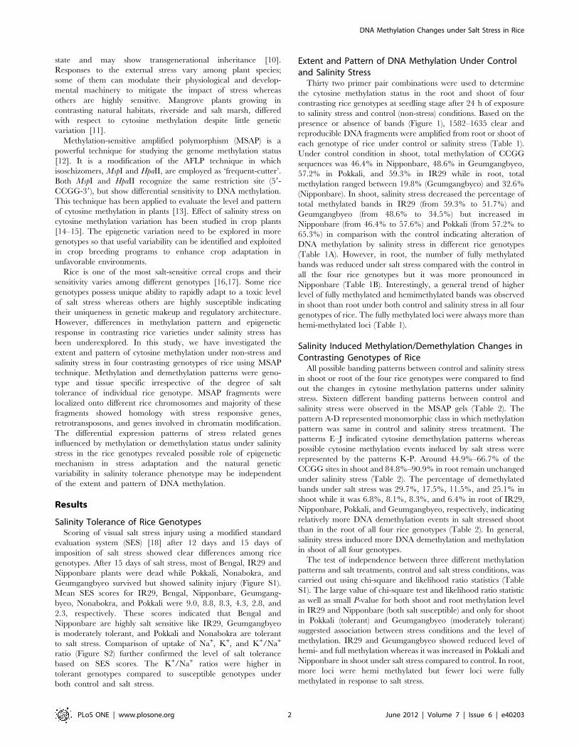
Extent and Pattern of DNA Methylation Under Controland Salinity Stress
Thirty two primer pair combinations were used to determine
the cytosine methylation status in the root and shoot of four
contrasting rice genotypes at seedling stage after 24 h of exposure
to salinity stress and control (non-stress) conditions. Based on the
presence or absence of bands (Figure 1), 1582–1635 clear and
reproducible DNA fragments were amplified from root or shoot of
each genotype of rice under control or salinity stress (Table 1).
Under control condition in shoot, total methylation of CCGG
sequences was 46.4% in Nipponbare, 48.6% in Geumgangbyeo,
57.2% in Pokkali, and 59.3% in IR29 while in root, total
methylation ranged between 19.8% (Geumgangbyeo) and 32.6%
(Nipponbare). In shoot, salinity stress decreased the percentage of
total methylated bands in IR29 (from 59.3% to 51.7%) and
Geumgangbyeo (from 48.6% to 34.5%) but increased in
Nipponbare (from 46.4% to 57.6%) and Pokkali (from 57.2% to
65.3%) in comparison with the control indicating alteration of
DNA methylation by salinity stress in different rice genotypes
(Table 1A). However, in root, the number of fully methylated
bands was reduced under salt stress compared with the control in
all the four rice genotypes but it was more pronounced in
Nipponbare (Table 1B). Interestingly, a general trend of higher
level of fully methylated and hemimethylated bands was observed
in shoot than root under both control and salinity stress in all four
genotypes of rice. The fully methylated loci were always more than
hemi-methylated loci (Table 1).
Salinity Induced Methylation/Demethylation Changes inContrasting Genotypes of Rice
All possible banding patterns between control and salinity stress
in shoot or root of the four rice genotypes were compared to find
out the changes in cytosine methylation patterns under salinity
stress. Sixteen different banding patterns between control and
salinity stress were observed in the MSAP gels (Table 2). The
pattern A-D represented monomorphic class in which methylation
pattern was same in control and salinity stress treatment. The
patterns E–J indicated cytosine demethylation patterns whereas
possible cytosine methylation events induced by salt stress were
represented by the patterns K-P. Around 44.9%–66.7% of the
CCGG sites in shoot and 84.8%–90.9% in root remain unchanged
under salinity stress (Table 2). The percentage of demethylated
bands under salt stress was 29.7%, 17.5%, 11.5%, and 25.1% in
shoot while it was 6.8%, 8.1%, 8.3%, and 6.4% in root of IR29,
Nipponbare, Pokkali, and Geumgangbyeo, respectively, indicating
relatively more DNA demethylation events in salt stressed shoot
than in the root of all four rice genotypes (Table 2). In general,
salinity stress induced more DNA demethylation and methylation
in shoot of all four genotypes.
The test of independence between three different methylation
patterns and salt treatments, control and salt stress conditions, was
carried out using chi-square and likelihood ratio statistics (Table
S1). The large value of chi-square test and likelihood ratio statistic
as well as small P-value for both shoot and root methylation level
in IR29 and Nipponbare (both salt susceptible) and only for shoot
in Pokkali (tolerant) and Geumgangbyeo (moderately tolerant)
suggested association between stress conditions and the level of
methylation. IR29 and Geumgangbyeo showed reduced level of
hemi- and full methylation whereas it was increased in Pokkali and
Nipponbare in shoot under salt stress compared to control. In root,
more loci were hemi methylated but fewer loci were fully
methylated in response to salt stress.
DNA Methylation Changes under Salt Stress in Rice
PLoS ONE | www.plosone.org 2 June 2012 | Volume 7 | Issue 6 | e40203

Analysis of the Differentially Methylated DNA SequencesA random set of ten polymorphic fragments showing unique
banding pattern in individual genotype for methylation/demethyl-
ation under salinity stress compared to control were sequenced to
identify the nature of DNA sequences involved in methylation-
demethylation under salinity (Table 3). The size of polymorphic
DNA fragments varied between 60 to 324 bp and these fragments
were distributed on the rice chromosomes 1, 2, 3, 4, and 11. The
positions of cytosine alteration due to salinity stress were located in
exon and UTR region of affected loci suggesting the gene-body
specific methylation of MSAP loci. BLAST analysis of sequenced
fragments revealed that the sequences are homologous to retro-
transposons, peptidyl prolyl cis trans isomerase, calcineurin B,
histone methyl transferase, rho guanine nucleotide exchange factor,
cystathionine gamma synthase, DUF domain containing protein,
methyl CG binding domain containing protein, and pentatricopep-
tide repeat domain containing protein (Table 3).These results
suggest that the genes involved in wide range of cellular functions
were affected mostly by gene-body specific cytosine methylation-
demethylation due to salinity stress in rice.
Expression Analysis of MSAP loci in Root and Shoot ofRice Genotypes
Expression analysis of five MSAP polymorphic genes was done
using quantitative RT-PCR in shoot and root of six contrasting
rice genotypes under control and salinity stress to determine the
effect of methylation changes on the gene expression (Figure 2).
Gene Os02g44330 (P4), methylated in the shoot of IR29, showed
downregulation under salinity stress in shoot of IR29 and
Nonabokra, whereas its expression remain unchanged in the
shoot of the remaining four genotypes under salinity stress in
comparison to control (Figure 2A). However in the root, its
expression was enhanced in IR29, Nonabokra, and Geumgang-
byeo but downregulated in Nipponbare and Pokkali. Bengal
showed a slight increase in expression. Similarly, expression
changes of Os03g32490 (P5) which was demethylated in the shoot
of IR29, was minimal with increased expression in the shoot of
IR29, Bengal, Nipponbare, and Pokkali, but reduced expression in
Nonabokra and Geumgangbyeo under salinity stress (Figure 2A).
In roots of IR29 and Geumgangbyeo, salt stress enhanced the
expression of P5 but reverse was the case in Bengal, Nipponbare,
Figure 1. A representative MSAP gel using the primer combination EcoRI-ACG/MspI-AATC. Both control and salinity stressed root andshoot of rice genotypes, IR29, Nipponbare (Nipp), Pokkali (Pokk), Geumgangbyeo (Geum) were used for MSAP analysis. EH and EM refer to digestionwith EcoRI+HpaII and EcoRI+MspI, respectively. RC: root control; RS: root stress; SC: shoot control; SS: shoot stress.doi:10.1371/journal.pone.0040203.g001
DNA Methylation Changes under Salt Stress in Rice
PLoS ONE | www.plosone.org 3 June 2012 | Volume 7 | Issue 6 | e40203

Pokkali, and Geumgangbyeo (Figure 2B). One of the demethylated
gene, Os11g23900 (P8) in the shoot of Nipponbare, showed 6–8
fold increase expression in shoot of Bengal, and Pokkali but was
downregulated in IR29, while remain unchanged in Nipponbare,
Nonabokra, and Geumgangbyeo under salinity (Figure 2A). It was
upregulated in roots of IR29 and Geumgangbyeo but down
regulated in other genotypes (Figure 2B).The gene Os02g52290
(P1) which was methylated in shoot of IR29 and homologous to
peptidyl-prolyl cis-trans isomerase, showed increased expression in
the shoot and root of IR29 and Geumgangbyeo under salinity
stress. The expression of P1 was enhanced in the shoot of other
four genotypes under salinity stress than control, but was
downregulated in the root of Bengal, Nipponbare, Pokkali, and
Nonabokra. Another gene, Os01g60309 (P9), a retrotransposon,
demethylated in the shoot of IR29, showed downregulation in
shoot of IR29, Bengal, Nipponbare, Pokkali, and Geumgangbyeo
whereas it was upregulated in Nonabokra. In root, expression of
P9 was enhanced under salinity stress in IR29, Pokkali, and
Geumgangbyeo, but remained unchanged or slightly increased in
Nipponbare, Bengal, and Nonabokra. Therefore, the methylation
pattern within the gene-body region did not always correspond to
its expression changes under salinity stress and expression
variation was genotype specific.
Bisulfite Sequencing of MSAP LociBisulfite sequencing of two selected MSAP loci, Os02g52290
(P1) and Os01g60309 (P9) was conducted to assess the cytosine
methylation status. The bisulfite nucleotide sequences from top
strands of these loci were similar with their corresponding loci
from TIGR database and most of the methylated cytosines
belonged to CG types (Figures S3, S4). Alignment of Os02g52290
(P1) (Figures S3 and S5) sequence with the bisulfite treated DNA
sequences from root and shoot of all the six rice genotypes under
non-stress and salinity stress conditions revealed that methylated
cytosine present under non-stress conditions at position 3, 5, and
29, did not change even under salinity stress. At 75th nucleotide
position, cytosine gets demethylated under salinity stress in shoot
and remained unmethylated under both conditions in root in
Pokkali while it maintained methylation status in other genotypes.
Cytosine at position 154 was only methylated in shoot of Bengal
under non-stress condition. The patterns of methylation were
identical at both 234 and 243 positions. In Bengal, cytosine at
position 264 was demethylated in shoot and methylated in root,
whereas, it maintained its methylated or unmethylated status in
rest five genotypes of rice under salinity compared to non-stress
condition. At position 278, cytosine maintained methylated status
in Nipponbare in both tissues under control or salinity stress.
Cytosine methylation (%) was reduced in shoot under salt stress
compared with control in Bengal, Nipponbare, and Pokkali, but
was increased IR29 and remained unchanged in Nonabokra and
Geumgangbyeo (Figure S3). Furthermore, cytosine methylation in
root under salinity was slightly increased in Bengal, but remained
unchanged in IR29, Nipponbare, Pokkali, Nonabokra, and
Geumgangbyeo.
In case of Os01g60309 (P9) (Figures S4 and S6), cytosines at 108
and 113 positions remained methylated under both conditions and
both tissues in all genotypes but cytosine at 242 was methylated
under salinity compared to control in all the genotypes except
Table 1. DNA methylation changes in shoot and root at seedling stage under non-stress and salinity stress conditions.
IR29 Nipponbare Pokkali Geumgangbyeo
MSAP band type Control Salinity Control Salinity Control Salinity Control Salinity
A. Shoot
I 665 789 877 692 698 566 838 1067
II 201 113 246 290 293 317 143 119
III 322 425 205 235 171 158 160 93
IV 446 308 306 418 471 593 489 350
Total amplified bands 1634 1635 1634 1635 1633 1634 1630 1629
MSAP (%)a 59.3 51.7 46.4 57.6 57.2 65.3 48.6 34.5
Fully methylated band (%)b 47.0 44.8 31.3 39.9 39.3 45.9 39.8 27.2
Hemi-methylated band (%)c 12.3 6.9 15.1 17.7 17.9 19.4 8.8 7.3
B. Root
I 1276 1314 1103 1310 1231 1232 1311 1319
II 7 23 34 39 32 34 10 19
III 98 84 239 83 91 114 95 97
IV 254 211 259 203 232 202 218 197
Total amplified bands 1635 1632 1635 1635 1586 1582 1634 1632
MSAP (%)a 21.9 19.5 32.6 19.9 22.4 22.1 19.8 19.2
Fully methylated band (%)b 21.5 18.1 30.5 17.5 20.4 19.9 19.2 18.0
Hemi-methylated band (%)c 0.4 1.4 2.1 2.4 2.0 2.2 0.6 1.2
aMSAP (%) = [(II+III+IV)/(I+II+III+IV)]6100;bFully methylated bands (%) = [(III+IV)/(I+II+III+IV)]6100;cHemimethylated bands (%) = [(II)/(I+II+III+IV)]6100.Type I indicated absence of methylation due to the presence of bands in both EcoRI/HpaII and EcoRI/MspI digest; type II bands appeared only in EcoRI/HpaII digestionbut not in the EcoRI/MspI digest; type III generated bands obtained in EcoRI/MspI digest but not in the EcoRI/HpaII digest; and type IV represents the absence of band inboth enzyme combinations.doi:10.1371/journal.pone.0040203.t001
DNA Methylation Changes under Salt Stress in Rice
PLoS ONE | www.plosone.org 4 June 2012 | Volume 7 | Issue 6 | e40203

Table 2. Analysis of DNA methylation patterns under salinity stress with respect to control condition in the shoot and root ofseedlings of rice varieties, IR29, Nipponbare (Nipp), Pokkali (Pokk), and Geumgangbyeo (Geum).
Description of Pattern Class Banding Pattern Shoot Root
Control Salinity
HpaII MspI HpaII MspI IR29 Nipp Pokk Geum IR29 Nipp Pokk Geum
A 1 0 1 0 20 62 42 17 3 7 4 3
No change B 0 1 0 1 108 44 30 12 54 55 54 51
C 1 1 1 1 505 541 287 775 1238 1168 1131 1240
D 0 0 0 0 184 180 373 282 188 131 153 153
Total 817(50.0%)
827(50.6%)
732(44.9%)
1086(66.7%)
1483(90.9%)
1361(85.8%)
1342(84.8%)
1447(88.7%)
E 1 0 1 1 62 70 17 80 3 17 21 5
F 0 1 1 1 136 53 42 105 39 26 28 30
Demethylation G 0 0 1 1 85 27 19 107 34 49 52 43
H 0 1 1 0 25 37 30 16 3 7 3 4
I 0 0 1 0 50 49 43 56 10 13 5 5
J 0 0 0 1 127 50 36 44 22 16 22 17
Total 485(29.7%)
286(17.5%)
187(11.5%)
408(25.1%)
111(6.8%)
128(8.1%)
131(8.3%)
104(6.4%)
K 1 1 1 0 18 142 202 30 7 12 22 7
L 1 1 0 1 113 98 75 16 7 7 38 29
Methylation M 1 1 0 0 29 96 134 17 21 46 37 32
N 1 0 0 1 77 43 17 21 1 5 0 0
O 1 0 0 0 42 71 217 24 0 5 7 2
P 0 1 0 0 53 71 68 27 2 21 5 10
Total 332(20.3%)
521(31.9%)
713(43.6%)
135(8.2%)
38(2.3%)
96(6.1%)
109(6.9%)
80(4.9%)
A score of 1 and 0 represents presence and absence of bands, respectively. Values in parentheses indicate percentage of bands in each pattern which was determinedby dividing number of bands in each pattern by total number of bands in all three patterns.doi:10.1371/journal.pone.0040203.t002
Table 3. BLAST results of ten randomly selected polymorphic methylated fragments and their location on rice genome.
MSAPfragment
Methylation statusunder stress
Tissuetype Genotype Gene TIGR Locus ID E value Position
P1 Methylated shoot IR29 Peptidyl-prolyl cis-transisomerase, FKBP-type
Os02g52290 8.3e-24 39 UTR
P2 Methylated shoot Geumgangbyeo Calcineurin B Os01g39770 2.9e-41 39 UTR
P3 Demethylated shoot IR29 Cystathionine gamma-synthase Os03g25940 1.8e-07 Exon
P4 Methylated shoot IR29 Rho guanine nucleotideexchange factor
Os02g44330 2.1e-19 Exon
P5 Demethylated shoot IR29 DUF1230 domain containingprotein
Os03g32490 7.3e-60 Exon
P6 Demethylated shoot IR29 Methyl-CpG domain containingprotein
Os04g19684 2.3e-29 Exon
P7 Methylated shoot IR29 Histone-lysineN-methyltransferase
Os01g70220 8.9e-56 59 UTR
P8 Demethylated shoot Nipponbare Retrotransposon protein Os11g23900 3.7e-56 Exon
P9 Demethylated shoot IR29 Retrotransposon protein,Ty1-copia subclass
Os01g60309 2.5e-40 Exon
P10 Demethylated shoot IR29 Pentatricopeptide repeatdomain containing protein
Os11g37330 8.9e-48 Exon
doi:10.1371/journal.pone.0040203.t003
DNA Methylation Changes under Salt Stress in Rice
PLoS ONE | www.plosone.org 5 June 2012 | Volume 7 | Issue 6 | e40203

Figure 2. Expression profiles of MSAP loci in rice under salt stress. Salinity stressed shoot (A) and root (B) of six genotypes of rice, Bengal(Beng), IR29, Nipponbare (Nipp), Pokkali (Pokk), Nonabokra (Nona), and Geumgangbyeo (Geum) were used for quantifying the expression of MSAPloci relative to control. Real-time PCR analysis was performed using gene-specific primers. The expression of each gene in different RNA samples wasnormalized with the expression of internal control gene, rice elongation factor 1 a (eEF1 a). Fold changes in mRNA expression for each candidategene in different genotypes were calculated relative to its control using ddCt method. The values represented are the mean of two biologicalreplicates, each with three technical replicates. Error bars indicate the standard deviation.doi:10.1371/journal.pone.0040203.g002
DNA Methylation Changes under Salt Stress in Rice
PLoS ONE | www.plosone.org 6 June 2012 | Volume 7 | Issue 6 | e40203

shoot of Nipponbare and root of IR29. Differential methylation
pattern was observed in both tissues at 107th position. At position
315, cytosine was methylated in shoot and root of all genotypes
under both conditions, except in root of Geumgangbyeo and
Nonabokra under salinity stress. Cytosine methylation (%) in shoot
and root under salinity compared to control remained unaltered in
Bengal, Nipponbare, and Nonabokra, increased in IR9, and
decreased in Pokkali (Figure S4). However, cytosine methylation
decreased in shoot and did not change in root under salinity in
comparison to control in Geumgangbyeo. Although salinity
affected the methylation status of cytosines in these two selected
loci, the changes were genotype and organ specific irrespective of
salinity tolerance of rice genotypes.
Discussion
Evidences are growing in favor of epigenetic modifications
influencing plants’ preparedness for stress adaptation through
regulation of gene expression [19,20]. However, the relationship
between DNA methylation and abiotic stress tolerance was rarely
studied in important field crops [15]. In this study, we assessed the
epigenetic consequences of salinity stress in rice genotypes with
contrasting salt tolerance behavior. Among the rice genotypes used
in this study, Pokkali and Nonabokra were tolerant and IR29 was
highly salt sensitive [16,17]. Bengal and Nipponbare showed salt
sensitivity whereas Geumgangbyeo showed moderate tolerance
(Figures S1 and S2).
We employed MSAP technique to assess the extent and pattern
of DNA methylation in response to salt stress in both shoot and
root of rice. Overall, the amount of DNA methylation was more in
shoot compared to root (Table 1), indicating unique biological
functions performed by each tissue in response to salt stress. Since
root experience the stress first, hypomethylation suggests the
preparedness of more stress responsive genes to respond to salt
stress. Hypomethylation has been reported to affect plant
development [21]. We did not observe any methylation pattern
specific to salt tolerant or salt susceptible genotypes in root or
shoot under salt stress (Table 2). However, significant association
between salt treatment and level of methylation was seen in shoot
of all four genotypes and in root of only IR29 and Nipponbare
(Table S1), suggesting that many methylation changes are not
‘‘directed’’. Genotype specific DNA methylation has been
reported in rice [13]. Furthermore, epigenetic diversity created
through methylation changes might be responsible for inter-
cultivar differences [21] but there was no correlation between
genetic distance and DNA methylation polymorphism [13,22].
Since these methylation changes have functional consequences
and heritable epigenetic changes can be induced by abiotic stresses
[23,24], such changes might have been subjected to selection
resulting in intervarietal differentiation and variation in methyl-
ation pattern among the genotypes as reported here. Therefore,
DNA methylation polymorphism may be helpful for the under-
standing of genetic and functional differences among rice cultivars
and genotype specific polymorphism observed in this study may be
exploited for generating epigenetic markers for DNA fingerprint-
ing and molecular breeding.
Salinity stress causes major alteration in the expression pattern
of genes involved in diverse physiological and regulatory pathways
in contrasting genotypes of rice at different stages of development
[25,26]. Despite the sequencing of limited number of MSAP
fragments, we could identify three different categories of genes
such as retrotransposons, abiotic stress responsive genes, and genes
involved in chromatin modification (Table 3). The use of
isoschizomers thus targeted the gene-rich areas spread over of
the genome as expected (Table 3) [27]. The genes, peptidyl-prolyl
cis-trans-isomerase FKBP77, calcinurin B, and guanine nucleotide
binding proteins of the Rho family are involved in abiotic stress
response, ABA signaling, and signal transduction in plants [28–30]
where as methyl CG binding domain containing proteins, histone
methyl transferase, and cystathionine c-synthas are implicated in
chromatic remodeling and DNA methylation [31–33]. The
pentatricopeptide repeat (PPR) protein is involved in the
posttranscriptional processes of gene expression in plant organelles
[34]. Identification of diverse category of genes with altered DNA
methylation pattern in our study provides clear evidence that
epigenetic modification plays a critical role in plants adaptation to
environmental challenges [20].
Retrotransposons are triggered in response to salt stress and
may generate true genetic or epigenetic changes thus increasing
plants’ adaptation to abiotic stresses [35]. Plant retrotransposons
are usually hypermethylated in comparison with host genes and
are said to be epigenetically silenced [36]. Both retrotransposon
genes identified in our study were demethylated in response to salt
stress and the expression level was genotype specific. Such
variation in the methylation status in different tissue types has
been reported in earlier studies involving LTR (Long Terminal
Repeat) retrotransposons in rice [37]. Therefore, the elucidation of
the epigenetic control of transcription and transposition of
retrotransposons may provide a novel strategy to create genetic
variation for developing stress tolerant crop plants in future [1].
It is not yet clear if the DNA methylation is directed at specific
sequences or a random response to abiotic stresses. Few genes such
as peptidyl prolyl cis-trans isomerase, histone lysine N-methyl
transferase, and retrotransposons differentially methylated under
salt stress in our study were also reported to be polymorphic under
salt stress in another MSAP study in rice [15]. This observation
suggested preferential methylation of some category of genes
under abiotic stress [38].
Despite the widely held belief that DNA methylation is
associated with transcription inhibition [39,40], weak relationship
between the hypermethylation status of the genes and their
expression level has been reported [41]. Around 33% of the genes
in A. thaliana were methylated within transcribed region and but
were highly expressed and constitutively active [42]. Analysis of
the methylation status of two selected MSAP loci, Os02g52290
(P1) and Os01g60309 (P9) by bisulphite sequencing revealed organ
and genotype specific alteration in cytosine methylation under
both control and salinity stress (Figures S3, S4). In case of
Os02g52290 (P1), gene expression under salinity was inversely
related to cytosine methylation in only shoot of all genotypes
except IR29 (Figure 2). But no such relationship could be evident
for Os01g60309 (P9). Although tissue specific differences in
cytosine methylation has been recently reported in sorghum, only
few of the tissue-specific differentially methylated regions could be
correlated with its tissue specific expression [43].
In A. thaliana moderately transcribed genes showed highest
methylation level within gene bodies, whereas genes with
expression levels at either extreme are less likely to be methylated
[40]. Gene body methylation may improve the accuracy of
splicing and promoter expression and may be functionally
important than unmethylated genes [44]. The gene body
methylation reported for all the MSAP loci suggests it may have
an important role in regulating gene expression in organ and
genotype specific manner under salinity stress which is in
agreement with earlier studies [15,43,45].
Our results suggest that the tissue and genotype specific
epigenetic changes in the rice genome may be an important
alternative regulatory mechanism for sensing and responding to
DNA Methylation Changes under Salt Stress in Rice
PLoS ONE | www.plosone.org 7 June 2012 | Volume 7 | Issue 6 | e40203
the salt stress through modification of the expression network of
salt stress responsive genes as well as the genes involved in
epigenetic modification. This investigation further suggests that
evolution of natural genetic variability for salinity tolerance in rice
germplasm may be independent of the extent and pattern of DNA
methylation in rice.
Materials and Methods
Plant Materials and Stress TreatmentSix rice genotypes (Bengal, IR29, Nipponbare, Pokkali, Nona
Bokra and Geumgangbyeo) were used in this investigation. Pokkali
(IRRI Acc No. IRGC 108921), Nonabokra (IRRI Acc. No. IRGC
22710), IR29 (Acc No. IRGC 30412) were procured from the
International Rice Research Institute, Philippines. Geumgangbyeo
(PI 464588) is a South Korean semi-dwarf variety listed as the
most salt tolerant rice variety at seedling stage (salt tolerance value
of 5.6) in the USDA- Germplasm Resources Information Network
(GRIN) database. Bengal is a semi-dwarf, early maturing medium-
grain rice cultivar released by the Rice Research Station of the
Louisiana Agricultural Experiment Station in 1992. Nipponbare
(GSOR #70) was obtained from the Dale Bumpers National Rice
Research Center located at Sturtgart, AR, USA. Two salt tolerant
rice genotypes (Pokkali and Geumgangbyeo) and two salt
susceptible cultivars (IR29 and Nipponbare) were used for MSAP
analysis. All six rice genotypes were used for gene expression study.
Seeds of all the genotypes were germinated in two different
hydroponic trays and grown under 16 h light and 8 h dark
condition in Yoshida nutrient solution [46]. After fifteen days,
seedlings in one tray were provided with Yoshida solution
containing150 mM NaCl whereas seedlings in the other tray were
maintained in normal Yoshida solution. Visual salt stress injury
was scored using a modified standard evaluation system (SES) 12
days and 15 days after salt stress [18]. SES scores were given on a
1–9 scale where 1 (highly tolerant) = normal growth, 2 (tolerant)
= nearly normal growth, but leaf tips or few leaves whitish and
rolled, 3 (moderately tolerant) = growth severely retarded, most
leaves rolled and only few are elongating, 7 (susceptible) =
complete cessation of growth, most leaves dry and some plants
drying, and score 9 (highly susceptible) = almost all plants dead or
dying. Roots and shoots of both control and salt stressed seedlings
were harvested separately after 24 h of salinity stress and
immediately frozen in liquid nitrogen and stored at 280uCfreezer till further use.
To estimate the Na+, K+ in six genotypes of rice, fifteen day old
hydroponically grown seedlings were supplied with 150 mM of
NaCl. After 7 days of salt stress, unstressed and stressed shoot
tissues were harvested. One hundred milligrams of fresh shoot
tissues were digested with 0.1% of HNO3 at 100uC for 45 min and
then Na+ and K+ concentrations were measured from three
replicates of each genotype using inductively coupled plasma-mass
spectrometry (ICP-MS, Perkin-Elmer Plasma 400 emission spec-
trometer).
DNA Extraction and Methylation Sensitive AmplifiedPolymorphism (MSAP) Analysis
Genomic DNA from root and shoot were isolated by method
described by Murray and Thompson [47]. MSAP technique was
employed following the procedure of Subudhi et al. [48] with little
modification, using a pair of methylation-sensitive restriction
enzymes, MspI and HpaII in combination with EcoRI. The
adapter, preamplification, and selective amplification primers were
listed in table S2. In this study, we used 700 and 800 fluorescently
labeled EcoRI primers during selective amplification step. The
denatured PCR-amplified products were separated on 6%
denaturing polyacrylamide gels on a LiCor 4300 DNA Analyzer
(LiCOR Inc., Lincoln, NE) at 1000 V for 2.5 h. Electrophore-
grams of all AFLP profiles were visually scored for polymorphisms
for the presence (1) or absence (0) of fragments ranging from 60 bp
to 350 bp in a binary matrix. Regular polyacrylamide gels were
run in a BioRad Sequi-Gen unit H (38650 cm) using the selected
primer combinations showing changes in methylation/demethyl-
ation pattern using non labeled EcoRI and HpaII/MspI primer
pairs followed by silver staining for visualization and elution of
desired band.
The amplified DNA fragments were divided into four types
based on presence or absence of bands due to the differential
sensitivity of MspI and HpaII restriction digestion. Type I
represents the presence of bands in both enzymes combinations
i.e. EcoRI/HpaII and EcoRI/MspI, type II bands appeared only in
EcoRI/HpaII but not in the EcoRI/MspI, type III generated bands
in EcoRI/MspI but not in the EcoRI/HpaII, and type IV represents
the absence of band in both enzyme combinations. Type II
indicates the hemimethylated state of DNA due to methylation in
one DNA strand but not in its complementary strand [49]. Type
III represents the case of full CG (internal cytosine) methylation,
whereas type IV is the case of full methylation at both cytosines.
Percentage of polymorphic MSAP bands in table 1 was calculated
using the following formula:
MSAP %ð Þ~ IIzIIIzIVð Þ= IzIIzIIIzIVð Þ½ �|100:
DNA Sequencing and Homology Search of DifferentiallyExpressed MSAP Fragments
DNA bands showing desirable polymorphism were excised from
silver stained AFLP gel. DNA was eluted from the excised bands
by adding 30 ml of deionized sterile water followed by heating at
100uC for 5 min. Eluted DNA was then reamplified using
corresponding primer pairs and DNA band of desired size was
excised from agarose gel and eluted using gel elution column
(Qiagen, Valencia, USA). Eluted bands were sequenced at the
Gene Lab of the School of Veterinary Medicine, Louisiana State
University. Identity of obtained DNA sequence was confirmed by
nucleotide-nucleotide homology search and the position of MSAP
fragment within the rice gene and chromosome location was
determined by alignment with homologous gene sequences in the
rice genome database available at the Rice Genome Annotation
Project Website (http://rice.plantbiology.msu.edu/).
RNA Isolation and Expression AnalysisTotal RNA was isolated from both control and salt stressed
tissues using total RNA isolation kit (Qiagen, USA). Reverse
transcription for the synthesis of 1st strand cDNA was carried out
using single strand cDNA synthesis kit (BioRad, Hercules, USA)
containing 2 mg of total RNA in a reaction volume of 20 ml.
Primers for expression analysis of methylated genes in rice
genotype were designed using Primer3 and manually verified by
Oligoanalyzer (IDT Inc., USA) (Table S3). The transcript levels in
different RNA samples were quantified by real-time PCR analysis
employing MyiQTM Real-Time PCR detection system (Bio-Rad,
Hercules, USA). Diluted cDNA samples were used as template
and mixed with 200 nM of each primer and SYBR Green PCR
Master Mix (BioRad, Hercules, USA). PCR reactions were
performed using the following parameters: 10 min at 95uC, 40
cycles of 15 s at 95uC and 1 min at 60uC in 96-well optical
DNA Methylation Changes under Salt Stress in Rice
PLoS ONE | www.plosone.org 8 June 2012 | Volume 7 | Issue 6 | e40203

reaction plates. The identities of the amplified DNA and the
specificity of the reaction were verified by agarose gel electropho-
resis and melting curve analysis, respectively. Rice endogenous
gene, eEF1 a (elongation factor 1a) was used to normalize variance
in the quality of RNA and the amount of input cDNA. The
relative mRNA levels for each of the genes in different RNA
samples were computed by ddCt method [50]. At least two
different RNA isolations and synthesized cDNA were used for
quantification and each cDNA sample was subjected to real-time
PCR in triplicate.
Bisulfite SequencingOne micrograms of total DNA extracted from unstressed and
salinity stressed tissues of six rice genotypes, collected 24 hours
after imposition of salt stress, was modified by sodium bisulfite
using Epitect Plus DNA Bisulfite Kit (Qiagen, USA) following the
manufacturer’s protocol. An aliquot of 2 ml of bisulfite-treated
DNA was used for each PCR reaction (25 ml) using EpiMarkH Hot
Start Taq DNA Polymerase (New England Biolab, USA). The
primers for two MSAP loci corresponding to fragments,
Os02g52290 (P1) and Os01g60309 (P9), were designed using
Methyl Primer Express SoftwareR (Applied Biosystems) to amplify
bisulfite-converted genomic DNA. The primers were: 59
TGTAAGTTTGTTTTTGGTTTG-39 (P1F), 59-TACAACC-
CAAAACTTATTATC-39 (P1R), 59-ATTAAGGGTTGGTGT-
TATTTT-39 (P9F), 5-CATCAAACCTTTTTCTTA-
TAACTCC-39(P9R). PCR amplified fragments were eluted from
gel using QiaQuick Gel Extraction Kit (Qiagen, USA) and top
strands of DNA were sequenced at the High Throughput
Genomics Unit of the University of Washington, Seattle, USA,
using gene specific forward primers. Methylation status of DNA
was obtained by comparing the sequence of the bisulfite-treated
DNA with that of untreated DNA, where conversion of a C to T
indicated non-methylated C. In contrast, the absence of C to T
conversion indicated methylation. Cytosine methylation status in
the top strand of obtained nucleotide sequences were calculated
using CyMATE v2 [51]. The methylation level for each of the
three kinds of cytosines, CG, CHG, and CHH, (H represents A, T,
or C) was calculated using following formula:
Methylated cytosine (%) = [Number of non-converted
(methylated) cytosines/Total number of cytosines of each
type]6100.
Statistical AnalysesChi-square test was used to test the independence between
methylation level and salt stress condition using SAS Version 9.1
(SAS Institute, Cary, NC).
Supporting Information
Figure S1 Phenotype of rice genotypes after 15 days ofsalinity stress (150 mM NaCl). Individual plants were scored
for visual salt injury on a scale of 1–9 using the modified Standard
Evaluation System [18].
(TIF)
Figure S2 Effect of 150 mM NaCl on the K+/Na+ ratio inthe shoot of six rice genotypes. The rice genotypes are
Bengal (Beng), IR29, Nipponbare (Nipp), Pokkali (Pokk), Non-
abokra (Nona), and Geumgangbyeo (Geum). Each value is the
mean 6 standard error from three independent replicates after
exposure of 15 day old seedlings to salt stress for 7 days.
(TIF)
Figure S3 Bisulfite sequencing analysis of the locusOs02g52290 (P1). Top strand of Os02g52290 was sequenced
from the shoot and root of six genotypes of rice, Bengal (Beng),
IR29, Nipponbare (Nipp), Pokkali (Pokk), Nonabokra (Nona), and
Geumgangbyeo (Geum) under non-stress and salinity stress
(150 mM NaCl for 24 h). Cytosine methylation count was given
for three types of methylation, CH, CHG, and CHH (H: A, T or
C). C: non-stress; S: 150 mM salinity stress; % mC: percentage of
methylated cytosines.
(TIF)
Figure S4 Bisulfite sequencing analysis of the locusOs01g60309 (P9). Top strand of Os01g60309 was sequenced
from the shoot and root of six genotypes of rice, Bengal (Beng),
IR29, Nipponbare (Nipp), Pokkali (Pokk), Nonabokra (Nona), and
Geumgangbyeo (Geum), under non-stress and salinity stress
(150 mM NaCl for 24 h). Cytosine methylation count was given
for three types of methylation, CH, CHG, and CHH (H: A, T or
C). C: non-stress; S: 150 mM salinity stress; % mC: percentage of
methylated cytosines.
(TIF)
Figure S5 Sequence alignment of sodium-bisulfite-mod-ified DNA of the locus Os02g52290 (P1). Top strand of
Os02g52290 was sequenced from the shoot and root of six
genotypes of rice, Bengal (Beng), IR29, Nipponbare (Nipp),
Pokkali (Pokk), Nonabokra (Nona), and Geumgangbyeo (Geum)
under non-stress and salinity stress (150 mM NaCl for 24 h.
Unmodified represents top strand of unmodified sequence of
Os02g52290 from Nipoonbare. C: non-stress; S: salinity stress.
(TIF)
Figure S6 Sequence alignment of sodium-bisulfite-mod-ified DNA of the locus Os01g60309 (P9). Top strand of
Os02g52290 was sequenced from the shoot and root of six
genotypes of rice, Bengal (Beng), IR29, Nipponbare (Nipp),
Pokkali (Pokk), Nonabokra (Nona), and Geumgangbyeo (Geum)
under non-stress and salinity stress (150 mM NaCl for 24 h).
Unmodified represents top strand of unmodified sequence of
Os01g60309 from Nipoonbare. C: non-stress; S: salinity stress.
(TIF)
Table S1 Chi-square test and adjusted residuals fortesting independence between methylation level and saltstress condition.(DOCX)
Table S2 Adapters and primers sequences used formethylation sensitive amplified polymorphism analysis.(DOCX)
Table S3 Primers used for quantitative reverse tran-scription polymerase chain reaction.(DOCX)
Acknowledgments
We thank Dr. Sabrina Taylor of the School of Renewable Natural
Resources, LSU Agricultural Center for allowing us to use the LI-COR
4300 for MSAP analysis. The manuscript is approved for publication by
the Director of Louisiana Agricultural Experiment Station, USA as
manuscript number 2012-306-7274.
Author Contributions
Conceived and designed the experiments: PKS RK. Performed the
experiments: RK TD HB. Analyzed the data: PKS RK HB. Contributed
reagents/materials/analysis tools: PKS. Wrote the paper: RK PKS.
DNA Methylation Changes under Salt Stress in Rice
PLoS ONE | www.plosone.org 9 June 2012 | Volume 7 | Issue 6 | e40203

References
1. Mirouze M, Paszkowski J (2011) Epigenetic contribution to stress adaptation in
plants. Curr Opin Plant Biol 14: 267–274.2. Fazzari MJ, Greally JM (2004) Epigenomics: beyond CpG islands. Nat Rev
Genet 5: 446–455.3. Suzuki MM, Bird A (2008) DNA methylation landscapes: provocative insights
from epigenomics. Nat Rev Genet 9: 465–476.
4. Bird A (2002) DNA methylation patterns and epigenetic memory. Genes Dev16: 6–21.
5. Yan HH, Kikuchi S, Neumann P, Zhang WL, Wu YF, et al. (2010) Genome-wide mapping of cytosine methylation revealed dynamic DNA methylation
patterns associated with genes and centromeres in rice. Plant J 63: 353–365.
6. Zemach A, McDaniel IE, Silva P, Zilberman D (2010) Genome-wideevolutionary analysis of eukaryotic DNA methylation. Science 328: 916–919.
7. Boyko A, Kovalchuk I (2011) Genome instability and epigenetic modification -heritable responses to environmental stress? Curr Opin Plant Biol 14: 260–266.
8. Choi CS, Sano H (2007) Abiotic-stress induces demethylation and transcrip-tional activation of a gene encoding a glycerophosphodiesterase-like protein in
tobacco plants. Mol Genet Genomics 277: 589–600.
9. Kou HP, Li Y, Song XX, Ou XF, Xing SC, et al. (2011) Heritable alteration inDNA methylation induced by nitrogen-deficiency stress accompanies enhanced
tolerance by progenies to the stress in rice (Oryza sativa L.). J Plant Physiol 168:1685–1693.
10. Bender J (2004) DNA methylation and epigenetics. Annu Rev Plant Biol 55: 41–
68.11. Lira-Medeiros CF, Parisod C, Fernandes RA, Mata CS, Cardoso MA (2010)
Epigenetic variation in mangrove plants occurring in contrasting naturalenvironment. PLoS One 5: e10326.
12. Reyna-Lopez GE, Simpson J, Ruiz-Herrera J (1997) Differences in DNAmethylation patterns are detectable during the dimorphic transition of fungi by
amplification of restriction polymorphism. Mol Genet Genomics 253: 703–710.
13. Ashikawa I (2001) Surveying CpG methylation at 59-CCGG in the genomes ofrice cultivars. Plant Mol Biol 45: 31–39.
14. Zhong L, Xu YH, Wang JB (2009) DNA-methylation changes induced by saltstress in wheat Triticum aestivum. Afr J Biotechnol 8: 6201–6207.
15. Wang WS, Zhao X, Pan Y, Zhu L, Fu B, et al. (2011) DNA methylation changes
detected by methylation-sensitive amplified polymorphism in two contrastingrice genotypes under salt stress. J Genet Genomics 38: 419–424.
16. Lee KS, Choi WY, Ko JC, Kim TS, Gregorio GB (2003) Salinity tolerance ofjaponica and indica rice (Oryza sativa L.) at the seedling stage. Planta 216: 1043–
1046.17. Ismail AM, Heuer S, Thomson MJ, Wissuwa M (2007) Genetic and genomic
approaches to develop rice germplasm for problem soils. Plant Mol Biol 65: 547–
570.18. Gregorio GB, Senadhira D, Mendoza RD (1997) Screening rice for salinity
tolerance. IRRI Discussion Paper Series, no. 22, International Rice ResearchInstitute, Manila, Philippines.
19. Lukens LN, Zhan S (2007) The plant genome’s methylation status and response
to stress: implications for plant improvement. Curr Opin Plant Biol 10: 317–322.20. Lopez-Maury L, Marguerat S, Bahler J (2008) Tuning gene expression to
changing environments: from rapid responses to evolutionary adaptation. NatRev Genet 9: 583–593.
21. Kakutani T (2002) Epi-alleles in plants: inheritance of epigenetic informationover generations. Plant Cell Physiol 43: 1106–1111.
22. Takata M, Yuji K, Yoshio S (2005) DNA methylation polymorphisms in rice
and wild rice strains: detection of epigenetic markers. Breed Sci 55: 57–63.23. Steward N, Ito M, Yamaguchi Y, Koizumi N, Sano H (2002) Periodic DNA
methylation in maize nucleosomes and demethylation by environmental stress.J Biol Chem 277: 37741–37746.
24. Hashida S, Kitamura K, Mikami T, Kishima Y (2003) Temperature shift
coordinately changes the activity and the methylation state of transposon Tam3
in Antirrhinum majus. Plant Physiol 132: 1207–1216.
25. Walia H, Wilson C, Condamine P, Liu X, Ismail AM, et al. (2005) Comparativetranscriptional profiling of two contrasting rice genotypes under salinity stress
during the vegetative growth stage. Plant Physiol 139: 822–835.
26. Walia H, Wilson C, Zeng L, Ismail AM, Condamine P, et al. (2007) Genome-wide transcriptional analysis of salinity stressed japonica and indica rice genotypes
during panicle initiation stage. Plant Mol Biol 63: 609–623.27. Tran R, Henikoff JG, Zilberman D, Ditt RF, Jacobsen S, et al. (2005) DNA
methylation profiling identifies CG methylation clusters in Arabidopsis genes.Curr Biol 15: 154–159.
28. Kurek I, Aviezer K, Erel N, Herman E, Breiman A (1999) The wheat peptidyl
prolyl cis-trans-isomerase FKBP77 is heat induced and developmentallyregulated. Plant Physiol 119: 693–704.
29. Pandey GK, Cheong YH, Kim KN, Grant JJ, Li L, et al. (2004) The Calciumsensor calcineurin b-like 9 modulates abscisic acid sensitivity and biosynthesis in
Arabidopsis. The Plant Cell 16: 1912–1924.
30. Berken A (2006) ROPs in the spotlight of plant signal transduction. Cell Mol LifeSci 63: 2446–2459.
31. Zemach A, Grafi G (2007) Methyl-CpG-binding domain proteins in plants:interpreters of DNA methylation. Trends Plant Sci 12: 80–85.
32. Berr A, Xu L, Gao J, Cognat V, Steinmetz A, et al. (2009) SET DOMAIN
GROUP25 encodes a histone methyltransferase and is involved in FLOWERING
LOCUS C activation and repression of flowering. Plant Physiol 151: 1476–1485.
33. Kim J, Lee M, Chalam R, Martin MN, Leustek T, et al. (2002) Constitutiveoverexpression of cystathionine c-synthase in Arabidopsis leads to accumulation of
soluble methionine and S-methylmethionine. Plant Physiol 128: 95–107.
34. Okuda K, Myouga F, Motohashi R, Shinozaki K, Shikanai T(2007) Conserved
domain structure of pentatricopeptide repeat proteins involved in chloroplastRNA editing. Proc Natl Acad Sci USA 104: 8178–8183.
35. Reinders J, Wulff BBH, Mirouze M, Marı-Ordonez A, Dapp M, et al. (2009)
Compromised stability of DNA methylation and transposon immobilization inmosaic Arabidopsis epigenomes. Genes Dev 23: 939–950.
36. Rabinowicz PD, Palmer LE, May BP, Hemann MT, Lowe SW, et al. (2003)Genes and transposons are differentially methylated in plants, but not in
mammals. Genome Res 13: 2658–2664.
37. Kashkush K, Khasdan V (2007) Large-scale survey of cytosine methylation ofretrotransposons and the impact of readout transcription from long terminal
repeats on expression of adjacent rice genes. Genetics 177: 1975–1985.
38. Labra M, Ghiani A, Citterio S, Sgorbati S, Sala F, et al. (2002) Analysis of
cytosine methylation pattern in response to water deficit in pea root tips. PlantBiol 4: 694–699.
39. Bilichak A, Ilnystkyy Y, Hollunder J, Kovalchuk I (2012) The progeny of
Arabidopsis thaliana plants exposed to salt exhibit changes in DNA methylation,histone modifications and gene expression. PLoS One 7: e30515.
40. Zilberman D, Gehring M, Tran RK, Ballinger T, Henikoff S (2007) Genome-wide analysis of Arabidopsis thaliana DNA methylation uncovers an interdepen-
dence between methylation and transcription. Nat Genet 39: 61–69.
41. Vaillant I, Schubert I, Tourmente S, Mathieu O (2006) MOM1 mediates DNA-
methylation-independent silencing of repetitive sequences in Arabidopsis. EMBO
Rep 7: 1273–1278.
42. Zhang X, Yazaki J, Sundaresan A, Cokus S, Chan SWL, et al. (2006) Genome-
wide high-resolution mapping and functional analysis of DNA methylation inArabidopsis. Cell 126: 1189–1201.
43. Zhang M, Xu C, Wettstein DV, Liu B (2011) Tissue-specific differences in
cytosine methylation and their association with differential gene expression insorghum. Plant Physiol 156: 1955–1966.
44. Takuno S, Gaut BS (2012) Body-methylated genes in Arabidopsis thaliana arefunctionally important and evolve slowly. Mol Biol Evol 29: 219–227.
45. Wang WS, Pan YJ, Zhao XQ, Dwivedi D, Zhu LH, et al. (2011) Drought-induced site-specific DNA methylation and its association with drought tolerance
in rice (Oryza sativa L.). J Exp Bot 62: 1951–1960.
46. Yoshida S, Forno DA, Cook JH, Gomes KA (1976) Routine procedure forgrowing rice plants in culture solution. Laboratory manual for physiological
studies of rice. 3rd edn. Los Banos, Laguna, Philippines: The International RiceResearch Institute. 61–65.
47. Murray HG, Thompson WF (1980) Rapid isolation of high molecular weightplant DNA. Nucl Acids Res 8: 4321–4326.
48. Subudhi PK, Nandi S, Casal C, Virmani SS, Huang N (1998) Classification of
rice germplasm: III. High resolution fingerprinting of cytoplasmic genetic malesterile (CMS) lines with AFLP. Theor Appl Genet 96: 941–949.
49. McClelland M, Nelson M, Raschke E (1994) Effect of site specific modificationrestriction endonucleases and DNA modification methyltransferases. Nucleic
Acids Res 22: 3640–3659.
50. Karan R, Singla-Pareek SL, Pareek A (2009) Histidine kinase and response
regulator genes as they relate to salinity tolerance in rice. Funct Integ Genomics
9: 411–417.
51. Hetzl J, Foerster AM, Raidl G, Mittelsten Scheid O (2007) CyMATE: a new tool
for methylation analysis of plant genomic DNA after bisulphite sequencing.Plant J 51: 526–536.
DNA Methylation Changes under Salt Stress in Rice
PLoS ONE | www.plosone.org 10 June 2012 | Volume 7 | Issue 6 | e40203