RND Efflux Pumps: Structural Information Translated into Function and Inhibition Mechanisms
22
Send Orders for Reprints to [email protected] Current Topics in Medicinal Chemistry, 2013, 13, 000-000 1 1568-0266/13 $58.00+.00 © 2013 Bentham Science Publishers RND Efflux Pumps: Structural Information Translated into Function and Inhibition Mechanisms Paolo Ruggerone 1 , Satoshi Murakami 2 , Klaas M. Pos 3 and Attilio V. Vargiu 1,* 1 Department of Physics, University of Cagliari, S.P. 8, km 0.700, 09042 Monserrato (CA), Italy; 2 Life Science Depart- ment, School and Graduate School of Bioscience and Biotechnology, Tokyo Institute of Technology, J2-17, 4259 Na- gatsuta-cho, Midori-ku, Yokohama 226-8503, Japan; 3 Institute of Biochemistry, Goethe Universität Frankfurt, Max-von- Laue-Straße 9, D-60438 Frankfurt, Germany Abstract: Efflux pumps of the Resistance Nodulation Division (RND) superfamily play a major role in the intrinsic and acquired resistance of Gram-negative pathogens to antibiotics. Moreover, they are largely responsible for multi-drug resis- tance (MDR) phenomena in these bacteria. The last decade has seen a sharp increase in the number of experimental and computational studies aimed at understanding their functional mechanisms. Most of these studies focused on the RND drug/proton antiporter AcrB, part of the AcrAB-TolC efflux pump actively recognizing and expelling noxious agents from the interior of bacteria. These studies have been focused on the dynamical interactions between AcrB and its substrates and inhibitors, on the details of the proton translocation mechanisms, and on the way AcrB assembles with protein part- ners to build up a functional pump. In this review we summarize these advances focusing on the role of AcrB. Keywords: Multi-drug resistance, membrane barrier, efflux pumps, RND transporters, proton motive force, AcrAB-TolC, anti- biotics, EPIs. ANTIBIOTIC RESISTANCE AND THE MEMBRANE BARRIER The re-emergence of bacterial resistance to known and new antimicrobials in the last decades is one of the major threats to public health all over the world [1-9]. New and re- emerging diseases are thought to be responsible for more than 13 million deaths worldwide each year [10]. Moreover, lethal bacterial strains, strictly confined to nosocomial set- tings until the recent past, are found these days in the com- munity with a severe frequency [11, 12]. This is a conse- quence of several factors. First, the intense (ab)use of antibiotics, biocides and her- bicides begun in the last century has prompted the evolution of defense strategies and the selection of resistant strains in a wide variety of microorganisms [13-16]. Second, despite the recognized need for new antimicro- bial agents [17, 18], pharmaceutical companies have cut their investments in antibiotic development [17, 19-21], and only in the last few years a new effort is ongoing in the field of antibacterial research [22]. As a result, only a few new classes of antibiotics have been brought to market in the last 30 years, and many companies have left the field [6, 23, 24]. Third, in addition to non-scientific concerns, the discovery and the development of novel antibacterial agents against multi-drug resistance has shown to be one of the most diffi- cult challenges for the scientific community [25-28]. Tradi- *Address correspondence to this author at the Department of Physics, Uni- versity of Cagliari, S.P. 8, km 0.700, 09042 Monserrato (CA), Italy; Tel: +390706754847; Fax: +390706753191; Email: [email protected] tional screening protocols having largely failed to address the complexity of bacterial resistance, which requires instead new and more powerful methods [11, 26, 29-31]. In view of these considerations, it is not surprising that some pathogenic bacterial strains have acquired today resis- tance to almost all known antibiotics [32-34]. These phe- nomena, known as multi, extensive or total (or pan) drug resistance (MDR, XDR and TDR (or PDR) respectively, depending on how many classes of antibiotics are effective in the treatment of the disease [35]), are related to the occur- rence of specific resistance mechanisms such as target and drug modification, and of more general ones which reduce the flux of antibiotics to the bacterial cytoplasm, where their targets reside [26, 36-38]. The reduction of intracellular drug concentration occurs by changes in membrane permeability (alterations and/or repression of porin expression), which slows down the influx of most drugs [21, 26, 29, 31, 36, 38- 41]. However, this mechanism is not sufficient to explain the high levels of resistance found in pathogenic bacteria, and the additional contribution of active exporters, the so-called efflux pumps, is necessary in order to achieve the character- istic levels of intrinsic resistance [36, 37, 40, 42-54]. The interplay between influx and efflux endows bacteria with a general mechanism of resistance that effectively keeps the concentration of noxious agents within bacteria at suble- thal levels. Furthermore, it gives bacteria the opportunity to reinforce more specific mechanisms such as enzymatic inac- tivation and modification of the drug target(s). In view of the mechanism behind MDR, it is not surprising that this phe- nomenon is particularly effective in Gram-negative bacteria, where two membranes (inner and outer respectively) enve- lope the periplasm [55-58].
Transcript of RND Efflux Pumps: Structural Information Translated into Function and Inhibition Mechanisms

Send Orders for Reprints to [email protected]
Current Topics in Medicinal Chemistry, 2013, 13, 000-000 1
1568-0266/13 $58.00+.00 © 2013 Bentham Science Publishers
RND Efflux Pumps: Structural Information Translated into Function and Inhibition Mechanisms
Paolo Ruggerone1, Satoshi Murakami2, Klaas M. Pos3 and Attilio V. Vargiu1,*
1Department of Physics, University of Cagliari, S.P. 8, km 0.700, 09042 Monserrato (CA), Italy;
2Life Science Depart-
ment, School and Graduate School of Bioscience and Biotechnology, Tokyo Institute of Technology, J2-17, 4259 Na-
gatsuta-cho, Midori-ku, Yokohama 226-8503, Japan; 3Institute of Biochemistry, Goethe Universität Frankfurt, Max-von-
Laue-Straße 9, D-60438 Frankfurt, Germany
Abstract: Efflux pumps of the Resistance Nodulation Division (RND) superfamily play a major role in the intrinsic and acquired resistance of Gram-negative pathogens to antibiotics. Moreover, they are largely responsible for multi-drug resis-tance (MDR) phenomena in these bacteria. The last decade has seen a sharp increase in the number of experimental and computational studies aimed at understanding their functional mechanisms. Most of these studies focused on the RND drug/proton antiporter AcrB, part of the AcrAB-TolC efflux pump actively recognizing and expelling noxious agents from the interior of bacteria. These studies have been focused on the dynamical interactions between AcrB and its substrates and inhibitors, on the details of the proton translocation mechanisms, and on the way AcrB assembles with protein part-ners to build up a functional pump. In this review we summarize these advances focusing on the role of AcrB.
Keywords: Multi-drug resistance, membrane barrier, efflux pumps, RND transporters, proton motive force, AcrAB-TolC, anti-biotics, EPIs.
ANTIBIOTIC RESISTANCE AND THE MEMBRANE
BARRIER
The re-emergence of bacterial resistance to known and new antimicrobials in the last decades is one of the major threats to public health all over the world [1-9]. New and re-emerging diseases are thought to be responsible for more than 13 million deaths worldwide each year [10]. Moreover, lethal bacterial strains, strictly confined to nosocomial set-tings until the recent past, are found these days in the com-munity with a severe frequency [11, 12]. This is a conse-quence of several factors.
First, the intense (ab)use of antibiotics, biocides and her-bicides begun in the last century has prompted the evolution of defense strategies and the selection of resistant strains in a wide variety of microorganisms [13-16].
Second, despite the recognized need for new antimicro-bial agents [17, 18], pharmaceutical companies have cut their investments in antibiotic development [17, 19-21], and only in the last few years a new effort is ongoing in the field of antibacterial research [22]. As a result, only a few new classes of antibiotics have been brought to market in the last 30 years, and many companies have left the field [6, 23, 24].
Third, in addition to non-scientific concerns, the discovery and the development of novel antibacterial agents against multi-drug resistance has shown to be one of the most diffi-cult challenges for the scientific community [25-28]. Tradi-
*Address correspondence to this author at the Department of Physics, Uni-versity of Cagliari, S.P. 8, km 0.700, 09042 Monserrato (CA), Italy; Tel: +390706754847; Fax: +390706753191; Email: [email protected]
tional screening protocols having largely failed to address the complexity of bacterial resistance, which requires instead new and more powerful methods [11, 26, 29-31].
In view of these considerations, it is not surprising that some pathogenic bacterial strains have acquired today resis-tance to almost all known antibiotics [32-34]. These phe-nomena, known as multi, extensive or total (or pan) drug resistance (MDR, XDR and TDR (or PDR) respectively, depending on how many classes of antibiotics are effective in the treatment of the disease [35]), are related to the occur-rence of specific resistance mechanisms such as target and drug modification, and of more general ones which reduce the flux of antibiotics to the bacterial cytoplasm, where their targets reside [26, 36-38]. The reduction of intracellular drug concentration occurs by changes in membrane permeability (alterations and/or repression of porin expression), which slows down the influx of most drugs [21, 26, 29, 31, 36, 38-41]. However, this mechanism is not sufficient to explain the high levels of resistance found in pathogenic bacteria, and the additional contribution of active exporters, the so-called efflux pumps, is necessary in order to achieve the character-istic levels of intrinsic resistance [36, 37, 40, 42-54].
The interplay between influx and efflux endows bacteria with a general mechanism of resistance that effectively keeps the concentration of noxious agents within bacteria at suble-thal levels. Furthermore, it gives bacteria the opportunity to reinforce more specific mechanisms such as enzymatic inac-tivation and modification of the drug target(s). In view of the mechanism behind MDR, it is not surprising that this phe-nomenon is particularly effective in Gram-negative bacteria, where two membranes (inner and outer respectively) enve-lope the periplasm [55-58].
wasim
Final

2 Current Topics in Medicinal Chemistry, 2013, Vol. 13, No. 24 Ruggerone et al.
RND EFFLUX PUMPS OF GRAM-NEGATIVE BAC-
TERIA ARE MAJOR PLAYERS IN MDR: THE AC-
RAB-TOLC PARADIGM
Among the five superfamilies of efflux pumps, Resis-tance-Nodulation-cell-Division (RND) superfamily members belonging to the HAE1 family (eight families of RND efflux pumps have been discovered so far [59]) are the most preva-lent in multidrug resistant Gram-negative bacteria pheno-types [44, 48, 58, 60-66]. RND efflux pumps have an estab-lished role in detoxification of intracellular metabolites, in-trinsic and acquired resistance, as well as in quorum sensing, invasion, adherence and colonization of the host [67-71]. The inner-membrane RND components match up with two other components, a periplasmic adaptor protein and an outer membrane (OM) channel (Fig. 1). Current hypothesis sug-gests that these tripartite pumps export substrates from the outer leaflet of the inner membrane (IM) and the periplasm across the OM into the external medium [62, 65, 72-77]. The efficiency of RND pumps is synergistically associated with the presence of single-components pumps located in the IM and able to flush out substrates from the cytoplasm [78, 79].
Fig. (1). Schematic drawing of the assembled AcrAB-TolC tripar-tite multidrug efflux system from Gram-negative E. coli. AcrB (RND component, in blue color) resides in the IM and is responsi-ble for substrate recognition/selection and energy transduction. Drug uptake is coupled to a flux of protons from the periplasm to the cytoplasm. TolC (OMF component, yellow) forms a pore in the OM which is extended by a long periplasmic conduit. AcrA (MFP component, red) mediates contact between AcrB and TolC. The presence of all three components is essential for the MDR pheno-type. Taken from Ref. [204].
Fig. 1 shows a model of the tripartite pump AcrB-AcrA-TolC, the major, constitutively expressed efflux pump in E. coli [80, 81]. Homologous pumps are present in all Gram-
negative bacteria (e.g. MexB-MexA-OprM in P. aeruginosa [82-84]). The IM component AcrB is a proton/drug homo-trimeric antiporter key to both energy transduction and sub-strate specificity of the entire three component setup [80, 85-87]. Homologs of AcrB are widespread not only in Gram-negative bacteria but also in Gram-positive ones, as well as in eukaryotic cells [88-90]. TolC is a homotrimeric outer membrane factor (OMF), a channel that is involved in the efflux of antibiotics and proteins [72, 91-98]. AcrA is a membrane fusion protein (MFP), an adaptor protein that is proposed to stabilize the assembly of the pump and to con-tribute to the transfer of efflux-coupled conformational tran-sitions from AcrB to TolC [80, 99-101]. AcrA is required for the functioning of AcrB in intact cells [81]. The X-ray struc-tures of all the components have been resolved by X-ray crystallography, but the structure and the stoichiometry of the functional assembly are still under debate [75, 76, 91, 94, 102-104].
Several excellent reviews have been published in the re-cent years addressing the link between MDR and RND ef-flux pumps, either from a broad perspective [34, 44, 47, 48, 56, 58, 63-65, 100, 105-107] or focusing on particular sys-tems [55, 62, 73, 91, 108-111] and methodologies [41, 77].
In this review we focus on the secondary active antiporter AcrB of E. coli, which is responsible for the recognition and the initial extrusion of substrates (driven by proton motive force across the IM), and is by far the best-studied RND pro-tein. The most striking characteristics of AcrB are its ex-tremely wide substrate specificity and high constitutive ex-pression levels under stress conditions [65]. The natural function of AcrAB-TolC is proposed to be removal of bile salts detergents and their derivatives, steroid hormones and host-defence molecules present in high concentrations within the natural habitat of E. coli [68, 87, 112]. However, this system extrudes several other neutral, zwitterionic, cationic and anionic compounds (Fig. 2) [48, 64], including basic dyes (acriflavine and ethidium), simple solvents (hexane, heptane, octane, nonane, cyclohexane), and moreover most of the known antibiotics (macrolides, fluoroquinolones, -lactams, tetracyclines, chloramphenicol, rifampin, novobio-cin, fusidic acid) [56, 113, 114]. The only common charac-teristic of these substrates is a certain degree of hydrophobic-ity/lipophilicity [115]. Indeed, a class of antibiotics that is not recognized by AcrB are the aminoglycosides such as kanamycin and streptomycin, which are more hydrophilic molecules. The fact that antibiotics are substrates of efflux pumps is actually not surprising, as these pumps are impli-cated in the export of antibiotics by antibiotic-producing bacteria [116].
In the following we analyze structural, computational, and biochemical studies performed over the last decades on AcrB and related proteins. Based on this analysis we outline a putative mechanism of substrate uptake, recognition and extrusion, and of AcrB inhibition. These mechanisms are compatible with the available data and should be applicable to homologous RND transporters. We start with the descrip-tion of the structural features of AcrB and of the mechanism of substrate uptake and binding. Then we describe the cur-rent view about the molecular mechanism of substrate extru-sion by concerted conformational changes in the antiporter. Finally, we describe the ongoing strategies aimed at

RND Efflux Pumps: Structural Information Translated into Function Current Topics in Medicinal Chemistry, 2013, Vol. 13, No. 24 3
Fig. (2). A selection of molecules interacting with the RND transporter AcrB. The substrate structures are displayed in panels containing anti-biotics, dyes, solvents and detergents. Inhibitors and non-substrates are collected in the lower panels. inhibiting efflux by RND pumps, with a focus on inhibitors of AcrB.
STRUCTURAL FEATURES OF THE ANTIPORTER ACRB AND CONNECTION TO SUBSTRATE UP-
TAKE AND AND RECOGNITION
The gene encoding the 1049-aa long membrane-spanning drug/proton antiporter AcrB was identified in 1993 and some years later it was shown that the conversion of the electro-chemical energy across the biological membrane into extru-sion of substrates occurs in the transmembrane (TM) region, which facilitates H+ transport [66, 86, 87]. Two large perip-lasmic loops connected to the TM domain are involved in substrate recognition. On the basis of periplasmic domain swapping experiments with AcrB and other RND transport-ers [117, 118], these loops were proposed to contain multiple sites of interaction (each possibly with multi-drug binding features) for the various structurally diverse compounds.
In 2002, the first three dimensional X-ray structure of AcrB became available [85]. The crystallographic data was derived from a crystal grown in a R32 space group and rep-resented a symmetric AcrB homotrimer free of substrate. The shape of the protein resembles that of a jellyfish (Fig. 3). Viewed orthogonally to the membrane plane, each protomer elongates for ~120 Å, comprising a TM region of ~50 Å composed of 12 -helices (TM1 to TM12), and a periplas-mic headpiece of about 70 Å. The latter is divided into a pore (porter) region, formed by four -strand – -helix – -strand subdomains (designated PC1, PC2, PN1, PN2), and in an upper region, formed by two mixed -sheet subdomains (DN and DC) (Fig. 3a).
The TM domain shows a pseudo-two-fold symmetry axis with the six N-terminal helices translationally symmetrical to
the six C-terminal ones (Fig. 3c, 3d). Interesting exceptions are represented by helices TM2 and TM8, both extended out of the TM domain towards the periplasmic pore domain. In addition, TM8 shows a disordered loop structure at the perip-lasmic side (residues 860-868) (Fig. 3d). Another interesting observation is the presence of a groove extending across the whole TM domain between helices 7 and 8, shallow at the cytoplasmic side of the membrane and deep at the periplas-mic side due to the tilting of TM9 (Figs. 3a and 4a, 4f). This groove was proposed to be a possible recognition site for AcrB substrates [85]. The three TM domains (one for each protomer) form a large cavity of about ~30 Å in diameter, likely filled with phospholipids in vivo.
The pore domain (Figs. 3a, 3b) comprises two interesting topological features at subdomains PN1 (a pore helix, N 2) and PC1 (a short hairpin, C 2’–C 3’) (Figs. 3a, 3d). The three helices N 2 delimit the central pore formed by the ar-rangement of the three protomers and connecting the upper region (see below) down to the central cavity located at the bottom of the headpiece just above the membrane plane. Notably, a hairpin (hook-like bend in Ref. [85], switch-loop in Ref. [119] or G-loop in Ref. [120]) is present at the depth of the periplasmic cleft formed by subdomains PC1 and PC2 in each protomer, which has been shown to be important for substrate adaptation [119, 121].
The relative arrangement of the AcrB monomers and the putative presence of phospholipids within the central hole delineated by the TM domains of each protomer result in the formation of 3 inter-monomer vestibules, each extending ~15 Å above the membrane plane and connecting the central cavity of the protein with the periplasm. It was suggested that substrates immersed within the outer leaflet of the mem-brane can enter this central cavity, which was also

4 Current Topics in Medicinal Chemistry, 2013, Vol. 13, No. 24 Ruggerone et al.
Fig. (3). Structure and secondary structural elements of AcrB. A) Side view of the structure of the AcrB asymmetric homotrimer. The A monomer is shown in solid ribbons, with different colors highlighting the main domains and subdomains of the pump. The remaining mono-mers are shown in transparent ribbons. B) Top view of the periplasmic domain of AcrB, showing the three monomers A (cyan), B (yellow) and C (red). The TM domain has been removed for clarity. C) Top view of the TM domain of AcrB (adapted from Ref. [131]). The color code is the same as in B, but the TM domains are divided in two regions to highlight the symmetry between the TM1-6 (darker colors) and TM7-12 moieties (lighter colors). Key helices contributing to the proton-relay network and to the transmission of allosteric conformational changes to/from the periplasmic domain are indicated by black labels in the A monomer, while gray labels indicate helices 1 of A and C monomers and 8 of monomer B. D) Subdomains and secondary structural elements within a monomer of AcrB (Adapted from Refs. [132] and [85]). considered as a possible recognition site for the uptake of substrates [66].
A third domain is present above the pore domain, featur-ing an internal funnel with a diameter of ~30 Å at the most distal side but closed at the bottom by the tight interaction of the three pore helices of the pore domain (Figs. 3a and 3d). The upper diameter matches that of the proximal entrance of the TolC protein [92], and thus that domain was tentatively called TolC docking domain.
The first structures of AcrB in complex with ligands (rhodamine 6G, ciprofloxacin, dequalinium and ethidium), derived from diffraction data at resolutions of 3.5-3.8 Å, were released in 2003 [122]. The differences with respect to the previous structure (which was used for phasing in these experiments) were negligible, and the AcrB monomers, each with a ligand bound within the large central cavity approxi-mately at the level of the outer leaflet of the IM, were ar-ranged also in a symmetric configuration (Figs. 4a, 4g). Al-though in these structures each ligand is coordinated by a slightly different subset of AcrB residues, the primary bind-ing interactions were always hydrophobic in nature, and at least with rhodamine 6G, dequalinium and ethidium, mutual interactions among the three ligands were also evident [122].
These structures supported the hypothesis of substrate en-try from the inter-monomer vestibules towards the central cavity [66, 85] and the hypothesis of the dual entrance model postulated by Nikaido et al. [115]. Site-directed mutagenesis seemed to confirm the role of the central cavity in substrate binding, as the F386A mutation nearly abolished the efflux activity [108], but it was recognized later that those results were likely flawed by the use of high-copy-number vectors
[65]. In fact, binding of these substrates to AcrB appears to be quite peripheral and loose (only two residues were found within 4.5 Å of the ligand in all structures), and the authors had to include interactions with phospholipids in the defini-tion of “binding sites” [65, 108, 122].
Consistent with these interpretations are the 3.1-3.8 Å resolution structures of AcrB with bound ciprofloxacin, ethidium, rhodamine 6G, nafcillin and Phe-Arg- -naphthylamide (PA N or MC-207,110, Fig 2) solved by Yu et al. in 2005 [123] using a N109A variant of AcrB. The symmetric structure of the N109A variant was overall very similar to that of wt AcrB, but the binding positions of ligands within the central cavity were somewhat different with respect to those reported earlier [122], yet still periph-eral. Moreover, in each structure there was a second ligand reported to be bound to the PC1/PC2 periplasmic cleft. The ligands appear to have more tight interactions with side chains of AcrB in this region compared to the reported sub-strate binding in the central cavity. In these structures bind-ing of ligands involves primarily residues F664, F666, E673, R717 (Fig. 4e). Subsequent alanine substitution of these residues decreased the MIC of most of the tested agents both in the N109A variant and wt AcrB background (in particular true for E673A, although to a lesser extent in the wt) [122]. Interestingly, residues F664, F666 and E673 were later con-firmed to be involved in the extrusion pathway of AcrB sub-strates [124].
After 2005, several other structures of AcrB featuring a symmetric arrangement of the three monomers were pub-lished at resolutions between 3.1 and 3.9 Å, both free of sub-strates [125] and in complex with ampicillin [126], bile acid [127] and linezolid [128].

RND Efflux Pumps: Structural Information Translated into Function Current Topics in Medicinal Chemistry, 2013, Vol. 13, No. 24 5
Fig. (4). Structures of AcrB-substrate complexes. a) Side view of AcrB monomers A (yellow) and B (cyan) bound to substrates of the pump as found in X-ray crystal structures published up to date. The substrates are minocycline, doxorubicin, erythromycin, rifampicin, linezolid, ethidium, ciprofloxacin, rhodamine 6G, dequalinium, nafcillin, ampicillin, bile acid, PA N, D13-9001. Substrates co-crystallized within AcrB asymmetric and symmetric structures are shown with thick and thin sticks respectively. Binding positions of additional ligands as pre-dicted by docking and/or MD simulations [120, 135] for additional substrates, are not shown for clarity. Labels indicate the deep (DP) and access (AP) pockets identified only in the asymmetric X-ray structures. The external PC1/PC2 subdomain cleft (CL), central cavity (CC), and vestibule (VB) are also indicated. Ligands are represented by colored sticks, with dodecyl- -D-maltoside is shown in grey sticks in the VB and CC, and dodecyl- -D-maltoside shown in white sticks in the DP of monomer B (below the inhibitor D13-9001 in black sticks). b-g): Close-up view on the DP of monomer B (b), AP of monomers A (c) and B (d), CL (e) and VB (f) between monomers A and B, and CC (g) displaying residues involved in liganding substrates. Residues are shown with sticks not including hydrogens. Hydrophobic residues are shown translucent, while the other residues (polar and charged ones) are rendered in pencil. Residues putatively involved in substrate trans-port as by biochemical cross-link experiments [124, 144], are shown in thicker sticks and are indicated by their residue number. In (d) and (f) also the dodecyl- -D-maltoside molecules are shown with sticks colored according to the atom type.
In Ref. [125] a possible role of conserved residues F4 and F5 in the recognition of substrates from the cytoplasm was proposed. The authors obtained a symmetric trimeric model with a resolved N-terminal stretch, which appears to narrow the cytosolic opening of the central cavity. Interestingly, alanine-substitution of F4 and F5 in MexB resulted in the change of susceptibility of cells towards those compounds
that have their inhibitory effect on targets in the cytosol [129]. Whether this observation can be transferred to other RND drug efflux pumps remains to be investigated.
In Ref. [126] two molecules of ampicillin per monomer were bound to the central cavity of AcrB. Furthermore, the transmembrane protein YajC was co-crystallized bound to the external side of the TM domain of AcrB. This interaction

6 Current Topics in Medicinal Chemistry, 2013, Vol. 13, No. 24 Ruggerone et al.
appeared to cause a slight conformational change on AcrB with respect to previous symmetric structures, which was progressively more prominent from the TM to the TolC docking domain, and suggested to be casual for opening the proximal gate of TolC. However, the presence of YajC did not significantly influence the efflux of -lactams tested [126].
The latest symmetric structures of AcrB were published in 2008 [127] and 2013 [128], showing the transporter in complex with taurocholic acid bound to the cleft (Fig. 4e) or with linezolid bound to the central cavity (Fig. 4a, 4g), re-spectively.
The comparison among the above structures reveals a certain degree of flexibility in the periplasmic domain of AcrB. With the purpose to obtain the structure of intermedi-ate conformations linked to alterations in protonation of the key residues in the TM domain (see next section), Nikaido and co-workers crystallized in 2006 a series of four AcrB variants with alanine-substitutions of D407, D408, K940 and T978, residues putatively involved in proton translocation across the IM [130]. Interestingly, the network of interac-tions present in wt AcrB was disrupted in all the variants. Indeed, K940 flipped away from residues 407 and 408 in the D407A mutant, although this interpretation could be partly flawed by the lack of electron density for the K940 side-chain. The putative disruption of the salt-bridges network induced shortening of several helices in the TM domain, in addition to a significant extension of TM8 towards the perip-lasmic PC2 subdomain coupled to a slight movement of this subdomain towards subdomain PC1. However, apart from this conformational change, no propagation of structural al-terations from the TM to the periplasmic domain was re-ported in that study. Moreover, structures were based on diffraction data of moderate resolution (3.4-3.6 Å), making interpretation difficult.
With the publication of three reports in 2006 and 2007 of three asymmetric structures of AcrB [131-133], based on diffraction data of higher resolution than before (2.5-3.3 Å) and obtained from crystals grown in different space groups (C2, P1 and P212121), a clearer picture arose concerning sub-strate binding and mechanism of drug transport by AcrB. In the asymmetric structures, each monomer of the trimer adopts a different conformation featuring channels inside the periplasmic domain either open towards the periplasmic space or towards the funnel in the TolC docking domain (Fig. 5).
One of the monomers, called A [133] or Loose (L) [132] or Access [131], closely resembled the structure found in the symmetric arrangement of AcrB. It features channels con-necting a cavity located near to the G-loop (Fig. 3a) to the PC1/PC2 cleft, to the side wall of the vestibule formed by residues S836, E842, L868 and Q872, and to a very small gate leading to the central cavity (Fig. 5a, and see Table 1 for definition of residues lining the key regions of AcrB).
The B or Tight (T) or Binding monomer had an overall conformation similar to A, but major differences stemmed from a significant shift of the PN2 subdomain towards the PN1/PC1 subdomains of monomer A, apparently creating a
phenylalanine-rich deep (or distal) binding pocket (hereafter DP) between the -sheets of subdomains PC1 and PN2 (Fig. 4). This pocket readily accommodates substrates of the pump, as structures by Murakami et al. displayed the two substrates minocycline and doxorubicin bound to it [131]. As with the A monomer, also the B monomer features a channel connecting the DP to the PC1/PC2 cleft entrance and to the TM7-9 groove at the level of the membrane/periplasm inter-face (Fig. 5b).
Interestingly, the conformation of the DP (as well as the overall structure of the trimer) in the asymmetric structures crystallized without added substrates of the pump [132, 133] are very similar to the one with bound substrates. It was sug-gested that bound substrate (in these cases the solubilizing detegernt dodecyl- -D-maltoside) was present in the DP but not visible due to the lack of binding restraints and/or to in-sufficient resolution. Consistent with this hypothesis are the results of recent all-atom MD simulations of substrate-free AcrB in a 150 mM NaCl solution (without detergent but with the TM domain embedded in a palmitoyloleoyl-phosphatidylethanol-amine – POPE – bilayer), showing the collapse of the hydrophobic DP of monomer B after a short simulation time [134].
An analysis of the binding positions of minocycline and doxorubicin reveals a set of different residues liganding the two substrates within this pocket (however, F178 and F615 are common), a finding that is compatible with the wide sub-strate-recognition spectrum of AcrB. The promiscuity of the DP in accommodating different substrates was further sub-stantiated by two recent computational studies addressing the binding of several compounds to this pocket in the B mono-mer [120, 135]. In Ref. [135] about 30 compounds were docked to the DP of the B monomer. Many of these com-pounds were predicted to bind to a narrow groove at one end of the pocket (groove binders), whereas some prefer to bind to a wide cave at the other end of the pocket (cave binders), and a third group of compounds were found docked in be-tween the groove and the cave (mixed binders). The distinc-tion between groove and cave binders was fairly consistent with labeling and competition experiments (although the number of compounds tested was limited), and with the presence of two “multifunctional-sites” (able to bind aro-matic, hydrophobic, polar groups) on the two ends of the DP [136]. In a subsequent study with docking and MD simula-tions complemented by free energy and surface matching calculations [120], the distinction between groove, cave and mixed binders became somewhat blurred, although the bind-ing positions of substrates confirmed the presence of a very wide and multifunctional pocket of exceptional promiscuity (Figs. 4a, 4b and 6a). Residues F136, Q176, F178, I277, V612, F615, R620 and F628 contributed most to the stabili-zation of substrates (Fig. 6b). Interestingly, all of these resi-dues but V612 were found to be part of the substrate path through the AcrB pore domain as was shown in intact-cell experiments [124]. Furthermore, a wide spectrum of interac-tion types were identified in the DP (15 hydrophobic resi-dues and 11 polar or charged amino acid residues were found to contribute to the binding, Fig. 6b), consistently with the multidrug recognition capacity of AcrB.
RND Efflux Pumps: Structural Information Translated into Function Current Topics in Medicinal Chemistry, 2013, Vol. 13, No. 24 7
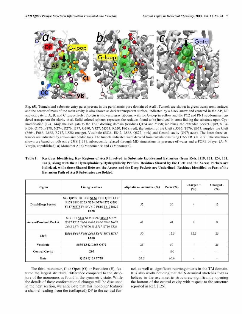
Fig. (5). Tunnels and substrate entry gates present in the periplasmic pore domain of AcrB. Tunnels are shown in green transparent surfaces and the center of mass of the main cavity is also shown as darker transparent surface, indicated by a black arrow and centered in the AP, DP and exit gate in A, B, and C respectively. Protein is shown in gray ribbons, with the G-loop in yellow and the PC2 and PN1 subdomains ren-dered transparent for clarity in a). Solid colored spheres represent the residues found to be involved in cross-linking the substrate upon Cys-modification [124, 144]: the exit gate to the TolC docking domain (residues Q124 and Y758; ice blue), the extended pocket (Q89, S134, F136, Q176, F178, N274, D276, I277, G290, Y327, M573, R620, F628; red), the bottom of the Cleft (D566, T676, E673; purple), the Cleft (F664, F666, L668, R717, L828; orange), Vestibule (S836, E842, L868, Q872; pink) and Central cavity (G97; azur). The latter three en-trances are indicated by arrows and bolded tags. The tunnels indicated were derived from calculations using CAVER 3.0 [205]. The structures shown are based on pdb entry 2J8S [133], subsequently relaxed through MD simulations in presence of water and a POPE bilayer (A. V. Vargiu, unpublished). a) Monomer A; b) Monomer B; and c) Monomer C.
Table 1. Residues Identifying Key Regions of AcrB Involved in Substrate Uptake and Extrusion (from Refs. [119, 121, 124, 131,
144]), Along with their Hydrophobicity/Hydrophilicity Profiles. Residues Shared by the Cleft and the Access Pockets are
Italicized, while those Shared Between the Access and the Deep Pockets are Underlined. Residues Identified as Part of the
Extrusion Path of AcrB Substrates are Bolded.
Region Lining residues Aliphatic or Aromatic (%) Polar (%) Charged +
(%)
Charged -
(%)
Distal/Deep Pocket
S46 Q89 S128 E130 S134 F136 Q176 L177
F178 S180 E273 N274 D276 I277 G290
Y327 M573 F610 V612 F615 F617 R620
F628
52 30 4 13
Access/Proximal Pocket
S79 T91 S134 S135 K292 M573 M575
Q577 F617 T624 M662 F664 F666 N667
L668 L674 T676 D681 R717 N719 E826
41 41 9 9
Cleft D566 F664 F666 L668 E673 T676 R717
L828
50 12.5 12.5 25
Vestibule S836 E842 L868 Q872 25 50 - 25
Central Cavity G97 - 100 - -
Gate Q124 Q125 Y758 33.3 66.6 - -
The third monomer, C or Open (O) or Extrusion (E), fea-
tured the largest structural difference compared to the struc-ture of the monomers as found in the symmetric state. While the details of these conformational changes will be discussed in the next section, we anticipate that this monomer features a channel leading from the (collapsed) DP to the central fun-
nel, as well as significant rearrangements in the TM domain. It is also worth noticing that the N-terminal stretches fold as helices in the asymmetric structures, significantly opening the bottom of the central cavity with respect to the structure reported in Ref. [125].

8 Current Topics in Medicinal Chemistry, 2013, Vol. 13, No. 24 Ruggerone et al.
Fig. (6). Residues in the AcrB periplasmic pore domain involved in substrate interaction based on computational studies [120]. a) Residues are shown in stick representation. The thickness of each residue is proportional to its frequency of binding contact to the tested sub-strates (minocycline, taurocholic acid, erythromycin, nitrocefin, chloramphenicol, ethidium, oxacillin, ciprofloxacin, NMP, PA N). The DP and AP, the PC1/PC2 subdomain entrance cleft, and the DP/AP interface are shown in transparent red, green, orange, and yellow surfaces. The G-loop is shown in yellow. Bold labels refer to residues involved in binding of more than three different substrates (inhibitors or not). The thick purple line roughly highlights the shared region of the binding site drawn according to this analysis. b) Histogram showing, for each residue (on the x axis), the number of substrates for which the contribution to the binding free energy by that residue is larger than kT 0.6 kcal/mol. Hydrophobic, polar and charged residues are identified by black, green, and red bars respectively. The sum over all frequen-cies is reported above each group of residues. Adapted from Ref. [120]. In addition to the DP present in the B monomer, a more proximal or access pocket (hereafter AP, Figs. 4a, 4c) was recently identified in the A monomer by two independent studies, publishing the structure of AcrB in complex with erythromycin and rifampicin [121], and a doxorubicin dimer [119]. The newly identified AP was located deeper into the PC1/PC2 cleft compared to the more peripheral binding site found in Ref. [123], and is separated from the DP by the G-loop [119-121] (Fig. 4a). The different binding poses found in the structures of AcrB co-crystallized with erythromycin, rifampicin and doxorubicin within the AP [119, 121] reveal its promiscous binding properties, shared with the DP and consistent with the broad specificity of AcrB. Importantly, ternary complexes with one substrate bound to the DP in the B monomer and a second to the AP of the A monomer within the same AcrB trimer were reported in Refs. [119, 121]. This finding is notable, as it has been postulated earlier on [62, 132] that allosteric bi-site activation of the AcrAB-TolC efflux pump might be essential for antiporter function-ing (we will return on this aspect in the next section).
The large size of the compounds (notice that doxorubicin bind as a dimer) bound to the AP led to the hypothesis that AcrB recognizes high molecular-mass (HMM) substrates via this binding site, while low molecular-mass (LMM) sub-strates are not recognized by the AP but bind directly to the DP [121]. The higher affinity of erythromycin to the access than to the distal pocket was confirmed by free energy and hydrophobic surface matching calculations performed on trajectories of AcrB-substrate complexes [120]. On the ex-perimental side, fluorescence-quenching experiments on AcrB variants bearing bulky side chain substitutions in the AP showed severe effects on doxorubicin efflux [121]. In addition, it was shown that the inhibition of doxorubicin ef-flux by erythromycin, occurring with wt AcrB, disappeared when residues involved in erythromycin binding were substi-tuted with bulky aminoacids. In contrast, the F610A muta-
tion in the DP reduced both doxorubicin and erythromycin resistance, and the efflux of the former substrate was signifi-cantly abrogated, as confirmed by two additional studies [137, 138].
The position and the flexibility of the G-loop seem to be important for the efflux of HMM substrates, as confirmed by several findings: a) A G616N AcrB variant deficient in mac-rolide transport [139] features a conformation of the loop in the A monomer resembling that of wt MexB (bearing N616 and unable to expel macrolides) in the same monomer [140] and to that of the B monomer in wt AcrB [119]; b) a compa-rable loop conformation was found in the X-ray structure of the G616P/G619P AcrB variant [121], which was designed to hinder conformational changes of the G-loop. This finding is consistent with a recent computational study highlighting the different flexibilities of wt and G616P/G619P loops [141]; c) The growth of mutants G616P/G619P and G614P/G621P was abrogated under sub-inhibitory concen-trations of doxorubicin or erythromycin, and this was cou-pled to impaired efflux of these substrates [121].
In summary, the broad specificity of AcrB is likely due also to the presence of two large multi-functional binding pockets separated by a flexible loop acting as a gate between them.
EXTRUSION OF SUBSTRATES BY ACRB: CHANGES IN PERIPLASMIC DOMAIN AND COU-
PLING TO PROTON MOTIVE FORCE BY PROTON
UPTAKE AND RELEASE IN THE TM DOMAIN
A mechanism for the extrusion of antibiotics by RND ef-flux pumps was firstly hypothesized in 1994, following the observation that good substrates of MexB were unable to penetrate the cytoplasm [55]. Nikaido and co-workers pro-posed a dual entrance model in which substrates are taken up from the periplasm (or the periplasm/IM interface) and from

RND Efflux Pumps: Structural Information Translated into Function Current Topics in Medicinal Chemistry, 2013, Vol. 13, No. 24 9
cytosol (or the cytosol/IM interface (Fig. 7a). This model was supported by several additional pieces of evidence. For example, Zgurskaya and Nikaido [86] showed that purified and reconstituted AcrB catalyzed the export of fluorescence-labeled phospholipids from within the bilayer. Additionally, according to Nikaido et al. [115], among -lactams only those with lipophilic side chains were efficiently pumped out by AcrB. On the other hand, aminoglycosides, which are more hydrophilic molecules compared to the substrates of AcrB, are expelled by RND transporters MexY [142] and AcrD [143]. Thus, another model might be needed to de-scribe the uptake of these compounds by RND transporters. It is worth noticing that the current hypothesis about sub-strates extrusion partly overlaps with the dual entrance model, as it assumes that compounds enter the transporter from the outer leaflet of the IM (through the TM7-9 groove) or directly from the periplasmic cleft [119, 121, 124, 144].
As pointed out in the previous section, a breakthrough in the understanding of mechanistic aspects of efflux by RND transporters arose in 2006/2007 with the publication of the asymmetric structures of AcrB [131-133]. It is believed that such structures represents the most populated conformation in the presence of substrates [62, 110, 119, 121, 145, 146] or inhibitors [18, 120], while the former symmetric structure might represent the “resting state” of the pump, preferred in the absence of substrates tightly bound to the protein [62, 65, 110, 134, 145].
On the basis of the X-ray structures, a “functional rota-tion” mechanism (Figs. 7b and 8) was postulated to explain drug export by AcrB, involving a concerted - but not neces-sarily synchronous [62, 111, 119] - cycling of the monomers through any of the asymmetric states A, B, C, and back to A. During a complete cycle ABC BCA CAB ABC occlu-sions and constrictions inside the pore domain propagate from external gates towards the central funnel (Fig. 5), driv-ing the unidirectional transport of substrate (hence the defini-tion of “peristaltic pump mechanism” [132]). Thus, guided translocation appears to be the mechanism of substrate trans-port within the AcrB monomers. This is compatible with the multi-site properties of the affinity sites discovered so far [119-121, 131], as also confirmed by a recent computational investigation of multi-functional sites in the asymmetric structure of the pump [136]. In Ref. [136] it was shown that binding sites are different in each of the AcrB monomers, implying that a drug avoids being trapped in one location through site-specific interactions. In addition, the authors shown that a complicated free-energy balance originating from weakly polar and weakly hydrophobic surroundings maintains substrates in the pockets, a finding confirmed for the DP by a recent computational study of the interaction between AcrB and series of its substrates [120].
In the A conformation, substrates are recruited from the periplasmic space (PC1/PC2 cleft) and/or the membrane (vestibule or central cavity) and bind a wide region that in-cludes the AP. More precisely, substrates partitioned in the outer leaflet of the IM can bind to the TM7-9 groove or to the entrance lined by G97 in the central cavity of the A and/or B monomers [62, 65, 73, 144]. More hydrophilic sub-strates located in the periplasm might bind to the PC1/PC2 cleft of monomers A and B [119, 121, 123, 124].
A concerted and consecutive drug uptake via the outer leaflet gates and subsequent transport to the cleft might be envisioned [119], but a recent computational study shows that periplasmic and TM7-9 gates appear to be quite specific for less and more hydrophobic/lipophilic compounds [147], respectively. In this respect, it is worth recalling that n-dodecyl- -D-maltoside was found to bind relatively tightly both to the TM7-9 groove in the A monomer and to the PC1/PC2 cleft in the B monomer in the highest-resolution X-ray structures reported so far [119, 133].
Along the A to B transition substrates move from the AP toward the DP, the second site being more hydrophobic than the former, consistently with the physico-chemical properties of substrates of the pump, which share a certain hydropho-bicity [119, 121] (Table 1). The G-loop was suggested to act as a gate between these two pockets, and indeed its flexibil-ity was shown to be crucial for the functioning of the pump [119, 121, 141]. Inhibitors were found to straddle this loop, which lead to the hypothesis of an action by hindering con-formational changes of the transporter and/or blocking the substrate pathway by binding to that region [120]. No major conformational changes in AcrB are apparent along the A B step of the cycle, except for a partial coil-to-helix tran-sition of TM8 and moreover a shift and a rotation of the PN2 subdomain, likely stabilized by the binding of substrates in the DP [131-133] (Figs. 8a-c).
Upon transition from the B to the C conformation, sub-strates are squeezed out from the binding pocket and they exit AcrB via its central funnel toward the TolC tunnel [131-133]. The squeezing traces back to the complete rewinding and to the kinking of TM8, which assumes a full helical structure compared to the coil and helix/coil conformations in A and B monomers. These conformational changes of TM8 are most likely due to the different protonation states of TM domain residues D407 and D408 in the respective monomers (Figs. 8d and 9). The recovery of helical confor-mation by TM8 coincides with a significant shift of the PC2 subdomain towards PC1 and towards the membrane plane. As a consequence, the PC1/PC2 cleft closes towards the pe-riplasm, thus blocking the entrance and the exit of substrates along this path (Figs. 8a, 8b, 8d). This movement is also due to the constraints imposed on PC2 by subdomain PN2, which moves after binding of substrates to the DP of monomer B (Figs. 8a, 8d). The shift of PC2 coincides with a tilting of subdomain PN1 (containing the pore helix N 2) by ~12° towards subdomains PN1 and PN2 of the adjacent B mono-mer and away from PN2. Consequently the movement of subdomain PN1 also opens a channel forming a substrate exit gate from the collapsed DP towards the central funnel lined by residues Q124, Q125 and Y758 and the OM channel TolC (Fig. 8d). Interestingly, this mechanism highlights the role of the flexibility in the region of pore helices N 2, and help explaining the outcomes of experiments on proteins bearing mutations thereon [148].
The displacement of the substrate out of the DP and to-wards the Gate to the central funnel (Figs. 5b, 5c) along B C has been supported by two computational studies [145, 149]. In Ref. [145] a coarse grain model of the AcrB pore domain (Fig. 3a) and of the substrate was employed to di-rectly observe extrusion of the latter during the B C con-formational transition. In Ref. [149] targeted MD simula-tions were performed on a full all-atoms model of AcrB in

10 Current Topics in Medicinal Chemistry, 2013, Vol. 13, No. 24 Ruggerone et al.
Fig. (7). Functional rotation mechanism of substrate extrusion by AcrB. a) Early schematic view of the tripartite AcrAB-TolC complex [65]. Note that amphiphilic drugs (empty and solid rectangles represent hydrophobic and hydrophilic parts of the molecule) are hypothesized to be captured at the interface between the IM and the periplasm or the cytoplasm. For the latter process, two possible pathways are envisaged (dashed arrows): Either the substrate is flipped over to the outer leaflet of the IM first and then follows the anticipated capture from the perip-lasmic side or it follows a different capture pathway from the cytosol; b) Schematic representation of the AcrB alternating site functional rotation transport mechanism. The conformational states A, B and C are colored blue, yellow and red, respectively. Upper panel: Side-view schematic representation of two of the three monomers of the AcrB trimer. AcrA and TolC are indicated in light green and light purple colors, respectively. Lower panel: The lateral grooves in the A (blue) and B (yellow) monomers indicate the substrate binding sites. The different geometric forms reflect low (triangle), high (rectangle), or no (circle) binding affinity for the transported substrates. In the first state of the cycle, a monomer binds a substrate (acridine) at the access site (AP in the A monomer), subsequently transports the substrate from AP to the DP (upon conversion to B monomer) and finally releases the substrate in the funnel toward TolC (C monomer). AcrA is postulated to partici-pate in the transduction of the conformational changes from AcrB to TolC, which results in the opening of the TolC channel and the facilita-tion of drug extrusion to the outside of the cell. Adapted from Ref. [132]; c) Schematic representation of the AcrB alternating site functional rotation transport mechanism extended by postulated intermediate steps. The lateral grooves in A and B monomers indicate the substrate binding sites. The different geometric forms reflect low (triangle), high (rectangle), or no (circle) binding affinity for the transported sub-strates. State BBB is postulated to occur at high substrate concentration, while AAA and AAB are postulated to occur in the absence or at low substrate concentrations. Adapted from Ref. [62]. complex with doxorubicin bound to the DP, such as to mimic the B C step of the functional rotation. Although full extrusion was expectedly not seen within the relatively short times affordable by such simulations, a significant dis-placement of almost 10 Å was observed towards the exit gate. Interestingly, doxorubicin remained stuck in front of the exit gate during a series of targeted MD performed to mimic five complete cycles of the functional rotation (Var-giu et al., unpublished data). Despite the limitations intrinsic to the methodology and the lack of AcrB partners in these studies, this finding is interesting as it might be indicating additional factors necessary for complete extrusion like e.g. the presence of more than one substrate inside the tunnel
system as suggested recently (see Fig. 15 in Ref. [111]). In Ref. [149] it was also shown that detachment of the antibi-otic from the binding site occurred with a zipper-like squeez-ing of the pocket. Moreover, the concerted opening of the channel between the doxorubicin pocket and the Gate was shown to be necessary in order to displace the ligand.
More in general, the importance of the flexibility of the AcrB trimer has been emphasized in a recent computational study of the transporter [134], showing clear opening and closing motions of the AP in A and B monomers, and of the exit gate in the C monomer. It appeared therefore from this study that each of the known three reaction cycle intermedi-ates, as deduced from crystallographic studies, can adopt

RND Efflux Pumps: Structural Information Translated into Function Current Topics in Medicinal Chemistry, 2013, Vol. 13, No. 24 11
Fig. (8). Conformational differences between the three AcrB monomers in the asymmetric structure (PDB code: 4DX5 [119]). a) Top view comparison between the asymmetric and the symmetric pore domains of AcrB. The symmetric structure is shown in transparent silver, while the asymmetric structure is superimposed in solid colors (monomers A, B and C are colored cyan, yellow and red respectively). Subdomains PN1, PN2, PC1 and PC2 of the pore region are labeled in the A monomer. Also the cleft of the A monomer and the vestibule between A and C are indicated. Major structural differences between the asymmetric and symmetric pore domain structure are shown with blue arrows. The size of the arrows is proportional to the RMSD calculated for the superimposed structures; b) Superimposition of the asymmetric and the symmetric TM domains of AcrB. Helices lining up this domain are labeled in the A monomer; c) Relevant conformational changes along the A B step of the functional rotation. No significant changes are seen in the TM domain (first panel), where the tight salt bridge between K940 and D407/408 is maintained. However, a partial winding of the TM helix 8 is clearly seen in the second panel, which leads to the open-ing of the vestibule entrance of substrates (residues lining the vestibule and the bottom of the cleft are shown with pink and purple spheres, respectively). The third panel shows how substrate binding to the distal pocket (red spheres from the pocket belonging to PN1 or PN2 are shown) relocates the PN2 subdomain from PN1, which leads to further opening of the external cleft (orange spheres indicate residues lining that region); d) The salt-bridge between K940 and D407/D408 in the TM region are disrupted in monomer C (first panel), and this is associ-ated to a further coil-to-helix transition of the TM helix 8 (second panel), which leads in turn to the closure of the vestibule and to a signifi-cant rototranslation of the PC2 domain. This structural change “drags” the upper part of the PN1 subdomain away from PN2, leading to the opening of the exit gate towards the funnel and TolC and simultaneous closure of the DBP, due to the release of the substrate (third panel). The movement of the PC2 domain induces also the closure of the external cleft in addition to the vestibule (fourth panel); e) The salt bridge between K940 and D407/D408 in the TM region are reestablished in the A monomer (first panel), and TM helix 8 undergoes a backward helix-to-coil transition which partially opens the vestibule and causes a rotation and a shift of PC2 subdomain (second panel). This movement induces reorientation of the PN2 subdomain so that the N 2’ helix (PN1 subdomain) closes the exit gate towards the funnel in the TolC dock-ing domain (third panel). The movement of subdomain PC2 also reopens the external cleft between the PC1 and PC2 subdomains (fourth panel).

12 Current Topics in Medicinal Chemistry, 2013, Vol. 13, No. 24 Ruggerone et al.
different conformations maybe indicating intermediate con-formations between the three known states A, B and C. The authors of Ref. [134] speculate furthermore that AcrA could enhance the activity of the pump by stabilizing sub-strate-accessible conformations, for instance by stabilizing PC1 and PC2 subdomain orientations. The importance of conformational coupling among the three monomers has been further assessed by an evaluation of the collective mo-tions in AcrB [150]. However, as the authors pointed out, the collective motions represent the intrinsic conformational flexibilities that encode the allosteric couplings of the protein assembly. Their methodology indeed cannot elucidate the local conformational motions due to external factors such as substrate and protein partner binding or protona-tion/deprotonation events).
In addition to completely asymmetric conformations, the presence of intermediate states bearing more than one monomer in the A or B conformation, such as BBC or BBB, has been described by several authors [62, 111, 119], and supported by experiments [133, 146, 151] and computer simulations [145]. This might lead to a more complex mechanism for the extrusion of substrates, perhaps involving bi-site activation (Fig. 7c). For instance, the interaction of a substrate with the A monomer in the presence of a second substrate in the B monomer could be the prerequisite for the conformational change of the latter into the C state, associ-ated with release of the substrate in the TolC docking do-main funnel. Consistent with this more complex view are the recently observed strong cooperative kinetics of the extru-sion of -lactam antibiotics by the AcrAB-TolC system [152, 153], as well as the stimulation of cephalosphorin efflux by some AcrB substrates [154]. Moreover, the recent X-ray structures (discussed in the previous section) featuring si-multaneous binding of substrates to the A and B monomers also support the possibility of cooperative mechanisms [119, 121]. The interdependence of monomers has been confirmed by cross-linking experiments [151, 155] and by the use of an AcrB trimer with covalently linked monomers [156]. Moreover, the PN1 domain has been indicated as the “ratchet pin” for the transmission of these conformational changes [132].
As briefly mentioned in the description of the structural changes accompanying the B to C transition, the electro-chemical proton gradient across the IM is most likely the driving force for the aforementioned conformational changes leading to uptake and extrusion of substrates in the periplas-mic domain of AcrB. The role of the proton-motive force on the drug efflux activity has been shown via the effect of un-couplers and ionophores in intact cells [87] and also with AcrA/AcrB reconstituted in liposomes using artificial proton motive force [86].
The flux of protons from the periplasm to the cytoplasm most likely involves rearrangements in the TM helices [130-133, 157]. Protonation and deprotonation events, involving D407 and D408 primarily but also K940 and R971, induce conformational changes propagating towards the periplasmic domain (Figs. 8d and 9). It is believed that the B to C transi-tion is the energy-demanding step of the cycle, with 2 pro-tons being necessary to complete one cycle from A to B to C and back to A per monomer and per substrate transported
(Fig. 9) [62, 65, 73, 110, 119, 133]. Once a substrate is bound to the DP in the B monomer, a conformational change propagates from PN2 to the TM domain, opening the path towards D407 and D408 for protons. Recent MD simulations by Fischer and Kandt [158] confirmed the existence of up to three connections to bulk water in the periplasm and one to bulk water in the cytoplasm in monomers A and B, while no connection was found in monomer C. Moreover, a signifi-cantly larger and persistent hydration of the region contain-ing the residues of the proton-relay pathway is seen in monomer B. Protonation of gating residues D407 and/or D408 was suggested to be the key step of the reaction [130-133, 159]. In particular, D408 was shown to specifically re-act with dicyclohexylcarbodiimide (DCCD) in a pH-dependent manner [160]. The apparent pKa of 7.4 at D408 would enable binding and release of protons under physio-logical conditions. In contrast to other secondary transport-ers, D408 was not protected by substrates against modifica-tion, which supports the notion of spatially separated sub-strate and proton transport pathways. In Ref. [158] the pro-ton uptake event was also suggested to occur in the A or B state, or during a previously unknown intermediate in be-tween B and C where cytoplasmic water access is still possi-ble.
This flux of protons across the membrane induces a con-formational change in the TM8 that propagates to the pore domain, inducing the B C transition which lowers the af-finity of the substrate to the DP and opens the Gate to the central funnel. The proton release event was suggested in Refs. [62, 158] to involve R971 in the C intermediate.
SUMMARY OF SUBSTRATE UPTAKE AND EXTRU-SION BY ACRB: LMM VS HMM COMPOUNDS
Summarizing the data reported so far, two slightly differ-ent mechanisms of extrusion by AcrB can be envisaged for LMM and HMM substrates. Concerning HMM substrates, after entry AcrB from the PC1/PC2 cleft or the vestibule, they bind to the AP in the A monomer and are then translo-cated into the DP upon the A to B transition by a peristaltic mechanism involving subdomain movements that include a shift of the G-loop. These data clearly points to the impor-tance of conformational changes of the G-loop coupled to translocation HMM substrates from the access to the distal pocket.
On the contrary, LMM drugs could also travel through the AP until they reach the DP of the B monomer. This is consistent with recent coarse-grained molecular simulations showing that most of the uptake events of relatively small substrates by AcrB occur from the B monomer [147], and with all-atom MD simulations showing the entrance of sev-eral solvent molecules into the DP from the cleft of the B monomer (Vargiu, Ruggerone & Nikaido, in preparation). Such a mechanism might also help to rationalize the binding of a dodecyl- -D-maltoside molecule to the cleft of the B monomer (Figs. 4a, 4d) reported in Ref. [119]. Indeed, a maltoside molecule is quite stretched and has a molecular mass similar to taurocholic acid, which was also shown to bind the DP using a computational approach [120]. There-fore, maltoside could in principle be transported from the access to the distal pocket without the need for significant

RND Efflux Pumps: Structural Information Translated into Function Current Topics in Medicinal Chemistry, 2013, Vol. 13, No. 24 13
Fig. (9). Geometry of the essential residues D407, D408, K940, R971, and T978 in the transmembrane domain of the AcrB in the A (a), B (b), and C (c) monomers. The putative protonation state of these side chains in each monomer is indicated. Proton uptake is anticipated in the B monomer and is postulated to lead to the side chain reorientation of D407, K940 and R971. These conformational changes appear to be correlated to the coil-to-helix transition and the PN1/PC2 subdomain movement seen in the periplasmic pore domain during the B to C transi-tion. Adapted from from Ref. [62]. rearrangements of the G-loop, as it has been found, although in the reverse direction, for the LMM inhibitor 1-naphthylmethyl-piperazine (NMP, Fig. 2) in recent molecu-lar dynamics simulations [120]. Moreover, as reported in the next section, despite for levofloxacin being a LMM com-pound, its wide planar 4-ring structure could hinder its smooth diffusion through the AP-to-DP channel of the B monomer. Thus, the shape of each compound could be a parameter as important as its molecular mass in discerning among the interaction routes with AcrB.
All substrates, of any mass and shape, should then be ex-pelled from the DP towards the funnel in the TolC docking domain along the B to C step of the cycle.
STRATEGIES TO OVERCOME EFFLUX-MEDIATED MDR
Two main strategies are being investigated nowadays to overcome resistance mediated by bacterial efflux pumps [10, 34, 67, 106, 116, 161-170]. The first strategy consists in by-passing efflux pumps by improving the molecular design of existing antibiotics or by designing new ones, with the pur-pose of altering the physico-chemical properties for recogni-tion by efflux transporters. It is worth noticing that despite some new molecules less amenable for efflux have been produced, their design has been far from trivial. Examples are third and fourth generation quinolones, ketolides or gly-cylcyclines (see e.g. Ref. [64] for a review). However, even for these new compounds resistance has been described very shortly after their deployment [48]. A second strategy con-cerns the inactivation of efflux pumps, which is highly desir-able for several reasons [162] as to:
• Increase the intracellular concentration of antibiotics, thus enhancing the therapeutic index.
• Decrease the intrinsic bacterial resistance to antibiotics, often caused by efflux pumps.
• Reverse the acquired resistance associated with efflux pump overexpression.
• Reduce the frequency of the emergence of highly resis-tant mutant strains. Efflux pumps often provide the initial means for resistance, and causing survival of bacteria en-able them to acquire other mechanisms of resistance like target-based mutations [163, 164, 166, 167, 169, 171].
• Prevent the export of virulence factors synthesized by microbes, thus inhibiting invasiveness and consequently the rise of bacterial infection [116, 172].
Inhibition of MDR pumps, specifically of RND pumps from Gram-negative bacteria, is a relatively new and dy-namic field of intense research. In the last two decades a large number of reviews has been written on the subject, to which the reader is referred for a more comprehensive view [10, 67, 106, 116, 161-170]. While the following list of in-hibitors describes several possible modes of inactivating efflux pumps, the focus here will be on RND efflux pumps, and in particular on pharmacological inhibitors interacting with E. coli AcrB for which structural information is avail-able:
1. Inhibitors of proton motive force. Compounds that affect the energy gradient across the bacterial membrane such as carbonylcyanide m-chlorophenylhydrazone (CCCP), valinomycin, dinitrophenol (DNP), phenothiazines such as promethazine [165] are used to completely abolish the efflux of all toxic compounds [162, 166, 167]. Unfortu-nately, these inhibitors have a very general mode of inac-tivation, not only affecting efflux pumps but the entire energetics of the cell, including eukaryotic cells which makes them less attractive for clinical use [10, 116, 161, 162].
2. Biological Inhibitors. This class of inhibitors affects ef-flux pump activity by blocking either the proteins them-selves, e.g. with neutralizing antibodies, or by inactivat-ing the genes encoding the pumps, by means of antisense oligonucleotides or small interfering RNAs, or other non-traditional antisense molecules, which can interfere with transcription of the gene or translation of the encoding mRNA. The antisense approach has been shown to work

14 Current Topics in Medicinal Chemistry, 2013, Vol. 13, No. 24 Ruggerone et al.
for AcrAB efflux pump in E. coli and has also been pat-ented [173, 174], but its application can be broadened to every pump of known sequence or to genes encoding proteins involved in the regulatory mechanism of pump expression (such as the Mar regulator [175]).
3. Pharmacological Inhibitors, also known as Efflux Pumps Inhibitors (EPIs). These are chemical compounds used: i) to interfere with the assembly or function of efflux pumps [176] (for instance, in the case of the tripartite RND systems, blocking of the OM channel may lead to the inhibition of efflux pump activity [177]); ii) in com-bination therapy to increase the antibiotic concentration inside a pathogenic cell [10, 67, 116, 161, 162]. EPIs of this latter class act by competitive/non-competitive inhi-bition of the pump, rendering antibiotics more effective, and might prevent accumulation of other resistance mechanisms over time. In addition to therapeutic use, specific EPIs can be employed for diagnostic purposes to evaluate the presence and contribution of the efflux mechanism in any given pathogen.
Here we focus on the class of EPIs for use in combina-tion therapy, summing up the ideal EPI characteristics [116, 163]:
1. The EPI should enhance the activity of multiple sub-strates of the pump, and should not increase the activity of antibiotics that are not substrates, in order to guarantee its specific action against the target efflux pump.
2. If the EPI should inhibit a specific pump of a given pathogen, cross-inactivation of other strains should be avoided. Moreover, the inhibitor should be free of any pharmacological activity on eukaryotic cells. This is true for PA N (vide infra), which is not recognized by eu-karyotic (efflux) transporters [161].
3. The EPI should be structurally stable to ensure enhanced serum levels and cellular accumulation that potentiates its activity in intracellular infections.
4. The EPI should have enhanced therapeutic index (or be atoxic) and pharmacokinetic profile to ensure maximum specificity and efficacy. In particular, the profile should be optimized in parallel to that of the antibiotic to be used in combination therapy. Toxicity is still a problem for several lead EPIs, and deserves special attention as EPIs might be used at high concentration in humans.
5. The EPI itself should be ideally devoid of antibacterial activity as this could lead to development of resistance mechanisms against the EPI. Unfortunately, despite ef-forts at matching this requirement, resistance induced by EPIs has already been reported in the literature ([178], and vide infra).
In the following we describe some EPIs for which struc-tural information of their interaction with AcrB, either from experimental or computational works, has been provided.
Peptidomimetic EPIs
These include the well-known PA N (Fig. 2) [179, 180] and its derivatives MC-02,595, MC-04,124, MC-510,051, MP 601,205 [164, 181], which were the first EPIs ever iden-
tified and have been a valuable tool for drug discovery. PA N was identified by assaying an array of synthetic and natural compounds against Pseudomonas aeruginosa strains over-expressing three MDR efflux pumps (MexAB-OprM, MexCD-OprJ, MexEF-OprN) in the presence of levoflox-acin. The magnitude of the potentiating effect was strongly dependent on the nature of the particular substrate, perhaps indicating that different antibiotics may have different bind-ing sites on AcrB and that inhibition by PA N is binding site-specific [180].
The attractiveness of PA N is based on its broad-spectrum efflux pump inhibitory activity, necessary to have clinically significant impact on fluoroquinolones, which are extruded by many efflux pumps. Furthermore, a major bene-fit of inhibition of multiple efflux pumps was a dramatic decrease in the frequency of emergence of P. aeruginosa strains with clinically relevant levels of resistance to fluoro-quinolones [164, 180]. Resistant P. aeruginosa mutants with an MIC of 1 μg/ml for levofloxacin were isolated with a fre-quency less than 10-11 (versus 10-7 in the absence of efflux pump inhibitors). These data are particularly important in view of results indicating that under stress conditions, such as those in acute clinical infections, there is an increased frequency of selection of hypermutable strains of P. aerugi-nosa [182].
In addition to the inhibitory effect on the Mex pumps of P. aeruginosa described above, PA N has been validated against the AcrAB-TolC in Klebsiella pneumoniae, E. coli, Salmonella thyphimurium and Enterobacter aerogenes [183-186], and in multiple homologous systems present in Acinetobacter baumannii [187, 188], Campylobacter jejuni and Campylobacter coli [189, 190].
Mechanistically, it has been proposed that PA N itself is an RND efflux pump substrate that acts as a competitive inhibitor by binding to the antibitoic binding pocket inside the RND component [164, 167, 191]. Consistent with this hypothesis are results of a recent docking survey of substrate and inhibitor binding sites within AcrB, showing that PA N binds indeed with the highest affinity to the DP of the trans-porter [135]. Furthermore, on the basis the distinction be-tween groove, cave, and mixed binders (see above), the authors furnished an explanation for the less efficient inhibi-tion of ethidium and carbenicillin efflux (compared to levofloxacin) by PA N. According to their hypothesis, PA N is able to inhibit the efflux of levofloxacin by compe-tition, because both compounds bind to the groove of the DP (although PA N was classified as mixed binder, it was bound mainly to the groove). In contrast, ethidium and car-becinillin were classified as cave binders, so competition with PA N should not occur.
However, in a follow-up study including docking and MD simulations [120] the distinction between groove and cave binders became blurred. In particular PA N changed significantly its mode of binding with respect to that found by docking, and moved towards the lower part of the DP. Moreover, this binding pose partly overlaps with the binding site of ethidium (Fig. 10; levofloxacin and carbenicillin were not included in the list of substrates studied in Ref. [120]), and also the calculated affinities of this compound and

RND Efflux Pumps: Structural Information Translated into Function Current Topics in Medicinal Chemistry, 2013, Vol. 13, No. 24 15
Fig. (10). Comparison between the binding modes of PA N, ethidium (ETH), taurocholic acid (TAU), and 1-naphthylmethyl-piperazine (NMP) on the DP and AP (NMP’) of AcrB. Ligands are shown in colored spheres according to atom types (with nonpolar hydrogens re-moved). Transparent surfaces indicate the DP (transparent red surface), the AP (green surface) pockets, and the PC1/PC2 subdomain cleft (orange surface). The G-loop is shown in gray. Residues that are within 3.5 Å from the ligand are shown as beads (red, green, orange, and yellow for those of DP, AP, PC1/PC2 subdomain cleft, and G-loop, respectively). Residues shared by the AP and DP are colored blue. For orientation, residues Q124 and Y758 defining the exit gate (far away from the ligand) are shown as gray beads. Adapted from Ref. [120]. PA N were similar. An interesting difference was found instead in the contribution to the stabilization of the ligand-AcrB adducts from residues belonging to the DP (Tables 1 and 2). Indeed, with ethidium (as well as with other sub-strates not inhibiting the pump) these contributions were always significantly larger than with inhibitors such as PA N (Table 2). This finding could help explaining the inhi-bition by PA N, if tight and specific interactions of sub-strates with the DP (not achieved in the B monomer by PA N) are necessary to trigger functional rearrangements of AcrB. Moreover, PA N was found to straddle the G-loop separating the distal and proximal pockets, and whose flexi-bility was shown to be important for the functioning of the pump [119, 121, 141], likely by regulating the smooth trans-location of substrates between the two affinity sites along the A B step of the functional rotation. Thus, binding of PA N along the extrusion channel on top of the G-loop could effec-tively block substrates while also hindering key conforma-tional changes of the pump in some fashion.
According to this hypothesis, we propose that ethidium and carbenicillin do not compete with PA N and their efflux is largely unaffected because these substrates may reach the DP by entering from the PC1/PC2 cleft of the B monomer itself, or because they have a larger affinity than PA N to this pocket. In agreement to this hypothesis are the recent findings by Yao et al. [147] and by some of the authors (Vargiu, Ruggerone & Nikaido, unpublished data), demon-strating that access to the DP from monomer B could indeed be possible at least for LMM compounds and solvents. In this respect, despite for levofloxacin being a LMM com-pound, its wide planar 4-ring structure could hinder its smooth diffusion through the AP-to-DP channel of the B monomer (Fig. 5).
The binding of PA N to the AP was not investigated in any of the aforementioned studies, but it cannot be excluded that this inhibitor is binding to this access site. For instance, very strong inhibition of clarithromycin and rifampin [164] efflux is observed in the presence of PA N. These HMM antibiotics are supposed to bind with high affinity to the AP of the A monomer [119, 121]. On the other hand, efflux of linezolid or chloramphenicol, i.e. LMM compounds similar to ethidium and carbenicillin, is hardly affected in presence of Pa N [163, 164]. Clearly, these observations are not ex-
clusively directing towards inhibition based on binding of the inhibitor to more than one site, since e.g. erythromycin is still expected to be transported via (part of the) DP in order to move towards the exit tunnel. Docking and MD studies are ongoing in our lab as to assess the likelihood of these additional mechanisms (Vargiu et al., unpublished data).
The major problem with PA N is its toxicity towards human cells, which has prevented its clinical application [164]. For this reason significant efforts have been devoted to the optimization of PA N and its derivatives, which re-sulted in the lead compound MC-04,124, exhibiting higher stability and reduced toxicity in experimental infections us-ing an efflux pump overexpressing strain of P. aeruginosa [181].
Besides broad-spectrum inhibitors such as PA N, various peptidomimetics have been identified which are specific for each of the MexAB-OprM, MexCD-OprJ or MexEF-OprN tripartite pumps [163]. As mentioned above, while pump selective inhibitors may have limited therapeutic use, they may prove valuable for studying the contribution and preva-lence of specific efflux pumps in clinical isolates of Gram-positive and Gram-negative bacteria. This approach has re-cently been successfully applied to P. aeruginosa and al-lowed straightforward identification of strains over-expressing efflux pumps [192].
Piperazines
Several arylpiperazines have been shown to reverse MDR in bacteria overexpressing the AcrAB-TolC and AcrEF-TolC efflux systems. Some anthrylpiperazines and naphthylpiperazines, notably 1-naphthylmethyl-piperazine (NMP, Fig. 2), are among the most potent unsubstituted arylpiperazines, with a minimal effective concentration and a dose-dependent ability to increase the intracellular concen-tration of several antibiotics, such as linezolid, chorampheni-col, tetracycline, macrolides, and fluoroquinolones [193, 194]. NMP seems to be effective in A. baumannii, several Enterobacteriaceae, but not in P. aeruginosa [187, 193-195]. The list also includes trimethoprim and epinephrine [196], indole derivatives and quinolone derivatives [197]. Due to the serotonin agonist properties of these compounds, they are likely to be too toxic for use in humans.

16 Current Topics in Medicinal Chemistry, 2013, Vol. 13, No. 24 Ruggerone et al.
Table 2. Affinities of Selected Substrates and Inhibitors to AcrB, Together with Contributions to the Affinity from Residues Be-
longing to the DP, the AP and the G-Loop. The Hydrophobic Match is a Fraction Over the Whole Solvent Accessible Sur-
face [203], and the Free Energies (Standard Deviations in Parentheses) are in kcal/mol. From Ref. [120].
Compounds ETH PAbN TAU NMP
Hydrophobic Match 0.90 0.68 0.78 0.77
G (no Sconf) 31.0 (2.1) 30.1 (5.3) -23.3 (4.6) -22.3 (2.5)
% GDP 41 29 52 20
% GAP - - - -
% GG-loop - - - 6
As has been shown for PA N, NMP decreases MIC val-
ues specifically for some of the drugs used against an AcrB-overproducing E. coli [193]. For instance, while the AcrB-mediated efflux of chloramphenicol and of linezolid seems to be completely inhibited by NMP, clarithromycin efflux appears to be affected to a much lower extent.
The mechanism of inhibition by NMP has not been un-veiled yet. Some authors report that NMP is not a substrate of RND pumps [137], while recent computational studies found a good affinity to the B monomer of AcrB [120, 135]. In particular, in one study [120] it was shown that NMP in-teracts and straddle the G-loop from either the DP or the AP side (Fig. 9 and Table 2). It was therefore suggested that, like PA N, the binding of NMP to a sub-pocket different than the DP or AP region might inactivate the pump by blocking the passage of antibiotics and hindering functional rearrange-ments in the structure of AcrB [120].
The analysis of available structural data suggests a possi-ble mechanism for the different extent of NMP inhibition of AcrB-mediated efflux of chloramphenicol and linezolid on one side and clarithromycin on the other side. Indeed, it was found by computers simulations that NMP binds with high affinity to the lower part of the DP of the B monomer, partly overlapping with the binding site of chloramphenicol [120] (and probably that of linezolid). On the contrary, it is hard to hypothesize for NMP to have a larger affinity than clarithromycin towards the AP of the A monomer, where the latter antibiotic putatively binds first when sequestered by AcrB.
Pyridopyrimidines
A series of pyridopyrimidine derivatives were shown to be specific inhibitors of AcrAB-TolC and MexAB-OprM [198]. In contrast to PA N, they do not cause membrane dysfunction and have almost no antibiotic activity [199, 200]. However, because these compounds do not inhibit MexXY–OprM, these are less useful as clinical drugs to tar-get MDR in P. aeruginosa. Among the various pyri-dopyrimidines, [{2-[({[(3R)-1-{8-[(4-tert-butyl-1,3-thiazol-2-yl)carbamoyl]-4-oxo-3-[(E)-2-(1H-tetrazol-5-yl)ethenyl]-4H-pyrido[1,2-a]pyrimidin-2-yl}piperidin-3-yl]oxy}carbo nyl)amino]ethyl}(dimethyl)ammonio]acetate (D13-9001, Fig. 2) is an AcrB- and MexB-specific inhibitor and exhibits potent efficacy in vivo, is highly soluble, and shows a good
safety profile in an acute toxicity assay [201]. In addition, D13-9001 is not exported by efflux pumps [200].
The structure of D13-9001 bound to AcrB and MexB (the first inhibitor-RND efflux pump structure) has been pub-lished very recently [18]. D13-9001 binds to the DP of both AcrB and MexB, but unlike as with the co-crystal structures with doxorubicin or minocyclin, one part of the inhibitor molecule, the pyridopyrimidine ring moiety, inserts into a hydrophobic phenylalanine-rich cage branching off from the substrate-translocation channel (Fig. 11a). This cage is lined by residues F136, F178, F610, F615 and F628 in AcrB, and in addition also lined with F573 in MexB. In the case of MexY, the position of F178, lying in AcrB and MexB at the edge of the hydrophobic trap, is occupied by a voluminous W177 side chain, which prevents the binding of the inhibi-tor. The authors of Ref. [18] also solved the structure of the F178W variant of AcrB, and revealed that the indolyl side chain of W178 hinders D13-9001 binding, as is the case for MexY. Moreover, by using microbiological and uptake as-says they demonstrated that AcrBF178W is not inhibited by D13-9001, while MexYW177F is susceptible to this compound in a similar way as wt AcrB and MexB. Interestingly, the activity of D13-9001 on MexBF178W is similar to that on wt MexB, at odd with the findings for AcrB. This apparent dis-crepancy was rationalized by the comparison of the X-ray structures of both mutants, showing that W178 protrudes into the narrow space of the hydrophobic trap in AcrB, but not in MexB. Indeed, the authors of Ref. [18] also resolved the structure of MexBF178W in complex with D13-9001, which was found within the hydrophobic trap as for the wt transporter. This work highlights how minimal changes in the conformation of the pocket drastically affect the affinity of a compound towards multi-functional sites of RND trans-porters, and how this reflects directly on the biological activ-ity of the substrate or inhibitor.
On the basis of their findings, Nakashima et al. [18] sug-gested the D13-9001 works by tightly binding to the hydro-phobic trap and hindering the functional rotation mechanism of AcrB/MexB monomers. Consistently with this hypothesis, the inhibitor potentiates the activities of all of the antibiotics exported by AcrB/MexB, irrespective of their affinity to the deep or proximal access pockets.
Interestingly, this observation resembles quite closely the hypothesis suggested in Ref. [202] for the impaired efflux of doxorubicin and many other compounds in F610A variants

RND Efflux Pumps: Structural Information Translated into Function Current Topics in Medicinal Chemistry, 2013, Vol. 13, No. 24 17
Fig. (11). Binding of inhibitor D13-9001 in the DP of AcrB on basis of the structure of the complex published in Ref. [18] (a), compared to the binding of doxorubicin in the DP in the wild type (b; X-ray structure of PDB entry 4DX7[119]) and F610A variant of AcrB (c; from MD simulations in Ref. [202]). a) The inhibitor is shown in colored stick representation according to the atom type, while phenylalanine side chains F136, F178, F610, F615 and F628 are shown in thinner light purple sticks. The two yellow lines enclose schematically the extrusion channel and the yellow arrow indicates the direction of efflux. The blue line delimits the hydrophobic space accomodating the tert-butyl thia-zolyl aminocarboxyl pyridopyrimidine (TAP) moiety of inhibitor D13-9001. b) Doxorubicin bound to the DP of AcrB Phenylalanines F178 and F615 are within 3.5 A of the ligand (thick sticks). c) Doxorubicin sliding within the F-rich cage in the F610A variant. The position of the drug in wt AcrB is shown in thin gray sticks to highlight the reorientation and embedding of the antibiotic within the hydrophobic trap in the F610A variant. of AcrB [137, 138]. In that report several MD simulations of doxorubicin in complex with the AcrBF610A were performed, and the binding to the DP was compared between the wt and mutant protein. It was found that, compared to the binding in the wt AcrB (Fig. 11b, notice that only F178 and F615 are within 3.5 Å of the ligand) the F610A variant allowed slid-ing of doxorubicin by 4/5 Å within a pocket lined by F136, F178, F615 and F628, now interacting more tightly with the antibiotic (Fig. 11c). In Ref. [202] it was suggested already that the inhibitory effect associated to the F610A mutation was likely due to the increased dwelling time and affinity of substrates in the binding pocket of the AcrB variant. Thus, the mutation can lead to a competitive inhibition by either hindering conformational changes in the pump or interfering with the extrusion of substrates due to the observed binding to the remote hydrophobic pocket area, which can normally not be accessed due to steric hindrance of F610.
SUMMARY AND FUTURE DIRECTIONS
Since the discovery of the first bacterial multi-drug transporter, the research in the field of efflux pumps has made great strides. Biological, biochemical, structural and computational, studies have revealed many aspects of the mechanisms behind multi-drug recognition and extrusion. In this review, we summarized the main outcomes from these studies focusing on the best known RND transporter AcrB, by outlining the key steps of substrate uptake, recognition and extrusion catalyzed by this drug transporter.
Despite the advances outlined in the review, dissecting the molecular and conformational steps that regulate trans-port of substrates by these pumps remains a very challenging task, which is increasingly needed for a complete under-standing of mechanistic and structure/function relationship of the drug efflux process. Regarding the structural proper-
ties, new crystallographic studies and/or more accurate mod-eling techniques are needed in order to achieve a full atomic picture of the entire tripartite complexes, which is required to understand the functional dynamics of this machinery in re-lation to substrate efflux. In turn, such knowledge will allow for a clearer interpretation of biological experiments, i.e. it will link experiments on the molecular level (including sin-gle molecule assays) to those conducted on the intact cell (i.e.containing an ensemble of efflux pumps). This informa-tion can be directly transferred to applied research aimed at the development of inhibitors against efflux transporters or of antibiotics less susceptible to efflux in Gram-negative bacteria.
CONFLICT OF INTEREST
The author(s) confirm that this article content has no con-flicts of interest.
ACKNOWLEDGEMENTS
A. V. V. is grateful to H. Nikaido for useful discussions about bacterial resistance in connection to efflux mecha-nisms. A. V. V. and P. R. have received support from the Innovative Medicines Joint Undertaking under Grant Agreement n°115525, resources which are composed of fi-nancial contribution from the European Union’s seventh framework, as part of the Project TRANSLOCATION (http://www.imi.europa.eu/content/translocation). A. V. V. and P. R. acknowledge for their research computational re-sources from CINECA (Bologna, Italy; PRACE 4th-Call Grant Pra04_774, and ISCRA-A Grants HP10ARSZ26, HP10ARS6ZX, and HP10ANF5M6) and from the Lawrence Berkeley National Laboratory (Berkeley, CA). Research in the lab of K.M.P. is funded by the Innovative Medicines Initiative (IMI) Joint Undertaking project Translocation, the
18 Current Topics in Medicinal Chemistry, 2013, Vol. 13, No. 24 Ruggerone et al.
DFG-EXC115 (Cluster of Excellence Macromolecular Complexes), the Deutsche Forschungsgemeinschaft SFB 807, and by grants from Europe Advancing Science through Pfizer - Investigator Research Exchange (ASPIRE) and Hu-man Frontier Science Program. Research in the lab of S. M. is funded by the Funding Program for Next Generation World-Leading Researchers from Japan Society for the Pro-motion Science, ERATO Murata Lipid Active Structure Pro-ject, Japan Science and Technology Agency, Advanced Re-search for Medical Products Mining Program of the National Institute of Biomedical Innovation (NIBIO), Japan and Hu-man Frontier Science Program (HFSP).
REFERENCES
[1] French, G.L. The continuing crisis in antibiotic resistance. Int. J.
Antimicrob. Agents, 2010, 36, S3-S7. [2] Tam, V.H.; Rogers, C.A.; Chang, K.-T.; Weston, J.S.; Caeiro, J.-P.;
Garey, K.W. Impact of multidrug-resistant Pseudomonas aeruginosa bacteremia on patient outcomes. Antimicrob. Agents
Chemother., 2010, 54, 3717-22. [3] The endless struggle. Lancet Infect. Dis., 2011, 11, 253. [4] Ereqat, S.; Spigelman, M.; Bar-Gal, G.K.; Ramlawi, A.; Abdeen, Z.
MDR tuberculosis and non-compliance with therapy. Lancet Infect.
Dis., 2011, 11, 662-662. [5] Pitout, J.D.D. The latest threat in the war on antimicrobial
resistance. Lancet Infect. Dis., 2010, 10, 578-9. [6] Arias, C.A.; Murray, B.E. Antibiotic-Resistant Bugs in the 21st
Century -- A Clinical Super-Challenge. N. Engl. J. Med., 2009, 360, 439-443.
[7] Nordmann, P.; Naas, T.; Fortineau, N.; Poirel, L. Superbugs in the coming new decade; multidrug resistance and prospects for treatment of Staphylococcus aureus, Enterococcus spp. and Pseudomonas aeruginosa in 2010. Curr. Opin. Microbiol., 2007, 10, 436-440.
[8] Dijkshoorn, L.; Nemec, A.; Seifert, H. An increasing threat in hospitals: multidrug-resistant Acinetobacter baumannii. Nat. Rev. Microbiol., 2007, 5, 939-951.
[9] Poole, K. Pseudomonas Aeruginosa: Resistance to the Max. Front. Cell. Infect. Microbiol., 2011, 2, 65.
[10] Kourtesi, C.; Ball, A.R.; Huang, Y.-Y.; Jachak, S.M.; Vera, D.M.A.; Khondkar, P.; Gibbons, S.; Hamblin, M.R.; Tegos, G.P. Microbial efflux systems and inhibitors: approaches to drug discovery and the challenge of clinical implementation. Open
Microbiol. J., 2013, 7, 34-52. [11] Davies, J.; Davies, D. Origins and Evolution of Antibiotic
Resistance. Microbiol. Mol. Biol. Rev., 2010, 74, 417-433. [12] Binder, S.; Levitt, A.M.; Sacks, J.J.; Hughes, J.M. Emerging
Infectious Diseases: Public Health Issues for the 21st Century. Science, 1999, 284, 1311-1313.
[13] Barbosa, T.M.; Levy, S.B. The impact of antibiotic use on resistance development and persistence. Drug Resist. Updat., 2000, 3, 303-311.
[14] Levy, S.B. Factors impacting on the problem of antibiotic resistance. J. Antimicrob. Chemother., 2002, 49, 25-30.
[15] Levy, S.B.; Marshall, B. Antibacterial resistance worldwide: causes, challenges and responses. Nat. Med., 2004, 10, S122-S129.
[16] Martinez, J.L. Antibiotics and antibiotic resistance genes in natural environments. Science, 2008, 321, 365-367.
[17] Wise, R.; Piddock, L. The need for new antibiotics. Lancet, 2010, 375, 638-638.
[18] Nakashima, R.; Sakurai, K.; Yamasaki, S.; Hayashi, K.; Nagata, C.; Hoshino, K.; Onodera, Y.; Nishino, K.; Yamaguchi, A. Structural basis for the inhibition of bacterial multidrug exporters. Nature, 2013, 500, 102-106.
[19] Wright, G.D. Antibiotics: A New Hope. Chem. Biol., 2012, 19, 3-10.
[20] Burki, T. Push and pull of antibiotic development. Lancet Infect.
Dis., 2010, 10, 12-13. [21] Fischbach, M.A.; Walsh, C.T. Antibiotics for Emerging Pathogens.
Science, 2009, 325, 1089-1093. [22] Theuretzbacher, U. Future antibiotics scenarios: is the tide starting
to turn? Int. J. Antimicrob. Agents, 2009, 34, 15-20.
[23] Butler, M.S.; Cooper, M.A. Antibiotics in the clinical pipeline in 2011. J. Antibiot. (Tokyo), 2011, 64, 413-425.
[24] Norrby, S.R.; Nord, C.E.; Finch, R. Lack of development of new antimicrobial drugs: a potential serious threat to public health. Lancet Infect. Dis., 2005, 5, 115-9.
[25] Payne, D.J.; Gwynn, M.N.; Holmes, D.J.; Pompliano, D.L. Drugs for bad bugs: confronting the challenges of antibacterial discovery. Nat. Rev. Drug Discovery, 2007, 6, 29-40.
[26] Ceccarelli, M.; Ruggerone, P. Physical Insights into Permeation of and Resistance to Antibiotics in Bacteria. Curr. Drug Targets, 2008, 9, 779-788.
[27] Butler, M.S.; Cooper, M.A. Screening Strategies to Identify New Antibiotics. Curr. Drug Targets, 2012, 13, 373-387.
[28] Silver, L.L. Challenges of Antibacterial Discovery. Clin. Microbiol. Rev., 2011, 24, 71-109.
[29] Simmons, K.J.; Chopra, I.; Fishwick, C.W.G. Structure-based discovery of antibacterial drugs. Nat. Rev. Microbiol., 2010, 8, 501-510.
[30] Ceccarelli, M.; Vargiu, A.V.; Ruggerone, P. A kinetic Monte Carlo approach to investigate antibiotic translocation through bacterial porins. J. Phys.-Condens. Mat, 2012, 24.
[31] Collu, F.; Spiga, E.; Kumar, A.; Hajjar, E.; Vargiu, A.V.; Ceccarelli, M.; Ruggerone, P. Drug design: Insights from atomistic simulations. Nuovo Cimento C, 2009, 32, 67-71.
[32] Giamarellou, H. Multidrug-resistant Gram-negative bacteria: how to treat and for how long. Int. J. Antimicrob. Agents, 2010, 36, S50-S54.
[33] Giamarellou, H.; Poulakou, G. Multidrug-Resistant Gram-Negative Infections What are the Treatment Options? Drugs, 2009, 69, 1879-1901.
[34] Bush, K.; Courvalin, P.; Dantas, G.; Davies, J.; Eisenstein, B.; Huovinen, P.; Jacoby, G.A.; Kishony, R.; Kreiswirth, B.N.; Kutter, E.; Lerner, S.A.; Levy, S.; Lewis, K.; Lomovskaya, O.; Miller, J.H.; Mobashery, S.; Piddock, L.J.V.; Projan, S.; Thomas, C.M.; Tomasz, A.; Tulkens, P.M.; Walsh, T.R.; Watson, J.D.; Witkowski, J.; Witte, W.; Wright, G.; Yeh, P.; Zgurskaya, H.I. Tackling antibiotic resistance. Nat. Rev. Microbiol., 2011, 9, 894-896.
[35] Falagas, M.E.; Karageorgopoulos, D.E. Pandrug Resistance (PDR), Extensive Drug Resistance (XDR), and Multidrug Resistance (MDR) among Gram-Negative Bacilli: Need for International Harmonization in Terminology. Clin. Infect. Dis., 2008, 46, 1121-1122.
[36] Davin-Regli, A.; Bolla, J.M.; James, C.E.; Lavigne, J.P.; Chevalier, J.; Garnotel, E.; Molitor, A.; Pages, J.M. Membrane Permeability and Regulation of Drug "Influx and Efflux" in Enterobacterial Pathogens. Curr. Drug Targets, 2008, 9, 750-759.
[37] Louw, G.E.; Warren, R.M.; van Pittius, N.C.G.; McEvoy, C.R.E.; Van Helden, P.D.; Victor, T.C. A Balancing Act: Efflux/Influx in Mycobacterial Drug Resistance. Antimicrob. Agents Chemother., 2009, 53, 3181-3189.
[38] Nikaido, H. Molecular basis of bacterial outer membrane permeability revisited. Microbiol. Mol. Biol. Rev., 2003, 67, 593-656.
[39] Delcour, A.H. Outer membrane permeability and antibiotic resistance. BBA-Proteins Proteom., 2009, 1794, 808-816.
[40] Nikaido, H. Prevention of drug access to bacterial targets: permeability barriers and active efflux. Science, 1994, 264, 382-8.
[41] Weingart, H.; Petrescu, M.; Winterhalter, M. Biophysical Characterization of In- and Efflux in Gram-Negative Bacteria. Curr. Drug Targets, 2008, 9, 789-796.
[42] Ko, T.; Peche, J. Bacterial antibiotic efflux systems of medical importance. Microbiology, 1999, 56, 771-778.
[43] Nikaido, H. Multidrug Resistance in Bacteria. Annu. Rev.
Biochem., 2009, 78, 119-146. [44] Poole, K. Efflux pumps as antimicrobial resistance mechanisms.
Ann. Med., 2007, 39, 162-176. [45] Van Bambeke, F.; Balzi, E.; Tulkens, P.M. Antibiotic efflux pumps
- Commentary. Biochem. Pharmacol., 2000, 60, 457-470. [46] Higgins, C.F. Multiple molecular mechanisms for multidrug
resistance transporters. Nature, 2007, 446, 749-757. [47] Lomovskaya, O.; Zgurskaya, H.I.; Totrov, M.; Watkins, W.J.
Waltzing transporters and 'the dance macabre' between humans and bacteria. Nat. Rev. Drug Discovery, 2007, 6, 56-65.
[48] Nikaido, H.; Pagès, J.-M. Broad-specificity efflux pumps and their role in multidrug resistance of Gram-negative bacteria. FEMS
Microbiol. Rev., 2012, 36, 340-63.

RND Efflux Pumps: Structural Information Translated into Function Current Topics in Medicinal Chemistry, 2013, Vol. 13, No. 24 19
[49] Webber, M.A.; Piddock, L.J.V. The importance of efflux pumps in bacterial antibiotic resistance. J. Antimicrob. Chemother., 2003, 51, 9-11.
[50] Alekshun, M.N.; Levy, S.B. Molecular Mechanisms of Antibacterial Multidrug Resistance. Cell, 2007, 128, 1037-1050.
[51] Coyne, S.; Courvalin, P.; Perichon, B. Efflux-Mediated Antibiotic Resistance in Acinetobacter spp. Antimicrob. Agents Chemother., 2011, 55, 947-953.
[52] Paulsen, I.T.; Brown, M.H.; Skurray, R.A. Proton-dependent multidrug efflux systems. Microbiol. Rev., 1996, 60, 575-608.
[53] Fernández, L.; Hancock, R.E.W. Adaptive and Mutational Resistance: Role of Porins and Efflux Pumps in Drug Resistance. Clin. Microbiol. Rev., 2012, 25, 661-681.
[54] Van Bambeke, F.; Michot, J.M.; Tulkens, P.M. Antibiotic efflux pumps in eukaryotic cells: occurrence and impact on antibiotic cellular pharmacokinetics, pharmacodynamics and toxicodynamics. J. Antimicrob. Chemother., 2003, 51, 1067-1077.
[55] Li, X.Z.; Livermore, D.M.; Nikaido, H. Role of efflux pump(s) in intrinsic resistance of pseudomonas-aeruginosa - resistance to tetracycline, chloramphenicol, and norfloxacin. Antimicrob. Agents Chemother., 1994, 38, 1732-1741.
[56] Nikaido, H. Multidrug Efflux Pumps of Gram-Negative Bacteria. Microbiology, 1996, 178, 5853-5859.
[57] Livermore, D.M. Fourteen years in resistance. Int. J. Antimicrob. Agents, 2012, 39, 283-294.
[58] Poole, K. Efflux-mediated antimicrobial resistance. J. Antimicrob. Chemother., 2005, 56, 20-51.
[59] Perrin, E.; Fondi, M.; Papaleo, M.C.; Maida, I.; Emiliani, G.; Buroni, S.; Pasca, M.R.; Riccardi, G.; Fani, R. A census of RND superfamily proteins in the Burkholderia genus. Future Microbiol., 2013, 8, 923-937.
[60] Blair, J.M.A.; Piddock, L.J.V. Structure, function and inhibition of RND efflux pumps in Gram-negative bacteria: an update. Curr.
Opin. Microbiol., 2009, 12, 512-519. [61] Zgurskaya, H.I. Multicomponent drug efflux complexes:
architecture and mechanism of assembly. Future Microbiol., 2009, 4, 919-932.
[62] Pos, K.M. Drug transport mechanism of the AcrB efflux pump. BBA-Proteins Proteom. 2009, 1794, 782-793.
[63] Zgurskaya, H.I.; Nikaido, H. Multidrug resistance mechanisms: drug efflux across two membranes. Mol. Microbiol., 2000, 37, 219-225.
[64] Li, X.Z.; Nikaido, H. Efflux-Mediated Drug Resistance in Bacteria: An Update. Drugs, 2009, 69, 1555-1623.
[65] Nikaido, H. Structure and mechanism of RND-type multidrug efflux pumps. Adv. Enzymol. Relat. Areas Mol. Biol., 2011, 77, 1-60.
[66] Murakami, S.; Yamaguchi, A. Multidrug-exporting secondary transporters. Curr. Opin. Struct. Biol., 2003, 13, 443-452.
[67] Tegos, G.P.; Haynes, M.; Strouse, J.; Khan, M.M.T.; Bologa, C.G.; Oprea, T.I.; Sklar, L.A. Microbial Efflux Pump Inhibition: Tactics and Strategies. Curr. Pharm. Des., 2011, 17, 1291-1302.
[68] Piddock, L.J.V. Multidrug-resistance efflux pumps - not just for resistance. Nat. Rev. Microbiol., 2006, 4, 629-636.
[69] Piddock, L.J.V. Clinically relevant chromosomally encoded multidrug resistance efflux pumps in bacteria. Clin. Microbiol. Rev., 2006, 19, 382-402.
[70] Hirakata, Y.; Srikumar, R.; Poole, K.; Gotoh, N.; Suematsu, T.; Kohno, S.; Kamihira, S.; Hancock, R.E.W.; Speert, D.P. Multidrug Efflux Systems Play an Important Role in the Invasiveness of Pseudomonas aeruginosa. J. Exp. Med., 2002, 196, 109-118.
[71] Aendekerk, S.; Diggle, S.P.; Song, Z.; Høiby, N.; Cornelis, P.; Williams, P.; Cámara, M. The MexGHI-OpmD multidrug efflux pump controls growth, antibiotic susceptibility and virulence in Pseudomonas aeruginosa via 4-quinolone-dependent cell-to-cell communication. Microbiology, 2005, 151, 1113-1125.
[72] Pietras, Z.; Bavro, V.N.; Furnham, N.; Pellegrini-Calace, M.; Milner-White, E.J.; Luisi, B.F. Structure and Mechanism of Drug Efflux Machinery in Gram Negative Bacteria. Curr. Drug Targets, 2008, 9, 719-728.
[73] Eicher, T.; Brandstaetter, L.; Pos, K.M. Structural and functional aspects of the multidrug efflux pump AcrB. Biol. Chem., 2009, 390, 693-699.
[74] Oswald, C.; Pos, K.M. Drug resistance: a periplasmic ménage à trois. Chem. Biol., 2011, 18, 405-7.
[75] Symmons, M.F.; Bokma, E.; Koronakis, E.; Hughes, C.; Koronakis, V. The assembled structure of a complete tripartite bacterial multidrug efflux pump. Proc. Natl. Acad. Sci. U. S. A., 2009, 106, 7173-7178.
[76] Xu, Y.; Lee, M.; Moeller, A.; Song, S.; Yoon, B.-y.; Kim, H.-m.; Jun, S.-y.; Lee, K.; Ha, N.-c. Funnel-like Hexameric Assembly of the Periplasmic Adapter Protein in the Tripartite Multidrug Efflux Pump in. J. Biol. Chem., 2011, 286, 17910 -17920.
[77] Ruggerone, P.; Vargiu, A.V.; Collu, F.; Fischer, N.; Kandt, C. Molecular Dynamics Computer Simulations of Multidrug RND Efflux Pumps. Comput. Struct. Biotechnol J., 2013, 5, e201302008.
[78] Lee, A.; Mao, W.; Warren, M.S.; Mistry, A.; Hoshino, K.; Okumura, R.Y.O.; Ishida, H.; Lomovskaya, O. Interplay between Efflux Pumps May Provide Either Additive or Multiplicative Effects on Drug Resistance. J. Bacteriol., 2000, 182, 3142-3150.
[79] Tal, N.; Schuldiner, S. A coordinated network of transporters with overlapping specificities provides a robust survival strategy. Proc.
Natl. Acad. Sci. U. S. A., 2009, 106, 9051-9056. [80] Ma, D.; Cook, D.N.; Alberti, M.; Pon, N.G.; Nikaido, H.; Hearst,
J.E. MOLECULAR-CLONING AND CHARACTERIZATION OF ACRA AND ACRE GENES OF ESCHERICHIA-COLI. J.
Bacteriol., 1993, 175, 6299-6313. [81] Ma, D.; Cook, D.N.; Alberti, M.; Pon, N.G.; Nikaido, H.; Hearst,
J.E. Genes acra and acrb encode a stress-induced efflux system of Escherichia-coli. Mol. Microbiol., 1995, 16, 45-55.
[82] Li, X.Z.; Nikaido, H.; Poole, K. Role of mexa-mexb-oprm in antibiotic efflux in pseudomonas-aeruginosa. Antimicrob. Agents
Chemother., 1995, 39, 1948-1953. [83] Collu, F.; Vargiu, A.V.; Dreier, J.; Cascella, M.; Ruggerone, P.
Recognition of Imipenem and Meropenem by the RND-Transporter MexB Studied by Computer Simulations. J. Am. Chem. Soc., 2012, 134, 19146-19158.
[84] Collu, F.; Cascella, M. Multidrug resistance and efflux pumps: insights from molecular dynamics simulations. Curr. Top. Med. Chem., 2013, 13, ????-????.
[85] Murakami, S.; Nakashima, R.; Yamashita, E.; Yamaguchi, A. Crystal structure of bacterial multidrug efflux transporter AcrB. Nature, 2002, 419, 587-593.
[86] Zgurskaya, H.I.; Nikaido, H. Bypassing the periplasm: Reconstitution of the AcrAB multidrug efflux pump of Escherichia coli. Proc. Natl. Acad. Sci. U. S. A., 1999, 96, 7190-7195.
[87] Thanassi, D.G.; Cheng, L.W.; Nikaido, H. Active efflux of bile salts by Escherichia coli. J. Bacteriol., 1997, 179, 2512-8.
[88] Tseng, T.-T.; Gratwick, K.S.; Kollman, J.; Park, D.; Nies, D.H.; Goffeau, A.; Saier, M.H., Jr. The RND permease superfamily: An ancient, ubiquitous and diverse family that includes human disease and development proteins. J. Mol. Microbiol. Biotechnol., 1999, 1, 107-125.
[89] Davies, J.P.; Chen, F.W.; Ioannou, Y.A. Transmembrane Molecular Pump Activity of Niemann-Pick C1 Protein. Science, 2000, 290, 2295-2298.
[90] Tsuge, K.; Ohata, Y.; Shoda, M. Gene yerP, Involved in Surfactin Self-Resistance in Bacillus subtilis. Antimicrob. Agents
Chemother., 2001, 45, 3566-3573. [91] Hinchliffe, P.; Symmons, M.F.; Hughes, C.; Koronakis, V.
Structure and Operation of Bacterial Tripartite Pumps. Annu. Rev. Microbiol., 2013, 67, in press.
[92] Koronakis, V.; Sharff, A.; Koronakis, E.; Luisi, B.; Hughes, C. Crystal structure of the bacterial membrane protein TolC central to multidrug efflux and protein export. Nature, 2000, 405, 914-919.
[93] Higgins, M.K.; Bokma, E.; Koronakis, E.; Hughes, C.; Koronakis, V. Structure of the periplasmic component of a bacterial drug efflux pump. Proc. Natl. Acad. Sci. U. S. A., 2004, 101, 9994-9999.
[94] Bavro, V.N.; Pietras, Z.; Furnham, N.; Perez-Cano, L.; Fernandez-Recio, J.; Pei, X.Y.; Misra, R.; Luisi, B. Assembly and channel opening in a bacterial drug efflux machine. Mol. Cell, 2008, 30, 114-121.
[95] Pei, X.-Y.; Hinchliffe, P.; Symmons, M.F.; Koronakis, E.; Benz, R.; Hughes, C.; Koronakis, V. Structures of sequential open states in a symmetrical opening transition of the TolC exit duct. Proc. Natl. Acad. Sci. U. S. A., 2011, 108, 2112-7.
[96] Schulz, R.; Kleinekathöfer, U. Transitions between Closed and Open Conformations of TolC: The Effects of Ions in Simulations. Biophys. J., 2009, 96, 3116-3125.

20 Current Topics in Medicinal Chemistry, 2013, Vol. 13, No. 24 Ruggerone et al.
[97] Raunest, M.; Kandt, C. Locked on One Side Only: Ground State Dynamics of the Outer Membrane Efflux Duct ToIC. Biochemistry, 2012, 51, 1719-1729.
[98] Vaccaro, L.; Scott, K.A.; Sansom, M.S.P. Gating at Both Ends and Breathing in the Middle: Conformational Dynamics of TolC. Biophys. J., 2008, 95, 5681-5691.
[99] Mikolosko, J.; Bobyk, K.; Zgurskaya, H.I.; Ghosh, P. Conformational flexibility in the multidrug efflux system protein AcrA. Structure, 2006, 14, 577-587.
[100] Nikaido, H.; Takatsuka, Y. Mechanisms of RND multidrug efflux pumps. BBA-Proteins Proteom., 2009, 1794, 769-781.
[101] Vaccaro, L.; Koronakis, V.; Sansom, M.S.P. Flexibility in a drug transport accessory protein: Molecular dynamics simulations of MexA. Biophys. J., 2006, 91, 558-564.
[102] Tikhonova, E.B.; Yamada, Y.; Zgurskaya, H.I. Sequential Mechanism of Assembly of Multidrug Efflux Pump AcrAB-TolC. Chem. Biol., 2011, 18, 454-463.
[103] Krishnamoorthy, G.; Tikhonova, E.B.; Zgurskaya, H.I. Fitting periplasmic membrane fusion proteins to inner membrane transporters: Mutations that enable Escherichia coli AcrA to function with Pseudomonas aeruginosa MexB. J. Bacteriol., 2008, 190, 691-698.
[104] Misra, R.; Bavro, V.N. Assembly and transport mechanism of tripartite drug efflux systems. BBA-Proteins Proteom., 2009, 1794, 817-25.
[105] Poole, K. Multidrug efflux pumps and antimicrobial resistance in Pseudomonas aeruginosa and related organisms. J. Mol. Microbiol.
Biotechnol., 2001, 3, 255-263. [106] Pagès, J.-M.; Amaral, L. Mechanisms of drug efflux and strategies
to combat them: Challenging the efflux pump of Gram-negative bacteria. BBA-Proteins Proteom., 2009, 1794, 826-833.
[107] Lomovskaya, O.; Watkins, W.J. Efflux pumps: Their role in antibacterial drug discovery. Curr. Med. Chem., 2001, 8, 1699-1711.
[108] Yu, E.W.; Aires, J.R.; Nikaido, H. AcrB Multidrug Efflux Pump of Escherichia coli: Composite Substrate-Binding Cavity of Exceptional Flexibility Generates Its Extremely Wide Substrate Specificity. J. Bacteriol., 2003, 185, 5657-5664.
[109] Li, X.Z.; Zhang, L.; Nikaido, H. Efflux pump-mediated intrinsic drug resistance in Mycobacterium smegmatis. Antimicrob. Agents Chemother., 2004, 48, 2415-2423.
[110] Murakami, S. Multidrug efflux transporter, AcrB - the pumping mechanism. Curr. Opin. Struct. Biol., 2008, 18, 459-465.
[111] Seeger, M.A.; Diederichs, K.; Eicher, T.; Brandstatter, L.; Schiefner, A.; Verrey, F.; Pos, K.M. The AcrB Efflux Pump: Conformational Cycling and Peristalsis Lead to Multidrug Resistance. Curr. Drug Targets, 2008, 9, 729-749.
[112] Elkins, C.A.; Mullis, L.B. Mammalian Steroid Hormones Are Substrates for the Major RND- and MFS-Type Tripartite Multidrug Efflux Pumps of Escherichia coli. J. Bacteriol., 2006, 188, 1191-1195.
[113] Tsukagoshi, N.; Aono, R. Entry into and release of solvents by Escherichia coli in an organic-aqueous two-liquid-phase system and substrate specificity of the AcrAB-TolC solvent-extruding pump. J. Bacteriol., 2000, 182, 4803-4810.
[114] White, D.G.; Goldman, J.D.; Demple, B.; Levy, S.B. Role of the acrAB locus in organic solvent tolerance mediated by expression of marA, soxS, or robA in Escherichia coli. J. Bacteriol., 1997, 179, 6122-6.
[115] Nikaido, H.; Basina, M.; Nguyen, V.; Rosenberg, E.Y. Multidrug efflux pump AcrAB of Salmonella typhimurium excretes only those beta-lactam antibiotics containing lipophilic side chains. J. Bacteriol., 1998, 180, 4686-4692.
[116] Bhardwaj, A.K.; Mohanty, P. Bacterial efflux pumps involved in multidrug resistance and their inhibitors: rejuvinating the antimicrobial chemotherapy. Recent Pat. Anti-Infect. Drug Discovery, 2012, 7, 73-89.
[117] Elkins, C.A.; Nikaido, H. Substrate Specificity of the RND-Type Multidrug Efflux Pumps AcrB and AcrD of Escherichia coli Is Determined Predominately by Two Large Periplasmic Loops. J. Bacteriol., 2002, 184, 6490-6498.
[118] Tikhonova, E.B.; Wang, Q.J.; Zgurskaya, H.I. Chimeric analysis of the multicomponent multidrug efflux transporters from gram-negative bacteria. J. Bacteriol., 2002, 184, 6499-6507.
[119] Eicher, T.; Cha, H.J.; Seeger, M.A.; Brandstaetter, L.; El-Delik, J.; Bohnert, J.A.; Kern, W.V.; Verrey, F.; Grütter, M.G.; Diederichs,
K.; Pos, K.M. Transport of drugs by the multidrug transporter AcrB involves an access and a deep binding pocket that are separated by a switch-loop. Proc. Natl. Acad. Sci. U. S. A., 2012, 109, 5687-5692.
[120] Vargiu, A.V.; Nikaido, H. Multidrug binding properties of the AcrB efflux pump characterized by molecular dynamics simulations. Proc. Natl. Acad. Sci. U. S. A., 2012, 109, 20637-20642.
[121] Nakashima, R.; Sakurai, K.; Yamasaki, S.; Nishino, K.; Yamaguchi, A. Structures of the multidrug exporter AcrB reveal a proximal multisite drug-binding pocket. Nature, 2011, 480, 565-569.
[122] Yu, E.W.; McDermott, G.; Zgurskaya, H.I.; Nikaido, H.; Koshland, D.E. Structural Basis of Multiple Drug-Binding Capacity of the AcrB Multidrug Efflux Pump. Science, 2003, 300, 976-980.
[123] Yu, E.W.; Aires, J.R.; McDermott, G.; Nikaido, H. A periplasmic drug-binding site of the AcrB multidrug efflux pump: a crystallographic and site-directed mutagenesis study. J. Bacteriol., 2005, 187, 6804-6815.
[124] Husain, F.; Nikaido, H. Substrate path in the AcrB multidrug efflux pump of Escherichia coli. Mol. Microbiol., 2010, 78, 320-330.
[125] Das, D.; Xu, Q.S.; Lee, J.Y.; Ankoudinova, I.; Huang, C.; Lou, Y.; DeGiovanni, A.; Kim, R.; Kim, S.H. Crystal structure of the multidrug efflux transporter AcrB at 3.1 angstrom resolution reveals the N-terminal region with conserved amino acids. J.
Struct. Biol., 2007, 158, 494-502. [126] Tornroth-Horsefield, S.; Gourdon, P.; Horsefield, R.; Brive, L.;
Yamamoto, N.; Mori, H.; Snijder, A.; Neutze, R. Crystal structure of AcrB in complex with a single transmembrane subunit reveals another twist. Structure, 2007, 15, 1663-1673.
[127] Drew, D.; Klepsch, M.M.; Newstead, S.; Flaig, R.; De Gier, J.-W.; Iwata, S.; Beis, K. The structure of the efflux pump AcrB in complex with bile acid. Mol. Membr. Biol., 2008, 25, 677-682.
[128] Hung, L.-W.; Kim, H.-B.; Murakami, S.; Gupta, G.; Kim, C.-Y.; Terwilliger, T. Crystal structure of AcrB complexed with linezolid at 3.5 Å resolution. J. Struct. Funct. Genomics, 2013, 14, 71-75.
[129] Ohene-Agyei, T.; Lea, J.D.; Venter, H. Mutations in MexB that affect the efflux of antibiotics with cytoplasmic targets. FEMS Microbiol. Lett., 2012, 333, 20-27.
[130] Su, C.C.; Li, M.; Gu, R.Y.; Takatsuka, Y.; McDermott, G.; Nikaido, H.; Yu, E.W. Conformation of the AcrB multidrug efflux pump in mutants of the putative proton relay pathway. J. Bacteriol., 2006, 188, 7290-7296.
[131] Murakami, S.; Nakashima, R.; Yamashita, E.; Matsumoto, T.; Yamaguchi, A. Crystal structures of a multidrug transporter reveal a functionally rotating mechanism. Nature, 2006, 443, 173-179.
[132] Seeger, M.A.; Schiefner, A.; Eicher, T.; Verrey, F.; Diederichs, K.; Pos, K.M. Structural Asymmetry of AcrB Trimer Suggests a Peristaltic Pump Mechanism. Science, 2006, 313, 1295-1298.
[133] Sennhauser, G.; Amstutz, P.; Briand, C.; Storchenegger, O.; Grutter, M.G. Drug export pathway of multidrug exporter AcrB revealed by DARPin inhibitors. PLoS Biol., 2007, 5, 106-113.
[134] Fischer, N.; Kandt, C. Porter domain opening and closing motions in the multi-drug efflux transporter AcrB. BBA - Biomembr., 2013, 1828, 632-641.
[135] Takatsuka, Y.; Chen, C.; Nikaido, H. Mechanism of recognition of compounds of diverse structures by the multidrug efflux pump AcrB of Escherichia coli. Proc. Natl. Acad. Sci. U. S. A., 2010, 107, 6559-6565.
[136] Imai, T.; Miyashita, N.; Sugita, Y.; Kovalenko, A.; Hirata, F.; Kidera, A. Functionality Mapping on Internal Surfaces of Multidrug Transporter AcrB Based on Molecular Theory of Solvation: Implications for Drug Efflux Pathway. J. Phys. Chem. B, 2011, 115, 8288-8295.
[137] Bohnert, J.A.; Schuster, S.; Seeger, M.A.; Fahnrich, E.; Pos, K.M.; Kern, W.V. Site-Directed Mutagenesis Reveals Putative Substrate Binding Residues in the Escherichia coli RND Efflux Pump AcrB. J. Bacteriol., 2008, 190, 8225-8229.
[138] Bohnert, J.A.; Schuster, S.; Szymaniak-Vits, M.; Kern, W.V. Determination of Real-Time Efflux Phenotypes in Escherichia coli AcrB Binding Pocket Phenylalanine Mutants Using a 1,2'-Dinaphthylamine Efflux Assay. PLoS ONE, 2011, 6, e21196.
[139] Wehmeier, C.; Schuster, S.; Fähnrich, E.; Kern, W.V.; Bohnert, J.A. Site-Directed Mutagenesis Reveals Amino Acid Residues in the Escherichia coli RND Efflux Pump AcrB That Confer

RND Efflux Pumps: Structural Information Translated into Function Current Topics in Medicinal Chemistry, 2013, Vol. 13, No. 24 21
Macrolide Resistance. Antimicrob. Agents Chemother., 2009, 53, 329-330.
[140] Sennhauser, G.; Bukowska, M.A.; Briand, C.; Grutter, M.G. Crystal Structure of the Multidrug Exporter MexB from Pseudomonas aeruginosa. J. Mol. Biol., 2009, 389, 134-145.
[141] Feng, Z.; Hou, T.; Li, Y. Unidirectional peristaltic movement in multisite drug binding pockets of AcrB from molecular dynamics simulations. Mol. Biosyst., 2012, 8, 2699-2709.
[142] Aires, J.R.; Koehler, T.; Nikaido, H.; Plésiat, P. Involvement of an Active Efflux System in the Natural Resistance of Pseudomonas aeruginosa to Aminoglycosides. Antimicrob. Agents Chemother., 1999, 43, 2624-2628.
[143] Rosenberg, E.Y.; Ma, D.; Nikaido, H. AcrD of Escherichia coli Is an Aminoglycoside Efflux Pump. J. Bacteriol., 2000, 182, 1754-1756.
[144] Husain, F.; Bikhchandani, M.; Nikaido, H. Vestibules Are Part of the Substrate Path in the Multidrug Efflux Transporter AcrB of Escherichia coli. J. Bacteriol., 2011, 193, 5847-5849.
[145] Yao, X.Q.; Kenzaki, H.; Murakami, S.; Takada, S. Drug export and allosteric coupling in a multidrug transporter revealed by molecular simulations. Nat. Commun., 2010, 1.
[146] Brandstätter, L.; Sokolova, L.; Eicher, T.; Seeger, M.A.; Briand, C.; Cha, H.-j.; Cernescu, M.; Bohnert, J.A.; Kern, W.V.; Brutschy, B.; Pos, K.M. Analysis of AcrB and AcrB/DARPin ligand complexes by LILBID MS. BBA - Biomembr., 2011, 1808, 2189-2196.
[147] Yao, X.-Q.; Kimura, N.; Murakami, S.; Takada, S. Drug Uptake Pathways of Multidrug Transporter AcrB Studied by Molecular Simulations and Site-Directed Mutagenesis Experiments. J. Am.
Chem. Soc., 2013, 135, 7474-7485. [148] Murakami, S.; Tamura, N.; Saito, A.; Hirata, T.; Yamaguchi, A.
Extramembrane central pore of multidrug exporter AcrB in Escherichia coli plays an important role in drug transport. J. Biol.
Chem., 2004, 279, 3743-3748. [149] Schulz, R.; Vargiu, A.V.; Collu, F.; Kleinekathofer, U.; Ruggerone,
P. Functional Rotation of the Transporter AcrB: Insights into Drug Extrusion from Simulations. PLoS Comput. Biol., 2010, 6, e1000806.
[150] Wang, B.; Weng, J.; Fan, K.; Wang, W. Elastic network model-based normal mode analysis reveals the conformational couplings in the tripartite AcrAB-TolC multidrug efflux complex. Proteins, 2011, 79, 2936-2945.
[151] Seeger, M.A.; von Ballmoos, C.; Eicher, T.; Brandstatter, L.; Verrey, F.; Diederichs, K.; Pos, K.M. Engineered disulfide bonds support the functional rotation mechanism of multidrug efflux pump AcrB. Nat. Struct. Mol. Biol., 2008, 15, 199-205.
[152] Nagano, K.; Nikaido, H. Kinetic behavior of the major multidrug efflux pump AcrB of Escherichia coli. Proc. Natl. Acad. Sci. U. S. A., 2009, 106, 5854-5858.
[153] Lim, S.P.; Nikaido, H. Kinetic Parameters of Efflux of Penicillins by the Multidrug Efflux Transporter AcrAB-TolC of Escherichia coli. Antimicrob. Agents Chemother., 2010, 54, 1800-1806.
[154] Kinana, A.D.; Vargiu, A.V.; Nikaido, H. Some Ligands Enhance the Efflux of Other Ligands by the Escherichia coli Multidrug Pump AcrB. Biochemistry, 2013, 52, 8342-8351.
[155] Takatsuka, Y.; Nikaido, H. Site-directed disulfide cross-linking shows that cleft flexibility in the periplasmic domain is needed for the multidrug efflux pump AcrB of Eschetichia coli. J. Bacteriol., 2007, 189, 8677-8684.
[156] Takatsuka, Y.; Nikaido, H. Covalently Linked Trimer of the AcrB Multidrug Efflux Pump Provides Support for the Functional Rotating Mechanism. J. Bacteriol., 2009, 191, 1729-1737.
[157] Takatsuka, Y.; Nikaido, H. Threonine-978 in the transmembrane segment of the multidrug efflux pump AcrB of Escherichia coli is crucial for drug transport as a probable component of the proton relay network. J. Bacteriol., 2006, 188, 7284-7289.
[158] Fischer, N.; Kandt, C. Three ways in, one way out: Water dynamics in the trans-membrane domains of the inner membrane translocase AcrB. Proteins, 2011, 79, 2871-2885.
[159] Guan, L.; Nakae, T. Identification of Essential Charged Residues in Transmembrane Segments of the Multidrug Transporter MexB ofPseudomonas aeruginosa. J. Bacteriol., 2001, 183, 1734-1739.
[160] Seeger, M.A.; von Ballmoos, C.; Verrey, F.; Pos, K.M. Crucial Role of Asp408 in the Proton Translocation Pathway of Multidrug Transporter AcrB: Evidence from Site-Directed Mutagenesis and Carbodiimide Labeling. Biochemistry, 2009, 48, 5801-5812.
[161] Van Bambeke, F.; Pages, J.-M.; Lee, V.J. Inhibitors of bacterial efflux pumps as adjuvants in antibiotic treatments and diagnostic tools for detection of resistance by efflux. Recent Pat. Anti-Infect.
Drug Discovery, 2006, 1, 157-75. [162] Zechini, B.; Versace, I. Inhibitors of multidrug resistant efflux
systems in bacteria. Recent Pat. Anti-Infect. Drug Discovery, 2009, 4, 37-50.
[163] Lomovskaya, O.; Watkins, W. Inhibition of efflux pumps as a novel approach to combat drug resistance in bacteria. J. Mol.
Microbiol. Biotechnol., 2001, 3, 225-236. [164] Lomovskaya, O.; Bostian, K.A. Practical applications and
feasibility of efflux pump inhibitors in the clinic - A vision for applied use. Biochem. Pharmacol., 2006, 71, 910-918.
[165] Martins, M.; Dastidar, S.G.; Fanning, S.; Kristiansen, J.E.; Molnar, J.; Pagès, J.-M.; Schelz, Z.; Spengler, G.; Viveiros, M.; Amaral, L. Potential role of non-antibiotics (helper compounds) in the treatment of multidrug-resistant Gram-negative infections: mechanisms for their direct and indirect activities. Int. J. Antimicrob. Agents, 2008, 31, 198-208.
[166] Pagès, J.-M.; Masi, M.; Barbe, J. Inhibitors of efflux pumps in Gram-negative bacteria. Trends Mol. Med., 2005, 11, 382-9.
[167] Mahamoud, A.; Chevalier, J.; Alibert-Franco, S.; Kern, W.V.; Pages, J.M. Antibiotic efflux pumps in Gram-negative bacteria: the inhibitor response strategy. J. Antimicrob. Chemother., 2007, 59, 1223-1229.
[168] Pagès, J.-M.; Sandrine, A.-F.; Mahamoud, A.; Bolla, J.-M.; Davin-Regli, A.; Chevalier, J.; Garnotel, E. Efflux pumps of gram-negative bacteria, a new target for new molecules. Curr. Top. Med. Chem., 2010, 10, 1848-57.
[169] Marquez, B. Bacterial efflux systems and efflux pumps inhibitors. Biochimie, 2005, 87, 1137-1147.
[170] Garvey, M.I.; Rahman, M.M.; Gibbons, S.; Piddock, L.J.V. Medicinal plant extracts with efflux inhibitory activity against Gram-negative bacteria. Int. J. Antimicrob. Agents, 2011, 37, 145-151.
[171] Lynch, A.S. Efflux systems in bacterial pathogens: An opportunity for therapeutic intervention? An industry view. Biochem.
Pharmacol., 2006, 71, 949-956. [172] Hirakata, Y.; Kondo, A.; Hoshino, K.; Yano, H.; Arai, K.; Hirotani,
A.; Kunishima, H.; Yamamoto, N.; Hatta, M.; Kitagawa, M.; Kohno, S.; Kaku, M. Efflux pump inhibitors reduce the invasiveness of Pseudomonas aeruginosa. Int. J. Antimicrob. Agents, 2009, 34, 343-346.
[173] Oethinger, M.; Levy, S.B. Methods of reducing microbial resistance to drugs. U. S. Patent 06346391, February 12, 2002.
[174] Oethinger, M.; Levy, S.B. Methods of screening for compounds that reduce microbial resistance to fluoroquinolones. U. S. Patent 08012711, September 6, 2011.
[175] Kern, W.V.; Oethinger, M.; Jellen-Ritter, A.S.; Levy, S.B. Non-Target Gene Mutations in the Development of Fluoroquinolone Resistance in Escherichia coli. Antimicrob. Agents Chemother., 2000, 44, 814-820.
[176] Vidaillac, C.; Guillon, J.; Moreau, S.; Arpin, C.; Lagardere, A.; Larrouture, S.; Dallemagne, P.; Caignard, D.-H.; Quentin, C.; Jarry, C. Synthesis of new 4- 2-(alkylamino)ethylthio pyrrolo 1,2-a quinoxaline and 5- 2-(alkylamino)ethylthio pyrrolo 1,2-a thieno 3,2-e pyrazine derivatives, as potential bacterial multidrug resistance pump inhibitors. J. Enzyme Inhib. Med. Chem., 2007, 22, 620-631.
[177] Yoshihara, E.; Inoko, H. Method or agent for inhibiting the function of efflux pump in Pseudomonas aeruginosa. U. S. Patent 20090221097, September 3, 2009.
[178] Lawler, A.J.; Ricci, V.; Busby, S.J.W.; Piddock, L.J.V. Genetic inactivation of acrAB or inhibition of efflux induces expression of ramA. J. Antimicrob. Chemother., 2013, 68, 1551-1557.
[179] Renau, T.E.; Léger, R.; Flamme, E.M.; Sangalang, J.; She, M.W.; Yen, R.; Gannon, C.L.; Griffith, D.; Chamberland, S.; Lomovskaya, O.; Hecker, S.J.; Lee, V.J.; Ohta, T.; Nakayama, K. Inhibitors of Efflux Pumps in Pseudomonas aeruginosa Potentiate the Activity of the Fluoroquinolone Antibacterial Levofloxacin. J. Med. Chem., 1999, 42, 4928-4931.
[180] Lomovskaya, O.; Warren, M.S.; Lee, A.; Galazzo, J.; Fronko, R.; Lee, M.A.Y.; Blais, J.; Cho, D.; Chamberland, S.; Renau, T.O.M.; Leger, R.; Hecker, S.; Watkins, W.; Hoshino, K.; Ishida, H.; Lee, V.J. Identification and Characterization of Inhibitors of Multidrug Resistance Efflux Pumps in Pseudomonas aeruginosa : Novel

22 Current Topics in Medicinal Chemistry, 2013, Vol. 13, No. 24 Ruggerone et al.
Agents for Combination Therapy. Antimicrob. Agents Chemother., 2001, 45, 105-116.
[181] Watkins, W.J.; Landaverry, Y.; R, L.; Litman, R.; Renau, T.E.; Williams, N.; Yen, R.; Zhang, J.Z.; Chamberland, S.; Madsen, D.; Griffith, D.; Tembe, V.; Huie, K.; Dudley, M.N. The relationship between physicochemical properties, In vitro activity and pharmacokinetic profiles of analogues of diamine-Containing efflux pump inhibitors. Bioorg. Med. Chem. Lett., 2003, 13, 4241-4244.
[182] Oliver, A.; Cantón, R.; Campo, P.; Baquero, F.; Blázquez, J. High Frequency of Hypermutable Pseudomonas aeruginosa in Cystic Fibrosis Lung Infection. Science, 2000, 288, 1251-1253.
[183] Malléa, M.; Chevalier, J.; Eyraud, A.; Pagés, J.-M. Inhibitors of antibiotic efflux pump in resistant Enterobacter aerogenes strains. Biochem. Biophys. Res. Commun., 2002, 293, 1370-1373.
[184] Mazzariol, A.; Tokue, Y.; Kanegawa, T.M.; Cornaglia, G.; Nikaido, H. High-level fluoroquinolone-resistant clinical isolates of Escherichia coli overproduce multidrug efflux protein AcrA. Antimicrob. Agents Chemother., 2000, 44, 3441-3443.
[185] Hasdemir, U.O.; Chevalier, J.; Nordmann, P.; Pagès, J.-M. Detection and Prevalence of Active Drug Efflux Mechanism in Various Multidrug-Resistant Klebsiella pneumoniae Strains from Turkey. J. Clin. Microbiol., 2004, 42, 2701-2706.
[186] Baucheron, S.; Imberechts, H.; Chaslus-Dancla, E.; Cloeckaert, A. The AcrB multidrug transporter plays a major role in high-level fluoroquinolone resistance in Salmonella enterica serovar typhimurium phage type DT204. Microb. Drug Resist., 2002, 8, 281-289.
[187] Pannek, S.; Higgins, P.G.; Steinke, P.; Jonas, D.; Akova, M.; Bohnert, J.A.; Seifert, H.; Kern, W.V. Multidrug efflux inhibition in Acinetobacter baumannii: comparison between 1-(1-naphthylmethyl)-piperazine and phenyl-arginine-beta-naphthylamide. J. Antimicrob. Chemother., 2006, 57, 970-974.
[188] Cortez-Cordova, J.; Kumar, A. Activity of the efflux pump inhibitor phenylalanine-arginine- -naphthylamide against the AdeFGH pump of Acinetobacter baumannii. Int. J. Antimicrob. Agents, 2011, 37, 420-424.
[189] Hannula, M.; Hänninen, M.-L. Effect of putative efflux pump inhibitors and inducers on the antimicrobial susceptibility of Campylobacter jejuni and Campylobacter coli. J. Med. Microbiol., 2008, 57, 851-855.
[190] Lin, J.; Martinez, A.L. Effect of efflux pump inhibitors on bile resistance and in vivo colonization of Campylobacter jejuni. J.
Antimicrob. Chemother., 2006, 58, 966-972. [191] Yoshida, K.I.; Nakayama, K.; Yokomizo, Y.; Ohtsuka, M.;
Takemura, M.; Hoshino, K.; Kanda, H.; Namba, K.; Nitanai, H.; Zhang, J.Z.; Lee, V.J.; Watkins, W.J. MexAB-OprM specific efflux pump inhibitors in Pseudomonas aeruginosa. Part 6: Exploration of aromatic substituents. Bioorg. Med. Chem., 2006, 14, 8506-8518.
[192] Kriengkauykiat, J.; Porter, E.; Lomovskaya, O.; Wong-Beringer, A. Use of an efflux pump inhibitor to determine the prevalence of efflux pump-mediated fluoroquinolone resistance and multidrug resistance in Pseudomonas aeruginosa. Antimicrob. Agents
Chemother., 2005, 49, 565-570. [193] Bohnert, J.A.; Kern, W.V. Selected Arylpiperazines Are Capable of
Reversing Multidrug Resistance in Escherichia coli Overexpressing
RND Efflux Pumps. Antimicrob. Agents Chemother., 2005, 49, 849-852.
[194] Schumacher, A.; Steinke, P.; Bohnert, J.A.; Akova, M.; Jonas, D.; Kern, W.V. Effect of 1-(1-naphthylmethyl)-piperazine, a novel putative efflux pump inhibitor, on antimicrobial drug susceptibility in clinical isolates of Enterobacteriaceae other than Escherichia coli. J. Antimicrob. Chemother., 2006, 57, 344-348.
[195] Coban, A.Y.; Cayci, Y.T.; Erturan, Z.; Durupinar, B. Effects of Efflux Pump Inhibitors Phenyl-Arginine-beta-Naphthylamide and 1-(1-Naphthylmethyl)-Piperazine on the Antimicrobial Susceptibility of Pseudomonas aeruginosa Isolates from Cystic Fibrosis Patients. J. Chemother., 2009, 21, 592-594.
[196] Piddock, L.J.V.; Garvey, M.I.; Rahman, M.M.; Gibbons, S. Natural and synthetic compounds such as trimethoprim behave as inhibitors of efflux in Gram-negative bacteria. J. Antimicrob. Chemother., 2010, 1215 - 1223.
[197] Chevalier, J.; Bredin, J.; Mahamoud, A.; Mallea, M.; Barbe, J.; Pages, J.M. Inhibitors of antibiotic efflux in resistant Enterobacter aerogenes and Klebsiella pneumoniae strains. Antimicrob. Agents
Chemother., 2004, 48, 1043-1046. [198] Nakayama, K.; Ishida, Y.; Ohtsuka, M.; Kawato, H.; Yoshida, K.-
i.; Yokomizo, Y.; Hosono, S.; Ohta, T.; Hoshino, K.; Ishida, H.; Yoshida, K.; Renau, T.E.; Léger, R.; Zhang, J.Z.; Lee, V.J.; Watkins, W.J. MexAB-OprM-Specific efflux pump inhibitors in Pseudomonas aeruginosa. Part 1: Discovery and early strategies for lead optimization. Bioorg. Med. Chem. Lett., 2003, 13, 4201-4204.
[199] Askoura, M.; Mottawea, W.; Abujamel, T.; Taher, I. Efflux pump inhibitors (EPIs) as new antimicrobial agents against Pseudomonas aeruginosa. Libyan J. Med., 2011, 6, 111-118.
[200] Matsumoto, Y.; Hayama, K.; Sakakihara, S.; Nishino, K.; Noji, H.; Iino, R.; Yamaguchi, A. Evaluation of Multidrug Efflux Pump Inhibitors by a New Method Using Microfluidic Channels. PLoS ONE, 2011, 6, e18547.
[201] Yoshida, K.-i.; Nakayama, K.; Ohtsuka, M.; Kuru, N.; Yokomizo, Y.; Sakamoto, A.; Takemura, M.; Hoshino, K.; Kanda, H.; Nitanai, H.; Namba, K.; Yoshida, K.; Imamura, Y.; Zhang, J.Z.; Lee, V.J.; Watkins, W.J. MexAB-OprM specific efflux pump inhibitors in Pseudomonas aeruginosa. Part 7: Highly soluble and in vivo active quaternary ammonium analogue D13-9001, a potential preclinical candidate. Bioorg. Med. Chem., 2007, 15, 7087-7097.
[202] Vargiu, A.V.; Collu, F.; Schulz, R.; Pos, K.M.; Zacharias, M.; Kleinekathofer, U.; Ruggerone, P. Effect of the F610A Mutation on Substrate Extrusion in the AcrB Transporter: Explanation and Rationale by Molecular Dynamics Simulations. J. Am. Chem. Soc., 2011, 133, 10704-10707.
[203] Pyrkov, T.V.; Chugunov, A.O.; Krylov, N.A.; Nolde, D.E.; Efremov, R.G. PLATINUM: a web tool for analysis of hydrophobic/hydrophilic organization of biomolecular complexes. Bioinformatics, 2009, 25, 1201-1202.
[204] Pos, K.M. Trinity revealed: Stoichiometric complex assembly of a bacterial multidrug efflux pump. Proc. Natl. Acad. Sci. U. S. A., 2009, 106, 6893-6894.
[205] Chovancova, E.; Pavelka, A.; Benes, P.; Strnad, O.; Brezovsky, J.; Kozlikova, B.; Gora, A.; Sustr, V.; Klvana, M.; Medek, P.; Biedermannova, L.; Sochor, J.; Damborsky, J. CAVER 3.0: A Tool for the Analysis of Transport Pathways in Dynamic Protein Structures. PLoS Comput. Biol., 2012, 8, e1002708.
Received: August 12, 2013 Revised: September 08, 2013 Accepted: September 15, 2013