Reproduction and fertility in the mink (Mustela vison)
29
Reproduction and fertility in the mink (Mustela vison) C. Sundqvist, A. G. Amador and A. Bartke Department of Physiology, School of Medicine, Southern Illinois University, Carbondale, IL 62901-6512, USA Page Introduction 413 The hypothalamus, pituitary and pineal 414 The hypothalamic\p=n-\pituitary unit 414 Anatomy 414 Secretion of hormones 414 The pineal gland 416 Anatomy and secretion of hormones 416 Reproductive biology of the female 416 Oogenesis and ovulation 416 The phenomenon of delayed implantation 419 Fertilization, gestation and parturition 421 Other studies on the reproductive biology of the female 422 Reproductive biology of the male 422 Testicular development and spermatogenesis 422 Photoperiodic regulation of male reproduction 425 Other aspects of male reproductive biology 426 Reproductive problems in the female 427 Reproductive problems in the male 429 References 431 Keywords: mink; reproduction; fertility; seasonal breeder; review Introduction The mink (Mustela vison) is a seasonally breeding, semi-aquatic animal belonging to the mammalian order Carnivora. It is not a traditional laboratory animal, but nevertheless of great importance because it is a valuable fur-bearing animal bred in captivity since the beginning of this century. There are only a few reviews on reproduction in the mink (Hansson, 1947; Enders, 1952; Venge, 1973; Sundqvist et al, 1988) and, despite a considerably large body of information about this species, there have recently been even fewer efforts to gather the accumulated knowledge on reproduction. Some books or book chapters have been written to satisfy the practical farmer (Hodgson, 1945; Kellogg et al, 1948; Gunn, 1949; Hodgson, 1958; Jorgensen, 1985; Dunstone, 1986; Tomson, 1987), but mostly the content of the book chapters does not deliver the information the scientist is looking for. Many investigators might not be aware of the information available about this animal. This review will, therefore, focus the reader's attention on current knowledge of mink reproduction and fertility employing a more scientific approach. Comparisons of repro¬ duction in traditional laboratory animals with that of the mink will be made when appropriate, and several suggestions for further studies are proposed. Downloaded from Bioscientifica.com at 01/13/2022 12:00:45PM via free access
-
Upload
khangminh22 -
Category
Documents
-
view
5 -
download
0
Transcript of Reproduction and fertility in the mink (Mustela vison)

Reproduction and fertility in the mink (Mustela vison)C. Sundqvist, A. G. Amador and A. Bartke
Department of Physiology, School of Medicine, Southern Illinois University, Carbondale,IL 62901-6512, USA
PageIntroduction 413The hypothalamus, pituitary and pineal 414
The hypothalamic\p=n-\pituitaryunit 414Anatomy 414Secretion of hormones 414
The pineal gland 416Anatomy and secretion of hormones 416
Reproductive biology of the female 416Oogenesis and ovulation 416The phenomenon of delayed implantation 419Fertilization, gestation and parturition 421Other studies on the reproductive biology of the female 422
Reproductive biology of the male 422Testicular development and spermatogenesis 422Photoperiodic regulation of male reproduction 425Other aspects of male reproductive biology 426
Reproductive problems in the female 427Reproductive problems in the male 429References 431
Keywords: mink; reproduction; fertility; seasonal breeder; review
Introduction
The mink (Mustela vison) is a seasonally breeding, semi-aquatic animal belonging to themammalian order Carnivora. It is not a traditional laboratory animal, but nevertheless of greatimportance because it is a valuable fur-bearing animal bred in captivity since the beginning of thiscentury. There are only a few reviews on reproduction in the mink (Hansson, 1947; Enders, 1952;Venge, 1973; Sundqvist et al, 1988) and, despite a considerably large body of information aboutthis species, there have recently been even fewer efforts to gather the accumulated knowledge on
reproduction. Some books or book chapters have been written to satisfy the practical farmer(Hodgson, 1945; Kellogg et al, 1948; Gunn, 1949; Hodgson, 1958; Jorgensen, 1985; Dunstone,1986; Tomson, 1987), but mostly the content of the book chapters does not deliver the informationthe scientist is looking for. Many investigators might not be aware of the information availableabout this animal. This review will, therefore, focus the reader's attention on current knowledge ofmink reproduction and fertility employing a more scientific approach. Comparisons of repro¬duction in traditional laboratory animals with that of the mink will be made when appropriate, andseveral suggestions for further studies are proposed.
Downloaded from Bioscientifica.com at 01/13/2022 12:00:45PMvia free access
References have been extracted from large databases (mainly BIOSIS and MEDLINE), andcontacts with several scientists have made it possible to gather information and articles otherwisenot easy to find. The total number of scientific research papers on mink that we were able to locatein the fall of 1988 was 4650 and 16% of these papers dealt with reproduction.
The reader will be guided briefly through the reproductive processes for which great differencesare not found between the mink and other mammals. More detailed descriptions are provided forthose processes for which differences are important or for which there is current active research.
The hypothalamus, pituitary and pinealThe hypothalamic-pituitary unit
AnatomyIn the mink, as in other mammals, the hypothalamus and the pituitary gland form a close
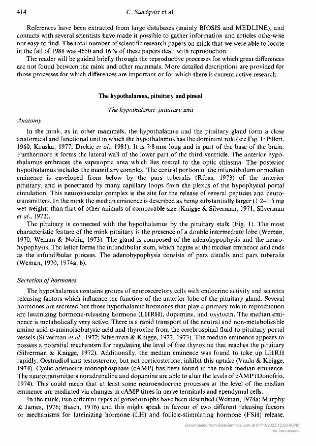
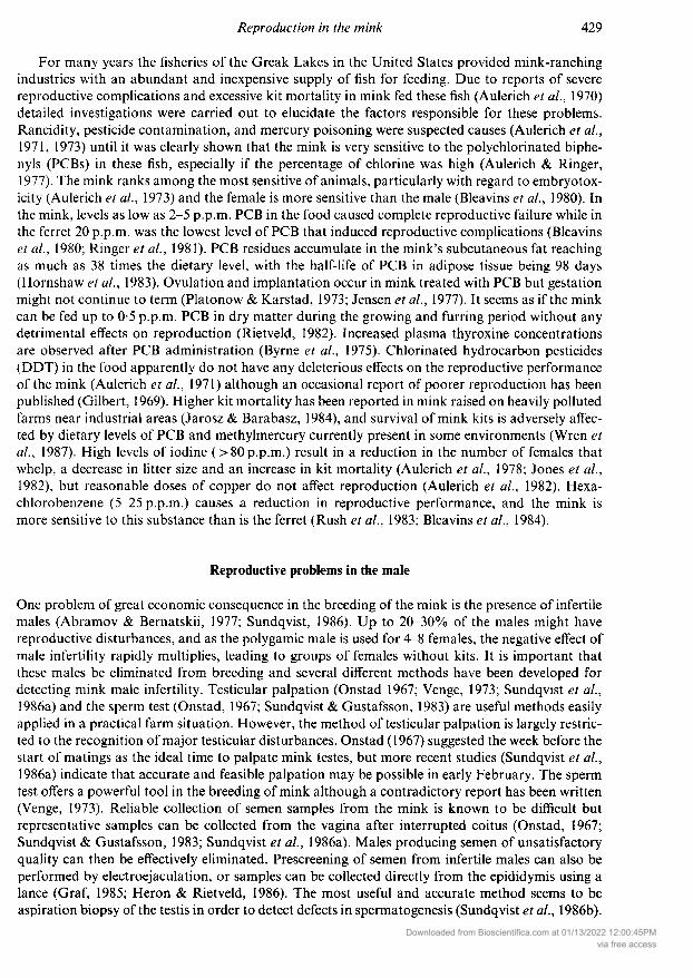
anatomical and functional unit in which the hypothalamus has the dominant role (see Fig. 1 : Pilleri,1960; Kruska, 1977; Drekic et al, 1981). It is 7-8 mm long and is part of the base of the brain.Furthermore it forms the lateral wall of the lower part of the third ventricle. The anterior hypo¬thalamus embraces the supraoptic area which lies rostral to the optic chiasma. The posteriorhypothalamus includes the mamillary complex. The central portion of the infundibulum or medianeminence is enveloped from below by the pars tuberalis (Ribas, 1973) of the anteriorpituitary, and is penetrated by many capillary loops from the plexus of the hypophysial portalcirculation. This neurovascular complex is the site for the release of several peptides and neuro-transmitters. In the mink the median eminence is described as being substantially larger (1-2-1-5 mgwet weight) than that of other animals of comparable size (Knigge & Silverman, 1971; Silvermanet al, 1972).
The pituitary is connected with the hypothalamus by the pituitary stalk (Fig. 1). The mostcharacteristic feature of the mink pituitary is the presence of a double intermediate lobe (Weman,1970; Weman & Nobin, 1973). The gland is composed of the adenohypophysis and the neuro¬
hypophysis. The latter forms the infundibular stem, which begins at the median eminence and endsas the infundibular process. The adenohypophysis consists of pars distalis and pars tuberalis(Weman, 1970, 1974a, b).
Secretion ofhormonesThe hypothalamus contains groups of neurosecretory cells with endocrine activity and secretes
releasing factors which influence the function of the anterior lobe of the pituitary gland. Severalhormones are secreted but those hypothalamic hormones that play a primary role in reproductionare luteinizing hormone-releasing hormone (LHRH), dopamine, and oxytocin. The median emi¬nence is metabolically very active. There is a rapid transport of the neutral and non-metabolizableamino acid a-aminoisobutyric acid and thyroxine from the cerebrospinal fluid to pituitary portalvessels (Silverman et al, 1972; Silverman & Knigge, 1972, 1973). The median eminence appears topossess a potential mechanism for regulating the level of free thyroxine that reaches the pituitary(Silverman & Knigge, 1972). Additionally, the median eminence was found to take up LHRHrapidly. Oestradiol and testosterone, but not corticosterone, inhibit this uptake (Vaala & Knigge,1974). Cyclic adenosine monophosphate (cAMP) has been found in the mink median eminence.The neurotransmitters noradrenaline and dopamine are able to alter the levels of cAMP (Donofrio,1974). This could mean that at least some neuroendocrine processes at the level of the medianeminence are mediated via changes in cAMP titres in nerve terminals and ependymal cells.
In the mink, two different types of gonadotrophs have been described (Weman, 1974a; Murphy& James, 1976; Busch, 1976) and this might speak in favour of two different releasing factorsor mechanisms for luteinizing hormone (LH) and follicle-stimulating hormone (FSH) release.
Downloaded from Bioscientifica.com at 01/13/2022 12:00:45PMvia free access

ob
ah = adenohypophysisahy = anterior hypothalamusc = cerebellumcc = corpus callosummb = mamillary bodyme = median eminence
mi = massa intermediamo = medulla oblongatanh = neurohypophysisob = olfactory bulboc = optic chiasma = pons
pb = pineal bodyph = posterior hypothalamuspi = pars intermediaqp = quadrigeminal platesoa = supraoptic areaIII = third ventricle
Fig. 1. Schematic representation of the mink brain. The brain weighs approximately 15 g in theadult male mink and its most characteristic features are a large median eminence, a doubleintermediate lobe of the pituitary gland and a pineal gland connected to the roof of the thirdventricle by a stalk.
Peripheral gonadotrophs presumably produce FSH while the central ones produce LH (Weman,1970). However, because of the paucity of pertinent information there is little reason to rejector accept the widely accepted concept of LHRH being the primary hypothalamic peptide thatregulates both LH and FSH secretion. Studies on the intriguing possibility of dual regulation of thegonadotrophins in the mink are required.
Pituitary prolactin release in the mink appears to be under dopaminergic inhibitory control as itis in other mammals. The dopamine agonist, bromocriptine, is effective in delaying the preimplan¬tation rise of prolactin and progesterone while opposite effects were evident after administration ofprolactin (Papke et al, 1980; Martinet et al, 1981a, b; Murphy et al, 1981). On the other handprolactin secretion can be stimulated with pimozide, a dopamine antagonist (Murphy, 1983b). Themain function of prolactin in the female is the stimulation of milk synthesis in the mammary gland.Indeed, large numbers of lactotrophs are found in the anterior pituitary of lactating mink (Murphy& James, 1976). Prolactin also plays an important role in the control of luteal function and in thetermination of delayed implantation, which is a typical feature of this species (Hansson, 1947;Busch, 1976). This effect of prolactin is discussed in more detail later.
Oxytocin seems to be synthesized in both the supraoptic and paraventricular nuclei, and isthereafter packaged in secretory granules and stored in the posterior pituitary. The stimulus ofsuckling initiates the release of oxytoxin which stimulates expulsion of milk from the nipples(Conant, 1962). If parturition is abnormally prolonged, uterine contractions can be intensified byinjecting a small dose of oxytocin (Jorgensen, 1985). The role of oxytocin in the male mink is unclear,since only one study on the stimulation of capsular contraction has been done (Ellis et al, 1981b).
In the female mink, LH induces ovulation and stimulates the secretion of oestrogens in theovarian theca interna cells (Venge, 1973). FSH promotes the proliferation of granulosa cells andthe growth of Graafian follicles in the ovary. The size of the ovum is independent of FSH. Thegranulosa cells shelter the ovum and act as precursors of the luteal cells. In the male mink, LHstimulates steroidogenesis in the Leydig cells, and FSH and androgens are responsible for thestimulation of spermatogenesis (Jergensen, 1985). Interestingly, plasma concentrations of
Downloaded from Bioscientifica.com at 01/13/2022 12:00:45PMvia free access

-melanocyte-stimulating hormone are inversely related to plasma testosterone values and testicu¬lar development (Ellis et al, 1982b).
Oestrogen-concentrating cells in the mink brain are almost solely distributed in the hypothala¬mus and the limbic system (Morrell et al, 1977), which is in agreement with similar studies in thehamster (Krieger et al, 1977). These oestrogen-concentrating cells are important in establishing thefeedback relationships between gonads and the brain. No seasonal effect on the number of brainoestrogen-concentrating cells has been found (Morrell et al, 1977), although short photoperiod hasbeen shown to affect the hypothalamic-pituitary neurosecretory system (Yurisova & Klochkov,1978; Yurisova et al., 1980). Possible worthwhile topics for future studies should include functionalsignificance of sex steroid-concentrating cells in the brain and the relationship of CNS steroidbinding to the sensitivity of the brain to extrinsic hormonal influences during critical periodsof early post-natal development and seasonal transitions between reproductive activity andquiescence.
The pineal glandAnatomy and secretion ofhormones
In the mink as in many mammalian species, the pineal gland mediates the effects of photoperiodon many physiological functions including reproductive activity (Reiter, 1974; Ellis et al, 1982b;Martinet & Allain, 1985; Ravault et al, 1986). The pineal is an elongated gland connected rostrallyto the posterior end of the roof of the third ventricle (Fig. 1) (Weman & Nobin, 1979; Rouvet,1982). Unlike the situation in the ferret (David & Herbert, 1973), in the mink there is a stalkconnecting the pineal to the brain (Rouvet, 1982). According to Rouvet (1982) the pineal iscomposed of secretory cells called pinealocytes. These cells can have either light or dark cytoplasmand the dark ones have been found to contain more free ribosomes and less rough endoplasmicreticulum (Rouvet, 1982).
The pineal synthesizes several indoleamines of which melatonin has the most pronouncedeffects on reproduction. When melatonin is given to the male mink in the summer in order toproduce earlier development of the winter coat there is either stimulation (Allain et al, 1981;Valtonen et al, 1986) or inhibition (Ellis, 1985) of testicular development. In the female, thepineal is involved in the regulation of the photo-dependent termination of the embryonic diapausewhich will be discussed later (Murphy & James, 1974a). Removal of the pineal renders the minkunresponsive to the stimulatory effects of artificial long days on prolactin and progesteronesecretion during pregnancy (Martinet et al, 1981a, 1985; Murphy et al, 1981).
Reproductive biology of the female
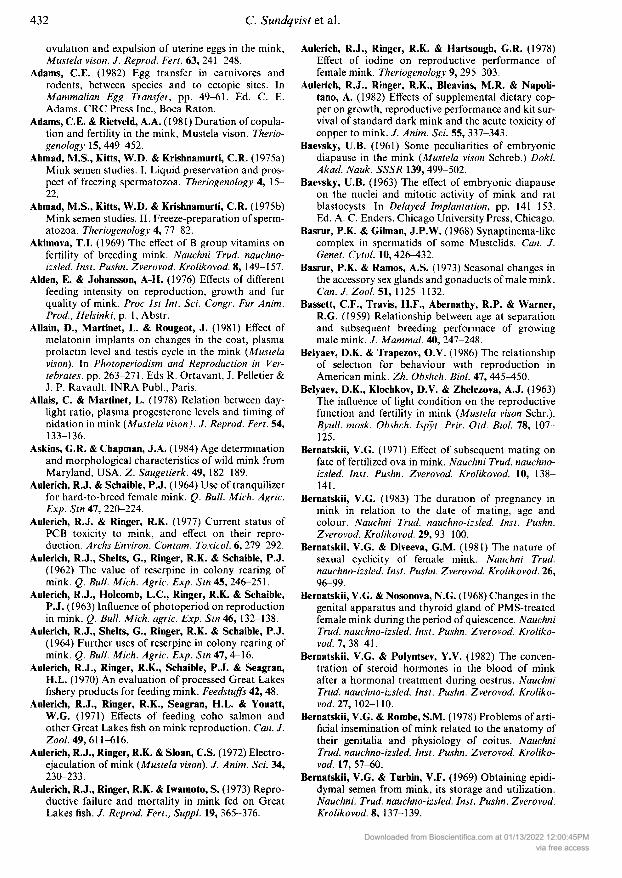
Oogénesis and ovulationThe ovaries of the mink are bean-shaped, located opposite to the caudal portion of the kidneys andsurrounded almost completely by the ovarian bursa (Fig. 2). Their size varies at different periods,being small (0-30 g) during the long anoestrum, increasing in size as the breeding season approaches(0-60 g) and reaching maximum size about 2 weeks before parturition (0-65 g) (Fig. 3) (Hansson,1947; Enders, 1952; Venge, 1973; Pilbeam et al, 1979). The amount of sunlight plays an active rolein accelerating oestrus and ovarian growth (Stevenson, 1946; Hansson, 1947; Hammond, 1951a)although breeding may commence even if complete darkness is maintained from the end ofDecember to May (Kirk, 1962). No systematic study on the prenatal development of the gonadalsystem could be found, but Kissen & Price (1962) point out that the mink in general appears todevelop in utero in a typical mammalian fashion.
The ovary consists of an outer zone (cortex) and an inner zone (medulla). In May, the medullain the newborn mink consists of loose connective tissue, and is not yet well differentiated from the
Downloaded from Bioscientifica.com at 01/13/2022 12:00:45PMvia free access

Fig. 2. The mink is a seasonally breeding animal with the size of the ovaries varying at differentperiods of the year. The ovaries are small during the long period of anoestrus (May-February)and increase in size as the mating season (March) approaches. The diagram shows the typicalfeatures of an ovary during anoestrus and pregnancy. One remarkable feature is that the cor¬
pora lutea are not initially producing sufficient amounts of progesterone for implantation. Thisphenomenon allows a 2nd or 3rd oestrus and the ovulation of a new set of eggs during the shortmating season.
cortex, which has a few gonocytes (Enders, 1952). In the adult, there is a clear demarcation betweenthe two zones (Hansson, 1947). A few oogonial cells (primordial germ cells) and primary oocytescan be found scattered through the ovarian cortex of the 4-day-old mink. The first interstitial cellsbegin to develop from cells of sterile sex cords in the medullar zone by Day 6 (Zaleska-Freljan,1976). The rudimentary rete ovarii has, during that time, also grown into the interior of the gonadfrom the hilar side (Byskov, 1975; Zaleska-Freljan, 1976). It is speculated that the rete systeminteracts with the cortex, initiating meiosis, and furthermore, that the rete cells as well as cells of thesurface epithelium contribute to the granulosa cell layer (Byskov, 1975). All germ cells have enteredmeiotic prophase by 2 weeks post partum (Byskov, 1975). In the medulla, follicle formation hasprogressed and many of the growing follicles are inter-connected by broad cell cords. Hansson(1947) gives a full description of the follicular development in the mink and he distinguishesbetween 9 different follicular stages.
In the 6-week-old mink (middle of June), primordial follicles accumulate, Graafian folliclesappear, and occasional follicles near the interstitial tissue degenerate, as the cortex of the ovarydevelops. Two distinct layers can be seen in the medulla: the external layer consisting of aggre¬gations of interstitial cells containing lipids, and the internal layer of connective tissue in the centreof which the rete ovarii can be seen.
In December, single primordial follicles, surrounded by a single layer of follicular cells,are observed in the outer part of the cortex. In the inner parts of the cortex, growing oocytes
Downloaded from Bioscientifica.com at 01/13/2022 12:00:45PMvia free access
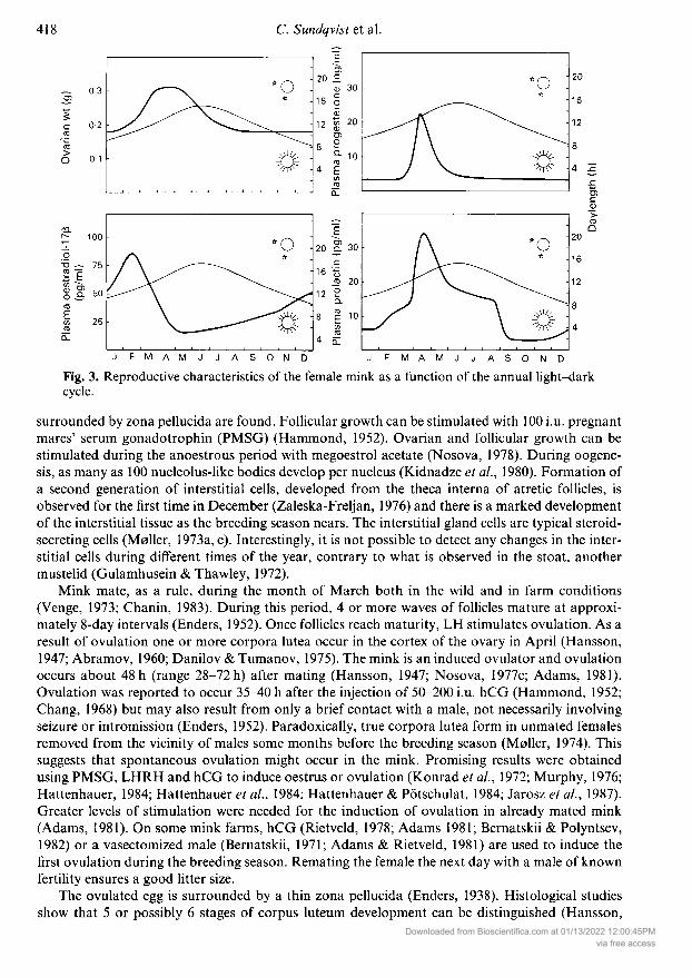
Fig. 3. Reproductive characteristics of the female mink as a function of the annualcycle.
D
light-dark
surrounded by zona pellucida are found. Follicular growth can be stimulated with 100 i.u. pregnantmares' serum gonadotrophin (PMSG) (Hammond, 1952). Ovarian and follicular growth can bestimulated during the anoestrous period with megoestrol acetate (Nosova, 1978). During oogéne¬sis, as many as 100 nucleolus-like bodies develop per nucleus (Kidnadze et al, 1980). Formation ofa second generation of interstitial cells, developed from the theca interna of atretic follicles, isobserved for the first time in December (Zaleska-Freljan, 1976) and there is a marked developmentof the interstitial tissue as the breeding season nears. The interstitial gland cells are typical steroid-secreting cells (Meiler, 1973a, c). Interestingly, it is not possible to detect any changes in the inter¬stitial cells during different times of the year, contrary to what is observed in the stoat, anothermustelid (Gulamhusein & Thawley, 1972).
Mink mate, as a rule, during the month of March both in the wild and in farm conditions(Venge, 1973; Chanin, 1983). During this period, 4 or more waves of follicles mature at approxi¬mately 8-day intervals (Enders, 1952). Once follicles reach maturity, LH stimulates ovulation. As a
result of ovulation one or more corpora lutea occur in the cortex of the ovary in April (Hansson,1947; Abramov, 1960; Danilo & Tumanov, 1975). The mink is an induced ovulator and ovulationoccurs about 48 h (range 28-72 h) after mating (Hansson, 1947; Nosova, 1977c; Adams, 1981).Ovulation was reported to occur 35-40 h after the injection of 50-200 i.u. hCG (Hammond, 1952;Chang, 1968) but may also result from only a brief contact with a male, not necessarily involvingseizure or intromission (Enders, 1952). Paradoxically, true corpora lutea form in unmated femalesremoved from the vicinity of males some months before the breeding season (Moller, 1974). Thissuggests that spontaneous ovulation might occur in the mink. Promising results were obtainedusing PMSG, LHRH and hCG to induce oestrus or ovulation (Konrad et al, 1972; Murphy, 1976;Hattenhauer, 1984; Hattenhauer et al, 1984; Hattenhauer & Pötschulat, 1984; Jarosz et al., 1987).Greater levels of stimulation were needed for the induction of ovulation in already mated mink(Adams, 1981). On some mink farms, hCG (Rietveld, 1978; Adams 1981; Bernatskii & Polyntsev,1982) or a vasectomized male (Bernatskii, 1971; Adams & Rietveld, 1981) are used to induce thefirst ovulation during the breeding season. Remating the female the next day with a male of knownfertility ensures a good litter size.
The ovulated egg is surrounded by a thin zona pellucida (Enders, 1938). Histological studiesshow that 5 or possibly 6 stages of corpus luteum development can be distinguished (Hansson,
Downloaded from Bioscientifica.com at 01/13/2022 12:00:45PMvia free access

1947). The delayed functioning of the corpora lutea is rather peculiar in the mink, and this will bediscussed in more detail in relation to the phenomenon of delayed implantation (see below). Thepresence of corpora lutea does not immediately suppress the formation of primary follicles (Venge,1973). Administration of megoestrol acetate during the oestrous cycle reduces the number ofovulating follicles (Nosova, 1977a). Normally, a slight tendency towards an optimum in thenumber of rupturing follicles may be seen 7-10 days after a previous ovulation, and more folliclesrupture in the middle than at the beginning or the end of the mating season (Hansson, 1947). AGraafian follicle that is ready for ovulation attains an average diameter of 1-0—1-1 mm. Luteinizinghormone has been successfully assayed in the mink (Murphy, 1979a; Mondain-Monval et al, 1985)but it has not been possible to measure FSH concentrations. We feel that better knowledge of thecontrol of oogénesis in the mink could be obtained if it were possible to measure FSH concen¬
trations. Although several articles deal with the anatomy and function of the mink ovary, thereseems to be a considerable lack of accurate quantitative data on oogénesis. For comparative pur¬poses it would, for example, be interesting to know how many primary follicles exist in the ovaryand the frequency with which the follicles become atretic during ovarian development.
The mink is able to ovulate several times during the same mating season. If the female is ferti¬lized during the first mating, she may still ovulate 8-10 days later, and thereafter may give birth tokits from two different ovulations (a phenomenon called superfetation) (Johansson & Venge, 1951;Enders, 1952; Shackelford, 1952). Many different mating schedules utilizing the phenomenon ofrepeated ovulations during the breeding season have been applied to the mink (Johansson & Venge,1951; Enders, 1952; Shackelford, 1952; Lecht & Reck, 1957; Friend & Crampton, 1960; Johansson,1965b; Narucka, 1973; Maciejowski et al, 1973; Kukla, 1975; Fiedler et al, 1975; Rebreanu et al,1981a; Stole et al, 1984). The generally accepted protocol to achieve the best results requires thatearly in the breeding season, mink be remated 7-8 days after the first mating, and that later in theseason, they be remated the day after the first mating (Venge, 1973; Jorgensen, 1985). To increaseproductivity hormonal therapy has been used with variable results (Venge, 1956b; Franklin, 1958;Cochrane & Shackelford, 1962; Murphy, 1976, 1979a; Hattenhauer, 1984; Hattenhauer et al, 1984;Hattenhauer & Pötschulat, 1984). Surprisingly, a 0-5 ml dose of the supermutagen 1,4-ow-diazo-acetylbutane deposited inside the vagina in a concentration of 1:500 increased the litter-sizecompared to controls (Bernatskii et al, 1977).
The structure of the corpus luteum has been extensively studied and it was found that minklutein cells are active, secreting progesterone during pregnancy (Venge, 1959; Enders, 1962; Enders& Enders, 1963; Miladinovic, 1969; Moller, 1973c; Kolpovskii, 1978; Muresan et al, 1984). Themink is, however, different from most other species in that corpora lutea do not initially producehigh quantities of progesterone (Canivenc & Bonnin-Laffargue, 1967). This low level of progester¬one allows a second oestrus, and the ovulation of a second set of eggs capable of normal develop¬ment (Bernatskii & Diveeva, 1981). However, there is indication that ova fertilized after the lastovulation develop more often than ova from previous ovulations (Enders, 1949). Only partialluteinization of the granulosa cells of ovulated follicles occurs before implantation (Enders, 1960).There is a considerable variation in the mean numbers of corpora lutea in different types of mink,e.g. 19-2, 17-2, 18-3 and 15-8 in the dark, pastel, pearl and silverblue colour phases respectively(Adams, 1973). At the end of the breeding season the primary and secondary follicles regressand the formation of new follicles is kept at a minimum until the end of the anoestrous period(Kolpovskii, 1979). In non-pregnant females the corpora lutea regress after the middle of April(Kolpovskii, 1983).
The phenomenon of delayed implantationOne remarkable feature in mink reproduction, is that the fertilized egg undergoes partial devel¬
opment (to the blastula stage) and thereafter remains inactive until it is implanted in the uterus
(Hansson, 1947; Enders, 1952). The mink is by no means unique in this aspect, but successfulDownloaded from Bioscientifica.com at 01/13/2022 12:00:45PM
via free access
breeding of the mink in farm conditions was greatly enhanced when it was detected that the minkexperiences an obligate arrest of implantation. This also makes it possible to give a proper expla¬nation of the great variability in pregnancy lengths. There is a large amount of information aboutdelayed implantation in this species. The mink embryo develops from zygote into a blastocyst bythe 8th day post coitum (Hansson, 1947). During this period the trophoblast is formed, ensuringnutrients for the blastocyst. As a result of the embryonic diapause, the embryo may remain in theblastocyst stage for up to 49 days post coitum. The blastocyst divides 7-8 times, and consists there¬after of a maximum of 300 cells (Enders, 1952). In culture, the trophoblast soon shrinks away fromthe zona pellucida, but the blastocysts survive for as long as 5 months (Enders & Pearson, 1946).However, they do not grow, regardless of the use of sophisticated culture techniques (Daniel, 1967;Gulyas et al, 1969). In the case of the mink, delayed implantation persists only for weeks, ratherthan months, as in other members of the mustelid family (Mead, 1981). Development of the embryois resumed when the maternal environment allows implantation (Baevsky, 1963; Vagin, 1983).
The length of embryonic quiescence may vary and the exact mechanism by which the dormantembryo becomes reactivated is not thoroughly understood. The morphological signs of the com¬
pletion of diapause are the disappearance of the zona pellucida, the release of the trophoblast,and the growth of fragments of transverse folds on the endometrium (Hansson, 1947). There isample evidence for photoperiodic control of egg implantation (Murphy & James, 1974a; Allais &Martinet, 1978; Travis & Pilbeam, 1980). The pineal might play a major role in the control ofimplantation because denervation of the pineal by cervical ganglionectomy inhibits the inductionof implantation in mink stimulated by long photoperiod (Murphy & James, 1974a), and eliminatesthe diurnal variation of serum melatonin (Ravault et al, 1986) while administration of melatonindelays the preimplantation rise in plasma progesterone (Duby et al, 1972). It has been shownthat whenever the mink is mated during the breeding season, implantation always occurs at thebeginning of April (near the vernal equinox) (Enders, 1952; Baevsky, 1961). Histologically,implantation does not occur until at least 20 days after mating (Abramov, 1960). This means thatthe gestation period becomes very long if the mink is mated early in March compared with matingsin late March (Hansson, 1947). Implantation follows the initial increase in progesterone by 5-10days (M0ller, 1973b; Murphy & Moger, 1977; Pilbeam et al, 1979). An artificial increase inphotoperiod after mating reduces the gestation period (Holcomb et al, 1962; Belyaev et al, 1963;Aulerich et al, 1963). In one study, however, a lengthened photoperiod after mating did not affectpregnancy length (Murphy, 1977). The time of breeding in mink can be moved forward by exposingthem to additional light before the mating season (Holcomb et al, 1962) by hastening the onset ofprolactin and progesterone secretion (Allais & Martinet, 1978; Martinet et al, 1981b). Theduration of pregnancy in the mink is very variable (Svihla, 1931; Bowness, 1942; Pearson & Enders,1944; Hansson, 1947; Enders, 1952; Bowness, 1968) and the date of mating is the most importantfactor accounting for this great variability (Hansson, 1947). Matings late in the breeding season
tend to shorten the gestation period. The mean length of pregnancy is approximately 51 days(Bowness, 1942; Pearson & Enders, 1944; Stevenson, 1945; Enders, 1952; Venge, 1966, 1973;Dukelow, 1966; Bernatskii, 1983). Short photoperiods, or injection of melatonin, inhibit prolactinsecretion (Martinet et al, 1983). Therefore, increasing daylength in the spring is the cue that elicitssufficient prolactin secretion, leading to the termination of embryonic diapause (Fig. 3). It appearsthat 5a-pregnane-3,20-dione is the predominant steroid to which uterine progesterone is converted(Rose et al, 1983b).
Murphy et al (1982, 1983) showed the importance of ovarian factors in implantation bydemonstrating that ovariectomy during embryonic quiescence consistently prevents implantation.Exogenous progesterone or medroxyprogesterone acetate failed to correct the situation andpregnancies could not be shortened by this treatment (Hansson, 1947; Hammon, 1951b; Holcomb,1967; Murphy et al, 1982, 1983; Murphy, 1983a; Christiansen, 1985). Contrary to these findings,some investigators (Concannon et al, 1980; Jarosz & Dukelow, 1985) found evidence thatmedroxyprogesterone could advance the time of implantation.
Downloaded from Bioscientifica.com at 01/13/2022 12:00:45PMvia free access
There is now evidence that prolactin is the major component of the luteotrophic complex thatterminates embryonic diapause in the mink by initiating luteal progesterone secretion (Papke et al,1980; Murphy et al, 1980; Martinet et al, 1981a). If the mink is treated with prolactin duringthe embryonic diapause there is an increase in progesterone production (Papke et al, 1980;Martinet et al, 1981a, b). An opposite effect on progesterone secretion can be obtained by adminis¬tering melatonin (Martinet et al, 1981b). Furthermore, Murphy et al (1981) showed that, in hypo¬physectomized mink, prolactin injections alone could activate the corpora lutea and stimulateimplantation. By repeated administration of LHRH antiserum, Murphy et al. (1984) showed thatpituitary release of LH is essential for luteal maintenance in the mink. Other mustelids also requirean intact hypophysis for normal luteal function and embryo implantation (Mead, 1975; Murphy,1979b). Canivene et al. (1966) suggested that the uterus is required in pregnant females for normalluteal function, but Duby et al (1972) failed to support this finding. Additional evidence for theimportance of prolactin in the regulation of ovarian function in the mink is given by Rose et al(1983a, 1986) who showed that prolactin receptors were present in the ovaries and the uterus. Thereis a reduction in the numbers of receptors during blastocyst reactivation, probably due to theoccupancy of those receptor sites by endogenous prolactin.
Fertilization, gestation and parturitionIn the oviduct an environment is provided that allows sperm capacitation, fertilization and earlyembryonic development (Hansson, 1947; Enders, 1952). The oviduct is firmly attached to the ovar¬
ian capsule and it is said to be -30 mm (Enders, 1952) or 10-15 mm (Hansson, 1947) long. As inmost other mammals, the lining of the lumen of the oviduct is greatly folded. When oestrusapproaches, the oviduct increases in size and the folds become more rounded. Cilia are presentthroughout the oviduct but are reduced in number near the uterus (Enders, 1952). Ciliary activityand co-ordinated muscular contractions regulate the rate at which spermatozoa move to the fertili¬zation site and eggs are transported to the uterus. There is some indirect evidence that the ovulatedeggs travel through the oviduct in 6 days (Hansson, 1947). Fertilization takes place in the bursaovarii or within the fimbriated portion of the oviduct (Enders, 1952). The fertilized ova measure
135-151 µ in diameter, whereas the cytoplasm measures 103-110 µ and the thickness of thezona pellucida is 11 µ (Enders, 1938).
The bicornuate uterus of the mink is ~ 30 mm long during the breeding season but the sizechanges dramatically throughout the year (Enders, 1952). Two different layers can be recognized:the endometrium and the myometrium. The former is composed of an epithelial lining of theuterine lumen and is filled with several endometrial glands (Enders et al, 1963). The gland cellsundergo greater alterations during the season than the epithelial cells, which are characterized bylarge amounts of glycogen and smaller amounts of lipids, especially during the implantation phase(Enders, 1961; Enders et al, 1963; Hedlund et al, 1972). An increase in endometrial mucopoly-saccharides is observed when implantation takes place (Murphy & James, 1974b). After implan¬tation, a zonodiscoidal (Hansson, 1947) or truly zonal (Enders, 1957) placenta is formed. Themyometrium is greatly thickened during oestrus (Hansson, 1947).
Length of pregnancy (mean = 51 days) is affected by delayed implantation, colour variety,photoperiod, age of female, mating frequency, and ambient temperature (Holcomb et al, 1962;Bura et al, 1981; Rebreanu et al, 1981b).
In the pregnant mink, basal plasma progesterone concentrations of 8 ng/ml show a graduaiincrease beginning about 40 days before parturition (Fig. 3) (Moller, 1973b; Allais & Martinet,1978; Pilbeam et al, 1979; Petrova et al, 1983; Einarsson, 1985). During this time the fertilized eggis still in the blastocyst stage. Maximum concentrations are observed 10-25 days later (early April)when the delayed implantation ends. After that, plasma progesterone concentrations declinegradually, and low levels are observed at parturition. Ovariectomy during pregnancy causes an
abrupt fall in peripheral plasma progesterone concentrations (M0ller, 1974). An obligatoryDownloaded from Bioscientifica.com at 01/13/2022 12:00:45PM
via free access

relationship seems to exist between luteal function and the maintenance of pregnancy, sinceovariectomy terminates pregnancy. Oestrogen concentrations remain low throughout pregnancy(Pilbeam et al, 1979). A pregnancy-associated serum protein has been isolated in the mink (Larsenet al, 1971).
Parturition occurs usually from the last week in April to the middle of May (Venge, 1973) andan average of 5 kits are born. Litters as large as 17 have been reported but litters of 10 or more are
infrequent (Enders, 1952).
Other studies on the reproductive biology of the femaleThe cervix is a muscular sphincter which is situated at the posterior end of the uterus. It is closed atall times except during oestrus and parturition, when it relaxes enough to permit the passage ofspermatozoa and fetuses respectively (Enders, 1952). When electrical stimulation of the cervix isused instead of a first mating, poor reproductive success is obtained (Murphy, 1979a). This wouldsuggest that natural matings, or hormonal treatment causing ovulation, are necessary for goodreproductive results in the mink. The plant derivative 6-methoxybenzoxazolinone, which stimulatesreproductive function in voles, has no effect in the mink (Ginther et al, 1985).
The vagina of the mink is quite long (34 mm on average), distensible and muscular, having a
tendency to lengthen further when oestrus approaches (Enders, 1952; Tihonov, 1963). It is not easyto classify the various phases of oestrus in the mink based on vaginal smears (Hansson, 1947;Enders, 1952). During oestrus the vagina contains a multilayered or partly keratinized and strati¬fied squamous epithelium, presumably to protect against mechanical damage during copulation(Busch et al, 1979). Size of the vulva increases greatly during the breeding season, and treatment inNovember with PMSG causes a slight increase in vulvar size and uterine growth is stimulated (4-5months before the breeding season) (Enders, 1952; Bernatskii & Nosonova, 1968). A better re¬
sponse to PMSG is seen immediately after the breeding season than during anoestrus (Hartsough,1942). The developmental stages of the external genitalia have been classified into 3 or 4 categories(Travis et al, 1978). Ovariectomized animals receiving diethylstilboestrol did not develop vulvarsize large enough to permit intromission although they exhibited clear willingness to mate (Enders,1948). Although vaginal smears can be useful to predict when the female will not mate, they cannotbe used to predict the opposite (Travis et al, 1978). Like the ferret, embryo transfer has beenapplied to mink (Chang, 1968; Adams, 1982; Zhelezova & Sekirina, 1982; Kraemer, 1983). Thismight be a very important technique for the mink industry in the 21st century and we encouragescientists to make this expanding field of animal production fully available to mink farmers. Itmight be profitable to use mink as surrogate mothers for embryos from endangered species, such as
the black-footed ferret.
Reproductive biology of the male
Testicular development and spermatogenesisIn the juvenile as well as the adult mink, consistent seasonal changes in testicular activity are
observed. Testes are functional only during a few months around the breeding season in March(Onstad, 1967; Hemmingsen, 1967). After the breeding season, testes regress and remain very smalland non-functional until the start of spermatogenesis again in November (Fig. 4). There is nocorrelation between body and testis weight (Onstad, 1967), but testicular and epididymal weightscorrelate with age (Askins & Chapman, 1984). Although Onstad (1967) found evidence for a sig¬nificantly larger left testis this could not be confirmed in a later study (Kostron & Kukla, 1970).
In the newborn mink, the small testes (003 g) contain a fairly compact system of seminiferoustubules surrounded by the tunica albugínea (Fig. 5) (Onstad, 1967; Zaleska-Freljan, 1976). Duringthat time the seminiferous tubules are lined with a single layer of Sertoli cells and a few developing
Downloaded from Bioscientifica.com at 01/13/2022 12:00:45PMvia free access
M A M J A S N
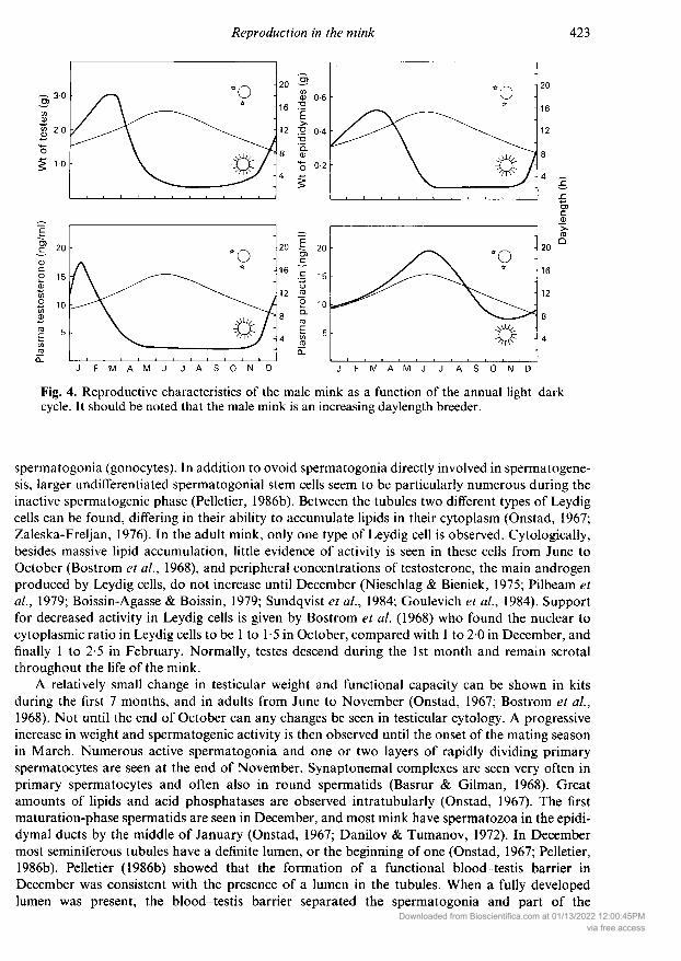
Fig. 4cycle.
. Reproductive characteristics of the male mink as a function of the annual light-darkIt should be noted that the male mink is an increasing daylength breeder.
spermatogonia (gonocytes). In addition to ovoid spermatogonia directly involved in spermatogene¬sis, larger undifferentiated spermatogonial stem cells seem to be particularly numerous during theinactive spermatogenic phase (Pelletier, 1986b). Between the tubules two different types of Leydigcells can be found, differing in their ability to accumulate lipids in their cytoplasm (Onstad, 1967;Zaleska-Freljan, 1976). In the adult mink, only one type of Leydig cell is observed. Cytologically,besides massive lipid accumulation, little evidence of activity is seen in these cells from June toOctober (Bostrom et al, 1968), and peripheral concentrations of testosterone, the main androgenproduced by Leydig cells, do not increase until December (Nieschlag & Bieniek, 1975; Pilbeam etal, 1979; Boissin-Agasse & Boissin, 1979; Sundqvist et al, 1984; Goulevich et al, 1984). Supportfor decreased activity in Leydig cells is given by Bostrom et al (1968) who found the nuclear tocytoplasmic ratio in Leydig cells to be 1 to 1-5 in October, compared with 1 to 20 in December, andfinally 1 to 2-5 in February. Normally, testes descend during the 1st month and remain scrotalthroughout the life of the mink.
A relatively small change in testicular weight and functional capacity can be shown in kitsduring the first 7 months, and in adults from June to November (Onstad, 1967; Bostrom et al,1968). Not until the end of October can any changes be seen in testicular cytology. A progressiveincrease in weight and spermatogenic activity is then observed until the onset of the mating seasonin March. Numerous active spermatogonia and one or two layers of rapidly dividing primaryspermatocytes are seen at the end of November. Synaptonemal complexes are seen very often inprimary spermatocytes and often also in round spermatids (Basrur & Gilman, 1968). Greatamounts of lipids and acid phosphatases are observed intratubularly (Onstad, 1967). The firstmaturation-phase spermatids are seen in December, and most mink have spermatozoa in the epidi¬dymal ducts by the middle of January (Onstad, 1967; Danilov & Tumanov, 1972). In Decembermost seminiferous tubules have a definite lumen, or the beginning of one (Onstad, 1967; Pelletier,1986b). Pelletier (1986b) showed that the formation of a functional blood-testis barrier inDecember was consistent with the presence of a lumen in the tubules. When a fully developedlumen was present, the blood-testis barrier separated the spermatogonia and part of the
Downloaded from Bioscientifica.com at 01/13/2022 12:00:45PMvia free access
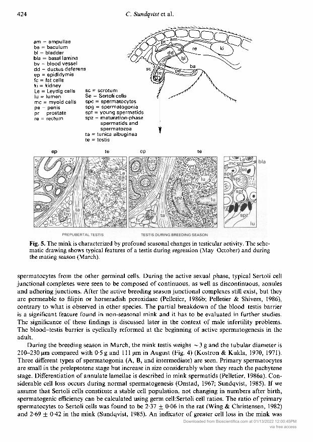
am = ampullaeba = baculumbl = bladderbla = basal laminabv = blood vesseldd = ductus deferensep = epididymisfc = fat cellski = kidneyLe = Leydig cellslu = lumenmc = myoid cellspe = penispr = prostatere = rectum
SsSBö^
sc = scrotumSe = Sertoli cellsspc = spermatocytesspg = spermatogoniaspt = young spermatidsspz = maturation-phase
spermatids andspermatozoa
ta = tunica albugíneate = testis
ep te ep te
Fig. 5. The mink is characterized by profound seasonal changes in testicular activity. The sche¬matic drawing shows typical features of a testis during regression (May-October) and duringthe mating season (March).
spermatocytes from the other germinal cells. During the active sexual phase, typical Sertoli celljunctional complexes were seen to be composed of continuous, as well as discontinuous, zonulesand adhering junctions. After the active breeding season junctional complexes still exist, but theyare permeable to filipin or horseradish peroxidase (Pelletier, 1986b; Pelletier & Shivers, 1986),contrary to what is observed in other species. The partial breakdown of the blood-testis barrieris a significant feature found in non-seasonal mink and it has to be evaluated in further studies.The significance of these findings is discussed later in the context of male infertility problems.The blood-testis barrier is cyclically reformed at the beginning of active spermatogenesis in theadult.
During the breeding season in March, the mink testis weighs ~ 3 g and the tubular diameter is210-230µ compared with 0-5g and 111 µ in August (Fig. 4) (Kostron & Kukla, 1970, 1971).Three different types of spermatogonia (A, B, and intermediate) are seen. Primary spermatocytesare small in the preleptotene stage but increase in size considerably when they reach the pachytenestage. Differentiation of annulate lamellae is described in mink spermatids (Pelletier, 1986a). Con¬siderable cell loss occurs during normal spermatogenesis (Onstad, 1967; Sundqvist, 1985). If we
assume that Sertoli cells constitute a stable cell population, not changing in numbers after birth,spermatogenic efficiency can be calculated using germ celhSertoli cell ratios. The ratio of primaryspermatocytes to Sertoli cells was found to be 2-37 ± 006 in the rat (Wing & Christensen, 1982)and 2-69 + 0-42 in the mink (Sundqvist, 1985). An indicator of greater cell loss in the mink was
Downloaded from Bioscientifica.com at 01/13/2022 12:00:45PMvia free access
found when calculating the ratio of round spermatids to Sertoli cells (7-89 + 0-27 and 4-61 ± 0-59in rat and mink respectively). Differences exist in the kinetics of spermatogenesis before and duringthe breeding season (Tiba et al, 1968a, b; Tiba, 1973a, b, c). More data on other morphometricparameters of mink testicular cells can be found in the literature (Prasolov, 1979; Yoshida, 1982;Sundqvist, 1985).
During active spermatogenesis, germ cells are organized into a definite number of cellular as¬
sociations comprising the stages of the seminiferous epithelial cycle. In the mink, the method ofRoosen-Runge & Giesel (1950) allowed classification of the cellular associations into 8 differentstages (Onstad, 1967; Tiba et al, 1968a; Tiba, 1973d; Deguchi, 1978; Sakai, 1981). Use of Leblond& Clermont's (1952) staging system, which is based on changes in the conformation of the acro¬
some system of developing spermatids, permitted identification of 12 different stages (Pelletier,1986b).
Mink spermatozoa have a tadpole shape, with a head length of 5-8-7-0 µ and a width of61 µ . The midpiece is 60µ long and the total length is 43-0µ (Kim et al, 1978a, 1979;Cummins & Woodall, 1985). Epididymal spermatozoa show species specific swellings near theacrosome (Kim et al, 1979) and the morphology of cytoplasmic droplets has also been described(Krause et al, 1986). Although artificial insemination has been tried in the mink and the techniqueof deep-freezing the semen seems to be working (Ishikawa et al, 1965; Bernatskii ¿Sí Turbin, 1969;Pomytko et al, 1972; Ahmad et al, 1975a, b; Heron & Rietveld, 1985b) reproductive results were
not convincing, mainly due to problems with obtaining adequate semen samples, inducing ovula¬tion at the proper time, or the inability of inseminated spermatozoa to pass the cervix (Bernatskii &Rombe, 1978). For example, deposition of semen artificially into the uterus of mink treated with200 i.u. PMSG and mated with vasectomized males on the previous day, resulted in a whelping rateof 66% (Pomytko et al, 1972). Semen can be successfully collected from most mink males byelectroejaculation, but ejaculate volumes are very small (0001-0-2ml) (Miljkovic et al, 1966;Pomytko et al, 1972; Aulerich et al, 1972; Jakovac et al, 1977).
There seem to be large individual differences in the length of the active breeding season. Fertilematings can take place in February or April but the general results of out-of-season matings mustbe considered doubtful (Ishikawa et al, 1965).
Hormones have been used in mink breeding to manipulate male reproduction, and sexual be¬haviour in castrated mink can be initiated by testosterone treatment, independent of the season
(Chagvardieff et al, 1984). Human chorionic gonadotrophin was successfully used to increase malereproductive capacity (Valtonen et al, 1982) and clomiphene citrate increased the sperm count ininfertile individuals (Lukola & Sundqvist, 1986). However, it is not yet practical to use hormonaltherapy to increase reproductive capacity in an ordinary farm situation. Due to the complexity inthe regulation of reproductive hormones, and the difficulties in determining each animal's basalhormonal status, progress in this area has been very slow (Sundqvist, 1986).
Testicular regression begins at the end of March and although meiosis is still being completed atthe end of May, no spermatozoa are seen in the testes. This may be related to the importance of a
functional blood-testis barrier. Lipids in Leydig cells show a tendency to decrease during the mat¬ing season. This decrease is especially prominent just after the season (Onstad, 1967). The lumen isprogressively absent in most, and later in all, tubules from April through November. No meiosis iscompleted after the end of June, and from July to September only occasional type A( spermatogo¬nia are present along the basement membrane. During the regression, the size of the seminiferoustubules is reduced to about a third of its maximal size.
Photoperiodic regulation ofmale reproductionAmong the numerous mammals that exhibit an annual cycle of testicular activity, the mink, a
short-day mammal, appears to hold a unique position. It is characterized by reactivation of testicu¬lar activity and seasonal gonadotrophic stimulation during decreasing photoperiod in autumn. The
Downloaded from Bioscientifica.com at 01/13/2022 12:00:45PMvia free access

increase in the number and immunoreactivity of the hypothalamic LHRH-secreting neurones oc¬
curs in November (Boissin-Agasse et al, 1988). The annual plasma testosterone cycle is character¬ized by a very sharp peak in testosterone secretion (Fig. 4) (Martinet et al, 1978; Boissin-Agasse &Boissin, 1979; Sundqvist et al, 1984). Under experimental conditions, it has been shown that acircadian rhythm is involved and that very short days (8 h light (L): 16 h dark (D) or 4 h L:20 h D)were extremely effective in stimulating testicular activity (Boissin-Agasse et al, 1982; Boissin-Agasse & Ortavant, 1982; Klochkov, 1984; Ortavant et al, 1985). Restriction of light to 8 h per daybetween July and October stimulates spermatogenesis (Klochkov & Kim, 1979; Klochkov, 1980)but continuous light given in the autumn arrests spermatogenesis (Klochkov et al, 1982). Anabrupt change from 4 h L:20 h D to 16 h L:8 h D either inhibits testicular development or initiatesgonadal atrophy (Duby & Travis, 1972). Using asymmetrical photoperiods in a series of elegantexperiments, it was possible to show that a 30-min light pulse, given 12 or 16 h after the beginningof the main light period, inhibited testicular growth and steroidogenesis (Boissin-Agasse & Boissin,1985). However, if the extra light was given 8, 10, 18, 20 or 22 h after the beginning of themain lightperiod, testes grew normally and secreted normal amounts of testosterone. Resonance experimentswithin a 24-h framework showed increased testicular activity and lower plasma prolactin concen¬
trations when the period of the light-dark cycle was equal to 24 or 48 h, but testicular activityremained unstimulated when the light-dark cycle was 12, 20 or 36 h. These experiments clearlyshow that, although photoregulation of the annual cycle of testicular function is dependent on
phase relationships between the daily cycle of photosensitivity and that of the photoperiod, thecharacteristics of the photoresponse are opposite to those described for long-day breeders (Boissin-Agasse et al, 1986). Definite differences among colour varieties of mink are observed in the sensi¬tivity of biological clocks to initiation of testicular development (Ellis et al, 1982b). The role ofcircadian rhythms in the photoregulation of testicular activity is not fully understood and this topicis beyond the scope of this review.
Plasma prolactin also seems to be regulated by a circadian cycle of photosensitivity. In contrastto long-day breeders, prolactin secretion in mink is maximal when testes are regressed in the sum¬
mer and minimal when they are active in the spring (Fig. 4) (Martinet et al, 1982), i.e. an inverserelationship (Boissin-Agasse et al, 1983). The situation is even more complex during testicularregression and it is difficult to draw any conclusions about the possible causal relationship betweenthe changes in prolactin release and testicular activity. It has also been proposed that an inversecorrelation between the testis and thyroid cycles could represent an important internal signal forthe end of the reproductive period. In fact, plasma thyroxine concentrations are highest during thespring and autumn months and lowest during winter (Boissin-Agasse et al, 1981). Thyroidectomyperformed at the beginning of the breeding season stimulated testosterone production and pro¬longed the duration of maximum testicular development (Jacquet et al, 1986).
Other aspects ofmale reproductive biologyThe spermatozoa pass from the seminiferous tubules to the epididymis in order to becomefunctionally competent. The time required for mink spermatozoa to travel from the testis to thedistal parts of the epididymis is unknown. As spermatozoa pass through the epididymal ducts theygradually show increased motility (Kim et al, 1978b). Testicular spermatozoa contain cytoplasmicdroplets which diminish during their epididymal transit and only a few ejaculated spermatozoacontained droplets (Kim et al, 1979; Krause et al, 1986). Although the total head length of sperm¬atozoa changed very little, anterior acrosomal length significantly decreased and postacrosomallength significantly increased during transit from the testis to the epididymis (Kim et al, 1978b).Little evidence of activity is seen in epididymides from June to November. The epididymal ductsare lined with low pseudostratified columnar epithelium with poorly developed stereocilia (Bos¬trom et al, 1968). A progressive increase in epididymal weight can be seen from December (0-28 g)to March (0-56 g) (Fig. 4) (Bostrom et al, 1968). Morphologically, this weight gain results from
Downloaded from Bioscientifica.com at 01/13/2022 12:00:45PMvia free access
the hypertrophy of the epithelial lining and smooth muscle fibres, an increase in sperm concen¬
tration, increased epithelial height, increased secretory activity, and the maximum development ofstereocilia.
The accessory sex glands in the mink include a prostate and distinct ampullae (Fig. 5) (Basrur &Ramos, 1973). The prostate is especially well developed and it completely surrounds the pelvicurethra. The size of the prostate reaches its maximum during the breeding season. Prostatesecretion serves as a medium for transport of spermatozoa to the female. The ampullary glandsare spindle-shaped enlargements composed of branched tubuli on the surface of the ductusdeferens.
The mink penis is ~5cm long (Enders, 1952). It is enclosed in a sheath close to the ventralmusculature. The corpora cavernosa unite to form the penis bone (baculum), a prominent featurewhich has been used as an accurate age criterion of wild mink (Popov, 1943; Elder, 1951; Paul,1968; Long, 1969; Long & Shirek, 1970).
Reproductive problems in the female
The economic situation of a mink farm can be measured by the number of live kits at pelting timeper breeding female. Unfortunately, various factors cause negative effects, preventing achievementof maximum productivity. Good management, proper nutrition, accurate disease control, and at¬tention to keeping good breeding records are known to be important factors leading to betterreproductive results (J0rgensen, 1985; Heron & Rietveld, 1985b). If fewer than 5-5-6 kits per matedfemale are produced, it is important to identify the factors minimizing the yield.
The sexual cycle of the mink differs from that of other laboratory animals. The mink may beclassified as seasonally polyoestrous (Moshonkin, 1981). A convex oestrous curve has been de¬scribed in unmated mink, but if they are mated, the oestrous cycle is changed to a curve consistingof peaks and troughs approximately 8 days apart. It is rather difficult to determine when a femalemink is in oestrus (Jorgensen, 1985). Neither behaviour nor appearance of the external genitalia is a
reliable indicator of possible mating response. Shackelford (1984) summarized these difficulties bystating that the female mink is in oestrus if she accepts the male. Mink farming pioneers experi¬enced many females remaining unmated during the breeding season. With good management andstrictly organized mating systems, taking into account the peculiarities in the oestrous cycle, thenumber of unmated females can nowadays be kept at a very low level (1-3%). Hard to breedfemales might mate after treatment with reserpine to produce a sedative effect (LaFortune &Rheault, 1960; Aulerich et al, 1962, 1964; Aulerich & Schaible, 1964). The absence of ovulationafter repeated mating was found to be an important cause of infertility (Nosova, 1977b). Theincidence of female infertility is unusually high among 1-year-old dark mink and a general lack ofovarian stimulation was observed in these animals (Ellis & Pace, 1987).
The prevention of social stress by visually isolating the females led to better reproductive re¬
sults, but isolated females come into full oestrus slightly later than do controls (Gilbert & Bailey,1967, 1969a, 1970; Hernesniemi, 1980). Females weaned before 8 weeks of age are easier to breedthan females weaned at an older age (Gilbert & Bailey, 1969b). When mink are exposed to noise,such as sonic booms or aircraft flying near the mink houses, either exceptionally high litter losses(Taylor, 1968) or no clear effects were reported (Travis et al, 1968, 1972, 1974). Sequential injec¬tions of pregnant mink with saline caused abortions that were interpreted as responses to the stressof handling and treatment (Daniel, 1971). The mink is a rather aggressive animal, and when con¬siderable efforts are made to produce tamer mink, reproductive performance is improved (Belyaev&Trapezov, 1986).
There appear to be differences in reproductive success and incidence of sterility among differentgenetic stocks (colour varieties) of mink (Skrivan et al, 1975; Jorgensen, 1985). Homozygosity formost recessive coat colour genes lowered fertility (Yamashita et al, 1965b). This was particularly
Downloaded from Bioscientifica.com at 01/13/2022 12:00:45PMvia free access

apparent with the Aleutian gene which produced a significant decrease in fertility in the homozy¬gous state (Evsikov & Belyaev, 1968). About 20% of the females heterozygous for the shadow genehave various malformations of the uterus, cervix and vagina, preventing conception (Nes, 1965).Also, mink with the Stewart and Aleutian genes have very poor fertility (Johansson, 1965a). TheHedlund white mink is difficult to mate due to congenital deafness (Johansson, 1965b). Apparentlydue to inbreeding, extremely dark mink females have various genetic defects that cause loss of kits(Ellis et al, 1981a, 1982a; Ellis & Pace, 1986).
A high prenatal death rate is reported in farm mink and it seems to be related to the length ofgestation (Enders, 1952). According to Hansson (1947) 84% of the eggs ovulated are implanted,but only 50% of those result in kits due to increased mortality during the embryonic diapause. InHedlund white mink the reproductive capacity is comparable to normal but an even higher pro¬portion of kits tend to die (Johansson, 1965b). Kit mortality is also higher in females having thevirally caused Aleutian disease (Jorgensen, 1985). The number of teats supplying milk does notcorrelate with the litter size, but is significantly and positively correlated with survival rate of thekits (Di & Zou, 1983).
A large amount of research has been conducted to demonstrate the effect of different feeds on
reproductive capacity. Generally speaking, the commonly found high prenatal mortality can bereduced by altering the diet (Broxup, 1968). This is an important step during implantation andpregnancy as well as during whelping and lactation. Nutritional causes accounted for 58% of thecases of anovulation (Chekalova, 1983).
The mink being a carnivore, dietary fat levels seldom affect reproduction, providing the fat is ofgood quality (Travis & Schaible, 1961). The mink seems to need at least 18 g digestible protein perday, since lower levels of protein tend to impair reproductive performance (Skrede, 1978). Theaddition of B-complex vitamins to food containing only 7-8 g digestible protein increased fertility(Akimova, 1969). Serious vitamin B, deficit caused by raw, thiaminase-containing fish, led to highembryonic mortality (Zimmermann, 1981). Also, B2 and B6 deficiency causes sterility primarily dueto embryonic death (Helgebostad et al, 1963; Helgebostad, 1980). Poor reproductive results due tosevere anaemia were obtained when mink were fed raw fish from the cod family (Helgebostad,1968).
Infertility was increased in mink fed large amounts of acid-preserved fish silage during thebreeding season (J0rgensen et al, 1976). Mink food is susceptible to microbial and enzymic de¬composition and inferior reproductive results are reported when endotoxins from Escherichia coliwere ingested by females after copulation (Moller & Nordstoga, 1978). Addition of colistin, an
antibiotic that inactivates endotoxins, to the diet improved reproductive results (Pekkanen et al,1983).
Weight loss due to restricted feeding during the pre-mating period has negative effects on repro¬ductive performance (Isupov et al, 1975; Tauson & Alden, 1979, 1984, 1985) although oppositeresults have also been reported (Wenzel & Schicketanz, 1980). Flushing (semi-starvation followedby re-feeding) improves reproductive performance (Tauson, 1985b).
Slaughter offal from the throat area (gullet trimmings) containing high levels of naturally occur¬
ring thyroactive compounds interferes with reproduction of female mink and with lactation (Traviset al, 1966; Kangas & Mäkelä, 1972). Food containing cattle and pig uteri lowers reproductivesuccess (Jarosz & Barteczko, 1976). Poultry offal containing residuals of the food additive dienoes-trol diacetate decreases the number of kits produced (Duby & Travis, 1971).
Hormone-implanted poultry has been fed to mink with detrimental effects on reproduction(Howell & Pickering, 1964). Experimentally, it was found that a daily intake of 10 µg stilboestrolduring the breeding season causes heavy kit losses (Mills, 1961; Shackelford & Cochrane, 1962;Travis & Schaible, 1962). Methandienone, an anabolic steroid, was used to improve fur qualityartificially, and a dose of 0-44 or 1-8 mg/day caused infertility in 50% and 100% of the femalesrespectively (Westermarck et al, 1979). The anticoagulant Dicumarol has been reported to cause
abortions (Kangas & Mäkelä, 1974).Downloaded from Bioscientifica.com at 01/13/2022 12:00:45PM
via free access
For many years the fisheries of the Greak Lakes in the United States provided mink-ranchingindustries with an abundant and inexpensive supply of fish for feeding. Due to reports of severe
reproductive complications and excessive kit mortality in mink fed these fish (Aulerich et al, 1970)detailed investigations were carried out to elucidate the factors responsible for these problems.Rancidity, pesticide contamination, and mercury poisoning were suspected causes (Aulerich et al,1971, 1973) until it was clearly shown that the mink is very sensitive to the polychlorinated biphe-nyls (PCBs) in these fish, especially if the percentage of chlorine was high (Aulerich & Ringer,1977). The mink ranks among the most sensitive of animals, particularly with regard to embryotox-icity (Aulerich et al, 1973) and the female is more sensitive than the male (Bleavins et al, 1980). Inthe mink, levels as low as 2-5 p.p.m. PCB in the food caused complete reproductive failure while inthe ferret 20 p.p.m. was the lowest level of PCB that induced reproductive complications (Bleavinset al, 1980; Ringer et al., 1981). PCB residues accumulate in the mink's subcutaneous fat reachingas much as 38 times the dietary level, with the half-life of PCB in adipose tissue being 98 days(Hornshaw et al, 1983). Ovulation and implantation occur in mink treated with PCB but gestationmight not continue to term (Platonow & Karstad, 1973; Jensen et al, 1977). It seems as if the minkcan be fed up to 0-5 p.p.m. PCB in dry matter during the growing and furring period without anydetrimental effects on reproduction (Rietveld, 1982). Increased plasma thyroxine concentrationsare observed after PCB administration (Byrne et al, 1975). Chlorinated hydrocarbon pesticides(DDT) in the food apparently do not have any deleterious effects on the reproductive performanceof the mink (Aulerich et al, 1971) although an occasional report of poorer reproduction has beenpublished (Gilbert, 1969). Higher kit mortality has been reported in mink raised on heavily pollutedfarms near industrial areas (Jarosz & Barabasz, 1984), and survival of mink kits is adversely affec¬ted by dietary levels of PCB and methylmercury currently present in some environments (Wren et
al, 1987). High levels of iodine (> 80 p.p.m.) result in a reduction in the number of females thatwhelp, a decrease in litter size and an increase in kit mortality (Aulerich et al, 1978; Jones et al,1982), but reasonable doses of copper do not affect reproduction (Aulerich et al, 1982). Hexa-chlorobenzene (5-25 p.p.m.) causes a reduction in reproductive performance, and the mink ismore sensitive to this substance than is the ferret (Rush et al, 1983; Bleavins et al, 1984).
Reproductive problems in the male
One problem of great economic consequence in the breeding of the mink is the presence of infertilemales (Abramov & Bernatskii, 1977; Sundqvist, 1986). Up to 20-30% of the males might havereproductive disturbances, and as the polygamie male is used for 4-8 females, the negative effect ofmale infertility rapidly multiplies, leading to groups of females without kits. It is important thatthese males be eliminated from breeding and several different methods have been developed fordetecting mink male infertility. Testicular palpation (Onstad 1967; Venge, 1973; Sundqvist et al,1986a) and the sperm test (Onstad, 1967; Sundqvist & Gustafsson, 1983) are useful methods easilyapplied in a practical farm situation. However, the method of testicular palpation is largely restric¬ted to the recognition of major testicular disturbances. Onstad (1967) suggested the week before thestart of matings as the ideal time to palpate mink testes, but more recent studies (Sundqvist et al,1986a) indicate that accurate and feasible palpation may be possible in early February. The spermtest offers a powerful tool in the breeding of mink although a contradictory report has been written(Venge, 1973). Reliable collection of semen samples from the mink is known to be difficult butrepresentative samples can be collected from the vagina after interrupted coitus (Onstad, 1967;Sundqvist & Gustafsson, 1983; Sundqvist et al, 1986a). Males producing semen of unsatisfactoryquality can then be effectively eliminated. Prescreening of semen from infertile males can also beperformed by electroejaculation, or samples can be collected directly from the epididymis using a
lance (Graf, 1985; Heron & Rietveld, 1986). The most useful and accurate method seems to beaspiration biopsy of the testis in order to detect defects in spermatogenesis (Sundqvist et al, 1986b).
Downloaded from Bioscientifica.com at 01/13/2022 12:00:45PMvia free access

When scored on a scale from 1 to 10 according to the developmental stage and number of sperm¬atogenic cells, males scoring 8-10 produced better breeding results than did males having scores
under 7 (Sundqvist et al, 1986b, 1987). The biopsy did not adversely affect libido or fertility. Bymeasuring plasma testosterone concentrations 1 month before the breeding season it was possibleto detect males with delayed puberty or greatly disturbed testicular development (Sundqvist et al,1984, 1985). Serum testosterone concentrations can be measured using solid-phase time-resolvedfluoroimmunoassay with as great an accuracy as with a conventional radioimmunoassay (Bertoft etal, 1987). However, the testosterone test appears to give conflicting results (Sundqvist et al, 1986a)and must await further study before its real value can be determined.
The duration of copulation affects litter size in the mink (Kukla, 1971; Abramov & Utkin,1972). Males that copulated for less than 30 min fertilized fewer females than did those that copu¬lated for more than 30 min (Kukla, 1977). Restriction of the duration of copulation to less than6 min results in significantly reduced fertility, either due to too few spermatozoa ejaculated or
defective sperm transport in the female genital tract (Venge, 1956a; Adams & Rietveld, 1981).Although very short copulations usually are unproductive, pregnancy can result from intromis¬sions of only 2 min, provided these intromissions were preceded by some sort of ethological stimu¬lation (Adams, 1973). An unusually high incidence of mink males not taking part in matings (3-29-2404%) was reported in one study (Kukla, 1970). Histological examination revealed that sperm¬atogenesis was normal in non-mating males (Kostron & Kukla, 1970). In some males injection oftestosterone propionate is able to induce mating (Chaddock, 1949). Adult mink males have moresuccessful breeding attempts than young ones, with intromission latency averaging 12-6 min inadult mink and 15-4 min in young mink (MacLennan & Bailey, 1972). Apparently experiencerather than physiological factors was the cause of better success in the adult mink.
Defective testes are often found in mink (Härtung & Seffner, 1983). Hypoplasia occurs in about1-9% of the mink and ~6-4% of the mink have cryptorchid testes (Sundqvist et al, 1986a). Bothhypoplasia and cryptorchidism might be hereditary in the mink (Onstad, 1967). Furthermore, themink differs from most other species in that the tunica vaginalis originates higher in the abdominalcavity and, instead of passing through the pelvis, it comes through an inguinal fat pad between theskin and the body wall. If the fat pad enlarges, the adipose cells might adhere to the tunica vaginalisand prevent the normal descent of testes (Ellis & Pace, 1987). Most of the mink with hypoplastictestes produce semen of unsatisfactory quality (Onstad, 1961a, 1967; Sundqvist et al, 1986a).Males with small testes had a slightly poorer reproductive capacity than controls, but having largertestes did not significantly improve breeding performance of the male (Heron & Rietveld, 1985a). Arare congenital defect characterized by lack of the epididymis has been described in mink (Onstad,1961b; Blom & Hermansen, 1969).
A much higher incidence of abnormal spermatozoa has been reported in the mink than in thefox (Borozdin & Micurina, 1966). The following sperm abnormalities occur in the mink: coiled andbent tails, tailless spermatozoa, various sperm-head defects, and abnormal clumping of spermato¬zoa (Onstad, 1967; Sundqvist et al, 1986a). Fertility is less affected when the number of spermato¬zoa with primary morphological abnormalities falls below 6% (Jarosz et al, 1968).
Mink that were abruptly changed from ambient light at 42°N latitude to an artificial lightregimen corresponding to 45°S latitude showed very poor reproductive capacity after 9 months(Travis & Pilbeam, 1980). Reproductive disturbances were also seen when mink were transportedfrom the USA to Finland. There was a direct correlation between the time after arrival and therestoration of normal hormonal, reproductive and testicular status (Sundqvist et al, 1985). Ex¬posure to artificial light during the winter months has a deleterious effect on reproduction (Venge,1968). It might be important not to separate males too early from their native environment, as
indicated by a much greater mating activity in males weaned at 11 weeks compared with thoseweaned at 6 weeks (Bassett et al, 1959).
There are great differences in male reproductive capacity between different colour varieties ofmink, with Hedlund white males showing low levels of copulatory activity (Borisova, 1967) and
Downloaded from Bioscientifica.com at 01/13/2022 12:00:45PMvia free access

males homozygous for the Stewart alide being sterile (Venge, 1963; Yamashita et al, 1965a). Asignificant increase in the incidence of polyploidy in bone marrow cells is seen in males with poorreproductive performance (Isakova, 1981). Males with poor libido and poor semen quality tendedto be sex chromatin positive (Rauluszkiewicz et al, 1971). Sperm quality varies markedly in differ¬ent colour varieties, with the poorest sperm quality being found in the sapphire mink and goodsperm quality in the pastel and jet-black mink (Sundqvist et al, 1986a; Sundqvist & Sundqvist,1986).
Inbreeding mink for an extremely dark fur has co-selected for male infertility which might beobserved at the onset of the breeding season (primary infertility) or after one or more fertile breed¬ing seasons (secondary infertility) (Tung et al, 1981a, 1984). Mink expressing primary infertilityhave low plasma LH and testosterone concentrations. Since they respond to exogenous LHRH andhCG treatment it could be that these mink had defective LHRH secretion. Autoimmune orchitisand testicular immune complexes are frequently found in mink with secondary infertility (Tung et
al, 1981a) and the extremely dark mink could be useful for studying autoimmune testicular dis¬turbances (Tung et al., 1981a, b, 1985). Testicular autoimmune reactions most probably developduring testicular regression in late March and April. As indicated above, Pelletier (1986b) found a
complete breakdown of the blood-testis barrier in the regressing mink testis and this might be theanatomical basis of autoimmune orchitis problems in the mink. Dark mink have a higher contentof testicular histamine than do those of other colour varieties during the breeding season and theelevated secretion rate was related to the onset of autoimmune infertility (Nemetallah et al, 1985).Presumably histamine would induce the breakdown of the blood-testis barrier allowing the germcells to be attacked by the immune system. Antibodies against germ cells are eventually producedand spontaneous destruction of the seminiferous epithelium starts (Ellis et al, 1985). The extremelydark mink differs from all other colour varieties so far studied, and although anti-sperm antibodiesare found in other varieties, only the dark mink develops autoimmune orchitis. Natural killer cellactivity was not higher in mink with autoimmune orchitis (Pace et al, 1987). Determining whetherthere is a causal relationship between the breakdown of the blood-testis barrier and the occurrenceof immunopathological problems warrants future investigation. Extensive testicular damage in allcolour varieties was observed after implantation of melatonin-containing capsules (Ellis, 1985).
Weight loss in male mink during the winter might have a deleterious effect on their reproductiveperformance (Tauson, 1985a), although obesity produces a similar effect (Alden & Johansson,1976). Precautions should be taken not to feed slaughter offal from hormone-implanted poultry, asit prevents the occurrence of normal reproductive activity (Howell & Pickering, 1964).
Financial support from The Academy of Finland and The National Institutes of Health(HD20033) made this review possible. We thank Steve Rogers and Sherie L. Hodges for editing thetext; Anthony Rietveld, for allowing use of the library at Northwood Fur Farms Inc.; KarenSchmitt, Cynthia Clabough and Cheryl Broadie for help with the illustrations; Dr Varadaraj Chan¬drashekar and Dr Artur Mayerhofer for helpful discussions; and Eric Dennison and David Nelsonfor assistance with the database. We apologize to those whose valuable contributions to the fieldhave not been discussed or quoted due to unintentional omissions.
References
Abramov, M.D. (1960) Characteristics of the biology ofreproduction of mink. Mustela vison Schreb. NauchniTrud. nauchno-izsled. Inst. Pushn. Zverovod. Kroliko-vod. 5, 3-39.
Abramov, M.D. & Utkin, L.G. (1972) The problem ofinterruption of copulation in mink. Nauchni Trud.nauchno-izsled. Inst. Pushn. Zverovod. Krolikovod. 11,194-195.
Abramov, M.D. & Bernatskii, V.G. (1977) Some causesof infertility in mink and methods for eliminating it.Nauchni Trud. nauchno-izsled. Inst. Pushn. Zverovod.Krolikovod. 15, 5-12.
Adams, C.E. (1973) The reproductive status of femalemink, Mustela vison, recorded as 'failed to mate'. J.Reprod. Fert. 33, 527-529.
Adams, C.E. (1981) Observations on the induction of
Downloaded from Bioscientifica.com at 01/13/2022 12:00:45PMvia free access
ovulation and expulsion of uterine eggs in the mink,Mustela vison. J. Reprod. Fert. 63, 241-248.
Adams, C.E. (1982) Egg transfer in carnivores androdents, between species and to ectopie sites. InMammalian Egg Transfer, pp. 49-61. Ed. C. E.Adams. CRC Press Inc., Boca Raton.
Adams, C.E. & Rietveld, A.A. (1981) Duration of copula¬tion and fertility in the mink, Mustela vison. Therio¬genology 15, 449-452.
Ahmad, M.S., Kitts, W.D. & Krishnamurti, CR. (1975a)Mink semen studies. I. Liquid preservation and pros¬pect of freezing spermatozoa. Theriogenology 4, 15-22.
Ahmad, M.S., Kitts, W.D. & Krishnamurti, CR. (1975b)Mink semen studies. II. Freeze-preparation of sperm¬atozoa. Theriogenology 4, 77-82.
Akimova, T.I. (1969) The effect of group vitamins on
fertility of breeding mink. Nauchni Trud. nauchno-izsled. Inst. Pushn. Zverovod. Krolikovod. 8, 149-157.
Alden, E. & Johansson, A-H. (1976) Effects of differentfeeding intensity on reproduction, growth and furquality of mink. Proc 1st Int. Sci. Congr. Fur Anim.Prod.. Helsinki, p. 1, Abstr.
Allain, D., Martinet, L. & Rougeot, J. (1981) Effect ofmelatonin implants on changes in the coat, plasmaprolactin level and testis cycle in the mink (Mustelavison). In Photoperiodism and Reproduction in Ver¬tebrates, pp. 263-271. Eds R. Ortavant, J. Pelletier &J. P. Ravault. INRA Pubi., Paris.
Allais, C & Martinet, L. (1978) Relation between day¬light ratio, plasma progesterone levels and timing ofnidation in mink (Mustela vison). J. Reprod. Fert. 54,133-136.
Askins, G.R. & Chapman, J.A. (1984) Age determinationand morphological characteristics of wild mink fromMaryland, USA. Z. Saugetierk. 49, 182-189.
Aulerich, R.J. & Schaible, P.J. (1964) Use of tranquilizerfor hard-to-breed female mink. Q. Bull. Mich. Agrie.Exp. Stn 47, 220-224.
Aulerich, R.J. & Ringer, R.K. (1977) Current status ofPCB toxicity to mink, and effect on their repro¬duction. Archs Environ. Contam. Toxicol. 6, 279-292.
Aulerich, R.J., Shelts, G., Ringer, R.K. & Schaible, P.J.(1962) The value of reserpine in colony rearing ofmink. Q. Bull. Mich. Agrie. Exp. Stn 45, 246-251.
Aulerich, R.J., Holcomb, L.C, Ringer, R.K. & Schaible,P.J. (1963) Influence of photoperiod on reproductionin mink. Q. Bull. Mich, agrie. Exp. Stn 46, 132-138.
Aulerich, R.J., Shelts, G., Ringer, R.K. & Schaible, P.J.(1964) Further uses of reserpine in colony rearing ofmink. Q. Bull. Mich. Agrie. Exp. Sin 47, 4-16.
Aulerich, R.J., Ringer, R.K., Schaible, P.J. & Seagran,H.L. (1970) An evaluation of processed Great Lakesfishery products for feeding mink. Feedstuff's 42, 48.
Aulerich, R.J., Ringer, R.K., Seagran, H.L. & Youatt,W.G. (1971) Effects of feeding coho salmon andother Great Lakes fish on mink reproduction. Can. J.Zool. 49, 611-616.
Aulerich, R.J., Ringer, R.K. & Sloan, CS. (1972) Electro¬ejaculation of mink (Mustela vison). J. Anim. Sci. 34,230-233.
Aulerich, R.J., Ringer, R.K. & Iwamoto, S. (1973) Repro¬ductive failure and mortality in mink fed on GreatLakes fish. J. Reprod. Fert., Suppl. 19, 365-376.
Aulerich, R.J., Ringer, R.K. & Hartsough, G.R. (1978)Effect of iodine on reproductive performance offemale mink. Theriogenology 9, 295-303.
Aulerich, R.J., Ringer, R.K., Bleavins, M.R. & Napoli¬tano, A. (1982) Effects of supplemental dietary cop¬per on growth, reproductive performance and kit sur¬
vival of standard dark mink and the acute toxicity ofcopper to mink. J. Anim. Sci. 55, 337-343.
Baevsky, U.B. (1961) Some peculiarities of embryonicdiapause in the mink (Mustela vison Schreb.) Dokl.Akad. Nauk. SSSR 139, 499-502.
Baevsky, U.B. (1963) The effect of embryonic diapauseon the nuclei and mitotic activity of mink and ratblastocysts. In Delayed Implantation, pp. 141-153.Ed. A. C. Enders. Chicago University Press, Chicago.
Basrur, P.K. & Gilman, J.P.W. (1968) Synaptinema-likecomplex in spermatids of some Mustelids. Can. J.Genet. Cytol. 10,426-432.
Basrur, P.K. & Ramos, A.S. (1973) Seasonal changes inthe accessory sex glands and gonaducts of male mink.Can. J. Zool. 51, 1125-1132.
Bassett, CF., Travis, H.F., Abernathy, R.P. & Warner,R.G. (1959) Relationship between age at separationand subsequent breeding performace of growingmale mink. J. Mammal. 40, 247-248.
Belyaev, D.K. & Trapezov, O.V. (1986) The relationshipof selection for behaviour with reproduction inAmerican mink. Zh. Obshch. Biol. 47, 445-450.
Belyaev, D.K., Klochkov, D.V. & Zhelezova, A.J. (1963)The influence of light condition on the reproductivefunction and fertility in mink (Mustela vison Sehr.).Byull. mosk. Obshch. Ispyt. Prir. Otd. Biol. 78, 107-125.
Bernatskii, V.G. (1971) Effect of subsequent mating onfate of fertilized ova in mink. Nauchni Trud. nauchno-izsled. Inst. Pushn. Zverovod. Krolikovod. 10, 138-141.
Bernatskii, V.G. (1983) The duration of pregnancy inmink in relation to the date of mating, age andcolour. Nauchni Trud. nauchno-izsled. Inst. Pushn.Zverovod. Krolikovod. 29, 93-100.
Bernatskii, V.G. & Diveeva, G.M. (1981) The nature ofsexual cyclicity of female mink. Nauchni Trud.nauchno-izsled. Inst. Pushn. Zverovod. Krolikovod. 26,96-99.
Bernatskii, V.G. & Nosonova, N.G. (1968) Changes in thegenital apparatus and thyroid gland of PMS-treatedfemale mink during the period of quiescence. NauchniTrud. nauchno-izsled. Inst. Pushn. Zverovod. Kroliko¬vod.!, 38-41.
Bernatskii, V.G. & Polyntsev, Y.V. (1982) The concen¬tration of steroid hormones in the blood of minkafter a hormonal treatment during oestrus. NauchniTrud. nauchno-izsled. Inst. Pushn. Zverovod. Kroliko¬vod. 27, 102-110.
Bernatskii, V.G. & Rombe, S.M. (1978) Problems of arti¬ficial insemination of mink related to the anatomy oftheir genitalia and physiology of coitus. NauchniTrud. nauchno-izsled. Inst. Pushn. Zverovod. Kroliko¬vod. 17, 57-60.
Bernatskii, V.G. & Turbin, V.F. (1969) Obtaining epidi¬dymal semen from mink, its storage and utilization.Nauchni. Trud. nauchno-izsled. Inst. Pushn. Zverovod.Krolikovod. 8, 137-139.
Downloaded from Bioscientifica.com at 01/13/2022 12:00:45PMvia free access
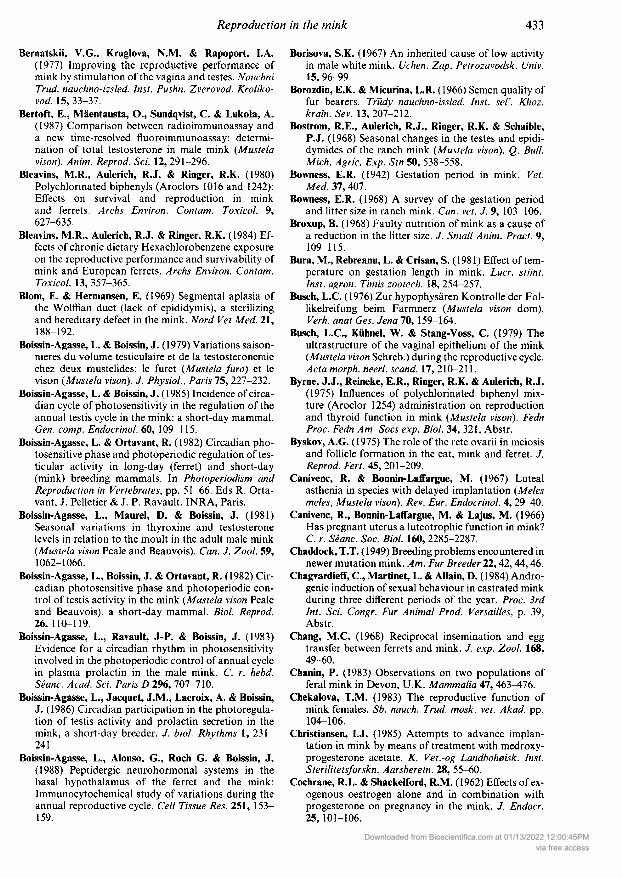
Bernatskii, V.G., Kruglova, N.M. & Rapoport, I.A.(1977) Improving the reproductive performance ofmink by stimulation of the vagina and testes. NauchniTrud. nauchno-izsled. Inst. Pushn. Zverovod. Kroliko¬vod. 15, 33-37.
Bertoft, E., Mäentausta, O., Sundqvist, C & Lukola, A.(1987) Comparison between radioimmunoassay anda new time-resolved fluoroimmunoassay: determi¬nation of total testosterone in male mink (Mustelavison). Anim. Reprod. Sci. 12, 291-296.
Bleavins, M.R., Aulerich, R.J. & Ringer, R.K. (1980)Polychlorinated biphenyls (Aroclors 1016 and 1242):Effects on survival and reproduction in minkand ferrets. Archs Environ. Contam. Toxicol. 9,627-635.
Bleavins, M.R., Aulerich, R.J. & Ringer, R.K. (1984) Ef¬fects of chronic dietary Hexachlorobenzene exposureon the reproductive performance and survivability ofmink and European ferrets. Archs Environ. Contam.Toxicol. 13, 357-365.
Blom, E. & Hermansen, E. (1969) Segmentai aplasia ofthe Wolffian duct (lack of epididymis), a sterilizingand hereditary defect in the mink. Nord Vet Med. 21,188-192.
Boissin-Agasse, L. & Boissin, J. (1979) Variations saison¬nières du volume testiculaire et de la testosteronemiechez deux mustelides: le furet (Mustela furo) et levison (Mustela vison). J. Physiol., Paris 75, 227-232.
Boissin-Agasse, L. & Boissin, J. (1985) Incidence of circa¬dian cycle of photosensitivity in the regulation of theannual testis cycle in the mink: a short-day mammal.Gen. comp. Endocrinol. 60, 109-115.
Boissin-Agasse, L. & Ortavant, R. (1982) Circadian pho¬tosensitive phase and photoperiodic regulation of tes¬ticular activity in long-day (ferret) and short-day(mink) breeding mammals. In Photoperiodism andReproduction in Vertebrates, pp. 51-66. Eds R. Orta-vant, J. Pelletier & J. P. Ravault. INRA, Paris.
Boissin-Agasse, L., Maurel, D. & Boissin, J. (1981)Seasonal variations in thyroxine and testosteronelevels in relation to the moult in the adult male mink(Mustela vison Peale and Beauvois). Can. J. Zool. 59,1062-1066.
Boissin-Agasse, L., Boissin, J. & Ortavant, R. (1982) Cir¬cadian photosensitive phase and photoperiodic con¬trol of testis activity in the mink (Mustela vison Pealeand Beauvois), a short-day mammal. Biol. Reprod.26, 110-119.
Boissin-Agasse, L., Ravault, J-P. & Boissin, J. (1983)Evidence for a circadian rhythm in photosensitivityinvolved in the photoperiodic control of annual cyclein plasma prolactin in the male mink. C. r. hebd.Séanc. Acad. Sci. Paris D 296, 707-710.
Boissin-Agasse, L., Jacquet, J.M., Lacroix, . & Boissin,J. (1986) Circadian participation in the photoregula¬tion of testis activity and prolactin secretion in themink, a short-day breeder. J. biol. Rhythms 1, 231-241.
Boissin-Agasse, L., Alonso, G., Roch G. & Boissin, J.(1988) Peptidergic neurohormonal systems in thebasal hypothalamus of the ferret and the mink:Immunocytochemical study of variations during theannual reproductive cycle. Cell Tissue Res. 251, 153-159.
Borisova, S.K. (1967) An inherited cause of low activityin male white mink. Uchen. Zap. Petrozavodsk. Univ.15, 96-99.
Borozdin, E.K. & Micurina, L.R. (1966) Semen quality offur bearers. Tfûdy nauchno-issled. Inst. sel. Khoz.kraln.Sev. 13,207-212.
Bostrom, R.E., Aulerich, R.J., Ringer, R.K. & Schaible,P.J. (1968) Seasonal changes in the testes and epidi¬dymides of the ranch mink (Mustela vison). Q. Bull.Mich. Agrie. Exp. Stn 50, 538-558.
Bowness, E.R. (1942) Gestation period in mink. Vet.Med. 37, 407.
Bowness, E.R. (1968) A survey of the gestation periodand litter size in ranch mink. Can. vet. J. 9, 103-106.
Broxup, B. (1968) Faulty nutrition of mink as a cause ofa reduction in the litter size. J. Small Anim. Pract. 9,109-115.
Bura, M., Rebreanu, L. & Crisan, S. (1981) Effect of tem¬perature on gestation length in mink. Lucr. stiint.Inst. agron. Timis zootech. 18, 254-257.
Busch, L.C. (1976) Zur hypophysären Kontrolle der Fol-likelreifung beim Farmnerz (Mustela vison dorn).Verh. anat Ges. Jena 70, 159-164.
Busch, L.C, Kühnel, W. & Stang-Voss, C (1979) Theultrastructure of the vaginal epithelium of the mink(Mustela vison Schreb.) during the reproductive cycle.Ada morph. neerl. scand. 17, 210-211.
Byrne, J.J., Reineke, E.R., Ringer, R.K. & Aulerich, R.J.(1975) Influences of polychlorinated biphenyl mix¬ture (Aroclor 1254) administration on reproductionand thyroid function in mink (Mustela vison). FednProc. Fedn Am. Socs exp. Biol. 34, 321, Abstr.
Byskov, A.G. (1975) The role of the rete ovarii in meiosisand follicle formation in the cat, mink and ferret. J.Reprod. Fert. 45, 201-209.
Canivenc, R. & Bonnin-Laffargue, M. (1967) Lutealasthenia in species with delayed implantation (Melesmeles, Mustela vison). Rev. Eur. Endocrinol. 4, 29-40.
Canivenc, R., Bonnin-Laffargue, M. & Lajus, M. (1966)Has pregnant uterus a luteotrophic function in mink?C. r. Séanc. Soc. Biol. 160, 2285-2287.
Chaddock, T.T. ( 1949) Breeding problems encountered innewer mutation mink. Am. Fur Breeder 11,42, 44,46.
Chagvardieff, C, Martinet, L. & Allain, D. (1984) Andro¬genic induction of sexual behaviour in castrated minkduring three different periods of the year. Proc. 3rdInt. Sci. Congr. Fur Animal Prod. Versailles, p. 39,Abstr.
Chang, M.C. (1968) Reciprocal insemination and eggtransfer between ferrets and mink. J. exp. Zool. 168,49-60.
Chanin, P. (1983) Observations on two populations offeral mink in Devon, U.K. Mammalia 47, 463^176.
Chekalova, T.M. (1983) The reproductive function ofmink females. Sb. nauch. Trud. mosk. vet. Akad. pp.104-106.
Christiansen, I.J. (1985) Attempts to advance implan¬tation in mink by means of treatment with medroxy¬progesterone acetate. K. Vet.-og Landboheisk. Inst.Sterilitetsforskn. Aarsberetn. 28, 55-60.
Cochrane, R.L. & Shackelford, R.M. (1962) Effects of ex¬ogenous oestrogen alone and in combination withprogesterone on pregnancy in the mink. J. Endocr.25, 101-106.
Downloaded from Bioscientifica.com at 01/13/2022 12:00:45PMvia free access

Conant, R.A. (1962) A milking technique and the compo¬sition of mink milk. Am. J. vet. Res. 23, 1104-1106.
Concannon, P., Pilbeam, T. & Travis, H. (1980) Ad¬vanced implantation in mink (Mustela vison) treatedwith medroxyprogesterone acetate during earlyembryonic diapause. J. Reprod. Fert. 58, 1-6.
Cummins, J.M. & Woodall, P.F. (1985) On mammaliansperm dimensions. J. Reprod. Fert. 75, 153-175.
Daniel, J.C. (1967) Studies on growth of the mink blasto¬cyst. J. Embryol exp. Morph. 17, 293-302.
Daniel, J.C. ( 1971 ) Termination of pregnancy in mink byrepeated injections during the period precedingimplantation. J. Anim. Sci. 33, 659-661.
Danilov, P.L & Tumanov, LL. (1972) Reproductive cyclesof males in some Mustelidae. Zool. Zh. 51, 871-880.
Danilov, P.I. & Tumanov, I.L. (1975) The reproductivecycle of some females of the Mustelidae family. Byull.mosk. Obshch. Ispyt. Prir. Old. Biol. 80, 137-146.
David, G.F.X. & Herbert, J. (1973) Experimental evi¬dence for a synaptic connection between habenulaand pineal ganglion in the ferret. Brain Res. 64, 327-343.
Deguchi, E. (1978) Histological studies on the kinetics ofthe spermatogenesis in the mink (Mustela vison). VII.Cellular association in the seminiferous epithelium inthe pre-breeding season (16 months old). Jpn J. vet.Res. 26, 1-10.
Di, R.H. & Zou, X.H. ( 1983) The effect of teat number on
reproduction of female mink. Fur Animal Farming 1,8-9.
Donofrio, R.J. (1974) Cyclic AMP in the median emi¬nence of the rat and mink (Mustela vison). Anat. Ree.178, 345, Abstr.
Drekic, D., Jablan-Pantic, O. & Miladinovic, Z. (1981)Morphological properties of mink (Mustela vison)brain. Folia morph. (Praha) 29, 128-130.
Duby, R.T. & Travis, H.F. (1971) Influence of dienestrolacetate on reproductive performance of female mink.Am. J. vet. Res. 32, 1599-1602.
Duby, R.T. & Travis, H.F. (1972) Photoperiodic controlof fur growth and reproduction in the mink (Mustelavisori). J. exp. Zool. 182, 217-225.
Duby, R.T., Pilbeam, T. & Travis, H.F. (1972) Theinfluence of melatonin and hysterectomy on plasmaprogesterone levels of the mink (Mustela vison). Biol.Reprod. 7, 125, Abstr.
Dukelow, W.R. (1966) Variations in gestation length ofmink (Mustela vison). Nature, Lond. 211, 211.
Dunstone, N. (1986) The mink. Biologist 33, 69-75.Einarsson, E.J. (1985) The time of increase in plasma
progesterone during pregnancy in mink (Mustelavison). Theriogenology 24, 375-383.
Elder, W.H. (1951) The baculum as an age criterion inmink. J. Mammal. 32, 43-50.
Ellis, L.C. (1985) Early furring, testicular developmentand cytotoxic lesions of testes and epididymides ofdark mink by melatonin implants. In Pineal Gland:Endocrine Aspects, pp. 145-150. Eds G. M. Brown &S. D. Wainwright. Pergamon Press, New York.
Ellis, L.C. & Pace, N. (1986) Watery kits and neonatalkit loss: factors responsible for mortality. Blue BookofFur Farming 1987; 58-61.
Ellis, L.C. & Pace, N.C. (1987) Genetic problems—alook at today's dark mink. Fur Rancher 67, 10-13.
Ellis, L.C, Groesbeck, M.D. & Howetl, R.E. ( 198 la) Pro¬duction problems in finely-bred dark mink. BlueBook of Fur Farming 1982, 20-21, 65, 88.
Ellis, L.C, Groesbeck, M.D., Farr, CH. & Tesi, R.J.(1981b) Contractility of seminiferous tubules asrelated to sperm transport in the male. Archs Androl.6, 283-294.
Ellis, L.C., Nemetallah, B.H. & Howell, R.E. (1982a)Management of secondary male infertility and neo¬natal mortality in dark mink. Blue Book ofFur Farm¬ing 1983, 67-70.
Ellis, L.C, Groesbeck, M.D. & Howell, R.E. (1982b)Pineal gland—pituitary (a-MSH) interrelationshipsin fur priming and reproductive cycles in mink (Mus¬tela vison). Prog. Clin. Biol. Res. 92, 197-206.
Ellis, L.C, Groesbeck, M.D., Smart, R.A. & Cosentino,M.J. (1985) Spontaneous autocytochemical destruc¬tion of dark mink testes and epididymides. J. Androl.6, 68, Abstr.
Enders, A.C. (1957) Histological observations on thechorio-allantoic placenta of the mink. Anat. Ree. 127,231-245.
Enders, A.C. (I960) Morphological evidence of fluctu¬ation in hormone levels in mink in the period ofdelayed implantation. Anat. Ree. 137, 352, Abstr.
Enders, A.C. ( 1961 ) Comparative studies on the endome¬trium of delayed implantation. Anat. Ree. 139, 483-497.
Enders, A.C. ( 1962) Observations on the fine structure oflutein cells. J. Cell Biol. 12, 101 113.
Enders, A.C., Enders, R.K. & Schlafke, S. (1963) An elec¬tron microscope study of the gland cells of the minkendometrium. J. Cell Biol. 18, 405^*18.
Enders, R.K. (1938) The ovum of the mink (Mustelavison). Anat. Ree. 72, 469^171.
Enders, R.K. (1948) Induced mating in ovariectomizedmink. Anat. Ree. 100, 738, Abstr.
Enders, R.K. (1949) Multiple ovulation in mink breeding.Am. Fur Breeder 21, 6, 8.
Enders, R.K. (1952) Reproduction in the mink (Mustelavisori). Proc. Am. Philos. Soc. 96, 691-755.
Enders, R.K. & Pearson, A.K. (1946) Mink blastocysts invitro. Anat. Ree. 96, 570, Abstr.
Enders, R.K. & Enders, A.C. (1963) Morphology of thefemale reproductive tract during delayed implan¬tation in the mink. In Delayed Implantation, pp. 129-140. Ed. A. C. Enders. Chicago University Press,Chicago.
Evsikov, V.l. & Belyaev, D.K. (1968) Genetic aspects ofreproduction in the mink (Lutreola vison Brisson).Proc. 6th Int. Cong. Anim. Reprod. & A.L, Paris, p.252, Abstr.
Fiedler, J., Skrivan, M. & Siler, R. (1975) Influence ofrepeated mating on litter size in standard minks.Zivocisna vyroba 20, 687-692.
Franklin, B.C. (1958) Studies on the effects of progester¬one on the physiology of reproduction in the mink.Mustela vison. Ohio J. Sci. 58, 163-170.
Friend, D.W. & Crampton, E.W. (1960) Observations on
the effect of four mating routines for female mink.Can. J. comp. Med. Vet. Sci. 24, 3-5.
Gilbert, F.F. (1969) Physiological effects of natural DDTresidues and metabolites on ranch mink. J. Wild!.Mgml 33, 933-943.
Downloaded from Bioscientifica.com at 01/13/2022 12:00:45PMvia free access

Gilbert, F.F. & Bailey, E.D. (1967) The effect of visualisolation on reproduction in the female ranch mink.J. Mammal. 48, 113-118.
Gilbert, F.F. & Bailey, E.D. (1969a) Visual isolation andstress in female ranch mink particularly during thereproductive season. Can. J. Zool. 47, 209-212.
Gilbert, F.F. & Bailey, E.D. (1969b) The effect of earlyweaning on the sexual behavior and reproductivesuccess of ranch mink. J. Mammal. 50, 742-747.
Gilbert, F.F. & Bailey, E.D. (1970) Reproductive per¬formance of three genetic strains of female mink iso¬lated after breeding. Cornell Vet. 60, 135-138.
Ginther, O.J., Bergfelt, D.R., Scraba, S.T., Pivonka, P.R.& Nuti, L.C. (1985) Effects of 6-MBOA on repro¬ductive function in ponies, mice, rats and mink. Ther¬iogenology 24, 587-595.
Goulevich, R.G., Osadchuk, L.V. & Klochkov, D.V.(1984) Endocrine function in the gonads in males oftwo genotypes of the mink Mustela vison. J. Evol.Biochem. Physiol. 20, 316-318.
Graf, Z. (1985) Development and use of an electroejacu-lator for zoo and domestic animals. Magy. Allatorv.Lap. 40, 169-174.
Gufamhusein, A.P. & Thawley, A.R. (1972) Ovarian cycleand plasma progesterone levels in the female stoat,Mustela erminea. J. Reprod. Fert. 31, 492-493.
Gulyas, B.J., Daniel, J.C. & Krishnan, R.S. (1969) Incor¬poration of labelled nucleosides in vitro by rabbit andmink blastocysts in the presence of blastokinin or
serum. J. Reprod. Fert. 20, 255-262.Gunn, CK. (1949) Mink Ranching. Pubi. Dept. Agrie.
Canada. No. 827, 60 pp.Hammond, J. (1951a) Control by light of reproduction in
ferrets and mink. Nature, Lond. 167, 150-151.Hammond, J. (1951b) Failure of progesterone treatment
to affect delayed implantation in the mink. J. Endocr.7, 330-334.
Hammond, J. (1952) Gonadotrophin-induced ovulationin mink. J. Mammal. 33, 218-233.
Hansson, A. (1947) The physiology of reproduction inmink (Mustela vison, Schreb.) with special reference todelayed implantation. Ada zool. (Stockh.) 28, 1-136.
Hartsough, G.R. (1942) A report on the use of Gonadinin mink. Am. Fur. Breeder 14, 18-19.
Härtung, J. & Seffner, W. (1983) Fortpflanzungsstörun¬gen bei Nerzrüden. Mschr. VetMed. 38, 611-617.
Hattenhauer, H. (1984) Effect of GnRH vet. "BerlinChemie" on reproductive performance of mink.Mschr. VetMed. 39, 271-273.
Hattenhauer, H. & Pötschulat, A. (1984) Experimentaluse of GnRH vet. "Berlin-Chemie" for single matingof mink females. Mschr. VetMed. 39, 524-527.
Hattenhauer, H., Pingel, H. & Elze, K. (1984) Use ofPMSG and hCG in mink breeding. Mschr. VetMed.39, 520-523.
Hedlund, K., Nilsson, ().. Reinius, S. & Aman, G. (1972)Attachment reaction of the uterine luminal epi¬thelium at implantation: light and electron mi¬croscopy of the hamster, guinea-pig, rabbit andmink. /. Reprod. Fert. 19, 131-132.
Helgebostad, A. (1968) Anaemia in the mink. Nord.VetMed. 20, 161-172.
Helgebostad, A. (1980) Embryonic death in mink due toriboflavine deficiency. Nord. VetMed. 32, 313-317.
Helgebostad, ., Svenderup, R. & Ender, F. (1963) Steri¬lity in mink induced experimentally by deficiency ofvitamin B6. Ada vet. scand. 4, 228-233.
Hemmingsen, B. (1967) Postnatal development and cyclicchanges in the testes of mink. Nord. VetMed. 19, 71-80.
Hernesniemi, E. (1980) The effect of visual isolation onmink reproduction. Scienlifur 4, 21-24.
Heron, L. & Rietveld, A.A. (1985a) Is there a correlationbetween testicle size and reproductive performance?Scienlifur 9, 296.
Heron, L. & Rietveld, A.A. (1985b) A digest of Dr CyrilAdams's research on mink reproduction. Scienlifur 9,297-300.
Heron, L. & Rietveld, W. (1986) An approach to selec¬tion for male reproductive performance. 10th AnnShort Course, Mishicot, Wisconsin, 6 pp.
Hodgson, R.G. (1945) The mink book. Fur Trade Jl Can.,Toronto, 284 pp.
Hodgson, R.G. (1958) The mink book. Fur Trade Jl Can.,Toronto, 272 pp.
Holcomb, L.C. (1967) Effects of progesterone treatmentson delayed implantation in mink. Ohio J. Sci. 67, 24-31.
Holcomb, L.C, Schaible, P.J. & Ringer, R.K. (1962) Theeffects of varied lighting regimes on reproduction inmink. Q. Bull. Mich. Agrie. Exp. Sin 44, 666-678.
Hornshaw, T.C, Aulerich, R.J. & Johnson, H.E. (1983)Feeding Great Lakes (USA, Canada) fish to mink:effects on mink and accumulation and elimination ofpolychlorinated biphenyls by mink. J. Toxicol.Environ. Health 11, 933-946.
Howell, J.M. & Pickering, CM. (1964) Suspected syn¬thetic oestrogen poisoning in mink. Vet. Ree. 76,169-170.
Isakova, G.K. (1981) Polyploidy in the bone marrow andspermatogenic epithelium of male mink of differingfertility. Genetika 17, 858-872.
Ishikawa, R., Tiba, T., Kagota, K., Kawabe, K. & Kinos-hita, S. (1965) Experimental study of artificial insemi¬nation in mink. Jpn. J. vet. Res. 13, 1-16.
Isupov, B.A., Barantseva, E.D., Kulikova, T.G., Kryakin,A.A. & Yureva, LM. (1975) The effect of nutritionallevel of mink before the breeding season and duringpregnancy on the yield of progeny. Sb. nauehno-tekh.Inform, vses. nauehno-issled. Inst. Okhot. Khoz. Zver¬ovod. 49, 150-157.
Jacquet, JM.. Coûtant, C, Maurel, D., Boissin-Agasse,L. & Boissin, J. (1986) Effects of thyroidectomy on
spring and summer variations in testis activity andplasma prolactin in the mink. C. r. hebd. Séanc. Acad.Sci., Paris 303 (Ser. Ill), 367-370.
Jakovac, M., Kopljar, VI.. Grabar, ., Kos, M. & Saric,B. (1977) Semen collection in male mink (Mustelavison) by electroejaculation. Vet. Arh. 47, 95-98.
Jarosz, S. & Barteczko, J. ( 1976) The effect of rationscontaining bovine and porcine uteri, fish and fat on
fertility and fecundity of mink. Zesz. Probi. Poste-pow. Nauk. Roln. 180, 491^198.
Jarosz, S. & Barabasz, B. (1984) Effect of environmentalfactors on fertility in mink. Proc. 3rd Int. Sci. Congr.Fur Animal Prod., Versailles, p. 27, Abstr.
Jarosz, S.J. & Dukelow, W.R. (1985) Effect of progester¬one and medroxyprogesterone acetate on pregnancy
Downloaded from Bioscientifica.com at 01/13/2022 12:00:45PMvia free access
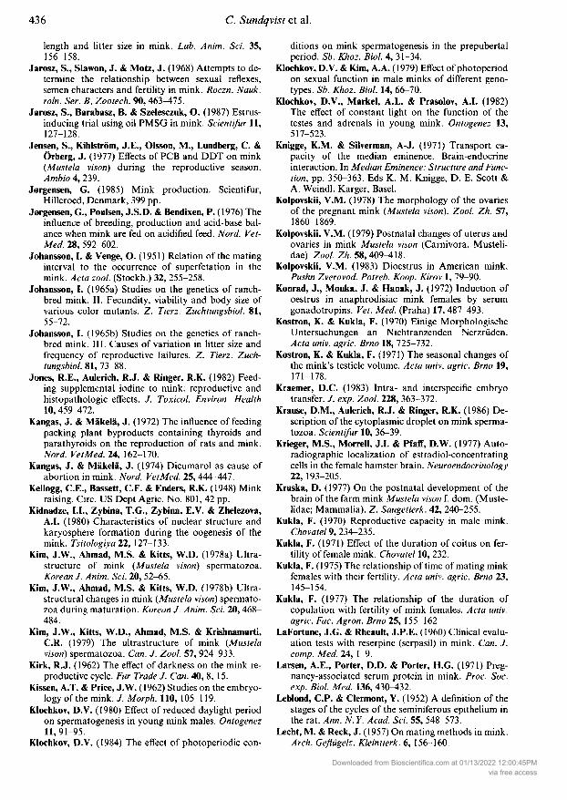
length and litter size in mink. Lab. Anim. Sci. 35,156-158.
Jarosz, S., Slawen, J. & Motz, J. (1968) Attempts to de¬termine the relationship between sexual reflexes,semen characters and fertility in mink. Roezn. Nauk.roln. Ser. , Zootech. 90, 463^175.
Jarosz, S., Barabasz, . & Szelesczuk, O. (1987) Estrus-inducing trial using oil PMSG in mink. Scienlifur 11,127-128.
Jensen, S., Kihlström, J.E., Olsson, M., Lundberg, C. &Örberg, J. (1977) Effects of PCB and DDT on mink(Mustela vison) during the reproductive season.Ambio 4, 239.
Jergensen, G. (1985) Mink production. Scientifur,Hilleroed, Denmark, 399 pp.
Jergensen, G., Poulsen, J.S.D. & Bendixen, P. (1976) Theinfluence of breeding, production and acid-base bal¬ance when mink are fed on acidified feed. Nord. Vet¬Med. 28, 592-602.
Johansson, I. & Venge, O. (1951) Relation of the matinginterval to the occurrence of superfetation in themink. Ada zool. (Stockh.) 32, 255-258.
Johansson, I. (1965a) Studies on the genetics of ranch-bred mink. II. Fecundity, viability and body size ofvarious color mutants. Z. Tierz. Zuchtungsbiol. 81,55-72.
Johansson, I. (1965b) Studies on the genetics of ranch-bred mink. III. Causes of variation in litter size andfrequency of reproductive failures. Tierz. Zuch¬tungsbiol. 81, 73-88.
Jones, R.E., Aulerich, R.J. & Ringer, R.K. (1982) Feed¬ing supplemental iodine to mink: reproductive andhistopathologic effects. J. Toxicol. Environ. Health10, 459^172.
Kangas, J. & Mäkelä, J. (1972) The influence of feedingpacking plant byproducts containing thyroids andparathyroids on the reproduction of rats and mink.Nord. VetMed. 14, 162-170.
Kangas, J. & Mäkelä, J. (1974) Dicumarol as cause ofabortion in mink. Nord. VetMed. 25, 444-447.
Kellogg, C.E., Bassett, CF. & Enders, R.K. (1948) Minkraising. Circ. US Dept Agrie. No. 801, 42 pp.
Kidnadze, LL, Zybina, T.G., Zybina, E.V. & Zhelezova,A.I. (1980) Characteristics of nuclear structure andkaryosphere formation during the oogénesis of themink. Tsitologiya 11, 127-133.
Kim, J.W., Ahmad, M.S. & Kitts, W.D. (1978a) Ultra-structure of mink (Mustela vison) spermatozoa.Korean J. Anim. Sci. 20, 52-65.
Kim, J.W., Ahmad, M.S. & Kitts, W.D. (1978b) Ultra-structural changes in mink (Mustela vison) spermato¬zoa during maturation. Korean J. Anim. Sei. 20,468-484.
Kim, J.W., Kitts, W.D., Ahmad, M.S. & Krishnamurti,CR. (1979) The ultrastructure of mink (Mustelavison) spermatozoa. Can. J. Zool. 57, 924-933.
Kirk, R.J. (1962) The effect of darkness on the mink re¬
productive cycle. Fur Trade J. Can. 40, 8, 15.Kissen, A.T. & Price, J.W. (1962) Studies on the embryo¬
logy of the mink. / Morph. 110, 105-119.Klochkov, D.V. (1980) Effect of reduced daylight period
on spermatogenesis in young mink males. Ontogenez11,91-95.
Klochkov, D.V. (1984) The effect of photoperiodic con-
ditions on mink spermatogenesis in the prepubertalperiod. Sb. Khoz. Biol. 4, 31-34.
Klochkov, D.V. & Kim, A.A. (1979) Effect of photoperiodon sexual function in male minks of different geno¬types. Sb. Khoz. Biol. 14, 66-70.
Klochkov, D.V., Markel, A.L. & Prasolov, A.I. (1982)The effect of constant light on the function of thetestes and adrenals in young mink. Ontogenez 13,517-523.
Knigge, K.M. & Silverman, A-J. (1971) Transport ca¬
pacity of the median eminence. Brain-endocrineinteraction. In Median Eminence: Structure and Func¬tion, pp. 350-363. Eds K. M. Knigge, D. E. Scott &A. Weindl. Karger, Basel.
Kolpovskii, V.M. (1978) The morphology of the ovariesof the pregnant mink (Mustela vison). Zool. Zh. 57,1860-1869.
Kolpovskii, V.M. (1979) Postnatal changes of uterus andovaries in mink Mustela vison (Carnivora, Musteli¬dae). Zool. Zh. 58, 409^118.
Kolpovskii, V.M. (1983) Dioestrus in American mink.Pushn Zverovod. Polreb. Koop. Kirov 1, 79-90.
Konrad, J., Mouka, J. & Hanak, J. (1972) Induction ofoestrus in anaphrodisiac mink females by serum
gonadotropins. Vet. Med. (Praha) 17, 487-493.Kostron, K. & Kukla, F. (1970) Einige Morphologische
Untersuchungen an Nichtranzenden Nerzrüden.Ada univ. agrie. Brno 18, 725-732.
Kostron, K. & Kukla, F. (1971) The seasonal changes ofthe mink's testicle volume. Acta univ. agrie. Brno 19,171-178.
Kraemer, D.C. (1983) Intra- and interspecific embryotransfer. J. exp. Zool. 228, 363-372.
Krause, D.M., Aulerich, R.J. & Ringer, R.K. (1986) De¬scription of the cytoplasmic droplet on mink sperma¬tozoa. Scienlifur 10, 36-39.
Krieger, M.S., Morrell, J.I. & Pfaff, D.W. (1977) Auto-radiographic localization of estradiol-concentratingcells in the female hamster brain. Neuroendocrinology11, 193-205.
Kruska, D. (1977) On the postnatal development of thebrain of the farm mink Mustela vison f. dorn. (Muste¬lidae; Mammalia). . Saugetìerk. 42, 240-255.
Kukla, F. (1970) Reproductive capacity in male mink.Chovatel 9, 234-235.
Kukla, F. (1971) Effect of the duration of coitus on fer¬tility of female mink. Chovatel 10, 232.
Kukla, F. (1975) The relationship of time of mating minkfemales with their fertility. Ada univ. agrie. Brno 23,145-154.
Kukla, F. (1977) The relationship of the duration ofcopulation with fertility of mink females. Ada univ.agrie. Fac. Agron. Brno 25, 155-162.
lai ornine, J.G. & Rheault, J.P.E. (1960) Clinical evalu¬ation tests with reserpine (serpasil) in mink. Can. J.comp. Med. 24, 1-9.
Larsen, A.E., Porter, D.D. & Porter, H.G. (1971) Preg¬nancy-associated serum protein in mink. Proc. Soc.exp. Biol. Med. 136, 430-432.
Leblond, CP. & Clermont, Y. (1952) A definition of thestages of the cycles of the seminiferous epithelium inthe rat. Ann. N.Y. Acad. Sci. 55, 548-573.
Lecht, M. & Reck, J. (1957) On mating methods in mink.Arch. Geflügelz. Kleintierk. 6, 156-160.
Downloaded from Bioscientifica.com at 01/13/2022 12:00:45PMvia free access

Long, CA. (1969) Gross morphology of the penis in seven
species of the Mustelidae. Mammalia 33, 145-160.Long, CA. & Shirek, L.R. (1970) Variation and corre¬
lation in the genital bones of ranch mink. Z. Sauge-tierk. 35,252-255.
Lukola, A. & Sundqvist, C. (1986) Improved spermcounts in mink males (Mustela vison) treated withclomiphene citrate. J. Endocr. Invest. 9, 243-244.
Maciejowski, J., Slawon, J. & Brzozowski, S. (1973) Ef¬fect of different mating systems on the fertility ofmink. Pr. Mater. Zootech. 4, 55-68.
MacLennan, R.R. & Bailey, E.D. (1972) Role of sexualexperience in breeding behavior of male ranch mink.J. Mammal. 53, 380-382.
Martinet, L. & Allain, D. (1985) Role of the pineal glandin the photoperiodic control of reproductive andnon-reproductive functions in mink (Mustela visori).In Photoperiodism, Melatonin and the Pineal (CibaFdn Symp. 117), pp. 170-187. Pitman, London.
Martinet, L., Allain, D. & Meunier, M. (1978) Photoper¬iodic control of plasma testosterone concentration inthe mink (Mustela visori). Scienlifur 2, 16-19.
Martinet, L., Allais, C & Allain, D. (1981a) Role of pro¬lactin and LH in luteal function and blastocystgrowth in mink (Mustela visori). J. Reprod. Fert.,Suppl.19, 119-130.
Martinet, L., Meunier, M. & Allain, D. (1981b) Controlof delayed implantation and onset of spring moult inthe mink (Mustela visori) by daylight ratio, prolactinand melatonin. In Photoperiodism and Reproductionin Vertebrates, pp. 253-261. Eds R. Ortavant, J. Pel-letter & J. P. Ravault. INRA, Paris.
Martinet, L., Ravault, J.P. & Meunier, M. (1982)Seasonal variations in mink (Mustela visori) plasmaprolactin measured by heterologous radioimmunoas¬say. Gen. comp. Endocrinol. 48, 71-75.
Martinet, L., Allain, D. & Meunier, M. (1983) Regulationin pregnant mink (Mustela visori) of plasma pro¬gesterone and prolactin concentrations and regu¬lation of onset of the spring moult by daylight ratioand melatonin injections. Can. J. Zool. 61, 1959-1963.
Martinet, L., Allain, D. & Chabi, Y. (1985) Pineal dener¬vation by cervical sympathetic ganglionectomy sup¬presses the role of photoperiod on pregnancy or pseu¬dopregnancy, body weight and moulting periods inthe mink (Mustela visori). J. Endocr. 107, 31-39.
Mead, R.A. (1975) Effects of hypophysectomy on blasto¬cyst survival, progesterone secretion and nidation inthe spotted skunk. Biol. Reprod. 15, 526-532.
Mead, R.A. (1981) Delayed implantation in mustelidswith special emphasis on the spotted skunk. J.Reprod. Fert., Suppl. 29, 11-24.
Miladinovic, Z. (1969) Morphology and vascularizationof the female genital organs in mink. Ada. vet.
(Belgr.) 19, 155-165.Miljkovic, V., Zivanovic, D. & Buric, I. (1966) Electro¬
ejaculation in the mink and some laboratory animals.Ada. vet. (Belgr.) 16, 363-371.
Mills, J. (1961) Diethylstilbestrol as a contaminant inmink feeding. Can. J. comp. Med. Vet. Sci. 25, 205-210.
Moller, O.M. (1973a) The fine structure of the ovarianinterstitial gland cells in the mink, Mustela vison. J.Reprod. Fert. M, 171-174.
Meiler, O.M. (1973b) The progesterone concentrationsin the peripheral plasma of the mink (Mustela vison).J. Endocr. 56, 121-132.
Meiler, O.M. (1973c) The fine structure of the lutein cellsin the mink (Mustela vison) with special reference tothe secretory activity during pregnancy. Z. Zell-forsch. mikrosk. Anat. 138, 523-544.
Meiler, O.M. (1974) Plasma progesterone before andafter ovariectomy in unmated and pregnant mink,Mustela vison. J. Reprod. Fert. 37, 367-372.
Meiler, O. & Nordstoga, . (1978) Endotoxin-inducedembryonic death in mink. Scienlifur 1, 7-10.
Mondain-Monval, M., Caillol, M. & Meunier, M. (1985)Heterologous radioimmunoassay of LH in two sea¬
sonally breeding animals: hare (Lepus europaeus) andmink (Mustela visori). Can. J. Zool. 63, 1339-1344.
Morrell, J.L, Ballin, A. & Pfaff, D.W. (1977) Autoradio¬graphic demonstration of the pattern of 3H-estradiolconcentrating cells in the brain of a carnivore, themink, Mustela vison. Anat. Ree. 189, 609-624.
Moshonkin, N.N. (1981) Potential polyestricity of themink. Zool. Zh. 60, 1731-1733.
Muresan, E., Miclea, M., Duca, C & Papay, Z. (1984)Morphological study of mink ovary within 1 sexualcycle. Bull. Inst. Agron. Cluj-Napoca, Ser. Zootech.Med. Vet. 38, 61-64.
Murphy, B.D. (1976) Effects of synthetic GnRH on littersize in ranch mink bred once or twice. Theriogenology6, 463^169.
Murphy, B.D. (1977) Effect of lengthened photoperiodafter mating on litter size in mink. Scienlifur 1, 16-19.
Murphy, B.D. (1979a) Effects of GnRH on plasma LHand fertility in mink. Can. J. Anim. Sci. 59, 25-33.
Murphy, B.D. (1979b) The role of prolactin in implan¬tation and luteal maintenance in the ferret. Biol.Reprod. 21,517-521.
Murphy, B.D. (1983a) Effects of single or multiple injec¬tions of medroxyprogesterone acetate on the repro¬ductive performance and gestation length of ranchmink. Can. J. Anim. Sci. 63, 989-991.
Murphy, B.D. (1983b) Precocious induction of lutealactivation and termination of delayed implantationin mink with the dopamine antagonist pimozide.Biol Reprod. 29, 658-662.
Murphy, B.D. & James, D.A. (1974a) The effects of lightand sympathetic innervation to the head on nidationin mink. J. exp. Zool. 187, 267-276.
Murphy, B.D. & James, D.A. (1974b) Mucopolysacchar-ide histochemistry of the mink uterus during ges¬tation. Can. J. Zool. 52, 687-693.
Murphy, B.D. & James, D.A. (1976) Cells of the adeno¬hypophysis of the mink (Mustela visori) identified byimmunohistochemical and functional criteria. Adaanat. 94, 184-203.
Murphy, B.D. & Moger, W.H. (1977) Progestins of minkgestation: the effects of hypophysectomy. Endocr.Res. Commun. 4, 45-60.
Murphy, B.D., Humphrey, W.D. & Shepstone, S.L.(1980) Luteal function in mink: The effects of hypo¬physectomy after the preimplantation rise in pro¬gesterone. Anim. Reprod. Sci. 3, 225-232.
Murphy, B.D., Concannon, P.W., Travis, H.F. & Hansel,W. (1981) Prolactin: the hypophyseal factor that ter¬minates embryonic diapause in mink. Biol. Reprod.»,487-491.
Downloaded from Bioscientifica.com at 01/13/2022 12:00:45PMvia free access
Murphy, B.D., Concannon, P.W. & Travis, H.F. (1982)Effects of medroxyprogesterone acetate on gestationin mink. J. Reprod. Fert. 66, 491^197.
Murphy, B.D., Mead, R.A. & McKibbin, P.E. (1983)Luteal contribution to the termination of preimplan¬tation delay in mink. Biol. Reprod. 28, 497-503.
Murphy, B.D., Rajkumar, K. & Silversides, D.W. (1984)Luteotrophic control of the mink corpus luteum dur¬ing the postimplantation phase of gestation. Proc.3rd Int. Sci. Congr. Fur Animal Prod., Versailles, p.32, Abstr.
Narucka, I. (1973) Effect of time and system of mating onlitter size in mink. Roczn. Akad. roln. Pozn. 66, 97104.
Nemetallah, B.R., Howell, R.E. & Ellis, L.C. (1985) His¬tamine and secondary autoimmune infertility in darkmink (Mustela vison). Archs Androl. 15, 79-82.
Nés, N. (1965) Abnormalities of the female genital or¬
gans in mink heterozygous for the Heggedal factor(Shadow factor). Ada vet. scand. 6, 65-99.
Nieschlag, E. & Bieniek, H. (1975) Endocrine testicularfunction in mink during the first year of life. Adaendocr., Copenh. 79, 375-379.
Nosova, N.G. (1977a) Effect of the progestogen meges-trol acetate on the ovaries and uterus of minks duringthe estrus cycle. Proc. 2nd All-Union Scient. Congr.,Kirov,pp. 126-127.
Nosova, N.G. (1977b) Some reasons for infertility infemale mink. Nauchni Trud. nauchno-izsled. Inst.Pushn. Zverovod. Krolikovod. 15, 13-20.
Nosova, N.G. (1977c) Ovulation and the development ofcorpora lutea in female mink. Nauchni Trud.nauchno-izsled. Inst. Pushn. Zverovod. Krolikovod. 15,21-32.
Nosova, N.G. (1978) Morphological and histologicalchanges of ovaries, uterus and thyroid gland in minkgiven megestrol acetate. Nauchni Trud. nauchno-izsled. Inst. Pushn. Zverovod. Krolikovod. 17, 61-75.
Onstad, O. (1961a) Hypoplasia of testes in mink. Nord.VetMed. 13, 510-516.
Onstad, O. (1961b) Aplasia of epididymis and vas defer¬ens as a cause of sterility in mink. Nord. VetMed. 13,543-549.
Onstad, O. (1967) Studies on postnatal testicularchanges, semen quality, and anomalies of repro¬ductive organs in the mink. Ada endocr., Copenh.,Suppl. 117, 1-117.
Ortavant, R., Pelletier, J., Ravault, J.P., Thimonier, J. &Volland-Nail, P. (1985) Photoperiod: main proximaland distal factor of the circannual cycle of repro¬duction in farm mammals. Oxford Rev. Reprod. Biol.1, 305-345.
Pace, N.C, Warren, R.P. & Ellis, L.C. (1987) A naturalkiller cell assay for the mink using a mouse lym¬phoma as the target cell line. Lab. Anim. Sci. 37, 220-223.
Papke, R.L., Concannon, P.W., Travis, H.F. & Hansel,W. (1980) Control of luteal function and implan¬tation in the mink by prolactin. J. Anim. Sci. 50,1102-1107.
Paul, J.R. (1968) Baculum development in mink. TransIH. Acad. Sa. 61, 308-309.
Pearson, O.P. & Enders, R.K. (1944) Duration of preg¬nancy in certain Mustelids. J. exp. Zool. 95, 21-35.
Pekkanen, T., Lindberg, P. & Sankari, S. (1983) The ef¬fect of colistin feeding during mating, gestation andlactation periods on whelping result in mink. Nord.VetMed. 35, 91-94.
Pelletier, R-M. (1986a) Differentiation of annulatelamellae during spermiogenesis in the mink. J. CellBiol. 103, 83A, Abstr.
Pelletier, R-M. (1986b) Cyclic formation and decay ofthe blood-testis barrier in the mink (Mustela visori), aseasonal breeder. Am. J. Anat. 175, 91-117.
Pelletier, R-M. & Shivers, R.R. (1986) Filipin-sterol com¬
plexes in breeding and nonbreeding mink (Mustelavisori) Sertoli cell junctional membranes. J. Cell Biol.103, 365A, Abstr.
Petrova, I.P., Reznikov, A.G., Kolpovskii, V.M. & Dem-chenko, V.N. (1983) The content of sex steroids in theblood of pregnant American mink. Fiziol. Zh. (Kiev)29, 434-438.
Pilbeam, T.E., Concannon, P.W. & Travis, H.F. (1979)The annual reproductive cycle of the mink (Mustelavisori). J. Anim. Sci. 48, 578-584.
Pilleri, G. (1960) The brain of Mustela vison and Mem-phitis memphitis (Carnivora, Mustelidae). Rev. SuisseZool. 67, 141-158.
Platonow, N.S. & Karstad, L.H. (1973) Dietary effects ofpolychlorinated biphenyls on mink. Can. J. comp.Med. 37, 391^400.
Pomytko, V.N., Bernatskii, V.G., Kruglova, N.M. &Dubovaya, R.G. (1972) Semen collection, dilution,storage and artificial insemination in mink. NauchniTrud. nauchno-izsled. Inst. Pushn. Zverovod. Kroliko¬vod. 11, 165 170.
Popov, V.A. (1943) A new age index in Mustelinae. Dokl.Akad. nauk SSSR 38,258-260.
Prasolov, A.I. (1979) A morphometric study of the sperm¬atogenic tubules of the mink during the period ofinvolution and regression of the spermatogenic epi¬thelium. Nauchni Trudy Voronezh. Lesokhim Inst.106, 105-112.
Rauluszkiewicz, S., Senze, A. & Nogala, M. ( 1971 ) Prac¬tical application of cytological and cytogenetic exam¬inations of mink. Medycyna wet. 27, 302-304.
Ravault, J.P., Martinet, L., Bonnefond, C, Claustrat, .& Brun, J. (1986) Diurnal variations of plasma mela¬tonin concentrations in pregnant mink (Mustelavisori) maintained under different photoperiods. /.Pineal. Res. 3, 365-373.
Rebreanu, L., Bura, M. & Crisan, S. (1981a) Effect ofnumber of matings and prolificacy on gestationlength in mink. Lucr. Sliint. Inst. agron. Timis. Zoo-tech. 18, 249-253.
Rebreanu, L., Bura, M. & Crisan, S. (1981b) Effect oflight factors on gestation length in mink. Lucr. Stiint.Inst. agron. Timis. Zootech. 18, 258-263.
Reiter, R.J. (1974) Circannual reproductive rhythms inmammals related to photoperiod and pineal func¬tion: a review. Chronobiologia 1, 365-395.
Ribas, J.L. (1973) Ultrastructural study of organ-cul¬tured rat and mink median-eminence and pars tuber¬alis. In Vitro 8, 439, Abstr.
Rietveld, A.A. (1978) Three years of practical applicationof hCG at Northwood Fur Farms Inc. Seientifur 1,27-33.
Rietveld, A.A.M. (1982) Tolerance levels of PCB in minkfeed. Seientifur 6, 35-37.
Downloaded from Bioscientifica.com at 01/13/2022 12:00:45PMvia free access

Ringer, R.K., Aulerich, R.J. & Bleavins, M.R. (1981) Bio¬logical effects of PCBs and PBBs on mink and fer¬rets—a review. In Halogenaled Hydrocarbons: Healthand Ecological Effects, pp. 329-343. Ed. M. A. Q.Khan. Pergamon Press, Elmsford.
Roosen-Runge, E.C. & Giesel, L.O. (1950) Quantitativestudies on spermatogenesis in the albino rat. Am. J.Anat. 87, 1-30.
Rose, J., Stormshak, F., Adair, J. & Oldfield, J. (1983a)Prolactin binding sites in the uterus of the mink.Molec. cell. Endocr. 31, 131-139.
Rose, J., Stormshak, F., Adair, J. & Oldfield, J. (1983b)Uterine progesterone metabolism in the mink duringdelayed implantation. Biol. Reprod. 28 (Suppl) 1, 137,Abstr.
Rose, J., Oldfield, J.E. & Stormshak, F. (1986) Changesin serum prolactin concentrations and ovarian pro¬lactin receptors during embryonic diapause in mink.Biol. Reprod. 34, 101-106.
Rouvet, D.M. (1982) Ultrastructure of the pineal gland ofthe mink (Mustela visori). Am. J. vet. Res. 43, 1492-1496.
Rush, G.F., Smith, J.H., Maita, K., Bleavins, M., Auler¬ich, R.J., Ringer, R.K. & Hook, J.B. (1983) Perinatalhexachlorobenzene toxicity in the mink. Environ.Res. 31, 116-124.
Sakai, Y. (1981) A quantitative study on the seminifer¬ous epithelium of the adult mink in the post-breedingseason. Jpn J. vet. Res. 29, 30, Abstr.
Shackelford, R.M. (1952) Superfetation in the ranchmink. Am. Nat. 86, 311-319.
Shackelford, R.M. (1984) American mink. In Evolutionof Domesticated Animals, pp. 229-234. Ed. I. L.Mason. Longman Group Ltd., London.
Shackelford, R.M. & Cochrane, R.L. (1962) Repro¬ductive performance of female mink fed stilbestrol. J.Anim. Sci. 21,226-231.
Silverman, A-J. & Knigge, K.M. (1972) Transport ca¬
pacity of the median eminence. II. Thyroxine trans¬port. Neuroendocrinology 10, 71-82.
Silverman, A-J. & Knigge, K.M. (1973) Transport ca¬
pacity of median eminence: III. Amino acid and thyr¬oxine transport of organ-cultured median eminence.Neuroendocrinology 11, 107-118.
Silverman, A-J., Knigge, K.M. & Peck, W.A. (1972)Transport capacity of median eminence. I. Aminoacid transport. Neuroendocrinology 9, 123-132.
Skrede, A. (1978) Utilization offish and animal by-pro¬ducts in mink nutrition. II. Effect of source and levelof protein on female reproductive performance, andpreweaning growth and mortality of the progeny.Ada agrie, scand. 28, 130-140.
Skrivan, M., Fiedler, J. & Siler, R. (1975) The number ofsterile females in a population of standard mink.Zivocisna vyroba 20, 681-686.
Stevenson, W.G. (1945) The gestation period of mink.Can. J. comp. Med. 9, 38-39.
Stevenson, W.G. (1946) The effect of sunlight on theinitiation of sexual activity in ranch mink. Can. J.comp. Med. 10, 137-142.
Stole, M., Fantova, M. & Skrivan, M. (1984) Fertility offemale Standard mink mated at different frequencies.Zivocisna vyroba 41, 123-133.
Sundqvist, C. (1985) Morphometric studies on mink testi¬cular tissue. Theriogenology 24, 713-723.
Sundqvist, C. (1986) Recent male mink reproductionstudies and some prospects for the future. Seientifur10,261-264.
Sundqvist, C. & Gustafsson, M. ( 1983) Sperm test—a use¬ful tool in breeding work on mink. J. Sci. agrie. Soc.Fini. 55, 119-131.
Sundqvist, C. & Sundqvist, T. (1986) Some observationson semen quality in mink of different fur quality.Seientifur \tS, 117-118.
Sundqvist, C, Lukola, A. & Valtonen, M. (1984) Re¬lationship between serum testosterone concen¬trations and fertility in male mink (Mustela visori). J.Reprod. Fert. 70, 409^112.
Sundqvist, C, Lukola, A. & Valtonen, M. (1985) Repro¬ductive capacity in male mink after long distancetransportation in pregnant females. Andrologia 17,575-578.
Sundqvist, C, Toppari, J., Parvinen, M., Fagerström, R.& Lukola, A. (1986a) Elimination of infertile malemink from breeding using sperm test, testicular pal¬pation, testosterone test and fine-needle aspirationbiopsy of the testis. Anim. Reprod. Sci. 11, 295-305.
Sundqvist, C, Lukola, A. & Parvinen, M. (1986b) Testi¬cular aspiration biopsy in evaluation of fertility inmink (Mustela visori). J. Reprod. Fert. 77, 531-535.
Sundqvist, C, Parvinen, M. & Lukola, A. (1987) Aspi¬ration biopsy of the testis—a useful tool in mink.Seientifur 11,209-211.
Sundqvist, C, Ellis, L.C. & Bartke, A. (1988) Repro¬ductive endocrinology of the mink (Mustela vison).Endocr. Rev. 9, 247-266.
Svihla, A. (1931) Habits of the Louisiana mink (Mustelavison vulgivaqus). J. Mammal. 12, 366-368.
Tauson, A- . (1985a) Different feeding intensity levels tomink. 1. Effects on male reproductive performance.Swed. J. agrie. Res. 15, 77-85.
Tauson, A- . (1985b) Effects of flushing on reproductiveperformance, ovulation rate, implantation rate andplasma progesterone levels in mink. Ada agrie,scand. 35, 295-309.
Tauson, A-H. & Alden, E. (1979) Feeding intensity trialswith mink. Seientifur 3, 34-35.
Tauson, A-H. & Alden, E. (1984) Pre-mating body weightchanges and reproductive performance in femalemink. Ada agrie, scand. 34, 177-187.
Tauson, A-H. & Alden, E. (1985) Different feeding inten¬sity levels to mink. 2. Effects on female reproductiveperformance, pre-weaning kit growth and longevityof females. Swed. J. agrie. Res. 15, 97-107.
Taylor, L.Z. (1968) Sonic booms 'common denominator'in Minnesota mink kit mortality. Am. Fur. Breeder41,20-21.
Tiba, T. (1973a) Histologische Untersuchung der Kine¬tik der Spermatogenese beim Mink (Mustela visori).III. Eine modifizierte Einteilung der Spermatogene-sekinetik unmittelbar vor den Paarungszeiten (7. und19. Lebensmonaten). Jpn. J. vet. Res. 21, 105-111.
Tiba, T. (1973b) Histologische Untersuchung der Kine¬tik der Spermatogenese beim Mink (Mustela vison).IV. Zellgemeinschaften in Samenepithel unmittelbarvor den Paarungszeiten (7. und 19. Lebensmonat).Jpn. J. vet. Res. 21, 112-123.
Tiba, T. (1973c) Histologische Untersuchung der Kinetikder Spermatogenese beim Mink (Mustela vison). V.
Downloaded from Bioscientifica.com at 01/13/2022 12:00:45PMvia free access

Gonozyten und Gonozyten-aehnliche Zellen unmit¬telbar vor den Paarungszeiten (7. und 19. Lebensmo¬nat). Jpn. J. vet. Res. 21, 125-138.
Tiba, T. (1973d) Histologische Untersuchung der Kine¬tik der Spermatogenese beim Mink. VI. Samenepith¬elwelle unmittelbar vor den Paarungszeiten (7. und19. Lebensmonat). Jpn. J. vet. Res. 21, 139-154.
Tiba, T., Ishikawa, T. & Murakami, A. (1968a) Histolo¬gische Untersuchung der Kinetik der Spermatoge¬nese beim Mink. I. Samenepithelzyklus in der Paar¬ungszeit. Jpn J. vet. Res. 16, 73-85.
Tiba, T., Ishikawa, T. & Murakami, A. (1968b) Histolo¬gische Untersuchung der Kinetik der Spermatoge¬nese beim Mink. II. Samenepithelwelle in der Paar¬ungszeit. Jpn. J. vet. Res. 16, 159-181.
Tihonov, I.F. (1963) The relative size of the genitalia offemale mink of different colour varieties. Uchen. Zap.Petrozavodsk Univ. 11, 62-63.
Tomson, F.N. (1987) Mink. Vet. Clin. North Am. Small.Anim.Prad.n, 1145-1154.
Travis, H.F. & Schaible, P.J. (1961) Effect of dietary fatlevels upon reproductive performance of mink. Q.Bull. Mich, agrie. Exp. Stn 43, 518-521.
Travis, H.F. & Schaible, P.J. (1962) Effects of diethylstil¬bestrol fed periodically during gestation of femalemink upon reproductive and kit performance. Am. J.vet. Res. 23, 359-361.
Travis, H.F. & Pilbeam, T.E. (1980) Use of artificial lightand day length to alter the life cycles of mink. J.Anim. Sci. 50, 1108-1112.
Travis, H.F., Bassett, CF., Warner, R.G. & Reineke,E.P. (1966) Some effects of feeding products high innaturally occurring thyroactive compounds upon re¬
production of mink (Mustela visori). Am. J. vet. Res.27,815-817.
Travis, H.F., Richardson, G.V., Menear, J.R. & Bond, J.(1968) The effects of simulated sonic booms on farm-raised mink. J. Anim. Sci. 27, 1516, Abstr.
Travis, H.F., Bond, J., Wilson, R.L., Leekley, J.R. &Menear, J.R. (1972) Effects of sonic booms on repro¬duction of mink. J. Anim. Sci. 35, 195, Abstr.
Travis, H.F., Bond, J., Wilson, R.L., Leekley, J.R.,Menear, J.R. & Curran, CR. (1974) Effects of realand simulated sonic booms upon reproduction andkit survival of farm-raised mink (Mustela vison).Proc. Int. Livest. Environ. Symp., New York pp. 157-169.
Travis, H.F., Pilbeam, T.E., Gardner, W.J. & Cole, R.S.(1978) Relationship of vulvar swelling to estrus inmink. J. Anim. Sci. 46, 219-224.
Tung, K.S.K., Ellis, L.C, Teuscher, ., Meng, ., Blaus¬tein, J.C, Kohno, S. & Howell, R. (1981a) The blackmink (Mustela vison)—a natural model of immunolo¬gie male infertility. J. exp. Med. 154, 1016-1032.
Tung, K.S.K., Teuscher, C & Meng, A.L. (1981b)Autoimmunity to spermatozoa and the testis. Immu¬nol. Rev. 55,217-255.
Tung, K.S.K., Ellis, L.C, Childs, G.V. & Dufau, M.(1984) The dark mink: a model of male infertility.Endocrinology 114, 922-929.
Tung, K.S.K., Teuscher, C, Smith, S., Ellis, L. & Dufau,M.L. (1985) Factors that regulate the development oftesticular autoimmune diseases. Ann. N. Y. Acad. Sci.438, 171-188.
Vaala, S.S. & Knigge, K.M. (1974) Transport capacity of
median eminence: in vitro uptake of 3H-LRF. Neur¬oendocrinology 15, 147-157.
Vagin, Y.V. (1983) Intrauterine selection of mink em¬
bryos and factors determining its direction and inten¬sity. Dokl. Biol. Sci. (Engl. Trans.) 283, 669-672.
Valtonen, M., Lukola, ., Sundqvist, C & Lohi, O. ( 1982)Treatment of mink males with chorionic gonadotro¬pin. Proc. NJF Meeting on Fur Animal Prod., Ale-sund, Norway, 3 pp.
Valtonen, M., Blomstedt, L. & Jokivartio, K. (1986) Ef¬fects of melatonin in mink. Seientifur 10, 83.
Venge, O. (1956a) Experiments on forced interruption ofthe copulation in mink. Ada zool. (Stockh.) 37, 287-304.
Venge, O. (1956b) Effect of oestrogen on reproduction inmink. Nord. VetMed. 8, 966-974.
Venge, O. (1959) Reproduction in the fox and mink.Anim. Breed. Abstr. 27, 129-145.
Venge, O. (1963) A note on induced sterility in mink. J.Reprod. Fert. 5, 83-86.
Venge, O. (1966) Mink in the wild. Nature, Lond. 217,692.
Venge, O. (1968) Cyclical changes in testis and influenceof additional light exposure on fertility in mink. Proc.6th Int. Congr. Anim. Reprod. & A.I., Paris, p. 81,Abstr.
Venge, O. (1973) Reproduction in the mink. K. vet.-ogLandbohojsk. Arsskr. 1973, 95-146.
Weman, B. (1970) Cytological and experimental studieson the pars distalis of the mink Mustela vison, a lightmicroscopic study. Ada zool. (Stockh.) 51, 183-202.
Weman, B. (1974a) Cytodifferentiation in the pars dista¬lis of the pituitary gland in the fetal and juvenilemink, Mustela vison. Ada zool. (Stockh.) 55, 101-117.
Weman, B. (1974b) Fine structure of the pars distalis ofthe pituitary gland in the female mink, Mustela vison.Ada zool. (Stockh.) 55, 119-136.
Weman, B. & Nobin, A. (1973) the pars intermedia of themink, Mustela vison. Fluorescence, light and electronmicroscopical studies. Z. Zellforsch. mikrosk. Anat.143,313-327.
Weman, B. & Nobin, A. (1979) The pineal gland of themink, Mustela vison; light-, fluorescence- and elec¬tron microscopical studies. Ada zool. (Stockh.) 60,47-64.
Wenzel, U. & Schicketanz, W. (1980) Correlationbetween nutritional state, breeding condition and re¬
production performance of female mink. Seientifur 4,23-28.
Westermarck, H., Lauren, R. & Eklund, E. (1979) Theeffect of methandienone on fur quality and repro¬duction of mink. Zentbl. VetMed. A 26, 754-764.
Wing, T.Y. & Christensen, A.K. (1982) Morphometricstudies on rat seminiferous tubules. Am. J. Anat. 165,13-25.
Wren, CD., Hunter, D.B., Leatherland, J.F. & Stokes,P.M. (1987) The effects of polychlorinated biphenylsand methylmercury, singly and in combination onmink. II: Reproduction and kit development. ArchsEnviron. Contam. Toxicol. 16, 449^t54.
Yamashita, T., Misu, M., Yamamoto, S., Terada, H. &Ishii, Y. (1965a) Morphological studies on the steri¬lity of the mink (Mustela visori), especially the so-called 'homos'. I. Macroscopical observations of the
Downloaded from Bioscientifica.com at 01/13/2022 12:00:45PMvia free access
male urogenital organs. Res. Bull. Obihiro Zootech.Univ. Ser. 14,21-42.
Yamashita, T., Misu, M., Yamamoto, S., Terada, H.,Takagi, S. & Ishii, Y. (1965b) Morphological studieson the sterility of the mink (Mustela visori), especiallythe so-called 'homos'. II. Macroscopical observationsof the female urogenital organs. Res. Bull. ObihiroZootech. Univ., Ser. 14, 137-151.
Yoshida, M. (1982) A stereological study on the testicularcomponents of the mink in the pre- (4-month old)and post- (13-month old) breeding season. Jpn. J. vet.Res. 30, 42, Abstr.
Yurisova, M.N. & Klochkov, D.V. (1978) The effect ofphotoperiodic conditions on hypothalamic-hypophy-seal neurosecretory and sexual systems in the minkMustela vison. Zh. Evol. Biokhim. Fiziol. 14, 559-565.
Yurisova, M.N., Klochkov, D.V., Yurlova, Z.N. & Aki-mova, N.V. (1980) Morphofunctional characteristicsof the hypothalamo-hypophyseal neurosecretory andgonadal systems in autumn and spring in mink. Izv.sib. Otdel. Akad. Nauk SSSR (Ser. Biol. Med. Nauk)pp. 77-84.
Zaleska-Freljan, K. (1976) Histological changes in thegonads of the American mink during postnatal devel¬opment. Ada theriol. 21, 287-298.
Zhelezova, A.I. & Sekirina, G.G. (1982) Culturing invitro and transplanting embryos of the mink, Mus¬tela vison. Dokl. Akad. Nauk SSSR 264, 715-717.
Zimmermann, H. (1981) Vitamin , deficit in pregnantmink. Mschr. VetMed. 36, 508-510.
Received 9 May 1988
Downloaded from Bioscientifica.com at 01/13/2022 12:00:45PMvia free access