REiNS June 2021-Updated 6-16-21.pdf
38
§ If sharing any data or information from these slides generated by the REiNS International Collaboration, please acknowledge the authors, group chairs, and specific working group. § If using any information presented with a citation, please reference the primary source.
-
Upload
khangminh22 -
Category
Documents
-
view
10 -
download
0
Transcript of REiNS June 2021-Updated 6-16-21.pdf

§ If sharing any data or information from these slides generated by the REiNS International Collaboration, please acknowledge the authors, group chairs, and specific working group.
§ If using any information presented with a citation, please reference the primary source.

Identifying Clinical, Genetic, and Radiologic Features Associated with Increased Risk for MPNST
Shivani Ahlawat, M.D.Jaishri Blakeley, M.D.
Brigitte Widemann, M.D.Andrea Gross, M.D.
Eva Dombi, M.D.
Strategies to prevent MPNST

NF1 Peripheral Nerve Sheath TumorsCutaneous
≥ 95%Plexiform
25-40%Atypical
Unknown ?MPNST 15.8%
Appearance, pruritusBiallelic loss of NF1
Appearance, pain, function loss è Malignant transformationBiallelic loss of NF1 + loss of CDKN2A/B + loss of PRC2, p53,
(and others)

FDA Approval of Selumetinib (KoselugoTM) – April 2020

Malignant Peripheral Nerve Sheath Tumor (MPNST)
§ Aggressive soft tissue sarcoma (STS) § 4% of all STS, 50% in neurofibromatosis type 1 (NF1)§ Lifetime incidence of MPNST in NF1 15.8%§ Risk factors:
§ Whole gene deletion, prior radiation therapy, ANF, large PN tumor burden§ Development in preexisting PN and ANF in NF1
§ LOF somatic alterations in PRC2 core components: EED and SUZ12§ 92% of Sporadic MPNSTs§ 70% NF1-associated MPNSTs§ 90% Radiotherapy-associated MPNSTs
§ Clinical signs and symptoms of PN and MPNST overlap§ Complete surgical resection with negative margins required for cure§ Response to standard chemotherapy in NF1: 8-30%§ No improvements in outcome
Lack of Progressin MPNST
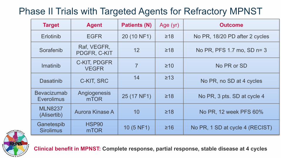
Phase II Trials with Targeted Agents for Refractory MPNSTTarget Agent Patients (N) Age (yr) Outcome
Erlotinib EGFR 20 (10 NF1) ≥18 No PR, 18/20 PD after 2 cycles
Sorafenib Raf, VEGFR, PDGFR, C-KIT 12 ≥18 No PR, PFS 1.7 mo, SD n= 3
Imatinib C-KIT, PDGFR VEGFR 7 ≥10 No PR or SD
Dasatinib C-KIT, SRC 14 ≥13 No PR, no SD at 4 cycles
BevacizumabEverolimus
AngiogenesismTOR 25 (17 NF1) ≥18 No PR, 3 pts. SD at cycle 4
MLN8237(Alisertib) Aurora Kinase A 10 ≥18 No PR, 12 week PFS 60%
GanetespibSirolimus
HSP90mTOR 10 (5 NF1) ≥16 No PR, 1 SD at cycle 4 (RECIST)
Clinical benefit in MPNST: Complete response, partial response, stable disease at 4 cycles

Characterization of Atypical Neurofibromas (AN)§ Distinct imaging, clinical, and genomic (CDKN2A loss) characteristics
Dominant PNN=70
Distinct Nodular Lesions N=61
Distinct nodular lesion
Pathology:• Atypia, • Loss of neurofibroma architecture• Mitosis• Increased cellularity• ANNUBP:
Atypical NeurofibromatousNeoplasm of Uncertain Biologic Potential
Akshintala S…Widemann B: Neuro Oncol 2020Reilly K…Stewart D: JNCI 2017Miettinen M…Perry A: Humpath 2017Kim A…Widemann B: Sarcoma 2017
State of the Science Meeting

Atypical Neurofibromas Are MPNST Precursors
Atypical neurofibroma (AN) characterization: § 63 patients (32 male, 31 female) with 76 AN§ Median age at diagnosis: 27.7 years (7.6-60)§ Most were FDG avid on FDG-PET (56/57)§ 21/63 (33%) of patients with AN had history of MPNST
Higham C,…Legius E*, Widemann B*, Ferner R*: Neuro-Oncology 2018
Clinical challenge: It is unknown if all and when AN transform to MPNST
Hypothesis: Most MPNST arise from preexisting AN and not directly from PN

Strategies to Prevent MPNST
2) Biomarkers for malignant transformation:§ Serial blood samples for detection of cell-free DNA§ Genomic dissection of tumor evolution, single cell sequencing
1) MPNST State of the Science Conference:§ Pathology consensus: Atypical Neurofibromatous
Neoplasm of Uncertain Biologic Potential (ANNUBP)§ Recommendation for surgical resection of AN
• Marginal resection of AN: Safe and feasible• Low recurrence risk
3) Clinical trials for atypical neurofibromas§ Phase I/II trial of CDK4/6 inhibitor abemaciclib§ Children and adults with unresectable pathology confirmed AN
Miettinen M…Widemann B, Perry A: Humpath, 2017Reilly K…Widemann B, Stewart D: JNCI, 2017Nelson C…Widemann B, Chittiboina P., J Neurosurg, 2019

18Y 9M 21Y 0M 21Y 11M
Upper arm, coronal view
Upper arm, axial view0
10
20
30
40
50
60
19 20 21 22
Volu
me
(ml)
Age (years)
18Y 9M
Development of MPNST in Atypical Neurofibroma

18Y 9M 21Y 0M 21Y 11M 22Y 4M
Upper arm, coronal view
Upper arm, axial view0
10
20
30
40
50
60
19 20 21 22
Volu
me
(ml)
Age (years)
How can we predict malignant transformation?When is the right time to intervene?
18Y 9M
Development of MPNST in Atypical Neurofibroma

Factors identified in the literature that are associated with increased risk of MPNST
§ Microdeletion on genetic testing§ Personal prior history of ANNUBP/ANF/MPNST§ Family history of ANNUBP/ANF/MPNST§ History of radiation treatment§ High burden of PN

Radiologic factors identified in the literature that are associated with increased risk of MPNST
§ High internal PN burden:§ Number of PN§ >1 PN? § Size threshold? § Whole body tumor volume?
§ Distinct nodular lesions (DNLs)§ Size threshold?

Number and volume of pNF in people with NF1 with and without MPNST
Nguyen, R., Jett, K., Harris, G.J. et al. Benign whole body tumor volume is a risk factor for malignant peripheral nerve sheath tumors in neurofibromatosis type 1. J Neurooncol 116, 307–313 (2014).

Number and volume of pNF in people with NF1 with and without MPNST
Nguyen, R., Jett, K., Harris, G.J. et al. Benign whole body tumor volume is a risk factor for malignant peripheral nerve sheath tumors in neurofibromatosis type 1. J Neurooncol 116, 307–313 (2014).

Number and volume of pNF in people with NF1 with and without MPNST
Nguyen, R., Jett, K., Harris, G.J. et al. Benign whole body tumor volume is a risk factor for malignant peripheral nerve sheath tumors in neurofibromatosis type 1. J Neurooncol 116, 307–313 (2014).
NF1 + MPNST NF1 - MPNST P-value
n 31 62
# of tumors 2.8 Range: 0–13 median: 2.0
1.4 Range: 0–13Median: 1.0
0.0012
Medianvolume (ml)
352.0 mL(Range: 0-5,368)
3.8 mL(range:0-2,978)
< 0.001

Number and volume of pNF in people with NF1 with and without NF1 gene deletion (Microdeletion)
Kluwe L, Nguyen R, Vogt J, Bengesser K, Mussotter T, Friedrich RE, Jett K, Kehrer-Sawatzki H, Mautner VF. Internal tumor burden in neurofibromatosis Type I patients
with large NF1 deletions. Genes Chromosomes Cancer. 2012 May;51(5):447-51.
Deletion Non-deletion P-valuen 30 90Incidence of internal tumors
21/30 (70%) 56/90 (62%) 0.5
# of tumors 2 (range: 1-5)
2 (range: 1-5)
0.22
Medianvolume (ml)
321 (Range: 8-8018)
176(range:2-3910)
0.19
High tumorburden (>3000 ml)
4/21 (19%) 1/56 (2%) 0.018

§ NF1 whole gene deletions à more severe phenotype of NF1 with higher tumor burden and higher growth-rates § 38 patients with NF1 whole gene deletions § type-1 group: n = 27§ atypical group: n = 11§ an age- and sex matched control: n= 38
Well L, Döbel K, Kluwe L, Bannas P, Farschtschi S, Adam G, Mautner VF, Salamon J. Genotype-phenotype correlation in neurofibromatosis type-1: NF1 whole gene deletions lead to high tumor-burden and increased tumor-growth. PLoS Genet. 2021 May 5;17(5):e1009517.
Number and volume of pNF in people with NF1 with and without NF1 gene deletion

DNLs in NF1§ Definition:
§ Well-demarcated
§ Encapsulated-appearing
§ Size ≥3 cm lesions
§ Lacking the central dot (“target sign”) sign characteristic of PNs
§ Present within or outside of a PN
Akshintala S, Baldwin A, Liewehr DJ, Goodwin A, Blakeley JO, Gross AM, Steinberg SM, Dombi E, Widemann BC. Longitudinal evaluation of peripheral nerve sheath tumors in neurofibromatosis type 1: growth analysis of plexiform neurofibromas and distinct nodular lesions. Neuro Oncol. 2020 Sep 29;22(9):1368-1378.
central dot (“target sign”) sign

DNLs on PET§ N= 103 enrolled in NF1 natural history study
§ 81 (79%) had PN on WB-MRI§ 15 patients à nodular target lesion
§ History of prior MPNST
§ Occurred in absence of prior MPNST history, but were associated with new pain, growth of the nodular lesion exceeding the growth of the surrounding or adjacent PN
§ History of prior abnormal FDG-PET study
§ 46 patients with nodular non-target lesions + least 1 FDG-PET
§ Histology of target DNLs§ Benign + No Bx: 10/15 (67%)
§ Atypical NF: 2/15 (13%)
§ MPNST/Sarcoma: 3/15 (20%)
Meany H, Dombi E, Reynolds J, Whatley M, Kurwa A, Tsokos M, Salzer W, Gillespie A, Baldwin A, Derdak J, Widemann B. 18-fluorodeoxyglucose-positron emission tomography (FDG-PET) evaluation of nodular lesions in patients with Neurofibromatosis type 1 and plexiform neurofibromas (PN) or malignant peripheral nerve sheath tumors (MPNST). Pediatr Blood Cancer. 2013 Jan;60(1):59-64.

DNLs on PETNodular target lesion Nodular non-target
PET avid lesionsPN
n SUVmax(g/ml)
Vol. (ml) Vol. change (% per year)
Pathology No. of lesions
SUVmax(g/ml)
TTV (ml):TTB vol./BSA
History of MPNST
4 3.0–8.8 71–356 NF (n=1)No path (n=3)
0–10 2.1-12.3 1,859–6,800
Prior Abnormal PET Scan or a Growing, Painful DNL
11 2.4–19.5 11–372 10–538 NF (n=4)No path (n=2)ANF (2)MPNST (n=2)Angiosarcoma (n=1)
0–5 1.9-8.8 83–8,649
Meany H, Dombi E, Reynolds J, Whatley M, Kurwa A, Tsokos M, Salzer W, Gillespie A, Baldwin A, Derdak J, Widemann B. 18-fluorodeoxyglucose-positron emission tomography (FDG-PET) evaluation of nodular lesions in patients with Neurofibromatosis type 1 and plexiform neurofibromas (PN) or malignant peripheral nerve sheath tumors (MPNST). Pediatr Blood Cancer. 2013 Jan;60(1):59-64.

DNLs on PET
Meany H, Dombi E, Reynolds J, Whatley M, Kurwa A, Tsokos M, Salzer W, Gillespie A, Baldwin A, Derdak J, Widemann B. 18-fluorodeoxyglucose-positron emission tomography (FDG-PET) evaluation of nodular lesions in patients with Neurofibromatosis type 1 and plexiform neurofibromas (PN) or malignant peripheral nerve sheath tumors (MPNST). Pediatr Blood Cancer. 2013 Jan;60(1):59-64.

DNLs: marker for ANF?63 patients with76 pathologically confirmed ANF§ Pain (n=46)§ Motor weakness
(n=19)§ Palpable or visible
(45)§ No clinical signs
(n=12)
Higham CS, Dombi E, Rogiers A, et al. The characteristics of 76 atypical neurofibromas as precursors to neurofibromatosis 1 associated malignant peripheral nerve sheath tumors. Neuro Oncol. 2018;20(6):818-825. doi:10.1093/neuonc/noy013
MRI (N = 58) Median Range
Size on MRI (N = 58)
Longest diameter (cm) 5.5 1.7–18.2Calculated 3D volume (cm3) 65.1 1.6–1647
Prior MRI (N = 22)
1D growth rate (%/y) 5.8 −42–43.9
3D growth rate (%/y) 27.4 −51–379
FDG-PET (N = 56)
PET avid lesions per patient 2 0–12Median SUVmax: 60–90min (N=50)
5.6 0–22.3
Median SUVmax: 180–240min (N=26)
6.6 3.2–21.6
Prior FDG-PET (N = 18)
Increase in SUVmax 13Decrease in SUVmax 3No change 2

DNLs in NF1

MPNST
DNLs in NF1

DNLs in NF1

Atypical PNST
DNLs in NF1Incidence: 58/122 (45.6%) patients in NIH NHx study
Median 2 DNLs/patientNo histologic data
Akshintala S et al. Neuro Oncol. 2020 Sep 29;22(9):1368-1378.

Proposed Eligibility Criteria for Increased Risk of MPNST:
Eligibility Criteria: Diagnosis of NF1 AND at least one of the following:• Microdeletion
• Other Genotypes associated with increased risk of MPNST?• Family/personal history of ANNUBP/ANF/MPNST
• DNL (Number? Size Criteria?)• Prior radiation therapy• High internal tumor burden
• Large PN burden (≥ 350 mL)?• PN ≥ 3 cm?• More than 1 PN, complex PN?
• Other (High subcutaneous tumor burden?)
For Discussion

33
Extra Slides

Imaging Concerning Features: Hopkins / NCI
Concerning Features” defined as at least one of the following:• Clinical:
• New symptoms, change in pain pattern• Concerns on physical exam
• Imaging MRI: • Rapid growth• ADC values <1x103mm2/s• Concerning change in imaging appearance
• Imaging FDG-PET:• Any tumor with SUV ≥ 3.5• Any individual DNL ≥ 5 cm diameter
For Discussion

Natural History Study: Focus on Increased Risk for MPNST
§ Eligibility criteria: Need to identify criteria to enrich for at risk population§ Standardized longitudinal evaluations:
§ Clinical, genetic:§ Family and medical history, physical exam§ Germline NF1 sequencing
§ Imaging:§ Whole body MRI§ Regional MRI§ FDG-PET-Imaging
§ Identify and manage high risk lesions prior to transformation:§ Biopsy§ Marginal resection§ Safety and tolerability of approach§ Incidence of MPNST in spite of prevention efforts§ Develop algorithms

Multi-parametric Biomarker Development to Predict Malignant Conversion in Patients with Neurofibromatosis type 1
1. Determine the prevalence of DNL in high-risk people with NF11. To describe the multi-parametric (qualitative and quantitative)
MRI/PET features of DNL in NF12. For people with DNL on WB-MRI with serial exams:
a. Determine the growth rateb. Assess the incidence of new DNLs
2. To correlate the multi-parametric (qualitative and quantitative) MRI/PET features with histology to see which DNL turn out to be benign versus atypical versus malignant1.Develop a predictive model for people with NF1 at risk for MPNST

Multi-parametric Biomarker Development to Predict Malignant Conversion in Patients with Neurofibromatosis type 1: Study Schema
High-risk person with NF1: § Microdeletion on genetic testing § Family history of ANNUBP/ANF/MPNST § History of radiation treatment § PN >3 cm § More than 1 PN or complex PN*
Annual comprehensive evaluations at NF Center and
Whole-body MRI using DWI/ADC mapping
Events: - Distinct nodular lesion - Concerning features for malignancy:
§ Clinical: New symptoms/change in pain pattern § Imaging: rapid growth, ADC values <1x103mm2/s, change in imaging appearance**
Event absent
Event present
Algorithm B
Localized MRI using DWI/DCE and
F18-FDG PET/CT
Event is present
No worrisome imaging features
of MPNST
Worrisome imaging features
of MPNST
Biopsy for tissue diagnosis
Biopsy for tissue diagnosis
Serial imaging with localized MRI (DWI) in 3 months (+/- 3 months)
No worrisome imaging features
of MPNST
Worrisome imaging features
of MPNST
Follow Algorithm A
Negative for MPNST
MPNST or Atypical PN
Follow Algorithm A
Therapy as recommended by treating physician
F18-FDG PET/CT

Eligibility Criteria: Patient with a diagnosis of NF1 AND at least one of the following:• Microdeletion• Family history of ANNUBP/MPNST or Personal history of ANNUBP/ANF/MPNST• Prior radiation therapy• Large PN burden (>350 mL)
Cohort 1:Tumors with Concerning
Features
Cohort 2:Tumors with
NO Concerning Features
“Concerning Features” defined as at least one of the following:• Biopsy proven ANNUBP/ANF (if not fully resected)• Any tumor with SUV ≥ 3.5• DWI with ADC value < 1• Any individual DNL ≥ 5 cm diameter
New NCI NF1 Natural History Study for Patients at Higher Risk for MPNST