Regulation of angiopoietin-like protein 4 production during and after exercise
12
ORIGINAL RESEARCH Regulation of angiopoietin-like protein 4 production during and after exercise Frode Norheim 1 , Marit Hjorth 1 , Torgrim M. Langleite 1 , Sindre Lee 1 , Torgeir Holen 1 , Christian Bindesbøll 1 , Hans K. Stadheim 2 , Hanne L. Gulseth 3 ,K are I. Birkeland 3,4 , Anders Kielland 1 , Jørgen Jensen 2 , Knut T. Dalen 1 & Christian A. Drevon 1 1 Department of Nutrition, Faculty of Medicine, Institute of Basic Medical Sciences, University of Oslo, Oslo, Norway 2 Department of Physical Performance, Norwegian School of Sport Sciences, Oslo, Norway 3 Department of Endocrinology, Morbid Obesity and Preventive Medicine, Oslo University Hospital, Oslo, Norway 4 Faculty of medicine, University of Oslo, Oslo, Norway Keywords Adipose tissue, ANGPTL4, exercise, skeletal muscle. Correspondence Frode Norheim, Department of Nutrition, University of Oslo, P.O Box 1046 Blindern, 0317 Oslo, Norway. Tel: +47-22851520 Fax: +47-22851393 E-mail: [email protected] Funding Information The study was supported by grants from Institute of Basic Medical Sciences, UiO, Helse Sør-Øst, Johan Throne-Holst Foundation for Nutrition Research, Freia Medical Research Foundation, and EU-financed FP7 project (NutriTech grant agreement no: 289511). Received: 2 June 2014; Revised: 8 July 2014; Accepted: 9 July 2014 doi: 10.14814/phy2.12109 Physiol Rep, 2 (8), 2014, e12109, doi: 10.14814/phy2.12109 Abstract Angiopoietin-like protein 4 (ANGPTL4) may regulate lipoprotein lipase- dependent plasma clearance of triacylglycerol from skeletal muscle during exercise. The aim of this study was to examine the importance of muscle in regulating ANGPTL4 in response to exercise. We sampled muscle biopsies and serum before, immediately after, and 2 h after 45 min of ergometer cycling. Sampling was done before and after a 12-week training intervention in con- trols and dysglycemic subjects. Moreover, fat biopsies were taken before and after the training intervention. The regulation of ANGPTL4 was also investi- gated in several tissues of exercising mice, and in cultured myotubes. ANG- PTL4 levels in serum and expression in muscle were highest 2 h after exercise in both groups. Whereas ANGPTL4 was higher in muscle of exercising con- trols as compared to dysglycemic subjects, the opposite was observed in serum. In exercising mice, Angptl4 mRNA showed both higher basal expression and induction in liver compared to muscle. Angptl4 mRNA was much higher in adipose tissue than muscle and was also induced by exercise. We observed two mRNA isoforms of ANGPTL4 in muscle and fat in humans. Both were induced by exercise in muscle; one isoform was expressed 5- to 10-fold higher than the other. Studies in mice and cultured myotubes showed that both fatty acids and cortisol have the potential to increase ANGPTL4 expression in mus- cle during exercise. In conclusion, ANGPTL4 is markedly induced in muscle in response to exercise. However, liver and adipose tissue may contribute more than muscle to the exercise-induced increase in circulating ANGPTL4. Introduction During the last decade, it has become apparent that skele- tal muscle is a major endocrine organ (Pedersen and Feb- braio 2012). Skeletal muscle can synthesize and secrete several hundred proteins (Bortoluzzi et al. 2006; Hen- ningsen et al. 2010; Norheim et al. 2011). Proteins that are expressed, synthesized, released by myofibers, and exert either paracrine or endocrine effects, are classified as “myokines” (Pedersen and Febbraio 2012). Although skel- etal muscle hypertrophy and regeneration may enhance myokine secretion for a long period (Ambrosio et al. 2009; Norheim et al. 2011; Shan et al. 2013), acute mus- cle fiber contraction can induce myokine secretion rap- idly, as shown for IL-6 (Pedersen and Febbraio 2012). ANGPTL4 is a multifunctional signal protein synthesized by most tissues (Grootaert et al. 2012). It is involved in regulation of angiogenesis, glucose and lipid metabolism, ª 2014 The Authors. Physiological Reports published by Wiley Periodicals, Inc. on behalf of the American Physiological Society and The Physiological Society. This is an open access article under the terms of the Creative Commons Attribution License, which permits use, distribution and reproduction in any medium, provided the original work is properly cited. 2014 | Vol. 2 | Iss. 8 | e12109 Page 1 Physiological Reports ISSN 2051-817X
-
Upload
independent -
Category
Documents
-
view
5 -
download
0
Transcript of Regulation of angiopoietin-like protein 4 production during and after exercise
ORIGINAL RESEARCH
Regulation of angiopoietin-like protein 4 production duringand after exerciseFrode Norheim1, Marit Hjorth1, Torgrim M. Langleite1, Sindre Lee1, Torgeir Holen1, ChristianBindesbøll1, Hans K. Stadheim2, Hanne L. Gulseth3, K�are I. Birkeland3,4, Anders Kielland1, JørgenJensen2, Knut T. Dalen1 & Christian A. Drevon1
1 Department of Nutrition, Faculty of Medicine, Institute of Basic Medical Sciences, University of Oslo, Oslo, Norway
2 Department of Physical Performance, Norwegian School of Sport Sciences, Oslo, Norway
3 Department of Endocrinology, Morbid Obesity and Preventive Medicine, Oslo University Hospital, Oslo, Norway
4 Faculty of medicine, University of Oslo, Oslo, Norway
Keywords
Adipose tissue, ANGPTL4, exercise, skeletal
muscle.
Correspondence
Frode Norheim, Department of Nutrition,
University of Oslo, P.O Box 1046 Blindern,
0317 Oslo, Norway.
Tel: +47-22851520
Fax: +47-22851393
E-mail: [email protected]
Funding Information
The study was supported by grants from
Institute of Basic Medical Sciences, UiO,
Helse Sør-Øst, Johan Throne-Holst
Foundation for Nutrition Research, Freia
Medical Research Foundation, and
EU-financed FP7 project (NutriTech
grant agreement no: 289511).
Received: 2 June 2014; Revised: 8 July 2014;
Accepted: 9 July 2014
doi: 10.14814/phy2.12109
Physiol Rep, 2 (8), 2014, e12109,
doi: 10.14814/phy2.12109
Abstract
Angiopoietin-like protein 4 (ANGPTL4) may regulate lipoprotein lipase-
dependent plasma clearance of triacylglycerol from skeletal muscle during
exercise. The aim of this study was to examine the importance of muscle in
regulating ANGPTL4 in response to exercise. We sampled muscle biopsies and
serum before, immediately after, and 2 h after 45 min of ergometer cycling.
Sampling was done before and after a 12-week training intervention in con-
trols and dysglycemic subjects. Moreover, fat biopsies were taken before and
after the training intervention. The regulation of ANGPTL4 was also investi-
gated in several tissues of exercising mice, and in cultured myotubes. ANG-
PTL4 levels in serum and expression in muscle were highest 2 h after exercise
in both groups. Whereas ANGPTL4 was higher in muscle of exercising con-
trols as compared to dysglycemic subjects, the opposite was observed in serum.
In exercising mice, Angptl4 mRNA showed both higher basal expression and
induction in liver compared to muscle. Angptl4 mRNA was much higher in
adipose tissue than muscle and was also induced by exercise. We observed two
mRNA isoforms of ANGPTL4 in muscle and fat in humans. Both were
induced by exercise in muscle; one isoform was expressed 5- to 10-fold higher
than the other. Studies in mice and cultured myotubes showed that both fatty
acids and cortisol have the potential to increase ANGPTL4 expression in mus-
cle during exercise. In conclusion, ANGPTL4 is markedly induced in muscle in
response to exercise. However, liver and adipose tissue may contribute more
than muscle to the exercise-induced increase in circulating ANGPTL4.
Introduction
During the last decade, it has become apparent that skele-
tal muscle is a major endocrine organ (Pedersen and Feb-
braio 2012). Skeletal muscle can synthesize and secrete
several hundred proteins (Bortoluzzi et al. 2006; Hen-
ningsen et al. 2010; Norheim et al. 2011). Proteins that
are expressed, synthesized, released by myofibers, and
exert either paracrine or endocrine effects, are classified as
“myokines” (Pedersen and Febbraio 2012). Although skel-
etal muscle hypertrophy and regeneration may enhance
myokine secretion for a long period (Ambrosio et al.
2009; Norheim et al. 2011; Shan et al. 2013), acute mus-
cle fiber contraction can induce myokine secretion rap-
idly, as shown for IL-6 (Pedersen and Febbraio 2012).
ANGPTL4 is a multifunctional signal protein synthesized
by most tissues (Grootaert et al. 2012). It is involved in
regulation of angiogenesis, glucose and lipid metabolism,
ª 2014 The Authors. Physiological Reports published by Wiley Periodicals, Inc. on behalf of
the American Physiological Society and The Physiological Society.
This is an open access article under the terms of the Creative Commons Attribution License,
which permits use, distribution and reproduction in any medium, provided the original work is properly cited.
2014 | Vol. 2 | Iss. 8 | e12109Page 1
Physiological Reports ISSN 2051-817X

and cell differentiation (Grootaert et al. 2012). Several
studies show that ANGPTL4 inhibits the activity of lipo-
protein lipase (LPL), which is responsible for hydrolysis of
plasma triacylglycerols (TG) to monoacylglycerols and free
fatty acids (FFAs) (Yoshida et al. 2002; Mandard et al.
2006; Desai et al. 2007; Grootaert et al. 2012). ANGPTL4
may also stimulate adipose tissue lipolysis, and thereby
release of glycerol and FFAs to the circulation (Yoshida
et al. 2002; Mandard et al. 2006). Thus, the net effect of
ANGPTL4 will represent a shift of FA oxidation derived
from lipoproteins toward FAs originating from adipose tis-
sue. Furthermore, ANGPTL4 is positively associated with
body fat mass (Smart-Halajko et al. 2010).
Expression of ANGPTL4 is increased via different per-
oxisome proliferator-activated receptors (PPARs) and the
glucocorticoid receptor (GR) in hepatocytes and adipo-
cytes (Kersten et al. 2000; Koliwad et al. 2009; Grootaert
et al. 2012). Studies on cultured human myotubes show
that secretion of ANGPTL4 is stimulated by fatty acids
(FAs) as well as the PPARd-specific activator GW501516
(Kersten et al. 2009; Staiger et al. 2009; Robciuc et al.
2012). The impact of exercise on ANGPTL4 expression
during exercise is not fully understood. Previous studies
suggest that ANGPTL4 is an exercise-responsive myokine
regulated by circulating factors (Kersten et al. 2009;
Catoire et al. 2014). Kersten et al. (2009) reported that the
plasma concentration of ANGPTL4 after endurance
activity is less enhanced in subjects given oral glucose, and
Catoire et al. (2014) demonstrated in a human one-legged
exercise study a stronger induction of ANGPTL4 mRNA in
the resting leg as compared to the exercising leg. The
inhibitory effect of glucose on ANGPTL4 transcription is
probably caused by increased release of insulin, causing
suppression of lipolysis and reduced plasma FFA concen-
tration (Kersten et al. 2009; Catoire et al. 2014). Further-
more, the stimulatory effect of plasma FFA on skeletal
muscle ANGPTL4 expression might be counteracted by the
activation of AMPK-activated kinase (AMPK) in exercising
muscle. To our knowledge, it is not known if ANGPTL4
produced in skeletal muscle mainly act on the local tissue
or whether the protein also convey an endocrine effect.
The main aim of this study was to examine the impor-
tance of skeletal muscle in regulating circulating ANGPTL4.
We also investigated if transcription of ANGPTL4 in skeletal
muscle is regulated solely via PPARd or if additional exer-
cise-related factors such as glucocorticoids may play a role.
Materials and Methods
Ethical approval
The study adhered to the Declaration of Helsinki and
was approved by the National Regional Committee for
Medical and Health Research Ethics North, Tromsø,
Oslo, Norway. The study was registered with the US
National Library of Medicine Clinical Trials registry
(NCT01803568). Written informed consent was obtained
from all participants prior to any study-related procedure.
Strength and endurance trainingintervention
Healthy and physically inactive men (40–65 years) were
recruited and divided into two groups; controls with
normal weight (23.5 � 2.0 kg/m2) and normal fasting
and 2-h serum glucose levels (n = 13) or overweight
(29.0 � 2.4 kg/m2) with abnormal glucose metabolism
(dysglycemic group, n = 13). Abnormal glucose metabo-
lism was defined as fasting glucose ≥5.6 mmol/L and/or
impaired glucose tolerance (2-h serum glucose
≥7.8 mmol/L). The participants were subjected to a com-
bined strength and endurance training program for
12 weeks, including two endurance bicycle sessions
(60 min) and two whole-body strength-training sessions
(60 min) per week. Each endurance session started with a
10-min warm-up at three different workloads, corre-
sponding to 50% (4 min), 55% (3 min), and 60%
(3 min) of VO2max. A 45-min bicycle session at 70% of
VO2max was performed before and after the 12-week
training period as an acute work challenge.
A carbohydrate-rich meal was provided 90–120 min
before the exercise test included bread, cheese, jam, and
apple juice, providing 23% of estimated total daily energy
expenditure, on average 2475 KJ. Tests were performed in
the morning, so the standardized meal was the only
intake after overnight fast. A few subjects were tested in
the afternoon (at the same time of day before as well as
after 12 weeks of training) and had the standardized meal
as their only intake during the last 4–5 h. Water could be
consumed freely.
Blood and tissue sampling
Blood and muscle samples were taken before, directly
after, and 2 h after the 70% of VO2max bicycle test, before
as well as after 12 weeks of training. Muscle biopsies were
lacking for one subject at 2 h post exercise, before as well
as after 12 weeks of training. Blood samples were taken by
standard antecubital venous puncture. A single subcutane-
ous adipose tissue biopsy in the periumbilical region was
taken ~30 min after the bicycle session, before as well as
after 12 weeks of training. Subcutaneous adipose biopsies
were obtained from 13 controls and 11 dysglycemic sub-
jects before as well as after 12 weeks of training.
Biopsies from m. vastus lateralis were immediately trans-
ferred to RNA-later (Qiagen, Limburg, the Netherlands),
2014 | Vol. 2 | Iss. 8 | e12109Page 2
ª 2014 The Authors. Physiological Reports published by Wiley Periodicals, Inc. on behalf of
the American Physiological Society and The Physiological Society.
Regulation of ANGPTL4 During Exercise F. Norheim et al.

kept overnight at 4°C, and transferred to �80°C.Subcutaneous biopsies were frozen immediately in liquid
nitrogen and stored at �80°C until further processing.
Serum and EDTA plasma were stored at �80°C until
further analysis.
Serum and plasma analyses
Serum samples of ANGPTL4 (Catalog # RAB0017, Sigma-
Aldrich, St. Louis, MO) and cortisol (Catalog # ab108665,
Abcam, Cambridge, UK) were measured in duplicates
using enzyme-linked immunesorbent assays according to
the manufacture’s protocol. Optical density was deter-
mined using a microplate reader (Titertec Multiscan Plus;
EFLAB, Helsinki, Finland) set to 450 nm. Standard curves
for ANGPTL4 and cortisol were generated with a 4
parameters logistics curve-fitting method (MyAs-
says.com). One subject with extremely high levels of
ANGPTL4 in serum (80–225 ng/mL) was excluded from
the ANGPTL4 analysis. FFA plasma levels were deter-
mined using a Maxmat PL multianalyzer (Maxmat,
France) with reagents (Catalog # D07940/D07950, DIA-
LAB, Wiener Neudorf, Austria).
Cell culture
Primary human myoblasts from m. obliquus internus ab-
dominis of healthy kidney donors were isolated (Haugen
et al. 2010). Myoblasts at passage 5 were proliferated on
collagen I-coated dishes in DMEM/Ham’s F12 1:1 (Life
Technologies, Grand Island, NY) containing glutamax
(Life Technologies), 50 U/mL penicillin, 50 g/mL strepto-
mycin, 5 mmol/L glucose, 10% FBS, 70 pM insulin,
10 ng/mL epidermal growth factor, and 2 ng/mL basic
fibroblast growth factor (Sigma-Aldrich). When the
cultures were near confluency, the myoblasts were differ-
entiated into multinucleated myotubes by changing
medium to DMEM/Ham’s F12 1:1 (5 mmol/L glucose)
containing glutamax, 50 U/mL penicillin, 50 lg/mL strep-
tomycin, and 2% horse serum (Sigma-Aldrich). After
6 days of differentiation, myotubes were incubated with
0, 0.1, 0.5, and 2 lmol/L of dexamethasone (Sigma-
Aldrich). All biopsies were obtained with informed
written consent and approved by the National Committee
for Research Ethics, Oslo, Norway.
Mouse muscle-derived C2C12 cells were transfected
with PPAR isoforms and cultured with FAs and PPAR
(ant-) agonists (Bindesboll et al. 2013). The PPAR (ant-)
agonists WY-14643 (PPARa agonist), GW6471 (PPARaantagonist), GSK0660 (PPARd antagonist), GW9662
(PPARc antagonist), and FAs were obtained from Sigma-
Aldrich. GW501516 (PPARd agonist) and rosiglitazone
(Rosi) (PPARc agonist) were from Enzo Life Sciences.
Mouse experiments
All animals used were approved and registered by the
Norwegian Animal Research Authority. C57BL/6 male
mice were housed in a temperature-controlled (22°C)facility with a strict 12-h light/dark cycle. Mice were anes-
thetized with 2.5% isoflurane before EDTA plasma was
collected, and euthanized by cervical dislocation prior to
harvest of muscles. Plasma and tissue samples were frozen
immediately on dry ice and in liquid nitrogen, respec-
tively, and stored at �80°C.Six-month-old mice were exposed to treadmill exercise
(TSE Systems, Germany) prior to sampling of EDTA
plasma, calf muscles (gastrocnemius and soleus), perirenal
fat, and liver. To avoid stress-related responses, the mice
were gradually habituated to treadmill running before a
final strenuous exercise session. The first session included
2-min free walk on the treadmill with low speed. The
next six sessions followed an identical warm-up pattern
including a speed of 0.10 m/s for 2 min and 0.15 m/s for
8 min. In session 3–7, the mice additionally ran at
0.25 m/s for 5, 10, 15, 30, and 45 min. The last session
ended with an additional final graded increase in speed
from 0.25 to 0.35 m/s for a 5-min period. A platform
placed behind the running band enabled the mice to rest
from running. Running was encouraged by up to five
gentle electrical shocks (0.3 mA) within one exercise ses-
sion. Mice that after five shocks still sat on the platform
were defined as exhausted and removed from the tread-
mill. All exercise sessions were performed between 2 PM
and 4 PM with no food withdrawal prior to exercise.
Blood and muscle samples were taken immediately after
the final exercise. Control mice (n = 8) were housed in
similar cages as the trained mice (n = 8) and were han-
dled equally except for the exercise regimen.
Four-month-old mice on a standard chow diet were
given intragastric gavage of 0.5% carboxymethylcellulose
(CMC) (Sigma # C4888) or 300 lL GW501516 (150 lLsolved in 0.5% CMC; 5 mg/kg) (Bindesboll et al. 2013).
Mice were gavaged 36 and 12 h before being euthanized
at the onset of the light cycle (n = 4–6 in each group).
RNA isolation
Total RNA was isolated from cultured human and murine
myotubes as described previously (Haugen et al. 2010; Bin-
desboll et al. 2013). Frozen human or mouse muscle biop-
sies were crushed to powder in a liquid nitrogen-cooled
mortar using a pestle. One-mL QIAzol Lysis Reagent (Qia-
gen) was added to muscle tissue powder, perirenal fat or
liver tissue, and the samples were homogenized using
TissueRuptor (Qiagen) at full speed twice for 15 sec. Total
RNA was then isolated by miRNeasy Mini Kit (Qiagen).
ª 2014 The Authors. Physiological Reports published by Wiley Periodicals, Inc. on behalf ofthe American Physiological Society and The Physiological Society.
2014 | Vol. 2 | Iss. 8 | e12109Page 3
F. Norheim et al. Regulation of ANGPTL4 During Exercise
Reverse transcription–polymerase chainreaction
Using High-Capacity cDNA Reverse Transcription Kit
(Applied Biosystems, Foster City, CA), 1000 ng of total
RNA from human and mouse tissue samples or cells was
converted into cDNA. The cDNA reaction mixture was
diluted in water and an equivalent of 25 and 50 ng was
analyzed in each sample in the mouse and human tissues,
respectively. Quantitative real-time PCR was performed in
the 96-well format using a 7900HT Fast instrument and
the SDS 2.3 software (Applied Biosystems) (Haugen et al.
2010). Predesigned commercial primers and probe sets
(TaqMan assays, Applied Biosystems) were used to
analyze mRNA levels of ANGPTL4 (Hs00401006_m1),
Angptl4 (Mm00480431_m1), beta-2 microglobulin (B2M,
Hs00984230_m1), large ribosomal protein P0 (RPLP0,
Hs99999902_m1), and TATA box binding protein (Tbp,
Mm00446973_m1). Relative target mRNA expression
levels were calculated as 2�DCt by normalizing to B2M
and RPLP0 in humans and TBP in mice.
High-throughput mRNA sequencing
All mRNA samples were deep sequenced using the
Illumina HiSeq 2000 system with multiplexed design.
Illumina HiSeq RTA (real-time analysis) v1.17.21.3 was
used for real-time analysis during the sequencing run.
Reads passing Illumina’s recommended parameters were
demultiplexed using CASAVA v1.8.2. For prealignment
quality checks we used the software FastQC (http://
www.bioinformatics.babraham.ac.uk/projects/fastqc/). The
mean library size was 44.1 million unstranded single-
ended reads with no difference between groups or time
points. Reads alignment was done using Tophat v2.0.8
(Kim et al. 2013), Samtools v0.1.18 (Li et al. 2009), and
Bowtie v2.1.0 (Langmead et al. 2009) with default settings
against the UCSC hg19 annotated transcriptome and gen-
ome dated 14th of May 2013. Postalignment quality
checks were done using the Integrative Genome Viewer
2.3 (Robinson et al. 2011; Thorvaldsdottir et al. 2013)
and BEDtools v2.19.1 (Quinlan and Hall 2010). Reads
counted by gene feature were done using the intersection
strict mode in HTSeq 0.6.1 (Anders et al. 2014). For dif-
ferential gene expression analyses ANGPTL4, LPL, PDK4,
PPARD, and SLC22A5 were chosen a priori. edgeR v3.4.2
(Robinson et al. 2010) was used to calculate normalized
gene expression levels in Counts Per Million reads (CPM)
and statistical significance. Filtering strategies, quality
check, and generalized linear model construction were
done in R v3.0.3 following the developers’ recommenda-
tions. For transcript-specific isoforms differential expres-
sion analyses of the a priori chosen genes, we used
Cuffdiff v2.1.1 (Trapnell et al. 2012) with default settings
and no novel discovery on the Tophat v2.0.8 constructed
BAM files. Reads were counted by Cufflinks v2.1.1 (Trap-
nell et al. 2012) by isoform feature in the general feature
format annotation file for hg19 before statistical calcula-
tions were performed in Cuffdiff. The isoform-normalized
expression values are presented as reads per kilobase per
million mapped reads (RPKM) to compensate for differ-
ent transcript isoforms length.
Statistical analyses
Statistical analyses were performed using Microsoft Excel
and SPSS 20.0 software (IBM, New York, NY). Effect
measures are presented as means � standard error of the
means (SEM) or means � standard deviation. Statistical
evaluation was done by Student’s t-tests for paired or
unpaired observations. Statistical calculations of ANG-
PTL4 mRNA isoforms were performed by Cuffdiff.
Results
Acute exercise tended to increase muscleANGPTL4 mRNA more in the controlscompared to the dysglycemic subjects
To test the effect of acute as well as chronic exercise in
healthy subjects, we obtained muscle biopsies and serum
samples, before, immediately after, and 2 h post exercise
of 45-min ergometer cycling (70% VO2max) at baseline
and after completion of a 12-week training regime. Both
healthy normal weight controls (n = 13) and overweight
dysglycemic subjects (n = 13) participated in the training
intervention. Twelve weeks of training significantly
increased aerobic capacity (VO2max) by approximately
17% and insulin sensitivity as measured by the glucose
infusion rate during euglycemic hyperinsulinemic clamp
by approximately 30% in both groups (T. M. Langleite,
unpubl. data, 2014). Exercise may enhance ANGPTL4
mRNA expression in muscle via elevated plasma FFAs
(Catoire et al. 2014). Circulating FFAs increased in
response to acute exercise and showed the highest con-
centration immediately after the acute workload both at
baseline and after 12 weeks of training (Fig. 1A). In skele-
tal muscle, on the other hand, ANGPTL4 mRNA expres-
sion was highest 2 h post exercise (Fig. 1B). The
ANGPTL4 mRNA levels were significantly higher in the
control group compared the dysglycemic group 2 h post
exercise at baseline (Fig. 1B). Alternative splicing can
make several ANGPTL4 mRNA isoforms. We performed
mRNA sequencing on muscle, which revealed the expres-
sion of two isoforms (Table 1). Both isoforms were regu-
lated by exercise and the full-length isoform
2014 | Vol. 2 | Iss. 8 | e12109Page 4
ª 2014 The Authors. Physiological Reports published by Wiley Periodicals, Inc. on behalf of
the American Physiological Society and The Physiological Society.
Regulation of ANGPTL4 During Exercise F. Norheim et al.

(NM_139314) was 5- to 10-fold higher expressed com-
pared to the isoform lacking exon 4 (NM_001039667). At
baseline, the full-length ANGPTL4 isoform was signifi-
cantly higher expressed in the controls compared to the
dysglycemic subjects 2 h post exercise (Table 1), which is
in accordance with the qPCR data (Fig. 1B).
ANGPTL4 serum concentration was higher indysglycemic subjects compared to controlsafter acute exercise
Using a polyclonal antibody raised against human ANG-
PTL4 residues 26–406, we measured the serum levels of
ANGPTL4. Circulating ANGPTL4 showed no change
immediately after acute exercise in either group at base-
line but was slightly increased acutely at 12 weeks in the
controls (Fig. 1C). Two hours postexercise ANGPTL4 lev-
els increased approximately two- and three-fold as com-
pared to before acute exercise in controls and dysglycemic
subjects, respectively (Fig. 1C). The absolute serum levels
of ANGPTL4 were significantly higher in the dysglycemic
group compared to the controls 2 h post exercise at base-
line as well as after 12 weeks of training (Fig. 1C), and
the relative increase from before acute exercise to after
2 h rest was significantly higher in the dysglycemic sub-
jects than the controls after 12 weeks of training
(P = 0.03). We observed no effect of 12 weeks of training
on ANGPTL4 levels at rest before acute exercise in skele-
tal muscle mRNA (Fig. 1A) or in serum protein
(Fig. 1C).
Because of the discrepancy in ANGPTL4 regulation
between the groups in skeletal muscle (Fig. 1A) and
serum (Fig. 1C) in response to acute exercise, we investi-
gated the ANGPTL4 mRNA expression in subcutaneous
fat. The ANGPTL4 mRNA level was significantly higher
in the dysglycemic subjects compared to the controls at
baseline in adipose tissue biopsies harvested about 30 min
after acute exercise (Fig 1D). Furthermore, we identified
the same two ANGPTL4 isoforms in adipose tissue as
observed in muscle, and the full-length isoform
0.12
0.08
0.04
0
0.16
0.20
Pre 0’ 2 h Pre 0’ 2 hBaseline 12 week training
**
**
**
**
**
*
aB
Ser
um A
NG
PTL
4 (n
g/m
L)
6
3
12
0
9
*
**
**
**
**
a
a
Pre 0’ 2 h Pre 0’ 2 hBaseline 12 week training
C
120
80
40
0
160
200
Sub
Q A
NG
PTL
4 m
RN
A (C
PM
)
Baseline 12 week training
a
D
30’ 30’
Pla
sma
FFA
(mm
ol/L
)
0.6
0.4
0.2
0
0.8
1.0
** **
**
**
** **
****
b
a
Pre 0’ 2 h Pre 0’ 2 hBaseline 12 week training
ControlDysglyc.
Rel
ativ
e A
NG
PTL
4 m
RN
A A
Figure 1. Acute exercise induces ANGPTL4 differently in muscle and serum of healthy controls and dysglycemic subjects. Changes in m. vastus
lateralis and subcutaneous fat mRNA and serum concentration of ANGPTL4 in healthy and dysglycemic men in response to acute and chronic
exercise (n = 26). Samples were obtained before (Pre), immediately after (00), and 2 h after exercise (2 h) of 45-min ergometer cycling (70%
VO2max) at baseline and after 12 weeks of training. Muscle biopsies were processed for mRNA expression analysis by quantitative RT-PCR and
mRNA sequencing (n = 13 at pre and 00, and n = 12 at 2 h in the controls). (A) Plasma levels of FFA; (B) Skeletal muscle ANGPTL4 mRNA
expression using RT-PCR; (C) Serum levels of ANGPTL4 (n = 12 controls, n = 13 dysglycemic subjects); (D) Subcutaneous fat ANGPTL4 mRNA
expression using mRNA sequencing (n = 13 controls, n = 11 dysglycemic subjects). All quantitative RT-PCR expression data were normalized to
B2M. The gene expression levels of ANGPTL4 obtained from the mRNA sequencing dataset were expressed in counts per million (CPM). Bars
depict means � SEM *P < 0.05 and **P < 0.01 between preexercise values and immediately after (00) or 2 h post exercise. aP < 0.05 as
compared to the control group at the same sampling time point. bP < 0.05 as compared to the same sampling point before 12 weeks of
training. Student’s t-test was used for single comparisons.
ª 2014 The Authors. Physiological Reports published by Wiley Periodicals, Inc. on behalf ofthe American Physiological Society and The Physiological Society.
2014 | Vol. 2 | Iss. 8 | e12109Page 5
F. Norheim et al. Regulation of ANGPTL4 During Exercise

(NM_139314) was significantly higher expressed in the
dysglycemic subjects at baseline (Table 2). ANGPTL4
mRNA expression of both isoforms were markedly higher
in adipose tissue (Table 2) than in muscle (Table 1). The
finding of a discrepancy in ANGPTL4 regulation between
the groups in skeletal muscle and serum in response to
acute exercise and the fact that ANGPTL4 mRNA level is
markedly higher in adipose tissue suggest that the ANG-
PTL4 produced in skeletal muscle during exercise has
only a minor influence on serum levels.
Acute exercise increases mRNA expressionof LPL and PPARd target genes
Because ANGPTL4 is a target of PPARd in skeletal muscle
cells and is suggested to play an important role as an
inhibitor of the enzyme LPL (Catoire et al. 2014), we
investigated the mRNA expression of LPL and the PPARdtargets SLC22A5 (solute carrier family 22 [organic cation/
carnitine transporter], member 5) and PDK4 (Pyruvate
dehydrogenase lipoamide kinase isozyme 4) in skeletal
muscle. Both LPL (Fig 2A), SLC22A5 (Fig. 2B) and PDK4
(Fig. 2C) expression increased in response to acute exer-
cise with the highest mRNA levels 2 h post exercise. The
absolute mRNA expression of LPL was higher in the con-
trols as compared to the dysglycemic group at most time
points (Fig. 2A).
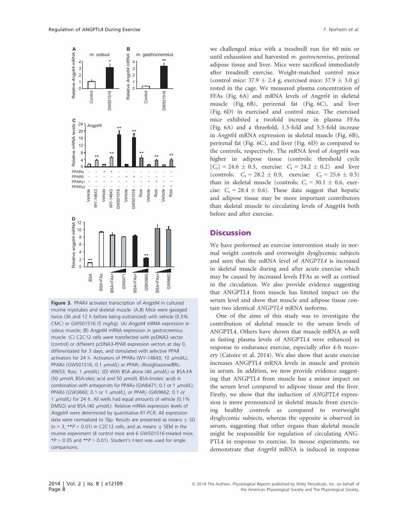
Fatty acids stimulate Angptl4 expression viaPPARd in vivo
Fatty acids and the PPARd-specific activator GW501516
stimulate mRNA expression and secretion of ANGPTL4
in cultured myotubes (Staiger et al. 2009; Robciuc et al.
2012; Catoire et al. 2014). To provide physiological rele-
vance to these in vitro findings, mice were gavaged with
the PPARd agonist GW501516. This increased Angptl4
mRNA threefold (Fig. 3A) and ninefold (Fig. 3B) in
soleus and gastrocnemius muscle, respectively. To investi-
gate if Angptl4 was specifically induced by the PPARd iso-
form, C2C12 cells were transfected with PPARa, PPARd,or PPARc, differentiated for 3 days and incubated for
24 h with PPAR isoform-specific ligands (Fig. 3A).
Expression of the different PPARs was similar after trans-
fection (Bindesboll et al. 2013). Incubating the cells with
ligands specific for PPARa (WY-14643), PPARd(GW501516), and PPARc (rosiglitazone), all enhanced
expression of Angptl4 mRNA (Fig. 3C). The PPARdligand increased Angptl4 mRNA about 11-fold, whereas
PPARa and PPARc ligands both increased Angptl4 expres-
sion about threefold. There was no effect on Angptl4 tran-
scription by transfecting the cells with the different
PPARs. To investigate if FA-induced Angptl4 expression
depended on a particular PPAR isoform, differentiated
C2C12 cells were incubated with a combination of oleic
acid and linoleic acid (50 lmol/L each) in the presence of
antagonists for PPARa (GW6471), PPARd (GSK0660),
and PPARc (GW9662) (Fig. 3D). FA-induced expression
of Angptl4 was only significantly lower in the presence of
Table 2. Subcutaneous ANGPTL4 mRNA isoforms in response to
chronic training
Group Isoform Baseline 12 weeks
Control NM_001039667 12.4 12.8
Dysglycemic NM_001039667 19.0 19.8
Control NM_139314 107.7 119.4
Dysglycemic NM_139314 138.7aa 138.4
The isoform-normalized expression values are presented as reads
per kilobase per million mapped reads and measured ~30 min
after acute exercise at baseline and after 12 weeks of training.aaP < 0.01 as compared the control group at the same sampling
time point.
Table 1. Changes in muscle ANGPTL4 mRNA isoforms in response to acute and chronic exercise
Group Isoform Baseline Pre 00 2 h 12 weeks Pre 00 2 h
Control NM_001039667 0.047 0.689* 0.443 0.110 0.323 0.825*
Dysglycemic NM_001039667 0.150 0.361 0.937* 0.057 0.934* 0.408
Control NM_139314 0.496 2.587** 11.473** 0.449 6.291**b 11.625**
Dysglycemic NM_139314 0.347 2.072* 8.007**aa 0.465 4.903**b 9.218**
The isoform-normalized expression values are presented as reads per kilobase per million mapped reads and measured before (Pre), immedi-
ately after (00), and 2 h after exercise (2 h) of 45-min ergometer cycling (70% VO2max) at baseline and after 12 weeks of training.
**P < 0.01 and *P < 0.05 as compared to before (pre) acute exercise.aaP < 0.01 as compared to the control group at the same sampling time point.bP < 0.05 as compared to the sampling time point before 12 weeks training.
2014 | Vol. 2 | Iss. 8 | e12109Page 6
ª 2014 The Authors. Physiological Reports published by Wiley Periodicals, Inc. on behalf of
the American Physiological Society and The Physiological Society.
Regulation of ANGPTL4 During Exercise F. Norheim et al.

the PPARd antagonist, which reduced the FFA-induced
Angptl4 expression by approximately 70%. These results
support that induction of Angptl4 in vivo is mediated
through FA activation of PPARd.
Dexamethasone stimulates ANGPTL4expression in human myotubes
Acute exercise elevates plasma levels of FFAs, which can
activate PPARd. Plasma cortisol levels also increase in
response to acute exercise (Kanaley et al. 2001). Cortisol
can activate GR, another member of the nuclear receptor
family, and expression of ANGPTL4 is increased after GR
activation in hepatocytes and adipocytes (Koliwad et al.
2009). In our study, serum concentration of cortisol was
acutely increased between 1.8- and 2.5-fold after 45-min
cycling, before as well as after 12 weeks of training in the
controls (Fig. 4A) and dysglycemic subjects (Fig. 4B).
There was no significant difference between the groups in
exercise induction of circulating cortisol. Based on these
observations, we investigated if activation of GR might
regulate ANGPTL4 gene expression in skeletal muscle cells
by incubating primary human myotubes with the synthetic
GR ligand, dexamethasone, in a dose–response experi-
ment. Incubation of human myotubes with 0.5 lmol/L
dexamethasone for 2 h increased ANGPTL4 expression
3.5-fold (Fig. 4C). Myotubes were then incubated with
0.5 lmol/L of dexamethasone up to 6 h. ANGPTL4
mRNA level increased time dependently up to 2 h with
dexamethasone compared to zero time (Fig. 4D). These
results show that ANGPTL4 can be regulated in vitro by
the cortisol-GR axis in addition to the FA-PPARd axis,
with possible implications for acute exercise.
No effect of ionomycin or caffeine onAngptl4 expression in cultured myotubes
Expression of Angptl4 under different conditions was
examined to address whether factors mimicking exercise
in vitro also affected Angptl4 expression. Neither caffeine
nor inonomycin induced Angptl4 mRNA (Fig. 5A and B);
10 mmol/L caffeine and 2.5 lmol/L ionomycin even
reduced Angptl4 expression significantly in murine myo-
tubes incubated for 3 h. Both caffeine and ionomycin had
a dose-dependent effect on mRNA expression of the posi-
tive control Il6 (Fig. 5A and B). Caffeine and ionomycin
had no dose-dependent effect on ANGPTL4 mRNA
expression after 3 h and up to 24 h in human myotubes
(data not shown).
Acute exercise increased expression ofmurine Angptl4 in muscle, adipose tissue,and liver
It has been suggested that the liver is the main organ
influencing the regulation of circulating ANGPTL4 (Dijk
and Kersten 2014). To test if exercise induces Angptl4
mRNA expression in other organs than skeletal muscle,
80
40
160
0
120
ControlDysglyc.
Pre 0’ 2 h Pre 0’ 2 hBaseline 12 week training
*
****
**
*
****
a
aa
a
bb
LPL
mR
NA
(CP
M)
30
20
0
40
SLC
22A
5 m
RN
A (C
PM
)
10
Pre 0’ 2 h Pre 0’ 2 hBaseline 12 week training
abb
***
******
**
**
**
600
400
0
800
PD
K4
mR
NA
(CP
M)
200
Pre 0’ 2 h Pre 0’ 2 hBaseline 12 week training
**
**
** **
** **
****
A
B
C
Figure 2. Acute exercise induces LPL, SLC22A5, and PDK4mRNA
levels in healthy controls and dysglycemic subjects. Changes inm.
vastus lateralismRNA of LPL, SLC22A5, and PDK4 in healthy and
dysglycemic men in response to acute and chronic exercise. Samples
were obtained before (Pre), immediately after (00), and 2 h after
exercise (2 h) of 45-min cycling (70% VO2max) at baseline and after
12 weeks of training (n = 26). The muscle biopsies were processed for
mRNA expression analysis by mRNA sequencing (n = 13 at pre and 00,and n = 12 at 2 h in the controls). (A) LPL; (B) SLC22A5; (C) PDK4.
Gene expression levels obtained from the mRNA sequencing data
were expressed in counts per million (CPM). Bars depict
means � SEM *P < 0.05 and **P < 0.01 between preexercise values
and immediately after (00) or 2 h post exercise. aP < 0.05 as compared
to the control group at the same sampling time point. bP < 0.05 andbbP < 0.01 as compared to the same sampling point before 12 weeks
of training. Student’s t-test was used for single comparisons.
ª 2014 The Authors. Physiological Reports published by Wiley Periodicals, Inc. on behalf ofthe American Physiological Society and The Physiological Society.
2014 | Vol. 2 | Iss. 8 | e12109Page 7
F. Norheim et al. Regulation of ANGPTL4 During Exercise
we challenged mice with a treadmill run for 60 min or
until exhaustion and harvested m. gastrocnemius, perirenal
adipose tissue and liver. Mice were sacrificed immediately
after treadmill exercise. Weight-matched control mice
(control mice: 37.9 � 2.4 g, exercised mice: 37.9 � 3.0 g)
rested in the cage. We measured plasma concentration of
FFAs (Fig. 6A) and mRNA levels of Angptl4 in skeletal
muscle (Fig. 6B), perirenal fat (Fig. 6C), and liver
(Fig. 6D) in exercised and control mice. The exercised
mice exhibited a twofold increase in plasma FFAs
(Fig. 6A) and a threefold, 1.5-fold and 5.5-fold increase
in Angptl4 mRNA expression in skeletal muscle (Fig. 6B),
perirenal fat (Fig. 6C), and liver (Fig. 6D) as compared to
the controls, respectively. The mRNA level of Angptl4 was
higher in adipose tissue (controls: threshold cycle
[Ct] = 24.6 � 0.5, exercise: Ct = 24.2 � 0.2) and liver
(controls: Ct = 28.2 � 0.9, exercise: Ct = 25.6 � 0.5)
than in skeletal muscle (controls: Ct = 30.1 � 0.6, exer-
cise: Ct = 28.4 � 0.6). These data suggest that hepatic
and adipose tissue may be more important contributors
than skeletal muscle to circulating levels of Angptl4 both
before and after exercise.
Discussion
We have performed an exercise intervention study in nor-
mal weight controls and overweight dysglycemic subjects
and seen that the mRNA level of ANGPTL4 is increased
in skeletal muscle during and after acute exercise which
may be caused by increased levels FFAs as well as cortisol
in the circulation. We also provide evidence suggesting
that ANGPTL4 from muscle has limited impact on the
serum level and show that muscle and adipose tissue con-
tain two identical ANGPTL4 mRNA isoforms.
One of the aims of this study was to investigate the
contribution of skeletal muscle to the serum levels of
ANGPTL4. Others have shown that muscle mRNA as well
as fasting plasma levels of ANGPTL4 were enhanced in
response to endurance exercise, especially after 4-h recov-
ery (Catoire et al. 2014). We also show that acute exercise
increases ANGPTL4 mRNA levels in muscle and protein
in serum. In addition, we now provide evidence suggest-
ing that ANGPTL4 from muscle has a minor impact on
the serum level compared to adipose tissue and the liver.
Firstly, we show that the induction of ANGPTL4 expres-
sion is more pronounced in skeletal muscle from exercis-
ing healthy controls as compared to overweight
dysglycemic subjects, whereas the opposite is observed in
serum, suggesting that other organs than skeletal muscle
might be responsible for regulation of circulating ANG-
PTL4 in response to exercise. In mouse experiments, we
demonstrate that Angptl4 mRNA is induced in response
3210
4
Rel
ativ
e A
ngpt
l4 m
RN
A
Con
trol
GW
5015
16
m. soleus
*3210
4
Rel
ativ
e A
ngpt
l4 m
RN
A
Con
trol
GW
5015
16
m. gastrocnemius**
Rel
ativ
e m
RN
A le
vels
12
8
40
1620
24C
A B
Angptl4
PPARαPPARδPPARγ1PPARγ2
____
____
+___
+___
____
_
+__
_
+__
____
__
+_
__
+_
___
+
___
+
Vehi
cle
WY-
1464
3
Vehi
cle
WY-
1464
3
GW
5015
16
Vehi
cle
GW
5015
16
Ros
i
Vehi
cle
Ros
i
Vehi
cle
Ros
i
** **
** **
** ** **
6
4
20
8
10
12
Rel
ativ
e an
gptl4
mR
NA
****
BSA
BSA+
FAs
BSA+
FAs+
GW
6471
BSA+
FAs+
GSK
0660
BSA+
FAs+
GW
9662
D
Figure 3. PPARd activates transcription of Angptl4 in cultured
murine myotubes and skeletal muscle. (A,B) Mice were gavaged
twice (36 and 12 h before being euthanized) with vehicle (0.5%
CMC) or GW501516 (5 mg/kg). (A) Angptl4 mRNA expression in
soleus muscle; (B) Angptl4 mRNA expression in gastrocnemius
muscle. (C) C2C12 cells were transfected with pcDNA3 vector
(control) or different pcDNA3-PPAR expression vectors at day 0,
differentiated for 3 days, and stimulated with selective PPAR
activators for 24 h. Activators of PPARa (WY-14643; 10 lmol/L),
PPARd (GW501516; 0.1 lmol/L) or PPARc (Rosiglitazone/BRL-
49653; Rosi; 1 lmol/L). (D) With BSA alone (40 lmol/L) or BSA-FA
(50 lmol/L BSA-oleic acid and 50 lmol/L BSA-linoleic acid) in
combination with antagonists for PPARa (GW6471; 0.1 or 1 lmol/L),
PPARd (GSK0660; 0.1 or 1 lmol/L), or PPARc (GW9662; 0.1 or
1 lmol/L) for 24 h. All wells had equal amounts of vehicle (0.1%
DMSO) and BSA (40 lmol/L). Relative mRNA expression levels of
Angptl4 were determined by quantitative RT-PCR; All expression
data were normalized to Tbp. Results are presented as means � SD
(n = 3, **P > 0.01) in C2C12 cells, and as means � SEM in the
murine experiment (4 control mice and 6 GW501516-treated mice,
*P > 0.05 and **P > 0.01). Student’s t-test was used for single
comparisons.
2014 | Vol. 2 | Iss. 8 | e12109Page 8
ª 2014 The Authors. Physiological Reports published by Wiley Periodicals, Inc. on behalf of
the American Physiological Society and The Physiological Society.
Regulation of ANGPTL4 During Exercise F. Norheim et al.

to exercise in skeletal muscle, adipose tissue, and the liver,
and show that liver and adipose tissue have a higher basal
expression of Angptl4 mRNA levels compared to skeletal
muscle. Also, we show that the Angptl4 mRNA induction
in response to exercise is higher in the liver as compared
to muscle. However, the fact that skeletal muscle accounts
for about 40% of body mass in a lean individual might
make it an important contributor to blood levels of ANG-
PTL4, although it has low ANGPTL4 expression in muscle
tissue. Thus, it is possible that skeletal muscle might be a
more important source of circulating ANGPTL4 in
subjects with more muscle mass and less adipose tissue.
In our human exercise study, the group with the most
adipose tissue exhibits the highest basal serum levels of
ANGPTL4 and the highest induction of circulating ANG-
PTL4 in response to exercise. It has also been suggested
that the liver is the main contributor to circulating ANG-
PTL4 during fasting (Dijk and Kersten 2014). Our data
suggests that this is also the case in response to exercise.
The fact that during a one-legged exercise intervention
ANGPTL4 mRNA expression was more induced in the rest-
ing leg as compared to the exercising leg suggests that cir-
culating factors can induce ANGPTL4 transcription in
skeletal muscle (Catoire et al. 2014). The plasma levels of
FFA are known to increase during acute exercise and are
hypothesized to increase transcription of ANGPTL4 in skel-
etal muscle after PPARd activation (Catoire et al. 2014).
Studies on cultured human myotubes have revealed that
secretion of ANGPTL4 is stimulated by FAs as well as a
PPARd-specific activator (Kersten et al. 2009; Staiger et al.
2009; Robciuc et al. 2012). In our study, we provide both
in vitro and in vivo evidence for a robust increase of Ang-
ptl4 transcription by PPARd in mouse skeletal muscle. We
demonstrate by gavage feeding mice that a PPARd-specificligand enhances muscle Angptl4 transcription in vivo. We
also show that the FFA-induced Angptl4 expression was
reduced in myotubes by an inhibitor specific for PPARd,but not by inhibitors directed against PPARa and PPARc.
0 0.5 1 2
5
4
3
2
1
0
Fol
d ch
ange
AN
GP
TL4
mR
NA
(Rel
ative
to 0
μM
dex
amet
haso
ne)
Dexamethasone (μmol/L)
C
1.5
*
Pre 0’ 2 h
Fol
d ch
ange
ser
um c
ortis
ol
(rea
ltive
to b
efor
e ex
erci
se)
1.5
1.0
0.5
0
2.0
2.5
3.0Baseline12 w
ControlsA
**
**
**
Fol
d ch
ange
AN
GP
TL4
mR
NA
(r
ealti
ve to
0 h
)
0.5 μM 0 μM
0 1 2 3 4 5 6
3
2
1
0
4
Incubation time (hours)
5
6
D
*
Pre 0’ 2 h
Fol
d ch
ange
ser
um c
ortis
ol
(rea
ltive
to b
efor
e ex
erci
se)
1.5
1.0
0.5
0
2.0
2.5
3.0
3.5DysglycemicsB
**
**
Figure 4. ANGPTL4 is induced in human myotubes incubated with dexamethasone. Fold change in serum concentration of cortisol in (A)
healthy (n = 13) and (B) dysglycemic men (n = 13) in response to acute exercise at baseline and after 12 weeks of training. Serum cortisol
levels before (Pre) acute exercise were compared with immediately after (00) 45-min ergometer cycling (70% VO2max) and after 2-h rest. (C)
Primary human myotubes were differentiated for 6 days and incubated with 0, 0.1, 0.5, and 2 lmol/L of dexamethasone for 2 h. Relative
mRNA expression levels of ANGPTL4 after 2 h of dexamethasone incubation were determined by quantitative RT-PCR, presented as fold change
relative to control (0 lmol/L). (D) Myotubes differentiated for 6 days were incubated with 0.5 lmol/L dexamethasone for 0, 0.5, 1, 2, 4, and
6 h. Fold changes in mRNA expression of ANGPTL4 with and without dexamethasone after 0.5, 1, 2, 4, and 6 h were compared to zero time.
All expression data were normalized to RPLP0. Data from three donors are presented as means � SEM *P < 0.05 and **P < 0.01, Student’s
t-test was used for single comparisons between matching time points.
ª 2014 The Authors. Physiological Reports published by Wiley Periodicals, Inc. on behalf ofthe American Physiological Society and The Physiological Society.
2014 | Vol. 2 | Iss. 8 | e12109Page 9
F. Norheim et al. Regulation of ANGPTL4 During Exercise

Several circulating factors are increased in response to
acute exercise in addition to FFAs. One of the hormones
that are known to be increased in response to exercise is
the glucocorticoid cortisol (Nieman et al. 2005). Because
it has been shown that ANGPTL4 is a direct GR target in
hepatocytes as well as in adipocytes (Koliwad et al. 2009),
one of the aims of this study was to investigate if human
myotubes also respond to glucocorticoids by increasing
ANGPTL4 expression. We show that the serum concen-
tration of cortisol is indeed increased in response to acute
exercise. We also show that ANGPTL4 transcription is
elevated in cultures of primary human muscle cells incu-
bated with the GR ligand, dexamethasone, comparable to
the effects of dexamethasone on primary rat hepatocytes
and human adipocytes (Koliwad et al. 2009). A recent
study showed that circulating ANGPTL4 is not elevated
by exercise when glucose is consumed (Catoire et al.
2014). Interestingly, both the plasma level of FFAs and
cortisol are reduced when glucose is ingested during exer-
cise (Nieman et al. 2005; Catoire et al. 2014) and may
thus both explain some of the suppressive effects of glu-
cose on circulating ANGPTL4 (Catoire et al. 2014). These
results suggest that ANGPTL4 mRNA in muscle can be
regulated by the cortisol-GR axis during acute exercise.
One function of ANGPTL4 may be to inhibit the
uptake of FAs derived from lipoproteins by regulating
LPL activity in skeletal muscle during exercise (Catoire
et al. 2014). Interestingly, Catoire et al. (2014) suggested
that during a one-legged exercise intervention ANGPTL4
mRNA expression was more increased in the resting leg
as compared to the exercising leg because of the counter-
acting effect of AMPK on ANGPTL4 transcription. The
repression of ANGPTL4 production and hence enhanced
LPL activity might promote use of circulating TG in the
exercising muscle (Catoire et al. 2014). We show in our
human exercise intervention that acute exercise induces
both LPL and ANGPTL4 transcription in skeletal muscle
to a higher degree in control subjects as compared to the
dysglycemic subjects. However, the serum levels of ANG-
PTL4 were more increased after exercise in the overweight
dysglycemic subjects than in the controls. Because the
A
B
0
5
10
15
20
****
*
**Il6Angptl4
0 2.5Caffeine (mM)
7.55
Fold
cha
nge
mR
NA
(Rela
tive
to 0
mM
caffe
ine)
10
Ionomycin (μM)0 0.5 1 1.5 2
Il6Angptl4 **
*
**
**
0
5
10
15
Fol
d ch
ange
mR
NA
(Rel
ative
to 0
μM
iono
myc
in)
2.5
Figure 5. Angptl4 mRNA was unchanged in murine myotubes
incubated with caffeine or ionomycin. (A) Differentiated C2C12
myotubes were incubated with caffeine (0, 2.5, 5, and 10 mmol/L)
for 3 h. Relative mRNA expression levels of Angptl4 and Il6 were
determined by quantitative RT-PCR, presented as fold change
relative to control (0 mmol/L caffeine). (B) Differentiated C2C12
myotubes were incubated with ionomycin (0, 0.1, 1, and 2.5 lmol/L)
for 3 h. Relative mRNA expression levels of Angptl4 and Il6 were
determined by quantitative RT-PCR, presented as fold change relative
to control (0 lmol/L ionomycin). All expression data were normalized
to Tbp, and presented as means � SD (n = 3, *P > 0.05,
**P > 0.01). Student’s t-test was used for single comparisons.
Pla
sma
FFA
(mm
ol/L
)
0
0.2
0.4
0.6
A
Control Exercise
**
Rel
ativ
e m
RN
A le
vels
C
0
304050
2010
Control Exercise
Angptl4
Perirenal fat
**
Rel
ativ
e m
RN
A le
vels
D
0
10
15
20
5
Control Exercise
Angptl4
Liver
**
Rel
ativ
e m
RN
A le
vels
B
0
1.0
1.5
2.0
0.5
Calf muscles
Control Exercise
Angptl4 **
Figure 6. Angptl4 mRNA expression in muscle, fat, and liver
increases after exercise in mice. Age- and weight-matched C57Bl/6
mice underwent 60-min treadmill exercise (n = 8), and the control
mice rested in the cage (n = 8). Plasma samples and gastrocnemius
muscles were collected immediately after exercise and in the
control mice. The muscle biopsies were processed for mRNA
expression analysis by quantitative RT-PCR. (A) plasma levels of FFA;
(B) Angptl4 mRNA in calf muscles; (C) Angptl4 mRNA in perirenal
fat; (D) Angptl4 mRNA in liver. All expression data were normalized
to Tbp. Bars depict means � SEM **P < 0.01 between exercised
mice and control mice. Student’s t-test was used for single
comparisons.
2014 | Vol. 2 | Iss. 8 | e12109Page 10
ª 2014 The Authors. Physiological Reports published by Wiley Periodicals, Inc. on behalf of
the American Physiological Society and The Physiological Society.
Regulation of ANGPTL4 During Exercise F. Norheim et al.

relative contribution of skeletal muscle to circulating
ANGPTL4 during exercise remains unknown, it is difficult
to predict in which group ANGPTL4 has the largest inhib-
itory effect on LPL activity. Coexpression of ANGPTL4
with LPL in skeletal muscle might suggest that ANGPTL4
acts mainly via local inhibition (Dijk and Kersten 2014).
Future studies should address the relative contribution of
skeletal muscle to circulating ANGPTL4. Thereby, it
would be possible to evaluate the inhibitory potential
ANGPTL4 may have on LPL activity during exercise.
In summary, our data suggest that ANGPTL4 produced
in skeletal muscle during and after exercise has limited
impact on the serum protein level and probably acts
mostly in local tissue. Our data also indicate that FFAs
and cortisol increase transcription of ANGPTL4 in muscle
during exercise via activation of PPARd and GR.
Acknowledgements
We thank Hilde Nebb for access to experimental materi-
als, Anne Randi Enget and Christin Zwafink for technical
assistance, Harald Carlsen for donation of mice, Ansgar
Heck and Birgitte Nellemann are for taking the biopsies,
and Kristoffer J Kolnes, Daniel S Tangen, Tor I Gloppen,
Torstein Dalen, H�avard Moen, Marius A Dahl, Guro
Grøthe, Egil Johansen, Katrine A Krog, Øyvind Skattebo
and Eirin N Rise for being responsible for and helping
out on different aspects of the human strength and
endurance intervention.
Conflict of Interest
None declared.
References
Ambrosio, F., F. Kadi, J. Lexell, G. K. Fitzgerald,
M. L. Boninger, and J. Huard. 2009. The effect of muscle
loading on skeletal muscle regenerative potential: an update
of current research findings relating to aging and
neuromuscular pathology. Am. J. Phys. Med. Rehabil.
88:145–155.
Anders, S., P. T. Pyl, and W. Huber. 2014. HTSeq – A Python
framework to work with high-throughput sequencing data.
BioRxiv preprint.
Bindesboll, C., O. Berg, B. Arntsen, H. I. Nebb, and
K. T. Dalen. 2013. Fatty acids regulate perilipin5 in muscle
by activating PPARdelta. J. Lipid Res. 54:1949–1963.
Bortoluzzi, S., P. Scannapieco, A. Cestaro, G. A. Danieli, and
S. Schiaffino. 2006. Computational reconstruction of the
human skeletal muscle secretome. Proteins 62:776–792.
Catoire, M., S. Alex, N. Paraskevopulos, F. Mattijssen,
G. I. Evers-Van, G. Schaart, et al. 2014. Fatty acid-inducible
ANGPTL4 governs lipid metabolic response to exercise.
Proc. Natl Acad. Sci. USA. 111:1043–1052.
Desai, U., E. C. Lee, K. Chung, C. Gao, J. Gay, B. Key, et al.
2007. Lipid-lowering effects of anti-angiopoietin-like 4
antibody recapitulate the lipid phenotype found in
angiopoietin-like 4 knockout mice. Proc. Natl Acad. Sci.
USA, 104:11766–11771.
Dijk, W., and S. Kersten. 2014. Regulation of lipoprotein
lipase by Angptl4. Trends Endocrinol. Metab. 25:146–155.
Grootaert, C., T. van de Wiele, W. Verstraete, M. Bracke, and
B. Vanhoecke. 2012. Angiopoietin-like protein 4: health
effects, modulating agents and structure-function
relationships. Expert. Rev. Proteomics. 9:181–199.
Haugen, F., F. Norheim, H. Lian, A. J. Wensaas, S. Dueland,
O. Berg, et al. 2010. IL-7 is expressed and secreted by
human skeletal muscle cells. Am. J. Physiol. Cell Physiol.
298:C807–C816.
Henningsen, J., K. T. Rigbolt, B. Blagoev, B. K. Pedersen, and
I. Kratchmarova. 2010. Dynamics of the skeletal muscle
secretome during myoblast differentiation. Mol. Cell.
Proteomics 9:2482–2496.
Kanaley, J. A., J. Y. Weltman, K. S. Pieper, A. Weltman, and
M. L. Hartman. 2001. Cortisol and growth hormone
responses to exercise at different times of day. J. Clin.
Endocrinol. Metab. 86:2881–2889.
Kersten, S., S. Mandard, N. S. Tan, P. Escher, D. Metzger,
P. Chambon, et al. 2000. Characterization of the
fasting-induced adipose factor FIAF, a novel peroxisome
proliferator-activated receptor target gene. J. Biol. Chem.
275:28488–28493.
Kersten, S., L. Lichtenstein, E. Steenbergen, K. Mudde,
H. F. Hendriks, M. K. Hesselink, et al. 2009. Caloric
restriction and exercise increase plasma ANGPTL4 levels in
humans via elevated free fatty acids. Arterioscler. Thromb.
Vasc. Biol. 29:969–974.
Kim, D., G. Pertea, C. Trapnell, H. Pimentel, R. Kelley, and
S. L. Salzberg. 2013. TopHat2: accurate alignment of
transcriptomes in the presence of insertions, deletions and
gene fusions. Genome Biol. 14:R36.
Koliwad, S. K., T. Kuo, L. E. Shipp, N. E. Gray, F. Backhed,
A. Y. So, et al. 2009. Angiopoietin-like 4 (ANGPTL4,
fasting-induced adipose factor) is a direct glucocorticoid
receptor target and participates in glucocorticoid-regulated
triglyceride metabolism. J. Biol. Chem. 284:25593–25601.
Langmead, B., C. Trapnell, M. Pop, and S. L. Salzberg. 2009.
Ultrafast and memory-efficient alignment of short DNA
sequences to the human genome. Genome Biol. 10:R25.
Li, H., B. Handsaker, A. Wysoker, T. Fennell, J. Ruan,
N. Homer, et al. 2009. The Sequence Alignment/Map
format and SAMtools. Bioinformatics 25:2078–2079.
Mandard, S., F. Zandbergen, S. E. Van, W. Wahli, F. Kuipers,
M. Muller, et al. 2006. The fasting-induced adipose factor/
angiopoietin-like protein 4 is physically associated with
ª 2014 The Authors. Physiological Reports published by Wiley Periodicals, Inc. on behalf ofthe American Physiological Society and The Physiological Society.
2014 | Vol. 2 | Iss. 8 | e12109Page 11
F. Norheim et al. Regulation of ANGPTL4 During Exercise
lipoproteins and governs plasma lipid levels and adiposity.
J. Biol. Chem. 281:934–944.
Nieman, D. C., J. M. Davis, D. A. Henson, S. J. Gross,
C. L. Dumke, A. C. Utter, et al. 2005. Muscle cytokine
mRNA changes after 2.5 h of cycling: influence of
carbohydrate. Med. Sci. Sports Exerc. 37:1283–1290.
Norheim, F., T. Raastad, B. Thiede, A. C. Rustan,
C. A. Drevon, and F. Haugen. 2011. Proteomic
identification of secreted proteins from human skeletal
muscle cells and expression in response to strength training.
Am. J. Physiol. Endocrinol. Metab. 301:E1013–E1021.
Pedersen, B. K., and M. A. Febbraio. 2012. Muscles, exercise
and obesity: skeletal muscle as a secretory organ. Nat. Rev.
Endocrinol. 8:457–465.
Quinlan, A. R., and I. M. Hall. 2010. BEDTools: a flexible
suite of utilities for comparing genomic features.
Bioinformatics 26:841–842.
Robciuc, M. R., P. Skrobuk, A. Anisimov, V. M. Olkkonen,
K. Alitalo, R. H. Eckel, et al. 2012. Angiopoietin-like 4
mediates PPAR delta effect on lipoprotein lipase-dependent
fatty acid uptake but not on beta-oxidation in myotubes.
PLoS ONE. 7, e46212.
Robinson, M. D., D. J. McCarthy, and G. K. Smyth. 2010.
edgeR: a Bioconductor package for differential expression
analysis of digital gene expression data. Bioinformatics
26:139–140.
Robinson, J. T., H. Thorvaldsdottir, W. Winckler,
M. Guttman, E. S. Lander, G. Getz, et al. 2011. Integrative
genomics viewer. Nat. Biotechnol. 29:24–26.
Shan, T., X. Liang, P. Bi, and S. Kuang. 2013. Myostatin
knockout drives browning of white adipose tissue through
activating the AMPK-PGC1alpha-Fndc5 pathway in muscle.
FASEB J. 27:1981–1989.
Smart-Halajko, M. C., M. R. Robciuc, J. A. Cooper,
M. Jauhiainen, M. Kumari, M. Kivimaki, et al. 2010. The
relationship between plasma angiopoietin-like protein 4
levels, angiopoietin-like protein 4 genotype, and coronary
heart disease risk. Arterioscler. Thromb. Vasc. Biol. 30:2277–
2282.
Staiger, H., C. Haas, J. Machann, R. Werner, M. Weisser,
F. Schick, et al. 2009. Muscle-derived angiopoietin-like
protein 4 is induced by fatty acids via peroxisome
proliferator-activated receptor (PPAR)-delta and is of
metabolic relevance in humans. Diabetes 58:579–589.
Thorvaldsdottir, H., J. T. Robinson, and J. P. Mesirov. 2013.
Integrative Genomics Viewer (IGV): high-performance
genomics data visualization and exploration. Brief.
Bioinform. 14:178–192.
Trapnell, C., A. Roberts, L. Goff, G. Pertea, D. Kim,
D. R. Kelley, et al. 2012. Differential gene and transcript
expression analysis of RNA-seq experiments with TopHat
and Cufflinks. Nat. Protoc. 7:562–578.
Yoshida, K., T. Shimizugawa, M. Ono, and H. Furukawa.
2002. Angiopoietin-like protein 4 is a potent
hyperlipidemia-inducing factor in mice and inhibitor of
lipoprotein lipase. J. Lipid Res. 43:1770–1772.
2014 | Vol. 2 | Iss. 8 | e12109Page 12
ª 2014 The Authors. Physiological Reports published by Wiley Periodicals, Inc. on behalf of
the American Physiological Society and The Physiological Society.
Regulation of ANGPTL4 During Exercise F. Norheim et al.