Refractive Power and Biometric Properties of the Nonhuman Primate Isolated Crystalline Lens
8
Refractive Power and Biometric Properties of the Nonhuman Primate Isolated Crystalline Lens David Borja, 1,2 Fabrice Manns, 1,2 Arthur Ho, 3,4,5 Noel M. Ziebarth, 1,2 Ana Carolina Acosta, 1 Esdras Arrieta-Quintera, 1 Robert C. Augusteyn, 3,4,5 and Jean-Marie Parel 1,2,5,6 PURPOSE. To characterize the age dependence of shape, refrac- tive power, and refractive index of isolated lenses from non- human primates. METHODS. Measurements were performed on ex vivo lenses from cynomolgus monkeys (cyno: n 120; age, 2.7–14.3 years), rhesus monkeys (n 61; age, 0.7–13.3 years), and hamadryas baboons (baboon: n 16; age, 1.7–27.3 years). Lens thickness, diameter, and surface curvatures were mea- sured with an optical comparator. Lens refractive power was measured with a custom optical system based on the Scheiner principle. The refractive contributions of the gradient, the surfaces, and the equivalent refractive index were calculated with optical ray-tracing software. The age dependence of the optical and biometric parameters was assessed. RESULTS. Over the measured age range isolated lens thickness decreased (baboon: 0.04, cyno: 0.05, and rhesus: 0.06 mm/y) and equatorial diameter increased (logarithmically for the baboon and rhesus, and linearly for cyno: 0.07 mm/y). The isolated lens surfaces flattened and the corresponding refrac- tive power from the surfaces decreased with age (0.33, 0.48, and 0.68 D/y). The isolated lens equivalent refractive index decreased (only significant for the baboon, 0.001 D/y), and as a result the total isolated lens refractive power de- creased with age (baboon: 1.26, cyno: 0.97, and rhesus: 1.76 D/y). CONCLUSIONS. The age-dependent trends in the optical and bio- metric properties, growth, and aging, of nonhuman primate lenses are similar to those of the pre-presbyopic human lens. As the lens ages, the decrease in refractive contributions from the gradient refractive index causes a rapid age-dependent de- crease in maximally accommodated lens refractive power. (In- vest Ophthalmol Vis Sci. 2010;51:2118 –2125) DOI:10.1167/ iovs.09-3905 N onhuman primates have been used as models for myopia, emmetropization, and accommodation. 1–8 Monkeys are the only species that are known to accommodate like humans. Most notably, the accommodative apparatus and age-depen- dent changes in the accommodative ability of the rhesus mon- key have been shown to be similar to those of humans. 3–12 For these reasons, several intraocular implants and surgical proce- dures to correct presbyopia are being evaluated in cynomolgus and rhesus monkeys. 13–16 Despite general similarities in relative refractive contribu- tion from the lens to total ocular power, there are key inter- species differences in lens shape and refractive power between humans and monkeys. 8 For instance, monkey lenses can have a smaller diameter, steeper curvatures, and higher refractive power than human lenses of comparable ages. 8 It has been suggested that there may be different underlying causes of presbyopia in monkeys and humans due to species differences in lens growth and ciliary body aging. 17 Since the monkey eye is often used as a model for the human eye in physiological optics, it is important to precisely quantify the optical and biometric properties of the monkey eye and to better charac- terize the interspecies differences. Of particular importance for studies on presbyopia and accommodation is a thorough un- derstanding of the properties of the lens and its changes with age. There have been several biometric studies on rhesus mon- key lens diameter, thickness, and surface curvatures, 3,8,12 but there is large variability in the reported optical and biometric properties. 5,8 There are few published studies on the optical and biometric properties of lenses from other nonhuman pri- mate species, such as baboons. To our knowledge, there have been no direct measurements of either the isolated lens shape or refractive power of the nonhuman primate over a wide age range. In a previous study conducted on isolated human lenses, we demonstrated that age-dependent changes in the gradient refractive index are a major contributor to the decrease in isolated lens refractive power with age. This decrease was found to correspond well with the loss of in vivo accommo- dative ability. 18 From the literature it is not clear whether the nonhuman primate undergoes similar age-dependent changes. The purpose of this study was to characterize the shape and the optical properties of isolated lenses from the rhesus mon- key, cynomolgus monkey, and hamadryas baboon as a function of age. The results are compared to those from a similar study on isolated human lenses. 18,19 From the 1 Ophthalmic Biophysics Center, Bascom Palmer Eye Institute, University of Miami Miller School of Medicine, Miami, Florida; the 2 Biomedical Optics and Laser Laboratory, Department of Biomed- ical Engineering, University of Miami, Coral Gables, Florida; the 3 Insti- tute for Eye Research, Sydney, NSW, Australia; the 4 School of Optom- etry and Vision Science, University of New South Wales, Sydney, NSW, Australia; the 5 Vision Cooperative Research Centre, Sydney, Australia; and the 6 CHU Department of Ophthalmology, University of Lie `ge CHU Sart-Tillmann, Lie `ge, Belgium. Supported in part by National Institutes of Health Grants 2R01EY14225, 5F31EY15395; an NRSA Individual Predoctoral Fellow- ship [DB]); a National Science Foundation (NSF) Graduate Fellowship (NMZ); the Australian Federal Government Cooperative Research Cen- tres Programme through the Vision Cooperative Research Centre; the Florida Lions Eye Bank; P30EY14801 (Center Grant); an unrestricted grant from Research to Prevent Blindness (JMP); and the Henri and Flore Lesieur Foundation (JMP). Submitted for publication April 24, 2009; revised July 20, 2009; accepted October 23, 2009. Disclosure: D. Borja, None; F. Manns, None; A. Ho, None; N.M. Ziebarth, None; A.C. Acosta, None; E. Arrieta-Quintera, None; R.C. Augusteyn, None; J.-M. Parel Corresponding author: Fabrice Manns, Bascom Palmer Eye Insti- tute, 1638 NW 10 Avenue, Miami, FL 33136; [email protected]. Lens Investigative Ophthalmology & Visual Science, April 2010, Vol. 51, No. 4 2118 Copyright © Association for Research in Vision and Ophthalmology
-
Upload
independent -
Category
Documents
-
view
1 -
download
0
Transcript of Refractive Power and Biometric Properties of the Nonhuman Primate Isolated Crystalline Lens

Refractive Power and Biometric Properties of theNonhuman Primate Isolated Crystalline Lens
David Borja,1,2 Fabrice Manns,1,2 Arthur Ho,3,4,5 Noel M. Ziebarth,1,2 Ana Carolina Acosta,1
Esdras Arrieta-Quintera,1 Robert C. Augusteyn,3,4,5 and Jean-Marie Parel1,2,5,6
PURPOSE. To characterize the age dependence of shape, refrac-tive power, and refractive index of isolated lenses from non-human primates.
METHODS. Measurements were performed on ex vivo lensesfrom cynomolgus monkeys (cyno: n � 120; age, 2.7–14.3years), rhesus monkeys (n � 61; age, 0.7–13.3 years), andhamadryas baboons (baboon: n � 16; age, 1.7–27.3 years).Lens thickness, diameter, and surface curvatures were mea-sured with an optical comparator. Lens refractive power wasmeasured with a custom optical system based on the Scheinerprinciple. The refractive contributions of the gradient, thesurfaces, and the equivalent refractive index were calculatedwith optical ray-tracing software. The age dependence of theoptical and biometric parameters was assessed.
RESULTS. Over the measured age range isolated lens thicknessdecreased (baboon: �0.04, cyno: �0.05, and rhesus: �0.06mm/y) and equatorial diameter increased (logarithmically forthe baboon and rhesus, and linearly for cyno: 0.07 mm/y). Theisolated lens surfaces flattened and the corresponding refrac-tive power from the surfaces decreased with age (�0.33,�0.48, and �0.68 D/y). The isolated lens equivalent refractiveindex decreased (only significant for the baboon, �0.001 D/y),and as a result the total isolated lens refractive power de-creased with age (baboon: �1.26, cyno: �0.97, and rhesus:�1.76 D/y).
CONCLUSIONS. The age-dependent trends in the optical and bio-metric properties, growth, and aging, of nonhuman primatelenses are similar to those of the pre-presbyopic human lens. As
the lens ages, the decrease in refractive contributions from thegradient refractive index causes a rapid age-dependent de-crease in maximally accommodated lens refractive power. (In-vest Ophthalmol Vis Sci. 2010;51:2118–2125) DOI:10.1167/iovs.09-3905
Nonhuman primates have been used as models for myopia,emmetropization, and accommodation.1–8 Monkeys are
the only species that are known to accommodate like humans.Most notably, the accommodative apparatus and age-depen-dent changes in the accommodative ability of the rhesus mon-key have been shown to be similar to those of humans.3–12 Forthese reasons, several intraocular implants and surgical proce-dures to correct presbyopia are being evaluated in cynomolgusand rhesus monkeys.13–16
Despite general similarities in relative refractive contribu-tion from the lens to total ocular power, there are key inter-species differences in lens shape and refractive power betweenhumans and monkeys.8 For instance, monkey lenses can havea smaller diameter, steeper curvatures, and higher refractivepower than human lenses of comparable ages.8 It has beensuggested that there may be different underlying causes ofpresbyopia in monkeys and humans due to species differencesin lens growth and ciliary body aging.17 Since the monkey eyeis often used as a model for the human eye in physiologicaloptics, it is important to precisely quantify the optical andbiometric properties of the monkey eye and to better charac-terize the interspecies differences. Of particular importance forstudies on presbyopia and accommodation is a thorough un-derstanding of the properties of the lens and its changes withage.
There have been several biometric studies on rhesus mon-key lens diameter, thickness, and surface curvatures,3,8,12 butthere is large variability in the reported optical and biometricproperties.5,8 There are few published studies on the opticaland biometric properties of lenses from other nonhuman pri-mate species, such as baboons. To our knowledge, there havebeen no direct measurements of either the isolated lens shapeor refractive power of the nonhuman primate over a wide agerange. In a previous study conducted on isolated human lenses,we demonstrated that age-dependent changes in the gradientrefractive index are a major contributor to the decrease inisolated lens refractive power with age. This decrease wasfound to correspond well with the loss of in vivo accommo-dative ability.18 From the literature it is not clear whether thenonhuman primate undergoes similar age-dependent changes.
The purpose of this study was to characterize the shape andthe optical properties of isolated lenses from the rhesus mon-key, cynomolgus monkey, and hamadryas baboon as a functionof age. The results are compared to those from a similar studyon isolated human lenses.18,19
From the 1Ophthalmic Biophysics Center, Bascom Palmer EyeInstitute, University of Miami Miller School of Medicine, Miami, Florida;the 2Biomedical Optics and Laser Laboratory, Department of Biomed-ical Engineering, University of Miami, Coral Gables, Florida; the 3Insti-tute for Eye Research, Sydney, NSW, Australia; the 4School of Optom-etry and Vision Science, University of New South Wales, Sydney, NSW,Australia; the 5Vision Cooperative Research Centre, Sydney, Australia;and the 6CHU Department of Ophthalmology, University of Liege CHUSart-Tillmann, Liege, Belgium.
Supported in part by National Institutes of Health Grants2R01EY14225, 5F31EY15395; an NRSA Individual Predoctoral Fellow-ship [DB]); a National Science Foundation (NSF) Graduate Fellowship(NMZ); the Australian Federal Government Cooperative Research Cen-tres Programme through the Vision Cooperative Research Centre; theFlorida Lions Eye Bank; P30EY14801 (Center Grant); an unrestrictedgrant from Research to Prevent Blindness (JMP); and the Henri andFlore Lesieur Foundation (JMP).
Submitted for publication April 24, 2009; revised July 20, 2009;accepted October 23, 2009.
Disclosure: D. Borja, None; F. Manns, None; A. Ho, None; N.M.Ziebarth, None; A.C. Acosta, None; E. Arrieta-Quintera, None; R.C.Augusteyn, None; J.-M. Parel
Corresponding author: Fabrice Manns, Bascom Palmer Eye Insti-tute, 1638 NW 10 Avenue, Miami, FL 33136; [email protected].
Lens
Investigative Ophthalmology & Visual Science, April 2010, Vol. 51, No. 42118 Copyright © Association for Research in Vision and Ophthalmology
MATERIALS AND METHODS
General Description
The refractive power and shape of ex vivo nonhuman primatecrystalline lenses were measured and used to calculate the refrac-tive contributions of the lens surfaces and the refractive indexgradient by using published techniques.18 –21 Briefly, the refractivepower was measured with a custom-designed optical system basedon the Scheiner principle while the lens was maintained in itsaccommodative framework and mounted in the testing cell of alens-stretching system under no applied tension.18,21 This systemdelivers four parallel laser beams (with a diameter of 300 �m)arranged in a 3-mm-square pattern through the crystalline lens. Acamera mounted on an adjustable vertical translation stage is usedto detect the location of convergence of the four beams. Thislocation corresponds to the focal plane of the lens. The refractivepower of the lens immersed in the testing chamber is calculated byusing a formula derived from a paraxial optical model of the system.The Scheiner system was calibrated on a set of plano convex glasslenses in the power range of 10 to 45 D. The measurement errorranged from �1.8 to 2.9 D (average, 1.5 D).18 A separate prelimi-nary study showed that the shape and refractive power of theunstretched crystalline lens are not affected by stretching experi-ments (Parel JM, et al. IOVS 2002;43:ARVO E-Abstract 406). The
shape of the isolated lens was measured from undistorted magnifiedsagittal profile images obtained with a modified optical comparator(Fig. 1). The comparator produces digital shadowgraph images ofthe isolated crystalline lens at 20� magnification.20,22 During thesemeasurements the lens was supported by a meshwork of 10-0 nylonmonofilament sutures in an immersion chamber filled with DMEM.The central lens thickness (t) and equatorial diameter (d), as well asthe anterior and posterior surface profiles,20 were obtained from theshadowgraph images (Fig. 1). The surface profiles were fit withconic functions over the central 6-mm zone, to calculate the radii ofcurvatures (R) and asphericities (Q).20,23 The accuracy of the sur-face curvature measurement was quantified by using a stainless steelball bearing with R � 4.763 mm. The measurement error was 0.9%for the radius of curvature and 5% for the asphericity. The accuracyof the thickness and diameter measurement estimated on the samecalibration sphere was �12 �m.
For each lens, the refractive contributions of the surfaces and therefractive index gradient to the measured lens refractive power werethen calculated by using optical ray-tracing software (OSLO LT;Lambda Research, Littleton, MA).18 In this study the measured lensshape and refractive power both corresponded to that of the maxi-mally accommodated lens under no external forces. The age depen-dence of the optical and biometric parameters were assessed andcompared to those of the human lens.
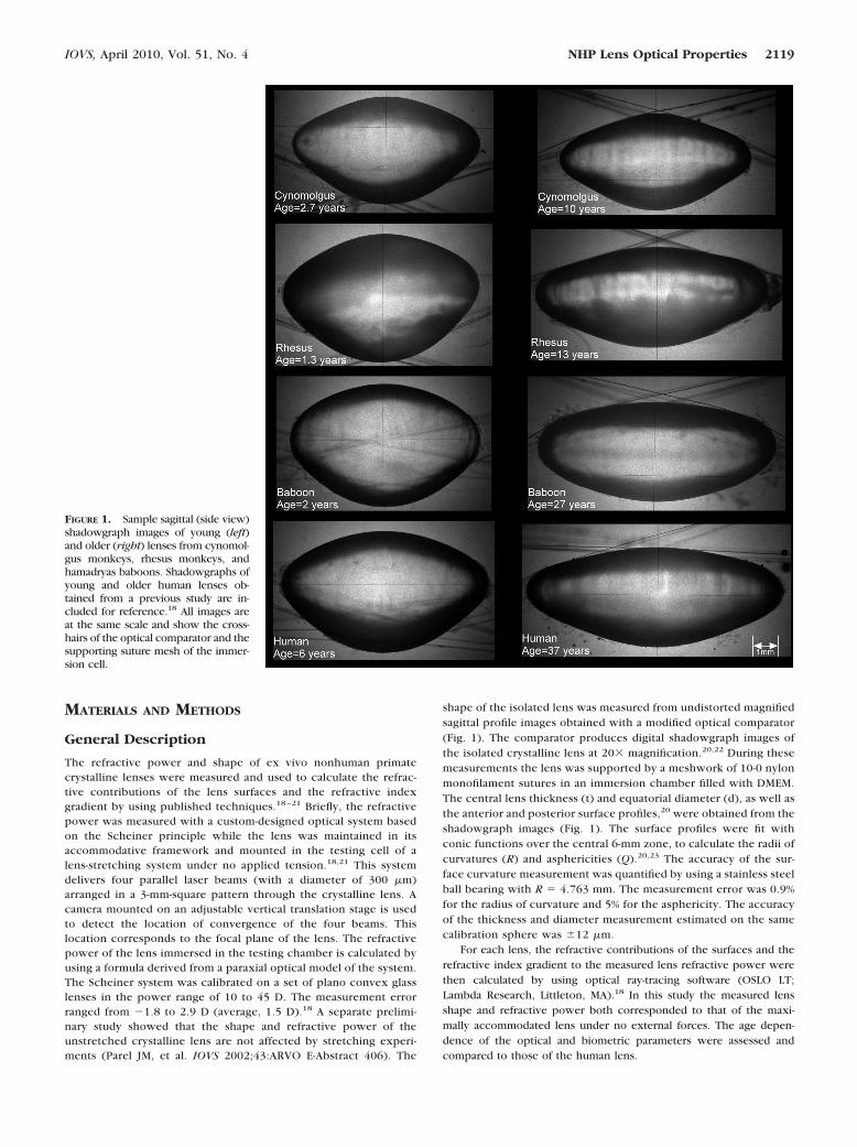
FIGURE 1. Sample sagittal (side view)shadowgraph images of young (left)and older (right) lenses from cynomol-gus monkeys, rhesus monkeys, andhamadryas baboons. Shadowgraphs ofyoung and older human lenses ob-tained from a previous study are in-cluded for reference.18 All images areat the same scale and show the cross-hairs of the optical comparator and thesupporting suture mesh of the immer-sion cell.
IOVS, April 2010, Vol. 51, No. 4 NHP Lens Optical Properties 2119
Donor Tissue
All animal experiments adhered to the ARVO Statement for the Use ofAnimals in Ophthalmic and Visual Research. No animal was killed forthe sole purpose of this study. Whole eyes of rhesus and cynomolgusmonkeys and hamadryas baboons were obtained through the Divisionof Veterinary Resources at the University of Miami as part of a univer-sity-wide tissue-sharing protocol and were used in accordance withinstitutional animal care and use guidelines.
Refractive power was measured on 120 ex vivo lenses from 89donor cynomolgus monkeys (Macaca fascicularis: postmortem time[PMT], 11.8 � 13.7 hours; age, 2.7–14.3 years); on 61 ex vivo lensesfrom 40 donor rhesus monkeys (Macaca mulatta: PMT, 22.3 � 14.9hours; age, 0.7–13.3 years); and on 16 ex vivo lenses from 16 donorhamadryas baboons (Papio hamadryas; PMT, 12.1 � 14.8 hours; ages,1.7–27.3 years) all obtained within 24 hours of euthanatization. Someof the lenses had been used for other experiments and therefore could
not be prepared for lens shape measurements in the optical compar-ator after the power was measured. In total, lens shape was measuredon 76 lenses from 58 cynomolgus monkeys, 43 lenses from 23 rhesusmonkeys, and 16 lenses from 13 hamadryas baboons. Any lens withvisible damage on shadow photogrammetry imaging was excludedfrom the analysis. In total, 8 lenses (0 rhesus, 1 baboon, and 7 cyno-molgus monkey lenses) of a total of 135 lenses placed in the shadow-graph had to be excluded due to damage or swelling.
Tissue Dissection
Dissections were performed by ophthalmic surgeons under a motor-ized operation microscope (OMS-300; Topcon, Tokyo, Japan). A com-plete description of the tissue preparation protocol can be found inanother publication.21 In this technique a band of eight independentshoes, matching the scleral curvature, were bonded with cyanoacrylateadhesive (Duro SuperGlue; Henkel Locktite Corp., Cleveland, OH)onto the anterior sclera surface from the limbus to the equator. Thismethod prevents deformation of the globe during dissection, and theshoes provide attachment for a lens-stretching device. The posteriorpole was then sectioned, and scleral incisions were made betweenadjacent shoes followed by removal of the cornea and iris to produceeight individual segments for stretching. The tissue section includingthe lens was mounted in the testing chamber of the lens stretcher, andrefractive power was measured in lenses in the unstretched state. Thetesting chamber of the lens stretcher was filled with preservativemedium (DMEM/F-12, D8437; Sigma-Aldrich, St. Louis, MO) to preventswelling.24
On average, the lens was immersed in DMEM for approximately 30minutes until the refractive power measurements were completed. Atthe end of the stretching experiments, the lens was extracted bycarefully cutting the zonules and adherent vitreous with Vannas scis-sors. The isolated lens was immediately immersed in a DMEM-filled vialto prevent swelling.24 The vial was then placed in the optical compar-ator for lens shape measurements. In total, the lenses were immersedin DMEM for �1 to 2 hours during the stretching experiments in theapparatus before shadowgraph measurements of the isolated lensshape.
Data and Statistical Analysis
Previous studies have shown that there is no significant differencebetween the diameter and the refractive power of the isolated lens and
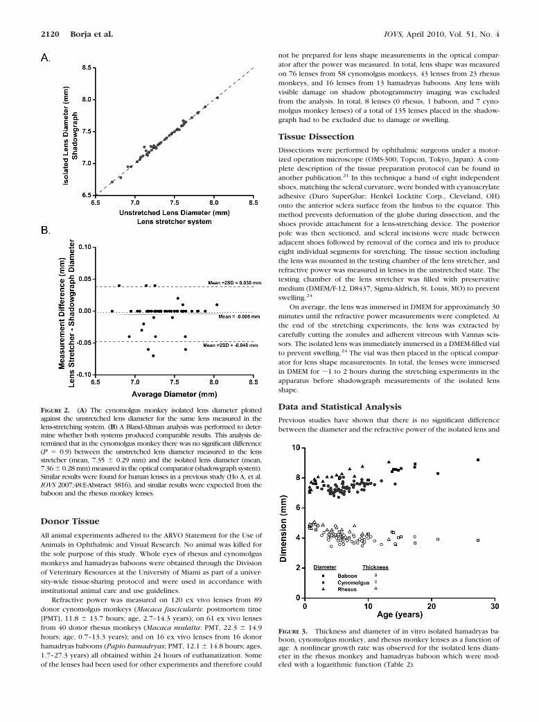
FIGURE 2. (A) The cynomolgus monkey isolated lens diameter plottedagainst the unstretched lens diameter for the same lens measured in thelens-stretching system. (B) A Bland-Altman analysis was performed to deter-mine whether both systems produced comparable results. This analysis de-termined that in the cynomolgus monkey there was no significant difference(P � 0.9) between the unstretched lens diameter measured in the lensstretcher (mean, 7.35 � 0.29 mm) and the isolated lens diameter (mean,7.36 � 0.28 mm) measured in the optical comparator (shadowgraph system).Similar results were found for human lenses in a previous study (Ho A, et al.IOVS 2007;48:E-Abstract 3816), and similar results were expected from thebaboon and the rhesus monkey lenses.
FIGURE 3. Thickness and diameter of in vitro isolated hamadryas ba-boon, cynomolgus monkey, and rhesus monkey lenses as a function ofage. A nonlinear growth rate was observed for the isolated lens diam-eter in the rhesus monkey and hamadryas baboon which were mod-eled with a logarithmic function (Table 2).
2120 Borja et al. IOVS, April 2010, Vol. 51, No. 4

the lens mounted in the lens stretcher under no tension18 (Ho A, et al.IOVS 2007;48:E-Abstract 3816). In these experiments, top views of thelens mounted in the lens stretcher were recorded with a digital cameramounted on an operation microscope set at 10� magnification.21 ABland-Altman analysis was performed to determine whether both sys-tems produced comparable results. The findings of this analysis arepresented in the Results section.
There was no significant difference (P � 0.05) in the biometric andoptical properties between lenses obtained from the eyes of the samedonor. Therefore, to prevent values obtained from paired eyes frombiasing the statistics for age dependence, the values from the eyes ofeach donor were averaged. The average values were then used as asingle point for age-dependence calculations.
The lens volume was estimated by assuming that the lens was arotationally symmetric ellipsoid with a long axis equal to the equa-torial diameter and a short axis equal to the lens thickness.20 Thisellipsoid model was used as an approximation for the whole lensshape, including the equatorial region, only for the calculation ofvolume, whereas the conic fits were used to obtain more accuratevalues of the radius of curvature and asphericity of the lens in thecentral 6-mm diameter zone. The surface radii of curvature andasphericity are reported with positive values for the anterior surfaceand negative values for the posterior surface.
The refractive contributions of the lens surfaces were determinedby calculating the refractive power of each lens, assuming a uniformrefractive index equal to the outer cortex refractive index (nsurfaces �1.371).25 The contribution of the refractive index gradient is expressedas an equivalent refractive index, which is the uniform refractive indexvalue required inside the lens to provide a refractive power equal tothe measured lens refractive power.
The biometric (central thickness, equatorial diameter, and sur-face radii of curvatures) and optical properties (refractive power,surface contribution, and equivalent refractive index) were plottedas a function of age. Linear and nonlinear regressions were calcu-lated for each data plot to determine whether there was an agedependence. A logarithmic growth function was used to model thegrowth of the isolated lens dimensions which displayed a nonlinearage dependence. P � 0.05 was set as the condition for statisticalsignificance.
RESULTS
Isolated nonhuman primate lenses displayed an increase inequatorial diameter, a decrease in thickness, a flattening of thesurfaces (Fig. 1), and a decrease in equivalent refractive index,leading to a dramatic age-dependent decrease in refractivepower of the isolated lens.
In this study, refractive power measurements were ob-tained while the lenses were mounted in a lens-stretchingapparatus. Figure 2 shows the cynomolgus monkey isolated
lens diameter versus the unstretched lens diameter measuredin the lens-stretching system. A Bland-Altman analysis per-formed on these results determined that there was no signifi-cant difference (P � 0.9) between the unstretched lens diam-eter measured in the lens-stretching system (mean diameter,
FIGURE 4. Anterior and posterior surface curvatures (A) and surfaceasphericities (B) of isolated hamadryas baboon, cynomolgus monkey,and rhesus monkey lenses as a function of age. A nonlinear trend wasobserved for the baboon lens anterior radius of curvature (Table 2).With age, the asphericity became more negative (Q � 0), whichcorresponds to a hyperbolic shape.
TABLE 1. Age-Dependent Linear and Nonlinear Regression Equations of the Measured NonhumanPrimate In Vitro Lens Biometric Properties
Species Hamadryas Baboon Cynomolgus Monkey Rhesus Monkey
Age range, y 1.67–27.3 1.7–14.3 0.7–13.3
Central thickness, mm 4.64-0.04 � Age 4.33–0.05 � Age 4.90–0.06 � AgeP � 0.002 P � 0.0001 P � 0.0001
Equatorial diameter, mm 7.16 � 0.50 � ln(Age) 6.92 � 0.07 � Age 7.42 � 0.53 � ln(Age)P � 0.0001 P � 0.0001 P � 0.0001
Approximate volume mm3 140.5 � 1.24 � Age 112.59 � 8.12 135.9 � 14.9 � ln(Age)P � 0.003 P � 0.14 P � 0.0001
The averages and SD are given for the parameters which showed no statistically significant agedependency.
IOVS, April 2010, Vol. 51, No. 4 NHP Lens Optical Properties 2121
7.35 � 0.29 mm) and that of the isolated lens (mean diameter,7.36 � 0.28 mm), when measured by the optical comparator(shadowgraph).
The examples of isolated crystalline lens shadowgraphs inFigure 1 illustrate the growth of the crystalline lens in all threenonhuman primate species. Lens dimensions are shown inFigure 3. Regression analyses are summarized in Table 1. Non-linear age-dependent trends were observed for isolated lensdiameter growth in the nonhuman primates as well as for theanterior surface curvatures in the hamadryas baboon. Thecentral lens thickness of all three species decreased linearly(P � 0.05) throughout the measured age ranges (Table 1). Themeasured lens refractive power and calculated surface refrac-tive power both decreased linearly over the sampled ageranges. Linear age-dependent trends were also observed withthe hamadryas baboon lens for surface refractive contributionand equivalent refractive index.
The approximate volume of the isolated rhesus monkeylens, calculated from the measured dimensions, displayedlogarithmic growth in the measured age range. The lensesof the cynomolgus monkeys are smaller in diameterand slightly thicker than comparably aged hamadryas ba-boon and rhesus monkey lenses. They are also slightlysmaller in total volume than the other nonhuman primatespecies.
The anterior and posterior radii of curvature for all threenonhuman primate species increased (flattening) with age(Fig. 4, Table 2). In the hamadryas baboon the anteriorsurface curvature increased nonlinearly over the age rangeof 1.7 to 27.3 years. It remained constant until approxi-mately 15 years of age and then started to increase. In therhesus monkey, the anterior lens surface curvature showedan age-dependent increase; however, the trend was notstatistically significant. The surface curvatures of the cyno-molgus monkey lens appeared to increase linearly through-out the sampled age range.
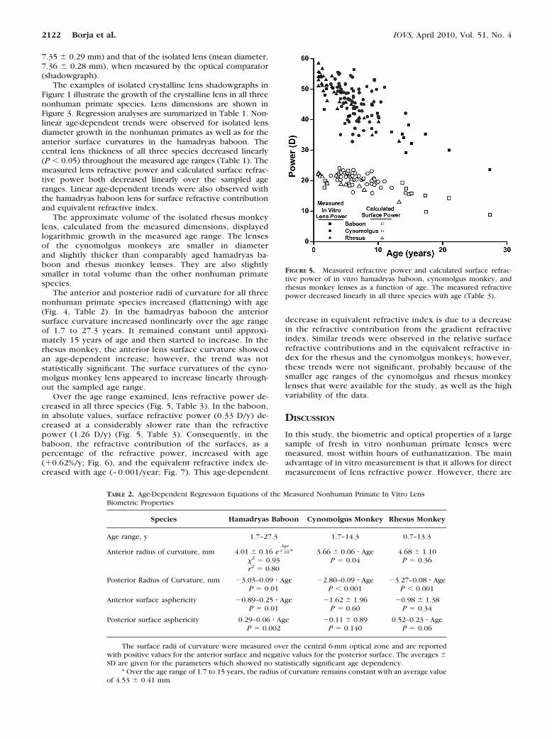
Over the age range examined, lens refractive power de-creased in all three species (Fig. 5, Table 3). In the baboon,in absolute values, surface refractive power (0.33 D/y) de-creased at a considerably slower rate than the refractivepower (1.26 D/y) (Fig. 5, Table 3). Consequently, in thebaboon, the refractive contribution of the surfaces, as apercentage of the refractive power, increased with age(�0.62%/y; Fig. 6), and the equivalent refractive index de-creased with age (– 0.001/year; Fig. 7). This age-dependent
decrease in equivalent refractive index is due to a decreasein the refractive contribution from the gradient refractiveindex. Similar trends were observed in the relative surfacerefractive contributions and in the equivalent refractive in-dex for the rhesus and the cynomolgus monkeys; however,these trends were not significant, probably because of thesmaller age ranges of the cynomolgus and rhesus monkeylenses that were available for the study, as well as the highvariability of the data.
DISCUSSION
In this study, the biometric and optical properties of a largesample of fresh in vitro nonhuman primate lenses weremeasured, most within hours of euthanatization. The mainadvantage of in vitro measurement is that it allows for directmeasurement of lens refractive power. However, there are
FIGURE 5. Measured refractive power and calculated surface refrac-tive power of in vitro hamadryas baboon, cynomolgus monkey, andrhesus monkey lenses as a function of age. The measured refractivepower decreased linearly in all three species with age (Table 3).
TABLE 2. Age-Dependent Regression Equations of the Measured Nonhuman Primate In Vitro LensBiometric Properties
Species Hamadryas Baboon Cynomolgus Monkey Rhesus Monkey
Age range, y 1.7–27.3 1.7–14.3 0.7–13.3
Anterior radius of curvature, mm 4.01 � 0.16 eAge
7.10 * 3.66 � 0.06 � Age 4.68 � 1.10�2 � 0.93 P � 0.04 P � 0.36r2 � 0.80
Posterior Radius of Curvature, mm �3.03–0.09 � Age �2.80–0.09 � Age �3.27–0.08 � AgeP � 0.01 P � 0.001 P � 0.001
Anterior surface asphericity �0.89–0.25 � Age �1.62 � 1.96 �0.98 � 1.38P � 0.01 P � 0.60 P � 0.34
Posterior surface asphericity 0.29–0.06 � Age �0.11 � 0.89 0.52–0.23 � AgeP � 0.002 P � 0.140 P � 0.06
The surface radii of curvature were measured over the central 6-mm optical zone and are reportedwith positive values for the anterior surface and negative values for the posterior surface. The averages �SD are given for the parameters which showed no statistically significant age dependency.
* Over the age range of 1.7 to 15 years, the radius of curvature remains constant with an average valueof 4.53 � 0.41 mm.
2122 Borja et al. IOVS, April 2010, Vol. 51, No. 4
several limitations of in vitro measurement of isolatedlenses, as opposed to in vivo measurement. For example, invitro measurements correspond to the maximally accommo-dated state, whereas in vivo measurement can be performedat various accommodative states. It is also not possible todetermine the relation between the lens parameters mea-sured in vitro and the actual accommodation response of thewhole eye. The main limitation of in vitro human lensstudies is that it is difficult to maintain the natural lens shapeand refractive power in postmortem lenses, especially if thelenses have been stored for several days in the preservativemedia used by eye banks.24 In previous studies,24,26 wefound that lenses with a postmortem time of less than 24hours could be immersed in DMEM for as long as 5 hourswithout swelling, as long as the capsule was intact. Swellingoccurred only in lenses that were damaged during preserva-tion or extraction. The short postmortem times proved to bea benefit compared with similar human in vitro crystallinelens studies in which long postmortem time results in therejection of many otherwise viable samples due to swell-ing.24 As part of the experimental protocol, the high-magni-fication shadowgraph images are used to detect any damageor capsular detachment. In the present study, only 8 (6%) of135 lenses imaged with the shadowgraph were found to bedamaged or swollen.
In all three nonhuman primate species, we found anage-dependent flattening of the lens surfaces (Figs. 1, 4) anda decrease in refractive power (Fig. 5) which also have beenobserved in pre-presbyopic adult human lenses (�50 yearsold).18 An approximate scaling factor of 1 monkey year to 3human years has been used by other investigators to com-pare the age-dependent decrease in the human and rhesusmonkey accommodative response3 and emmetropization.8
With this scaling factor, the nonhuman primates in thisstudy corresponded to a human age range of 2 to 40 years.In that age range, the rate of decrease in the human lensrefractive power was �0.41 D/y in our previous in vitrostudy.18 This result is similar to those found in the rhesusmonkey in the present study scaled for age as well as for theother two species (rhesus: �0.58 D/human year; cyno:�0.42 D/human year; baboon �0.32D/human year; Table3). The similarities in the lens properties as well as the easeof availability and short postmortem times tends to justifythe use of the nonhuman primate as a model for the humanlens in in vitro accommodation and presbyopia studies.
Our results show that over the sampled age range, therefractive power of the isolated crystalline lens decreased bymore than 20 D in all three species. Equatorial lens diameterincreased, whereas central thickness decreased over the sam-pled age range in all three species. It might have been ex-
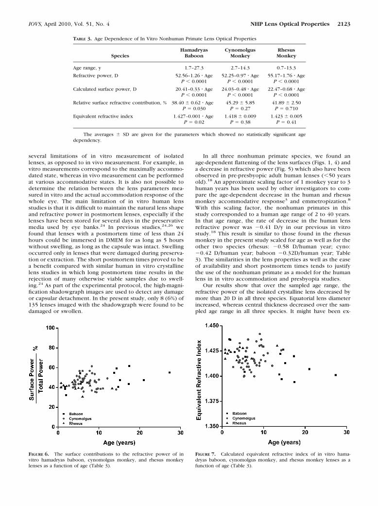
FIGURE 6. The surface contributions to the refractive power of invitro hamadryas baboon, cynomolgus monkey, and rhesus monkeylenses as a function of age (Table 3).
FIGURE 7. Calculated equivalent refractive index of in vitro hama-dryas baboon, cynomolgus monkey, and rhesus monkey lenses as afunction of age (Table 3).
TABLE 3. Age Dependence of In Vitro Nonhuman Primate Lens Optical Properties
SpeciesHamadryas
BaboonCynomolgus
MonkeyRhesusMonkey
Age range, y 1.7–27.3 2.7–14.3 0.7–13.3
Refractive power, D 52.56–1.26 � Age 52.25–0.97 � Age 55.17–1.76 � AgeP � 0.0001 P � 0.0001 P � 0.0001
Calculated surface power, D 20.41–0.33 � Age 24.03–0.48 � Age 22.47–0.68 � AgeP � 0.0001 P � 0.0001 P � 0.0001
Relative surface refractive contribution, % 38.40 � 0.62 � Age 45.29 � 5.85 41.89 � 2.50P � 0.030 P � 0.27 P � 0.710
Equivalent refractive index 1.427–0.001 � Age 1.418 � 0.009 1.423 � 0.005P � 0.02 P � 0.38 P � 0.41
The averages � SD are given for the parameters which showed no statistically significant agedependency.
IOVS, April 2010, Vol. 51, No. 4 NHP Lens Optical Properties 2123

pected that central lens thickness would increase in the mon-key lens, similar to the human lens, which decreases in the firstdecade or so of life and then increases.27 However, this trendwas not observed in this study, probably because of the limitednumber of samples in the upper age range.
It is difficult to directly compare the lens refractive powerand biometry results from this study with in vivo measure-ments. Typically, in vivo measurements of lens shape andrefractive power are performed on the relaxed, disaccommo-dated lens after pharmacologic pupil dilation. The lenses in-cluded in this study were isolated and free of external forces.Their shape and refractive power were expected to corre-spond to those of the maximally accommodated lens which isfree of zonular tension. The isolated lens growth trends ob-served in this study are in good agreement with results ob-tained on maximally accommodated rhesus monkey lenses invivo.5,8,12 Wendt et al.12 found that the maximally accommo-dated rhesus monkey lens diameter increases with age at a rateof 0.043 mm/y, which is similar to the results reported here forthe isolated lens. Other investigators have observed an age-dependent decrease in the central lens thickness of the younghuman and nonhuman primate during in vivo measure-ments.8,27–30 A general flattening of the lens surfaces with agehas been well documented in the monkey.5,8 Recently, Rosaleset al.30 used Purkinje imaging to measure the accommodation-dependent lens radii of curvature in two 9-year-old rhesusmonkeys. They found maximally accommodated radii of cur-vature (6.79 mm for the anterior and �5.11mm for the poste-rior surfaces) that were within the range of our measurements,indicating that the isolated lens is representative of the in vivomaximally accommodated lens in the nonhuman primate, as isthe case in the human.
In the hamadryas baboon, as in the human lens, the de-crease in refractive contribution from the internal refractiveindex gradient was the major contributing factor to the rapidloss of maximally accommodated lens refractive power frombirth to the age range of presbyopia onset (Fig. 6).
The equivalent refractive indexes from the present study(maximum measured values of 1.436 for both cynomolgusmonkey and baboon lenses and 1.430 for the rhesus mon-key) are similar to those of the human lens in vivo (maxi-mum of 1.4375)31 and in vitro (maximum of 1.432).18 Therhesus monkey equivalent refractive indexes from thepresent study are lower than previously reported in vivovalues (1.447 for a 5-year-old rhesus monkey lens8). Thehigher equivalent refractive indexes reported by Qiao-Grider et al.8 may be due to differences in accommodativestate during biometric measurements. In their study themeasurements of the refractive state and lens surface pro-files of rhesus monkeys were performed after accommoda-tion was pharmacologically relaxed. In the present study,the measurements correspond to the maximally accommo-dated state (lens free of external forces). The results fromthe present study indicate the similarity between humansand these three nonhuman primate species in the generalvalue and the age-dependent trend of the equivalent refrac-tive index.
In summary, the results of this study highlight the similari-ties in optical properties, biometric properties, growth, andaging of the isolated nonhuman primate lens to those of thehuman lens. In both human and nonhuman primates, there isa rapid age-dependent decrease in the refractive power of themaximally accommodated lens from birth up to the onset ofpresbyopia. The flattening of the lens surfaces in the maximallyaccommodated state contributes only a small portion to theage-dependent decrease in maximally accommodated lens re-fractive power. In both humans and nonhuman primates, the
decrease in refractive contributions from the gradient refrac-tive index is the major contributor to the rapid age-dependentdecrease in maximally accommodated lens refractive powerand the consequent decrease in maximum accommodativeamplitude. These findings suggest that independent of thechanges in lens shape, the age-dependent changes in the inter-nal lens refractive index distribution have a significant contri-bution to the loss of accommodative amplitude that leads topresbyopia.
Acknowledgments
The authors thank Norma Kenyon, PhD, and Dora Berman-Weinberg,PhD, of the DRI, and Linda Waterman, PhD, of DVR for scientificsupport; Viviana Fernandez, MD, Christian Billotte, MD, Ali Abri MD,Mohammed Aly, MD, Hideo Yamamoto, MD, PhD, and AdrianaAmelinckx, MD, for surgical support; and David Denham, MBA, AndresBernal, MSBME, Raksha Urs, MSBME, Marcia Orozco, MSBME, DerekNankivil, BS, Izuru Nose, BSEE, William Lee, David Chin Yee, MD, MinhHoang, BSBME, Stephanie Delgado, BSBME, and Jared Smith, BSBME,for technical support. This study includes data that were originallyacquired by Alexandre M. Rosen, MD, MSBME.
References
1. Tornqvist G. Effect of topical carbachol on the pupil and refractionin young and presbyopic monkeys. Invest Ophthalmol Vis Sci.1966;5:186–195.
2. Kaufman PL, Rohen JW, Barany EH. Hyperopia and loss of accom-modation following ciliary muscle disinsertion in the cynomolgusmonkey physiologic and scanning electron microscopic studies.Invest Ophthalmol Vis Sci . 1979;18:665–673.
3. Bito LZ, DeRousseau CJ, Kaufman PL, Bito JW. Age-dependent lossof accommodative amplitude in rhesus monkeys: an animal modelfor presbyopia. Invest Ophthalmol Vis Sci. 1982;23(1):23–31.
4. Koretz JF, Neider MW, Kaufman PL, Bertasso AM, DeRousseau CJ,Bito LZ. Slit-lamp studies of the rhesus monkey eye, I: survey of theanterior segment. Exp Eye Res. 1987;44(2):307–318.
5. Koretz JF, Bertasso AM, Neider MW, True-Gabelt BA, Kaufman PL.Slit-lamp studies of the rhesus monkey eye, II: changes in crystal-line lens shape, thickness and position during accommodation andaging. Exp Eye Res. 1987;45(2):317–326.
6. Koretz JF, Bertasso AM, Neider MW, Kaufman PL. Slit-lamp studiesof the rhesus monkey eye, III: the zones of discontinuity. Exp EyeRes. 1988;46(6):871–880.
7. Glasser A, Kaufman PL. The mechanism of accommodation inprimates. Ophthalmology. 1999;106(5):863–872.
8. Qiao-Grider Y, Hung LF, Kee CS, Ramamirtham R, Smith EL 3rd.Normal ocular development in young rhesus monkeys (Macacamulatta). Vision Res. 2007;47(11):1424–1444.
9. Neider MW, Crawford K, Kaufman PL, Bito LZ. In vivo videographyof the rhesus monkey accommodative apparatus: age-related lossof ciliary muscle response to central stimulation. Arch Ophthal-mol. 1990;108:69–74.
10. Croft MA, Kaufman PL, Crawford KS, Neider MW, Glasser A, BitoLZ. Accommodation dynamics in aging rhesus monkeys. Am JPhysiol. 1998;275:1885–1897.
11. Croft MA, Glasser A, Heatley G, et al. Accommodative ciliary bodyand lens function in rhesus monkeys, I: normal lens, zonule andciliary process configuration in the iridectomized eye. Invest Oph-thalmol Vis Sci. 2006;47:1076–1086.
12. Wendt M, Croft MA, McDonald J, Kaufman PL, Glasser A. Lensdiameter and thickness as a function of age and pharmacologicallystimulated accommodation in rhesus monkeys. Exp Eye Res. 2008;86(5):746–752.
13. Haefliger E, Parel JM, Fantes F, et al. Accommodation of an endo-capsular silicone lens (Phaco-Ersatz) in the nonhuman primate.Ophthalmology. 1987;94(5):471–477.
14. Haefliger E, Parel JM. Accommodation of an endocapsular siliconelens (Phaco-Ersatz) in the aging rhesus monkey. J Refract CornealSurg. 1994;10:550–555.
2124 Borja et al. IOVS, April 2010, Vol. 51, No. 4
15. Nishi O, Nishi K. Accommodation amplitude after lens refillingwith injectable silicone by sealing the capsule with a plug inprimates. Arch Ophthalmol. 1998;116:1358–1361.
16. Koopmans SA, Terwee T, Glasser A, et al. Accommodative lensrefilling in rhesus monkeys. Invest Ophthalmol Vis Sci. 2006;47:2976–2984.
17. Strenk SA, Strenk LM, Guo S. Magnetic resonance imaging of aging,accommodating, phakic, and pseudophakic ciliary muscle diame-ters. J Cataract Refract Surg. 2006;32(11):1792–1798.
18. Borja D, Manns F, Ho A, et al. Optical power of the isolated humancrystalline lens. Invest Ophthalmol Vis Sci. 2008;49:2541–2548.
19. Urs R, Manns F, Ho A, et al. Shape of the isolated ex-vivo humancrystalline lens. Vision Res. 2009;49:74–83.
20. Rosen AM, Denham DB, Fernandez V, et al. In vitro dimensionsand curvatures of human lenses. Vision Res. 2006;46:1002–1009.
21. Manns F, Parel JM, Denham D, et al. Optomechanical response ofhuman and monkey lenses in a lens stretcher. Invest OphthalmolVis Sci. 2007;48:3260–3268.
22. Denham D, Holland S, Mandelbaum S, Pflugfelder S, Parel JM.Shadow photogrammetric apparatus for the quantitative evalua-tion of corneal buttons. Ophthalmic Surg. 1989;20:794–799.
23. Manns F, Fernandez V, Zipper S, et al. Radius of curvature andasphericity of the anterior and posterior surface of human cadavercrystalline lenses. Exp Eye Res. 2004;78(1):39–51.
24. Augusteyn RC, Rosen AM, Borja D, Ziebarth NM, Parel JM. Biom-etry of primate lenses during immersion in preservation media.Mol Vis. 2006;12:740–747.
25. Jones CE, Atchison DA, Meder R, Pope JM. Refractive index distri-bution and optical properties of the isolated human lens measuredusing magnetic resonance imaging (MRI). Vision Res. 2005;45(18):2352–2366.
26. Rosen A. Biometry of the primate crystalline lens. Master’s thesis.Miami, FL: University of Miami; 2003.
27. Larsen JS. The sagittal growth of the eye, II: ultrasonic measure-ment of the axial diameter of the lens and the anterior segmentfrom birth to puberty. Acta Ophthalmol. 1971;49:427–439.
28. Zadnik K, Mutti DO, Friedman NE, Adams AJ. Initial cross-sectionalresults from the Orinda Longitudinal Study of Myopia. Optom VisSci. 1993;70(9):750–758.
29. Ip JM, Huynh SC, Kifley A, et al. Variation of the contribution fromaxial length and other oculometric parameters to refraction by ageand ethnicity. Invest Ophthalmol Vis Sci. 2007;48(10):4846–4853.
30. Rosales P, Wendt M, Marcos S, Glasser A. Changes in crystallinelens radii of curvature and lens tilt and decentration during dy-namic accommodation in rhesus monkeys. J Vision. 2008;8(1):1–12.
31. Dubbelman M, Van der Heijde GL. The shape of the aging humanlens: curvature, equivalent refractive index and the lens paradox.Vision Res. 2001;41(14):1867–1877.
IOVS, April 2010, Vol. 51, No. 4 NHP Lens Optical Properties 2125