Redox-silent tocotrienol esters as breast cancer proliferation and migration inhibitors
10
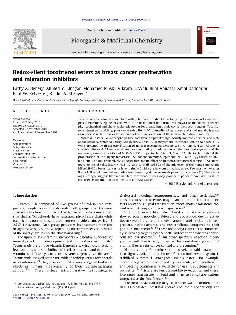
Redox-silent tocotrienol esters as breast cancer proliferation and migration inhibitors Fathy A. Behery, Ahmed Y. Elnagar, Mohamed R. Akl, Vikram B. Wali, Bilal Abuasal, Amal Kaddoumi, Paul W. Sylvester, Khalid A. El Sayed ⇑ Department of Basic Pharmaceutical Sciences, College of Pharmacy, University of Louisiana at Monroe, Monroe, LA 71201, United States article info Article history: Received 19 May 2010 Revised 25 August 2010 Accepted 2 September 2010 Available online 19 September 2010 Keywords: Anti-migratory Antiproliferative Breast cancer Chemical stability Semisynthetic esterification Tocotrienol Vitamin E Water solubility abstract Tocotrienols are vitamin E members with potent antiproliferative activity against preneoplastic and neo- plastic mammary epithelial cells with little or no effect on normal cell growth or functions. However, physicochemical and pharmacokinetic properties greatly limit their use as therapeutic agents. Tocotrie- nols’ chemical instability, poor water solubility, NPC1L1-mediated transport, and rapid metabolism are examples of such obstacles which hinder the therapeutic use of these valuable natural products. Vitamin E esters like a-tocopheryl succinate were prepared to significantly improve chemical and met- abolic stability, water solubility, and potency. Thus, 12 semisynthetic tocotrienol ester analogues 4–15 were prepared by direct esterification of natural tocotrienol isomers with various acid anhydrides or chlorides. Esters 4–15 were evaluated for their ability to inhibit the proliferation and migration of the mammary tumor cells +SA and MDA-MB-231, respectively. Esters 5, 9, and 11 effectively inhibited the proliferation of the highly metastatic +SA rodent mammary epithelial cells with IC 50 values of 0.62, 0.51, and 0.86 lM, respectively, at doses that had no effect on immortalized normal mouse CL-S1 mam- mary epithelial cells. Esters 4, 6, 8–10, and 13 inhibited 50% of the migration of the human metastatic MDA-MB-231 breast cancer cells at a single 5 lM dose in wound-healing assay. The most active ester 9 was 1000-fold more water-soluble and chemically stable versus its parent a-tocotrienol (1). These find- ings strongly suggest that redox-silent tocotrienol esters may provide superior therapeutic forms of tocotrienols for the control of metastatic breast cancer. Ó 2010 Elsevier Ltd. All rights reserved. 1. Introduction Vitamin E is composed of two groups of lipid-soluble com- pounds, tocopherols and tocotrienols. 1 Both groups share the same chemical structure, but differ in the degree of unsaturation of their side chains. Tocopherols have saturated phytyl side chain while tocotrienols possess unsaturated isoprenoid side chain, with all E D3 0 ,7 0 ,11 0 systems. Each group possesses four isomeric members designated as a, b, c, and d, depending on the number and position of the methyl groups on the chromanol ring. 2 The lipid-soluble vitamin E members are essential nutrients for normal growth and development and antioxidants in animals. 3 Tocotrienols are unique vitamin E members, which occur only in few natural sources including palm oil, barley, oat, and rice bran. 4 Vitamin E deficiency can cause severe degenerative diseases. 5 Tocotrienols showed better antioxidant activity versus tocopherols in membranes. 6–9 They also exhibited a wide range of biological effects in humans, independently of their radical-scavenging abilities. 10,11 These include antiproliferative, anti-angiogenic, cholesterol-lowering, neuroprotective, and other activities. 6,11 These redox-silent activities may be attributed to their unique ef- fects on various signal transduction, mevalonate–cholesterol bio- synthetic pathways, and gene expressions. 4,6 Vitamin E esters like a-tocopheryl succinate or oxyacetate showed potent growth-inhibitory and apoptotic-inducing activi- ties in several in vitro and in vivo cancer models including breast, prostate, neuroblastoma, and mesothelioma compared to their parent a-tocopherol. 12–14 These tocopheryl esters act as ‘mitocans’ by selectively targeting cancer cells’ mitochondria whereas normal cells are less affected. 12–15 This broad spectrum of action in con- junction with low toxicity underlies the translational potential of vitamin E esters for cancer control and prevention. 15 Natural vitamin E members are relatively unstable toward air, heat, light, alkali, and metal ions. 16,17 Therefore, several synthetic stabilized vitamin E analogues, mainly esters, for example, a-tocopheryl acetate and tocopheryl succinate, were synthesized and became commercially available for use in supplements and cosmetics. 17–19 Esters are less susceptible to oxidation and there- fore more appropriate for food and pharmaceutical applications compared to the free form. 17–19 The poor bioavailability of c-tocotrienol was attributed to its NPC1L1-mediated intestinal uptake and their lipophilicity and 0968-0896/$ - see front matter Ó 2010 Elsevier Ltd. All rights reserved. doi:10.1016/j.bmc.2010.09.009 ⇑ Corresponding author. Tel.: +1 318 342 1725; fax: +1 318 342 1737. E-mail address: [email protected] (K.A. El Sayed). Bioorganic & Medicinal Chemistry 18 (2010) 8066–8075 Contents lists available at ScienceDirect Bioorganic & Medicinal Chemistry journal homepage: www.elsevier.com/locate/bmc
Transcript of Redox-silent tocotrienol esters as breast cancer proliferation and migration inhibitors
Bioorganic & Medicinal Chemistry 18 (2010) 8066–8075
Contents lists available at ScienceDirect
Bioorganic & Medicinal Chemistry
journal homepage: www.elsevier .com/locate /bmc
Redox-silent tocotrienol esters as breast cancer proliferationand migration inhibitors
Fathy A. Behery, Ahmed Y. Elnagar, Mohamed R. Akl, Vikram B. Wali, Bilal Abuasal, Amal Kaddoumi,Paul W. Sylvester, Khalid A. El Sayed ⇑Department of Basic Pharmaceutical Sciences, College of Pharmacy, University of Louisiana at Monroe, Monroe, LA 71201, United States
a r t i c l e i n f o
Article history:Received 19 May 2010Revised 25 August 2010Accepted 2 September 2010Available online 19 September 2010
Keywords:Anti-migratoryAntiproliferativeBreast cancerChemical stabilitySemisynthetic esterificationTocotrienolVitamin EWater solubility
0968-0896/$ - see front matter � 2010 Elsevier Ltd. Adoi:10.1016/j.bmc.2010.09.009
⇑ Corresponding author. Tel.: +1 318 342 1725; faxE-mail address: [email protected] (K.A. El Sayed).
a b s t r a c t
Tocotrienols are vitamin E members with potent antiproliferative activity against preneoplastic and neo-plastic mammary epithelial cells with little or no effect on normal cell growth or functions. However,physicochemical and pharmacokinetic properties greatly limit their use as therapeutic agents. Tocotrie-nols’ chemical instability, poor water solubility, NPC1L1-mediated transport, and rapid metabolism areexamples of such obstacles which hinder the therapeutic use of these valuable natural products.
Vitamin E esters like a-tocopheryl succinate were prepared to significantly improve chemical and met-abolic stability, water solubility, and potency. Thus, 12 semisynthetic tocotrienol ester analogues 4–15were prepared by direct esterification of natural tocotrienol isomers with various acid anhydrides orchlorides. Esters 4–15 were evaluated for their ability to inhibit the proliferation and migration of themammary tumor cells +SA and MDA-MB-231, respectively. Esters 5, 9, and 11 effectively inhibited theproliferation of the highly metastatic +SA rodent mammary epithelial cells with IC50 values of 0.62,0.51, and 0.86 lM, respectively, at doses that had no effect on immortalized normal mouse CL-S1 mam-mary epithelial cells. Esters 4, 6, 8–10, and 13 inhibited 50% of the migration of the human metastaticMDA-MB-231 breast cancer cells at a single 5 lM dose in wound-healing assay. The most active ester9 was 1000-fold more water-soluble and chemically stable versus its parent a-tocotrienol (1). These find-ings strongly suggest that redox-silent tocotrienol esters may provide superior therapeutic forms oftocotrienols for the control of metastatic breast cancer.
� 2010 Elsevier Ltd. All rights reserved.
1. Introduction cholesterol-lowering, neuroprotective, and other activities.6,11
Vitamin E is composed of two groups of lipid-soluble com-pounds, tocopherols and tocotrienols.1 Both groups share the samechemical structure, but differ in the degree of unsaturation of theirside chains. Tocopherols have saturated phytyl side chain whiletocotrienols possess unsaturated isoprenoid side chain, with all ED30,70,110 systems. Each group possesses four isomeric membersdesignated as a, b, c, and d, depending on the number and positionof the methyl groups on the chromanol ring.2
The lipid-soluble vitamin E members are essential nutrients fornormal growth and development and antioxidants in animals.3
Tocotrienols are unique vitamin E members, which occur only infew natural sources including palm oil, barley, oat, and rice bran.4
Vitamin E deficiency can cause severe degenerative diseases.5
Tocotrienols showed better antioxidant activity versus tocopherolsin membranes.6–9 They also exhibited a wide range of biologicaleffects in humans, independently of their radical-scavengingabilities.10,11 These include antiproliferative, anti-angiogenic,
ll rights reserved.
: +1 318 342 1737.
These redox-silent activities may be attributed to their unique ef-fects on various signal transduction, mevalonate–cholesterol bio-synthetic pathways, and gene expressions.4,6
Vitamin E esters like a-tocopheryl succinate or oxyacetateshowed potent growth-inhibitory and apoptotic-inducing activi-ties in several in vitro and in vivo cancer models including breast,prostate, neuroblastoma, and mesothelioma compared to theirparent a-tocopherol.12–14 These tocopheryl esters act as ‘mitocans’by selectively targeting cancer cells’ mitochondria whereas normalcells are less affected.12–15 This broad spectrum of action in con-junction with low toxicity underlies the translational potential ofvitamin E esters for cancer control and prevention.15
Natural vitamin E members are relatively unstable toward air,heat, light, alkali, and metal ions.16,17 Therefore, several syntheticstabilized vitamin E analogues, mainly esters, for example,a-tocopheryl acetate and tocopheryl succinate, were synthesizedand became commercially available for use in supplements andcosmetics.17–19 Esters are less susceptible to oxidation and there-fore more appropriate for food and pharmaceutical applicationscompared to the free form.17–19
The poor bioavailability of c-tocotrienol was attributed to itsNPC1L1-mediated intestinal uptake and their lipophilicity and

Table 11H and 13C NMR data of 4 and 5a
Position 4 5
dC, mult. dH (J) dC, mult. dH (J)
2 75.0, qC 76.8, qC2-CH3 17.7, CH3 1.22, s 24.1, CH3 1.26, s3 31.2, CH2 1.76, m 31.1, CH2 1.76, m4 20.7, CH2 2.59, m 22.3, CH2 2.74, m5 123.3, qC 118.6, CH 6.81, s5-CH3 11.9, CH3 2.05, s6 149.7, qC 150.0, qC7 125.4, qC 127.2, qC7-CH3 13.1, CH3 2.09, s 12.1, CH3 2.13, s8 127.1, qC 126.1, qC8-CH3 12.2, CH3 2.08, s 12.8, CH3 2.12, s9 117.6, qC 118.8, qC10 140.6, qC 141.6, qC10 39.8, CH2 1.53, 1.60, m 39.8, CH2 1.56, 1.64, m20 22.2, CH2 2.07, 2.14, m 22.3, CH2 2.11, 2.15, m30 124.4, CH 5.06, m 124.5, CH 5.09, m40 135.0, qC 135.3, qC40-CH3 15.9, CH3 1.55, s 16.0, CH3 1.58, s50 39.8, CH2 1.94, m 39.8, CH2 1.96, m60 26.8, CH2 2.02, m 26.8, CH2 2.06, m70 124.2, CH 5.06, m 124.3, CH 5.09, m0
F. A. Behery et al. / Bioorg. Med. Chem. 18 (2010) 8066–8075 8067
poor water solubility.20–22 A commercially available water- solublevitamin E ester was produced by Eastman Chemical Company byesterification of polyethylene glycol-1000 onto RRR-a-tocopherylsuccinate.23 This ester has the ability to form miscible micelles inwater due to its amphiphilic properties. This approach enhancedtocopherols’ bioavailability in animals and humans via improvingtheir water solubility and absorption.23
After exerting their antioxidant activity, vitamin E isomers areusually rapidly metabolized and excreted.16,17,24–26 Esterificationof tocotrienols affords redox-silent analogues, which will preventtheir rapid metabolic inactivation via masking the free phenolicgroup required for chromanoxy radical formation. Generally, esterswill slowly hydrolyze to release their parent natural phenol, whichdecrease the rate of metabolism and enhance the metabolic stabil-ity of tocotrienols.2,16 Intestinal or epidermal esterases will cata-lyze ester hydrolysis and thus can be considered as pro-vitaminsor prodrugs of the natural tocotrienols. Esterification of tocotrie-nols is expected not only to maintain the bioactivity but also to im-prove its solubility and chemical stability.12–14,27 Therefore, thisstudy aims at the preparation of new highly polar esters of tocot-rienols with expected enhanced water solubility, stability, andbioactivity.
8 134.9, qC 135.1, qC80-CH3 16.0, CH3 1.55, s 16.1, CH3 1.58, s90 39.8, CH2 1.94, m 39.8, CH2 1.96, m100 26.6, CH2 2.02, m 26.7, CH2 2.06, m110 124.3, CH 5.06, m 124.3, CH 5.09, m120 131.4, qC 130.5, qC120a-CH3 16.6, CH3 1.57, s 17.8, CH3 1.60, s120b-CH3 25.7, CH3 1.63, s 25.8, CH3 1.66, s100 165.6, qC 166.6, qC200 135.3, qC 138.0, qC300 133.8, qC 131.8, qC300-COO 169.1, qC 170.4, qC400 130.7, CH 8.44, d, (1.4) 131.0, CH 8.71, d (1.4)500 133.5, qC 131.3, qC500-COO 167.2, qC 169.6, qC600 132.2, CH 8.23, dd (1.8, 8.1) 133.9, CH 8.37, dd (1.5, 8.0)700 129.4, CH 8.03, d (8.0) 129.2, CH 7.92, d (8.0)
a In CDCl3, J in Hertz. 400 MHz for 1H and 100 MHz for 13C NMR. Carbonmultiplicities were determined by APT experiments, C = quaternary, CH = methine,CH2 = methylene, CH3 = methyl carbons.
2. Results and discussion
2.1. Semisynthesis of tocotrienol ester analogues
a-, c-, and d-Tocotrienols (1–3, respectively) were isolated froma tocotrienol-rich fraction of palm oil using normal phase vacuumliquid chromatography. New esters 4–15 were semisynthesized bydirect esterification with carboxylic acid anhydrides or chlorides.Acids with terminal ionizable groups were used to esterify tocotri-enol isomers. 1,2,4-Benzenetricarboxylic, maleic, and 3-methylglu-taric anhydrides successfully reacted with tocotrienols. Thenaturally occurring caffeic and gallic acids did not react with toco-trienol isomers before activation with oxalyl chloride after protect-ing their phenolic groups by acetylation. The identity of each newtocotrienol ester was established by spectral techniques.
The HREIMS of 4 and 6 suggested the molecular formula C38H48O7
and possible esterification of 1 with 1,2,4-benzenetricarboxylicanhydride. The 1H and 13C NMR data (Tables 1 and 2) further sup-ported this conclusion. Esterification induced downfield shifting ofC-5 (Dd +4.7) and C-7 (Dd +4.2) in 4, compared to its parent 1. Similardownfield shift was also observed in 6 for C-5 and C-7 (Dd +4.7 and4.1, respectively). A +4.0 ppm downfield shift for C-6 was also ob-served in both compounds. The hypsochromic shift (�2 and �6 nmin 4 and 6, respectively) in the UV spectrum further confirmed theC-6 esterification.16 The HMBC spectra of 4 and 6 showed correlationcontours between the o- and m-coupled aromatic proton H-700 (dH
8.03 and 8.75 in 4 and 6, respectively) and the downfield carbonylcarbon C-100 (dC 165.6 and 164.8 in 4 and 6, respectively). Thus, com-pounds 4 and 6 were determined to be 4-(((R)-2,5,7,8-tetramethyl-2-((3E,7E)-4,8,12-trimethyltrideca-3,7,11-trienyl)chroman-6-yloxy)-carbonyl)-isophthalic acid and 2-(((R)-2,5,7,8-tetramethyl-2-((3E,7E)-4,8,12-trimethyltrideca-3,7,11-trienyl)chroman-6-yloxy)carbonyl)terephthalic acid, respectively.
The HREIMS of 5 and 7 suggested the molecular formulaC37H46O7 and possible esterification with 1,2,4-benzenetricarboxy-lic anhydride. The 1H and 13C NMR data (Tables 1 and 2) furthersupported this conclusion. Esterification induced the downfieldshifting of C-5 and H-5 (Dd +6.4, +0.44, respectively) and C-7 (Dd+1.3) in 5 compared to its parent 2. Similar downfield shift was alsoobserved in 7 for C-5, H-5 and C-7 (Dd +6.5, +0.39, and +1.5, respec-tively). Carbon C-6 was also downfield shifted in 5 and 7 (Dd +3.7and +3.5, respectively). The UV spectra of 5 and 7 also showed hyp-
sochromic shift as previously described, further confirming theesterification. The HMBC spectra of 5 and 7 connected the o- andm-coupled aromatic proton H-700, respectively, with the estercarbonyl C-100. Thus, compounds 5 and 7 were determined to be4-(((R)-2,7,8-trimethyl-2-((3E,7E)-4,8,12-trimethyltrideca-3,7,11-trienyl)chroman-6-yloxy)carbonyl)isophthalic acid and 2-(((R)-2,7,8-trimethyl-2-((3E,7E)-4,8,12-trimethyltrideca-3,7,11-trie-nyl)chroman-6-yloxy)carbonyl)terephthalic acid, respectively.
The HREIMS of 8 suggested the molecular formula C36H44O7 andpossible esterification of 3 with 1,2,4-benzenetricarboxylic anhy-dride. The 1H and 13C NMR data (Table 3) further supported thisconclusion. The esterification with terephthalate was further con-firmed by UV, 1H, 13C NMR chemical shifts, and HMBC data. Thus,compound 8 was determined to be 2-(((R)-2,8-dimethyl-2-((3E,7E)-4,8,12-trimethyltrideca-3,7,11-trienyl)-chroman-6-yloxy)-carbonyl)terephthalic acid.
The HREIMS of 9 suggested the molecular formula C33H46O5 andpossible maleate esterification. The 1H and 13C NMR data (Table 3)further supported this conclusion. The proton doublet H-300 (dH
7.06) showed a 3J-HMBC coupling with the carbonyl carbon C-100
and COSY coupling with proton H-200 (dH 7.13). The later showeda 3J-HMBC coupling with the carbonyl C-400, confirming the identityof the maleate ester. Thus, compound 9 was determined to be (Z)-4-oxo-4-((R)-2,5,7,8-tetramethyl-2-((3E,7E)-4,8,12-trimethyltrideca-3,7,11-trienyl)chroman-6-yloxy)but-2-enoic acid.

Table 21H and 13C NMR data of 6 and 7a
Position 6 7
dC, mult. dH (J) dC, mult. dH (J)
2 75.0, qC 75.9, qC2-CH3 17.8, CH3 1.26, s 24.1, CH3 1.24, s3 31.2, CH2 1.80, m 31.1, CH2 1.74, m4 22.3, CH2 2.61, m 22.2, CH2 2.71, m5 123.3, qC 118.7, CH 6.76, s5-CH3 12.0, CH3 2.05, s —6 149.7, qC 149.8, qC7 125.3, qC 127.4, qC7-CH3 13.1, CH3 2.12, s 12.0, CH3 2.10, s8 127.0, qC 126.0, qC8-CH3 12.3, CH3 2.10, s 12.7, CH3 2.07, s9 117.6, qC 118.6, qC10 140.6, qC 141.7, qC10 39.8, CH2 1.57, 1.62, m 39.8, CH2 1.50, 1.57, m20 20.7, CH2 2.07, 2.14, m 22.3, CH2 2.04, 2.08, m30 124.5, CH 5.09, m 124.4, CH 5.07, m40 135.3, qC 135.3, qC40-CH3 16.0, CH3 1.58, s 15.9, CH3 1.55, s50 39.8, CH2 1.96, m 39.8, CH2 1.93, m60 26.8, CH2 2.04, m 26.8, CH2 2.01, m70 124.3, CH 5.09, m 124.2, CH 5.07, m80 135.1, qC 135.1, qC80-CH3 16.1, CH3 1.58, s 16.0, CH3 1.55, s90 39.8, CH2 1.96, m 39.8, CH2 1.93, m100 26.7, CH2 2.04, m 26.6, CH2 2.01, m110 124.3, CH 5.09, m 124.2, CH 5.07, m120 130.4, qC 131.4, qC120a-CH3 16.5, CH3 1.60, s 17.7, CH3 1.57, s120b-CH3 25.8, CH3 1.66, s 25.7, CH3 1.63, s100 164.8, qC 166.4, qC200 132.4, qC 133.1, qC300 138.3, qC 136.5, qC300-COO 169.7, qC 168.8, qC400 129.1, CH 7.81, d (8.1) 129.5, CH 7.86, d (8.0)500 133.4, CH 8.29, dd (1.5, 8.0) 132.7, CH 8.21, dd (1.5, 7.7)600 131.4, qC 134.3, qC600-COO 167.3, qC 167.1, qC700 131.2, CH 8.75, d (1.5) 130.4, CH 8.51, d (1.5)
a In CDCl3, J in Hertz. 400 MHz for 1H and 100 MHz for 13C NMR. Carbonmultiplicities were determined by APT experiments, C = quaternary, CH = methine,CH2 = methylene, CH3 = methyl carbons.
Table 31H and 13C NMR data of 8 and 9a
Position 8 9
dC, mult. dH (J) dC, mult. dH (J)
2 76.0, qC 75.0, qC —2-CH3 24.1, CH3 1.24, s 24.2, CH3 1.25, s3 31.1, CH2 1.75, m 31.1, CH2 1.76, m4 22.5, CH2 2.73, m 20.7, CH2 2.60, m5 119.0, CH 6.77, d (2.9) 123.3, qC5-CH3 11.9, CH3 1.96, s6 150.0, qC 149.8, qC7 121.0, CH 6.82, d (2.6) 124.8, qC7-CH3 13.1, CH3 2.10, s8 127.5, qC 126.6, qC8-CH3 16.1, CH3 2.13, s 12.2, CH3 2.00, s9 121.1, qC 117.6, qC10 142.6, qC 140.3, qC10 39.8, CH2 1.50, 1.59, m 39.8, CH2 1.54, 1.62, m20 22.2, CH2 2.06, 2.10, m 22.3, CH2 2.08, m30 124.4, CH 5.06, m 124.5, CH 5.09, m40 135.4, qC 135.3, qC40-CH3 15.9, CH3 1.55, s 16.1, CH3 1.59, s50 39.8, CH2 1.92, m 39.8, CH2 1.96, m60 26.8, CH2 2.02, m 26.8, CH2 2.05, m70 124.2, CH 5.06, m 124.3, CH 5.09, m80 135.1, qC 135.1, qC80-CH3 16.0, CH3 1.55, s 16.0, CH3 1.59, s90 39.8, CH2 1.92, m 39.8, CH2 1.96, m100 26.6, CH2 2.02, m 26.7, CH2 2.05, m110 124.2, CH 5.06, m 124.3, CH 5.09, m120 131.4, qC 131.3, qC120a-CH3 17.7, CH3 1.56, s 17.8, CH3 1.58, s120b-CH3 25.7, CH3 1.63, s 25.8, CH3 1.67, s100 166.8, qC 163.8, qC200 133.2, qC 133.4, CH 7.13, d (15.8)300 136.0, qC 135.6, CH 7.06, d (15.8)300-COO 168.6, qC400 129.5, CH 7.89, d (8.0) 168.7, qC500 133.2, CH 8.21, dd (1.5, 7.0)500-COO 167.1, qC600 134.4, qC700 130.4, CH 8.45, d (1.4)
a In CDCl3, J in Hertz. 400 MHz for 1H and 100 MHz for 13C NMR. Carbonmultiplicities were determined by APT experiments, C = quaternary, CH = methine,CH2 = methylene, CH3 = methyl carbons.
8068 F. A. Behery et al. / Bioorg. Med. Chem. 18 (2010) 8066–8075
The HREIMS of 10 and 11 suggested the molecular formulaC32H44O5 and C31H42O5, respectively, suggesting possible maleateesterification of 2 and 3, respectively. Their 1H and 13C NMR data(Table 4) further supported this conclusion. Thus, compounds 10and 11 were determined to be (Z)-4-oxo-4-((R)-2,7,8-trimethyl-2-((3E,7E)-4,8,12-trimethyltrideca-3,7,11-trienyl)-chroman-6-yloxy)but-2-enoic acid and (Z)-4-((R)-2,8-dimethyl-2-((3E,7E)-4,8,12-trimethyltrideca-3,7,11-trienyl)chroman-6-yloxy)-4-oxo-but-2-enoic acid, respectively.
The HREIMS of 12 suggested the molecular formula C34H50O5
and possible esterification with 3-methylglutaric anhydride. The1H and 13C NMR data (Table 5) further supported this conclusion.The methyl doublet H3-600 (dH 1.16) showed 3J-HMBC couplingswith the methylene carbons C-200 and C-400 (dC 40.6 and 40.5,respectively). It also showed a 2J-HMBC coupling with the methinecarbon C-300 (dC 27.4). Proton H-300 showed 3J-HMBC couplings withthe carbonyl carbons C-100 and C-500 (dC 171.5 and 178.2, respec-tively) and COSY couplings with the methylene protons H2-200
and H2-H-400, confirming the 3-methyl-glutaryl ester. Thus, com-pound 12 was determined to be 3-Methyl-5-oxo-5-((R)-2,7,8-tri-methyl-2-((3E,7E)-4,8,12-trimethyltrideca-3,7,11-trienyl)chro-man-6-yloxy)pentanoic acid.
The HREIMS of 13 suggested the molecular formula C30H46O2
and possible benzoylation. The 1H and 13C NMR data (Table 5)further supported this conclusion. The proton doublet H-300/H-700
(dH 8.21) showed 3J-HMBC couplings with the carbonyl ester C-100
and the aromatic methine carbon C-500 (dC 165.7 and 133.4, respec-tively). Protons H-300/H-700 also showed a COSY coupling withproton doublet of doublets H-400/H-600 (dH 7.50), confirming theidentity of the benzoate moiety. Thus, compound 13 was deter-mined to be (R)-2,7,8-trimethyl-2-((3E,7E)-4,8,12-trimethyltri-deca-3,7,11-trienyl)chroman-6-yl benzoate.
The HREIMS of 14 suggested the molecular formula C41H52O9
and possible esterification with 3,4,5-triacetoxybenzoyl chloride.The proton singlet H-300/H-700 (dH 7.95) showed 3J-HMBC couplingswith the carbonyl ester C-100 and the aromatic oxygenated quater-nary carbon C-500 (dC 163.5 and 139.1, respectively). The C-400, C-500,and C-600 acetoxy methyl singlets (dH 2.31, 2.32, and 2.31, respec-tively) showed 2J-HMBC couplings with their corresponding car-bonyl carbons (dC 167.8, 166.5, and 167.8, respectively). Therewas no enough available spectral evidence to unambiguously as-sign these acetoxy groups. Thus, compound 14 was determinedto be 5-(((R)-2,7,8-trimethyl-2-((3E,7E)-4,8,12-trimethyltrideca-3,7,11-trienyl)chroman-6-yloxy)carbonyl)benzene-1,2,3-triyltriacetate.
The HREIMS of 15 suggested the molecular formula C41H52O7
and possible esterification with 3,4-diacetylcaffeoyl chloride. The1H and 13C NMR data (Table 6) further supported this conclusion.The proton doublet H-300 (dH 7.79) showed 3J-HMBC couplings withthe carbonyl ester C-100 and the aromatic methine carbons C-500 and

F. A. Behery et al. / Bioorg. Med. Chem. 18 (2010) 8066–8075 8069
C-900 (dC 165.7, 123.0, and 126.7, respectively). Proton H-300 showeda COSY coupling with the proton doublet H-200 (dH 6.59). ProtonH-900 (dH 7.46) showed 3J-HMBC couplings with C-500 and the oxy-genated quaternary aromatic carbon C-700 (dC 143.8). It also showedCOSY coupling with proton doublet H-800 (dH 7.25). The later protonshowed 3J-HMBC couplings with the quaternary aromatic carbonsC-400 and C-600 (dC 133.3 and 143.4, respectively), confirming theidentity of the caffeoyl moiety. The C-600and C-700 acetoxymethylsinglets (dH 2.31 and 2.32, respectively) showed 2J-HMBC couplingswith their corresponding carbonyl carbons (dC 168.2 and 168.1,respectively). There was no enough available spectral evidence tounambiguously assign these acetoxy groups. Thus, compound 15was determined to be 4-((E)-3-oxo-3-((R)-2,7,8-trimethyl-2-((3E,7E)-4,8,12-trimethyltrideca-3,7,11-trienyl)chroman-6-yloxy)-prop-1-enyl)-1,2-phenylene diacetate.
R2O
R3
R1
O 2
45
8 1' 5' 9'
Compound R1 R2 R3
1 CH3 H CH3
2 H H CH3
3 H H H 4 CH3 a CH3
5 H a CH3
6 CH3 b CH3
7 H b CH3
8 H b H 9 CH3 c CH3
10 H c CH3
11 H c H 12 H d CH3
13 H e CH3
14 H f CH3
15 H g CH3
O
O
HO
O
OH
1''2''
3''5''
O
HO
O O
OH1''2''
3''
6''
O
OH
O1''
3''4''
O
OH
O1'' 3'' 5''
O
1''
3''
7''
O
AcO
OAc
OAc1''
3''
7''
a b
c d e
f g
O
OAc
OAc
1''
3''
5''
8''
6''
2.2. Biological activity
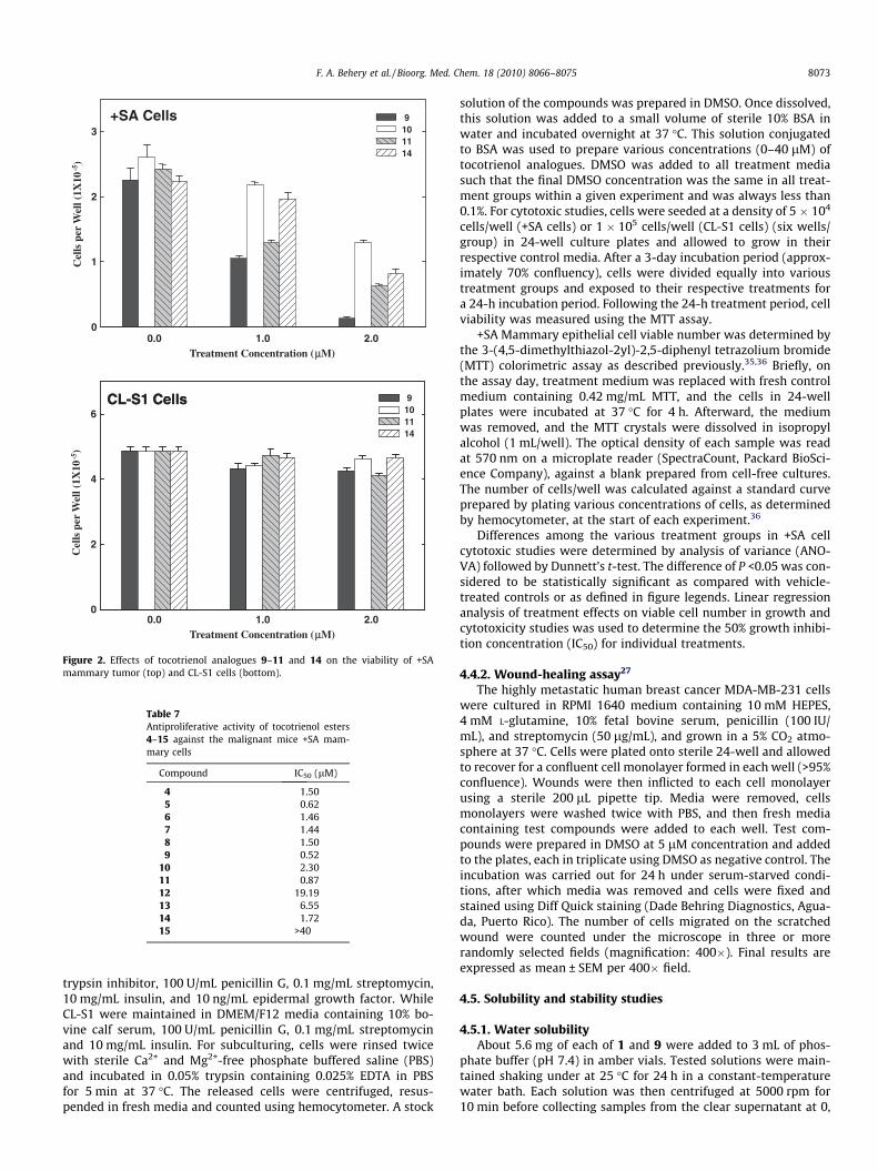
Esters 4–15 have been tested for their antiproliferative activityon malignant +SA and normal CL-S1 mouse mammary epithelialcells using MTT assay (Figs. 1 and 2). All compounds displayedantiproliferative activity except 12, 13, and 15 on +SA mammarytumor cells. However, treatment with similar doses of these com-pounds had no effect on the growth or the viability of immortal-ized normal CL-S1 mammary epithelial cells. The presence ofhydrogen bond donating (HBD) and/or accepting (HBA) groupmay play a role in the activity. Despite the presence of a free car-boxylic group in compound 12, it was inactive. Compound 12 differthan the other active compounds in that it lacks the a,b-unsatura-tion and the acid is five-carbons long. This may indicate that anacid that will afford a maximum of four-carbon distance betweenthe C-6 oxygen and the HBD and/or HBA group is preferred foractivity. In addition, the coexistence of both a,b-unsaturation andHBD and/or HBA group is required for high activity. The data areconsistent with literature where unsaturated dicarboxylic acids(e.g., maleate or fumarate) esters or amides of tocopherols showedgreater apoptotic activity than the saturated tocopheryl esters likesuccinate.14,28 The most active esters 5, 9, and 11 potently inhib-ited the proliferation of +SA mammary epithelial cells with IC50
values of 0.62, 0.51, and 0.86 lM, respectively (Table 7) while theIC50 values of 1–3 are 5 lM, 4 lM, and 3 lM, respectively, against+SA cells.29 The remarkable activity of a-tocotrienol maleate (9)was of special interest because its parent natural product (a-toco-trienol, 1) was the least active versus other tocotrienols. Thisremarkable activity improvement opens new horizons for futuredevelopment of tocotrienol redox-silent ester analogues as poten-tial anticancer drugs.
Esters 4–15 have been tested for their anti-migratory activityagainst the human highly metastatic MDA-MB-231 breast cancercells in wound-healing assay (Figs. 3 and 4). Compounds 4, 6,8–10, and 13 showed inhibition of nearly 50% of the migratedMDA-MB231 cells at a single 5 lM dose (Figs. 3 and 4). The pres-ence of terminal free ionizable groups together with aromatic ora,b-unsaturated ester moiety was also correlated with the anti-
migratory activity. It is also worth noting the potent anti-migratoryactivity of a-tocotrienol isophthalate, terephthalate, and maleate(4, 6, and 9, respectively) compared to its inactive parent naturala-tocotrienol. The activity of c-tocotrienol benzoate was consis-tent with the reported activity of c-tocotrienol benzyl carbamate,27
which suggests a possible p-stacking interaction role for the benzylmoiety, which improve the binding affinity at specific moleculartarget(s) when located at optimized distance from C-6 oxygen.
2.3. Solubility and stability
The water solubility of the most active ester 9 and its parent 1was investigated in phosphate buffer (pH 7.4, 25 �C). The solubilityof a-tocotrienol maleate (9) was nearly 1000-fold that of its parent1. The stability of 9 in phosphate buffer (pH 7.4, 37 �C) was alsoexamined. a-Tocotrienol maleate (9) showed 100% chemical stabil-ity relative to the zero time concentration over 24 h. On contrary,the parent 1 was highly unstable under the same conditions andstarted decomposition 1 h after dissolution. a-Tocotrienol (1) wascompletely undetectable after 24 h, in the buffer solution and otherpeaks were detected by HPLC with shorter retention times. In ratplasma, ester 9 concentration was 100% stable over the first 3 h.Its concentration then started to decrease at the 4th hour and

Table 41H and 13C NMR data of 10 and 11a
Position 10 11
dC, mult. dH (J) dC, mult. dH (J)
2 76.8, qC — 76.8, qC2-CH3 24.2, CH3 1.27, s 24.2, CH3 1.27, s3 31.1, CH2 1.76, m 31.0, CH2 1.80, m4 22.3, CH2 2.71, m 22.2, CH2 2.74, m5 118.7, CH 6.62, s 118.8, CH 6.67, d (2.6)5-CH3 — —6 149.9, qC 150.1, qC7 126.9, qC 120.9, CH 6.72, d (2.2)7-CH3 12.1, CH3 2.12, s —8 126.1, qC 127.6, qC8-CH3 12.8, CH3 2.02, s 16.2, CH3 2.15, s9 118.5, qC 121.1, qC10 141.4, qC 142.1, qC10 39.8, CH2 1.54, 1.62, m 39.8, CH2 1.53, 1.64, m20 22.3, CH2 2.09, m 22.5, CH2 2.11, m30 124.5, CH 5.10, m 124.5, CH 5.11, m40 135.3, qC 135.4, qC40-CH3 16.1, CH3 1.59, s 16.1, CH3 1.59, s50 39.8, CH2 1.97, m 39.8, CH2 1.96, m60 26.8, CH2 2.04, m 26.8, CH2 2.04, m70 124.3, CH 5.10, m 124.2, CH 5.11, m80 135.0, qC 135.1, qC80-CH3 16.0, CH3 1.59, s 16.0, CH3 1.59, s90 39.8, CH2 1.97, m 39.8, CH2 1.96, m100 26.7, CH2 2.04, m 26.7, CH2 2.04, m110 124.3, CH 5.10, m 124.2, CH 5.11, m120 131.3, qC 131.4, qC120a-CH3 17.7, CH3 1.58, s 17.8, CH3 1.59, s120b-CH3 25.8, CH3 1.67, s 25.8, CH3 1.67, s100 164.1, qC 164.0, qC200 134.2, CH 7.15, d (15.8) 134.8, CH 7.08, d (15.8)300 134.8, CH 7.04, d (15.8) 134.2, CH 6.99, d (15.8)400 168.2, qC 168.5, qC
a In CDCl3, J in Hertz. 400 MHz for 1H and 100 MHz for 13C NMR. Carbon multi-plicities were determined by APT experiments, C = quaternary, CH = methine,CH2 = methylene, CH3 = methyl carbons.
8070 F. A. Behery et al. / Bioorg. Med. Chem. 18 (2010) 8066–8075
5th hour to become 81% and 77% of the initial concentration,respectively. After 24 h, only 43% of the initial concentration of 9was detectable. The slow rate of hydrolysis of 9 to its parent 1bodes well for expected enhanced metabolic stability.
3. Conclusions
In conclusion, 12 new tocotrienol esters 4–15 were semisynthe-sized. At doses that had no effect on the normal mouse CL-S1 cells,esters 5, 9, and 11 effectively inhibited the proliferation of thehighly metastatic +SA mammary epithelial cells at nM doses. Esters4, 6, 8–10, and 13 potently inhibited the wound-healing migrationof MDA-MB231, the metastatic human breast cancer cell line.Terminal HBD and/or HBA group together with aromatic ora,b-unsaturation in the acylating acid can greatly improve the anti-proliferative and anti-migratory activities. Tocotrienol estersshowed remarkable enhancement of water solubility, chemicalstability, and slow decomposition rate in rat plasma.
4. Experimental
4.1. General experimental procedures
Optical rotations were measured on a Rudolph Research Analyt-ical Autopol III polarimeter. IR spectra were recorded on a Varian800 FT-IR spectrophotometer. The 1H and 13C NMR spectra wererecorded in CDCl3, using TMS as an internal standard, on a JEOLEclipse-400 NMR spectrometer, operating at 400 MHz for 1H and
100 MHz for 13C. The HREIMS experiments were conducted at Lou-isiana State University on a 6200-TOF LC–MS (Agilent) equippedwith multimode source (mixed source that can ionized the com-pounds alternatively by ESI and APCI). TLC analysis was carriedon precoated Si gel 60 F254 500 lm TLC plates (EMD Chemicals),using variable proportions of n-hexane–ethyl acetate and ethylacetate–methanol–water as a mobile phase. 1% Vanillin in concen-trated H2SO4 was used as visualizing reagent. For column chroma-tography, Si gel 60 (Natland, 63–200 lm) was used.
4.2. Biological material
Tocotrienol-rich fraction 50 g (Palm TRF 70%, low in tocopherolfrom First Tech International Ltd, Hong Kong) was fractionated onSi gel 60 using n-hexane–ethyl acetate (gradient elution).
4.3. Chemical reactions
4.3.1. Preparation of triacetylgallic and diacetylcaffeic acidchlorides30
To a solution of gallic or caffeic acids (20 mmol, each) wasadded acetic anhydride (6 equiv) and 2 mL pyridine (Scheme 1).Each mixture was then stirred for 4 h, at rt and then poured onto10 mL 1 M H3PO4 cold solution. Each mixture was extracted withethyl acetate (3 � 10 mL). The organic layers were washed withbrine, saturated aqueous NaHCO3, and H2O. The combined organicsolution was dried using anhydrous MgSO4, filtered, and the sol-vent was removed under a vacuum to afford 3,4,5-triacetoxyben-zoic acid, and 3,4-diacetoxycinnamic acid. These products wereidentified by 1H and 13C NMR analysis and comparison with the lit-erature.31,32 To a solution of each acetylated acid (5 mmol) in dryCH2Cl2 (10 mL), 0.7 mL oxalyl chloride was added and each mix-ture was stirred for 8 h at rt. Each mixture was concentrated undervacuum to give the 3,4,5-triacetoxybenzoic and 3,4-diacetoxycin-namic acid chlorides.
4.3.2. Esterification of tocotrienols using acid chlorides30
A mixture of dry CH2Cl2 (10 mL), triethylamine (5 mL), andtocotrienol (1.2 equiv) was added to acid chloride concentrate at0 �C (Scheme 2). Each reaction mixture was stirred over nightand then poured onto ice water. Each mixture was extracted threetimes with ethyl acetate (3 � 10 mL). The combined organic phasewas dried over anhydrous MgSO4, filtered, and the solvent was re-moved under vacuum. Each residue was purified by column chro-matography on Si gel 60.33
4.3.3. Esterification of tocotrienols using acid anhydridesTo a dry pyridine solution of tocotrienol (4.8 mmol), 5.7 mmol
of acid anhydrides, and 5.7 mmol of dicyclohexylcarbodiimide(DCC) were added (Scheme 2). The reaction mixture was stirredat rt for 20 h and the dicyclohexylurea formed was removed by fil-tration. After solvent evaporation, the residue was treated with100 mL H2O and alkalinized by adding NaHCO3. The solution wasthen extracted with EtOAc (100 mL � 3). The organic layer wasdried over anhydrous MgSO4 and evaporated. The residue waspurified by column chromatography either over Si gel 60 using iso-cratic ethyl acetate–methanol (92.5:7.5) as a mobile system, orover C18-RP Si gel, using MeOH–H2O, gradient elution.
4.3.3.1. 4-(((R)-2,5,7,8-Tetramethyl-2-((3E,7E)-4,8,12-trimethyl-trideca-3,7,11-trienyl)-chroman-6-yloxy)carbonyl)isophthalicacid (4). Yellow viscous oil, UV (MEOH) kmax (log e) 288 (6.09);½a�25
D +2.5 (c 0.16, CHCl3); IR (CHCl3) mmax 3626, 2926.4, 1740,1706.9, 1230.3 cm�1; 1H and 13C NMR: see Table 1; HREIMS m/z615.3327, [M�H]+ (calcd for C38H47O7, 615.3323).

Table 51H and 13C NMR data of 12 and 13a
Position 12 13
dC, mult. dH (J) dC, mult. dH (J)
2 76.8, qC 75.9, qC2-CH3 24.2, CH3 1.59, s 24.2, CH3 1.29, s3 31.1, CH2 1.73, m 29.7, CH2 1.77, m4 22.3, CH2 2.71, m 22.3, CH2 2.74, m5 118.9, CH 6.55, s 119.1, CH 6.70, s6 149.6, qC 149.7, qC7 127.1, qC 127.4, qC7-CH3 12.1, CH3 2.10, s 12.1, CH3 2.14, s8 126.0, qC 126.0, qC8-CH3 12.9, CH3 2.01, s 12.9, CH3 2.04, s9 118.5, qC 118.6, qC10 141.5, qC 141.9, qC10 39.8, CH2 1.54, 1.63, m 39.8, CH2 1.50, 1.62, m20 22.3, CH2 2.07, m 22.3, CH2 2.12, m30 124.5, CH 5.10, m 124.5, CH 5.11, m40 135.3, qC 135.3, qC40-CH3 16.1, CH3 1.60, s 16.1, CH3 1.59, s50 39.8, CH2 1.97, m 39.8, CH2 1.97, m60 26.8, CH2 2.05, m 26.8, CH2 2.04, m70 124.3, CH 5.10, m 124.3, CH 5.11, m80 135.1, qC 135.1, qC80-CH3 16.0, CH3 1.60, s 16.0, CH3 1.59, s90 39.8, CH2 1.97, m 39.8, CH2 1.97, m100 26.7, CH2 2.05, m 26.7, CH2 2.04, m110 124.3, CH 5.10, m 124.3, CH 5.11, m120 131.3, qC 131.3, qC120a-CH3 17.8, CH3 1.59, s 17.8, CH3 1.61, s120b-CH3 25.8, CH3 1.67, s 25.8, CH3 1.67, s100 171.5, qC 165.7, qC200 40.6, CH2 2.60, dd (6.2, 12.8), 2.66, d (5.5) 129.9, qC300 27.4, CH2 2.54, m 130.2, CH 8.21, dd (1.1, 7.3)400 40.5, CH2 2.37, dd (7.0, 15.4), 2.49, d (7.0) 128.6, CH 7.50, dd (7.7, 7.7)500 178.2, qC 133.4, CH 7.62, br t (7.3)600 20.0, CH3 1.16, d (6.2) 128.6, CH 7.50, dd (7.7, 7.7)700 130.2, CH 8.21, dd (1.1, 7.3)
a In CDCl3, J in Hertz. 400 MHz for 1H and 100 MHz for 13C NMR. Carbon multiplicities were determined by APT experiments, C = quaternary, CH = methine,CH2 = methylene, CH3 = methyl carbons.
F. A. Behery et al. / Bioorg. Med. Chem. 18 (2010) 8066–8075 8071
4.3.3.2. 4-(((R)-2,7,8-Trimethyl-2-((3E,7E)-4,8,12-trimethyltri-deca-3,7,11-trienyl)chroman-6-yloxy)carbonyl)isophthalic acid(5). Yellowish white semisolid, UV (MEOH) kmax (log e) 286 (6.48);½a�25
D 0.0 (c 0.08, CHCl3); IR (CHCl3) mmax 3623, 2832.9, 2305.4,1720.5, 1263.3 cm�1; 1H and 13C NMR: see Table 1; HREIMS m/z601.3165, [M�H]+ (calcd for C37H45O7, 601.3171).
4.3.3.3. 2-(((R)-2,5,7,8-Tetramethyl-2-((3E,7E)-4,8,12-trimethyl-trideca-3,7,11-trienyl)chroman-6-yloxy)carbonyl)terephthalicacid (6). Yellowish white semisolid, UV (MEOH) kmax (log e) 284(6.38); ½a�25
D �2.3 (c 0.04, CHCl3); IR (CHCl3) mmax 3625, 3054,2986, 2305, 1738, 1605, 1422, 1018 cm�1; 1H and 13C NMR: seeTable 2; HREIMS m/z 615.3328, [M�H]+ (calcd for C38H47O7,615.3327).
4.3.3.4. 2-(((R)-2,7,8-Trimethyl-2-((3E,7E)-4,8,12-trimethyltri-deca-3,7,11-trienyl)chroman-6-yloxy)carbonyl)terephthalicacid (7). Yellowish white semisolid, UV (MEOH) kmax (log e) 290(6.25); ½a�25
D 0.0 (c 0.19, CHCl3); IR (CHCl3) mmax 2927, 1742, 1705,1228, 1102 cm�1; 1H and 13C NMR: see Table 2; HREIMS m/z601.3170, [M�H]+ (calcd for C37H45O7, 601.3171).
4.3.3.5. 2-(((R)-2,8-Dimethyl-2-((3E,7E)-4,8,12-trimethyltrideca-3,7,11-trienyl)chroman-6-yloxy)-carbonyl)terephthalic acid(8). Yellowish white semisolid, UV (MEOH) kmax (log e) 290(6.52); ½a�25
D �2.5 (c 0.04, CHCl3); IR (CHCl3) mmax 3054, 2986,2305, 1741, 1705, 1422, 1154 cm�1; 1H and 13C NMR: see Table3; HREIMS m/z 587.3014, [M�H]+ (calcd for C36H43O7, 587.3014).
4.3.3.6. (Z)-4-Oxo-4-((R)-2,5,7,8-tetramethyl-2-((3E,7E)-4,8,12-trimethyltrideca-3,7,11-trienyl)chroman-6-yloxy)but-2-enoicacid (9). Yellow viscous oil, UV (MEOH) kmax (log e) 284 (6.22);½a�25
D �4.8 (c 0.08, CHCl3); IR (CHCl3) mmax 3054, 2933, 2857, 2305,1736, 1710, 1230, 1152 cm�1; 1H and 13C NMR: see Table 3; HRE-IMS m/z 521.3269, [M�H]+ (calcd for C33H45O5, 521.3272).
4.3.3.7. (Z)-4-Oxo-4-((R)-2,7,8-trimethyl-2-((3E,7E)-4,8,12-trim-ethyltrideca-3,7,11-trienyl)-chroman-6-yloxy)but-2-enoic acid(10). Yellow viscous oil, UV (MEOH) kmax (log e) 288 (6.23); ½a�25
D
�36.6 (c 0.03, CHCl3); IR (CHCl3) mmax 3054, 2987, 2685, 2360,2306, 1734, 1605, 1421, 1157 cm�1; 1H and 13C NMR: see Table4; HREIMS m/z 507.3118, [M�H]+ (calcd for C32H43O5, 507.3116).
4.3.3.8. (Z)-4-((R)-2,8-Dimethyl-2-((3E,7E)-4,8,12-trimethyltri-deca-3,7,11-trienyl)chroman-6-yloxy)-4-oxobut-2-enoic acid(11). Yellow viscous oil, UV (MEOH) kmax (log e) 288 (6.18); ½a�25
D
12.8 (c 0.023, CHCl3); IR (CHCl3) mmax 3054, 2987, 2685, 2360,2306, 1734, 1605, 1421, 11266 cm�1; 1H and 13C NMR: see Table4; HREIMS m/z 493.2960, [M�H]+ (calcd for C31H41O5, 493.2959).
4.3.3.9. 3-Methyl-5-oxo-5-((R)-2,7,8-trimethyl-2-((3E,7E)-4,8,12-trimethyltrideca-3,7,11-trienyl)chroman-6-yloxy)penta-noic acid (12). White yellowish viscous oil, UV (MEOH) kmax
(log e) 288 (5.57); ½a�25D -2.3 (c 0.39, CHCl3); IR (CHCl3) mmax 3626,
2927, 2856, 1711, 1476, 1377, 1198 cm�1; 1H and 13C NMR: seeTable 5; HREIMS m/z 537.3585, [M�H]+ (calcd for C34H49O5,537.3585).

Table 61H and 13C NMR data of 14 and 15a
Position 14 15
dC, mult. dH (J) dC, mult. dH (J)
2 76.8, qC 76.8, qC2-CH3 24.2, CH3 1.28, s 24.2, CH3 1.27, s3 31.3, CH2 1.78, m 31.1, CH2 1.76, m4 22.3, CH2 2.72, m 22.3, CH2 2.73, m5 118.9, CH 6.64, s 118.9, CH 6.64, s6 149.8, qC 149.7, qC7 127.2, qC 126.9, qC7-CH3 12.1, CH3 2.12, s 12.1, CH3 2.12, s8 126.1, qC 126.6, qC8-CH3 12.9, CH3 2.04, s 12.8, CH3 2.04, s9 118.6, qC 118.5, qC10 141.6, qC 141.6, qC10 39.8, CH2 1.54, m 1.62, m 39.8, CH2 1.57, m 1.62, m20 22.2, CH2 2.10, m 22.3, CH2 2.10, m30 124.5, CH 5.11, m 124.5, CH 5.11, m40 135.3, qC 135.3, qC40-CH3 16.0, CH3 1.59, s 16.1, CH3 1.59, s50 39.8, CH2 1.97, m 39.8, CH2 1.96, m60 26.8, CH2 2.07, m 26.8, CH2 2.06, m70 124.3, CH 5.11, m 124.3, CH 5.11, m80 135.1, qC 135.0, qC80-CH3 16.1, CH3 1.59, s 16.0, CH3 1.59, s90 39.8, CH2 1.97, m 39.8, CH2 1.96, m100 26.7, CH2 2.07, m 26.7, CH2 2.06, m110 124.3, CH 5.11, m 124.3, CH 5.11, m120 131.4, qC 131.3, qC120a-CH3 17.8, CH3 1.60, s 17.8, CH3 1.60, s120b-CH3 25.8, CH3 1.67, s 25.8, CH3 1.67, s,100 163.5, qC 165.7, qC200 128.1, qC 118.8, CH 6.59, d (16.1)300 122.8, CH 7.95, s 144.2, CH 7.79, d (16.1)400 143.6, qC 133.3, qC400-
OCO(CH3)167.8,b qC
400-OCO(CH3)
20.7,c CH3 2.31,b s
500 139.1, qC 123.0, CH 7.42, d (1.8)500-
OCO(CH3)166.5,b qC
500-OCO(CH3)
20.3,c CH3 2.32,b s
600 143.6, qC 143.4, qC600-
OCO(CH3)167.8,b qC 168.2,b qC
600-OCO(CH3)
20.7,c CH3 2.31,b s 20.8,c CH3 2.31,b s
700 122.8, CH 7.95, s 143.8, qC700-
OCO(CH3)168.1,b qC
700-OCO(CH3)
20.7,c CH3 2.32,b s
800 124.1, CH 7.25, d (8.1)900 126.7, CH 7.46, dd (1.8, 8.4)
a In CDCl3, J in Hertz. 400 MHz for 1H and 100 MHz for 13C NMR. Carbonmultiplicities were determined by APT experiments, C = quaternary, CH = methine,CH2 = methylene, CH3 = methyl carbons.b,c Interchangeable in the same column.
2.01.00.00
1
2
3
45678
Treatment Concentration (µM)
2.01.00.0Treatment Concentration (µM)
Cel
ls p
er W
ell (
1X10
-5)
0
2
4
6
Cel
ls p
er W
ell (
1X10
-5)
CL-S1 Cells 45678
+SA Cells
Figure 1. Effects of tocotrienol analogues 4–8 on the viability of +SA mammarytumor (top) and CL-S1 cells (bottom).
8072 F. A. Behery et al. / Bioorg. Med. Chem. 18 (2010) 8066–8075
4.3.3.10. (R)-2,7,8-Trimethyl-2-((3E,7E)-4,8,12-trimethyltrideca-3,7,11-trienyl)chroman-6-yl benzoate (13). White yellowish vis-cous oil, UV (MEOH) kmax (log e) 282 (6.40); ½a�25
D �4.6 (c 0.065,CHCl3); IR (CHCl3) mmax 2927, 2855, 1731, 1602, 1229,1094 cm�1; 1H and 13C NMR: see Table 5; HREIMS m/z 437.3421,[M�H]+ (calcd for C30H45O2, 437.3425).
4.3.3.11. 5-(((R)-2,7,8-Trimethyl-2-((3E,7E)-4,8,12-trimethyltri-deca-3,7,11-trienyl)chroman-6-yloxy)carbonyl)benzene-1,2,3-triyl triacetate (14). Yellowish white semisolid, UV (MEOH) kmax
(log e) 286 (6.52); ½a�25D -6.0 (c 0.13, CHCl3); IR (CHCl3) mmax 2928,
2856, 2303, 1782, 1736, 1612, 1493, 1371, 1326, 1190 cm�1; 1H
and 13C NMR: see Table 6; HREIMS m/z 689.3690, [M+H]+ (calcdfor C41H53O9, 689.3684).
4.3.3.12. 4-((E)-3-Oxo-3-((R)-2,7,8-trimethyl-2-((3E,7E)-4,8,12-trimethyltrideca-3,7,11-trienyl)chroman-6-yloxy)prop-1-enyl)-1,2-phenylene diacetate (15). Yellow oil, UV (MEOH) kmax (log e)284 (6.96); ½a�25
D 10 (c 0.03, CHCl3); IR (CHCl3) mmax 3054, 2927,2855, 1774, 1725, 1641, 1205 cm�1; 1H and 13C NMR: Table 6;HREIMS m/z 657.3772, [M+H]+ (calcd for C41H53O7: 657.3786).
4.4. Biological assays
4.4.1. MTT assayAll materials were purchased from Sigma Chemical Company
(St. Louis, MO) unless otherwise stated. The highly malignant +SAmouse mammary epithelial cell line were serially passaged at sub-confluent cell density. The +SA mammary tumor cell line was de-rived from an adenocarcinoma that developed spontaneously in aBALB/c female mouse.34 The normal CL-S1 mammary epithelial cellline is immortal in culture tube, but does not grow in soft agaroseor form solid tumors upon transportation back into the mammarypad of syngeneic BALB/c mice.29,34 Cell culture conditions havebeen previously described in detail.35 Briefly, +SA cells were main-tained in serum-free defined medium consisting of Dulbecco’smodified Eagle’s medium (DMEM)/F12 containing 5 mg/mL bovineserum albumin (BSA), 10 mg/mL transferrin, 100 U/mL soybean
0
1
2
39 10 11 14
Cel
ls p
er W
ell (
1X10
-5)
0
2
4
6
9 10 11 14
Cel
ls p
er W
ell (
1X10
-5)
CL-S1 CellsCL-S1 Cells
+SA Cells
2.01.00.0Treatment Concentration (µM)
2.01.00.0Treatment Concentration (µM)
Figure 2. Effects of tocotrienol analogues 9–11 and 14 on the viability of +SAmammary tumor (top) and CL-S1 cells (bottom).
Table 7Antiproliferative activity of tocotrienol esters4–15 against the malignant mice +SA mam-mary cells
Compound IC50 (lM)
4 1.505 0.626 1.467 1.448 1.509 0.52
10 2.3011 0.8712 19.1913 6.5514 1.7215 >40
F. A. Behery et al. / Bioorg. Med. Chem. 18 (2010) 8066–8075 8073
trypsin inhibitor, 100 U/mL penicillin G, 0.1 mg/mL streptomycin,10 mg/mL insulin, and 10 ng/mL epidermal growth factor. WhileCL-S1 were maintained in DMEM/F12 media containing 10% bo-vine calf serum, 100 U/mL penicillin G, 0.1 mg/mL streptomycinand 10 mg/mL insulin. For subculturing, cells were rinsed twicewith sterile Ca2+ and Mg2+-free phosphate buffered saline (PBS)and incubated in 0.05% trypsin containing 0.025% EDTA in PBSfor 5 min at 37 �C. The released cells were centrifuged, resus-pended in fresh media and counted using hemocytometer. A stock
solution of the compounds was prepared in DMSO. Once dissolved,this solution was added to a small volume of sterile 10% BSA inwater and incubated overnight at 37 �C. This solution conjugatedto BSA was used to prepare various concentrations (0–40 lM) oftocotrienol analogues. DMSO was added to all treatment mediasuch that the final DMSO concentration was the same in all treat-ment groups within a given experiment and was always less than0.1%. For cytotoxic studies, cells were seeded at a density of 5 � 104
cells/well (+SA cells) or 1 � 105 cells/well (CL-S1 cells) (six wells/group) in 24-well culture plates and allowed to grow in theirrespective control media. After a 3-day incubation period (approx-imately 70% confluency), cells were divided equally into varioustreatment groups and exposed to their respective treatments fora 24-h incubation period. Following the 24-h treatment period, cellviability was measured using the MTT assay.
+SA Mammary epithelial cell viable number was determined bythe 3-(4,5-dimethylthiazol-2yl)-2,5-diphenyl tetrazolium bromide(MTT) colorimetric assay as described previously.35,36 Briefly, onthe assay day, treatment medium was replaced with fresh controlmedium containing 0.42 mg/mL MTT, and the cells in 24-wellplates were incubated at 37 �C for 4 h. Afterward, the mediumwas removed, and the MTT crystals were dissolved in isopropylalcohol (1 mL/well). The optical density of each sample was readat 570 nm on a microplate reader (SpectraCount, Packard BioSci-ence Company), against a blank prepared from cell-free cultures.The number of cells/well was calculated against a standard curveprepared by plating various concentrations of cells, as determinedby hemocytometer, at the start of each experiment.36
Differences among the various treatment groups in +SA cellcytotoxic studies were determined by analysis of variance (ANO-VA) followed by Dunnett’s t-test. The difference of P <0.05 was con-sidered to be statistically significant as compared with vehicle-treated controls or as defined in figure legends. Linear regressionanalysis of treatment effects on viable cell number in growth andcytotoxicity studies was used to determine the 50% growth inhibi-tion concentration (IC50) for individual treatments.
4.4.2. Wound-healing assay27
The highly metastatic human breast cancer MDA-MB-231 cellswere cultured in RPMI 1640 medium containing 10 mM HEPES,4 mM L-glutamine, 10% fetal bovine serum, penicillin (100 IU/mL), and streptomycin (50 lg/mL), and grown in a 5% CO2 atmo-sphere at 37 �C. Cells were plated onto sterile 24-well and allowedto recover for a confluent cell monolayer formed in each well (>95%confluence). Wounds were then inflicted to each cell monolayerusing a sterile 200 lL pipette tip. Media were removed, cellsmonolayers were washed twice with PBS, and then fresh mediacontaining test compounds were added to each well. Test com-pounds were prepared in DMSO at 5 lM concentration and addedto the plates, each in triplicate using DMSO as negative control. Theincubation was carried out for 24 h under serum-starved condi-tions, after which media was removed and cells were fixed andstained using Diff Quick staining (Dade Behring Diagnostics, Agua-da, Puerto Rico). The number of cells migrated on the scratchedwound were counted under the microscope in three or morerandomly selected fields (magnification: 400�). Final results areexpressed as mean ± SEM per 400� field.
4.5. Solubility and stability studies
4.5.1. Water solubilityAbout 5.6 mg of each of 1 and 9 were added to 3 mL of phos-
phate buffer (pH 7.4) in amber vials. Tested solutions were main-tained shaking under at 25 �C for 24 h in a constant-temperaturewater bath. Each solution was then centrifuged at 5000 rpm for10 min before collecting samples from the clear supernatant at 0,
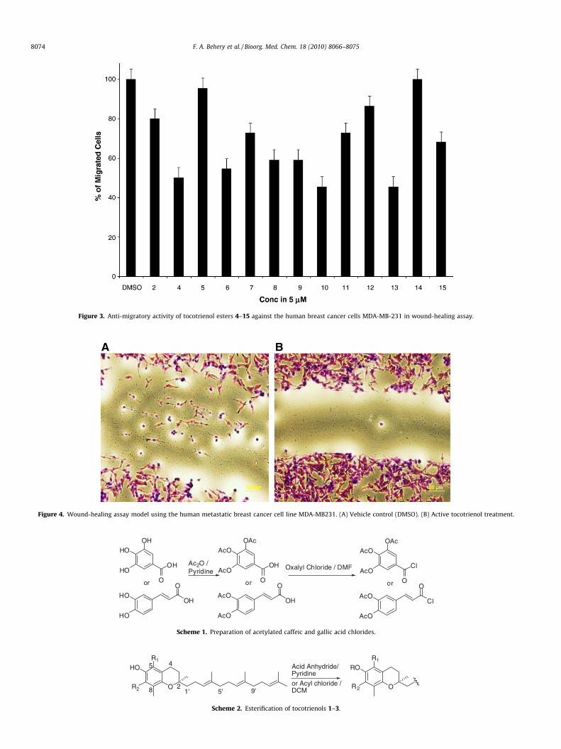
Figure 3. Anti-migratory activity of tocotrienol esters 4–15 against the human breast cancer cells MDA-MB-231 in wound-healing assay.
Figure 4. Wound-healing assay model using the human metastatic breast cancer cell line MDA-MB231. (A) Vehicle control (DMSO). (B) Active tocotrienol treatment.
OH
O
OH
HO
HO
HO
HO
O
OH
or
OH
O
OAc
AcO
AcO
AcO
AcO
O
OH
or
Ac2O /Pyridine
Cl
O
OAc
AcO
AcO
AcO
AcO
O
Cl
or
Oxalyl Chloride / DMF
Scheme 1. Preparation of acetylated caffeic and gallic acid chlorides.
HO
R2
R1
O
Acid Anhydride/Pyridine
or Acyl chloride /DCM
RO
R2
R1
O2
45
8 1' 5' 9'
Scheme 2. Esterification of tocotrienols 1–3.
8074 F. A. Behery et al. / Bioorg. Med. Chem. 18 (2010) 8066–8075

F. A. Behery et al. / Bioorg. Med. Chem. 18 (2010) 8066–8075 8075
1, 2, 4, 6, and 24 h. Each compound concentration in the aspirateswas determined using the HPLC method described below.
4.5.2. Stability studies4.5.2.1. Stability in phosphate buffer. The stability of each of 1and 9 was studied at 37 �C in phosphate buffer (pH 7.4). In ambervials, 500 lg/mL of each compound were dissolved in the bufferand the solution was incubated at 37 �C. At interval times (0, 0.5,1, 2, 4, 6, and 24 h), 50 lL samples were collected and equal vol-umes of methanol were added to each sample followed by HPLCanalysis.
4.5.2.2. Stability in rat plasma. The stability of each of 1 and 9was studied at 37 �C in rat plasma. In amber vials, 50 lg/mL of eachcompound were dissolved in the plasma. The solution was incu-bated at 37 �C, and at interval times (0, 0.5, 1, 2, 4, 6, and 24 h),50 lL samples were taken and mixed with 100 lL of methanol fol-lowed by centrifugation at 14,000 rpm for 10 min. Each superna-tant was analyzed by HPLC.
4.5.3. HPLC analysis of 1 and 9A Shimadzu HPLC system (Columbia, MD) was used for quanti-
fication of 1 and 9. This system is composed of SIL 20-AHT auto-sampler, SPD-20A UV/vis detector, and LC-20AB pump connectedto a Dgu-20A3 degasser. Data acquisition was achieved by LC Solu-tion software version 1.22 SP1 Shimadzu. The following chromato-graphic conditions were used: Luna 5l C18 column (250 � 4.6 mmid; Phenomenex, Torrance, CA), flow rate was adjusted to 1.0 mL/min, and k was set at 295 nm. For compound 1 solubility and sta-bility studies, an isocratic elution with MeOH–EtOH–CH3CN(40:30:30, v/v/v) as mobile phase for 10 min. Compound 1 waseluted at 6.7 min. For the simultaneous separation of compounds1 and 9 the following gradient elution was also used starting withH2O–MeOH–N(Et)3 (10:90:0.05, v/v/v) for 5 min, followed by iso-cratic MeOH–N(Et)3 (100:0.05, v/v) for another 10 min. The reten-tion times were 4 and 14.1 for 9 and 1, respectively. Standardcurves for 1 and 9 in MeOH were prepared in the range of 0.4–50 lg/mL and 1–1000 lg/mL, respectively. Each compound wasquantified using its calibration curve for compounds peak area ver-sus its concentration.
4.6. Chemicals and reagents
All reagents have been purchased from Sigma–Aldrich. (TRF)50 g (Palm TRF 70%, low in tocopherol from First Tech InternationalLtd, Hong Kong) was fractionated using Si gel 60 VLC using n-hex-ane/EtOAc (gradient elution) as a mobile phase.
Acknowledgment
This publication was supported in part by First Tech Interna-tional Ltd (Hong Kong).
References and notes
1. Packer, L. Am. J. Clin. Nutr. 1993, 53, 1050S.2. Kamal-Eldin, A.; Appelqvist, L. Lipids 1996, 31, 671.3. Burton, G. W. Proc. Nutr. Soc. 1994, 53, 251.4. Sen, C. K.; Khanna, S.; Roy, S. Mol. Aspects Med. 2007, 28, 692.5. Sies, H. Oxidative Stress: Introduction. In Oxidative Stress: Oxidants and
Antioxidants; Academic Press: London, 1991; p xv.6. Packer, L.; Weber, S. U.; Rimbach, G. J. Nutr. 2001, 131, 369S.7. Serbinova, E.; Kagen, V.; Han, D.; Packer, L. Free Radical Biol. Med. 1991, 10, 263.8. Packer, L. Nutrition and Biochemistry of the Lipophilic Antioxidants, Vitamin E
and Carotenoids. In Nutrition, Lipids, Health and Disease; Ong, A. S. H., Niki, E.,Packer, L., Eds.; American Oil Chemists Society: Champaign, IL, 1995; p 8.Chapter 2.
9. Sen, C. K.; Khanna, S.; Roy, S. Life Sci. 2006, 78, 2088.10. Kamat, J. P.; Devasagayam, T. P. A. Neurosci. Lett. 1995, 195, 179.11. Xu, Z.; Hua, N.; Godber, J. S. J. Agric. Food Chem. 2001, 49, 2077.12. Basu, A.; Imrhan, V. Nutr. Rev. 2005, 63, 247.13. Kline, K.; Yu, W.; Sanders, B. G. J. Nutr. 2004, 134, 3458S.14. Neuzil, J.; Tomasetti, M.; Zhao, Y.; Dong, L. F.; Birringer, M.; Wang, X. F.; Low, P.;
Wu, K.; Salvatore, B. A.; Ralph, S. J. Mol. Pharmacol. 2007, 71, 1185.15. Wang, X. F.; Dong, L.; Zhao, Y.; Tomasetti, M.; Wu, K.; Neuzil, J. Mol. Nutr. Food
Res. 2006, 50, 675.16. Eitenmiller, R.; Lee, L. Vitamin E: Food Chemistry, Composition, and Analysis;
Marcel Dekker Inc.: New York, Basil, 2004. pp 89–135.17. Chow, C. K. Vitamin E. In Hand Book of Vitamins; Bruker, R. B., Suttie, J. W.,
McCormick, D. B., Machlin, L. J., Eds.; Marcel Dekker Inc.: New York, Basel,2001; p 165.
18. Repka, M. A.; McGinity, J. W. Int. J. Pharm. 2000, 202, 63.19. Zingg, J. M. Mini-Rev. Med. Chem. 2007, 7, 543.20. Hayes, K. C.; Pronczuk, A.; Liang, J. S. Proc. Soc. Exp. Biol. Med. 1993, 202, 353.21. Yap, S. P.; Yuen, K. H.; Wong, J. W. J. Pharm. Pharmacol. 2001, 53, 67.22. Abuasal, B.; Sylvester, P. W.; Kaddoumi, A. Drug Metab. Dispos. 2010, 38, 939.23. Sokol, R. J.; Butler-Simon, N.; Conner, C.; Heubi, J. E.; Sinatra, F. R.; Suchy, F. J.;
Heyman, M. B.; Perrault, J.; Rothbaum, R. J.; Levy, J.; Iannaccone, S. T.; Shneider,B. L.; Koch, T. K.; Narkewicz, M. R. Gastroenterology 1993, 104, 1727.
24. Csallany, A. S.; Draper, H. H. Arch. Biochem. Biophys. 1963, 100, 335.25. Draper, H. H.; Csallany, A. H.; Chiu, M. Lipids 1967, 2, 47.26. Schltz, N.; Leist, M.; Petrzika, M.; Gassmann, B.; Breigelius-Flohe, R. Am. J. Clin.
Nutr. 1995, 62, 1527S.27. Elnagar, A. Y.; Wali, V. B.; Sylvester, P. W.; El Sayed, K. A. Bioorg. Med. Chem.
2010, 18, 755.28. Birringer, M.; Eytina, J. H.; Salvatore, B. A.; Neuzil, J. Br. J. Cancer 2003, 88, 1948.29. McIntyre, B. S.; Briski, K. P.; Gabor, A.; Sylvester, P. W. Proc. Soc. Exp. Biol. Med.
2000, 224, 292.30. Lin, C. F.; Chang, T. C.; Chiang, C.-C.; Tsai, H. I.; Hsu, L. Y. Chem. Pharm. Bull.
2005, 53, 1402.31. Inayama, S.; Harimaya, K.; Hori, H.; Ohkura, T.; Kawamata, T.; Hikichi, M.;
Yokokura, T. Chem. Pharm. Bull. 1984, 32, 1135.32. Andrus, M. B.; Liu, J.; Meredith, E. L.; Nartey, E. Tetrahedron Lett. 2003, 44, 4819.33. Takata, J.; Hidaka, R.; Yamasaki, A.; Hattori, A.; Fukushima, T.; Tanabe, M.;
Matsunaga, K.; Karube, Y.; Imai, K. Lipid Res. 2002, 43, 2196.34. Anderson, L. W.; Danielson, K. G.; Hosick, H. L. In Vitro 1979, 15, 841.35. Wali, V. B.; Sylvester, P. W. Lipids 2007, 42, 1113.36. Sylvester, P. W.; Birkenfeld, H. P.; Hosick, H. L.; Briski, K. P. Exp. Cell Res. 1994,
214, 145.