RECORDS - Zoological Survey of India
108
RECORDS IND AN MUSEUM (A JOURNAL OF INDIAN . ZOOLOOV) Vo • XXXIV, Part I MARCH, 1932 Land Leecbesio tbe "F aona of Brifsh Ind.ia·' .-Some Correctioa •• J. P. Moore • • •'. • • • • .. • • • 1 A Revision of the genus Coeliccia (Order Odonata). F. F. Laidla,w •• 7 On a new genos and specie,s of Strepsiptera,. T. V. Subramaniam • • 43 De!"''',ntion of a D' ew s'peties of Collembola and its Anatomy. D.urgadas M rj i ' ... • • '. • • 47 QI'ntmttll : PUBLISHED BY THE SUPERINTENDENT" ZOOLOGICAL SURVEY OF INDIA 1932
-
Upload
khangminh22 -
Category
Documents
-
view
0 -
download
0
Transcript of RECORDS - Zoological Survey of India

RECORDS
IND AN MUSEUM (A JOURNAL OF INDIAN .ZOOLOOV)
Vo • XXXIV, Part I
MARCH, 1932
Land Leecbesio tbe "F aona of Brifsh Ind.ia·' .-Some Correctioa •• J. P. Moore • • • '. • • • • .. • • • 1
A Revision of the genus Coeliccia (Order Odonata). F. F. Laidla,w •• 7 On a new genos and specie,s of Strepsiptera,. T. V. Subramaniam • • 43 De!"''',ntion of a D'ew s'peties of Collembola and its Anatomy. D.urgadas
M ut~c rj i '... • • '. • • 47
QI'ntmttll : PUBLISHED BY THE SUPERINTENDENT" ZOOLOGICAL SURVEY OF INDIA
1932

N. l.-RI34.
RECORDS ef the
INDIAN MUSEUM (A ,JOURNAL OF INDIAN ZOOLOGy)
VoL XXXIV, 1932.
EDITED BY
tHE DIRECTO~ ZOOLOGICAL SURVEY OF INDIA.
Qtllicutt~ : PUBLISHED BY THE DIRECfOR. ZOOLOGICAL SURVEY OF INOlA
1933.
Price As./) 0" (Jll.

CONTE N'TS.
Page. PART I.-Published Mat'ok, 19,,2.
Land Leeches in the " Fauna ,of British India" .-Some 'correc-~~ 1
A Revision of the genusOoeliccia (Order Odonata) 7
On.a new genus and species of Strepsiptera 43 Description of a new species of CoUembola and It8Anatomy 47
PART II.-Published June, "1932.
On some ,New Rhyn~hota of the Family Aleyrodida,e from Burma. 81
Oontribution a, l' etude des nfusoires Parasites des Anoures du. Malabar 89
On a Rare Eastern H~malayanand A,ssame,se Loac,h .Somileptes gongo,ta, (Ham,. Buch.) wIth special refe.rence to its :generic positio.n 125
Une Collection d'Hydropolypes Appartenanta I'Indian Museum de Calcutta 131
The S~yle Sacs of some mor'e Gastropod.s 171 Some Chironomidae I(Diptera) from Barkuda Island, Chilka
Lake 17'7 Spongilla carteri and its varieties 185
A key to the Dragonflies of India 195
PART IIl.--Publi81u~iJ, Septe1nber, 1932.
Parasitio Ne.matodes obtained from animals.dyiIl,g"n the Cal-cutta Zoological Gardens. Parts 9-11 229
A New Species of Spinitectus, A Nematode, from India 263 Note:s on India I Bleph.aroceridae, III 269
A New Genus and Species of Thysa.nopt'era from South India 277 On a Collection of Fish fro.m Jower Burma 281 A New Chal~id'with branched Antennae from South India
(Tetracnemus indimts, sp~ nov.) 287
Observations on the Ecology of Certain Fishes and Cl'usta'ce,ans along the bank of the Matla River at PortOannilll( 289
Report on the Shallow·water Marine Spongles in the collection of the Indian Museum. Pa,rt I 299

•• n Conf£nts •
Page. P ART IV.-P'Ubli,sked December, 1932.
'The Earthworms of Burma III. The Megascolecina,e. . 357
On a Colleotion of Crinoids from the Indian Ocean and the Bay of Bengal .. '. 551
Biological observations on ,and instanoes of commensalism oO'f ;In Ophioid Fish with Echinoderms of the Andaman Islands • . . • '. • . . '. 5,67
Notes on Fishe,s in the Indian Museum,. XIX. On a new lo.ach .of ,the IGenus Botia, with remarks onB. dario (Ham. Buch,.) • • • • • • • '. • ,571

LIST OF PLATES.
Plates I- III (Coeliccia) Plate IV (Tridacty,lophagus m,ysorensis) • Plates V- VII (Col embola) Plates VIII~XI (Rhynchota) Plates XII~XIV (Infusoires)
Plate XV (Somi leptes gOfI!Jota)
Plates XVI-- XVII (Hydropolypes) Plate XVIII (Indian Sponges) Plates XIX~XX (Ind;tan Ctinoids)
Follow page,. 42
46 ~o
~8
124
130
170
a56 5(i6

LIST OF AUTHORS. Page.
AVYAR,T. V. R. ANew Genus and Species of Thysanoptera from South
India ..••• 277
AYY'&R, T. V R. A New Chaleid with branched Antennae from South India
(TettaCnemUB indicus, ap. nov.) ••• 287
BuBTON, M. Report on the Shallow .. waterMarine Sponges in the collec
tion of the Indian Museum. Part I. (In collaboration with II. S. Rao) • • 299
CLARK, AUSTIN, H. On ,a Oolle1ction of Crinoids from the Indian Ocean and the
Bay of Bengal •••••• 55
EDWARDS, F. W,. Some Ohironomidae (Diptera) from Barkuda Island, Chilka Lake' • . • • • 1'77
GATES, G. E. The Earthworms of Burma. III. The Megasooleeinae 357
GIST GEE, N. :Spongilla carteri and its varieties
RORA, S. L.
• •
Notes on Fishes in the Indian Museum. XIX. On a new loach of the GenUs Botia, with remarks on B. llano
185
(Ham. Buch.) • f)'71
LAIDLAW, F. F,. A Revision of the Genus Coeliccia (Order Odonata) • 7
LELOUP, E. Una Collection ,d'Hydropolypes Appartenant a l'Indian
Museum de Calcutta 131
MAPLESTONE,P. A. Para'sitic Nematode,s obtained from animals dying in the
Calcutta Zoological Gardens.. Parts 9-11 229
MELLO, FROILANO DE.
Contribution A l'etude des Infusoires Parasites des Anonres du Malabar • 8~

vi List of A.uthors.
Page. MOORE, J. P.
Land Leeches in the "Fauna of British India" .-Some corrections • • ••• 1
MUKERJI, D. D. On a Rare E,astern Himalayan and Assamese Loach Somi
leptes gongota (Ham,. Buch.) with special reference to its generic position '. 125
MUKERJI, D. D. o a Collection of Fish from Lowet Burma • •
MUKERJI, D. D. Biological observations on ,and instflnces of co;mmensalism
of an Ophioid 'Fish with Echinoderms of the Andaman
281
Islands • • 567
MUK E,RJ'I , DURGADAS.
Description of a new species of Collembola and its Anatomy 47
'NEEDHAM, J. G. A key to the Dra,gonfiies of India • • 195
PEARSE, AS. Observ.ations on the Ecology of Certain Fish,es and Crusta-
·ceans a ong the bank of the Matla River at Port Cann·ng 289
RAO, H. S. Report on the 8ha.llow ... water Marine sponges in the oollec
tion of the India Museum. Part t. (In coZlaboration with M. Burton)" •• '. 299
SESHAIYA, R. V The Style Sacs of some more Gastropods '. '. • 171
StNGH, IURAM. On some new Rhynchota of the Family Aleyrodidae from
Burma • 81
SUBRAMANIAN, T. V On a new ,genus and species of Strepsiptera ,43
TONNOIR, A. L,. Notes on Indian Blepharooeridae, III • • • 269
VERMA, S. C. A New Spe'cies of Spinitectus,A Nematode, from India 268

INDEX.
[N.B.--:-~ asterisk (*) pr.ooeding a line denotes a new variety or subspecies; a dagget (t) lndic~tes a ne'W speCIes j a double d8gg~r (t) a new genlls on BubgfDtlf:' synonyms are pnnted in italics.] . ,
k Page. '
, Amphithemis curvistyla
AohQrutea 48 mariae hirtellus 52 nigricolor indiCUB • 62 vacillaD8 eexoculatus 51 Anaciaeschna
Achol'utinae 48 i 8.spidea Achorutini . " 48 ' martini Acisoma. 199" 200, 206 , dona:Jdi
panorpoides 206 Anax • Acoura . . 125 baochus Acrogomphus 217, 218, 222 goliathu8
fraseri " • 222 ~uttatus " Acryllium vulturinum 288, 240 ~m~aculifron8 Actinopyga. • 568 Juliu8.
! mauritiana . 568 nigrofasciatus t Acuaria (Aouaria) . 251 I • pa~thenope
t brevispicula 249,250 1 ~!sa.k18 h conic a. •• • 249 I rueogomp us t (Aouaria) indica 248 249 candalis "
Aderobiidae . • ' 171 oC?ipitalis Aeschna . 213 215 ontes
erythromelas '215 AniBoptera . juncea 215 Anormogotnphua m.b:ta 216 ' beteropterus otnithocepha.la 216 kiritscbcnkoi petalura 215 ' Anotogaster quadrilateraUs • 216 ' b~saJiB
Aescbnidae 198 212 ' glgantioa Aeschninae 198' 212 nipaJensia Aethrlamanta 199, 200: 205 palampurensis
bl'evipennis . • . • 200 Anoura Aglaophenia latecarinata 132, 145, 160. 164, Antedo~
165 cannata min-uta.
Aglaopheniidae Agrionoptera
dorothea. insignia
Aleurocanthus COOOi8 dissimills longispinu8 •
Aleurodicu8 bUl'.mitiCU8 Aleurolobus bal'oden:sis Aleurotrachelu8
t multipapilJus. Aleyrodidae I
} ,64 enerinus. . 164 ma.rmorata
201 . Antenell" H),9, 200, -201 ajf'icana 201 integerrim&
87 qu&driaurata
ajricana ritchii
87 81, 87 81,87
,81 serrat~ 81 ' variaDS 86 ' Anurida 86 Aphididac
• 81 Ard.ue~ninae 227 228 .Asslmlnea
131,
Page,. 199,200,201
201,202 2()1,2()2 201,202 · 201
212, 213, 214-214 214
· 214 212, 213, 214-
214 214 214 214 214 214 214
• 233 2].8, 226 225.226 225,226 225,226 • 198
217 J 218, .225 225
• 225 227,228
228 228 228 228 48
560 560 660 660
• 163 162, 163
• . )62 132, 147, 148,
162, l03 J63 163 lOa )62
DO, CO
Allogaster -hermionae latifrons parvistigm&
'228 Astur ba.dius · . 228 Atriehopogon flo.vcllu8 228 Austroaeschna 178 intersedens
81 • 230 172, 171l 248, 24\\ 177,178 218, 2J4
214 Alluaudomyia xanthocoma .
Amnico,Ia (Alocinma) Amphia.eeohna
beesoni •
" 1'18 172, 178 ,
212,213,214 Balantidium 214 _ bica.va.ta.
B lOl, 102, 107, 108, lOU
8U, UJ
viii l"dex.
Page. I
Ba.lantidium duodeni 101, 102 Burma;gomphu8 v .. fiavllUl -elonptum •• 102 williamsoni.
Page. 2.26,227 226,227
91 •
gracile. 101, 103, 112, 118, 124 I Burs&l'ia cordiformis helenae 101, 102, 107, 108, 112, 116,
•
hya.1inum ov,alE) nna.rum
t Belanisakis t ibidis
Bezzia. rufiventris
•
Bimaoula.ta • Bim"f"ia corpopsis
francisCana
117, 119, 124 • '. . - . 101
lOl, 10.2, 108., Il8, 124 10l,102 I
• • 233 : ealiaeecbna • 213 231, 232, 233 Oalicnem·· 8 8, 10, 11, 22 • • 179 chromothorax 8 10, 11, 12, 16 Calyptraeidae. • 171
• • 142, 143 CamaciDia 200,209 131, 132, 139, 141, 142 gigantea. • 209
rigida veatita. ·i.nana
142, 160 : huterti •• .• 209 • .• 132; 142, 143 . CA.mpanul.a.ria (Clytia) JloJiformia, 131, 132,
131, 132, 142, 146, 169, 160., l33, 140, 140, 160, 163, 166, 169, 164 161 magnifica 166
Bispinosa • • 120- speeios& 106 Bithyni& 171, 172, 176 8pinulosa. 166 Bombinator igneus • 91, III Campa.nulinidae 160 Doti. - 125, 127,671 ' Canthophrys 1.26
almorhae. .572 Capillasterinaeo 66.2 birdi • . ·5'71, 672 CapuUdae • 171 Idarlo 671, 672, ~)'73 Ca.rapus. . • 56'1:, 568 t d~yi • .571, 572 brandesii 56? gtto 671, 672, 673 gr,a~ili8 • 667 rostrate. 672 homei. 668, 569
Bougainvilliidae •. 135 lum bncoides 667 Braollydipla.x 1'99, 200, 206 sagamianus.. • 667
6~alybea 206 Casuariu8 bicarunoulatu8 • 244,246 .farinosa206 Cepedea. 90, 9,3, 96, 97, 101, 106, 112, 120, ge8tf'oi 206 122 eobrina • • 206 I bomeonensi's •• 121
Bracbythemis 199, 200, 205, 206 dimidiata '121, 122 oontaminata • 205 orientalis. 122 fusoopalliata • 2Ol), 206 paraguensis. 122
Bradinopyga 199, 200, 204 . fOrIDOsae 121 geminata. 204 fuljensis 122
Bmo america;nu8 12S lanceolata •• 121 aSlatiollS JJ'9 Ionga. •• 121 boreas 123 madagascaHensis. • '. 120 omereus • 91, III magna.. • 122 duodeni lOr, 102 metcalfi 89, 90. 94 elongatum loa mexicana 122 entozoon 102 mnltiformis • 12.2 fO'lllloSUS 122 ooovoidea •• :22 fowled 123 oQoidentalis • 120, 121 ha.iophilus • 123 ophis. 101, 121 helena.e 102, 103 phrynoma.ntidja •• 122 hyalinum 102 pulchra '99, 94, 120, 191 jerb8.a • 121 ja'Vensis • 90, 94. 120, 121 maorotis • • . • 94, 112 punjabensis • ,89, 90, 94 melanostictuB 89, 90, 91, 92, 9,5, 96, 98, segmenta ;.. 121
lOlt 104, 105., 111, 112., 113, 120, 12 ,seychellensis • .122
punCtatU8 rotundum smithi vanabilis .. viridis • wlgaris •
Burmagomphus ,cauveric)Qs hasimaricus laidla.w· • pyramidalis eivalik·ensis
122:, 123, 124 t angusta. 122, 124 123 sialkoti. • • • • - 94 101 I . tsubcylindriea 9o, 96, 101, 120, 121, 124 124 Cepbataeschna al3, 215
9& aoutifrons 2Ui 124 I lugubris • 215
• 96 masoni • 215 218,226 orbifrona. ~ 216 226,227 Ceratopogonidae. 1''7 • 226 Cerithiidae .. • •• 172 226,227 Oe~Je smymt'nsj,s. • 283 • 226 Chironomidae. • • • 1'17, )81 22·6,227 ChiroDlominae.. •• - 181

lntle:e.
Page • . Chironomus 181 . Comanthustimo~e08is
albifoToeps ,.82; Comaster brevicirra barba.tilal'8is 181 Comasterida. caUiga&ter • ". 181 Comasterida.e (Glyptotendipes} melanostolus. 182 Comasterinae (? Ki'ibiocharis) caunteri 182 Comatella macula,ta, lamprothorax '.. 181 Comatula mi,efastel' (Limnolohir1onomus) punctalipellois l8pecti.nata melanostohu 182 Comatulids nigromarg'natus 181 t Cosmiometra leilae notatUI 181 I Gomissia ehadwieki pelo,stolum 182 Copsychus saularis (Polypedilum) annuiatipes. 183 Cordulega8t~r (PolYPedilum) fasoiatipennis 188 bidentatus (Polypedilum) !cempi - 183 brevisitigma t (Polypedilum) ,scalaenulus 182 folia, • sralaenu8 •• 183 Oorduleg&fJterinae (PolypedUum) tripartitus 182 Oorduliinae • v,errueosus 182 CraspedoBletra acuticirrn.
Chlorogomphu8 198 Cratilla Ohromothorax 10,25 Uneata 'Ciliata • • 90 Uleta Hea Circus micrurus 242, 243 ; Crinoids OIavaotinia •• 135 Crocothell1is .
gallensis • 131, 132, 135, 153 e~ytbraea OUnow,nypus eeylantcus '. }81 servilia
orux . 181 CryptoJaria
ix
Page. 551, 56,3 661., 5,63 5lH,552 561, 552 • 553
• 551, 552 551, 552,563 55 , 552, 553
• 501 ll51, 565, 666
551, 552 249,251 227, 228
228 228
· 228 1'98, 227 198, 209
• 551, 556 }'99,200, 202
202 20.2
• 551 199, 200, 206
206 266 14'6
fumipennis 181 - -conferta . .. 146 Clytia. alternate 154, 155 erassicaulis
noZi/or-mis • 150, 156 1
131, 132, 14.6, 147, 148. 162
Cobiti8 • 125, 126, 127 Culcita. discoidea botia '... 125 : Culicoides !/ongQta 125, 126, 127 peregrinus Qcuiata 126 pungens taelU& 126 oxystonta .
C"Oeoidae . '. . 8 CupiUaster multir.adiatll Ooelicoia 7, 8, 9, 11, 22, 38, 39 Cyan-omelas
albieauda II, 12, 38,39 I OyclogompbuR bimacula,ta : 0, 11, 12 Cyelostomatidae • boraeensis 12, 40,41 tCyllometra prashadi brao.hysticta. • • • 12, 23 soluta campiom 11, 12, 34;35, 36, 39, 40 011rnea chromothora~ 10, 12,21 cyanomelas ••• 12, 29 didyma. 10, 12, 16, 18, 22, ,25 loringae . 12 dinoceras 11, 12" 38
D
er.iei. • 0, 12, 20 Dasyhelea? mncrostomn. tls.vicauda 10, 1.2, 28,30 -paivai flavostri&ta 11, 12, ,34 D.vidioid~s t fraseri • 10, 11, 14 martini t lieftineki 11, 12, 32 D& vidius t loogali 11., 12, .26 a'berrans '. t loringae • 17 assantenRis macroetig &. .~, 35,39 davidi membl'a.nipes 8, 9, II, 12, 30, 82 delineatuR nigrohamata. 11, 12, 37, 38, 39 kUDlaonensis ootogeaima. 12 malloryi • poungyi • 12, 24 senehalensis t pyriformis 0, 12. 26 ' zaUorensis renifera • • 12, 1,8 Delphacixenos anantn.)ocer\l.A t scutellum 1.2, 22, 23 Desmoscyphus longitiw('o, III hainanens6 • 23 DiaeanthuB tvaooa • • II, 16 Dialeu'rodes. .
CoUembola • 47, 51. 53, 66 t ()ephalidiatinctuR Colombometridae 068 t euntiugum Co'mactiniinae • 062 t dorsjd~rnar~n.t.n Comanthu8 parvicirra 6eH, 004 'elongntA.
667,568,669 178 178 178
'. 178 55}, 562
)0 218 171
5tH t 558 01)9 24'7
178 . • 178 217" 218, 226 • • 220 217, 218. 224
224.,225 2,24, 220 224,2'25 224, 225 224, 2,26 • 224
224, 226 224
44 161 12li
81 S3 86
82,ij4 88

x
Page. ' DiaJeurodes fijiensis • 85
t hexpuncta 81 t nitid\l8 84 t oweni 82 t pa pulae 83 t rangoonl . 85
l>ichrometra ciliata. •. 051, 558 Dipha.sia digitalis 131, 132, 142:, 146, 159, I
Diplacodes nebulosa trivi&lis
Doclea. D~awida Dyn&tnona cornicina.
160, 161 199,200,201
201 201 136
• • 361 131, 132, 133, lfiO,
Gl'Yllotalpa • Gynacantha
~ albistyla &lJicalis bainbriggei basiguttata bayadera .. biharlca. furcata hanuDlana. hyalina kha.sica miUardi • o'doneli .. saltatrix :subinterrupta.
•
153, 15'9 crisioides l31, 132, 142, 146, 159, 160,
161 quadridentata 131, 132, 142, 146, 159,
1160, 161, 165
Gynae,anthaesch:na. " sikkima •
. Epiophlebia laidlawi
Epiophlebiidae 'Epophthabnia
- cya.no'eepbala.
E
viridifroDs
228 ' abronenl& · 228 't asymmetric&'
198, ,228 bulbosa • 210, 211 t easuarii
211 t dicsingi 211 euplo,cami
B
frontalis vittata vittigera
211 t magnilabiata 211 Ha.ema.dipsa. montana
22 I ornata, 143 , sylvestris .
• •
•
• •
•
•
•
•
•
•
Ptllle. • 48
218,216 '. 216
2.16' "218 216
• 216 ~16, 21'7
216 216 216 216 216 216
• 216 216, ~17 213,215
216 215
24'1,24"1 242,243 • 247
• 244,245 238" 239, 240
240,246 241,242
1,4 Escutoheon Eudendriidae Eudendrium • '. 145 zeylanica e,oohiniana
2,4 2,3,4-
1 1,3
1 146
&D1hoinensis gritlini
EUdiocrinidae Eudiocrinus ornatus
t philenol' • Euplocamus leucomelanus Eutyphoeu8
F
FlavicQ;uda. Fla;vostriata. • ForcipoUlyia a,lbosigna'ta
t annandalei • tristicta
Ga,steroz'oides •
G
Gastrophryne e&rolinens,is Glyptotend" pos • Gomphidia.
abbotti fletcberi . kodagueU8is T.nigrURl wUliaDl,soni
GomphiDae Gomphu8
nilgiricu8 o'doneli , personatn8 • •
131, 132, 14,3, 144 zeylanica montivindiois 148, 144 Haemadipsinae
'. . 554 Haleolidae 551:, 504, 5,56 Halecium • • 146
551, 554 expansum 131, 182, 14:1), 64 246 halecinum • . • 132 3161 minor 131, 132, l45, 146, un, 166,
164 tenellum 131, 132, 142, 146, 159, 160,
161 , HaUctophagidae. ,44
10 Halocordyle disticAa aU8trali8. •• I.32 10 ' HeUaescbna • 2 2, 218, 215
177 177 178
1:36 123
. • 182 217,218,219
219
•
219 11 219
219 • 219
198,217 218, 226
226 226
• 226
uninervuJata Heliogomphus
,oey lonicu8 neitneri promeias sely~i walli
H'emia,nax ephippiger.
H emiearptl,~ /(J8ciculatUB Hemioordulia.
asiatioa • He'terogomphu8 • HeteroDletra reynaudi
siogulads Himero~etddae Hirudinaria •
manillensis Hirudo 11 •
birmanioa Holothuria •
• '. •
'.
• • 215 217, 2l8, 220
2~O ,220 2,20 22()
• • 220 212, 213:, 214
•
• • •
214 • 1166
209,2.10 • 210 • 221
651,666 661,566
•
•
666 6 6 6 6
• 667

lIoDlohelea • abjucta • t barkudensis pictipes
HyaUnoecia Hydraotinia
epiconcha t epidocleensis
fUQicola ntilleri pacifica
llydiob8isilu8 croceus
Hydrobiidae Hyla ,arborea.
arenicolor oinerea femoralis 'pickeringii
Hy laeothemie fruhstorferi gardeneri
'By o'bates boolock
• •
Ibis melanocephalus Iotiuus '.
angulosus atrQx mordax periin&x praecox rapa¥.
Idionyx bul'liyarensis corona galeata imbricata intricata na.dg,aniensis nilgirensis optata s&fironata selyeU stevensi • ungincula.ta. yolanda ..
• '. • •
•
• •
1'lliJe1J.
PGfjf!". 179,180 • . 180
It
17'9, 180 Kali~ephalus brachycephalu,s • 179 : indicus
135, 153 "1 naiae • • 138 t parvus •
Page •
• • . 137, 1.38 I Kaloula pul~hra 131, 132, 135, 186, 137, , Kif'cnenpauuia h,aleeiodu
138 Kdbioc,har,iB
251,252 .. 254 263,264 253,254-
120 164 183
I
• 13'7 • 137,138 ' 137, 138, 139
200,20.8 • 20.8
L
171, 172 Lafoeida6 • .. .146 91, 95, 112 Lamellogomphus 217,21.8, 223
23 1 acinaces . 224 123 biforceps . 224-123 drull1Jnondi 223, 224
• • 123 ,inglisi 223, 224 198,200,,201 ma.labarensis • 224
201 nilgirien:sis.. 228, 224 • 201 ,Latnprometr s, klunzingeri ,55}, 668
229, 286 I pa.lmate, 561, 567 p~otectUB '.. 5tH
La,oIitedea (.obelia) bicuspidata. • 156 * pickti 131, 132,61, 162 t (Obelia,) bistriata 181, 132, 133,135,
139, 100, 153, 154, 169 " 231,233 217,218,219
. . 219 (Obelia) longicyatha .. 154, 165 (ObeUa) spinulosa '. 132, 145, 156
219 219 I
** . f. minor l31, 132, 145, 151,
219 Lathr,ec'sta. • 219 asia.tioa. "
219 . Lepidoeephalichthye _ 209 LeptocneDlis
209, 210 Leptogomphu8 209" 216 bideutatus 209, 210 gestroi 209, ,210 incUtus .. 209, 210 I m8.ouJivertex 20'9, 210 ' Libellula 209,210 fulva 209, 210 qua.driDl&culata 209,210 L 'beUulidae 209, 210 Libellulinae • 209, 210 Libinia omarginata. 2()9,210 LictQrells.
209 abyssicola
1164 199, ,200., 202
202 1'27
• • 8 217, 218, 220
220,221 220, 221 220, .221 220,221
199,200,202 202 202 198 198
" 139 149, 150
IudexenosmeDlbraoiphag,a, Indocnemis
44 arboresc'ens 8, 11
• 149, 160 131, 132, 1-47" 148,
162 149, 160
4U • 149 149, 150
J,49 187
oran,g Indogompbus
cera&tes longistig1D.a. martini
Indoneura Indothemia
oaesia limbata • aita
Jagoda. • Dlartini
J
• '.
• .. 10 I
217,218,224 ' 224 224 224
• • 25 199,2Q(},206
206 • 206 • 206
212.213 .' 213
conoinn& dexiUs genioulata. pinnata itecbowi
Limnan theDlUD\ LiDlnQtrochu8 Lindenia. • . tetraphylla Lithobius LyriothoJn.i:s
aoigastra. eleie morto.ni f.t>ioolor •
Lytocarpu8/a8ciculatu8
• 174 217,21'9
219 • '66
190,200,201
•
202 202 202 202
• 166

,xii
Maor,odi pl&x oor,a,
:\laerogoutph us aonulatuB m.ontanus robustus seduotus wyna.adicus
llacromia aculeata. • atu bercula.ta. bellicosa • binocellato. cinguLata. cuprt.c!ncta ellisoni fiavicincta ida indica irata. 111 iniat a. Diloorei paUida • 'tritubereulata. zeylonio& •
Maoromidia donaldi shan'ensis
MacroJQiinae. MaorotoDl. pluotbea Mariauaetrida Mariametridae •
M
Megalixalus seycheUensis Mega.logompbus
bicorBlltus e,eylonicn8 8avicolor. hannyngtoni sDlithii superbu8
Megas,coleoidae Megascolecinae Megasoolex
horai mauritii
Melanella Mela.nia
lndefe.
Page.
L99, 200, 205 • • 205 217,218,220
220 220 220 220
Monotlheoa coDlpress& oblique spinulosa. •
Jl vrionema amboineneia Mysorella
Page. • • 160
160 • • 160 • :141,145 :172, 173, 174
• 220 Naia tripudians • 263. 154 210, 211 Naunophya • 1.98, 208, 201 211, 212 pygnlae. • 2()1 • 211 Nard10a semiregula.ris japonica 5&rT.
211, 212 Na8sopsis '. • 174 211,212 Neanura. 48, 5e, 68., 70 ,211, 212 .w - 61,60 musooru~ ,2l1, 212 sexoculata, ,51 211, 212 Neanurina 54 · - 211 ooralina . 52
211, 212 , intermedia 52 211, 212 pudibunda • 52 2.11, 212 Neanurinae • • 51, 52 211,212 I Nemaohilus botia. 25 ~ 12, i~~ , Neooeratop:~gon •• l78
2.11 212 Neurothem.ls 199, 200, 264-. , degener 204,205
211, 212 feralis • 204 210 fluotuans 204,206 210 fulvia. 204-
• 210 intermedia • 2M 198, 2 0 terIllina.ta. • 204 206
59, 62 tullina 204 5,54 Nilobezzia ISO 55? allotropic a. 180
• . , 122 l.ac teip ennis •• 180 217" 218, 221 Notoscolex 357,. 300. 366, 366
221.,.222 bkm,aniou8 3,57, 366, :367, 368, 37'9 , • 222 clwprai '. 360:. 388, 369 221,,222 depressus 357,360,366,368,369,370
222 Illnatus 357., 386,388. 870 222 oneilli 3,65 222 stewarti :365
• ,361 striatu8 • • 365 • 360.361 t triquetrus 357,366 •. 3110.371 367, 365, 374, NyototherU8 • 0 ., 109, Ill,. ll2, 113
• 360 0,ol'diformi8 89, '91, 101, lll~ 112., 113, 357, 374 116. 117., 124
657 hylae • • . - III • 173 maoropharyogeu8 101.,.108 •. 1-11, 112.
Melaniid&6 Melanoides Meld braaipes . MengenUla, choubati MerogoDlphus Meso,gomphu8 graJJlmieus
1'71, 172 - 11-6, 117 ~ 118;,119, 124 . • 72, 174 Dlagnu8 • • Il~ 8, 10, 11, 12, 30" 84 *malabarica Idd •. li.4
lindgreni Jineatus riei
~licrogontphUB burm.ious. lilliputians ,sou.teri tOrquatU8 verticalis
Mixohelea • rQseiventl'is •
Monoser,ju9 faaciculatu,s MODotheoa australis
44 papillatus 89, 91, 112 217 IIA 2 parvul ' ~
. 21 piaoioola. 113 221 reniformis 112 2.21
• '. 2Jl I
217,218,221 , - 221 o
221 221 'Obelia 221 b,identata 221 8pin.ulo8a. • 179 Ootoohaetu8 'fermorl
• • 179 I lunat1/.8 • 132, lGo, 166 I Odonata '. • •
'. 160 OUgoD1etr&serripinna '.
167 161 155 861 361
• 1:98 li61 .. 6'60

Oeyokogempbu8 &nnulafts. &ureus irietrigatu8 oirealaris Cimiuwtivu8 diDga. vapi .u&rjcas • earllsbawi ~ eclimoocipit&iis 'frol1teJis nta,claohlani modestus Mi-davum ~y'chQgomphus puloherrima. s.underai sta.iatUB w.&tli
Page. 2' 1,218, i22
222,223 -222,223 . 2.22,223 • · 223 22.2,223 • ·,223
22.2,223 222,223
223 • 223 '
.222,223 223 223 I
22·3 , .222 I
222,223 · .• • 223
Orthetrum o&ncella.tum. chrysis • ehrysostigma gla.ucU1ll japOJiicum preci osum ransonetti sabinum taepjolatum testaooum trinacria ·triangulare
'p
... nIl
Page. · 203
203,204 • 203
20.3, 204 · 203
203,204 • '203
203, '204 · 203
203,204 • 203
203,204
• 222, 223 ' P-.lpomyia. 179, I SO 199, .209,204 rosivent.ris " )79 • • 204 Pa.lpopleura . 19'9, 200, 204
ODycbothemis eeylan.ica •
-Opalma oQl'acoldea
90. 102, 10o, 114, 116 -seJunaeul&ta " 204-
lahoriensis diDlidia.ta forme ,japoniea !&t&
'Corda;ta longa, • maoronu()le,ata obtrigona obtrigonidea
,am.erioana austricola lata DlaxiDla orbiculam
90,114., 118, 119 I .P.,ludomus.. 172, 17.3:, 174 .• 89, 90, 101, Il8 I P0lyplectrwn biealcs,l'atum 246
95P.,ntaJ... 200 • • 124 fiavescens 208
. to, 119 Pa.r&nisa.kis 233 114,116, 117, 124 Paratendipes 182 114, 118, 119. 124 Pavo criSta.tU6 247
94, 116 mutieu8 247 • 90 speoifer 24? 95, 12·3 Pennaria. 134.
128, 124 af.U't1'ali$ • 133 123 di8ticAa •• 133, 150 123 oustralie 131, 182, 33, 134, 160., 123 163, 159 123 , I?enna.riidae... 132
plicate. _lla.tum
.• • 123 Pentaneur,a (Isoplastus) monilia 181 • 89, 90, 12,8, 124 (Isoplastus) ,orn.atipes ) 81
100, 101, 104" 105, 112, 114, (Isopiastus) pulchripes 181
1. analis oiaetoidea f.lata • • f. parvipalmata.e slJlithi
11'8, 11'9: 124 Periaeschna. - 213 124 magdalena. 213 124 ' no()turnalis 213 124 Periehaeta annula(,8r 376 124 barbad~nsis 375
f ,. truDca.ta 8(18,lpritoratis trie.ngularis virgula. •
124 hesperidum 375 • • • • 12,4 j,a;ponica. • 375 89, 90, 98, 101, 123, 124 8fJftavic,cft8<i,6 • '. ,37G 89,90,95,.101,122,124 , PerigonUnusjo cali 131,1.32, ]:1'0, 92, 93, 96,t7 , 'IGI, 124 I - 150, 153
OpaJ.iD~ angustae !.tae
OpaUIl~ OpaJiDid.~ Opaliain&re. • Ophiodes eeeiniformi8 OpbiogoDlphus Qn,heseU. Orogo.tpll1ll •
atkinBODi. campioni preei08US eelfsi • IpeciOlllS aathoptera •
01'tbetrum -, .. ep8 ltnmDeum
.. '. •
• '90 ' purillu8 131, 13.:2, 139, 140 to, 104 ! 'tJe8titU8 • • 142
'95, 98 1 Perionyx. 359, 545, 546 90 Perionyx sp. ? 359 90 arboriQola . 360
• • 160 ditheca • 360, 545 21''1, %18, 222 ' ~XC8,vat\ls 859. 545, 546
68 lulfJ1UJ. 360., 545 227 m"intotihi • 360, 1i46 2'27 Perissogomphua '21'7,218, 225 227 stevtmsi • • 2'21) 227 Pet&lia.eschna. 212,213, 214 227 fletcheri 214 227 ~t.robiu8 •• ~ . 61
• .227 Pheretima • 4:, :367,359, 360, 3'''G, 376, 199, 200, 202 ' 381, 385, 38tJ, 387, 413, 414, 424,.43'7, •• 203 .38, "0, 4.63, 475:,480, ,496, 498, 500, • .. 203 i 543, 54.
• •

xiv
P4fJe" Phoretwa sp. (1) ,359
aeruginosus ••• 376 alexandri • 359, 360, 382, 383,
386, 387, 388, 463, 492, 494, 496, 491, 499, 500 ,
graoilior '. 359, 494 : typioa 359,492,495 I
amazon.ica~35, 436 ' tanaleota ,. ,3,59, 501 andaDlanensis '. 358, 387, 414 andersoni • 3,59, 383, 414, 463,
499, 504,508, 518, 619, 533,534, 540 choprai . 360, 504 typioa. 359, 498, 504,
506 annulata • '. • • 375 &nomaia 357, 386,387, 388, ,38'9, 391,
519, ,538 oentralis 357, 389, 390, ,39t, I
499 insolita 357, S88, 390, 391, 499, 538 'typica. 338, 35'7, 379, 388, ,
389, 390 tba,lteolat& • 358, 425, 426 barbaden8i8 43,5, 436, 438 tbeUatula. 358,42'7 bermu4e1l8i83'75,435,436 bipora. • 522 birIllanioa 358, 386, 428,UO bournei 35,8, 381, 441, 446 : fyr,otun.i360, 434, 436, 420, 421 ealifoflliea '... ,376 oampanulata '. 358, 377,37'9, 380"
382" 38S, 386, 387, ,398, 4l3;, 462, ,463, 46'9, 472, 480, 48'6, 496, 497, 498, 004 ; *meridiana · 358, 454, 457, 459,
460,472 penetl'alis ,3,58, 45,8" 4,59, 460 typica 858, 360,452, 455, 457, 458, . - 459, ,461:, 466, 468 I
tcanaliculata. • . ,357,408 ()8;rmensis 358,360, 406,446,
·m.ota pinguis *,sootilis typio&
carno8a ohoprai
t 'ooDlposita tOOll1pta cupuli/era defeota doliaria •
*arIllillata ·dolo8& *,sterooraria typioa
tdolos& 'elongata
exo&valu,s ,exigu& '.
*austrma typic&'
tf&oeta •
498 .358,449
858, 4,50,451, 498 358, 450, 4,51
358,447 • '. 43,5, 436 309, 383, 504, ,032"
634 358,430,481 '. 359, 511 . 435, 436, 43,8 35'7:,391,4'98
,358, ,396, 415, ~9,8 368,416
• 3,58,416 358, 415, 417 · 3,58,410
• 358,443,444 357, :380, 386, 387, 391,
392, 498 • • 360 : 3,59,486,512
• '. ,359, 614 ' 359, ,387, 444, 498, 512,
513, 514, 616, ,516 '. 358, 681, 387" 422., 423 .
PQlJe. Pheretima feae 359, ,499, 616, 618, 619
gemella :359,382,519,0'2'2 'bipora • 622 *'quadripOTs, '. 359, 523 iiypio3 859, 498,4'99, 519, 52:3, 624
tg1abra. :35?, 395,396, 498 guillelmi • •• 377, 469 ha.rrietensis 35'8, :3'87, 463" 4'77,
480 ha.w~y&na. .. 358., 375, ~38'6, 38'1,
433, -434,435, 43,6, 437., 43:8, 443 lineata ·358, 420, 434, 43'6,
typio.a. . typi~a (?)
43'7,438 • . 437, 4,38 35'8,433, ,43', 4316,
437,438 35'9,3'82, 386, 463,
,486, ,498,524 hesperldum.. 375,376, 377 hesperidUDl .. lohri • ..376 honlleti. 358, 3'77, 379, 380, 383,
385, 886, 39'8, 413, 41'9, 462, 463" 464, 468, 469, 480, 486, 496, 498
rugo8a tillota iDtmerita finc)ara, in8ftlae iMulanus
• jaoita kengtungensis '~;'a~ge1WJi8 flobosa (1) longioauliouiata malayana Dla.milla.n&
manilAort'Mi8 Dlanioata.
*decoros& *supina typioa
tna'Uritiana tmendosa minuta 'Mode8ta *moleste. morr-iri neD10ralis nugalis ornata, • osmastoni
pallidG • papilio
*hiulca. ·iDsignis typio&.
pe,gua.na pinguis planata
tporreota. posthuma
tquadrigeDliDa referia •
•
•
• .398 357,39'1,498
35'7,410 ,358,439 ,435,436 358,419 858,440 358,420 376,37" .359 359,525
• ,. .• 484 358, 382, 46(), 470, 474, 475,496, 497
4'35,436 .35'9,52'7 359,528,529 3,69, 629, 630 369, 527, 631
• 435, 436, 431 358,47,3, 474,475, '76
• 612 '. • 421
• 368, 360,~20, 421 375,435,436,437,438
3,59:, ,5'31 357,401
•. 358,421 358, ,S87, 476~. 477,
479, ,480, 538
'. 436,436 • 357" 401, 498
• 3,57,,402,403,406
3,67, 404, 40(S 357, 401, 402
358, 386, 48}, 488 • • ,360
368, 374, -S81, 3,82, 386, 387,411,422
358,38'7,444,446 369, 380, tsSl, 384, 386, 387,482,48'1
• 359,486 3lJ!7; 408, 498

Pheretima riDlosa • *effeDlinata typiC80
rugosa.
Page. 359, 498, 534
359,498,537,538 • 359. 534, 538
357,380,386,398,434, 4.58, 459, 498, 514 '
8ancti~jacobi • 435,436 .8andtJi~ensi8 .375, 37S, 3'77 ftUctoria manicata 360, 527
II1ullani .• • • • 383 tsuloata 358, 383, 387, 408, 424, 425 ttenellula. 357, 398, 498 tterrigena 358, 482, 483 'Udekemi • •• 377 tUDlbraticola 358, 484, 485, 486 velata359, 406, 498, 538 .
Protaneurinae Proto opalina. .
axonuoleata. filiformis forntosa.e q uadrinueheata
Protoopalininae Pseudotralllea.
prateri Pterocyolus Pyrillozenos compactus
Q
*a.lveata 359, 539, 540 t Quasistrongylus ola.vata 359, 541, 542 ttrheae .
Page. 11
80,90 89
101 89 89 '90
200,209 209 174 45
typioa 359, 538, 542 tt Quasithelazia tenuis
237 235, 236, 237 233,234,235
vulgaris . . 469 wimberleyana 360, 452, 457 tyoungi • 357, 406, 407, 408 R
Physaloptera bulbosa 247 t Physooephaloides 229 Ra.cophoru$ m·aculatu8 91, 92,93, 94,
tpriInus . 230 I
Physooeph.a.lus 229, 230 Ranaa.esopus Pistia . 187 arvalis Platycnemin&e 7, 8, 9, 11 aurora PhatygoD1phus 218, 225 chrysoprasina
dolobratus 225 olinia. feae 225 cyanophlyctis
Platy'poda, . • 171 Plumulal'ia alte1'nala 163,164 dalmatiba.
d·iaphana 163esculenta filioa.ulis .. 160 c.hin~n~is goldsteini •. 160 hexadaotyla. halecioides 132, 146, 151, 156, 164 japonica. • inermis 164 limnocbaris
PIWllula.riidae ..• 162 Plutellu8 357, 360, 361, 362, 364 .
ambiguus 357,362, 364 aq uatilis 364 eorethrul'US •• 362 inflexus360,362,364 kem.p· • •. 364 tpratensis 357, 361, 363, 364
Podocoryne aneohinata. 160 Podura aquatios. 63
macrodon mala. ba.rica.
nigromaculata palustl'is pipiens austricola. rugosa.
. tem poraria
112 123 124 123 120
· . . 112 90, 102, 103, 104, 112,
118, 119, 124 • 124
91, 95, 101, 102, 111 101, 121
102, 103, 112, 114 • •• 119 .94, 102, 114, 116, 117,
119, 124 . • 89
112, 113, 114, 116, 19J 124
122 12, 123
123 123
• • 120
'Podurida.e '48 Polioaetus plumbeus 241 ;
par vipalmatac tigrina 90,
Ul, 101, 102, Ill, 124 • . . 124
01, 102, 103, 105, 107, Polypedilum 183 Pontodrilus 367,364 Rhea amerie.ana.
berDludensis 357, 364 . Rhodothemis Potamarcha 199, 200, 202 rufs.
obsoura. • . 202 Rhynchota Pots.rnoldes . 172, 173, 174 Rhyothemis
(Cerithidea,) obtUBum. 171,172 phyllis (Telescopium) telescopium 171,172 plutonia, • (Tym.panotonos) cingulatus 171, 172 triangularis
Procladiu8 181 variegate. ornatissiDluB 181 RusgunieUa brevis piotidorsum 181 . (Psilotanypus) vitripennis 181
Prosobranchs .. 171 Protanura 48, 62, 67, 70
109, Ill, 112, 121, 124 230, 237
199,200,206 206
s
· 81 190,200,205
205 205 205 205 283
toarpenteri . 49 Scaphiopus solitariu8 oitronella 50,52,58 ' Sohlzotric}u). diaphan~ kra.epelini 51, 52
• 123 131, 132, J 58,
spinifera 61,62,58 Soolopendr.a, 163
6()

lrtdex.
Page. ' Selysiothemis 199,260, 205 Trichoonemis
nigra • •. 2Q5 ' tTridactylophagus Sertularella minus oula. 131, 132, 142 tnlysorensis
146, 159, 160, 161 Trida.ctylu8 simplex . • 162 Trithemi8
Sertularia minima 160, 161 annulate, Sertulamdae . UiS ·aurora Sieboldius 217, 218, 220 ~tiva
nigricolor .• 220 kirhyi Somileptes 125, 126, 127 I pallidinervis
gongota. 125, 126, 127 Triton SphaeroDlias 179 Trochus
fasoiatus ... 179 TropiOD1etra Spong ilIa carteri 185, 186, 187, 188, ' audouini
189, 190, 191, 192, 193 ca.rinata calatonensis 185, 189, 192, 193 encrinus cava 188, 192, 193 'indioa lobo8a • 188, 192, 193 ' indica . melli 185, 189, 190, 192, 193 Tropiontetrida moUis - 187, 188, 192, 193 Turritella rotundacuta 191, 192 T'urritellidm
cl'assissima •. 187 Typhobiidm rotund&Cuta • 185, 190, 191
Stegopoma fastigiatum 131, 132, 139, 160, 153
Stephanometra Dlona'canth,a, .. ,551 U proteota 551, 557 protectua 551 Urothemis
Stiohopus 567 signata Stilo bezzia 179
linea.ta 179 tt Strepsiptera • 43 StroD1bidae 171 V Strongylidae ... 251 ,StylogoDlphus .217,218,222 Vel1lletidm
ingllsi . 222 Sympetrum 200, 207
oommixtutn 207 decolora.tum 207 W fonscolom.bei • 207 hypomelas 107, 208 Woodwardiella. orientale 207,208 burkilli ,striolatum 207 . pumila.
T x Tanytarsus pento.tomus • . 183 Xenohelea Temnogolllphu8 218, 225 Xylocrypta
biviltatus • 225 Tetra,themis 198, 200, 201 '
pl&tYl)tera 20 I yerburyi. 201
Thala.s8ometrinae 565 z
ThecoOa-lUu~ diaplw:na • 163 ,zamenis ml1COSUB • Tholymis 200, 208 Zelleriella .
tiUarga 208 ma.cronucleata. Thyrosoyphinaft. .• 158 Zoobotryon • Thyroscyphus fructicoliUS . 1:)1, 132, UiS Zygonyx
rO!Jlo.sus • 131.. 13,2, 15,8" 164 I ,oeylanica 'Thysanognathus 178 da vina • Tontocerus 56 iris
plumbeus • 56 isa • Tramee, 200,208 Illalaba.rioa
burmoisteri 208 mildredae liUlba.ta 208 Zygopterou8 •
Trialeurodes . 86 • Zyxoll1ma tmegapapillae 86 petioiatum •
•
• •
• PQ,g~. 9, 12
43 44
43,45 200,208
208 208 208 208 208 102
• 174 560,564 • 564
• 560, 564-551, 660, 561, 564
• . 561 551, 560, 564 · • 560
171,172, 17·3, 174 171 171
•
199,200,005 209
171
367,360,362,364 364,365 36',366
• 1'79 IV9
251 89,90
• • 90 145, 161, 156, 164-
199,200,207 207 207 207 203 2Q7 207
• • 11 199,200, 209
• 209
LAND LEECHES IN THE" FAUNA OF BRITISH INDIA ".-SOME CORRECTIONS.
By J. PERCY MOORE, Philadelphia.
A Beason spent in India in the study of land leeches (Haemadipsinae) has afforded an opportunity of correcting or modifying a number of statements made in the account of this group in the volume on Hirudinea in the " Fa una of British India "series. When this account was prepared the author had never seen one of these creatures alive, much less iu, its native habitat, and all matter relating to their lives and habits was built of compilation and inference. Recent personal experience, though limited, has given first-hand knowledge of the conditions under which these interesting animals occur and has cleared up some hitherto obscure points in their life-histories and behaviour.
It is not intended to attempt here any general or complete summary of the results of these investigations, but it does seem desirable to pre .. sent to Indian readers in a form. that will have about the same pllblicity as' the original som.e corrections that are required now.
So far as systematics are concerned the earlier conclusions are generally confirmed. The only important change that seems indicated is that IJaemadipsa zeylanica cochiniana should be considered a full species instead of a subspecies.. Two new colour forms of the zeylanica type were discovered, the final determination of the status of which must await morphological study and it is possible that the two colour-pattern types of H. montana may require separation. These questions will receive full treatment later.
One of the most intricate problems concerning these leeches rel1,tes to their local distribution and the physical and biological factors determining it. One who has not visited the Himalayan foothills and lower ranges .can have little conception of the topographical and meteorological complexities which they present. This gives them the greatest possible interest to the student of ecology and distribution but anything like a complete understanding must involve an experience extending over several years. Here will be considered only very broadly the general distribution of the four principal species found in the Darjeeling District and Sikkim, concerning which somewhat erroneous conclusions were stated. There is nothing to be" added to the accounts of the other specles.
The common small grass leech (H. zeylanica montivindicis, p. 267) has a wide altitudinal range, according to my personal observations from 800 to 11,200 feet, those at the higher elevations being generally darker in colour. While most characteristic of open or partially open grass and pasture lands they live equally in jungle which, no doubt, was their original habitat. The vertical range. of the other species is much more restricted. H. montana (p. 269) is generally encountered on forest grazing grounds and along pony trails and cattle paths in the misty zone between 6,000 and 8,000 feet. It is a jungle species not found on open
B

2 Records of the Indian Museum. [VOL. XXXIV,
grass lands. Like H. ornata, which may be considered as replacing it at lower altitudes, it is a characteristically bush-climbing species. Hooker refers to a large yellow-brown solitary leech as abundant below 8,000 feet. In the" Fauna" volume this was identified without hesitation as H. sylvestris (p. 283). During my own explorations this species was very rarely met with in the Darjeeling District and only in the lower parts of the Teesta and Balasan Valleys, and near Siliguri. It therefore Beems much more probable that the large solitary leech encountered by Hooker was more often H. ornata (p. 289), which is generally distributed throughout this region below 3,000 feet.
In considering the abundance of leeches reference was made (p. 244, et seq.) to the accounts o~ several observers which now seem, to me to represent these annelids as far more numerous than they actually are. Certainly I had been led thereby to a picture very diff<0rent from that actually found. When a scientific observer like Dr. Hooker uses .such language as incredible profusion, it produces an impression of numbers too great to count or even to estimate fairly. And similar terms are used by many other observers. Throughout my stay of 11 months I continually hoped to find leech populations of a density to .meet my expectations. Every locality within reach where leeches were . reputed to be excessively abundant was visited but invariably my hopes were dashed. Without intending to mislead, it is very easy to get and :to give an exaggerated impression of the numbers of such animals. 'The activity of their exploratory and looping movements and the rapidity with which they concentrate on one's feet, com.bined with the repugnance which most persons feel toward them, all tend to lead to over estimates. These affect both the extent of their distribution and their .abundance at particular spots. Land leeches are found by no means .€verywhere in jungle, forest or grass land but chiefly in local aggrega-tions or colonies. Such areas of concentration may be only a few feet or yards in diameter or they may cover acres, especially of forest grazing lands. Elsewhere one may proceed often for several miles along a £eemingly favourable jungle trail and meet with scarcely a leech. The first impression that one gets on finding a populous leech colony is of larger numbers than actual counts just~fy. If one quickly marks out a square foot or yard and, taking proper precautions to insure reasonable .accuracy, captures and counts all of the leeches that appear on that area in a fixed time (say 2 minutes) the results prove surprisingly low. Many .such counts were made and rarely did they exceed 6 or Bleeches per square foot. But if one remained standing on such a spot for 10 or even 5 minutes the number of leeches on the square foot including one's feet would often be tripled or quadrupled.. Only during July when the Southwest Monsoon was well advanced and young leeches were hatching in large numbers were these figures much exceeded. Under such circumstances as many as 14 to 20 per square feet were counted in a few cases, and owing to the sm.all size of the young leeches some might have been overlooked. The figures given ate maximum figures for selected square feet. Only rarely would several contiguous feet yield so many. ~f larger areas or if scattered square feet taken at random over a conSl·derable area were counted the average number would fall much lower • .on many feet none would appear, others would yield only one. On

1932.] J. P. MOORE: Lana Leeohes " Fauna of B·ritisk India." 3
100 square feet scattered at random over an acre of good leech ground the average was 1·8. On this ground there were spots where the leeches were concentrated in such numbers as to give the impression of great abundance, but the largest number counted on one square foot was 13 and in places one might walk 50 paces quite unmolested. The ques .. tion arises whether the season of my observations, coming in a year of somewhat abnormal weather conditions, might not have been one of reduced number of leeches. Of course I lack the perspective to form a judgment myself. But this question was put to a number of planters, forest men and other permanent residents of the hills. The answers were conflicting. Some thought that the leeches were present in normal, others in reduced, numbers. I should be very glad to receive data on this point from any readers.
There has been much speculation but little observation on the disappearance of leeches during the dry season. In the" Fauna" account (p. 251) after summarizing the several suggested explanations and the available evidence bearing thereon, I was inclined to accept tentatively a limited migration. as explaining the facts. Later observations par· tially confirm this view. There is, however, some difference in the habits of the several species, doubtless determined by the diverse condi .. tions of their environments. H. sylvestris, the large 3-lined species of the plains, can be found throughout the year. In every month of the dry season, from November to April inclusive, I had no difficulty in finding many large fed individuals lying at the surface of the ground under tiles, bricks, sticks, etc., on air dry soil. However, they had left the open fields wher~ they are found during the rains and were congregated at low spots often in the vicinity of a tank or ditch, where a little water or wet mud might remajn and generally in the shade of jungle growth, mango groves, or other trees. Sm.all and medium-sized leeches also were found during the winter and spring associated in small n~bers with the large ones but mostly hidden at the roots of grasses or under fallen leaves, sticks, etc., along shaded roadside ditches and cattle paths and often in the vicinity of permanent water. Unlike the large fed leeches, which were in a semi-dormant condition, the unfed leeches were active and could be tempted to come into the open, particularly in the early morning when the grass was still moist with dew. Alter the coming of the rains few of the largest sized leeches could be found. They had deserted their winter resorts and were scarce elsewhere. But medium .. sized and small leeches were generally distributed over the fields and in the jungle and by the end of July their number was largely augmented by newly hatched young from t to! inches long. It seems evident that with this species there is a slight movement to sheltered spots at the beginning of the dry season and a wider dispersal to the open during the rains.
With H. z. montivindicis, the little mountain leech of the eastern Himalayas, with a vertical range of at least 10,500 feet, the habits appear to differ somewhat with the elevation. In the lower part of the misty zone, with its high humidity and mild climate, active individuals may ,be found at all seasons, although they are much less evident during the dry season. I rarely found resting individuals and those found were invariably fed leeches hidden under stones and not buried in the soil.

4 Records of the Indian M 'Useum. [VOL. XXXIV,
Above and below the misty zone the seasonal wax and wane in abundance of the leeches is very marked, as is well known. DUling May and June, 1931 there was abundant evidence to show that the active leeches were mostly concentra ted in the ravines and gulleys and along the iko'1a~ and nullahs and that with the early rains they were spreading over the neighbouring grass lands and drier jungle. The evidence for this movement was found not only in counts on successive days of the number of leeches on the same measured and marked areas, but even more directly by tracing the movements of marked leeches from the gulleys, where they were liberated for many yards out into the open but not in the other direction. Unfortunately no such crucial observations were made at the close of the rainy season to determine if a reverse movement into and down the gulleys was in progress. There seems to be a seasonal local movement, chiefly altitudinal, but there is no direct evidence that this extends to any considerable distance.
In describing the functions of the caudal sucker stress was laid upon its use as a vacuum disk and prehensile organ (p. 248). A third means of adhesion is the stickiness of the mucous, which applies not only to the suckers but to all parts of the body surface. This is noticeable especially when a leech is moving over a surface, as of glass, not actually wet, when the mucous reaches a consistency making it very effective as an adhesive, at times even to the point of hampering the movements of the leech.
Not a few persons, including even some scientific observers, have at-tributed to land leeches not only great agility but even the ability to actually jump. Impressed by the competency of the testimony I was led to partially accept it (p. 248), having particularly in mind the power of certain earthworms (like Pkeretirna) to flex the body with such violence that they are thrown clear of the ground. Careful attention was.. paid to this, with the result that I saw not the slightest evidence that these leeches are capable of any activity that by any stretch of the meaning of the word could be described as a jump. They do reach far out from their places of attachment and transfer themselves to a passerby with such rapidity that a careless observer might think that they had projected themselves. The bush-climbing leeches (H. ornata and H. montana) will frequently abruptly loosen their hold on a leaf or twig and drop to the ground or on to a passing animal, and the ground leeches will similarly drop from grass stems on to one's foot. Such performances have no doubt been mistaken for leaping. I now fully agree with Whitman that the several species of land leeches that have come under my observation are incapable of leaping in any proper sense.
In the account of H. sylvestris it is. stated that this species enters streams and Mr. G. Mackrell is quoted (p. 283) that they swim well. Many futile eHorts were made to induce individuals of this species to swimand experimental studies on the motor mechanism were made, the conclusion being that the land leeches not only do not swim but that they have lost the efiective swimming reflexes.
Concerning the ability or even the attempt of land leeches to gnaw through a pasteboard box as quoted from Green (p. 264) I am quite sceptical. Most probably the hole which he describes was the work of a mouse. My leeches were ofiered many opportunities to show such-

1932.] J. P. MOORE.: Land leeches "Fauna of British India.'; t)
powers, of which they never took advantage. They will discover and force a way through an astonishingly small orifice which they may enlarge by muscular pressure and in the case of textiles sometimes by cutting one or two obstructing threads, but I have seen nothing more. My cultures were kept in wide-mouthed vessels with one to three thicknesses of closely woven cloth tied securely over the top. During my early experience several times on coming into the laboratory in the morning large holes were found cut through these covers and the leeches gone. Recalling Green's statement this was at first attributed to the leeches themselves. It was soon discovered, however, that the building was overrun with mice which were the real culprits. When suitable precautions were taken there was no further trouble.
There is a very widespread belief in India tha t most land leeches never secure a meal of blood, failing which they subsist on plant juices. While my limited experience makes me unwilling to deny that they ever imbibe plant juices no affirmative evidence has been found and it is practically certain that they do not do so habitually. This statement is based upon the examination of the stomachs of hundreds of leeches fresh from the jungle, which in no case contained anything resembling plant juices, and upon unsuccessful attempts to induce hungry leeches to imbibe the juices of highly vascular plants found in their habitats, of various fruits (oranges, peaches, mangoes, grapes, raisins) and sugar water.
In considering the origin of .land leeches I was led to quote with ap. proval a manuscript note of Prof. Whitman (p. 251) in which he suggests that the elevation and drainage of the Siwalik lake-beds was the occasion of the evolution of the true land leeches from their aquatic ancestors. While this subject must ~ecessarily remain speculative my observations on the mode of life of both aquatic and land leeches in India incline me to believe that a more immediate explanation is at least as probable.
Any naturalist visiting India must be impressed by the tremendous influence of the monsoons on the character and rhythm of life. The outstanding biological fact is the adaptation of practically all life-both plant and animal, including hum.an-to the stress of the alternation of well defined wet and dry seasons. Human social and economic customs have been adjusted thereto, as have the hauits of most animals and plants. In general the dry season is one of comparative growth, rest and inactivity; the wet season one of reproduction, development, growth and general quickening. Practically all animals dependent upon a plentiful supply of water have a dry season rest period.
The habits of the aquatic blood-sucking leeches of India, the putative progenitors of the land leeches, conforln to these principles. The species of Hirudinaria, Hirudo, etc. during tIle season of rains spread out into the paddy and jute fields and other flooded or irrigated areas. After the cessation of the rains many of them follow the receding waters back through the main nullahs to rivers or tanks or swamps where there is permanent water. Large numbers, however, I,emain in smaller ponds and pools which later become nearly, or quite dry. In such places I under logs or in the last remaining mud, these leeches may often be found aggregated in large numbers. In ordinary seasons, when the mud does not become dried completely for too long a period or where the aggregu.-
o

6 Reco'fds ojtke Indian Museum. [VOL. XXXIV,
tiohS are very large, most of these leeches are likely to survive. Probably there is a selection of those best fitted to survive dry conditions which one would expect to become rigid in times of severe drought. Thus the regular seasonal changes and the adjustment of aquatic leeches to them, offer recurring conditions that are favourable to the evolution of terrestrial forms.
Another fact of interest in this connection is the frequency with which aquatic leeches are carried by their hosts well away from the water .• It is a common sight to see buffaloes leaving the water bearing on their skins a number of feeding Hirudinaria manillensis or Hirudo birmanica. Those that are sated usually drop off at the water's edge but those only partially gorged will continue their meal and may be carried a considerable distance before they drop off and seek shelter. I have been told that elephants similarly often carry leeches on the thin protected skin of the axilla, groin, and behind the ears for long distances from the water in which they originated. In general it may be said that the aquatic blood-sucking leeches of India are better adapted to an amphibious mode of life and a periodical shortage of water than are those of western countries. It seems possible that the true land leeches may have evolved through the operation of everyday factors without the necessity of any distance and far-reaching geological changes.
No evidence was found that any mimetic relation exists between land leeches and land planarians of similar colour pattern (p. 254). Indeed the evidence all points against it.

A REVISION OF THE GENUS COELICCIA (ORDER ODONATA).
By F. F. LAIDLAW.
(Plates I-III.)
INTRODUCTION.
In this paper I have attempted to give a sufficient description of all the species of Ooeliccia known to m,e, and at the same time to arrange them into groups which will indicate as far as possible their relationships to one another.
I have been fortunate in the opportunities provided for examining a very large proportion of the species, nevertheless this account is still incomplete.
I have to acknowledge with my best thanks material from the following sources.-The British, United States National, Indian, Raffies and Sarawak Museums and from. the private collections of Mr. E. B. Williamson, the late Dr. F. Ris, Mr. N. M. A. Lieftinck and specimens from Captain Alleyne collected in the Shan States.
Professor C. Kennedy has given me valuable help in studying the genital structures of the m.ales of several species and lastly Lt.-Col. F. C. Fraser has generously allowed me to incorporate in this paper descriptions of new species in his own collection and has further devoted time and trouble to the preparation of plates and figures which largely enhance the value of the paper.
In discussing characters of venation, I have employed the ComstockNeedham notation as this has been employed in former descriptions of species, and thus facilitates comparisons.
GENERAL ACCOUNT OF THE GENUS Ooeliccia.
Defi~iUon of the genus.-A genus belonging to the subfamily Platycnem,inae in which the medio-anallink is as a rule strongly angulated ; the quadrangle of the hindwing always a little longer tha.n that of the forewing, and the anal side of the quadrangle in both cases, longer than the costal side, generally markedly so. The outer side is thus more oblique than the inner and this is correlated with the condition of the medio-anal link. The sector Rs arises at or shortly distad the vein <Jescending from the nodus (referred to hereafter as the subnodus), whilst the sector M iii arises from or proximad the same vein, the position' of these two sectors relative to the subnodus serving as a character for defining groups of species within the genus. T~e wings relatively narrow and their petiolation ceasing at or a little proximad the nervure Ac, that is to say, the vein Ab arises at or immediately proximad the level of Ac. The latter lies at a point halfway between the levels of the two antenodal nervures in a.11 species; between the distal end ()f quadrangle and the subnodus 2 to 4 cells (referred to hereafter as " discal cells "). Pterostigma usually somewhat inflated and covering from 1 to 2 cells.
{ 7 ]

8 Records of the Indian Museum. [VOL. XXXIV,
Antennae with the two ba.sal joints subequal and together equal in length to the third joint which is hO"w'ever more slender; legs with long ciliae; abdomen long and slender. Genitalia differing in the species. Larval characters lmknown.
Genotype.-O. membranipes (Ramb.).
Taxonomy.-Ooeliccia agrees with certain genera of the Platycneminae, and differs from all the others in having a strongly angled medio-anal link.
The genera with which it agrees are Calicnemis, Indo cnemis and Leptocnemis. To the last it is very closely allied, in fact the two were not originally separated by de Selys.
The species 01 Leptocnemis are found only in the Seychells Is., and I think we may take this distribution ~8 arguing a ver.y high antiquity for both genera.
The two others, Calionemis and I ndocnemis are both found in oeeu .. pation of the same territory as Ooeliccia, but are both of them restricted to a smaller area.
Though it is not alt"ogether easy to separate them on characters which are not sexual, they have a distinct" facies ".
Oalionemis is a more robust insect with a relatively shorter abdomen, and with a different style of colouring; the wings are relatively a l~ttle broader, and the venation shows a curious and rather extreme tendency to individual variation amounting to a condition which can be spoken of as freak venation in not a few cases. Further the posit.ion of Ac relative to Ab, much more distal than in any species of Ooeliccia, save in individuals of a. chrornothorax, serves to distinguish the two genera.
1 ndocnemis on the other hand resem.bles Coeliccia in it·s proportions,. but is a larger insect with more dense venation. The wings are relatively long and slender, as in Coeliccia, but have the same degree of petiolation as is found in Calicnemis. It shows little or no elaboration of the colour pattern of the synthorax, and like Oalicnemis, has, in the female, a simple hind-margin to the prothorax.
It is not possible to say, on present knowledge, that anyone of these genera is ancestral to the others.
It is convenient to arrange the species of Ooeliccia in three groups, using a venational character for the purpose, and I believe that this arrangement is a natural one.
Group 1.-bimaculata.
Rs arising at the Bubnodu8, Miii proximal to it.
Group 2.-didmya.
Rs arising distal to the subnodus, Miii proximal to it. This group is largely confined to the mainland of tropical Asia, but
e~amples occur in the islands of Formosa and Hainan, and the Philippmes.
Group 3.-membranipes.
Rs afises distal to the subnodus, M iii fro~ it.

1932·1 F. F. LAIDLAW: The Genus Coeliccia (Order Odonata). 9
This group is massed in the Malay Peninsula and the great Bunda Islands. with an outlying species in the Philippines.
Unfortunately there is a tendency to a considerable amount of individual variation in the point of origin of these sectors, and it is well, where possihle, to determine their most usual position in any given species. This I have not always been able to do, and consequently in a few cases the position of a species in one or other gToup must be regarded as arbitfary, though supported to some extent by other characters.
The genotype is C. mernbranipes, desoribed by Rambur in 1842 (I ns. N evrop. p. 240). He referred the species to Platycnemia. The genus was defined by Selys in 1863 (Syn. Agr.) as a subgenus of Platycnemia and named by him Tr·ichocnemis; the latter name was however preoccupied, and Kirby in 1890, in his Catalogue, proposed the name Ooeliooia for the genus. The latter author listed five species but at the present time the genus has swollen to some twenty-five named species.
S~ual characters.
1. Colouration.
Like other genera of the Platgcneminae, Coeliccia has a type of colouring of the synthorax which shows a wide departure from that of other subfamilies.
It is unfortunately impossible to trace the evolutionary history of the development of the colour pattern at present. All that can be attempted here is a very imperfect sketch.
It seems feasible to derive the various colour-'patterns of the synthorax from a (theoretic) primitive type in which the dorsum of the synthorax had two pale bands on either side of the middle line (c.!. certain Epallaginae).
By the fusing or suppression of parts or of the whole of these lines, it is possible to plot. out in a somewhat diagrammatic fashion, a scheme which indicntes how the adult pattern of the male of any species has been arrived at. A difficulty arises in that in the case of the female the pattern is as a rule different from that of the male, and does not necessarily show a lesser degree of suppression of one or other of the original elem.ents, though as the fem.ales of at least m.any of the species show a greater resemblance to one another than do the males, one is not unnaturally inclined to think that they must be more pr.imitive in this respect.
It must be noted further that the two presumed primitive pale bands are to be regarded as being wider anteriorly and somewhat suddenly constricted just before their middles.
Taking then such a species as didyma, on..e may regard the pattern of the dorsum of the synthorax of the adult male as consisting of the fused lower ends of the two bands, which form the larger anterior spot; whilst a vestige of the upper end of the outer band remains as the small upper spot. That this view is in part at least correct is I think demon .. $tl"ated by the appearance seen in the young male in which a complete outer balld is continued up ITODl the anterior sl?ot.
D2

10 Records of the I ndian Museum. [VOL. XXXIV,
Similarly also in renifera, but of course in this species the upper part of the outer band is completely suppressed in the adult, though here again it is to be seen in the young male.
In the females of these species the inner band is completely gone, whilst the outer band, of even width throughout, is retained.
In erici and albicauda a similar condition is found, and likewise in cyanomelas.
The pattern of scutellum and of loringae has probably developed on similar lines, but in the case of the former the fem.ale is unknown.
Slight modifications of the same general plan, i.e., the fusion of' the lower half of the two presumed original lines with loss of the upper part of the inner line in part or altogether, and some modification of the upper part of the outer line would give the pattern found in such forms as macrostigrna, nigrohamata,jlavostriata, campioni ; in these the condition of the fem.ales is either similar to that of such a species as didyma, or in some cases females, w hose exact identity is a little doubtful, show an approximation to the condition found in the male.
The cases of ckromothorax and pOltngyi may be explained by supposing that in the m,ales the two lines have fused for their whole length, so producing the large colour-spot which so strikingly characterizes them. In them again the female has retained only the outer stripe.
On the other hand, in the case of jlavicauda the female shows a colour pattern identical with that of the male, and one that is very similar to that of (say) the male of didyma.
In a number of species both sexes fig, ve retained onfy tne outer stripe, such are membranipes, and loogali.
The only known sex of vacca and dinoceras show this arrangement. Lastly in the case ofJraseri the males appear to have lost both stripes
or to have retained the inner stripe only, as have also the young females, whilst the very adult female has almost lost the stripe altogether. In the closely allied bimacul'J,ta the male has an anterior spot apparently derived chiefly from the lower end of the inner stripe, the female is much as infraseri.
O. pyriformis is not sufficiently known to me.
2. Penile organ.
In all species examined the shaft is devoid of spines; the inner and terminal 10 bes are well developed; the third segment cannot be said to show any form definitely characteristic of the genus.
One series of species has the terminal lobe deeply cleft and ending in a pair of long whip-like processes, which vary in length and slimness according to species. This series includes the following :-0. fraseri, fiavicauda, bimaculata, erici, didyma and ckromotkorax.
It is to be noted in passing, that the structure of the organ in Indocnemis orang is very similar to that of members of this series.
In a second series of species the terminal segment is not deeply bifid, but may terminate in a hood-like process, thus resembling to some extent the condition found in Oalicnemis. This hood-like process may itself bear paired appendages, usually much shorter than the processes

t932.] F. F. LAIDLAW: The GCtU'8 Ooeliccia (Order Odonata). il
of the first series. The members of this second series are as follows:O. loogali, liejtincki, campioni, flavostriata and dinoceras.
In the following, the third segment carries a short median process, which is perched on the apex of the basal part of the segment, and from under it there arises a pair of short horn-like projections.-O. nigrohamata, membranipes and albicauda.
A study of the penile structures does not appear to furnish any clue to the relationships of the species beyond that in a general way it supports the evidence furnished by the venation and by other characters as to such relationship.
3. Female prothorax. This structure shows some of the most interesting features in the
morphology of the genus. In several genera, the Platycneminae have an elaboration of the
hinder margin of the prothorax of the female, a, character which is shared by the Protoneurinae, though the elaboration is by no means identical in tile two subfamilies.
In the case of Ooeliccia the margin in question presents in general the following characters.
The middle of the margin carries a proj ection usually of small size and triangular shape, projecting dorsally. On either side of this projection, the margin is more or less deeply incised or notched, and on either side of each notch there is a lateral lobe or projection which I refer to hereafter as the" lappel"
The degree of development of these processes varies of course with the species, and it is entirely absent in the male. In the male of a single species only is there any com.parable structure, and in that species, dinoceras, the prothoracic horns are not homologous with the processes of females of the genus.
In the femal~ of the new species vacca the median process is bifid, in all othefS, so far as I know, it is simple.
Another point worth mention is that, in the genera most nearly related, the posterior margin is simple (Oalicnemis, Indocnemis).
This does not exhaust the interest of the prothorax in the females of the genus. In certain of them, apparently only in members of the group membranipes, the middle lobe of this structure carries on either side a lateral spur which rises close behind the junction of the anterior and median lobes. This spur or horn is particularly well developed in campioni, but can be distinguished also in females allied to nigrohamata. I do not know of anything like it in any other Zygopterous genus.
Presumably it functions as a " point d'appui " for the anal appendages of the male during copulation.
The arrangement of the species adopted is as follows :-
Group l.-bimaculata. . Rs arises from subnodus, M iii proximal to it; 2 discal cells in all
wmgs. Ooeliccia bimaculata Laidlaw. Ooeliccia jraseri, sp. nov. Ooeliccia vaooa. Spa nov.

12 Records of the Indian Museum. [VOL. XXXIV,
Group 2.-didyrna. Rs arises distal to subnodus, Miii proximal to it. 3 discal cells in all wings.
Ooeliccia didyma didyrna Selys. tJoeliccia didyma loringae, subsp. nov. VoeliccJia renifera Selys. Ooeliccia erici Laidlaw. Ooeliccia ckrornotkotax (Selys). Ooeliccia scutellum, sp. nov. Coeliccia brackysticta Ris. Ooeliccia poungyi Fraser. Ooeliceia Pllrijorml:s, ap. nov.
2 disca} cells in hindwing, 2 to 3 in the fore. Ooeliccia loogali, sp. nov. Ooelicct"a flaV1.".cauda Ris. OoeUccia cyanomelas Rig-.
Group 3.-membranipes. Rs arises distal to subnodus, Miii from it. 3 lliscal cells in all wings.
Ooeliccia memhranipes (Ramb). Ooeliccia lieftincki, sp. nov. Ooeliccia dinoceras Laidlaw. Ooeliccia flavostriata Laidlaw. Ooeli~ia campioni Laidlaw. Ooeliccia nigroharnata Laidlaw. Ooeliccia macrostigrna Laidlaw_ Ooeliccia octogesima Selys. Ooelicci·a borneensis Selys.
2 discal cells in all wings. O. albicauda Forster.
l'his account is necessarily incomplete as I have not been 8,bl~ to examine specimens of octogesima and borneensis, both of which l'equite re-examination.
Quite recently I have received examples of what I take to 'be a new species from Malaya, a description of this being now in the pres'S-. Lastly N. Pendlebury has sent me several specimens from Kina Baht, takel1 by him at an altitude of 5,000 ft. These too seem to belong to an 110.describer! species.
The synonymy involved is unimportant save in the case o-f O. ckrornothora.x Selys, which was described by its author as a Oaltionemis, and in the case of the name Trichocnemis which, as has already heen noted, was preoccupied. A list of the literature and references 'consulted is u,ppended.
A. Group bimaeulata.
Coeliccia bimaculata Laidlaw.
(Plate III, figs. 12, 17.) Material examined: 3 males and 2 females, Tura., Garo mils,
Assam. Type in the Indian Museum .. Length of abdomen (J 36 mm. Hindwing 22-5 mm •
• , ,,~ 35 mID. " 22-75 llllil.

1932.] F. F. LAIDLAW: The Genus Ooeliocia (Order Odonata). 13
Venation.-Quadrancrle of forewing! length of that of hindwing. Costal side of quadrangle of forewing ! length of anal side, of. hindwing more nearly equal. Miii rises before subnodus, Rs at subnodus. Ab rises at level of Ac; 2 discal cells in all wings.
Postnodals, fore-wings 20, hindwing 18. Pterostigma small, cover .. ing scarcely more than one cell, its inner margin more oblique than outer, no deeper than the cell immediately following it, black.
In all the wings examined (24) the number of discal cells is constant. In one female the origin of Rs and Miii is closely approximated on all four wings, and on one wing both lie before the Bubnodus. In all the other wings Rs and M iii are well separated at their origins.
Type (J (Sub-adult). I1 ead: dorsal surfaces black, except the bases of the mandibles,
genae, anteclypeus and a pair of small post-ocular transverse spots, which are yellowish-white.
Prothorax: entirely black on dorsum, ventrally yellowish-white. Synthorax: dorsum black, with a pair of elongate-oval spots of pale
blue, lying close against the mid-dorsal carina, and extending along it for nearly its anterior half. Sides pale blue with a black line along the second lateral suture. Ventral surfaces pale.
Abdomen: dorsally very dark brown, tending to black on segments 2 and 8, 9, 10, as well as at the joints. Segments 1-7 whitish-yellow ventrally, 1-2 also with yellow at the sides, whilst 4-7 have small ventro .. lateral spots of the same colour apically.
Legs: whitish-yellow, femoral and tibial ridges, joints and ciliae black.
Anal appendages; white or yellowish-white, upper pair club-shaped pointed apically, somewhat excavated internally, each with a very ~inute, conical black-tipped projection. Lower pair rather longer, slender, together in shape like a p"air of pincers.
(A young male has the ninth and tenth segments milky-white, the former with a small black triangle at its base .dorsally, as in the type which also is immature.)
~ (adult). Colourillg as in the male with the following differences :--Prothorax: middle lobe yellowish-white, with a very fine line of
black mid-dorsally, joining the black anterior and posterior lobes. Synthorax: dorsum with oval anterior markings smaller than in the
male, narrowing suddenly at their upper ends, continued thence as a very narrow stripe almost to the level of the ante-alar sinus.
Abdomen: pale markings on sides of first and second segments more extensive than in male. Distal two-thirds of 8 milky-white, 9 and 10 black.
A younger female has the stripes on the dorsum of the synthorax of nearly equal width throughout. The black line on the second lateral suture is very narrow and incomplete below. Segments 8, 9, ] 0 of abdomen entirely white.
Penis: terminal 10 be prominent. Inner fold of second segment well develope~ .. Third se.gment consists of a pair of long whip-like processes, conJomed at theIr bases.

i4 Recm'ds o/tke Indian Museum. [VOL. XXXIV,
Female prothorax: median process of hind-margin has the shape of a small isosceles triangle, the two lappels lie close to it on either side and are slightly smaller than it.
Coeliccia fraseri, sp. nov.
(Pl. I, figs. 15, 22; PI. II, figs. 9, 10; Pl. III, fig. 8.)
Material examined: 7 80- Shillong, Assam. May-July, 1923 (one of these is the type). 2 ~~ Same data (one is the allotype).
All collected by Bainbrigge-Fletcher. At pre,sent in my collection; the types will be deposited in the British Museum.
Length of abdomen ~ 36 mm., of hind-wing 24 mm. " ,,~ 35 mm., " 24 mm.
Venation: quadrangle of fore-wing about! length of that of hindwing. Costal side of quadrangle of fore-wing about i length of anal side; Miii rises before subnodus, Rs at subnodus; 2 diBcal cells on all wings; Ab rises definitely before level of Ac. Postnodals, fore-wing 15-18, hind-wing 15. Pterostigma covering one cell, its outer margin slightly more oblique than its inner, not deeper than the cells immediately following and preceding it; gray-brown with fine pale margin. .
The nll1ll:ber of discal cells is constant in all the wings. In 20 per cent. of the thirty-six wings examined Rs rises after instead of at the subnodus.
Type J (Adult). Head: black; upper lip and post-clypeus with slight metallic lustre.
Genae, bases of mandibles and ante-clypeus bluish-white as are a pair of small, linear, postocular spots. Apex of pedicle of antennae white.
Prothorax: dorsum black, sides dull yellow, this colour encroaching on the black of the dorsum in the middle lobe.
Synthorax: dorsum black, with a very fine blue line tapering posteriorly on either side of the mid-dorsal carina and not quite reaching the ante-alar sinus. Sides bluish with a complete black band along the second lateral suture.
Abdomen: black generally, sides of segments 1-2-3 blue or pale yellow. Distal half of 9 and all 10 dull ochre.
Legs: yellow, margins of femora, tarsi and ciliae black . .Anal appendages: brownish yellow, lower pair longer and darker
than upper, the latter club-shaped, each with a blunt internal projection near its apex, and a smaller spine-like process a little basal to this.
~ (younger) : similar to this but with the following differences :yellow of genae and ante-clypeus continuous across frons; a narrow line of yellow runs from eye to eye across the head at the level of anterior ocellus. The dorsum of middle lobe of prothorax with only a very narrow line of black mid-dorsally; dorsal thoracic stripe yellow, black line of second lateral suture of synthorax incomplete below. Small pale basal spots laterally on segments 3-6 of abdomen, segments 9-10 wholely yellow.
~ (allotype, not fully mature): colour exactly as the young male, with the addition of a pale apical lateral spot on segment 8 of abdomen.

1932.] F. F. LAIDLAW: The f1.enus Ooelieeia (Order Odonata). 15
Penis: terminal lobe large, apparently rugose. Inner fold of second segment well developed. Third segment consisting of a pair of long spirally coiled whip-like processes, conjoined at the base.
Female p'fothO'lax: hinder margin simple, exactly as in male. Closely allied in colou:r- and in structure of penis to bimaculata.
Coeliccia vacca, sp. nov. Material examined: 1 ~ (type) from above Tura, Garo Hills,
Assam, 3,000-3,900 ft., July-August 1917, colI. S. Kemp. Type No. 7965/HI in the Indian Museum. Female (male unknown). Abdomen 36 mm. Hindwing 25 mm.
Venation; quadrangle of forewing perceptibly shorter than that of hindwing; costal side of quadrangle of forewing and hindwing, at least! the length of anal side; Lib arises at level of Lie; Rs arises at level of subnodus, Miii proximal to it; 2 discal cells in all wings; postnodal nervures 19 in forewing, 17 in the hind ; pterostigma black, covering about Ii cells, its inner and outer margins oblique, parallel, scarcely deeper than the cells following and preceding it.
~ (Adult). Head: black, except genae, anteclypeus, bases of mandibles, a pair
of small spots running out and forward on either side of the ocelli and a pair of small postocular spots, all pale yellowish-white.
Protkorax : .primrose yellow, broadly marked with black on dorsum.
b. TEXT-FlO. l.-a. Right lateral view of pro thorax of Coeliccia vacca, (!. b. Dorsal "dew
of same.
Synthorax: dorsum black, with a narrow antehumeral stripe of pale blue on either side, just internal to the humeral suture. Sides pale blue, with a narrow black oblique stripe along the second lateral suture incomplete below.
Abdomen: brownish-black above, yellowish-brown below, this paler colour extending up the sides of the eighth segment near its apex so as to be visible from above as a pair of apical yellow spots. Basal half of dorsum of segment 9 also marked with yellow.
Legs: yellowish-white, posterior surfaces of femora, anterior mark on lower half of femora, anterior ridges of tibiae, joints and ciliae black.
Female prothorax: posterior lobe very remarkable, the median projection large and bifid, its terminal horns project forward resembling the horns of a cow (hence the specific name), the lappels are quite

16 Records of the Indian M use'llm. [VOL. XXXIV,
isolated from the median part and project backward on either aide as a small triangular process.
~ Unknown to me. Venationally this species belongs to the same group as birnac~lata
and fraseri; in colour pattern of the synthorax and in structure of the pos~erior margin of the prothorax it stands alone; and the latter charac .. ter is so striking that I have no hesitation in naming the species from a single specimen.
B. Group didyma.
Coeliccia didyma (Selys).
(PI. I, fig. 24; Pl. III, fig. 6.) Material examined: 1 ~ Burma (colI. E. B. Williamson), 2 iJ,
4 ~ Simla and Cachar, Assam. Length of abdomen ~ 40 rom., of hindwing 25 mm.
" " ~ 36 mm., " 25 mm. Venation: quadrangle of hindwing longer than that of forewing
in the ratio of 6: 5; costal side of quadrangle of forewing! the length of anal side, of the hindwing !-; Ab arises just before Ac; Rs and Miii on either side of subnodus; discal cells numbering 3 in all wings; pterostigma black, covering nearly 2 cells, deeper than cells preceding and following it; 18 postnodal nervures in forewing, 16 in the hind.
One specimen, a male from Cachar, Assam, has in all four wings only 2 discal cells but it is in all other respects identical with typical specimens. Another from Simla has 3 in the forewing, 2 in the hind. d (Adult).
Head: black, anteclypeus, genae and small cuneiform postocular Jpots pale blue.
Prothorax: black above, with a large blue spot on either side of middle lobe; ventral surfaces pale whitish-yellow.
Synthorax: dorsum black, on either side of the middle line a large oblong-oval mark of blue extending about halfway up the dorsum; just in front of the antealar sinus on either side is a smaller blue spot, wedge-shaped, or, in very mature specimens, reduced to a ~ere point. Sides of thorax blue with a narrow oblique black stripe along the second lateral suture.
Abdomen: black; first segment extensively marked with blue, restricting the black to a dorsal triangle, with its base anterior; second segment marked with blue at the sides; segments 3-7 with minute, paired lunules' of blue situated basally on the dorsum, the ventral surface of these segments brownish-yellow; distal two-thirds of 9 and the whole of 10 blue.
Legs: yellowish-white with black lines on anterior and posterior surfaces -of femora, and on anterior surfaces of tibiae; tarsi, joints and ciliae black.
Anal appendages: upper pair black, blue on dorsum, slender with pointed apices and a ventral spur tipped with blue (or white), and a smaller basal spur directed ventrally also. Lo\ver pair s~ghtly shorter, stouter, blue.

1932.] F. F. LAIDLAW: The Gen'us Ooeliccia (Order Odonata). 17
Young males have a yellowish band running across the vertex from eye to eye, crossing the anterior ocellus. The small upper pair of spots on the dorsum of the synthorax are continued forward as a complete line to meet the large anterior spots on their outer sides, the minute upper spots of the adult thus evidently being vestiges of the dorsal stripe of the female. The blue of the adult is replaced everywhere by yellowish-white.
~ (Adult). Colouring as in young male but with blue instead of whitish yellow, and without the large anterior spots of the synthorax.
Penis: internal and terminal folds well developed. Third segment deeply and roundly cleft, its apex terminating in a pair of long curving processes which arch back over the second segment on either side.
Posterior 'margin of female prothorax: median lobe well developed, viewed from above it has the shape of a,n equilateral triangle, and seen in profile, it shows as a small dorsally directed process; lateral processes small.
This is perhaps the most widely spread of all species, its range ex .. tending from Simla to the north of the Malay Peninsula.
Coeliccia loringae, sp. nov.
(PI. I, fig. 12; PI. II, figs. 7, 8; PI. III, fig. 3.) Material examined: several ~~ from Gokteik, Upper Shan States,
Burma, June and July; 1 ~ from the same locality, colI. Col. F. Wall, received from F. C. Fraser.
Type male in Laidlaw collection, pa'J'atype in Fraser collection, para types in Br. Museum collection.
Male. Length of abdomen 43 mm. Hindwing 27 mm. Venation: quadrangle of forewing about three-fourths the length
of that of hindwing; costal side of quadrangle of forewing about threefourths the length of anal side, of hind-wing about four-fifths; Ab arises barely before the level of Ac; Rs and M iii one on either side of subnodus, or Miii at the subnodus; 3 discal cells in all wings; 20 postnodals in forewings, 19 in the hind; pterostigma rather large, covering 1 to 11- cells, deeper than the cells immediately preceding it, with inner and outer margins parallel.
Head: black, bases of mandibles, genae, anteclypeus and lateral part of frons blue; occiput with elongate pyriform blue or yellow postocular spots.
Prothorax: black with a large blue spot on either side of middle lobe.
Synthorax: black on the dorsum, with a large blue spot on either side of the middle line which extends a trifle more than half way up the dorsum, pyriform in outline, the broad rounded end anterior, the tapering end bifid on the outer side. At the upper end of dorsum a small oblong oval blue spot well separated from. the lower larger spot; laterally blue with a narrow oblique black line along the second lateral suture.
Abdomen: black marked with blue.-dorsum of segment 1 black; sides of segment 2 broadly blue; 3 to 7 with subapical blue spots ventrolaterally; on 3 to 6 these show from above as sub-apical lunules;

18 Records of the Indian Museum. [ VOL. XXXIV,
a similar subbasal marking on the same segments; distal third of segment 8 and the whole of 9 blue; 10 blue with a large black apical m.arking, trilobate in shape, the middle lobe extending as far as base of segment.
Legs: bluish-white, coxae oj. the same colour: posterior surface of femora, anterior surface of tibiae, tarsi, ciliae and articulations black.
Anal appendages: bluish-white, identical in appearance with those of o. didyma.
Penis: identical to that of O. didyma. Female: Abdomen 41 mm. Hindwing 27 mID.
Differs in several respects from the male -labrum bright ochreous, its base narrowly black; postclypeus black with a rounded yellow spot on each side; bases of mandibles and genae bright yellow; isolated spots on vertex (usually present in the male on the ocellar space) are replaced by a continuous transverse stripe, very sinuous and traversing vertex between the ocelli; postocular spots always yellow; eyes bordered with yellow beneath.
Pro thorax : black, broadly yellow at the sides and beneath; posterior lobe reduced to a small median tongue-like process directed straight back.
Thorax: black marked with bright yellow as follows-a very narrow antehumeral stripe on each side of dorsum and a tiny upper humeral spot; the sides and beneath all yellow save for the narrow black lateral stripe seen in the male.
Legs: yellow marked as in the male, but tibiae, trochanters and coxae entirely yellow.
Wings: with Rs and M iii arising close together, one on either side of the subnodus; postnodals 17 to 20 in number.
Abdomen: black on dorsum with a bright yellow stripe extending the whole length of side; segment 3 with a pair of subdorsal spots confluent with this yellow stripe; segment 1 broadly yellow marked with a broad triangular dorsal black spot; segments 2 to 6 with the middorsal carina -finely yellow; segment 8 with nearly its apical half bright ochreous, the extreme apical border bearing a small triangular black spot; segments 9 and 10 entirely yellow.
Very closely allied to O. didyma from which it is distinguished by the following characters-Dorsal thor~cic spots differing in sha pe and very constant in this; subapical blue annules on abdomen very conspicuous; segment 8 with its apical third or half blue; nodal index higher; in most specimens examined Rs and Miii arise together from the subnodus or Rs rarely a shade proximal to it, thus differing from the origins of these nervures in C. didyma. Apart from this however, the similarity of the anal appendages and shape of penis rather point to subspecific value.
Coeliccia renifera (Selys).
(PI. I, figs. 9, 26; PI. II, figs. 11, 12; PI. III, fig. 9.) Material exarr4ined: 2 males and 1 female, Pashok, 2,000 ft.,
Darjeeling District. ColI. F. H. Gravely. 11ype in Selysian collection. Length of abdomen ~ 44 rom. Hindwmg 27·5-28·5 mm.
" " ~ 40 mID. u 27·5 DUn.

1932.] F. F. LAIDLAW: The Genus Ooeliccia (Order Odonata). 19
cS (Adult). Venation: quadrangle of hindwing longer than that of forewing
in the ratio 5: 4, Costal side of quadrangle, of forewing about! length of anal side, of hindwing about!; Ab rises distinctly before level of Ac; Rs and M iii rise one on either side of subnodus; 3 discal cells in all wings. Pterostigma black; proximal and distal sides parallel, oblique, covering about Ii cells, deeper than cells immediately preceding and following. Postnodals-, forewing 20-21, hindwing 20,
Head: black, genae, anteclypeus and cuneiform postocular markings, blue.
Prothorax: dorsum entirely black, sides broadly blue. Synthorax: dorsum black, with a pair of large oval or slightly re
niform marks of pale blue on either side of the middle line, extending from near the anterior margin to half-way up the dorsum. Sides and under-surfaces black, the side with a large sub quadrate blue mark divided into two by a black line running along the second lateral suture.
Abdomen,' entirely black, second segment with white markings below, segments 3-6 with paired, apical, subventral spots oblong in shape, white or pale blue in colour, 9-10 with minute lateral spots of the same colour.
Legs: white, or bluish-white, ciliae and tarsi black, femora and tibiae heavily lined with black.
Anal appendages: white, upper pair shorter than lower, rather club-sha ped with a stout ventral spur beyond the middle of their length. Lower pair incurved apically.
cr (Teneral). Differs from adult in having pale markings on either side of ocelli; the dorsum of the prothorax is likewise whitish-yellow, whilst the dorsum of the synthorax shows the oval markings of the adult male and the broad humeral band of the adult female combined.
~ (Adult). Differs from the adult male as follows :-Head as in young male, Prothorax brownish-black, with a pair of
large blue spots one on either side of the middle lobe. Synthorax.' dorsum black, with a pair of humeral stripes, which
are rather broad. Sides bluish-yellow with a very fine line along the second lateral suture, incomplete below. Ventral surfaces whitishyellow.
Abdomen: dorsum brownish-black, ventral surfaces whitish-yellow. This latter colour extends up the sides of the first segment; 2-6 have the dorsal colour shading to black apically dorsum whitish-bluey-black. Distal half of 8 and distal of 9 whitish-blue.
Legs as in male. Penis " Apical process of third segment bilobed, but not so deeply
cleft as in O. didyma. Terminal lobe large and hood-like. Posterior 'margin of female prothorax: median process much reduced
scarcely visible in profile. Lateral processes small. This fine species is readily distinguished by the black ventral surface
of the synthorax and the white anal appendages of the male. It is recorded from moderate elevations in the neighbourhood of Darjeeling, and Martin also includes it in his list of species from Tonkin. This latter however I am inclined to query as to the correct identification.

20 Records of the Indian Museum. [VOL. XXXIV,
Coeliccia erici Laidlaw.
(PI. I, figs. 13, 25; PI. II, figs. 1, 2; PL III, fig. 11.)
Material examined: 1 6-, 1 ~ (Type and allotype, both much damaged) Bukit Besar, Johor, (colI. N. Annandale), 1 ~ (in good condition) Kuala Teku, F . .M. S. Museum.
Length of abdomen 42-43 rom., of hindwing 25-26 mm. Venation: Quadrangle of hind-wing slightly longer than that of
forewing. Costal side of quadrangle, of forewing about I length of anal side, of hindwing about {.
Ab rises perceptibly before the level of 40. Rs and M iii rise one on either side of subnodus. 3 discal cells on all wings. Pterostigma with inner and outer sides parallel, slightly deeper than the cell following it, covering It cells. Postnodals forewing 16, hindwing 15.
6- (Type, in poor condition). Head: black, genae and bases of mandibles blue, also a pair of small
postocular spots. Prothorax: black, a large whitish spot on either side of the middle
lobe. Posterior lobe rather large, projecting backwards and a little dorsally when viewed in profile.
Synthorax: dorsum black, with a pair of very large spots of rich blue lying anteriorly. Sides bright blue with a narrow band of black along the second lateral suture. Ventral surfaces bluish white.
Abdomen: segment 1 blue at the sides dorsally brownish-black. Remaining segments much damaged, black dorsally, brownish beneath.
Legs: yellowish-white, a narrow brown line along the posterior surface of the femora, and on the anterior surface of tibiae ; tarsi and ciliae black.
Anal appendages: brownish-black, the upper pair distinctly longer than the lower. The former are .shaped like a short-handled lancet, with a rounded apex, the handle excavated semicircularly, just before the blade, on either side of the excavation is a very small tooth directed ventrally.
Lower pair short, stout, blunt and apically incurved. ~ (Allotype, abdomen lost): as for the male, but the round spots
of the synthorax are replaced by a very broad humeral stripe on either side of the dorsum.
Penis: resembles that of didyma, but the terminal fold is smaller than in that species, and the apical processes of the third segment are shorter and stouter.
Female prothorax: posterior lobe small, its hind margin rounded, a small spine projects directly upwards from a point just anterior to the middle of the margin.
Differs from. all other species in the character of the armature of the female prothorax, as well as in the colour pattern of the male synthorax. The large spots recall those of scutellum but of course differ in colour.
The species appears to be peculiar to the northern half of the Malay Peninsular.
In the male from Kuala Teku, segments 2 to 6 are bronzed brown above fading at the sides to yellow which is paler velltrally.

1932.] F. F. LAIDLAW: The Genus Ooeliccia (Order Odonata). 21
Each of these segments has a very fine basal and apical ring of black. Segments 7 and 8 are bronzed black dorsally and on the sides; 9 and 10 are bright blue with rather thick black margins; anal appendages black.
Coeliccia chromothorax (Selys).
(PI. I, figs. 11, 21 ; PI. II, figs. 3, 4; PI. III, fig. 7.)
Material examined: 4 males and 1 female (fragmentary), N. Shan States, July-Nov., Col. F. Wall. 1 male Kalaw, S. Shan States, coIl. Capt. F. W. Alleyne.
Type and co-type in the Selysian colI. Length of abdomen J 42-43 mm. Hindwing 26-29 nun.
" ,,~ 40 mm. " 28 mm. Venation: quadrangle of hinder wing longer than that of forewing
in the ratio of 5: 4. Costal side of quadrangle of forewing about! length of anal side, of hindwing about--~.
Ab rises distinctly before level of Ac. Rs and M iii rise one. on either side of subnodus; 3 or 4 discal cells in forewing, 3 in hindwing. Postnodals, forewing 19-22, hindwing 18-22: Pterostigma rather la,rge, its inner and outer margins parallel and oblique, covering about Ii cells, deeper than cell preceding and following it.
Of the forewings about 40 per cent. have 4 discal cells, and 60 per cent. have 3. The point of origin of Miii and Rs does not vary in the males but in the female Rs rises from the subnodus and Miii before it, in 3 wings.
In one male the origin of .Ab lies much more proximal to Ac than it does in the other specimens.
c! (Adult). Head: entirely black except for a pair of small cuneiform post
ocular spots, which are bright chrome yellow. Protkorax: dorsum black, with a pair of small yellow spots on the
middle lobe, sides and ventral surface yellow. Synthorax: dorsum with a pair of very large chrome-yellow spots
occupying almost the whole of it; separated narrowly by the fine black mid-dorsal carina. Laterally yellow less vivid, with a narrow black line along the second lateral suture, incomplete below . Ventral surfaces whitish-yellow.
A.bdomen: black, first segment yellow, with black basal and apical rings and a black mark on the dorsum. Segments 2-6 yellow ventrally, and a minute yellow, lateral-basal spot on each of segments 4-6. Distal third of 9 and whole of 10 chrome-yellow.
Legs.' black, with yellow on anterior surface of femora. A.nal appendages: chrome-yellow. Upper pair shorter than lower,
rather club-shaped, with strong downwardly-directed spur at their middle below. Lower pair together like a pair 9f pincers, sloping upwards a little apically.
(According to' de Selys' account the male has a fine yellow line between the ocelli and the antennae, and also a longitudinal line of yellow on the dorsum of the second abdomin&l segment. These marks ~fe obsolete on fully:mature specimens.)

22 Rec(),J"ds of tke I ndian Museum. [VOL. XXXIV,
~: resem.bles the male, but the yellow markings on the dorsum of the synthorax consist merely of a pair of antehumeral stripes instead of the broad area of the male; whilst the markings on the head are more distinct. The distal half of the eighth abdominal segment is also yellow.
Penis: tesembles that of O. didyma. Female prothorax: posterior lobe small, margin slightly indented on
either side of the small median projection, but this is much less developed than in most species.
Lt.-Col. Fraser who kindly sent me one of Col. Wall's specimens of this species, suggested to me (in litt.) that it should be referred to Ooeliccia rather than to Oalicnemis; with this suggestion I fully agree, and I find that the species falls naturally into the present genus in every respect. The origin of Ab well proximal to Ac is perhaps an approach to the condition found in Oalicnemis, but the gene;ral build of the species is utterly unlike that of any Oalicnemis, whilst as stated above the· species agrees well in all other respects with other species of Ooeliccia.
This is one of the most beautiful of all the species, its splendid colouring is unlike that of any of the species allied to it except perhaps C. scutellum. Its range appears.to be restricted to the central parts of the Indo-Chinese Peninsular region.
Coeliccia scutellum, sp. nov.
(PI. III, fig. 18.) 1 ~ N gai Tio, 13-iv-24, 4,800 ft. Tonkin. 1 d" Bao Ra, 20-iv-24, Tonkin, ColI. R. Stevens. (In Brit. Mus.). iJ Length of abdomen 45 mm., of hindwing 27 mm.
Venation: Rs and M iii rise one on either side of the subnodus. In two specimens on one wing of each, Ab commences well before the level of Ac, in all the remaining wings (18) (vide Fraser) Ab rises at level of Ac.
Pterostigma small, black, covering about It cells; 3 discal cells on all wings.
Postnodals, forewing 21, hindwing 19-20. ~: entirely black. Prothorax: black above, yellow below and laterally. Synthorax: dorsum black above, with a pair of citron or sulphur
yellow marks anteriorly, each shaped like an escutcheon (scutellum), its posterior border concave, rather more than one-third the total length of the dorsum; its inner margin also concave. Sides yellow, ventral surface black.
Legs: black. Abdomen: black, with ventral border of second segment narrowly
yellow. Segments 3-6 each with apical lateral yellow spots; 9 apical half yellow, the black matgin undulate; 10 entirely yellow.
Anal appendages: yellow, sub equal in length, superiors choppershaped as seen from above and w'ith a small ~pine or tooth on the inner border at the point where the blade begins, apex rounded; inferiors slim, cylindrical, tapering after the base, the apices turned in rather a bru ptly and Iorcipa te.

1932.] F. F. LAIDLAW: The Genus Ooeliccia (Order Odonata). 23
Other structures not examined. This fine species will probably proye to be allied to O. chromothorax,
of which it is perhaps the eastern race. It is readily distinguished from all its congeners by the colour-pattern of the synthorax.
Coeliccia scutellum hainanense, subsp. nov.
In the British Mus(}um there are four males (Mt. Wuchi, Hainan, 19-v-OS) of what appear to be a distinct race of O. scutellum and certainly sufficiently distinct to require a name.
The chief difference lies in the greater size of the synthoracic marks which are longer, and more rounded at their upper end. The posterior half of segment 8 of the abdomen as well as the whole of 9 and 10, is yellow.
A single specimen from Bao Ha, Tonkin, a m.ale taken by Mr. H. Stevens, ll-xii-24 is regarded by Lt.-Col. Fraser as representing a distinct but very similar species. It has only 2 discal cells on the wings, the head has more yellow marking than scutellum, and tp.e shape of the yellow marks on the dorsum of the synthorax is a little different; they are shorter and squarer than in scutell'u,n and with the upper margin straight. There are also small differences in the shape of the anal appendages.
The specimen is teneral, and I do not feel disposed to give it a name. A good series is necessary to determine whether the differences noted are not due to individual variation, age, and shrinkage.
In the four specimens from Haman, in the British Museum, the number of discaI cells i3 as follows :-
333333 33 3 3 3 21 3 2! 3 2.
Coeliccia brachysticta Ris.
1 a~ 1 ~. (type and allotype) ColI. Ris, Naujau, Mindoro, Philippmes.
Length of abdomen cr 44 mm., ~ 41 mm. ; of hindwing c! 30 mm., ~ 30 nlm.
~. Venation: costal side of quadrangle of forewing not less than i length of anal side; of hindwing still more nearly equal in length.
Ab rises at or exceedingly near level of Ac; Rs rises proximal to subnod us, M iii proximal to it; 3 discal cells on all wings. Costal side of pterostigma about one-third shorter than anal side, the inner margin m?re oblique than the outer. Postnodals 19 in forewing, 17 in hindwmg.
Head: upper lip greenish-blue, genae and base of mandibles pale greenish-blue. Anteclypeus dark brown, postclypeus black, rest of dorsal surface black, except a reddish-brown stripe between the base of the antennae, and on either side between the posterior ocellus and the eye is a mark of the same colour for its inner half, its outer half being blue. On the occiput there is a blue band along the margin of the eyes. The first and second joints of the antennae are pale brown.
Prothorax I black, the sides bright blue.
E

24 Records of the I ndian Museum. [VOL. XXXIV,
Synthorax: dorsum entirely bronze-black, except for a very small stripe at the dorsal end of the hUJlleral suture; the black colour extends a little more than halfway between the same suture and the stigma. Sides dull blue, a black band along the second lateral suture narrowing dorsally. Ventral surfaces pale blue, the median suture outlined with black.
Abdomen: very slender, segment 1 black dorsally, on the sides bright pale blue; 2 dark brown, its sides dull blue and a small blue spot in the mid -dorsal line; 3-7 dark brown, each with two incomplete rings of dull yellow, the anterior quite near the base of the segment, the posterior about one millimetre from its apex; 8 black, rather less than its distal half greenish-blue; 9 greenish-blue with black basallateral spots; 10 greenish-blue with broad lateral bands of black.
Legs: pale yellow, with a black line on extensor surface of femora, black articulations and ciliae.
Anal appendages: blackish-brown, the upper pair a trifle shorter than the lower, rather club-shaped with the apex slightly bilobed.
~ very similar to the male, but all the light markings a little more extensive. Segments 9-10 of the abdomen entirely black.
Penis: not studied. Female prothorax: not very difierent from that of the male, its
posterior margin is divided by a small median indentation into two flatly curved lobes.
The female specimen has only two discal cells on the left hindwing.
Coeliccia poungyi Fraser.
(PI. I, figs. 7, 20; PI. II, figs. 21, 22; PI. III, fig. 4.) Material examined :-
433, 13 ~~, Maymyo. Upper Burma. May-July, Dec. 1925. 2 33, 1 ~, Ani Sakan. All collected by Col. Wall.
Length of abdomen ~ 39 mm., of hindwing 3 24 mm. " ,,~ 35 mm., " ~ 24 mm.
3. Venation I quadrangle of forewing slightly shorter than that of hindwing. Costal side of quadrangle of forewing about! length of anal side, of hindwing more nearly equal. Origin of Miii and Rs one on eitb'")r side of subnodus, rather close together. Angulation of medioanal link less marked than in other species. Ab rises very slightly before level of Ac; 3 discal cells on all wing~.
Postnodals, forewing 20-21, hindwing 18-19. Pterostigma black, covering about 1 !-2 cells, its outer and inner
sides parallel, a little deeper than the cells immediately preceding and following it.
The position of Miii and Rs with regard to the subnodus does not vary in a series of 40 wings examined, except that in some cases they are placed much nearer together than in others. On the other hand the number of discal cells, especially on the hindwing, varies, in that .in about a third of all the hindwings the third cell is not complete, i.e., its distal margin lies same distance beyond the subnodus~ so that the number of discal cells on that wing in about 30 per cent. is only 2l or 2£. -
1932.] F. F. LAIDLAW: The Genus Ooeliccia (Order Odonat'a). 25
Head: black, a spot on each side of the genae, one on either side of the ocelli, and a cuneiform postocular mark on either side blue.
Protkorax: dorsal surface entirely black, sides and ventral surface pale blue.
Synthorax: dorsum almost entirely occupied by a pair of very large oblong-oval spots of a beautiful pale blue, narrowly separated in the middle line by the mid -dorsal carina, bounded Ia terally by a black stripe along the line of the first lateral suture. Sides and under surface pale blue, a fine black stripe along the second lateral suture.
Abdomen: black, distal half of segment 8 and the whole of 9 and 10 chrome yellow. Distal half of sides of 1, ventral surface of 2 and small ventral basal marks on 3 and 4, blue.
Legs: entrrely black. Anal appendages: in well coloured specimens yellow, in very mature
individuals deeply tinged with brownish-black. Upper pair rather spatulate, with a ventral spur projecting from each just distal to its middle. Lower pair longer than upper, slender, ,forcipate.
~ (Adult). Head: upper lip yellowish, with a black mark at its base. A trans
verse, pale yellow line across the frons immediately behind the level of the antennae, the basal joint of these likewise yellow. Otherwise as in the male.
Protkorax: dorsum black, sides and ventral surface yellow .. Synthorax: dorsum black, with a pair of narrow bluish-green ante
humeral stripes. Sides and ventral surface yellow) with a complete but narrow black band along the second lateral suture.
Abdomen: black. Sides' of segment 1 and ventral surfaces of segments 2-7 yellow. Distal half (or in some cases distal third) of segment 8 and the whole of segments 9 and 10 yellow.
Legs: black, anterior surface of femora and posterior surface of tibiae dull yellow.
Penis: terminal fold well developed and broad. Internal fold well developed. The third segment has a large tongue-like proje"ction at its apex, and from the base of this there rises on either side a slender recurved process, similar to the terminal processes of O. didyma.
Female prothorax:' median process of hind margin large, having the shape of an equilateral triangle, it:, extreme apex upturned; the margin deeply cut on either side of it; the lappels sma]l.
Known only from Burma. I understand from Lt.-Col. Fraser that the term "pOUrlgy" is the
Burmese for a priest. The male of this species resembles in colour-pattern though of course
not in colour the species chromothorax. It is a very beautiful form and quite distinct from any other. Col. Wall writes that loogali and pourl9yi rest on bushes or ferns over hanging streams in shady places, or where the sunlight filters through in splashes. The females are found in deeper forest some short distance from streams to which they only come to pair. They rest with wings folded. Lt.-Col. Fraser adds that these habits are entirely similar to those of J ndoneura.
E 2

26 Records of the Indian Museum. [VOL. XXXIV,
CoeIiccia pyriformis, sp. nov. (Fraser Mss.).
1 cr. CoIl. H. Stevens. Bao Ha, Tonkin. 21-xii-24. The specimen which is the type is in the British Museum. It
is fully adult. (Uniq ue). Length of abdomen 35 rom., of hindwing 22 mm.
Venation: Rs rises distal to subnodus, M iii proximal to it ; 3 discal cells on all wings. Pterostigma black, covering about Ii cells.
Head: glossy black, genae, bases of mandibles and a small median spot on clypeus, yellow.
Prothorax: black, paler on the sides, without any abrupt delimitation.
Synthorax: dorsum black, the black extending on to the sides and fading gradually to a creamy white below. A pyriform mark of pale blue on either side on the dorsum with its pointed end upwards. (This mark has faded whilst the specimen was undergoing relaxation for setting, and. is not now evident in the type, the whole dorsum. being uniformly black).
Abdomen: black, 1 and 2, with the sides creamy white, leaving on the dorsum of 1. a narrow hour-glass shaped mark of black; 3-6 with ventral surfaces creamy white, the white extending upwards subapically to show as a pair of small spots when the abdomen is looked at directly from above. 9 and 10 entirely ochreous;
Legs: pale yellow, wit11 a fine black line on extensor surface only of femora.
Anal appendages: ochreous, lower pair much longer than upper, angularly forcipate. Upper pair rather club-shaped, no marked projection.
This species differs from all others in the gradual shading of its colouring on the sides of the synthorax and by the large blue pyriform dorsal thoracic spots which closely resemble those found in Caconeura boUi Fras.
Coeliccia ioogaii, sp. nov. (Fraser Mss.).
(PI. I, figs. 4, 27 ; PI. II, figs. 17, 18; PI. III, fig. 1.)
1\laterial examined: 8 cJcr, 6 ~~ Maymyo, N. Shan States, vii-a:-1924. CoIl. Col. Wall.
23 (teneral) Ani Sakan. v-viii-1925. ColI. Col. Wall . . 1 J Loimwe Cart Road. S. Shan States. 3,500 ft. ColI. Capt.
Drummond. 27-ix-1923. 5 crcr, 2 ~~ Maymyo. v-vii-1924-1925. ColI. Col. Wall. The type and allotype are from Maymyo. Length of abdomen cr 43 mm., of hindwing cr 28 mm.
" ,,~ 43 rom., " ~ 29 mm.
Venation : (type cr). Quadrangle of forewing about! length of that of hindwing. Costal
side of quadrangle, of forewing about! length of anal side, of hindwing about t,. Ab rises perceptibly proximal to level of Ac. Rs and Miii rise on either side of the subnodus, well separated; 2 discal cells

1932.] F. F. LAIDLAW: The Ge'Y4Us Ooeliccia (Order Odonata). 27
on all wings. Pterostigma grayish-brown, covering I-I! cells, its inner and outer borders parallel, rather inflated. Post-nodals, forewing 20, hindwing 19.
Variations in the venation are frequent in respect to the number of discal cells, and position of origin of Rs. Instead of 2 disca! cells there are in many cases 21 or 3. Amongst the males examined (11 specimens) all cases of this variation amounted to 18 per cent. of the total, in 9 per cent. the variation was found to be symmetrical. Amongst the females (11 specimens) about 40 per cent. of the whole number showed the same variation which was symmetrical in 18 per cent. Approximately 16 per cent. of all the wings have the origin of Rs from the subnodus. In two females it occurs on all four wings, and in the female generally it is twice as common as in the male.
c! (Type, adult). Head: black, genae, bases of mandibles, anteclypeus and a minute
spot on either side of the posterior ocelli blue, as is a cuneiform postocular mark.
Prothorax: entirely black on the dorsum; sides blue fading to white ventrally.
Synthorax: dorsum black, the black colour extending nearly to the first lateral suture. A narrow blue stripe on either side, slightly curved and tapering at its extremities lies wide from the mid-dorsal carina, close to the inside of the humeral suture. Sides blue, a small black mark at the upper end of the second lateral suture. Ventral surfaces whitish.
Abdomen: almost entirely black. Sides of first segment extensively marked with yellowish-white, this colour encroaching somewhat on the black of the dorsum. Sides of second segment with whitish markings. Ventral surfaces of 3 and 6 whitish, the white colouring expanding a little at the apex of each of these segments.
Legs: white, or yellowish-white, with a narrow black line along the posterior surface of the femora. Ciliae and tarsi also black.
Anal appendages: black, upper pair shorter than lower rather lancet.. shaped; lower pair incurved and decurved apically.
~ (Allotype). Colour pattern very similar to that of the male, but ~he bright blue
of that sex is replaced by a more yellowish colour. The followlllg difierences may be noted. On the head there is a transverse broken line of yellow from the posterior ocellus to the eye on either side. Distal half of segment 8 of abdomen, and distal quarter of 9 yellow.
Penis: internal fold well developed, terminal fold rather small and indefinite. Third segment broadly bilobate at its distal end the lobes being rather flattened laterally and with a very shallow cleft between them.
P'1othorax of female: posterior margin with a minute median projection, directed upwards. Margin gently curved on either side of this. The middle lobe has on either side a deep constr iction and immediately in front of this an inconspicuous spur, which is provided with a number of hairs. The lappels are placed very laterally.
Range, Upper Burma and Shan States.

28 Records of the Indian Museum. [VOL. XXXIV,
Quite distinct from any other species that I have seen; the female is similar in appearance to that of o. poungyi from the same region ; but apart from structural characters it is readily distinguished by the black mark on the second lateral suture of the synthorax. This in c. loogali is incomplete, whilst in O. poungyi it is complete.
The word loogali is I understand Burmese for a lad or youth.
Coeliccia ftavicauda Ris.
(PI. I, figs. 10, 16 ; PI. II, figs. 19, 20; PI. III, fig. 14.)
Material examined: 1 male, 1 female, .Tainan, Formosa, iv. 10, coIl. Rolle aea. F. Ris.
Length of abdomen ~ 43 mm. Hindwing 27 mm. " ,,~ 41 mm.. " 29 rom.
Venation: quadrangle of forewing about! length that of hlndwing. Costal side of quadrangle, of forewing about! length of anal side, of hindwing more nearly equal. Miii rises before the subnodus, Rs distal to subnodus. Discal cells, 2 or 3 in forewing, usually 2 in hindwing. Prerostigma small, gray-brown, with a very fine lighter margin, its costal margin barely shorter than the anal. Postnodals 16 in forewing, 14 in hindwing.
~ (Adult). Head: upper lip pale yellow with a fine black line at its base. Ante
clypeus greenish, postclypeus, genae, and bases of mandibles, greenishwhite.
Rest of upper surface black, with a small yellow stripe on either side of the hinder ocelli, and a very small whitish line on the crest of the occiput; postocular spots whitish. Apex of first and second joints of antennae also whitish.
Prothorax: black, with large lateral spots of yellow. Synthorax: dorsum black, the black colour extending to a line along
the middle of the mesepim,erite. On either side of the middorsal carina there is a large 0 blong-oval
spot of a bluish-yellow colour, extending about halfway up the dorsum anteriorly.
Just to the outer side of the humeral suture and lying along its upper half there is, on either side, a cuneiform bluish mark, its apex directed downwards and forwards.
In some specimens this mark is continuous at its upper end with the light colour of the sides.
Laterally the synthorax is bluish-yellow, with a band of black along the second lateral suture. The ventral surfaces are greenish-white.
Abdomen: segments 1-8 black dorsally. First segment greenish yellow at the sides and below; second laterally yellowish and with a very small longitudinal dorsal stripe on its basal half; 3-7 with lateralapical lunules of bluish-yellow, that on 7 very small; 8 entirely black; 9 variable, either all black, 't or with a dorsal apical spot of yellow, or entirely yellow; 10 also variable1 all yellow, or with small basal-dorsal and basal-lateral spots of black.
1932.] F. F. LAIDLAW: The Genus Ooeliccia (Order Odonata). 29
Legs: whitish-yellow, femora and tibiae with black carinae; tarsi and ciliae black.
Anal appendages: yellow. Upper pair a trifle shorter than the lower rather cylindrical, with a squarish ventral process. Lower pair forcipate, a little hooked downwards at the extreme apex.
~ (Adult). Colour pattern of head and prothorax as in adult male, but the
markings on the head more extensive than in the other sex. Synthorax also as in the male.
Abdomen with segments 1-7 again as in the male; basal half of 8 black, apical half yellow; 9 all yellow; 10 and appendages black.
Penis: inner and terminal lobes well developed. Third segment with short, rounded median process, on either side of this a stout, rather short, lateral process.
Female prothorax: hind-margin with small median projection directed upwards and backwards. La ppels rather small.
Dr. Ris describes two colour varieties of the female Var.« with the oval spots of the synthorax reaching only about! of the length of the dorsum, and the mark just external to the humeral suture only represented at its upper end, where it is continuous with the colour of the side.
Var~. Oval spot of synthorax still more reduced, only about! length of dorsum; and spot outside humeral suture reduced to a very fine line, not continuous with the colour of the side. In the female of this variety the oval spot is l'ep~esented merely by a fine point of colour.
This species is very distinct, and is remarkable for the close similarity of the colour pattern of the two sexes.
Known from Formosa only.
Coeliccia cyanomelas Ris.
Type and cotype from Hoozan, Formosa, v. 10, in Ris collection. Occurs also in South China (Tsa-Yiu-San). Length of abdomen d 39-42 mm. Hindwing 26-28 mm.
" ,,~ 40 mm. ,,29 mm. Venation: costal side of quadrangle of forewing about a fifth short(~['
than anal side, of hindwing a little more nearly equal. Three discal cells in forewing, two in hindwing. Ab rises just before level of Ac.
Miii rises proximal to subnodus, Rs distal to subnodus. Costal margin of pterostigma a trifle shorter than anal.
20 postnodals in forewing, 18 19 in hindwing. ~ (Adult). Head: upper surfaces black, with blue anteclypeus, genae, and bases
of mandibles. A whitish spot on either side between the base of the antennae and the hinder ocellus. Post-ocular spots whitish.
Prothorax: black, laterally bright blue. Synthorax: dorsum black; on either side of the mid-dorsal carina
there is a light blue spot, extending along about the anterior third of the dorsum, and narrowing at its upper end. Externally to this there lies on either side a small streak of the same colour, parallel to the upper

so Records of the Indian Museum. [VOL. XXXIV,
third of the humeral suture. Laterally the black colour reaches halfway between that suture and the stigma, its margin being rather wavy. The rest of the side light lilac-blue, with a very fine black line along the second lateral suture. Ventral surfaces whitish-yellow.
Abdomen: very slender; first segment whitish with a small black longitudinal line on the dorsum; 2-7 black dorsally, sides of 2 whitishblue, this segment also has a whitish longitudinal line on the dorsum; 3 ,has the sides yellowish, with a fine basal ring, interrupted mid -dorsally, of blue; 4-7 narrow lateral markings of whitish colour, the basal colour extending a little dorsally at the apex of each. Basal half of 8 black, distal half blue; 9 pale blue, with small lateral basal spots of black; 10 dorsum blue, sides black ..
Legs: whitish-yellow, black lines on the dorsal sides of the femora, and ventral sides of the tibiae, tarsi, ciliae and articulations black.
Anal appendages: upper pair pale blue above, black ventrally; lower pair black with yellowish markings on the outside and on the ventralmedium margin.
Both pairs almost equal in length, the upper straight, rather conical, with a stout mid-ventral tooth.
~ Differs from male as follows.-Anterior margin of upper lip yellow. On the synthorax the median pair of spots on the dorsum are absent. Segment 9 of abdomen black save for a greenish apical-dorsal spot; 10 and appendages entirely black.
Penis: not examined. Female prothorax: posterior margin, has the median process developed
as a very small rather obtuse triangle, with smalliappeis on either sidel
not unlike those of o. flavicauda.
c. Group membranipes.
CoeIiccia membranipes (Ramb.).
(PI. I, figs. 3, 17 ; PI. II, figs. 15, 16 ; PI. III, fig. 13.) . Material examined: Several of both sexes from N. Borneo. Type in the Paris Museum collection. Length of abdomen 0 43 rom. Hindwing 26-28 mm.
" ,,~ 40 mm. " 27 rom. Venation: quadrangle of forewing I length of that of the hind;
costal side of quadrangle of forewing 1 "length of anal side but t the length in the hind; its rises distal to subnodus, Miii from the subnodus. Three discal cells in all wings; 18 postnodals in forewings, 15-16 in the hind.
Pterostigma small, covering scarcely more than one cell, its inner and outer margins approximately parallel. Origin of Ab perceptibly distal to Ac.
The venation shows a large amount of individual variation more than in any other species perhaps. My account refers only to the Bornean form, and I do not know that material from Sumatra or Java would show the same amount or the same kinds of departures from wha.t I take to be the really typical venation of the species.

1932 .. ] F. F. LAIDLAW: The Genus Ooeliccia (Order Odonata). 81
The chief variation is in the position of origin of the sectors Rs and Miii. In at least 30 per cent. of all wmgs examined (32) these originate one on either side of the subnodus, further, whilst as a rule they are not very closely approximated at their origins, the spacing of the two varies from wing 'to wing even in the same individual, but in such slight amounts that it is not possible to tabulate. On one hindwing of a female specimen, (and also in the female figured by Dr. Ris) there are only two and a half discal cells.
~ (Adult). Head: with yellow genae, and a yellow margin to the eyes, otherwise
as in immature males. P'1othorax: as in male but the yellow lateral spots relatively larger. BiJnthO'1ax: colour pattern as in male, but the blue replaced by yellow. Abdomen: brown fading ventrally to yellow, with traces of lunules
on segments 3-7. Basal half of 8 darker, almost black; 9 and 10 wholly black, save for a small subapical yellow mark on 9. The first· segment is pale yellowish.
Penis: almost exactly as described below for o. nigrohamata. Female pro thorax : posterior margin deeply cut on either side of the
median projection, which is directed upwards and a little backwards. Lappels rather large.
Anterior part of the middle lobe with a simple conical boss on either side.
This race is barely distinguishable from the type from Java, which also occurs in Sumatra, the most obvious difference is that, whilst the type speclIUen from Padang in Java has the anal appendages of the male blue, all the males from Borneo now before me, as well as some others that I have seen, have these structures black.
(In J avan specimens all the abdominal markings are blue and there is also a subapical blue annule on segment 8; the sides of segments 9 and 10 are black. All pale markings of thorax are definitely blue. In some specimens only the apical half of segment 9, but the whole, dorsum of 10 is blue.)
Though my grounds are not very secure, I keep the specimens from N. Borneo distinct as a race, to which the name nemoricola must be applied.
The following is a description of this race, to which description I add a note pointing out the few features which may serve to distinguish it from the type race which occurs in Sumatra and in Java.
~ (Adult). Head: black, with blue post...clypeus, and blue, cuneiform marks
postocularly (younger males with an irregular blue stripe on either side of the ocelli).
Prothorax: dorsum black, with a pair of lateral spots of yellow (1 blue).
Synthorax: dorsum black, with a pair of stripes one on either side just internal to the humeral suture, blue in colour, slightly crescentic in shape, rather broader anteriorly. Sides blue, with a black band incomplete below along the second lateral suture. Ventrally whitish yellow.

32 Records of the Indian Museum. [VOL. XXXIV,
Abdomen: black, segments 9-10 blue, 9 with a black basal triangle, its apex posterior, along the basal half of the segment. Segments 1 and 2 largely yellow at the sides, segments 3-8 yellow below, with subapical, lateral-ventrallunules.
Legs: primrose yellow, with black lines on the femora and tibiae, the tarsi and ciliae also black.
Anal appendages: black, the upper pair shorter than the lower, which have the pincer-like shape characteristic of the genus; the upper pair are slightly club-shaped, or when viewed from above, triangular.
Coeliccia Iieftincki, sp. nov.
(PI. I, figs. 8, 14; PI. II, figs. 13, 14 ;. PI. III, fig. 2.)
Material examined: 1 pair in cop., Nusa Kambangan, Java, 18-vi-27, ColI. F. C. Drescher.
Male.-Abdomen 36 mIn. Hindwing 24 rom. CS. Wings: hyaline; 16 postnodals in forewings, 14 in the hind; 3 cells
between quadrilateral and subnodus in all wings; costal margin of quadrilateral two-thirds the length of anal ma~gin in forewing; threefourths in the hind; Miii arising at the subnodus; Rs distad (in both sexes) ; wings distinctly broader in the middle and with more rounded apices than in O. membranipes (Ramb.). Pterostigma dull ochreous with fine pale framing, nearly rectangular, broader than the underlying cells; in the male the sides approximately parallel but in the female, the costal margin much shorter than the anal margin, especially in the forewing; in general the pterostigma less oblique than in other species.
Head: Labium yellowish; labrum and postclypeus glossy black; frons and vertex black; first joint of antennae, and an irregular stripe at level of ocelli yellowish; occiput black, bearing two large postocular rounded spots.
Prothorax: largely bluish white, dorsum with a large X-shaped black mark connected posteriorly with the black middle lobe.
Synthorax: dorsum black with small bluish white antehumeral stripes incurved and pointed below; also a distinct, nearly rectangular bluish white spot at the upper part of each shoulder to outer side of antehumeral stripes along the upper end of humeral suture. Sides yellowish green with a brown line at the second lateral suture extending as far as the stigma. Ventrum yellowish white.
Legs: yellowish, the joints, spines and a minute line on the femora brown.
Abdo1nen: segment 1 bluish white with a small dorsal brown spot; dorsum of segments 2 to 6 brown with distinct yenowish green rings just before the darker brown ends. These light coloured rings inc om plete above but separated by a very small longitudinal line; segment 7 dark .brown dorsally; segment 8 blackish brown with a pale bluish green transverse spot near the end; segments 9 and 10 wholly black ; 9 with a large scutate pale blue spot above, and 10 also blue -dorsally. Ventral side of segments 1 to 7 yellowish, of 8 to 10 black. AbrlQmen broader and more robust than in C. membra'flipe&.
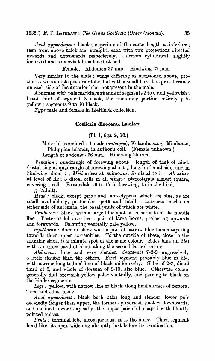
1932.] F. F. LAIDLAW: The Genus Ooeliccia (Order Odonata). 33
Anal appendages: black; superiors of the same length as inferiors; seen from above thick and straight, each with two projections directed inwards and downwards respectively.. Inferiors cylindrical, slightly incurved and somewhat broadened at end.
Female. Abdomen 37 mm. Hindwing 27 mm. Very similar to the male; wings differing as mentioned above, pro
thorax with simple posterior lobe, but with a small horn-like protuberance on each side of the anterior lobe, not present in the male.
Abdomen with pale markings at ends of segments 2 to 6 dull yellowish; basal third of segment 8 black, the remaining portion entirely pale yellow; segments 9 to 10 black.
Type male and female in Lieftinck collection.
Coeliccia dinoceras Laidlaw.
(PI. I, figs. 2, 18.)
Material examined: 1 male (autotype), Kolambugang, Mindanao, Philippine Islands, in author's 0011. (Female unknown.)
Length of abdomen 36 mm. Hindwing 25 mm. Venation: quadrangle of forewing about length of that of hind.
Costal side of quadrangle of forewing about f length of anal side, and in hindwing about -t; Miii arises at suonociu~, Rs distal to it. Ab arises at level of Ltc; 3 discal cells in all wings; pterostigm.a almost square, covering 1 cell. Postnodals 16 to 17 in forewing, 15 in the hind.
~ (Adult). Head: black, except genae and anteclypeus, which are blue, as are
small oval-oblong, postocular spots and small transverse marks on either side of antennae, the basal joints of which are white.
Pro thorax : black, with a large blue spot on either side of the middle line. Posterior lobe carries a pair of large horns, projecting upwards and forewards. Colouring ventrally pale yellow,
Synthorax: dorsum black with a pair of narrow blue bands tapering towards their upper 8xtremities. To the outside of these, close to the antealar sinus, is a minute spot of the same colour. Sides blue (in life) with a narrow band of black along the second lateral suture.
Abdomen: long and very slender. Segments 7-8-9 progressively a little stouter than the others. First segment probably blue in life, with narrow longitudinal line of black middorsally. Sides of 2-3, distal third of 8, and whole of dorsum of 9-10, also blue. Otherwise colour generally dull brownish-yellow paler ventrally, and passing to black on the hinder segments.
Legs: yellow, with narrow line of black along hind surface of femora. Tarsi and ciliae black.
Anal appendages: black both pairs long and slender, lower pair decidedly longer than upper, the former cylindrical, hooked downwards, and inclined inwards apically, the upper pair club-shaped with bluntly pointed apices.
Penis: terminal lobe inconspicuous, as is the inner. Third segment hood-like, its apex widening abruptly just before its termination.

34 Records of the I ndian Museum. [VOL. XXXIV,
A very remarkable species, unique in the genus in that the male possesses a pair of horn-like processes on the prothorax which do not seem to be hom.ologous with the prothoracic processes of females of other species of the genus. It is further remarkable for its very long and slender abdomen and appendages; the penis is, in a general way, not unlike that of O. campioni.
On the whole the -species seems to stand most naturally in the membranipes section of the genus.
Coeliccia ftavostriata Laidlaw.
Material examined: 1 ~ (partatype) Mt. Merinjak, Borneo. Autotype d' in Brit., Mus., Mt. Merinjak.
Length of abdomen 35 rom., of hindwing 21·5 mm. Venation: quadrangle of forewing very slightly shorter than that
of hindwing. Costal side of quadrangle of forewing i length of anal side, of hind
wing -to Ab rises scarcely perceptibly before the level of Ac. Miii rises at (or barely before) the ·subnodus, Rs well after; 3 discal cells in all wings.
Pterostigma covering about one cell, its inner and outer margins parallel, scarcely deeper than the cell next distal to it.
Postnodal nervures, forewing 14-15, hindwing 13. ~ (Type). Head: black, anteclypeus, base of upper lip, and a pair of transverse
marks one on either side of the posterior ocelli, yellowish-white. Prothorax: anterior and posterior lobes black, median lobe yellow
with a fine median longitudinal line of black dorsally. Synthorax: dorsum black, with an orange-yellow antehumeral stripe
on either side, and immediately in front of the antealar sinus a small yellow spot lying externally to the antehumeral stripe. Sides yellow with a narrow black band incomplete below, along the second lateral suture.
Abdomen: segment 1 yellow with median line of black on dorsum ; 2-5 brown, paler below, with terminal black ring on each. The remaining segments progressively· darker from before backward, the last two bronze-black.
A young male has a whitish diamond shaped m'ark on the dorsum of 10.
Legs: femora yellow with black lines, tibiae brown, tarsi and ciliae black.
Anal appendages: black, upper pair about i length of lower, rather flattened apically, incurvedJ each carries a stout spur on its inner side at the end of the first third of its length. Lower pair slender rather oylindrical incurved apically.
(~ Unknown). Penis: inner lobe small, terminal lobe large, hood-like; the third
segment terminates in a pair of short widely separated processes. The organ resembles closely that of the next species, and to a less extent that of renifera.
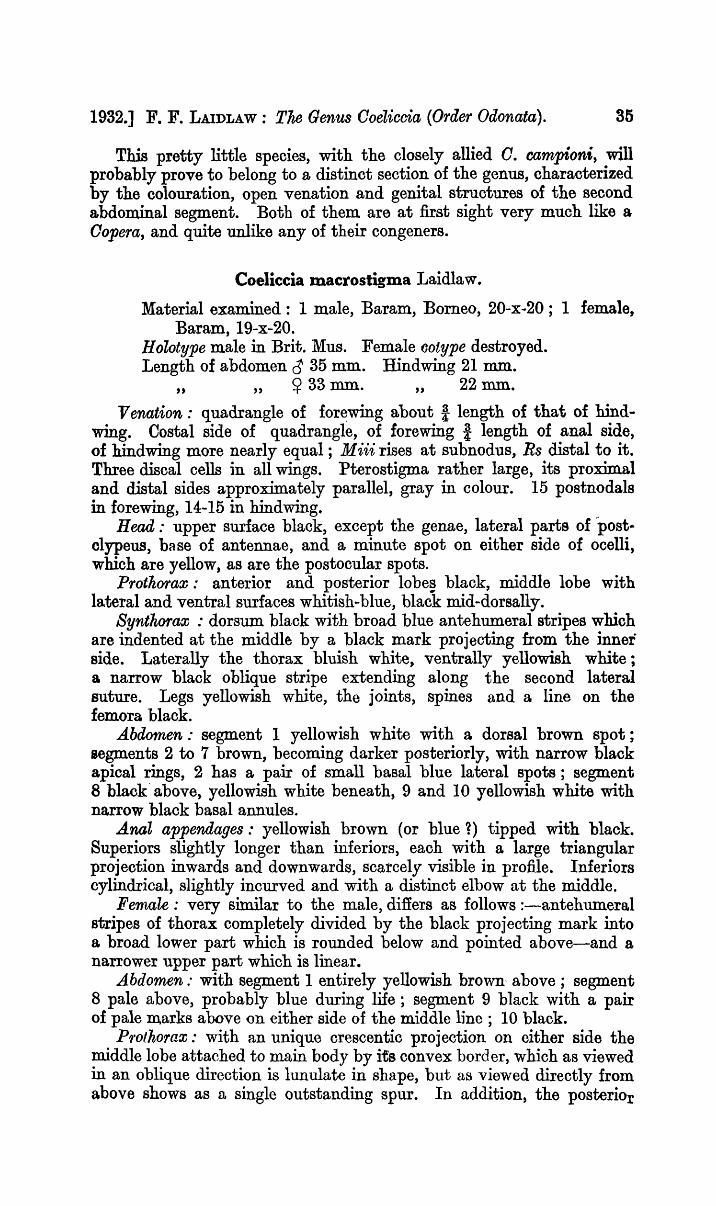
1932.] F. F. LAIDLAW: The Genus Ooeliccia (Order Odonata). 35
This pretty little species, with the closely allied O. campioni, will probably prove to belong to a distinct section of the genus, characterized by the colouration, open venation and genital structures of the second abdominal segment. Both of them are at first sight very much like a Oopera, and quite unlike any of their congeners.
Coeliccia macro stigma Laidlaw.
Material examined: 1 male, Baram, Borneo, 20-x-20; 1 female, Baram, 19-x-20.
H oZotype male in Brit. Mus. Female ootype destroyed. Length of abdomen ~ 35 mm. Hindwing 21 mm.
" ,,~ 33 mm. " 22 mm. Venation: quadrangle of for~wing about! length of that of hind
wing. Costal side of quadrangle, of forewing i length of anal side, of hindwing more nearly equal; M iii rises at subnodus, Rs distal to it. Three discal cells in all wings. Pterostigma rather large, its proximal and distal sides approximately parallel, gray in colour. 15 postnodala in forewing, 14-15 in hindwing.
Head: upper surface black, except the genae, lateral parts of ·post. clypeus, base of antennae, and a minute spot on either side of ocelli, which are yellow, as are the postocular spots.
Pro thorax : anterior and posterior lobe~ black, middle lobe with lateral and ventral surfaces whitish-blue, black mid-dorsally.
SynthO'fax: dorsum black with broad blue antehumeral stripes which are indented at the middle by a black mark projecting from the inner side. Laterally the thorax bluish white, ventrally yellowish white; a narrow black oblique stripe extending along the second lateral suture. Legs yellowish white, the joints, spines and a line on the femora black.
Abdomen: segment 1 yellowish white with a dorsal brown spot; segments 2 to 7 brown, becoming darker posteriorly, with narrow black apical rings, 2 has a pair of small basal blue lateral spots; segment 8 black' above, yellowish white beneath, 9 and 10 yellowish white with narrow black basal annules.
Anal appendages: yellowish brown (or blue~) tipped with black. Superiors slightly longer than inferiors, each with a large triangular projection inwards and downwards, scarcely visible in profile. Inferiors cylindrical, slightly incurved and with a distinct elbow at the middle.
Female: very similar to the male, differs as follows :-antehumeral stripes of thorax completely divided by the black projecting mark into a broad lower part which is rounded below and pointed above-and a narrower upper part which is linear.
Abdomen: with segment 1 entirely yellowish brown, above; segment 8 pale above, probably blue during life; segment 9 black with a pair of pale marks above on either side of the middle line; 10 black.
P1'otho'1'ax: with an unique crescentic projection on either side the middle lobe attached to main body by its convex border, which as viewed in an oblique direction is lunulate in shape, but as viewed directly from above shows as a single outstanding spur. In addition, the posteriOr

36 Records of tke Indian Museum. [VOL. XXXIV,
lobe has a fine black spur at the middle of its posterior margin which projects strongly upwards and forwards. On either side of this process lies a small la ppel directed backwards.
Coeliccia campioni Laidlaw.
(PI. I, fig. 6; PI. II, figs. 5 ad 6; PI. III, fig. 5.)
Material examined: 1 male, Long Semiyan, 5-x -20; 1 female, same locality, 25-x -20 (fragmentary).
Type'male, Lio Matu, Borneo, in Brit. Mus. colI. Length of abdomen d 37 mm. Hindwing 21 mm.
" ,,~1 ,,2I·5mm.
Venation: quadrangle of forewing about ! length of that of hindwing. Costal side of quadrangle, of forewing about i, of hind wing -t length of anal side. 3 discal cells in all wings. Pterostigma black, not inflated, its inner and outer margins almost parallel. Miii rises from subnodus, Rs a little distal. Postnodals 13 in forewing, 11 in hindwIng.
~ (Adult). Head: black, genae, bases of mandibles, a small rather square spot
on either side of the ocelli, and a pair of very small postocular marks, yellow.
Prothorax: dorsum black, middle lobe with a pair of large yellow spots. The posterior lobe carries a very fine projecting point at either lateral angle. Under surfaces pale yellow.
a. TEXT-FIG. 2.-a. Dorsal view of pro thorax of Ooeliccia c,ampion1",~. b. Latera.l view
of anterior portion of same.
Syntkorax: dorsum bla:ck as far as the level of the stigma, with a stripe of yellow immediately to the inside of the humeral suture, this stripe narrows about halfway and tapers at its upper extremity. To the outside of its upper end, which is a little curved inward, a small spot of the same colour.
Sides and ventral surfaces yellow, with a black line, incomplete below, on the second lateral suture.
Abdo'lnen: segment 1 pale yellow, with a black apical ring; 2-10 .brown becoming progressively darker from before backwards; the terminal segments bronze-black. Ventrally yellowish brown.
Legs: yellow, the posterior sides of the femora, anterior sides of tibiae, tarsi, and ciliae black.

1932.] F. F. LAIDLAW: The Genus OQelicda (Order Odonata). 37
AnaZappendages: black. Upper pair a little longer than the lower, stout and slightly incurved apically, each carries an inwardly directed spur near its middle, and is sharply hooked downwards at its free extremity. Lower pair forcipate.
~ (Adult, much damaged) differs from the male in having the yellow stripe on the dorsum of the synthorax extremely narrow throughout, whilst the small upper spot is entirely absent.
Penis: inner lobe small, terminal lobe large, hood-like. The third segment bl~tly bifid at its apex.
Female prothorax: the posterior lobe is large and collar-like, the median process very broad and truncate, separated by a shallow indentation from the small lappels which are scarcely distinguishable from the rest of the margin.
Laterally between the anterior and middle lobes, but apparently derived from the latter there is a pair of extraordinary, horn-like processes, yellow in colour. These are quite the most highly developed of any such structures that are known in the genus. They are conical, the upper half rising as though jointed on to the stouter basal pediclelike part.
Coeliccia nigrohamata Laidlaw.
(PI. III, figs. 15, 16.)
Material examined: 6 males, Mt. Merinjak, 21-v-14, Lio Matu, Borneo, 14-xi-14, and Ruching, Sarawak.
Length of abdomen 38 ro.m. Hindwing 24 mm.
Venation: quadrangle of forewing about t length of that of hindwing. Costal side of quadrangle, of forewing i length of anal side, of hindwing f; Ac rises distinctly after the level of Ab. Miii rises from subnodus, Rs distal to it; 3 discal cells in all wings, pterostigma long covering II cells, gray-brown in colour; 16-18 postnodal nerves in forewing, 16 in hindwing.
~ (Adult).
a" TEXT-FIG. 3.-a. Dorsal view of prothorax of Ooeliccia nigroltarnata, ~. b. Lateral
view of same.
Head: black except for yellow marks on the genae, the ba,se of the mandibles, and a pair 0 f m.inute spots on either side of the posterior Qcelli, as well as a pair of postocular s:potSt

38 Records of the Indian Museum. [VOL. XXXIV,
Prothorax: dorsum black, sides and ventral surfaces yellow. Syntkorax: dorsum black to the level of the first lateral suture,
with a pair of large hook shaped blue marks, the prong of the hooks lying close to the middle line, the shanks occupying the position of the antehumeral band.
Sides between the first and second lateral sutures blue, below the latter yellowish white, with a fine black line incomplete below along the second suture.
Abdomen: first segment yellowish white, with dark basal and apical rings, segments 2-7 bronze black above, yellowish brown ventrally, 8 black abov.e yellow at the sides, 9-10 blue (1) with very fine basal rings of black, 9 has also a minute basal triangle of black mid-dorsally.
Legs: yellowish white, with black lines on the extensor surfaces of the femora and flexor surfaces of the tibiae. Tarsi, joints, and ciliae also black.
Abdomen: segment 1 yellow, with a narrow line of black on the dorsum, 2-7 black above, ventrally yellowish-brown, 8 with yellow at the sides, posteriorly forming a pair of lunules, 9-10 blue .•
Anal appendages: black, but a younger male has the upper pair blue, the lower pair are in all cases black. Upper pair a little shorter than lower with each two small internal projections; lower pair more itrongly incurved apically than in most species, each with a bend inwards at about its middle, at which point also it is a little flattened, so that it has a small shelf-like process on its inner side.
Penis: inner and terminal lobes large, third segment narrow, ending in a pair of short pointed processes. At the point from where'these processes rise there is a curious hood-like projection, recalling that seen in O. albicauda, but more prominent than in that species.
[Females belonging to at least two species, neither of which show any character by which I can definitely identify them with the present species, but which may be from their locale, one or other of them, the female of nigrohamata, are discussed below.]
Coeliccia sp.
2 ~~ Kuching. Borneo.
Both these specimens are very much battered .and scarcely fit for description. They are probably the female of nigrohamata but as this is not definitely ascertainable I prefer to describe as many of their characters as are in condition for description under a separate heading.
The venation is in general the same as that of O. nigrohamata. The only other characters sufficiently available for study are those shown by the prothorax and synthorax.
The dorsum of the synthorax is black and has a pair of humeral stripes shaped each like an inverted" ! " but with the upper and lower parts of the mark conjoined. This mark is like the mark on the dorsum of O. nigrohamata without the proIl;g .of the hook.
The sides and ventral surfaces as in O. nigrohamata. The prothorax is yellowish-white dorsally, with the anterior and
posterior lobes black.

1932.] F. F. LAIDLAW: The Genus Ooeliccia (Order Odonata). 39
The median process of the posterior lobe projects upwards and backwards like a trigger, the la ppels are large and lie close to the median process.
On either side of the median lobe there is a conical papilla projecting outwards, lying well forward, similar to, but smaller than, that of O. campioni.
Coelicc:ia sp. Long Akar, Baram, 1.x.20.
This specimen which is very fragmentary, differs from the two females noted above in the following particulars.
The posterior margin of the prothorax is more markedly arcuate, and the median process is relatively larger, moreover its upper extremity is strongly hooked forward, and the lappels are relatively smaller. The lateral process of the middle lobe are similar but arise as the lower limb of a crescentic ridge.
The humeral marks on the dorsum of the synthorax consist of a pair of blue bands one on either side of the middle line. These bands are rather broad, and each is deeply indented on its inner side at about its middle. The black line on the second lateral suture is incomplete below.
As already stated there is nothing definite either in the venation or in any other character to guide one in placing the three females noted above under any of the species named.
It is quite cert~in that they. are to be referred to two distinct species, and that these species are both of them very closely related to both O. macrostigma and C. nigrohamata; in fact it is quite likely that they are the females of those species, but beyond stating this probability' I am not able to speak with greater definiteness.
Coeliccia albicauda (Forster).
(Pl. I, figs. 1, 23; PI. II, figs. 23, 24; PI. III, fig. 10.)
Material examined: 1 male, Kampong Jor, Pahang; 1 female Tam.ang, Pahang, 2-vi-23. ColI. F .. Chasen.
Length of abdom.en (J 43 mm. Hindwing 25 mm.. " ,,~ 38 mm. " 26 mm.
Venation: quadrangle of forewing a little shorter than that of hind~g. Costal side in each case only a trifle shorter than anal side. Miii rIses from subnodus, Rs distal to it ; 2 dis cal cells in all wings. PterosHgsm.a. a1most square, black, covering one cell, very finely margined 'with white, scarcely deeper than the cell following it. Postnodals forewing 18; hind wing 17-18.
~ (Adult). Head : black; genae, anteclypeus and the transverse postocular
spots blue as also are a pair of small square spots on each side of posterior ocelli.
Prothorax: yellowish-white with the posterior margin and a broad b~nd on the dorsum of the middle lobe black; anterior lobe also black.
F

40 Reoords aftke Indian Museum. [VOL. XXXIV,
Synthorax: dorsum black, with a pair of small triangular blue marks near its anterior end, and posteriorly a very small pair of blue spots near the alar sinuses. The apices of these triangles are directed forward. Sides blue, with a narrow black line along the second lateral suture. Ventral surface ivory white.
Abdomen: black, the first segment yellowish-white; 2 with the same colour at the sides and below, the white extending dorsalwards subapically to form a small pair of lunules. Ventral surface of the remaining segments also whitish, 3-7 with small apical lunules. Distal third of 10 with a similar pair of spots laterally but much larger.
Legs: yellowish-white, carinae, articulations, and tarsi black .. Anal appendages.' ivory-white, upper and lower pairs sub-equal in
length; upper pair each with a large ventral spur at about the middle; lower pair forcipate.
~ (Adult, fragmentary) differs from the male chiefly as follows:the black band on the dorsum of the prothorax is reduced to an exceedingly narrow line, and on the dOrSllUl. of the synthorax there are a pair of na.rrow yellowish bands in place of the blue spots of the male. Segment 10 of the abdomen is black, whilst the black of the dorsum of segments 8-9 is reduced to a narrow band.
Penis: inner and terminal 10 bes well developed, the third segment carries at its apex a pair of short strap-like processes, which curve round the sides of the second segment.
Female pro thorax : the middle lobe has a pair of blunt processes laterally, one on either side; these are quite inconspicuous, but resemble the homologous processes of such a species as C. campioni.
The median process of the hind margin is sm.all and triangular and does not project upwards, nor does it extend backwards so far as the small lappels.
I have seen three females of this species. They differ entirely from the form described by De Selys as octogesima, and I think there can be no doubt but that this species is quite distinct, though it belongs to the same group. Forster's name must therefore be retained for the species, which is a very beautiful one. It certainly includes the specimens I described as O. borneensis from the Malay Peninsula. The locality, from which the specimens described were taken, is the same as that from which came Forster's type.
Coeliccia octogesima Selys.
Type male from Labuan, now in MacLachlan colI., cotype female from Singapore, in Selysian coll.
Length of abdomen cr 37 mm., ~ 32 rom., of hindwing ~ 21 mm., ~ 21 mm.
Venation: Rs rises distal to subnodus, Miii at subnodus .. ~ (Adult). Head: black, with the genae, po stclypeus , a mark on either side
of the ocelli, and cuneiform postocular spots yellowish. Prothorax.· black with a large yellow spot on either side of the middle
lobe.

1932.] F. F. LAIDLAW: The Genus Ooeliooia (Order Odonata). 41
Byntkoraa;: dorsum black with two blue spots on either side of the middle line. The lower of these is oval, the upper is smaller and almost round and lies near the ante alar sinus. Sides blue with a dark line along the second lateral suture. Ventral surfaces yellowish white.
Abdomen: long and slender, the first segment yellowish with a black line dorsally; 2-8 shiny-black above, brownish yellow below. This colour paler and more extended on the sides on 8 ; 9-10 pale bluish, with black articulations and a basal lateral black mark on the sides of 9.
Anal appendages: upper pair bluish-white above, darker below, stout at their base, narrowing to a point apically; the lower, black beyond the base, pincer-shaped, touching each other apically.
~ (Adult). Difiers from the male as follows :-Prothorax yellow, with a narrow, longitudinal line of black mid
dorsally; its posterior margin notched, median process projecting upwards and a little forwards.
Synthorax: the blue markings on the dorsum are less widely separated from one another than in the male, forming on either side almost a figure 8.
Abdomen: brownish-black, segment 8 paler, but dark at the base and dorsum; 9 black, with an incomplete pale band on either side of the mid-dorsal carina.
Legs: pale, with a trace of a dark line on the posterior surface of the femora.
I think it very unlikely that these specimens should be conspecifLc. The account given above is translated from De Selys' description, and I have not been able to examine either of the specimens.
Coeliccia borneensis Selys.
(Unknown to me.) Type from N. Borneo, in MacLachlan ColI. Length of abdomen 30 mm. Hindwing 20 mm. Described from a single female ; male unknown.
Venation: M iii arises from the sub no dus, Rs distal to it; 13 postnodals in forewing.
Head: black, genae and anteclypeus yellow, a blue spot between the posterior ocellus and the eye, on either side.
Protkorax: black with a large blue spot on either side of the middle lobe.
Synthorax: dorsum black as far as the first lateral suture, witih it
(blue) ante humeral band, narrowing above and not reaching the antea1ar sinus. The rest of the sides and ;entral surface pale yellow, with indications of a black line, incomplete below, on the second lateral suture.
Abdomen: black above, brownish-yellow below as is also the distal half of the eighth segment.
Legs: yellow, a black line on the posterior surfaces of the femora and on the anterior surfaces of the tibiae. Tarsi also black.
Posterior margin of prothorax forms two rounded lobes (the lappels), scarcely separated by a small indentation, in the middle of which lies a small tubercle (the median process).

42 Records oftke Indian Museum. [VOL. XXXIV,
This species appears to be quite distinct from any of the other described forms. It i~ distinguished by the small number of postnodal nerves, and by the colour pattern of the synthorax, as well as by the prothoracic margin.
REFERENCES TO LITERATURE.
Fraser, Journ. Bornhay Nat. Hist. Soc. Vol. XXIX, pp. 744-745 (1923). Fraser, Journ. Bombay Nat. Hist. Soc. Vol. XXX, pp. 52-53 (1924). Kirby, Oat. Odon., p. 128 (1890). Kruger, Die Odonat. v. Sumatra (1898). Laidlaw, Proc. Zool. Soc. Lond. Skeat Exp. Pt. II, p. 385 (1902). Laidlaw, Faso. Malay. (Zool.), IV, Pt. II (Odonata), pp. 1-7 (1907). Laidlaw, F'cu;o. Malay. (Zool.), Pt. IV (Odonata), pp. 1-7 (1907). Laidlaw, Rec. Ind. Mus. Vol. VIII, p .. 341, Pl.. xvi, fig .. 1 (1914). Laidlaw, Rec. Ind .. Mus. Vol. XIII, pp. 322, 331-336, text-fig. 3, PI. xiii
and PI. xiv, fig. 3 (1917). Laidlaw, Proc. Zool. Soc. Lond., pp. 223-228, figs. 1-8 (1917). Laidlaw, Proc. Zool. Soc. Lond., pp. 223-231, figs. 2-9 (1918). Laidlaw, Proc. Zool. Soc. Lond., pp. 333-334 (1920). Laidlaw, Philippine Journ. Sci. Vol. xx,rIII, No.4, pp. 562-563, fig. 3
(1925). Martin, Mission Pavie (Nevrop.) (sep.), p. 19 (1904). Rambur, Ins. N evrop, p. 240 (1842). Ris, Supple Ent. No.1, pp. 60-68 (1912). SelY8, Sagra, Hist. Cuba, Ins., p. 464 (1857). Selys, Bull. Acad. Belg. (2) XVI, pp. 155-158 (1863). Selys} Mem. Oouron. Roy. Acad. Belg. XXXVIII, pp. 114-119 (1885).

EXPLANATION OF PLATE II.
Anal appendages viewed from the dorsum and right side.
FIGs. 1, 2.-0oeliccia erici Laidlaw.
FIGS. 3, 4.-0oeliccia chromothorax (Selys).
FIGS. 5, 6.-0oeliccia campioni Laidlaw.
FIGs. 7, 8.-0oeliccia loringae, sp. nov.
FIGS. 9, 10.-0oeliccia fraseri, sp. nov.
FIGS. 11, 12.-0oeliccia reniJera Selys.
FIGS. 13, 14.-0oeliccia lieftincki, sp. nov.
FIGS. 15, 16.-0oeliccia membranipes (Ramb.). FIGS. 17, 18.-0oeliccia loogali, sp. nov.
FIGS. 19, 20.-0oeliccia flavicauda Ris. FIGS. 21, 22.-0oeliccia poungyi Fraser.
FIGS. 23, 24.-0oeliccia albicauda Forster.

REO. IND. Mus., VOL. XXXIV, 1932. PLATE II.
1. ~. 3. 5. 6.
9. 11.
13. 15. 17.
IS.

EXPLANATION OF PLATE III.
Lateral view of thorax of :
FIG. 1.-0oellicoia loogaZi, ap. nov., <1. FIG. 2.-Ooeliccia lieftinoki, sp. nov.,-d'. FIG. 3.-0oeliooia lO'fingae, sp. nov., ~ .. FIG. 4 .. -0oeliooia poungyi Fraser, 0'. FIG. 5.-Coeliccia oampioni Laidlaw, 0'. FIG. 6.-Coeliccia didyma Selys, ~. FIG. 7.-0oeliccia ck'fomotkO'fax (Selys), d'. FIG. 8.-0oeliooia j'J'aseri, sp. nov., 0'. FIG. 9.-Coeliccia 'J'enije'fa Selys, d'. FIG. lO.-Coeliccia albicauda Forster, d'. FIG. ll.-Coeliooia erici Laidlaw, 0'. FIG. 12.-Ooeliccia birnaculata Laidlaw, ~. FIG. 13.-Ooeliccia rnembran'ipes (Ramb.), 0' FIG. 14.-0oeliccia {la'lJicauaa Ria, d' ..
FIG. 15.-0oeliccia nig'l'okamata Laidlaw, ~.
FIG. 16.-Coelwcia nigrokarno,ta Laidlaw, ~.
FIG. 17.-Coeliooia bimaculata Laidlaw, ~.
FIG. 18.-0oeliooia scutellum, ap. nov., ~.


ON A NEW GENUS AND SPECIES OF STREPSIPTERA.
By T. V SUBRAMANIAM, Senior Assistant Ento1nologist, Department of Agriculture in M ysore, Bangalore.
(Plate IV.)
'\\7Jlile searching for stylopised Jassids on grass and other low vegetation, the writer happened to come across large numbers of a smail, very active black cricket (Tridactylus sp.) commonly found in superficial tunnels and burrows in damp soil, especially along irrigation channels in orcnards and kitchen gardens. Many of these crickets were found parasitised by a species of Strepsiptera and fTom the material collected a couple of males and a number of triungulins were reared out in Bangalore. Excepting a reference by Hofeneder (1) to a stylopised Gryllotalpa found by Voeltzkow in Wituland, Africa, nothing is on record of stylopised Gryllidae. It is not unlikely that more forms associated with these hosts may be found in India.
At the outset it was evident that it belonged to the super-family Halictophagoidea, Pierce, owing to the male having three-jointed tarsi, which is characteristic of this super-family. The super-family is divided by Pierce (4) in the following manner into two families.
Super-family Halictophagoidea, Pierce. A. Male antennae four-jointed with flagellum of' the
third and the fourth joint elongate, sub-equal Diozooeridae, Pierce. 13. Male antennae seven-jointed with the third, fourth,
fifth and sixth joints laterally produced, and the seventh elongate HaIictophagidae, Pierce.
This insect has seven joints in the antennae and therefore belongs to the family Halictophagidae. As regards the character of the antenna} segments, however, the insect differs from all the species of Halictophagidae so far known, for they all have the third, fourth, fifth and sixth segments laterally produced. In this insect however only the third joint is 80 produced, the rest being normal.
The structure of the antennae in this insect is so unique that it cannot fit into either of the two families named above.
It may be necessary for the new species to be considered as the type of a, new Bub-family, but for the present I desist from taking this course. The erection of a separate genus for the reception of the new species seems to me, however, clearly indicated. I propose for it the name Tridactylopkagus.
Tridactylophagus gen. nov.
Head very much excavated behind. Eyes large and reaching the base of the elytra. Antennae (PI. IV, flg. 1) slender, elongate, sevenjointed, with sensory pits; third joint la.teral1y produced and longer than the fourth and fifth; seventh joint about as long as the fifth an~ ai4th, pointed ~~ t}le distal end.
[ 43 ]

Records of the Indian Museum. [VOL. XXXIV,
Pro-thorax (Pl. IV, fig. 7) conical, with an indentation at the apex, fitting into the excavated portion of the head. The two lower extremities of the prothorax produced at the sides to form a band.
Meso-thorax consisting of three tra,nsverse pieces. W i'flgs with seven primary veins, most of them broad and smoky
in colour. Oedeagus (Pl. IV, fig. 2) hroad and sinuous near the base, obtusely
flexed near the apex, with a very minute triangular piece attached to the curve at the outer angle.
Genotype: Tridactylopkagus mysO'fensi..;:, ap. nov.
Tridactylopbagus mysorensis, 8p. nov.
Male: 2·04 mm. long. General colour smoky black, eyes and ha.Iters piceous. Base of the scutellum and lower two-thirds of the postscutellum sufiused with smoky black. Whole insect covered with fine hairs, ttnd with a silvery bloom. Head large, as broad as thorax or slightly broader; the facets of the eyes large and wen separated with the interspaces hirsute .. Mandibles large and triangular; their bases velY broad and apices constricted to a fine point which do not meet. Maxilla.e two-jointed, about two-thirds of the length of the mandiJjles; the first joint stout, cylindrical, the second very tiny and more or less circular. The frontal portion of the head between the eyes-the portion comprising the epistome and the labrum of Perkins (3)-is highly chitinised and forms a pattern as shown in Pl. nT, ng. 3. The broad distal end of the fourth joint of the antennae with a circular sensorial organ (PI. IV, fig. 1, a) more or less similar to the one described by Hofeneder (1) in M engenilla cJloubati, by Ogloblin (2) in Delpkacixenos anamalocerus and hy the writer (5) in I ndexenos mem.m'a.ciphaga.
I have reason to believe that these sensory organs are characteristic of the family Halictopltagidae hecause of the fact that these are present in the males of several species reared out by me from different hosts, which have not vet been identified.
Mesonotum p;e-scut-um oval, with the base slightly arched. Scutum and scutellum broad, with the posterior angles produced to form a band. Halters very long and ladle-shaped. Metanotum with the prescutum elongate, key-stone shaped, truncate at the posterior end a,nd well separated from the scutellum. Scuta oblique, the basal portions of their outel' angles lower than that of th.e prescutntn and separated very slightly from the ~cutellum at their outer angles (P1. IV, fig. 7). Scutellum broad, about as broad as the three pieces of the metanotum in the middle. The sides behind the outer angle of the scutii concave, anterior anglee reotangular, the posterior very slightly produced upwards at the sides, the base sinuate and the apex arched (PI. IV, fig. 7). Post-scutellum broad, elongate, much longer than prescutum, scutum and scutellum together (P1. IV, fig. 7). Abdom,en with the sixth, seventh and eighth Regm~nts dra'wn out to a point underneath, the last prolonged to over h.alf the length of the ninth, ~he latter being' ploduced very much beyond the tenth (PI. IV, fig. 4).
Female.-Cephalothorax light brown, more less domeshaped, and very slightly longer than broad; the edges dark brQwn and highly

1932.] T. V. SUBRAMANIAM: NetV Genus & Species of Strepsiptera. 45
chitinised; the sides (PI. IV, 5, a and b.) constricted at the base, diverging as far as the spiracles, near which there is a slight constriction, and gradually becoming narrow towards the mandibles which are stout and broad and possess one short stout tooth. Four genita1 jubes entering the brood canal.
Measurement of female.
(Taken With K.7 Zeiss micrometer ocular and A. A. objective, I division equals 0·015 mm.)
Length from base to vertex Length from spiracles to vertex Width at emargination at the base of mandibles Width between spiracles Width at base Width at base of head
0·41 mm. 0·32 mm. 0·11 mm. 0·32 mm. 0·30 rom. 0·30 mm.
Described from two males bred from T,,~~dactylus sp. in Bangalofe, Mysore State, in June, 1927 and two females out of a number collected at Bangalore in June, 1927
Triungul1~ns (PI. IV, fig. 6). These are found to shoot out into space on emergence from the genital opening as described by the writer (5) in Pyrillozenos c01npactus. Only some stray ones are found crawling over the body of the host. Immediately on emergence, the triungulins are dark, with a very light yellowish tint, but the latter is lost after some time. They have a pair of spines, one on each side of the ten abdominal segments. The tenth segment ends in two long and two short styleta. On the ventral side of the ninth segment there are two long spines arising from a more or less triangular space in the middle and two smaH curved spines arising close to the base of the above. Short hairs are found arranged parallel to one another on the ventral portion of the abdominal segments and alsa a group of 5 or 6 closely-set hairs on the venter of the three thoracic plates. The triungulins are very active and can hop like typical Trypetid maggots.
The following are the measurements of the triungulins (fresh specimens mounted in gIycerine,.
Length excluding stylets Length of stylets Width of body at the broadest portion Width of head
0·13 mm. 0-05 mm. 0-08 mm. 0·06 mm.
The writer wishes to acknowledge the encouragement and help he has received in this work from Dr. Leslie C. Coleman, the Director of Agriculture in Mysore, and .Dr. K. Kunhikannan, the Entomologist.
REFERENCES.
1. Hofeneder, K. Mengenilla. N. G. Choubati. N. Sp. Eine neue Strepsiptre aus Nordafrika. Berichten des Nat1J.rw.-med. Vel'eins XXXII Jahrgang, p. 33, (1910).
2. Ogloblin, A. A. Strepsiptera. Sbirek Entomologickeho Oddeleni Narodniho Musea V Praze III, p. 171 (1924).

46 Records of the Indian Museum. [VOL. XXXIV,
3. Perkins, R. C. L.-Leaf hoppers and their Batural enemies. Report of the Experimental Station of the Hawaiian Sugar Planters' Associa .. tion, p. 100 (1906).
4-. Pierce, W D. The comparative morphology of the order Strepsiptera together with records and descriptions of insects. Proc. U. S. Nat. Mus., LIV (1918).
5. Sub ramaniam, T. V Some natural enemies of mango leaf hoppers (Idiocerus sp.) in India. Bul. Ent. Research, XII, p .. 465, (1922).
6. Subramaniam, T. V A new genus and species of Strepsiptera from South India (Indoxenos membraciphaga) Trans. Ent. Soc. LOndon, p. 131 (July, 1927).

EXPLANATION OF PLATE IV.
Tridactylophaglls mysorensis, gen. et. ap. nov.
(All figures are very much enlarged. The drawings were all made with a camera lucida, excepting fig. 7.)
FIG. I.-Antenna a. The position of the.Benaorial organ on the fourth antennal joint.
FIG. 2.-0edeagus. FIG. 3.-Front view of head of male. FIG. 4.-A. few posterior segments of the abdomen. FIG. 0.-0,. Cephalothorax of female, ventral view.
h. " "" dorsal view. FIG. 6.-Triungulin. FIG. 7.-Male, dorsal view.

REC. IND. Mus., VOL. XXXIV, 1932. PLATE IV.
cc: ....... , ~
.......... -.....

DESCRIPTION OF A NEW SPECIES OF COLLEMBOLA AND ITS ANATOMY.
By DURGADAS MUKERJI, University Lecl-urer in Zoology, Calcutta University.
(Plates V-VI!.)
INTRODUCTION.
A number of species of Collembola has been recorded from India by Parona (1893), Ritter (1910-11), Imm,s (1912), Carpenter <1917, 1924) and Handschin (1929). Owing to the difficulty of making a thorough collection of these minute insects, our knowledge of the Indian species of the group is far from, complete. Moreover, the mouth-parts which are of considerable importance from the points of view of phylogeny and taxonomy, have received in the case of the Indian species, scantyattention, and a general account of the anatomy of the Indian species is lacking. Again, most -0£ the authors who deal with the structure and. function of the organs of those species of Collem.bola that occur outside India are at variance. For their various views, the works of the following authors, among others given in the bibliography at the end of this paper, may be referred to : Sommer (1884-85)) Oudemans (1888), Folsom (1899), Willem (1900), Imms (1906), Hoffmann (1904, 1905, 1908, 1911), Philiptschenko (1906, 1908) and Denis (1928).
In the present- paper a description of a new species of Collembola collected from Calcutta is given, together with an account of its anatomy. In dealing with the anatomy I have chiefly confined myself to descriptions of the digestive system and cephalic glands, as these offer consider-able structural peculiarities. . .
"I wish to acknowledge with thanks my indebtedness to Dr. G. W. Carpenter to whom the specimens were sent for confirmation of the identification, for his kind help and valuable suggestions. My best thanks are due to Lt.-Col. R. B. Seymour Sewell, Director of the Zoological Survey of India, Indian Museum and Dr. H. S. Pruthi of the Entomological Section, Indian Museum, for permitting me to examine the type specimens of Collembola preserved in the Indian Museum. It is a pleasure to record my thanks to Lt.-Col. Sewell for his ~eady help and advice.
Technique.
Live specimens of the species were observed under a binocular microscope and their habits were noted. For dissection of the mouth-parts, specimens were killed in hot water and treated with KOH solution. The dissected parts were either mounted in glycerine or stained in alcoholic eosin and mounted in canada balsam. For serial section cutting the following method, after trial, was found to be highly satisfactory. Fresh specimens were killed in hot 70 per cent alcohol containing 1 per cent acetic acid. They were then fixed in the following mixture.
[ U ] H

Records of the Indian Museum. [VOL. XXXIV,
Saturated solution of picric acid in 90 per cent alcohol 75 parts, formalin 25 parts, nitric acid 5 parts. Fixed material, after dehydration, was cleared in cedar wood oil, embedded in paraffin and serially cut. In a few cases the material after dehydration was cleared for a short time in clove oil and was infiltrated in the cold by celloidin dissolved in clove oil. The infiltrated specimens were then transferred to a thick drop of celloidin solution put on a cover slip previously coated with a thin layer of paraffin. This layer of paraffin, before the celloidin solution was dropped on it, was cross marked by a soft china glass pencil; the specimens were oriented under a binocular microscope. The celloidin mass containing the specimens was then solidified by dipping the cover slip into chloroform and later was re-embedded in paraffin. The pencil marking impressed on the celloidin mass could be seen through the thin coating of the paraffin block and indicated the position of the specimen. The double embedding in celloidin and paraffin was particularly advantageous for facilitating easy orientation of these minute insects during section cutting.
Sections were stained by Ehrlich's haematoxylin, and counter stained hy alcoholic eosin, Heidenhain's iron haematoxylin, or Mallory's mixture (Saurefuchsin, Orange G. and Wasserblau). Mallory's stain, after slight modification of the concentration of the stain and a suitable adjustment of timings, yielded the best results.
Camera lucida drawings and microphotographs of typical sections were taken.
Family PODURIDAE Lbk.-Bor.
Sub-family ACHORUTIN~ Bor.
Tribe ACHORUTINI Bor.
Genus Protanura Borner (1906).
ThiS genus includes those Achorutini in which the maxillae have toothed apices and are provided with lamellae and basal lobes. The allied genus Neanura, which along with Protanura was included in the Neanurinae Borner (1901) but subsequently was transferred to the Achorutinae by Borner himself, differs from Protanura in having a maxilla that is pointed and is without any tooth or lamella.!
The number of species of Protanura and Neanura previously recorded from India, Burma and Ceylon is one and seven respectively. But since the mouth-parts of t.he majority of these species are not known, it is doubtful, as has been pointed out by Carpenter (1917), which of these described species are really referrable to Protanura and which to Nea;n,ura.
1 It is worth mentioning in this oonneotion that Neanura, whioh Borner now calls Achorutes, was proposed by Maogillivray (1893) to replace the genus Anoura erected by Gervais (1842), as the latter generio name was preoccupied. Achorutes (Templ., 1835) and Anoura Gerv., (1842), however, as expounded by Tullberg (1871) in his monograph " SlJeriges Podurider," are two different genera. The substitution of Neanura Macgillv., bV Achorute8 Templ., Bor., in acoordance with Borner's olassifioation, is, therefore~ not withQut the danger of confusion and was oontested by Carpenter (1916).

1982.] DURGADAS MUKERJI: A new Oollembole and its Anatomy. 49
Protanura carpenteri, sp. nov_
-Length, 1-2 to 2·5 mm. Head broader than long. Pigmented ocelli, three in number, on each
side of the head; two of them close to each other, the third being placed towards the base of the head at a slight distance from them. Antennae, with antennal organ and sensory hairs, a little shorter than the head in length. The proportional lengths of the segments of the antenna are approximately 1·6: 2:.1: 1·8. The segments of the trunk are approximately related to one another as 6: 11: 12: 10: 8: 10: 11: 4: 3 ..
TEXT-FIG. I.-Dorsal view of Protanura carpenleri, sp., nov., X ca. 28.
Tubercles carrying three or four sensory bristles present on the dorsal and lateral sides of the body. An additional pair of small ventro-lateral tubercles on the m,eso- and meta-thoracic segments. The tubercles of the last two abdominal segments well developed and lobed. Legs short, femur and tibia of the third leg subequal; superior claw without a tooth; lower claw vestigial.
Colouration deep red, but white when preserved in spirit. The species is gregarious in habit and occurs in Calcutta in shady and Inoist places. Examples of the species were also collected from other parts of Bengal under decaying vegetable leaves and leaf-stems of plantain trees.
The mouth-parts consist of labrum, labium, hypopharynx or tongue, mandible and maxilla. The detailed structure of the mandibles and maxillae, which offer certain specific peculiarities and are of advanta~e in discussing the affinities of the species, is best seen in dissected materIal previously treated with a solution of potassium hydroxide. The rest of the mouth-parts and their topographical positions with reference t.o the head and buccal cavity will be taken up, for the sake of conveluence, when describing the digestive system.
H2

50 Records of the Indian Museum. [VOL. XXXIV,
The mandible (text-fig. 2 md.) is an elongated tubular chitinous structure, stout at the base and narrow at the apex. The apical region bears five teeth and m.ay be designated the dental portion. The dental portion is slightly bent, its outer surface being convex and the inner concave. Four of the teeth are inserted on the concave side, and one tooth on the dorsal side. The apices of all these teeth are directed inwards and in dissected specimens are found to oppose, in the region of the tongue, members of the opposite side. The first tooth is curved inward. The second tooth, which arises from the dorsal side of the dental portion, is pointed. The third tooth is stouter and slightly smaller than the preceding ones. The fourth and fifth teeth are minute in size and are so close to each other that it appears as if they were united at their bases. The basal portion of the mandible runs back obliquely as far as the posterior third of the head and reaches the angle subtended by the cardo and stipes. It is hollow for the insertion of the muscles.
The maxilla (text-fig. 2 mx) is a chitinous rod, a little longer than the mandible, and can be subdivided into two regions: an apical region carrying a toothed process, a comb-appendage and a lamella, and a basal region, called stipes, which constitutes the supporting stalk of the apical region. The toothed process is comparable to the galea described by Carpenter (1904, p. 303) in the case of Protanura citronella (Carpenter), and bears three teeth. The first tooth is sickle~shaped and pointed at its extremity. The second is conical and pointed. The third tooth, lying behind the second, is peculiar in being truncated and blunt. The
J:II~---- C TnX.
tm~ ___ ~~~ /Jt=----l~x.
th,. ___ ~~~_ t------slp.
tst.--____ ~Hm~--~
, __ --m3l!.
cc. ____ ...},. ~==-=-'-
TEXT-FIG. 2.-Diagram of the mouth-parts: X ca. 112. cmx., oomb.appendage of the maxilla; ce., oardo; lmx., lamella of the maxilla; lst., lingual stalk; md., m.andible; mx., maxilla; slp., superlingua; th., tongue; tmx., toothed process of the maxilla; tst., transverse bar joining the lingual stalk.
comb-appendage (text-fig. 2, cmx) is composed of a tapering median axis with two rows of bristles forming a bipinnate structure. Sometimes the two rows of bristles are directed to one side and give the appearance of a brush. The axis itself is gently sinuate. The comb-appendage is comparable to the lacinia of P. citronella (Carpenter, 1904).
1932.] DURGADAS MUKERJI : A new Collembole and its Anatomy. 51
The apices of the maxillae anteriorly converge towards each other, while the stipes towards the posterior end are divergent. The base of the stipes is connected by a ligamentous joint with the outer end of the cardo. The cardo (text-fig. 2, cc) is a straight chitinous rod, lying transversely to the main axis of the body near the posterior region of the head, and corresponds to the articulare of N eanU'la mUSCO'lUl1t
(Temp!.), depicted by Tullberg (1871). The outer end of the cardo nearly subtends an angle of 45° with the stipes with which it is joined. The inner end of the cardo is attached to the basal end of the lingual stalk (text-fig. 2, 1st) and lies nearly perpendicular to it. The lingual stalk is a straight chitinous rod ending anteriorly in a sharp point. The terminal end of the lingual stalk converges towards its fellow of the opposite side and at about the junction of the middle and distal thirds of their length they are connected with each other by means of a transverse thin chitinous bar (text-fig. 2, tst). The portions of the stalks lying anterior to the transverse bar, support the base of the tongue (text-fig 2, th) with which they are intimately connected. The lingual stalk cor.responds to the' fulcrum, hypopharyngis' of Neanura muscorum (Temp!.), described by Tullberg (l.c.), and is called the hypopharyngeal apodeme by Snodgrass (1928). It is interesting to note that Snodgrass (l.c.) holds that the hypopharyngeal apophyses of the Apterygota are the primary elements of the Pterygote tentorium, and the "Zungenapparatus" is homologous with this structure (Prell, 1913). It is also worth noting that the lingual stalks resemble the lingual lorae of crustacea, which according to Crampton (1921) are the precursors of portions of the· tentorium of insects.
The tentorium in Collembola has been reported as a distinct chitinous piece by Folsom (1899), Hoffmann (1905) and Denis (1928). The body of the tentorium in the present instance is best seen in a transverse section, lying between the oesophagus and the sub-oesophageal ganglia.
The mouth-parts of the present species, while agreeing in general with those of Neanura mU8corurn (Temp!'), as given by Tullberg (1871), diller markedly from the latter in the presence of the teeth and the combappendage of .the maxilla. They also differ in a similar way from those of Neanura sexoculata Carpenter (1916), which is recorded from Peradeniya (Ceylon) by Handschin (1928) under the name Achorutes sexoculatus (Carp.). N. sexoculata Carpenter possesses a maxilla with an acute apex and a single delicate process.
The present species can easily be distinguished from Protanura spinifera Carpenter (1917), collected from the North-East of Assam and Lower Burma, and also from Protanura kraepelini Borner (1906), obtained from Java, by the character of their mandibles and maxillae. In both P. spinijera and P. kraepelini, the mandible is provided with a delicate dorsal lamella beset with fine teeth, which is absent in the present case. The comb-appendage of the maxilla of P. 8pinifera has an inner row of bifid teeth, whereas in the present instance the comb-appendage is without any bifid teeth and has, instead, two rows of bristles forming a brush extended over one-third the length of the axis of the appendage. The maxilla of P. kraepelini, again, is without the lamella noted in the present species. Of all the species of Neanurinae, the present one, if

52 Reco1'ds of tlte Indian Museum. [VOL. XXXIV,
the structure of the maxilla be considered as the basis of comparison, comes nearer to Protanura citronella (Carpenter, 1904), reported from the Sandwich Islands. In both, the maxilla possesses a distinct toothed galea, a lacinia fringed with curved setae, and a lamtlla or basal lobe. The row of curved setae in the comb-appendage (lacinia) is, however, single in P. citronella, and is double in this species. The basal lobe in P. citronella is in the form of a conical palp, while in the present species it is bifid. So far as the mandible is concerned, the Hawaiian species P. citronella closely resembles P. spinijera and P. k'l'aepelini and differs from the species under report in the details of the structure of the mandible. If the structural details of the mouth-parts are taken into consideration, this species is, in many respects, distinct from those previously described. It is also easily distinguished from the remain.it;lg Indian species of N eanurinae, whose mouth-parts are not known. It is separated from N. dubiosa Ritter (1910-11), collected from Peradeniya "under stone ", by the pigmented eyes of the latter. It is also marked ofi from N. cO'rallina Imms (1912), which it resembles, by the number of eyes. N. corallina Im,ms has two eyes on either side and is reported from Peradeniya (Ceylon). It also differs from N. intermedia ImIns (1912) collected from Bhowali, N ainita} district, in the number of eyes Mid in the presence of dorso-Iateral protuberances of the body. N. intermedia Imms possesses two eyes on either side and lacks dorso-Iateral protuberances of the body. From the description of N. pudibunda Imms (1912), which was found in the Khayen Caves near Moulmein, Lower Burma, the species under report seems to be allied to it, but is distinguished by its dorso-ventrally flattened body and by the o.bsence of a tooth in the superior claw. N. pudibunda Imms, types of which I had the opportunity of examining, possesses a sub cylindrical body and is without any lateral protuberance in the thorax. The species also difielS from Achorutes indicus Handscrun (1928), obtained from Dodabetta (Nilgiris), by the number and character of eyes. In A. indicus Handschin, the eyes are two in number on each side and are devoid of pigment. The species also differs from Ackorutes kirtellus Borner (1906) reported by Handschin (1928) from Coonoor (Nilgiris) and Dodabetta (NilgirisJ, by the character of the eyes, the structure of bristles on the body and the nature of the claw. In..A. hirtellus Borner, the bristles are feathery, whereas in this case they are simple. It is important to note also that none of the known species of Protanura and N eanura hitherto recorded, have been from Bengal and the occurrence of the genus Protanura in Bengal is recorded here for the first time.
The Digestive System. In the following account of the digestive system I have given, for the
convenience of description, first, a brief outline of the head and its append .. ages, and then have passed on to the description of the alimentary canal. The topographic position of the mouth-parts, the structural peculiarity of which has already been referred to, is treated in connection with the buccal cavity which contains them. Glandular structures present in the head region have been dealt with under a separate heading, as their physiological activity in relation to the process of alimentation is controversial.

1982.] DURGADAS MUKERJI: .. 4 new Oollembole and its Anatomy. 53
The head, as in other Collembola, is bounded above by the labrum and the clypeus, and below by the labium, and its sides are called by Denis (1928) "pli oraL" The labrum is suspended from the lower edge of the clypeus and in sagittal section is seen to be drawn out anteriorly in the form of a beak (text-fig. 3). The labium is similarly drawn out beyond the head. By the meeting of the edges of these mouth-parts, projecting beyond and beneath the head, a kind of snoUt is formed_ The external surface of the labrum bears sensory hairs. A transverse suture shows the line of junction of the clypeus and jihe labrum. The clypeus slopes upwards and posteriorly meets the epicranium .0£ the head, its posterior limit being marked by a deep transverse suture formed by the infolding of the chitinous cuticle (text-fig. 3, ic.). The region in front of this transverse groove, according to Denis (1928), belongs to the gnathocephalon and may be called the frons or clypeo-frons. The ventral surface of the -head is constituted by the labium. The labium not only retains its paired condition but anteriorly the members of the pair are distinctly separated from each other by the presence of a na~row median slit-like gap (pl. V, fig. 2, 1no). The paired labial pieces carry setigerous protuberances, which are sensory in function. The setigerous portion corresponds to the" I\:alauenteil " of Hoffmann. Folsom (1899) considers those portions of the labium, which are tactile in function, to be palpi, and according to Prell (1913) they represent the palpi as well as the external lobes. In the posterior region the labial pieces ·are fused with each other in the median pla.ne and form the ventral wall of the head.
The alimentary canal is represented by a long cylindrical tube running more or less straight from mouth to anus, and giving off along the middle region of its course a few narrow and small diverticula. Three distinct regions can be recognised, namely the foregut, the midgut and the hindgut. The limits of these regions are easily made out by the differences in their widths, by the character of their epithelium and the presence of internal valves.
The foregut commences from the mouth and reaches the anterior region of the prothorax; the midgut extends from the prothorax to the third abdominal segment; and the hindgut commences from the posterior limit of the third abdominal segment and terminates at the anus. With regard to their relative lengths, the midgut is the longest, being about three times the length of the foregut, while the foregut and hindgut are nearly equal in length. The diameter of the digestive tube also varies in different regions; the midgut is the widest, while the hindgut is narrower than the foregut. The histological peculiarities of each of these regions of the alimentary canal are dealt with later.
The foregut, again, is, divisible from before backwar.ds, into the buccal cavity, pharynx and oesophagus, owing to structural distinctions between them. With regard to the limits of these portions it may be stated that the buccal cavity, which represents the anterior end of the foregut, dorsally extends from the tip of the snout to the level of the middle region of the clypeus (text-fig. 3), while ventrally it reaches back nearly to the level of the junction of the pharynx and the oesophagus. The pharynx (PI. V, fig. 1, ph), commencing from th'e pOf\terior limit of

54 Records oftltB Indian Museum. [VOL. XXXIV,
the epipharyngeal groove, slopes at first gently upwards, then curves .down and finally, passing through the circum-oesophageal nerve connectives, is continued posteriorly as the oesophagus. The oesophagus (PI. V, fig. 1, oe) comprises the portion of the foregut lying between the level of the fused supra-oesophageal ganglion and the midgut. The oesophagus widens as it passes backward to open into the midgut and at the point where it joins the midgut, forms a protrusion into the lumen of the latter, the rim of the projecting portion being thick and fringed with papillae. This projecting rim constitutes the oesophageal valve (text-fig. 3, voe), which guards the aperture of communication between
•
pdm.
606. ecr.
m&,
vo~. ic-. cl.dm.s.
oe,. dme. r.
v»t,s. l!~ C!P17V.
In.,.,." llrr.
8-i,lJ. t}"
vbc-.
tan" TV. l1f&.",. cv. t. m,x" l~
TEXT-FIG. 3.-A sagittal section of the head (diagrammatic): X ca. 274. cl., clypeus ; ev., clump of sensory cells; dme., dorsal dilator muscle of the epipharynx; dms., dorsal muscle of the pharynx; dpm., depressor muscle ; ecr., epicranium; ie., chitinous infolding of the cuticle; lbr., labrum; l~t., lingual stalk; la., labium; lmn., lingual nerve; lan., labial nerve; me., midgut; mn., mandibular nerve; mx., maxilla; •. , nerve joining the sensory cells with the suboesophageal ganglion; oe., oesophagus; pdm., postero-dorsal dilator muscle of the pharynx; r., ridge of the pharynx; ph., pha,rynx; sob., supraoesophageal ganglion; sib., suboesophageal ganglion; th., lingua; vbe., ventral chamber of the buccal cavity; voe., oesophageal valve; vms., ventral dilator musole of the pharynx.
the foregut and the midgut. The valve consists of cells whose Dody, as pointed out by Sommer (1884), is drawn out in the form of a flask with a long neck and a broad base. The base contains the nucleus and projects into the lumen of the midgut. A thin delicate membrane, which is continuous with the lining intima of the oesophagus, covers these valves.
Having given above the limits of the different regions of the foregut I now pass on to the composition of the mouth and the buccal cavity, and give in that connection histological peculiarities of the different sub-regions of the foregut indicated above.
The mouth, as in Neanurina, is produced, cone-like, beneath the head, giving rise to the formation of a short snout, reference to the composition of which' has already been made. The mouth leads into the buccal cavity.

1932.] DURGADAS MUKERJI: A new Oollembole and its Anatomy. 55
The buccal cavity (Pl. V, fig. 2) occupies a large part of the inside of the head and is separated from the haemocoelic cavity, dorsomedially by the epipharynx and dorso-Iaterally by ligaments that are hypodermal in origin. It is capacious and is divided by the tongue or hypopharynx into an upper or dorsal and a lower or ventral chamber (PI.. V, fig. 3). The dorsal chamber of the cavity is occupied by the epipharyngeal groove (PI. V, fig. 2, epr) and the ventral chamber contains the mandibles, maxillae and lingual stalks whose apices support the medial portion of the tongue (text-fig. 3). The upper and lower portions of the buccal cavity, though sharply marked off from each other in the region of the head, are confluent with each other at the tip of the snout. The upper portion of the buccal cavity, which can be identified with the epipharyngeal groove, leads into the pharynx, which is con ... tinued posteriorly as the oesophagus to open into the midgut. The nutriment taken into the mouth with the help of the mandibles and maxillae passes along the passage leading from the dorsal chamber of the buccal cavity to the midgut. The whole of this passage is situated above the ventral chamber of the buccal cavity and constitutes the food-channel (PI. V, fig. 1).
The epipharyngeal groove is bounded above by the epipharynx and below by the dorsal surface of the free portion of the tongue. The epipharynx (PI. V, fig. 2, eph) is a thin chitinous membrane continuous at the tip of the snout with the clypeus, that forms the roof of the epipharyngeal groove: in this species it is peculiar in not being provided with teeth in its anterior region. Posteriorly it reaches the level of the middle region of the clypeus, and there merges into the roof of the pharynx. The epipharyngeal groove is narrow at its anterior end and gradually widens posteriorly. In cross section the groove (PI. V, fig. 3) is dorso-ventrally compressed. The lining membrane of its lumen is chitinous in nature and when examined under a high power reveals fine striations. Anteriorly the epipharyngeal groove and the ventral chamber of the buccal cavity are continuous around the sides of the tongue and it thus receives the apices of the mandibles (PI. V, fig. 2). The epipharynx gradually merges into the roof of the pharynx, and it is thus difficult to draw a sharp line of demarcation between them; but the two regions can to a certain extent be distinguished by the character of their linings and by the arrangement of their musculature. In general it may be stated that the protoplasm of the lining membrane of the pharynx is granular and contains distinct nuclei, wher&as, in the case of the epipharynx, which is chitinous in nature, nuclei cou1d not be made out, specially in the anterior region.
Distinction can also be made in a similar way by the character of Hnings between the pharynx and the oesophagus which follows it. The walls of the pharynx and the oesophagus consist of three layers, as described by Imms (1906) in the Anurida. The chitinous intima forms the innermost layer, the middle layer is represented by an epithelium, and the outer by a muscular ring. Imms remarks, however, in this conneotion that it is uncertain whether the epithelial layer spoken of here is composed of true epithelium or not. The character of the epithelial layer of the pharynx and the oesophagus is as follows: in the

56 Records of tke Indian M use'Um. [VOL. XXXIV,
anterior region of the pharynx the cytoplasm of the epithelial layer is granular and completely fills up -the space between the inner chitinous intima and the outer ring musculature, the cell outlines being indistinci (text-fig. 4, ph). In the region of the pharynx lying immediately in front of the supra-oesophageal ganglion the cytoplasm of the epithelial layer, however, shrinks, so as to leave a space between the chitinous intima and the ring musculature-. The protoplasm is reticulated in appearanc61 and contains nuclei. In the region where the base of the tongue is intimately fused with the ventra] wall of the pharynx, the floor of the pharynx is peculiar in being thickened and raised in the form of a broad ridge tha~ projects backwards into the lumen of the pharynx (text-fig. 3, 't.). This protruding ridge has also been noted by HoHmann (1905) in the case of Tmnocerus. The ventral area of this region of the pharynx contains a number of flask-shaped cells having large· nuclei. I am not certain whether these are sensory or glandular cells. In the oesophagus the epithelial layer occupies the whole of the area between the chitinous intima and the ring musculature ; its cytoplasm is distinctly reticulated and the nuclei are prominent. The lumen of the oesoph3gus (PI. V, fig. 5, oe), w:hen cut transversely, is more or less circular, whereas that of the pharynx and the epipharynx is elliptio in contour.
With regard to the musculature it may be recalled that a layer of circular muscles surrounds the epithelial wall of the pharynx and oesophagus in the form of a ring. This ring-musculature is absent in the region of the epipharyngeal groove: but towards the posterior end of the groove, near its junction with the pharynx, two sectors of circular muscles are found above the lining membrane of the epipharyngeal groove (PI. V, fig. 3).
The disposition of the radial muscles serves also to distinguish the epipharyngeal groove from the pharynx. The epipharyngeal groove is supplied with a single set of radial muscles dorsal in position to the alimentary canal. The pharynx on the other hand is supplied with two sets of radial muscles; one set, dorsal, in'3erted into its roof and the other, ventral, attached to its :floor. The arrangement of these radial muscles is clearly brought out in the sagittal sections of the head region.
In text-fig. 3, a pair of radial muscles is seen inserted into the anterior end of.the epipharynx. This pair slopes backwards and is attaohed to the labIum at its junction with .the clypeus. This pair of muscles (text-fig. 3, dpm) is cal1ed by Hoffmann (1905) in Tomocerus plumbeus (L.), the depressor of the 1abrum, since by pulling on the 1abrum it brings about the closure of the lumen of the epipharyngeal groov~. Posterior to the point. of insertion of this muscle other longitudinal muscles occur. These are termed by Hoffmann (loc. cit.) dilators of the epipharynx (text-fig. 3, dme), as they help in widening the lumen of the epipharyngeal groove. According to the direction and point of attachment, these dilator muscles of the epipharynx can be grouped into two series. The first series includes the anterior muscles which are directed forward to be attached to the inner surface of the clypeus, while the second seri~s comprises the posterior muscles that run obliquely backward to join the chitinous infolding of the head at the posterior limit of the clypeus. The dorsal set of radial muscles of the pharynx (text-fig. 3, dms) can

1932.] DURGADAS MUKERJI: A new Oollembole and its Anatomy. 57
-similarly be grouped into two series: an anterior series composed of more than eight longitudinal muscles, arising from the dorsal surface of the pharynx and converging to be attached to the chitinous infolding of the roof of the head and a posterior series (text-fig. 3, pcZm) represented by one or t\VO longitudinal muscles that are inserted into the pharyn x close to the supra-oesophageal ganglion and pass up to be attached to the epicranium of the head. The ventral set of radial muscles of the pharynx (PI. V, fig. 1; text-fig. 3, vms), supplying its floor, pursues a slanting course more or less paral1el to the longitudinal axis of the oesophagus and is attaQhed to the tentorium. The ventral wall of the pharynx at the place of insertion of the ventral set of radial muscles shows considerable thiokening and contains nuclei of large dimensions, evidently of sensory cells: such a thickening dces not occur on the ventral side of the epipharyngeal groove or of the oesophagus. The radial mUEcles are distinctly striated transversely and are directly inserted into the chitinous intima lining the lumen of the foregut. The radial sets of muscles of the pharynx by their contraction widen the cavity of the pharynx and aTe known as dilators of the pharynx. It is interesting to note also that a dorsal set of radial mu~cles, such as is present in the pharynx, does not occur in the region of the oesophagus, which can thus easily be distinguished from the pharyngeal region, which is richly supplied with both dorsal and ventral dilator muscles.
The ventral chamber of the buccal cavity is more capacious than the dorsal chamber and lodges the mandibles, maxillae, hypopharynx and the lingual stalks (PI. V, fig. 3; PI. VI, fig. 1). The disposition of the ventral chamber with reference to the enclosed mouth-parts, however, varies considerably as -we pass from the tip of the snout to basal region of the head.
The tongue, or hypopharynx as Denis calls it (1929, p. 16), separates the dorsal chamber from the ventral one (PI. V, figs. 2, 3). It is composed of a median part known as lingua and two lateral lobes termed superlinguae (PI. V, fig. 2, slp). These lobes have been homologised by Crampton (1921) with the paragnaths of Crustacea and since the terms paraglossae and maxillulae are undesira,ble, the term superlinguae employed by Folsom '1899) has been applied to them, in accordance with the suggestion made by Denis (1928) and Imms (1925). The lingua (textfig. 3, th) in a sagittal section is seen to extend as a chitinous shelf within the buccal cavity and forms the floor of the epipharyngeal groove and part of the pharynx. The base of the lingua is fused with the ventral wall of the pharynx and its apical region is supported on the terminal parts of the lingual stalks. The posterior portion of the lingual stalk slopes down as it passes below the ventral wall of the pharynx (textfig.3; PI. V, fig. 1). The position of the lateral lobes of the tongue varies somewhat in different regions of the head. In PI. V, fig. 2 of a transverse section through the snout, the sides and partially the floor also of the buccal cavity are seen to be bounded by the labial pieces (la). The lateral lobes (slp) of the tongue are here seen together with the median lobe or lingua -(tit), separating the buccal cavity into the upper and lower chambers, as mentioned above. The median portion of the upper chamber represents the epipharyngeal groove (epr) which is in oommuni-

58 Records of the Indian Museum. [VOL. XXXIV,
cation with the lateral pouches containing the mandib!es (md). It is for this reason that the mandibles are seen in this figure to lie inside the
ems. . .
. /:.:1 epg .
..,,-. ....... ..;.;: ""~I-IfIio~r+.:-------+-p k. 7I:----..:~-mdp.
-~:___-----It~~~ ~-~ ~~~W-fi---~~- nv6.
~~ __ ~ ____ == mx~ llv.
l~-----....... lJm~. m,o. _________ _
TEXT-FIG. 4.-A transverse section of the head through the anterior region of the pharynx, (diagrammatio). (Upper half not shown in the figure): X ca. 225. aeg., acinose gland; ems., circula.r layer of muscle; epg., epipharyngeal ganglion; lh., hypodermal liga.ment; la., labium; lst., lingual stalk; md., mandible; mx., maxilla; mdp., mandibular pocket; mxp., maxillary pocket; mo., median gap in the labium; ph., pharynx; nvb., nerve band; slp., superlingua; vme., m.edian portion of the ventral chamber of the buccal cavity.
superlinguae (slp), which are bent inward in the form of a L. The cut portions of the lingual stalks (lst) lie immediately below the medial pOltion of the tongue, flanked on the sides by the inner ends of the Buperlinguae. rrhe maxillae (mx) ~re found to lie just below the horizontallimbs of the superlinguae and a little away from the lingual stalks. The configuration of the lateral lobes of the tongue changes, however, as we pass towards the posterior end of the head. In text-fig. 4 the lateral lobes of the tongue are seen as chitinous transverse bars imperfectly dividing the ventra] chamber of the buccal cavity into upper and lower halves .. In the section figured in PI. V, fig. 3 the epipharyngeal groove is shut off completely from -the lateral pouches containing the mandibles, as well as from the medial portion of the ventral chamber. The superlinguae appear as stumps flanking the medial portion of the ventral wall of the epipharynx. In dissected specimens (text-fig. 2, slp) the lateral lobes of t~e tongue are found as chitinous plates slightly rolled into an imperfect tube through which the maxillae are seen to pass: they closely resemble the maxillulae of Protanura spinijera, Carpenter (1907) and Protanura cit1'onella Carpenter (1904).
It is clear from the study of these transverse sections that the anterior region of the ventral chamber of the buccal cavity is somewhat different from its posterior portion. In PI. V, fig. 3, the anterior region of the mid -portion of the ventral chamber of the buccal oa vity lies below the epipharyngeal groove. 1n the posterior region, however, the ventral chamber (Pl. VI, fig. 1, vrnc) gives off two dorso-Iateral evaginations forming two pouches, one on either side of the food -channel. These pouches enclose the vasal portions of mandililes and maxillae. These

1932.] DURGADAS MUKERJI: A new Oollembole and its Anatomy. 59
pouches again are divided imperfeotly by menns of ligaments (text-fig. 4, lh) into upper and lower halves, the upper half oontaining the bases of mandibles and the lower half those of maxillae. The pouches containing mandibles and maxillae have been described by Denis (1928, p. 14) as " pouche mandibulare '? and" pouche maxillaire " respeotively.
The lateral pouches (PI. V, fig. 4, lbc) extend baok as far as the level of the anterior end of the supra-oesophageal ganglion and are separated from each other at their posterior limit by means of a broad partition composed of cells, which seem to be glandular in nature (PI. V, fig. 4, mgp).
The buccal cavity is comparable to the ' Miindhohle' of Tomocerus plumbeus (L.), desoribed by Hoffmann (1906, 1908) and resembles, to a certain extent, the atrial oavity defined by Denis (1928-29, p. 13). It corresponds also to the pre-oral cavity of a generalised insect as depicted by Snodgrass (1928, p. 48, fig. 19). Snodgrass' figure, however, refers to an inseot having mouth-parts of an ectognathous type, while in the present instance the mouth-parts are concealed within the head as in other species of Collembola. Folsom (1899) and Imms (1906) have called the anterior end of the foregut, that encloses the mouth-parts, the pharynx, aud the remaining portion of the foregut the oesophagus. Sommer (1884-85), however, divides the foregut of Macrotoma plumbea L., excluding, it seems to me, the portion called by me the buccal cavity, into the pharynx (Sohlund) and the oesophagus on account of the differences of musculature along the length of the foregut. It seem3, therefore,_ that the portion of the foregut called pharynx by Sommer really corresponds to the anterior portion of the part designated oesophagus by Folsom and Imms. In the present instance the buccal cavity enclosing the mouth-parts is quite distinct from the remainder of the stomodaeum which is distingujsha ble into two distinct regions, the pharynx and the oesophagus. The employment of the term pharynx to denote the buccal cavity, enclosing the mouth-parts, and in addition the anterior portion of the oesopb.agus, seems to be confusing. I have therefore adopted t"he term bucoal cavity, first employed by Fernald (1890). to indicate the mouth-cavity, enclosing the mouth-parts, and have, restrioted the term pharynx to the anterior portion of the foregut lying between the buccal cavity and the oesophagus. The histological difierenti~tion and the method of formation of the box-like oavity enclosing the mouth-parts by the fusion of the fundaments of labium and labrum with the lateral evaginations of the facial region in the embryo, as mentioned by Imms (1906), justify also the employment of the terms used here. Tullberg (1871) first described the epipharynx in Collembola. The term epipharyngeal groove has been adopted from Hoffmann (1905, pp. 643-646) who called it " epipharyngeal Rinne" It should be noted, h.owever, that the epipharyngeal groove described by Hoffmann contained chitinous hooks which in this instance are wholly absent.
The midgut is cylindrical in form and its limits have already been mentioned. In specimens mounted whole in canada balsam, the midgut becomes sufficiently transparent to allow the granular food matter contained in it to be seen. fie wall of the midgut consists of an epithelil:lID (PI. VII, fig. 3, mep) resting externally on a tunica propria and

60 Records of the Indian Museum. [VOl". XXXIV,
surrounded by a muscuJar coating, as in the case of Anurida described by Imms (1906). The external muscular coat is formed by a system of circular and longitudinal muscle fibres. The epithelial layer is composed of cubical cells with distinct nuclei. The boundaries of the epithelial cells are clear; but distinctness of these cells, as pointed out by Imms (1906), depends largely upon the particular phase of the physiological activity of the digestive system. In some of my preparations the pro toplasm of these cells is found aggregated in the form of a granular heap toward the lumen of the gut. In some the internal free edge exhibits fine striations recalling the "Harschensaum" or ciliated band which Fernald (1890, p. 439) believes to be produced as a result of poor fixation. The protoplasm either in the middle or peripherial zone of these cells is vacuolated. Embedded in the peripherial zone of the epithelial layer, are found small vesicles deeply stained like the· nuclei, which occupy a more central position. Imms (1906) m~ntions these vesicles as chromatic particles of unknown function. In my sections these granules closely resemble nuclei in structure with this difference, that they are smaller in size. The cytoplasm around them is also marked off from that around the nuclei. In view, however, of the fact that no selective cytological reagent was employed in order to test the nature of these vesicles, it is impossible to say whether they are to be regarded as chromatio particles or true nuclei. After examining a number of seotions differently stained, I am, however, inolined to believe that these particles are nuclear in nature and that the epithelium. surrounding the }um.en of the midgut is possibly, therefore, composed of two layers.
In the t.horacic region the midgut gives off laterally a number of shallow divert.icula (text-fig. 5). Similar thoracic divertioula have been recorded in the case of Neanura muscorum by Tullberg (1871) and Willem (1900). Since these diverticula are not known to occur in other subfamilies of Collembola it is possible that they represent a character peculiar to the Neanurinae. The wall of these diverticula is similar in composition to that of the midgut. In addition to these divertioula, a pair is given off that is directed forward and runs into tbe head (PI. VI, fig. 8, dve). Such a pair has not, so far, been recorded as occurring in the head region of Collembola, and from their peculiar position in the body I have termed them "cephalic diverticula" Their origin from the midgut is interesting. The anterior end of the midgut, on account of the inclination of the axis of the oesophagus to th.at of the midgut, presents on its dorsal side a median sac-like cavity projecting a little forward beyond the line of junction of the two regions: a similar cavity has also been noted by Sommer and also by Fernald (loc. C1:t.). The cephalic diverticula (PI. VI, fig. 2, a've) arise from this dorsal cavity, one on either side of the oesophageal valve, and extend nearly as far as the posterior limit of the supra-oesophageal ganglion. Their method of origin and their relation to the oesophageal valves are clearly seen in transverse sections through this region. Near their origin, these diverticula lie close to each other, so much so that their medial walls are confluent with each other: as they pass forward, they diverge and lie above the plane of the oesophagus,· one on each side (PI. V, fig. 5, dve). Their diameter is wider than that of the oeso:phagus but n~rrQwer than the

1932.] DURGADAS MUKERJI: A new Oollembole and its Anatomy. 61
lumen of the midgut. The wall of the cephalic diverticula is closely similar in composition to that of the midgut, being formed of a thick epithelium: the nuclei of the epithelial cel1s are distinct and the cytoplasm is reticulated; the inner zone of the epithelium is tliicker in comparison with that of the midgut proper and shows fine striations when examined under the high power. These cephalic diverticula bear some resemblance to the anterior caeca of Petrobiu8, described by Oudemann (1888), and it is possible that they are homologous organs: it is, however, equally possible that they may be an example of parallelism in evolution. It would be premature, therefore, to say whether the presence of these orgallB has any bearing on the question of the affinities of Thysanura and Collembola.
The hindgut extends, as already mentioned, from the third abdominal segment to the anus; it is separated from the midgut by means of valves projecting inwards (text-fig. 5). Immediately posterior to the
TEXT-FIG. 5.-A sagittal section through the body (diagrammatic): X ca. 38. kg. hindgut ;-me., midgut mtg., metathoracic ganglion; pi., tubular pocket of the hindgut; Bob., supra-oesophageal ganglion; vng., ventral nerve cord; tb., ventral tube.
valves and at the oommencement of the hindgut the lumen of the digestive tube suddenly ·widens out to form a small pocket-like cavity (textfig. 5, pi). The rest of the lumen of the hindgut is not uniform along its length. Along the whole of the length of the hindgut the epithelial layer surrounding the lumen is granular and is without distinct cell
TEXT-FIG. 6.-A transverse section through the middle region of t.he hindgut: X ca·. 466. ems., circular layer of muscles; epi., epithelium of the intestine.
boundaries. The chitinous intima lining its lumen is, comparatively speaking, well developed and iJl. the anterior region lies closely applied

62 Records of the Indian Museum. [ VOL. XXXIV,
to the epithelial layer : in the anal region,' however, it liesd~tached from the epithelial lining, leaving a narrow spaoe between them. This detachment of the intima from the epithelial membrane may, however, have been caused· by fixation or be due to moulting. The muscular coa.t, external to the epithelial layer, V"ariea in thickness in different regions of the hindgut: in the anterior region the muscular ring is thin and is in olose contact with the epithelial layer ; but towards the posterior end, this ring musculature gains in thickness and in the middle portion of the hindgut forms a broad circular band (text-fig. 5, ems) that oompletely surrounds the gut; but a large clear space intervenes between the inner limits of the muscle ring and the basement membrane of the epithelial layer. The space thus enclosed communicates freely with the haemocoelic oavity of the body: Imms reports the presence of blood corpuscles in this intervening space. Sommer working on Maerotoma plumbea describes a similar cavity in the region of the rectum and hints at its respiratory function, as it is filled with blood. I have noted several granules in this space but I do not find any blood corpuscles in it. The physiological significance of this space is, therefore, obscure; it is possible, however, that it is a mechanical contrivance for the expansion of the hindgut when choked with contents. Towards the posterior end of the hindgut, in the region of the rectum, the muscles are arranged in six longitudinal bands that come into direct contact with the peripheral zone of the epithelial layer and in transverse sections through the anal region, form a series of six projections into the lumen. These projections are conical in shape and small in size (text-fig. 7, cmp). Radial muscles, called' dilatores recti' and' dilatores ani' occur in the rectum., as des .. cribed by Jmms (1906) in Anwrida.
.. -~epv.
TEXT-FIG. 7.-A transverse section through the posterior region of the hindgut: X ca. 533. cmp., musoular projeotions; epi., epithelium of the reotum.
The nature of the epithelial layer of the hindgut, varies somewhat along its length. In the anterior region the cell-boundaries of the epithelial layer are indistinct and the protoplasm of the cells is granular and vacuolated: the chitinous lining is very thin and the cytoplasm of the epithelial layer from its inner surface sends up irregular processes into the lumen. Near . the middle region, where tbe ring musculature is prominent, the epithelial layer and the chitinorF intima are thrown

1932.] DURGADAS MUKERJI: .A. new Oollembole and its Anatomy. 63
into folds and ridges (text-fig. 6, epi) and, as a result, the lumen of the hindgut in this region becomes irregular. In the region of the rectum the epithelial layer, as already mentioned, projects into the lumen of the gut in the form of conical processes, the protoplasm is granular and without cell-boundaries, and in my sections nuclei cannot be detected.
From the nature of the mouth-parts and the buccal cavity, the mouth can be regarded as a sucking apparatus. Willem, (1900) in the case of Neanura (Gerv.) considers the buccal cavity to be adapted for sucking and the tongue, which he terms the ligula, as playing the role of a piston within the buccal cavity. The mandibles and maxillae are considered by him to be perforating organs. As the mandibles in the present instance, are provided with teeth I am of the opinion that the mandibles cut the soft woody tissues and the maxillae help in seizing and bringing food into the mouth by means of the feathery appendage and toothed lamellae attached at their ends. It may be pointed out that in live specimens tihe maxillae are protruded out beyond the oral aperture, and their purpose may, therefore, be to draw food into the buccal cavity and reduce it to pulp, so as to help in the suction of the juice. The dilatation of the buccal cavity and the pharynx by means of muscles will further help in the suction of food. In order to ascertain the nature of their food I kept examples in the laboratory in large, open vessels filled with earth, dug frOIQ. the neighbourhood of a tank and periodically moistened with water. Individuals were kept alive for weeks together, although no food was added. Apparently they lived on the organic vegetable m~tter contained in the soil. According to Macnamara (1924) the Collembola fall into three -categories, viz., (i) vegetable feeders, (ii) carnivorous, and (iii) thosa that live on a liquid diet. Species depending on a vegetable diet possess mandibles provided with a molar surface. In the present instance the mandibles do not possess a molar surface and individuals have not been O'bserved in the field either feeding on animal matter or present in the neighbourhood of a carcase. Further, Macnamara (1924, p. 99) observes" the suctorial species seem to find an innocent nourishment in the fluids of rotting wood and fungi" I presume, therefore, that this species chiefly lives on vegetable nutriment, rich in juice.
The Nervous System.
The nervous system is built on the same plan as that of Podura aquatica (L.), described by Willem (1900) and also differs but little, except in details from that of other examples of the Collembola investigated by Folsom (1889), Willem (1900), Imms (1906), Hoffmann (1908), Hilton (1913) and Denis (1928). It consists of the supra-oesophageal ganglion, the suboesophageal ganglion and the nerve cord comprised of three thoracic ganglia (text-fig. 5). These ganglia usually occur in insects in pairs, but in this species the members of each pair are so intimately fused with each other that they appear as a single ganglion (PI. V, fig. 4, sob).
The supra-oesophageal ganglion (text-fig. 3, sob), formed by the fusion of proto-, deuto- and trito cere brallo bes, gives off paired nerves to the ocelli, antennae and labrum. The labral nerve trunk originates from the supra-oesophageal ganglion near the junction of the latter with the
l

64 Records of the Indian Museum. [VOL. XXXIV,
oesophageal connectives and after coursing forwards over the pharynx, divides into three branches which innervate the pharynx, epipharynx and labrum" respectively. The oesophageal connectives are stout and pass round the oesophagus to connect the supra-oesophageal ganglion with the anterior end of the suboesophageal ganglion. The suboesophageal ganglion (text-fig. 3, sib) is like the supra-oesophageal, a complex ganglion being formed, accorrung to Imms (1906), by the coalescence of four primitive ganglia. It is spindle-shaped and extends below the oesophagus. It gives ofi paired nerves to the mandible, maxilla and labium, and an unpaired nerve (text-fig. 3, lmn) to the lingua or tongue. The mandibular (text-fig. 3 mn) and the maxillary nerves, soon after their origin, divide into two branches: a branch passes to the base of the appendage concerned and terminates in a mass of filiform cells, while the other innervates the muscles supplying the appendage. The labial nerve (text-fig. 3, Zan) arises as a main trunk which divides and ultimately terminates in sensory cells in the labium. The median nerve sUflplying the lingua arises from the anterior end of the suboesophageal ganglion close to the points of attachment of the oesophageal connectives and, passing through the thick ventral wall of the anterior portion of the pharynx, breaks up there into fine fibrils.
A peculiarity worth noting is that in transverse sections through the anterior region of the pharynx or through the epiphaqngeal groove a large mass composed of :Bask-shaped cells (text-fig. 4, epg.; PI. VI, fig. 1) is noticed to lie on either side of the digestive tract. This mass is connected with its member of the opposite side by means of a nerve b~nd (text-fig. 4, nvb) that passes ventral to the digestive tube, a litt.le below the level of the points of insertion of the ventral dilator muscles of the pharynx. Similar masses of cells have been described by HoHmann (1908) in the case of Tomoce'J'us plumbeus (L.) under the name epipharyngeal ganglion. The cells composing the mass are evidently sensory nerve-cells and may be gustatory in function.
Again, in sagittal sections passing through the head a little lateral to the median vertical plane, a large clump of similar cells (text-fig. 3, cv) is noted lying in the ventral chamber of the buccal cavity close to the basal region of the maxilla. The clump (text-fig. 3, cv) is connected with the suboesophageal ganglion by means of a short nerve (text-fig. 3, n) that is swollen in the mid-region into a ganglionated knot. I believe this mass is distinct from the superlingual or maxillary ganglion and different from the cephalic glands of the ventral tube. It seems to me that the constituent cells represent sensory cells but I am uncertain of the exact homology of this organ.
The ventral nerve cord, as in other Collembola, is a concentrated one and is composed of three ganglia that are serially placed in the pro-, meso- and meta-thorax. The meta-thoracic ganglion (text-fig. 5, mtg) is considered to be formed by the fusion of the last thoracic and the first abdominal ganglion which has m.igrated forwards. All the three thoracic ganglia are joined to one another by distinct double connectives. A pair of stout and parallel nerves arise from the last ganglion of the nerve cord (PI. VII, fig. 3, mt) and pass backwards over the ventral tube. This, ltowever, does ~9t reach the posterior e~tremity of the body.

1932.] DURGADAS MUKERJI: A new Collembole and its Anatomy. 65
Oephalic Glands.
Diverse views ha ve been expressed regarding the disposition homology and function of the glands existing in the head region of Collembola. A review of these is given by Folsom (1899), Willem (1900, 1901), Hoffmann (1904), Tmms (1906) and recently by Denis (1928). I, therefore, refer here to the principal controversial points only.
Denis (1928) classifies the glandular systems belonging to the head region of Collembola as follows: (1) Cephalic glands of the ventral tube, composed of (a) tubular glands and (b) globose or acinose glands. Secretions of these glands, according to this author, flow out of the buccal cavity through the labial cleft to the ventral groove and finally reach the ventral tube, which, being thus smeared with the secretions, acts as an adhesive organ. .(2) Salivary glands composed of (a) an anterior pair situated on the external wall of the "pli oral" and opening into the atrial cavity~ and (b) a posterior pair located within the cephalic cavity above the level of the maxillary pouch. The ducts of these glands follow the mandibles closely and open into the buccal cavity. (3) Excretory glands-(Reins Cephaliq·ues)-closed glands without any duct and containing concretions of mates.
Attention may be -directed to the fact that, so far as the structure and function of- the cephalic glands of the ventral tube are concerned, Denis is in general agreement with the observations of Fernald (1890), Willem and Sabbe (1897), Willem (1900, 1901), Hoffmann (1904) and Imms (1906) ; but he differs from Folsom (1899), who held that the secretion of the cephalic glands could not pass to the ventral tube, since the 'vent/ral groove, which was thought by Fernald (1890) to be the connecting passage between the opening of the duct of these glands and the ventral tube, was itself interrupted in places. Folsom (loc. cit.) p.oted further, that the secretion from these glands which were called by him salivary glands, was conveyed through the trough like median cleft of the labium to the border of the mouth to moisten the food before it was taken in. The ducts of these glands were shown to open separately into the buccal cavity and thus they differed in their course from that indicated by Willem and his school.
The cephalic glands of the ventral tube have, on more than one occasion, been confused with th~ salivary glands. Fernald (1890), who first showed that a morphological connection between the cephalic glands and the ventral tube existed by means of the ventral groove, called these glands salivary glands. Willem and Sabbe (1897), to whom the credit belongs for explaining the functional correlation between the cephalic glands and the ventral tube alluded to, called these glands, since they supply adhesive substance to the ventral tube, the cephalic glands of the ventral tube. Willem (1901) clearly distinguished the cephalic glands of the ventral tube from the salivary glands, and pointed out that the salivary glands mentioned by Tullberg and Nassanow were tubular glands, and that Lubbock (1862, 1873) and OHler (1862) mistook adipose cells for these glands. According to him, the cephalic glands of the ventral tube are composed of tubular and globose glands; the ducts of the tubular glands anteriorly receive the ducts from the globose ~lands and e~en b:r a common duct into the buccal cavity close to the
(2

66 Records of tke Indian Museum. [VOL. XXXIV,
labial cleft on the ventral side of the head; further, in addition to these, a pair of salivary glands occurs in the head and their ducts are quite distinct from those of the tubular and globose glands, and open on the hypopharynx. Hoffmann (1904) studied the structuxal details of the cephalic glands of the ventral tube and agreed in most respects with Willem. Imms (1906) accepted the nomenclature of the glands enunciated by Willem and HofImann but pointed out that Fernald mistook the cephalic glands for the salivary glands. Somm,er (1884) and Folsom (1899) on the other hand advocated different views. Sommer (1884) reported that in place of the so-called salivary glands recorded by earlier authors, cells containing concretions of urate occur, which must have been mistaken by them for the salivary glands. Folsom (1899) maintained that the salivary glands, as meant by him, weI'e homologous to the salivary glands depicted by .Tullberg (1871) and N assanow (1887). Besides these Folsom described another pair of glands, situated close to the bases of the mandibles and maxillae and having chitinous ducts which coursed down between the mandible and the maxilla. It seems from the topography and structure of the glands that those mentioned by Folsom as lying near the mandible really correspond to the salivary glands mentioned by Willem and the salivary glands described by Folsom are comparable to the tubular glands indicated by Denis. Philiptschenko (1908) describes two pairs of salivary glands, an anterior and a posterior, and also a pair of tubular glaD ds. He regards the tubular glands as excretory in function.
The homology of these cephalic glands is by no means certain. Willem considers that the tubular glands belong to the segment corresponding to the first maxilla and that the salivary glands belong to the mandibular segment. Imms on the basis of Uzel's observation concludes that the salivary glands are homologous with the shell glands of Crustacea. Phlliptschenko assigns the tubular glands to the labial segment and -regards them as homologous with the antennary and maxillary glands of Crustacea and representing the head kidney. The anterior pair of salivary glands, according to him belongs to the mandibular segment and the posterior pair of salivary glands to the labial segment. The salivary glands, further, in his view are comparable to the cephalic glands of Lithobius and Scolopendra. Denis, howevel, is of the opinion that both the tubular and globose glands belong to the labial segment and the salivary glands to the mandibular segment. He points out also that the anterior pair of salivary glands, mentioned by Phlliptschenko, correspond to the salivary glands described by him, while the posterior ones correspond to the globose glands.
With regard to the excretory glands described by Denis it may be said, that they too are not beyond the range of controversy. HofImann (1904) has the credit of first reporting these glands as " Kopfnieren " : but Becker (1910) contests that they are sensory cells of the post-antennal organ. Nabert (1913) is opposed to Becker's view but Quiel (1915) supports the latter. Denis (1928) upholds Hoffmann's observation, and, further, is of the opinion that though these glands can be compared with fat cells containing a deposit of urate, any homology between them ~,:p.d the antennary and maxillary glands of Crustacea, a·s indicp,t.ed by

1932.] lJURGADAS MUKERJI: A new Ooilemhole and its Anatorny. 67
Philip tsc henko, is untenable. He considers these glands as belonging to the trito-cephalic metamere.
It is likely that the above difierences of opinion may to a certain extent have been caused by the structural similarity hinted ~t by some of the authors, between salivary, tubular and acinose glands. and also by the difficulty of tracing the ducts of these glands; but the possibility of variation in number, ~rrangement and disposition of these glands in different groups or species, cannot be ignored. Further observations on species other than those already investigated are needed before any definite conclusion can be reached.
The glandular organs located within the head region of the present species of Protanura, occur iu pairs and fall, according to their structure, into two distinct systems: (a) tubular glands, (b) acinose or globose glands. In dividing the cephalic glands into the above two categories only, I have excluded those structures which have not been established beyond doubt to be true glandular elements and which, on account of their morphology, have been included by me in the nervous system as sensory cells; and I have relied neither on the topographic position of the glandular organs in the body, which itself is variable, nor on the course of their ducts, which cannot be traced without some element of doubt, but have been pr~cipally guided by their form and histology.
I also refrain from homologising the above two types of glands or giving any definite names to them, as, so far, previous authors have not made out a clear histological difierentiation between the acinose gland proper and the Salivary gland or between the tubular gland and the salivary g~and.
(a) Tubular gland.
The tubular gland (PI. V I, fig. 3, tbg), in its essential principle, is built up of a long convoluted tube with a well defined lumen and a wall composed of a single layer· of gland cells, and extends from the middle region of the head as far as the metathorax, lying on the sides of the alimentary canal. It can be subdivided into the following three parb; : (a) a sac, (b) a labyrinth, and (c) a duct.
The sac (PI. "I,. fig. 2, sgl) is situated in the prothoracic region and encloses a large cavity and is compressed from side to side. The sac as flne traces it towards the posterior end of the body, becomes narrower and cylindrical, and on reaching the metathorax, bends down in the form of a U (PI. v II, fig. 1, tbg.). The lower limb of this is continued forwards as the labyrinth. The labyrinth, which has a narrow bore, passes anteriorly below the sac, following a somewhat winding course, and on reaching the prothoracic region ascends upwards and comes to lie above the level of the sac (text-fig. 8). In the anterior region of the pr.othorax, it forms loops. A dorsal loop (PI. VII, fig. 2, dl) is given off which passes into the head and comes very close to the anterior acinose glands of the head, and a ventral loop (PI. VII, fig. 2, vl) that continues forward as the duct of the tubular gland and, crossing the level of the suboesophageal ganglion, lies on the floor of the buccal cavity. The duct which is narrower than the labyrinth passes onward through the hypodermalla yer and joins its fellow of the opposite side to form a common

68 Records of the Indian Museum. [VOL. XXXIV,
chitinous duct which slopes down to open to the exterior on the ventral surface of the head immediately posterior to the median gap between the abial pieces.
dt.
TExrr.ll'IG. S.-Diagram of the tn bular gland. sgl., sac; lbg., labyrinth; dl., dorsa.lloop; dct., duct.
The end-sac and the labyrinth, which together constitute the greater portion of the tubular gland, represent the secretory part of the organ and the duct leads the secreted substance to the exterior. The tubular gland and its duct thxoughout their length are lined by a single layer of cells but the character of these lining cells varies in the three parts into which the tubular gland has been divided by me. In the end-sa.c the cells are rectangular, being longer than broad. Cell-boundaries are quite distinct and the nuclei are prominent. In a few sections, these eells, when examined under an oil-immersion lens, revealed the presence of very fine canals. Possibly the secretions of these cells pass through these canals into the lumen of the sac. The cavity of the saC enclosed by these cells is spacious in comparison to that of the labyrinth or the duct. The secretions evidently first collect in the lumen of this sac, and are then conducted to the exterior; the end-sac therefore may be looked upon as forming a reservoir, the wall of which is secretory. The glandular cells lining the labyrinth, though similar to those of the endsac, differ from them in shape and size and in the absence of fine canals. The walls of the duct are thicker than those of the preceding portions and differ from these in that cell-boundaries are here absent and the lining appears to form a syncytium.
The divisions of the tubular gland as made here correspond, to some extent, to those indicated by Philiptschenko (1908) with this difference that tho sac is not thin-walled.
The shape and the course of the tubular gland in this species, it may be noted, closely resemble that of the tubular gland of Orchesella described by Willero (1901), but with this difierence that the common duct in the present instance does not receive the ducts of the acinose glands situated in the anterior rogion of the head, as shown by Willem. The gland differs from the tubular gland described by Hofimann both as regards the character of the common duct and the convolutions of the tube.
(b) Acinose Glands.
The Acinose gland comprises a group of unicellular glands, each with a fine ductiole which opens into a common chitinous duct of narrow bore and without an epithelial wall of its own.

1932.] DURGADAS MUKERJI: ... 4 new Oollembole and its ... 4natomy~ 69
In sections (PI. VI, figs. 2, 3; PI. VII, fig. 2) each acinose gland is seen to be composed of a group of gland cells arranged in the form of a ring with a common duct in the centre. The outer margin of each gland cell is slightly indented inwards and faint lines mark the limit of the edges where the adjoining cells meet. Each gland cell is characterised by the possession of a large multilobed nucleus and a narrow ductiole within the substance of the cell. The ductioles from each cell open into the common duct enclosed in the centre of the group. In addition to the ductiole, minute fissures, disconnected from each other, are also seen in sections cut through various planes. In my preparations these, along with the ductiole and the common duct, did not take up any stain. Evidently they represent either fine collecting canals opening into the ductiole or cut pJrtions of the ductiole which were not running in the same plane. The appearance of the cytoplasm of the gland cells varies somewhat according to the mode of staining and the plane of section. The common duct, seen in frontal sections of the head to be located in the centre of the ring of glandular cells, does not possess any special lining of epithelial cells but the wall shows under high magnification fine striations, and is probably chitinous in character. The final opening of the common duct is very difficult to observe and I must confess I have not been able to ascertain it with certainty. I have, however, been able to trace the duct, starting from the glandualr cells and passing through the tissues, towards the lateral pouches of the buccal cavity_ In PI. VI, fig. 6, the origin and direction of the course of the common duct are clearly seen. The duct is difficult to distinguish in frontal or sagittal sections as it does not take up any selective stain. From a careful examination of several sections, stained differently and cut in different planes, I am led to believe that the common duct of each of the acinose glands opens separately into the lateral pouches of the buccal cavity. .
The acinose glands are widely scattered over the head region, but they do not, like the tubular gland, extend into the thoracic region. These glands may be noted on the sides, roof and floor of the head region, either embedded in the hypodermis or wedged in between them, and they also occur close to the tubular glands that lie in the head region. Sometimes, as in the case of transverse sections through the head anterior to the region of the brain, these acinose glands extend deep into the haemocoelic cavity and can be found just below the d:lrsallayer of fat cells and a little above the lining intima of the buccal cavity. In transverse sections passing through the anterior region of the cephalic diverticula of the gut, a few gland cells are found adjoining the cephalic diverticula or very close to the end sac. I have not been able to trace any ducts from these latter groups and from their appearance it looks as if those glands that extend into the haemocoelic cavity of the head, are different from those lodged in the hypodermis and constituting the acinose ring referred to above; and, in consequence, they might easily be taken for the excretory glands mentioned by Denis (1929). Examination of a series of sections shows, however, that though these glands are variable in their position and occur equally on the sides and floor of the head embedded in the hypodermis or in the haemocoelic spaco, there is no disparity bet N3en them and the other acinose glanrls, so far as their

70 Records of tlte 1 ndian Museum. [VOL. XXXIV,
structure is concerned. I consider them, therefore, irrespective of their topographical position and size, as component parts of the same gland system. The excretory glands described by Denis, it may be remembered, are without any duot and contain concretions of urates. The number of groups of aci~ose glands can roughly be computed from the frontal and sagittal sections of the head. There are four pairs in the head. fley are distributed as follows: a pair in the anterior region of the head, a pair in the basal region of the head and two pairs, one behind the other, intervening between the anterior and the posterior pairs. The anterior and the posterior pairs are situated near the dorsal wall of the head. The intervening pairs occupy a more lateral position in the ventro-la~eral portions of the he·ad. Each group possesses a duct of its own which opens into the lateral pouches of the buccal cavity.
Since all four pairs of acinose glands closely resemble each other in structure and in the position of the openings of their ducts they cannot be separated into salivary glands and acinose glands. The acinose glands proper, it may be remembered, have been pointed out by previous workers as opening into the anterior end of the common duct of the tubular gland and the salivary glands have been shown to have ducts of their own :opening independently into the buccal cavity. In this case no such difference could be made out and, therefore, the observations recorded here differ from those of previous workers. The whole series appears to belong to the group hitherto called salivary glands by Denis (1928).
So far as the function of the acinose glands are concerned, as these glands open into the buccal cavity and as no separate salivary glands have been found by me, it is possible that they act as salivary glands. That the secretions liberated by them have a softening effect on succulent vegetable tissues, the juices of which the animals probably suck, has been mentioned by Willem (1900) in the case of Nean1tra. I do not agree, therefore,· with Denis that both salivary and acinose glands occur as separate glands though -the posterior pairs of acinose glands, from their topographic position. at the base of the head, may corlespond to the so-called salivary glands.
Ventral Tube.
The ventral tube has been the subject of much discussion. The conflicting views of earliel' authors have been summarised by HofImann (1904) and Imms (1906), it is therefore, unnecessary, to state them again here beyond mentioning the chief controversial points. Previous authors ascribed particular functions to the ventral tube, such as those of support, reproduction, respiration, lubrioation, absorption of moisture from the surface of the body and adhesion. Opposing views are also expressed as to the presence or absence of secretory gland cells within it. The consensus of opinion at present is in favour of regarding the ventral tube as an organ of adhesion, enabling the Collembola to walk over smooth surfaces; but the way in which suoh adhesion is effected has not been settled beyond dispute. Again,' while its adhesive function is admitted, the possibility of a respiratory function is also emphasised (vide Willem, 1900; Hofinlann, 1904; Imms, 1907). Macnammara (1919), however,

.1932.] DURGADAS MUXERji: A new Oollembole and its Anatomy. 71
denies its respiratory function. The diverse views as to the manner, in which the adhesion of the ventral tube to a smooth surface is effected, may be grouped for sake of convenience under three different categories: (a) mechanical adhesion by suction, (b) adhesion helped by the secretion of adhesive substance elaborated by the glands of the ventral tube itself, (c) adhesion by reasons of the adhesive substance supplied to the ventral tube by the cephalic glands. Bonrelet, Oller, and Tullberg partly support the first view. The opinions of de Geer, Nicolet, Lubbock Maonammara and Tillyard come under the second category; while Willem, Hoffmann, Imms and Denis hold the last.
In view of the contradictory statements about the ventral tube the neoessity of re-investigation is olear. I have, therefore, studied the organ in the living state as well as by microscopical sections. It may also be Doted that the observations of previous authors were based upon specimens belonging to genera other than Protanura Borner and it is hoped, therefore, that a comparison of my observations with those of previous authors will be fruitful in clearing up the doubts about its function.
The ventral tube is a cylindrical organ, the terminal portion of which is divided into two equal ha1ves by a median furrow. It is situated on the first ventral abdominal segment. Comparatively speaking, the ventral tube is small in the present species and in the ordinary resting state appears as a small medially cleft knob, the apex of which is nearly flush with the ventral surface of the other abdominal segments but when extended the ventral tube is nearly perpendicular to the longitudinal axis of the body. The groove dividing the organ into two symmetrical halves runs, in the contracted conditio!!, in an anterio-posterior directIon parallel to the longitudinal axis of the body and nearly reaches to the base of the tube. In the extended condition two small flat lobes, one at the tip of each half, are simultaneously evaginated and pressed against the surface on which the animal moves. The portion of the ventral tube, within whioh the eversible lobes can be withdrawn, may be called the collar. The lobes are spoken of by Tillyard (1925) as eversible sacs. In the retracted condition the eversible lobes remain covered by the extension of the external wall of the tube up to its apex, and only in t,he extended state oan the ventral tube be differentiated, as sho\vn by Hoffmann (1904), int·o (a) a cylindrical tube, (b) a collar and (c) the eversible lobes. The ventral tube is hollow and highly contractile and the contraction is brought about by means of muscles that pass through the cylindrical tube into each of the eversible· sacs. Elongation of the ventral tube possibly takes place, as is held to be the case by most of t,he authors alluded to above, as the result of turgescence produced by the inflow of blood, controlled by the ventral muscles of the abdomen which when eontracted bend the body of the insect and force blood into the ventral tube.
Since the chief points of dispute centre' round ~he question whether the eversible sacs of the ventral tube exude a sticky substanoe, the olue to the solution lies in the presence or absenoe of glandular elements, in or· conneoted with the ventral tube. In PI. VII, fig. 4, a transverse section through the terminal region of the ventral tube is given. TIle

72 Records of the Indian 'Museum. [VOL. XXXIV;
plane of the section passes slightly oblique to the longitudinal axis of the ventral tube and in oonsequence the two halves are not symmetrically disposed. The deep median furrow is seen to divide the distal ena of the ventral tube into right and left lateral halves or lobes. The chitinous cuticle not only surrounds the ventral tube but also runs within the furrow separating the two lobes. A narrow cavity is s~n inside each lobe~ The inner wall of each lobe facing the median furrow contains a thick layer of large cells with distinct nuclei (ggl). These cells are the glandular elements in the ventral tube. The outer portion of the wall of each 10 be contains a thin peripheral layer of hypodermis (hy) and a layer of fat cells ~djoining it. In transverse sections of the body (text-fig. 9) passing
TEXT-FIG. 9.-A section of the ventral tube cut by a, plane passing tra.ns~erecly across the body: x ca. 146_ ggl., glandu18ir layer of cells of the ventral tube.
through the tip of the ventral tube and, therefore, through the eversible sacs, tIle cavity enolosed within' each lobe is seen to be lined on all sides by a layer of glandular cells (ggl) and the hypodermal layer and fat cells are found confined to the external wall of the ventral tube. Such a condition is due to the tucking in of the eversible sac within the tube, and the glandular cells lining the cavity of the each lobe represent, therefore, the external wall of the eversible sac whose glandular nature thereby is established. Sagittal sections through the ventral tube (PI. VII, fig. 3, vt) are also instructive inasmuch as they serve to show the extent of the gland cells along the length of the ventral tube. In such sections the glandular cells, containing granular cytoplasm and large nuclei, are found at the tip of the ventral tube. The hypodermal layer on the anterior and the posterior walls of the tube, it may be observed, is very thin and the constituent cells can be easily distinguished from the glandular cells by the size and character of their cytoplasm and nuclei. The cavity of the ventral tube, as may be seen, is largely encroached on by the inner layer of fat cells_
It may here be pointed out that glandular elements in the apioa.l region of the ventral tube were long ago shown to be present by Sommer (1884); but Hoffmann (1904) contested that the so-called glandular cells were really hypodermal cells. Since the same objection, in spite of the clear distinction between the hypodermal and the glandular cells made in this case, may crop up again, I have attempted t~ check the results derived from morphological investigation by direct observation on living specimens. Direct exudation of fluid from the tip of the ven .. tral tube is a convincing proof of the presence of glandular eleme~ts in the ventral tube. Before I pass on to record my' observations on live specimens a brief reference to the ventral groove must be made as the
1932.] DURGADAS MUKERJI: A new Oollembole ani/, its Anatomy. 73
latter morphologically appears to connect the external openings of the tubular glands and the ventral tube, as was first shown by Willem and Sabbe (1897).
The ventral groove (PI. V, fig. 5, vgr) runs in an anterio-posterior direction along the longitudinal a,xis of the body on the ventral surface. It starts from the posterior end of the labial cleft and reaches as far as the ventral tube. The anterior portion of the groove which is very small in comparison to the remaining portion, can be separated from the remainder by the difference in the character of the groove and by the presence of t~e external opening of the common duct of the tubular glands. The external opening of the common duct, which opens into the groove at the point of separation of the anterior and the posterior portions of the groove, is very difficult to see, but its position can easily be located by a reference to the configuration of the internal structures" The external opening of the duct and, consequently, the line of demaroation between the anterior and the posterior portions of the ventral groove lie in a plane that cuts the head transversely and passes through the extreme anterior limit of the supra-oesophageal ganglion. The anterior portion of the groove that serves as the external communicating passage between the ventral cleft of the labium and the external opening of the common duct of the tubula.r glands is formed by the infolding of the external chitinous cuticle investing the ventral side of the head. The groove in transverse section appears as a narrow-necked flask the broad inner end of .the flask protruding slightly into the haemocoelic ca vity. The ventral cleft of the labium, which posteriorly meets the anterior portion of this groove, extends as far as the oral aperture. From my 0 bservations on live specimens and from its structural peculiarity, it is inferred that the secretion from the tubular glands is conducted forward ah.ng the anterior portion of the groove to the mouth and not backward to the ventral tube.
The posterior portion of the ventral groove, reaching back as far as the ventral tube, is formed by the curling in of the edges of the chitinous cuticle of the body. ~he two bent limbs of the groove look like curved horns attached dorsally and directed downwards (PI. V, fig. 5, vgr). In spite of the difference between the anterior and posterior portions of the groove, the two portions are continuous with each other. The gro ove morphologically forms a communicating channel between the ventral cleft of the labium and the ventral tube and is not interrupted as mentioned by Folsom (1899). Folsom's contention that the ventral groove is ill adapted for the conveyance of a sticky fluid, secreted by the cephalic glands, from the base of the head to as far as the ventral tube seems justifiable for reasons other than that of the discontinuity of the groove. The passage of a sticky Huid over a compara.tively long distance through a narrow open chitinous channel, exposed below to dust and atmospheric air, seems untenable on physical grounds; but such objection cannot be raised with equal force if the fluid secreted passes anteriorly from the opening of the common duct of the tubular glands to the ventral cleft of the labium and thus to the mouth, as in such a case the substance extruded has only to traverse a very short distance through the anterior portion of the groove.

'74 Records of"tke Indian Museum. [VOL. XXXIV,
Live speoimens of different sizes with a small quantity of the debris ~()f earth, on which they are fOlmd in the natural state, were gently put iin a small glass vial with a narrow neck. As examples of the present lspecies do not survive in a dry atmosphere, a few drops of water were :introduced into the tube to keep the contents moist. The specimens ]lived for a week and were active in their habits. Observations of their Jrnovements were made from time to time under a binocular dissecting lmioroscope and the glass plate, to whioh they were transferred, was from -time to time turned over to facilitate observation of the organ from the 'Ventral aide. The animals progressed on vertical walls with the tips of -their legs resting on the surface and the ventral tube in tbe contI'ltcted Istate: the eversible lobes of the ventral tube were seen at short intervals to be closely applied against the surface of the glass and appeared to act as a sucker. The pair of lobes, when pressed against the glass, appeared to be soft, membraneous and moist. In a few cases the evaginated lobes, while adhering to the surface, were found to be drawn forward, sliding over the surface of the glass, to a distance equalling half the length of the animal. In no case, however, were the insects found to keep the end of the ventral tube continuously in touch with the sur:face. The terminal portion of the tube was attached to the surfaoce by means of the lobes whenever the legs appeared to slip against the glass and the lobes at this time secreted fine droplets of fluid. The animals behaved similarly when they ran upside down on the horizontal undersurface of the glass plate. The movements of these insects were watohed for hours but in no case was any viscous fluid noticed to be running along the ventral groove. On the contrary some individuals after walking f ()r an hour or so, stopped moving, pressed the end of the ventral tube against the surface of the glass and remained with their bodies hanging in the inverted position with the tips of their legs resting upon the lower surface of the glass: in this position, they bent their body: their fore legs were drawn near the mounh and a drop of viscous fluid was noticed to 'collect gradually at the mouth. It is possible, as mentioned before, that the fluid was derived f.rom the tubular glands and flowed from the external opening of the common duot of the gland to the mouth through the short anterior portion "of the ventral groove. The tips of the fore legs were then brought in contact with the fluid and thus became smeared with it. By the contraction of the body the middle legs were also drawn close to the mouth and the tips of these legs, later on, rubbed against each other. Thus·the fluid, collected neal' the mouth, was used to smear the tips of the legs. Aft'er this the animals again began to walk upon the tips of their legs·in the inverted position. All these operations could be clearly seen, and it seems obviQUS that the extruded secretion was sticky in nature and that the legs were smeared with the secreted fluid to enable the animals to move over smooth surfaces with agility.
It is interesting to Dote that though Reuter (1880) was wrong in his interpretation that the tarsal claws gathered water to the mouth, he must have noticed that these claws were brought to the mouth, as has been observed by me.
It is seen hom above that lny observations, based on this species, lend support to Folsom's view (1899) that secretions elaborated by the

1932.] DURGADAS MUKERJI: A new Oollembole and its Anatomy. 75
tubular glands, pass to the mouth and do not reach the ventral tube through the ventral groove as advooated by Will em , Hoffmann, Imms and Denis. I differ, however, from Folsom in my belief that the secretions of these glands are smeared over the olaws of the feet and thereby help the animal to walk over smooth surfaces irrespective of tl1e presence or absence of tenent hairs (cf. Packard, 1909, p. 99). It is also possible that these secretions may have a cleaning effect on structures which are smeared with them, and I do not deny the possibility that the seoretions of the tubular glands, conducted to the mouth, may become mixed up with the secretions of the acinose glands and have a softening effect on the food taken in by the mouth: but observations on live specimens do not lend support to this inference.
From the structure of the tubular glands we have seen that they resemble the tubular glands desoribed by Bruntz and Philiptsohenko, and may, therefore, be comparable to the excretory glands of Crustacea. Even if such a homology be held to be correct, the change in function, as indicated above, is not hard to reconcile, in view of the fact that the last nephridium of the land leeches exudes fluid adhesive in action (Moore, 1927).
With regard to the function of the ventral tube I wholly agree with FGlsom, Maonammara and Tillyard that the ventral tube exudes adhesive seoretions. Sommer, therefore, was right in indicating the glandular elements in the ventral tube, though he was led astray in attributing to it a function like the preening gland of a bird. A special respiratory function of the ventral tube, ascribed to it by Hoffmann and others, can not be entertained, for, in the absence of tracheae the surface of the body appears to be well adapted for cutel,ueous respiration and the ventral tube does not possess any special features adapted for carrying on gaseous exchange.
BIBLIOGRAPHY.
1. Absolon, K. (1901). Dber Neanura tenebrarum D. Bp. aus den Hohlen der mahrischen karstes; iiber die Gattlmg Tetrodontopkora Reuter und einige SinneS()rgane der Collembolen. ZooZ. Anz., Vol. XXIV, pp. 575-585.
2.1 Becker, E. (1903). Anatomic comparee des glandes cephaliques chez les Collemboles. Mem. Soc. Amis S. N Et'f. Moscow, Vol. XCVIII (en russe).
3. Becker, E. (1910). Bau des Post-antennal organs der Collembolen. Zeits. Wiss. Zool., Vol. XCIV, pp. 327-399.
4. Berlese, A. (1909). Gli Insetti. Milano, Vol. I, p. 530. 5. Borner, C. (1901). Vorlalifige Mittheilungen liber einige Aphoru ..
rinen und zur Systematik der Collembolen. Zool. Anz., Vol. XXIV, pp. 1-15.
6. Borner, C. (1906). Das System der Collembolen nebst Beschreibung neuer C.)llembolen des Hamburger naturhistorischen Museums. Mit. Nat. Mus. Hamburg, Vol. XXIII, pp. 147-188.
1 Not accessible to the author.

76 Records of tke Indian Museum. [VOL. XXXIV,
7. Borner, C. (1908). Apterygoten (1), Collembolen aU8 Siidafrika nebst einer studien iiber die erste Maxille der Collembolen, in Schultze Forschungreise in Westlichen und zentra.Ien Sudafrika. Jenaische Denkschr., Vol. XIII, pp. 53-68.
8. Borner, C. (1909). Neue Homologien zwischen Cru.stacean und Hexapod en. Die Beissmandibel der Insekten und ihre Phylogenetische Bedeutung. Zool. Anz., Vo1. XXXIV, pp. 100-125.
9. Borner, C. (1913). Die Familien der Collembolen. Zool. Anz., Vol. XLI, pp. 315-322.
10. Borner, C. (1914). Die Gliedmassen der Arthropoden. Lang Handbuch der Wirbellosen Tjere~ Jena, Vol. IV, pp. 649-694.
11. Bourlet, l'abbe, (1841-42). Memoires sur les Podurelles. Mem. Soc. Roy. Et. Centro l'Agric. Sci. et. Arts. Douai.
12. Bruntz, L. (1908a). Les reines labiaux· et les glandes cephaliques des ThysanoUl'es. .Arch. Zool. Exp. (4), Vol. IX, pp. 95-228.
13.1 Bruntz, L. (1908b). Note sur l'anatomie et la physiologiques des Thysanouree. C. R. Soc. Biol. Paris, Vol. LXIV, No.5, p. 231.
14. Carpenter, G. H. (1904). Collembola.. Fauna Hawaiiensis, Vol. Ill, Part 4, pp. 299-303.
15. Carpenter, G. H. (1903). On the relationships between the classes of the Arthropoda. Proc. Roy. Irish Acad., Vol. XXIV, Sec. B, pp. 320-353.
16. Carpenter, G. H. (1916). The Apterygota of the Seychelles. P'1oc. Roy. Irish. Acad., Vol. XXXIII, Sec. B, pp. 1-70.
17. Carpenter, G. H. (1917). Zoological results of the Abor Expedition (1911-12). Rec. Ind. Mus., Vol. VIII, pp. 561-568.
18. Carpenter, G. H. (1924). Collembola of the Siju Cave, A8~am. ReG. Ind. Mus., Vol. XXXVI, pp. 285-289.
19. Crampton, G. C. (1921 a). The origin and homologies of the so-called Super-linguae or Paraglossae (Paragnatlts) of insects and rel~ted Arthropods. Psyche, Vol. XXVIII, pp. 84-92.
20.1 Crampton, G. C. (1921 b). The phylogenetic origin of the mandibles of Insects and their arthropodan relatives. A ,contribution to the study of the evolution of Arthropoda. Journ. New YO'1k Ent. Soc., Vol. XXIX, pp. 63-100.
21. Crampton, G. C.- (1922). A comparison of the first maxillae 9f apterygotan insects and crustacea from the standpoint of Phylogeny. Proc. Ent. Soc. Washington, Vol. XXIV, pp. 65-82.
22. Collinge, W. E. (1910). Collembola as injurious insects. J.' Eerm. Ent., Vol. III, pp. 204-205.
23. Davenport, C. B. (1903). The Collembola of cold spring beach with special reference to the movements of the Poduridae. Cold Spring Harbour Monograph, Vol. II, p. 32.
24. Denis, J. R. (1928). Etudes sur l'anatomie de la Tete des quelques Collemboles. Arch. Zool. Exp. Gen. Vol. LXVIII, pp. 1-291.
25. Evans, A. M. (1921). On the structure and occurrence of MaxilluIae· in the orders of insects. J ourn. Linn. Soc. LondO'1l, Zool., Vol. XXXIV, pp. 429-456.
1 Not accessibI() to the autho~.
1932.] DURGADAS MUKERJI: ,A. new Oollembole and its Anatomy. 77
26. Fernald, H. T. (1890). The Relationships of Arthropods. Studies Biol. Lab. Johns Hopkins Univ., Vol. IV, No.7, pp. 431-513.
27 Folsom, J. W (1899). The Anatomy and Physiology of the mouthparts of the Collembolan Orchesella cincta L.; Bull. Mus. Compo Zool. Harvard, Vol. XXXV, No.2, pp. 7-39.
28. Folsom, J. W (1900). The Development of the mouth-parts of Anurida maritima Guer. Bull. Mus. Compo Zool. Harvard., Vol. XXXVI, No.5, pp. 87-157
29. Folsom, J. W (1916). North American Collembolous insects of the subfamilies Achorutinae, Neanurinae, and Poduridae. Proc. U. S. Nat. Mus., Vol. L, pp. 477-525.
30. Gervais, P. (1842). Une quinzaine d'especes d'insectes apteres. Ann. Soc. Ent. France, Vol. XI, Bull., p. XLVII.
31. Gervais, P. (1844). In Walckenaer Insp: Histoire naturelle des insectes apteres. Paris, VoL III, pp. 377-456.
32. de Geer, Ch. (1783). Abhandlungen zur Geschichte der Insekten. Vol. VII, Nurnberg.
33. Handschin, E. (1929). Beitrage zur Collembolen Fauna von Sud-Indian. Rev. Suisse. Zool., T. XXXVI, No. 16, pp. 229-262.
34. Haase, E. (1889). Die abdominalanhange der Insekten. Morph. J ahrbuch., Vol. XV, pp. 331-435.
35. Heymons, R. (1897). 'Ober die Bildung und den Bau des Darmkanals bei niederen Insekten. SB. Ges. N aturf. Berlin, p. 111.
36. Hilton, (1913). Nervous System of Collembola. J. Ent. Zool., Vol. V, pp. 37-42.
37. Hoffmann, R. W (1904). TIber den Ventraltubus von Tomocerus plumbeus L., und seine Beziehungen zu den gross en unteren Kopfdrtisen. Ein Beitrag zur Kenntnis der Collembolen I. Zool. Anz. Vol. XXVIII, N"o. 3, pp. 87-115.
38. Hoffmann, R. W. (1905). Dber die Morphologie und die Funktion der Kauwerkzeuge von Tomocerus plumbeus L. II. Beitrag zur Kenntnis der Collembolen: Zeits. Wiss. Zool., Vol. LXXXII, pp. 639-662.
39. Hoffmann, R. W (1908). Ober die Morphologie und die Funktion der Kauwerkzeuge und tiber das Kopfnerven system von Tomocerus plumbeus L. III. Beitrage zur Kenntnis der Collembolen. Zeits. Wiss. Zool., Vol. LXXXIX, pp. 598-689.
40. Hofimann, R. W (1911). Die Entwicklung der Mundwerkzeuge von Tomocerus plumbeus L. Zool. Anz., Vol. XXXVII, pp. 353-377.
41. Imms, A. D. (1906). Anurida. L. M. B. O. Memoirs, Vol. XXIII, pp. 1-99.
42. Imms, A. D. (1912). On some Collembola from India, Burma and Ceylon, with a catalogue of Oriental species of the Order. Pl'OC.
Zool. Soc. Land., Vol. I, pp. 80-125. 43. Imms, A. D. (1925). Text-book of Entomolgy. 44.1 Kolenati, F. A. (1858). Zwei neue Osterreichische Poduriden.
Wien. Sitzbr. d. Matk.-Nat. Ol. d. k. Akad d. Wiss., Vol. XXIX.
1 Not accessible to the aut!lOr.

78 Records oftke Indian Museum. [VOL. XXXIV,
45. Lubbock, J. (1862). Notes on the Thysanura. Trans. Linn. Soc., Vol. XXIII, pp. 569-601.
46. Lubbock, J. (1873). Monograph of the Collembola and Thysanura. Ray. Soc., London. _
47. MacGillivray, A. D. (1893). North American Thysanura. Cando Ent., Vol. XXV, pp. 313-318.
48. Macnammara, C. (1919). Popular and Practica1 Entomology. Canada Ent.~ Vol. LI, pp. 73-80.
49. Macnam,mara, C. (1924). Food of Collembola. Cand. Ent., Vol. LVI, pp. 99-105.
50. Moore, P. (1927). Leeches. Fauna Brit. Ind., pp. 246-247. 51. Nabert, A. (1913). Die Corpora allata der Insekten. Zeits.
Wiss. Zool., Vol. CIV, p. 181. 52.1 Nassonow, N. (1887). Zur Morphologische der neidersten Insekten
(Lepisma, Campodea und Podura). Russisch. Schrijten der Gesellschaft der Freunde der Naturw. Moscow, Vol. III, p. 15.
53. Nicolet, H. (1841-42). Recherches pour servir jj' l'histoire nature lIe des Podurelles. Nouv. Mem. Soc. Helv. S. O. Nat., Neuchatel, Vol. VI; pp. 1-88.
54. Oifers, E. V. (1862). Annotationes ad anatomiupl Podurarum. Inaug, Diss., Berlin.
55. Oudemans, J. T. (1888). Beitrage zur Kenntnis der Thysanura und Collembola. B~idr. tot Dierk., Aft., (Amsterdam), XVI, pp. 147-226.
56. Parker, R. N. (1921). Insects living in snow at 14,000 ft. in India. Journ. Bombay Nat. Hist. Soc., Vol. XXVII, pp. 639-640.
57.1 Packard, A. S. (1871). Bristle tails and spring tails. A mer. Nat., Vol. V, M. 91-107.,
58. Packard, A. S. (1909). Text-book of Entomology. 59. Philiptschenko, J. (1906). Anatomische Studien liber Collembola.
Zeits. Wiss. Zool., Vol. LXXXV, pp. 270-304. 60. Phi~iptschenko, J. (1908). Beitrage zur Kenntnis der Apterygoten.
Dber die Kopdriisen der Thysanuren. Zeits. Wiss. Zool., Vol. XCI, pp. 92-111.
61. Philiptschenko, J. (1912). Embryonalentwicklung von Isotoma. Zeits. Wiss. ZooZ., Vol. CIII, p. 579.
62. Parona, C. (1893), Di alcune Tisanuri e Collembole della Birmania. Atti. Soc. ltal. Sci. Nat., Vol. XXXIV, p. 123.
63. Prell, H. (1913). Das chitin skelett von Eosentomen. Zoologica, Vol. LXIV, pp. 1-55.
64. Prowazek, S. (1900). Bau und Entwicklung der Collembolen. Arbeit. Zool. lnst. Wien., Vol. XII, p. 335.
65. Quiel, G. (1915). Anatomische Untersuchungen an Collembolen. Zeits. Wiss. Zooz., Vol. CXIII, pp. 113-164.
66. Ritter, W. (1910). Neue Thysanuren und Collembolen aus Ceylon und Bombay, gesammelt von Dr. Uzel. Ann. K. K. Nat. Ho/mud., Vol. XXIV, p. 393.
67.1 Reuter, 0,. M. (1880). Sur la fonction du tube ventral des CoUemboles: Entom. Tidskr., Vol. I, p. 162.
1 Not accessib!e to the author.

1932.] DURGADAS MUKERJI: A new Oollembole and °its Anatomy. 79
68. Schoebotham, J. W. (1917). Notes on Collembola. The classification of the Collembola; with a, list of genera known to occur in the British Isles. Ann. Mag. Nat. Hist., Series 8, Vol. XIX, pp. 425-436.
69. Schroder, O. (1925). Handbuck d. Ent., Vol. III, pp. 409-413. 70. Sommer, A: (1884-85). tIber Macrotornn, plumbea. Beitrage zur
Anatomie der Poduriden. Zeits. WiS8. Zool., Vol. XLI, pp. 683-718.
71. Snodgrass, R. E. (1928). Morphology and Evolution of the insect head and its appendages. Smith. Misc. Goll., Vol. TJXXXT, No.3, p .. 48.
12. Stummer-Traunfels, R. vou. (1891). Vergleichende Untersuchungen iiber die Mundwerkzeuge der Thysanuren und Collembolen. SB. Akad. Berlin, Vol. 0, Aht. 1, Heft. 4, p. 216.
73. Strebel, O. (1928). Biologische Studien an einheimischen Collembolen. Zeits. wiss. Insekten Biologie, Vol. XXIII, pp. 135 .. 143.
74. Templeton, R. (1835-36); Thysanurae Hibrenicae or Descriptions of such species of spring tailed Insects (P oo,ura and Lepisma Linn.) as have been observed in Ireland. Trans. Ent. Soc. Lond., Vol. I, pp. 89-98.
75. Tillyard, J. (1925). Primitive wingless insects. N. Z. Jour. Sci. and Teck., Vol. VII, pp. 299-300.
76. Tullberg, T. (1871-72). Sveriges Podurider. Kangl. Sv. Vet ... Akad. HaniJ,i., Stockholm, Vol. X, No. 10, pp. 1-70.
77. Uzel, H. (1898). Studien iiber die Entwicklung der Apterygoten Insekten. Berlin.
78. Willem, V. and Sabbe, H. (1897). Le tube ventral et les glandes cephaliques des Sminthures; Ann. Soc. Ent. Belg., Vol. XLI, pp. 130-132.
79. Willem, V. (1900). Recherches sur les Collemboles et les Thysanoures. Mem. Gour. l'.A.cad. Sci. Belg., T. LVIII, pp. 1-144.
80. Willem, V (1901). Les glandes cephaliques des Orcheselles. Arch. de Biol., Vol. XVII, pp. 653-671.
K
MGIPO ";'M-Ill-8 22-19.3-32. __ 500

EXPLANATION OF PLATE V.
P'lotanwra COIfpe'fllletri" sp. nov.
(The figures represent photomicrographs of sections). FIG. 1.-A median sagittal section of the head, showing the divisions
of the foregut: X 160. FIG. 2.-A cross section of the snout, showing the position of the mouth
parts: X 400. FIG. 3.-A transverse section of the head at ,the level of the junction of
the pharynx and epipharynx, showing the division of the buccal cavity into the dorsal and ventral ehambe;rs: X 280.
FIG. 4.-A transverse section of the head passing through the anterior end of the supra-oesophageal ganglion, showing the lateral pockets of the buccal cavity: X 180.
FIG. 5.-A transverse section through the basal region of the nead, showing the cephalio diverticulum of the midgut: X 170.
RE:B~ERENCE LETTERS.
dve., diverticulum of the midgut; epA., epipharynx; epr., epipharyngeal groove; Zbc., lateral pocket of the buccal cavity; mgp., median glandular partition separating the lateral pockets of the b~cca1 cavity; tJg,., ventral groove; other letters same as in text·figs. Sand 4.'

I vms.
/
sob. \
lst.-- -
.!>·ot. ,
"iv ,
, i 1
J
-/ ,J
tJ",. -...
1.
.-- ----1nd~
............ J'l'VX.
3 .
'l " pen f <~ r i, s p. !l (I \'.
PJ,ATE "\/
... .. --la . .........
....... 0 2. - Tn .
d,v&. , I
"" ··b· (' ., *,,: ,'." "t ",
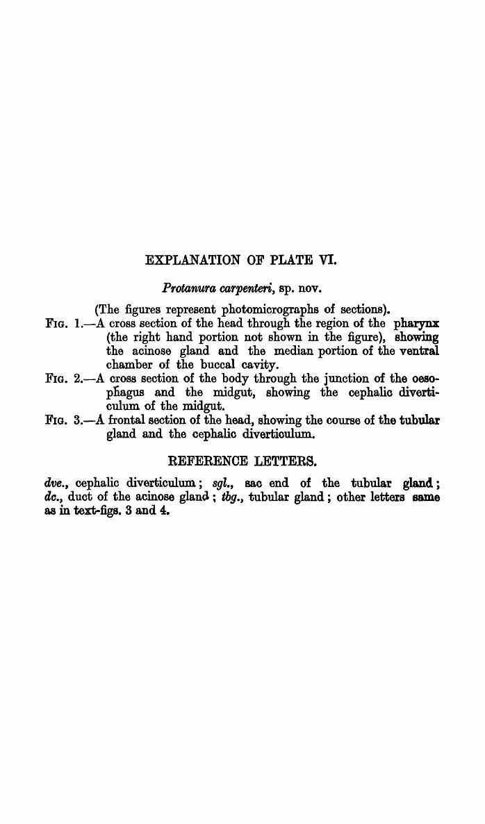
EXPLANATION OF PLATE VI.
Pf'otanUf'a co,f'penteri, ap. nov.
(The figures represent photomicrographs of sections). FIG. l.-A cross section of the head through the region of the pharynx
(the right hand portion not shown in the figure), showing the acinose gland and the median portion of the ventral chamber of the buccal cavity.
FIG. 2.-A cross section of the body through the junction of the oesopnagus and the midgut, showing the cephalic diverticulum of the midgut.
FIG. 3.-A frontal section of the head, showing the course of the tubular gland and the cephalic diverticulum.
REFERENCE LETTERS.
ave., cephalic diverticulum; 89Z., sac end of the tubular gland; do., duct of the acinose glantl; thg., tubular gland; other letters same as in text-figs. 3 and 4.

Rl'~c. IND. Mus., VOL. XXXIV, 1932.
d Ve.. __
~cg:---
Bagehi Photo.
, , mx.
clve,. , , ,
8.
Pro tan u rae a r pen t e r l, 8 p. no,'.
2.

EXPLANATION OF PLATE VII.
P'1otanU'1a oa'1penteri, sp. nov.
(The figures represent photomicrographs of sections). FIG. 1.-A lateral sagittal section through the thoracio.region, showing
the course of the posterior end of the tubular gland in the thorax.
FIG. 2.-A lateral sagittal section through the head a.nd anterior region of the prothorax, showing. the dorsal and ventral lobes of the tubular gland in the region of the head.
FIG. 3.-A median sagittal section of the body passing through the region of the ventral tube, showing the glandular cells in the ventral tube.
FIG. 4.-A cross section of the ventral tube, showing the gland cells .in the inner wall of the ventral tube.
REFERENCE LETTERS.
dl., dorsal loop of the tubular gland; ggl., glandular cells in the ventral tube; ky. hypodermal cells; lbg., labyrinth; mep., epithelium of the midgut; mt., metathoracic ganglion; vl., ventral loop of the tubular gland; vt., ventral tube; other letters same as in text-figs. 3 and 4.

Ee. I~D. lVlus., VOL. XXXIV, 1~L3:'?
I
tog.
2.
/,,~l 1'''''1' v"_ .... c.
,~l;g. .. 1\
" 1\ " \ , \ , , ,
a&.
, , "h:y.
, , \
Pro tan u rae a r pen t p- r;, s p. 1l 0 Y.
PLATE V I I.
1.
CifC!f.