Numerical Characterization of Landing Gear Aeroacoustics ...
Upload
independentCategory
view
1download
0
FOCUS: ION-SURFACE COLLISIONS AND PEPTIDE RADICALCATIONS
Reactive Landing of Gas-Phase Ions as a Toolfor the Fabrication of Metal Oxide Surfaces forIn Situ Phosphopeptide Enrichment
Grady R. Blacken, Michael Volný,* Matthew Diener, Karl E. Jackson,Pratistha Ranjitkar, Dustin J. Maly, and František TurecekDepartment of Chemistry, University of Washington, Seattle, Washington, USA
Zirconium, titanium, and hafnium oxide-coated stainless steel surfaces are fabricated byreactive landing of gas-phase ions produced by electrospray ionization of group IVB metalalkoxides. The surfaces are used for in situ enrichment of phosphopeptides before analysis bymatrix-assisted laser desorption ionization (MALDI) mass spectrometry. To evaluate thismethod we characterized ZrO2 (zirconia) surfaces by (1) comparison with the other group IVBmetal oxides of TiO2 (titania) and HfO2 (hafnia), (2) morphological characterization by SEMimage analysis, and (3) dependence of phosphopeptide enrichment on the metal oxide layerthickness. Furthermore, we evaluated the necessity of the reactive landing process for theconstruction of useful metal oxide surfaces by preparing surfaces by electrospray deposition of Zr,Ti, and Hf alkoxides directly onto polished metal surfaces at atmospheric pressure. Although allthree metal oxide surfaces evaluated were capable of phosphopeptide enrichment from complexpeptide mixtures, zirconia performed better than hafnia or titania as a result of morphologicalcharacteristics illustrated by the SEM analysis. Metal oxide coatings that were fabricated byatmospheric pressure deposition were still capable of in situ phosphopeptide enrichment, althoughwith inferior efficiency and surface durability. We show that zirconia surfaces prepared by reactivelanding of gas-phase ions can be a useful tool for high throughput screening of novel phosphor-ylation sites and quantitation of phosphorylation kinetics. (J Am Soc Mass Spectrom 2009, 20,915–926) © 2009 American Society for Mass Spectrometry
Many cellular processes including metabolism,transcription, and differentiation are largelycontrolled by reversible protein phosphoryla-
tion [1]. Kinases, phosphorylating enzymes, and phos-phatases, dephosphorylating enzymes, are able to mod-ulate phosphorylation of serine, threonine, and tyrosineresidues, causing dynamic changes in protein structureand function. It has been estimated that 30–50% of allproteins in eukaryotic organisms are phosphorylated atany time [2]. Given the extensive role of phosphoryla-tion in the human body, it is perhaps surprising that thehuman genome codes for only about 520 kinases (1–2%of the genome) out of 24,500 coding genes [3]. Of these520 kinases, 244 have been mapped to disease orcancer-causing conditions—thus understanding theirfunction can be of great use for designing therapies andcures for many human ailments.
The typical phosphoproteomics workflow [4] ofteninvolves growing target cells under two conditions: a
Address reprint requests to Dr. František Turecek, University of Washing-ton, Department of Chemistry, Bagley Hall, Box 351700, Seattle, WA98195-1700. E-mail: [email protected]* Present address: Laboratory of Molecular Structure Characterization,
Academy of Sciences of the Czech Republic, Videnska 1083, Prague 4, 14220Czech Republic.© 2009 American Society for Mass Spectrometry. Published by Elsevie1044-0305/09/$32.00doi:10.1016/j.jasms.2009.01.006
control and a perturbed condition [5]. The proteinsformed either can be labeled in the cell culture, usingapproaches such as stable isotopic labeling of aminoacids in cell culture (SILAC) [6], or the purified proteinscan be modified after extraction using isotope-codedaffinity tag (ICAT) based derivatization methods [7, 8].After labeling, the differentially expressed proteins canbe subjected to either global proteomics analysis or atargeted analytical approach. In the former techniquethe extracted proteins are digested and analyzed byliquid chromatography mass spectrometry (LC-MS)without any further purification. This global bottom-upapproach requires either a targeted detection strategyusing tandem mass spectrometry [9–15] or the use ofhigh-resolution, high-capacity mass analyzers.
Although global and top-down proteomics strategiesare becoming increasingly more possible, targeted strat-egies are still necessary to achieve meaningful biologi-cal conclusions [4, 5]. Targeted phosphoproteomicsinvolves several stages of enrichment and selectivepurification to isolate desired biomolecules. The firststage involves phosphoprotein enrichment or immuno-precipitation (IP) to isolate the phosphoproteome. Thisis usually followed by two-dimensional electrophoretic
separation on a sodium dodecyl sulfate polyacrylamidePublished online January 22, 2009r Inc. Received October 31, 2008
Revised January 11, 2009Accepted January 13, 2009
916 BLACKEN ET AL. J Am Soc Mass Spectrom 2009, 20, 915–926
gel (2D-SDS-PAGE) to isolate individual phosphopro-teins or, more likely, groups of proteins that might becontained in a single gel band. These bands can then besliced out and reconstituted for tryptic digestion tosmaller peptides, of 8–14 amino residues, that are moreamenable to MS analysis. Once the target protein orproteins are isolated, the peptides might then be sub-jected to further enrichment to purify the tryptic phos-phopeptides from the complex digest. This purificationstep is often necessary before MS analysis because ofthe high complexity of biological samples, the largedynamic range of phosphorylation, and the typicallylow ionization efficiency of phosphorylated peptidescompared with the nonphosphorylated forms [5]. Phos-phopeptide enrichment has undergone significantgrowth over the past two decades as a result of both thesuccesses and the limitations of immobilized metal-ionaffinity chromatography (IMAC) [16–18]. The IMACstrategy typically involves a metal chelating resin, suchas agarose derivatized with nitrilotriacetic acid (NTA),loaded with a phosphate-selective metal such as Ga3�
or Fe3�. When peptide or protein samples are applied tothis column, the phosphorylated residues are selec-tively retained, whereas the nonphosphorylated formscan be washed away. This technique is rapid, inexpen-sive, and effective and thus remains a very popular toolfor phosphopeptide enrichment in proteomics analyses.
Several limitations exist for IMAC techniques result-ing in sample complexity by nonspecific binding to thecolumn. The main issues include (1) the capture ofpeptide acidic residues such as aspartic acid, glutamicacid, or the C-terminus and (2) capture of hydrophobicresidues by the gel bead. The first problem was initiallyaddressed by Ficarro et al. [17], who demonstrated thatcarboxylate methyl esterification could block the non-specific capture of acidic residues. The derivatizationalso allows for the incorporation of an isotopic labeluseful for quantitation. The technique is widely used,although its continued growth is hampered by the yieldof the derivatization reaction. Specifically, incompletemethyl esterification leads to increased sample com-plexity and signal dilution of individual peptide ions[19]. Thus, the field of phosphoproteomics is still seek-ing to simplify and improve on current techniques forphosphopeptide enrichment.
More recently, metal oxide affinity chromatography(MOAC) has begun to rival IMAC in utility withphosphoproteomic applications. Originally described in2004, titanium dioxide columns were used to selectivelyenrich phosphopeptides from complex samples, includ-ing the Drosophila melanogaster proteome [20, 21]. Zirco-nia, another group IVB metal oxide, has also proved tobe useful as a phosphopeptide enrichment material [22].The benefit of MOAC columns is the ability to with-stand extreme solution conditions, including the use oforganic acids to aid in phosphopeptide binding speci-ficity and low pH binding and washing conditions [23].MOAC columns can be loaded and washed in up to 5%
trifluoroacetic acid (TFA), allowing for efficient proto-nation of peptide carboxylates. IMAC columns, on theother hand, cannot be exposed to low pKa bindingconditions because the NTA moiety will be protonatedand the metal leached from the column. The effect isthat phosphopeptides must be bound to IMAC columnsat pH values near or above the pKa of peptide carboxy-lates, leaving many Asp and Glu residues deprotonatedand capable of nonspecific binding.
Two recent comparative studies illustrate the need toevaluate each enrichment technique for a particularapplication because IMAC and MOAC are both capableof complementary and distinct phosphopeptide enrich-ment. In one study of the D. melanogaster phosphopro-teome, the digested protein sample was subjected tothree separate phosphopeptide enrichment strategies:IMAC, MOAC, and covalent capture via phosphorami-date chemistry [24]. This study found that the threetechniques were able to enrich complementary portionsof the fruit fly phosphoproteome. Alternately, a sepa-rate study has found that TiO2 chromatography wassuperior to both ZrO2 and IMAC [25]. In that study, theenhanced stability of MOAC in several loading andwashing buffers was demonstrated. The advantage ofTiO2 over ZrO2 was not elucidated and was presumablyattributed only to differences in the manufacturingprocess. Regardless, it is clear that the choice of enrich-ment strategy may be an empirical decision unique toeach application.
We have recently reported on the utility of stainlesssteel surfaces coated with a durable layer of zirconia forthe efficient enrichment of phosphopeptides directly onmatrix-assisted laser desorption ionization (MALDI)sample plates [26]. By detecting the phosphopeptidesdirectly from the MALDI surfaces our method elimi-nates complications associated with phosphopeptideelution and allows for automated, high-throughputMALDI analyses. Furthermore, the rapid enrichment onthese surfaces allows for an additional stage of chro-matographic separation to be performed in a matter ofminutes with minimal volumes of solvent. Thus thetechnique presented here will fit well with the online,multistage separations, before MALDI-MS analysis,that have become standard in proteomics [27–29]. Thesurfaces are generated by exposing plasma-oxidizedstainless steel surfaces to collisions with gas-phasecations generated by electrospray ionization (ESI) ofzirconium(IV)-n-propoxide. This robust and highly re-producible surface preparation process lends itself to avariety of applications [30–33].
Here we evaluate the phosphopeptide enrichmentcapabilities of several metal oxide coatings. First, wecompare zirconia with the other group IVB metaloxides titania and hafnia. We use scanning electronmicroscope (SEM) image analysis to show how en-richment may be related to surface morphology.Second, we evaluate the effect of metal oxide surfacethickness on phosphopeptide enrichment. Finally, we
compare our soft and reactive-landing preparation
917J Am Soc Mass Spectrom 2009, 20, 915–926 METAL OXIDE SURFACES BY REACTIVE ION LANDING
technique with a simplified, bench-top method formetal oxide surface preparation.
Experimental
Materials
Titanium(IV)-n-butoxide, zirconium(IV)-n-propoxide,and hafnium(IV)-n-propoxide were obtained from Sig-ma–Aldrich (Milwaukee, WI, USA) as 70% solutions in1-propanol. Solvents were purchased from Fisher Sci-entific (Pittsburgh, PA, USA). The peptides used tocharacterize phosphopeptide enrichment on the zirco-nium oxide functional surfaces include the syntheticpeptides NQLLpTPLR, MpSGIFR, MSGIFR, and pY-WQAFR, purchased from Genscript Corporation (Pis-cataway, NJ, USA), and peptides from the tryptic diges-tion of �-casein. Sequence-grade trypsin and �-caseinwere purchased from Sigma–Aldrich. The tryptic diges-tion of �-casein was performed at a 1:20 enzyme-to-substrate weight ratio in 50 mM NH4HCO3 and incu-bated overnight at 37 °C. Organic acids used as MALDImatrixes (�-cyano-4-hydroxycinnamic acid [CHCA]and 2,5-dihydroxybenzoic acid [DHB]) were purchasedfrom Sigma–Aldrich.
MALDI-TOF/TOF Mass Spectrometry
MALDI mass spectra were acquired on an AppliedBiosystems 4700 Proteomics Analyzer system (AppliedBiosystems, Carlsbad, CA, USA), operated in positive-ion mode. Optimum precursor ion signals in MALDI/time-of-flight (TOF)-MS analyses, operated with thereflector on, were acquired with matrix solutions con-sisting of either 1 mg/mL CHCA in 50% acetonitrilewith 1% phosphoric acid (PA) or 1 mg/mL DHB in50% acetonitrile with 1% PA. The relatively diluteconcentrations of organic matrix are necessary tocreate a thin crystal layer so that phosphopeptidesbound to the metal oxide surface may be accessible tolaser desorption.
Alteration of Standard MALDI Plates
A standard stainless steel MALDI plate was modifiedby milling off several layers of material from the area ofabout 20 � 20 mm and 2 mm depth to accommodatethe Zr-oxide– coated substrate plates. The substrateplate was mounted inside the depression with adhe-sive tape and thus leveled with the rest of the MALDIplate surface. In principle, reactive landing could bedone on the MALDI plate directly, without having tomount externally prepared surfaces. Reasons for ourcurrent approach include the higher price of commer-cial MALDI plates and the fact that smaller chips ofcut stainless steel are easier to handle inside thesoft-landing instrument, which has relatively narrow
chambers [30].Scanning Electron Microscopy (SEM)
Images were acquired on the FEI Sirion SEM system inthe nanotechnology user facility at the University ofWashington. Before analysis, gold was sputter-coatedonto the surface for 30 s at a rate of 7 nm/min to builda 3.5-nm-thick conducting layer to account for the lowconductivity of the metal oxide. Optimal resolutionoccurred using a 7-keV incident electron beam. Anenergy-dispersive X-ray (EDX) photon detector equippedon the SEM system allowed for localization of metal oxideon the stainless steel surfaces.
Surfaces
Metal targets used as substrates for deposition of groupIVB metal dioxides were made of roughly 15 � 15-mm316L stainless steel plates. The plates were mechanicallypolished with a diamond paste; rinsed successivelywith hexane, methanol, and water; sonicated in 1:1chloroform/methanol for 15 min; rinsed with meth-anol; and exposed to oxygen plasma immediatelyafter cleaning.
In Situ Plasma Treatment
The custom-made plasma reactor is similar to thatdescribed by Ratner [34] and was operated at 13.56MHz. It is an integral part of the soft and reactivelanding instrument as described previously [30]. Thesurface treatment was carried out at 60-W radio-fre-quency power for 10 min in 250 mTorr of flowingoxygen gas.
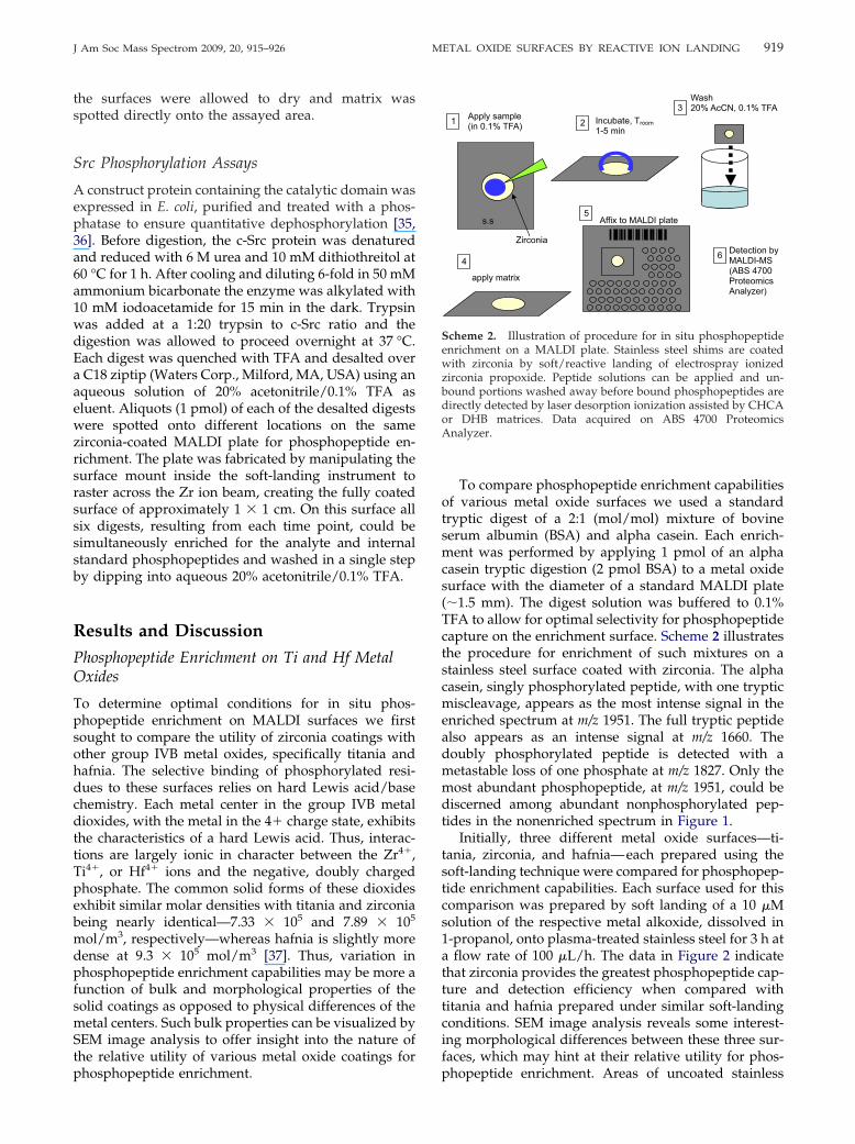
Preparation of Surfaces by Reactive Landing
For reactive landing experiments, 70% metal alkoxide in1-propanol was diluted in the same solvent to the finalconcentration of 10 �M. The solution was electro-sprayed in positive mode and the ions were landed ona freshly plasma-treated stainless steel surface. Thelanding experiment lasted between 1.5 and 6 h and thelanding substrate was biased at �50 V, correspondingto a landing energy of 50 eV for singly charged ions. Thegas-phase ions consisted of a mixture of alkoxylated Zr,Ti, or Hf clusters, similar to those reported for negative-ion electrospray of Zr(IV) alkoxides. The cations werenot mass-separated before reactive landing. The samplesurface was then removed from the instrument andfurther examined. The landed material forms a compactspot that is visible to the naked eye (Figure 1, bottom,inset). The spot was successively rinsed with water,methanol, propanol, and hexane and then soaked in awater/propanol mixture for 4 h. All samples that wereprepared and used in this study did not visibly changeupon multiple washings. The Zr-oxide coating re-mained intact even after it was rubbed with wet soft
material such as filtration paper or cotton wool.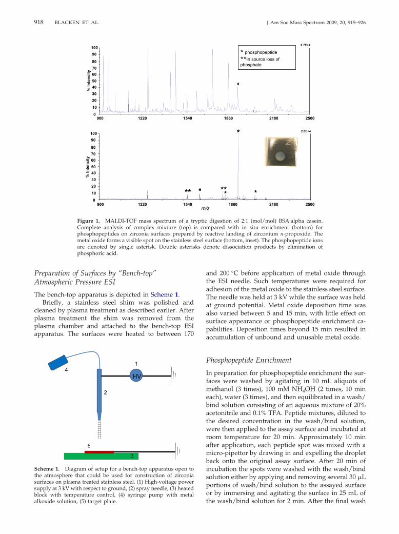
918 BLACKEN ET AL. J Am Soc Mass Spectrom 2009, 20, 915–926
Preparation of Surfaces by “Bench-top”Atmospheric Pressure ESI
The bench-top apparatus is depicted in Scheme 1.Briefly, a stainless steel shim was polished and
cleaned by plasma treatment as described earlier. Afterplasma treatment the shim was removed from theplasma chamber and attached to the bench-top ESIapparatus. The surfaces were heated to between 170
010
20304050607080
90100
% In
tens
ity
15401220900
**0
1020304050607080
90100
% In
tens
ity
15401220900
Figure 1. MALDI-TOF mass spectrum of a trComplete analysis of complex mixture (top)phosphopeptides on zirconia surfaces preparedmetal oxide forms a visible spot on the stainless sare denoted by single asterisk. Double asterisphosphoric acid.
HV
1
2
3
4
5
Scheme 1. Diagram of setup for a bench-top apparatus open tothe atmosphere that could be used for construction of zirconiasurfaces on plasma treated stainless steel. (1) High-voltage powersupply at 3 kV with respect to ground, (2) spray needle, (3) heatedblock with temperature control, (4) syringe pump with metal
alkoxide solution, (5) target plate.and 200 °C before application of metal oxide throughthe ESI needle. Such temperatures were required foradhesion of the metal oxide to the stainless steel surface.The needle was held at 3 kV while the surface was heldat ground potential. Metal oxide deposition time wasalso varied between 5 and 15 min, with little effect onsurface appearance or phosphopeptide enrichment ca-pabilities. Deposition times beyond 15 min resulted inaccumulation of unbound and unusable metal oxide.
Phosphopeptide Enrichment
In preparation for phosphopeptide enrichment the sur-faces were washed by agitating in 10 mL aliquots ofmethanol (3 times), 100 mM NH4OH (2 times, 10 mineach), water (3 times), and then equilibrated in a wash/bind solution consisting of an aqueous mixture of 20%acetonitrile and 0.1% TFA. Peptide mixtures, diluted tothe desired concentration in the wash/bind solution,were then applied to the assay surface and incubated atroom temperature for 20 min. Approximately 10 minafter application, each peptide spot was mixed with amicro-pipettor by drawing in and expelling the dropletback onto the original assay surface. After 20 min ofincubation the spots were washed with the wash/bindsolution either by applying and removing several 30 �Lportions of wash/bind solution to the assayed surfaceor by immersing and agitating the surface in 25 mL of
*
* phosphopeptide **In source loss of phosphate
9.7E+4
250021801860
*
*****
2.0E+4
z250021801860
digestion of 2:1 (mol/mol) BSA:alpha casein.mpared with in situ enrichment (bottom) foreactive landing of zirconium n-propoxide. Theurface (bottom, inset). The phosphopeptide ionsenote dissociation products by elimination of
*
m/
ypticis co
by rteel sks d
the wash/bind solution for 2 min. After the final wash
919J Am Soc Mass Spectrom 2009, 20, 915–926 METAL OXIDE SURFACES BY REACTIVE ION LANDING
the surfaces were allowed to dry and matrix wasspotted directly onto the assayed area.
Src Phosphorylation Assays
A construct protein containing the catalytic domain wasexpressed in E. coli, purified and treated with a phos-phatase to ensure quantitative dephosphorylation [35,36]. Before digestion, the c-Src protein was denaturedand reduced with 6 M urea and 10 mM dithiothreitol at60 °C for 1 h. After cooling and diluting 6-fold in 50 mMammonium bicarbonate the enzyme was alkylated with10 mM iodoacetamide for 15 min in the dark. Trypsinwas added at a 1:20 trypsin to c-Src ratio and thedigestion was allowed to proceed overnight at 37 °C.Each digest was quenched with TFA and desalted overa C18 ziptip (Waters Corp., Milford, MA, USA) using anaqueous solution of 20% acetonitrile/0.1% TFA aseluent. Aliquots (1 pmol) of each of the desalted digestswere spotted onto different locations on the samezirconia-coated MALDI plate for phosphopeptide en-richment. The plate was fabricated by manipulating thesurface mount inside the soft-landing instrument toraster across the Zr ion beam, creating the fully coatedsurface of approximately 1 � 1 cm. On this surface allsix digests, resulting from each time point, could besimultaneously enriched for the analyte and internalstandard phosphopeptides and washed in a single stepby dipping into aqueous 20% acetonitrile/0.1% TFA.
Results and Discussion
Phosphopeptide Enrichment on Ti and Hf MetalOxides
To determine optimal conditions for in situ phos-phopeptide enrichment on MALDI surfaces we firstsought to compare the utility of zirconia coatings withother group IVB metal oxides, specifically titania andhafnia. The selective binding of phosphorylated resi-dues to these surfaces relies on hard Lewis acid/basechemistry. Each metal center in the group IVB metaldioxides, with the metal in the 4� charge state, exhibitsthe characteristics of a hard Lewis acid. Thus, interac-tions are largely ionic in character between the Zr4�,Ti4�, or Hf4� ions and the negative, doubly chargedphosphate. The common solid forms of these dioxidesexhibit similar molar densities with titania and zirconiabeing nearly identical—7.33 � 105 and 7.89 � 105
mol/m3, respectively—whereas hafnia is slightly moredense at 9.3 � 105 mol/m3 [37]. Thus, variation inphosphopeptide enrichment capabilities may be more afunction of bulk and morphological properties of thesolid coatings as opposed to physical differences of themetal centers. Such bulk properties can be visualized bySEM image analysis to offer insight into the nature ofthe relative utility of various metal oxide coatings for
phosphopeptide enrichment.To compare phosphopeptide enrichment capabilitiesof various metal oxide surfaces we used a standardtryptic digest of a 2:1 (mol/mol) mixture of bovineserum albumin (BSA) and alpha casein. Each enrich-ment was performed by applying 1 pmol of an alphacasein tryptic digestion (2 pmol BSA) to a metal oxidesurface with the diameter of a standard MALDI plate(�1.5 mm). The digest solution was buffered to 0.1%TFA to allow for optimal selectivity for phosphopeptidecapture on the enrichment surface. Scheme 2 illustratesthe procedure for enrichment of such mixtures on astainless steel surface coated with zirconia. The alphacasein, singly phosphorylated peptide, with one trypticmiscleavage, appears as the most intense signal in theenriched spectrum at m/z 1951. The full tryptic peptidealso appears as an intense signal at m/z 1660. Thedoubly phosphorylated peptide is detected with ametastable loss of one phosphate at m/z 1827. Only themost abundant phosphopeptide, at m/z 1951, could bediscerned among abundant nonphosphorylated pep-tides in the nonenriched spectrum in Figure 1.
Initially, three different metal oxide surfaces—ti-tania, zirconia, and hafnia—each prepared using thesoft-landing technique were compared for phosphopep-tide enrichment capabilities. Each surface used for thiscomparison was prepared by soft landing of a 10 �Msolution of the respective metal alkoxide, dissolved in1-propanol, onto plasma-treated stainless steel for 3 h ata flow rate of 100 �L/h. The data in Figure 2 indicatethat zirconia provides the greatest phosphopeptide cap-ture and detection efficiency when compared withtitania and hafnia prepared under similar soft-landingconditions. SEM image analysis reveals some interest-ing morphological differences between these three sur-faces, which may hint at their relative utility for phos-
Wash 20% AcCN, 0.1% TFA
Incubate, Troom1-5 min
Apply sample (in 0.1% TFA)
apply matrix
1 2
3
4
Affix to MALDI plate 5
s.s
Zirconia
6 Detection by MALDI-MS (ABS 4700 Proteomics Analyzer)
Scheme 2. Illustration of procedure for in situ phosphopeptideenrichment on a MALDI plate. Stainless steel shims are coatedwith zirconia by soft/reactive landing of electrospray ionizedzirconia propoxide. Peptide solutions can be applied and un-bound portions washed away before bound phosphopeptides aredirectly detected by laser desorption ionization assisted by CHCAor DHB matrices. Data acquired on ABS 4700 ProteomicsAnalyzer.
phopeptide enrichment. Areas of uncoated stainless
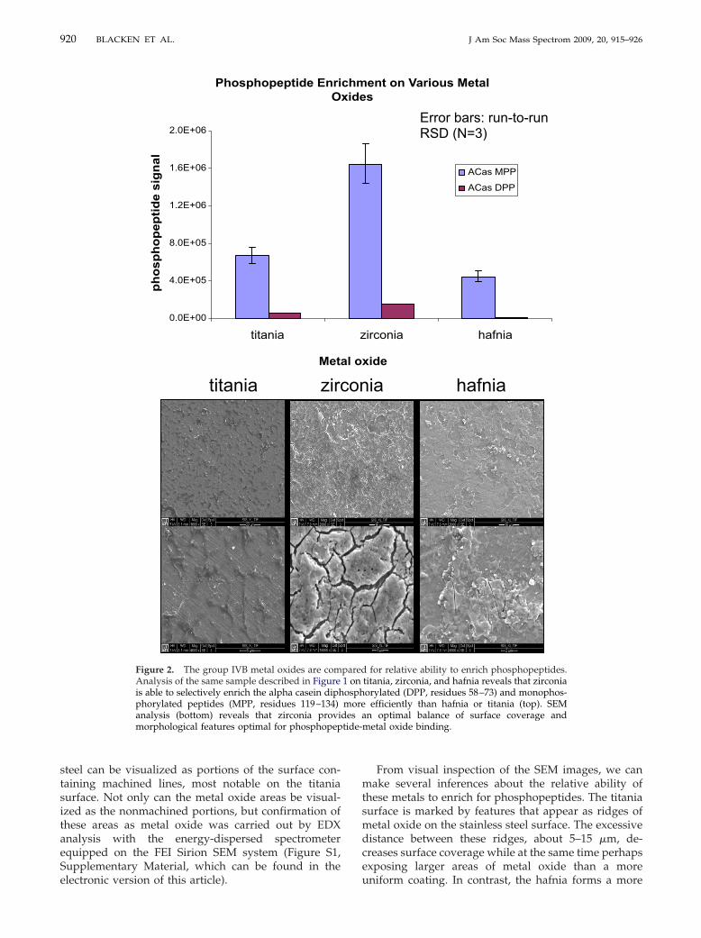
tide-
920 BLACKEN ET AL. J Am Soc Mass Spectrom 2009, 20, 915–926
steel can be visualized as portions of the surface con-taining machined lines, most notable on the titaniasurface. Not only can the metal oxide areas be visual-ized as the nonmachined portions, but confirmation ofthese areas as metal oxide was carried out by EDXanalysis with the energy-dispersed spectrometerequipped on the FEI Sirion SEM system (Figure S1,Supplementary Material, which can be found in the
titania zirc
Phosphopeptide EnriO
0.0E+00
4.0E+05
8.0E+05
1.2E+06
1.6E+06
2.0E+06
titania
Met
phos
phop
eptid
e si
gnal
Figure 2. The group IVB metal oxides are comAnalysis of the same sample described in Figureis able to selectively enrich the alpha casein diphphorylated peptides (MPP, residues 119–134)analysis (bottom) reveals that zirconia provimorphological features optimal for phosphopep
electronic version of this article).
From visual inspection of the SEM images, we canmake several inferences about the relative ability ofthese metals to enrich for phosphopeptides. The titaniasurface is marked by features that appear as ridges ofmetal oxide on the stainless steel surface. The excessivedistance between these ridges, about 5–15 �m, de-creases surface coverage while at the same time perhapsexposing larger areas of metal oxide than a more
ia hafnia
ent on Various Metals
irconia hafnia
xide
ACas MPP
ACas DPP
Error bars: run-to-run RSD (N=3)
for relative ability to enrich phosphopeptides.titania, zirconia, and hafnia reveals that zirconiaorylated (DPP, residues 58–73) and monophos-
efficiently than hafnia or titania (top). SEMn optimal balance of surface coverage and
metal oxide binding.
on
chmxide
z
al o
pared1 onosphmore
des a
uniform coating. In contrast, the hafnia forms a more
921J Am Soc Mass Spectrom 2009, 20, 915–926 METAL OXIDE SURFACES BY REACTIVE ION LANDING
continuous surface layer, providing more surface cov-erage than titania but perhaps still a comparable metaloxide surface area because of the presence of fewermorphological features. By comparison, zirconia ap-pears to offer an optimal balance of surface coverageand metal oxide surface area. The increased presence ofcracks and pores relative to the hafnia surface and theimproved surface coverage relative to the titania surfacemake zirconia more desirable for phosphopeptide en-richment, as a result of increased capacity and optimalchromatography. Indeed the nature of the differences inmetal oxide structure between these three surfaces islikely specific to the soft-landing method of metal oxidesurface preparation. This is probably true of all metaloxide preparation techniques in that it has been ob-served that the relative enrichment performance ofmetal oxides is dependent on the manufacturer andmanufacturing process.
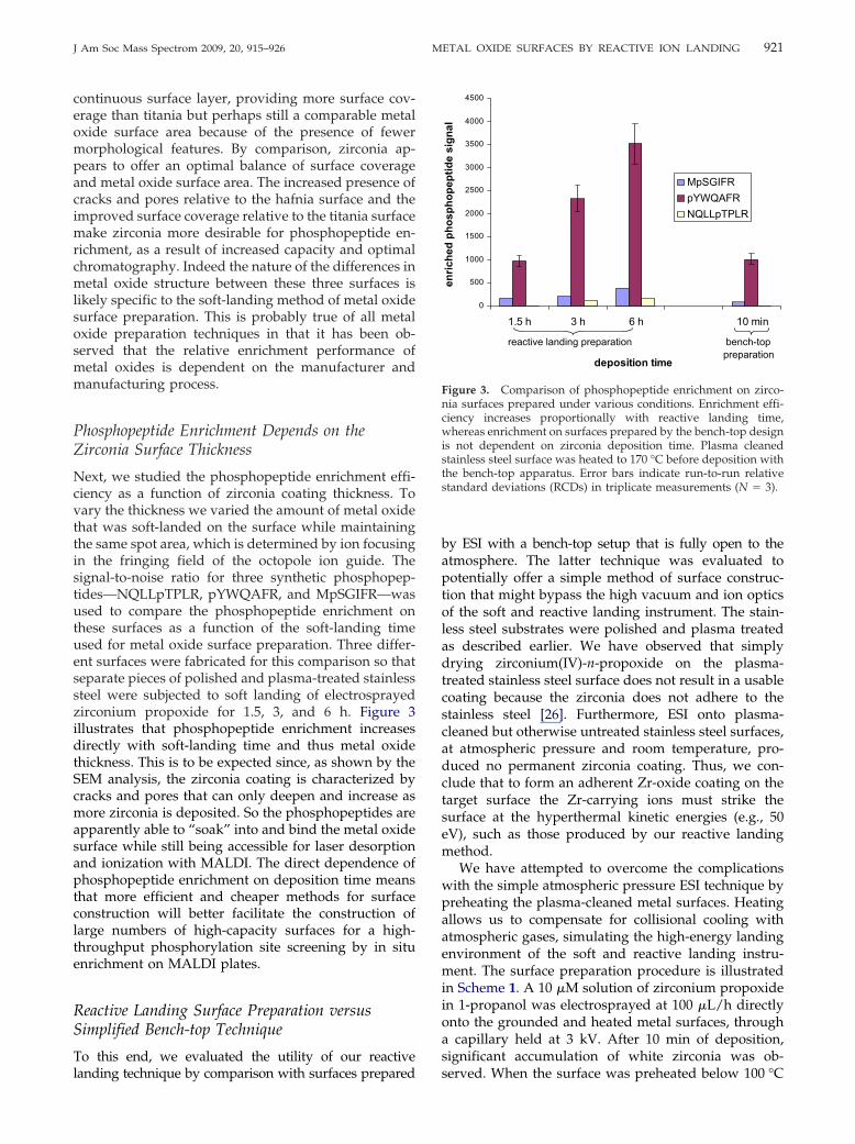
Phosphopeptide Enrichment Depends on theZirconia Surface Thickness
Next, we studied the phosphopeptide enrichment effi-ciency as a function of zirconia coating thickness. Tovary the thickness we varied the amount of metal oxidethat was soft-landed on the surface while maintainingthe same spot area, which is determined by ion focusingin the fringing field of the octopole ion guide. Thesignal-to-noise ratio for three synthetic phosphopep-tides—NQLLpTPLR, pYWQAFR, and MpSGIFR—wasused to compare the phosphopeptide enrichment onthese surfaces as a function of the soft-landing timeused for metal oxide surface preparation. Three differ-ent surfaces were fabricated for this comparison so thatseparate pieces of polished and plasma-treated stainlesssteel were subjected to soft landing of electrosprayedzirconium propoxide for 1.5, 3, and 6 h. Figure 3illustrates that phosphopeptide enrichment increasesdirectly with soft-landing time and thus metal oxidethickness. This is to be expected since, as shown by theSEM analysis, the zirconia coating is characterized bycracks and pores that can only deepen and increase asmore zirconia is deposited. So the phosphopeptides areapparently able to “soak” into and bind the metal oxidesurface while still being accessible for laser desorptionand ionization with MALDI. The direct dependence ofphosphopeptide enrichment on deposition time meansthat more efficient and cheaper methods for surfaceconstruction will better facilitate the construction oflarge numbers of high-capacity surfaces for a high-throughput phosphorylation site screening by in situenrichment on MALDI plates.
Reactive Landing Surface Preparation versusSimplified Bench-top Technique
To this end, we evaluated the utility of our reactive
landing technique by comparison with surfaces preparedby ESI with a bench-top setup that is fully open to theatmosphere. The latter technique was evaluated topotentially offer a simple method of surface construc-tion that might bypass the high vacuum and ion opticsof the soft and reactive landing instrument. The stain-less steel substrates were polished and plasma treatedas described earlier. We have observed that simplydrying zirconium(IV)-n-propoxide on the plasma-treated stainless steel surface does not result in a usablecoating because the zirconia does not adhere to thestainless steel [26]. Furthermore, ESI onto plasma-cleaned but otherwise untreated stainless steel surfaces,at atmospheric pressure and room temperature, pro-duced no permanent zirconia coating. Thus, we con-clude that to form an adherent Zr-oxide coating on thetarget surface the Zr-carrying ions must strike thesurface at the hyperthermal kinetic energies (e.g., 50eV), such as those produced by our reactive landingmethod.
We have attempted to overcome the complicationswith the simple atmospheric pressure ESI technique bypreheating the plasma-cleaned metal surfaces. Heatingallows us to compensate for collisional cooling withatmospheric gases, simulating the high-energy landingenvironment of the soft and reactive landing instru-ment. The surface preparation procedure is illustratedin Scheme 1. A 10 �M solution of zirconium propoxidein 1-propanol was electrosprayed at 100 �L/h directlyonto the grounded and heated metal surfaces, througha capillary held at 3 kV. After 10 min of deposition,significant accumulation of white zirconia was ob-
0
500
1000
1500
2000
2500
3000
3500
4000
4500
1.5 h 3 h 6 h 10 min
deposition time
enric
hed
phos
phop
eptid
e si
gnal
MpSGIFRpYWQAFRNQLLpTPLR
reactive landing preparation bench-top preparation
Figure 3. Comparison of phosphopeptide enrichment on zirco-nia surfaces prepared under various conditions. Enrichment effi-ciency increases proportionally with reactive landing time,whereas enrichment on surfaces prepared by the bench-top designis not dependent on zirconia deposition time. Plasma cleanedstainless steel surface was heated to 170 °C before deposition withthe bench-top apparatus. Error bars indicate run-to-run relativestandard deviations (RCDs) in triplicate measurements (N � 3).
served. When the surface was preheated below 100 °C
922 BLACKEN ET AL. J Am Soc Mass Spectrom 2009, 20, 915–926
the zirconia layer durability was significantly dimin-ished and was not retained on the surface. Heating thesurfaces to between 170 and 200 °C resulted in func-tional surfaces that could be used for phosphopeptideenrichment.
Within this range of preheated substrate tempera-tures used for zirconia surface preparation, the phos-phopeptide capture efficiency of the respective zirconiacoatings was observed to decrease with increasingtemperature. SEM image analysis shows a more amor-phous physical structure for the coatings prepared athigher temperature (Figure S2). The relative phos-phopeptide enrichment efficiencies of these surfacesprepared under various conditions are also illustratedin Figure 3. This chart shows that phosphopeptideenrichment on the surfaces prepared with the bench-toptechnique compares with enrichment on the thinnestand least efficient zirconia surfaces prepared by reactivelanding. Furthermore, enrichment capabilities for thesurfaces prepared by the simplified technique werenot found to vary with deposition time beyond 5 minof electrospray coating at atmospheric pressure. Thus,the enrichment efficiency could not be improved byincreasing deposition time with the bench-top setup asit was possible under reactive landing conditions. Thisis probably attributable to the different structure ofthe material formed by the atmospheric pressuremethod. Phosphopeptide enrichment on the surfacesprepared by the bench-top ESI method may be usefulfor applications requiring a phosphorylation sitescreening of samples of relatively low complexityor those known to contain high-abundance phos-phopeptides. Conversely, the reactive landing condi-tions for zirconia surface preparation will provide themore efficient, higher-capacity phosphopeptide en-richment surface.
Quantification of Kinase Kinetics Using In SituPhosphopeptide Enrichment
We demonstrate the utility of phosphopeptide enrich-ment on zirconia-coated MALDI plates by quantitativemeasurements of the effects of various stimuli on phos-phorylation kinetics. One example of this is the screen-ing of small-molecule kinase inhibitors that may affectenzyme activity and thus allow for control of variouscellular processes. Numerous methods currently existfor such studies that typically include scintillation-based detection of phosphorylation events derivedfrom incorporation of radioactive, 32P-labeled adeno-sine 5=-triphosphate (ATP). Although typically verysensitive, these methods give no information aboutphosphorylation site location and carry the addedcosts and hazards associated with the use of radioac-tive materials. Thus, more informative and less haz-ardous quantitation by MS is advantageous to these
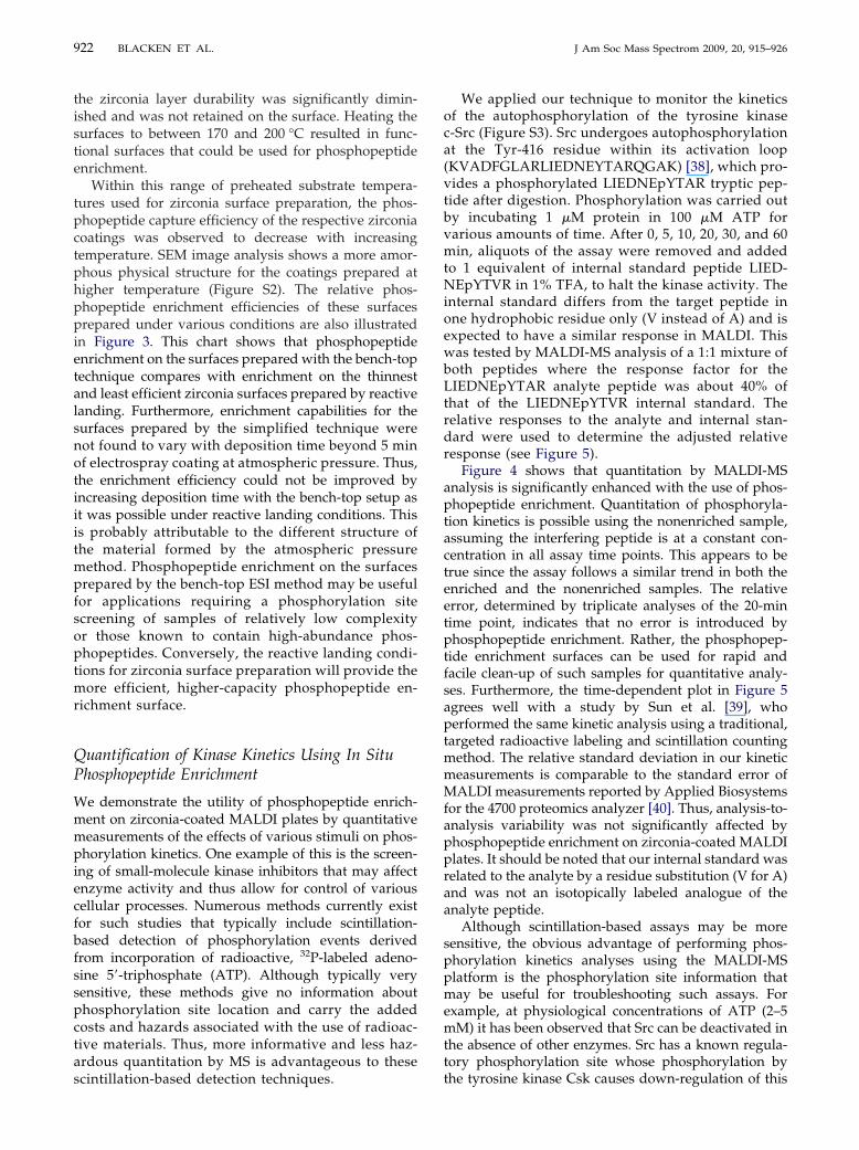
scintillation-based detection techniques.We applied our technique to monitor the kineticsof the autophosphorylation of the tyrosine kinasec-Src (Figure S3). Src undergoes autophosphorylationat the Tyr-416 residue within its activation loop(KVADFGLARLIEDNEYTARQGAK) [38], which pro-vides a phosphorylated LIEDNEpYTAR tryptic pep-tide after digestion. Phosphorylation was carried outby incubating 1 �M protein in 100 �M ATP forvarious amounts of time. After 0, 5, 10, 20, 30, and 60min, aliquots of the assay were removed and addedto 1 equivalent of internal standard peptide LIED-NEpYTVR in 1% TFA, to halt the kinase activity. Theinternal standard differs from the target peptide inone hydrophobic residue only (V instead of A) and isexpected to have a similar response in MALDI. Thiswas tested by MALDI-MS analysis of a 1:1 mixture ofboth peptides where the response factor for theLIEDNEpYTAR analyte peptide was about 40% ofthat of the LIEDNEpYTVR internal standard. Therelative responses to the analyte and internal stan-dard were used to determine the adjusted relativeresponse (see Figure 5).
Figure 4 shows that quantitation by MALDI-MSanalysis is significantly enhanced with the use of phos-phopeptide enrichment. Quantitation of phosphoryla-tion kinetics is possible using the nonenriched sample,assuming the interfering peptide is at a constant con-centration in all assay time points. This appears to betrue since the assay follows a similar trend in both theenriched and the nonenriched samples. The relativeerror, determined by triplicate analyses of the 20-mintime point, indicates that no error is introduced byphosphopeptide enrichment. Rather, the phosphopep-tide enrichment surfaces can be used for rapid andfacile clean-up of such samples for quantitative analy-ses. Furthermore, the time-dependent plot in Figure 5agrees well with a study by Sun et al. [39], whoperformed the same kinetic analysis using a traditional,targeted radioactive labeling and scintillation countingmethod. The relative standard deviation in our kineticmeasurements is comparable to the standard error ofMALDI measurements reported by Applied Biosystemsfor the 4700 proteomics analyzer [40]. Thus, analysis-to-analysis variability was not significantly affected byphosphopeptide enrichment on zirconia-coated MALDIplates. It should be noted that our internal standard wasrelated to the analyte by a residue substitution (V for A)and was not an isotopically labeled analogue of theanalyte peptide.
Although scintillation-based assays may be moresensitive, the obvious advantage of performing phos-phorylation kinetics analyses using the MALDI-MSplatform is the phosphorylation site information thatmay be useful for troubleshooting such assays. Forexample, at physiological concentrations of ATP (2–5mM) it has been observed that Src can be deactivated inthe absence of other enzymes. Src has a known regula-tory phosphorylation site whose phosphorylation by
the tyrosine kinase Csk causes down-regulation of this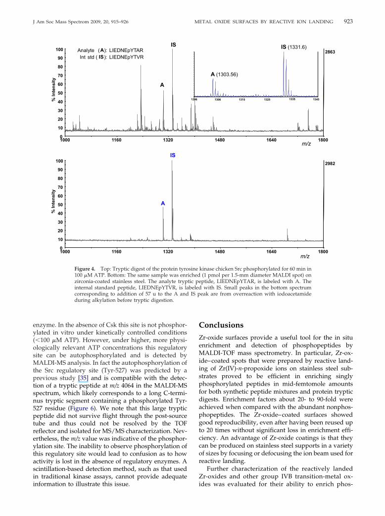
923J Am Soc Mass Spectrom 2009, 20, 915–926 METAL OXIDE SURFACES BY REACTIVE ION LANDING
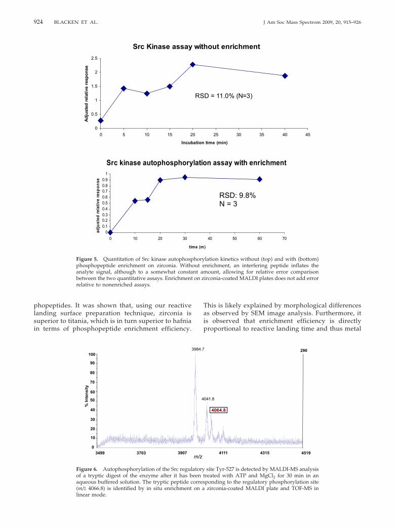
enzyme. In the absence of Csk this site is not phosphor-ylated in vitro under kinetically controlled conditions(�100 �M ATP). However, under higher, more physi-ologically relevant ATP concentrations this regulatorysite can be autophosphorylated and is detected byMALDI-MS analysis. In fact the autophosphorylation ofthe Src regulatory site (Tyr-527) was predicted by aprevious study [35] and is compatible with the detec-tion of a tryptic peptide at m/z 4064 in the MALDI-MSspectrum, which likely corresponds to a long C-termi-nus tryptic segment containing a phosphorylated Tyr-527 residue (Figure 6). We note that this large trypticpeptide did not survive flight through the post-sourcetube and thus could not be resolved by the TOFreflector and isolated for MS/MS characterization. Nev-ertheless, the m/z value was indicative of the phosphor-ylation site. The inability to observe phosphorylation ofthis regulatory site would lead to confusion as to howactivity is lost in the absence of regulatory enzymes. Ascintillation-based detection method, such as that usedin traditional kinase assays, cannot provide adequate
1000 1160 13200
10
20
30
40
50
60
70
80
90
100%
Inte
nsity
IS
A
IS
A
IS
A
over
alk
ylat
ion
Analyte (A): LIEDNEpYTARInt std ( IS): LIEDNEpYTVR
After Enrichment
1000 1160 13200
10
20
30
40
50
60
70
80
90
100
% In
tens
ity
A
Figure 4. Top: Tryptic digest of the protein tyro100 �M ATP. Bottom: The same sample was enrzirconia-coated stainless steel. The analyte trypinternal standard peptide, LIEDNEpYTVR, is lacorresponding to addition of 57 u to the A andduring alkylation before tryptic digestion.
information to illustrate this issue.
Conclusions
Zr-oxide surfaces provide a useful tool for the in situenrichment and detection of phosphopeptides byMALDI-TOF mass spectrometry. In particular, Zr-ox-ide–coated spots that were prepared by reactive land-ing of Zr(IV)-n-propoxide ions on stainless steel sub-strates proved to be efficient in enriching singlyphosphorylated peptides in mid-femtomole amountsfor both synthetic peptide mixtures and protein trypticdigests. Enrichment factors about 20- to 90-fold wereachieved when compared with the abundant nonphos-phopeptides. The Zr-oxide–coated surfaces showedgood reproducibility, even after having been reused upto 20 times without significant loss in enrichment effi-ciency. An advantage of Zr-oxide coatings is that theycan be produced on stainless steel supports in a varietyof sizes by focusing or defocusing the ion beam used forreactive landing.
Further characterization of the reactively landedZr-oxides and other group IVB transition-metal ox-
1480 1640 1800m/z
2863IS (1331.6)
A (1303.56)
mis
clea
vage
1306 1315 1325 1335 1345
1480 1640 1800m/z
2982
kinase chicken Src phosphorylated for 60 min in(1 pmol per 1.5-mm diameter MALDI spot) onptide, LIEDNEpYTAR, is labeled with A. Thewith IS. Small peaks in the bottom spectrum
eak are from overreaction with iodoacetamide
1296
over
alk
ylat
ion
sineichedtic pebeledIS p
ides was evaluated for their ability to enrich phos-
924 BLACKEN ET AL. J Am Soc Mass Spectrom 2009, 20, 915–926
phopeptides. It was shown that, using our reactivelanding surface preparation technique, zirconia issuperior to titania, which is in turn superior to hafniain terms of phosphopeptide enrichment efficiency.
Src Kinase assay
0
0.5
1
1.5
2
2.5
0 5 10 15 2
Incub
Adju
sted
rela
tive
resp
onse
Src kinase autophosphory
00.10.20.30.40.50.60.70.80.9
1
0 10 20 30
ti
adju
sted
rela
tive
resp
onse
Figure 5. Quantitation of Src kinase autophospphosphopeptide enrichment on zirconia. Withanalyte signal, although to a somewhat constabetween the two quantitative assays. Enrichmentrelative to nonenriched assays.
% In
tens
ity
60
50
40
30
20
10
0
70
80
90
100
390737033499
Figure 6. Autophosphorylation of the Src regulof a tryptic digest of the enzyme after it has baqueous buffered solution. The tryptic peptide c(m/z 4066.8) is identified by in situ enrichment
linear mode.This is likely explained by morphological differencesas observed by SEM image analysis. Furthermore, itis observed that enrichment efficiency is directlyproportional to reactive landing time and thus metal
out enrichment
25 30 35 40 45
time (min)
D = 11.0% (N=3)
n assay with enrichment
40 50 60 70
)
RSD: 9.8% N = 3
lation kinetics without (top) and with (bottom)nrichment, an interfering peptide inflates the
ount, allowing for relative error comparisonirconia-coated MALDI plates does not add error
4519
4064.8
041.8
43154111
290
site Tyr-527 is detected by MALDI-MS analysisreated with ATP and MgCl2 for 30 min in anponding to the regulatory phosphorylation sitezirconia-coated MALDI plate and TOF-MS in
with
0
ation
RS
latio
me (m
horyout ent amon z
m/z
4
3984.7
atoryeen torreson a
925J Am Soc Mass Spectrom 2009, 20, 915–926 METAL OXIDE SURFACES BY REACTIVE ION LANDING
oxide thickness. This trend likely illustrates the abil-ity of analyte molecules to diffuse into the metaloxide layer, thus accessing a larger surface area thanif diffusion were not possible. Since the enrichmentefficiency of these zirconia surfaces depends on thereactive landing deposition time, we then also at-tempted to make a rapid and simplified, bench-topsurface construction technique. Phosphopeptideswere in fact enriched on these surfaces, although a�3-fold increase in efficiency was achieved using thereactively landed zirconia surfaces.
Finally, we applied our in situ phosphopeptideenrichment strategy to quantitative phosphorylationkinetics analysis. The zirconia surfaces are able toprovide rapid and selective phosphopeptide informa-tion that will allow for high-throughput phosphory-lation site screening with MALDI-TOF/TOF detec-tion. Quantitation of phosphorylation kinetics isenhanced by enrichment on zirconia surfaces. Asshown by data from Src autophosphorylation assays,the in situ phosphopeptide enrichment techniqueallows for robust and reproducible quantitative infor-mation to be gathered. This potentially allows forhigh-throughput screening of small molecules to tar-get the activity of kinases whose functions may beassociated with disease or cancer.
AcknowledgmentsSupport by grants for the University of Washington RoyaltyResearch Fund and National Institutes of Health (Grant DK-67869)is gratefully acknowledged.
Appendix ASupplementary Material
Supplementary material associated with this articlemay be found in the online version at doi:10.1016/j.jasms.2009.01.006.
References1. Hunter, T. Signaling: 2000 and Beyond. Cell 2000, 100, 113–127.2. Kalume, D. E.; Molina, H.; Pandey, A. A. Tackling the Phosphopro-
teome: Tools and Strategies. Curr. Opin. Chem. Biol. 2003, 7, 64–69.3. Manning, G.; Whyte, D. B.; Martinez, R.; Hunter, T.; Sudarsanam, S. The
Protein Kinase Complement of the Human Genome. Science 2002, 298,1912–1934.
4. Paradela, A.; Albar, J. P. Advances in the Analysis of Protein Phosphor-ylation. J. Proteome Res. 2008, 7, 1809–1818.
5. Smith, J. C.; Figeys, D. Proteomics Technology in Systems Biology. Mol.BioSyst. 2006, 2, 364–370.
6. Ong, S. E.; Blagoev, B.; Kratchmarova, I.; Kristensen, D. B.; Steen, H.;Pandey, A.; Mann, M. Stable Isotope Labeling by Amino Acids in CellCulture, SILAC, as a Simple and Accurate Approach to ExpressionProteomics. Mol. Cell Proteomics 2002, 1, 376–386.
7. Gygi, S. P.; Rist, B.; Gerber, S. A.; Turecek, F.; Gelb, M. H.; Aebersold, R.Quantitative Analysis of Complex Protein Mixtures Using IsotopeCoded Affinity Tags. Nat. Biotechnol. 1999, 17, 994–999.
8. Goshe, M. B.; Conrads, T. P.; Panisko, E. A.; Angell, N. H.; Veenstra,T. D.; Smith, R. D. Phosphoprotein Isotope-Coded Affinity Tag Ap-proach for Isolating and Quantitating Phosphopeptides in Proteome-Wide Analyses. Anal. Chem. 2001, 73, 2578–2586.
9. Blacken, G. B.; Gelb, M. H.; Turecek, F. Metal Affinity Capture Tandem
Mass Spectrometry for the Selective Detection of Phosphopeptides.Anal. Chem. 2006, 78, 6065–6073.10. Blacken, G. R.; Sadilek, M.; Turecek, F. Gallium Metal Affinity CaptureTandem Mass Spectrometry for the Selective Detection of Phosphopep-tides in Complex Mixtures. J. Mass Spectrom. 2008, 43, 1072–1080.
11. Carr, S. A.; Huddleston, M. J.; Annan, R. S. Selective Detection andSequencing of Phosphopeptides at the Femtomole Level by MassSpectrometry. Anal. Biochem. 1996, 239, 180–192.
12. Schroeder, M. J.; Shabanowitz, J.; Schwartz, J. C.; Hunt, D. F.; Coon, J. C.A Neutral Loss Activation Method for Improved PhosphopeptideSequence Analysis by Quadrupole Ion Trap Mass Spectrometry. Anal.Chem. 2004, 76, 3590–3598.
13. Chang, E. J.; Archambault, V.; McLachlin, D. T.; Krutchinsky, A. N.;Chait, B. T. Analysis of Protein Phosphorylation by Hypothesis-DrivenMultiple-Stage Mass sSectrometry. Anal. Chem. 2004, 76, 4472–4483.
14. Flora, F. W.; Muddiman, D. C. Gas-Phase Ion Unimolecular Dissociationfor Rapid Phosphopeptide Mapping by IRMPD in a Penning Ion Trap: AnEnergetically Favored Process. J. Am. Chem. Soc. 2002, 124, 6546–6547.
15. Crowe, M. C.; Brodbelt, J. S. Differentiation of Phosphorylated andUnphosphorylated Peptides by High-Performance Liquid Chromatog-raphy-Electrospray Ionization-Infrared Multiphoton Dissociation in aQuadrupole Ion Trap. Anal. Chem. 2005, 77, 5726–5734.
16. Andersson, L.; Porath, J. Isolation of Phosphoproteins by ImmobilizedMetal (Fe3�) Affinity Chromatography. Anal. Biochem. 1986, 154, 250–254.
17. Ficarro, S.; McCleland, M.; Stukenberg, P.; Burke, D.; Ross, M.; Sha-banowitz, J.; Hunt, D. F.; White, F. Phosphoproteome Analysis by MassSpectrometry and Its Application to Saccharomyces cerevisiae. Nat. Bio-technol. 2002, 20, 301–305.
18. Posewitz, M. C.; Tempst, P. Immobilized Gallium(III) Affinity Chroma-tography of Phosphopeptides. Anal. Chem. 1999, 71, 2883–2892.
19. Goshe, M. B. Characterizing Phosphoproteins and PhosphoproteomesUsing Mass Spectroscopy. Brief. Funct. Genomic. Proteomic. 2006, 4,363–376.
20. Pinkse, M. W.; Uitto, P. M.; Hilhorst, M. J.; Ooms, B.; Heck, A. J.Selective Isolation at the Femtomole Level of Phosphopeptides fromProteolytic Digests Using 2D-NanoLC-ESI-MS/MS and Titanium OxidePrecolumns. Anal. Chem. 2004, 76, 3935–3943.
21. Pinkse, M. W.; Mohammed, S.; Gouw, J. W.; van Breukelen, B.; Vos,H. R.; Heck, A. J. R. Highly Robust, Automated, and Sensitive OnlineTiO2-Based Phosphoproteomics Applied to Study Endogenous Phos-phorylation in Drosophila melanogaster. J. Proteome Res. 2008, 7, 687–697.
22. Kweon, H. K.; Håkansson, K. Selective Zirconium Dioxide-Based En-richment of Phosphorylated Peptides for Mass Spectrometric Analysis.Anal. Chem. 2006, 78, 1743–1749.
23. Larsen, M. R.; Thinghom, T. E.; Jensen, O. N.; Roepstorff, P.; Jorgensen,T. J. D. Highly Selective Enrichment of Phosphorylated Peptides fromPeptide Mixtures Using Titanium Dioxide Microcolumns. Mol. CellProteomics 2005, 4, 873–886.
24. Bodenmiller, B.; Mueller, L. N.; Mueller, M.; Domon, B.; Aebersold, R.Reproducible Isolation of Distinct, Overlapping Segments of the Phos-phoproteome. Nat. Methods 2007, 4, 231–237.
25. Jensen, S. S.; Larsen, M. R. Evaluation of the Impact of Some Experi-mental Procedures on Different Phosphopeptide Enrichment Tech-niques. Rapid Commun. Mass Spectrom. 2007, 21, 3635–3645.
26. Blacken, G. R.; Volny, M. E.; Vaisar, T.; Sadilek, M.; Turecek, F. In SituEnrichment of Phosphopeptides on MALDI Plates Functionalized byReactive Landing of Zirconium(IV)-n-Propoxide Ions. Anal. Chem. 2007,79, 5449–5456.
27. Zhai, B.; Villén, J.; Beausoleil, S. A.; Mintseris, J.; Gygi, S. P. Phospho-proteome Analysis of Drosophila melanogaster Embryos. J. Proteome Res.2008, 7, 1675–1682.
28. Mohammed, S.; Kraiczek, K.; Pinkse, M. W. H.; Lemeer, S.; Benschop,J. J.; Heck, A. J. R. Chip-Based Enrichment and NanoLC-MS/MSAnalysis of Phosphopeptides from Whole Lysates. J. Proteome Res. 2008,7, 1565–1571.
29. Cantin, G. T.; Yi, W.; Lu, B.; Park, S. K.; Xu, T.; Lee, J.-D.; Yates, J. R., 3rd.Combining Protein-Based IMAC, Peptide-Based IMAC, and MudPITfor Efficient Phosphoproteomic Analysis. J. Proteome Res. 2008, 7,1346–1351.
30. Kitching, K. J.; Lee, H.-N.; Elam, W. T.; Turecek, F.; Ratner, B. D.;Johnston, E. E.; MacGregor, H.; Miller, R. Development of an Electro-spray Approach to Deposit Complex Molecules on Plasma ModifiedSurfaces. Rev. Sci. Instrum. 2003, 74, 4832–4839.
31. Volny, M.; Elam, W. T.; Ratner, B. D.; Turecek, F. Preparative Soft andReactive Landing of Gas-Phase Ions on Plasma-Treated Metal Surfaces.Anal. Chem. 2005, 77, 4846–4853.
32. Volny, M.; Elam, W. T.; Branca, A.; Ratner, B. D.; Turecek, F. PreparativeSoft and Reactive Landing of Multiply Charged Protein Ions on aPlasma-Treated Metal Surface. Anal. Chem. 2005, 77, 4890–4896.
33. Volny, M.; Elam, W. T.; Ratner, B. D.; Turecek, F. J. Enhanced In-VitroBlood Compatibility of 316L Stainless Steel Surfaces by Reactive Land-ing of Hyaluronan Ions. J. Biomed. Mater. Res. B 2007, 80B, 505–510.
34. Ratner, B. D. In Polymeric Materials Encyclopedia, Salamone, J. C., Ed.;Chemical Rubber Corp.: Boca Raton, FL, 1996; Vol. 11, pp 1006.
35. Sabe, H.; Knudsen, B.; Okada, M.; Nada, S.; Nakagawa, H.; Hanafusa,H. Molecular Cloning and Expression of Chicken c-Src Kinase: Lack ofStable Association with c-Src Protein. Proc. Natl. Acad. Sci. U. S. A. 1992,89, 2190–2194.
36. Seeliger, M. A.; Young, M.; Henderson, M. N.; Pellicena, P.; King, D. S.;
Falick, A. M.; Kuriyan, J. High yield bacterial expression of active c-Abland c-Src tyrosine kinases. Protein Sci. 2005, 14, 3135–3139.
926 BLACKEN ET AL. J Am Soc Mass Spectrom 2009, 20, 915–926
37. Lide, D. R., Ed. CRC Handbook of Chemistry and Physics, 87th edition.CRC Press: Boca Raton, FL, 2007.
38. Osusky, M.; Taylor, S. J.; Shalloway, D. Autophosphorylation of Puri-
fied c-Src at its Primary Negative Regulation Site. J. Biol. Chem. 1995,270, 25729–25734.39. Sun, G.; Sharma, A. K.; Budde, R. J. A. Autophosphorylation of Src andYes Blocks Their Inactivation by Csk Phosphorylation. Oncogene 1998,17, 1587–1595.
40. For example, see Applied Biosystems application note 114AP20-01 andABS technical note 114TN49-02 at www.appliedbiosystems.com.
Copyright © 2022 FDOKUMEN