R Effect of 5-HT 7 receptor blockade on liver regeneration after 60-70% partial hepatectomy
11
RESEARCH ARTICLE Open Access Effect of 5-HT 7 receptor blockade on liver regeneration after 60-70% partial hepatectomy Konstantinos N Tzirogiannis 1† , Kalliopi T Kourentzi 1† , Sofia Zyga 2 , Vassiliki Papalimneou 3 , Maria Tsironi 2 , Agni D Grypioti 1 , Ioannis Protopsaltis 4 , Dimitrios Panidis 2 and Georgios I Panoutsopoulos 5* Abstract Background: Serotonin exhibits a vast repertoire of actions including cell proliferation and differentiation. The effect of serotonin, as an incomplete mitogen, on liver regeneration has recently been unveiled and is mediated through 5-HT 2 receptor. The aim of the present study was to investigate the effect of 5-HT 7 receptor blockade on liver regeneration after partial hepatectomy. Methods: Male Wistar rats were subjected to 60-70% partial hepatectomy. 5-HT 7 receptor blockade was applied by intraperitoneal administration of SB-269970 hydrochloride two hours prior to and sixteen hours after partial hepatectomy and by intraperitoneal administration of SB-258719 sixteen hours after partial hepatectomy. Animals were sacrificed at different time points until 72 h after partial hepatectomy. Liver regeneration was evaluated by [ 3 H]-thymidine incorporation into hepatic DNA, the mitotic index in hematoxylin-eosin (HE) sections and by immunochemical detection of Ki67 nuclear antigen. Reversion of 5-HT 7 blockade was performed by intraperitoneal administration of AS-19. Serum and liver tissue lipids were also quantified. Results: Liver regeneration peaked at 24 h ([ 3 H]-thymidine incorporation into hepatic DNA and mitotic index by immunochemical detection of Ki67) and at 32 h (mitotic index in HE sections) in the control group of rats. 5-HT 7 receptor blockade had no effect on liver regeneration when applied 2 h prior to partial hepatectomy. Liver regeneration was greatly attenuated when blockade of 5-HT 7 receptor was applied (by SB-258719 and SB-269970) at 16 h after partial hepatectomy and peaked at 32 h ([ 3 H]-thymidine incorporation into hepatic DNA and mitotic index by immunochemical detection of Ki67) and 40 h (mitotic index in HE sections) after partial hepatectomy. AS-19 administration totally reversed the observed attenuation of liver regeneration. Conclusions: In conclusion, 5-HT 7 receptor is a novel type of serotonin receptor implicated in hepatocyte proliferation. Keywords: Liver regeneration, Partial hepatectomy, 5-HT 7 receptor, SB-269970, SB-258719, AS-19 Background Serotonin (5-HT) is an ancient chemical and neuro- transmitter implicated in a vast variety of physiological and pathophysiological processes [1-3]. 5-HT mediates its actions through 14 distinct types of receptors encoded by a respective number of genes and its actions outnumber by far those of any other neurotransmitter. The majority of serotonin in the body (90%) is synthesized in the GI tract by enterochromafin cells and is known to control mood, behavior, memory, sleep and anxiety in the central nervous system (CNS). In the periphery, serotonin medi- ates vascular contraction and relaxation, GI tract smooth muscle cell tone (contraction and/or relaxation), platelet aggregation and is also acting as a growth factor for diverse cell types promoting survival, cell differentiation and proliferation as well as inhibition of apoptosis [1-3]. In the liver, serotonin is implicated in the regulation of blood flow at the level of portal vein and sinusoids through activation of 5-HT 2 subtype of receptors [1], in biliary tree growth (5-HT 1α and 5-HT 1β receptors), in the development of liver cirrhosis through activation and proliferation of HSC cells (5-HT 2α and 5-HT 2β ) and hep- atocyte proliferation (mainly 5-HT 2α/β ) [4]. Hepatocytes * Correspondence: [email protected] † Equal contributors 5 Department of Nursing, Faculty of Human Movement and Quality of Life Sciences, University of Peloponnese, Orthias Artemidos and Plateon, Sparta 23100, Greece Full list of author information is available at the end of the article © 2014 Tzirogiannis et al.; licensee BioMed Central Ltd. This is an Open Access article distributed under the terms of the Creative Commons Attribution License (http://creativecommons.org/licenses/by/4.0), which permits unrestricted use, distribution, and reproduction in any medium, provided the original work is properly credited. The Creative Commons Public Domain Dedication waiver (http://creativecommons.org/publicdomain/zero/1.0/) applies to the data made available in this article, unless otherwise stated. Tzirogiannis et al. BMC Gastroenterology 2014, 14:201 http://www.biomedcentral.com/1471-230X/14/201
Transcript of R Effect of 5-HT 7 receptor blockade on liver regeneration after 60-70% partial hepatectomy

Tzirogiannis et al. BMC Gastroenterology 2014, 14:201http://www.biomedcentral.com/1471-230X/14/201
RESEARCH ARTICLE Open Access
Effect of 5-HT7 receptor blockade on liverregeneration after 60-70% partial hepatectomyKonstantinos N Tzirogiannis1†, Kalliopi T Kourentzi1†, Sofia Zyga2, Vassiliki Papalimneou3, Maria Tsironi2,Agni D Grypioti1, Ioannis Protopsaltis4, Dimitrios Panidis2 and Georgios I Panoutsopoulos5*
Abstract
Background: Serotonin exhibits a vast repertoire of actions including cell proliferation and differentiation. Theeffect of serotonin, as an incomplete mitogen, on liver regeneration has recently been unveiled and is mediatedthrough 5-HT2 receptor. The aim of the present study was to investigate the effect of 5-HT7 receptor blockade onliver regeneration after partial hepatectomy.
Methods: Male Wistar rats were subjected to 60-70% partial hepatectomy. 5-HT7 receptor blockade was appliedby intraperitoneal administration of SB-269970 hydrochloride two hours prior to and sixteen hours after partialhepatectomy and by intraperitoneal administration of SB-258719 sixteen hours after partial hepatectomy. Animalswere sacrificed at different time points until 72 h after partial hepatectomy. Liver regeneration was evaluated by[3H]-thymidine incorporation into hepatic DNA, the mitotic index in hematoxylin-eosin (HE) sections and byimmunochemical detection of Ki67 nuclear antigen. Reversion of 5-HT7 blockade was performed by intraperitonealadministration of AS-19. Serum and liver tissue lipids were also quantified.
Results: Liver regeneration peaked at 24 h ([3H]-thymidine incorporation into hepatic DNA and mitotic index byimmunochemical detection of Ki67) and at 32 h (mitotic index in HE sections) in the control group of rats. 5-HT7 receptorblockade had no effect on liver regeneration when applied 2 h prior to partial hepatectomy. Liver regeneration wasgreatly attenuated when blockade of 5-HT7 receptor was applied (by SB-258719 and SB-269970) at 16 h after partialhepatectomy and peaked at 32 h ([3H]-thymidine incorporation into hepatic DNA and mitotic index by immunochemicaldetection of Ki67) and 40 h (mitotic index in HE sections) after partial hepatectomy. AS-19 administration totally reversedthe observed attenuation of liver regeneration.
Conclusions: In conclusion, 5-HT7 receptor is a novel type of serotonin receptor implicated in hepatocyte proliferation.
Keywords: Liver regeneration, Partial hepatectomy, 5-HT7 receptor, SB-269970, SB-258719, AS-19
BackgroundSerotonin (5-HT) is an ancient chemical and neuro-transmitter implicated in a vast variety of physiologicaland pathophysiological processes [1-3]. 5-HT mediates itsactions through 14 distinct types of receptors encoded bya respective number of genes and its actions outnumberby far those of any other neurotransmitter. The majorityof serotonin in the body (90%) is synthesized in the GItract by enterochromafin cells and is known to control
* Correspondence: [email protected]†Equal contributors5Department of Nursing, Faculty of Human Movement and Quality of LifeSciences, University of Peloponnese, Orthias Artemidos and Plateon, Sparta23100, GreeceFull list of author information is available at the end of the article
© 2014 Tzirogiannis et al.; licensee BioMed CeCreative Commons Attribution License (http:/distribution, and reproduction in any mediumDomain Dedication waiver (http://creativecomarticle, unless otherwise stated.
mood, behavior, memory, sleep and anxiety in the centralnervous system (CNS). In the periphery, serotonin medi-ates vascular contraction and relaxation, GI tract smoothmuscle cell tone (contraction and/or relaxation), plateletaggregation and is also acting as a growth factor fordiverse cell types promoting survival, cell differentiationand proliferation as well as inhibition of apoptosis [1-3].In the liver, serotonin is implicated in the regulation of
blood flow at the level of portal vein and sinusoidsthrough activation of 5-HT2 subtype of receptors [1], inbiliary tree growth (5-HT1α and 5-HT1β receptors), in thedevelopment of liver cirrhosis through activation andproliferation of HSC cells (5-HT2α and 5-HT2β) and hep-atocyte proliferation (mainly 5-HT2α/β) [4]. Hepatocytes
ntral Ltd. This is an Open Access article distributed under the terms of the/creativecommons.org/licenses/by/4.0), which permits unrestricted use,, provided the original work is properly credited. The Creative Commons Publicmons.org/publicdomain/zero/1.0/) applies to the data made available in this

Tzirogiannis et al. BMC Gastroenterology 2014, 14:201 Page 2 of 11http://www.biomedcentral.com/1471-230X/14/201
express SERT, 5-HT2α and 5-HT2β and possibly othertypes of serotonin receptors and HSC cells express 5-HT1β, 5-ΗΤ1F, 5-HT2α, 5-HT2β, 5-HT7 and SERT [1].Reports regarding implication of serotonin in liver re-
generation are dated back in the early 80s in non-Englishliterature or even earlier [5,6]. A number of recent in vivostudies including studies from our laboratory have eluci-dated the role of serotonin in liver regeneration afterpartial hepatectomy [7-9] with platelets to be the majorreservoir accounting for the increased hepatic concentra-tions of the monoamine during liver regeneration. Fromexperiments with 5-HT2 receptor blockade with ketan-serin or ritanserin in our laboratory, it has become evidentthat serotonin exerts its actions mainly at the G1/S transi-tion point and this suggests implication of the monoaminein the control of this major restrictive checkpoint of thecell cycle [8]. In cultured rat hepatocytes, in in vitro exper-iments, serotonin induces dose-dependent increase inDNA synthesis only in the presence of insulin and epi-dermal growth factor (EGF) [7] and recently serotonin hasbeen shown to promote hepatocellular cancer growth inhuman hepatocellular cancer cell lines [10].5-HT7 receptor has been the last family of serotonin
receptors to be discovered. It is a Gs coupled receptorwith at least four different splice variants that differ in thelength of the C termini and in the number of phosphoryl-ation sites, and the above have significant biochemicalconsequences in the G protein coupling efficiency and thedifferential susceptibility to desensitization [11]. The dis-tribution of the receptor has not been fully elucidated andits mRNA is most abundant in the thalamus, hippocam-pus and hypothalamus. In the central nervous system, 5-HT7 receptor mediates thermoregulation, learning andmemory, regulation of circadian rhythms and mood, andendocrine functions. In the periphery the receptor is local-ized mainly on smooth muscle cells in blood vessels in avariety of organs where it mediates relaxation of bloodvessels as well as in the gastrointestinal tract where itregulates motility [2,3,12].In the present study, we investigated the effect of 5-
HT7 receptor blockade on liver regeneration after partialhepatectomy.
MethodsExperimental animal modelMale Wistar rats, weighing 160–200 g, four to fivemonths old (Hellenic Pasteur Institute, Athens, Greece)were used in this study. The animals were kept in atemperature-controlled room (22-25°C), under 12 h oflight (08.00 h-20.00 h) and 12 h of darkness (20.00 h-08.00 h) and they had free access to a commercial pelletdiet and tap water. The study protocol was approved by theDeontology Committee of the University of Peloponneseand animals were handled with humane care in accordance
with the European Union Directive and adapted in therelevant Greek Presidential decree for the care and useof laboratory animals [13].All surgical procedures were performed between 07.00-
09.00 am with the animals under light ether anesthesia (di-ethyl ether per anesthesia; Codex, Carlo Erba, Milan, Italy).5-HT7 receptor blockade was applied by intraperitoneal ad-ministration of SB-269970 hydrochloride (Sigma-Aldrich)and SB-258719 (Tokris Bioscience, Ellisville Missouri,USA). Reversion of 5-HT7 blockade was achieved by intra-peritoneal administration of selective agonist AS-19 (TokrisBioscience, Ellisville Missouri, USA).The experimental rats were randomly assigned to the
following groups:
Group A: rats submitted to 60-70% partial hepatectomyand intraperitoneal administration of normal saline 2 hprior and 16 h after partial hepatectomy.Group B: rats submitted to 60-70% partial hepatectomyand intraperitoneal administration of SB-269970hydrochloride at the dose of 2 mg/kg bodyweight 2 hprior to partial hepatectomy.Group C: rats submitted to 60-70% partial hepatectomyand intraperitoneal administration of SB-269970hydrochloride at the dose of 2 mg/kg bodyweight 16 hafter partial hepatectomy.Group D: rats submitted to 60-70% partial hepatectomyand intraperitoneal administration of SB-269970hydrochloride at the dose of 2 mg/kg bodyweight 2 hprior and 16 h after partial hepatectomy.Group E: rats submitted to 60-70% partial hepatectomyand intraperitoneal administration of SB-258719 at thedose of 4 mg/kg bodyweight 16 h after partialhepatectomy.Group F: rats submitted to 60-70% partial hepatectomy,intraperitoneal administration of SB-269970 16 h afterpartial hepatectomy at the dose of 2 mg/kg bodyweightfollowed by intraperitoneal administration of AS-19 atthe dose of 10 mg/kg bodyweight.Group G: rats submitted to 60-70% partial hepatectomy,intraperitoneal administration of SB-258719 16 h afterpartial hepatectomy at the dose of 4 mg/kg bodyweightfollowed by intraperitoneal administration of AS-19 atthe dose of 10 mg/kg bodyweight.
Dosage of SB-269970 and SB-258719 was determinedafter dose–response experiments (Figure 1). Pilot experi-ments were also conducted with AS-19 (administrationat the doses of 1, 2, 5, 7.5 and 10 mg/kg) (Figure 2).Animals from groups A, B and D were killed at 8, 18,
20, 24, 32, 40, 48, 60 and 72 h after partial hepatectomyvia cardiac puncture. Animals of groups C, E, F, and Gwere sacrificed at 18, 20, 24, 32, 40, 48, 60 and 72 h afterpartial hepatectomy.

Figure 1 Dose–response Curves of SB-269970 and SB-258719administration. Rate of liver regeneration at 24 h after 60-70%partial hepatectomy as evaluated by [3H]-thymidine incorporationinto hepatic DNA in rats having administered different doses ofSB-269970 (0.5, 1, 1.5, 2, and 2.5 mg/kg body weight) and SB-258719(0.5, 1, 2, 4 and 5 mg/kg body weight) intraperitoneally at 16 h afterpartial hepatectomy. Results represent the findings from at least fiverats. Values are expressed as means ± SE.
Tzirogiannis et al. BMC Gastroenterology 2014, 14:201 Page 3 of 11http://www.biomedcentral.com/1471-230X/14/201
One hour prior to sacrifice the animals of all groups wereinjected with [3H]-thymidine at the dose of 250 μCi/kgbodyweight intraperitoneally. A standard portion of the me-dian liver lobe was used for histological evaluation and therest was rapidly frozen in liquid nitrogen for further deter-minations. Liver weights were also tabulated for all groupsof rats.
Histological evaluationA standard portion of the median liver lobe was fixed in4% buffered formalin for 24 hours. Sections 5-μm thick
Figure 2 Dose–response Curves of AS-19 administration. Rate ofliver regeneration at 24 h after 60-70% partial hepatectomy as evaluatedby [3H]-thymidine incorporation into hepatic DNA in rats havingadministered SB-269970 hydrochloride (2 mg/kg bodyweight) 16 h afterpartial hepatectomy (group C) and SB-258719 (4 mg/kg bodyweight)16 h after partial hepatectomy (group E) and different doses of AS-19(1, 2, 5, 7.5 and 10 mg/kg body weight) intraperitoneally at 16.5 h afterpartial hepatectomy. Results represent the findings from at least five rats.Values are expressed as means ± SE.
were processed routinely, stained with hematoxylin-eosin(HE) and analysed for mitoses. Mitoses were countedin 10 randomly selected high-power fields (HPF) andexpressed as the mean number of mitoses/HPF. Themitotic index was also evaluated by the immunochemicaldetection of Ki67 nuclear antigen (Dako, MIB 5 clone,1:50, with microwave pre-treatment).
Liver regenerationThe rate of liver regeneration was evaluated by the rateof [3H]-thymidine incorporation into hepatic DNA, themitotic index in HE sections and by immunochemicaldetection of Ki67 nuclear antigen.
Rate of [3H]-thymidine Incorporation into Hepatic DNAAnimals of all groups were injected intraperitoneallywith 250 μCi/kg bodyweight of [3H]-thymidine 1 h priorto sacrifice. DNA was extracted from the tissue accordingto the method of Munro and Fleck [14] as modified byKyprianidis et al. [15]. The content of tissue DNA was es-timated by the method of Richards [16]. The rate of [3H]-thymidine incorporation into hepatic DNA was calculatedfrom the radioactivity measured in a liquid scintillationcounter (Wallac LKB 1211 Rackbeta, Sweden) and resultswere expressed as counts/min/μg of DNA.
Analysis of liver and serum lipid contentFrozen liver tissue (~100 mg) was homogenised in1.6 ml phosphate-buffered saline and protein concentra-tion was determined using the method of Lowry [17].Lipids were extracted using chloroform: methanol (2:1)according to Folch et al. [18]. Phase separation wasachieved with sulphuric acid 0.1% and the organic phasewas solubilized in Triton X-100. Cholesterol, TG, FFAand phospholipid content were determined in liver tissueand plasma with the use of commercially available kits(Wako, Chemicals) and normalized to protein concen-tration of the homogenate. Free plasma glycerol levelswere also determined in deproteinised serum samples asan indicator of lipolysis in adipose tissue [19].
Statistical analysisData were expressed as means ± SE. All observations wereobtained from at least five animals. The statistical analysisof the results was performed by unpaired Student’s t-test.
ResultsIn rats subjected to 60-70% partial hepatectomy (groupA), liver regeneration as evaluated by [3H]-thymidine in-corporation into hepatic DNA, peaked at 24 and 32 h afterpartial hepatectomy and high rates were also observed at40 h. The regenerative rates declined abruptly after 40 hand remained at low levels thereafter (Figure 3).

Figure 3 Liver regeneration as evaluated by [3H]-thymidineincorporation into hepatic DNA in 60-70% partiallyhepatectomized rats and SB-269970. Time course of liverregeneration as evaluated by [3H]-thymidine incorporation intohepatic DNA in 60-70% partially hepatectomized rats havingreceived intraperitoneally saline (group A), SB-269970 hydrochloride(2 mg/kg bodyweight) 2 h prior to partial hepatectomy (group B),SB-269970 hydrochloride (2 mg/kg bodyweight) 16 h after partialhepatectomy (group C) or SB-269970 hydrochloride (2 mg/kgbodyweight) 2 h prior and 16 h after partial hepatectomy (groupD). Results represent the findings from at least five rats: killed at 8,18, 20, 24, 32, 40, 60 and 72 h (groups A, B and D) and at 18, 20, 24,32, 40, 48, 60 and 72 h (group C). Values are expressed as means ±SE. DNA group A vs group C and D; P < 0.001: 18–40 h.
Figure 4 Liver regeneration as evaluated by [3H]-thymidineincorporation into hepatic DNA in 60-70% partiallyhepatectomized rats and SB-258719. Time course of liverregeneration as evaluated by [3H]-thymidine incorporation intoDNA in 60-70% partially hepatectomized rats having receivedSB-258719 (4 mg/kg bodyweight) 16 h after partial hepatectomy.Results represent the findings from at least five rats killed at 18, 20, 24,32, 40, 48, 60 and 72 h (group E). Values are expressed as means ± SE.
Figure 5 Liver regeneration as evaluated by [3H]-thymidineincorporation into hepatic DNA in 60-70% partiallyhepatectomized rats and AS-19. Time course of liverregeneration as evaluated by [3H]-thymidine incorporation into DNA in60-70% partially hepatectomized rats having received intraperitoneallysaline (group A) or SB-269970 hydrochloride (2 mg/kg bodyweight)16 h after partial hepatectomy (group C) or SB-269970 hydrochloride(2 mg/kg bodyweight) followed by intraperitoneal administration ofAS-19 (10 mg/kg bodyweight) 16.5 h after partial hepatectomy(group F). Results represent the findings from at least five rats killed at8, 18, 20, 24, 32, 40, 48, 60 and 72 h (group A) and at 18, 20, 24, 32, 40,48, 60 and 72 h (groups C and F). Values are expressed as means ± SE.DNA group C vs group F; P < 0.001: 18–48 h.
Tzirogiannis et al. BMC Gastroenterology 2014, 14:201 Page 4 of 11http://www.biomedcentral.com/1471-230X/14/201
In rats subjected to 60-70% partial hepatectomy andintraperitoneal administration of SB-269970 2 h prior topartial hepatectomy (group B), [3H]-thymidine incorpor-ation into hepatic DNA was maximal at 24 h and 32 h afterpartial hepatectomy with high rates also at 40 h (Figure 3).The temporal pattern and values of regenerative rate werealmost identical in groups A and B of rats (Figure 3).In group C of rats, intraperitoneal administration of SB-
269970 16 h after partial hepatectomy greatly attenuatedliver regeneration as evaluated by [3H]-thymidine incorpor-ation into hepatic DNA at 24 h after partial hepatectomy(Figure 3). [3H]-thymidine incorporation into hepatic DNAwas maximal at 32 h after partial hepatectomy in group Cof rats and sharply declined thereafter (Figure 3). The max-imal regenerative rate observed at 32 h in group C as wellas the regenerative rates at all time points examined in thisgroup were lower than the corresponding rates at the sametime points for groups A and B (Figure 3).In group D of rats [3H]-thymidine incorporation into
hepatic DNA peaked at 32 h after partial hepatectomyshowing the same temporal pattern as in group C (Figure 3).As in group C, liver regeneration was greatly attenuated atall time points examined.In group E of rats [3H]-thymidine incorporation into hep-
atic DNA peaked at 32 h after partial hepatectomy showingthe same temporal pattern and similar values as in groupsC and D (Figure 4). As in group C, liver regeneration wasgreatly attenuated at all time points examined.
In groups F and G, AS-19 administration reversedthe observed attenuation of liver regeneration and [3H]-thymidine incorporation into hepatic DNA peaked at 24and 32 h after partial hepatectomy while it was also athigh levels at 40 h. The time pattern and values of [3H]-thymidine incorporation into hepatic DNA in groupsF and G were almost identical with that in group A(Figures 5 and 6).
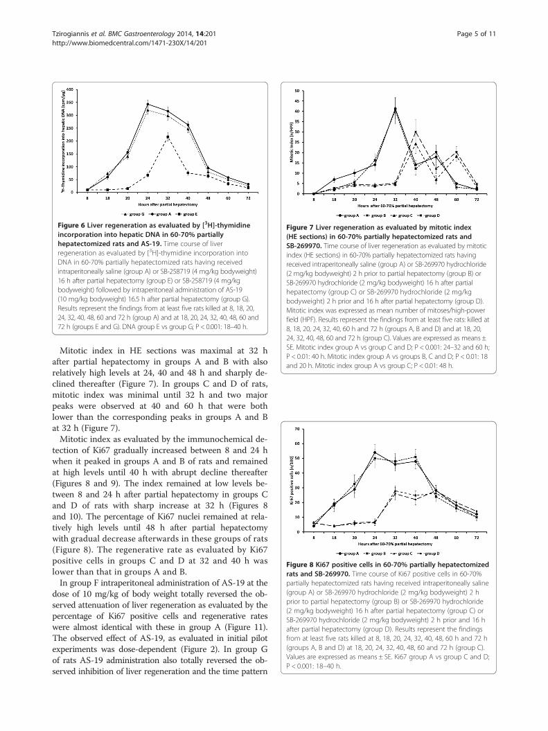
Figure 6 Liver regeneration as evaluated by [3H]-thymidineincorporation into hepatic DNA in 60-70% partiallyhepatectomized rats and AS-19. Time course of liverregeneration as evaluated by [3H]-thymidine incorporation intoDNA in 60-70% partially hepatectomized rats having receivedintraperitoneally saline (group A) or SB-258719 (4 mg/kg bodyweight)16 h after partial hepatectomy (group E) or SB-258719 (4 mg/kgbodyweight) followed by intraperitoneal administration of AS-19(10 mg/kg bodyweight) 16.5 h after partial hepatectomy (group G).Results represent the findings from at least five rats killed at 8, 18, 20,24, 32, 40, 48, 60 and 72 h (group A) and at 18, 20, 24, 32, 40, 48, 60 and72 h (groups E and G). DNA group E vs group G; P < 0.001: 18–40 h.
Figure 7 Liver regeneration as evaluated by mitotic index(HE sections) in 60-70% partially hepatectomized rats andSB-269970. Time course of liver regeneration as evaluated by mitoticindex (HE sections) in 60-70% partially hepatectomized rats havingreceived intraperitoneally saline (group A) or SB-269970 hydrochloride(2 mg/kg bodyweight) 2 h prior to partial hepatectomy (group B) orSB-269970 hydrochloride (2 mg/kg bodyweight) 16 h after partialhepatectomy (group C) or SB-269970 hydrochloride (2 mg/kgbodyweight) 2 h prior and 16 h after partial hepatectomy (group D).Mitotic index was expressed as mean number of mitoses/high-powerfield (HPF). Results represent the findings from at least five rats: killed at8, 18, 20, 24, 32, 40, 60 h and 72 h (groups A, B and D) and at 18, 20,24, 32, 40, 48, 60 and 72 h (group C). Values are expressed as means ±SE. Mitotic index group A vs group C and D; P < 0.001: 24–32 and 60 h;P < 0.01: 40 h. Mitotic index group A vs groups B, C and D; P < 0.01: 18and 20 h. Mitotic index group A vs group C; P < 0.01: 48 h.
Figure 8 Ki67 positive cells in 60-70% partially hepatectomizedrats and SB-269970. Time course of Ki67 positive cells in 60-70%partially hepatectomized rats having received intraperitoneally saline(group A) or SB-269970 hydrochloride (2 mg/kg bodyweight) 2 hprior to partial hepatectomy (group B) or SB-269970 hydrochloride(2 mg/kg bodyweight) 16 h after partial hepatectomy (group C) orSB-269970 hydrochloride (2 mg/kg bodyweight) 2 h prior and 16 hafter partial hepatectomy (group D). Results represent the findingsfrom at least five rats killed at 8, 18, 20, 24, 32, 40, 48, 60 h and 72 h(groups A, B and D) at 18, 20, 24, 32, 40, 48, 60 and 72 h (group C).Values are expressed as means ± SE. Ki67 group A vs group C and D;P < 0.001: 18–40 h.
Tzirogiannis et al. BMC Gastroenterology 2014, 14:201 Page 5 of 11http://www.biomedcentral.com/1471-230X/14/201
Mitotic index in HE sections was maximal at 32 hafter partial hepatectomy in groups A and B with alsorelatively high levels at 24, 40 and 48 h and sharply de-clined thereafter (Figure 7). In groups C and D of rats,mitotic index was minimal until 32 h and two majorpeaks were observed at 40 and 60 h that were bothlower than the corresponding peaks in groups A and Bat 32 h (Figure 7).Mitotic index as evaluated by the immunochemical de-
tection of Ki67 gradually increased between 8 and 24 hwhen it peaked in groups A and B of rats and remainedat high levels until 40 h with abrupt decline thereafter(Figures 8 and 9). The index remained at low levels be-tween 8 and 24 h after partial hepatectomy in groups Cand D of rats with sharp increase at 32 h (Figures 8and 10). The percentage of Ki67 nuclei remained at rela-tively high levels until 48 h after partial hepatectomywith gradual decrease afterwards in these groups of rats(Figure 8). The regenerative rate as evaluated by Ki67positive cells in groups C and D at 32 and 40 h waslower than that in groups A and B.In group F intraperitoneal administration of AS-19 at the
dose of 10 mg/kg of body weight totally reversed the ob-served attenuation of liver regeneration as evaluated by thepercentage of Ki67 positive cells and regenerative rateswere almost identical with these in group A (Figure 11).The observed effect of AS-19, as evaluated in initial pilotexperiments was dose-dependent (Figure 2). In group Gof rats AS-19 administration also totally reversed the ob-served inhibition of liver regeneration and the time pattern

Figure 9 Ki67 positive cells at 24 h (×400) in 60–70% partiallyhepatectomized rats having received saline (group A).
Figure 11 Ki67 positive cells in 60-70% partially hepatectomizedrats and AS-19. Time course of Ki67 positive cells in 60-70% partiallyhepatectomized rats having received intraperitoneally saline (group A)or SB-269970 hydrochloride (2 mg/kg bodyweight) 16 h after partialhepatectomy (group C) or SB-269970 hydrochloride (2 mg/kgbodyweight) followed by intraperitoneal administration of AS-19(10 mg/kg bodyweight) 16.5 h after partial hepatectomy (group F).Results represent the findings at least five rats killed at 8, 18, 20, 24, 32,40, 48, 60 and 72 h (group A) and at 18, 20, 24, 32, 40, 48, 60 and 72 h(groups C and F). Values are expressed as means ± SE. Ki67 group C vsgroup F; P < 0.001: 18–40 h.
Tzirogiannis et al. BMC Gastroenterology 2014, 14:201 Page 6 of 11http://www.biomedcentral.com/1471-230X/14/201
and values of Ki67 positive cells were also almost identicalwith these in groups A and F (data not shown).Relative liver weight (liver weight in g/100 g bodyweight)
sharply decreased, as expected after partial hepatectomy,with gradual increase thereafter in group A of rats. Ingroups C, D and E, relative liver weight remained at lowlevels without significant increases until 24 h after partialhepatectomy. In these groups a small increase was ob-served at 32 h with further increase at 40 and 48 h afterpartial hepatectomy. In groups F and G of rats, the relativeliver weights showed the same gradual increases as ingroup A (Table 1).Regarding lipid changes after partial hepatectomy, in-
crease in liver triglyceride levels was observed at 18 hafter partial hepatectomy with further increase at 24 h ingroup A of rats. Liver triglyceride content peaked at40 h after partial hepatectomy and decreased thereafter
Figure 10 Ki67 positive cells at 24 h (×400) in 60–70% partiallyhepatectomized rats having received SB-269970 hydrochloride(2 mg/kg bodyweight) 16 h after partial hepatectomy (group C).
but was still at high levels at 72 h after partial hepatec-tomy. Serum triglyceride concentration decreased at 18and 24 h after partial hepatectomy and increased after-wards and these increases were still present at 72 h afterpartial hepatectomy. Serum FFA and free glycerol levelsboth increased at 18 h after partial hepatectomy andremained at high levels thereafter. The temporal patternsof liver and plasma lipid changes were similar in allgroups of rats (Tables 2 and 3).
DiscussionThe ability of the liver to regenerate after surgical resec-tion or any short of hepatic injury has been known fromlong and has drawn immense scientific interest. 60-70%partial hepatectomy is the most commonly applied stimu-lus for the study of liver regeneration mainly due to thefact that the mitotic stimulus is accurately applied in timeand not associated with necrotic or inflammatory pro-cesses [20]. A great number of substances influence the re-generative process and traditionally they are classified ascomplete and incomplete (auxiliary) mitogens [20].The autonomic nervous system, both sympathetic and
parasympathetic, is implicated in liver regeneration al-though the exact mechanisms of its effects still remain ob-scure [21-23]. Among neurotransmitters norepinephrine,mainly through α1-adrenergic receptor [23-25] (actionsthrough β-adrenergic receptors have also been reported)[26], and serotonin, mainly through 5-HT2 receptor, areconsidered auxiliary mitogens [7-9].

Table 1 Relative liver weights (g/100 g bodyweight) after 60-70% partial hepatectomy in groups A, C, E, F and G
Relative liver weights (g/100 g body weight)
Hours after partial hepatectomy Group A Group C Group E Group F Group G
8 1,6 ± 0,1 1,5 ± 0,1 1,6 ± 0,1 1,7 ± 0,2 1,6 ± 0,1
18 1,9 ± 0,2 1,7 ± 0,1 1,6 ± 0,1 2,0 ± 0,2 2,0 ± 0,1
24 2,3 ± 0,2 1,7 ± 0,1 1,8 ± 0,1 2,2 ± 0,2 2,4 ± 0,2
32 2,6 ± 0,3 2,3 ± 0,2 2,2 ± 0,2 2,5 ± 0,3 2,7 ± 0,2
40 3,1 ± 0,3 2,6 ± 0,2 2,6 ± 0,1 3,1 ± 0,2 3,0 ± 0,3
48 3,5 ± 0,2 2,7 ± 0,2 2,8 ± 0,2 3,4 ± 0,3 3,4 ± 0,3
60 3,7 ± 0,2 2,7 ± 0,2 2,7 ± 0,1 3,8 ± 0,2 3,7 ± 0,3
72 4,2 ± 0,2 2,6 ± 0,1 2,8 ± 0,1 4,1 ± 0,3 4,3 ± 0,2
The mean relative liver weight for normal rats (n = 5) of the same age and weight range was 4.5 ± 0.3.
Tzirogiannis et al. BMC Gastroenterology 2014, 14:201 Page 7 of 11http://www.biomedcentral.com/1471-230X/14/201
Serotonin is an important neurotransmitter of the auto-nomic nervous system and in the liver serotonergic nervefibres are localized in the tunica media of branches of thehepatic artery, portal vein, bile ducts and the connectivetissue of the interlobular septae in humans and rats[27,28]. 5-HT receptors are expressed in various liver celltypes, apart from hepatocytes, as hepatic stelate cells andsinusoidal endothelial cells [4,29,30].From experiments on differential 5-HT receptor sub-
type expression and blockade experiments with variousreceptor antagonists of other research groups it has be-come evident that 5-HT2α and 5-HT2β receptors mediateliver regeneration [31] and molecular pathways havebeen elucidated in the case of 5-HT2β receptor [32-34].In our study, 5-HT7 receptor blockade greatly attenu-
ated liver regeneration when applied close to the G1/Stransition point of the cell cycle and this is the first studyto reveal implication of the 5-HT7 receptor in liver regen-eration and more specifically in this major restrictive cellcycle check point. In the central nervous system, blockadeof 5-HT7 receptor has been reported to increase hippo-campal cell proliferation [35] and the receptor is also im-plicated at least in the initial stages of T-cell activation andpossibly in T-cell proliferation [36]. Additionally, 5-ΗΤ7
receptor has been recently found to be expressed in hepa-tocytes although the full repertoire of its actions in theliver still remains obscure [37].SB-269970 used in our study is considered a highly se-
lective ligand for 5-HT7 receptors (pKi= 8.9 ± 0.1) withat least 100-fold greater affinity in relation to other typesof 5-HT receptor subtypes but some researchers havealso reported that it is also a potent α2-adrenergic recep-tor blocker [38-41]. Although only α1-adrenoreceptorshave been reported to participate in liver regeneration,the observed inhibitory effect by SB-269970 could alsobe attributed to α2-receptor blockade especially since α2-adrenoreceptors are expressed in hepatocytes [42,43].Activation of α2-adrenergic receptors has been reportedto induce cell proliferation in different cell types [44-46],
whereas competitive inhibition of these receptors at-tenuates cell proliferation and/or induces apoptosis[44,45,47]. However, there are reports that connect α2-receptor stimulation with inhibition of cell growth [48].In order to elucidate the above, another series of exper-iments has been conducted in our laboratory with in-traperitoneal administration of SB-258719 (pKi= 7.5) atthe dose of 4 mg/kg bodyweight at 16 h after partialhepatectomy [38,49,50]. SB-258719 is a known weak in-verse agonist of 5-HT7 receptor without any known actionson other type of serotonin receptors and its administrationhad the same effect on liver regeneration as SB-269970administration and the above clearly suggests that the ob-served inhibitory effect must be attributed to 5-HT7 recep-tor blockade.In order to verify that the observed effect on liver regen-
eration is due to blockade of 5-HT7 receptor we con-ducted another series of experiments with the selective 5-HT7 receptor agonist AS-19 [51-53]. AS-19 is considereda selective 5 HT7 agonist (Ki = 0.6 nM, IC50 = 0.83nM)[54]. AS-19 administration reversed the observed attenu-ation of liver regeneration caused by administration ofSB-269970 and SB-258719 and this verifies the implicationof 5-HT7 in liver regeneration.It is known from long that liver regeneration is accom-
panied by transient hepatic steatosis and intracellularaccumulation of triglycerides in hepatocytes through in-creased lipolysis in the adipose tissue and increased hep-atic lipogenesis [55,56]. Serotonin induces lipolysis inadipocytes and promotes gluconeogenesis in hepatocytesthrough 5-HT2b receptor during fasting adaptation [57].Additionally serotonin is also implicated in the regulationof lipid metabolism through 5-HT2c receptors by alteringsympathetic outflow at the brain level [58]. In our experi-ments no significant differences have been observed inserum and liver lipids during liver regeneration after 5-HT7 receptor blockade and consequently 5-HT7 receptordoes not seem to be implicated in the adaptive changes oflipid metabolism during liver regeneration.

Table 2 Liver and serum triacylglycerol levels and serum glycerol and FFA levels in groups A, C and D
Time afterPH (hours)
Group A Group C Group D
Livertriacylglycerol(μg/mg ofprotein)
Serumtriacylglycerol(mg/dl)
Serumglycerol(μmol/l)
Serum FFA(μmol/mlor mmol/l)
Livertriacylglycerol(μg/mg of protein
Serumtriacylglycerol(mg/dl)
Serumglycerol(μmol/l)
Serum FFA(μmol/mlor mmol/l)
Livertriacylglycerol(μg/mg of protein)
Serumtriacylglycerol(mg/dl)
Serumglycerol(μmol/l)
Serum FFA(μmol/mlor mmol/l)
0 15.8 ± 0.8 6.2 ± 0.6 61.2 ± 5.2 0.32 ± 0.05 15.8 ± 0.8 6.2 ± 0.6 61.2 ± 5.2 0.32 ± 0.05 15.8 ± 0.8 6.2 ± 0.6 61.2 ± 5.2 0.32 ± 0.05
8 16.8 ± 0.9 5.8 ± 0.6 75.4 ± 6.5 0.44 ± 0.05 N.D. N.D. N.D. N.D. 17.4 ± 1.3 6.0 ± 0.9 72.8 ± 6.1 0.50 ± 0.06
18 28.1 ± 2.3 3.8 ± 0.4 188.6 ± 8.8 0.86 ± 0.07 27.5 ± 3.1 4.2 ± 0.4 174.2 ± 7.5 0.82 ± 0.08 29.4 ± 2.4 3.5 ± 0.6 179.4 ± 7.8 0.89 ± 0.08
20 30.6 ± 3.4 3.6 ± 0.5 192.2 ± 9.5 0.84 ± 0.08 29.7 ± 2.5 3.9 ± 0.4 190.3 ± 8.6 0.86 ± 0.09 31.2 ± 2.6 3.3 ± 0.4 195.1 ± 8.2 0.81 ± 0.06
24 34.8 ± 3.8 3.4 ± 0.4 187.8 ± 9.1 0.82 ± 0.07 35.3 ± 3.4 3.2 ± 0.5 195.2 ± 8.9 0.89 ± 0.09 36.2 ± 2.9 3.0 ± 0.4 189.3 ± 8.8 0.79 ± 0.07
32 37.2 ± 2.3 5.6 ± 0.6 204.2 ± 10.4 0.90 ± 0.06 38.1 ± 3.8 4.8 ± 0.4 197.5 ± 9.3 0.94 ± 0.08 37.8 ± 2.7 5.4 ± 0.7 201.8 ± 9.4 0.88 ± 0.09
40 40.1 ± 3.4 6.4 ± 0.7 196.3 ± 10.1 0.86 ± 0.09 41.3 ± 4.2 5.9 ± 0.6 203.4 ± 8.7 0.88 ± 0.07 39.2 ± 3.1 6.7 ± 0.8 204.9 ± 8.5 0.91 ± 0.09
48 33.8 ± 2.6 7.4 ± 0.7 205.1 ± 9.5 0.84 ± 0.08 34.5 ± 3.5 7.1 ± 0.7 197.8 ± 9.5 0.84 ± 0.08 31.6 ± 2.5 7.8 ± 0.9 209.5 ± 9.7 0.80 ± 0.08
60 26.6 ± 2.2 7.2 ± 0.8 179.9 ± 8.6 0.83 ± 0.07 27.6 ± 3.1 7.4 ± 0.6 189.7 ± 7.8 0.81 ± 0.08 27.3 ± 1.9 7.7 ± 0.6 192.5 ± 8.3 0.78 ± 0.08
72 24.6 ± 1.6 7.0 ± 0.6 204.1 ± 8.9 0.85 ± 0.08 26.5 ± 2.6 7.5 ± 0.7 196.8 ± 7.5 0.80 ± 0.07 23.2 ± 1.7 7.2 ± 0.8 189.6 ± 7.8 0.74 ± 0.06
Values are expressed as mean ± standard error.FFA = Free fatty acid.N.D. = Not Determined.
Tzirogianniset
al.BMCGastroenterology
2014,14:201Page
8of
11http://w
ww.biom
edcentral.com/1471-230X/14/201

Table 3 Liver and serum triacylglycerol levels and serum glycerol and FFA levels in groups E, F and G
Time afterPH (hours)
Group E Group F Group G
Livertriacylglycerol(μg/mg of protein)
Serumtriacylglycerol(mg/dl)
Serumglycerol(μmol/l)
Serum FFA(μmol/mlor mmol/l)
Livertriacylglycerol(μg/mg ofprotein
Serumtriacylglycerol(mg/dl)
Serumglycerol(μmol/l)
Serum FFA(μmol/mlor mmol/l)
Livertriacylglycerol(μg/mgof protein)
Serumtriacylglycerol(mg/dl)
Serumglycerol(μmol/l)
Serum FFA(μmol/mlor mmol/l)
0 15.8 ± 0.8 6.2 ± 0.6 61.2 ± 5.2 0.32 ± 0.05 15.8 ± 0.8 6.2 ± 0.6 61.2 ± 5.2 0.32 ± 0.05 15.8 ± 0.8 6.2 ± 0.6 61.2 ± 5.2 0.32 ± 0.05
18 30.3 ± 2.9 4.0 ± 0.6 180.7 ± 8.3 0.85 ± 0.09 28.9 ± 3.3 3.8 ± 0.5 175.9 ± 8.5 0.72 ± 0.08 29.9 ± 2.8 4.2 ± 0.8 184.8 ± 8.8 0.81 ± 0.08
20 32.7 ± 3.8 3.7 ± 0.5 191.3 ± 8.9 0.83 ± 0.06 30.8 ± 3.5 3.9 ± 0.5 190.7 ± 9.0 0.79 ± 0.09 34.3 ± 3.1 3.6 ± 0.7 196.7 ± 8.5 0.77 ± 0.08
24 35.7 ± 3.9 3.5 ± 0.5 194.5 ± 9.7 0.80 ± 0.07 34.4 ± 3.8 3.4 ± 0.7 193.8 ± 8.5 0.87 ± 0.13 37.2 ± 3.5 3.1 ± 0.6 199.5 ± 9.2 0.79 ± 0.07
32 39.5 ± 3.3 5.0 ± 0.9 201.6 ± 10.3 0.93 ± 0.11 42.3 ± 4.0 5.3 ± 0.9 198.5 ± 9.9 0.96 ± 0.15 38.8 ± 3.7 4.5 ± 0.9 207.8 ± 10.4 0.90 ± 0.09
40 42.6 ± 4.1 6.7 ± 0.6 199.2 ± 10.8 0.86 ± 0.09 44.6 ± 4.6 6.3 ± 1.1 205.6 ± 10.7 0.91 ± 0.09 43.4 ± 3.6 6.1 ± 0.9 210.9 ± 9.5 0.95 ± 0.09
48 35.9 ± 2.9 7.7 ± 0.8 206.7 ± 11.3 0.83 ± 0.08 37.5 ± 3.9 7.6 ± 0.9 203.7 ± 10.2 0.87 ± 0.08 32.9 ± 3.5 7.2 ± 1.2 200.3 ± 10.7 0.81 ± 0.08
60 27.4 ± 2.6 7.1 ± 0.9 182.9 ± 9.6 0.81 ± 0.08 29.8 ± 3.3 7.4 ± 0.9 185.8 ± 8.8 0.82 ± 0.07 26.3 ± 2.4 7.3 ± 0.9 172.7 ± 8.9 0.73 ± 0.07
72 23.6 ± 2.2 6.7 ± 0.6 200.4 ± 9.9 0.85 ± 0.09 25.6 ± 2.9 6.5 ± 0.8 203.6 ± 9.5 0.76 ± 0.09 21.8 ± 1.9 7.0 ± 1.0 192.6 ± 7.5 0.81 ± 0.09
Values are expressed as mean ± standard error.FFA = Free fatty acid.
Tzirogianniset
al.BMCGastroenterology
2014,14:201Page
9of
11http://w
ww.biom
edcentral.com/1471-230X/14/201

Tzirogiannis et al. BMC Gastroenterology 2014, 14:201 Page 10 of 11http://www.biomedcentral.com/1471-230X/14/201
5-HT7 receptors have been reported to activate MAPK[59,60] and this activation has also been reported to beRAS-dependent [61]. The above seems to represent amore general pattern of MAPK activation from Gs-coupled receptors with RAS independent pathways tohave also been described [62,63]. Both 5-HT2α and 5-HT2β receptors have also been reported to activate MAPKthrough similar pathways [33,64] and this hints at a pos-sible role of 5-HT7 receptor in mitogenesis and cell-cycle progression although further research is needed atthis point.
ConclusionsThe results of this study indicate that 5-HT7 receptor isimplicated in liver regeneration after partial hepatectomy.Serotonin through 5-HT7 receptor seems to exert its aux-iliary proliferative effect close to G1/S transition point andduring the S phase. Therefore, the results identify a noveltype of 5-HT receptor that mediates the proliferative effectof the monoamine in the liver.
Abbreviations5-HT: Serotonin; HE: Hematoxylin-eosin; CNS: Central nervous system;GI: Gastro-intestinal; EGF: Epidermal growth factor; HPF: High-power fields;MAPK: Mitogen-activated protein kinase.
Competing interestsThe authors declare that they have no competing interests.
Authors’ contributionsKNT and GIP designed the study; KNT, KTK, SZ, and GIP coordinated thestudy; KNT, KTK, ADG, MT, SZ, DP, IP, VP and GIP performed the study; KNT,KTK, MT, SZ, DP, VP, IP and GIP analyzed the data; KTK, MT, SZ, VP helped todraft the manuscript; and KNT and GIP wrote the manuscript. All authorsread and approved the final manuscript.
Author details1Department of Experimental Pharmacology, Medical School, AthensUniversity, Athens 11527, Greece. 2Department of Nursing, Faculty of HumanMovement and Quality of Life Sciences, University of Peloponnese, Sparta23100, Greece. 3Department of Internal Medicine, Elpis General Hospital,Athens 11522, Greece. 4Department of Internal Medicine, Tzanio GeneralHospital of Piraeus, Piraeus 18537, Greece. 5Department of Nursing, Facultyof Human Movement and Quality of Life Sciences, University ofPeloponnese, Orthias Artemidos and Plateon, Sparta 23100, Greece.
Received: 28 February 2014 Accepted: 11 November 2014
References1. Ruddell RG, Mann DA, Ramm GA: The function of serotonin within the
liver. J Hepatol 2008, 48:666–675.2. Beattie DT, Smith JA: Serotonin Pharmacology in the gastrointestinal
tract: a review. Naunyn Schmiedebergs Arch Pharmacol 2008, 377:181–203.3. Lesurtel M, Soll C, Graf R, Clavien PA: Role of serotonin in the hepato-
gastrointestnal tact: an old molecule for new perspectives. Cell Mol LifeSci 2008, 65:940–952.
4. Ruddel RG, Oakley F, Hussain Z, Yeung I, Bryan-Lluka LJ, Ramm GA, MannDA: A Role for Serotonin (5-HT) in hepatic Stellate Cell Function andLiver Fibrosis. Am J Pathol 2006, 169:861–876.
5. Kulinskii VI, Saratikov AS, Vstavskaia IA, Udovitsina TI: Receptors mediatingserotonin-stimulating liver regeneration in mice. Biull Eksp Biol Med 1983,95:89–91.
6. Kulinskii VI, Udovitsina TI, Vstavskaia IA, Rykov SA: Comparison of thechanges in mitotic activity and in serotonin concentration inregenerating liver. Vopr Med Khim 1983, 29:104–107.
7. Balasubramanian S, Paulose CS: Induction of DNA synthesis in primarycultures of rat hepatocytes by serotonin: possible involvement ofserotonin S2 receptor. Hepatology 1998, 27:62–66.
8. Papadimas GK, Tzirogiannis KN, Panoutsopoulos GI, Demonakou MD,Skaltsas SD, Hereti RI, Papadopoulou-Daifoti Z, Mykoniatis MG: Effect ofserotonin receptor 2 blockade on liver regeneration after partialhepatectomy in the rat liver. Liver Int 2006, 26:352–361.
9. Lesurtel M, Graf R, Aleil B, Walther DJ, Tian Y, Jochum W, Gachet C, Bader M,Clavien PA: Platelet-derived serotonin mediates liver regeneration.Science 2006, 312:104–107.
10. Soll C, Jang JH, Riener MO, Moritz W, Wild PJ, Graf R, Clavien PA: Serotoninpromotes tumor growth in Human Hepatocellular Cancer.Hepatology 2010, 51:1244–1254.
11. Vanhoenacker P, Haegeman G, Leysen JE: 5-HT7 receptors: currentknowledge and future prospects. Trends Pharmacol Sci 2000, 21:70–77.
12. Hedlund PB, Sutcliffe JG: Functional, molecular and pharmacologicaladvances in 5-HT 7 receptor research. Trends Pharmacol Sci 2004,25:481–486.
13. GREEK PRESIDENTIAL DECREE No 160/1991: Protection of Animals Used forExperimental and Other Scientific Purposes in Accordance With EU Directive86/609/EEC of the Council, Governmental Gazette No 64; 1991.
14. Munro HN, Fleck A: Recent developments in the measurement of nucleicacids in biological materials. Analyst 1966, 91:78–88.
15. Kyprianidis KG, Mykoniatis MG, Papadimitriou DG, Valsamidou A: Effect ofsubtotal pancreatectomy on the rate of liver regeneration: the role ofhepatic stimulator substance. J Surg Res 1996, 62:267–272.
16. Richards G: Modifications of the diphenylamine reaction giving increasedsensitivity and simplicity in the estimation of DNA. Anal Biochem 1974,57:369–376.
17. Lowry O, Rosenbrough N, Farr A, Randall R: Protein measurement with thefolin phenol reagent. J Biol Chem 1951, 193:265–275.
18. Folch J, Lees M, Sloane Stanley GH: A simple method for the isolation andpurification of total lipids from animal tissues. J Biol Chem 1982,28:497–509.
19. Tijburg LB, Maquedano A, Bijleveld C, Guzman M, Geelen MJ: Effects ofethanol feeding on hepatic lipid synthesis. Arch Biochem Biophys 1988,267:568–579.
20. Michalopoulos GK: Liver regeneration after partial Hepatectomy: criticalanalysis of mechanistic dilemmas. Am J Pathol 2010, 176:2–13.
21. Tanaka K, Ohkawa S, Nishino T, Niijima A, Inoue S: Role of the hepaticbranch of the vagus nerve in lever regeneration in rats. Am J Physiol1987, 253:G439–G444.
22. Oben JA, Diehl AM: Sympathetic nervous system regulation of liverrepair. Anat Rec A: Discov Mol Cell Evol Biol 2004, 280:874–883.
23. Cruise JL, Knechtle SJ, Bollinger RR, Kuhn C, Michalopoulos G: Alpha1-adrenergic effects and liver regeneration. Hepatology 1987, 7:1189–1194.
24. Cruise JL, Houck KA, Michalopoulos G: Early events in the regulation ofHepatocyte DNA synthesis: the role of alpha- adrenergic stimulation.Scand J Gastroenterol 1988, 151:19–30.
25. Cruise JL, Houck KA, Michalopoulos GK: Induction of DNA synthesis incultured rat hepatocytes through stimulation of alpha 1 adrenoreceptorby norepinephrine. Science 1985, 227:749–751.
26. Refsnes M, Thoresen GH, Sandnes D, Dajani OF, Dajani L, Christoffersen T:Stimulatory and inhibitory effects of catecholamines on DNA synthesisin primary rat hepatocyte cultures: role of alpha1- and beta-adrenergicmechanisms. J Cell Physiol 1992, 151:164–171.
27. El-Salhy M, Stenling R, Grimelius L: Peptidergic innervation and endocrinecells in the human liver. Scand J Gastroenterol 1993, 28:809–815.
28. Stoyanova JL: Relevance of mast cells and hepatic lobule innervations toliver. Rom J Gastroenterol 2004, 13:203–209.
29. Brauneis U, Gatmaitan Z, Arias IM: Serotonin stimulates a Ca2+ permeantnonspecific cation channel in hepatic endothelial cells. Biochem BiophysRes Commun 1992, 186:1560–1566.
30. Gatmaitan Z, Varticovski L, Ling L, Mikkelsen R, Steffan AM, Arias IM: Studieson fenestral contraction in rat liver endothelial cells in culture. Am JPathol 1996, 148:2027–2041.
31. Clavien PA: Liver regeneration: a spotlight on the novel role of plateletsand serotonin. Swiss Med Wkly 2008, 138:361–370.

Tzirogiannis et al. BMC Gastroenterology 2014, 14:201 Page 11 of 11http://www.biomedcentral.com/1471-230X/14/201
32. Gooz M, Gooz P, Luttrell LM, Raymond JR: 5-HT2A receptor induces ERKphosphorylation and proliferation through ADAM-17 tumor necrosisfactor-alpha-converting enzyme (TACE) activation and heparin-boundepidermal growth factor-like growth factor (HB-EGF) shedding inmesangial cells. J Biol Chem 2006, 281:21004–21012.
33. Nebigil CG, Launay JM, Hickel P, Tournois C, Maroteaux L: 5-hydroxytrypramine 2B receptor regulates cell-cycle progression: cross-talkwith tyrosine kinase pathways. Proc Natl Acad Sci U S A 2000, 97:2591–2596.
34. Liu Y, Li M, Warburton RR, Hill NS, Fanburg BL: The 5-HT transportertransactivates the PDGF beta receptor in pulmonary artery smoothmuscle cells. FASEB J 2007, 21:2725–2734.
35. Mnie-Filali O, Faure C, Lambas-Senas L, El Mansari M, Belblidia H, Gondard E,Etievant A, Scarna H, Didier A, Berod A, Blier P, Haddjeri N: Pharmacologicalblockade of 5-HT7 receptors as a putative fast acting antidepressantstrategy. Neuropsychopharmacology 2011, 36:1275–1288.
36. Leon-Ponte M, Ahern GP, O’Connell PJ: Serotonin provides an accessorysignal to enhance T-cell activation by signaling through the 5-HT7receptor. Blood 2007, 109:3139–3146.
37. Svejda B, Kidd M, Timberlake A, Harry K, Kazberouk A, Schimmack S,Lawrence B, Pfragner R, Modlin IM: Serotonin and the 5-HT7 receptor: thelink between hepatocytes, IGF-1 and small intestinal neuroendocrinetumors. Cancer Sci 2013, 104:844–855.
38. Mahe C, Loetscher E, Feuerbach D, Muller W, Seiler MP, Schoeffter P:Differential inverse agonist efficacies of SB-258719, SB-258741 andSB-269970 at human recombinant serotonin 5-HT7 receptors. Eur JPharmacol 2004, 495:97–102.
39. Lovell PJ, Bromidge SM, Dabbs S, Duckworth DM, Forbes IT, Jennings AJ,King FD, Middlemiss DN, Rahman SK, Saunders DV, Collin LL, Hagan JJ, Riley GJ,Thomas DR: A novel, potent, and selective 5-HT7 antagonist: (R)-3-(2-(2-(4-methylpiperidin-1-yl)ethyl)pyrrolidine-1-sulfonyl)phenol (SB 269970). J MedChem 2000, 43:342–345.
40. Foong JP, Bornstein JC: 5-HT antagonists NAN-190 and SB 269970 blockαlpha2-adrenoceptors in the guinea pig. Neuroreport 2009, 20:325–330.
41. Hagan JJ, Price GW, Jeffrey P, Deeks NJ, Stean T, Piper D, Smith MI, Upton N,Medhurst AD, Middlemiss DN, Riley GJ, Lovell PJ, Bromidge SM, Thomas DR:Characterization of SB-269970-A, a selective 5-HT(7) receptor antagonist.Br J Pharmacol 2000, 130:539–548.
42. Hoffman BB, Dukes DF, Lefkowitz RJ: Alpha-adrenergic receptors in livermembranes: delineation with subtype selective radioligands. Life Sci1981, 28:265–272.
43. Bylund DB: Subtypes of αlpha-1- and alpha-2- adrenergic receptors.FASEB J 1992, 6:832–839.
44. Seuwen K, Magnaldo I, Kobilka BK, Caron MG, Regan JW, Lefkowitz RJ,Pouyssegur J: Alpha 2-adrenergic agonists stimulate DNA synthesis inChinese hamster lung fibroblasts transfected with a human alpha2-adrenergic receptor gene. Cell Regul 1990, 1:445–451.
45. Chiesa IJ, Castillo LF, Luthy IA: Contribution of alpha-2-adrenoceptors tothe mitogenic effect of catecholestrogen in human breast cancer MCF-7cells. J Steroid Biochem Mol Biol 2008, 110:170–175.
46. Bruzzone A, Pinero CP, Castillo LF, Sarappa MG, Rojas P, Lanari C, Luthy IA:Alpha-2- adrenoceptor action on cell proliferation and mammarytumour growth in mice. Br J Pharmacol 2008, 155:494–504.
47. Shen SG, Zhang D, Hu HT, Li JH, Wang Z, Ma QY: Effects of alpha-adrenoreceptor antagonists on apoptosis and proliferation of pancreaticcancer cells in vitro. World J Gastroenterol 2008, 14:2358–2363.
48. Kanno N, Lesage G, Phinizy JL, Glaser S, Francis H, Alpini G: Stimulation ofalpha2-adrenergic receptor inhibits cholangiocarcinoma growth throughmodulation of Raf-1 and B-Raf activities. Hepatology 2002, 35:1329–1340.
49. Romero G, Pujol M, Pauwels PJ: Reanalysis of constitutively active rat andhuman 5-HT7(a) receptors in HEK-293 F cells demonstates lack of silentproperties for reported neutral antagonists. Naunyn Schmiedebergs ArchPharmacol 2006, 374:31–39.
50. Forbes IT, Dabbs S, Duckworth DM, Jennings AJ, King FD, Lovell PJ,Brown AM, Collin L, Hagan JJ, Middlemiss DN, Riley GJ, Thomas DR,Upton N: (R)-3, N-dimethyl-N-[1-methyl-3-(4-methyl-piperidin-1-yl)propyl]benzenesulfonamide: the first selective 5–HT7 receptorantagonist. J Med Chem 1998, 41:655–657.
51. Perez-Garcia GS, Meneses A: Effects of the potential 5-HT7 receptor agonistAS 19 in an autoshaping learning task. Behav Brain Res 2005, 163:136–140.
52. Perez-Garcia G, Gonzalez-Espinosa C, Meneses A: An mRNA expressionanalysis of stimulation and blockade of 5-HT7 receptors during memoryconsolidation. Behav Brain Res 2006, 169:83–92.
53. Sanin A, Brisander M, Rosqvist S, Mohell N, Malberg A, Johansson A: 5-ArylSubstituted (S)-2-(Dimethylamino)-Tetralins Nover Serotonin 5HT 7Receptor Ligands. In Proceedings of the 14th Camerino-Noord Symposium,Ongoing Progress in the Receptor Chemistry; 2004:27.
54. Brenchat A, Rocasalbas M, Zamanillo D, Hamon M, Miguel Vela J, Romero L:Assessment of 5-HT7 receptor agonists selectivity using nociceptive andthermoregulation tests in knockout versus wild-type mice. Adv PharmacolSci 2012, 2012:312041.
55. Tijburg LB, Nyathi CB, Meijer GW, Geelen MJ: Biosynthesis and secretion oftriacylglycerol in rat liver after partial hepatectomy. Biochem J 1991,277:723–728.
56. Rudnick DA: Trimming the fat from liver regeneration. Hepatology 2005,42:1001–1003.
57. Sumara G, Sumara O, Kim JK, Karsenty G: Gut-derived serotonin is amultifunctional determinant to fasting adaptation. Cell Metab 2012,16:588–600.
58. Nonogaki K: New insights into sympathetic regulation of glucose and fatmetabolism. Diabetologia 2000, 43:533–549.
59. Watts SW, Yang P, Banes AK, Baez M: Activation of Erk mitogen-activatedprotein kinase proteins by vascular serotonin receptors. J CardiovascPharmacol 2001, 38:539–551.
60. Lieb K, Biersack L, Waschbiisch A, Orlikowski S, Akundi ES, Candelario-Jalil E,Hull M, Fiebich BL: Serotonin via 5-HT7 receptors activates p38 mitogen-activated protein kinase and protein kinase C epsilon resulting ininterleukin-6 synthesis in human U373 MG astrocytoma cells.J Neurochem 2005, 93:549–559.
61. Norum JH, Hart K, Levy FO: Ras-dependent ERK activation by the humanG(s)-coupled serotonin receptors 5-HT4(b) and 5-HT7(a). J Biol Chem2003, 278:3098–3104.
62. Klinger M, Kudlacek O, Seidel MG, Freissmuth M, Sexl V: MAP kinasestimulation by cAMP does not require RAP1 but SRC family kinases.J Biol Chem 2002, 277:32490–32497.
63. Enserink JM, Christensen AE, de Rooij J, van Triest M, Schwede F, GenieserHG, Doskeland SO, Blank JL, Bos JL: A novel Epac-specific cAMP analoguedemonstrates independent regulation of Rap1 and ERK. Nat Cell Biol2002, 4:901–906.
64. Banes A, Florian JA, Watts SW: Mechanisms of 5-Hydroxytryptamine (2A)receptor activation of the mitogen-activated protein kinase pathway invascular smooth muscle. J Pharmacol Exp Ther 1999, 291:1179–1187.
doi:10.1186/s12876-014-0201-2Cite this article as: Tzirogiannis et al.: Effect of 5-HT7 receptor blockade onliver regeneration after 60-70% partial hepatectomy. BMC Gastroenterology2014 14:201.
Submit your next manuscript to BioMed Centraland take full advantage of:
• Convenient online submission
• Thorough peer review
• No space constraints or color figure charges
• Immediate publication on acceptance
• Inclusion in PubMed, CAS, Scopus and Google Scholar
• Research which is freely available for redistribution
Submit your manuscript at www.biomedcentral.com/submit