protective equipment: continuous and contingent application ...
Upload
khangminh22Category
view
3download
0
Riga, 2019
Kristīne Voļska
PROTECTIVE EFFECTS AND MECHANISMS OF ACTION OF
METHYL-GBB IN THE PRECLINICAL MODELS OF DIABETES AND
ITS COMPLICATIONS
Doctoral Thesis for obtaining the degree of Doctor of Pharmacy
Speciality – Pharmaceutical Pharmacology
doi:10.25143/prom-rsu_2019-06_dt

Kristīne Voļska
PROTECTIVE EFFECTS AND MECHANISMS OF ACTION
OF METHYL-GBB IN THE PRECLINICAL MODELS OF
DIABETES AND ITS COMPLICATIONS
Scientific supervisor: Dr. pharm., Professor Maija Dambrova
Speciality – Pharmaceutical Pharmacology
Doctoral Thesis for obtaining the degree of Doctor of Pharmacy
Riga, 2019

2
ABSTRACT
The global prevalence of diabetes continues to rise concomitantly increasing the
number of diabetes patients at risk of developing cardiovascular complications, such as
atherosclerosis and ischaemic heart disease. Incomplete fatty acid oxidation and subsequent
accumulation of fatty acid intermediates, long-chain acylcarnitines, are linked to the
development of insulin resistance and cardiovascular diseases. Therefore, novel treatment
strategies targeting fatty acid metabolism are needed to improve the clinical outcomes of
patients with diabetes and its cardiovascular complications. The aim of the thesis was to
investigate the pharmacological mechanisms of action of an acylcarnitine concentration
lowering drug methyl-GBB in experimental animal models of diabetes, cardiac
ischaemia/reperfusion injury and atherosclerosis. This thesis describes the molecular
mechanisms of excessive accumulation of long-chain acylcarnitines and their detrimental
effects during the development of insulin resistance and in the ischaemia/reperfusion-induced
damage. The protective effects of lowering long-chain acylcarnitine levels by methyl-GBB
treatment in experimental models of diabetes and atherosclerosis are described. The results
indicate that accumulation of long-chain acylcarnitines limits metabolic flexibility and
accelerates hyperglycaemia and hyperinsulinemia during the fed state. Methyl-GBB
treatment-induced decrease in long-chain acylcarnitine content improves insulin sensitivity
and significantly reduces blood glucose and insulin levels in mice with insulin resistance and
diabetes. The results demonstrate that long-chain acylcarnitines are the main fatty acid
intermediates that induce ischaemia/reperfusion-related damage by inhibiting oxidative
phosphorylation and subsequent mitochondrial membrane hyperpolarization and stimulated
production of reactive oxygen species in cardiac mitochondria. The anti-atherosclerotic effect
of methyl-GBB treatment is mediated by decreased amounts of long-chain acylcarnitines and
decreased infiltration of macrophages and monocytes into the aortic lesions of the aortic root.
During this study, it was confirmed that pharmacologically induced decrease in the content of
long-chain acylcarnitines by methyl-GBB facilitates glucose metabolism, improves insulin
sensitivity, protects cardiac mitochondria against ischaemia/reperfusion injury and attenuates
the development of atherosclerosis, and therefore represents an effective strategy for the
treatment of diabetes and its complications.

3
DARBA ANOTĀCIJA
Diabēta prevalence pasaulē turpina pieaugt, vienlaikus pacientiem palielinot
aterosklerozes un išēmiskās sirds slimības kā kardiovaskulāro komplikāciju risku. Nepilnīga
taukskābju oksidācija un sekojoša taukskābju metabolītu, garķēžu acilkarnitīnu, uzkrāšanās ir
saistīta ar insulīna rezistences un kardiovaskulāro slimību attīstību. Lai uzlabotu cukura
diabēta un tā kardiovaskulāro komplikāciju pacientu klīnisko iznākumu, ir nepieciešamas
jaunas ārstēšanas stratēģijas, kuru darbības mehānisms ietver taukskābju metabolisma
izmaiņas. Promocijas darba mērķis bija pētīt jaunas acilkarnitīnu līmeni pazeminošas vielas
metil-GBB ietekmi uz enerģijas metabolisma procesiem un noskaidrot tās iespējamos
farmakoloģiskos darbības mehānismus cukura diabēta, aterosklerozes un sirds išēmijas-
reperfūzijas eksperimentālajos modeļos. Šajā darbā ir aprakstīti garķēžu acilkarnitīnu
pārmērīgas uzkrāšanās molekulārie mehānismi un tās izraisīto bojājumu nozīme insulīna
rezistences un išēmijas-reperfūzijas izraisītu bojājumu attīstībā. Ir aprakstīti metil-GBB
terapijas izraisītā garķēžu acilkarnitīnu līmeņa samazinājuma aizsargājošie efekti
eksperimentālajos diabēta un aterosklerozes modeļos. Rezultāti liecina, ka garķēžu
acilkarnitīnu uzkrāšanās postprandiālā stāvokļa laikā ierobežo metabolisma elastīgumu un
veicina hiperglikēmijas un hiperinsulinēmijas veidošanos. Metil-GBB terapijas izraisītā
garķēžu acilkarnitīnu līmeņa samazināšanās uzlabo insulīna jutību un ievērojami samazina
glikozes un insulīna līmeņus asinīs pelēm ar insulīna rezistenci un diabētu. Rezultāti liecina,
ka garķēžu acilkarnitīni ir galvenie taukskābju metabolīti, kuri nosaka išēmijas-reperfūzijas
izraistītos bojājumus, kavējot oksidatīvo fosforilēšanu un sekojoši izsaucot mitohondriju
membrānas hiperpolarizāciju un stimulējot reaktīvo skābekļa formu veidošanās sirds
mitohondrijos. Metil-GBB terapijas pretaterosklerozes mehānisms ir saistīts ar garķēžu
acilkarnitīnu daudzuma samazinājumu vaskulārajos audos, kā arī makrofāgu un monocītu
infiltrācijas kavēšanu aterosklerotiskajās pangās aortas sīnusā. Šī pētījuma laikā tika pierādīts,
ka farmakoloģiski izraisīta garķēžu acilkarnitīnu līmeņa samazināšana ar metil-GBB veicina
glikozes metabolismu, uzlabo insulīna jutību, aizsargā sirds mitohondrijus pret išēmijas-
reperfūzijas bojājumu un kavē aterosklerozes attīstību, un tādēļ tas ir pielietojams diabēta un
tā komplikāciju ārstēšanā.

4
TABLE OF CONTENTS
Introduction .............................................................................................................................. 9 Aim of the study ...................................................................................................................... 11 Objectives of the study ........................................................................................................... 11 Hypothesis of the study .......................................................................................................... 11 Scientific novelty of the study ................................................................................................ 12 1 Literature ......................................................................................................................... 13
1.1 Energy metabolism in muscle and heart ....................................................................... 13 1.1.1 Fatty acid uptake and cellular handling ............................................................... 13 1.1.2 Fatty acid transport into mitochondria and fatty acid β-oxidation ...................... 16 1.1.3 Glucose uptake and metabolism .......................................................................... 17 1.1.4 Insulin signalling pathway .................................................................................. 19
1.2 Regulation of energy metabolism and metabolic flexibility in muscles and cardiac tissue ................................................................................................................. 20
1.3 Diabetes and cardiometabolic complications ............................................................... 23 1.3.1 Physical exercise as a treatment option for cardiometabolic diseases ................ 24 1.3.2 Pharmacological regulators for cardiometabolic diseases .................................. 25
1.3.2.1 Meldonium .............................................................................................. 25 1.3.2.2 Trimetazidine .......................................................................................... 26 1.3.2.3 CPT1 inhibitors ....................................................................................... 27
2 Materials and methods .................................................................................................... 30 2.1 Animals and treatment .................................................................................................. 30 2.2 Materials ....................................................................................................................... 32 2.3 Methods ........................................................................................................................ 33
2.3.1 In vitro methods .................................................................................................. 33 2.3.1.1 Evaluation of palmitoylcarnitine-induced effects on insulin
signalling pathway in cell cultures .......................................................... 33 2.3.1.2 Western blot analysis of tissue lysates .................................................... 34 2.3.1.3 mRNA isolation and quantitative RT-PCR analysis ............................... 34 2.3.1.4 Determination of the acylcarnitines, acyl-CoAs and CoAs in the
plasma and tissue .................................................................................... 34 2.3.1.5 Determination of methyl-GBB, L-carnitine, GBB and
trimethylamine N-oxide levels in the plasma ......................................... 35 2.3.1.6 Determination of biochemical parameters in the plasma and blood
samples .................................................................................................... 35 2.3.1.7 Isolation of cardiac mitochondria ........................................................... 35 2.3.1.8 Determination of fatty acid accumulation in cardiac mitochondria
during ischaemia/reperfusion .................................................................. 36 2.3.1.9 Flow cytometry and FACS ..................................................................... 36 2.3.1.10 Determination of palmitoylcarnitine accumulation in mitochondrial
fractions ................................................................................................... 37 2.3.1.11 Mitochondrial respiration measurements ................................................ 38 2.3.1.12 Measurement of CPT1 activity and CPT2-dependent β-oxidation ......... 39 2.3.1.13 Binding of fatty acid derivatives to recombinant FABP and ACBP ...... 39 2.3.1.14 Quantification of the atherosclerotic lesions in the aortic sinus ............. 40 2.3.1.15 Quantification of the infiltration of macrophages and monocytes in
the atherosclerotic lesions ....................................................................... 40 2.3.1.16 Quantification of the atherosclerotic lesions in the aorta ........................ 41

5
2.3.2 Ex vivo and in vivo methods ............................................................................... 41 2.3.2.1 Determination of glucose and insulin tolerance tests ............................. 41 2.3.2.2 Measurements of glucose uptake and fatty acid metabolism in vivo ...... 41 2.3.2.3 Isolated rat heart infarction study ........................................................... 41 2.3.2.4 Metabolic phenotyping ........................................................................... 42
2.4 Statistical analysis ......................................................................................................... 42 3 Results ............................................................................................................................... 43
3.1 The effects of acute and long-term administration of palmitoylcarnitine on energy metabolism in mice ........................................................................................... 43 3.1.1 Effects of single-dose palmitoylcarnitine administration on glucose
metabolism .......................................................................................................... 43 3.1.2 Mechanisms of palmitoylcarnitine action ........................................................... 48 3.1.3 Effects induced by the long-term, slow-release administration of
palmitoylcarnitine ................................................................................................ 49 3.2 The effects of methyl-GBB treatment combined with exercise on insulin
sensitivity in experimental mice models of diabetes and insulin resistance ................. 51 3.2.1 Content of acylcarnitines in db/db mice plasma and muscles ............................. 52 3.2.2 Methyl-GBB and exercise induced effects on glucose and insulin tolerance ..... 54 3.2.3 Methyl-GBB and exercise induced effects on glucose, lactate and insulin
concentrations ...................................................................................................... 55 3.2.4 Methyl-GBB and exercise induced effects on fatty acid metabolism ................. 56 3.2.5 Methyl-GBB- and exercise-induced effects on fatty acid metabolism-
related gene expression ....................................................................................... 57 3.3 The role of the long-chain acylcarnitines in the development of
ischaemia/reperfusion-induced damage in the heart mitochondria .............................. 59 3.3.1 Fatty acid accumulation in the heart during ischaemia and reperfusion ............. 59 3.3.2 Acylcarnitine and acyl-CoA contents in the heart and in cardiac
mitochondria ........................................................................................................ 60 3.3.3 Mechanisms of acylcarnitine accumulation ........................................................ 62 3.3.4 Palmitoylcarnitine-induced mitochondrial damage ............................................ 63 3.3.5 Protective mechanisms against mitochondrial damage induced by long-
chain fatty acid metabolites ................................................................................. 65 3.3.6 Effects of increased and decreased acylcarnitine content on myocardial
infarction ............................................................................................................. 67 3.4 The effects of methyl-GBB treatment on the development of atherosclerosis ............. 70
3.4.1 Effect of treatment on the plasma concentrations of GBB, L-carnitine, methyl-GBB and TMAO ..................................................................................... 70
3.4.2 Effects of the treatment on the L-carnitine amount and acylcarnitine profile..... 71 3.4.3 Effect of administration of methyl-GBB on the biochemical profile of
plasma .................................................................................................................. 71 3.4.4 Effect of administration of methyl-GBB on the TNFα concentration in
plasma .................................................................................................................. 72 3.4.5 Effects of the treatment on the progression of atherosclerotic lesions in the
whole aorta and aortic root .................................................................................. 72 3.4.6 Effects of the treatment on the macrophage and monocyte counts in the
atherosclerotic lesions ......................................................................................... 73 3.4.7 Effects of administration of methyl-GBB on the mRNA levels ......................... 73
4 Discussion ......................................................................................................................... 75 4.1 Role of long-chain acylcarnitine accumulation in the development of insulin
resistance ....................................................................................................................... 75 4.2 Reduction of long-chain acylcarnitine content: an effective pharmacologic
strategy to prevent the development of diabetes ........................................................... 78

6
4.3 Role of long-chain acylcarnitines in the development of cardiovascular complications of diabetes ............................................................................................. 80
4.4 Methyl-GBB attenuates the development of atherosclerosis by decreasing levels of long-chain acylcarnitines .......................................................................................... 84
5 Conclusions ...................................................................................................................... 88 6 Approbation of the study – publications and thesis ..................................................... 89 7 Acknowledgements .......................................................................................................... 92 References................................................................................................................................ 93

7
ABBREVIATIONS
[3H]-DOG – 2-[1,2-3H]-deoxy-D-glucose
[3H]-palmitate – [9,10-3H]-palmitate
[3H]-palmitoylcarnitine – [9,10-3H]-palmitoylcarnitine
ACBP – acyl-CoA-binding protein
ACOX1 – acyl-CoA oxidase 1
ACSL – long-chain fatty acid CoA synthetase
ADP – adenosine diphosphate
AMP – adenosine monophosphate
AMPK – adenosine monophosphate-activated protein kinase
ATP – adenosine triphosphate
BSA – bovine serum albumin
cDNA – complementary deoxyribonucleic acid
CoA – coenzyme A
COX-2 – Cyclooxygenase 2
CPT1 – carnitine palmitoyltransferase 1, CPT1A (liver type) and CPT1B (muscle type)
CPT2 – carnitine palmitoyltransferase 2
CsA – cyclosporin A
DMEM – Dulbecco’s Modified Eagle Medium
FA – fatty acid
FABP3 – fatty acid binding protein 3
FADH2 – reduced flavin adenine dinucleotide
GB – guanabenz
GBB – γ-butyrobetaine
GLUT – glucose transporter
HDL – high-density lipoproteins
HFD – high-fat diet
IC50 – half maximal inhibitory concentration
IL1β – interleukin 1 β
iNOS – inducible nitric oxide synthase
KH – Krebs–Henseleit
LAD – left anterior descending coronary artery
LDL – low-density lipoproteins
LEAK state – respiration state induced by inhibition of adenine nucleotide translocator

8
LV – left ventricle
Methyl-GBB – 4-[ethyl(dimethyl)ammonio]butanoate
mRNA – messenger ribonucleic acid
NAD – nicotinamide adenine dinucleotide
NADH – reduced nicotinamide adenine dinucleotide
O2k – Oxygraph-2k
Omy A – oligomycin A
OXPHOS – oxidative phosphorylation
OXPHOS state – ADP-stimulated mitochondrial respiration
P-Akt – phosphorylated protein kinase B
PBS – phosphate buffer solution
PC – palmitoylcarnitine
PCoA – palmitoyl-CoA
PDH – pyruvate dehydrogenase
PDK4 – pyruvate dehydrogenase lipoamide kinase isozyme 4
PGC1α – peroxisome proliferator-activated receptor-γ coactivator 1α
PPARα – peroxisome proliferator activated receptor alpha
ROS – reactive oxygen species
RT-PCR – real-time polymerase chain reaction
TFPD – Mitochondrial Trifunctional Protein deficiency
TG – triglycerides
TMAO – Trimethylamine N-oxide
TMRM – tetramethylrhodamine methyl ester
TNFα – tumor necrosis factor α.
UPLC MS/MS – ultra-performance liquid chromatography tandem mass-spectrometry
WB – western blot
9
INTRODUCTION
The global prevalence of diabetes has been continuously increasing for the past three
decades, rising in the adult population from 4.7 % to 8.5 % (NCD Risk Factor Collaboration
(NCD-RisC), 2016). Diabetes accounts for approximately 2 million deaths every year and is
the seventh leading cause of disability worldwide (Mathers and Loncar, 2006). The primary
pathophysiology of type 2 diabetes mellitus (T2DM) is associated with the insufficient action
of insulin (Kerner et al., 2014). The inability of insulin to stimulate glucose utilization in
skeletal muscle and storage in adipose tissue results in increasing concentrations of blood
glucose and other energy metabolism disturbances, which in long term increases the risk of
developing a wide range of T2DM complications, including atherosclerosis and ischaemic
heart disease (Defronzo, 2009; The Emerging Risk Factors Collaboration et al., 2010).
Current treatments for T2DM are mainly based on several approaches intended to reduce the
hyperglycaemia; however, these therapies possess limited efficacy and tolerability and
significant mechanism-based side effects (Moller, 2001). Therefore, novel pharmacological
targets and treatment strategies are needed to improve the clinical outcomes of patients with
diabetes.
It has been suggested that insulin resistance and disturbances in glucose metabolism
are induced by excessive fatty acid (FA) flux which results in incomplete FA oxidation and
the accumulation of various lipid metabolites, including free FA, diacylglycerols, ceramides,
acyl-CoAs and acylcarnitines (Martins et al., 2012; Schooneman et al., 2013; van de Weijer et
al., 2013). Among FA intermediates linked to insulin resistance are long-chain acylcarnitines
(McCoin et al., 2015; Schooneman et al., 2013). Acylcarnitines are formed from activated
FAs and L-carnitine to ensure transportation of long-chain FA into the mitochondrial matrix
for further β-oxidation. Long-chain acylcarnitines are very active and they effectively inhibit
pyruvate and lactate oxidation in mitochondria, thus, compromising glucose uptake and
oxidation in models of isolated mitochondria (Makrecka et al., 2014), cell culture (Aguer et
al., 2015) and isolated heart model (Makrecka et al., 2014). It was hypothesized that in
physiological conditions long-chain acylcarnitines ensure the inhibition of glucose
metabolism in order to avoid hypoglycaemia and gain energy from lipid stores (Makrecka et
al., 2014). The plasma and skeletal muscle concentrations of long-chain acylcarnitines are
modestly increased among individuals with insulin-resistance and T2DM (Adams et al., 2009;
Mihalik et al., 2010). Recently, it was shown that during insulin stimulation plasma levels of
long-chain acylcarnitines reflect age-related metabolic dysfunction (Consitt et al., 2016).
10
Thus, the accumulation of long-chain acylcarnitines could be a marker of incomplete
mitochondrial FA metabolism and the role of acylcarnitines should be evaluated in the
development of insulin resistance.
T2DM is known to accelerate the development of atherosclerosis (Zeadin et al., 2013).
Further progression of atherosclerosis leads to cardiovascular complications, which are the
main reason for the high morbidity and mortality in people with diabetes. Patients with T2DM
have a three to five times higher risk for mortality of ischaemic heart disease than nondiabetic
subjects (Panzram, 1987; Stamler et al., 1993). Thus, therapeutic approaches that not only
lower glucose, but also specifically address diabetic dyslipidaemia and atherosclerotic
cardiovascular complications are critically needed. Previously, the accumulation of L-
carnitine and long-chain acylcarnitines has been observed in experimental animals developing
atherosclerosis (Gillies and Bell, 1976). Furthermore, a link between the supplementation of
L-carnitine and the accelerated development of atherosclerosis has been previously described
(Koeth et al., 2013). Several previous studies have also noted accumulation of FA metabolites
in cases of heart ischaemia/reperfusion (Corr et al., 1984; Ford et al., 1996; Idell-Wenger et
al., 1978; Whitmer et al., 1978). In a recent study it has been shown that decreasing long-
chain acylcarnitine levels protects the heart against ischaemia/reperfusion-induced injury
(Liepinsh et al., 2015). However, there is no direct evidence that an increase in long-chain
acylcarnitine content in the heart tissues would induce detrimental effects on heart
mitochondria (Ford, 2002). More studies are needed to determine whether decreasing the
content of long-chain acylcarnitines could serve as a treatment option for atherosclerosis and
whether the increase in acylcarnitine content represents an important pathological mechanism
behind the mitochondrial damage during acute ischaemia/reperfusion in the heart.
Thus, it has been hypothesized that the pharmacological reduction of acylcarnitine
levels could be beneficial in patients with diabetes and cardiovascular complications of
diabetes. Considering that long-chain acylcarnitines are produced from long-chain FAs and L-
carnitine, decreasing the pools of L-carnitine and its derivatives might present a way to
attenuate the development of insulin resistance and diabetes-related cardiovascular diseases.
Recently, a series of compounds that inhibit the biosynthesis and transport of L-carnitine were
synthesised and characterised (Liepinsh et al., 2014a; Tars et al., 2014). The synthesised
compounds effectively decreased the L-carnitine pools in the plasma and heart tissues and
possessed marked cardioprotective activities (Liepinsh et al., 2014a, 2015). The best
cardioprotective effect in the rat experimental heart infarction model was observed after
treatment with 4-[ethyl(dimethyl)ammonio]butanoate (methyl-GBB), a methyl-derivative of
γ-butyrobetaine (GBB) that effectively inhibits GBB dioxygenase (IC50 2.8 μM) and organic
11
cation transporter 2 (IC50 3.0 μM) (Liepinsh et al., 2015). It is not yet known whether methyl-
GBB treatment attenuates the development of insulin resistance and is beneficial for the
treatment of diabetes.
Overall, further studies are necessary to clarify the role of acylcarnitine accumulation
in the development of insulin resistance and cardiovascular complications of diabetes, and
whether treatment with methyl-GBB serves as an effective strategy for the treatment of
diabetes and its complications.
AIM OF THE STUDY
To investigate the pharmacological mechanisms of action of an acylcarnitine
concentration lowering drug methyl-GBB in experimental animal models of diabetes, cardiac
ischaemia/reperfusion injury and atherosclerosis.
OBJECTIVES OF THE STUDY
1. To study the molecular mechanisms of excessive accumulation of long-chain
acylcarnitines in the accelerated development of insulin resistance.
2. To determine whether decreasing long-chain acylcarnitine content with methyl-GBB
alone or in combination with exercise intervention induces antidiabetic effects in
experimental mice models of insulin resistance.
3. To study the pathological mechanism and damaging effects of an increased long-chain
acylcarnitine content on cardiac mitochondria during acute ischaemia/reperfusion injury.
4. To evaluate the impact of decrease in long-chain acylcarnitine content by methyl-GBB
treatment on the development of atherosclerosis.
HYPOTHESIS OF THE STUDY
Pharmacologically induced decrease in the content of long-chain acylcarnitines by
methyl-GBB facilitates glucose metabolism, improves insulin sensitivity, protects the heart
mitochondria against ischaemia/reperfusion injury, attenuates the development of
atherosclerosis and therefore represents an effective strategy for the treatment of diabetes and
its complications.

12
SCIENTIFIC NOVELTY OF THE STUDY
Within the framework of the research, the role of long-chain acylcarnitines in the
regulation of energy metabolism was evaluated and protective effects of methyl-GBB
treatment in experimental models of diabetes and its cardiovascular complications were
studied. The current study resulted in following novel findings:
1. In the presence of glucose long-chain acylcarnitines facilitate insulin release to stimulate
the transition from the fasted to fed state. The accumulation of long-chain acylcarnitines
during the fed state limits metabolic flexibility and accelerates hyperglycaemia and
hyperinsulinemia.
2. The protective effects of methyl-GBB treatment in experimental models of diabetes are
described for the first time. Methyl-GBB administration-induced decrease in acylcarnitine
content improves insulin sensitivity and significantly reduces blood glucose and insulin
levels in mice with insulin resistance and diabetes.
3. Long-chain acylcarnitines are the main FA intermediates that induce
ischaemia/reperfusion-related damage. Acylcarnitine accumulation during ischaemia leads
to inhibited oxidative phosphorylation, thus inducing mitochondrial membrane
hyperpolarization and stimulating the production of reactive oxygen species in cardiac
mitochondria.
4. For the first time, the protective effects of methyl-GBB treatment on the development of
atherosclerosis in apoE−/− mice have been demonstrated. The anti-atherosclerotic
mechanism of methyl-GBB treatment is mediated by decreased amounts of long-chain
acylcarnitines and decreased infiltration of macrophages and monocytes into the aortic
lesions of the aortic root.

13
1 LITERATURE
1.1 Energy metabolism in muscle and heart
1.1.1 Fatty acid uptake and cellular handling
FAs are a major fuel for the body and their oxidation is particularly important during
fasting, sustained aerobic exercise and stress. The myocardium and resting skeletal muscle
utilise long-chain FAs (Table 1.1) as a major source of energy. In particular, in the fasted
state, up to 95 % of ATP production in myocardium is generated from FA oxidation in
mitochondria (Liepinsh et al., 2014b).
Table 1.1
Classification of fatty acids by chain length
FA carbon chain length Example
Short (< C4)
Butyric acid (C4)
Medium (C6 – C12)
Lauric acid (C12)
Long (C14–C18) Palmitic acid (C16)
Circulating concentrations of plasma free FAs are determined predominantly by the
release of adipocyte triglyceride stores by lipolysis, involving three enzymes (Figure 1.1):
adipose triglyceride lipase (EC 3.1.1.31), hormone-sensitive lipase (HSL, EC 3.1.1.79) and
monoacylglycerol lipase (EC 3.1.1.23) (Stillwell, 2016). Adipose triglyceride lipase
selectively performs the first step hydrolysing triacylglycerols to generate diacylglycerols and
free FAs (Zimmermann et al., 2004). Hormone-sensitive lipase is capable of hydrolysing a
variety of acylesters including triacylglycerol, diacylglycerol, and monoacylglycerol and it is
tightly controlled by hormones that are regulated by the metabolic status (Stanley et al.,
2005). The main hormones involved in the regulation of HSL activity are catecholamines and
______________________ 1 For enzyme classification, the nomenclature recommended by The International Union of Biochemistry is used.
Information obtained from The BRENDA enzyme database www.brenda-enzymes.org

14
insulin. During conditions such as fasting, when blood glucose is low, catecholamines lead to
activation of HSL to promote hydrolysis of triglycerides to free FAs. By contrast, in the fed
state insulin inactivates HSL and inhibits lipolysis (Stanley et al., 2005). Monoglyceride
lipase catalyses the last step in the lipolysis, where it efficiently cleaves monoacylglycerol
into glycerol and free FAs (Karlsson et al., 1997).
Figure 1.1 Hydrolysis of triglycerides ATGL – Adipose triglyceride lipase; HSL – Hormone-sensitive lipase; MGL - Monoglyceride lipase.
Most of free FAs that are delivered to target tissue cells arise from hydrolysis of
triglycerides, which are transported in plasma in chylomicrons or very-low-density lipoprotein
(VLDL) particles, and the remainder FAs circulate in the non-esterified form bound to serum
albumin. Plasma free FAs levels can increase in healthy individuals due to adrenergic
stimulation brought on by exercise, stress, fasting, ischemia, or diabetes. The release of free
FAs from chylomicrons or VLDL by lipoprotein lipases in these situations also increases
plasma concentration of free FAs (Stanley et al., 2005). After FAs are taken up by target
tissues, they can be esterified into triglycerides, diglycerides, or phospholipids; converted to
sphingolipids; or oxidized for energy (Chavez and Summers, 2010). Free FAs are transported
across the plasma membrane by either passive diffusion or transport proteins (Holloway et al.,
2008). The main proteins involved in the transfer of FAs across the plasma membrane are:
1. FA translocase (FAT/CD36 (Abumrad et al., 1993));
2. a family of FA transport proteins (FATP 1 to 6 (Schaffer and Lodish, 1994));
3. plasma membrane–associated FA binding protein (FABPpm (Stremmel et al., 1985)).
In the muscle and heart tissue FAT/CD36 and FABPpm both appear to be the key
transporters of FAs thus they are expected to regulate the uptake of FAs (Figure 1.2).
Inhibition of FAT/CD36 decreases cardiac FA uptake by more than 50 % (Glatz et al., 2001;
Ibrahimi and Abumrad, 2002). FAT/CD36 also facilitates the uptake of FAs derived from
VLDLs, but is not involved in FA uptake from chylomicrons (Bharadwaj et al., 2010).
However, not only FAT/CD36 but also other FA transporters could play significant roles in
regulating FA uptake in the cells. For example, it is shown that forced overexpression of
FATP1 in the heart leads to increased FA uptake and metabolism (Chiu et al., 2005).
Free FAs are hydrophobic molecules and thus require immediate further usage in the
cell to avoid possible toxic effects. FA-binding proteins (FABPs) are located in the cytosol

15
and with high affinity reversibly bind hydrophobic ligands, such as saturated and unsaturated
long-chain FA (Figure 1.2, Smathers and Petersen, 2011). The FABP family consists of at
least nine ~15 kDa members with different biological functions (Coe and Bernlohr, 1998).
FABPs traffic their ligands between various intracellular compartments, which includes
transporting FAs to the mitochondria, activation to acyl-CoAs by long-chain acyl-CoA
synthase and β-oxidation, or transfer to the endoplasmic reticulum for glycerolipid synthesis.
In the heart and muscles, the most characteristic FA binding protein is FABP3. It is worth
noting that the FABPpm does not belong to the same FABP family, it is structurally different
and thought to be responsible for FA transport across plasma membrane (Glatz et al., 2010).
Figure 1.2 Fatty acid uptake and metabolism in the cell ACBP – acyl-CoA binding protein; ACS – acyl-CoA synthetase; ATP – adenosine triphosphate; β-ox – β-oxidation; CPT1 – carnitine palmitoyltransferase 1; FABP – fatty acid binding protein; FAT/CD36 – FA
translocase; FATP – FA transport protein; VLDL – very-low-density lipoproteins
In the cytosol, free FAs are activated before mitochondrial β-oxidation. This ATP-
dependent reaction takes place at the outer membrane of the mitochondria, where it is
catalysed by acyl-CoA synthetase (ACS, EC 2.3.1.86), producing a fatty acyl-CoA ester
(Figure 1.2 and Figure 1.3). As the long-chain acyl-CoAs are highly reactive intermediates, in
all cells these intermediates are bound to the acyl-CoA-binding protein (ACBP, Figure 1.2),
and only minor diffusion of free acyl-CoA is detectable. In addition, acyl-CoA hydrolase (EC
3.1.2.18) catalyses hydrolysis of unbound acyl-CoAs into less reactive non-esterified FAs and
CoAs (Hunt et al., 2014). In contrast, similar protective mechanisms have not been identified

16
in the case of acylcarnitines, suggesting that increased content of acylcarnitines is more
harmful to the heart and mitochondrial function. Previous studies have evaluated the role of
FABPs in metabolic and immune response pathways as well as their potential as therapeutic
targets for a range of associated disorders, including obesity, diabetes and atherosclerosis
(Maeda et al., 2003; Makowski et al., 2001). However, the importance of FABPs in protective
mechanisms against the detrimental effects of acyl-CoAs and acylcarnitines has yet to be
characterized.
1.1.2 Fatty acid transport into mitochondria and fatty acid β-oxidation
Mitochondrial β-oxidation is the major pathway for FA oxidation and obtaining
energy by utilizing mostly short- and medium-chain FA (Rustaeus et al., 1999). Long-chain
FA can be β-oxidized in both mitochondria and peroxisomes. The inner mitochondrial
membrane is impervious to FAs, therefore, at first they are activated to acyl-CoAs and then
the mitochondrial uptake of acyl-CoAs is mediated by a protein complex using L-carnitine as
a shuttle mechanism (Murthy and Pande, 1984). L-carnitine can be obtained through the diet
or synthesised in the liver and kidneys from its precursor γ-butyrobetaine (GBB) by GBB
dioxygenase (EC 1.14.11.1) (Pekala et al., 2011; Strijbis et al., 2010). The long-chain acyl-
CoA esters are converted into acylcarnitines by carnitine palmitoyltransferase 1 (CPT1, EC
2.3.1.21, Figure 1.3) at the outer membrane of mitochondria. CPT1 is a mitochondrial
transmembrane enzyme believed to be rate limiting for transporting long-chain FAs in
mitochondria for β-oxidation (Kim et al., 2000; Stephens et al., 2007). There are three known
CPT1 isoforms encoded by different genes (Ramsay et al., 2001). CPT1A is expressed in the
liver and most other abdominal organs, as well as in human fibroblasts. CPT1B is highly
expressed in skeletal and cardiac myocytes, adipose tissue, and testis (Adams et al., 1998;
Brown et al., 1997). CPT1C is expressed only in the endoplasmic reticulum of neurons in the
brain (Sierra et al., 2008). CPT1 is inhibited by malonyl-CoA which is synthesized from
acetyl-CoA by acetyl-CoA carboxylase (ACC, EC 6.4.1.2) during stimulated glycolysis in the
fed state. Malonyl-CoA decarboxylase (MCD, EC 4.1.1.9) catalyses the conversion of
malonyl-CoA to acetyl-CoA and restores the activity of CPT1. ACC is expressed in two
isoforms. ACC1 plays a role in the biosynthesis of FA. ACC2 is involved in the regulation of
FA oxidation and is localized at the outer mitochondrial membrane (Abu-Elheiga et al.,
2000). In the fasted state, activated AMP-activated protein kinase (EC 2.7.11.31) inhibits
ACC, leading to a drop in malonyl-CoA levels, which results in higher CPT1 activity and thus
stimulated FA oxidation. After acylcarnitines are generated by CPT1, they are transported

17
into the mitochondrial matrix by the mitochondrial inner membrane transporter carnitine-
acylcarnitine translocase (CACT, Figure 1.3).
Figure 1.3 Fatty acid transport into the mitochondria ACC – acetyl-CoA carboxylase; ACS – long-chain acyl-CoA synthase; ADP – adenosine diphosphate;
AMPK – AMP-activated protein kinase; ATP – adenosine triphosphate; β-ox – β-oxidation; CACT – carnitine translocase; CPT1 – carnitine palmitoyltransferase 1; CPT2 – carnitine palmitoyltransferase 2; MCD – Malonyl-
CoA-decarboxylase
Finally, carnitine palmitoyltransferase 2 (CPT2, EC 2.3.1.21) converts acylcarnitines
back to free carnitine and long-chain acyl-CoAs, which can then be oxidized through FA β-
oxidation by the sequential action of the enzymes acyl-CoA dehydrogenase (EC 1.3.8.1, EC
1.3.8.7-1.3.8.9), enoyl-CoA hydratase (EC 4.2.1.17), 3-L-hydroxyacyl-CoA dehydrogenase
(EC 1.1.1.35, and 3-ketoacyl-CoA thiolase (EC 2.3.1.16).
1.1.3 Glucose uptake and metabolism
The transport of glucose across the plasma membrane is mediated by a group of
structurally related enzymes, the facilitative glucose transporters (GLUTs, Figure 1.4). At
least 12 GLUTs have been described (Joost et al., 2002). In skeletal muscle and adipose
tissue, GLUT1 mediates basal glucose transport, whereas GLUT4 is responsible for insulin-
mediated glucose uptake (Tordjman et al., 1989). Insulin increases glucose uptake mainly by
enriching the concentration of GLUT4 proteins at the plasma membrane (Figure 1.5), rather
than by increasing the intrinsic activity of the transporter (Furtado et al., 2002). Upon insulin-

18
mediated uptake of glucose into the cell, glucose is converted to glucose-6-phosphate by
hexokinase (EC 2.7.1.1, Figure 1.4) in heart, skeletal muscle, and adipose tissue. There are
several possible metabolic pathways where glucose-6-phosphate can be further utilized in the
cell, but the two primary ones are glycolysis for energy production and glycogen for storage,
both of which are augmented by insulin signalling.
Figure 1.4 Glucose uptake and metabolism in the cell ATP – adenosine triphosphate; GLUT – glucose transporter; LDH – lactate dehydrogenase; MPC –
mitochondrial pyruvate carrier; PDH – pyruvate dehydrogenase
Glycolysis is the biochemical process that converts glucose to lactate under anaerobic
conditions or to pyruvate under aerobic conditions. The next step in glycolysis takes place in
the cell and it is the isomerization of glucose-6-phosphate to fructose-6-phosphate followed
by latter phosphorylation to fructose-1,6-bisphosphate. Then fructose-1,6-bisphosphate is split
into glyceraldehyde-3-phosphate and dihydroxyacetone phosphate. Glyceraldehyde-3-
phosphate dehydrogenase (GAPDH, EC 1.2.1.12) is the first enzyme of the ATP generating
stage of glycolysis and it is involved in the oxidation and phosphorylation of glyceraldehyde
phosphate coupled to the production of NADH from NAD+ (Depré et al., 1998). Under
anaerobic conditions, NADH then is reoxidized back to NAD+ by the enzyme lactate
dehydrogenase (LDH, EC 1.1.1.27), which converts pyruvate to lactate (Figure 1.4).
Conversely, under aerobic conditions the reoxidation of NADH to NAD+ occurs via the
malate/aspartate shuttle and the electron transport system in the mitochondria, generating
ATP. Pyruvate oxidation requires pyruvate transport into the mitochondria via a

19
monocarboxylate carrier (Poole and Halestrap, 1993). In the mitochondrial matrix, pyruvate
can be either oxidized into acetyl-CoA by pyruvate dehydrogenase (PDH, EC 1.2.4.1) or
carboxylated to oxaloacetate by pyruvate carboxylase.
Figure 1.5 Insulin signalling pathway AKT2 - protein kinase B beta; GLUT – glucose transporter; GS – GSK3 – glycogen synthase kinase 3; IRS1 – insulin receptor substrate 1; PDK1 – pyruvate dehydrogenase kinase 1; PI3K – phosphatidylinositol 3-kinase;
PKC – protein kinase C
In heart and oxidative skeletal muscle, oxidation by the PDH is the predominant fate
for mitochondrial pyruvate. The PDH complex consists of PDH itself, dihydrolipoamide
acetyltransferase (EC 2.3.1.12) and dihydrolipoamide dehydrogenase (EC 1.8.1.4), and is
regulated both by its substrates and products and by covalent modification (Harris et al.,
2002).
1.1.4 Insulin signalling pathway
Insulin-stimulated GLUT4 translocation requires a cascade of protein phosphorylation
events and involves multiple pathways, each compartmentalized in discrete domains. The
activation of insulin receptor catalyses the tyrosine phosphorylation of a number of signal
transducing proteins. One family of these are the insulin receptor substrate proteins, which
initiate activation of the phosphatidylinositol 3-kinase (PI3K, EC 2.7.1.137) pathway,
resulting in stimulation of protein kinases such as Akt (protein kinase B, EC 2.7.11.1) and
atypical protein kinase C (EC 2.7.11.13, Figure 1.5). Akt is one of the serine/threonine
kinases downstream of PI3K. Akt is expressed in three isoforms: Akt1, Akt2 and Akt3, all of
20
which are abundantly found in different tissues. Previous research has identified distinct roles
for each isoform: Akt1 is associated with cell survival, Akt2 with cell-substrate metabolism
(Stein et al., 2000), and Akt3 with brain development (Hawley et al., 2005). Hyperglycaemia
and reduced glucose transport in muscle is present in knockout Akt2 mice (Garofalo et al.,
2003), but is not apparent with deletion of the Akt1 and Akt3 isoforms. Mice lacking Akt1
isoform had no deleterious effect on insulin sensitivity; however, their growth was severely
compromised (Chen et al., 2001). In turn, mice lacking Akt2, a predominant isoform
expressed in skeletal muscle and fat, exhibited hyperglycaemia, hyperinsulinemia and glucose
intolerance (Cho et al., 2001; Garofalo et al., 2003). Also insulin-mediated glucose uptake
was reduced in soleus and extensor digitorum longus muscles from Akt2-deficient mice
compared to their wild-type controls (Cho et al., 2001; Garofalo et al., 2003). These studies
indicate that the Akt2 is essential for normal glucose homeostasis; however, possible
underlying mechanisms causing damaging effects in Akt pathway responsible for induced
insulin insensitivity are not fully characterized yet. Several in-vitro studies have noted
detrimental effects of long-chain acylcarnitines on insulin signalling pathway. Treatment with
C4, C14, and C16 acylcarnitines resulted in 20–30 % decrease in insulin response at the level
of Akt phosphorylation and/or glucose uptake (Aguer et al., 2015). A different study by
Koves et al. reported impairments of glucose metabolism in L6 myotubes that were pre-
treated for 24 h with BSA complexed with FAs in the presence of carnitine. Taking into
account these effects induced by long-chain acylcarnitines on Akt phosphorylation, an
increase in long-chain acylcarnitine content could be considered as a feedback inhibition
mechanism of insulin action. In this way, insulin- and AMPK-mediated regulation of CPT1
activity would have physiological meaning, and long-chain acylcarnitines would emerge as
active metabolites important for the regulation of energy metabolism. Further studies using in
vivo models are necessary to prove the role of long-chain acylcarnitines as active metabolites
important for the regulation of energy metabolism and insulin sensitivity.
1.2 Regulation of energy metabolism and metabolic flexibility in muscles and cardiac
tissue
The ability of skeletal muscle and heart tissue to switch between the substrates necessary
for energy production is defined as metabolic flexibility. Free FA oxidation is particularly
important during starvation, prolonged exercise, and pregnancy. However, under postprandial
conditions skeletal muscle tissue prefer glucose oxidation for energy generation. In the
postprandial state, insulin promotes carbohydrate uptake at skeletal muscle and liver and
prompts the conversion of carbohydrates and protein to lipids, which store calories more

21
efficiently. By contrast, obesity-related cardiometabolic diseases are increasingly recognized
as disorders of metabolic inflexibility, in which nutrient overload and heightened substrate
competition result in mitochondrial indecision, impaired fuel switching, and energy
dysregulation.
Figure 1.6 Randle cycle or the glucose-fatty acid cycle Adapted from Randle et al. 1963
Randle and colleagues (1963) were the first to find evidence that the cardiac and skeletal
muscles have mechanisms that allow these tissues to easily switch between carbohydrates and
fats as sources of energy, depending on the presence of free FA. This theory became known
as the glucose-FA cycle or the Randle cycle (Figure 1.6). Their experiments showed that
increased supply and oxidation of FA leads to reduced carbohydrate uptake in isolated
perfused rat heart and hemi-diaphragm (Randle et al., 1963). Briefly, increased availability of
free FA in circulation increases the amount of intramuscular acetyl-CoA and citrate, which
leads to inhibition of PDH and phophofructokinase-1 (EC 2.7.1.11), which in turn reduces
glucose oxidation and glycolysis. A subsequent increase in the concentration of intracellular
glucose-6-phosphate inhibits the activity of hexokinase II, which leads to an increase of
intracellular glucose concentration and a decrease in glucose uptake in muscles (Randle et al.,
1963).
Recent studies have shown also other possible mechanisms contributing to metabolic
flexibility. Previous findings have identified protein acetylation as a novel regulatory
mechanism for mitochondrial FA oxidation with sirtuins (SIRTs), NAD+-dependent
deacetylases, playing an important role in this mechanism of the regulation of metabolism
(Ahn et al., 2008; Hirschey et al., 2010; Jing et al., 2011, 2013; Michan and Sinclair, 2007).
SIRT3 (EC 2.4.2.B15) is one of seven members of this protein family and is localized in
mitochondrial matrix (Cooper and Spelbrink, 2008). In skeletal muscles, SIRT3 expression is
upregulated by fasting and caloric restriction, as well as exercise training, and the protein

22
expression is decreased by high-fat diet (Hokari et al., 2010; Palacios et al., 2009). Numerous
studies have confirmed that mitochondrial SIRTs are uniquely positioned to regulate energy
metabolism via protein deacetylation. These studies have showed different effects on lipid
metabolism, reactive oxygen species production, oxidative stress response, and cell survival
different caused by altered SIRT3 expression (Bell et al., 2011; Hirschey et al., 2010; Qiu et
al., 2010; Yang et al., 2007). In fasted SIRT3-/- mice, long-chain acylcarnitines, but not
medium- or short-chain acylcarnitines, were shown to accumulate in both the plasma and the
liver, suggesting incomplete oxidation of long-chain FAs (Hirschey et al., 2010). The same
study also reported reduced palmitate oxidation in SIRT3-/- mice liver. Another study
suggested that SIRT3 contributes to regulation of skeletal muscle metabolic flexibility by
targeting the enzymatic deacetylation of PDH E1α (Jing et al., 2013). During fed state, SIRT3
deacetylation promotes PDH activity and postprandial glucose metabolism. On contrary,
during fasted state, SIRT3 protein expression decreases in skeletal muscle, leading to
hyperacetylation of PDH E1α and a metabolic switch occurs from glucose to FAs as a
predominant substrate (Jing et al., 2013). The obtained evidence demonstrates the role of
acetylation in the inhibition of the insulin-stimulated glucose uptake in heart and skeletal
muscle, and the importance of SIRTs in switching substrate utilization between glucose and
lipid oxidation and flexibility of energy metabolism.
Insulin resistance is a key component of the metabolic inflexibility that can develop in
many tissues and organs. The cellular mechanisms for insulin resistance have been studied
extensively. It has been suggested that insulin resistance and disturbances in glucose
metabolism is induced by excessive FA flux which results in incomplete FA oxidation and the
accumulation of various lipid metabolites, including free FA, diacylglycerols, ceramides,
acyl-CoAs, and acylcarnitines. Among FA intermediates linked to insulin resistance are long-
chain acylcarnitines (McCoin et al., 2015; Schooneman et al., 2013). In previous studies
disturbances in glucose metabolism have been associated with the increased availability of
acylcarnitines (Koves et al., 2008; Mihalik et al., 2010). Generally, the role of acylcarnitines
is limited to transport functions. However, similarly to lactate, acylcarnitines may accumulate
and have detrimental effect on energy metabolism in mitochondria. Thus far, accumulation of
long-chain acylcarnitines has been seen as marker of incomplete mitochondrial metabolism of
FA without any consequences on energy metabolism. However, increased availability of
activated FAs could be an indicator of metabolic inflexibility. Long-chain acylcarnitines are
very active and effectively inhibit pyruvate and lactate oxidation in mitochondria, thus,
compromising glucose uptake and oxidation in models of isolated mitochondria (Makrecka et
al., 2014), cell culture (Aguer et al., 2015), and isolated heart tissue (Liepinsh et al., 2016;

23
Makrecka et al., 2014). It was hypothesized that long-chain acylcarnitines ensure the
inhibition of glucose metabolism in order to avoid hypoglycaemia and gain energy from
unlimited lipid stores (Makrecka et al., 2014). However, no direct evidence has been provided
on whether long-chain acylcarnitines are only reflecting or also inducing insufficient glucose
metabolism and insulin resistance. Thus, considering long-chain acylcarnitines as important
players in metabolism, it is worthwhile to study their role in the development of insulin
resistance and evaluate energy metabolism processes under different concentrations of long-
chain acylcarnitines.
1.3 Diabetes and cardiometabolic complications
In the past decades, there has been a significant rise in diabetes and obesity not only in
industrialized nations but also in developing countries with emerging economies (Malik et al.,
2013; NCD Risk Factor Collaboration (NCD-RisC), 2016). Concomitantly with the escalation
of obesity and diabetes there has been a rise in the incidence and prevalence of
cardiometabolic complications (May et al., 2012; Skinner et al., 2015; Twig et al., 2016).
Cardiometabolic complications are a clustering of disorders including abdominal adiposity,
hypertension, dyslipidaemia, hyperinsulinemia and glucose intolerance. There is a wide range
of risk factors developing these diseases including diet, lifestyle, also genetic and epigenetic
factors. T2DM is among the major causes of cardiometabolic complications. Patients with
diabetes have a two to fourfold increase in risk of incident coronary heart disease, ischemic
stroke and a 1.5 to 3.6-fold increase in mortality than people without diabetes (Haffner et al.,
1998; Lehto et al., 2000; The Emerging Risk Factors Collaboration et al., 2010). The inability
of insulin to stimulate glucose utilization in skeletal muscle and storage in adipose tissue
results in increasing concentrations of blood glucose (Defronzo, 2009). Thus, the main
treatment strategy for diabetes involves the maintenance of glycaemic levels within a target
range; however, it has been questioned whether glucose lowering is enough to decrease the
risk of cardiovascular complications (Mannucci et al., 2013). Prompt intervention, with
significant changes in lifestyle and implementation of appropriate pharmacotherapy would be
necessary.
Previously it has been suggested that diminishing intake of saturated FAs reduces the
risk of cardiovascular disease (Joint WHO/FAO Expert Consultation, 2003; Lichtenstein et
al., 2006). It has been found that plasma and skeletal muscle concentrations of long-chain
acylcarnitines are modestly increased among individuals with insulin resistance and T2DM
(Adams et al., 2009; Mihalik et al., 2010). More recently, it was shown that during insulin
stimulation, plasma levels of long-chain acylcarnitines reflect age-related metabolic

24
dysfunction (Consitt et al., 2016). Thus, it might be more important to develop
pharmacological compounds that target FA metabolism. Limited FA flux is also considered to
be cardioprotective as state of limited FA oxidation is known to facilitate glucose oxidation in
heart and skeletal muscle. To stimulate glucose oxidation, both genetic models and FA
metabolism regulating pharmacological compounds have been used (Horowitz et al., 2010;
Keung et al., 2013; Liepinsh et al., 2009, 2015; Nagendran et al., 2013). It must be noted that
limited FA flux and a low oxidation rate usually result in decreased acyl-CoA and
acylcarnitine content as well. The accumulation of L-carnitine and acylcarnitines has been
observed also in experimental animals developing atherosclerosis (Gillies and Bell, 1976).
The effects of administering of L-carnitine or its short-chain FA derivative, propionyl-L-
carnitine, on the development of atherosclerosis in experimental models have been widely
studied. It was shown that the administration of L-carnitine to rabbits that received
cholesterol-enriched diet attenuated the development of atherosclerosis, but the administration
of D-carnitine accelerated the development of vascular lesions (Sayed-Ahmed et al., 2001).
Thus, it was suggested that a deficiency or depletion in L-carnitine should be viewed as a risk
factor for atherosclerosis (Sayed-Ahmed et al., 2001). Similarly, treating hypercholesteraemic
rabbits with propionyl-L-carnitine decreased the amount of atherosclerotic lesions (Spagnoli
et al., 1995). Although the results linking L-carnitine and the development of atherosclerosis
are still contradictory, the latest studies have concluded that decreasing of the pools of L-
carnitine and its derivatives might present a way to attenuate the development of
atherosclerosis. Therefore, pharmacological interventions that target acylcarnitine
accumulation are a possibility for the development of novel treatment strategies to improve
the clinical outcomes of patients with diabetes.
1.3.1 Physical exercise as a treatment option for cardiometabolic diseases
Numerous studies indicate that increase in low to moderate physical activity result in
significant health gains, including the prevention of cardiovascular disease and the prevention
of diabetes. ADA and EASD guidelines state that changes in lifestyle including increased
physical activity are an integral part of type 2 diabetes therapy (Inzucchi et al., 2012). Indeed,
exercise training results not only in improved insulin sensitivity and decreased glucose levels
in patients with type 2 diabetes or obesity/insulin resistance (MacLeod et al., 2013; Malin et
al., 2013) but also reduces coronary heart disease risk and improves exercise capacity
(Blomster et al., 2013; Di Loreto et al., 2005; Sacre et al., 2014). However, for various
reasons, exercise training often does not reach diabetes treatment endpoints (De Feo and
Schwarz, 2013). In addition, results of a combination of lifestyle changes with antidiabetic

25
drug treatments have been inconsistent, and current strategies to combine physical activity
with medication have not led to the expected outcomes in clinical practice (Boulé et al., 2013;
Hansen et al., 2015; Malin et al., 2012). Therefore, novel strategies to improve effectiveness
of physical activity and the induced effects of drug treatment are necessary. Interestingly,
physical activity leads to a significant increase in medium- to long-chain acylcarnitines
(Huffman et al., 2014), while a similar tendency towards short- to medium-chain acylcarnitine
accumulation has been observed in obese patients on a high fat diet (Boyle et al., 2011). This
implicates the acylcarnitines in the regulation of metabolic responses to physical activities and
diet depending on changes in the levels of acylcarnitines of various chain lengths.
1.3.2 Pharmacological regulators for cardiometabolic diseases
1.3.2.1 Meldonium
Meldonium (3-(2,2,2-trimethylhydrazinium)propionate) is a clinically used
cardioprotective drug whose biochemical mechanism of action is based on diminishing the
availability of L-carnitine (Dambrova et al., 2002, 2016) by inhibiting its biosynthesis,
reabsorption and transport into tissues (Figure 1.7). Cardioprotective effects of meldonium
have been reported in a series of studies (Hayashi et al., 2000a; Liepinsh et al., 2006; Rupp et
al., 2002; Sesti et al., 2006; Vilskersts et al., 2009). Meldonium has been used in combination
therapy for post-infarction chronic heart failure in patients with T2DM (Statsenko et al.,
2007).
A significant reduction of the infarct size was shown experimentally ex vivo in an
isolated rat heart infarction model (Liepinsh et al., 2006), and in vivo in a rat heart infarction
model (Sesti et al., 2006). Furthermore, meldonium anti-infarction effect was asserted also in
diabetic Goto-Kakizaki rats, where chronic administration of meldonium diminished the
infarct size by 30 % (Liepinsh et al., 2009). In addition, in an experimental mouse model of
atherosclerosis, it was demonstrated that the administration of meldonium decreased the
amount of L-carnitine in vascular tissues and simultaneously attenuated the development of
atherosclerosis (Vilskersts et al., 2009). In several studies meldonium was shown to decrease
the levels of long-chain acylcarnitines in plasma or heart tissue (Asaka et al., 1998;
Simkhovich et al., 1988; Zaugg et al., 2003). Furthermore, in an isolated rat heart model,
meldonium prevented the accumulation of long-chain acylcarnitines induced by ischemia
(Hayashi et al., 2000b). This suggests that one of cardioprotective mechanisms of meldonium
may be decreasing the long-chain acylcarnitine content in heart.
Pharmacological effects of meldonium are not limited to cardioprotective effects only.
Meldonium decreased glucose concentration in blood plasma of Wistar rats, stimulated
26
glucose uptake and glucose metabolism-related gene expression in mouse heart (Degrace et
al., 2007; Liepinsh et al., 2008), suggesting the use of meldonium in the treatment of diabetes.
Indeed, further studies showed that meldonium treatment improved glucose tolerance,
prevented the development of diabetic neuropathy, diabetes-related endothelial dysfunction
and the loss of pain sensitivity in different animal models of diabetes (Liepinsh et al., 2009,
2011, Sokolovska et al., 2011b, 2011a).
1.3.2.2 Trimetazidine
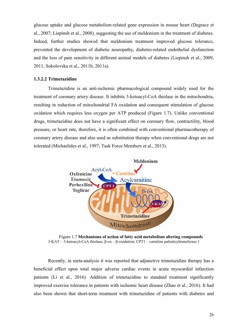
Trimetazidine is an anti-ischemic pharmacological compound widely used for the
treatment of coronary artery disease. It inhibits 3-ketoacyl-CoA thiolase in the mitochondria,
resulting in reduction of mitochondrial FA oxidation and consequent stimulation of glucose
oxidation which requires less oxygen per ATP produced (Figure 1.7). Unlike conventional
drugs, trimetazidine does not have a significant effect on coronary flow, contractility, blood
pressure, or heart rate, therefore, it is often combined with conventional pharmacotherapy of
coronary artery disease and also used as substitution therapy when conventional drugs are not
tolerated (Michaelides et al., 1997; Task Force Members et al., 2013).
Figure 1.7 Mechanisms of action of fatty acid metabolism altering compounds 3-KAT – 3-ketoacyl-CoA thiolase; β-ox – β-oxidation; CPT1 – carnitine palmitoyltransferase 1
Recently, in meta-analysis it was reported that adjunctive trimetazidine therapy has a
beneficial effect upon total major adverse cardiac events in acute myocardial infarction
patients (Li et al., 2016). Addition of trimetazidine to standard treatment significantly
improved exercise tolerance in patients with ischemic heart disease (Zhao et al., 2016). It had
also been shown that short-term treatment with trimetazidine of patients with diabetes and

27
ischemic cardiomyopathy improved left ventricular function, glucose metabolism, and
endothelial function (Fragasso et al., 2003; Rosano et al., 2003).
Interestingly, it was observed that ischemia-induced increase in acylcarnitine levels
significantly decreased with acute trimetazidine treatment along with a lower reduction in the
intracellular pH during ischemia (El Banani et al., 1998). A possible direct effect on CPT1
activity was hypothesised but disapproved as trimetazidine had a relatively low potency to
inhibit myocardial CPT1 (Kennedy and Horowitz, 1998).
1.3.2.3 CPT1 inhibitors
FA oxidation pathway inhibition has been widely studied as a possible drug target in
the case of genetic disorders involving long-chain FA oxidation defects. Recently, a study
showed that pharmacological inhibition of CPT1 restores mitochondrial oxidative
phosphorylation in human trifunctional protein deficient fibroblasts (Lefort et al., 2017).
Mitochondrial Trifunctional Protein deficiency (TFPD) is a rare genetic disorder
characterized by altered energy metabolism and subsequent accumulation of long-chain
acylcarnitines in blood and tissues. Mitochondrial trifunctional protein is an enzyme that
catalyses the three final steps of mitochondrial FA β-oxidation. Accumulation of long-chain
acylcarnitines plays a major role in the pathophysiology of TFPD, reducing oxidative
phosphorylation capacities (Lefort et al., 2017). Different clinical presentations of TFPD are
observed in patients, one of few described as a severe neonatal form with high rate mortality
due to cardiomyopathy and Reye's syndrome (den Boer et al., 2003). Cardiac disorders are
common in long-chain FA oxidation defects and is often the cause for mortality (Baruteau et
al., 2013; Vockley et al., 2015). The only currently available treatment is a diet limited in
long-chain FA to prevent the accumulation of long-chain acylcarnitines, and enriched in
carbohydrates and medium-chain triglycerides to ensure sufficient energy production
(Spiekerkoetter et al., 2009). Unfortunately, this diet does not stop the disease progression
suggesting that besides the reduced energy supply from long-chain FA, oxidative
phosphorylation capacities may be impaired, possibly due to a toxic effect of long-chain
acylcarnitines accumulated upstream the enzyme deficiency. Lefort et al. reported that CPT1
inhibition with etomoxir not only reduced the production of long-chain acylcarnitines but also
restored mitochondrial oxygen consumption and maximal ATP production rate (Lefort et al.,
2017). This evidence confirms that the long-chain acylcarnitine accumulation has a distinct
role in the pathological mechanism of cardiomyopathy and suggests possible their
involvement in development of other cardiovascular disorders. Several studies have proposed
that CPT1 inhibitors could have a beneficial effect in ischemic heart (Bergman et al., 1980;

28
Korb et al., 1984), limiting the progression of heart failure (Lionetti et al., 2005; Schmidt-
Schweda and Holubarsch, 2000) and the treatment of diabetes (Conti et al., 2011; Keung et
al., 2013). Mostly oxfenicine and etomoxir have been evaluated for these purposes (Figure
1.7).
Oxfenicine is an effective inhibitor of cardiac and muscle type CPT1 that was initially
developed for the treatment of chronic stable angina (Bergman et al., 1980). It was also shown
that oxfenicine reduced the accumulation of long-chain acylcarnitine in the ischemic
myocardium after coronary artery occlusion and reduced the myocardial infarct size (Vik-Mo
et al., 1986). However, oxfenicine is not available for human use due to its cardiac toxicity in
the form of hypertrophy (Bachmann and Weber, 1988; Greaves et al., 1984). In recent
publication oxfenicine was used to evaluate CPT1 inhibition as a drug target for treating non-
insulin-dependent diabetes mellitus. Treatment of mice with oxfenicine improved whole-body
glucose tolerance and insulin sensitivity in a diet-induced insulin resistance model (Keung et
al., 2013).
Etomoxir is an irreversible inhibitor of CPT1 that has been developed primarily for
treatment of diabetes mellitus (McGarry et al., 1989; Wolf and Engel, 1985). Etomoxir
treatment has been shown to improve glucose tolerance in diabetic patients (Hübinger et al.,
1997). Possible use of etomoxir in the treatment of heart failure was showed, as etomoxir
improved left ventricular performance in rats with pressure overload-induced cardiac
hypertrophy (Turcani and Rupp, 1997). Even though the initial clinical trial with etomoxir in
patients with heart failure showed positive results, the subsequent placebo-controlled trial was
stopped prematurely due to hepatotoxicity in patients taking etomoxir (Holubarsch et al.,
2007; Schmidt-Schweda and Holubarsch, 2000).
Besides the previously mentioned widely studied CPT1 inhibitors, oxfenicine and
etomoxir, several other inhibitors have been introduced (Figure 1.7). Perhexiline, inhibitor of
cardiac and liver type CPT1, has been shown to protect against diastolic dysfunction during
low-flow ischemia/reperfusion in the isolated rat heart (Kennedy et al., 1996, 2000).
Additional mechanisms of perhexiline have been reported; however, limited knowledge of
influence on cardiovascular function of those mechanisms is restricting the clinical
application of perhexiline (Cappola, 2015). Antidiabetic activity was assessed for teglicar, an
antihyperglycemic agent, which reduces gluconeogenesis and improves glucose homeostasis,
in both in vitro and in animal models through the selective and reversible inhibition of the
liver type CPT1 (Conti et al., 2011).
Taken together, these findings suggest that therapeutic strategies aiming at reducing
excessive FA oxidation in muscle and heart may improve insulin sensitivity and therefore

29
attenuate the development of diabetes and its cardiovascular complications. However, neither
of these studies measured the content of acylcarnitines to verify their relationship to the
progression of insulin resistance and the possible therapeutic effects of this treatment.

30
2 MATERIALS AND METHODS
2.1 Animals and treatment
Male CD-1 (8–12 weeks old, Envigo Netherlands (former Harlan Laboratories BV),
Venray, Netherlands), C57BL/6 male mice (8–12 weeks old, Envigo Netherlands), male
db/db mice (10 weeks old, Envigo Netherlands), non-diabetic db/Lean male mice (Envigo
Netherlands), male Wistar rats (10–16 weeks, Laboratory of Experimental Animals, Riga
Stradins University, Latvia), female apoE−/− mice (7 weeks old, Taconic, Ejby, Denmark)
were used for the experiments. Animals were housed under standard conditions (21−23 °C, 12
h light-dark cycle) with unlimited access to food (R70 diet, Lantmännen Lantbruk, Sweden)
and water. All animals were adapted to local conditions for two weeks before the start of
experiments. The experimental procedures were carried out in accordance with the guidelines
of the European Community, local laws and policies and were approved by the Latvian
Animal Protection Ethical Committee, Food and Veterinary Service, Riga, Latvia. All studies
involving animals are reported in accordance with the ARRIVE guidelines (Kilkenny et al.,
2010; McGrath et al., 2010).
To study the effects of long-chain acylcarnitines on glucose homeostasis, CD-1 mice
were administered with palmitoylcarnitine intraperitoneally at a dose of 50 mg/kg. In the
fasted state, glucose and insulin levels were measured 60 min after palmitoylcarnitine
administration. In the fed state, after 30 min of PC administration, insulin (0.3 IU/kg) was
administered subcutaneously, and glucose concentrations were measured 60 min after
palmitoylcarnitine administration. In addition, to ensure a continuous dosing of
palmitoylcarnitine, osmotic minipumps (ALZET®, USA) filled with palmitoylcarnitine (50
mg/kg/day) were implanted subcutaneously in the mice for 24 h. In control animals osmotic
minipumps loaded with saline (vehicle) were implanted. Glucose tolerance testing and
metabolic phenotyping was performed during 24 h after the implantation of minipumps. To
inhibit endogenous insulin release, guanabenz (i.p. 1 mg/kg), an α2-adrenoreceptor agonist
(Saha et al., 2005), was used after the single-dose palmitoylcarnitine (50 mg/kg)
administration. Mice were randomly separated into two experimental groups, guanabenz and
guanabenz + palmitoylcarnitine. To determine the insulin-dependent effects of
palmitoylcarnitine, insulin (0.3 IU/kg) was administered subcutaneously 1 h after the
intraperitoneal injection of guanabenz or guanabenz + palmitoylcarnitine.
To determine the effects of the long-term increased availability of long-chain
acylcarnitines, CD1 mice were randomly separated into control and palmitoylcarnitine

31
groups. To ensure a continuous and long-term dosing of palmitoylcarnitine, osmotic
minipumps (ALZET®, USA) filled with palmitoylcarnitine (10 mg/kg/day) were implanted
subcutaneously in the mice for 28 days. In control animals, osmotic minipumps loaded with
saline (vehicle) were implanted. At the end of the 28-day treatment, glucose and insulin
tolerance tests were performed, and biochemical parameters were measured.
To study the antidiabetic effects of pharmacological reduction of long-chain
acylcarnitine levels and physical intervention, C57BL/6 mice were divided into 3 groups and
db/db mice were divided into 4 groups as shown in Table 2.1. C57BL/6 mice were treated
with normal chow or a high fat diet (HFD, Special Diets Services, UK) for 8 weeks to induce
insulin resistance. Methyl-GBB phosphate (equivalent to 5 mg/kg of methyl-GBB) was
administered with drinking water for 8 weeks. During the same time period, forced exercise
was performed for db/db mice. For the exercise experiment, a 21-wheels forced
exercise/walking wheel apparatus (PsymCon Model 35500, Lafayette Instrument, Lafayette,
USA) was used. Before the experiment, mice were adapted to exercise for one week. For
further experiment, mice walked five days a week, 60 min a day, at a speed of 5 m/min for 8
weeks in total. The mean total number of steps performed in a 60-min test period was 3600
steps (60 steps/min). Animals were weighed twice per week. The blood samples from fed and
fasted animals were collected from the tail vein prior the start of insulin and glucose tolerance
tests. After euthanasia by cervical dislocation, the organ samples were collected. The obtained
plasma and tissue samples were stored at – 80 ºC until analysis.
Table 2.1
The design of the experiments with C57BL/6, db/Lean and db/db mice
No Group Mice with impaired insulin sensitivity
Mice with diabetes
Treatment
1 Non-diabetic control Normal chow db/Lean water
2 Control with diabetes HFD db/db water
3 Methyl-GBB HFD db/db Methyl-GBB1
4 Exercise – db/db Ex2
5 Methyl-GBB + Exercise – db/db Methyl-GBB1 + Ex2 1 methyl-GBB once a day, p.o. 5 mg/kg; 2 Ex - forced walking five days a week, 60 min/day at a speed of 5
m/min.
To study the role of acylcarnitines in ischaemia/reperfusion damage, for the isolated
heart experiments, Wistar rats were anaesthetized using sodium pentobarbital (60 mg/kg
intraperitoneal injection) with the concomitant administration of heparin (1000 units/kg). For
the isolated heart mitochondria experiments, rats were killed by decapitation. Anaesthesia
32
before decapitation was not used because chemical anaesthetics are known to affect
mitochondrial functions (Agarwal et al., 2014; La Monaca and Fodale, 2012; Nouette-Gaulain
et al., 2011).
To evaluate the link between increased pools of plasma and tissue acylcarnitines and
accelerated development of atherosclerosis and to study molecular anti-atherosclerotic
mechanisms of methyl-GBB, apoE−/− mice were treated with methyl-GBB phosphate
(equivalent to 10 mg/kg of methyl-GBB). Methyl-GBB was dissolved in the drinking water
and control group received drinking water. For the progression of atherosclerosis, all the
experimental animals at the age of 8 weeks were switched to a Western RD (P) diet that
contained 21 % fat and 0.15 % cholesterol from Special Diets Services (Essex, Great Britain).
After 4 months of treatment, the apoE−/− mice were intraperitoneally (ip) injected with 1000
UI of heparin and sacrificed under anaesthesia (sodium pentobarbital, 60 mg/kg, ip).
Afterwards, the thorax was longitudinally opened and the blood was collected from the right
ventricle. The obtained plasma was stored at − 80 °C until further analysis. Piece of the heart
muscle (~ 100 mg) from the apex of the heart was frozen in liquid nitrogen for the qRT-PCR
analysis.
2.2 Materials
Palmitoylcarnitine hydrochloride was synthesized from L-carnitine and palmitoyl
chloride as described in the literature (Nivet et al., 1991) with some modifications. Methyl-
GBB or its less hygroscopic form, methyl-GBB phosphate, were used as a source of methyl-
GBB. Both substances were provided by JSC Grindeks (Riga, Latvia). Acetonitrile and
methanol were obtained from Merck (Darmstadt, Germany). Sodium pentobarbital (Dorminal
20 %) solution was purchased from Alfasan (Woerden, Holland). Ketamine was from
Vetoquinol Biowet (Poland). Heparin sodium was purchased from Panpharma (Fougeres,
France). Coenzyme A and palmitoyl coenzyme A were from Larodan (Malmö, Sweden).
Glucose and saline solution were from Fresenius Kabi (Warsaw, Poland). Magnesium
chloride hexahydrate, L-Malic acid, calcium chloride dihydrate, sodium chloride, sodium
hydrogenphosphate, potassium phosphate monobasic, potassium chloride, L-Carnitine,
ethylene glycol-bis(β-aminoethyl ether)-N,N,N',N'-tetraacetic acid (EGTA) were obtained
from Acros Organics (Geel, Belgium). Guanabenz was purchased from Tocris Bioscience
(Bristol, UK). FA free bovine serum albumin was from Europa Bioproducts Ltd (UK). Insulin
was from Novo Nordisk (Denmark). Sodium palmitate was obtained from Tokyo chemical
industry (Tokyo, Japan). 4-(2-hydroxyethyl)-1-piperazineethanesulfonic acid (HEPES), ADP,
aprotinin, ATP, DC260126, dithiothreitol, DMEM, ethylenediamine-tetraacetate sodium salt

33
(EDTA), glycerol 3-phosphate, glycine, Igepal CA 630, leupeptin, NAD, n-butanol, Oil Red
O, oligomycin A, pepstatin, phenylmethanesulfonyl fluoride (PMSF), reduced glutathione,
rotenone, sodium dodecyl sulfate, sodium fluoride, sodium lactate, sodium orthovanadate,
sodium pyruvate, sodium succinate, subtilisin A, sucrose, TMPD, 4 % formaldehyde, Sudan
IV were obtained from Sigma Aldrich (Schnelldorf, Germany). 2-[1,2-3H]-deoxy-D-glucose,
[9,10-3H]-palmitate, [9,10-3H]palmitoylcarnitine, L-[N-methyl-3H]carnitine were from
BIOTREND Chemikalien GmbH (Köln, Germany).
2.3 Methods
2.3.1 In vitro methods
2.3.1.1 Evaluation of palmitoylcarnitine-induced effects on insulin signalling pathway in
cell cultures
To assess the effect of palmitoylcarnitine on insulin secretion in vitro, an insulin-
secreting cell line, RIN-5F (ATCC® Number: CRL-2058), was used and the insulin
concentration in the culture medium was determined using a Sensitive Rat Insulin RIA kit
(Millipore, Billerica, USA) after 1 h of incubation with palmitoylcarnitine at a dose of either 3
or 10 μM under both basal and hyperglycaemic conditions, which contained 5.5 and 11 mM
glucose, respectively. To differentiate the palmitoylcarnitine and palmitic acid-induced effects
on insulin secretion, DC260126, a small-molecule antagonist of free FA receptor 1
(FFAR1/GPR40), at a concentration of 5 μM was preincubated 1 h before the addition of
palmitoylcarnitine at a concentration of 10 μM.
To evaluate the palmitoylcarnitine-induced effect on Akt phosphorylation, C2C12
(ATCC® CRL-1772™) mouse skeletal myoblasts were differentiated in DMEM containing 2
% horse serum for 5 days. Then, the medium was replaced with serum-free DMEM and
palmitoylcarnitine at doses of 5 and 10 μM; after an overnight incubation, the basal or insulin-
stimulated (10 nM for 15 min) level of Akt phosphorylation was determined by a western blot
analysis. The medium was removed, and the cells were lysed in a buffer containing 100 mM
Tris-HCl, pH 7.4, 10 mM EDTA, 5 mM MgCl2, 1 mM glycerol 3-phosphate, 1 mM NaF, 500
μM Na3VO4, 1 mM DTT, phosphatase inhibitor cocktail I 1:100 (Alfa Aesar, United States),
protease inhibitors (10 μM leupeptin, 1 μM pepstatin, 1 μM aprotinin, and 100 μM PMSF)
and 1 % IGEPAL at 4 °C for 10 min. The lysates were centrifuged at 10000 rpm for 5 min at
4 °C. The supernatants were stored at − 80 °C until analysis.

34
2.3.1.2 Western blot analysis of tissue lysates
Muscle tissues were homogenized by an Ultra-Turrax® homogenizer (IKA, Germany)
at a ratio of 1:10 (w/v) at 4 °C in a buffer containing 100 mM Tris-HCl, pH 7.4, 10 mM
EDTA, 5 mM MgCl2, 1 mM glycerol 3-phosphate, 1 mM NaF and protease inhibitors (10 μM
leupeptin, 1 μM pepstatin, 1 μM aprotinin, and 100 μM PMSF). The PAGE and western blot
analysis of tissue lysates was performed as described previously (Liepinsh et al., 2014b). To
detect the phosphorylation level of Akt at Ser473, membranes were incubated with anti-P-Akt
(#sc-7985-R; Santa Cruz Biotechnology, CA, USA or #4060S; Cell Signaling Technology,
Danvers, MA, USA) specific antibodies and the obtained data were normalized against total
Akt (#sc-8312; Santa Cruz Biotechnology) protein expression. The blots were developed
using chemiluminescence reagents (Merck, Germany). The western blot images were scanned
and then analyzed using Gel-Pro Analyzer 6.0 software.
2.3.1.3 mRNA isolation and quantitative RT-PCR analysis
Total RNA from heart tissues was isolated using the TRI Reagent from Sigma (St.
Louis, USA) according to the manufacturer's protocol. First-strand cDNA was synthesised
using a High Capacity cDNA Reverse Transcription Kit from Applied Biosystems™ (Foster
City, USA) following the manufacturer's instructions.
Quantitative RT-PCR analysis for genes was performed by mixing synthesised cDNA,
appropriate primers, and SYBR® Green Master Mix from Applied Biosystems™ (Foster
City, USA) and run in the Applied Biosystems Prism 7500 according to the manufacturer's
protocol. The transcript levels for the constitutive housekeeping gene product β-actin were
quantitatively measured for each sample, and RT-PCR data were reported as the number of
transcripts per number of β-actin mRNA molecules.
2.3.1.4 Determination of the acylcarnitines, acyl-CoAs and CoAs in the plasma and
tissue
Determination of the acylcarnitine, acyl-CoA and CoA content in the samples (plasma,
mitochondrial suspension, heart, aortic and muscle tissue) was performed by ultra-
performance liquid chromatography MS/MS (Makrecka et al., 2014). A Micromass Quattro
Micro tandem mass spectrometer with an Acquity UPLC chromatographic system (Waters)
was used to perform the analysis. The sample extraction was performed as previously
described (Blachnio-Zabielska et al., 2011) with some modifications. Briefly, 200 μl of
freshly prepared 100 mM potassium phosphate monobasic (KH2PO4, pH 4.9) and 250 μl of
acetonitrile/2-propanol/methanol 3:1:1 (v/v/v) was added to 15 mg of aortic tissues, 50 mg of

35
other tissues or 50 μl of plasma. The sample was sonicated twice for 30 s and centrifuged at
16000 g for 10 min. The supernatant was evaporated and lyophilised. The dry extract was re-
suspended in 500 μl of acetonitrile/methanol 3:1 (v/v), vortexed and centrifuged at 13000 g
for 10 min. This supernatant was used for the UPLC/MS/MS analysis. The concentrations of
acylcarnitines and acyl-CoAs were measured against a seven-point standard curve ranging
from 10 to 1000 ng/ml for each analyte.
2.3.1.5 Determination of methyl-GBB, L-carnitine, GBB and trimethylamine N-oxide
levels in the plasma
The determination of the L-carnitine, GBB, methyl-GBB and trimethylamine N-oxide
(TMAO) concentrations in the plasma samples was performed using the UPLC/MS/MS
method and a Quattro Micro triple-quadrupole mass spectrometer (Micromass, Waters,
Milford, MA, USA) using electrospray ionisation in the positive ion mode as previously
described (Dambrova et al., 2008, 2013).
2.3.1.6 Determination of biochemical parameters in the plasma and blood samples
For biochemical measurements, blood samples were collected from the tail vein in
heparin-containing tubes. To obtain plasma, the samples were centrifuged at 1000 g for 10
min at 4 °C. All samples were stored at − 80 °C until analysis.
Glucose and lactate concentrations in plasma samples were measured by
Instrumentation Laboratory (Milan, Italy) and Roche Diagnostics (Mannheim, Germany)
enzymatic kits. Blood glucose was measured using a MediSense Optium (Abbott Diabetes
Care, Maidenhead, UK) blood glucose meter and strips. The concentration of free FAs was
measured using enzymatic kits from Wako (Neuss, Germany). The plasma insulin
concentrations were determined using a Sensitive Rat Insulin RIA kit and a Rat/Mouse
Insulin ELISA kit (Millipore, Billerica, USA). Concentrations of triglycerides (TG), HDL-
and LDL-cholesterol and the activity of alanine aminotransferase (EC 2.6.1.2 ALT) and
aspartate transaminase (EC 2.6.1.1 AST) in plasma samples were measured using kits from IL
Laboratories (Lexington, USA). Concentration of tumour necrosis factor alpha (TNFα) in
plasma was measured using TNFα ELISA Kit from Millipore.
2.3.1.7 Isolation of cardiac mitochondria
Mitochondria were isolated from the cardiac tissues as described previously (Kuka et
al., 2012a), with certain modifications. Cardiac tissues were homogenized on ice in 1:10 (w/v)
medium containing 180 mM KCl, 10 mM Tris-HCl and 1 mM EGTA (pH 7.7 at 4 °C)
supplemented with subtilisin A (0.5 mg/ml) using a Teflon glass homogenizer. The

36
homogenate was centrifuged at 750 g for 5 min at 4 °C, and then the supernatant was
centrifuged at 6800 g for 10 min at 4 °C. The obtained mitochondrial pellet was washed once
(10 min at 6800 g) and resuspended in the buffer containing 180 mM KCl and 20 mM Tris-
HCl (pH 7.2 at 4 °C). Isolated heart mitochondria were stored on ice until use or were frozen
until analysis.
2.3.1.8 Determination of fatty acid accumulation in cardiac mitochondria during
ischaemia/reperfusion
To assess the accumulation of FAs and their derivatives in cardiac mitochondria
during ischaemia, hearts were perfused as described recently (Liepinsh et al., 2014b), with
certain modifications. In particular, hearts from fasted (starved) rats were perfused with
Krebs–Henseleit (KH) buffer solution supplemented with 5 mM glucose, 1.2 mM sodium
palmitate bound to 2 % BSA, 1 mM lactate, 0.1 mM pyruvate and 0.3 ng/ml insulin at a
constant perfusion pressure of 9.33 kPa. During the adaptation period, the isolated rat hearts
were perfused with non-labelled KH buffer solution for 10 min. The perfusate was then
switched to radiolabelled ([9,10-3H]-palmitate ([3H]-palmitate), specific radioactivity, 60
Ci/mmol) KH buffer solution for 10 min. Then, the hearts were switched back to the non-
labelled KH perfusion solution, and the left anterior descending coronary artery (LAD) was
subsequently occluded for 30 min. Hearts were removed immediately after occlusion or 30
min after reperfusion. To measure the accumulation of FAs during reperfusion, hearts from
fasted rats were perfused for 20 min with KH buffer solution, as described above. The LAD
was then occluded for 30 min. During the first 10 min of reperfusion, the perfusate was
switched to the radiolabelled ([3H]-palmitate) KH buffer solution. The hearts were then
switched back to the non-labelled KH perfusion solution and perfused for additional 20 min.
At-risk and non-risk areas were separated, and mitochondria were isolated from both areas.
2.3.1.9 Flow cytometry and FACS
Mitochondria were incubated with the fluorescent dyes MitoTracker® Deep Red FM
(M22426, Life Technologies) and JC-1 (T-3168, Life Technologies). MitoTracker® Deep
Red is a far red-fluorescent dye (excitation ∼ 644 nm/emission 665 nm) that stains all
mitochondria (Cottet-Rousselle et al., 2011), and JC-1 exhibits potential-dependent
accumulation in mitochondria, as indicated by a fluorescence emission shift from green (∼
529 nm) to red (∼ 590 nm). JC-1 was used to detect the loss of mitochondrial membrane
potential, as indicated by a decrease in the red and green fluorescence intensities, and healthy
and damaged mitochondria were accordingly separated using a BD FACSAria II cell sorter
(Becton Dickinson) (Lecoeur et al., 2004). Flow cytometry data obtained for 100000 events

37
were analysed using Flowing Software (Cell Imaging Core, Turku Centre for Biotechnology,
Turku, Finland). To assess the association of [3H]-palmitate accumulation and mitochondrial
damage, isolated rat hearts were subjected to 30 min of ischaemia, followed by 30 min of
reperfusion. Approximately 2000000 mitochondria (MitoTracker®-positive) isolated from the
at-risk area were sorted into healthy and damaged subpopulations according to JC-1
fluorescence (Figure 2.1).
Figure 2.1 Separation of healthy and damaged mitochondrial subpopulations by cell sorting according to JC-1 fluorescence
The [3H]-palmitate content in each mitochondrial subpopulation was then determined
by liquid scintillation.
2.3.1.10 Determination of palmitoylcarnitine accumulation in mitochondrial fractions
Isolated rat cardiac mitochondria were incubated with 5.6 nM labelled [9,10-3H]-
palmitoylcarnitine ([3H]-palmitoylcarnitine, specific radioactivity, 60 Ci/mmol; American
Radiolabeled Chemicals) in the presence of 10 mM succinate, 1 μM rotenone and 1 mM ADP
to ensure palmitoylcarnitine accumulation in mitochondria. The mitochondria were then
washed, and mitochondrial fractions (intermembrane space, outer and inner mitochondrial
membranes and mitochondrial matrix) were prepared. The separation of the outer
mitochondrial membrane and intermembrane space was performed as described previously
(Ryu et al., 2011). Briefly, mitochondria were incubated in hypotonic solution (5 mM sucrose,
5 mM HEPES and 1 mM EGTA, pH 7.2) on ice for 5 min and then centrifuged at 14500 g for
10 min. The obtained supernatant contained outer mitochondrial membrane and
intermembrane space, whereas the obtained pellet contained inner mitochondrial membrane
and matrix. The supernatant was centrifuged at 200000 g for 90 min to sediment the outer
mitochondrial membrane, and the pellet was subjected to ten freeze-thaw cycles and

38
sonication (40 % for 20 s) to disturb inner mitochondrial membrane. The inner mitochondrial
membrane and mitochondrial matrix were separated by centrifugation at 100000 g for 30 min.
2.3.1.11 Mitochondrial respiration measurements
FA β-oxidation was determined in mitochondria isolated from the area at risk and non-
risk area of the ischaemic hearts after 30 min of ischaemia using palmitoyl-CoA (2 μM) and
carnitine (700 μM) as substrates in the presence of succinate (10 mM) and ADP (2.5 mM).
Mitochondrial respiration rate measurements were performed in mitochondria isolated from
the at-risk area and the non-risk area of ischaemic hearts in the presence of the respective 5 %
supernatant obtained during the isolation process.
The protective effects against FA derivative-induced mitochondrial toxicity were
studied using recombinant ACBP, FABP3 and purified cytosol obtained from hearts from fed
or fasted rats. To purify cytosol, hearts from fed or fasted rats were homogenized on ice in
1:10 (w/v) medium containing 180 mM KCl, 10 mM Tris-HCl and 1 mM EGTA (pH 7.7 at 4
°C) using a Teflon glass homogenizer. The homogenate was centrifuged at 6800 g for 10 min
at 4 °C, and the obtained supernatant was centrifuged at 100000 g for 60 min at 4 °C.
Centrifugation of the supernatant using an Amicon® Ultra 10K device (Millipore) was then
performed to wash out the energy substrates and to concentrate the cytosol into the initial
volume (1 g of cardiac tissues equivalent to 1 ml of purified cytosol). Mitochondrial
respiration was measured using a Clark-type electrode (Microelectrodes) in buffer containing
150 mM KCl, 2.25 mM MgCl2, 10mM Tris-HCl and 5mM KH2PO4 (pH 7.2 at 37°C).
Titrations with palmitoyl-CoA and palmitoylcarnitine were performed in the oxidative
phosphorylation (OXPHOS) state using succinate (10 mM) with rotenone (1 μM) and ADP (1
mM) as substrates.
To determine the palmitoylcarnitine-induced mitochondrial damage, high-resolution
respirometry with simultaneous fluorimetry was performed using an Oxygraph-2k (O2k;
Oroboros Instruments) combined with the O2k-Fluorescence LED2-Module. Succinate (10
mM) with rotenone (1 μM) were used to determine complex II-linked respiration, and
pyruvate and malate (5 mM and 0.5 mM respectively) with succinate (10 mM) were used to
determine Complex I- and II-linked respiration. Measurements were performed in the
OXPHOS state using 1 mM ADP. Oligomycin A (Omy A) at 10 μM was added to determine
LEAKOmy-linked respiration. In addition, simultaneously with respiratory measurements,
tetramethylrhodamine methyl ester (TMRM, LST668, Life Technologies) (2 μM) was used to
determine the mitochondrial membrane potential, Calcium GreenTM-5N (C3737, Invitrogen)
was used to determine mitochondrial calcium release, and H2O2 flux was measured using the

39
H2O2-sensitive probe Amplex® Ultra Red (A36006, Life Technologies), as described
previously (Makrecka-Kuka et al., 2015).
2.3.1.12 Measurement of CPT1 activity and CPT2-dependent β-oxidation
CPT1 activity in intact isolated mitochondria was assayed as the formation of
palmitoyl-[3H]-carnitine using palmitoyl-CoA and radiolabelled L-[N-methyl-3H]carnitine as
described previously (Berthon et al., 1998) with some modifications. The final concentrations
in the assay were 50 μM palmitoyl-CoA, 1.5pmol of L-[N-methyl-3H]carnitine (specific
radioactivity, 85 Ci/mmol; Biotrend), 0.5 μM rotenone, 60 mM KCl, 150 mM sucrose, 1 mM
DTT, 1 mg/ml FA free-BSA and 25 mM HEPES, pH 7.4. Mitochondria isolated from the at-
risk and non-risk areas of the ischaemic hearts after 30 min of ischaemia were diluted in the
respective supernatant obtained during the isolation process. The reaction was performed at
room temperature for 5 min and stopped by the addition of ice-cold 2 M perchloric acid.
Samples were then centrifuged at 10000 g for 10 min at 4 °C. The obtained pellet was
suspended in water and palmitoyl-[3H]carnitine was extracted with n-butanol by vigorously
mixing samples for 1 min followed by centrifugation at 10000 g for 10 min at 4 °C. After
centrifugation, an aliquot of the butanol phase was collected and the radioactivity was
measured using a Wallac MicroBeta Trilux scintillation counter (PerkinElmer). The data were
normalized to the mitochondrial protein content (determined using the Lowry method).
To determine the CPT2-dependent β-oxidation, isolated mitochondria were sonicated
(Cole Parmer 130-Watt ultrasonic processor set at 20 % for 20 s) and diluted in isolation
buffer (180 mM KCl and 20 mM Tris-HCl, pH 7.2 at 4°C) or in respective supernatant from
the at-risk and non-risk areas of the ischaemic hearts. The CPT2-dependent β-oxidation was
determined as described previously (Demaugre et al., 1988) with some modifications. The
incubation medium (200 μl) contained 115 mM Tris-HCl, pH 7.4, 70 mM KCl, 4.5 mM
reduced glutathione and 20 μl of diluted mitochondria. After 2.5 min of pre-incubation, the
reaction was initialized by the addition of 1.67 pmol of [3H]-palmitoylcarnitine in 50 μl. The
reaction was performed at room temperature for 3 min and stopped with 50 μl of 6 M
perchloric acid. Samples were then centrifuged at 10000 g for 10 min at 4°C. The obtained
supernatant was vigorously mixed with n-butanol for 1 min and centrifuged at 10000 g for 10
min at 4 °C. The radioactivity content in the aqueous phase was then determined by liquid
scintillation. The data were normalized to the mitochondrial protein content.
2.3.1.13 Binding of fatty acid derivatives to recombinant FABP and ACBP
To determine the ability of palmitoylcarnitine to compete with palmitate and
palmitoyl-CoA for binding to FABP and ACBP respectively, binding studies were performed

40
as described previously (Rasmussen et al., 1990; Vork et al. 1990). Recombinant human
FABP3 was purchased from Cayman chemical (Batch No. 0450256-1). Recombinant ACBP
was prepared in collaboration with Dr. Kaspars Tārs (Latvian Biomedical Research and Study
Centre) as follows. A plasmid composed of synthetic cDNA corresponding to ACBP isoform
3 (NCBI Reference Sequence NP_001073331.1) that was complemented at the 5’ end with
6×His codons, a tobacco etch virus protease cleavage site and the T7 promoter was ordered
from Integrated DNA Technologies. This plasmid was transformed into Escherichia coli
strain BL21(DE3). The cells were grown in 2×TY medium until the D600 reached 0.8–1.0,
induced by addition of IPTG to a concentration of 1 mM and harvested by centrifugation at
1000 g after 4 h. The cells were then lysed by sonication, and the protein was purified by
metal-affinity chromatography using a 1 ml HisTrap (GE Healthcare) column.
2.3.1.14 Quantification of the atherosclerotic lesions in the aortic sinus
After the blood was taken, the right ventricle was incised, and the heart was perfused
with 10 ml of phosphate buffered saline through the apex of the left ventricle. Next, the heart
and the whole aorta were cleaned from the surrounding fatty and connective tissues and
dissected. Afterwards the heart was cut along a plane between the tips of 2 atria, and the top
half was embedded in OCT Tissue-Tek embedding medium (Sakura, The Netherlands) and
snap-frozen. Nine 10 μm thick cryosections were cut from the appearance of the aortic valves
with 100 μm intervals. Sections were mounted on poly-L-lysine coated slides and dried. After
fixation in neutral 4 % paraformaldehyde solution, sections were stained with Oil Red O
(ORO). Finally, the ORO-stained sections were examined under a Leica DM IL microscope
(Wetzlar, Germany) and used for quantitative evaluation. Images of the aorta were recorded
using a Leica DFC490 digital camera (Wetzlar, Germany). The total area of the lesion was
measured using Image-Pro Plus 6.3 software, as previously described (Liepinsh et al., 2013a).
2.3.1.15 Quantification of the infiltration of macrophages and monocytes in the
atherosclerotic lesions
To determine the number of macrophages and monocytes in atherosclerotic lesions of
the aortic sinus, 10 μm sections from aortic roots were prepared similarly as described before
(Section 2.3.1.14). Sections were mounted on poly-L-lysine coated slides, air-dried and fixed
in cold acetone. Following fixation, the sections were stained with rat anti-MOMA-2
antibodies from Abcam (Cambridge, UK), as previously described (Hoyer et al., 2012).

41
2.3.1.16 Quantification of the atherosclerotic lesions in the aorta
The aortas, from arch to bifurcation, were fixed in 4 % formaldehyde. Following
fixation, the whole aorta was longitudinally opened, pinned onto silicone plates and stained
for lipids with Sudan IV. Images of the aorta were captured using a Sony α77 digital camera,
and the total area of the lesion was calculated using the Image-Pro Plus 6.3 software. The
extent of atherosclerosis was expressed as a percentage of the aortic surface covered by
lesions compared to the total aortic surface.
2.3.2 Ex vivo and in vivo methods
2.3.2.1 Determination of glucose and insulin tolerance tests
To perform the glucose tolerance test, the mice were fasted overnight. Then, the
glucose solution (0.5 or 1 g/kg of body weight) was administered intraperitoneally, and blood
samples were drawn from the tail vein at 0 (fasting), 15, 30, 60, 120, 180 and 240 min. For
the insulin tolerance test experiment, an insulin solution was administered intraperitoneally or
subcutaneously (with the respective doses of 0.75 or 0.3–0.5 IU/kg) to the fed mice, and
blood samples were drawn from the tail vein at 0 (fed), 30, 60, 120 and 240 min (additional
blood sample at 24 hours was collected in the experiment with db/db mice). The blood
glucose concentration was measured using a MediSense Optium Xceed blood glucose meter
and strips.
2.3.2.2 Measurements of glucose uptake and fatty acid metabolism in vivo
To determine the glucose uptake or FA metabolism (uptake and oxidation) in vivo, 1
μCi of 2-[1,2-3H]-deoxy-D-glucose ([3H]-DOG, specific activity, 60 Ci/mmol) or 1 μCi [9,10-3H]-palmitate (specific activity, 60 Ci/mmol), respectively, were administered intravenously
to the mice. After 10 min, the mice were sacrificed by cervical dislocation, and heart, skeletal
muscle, liver and adipose tissue homogenates (1:5, w/v in MilliQ water) were prepared. To
determine the palmitate metabolism in vivo, samples were treated as previously described
(Liepinsh et al., 2015). The contents of [3H]-DOG or [3H]-palmitate in the tissue samples
were determined by a liquid scintillation method.
2.3.2.3 Isolated rat heart infarction study
Infarction was induced according to the Langendorff technique, as described
previously (Kuka et al., 2012a), with certain modifications. For the infarction studies, hearts
were perfused with KH buffer solution at a constant perfusion pressure of 8 kPa. The isolated
rat hearts were adapted for 20 min, and the LAD was subsequently occluded for 30 min,

42
followed by 120 min of reperfusion. Occlusion was confirmed by a 40 % fall in coronary
flow. The infarct size (IS) was determined as described previously (Liepinsh et al., 2013a).
Briefly, at the end of the reperfusion, the LAD was re-occluded, and the heart was perfused
with 0.1 % Methylene Blue dissolved in KH buffer solution. Afterwards, the ventricles of the
heart were transversely cut into 2-mm-thick slices and photographed. Computerized
planimetric analyses of the photographs of the stained ventricle slices were performed using
Image-Pro Plus v6.3 software (Media Cybernetics) to determine the area at risk (AAR) and
the area of necrosis (AN), and each area was expressed as a percentage of the left ventricle
(LV) area. The obtained values were then used to calculate the IS as a percentage of the AAR,
according to the formula IS = (AN/AAR) × 100 %.
2.3.2.4 Metabolic phenotyping
The PhenoMaster system for mice (TSE, Germany), with automated food/liquid
access control units and an indirect gas calorimetry system, was used to monitor 24 h changes
in energy metabolism. Indirect gas calorimetry was used for the measurements of the animals’
oxygen consumption (VO2) and carbon dioxide production (VCO2) to estimate various
metabolic parameters, including the respiratory exchange rate (RER) and fat and carbohydrate
utilization for energy production.
2.4 Statistical analysis
Data are presented as the mean ± standard error of the mean (S.E.M.). Statistically
significant differences in the mean values were evaluated using Student’s t-test, Chi-Square
test or a one-way ANOVA with Tukey`s, two-tailed Student's t-test, Mann–Whitney U test or
Dunnett’s post-tests. The differences were considered significant when P < 0.05. The data
were analysed using Graph Pad Prism 5.03 software (Graph Pad Inc., La Jolla, USA).

43
3 RESULTS
3.1 The effects of acute and long-term administration of palmitoylcarnitine on energy
metabolism in mice
To study the role of acylcarnitines in the development of insulin resistance, acute and
long-term palmitoylcarnitine administration was used to induce an increase in long-chain
acylcarnitine concentrations in mice. In vitro experiments in cell culture lines were performed
to detail the molecular mechanisms of excessive accumulation of acylcarnitines during the
accelerated development of insulin resistance.
3.1.1 Effects of single-dose palmitoylcarnitine administration on glucose metabolism
Administration of palmitoylcarnitine at a dose of 50 mg/kg induced only a 3-fold
increase in the intramuscular content of long-chain acylcarnitines (Figure 3.1). In comparison,
after overnight fasting skeletal muscle long-chain acylcarnitine content was 5-fold higher than
long-chain acylcarnitine content in fed state (Figure 3.1). Thus, palmitoylcarnitine treatment-
induced effect on muscle long-chain acylcarnitine content is similar to short-term fasting.
*
*
0
10
20
30
40
50
Fed Fed + PC Fasted
Lon
g-ch
ain
acyl
carn
itin
es,
nmol
/g m
uscl
e
Figure 3.1 The effect of acute administration of palmitoylcarnitine (PC, 50 mg/kg i.p. 1 h) on the concentrations of long-chain acylcarnitines in muscle tissues
Each value represents the mean ± S.E.M. of 8 (Fed), 3 (Fed + PC) or 5 (fasted) animals. *Significantly different from the fed group (ANOVA following Tukey's test, P < 0.05).
The single-dose administration of palmitoylcarnitine induced a significant increase in
the blood glucose concentrations of fasted (Figure 3.2 A) and fed (Figure 3.2 B) mice. The
administration of insulin significantly reduced blood glucose with and without the co-
administration of palmitoylcarnitine. This additional increase in insulin concentration could
decrease the palmitoylcarnitine-induced effect on blood glucose, suggesting that higher
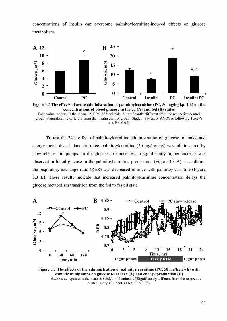
44
concentrations of insulin can overcome palmitoylcarnitine-induced effects on glucose
metabolism.
*
0
2
4
6
8
10
12A
*
*
*, #
0
5
10
15
20
25B
Control
Glu
cose
, mM
Glu
cose
, mM
PC Control Insulin PC Insulin+PC
Figure 3.2 The effects of acute administration of palmitoylcarnitine (PC, 50 mg/kg i.p. 1 h) on the concentrations of blood glucose in fasted (A) and fed (B) states
Each value represents the mean ± S.E.M. of 5 animals. *Significantly different from the respective control group, # significantly different from the insulin control group (Student’s t-test or ANOVA following Tukey's
test, P < 0.05).
To test the 24 h effect of palmitoylcarnitine administration on glucose tolerance and
energy metabolism balance in mice, palmitoylcarnitine (50 mg/kg/day) was administered by
slow-release minipumps. In the glucose tolerance test, a significantly higher increase was
observed in blood glucose in the palmitoylcarnitine group mice (Figure 3.3 A). In addition,
the respiratory exchange ratio (RER) was decreased in mice with palmitoylcarnitine (Figure
3.3 B). These results indicate that increased palmitoylcarnitine concentration delays the
glucose metabolism transition from the fed to fasted state.
*
0
3
6
9
12
0 30 60 120Time , min
Control PC
0.7
0.75
0.8
0.85
0.9
0.95
0 3 6 9 12 15 18 21 24
RE
R
Time, hrs
Control PC slow release
Light phase Dark phase Light phase
BA
Glu
cose
,mM
Figure 3.3 The effects of the administration of palmitoylcarnitine (PC, 50 mg/kg/24 h) with osmotic minipumps on glucose tolerance (A) and energy production (B)
Each value represents the mean ± S.E.M. of 4 animals. *Significantly different from the respective control group (Student’s t-test, P < 0.05).

45
To determine whether acylcarnitines stimulate insulin secretion the effects of
palmitoylcarnitine on insulin release were studied both in vitro and in vivo. Results showed
that 3 and 10 μM concentrations of palmitoylcarnitine were sufficient to increase intracellular
long-chain acylcarnitine contents within one hour and to stimulate insulin release from RIN-
5F cells (Figure 3.4). To evaluate whether palmitoylcarnitine acts by a different mechanism
than FA, palmitoylcarnitine was incubated in the presence of DC260126, an inhibitor of FA
targeted GPR40 (FFAR1) receptor in β-cells. The effect of 10 μM palmitoylcarnitine was not
inhibited by DC260126 (Figure 3.4) indicating that observed palmitoylcarnitine effect on
insulin release was not induced by palmitate action.
*
*
*
*
0
5
10
15
20
25
30
35
Control PC 3 PC 10 PC 10 +inhibitor
Control PC 3 PC 10 PC 10 +inhibitor
5.5 mM glucose 11 mM glucose
Insu
lin, A
U
Figure 3.4 Insulin release in insulin-secreting cell line (RIN-5F) after 1 h incubation with palmitoylcarnitine (PC, 3 and 10 μM) and inhibitor DC260126
Each value represents the mean ± S.E.M. of 5 animals. *Significantly different from the respective control group (ANOVA following Tukey's test, P < 0.05).
To confirm this effect in vivo, a bolus palmitoylcarnitine was administered to fasted
mice. The administration of palmitoylcarnitine induced a significant 5-fold increase in insulin
concentration (Figure 3.5 A). Up to a 2-fold increase in insulin concentration was also
observed after palmitoylcarnitine administration in fed mice (Figure 3.5 B). Overall,
palmitoylcarnitine potentiates glucose-stimulated insulin release, thus suggesting that the
long-chain acylcarnitine effect is important for the physiological transition from the fasted to
fed state and that it induces hyperinsulinemia in the case of diabetes.
For more detailed analysis of the palmitoylcarnitine effect on insulin release, another
experimental method suitable for the evaluation of both insulin-dependent and insulin-
independent glucose homeostasis in vivo was used. To study the insulin-independent
palmitoylcarnitine effects on plasma glucose, mice were administered the α2-adrenoreceptor
agonist guanabenz, which inhibits endogenous insulin release (Angel et al., 1988; Saha et al.,

46
2005). For the evaluation of insulin-dependent effects, guanabenz administration was
followed by insulin administration (Figure 3.6).
*
0.0
0.2
0.4
0.6
Control PC
Insu
lin, n
g/m
lA
*
*
0.0
0.5
1.0
1.5
2.0
2.5
Control 1 h 4 h
PC
Insu
lin, n
g/m
l
B
Figure 3.5 The concentration of insulin in the plasma after the administration of palmitoylcarnitine (PC, 50 and 100 mg/kg) in fasted (A) and fed state (B)
Each value represents the mean ± S.E.M. of 5 animals. *Significantly different from the respective control group (Student’s t-test or ANOVA following Tukey's test, P < 0.05).
In the fed control mice, administration of guanabenz caused hypoinsulinemia (Figure
3.6 A) and induced a marked increase in plasma glucose from 8.6 mM up to 17.7 mM, while
the subsequent insulin administration increased insulin concentration (Figure 3.6 A) and
significantly decreased the glucose concentration back to the initial level (Figure 3.6 B). The
administration of palmitoylcarnitine in combination with guanabenz induced an increase in
the plasma glucose concentration to 3 mM higher concentration than compared to the
guanabenz group.
$$
0.0
0.4
0.8
1.2
1.6
2.0
Hypo-insulinemia
InsulineffectBaseline
GB 60 min
GB
GB + PC
Insu
lin,
ng/
ml
A
*
*
0
5
10
15
20
25
30 min 60 min 30 min 60 minBaseline Hypo-
insulinemia Insulin effect
GB GB +PCBInsulin
GB
Glu
cos
e,m
M
Figure 3.6 Insulin-independent palmitoylcarnitine (PC, 50 mg/kg i.p.) effects on the levels of plasma insulin (A) and glucose (B)
Each value represents the mean ± S.E.M. of 8 animals. *Significantly different from the respective control group, $ significantly different from the baseline (Student’s t-test or ANOVA following Tukey's test, P <
0.05). GB – guanabenz.

47
Palmitoylcarnitine also significantly diminished the insulin-induced blood glucose
lowering effect (Figure 3.6 B). Thus, the blood glucose concentration in the
palmitoylcarnitine group remained 5 mM higher than that in the control mice.
These effects could be explained by a palmitoylcarnitine-induced significant decrease
in insulin stimulated [3H]-DOG uptake in skeletal muscles (Figure 3.7 A).
** *
*
0
50
100
150
200
250
300
Muscle LiverAdiposetissues Muscle Liver
Adiposetissues
Insulin
GB GB + PC
[H
]-D
OG
up
tak
e3
, fm
ol/g
A
0
200
400
600
800
1000
1200
1400
ACS FA ACS FA ACS FA
Muscle Liver Adipose tissues
GB GB + PC
3 H F
A m
etab
olit
es, f
mol
/g
B
Figure 3.7 The effects of acute administration of palmitoylcarnitine (PC, 50 mg/kg i.p. 1 h) with guanabenz (GB) and insulin on [3H]deoxyglucose uptake (A) and the uptake and metabolism of
[3H]-palmitate (B) Each value represents the mean ± S.E.M. of 8 animals. *Significantly different from the respective
control group (ANOVA following Tukey's test, P < 0.05).
Importantly, the palmitoylcarnitine effect on blood glucose is partially masked by the
significantly stimulated [3H]-DOG uptake in liver and adipose tissue (Figure 3.7 A). In
addition, no effect of palmitoylcarnitine on [3H]-palmitate uptake and metabolism was
observed (Figure 3.7 B). Overall, the palmitoylcarnitine administration limits insulin- related
glucose uptake in muscles.

48
3.1.2 Mechanisms of palmitoylcarnitine action
To assess whether palmitoylcarnitine has an impact on insulin signalling pathway the
phosphorylation of Akt Ser-473 was evaluated in the C2C12 muscle cell line after incubation
with palmitoylcarnitine (Figure 3.8).
P-Akt 473
Insulin 100
PC 10 PC 5
Akt
P-Akt 473( )long exp .
P-Akt 473
Insulin 100 + + + + + +– – – – –
PC 10 + + +– – – – – – – –PC 5 + + + – – – – – – – –
Akt
P-Akt 473( )long exp .
*
*,#
#
0
50
100
150
200
250
300
350
Control PC Insulin PC+Insulin
B
A
P-A
kt/
Ak
t, A
U
Akt
P-Akt 473
PC - + - + - + - +
Akt
P-Akt 473
Insulin + + + + + + + + PC - + - + - + - +
Figure 3.8 The effect of palmitoylcarnitine (PC) with or without insulin on Akt (Ser-473) phosphorylation in C2C12 cells (overnight incubation of 5 or 10 μM PC, A) or in fed mouse
muscle in vivo (50 mg/kg PC i.p. 1 h, B) Each value represents the mean ± S.E.M. of 5 animals. *Significantly different from the control group, #significantly different from the palmitoylcarnitine group (ANOVA following Tukey's test, P < 0.05).
The overnight incubation of palmitoylcarnitine (5 or 10 μM) with or without insulin
(10 nM for 15 min) decreased Akt phosphorylation in the C2C12 cell line (Figure 3.8 A). The
increase in insulin concentration overcame the palmitoylcarnitine-induced inhibition of Akt
phosphorylation (Figure 3.8 A). In the C2C12 cell line, incubation with palmitoylcarnitine in
the presence of 100 nM insulin did not influence the phospho-Akt Ser-473 levels (Figure 3.8
A). A similar effect on Akt phosphorylation was observed in mouse muscles after a single
administration of palmitoylcarnitine (50 mg/kg) (Figure 3.8 B). Thus, palmitoylcarnitine
induces the inhibition of Akt phosphorylation, while increases in insulin concentration up to a
certain level can overcome the palmitoylcarnitine-induced effects on insulin signalling.
It would be expected that administered palmitoylcarnitine competes with FA for
uptake and oxidation in mitochondria. However, palmitoylcarnitine does not influence the FA
uptake and metabolism rate in skeletal muscle tissue (Figure 3.7 B). Taking into account that
FA metabolism was not influenced by palmitoylcarnitine, the inhibition of glucose

49
metabolism is also not related to the Randle cycle and excessive mitochondrial oxidation of
palmitoylcarnitine. In addition, palmitoylcarnitine administration does not influence the
expression of genes involved in muscle glucose transport and FA metabolism (Figure 3.9 AB)
and genes related to inflammation (Figure 3.9 C).
$
$
$*
0
1
2
3
4
GLUT1 GLUT4 CPT1A CPT1B ACSLGen
e ex
pres
sion
, AU
Control, fedPC, fedControl, fastedPC, fasted
A$
0
4
8
12
16
PDK4
Gen
e ex
pres
sion
, AU
B
$
0
0.5
1
1.5
2
TNFa IL1B iNOSGen
e ex
pres
sion
, AU
Control, fed PC, fedControl, fasted PC, fasted
C
Figure 3.9 Differences in the expression of genes involved in energy metabolism (A, B) and inflammation (C) in the muscle tissue from fasted and fed mice after acute administration of
palmitoylcarnitine (PC, 100 mg, 1 h) Each value represents the mean ± S.E.M. of 5 animals. *Significantly different from the control group,
$Significantly different from the fed control group (ANOVA following Tukey's test, P < 0.05).
We observed significant differences in gene expression between the fed and fasted
states; however, palmitoylcarnitine did not influence FA metabolism-related and
inflammatory gene expression in either state. Overall, the inhibitory effect on Akt
phosphorylation and related insulin signalling is an important mechanism of
palmitoylcarnitine action.
3.1.3 Effects induced by the long-term, slow-release administration of
palmitoylcarnitine
To ensure the long-term administration of palmitoylcarnitine, we used slow-release
osmotic minipumps (ALZET®, USA). This experimental setup ensured a permanent increase
in long-chain acylcarnitine content in muscles for 28 days. In the control mice minipumps
loaded with saline (vehicle) were implanted. Palmitoylcarnitine administration at a dose of 10
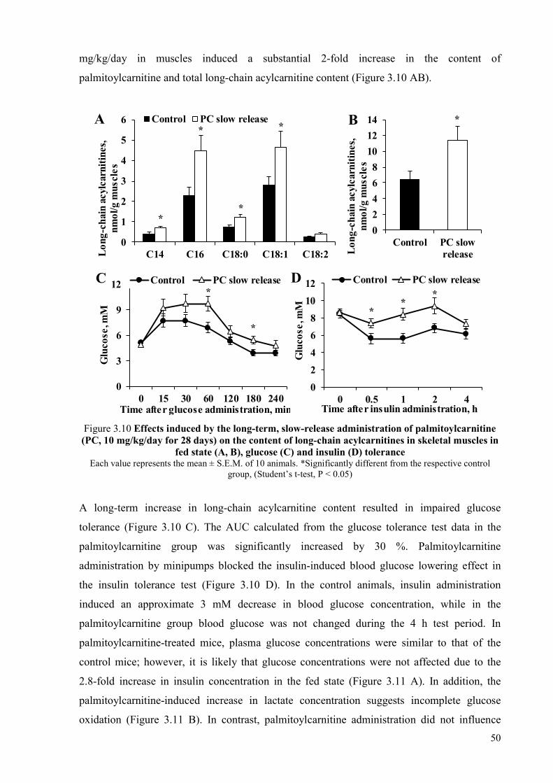
50
mg/kg/day in muscles induced a substantial 2-fold increase in the content of
palmitoylcarnitine and total long-chain acylcarnitine content (Figure 3.10 AB).
*
*
*
*
0
1
2
3
4
5
6
C14 C16 C18:0 C18:1 C18:2Lon
g-ch
ain
acyl
carn
itin
es,
nmol
/g m
uscl
esControl PC slow releaseA *
0
2
4
6
8
10
12
14
Control PC slowreleaseL
ong-
chai
nac
ylca
rnit
ines
, nm
ol/g
mus
cles
B
*
*
0
3
6
9
12
0 15 30 60 120 180 240
Glu
cose
, mM
Control PC slow releaseC
Time after glucose administration, min
**
*
0
2
4
6
8
10
12
0 0.5 1 2 4
Glu
cose
, mM
Control PC slow releaseD
Time after insulin administration, h
Figure 3.10 Effects induced by the long-term, slow-release administration of palmitoylcarnitine (PC, 10 mg/kg/day for 28 days) on the content of long-chain acylcarnitines in skeletal muscles in
fed state (A, B), glucose (C) and insulin (D) tolerance Each value represents the mean ± S.E.M. of 10 animals. *Significantly different from the respective control
group, (Student’s t-test, P < 0.05)
A long-term increase in long-chain acylcarnitine content resulted in impaired glucose
tolerance (Figure 3.10 C). The AUC calculated from the glucose tolerance test data in the
palmitoylcarnitine group was significantly increased by 30 %. Palmitoylcarnitine
administration by minipumps blocked the insulin-induced blood glucose lowering effect in
the insulin tolerance test (Figure 3.10 D). In the control animals, insulin administration
induced an approximate 3 mM decrease in blood glucose concentration, while in the
palmitoylcarnitine group blood glucose was not changed during the 4 h test period. In
palmitoylcarnitine-treated mice, plasma glucose concentrations were similar to that of the
control mice; however, it is likely that glucose concentrations were not affected due to the
2.8-fold increase in insulin concentration in the fed state (Figure 3.11 A). In addition, the
palmitoylcarnitine-induced increase in lactate concentration suggests incomplete glucose
oxidation (Figure 3.11 B). In contrast, palmitoylcarnitine administration did not influence
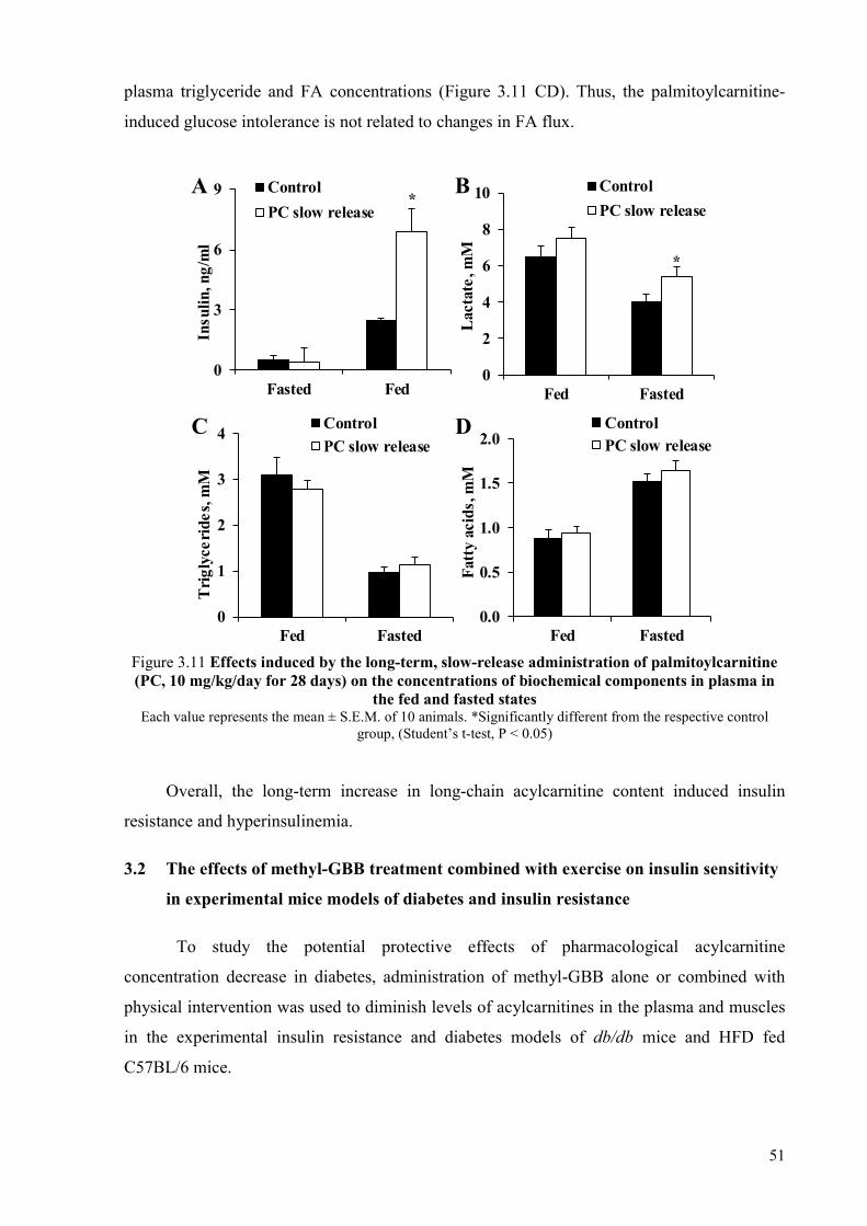
51
plasma triglyceride and FA concentrations (Figure 3.11 CD). Thus, the palmitoylcarnitine-
induced glucose intolerance is not related to changes in FA flux.
*
0
3
6
9
Fasted Fed
Insu
lin,n
g/m
lControl
PC slow release
A
*
0
2
4
6
8
10
Fed Fasted
Lac
tate
, mM
Control
PC slow release
B
0
1
2
3
4
Fed Fasted
Tri
glyc
erid
es, m
M
Control
PC slow releaseC
0.0
0.5
1.0
1.5
2.0
Fed Fasted
Fat
ty a
cids
, mM
ControlPC slow release
D
Figure 3.11 Effects induced by the long-term, slow-release administration of palmitoylcarnitine (PC, 10 mg/kg/day for 28 days) on the concentrations of biochemical components in plasma in
the fed and fasted states Each value represents the mean ± S.E.M. of 10 animals. *Significantly different from the respective control
group, (Student’s t-test, P < 0.05)
Overall, the long-term increase in long-chain acylcarnitine content induced insulin
resistance and hyperinsulinemia.
3.2 The effects of methyl-GBB treatment combined with exercise on insulin sensitivity
in experimental mice models of diabetes and insulin resistance
To study the potential protective effects of pharmacological acylcarnitine
concentration decrease in diabetes, administration of methyl-GBB alone or combined with
physical intervention was used to diminish levels of acylcarnitines in the plasma and muscles
in the experimental insulin resistance and diabetes models of db/db mice and HFD fed
C57BL/6 mice.

52
3.2.1 Content of acylcarnitines in db/db mice plasma and muscles
To evaluate whether acylcarnitines accumulate during diabetes, the acylcarnitine
content was measured in the fed and fasted states (Table 3.1).
Table 3.1
Content of acylcarnitines in plasma and muscle tissues
Plasma acylcarnitines (pM)
Group Metabolic
state Short-chain Medium-chain Long-chain
db/Lean Fasted 689 ± 32 248 ± 26* 5384 ± 721
Fed 471 ± 39 ND 417 ± 65*
db/db Control Fasted 656 ± 109 78 ± 12 3358 ± 710
Fed 538 ± 30 29 ± 4 1413 ± 72
db/db Methyl-GBB Fasted 32 ± 5* ND 646 ± 77*
Fed 29 ± 4* ND 202 ± 15*
db/db Exercise Fasted 668 ± 20 129 ± 7* 3305 ± 231
Fed 545 ± 61 43 ± 6 1501 ± 132
db/db Methyl-GBB + Exercise
Fasted 44 ± 5* ND 702 ± 39*
Fed 37 ± 22* ND 290 ± 113*
Muscle tissue acylcarnitines (nmol/g)
Group Metabolic
state Short-chain Medium-chain Long-chain
db/Lean Fasted 44.4 ± 3.6 3.5 ± 0.1 116.6 ± 14.9
Fed 43.3 ± 1.1 0.6 ± 0.1* 12.3 ± 1.1*
db/db Control Fasted 47.3 ± 7.3 4.3 ± 0.8 101.7 ± 17.9
Fed 55.7 ± 5.6 2.4 ± 0.7 65.0 ± 14.6
db/db Methyl-GBB Fasted 0.7 ± 0.1* 0.02 ± 0.02* 1.9 ± 0.6*
Fed 1.1 ± 0.2* 0.04 ± 0.02* 1.7 ± 0.4*
db/db Exercise Fasted 65.8 ± 7.3 3.5 ± 0.3 102.2 ± 7.6
Fed 58.2 ± 3.0 2.5 ± 0.2 73.9 ± 4.4
db/db Methyl-GBB + Exercise
Fasted 1.5 ± 0.2* ND 1.6 ± 0.4*
Fed 0.8 ± 0.1* ND 1.3 ± 0.4*
The results are average values ± S.E.M. of 5 animals. *Significantly different from db/db control group (one-way ANOVA with Dunnett’s post-test P < 0.05). ND – not detected.
In the fed db/Lean mice, the plasma concentrations of long-chain acylcarnitines were
13-fold lower than in the fasted state. However, in db/db mice the difference drops to two-
fold, suggesting it as a possible indicator of metabolic inflexibility. Thus, in the fasted state,
the plasma content of long-chain acylcarnitines in db/db mice was 37 % lower than in
db/Lean mice (Table 3.1, Figure 3.12 A). However, in the fed state, plasma long-chain
acylcarnitine concentration was 3.5-times higher in db/db mice than in db/Lean mice (Table
3.1, Figure 3.12 A), resulting in less difference between the content of long-chain
acylcarnitines in the fed and fasted states in db/db control mice. The acylcarnitine content in

53
the plasma fully reflects the acylcarnitine content in the muscles (Table 3.1, Figure 3.12 AB).
In the muscles of fasted db/db and db/Lean mice, long-chain acylcarnitine contents were
similar, while in fed db/db mice compared to db/Lean mice, a 5-fold higher long-chain
acylcarnitine content was observed. These results demonstrate that muscle accumulation of
acylcarnitines is related to the detrimental effects on glucose metabolism in the fed state.
Exercise did not influence the concentrations of short-, medium- and long-chain
acylcarnitines in plasma and muscle (Table 3.1, Figure 3.12 AB). In contrast, methyl-GBB
treatment induced a substantial decrease in all acylcarnitine plasma and muscle concentrations
in both states as well as when it was combined with exercise (Table 3.1, Figure 3.12 A). Thus,
methyl-GBB induced up to a 60-fold decrease in the long-chain acylcarnitine concentration in
muscles and up to a 7-fold decrease in the plasma (Table 3.1, Figure 3.12 AB).
* *# # #0
100020003000400050006000
7000
db/Lean Control Methyl-GBB
Ex
db/db
Fasted Fed
Lon
g-ch
ain
acyl
carn
itin
es, p
M
B
A
Methyl-GBB+Ex
* *#
# #0
2040
60
80100
120
140Fasted Fed
db/Lean Control Methyl-GBB
Ex
db/db
Methyl-GBB+Ex
Lon
g-ch
ain
acyl
carn
itin
es, n
mol
/g
Figure 3.12 The effect of methyl-GBB administration (5 mg/kg for 8 weeks) and exercise on long-chain acylcarnitine concentrations in db/db mouse plasma (A) and muscles (B)
Each value represents the mean ± S.E.M. of 10 animals. *Significantly different from the fasted db/db control group, #significantly different from the fed db/db control group (ANOVA following Tukey's test P < 0.05).
Results indicate that the postprandial plasma concentration of long-chain
acylcarnitines could be a marker for insulin resistance. The methyl-GBB-induced decrease in
acylcarnitine content was consistent in fed and fasted states and in combination with exercise.

54
3.2.2 Methyl-GBB and exercise induced effects on glucose and insulin tolerance
To test glucose metabolism disturbances in db/db mice and insulin resistant, HFD fed
C57BL/6 mice, glucose and insulin tolerance tests were performed (Figure 3.13).
**
**
*
0
3
6
9
12
0 0.5 1 2
Control normal chowHFD-ControlHFD + Methyl-GBB
Time after insulin administration, h
Glu
cose
, mM
A
*
* **
* * **
**
0
5
10
15
20
25
0 15 30 60 120 180 240
Control normal chowHFD-ControlHFD + Methyl-GBB
Time after glucose administration, min
B
Glu
cose
, mM
** * *,$ *$
*,$
* *
*, $*, $ *, $
*
0
5
10
15
20
25
30
35
0 0.5 1 2 4 24Time after insulin administration, h
db/Lean db/db Controldb/db Methyl-GBB db/db Exdb/db Methyl-GBB + Ex
C
Glu
cose
, mM
Figure 3.13 Methyl-GBB administration (5 mg/kg for 8 weeks) and exercise induced effects on glucose concentration after insulin (A, C) and glucose (B) administration-induced changes in
HFD (A, B) and db/db (C) mice Each value represents the mean ± S.E.M. of 10 animals. *Significantly different from the corresponding control
group, $significantly different from the Ex group (ANOVA following Tukey's test P < 0.05).
Results showed, that HFD-induced disturbances in glucose metabolism were
prevented by methyl-GBB treatment, leading to significantly improved insulin sensitivity and
glucose tolerance (Figure 3.13 AB). Also in db/db mice an insulin tolerance test showed
serious disturbances in insulin sensitivity (Figure 3.13 C). Both exercise and methyl-GBB
administration improved insulin sensitivity and a particularly pronounced effect on insulin
sensitivity was induced by the combination of methyl-GBB treatment and exercise.
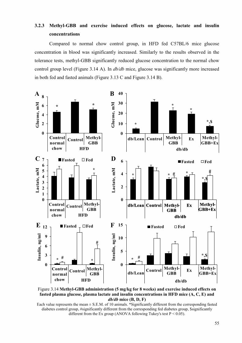
55
3.2.3 Methyl-GBB and exercise induced effects on glucose, lactate and insulin
concentrations
Compared to normal chow control group, in HFD fed C57BL/6 mice glucose
concentration in blood was significantly increased. Similarly to the results observed in the
tolerance tests, methyl-GBB significantly reduced glucose concentration to the normal chow
control group level (Figure 3.14 A). In db/db mice, glucose was significantly more increased
in both fed and fasted animals (Figure 3.13 C and Figure 3.14 B).
* *
0
2
4
6
8
Controlnormalchow
Control Methyl-GBB
HFD
A
Glu
cose
, mM
*
**
*,$
0
10
20
30
40
db/Lean Control Ex
db/db
Methyl-GBB+Ex
Methyl-GBB
B
Glu
cose
, mM
*
01234567 Fasted FedC
Controlnormalchow
Control
HFD
Methyl-GBB
Lac
tate
, mM
* * **,$
##
0
2
4
6
db/Lean Control Methyl-GBB
Ex
Fasted FedD
Lac
tate
, mM
Methyl-GBB+Ex
db/db
* *#
#
0
3
6
9
12 Fasted Fed
Controlnormalchow
Control
HFD
Methyl-GBB
E
Insu
lin, n
g/m
l
**,$#
0
5
10
15 Fasted Fed
db/Lean Control Methyl-GBB
Ex Methyl-GBB+Ex
db/db
Control Exdb/Lean
Methyl-GBB+Ex
db/db
Methyl-GBB
F
Insu
lin, n
g/m
l
Figure 3.14 Methyl-GBB administration (5 mg/kg for 8 weeks) and exercise induced effects on fasted plasma glucose, plasma lactate and insulin concentrations in HFD mice (A, C, E) and
db/db mice (B, D, F) Each value represents the mean ± S.E.M. of 10 animals. *Significantly different from the corresponding fasted
diabetes control group, #significantly different from the corresponding fed diabetes group, $significantly different from the Ex group (ANOVA following Tukey's test P < 0.05).

56
This indicates that db/db mice are characterized by severe hyperglycaemia and
uncompensated type 2 diabetes. Exercise decreased postprandial glucose by 4 mM in db/db
mice. An additional decrease of 6.5 mM in the fed state glucose concentration was induced by
combined treatment (Figure 3.13 C). In addition, in fasted db/db mice, both methyl-GBB
administration and exercise as well as the combination of both significantly decreased the
plasma glucose concentration by 8.6 mM, 12.3 mM and 24.3 mM respectively (Figure 3.14
B). Thus, while accumulation of acylcarnitines was observed in the fed state, methyl-GBB-
and combination-induced glucose-lowering effects were more pronounced in the fasting
conditions. In the fed state, a physiological increase in lactate concentration in control animal
plasma was observed (Figure 3.14 CD). Lactate accumulation could be a marker of increased
glucose uptake and incomplete glucose oxidation in tissues, as it is observed during intensive
exercise after the lactate threshold is reached. In HFD control mice the lactate concentrations
were not significantly increased, but, nevertheless, methyl-GBB treatment induced a
significant decrease in the lactate concentration in the fed state. In db/db mice compared to
db/Lean mice, higher lactate concentrations in the plasma were observed only in the fasted
state. Methyl-GBB treatment significantly reduced the lactate concentrations in db/db mice
plasma in both the fasted and fed states while an exercise-induced effect was observed only in
the fasted mice (Figure 3.14 B). This suggests that the methyl-GBB-induced acylcarnitine
decrease facilitates lactate and pyruvate oxidation in the mitochondria of mice muscles in both
the fasted and fed states. In HFD and db/db mice marked hyperinsulinemia was observed. In
HFD mice methyl-GBB significantly decreased insulin concentration (Figure 3.14 E). In
contrast, exercise and methyl-GBB administration did not significantly influence
hyperinsulinemia in db/db mice (Figure 3.14 F), suggesting that the glucose lowering effect
induced by treatments is related to increased insulin sensitivity.
3.2.4 Methyl-GBB and exercise induced effects on fatty acid metabolism
To test whether the changes in insulin sensitivity induced by exercise and methyl-
GBB treatment are related to FA metabolism, we determined the [3H]-palmitate uptake and
metabolism in the muscles and heart in vivo (Figure 3.15). In db/db hearts compared to
db/Lean mice hearts, 2-fold higher palmitate uptake and metabolism were observed in both
the fed and fasted states. Exercise induced a significant 2-fold decrease in palmitate oxidation
in hearts in the fasted state, but not in the fed state (Figure 3.15 AB). In methyl-GBB-treated
mouse hearts, we observed a significant 2-fold decrease in the labelled palmitate uptake and
oxidation rates in the fed state, but not in the fasted state. Meanwhile, the combination of
exercise and methyl-GBB-induced decrease in acylcarnitine content resulted in reduced

57
metabolism of palmitate in both the fed and fasted states. In contrast, in the skeletal muscle of
db/db mice, palmitate uptake and metabolism were not significantly higher compared to
db/Lean mice (Figure 3.15 AB). Only in fed db/db mice muscles there was a tendency to have
50 % higher palmitate metabolism, while muscles from db/db mice treated by methyl-GBB
and combination were similar to db/Lean controls (Figure 3.15 A).
*
*
* *, $
* $
0
200
400
600
ACS FA ACS FA
Heart Muscle
3 H p
alm
itat
e an
d m
etab
olit
es, f
mol
/g
db/Lean db/db Controldb/db Methyl-GBB db/db Exdb/db Methyl-GBB + Ex
A
*
*
*
*$
0
200
400
600
800
ACS FA ACS FA
Heart Muscle
3 H p
alm
itat
ean
d m
etab
olit
es, f
mol
/g
db/Lean db/db Controldb/db Methyl-GBB db/db Exdb/db Methyl-GBB + Ex
B
Figure 3.15 Methyl-GBB administration (5 mg/kg for 8 weeks) and exercise induced effects on the fed (A) and fasted (B) state [3H]-palmitate uptake (FA – unmetabolized fatty acids) and
metabolism (ACS - acid soluble substances) in db/db mice Each value represents the mean ± S.E.M. of 5 animals. *Significantly different from the db/db control group. $
Significantly different from the db/db methyl-GBB group (ANOVA following Tukey's test P < 0.05).
Overall, insulin insensitivity in db/db mice was not induced by significantly increased
FA metabolism, while increased insulin sensitivity by both treatments was not related to
decreased FA metabolism in muscles.
3.2.5 Methyl-GBB- and exercise-induced effects on fatty acid metabolism-related gene
expression
To determine whether methyl-GBB administration and exercise caused changes in the
expression of the PPARα/PGC1α target genes involved in long-chain FA metabolism, the

58
mRNA expression of genes was measured in the heart tissues by qRT-PCR. The expression of
genes involved in long-chain FA metabolism (CPT1A, CPT1B, FABP3 and PDK4) was
increased in the tissue of HFD fed and db/db mice in comparison to non-diabetic control mice
(Figure 3.16).
* * * *
*
*
** *
**, $ *, $
*, $
0
2
3
5
CPT1A CPT1B ACSL ACOX1 FABP3 PDK4
db/Lean db/db Controldb/db Methyl-GBB db/db Exdb/db Methyl-GBB + Ex
Gen
e ex
pres
sion
, AU
B
Figure 3.16 Methyl-GBB administration (5 mg/kg for 8 weeks) and exercise induced effect on the
expression of PPARα target genes in the fed HFD (A) and db/db (B) mice Each value represents the mean ± S.E.M. of at least 4 animals. *Significantly different from the corresponding diabetes control group, $significantly different from the Ex group (ANOVA following Tukey's test P<0.05).
Exercise in db/db mice induced a 2-fold increase in ACSL gene expression, but other
genes involved in long-chain FA metabolism were not significantly altered by exercise
compared to controls. In both animal models in response to decreased acylcarnitine content
the mRNA levels of PPARα target genes were increased by 1.5-fold compared to
corresponding HFD or db/db control. The combination of exercise with methyl-GBB
treatment significantly elevated the expression of FA metabolism-related genes, and
moreover, the effect of methyl-GBB on ACSL, ACOX1, and FABP3 mRNA expression was
amplified by exercise in db/db mice, but not in HFD mice (Figure 3.16 AB). These results
indicate that methyl-GBB administration and the combination of the drug and exercise
activates the PPARα/PGC1α signalling pathway and stimulates the corresponding target gene
expression.

59
3.3 The role of the long-chain acylcarnitines in the development of
ischaemia/reperfusion-induced damage in the heart mitochondria
Isolated rat heart infarction experiments and in vitro experiments with isolated cardiac
mitochondria were used to study the mechanisms of the long-chain acylcarnitine
accumulation-induced damage in the heart mitochondria during the ischaemia/reperfusion
injury. Wistar rats were treated with methyl-GBB to test possible benefits of a decrease in
acylcarnitine content.
3.3.1 Fatty acid accumulation in the heart during ischaemia and reperfusion
Initially, we tested FA accumulation in mitochondria in the area at risk and the non-
risk area during 30 min of ischaemia and 30 min of reperfusion. Labelled [3H]-palmitate was
added to the perfusion buffer before left coronary artery occlusion. In the mitochondria
isolated from the non-risk area, palmitate was mostly oxidized (Figure 3.17). In contrast, in
mitochondria from the area at risk, three times more palmitate remained unmetabolized,
suggesting that hypoxia induced the accumulation of FAs and their metabolites. During the
reperfusion stage, the palmitate content in mitochondria did not change substantially (Figure
3.17). Taken together, these results suggest that FAs and metabolites accumulated in the
cardiac mitochondria during ischaemia, but not reperfusion.
* *
0
4
8
12
16
Ischaemia Reperfusion
Non-risk Risk
[3H
]-P
alm
itat
e,
nmol
/mg
mit
o. p
rot.
Figure 3.17 The accumulation of [3H]-palmitate in mitochondria of the at-risk and the non-risk
area during ischaemia and reperfusion Each value represents the mean ± S.E.M. for five hearts. *Significantly different from the healthy mitochondria
group (Student’s t-test, P < 0.05).
To test whether FA accumulation is related to cardiac mitochondrial damage, we
separated JC-1-labelled mitochondria from the tissues in the area at risk by cell sorting
(Figure 3.18). Healthy mitochondria contained two times less [3H]-palmitate compared with
damaged mitochondria. Thus, the accumulation of FAs determines the severity of
mitochondrial damage during ischaemia.

60
*
0.0
0.5
1.0
1.5
2.0
2.5
Control Damaged[3
H]-
Pal
mit
ate,
AU
Figure 3.18 The accumulation of [3H]-palmitate in healthy control and damaged mitochondria Each value represents the mean ± S.E.M. for five hearts. *Significantly different from the healthy control
mitochondria group (Student’s t-test, P <0.05).
3.3.2 Acylcarnitine and acyl-CoA contents in the heart and in cardiac mitochondria
The contents of acylcarnitines and acyl-CoAs were measured both in heart tissues and
in mitochondria in the area at risk and the non-risk area after 30 min of ischaemia (Table 3.2,
Figure 3.19).
Table 3.2
Acylcarnitine and acyl-CoA contents in heart tissues and mitochondria in the area at risk and the non-risk area
Acylcarnitines (nmol/g heart)
Sample Group Short-chain Medium-chain Long-chain
Heart tissue Non-risk 284.7 ± 32.7 8.7 ± 0.5 309.1 ± 53.2
AAR 288.6 ± 40.4 17.4 ± 3.3* 896 ± 140.2*
Isolated mitochondria
Non-risk 8.7 ± 2.6 4.4 ± 0.8 59 ± 11.8
AAR 15.1 ± 2.1 5.2 ± 0.4 144.5 ± 11.7*
Acyl-CoAs (pmol/mg protein)
Sample Group Short-chain Medium-chain Long-chain
Heart tissue Non-risk 0.3 ± 0.04 0.6 ± 0.1 5.7 ± 0.9
AAR 0.2 ± 0.01* 0.6 ± 0 10.4 ± 2.1*
Isolated mitochondria
Non-risk ND 2.6 ± 0.7 13.7 ± 1.5
AAR ND 1.9 ± 0.2 28.6 ± 3* Each value represents the mean ± S.E.M. of 6 hearts *Significantly different from the corresponding non-risk
group (Student’s t-test, P < 0.05). ND – not detected. AAR – area at risk.
In infarcted hearts, in the area at risk the medium-chain acylcarnitine content was two
times higher and the long-chain acylcarnitine content was three times higher compared with
those in the non-risk area (Table 3.2, Figure 3.19 A).

61
*
0
400
800
1200
Non-risk Risk
A
Lon
g-ch
ain
acyl
carn
itin
es,
nmol
/g h
eart
*
0
2
4
6
8
10
12
14
Non-risk Risk
Lon
g-ch
ain
acyl
-CoA
s,
nmol
/g h
eart
B
*
0
60
120
180
Non-risk RiskLon
g-ch
ain
acyl
carn
itin
es,
pmol
/mg
prot
C
*
0
7
14
21
28
35
Non-risk RiskLon
g-ch
ain
acyl
-CoA
s,
pmol
/mg
prot
D
Figure 3.19 The accumulation of long-chain acylcarnitines (A, C) and acyl-CoAs (B, D) in heart tissues (A, B) and in the mitochondria (C, D) isolated from the at-risk and the non-risk areas
Each value represents the mean ± S.E.M. for six hearts. *Significantly different from the corresponding non-risk group (Student’s t-test, P < 0.05).
In ischaemic mitochondria, accumulation of acylcarnitines and especially long-chain
acylcarnitines was observed (Table 3.2, Figure 3.19 C). Similarly, in the area at risk, we found
a higher content of long-chain acyl-CoAs (Table 3.2, Figure 3.19 BD); however, content was
up to 50-fold lower than the measured acylcarnitine content.
To study long-chain acylcarnitine accumulation in mitochondrial fractions, we
measured labelled [3H]-palmitoylcarnitine content in mitochondria, and specifically in the
inner and outer membranes, intermembrane space and mitochondrial matrix (Figure 3.20).
0
10
20
30
40
50
Outermitochondrial
membrane
Intermembranespace
Innermitochondrial
membrane
Mitochondrialmatrix
[3H
]-P
alm
itoy
lcar
niti
ne,
% o
f tot
al
Figure 3.20 The accumulation of palmitoylcarnitine in subfractions of mitochondria Each value represents the mean ± S.E.M. for four experiments. *Significantly different from the corresponding
non-risk group (Student’s t-test, P < 0.05).
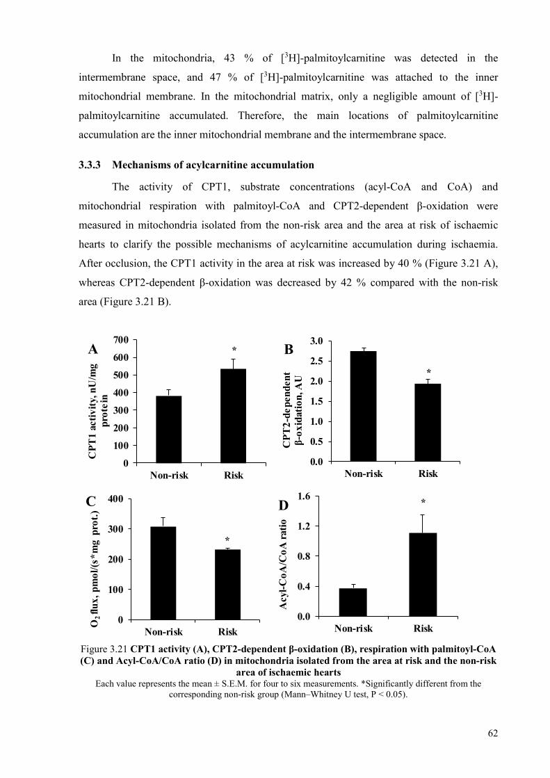
62
In the mitochondria, 43 % of [3H]-palmitoylcarnitine was detected in the
intermembrane space, and 47 % of [3H]-palmitoylcarnitine was attached to the inner
mitochondrial membrane. In the mitochondrial matrix, only a negligible amount of [3H]-
palmitoylcarnitine accumulated. Therefore, the main locations of palmitoylcarnitine
accumulation are the inner mitochondrial membrane and the intermembrane space.
3.3.3 Mechanisms of acylcarnitine accumulation
The activity of CPT1, substrate concentrations (acyl-CoA and CoA) and
mitochondrial respiration with palmitoyl-CoA and CPT2-dependent β-oxidation were
measured in mitochondria isolated from the non-risk area and the area at risk of ischaemic
hearts to clarify the possible mechanisms of acylcarnitine accumulation during ischaemia.
After occlusion, the CPT1 activity in the area at risk was increased by 40 % (Figure 3.21 A),
whereas CPT2-dependent β-oxidation was decreased by 42 % compared with the non-risk
area (Figure 3.21 B).
*
0
100
200
300
400
500
600
700
Non-risk Risk
CP
T1
acti
vity
, nU
/mg
prot
ein
A
*
0.0
0.5
1.0
1.5
2.0
2.5
3.0
Non-risk Risk
CP
T2-
depe
nden
t β-
oxid
atio
n, A
U
B
*
0
100
200
300
400
Non-risk Risk
O2 flu
x, p
mol
/(s*
mg
pro
t.)
C *
0.0
0.4
0.8
1.2
1.6
Non-risk Risk
Acy
l-C
oA/C
oA r
atio
D
Figure 3.21 CPT1 activity (A), CPT2-dependent β-oxidation (B), respiration with palmitoyl-CoA (C) and Acyl-CoA/CoA ratio (D) in mitochondria isolated from the area at risk and the non-risk
area of ischaemic hearts Each value represents the mean ± S.E.M. for four to six measurements. *Significantly different from the
corresponding non-risk group (Mann–Whitney U test, P < 0.05).
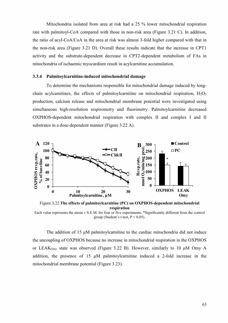
63
Mitochondria isolated from area at risk had a 25 % lower mitochondrial respiration
rate with palmitoyl-CoA compared with those in non-risk area (Figure 3.21 C). In addition,
the ratio of acyl-CoA/CoA in the area at risk was almost 3-fold higher compared with that in
the non-risk area (Figure 3.21 D). Overall these results indicate that the increase in CPT1
activity and the substrate-dependent decrease in CPT2-dependent metabolism of FAs in
mitochondria of ischaemic myocardium result in acylcarnitine accumulation.
3.3.4 Palmitoylcarnitine-induced mitochondrial damage
To determine the mechanisms responsible for mitochondrial damage induced by long-
chain acylcarnitines, the effects of palmitoylcarnitine on mitochondrial respiration, H2O2
production, calcium release and mitochondrial membrane potential were investigated using
simultaneous high-resolution respirometry and fluorimetry. Palmitoylcarnitine decreased
OXPHOS-dependent mitochondrial respiration with complex II and complex I and II
substrates in a dose-dependent manner (Figure 3.22 A).
0
20
40
60
80
100
120
0 10 20 30
OX
PH
OS
resp
.rat
e,%
of b
asel
ine
Palmitoylcarnitine, µM
CIICI&II
A
*
0
50
100
150
200
250
300
OXPHOS LEAKOmy
Res
p.ra
te,
nmol
O2/
min
/mg
prot
.Control
PCB
Figure 3.22 The effects of palmitoylcarnitine (PC) on OXPHOS-dependent mitochondrial respiration
Each value represents the mean ± S.E.M. for four or five experiments. *Significantly different from the control group (Student’s t-test, P < 0.05).
The addition of 15 μM palmitoylcarnitine to the cardiac mitochondria did not induce
the uncoupling of OXPHOS because no increase in mitochondrial respiration in the OXPHOS
or LEAKOmy state was observed (Figure 3.22 B). However, similarly to 10 μM Omy A
addition, the presence of 15 μM palmitoylcarnitine induced a 2-fold increase in the
mitochondrial membrane potential (Figure 3.23).
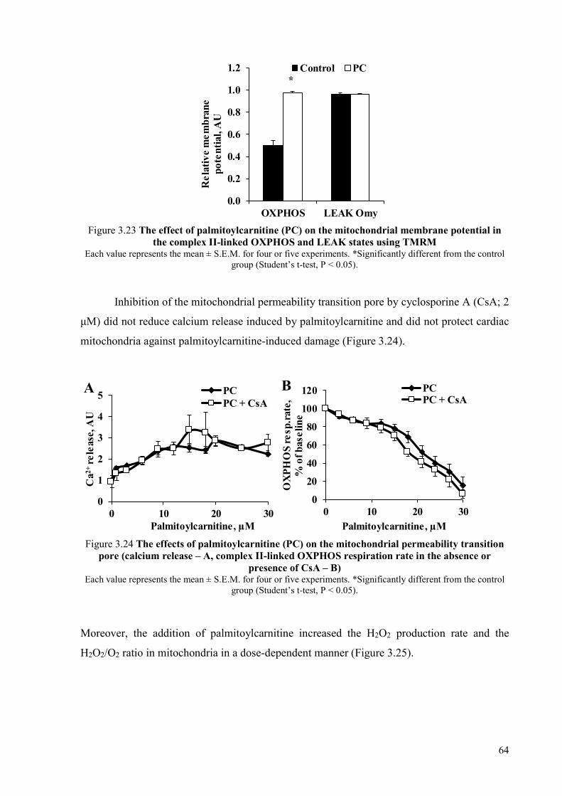
64
*
0.0
0.2
0.4
0.6
0.8
1.0
1.2
OXPHOS LEAK OmyR
elat
ive
mem
bran
e po
tent
ial,
AU
Control PC
Figure 3.23 The effect of palmitoylcarnitine (PC) on the mitochondrial membrane potential in the complex II-linked OXPHOS and LEAK states using TMRM
Each value represents the mean ± S.E.M. for four or five experiments. *Significantly different from the control group (Student’s t-test, P < 0.05).
Inhibition of the mitochondrial permeability transition pore by cyclosporine A (CsA; 2
μM) did not reduce calcium release induced by palmitoylcarnitine and did not protect cardiac
mitochondria against palmitoylcarnitine-induced damage (Figure 3.24).
0
1
2
3
4
5
0 10 20 30
Ca2
+ re
leas
e, A
U
Palmitoylcarnitine, µM
PCPC + CsA
A
0
20
40
60
80
100
120
0 10 20 30
OX
PH
OS
resp
.rat
e,%
of b
asel
ine
Palmitoylcarnitine, µM
PCPC + CsA
B
Figure 3.24 The effects of palmitoylcarnitine (PC) on the mitochondrial permeability transition pore (calcium release – A, complex II-linked OXPHOS respiration rate in the absence or
presence of CsA – B) Each value represents the mean ± S.E.M. for four or five experiments. *Significantly different from the control
group (Student’s t-test, P < 0.05).
Moreover, the addition of palmitoylcarnitine increased the H2O2 production rate and the
H2O2/O2 ratio in mitochondria in a dose-dependent manner (Figure 3.25).

65
0.0
0.5
1.0
1.5
2.0
0 10 20 30
H2O
2pr
oduc
tion
,AU
Palmitoylcarnitine, µM
A
0
1
2
3
0 10 20 30
H2O
/ O
2, %
Palmitoylcarnitine, µM
B
Figure 3.25 The effect of palmitoylcarnitine on H2O2 production under complex II-linked OXPHOS state (A and B)
Each value represents the mean ± S.E.M. for four or five experiments. *Significantly different from the control group (Student’s t-test, P < 0.05).
Taken together, these results demonstrate that long-chain acylcarnitines inhibit
OXPHOS in cardiac mitochondria, thus inducing mitochondrial membrane hyperpolarization
and stimulating the production of reactive oxygen species (ROS), which can lead to cell
death.
3.3.5 Protective mechanisms against mitochondrial damage induced by long-chain
fatty acid metabolites
To explore the possible mechanisms that protect mitochondria against damage induced
by long-chain FA metabolite accumulation, we determined the effect of increasing the
concentrations of palmitoylcarnitine and palmitoyl-CoA on mitochondrial respiration in
isolated rat cardiac mitochondria in the absence or presence of cardiac cell cytosol and FA
binding proteins (Figure 3.26). Palmitoylcarnitine and palmitoyl-CoA decreased OXPHOS-
dependent mitochondrial respiration in a dose-dependent manner with corresponding IC50
values for palmitoyl-CoA and palmitoylcarnitine of 5.4 ± 0.7 μM and 13.4 ± 1.7 μM
respectively (Figure 3.26). Thus, palmitoyl-CoA is approximately 2.5-fold more toxic to
mitochondria than palmitoylcarnitine. As expected, both long-chain FA intermediates were
harmful to mitochondria, and intracellular FA binding proteins were essential to protect
against these FA intermediates. To study the protective mechanisms, cardiac cell cytosol from
fed and fasted rat hearts was used. The cytosol from the fed hearts was able to protect
mitochondria against the damage induced by palmitoylcarnitine and palmitoyl-CoA. The
addition of palmitate to cytosol isolated from the fed hearts partially diminished its protective
effects. In the fasted state, due to the higher amounts of FAs and their metabolites in the
cytosol, the capacity of binding proteins to associate with excess acylcarnitine and acyl-CoA

66
was limited, and cytosol from fasted hearts did not protect against palmitoyl-CoA- or
palmitoylcarnitine-induced damage (Figure 3.26 AB).
*
*
0
20
40
60
80
100
120
0 10 20 30
OX
PH
OS
resp
.rat
e,%
of b
asel
ine
PalmitoylCoA, µM
PCoAPCoA + cytosol FEDPCoA + cytosol FASTED
A
*
*
0
20
40
60
80
100
120
0 10 20 30
OX
PH
OS
resp
.rat
e,%
of b
asel
ine
Palmitoylcarnitine, µM
PCPC + cytosol FEDPC + cytosol FED + FAPC + cytosol FASTED
B
*
0
20
40
60
80
100
120
0 10 20 30
OX
PH
OS
resp
.rat
e,%
of b
asel
ine
PalmitoylCoA, µM
PCoA
PCoA + ACBP
C
0
20
40
60
80
100
120
0 10 20 30
OX
PH
OS
resp
.rat
e,%
of b
asel
ine
Palmitoylcarnitine, µM
PCPC + ACBP
D
0
20
40
60
80
100
120
0 10 20 30
OX
PH
OS
resp
.rat
e,%
of b
asel
ine
PalmitoylCoA, µM
PCoA
PCoA + FABP
E
0
20
40
60
80
100
120
0 10 20 30
OX
PH
OS
resp
.rat
e,%
of b
asel
ine
Palmitoylcarnitine, µM
PC
PC + FABPF
Figure 3.26 Respiration rate of isolated mitochondria treated with palmitoylcarnitine (PC) and palmitoyl-CoA (PCoA, 5 – 30 μM) in the absence or presence of heart cell cytosol from fed or fasted rat hearts or cytosol from fed animals supplemented with FAs (A and B), 50 μM acyl-
CoA-binding protein (ACBP, C and D) or 30 μM FA binding protein (FABP, E and F) Each value represents the mean ± S.E.M. for three to five experiments. *Significantly different from the
corresponding control group (Tukey’s test, P < 0.05).
It should be noted that the experiments were performed in the presence of the 20 %
cytosol fraction; under physiological conditions, the protective capabilities of binding proteins
could be up to 5-fold higher. In further experiments with isolated proteins, we found that at a
concentration of 50 μM, recombinant acyl-CoA-binding protein (ACBP) significantly

67
protected against palmitoyl-CoA-induced mitochondrial damage (Figure 3.26 C) but it did not
influence the palmitoylcarnitine-induced mitochondrial damage (Figure 3.26 D).
Recombinant human FABP3 was also tested in our experimental set-up. However, the
addition of FABP3 at concentrations up to 30 μM did not influence FA metabolite-induced
mitochondrial damage (Figure 3.26 EF). It is possible that the higher concentrations of
FABP3 found in cells (Glatz and van der Vusse, 1996) would protect against acylcarnitine
accumulation-induced damage. Taken together, these results demonstrate that FA binding
proteins represent an important protective mechanism against damage induced by long-chain
FAs. However, cardiac mitochondria are better protected against acyl-CoAs than against
acylcarnitines.
3.3.6 Effects of increased and decreased acylcarnitine content on myocardial infarction
The harmful effects of palmitoylcarnitine were investigated in an isolated rat heart
ischaemia/reperfusion model (Figure 3.27).
↓
Adaptation + PC Ischaemia Reperfusion
20 min 30 min 120 min
Figure 3.27 The myocardial infarction experimental setup with palmitoylcarnitine (PC) PC was added to KH buffer at a concentration of 2 μM for 20 min before ischaemia.
Palmitoylcarnitine at a concentration of 2 μM was used, as it does not affect cardiac
work during normoxia (Figure 3.28).
* * *
0
10
20
30
0 20 40 60 80 100 120 140 160 180
Car
diac
wor
k, k
U
Time, min
Control PC
Figure 3.28 Changes in cardiac work during the experiment in the control and palmitoylcarnitine (PC) groups
Each value represents the mean ± S.E.M. for five to ten animals. *Significantly different from the corresponding control group (Student’s t-test, P < 0.05).
In spontaneously beating isolated rat hearts, the addition of 2 μM palmitoylcarnitine to
the perfusion buffer increased the palmitoylcarnitine concentration in the heart 4-fold (Figure

68
3.29 A) without affecting acyl-CoA content (Figure 3.29 B). The values for the area at risk
were similar to the hearts of all of the experimental groups (Figure 3.30 A), and the area at
risk was approximately 45 – 50 % of the area of the left ventricle.
A
B
Figure 3.29 The accumulation of acylcarnitines (A) and acyl-CoAs (B) in the non-risk area in the isolated heart ischaemia/reperfusion experiment with palmitoylcarnitine (PC)
Each value represents the mean ± S.E.M. for five to ten animals. *Significantly different from the corresponding control group (Student’s t-test, P < 0.05).
As shown in Figure 3.30 B, palmitoylcarnitine addition significantly increased the
infarct size by 33 % relative to the control group’s infarct size. Moreover, treatment with
palmitoylcarnitine significantly decreased cardiac work during reperfusion (Figure 3.28).
Overall, palmitoylcarnitine seems to induce significant disturbances in heart function and an
increase in the infarct size.

69
0
10
20
30
40
50
60
Control PCAA
R, %
of l
eft v
entr
icle
A*
0
10
20
30
40
50
60
Control PC
Infa
rct
size
, % o
f AA
R
B
Figure 3.30 The size of area at risk (AAR, A) and the infarct size (B) after reperfusion in the control and palmitoylcarnitine (PC) groups
Each value represents the mean ± S.E.M. for five to ten animals. *Significantly different from the corresponding control group (Student’s t-test, P < 0.05). AAR – area at risk.
To test the effects of decreased acylcarnitine content, pre-treatment with methyl-GBB
for 14 days before ischaemia/reperfusion injury was used. The area at risk did not differ
between control and methyl-GBB pre-treatment groups (Figure 3.31 B).
A
0
10
20
30
40
50
60
Control Methyl-GBBAA
R, %
of
left
ven
tric
le
B
*
0
10
20
30
40
50
60
70
Control Methyl-GBB
Infa
rct
size
, % o
f AA
R
C
Acy
lcar
niti
nes,
pm
ol/m
g m
ito.
prot
.
MC FASC FA LC FA
Figure 3.31 The effects of long-term methyl-GBB (20 mg/kg) treatment on the acylcarnitine content (A), the area at risk (AAR, B) and the infarct size (C).
Each value represents the mean ± S.E.M. for five to ten animals. *Significantly different from the corresponding control group (Student’s t-test, P < 0.05). AAR – area at risk. SC – short-chain, MC – medium-chain,
LC – long-chain

70
Results showed that methyl-GBB pre-treatment completely prevented acylcarnitine
accumulation. Thus, methyl-GBB induced up to a 60-fold decrease in short-, medium- and
long-chain acylcarnitine content in heart mitochondria isolated from the at-risk area (Figure
3.31 A). As a result, the infarct size decreased by 44 % (Figure 3.31 C). Thus, the methyl-
GBB-induced decrease in the acylcarnitine content is protective against acute
ischaemia/reperfusion-induced damage.
3.4 The effects of methyl-GBB treatment on the development of atherosclerosis
Previous results show that the accumulation of long-chain acylcarnitines determines
the mitochondrial damage in ischaemia/reperfusion injury (Section 3.3). Furthermore, it is
possible to achieve cardioprotection by decreasing the levels of long-chain acylcarnitines with
long-term methyl-GBB treatment before ischaemia/reperfusion injury (Section 3.3.6, Liepinsh
et al., 2015). Since the rate of atherosclerotic plaque formation predicts cardiovascular events
(Gatto et al., 2017), it was of interest to evaluate whether treatment with methyl-GBB
attenuates the development of atherosclerosis. In this study apoE−/− mice were treated with
methyl-GBB to test the effects of the treatment on the development of atherosclerosis. CD-1
outbred mice received methyl-GBB and molecular anti-atherosclerotic mechanisms of the
drug were evaluated.
3.4.1 Effect of treatment on the plasma concentrations of GBB, L-carnitine, methyl-
GBB and TMAO
The concentrations of L-carnitine, GBB, methyl-GBB and TMAO were measured in
plasma after four months of treatment (Table 3.3).
Table 3.3
L-carnitine, GBB, methyl-GBB and TMAO concentrations in the plasma
Group L-carnitine, µM GBB, µM Methyl-GBB, µM TMAO, µM
Control 19.8 ± 1.1 0.4 ± 0.1 – 1.9 ± 0.3
Methyl-GBB 0.5 ± 0.1* 0.9 ± 0.1* 12.5 ± 2.4 1.8 ± 0.4
Each value represents the mean ± S.E.M. for ten animals. *Significantly different from the control group (one-way ANOVA followed by Tukey's multiple comparison test, P < 0.05).
Methyl-GBB administration substantially decreased the amount of L-carnitine present
by nearly 40-fold and increased the concentration of GBB two-fold. The mean TMAO
concentration in control group animals was 1.9 ± 0.3 μM. Methyl-GBB administration did not
affect the plasma concentration of TMAO.

71
3.4.2 Effects of the treatment on the L-carnitine amount and acylcarnitine profile
Effects of the treatment with methyl-GBB on the L-carnitine amount and acylcarnitine
profile in the aortic tissues were assayed in the control and methyl-GBB receiving CD-1
outbred mice (Figure 3.32).
0
5
1050
70
90
Control
Methyl-GBB
**
*
*
Totalacylcarnitines
Short-chainacylcarnitines
Medium-chainacylcarnitines
Long-chainacylcarnitines
Acy
lcar
nit
ines
, n
mol
/g t
issu
es
Figure 3.32 The effects of methyl-GBB treatment on the acylcarnitine profile in the aortic tissues Each value represents the mean ± S.E.M. for ten animals. *Significantly different from the control group (two-
tailed Student's t-test, P < 0.05).
The average level of L-carnitine in aortic tissues of control group animals was 194 ±
32 nmol/g tissues. Two-week treatment with methyl-GBB significantly decreased aortic level
of L-carnitine to 18 ± 1 nmol/g tissues. As it can be seen in Figure 3.32, methyl-GBB
administration to the CD-1 outbred mice at the dose of 16.8 mg/kg markedly decreased the
amount of acylcarnitines in the aortic tissues. Methyl-GBB treatment decreased the total and
the short-chain acylcarnitine level by nearly seventeen-fold and the medium- and long-chain
acylcarnitine level by seven-fold.
3.4.3 Effect of administration of methyl-GBB on the biochemical profile of plasma
Treatment of apoE−/− mice with methyl-GBB at the dose of 10 mg/kg for four months
statistically significantly decreased the concentration of TG, HDL- and LDL-cholesterol for
44 %, 29 % and 24 %, respectively (Table 3.4).
Table 3.4
Biochemical profile of plasma after treatment with methyl-GBB
Group TG, mM HDL, mg/dl LDL, mg/dl
Control 1.8 ± 0.2 212 ± 22 1014 ± 53
Methyl-GBB 1.0 ± 0.1* 150 ± 18* 767 ± 47*
Each value represents the mean ± S.E.M. for ten animals. *Significantly different from the control group (two-tailed Student's t-test, P < 0.05).
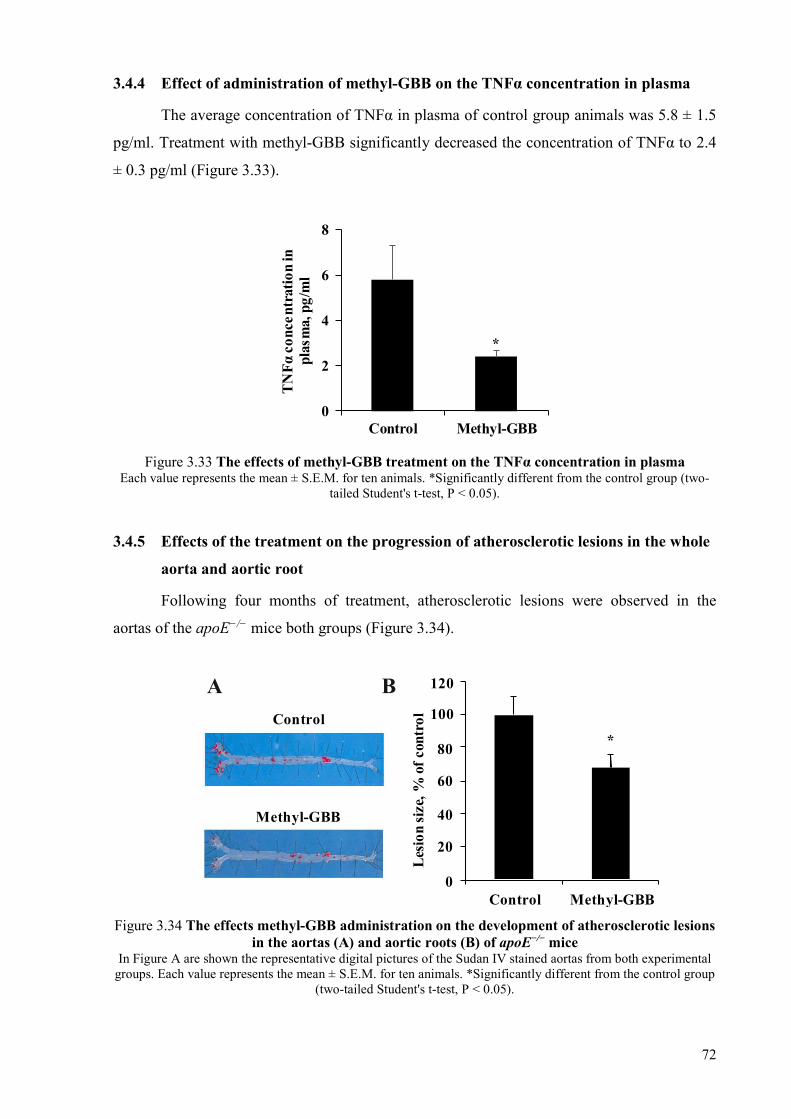
72
3.4.4 Effect of administration of methyl-GBB on the TNFα concentration in plasma
The average concentration of TNFα in plasma of control group animals was 5.8 ± 1.5
pg/ml. Treatment with methyl-GBB significantly decreased the concentration of TNFα to 2.4
± 0.3 pg/ml (Figure 3.33).
*
0
2
4
6
8
Control Methyl-GBB
TN
Fα
conc
entr
atio
n in
pl
asm
a, p
g/m
l
Figure 3.33 The effects of methyl-GBB treatment on the TNFα concentration in plasma Each value represents the mean ± S.E.M. for ten animals. *Significantly different from the control group (two-
tailed Student's t-test, P < 0.05).
3.4.5 Effects of the treatment on the progression of atherosclerotic lesions in the whole
aorta and aortic root
Following four months of treatment, atherosclerotic lesions were observed in the
aortas of the apoE−/− mice both groups (Figure 3.34).
*
0
20
40
60
80
100
120
Control Methyl-GBB
Les
ion
size
, % o
f co
ntro
lControl
Methyl-GBB
A B
Figure 3.34 The effects methyl-GBB administration on the development of atherosclerotic lesions in the aortas (A) and aortic roots (B) of apoE−/− mice
In Figure A are shown the representative digital pictures of the Sudan IV stained aortas from both experimental groups. Each value represents the mean ± S.E.M. for ten animals. *Significantly different from the control group
(two-tailed Student's t-test, P < 0.05).
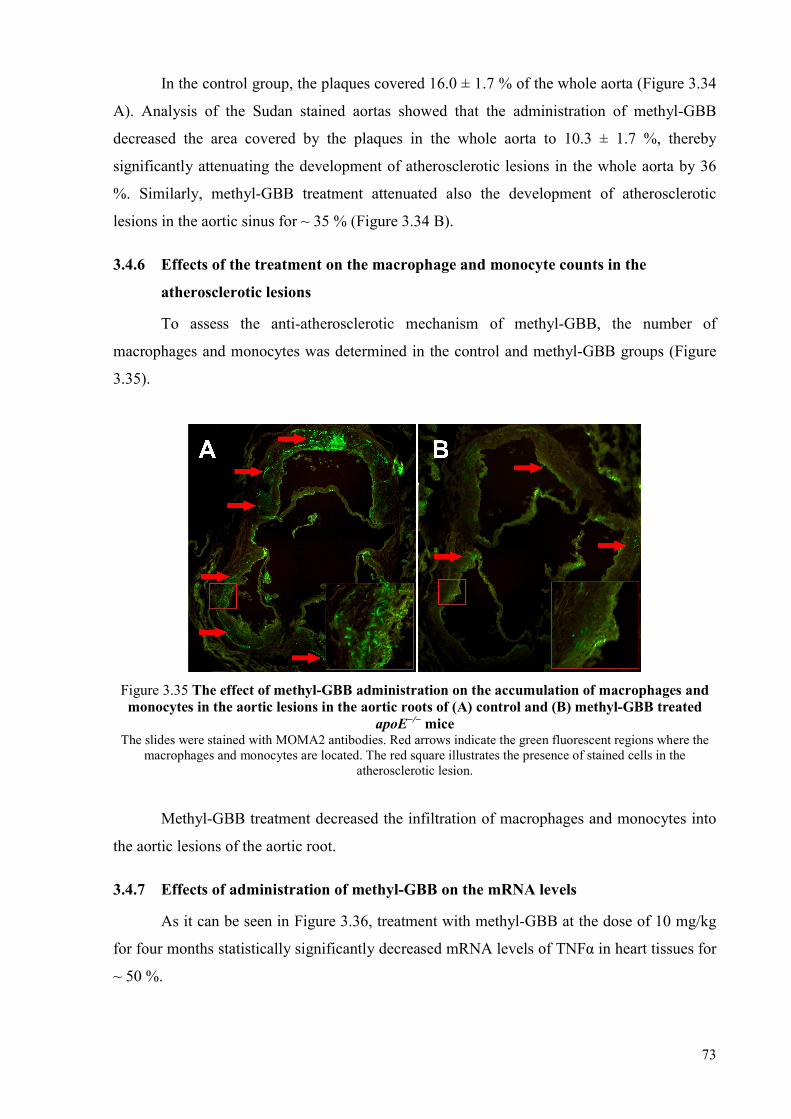
73
In the control group, the plaques covered 16.0 ± 1.7 % of the whole aorta (Figure 3.34
A). Analysis of the Sudan stained aortas showed that the administration of methyl-GBB
decreased the area covered by the plaques in the whole aorta to 10.3 ± 1.7 %, thereby
significantly attenuating the development of atherosclerotic lesions in the whole aorta by 36
%. Similarly, methyl-GBB treatment attenuated also the development of atherosclerotic
lesions in the aortic sinus for ~ 35 % (Figure 3.34 B).
3.4.6 Effects of the treatment on the macrophage and monocyte counts in the
atherosclerotic lesions
To assess the anti-atherosclerotic mechanism of methyl-GBB, the number of
macrophages and monocytes was determined in the control and methyl-GBB groups (Figure
3.35).
Figure 3.35 The effect of methyl-GBB administration on the accumulation of macrophages and monocytes in the aortic lesions in the aortic roots of (A) control and (B) methyl-GBB treated
apoE−/− mice The slides were stained with MOMA2 antibodies. Red arrows indicate the green fluorescent regions where the
macrophages and monocytes are located. The red square illustrates the presence of stained cells in the atherosclerotic lesion.
Methyl-GBB treatment decreased the infiltration of macrophages and monocytes into
the aortic lesions of the aortic root.
3.4.7 Effects of administration of methyl-GBB on the mRNA levels
As it can be seen in Figure 3.36, treatment with methyl-GBB at the dose of 10 mg/kg
for four months statistically significantly decreased mRNA levels of TNFα in heart tissues for
~ 50 %.

74
*
0.0
0.5
1.0
1.5
iNOS IL6 TNFα IL1β
Gen
e ex
pres
sion
, AU
Control Methyl-GBB
Figure 3.36 The effects of methyl-GBB administration on the mRNA levels of iNOS, IL-6, TNFα and IL1β in heart tissues
Each value represents the mean ± S.E.M. for at least four animals. *Significantly different from the control group (two-tailed Mann–Whitney U test, P < 0.05).
In addition, administration of methyl-GBB decreased the mRNA levels of inducible nitric
oxide synthase (iNOS), interleukin 6 (IL6) and interleukin 1β (IL1β), but the effect was not
statistically significant.

75
4 DISCUSSION
The present thesis describes the link between acylcarnitine accumulation and the
pathological processes associated with cardiometabolic diseases: development of insulin
resistance, atherosclerosis and heart ischaemia/reperfusion injury. The aim of the study was to
determine, whether pharmacological decrease in the acylcarnitine concentration by methyl-
GBB is beneficial for the treatment of the mentioned diseases and to investigate the
pharmacological mechanisms of action of methyl-GBB. Initially, molecular mechanisms of
excessive accumulation of long-chain acylcarnitines in the model of accelerated development
of insulin resistance were investigated. Then, the antidiabetic effects of methyl-GBB
treatment alone or in combination with physical intervention were studied. Further, the
damage in the heart mitochondria induced by long-chain acylcarnitine accumulation in the
ischaemia/reperfusion injury was described and methyl-GBB cardioprotective effects were
assessed. Finally, the effects of methyl-GBB treatment on the development of atherosclerosis
were evaluated.
4.1 Role of long-chain acylcarnitine accumulation in the development of insulin
resistance
We have shown that long-chain acylcarnitines influence glucose metabolism in vivo
and therefore are important players in the physiological regulation of energy metabolism.
Moreover, the accumulation of long-chain acylcarnitines during the fed state can accelerate
diabetes progression by the simultaneous induction of insulin resistance and insulin release in
vivo. A single-dose administration of palmitoylcarnitine in mice inhibits Akt phosphorylation
in muscles and the downstream signalling pathways involved in glucose uptake. The long-
term administration of palmitoylcarnitine induces insulin resistance, hyperinsulinemia and
disturbances in glucose tolerance (Table 4.1).
In previous studies, we hypothesized that long-chain acylcarnitines are not only
markers for incomplete FA oxidation but also active FA metabolites involved in the
regulation of energy metabolism (Makrecka et al., 2014). Our present data confirm this
hypothesis through the results indicating that the acute and chronic administration of
palmitoylcarnitine in vivo limits insulin signalling-induced effects and insulin-related glucose
uptake in muscles. According to our results, the mechanism behind long-chain acylcarnitine
action in muscles in vivo is the inhibition of Akt phosphorylation and the subsequent
inhibition of downstream signalling. This is in line with previous studies in C2C12 myotubes

76
in vitro (Aguer et al., 2015; Koves et al., 2008). Interestingly, an increased concentration of
insulin can overcome the long-chain acylcarnitine-induced effects and stimulate Akt
phosphorylation to the appropriate level. This explains previous results showing that the
increased concentrations of insulin in response to glucose stimulation in the fed state can
overcome the inhibitory effects of high long-chain acylcarnitine content (Consitt et al., 2016;
Soeters et al., 2009). In addition, to support the transition from the fasted to fed state and
overcome transient intramuscular insulin insensitivity, long-chain acylcarnitines facilitate
insulin release.
In the fasted state, the low level of insulin is unable to inhibit long-chain acylcarnitine
production, and the high long-chain acylcarnitine content continues to inhibit the Akt
pathway. As a result, glucose uptake and metabolism is limited, while long-chain
acylcarnitine synthesis by CPT1 and subsequent FA oxidation increases (Figure 4.1 A).
Therefore, in starved individuals, the intracellular content of long-chain acylcarnitines is
higher than in the fasted state (Soeters et al., 2009). In our study, both a bolus and long-term
administration of palmitoylcarnitine (C16) increased the content of C18 acylcarnitines,
suggesting that a high long-chain acylcarnitine content blocks the insulin-induced CPT1
inhibition and stimulates an even higher increase in long-chain acylcarnitine content. Many
studies indicate that acylcarnitine accumulation is a result of incomplete FA oxidation (Aguer
et al., 2015; McCoin et al., 2015; Samuel and Shulman, 2012; Schooneman et al., 2013).
However, our results also indicate that in certain conditions such as the fasted state, the
physiologically important long-chain acylcarnitine accumulation is a result of their CPT1-
driven overproduction even if coupled to a high FA oxidation rate. Overall, long-chain
acylcarnitines, as inhibitors of Akt phosphorylation, are active participants in an intercellular
feedback mechanism of insulin signalling and are a substantial part of the energy metabolism
regulation program (Figure 4.1 AB). In healthy subjects in the fed state, the increased
concentration of insulin inhibits long-chain acylcarnitine production via the increased tissue
content of malonyl-CoA (Schooneman et al., 2013; Soeters et al., 2009). That occurs due to
the insulin-induced inhibition of AMP-activated protein kinase activity, which results in the
stimulation of acetyl-CoA carboxylase (synthesis of malonyl-CoA) and inhibition of malonyl-
CoA decarboxylase (degradation of malonyl-CoA, Ruderman et al., 1999; Valentine et al.,
2014). The inability of insulin to inhibit long-chain acylcarnitine production in the fed state
induces disturbances in glucose uptake and metabolism. In the early stage of insulin
resistance, hyperinsulinemia can compensate for insulin resistance and also overcome the
long-chain acylcarnitine-induced effects. In the later stages of the disease, insulin resistance
leads to the inability to inhibit long-chain acylcarnitine production and is accompanied by
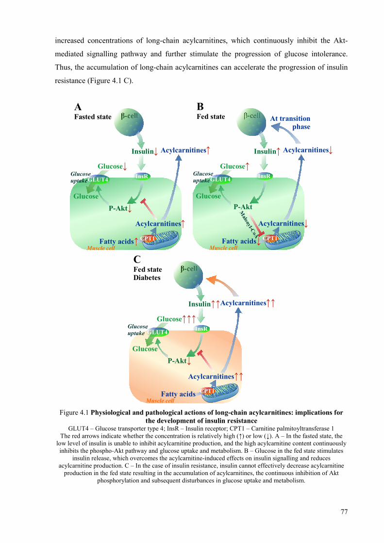
77
increased concentrations of long-chain acylcarnitines, which continuously inhibit the Akt-
mediated signalling pathway and further stimulate the progression of glucose intolerance.
Thus, the accumulation of long-chain acylcarnitines can accelerate the progression of insulin
resistance (Figure 4.1 C).
Acylcarnitines ↑
Fatty acids↑
InsRGlucoseuptakeGLUT4
β-cell
Insulin↓
Muscle cell
Acylcarnitines↑
Glucose
Glucose↓
CPT1
AFasted state
P-Akt↓
B Fed state At transition
phase
Acylcarnitines↓
Fatty acids↓
InsRGlucoseuptakeGLUT4
β-cell
Insulin↑
Muscle cell
Acylcarnitines↓
Glucose
Glucose↑
CPT1
P-Akt
Malonyl-C
oA
CFed stateDiabetes
Acylcarnitines ↑↑
Fatty acids
InsRGlucoseuptake GLUT4
β-cell
Insulin↑↑
Muscle cell
Acylcarnitines↑↑
Glucose
Glucose↑↑↑
CPT1
P-Akt↓
Figure 4.1 Physiological and pathological actions of long-chain acylcarnitines: implications for the development of insulin resistance
GLUT4 – Glucose transporter type 4; InsR – Insulin receptor; CPT1 – Carnitine palmitoyltransferase 1 The red arrows indicate whether the concentration is relatively high (↑) or low (↓). A – In the fasted state, the
low level of insulin is unable to inhibit acylcarnitine production, and the high acylcarnitine content continuously inhibits the phospho-Akt pathway and glucose uptake and metabolism. B – Glucose in the fed state stimulates
insulin release, which overcomes the acylcarnitine-induced effects on insulin signalling and reduces acylcarnitine production. C – In the case of insulin resistance, insulin cannot effectively decrease acylcarnitine
production in the fed state resulting in the accumulation of acylcarnitines, the continuous inhibition of Akt phosphorylation and subsequent disturbances in glucose uptake and metabolism.

78
In conclusion, taking into account the importance of muscle metabolic flexibility and
the ability to switch between fed and fasted states, long-chain acylcarnitines play a role in the
regulation of energy metabolism. These results link the development of skeletal muscle
insulin resistance to the excessive accumulation of long-chain acylcarnitines and, moreover,
suggest that acylcarnitines could induce insulin resistance independently of FA content and
oxidation rate.
4.2 Reduction of long-chain acylcarnitine content: an effective pharmacologic strategy
to prevent the development of diabetes
The present study demonstrates that long-term acylcarnitine accumulation in the fed
state is a feature of type 2 diabetes. Therefore, decreased acylcarnitine content by methyl-
GBB administration improved insulin sensitivity and significantly reduced blood glucose and
insulin levels in mice with impaired insulin sensitivity and diabetes (Table 4.1). Exercise and
the combination of methyl-GBB treatment with exercise improved insulin sensitivity in db/db
mice. Thus, decrease in acylcarnitine levels is sufficient to restore insulin sensitivity in the
early stage of diabetes, while in case of severe diabetes an additive insulin sensitizing effect
of forced physical activity and decreased acylcarnitine content by methyl-GBB would be
necessary to achieve substantial antidiabetic effect.
Several previous studies have attempted to associate acylcarnitines with diabetes;
however, the content of acylcarnitines in relation to the fed and fasted states and the
efficiency of treatment were not monitored (Rodríguez-Gutiérrez et al., 2012; Su et al., 2005;
Zhang et al., 2014). In this study, acylcarnitine content was measured in the plasma and
muscles of fed and fasted animals, and the results clearly demonstrated that the main
pathological role of acylcarnitine accumulation in type 2 diabetes is related to the postprandial
or fed state. Acylcarnitine accumulation in the fed state is a consequence of insulin resistance
or the inability of insulin to sufficiently decrease the production of acylcarnitines. In turn,
acylcarnitines inhibit pyruvate and lactate oxidation leading to an increase in circulating
glucose and lactate concentrations and the induction of common hyperglycaemia-related
complications.
Previously, inhibition of FA oxidation was suggested as a target for diabetes
treatment. In this study, a 60-fold decrease in long-chain acylcarnitines did not reflect a
similar decrease in the FA metabolism rate in skeletal muscles. Also previously it has been
indicated that the inhibition of CPT1 and decreased acylcarnitine content does not fully reflect
the FA uptake or oxidation rate (Liepinsh et al., 2013b, 2015; Luiken et al., 2009). This leads
to the conclusion that methyl-GBB-induced improvements in insulin sensitivity are mainly

79
due to the decreased content of acylcarnitines. In addition, this suggests that in the muscles,
methyl-GBB decreases only acylcarnitine overproduction, thus balancing acylcarnitine
synthesis and utilization. Moreover, the marked decrease in acylcarnitine content only
partially restores insulin sensitivity, and additional improvements could be possible through
other mechanisms such as the inhibition of FA oxidation. Interestingly, the heart was more
sensitive to decreased acylcarnitine production, resulting in a significant 2-fold decrease in
heart FA uptake and metabolism. In addition, we observed that methyl-GBB treatment
facilitated complete FA mitochondrial β-oxidation and induced a 20-fold decrease in medium-
and short- chain acylcarnitine content. Overall, apart from the antidiabetic action of methyl-
GBB, the treatment-induced acylcarnitine profile in the heart was similar to a previously
observed cardioprotective profile (Liepinsh et al., 2015).
In previous studies, we observed an increase in PPARα and PGC1α nuclear content in
response to decreased acylcarnitine content (Liepinsh et al., 2011, 2013a). In this study,
PPARα/PGC1α pathway dependent gene expression in the muscles compensated for the
reduced acylcarnitine production, and a decrease in long-chain FA metabolism was not
observed. In addition, PPARα/PGC1α pathway-induced changes in the expression of the
genes ensured complete oxidation of FAs in the mitochondria. Previously, a mouse model of
high fat feeding demonstrated that elevated expression of PGC1α in the skeletal muscle
improves whole body insulin sensitivity when combined with exercise (Summermatter et al.,
2013). This partially explains the additive effects of decreased acylcarnitine levels and
exercise observed in this study. At the same time, PGC1α overexpression in sedentary
animals induced insulin resistance related to the accumulation of long-chain acylcarnitines
(Summermatter et al., 2013). Taking into account that methyl-GBB improves insulin
sensitivity in sedentary animals, this approach to stimulate PGC1α and simultaneously
decrease acylcarnitines seems to be more effective and safe in regard to sedentary subjects.
There is very good clinical evidence that physical activity is important for prevention
and effective therapy for diabetes (Balducci et al., 2014). However, implementation of this
strategy in clinical practice has not always led to the expected outcomes (De Feo and
Schwarz, 2013). In the present study, low intensity exercise was beneficial for db/db mice.
Higher intensity exercise provides greater benefit in diabetes treatment; however, it also
increases the risk of injury and cardiovascular complications (Balducci et al., 2014). Thus,
low-risk moderate intensity exercise such as walking could provide the best overall health
benefit for patients with diabetes. Additional improvements could be achieved by
pharmacological interventions that facilitate physical activity and improve exercise-induced
antidiabetic effects. Patients with diabetes walking an additional 1,000 steps per day could

80
reduce postprandial blood glucose by 1.6 mM over a period of 2 years (Tudor-Locke and
Bassett, 2004). In our experiment, improvements in insulin sensitivity and a decrease in
postprandial glucose of 4 mM in db/db mice were achieved by walking an additional 3,600
steps/day on working days. Meanwhile, an additional decrease in the fed state glucose
concentration and a significant improvement in insulin sensitivity were achieved by combined
intervention. In addition, the cardioprotective properties of methyl-GBB can reduce the
possible ischaemia induced cardiovascular complications (Liepinsh et al., 2015). Thus,
pharmacological decrease of acylcarnitine levels by methyl-GBB would be particularly
beneficial for patients with diabetes along with physical activity.
4.3 Role of long-chain acylcarnitines in the development of cardiovascular
complications of diabetes
The present study demonstrates that the accumulation of long-chain acylcarnitines in
mitochondria determines the ischemia-reperfusion induced damage. Acylcarnitine
accumulation during ischaemia leads to inhibited oxidative phosphorylation in cardiac
mitochondria, thus inducing mitochondrial membrane hyperpolarization and stimulating the
production of reactive oxygen species. A pharmacologically induced decrease in the
mitochondrial acylcarnitine content by methyl-GBB treatment reduces the infarct size.
Acylcarnitine and acyl-CoA accumulation during ischaemia was discovered
approximately three decades ago (Corr et al., 1984; Ford et al., 1996; Idell-Wenger et al.,
1978; Whitmer et al., 1978), but many aspects remain unclear. In general, acylcarnitine and
acyl-CoA content depends on both the production and the utilization rates of particular FA
intermediates. Consequently, overproduction of FA intermediates in the FA metabolism
pathway occurs during stimulated FA flux or due to insufficient utilization in mitochondria.
Thus, in the fasted state, the acylcarnitine and acyl-CoA content in the heart is up to 5-fold
higher than in the fed state (Makrecka et al., 2014). Taking into account that fasting is a
physiological condition and does not induce damage to heart mitochondria, an average 1.5 to
3-fold increase in FA intermediates during ischaemia is harmful only to fasted or starved
animals. Similarly, in diabetes patients, the higher risk of heart disease (Peters et al., 2015)
might be attributable to higher acylcarnitine content in the heart. Our previous study, which
demonstrated that ischaemic damage to the heart is significantly lower in the fed state
compared with the fasted state (Liepinsh et al., 2014b), also indicated that the accumulation of
FA intermediates is at least partially responsible for the more pronounced heart damage in
ischaemia.

81
Acyl-CoAs and acylcarnitines accumulate during ischaemia, but acyl-CoAs have been
rated as the most prominent FA intermediates responsible for mitochondrial damage
(Drosatos and Schulze, 2013; Li et al., 2010). This discrepancy could be due to the fact that
acyl-CoAs are approximately three times more toxic to mitochondria than acylcarnitines are,
as shown in the present study as well. However, the acyl-CoA content is approximately 50-
fold lower than the acylcarnitine content, and therefore the acyl-CoA content in the area at
risk is too low to induce significant toxic effects on mitochondria. In addition, in a previous
study, we demonstrated that the physiological acylcarnitine content, but not the acyl-CoA
content, is sufficient to inhibit pyruvate and lactate metabolism in mitochondria (Makrecka et
al., 2014). In the present study, supplementation of the heart perfusion buffer with
palmitoylcarnitine did not affect the acyl-CoA content but induced a 2-fold increase in the
acylcarnitine content and significantly increased the infarct size (Table 4.1). Overall, these
results indicate that acylcarnitines are the main FA intermediates that induce
ischaemia/reperfusion-related damage.
Mechanism of accumulation of acylcarnitines in ischaemic mitochondria is related to
ischaemia-induced changes in energy metabolism pathways and metabolite concentrations
(Figure 4.2). In ischaemic myocardium, an increased AMP/ATP ratio stimulates AMP-
activated protein kinase phosphorylation, which, in turn, inhibits acetyl-CoA carboxylase
resulting in a lower content of malonyl-CoA in myocardium and consequently higher
acylcarnitine production by CPT1 (Lopaschuk, 2000) (Figure 4.2). At the same time, during
ischaemia, the activity of CPT2 is decreased due to changes in the acyl-CoA/CoA ratio. Thus,
in ischaemic mitochondria, accumulation of NADH and FADH2 inhibits acyl-CoA
dehydrogenase and 3-hydroxyacyl-CoA dehydrogenase in FA β-oxidation pathway (Jaswal et
al., 2011; Neely and Feuvray, 1981), which leads to accumulation of acyl-CoA in the
mitochondrial matrix and increase in the acyl-CoA/CoA ratio. We also found that the
mitochondrial CoA pool is depleted and the acyl-CoA/CoA ratio is increased during
ischaemia. The acylcarnitine binding site of CPT2 is accessible only if CoA is bound to
enzyme, therefore the activity of CPT2 depends on the acylation state of the mitochondrial
pool of CoA (Nic a’ Bháird et al., 1993). In the ischaemic mitochondria, the content of CoA is
decreased and CPT2 activity is significantly reduced. Overall, acylcarnitine accumulation is a
result of increased CPT1 and decreased CPT2 activity in mitochondria of ischaemic
myocardium (Figure 4.2). Previously, the role of binding proteins in acylcarnitine toxicity
was vastly underestimated. As the major plasma protein functioning as a depot and carrier for
FAs in the blood, albumin also protects against the deleterious effects of acylcarnitines. The
acylcarnitine concentration in the circulation usually does not exceed 1 μM, although

82
circulating albumin has the capacity to protect cells against up to 100 μM of total
acylcarnitine concentration. Similarly, the intracellular proteins ACBP and FABP can protect
mitochondria against FA intermediates.
Figure 4.2 Ischaemia-induced changes in substrate concentrations and activities of enzymes Arrows indicate whether the concentration is relatively high (↑) or low (↓). ACC – acetyl-CoA carboxylase,
AMPK – AMP-activated protein kinase.
ACBP and FABP are highly conserved proteins in eukaryotic organisms from
protozoa to humans, which suggests that these proteins are essential for protection, transport
and other functions (Glatz and van der Vusse, 1996; Neess et al., 2015). The binding proteins
bind and slowly release FA intermediates thus decreasing their free concentration and
protecting mitochondria from their direct effects. In addition, particular intermediates bound
to appropriate proteins are delivered to specific enzymes, thus avoiding unspecific toxic
reactions (Neess et al., 2015). ACBP efficiently protects acetyl-CoA carboxylase and the
mitochondrial adenine nucleotide translocase against acyl-CoA inhibition (Rasmussen et al.,
1993). However, ACBP is highly specific to acyl-CoA, so FAs and acylcarnitines do not bind
to ACBP. Certain studies have demonstrated that acylcarnitine binding to FABP is important
for acylcarnitine oxidation in mitochondria (Fournier and Richard, 1988), but, in the present
study, we did not observe any significant protective role for FABP3 against acylcarnitine-
induced mitochondrial damage. This finding was somewhat surprising because cytosol from
hearts from fed rats protected mitochondria against acylcarnitines. Nevertheless, the
protective capacity of cytosol is limited, and a high FA load during fasting could diminish it.
83
Overall, these findings indicate that FA binding proteins have an important protective
function in the heart. However, cardiac mitochondria are better protected against acyl-CoA
than against acylcarnitine accumulation.
To date, only some studies have addressed the mechanisms of mitochondrial
dysfunction induced by long-chain acylcarnitines (Korge et al., 2003; Siliprandi et al., 1992;
Tominaga et al., 2008). Inhibition or uncoupling of oxidative phosphorylation, mitochondrial
membrane depolarization, opening of the mitochondrial permeability transition pore and
production of reactive oxygen species were proposed to be mechanisms by which FAs and
their intermediates induce mitochondrial damage and subsequent cell damage. In a study
using permeabilized isolated rat ventricular myocytes, it was demonstrated that the
mechanism of palmitoylcarnitine-induced mitochondrial damage is different at low and high
concentrations of acylcarnitines (Tominaga et al., 2008). The important finding of the present
study is the demonstrated localization of palmitoylcarnitine accumulation in mitochondria,
suggesting that either CPT1-generated acylcarnitines or acylcarnitines transported from
cytosol tend to accumulate between the mitochondrial membranes and on the inner
mitochondrial membrane. To a certain extent, mitochondria are protected by binding proteins
localized on mitochondrial membranes, but overproduction of acylcarnitines results in an
increased unbound acylcarnitine concentration in the intermembrane space and corresponding
damaging effects. Overall, long-chain acylcarnitines inhibit oxidative phosphorylation and
thus induce accumulation of protons and mitochondrial membrane hyperpolarization, which
stimulates reverse proton flux through the respiratory chain and subsequent production of
reactive oxygen species.
The inhibition of acylcarnitine production has been suggested as a cardioprotective
drug target (Keung et al., 2013; Liepinsh et al., 2013b, 2015). CPT1 is also considered to be
an important FA mitochondrial transport-limiting enzyme and is often directly or indirectly
targeted to achieve inhibition of FA metabolism and facilitation of glucose oxidation. It is
apparent that any inhibition of the FA pathway upstream of CPT1 would also limit
acylcarnitine production, and at least part of the protective mechanism of inhibited FA flux is
related to decreased acylcarnitine content. Methyl-GBB treatment has been shown as a
powerful tool for reducing the infarct size after ischaemia/reperfusion injury by decreasing the
acylcarnitine content in the heart and mitochondria and limiting long-chain FA oxidation in
favour of glucose oxidation (Liepinsh et al., 2015). Also in this study the administration of
methyl-GBB completely prevented acylcarnitine accumulation and decreased the infarct size
(Table 4.1). Therefore, the main advantage of the pharmacological decrease of acylcarnitine
84
content might be the reduction of direct damage by the long-chain acylcarnitines on
mitochondria in an ischaemic heart.
4.4 Methyl-GBB attenuates the development of atherosclerosis by decreasing levels of
long-chain acylcarnitines
In this study, the effects of lowering the long-chain acylcarnitine levels by methyl-
GBB treatment were studied on the development of atherosclerosis in apoE−/− mice (Table
4.1). We found that methyl-GBB administration significantly attenuated the development of
atherosclerotic lesions in the whole aorta and markedly decreased the amount of
acylcarnitines in the aortic tissues.
Atherosclerosis is characterised as an inflammation of blood vessels with the
participation of immune cells (Libby, 2012). Moreover, it has been shown that acylcarnitines
accumulate in the aortic tissues during the development of atherosclerotic lesions (Gillies and
Bell, 1976). Previously, a study showed that acylcarnitines induce the pro-inflammatory
activation of macrophages and promote the expression of COX-2 and the secretion of TNFα
and other pro-inflammatory cytokines (Rutkowsky et al., 2014). In addition,
palmitoylcarnitine has been shown to disrupt the function of the vascular endothelium and
decrease the synthesis of nitric oxide (Inoue et al., 1994), pathways that could promote the
progression of atherosclerosis. The analysis of the mice aortic tissue extracts revealed that
methyl-GBB administration significantly decreased the amount of short-, medium- and long-
chain acylcarnitines. Thus, the methyl-GBB anti-atherosclerotic mechanism could include the
inhibition immune cell infiltration in atherosclerotic lesions. In support of previously
mentioned presumption are the results obtained from the analysis of atherosclerotic lesions in
the aortic root, which showed that methyl-GBB treatment decreases the accumulation of
macrophages and monocytes in the atherosclerotic lesions. Moreover, administration of
methyl-GBB significantly decreased the TNFα concentration in plasma that could be a result
of decreased secretion of TNFα by activated macrophages. Thus, this study demonstrates that
methyl-GBB attenuates the development of atherosclerosis by inhibiting infiltration of
macrophages and monocytes into the aortic lesions of the aortic root, decreasing their
secretion of pro-inflammatory cytokines and possibly by reducing deleterious effects of
acylcarnitines on the vascular endothelium (Table 4.1).
Postprandial hypertriglyceridemia and elevated LDL levels are risk factors for the
development of atherosclerosis (Fujioka and Ishikawa, 2009; Odden et al., 2014). Methyl-
GBB treatment induced decrease in triglyceride concentration, thus, the decrease of
triglyceride concentration could contribute the attenuation of development of atherosclerosis.
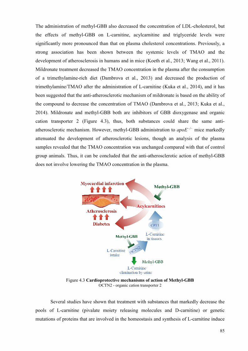
85
The administration of methyl-GBB also decreased the concentration of LDL-cholesterol, but
the effects of methyl-GBB on L-carnitine, acylcarnitine and triglyceride levels were
significantly more pronounced than that on plasma cholesterol concentrations. Previously, a
strong association has been shown between the systemic levels of TMAO and the
development of atherosclerosis in humans and in mice (Koeth et al., 2013; Wang et al., 2011).
Mildronate treatment decreased the TMAO concentration in the plasma after the consumption
of a trimethylamine-rich diet (Dambrova et al., 2013) and decreased the production of
trimethylamine/TMAO after the administration of L-carnitine (Kuka et al., 2014), and it has
been suggested that the anti-atherosclerotic mechanism of mildronate is based on the ability of
the compound to decrease the concentration of TMAO (Dambrova et al., 2013; Kuka et al.,
2014). Mildronate and methyl-GBB both are inhibitors of GBB dioxygenase and organic
cation transporter 2 (Figure 4.3), thus, both substances could share the same anti-
atherosclerotic mechanism. However, methyl-GBB administration to apoE−/− mice markedly
attenuated the development of atherosclerotic lesions, though an analysis of the plasma
samples revealed that the TMAO concentration was unchanged compared with that of control
group animals. Thus, it can be concluded that the anti-atherosclerotic action of methyl-GBB
does not involve lowering the TMAO concentration in the plasma.
Figure 4.3 Cardioprotective mechanisms of action of Methyl-GBB OCTN2 - organic cation transporter 2
Several studies have shown that treatment with substances that markedly decrease the
pools of L-carnitine (pivalate moiety releasing molecules and D-carnitine) or genetic
mutations of proteins that are involved in the homeostasis and synthesis of L-carnitine induce

86
impairment of functioning of different organs (Broderick, 2006; Magoulas and El-Hattab,
2012). In our experimental setup, treatment of apoE−/− mice with methyl-GBB significantly
decreased pools of L-carnitine but we did not observe any signs of muscle weakness or
elevations of ALT and AST. Previously it has been shown, that 2-week treatment with
methyl-GBB at the dose 20 mg/kg decreased L-carnitine pools in heart tissues by 95 %, but
the systolic function and anatomical parameters of the left ventricle was unchanged (Liepinsh
et al., 2015). Thus, we conclude that the decrease of L-carnitine pools may not be the main
factor that determines the development of organ dysfunction and it could be a matter of
pharmacological properties of the studied substance. In favour of that is a study with sodium
pivalate which showed that treatment with it decreased amount of L-carnitine in heart tissues
only by 37 % and simultaneously impaired metabolism of pyruvate and decreased metabolism
of palmitoyl-CoA (Kuka et al., 2012b). This could lead to a depletion of ATP levels and
development of ventricular dysfunction, which was observed after prolonged administration
of pivalate (Broderick, 2006). On the contrary, treatment with methyl-GBB not only limited
FA metabolism, but also facilitated glucose metabolism in heart tissues (Liepinsh et al.,
2015), thus, preserving ATP pools and heart function.
Table 4.1
Effects induced by accumulation of long-chain acylcarnitines or the treatment of methyl-GBB
Effect LC AC Methyl-GBB References
Akt phosphorylation ↓ nd Aguer et al., 2015; Liepinsh et al., 2017
Lactate and pyruvate oxidation ↓ ↑ Liepinsh et al., 2016b; Makrecka et al., 2014
Blood glucose ↑ ↓ Liepinsh et al., 2016b, 2017 Insulin sensitivity ↓ ↑ Liepinsh et al., 2016b, 2017 Inflammation processes: TNF alpha protein expression MOMA infiltration in lesions
↑ ↑ nd
nd ↓ ↓
Rutkowsky et al., 2014; Vilskersts et al., 2015
Atherosclerotic lesion development
↑ ↓ Gillies and Bell, 1976; Vilskersts et al., 2015
FA oxidation ↔ ↓ Liepinsh et al., 2015, 2016b, 2017 Glucose oxidation ↓ ↑ Liepinsh et al., 2015;
Makrecka et al., 2014 Mitochondrial damage (during ischemia)
↑ ↓ Korge et al., 2003; Tominaga et al., 2008; Liepinsh et al., 2016a
Infarct size ↑ ↓ Liepinsh et al., 2016a
The arrows indicate whether the concentration/process is relatively high/activated (↑) or low/inactivated (↓). nd – no data available. ↔ – no effect. FA – fatty acid, LC AC – long-chain acylcarnitines

87
Overall, since the apoE−/− mouse model closely resembles the development of
vascular lesions in humans (Nakashima et al., 1994), we can hypothesise that the treatment
with methyl-GBB could be a powerful approach to attenuate the development of
atherosclerosis also in clinics. Overall, we have shown that by inhibiting Akt phosphorylation
and glucose metabolism the accumulation of long-chain acylcarnitines accelerates
hyperglycaemia and hyperinsulinemia leading to metabolic inflexibility and insulin resistance
(Table 4.1). Long-chain acylcarnitines participate in various inflammatory processes,
including the expression and secretion of TNFα and other pro-inflammatory cytokines, and
their accumulation is observed in atherosclerotic lesions. Long-chain acylcarnitine
accumulation during ischaemia leads to mitochondrial damage in cardiac cells, indicating that
long-chain acylcarnitines are the main FA intermediates that induce ischaemia/reperfusion-
related injury (Table 4.1). Our data on the harmful effects of increased acylcarnitine content
and the protective effects of decreased acylcarnitine content by methyl-GBB treatment
suggest that inhibition of acylcarnitine production in mitochondria is a valuable
cardiometabolic drug target.
In conclusion, pharmacologically induced decrease in the content of long-chain
acylcarnitines by methyl-GBB facilitates glucose metabolism, improves insulin sensitivity,
protects the heart mitochondria against ischaemia/reperfusion injury and attenuates the
development of atherosclerosis and therefore represents an effective strategy for the treatment
of diabetes and its complications.

88
5 CONCLUSIONS
1. The accumulation of long-chain acylcarnitines during the fed state limits metabolic
flexibility, induces insulin resistance, hyperinsulinemia and disturbances in glucose
tolerance. Inhibition of Akt phosphorylation and related insulin signalling is an important
mechanism of long-chain acylcarnitine-induced detrimental effects on glucose
metabolism.
2. Methyl-GBB administration-induced decrease in acylcarnitine content improves insulin
sensitivity and significantly reduces blood glucose and insulin levels in mice with
impaired insulin sensitivity and diabetes.
3. Long-chain acylcarnitines are the main FA intermediates that induce
ischaemia/reperfusion-related damage in heart mitochondria. Acylcarnitine accumulation
during ischaemia leads to inhibited oxidative phosphorylation in cardiac mitochondria,
thus inducing mitochondrial membrane hyperpolarization and stimulating the production
of reactive oxygen species. Treatment with methyl-GBB protects the cardiac mitochondria
against ischaemia/reperfusion-induced accumulation of long-chain acylcarnitines.
4. Treatment with methyl-GBB attenuates the development of atherosclerosis in apoE−/−
mice. The anti-atherosclerotic mechanism of methyl-GBB treatment is mediated by
decreased amounts of long-chain acylcarnitines and decreased infiltration of macrophages
and monocytes into the aortic lesions of the aortic root.

89
6 APPROBATION OF THE STUDY – PUBLICATIONS AND THESIS
Doctoral thesis is based on following SCI publications:
1. Liepinsh, E., Makrecka-Kuka, M., Makarova, E., Volska, K., Vilks, K., Sevostjanovs, E.,
et al. 2017. Acute and long-term administration of palmitoylcarnitine induces muscle-
specific insulin resistance in mice. BioFactors. 43(5), 718–730.
2. Liepinsh, E., Makrecka-Kuka, M., Makarova, E., Volska, K., Svalbe, B., Sevostjanovs, E.,
et al. 2016. Decreased acylcarnitine content improves insulin sensitivity in experimental
mice models of insulin resistance. Pharmacol Res. 113(Pt B), 788–795.
3. Liepinsh, E., Makrecka-Kuka, M., Volska, K., Kuka, J., Makarova, E., Antone, U., et al.
2016. Long-chain acylcarnitines determine ischaemia/reperfusion-induced damage in
heart mitochondria. Biochem J. 473(9), 1191–1202.
4. Vilskersts, R., Kuka, J., Liepinsh, E., Makrecka-Kuka, M., Volska, K., Makarova, E., et
al. 2015. Methyl-γ-butyrobetaine decreases levels of acylcarnitines and attenuates the
development of atherosclerosis. Vascul Pharmacol. 72, 101–107.
Results are reported in the following international conferences:
1. Volska, K., Liepinsh, E., Makarova, E., Makrecka-Kuka, M., Kuka, J., Dambrova, M.
2018. The mechanisms of long-chain acylcarnitine accumulation during ischemia, 13th
Conference on Mitochondrial Physiology: The role of mitochondria in health, disease and
drug discovery, Jurmala, Latvia, 18–21 Sep 2018, Book of Abstracts, p. 22.
2. Volska, K., Makrecka-Kuka, M., Makarova, E., Kuka, J., Vilskersts, R., Liepinsh, E.,
Dambrova, M. 2017. Protective effects of pharmacologically decreased long-chain
acylcarnitine contents in the preclinical models of diabetes and its complications, 2nd
International Conference in Pharmacology: From Cellular Processes to Drug Targets
(ICP2017RIGA), Riga, Latvia, October 19–20, 2017. doi:10.25006/IA.5.S2-A2.28
3. Vilks, K., Volska, K., Makarova, E., Makrecka-Kuka, M., Dambrova, M., Liepinsh, E.
2017. Impact of long-chain acylcarnitines on muscle insulin sensitivity and interaction
with Akt-related insulin signalling pathway, 2nd International Conference in
Pharmacology: From Cellular Processes to Drug Targets (ICP2017RIGA), Riga, Latvia,
October 19–20, 2017. doi:10.25006/IA.5.S2-A2.20
4. Vilks, K., Volska, K., Makarova, E., Makrecka-Kuka, M., Dambrova, M., Liepinsh, E.
2017. Elevated acylcarnitine levels induce muscle insulin insensitivity through the

90
interaction with Akt-related insulin signalling pathway, 42nd FEBS Congress – From
molecules to cells and back, Jerusalem, Israel, September 10, 2017, p.5.4-013.
5. Vilks, K., Volska, K., Makarova, E., Makrecka-Kuka, M., Dambrova, M., Liepinsh, E.
2017. Palmitoylcarnitine interacts with Akt-related insulin signalling pathway and induces
muscle-specific insulin resistance. Latvijas Universitātes 75. konference, Molekulārās
bioloģijas sekcija, Rīga, Latvija, 2017. gada 31. janvāris.
6. Makarova, E., Makrecka-Kuka, M., Volska, K., Vilks, K., Grinberga, S., Dambrova, M.,
Liepinsh, E. 2017. The impaired change in plasma long-chain acylcarnitine level as a
marker of insulin resistance in db/db and high fat diet-fed mice, 50th Miami Winter
Symposium, Diabetes: Today’s Research – Tomorrow’s Therapies, Miami, USA, January
22–25, 2017, p.028.
7. Volska, K., Liepinsh, E., Makarova, E., Makrecka-Kuka, M., Kuka, J., Dambrova, M.
2016. Mitochondrial damage induced by accumulation of acyl-coenzymes A and
acylcarnitines during ischemia. FEBS Workshop “Coenzyme A and its derivatives in
health and disease”, Marseille, France, August 23–27, 2016, Book of Abstracts, p. 51.
8. Liepinsh, E., Makrecka-Kuka, M., Volska, K., Kuka, J., Dambrova, M. Long chain
acylcarnitines: new target to prevent ischemia-reperfusion induced damage in heart
mitochondria. 2016. Mitochondrial Medicine: Developing New Treatments for
Mitochondrial Disease, Hinxton, Cambridge, UK, May 4–6, 2016, Book of Abstracts, p.
58.
9. Dambrova, M., Volska, K., Makrecka-Kuka, M., Makarova, E., Kuka, J., Vilskersts, R.,
Liepinsh, E. 2016. Cardioprotective effects of pharmacologically decreased long-chain
acylcarnitine contents in experimental models of myocardial infarction, atherosclerosis,
and diabetes, 7th European Congress of Pharmacology, Istanbul, Turkey, June 26–30,
2016, Book of Abstracts, p. 81–82.
10. Dambrova, M., Makrecka-Kuka, M., Volska, K., Sevostjanovs, E., Konrade, I., Liepinsh,
E. 2015. Acyl-carnitines induce insulin resistance in high fat diet-fed and diabetic db/db
mice. 51st EASD Annual Meeting, Stockholm, Sweden, September 14–18, 2015, Book of
Abstracts, p. 606.
11. Volska, K., Makrecka-Kuka, M., Makarova, E., Svalbe, B., Sevostjanovs, E., Liepinsh, E.,
Dambrova, M. 2015. Decreased acylcarnitine content and increased physical activity
improve insulin sensitivity in db/db mice. Drug Discovery conference, Riga, Latvia,
August 27–29, 2015, Book of Abstracts, p. 160.
12. Vilskersts, R., Kuka, J., Makrecka-Kuka, M., Volska, K., Makarova, E., Liepinsh, E.,
Dambrova, M. 2015. Attenuation of atherosclerosis development by targeting
91
acylcarnitine pools. Drug Discovery conference, Riga, Latvia, August 27–29, 2015, Book
of Abstracts, p. 37.
13. Makrecka-Kuka, M., Volska, K., Kuka, J., Dambrova, M., Liepinsh, E. 2015.
Acylcarnitines in mitochondrial bioenergetics. Drug Discovery conference, Riga, Latvia,
August 27–29, 2015, Book of Abstracts, p. 46.
14. Vilskersts, R., Kuka, J., Makrecka-Kuka, M., Volska, K., Dambrova, M., Liepinsh, E.
2015. Targeting acylcarnitine content on vascular tissue to attenuate development of
atherosclerosis. Annual Congress of The European Atherosclerosis Society. Glasgow, UK,
March 21–25, 2015.
Results are reported in following local conferences:
1. Konrāde, I., Makarova, E., Tonne, I., Dambrova, G., Kalere, I., Vilks, K., Voļska, K.,
Dambrova, M. 2017. Acilkarnitīnu koncentrācijas mērījumi – jauna diagnostikas metode
insulīna rezistences noteikšanai, RSU 2017. gada zinātniskā konference, Sekc. “Hroniskās
slimības, terapiju algoritmi, jaunas ārstniecības metodes” [Rīga, Latvija, 6. un 7. aprīlis,
2017. gads]: Tēzes, 274. lpp.
2. Voļska, K., Kūka, J., Makrecka-Kūka, M., Alonso Garcia-Mauriño, M., Dambrova, M.
2016. Garķēžu acilkarnitīnu nozīme sirds išēmijas-reperfūzijas bojājuma attīstībā, RSU
2016. gada zinātniskā konference [Rīga, Latvija, 17.–18. marts, 2016]: Tēzes, 65. lpp.
3. Voļska, K., Makrecka-Kūka, M., Kūka, J., Dambrova, M., Liepiņš, E. 2015. Garķēžu
acilkarnitīnu loma insulīna rezistences attīstībā: pētījumi eksperimentālajos modeļos. RSU
2015. gada zinātniskā konference, Sekc. “Latvijas iedzīvotāju veselību apdraudošo
eksogēno un endogēno faktoru izpēte” [Rīga, Latvija, 26.–27. marts, 2015]: Tēzes, 64.
lpp.
4. Vilšķērsts, R., Kūka, J., Makrecka-Kūka, M., Voļska, K., Liepiņš, E., Dambrova, M.
2015. Metil-γ-butirobetaīns samazina acilkarnitīnu līmeni un aizkavē aterosklerozes
attīstību. RSU 2015. gada zinātniskā konference, Sekc. “Latvijas iedzīvotāju veselību
apdraudošo eksogēno un endogēno faktoru izpēte” [Rīga, Latvija, 26.–27. marts, 2015]:
Tēzes, 62. lpp.

92
7 ACKNOWLEDGEMENTS
I would like to express my sincerest gratitude to my scientific supervisor, Dr. pharm
Maija Dambrova, for her patience, support and guiding me through this research. Also my
deepest gratitude to the author of many great scientific ideas Dr. pharm Edgars Liepiņš for
being the main driving force of this research.
A very special thank you to my first teacher in the laboratory, Marina Makrecka-Kūka,
who introduced me to science in practice. Thanks to Marina and Jānis Kūka for their endless
support, both in the lab and outside of it when it was needed the most. Special thanks to Elīna
Makarova, who introduced me to Western blot and RT-PCR methods and has always been a
valuable advisor.
I am sincerely grateful to my laboratory colleagues for their expertise, valuable advice
and remarkable help: atherosclerosis guru Reinis Vilšķērsts, isolated heart model expert Jānis
Kūka, excellent laboratory animal carer Helēna Cīrule, as well as CNS experts Baiba Švalbe,
Līga Zvejniece and Edijs Vāvers. My appreciation also extends to my colleagues Lāsma
Ļauberte, Olga Žarkova-Malkova, Unigunde Antone, Rūdolfs Mežapuķe and Raita Brikmane
for their help in the laboratory.
Special thanks to Solveiga Grīnberga and Eduards Sevostjanovs for chromatography
analysis, Kaspars Tārs for preparation of recombinant ACBP, Erich Gnaiger and Gerhard
Krumschnabel for the help with the mitochondrial functionality assessments. I would also like
to thank JSC Grindeks for the supply of methyl-GBB and methyl-GBB phosphate.
A warm thank you also to my family and friends for their encouragement and support.
This research would not be possible without the excellent conditions provided by the
Latvian Institute of Organic Synthesis and its director, Dr.chem. Osvalds Pugovičs, and the
financial support of Latvian National Research Program BIOMEDICINE and the European
Union 7th Framework Programme project InnovaBalt.
93
REFERENCES
1. Abu-Elheiga, L., Brinkley, W. R., Zhong, L., Chirala, S. S., Woldegiorgis, G., et al. 2000. The Subcellular Localization of Acetyl-CoA Carboxylase 2. Proceedings of the National Academy of Sciences of the United States of America 97(4), 1444–1449.
2. Abumrad, N. A., el-Maghrabi, M. R., Amri, E. Z., Lopez, E., and Grimaldi, P. A. 1993. Cloning of a Rat Adipocyte Membrane Protein Implicated in Binding or Transport of Long-Chain Fatty Acids That Is Induced during Preadipocyte Differentiation. Homology with Human CD36. The Journal of Biological Chemistry 268(24), 17665–17668.
3. Adams, S. H., Esser, V., Brown, N. F., Ing, N. H., Johnson, L., et al. 1998. Expression and Possible Role of Muscle-Type Carnitine Palmitoyltransferase I during Sperm Development in the Rat. Biology of Reproduction 59(6), 1399–1405.
4. Adams, S. H., Hoppel, C. L., Lok, K. H., Zhao, L., Wong, S. W., et al. 2009. Plasma Acylcarnitine Profiles Suggest Incomplete Long-Chain Fatty Acid -Oxidation and Altered Tricarboxylic Acid Cycle Activity in Type 2 Diabetic African-American Women. Journal of Nutrition 139(6), 1073–1081.
5. Agarwal, B., Stowe, D. F., Dash, R. K., Bosnjak, Z. J., and Camara, A. K. S. 2014. Mitochondrial Targets for Volatile Anesthetics against Cardiac Ischemia-Reperfusion Injury. Frontiers in Physiology 5, 341.
6. Aguer, C., McCoin, C. S., Knotts, T. A., Thrush, A. B., Ono-Moore, K., et al. 2015. Acylcarnitines: Potential Implications for Skeletal Muscle Insulin Resistance. FASEB Journal : Official Publication of the Federation of American Societies for Experimental Biology 29(1), 336–345.
7. Ahn, B.-H., Kim, H.-S., Song, S., Lee, I. H., Liu, J., et al. 2008. A Role for the Mitochondrial Deacetylase Sirt3 in Regulating Energy Homeostasis. Proceedings of the National Academy of Sciences of the United States of America 105(38), 14447–14452.
8. Angel, I., Bidet, S., and Langer, S. Z. 1988. Pharmacological Characterization of the Hyperglycemia Induced by Alpha-2 Adrenoceptor Agonists. The Journal of Pharmacology and Experimental Therapeutics 246(3), 1098–1103.
9. Asaka, N., Muranaka, Y., Kirimoto, T., and Miyake, H. 1998. Cardioprotective Profile of MET-88, an Inhibitor of Carnitine Synthesis, and Insulin during Hypoxia in Isolated Perfused Rat Hearts. Fundamental & Clinical Pharmacology 12(2), 158–163.
10. Bachmann, E. and Weber, E. 1988. Biochemical Mechanisms of Oxfenicine Cardiotoxicity. Pharmacology 36(4), 238–248.
11. Balducci, S., Sacchetti, M., Haxhi, J., Orlando, G., D’Errico, V., et al. 2014. Physical Exercise as Therapy for Type 2 Diabetes Mellitus. Diabetes/Metabolism Research and Reviews 30(S1), 13–23.
12. El Banani, H., Bernard, M., Cozzone, P., James, F., and Feuvray, D. 1998. Ionic and Metabolic Imbalance as Potential Factors of Ischemia Reperfusion Injury. The American Journal of Cardiology 82(5A), 25K–29K.
13. Baruteau, J., Sachs, P., Broué, P., Brivet, M., Abdoul, H., et al. 2013. Clinical and Biological Features at Diagnosis in Mitochondrial Fatty Acid Beta-Oxidation Defects: A French Pediatric Study of 187 Patients. Journal of Inherited Metabolic Disease 36(5), 795–803.
14. Bell, E. L., Emerling, B. M., Ricoult, S. J. H., and Guarente, L. 2011. SirT3 Suppresses Hypoxia Inducible Factor 1α and Tumor Growth by Inhibiting Mitochondrial ROS Production. Oncogene 30(26), 2986–2996.
15. Bergman, G., Atkinson, L., Metcalfe, J., Jackson, N., and Jewitt, D. E. 1980. Beneficial Effect of Enhanced Myocardial Carbohydrate Utilisation after Oxfenicine (L-Hydroxyphenylglycine) in Angina Pectoris. European Heart Journal 1(4), 247–253.
16. Berthon, P. M., Howlett, R. A., Heigenhauser, G. J., and Spriet, L. L. 1998. Human Skeletal Muscle Carnitine Palmitoyltransferase I Activity Determined in Isolated Intact Mitochondria. Journal of Applied Physiology 85(1), 148–153.
17. Bharadwaj, K. G., Hiyama, Y., Hu, Y., Huggins, L. A., Ramakrishnan, R., et al. 2010. Chylomicron- and VLDL-Derived Lipids Enter the Heart through Different Pathways: In Vivo

94
Evidence for Receptor- and Non-Receptor-Mediated Fatty Acid Uptake. The Journal of Biological Chemistry 285(49), 37976–37986.
18. Blachnio-Zabielska, A. U., Koutsari, C., and Jensen, M. D. 2011. Measuring Long-Chain Acyl-Coenzyme A Concentrations and Enrichment Using Liquid Chromatography/Tandem Mass Spectrometry with Selected Reaction Monitoring. Rapid Communications in Mass Spectrometry 25(15), 2223–2230.
19. Blomster, J. I., Chow, C. K., Zoungas, S., Woodward, M., Patel, A., et al. 2013. The Influence of Physical Activity on Vascular Complications and Mortality in Patients with Type 2 Diabetes Mellitus. Diabetes, Obesity & Metabolism 15(11), 1008–1012.
20. den Boer, M. E. J., Dionisi-Vici, C., Chakrapani, A., van Thuijl, A. O. J., Wanders, R. J. A., et al. 2003. Mitochondrial Trifunctional Protein Deficiency: A Severe Fatty Acid Oxidation Disorder with Cardiac and Neurologic Involvement. The Journal of Pediatrics 142(6), 684–689.
21. Boulé, N. G., Kenny, G. P., Larose, J., Khandwala, F., Kuzik, N., et al. 2013. Does Metformin Modify the Effect on Glycaemic Control of Aerobic Exercise, Resistance Exercise or Both? Diabetologia 56(11), 2378–2382.
22. Boyle, K. E., Canham, J. P., Consitt, L. A., Zheng, D., Koves, T. R., et al. 2011. A High-Fat Diet Elicits Differential Responses in Genes Coordinating Oxidative Metabolism in Skeletal Muscle of Lean and Obese Individuals. The Journal of Clinical Endocrinology and Metabolism 96(3), 775–781.
23. Broderick, T. L. 2006. Hypocarnitinaemia Induced by Sodium Pivalate in the Rat Is Associated with Left Ventricular Dysfunction and Impaired Energy Metabolism. Drugs in R & D 7(3), 153–161.
24. Brown, N. F., Hill, J. K., Esser, V., Kirkland, J. L., Corkey, B. E., et al. 1997. Mouse White Adipocytes and 3T3-L1 Cells Display an Anomalous Pattern of Carnitine Palmitoyltransferase (CPT) I Isoform Expression during Differentiation: Inter-Tissue and Inter-Species Expression of CPT I and CPT II Enzymes. Biochemical Journal 327(1), 225–231.
25. Cappola, T. P. 2015. Perhexiline: Lessons for Heart Failure Therapeutics ∗. JACC: Heart Failure 3(3), 212–213.
26. Chavez, J. A. and Summers, S. A. 2010. Lipid Oversupply, Selective Insulin Resistance, and Lipotoxicity: Molecular Mechanisms. Biochimica et Biophysica Acta (BBA) - Molecular and Cell Biology of Lipids 1801(3), 252–265.
27. Chen, W. S., Xu, P. Z., Gottlob, K., Chen, M. L., Sokol, K., et al. 2001. Growth Retardation and Increased Apoptosis in Mice with Homozygous Disruption of the Akt1 Gene. Genes & Development 15(17), 2203–2208.
28. Chiu, H.-C., Kovacs, A., Blanton, R. M., Han, X., Courtois, M., et al. 2005. Transgenic Expression of Fatty Acid Transport Protein 1 in the Heart Causes Lipotoxic Cardiomyopathy. Circulation Research 96(2), 225–233.
29. Cho, H., Mu, J., Kim, J. K., Thorvaldsen, J. L., Chu, Q., et al. 2001. Insulin Resistance and a Diabetes Mellitus-Like Syndrome in Mice Lacking the Protein Kinase Akt2 (PKBbeta ). Science 292(5522), 1728–1731.
30. Coe, N. R. and Bernlohr, D. A. 1998. Physiological Properties and Functions of Intracellular Fatty Acid-Binding Proteins. Biochimica et Biophysica Acta (BBA) - Lipids and Lipid Metabolism 1391(3), 287–306.
31. Consitt, L. A., Koves, T. R., Muoio, D. M., Nakazawa, M., Newton, C. A., et al. 2016. Plasma Acylcarnitines during Insulin Stimulation in Humans Are Reflective of Age-Related Metabolic Dysfunction. Biochemical and Biophysical Research Communications 479(4), 868–874.
32. Conti, R., Mannucci, E., Pessotto, P., Tassoni, E., Carminati, P., et al. 2011. Selective Reversible Inhibition of Liver Carnitine Palmitoyl-Transferase 1 by Teglicar Reduces Gluconeogenesis and Improves Glucose Homeostasis. Diabetes 60(2), 644–651.
33. Cooper, H. M. and Spelbrink, J. N. 2008. The Human SIRT3 Protein Deacetylase Is Exclusively Mitochondrial. Biochemical Journal 411(2), 279–285.
34. Corr, P. B., Gross, R. W., and Sobel, B. E. 1984. Amphipathic Metabolites and Membrane Dysfunction in Ischemic Myocardium. Circulation Research 55(2), 135–154.
35. Cottet-Rousselle, C., Ronot, X., Leverve, X., and Mayol, J. F. 2011. Cytometric Assessment of Mitochondria Using Fluorescent Probes. Cytometry Part A 79 A(6), 405–425.
36. Dambrova, M., Cirule, H., Svalbe, B., Zvejniece, L., Pugovichs, O., et al. 2008. Effect of Inhibiting Carnitine Biosynthesis on Male Rat Sexual Performance. Physiology & Behavior

95
95(3), 341–347. 37. Dambrova, M., Liepinsh, E., and Kalvinsh, I. 2002. Mildronate: Cardioprotective Action
through Carnitine-Lowering Effect. Trends in Cardiovascular Medicine 12(6), 275–279. 38. Dambrova, M., Makrecka-Kuka, M., Vilskersts, R., Makarova, E., Kuka, J., et al. 2016.
Pharmacological Effects of Meldonium: Biochemical Mechanisms and Biomarkers of Cardiometabolic Activity. Pharmacological Research 113(Pt B), 771–780.
39. Dambrova, M., Skapare-Makarova, E., Konrade, I., Pugovics, O., Grinberga, S., et al. 2013. Meldonium Decreases the Diet-Increased Plasma Levels of Trimethylamine N-Oxide, a Metabolite Associated with Atherosclerosis. The Journal of Clinical Pharmacology 53(10), 1095–1098.
40. Defronzo, R. A. 2009. Banting Lecture. From the Triumvirate to the Ominous Octet: A New Paradigm for the Treatment of Type 2 Diabetes Mellitus. Diabetes 58(4), 773–795.
41. Degrace, P., Demizieux, L., Du, Z., Gresti, J., Caverot, L., et al. 2007. Regulation of Lipid Flux between Liver and Adipose Tissue during Transient Hepatic Steatosis in Carnitine-Depleted Rats. Journal of Biological Chemistry 282(29), 20816–20826.
42. Demaugre, F., Bonnefont, J.-P., Mitchell, G., Nguyen-Hoang, N., Pelet, A., et al. 1988. Hepatic and Muscular Presentations of Carnitine Palmitoyl Transferase Deficiency: Two Distinct Entities. Pediatric Research 24(3), 308–311.
43. Depré, C., Rider, M. H., and Hue, L. 1998. Mechanisms of Control of Heart Glycolysis. European Journal of Biochemistry 258(2), 277–290.
44. Drosatos, K. and Schulze, P. C. 2013. Cardiac Lipotoxicity: Molecular Pathways and Therapeutic Implications. Current Heart Failure Reports 10(2), 109–121.
45. De Feo, P. and Schwarz, P. 2013. Is Physical Exercise a Core Therapeutical Element for Most Patients with Type 2 Diabetes? Diabetes Care 36(Supplement 2), S149-154.
46. Ford, D. A. 2002. Alterations in Myocardial Lipid Metabolism during Myocardial Ischemia and Reperfusion. Progress in Lipid Research 41(1), 6–26.
47. Ford, D. A., Han, X., Horner, C. C., and Gross, R. W. 1996. Accumulation of Unsaturated Acylcarnitine Molecular Species During Acute Myocardial Ischemia: Metabolic Compartmentalization of Products of Fatty Acyl Chain Elongation in the Acylcarnitine Pool. Biochemistry 35(24), 7903–7909.
48. Fournier, N. C. and Richard, M. A. 1988. Fatty Acid-Binding Protein, a Potential Regulator of Energy Production in the Heart. Investigation of Mechanisms by Electron Spin Resonance. The Journal of Biological Chemistry 263(28), 14471–14479.
49. Fragasso, G., Piatti, MD, P., Monti, L., Palloshi, A., Setola, E., et al. 2003. Short- and Long-Term Beneficial Effects of Trimetazidine in Patients with Diabetes and Ischemic Cardiomyopathy. American Heart Journal 146(5), 854.
50. Fujioka, Y. and Ishikawa, Y. 2009. Remnant Lipoproteins As Strong Key Particles to Atherogenesis. Journal of Atherosclerosis and Thrombosis 16(3), 145–154.
51. Furtado, L. M., Somwar, R., Sweeney, G., Niu, W., and Klip, A. 2002. Activation of the Glucose Transporter GLUT4 by Insulin. Biochemistry and Cell Biology 80(5), 569–578.
52. Garofalo, R. S., Orena, S. J., Rafidi, K., Torchia, A. J., Stock, J. L., et al. 2003. Severe Diabetes, Age-Dependent Loss of Adipose Tissue, and Mild Growth Deficiency in Mice Lacking Akt2/PKBβ. Journal of Clinical Investigation 112(2), 197–208.
53. Gatto, L., Marco, V., Contarini, M., and Prati, F. 2017. Atherosclerosis to Predict Cardiac Events. Journal of Cardiovascular Medicine 18:154–156.
54. Gillies, P. J. and Bell, F. P. 1976. Arterial and Plasma Carnitine Levels in Rabbits: Influence of Age and Dietary Cholesterol. Experimental and Molecular Pathology 25(3), 402–411.
55. Glatz, J. F. C., Luiken, J. J. F. P., and Bonen, A. 2001. Involvement of Membrane-Associated Proteins in the Acute Regulation of Cellular Fatty Acid Uptake. Journal of Molecular Neuroscience 16(2–3), 123–132.
56. Glatz, J. F. C., Luiken, J. J. F. P., and Bonen, A. 2010. Membrane Fatty Acid Transporters as Regulators of Lipid Metabolism: Implications for Metabolic Disease. Physiological Reviews 90(1), 367–417.
57. Glatz, J. F. and van der Vusse, G. J. 1996. Cellular Fatty Acid-Binding Proteins: Their Function and Physiological Significance. Progress in Lipid Research 35(3), 243–282.
58. Greaves, P., Martin, J., Michel, M. C., and Mompon, P. 1984. Cardiac Hypertrophy in the Dog and Rat Induced by Oxfenicine, an Agent Which Modifies Muscle Metabolism. Archives of
96
Toxicology. Supplement. = Archiv Fur Toxikologie. Supplement 7:488–493. 59. Haffner, S. M., Lehto, S., Rönnemaa, T., Pyörälä, K., and Laakso, M. 1998. Mortality from
Coronary Heart Disease in Subjects with Type 2 Diabetes and in Nondiabetic Subjects with and without Prior Myocardial Infarction. New England Journal of Medicine 339(4), 229–234.
60. Hansen, M., Palsøe, M. K., Helge, J. W., and Dela, F. 2015. The Effect of Metformin on Glucose Homeostasis during Moderate Exercise. Diabetes Care 38(2), 293–301.
61. Harris, R. A., Bowker-Kinley, M. M., Huang, B., and Wu, P. 2002. Regulation of the Activity of the Pyruvate Dehydrogenase Complex. Advances in Enzyme Regulation 42:249–259.
62. Hawley, S. A., Pan, D. A., Mustard, K. J., Ross, L., Bain, J., et al. 2005. Calmodulin-Dependent Protein Kinase Kinase-Beta Is an Alternative Upstream Kinase for AMP-Activated Protein Kinase. Cell Metabolism 2(1), 9–19.
63. Hayashi, Y., Ishida, H., Hoshiai, M., Hoshiai, K., Kirimoto, T., et al. 2000. MET-88, a Gamma-Butyrobetaine Hydroxylase Inhibitor, Improves Cardiac SR Ca2+ Uptake Activity in Rats with Congestive Heart Failure Following Myocardial Infarction. Molecular and Cellular Biochemistry 209(1–2), 39–46.
64. Hayashi, Y., Muranaka, Y., Kirimoto, T., Asaka, N., Miyake, H., et al. 2000. Effects of MET-88, a Gamma-Butyrobetaine Hydroxylase Inhibitor, on Tissue Carnitine and Lipid Levels in Rats. Biological & Pharmaceutical Bulletin 23(6), 770–773.
65. Hirschey, M. D., Shimazu, T., Goetzman, E., Jing, E., Schwer, B., et al. 2010. SIRT3 Regulates Mitochondrial Fatty-Acid Oxidation by Reversible Enzyme Deacetylation. Nature 464(7285), 121–125.
66. Hokari, F., Kawasaki, E., Sakai, A., Koshinaka, K., Sakuma, K., et al. 2010. Muscle Contractile Activity Regulates Sirt3 Protein Expression in Rat Skeletal Muscles. Journal of Applied Physiology 109(2), 332–340.
67. Holloway, G. P., Bonen, A., and Spriet, L. L. 2008. Regulation of Skeletal Muscle Mitochondrial Fatty Acid Metabolism in Lean and Obese Individuals. American Journal of Clinical Nutrition 89(1), 455S–462S.
68. Holubarsch, C. J. F., Rohrbach, M., Karrasch, M., Boehm, E., Polonski, L., et al. 2007. A Double-Blind Randomized Multicentre Clinical Trial to Evaluate the Efficacy and Safety of Two Doses of Etomoxir in Comparison with Placebo in Patients with Moderate Congestive Heart Failure: The ERGO (Etomoxir for the Recovery of Glucose Oxidation) Study. Clinical Science 113(4), 205–212.
69. Horowitz, J. D., Chirkov, Y. Y., Kennedy, J. A., and Sverdlov, A. L. 2010. Modulation of Myocardial Metabolism: An Emerging Therapeutic Principle. Current Opinion in Cardiology 25(4), 329–334.
70. Hoyer, F. F., Albrecht, L., Nickenig, G., and Müller, C. 2012. Selective Inhibition of Leukotriene Receptor BLT-2 Reduces Vascular Oxidative Stress and Improves Endothelial Function in ApoE-/- Mice. Molecular and Cellular Biochemistry 359(1–2), 25–31.
71. Hübinger, A., Knode, O., Susanto, F., Reinauer, H., and Gries, F. 1997. Effects of the Carnitine-Acyltransferase Inhibitor Etomoxir on Insulin Sensitivity, Energy Expenditure and Substrate Oxidation in NIDDM. Hormone and Metabolic Research 29(09), 436–439.
72. Huffman, K. M., Koves, T. R., Hubal, M. J., Abouassi, H., Beri, N., et al. 2014. Metabolite Signatures of Exercise Training in Human Skeletal Muscle Relate to Mitochondrial Remodelling and Cardiometabolic Fitness. Diabetologia 57(11), 2282–2295.
73. Hunt, M. C., Tillander, V., and Alexson, S. E. H. 2014. Regulation of Peroxisomal Lipid Metabolism: The Role of Acyl-CoA and Coenzyme A Metabolizing Enzymes. Biochimie 98:45–55.
74. Ibrahimi, A. and Abumrad, N. A. 2002. Role of CD36 in Membrane Transport of Long-Chain Fatty Acids. Current Opinion in Clinical Nutrition and Metabolic Care 5(2), 139–45.
75. Idell-Wenger, J. A., Grotyohann, L. W., and Neely, J. R. 1978. Coenzyme A and Carnitine Distribution in Normal and Ischemic Hearts. The Journal of Biological Chemistry 253(12), 4310–4318.
76. Inoue, N., Hirata, K. -i., Akita, H., and Yokoyama, M. 1994. Palmitoyl-L-Carnitine Modifies the Function of Vascular Endothelium. Cardiovascular Research 28(1), 129–134.
77. Inzucchi, S. E., Bergenstal, R. M., Buse, J. B., Diamant, M., Ferrannini, E., et al. 2012. Management of Hyperglycemia in Type 2 Diabetes: A Patient-Centered Approach: Position Statement of the American Diabetes Association (ADA) and the European Association for the
97
Study of Diabetes (EASD). Diabetes Care 35(6), 1364–1379. 78. Jaswal, J. S., Keung, W., Wang, W., Ussher, J. R., and Lopaschuk, G. D. 2011. Targeting Fatty
Acid and Carbohydrate Oxidation — A Novel Therapeutic Intervention in the Ischemic and Failing Heart. Biochimica et Biophysica Acta (BBA) - Molecular Cell Research 1813(7), 1333–1350.
79. Jing, E., Emanuelli, B., Hirschey, M. D., Boucher, J., Lee, K. Y., et al. 2011. Sirtuin-3 (Sirt3) Regulates Skeletal Muscle Metabolism and Insulin Signaling via Altered Mitochondrial Oxidation and Reactive Oxygen Species Production. Proceedings of the National Academy of Sciences of the United States of America 108(35), 14608–14613.
80. Jing, E., O’Neill, B. T., Rardin, M. J., Kleinridders, A., Ilkeyeva, O. R., et al. 2013. Sirt3 Regulates Metabolic Flexibility of Skeletal Muscle Through Reversible Enzymatic Deacetylation. Diabetes 62(10), 3404–3417.
81. Joint WHO/FAO Expert Consultation. 2003. Diet, Nutrition and the Prevention of Chronic Diseases. World Health Organization Technical Report Series 916:i–viii, 1-149.
82. Joost, H.-G., Bell, G. I., Best, J. D., Birnbaum, M. J., Charron, M. J., et al. 2002. Nomenclature of the GLUT/SLC2A Family of Sugar/Polyol Transport Facilitators. American Journal of Physiology-Endocrinology and Metabolism 282(4), E974–976.
83. Karlsson, M., Contreras, J. A., Hellman, U., Tornqvist, H., and Holm, C. 1997. CDNA Cloning, Tissue Distribution, and Identification of the Catalytic Triad of Monoglyceride Lipase. Evolutionary Relationship to Esterases, Lysophospholipases, and Haloperoxidases. The Journal of Biological Chemistry 272(43), 27218–27223.
84. Kennedy, J. A. and Horowitz, J. D. 1998. Effect of Trimetazidine on Carnitine Palmitoyltransferase-1 in the Rat Heart. Cardiovascular Drugs and Therapy 12(4), 359–363.
85. Kennedy, J. A., Kiosoglous, A. J., Murphy, G. A., Pelle, M. A., and Horowitz, J. D. 2000. Effect of Perhexiline and Oxfenicine on Myocardial Function and Metabolism during Low-Flow Ischemia/Reperfusion in the Isolated Rat Heart. Journal of Cardiovascular Pharmacology 36(6), 794–801.
86. Kennedy, J. A., Unger, S. A., and Horowitz, J. D. 1996. Inhibition of Carnitine Palmitoyltransferase-1 in Rat Heart and Liver by Perhexiline and Amiodarone. Biochemical Pharmacology 52(2), 273–280.
87. Kerner, W., Brückel, J., and German Diabetes Association. 2014. Definition, Classification and Diagnosis of Diabetes Mellitus. Experimental and Clinical Endocrinology & Diabetes 122(07), 384–386.
88. Keung, W., Ussher, J. R., Jaswal, J. S., Raubenheimer, M., Lam, V. H. M., et al. 2013. Inhibition of Carnitine Palmitoyltransferase-1 Activity Alleviates Insulin Resistance in Diet-Induced Obese Mice. Diabetes 62(3), 711–720.
89. Kilkenny, C., Browne, W., Cuthill, I. C., Emerson, M., Altman, D. G., et al. 2010. Animal Research: Reporting in Vivo Experiments: The ARRIVE Guidelines. British Journal of Pharmacology 160(7), 1577–1579.
90. Kim, J.-Y., Hickner, R. C., Cortright, R. L., Dohm, G. L., and Houmard, J. A. 2000. Lipid Oxidation Is Reduced in Obese Human Skeletal Muscle. American Journal of Physiology-Endocrinology and Metabolism 279(5), E1039–1044.
91. Koeth, R. A., Wang, Z., Levison, B. S., Buffa, J. A., Org, E., et al. 2013. Intestinal Microbiota Metabolism of L-Carnitine, a Nutrient in Red Meat, Promotes Atherosclerosis. Nature Medicine 19(5), 576–585.
92. Korb, H., Hoeft, A., Hunneman, D. H., Schraeder, R., Wolpers, H. G., et al. 1984. Changes in Myocardial Substrate Utilisation and Protection of Ischemic Stressed Myocardium by Oxfenicine [(S)-4-Hydroxyphenylglycine]. Naunyn-Schmiedeberg’s Archives of Pharmacology 327(1), 70–74.
93. Korge, P., Honda, H. M., and Weiss, J. N. 2003. Effects of Fatty Acids in Isolated Mitochondria: Implications for Ischemic Injury and Cardioprotection. American Journal of Physiology - Heart and Circulatory Physiology 285(1), H259–269.
94. Koves, T. R., Ussher, J. R., Noland, R. C., Slentz, D., Mosedale, M., et al. 2008. Mitochondrial Overload and Incomplete Fatty Acid Oxidation Contribute to Skeletal Muscle Insulin Resistance. Cell Metabolism 7(1), 45–56.
95. Kuka, J., Liepinsh, E., Makrecka-Kuka, M., Liepins, J., Cirule, H., et al. 2014. Suppression of Intestinal Microbiota-Dependent Production of pro-Atherogenic Trimethylamine N-Oxide by

98
Shifting L-Carnitine Microbial Degradation. Life Sciences 117(2), 84–92. 96. Kuka, J., Makrecka, M., Grinberga, S., Pugovics, O., Liepinsh, E., et al. 2012. A Short-Term
High-Dose Administration of Sodium Pivalate Impairs Pyruvate Metabolism without Affecting Cardiac Function. Cardiovascular Toxicology 12(4), 298–303.
97. Kuka, J., Vilskersts, R., Cirule, H., Makrecka, M., Pugovics, O., et al. 2012. The Cardioprotective Effect of Mildronate Is Diminished After Co-Treatment With l -Carnitine. Journal of Cardiovascular Pharmacology and Therapeutics 17(2), 215–222.
98. Lecoeur, H., Langonné, A., Baux, L., Rebouillat, D., Rustin, P., et al. 2004. Real-Time Flow Cytometry Analysis of Permeability Transition in Isolated Mitochondria. Experimental Cell Research 294(1), 106–117.
99. Lefort, B., Gouache, E., Acquaviva, C., Tardieu, M., Benoist, J. F., et al. 2017. Pharmacological Inhibition of Carnitine Palmitoyltransferase 1 Restores Mitochondrial Oxidative Phosphorylation in Human Trifunctional Protein Deficient Fibroblasts. Biochimica et Biophysica Acta (BBA) - Molecular Basis of Disease 1863(6), 1292–1299.
100. Lehto, S., Rönnemaa, T., Pyörälä, K., and Laakso, M. 2000. Cardiovascular Risk Factors Clustering with Endogenous Hyperinsulinaemia Predict Death from Coronary Heart Disease in Patients with Type II Diabetes. Diabetologia 43(2), 148–155.
101. Li, L. O., Klett, E. L., and Coleman, R. A. 2010. Acyl-CoA Synthesis, Lipid Metabolism and Lipotoxicity. Biochimica et Biophysica Acta (BBA) - Molecular and Cell Biology of Lipids 1801(3), 246–251.
102. Li, Y., Wang, D., Hu, C., Zhang, P., Zhang, D., et al. 2016. Efficacy and Safety of Adjunctive Trimetazidine Therapy for Acute Myocardial Infarction: A Systematic Review and Meta-Analysis. Cardiology 135(3), 188–195.
103. Libby, P. 2012. Inflammation in Atherosclerosis. Arteriosclerosis, Thrombosis, and Vascular Biology 32(9).
104. Lichtenstein, A. H., Lichtenstein, A. H., Appel, L. J., Brands, M., Carnethon, M., et al. 2006. Diet and Lifestyle Recommendations Revision 2006: A Scientific Statement From the American Heart Association Nutrition Committee. Circulation 114(1), 82–96.
105. Liepinsh, E., Kuka, J., and Dambrova, M. 2013. Troubleshooting Digital Macro Photography for Image Acquisition and the Analysis of Biological Samples. Journal of Pharmacological and Toxicological Methods 67(2), 98–106.
106. Liepinsh, E., Makrecka-Kuka, M., Kuka, J., Vilskersts, R., Makarova, E., et al. 2015. Inhibition of L-Carnitine Biosynthesis and Transport by Methyl-γ-Butyrobetaine Decreases Fatty Acid Oxidation and Protects against Myocardial Infarction. British Journal of Pharmacology 172(5), 1319–1332.
107. Liepinsh, E., Makrecka-Kuka, M., Volska, K., Kuka, J., Makarova, E., et al. 2016. Long-Chain Acylcarnitines Determine Ischaemia/Reperfusion-Induced Damage in Heart Mitochondria. The Biochemical Journal 473(9), 1191–1202.
108. Liepinsh, E., Makrecka, M., Kuka, J., Cirule, H., Makarova, E., et al. 2014. Selective Inhibition of OCTN2 Is More Effective than Inhibition of Gamma-Butyrobetaine Dioxygenase to Decrease the Availability of l-Carnitine and to Reduce Myocardial Infarct Size. Pharmacological Research 85, 33–38.
109. Liepinsh, E., Makrecka, M., Kuka, J., Makarova, E., Vilskersts, R., et al. 2014. The Heart Is Better Protected against Myocardial Infarction in the Fed State Compared to the Fasted State. Metabolism: Clinical and Experimental 63(1), 127–136.
110. Liepinsh, E., Skapare, E., Kuka, J., Makrecka, M., Cirule, H., et al. 2013. Activated Peroxisomal Fatty Acid Metabolism Improves Cardiac Recovery in Ischemia-Reperfusion. Naunyn-Schmiedeberg’s Archives of Pharmacology 386(6), 541–550.
111. Liepinsh, E., Skapare, E., Svalbe, B., Makrecka, M., Cirule, H., et al. 2011. Anti-Diabetic Effects of Mildronate Alone or in Combination with Metformin in Obese Zucker Rats. European Journal of Pharmacology 658(2–3), 277–283.
112. Liepinsh, E., Vilskersts, R., Loca, D., Kirjanova, O., Pugovichs, O., et al. 2006. Mildronate, an Inhibitor of Carnitine Biosynthesis, Induces an Increase in Gamma-Butyrobetaine Contents and Cardioprotection in Isolated Rat Heart Infarction. Journal of Cardiovascular Pharmacology 48(6), 314–319.
113. Liepinsh, E., Vilskersts, R., Skapare, E., Svalbe, B., Kuka, J., et al. 2008. Mildronate Decreases Carnitine Availability and Up-Regulates Glucose Uptake and Related Gene Expression in the

99
Mouse Heart. Life Sciences 83(17–18), 613–619. 114. Liepinsh, E., Vilskersts, R., Zvejniece, L., Svalbe, B., Skapare, E., et al. 2009. Protective Effects
of Mildronate in an Experimental Model of Type 2 Diabetes in Goto-Kakizaki Rats. British Journal of Pharmacology 157(8), 1549–1556.
115. Lionetti, V., Linke, A., Chandler, M., Young, M., Penn, M., et al. 2005. Carnitine Palmitoyl Transferase-I Inhibition Prevents Ventricular Remodeling and Delays Decompensation in Pacing-Induced Heart Failure. Cardiovascular Research 66(3), 454–461.
116. Lopaschuk, G. 2000. Regulation of Carbohydrate Metabolism in Ischemia and Reperfusion. American Heart Journal 139(2 Pt 3), S115-119.
117. Di Loreto, C., Fanelli, C., Lucidi, P., Murdolo, G., De Cicco, A., et al. 2005. Make Your Diabetic Patients Walk: Long-Term Impact of Different Amounts of Physical Activity on Type 2 Diabetes. Diabetes Care 28(6), 1295–1302.
118. Luiken, J. J. F. P., Niessen, H. E. C., Coort, S. L. M., Hoebers, N., Coumans, W. A., et al. 2009. Etomoxir-Induced Partial Carnitine Palmitoyltransferase-I (CPT-I) Inhibition in Vivo Does Not Alter Cardiac Long-Chain Fatty Acid Uptake and Oxidation Rates. The Biochemical Journal 419(2), 447–455.
119. MacLeod, S. F., Terada, T., Chahal, B. S., and Boulé, N. G. 2013. Exercise Lowers Postprandial Glucose but Not Fasting Glucose in Type 2 Diabetes: A Meta-Analysis of Studies Using Continuous Glucose Monitoring. Diabetes/Metabolism Research and Reviews 29(8), 593–603.
120. Maeda, K., Uysal, K. T., Makowski, L., Görgün, C. Z., Atsumi, G., et al. 2003. Role of the Fatty Acid Binding Protein Mal1 in Obesity and Insulin Resistance. Diabetes 52(2), 300–307.
121. Magoulas, P. L. and El-Hattab, A. W. 2012. Systemic Primary Carnitine Deficiency: An Overview of Clinical Manifestations, Diagnosis, and Management. Orphanet Journal of Rare Diseases 7(1), 68.
122. Makowski, L., Boord, J. B., Maeda, K., Babaev, V. R., Uysal, K. T., et al. 2001. Lack of Macrophage Fatty-Acid-Binding Protein AP2 Protects Mice Deficient in Apolipoprotein E against Atherosclerosis. Nature Medicine 7(6), 699–705.
123. Makrecka-Kuka, M., Krumschnabel, G., and Gnaiger, E. 2015. High-Resolution Respirometry for Simultaneous Measurement of Oxygen and Hydrogen Peroxide Fluxes in Permeabilized Cells, Tissue Homogenate and Isolated Mitochondria. Biomolecules 5(3), 1319–1338.
124. Makrecka, M., Kuka, J., Volska, K., Antone, U., Sevostjanovs, E., et al. 2014. Long-Chain Acylcarnitine Content Determines the Pattern of Energy Metabolism in Cardiac Mitochondria. Molecular and Cellular Biochemistry 395(1–2).
125. Malik, V. S., Willett, W. C., and Hu, F. B. 2013. Global Obesity: Trends, Risk Factors and Policy Implications. Nature Reviews Endocrinology 9(1), 13–27.
126. Malin, S. K., Gerber, R., Chipkin, S. R., and Braun, B. 2012. Independent and Combined Effects of Exercise Training and Metformin on Insulin Sensitivity in Individuals With Prediabetes. Diabetes Care 35(1), 131–136.
127. Malin, S. K., Haus, J. M., Solomon, T. P. J., Blaszczak, A., Kashyap, S. R., et al. 2013. Insulin Sensitivity and Metabolic Flexibility Following Exercise Training among Different Obese Insulin-Resistant Phenotypes. American Journal of Physiology. Endocrinology and Metabolism 305(10), E1292-1298.
128. Mannucci, E., Dicembrini, I., Lauria, A., and Pozzilli, P. 2013. Is Glucose Control Important for Prevention of Cardiovascular Disease in Diabetes? Diabetes Care 36 Suppl 2(Suppl 2), S259-263.
129. Martins, A. R., Nachbar, R. T., Gorjao, R., Vinolo, M. A., Festuccia, W. T., et al. 2012. Mechanisms Underlying Skeletal Muscle Insulin Resistance Induced by Fatty Acids: Importance of the Mitochondrial Function. Lipids in Health and Disease 11(1), 30.
130. Mathers, C. D. and Loncar, D. 2006. Projections of Global Mortality and Burden of Disease from 2002 to 2030 edited by J. Samet. PLoS Medicine 3(11), e442.
131. May, A. L., Kuklina, E. V., and Yoon, P. W. 2012. Prevalence of Cardiovascular Disease Risk Factors Among US Adolescents, 1999-2008. PEDIATRICS 129(6), 1035–1041.
132. McCoin, C. S., Knotts, T. A., and Adams, S. H. 2015. Acylcarnitines--Old Actors Auditioning for New Roles in Metabolic Physiology. Nature Reviews. Endocrinology 11(10), 617–625.
133. McGarry, J. D., Woeltje, K. F., Kuwajima, M., and Foster, D. W. 1989. Regulation of Ketogenesis and the Renaissance of Carnitine Palmitoyltransferase. Diabetes / Metabolism Reviews 5(3), 271–284.

100
134. McGrath, J., Drummond, G., McLachlan, E., Kilkenny, C., and Wainwright, C. 2010. Guidelines for Reporting Experiments Involving Animals: The ARRIVE Guidelines. British Journal of Pharmacology 160(7), 1573–1576.
135. Michaelides, A. P., Spiropoulos, K., Dimopoulos, K., Athanasiades, D., and Toutouzas, P. 1997. Antianginal Efficacy of the Combination of Trimetazidine-Propranolol Compared with Isosorbide Dinitrate-Propranolol in Patients with Stable Angina. Clinical Drug Investigation 13(1), 8–14.
136. Michan, S. and Sinclair, D. 2007. Sirtuins in Mammals: Insights into Their Biological Function. The Biochemical Journal 404(1), 1–13.
137. Mihalik, S. J., Goodpaster, B. H., Kelley, D. E., Chace, D. H., Vockley, J., et al. 2010. Increased Levels of Plasma Acylcarnitines in Obesity and Type 2 Diabetes and Identification of a Marker of Glucolipotoxicity. Obesity (Silver Spring, Md.) 18(9), 1695–1700.
138. Moller, D. E. 2001. New Drug Targets for Type 2 Diabetes and the Metabolic Syndrome. Nature 414(6865), 821–827.
139. La Monaca, E. and Fodale, V. 2012. Effects of Anesthetics on Mitochondrial Signaling and Function. Current Drug Safety 7(2), 126–139.
140. Murthy, M. S. and Pande, S. V. 1984. Mechanism of Carnitine Acylcarnitine Translocase-Catalyzed Import of Acylcarnitines into Mitochondria. The Journal of Biological Chemistry 259(14), 9082–9089.
141. Nagendran, J., Pulinilkunnil, T., Kienesberger, P. C., Sung, M. M., Fung, D., et al. 2013. Cardiomyocyte-Specific Ablation of CD36 Improves Post-Ischemic Functional Recovery. Journal of Molecular and Cellular Cardiology 63:180–188.
142. Nakashima, Y., Plump, A. S., Raines, E. W., Breslow, J. L., and Ross, R. 1994. ApoE-Deficient Mice Develop Lesions of All Phases of Atherosclerosis throughout the Arterial Tree. Arteriosclerosis, Thrombosis, and Vascular Biology 14(1), 133-140.
143. NCD Risk Factor Collaboration (NCD-RisC). 2016. Worldwide Trends in Diabetes since 1980: A Pooled Analysis of 751 Population-Based Studies with 4.4 Million Participants. Lancet (London, England) 387(10027), 1513–1530.
144. Neely, J. R. and Feuvray, D. 1981. Metabolic Products and Myocardial Ischemia. The American Journal of Pathology 102(2), 282–291.
145. Neess, D., Bek, S., Engelsby, H., Gallego, S. F., and Færgeman, N. J. 2015. Long-Chain Acyl-CoA Esters in Metabolism and Signaling: Role of Acyl-CoA Binding Proteins. Progress in Lipid Research 59, 1–25.
146. Nic a’ Bháird, N., Kumaravel, G., Gandour, R. D., Krueger, M. J., and Ramsay, R. R. 1993. Comparison of the Active Sites of the Purified Carnitine Acyltransferases from Peroxisomes and Mitochondria by Using a Reaction-Intermediate Analogue. The Biochemical Journal 294 ( Pt 3), 645–651.
147. Nivet, J., Le Blanc, M., and Riess, J. 1991. Synthesis and Preliminary Evaluation of Perfluoroalkylacyl Carnitines as Surfactants for Biomedical Use. European Journal of Medicinal Chemistry 26(9), 953–960.
148. Nouette-Gaulain, K., Jose, C., Capdevila, X., and Rossignol, R. 2011. From Analgesia to Myopathy: When Local Anesthetics Impair the Mitochondrion. The International Journal of Biochemistry & Cell Biology 43(1), 14–19.
149. Odden, M. C., Shlipak, M. G., Whitson, H. E., Katz, R., Kearney, P. M., et al. 2014. Risk Factors for Cardiovascular Disease across the Spectrum of Older Age: The Cardiovascular Health Study. Atherosclerosis 237(1), 336–342.
150. Palacios, O. M., Carmona, J. J., Michan, S., Chen, K. Y., Manabe, Y., et al. 2009. Diet and Exercise Signals Regulate SIRT3 and Activate AMPK and PGC-1alpha in Skeletal Muscle. Aging 1(9), 771–783.
151. Panzram, G. 1987. Mortality and Survival in Type 2 (Non-Insulin-Dependent) Diabetes Mellitus. Diabetologia 30(3), 123–131.
152. Pekala, J., Patkowska-Sokola, B., Bodkowski, R., Jamroz, D., Nowakowski, P., et al. 2011. L-Carnitine - Metabolic Functions and Meaning in Humans Life. Current Drug Metabolism 12(7), 667–678.
153. Peters, S. A. E., Huxley, R. R., Sattar, N., and Woodward, M. 2015. Sex Differences in the Excess Risk of Cardiovascular Diseases Associated with Type 2 Diabetes: Potential Explanations and Clinical Implications. Current Cardiovascular Risk Reports 9(7), 36.

101
154. Poole, R. C. and Halestrap, A. P. 1993. Transport of Lactate and Other Monocarboxylates across Mammalian Plasma Membranes. American Journal of Physiology-Cell Physiology 264(4), C761–782.
155. Qiu, X., Brown, K., Hirschey, M. D., Verdin, E., and Chen, D. 2010. Calorie Restriction Reduces Oxidative Stress by SIRT3-Mediated SOD2 Activation. Cell Metabolism 12(6), 662–667.
156. Ramsay, R. R., Gandour, R. D., and van der Leij, F. R. 2001. Molecular Enzymology of Carnitine Transfer and Transport. Biochimica et Biophysica Acta 1546(1), 21–43.
157. Randle, P. J., Garland, P. B., Hales, C. N., and Newsholme, E. A. 1963. The Glucose Fatty-Acid Cycle. Its Role in Insulin Sensitivity and the Metabolic Disturbances of Diabetes Mellitus. Lancet (London, England) 1(7285), 785–789.
158. Rasmussen, J. T., Börchers, T., and Knudsen, J. 1990. Comparison of the Binding Affinities of Acyl-CoA-Binding Protein and Fatty-Acid-Binding Protein for Long-Chain Acyl-CoA Esters. The Biochemical Journal 265(3), 849–855.
159. Rasmussen, J. T., Rosendal, J., and Knudsen, J. 1993. Interaction of Acyl-CoA Binding Protein (ACBP) on Processes for Which Acyl-CoA Is a Substrate, Product or Inhibitor. The Biochemical Journal 292 (Pt 3), 907–913.
160. Rodríguez-Gutiérrez, R., Lavalle-González, F. J., Martínez-Garza, L. E., Landeros-Olvera, E., López-Alvarenga, J. C., et al. 2012. Impact of an Exercise Program on Acylcarnitines in Obesity: A Prospective Controlled Study. Journal of the International Society of Sports Nutrition 9(1), 22.
161. Rosano, G. M. C., Vitale, C., Sposato, B., Mercuro, G., and Fini, M. 2003. Trimetazidine Improves Left Ventricular Function in Diabetic Patients with Coronary Artery Disease: A Double-Blind Placebo-Controlled Study. Cardiovascular Diabetology 2:16.
162. Ruderman, N. B., Saha, A. K., Vavvas, D., and Witters, L. A. 1999. Malonyl-CoA, Fuel Sensing, and Insulin Resistance. The American Journal of Physiology 276(1 Pt 1), E1–18.
163. Rupp, H., Zarain-Herzberg, A., and Maisch, B. 2002. The Use of Partial Fatty Acid Oxidation Inhibitors for Metabolic Therapy of Angina Pectoris and Heart Failure. Herz 27(7), 621–636.
164. Rutkowsky, J. M., Knotts, T. A., Ono-Moore, K. D., McCoin, C. S., Huang, S., et al. 2014. Acylcarnitines Activate Proinflammatory Signaling Pathways. American Journal of Physiology - Endocrinology and Metabolism 306(12).
165. Ryu, S.-Y., Beutner, G., Kinnally, K. W., Dirksen, R. T., and Sheu, S.-S. 2011. Single Channel Characterization of the Mitochondrial Ryanodine Receptor in Heart Mitoplasts. Journal of Biological Chemistry 286(24), 21324–21329.
166. Sacre, J. W., Jellis, C. L., Jenkins, C., Haluska, B. A., Baumert, M., et al. 2014. A Six-Month Exercise Intervention in Subclinical Diabetic Heart Disease: Effects on Exercise Capacity, Autonomic and Myocardial Function. Metabolism: Clinical and Experimental 63(9), 1104–1114.
167. Saha, J. K., Xia, J., Grondin, J. M., Engle, S. K., and Jakubowski, J. A. 2005. Acute Hyperglycemia Induced by Ketamine/Xylazine Anesthesia in Rats: Mechanisms and Implications for Preclinical Models. Experimental Biology and Medicine (Maywood, N.J.) 230(10), 777–784.
168. Samuel, V. T. and Shulman, G. I. 2012. Mechanisms for Insulin Resistance: Common Threads and Missing Links. Cell 148(5), 852–871.
169. Sayed-Ahmed, M. M., Khattab, M. M., Gad, M. Z., and Mostafa, N. 2001. L -Carnitine Prevents the Progression of Atherosclerotic Lesions in Hypercholesterolaemic Rabbits. Pharmacological Research 44(3), 235–242.
170. Schaffer, J. E. and Lodish, H. F. 1994. Expression Cloning and Characterization of a Novel Adipocyte Long Chain Fatty Acid Transport Protein. Cell 79(3), 427–436.
171. Schmidt-Schweda, S. and Holubarsch, C. 2000. First Clinical Trial with Etomoxir in Patients with Chronic Congestive Heart Failure. Clinical Science (London, England : 1979) 99(1), 27–35.
172. Schooneman, M. G., Vaz, F. M., Houten, S. M., and Soeters, M. R. 2013. Acylcarnitines: Reflecting or Inflicting Insulin Resistance? Diabetes 62(1), 1–8.
173. Sesti, C., Simkhovich, B. Z., Kalvinsh, I., and Kloner, R. A. 2006. Mildronate, a Novel Fatty Acid Oxidation Inhibitor and Antianginal Agent, Reduces Myocardial Infarct Size without Affecting Hemodynamics. Journal of Cardiovascular Pharmacology 47(3), 493–499.

102
174. Sierra, A. Y., Gratacós, E., Carrasco, P., Clotet, J., Ureña, J., et al. 2008. CPT1c Is Localized in Endoplasmic Reticulum of Neurons and Has Carnitine Palmitoyltransferase Activity. Journal of Biological Chemistry 283(11), 6878–6885.
175. Siliprandi, D., Biban, C., Testa, S., Toninello, A., and Siliprandi, N. 1992. Effects of Palmitoyl CoA and Palmitoyl Carnitine on the Membrane Potential and Mg2+ Content of Rat Heart Mitochondria. Molecular and Cellular Biochemistry 116(1–2), 117–123.
176. Simkhovich, B. Z., Shutenko, Z. V, Meirena, D. V, Khagi, K. B., Mezapuķe, R. J., et al. 1988. 3-(2,2,2-Trimethylhydrazinium)Propionate (THP)--a Novel Gamma-Butyrobetaine Hydroxylase Inhibitor with Cardioprotective Properties. Biochemical Pharmacology 37(2), 195–202.
177. Skinner, A. C., Perrin, E. M., Moss, L. A., and Skelton, J. A. 2015. Cardiometabolic Risks and Severity of Obesity in Children and Young Adults. New England Journal of Medicine 373(14), 1307–1317.
178. Smathers, R. L. and Petersen, D. R. 2011. The Human Fatty Acid-Binding Protein Family: Evolutionary Divergences and Functions. Human Genomics 5(3), 170–191.
179. Soeters, M. R., Sauerwein, H. P., Duran, M., Wanders, R. J., Ackermans, M. T., et al. 2009. Muscle Acylcarnitines during Short-Term Fasting in Lean Healthy Men. Clinical Science (London, England : 1979) 116(7), 585–592.
180. Sokolovska, J., Isajevs, S., Sugoka, O., Sharipova, J., Lauberte, L., et al. 2011. Correction of Glycaemia and GLUT1 Level by Mildronate in Rat Streptozotocin Diabetes Mellitus Model. Cell Biochemistry and Function 29(1), 55–63.
181. Sokolovska, J., Rumaks, J., Karajeva, N., Grīnvalde, D., Shapirova, J., et al. 2011. [The Influence of Mildronate on Peripheral Neuropathy and Some Characteristics of Glucose and Lipid Metabolism in Rat Streptozotocin-Induced Diabetes Mellitus Model]. Biomeditsinskaia Khimiia 57(5), 490–500.
182. Spagnoli, L. G., Orlandi, A., Marino, B., Mauriello, A., De Angelis, C., et al. 1995. Propionyl-L-Carnitine Prevents the Progression of Atherosclerotic Lesions in Aged Hyperlipemic Rabbits. Atherosclerosis 114(1), 29–44.
183. Spiekerkoetter, U., Lindner, M., Santer, R., Grotzke, M., Baumgartner, M. R., et al. 2009. Treatment Recommendations in Long-Chain Fatty Acid Oxidation Defects: Consensus from a Workshop. Journal of Inherited Metabolic Disease 32(4), 498–505.
184. Stamler, J., Vaccaro, O., Neaton, J. D., and Wentworth, D. 1993. Diabetes, Other Risk Factors, and 12-Yr Cardiovascular Mortality for Men Screened in the Multiple Risk Factor Intervention Trial. Diabetes Care 16(2), 434–44.
185. Stanley, W. C., Recchia, F. A., and Lopaschuk, G. D. 2005. Myocardial Substrate Metabolism in the Normal and Failing Heart. Physiological Reviews 85(3), 1093–1129.
186. Statsenko, M. E., Belenkova, S. V, Sporova, O. E., and Shilina, N. N. 2007. [The Use of Mildronate in Combined Therapy of Postinfarction Chronic Heart Failure in Patients with Type 2 Diabetes Mellitus]. Klinicheskaia Meditsina 85(7), 39–42.
187. Stein, S. C., Woods, A., Jones, N. A., Davison, M. D., and Carling, D. 2000. The Regulation of AMP-Activated Protein Kinase by Phosphorylation. The Biochemical Journal 345 Pt 3(Pt 3), 437–43.
188. Stephens, F. B., Constantin-Teodosiu, D., and Greenhaff, P. L. 2007. New Insights Concerning the Role of Carnitine in the Regulation of Fuel Metabolism in Skeletal Muscle. The Journal of Physiology 581(2), 431–44.
189. Stillwell, W. 2016. Membrane Biogenesis. An Introduction to Biological Membranes. Elsevier 315–329
190. Stremmel, W., Lotz, G., Strohmeyer, G., and Berk, P. D. 1985. Identification, Isolation, and Partial Characterization of a Fatty Acid Binding Protein from Rat Jejunal Microvillous Membranes. Journal of Clinical Investigation 75(3), 1068–76.
191. Strijbis, K., Vaz, F. M., and Distel, B. 2010. Enzymology of the Carnitine Biosynthesis Pathway. IUBMB Life 62(5), 357–362
192. Su, X., Han, X., Mancuso, D. J., Abendschein, D. R., and Gross, R. W. 2005. Accumulation of Long-Chain Acylcarnitine and 3-Hydroxy Acylcarnitine Molecular Species in Diabetic Myocardium: Identification of Alterations in Mitochondrial Fatty Acid Processing in Diabetic Myocardium by Shotgun Lipidomics. Biochemistry 44(13), 5234–5245.
193. Summermatter, S., Shui, G., Maag, D., Santos, G., Wenk, M. R., et al. 2013. PGC-1α Improves Glucose Homeostasis in Skeletal Muscle in an Activity-Dependent Manner. Diabetes 62(1), 85–

103
95. 194. Tars, K., Leitans, J., Kazaks, A., Zelencova, D., Liepinsh, E., et al. 2014. Targeting Carnitine
Biosynthesis: Discovery of New Inhibitors against γ-Butyrobetaine Hydroxylase. Journal of Medicinal Chemistry 57(6), 2213–2236.
195. Task Force Members, Montalescot, G., Sechtem, U., Achenbach, S., Andreotti, F., et al. 2013. 2013 ESC Guidelines on the Management of Stable Coronary Artery Disease. European Heart Journal 34(38), 2949–3003.
196. The Emerging Risk Factors Collaboration, Sarwar, N., Gao, P., Seshasai, S. R. K., Gobin, R., et al. 2010. Diabetes Mellitus, Fasting Blood Glucose Concentration, and Risk of Vascular Disease: A Collaborative Meta-Analysis of 102 Prospective Studies. The Lancet 375(9733), 2215–2222.
197. Tominaga, H., Katoh, H., Odagiri, K., Takeuchi, Y., Kawashima, H., et al. 2008. Different Effects of Palmitoyl-L-Carnitine and Palmitoyl-CoA on Mitochondrial Function in Rat Ventricular Myocytes. AJP: Heart and Circulatory Physiology 295(1), H105–112.
198. Tordjman, K. M., Leingang, K. A., James, D. E., and Mueckler, M. M. 1989. Differential Regulation of Two Distinct Glucose Transporter Species Expressed in 3T3-L1 Adipocytes: Effect of Chronic Insulin and Tolbutamide Treatment. Proceedings of the National Academy of Sciences of the United States of America 86(20), 7761–7765.
199. Tudor-Locke, C. and Bassett, D. R. 2004. How Many Steps/Day Are Enough? Preliminary Pedometer Indices for Public Health. Sports Medicine 34(1), 1–8.
200. Turcani, M. and Rupp, H. 1997. Etomoxir Improves Left Ventricular Performance of Pressure-Overloaded Rat Heart. Circulation 96(10), 3681–3686.
201. Twig, G., Yaniv, G., Levine, H., Leiba, A., Goldberger, N., et al. 2016. Body-Mass Index in 2.3 Million Adolescents and Cardiovascular Death in Adulthood. New England Journal of Medicine 374(25), 2430–2440.
202. Valentine, R. J., Coughlan, K. A., Ruderman, N. B., and Saha, A. K. 2014. Insulin Inhibits AMPK Activity and Phosphorylates AMPK Ser485/491 through Akt in Hepatocytes, Myotubes and Incubated Rat Skeletal Muscle. Archives of Biochemistry and Biophysics 562:62–69.
203. Vik-Mo, H., Mjøs, O. D., Neely, J. R., Maroko, P. R., and Ribeiro, L. G. T. 1986. Limitation of Myocardial Infarct Size by Metabolic Interventions That Reduce Accumulation of Fatty Acid Metabolites in Ischemic Myocardium. American Heart Journal 111(6), 1048–1054.
204. Vilskersts, R., Liepinsh, E., Mateuszuk, L., Grinberga, S., Kalvinsh, I., et al. 2009. Mildronate, a Regulator of Energy Metabolism, Reduces Atherosclerosis in ApoE/LDLR-/- Mice. Pharmacology 83(5), 287–293.
205. Vockley, J., Marsden, D., McCracken, E., DeWard, S., Barone, A., et al. 2015. Long-Term Major Clinical Outcomes in Patients with Long Chain Fatty Acid Oxidation Disorders before and after Transition to Triheptanoin Treatment—A Retrospective Chart Review. Molecular Genetics and Metabolism 116(1–2), 53–60.
206. Vork, M. M., Glatz, J. F., Surtel, D. A., and van der Vusse, G. J. n.d. Assay of the Binding of Fatty Acids by Proteins: Evaluation of the Lipidex 1000 Procedure. Molecular and Cellular Biochemistry 98(1–2), 111–117.
207. Wang, Z., Klipfell, E., Bennett, B. J., Koeth, R., Levison, B. S., et al. 2011. Gut Flora Metabolism of Phosphatidylcholine Promotes Cardiovascular Disease. Nature 472(7341), 57–63.
208. van de Weijer, T., Sparks, L. M., Phielix, E., Meex, R. C., van Herpen, N. A., et al. 2013. Relationships between Mitochondrial Function and Metabolic Flexibility in Type 2 Diabetes Mellitus. PLoS ONE 8(2), e51648.
209. Whitmer, J. T., Idell-Wenger, J. A., Rovetto, M. J., and Neely, J. R. 1978. Control of Fatty Acid Metabolism in Ischemic and Hypoxic Hearts. The Journal of Biological Chemistry 253(12), 4305–4309.
210. Wolf, H. P. O. and Engel, D. W. 1985. Decrease of Fatty Acid Oxidation, Ketogenesis and Gluconeogenesis in Isolated Perfused Rat Liver by Phenylalkyl Oxirane Carboxylate (B 807-27) Due to Inhibition of CPT I (EC 2.3.1.21). European Journal of Biochemistry 146(2), 359–363.
211. Yang, H., Yang, T., Baur, J. A., Perez, E., Matsui, T., et al. 2007. Nutrient-Sensitive Mitochondrial NAD+ Levels Dictate Cell Survival. Cell 130(6), 1095–1107.
212. Zaugg, C. E., Spaniol, M., Kaufmann, P., Bellahcene, M., Barbosa, V., et al. 2003. Myocardial Function and Energy Metabolism in Carnitine-Deficient Rats. Cellular and Molecular Life

104
Sciences (CMLS) 60(4), 767–775. 213. Zeadin, M. G., Petlura, C. I., Werstuck, G. H., and al., et. 2013. Molecular Mechanisms Linking
Diabetes to the Accelerated Development of Atherosclerosis. Canadian Journal of Diabetes 37(5), 345–350.
214. Zhang, X., Zhang, C., Chen, L., Han, X., and Ji, L. 2014. Human Serum Acylcarnitine Profiles in Different Glucose Tolerance States. Diabetes Research and Clinical Practice 104(3), 376–382.
215. Zhao, Y., Peng, L., Luo, Y., Li, S., Zheng, Z., et al. 2016. Trimetazidine Improves Exercise Tolerance in Patients with Ischemic Heart Disease. Herz 41(6), 514–522.
216. Zimmermann, R., Strauss, J. G., Haemmerle, G., Schoiswohl, G., Birner-Gruenberger, R., et al. 2004. Fat Mobilization in Adipose Tissue Is Promoted by Adipose Triglyceride Lipase. Science 306(5700), 1383–1386.
Copyright © 2022 FDOKUMEN


![Methyl (Z)-2-[(2,4-dioxothiazolidin-3-yl)- methyl]-3-(2-methylphenyl)prop-2- enoate](https://static.fdokumen.com/doc/165x107/6321cafbf2b35f3bd1100e8d/methyl-z-2-24-dioxothiazolidin-3-yl-methyl-3-2-methylphenylprop-2-enoate.jpg)