Micropatterned methacrylate polymers direct spiral ganglion neurite and Schwann cell growth
Upload
independentCategory
view
4download
0
Poly(N-isopropylacrylamide-co-hydroxyethylmethacrylate) Graft Copolymers and TheirApplication as Carriers for Drug Delivery System
Tran Minh Quynh,1,2 Masaru Yoneyamab,3 Yasuyuki Maki,3 Toshiaki Dobashi3
1Advanced Technology Research Center, Gunma University, Kiryu, Gunma 376-8515, Japan2Department of Radiation Technology and Materials, Vietnam Atomic Energy Institute, 59 Ly Thuong Kiet, Hanoi, Vietnam3Department of Biological and Chemical Engineering, Graduate School of Engineering,Gunma University, Tenjin-cho 1-5-1, Kiryu, Gunma 376-8515, Japan
Received 27 June 2010; accepted 25 April 2011DOI 10.1002/app.34821Published online 24 August 2011 in Wiley Online Library (wileyonlinelibrary.com).
ABSTRACT: Poly(N-isopropylacrylamide-co-hydroxyethylmethacrylate) [P(NIPAM-co-HEMA)] copolymer was syn-thesized by controlled radical polymerization from respec-tive N-isopropylacrylamide (NIPAM) and hydroxyethylmethacrylate (HEMA) monomers with a predeterminedratio. To prepare the thermosensitive and biodegradablenanoparticles, new thermosensitive graft copolymer, poly-(L-lactide)-graft-poly(N-isoporylacrylamide-co-hydroxyethylmethacrylate) [PLLA-g-P(NIPAM-co-HEMA)], with thelower critical solution temperature (LCST) near the normalbody temperature, was synthesized by ring opening poly-merization of L-lactide in the presence of P(NIPAM-co-HEMA). The amphiphilic property of the graft copolymerswas formed by the grafting of the PLLA hydrophobicchains onto the PNIPAM based hydrophilic backbone.
Therefore, the graft copolymers can self-assemble into uni-formly spherical micelles o about 150–240 nm in diameteras observed by the field emission scanning electron micro-scope and dynamic light scattering. Dexamethasone can beloaded into these nanostructures during dialysis with arelative high loading capacity and its in vitro releasedepends on temperature. Above the LCST, most of thedrugs were released from the drug-loaded micelles,whereas a large amount of drugs still remains in themicelles after 48 h below the LCST. VC 2011 Wiley Periodicals,Inc. J Appl Polym Sci 123: 2368–2376, 2012
Key words: poly(N-isopropylacrylamide-co-hydroxyethylmethacrylate); poly(L-lactide); graft copolymer; thermo-sensitivity micelle
INTRODUCTION
Recently, amphiphilic polymers have attracted muchattention as carriers for drug delivery systems.Because they contain both hydrophilic and hydro-phobic segments, the amphiphilic polymers can self-assemble into various ordered structures such asspherical micelles, cylindrical micelles, and vesiclesin selective solvents.1–3 During the process, differentsmall molecules such as proteins, genes, or drugscan be loaded into and then slowly released fromthese structures.
Poly(N-isopropylacrylamide) (PNIPAM), an amphi-philic polymer with lower critical solution temperature(LCST) at about 32�C, has been intensively studieddue to its potential applications in biomedicine.3–7 ThisLCST of PNIPAM can be modulated by copolyme-rization of with other hydrophilic or hydrophobicmonomers.6–8 Hydroxyethyl methacrylate (HEMA) is ahydrophilic monomer, which can be applied to
increase the LCST of PNIPAM-based materials. Poly(HEMA) is an excellent biocompatible polymer thatwas successfully applied in biomedicine,9 and its blockcopolymers have proven to be suitable for grafting ofpoly(caprolactone) (PCL) and poly(lactide) (PLA) ontotheir primary hydroxyl groups.10
PLA is a biopolymer, which have been extensivelystudied and used as a biomaterial,11 therefore, thecopolymers of PNIPAM and this biodegradablepolymer are promising candidates for the drugsdelivery systems due to their in vivo degradability.Block and graft copolymers based on PLA and PNI-PAM will gain both biodegradability and thermosen-sitivity, which are very favorable for controlling ofthe drug release. In the last decades, some polymericmicelles obtained from the block copolymers havebeen published.12–15 However, a few studies on thegraft copolymers have been reported, due to the dif-ficulties in preparation of the micelles, resulting inthese useful graft copolymers have not been studiedas much as respective block copolymers.16–19
In the previous works, we obtained different graftcopolymers by the radiation polymerization con-current with direct radiation grafting of PNIPAMonto a PLLA backbone.20 However, the presence of
Correspondence to:TranMinhQuynh ([email protected]).
Journal of Applied Polymer Science, Vol. 123, 2368–2376 (2012)VC 2011 Wiley Periodicals, Inc.
hydrophobic PLLA segments has slightly reducedthe LCST of the graft polymers. For the effectivedelivery systems, it is required that they shouldhave the LCST higher than normal body tempera-ture but lower than that routinely used in clinicalhyperthermia.21 The copolymers formed by thegrafting of several hydrophobic PLLA chains ontothe hydrophilic backbones composed of PNIPAMand PHEMA, are amphiphilic, and their balance ofhydrophilic and hydrophobic domains can be eas-ily adjusted by changing the relative length of con-stituent segments as well as the grafting density.19
The present study is aiming to prepare P(NIPAM-co-HEMA)-OH copolymers, and then its relativegraft copolymers, PLLA-g-P(NIPAM-co-HEMA),with the LCST near the body temperature. Thegraft copolymer is also allowed to self-assembleinto the thermosensitive and biodegradablemicelles, which can be used as a new drug carrierfor dexamethasone (DEX).
These micellar nanoparticles may not be suitablefor carrying all kinds of drugs, especially thosedrugs that are less potent because the higherdose of the drug would make the amount of thedrugs much larger, which would be difficult toadminister.15 DEX is a glucocorticoid that is usedclinically as an effectively anti-inflammatoryagent,22 but the conventional prolonged adminis-tration of DEX usually causes the undesired sideeffects.23 DEX is also applied in chemotherapy tocancer patients. Aiming to use the amphiphilicand biodegradable copolymers as effective drugcarriers, which can be applied for cancer therapy,DEX was loaded into the polymeric micelles as adrug model and its in vitro release from thedrug-load micelles was investigated with differentincubation time at the temperatures above andbelow the LCST.
EXPERIMENTAL
Materials
N-isopropylacrylamide (NIPA), 2,20-azobis(isobutyro-nitrile) (AIBN) were purchased from Wako Pure andTokyo Chemical Industries, respectively. NIPAmonomer was recrystallized from n-hexane followedby vacuum drying for 48 h before use. 2-Hydroxyle-thanethiol (HET), 2-hydroxylethyl methacrylate(HEMA), L-lactide, and Tin (II) 2-ethylhexanoatewere purchased from 1-Aldrich. HEMA was purifiedunder reduced pressure prior to polymerization.DEX, D2O, DMSO-d6, and other solvents were boughtfrom Wako Pure Chemical Industries (Tokyo, Japan)and used as received.
Synthesis of P(NIPAM-co-HEMA)-OH
Hydroxyl terminated poly(N-isopropylacrylamide-co-hydroxyethyl methacrylate) copolymer [P(NIPAM-co-HEMA)-OH] was prepared by the controlledradical polymerization. NIPAM (5.65 g; 50 mM),HEMA (0.65 g; 5 mM), HET, transfer agent, (78 mg)and 22 mg of AIBN (initiator) were dissolved in30 mL DMF. The solution was bubbled with N2 gasfor 20 min, and then polymerization reaction was car-ried out at 70�C under nitrogen. The synthetic pro-cess was described in Scheme 1(a). After 20 h, thereaction was terminated and the products were pre-cipitated with diethyl ether. P(NIPAM-co-HEMA)-OHwas purified by repeated precipitation in diethylether from DMF, followed by vacuum-dried for 24 h.
Synthesis of PLLA-g-P(NIPAM-co-HEMA)
Graft copolymer was prepared by ring openingpolymerization of L-lactide (5 g) in the presence ofthe same amount of P(NIPAM-co-HEMA)-OH using
Scheme 1 Polymerization of P(NIPAM-co-HEMA) (a) and PLLA-g-P(NIPAM-co-HEMA) (b).
P(NIPAM-CO-HEMA) GRAFT COPOLYMERS 2369
Journal of Applied Polymer Science DOI 10.1002/app
tin (II) 2-ethylhexanoate as a catalyst. The reactantswere dissolved in 30 mL xylene, and then the solu-tion was bubbled with N2 for 30 min. Polymeriza-tion was performed at 150�C under nitrogen for24 h. After termination, reaction solution was pre-cipitated in the mixture of 90% diethyl ether and10% chloroform to remove the PLLA homopolymer.The obtained PLLA-g-P(NIPAM-co-HEMA) graftcopolymers was further purified by dialysis using adialysis membrane with molecular weight cut-off(MWCO) of 12–14,000 in the mixture of methanoland chloroform (50 : 50) to remove low molecularweight molecules, dried under vacuum for 24 h andkept in refrigerator.
Characterization
Molecular properties of P(NIPAM-co-HEMA) andPLLA-g-P(NIPAM-co-HEMA) copolymers weredetermined by gel permeation chromatography(GPC, Jasco, Japan) with polystyrene as the stand-ards. The constituents of these copolymers weredetermined by NMR spectrometer (500 MHz, JNM-k500, JEOL, Japan). The samples were obtained bydissolving the respective polymer in D2O or DMSO-d6 containing 0.05% tetramethylsilane (TMS) as theinternal standard and 1H-NMR spectra wererecorded at 20�C. Fourier transform infrared (FTIR)spectrophotometer (Nicolet, Magna 560, Japan) wasalso used to characterize the molecular structure ofthe obtained copolymers.
Ratios of the constituent polymers in the graftcopolymers also evaluated by themogravimetricanalysis (TGA) using a TGA-50 thermal analyzer(Shimadzu, Japan) under nitrogen atmosphere at aflow rate of 50 mL/min. About 3 mg of each copoly-mer was placed on an aluminum pan for sampling.The sample was heated from room temperature (RT)to 500�C with a heating rate of 10�C/min and itsweight loss was recorded with temperature.
Aqueous solutions (1 mg/mL, 0.1%) of corre-sponding copolymers were used to investigate theirphase transition behaviors. The solution was thermo-stated by a cell holder and its optical transmittanceswere measured from 30 to 45�C at 500 nm using aUV–vis spectrometer (Shimadzu, Japan). At eachtemperature, the sample was kept at least 5 minbefore measurement for reaching the stable state.The solution was thickened by heating and theLCST value was defined as the temperature showinga 50% reduction of the optical transmittance.
Micelle formation and drug loading
Fifty milligram of PLLA-g-P(NIPAM-co-HEMA) wasdissolved in 10 mL DMF. The solution was dialyzedagainst 1000 mL distilled water using a dialysis
membrane of 12–14 kDa MWCO (Viskase Compa-nies, Japan). The water was renewed every 3 h forfirst 12 h, then renewed every 12–48 h. After thedialysis, the micelles were purified by filtration witha 0.8 lm filter membrane (Advantec, Japan), freeze-dried for 48 h and kept in a refrigerator.For drug loading, 20 mg of the graft copolymer
and 10 mg of DEX were dissolved in 5 mL DMF. Thesolution was vigorously stirred at RT for 30 min, andthen dialyzed against distilled water as mentionedabove. The suspension was filtered with 0.8 lm filtermembrane, and subjected to ultracentrifugation(Hitachi 18PR-5 Centrifuge) at 12,000 rpm for 30 minto remove the drugs, which may be entangled in thehydrophilic outer shell of the micelles during loadingprocess. The supernatant containing the free drugwas discarded; the drug-loaded micellar nanopar-ticles (DEX-loaded micelles) were collected, freeze-dried for 48 h, and kept in refrigerator.To determine the content of DEX that has been
loaded into the micelles, 1 mg of freeze-dried DEX-loaded micelles was suspended in 10 mL methanol,vigorously stirred for 2 h, and sonicated for 20 min.The solution was centrifuged at 3,000 rpm for20 min, the supernatant was taken and its absorb-ance was measured by the UV spectrometer at242 nm.22 The drug content was calculated from thecalibration curve for DEX. In this experiment, theloaded DEX was determined as � 16 wt %.
Morphology and dimensionof micellar nanoparticles
A field emission scanning electron microscope, Fe-SEM, (JSM-6330F, JEOL, Japan) working at 15 kV wasused to observe the morphologies of both free andDEX-loaded micelles. Before observing, the micellarnanoparticles were attached to the sample stage,coated by Pt with a currency of 10 mA under argon.The dried micelles were dispersed in distilled water
with a concentration of 0.5 g/mL. The aqueous solu-tion was filtered through a 0.8-lm membrane filter(Advantec, Japan), then the hydrodynamic diametersof these micellar nanoparticles were determined by adynamic light scattering (DLS, Zetasizer ZS-3600, Mal-vern) equipped with a He-Ne laser source (4 mW at633 nm) and a digital autocorrelator. The scatteringangle was kept at 173� and the result was reported asthe average of three independent measurements.
Biodegradability
Enzymatic degradation of the graft copolymers andtheir micelles were investigated with proteinase K,one kind of enzyme can digest PLLA completely.One milligram of the graft copolymer was dissolvedinto 5 mL phosphate buffer solution (PBS, pH 7.4)
2370 QUYNH TM ET AL.
Journal of Applied Polymer Science DOI 10.1002/app

containing a small amount of NaN3 to preventmicroorganisms growth, incubated with proteinaseK at 37�C. After certain periods, the samples werefiltered through 1.2 lL membrane filter and trans-ferred to sample cells for DLS measurements. Allsample cells were densely sealed with a stopper andDLS measurements were carried out at 25�C as men-tioned above.
In vitro drug release studies
The release experiments were carried out using a di-alysis sack with MWCO of 2,000. Briefly, 10 mgDEX-loaded micelles was suspended in 1 mL phos-phate buffer solution (PBS 0.05M, pH 7.4). The solu-tion was put in the dialysis sack. This dialysis sackwas immersed in 200 mL PBS containing 0.02 wt %NaN3 at different temperatures. After predeterminedtime, 3 mL of the solution was withdrawn andmeasured by the UV spectrometer at 242 nm asmentioned above. The amount of DEX released fromthe micelles was calculated using the calibrationcurve for DEX.
RESULTS AND DISCUSSIONS
Syntheses of copolymers
Thiol compounds are the chain transfer agents thatcan limit the size of the obtained polymeric chainsduring the polymerization of NIPAM monomer,24
where their hydroxyl group linked to one terminalof the polymer. When HEMA is used as a comono-mer of NIPAM, it can modulate the LCST of theresulting P(NIPAM-co-HEMA) copolymer. In thisprocess, the free hydroxyl group of HET alsoenhanced the hydrophilicity of the copolymer. Afterthat, PLLA chains can graft onto both hydroxylgroups of HEMA and HET during the synthesis ofPLLA-g-P(NIPAM-co-HEMA) as described inScheme 1(b). Consequently, the thiol compounds,which are usually highly toxic have not to use dur-ing the synthesis of P(NIPAM-co-HEMA) copoly-mers, and the obtained graft copolymers are moresuitable for the drug carriers.25
Table I summarizes the molecular properties ofthe resulting copolymers. Relatively high yields havebeen obtained for both copolymerization reactions.The comonomer ratios feeding to the reaction aswell as determining from 1H-NMR spectra of theobtained copolymers are showed in Table I. The dif-ferent ratios mean that homopolymers also formedduring the radical polymerization. These copolymersare soluble in water at room temperature and theoptical transmittances of respective solutions aredetermined with temperature. Figure 1 shows thephase transition behaviors of both copolymers inaqueous solutions. The lower transmittance at lowtemperatures and higher transmittance at high tem-peratures could be attributed to aggregation of thecopolymers. The LCST values of these solutionsdetermined from the temperature at 50% reductionof transmittance are much higher than that of PNI-PAM due to the presence of the hydrophilic groupsof HEMA and HET components. The LCST ofP(NIPAM-co-HEMA) copolymers are about 38.2�C,higher than 34.9�C, which has been reported by Liet al.19 It is because the P(NIPAM-co-HEMA) copoly-mer in their study was prepared from a smaller ratioof HEMA.
TABLE ISyntheses of Copolymers and Their Molecular Characterization
Copolymers Yielda of Polymerization
LA/NIPA/HEMAb
Mn Mw/MnFeed Obtainedc
P(NIPAM-co-HEMA) 83.3 0 : 5 : 1 0 : 13 : 2 5300 1.51PLLA-g-P(NIPAM-co-HEMA) 82.5 5 : 5 : 1 15 : 13 : 2 10,300 1.67
a Determined by 1H-NMR analysis.b Weight ratio of LA and P(NIPAM-co-HEMA)-OH.c Molar ratios were calculated from the integrals of the peaks representative of each component in 1H-NMR spectra of
corresponding copolymers in DMSO.
Figure 1 Phase transition behaviors of different copoly-mers (nPNIPAM; ^(NIPAM-co-HEMA) and ~ PLLA-g-P(NIPAM-co-HEMA) in distilled water. [Color figure canbe viewed in the online issue, which is available atwileyonlinelibrary.com.]
P(NIPAM-CO-HEMA) GRAFT COPOLYMERS 2371
Journal of Applied Polymer Science DOI 10.1002/app

The LCST of PLLA-g-P(NIPAM-co-HEMA) islower than that of P(NIPAM-co-HEMA). The graft-ing of hydrophobic PLLA chains onto the P(NIPAM-co-HEMA) backbone, modified the hydroxyl groupsin HEMA and/or HET into hydrophobic esterbonds, as illustrated in Scheme 1. As a result, thegraft copolymers become less hydrophilic. The phasetransition temperature of these graft copolymers areabout 37�C, the normal body temperature. Therefore,they can be used as effective drug carriers becausetheir circulation in the body.
To determine chemical structures of the resultantcopolymers, P(NIPAM-co-HEMA) and PLLA-g-P(NIPAM-co-HEMA) were subjected to NMRmeasurements. Figure 2 shows the 1H-NMR spec-tra of both copolymers in DMSO-d6. The peaks ataround 1.0 and 3.4 ppm are ascribed to methyl(CH3) and methine (CH) protons of PNIPAMsegments. The peaks at 1.9 and 3.8 ppm areattributed to methyl (CH3) and methylene (CH2)protons of HEMA segments. Besides that, thepeak representative of HET component is alsoobserved at about 2.8–2.9 ppm. The spectrum ofthe graft copolymer reveals the peaks at 1.4and 5.2 ppm corresponding to methyl (CH3)and methine (CH) protons of PLLA segment,respectively.
IR measurements also confirm the PLLA chainsgrafted onto the P(NIPAM-co-HEMA) backbone. Asindicated in Figure 3, IR spectra of both copoly-mers show an absorbent peak of amide carbonylgroups (C¼¼O) in PNIPAM domain at 1650 cm�1,while the peaks at 1545 and 1457 cm�1 areassigned to the characteristic absorptions of itsCAN groups.18 The IR spectrum of PLLA-g-
P(NIPAM-co-HEMA) reveals an apparent peak at1759 cm�1 caused by the stretching vibration ofC¼¼O groups in PLLA domain and a new absorb-ance at 1049 cm�1 representative for the absorp-tions of CAOACO stretching,26 which were formedby the grafting of PLLA chain onto P(NIPAM-co-HEMA) backbone as described in Scheme 1.The weight changes of both copolymers during
heating are recorded by thermogravity analyses. Aspresented in Figure 4, the weight loss of P(NIPAM-co-HEMA) starts at around 215�C, but keeps theweight up to 330�C because of the differences inthermal stabilities of HEMA and PNIPAM constitu-ents. The thermal stability of PLLA-g-P(NIPAM-co-HEMA) is much reduced by the grafting of PLLAchains onto the P(NIPAM-co-HEMA) backbone. It isexplained by the lower thermal stability of PLLAcompared to PNIPAM as indicated in our previous
Figure 2 H-NMR spectrum of P(NIPAM-co-HEMA) (a)and PLLA-g-P(NIPAM-co-HEMA) (b) copolymers. [Colorfigure can be viewed in the online issue, which is availableat wileyonlinelibrary.com.]
Figure 3 FTIR spectrum of P(NIPAM-co-HEMA) (a) andPLLA-g-P(NIPAM-co-HEMA) (b) copolymers. [Color figurecan be viewed in the online issue, which is available atwileyonlinelibrary.com.]
Figure 4 TGA heating curves of the copolymers. [Colorfigure can be viewed in the online issue, which is availableat wileyonlinelibrary.com.]
2372 QUYNH TM ET AL.
Journal of Applied Polymer Science DOI 10.1002/app

study.20 The PLLA grafted chains are degraded atabout 220�C, which is lower than the temperaturewhere the PLLA backbones in PNIPAM-g-PLLAgraft copolymers started to be degraded. It may bedue to the PLLA side chains in this study are shortones, revealed by a lower thermal stability. The ratioof constituent polymers of the graft copolymer canbe also estimated from the figure by their corre-sponding pyrolysis.
Micellar formation
Free and DEX-loaded micelles were obtained by self-assemblies of the graft copolymers and those withthe drug in aqueous solution. By hydrophobic inter-actions among PLLA chains as well as PLLA andDEX, the resultant micelles will be spherical nano-structures. The formation of those structures can beconfirmed by NMR or FTIR measurements.14,19
Figure 5 shows the corresponding constituents ofthese micellar structures. 1H-NMR spectrum of themicelles in D2O (a) reveals characteristic peaks ofPNIPAM segments at 0.93 and 3.68 ppm, the smallerpeak at 3.6 ppm representing for CH2OH hydro-philic groups of P(HEMA), whereas no other peakscan be observed. It is explained by the hydrophobicdomains composed of PLLA side chains formed inD2O was isolated from the hydrophilic domainscomposed of P(NIPAM-co-HEMA). Figure 5(b)shows more evidence for micellization of the graftcopolymers. The peak caused by C¼¼O stretchingvibration in the PLLA domain of the micelles at1758 cm�1 become smaller compared to that ofthe graft copolymer (Fig. 3). It is explained by thevibration of C¼¼O groups in the hydrophobic PLLAcore was suppressed by the well-ordered structureof the micelles.Scanning electron microscopy has been proved to
be very useful for the structural research of nanopar-ticles. Morphology and size of the obtained micellarnanoparticles were investigated by FE-SEM observa-tion and DLS measurement. Figure 6 shows the SEMimages of the free and DEX-loaded micelles. It is evi-dent that the micelles are spherical nanoparticleswith a narrow size distribution. There is no signifi-cant difference in the dimension between free andDEX-loaded micelles.The average diameters of both free and DEX-
loaded micellar nanoparticles are also obtained byDLS and reported in the Table II. Figure 7 shows the
Figure 5 Characterization of the micelles from PLLA-g-P(NIPAM-co-HEMA) graft copolymers. [Color figure canbe viewed in the online issue, which is available atwileyonlinelibrary.com.]
Figure 6 SEM images of free (a) and DEX-loaded (b) micelles.
P(NIPAM-CO-HEMA) GRAFT COPOLYMERS 2373
Journal of Applied Polymer Science DOI 10.1002/app
size distributions of these nanoparticles. The particu-late diameters determined from DLS were com-pletely consistent with those calculated from theSEM images, though small particles with diameterranging from 5 to 30 nm were not be observed bySEM as indicated in the Figure 7.
Table II also shows the similar phase transitionbehaviors of both micelles. The results suggest thatDEX has no significant influence on the phase transi-tion behavior of micelles. DEX is a hydrophobicdrug, which was loaded and stabilized in the hydro-phobic part of the micelles by its hydrophobic inter-action with PLLA segments in the micelle, but thelength of hydrophilic and hydrophobic segmentsremains constant. Therefore, the drugs have insignif-icant effect on its hydrophilic and hydrophobic bal-ance of the micelles.
DLS also indicates the scattering intensities andsizes of the micelles quickly reduced after enzymaticdegradation as presented in the Figure 8. It isexplained by the attack of proteinase K on PLLAdomains after their penetration into the micellesand the enzyme can cleave the ester bond inPLLA domains, leaving the hydrophilic PNIPAM-HEMA chains and shorter degraded PLLA chains.
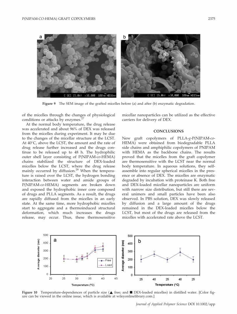
Therefore, intensity of the micelles is reduced withincubation time. Furthermore, changes in morphol-ogy of the polymeric micelles can also affect scat-tering intensity. After incubation, the micelles aresignificantly degraded but the enzymatic degrada-tion is not completed. Some micelles can keep theparticulate structure though they are partlydegraded. And other smaller micelles can be alsoformed from the degraded graft copolymers as ob-servation in the Figure 9. As a result, the size ofmicelles was much reduced.
Thermoresponsive drug release in vitro
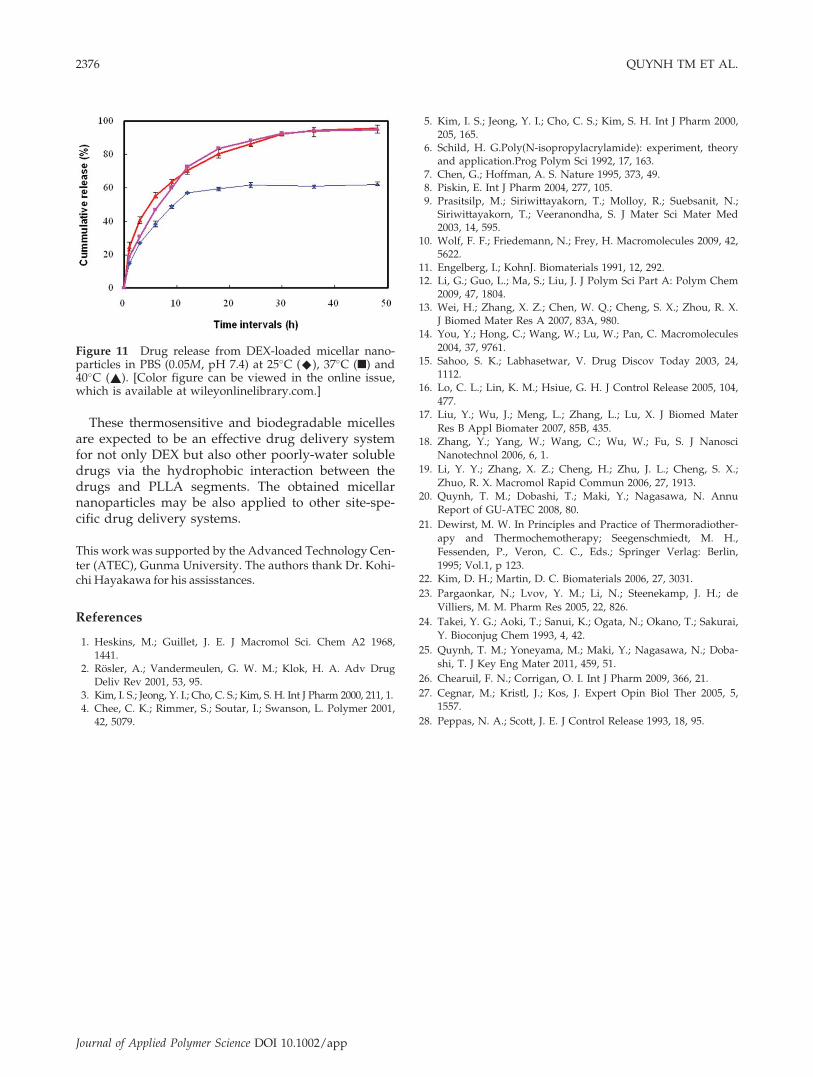
Through the hydrophobic interaction with the mi-cellar core, different hydrophobic drugs can beloaded into the micelles. In this study, about 16%w/w DEX was loaded into the micelles after dialy-sis process. Because of the thermosensitivity of themicelles, the drug release is temperature-depend-ent. When temperature increases up to the LCST,the hydrophilic shell becomes hydrophobic, result-ing in aggregation of several adjacent micelles asobserved in Figure 10(a). However, these aggre-gated states of the DEX-loaded micelles are rever-sibly changed by raising and lowering temperaturequickly [Fig. 10(b)].Figure 11 shows the drug release behaviors of the
DEX-loaded micelles as functions of time above andbelow the LCST. At room temperature, 15% DEXwas released after 1 h, and then the release rate isgradually reduced with time. About 60% of the drugwas released from the particles after 12 h, whereas alarge amount of DEX still remains in the micellesbecause of stable well-ordered assembly of themicelles under the LCST. Based on these stablestructures, they can escape from the nonselective re-ticuloendothelial system (RES), then passive target tothe specific site. Active targeting can be also modu-lated with controlling the interaction of the micelleswith the target sites, where the drugs are releasedand become effectual by deformation or degradation
TABLE IISize Distribution and LCST of the Micellar
Nanoparticles
Micelles Diametera (nm) Z-Aveb (nm) PdI LCST
Free 150–250 180 0.65 36.9DEX-loaded 160–280 200 0.78 36.8
a Determined by microscope (SEM images).b Average diameter and polydispersity index of micelles
measured by DLS at room temperature.
Figure 7 Size distribution by intensity of free (a) andDEX-loaded (b) micellar nanoparticles in distilled water.
Figure 8 Size-intensity curves of the degraded micellarnanoparticles with incubation times. [Color figure can beviewed in the online issue, which is available atwileyonlinelibrary.com.]
2374 QUYNH TM ET AL.
Journal of Applied Polymer Science DOI 10.1002/app
of the micelles through the changes of physiologicalconditions or attacks by enzymes.27
At the normal body temperature, the drug releasewas accelerated and about 96% of DEX was releasedfrom the micelles during experiment. It may be dueto the changes of the micellar structure at the LCST.At 40�C, above the LCST, the amount and the rate ofdrug release further increased and the drugs con-tinue to be released up to 48 h. The hydrophilicouter shell layer consisting of P(NIPAM-co-HEMA)chains stabilized the structure of DEX-loadedmicelles below the LCST, where the drug releasemainly occurred by diffusion.28 When the tempera-ture is raised over the LCST, the hydrogen bondinginteraction between water and amide groups ofP(NIPAM-co-HEMA) segments are broken downand exposed the hydrophobic inner core composedof drugs and PLLA segments. As a result, the drugsare rapidly diffused from the micelles in an earlystate. At the same time, more hydrophobic micellesstart to aggregate and a thermoinduced structuraldeformation, which much increases the drugsrelease, may occur. Thus, these thermosensitive
micellar nanoparticles can be utilized as the effectivecarriers for delivery of DEX.
CONCLUSIONS
New graft copolymers of PLLA-g-P(NIPAM-co-HEMA) were obtained from biodegradable PLLAside chains and amphiphilic copolymers of PNIPAMwith HEMA as the backbone chains. The resultsproved that the micelles from the graft copolymerare thermosensitive with the LCST near the normalbody temperature. In aqueous solutions, they self-assemble into regular spherical micelles in the pres-ence or absence of DEX. The micelles are enzymaticdegraded by incubation with proteinase K. Both freeand DEX-loaded micellar nanoparticles are uniformwith narrow size distribution, but still there are sev-eral unimers and small particles have been alsoobserved. In PBS solution, DEX was slowly releasedby diffusion and a large amount of the drugsremained in the DEX-loaded micelles below theLCST, but most of the drugs are released from themicelles with accelerated rate above the LCST.
Figure 9 The SEM image of the grafted micelles before (a) and after (b) enzymatic degradation.
Figure 10 Temperature-dependences of particle size (~ free; and n DEX-loaded micelles) in distilled water. [Color fig-ure can be viewed in the online issue, which is available at wileyonlinelibrary.com.]
P(NIPAM-CO-HEMA) GRAFT COPOLYMERS 2375
Journal of Applied Polymer Science DOI 10.1002/app
These thermosensitive and biodegradable micellesare expected to be an effective drug delivery systemfor not only DEX but also other poorly-water solubledrugs via the hydrophobic interaction between thedrugs and PLLA segments. The obtained micellarnanoparticles may be also applied to other site-spe-cific drug delivery systems.
This work was supported by the Advanced Technology Cen-ter (ATEC), Gunma University. The authors thank Dr. Kohi-chi Hayakawa for his assisstances.
References
1. Heskins, M.; Guillet, J. E. J Macromol Sci. Chem A2 1968,1441.
2. Rosler, A.; Vandermeulen, G. W. M.; Klok, H. A. Adv DrugDeliv Rev 2001, 53, 95.
3. Kim, I. S.; Jeong, Y. I.; Cho, C. S.; Kim, S. H. Int J Pharm 2000, 211, 1.4. Chee, C. K.; Rimmer, S.; Soutar, I.; Swanson, L. Polymer 2001,
42, 5079.
5. Kim, I. S.; Jeong, Y. I.; Cho, C. S.; Kim, S. H. Int J Pharm 2000,205, 165.
6. Schild, H. G.Poly(N-isopropylacrylamide): experiment, theoryand application.Prog Polym Sci 1992, 17, 163.
7. Chen, G.; Hoffman, A. S. Nature 1995, 373, 49.8. Piskin, E. Int J Pharm 2004, 277, 105.9. Prasitsilp, M.; Siriwittayakorn, T.; Molloy, R.; Suebsanit, N.;
Siriwittayakorn, T.; Veeranondha, S. J Mater Sci Mater Med2003, 14, 595.
10. Wolf, F. F.; Friedemann, N.; Frey, H. Macromolecules 2009, 42,5622.
11. Engelberg, I.; KohnJ. Biomaterials 1991, 12, 292.12. Li, G.; Guo, L.; Ma, S.; Liu, J. J Polym Sci Part A: Polym Chem
2009, 47, 1804.13. Wei, H.; Zhang, X. Z.; Chen, W. Q.; Cheng, S. X.; Zhou, R. X.
J Biomed Mater Res A 2007, 83A, 980.14. You, Y.; Hong, C.; Wang, W.; Lu, W.; Pan, C. Macromolecules
2004, 37, 9761.15. Sahoo, S. K.; Labhasetwar, V. Drug Discov Today 2003, 24,
1112.16. Lo, C. L.; Lin, K. M.; Hsiue, G. H. J Control Release 2005, 104,
477.17. Liu, Y.; Wu, J.; Meng, L.; Zhang, L.; Lu, X. J Biomed Mater
Res B Appl Biomater 2007, 85B, 435.18. Zhang, Y.; Yang, W.; Wang, C.; Wu, W.; Fu, S. J Nanosci
Nanotechnol 2006, 6, 1.
19. Li, Y. Y.; Zhang, X. Z.; Cheng, H.; Zhu, J. L.; Cheng, S. X.;
Zhuo, R. X. Macromol Rapid Commun 2006, 27, 1913.20. Quynh, T. M.; Dobashi, T.; Maki, Y.; Nagasawa, N. Annu
Report of GU-ATEC 2008, 80.
21. Dewirst, M. W. In Principles and Practice of Thermoradiother-
apy and Thermochemotherapy; Seegenschmiedt, M. H.,
Fessenden, P., Veron, C. C., Eds.; Springer Verlag: Berlin,
1995; Vol.1, p 123.22. Kim, D. H.; Martin, D. C. Biomaterials 2006, 27, 3031.
23. Pargaonkar, N.; Lvov, Y. M.; Li, N.; Steenekamp, J. H.; de
Villiers, M. M. Pharm Res 2005, 22, 826.
24. Takei, Y. G.; Aoki, T.; Sanui, K.; Ogata, N.; Okano, T.; Sakurai,
Y. Bioconjug Chem 1993, 4, 42.
25. Quynh, T. M.; Yoneyama, M.; Maki, Y.; Nagasawa, N.; Doba-
shi, T. J Key Eng Mater 2011, 459, 51.
26. Chearuil, F. N.; Corrigan, O. I. Int J Pharm 2009, 366, 21.
27. Cegnar, M.; Kristl, J.; Kos, J. Expert Opin Biol Ther 2005, 5,1557.
28. Peppas, N. A.; Scott, J. E. J Control Release 1993, 18, 95.
Figure 11 Drug release from DEX-loaded micellar nano-particles in PBS (0.05M, pH 7.4) at 25�C (^), 37�C (n) and40�C (~). [Color figure can be viewed in the online issue,which is available at wileyonlinelibrary.com.]
2376 QUYNH TM ET AL.
Journal of Applied Polymer Science DOI 10.1002/app
Copyright © 2022 FDOKUMEN