Politi et al 2008 Direct NMR Cannabis Phytochemistry 69
10
This article was published in an Elsevier journal. The attached copy is furnished to the author for non-commercial research and education use, including for instruction at the author’s institution, sharing with colleagues and providing to institution administration. Other uses, including reproduction and distribution, or selling or licensing copies, or posting to personal, institutional or third party websites are prohibited. In most cases authors are permitted to post their version of the article (e.g. in Word or Tex form) to their personal website or institutional repository. Authors requiring further information regarding Elsevier’s archiving and manuscript policies are encouraged to visit: http://www.elsevier.com/copyright
-
Upload
iamoecenter -
Category
Documents
-
view
2 -
download
0
Transcript of Politi et al 2008 Direct NMR Cannabis Phytochemistry 69

This article was published in an Elsevier journal. The attached copyis furnished to the author for non-commercial research and
education use, including for instruction at the author’s institution,sharing with colleagues and providing to institution administration.
Other uses, including reproduction and distribution, or selling orlicensing copies, or posting to personal, institutional or third party
websites are prohibited.
In most cases authors are permitted to post their version of thearticle (e.g. in Word or Tex form) to their personal website orinstitutional repository. Authors requiring further information
regarding Elsevier’s archiving and manuscript policies areencouraged to visit:
http://www.elsevier.com/copyright

Author's personal copy
Direct NMR analysis of cannabis water extracts and tincturesand semi-quantitative data on D9-THC and D9-THC-acid
M. Politi a, W. Peschel a, N. Wilson a, M. Zloh b, J.M. Prieto a,*, M. Heinrich a
a Centre for Pharmacognosy and Phytotherapy, The School of Pharmacy, University of London,
29–39 Brunswick Square, London WC1N 1AX, UKb Department of Pharmaceutical and Biological Chemistry, The School of Pharmacy, University of London, 29–39 Brunswick Square,
London WC1N 1AX, UK
Received 7 February 2007; received in revised form 21 July 2007Available online 26 October 2007
Abstract
Cannabis sativa L. is the source for a whole series of chemically diverse bioactive compounds that are currently under intensive phar-maceutical investigation. In this work, hot and cold water extracts as well as ethanol/water mixtures (tinctures) of cannabis were com-pared in order to better understand how these extracts differ in their overall composition. NMR analysis and in vitro cell assays of crudeextracts and fractions were performed. Manufacturing procedures to produce natural remedies can strongly affect the final compositionof the herbal medicines. Temperature and polarity of the solvents used for the extraction resulted to be two factors that affect the totalamount of D9-THC in the extracts and its relative quantity with respect to D9-THC-acid and other metabolites. Diffusion-edited 1HNMR (1D DOSY) and 1H NMR with suppression of the ethanol and water signals were used. With this method it was possible, withoutany evaporation or separation step, to distinguish between tinctures from different cannabis cultivars. This approach is proposed as adirect analysis of plant tinctures.� 2007 Elsevier Ltd. All rights reserved.
Keywords: Cannabis; Cannabis water extract; Tinctures; Nuclear magnetic resonance; Solvent signal suppression; Principal component analysis; NFjB
1. Introduction
The potential use of cannabis for medical purposes iscurrently under intensive investigation (Ben Amar,2006). Around 500 cannabis metabolites are known (ElSohly and Slade, 2005). About 70 structures belong tothe typical class of terpenophenolic derivatives known ascannabinoids; (�)-D9-trans-(6aR,10aR)-tetrahydrocannab-inol (D9-THC) is probably the best studied cannabisconstituent, but not the only one to have proven pharma-cological activities (Barrett et al., 1985; Fairbairn andPickens, 1987; Thomas et al., 2005; Wilkinson et al.,2003). In fact, D9-THC-acid (Verhoeckx et al., 2006), can-
nabidiol (CBD) (Mechoulam et al., 2002), cannabigerol(CBG) and cannabinol (CBN) (Wilkinson and William-son, 2007), for instance, also show important therapeuticeffects without causing the undesired psychotropic activitytypical of D9-THC. Furthermore, the bioactivities of D9-THC alone are different from those of a crude cannabisextract, as synergistic effects have been reported (Russoand Guy, 2006). The presence of alkaloids in very lowamounts has been described in both roots and leaves ofcannabis (El Sohly, 1985; Mechoulam, 1988). This crudealkaloid mixture is endowed with anti-inflammatoryeffects but at the moment there is no pharmacologicaldata on the purified alkaloids.
This chemical and pharmacological complexity led tothe re-introduction of cannabis crude extracts in clinics.Bedrocan�, a special cannabis variety with standardisedcontent of D9-THC and CBD is sold in Dutch pharmacies
0031-9422/$ - see front matter � 2007 Elsevier Ltd. All rights reserved.
doi:10.1016/j.phytochem.2007.07.018
* Corresponding author. Tel.: +44 0 20 7753 5841; fax: +44 0 20 77535909.
E-mail address: [email protected] (J.M. Prieto).
www.elsevier.com/locate/phytochem
Available online at www.sciencedirect.com
Phytochemistry 69 (2008) 562–570
PHYTOCHEMISTRY

Author's personal copy
and Sativex�, a medicine consisting in the mixture of twocannabis extracts rich in D9-THC and CBD, respectively,has been registered in Canada for the treatment of multiplesclerosis symptoms (Barnes, 2006). However, there is aneed to develop medicinal cannabis with low contents ofD9-THC. This is the goal of an EU-funded research projectcoordinated by our research group. As part of this, wedecided to study the chemistry and pharmacology of boththe cannabis aqueous extracts (infusions) and hydroethan-olic extracts (tinctures) in order to establish their metabo-lite profiles with emphasis on the relationship betweenthe ratio D9-THC/D9-THC-acid and the extractive process.
Cannabis tea (water extract) is a popular remedy (Wareet al., 2005) while, prior to 1971, cannabis tinctures wereavailable for prescription by British physicians (House ofLords, 1998) and can be considered as the traditional andofficinal ways to consume medicinal cannabis products,respectively. However, they have not been investigatedwith modern techniques and the majority of the pharmaco-logical and phytochemical data on cannabis analyzeorganic extracts without pharmaceutical relevance (e.g.,chloroform, methanol, acetone).
Literature data on cannabis water extract are very scarceand certainly not up-to-date, although analgesic activity isreported for cannabis water extract (Segelman et al., 1974).Moreover, the use of cannabis tea has been stated to pro-long and intensify significantly the psychotropic effectsresulting from smoking marijuana (Segelman and Sofia,1973; Segelman et al., 1974). More recently (Giroudet al., 2000), a case study with six healthy volunteersreported on the non-psychotropic activity of cannabis teaalone; this fact was linked to the low amount of D9-THCin the cannabis tea. However, D9-THC-acid, the main con-stituent of THC-rich Cannabis sativa L. cultivars, decarb-oxylizes into D9-THC at temperatures around 103 �C. Inthis work, this effect was evaluated by extracting the plantwith hot and cold water. On the other hand, tinctures arethe most widely plant extracts produced in the pharmaceu-tical industry. Thus, the optimization of the analyticalmethods to determine the optimal alcoholic strength givingthe best pharmacological activity with the least toxicity isan important issue. Moreover, the study of the traditionalmanufacturing procedures to prepare herbal medicines canmake important contributions to the identification of newnatural products as well as new synergistic effects (Politiet al., 2005, 2006a) because the way a natural medicine isprepared can strongly affect the final composition of theremedy. Two factors are of importance in the case of can-nabis: the effect of the solvent polarity (different ethanol/water mixtures or water) and the temperature used forthe extraction of the plant on the relative content ofD9-THC and D9-THC-acid.
The processing of the complex mixtures of plant metab-olites for analytical purposes may also give erroneous inter-pretations of the original composition and the idealapproach would be their analysis without neither prepara-tive nor chromatographic steps. The selection of the most
suitable analytical technique is generally a compromisebetween speed, selectivity and sensitivity (Sumner et al.,2003). Several spectroscopic techniques such as NMR,MS, HPLC-UV/PDA and IR, currently also named ‘‘met-abolomics tools’’, are available for the direct analysis ofplant metabolites (Dunn et al., 2005). NMR is a rapidand selective tool but has low sensitivity, while MS offersgood sensitivity and selectivity but relatively longer analy-sis times. For the present investigation it was our goal toacquire information on aqueous and hydroethanolic can-nabis extracts in a fast and direct way. Therefore, this workis mostly based on 1D NMR experiments, also used forsemi-quantitative analysis (Pauli et al., 2005), and compar-ison with the recent NMR-based literature on cannabi-noids and cannabis extracts (Choi et al., 2004a,b;Hazekamp et al., 2004). Cell assays and principal compo-nent analysis of the NMR data were also included in theinvestigation.
2. Result and discussion
2.1. Qualitative phytochemical analysis of cannabis aqueous
extracts
The first strategy adopted to abate the content of thepsychoactive D9-THC was by producing a cannabis waterextract. Fig. 1 shows the 1H NMR spectra of three extractsobtained from three aliquots of THC-rich cannabis mate-rial after maceration in deuterated chloroform, methanoland water. The typical cannabinoid proton signals of theextracts in chloroform (Fig. 1a) and methanol (Fig. 1b)emerge in particular in the NMR region between6-6.5 ppm. These are mostly due to D9-THC (1) and
23456789 ppm
a
b
c
Fig. 1. 1H NMR spectra (400 MHz) of crude cannabis extracts obtainedafter overnight maceration of three aliquots of 70 mg aerial parts in 1 mlof the following deuterated solvents: (a) chloroform; (b) methanol; (c)water.
M. Politi et al. / Phytochemistry 69 (2008) 562–570 563

Author's personal copy
D9-THC-acid (2) (see Fig. 2). These proton signals are notdetected in the water extract (Fig. 1c) indicating that thecontent of both cannabinoids in cannabis water extractare at least greatly reduced.
In a second experiment, 5 g of the same batch of THC-rich C. sativa (labelled CS) were used to produce the hotwater extract (CShw) and other 5 g to produce the coldwater extract (CScw) (see experimental). Twenty-five milli-liters of both water extracts were extracted with n-hexaneobtaining 13 mg of the organic fraction from the hot waterextract (CShw_c) and 5 mg from the cold water extract(CScw_c). Fig. 3 shows the NMR analysis of both organicfractions dissolved in deuterated chloroform. Both mix-tures contain mostly D9-THC and D9-THC-acid. Integra-tion of the D9-THC signal at 6.14 ppm and of theD9-THC-acid signal at 6.39 ppm allowed us to calculatethe ratio of these cannabinoids. In the organic fraction
from the hot water extract (CShw_c), the content of D9-THC is 1.52 times that of D9-THC-acid while in the organicfraction from the cold water extract (CScw_c) the relativeamount of D9-THC is reduced to 0.09 times that ofD9-THC-acid. The higher amount of D9-THC in the hotwater extract is due to the known decarboxylation ofD9-THC-acid into D9-THC that occurs approaching103 �C (Segelman and Sofia, 1973). Based on the solubilityof D9-THC in water (2.8 mg/l), from 25 ml of water a max-imum of 0.07 mg of D9-THC can be expected. However,from 25 ml of the hot water extract 13 mg of a mixturecontaining more than 50% of D9-THC was obtained. Thisresult indicates that the use of high temperature or thepresence of other compounds from cannabis favour theextraction and solubilisation of D9-THC in water.
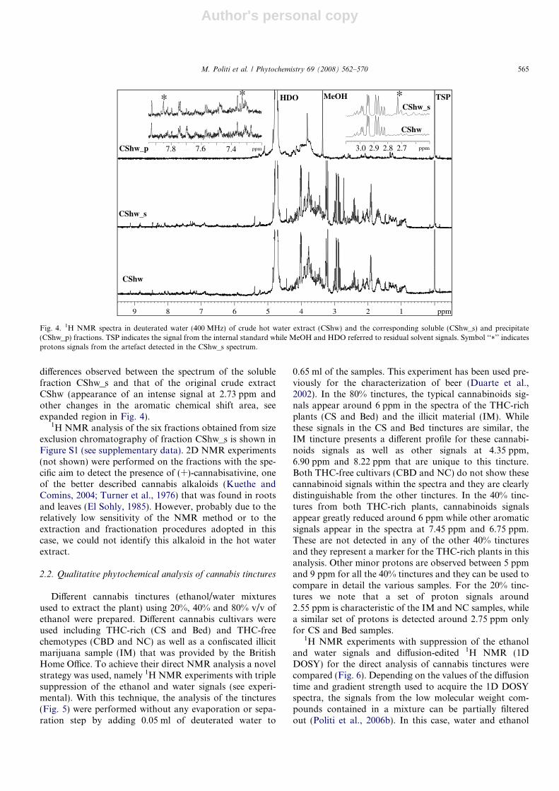
In order to acquire further data on the cannabis waterextract this crude mixture was fractionated by precipitationin methanol/water 4:1 obtaining a soluble fraction and aprecipitate (see Section 4). Fig. 4 shows the 1H NMR anal-ysis of the crude extract compared with both fractions. Theprecipitate CShw_p presents broad signals in the carbohy-drate region that indicate the presence of polysaccharides.A polysaccharide from the aqueous extract of cannabiswith potential anti-glaucoma activity has been described(Hodges et al., 1985). This mixture had shown a verypotent intraocular pressure-lowering activity (antiglauco-ma) when tested by i.v. injection into rabbits. Partial puri-fication with gel exclusion chromatography yielded veryactive material (lowers intraocular pressure maximally at1 mg/animal) with an estimated molecular weight of500 kDa; Rhamnose, galactose and uronic acid were themajor sugar constituents identified. Further effort couldbe made in the future in order to assess if such bioactivepolysaccharide corresponds to that one detected in thiswork.
Previously, Cannabis polypeptides have also beenreported as water soluble derivatives (Hillestad et al.,1977; Tang et al., 2006). It is known that folding of poly-peptides can occur in presence of methanol or otherorganic solvent in water solution (Chakraborty et al.,2005; Zloh et al., 1998). It is therefore theoretically possiblethat, due to the use of methanol in the fractionation step,the refolding of polypeptides may be responsible of the
6.31 δH
123.7 δC
6.14 δH; 107.5 δC
6.27 δH
110.1 δC
154.7 δC
142.8 δC
OH
O
1
154.2 δC
OH
O
COOH
6.39 δH
123.6 δC
6.25 δH
112.6 δC
164.7 δC
146.9 δC
2
159.8 δC
Fig. 2. Structures of D9-THC (1) and D9-THC-acid (2). Atoms with protons and carbons chemical shifts values measured in deuterated chloroform arecircled.
23456 ppm6.36.4 ppm
CScw_c
CShw_c
THC
THC-acid
Fig. 3. 1H NMR spectra in deuterated chloroform (400 MHz) of theorganic fraction from the hot water extract (CShw_c) and the organicfraction from the cold water extract (CScw_c). On the left part of thespectra the signal intensity has been magnified. D9-THC and D9-THC-acidprotons signals in the region of 6–6.4 ppm are indicated with arrows.
564 M. Politi et al. / Phytochemistry 69 (2008) 562–570
Author's personal copy
differences observed between the spectrum of the solublefraction CShw_s and that of the original crude extractCShw (appearance of an intense signal at 2.73 ppm andother changes in the aromatic chemical shift area, seeexpanded region in Fig. 4).
1H NMR analysis of the six fractions obtained from sizeexclusion chromatography of fraction CShw_s is shown inFigure S1 (see supplementary data). 2D NMR experiments(not shown) were performed on the fractions with the spe-cific aim to detect the presence of (+)-cannabisativine, oneof the better described cannabis alkaloids (Kuethe andComins, 2004; Turner et al., 1976) that was found in rootsand leaves (El Sohly, 1985). However, probably due to therelatively low sensitivity of the NMR method or to theextraction and fractionation procedures adopted in thiscase, we could not identify this alkaloid in the hot waterextract.
2.2. Qualitative phytochemical analysis of cannabis tinctures
Different cannabis tinctures (ethanol/water mixturesused to extract the plant) using 20%, 40% and 80% v/v ofethanol were prepared. Different cannabis cultivars wereused including THC-rich (CS and Bed) and THC-freechemotypes (CBD and NC) as well as a confiscated illicitmarijuana sample (IM) that was provided by the BritishHome Office. To achieve their direct NMR analysis a novelstrategy was used, namely 1H NMR experiments with triplesuppression of the ethanol and water signals (see experi-mental). With this technique, the analysis of the tinctures(Fig. 5) were performed without any evaporation or sepa-ration step by adding 0.05 ml of deuterated water to
0.65 ml of the samples. This experiment has been used pre-viously for the characterization of beer (Duarte et al.,2002). In the 80% tinctures, the typical cannabinoids sig-nals appear around 6 ppm in the spectra of the THC-richplants (CS and Bed) and the illicit material (IM). Whilethese signals in the CS and Bed tinctures are similar, theIM tincture presents a different profile for these cannabi-noids signals as well as other signals at 4.35 ppm,6.90 ppm and 8.22 ppm that are unique to this tincture.Both THC-free cultivars (CBD and NC) do not show thesecannabinoid signals within the spectra and they are clearlydistinguishable from the other tinctures. In the 40% tinc-tures from both THC-rich plants, cannabinoids signalsappear greatly reduced around 6 ppm while other aromaticsignals appear in the spectra at 7.45 ppm and 6.75 ppm.These are not detected in any of the other 40% tincturesand they represent a marker for the THC-rich plants in thisanalysis. Other minor protons are observed between 5 ppmand 9 ppm for all the 40% tinctures and they can be used tocompare in detail the various samples. For the 20% tinc-tures we note that a set of proton signals around2.55 ppm is characteristic of the IM and NC samples, whilea similar set of protons is detected around 2.75 ppm onlyfor CS and Bed samples.
1H NMR experiments with suppression of the ethanoland water signals and diffusion-edited 1H NMR (1DDOSY) for the direct analysis of cannabis tinctures werecompared (Fig. 6). Depending on the values of the diffusiontime and gradient strength used to acquire the 1D DOSYspectra, the signals from the low molecular weight com-pounds contained in a mixture can be partially filteredout (Politi et al., 2006b). In this case, water and ethanol
7.47.67.88.08.28.4 ppm
**
7.47.67.8
9 8 7 6 5 4 3 2 1 ppm
CShw
CShw_s
TSPHDO
CShw_p
MeOH
2.72.82.93.0 ppm
CShw
CShw_s*
2.72.9 2.83.0
Fig. 4. 1H NMR spectra in deuterated water (400 MHz) of crude hot water extract (CShw) and the corresponding soluble (CShw_s) and precipitate(CShw_p) fractions. TSP indicates the signal from the internal standard while MeOH and HDO referred to residual solvent signals. Symbol ‘‘*’’ indicatesprotons signals from the artefact detected in the CShw_s spectrum.
M. Politi et al. / Phytochemistry 69 (2008) 562–570 565

Author's personal copy
represent the lowest molecular weight molecules in thesamples. Reducing the solvents signals in the spectra allowsthe detection of protons from cannabis metabolites. Under
the conditions used (see Section 4), the experiments withsolvent suppression take around 25 min while the 1DDOSY takes only 8 min, using 64 scans in both cases.
Tinctures 20% v/v
23456789 ppm
CS(Northern LightTHC-rich)
Bed(BedrocanTHC-rich)
CBD(Cannabidiol richTHC-free)
NC(Non CannabinoidsTHC-free)
IM(Illicit material)
Tinctures 40% v/v
23456789 ppm
CS
Bed
CBD
NC
IM
Tinctures 80% v/v
23456789 ppm
CS
Bed
CBD
NC
IM
Fig. 5. 1H NMR experiments (500 MHz) with suppression of the ethanol and water signals. Comparison of three types of tinctures (20%, 40% and 80% v/v) from five different cannabis cultivars (CS = Northern Lights 5 crossed with Haze, THC-rich; Bed = Bedrocan, THC-rich; IM = illicit material;CBD = cannabidiol-rich, THC-free; NC = non-cannabinoids, THC-free).
2345 ppm789 ppm
CS80
CS40
CS20
1H NMR1D NOESY Pulse sequence with triple suppression of water and ethanol signals
CS80
CS40
CS20
1D DOSYGradient strength 80%,
Little Delta 1000 micro-secBig Delta 0.2 sec
2345 ppm789 ppm
Fig. 6. Comparison of the 1H NMR experiments with suppression of the ethanol and water signals with the diffusion-edited 1H NMR (1D DOSY)experiments (500 MHz) in the analysis of CS tinctures. On the left part of the spectra the signal intensity has been magnified.
566 M. Politi et al. / Phytochemistry 69 (2008) 562–570

Author's personal copy
Although in the 1D DOSY the signal to noise ratio islower, all the major protons previously described for CStinctures were also detected with this faster NMR experi-ment. With both experiments parts of the spectra (around4.77 pm, 3.65 ppm and 1.17 ppm) appear unresolved dueto the incomplete suppression of the solvent signals, butuseful information can still be acquired; the region of thespectra between 5 and 12 ppm, for instance, is well resolvedand it affords crucial information for natural product iden-tification. The proton chemical shift values usually vary fora single component analysed in different solvents. There-fore, with this NMR approach, only tinctures with thesame ethanolic strength can be used for a comparative fin-gerprinting analysis.
2.3. Quantitative phytochemical analysis of cannabis
tinctures
In order to better acquire semi-quantitative data on D9-THC and D9-THC-acid, and to compare our data withthose from the literature (Choi et al., 2004a,b; Hazekampet al., 2004) the dried residues from the tinctures wereextracted with chloroform and methanol as described inSection 4. Fig. 7 shows the comparison between the protonspectra acquired in deuterated chloroform of fractionsCS80_c, CS40_c and CS20_c. The first two contain mostlyD9-THC and D9-THC-acid while in the latter other protonsignals appear in the spectrum (indicated with the aster-isks). The D9-THC content was 0.46 times that ofD9-THC-acid in fraction CS80_c, 0.28 times in fractionCS40_c, and only 0.10 times in fraction CS20_c. For the
first two cases, quantitative 13C NMR analysis confirmsthese observed ratio values (see supplementary data andFigure S3). The small amount of fraction CS20_c obtaineddid not allow a quantitative 13C NMR analysis of this mix-ture. In the CS20_c sample other cannabinoid signalsbetween 6 and 6.5 ppm appear in the spectra with a relativehigh intensity with respect to the content of D9-THC andD9-THC-acid. Other protons possibly belonging to canna-binol and cannflavins are also detected around 6.9–7.6 ppm.
Minor quali-quantitative differences between the metha-nol and water soluble fractions are detected within theoverall NMR regions of the corresponding spectra(Fig. S4 and S5 in supplementary data).
As shown, this technique is still semi-quantitative, andfurther work must be done to allow the quantitation of sin-gle compounds in the mixture. The strength of thisapproach resides in the fast acquisition of data from tinc-tures without virtually any pre-processing of the samples.Furthermore, the resulting fingerprint reflects the real com-position of the mixture and multivariate analysis can beapplied to the NMR data if necessary. We have alreadyapplied this method for batch-to-batch variability and sta-bility test on different commercial tinctures (Politi et al.,unpublished). However, if a quantitative standardisationof certain markers or bioactives of the tincture is needed,then LC–UV–MS in any of its variants is the instrumentaltechnique of choice. The development of different condi-tions of elution and processing of the samples is time con-suming but the analytical results give accurate quantitativedata of selected metabolites, even if they are present in min-ute quantities. Examples are the application of LC–MS tothe control of cannabinoids in crude extracts (Stolker et al.,2004) or in forensic samples (Yang and Xie, 2006).
2.4. Pharmacological data
All water soluble samples including the crude hot waterextract CShw, fractions CShw_s and CShw_p, and frac-tions 1–6 from the size exclusion chromatography (seeexperimental) were tested in the NF-jB assay in HeLa cells(Bremner et al., 2004) (see Section 4). At a concentration of0.1 mg/ml, neither NF-jB inhibitory nor cytotoxic effectswere observed in the model. However, the EtOH 100%extracts from the same THC-rich plant material (CS) atthe same concentration resulted in toxicity for the HeLacells as evidenced by morphological changes induced onthe cells during incubation (cells with normalphenotype < 10%).
With regard to the tinctures, the cannabinoid contentaugments increasing the concentrations of ethanol. In thesame way, the maximum non-toxic concentration (see Sec-tion 4) of these extracts, measured with the MTT assay onHeLa cells, increases by reducing the ethanol strength usedfor extraction. Whether the non-toxicity of cannabisextracts is due to the low levels of cannabinoids remainsunclear, but a positive correlation between increasing etha-
234 ppm678 ppm
CS20_c
CS40_c
CS80_c
1H NMR
CDCl3
* **** ** *
*
THC and THC-acid
Fig. 7. 1H NMR in deuterated chloroform (400 MHz) of fractionsCS20_c, CS40_c, and CS80_c. On the left part of the spectra the signalintensity has been magnified. Asterisks indicate proton signals that arerelatively more abundant with respect to D9-THC and D9-THC-acidsignals in CS20_c spectrum compared to CS40_c and CS80_c ones.
M. Politi et al. / Phytochemistry 69 (2008) 562–570 567

Author's personal copy
nol tincture strength (and therefore cannabinoid content)and cytotoxicity as measured in the MTT assay (Fig. S2in supplementary data) was found. The maximum non-toxic concentrations for the three tinctures CS20, CS40and CS80 were 20.83 lmol/ml, 1.47 lmol/ml and0.03 lmol/ml, respectively, after 6 h incubation and14.58 lmol/ml, 0.43 lmol/ml and 0.01 lmol/ml, respec-tively, after 24 h incubation as derived from the MTT assayon HeLa cells (Fig. S2 in supplementary data).
3. Conclusion
A novel approach based on NMR spectroscopy, 1HNMR experiments with suppression of the ethanol andwater signals and diffusion-edited 1H NMR (1D DOSY),was developed and successfully applied for the fast anddirect analysis of tinctures. Relative amounts ofD9-THC and D9-THC-acid in the extracts analysed varydepending on the temperature (extraction with hot andcold water) and the polarity of the solvent used for theextraction (tinctures with different ethanol strength). Tothe best of our knowledge, this is the first time that thepolarity of the solvent used for the extraction is describedas a factor that can affect the relative quantity of D9-THCrespect to other cannabinoids in the extract. FractionCS20_c from the 20% tincture fulfils the demand for can-nabis extracts low in D9-THC still containing other can-nabinoids of medicinal interest. Further studies tomaximize the yield of this promising mixture of cannabisproducts are ongoing. As for the aqueous extracts, thisapproach revealed the non-negligible presence ofD9-THC and D9-THC-acid in infusions of cannabis whichmay, at least in part, explain the recreational and medic-inal uses of this particular preparation. In the emergingmetabolomic era, it is essential to monitor how manufac-turing procedures affect the final content of natural prod-ucts in herbal medicines.
4. Experimental
4.1. Plant material
All relevant Home Office licenses for growing, transp-roting and possession of cannabis have been in place atall times. The seeds of Northern Lights 5 crossed with Haze(labelled CS) were bought from the Pukka Seed Company,Guildford, UK. This cannabis cultivar was grown underhydroponic conditions. Plants grown in pods filled withceramic beads where nutrient rich water was pumped tothe base of the plant 4 times a day. Growing conditionswere the following: 70% Humidity, 24 �C Temperature,5.5–6 pH of water, 1300 ppm of CO2, metal halides600 W for vegetative cycle (18 h a day cycle), and sodium’s1000 W for blooming (18 h then slowly reduced to 12 h).The seeds were planted on 15/08/2005, the first males were
removed on 27/09/2005 and the females were harvested on02/12/2005. Bedrocan (labelled Bed) was received in 2006from the Office of Medicinal Cannabis, Ministry of Health,Welfare and Sports, The Hague, The Netherlands. Illicitmaterial (labelled IM) was provided in 2004 by the HomeOffice, UK. Cannabidiol-rich and non-cannabinoid culti-vars (labelled CBD and labelled NC, respectively) werereceived in 2005 from the ISCI (Experimental Institutefor the Industrial Crop), Rovigo, Italy.
4.2. Chemicals
Freshly deionised, ultrafiltered water was obtained witha Milli-Q system (Molsheim, France). Ethanol 99.7–100%(AnalaR, VWR, Poole, UK).
4.3. Extraction and fractionation
Three different aliquots of CS (70 mg each) were macer-ated overnight at room temperature in 1 ml of CDCl3,MeOD, and D2O and the extracts were directly analysedby NMR (Fig. 1).
Five grams of dried CS were extracted with 100 ml hotwater (heating and boiling for 3 min) and after filtrationapproximately 50 ml of this decoction, labelled CShw, wererecovered. Another 5 g sample of CS was extracted with100 ml cold water (overnight under agitation, room tem-perature) and after filtration almost 50 ml of this extractlabelled CScw were recovered. 25 ml of both water extractswere extracted with 5 ml of n-hexane (3 times) and theseorganic fractions, labelled CShw_c and CScw_c, were driedin the rotavap to give 13 mg and 5 mg, respectively.
Another CShw extract was produced from 150 g of CSin 2 l hot water (heating and boiling for 3 min). This extractwas concentrated in a rotavap and finally freeze-dried togive 15 g of crude lyophilised material. Ten grams of CShwwere dissolved in 20 ml of water and 80 ml MeOH wereadded obtaining a precipitate and a soluble fraction thatwere separated by centrifugation and labelled CShw_p(yield 30%) and CShw_s (yield 70%), respectively.
The five different plant cultivars; Northern Lights 5crossed with Haze (CS), Bedrocan (Bed), illicit materialconfiscated and provided by the British home office (IM),cannabidiol-rich cultivar (CBD), and non-cannabinoid cul-tivar (NC) were used to produce the corresponding tinc-tures. In every single extraction, 10 g of each dried plantmaterial were used. Hundred milliliters of different mix-tures of ethanol/water (20%, 40%, and 80% v/v) were usedto macerate the plants for 3 days in the dark under agita-tion. After filtration under vacuum, approximately 50 ml,65 ml, and 75 ml were recovered from the 20%, 40%, and80% tinctures, respectively. These different recovery levelswere probably due to the absorption of water by the driedcannabis material. This absorbed water was not releasedduring filtration. Fifty milliliters of the three tinctures fromNorthern Lights 5 crossed with Haze, labelled, respectivelyCS20, CS40, and CS80, were dried in the rotavap obtaining
568 M. Politi et al. / Phytochemistry 69 (2008) 562–570

Author's personal copy
1.4 g, 2.3 g, and 1.7 g. These residues were extracted with100 ml of CHCl3 and centrifuged to separate the solublepart from the insoluble one. Then the residues wereextracted with 100 ml of MeOH and centrifuged. Theresidual materials contained only water-soluble constitu-ents. From the 20% tincture the chloroform-soluble frac-tion labelled CS20_c (40 mg), the methanol-solublefraction labelled CS20_m (371 mg), and the water-solublefraction labelled CS20_w (438 mg) were obtained. Fromthe 40% tincture fractions CS40_c (280 mg), CS40_m(738 mg), and CS40_w (612 mg) were obtained. From the80% tincture fractions CS80_c (1315 mg), CS80_m(131 mg), and CS80_w (60 mg) were obtained.
4.4. NMR sample preparation
CShw_c (13 mg) and CScw_c (5 mg) were dissolved in0.7 ml CDCl3. The hot water extract CShw and both frac-tions CShw_p and CShw_s were all analysed at a concen-tration of 10 mg/ml using 0.7 ml of D2O with 3-(trimethylsilyl)propionic-2,2,3,3-d4 acid, sodium salt(TSP) as internal standard (0.2 mg/ml). All tinctures pro-duced (CS20, CS40 and CS80, Bed20, Bed40 and Bed80,IM20, IM40 and IM80, CBD20, CBD40 and CBD80,NC20, NC40 and NC80) were directly analysed by adding0.05 ml of D2O as internal lock to 0.65 ml of each tinctureusing 1D DOSY experiments and the 1D NOESY pulsesequence with water and ethanol signals suppression.Approximately 40 mg of each chloroform, methanol andwater fraction from the three tinctures CS20, CS40 andCS80 were dissolved in 0.7 ml of the corresponding deuter-ated solvents.
4.5. NMR analysis
NMR spectra of samples were obtained on BrukerAVANCE 400 and 500 MHz spectrometers equipped witha multinuclear probehead with z-gradient. The TOPSPINv1.3 software was used for spectra acquisition and process-ing, The spectra were recorded in various solvent systemsat 300 K and the chemical shift calibration was carriedout either on the TSP signal or residual solvent peak.The size of all 1D spectra was 65 K and the number oftransients varied for different types of spectra. The stan-dard 1D 1H NMR spectra were acquired with 30o pulselength and a relaxation delay of 2 s, while the 1HNMR45 spectra were acquired with 45o pulse length anda relaxation delay of 60 s to enable an accurate quantifica-tion of peaks. Similarly 13C NMR and 13C NMR45 wereacquired at a operating frequency of 100.6 MHz (30o pulselengths and relaxation delay of 1 s and 45o pulse lengthsand relaxation delay of 60 s, respectively).
The rest of the experiments were conducted on a500 MHz instrument. 1D DOSY was obtained using apulse sequence from Bruker library (led bpgp2s1d) usinggradient strength (gpz6) 80, little delta (p30) 1000 ls andbig delta (d20) 0.2 s. 1D 1H NOESY pulse sequence
(lc1pnfr) with multiple offset presaturation using frequencylist was employed to suppress water and ethanol signals ofthe samples. Presaturation was carried out with a relaxa-tion delay (d1 = 2 s) and mixing time (d18 = 0.8 s). In bothcases the numbers of scans were 64.
4.6. 3-(4,5-Dimethylthiazol-2-yl)-2,5-diphenyl tetrazolium
bromide (MTT) assay
A modification of the assay described previously (Mos-mann, 1983) was used as a criterion of cytotoxicity. BrieflyCells were incubated at 37 �C and 5% CO2 atmosphere for6 h and 24 h in media supplemented with PBS at eight dif-ferent concentrations between 0.1 and 100 lg/ml. Controlsreceived vehicle only. The maximum non-toxic concentra-tion (MNTC) was determined for each extract, i.e. cell via-bility >85% of the control. The dark blue formazanproduct was dissolved in DMSO/isopropanol and mea-sured using a Anthos Lucy 1 luminometer/photometer(Anthos-Biochrome Ltd., Cambridge, UK) at 490 nm,and the data collected and processed using Stingray 1.5software (Dazdaq Ltd., Brighton, UK).
4.7. IL-6/luciferase (IL-6/Luc) assay
We followed the protocol described previously (Bremneret al., 2004). Briefly, HeLa cells stably transfected with aluciferase reporter gene controlled by the IL-6 promoter.Cells were incubated in the presence of the compounds orplant extracts for 1 h and then stimulated (PMA, 50 ng/ml, final concentration). The cells were incubated for a fur-ther 7 h (37 �C/5% CO2) and then lysed with luciferase lysisreagent (Promega, Madison, WI, USA). Lysates weretransferred to 96-well plates and an Anthos Lucy 1 lumino-meter/photometer (Anthos-Biochrome Ltd., Cambridge,UK) was used both to add the luciferase substrate (Pro-mega) and record the resulting luminometric readings fol-lowing a reaction time of 10 s. Positive (stimulated cellswithout a sample) and negative (resting cells without stim-ulation) controls were included.
Acknowledgments
We thank the Office of Medicinal Cannabis (The Neth-erlands), Customs and Excise (UK) and the ISCI (Italy) forsupplying the plant material. We also acknowledge theHome Office for providing all the related licenses. We final-ly thank the European Commission for financial supportunder the FP6 (COOP-CT-2004-512696).
Appendix A. Supplementary data
Supplementary data associated with this article can befound, in the online version, at doi:10.1016/j.phytochem.2007.07.018.
M. Politi et al. / Phytochemistry 69 (2008) 562–570 569

Author's personal copy
References
Barnes, M.P., 2006. Sativex: clinical efficacy and tolerability in thetreatment of symptoms of multiple sclerosis and neuropathic pain.Expert. Opin. Pharmacother. 7, 607–615.
Barrett, M., Gordon, D., Evans, F., 1985. Isolation from Cannabis sativa
L of cannflavin; a novel inhibitor of prostaglandin production.Biochem. Pharmacol. 34, 2019–2024.
Ben Amar, M., 2006. Cannabinoids in medicine: a review of theirtherapeutic potential. J. Ethnopharmacol. 105, 1–25.
Bremner, P., Tang, S., Birkmayer, H., Fiebich, B.L., Munoz, E.,Marquez, N., Rivera, D., Heinrich, M., 2004. Phenylpropanoid NF-kappaB inhibitors from Bupleurum fruticosum. Planta Med. 70, 914–918.
Chakraborty, K., Shivakumar, P., Raghothama, S., Varadarajan, R.,2005. NMR structural analysis of a peptide mimic of the bridgingsheet of HIV-1 gp120 in methanol and water. Biochem. J. 390, 573–581.
Choi, Y.H., Hazekamp, A., Peltenburg-Looman, A.M., Frederich, M.,Erkelens, C., Lefeber, A.W., Verpoorte, R., 2004a. NMR assignmentsof the major cannabinoids and cannabiflavonoids isolated fromflowers of Cannabis sativa. Phytochem. Anal. 15, 345–354.
Choi, Y.H., Kim, H.K., Hazekamp, A., Erkelens, C., Lefeber, A.W.,Verpoorte, R., 2004b. Metabolomic differentiation of Cannabis sativa
cultivars using 1H NMR spectroscopy and principal componentanalysis. J. Nat. Prod. 67, 953–957.
Duarte, I., Barros, A., Belton, P.S., Righelato, R., Spraul, M., Humpfer,E., Gil, A.M., 2002. High-resolution nuclear magnetic resonancespectroscopy and multivariate analysis for the characterization of beer.J. Agric. Food Chem. 50, 2475–2481.
Dunn, W.B., Bailey, N.J., Johnson, H.E., 2005. Measuring the metabo-lome: current analytical technologies. Analyst 130, 606–625.
El Sohly, M.A., 1985. Cannabis alkaloids. In: Pelletier, S.W. (Ed.),Alkaloids: Chemical and Biological Perspectives. Pergamon, Oxford,pp. 169–184.
El Sohly, M.A., Slade, D., 2005. Chemical constituents of marijuana: thecomplex mixture of natural cannabinoids. Life Sci. 78, 539–548.
Fairbairn, J., Pickens, J., 1987. Activity of cannabis in relation to its delta-trans-tetrahydro-cannabinol. Br. J. Pharmacol. 72, 401–409.
Giroud, C., Menetrey, A., Augsburger, M., Buclin, T., Sanchez-Mazas, P.,Mangin, P., 2000. Hemp tea versus hemp milk: behavioral, physio-logical effects, blood, urine, saliva, and sweat cannabinoids levelsfollowing ingestion by 2 groups of 6 healthy volunteers. Z. ZagadnienNauk Sadowych 42, 102–110.
Hazekamp, A., Choi, Y.H., Verpoorte, R., 2004. Quantitative analysis ofcannabinoids from Cannabis sativa using 1H NMR. Chem. Pharm.Bull. 52, 718–721.
Hillestad, A., Wold, J.K., Paulsen, B.S., 1977. Structural studies of water-soluble glycoproteins from Cannabis sativa L. Carbohydr. Res. 57,135–144.
Hodges, LC., Deutsch, HM., Green, K., Zalkow, LH., 1985. Polysaccha-rides from Cannabis sativa active in lowering intraocular pressure.Carbohydr. Polym. 5, 141–154.
House of Lords, UK, 1998. Science and Technology – Ninth Report. HerMajesty’s Stationary Office.
Kuethe, J.T., Comins, D.L., 2004. Asymmetric total synthesis of (+)-cannabisativine. J. Org. Chem. 69, 5219–5231.
Mechoulam, R., 1988. Alkaloids in Cannabis sativa L. In: Brossi, A. (Ed.),The Alkaloids. Academic Press, San Diego, pp. 77–93.
Mechoulam, R., Parker, L.A., Gallily, R., 2002. Cannabidiol: an overviewof some pharmacological aspects. J. Clin. Pharmacol. 42, 11S–19S.
Mosmann, T., 1983. Rapid colorimetric assay for cellular growth andsurvival: application to proliferation and cytotoxicity assays. J.Immunol. Methods 65, 55–63.
Pauli, G.F., Jaki, B.U., Lankin, D.C., 2005. Quantitative 1H NMR:development and potential of a method for natural products analysis.J. Nat. Prod. 68, 133–149.
Politi, M., Alvaro-Blanco, J., Groves, P., Prieto, A., Leal, J., Canada, F.J.,Jimenez-Barbero, J., 2006a. Screening garlic water extract for bindingactivity with Cholera Toxin B pentamer by NMR spectroscopy. Anold remedy giving a new surprise. Eur. J. Org. Chem. 9, 2067–2073.
Politi, M., Chavez, M.I., Canada, F.J., Jimenez-Barbero, J., 2005.Screening by NMR: a new approach for the study of bioactive naturalproducts? The example of Pleurotus ostreatus hot water extract. Eur. J.Org. Chem. 7, 1392–1396.
Politi, M., Groves, P., Chavez, M.I., Canada, F.J., Jimenez-Barbero, J.,2006b. Useful applications of DOSY experiments for the study ofmushroom polysaccharides. Carbohydr. Res. 341, 84–89.
Russo, E., Guy, G.W., 2006. A tale of two cannabinoids: the therapeuticrationale for combining tetrahydrocannabinol and cannabidiol. Med.Hypoth. 66, 234–246.
Segelman, A.B., Sofia, R.D., 1973. Cannabis sativa L. (marijuana). IV.Chemical basis for increased potency related to novel method ofpreparation. J. Pharm. Sci. 62, 2044–2046.
Segelman, A.B., Sofia, R.D., Segelman, F.P., Harakal, J.J., Knobloch,L.C., 1974. Cannabis sativa L. (marijuana). V. Pharmacologicalevaluation of marijuana aqueous extract and volatile oil. J. Pharm.Sci. 63, 962–964.
Stolker, A.A., van Schoonhoven, J., de Vries, A.J., Bobeldijk-Pastorova,I., Vaes, W.H., van den Berg, R., 2004. Determination of cannabinoidsin cannabis products using liquid chromatography-ion trap massspectrometry. J. Chromatogr. A. 1058, 143–151.
Sumner, L.W., Mendes, P., Dixon, R.A., 2003. Plant metabolomics: large-scale phytochemistry in the functional genomics era. Phytochemistry62, 817–836.
Tang, C.H., Ten, Z., Wang, X.S., Yang, X.Q., 2006. Physicochemical andfunctional properties of hemp Cannabis sativa L.) protein isolate. J.Agric. Food Chem. 54, 8945–8950.
Thomas, A., Stevenson, L., Wease, K., Price, M., Baillie, G., Ross, R.,Pertwee, R., 2005. Evidence that the plant cannabinoid Delta9-tetrahydrocannabivarin is a cannabinoid CB1 and CB2 receptorantagonist. Br. J. Pharmacol. 146, 917–926.
Turner, C.E., Hsu, M.H., Knapp, J.E., Schiff, P.L.Jr., Slatkin, D.J., 1976.Isolation of cannabisativine, an alkaloid, from Cannabis sativa L. root.J. Pharm. Sci. 65, 1084–1085.
Verhoeckx, K.C., Korthout, H.A., van Meeteren-Kreikamp, A.P., Ehlert,K.A., Wang, M., van der Greef, J., Rodenburg, R.J., Witkamp, R.F.,2006. Unheated Cannabis sativa extracts and its major compoundTHC-acid have potential immuno-modulating properties not mediatedby CB1 and CB2 receptor coupled pathways. Int. Immunopharmacol.6, 656–665.
Ware, M.A., Adams, H., Guy, G.W., 2005. The medicinal use of cannabisin the UK: results of a nationwide survey. Int. J. Clin. Pract. 59, 291–295.
Wilkinson, J.D., Williamson, E.M., 2007. Cannabinoids inhibit humankeratinocyte proliferation through a non-CB1/CB2 mechanism andhave a potential therapeutic value in the treatment of psoriasis. J.Dermatol. Sci. 45, 87–92.
Wilkinson, J., Whalley, B., Baker, D., Pryce, G., Constanti, A., Gibbons,S., Williamson, E., 2003. Medicinal cannabis: is delta9-tetrahydrocan-nabinol necessary for all its effects? J. Pharm. Pharmacol. 55, 1687–1694.
Yang, R., Xie, W., 2006. Determination of cannabinoids in biologicalsamples using a new solid phase micro-extraction membrane and liquidchromatography–mass spectrometry. Forensic Sci. Int. 162, 135–139.
Zloh, M., Esposito, D., Gibbons, W.A., 1998. NMR studies of theextracellular loop of the beta subunit of the high affinity IgE receptor.Biochem. Soc. Trans. 26, S34.
570 M. Politi et al. / Phytochemistry 69 (2008) 562–570